A peptide inhibitor of synuclein-γ reduces neovascularization of human endometriotic lesions
Upload
independentCategory
view
0download
0
Animal ModelIntrachoroidal Neovascularization in Transgenic MiceOverexpressing Vascular Endothelial Growth Factorin the Retinal Pigment Epithelium
Catherine Schwesinger,*† Charles Yee,†
Richard M. Rohan,† Antonia M. Joussen,†
Antonio Fernandez,† Tobias N. Meyer,‡
Vassiliki Poulaki,† Joseph J. K. Ma,§
T. Michael Redmond,¶ Suyan Liu,¶
Anthony P. Adamis,†‡ and Robert J. D’Amato†‡
From the Albert Ludwigs University,* Freiburg, Germany; the
Laboratory for Surgical Research,† Children’s Hospital, Boston,
Massachusetts; the Department of Ophthalmology,§ Harvard
Medical School, Massachusetts Eye and Ear Infirmary, Boston,
Massachusetts; Brigham and Women’s Hospital and Harvard
Medical School,‡ Boston, Massachusetts; and the Laboratory of
Retinal Cell and Molecular Biology,¶ National Eye Institute,
National Institutes of Health, Bethesda, Maryland
Choroidal neovascularization in age-related maculardegeneration is a frequent and poorly treatable causeof vision loss in elderly Caucasians. This choroidalneovascularization has been associated with the ex-pression of vascular endothelial growth factor(VEGF). In current animal models choroidal neovas-cularization is induced by subretinal injection ofgrowth factors or vectors encoding growth factorssuch as VEGF, or by disruption of the Bruch’s mem-brane/retinal pigment epithelium complex with lasertreatment. We wished to establish a transgenic mu-rine model of age-related macular degeneration, inwhich the overexpression of VEGF by the retinal pig-ment epithelium induces choroidal neovasculariza-tion. A construct consisting of a tissue-specific murineretinal pigment epithelium promoter (RPE65 pro-moter) coupled to murine VEGF164 cDNA with a rabbitb-globin-3* UTR was introduced into the genome ofalbino mice. Transgene mRNA was expressed in theretinal pigment epithelium at all ages peaking at 4months. The expression of VEGF protein was in-creased in both the retinal pigment epithelium andchoroid. An increase of intravascular adherent leuko-cytes and vessel leakage was observed. Histopathol-ogy revealed intrachoroidal neovascularization thatdid not penetrate through an intact Bruch’s mem-
brane. These results support the hypothesis that ad-ditional insults to the integrity of Bruch’s membraneare required to induce growth of choroidal vesselsinto the subretinal space as seen in age-related mac-ular degeneration. This model may be useful to screenfor inhibitors of choroidal vessel growth. (Am JPathol 2001, 158:1161–1172)
Choroidal neovascularization remains the leading causeof severe vision loss in patients with the exudative form ofage-related macular degeneration (ARMD). Choroidalvessels grow through breaks in Bruch’s membrane andproliferate under the retinal pigment epithelium (RPE) andthe sensory retina. The RPE as well as the choriocapillarisare morphologically altered before neovascularizationoccurs.1,2 These immature vessels leak serum and bloodthat can induce a fibrotic reaction known as a disciformscar. Despite recent advances in medical and surgicaltreatment of choroidal neovascularization the long-termprognosis of ARMD is still poor.
The pathogenesis of exudative ARMD is primarily un-known. Histopathological studies of choroidal neovascu-lar membranes from patients with ARMD have demon-strated the presence of various growth factors thatinclude basic fibroblast growth factor,3,4 vascular endo-thelial growth factor (VEGF),5–7 and transforming growthfactor-b.3,8 The hypoxia-regulated protein VEGF is one ofthe major stimulators of angiogenesis. VEGF was firstreported in 1983 in highly vascularized tumors.9 VEGF issecreted as a homodimeric protein that specifically stim-ulates proliferation of endothelial cells in blood vessels.10
Five known isoforms with 121 to 206 amino acids aregenerated from a single gene by alternative mRNA splic-ing.11–14 The 165- and 121-kd proteins are commonly
Supported by the American Health Assistance Foundation No.M2000020, and Gottlieb Daimler and Karl Benz Foundation, project num-ber 02-18/99.
Accepted for publication December 8, 2000.
Address reprint requests to Robert J. D’Amato, M.D., Ph.D., EndersBuilding 1006, Children‘s Hospital, 300 Longwood Ave., Boston, Massa-chusetts, 02115. E-mail: [email protected].
American Journal of Pathology, Vol. 158, No. 3, March 2001
Copyright © American Society for Investigative Pathology
1161

expressed in the ischemic retina.15 Two high-affinity ty-rosine kinase transmembrane VEGF receptors, flt-1 andflk-1, are expressed on vascular endothelial cells.16,17
Recent evidence suggests a central role for VEGF inthe development of choroidal neovascularization second-ary to ARMD. RPE cells produce VEGF in vivo underphysiological conditions,18 and in vitro after experimentalischemia/reperfusion.19 In patients with ARMD, high con-centrations of VEGF and VEGF receptors were detectedin the subfoveal fibrovascular membrane, the surround-ing tissue and the RPE.5,7 In vitro, VEGF mRNA as well asthe VEGF protein concentration are increased in RPEcells that were exposed to hypoxia, reactive oxygen spe-cies, or cytokines.19,20
To date, there is no generally accepted experimental invivo model for choroidal neovascularization that occurs inARMD. Reproducibility of existing models is limited,partly because of technical artifacts and because theinduction of choroidal neovascularization was accompa-nied by a nonspecific, local inflammatory reaction.21–27
Recent work reported a transgenic mouse model in whichoverexpression of VEGF in the photoreceptors was underthe control of a constitutively active rhodopsin promoter.These mice developed retinal neovascularization butfailed to develop choroidal neovascularization that ischaracteristic of ARMD.28 It was hypothesized that theRPE may serve as a barrier that blocks VEGF diffusionfrom the photoreceptors to the choroid. We sought todetermine whether VEGF overexpressed directly in theRPE would overcome this barrier and be sufficient toinduce choroidal neovascularization.
Materials and Methods
Unless indicated, reagents were purchased from SigmaChemical Co., St. Louis, MO.
Generation of Transgenic Mice
A full-length cDNA for murine VEGF164 (a gift from Dr.Yin-Shan Ng and Dr. P. D’Amore) was cloned into theSmaI site of pBluescript II KS. A rabbit b-globin-39 UTRsequence (a gift from Dr. H. Bujard) was directionallycloned at the 39 end of the VEGF-coding sequence toadd a polyadenylation tail to the transcript to increasemRNA stability. A fragment (2655 to 152) of the cloned2.8-kb murine RPE65 promoter was directionally clonedinto the 59 end of the VEGF-coding sequence.29 Thesequence of the final construct was confirmed (2,517bp). The construct sequence was excised and used togenerate three founder mice at the National Institute ofChild Health and Human Development TransgenicMouse Development Facility, University of Alabama atBirmingham. To expand the transgenic lines the foundermice were crossed into a C57BL/6J-TyrC-2J background(Jackson Laboratories, Bar Harbor, ME). Founder no. 1died without giving offspring, the transgene incorporatedin founder no. 2 had a deletion and point mutation. Allinvestigated animals were offspring from founder no. 3and heterozygous, as they were generated by mating atransgenic parent with a C57BL/6J-TyrC-2J mouse.
Polymerase Chain Reaction
Mice were screened for the presence of the transgene bypolymerase chain reaction (PCR) of tail DNA. Tail pieceswere digested overnight at 56°C in 0.2% sodium dodecylsulfate, 100 mmol/L Tris-HCl, pH 8.5, 5 mmol/L ethyl-enediaminetetraacetic acid, 200 mmol/L NaCl, and 30 mlproteinase K at 20 mg/ml. For PCR at 63°C, a 59 primer(f-RPE65-1, ACC TCG AGG CAA TGG TGA AGA CAGTGA TG), and a 39 primer (r-exon-1, TGG TGG AGG TACAGC AGT AA) (Figure 1) were used to amplify 800 bp ofthe transgene-specific sequence.
Southern Blot and Sequencing
Offspring were screened for the complete presence of thetransgene by Southern blot and sequencing. EcoRI-di-gested tail DNA (10 mg) was used for Southern blot analy-sis. Hybridization was performed with a-32-phosphate dCTP(New England Nuclear Life Science Products Inc., Boston,MA)-labeled DNA probe to exon 3 of the VEGF gene for 24hours at 42°C. After washes (53 SSPE and 0.5% sodiumdodecyl sulfate at room temperature, 13 SSPE and 0.5%sodium dodecyl sulfate at room temperature, and 0.13SSPE and 1% sodium dodecyl sulfate at 64°C) the Hy-bond-N1 nucleic acid transfer membrane (Amersham LifeScience, Arlington Heights, IL) was autoradiographed(Kodak Scientific Imaging Film Ready Pack, X-OMAT AR;Eastman-Kodak, Rochester, NY).
Sequencing of tail DNA was performed with three setsof primer pairs to cover most of the construct (Figure 1):f-RPE65-2 (CTC TAA TCT TCA CTG GAA GCT) with r-exon-7 (CAC ACT TGC AAG TAC GTT CGT), f-exon-1(CGT CAG AGA GCA ACA TCA TCA CC) with r-b-glo-bin-1 (GGA GAC AAT GGT TGT CAA CA), f-exon 1 withr-b-globin-2 (CTT CCG AGT GAG AGA CAC AA). ThePCR product was subcloned into a pCRII-TOPO vector(Invitrogen, Carlsbad, CA) and sequenced in a biopoly-mer facility (Department of Cardiology, Children’s Hospi-tal, Boston, MA).
Figure 1. Scheme of the transgenic construct RPE65/VEGF164/b-globin-39UTR (2,517 bp) and location of the primer pairs. The three primer pairsf-RPE65-2 with r-exon-7; and f-exon-4 with r-b-globin-1 or r-b-globin-2 wereused for sequencing of the transgenic construct. The primer pair f-RPE65-1and r-exon-1 was used for PCR amplification of a transgene-specific se-quence (800 bp). The primer pair f-exon-4 with r-exon-8 was used in RT-PCRamplification of total VEGF cDNA isoforms, whereas f-RPE6- 2 with r-exon-7amplified a transgene-specific VEGF164 cDNA sequence (600 bp).
1162 Schwesinger et alAJP March 2001, Vol. 158, No. 3
Veterinary Care and Euthanasia
Mice were housed in a barrier-care facility and fed a dietof animal chow and water ad libitum. Euthanasia wasachieved with application of 75 mg/kg of pentobarbitalintraperitoneally followed by cervical dislocation. All pro-cedures were performed according to the ARVO guide-lines and Children’s Hospital recommendations.
Reverse Transcriptase (RT)-PCR
Whole eyes from postnatal day 15, 1 month-, 2 month-, 4month-, 5 month-, and 7-month-old mice were analyzed.Total RNA was purified by homogenizing the eyes inRNazol-B (Tel Test Inc., Friendswood, Texas) accordingto the manufacturer’s instructions. Reverse transcriptionwas performed with 2 mg of total RNA, oligo (dT) primers(Ambion Inc., Austin, Texas), and Moloney murine leuke-mia virus reverse transcriptase (New England Biolabs,Beverly, MA). The resulting cDNAs were diluted 1:10 andused for subsequent PCR amplification of endogenousVEGF isoforms at 52°C with the primers f-exon-4 (59 ATCATG CGG ATC AAA CCT CAC CA) and r-exon-8 (39 TACGGA TCC TCC GGA CCC AAA GTG CTC) (Figure 1).Amplification of a transgene-specific 600-bp sequencewas achieved at 62°C with 59 f-RPE65-2 and 39 r-exon-7primers. Housekeeping gene glyceraldehyde-3-phos-phate dehydrogenase (GAPDH) cDNA was amplifiedwith 59 primer TTA GCA CCC CTG GCC AAG G and 39primer CTT ACT CCT TGG AGG CCA TG at 62°C.
In Situ Hybridization
Briefly, a 380-bp transgene sequence spanning exon 4 ofVEGF164 and part of the b-globin was cloned into a pCRII-TOPO vector. Antisense and sense probes labeled withdigoxigenin-dUTP were generated with the digoxigenin-labeling kit (Boehringer Mannheim, Indianapolis, IN) ac-cording to the manufacturer’s instructions. The antisenseprobe was not specific for transgenic VEGF mRNA butalso recognized endogenous VEGF mRNA. Transgenicand control mice eyes fixed in 4% paraformaldehyde andparaffin-embedded were hybridized at 55°C overnight in50% formamide, 0.3 mol/L NaCl, 20 mmol/L Tris-HCl, pH8, 5 mmol/L ethylenediaminetetraacetic acid, 10 mmol/LNa2HPO4, 10% dextran sulfate, 13 Denhardt’s, and 0.5mg/ml yeast RNA. Washes [53 standard saline citrate(SSC), 23 SSC plus 50% formamide, 23 SSC, and 0.23SSC] preceded immunohistochemistry with a 1:200 anti-digoxigenin Fab conjugated to alkaline phosphatase andcolor development with BM purple (all Boehringer Mann-heim) for 3 to 5 days at 4°C. Eyes were evaluated underlight microscopy.
Screening for Morphological Differences
Routine Harris hematoxylin and eosin staining was per-formed to screen for morphological differences betweencontrol and transgenic mice aged 15 days to 11 months.Briefly, deparaffinized and rehydrated 4- to 5-mm paraffin
sections were placed in Harris hematoxylin for 10 minutesat room temperature and rinsed in water. Quick dips inacid alcohol and rinses in tap water preceded quick dipsin ammonia water. After rinsing in tap water, the sectionswere stained for 1 minute at room temperature with eosinfollowed by dehydration to 100% ethanol and xylene. Thesections were mounted in permount.
Furthermore, control and transgenic eye sections werestained with routine periodic acid-Schiff and Gill’s hema-toxylin to visualize Bruch’s membrane. Briefly, deparaf-finized and rehydrated 4- to 5-mm paraffin sections wereincubated with Schiff’s reagent for 6 minutes at roomtemperature, rinsed 3 3 5 minutes with tap water andincubated for 15 minutes at room temperature in periodicacid solution. After rinses in tap water the sections werestained with Gill’s hematoxylin for 2 minutes at roomtemperature and developed in ammonia water. The sec-tions were mounted in aquamount.
Immunohistochemistry
Transgenic and littermate control mice were sacrificedand their eyes enucleated. Eyes were embedded in Tis-sue Tek (Sakura Finetechnical Co, Tokyo, Japan) forfrozen sections, or fixed in 4% paraformaldehyde. Four-mm-thick paraffin sections were cut.
For VEGF immunohistochemistry, frozen sections werefixed in acetone and blocked with 0.1 mol/L phosphate-buffered saline (PBS), 1% bovine serum albumin (BSA),2% rabbit serum, 0.3% Triton X-100, and 0.01% sodiumazide. Incubation for 1 hour at room temperature withpolyclonal goat anti-mouse VEGF (dilution 1:50 in block-ing buffer; R&D Systems, Minneapolis, MN) was followedby incubation with an affinity-purified polyclonal rabbitanti-goat IgG (diluted 1:200 in blocking solution; VectorLaboratories, Burlingame, CA) for 30 minutes at roomtemperature. Amplification with 1:100 streptavidin conju-gated to horseradish peroxidase for 30 minutes at roomtemperature preceded color development with diamino-benzidine (DAKO Corp., Carpinteria, CA). Counterstainwas performed with Gill’s hematoxylin. Correspondingcross-sections in proximity of the optic nerve entry wereevaluated under light microscopy.
CD31 immunohistochemistry was performed on depar-affinized sections. Endogenous peroxidase activity wasquenched with 1% H2O2 and antigen retrieval was per-formed with 0.0036 mg/ml proteinase K in 0.2 mol/L Tris-HCl, pH 7.5. The tissue was preincubated with TNB-rabbit blocking solution (10% rabbit serum in 0.15 mol/LNaCl, 0.1 mol/L Tris-HCl, pH 7.5, and 0.5% blockingreagent from a TSA indirect amplification kit (New En-gland Nuclear Life Sciences Technology) for 1 hour atroom temperature. The sections were then incubatedovernight at 4°C with 1:200 purified anti-mouse CD31(PECAM-1, MEC13.3; PharMingen, San Diego, CA). A1:400 dilution of affinity-purified, biotinylated rabbit anti-rat IgG (Vector Laboratories) was applied in TNB-rabbitfor 1 hour at room temperature. Signal amplification wasobtained with the TSA indirect kit followed by a strepta-vidin-biotin-alkaline phosphatase complex (ABC Kit; Vec-
Choroidal Neovascularization in VEGF Transgenic Mice 1163AJP March 2001, Vol. 158, No. 3

tor Laboratories). Color development was performed withFast Red (BioGenex, San Ramon, CA) and counterstainwith Gill‘s hematoxylin. Specificity of staining was as-sessed by omitting the primary antibody. Correspondingchoroidal regions with constant retinal thickness wereevaluated with a reticule grid at magnification 31,000under light microscopy. Choroidal thickness of controland transgenic choroids at the optic nerve entry and 10mm from the ora serrata was measured in mm in 5 sec-tions each. Statistical analysis of the means was per-formed with a two-tailed Student’s t-test with unequalvariance.
ADPase Staining
Choroidal flatmounts were incubated in 5 mmol/L ethyl-enediaminetetraacetic acid for 30 minutes at room tem-perature and fixed in 10% buffered formalin overnight at4°C. ADPase staining was performed as previously de-scribed.30 Briefly, incubation with a 1:100 dilution of 1mg/ml ADP in 0.2 mol/L of Tris-maleate, pH 7.2, 3 mmol/LPb(NO3)2, and 6 mmol/L MgCl2 for 30 minutes at 37°Cpreceded color development with 2% ammonium sulfide.Flatmounts were inspected under light microscopy. Cor-responding areas at equal magnifications of the vortexvein-, the optic nerve-, and the long ciliary artery regionwere photographed. To determine vessel density, thearea of stained vessels relative to the total area wasanalyzed with NIH image software. Statistical analysis ofthe values was performed with a two-tailed Student’st-test with unequal variance.
Lectin Perfusion
Mice were anesthetized with 0.5 ml of avertin intraperito-neally. A thoracotomy exposed the heart and a 20-gaugeblunt-ended feeding needle (Fine Science Tools, FosterCity, CA) was inserted into the left ventricle and fixed witha Dieffenbach Serrefine clamp (Arista). The right atriumwas perforated to drain 10 ml of PBS, 10 ml of 4%paraformaldehyde, 5 ml of 1% BSA, and 10 ml of 40mg/ml fluorescein isothiocyanate-labeled Lycopersiconesculentum lectin solution. The eyes were enucleated,prepared as a flatmount, and inspected by fluorescentmicroscopy.
5-Bromo-29-Deoxy-Uridine (BrdU) Staining
Mice of the indicated age were injected with 0.25 ml to0.5 ml of undiluted BrdU (Boehringer Mannheim) intra-peritoneally and sacrificed after 1.15 hours. The eyeswere enucleated, fixed in 70% ethanol, 0.2 mol/L glycine,pH 2, and processed for 4-mm paraffin sections. Sectionswere stained according to the BrdU labeling and detec-tion kit II (Boehringer Mannheim) instructions. CD31 andBrdU double stainings were performed before the evalu-ation of choroidal cell proliferation to establish which cellsin the choroid were BrdU-positive. Corresponding cross-sections (n $ 7) with equal optic nerve diameter wereanalyzed by light microscopy with magnification 3800
and the number of stained choroidal cell nuclei wascounted. Statistical analysis of the means was performedwith a two-tailed Student’s t-test with unequal variance.
Measurement of Blood Vessel Leakage
Choroidal and retinal blood vessel leakage was quanti-tated in transgenic and control choroids and retinae (n 56) of 3-month-old mice using Evans blue dye. Evans bluenoncovalently binds to plasma albumin in the bloodstream (Xu, Quaum, IOVS in print) and in situations ofincreased vessel leakage is extravasated into the inter-stitial space. After clearance of Evans blue from the ves-sel lumina, the amount of extravasated dye is extractedfrom the interstitial space and quantitated.
Evans blue dye was dissolved in normal saline soni-cated for 5 minutes, and filtered through a 5-mm filter.Under deep anesthesia, 30 mg/kg of Evans blue wasinjected into the tail vein and circulated for 1 hour. Thechest cavity was opened and the left ventricle of the heartcannulated. Each mouse was then perfused with citrate-buffered 1% paraformaldehyde, pH 4.2, 37°C for 2 min-utes at a physiological pressure of 100 mmHg to clear thedye out of the vessel lumina. Immediately after perfusion,the eyes were enucleated and the retinae and sclera-choroid complex were carefully dissected and collectedin separate tubes. After thorough drying (Speed-Vac) ofthe tissue, the dry weight was measured. Evans blue wasextracted by subsequent incubation of the tissue in 75 mlof formamide for 18 hours at 70°C. The extract was ultra-centrifuged at a speed of 70,000 rpm for 45 minutes. Theabsorbance of the supernatant was measured at theabsorption maximum for Evans blue in formamide (620nm) with a spectrophotometer. The dye concentration inthe extracts was calculated from a standard curve ofEvans blue in formamide and normalized to the dry tissueweight.
CD18 Staining
Choroidal flatmounts were fixed in acetone, permeabil-ized for 24 hours at room temperature in 1% Triton X-100in PBS, and blocked with PBS, 3% BSA, and 1% TritonX-100. Biotinylated rat anti-mouse CD18 antibody(Chemicon, Temecula, CA) was incubated at a dilution of1:100 in PBS, 1% BSA, and 1% Triton X-100 for 24 hoursat room temperature, followed by a 1:250 dilution of flu-orescein isothiocyanate-conjugated streptavidin (VectorLaboratories) in 0.5% Triton X-100 in PBS at room tem-perature for 24 hours. Staining of leukocytes was evalu-ated under a fluorescent microscope.
Results
Generation of Transgenic Mice
Offspring from line no. 3 mice were generated in aC57BL/SJL background, mated with C57BL/6J-TyrC-2J
and expanded by five back-crosses to maintain an albinophenotype. As determined by Southern blot analysis,
1164 Schwesinger et alAJP March 2001, Vol. 158, No. 3

genomic incorporation of a full copy of the transgene wascomplete, as both hybridization products for the endog-enous VEGF gene (an ;9-kb band) and for the trans-genic VEGF164 gene (a 1,256-bp band) were detected(Figure 2). The transgene copy number was ;2 to 3, asthe intensity of the transgene hybridization product was2–3 stronger than the endogenous hybridization product.Control littermates showed only the endogenous VEGFgene hybridization product. Sequencing of the transgeneshowed the correct sequence of the complete RPE65/VEGF164/b-globin-39 UTR construct (data not shown).
The mRNA Expression of the Transgene isAge-Related
Time course and expression level of transgenic VEGF164
was assessed by RT-PCR and in situ hybridization. InRT-PCR, transgene-specific primers amplified a VEGF164
cDNA-specific 600-bp sequence throughout the life oftransgenic mice but not of control mice (Figure 3A). Nor-malized transgenic VEGF164 mRNA increased toward 4months of age and decreased afterward. GAPDH bandsof similar intensity demonstrated equal loading (Figure3B). In situ hybridization with an anti-sense exon 4 tob-globin probe detected endogenous as well as trans-genic VEGF164 mRNA. The anti-sense probe showedminimal hybridization product in the RPE cell nuclei of
control eye sections (Figure 4A), which was markedlyincreased in the RPE cell nuclei of transgenic eye sec-tions (Figure 4C). No difference was seen between thehybridization observed in choroidal cells of transgenicand control eyes. As well, no difference in VEGF mRNAwas detected in the inner nuclear or ganglion cell layer ofthe retina between both groups (data not shown). Asense exon 4 -b-globin probe showed no hybridization(Figure 4, B and D).
Figure 2. Southern blot analysis of transgenic (T) and control (C) mice tailDNA that was digested with the restriction enzyme EcoRI. A a-32P dCTP-labeled probe to exon 3 of VEGF164 hybridized to the exon 3 sequence of theendogenous VEGF gene ('9-kb upper band) in all samples. The probe alsohybridized to the exon 3 sequence of transgenic VEGF164 in transgenic mice(T) (1,259-bp band). EcoRI digestion of the plasmid (P) that contained thetransgenic construct also generated a 1,259 bp band.
Figure 3. RT-PCR of transgenic and control whole eyes aged 15 days to 7months. A: The primer pair f-RPE65-2 with r-exon-7 amplifies transgenicVEGF164 cDNA ('600 bp) in transgenic eyes at all ages. Control eyes showno amplification product. B: Amplification of the housekeeping gene glyc-eraldehyde-3-phosphate-dehydrogenase (GAPDH). All PCR control reac-tions are negative.
Figure 4. In situ hybridization. Original magnification, 31,600. A: In controleyes, hybridization with an antisense probe demonstrates basal VEGF mRNAexpression in the RPE cell nucleus (RPE cell layer, rpe; small dots surrounda RPE cell nucleus) and in choroidal cells (choroid, c). B: A sense probeshows no hybridization product. C: In transgenic eyes, hybridization with anantisense probe demonstrates increased hybridization product in RPE cellnuclei and basal VEGF mRNA expression in choroidal cells. D: A sense probeshows no hybridization product.
Choroidal Neovascularization in VEGF Transgenic Mice 1165AJP March 2001, Vol. 158, No. 3
1166 Schwesinger et alAJP March 2001, Vol. 158, No. 3

Expression of VEGF Protein in TransgenicMouse Choroids
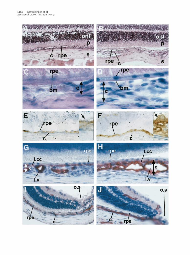
Immunohistochemical staining for VEGF (Figure 5, E andF) in transgenic mice showed specific and intense stain-ing of RPE cells and choroidal vessels. VEGF immunore-activity was observed uniformly on the basal side of theRPE cells (Figure 5, E and F; inserts). No apical RPE celland photoreceptor staining could be observed. Choroi-dal staining was not uniform throughout the eye. Areas ofvery intense and cluster-like staining alternated with ar-eas of less intense staining. The lamina choriocapillarisshowed intense staining, as well as some areas of thelamina vasculosa. In contrast, control choroids were al-most free of VEGF staining in the lamina choriocapillarisand vasculosa, and also lacked the cluster-like stainingpattern observed in transgenic choroids. The RPE cells ofcontrol mice stained uniformly and less intense through-out the eye. In transgenic and control animals compara-ble retinal staining was observed in ganglion cells andcells of the inner nuclear layer (data not shown). Anage-dependent increase of RPE cell and choroidal stain-ing was observed in transgenic animals. This increasewas most pronounced at 3 to 4 months of age (data notshown).
Choroidal Morphology in Transgenic Mice
Control and transgenic eye sections of mice aged 15days to 11 months screened by hematoxylin and eosinand by PAS and hematoxylin staining showed differencesin the choroid of transgenic mice, which was thickened(Figure 5, A and B). In transgenic as well as control eyesthe RPE cells and Bruch’s membrane were intact andshowed no areas of disruption or damage (Figure 5, Cand D). In both groups the retina and sclera did not differ.
Immunohistochemistry for the endothelial cell markerCD31 was performed to evaluate the differences in vas-cularization between transgenic and control choroids ofmice aged 15 days to 11 months (Figure 5; G, H, I, and J).Transgenic mice demonstrated a thickened and irregularchoroidal vasculature. In some areas the vessels ap-peared in clusters that bulged toward the sclera. Thelamina choriocapillaris stained most intensely. Chorio-capillary density was increased and the distance be-tween capillary vessel lumina was reduced. The laminavasculosa also appeared more dense with dilated ves-sels and wide lumina. Measurements of total choroidalthickness in standardized areas of the choroid (n 5 4 pergroup) showed a significant increase (P , 0.05) of 35 64% in the optic nerve region and of 40 6 6% in the
Figure 6. ADPase staining of control and transgenic choroidal flatmounts. A: The complete view (original magnification, 325) of the control choroid shows lightvessel staining and normal choroidal architecture. Stem of the long ciliary artery of the control choroid (B) (original magnification, 3400) and optic nerve entryof the control choroid (C). Retinal artery (RA) remnants after dissection. Original magnification, 3250. D: Complete view of the transgenic choroid with strongvessel staining and perturbed vessel architecture. E: Stem of the long ciliary artery of the transgenic choroid with tortuous vessel sprouts (arrows). F: Optic nerveentry of the transgenic choroid. A dense choriocapillary net with looping vessels (arrow) is shown.
Figure 5. A and B: Harris hematoxylin (blue) and eosin (pink) staining. Original magnification, 3400. Control (A) and transgenic eye (B) sections demonstratedifferences in choroidal (c) architecture underneath the RPE (rpe). No morphological differences are seen in the outer nuclear layer (onl) and the photoreceptorendpieces (p) of the retina. Sclera (s). C and D: PAS (pink) and Gill’s hematoxylin (blue). Original magnification, 31,000. Control (C) and transgenic (D) eyesshow an intact Bruch’s membrane (bm) and no sub-RPE vessels. Limitation of the choroid that was measured is demonstrated by the double-headed arrow. Eand F: Immunohistochemistry against VEGF (brown, diaminobenzidine precipitate) in 3-month-old mice counterstained with Gill’s hematoxylin. Originalmagnifications: 3630, insets 31,600. E: The control eye shows light VEGF staining in the RPE cell layer and no staining in the choroid. The inset shows ahigh-power view of VEGF in the RPE cells (arrow). F: Strong VEGF staining is demonstrated in RPE cell layer and the choroid of the transgenic eye. The insetshows a high-power view of an RPE cell with basal secretion of VEGF toward the choroid. G–J: CD31 immunohistochemistry (red, fast red precipitate) in3-month-old mice counterstained with Gill’s hematoxylin. Original magnification, 3630. Control choroid (G) and transgenic choroid (H) at the optic nerve entry.In the transgenic choroid the lamina choriocapillaris (l.ml) is pronounced and the lamina vasculosa (l.v) thickened because of an increased density of vessels withpronounced lumina because of dilatation. The RPE cell layer is not disrupted. Control choroid (I) and transgenic choroid (J) in proximity of the ora serrata (o.s).The transgenic choroid is thickened.
Choroidal Neovascularization in VEGF Transgenic Mice 1167AJP March 2001, Vol. 158, No. 3
proximity of the ora serrata in transgenic animals relativeto control mice. Choroidal vessels did not penetrateBruch’s membrane or appear in the subretinal space atany age. No differences in the retinal vasculature wereseen by CD31 immunohistochemistry or by ADPasestaining of retinal whole mounts.
For further evaluation of the choroidal changes ob-served with CD31 staining, we performed adenosinediphosphatase stains of transgenic and control choroidalflatmounts at 1 to 11 months of age. Choroidal flatmountsof 1- to 2-month-old mice stained poorly (data not
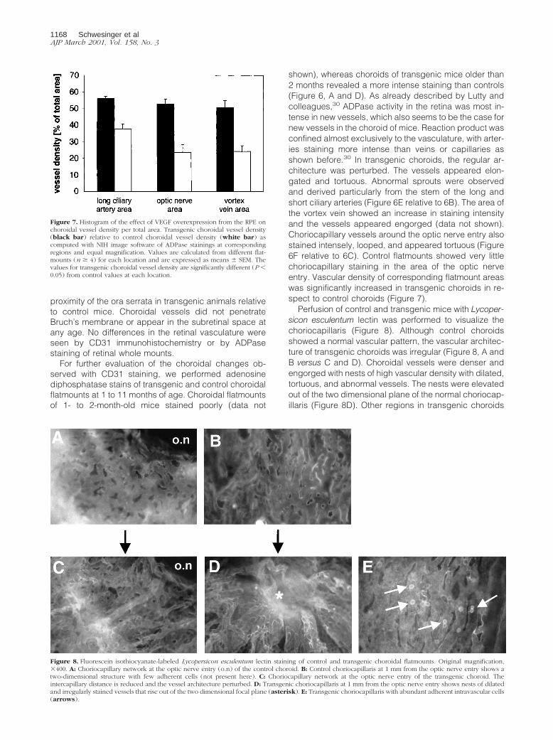
shown), whereas choroids of transgenic mice older than2 months revealed a more intense staining than controls(Figure 6, A and D). As already described by Lutty andcolleagues,30 ADPase activity in the retina was most in-tense in new vessels, which also seems to be the case fornew vessels in the choroid of mice. Reaction product wasconfined almost exclusively to the vasculature, with arter-ies staining more intense than veins or capillaries asshown before.30 In transgenic choroids, the regular ar-chitecture was perturbed. The vessels appeared elon-gated and tortuous. Abnormal sprouts were observedand derived particularly from the stem of the long andshort ciliary arteries (Figure 6E relative to 6B). The area ofthe vortex vein showed an increase in staining intensityand the vessels appeared engorged (data not shown).Choriocapillary vessels around the optic nerve entry alsostained intensely, looped, and appeared tortuous (Figure6F relative to 6C). Control flatmounts showed very littlechoriocapillary staining in the area of the optic nerveentry. Vascular density of corresponding flatmount areaswas significantly increased in transgenic choroids in re-spect to control choroids (Figure 7).
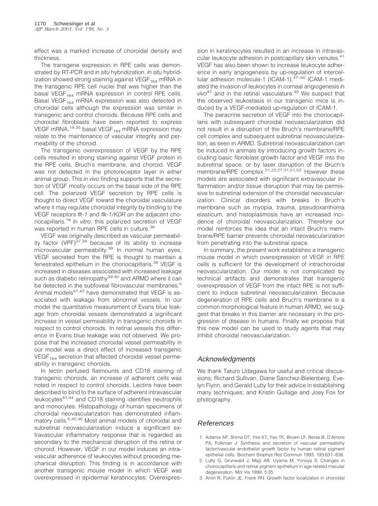
Perfusion of control and transgenic mice with Lycoper-sicon esculentum lectin was performed to visualize thechoriocapillaris (Figure 8). Although control choroidsshowed a normal vascular pattern, the vascular architec-ture of transgenic choroids was irregular (Figure 8, A andB versus C and D). Choroidal vessels were denser andengorged with nests of high vascular density with dilated,tortuous, and abnormal vessels. The nests were elevatedout of the two dimensional plane of the normal choriocap-illaris (Figure 8D). Other regions in transgenic choroids
Figure 7. Histogram of the effect of VEGF overexpression from the RPE onchoroidal vessel density per total area. Transgenic choroidal vessel density(black bar) relative to control choroidal vessel density (white bar) ascomputed with NIH image software of ADPase stainings at correspondingregions and equal magnification. Values are calculated from different flat-mounts (n $ 4) for each location and are expressed as means 6 SEM. Thevalues for transgenic choroidal vessel density are significantly different (P ,0.05) from control values at each location.
Figure 8. Fluorescein isothiocyanate-labeled Lycopersicon esculentum lectin staining of control and transgenic choroidal flatmounts. Original magnification,3400. A: Choriocapillary network at the optic nerve entry (o.n) of the control choroid. B: Control choriocapillaris at 1 mm from the optic nerve entry shows atwo-dimensional structure with few adherent cells (not present here). C: Choriocapillary network at the optic nerve entry of the transgenic choroid. Theintercapillary distance is reduced and the vessel architecture perturbed. D: Transgenic choriocapillaris at 1 mm from the optic nerve entry shows nests of dilatedand irregularly stained vessels that rise out of the two dimensional focal plane (asterisk). E: Transgenic choriocapillaris with abundant adherent intravascular cells(arrows).
1168 Schwesinger et alAJP March 2001, Vol. 158, No. 3
were characterized by tortuous vessels that branched offthe stem of larger vessels (data not shown), mainly thelong ciliary artery. Vessels of the choriocapillaris weredenser with only small spaces between the capillaries(Figure 8C versus 8A). The staining was not as uniform asin control choroids.
Increased Adherent Leukocytes in TransgenicChoroids
Lectin perfusion revealed cells adherent to the inside ofcapillaries that were abundant and appeared in clustersof two to four cells in transgenic choroids (Figure 8E). Thechoroidal capillaries were not occluded because of theirwide caliber. Only a few adherent cells and no clusterswere seen in control choroids. CD18 staining of 3-month-old control and transgenic mice (data not shown) dem-onstrated that the adherent cells were neutrophils and/ormonocytes. These cells were more abundant in trans-genic choroids and formed rolls and clusters, whereascontrol choroids contained only single adherent cells.Flatmounts that were processed without primary CD18antibody showed no staining.
Evans Blue Leakage in Transgenic Mice
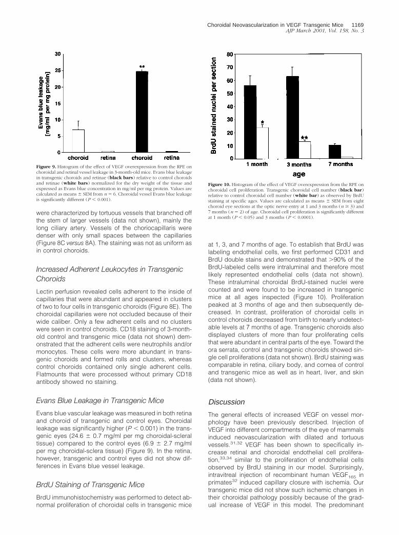
Evans blue vascular leakage was measured in both retinaand choroid of transgenic and control eyes. Choroidalleakage was significantly higher (P , 0.001) in the trans-genic eyes (24.6 6 0.7 mg/ml per mg choroidal-scleraltissue) compared to the control eyes (6.9 6 2.7 mg/mlper mg choroidal-sclera tissue) (Figure 9). In the retina,however, transgenic and control eyes did not show dif-ferences in Evans blue vessel leakage.
BrdU Staining of Transgenic Mice
BrdU immunohistochemistry was performed to detect ab-normal proliferation of choroidal cells in transgenic mice
at 1, 3, and 7 months of age. To establish that BrdU waslabeling endothelial cells, we first performed CD31 andBrdU double stains and demonstrated that .90% of theBrdU-labeled cells were intraluminal and therefore mostlikely represented endothelial cells (data not shown).These intraluminal choroidal BrdU-stained nuclei werecounted and were found to be increased in transgenicmice at all ages inspected (Figure 10). Proliferationpeaked at 3 months of age and then subsequently de-creased. In contrast, proliferation of choroidal cells incontrol choroids decreased from birth to nearly undetect-able levels at 7 months of age. Transgenic choroids alsodisplayed clusters of more than four proliferating cellsthat were abundant in central parts of the eye. Toward theora serrata, control and transgenic choroids showed sin-gle cell proliferations (data not shown). BrdU staining wascomparable in retina, ciliary body, and cornea of controland transgenic mice as well as in heart, liver, and skin(data not shown).
Discussion
The general effects of increased VEGF on vessel mor-phology have been previously described. Injection ofVEGF into different compartments of the eye of mammalsinduced neovascularization with dilated and tortuousvessels.31,32 VEGF has been shown to specifically in-crease retinal and choroidal endothelial cell prolifera-tion,33,34 similar to the proliferation of endothelial cellsobserved by BrdU staining in our model. Surprisingly,intravitreal injection of recombinant human VEGF165 inprimates32 induced capillary closure with ischemia. Ourtransgenic mice did not show such ischemic changes intheir choroidal pathology possibly because of the grad-ual increase of VEGF in this model. The predominant
Figure 9. Histogram of the effect of VEGF overexpression from the RPE onchoroidal and retinal vessel leakage in 3-month-old mice. Evans blue leakagein transgenic choroids and retinae (black bars) relative to control choroidsand retinae (white bars) normalized for the dry weight of the tissue andexpressed as Evans blue concentration in mg/ml per mg protein. Values arecalculated as means 6 SEM from n 5 6. Choroidal vessel Evans blue leakageis significantly different (P , 0.001).
Figure 10. Histogram of the effect of VEGF overexpression from the RPE onchoroidal cell proliferation. Transgenic choroidal cell number (black bar)relative to control choroidal cell number (white bar) as observed by BrdUstaining at specific ages. Values are calculated as means 6 SEM from eightchoroid eye sections at the optic nerve entry at 1 and 3 months (n $ 3) and7 months (n 5 2) of age. Choroidal cell proliferation is significantly differentat 1 month (P , 0.05) and 3 months (P , 0.0001).
Choroidal Neovascularization in VEGF Transgenic Mice 1169AJP March 2001, Vol. 158, No. 3
effect was a marked increase of choroidal density andthickness.
The transgene expression in RPE cells was demon-strated by RT-PCR and in situ hybridization. In situ hybrid-ization showed strong staining against VEGF164 mRNA inthe transgenic RPE cell nuclei that was higher than thebasal VEGF164 mRNA expression in control RPE cells.Basal VEGF164 mRNA expression was also detected inchoroidal cells although the expression was similar intransgenic and control choroids. Because RPE cells andchoroidal fibroblasts have been reported to expressVEGF mRNA,18,35 basal VEGF164 mRNA expression mayrelate to the maintenance of vascular integrity and per-meability of the choroid.
The transgenic overexpression of VEGF by the RPEcells resulted in strong staining against VEGF protein inthe RPE cells, Bruch‘s membrane, and choroid. VEGFwas not detected in the photoreceptor layer in eitheranimal group. This in vivo finding supports that the secre-tion of VEGF mostly occurs on the basal side of the RPEcell. The polarized VEGF secretion by RPE cells isthought to direct VEGF toward the choroidal vasculaturewhere it may regulate choroidal integrity by binding to theVEGF receptors flt-1 and flk-1/KDR on the adjacent cho-riocapillaris.18 In vitro, this polarized secretion of VEGFwas reported in human RPE cells in culture.36
VEGF was originally described as vascular permeabil-ity factor (VPF)37,38 because of its ability to increasemicrovascular permeability.38 In normal human eyes,VEGF secreted from the RPE is thought to maintain afenestrated epithelium in the choriocapillaris.36 VEGF isincreased in diseases associated with increased leakagesuch as diabetic retinopathy39,40 and ARMD where it canbe detected in the subfoveal fibrovascular membranes.5
Animal models41,42 have demonstrated that VEGF is as-sociated with leakage from abnormal vessels. In ourmodel the quantitative measurement of Evans blue leak-age from choroidal vessels demonstrated a significantincrease in vessel permeability in transgenic choroids inrespect to control choroids. In retinal vessels this differ-ence in Evans blue leakage was not observed. We pro-pose that the increased choroidal vessel permeability inour model was a direct effect of increased transgenicVEGF164 secretion that affected choroidal vessel perme-ability in transgenic choroids.
In lectin perfused flatmounts and CD18 staining oftransgenic choroids, an increase of adherent cells wasnoted in respect to control choroids. Lectins have beendescribed to bind to the surface of adherent intravascularleukocytes43,44 and CD18 staining identifies neutrophilsand monocytes. Histopathology of human specimens ofchoroidal neovascularization has demonstrated inflam-matory cells.6,45,46 Most animal models of choroidal andsubretinal neovascularization induce a significant ex-travascular inflammatory response that is regarded assecondary to the mechanical disruption of the retina orchoroid. However, VEGF in our model induces an intra-vascular adherence of leukocytes without preceding me-chanical disruption. This finding is in accordance withanother transgenic mouse model in which VEGF wasoverexpressed in epidermal keratinocytes. Overexpres-
sion in keratinocytes resulted in an increase in intravas-cular leukocyte adhesion in postcapillary skin venules.41
VEGF has also been shown to increase leukocyte adher-ence in early angiogenesis by up-regulation of intercel-lular adhesion molecule-1 (ICAM-1).47–50 ICAM-1 medi-ated the invasion of leukocytes in corneal angiogenesis invivo47 and in the retinal vasculature.49 We suspect thatthe observed leukostasis in our transgenic mice is in-duced by a VEGF-mediated up-regulation of ICAM-1.
The paracrine secretion of VEGF into the choriocapil-laris with subsequent choroidal neovascularization didnot result in a disruption of the Bruch’s membrane/RPEcell complex and subsequent subretinal neovasculariza-tion, as seen in ARMD. Subretinal neovascularization canbe induced in animals by introducing growth factors in-cluding basic fibroblast growth factor and VEGF into thesubretinal space, or by laser disruption of the Bruch’smembrane/RPE complex.21,22,27,31,51,52 However thesemodels are associated with significant extravascular in-flammation and/or tissue disruption that may be permis-sive to subretinal extension of the choroidal neovascular-ization. Clinical disorders with breaks in Bruch‘smembrane such as myopia, trauma, pseudoxanthomaelasticum, and histoplasmosis have an increased inci-dence of choroidal neovascularization. Therefore ourmodel reinforces the idea that an intact Bruch’s mem-brane/RPE barrier prevents choroidal neovascularizationfrom penetrating into the subretinal space.
In summary, the present work establishes a transgenicmouse model in which overexpression of VEGF in RPEcells is sufficient for the development of intrachoroidalneovascularization. Our model is not complicated bytechnical artifacts and demonstrates that transgenicoverexpression of VEGF from the intact RPE is not suffi-cient to induce subretinal neovascularization. Becausedegeneration of RPE cells and Bruch’s membrane is acommon morphological feature in human ARMD, we sug-gest that breaks in this barrier are necessary in the pro-gression of disease in humans. Finally we propose thatthis new model can be used to study agents that mayinhibit choroidal neovascularization.
Acknowledgments
We thank Taturo Udagawa for useful and critical discus-sions; Richard Sullivan, Diane Sanchez-Bielenberg, Eve-lyn Flynn, and Gerald Lutty for their advice in establishingmany techniques; and Kristin Gullage and Joey Fox forphotography.
References
1. Adamis AP, Shima DT, Yeo KT, Yeo TK, Brown LF, Berse B, D’AmorePA, Folkman J: Synthesis and secretion of vascular permeabilityfactor/vascular endothelial growth factor by human retinal pigmentepithelial cells. Biochem Biophys Res Commun 1993, 193:631–638
2. Lutty G, Grunwald J, Majji AB, Uyama M, Yoneya S: Changes inchoriocapillaris and retinal pigment epithelium in age-related maculardegeneration. Mol Vis 1999, 5:35
3. Amin R, Puklin JE, Frank RN: Growth factor localization in choroidal
1170 Schwesinger et alAJP March 2001, Vol. 158, No. 3
neovascular membranes of age-related macular degeneration. InvestOphthalmol Vis Sci 1994, 35:3178–3188
4. Frank RN, Amin RH, Eliott D, Puklin JE, Abrams GW: Basic fibroblastgrowth factor and vascular endothelial growth factor are present inepiretinal and choroidal neovascular membranes. Am J Ophthalmol1996, 122:393–403
5. Kvanta A, Algvere PV, Berglin L, Seregard S: Subfoveal fibrovascularmembranes in age-related macular degeneration express vascularendothelial growth factor. Invest Ophthalmol Vis Sci 1996, 37:1929–1934
6. Lopez PF, Sippy BD, Lambert HM, Thach AB, Hinton DR: Transdif-ferentiated retinal pigment epithelial cells are immunoreactive forvascular endothelial growth factor in surgically excised age-relatedmacular degeneration-related choroidal neovascular membranes. In-vest Ophthalmol Vis Sci 1996, 37:855–868
7. Kliffen M, Sharma HS, Mooy CM, Kerkvliet S, de Jong PT: Increasedexpression of angiogenic growth factors in age-related maculopathy.Br J Ophthalmol 1997, 81:154–162
8. Reddy VM, Zamora RL, Kaplan HJ: Distribution of growth factors insubfoveal neovascular membranes in age-related macular degener-ation and presumed ocular histoplasmosis syndrome. Am J Ophthal-mol 1995, 120:291–301
9. Senger DR, Galli SJ, Dvorak AM, Perruzzi CA, Harvey VS, Dvorak HF:Tumor cells secrete a vascular permeability factor that promotesaccumulation of ascites fluid. Science 1983, 219:983–985
10. Leung DW, Cachianes G, Kuang WJ, Goeddel DV, Ferrara N: Vas-cular endothelial growth factor is a secreted angiogenic mitogen.Science 1989, 246:1306–1309
11. Ferrara N, Houck KA, Jakeman LB, Winer J, Leung DW: The vascularendothelial growth factor family of polypeptides. J Cell Biochem1991, 47:211–218
12. Klagsbrun M, D’Amore PA: Vascular endothelial growth factor and itsreceptors. Cytokine Growth Factor Rev 1996, 7:259–270
13. Veikkola T, Alitalo K: VEGFs, receptors and angiogenesis. SeminCancer Biol 1999, 9:211–220
14. Poltorak Z, Cohen T, Neufeld G: The VEGF splice variants: properties,receptors, and usage for the treatment of ischemic diseases [InProcess Citation]. Herz 2000, 25:126–129
15. Shima DT, Gougos A, Miller JW, Tolentino M, Robinson G, AdamisAP, D’Amore PA: Cloning and mRNA expression of vascular endo-thelial growth factor in ischemic retinas of Macaca fascicularis. InvestOphthalmol Vis Sci 1996, 37:1334–1340
16. de Vries C, Escobedo JA, Ueno H, Houck K, Ferrara N, Williams LT:The fms-like tyrosine kinase, a receptor for vascular endothelialgrowth factor. Science 1992, 255:989–991
17. Terman BI, Dougher-Vermazen M, Carrion ME, Dimitrov D, ArmellinoDC, Gospodarowicz D, Bohlen P: Identification of the KDR tyrosinekinase as a receptor for vascular endothelial cell growth factor. Bio-chem Biophys Res Commun 1992, 187:1579–1586
18. Kim I, Ryan AM, Rohan R, Amano S, Agular S, Miller JW, Adamis AP:Constitutive expression of VEGF, VEGFR-1, and VEGFR-2 in normaleyes [published erratum appears in Invest Ophthalmol Vis Sci 2000Feb;41:368]. Invest Ophthalmol Vis Sci 1999, 40:2115–2121
19. Kuroki M, Voest EE, Amano S, Beerepoot LV, Takashima S, TolentinoM, Kim RY, Rohan RM, Colby KA, Yeo KT, Adamis AP: Reactiveoxygen intermediates increase vascular endothelial growth factorexpression in vitro and in vivo. J Clin Invest 1996, 98:1667–1675
20. Punglia RS, Lu M, Hsu J, Kuroki M, Tolentino MJ, Keough K, Levy AP,Levy NS, Goldberg MA, D’Amato RJ, Adamis AP: Regulation ofvascular endothelial growth factor expression by insulin-like growthfactor I. Diabetes 1997, 46:1619–1626
21. Soubrane G, Cohen SY, Delayre T, Tassin J, Hartmann MP, CoscasGJ, Courtois Y, Jeanny JC: Basic fibroblast growth factor experimen-tally induced choroidal angiogenesis in the minipig. Curr Eye Res1994, 13:183–195
22. Kimura H, Sakamoto T, Hinton DR, Spee C, Ogura Y, Tabata Y, IkadaY, Ryan SJ: A new model of subretinal neovascularization in therabbit. Invest Ophthalmol Vis Sci 1995, 36:2110–2119
23. Orzalesi N, Migliavacca L, Miglior S: Subretinal neovascularizationafter naphthalene damage to the rabbit retina. Invest Ophthalmol VisSci 1994, 35:696–705
24. Pollack A, Korte GE, Weitzner AL, Henkind P: Ultrastructure ofBruch’s membrane after krypton laser photocoagulation. I. Break-down of Bruch’s membrane. Arch Ophthalmol 1986, 104:1372–1376
25. Pollack A, Korte GE, Heriot WJ, Henkind P: Ultrastructure of Bruch’smembrane after krypton laser photocoagulation. II. Repair of Bruch’smembrane and the role of macrophages. Arch Ophthalmol 1986,104:1377–1382
26. Lutty GA, McLeod DS, Pachnis A, Costantini F, Fabry ME, Nagel RL:Retinal and choroidal neovascularization in a transgenic mousemodel of sickle cell disease. Am J Pathol 1994, 145:490–497
27. Spilsbury K, Garrett KL, Shen WY, Constable IJ, Rakoczy PE: Over-expression of vascular endothelial growth factor (VEGF) in the retinalpigment epithelium leads to the development of choroidal neovascu-larization. Am J Pathol 2000, 157:135–144
28. Okamoto N, Tobe T, Hackett SF, Ozaki H, Vinores MA, LaRochelle W,Zack DJ, Campochiaro PA: Transgenic mice with increased expres-sion of vascular endothelial growth factor in the retina: a new modelof intraretinal and subretinal neovascularization [see comments].Am J Pathol 1997, 151:281–291
29. Boulanger A, Liu S, Henningsgaard AA, Yu S, Redmond TM: Theupstream region of the RPE65 gene confers retinal pigment epitheli-um-specific expression in vivo and in vitro and contains critical oc-tamer and E-box sites. J Biol Chem 2000, 275:31274–31282
30. Lutty GA, McLeod DS: A new technique for visualization of the humanretinal vasculature. Arch Ophthalmol 1992, 110:267–276
31. Ozaki H, Hayashi H, Vinores SA, Moromizato Y, Campochiaro PA,Oshima K: Intravitreal sustained release of VEGF causes retinal neo-vascularization in rabbits and breakdown of the blood-retinal barrierin rabbits and primates. Exp Eye Res 1997, 64:505–517
32. Tolentino MJ, Miller JW, Gragoudas ES, Chatzistefanou K, Ferrara N,Adamis AP: Vascular endothelial growth factor is sufficient to produceiris neovascularization and neovascular glaucoma in a nonhumanprimate. Arch Ophthalmol 1996, 114:964–970
33. Tolentino MJ, Miller JW, Gragoudas ES, Jakobiec FA, Flynn E, Chat-zistefanou K, Ferrara N, Adamis AP: Intravitreous injections of vascu-lar endothelial growth factor produce retinal ischemia and microan-giopathy in an adult primate. Ophthalmology 1996, 103:1820–1828
34. Li W, He Z, Li Y, Yanoff M: Vascular endothelial growth factor regu-lates both apoptosis and angiogenesis of choriocapillaris endothelialcells. Microvasc Res 2000, 59:286–289
35. Kvanta A: Expression and regulation of vascular endothelial growthfactor in choroidal fibroblasts. Curr Eye Res 1995, 14:1015–1020
36. Blaauwgeers HG, Holtkamp GM, Rutten H, Witmer AN, Koolwijk P,Partanen TA, Alitalo K, Kroon ME, Kijlstra A, van Hinsbergh VW,Schlingemann RO: Polarized vascular endothelial growth factor se-cretion by human retinal pigment epithelium and localization of vas-cular endothelial growth factor receptors on the inner choriocapillaris.Evidence for a trophic paracrine relation. Am J Pathol 1999, 155:421–428
37. Senger DR, Perruzzi CA, Feder J, Dvorak HF: A highly conservedvascular permeability factor secreted by a variety of human androdent tumor cell lines. Cancer Res 1986, 46:5629–5632
38. Roberts WG, Palade GE: Increased microvascular permeability andendothelial fenestration induced by vascular endothelial growth fac-tor. J Cell Sci 1995, 108:2369–2379
39. Murata T, Nakagawa K, Khalil A, Ishibashi T, Inomata H, Sueishi K:The relation between expression of vascular endothelial growth factorand breakdown of the blood-retinal barrier in diabetic rat retinas. LabInvest 1996, 74:819–825
40. Mathews MK, Merges C, McLeod DS, Lutty GA: Vascular endothelialgrowth factor and vascular permeability changes in human diabeticretinopathy. Invest Ophthalmol Vis Sci 1997, 38:2729–2741
41. Detmar M, Brown LF, Schon MP, Elicker BM, Velasco P, Richard L,Fukumura D, Monsky W, Claffey KP, Jain RK: Increased microvascu-lar density and enhanced leukocyte rolling and adhesion in the skin ofVEGF transgenic mice. J Invest Dermatol 1998, 111:1–6
42. Thurston G, Suri C, Smith K, McClain J, Sato TN, Yancopoulos GD,McDonald DM: Leakage-resistant blood vessels in mice transgeni-cally overexpressing angiopoietin-1. Science 1999, 286:2511–2514
43. Thurston G, Baluk P, Hirata A, McDonald DM: Permeability-relatedchanges revealed at endothelial cell borders in inflamed venules bylectin binding. Am J Physiol 1996, 271:H2547–H2562
44. Thurston G, Murphy TJ, Baluk P, Lindsey JR, McDonald DM: Angio-genesis in mice with chronic airway inflammation: strain-dependentdifferences [see comments]. Am J Pathol 1998, 153:1099–1112
45. Lopez PF, Grossniklaus HE, Lambert HM, Aaberg TM, Capone Jr A,Sternberg Jr P, L’Hernault N: Pathologic features of surgically excised
Choroidal Neovascularization in VEGF Transgenic Mice 1171AJP March 2001, Vol. 158, No. 3
subretinal neovascular membranes in age-related macular degener-ation. Am J Ophthalmol 1991, 112:647–656
46. Seregard S, Algvere PV, Berglin L: Immunohistochemical character-ization of surgically removed subfoveal fibrovascular membranes.Graefes Arch Clin Exp Ophthalmol 1994, 232:325–329
47. Becker MD, Kruse FE, Azzam L, Nobiling R, Reichling J, Volcker HE:In vivo significance of ICAM-1-dependent leukocyte adhesion in earlycorneal angiogenesis. Invest Ophthalmol Vis Sci 1999, 40:612–618
48. Melder RJ, Koenig GC, Witwer BP, Safabakhsh N, Munn LL, Jain RK:During angiogenesis, vascular endothelial growth factor and basicfibroblast growth factor regulate natural killer cell adhesion to tumorendothelium [see comments]. Nat Med 1996, 2:992–997
49. Lu M, Perez VL, Ma N, Miyamoto K, Peng HB, Liao JK, Adamis AP:VEGF increases retinal vascular ICAM-1 expression in vivo. InvestOphthalmol Vis Sci 1999, 40:1808–1812
50. Miyamoto K, Khosrof S, Bursell SE, Moromizato Y, Aiello LP, Ogura Y,Adamis AP: Vascular endothelial growth factor (VEGF)-induced reti-nal vascular permeability is mediated by intercellular adhesion mol-ecule-1 (ICAM-1). Am J Pathol 2000, 156:1733–1739
51. Dobi ET, Puliafito CA, Destro M: A new model of experimental cho-roidal neovascularization in the rat. Arch Ophthalmol 1989, 107:264–269
52. Frank RN, Das A, Weber ML: A model of subretinal neovasculariza-tion in the pigmented rat. Curr Eye Res 1989, 8:239–247
1172 Schwesinger et alAJP March 2001, Vol. 158, No. 3
Copyright © 2022 FDOKUMEN