The Photobleaching Sequence of a Short-Wavelength Visual Pigment
13
The Photobleaching Sequence of a Short-Wavelength Visual Pigment ² Anakarin Kusnetzow, ‡,§,| Abhiram Dukkipati, § Kunnel R. Babu, ⊥ Deepak Singh, § Bryan W. Vought, §,@ Barry E. Knox,* ,⊥ and Robert R. Birge* ,‡,§ Departments of Chemistry and of Molecular and Cell Biology, UniVersity of Connecticut, 55 North EagleVille Road, Storrs, Connecticut 06268-3060, Departments of Chemistry and Biology, Syracuse UniVersity, 111 College Place, Syracuse, New York 13244-4100, and Department of Biochemistry and Molecular Biology, State UniVersity of New York Upstate Medical UniVersity, 750 East Adams Street, Syracuse, New York 13210 ReceiVed February 23, 2001; ReVised Manuscript ReceiVed April 27, 2001 ABSTRACT: The photobleaching pathway of a short-wavelength cone opsin purified in delipidated form (λ max ) 425 nm) is reported. The batho intermediate of the violet cone opsin generated at 45 K has an absorption maximum at 450 nm. The batho intermediate thermally decays to the lumi intermediate (λ max ) 435 nm) at 200 K. The lumi intermediate decays to the meta I (λ max ) 420 nm) and meta II (λ max ) 388 nm) intermediates at 258 and 263 K, respectively. The meta II intermediate decays to free retinal and opsin at >270 K. At 45, 75, and 140 K, the photochemical excitation of the violet cone opsin at 425 nm generates the batho intermediate at high concentrations under moderate illumination. The batho intermediate spectra, generated via decomposing the photostationary state spectra at 45 and 140 K, are identical and have properties typical of batho intermediates of other visual pigments. Extended illumination of the violet cone opsin at 75 K, however, generates a red-shifted photostationary state (relative to both the dark and the batho intermediates) that has as absorption maximum at ∼470 nm, and thermally reverts to form the normal batho intermediate when warmed to 140 K. We conclude that this red-shifted photostationary state is a metastable state, characterized by a higher-energy protein conformation that allows relaxation of the all-trans chromophore into a more planar conformation. FTIR spectroscopy of violet cone opsin indicates conclusively that the chromophore is protonated. A similar transformation of the rhodopsin binding site generates a model for the VCOP binding site that predicts roughly 75% of the observed blue shift of the violet cone pigment relative to rhodopsin. MNDO-PSDCI calculations indicate that secondary interactions involving the binding site residues are as important as the first-order chromophore protein interactions in mediating the wavelength maximum. Two types of photoreceptor cells, rods and cones, mediate vision in vertebrates. Rods are responsible for scotopic (low- light) vision, and cones are responsible for color discrimina- tion and high-flux visual acuity (1-3). The visual opsins contain an 11-cis-retinal chromophore covalently bound to the apoprotein via a Schiff base linkage to a conserved lysine residue (4). The 11-cis chromophore isomerizes to an all- trans conformation upon light activation, which induces the sequential formation of a series of thermal intermediates (5, 6). The key biological state, the meta II 1 intermediate, activates the G-protein transducin, which in turn initiates the visual cascade (7, 8). The vertebrate visual pigments are classified into five groups based on sequence similarity (9- 12) as shown in the dendogram of Figure 1 based on the review by Ebrey and Koutalos (13). We have updated the absorption maxima for the SWS1 pigment group using data from refs 14-20. It should be noted that some of the absorption maxima listed in Figure 1 were assigned from very small pigment samples and are subject to uncertainty. The pigments responsible for scotopic vision (rhodopsins or RH1) absorb maximally at ∼500 nm. The pigments that mediate photopic vision are subdivided into middle- to long- wavelength (M/LWS, >510 nm), two short-wavelength (SWS2, 440-460 nm, and SWS1, 350-450 nm), and rhodopsin-like, middle-wavelength (RH2, 460-510 nm) groups. The absorption maxima of the majority of the pigments in each group fall within a constrained spectral range, suggesting that the spectral tuning mechanism is mediated by the protein binding site (9-12). The photo- bleaching kinetics of rod and cone opsins may play an ² This work was supported in part by NIH Grants GM-34548 (R.R.B.) and EY-11256 (B.E.K.) and the W. M. Keck Center for Molecular Electronics at Syracuse University. * To whom correspondence should be addressed. R.R.B.: telephone, (860) 486-6720; fax, (860) 486-2981; e-mail, [email protected]. B.E.K.: telephone, (315) 464-8719; fax, (315) 464-8750; e-mail, [email protected]. ‡ University of Connecticut. § Syracuse University. | Present address: Jules Stein Eye Institute, 100 Stein Plaza, University of California, Box 957000, Los Angeles, CA 90095-7000. ⊥ State University of New York Upstate Medical University. @ Present address: Department of Biological Chemistry and Mo- lecular Pharmacology, Harvard Medical School, 240 Longwood Ave., Boston, MA 02115. 1 Abbreviations: VCOP, Xenopus laeVis violet cone opsin; ROS, rod outer segments; rho or s. rho, solubilized rhodopsin; LM, N-dodecyl -D-maltoside; HEPES, N-(2-hydroxyethyl)piperazine-N′-2-ethane- sulfonic acid; SB, Schiff base; PSB, protonated Schiff base; PSS, photostationary state; PSSλλλ, photostationary state generated with illumination at wavelength λλλ in nanometers; B1, primary photo- stationary state of VCOP at cryogenic temperatures; B2, red-shifted photostationary state generated upon extended photochemical excitation of the B1 state at certain temperatures; batho, lumi, meta I, and meta II, discrete thermal intermediates of the visual opsin bleaching pathway. 7832 Biochemistry 2001, 40, 7832-7844 10.1021/bi010387y CCC: $20.00 © 2001 American Chemical Society Published on Web 06/07/2001
-
Upload
independent -
Category
Documents
-
view
0 -
download
0
Transcript of The Photobleaching Sequence of a Short-Wavelength Visual Pigment

The Photobleaching Sequence of a Short-Wavelength Visual Pigment†
Anakarin Kusnetzow,‡,§,| Abhiram Dukkipati,§ Kunnel R. Babu,⊥ Deepak Singh,§ Bryan W. Vought,§,@
Barry E. Knox,*,⊥ and Robert R. Birge*,‡,§
Departments of Chemistry and of Molecular and Cell Biology, UniVersity of Connecticut, 55 North EagleVille Road,Storrs, Connecticut 06268-3060, Departments of Chemistry and Biology, Syracuse UniVersity, 111 College Place,
Syracuse, New York 13244-4100, and Department of Biochemistry and Molecular Biology, State UniVersity ofNew York Upstate Medical UniVersity, 750 East Adams Street, Syracuse, New York 13210
ReceiVed February 23, 2001; ReVised Manuscript ReceiVed April 27, 2001
ABSTRACT: The photobleaching pathway of a short-wavelength cone opsin purified in delipidated form(λmax ) 425 nm) is reported. Thebatho intermediate of the violet cone opsin generated at 45 K has anabsorption maximum at 450 nm. Thebatho intermediate thermally decays to thelumi intermediate (λmax
) 435 nm) at 200 K. Thelumi intermediate decays to themeta I (λmax ) 420 nm) andmeta II (λmax )388 nm) intermediates at 258 and 263 K, respectively. Themeta II intermediate decays to free retinal andopsin at>270 K. At 45, 75, and 140 K, the photochemical excitation of the violet cone opsin at 425 nmgenerates thebathointermediate at high concentrations under moderate illumination. Thebathointermediatespectra, generated via decomposing the photostationary state spectra at 45 and 140 K, are identical andhave properties typical ofbatho intermediates of other visual pigments. Extended illumination of theviolet cone opsin at 75 K, however, generates a red-shifted photostationary state (relative to both the darkand thebathointermediates) that has as absorption maximum at∼470 nm, and thermally reverts to formthe normalbathointermediate when warmed to 140 K. We conclude that this red-shifted photostationarystate is a metastable state, characterized by a higher-energy protein conformation that allows relaxationof the all-trans chromophore into a more planar conformation. FTIR spectroscopy of violet cone opsinindicates conclusively that the chromophore is protonated. A similar transformation of the rhodopsin bindingsite generates a model for the VCOP binding site that predicts roughly 75% of the observed blue shift ofthe violet cone pigment relative to rhodopsin. MNDO-PSDCI calculations indicate that secondaryinteractions involving the binding site residues are as important as the first-order chromophore proteininteractions in mediating the wavelength maximum.
Two types of photoreceptor cells, rods and cones, mediatevision in vertebrates. Rods are responsible for scotopic (low-light) vision, and cones are responsible for color discrimina-tion and high-flux visual acuity (1-3). The visual opsinscontain an 11-cis-retinal chromophore covalently bound tothe apoprotein via a Schiff base linkage to a conserved lysineresidue (4). The 11-cis chromophore isomerizes to an all-transconformation upon light activation, which induces thesequential formation of a series of thermal intermediates (5,6). The key biological state, themeta II1 intermediate,activates the G-protein transducin, which in turn initiates thevisual cascade (7, 8). The vertebrate visual pigments areclassified into five groups based on sequence similarity (9-
12) as shown in the dendogram of Figure 1 based on thereview by Ebrey and Koutalos (13). We have updated theabsorption maxima for the SWS1 pigment group using datafrom refs 14-20. It should be noted that some of theabsorption maxima listed in Figure 1 were assigned fromvery small pigment samples and are subject to uncertainty.The pigments responsible for scotopic vision (rhodopsins orRH1) absorb maximally at∼500 nm. The pigments thatmediate photopic vision are subdivided into middle- to long-wavelength (M/LWS,>510 nm), two short-wavelength(SWS2, 440-460 nm, and SWS1, 350-450 nm), andrhodopsin-like, middle-wavelength (RH2, 460-510 nm)groups. The absorption maxima of the majority of thepigments in each group fall within a constrained spectralrange, suggesting that the spectral tuning mechanism ismediated by the protein binding site (9-12). The photo-bleaching kinetics of rod and cone opsins may play an
† This work was supported in part by NIH Grants GM-34548(R.R.B.) and EY-11256 (B.E.K.) and the W. M. Keck Center forMolecular Electronics at Syracuse University.
* To whom correspondence should be addressed. R.R.B.: telephone,(860) 486-6720; fax, (860) 486-2981; e-mail, [email protected].: telephone, (315) 464-8719; fax, (315) 464-8750; e-mail,[email protected].
‡ University of Connecticut.§ Syracuse University.| Present address: Jules Stein Eye Institute, 100 Stein Plaza,
University of California, Box 957000, Los Angeles, CA 90095-7000.⊥ State University of New York Upstate Medical University.@ Present address: Department of Biological Chemistry and Mo-
lecular Pharmacology, Harvard Medical School, 240 Longwood Ave.,Boston, MA 02115.
1 Abbreviations: VCOP,Xenopus laeVis violet cone opsin; ROS,rod outer segments; rho or s. rho, solubilized rhodopsin; LM,N-dodecylâ-D-maltoside; HEPES,N-(2-hydroxyethyl)piperazine-N′-2-ethane-sulfonic acid; SB, Schiff base; PSB, protonated Schiff base; PSS,photostationary state; PSSλλλ, photostationary state generated withillumination at wavelengthλλλ in nanometers;B1, primary photo-stationary state of VCOP at cryogenic temperatures;B2, red-shiftedphotostationary state generated upon extended photochemical excitationof the B1 state at certain temperatures;batho, lumi, meta I, andmetaII , discrete thermal intermediates of the visual opsin bleaching pathway.
7832 Biochemistry2001,40, 7832-7844
10.1021/bi010387y CCC: $20.00 © 2001 American Chemical SocietyPublished on Web 06/07/2001

important role in determining the signal amplification,recovery, and response kinetics of the rod and cone photo-receptor cells. The intermediates in the photobleachingpathway of rod and cone opsins can be thermally trappedand detected by observing changes in the UV-vis spectrum.This approach has been used extensively to study thepathways of several pigments and to understand the molec-ular basis of rod and cone opsin function (21-27). Therelatively small number of studies on group SWS1 pigmentsis due in part to the difficulty of obtaining sufficient quantitiesof stable pigments (12, 14, 28-35). Our expression andisolation protocol yields sufficient quantities of theXenopusviolet cone opsin (VCOP) (14, 36) to permit detailedbiochemical and spectroscopic study.
All of the pigments in the M/LWS, RH1, RH2, and SWS2groups are red-shifted relative to a protonated Schiff basein solution (λmax ) 400-440 nm) and presumably have aprotonated Schiff base linkage in the dark state. In contrast,
some of the SWS1 group pigments exhibit absorptionmaxima blue-shifted relative to that of a protonated Schiffbase in solution. Some pigments have absorption maximain the UV (∼370 nm), while others absorb in the violet(410-450 nm). The separation of the pigments into twowavelength distributions suggests that the UV pigments havea deprotonated Schiff base whereas the violet cones have aprotonated Schiff base in the dark (14).
In the study presented here, we use the low-temperaturetrapping method to investigate the photobleaching pathwayof the violet cone opsin. We supplement these results withthe use of FTIR spectroscopy to determine the protonationstate of the chromophore, and semiempirical (MNDO-PSDCI) and ab initio (B3LYP/6-31G) molecular orbitaltheory to analyze the protein binding site and the mechanismof wavelength regulation.
MATERIALS AND METHODS
Expression, Isolation, and Purification of the VisualPigments. An expression construct, pMT-VCOP, containingthe first 328 codons of theXenopusviolet cone opsin codingregion and the last 14 codons of bovine rod opsin wasexpressed in mammalian COS1 cells by transient transfection(14, 35, 36). The violet cone opsin was regenerated with11-cis-A1-retinal and purified by column immunoaffinitychromatography on 1D4-Sepahrose. Fractions that containedthe visual pigment were collected from the column andconcentrated to a finalA425 of ∼0.3 using a Centricon C-30filter (Millipore, Bedford, MA). The rhodopsin E113Qmutant was expressed in mammalian COS1 cells, isolated,and purified in the same manner (37). Rhodopsin wassolubilized (s. rho) and purified from rod outer segments(ROS) by using immunoaffinity chromatography on con-canavalin A-Sepharose (38). All pigments were stored at-80 °C until they were used.
Violet Cone Opsin Studied by Cryogenic Electron Spec-troscopy. Visual pigment samples were mixed with glycerolto produce a 67% glycerol and 0.05%N-dodecyl â-D-maltoside (LM, Anatrace, Maumee, OH) solution in bufferA [50 mM HEPES, 140 mM NaCl, and 3 mM MgCl2 (pH6.8)]. Samples for cryogenic studies at 45-200 K wereplaced in an Air Products Displex helium-refrigeratedcryostat coupled to a Shimadzu 3101 UV-vis-NIR spec-trophotometer as previously described (14). Samples for thestudies at 230-273 K were placed in a Varian Cary 3E UV-vis spectrophotometer modified to allow accurate temperaturecontrol from 228 to 280 K, thus enabling the stabilizationof the later intermediates via temperature trapping.
The sample was illuminated with a Photomax systemequipped with a 200 W arc lamp coupled to a monochro-mator (Oriel, Stratford, CT). A photostationary state (PSS)was generated by successive illuminations until no furtherspectral changes could be detected (∼1 h, unless specifiedotherwise). In the subsequent discussion, the wavelengthof illumination in nanometers used to generate the photo-stationary state is indicated following the PSS abbreviation.
All the electronic spectra are the average of four spectraat each temperature normalized with respect to the proteinaromatic residue band at 280 nm (A280). Each photostationarystate described in this paper was reproduced two to four timesfrom independent transfections. Each of the difference spectra
FIGURE 1: Dendogram of selected vertebrate visual opsins. Absorp-tion maxima when known are shown within parentheses innanometers, and the text is displayed in color based on thewavelength (nanometers) template shown in the column at right.TheX. laeVis violet cone opsin (VCOP) is labeled “xenopus violet”and is shown in enhanced type in the SWS1 group. The VCOPpigment absorbs maximally at 425 nm when regenerated with 11-cis-A1-retinal, an absorption maximum nearly identical to that ofthe human blue cone. This figure was adapted from Figure 3 ofthe review by Ebrey and Koutalos (13), with additional or updatedSWS1 pigment data from refs14-20.
Photobleaching of a Cone Pigment Biochemistry, Vol. 40, No. 26, 20017833

was calculated by subtracting the appropriate photostationarystate from the corresponding dark or previous photostationarystate.
FTIR Difference Spectroscopy of the Visual Pigments.FTIR samples had the glycerol and HEPES removed andthe concentration of LM decreased by exchanging buffer Awith buffer B [10 mM potassium phosphate buffer (pH 6.8)and 0.02% LM] using Centricon C-30 filters. The buffer Ain which E113Q was purified was exchanged for buffer C[10 mM potassium phosphate buffer (pH 8.2) and 0.02%LM]. Excess 1D4 peptide was removed from the samplesby passing the pigments over prepacked Sephadex G-50columns (Pharmacia, Peapack, NJ). The final concentrationof the samples was adjusted to 1µg/µL. Deuterated sampleswere prepared by following the above procedure using bufferB or C prepared with D2O.
The pigment (∼100 µg) was dried on an infrared qualityCaF2 window under a continuous stream of nitrogen. Thefilm was sealed with another CaF2 window and mounted ona Nicolet Magna IR 750, series II instrument (NicoletInstrument Corp., Madison, WI). The protein samples wereequilibrated at 263 K for approximately 1 h. The dark statespectra of the pigments were obtained by collecting andaveraging 512 scans. Illuminating the sample from anappropriately filtered 250 W quartz halogen lamp (3-5 min)at 263 K generated the active state of the proteins. Therhodopsinmeta II intermediate was generated using a 495nm cutoff filter (Melles Griot 03FCG067). The violet coneopsin meta II intermediate was generated using a 395 nmcutoff filter (Melles Griot 03FCG055), and the E113QmetaII intermediate was generated using a 305 nm cutoff filter(Melles Griot 03FCG121). Themeta II spectra of thepigments were obtained by collecting and averaging 512scans (∼8 min). The samples were illuminated for anadditional 2-3 min, and the spectra were collected. Datacollection and analysis were performed using OMNIC 3.1(Nicolet Instrument Corp.). Each difference spectrum wascalculated by subtracting the spectrum for themeta IIintermediate from the spectrum from the appropriate darkstate. The resulting difference spectra from the secondillumination were identical to that of the first illumination,indicating the complete formation of the photoproduct at 263K. The raw spectra have not been corrected for baselinedeviations, but have been smoothed using a three-pointtriangular slit function.
Theoretical. The ground state vibrational properties ofmodel chromophores were calculated using both Hartree-Fock and density functional (B3LYP) ab initio molecularorbital theory and basis sets up to 6-31G(d) via Gaussian 98(39). The relevant details will be reported below. Thespectroscopic properties of the protein-bound chromophorewere calculated using MNDO-PSDCI molecular orbitaltheory, including full single and double configurationinteraction within the chromophoreπ-electron manifold (40).This method allows the entire protein binding site to beincluded in the SCF calculation while constraining theconfiguration interaction to the chromophore. However, whendispersive interactions between the chromophore and nearbyaromatic residues are of interest, single and double CI isextended to include the aromatic residues. (Dispersiveinteractions can be simulated theoretically by using doubleCI where the coupled excitations on both the chromophore
and residue are included.) Although we have developedspectroscopic parametrizations that work with the MNDO,AM1, or PM3 Hamiltonians, we have found that the PM3Hamiltonian, which will be adopted in this study, providesthe best agreement with experiment when simulating theprotein binding sites of both bacteriorhodopsin and rhodopsin(14, 40-47).
RESULTS
Batho Intermediate of the Violet Cone Opsin. Previously,we observed twobatho-shifted photostationary intermediatesfor VCOP at very low temperatures (14). In the studypresented here, we investigated the behavior of the two statesupon warming. We initially formed the photostationary state(PSS395) at 45 K (Figure 2, spectrum 2) which was identicalto the one previously observed (called XB30K in ref 14).Henceforth, we will call this photostationary stateB1. WhenB1 is warmed to 75 K and thermally equilibrated for 12 h,no spectral change is observed (data not shown). Therefore,B1 cannot be converted thermally to the secondbatho-likeintermediate previously observed (called XB70K in ref 14),and henceforth calledB2. Furthermore, no change in theB1
spectrum is detected until the next thermal intermediate isformed at 200 K (Figure 2, spectrum 3). The 395 nmphotostationary state (PSS395) is usually reached within 20-30 min at 45 K. However, because of the high concentrationsof protein used in this study, illumination was carried outfor longer times (1 h) to make sure that the maximum amountof bathoproduct was formed. At 45 K, continued illumina-
FIGURE 2: Photochemistry of the violet cone opsin at 45 K. TheB1 state generated at 45 K (spectrum 2) is stable to 198 K, butdecayed to thelumi intermediate at 200 K (spectrum 3). The tablelists the conditions under which the spectra were collected, whereλill is the wavelength of light (in nanometers) used to generate thephotoproduct,Till is the temperature at which the illumination wasperformed (in kelvin),till is the length of illumination (in hours),and Tspectrum is the temperature at which each spectrum wascollected. Dashes indicate no change from the previous conditions(i.e., no further illumination, or no change in temperature). Thedifference spectra are shown in the inset. Curves 4 and 5 correspondto spectrum 2 and spectrum 3 minus spectrum 1, respectively.
7834 Biochemistry, Vol. 40, No. 26, 2001 Kusnetzow et al.
tion (2-12 h) of B1 does not result in any further spectralshift, thus ruling out the formation of a low-quantumefficiency product at this temperature. However at 75 K,depending on the chromophore composition of the initial darkstate,B2 is formed within 1 h of illumination or after 12 hof illumination. If formation of B2 is a low-quantumefficiency event, then we would expect that the time requiredto formB2 at 75 K would be independent of the chromophorecomposition of the dark state at 75 K, which is not the case.
We carried out a series of experiments to probe the natureof B1 and B2 in more detail, and the spectroscopic resultsare shown in Figures 3 and 4. BecauseB2 was originallyobserved following irradiation at 75 K, we investigatedwhetherB1 could also be formed at 75 K. A careful set ofexperiments as a function of irradiation time at 75 K indicatesthat B1 not only forms at this temperature but also is theinitial product observed at short irradiation times. Illumina-tion of a dark sample at 75 K with 395 nm light for∼1 hresults in the formation ofB1 (Figure 3, spectrum 2).However, prolonged illumination (∼12 h) results in theformation ofB2 (Figure 3, spectrum 3). Interestingly, whenB2 is cooled to 45 K, the spectrum blue shifts to a spectrumvery similar to that ofB1 (Figure 3, spectrum 4). Thisbehavior is reminiscent of thebathointermediate formed atlow temperatures from iodopsin (chicken red), which displaysabathointermediate that blue shifts back to the original darkspectrum upon warming (48). In other experiments, we foundthat warmingB2 to 140 K results in a blue-shifted spectrumidentical to that ofB1 (data not shown). To investigate thisphenomenon further, the following experiments were carriedout. A dark sample (Figure 4, spectrum 1) was illuminated
at 45 K to generateB1 (Figure 4, spectrum 2). Thephotostationary state mixture was then gradually warmed to140 K, and illuminated with 500 nm light to generatePSS500. The resulting spectrum (Figure 4, spectrum 3) wasnearly identical to the original dark spectrum (Figure 3,spectrum 1). Illumination ofPSS500with 395 nm light at140 K results in the formation of a red-shifted species (Figure4, spectrum 4) that is virtually identical toB1 (Figure 4,spectrum 2). This red-shifted species was illuminated with500 nm light to generatePSS500at 140 K. ThePSS500mixture was then cooled to 75 K (Figure 4, spectrum 5) andilluminated with 395 nm light to generate a red-shiftedPSS395species very similar toB2 (Figure 4, spectrum 6).Warming this PSS395 species (Figure 4, spectrum 6)gradually to 140 K results in a blue-shifted species (Figure4, spectrum 7) that is similar toB1. Further warming themixture subsequently forms the next thermally stable inter-mediate (lumi) at 200 K (data not shown).
Studies on the chicken red pigment indicate that increasingthe temperature of thebathophotostationary state regeneratesthe original dark spectrum (49, 50). Extraction studiesconfirm that increasing the temperature induces a thermalisomerization of the chromophore from all-trans to 11-cis(49, 50). In contrast, thebatho PSS of VCOP displays nospectral shifts upon warming from 45 to 198 K, and weconclude that the amount of the all-trans chromophoreremains constant during this temperature increase. Also inchicken red, it has been shown that the 9-cis isomer formedat 77 K is stable and does not undergo any significantisomerization upon warming (49, 50). Previously, Voughtet al. (14) have shown that when dark VCOP is cooled to77 K, rewarming and extracting the chromophore results inpredominantly the 11-cis isomer, ruling out any significantisomerization of the 11-cis form during the temperatureramping. Thus, all these observations justify our assumptionthat the chromophore isomer composition remains constantduring the temperature ramping experiments.
It is clear from our experiments thatB2 is formed via aside reaction that is not part of the main pathway. Therefore,we identify theB1 photostationary state as one containingthe primary photochemical intermediate and conclude thatB2 is generated via a process that involves a change in proteinconformation not relevant to the thermal pathway observedduring the photobleaching sequence. The nature of thereversal fromB2 back toB1 upon cooling is discussed ingreater detail in the Discussion.
Later Intermediates of the Violet Cone Opsin. The B1
spectrum (λmax ) 450 nm) is slightly blue-shifted at 200 K,but remains red-shifted relative to that of the dark state (λmax
) 425 nm). This spectral behavior is characteristic of thelumi intermediate (λmax ) 435 nm) (Figure 2, spectrum 3,and Figure 5, spectrum 2). Once formed, thelumi intermedi-ate is stable up to 228 K.
Themeta Iandmeta II intermediates of the sequence weretrapped at various temperatures after formation of thelumiintermediate at 228 K (Figure 5, spectrum 2). Increasing thetemperature to 253 K broadened and blue-shifted thespectrum (Figure 5, spectrum 3), indicating the presence ofthelumi andmeta Iintermediates. Increasing the temperatureto 258 K initiates the slow decay of the mixture (Figure 5C).The difference spectra were calculated by subtractingindividual, successive spectra at 258 K from the dark state
FIGURE 3: Photochemistry of the violet cone opsin at 75 K. Theformation of theB1 state at 75 K was observed at early illuminationtimes (spectrum 2), provided that the protein was in the dark form.Extended illumination at 75 K generated theB2 state (spectrum 3).B2 was not stable at low temperatures, and blue-shifted at 45 K(spectrum 4). The table lists the conditions under which the spectrawere collected using the same scheme outlined in the legend ofFigure 2. The difference spectra are shown in the inset. Curves5-7 correspond to spectra 2-4 minus spectrum 1, respectively.
Photobleaching of a Cone Pigment Biochemistry, Vol. 40, No. 26, 20017835

spectrum at 228 K (Figure 5C, inset). Increasing thetemperature to 263 K did not affect the spectrum in anyappreciable way (Figure 5, spectrum 4). The differencespectra of thelumi intermediate, thelumi/meta I mixture,
and themeta II intermediate minus the dark state spectrumare shown in Figure 5B.
Spectra of the pure intermediates were calculated by usingthe HPLC data to assign the isomer composition of theB1
FIGURE 4: Photochemistry of the violet cone opsin at 45, 75, and 140 K. The B1 state was generated at 45 (spectrum 2) and 140 K(spectrum 4) by illuminating at 395 nm for∼1 h. B1 was driven back with 500 nm light at 45 (spectrum 3) and 140 K (spectrum 5). TheB1 PSS at 75 K cannot be detected after formation ofPSS500at 140 K. Instead, a red-shifted state (relative toB1), B2, was generated(spectrum 6). TheB2 PSS was not stable at higher temperatures, and decayed back to theB1 PSS (spectrum 7). The table lists the conditionsunder which the spectra were collected using the same scheme outlined in the legend of Figure 2. The difference spectra are shown in panelC. Curve 8 corresponds to spectrum 2 minus spectrum 1. Curve 9 corresponds to spectrum 4 minus spectrum 3. Curve 10 corresponds tospectrum 6 minus spectrum 5. Curve 11 corresponds to spectrum 7 minus spectrum 5.
FIGURE 5: Formation of the latter intermediates of the violet cone opsin. Thelumi intermediate formed at 228 K (spectrum 2) and decayedwithout further illumination to a mixture oflumi andmeta Iat 253 K (spectrum 3). The mixture started to decay to themeta II intermediateat 258 K. The transition took∼4 h for completion. The spectra in panel C were collected in 20 min intervals at 258 K. The inset in panelC shows the spectra at 258 K minus the spectra of the dark state (spectrum 1). The table lists the conditions under which the spectra werecollected using the same scheme outlined in the legend of Figure 2. The difference spectra are shown in panel B. Curves 5-7 correspondto spectra 2-4 minus spectrum 1, respectively.
7836 Biochemistry, Vol. 40, No. 26, 2001 Kusnetzow et al.

state, and assuming that the amount of the dark (11-cis and9-cis) states remained constant during the temperatureramping experiments described above. Decomposition intopure components was straightforward for all species exceptmeta I, which had an uncertainty of(5% with respect tolumi contamination. The spectra of the VCOP intermediatesand a comparison of the photobleaching sequence of VCOPand rhodopsin are presented in Figures 6 and 7. We willreturn to a more detailed discussion of the data presented inthese figures in the Discussion.
FTIR Difference Spectroscopy of the Visual Pigments.FTIR difference spectroscopy monitors the molecular vibra-tions of specific groups between a photoproduct and the darkstate of a protein. The FTIR difference spectra of themetaII minus the spectra of the dark states of rhodopsin (s. rhoand ROS), the rhodopsin counterion mutant (E113Q at pH8.2), and the violet cone opsin at 263 K are shown in Figure8. The rhodopsin FTIR difference spectrum was used becauserhodopsin has been shown to contain a protonated Schiffbase linkage in the dark state, while at pH 8.2, E113Q isunprotonated. The rhodopsin difference spectra in H2O andD2O are in agreement with the previously published spectra,and the E113Q difference spectra are very similar to thoseof the previously studied E113A mutant (51-54).
The difference spectra of the violet cone opsin in H2Oand D2O represent the first FTIR study of a vertebrate short-wavelength cone pigment. The amide I (CdO peptidestretch) and amide II (N-H peptide bend coupled to theC-N peptide stretch) bands absorb between 1690 and 1620cm-1 and between 1570 and 1500 cm-1, respectively. Largechanges observed in the difference spectrum in these regionsreflect the rearrangement of the protein backbone during theformation of the active state. The bands at 1652 and 1640cm-1 in the rhodopsin difference spectrum are due to amideI vibrations. Previous reports and our observations haveshown that the bands in this region are highly dependent onthe sample preparation (data not shown) (54). The Schiffbase stretching frequency in proteins is located at∼1650cm-1 (54, 55). However, due to the high band variability inthis region, a conclusive assignment of the prominent VCOPband at∼1660 cm-1 is difficult.
The intense bands between 1570 and 1500 cm-1 in therhodopsin spectrum are due to the CdC stretching vibrationsof the chromophore (56). The polyene bands in the rhodopsin
difference spectrum are at 1530 (+) and 1557 (-) cm-1
[henceforth, we refer to the negative bands with (-) andthe positive bands with (+)]. The polyene chromophorebands in the E113Q mutant spectrum are located at 1550(+) and 1560 (-) cm-1. The VCOP spectrum only has twobands at 1538 (-) and 1550 (+) cm-1. These two bandscan be tentatively assigned to the chromophore CdCstretches on the basis of the position of the chromophoreCdC stretches in rhodopsin and the E113Q mutant, theintensity of the bands, and the lack of other bands in thisregion. Note that these bands in the VCOP spectrum areinverted when compared to the bands in the rhodopsin andE113Q spectra. Despite the large difference in the absorptionmaxima of rhodopsin (λmax ) 500 nm) and E113Q (λmax )380 nm) and themeta IIstate (λmax ) 381 nm), the orderingof the CdC bands in rhodopsin and E113Q is the same. Theinversion of the VCOP bands assigned to the CdC chromo-phore stretches suggests that the binding site in the violetcone opsin is significantly altered when compared to therhodopsin and E113Q binding sites. The bands arising fromthe chromophore in the fingerprint region and the CdOstretching modes of carboxylic acids are examined below.
FIGURE 6: Electronic absorption spectra of the violet cone opsinand its key photobleaching intermediates. The spectra are generatedvia decomposition of the temperature-trapped photostationary statespectra, and each is measured at the temperature indicated in thelegend. The arrowheads point to the absorption maxima.
FIGURE 7: Comparison of the photobleaching sequences of theXenopusviolet cone opsin and rhodopsin. The plot at the topshows the blue shift associated with incorporation of a hypotheticalall-trans protonated Schiff base chromophore (λmax ≈ 600 nm)into the protein binding site for the protonated intermediates. Thelower left column shows the photobleaching sequence of VCOP.Illumination of the dark state generates thebatho intermediate(λmax ) 450 nm). Thebatho intermediate decays thermally to thelumi intermediate (λmax ) 435 nm) at 200 K. At 258 K, themetaI intermediate (λmax ) 420 nm) is formed. The transition to themeta II intermediate (λmax ) 388 nm) begins to occur at 258 Kand is complete after 4 h at 258 K.Finally, themeta IIstate decaysto free retinal and opsin. The photobleaching sequence of bovinerhodopsin is shown in the lower right column. Straight arrowsdesignate light-activated transitions, whereas wavy arrows designatethermally driven transitions.
Photobleaching of a Cone Pigment Biochemistry, Vol. 40, No. 26, 20017837

DISCUSSION
We studied the formation and decay of all the intermedi-ates of VCOP that were trapped from 45 to 280 K. Thus,while we were able to characterize thebatho, lumi, meta I,andmeta II intermediates, the possible existence of a blue-shifted intermediate (57) was not studied. At 45 K, theprimary photochemical intermediate,batho, is formed. Thebatho intermediate (all-trans-retinal bound to opsin) is red-shifted with respect to the dark state (11-cis-retinal boundto opsin) and is the principal component of the primaryphotostationary state,B1. TheB1 PSS generated at 45 K isstable from 45 to 198 K (Figures 2 and 3). Light activationat 140 K also generates thebatho intermediate, which isstable to 198 K (data not shown). At 200 K, thebathointermediate decays to thelumi intermediate (see below) asexpected.
The violet cone opsin exhibits unique photochemistry at75 K. Illuminating the (dark) violet cone opsin at 75 K
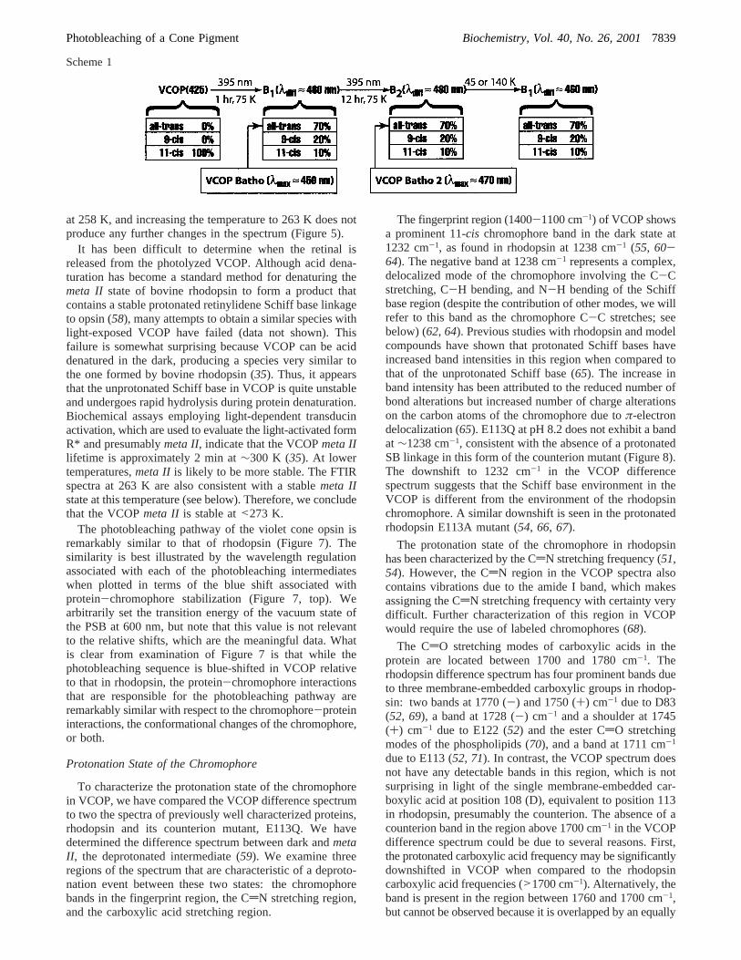
generates theB1 PSS within 1 h (Figure 4, spectrum 2), butextended illumination (∼12 h) at 75 K accesses a state evenfurther red-shifted, theB2 state (Figure 4, spectrum 3). TheB2 PSS is also accessible when the violet cone opsinPSS500(50% 11-cis-, 11% all-trans-, and 39% 9-cis-retinal) isilluminated with 395 nm light for approximately 1 h at 75K. Despite the identical, or nearly identical, chromophorecompositions of the two photostationary states, theB2 state(whoseλmax can vary between 460 and 480 nm dependingupon how it is generated) is significantly red-shifted fromthe B1 PSS (λmax ≈ 460 nm).B2 generated fromPSS500reverts to theB1 PSS upon increasing the temperature to140 K (Figure 4) or decreasing the temperature to 45 K(Figure 3). Scheme 1 depicts the formation of the photo-product at 75 K using short-term illumination (B1), followedby extended illumination (B2), and finally the change intemperature to 140 and 45 K.
We have indicated the absorption maxima (λmax), or inthe case of photostationary states, the difference maxima(λdiff), in parentheses. The absorption spectra of thebatho(pure all-trans species) photoproducts were generated viadeconvolution based on the HPLC data. A key point ofScheme 1 is that the isomer compositions of theB1 andB2
photostationary states are identical within experimental error.(Measurements of the isomer composition differed by(5%,and the values shown in Scheme 1 are rounded to(10%.)This observation places important constraints on the natureof these photostationary states and the origin of the spectraldifferences.
One possible explanation forB2 worth considering is thatat 75 K, the residues in the binding site are able to move toa higher-energy conformation, allowing the all-transchromo-phore to relax into a more planar conformation. The moreplanar chromophore has less single-bond torsional distor-tion which lowers the energy of the excited state more thanthat of the ground state, and therefore leads to a longer-wavelength absorption maximum. However, the observationthatB2 reverts toB1 when the temperature is both increasedto 140 K or decreased to 45 K indicates that the system isnot in equilibrium. We hypothesize that the total free energyof theB2 PSS is higher than that of theB1 PSS, because ofthe distortion of the binding site rather than distortion of thelong, and relatively flexible, retinyl chromophore. ThemetastableB2 PSS should be quite sensitive to conditionsthat alter the strain energy of the system, which wouldinclude cooling. This explanation suggests that site-directedmutations of the binding site would influence the propertiesof bothB1 andB2. Indeed, we have found one mutant, S85D,which displays only a single bathochromically shiftedphotostationary state with a wavelength maximum roughlyintermediate between those ofB1 and B2 (manuscript inpreparation).
The lumi intermediate (λmax ) 435 nm) is formed at 200K via the thermal decay of thebatho intermediate and isstable up to 228 K (Figures 2 and 5). The transition to themeta I intermediate starts occurring at 253 K (Figure 5,spectrum 3). Themeta Ispectrum is very broad and may becontaminated with thelumi intermediate that has not decayedcompletely at 253 K. Increasing the temperature to 258 Kdestabilizes themeta Iintermediate, and it decays to themetaII intermediate in approximately 4 h (Figure 5C). Theformation of themeta II intermediate is complete after 4 h
FIGURE 8: FTIR difference spectra of themeta II intermediatespectrum minus the rhodopsin (s. rho and ROS), E113Q, and VCOPdark state spectra in H2O and D2O. The graph at the top shows theroom-temperature absorption spectra of the three pigments that wereexamined. E113Q at pH 8.2 was selected because it contains anunprotonated Schiff base chromophore. All the FTIR spectra werecollected at 263 K, and the pH of each sample is indicated in thelabel. The dotted lines and frequencies noted represent the positionof bands in the solubilized rhodopsin spectrum in H2O at pH 6.8.Literature assignments of the rhodopsin bands are as follows: 1238cm-1 (C12-C13, C10-C11, C14-C15, and C15-H coupled mode, andN-H bending modes) (62-64), 1530 and 1557 cm-1 (CdCchromophore stretches) (56), 1655 cm-1 (amide I, CdN-Hstretches) (62), 1712 cm-1 (E113 carbonyl stretch) (71), 1728 cm-1
(E122 carbonyl stretch and the ester CdO stretch of phospholipids)(56, 70), 1745 and 1770 cm-1 (D83 carbonyl stretch) (52, 69).
7838 Biochemistry, Vol. 40, No. 26, 2001 Kusnetzow et al.
at 258 K, and increasing the temperature to 263 K does notproduce any further changes in the spectrum (Figure 5).
It has been difficult to determine when the retinal isreleased from the photolyzed VCOP. Although acid dena-turation has become a standard method for denaturing themeta II state of bovine rhodopsin to form a product thatcontains a stable protonated retinylidene Schiff base linkageto opsin (58), many attempts to obtain a similar species withlight-exposed VCOP have failed (data not shown). Thisfailure is somewhat surprising because VCOP can be aciddenatured in the dark, producing a species very similar tothe one formed by bovine rhodopsin (35). Thus, it appearsthat the unprotonated Schiff base in VCOP is quite unstableand undergoes rapid hydrolysis during protein denaturation.Biochemical assays employing light-dependent transducinactivation, which are used to evaluate the light-activated formR* and presumablymeta II, indicate that the VCOPmeta IIlifetime is approximately 2 min at∼300 K (35). At lowertemperatures,meta II is likely to be more stable. The FTIRspectra at 263 K are also consistent with a stablemeta IIstate at this temperature (see below). Therefore, we concludethat the VCOPmeta II is stable at<273 K.
The photobleaching pathway of the violet cone opsin isremarkably similar to that of rhodopsin (Figure 7). Thesimilarity is best illustrated by the wavelength regulationassociated with each of the photobleaching intermediateswhen plotted in terms of the blue shift associated withprotein-chromophore stabilization (Figure 7, top). Wearbitrarily set the transition energy of the vacuum state ofthe PSB at 600 nm, but note that this value is not relevantto the relative shifts, which are the meaningful data. Whatis clear from examination of Figure 7 is that while thephotobleaching sequence is blue-shifted in VCOP relativeto that in rhodopsin, the protein-chromophore interactionsthat are responsible for the photobleaching pathway areremarkably similar with respect to the chromophore-proteininteractions, the conformational changes of the chromophore,or both.
Protonation State of the Chromophore
To characterize the protonation state of the chromophorein VCOP, we have compared the VCOP difference spectrumto two the spectra of previously well characterized proteins,rhodopsin and its counterion mutant, E113Q. We havedetermined the difference spectrum between dark andmetaII , the deprotonated intermediate (59). We examine threeregions of the spectrum that are characteristic of a deproto-nation event between these two states: the chromophorebands in the fingerprint region, the CdN stretching region,and the carboxylic acid stretching region.
The fingerprint region (1400-1100 cm-1) of VCOP showsa prominent 11-cis chromophore band in the dark state at1232 cm-1, as found in rhodopsin at 1238 cm-1 (55, 60-64). The negative band at 1238 cm-1 represents a complex,delocalized mode of the chromophore involving the C-Cstretching, C-H bending, and N-H bending of the Schiffbase region (despite the contribution of other modes, we willrefer to this band as the chromophore C-C stretches; seebelow) (62, 64). Previous studies with rhodopsin and modelcompounds have shown that protonated Schiff bases haveincreased band intensities in this region when compared tothat of the unprotonated Schiff base (65). The increase inband intensity has been attributed to the reduced number ofbond alterations but increased number of charge alterationson the carbon atoms of the chromophore due toπ-electrondelocalization (65). E113Q at pH 8.2 does not exhibit a bandat ∼1238 cm-1, consistent with the absence of a protonatedSB linkage in this form of the counterion mutant (Figure 8).The downshift to 1232 cm-1 in the VCOP differencespectrum suggests that the Schiff base environment in theVCOP is different from the environment of the rhodopsinchromophore. A similar downshift is seen in the protonatedrhodopsin E113A mutant (54, 66, 67).
The protonation state of the chromophore in rhodopsinhas been characterized by the CdN stretching frequency (51,54). However, the CdN region in the VCOP spectra alsocontains vibrations due to the amide I band, which makesassigning the CdN stretching frequency with certainty verydifficult. Further characterization of this region in VCOPwould require the use of labeled chromophores (68).
The CdO stretching modes of carboxylic acids in theprotein are located between 1700 and 1780 cm-1. Therhodopsin difference spectrum has four prominent bands dueto three membrane-embedded carboxylic groups in rhodop-sin: two bands at 1770 (-) and 1750 (+) cm-1 due to D83(52, 69), a band at 1728 (-) cm-1 and a shoulder at 1745(+) cm-1 due to E122 (52) and the ester CdO stretchingmodes of the phospholipids (70), and a band at 1711 cm-1
due to E113 (52, 71). In contrast, the VCOP spectrum doesnot have any detectable bands in this region, which is notsurprising in light of the single membrane-embedded car-boxylic acid at position 108 (D), equivalent to position 113in rhodopsin, presumably the counterion. The absence of acounterion band in the region above 1700 cm-1 in the VCOPdifference spectrum could be due to several reasons. First,the protonated carboxylic acid frequency may be significantlydownshifted in VCOP when compared to the rhodopsincarboxylic acid frequencies (>1700 cm-1). Alternatively, theband is present in the region between 1760 and 1700 cm-1,but cannot be observed because it is overlapped by an equally
Scheme 1
Photobleaching of a Cone Pigment Biochemistry, Vol. 40, No. 26, 20017839

intense negative band, and thus canceled out. We alsoconsidered the possibility that themeta II intermediate wasnot trapped in these experiments, but we discounted thispossibility on the basis of the observation of themeta IIintermediate at 263 K in the temperature trapping experi-ments (Figure 5, and previous discussion).
In summary, the C-C stretching band of the chromophoreprovides the strongest evidence for a protonated Schiff basein VCOP. The vibrational modes that contribute to thecomposite band between 1240 and 1230 cm-1 have not beenthoroughly studied in the literature, although there isconsensus that these bands are due to C-C modes coupledto hydrogen in-plane bends (see the discussion above). Wehave carried out a series of ab initio molecular orbitalcalculations on model 11-cis chromophores. There are anumber of calculated vibrations in the region from 1200 to1500 cm-1 (Figure 9). The strong vibration in the 1230-1240 cm-1 region of the visual pigments seems to bedescribed well using a simple theoretical model based onan 11-cisprotonated Schiff base (PSB) mediated by a singleperchlorate counterion (Figure 10). The significant intensityof this mode in the PSB compounds can be traced to thelarge variation in atomic charges along the chromophore,which generates a large change in dipole moment during thevibrational motion. In the unprotonated SB, the chargedistribution is much less varied and the intensity of thecomparable mode is at least an order of magnitude smaller(Figure 9). What remains to be explained is why only onevibration appears in this region while three or four arepredicted with comparable intensity. We suggest that con-formational distortion of the chromophore within the binding
site broadens some of the vibrational bands in this region.The integral of the vibrational distribution suggests that thereare other bands in this region, but few survive as sharp linesdue to inhomogeneous broadening of the environment.
To do a quantitative comparison of the theoretical andexperimental results, we normalized the difference spectraby reference to the absolute intensity of the parent amide Iband at∼1650 cm-1. We integrated under the light minusdark spectra of rhodopsin, E113Q (pH 8.2), and violet coneopsin from 1500 to 1100 cm-1 (see Table 1). All the valuesin Table 1 are negative because the dark state, in each case,has greater absorptivity in this region than the light (metaII ) state. The integral values of rhodopsin and VCOP aresimilar in magnitude and much larger than the value of theunprotonated E113Q mutant. We suggest that this integralprovides the single best determinant of protonation state. Weconclude that the chromophore in VCOP is protonated inthe dark and becomes deprotonated in themeta II intermedi-ate.
WaVelength Regulation in Rhodopsin and the Violet ConeOpsin
We explore in this section the molecular origins ofwavelength regulation in both rhodopsin and VCOP. Webriefly examine the nature of wavelength regulation inrhodopsin, prior to a re-examination of the role of thecounterion position in mediating spectral tuning, consideredin our previous work (14) on the violet opsin.
Rhodopsin.The rhodopsin crystal structure indicates threeglutamic acid residues in the proximity of the chromophore(E122, E181, and E113) as well as a histidine residue (H211)
FIGURE 9: Theoretical simulations of the vibrational modes of the Schiff base and protonated Schiff base of 11-cis-retinal: Schiff base (A),protonated Schiff base without water (B), and protonated Schiff base with water (C). All calculations were carried out using density functionaltheory (B3LYP) and a 6-31G basis set. Selected complex C-C stretch and C-H bending modes in the 1200-1400 cm-1 region are shownat right, with the mode descriptions and peaks designated using the letters p (protonated) and u (unprotonated). The arrows show thedirection of atom motion during a single phase of the vibration, and the length is proportional, but exaggerates, the amount of motion.Additional mode descriptions for the unprotonated chromophore are as follows: u0 (pair of modes involving C13dC14-C15 bending mixedwith C-H bending modes nearby), u5 and u6 (C-H bends primarily involving the polyene chain methyl groups), u7 (coupled CdC modeinvolving primarily C7dC8, C9dC10, C11dC12, and C13dC14), u8 (coupled CdC mode involving C5dC6, C7dC8, C11dC12, C13dC14, andC15dN), and u9 (lysine mode not properly described in this simulation). Additional mode descriptions for the protonated chromophore areas follows: p5 and p6 (C-H bends involving polyene Hs and polyene methyl groups), p7 [very intense (2617 KM/mol) coupled CdCmode involving C5dC6, C7dC8, C11dC12, and C13dC14], and p8 (imine N-H bending mode coupled with the C11dC12 stretch and theC14-C15dN in-plane bend).
7840 Biochemistry, Vol. 40, No. 26, 2001 Kusnetzow et al.

near theâ-ionylidene ring (72). The crystal structure indicatesthat E122 and H211 are sufficiently close to form a saltbridge. Simple pKa calculations on the binding site clearlyindicate that E113 is deprotonated, but the results areinconclusive on the status of E122 and H211, which arepredicted to either be both charged [E122(-)...H211(+)] orboth neutral (E122, H211). Previous UV-vis and FTIRstudies have concluded that E122 is protonated (52, 73, 74).However, the evidence is not conclusive because an interac-tion of E122(-) with H211(+) might produce vibrationaland electronic signatures similar to protonation, and the smallshifts observed upon replacement of E122 with unchargedresidues could be due to a simultaneous deprotonation ofH211. The pKa calculations were also inconclusive regardingthe protonation state of E181. If we assume E181 isprotonated, then the binding site is neutral, in agreement withthe two-photon studies (75). To explore all possibilities,however, we assumed that all four residues were charged inthe simulations below.
The principal residues within the binding site are shownin Figure 11A together with the participation of individualresidues in wavelength regulation based on the use ofMNDO-PSDCI molecular orbital theory (40, 47). Theisolated 11-cis, 6-s-cis chromophore is calculated to havean absorption maximum at 588 nm (2.107 eV), while inrhodopsin, the actual absorption maximum is blue-shiftedby ∼0.38 eV. Calculations were first performed to determinethe effect of individual residues on the absorption propertiesof a protonated Schiff base chromophore, maintaining thegeometry of the rhodopsin crystal structure. The energy shiftsfor each individual residue are shown in electronvolts (eV)in Figure 11A. If the individual contributions (ignoring that
of water) are added, a net blue shift of 0.06 is found, farsmaller than the observed value. Even if one water moleculehydrogen bonded to the Schiff base proton is included, theadditional blue shift of 0.16 only yields a net shift of 0.22,which is still too small. However, when an MNDO-PSDCIcalculation is carried out, including the entire first shell ofthe binding site plus water, a blue shift of 0.43 and anabsorption maximum of 488 nm are found. This value is inmuch better agreement with experiment, and suggests thatwavelength mediation in rhodopsin is due in part to second-order interactions not adequately described by simple sumrules based on individual residue shifts. The type ofsecondary interactions relevant to this process includedipole-dipole and dipole-induced dipole interactions thatproduce subtle changes in the orientation of polar groups,alter, or enhance the dipole moment vector of a residue, andin combination change the effective reaction field of thebinding site.
We note also that E181 is very near the electrostatic nullpoint in the binding site (see Figure 9 of ref14). Thus, thisresidue contributes very little to the wavelength regulationprocess regardless of ionization state. This observation is ingeneral agreement with site-directed mutagenesis studies ofrhodopsin by Nathans (76, 77) and Terakita et al. (78). Theseinvestigators found either no effect (76, 77) or a small redshift [10 nm (78)] (our calculations predict a 0.02 eV or 4nm red shift). However, the corresponding glutamic acidresidue in retinochrome, a photoisomerase in squid photo-receptor cells, is found to provide the primary counterion(retinochrome lacks the glutamic acid residue at position 113)(78). We note that our calculations are extremely sensitiveto the location of this residue, and a small 1 Å shift toward
FIGURE 10: Vibrational mode (1232 cm-1) observed in VCOP described well using a simple theoretical model based on an 11-cisprotonatedSchiff base mediated by a single perchlorate counterion. The above mode description was calculated by using DFT molecular orbital theory(B3LYP, 6-31G) (39), and the vibrational frequency was scaled by using the appropriate scaling factor of Scott and Radom (79). Theatomic motion during the vibration generates a large change in dipole moment, visualized above using electrostatic field contours (redbeing positive and blue negative). This vibration corresponds to mode p2 shown in Figure 9. Note that the mode arrows cross many contourlines, in contrast to a comparable map drawn for the corresponding unprotonated Schiff base mode (u2, Figure 9, electrostatic contours notshown).
Photobleaching of a Cone Pigment Biochemistry, Vol. 40, No. 26, 20017841

the imine linkage transforms a small red shift to a large (30nm) blue shift.
Violet Cone Opsin. We examined the violet cone opsinbinding site on the basis of a similarity replacement of therhodopsin side chains with the corresponding residues ofviolet cone opsin. The rhodopsin backbone was held fixedduring the process, and an energy minimization was carriedout after the replacement residues were inserted. The resultsare shown in Figure 11B. The N-terminal end of VCOP isshorter by five residues than rhodopsin, and one must add 5to the residue numbers in Figure 11B to find the correspond-ing rhodopsin residue in Figure 11A (for example, D108 inVCOP correlates with E113 in rhodopsin).
We have not carried out calculations for the individualshifts for each binding site residue in VCOP, because ourcalculations on rhodopsin indicate that individual residueshifts are not additive (see above). VCOP has an absorptionmaximum at 425 nm (2.915 eV), which represents a blueshift of 0.43 eV relative to that of rhodopsin. A comparisonof panels A and B of Figure 11 suggests that the single largestchange in going from the rhodopsin to the VCOP bindingsite is the loss of the E122-H211 pair near theâ-ionylidenering. As shown in Figure 11A, this charged pair provides a
first-order red shift of-0.31 eV, and by itself would accountfor ∼75% of the blue shift required. The E122-H211 pair isalso absent from the human blue cone. As noted above, theresults of some site-directed mutagenesis studies on E122have been interpreted to indicate that this residue is proton-ated in rhodopsin (52, 73, 74). However, strong interactionwith H211 could produce vibrational and electronic signa-tures that are similar to direct protonation, and further workon the characterization of the protonation state of E122 wouldbe welcome.
An MNDO-PSDCI calculation on the first shell of theVCOP binding site model shown in Figure 11B predicts an
Table 1: Comparison of Observed and Calculated Integrals for theMeta II Minus Dark FTIR Difference Spectraa
Experimentalprotein pH Schiff base integral value
solubilized rhodopsin 6.8 protonated -0.149( 0.037E113Q rhodopsin 8.2 unprotonated -0.034( 0.042VCOP 6.8 see the Discussion-0.146( 0.051
Theoretical11-cis
chromophore methodbp2 band
(intensity)cintegralvalue
unprotonated Schiffbase (SB)
B3LYP, 6-31G 1221.1 (55.5) -0.014
SB B3LYP, 6-31G(d) 1203.8 (63.2) 0.006protonated Schiff
base (PSB)B3LYP, 6-31G 1247.2 (1579.5)-0.816
PSBd B3LYP, 6-31G(d) 1224.3 (1966.3)-0.827PSB with water B3LYP, 6-31G 1244.1 (1165.2)-0.745PSB with perchloratee B3LYP, 6-31G 1233.6 (413.9) -0.262PSB with perchlorate
in waterfB3LYP, 6-31G 1237.8 (1117.6)-0.744
PSB with perchlorateand two waters
HF, 6-31G 1239.7 (450.3) -0.206
PSB with perchlorateand two waters
HF, 6-31G(d) 1218.9 (443.0) -0.180
a All the experimental integral values are for themeta IIintermediateminus the dark state and were measured from 1100 to 1500 cm-1 inunits of cm-1 with the values normalized by adjusting the amide I bandabsorptivity to 1. The theoretical values are calculated relative to theall-trans unprotonated Schiff base (e.g.,meta II-like) chromophore(B3LYP, 6-31G) with the integrals normalized to the central amide Iband of polyglycine multiplied by the total number of residues in theprotein (see the Discussion).b Calculations were carried out usingGaussian 98(39). c Location in wavenumber of the intense mode,labeled p2 in the isolated protonated Schiff base, which correspondsto the observed mode at∼1238 cm-1 in rhodopsin and by analogy at1232 cm-1 in VCOP. The intensity is in KM/mol.d The results of theB3LYP 6-31G(d) (e.g., 6-31G*) calculation are similar to those shownin Figure 9B, but the peak labeled p2 is now the strongest peak in thespectrum. The single water molecule is hydrogen bonded to the Schiffbase proton (see Figure 9).e Molecule and vibration shown in Figure10. f The solvent environment was simulated by using a sphericalOnsager reaction field and carrying out the minimization using adielectric constant of 78 and a cavity radius of 10 Å.
FIGURE 11: Rhodopsin and violet cone opsin chromophore bindingsites. A comparison of the rhodopsin binding site (A) and asimilarity-based preliminary model of the VCOP binding site (B).The coordinates for the rhodopsin binding site are from the crystalstructure, but the water molecule near the Schiff base was addedand minimized by us. The VCOP model was generated using asimilarity transformation (see the text). First-orderλmax bandtransition energy shifts associated with nearby residues in rhodopsinwere calculated using MNDO-PSDCI molecular orbital theory andare indicated in electronvolts under the residue name in panel A.Negative numbers represent red shifts, and positive numbersrepresent blue shifts. The shift for each residue is based on thatresidue and the chromophore alone, and all other residues areignored unless noted. The shifts calculated for the Glu and Hisresidues assume a nominally charged [Glu(-), His(+)] residue.Most experimental studies of rhodopsin conclude that LYR296 ispositively charged, E113 is negatively charged, and all otherresidues shown in panel A are uncharged (see the Discussion).
7842 Biochemistry, Vol. 40, No. 26, 2001 Kusnetzow et al.

absorption maximum at 433 nm. The calculated blue shiftof 0.32 eV is in respectable agreement with the observedrhodopsin to VCOP blue shift of 0.43 eV. We conclude thatour simple binding site analysis is correct to first order, andprovides a realistic perspective on the primary componentsof wavelength regulation in VCOP.
It is likely that any model of wavelength regulation whichrelies solely on the location and charges of a few individualresidues will fail to have global applicability. We suggestthat a significant portion of wavelength regulation is ac-complished via evolution of the entire binding site ensembleinvolving 10-20 residues and that secondary interactionsamong these residues provide the dominant electrostatic anddispersive wavelength shifts.
SUMMARY AND CONCLUSIONS
We show that theXenopusviolet cone opsin has aprotonated Schiff base chromophore in the dark, andundergoes a series of conformational changes upon excitationthat can be identified as thebatho, lumi, meta I, andmeta IIphotoproducts. The absorption spectra of these intermediateshave been recorded, and the photobleaching sequence is quitesimilar to that observed in rhodopsin, but blue shifted. Whenthese two systems are compared on the basis of wavelengthmediation along the photobleaching pathway, the similaritiesare striking (Figure 7). Like that of rhodopsin and the othervisual pigments, light activation of theXenopusviolet coneopsin initiates a sequence of events that leads to the formationof the biologically active state,meta II, and the ultimateexpulsion of the chromophore from the binding pocket.
A similarity transformation of the rhodopsin binding sitegenerates a model for the VCOP binding site that predictsroughly 75% of the observed blue shift of the violet conerelative to rhodopsin. The MNDO-PSDCI calculationsindicate that secondary interactions between the binding siteresidues are just as important as first-order chromophoreprotein interactions at the mediating wavelength. The im-portance of secondary interactions in wavelength regulationcomplicates and may even preclude assignment of a simplemechanism for wavelength regulation that applies to allpigments. An example of such a mechanism would be onebased on the strategic placement of a single negativelycharged residue as we proposed earlier (14). Evolution ofthe entire pocket rather than adjustment of a single chargedresidues is not an unexpected consequence. Evolution of avisual pigment must optimize simultaneously (1) the wave-length maximum, (2) the quantum efficiency for the primaryphotochemical event, and (3) energy storage to drive thephotobleaching thermal reactions and conformational pro-cesses responsible for activation. Strategic placement of asingle residue cannot accomplish all three goals simulta-neously.
The observation of a significantly red-shifted photoproductin VCOP, only observed upon extended illumination, remainsa unique and not fully understood aspect of the VCOPprimary photochemical event. We suggest that this red-shiftedstate is due to relaxation of the chromophore into a moreplanar conformation via a pathway normally blocked by oneor more residues in the binding site. Repeated photochemicaltransformation of the chromophore rearranges the localprotein environment and causes the blocking residues to shift
to higher-energy conformations that accommodate a moreplanar all-transchromophore. The presence of this interme-diate, not relevant to the primary photobleaching sequence,suggests that the binding site of VCOP is more compact thanrhodopsin and a majority of the other visual pigments.
ACKNOWLEDGMENT
We thank Prof. Jerry Goodisman for interesting and helpfuldiscussions, and the National Science Foundation for gener-ous allocations of supercomputer time at the San Diego andIllinois supercomputer facilities.
REFERENCES
1. Schnapf, J., and Baylor, D. A. (1986)Sci. Am. 256, 32-39.2. Yau, K. W. (1994)InVest. Ophthalmol. Visual Sci. 35, 9-32.3. Baylor, D. (1996)Proc. Natl. Acad. Sci. U.S.A. 93, 560-565.4. Wald, G. (1968)Nature 219, 800-808.5. Oseroff, A. R., and Callender, R. H. (1974)Biochemistry 13,
4243-4348.6. Hargrave, P. A., Mcdowell, J. H., Curtis, D. R., Wang, J. K.,
Juszczak, E., Fong, S. L., Rao, J. K. M., and Argos, R. (1983)Biophys. Struct. Mech. 9, 235-244.
7. Hargrave, P. A., and McDowell, J. H. (1992)FASEB J. 6,2323-2331.
8. Scott, K., Becker, A., Sun, Y., Hardy, R., and Zuker, C. (1995)Neuron 15, 919-927.
9. Okano, T., Kojima, D., Fukada, Y., Shichida, Y., andYoshizawa, T. (1992)Proc. Natl. Acad. Sci. U.S.A. 89, 5932-5936.
10. Yoshizawa, T. (1994)Biophys. Chem. 50, 17-24.11. Yokoyama, S. (1995)Mol. Biol. EVol. 12, 53-61.12. Yokoyama, S., Radlwimmer, F., and Blow, N. (2000)Proc.
Natl. Acad. Sci. U.S.A. 97, 7366-7371.13. Ebrey, T., and Koutalos, Y. (2001)Prog. Retinal Eye Res.
20, 49-94.14. Vought, B. W., Dukkipatti, A., Max, M., Knox, B. E., and
Birge, R. R. (1999)Biochemistry 38, 11287-11297.15. Jacobs, G. H., Deegan, G. F., and Neitz, J. (1998)Vision
Neurol. 15, 581-584.16. Merbs, S., and Nathans, J. (1992)Nature 356, 443-435.17. Okano, T., Fukada, Y., Artamonov, I. D., and Yoshizawa, T.
(1989)Biochemistry 28, 8848-8856.18. Kawamura, S., and Yokoyama, S. (1997)Vision Res. 38, 37-
44.19. Yokoyama, S., Radlwimmer, F. B., and Kawamura, S. (1998)
FEBS Lett. 423, 155-158.20. Hisatomi, O., Satoh, T., Barthel, L. K., Stenkamp, D. L.,
Raymond, P. A., and Tokunga, F. (1996)Vision Res. 36, 933-939.
21. Kliger, D. S., and Lewis, J. W. (1995)Isr. Jour. Chem. 35,289-307.
22. Yoshizawa, T., and Shichida, Y. (1982) inMethods inEnzymology, pp 333-354, Academic Press, New York.
23. Yoshizawa, T., and Wald, G. (1967)Nature 214, 566-571.24. Shichida, Y., Imai, H., Imamoto, Y., Fukada, Y., and
Yoshizawa, T. (1994)Biochemistry 33, 9040-9044.25. Tsuda, M., Tokunga, F., Ebrey, T. G., Yue, K. T., Marque, J.,
and Eisenstein, L. (1980)Nature 287, 461-462.26. Imai, H., Imamoto, Y., Yoshizawa, T., and Shichida, Y. (1995)
Biochemistry 34, 10525-10531.27. Kojima, D., Imai, H., Okano, T., Fukada, Y., Crescitelli, F.,
Yoshizawa, T., and Shichida, Y. (1995)Biochemistry 34,1096-1106.
28. Archer, S., and Hirano, J. (1996)Proc. R. Soc. London, Ser.B 263, 761-767.
29. Baldwin, P., and Hubbell, W. (1993)EMBO J. 12, 1693-1703.
30. Lin, S. W., Kochendoerfer, G. G., Caroll, K. S., Wang, D.,Mathies, R. A., and Sakmar, T. P. (1998)J. Biol. Chem. 273,24583-24591.
Photobleaching of a Cone Pigment Biochemistry, Vol. 40, No. 26, 20017843

31. Kochendoerfer, G. G., Wang, Z., Oprian, D. D., and Mathies,R. A. (1997)Biochemistry 36, 6577-6587.
32. Wilkie, S. E., Vissers, P. M. A. M., Das, D., DeGrip, W. J.,Bowmaker, J. K., and Hunt, D. M. (1998)Biochem. J. 330,541-547.
33. Chang, B. S. W., Crandall, K. A., Carulli, J. P., and Hartl, D.L. (1995)Mol. Phylogenet. EVol. 4, 31-43.
34. Yokoyama, S., and Zhang, H. (1997)Gene 202, 89-93.35. Starace, D. M., and Knox, B. E. (1997)J. Biol. Chem. 272,
1095-1100.36. Starace, D. M., and Knox, B. E. (1998)Exp. Eye Res. 67,
209-220.37. Sakmar, T. P., Franke, R. R., and Khorana, H. G. (1989)Proc.
Natl. Acad. Sci. U.S.A. 86, 8309-8313.38. De Grip, W. J. (1982) inMethods in Enzymology(Packer, L.,
Ed.) pp 197-207, Academic Press, New York.39. Frisch, M. J., Trucks, G. W., Schlegel, H. B., Scuseria, G. E.,
Robb, M. A., Cheeseman, J. R., Zakrzewski, V. G., Mont-gomery, J. A., Stratmann, R. E., Burant, J. C., Dapprich, S.,Millam, J. M., Daniels, A. D., Kudin, K. N., Strain, M. C.,Farkas, O., Tomasi, J., Barone, V., Cossi, M., Cammi, R.,Mennucci, B., Pomelli, C., Adamo, C., Clifford, S., Ochterski,J., Petersson, G. A., Ayala, P. Y., Cui, Q., Morokuma, K.,Malick, D. K., Rabuck, A. D., Raghavachari, K., Foresman,J. B., Cioslowski, J., Ortiz, J. V., Stefanov, B. B., Liu, G.,Liashenko, A., Piskorz, P., Komaromi, I., Gomperts, R.,Martin, R. L., Fox, D. J., Keith, T., Al-Laham, M. A., Peng,C. Y., Nanayakkara, A., Gonzalez, C., Challacombe, M., Gill,P. M. W., Johnson, B. G., Chen, W., Wong, M. W., Andres,J. L., Head-Gordon, M., Replogle, E. S., and Pople, J. A.(1998)Gaussian 98, Gaussian Inc., Pittsburgh, PA.
40. Martin, C. H., and Birge, R. R. (1998)J. Phys. Chem. A 102,852-860.
41. Barlow, R. B., Birge, R. R., Kaplan, E., and Tallent, J. R.(1993)Nature 366, 64-66.
42. Stuart, J. A., Vought, B. W., Zhang, C. F., and Birge, R. R.(1995)Biospectroscopy 1, 9-28.
43. Yamazaki, M., Goodisman, J., and Birge, R. R. (1998)J.Chem. Phys. 108, 5876-5887.
44. Birge, R. R., Zgierski, M. Z., Serrano-Andres, L., and Hudson,B. S. (1999)J. Phys. Chem. A 103, 2251-2255.
45. Birge, R. R., Gillespie, N. B., Izaguirre, E. W., Kusnetzow,A., Lawrence, A. F., Singh, D., Song, Q. W., Schmidt, E.,Stuart, J. A., Seetharaman, S., and Wise, K. J. (1999)J. Phys.Chem. B. 103, 10746-10766.
46. Hudson, B. S., and Birge, R. R. (1999)J. Phys. Chem. 103,2274-2281.
47. Kusnetzow, A., Singh, D. L., Martin, C. H., Barani, I., andBirge, R. R. (1999)Biophys. J. 76, 2370-2389.
48. Tachibanaki, S., Imamoto, Y., Imai, H., and Shichida, Y.(1995)Biochemistry 34, 13170-13175.
49. Imamoto, Y., Yoshizawa, T., and Shichida, Y. (1996)Bio-chemistry 35, 14599-14607.
50. Imamoto, Y., Yoshizawa, T., and Shichida, Y. (1996)Bio-chemistry 35, 14599-14607.
51. Jager, F., Fahmy, K., Sakmar, T. P., and Siebert, F. (1994)Biochemistry 33, 10878-10882.
52. Fahmy, K., Jaeger, F., Beck, M., Zvyaga, T. A., Sakmar, T.P., and Siebert, F. (1993)Proc. Natl. Acad. Sci. U.S.A. 90,10206-10210.
53. Zvyaga, T., Fahmy, K., Siebert, F., and Sakmar, T. (1996)Biochemistry 35, 7536-7545.
54. Fahmy, K., Sakmar, T., and Siebert, F. (2000)MethodsEnzymol. 315, 178-196.
55. Callender, R. H., and Honig, B. (1977)Annu. ReV. Biophys.Bioeng. 6, 33-55.
56. Siebert, F. (1995)Isr. J. Chem. 35, 309-323.57. Hug, S. J., Lewis, J. W., Einterz, C. M., Thorgeirsson, T. E.,
and Kliger, D. S. (1990)Biochemistry 29, 1475-1485.58. Kito, Y., Suzuki, T., Azuma, M., and Sekoguti, Y. (1968)
Nature 218, 955-957.59. Longstaff, C., Calhoon, R. D., and Rando, R. R. (1986)Proc.
Natl. Acad. Sci. U.S.A. 83, 4209-4213.60. Mathies, R., Freedman, T. B., and Stryer, L. (1977)J. Mol.
Biol. 109, 367-372.61. Doukas, A. G., Aton, B., Callender, H., and Ebrey, T. G.
(1978)Biochemistry 17, 2430-2435.62. Ganter, U. M., Ga¨rtner, W., and Siebert, F. (1988)Biochemistry
27, 7480-7488.63. Ganter, U., Schmid, E., and Siebert, F. (1988)J. Photochem.
Photobiol., B 2, 417-426.64. Palings, J., Pardoen, J. A., van der Berg, E., Winkel, C.,
Lugtenburg, J., and Mathies, R. S. (1987)Biochemistry 26,2544-2556.
65. Siebert, F., and Ma¨ntele, W. (1980)Biophys. Struct. Mech. 6,147-164.
66. Fahmy, K., Weidlich, O., Engelhard, M., Tittor, J., Oesterhelt,D., and Siebert, F. (1992)Photochem. Photobiol. 56, 1073-1083.
67. Fahmy, K., Siebert, F., and Sakmar, T. P. (1994)Biochemistry33, 13700-13705.
68. Nakanishi, K., and Crouch, R. (1995)Isr. J. Chem. 35, 253-272.
69. Rath, P., DeCaluwe, L. L. J., Bovee-Geurts, P. H. M., DeGrip,W. J., and Rothschild, K. J. (1993)Biochemistry 32, 10277-10282.
70. Beck, M., Siebert, F., and Sakmar, T. (1998)FEBS Lett. 436,304-308.
71. Jager, F., Ja¨ger, S., Kra¨utle, O., Friedman, N., Sheves, M.,Hofmann, K., and Siebert, F. (1994)Biochemistry 33, 7389-7397.
72. Palczewski, K., Kumasaka, T., Hori, T., Behnke, C. A.,Motoshima, H., Fox, B. A., Le Trong, I., Teller, D. C., Okada,T., Stenkamp, R. E., Yamamoto, M., and Miyano, M. (2000)Science 289, 739-745.
73. DeCaluwe, G. L. J., Bovee-Geurts, P. H. M., Rath, P.,Rothschild, K. J., and de Grip, W. J. (1995)Biophys. Chem.56, 79-87.
74. Nagata, T., Terakita, A., Kandori, H., Shichida, Y., and Maeda,A. (1998)Biochemistry 37, 17216-17222.
75. Birge, R. R., Murray, L. P., Pierce, B. M., Akita, H., Balogh-Nair, V., Findsen, L. A., and Nakanishi, K. (1985)Proc. Natl.Acad. Sci. U.S.A. 82, 4117-4121.
76. Nathans, J. (1990)Biochemistry 29, 937-942.77. Nathans, J. (1990)Biochemistry 29, 9746-9752.78. Terakita, A., Yamashita, T., and Shichida, Y. (2000)Proc.
Natl. Acad. Sci. U.S.A. 97, 14263-14267.79. Scott, A. P., and Radom, L. (1996)J. Phys. Chem. 100,
16502-16513.
BI010387Y
7844 Biochemistry, Vol. 40, No. 26, 2001 Kusnetzow et al.