Functions and Mechanisms of Fibroblast Growth Factor (FGF ...
A R T I C L E S
1026 VOLUME 9 | NUMBER 8 | AUGUST 2003 NATURE MEDICINE
1Centre for Vascular Research, University of New South Wales, Sydney NSW 2052, Australia. 2Department of Haematology, The Prince of Wales Hospital, SydneyNSW 2052, Australia. 3Johnson & Johnson Research Laboratories, Sydney, Australia. Correspondence should be addressed to L.M.K. ([email protected]).
Angiogenesis, the formation of new blood vessels from pre-existingvasculature, is a complex, multistep process involving endothelialbasement membrane degradation, cell migration, proliferation,canalization, branching and maturation of neovessels. Angiogenesisis mandatory for tumor progression because it supplies oxygen andnutrients to the growing tumor, a concept first proposed 30 yearsago1. It follows, then, that agents that interfere with blood vessel for-mation could be used to block tumor progression2. Examples ofthese include antagonists of angiogenic growth factors, receptors,integrins and proteolytic enzymes; some of these are currently underevaluation in various clinical trials3.
Egr-1, first discovered as an immediate-early gene induciblyexpressed in growth-quiescent fibroblasts exposed to serum4, is abroadly expressed prototypical member of the Cys2-His2 zinc fin-ger family of transcription factors5. Egr-1 is rapidly activated bymultiple extracellular agonists (such as growth factors andcytokines) and environmental stresses (such as hypoxia, fluid shearstresses and vascular injury)6. Once activated, Egr-1 controls theexpression of a diverse array of proangiogenic genes (encodinggrowth factors, cytokines, receptors, adhesion molecules and pro-teases)5,7 through GC-rich, cis-acting elements in the promoterregions of these genes. Egr-1 is strongly coexpressed with prolifer-ating cell nuclear antigen in von Willebrand factor–positive bloodvessels in the chorioallantoic membrane assay (data not shown).Therefore, Egr-1 seemed prima facie to be an appealing therapeutictarget given its capacity to integrate extracellular stimuli withchanges in gene expression.
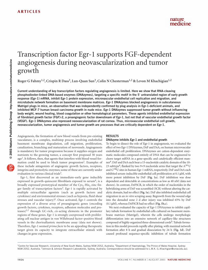
RESULTSDNAzyme inhibits Egr-1 and endothelial growthTo begin to dissect the role of Egr-1 in angiogenesis, we evaluated theeffect of two Egr-1 DNAzymes, DzF and DzA, on human microvascularendothelial cell proliferation. DNAzymes are cation-dependent enzy-matic molecules composed entirely of DNA that can be engineered tocleave target mRNA in a gene-specific and catalytically efficient man-ner8. DzF and DzA each bear a 15-nucleotide catalytic domain of the 10-23 subtype8, flanked by two 9+9 nucleotide arms that target the A301Uand G198U sites in human Egr-1 mRNA, respectively. DzF and DzA eachinhibited serum-inducible endothelial cell proliferation at 0.1 µM, withmore potent inhibition by DzF (Fig. 1a). DzF inhibition was dosedependent and detectable at concentrations as low as 40 nM (data notshown). In contrast, DzFSCR, in which the order of nucleotides in thehybridizing arms of DzF was scrambled (SCR) without altering the cat-alytic domain, had no effect (Fig. 1a). DzF also inhibited endothelial cellmigration in an in vitro scraping assay. Regrowth from the wound edgeinto the denuded zone 2 d after injury was inhibited 65% by DzF (0.1 µM), whereas DzFSCR had no effect (Fig. 1b,c).
We next evaluated the capacity of Egr-1 DNAzymes to inhibit capil-lary tubule formation by endothelial cells cultured on basement mem-brane matrices (Matrigel), wherein the cells undergo morphologicdifferentiation into an extensive network of capillary-like structurescomposed of highly organized three-dimensional cords9. Tubule forma-tion in this model proceeds rapidly after plating, with maximal networkformation after 8 h and gradual dissociation by 24 h (Fig. 1d). DzFcaused profound sequence-specific inhibition of tubule formation
Transcription factor Egr-1 supports FGF-dependentangiogenesis during neovascularization and tumorgrowthRoger G Fahmy1,2, Crispin R Dass3, Lun-Quan Sun3, Colin N Chesterman1,2 & Levon M Khachigian1,2
Current understanding of key transcription factors regulating angiogenesis is limited. Here we show that RNA-cleavingphosphodiester-linked DNA-based enzymes (DNAzymes), targeting a specific motif in the 5′ untranslated region of early growthresponse (Egr-1) mRNA, inhibit Egr-1 protein expression, microvascular endothelial cell replication and migration, andmicrotubule network formation on basement membrane matrices. Egr-1 DNAzymes blocked angiogenesis in subcutaneousMatrigel plugs in mice, an observation that was independently confirmed by plug analysis in Egr-1-deficient animals, andinhibited MCF-7 human breast carcinoma growth in nude mice. Egr-1 DNAzymes suppressed tumor growth without influencingbody weight, wound healing, blood coagulation or other hematological parameters. These agents inhibited endothelial expressionof fibroblast growth factor (FGF)-2, a proangiogenic factor downstream of Egr-1, but not that of vascular endothelial growth factor(VEGF). Egr-1 DNAzymes also repressed neovascularization of rat cornea. Thus, microvascular endothelial cell growth,neovascularization, tumor angiogenesis and tumor growth are processes that are critically dependent on Egr-1.
©20
03 N
atu
re P
ub
lish
ing
Gro
up
h
ttp
://w
ww
.nat
ure
.co
m/n
atu
rem
edic
ine
A R T I C L E S
NATURE MEDICINE VOLUME 9 | NUMBER 8 | AUGUST 2003 1027
(Fig. 1d,e). It also induced time-dependent regression of pre-existingtubules (Fig. 1f). Comparison of DzF with DNAzymes targeting inte-grins β1 or β3 (ref. 10) revealed superior inhibition by the former (Fig. 1e,f). DzF, but not DzFSCR, virtually abrogated endothelial Egr-1expression, whereas levels of transcription factor Sp1 did not change(Fig. 1g). Collectively, these findings indicate sequence-specific inhibi-tion of microvascular endothelial cell proliferation, migration, three-dimensional capillary tube formation and Egr-1 protein expression.
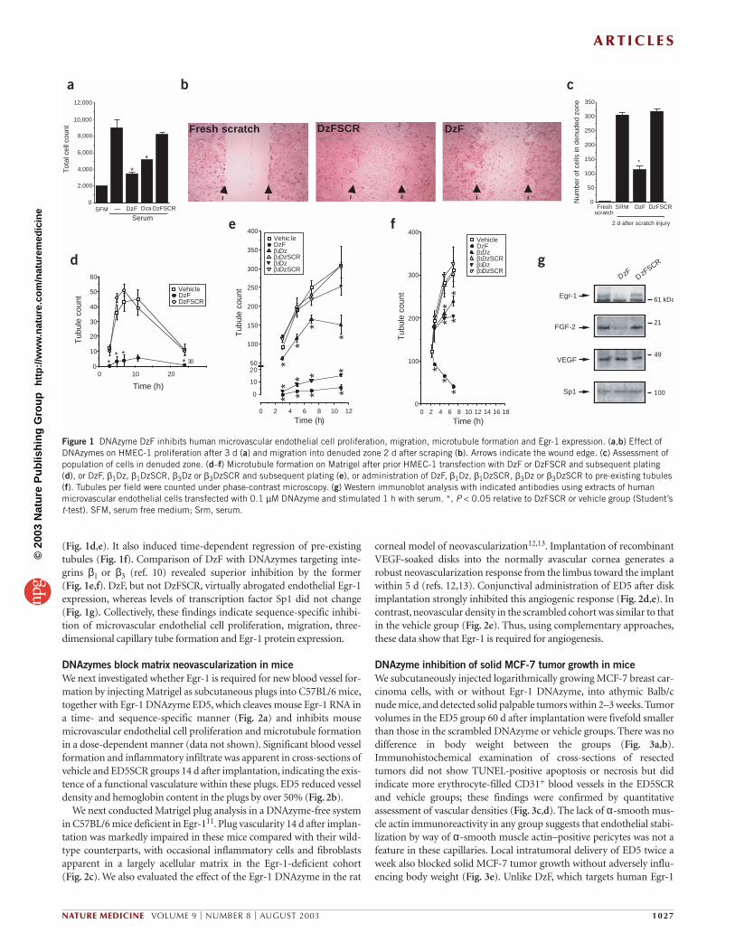
DNAzymes block matrix neovascularization in miceWe next investigated whether Egr-1 is required for new blood vessel for-mation by injecting Matrigel as subcutaneous plugs into C57BL/6 mice,together with Egr-1 DNAzyme ED5, which cleaves mouse Egr-1 RNA ina time- and sequence-specific manner (Fig. 2a) and inhibits mousemicrovascular endothelial cell proliferation and microtubule formationin a dose-dependent manner (data not shown). Significant blood vesselformation and inflammatory infiltrate was apparent in cross-sections ofvehicle and ED5SCR groups 14 d after implantation, indicating the exis-tence of a functional vasculature within these plugs. ED5 reduced vesseldensity and hemoglobin content in the plugs by over 50% (Fig. 2b).
We next conducted Matrigel plug analysis in a DNAzyme-free systemin C57BL/6 mice deficient in Egr-111. Plug vascularity 14 d after implan-tation was markedly impaired in these mice compared with their wild-type counterparts, with occasional inflammatory cells and fibroblastsapparent in a largely acellular matrix in the Egr-1-deficient cohort (Fig. 2c). We also evaluated the effect of the Egr-1 DNAzyme in the rat
corneal model of neovascularization12,13. Implantation of recombinantVEGF-soaked disks into the normally avascular cornea generates arobust neovascularization response from the limbus toward the implantwithin 5 d (refs. 12,13). Conjunctival administration of ED5 after diskimplantation strongly inhibited this angiogenic response (Fig. 2d,e). Incontrast, neovascular density in the scrambled cohort was similar to thatin the vehicle group (Fig. 2e). Thus, using complementary approaches,these data show that Egr-1 is required for angiogenesis.
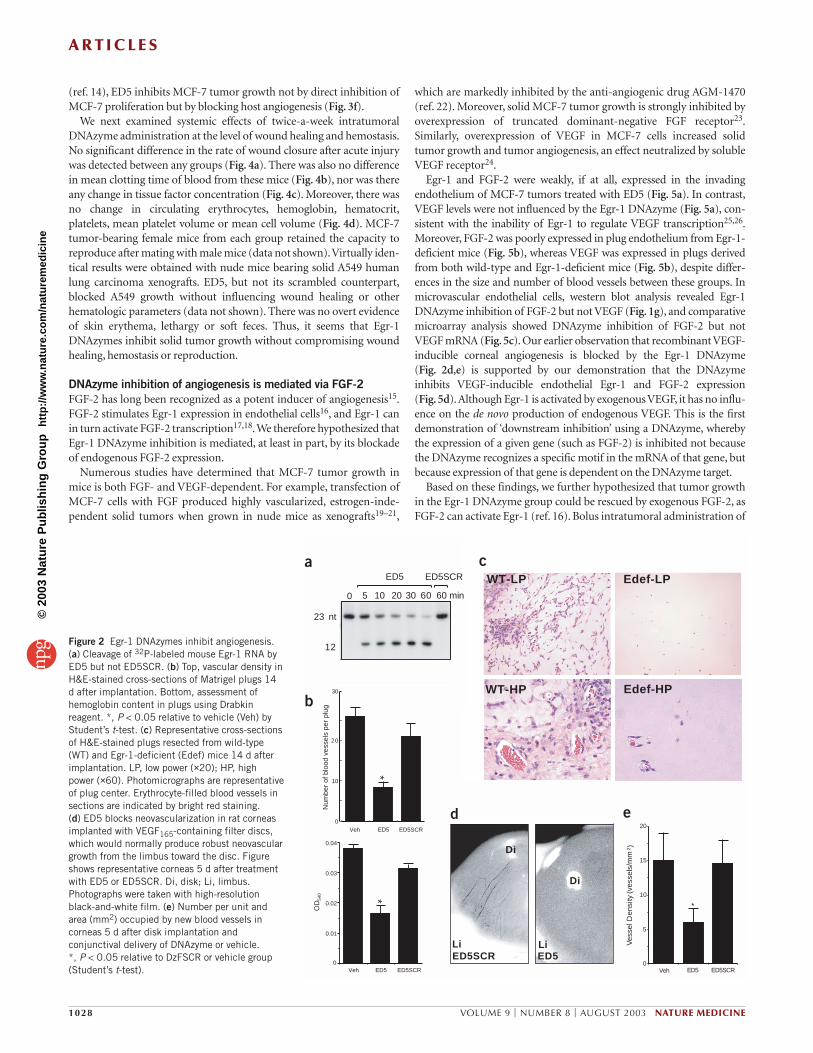
DNAzyme inhibition of solid MCF-7 tumor growth in miceWe subcutaneously injected logarithmically growing MCF-7 breast car-cinoma cells, with or without Egr-1 DNAzyme, into athymic Balb/cnude mice, and detected solid palpable tumors within 2–3 weeks. Tumorvolumes in the ED5 group 60 d after implantation were fivefold smallerthan those in the scrambled DNAzyme or vehicle groups. There was nodifference in body weight between the groups (Fig. 3a,b).Immunohistochemical examination of cross-sections of resectedtumors did not show TUNEL-positive apoptosis or necrosis but didindicate more erythrocyte-filled CD31+ blood vessels in the ED5SCRand vehicle groups; these findings were confirmed by quantitativeassessment of vascular densities (Fig. 3c,d). The lack of α-smooth mus-cle actin immunoreactivity in any group suggests that endothelial stabi-lization by way of α-smooth muscle actin–positive pericytes was not afeature in these capillaries. Local intratumoral delivery of ED5 twice aweek also blocked solid MCF-7 tumor growth without adversely influ-encing body weight (Fig. 3e). Unlike DzF, which targets human Egr-1
Fresh scratch DzFSCR DzF
0
2,000
4,000
6,000
8,000
10,000
12,000
Tot
al c
ell c
ount
DzF
Serum
*
*
0
50
100
150
200
250
300
350
Num
ber
of c
ells
in d
enud
ed z
one
*
DzFFreshscratch
2 d after scratch injury
30
201000
10
20
30
40
50
60
Vehicle
DzFSCRDzF
Time (h)
Tub
ule
coun
t
** *
* 50
100
150
200
250
300
350
400
Time (h)
Tub
ule
coun
t
121086420
0
10
20
* * *
** * * *
*
**
*
Vehic leDzFβ1Dzβ1DzSCRβ3Dzβ3DzSCR
1816141210864200
100
200
300
400VehicleDzFβ1Dzβ1DzSCRβ3Dzβ3DzSCR
Tub
ule
coun
t
Time (h)
**
** *
**
Dza DzFSCR DzFSCR
a b c
d
e f
61 kDaEgr-1
DzF DzFSCR
21FGF-2
VEGF49
Sp1 100
g
SFM — SRM
Figure 1 DNAzyme DzF inhibits human microvascular endothelial cell proliferation, migration, microtubule formation and Egr-1 expression. (a,b) Effect ofDNAzymes on HMEC-1 proliferation after 3 d (a) and migration into denuded zone 2 d after scraping (b). Arrows indicate the wound edge. (c) Assessment ofpopulation of cells in denuded zone. (d–f) Microtubule formation on Matrigel after prior HMEC-1 transfection with DzF or DzFSCR and subsequent plating(d), or DzF, β1Dz, β1DzSCR, β3Dz or β3DzSCR and subsequent plating (e), or administration of DzF, β1Dz, β1DzSCR, β3Dz or β3DzSCR to pre-existing tubules(f). Tubules per field were counted under phase-contrast microscopy. (g) Western immunoblot analysis with indicated antibodies using extracts of humanmicrovascular endothelial cells transfected with 0.1 µM DNAzyme and stimulated 1 h with serum. *, P < 0.05 relative to DzFSCR or vehicle group (Student’st-test). SFM, serum free medium; Srm, serum.
©20
03 N
atu
re P
ub
lish
ing
Gro
up
h
ttp
://w
ww
.nat
ure
.co
m/n
atu
rem
edic
ine
A R T I C L E S
1028 VOLUME 9 | NUMBER 8 | AUGUST 2003 NATURE MEDICINE
(ref. 14), ED5 inhibits MCF-7 tumor growth not by direct inhibition ofMCF-7 proliferation but by blocking host angiogenesis (Fig. 3f).
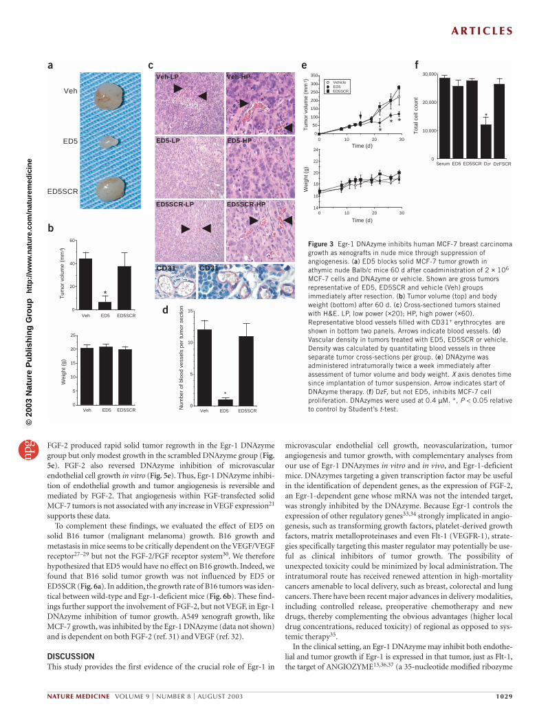
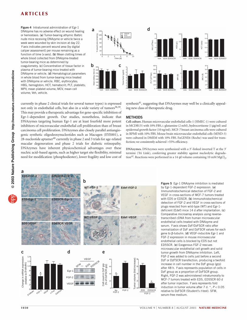
We next examined systemic effects of twice-a-week intratumoralDNAzyme administration at the level of wound healing and hemostasis.No significant difference in the rate of wound closure after acute injurywas detected between any groups (Fig. 4a). There was also no differencein mean clotting time of blood from these mice (Fig. 4b), nor was thereany change in tissue factor concentration (Fig. 4c). Moreover, there wasno change in circulating erythrocytes, hemoglobin, hematocrit,platelets, mean platelet volume or mean cell volume (Fig. 4d). MCF-7tumor-bearing female mice from each group retained the capacity toreproduce after mating with male mice (data not shown).Virtually iden-tical results were obtained with nude mice bearing solid A549 humanlung carcinoma xenografts. ED5, but not its scrambled counterpart,blocked A549 growth without influencing wound healing or otherhematologic parameters (data not shown). There was no overt evidenceof skin erythema, lethargy or soft feces. Thus, it seems that Egr-1DNAzymes inhibit solid tumor growth without compromising woundhealing, hemostasis or reproduction.
DNAzyme inhibition of angiogenesis is mediated via FGF-2FGF-2 has long been recognized as a potent inducer of angiogenesis15.FGF-2 stimulates Egr-1 expression in endothelial cells16, and Egr-1 canin turn activate FGF-2 transcription17,18.We therefore hypothesized thatEgr-1 DNAzyme inhibition is mediated, at least in part, by its blockadeof endogenous FGF-2 expression.
Numerous studies have determined that MCF-7 tumor growth inmice is both FGF- and VEGF-dependent. For example, transfection ofMCF-7 cells with FGF produced highly vascularized, estrogen-inde-pendent solid tumors when grown in nude mice as xenografts19–21,
which are markedly inhibited by the anti-angiogenic drug AGM-1470(ref. 22). Moreover, solid MCF-7 tumor growth is strongly inhibited byoverexpression of truncated dominant-negative FGF receptor23.Similarly, overexpression of VEGF in MCF-7 cells increased solidtumor growth and tumor angiogenesis, an effect neutralized by solubleVEGF receptor24.
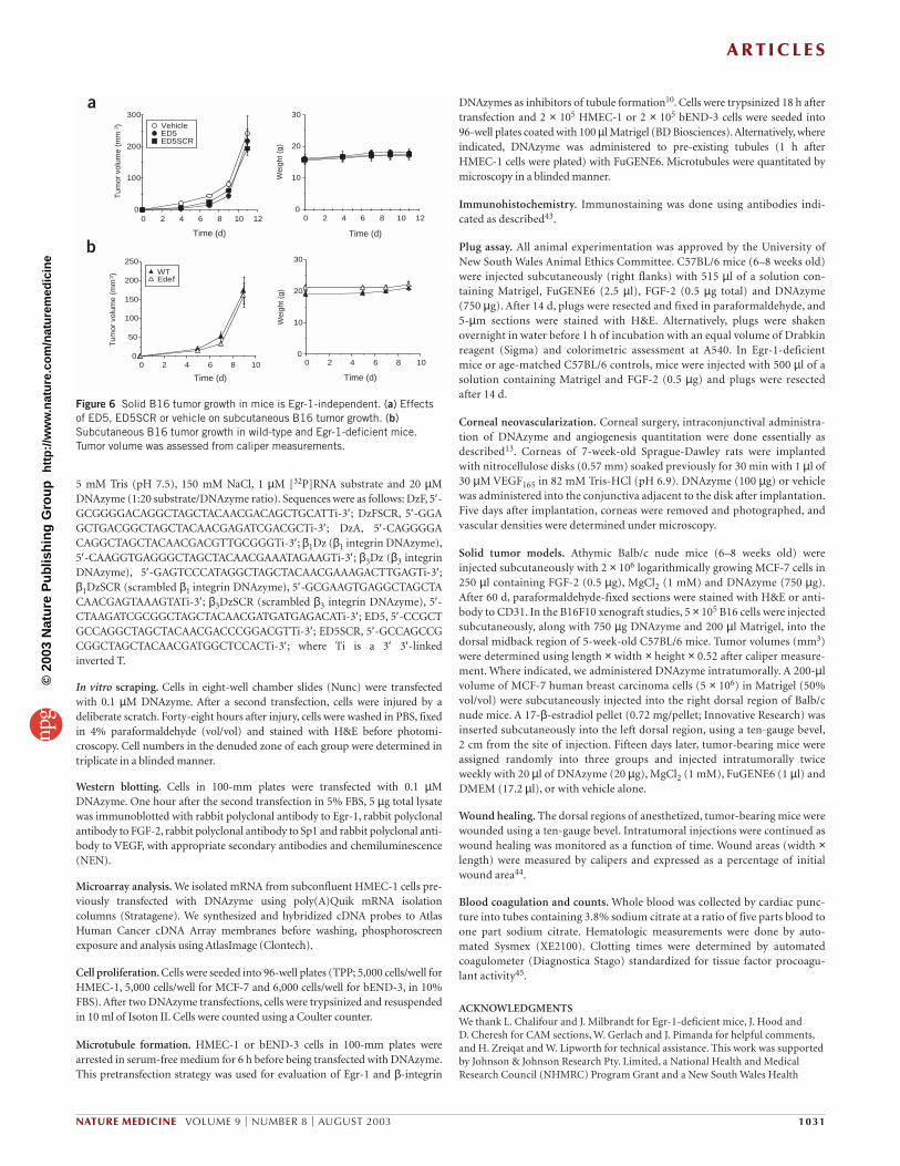
Egr-1 and FGF-2 were weakly, if at all, expressed in the invadingendothelium of MCF-7 tumors treated with ED5 (Fig. 5a). In contrast,VEGF levels were not influenced by the Egr-1 DNAzyme (Fig. 5a), con-sistent with the inability of Egr-1 to regulate VEGF transcription25,26.Moreover, FGF-2 was poorly expressed in plug endothelium from Egr-1-deficient mice (Fig. 5b), whereas VEGF was expressed in plugs derivedfrom both wild-type and Egr-1-deficient mice (Fig. 5b), despite differ-ences in the size and number of blood vessels between these groups. Inmicrovascular endothelial cells, western blot analysis revealed Egr-1DNAzyme inhibition of FGF-2 but not VEGF (Fig. 1g), and comparativemicroarray analysis showed DNAzyme inhibition of FGF-2 but notVEGF mRNA (Fig. 5c). Our earlier observation that recombinant VEGF-inducible corneal angiogenesis is blocked by the Egr-1 DNAzyme (Fig. 2d,e) is supported by our demonstration that the DNAzymeinhibits VEGF-inducible endothelial Egr-1 and FGF-2 expression (Fig. 5d).Although Egr-1 is activated by exogenous VEGF, it has no influ-ence on the de novo production of endogenous VEGF. This is the firstdemonstration of ‘downstream inhibition’ using a DNAzyme, wherebythe expression of a given gene (such as FGF-2) is inhibited not becausethe DNAzyme recognizes a specific motif in the mRNA of that gene, butbecause expression of that gene is dependent on the DNAzyme target.
Based on these findings, we further hypothesized that tumor growthin the Egr-1 DNAzyme group could be rescued by exogenous FGF-2, asFGF-2 can activate Egr-1 (ref. 16). Bolus intratumoral administration of
WT-HP
Edef-LP
Edef-HP
0
5
10
15
20
Ves
sel D
ensi
ty (
vess
els/
mm
Veh
*
2)
0
0.01
0.02
0.03
0.04
0
10
20
Num
ber
of b
lood
ves
sels
per
plu
gO
D54
0
Veh
ED5SCR
**
WT-LP
30
ED5 ED5SCR
ED5 ED5SCR
ED5Veh
10 20 30 60 60 min
ED5 ED5SCR
ED5SCR
Di
Li
Di
LiED5
a
b
c
d e
23 nt
12
0 5
Figure 2 Egr-1 DNAzymes inhibit angiogenesis.(a) Cleavage of 32P-labeled mouse Egr-1 RNA byED5 but not ED5SCR. (b) Top, vascular density inH&E-stained cross-sections of Matrigel plugs 14d after implantation. Bottom, assessment ofhemoglobin content in plugs using Drabkinreagent. *, P < 0.05 relative to vehicle (Veh) byStudent’s t-test. (c) Representative cross-sectionsof H&E-stained plugs resected from wild-type(WT) and Egr-1-deficient (Edef) mice 14 d afterimplantation. LP, low power (×20); HP, highpower (×60). Photomicrographs are representativeof plug center. Erythrocyte-filled blood vessels insections are indicated by bright red staining. (d) ED5 blocks neovascularization in rat corneasimplanted with VEGF165-containing filter discs,which would normally produce robust neovasculargrowth from the limbus toward the disc. Figureshows representative corneas 5 d after treatmentwith ED5 or ED5SCR. Di, disk; Li, limbus.Photographs were taken with high-resolutionblack-and-white film. (e) Number per unit andarea (mm2) occupied by new blood vessels incorneas 5 d after disk implantation andconjunctival delivery of DNAzyme or vehicle. *, P < 0.05 relative to DzFSCR or vehicle group(Student’s t-test).
©20
03 N
atu
re P
ub
lish
ing
Gro
up
h
ttp
://w
ww
.nat
ure
.co
m/n
atu
rem
edic
ine
A R T I C L E S
NATURE MEDICINE VOLUME 9 | NUMBER 8 | AUGUST 2003 1029
FGF-2 produced rapid solid tumor regrowth in the Egr-1 DNAzymegroup but only modest growth in the scrambled DNAzyme group (Fig.5e). FGF-2 also reversed DNAzyme inhibition of microvascularendothelial cell growth in vitro (Fig. 5e). Thus, Egr-1 DNAzyme inhibi-tion of endothelial growth and tumor angiogenesis is reversible andmediated by FGF-2. That angiogenesis within FGF-transfected solidMCF-7 tumors is not associated with any increase in VEGF expression21
supports these data.To complement these findings, we evaluated the effect of ED5 on
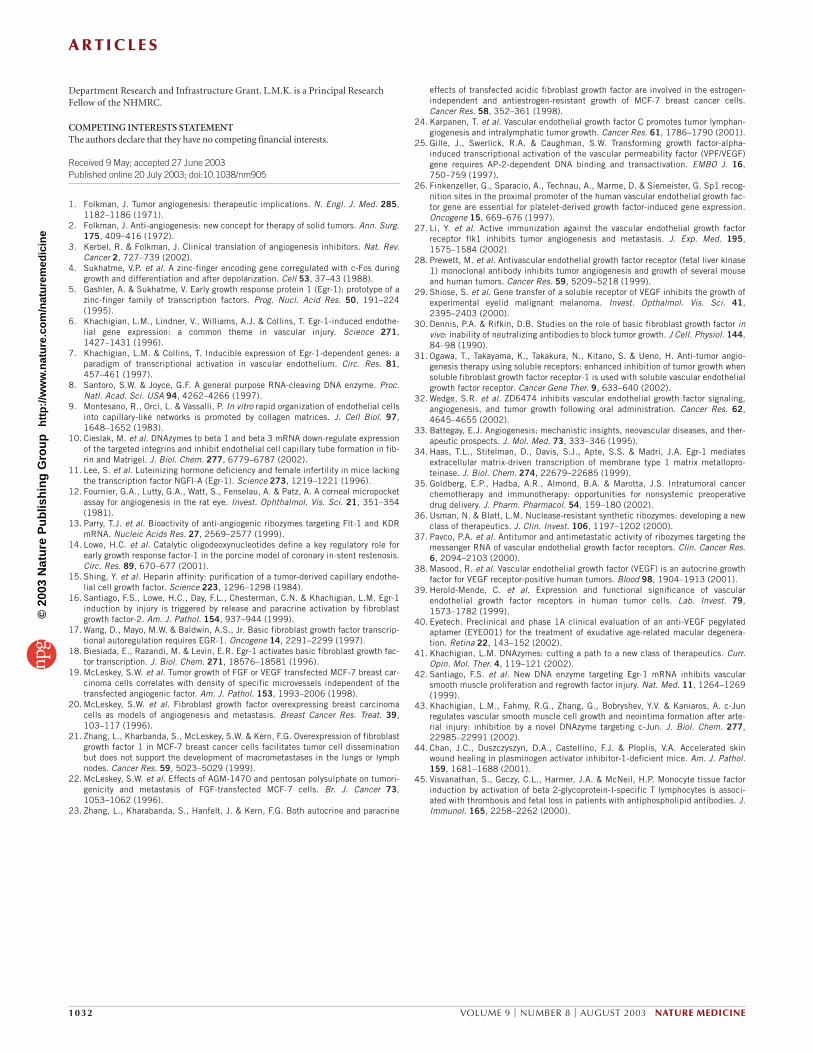
solid B16 tumor (malignant melanoma) growth. B16 growth andmetastasis in mice seems to be critically dependent on the VEGF/VEGFreceptor27–29 but not the FGF-2/FGF receptor system30. We thereforehypothesized that ED5 would have no effect on B16 growth. Indeed, wefound that B16 solid tumor growth was not influenced by ED5 orED5SCR (Fig. 6a). In addition, the growth rate of B16 tumors was iden-tical between wild-type and Egr-1-deficient mice (Fig. 6b). These find-ings further support the involvement of FGF-2, but not VEGF, in Egr-1DNAzyme inhibition of tumor growth. A549 xenograft growth, likeMCF-7 growth, was inhibited by the Egr-1 DNAzyme (data not shown)and is dependent on both FGF-2 (ref. 31) and VEGF (ref. 32).
DISCUSSIONThis study provides the first evidence of the crucial role of Egr-1 in
microvascular endothelial cell growth, neovascularization, tumorangiogenesis and tumor growth, with complementary analyses fromour use of Egr-1 DNAzymes in vitro and in vivo, and Egr-1-deficientmice. DNAzymes targeting a given transcription factor may be usefulin the identification of dependent genes, as the expression of FGF-2,an Egr-1-dependent gene whose mRNA was not the intended target,was strongly inhibited by the DNAzyme. Because Egr-1 controls theexpression of other regulatory genes33,34 strongly implicated in angio-genesis, such as transforming growth factors, platelet-derived growthfactors, matrix metalloproteinases and even Flt-1 (VEGFR-1), strate-gies specifically targeting this master regulator may potentially be use-ful as clinical inhibitors of tumor growth. The possibility ofunexpected toxicity could be minimized by local administration. Theintratumoral route has received renewed attention in high-mortalitycancers amenable to local delivery, such as breast, colorectal and lungcancers. There have been recent major advances in delivery modalities,including controlled release, preoperative chemotherapy and newdrugs, thereby complementing the obvious advantages (higher localdrug concentrations, reduced toxicity) of regional as opposed to sys-temic therapy35.
In the clinical setting, an Egr-1 DNAzyme may inhibit both endothe-lial and tumor growth if Egr-1 is expressed in that tumor, just as Flt-1,the target of ANGIOZYME13,36,37 (a 35-nucleotide modified ribozyme
Veh
ED5
ED5SCR
ED5-HP
ED5SCR-HP
Veh-HP
ED5-LP
ED5SCR-LP
Veh-LP
CD31
Tum
or v
olum
e (m
m3 )
Veh0
5
10
15
20
25
Wei
ght (
g)
*
0
20
40
60
Veh ED5 ED5SCR
Serum ED5 ED5SCR DzF DzFSCR0
10,000
20,000
30,000
Tot
al c
ell c
ount
*
0
5
10
15
Num
ber
of b
lood
ves
sels
per
tum
or s
ectio
n
*
Veh
30201000
50
100
150
200
250
300
350
Tum
or v
olum
e (m
m3 )
** *
3020100
Wei
ght (
g)
Time (d)
Time (d)
14
16
18
20
22
24
VehicleED5ED5SCR
ED5 ED5SCRED5 ED5SCR
a
b
c e
d
f
CD31
Figure 3 Egr-1 DNAzyme inhibits human MCF-7 breast carcinomagrowth as xenografts in nude mice through suppression ofangiogenesis. (a) ED5 blocks solid MCF-7 tumor growth inathymic nude Balb/c mice 60 d after coadministration of 2 × 10⁶
MCF-7 cells and DNAzyme or vehicle. Shown are gross tumorsrepresentative of ED5, ED5SCR and vehicle (Veh) groupsimmediately after resection. (b) Tumor volume (top) and bodyweight (bottom) after 60 d. (c) Cross-sectioned tumors stainedwith H&E. LP, low power (×20); HP, high power (×60).Representative blood vessels filled with CD31+ erythrocytes areshown in bottom two panels. Arrows indicate blood vessels. (d)Vascular density in tumors treated with ED5, ED5SCR or vehicle.Density was calculated by quantitating blood vessels in threeseparate tumor cross-sections per group. (e) DNAzyme wasadministered intratumorally twice a week immediately afterassessment of tumor volume and body weight. X axis denotes timesince implantation of tumor suspension. Arrow indicates start ofDNAzyme therapy. (f) DzF, but not ED5, inhibits MCF-7 cellproliferation. DNAzymes were used at 0.4 µM. *, P < 0.05 relativeto control by Student’s t-test.
©20
03 N
atu
re P
ub
lish
ing
Gro
up
h
ttp
://w
ww
.nat
ure
.co
m/n
atu
rem
edic
ine
A R T I C L E S
1030 VOLUME 9 | NUMBER 8 | AUGUST 2003 NATURE MEDICINE
currently in phase 2 clinical trials for several tumor types) is expressednot only in endothelial cells, but also in a wide variety of tumors38,39.This may provide a therapeutic advantage for gene-specific inhibition ofEgr-1-dependent growth. Our studies, nonetheless, indicate thatDNAzymes targeting human Egr-1 are at least fourfold more potentinhibitors of microvascular endothelial cell proliferation than of breastcarcinoma cell proliferation. DNAzymes also closely parallel antiangio-genic synthetic oligodeoxynucleotides such as Macugen (EYE001), a 28-nucleotide aptamer40 currently in phase 2 and 3 trials for age-relatedmacular degeneration and phase 2 trials for diabetic retinopathy.DNAzymes have inherent physicochemical advantages over thesenucleic acid–based agents, such as higher target site flexibility, minimalneed for modification (phosphodiester), lower fragility and low cost of
synthesis41, suggesting that DNAzymes may well be a clinically appeal-ing new class of therapeutic drug.
METHODSCell culture. Human microvascular endothelial cells-1 (HMEC-1) were culturedin MCDB131 with 10% FBS, L-glutamine (2 mM), hydrocortisone (1 µg/ml) andepidermal growth factor (10 ng/ml). MCF-7 breast carcinoma cells were culturedin RPMI with 10% FBS. Mouse brain microvascular endothelial cells (bEND-3)were cultured in DMEM with 10% FBS. FuGENE6 (Roche) was used for trans-fection; we consistently achieved >55% efficiency.
DNAzymes. DNAzymes were synthesized with a 3′-linked inverted T at the 3′termini (Tri Link), conferring greater stability against nucleolytic degrada-tion42. Reactions were performed in a 14-µl volume containing 10 mM MgCl2,
10864200
20
40
60
80
100
120
Time (d)
% w
ound
are
a re
mai
ning
VehicleED5ED5SCR
Veh ED5 ED5SCR0
50
100
150
200
250
300
350
Mea
n cl
ottin
g ti
me
(s)
Veh ED5 ED5SCR0
2
4
6
8
10
12
0
50
100
150
Veh ED5 ED5SCR
HG
B (g
/L)
0
0.1
0.2
0.3
0.4
0.5
HC
T (r
atio
)
Veh ED5 ED5SCR
0
100
200
300
400
500
600
Veh ED5 ED5SCR
9
0
3
6
9
Veh ED5 ED5SCR
MP
V (f
l)
0
10
20
30
40
50
60
Veh ED5 ED5SCR
(MC
V (f
l)
0
1
2
3
4
5
6
7
8
Veh ED5 ED5SCR
Tiss
ue fa
ctor
(µg/
ml)
a
b c
RB
C (1
0 /l
iter)
PLT
(10
/lite
r)
dFigure 4 Intratumoral administration of Egr-1DNAzyme has no adverse effect on wound healingor hemostasis. (a) Tumor-bearing athymic Balb/cnude mice receiving DNAzyme or vehicle twice aweek were wounded by skin incision at day 22. Y axis indicates percent wound area (by digitalcaliper assessment) per mouse remaining as afunction of time (x axis). (b) Mean clotting times ofwhole blood collected from DNAzyme-treatedtumor-bearing mice as determined bycoagulometry. (c) Concentration of tissue factor inplasma of tumor-bearing mice treated withDNAzyme or vehicle. (d) Hematological parametersin whole blood from tumor-bearing mice treatedwith DNAzyme or vehicle. RBC, erythrocytes; HBG, hemoglobin; HCT, hematocrit; PLT, platelets;MPV, mean platelet volume; MCV, mean cellvolume; Veh, vehicle.
ED5SCR-FGF-2
ED5SCR-Egr-1
ED5SCR-VEGFED5-VEGF
ED5-Egr-1
ED5-FGF-2
WT-VEGF Edef -VEGF
Edef-FGF-2WT-FGF-2
Egr-1
FGF-2
VEGF
ED5ED5SCR
VEGF
0
2
4
6
8
0
20
40
60
80
Tum
or v
olum
e (f
old
incr
ease
)
Cel
l cou
nt in
DzF
grp
(%
of D
zFS
CR
grp
)
FGF-2 ED5SCRED5
FGF-2
*
*
0
.2
.4
.6
.8
1.0
1.2
1.4
Cha
nge
in m
RN
A e
xpre
ssio
n (r
elat
ive
to β
-tub
ulin
)
β-tubulin
FGF-2VEGF
c-Fos
GAPDH
*
a
b
c d e
SFM—
NoFGF-2
Figure 5 Egr-1 DNAzyme inhibition is mediatedby Egr-1-dependent FGF-2 expression. (a)Immunohistochemical detection of FGF-2 andVEGF in cross-sections of MCF-7 tumors treatedwith ED5 or EDSCR. (b) Immunohistochemicaldetection of FGF-2 and VEGF in cross-sections ofplugs resected from wild-type (WT) and Egr-1-deficient (Edef) mice 14 d after implantation. (c)Comparative microarray analysis using reverse-transcribed cDNA from human microvascularendothelial cells treated with DNAzyme andserum. Y axis shows DzF/DzFSCR ratio afternormalization of DzF and DzFSCR values for eachgene to β-tubulin. (d) VEGF-inducible Egr-1 andFGF-2 expression in mouse microvascularendothelial cells is blocked by ED5 but notED5SCR. (e) Exogenous FGF-2 rescuesmicrovascular endothelial cell growth and solidtumor growth from DNAzyme inhibition. Left,FGF-2 was added to cells just before a secondDzF or DzFSCR transfection, producing a twofoldincrease in cell number in the DzF group (grp)after 48 h. Y axis represents population of cells inDzF group as a proportion of DzFSCR group.Right, FGF-2 was administered intratumorally toMCF-7 tumors treated with ED5, ED5SCR 60 dafter tumor injection. Y axis represents foldinduction in tumor volume after 7 d. *, P < 0.05relative to DzFSCR (Student’s t-test). SFM,serum-free medium.
©20
03 N
atu
re P
ub
lish
ing
Gro
up
h
ttp
://w
ww
.nat
ure
.co
m/n
atu
rem
edic
ine
A R T I C L E S
NATURE MEDICINE VOLUME 9 | NUMBER 8 | AUGUST 2003 1031
5 mM Tris (pH 7.5), 150 mM NaCl, 1 µM [32P]RNA substrate and 20 µMDNAzyme (1:20 substrate/DNAzyme ratio). Sequences were as follows: DzF, 5′-GCGGGGACAGGCTAGCTACAACGACAGCTGCATTi-3′; DzFSCR, 5′-GGAGCTGACGGCTAGCTACAACGAGATCGACGCTi-3′; DzA, 5′-CAGGGGACAGGCTAGCTACAACGACGTTGCGGGTi-3′; β1Dz (β1 integrin DNAzyme),5′-CAAGGTGAGGGCTAGCTACAACGAAATAGAAGTi-3′; β3Dz (β3 integrinDNAzyme), 5′-GAGTCCCATAGGCTAGCTACAACGAAAGACTTGAGTi-3′;β1DzSCR (scrambled β1 integrin DNAzyme), 5′-GCGAAGTGAGGCTAGCTACAACGAGTAAAGTATi-3′; β3DzSCR (scrambled β3 integrin DNAzyme), 5′-CTAAGATCGCGGCTAGCTACAACGATGATGAGACATi-3′; ED5, 5′-CCGCTGCCAGGCTAGCTACAACGACCCGGACGTTi-3′; ED5SCR, 5′-GCCAGCCGCGGCTAGCTACAACGATGGCTCCACTi-3′; where Ti is a 3′ 3′-linkedinverted T.
In vitro scraping. Cells in eight-well chamber slides (Nunc) were transfectedwith 0.1 µM DNAzyme. After a second transfection, cells were injured by adeliberate scratch. Forty-eight hours after injury, cells were washed in PBS, fixedin 4% paraformaldehyde (vol/vol) and stained with H&E before photomi-croscopy. Cell numbers in the denuded zone of each group were determined intriplicate in a blinded manner.
Western blotting. Cells in 100-mm plates were transfected with 0.1 µMDNAzyme. One hour after the second transfection in 5% FBS, 5 µg total lysatewas immunoblotted with rabbit polyclonal antibody to Egr-1, rabbit polyclonalantibody to FGF-2, rabbit polyclonal antibody to Sp1 and rabbit polyclonal anti-body to VEGF, with appropriate secondary antibodies and chemiluminescence(NEN).
Microarray analysis. We isolated mRNA from subconfluent HMEC-1 cells pre-viously transfected with DNAzyme using poly(A)Quik mRNA isolationcolumns (Stratagene). We synthesized and hybridized cDNA probes to AtlasHuman Cancer cDNA Array membranes before washing, phosphoroscreenexposure and analysis using AtlasImage (Clontech).
Cell proliferation. Cells were seeded into 96-well plates (TPP; 5,000 cells/well forHMEC-1, 5,000 cells/well for MCF-7 and 6,000 cells/well for bEND-3, in 10%FBS). After two DNAzyme transfections, cells were trypsinized and resuspendedin 10 ml of Isoton II. Cells were counted using a Coulter counter.
Microtubule formation. HMEC-1 or bEND-3 cells in 100-mm plates werearrested in serum-free medium for 6 h before being transfected with DNAzyme.This pretransfection strategy was used for evaluation of Egr-1 and β-integrin
DNAzymes as inhibitors of tubule formation10. Cells were trypsinized 18 h aftertransfection and 2 × 10⁵ HMEC-1 or 2 × 10⁵ bEND-3 cells were seeded into 96-well plates coated with 100 µl Matrigel (BD Biosciences). Alternatively, whereindicated, DNAzyme was administered to pre-existing tubules (1 h after HMEC-1 cells were plated) with FuGENE6. Microtubules were quantitated bymicroscopy in a blinded manner.
Immunohistochemistry. Immunostaining was done using antibodies indi-cated as described43.
Plug assay. All animal experimentation was approved by the University ofNew South Wales Animal Ethics Committee. C57BL/6 mice (6–8 weeks old)were injected subcutaneously (right flanks) with 515 µl of a solution con-taining Matrigel, FuGENE6 (2.5 µl), FGF-2 (0.5 µg total) and DNAzyme(750 µg). After 14 d, plugs were resected and fixed in paraformaldehyde, and5-µm sections were stained with H&E. Alternatively, plugs were shakenovernight in water before 1 h of incubation with an equal volume of Drabkinreagent (Sigma) and colorimetric assessment at A540. In Egr-1-deficientmice or age-matched C57BL/6 controls, mice were injected with 500 µl of asolution containing Matrigel and FGF-2 (0.5 µg) and plugs were resectedafter 14 d.
Corneal neovascularization. Corneal surgery, intraconjunctival administra-tion of DNAzyme and angiogenesis quantitation were done essentially asdescribed13. Corneas of 7-week-old Sprague-Dawley rats were implantedwith nitrocellulose disks (0.57 mm) soaked previously for 30 min with 1 µl of30 µM VEGF165 in 82 mM Tris-HCl (pH 6.9). DNAzyme (100 µg) or vehiclewas administered into the conjunctiva adjacent to the disk after implantation.Five days after implantation, corneas were removed and photographed, andvascular densities were determined under microscopy.
Solid tumor models. Athymic Balb/c nude mice (6–8 weeks old) wereinjected subcutaneously with 2 × 10⁶ logarithmically growing MCF-7 cells in250 µl containing FGF-2 (0.5 µg), MgCl2 (1 mM) and DNAzyme (750 µg).After 60 d, paraformaldehyde-fixed sections were stained with H&E or anti-body to CD31. In the B16F10 xenograft studies, 5 × 10⁵ B16 cells were injectedsubcutaneously, along with 750 µg DNAzyme and 200 µl Matrigel, into thedorsal midback region of 5-week-old C57BL/6 mice. Tumor volumes (mm3)were determined using length × width × height × 0.52 after caliper measure-ment. Where indicated, we administered DNAzyme intratumorally. A 200-µlvolume of MCF-7 human breast carcinoma cells (5 × 10⁶) in Matrigel (50%vol/vol) were subcutaneously injected into the right dorsal region of Balb/cnude mice. A 17-β-estradiol pellet (0.72 mg/pellet; Innovative Research) wasinserted subcutaneously into the left dorsal region, using a ten-gauge bevel,2 cm from the site of injection. Fifteen days later, tumor-bearing mice wereassigned randomly into three groups and injected intratumorally twiceweekly with 20 µl of DNAzyme (20 µg), MgCl2 (1 mM), FuGENE6 (1 µl) andDMEM (17.2 µl), or with vehicle alone.
Wound healing. The dorsal regions of anesthetized, tumor-bearing mice werewounded using a ten-gauge bevel. Intratumoral injections were continued aswound healing was monitored as a function of time. Wound areas (width ×length) were measured by calipers and expressed as a percentage of initialwound area44.
Blood coagulation and counts. Whole blood was collected by cardiac punc-ture into tubes containing 3.8% sodium citrate at a ratio of five parts blood toone part sodium citrate. Hematologic measurements were done by auto-mated Sysmex (XE2100). Clotting times were determined by automatedcoagulometer (Diagnostica Stago) standardized for tissue factor procoagu-lant activity45.
ACKNOWLEDGMENTSWe thank L. Chalifour and J. Milbrandt for Egr-1-deficient mice, J. Hood and D. Cheresh for CAM sections, W. Gerlach and J. Pimanda for helpful comments,and H. Zreiqat and W. Lipworth for technical assistance. This work was supportedby Johnson & Johnson Research Pty. Limited, a National Health and MedicalResearch Council (NHMRC) Program Grant and a New South Wales Health
10864200
50
100
150
200
250
10864200
10
20
30
WTEdef
1210864200
100
200
300
1210864200
10
20
30
VehicleED5ED5SCR
Tum
or v
olum
e (m
m3 )
Wei
ght (
g)
Tum
or v
olum
e (m
m3)
Wei
ght (
g)
Time (d)
Time (d)
a
bTime (d)
Time (d)
Figure 6 Solid B16 tumor growth in mice is Egr-1-independent. (a) Effectsof ED5, ED5SCR or vehicle on subcutaneous B16 tumor growth. (b)Subcutaneous B16 tumor growth in wild-type and Egr-1-deficient mice.Tumor volume was assessed from caliper measurements.
©20
03 N
atu
re P
ub
lish
ing
Gro
up
h
ttp
://w
ww
.nat
ure
.co
m/n
atu
rem
edic
ine
A R T I C L E S
1032 VOLUME 9 | NUMBER 8 | AUGUST 2003 NATURE MEDICINE
Department Research and Infrastructure Grant. L.M.K. is a Principal ResearchFellow of the NHMRC.
COMPETING INTERESTS STATEMENTThe authors declare that they have no competing financial interests.
Received 9 May; accepted 27 June 2003Published online 20 July 2003; doi:10.1038/nm905
1. Folkman, J. Tumor angiogenesis: therapeutic implications. N. Engl. J. Med. 285,1182–1186 (1971).
2. Folkman, J. Anti-angiogenesis: new concept for therapy of solid tumors. Ann. Surg.175, 409–416 (1972).
3. Kerbel, R. & Folkman, J. Clinical translation of angiogenesis inhibitors. Nat. Rev.Cancer 2, 727–739 (2002).
4. Sukhatme, V.P. et al. A zinc-finger encoding gene corregulated with c-Fos duringgrowth and differentiation and after depolarization. Cell 53, 37–43 (1988).
5. Gashler, A. & Sukhatme, V. Early growth response protein 1 (Egr-1): prototype of azinc-finger family of transcription factors. Prog. Nucl. Acid Res. 50, 191–224(1995).
6. Khachigian, L.M., Lindner, V., Williams, A.J. & Collins, T. Egr-1-induced endothe-lial gene expression: a common theme in vascular injury. Science 271,1427–1431 (1996).
7. Khachigian, L.M. & Collins, T. Inducible expression of Egr-1-dependent genes: aparadigm of transcriptional activation in vascular endothelium. Circ. Res. 81,457–461 (1997).
8. Santoro, S.W. & Joyce, G.F. A general purpose RNA-cleaving DNA enzyme. Proc.Natl. Acad. Sci. USA 94, 4262–4266 (1997).
9. Montesano, R., Orci, L. & Vassalli, P. In vitro rapid organization of endothelial cellsinto capillary-like networks is promoted by collagen matrices. J. Cell Biol. 97,1648–1652 (1983).
10. Cieslak, M. et al. DNAzymes to beta 1 and beta 3 mRNA down-regulate expressionof the targeted integrins and inhibit endothelial cell capillary tube formation in fib-rin and Matrigel. J. Biol. Chem. 277, 6779–6787 (2002).
11. Lee, S. et al. Luteinizing hormone deficiency and female infertility in mice lackingthe transcription factor NGFI-A (Egr-1). Science 273, 1219–1221 (1996).
12. Fournier, G.A., Lutty, G.A., Watt, S., Fenselau, A. & Patz, A. A corneal micropocketassay for angiogenesis in the rat eye. Invest. Ophthalmol. Vis. Sci. 21, 351–354(1981).
13. Parry, T.J. et al. Bioactivity of anti-angiogenic ribozymes targeting Flt-1 and KDRmRNA. Nucleic Acids Res. 27, 2569–2577 (1999).
14. Lowe, H.C. et al. Catalytic oligodeoxynucleotides define a key regulatory role forearly growth response factor-1 in the porcine model of coronary in-stent restenosis.Circ. Res. 89, 670–677 (2001).
15. Shing, Y. et al. Heparin affinity: purification of a tumor-derived capillary endothe-lial cell growth factor. Science 223, 1296–1298 (1984).
16. Santiago, F.S., Lowe, H.C., Day, F.L., Chesterman, C.N. & Khachigian, L.M. Egr-1induction by injury is triggered by release and paracrine activation by fibroblastgrowth factor-2. Am. J. Pathol. 154, 937–944 (1999).
17. Wang, D., Mayo, M.W. & Baldwin, A.S., Jr. Basic fibroblast growth factor transcrip-tional autoregulation requires EGR-1. Oncogene 14, 2291–2299 (1997).
18. Biesiada, E., Razandi, M. & Levin, E.R. Egr-1 activates basic fibroblast growth fac-tor transcription. J. Biol. Chem. 271, 18576–18581 (1996).
19. McLeskey, S.W. et al. Tumor growth of FGF or VEGF transfected MCF-7 breast car-cinoma cells correlates with density of specific microvessels independent of thetransfected angiogenic factor. Am. J. Pathol. 153, 1993–2006 (1998).
20. McLeskey, S.W. et al. Fibroblast growth factor overexpressing breast carcinomacells as models of angiogenesis and metastasis. Breast Cancer Res. Treat. 39,103–117 (1996).
21. Zhang, L., Kharbanda, S., McLeskey, S.W. & Kern, F.G. Overexpression of fibroblastgrowth factor 1 in MCF-7 breast cancer cells facilitates tumor cell disseminationbut does not support the development of macrometastases in the lungs or lymphnodes. Cancer Res. 59, 5023–5029 (1999).
22. McLeskey, S.W. et al. Effects of AGM-1470 and pentosan polysulphate on tumori-genicity and metastasis of FGF-transfected MCF-7 cells. Br. J. Cancer 73,1053–1062 (1996).
23. Zhang, L., Kharabanda, S., Hanfelt, J. & Kern, F.G. Both autocrine and paracrine
effects of transfected acidic fibroblast growth factor are involved in the estrogen-independent and antiestrogen-resistant growth of MCF-7 breast cancer cells.Cancer Res. 58, 352–361 (1998).
24. Karpanen, T. et al. Vascular endothelial growth factor C promotes tumor lymphan-giogenesis and intralymphatic tumor growth. Cancer Res. 61, 1786–1790 (2001).
25. Gille, J., Swerlick, R.A. & Caughman, S.W. Transforming growth factor-alpha-induced transcriptional activation of the vascular permeability factor (VPF/VEGF)gene requires AP-2-dependent DNA binding and transactivation. EMBO J. 16,750–759 (1997).
26. Finkenzeller, G., Sparacio, A., Technau, A., Marme, D. & Siemeister, G. Sp1 recog-nition sites in the proximal promoter of the human vascular endothelial growth fac-tor gene are essential for platelet-derived growth factor-induced gene expression.Oncogene 15, 669–676 (1997).
27. Li, Y. et al. Active immunization against the vascular endothelial growth factorreceptor flk1 inhibits tumor angiogenesis and metastasis. J. Exp. Med. 195,1575–1584 (2002).
28. Prewett, M. et al. Antivascular endothelial growth factor receptor (fetal liver kinase1) monoclonal antibody inhibits tumor angiogenesis and growth of several mouseand human tumors. Cancer Res. 59, 5209–5218 (1999).
29. Shiose, S. et al. Gene transfer of a soluble receptor of VEGF inhibits the growth ofexperimental eyelid malignant melanoma. Invest. Opthalmol. Vis. Sci. 41,2395–2403 (2000).
30. Dennis, P.A. & Rifkin, D.B. Studies on the role of basic fibroblast growth factor invivo: inability of neutralizing antibodies to block tumor growth. J Cell. Physiol. 144,84–98 (1990).
31. Ogawa, T., Takayama, K., Takakura, N., Kitano, S. & Ueno, H. Anti-tumor angio-genesis therapy using soluble receptors: enhanced inhibition of tumor growth whensoluble fibroblast growth factor receptor-1 is used with soluble vascular endothelialgrowth factor receptor. Cancer Gene Ther. 9, 633–640 (2002).
32. Wedge, S.R. et al. ZD6474 inhibits vascular endothelial growth factor signaling,angiogenesis, and tumor growth following oral administration. Cancer Res. 62,4645–4655 (2002).
33. Battegay, E.J. Angiogenesis: mechanistic insights, neovascular diseases, and ther-apeutic prospects. J. Mol. Med. 73, 333–346 (1995).
34. Haas, T.L., Stitelman, D., Davis, S.J., Apte, S.S. & Madri, J.A. Egr-1 mediatesextracellular matrix-driven transcription of membrane type 1 matrix metallopro-teinase. J. Biol. Chem. 274, 22679–22685 (1999).
35. Goldberg, E.P., Hadba, A.R., Almond, B.A. & Marotta, J.S. Intratumoral cancerchemotherapy and immunotherapy: opportunities for nonsystemic preoperativedrug delivery. J. Pharm. Pharmacol. 54, 159–180 (2002).
36. Usman, N. & Blatt, L.M. Nuclease-resistant synthetic ribozymes: developing a newclass of therapeutics. J. Clin. Invest. 106, 1197–1202 (2000).
37. Pavco, P.A. et al. Antitumor and antimetastatic activity of ribozymes targeting themessenger RNA of vascular endothelial growth factor receptors. Clin. Cancer Res.6, 2094–2103 (2000).
38. Masood, R. et al. Vascular endothelial growth factor (VEGF) is an autocrine growthfactor for VEGF receptor-positive human tumors. Blood 98, 1904–1913 (2001).
39. Herold-Mende, C. et al. Expression and functional significance of vascularendothelial growth factor receptors in human tumor cells. Lab. Invest. 79,1573–1782 (1999).
40. Eyetech. Preclinical and phase 1A clinical evaluation of an anti-VEGF pegylatedaptamer (EYE001) for the treatment of exudative age-related macular degenera-tion. Retina 22, 143–152 (2002).
41. Khachigian, L.M. DNAzymes: cutting a path to a new class of therapeutics. Curr.Opin. Mol. Ther. 4, 119–121 (2002).
42. Santiago, F.S. et al. New DNA enzyme targeting Egr-1 mRNA inhibits vascularsmooth muscle proliferation and regrowth factor injury. Nat. Med. 11, 1264–1269(1999).
43. Khachigian, L.M., Fahmy, R.G., Zhang, G., Bobryshev, Y.V. & Kaniaros, A. c-Junregulates vascular smooth muscle cell growth and neointima formation after arte-rial injury: inhibition by a novel DNAzyme targeting c-Jun. J. Biol. Chem. 277,22985–22991 (2002).
44. Chan, J.C., Duszczyszyn, D.A., Castellino, F.J. & Ploplis, V.A. Accelerated skinwound healing in plasminogen activator inhibitor-1-deficient mice. Am. J. Pathol.159, 1681–1688 (2001).
45. Visvanathan, S., Geczy, C.L., Harmer, J.A. & McNeil, H.P. Monocyte tissue factorinduction by activation of beta 2-glycoprotein-I-specific T lymphocytes is associ-ated with thrombosis and fetal loss in patients with antiphospholipid antibodies. J.Immunol. 165, 2258–2262 (2000).
©20
03 N
atu
re P
ub
lish
ing
Gro
up
h
ttp
://w
ww
.nat
ure
.co
m/n
atu
rem
edic
ine
Copyright © 2022 FDOKUMEN