Theoretical investigation of thermal retinal response to photodynamic therapy or choroidal...
7
Theoretical investigation of thermal retinal response to photodynamic therapy for choroidal neovascularization Hongxia Chen a , Ying Gu *a , Gang Cheng b , Zaifu Yang c , Fan-guang Liu a , Li Zhang a , Haixia Qiu a a Dept. of Laser Medicine, Chinese PLA General Hospital, Beijing 100853 b Dept. of Automatic Control, Beijing Institute of Technology, Beijing, 100081 c Beijing Institute of Radiation Medicine, Beijing, 100850 ABSTRACT To study the risk of thermal injury in photodynamic therapy for choroidal neovascularization by calculating the retinal temperature of rabbits, a mathematical model for laser induced thermal effect on retina was developed. Homogeneous layer retinal models of different pigmented rabbits were presented to analyze the light distribution. The finite element method realized by Matlab software was used to solve classical bio-heat transfer model - Pennis equation, in which heat loss due to choroidal blood flow was considered. The retinal temperature was calculated with different laser parameters, including different wavelengths (532nm, 578nm and 690nm), power density (200~1600 mW/cm 2 ), spot diameter (1mm, 2mm and 3mm) and different pigmented eye fundi. The prediction results showed the retinal temperature increased first, then reached maximum in a few seconds and kept constant during laser irradiation. Once laser exposure ended, the temperature decreased quickly to normal. With the increase of laser power density and spot size, the retinal temperature raised too. The temperature reduced exponentially with the distance from laser spot increased. The maximum temperature of non-pigmented rabbits was lower than that of pigmented rabbits. The temperature induced by 578nm laser irradiation was highest, the next was by 532nm laser and the lowest was by 690nm laser. For current parameters used to treat choroidal neovascularization (690nm, 600mW, 2mm, 83sec), the maximum retinal temperature calculated was less than 45 o C, which indicating no thermal damage induced. Keywords: mathematical model, retinal temperature, choroidal neovascularization, photodynamic therapy 1. INTRODUCTION Choroidal Neovascularization (CNV) leads to severe loss of central vision in patients with age-related macular degeneration, which is the leading cause of legal blindness in people over 65[1]. The currently available treatment consists of laser photocoagulation, but it is only applicable in less than 15% of the patients [2]. And the visual loss and the high recurrence rate proved this treatment unsatisfactory. Photodynamic therapy (PDT) may offer selective occlusion of neovascular and has been demonstrated benefit to treating both classic and occult choroidal neovascularization. The primary mechanism of PDT is a purely photochemical reaction in which the photosensitizer is selectively absorbed by the target tissues such as choroidal neovascular and activated by a long-pulse, large spot, very low irradiance of laser exposure [3]. Since the selectivity of PDT is achieved by avoiding any thermal damage to retina, it is important to analyze temperature distribution in retina. In order to provide safe guidance for laser exposure during PDT on fundus diseases, it is necessary to develop a detailed mathematical model for calculating retinal temperature increases from laser radiation absorption. Many models have been developed to describe thermal retinal damage induced by laser photocoagulation [4~7]. But there is little concerning the thermal change of retina especially for real-time temperature calculation during PDT which differs from conventional short-pulse retinal photocoagulation because of its long-pulse, large spot and low irradiance laser exposure. Complications of PDT included macular infarcts, RPE tears and ganglion cell damage, which were probably caused by excess laser exposure. Modulating laser parameters may help reducing these complications. The * [email protected]; phone: 010-66939394; fax: 010-68222584 Optics in Health Care and Biomedical Optics III, edited by Xingde Li, Qingming Luo, Ying Gu, Proc. of SPIE Vol. 6826, 68261O, (2007) · 1605-7422/07/$18 · doi: 10.1117/12.756110 Proc. of SPIE Vol. 6826 68261O-1 Downloaded from SPIE Digital Library on 09 Feb 2010 to 202.204.81.80. Terms of Use: http://spiedl.org/terms
-
Upload
independent -
Category
Documents
-
view
3 -
download
0
Transcript of Theoretical investigation of thermal retinal response to photodynamic therapy or choroidal...

Theoretical investigation of thermal retinal response to photodynamic
therapy for choroidal neovascularization
Hongxia Chena, Ying Gu*a, Gang Chengb, Zaifu Yangc, Fan-guang Liu a, Li Zhanga, Haixia Qiua
aDept. of Laser Medicine, Chinese PLA General Hospital, Beijing 100853
bDept. of Automatic Control, Beijing Institute of Technology, Beijing, 100081 cBeijing Institute of Radiation Medicine, Beijing, 100850
ABSTRACT To study the risk of thermal injury in photodynamic therapy for choroidal neovascularization by calculating the retinal temperature of rabbits, a mathematical model for laser induced thermal effect on retina was developed. Homogeneous layer retinal models of different pigmented rabbits were presented to analyze the light distribution. The finite element method realized by Matlab software was used to solve classical bio-heat transfer model - Pennis equation, in which heat loss due to choroidal blood flow was considered. The retinal temperature was calculated with different laser parameters, including different wavelengths (532nm, 578nm and 690nm), power density (200~1600 mW/cm2), spot diameter (1mm, 2mm and 3mm) and different pigmented eye fundi. The prediction results showed the retinal temperature increased first, then reached maximum in a few seconds and kept constant during laser irradiation. Once laser exposure ended, the temperature decreased quickly to normal. With the increase of laser power density and spot size, the retinal temperature raised too. The temperature reduced exponentially with the distance from laser spot increased. The maximum temperature of non-pigmented rabbits was lower than that of pigmented rabbits. The temperature induced by 578nm laser irradiation was highest, the next was by 532nm laser and the lowest was by 690nm laser. For current parameters used to treat choroidal neovascularization (690nm, 600mW, 2mm, 83sec), the maximum retinal temperature calculated was less than 45 oC, which indicating no thermal damage induced. Keywords: mathematical model, retinal temperature, choroidal neovascularization, photodynamic therapy
1. INTRODUCTION
Choroidal Neovascularization (CNV) leads to severe loss of central vision in patients with age-related macular degeneration, which is the leading cause of legal blindness in people over 65[1]. The currently available treatment consists of laser photocoagulation, but it is only applicable in less than 15% of the patients [2]. And the visual loss and the high recurrence rate proved this treatment unsatisfactory. Photodynamic therapy (PDT) may offer selective occlusion of neovascular and has been demonstrated benefit to treating both classic and occult choroidal neovascularization. The primary mechanism of PDT is a purely photochemical reaction in which the photosensitizer is selectively absorbed by the target tissues such as choroidal neovascular and activated by a long-pulse, large spot, very low irradiance of laser exposure [3]. Since the selectivity of PDT is achieved by avoiding any thermal damage to retina, it is important to analyze temperature distribution in retina. In order to provide safe guidance for laser exposure during PDT on fundus diseases, it is necessary to develop a detailed mathematical model for calculating retinal temperature increases from laser radiation absorption. Many models have been developed to describe thermal retinal damage induced by laser photocoagulation [4~7]. But there is little concerning the thermal change of retina especially for real-time temperature calculation during PDT which differs from conventional short-pulse retinal photocoagulation because of its long-pulse, large spot and low irradiance laser exposure. Complications of PDT included macular infarcts, RPE tears and ganglion cell damage, which were probably caused by excess laser exposure. Modulating laser parameters may help reducing these complications. The * [email protected]; phone: 010-66939394; fax: 010-68222584
Optics in Health Care and Biomedical Optics III, edited by Xingde Li, Qingming Luo, Ying Gu, Proc. of SPIE Vol. 6826, 68261O, (2007) · 1605-7422/07/$18 · doi: 10.1117/12.756110
Proc. of SPIE Vol. 6826 68261O-1
Downloaded from SPIE Digital Library on 09 Feb 2010 to 202.204.81.80. Terms of Use: http://spiedl.org/terms

purpose of this work is to develop a mathematical model to calculate retinal temperature distribution and find suitable range of laser parameters such as power density and spot size during PDT.
2. MATHEMATICAL MODELING
Though about 70% of the entire intraocular blood supply flows through the choriocapillaris directly adjacent to the retina, Birngruber [7] found no temperature differences between the living and dead animals with a small focus diameter of 100 µm and an exposure time of 500 ms. So, most thermal models of retinal photocoagulation neglected the influence of choroidal circulation on heat convection. However, when considering long-pulse (>7 s), large spot size and low laser irradiance during PDT for CNV, we should not neglect the convective heat loss due to blood flow in thermal model [8]. The heat transport equation for tissue exposed to the laser was given by [9]
2b m r
Tc k T Q Q Qt
ρ ∂⋅ ⋅ = ⋅∇ + + +
∂ (1)
Where ρ was the density, c was the specific heat, k was the thermal conductivity of tissue medium, T was the tissue temperature, Qb =h(Te-T) was heat convection due to blood flow, Qm was heat convection due to metabolism, and Qr was the heat source term. In the following, the anatomical conditions and the absorption character on the fundus of pigmented and non-pigmented rabbits will be discussed in detail. Since the pigmented layer and blood distribution throughout the fundus varied little, the homogeneous layer model was assumed in this paper. Figure 1 showed the histological section of a pigmented rabbit’s fundus. The retina was about 150µm. The pigment epithelium was about 10µm (R), and the thickness of the pigmented layer was about 4µm (PE1), which contained a large number of melanin granules and was the primary light absorber in fundus. The choroid was about 65µm thick including choriocapillaris layer (CH1) and 20µm thick pigmented layer (CH2). The choriod and retina of non-pigmented rabbits were lack of melanin granules, so the laser energy was mainly absorbed by hemogloblin. The thermal model could be established on the basis of a homogeneous absorbing layer of blood in the choroid as shown in figure 2.
Fig.1. Geometrical distribution of the various layers of the fundus of the pigmented rabbit
150µm
4µm 6µm
45µm
20µm
R
PE1
CH2
PE2
CH1
Proc. of SPIE Vol. 6826 68261O-2
Downloaded from SPIE Digital Library on 09 Feb 2010 to 202.204.81.80. Terms of Use: http://spiedl.org/terms

Fig.2. Geometrical distribution of the various layers of the fundus of the non-pigmented rabbit The current laser source of PDT for CNV was diode laser with the wavelength of 690nm. So we selected this wavelength to establish the theoretical model. Other lasers including 532nm and 578nm were also calculated because some photosensitizers could be efficiently activated by them. The eye fundus optical parameters of pigmented rabbit on 532nm, 578nm and 690nm were listed in table 1, those of albino rabbit were listed in table 2.
Table.1. Absorbance coefficient of pigmented rabbit eye fundus
Wavelength(nm) PE1* CH1** CH2***
532 508 133 387 578 508 177 431 690 508 15 269
* Calculated from [10] and assumed equal among the three lasers, the melanin density was supposed to be 500 granules per cell. ** The CH1 was supposed to be composed of 50% blood[11] and 50% non-pigmented tissue, αCH1 =αblood/2. *** αCH2 =(αPE1+αblood)/2.
Table.2. Absorbance coefficient of pigmented rabbit eye fundus
Wavelength(nm) PE1* CH1** CH2***
532 0 133 133 578 0 177 177 690 0 15 15
* Calculated from[10], the melanin density was supposed to be 0 granules per cell. ** The CH1 was supposed to be composed of 50% blood and 50% non-pigmented tissue, αCH1 =αblood/2. *** αCH2 =αCH1.
The thermal constants ρ, c and k for tissue were as follows[12]:
3 1 16.3 10k W cm K− − −= × ⋅ ⋅ 31.0g cmρ −= ⋅
1 14.2c J g K− −= ⋅ ⋅ The laser beam used in PDT directed into the eyes was parallel and round shape. The initial and boundary conditions pertinent to the physical problem above were as follows. Immediately before application of the laser (t=0), the tissues were assumed to be at a uniform temperature T = Tc. Source term Qr was in direct proportion to the light absorpted by tissue. If only light absorbed by fundus was considered, the Qr could be described as
( ) ( )⎩⎨⎧
≥≤⋅−⋅⋅
= 0
exp, 11
1 RrRrzE
zrQ PEPErPE
αα (2)
150µm
4µm 6µm
45µm
20µm
R
PE1
CH2
PE2
CH1
Proc. of SPIE Vol. 6826 68261O-3
Downloaded from SPIE Digital Library on 09 Feb 2010 to 202.204.81.80. Terms of Use: http://spiedl.org/terms
48
1— 42
- 532A 578- 69O
Rathus(cm)
84
82 I
48
48
44
42
28 48 88
Time(s)
88 188 128
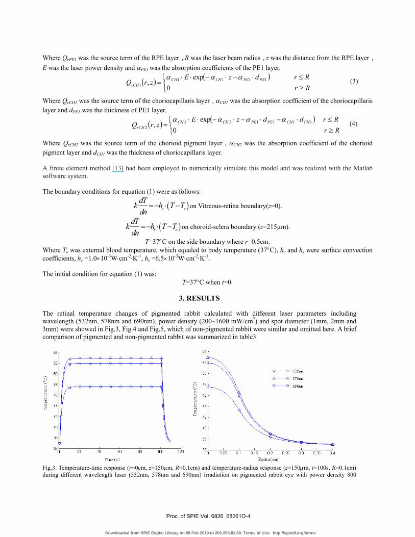
Where QrPE1 was the source term of the RPE layer,R was the laser beam radius,z was the distance from the RPE layer,E was the laser power density and αPE1 was the absorption coefficients of the PE1 layer.
( ) ( )⎩⎨⎧
≥≤⋅−⋅−⋅⋅
= 0
exp, 1111
1 RrRrdzE
zrQ PEPECHCHrCH
ααα (3)
Where QrCH1 was the source term of the choriocapillaris layer,αCH1 was the absorption coefficient of the choriocapillaris layer and dPE1 was the thickness of PE1 layer.
( ) ( )⎩⎨⎧
≥≤⋅−⋅−⋅−⋅⋅
= 0
exp, 111122
2 RrRrddzE
zrQ CHCHPEPECHCHrCH
αααα (4)
Where QrCH2 was the source term of the chorioid pigment layer,αCH2 was the absorption coefficient of the chorioid pigment layer and dCH1 was the thickness of choriocapillaris layer. A finite element method [13] had been employed to numerically simulate this model and was realized with the Matlab software system. The boundary conditions for equation (1) were as follows:
( )c edTk h T Tdn
=− ⋅ − on Vitreous-retina boundary(z=0).
( )s edTk h T Tdn
=− ⋅ − on choroid-sclera boundary (z=215µm).
T=37°C on the side boundary where r=0.5cm. Where Te was external blood temperature, which equaled to body temperature (37°C), hc and hs were surface convection coefficients, hc =1.0×10⁻³W⋅cm-2⋅K-1, hs =6.5×10⁻³W⋅cm-2⋅K-1. The initial condition for equation (1) was:
T=37°C when t=0.
3. RESULTS
The retinal temperature changes of pigmented rabbit calculated with different laser parameters including wavelength (532nm, 578nm and 690nm), power density (200~1600 mW/cm2) and spot diameter (1mm, 2mm and 3mm) were showed in Fig.3, Fig.4 and Fig.5, which of non-pigmented rabbit were similar and omitted here. A brief comparison of pigmented and non-pigmented rabbit was summarized in table3.
Fig.3. Temperature-time response (r=0cm, z=150µm, R=0.1cm) and temperature-radius response (z=150µm, t=100s, R=0.1cm) during different wavelength laser (532nm, 578nm and 690nm) irradiation on pigmented rabbit eye with power density 800
Proc. of SPIE Vol. 6826 68261O-4
Downloaded from SPIE Digital Library on 09 Feb 2010 to 202.204.81.80. Terms of Use: http://spiedl.org/terms

Tem
pera
twe (
CC
)
- 1mm2mme-o 2mm
0 0.06 0.1 0.16 0.2 0.26 0.3 0.36 0.4
Rathus(cm)
Tna
twe
(C)
Rathus(cm)
v-v 200nJ/0m1400nJ/0m1
—> 600nJ/0m1-< 800nJ/0m1— 1200nJ/0m2e-o lsOOnJ/0m2
mW/cm2
Fig.4. Temperature-time response (r=0cm, z=150µm, R=0.1cm) and temperature-radius response (z=150µm, t=100s,
R=0.1cm) during 532nm laser irradiation on pigmented rabbit eye with different power density (200~1600 mW/cm2).
Fig.5. Temperature-time response during 532nm laser irradiation on pigmented rabbit eye with different spot diameter (1mm, 2mm and 3mm) (r=0cm, z=150µm, power density 800 mW/cm2).
Table.3. Maximum temperature increase of pigmented and non-pigmented rabbit retina*
Temperature increase(oC) Wavelength (nm)
Power density (mW/cm2) Pigmented rabbit Non-pigmented rabbit
532 600 11.2 7.9 1200 22.4 15.8
578 600 12.0 9.4 1200 24.0 18.8
690 600 7.8 1.3 1200 15.6 2.6
* r=0cm, z=150µm, R=0.1cm
Results above showed that the temperature increased and reached heat balance in a few seconds following laser irradiation, then decreased to normal body temperature a few seconds after exposure. The temperature reduced quickly with the distance from beam center increased. The higher laser power density applied, the higher retinal temperature was induced. The temperature induced by 578nm laser was the highest, and 532nm laser was next to it. The temperature
Proc. of SPIE Vol. 6826 68261O-5
Downloaded from SPIE Digital Library on 09 Feb 2010 to 202.204.81.80. Terms of Use: http://spiedl.org/terms

induced by 690nm laser was lowest. The balanced temperature of retina rose with the increase of spot diameter. The maximum temperature increase of pigmented rabbit retina was higher than that of non-pigmented rabbit, especially when 690nm laser applied.
4. DISCUSSION
The laser therapy for retinal diseases is widely used in ophthalmology. The eye is the most vulnerable organ to laser damage especially the energy focused on macula area. Many researchers have studied the damage caused to the eye by heat sources. Birgruber [7] and other researchers [14-15] used a theoretical model to predict the thermal damage on retina photocoagulation. Their results declared retinal temperature could be increased tens of degrees and controlled by the average power, spot size, and exposure time. Since no theoretical work has been done on the unwanted thermal damage of retina during PDT, we present a computation work with finite element method to predict the thermal effect on the retina exposed to laser irradiation of PDT. The model will provide a useful guide for determining laser parameters during PDT, especially to establish the PDT schedule for new photosensitizers. The primary light absorbers in eye fundus are melanin and hemoglobin [16]. For shorter laser wavelength (532 nm or 578nm laser), the hemoglobin plays a main role on light-absorbing and temperature increase. While for longer laser wavelengths, such as diode red (690nm), the melanin mainly absorbs light and contributes to temperature increase. The green and yellow light are well absorbed, while red light is poorly absorbed by hemoglobin. So the temperature induced by green and yellow laser have no great difference and much higher than that induced by red laser, which indicates that the heat effect should be considered seriously if we select shorter laser wavelength (green and yellow laser) as new light source in PDT. With the increase of the spot size, the maximum retinal temperature rises too, which is more obvious when the laser spots are relatively small. Outside laser spot, retinal temperature decreases quickly with the radius. That means that thermal effect is limited in the area of laser spot and can hardly damage the surrounding tissue. For laser parameters used to treat choroidal neovascularization (690nm, 600mW, 2mm, 83sec), the maximum retinal temperature calculated is less than 45oC, which indicates no thermal damage induced. Our results also indicate that doctors should be careful in selecting laser parameters to treat the patient whose pigments of eye fundus are heavy, because of obviously higher temperature in pigmented than non-pigmented rabbit retina especially for 690nm laser during PDT for CNV. In our previous experiment (unpublished), 532nm laser threshold power settings for visible lesions in non-pigmented and pigmented rabbits are 1200mW/cm2 and 800mW/cm2 respectively, with the exposure duration of 100s. Corresponding to threshold power settings, retinal temperature of non-pigmented rabbits and pigmented rabbits theoretically increases to 52.8°C and 51.9°C respectively. These indicated that our mathematical model may correctly calculate the temperature induced by irradiation of different laser parameters on non-pigmented and pigmented rabbits. In summary, laser induced retina thermal effect is strongly influenced by laser power density and light absorbers of fundus. The retinal temperature increase during laser PDT is proportional to retinal power density. The temperature induced by 578nm laser is the highest, and 532nm laser is next to it. The temperature induced by 690nm laser is lowest. The balanced temperature of retina rises with the increase of spot diameter. The maximum temperature increase of pigmented rabbit retina is higher than that of non-pigmented rabbit, especially when 690nm laser applied. For laser parameters used to treat choroidal neovascularization, the maximum retinal temperature calculated is less than 45oC, which indicates no thermal damage induced.
REFERENCES
1. Michels S, Schmidt-Erfurth U, “Photodynamic therapy with verteporfin: A new treatment in ophthalmology,” Semin. Ophthalmol. 16(4), 201-206(2001).
2. van den Bergh H, Ballini JP, Sickenberg M, “On the selectivity of photodynamic therapy of choroidal neovascularization associated with age-related macular degeneration,” J. Fr. Ophtalm. 27(1), 75-78(2004).
Proc. of SPIE Vol. 6826 68261O-6
Downloaded from SPIE Digital Library on 09 Feb 2010 to 202.204.81.80. Terms of Use: http://spiedl.org/terms

3. Ursula Schmidt-Erfurth, Tayyaba Hasan, “Mechanisms of Action of Photodynamic Therapy with Verteporfin for the Treatment of Age-Related Macular Degeneration,” Survey of Ophthalmology. 45(3),195-214(2000)
4. Cain CP, Welch AJ, “Measure and predicted laser-induced temperature rises in the rabbit fundus,” Invest. Ophthalmol. 13(1), 60-70(1974).
5. Priebe LA, Cain CP, Welch AJ, “Temperature rises required for production of minimal lesions in the Macaca mulatta retina,” Am. J. Ophthalmol. 79, 405-413(1975).
6. Polhamus GD, Welch AJ, “Threshold lesion temperature in argon laser-irradiated rabbit eyes,” J. Heat. Transfer. 97, 457-462(1975).
7. Birngruber R, Hillenkamp F, Gabel V P, “Theoretical Investigations of Laser Thermal Retinal Injury,” Health. Physics. 48(6), 781-796(1985).
8. Parver LM, “Temperature modulating action of choroidal blood flow,” Eye. 5(Pt2), 181-185(1991). 9. Pennes H. H, “Analysis of Tissue and arterial blood temperatures in the resting human forearm,” J. Appli.
Physiol, 1(2), 93-122(1948). 10. Thompson CR, Gerstman BS, Jacques SL, Rogers ME, “Melanin Granule Model for Laser-induced
Thermal in the Retina,” Bull. Math. Biol. 58(3),513-553(1996). 11. Verkruysse W, Pickering JW, Beek JF, Keijzer M, van Gemert MJC, “Modelling the effect of wavelength
on the pulsed dye laser treatment of port waine stains,” Applied. Optics.32(4),393-398(1993). 12. Kim BM, Jacques SL, Rastegar S, Thomsen S, Motamedi M, “Nonlinear Finite-Element Analysis of the
Role of Dynamic Changes in Blood Perfusion and Optical Properties in Laser Coagulation of Tissue,” IEEE Journal on Selected Topics In Quantum Electronics. 2, 922-933(1996).
13. Scott JA, “A finite element model of heat transport in the human eye,” Phys. ed. Biol. 33(2), 227-241(1988).
14. Takata AN, “Thermal model of laser induced eye damage”. Final Technical Report J-TR-74-6324, Illinois Institute of Technology Research Institute.
15. Gabay S, Kremer I, Ben-Sira I, Erez G, “Retinal Thermal Response to Copper-Vapor Laser Exposure,” Lasers in Surgery and Medicine. 8(4), 418~427(1988).
16. Mainster MA, “Wavelength selection in macular photocoagulation,” Ophthalmology. 93(7), 952-958(1986).
Proc. of SPIE Vol. 6826 68261O-7
Downloaded from SPIE Digital Library on 09 Feb 2010 to 202.204.81.80. Terms of Use: http://spiedl.org/terms