Involvement of Foxo transcription factors in angiogenesis and postnatal neovascularization
Upload
independentCategory
view
0download
0
ASIP
Journal
CME Program
Vascular Biology, Atherosclerosis and Endothelium Biology
Hematopoietic Stem Cells Provide Repair Functionsafter Laser-Induced Bruch’s Membrane RuptureModel of Choroidal Neovascularization
Tailoi Chan-Ling,* Louise Baxter,* Aqeela Afzal,†
Nilanjana Sengupta,† Sergio Caballero,†
Emilia Rosinova,* and Maria B. Grant†
From the Department of Anatomy and Histology,* Institute for
Biomedical Research, University of Sydney, Sydney, Australia;
and the Department of Pharmacology and Therapeutics,†
University of Florida, Gainesville, Florida
Vascular repair by adult hematopoietic stem cells(HSCs) is well-appreciated because these cells areknown for their plasticity. We have shown that adultHSCs differentiate into endothelial cells and partici-pate in both retinal and choroidal neovascularization.We asked whether HSCs participated in the woundingresponse by forming astrocytes, retinal pigment epi-thelia (RPE), macrophages, and pericytes. Lethallyirradiated C57BL6/J mice were reconstituted withHSCs from mice homozygous for green fluorescentprotein (GFP) and then subjected to laser-inducedrupture of Bruch’s membrane. After immunohisto-chemical examination of ocular tissue, GFP� astro-cytes were observed concentrated along the edge ofthe laser wound, where they and mural cells closelyensheathed the neovasculature. GFP� vascular endo-thelial cells and macrophages/microglia were also ev-ident. Large irregularly shaped GFP� RPE cells consti-tuted �93% of RPE cells adjacent to the edge of thedenuded RPE area. In regions farther away from thewound, GFP� RPE cells were integrated among theGFP� host RPE. Thus, postnatal HSCs can differentiateinto cells expressing markers specific to astrocytes,macrophages/microglia, mural cells, or RPE. Thesestudies suggest that HSCs could serve as a therapeuticsource for long-term regeneration of injured retinaand choroid in diseases such as age-related maculardegeneration and retinitis pigmentosa. (Am J Pathol2006, 168:1031–1044; DOI: 10.2353/ajpath.2006.050697)
The hematopoietic stem cell (HSC) is the most robust,highly characterized stem cell of the body. From the initial
studies showing that irradiated mice could be rescuedfrom death with bone marrow transplants from a healthydonor, it became clear that HSCs could self-renew andproduce all of the different cells in the blood.1,2 Approx-imately 1 of every 10,000 cells in the bone marrow is astem cell and is defined as being Sca-1�, c-kit�, andlineage-negative (SKL), with �1 of 10 SKL cells thoughtto be a true HSC that can traffic to the bone marrow andperform HSC functions.3
One main HSC function involves new blood vesselformation. Angiogenesis represents proliferation and mi-gration of pre-existing fully differentiated endothelial cellsthat reside within parent vessels.4 The finding that bonemarrow-derived cells may traffic to sites of neovascular-ization and differentiate is consistent with vasculogen-esis,5 a critical paradigm for the establishment of vascu-lar networks in the embryo. Asahara and colleagues6
identified the cells derived from the bone marrow capa-ble of differentiating into neovasculature. Injection ofHSCs into mice accelerates revascularization of ischemiclimbs7 and wound healing8; thus, these cells are pre-sumed to be key mediators of endothelial repair. Previ-ously, we demonstrated that HSCs differentiate into en-dothelial cells and participate with resident endothelialcells to form neovascularization in the retina, choroid, andiris.9–11
Stem cells are self-renewable, pluripotent cells that inadult life proliferate by a characteristic asymmetric divi-sion in which one daughter cell is committed to differen-tiation whereas the other remains a stem cell. These cellsare also characterized by their ability to differentiate intovarious cell types under heterotypic environmental influ-
Supported by the National Health and Medical Research Council ofAustralia (153789 to T.C.L.), the Rebecca Cooper Foundation for MedicalResearch (to T.C.L.), the National Institutes of Health (National Eye Insti-tute EY012601 and EY007739 to M.B.G.), and Juvenile Diabetes Re-search Foundation (JDF4-2000-847 to M.B.G.).
Accepted for publication November 23, 2005.
Address reprint requests to Maria Grant, M.D., Professor of Pharmacol-ogy and Therapeutics, University of Florida, Academic Research Bldg.,ARB5-252, 1600 SW Archer Rd., Gainesville FL 32610. E-mail: [email protected].
American Journal of Pathology, Vol. 168, No. 3, March 2006
Copyright © American Society for Investigative Pathology
DOI: 10.2353/ajpath.2006.050697
1031

1032 Chan-Ling et alAJP March 2006, Vol. 168, No. 3

ences. Plasticity refers to the ability of stem cells to ac-quire a mature phenotype that differs from the tissue oforigin.12 This is true for both embryonic and adult stemcells. Adult bone marrow-derived stem cells, either HSCsor mesenchymal stem cells, are multipotent and havebeen shown to engraft and express properties of othertissues. HSCs have been shown to transdifferentiate intocell types such as epithelium,13 myocardium,14,15 andliver.16
Several studies support the potential of neural stemcells to differentiate into ocular cells such as retinal pig-ment epithelial (RPE) cells when exposed to RPE-condi-tioned medium.17 These cells were able to attenuate theloss of photoreceptors when transplanted subretinallyinto Royal College of Surgeons (RCS) rats. This preser-vation was believed to be mediated either by the cell’sability to phagocytose the host’s outer segments or by itsability to secrete soluble factors.18 To date, however,HSCs have not been shown to differentiate into RPE cellsin vitro or in vivo.
Recent studies have suggested that circulating bonemarrow cells can traffic to brain and transdifferentiate intoneural cells. Bonilla and co-workers19 have explored thepotential of adult hematopoietic bone marrow cells todifferentiate into cells of oligodendroglial lineage underphysiological active-myelinating conditions. They dem-onstrated that cells from a bone marrow subpopulationenriched in adult hematopoietic progenitor cells (c-kit�)express oligodendroglial-specific markers in vivo afterintracerebral transplantation into the neonatal mousebrain. Their results suggest that adult bone marrow cellshave the capacity to undergo differentiation from hema-topoietic to oligodendroglial cells and support the validityof bone marrow transplantation as an alternative treat-ment for demyelinating diseases of the central nervoussystem, including multiple sclerosis.
Bone marrow cells traffic to sites of injury becausethey are attracted by locally produced growth factorsand/or cytokines, including stromal-derived growthfactor-1 (SDF-1).20 –23 RPE cells express SDF-1 duringhealthy conditions and after injury.24 If an SDF-1 gra-dient is established, cells traffic more efficiently. Con-versely, if this gradient is blocked, homing efficiency isreduced to negligible levels.25 Studies illustrate thatHSCs and progenitor cells are sensitive to SDF-1, sup-porting the view that this chemokine is important forstem and progenitor cell migration in both embryonicand adult tissues.26
Although vascular repair by adult HSCs is well appre-ciated, these cells are also known for their plasticity andcan generate liver, brain, muscle, and fat.12,16,27–29 We
therefore asked whether circulating HSCs have the po-tential not only to form endothelial cells and participate inchoroidal neovascularization (CNV) but also to becomeother cell types that participate in the development of theCNV lesion after rupture of Bruch’s membrane. A CNVlesion, whether occurring clinically or after mechanicalinjury, typically contains numerous cell types that partic-ipate in a type of wound healing reaction in the subretinalspace. We postulated that the injured subretinal spacewould provide cues to mobilize HSCs to this region andpromote their formation into cell types other than endo-thelial cells, such as RPE, pericytes, smooth muscle cells,astrocytes, and macrophages.
Materials and Methods
Generation of GFP� Chimeric Mice
All procedures were conducted in accordance with theAssociation for Research in Vision and OphthalmologyStatement for the Use of Animals in Ophthalmic andVisual Research and were approved by the University ofSydney Animal Ethics Committee. Reconstitution of le-thally irradiated C57BL6/J mice with HSCs from GFP�/�
donors was performed as previously described by us.9,10
Induction of CNV lesions by laser rupture of Bruch’smembrane was done as previously described.10
Tissue Preparation and Immunohistochemistry
Retinas were fixed by immersion in 4% paraformalde-hyde in 0.1 mol/L phosphate buffer for 1 hour at 4°C.Retinal whole-mounts were prepared as described pre-viously.30 Triple-marker immunohistochemistry was usedto co-visualize astrocytes (rabbit polyclonal antibodyS-100� from Chemicon, Temecula, CA), mural cells (rab-bit polyclonal antibody NG2 from Chemicon), GFP� cells(anti-GFP from Chemicon), vasculature (GS isolectin B4,Sigma, St. Louis, MO), and RPE (anti-RPE65, gift fromJ.-X. Ma [Department of Medicine, Oklahoma University,Oklahoma City, OK]; cytokeratin 8/18, clone SD3 fromNovocastra, Newcastle-upon-Tyne, UK).
A maximum of three primary antibodies were appliedto each specimen to determine the progeny of trans-planted GFP� cells in either retinal whole mounts orRPE/choroidal eyecups in treated and untreated tissue. Aminimum of three specimens were analyzed per de-scribed phenomenon, and conclusions reached wererepresentative of all specimens examined. Retinal wholemounts were permeabilized with 0.1% Triton X-100 in
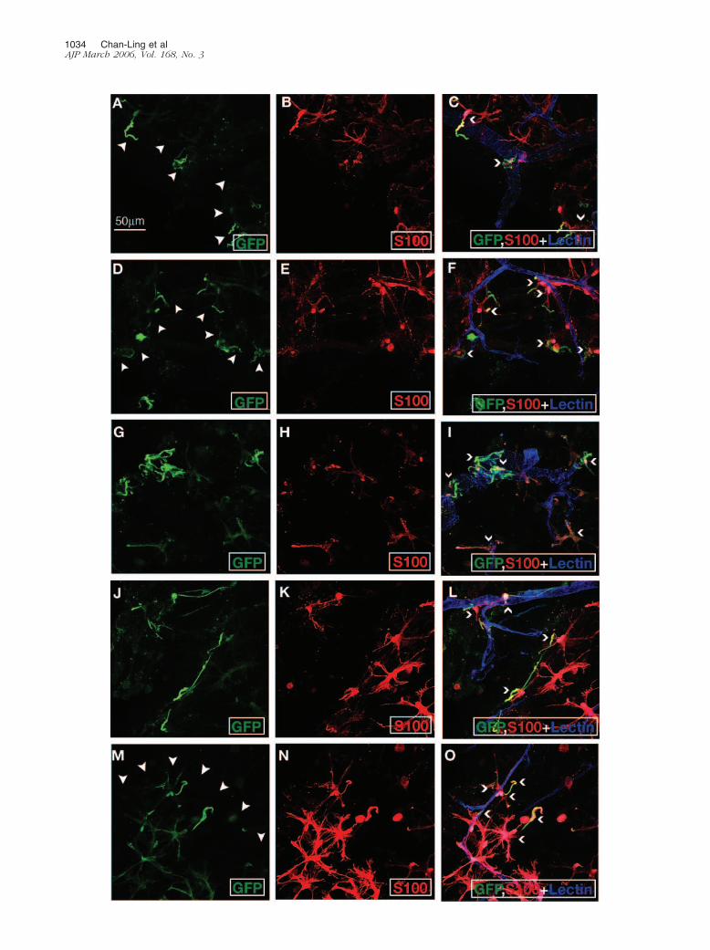
Figure 1. HSCs differentiate into S-100�� astrocytes in a rodent model of CNV. A–C and D–F show cells positive for GFP (A and D), S-100� (B and E), and GFP,S-100�, and GS isolectin (C and F). A–C show that GFP� HSCs give rise to S-100�� astrocytes with predominantly stellate morphology. Brighter GFP�/S-100��
astrocytes with a simple bipolar morphology are observed interspersed among the GFP� stellate astrocytes. This field of view shows the morphology ofincorporated GFP� astrocytes in mid-retina that are some distance from the lesion. Remarkably, these GFP�/S-100�� astrocytes closely ensheath the retinal bloodvessels as seen in normal retinas. These GFP� astrocytes are more concentrated in the region of the laser-induced wound. Although numerous S-100�� astrocytesare evident throughout the retina, GFP�/S-100�� astrocytes are absent in the most peripheral areas of the experimental eye. D and E show a region of anexperimental eye adjacent to the laser-induced wound (arrowheads point along margin of wound). Astrocytes in this area were observed with a predominantorientation along the wound edge. F shows that GS isolectin� neovascularization entering the deeper layers of the mouse retina was evident at the wound edge(arrows). GFP�/S-100�� astrocytes re-establish normal cell-cell relationships in the inner retina; however, many of the neovascular tufts (blue) in the outer retinalack close association with S-100�� astrocytes. Representative GFP�/S-100� astrocytes are shown by open arrowheads in C and F.
Stem Cells Provide Ocular Repair After Injury 1033AJP March 2006, Vol. 168, No. 3
1034 Chan-Ling et alAJP March 2006, Vol. 168, No. 3

phosphate-buffered saline (PBS) for anti-GFP and 1%Triton X-100 in PBS for S-100B�, GFAP, and GS isolectin,and samples were then blocked in 1% bovine serumalbumin in PBS. Retinas were incubated overnight at 4°Cwith primary antibodies, washed with 0.1% Triton X-100 inPBS, incubated for 4 hours at room temperature with theappropriate secondary antibodies, and washed again.For double or triple labeling, this procedure was repeatedwith the different primary antibodies and appropriate sec-ondary antibodies. Negative controls omitting a primaryantibody were performed for each antibody and protocol.Vessels were labeled with biotinylated GS isolectin fol-lowed by streptavidin conjugated with Cy5 (Jackson Lab-oratories, Bar Harbor, ME). All antibodies and streptavi-din were diluted with 1% bovine serum albumin in PBS,and washes were performed with 0.1% Triton X-100 inPBS. Retinal whole-mounts were finally mounted with theganglion cell layer up in glycerol:PBS (2:1, v/v) or ProlongAnti-Fade (Molecular Probes, Eugene, OR).
Microscopy and Mapping
Retinal whole-mounts were examined by both deconvo-lution and confocal microscopy. For deconvolution fluo-rescence microscopy and photography, we used a Zeissmicroscope (model Axioplan 2 attachment HBO 100) andAxiocam HRm camera. Confocal microscopy was per-formed with a Leica argon-krypton laser mounted on aLeica DMRBE epifluorescence photomicroscope. AlexaFluor 488, Cy3, and Cy5 fluorescence was excited se-quentially at 488, 550, and 649 nm, respectively. Imageswere processed with Adobe Photoshop V5.0 software.
Results
HSCs Generate Astrocytes
In regions at or immediately adjacent to the wound, GFP�
HSCs gave rise to S-100�� astrocytes with predomi-nantly stellate morphology (Figure 1, A, B, D, and E) astypically observed in adult mouse retina. Brighter GFP�/S-100�� astrocytes with a simple bipolar morphologywere observed interspersed among the GFP� stellateastrocytes (Figure 1, A and B; Figure 2, A–F, M–O).GFP�/S-100�� astrocytes (Figure 1, D–F) were observedin patches concentrated along the edge of the laserwound (Figure 1D, arrowheads) and were not evident inthe control eye. This concentration along the wound edgeis suggestive of the ability of HSCs to traffic to sites ofneovascularization and exert some rescue effects. Themorphology of these GFP�/S-100�� astrocytes at the
edge of a wound was typically simple bipolar (Figure 1, Cand F; Figure 2, A–F). Remarkably, astrocyte precursorcells also have a simple bipolar morphology during nor-mal development,31 suggestive of a more immature phe-notype along the astrocytic differentiation pathway.
Figure 1F shows an area of retinal neovascularizationbecause retinal blood vessels are not normally found inthese layers or with this morphology (Figure 1F, arrows).These GFP� astrocytes closely ensheathed some of thevasculature in the superficial vascular plexus (Figure 1F;Figure 2, G–L), mimicking the close ensheathment ofretinal blood vessels by astrocytes seen normally. Onexamination at higher magnification, some neovascularsegments lacked a continuous astrocytic ensheathmentas would be found in a normal adult mouse retina (Figure2, D–F and J–L). Remarkably, the neovasculature in thedeeper layers lacked any S-100�� astrocytic ensheath-ment in this field of view (Figure 1F).
A quantitative analysis of the number of GFP�/S-100��
astrocytes within 300 �m of the lesion site showed amean of 21.0 cells/mm2 (SD � 13.1) determined overeight fields of view using a �16 objective. The numbersof GFP�/S-100�� astrocytes showed significant variationwith the field of view examined, where the actual countsin the eight fields of view were 18, 18, 10, 25, 51, 18, 10,and 18 cells/mm2. The average number of GFP�/S-100�� astrocytes farther than 300 �m away from lesionswas 1.3 (SD � 2.9, in eight fields of view at �16). Theactual counts in the eight fields of view examined were 8,0, 0, 0, 3, 0, 0, and 0 cells/mm2. Although the majority ofHSC-derived (GFP�/S-100��) astrocytes appeared toconcentrate at the wound margin, a lower number ofHSC-derived astrocytes can incorporate into the existingsyncytium of resident astrocytes even at distancesgreater than 300 �m from the edge of the wound. As anegative control, we reacted retinas from control B57/BL6/J mice with an antibody against GFP. No GFP� cellswere evident throughout the entire retina. Examination ofthe nontreated eyes in the chimeric mouse using anantibody to GFP revealed small numbers of GFP� vas-cular endothelial cells (VECs).
HSCs Generate Activated RetinalMicroglia/Macrophages
Activated microglia/macrophages in the retina are char-acterized by an amoeboid morphology, enhanced ex-pression of Griffonia simplicifolia (GS) isolectin B4, and anoncontact spacing pattern of distribution. NumerousGFP�/GS isolectin B4� macrophages/microglia were ev-ident in the experimental retina (Figure 3). These cells
Figure 2. Higher magnification views of HSCs that differentiated into astrocytes in a rodent model of CNV. Although HSC-derived astrocytes ensheathed numerousnew blood vessels, astrocytic ensheathment of these new vessels was often incomplete, as shown throughout this panel. A, D, G, J, and M show GFP fluorescence.B, E, H, K, and N show S-100� staining. C, F, I, L, and O show triple-labeling with multiple exposures showing GFP/S-100� and GS isolectin. A–C show apreponderance of bipolar astrocytes aligned along the wound edge. Because bipolar astrocytes are indicative of a more immature astrocyte phenotype, thisobservation is supportive of HSCs giving rise to immature astrocytes after laser-induced CNV. D–F, G–I, J–L, and M–O show other examples of neovascularizationafter laser-induced Bruch’s membrane rupture. The morphology of the vessels is not typically observed in normal mouse retina. This retinal neovasculature isclosely ensheathed by GFP�/S-100�� astrocytes, supportive of the role played by HSC-derived astrocytes in the wound healing process and induction ofblood-retinal barrier properties. Representative GFP�/S-100� astrocytes are shown by open arrowheads in C, F, I, L, and O. J: Small numbers of GFP�, but notS-100�, cells were evident. Because S-100 is only expressed by mature astrocytes, the GFP� astrocyte-like cells on the left side of this field of view could representastrocyte precursor cells before S-100 expression.
Stem Cells Provide Ocular Repair After Injury 1035AJP March 2006, Vol. 168, No. 3

Figure 3. HSCs differentiate into VECs and macrophages/microglia in a rodent model of CNV. A, D, G, and J show GFP fluorescence. B shows S-100� staining.C shows triple labeling with multiple exposures showing GFP/S-100/GS-isolectin. A–C show isolated incorporation of GFP� HSCs into GS isolectin� VECs(arrowheads) and GS isolectin� macrophages/microglia (arrow). D–F show isolated incorporation of GFP� HSCs into GS isolectin� VECs (arrowheads) andmacrophages/microglia (arrows) in the superficial retinal plexus in the peripheral region of the experimental retina. G–I show GFP�, but weakly GS isolectin�,activated macrophages/microglia with a regular noncontact spacing distribution throughout the retinal parenchyma in the laser-injured eye. J–L show GFP�, butweakly GS isolectin�, vascular-associated activated macrophages/microglia in the retina of the laser-injured eye.
1036 Chan-Ling et alAJP March 2006, Vol. 168, No. 3
were regularly distributed and had an amoeboid mor-phology (Figure 3G).
Significant populations of GFP�/GS isolectin B4� mac-rophages/microglia were evident over the entire experi-mental retina, with a slightly higher frequency of distribu-tion in the periphery of the retina (Figure 3). These cellswere distributed in a noncontact spacing pattern typicalof macrophages/microglia. Such HSC-derived macro-phages/microglia were often associated with retinalblood vessels (Figure 3, J–L). A resident population ofGFP�/GS isolectin B4� macrophages/microglia per-sisted in the experimental retinas (Figure 3I, arrowheads).
HSCs Generate RPE
HSCs differentiated into RPE after laser-induced Bruch’smembrane rupture. Closure of the wound was incompleteat time of sacrifice 3 weeks after wounding (Figure 4A). Inthe area adjacent to the denuded area, RPE cells werepresent with significantly higher levels of GFP expressionthan surrounding areas (Figure 4, B–D). A small propor-tion of these cells had enlarged cell surface area and lessregularly distributed pigment granules, resulting in irreg-ularity to the typical hexagonal mosaic pattern. In con-trast, in regions that were a substantial distance awayfrom the wound (Figure 4E) or in the periphery (Figure4F), fewer GFP� RPE cells were present. These GFP�
RPE cells were smaller and more regular in appearanceand formed a heterogeneous hexagonal array. HSC-de-rived GFP� RPE cells integrated among the host RPEcells, demonstrating patches of GFP� and patches ofGFP� RPE cells adjacent to each other. These observa-tions suggest that HSCs can differentiate into RPE andparticipate in wound healing in regions of laser-inducedRPE damage.
As controls, posterior cups from C57BL6/J mice weresubjected to GFP/GS isolectin double-label immunohis-tochemistry. Figure 4G shows the low level of GFP fluo-rescence and the regularity of the RPE mosaic in theseeyes. Further, RPE cells in control C57BL6/J miceautofluoresce. RPE autofluorescence in control animalshas been reported and is due to the presence of second-ary inclusion bodies, such as lipofuscin, which havebroad-band autofluorescence.32 In addition, the pres-ence of other fluorophores such as retinoids, flavins, andcytochrome contribute to RPE autofluorescence. Al-though this level is much lower than that observed in theGFP� RPE cells (Figure 4, H–J), the level of autofluores-cence could be detected and captured photographically.Further, confirming earlier reports, double-nucleated RPEcells were also occasionally observed in controlC57BL6/J mice (Figure 4, I and J). Double-nucleatedRPE cells could be interpreted as cell fusion or phago-cytosis of adjacent cells as opposed to HSC-generatingRPE cells.
Two RPE-specific markers, RPE-65 and cytokeratin8/18, were also applied to the laser-induced CNV retinasto provide further support for the conclusion that GFP�
HSCs generate RPE. All RPE cells were positive forRPE-65 and cytokeratin 8/18. Figure 5, A–C, shows
GFP�/RPE-65� HSC-derived RPE cells in the region ofthe wound whereas Figure 5, D–F, shows GFP�/RPE-65�
HSC-derived RPE cells at the edge of the eyecup. It isunclear why the majority of GFP� showed a markedincrease in the expression of RPE-65. Figure 5, G–I,shows GFP�/cytokeratin 8/18� HSC-derived RPE cells inthe region of the wound. Cytokeratin bundles outline theperiphery of all cuboidal RPE cells. The nuclei and largermelanin granules impart a finely vacuolated appearanceto the less intensely immunopositive central cytoplasm.
We undertook a quantitative analysis of the number ofGFP� RPE cells in two regions: within 400 �m of thelesion site and in a region just internal to the edge of theeyecup. Four fields of view were counted per regionusing the �20 objective on the Zeiss deconvolution mi-croscope, resulting in �1400 cells being counted perregion. A representative field of view adjacent to thelesion is shown in Figure 4A. The average number ofGFP� RPE cells adjacent to the lesions was 1939 � 488cells/mm2. The average number of GFP� RPE cells ad-jacent to the lesions was 173 � 116 cells/mm2. Thisequates to a mean of 92.5 � 3.4% of RPE cells adjacentto the lesion being derived from GFP� HSCs. The aver-age number of GFP� RPE cells at the edge of the eyecupwas 1010 � 189 cells/mm2. The average number ofGFP� RPE cells at the edge of the eyecup was 1801 �372 cells/mm2. This equates to a mean of 36.2 � 6.8% ofRPE cells at the edge of the eyecup being derived fromGFP� HSCs. No GFP� RPE cells were evident in naıveC57BL6/J mice.
Although HSC-derived GFP� RPE cells concentratedat the wound margin, a significant number (36% of allRPE cells) also incorporated into the existing network ofresident RPE cells at the periphery of the eyecup. Thisfinding is consistent with our current understanding thatthe RPE is the site where the energy from the laser isabsorbed and dissipated. Thus, it would be expectedthat the size of RPE wounds exceeds the actual laser spotsize and suggests that laser injury stimulates HSC recruit-ment well beyond the region of Bruch’s membranerupture.
HSCs Generate VECs in the Retina and Choroid
A small number of GFP�/GS isolectin B4� vascular en-dothelial strands were evident in the superficial vascularplexus in the experimental retina (Figure 3, A and C),suggesting the possibility of laser-stimulated recruitmentinto these regions. GFP� VECs were observed in regionswhere HSC-derived astrocytes (Figure 3B) and macro-phages (Figure 3, A and C) were also evident. Remark-ably, GFP� VECs were more frequently observed in theouter retinal vascular plexus (data not shown), as onemight expect, because the outer plexus is closer to thesite of laser burn. In support of this observation, wide-spread incorporation of GFP�/GS isolectin� VECs wasevident in the choriocapillaris (Figure 6, A–D).
Stem Cells Provide Ocular Repair After Injury 1037AJP March 2006, Vol. 168, No. 3

1038 Chan-Ling et alAJP March 2006, Vol. 168, No. 3

Figure 5. A–C: Field of view immediately adjacent to the denuded region of the RPE double-labeled with anti-GFP and RPE-65. The denuded region is just superior toand left of the area shown. A: GFP� RPE cells are evident throughout this field of view. B: All cells in this field of view are RPE-65�, confirming that GFP� cells are RPEcells. C: Co-localization of GFP and RPE-65 show that the two markers co-localize to the same population. D–F: Field of view at the edge of the eyecup showing far fewerGFP� RPE cells double-labeled with anti-GFP and RPE-65. RPE morphology of host-derived RPE cells away from the wound shows a much smaller nuclear size comparedto GFP� HSC-derived RPE cells. D: Very few GFP� RPE cells are evident throughout this field of view. E: All cells in this field of view are RPE-65�, confirming that GFP�
cells are RPE cells. F: Co-localization of GFP and RPE-65 shows that the two markers co-localize to the same population. G–I: Field of view adjacent to the denuded regionof the RPE double-labeled with anti-GFP and cytokeratin 8/18. The denuded region is just superior to and left of the area shown. Cytokeratin bundles outline the peripheryof cuboidal RPE cells. The nuclei and larger melanin granules impart a finely vacuolated appearance to the less intensely immunopositive central cytoplasm. G: GFP�
RPE cells are evident throughout this field of view with a small number of GFP� cells evident in the lower left area of the field of view. H: Co-localization of GFP andcytokeratin 8/18 show that the two markers co-localize to the same population.
Figure 4. HSCs differentiate into RPE in a rodent model of CNV. A is a low-magnification view of the GFP fluorescence in the region surrounding a laser-inducedrupture of Bruch’s membrane. Closure of the laser-induced RPE wound was incomplete at 3 weeks after wounding. The white boxes indicate the area wherethe various fields of view were captured at higher magnification. B–D show high-magnification views of GFP� RPE cells taken at various regions in the proximityof the laser-induced wound. RPE cells in these regions showed a marked variation in fluorescence intensity. Some regions are brightly fluorescent, correlating withregions of cell heterogeneity (ie, large and small cells and double-nucleated cells) while other areas have homogeneous levels of fluorescence and RPEmorphology. C shows maximal irregularity of RPE mosaic and frequent occurrence of double-nucleated RPE in a region adjacent to the wound in the experimentaleye. D shows a region adjacent to the wound with marked irregularity of the pigment granules. E shows RPE cells with a much lower level of fluorescence andmore homogeneous morphology some distance away from the wound of the second experimental eye. F shows a higher magnification view of the RPE at theperiphery in an experimental eye, with low levels of GFP� fluorescence and homogenous RPE morphology. G shows the level of fluorescence in a C57B6/J controlmouse reacted with GFP/S-100�/GS isolectin triple-label immunohistochemistry. The level of GFP fluorescence is extremely low and homogeneous. H–J showlow- and high-magnification views showing the more regular morphology and low level of autofluorescence of the RPE in control C57B6/J mice without anyimmunohistochemical labeling. The RPE cell outlines are only marginally visible above background fluorescence. Double-nucleated RPE cells were also evidentin this control tissue.of macrophages/microglia. Such HSC-derived macrophages/microglia were often associated with retinal blood vessels (Figure 3, J–L). Aresident population of GFP�/GS isolectin B4� macrophages/microglia persisted in the experimental retinas (Figure 3I, arrowheads).
Stem Cells Provide Ocular Repair After Injury 1039AJP March 2006, Vol. 168, No. 3

1040 Chan-Ling et alAJP March 2006, Vol. 168, No. 3
HSCs Express Markers and Morphology ofNG2� Mural Cells
Examination of the posterior cups in the laser-inducedBruch’s membrane rupture model showed obvious tran-sitional zones of HSC incorporation, evident as a demar-cation zone of bright and dim GFP immunofluorescence(Figure 6, E and F). HSC-derived mural cells were evidentas GFP�/NG2� somas on the outside of choroidalvenules (Figure 6, G–L) and choroidal arterioles (Figure6, M–R). The arteriole shown in Figure 6, O–R, was ob-served directly under a region of the choriocapillaris withlarge numbers of HSC-derived VECs.
Discussion
The CNV model used for these studies represents amodel of acute repair and as such the HSC behaviorwe observed must be considered in this context. Onemay speculate that the stimuli for HSC recruitment inthis laser-induced injury model may be more dramaticor abrupt compared to the more insidious stimuli as-sociated with a chronic disease such as age-relatedmacular degeneration. Thus, what we observe mayrepresent a phenomenon associated with the laserinjury conditions; however, we believe that this repre-sents a plausible mechanism of repair throughout theretina and choroid and a possible avenue that needs tobe explored therapeutically. Several caveats must bekept in mind however. For example, the transplantationapproach used is one that enriches for HSCs and doesnot reflect normal bone marrow homeostasis. Trans-plantation is accomplished by using Sca-1�/c-kit�
stem cells. Although these cells are accepted as en-dothelial precursor cells, their plasticity is also wellappreciated.
The importance of HSC plasticity has been recognizedin the context of many injured tissues.33,34 It is well ac-cepted that the local environment ultimately dictates thefate of the HSCs after local injury. The environment sendsboth attractive cues and repulsive cues to HSCs to targetthe cells to exactly the correct location. Stimulatory sig-nals for attracting HSCs to the subretinal space mayinclude changes in local growth factor expression. Cuesfor amplification of HSC number in the appropriate loca-tion are also active. However, the precise forces control-ling the transition of these cells to their new phenotyperemain elusive.
In general terms, after the initial activation of the HSCsfrom the bone marrow sinusoids to the circulation, thecells are not only stimulated to migrate to the area ofischemia/injury but are activated to proliferate in this re-gion. After these initial steps HSCs are stimulated to
differentiate and finally integrate into the area forming thepostlaser wound. After integration, the HSCs are ex-pected to have proper function and participate in repairof this subretinal tissue and restore it to normal function.Not only are the cells destined for a particular cellularfate, they must be committed to the positional fate in therepaired tissue.
Ikehara and co-workers35 have recently demonstratedthat bone marrow stem cells can differentiate into retinalnerve cells. When these cells were injected into the vit-reous cavity of untreated eyes, very few cells were foundin the retina 2 weeks after injection. In contrast, when theyperformed laser photocoagulation before the injection ofthe cells, a large number of them survived 2 weeks afterinjection and the cells expressed neural cell-specific orretinal nerve cell-specific antigens. These cells remainedviable in the retina 1 year after injection.35 These sameauthors performed laser photocoagulation to induce CNVin GFP-chimeric mice. They corroborated our findings10
as well as those of others36,37 that the vascular wall cellsof the CNV lesion expressed both GFP and CD31. Thisdemonstrates that newly developed blood vessels in theCNV are derived from the bone marrow cells.
The studies presented here extend these observationsby demonstrating more rigorous repair function. The in-corporation of GFP� cells with RPE morphology into thehealing lesion has important consequences to repair ofchronically injured retina and to the natural turnover ormaintenance of the RPE layer. There may be areas of theRPE where the HSCs preferentially migrate into and es-tablish contact with resident RPE because the RPE hasbeen shown to be composed of a heterogeneouspopulation.
The clinical significance of this study lies in the obser-vation that HSCs can be recruited into an area of injuryand can generate the unique cell populations required forrepair of the injured tissue. Astrocytes contribute directlyto the formation and maintenance of functional barrierproperties by inducing the formation of tight junctionsbetween VECs.38 Astrocytes are intimately associatedwith the vasculature during development of the mamma-lian retina39 where astrocyte migration precedes the for-mation of patent vessels during normal development ofthe human retina.40,41 Thus, ensheathment of the neovas-culature by HSC-derived astrocytes should lead to im-proved barrier properties of the neovasculature afterlaser-induced CNV.
The main function of HSCs is one of repair becausethese cells are exquisitely sensitive to the cues frominjured tissue throughout the body, much like white bloodcells are in constant surveillance for infection within atissue. The signal from injured tissue results in the recruit-ment of HSCs to these injured areas for repair, and thus
Figure 6. HSCs differentiate into choroidal VECs and choroidal mural cells in a rodent model of CNV. A–D show two regions where GFP�/GS isolectin� VECswere evident in the choriocapillaries of the experimental eye. A far greater incidence of HSC-derived endothelial cells was observed in the choriocapillaries thanin intraretinal vessels, perhaps as a consequence of its proximity to the RPE. E and F show two transitional zones of HSC cell incorporation in experimentalchoroids, evident as a demarcation zone of bright and dim GFP� immunofluorescence. G–J show a choroidal venule with obvious GFP�/NG2� somas on theextraluminal surface. G–I show the individual marker while J shows multiple exposures for all three markers. K and L show that GFP�/NG2� somas of muralcells were evident on the outside of venules in the choroids. M and N show an example of GFP�/NG2� mural cells on a choroidal arteriole. O–R show a secondexample of a choroidal arteriole with GFP�/NG2� mural cells.
Stem Cells Provide Ocular Repair After Injury 1041AJP March 2006, Vol. 168, No. 3
the unique plasticity of these cells speaks to their impor-tance in repair of multiple tissues of the body. Here, weare focusing on ocular repair, which is less well studied.The phenotypic change of HSCs in the eye is quite re-markable when one considers that HSCs can expressmarkers unique to astrocytes, RPE, mural cells, and ac-tivated microglia/macrophages.
Although the HSCs can become NG2� mural cells, thisrepresents a rare event. The limited number of GFP�
mural cells observed in the CNV lesion suggests thatlocal events in the burn regulate HSC behavior and thatthe vessels that do form do not represent the endothelial-pericyte relationship typically observed in normal ves-sels.42 This is also the case for the astrocyte-endothelialrelationship because significant lengths of the neovascu-lature lacked the astrocytic ensheathment, even thoughsome of the neovasculature was ensheathed by HSC-derived astrocytes. However, this study represents a sin-gle time point, and perhaps with later analysis, moretypical vascular cell interactions will be observed.
The identification of HSC-derived cells was under-taken using a combination of cell-specific markers. TheGS isolectin B4 stains VECs and microglia in the ratcentral nervous system43,44 and is up-regulated onactivated macrophages.45– 47 Like von Willebrand fac-tor, GS isolectin labels endothelial cells in culture.48
This marker detects the smallest capillary and endo-thelial extensions, not always detectable by von Wille-brand factor,41,49 and does not label pericytes or as-trocytes.40,42,47,50 The response of the HSCs is clearlyastrocytic rather than that of Muller cells; however,both cells have been intimately associated with theformation of glial vascular ensheathment in the reti-na.51,52 The phenotype of these glial cells is clearly thatof astrocytes and not Muller cells.
NG2 is the rat homologue of HMW-MAA, a transmem-brane chondroitin sulfate proteoglycan expressed by im-mature human smooth muscle cells and pericytes as wellas by mature smooth muscle cells.53 Nevertheless, it isexpressed only sporadically and at low levels by thecapillaries of quiescent vasculature.42 NG2 also labels apopulation of oligodendrocyte precursor cells in the ro-dent central nervous system.54 However, because nocells of the oligodendrocytic lineage have been reportedin the mouse retina or choroid, labeling of NG2� cells canbe confirmed as being either of pericytes or smoothmuscle cells. Because only NG2 was applied to thesetissues, we were unable to determine whether the GFP�/NG2� cells were pericytes or smooth muscle cells andhave instead concluded that HSCs give rise to mural cellsin this rodent model of CNV.
S-100 is a dimeric protein composed of two subunits, �and �, that form three types of dimmers: ��, ��, and��.55 The �� dimer is only found in neurons54 whereasthe other S-100 proteins (�� and ��) are found in thecytoplasm of glial cells, particularly astrocytes. Further-more, S-100 proteins have not been detected in microgliaor oligodendrocytes in the mammalian retina.55 In the ratretina anti-S-100 antibodies label Muller cells and astro-cytes.56,57 However, in the rat brain anti-S-100� antibod-ies label only GFAP� astrocytes.58,59 RPE was identified
based on pigment granules, morphology, and stainingwith two RPE-specific markers, RPE-6560 and cytokeratin8/18.61
Although, some of our observations could be ex-plained by fusion,62–65 we feel that this is an unlikelypossibility for all of the reported results. In certain tissuesfusion occurs more frequently, eg, in the injured liver andPurkinje cells in brain.66 Bone marrow-derived cells cancontribute to the regeneration of diverse adult tissues,including brain, liver, and heart, after bone marrow trans-plantation, and these events were initially considered tobe a result of transdifferentiation of bone marrow-derivedcells, supporting the emerging idea of extended plasticityof adult stem cells. However, the concept of transdiffer-entiation of adult stem cells has recently been called intoquestion. Studies have now demonstrated that spontane-ous cell fusion, rather than transdifferentiation, was thecause for some of the cell fate-switches of bone marrow-derived cells into hepatocytes, Purkinje cells, and car-diac myocytes in vivo.66,67
Differentiated somatic nuclei have the flexibility to ded-ifferentiate when transferred into oocytes or when fusedto pluripotent embryonic stem cells. Many recent publi-cations also claim that somatic stem cells can convertinto developmentally unrelated cell types both in vivo andex vivo without such drastic cell manipulations. Some ofthese claims are still controversial, making it difficult forus to determine the reality of somatic stem cell plasticity.It is possible, however, that a reprogramming effect mayoccur in cells isolated from their physiological environ-ment and exposed in vitro to stress/growth factors forprolonged periods. The potential of cell plasticity is ap-preciated and supported by these studies, but little isknown about the mechanism.68
The CNV lesion as initiated in this model represents awounding model that requires repair and is thus ideallysuited to evaluate HSC behavior. Although we appreciatethat transdifferentiation is a dynamic process and maydemonstrate temporal variation, our study evaluates thisprocess only at one time point. Future studies may in-volve evaluation of a number of time points throughoutthe entire 3-week period after laser-induced injury. Addi-tional studies including single cell transplantation andserial transplantations will be required to confirm trueHSC plasticity. The repair involves all of the cell types thatare injured, and thus it is not surprising that HSCs phe-notypically change into multiple cell types that are foundin this wound site.
Our data are consistent with HSCs giving rise to astro-cytes. This report is the first to demonstrate this in theeye. To our knowledge this is the first report of HSCsphenotypically becoming RPE and incorporating into theRPE layer. The pattern observed in GFP� HSC incorpo-ration is consistent with wound healing in a nonprolifera-tive cell monolayer such as the RPE or human cornealendothelium.
Retinal diseases for which HSC transdifferentiation andrepair may have a role include age-related macular de-generation and hereditary retinal degenerations due toprimary RPE dysfunction, such as some forms of retinitispigmentosa. Using an individual’s own HSCs would elim-
1042 Chan-Ling et alAJP March 2006, Vol. 168, No. 3
inate any immunological reaction that typically occurswith transplantation of allograph tissue.
Our results indicate the HSCs can express surfacemarkers and morphology consistent with endothelialcells, pericytes, astrocytes, RPE, and macrophages.The GFP� cells adopt the expected morphological andimmunological characteristics of these cell types. Thisis the first study to demonstrate the successful mor-phological change of HSCs into these ocular cells toparticipate in repair of the CNV lesion. The uniqueplasticity of these cells may allow their manipulation forcorrection of ocular dysfunction in a wide variety ofdiseases.
Acknowledgments
We thank Dr. Toby Knight and Ms. Ruth-Ann Sterling forsuperb assistance with digital imaging and confocalmicroscopy.
References
1. Till JE, McCulloch EA: A direct measurement of the radiation sensi-tivity of normal mouse bone marrow cells. Radiat Res 1961,14:213–222
2. Spangrude GJ, Heimfeld S, Weissman IL: Purification and character-ization of mouse hematopoietic stem cells. Science 1988, 241:58–62
3. Jackson KA, Majka SM, Wulf GG, Goodell MA: Stem cells: a minire-view. J Cell Biochem Suppl 2002, 38:1–6
4. Folkman J, Merler E, Abernathy C, Williams G: Isolation of a tumorfactor responsible for angiogenesis. J Exp Med 1971, 133:275–288
5. Risau W, Lemmon V: Changes in the vascular extracellular matrixduring embryonic vasculogenesis and angiogenesis. Dev Biol 1988,125:441–450
6. Asahara T, Murohara T, Sullivan A, Silver M, van der Zee R, Li T:Isolation of putative progenitor endothelial cells for angiogenesis.Science 1997, 275:964–967
7. Schatteman GC, Hanlon HD, Jiao C, Dodds SG, Christy BA: Blood-derived angioblasts accelerate blood-flow restoration in diabeticmice. J Clin Invest 2000, 106:571–578
8. Sivan-Loukianova E, Awad OA, Stepanovic V, Bickenbach J, Schat-teman GC: CD34� blood cells accelerate vascularization and healingof diabetic mouse skin wounds. J Vasc Res 2003, 40:368–377
9. Grant MB, May WS, Caballero S, Brown GA, Guthrie SM, Mames RN,Byrne BJ, Vaught T, Spoerri PE, Peck AB, Scott EW: Adult hemato-poietic stem cells provide functional hemangioblast activity duringretinal neovascularization. Nat Med 2002, 8:607–612
10. Sengupta N, Caballero S, Mames RN, Butler JM, Scott EW, Grant MB:The role of adult bone marrow-derived stem cells in choroidal neo-vascularization. Invest Ophthalmol Vis Sci 2003, 44:4908–4913
11. Caballero S, Sengupta N, Mames RN, Scott EW, Grant MB: Hemato-poietic stem cell contribution to neovascularization of the iris. Pre-sented at: the Annual Meeting of the Association for Research inVision and Ophthalmology; April 25–30, 2004; Ft. Lauderdale, FL.
12. Grove JE, Bruscia E, Krause DS: Plasticity of bone marrow-derivedstem cells. Stem Cells 2004, 22:487–500
13. Borue X, Lee S, Grove J, Herzog EL, Harris R, Diflo T, Glusac E,Hyman K, Theise ND, Krause DS: Bone marrow-derived cells con-tribute to epithelial engraftment during wound healing. Am J Pathol2004, 165:1767–1772
14. Balsam LB, Wagers AJ, Christensen JL, Kofidis T, Weissman IL,Robbins RC: Haematopoietic stem cells adopt mature haematopoi-etic fates in ischaemic myocardium. Nature 2004, 428:668–673
15. Orlic D: Adult bone marrow stem cells regenerate myocardium inischemic heart disease. Ann NY Acad Sci 2003, 996:152–157
16. Petersen BE, Bowen WC, Patrene KD, Mars WM, Sullivan AK, Murase
N, Boggs SS, Greenberger JS, Goff JP: Bone marrow as a potentialsource of hepatic oval cells. Science 1999, 284:1168–1170
17. Enzmann V, Howard RM, Yamauchi Y, Whittemore SR, Kaplan HJ:Enhanced induction of RPE lineage markers in pluripotent neuralstem cells engrafted into the adult rat subretinal space. Invest Oph-thalmol Vis Sci 2003, 44:5417–5422
18. Faktorovich EG, Steinberg RH, Yasumura D, Matthes MT, LaVail MM:Photoreceptor degeneration in inherited retinal dystrophy delayed bybasic fibroblast growth factor. Nature 1990, 347:83–86
19. Bonilla S, Silva A, Valdes L, Geijo E, Garcia-Verdugo JM, Martinez S:Functional neural stem cells derived from adult bone marrow. Neu-roscience 2005, 133:85–95
20. Aiuti A, Webb IJ, Bleul C, Springer T, Gutierrez-Ramos JC: Thechemokine SDF-1 is a chemoattractant for human CD34� hemato-poietic progenitor cells and provides a new mechanism to explain themobilization of CD34� progenitors to peripheral blood. J Exp Med1997, 185:111–120
21. Ma Q, Jones D, PR Borghesani, Segal RA, Nagasawa T, Kishimoto T,Bronson RT, Springer TA: Impaired B-lymphopoiesis, myelopoiesis,and derailed cerebellar neuron migration in CXCR4- and SDF-1-deficient mice. Proc Natl Acad Sci USA 1998, 95:9448–9453
22. Nagasawa T, Hirota S, Tachibana K, Takakura N, Nishikawa S, Kita-mura Y, Yoshida N, Kikutani H, Kishimoto T: Defects of B-cell lym-phopoiesis and bone-marrow myelopoiesis in mice lacking the CXCchemokine PBSF/SDF-1. Nature 1996, 382:635–638
23. Tachibana K, Hirota S, Iizasa H, Yoshida H, Kawabata K, Kataoka Y,Kitamura Y, Matsushima K, Yoshida N, Nishikawa S, Kishimoto T,Nagasawa T: The chemokine receptor CXCR4 is essential for vascu-larization of the gastrointestinal tract. Nature 1998, 393:591–594
24. Crane IJ, Wallace CA, McKillop-Smith S, Forrester JV: CXCR4 recep-tor expression on human retinal pigment epithelial cells from theblood-retina barrier leads to chemokine secretion and migration inresponse to stromal cell-derived factor 1�. J Immunol 2000,165:4372–4378
25. Butler JM, Guthrie SM, Koc M, Afzal A, Caballero S, Brooks HL,Mames RN, Segal MS, Grant MB, Scott EW: SDF-1 is both necessaryand sufficient to promote proliferative retinopathy. J Clin Invest 2005,115:86–93
26. Hattori K, Heissig B, Rafii S: The regulation of hematopoietic stem celland progenitor mobilization by chemokine SDF-1. Leuk Lymphoma2003, 44:575–582
27. Brazelton TR, Rossi FM, Keshet GI, Blau HM: From marrow to brain:expression of neuronal phenotypes in adult mice. Science 2000,290:1775–1779
28. Pittenger MF, Mackay AM, Beck SC, Jaiswal RK, Douglas R, MoscaJD, Moorman MA, Simonetti DW, Craig S, Marshak DR: Multilineagepotential of adult human mesenchymal stem cells. Science 1999,284:143–147
29. Kawada H, Fujita J, Kinjo K, Matsuzaki Y, Tsuma M, Miyatake H,Muguruma Y, Tsuboi K, Itabashi Y, Ikeda Y, Ogawa S, Okano H, HottaT, Ando K, Fukuda K: Nonhematopoietic mesenchymal stem cellscan be mobilized and differentiate into cardiomyocytes after myocar-dial infarction. Blood 2004, 104:3581–3587
30. Chan-Ling T: Glial, vascular, and neuronal cytogenesis in whole-mounted cat retina. Microsc Res Tech 1997, 36:1–16
31. Chu Y, Hughes S, Chan-Ling T: Differentiation and migration of as-trocyte precursor cells and astrocytes in human fetal retina: relevanceto optic nerve coloboma. FASEB J 2001, 15:2013–2015
32. Wassell J, Ellis S, Burke J, Boulton M: Fluorescence properties ofautofluorescent granules generated by cultured human RPE cells.Invest Ophthalmol Vis Sci 1998, 39:1487–1492
33. Quesenberry PJ, Colvin GA, Abedi M, Dooner G, Dooner M, Aliotta J,Keaney P, Luo L, Demers D, Peterson A, Foster B, Greer D: The stemcell continuum. Ann NY Acad Sci 2005, 1044:228–235
34. Akashi K: Lineage promiscuity and plasticity in hematopoietic devel-opment. Ann NY Acad Sci 2005, 1044:125–131
35. Minamino K, Adachi Y, Yamada H, Higuchi A, Suzuki Y, Iwasaki M,Nakano K, Koike Y, Mukaide H, Kiriyama N, Shigematsu A, Mat-sumura M, Ikehara S: Long-term survival of bone marrow-derivedretinal nerve cells in the retina. Neuroreport 2005, 16:1255–1259
36. Espinosa-Heidmann DG, Caicedo A, Hernandez EP, Csaky KG,Cousins SW: Bone marrow-derived progenitor cells contribute toexperimental choroidal neovascularization. Invest Ophthalmol Vis Sci2003, 44:4914–4919
Stem Cells Provide Ocular Repair After Injury 1043AJP March 2006, Vol. 168, No. 3
37. Csaky KG, Baffi JZ, Byrnes GA, Wolfe JD, Hilmer SC, Flippin J,Cousins SW: Recruitment of marrow-derived endothelial cells to ex-perimental choroidal neovascularization by local expression of vas-cular endothelial growth factor. Exp Eye Res 2004, 78:1107–1116
38. Tao-Cheng JH, Nagy Z, Brightman MW: Tight junctions of brainendothelium in vitro are enhanced by astroglia. J Neurosci 1987,7:3293–3299
39. Chan-Ling T: Glial, neuronal and vascular interactions in the mam-malian retina. Prog Retin Eye Res 1994, 13:357–389
40. Chan-Ling T, McLeod DS, Hughes S, Baxter L, Chu Y, Hasegawa T,Lutty GA: Astrocyte-endothelial cell relationships during human reti-nal vascular development. Invest Ophthalmol Vis Sci 2004,45:2020–2032
41. Hughes S, Yang H, Chan-Ling T: Vascularization of the human fetalretina: roles of vasculogenesis and angiogenesis. Invest OphthalmolVis Sci 2000, 41:1217–1228
42. Hughes S, Chan-Ling T: Characterization of smooth muscle cell andpericyte differentiation in the rat retina in vivo. Invest Ophthalmol VisSci 2004, 45:2795–2806
43. Ashwell KW, Hollander H, Streit W, Stone J: The appearance anddistribution of microglia in the developing retina of the rat. Vis Neu-rosci 1989, 2:437–448
44. Streit WJ, Kreutzberg GW: Lectin binding by resting and reactivemicroglia. J Neurocytol 1987, 16:249–260
45. Htain WW, Leong SK, Ling EA: A comparative Mac-1 immunocyto-chemical and lectin histochemical study of microglial cells in thenormal and athymic mice. Glia 1994, 12:44–51
46. Maddox DE, Shibata S, Goldstein IJ: Stimulated macrophages ex-press a new glycoprotein receptor reactive with Griffonia simplicifoliaI-B4 isolectin. Proc Natl Acad Sci USA 1982, 79:166–170
47. Medana IM, Hunt NH, Chan-Ling T: Early activation of microglia in thepathogenesis of fatal murine cerebral malaria. Glia 1997, 19:91–103
48. Tontsch U, Bauer HC: Isolation, characterization, and long-term cul-tivation of porcine and murine cerebral capillary endothelial cells.Microvasc Res 1989, 37:148–161
49. Laitinen L: Griffonia simplicifolia lectins bind specifically to endothe-lial cells and some epithelial cells in mouse tissues. Histochem J1987, 19:225–234
50. Ling TL, Mitrofanis J, Stone J: Origin of retinal astrocytes in the rat:evidence of migration from the optic nerve. J Comp Neurol 1989,286:345–352
51. Hollander H, Makarov F, Dreher Z, van Driel D, Chan-Ling TL, StoneJ: Structure of the macroglia of the retina: sharing and division oflabour between astrocytes and Muller cells. J Comp Neurol 1991,313:587–603
52. Chan-Ling T, Stone J: Factors determining the migration of astrocytesinto the developing retina: migration does not depend on intact axonsor patent vessels. J Comp Neurol 1991, 303:375–386
53. Schlingemann RO, Rietveld FJ, de Waal RM, Ferrone S, Ruiter DJ:
Expression of the high molecular weight melanoma-associated anti-gen by pericytes during angiogenesis in tumors and in healingwounds. Am J Pathol 1990, 136:1393–1405
54. Lin SC, Bergles DE: Physiological characteristics of NG2-expressingglial cells. J Neurocytol 2002, 31:537–549
55. Carbone A, Manconi R, Poletti A, Volpe R: Significance of S-100protein immunostaining in the immunohistological analysis of normaland neoplastic lymphoid tissues—an appraisal. Int J Biol Markers1986, 1:57–66
56. Isobe T, Takahashi K, Okuyama T: S100a0 (alpha alpha) protein ispresent in neurons of the central and peripheral nervous system.J Neurochem 1984, 43:1494–1496
57. Schnitzer J: Immunocytochemical localization of S-100 protein inastrocytes and Muller cells in the rabbit retina. Cell Tissue Res 1987,248:55–61
58. Kondo H, Iwanaga T, Nakajima T: An immunocytochemical study onthe localization of S-100 protein in the retina of rats. Cell Tissue Res1983, 231:527–532
59. Kondo H, Takahashi H, Takahashi Y: Immunohistochemical study ofS-100 protein in the postnatal development of Muller cells and astro-cytes in the rat retina. Cell Tissue Res 1984, 238:503–508
60. Znoiko SL, Crouch RK, Moiseyev G, Ma JX: Identification of theRPE65 protein in mammalian cone photoreceptors. Invest Ophthal-mol Vis Sci 2002, 43:1604–1609
61. McLellan GJ, Bedford PG: The cytoskeletal intermediate filaments ofcanine retinal pigment epithelial cells in vivo and in vitro. Res Vet Sci1997, 63:245–251
62. Terada N, Hamazaki T, Oka M, Hoki M, Mastalerz DM, Nakano Y,Meyer EM, Morel L, Petersen BE, Scott EW: Bone marrow cells adoptthe phenotype of other cells by spontaneous cell fusion. Nature 2002,416:542–545
63. Ying QL, Nichols J, Evans EP, Smith AG: Changing potency byspontaneous fusion. Nature 2002, 416:545–548
64. Camargo FD, Finegold M, Goodell MA: Hematopoietic myelomono-cytic cells are the major source of hepatocyte fusion partners. J ClinInvest 2004, 113:1266–1270
65. Wang X, Willenbring H, Akkari Y, Torimaru Y, Foster M, Al-Dhalimy M,Lagasse E, Finegold M, Olson S, Grompe M: Cell fusion is theprincipal source of bone-marrow-derived hepatocytes. Nature 2003,422:897–901
66. Alvarez-Dolado M, Pardal R, Garcia-Verdugo JM, Fike JR, Lee HO,Pfeffer K, Lois C, Morrison SJ, Alvarez-Buylla A: Fusion of bone-marrow-derived cells with Purkinje neurons, cardiomyocytes andhepatocytes. Nature 2003, 425:968–973
67. Rodic N, Rutenberg MS, Terada N: Cell fusion and reprogramming:resolving our transdifferences. Trends Mol Med 2004, 10:93–96
68. Rutenberg MS, Hamazaki T, Singh AM, Terada N: Stem cell plasticity,beyond alchemy. Int J Hematol 2004, 79:15–21
1044 Chan-Ling et alAJP March 2006, Vol. 168, No. 3
Copyright © 2022 FDOKUMEN