Antimicrobial mechanisms behind photodynamic effect in the ...
8
PAPER www.rsc.org/pps | Photochemical & Photobiological Sciences Antimicrobial mechanisms behind photodynamic effect in the presence of hydrogen peroxide Aguinaldo Silva Garcez, a Silvia Cristina N ´ u˜ nez, b Mauricio S. Baptista, c Nasser Ali Daghastanli, d Rosangela Itri, e Michael R. Hamblin f ,g,h and Martha Sim ˜ oes Ribeiro* i Received 1st April 2010, Accepted 8th November 2010 DOI: 10.1039/c0pp00082e This study describes the use of methylene blue (MB) plus light (photodynamic inactivation, PDI) in the presence of hydrogen peroxide (H 2 O 2 ) to kill Staphylococcus aureus, Escherichia coli, and Candida albicans. When H 2 O 2 was added to MB plus light there was an increased antimicrobial effect, which could be due to a change in the type of ROS generated or increased microbial uptake of MB. To clarify the mechanism, the production of ROS was investigated in the presence and absence of H 2 O 2 . It was observed that ROS production was almost inhibited by the presence of H 2 O 2 when cells were not present. In addition, experiments using different sequence combinations of MB and H 2 O 2 were performed and MB optical properties inside the cell were analyzed. Spectroscopy experiments suggested that the amount of MB was higher inside the cells when H 2 O 2 was used before or simultaneously with PDI, and ROS formation inside C. albicans cells confirmed that ROS production is higher in the presence of H 2 O 2 . Moreover enzymatic reduction of MB by E. coli during photosensitizer uptake to the photochemically inactive leucoMB could be reversed by the oxidative effects of hydrogen peroxide, increasing ROS formation inside the microorganism. Therefore, the combination of a photosensitizer such as MB and H 2 O 2 is an interesting approach to improve PDI efficiency. 1 Introduction The rapidly increasing emergence of antibiotic resistance amongst pathogenic bacteria may be bringing to an end of a period ex- tending over the past 60 years, termed “the antibiotic era”. 1 In the 1940s, with the introduction of penicillin, antibiotic therapy began, but within just two years, pathogenic bacterial strains resistant to the drug were discovered. 2 Currently, the epidemiology of infec- tious diseases is in crisis due to the emergence of bacterial strains with multiple resistances to many conventional antibiotics 3,4 and there is an urgent need for new treatments with novel mechanisms of action. 5,6 Therefore, an alternative antimicrobial approach that could avoid the appearance of resistance and its side effects is clearly desirable. Photodynamic therapy (PDT) involves the use of light sources to kill undesired cells or microorganisms and a photosensitizer a Centro de Pesquisa e P´ os-Graduac ¸˜ aoS˜ ao Leopoldo Mandic, Campinas-SP, Brazil b Centro de Estudos, Treinamento e Aperfeic ¸oamento em Odontologia, S˜ ao Paulo-SP, Brazil c Departamento de Bioqu´ ımica, Instituto de Qu´ ımica, Universidade de S˜ ao Paulo, S˜ ao Paulo-SP, Brazil d Universidade Federal do ABC, Santo Andr´ e-SP, Brazil e Departamento de F´ ısica Aplicada, Instituto de F´ ısica, Universidade de S˜ ao Paulo, S˜ ao Paulo-SP, Brazil f Wellman Center for Photomedicine, Massachusetts General Hospital, Boston, MA, USA g Department of Dermatology, Harvard Medical School, Boston, MA, USA h Harvard-MIT Division of Health Sciences and Technology, Cambridge, MA, USA i Centro de Laser e Aplicac ¸˜ oes, Instituto de Pesquisas Energ´ eticas e Nu- cleares, IPEN-CNEN/SP, Av. Lineu Prestes, 2242, Cidade Universit´ aria, 05508-000, S˜ ao Paulo-SP, Brazil. E-mail: [email protected]; Fax: +55-11- 31339374; Tel: +55-11-31339197 agent. The excited photosensitizer reacts with molecular oxygen, generating highly reactive oxygen species (ROS) that promote injury and death of microorganisms as well as tumor cells. 7,8 PDT has an efficient killing effect upon illumination of different microorganisms, such as Gram-positive bacteria, but less efficient killing of fungi and Gram-negative bacteria. 9,10 The structure of the cellular wall seems to be fundamental to determine the difference in susceptibility to PDT between Gram+ and Gram- bacteria. The high susceptibility of Gram+ species is explained by their morphology, with a relatively porous layer of peptidoglycan and lipoproteic acid that allows the photosensitizer to cross their cytoplasmatic membrane. On the other hand, the outer membrane of Gram- microorganisms forms a physical and functional barrier between the cell and its environment. Hydrogen peroxide (H 2 O 2 ) is used worldwide for cleaning wounds, removing dead tissue, or as an oral debriding agent, due to its strong oxidizing properties. 11 Previous studies from our group 12,13 and from McCullagh and Robertson 14,15 have indicated that the use of hydrogen peroxide associated with PDT gives increased killing of microorganisms. Therefore, the combination of a well-known antiseptic agent and a potential antimicrobial therapy could improve the efficiency of both treatments, achieving a better result especially for clinical applications. The enhancement of the killing effect may occur due to enhanced photochemistry of the dye, by a chemical reaction between H 2 O 2 and the ROS produced during PDT, or by one treatment (H 2 O 2 or dye + light) damaging the microbial cell and allowing the other treatment to penetrate better. In this study, our aim was to investigate the enhancement of antimicrobial effect between photodynamic reactions in the pres- ence of hydrogen peroxide. For this purpose, we used spectroscopic methods and fluorescence microscopy to examine ROS formation This journal is © The Royal Society of Chemistry and Owner Societies 2011 Photochem. Photobiol. Sci., 2011, 10, 483–490 | 483
-
Upload
khangminh22 -
Category
Documents
-
view
5 -
download
0
Transcript of Antimicrobial mechanisms behind photodynamic effect in the ...
PAPER www.rsc.org/pps | Photochemical & Photobiological Sciences
Antimicrobial mechanisms behind photodynamic effect in the presence ofhydrogen peroxide
Aguinaldo Silva Garcez,a Silvia Cristina Nunez,b Mauricio S. Baptista,c Nasser Ali Daghastanli,d
Rosangela Itri,e Michael R. Hamblin f ,g,h and Martha Simoes Ribeiro*i
Received 1st April 2010, Accepted 8th November 2010DOI: 10.1039/c0pp00082e
This study describes the use of methylene blue (MB) plus light (photodynamic inactivation, PDI) in thepresence of hydrogen peroxide (H2O2) to kill Staphylococcus aureus, Escherichia coli, and Candidaalbicans. When H2O2 was added to MB plus light there was an increased antimicrobial effect, whichcould be due to a change in the type of ROS generated or increased microbial uptake of MB. To clarifythe mechanism, the production of ROS was investigated in the presence and absence of H2O2. It wasobserved that ROS production was almost inhibited by the presence of H2O2 when cells were notpresent. In addition, experiments using different sequence combinations of MB and H2O2 wereperformed and MB optical properties inside the cell were analyzed. Spectroscopy experimentssuggested that the amount of MB was higher inside the cells when H2O2 was used before orsimultaneously with PDI, and ROS formation inside C. albicans cells confirmed that ROS production ishigher in the presence of H2O2. Moreover enzymatic reduction of MB by E. coli during photosensitizeruptake to the photochemically inactive leucoMB could be reversed by the oxidative effects of hydrogenperoxide, increasing ROS formation inside the microorganism. Therefore, the combination of aphotosensitizer such as MB and H2O2 is an interesting approach to improve PDI efficiency.
1 Introduction
The rapidly increasing emergence of antibiotic resistance amongstpathogenic bacteria may be bringing to an end of a period ex-tending over the past 60 years, termed “the antibiotic era”.1 In the1940s, with the introduction of penicillin, antibiotic therapy began,but within just two years, pathogenic bacterial strains resistant tothe drug were discovered.2 Currently, the epidemiology of infec-tious diseases is in crisis due to the emergence of bacterial strainswith multiple resistances to many conventional antibiotics3,4 andthere is an urgent need for new treatments with novel mechanismsof action.5,6 Therefore, an alternative antimicrobial approach thatcould avoid the appearance of resistance and its side effects isclearly desirable.
Photodynamic therapy (PDT) involves the use of light sourcesto kill undesired cells or microorganisms and a photosensitizer
aCentro de Pesquisa e Pos-Graduacao Sao Leopoldo Mandic, Campinas-SP,BrazilbCentro de Estudos, Treinamento e Aperfeicoamento em Odontologia, SaoPaulo-SP, BrazilcDepartamento de Bioquımica, Instituto de Quımica, Universidade de SaoPaulo, Sao Paulo-SP, BrazildUniversidade Federal do ABC, Santo Andre-SP, BrazileDepartamento de Fısica Aplicada, Instituto de Fısica, Universidade de SaoPaulo, Sao Paulo-SP, BrazilfWellman Center for Photomedicine, Massachusetts General Hospital,Boston, MA, USAgDepartment of Dermatology, Harvard Medical School, Boston, MA, USAhHarvard-MIT Division of Health Sciences and Technology, Cambridge,MA, USAiCentro de Laser e Aplicacoes, Instituto de Pesquisas Energeticas e Nu-cleares, IPEN-CNEN/SP, Av. Lineu Prestes, 2242, Cidade Universitaria,05508-000, Sao Paulo-SP, Brazil. E-mail: [email protected]; Fax: +55-11-31339374; Tel: +55-11-31339197
agent. The excited photosensitizer reacts with molecular oxygen,generating highly reactive oxygen species (ROS) that promoteinjury and death of microorganisms as well as tumor cells.7,8
PDT has an efficient killing effect upon illumination of differentmicroorganisms, such as Gram-positive bacteria, but less efficientkilling of fungi and Gram-negative bacteria.9,10 The structureof the cellular wall seems to be fundamental to determine thedifference in susceptibility to PDT between Gram+ and Gram-bacteria. The high susceptibility of Gram+ species is explained bytheir morphology, with a relatively porous layer of peptidoglycanand lipoproteic acid that allows the photosensitizer to cross theircytoplasmatic membrane. On the other hand, the outer membraneof Gram- microorganisms forms a physical and functional barrierbetween the cell and its environment.
Hydrogen peroxide (H2O2) is used worldwide for cleaningwounds, removing dead tissue, or as an oral debriding agent,due to its strong oxidizing properties.11 Previous studies from ourgroup12,13 and from McCullagh and Robertson14,15 have indicatedthat the use of hydrogen peroxide associated with PDT givesincreased killing of microorganisms. Therefore, the combinationof a well-known antiseptic agent and a potential antimicrobialtherapy could improve the efficiency of both treatments, achievinga better result especially for clinical applications. The enhancementof the killing effect may occur due to enhanced photochemistryof the dye, by a chemical reaction between H2O2 and the ROSproduced during PDT, or by one treatment (H2O2 or dye + light)damaging the microbial cell and allowing the other treatment topenetrate better.
In this study, our aim was to investigate the enhancement ofantimicrobial effect between photodynamic reactions in the pres-ence of hydrogen peroxide. For this purpose, we used spectroscopicmethods and fluorescence microscopy to examine ROS formation
This journal is © The Royal Society of Chemistry and Owner Societies 2011 Photochem. Photobiol. Sci., 2011, 10, 483–490 | 483

and photosensitizer uptake by cells. In addition, we also evaluatedthe optical characteristics of the dye in a biological environmentto elucidate the possible mechanisms involved in this association.
2 Results
2.1 Investigation of the microbial reduction in vitro
Three different species of microorganisms with diverse physiology,morphology and different resistance were used in the photoinac-tivation studies, and all species are important human and animalpathogens. Methylene blue (MB) alone (60 mM) and H2O2 solution(100 mM) were incubated with each of the microorganisms for30 min to detect any inherent dark toxicity of the compounds.Both of them presented only slight toxicity for the three testedmicroorganisms.
Fig. 1 shows how the viability of the microorganisms changeswith PDT in the presence of increasing H2O2 concentration. Itcan be observed that in the dark or using only H2O2, the bacterialinactivation was insignificant for any microorganism. When MBwas diluted in PBS and the dye is photoactivated by red laser(PDT group), microbial reduction was less than 1 log. In fact,the decrease was approximately 70% for Staphylococcus aureusand Candida albicans. For Escherichia coli, the Gram-negative
species, the reduction was about 60%. However, for all examinedmicroorganisms, PDT in the presence of increasing hydrogenperoxide concentrations gave increased microbial killing in anH2O2 dose-dependent manner.
2.2 Investigation of the mechanisms involved in the reaction
2.2.1 Singlet oxygen and total ROS detection in cell freesystem. As presented in Fig. 2A and 2B, there are importantdifferences between the rates of ROS generation from illuminatedMB in the presence and absence of H2O2, since the formationof singlet oxygen as measured by 1270 nm luminescence wasalmost inhibited (Fig. 2A) and total ROS production as measuredby DCDHF oxidation was diminished (Fig. 2B). These resultsindicate that altered production of ROS by changes in photochem-istry produced by H2O2 does not explain the increased microbialkilling. However the photochemical reactions taking place insidethe microorganisms may not be the same as those taking place insolution.
2.2.2 ROS production inside the microorganism. Surpris-ingly, as shown in the previous sections, the measurement ofsinglet oxygen and total ROS detection did not confirm our firstassumption to explain the increased PDI effect in the presenceof H2O2, i.e., H2O2 would provide more molecules of oxygen for
Fig. 1 Effect of MB (60 mM during 10 min) in PBS or H2O2 (100 mM during 10 min) without irradiation and PDT using MB in PBS solution or withcrescent concentrations of H2O2 solution under irradiation on the reduction of S. aureus, E. coli and C. albicans. Illumination parameters: l = 660 nm,P = 40 mW, Dt = 60 s.
484 | Photochem. Photobiol. Sci., 2011, 10, 483–490 This journal is © The Royal Society of Chemistry and Owner Societies 2011

Fig. 2 ROS production by MB in the presence and absence of H2O2. (A) Direct measure of the phosphorescence of 1O2 at l = 1270 nm (lexc = 532 nm);(B) Indirect measurement of total ROS formation by DCF fluorescence at lemiss 522 nm.
the photoreaction than in water solution and this should increasethe amount of ROS produced. Thus, we decided to investigatewhat would happen with the photoreaction in the presence of abiological substance, since our microbiological experiments andalso the literature12–15 agreed that H2O2 gave more microbial PDTkilling.
Fig. 3 displays the fluorescence images of DCF (l > 500 nm)as a function of the irradiation time inside the microorganismdue to photo-oxidized MB taken up by C. albicans. Fig. 4 showsthe formation of DCFH to DCF due to production of ROSunder MB irradiation in presence or not of H2O2. To comparethe rates of DCF formation, we assumed that the fluorescencecan be written with the expressionF(t) = Fmax[1 - be-t/k],16 whereF(t) is the intensity of fluorescence at time, b is a fit parameter,and k is the time constant, i.e., the inverse of the rate of DCFformation. Then, the rate of DCF formation can be calculatedfrom expression: V F = dF(t)/dt. Using this analysis, when t→0,the relative rate (V perox/V PBS) of DCF formation in the presenceof hydrogen peroxide is approximately 4 times faster than in PBSsolution.
2.3 Investigation of the mechanisms involved
2.3.1 MB uptake by bacteria. MB concentration from thesupernatant removed from the E. coli suspension treated (experi-ment) or not (control) with H2O2 was calculated from absorbancespectra. The optical density at l = 660 nm is higher when MBis diluted in PBS solution with a calculated concentration ofapproximately 22 mM and lower when the dye is in a peroxidesolution (around 17 mM), which means that the amount of MBuptake by bacteria increases when H2O2 is combined with MB,i.e., 72% and 63% of MB is taken up by cells in the presenceof H2O2 and in PBS solution, respectively. These data (Fig. 5)indicate that there is more MB bound to the microorganism whenthey are treated with a peroxide solution, therefore maybe there isan increased probability of generating ROS inside the cells.
2.3.2 Reduction state of the dye. The contact of MB withbiological environments containing reductase enzymes usuallycauses a small but reversible chemical reduction of the dye
leading to an increase in the concentration of leucoMB.17,18 Inour experimental conditions, this effect was visualized by puttingMB in contact with E. coli and observing an increased absorptionat 290 nm and a decrease in the absorption peak at 660 nm.Interestingly, when H2O2 was added to the cuvette there wasa progressive inversion in the optical characteristics of the dyebetter visualized after 20 min of peroxide incubation (Fig. 6).The absorption peak at 290 nm diminished and the absorptionpeak at 660 nm increased showing oxidation of leucoMB andreappearance of “colored” form. In fact, the initial ratio 660/290was 2.5 while following 20 min, the ratio increased to 2.7 (p =0.001686). This finding could also be important to explain theenhancement in cell killing when MB and H2O2 are used duringPDT.
2.3.3 Changing the sequence of addition of the components.The presence of biological substrates affects the photoreaction ofMB in a peroxide environment, as we can see at panel 2. We thenasked if, as suggested by Funk and Krise,19 the H2O2 could alterthe membrane permeability and improve the cellular accumulationof MB or, as suggested by Caetano and co-workers,26 that thephotoreaction would promote membrane disruption and facilitatethe penetration of H2O2 into the cell. A possible explanation ofthe higher effect of PDT in the presence of H2O2 could be thatMB has an easier transit across the cell membrane.20 To test thishypothesis, we used a Gram-negative bacterium since these specieshave an outer membrane that provides a physical and functionalbarrier between the cell and its internal environment.
As shown in Fig. 7, the bacterial inactivation using MB in a PBSsolution was only about 0.5 log and this value was significantlydifferent from all the other groups. Concerning the other testedsequences of addition of H2O2 and PDT, a remarkable differencewas observed among the groups. The group (MB+H2O2)+lightshowed a reduction of 1.8 log while the decrease for the group(MB+light)+H2O2 was about 1.4 log. Interestingly, the groupH2O2+(MB+light) presented a diminution of 2.8 log suggestingthat the H2O2 damages the bacterial outer membrane and improvesthe dye uptake.
This journal is © The Royal Society of Chemistry and Owner Societies 2011 Photochem. Photobiol. Sci., 2011, 10, 483–490 | 485
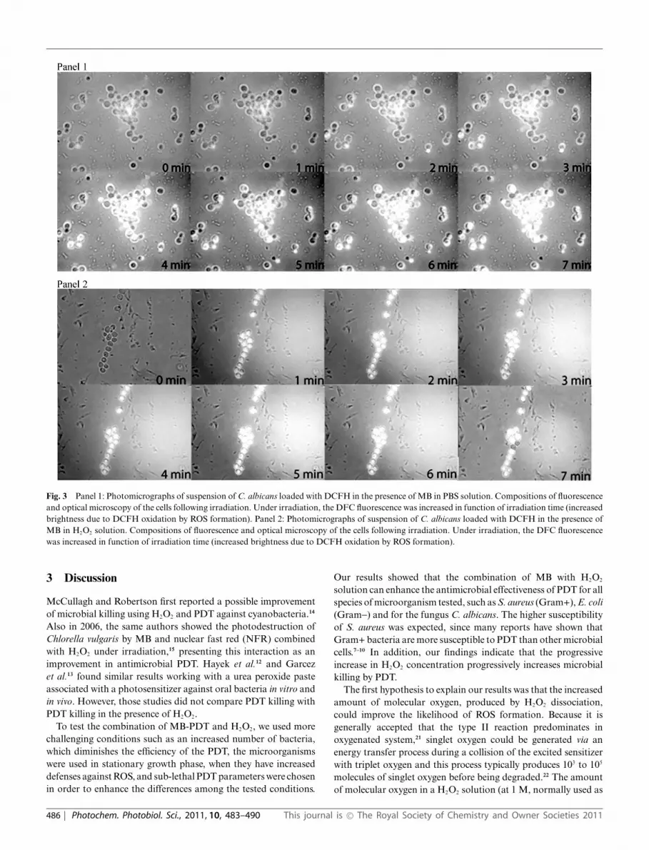
Fig. 3 Panel 1: Photomicrographs of suspension of C. albicans loaded with DCFH in the presence of MB in PBS solution. Compositions of fluorescenceand optical microscopy of the cells following irradiation. Under irradiation, the DFC fluorescence was increased in function of irradiation time (increasedbrightness due to DCFH oxidation by ROS formation). Panel 2: Photomicrographs of suspension of C. albicans loaded with DCFH in the presence ofMB in H2O2 solution. Compositions of fluorescence and optical microscopy of the cells following irradiation. Under irradiation, the DFC fluorescencewas increased in function of irradiation time (increased brightness due to DCFH oxidation by ROS formation).
3 Discussion
McCullagh and Robertson first reported a possible improvementof microbial killing using H2O2 and PDT against cyanobacteria.14
Also in 2006, the same authors showed the photodestruction ofChlorella vulgaris by MB and nuclear fast red (NFR) combinedwith H2O2 under irradiation,15 presenting this interaction as animprovement in antimicrobial PDT. Hayek et al.12 and Garcezet al.13 found similar results working with a urea peroxide pasteassociated with a photosensitizer against oral bacteria in vitro andin vivo. However, those studies did not compare PDT killing withPDT killing in the presence of H2O2.
To test the combination of MB-PDT and H2O2, we used morechallenging conditions such as an increased number of bacteria,which diminishes the efficiency of the PDT, the microorganismswere used in stationary growth phase, when they have increaseddefenses against ROS, and sub-lethal PDT parameters were chosenin order to enhance the differences among the tested conditions.
Our results showed that the combination of MB with H2O2
solution can enhance the antimicrobial effectiveness of PDT for allspecies of microorganism tested, such as S. aureus (Gram+), E. coli(Gram-) and for the fungus C. albicans. The higher susceptibilityof S. aureus was expected, since many reports have shown thatGram+ bacteria are more susceptible to PDT than other microbialcells.7–10 In addition, our findings indicate that the progressiveincrease in H2O2 concentration progressively increases microbialkilling by PDT.
The first hypothesis to explain our results was that the increasedamount of molecular oxygen, produced by H2O2 dissociation,could improve the likelihood of ROS formation. Because it isgenerally accepted that the type II reaction predominates inoxygenated system,21 singlet oxygen could be generated via anenergy transfer process during a collision of the excited sensitizerwith triplet oxygen and this process typically produces 103 to 105
molecules of singlet oxygen before being degraded.22 The amountof molecular oxygen in a H2O2 solution (at 1 M, normally used as
486 | Photochem. Photobiol. Sci., 2011, 10, 483–490 This journal is © The Royal Society of Chemistry and Owner Societies 2011

Fig. 4 Dynamic of relative ROS formation inside C. albicans cells byMB in PBS solution and MB in H2O2 solution evaluated by fluorescenceintensity (F) of DCF formation under irradiation. F 0 is the fluorescence atinitial irradiation time. Illumination parameters: l = 660 nm, P = 40 mW,Dt = 60 s. Solid and dashed lines are the exponential data fits (see text).
Fig. 5 Absorbance spectrum of E. coli supernatant after incubation withMB in PBS solution, incubation with MB in H2O2 solution, and incubationwith H2O2 and then with MB.
an antimicrobial agent) is approximately 30% higher than in a PBSsolution (data not shown). However, our findings undoubtedlydemonstrate that ROS production in solution decreases ratherthan increases in the presence of H2O2 and the production ofsinglet oxygen is almost inhibited compared to the production ofsinglet oxygen in PBS solution. Therefore, the possibility of a typeI mechanism was tested.
We tried to explain the enhanced microbial reduction byphotochemical reaction. It could occur that the photo-excitedMB reacts directly with H2O2 in a type I process via electrontransfer generating either hydroxyl or more likely hydroperoxideradicals and that these ROS could attack the microorganisms.This hypothesis was supported by the report from Gak et al.23 thatinvestigated the quenching of triplet excited MB by H2O2. Theauthors suggested two mechanisms for the quenching: electrontransfer from the excited MB to H2O2 with the formation of a
Fig. 6 Absorbance spectra of methylene blue inside E. coli (initial) andafter 20 min of incubation in peroxide solution. Note the variation of theleucoMB and MB absorbance peaks (p < 0.01).
Fig. 7 Effects of different sequences of H2O2, MB diluted in PBS or H2O2
and light on in vitro killing of E. coli. Illumination parameters: l = 660 nm,P = 40 mW, Dt = 60 s.
MB+ radical and OH- or electron transfer from H2O2 to the MBmolecule with the formation of the MB- radical and HO2* radical.McCullagh and Robertson14 found an increase in the antimicrobialactivity of MB/H2O2 system when investigating the antimicrobialphotodynamic effect of MB and H2O2 against cyanobacteria inan oxygen-free atmosphere. It was proposed that the processinvolved a type I mechanism with electron transfer from MB toH2O2 generating active radical species. However, the total ROSproduction measured in the present study indicated a decrease inROS formation in a H2O2 environment.
Investigating the behavior and resistance of bacteria underextreme conditions and the physiological changes associated withoxidative stress, Baatout and collaborators24 showed that bacterialstrains exhibited varying sensitivities towards exogenous H2O2.For all bacterial strains some physiological damage was noted, inparticular with regard to their membrane permeability. Dependingon the bacterial strains, moderate to high physiological damagewas observed. Membrane potential, esterase activity, intracellularpH were considerably modified at high H2O2 concentrations. In
This journal is © The Royal Society of Chemistry and Owner Societies 2011 Photochem. Photobiol. Sci., 2011, 10, 483–490 | 487

addition, a low micromolar, single dose of hydrogen peroxide wasshown to cause dramatic increases in the apparent intracellularaccumulation of fluorophores with different physicochemicalproperties in different cell types.19 The results were consistentwith changes in lateral membrane diffusion caused by hydrogenperoxide.
In fact, Funk and Krise19 reported that exposure to H2O2
facilitated the accumulation of the test substrates (lucifer yellow,oregon green and daunorubincin) in different cell types (HL-60and human foreskin fibroblasts). Our results agree with thoseof Funk and Krise, since the amount of MB uptake afterincubation with H2O2 is higher (5 mM more compared to PBSsolution). The enhancement on the amount of MB uptake by themicroorganism results in a higher concentration of intracellularMB, therefore MB might be closer to the intracellular sitessensitive to PDT damage. It has been recognized for many yearsthat MB binds to DNA, preferentially to G-C base pairs and it hasbeen assumed that MB, being a tricyclic heterocyclic compound,would intercalate between the base pairs of DNA.24 MB canalso photo-oxidize several amino acids, specifically tryptophan,tyrosine, histidine, methionine and cysteine and this reaction canoccur by type I or type II mechanisms. Some organelles as E.coli ribosomes are inactivated by MB photodynamic treatmentand biological membrane contains proteins, phospholipids andcholesterol, which are susceptible to photodamage.25
Increased MB uptake after H2O2 can explain the results foundwhen changing the sequence of treatments. It is clear that usingH2O2 before PDT or using MB in H2O2 solution improves theuptake of MB by the microorganism and leads to increasedmicrobial killing. However, it is not totally clear how to explainimproved killing when PDT was used first and subsequently H2O2
solution.Caetano and coworkers26 investigated the photodecomposition
of phospholipid bilayers in aqueous solutions of methylene blueand observed giant unilamellar vesicles under an optical micro-scope, which revealed a consistent pattern of membrane disruptionas a function of MB concentration and photon density for differentsubstrates supporting the vesicles. Also, Giroldo et al.27 studyingthe effect of antimicrobial PDT in C. albicans propose that theuse of PDT in microorganisms results in an increase of membranepermeabilization, suggesting that antimicrobial PDT mechanismusing MB may be related to damage in the plasma membrane ofthe cells.
Using this hypothesis, PDT alone could promote minor mem-brane disruption and this could increase the exposure of internalcomponents of the bacteria to H2O2. Since H2O2 is currently usedas antimicrobial agent,15 it was expected that facilitating the accessof this agent to the interior of bacteria could increase the bacterialkilling.
Investigating the production of ROS inside the microorganismand comparing the pictures in panel 1 and 2, the DCF fluorescenceis higher inside the cells when the MB is in a H2O2 solution thanin PBS solution. The dynamic of ROS production is differentbetween the solutions. In a H2O2 environment, the intracellularconcentration of MB may result in a faster and a higher amountof ROS production inside the microorganism and therefore a moreefficient antimicrobial effect.
Apparently, these data are not consistent with the results ofin vitro analysis of ROS and singlet oxygen photo-production
in the presence of H2O2. However, the results of the opticalcharacteristics of the dye showed that the amount of leucoMBdecreases when in contact with H2O2 and the oxidized “colored”form increases. May et al.28 suggested that a reduction occurs onMB at the external face of the membrane through an interactionwith reductase enzymes. The dye enters the cell by diffusion, andentry is facilitated because leucoMB is more lipophilic and it lacksthe charge possessed by MB. It is proposed that once inside thecell, the leuco form is reoxidized.29 Moreover, when in a H2O2
solution, the oxidation process is more intense and the amount ofoxidized “colored” molecules inside the bacteria is enhanced. Inaqueous solution, MB should be found in its leuco form, which isincapable of absorbing visible light (l = 660 nm) and consequentlyinitiate a photodynamic reaction.27
Also, C. albicans is an aerobic microorganism and can dealwith H2O2 by its expression of peroxidase and catalase enzymes,probably during the pre-irradiation time when the microorganismis in contact with MB and H2O2. The hydrogen peroxide improvesthe uptake of the photosensitizer and part of the H2O2 may beinactivated by the yeast enzymes. Moreover, there are a lot ofdifferent sites to be oxidized by H2O2 or PDT inside the cellsand the quenching proposed by Gak et al.23 (electron transferfrom the excited MB to H2O2 or electron transfer from H2O2
to the MB molecule) is probably lower than in vitro or eventhere was no quenching. Therefore, the photoreaction inside themicroorganism was not decreased by the presence of H2O2. Indeed,the superior amount of MB inside the cells produces an enhancedphotoreaction.
These data indicate that the photochemistry of the MB is mostunfavorable in the presence of H2O2 in cell free systems. However,the exposure of microorganisms to H2O2 leads to a higher uptakeof MB inside the cells resulting in an improved ROS productionclose to vital structures and, consequently, to a higher microbialdeath.
4 Experimental procedure
4.1 In vitro antimicrobial experiments
Suspensions of three microorganisms, Gram-positive (Staphylo-coccus aureus, ATCC 27659), Gram-negative (Escherichia coli, HB101) and yeast (Candida albicans, 51393) were grown in brain heartinfusion (BHI) broth at 37 ◦C with shaking (150 rpm) for 48 h,until they formed a stationary growth phase suspension.
The suspensions were diluted in PBS to a cell density of 107 permL, and 1 mL aliquots were added to wells of a 24-well plate. Thethree microorganisms were incubated with 60 mM of methyleneblue (MB) (Sigma Aldrich, Milwaukee, USA) in a PBS solutionand in 100 mM solution of H2O2 for 30 min in the absence of lightto verify intrinsic (dark) toxicity.
When the results showed a similar microbial killing for the dyeand for the H2O2, around 20% after 30 min (data not shown),we tested the effect of photoreaction when 60 mM of MB wereincubated in a solution of PBS or in a solution of H2O2 (10 mM,100 mM and 1 M) for 10 min (pre-irradiation time) followed byillumination with a diode laser at l = 660 nm, output power of 40mW (MMOptics, Sao Carlos, Brazil) and exposure time of 60 scorresponding to the delivery of 60 J cm-2.
488 | Photochem. Photobiol. Sci., 2011, 10, 483–490 This journal is © The Royal Society of Chemistry and Owner Societies 2011
Since 30 min of contact with a solution of 100 mM H2O2
did not affect the viability of the microorganisms more than thedark toxicity of the MB, we tested the microbial reduction in thepresence of 10 times less and 10 times more H2O2 than the darkexposure to verify if more or less oxygen available in the solutioncould kill more microorganisms.
A control group was incubated by the same time without anyirradiation or H2O2 challenge. Survival fractions were determinedfrom the colony forming units (CFU) in the initial inoculums andcompared with the experimental CFU.
4.2 In vitro singlet oxygen and total ROS detection
The first hypothesis that we tested concerned about oxygenconcentration. In H2O2 solution the molecules of oxygen availablefor a photoreaction could be greater than in water solution, as aconsequence, this would increase the amount of ROS, especiallysinglet oxygen.
To validate this hypothesis, a direct singlet oxygen detection wasperformed, based on its phosphorescence at l = 1270 nm. A cuvettecontaining 30 mM of MB in a deutered (D2O) solution or D2O plusH2O2 solution was irradiated using a Nd:YAG laser (Surelite III,Continuum, Santa Clara, USA) at l = 532 nm (pulse-energy of 30mJ and pulse-length of 10 ns). The transient emission of singletoxygen phosphorescence was accumulated and integrated over20 pulses by a fluorimeter linked to an infrared photomultipliertube.30 A D2O solution was used to enhance the lifetime of singletoxygen and therefore increase its detection by the equipment.
In addition, an indirect total ROS detection was performed,based on the oxidation of diacetate of dichlorofluorescein (DCFH-DA) to its fluorescent form dichlorofluorescein (DCF form).The fluorescence was measured (l = 522 nm) with a standardfluorimeter. Initially, 10 mL of sulfuric acid (Aldrich, Milwaukee,USA) was added to a cuvette containing 20 mL of DCFH-DA for20 min in dark conditions. Then, ten-mL of sodium hydroxide wasadded to neutralize the acid. MB at 60 mM in PBS or H2O2 solutioncompleted the cuvette and a baseline was registered. Illuminationwas performed using a l = 660 nm diode laser with P = 40 mWfor periods of 1 min performing 12 min of irradiation and DCFfluorescence was measured (at l = 522 nm) following excitationwith a l = 490 nm lamp.16
4.3 Total ROS detection inside C. albicans
To evaluate if biological material could affect the photoreactionof MB in a peroxide environment, a suspension of C. albicanswas incubated with 60 mM of MB in PBS solution for 10 min.First, the microorganism incubated with MB was washed andcentrifuged for 10 min at 4000 rpm; the supernatant was removedand the cells re-suspended in a PBS solution. Then, a solution of10 mM of DCFH-DA was added for 15 min and again washedto remove all DCFH-DA not bound to the cells The cells wereirradiated using the same laser as above mentioned with a fluenceof 60 J cm-2 (Dt = 1 min) and the rate of DCF formation wasevaluated at l = 522 nm by fluorescence microscopy images(inverted Zeiss microscope). The images were analyzed using thesoftware Image J (National Institute of Health, USA) and AdobePhotoshop CS3 for Macintosh (Adobe, USA). Each picture has640 ¥ 480 pixels and 8 bits black and white image. The total
area of each image was selected and analyzed by fluorescenceintensity through the histogram and the values were analyzed as afunction of time. The relative brightness (maximum and extension)of fluorescence images was evaluated as a function of irradiationtime, and plotted as intensity versus irradiation time. The sameexperimental procedure was repeated using MB in a 100 mM H2O2
solution. The use of yeast instead of bacteria is justified becausethe morphology of C. albicans is more complex and it is biggerthan S. aureus or E. coli to be observed by optical microscopy.
4.4 Mechanisms involved in the combination
4.4.1 MB uptake by bacteria. Suspensions of E. coli werediluted in 60 mM MB in PBS solution to a cell density of 107
per mL for 10 min. The suspension was centrifuged for 10 min ata rate of 4000 rpm sedimenting bacteria with bound dye. In theother group, suspension of E. coli was diluted in 100 mM of H2O2
associated to 60 mM MB solution to a cell density of 107 per mL for10 min, and then the suspension was also centrifuged for 10 minat 4000 rpm and the supernatant was removed. In the third group,bacterial suspension was firstly incubated with 100 mM of H2O2
for 10 min. The peroxide solution was washed and replaced by MBat 60 mM in PBS solution. The suspension was again centrifugedfor another 10 min at 4000 rpm. The supernatant of all groupswas removed and analyzed by optical spectroscopy to evaluate theMB uptake by bacteria.
4.4.2 Reduction state of the dye. To evaluate if H2O2 wouldalter the balance between MB and leucoMB,18 the optical char-acteristics of the MB inside an E. coli suspension in aqueousor H2O2 solution were analyzed via absorbance spectroscopy ina computer-interfaced double-beam spectrophotometer (Cary –17D Spectrophotometer Conversion, On-Line Instrument SystemInc, USA) in 1.0 cm optical path length quartz cuvettes. Thedye was dissolved in 3 mL of de-ionized water or H2O2 at theconcentration of 60 mM in the presence or absence of 104 cfu mL-1.The optical density at l = 290 nm to l = 660 nm was obtainedfrom each wavelength. A second assay was performed replacing1 mL of aqueous solution by peroxide solution to keep the sameMB molarity.
Optical densities were measured every 1 min and analyzedregarding MB/leucoMB ratio; the ratio was calculated by ab-sorbance at 660 nm/absorbance at 290 nm, since the absorbanceof MB monomers is around l = 660 nm and for leucoMB is aroundl = 290 nm. The different values found at each sample time werestatistically compared using t-test with Microsoft Excel. Resultswere considered significant when p < 0.01.
4.4.3 Changing incubation sequence. Different combinationsequences of MB and H2O2 were performed to verify the hypoth-esis that different mechanism of killing could occurs dependingon the sequence used for the antimicrobial effect. Suspensionsof E. coli in the stationary phase were diluted in PBS to a celldensity of 107 per mL, and 100 mL aliquots were added to wellsof a 96-well plate. Regarding to the experimental groups, onegroup [(MB+H2O2)+light] received the 60 mM MB in a 100 mMsolution of H2O2 for 10 min and was irradiated by the same diodelaser used above (light fluence: 60 J cm-2). The second group[H2O2+(MB+light)] received 100 mM of H2O2 for 10 min, andthen the suspension was centrifuged for 10 min at 4000 rpm. The
This journal is © The Royal Society of Chemistry and Owner Societies 2011 Photochem. Photobiol. Sci., 2011, 10, 483–490 | 489

supernatant was removed and replaced by a 60 mM of MB solutionin PBS for further 10 min and then irradiated (light fluence:60 J cm-2). The third group [(MB+light)+H2O2] was incubatedwith 60 mM of MB for 10 min in a PBS solution followed byirradiation. Then, this group received a 100 mM solution of H2O2
in the absence of light for further 10 min. Survival fractions weredetermined from the CFU in the initial inoculums and comparedwith the experimental CFU.
Conclusion
The combination of methylene blue and H2O2 has been shownto be an interesting approach to improve PDT efficiency againstGram+ bacteria, Gram- bacteria and fungi. The mechanisminvolved seems to be related with the increase in cell membranepermeability, MB reduction by the cell during photosensitizeruptake followed by oxidation of the leucoMB form as a result ofthe hydrogen peroxide, therefore increasing ROS formation insidethe microorganism.
Acknowledgements
The authors kindly thank Prof. Karin do Amaral Riske for the useof the optical microscope, Helena Junqueira for her collaboration,and FAPESP (grant 05/51598-7) for financial support. R. Itri, M.S. Baptista and M. S. Ribeiro also acknowledge CNPq for researchfellowship. N. A. Daghastanli was supported by a post-doctoratefellowship from FAPESP (05/00253-0). Research in Dr Hamblin’slaboratory is supported by US NIH grant R01AI050875, US AirForce MFEL Program (FA9550-04-1-0079).
References
1 T. T. Yoshikawa, Antimicrobial resistance and aging: beginning of theend of the antibiotic era?, J. Am. Geriatr. Soc., 2002, 50, 226–229.
2 R. Wise, T. Hart and O. Cars, Antimicrobial resistance, Br. Med. J.,1998, 37, 609–610.
3 R. E. Hancock and A. Bell, Antibiotic uptake into gram-negativebacteria, Eur. J. Clin. Microbiol. Infect. Dis., 1988, 7, 713–720.
4 D. M. Livermore, Antibiotic resistance in staphylococci, Int. J. Antimi-crob. Agents, 2001, 16, 3–10.
5 R. Agarwal, M. Athar, S. A. Urban, D. R. Bickers and H. Mukhtar,Involvement of singlet oxygen in chloroaluminium phthalocyaninetetrasulfonate-mediated photoenhancement of lipid peroxidation in ratepidermal microsomes, Cancer Lett., 1991, 56, 125–129.
6 D. A. Phoenix, Z. Sayed, S. Hussain, F. Harris and M. Wainwright, Thephototoxicity of phenothiazinium derivates against Escherichia coliand Staphylococcus aureus, FEMS Immunol. Med. Microbiol., 2003,39, 17–22.
7 M. R. Hamblin, D. A. O’Donnell, N. Murthy, K. Rajagopalan, N.Michaud, M. E. Sherwood and T. Hasan, Polycationic photosensitizerconjugates: effects of chain length and Gram classification on thephotodynamic inactivation of bacteria, J. Antimicrob. Chemother.,2002, 49, 941–951.
8 M. Wainwright, D. A. Phoenix, J. Marland, D. R. A. Wareing andF. J. Bolton, A study of photobactericidal activity in phenothiaziniumseries, FEMS Immunol. Med. Microbiol., 1997, 19, 75–83.
9 T. A. Dahl, W. R. Midden and P. E. Hartman, Comparison of killingof gram-negative and gram-positive bacteria by pure singlet oxygen, J.Bacteriol., 1989, 171, 2188–2194.
10 J. S. Friedberg, C. Skema, E. D. Baum, J. Burdick, S. A. Vinogradov,D. F. Wilson, A. D. Horan and I. Nachamkin, In vitro effects
of photodynamic therapy on Aspergillus fumigatus, J. Antimicrob.Chemother., 2001, 48, 105–107.
11 O. Feuerstein, D. Moreinos and D. Steinberg, Synergic antibacterialeffect between visible light and hydrogen peroxide on Streptococcusmutans, J. Antimicrob. Chemother., 2006, 57, 872–876.
12 R. R. Hayek, N. S. Araujo, M. A. Gioso, J. Ferreira, C. A. Baptista-Sobrinho, A. M. Yamada and M. S. Ribeiro, Comparative studybetween the effects of photodynamic therapy and conventional therapyon microbial reduction in ligature-induced peri-implantitis in dogs, J.Periodontol., 2005, 76, 1275–1281.
13 A. S. Garcez, S. C. Nunez, J. L. Lage-Marques, A. O. C. Jorge and M. S.Ribeiro, Efficiency of NaOCl and laser-assisted photosensitization onthe reduction of Enterococcus faecalis in vitro, Oral Surg., Oral Med.,Oral Pathol., Oral Radiol. Endodontol., 2006, 102, e93–98.
14 C. McCullagh and P. K. J. Robertson, Photo-dynamic biocidal actionof methylene blue and hydrogen peroxide on the cyanobacterium Syne-chococcus leopoliensis under visible light irradiation, J. Photochem.Photobiol., B, 2006, 83, 63–68.
15 C. McCullagh and P. K. J. Robertson, Photodestruction of Chlorellavulgaris by methylene blue or nuclear fast red combined with hydrogenperoxide under visible light irradiation, Environ. Sci. Technol., 2006, 40,2421–2425.
16 N. Daghastanli, R. Itri and M. S. Baptista, Singlet oxygen reactswith 2¢,7¢-Dichlorodihydrofluorescein and contributes to the formationof 2¢,7¢-Dichlorofluorescein, Photochem. Photobiol., 2008, 84, 1238–1243.
17 D. Gabrieli, E. Belisle, D. Severino, A. J. Kowaltowski and M.S. Baptista, Binding, aggregation and photochemical properties ofmethylene blue in mitochondrial suspensions, Photochem. Photobiol.,2004, 79, 227–232.
18 A. Blazquez-Castro, J. C. Stockert, F. Sanz-Rodrıguez, A. Zamarronand A. Juarranz, Differential photodynamic response of cultured cellsto methylene blue and toluidine blue: role of dark redox processes,Photochem. Photobiol. Sci., 2009, 8, 371–376.
19 R. S. Funk and J. P. Krise, Exposure to hydrogen peroxide can increasethe intracellular accumulation of drugs, Mol. Pharmaceutics, 2007, 4,154–159.
20 L. C. Seaver and J. A. Imlay, Hydrogen peroxide fluxes and compart-mentalization inside growing Escherichia coli, J. Bacteriol., 2001, 183,7182–7189.
21 C. S. Foote, Definition of type I and type II photosensitized oxidation.,Photochem. Photobiol., 1991, 54, 659.
22 M. C. De Rosa and R. J. Crutchley, Photosensitized singlet oxygen andits applications, Coord. Chem. Rev., 2002, 233–234, 351–371.
23 V. Y. Gak, V. A. Nadtochenko and J. Kiwi, Triplet-excited dye molecules(eosine and methylene blue) quenching by H2O2 in aqueous solution,J. Photochem. Photobiol., A, 1998, 116, 57–62.
24 S. Baatout, P. De Boever and M. Mergeay, Physiological changesinduced in four bacterial strains following oxidative stress, Prikl.Biokhim. Mikrobiol., 2006, 42, 418–427.
25 M. Wainwright, Photodynamic antimicrobial chemotherapy (PACT),J. Antimicrob. Chemother., 1998, 42, 13–28.
26 W. Caetano, P. S. Haddad, R. Itri, D. Severino, V. C. Vieira, M.S. Baptista, A. P. Schroder and C. M. Marques, Photo-induceddestruction of giant vesicles in methylene blue solutions, Langmuir,2007, 23, 1307–1314.
27 L. M. Giroldo, M. P. Felipe, M. A. de Oliveira, E. Munin, L. P. Alves andM. S. Costa, Photodynamic antimicrobial chemotherapy (PACT) withmethylene blue increases membrane permeability in Candida albicans.,Lasers Med. Sci., 2009, 24, 109–112.
28 J. M. May, Z. C. Qu and C. E. Cobb, Reduction and uptake of methyleneblue by human erythrocytes., Am. J. Physiol.: Cell Physiol., 2004, 286,C1390–C1398.
29 G. T. Wondrak, NQO1-activated phenothiazinium redox cyclers for thetargeted bioreductive induction of cancer cell apoptosis., Free RadicalBiol. Med., 2007, 43, 178–190.
30 M. S. Oliveira, M. Lima, D. Severino, M. S. Baptista, P. Di Mascioand M. Tabak, Quenching of singlet molecular oxygen, O2 (1Dg), bydipyridamole and derivatives, Photochem. Photobiol., 2007, 83, 1379–1385.
490 | Photochem. Photobiol. Sci., 2011, 10, 483–490 This journal is © The Royal Society of Chemistry and Owner Societies 2011