Nanoparticles for Photodynamic Therapy Applications
22
Peptide-conjugated nanoparticles for photodynamic therapy targeting neuropilin-1 Pierre Couleaud 1* , Denise Bechet 3* , Régis Vanderesse 2,5 , Muriel Barberi-Heyob 3,5 , Anne- Charlotte Faure 4 , Stéphane Roux 4 , Olivier Tillement 4 , Sabine Porhel 1 , François Guillemin 3,5 , Céline Frochot 1,5 1 Laboratoire Réactions et Génie des Procédés (LRGP), Nancy-University, CNRS, Nancy, France. 2 Laboratoire de Chimie Physique Macromoléculaire (LCPM), Nancy-University, CNRS, Nancy. 3 Centre de Recherche en Automatique de Nancy (CRAN), Nancy-University, CNRS, Centre Alexis Vautrin, Vandœuvre-lès-Nancy, France. 4 Laboratoire de Physico Chimie des Matériaux Luminescents (LPCML), UMR 5620 CNRS, Lyon I-University, Lyon, France 5 GDR CNRS 3049 "Médicaments Photoactivables-Photochimiothérapie (PHOTOMED)" * contributed equally to this study Correspondence to: Céline Frochot (for chemical and photophysical approaches) Laboratoire Réactions et Génie des Procédés 1 rue Grandville BP 20451 F-54001, Nancy, France Telephone: +33 3 83 17 51 15 [email protected] Muriel Barberi-Heyob (for molecular and cellular approaches) Centre de Recherche en Automatique de Nancy Centre Alexis Vautrin Avenue de Bourgogne, Brabois F-54511 Vandœuvre-lès-Nancy, France Telephone: +33 3 83 59 83 76 [email protected] Couleaud P., Bechet D. et al., Nanomedicine, 2010. 1
-
Upload
independent -
Category
Documents
-
view
5 -
download
0
Transcript of Nanoparticles for Photodynamic Therapy Applications

Peptide-conjugated nanoparticles for photodynamic therapy
targeting neuropilin-1
Pierre Couleaud1*, Denise Bechet3*, Régis Vanderesse2,5, Muriel Barberi-Heyob3,5, Anne-
Charlotte Faure4, Stéphane Roux4, Olivier Tillement4, Sabine Porhel1, François Guillemin3,5,
Céline Frochot1,5
1 Laboratoire Réactions et Génie des Procédés (LRGP), Nancy-University, CNRS, Nancy, France. 2 Laboratoire de Chimie Physique Macromoléculaire (LCPM), Nancy-University, CNRS, Nancy. 3 Centre de Recherche en Automatique de Nancy (CRAN), Nancy-University, CNRS, Centre Alexis Vautrin, Vandœuvre-lès-Nancy, France. 4 Laboratoire de Physico Chimie des Matériaux Luminescents (LPCML), UMR 5620 CNRS, Lyon I-University, Lyon, France 5 GDR CNRS 3049 "Médicaments Photoactivables-Photochimiothérapie (PHOTOMED)"
* contributed equally to this study
Correspondence to: Céline Frochot (for chemical and photophysical approaches) Laboratoire Réactions et Génie des Procédés 1 rue Grandville BP 20451 F-54001, Nancy, France Telephone: +33 3 83 17 51 15 [email protected] Muriel Barberi-Heyob (for molecular and cellular approaches) Centre de Recherche en Automatique de Nancy Centre Alexis Vautrin Avenue de Bourgogne, Brabois F-54511 Vandœuvre-lès-Nancy, France Telephone: +33 3 83 59 83 76 [email protected]
Couleaud P., Bechet D. et al., Nanomedicine, 2010.
1

Keywords: peptide; functionalized-nanoparticles, neuropilin-1; in vitro photodynamic efficiency, molecular affinity Number of text pages, including references: 22 Number of figures: 7 Number of references: 70 Number of words in abstract: 119 Non-standard abbreviations: ABDM, 9,10 anthracenediyl-bis-(methylene)dimalonic acid; ADBA, disodium 9,10-
anthracenedipropionic acid; APTES, aminopropyltriethoxysilane; ATCC, American Type
Culture Collection; ATWLPPR, H-Ala-Thr-Trp-Leu-Pro-Pro-Arg-OH; a.i., arbitrary
intensity; a.u., arbitrary units; BSA, bovine serum albumine; BtOH, 1-hydroxybenzotriazole;
DEG, diethyl glycol; DLI, drug-light interval; DIEA, diisopropylethylamine; DMF,
dimethylformamide, DMSO, dimethyl sulfoxide; DPBF, 1,3 diphenylisobenzofuran; i.v.,
intravenous; EDC, 1-(3-dimethylaminopropyl)-3-ethylcarbodiimide hydrochloride; FBS, fetal
bovine serum; Fmoc, 9-fluorenyl-methoxy-carbonyl; Gd2O3, gadolinium oxide; HBTU, 2-
(1H-benzotriazol-1-yl)-1,1,3,3-tetramethyl-uronium hexafluorophosphate; HPLC, high
performance liquid chromatography; LC-MS, liquid chromatography-mass spectroscopy;
MRI, magnetic resonance imaging; MTT, 3-(4,5-dimethylthiazol-2-yl)-2,5-diphenyl
tetrazolium bromide; MWCO, molecular weight cut off; NaCl, sodium chloride; NP,
nanoparticle; NRP-1, neuropilin-1; Pbf, 2.2.4.6.7-pentamethyldihydro-benzofuran-5-sulfonyl;
PCS, photon correlation spectroscopy; PBS, phosphate-buffered saline; PDT, photodynamic
therapy; PEG, polyethylene glycol; PFP, pentafluorophenol; RGD, H-Arg-Gly-Asp-OH;
RNA, ribonucleic acid; RNO, N,N dimethyl-4-nitrosoaniline; SD, standard deviation; SOSG,
singlet oxygen sensor green; tBu, tertio-butyl; TEA, triethylamine; TEMP, 2,2,6,6-tetraethyl-
4-piperridone; TEOS, tetraethyl orthosilicate; TFA, trifluoroacetic acid; TLC, thin-layer
chromatography; TPC, 5-(4-carboxyphenyl)-10,15,20-triphenyl-chlorin; TPC-NHS,
5,10,15,tri-(p-tolyl)-20-(p-carboxylphenyl)chlorin succinidyl ester; TPP, tretraphenyl
porphyrin; UV, ultraviolet; VEGF, vascular endothelial growth factor; VTP, vascular targeted
photodynamic therapy; Φf, fluorescence quantum yield; 1O2, singlet oxygen; ΦΔ, 1O2 quantum
yield.
Couleaud P., Bechet D. et al., Nanomedicine, 2010.
2

Abstract
The strategy developed aims to favor the vascular effect of photodynamic therapy by
targeting tumor-associated vascularisation using peptide-functionalized nanoparticles. We
previously described the conjugation of a photosensitizer to a peptide targeting neuropilin-1
over-expressed in tumor angiogenic vessels. In this study, we have designed and
photophysically characterized a multifunctional nanoparticle consisting of a surface-localized
tumor vasculature targeting peptides and encapsulated photodynamic therapy and imaging
agents. The elaboration of these multifunctional silica-based nanoparticles is reported.
Nanoparticles functionalized with ~4.2 peptides specifically bound to neuropilin-1
recombinant protein. Nanoparticles conferred photosensitivity to cells over-expressing
neuropilin-1, providing evidence that the chlorin grafted within the nanoparticle matrix can be
photoactivated to yield photocytotoxic effects in vitro. Nanoparticles were eliminated by renal
excretion after intravenous injection.
Introduction
Photodynamic Therapy (PDT) is a treatment modality for cancer and various other diseases
[1,2]. This treatment involves the uptake of a photoactivable molecule called photosensitizer
by cancer tissue followed by a photoirradiation. Interaction of light with the photosensitizer
results in the formation of photosensitizer excited states that transfer its energy through
intermolecular triplet-triplet energy transfer to surrounding molecular oxygen (O2) molecules
and lead to the production of reactive oxygen species, such as singlet oxygen (1O2) [3,4].
Selectivity of PDT as an anti-cancer treatment relies on the local generation of cytotoxic
reactive oxygen species in the tumor and is achieved from the preferential uptake of the
photosensitizer by malignant tissue and from the subsequent localized light irradiation.
Several new strategies have emerged to improve the selectivity of PDT agents. Main of the
research is based on conjugation of vectors to target receptors over expressed on cancer cells,
such as oligonucleotides, monoclonal antibodies, epidermal growth factor, carrier proteins,
carbohydrates, folic acid for selective delivery of the agents into tumor tissues [5-8].
Destruction of the vasculature can also indirectly lead to tumor eradication, following
deprivation of life-sustaining nutrients and oxygen [9,10], and this vascular effect is thought
to play a major part in the destruction of some vascularized tumors by PDT [11-17]. Vascular
targeted photodynamic therapy (VTP) is a promising strategy and tumor-associated
vasculature a potential target of PDT damage.
Couleaud P., Bechet D. et al., Nanomedicine, 2010.
3

Synthetic peptides represent interesting targeting molecules, since they do not only exist in
natural form but can also be designed synthetically as novel molecules. In addition, the
effective tissue penetration of short synthetic peptides, in combination with their selective
binding and internalizing capacities by cancer cells, make peptides ideal candidates for the
delivery of therapeutic agents such as photosensitizers [18]. We have previously described the
conjugation of a photosensitizer (a chlorin) to an heptapeptide (ATWLPPR), specific for the
vascular endothelial growth factor (VEGF) receptor, neuropilin-1 (NRP-1) [19]. We
evidenced a statistically significant decrease of the ATWLPPR-conjugated photosensitizer
cellular uptake after RNA interference-mediated silencing of NRP-1 [20]. This targeted
photosensitizer proved to be very efficient in vitro in human umbilical vein endothelial cells
compared to its non-conjugated counterpart [19]. In vivo, we also demonstrated the interest of
using this active-targeting strategy by the induction of tissue factor expression immediately
post-treatment [21,22], and a specific localization in endothelial cells lining tumor vessels of
the conjugated photosensitizer [20]. Nevertheless, we could observe a degradation of the
peptide in plasma from 2 h post-intravenous injection and biodistribution results suggested
that its appearance in plasma mainly resulted from the degradation of the peptidic moiety into
organs of the reticulo-endothelial system [23,24].
As we previously described, non-biodegradable nanoparticles seem to be very promising
careers satisfying all the requirements for an ideal targeted PDT [25,26]. Hybrid gadolinium
oxide nanoparticles have been suggested to be useful for magnetic resonance imaging (MRI)
and treatment [27-29]; their small size (less than 50 nm of diameter), their silica shell that
allows the covalent coupling of a photosensitizer, their polyethylene glycol (PEG) grafting
that allows the covalent coupling of peptide units make them an ideal nanoplatform for both
imaging and VTP. In this targeting strategy using nanomaterials, we conjugated a second
generation photosensitizer, 5-(4-carboxyphenyl)-10,15,20-triphenyl-chlorin (TPC) and the
ATWLPPR heptapeptide to a hybrid gadolinium oxide nanoparticle. The paper focuses on its
functionalization, photophysical properties as well as cytotoxicity and photodynamic
efficiency in vitro, describing for the first time the molecular affinity of the functionalized
nanoparticle to NRP-1 molecular target.
Materials and Methods
The synthesis of gadolinium oxide nanoparticles embedded in a polysiloxane shell has been
widely described previously by our group [30-32].
Couleaud P., Bechet D. et al., Nanomedicine, 2010.
4

Peptide synthesis. Synthesis of the protected peptide H-AT(tertio-butyl (tBu))W(N-tert-
butoxy-carbonyl)LPPR(2.2.4.6.7-pentamethyldihydro-benzofuran-5-sulfonyl (Pbf))-OH was
carried out on a fully automated ResPepXL synthesizer (Intavis AG, Köln, Germany)
according to a classical 9-fluorenyl-methoxy-carbonyl (Fmoc)/tBu solid-phase methodology.
For the coupling of each amino acid, double couplings were performed using a threefold
excess of N-Fmoc-amino acid and activation reagents 2-(1H-benzotriazol-1-yl)-1,1,3,3-
tetramethyl-uroniumhexafluorophosphate (HBTU) (3 eq.), 1-hydroxybenzotriazole (BtOH) (3
eq.) and N-methymorpholin (9 eq.) in dimethylformamide (DMF). The cleavage, was realized
by a 2 h treatment with an acetic acid/trifluoroethanol/dichloromethane (2:2:6) mixture. The
protected peptide was eluted with a gradient of acetonitrile/H2O, 0.1 % trifluoroacetic acid
(TFA), from 40/60 to 90/10 for 20 min, followed by 100% acetonitrile for the last 15 min. A
preparative Pursuit C18, 5µm column (250 x 10 mm I.D., Varian, France) was used for
purification. Fluorescence detection was optimized with excitation and emission wavelengths
of 280 and 350 nm respectively coupled with an UV detection at 280 nm. Unless otherwise
stated, reagents were purchased from chemical companies and used without prior purification.
Fmoc-Arg(Pbf)-Wang and H-Arg(Pbf)-2-chlorotrityl resins, and Fmoc-amino acids were
from Merck Novabiochem. Thin-layer chromatography (TLC) was carried out on Merck
silica gel 60 F254 plates (Merck Chimie S.A.S., Fontenay sous Bois, France). The TLC spots
were visualized either by UV light or by heating plates sprayed with a solution of
phosphomolybdic acid (5% ethanol solution).
Photosensitizer synthesis. 5-(4-carboxyphenyl)-10,15,20-triphenylchlorin (TPC) was
synthesized as described by Whitlock [33] and our group [34] as well as 5,10,15,tri-(p-tolyl)-
20-(p-carboxylphenyl)chlorin succinidyl ester (TPC-NHS) [14].
Grafting of TPC-NHS into the polysiloxane shell. A solution containing TPC-NHS (6.2
mg) and 3.8 µL of aminopropyltriethoxysilane (APTES) dissolved in 1.30 mL of dimethyl
sulfoxide (DMSO) was prepared as a precursor and stirred overnight. The coupling reaction
between the chlorin and APTES was certified by mass spectrometry with complete
disappearance of the molecular peak of TPC-NHS (m/z= 758.1 (M-H+)) and the appearance
of the molecular peak of the chlorin conjugated APTES (m/z = 864.6 (M-H+), 1726.9 (2M-
H+) and 1747.3 (2M-Na+)). TPC-APTES solution and a portion of 327 µL of APTES and of
209 µL of tetraethyl orthosilicate (TEOS) were added to 1.5 mL solution containing Gd2O3
Couleaud P., Bechet D. et al., Nanomedicine, 2010.
5
nanoparticles diluted in 13.5 mL of diethyl glycol (DEG) under stirring at 40°C. After 1 h, a
portion of 792 µL of a DEG solution (0.1 M of triethylamine (TEA), 10 M of water) was
added. The other portions of polysiloxane precursors and of hydrolysis solution were
sequentially and alternatively added. The final mixture was stirred for 48 h at 40°C.
Covalent grafting of carboxylated PEG250 on hybrid nanoparticles. For grafting PEG250
via its carboxylic acid function (COOH), COOH functions were activated by 1-(3-
dimethylaminopropyl)-3-ethylcarbodiimide hydrochloride (EDC) and pentafluorophenol
(PFP). 500 µL of DEG solution containing PEG250-COOH (0.091 M) were added to an
isopropanol solution (5 mL) containing a mixture of EDC/PFP (0.367 M). The reaction
between COOH groups of the polymer and PFP, assisted by EDC, is expected to yield
pentafluorophenyl esters. The resulting mixture was stirred for 90 min at room temperature
and added to 1 mL of colloidal suspension composed of hybrid nanoparticles (Gd2O3 cores
encapsulated in a polysiloxane shell) dispersed in DEG. The covalent grafting of PEG250-
COOH onto the hybrid nanoparticles was performed overnight at room temperature with
gentle stirring. The colloids of pegylated nanoparticles were introduced in a tubular
membrane of dialysis (molecular weight cut off (MWCO) = 4000-6000 Da) and immersed in
250 mL of water which was replaced four times every 12 h. Diluted purified solutions were
finally concentrated by ultrafiltration using Vivaspin® (MWCO = 10 000 Da).
Covalent grafting of the protected peptide on PEG250. 1.67 10-8 mol of Pegylated-
nanoparticles was dispersed in 7 mL dimethylformamide (DMF). The PEG carboxylic acid
functions were first activated by the classic peptidic synthesis mixture HOBt/HBTU/DIEA,
1/1/6 (HOBt 8.3 .10-8 mol, 12 µg, HBTU 8.3 .10-8 mol, 32 µg, DIEA 10 μL). The mixture was
stirred during 10 min before addition of 105 µg (8.3.10-8 mol) protected peptide. After 18 h
DMF was evaporated, 5 mL of pure TFA was added and the stirring was carried on during 2
h. Ultrapure water was added and the solvents evaporated. Several cycles of addition of water
and evaporation were realized to eliminate most of the TFA. The purification of the peptide-
functionalized nanoparticles was made by dialyses and ultrafiltrations. First dialysis was
realized with a 5000 Da cellulose membrane against ultrapure water. Several cycles of
dialysis were realized during 2 or 3 h each followed by ultrafiltrations with Vivaspin®
(MCWO 5000 Da) during 7 min at 3500 rpm. Finally, the water was removed under vacuum.
Couleaud P., Bechet D. et al., Nanomedicine, 2010.
6

Size measurement. Direct measurements of the size distribution of the nanoparticles were
performed by photon correlation spectroscopy (PCS) with Zetasizer NanoZS from Malvern
Instrument. Measurements were taken on the colloids after purification by dialysis.
Zeta-potential measurements. Direct determination of the ζ-potential of the nanoparticles
were performed using a Zetasizer NanoZS (laser He-Ne (633 nm) from Malvern Instrument.
Prior to the experiment, the aqueous colloid was diluted in an aqueous solution containing
0.01 M NaCl and adjusted to the desired pH.
Photophysical properties. Absorption spectra were recorded on a Perkin-Elmer (Lambda 2,
Courtaboeuf, France) UV-visible spectrophotometer. Fluorescence spectra were recorded on a
SPEX Fluorolog-3 spectrofluorimeter (Jobin Yvon, Longjumeau, France) equipped with a
thermo stated cell compartment (25°C), using a 450 W Xenon lamp. Fluorescence quantum
yields (Φf) were determined using a tretraphenyl porhyrin (TPP) solution as a fluorescence
standard (Φf = 0.11, in toluene, taking into account solvent refractive index and absorption
efficiencies) [35]. For the direct determination of singlet oxygen quantum yield (ΦΔ):
excitation occurred with a Xe-arc, the light was separated in a SPEX 1680, 0.22 µm double
monochromator. The detection at 1270 nm was done through a PTI S/N 1565
monochromator, and the emission was monitored by a liquid nitrogen-cooled Ge-detector
model (EO-817L, North Coast Scientific Co). The absorbance of the reference solution
(Bengal pink in EtOH ΦΔ = 0.68 [36] and the sample solution (at 515 nm) were set equal
(between 0.2 and 0.5) by dilution. Determination of ΦΔ has been also realized with a chemical
probe, the singlet oxygen sensor green (SOSG, Invitrogen). The SOSG is highly selective for 1O2 [37] and can be used in aqueous solution. It initially exhibits weak blue fluorescence with
excitation peaks at 371 nm and 393 nm and emission peaks at 395 nm and 416 nm. In the
presence of oxygen, it emits a green fluorescence at 525 nm after excitation at 504 nm. SOSG
reagent was dissolved in methanol to make a stock solution of 5 mM. SOSG (final
concentration 50 µM) was added to the photosensitizer (2 mg mL–1) and 3 mL water was
added. Following excitation of SOSG at 480 nm using a 450 W Xenon lamp of the SPEX
fluorolog-3, the fluorescence was recorded at 525 nm.
Cell line and culture. To argue the involvement of NRP-1, MDA-MB-231 breast cancer cells
were used, strongly over-expressing NRP-1 receptor [38]. For in vitro experiments, MDA-
Couleaud P., Bechet D. et al., Nanomedicine, 2010.
7

MB-231 cells were purchased from American Type Culture Collection (ATCC, Manassas,
VA, USA). Cells were maintained in minimum essential medium (RPMI 1640) supplemented
with 10% fetal bovine serum (FBS), 1% antibiotic/antimitotic solution. They were cultured in
75 cm2 flasks and incubated in 5% CO2/95% humidified air at 37°C. Cell culture materials
were purchased from Costar (Dutscher, Brumath, France). All other chemicals were
purchased from Sigma (Saint Quentin Fallavier, France).
Binding of functionalized nanoparticles to recombinant NRP-1 protein. Neuropilin-1 was
obtained from R&D Systems (Lille, France), as recombinant chimeric protein. The surface of
Maxisorp microplates (Dutscher) was coated with NRP-1 (2 µg/mL) in phosphate buffered
saline (PBS), overnight at room temperature. The plates were blocked with PBS containing
0.5% bovine serum albumine (BSA, blocking buffer) during 1 h at 37°C, to prevent non-
specific interactions. Binding of the functionalized nanoparticles to NRP-1 was assessed
using 5 ng/mL of biotinylated VEGF165 (R&D Systems) in blocking buffer containing 2
µg/mL heparin. Biotinylated VEGF165 was added to the coated wells, in competition after an
excess of NP-TPC-ATWLPPR or control NP alone or unlabelled VEGF165 as positive control
(0.5 µg/mL, R&D Systems). After a 2 h-incubation at room temperature, the plates were
washed and the amount of bound biotinylated VEGF165 stained with streptavidin horseradish
peroxidase conjugate (R&D Systems) and assayed. After 20 min at room temperature,
reaction was stopped by the addition of Stop Solution (R&D Systems). Optical densities were
measured at 450 nm. Results were expressed as relative absorbance to wells containing only
blocking buffer. Three wells per condition were used. Reported values are the average of
triplicate measurements.
Dark cytotoxicity. Cell survival after incubation with the different batches of nanoparticles in
the dark was measured using a 3-(4,5-dimethylthiazol-2-yl)-2,5-diphenyl tetrazolium bromide
(MTT) assay. Briefly, MDA-MB-231 cells were plated at an initial cell density of 5 x 10³
cells/well in 96-well microtitration plates and allowed to attach overnight. Wells were then
emptied, rinsed twice with PBS and filled with 200 µL RPMI medium containing various
concentrations of nanoparticles (photosensitizer concentrations ranged from 0.1 to 57.0 µM),
and the corresponding concentrations of gadolinium oxide (from 0.01 to 0.50 mM) for the
control nanoparticles. After a 24h-incubation at 37°C, wells were emptied, rinsed twice with
cold PBS and filled with 200 µL RPMI. Cell survival was measured 24 h later, as described
previously [39]. Experiments were carried out in triplicates.
Couleaud P., Bechet D. et al., Nanomedicine, 2010.
8
Photodynamic activity and light source. Photodynamic activity of nanoparticles on MDA-
MB-231 was also assessed by MTT assay. Cell survival was measured 24 h after
photosensitization. MDA-MB-231 cells were seeded at the initial density of 2 x 10⁴ cellsmL-
1 in 96-well microtitration plates. Forty-eight hours after plating, cells were exposed to
photoactive nanoparticles at 0.1 and 1 µM. After 24 h incubation at 37°C, the medium was
removed, and cells were then washed three times with cold PBS and fresh RPMI was added
before cell irradiation. Irradiation was carried out at 652 nm, using a dye laser (Spectra-
Physics 375B, Les Ulis, France), pumped with an argon laser (Spectra-Physics 2020). The
output power was 700 mW. The light spot was 14 cm in diameter, providing a fluence rate of
4.54 mW/cm2. Light doses ranged from 1 to 20 J/cm2. During irradiation, the temperature
never exceeded 24±2°C. This temperature did not influence cell viability. Caution was taken
to avoid exposure of cells to light before and after irradiation. Negative controls were
photosensitizer free-medium without photo-irradiation, photosensitizer free-nanoparticles
(control NP) with photo-irradiation, photosensitizer containing-medium without photo-
irradiation. Experiments were carried out in triplicates at different days. Irradiation doses
yielding 50% growth inhibition (LD50) were determined using the medium effect method
[19].
Biodistribution of nanoparticles. Female NMRI nude mice (6–8 weeks old, Janvier, France)
were anesthetized (isoflurane/oxygen 4% for induction and 2% thereafter) and were injected
intravenously with a hybrid gadolinium oxide nanoparticles coated by PEG250 (200 µL)
containing Cy5 mono-NHS ester (Amersham Bioscience). The mice were illuminated by 633
nm light-emitting diodes equipped with interference filters. Fluorescence images, as well as
black-and white pictures, were acquired by a back-thinned charge-coupled device cooled
camera (ORCAII-BT-512G, Hamamatsu). A colored glass long-pass filter (RG 665) cut off
all excitation light in the image [40,41]. In vivo imaging experiments were reproduced at least
twice.
Statistical analysis. Mann-Whitney U test was used to assess the significant level between
independent variables. The level of significance was set to p < 0.05.
Results
Synthesis of nanoparticles, grafting with photosensitizer and peptide. Pegylated hybrid
gadolium oxide nanoparticles have been synthesized following several steps described in a
Couleaud P., Bechet D. et al., Nanomedicine, 2010.
9

previous study [28]. As also shown in other previous papers, the encapsulation of the
gadolinium core in a polysiloxane shell afforded the opportunity to enlarge the palette of
properties of the nanoparticles [27-29,32,42]. The functionalization of the nanoparticles was
rendered possible by the use of APTES whose hydrolysis-condensation in presence of TEOS
yields a polysiloxane shell anchored onto each oxide core. The amine function of APTES
allowed the immobilization of various molecules inside and/or onto the shell. The
modification of APTES before the hydrolysis induces the distribution of the conjugates inside
the shell whereas the derivatization of the polysiloxane shell will favors the distribution of the
conjugates onto the shell. The photosensitizer (TPC-NHS) was conjugated to APTES before
the hydrolysis-condensation whereas the ATWLPPR peptide units were immobilized onto the
nanoparticles after the derivatization of the polysiloxane shell by highly hydrophilic PEG250-
COOH chains. For the linkage of ATWLPPR to the nanoparticle, two pathways were possible
and described in Figure 1A. Figure 1B shows the chromatographic profiles of the nanoparticle
coupled to the peptide, the free peptide and the mixture of both. Some previous work [43]
proved that it was possible to use a wide pore C5 column with a gradient of water and
acetonitrile to separate gold nanoparticles (3 nm diameter) from proteins. In our case, after
several attempts with C4 and C18 column and different gradients, we concluded that the best
chromatographic separation was performed with a C18 column and a methanol/water
gradient. By this original and appropriated elution condition, it was possible to check that all
the free peptide has been eliminated after dialysis and ultrafiltrations.
Nanoparticles sizing and zeta potential. Size distribution and Zeta potential are given in
Figures 2A and 2B, respectively. Hydrodynamic diameter of the nanoparticles was comprised
between 8 and 14 nm (Fig. 2A) whereas the Gd2O3 cores have a hydrodynamic diameter of
3.8 nm (data not shown). Hence, encapsulation of the gadolinium oxide core into hydrophilic
shell containing fluorophore was reflected by the increase of the hydrodynamic diameters of
the particles. The nanoparticles exhibited a negative charge at pH 7.4 (Fig. 2B). As expected,
the derivatization of the nanoparticles by PEG250 rendered them water soluble in a wide pH
range, including pH of biological fluid whereas the colloidal stability of uncoated
nanoparticles was not sufficient. For pH>4, the negative charge was attributed both to the
negative charge of carboxylate groups, resulting from the deprotonation of COOH
(pKa(COOH/COO-) ~ 5), the negative charge of silanolate and from the steric hindrance and
hydrophilic character of PEG chains.
Couleaud P., Bechet D. et al., Nanomedicine, 2010.
10
Photophysical properties and singlet oxygen formation. Regarding the photophysical
properties, Figure 3A shows the absorption spectra of free TPC and different suspensions of
nanoparticles with or without peptide. No shift of the maximum absorption at 410 nm can be
observed, meaning that the photosensitizer was mainly in a monomeric form without
aggregation inside the nanoparticles. The low decrease of the nanoparticle fluorescence
emission indicated that the photosensitizer molecules linked into the silica matrix were
slightly affected by the micro environment (Fig. 3B). As illustrated in Figure 3C, the
signature phosphorescence emission of 1O2 at 1270 nm was clearly observed under 414 nm
excitation of the nanoparticles. Sensor green fluorescence increased rapidly after excitation of
the nanoparticles (Fig. 3D). No significant changes have been measured for free or coupled
TPC fluorescence lifetimes, fluorescence and singlet oxygen quantum yields (Fig. 3F).
Quantification of ATWLPPR grafting. Thanks to the fluorescence of the tryptophan residue
(excitation and emission wavelengths at 280 and 350 nm, respectively), it was possible to
estimate the number of grafted peptide onto the nanoparticles (Fig. 3D). The number of the
peptide units coupled to the nanoparticles has been estimated by comparing the fluorescence
of the tryptophan of the peptide coupled to the nanoparticle to a fluorescence calibration curve
of the peptide in solution. We found ~4.2 peptides per nanoparticles.
Molecular affinity of nanoparticles with peptides. As previously described, the endothelium-
homing peptide ATWLPPR, selectively targets to angiogenic vasculature and NRP-1 receptor
[19-23]. To assess the ability of ATWLPPR peptide to target nanoparticles to tumor-
associated vasculature, we synthesized and characterized multifunctional nanoparticles.
grafted with TPC and encapsulated gadolinium oxide within the core matrix. Polyethylene
glycol was attached to the surface of the nanoparticle along with the homing peptide. The
number of ATWLPPR peptide, when analyzed by spectrophotometry, revealed an average of
4.2 peptides per nanoparticles. The molecular affinity of the functionalized nanoparticles for
recombinant NRP-1 protein, has been estimated using binding tests. As VEGF165 binding to
its receptors is heparin dependent, the competitive binding experiments were always carried
out in the presence of heparin. Binding of biotinylated VEGF165 to NRP-1 was displaced by
the NP-TPC-ATWLPPR. Nanoparticles conjugated to ATWLPPR indeed bound to
recombinant NRP-1 chimeric protein without interaction of the nanoparticle alone (Fig. 4).
Interestingly, at 100 µM of peptides, we quantified for the functionalized nanoparticles (~4.2
peptides per nanoparticles) a better binding than the peptide alone.
Couleaud P., Bechet D. et al., Nanomedicine, 2010.
11
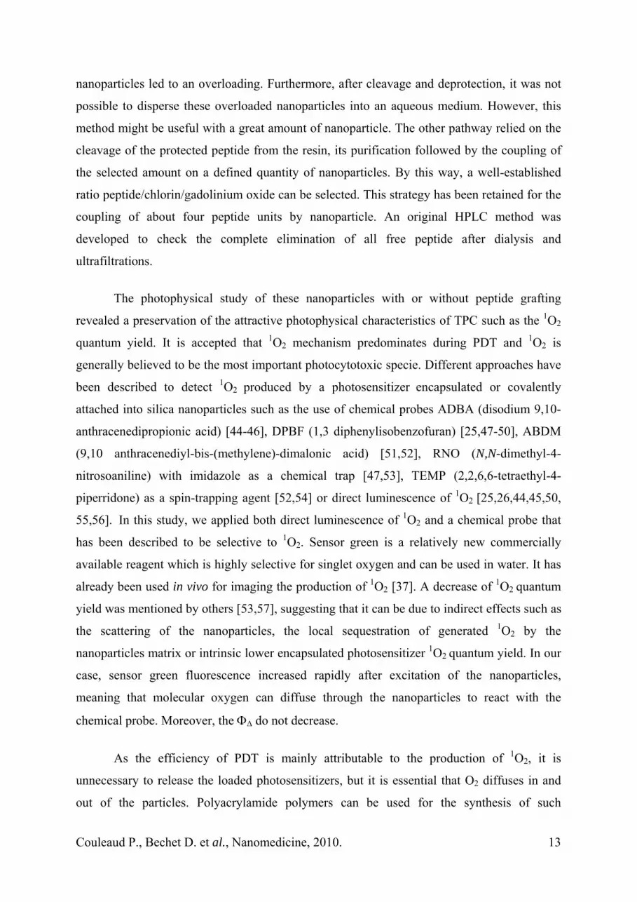
Dark cytotoxicity. As previously demonstrated, we used MDA-MB-231 breast cancer cells
strongly over-expressing NRP-1 receptor [38]. A MTT test was used to evaluate the dark
cytotoxicity of the different batches of nanoparticles (control NP, NP-TPC without peptide
and NP-TPC-ATWLPPR) for concentrations of TPC ranging from 0.10 to 57.00 µM. A 24 h-
incubation of MDA-MB-231 in the absence of light exposure with either photoactive
nanoparticle yielded a surviving cell fraction higher than 85% for concentrations up to 10.0
µM (Fig. 5). Control nanoparticles without chlorin displayed practically no cytotoxic effect
for all concentrations tested until 0.5 mM. We estimated an IC50 value of 16.9 µM for the
nanoparticles with TPC, corresponding to an equivalent molar concentration of 0.15 mM for
the control nanoparticles without photoactivable compound (Fig. 5). All subsequent
experiments were carried out at non-cytotoxic concentrations equal or inferior to 1.0 μM.
Photodynamic activity. MDA-MB-231 cells were incubated with the control nanoparticles
without TPC and peptide (NP), with TPC without peptide (NP-TPC), and with TPC and
peptide (NP-TPC-ATWLPR) and irradiated by red light (652 nm). Whereas NP-TPC at 0.1
µM displayed a weak photodynamic activity in MDA-MB-231 cells, conjugation with
ATWLPPR significantly enhanced photodynamic activity (Fig. 6A). The LD50 values of NP-
TPC and NP-TPC-ATWLPPR at 1µM of TPC were 5 and 3 J/cm2, respectively (Fig. 6C, D).
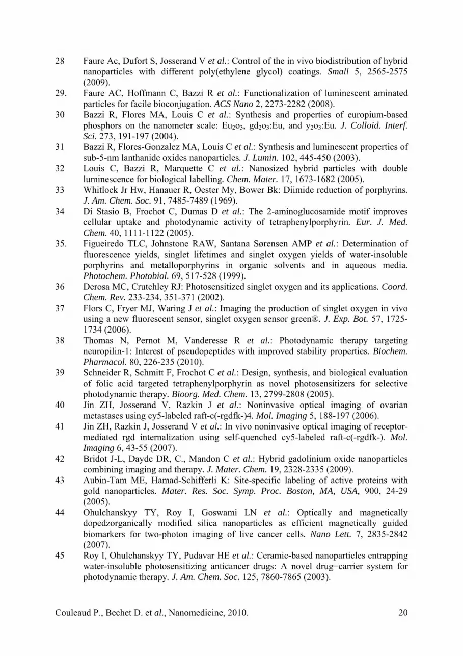
In vivo biodistribution. Figure 7 illustrates the biodistribution of nanoparticles at various
durations after the intravenous injection to nude mice. It appeared that only the kidneys and
the bladder, which are involved in renal excretion, were fluorescent as soon as 15 min after
the injection. Light emitted from the bladder vanished 1 h post-intravenous injection.
Discussion
The goals of this study were to synthesize, characterize and evaluate a multifunctional
nanoparticle formulation for use as a vascular-targeted photosensitizer delivery vehicle. We
found that the preparations of these nanoparticles could be readily achieved. For the linkage
of the ATWLPPR peptide to the nanoparticle, two pathways were investigated. The first one
involved the coupling of the nanoparticle on the preformed peptide linked to the resin. By this
way, the number of peptides per nanoparticle was not clearly determined as the large excess
of peptide, inherent to the synthesis conditions in term of scale, compared to the number of
Couleaud P., Bechet D. et al., Nanomedicine, 2010.
12
nanoparticles led to an overloading. Furthermore, after cleavage and deprotection, it was not
possible to disperse these overloaded nanoparticles into an aqueous medium. However, this
method might be useful with a great amount of nanoparticle. The other pathway relied on the
cleavage of the protected peptide from the resin, its purification followed by the coupling of
the selected amount on a defined quantity of nanoparticles. By this way, a well-established
ratio peptide/chlorin/gadolinium oxide can be selected. This strategy has been retained for the
coupling of about four peptide units by nanoparticle. An original HPLC method was
developed to check the complete elimination of all free peptide after dialysis and
ultrafiltrations.
The photophysical study of these nanoparticles with or without peptide grafting
revealed a preservation of the attractive photophysical characteristics of TPC such as the 1O2
quantum yield. It is accepted that 1O2 mechanism predominates during PDT and 1O2 is
generally believed to be the most important photocytotoxic specie. Different approaches have
been described to detect 1O2 produced by a photosensitizer encapsulated or covalently
attached into silica nanoparticles such as the use of chemical probes ADBA (disodium 9,10-
anthracenedipropionic acid) [44-46], DPBF (1,3 diphenylisobenzofuran) [25,47-50], ABDM
(9,10 anthracenediyl-bis-(methylene)-dimalonic acid) [51,52], RNO (N,N-dimethyl-4-
nitrosoaniline) with imidazole as a chemical trap [47,53], TEMP (2,2,6,6-tetraethyl-4-
piperridone) as a spin-trapping agent [52,54] or direct luminescence of 1O2 [25,26,44,45,50,
55,56]. In this study, we applied both direct luminescence of 1O2 and a chemical probe that
has been described to be selective to 1O2. Sensor green is a relatively new commercially
available reagent which is highly selective for singlet oxygen and can be used in water. It has
already been used in vivo for imaging the production of 1O2 [37]. A decrease of 1O2 quantum
yield was mentioned by others [53,57], suggesting that it can be due to indirect effects such as
the scattering of the nanoparticles, the local sequestration of generated 1O2 by the
nanoparticles matrix or intrinsic lower encapsulated photosensitizer 1O2 quantum yield. In our
case, sensor green fluorescence increased rapidly after excitation of the nanoparticles,
meaning that molecular oxygen can diffuse through the nanoparticles to react with the
chemical probe. Moreover, the ΦΔ do not decrease.
As the efficiency of PDT is mainly attributable to the production of 1O2, it is
unnecessary to release the loaded photosensitizers, but it is essential that O2 diffuses in and
out of the particles. Polyacrylamide polymers can be used for the synthesis of such
Couleaud P., Bechet D. et al., Nanomedicine, 2010.
13

nanoparticles, but most of them are ceramic-based or metallic. Their application in
photosensitizer delivery has not been fully exploited, even though the synthesis of this kind of
nanoparticle has been extensively reported in the literature [43]. In vitro characterization
studies of these nanoparticles revealed a photodynamic efficiency in a concentration-
dependent manner, providing direct evidence that the conjugated photosensitizer in the
nanoparticle matrix could be photoactivated to yield cytotoxic reactive oxygen species. Tada
et al. described the preparation and characterization of methylene-blue-containing silica-
coated magnetic particles. They showed that 1O2 generated by the photosensitizer
immobilized in the silica matrix also diffused. Nevertheless, the quantum yield was lower in
the silica-coated magnetic particles, estimated to be 3% versus 50% for the free methylene
blue [57]. Wieder and al. compared silica and gold nanoparticles, speculating that the surface-
bound photosensitizer on the gold nanoparticles might present a real advantage over an
encapsulated photosensitizer in terms of diffusion of 1O2 [58]. Conjugation with our hybrid
multifunctional nanoparticles did not induce significant variations in photophysical properties
and no changes have been measured between singlet oxygen quantum yields of TPC, NP-TPC
and NP-TPC-ATWLPPR.
A marked advantage of using non-biodegradable nanoparticles is that they can serve
as multifunctional platforms (For a review, see [25]). Kopelman et al. synthesized a
polyacrylamide multifunctional platform with a MRI contrast enhancer and Photofrin®, as
well as a vector (the integrin-targeting RGD peptide) for specific cell targeting [59-61]. In
vitro experiments confirmed the production of 1O2 at levels believed to be sufficient to induce
cell death. Applying a similar approach, Reddy and al. developed another polyacrylamide
nanoparticle encapsulating Photofrin® and imaging agents (fluorescent dye or iron oxide),
using an F3 peptide targeting surface-localized vasculature [62]. In our targeting strategy,
nanoparticles conferred photosensitivity to cells, providing evidence that the chlorin grafted
within the nanoparticle matrix can be photoactivated in vitro. The improved photodynamic
efficiency of peptides-conjugated nanoparticles compared to unconjugated nanoparticles, was
consistent with the binding to NRP-1 receptor followed by an active transport mechanism.
The molecular affinity was tested for functionalized nanoparticles, demonstrating the ability
of ATWLPPR homing heptapeptide to bind recombinant NRP-1 protein testing about four
peptides per nanoparticle. Peptide-conjugated chlorin demonstrated a molecular affinity of
213 µM (IC50 value) for NRP-1 recombinant chimeric protein but to a lesser extent than non-
Couleaud P., Bechet D. et al., Nanomedicine, 2010.
14

conjugated peptide [38]. The presence of four peptides ligand per nanoparticle seems to lead
to a positive cooperativity in binding of ATWLPPR to NRP-1 protein.
The potential of the multifunctional nanoparticles can be fully exploited for PDT applications
only if they do not non specifically accumulate, and if they are rather quickly eliminated.
Choi et al. emphasized the necessity of rapid clearance and pointed out the problems related
to accumulation of nanoparticles in vivo [63]. A rapid clearance of nanoparticles such as
quantum dots was observed only when their hydrodynamic diameter was smaller than 5.5 nm.
However, non specific accumulation of cysteine-coated quantum dots can be observed in the
liver even for the smallest particles, while the largest ones are mostly accumulated in the
liver, spleen, and generally ascribed to uptake by the reticulo-endothelial system [64]. The
detection of nanoparticles in the lungs seems to provide from their agglomeration caused by
the adsorption of plasma proteins [65]. The functionalization of nanoparticles by a PEG
coating is probably the most efficient strategy to avoid nonspecific accumulation, which could
be detrimental for the in vivo application. Many studies have indeed demonstrated the positive
effect of PEG on the biodistribution of such particles [66]. Derivatization by PEG chains
limits the uptake by resident phagocytes in the liver and in the spleen [67]. Because of steric
shielding, the PEG chains avoid the adsorption of plasma proteins onto the surface of the
nanoparticles (opsonisation), occurring when nanoparticles are injected into the bloodstream.
However, the effect of PEG chains on the biodistribution can be counterbalanced by the
charge of the particle. Although pegylation seems to induce prolonged blood circulation, in
some cases, it does not prevent the liver uptake of nanoparticles [69,70]. Our group also
investigated the influence of the length of different PEG on the biodistribution of hybrid
gadolinium oxide nanoparticles [28]. Hybrid gadolinium oxide nanoparticles coated by
PEG250 leaded a rapid in vivo elimination of the nanoparticle. Ex vivo examination of the
organs 24 h post-intravenous injection checked that the nanoparticles did not accumulate into
organs other than the kidneys, which remained strongly fluorescent. In order to preserve this
biodistribution characteristic, we only selected multifunctional nanoparticles describing few
peptides per nanoparticle. We previously evidenced that the peptide-conjugated
photosensitizer was mainly concentrated into organs of the reticulo-endothelial system,
leading to a degradation of the peptide moiety characterized by an enzymatic cleavage of the
peptide bond between Ala and Thr [20]. Interestingly, using functionalized nanoparticles
eliminated by renal excretion will hence overcome this degradation process.
This study revealed the photodynamic efficiency of multifunctional nanoparticles for
PDT. According to our in vitro results ATWLPPR-functionalized nanoparticles could not
Couleaud P., Bechet D. et al., Nanomedicine, 2010.
15

only target NRP-1 protein and cells over expressing NRP-1 but also in vivo endothelial cells
lining tumor vessels. This experiment is currently in progress.
Acknowledgements
This work was supported by the research funds of the French Ligue Nationale Contre le
Cancer and the ANR project no. ANR-08-PCVI-0021-01 Nano-VTP.
Couleaud P., Bechet D. et al., Nanomedicine, 2010.
16

Legends
Figure 1. Strategies of synthesis and purification of the targeted nanoparticles. (A) Two
possible pathways for the linkage of ATWLPPR to the nanoparticle. i) coupling of the
nanoparticle on the growing peptide attached to the resin (pathway A); ii) cleavage of the
protected peptide from the resin followed by coupling on the nanoparticle (pathway B); (B)
Reversed phase HPLC chromatogram of nanoparticles coupled to ATWLPPR (a), free peptide
(b), and mixture of both (c). λ ex =280 nm, λ em = 350 nm.
Figure 2. (A) Size distribution determined by photon correlation spectroscopy of hybrid
gadolinium oxide nanoparticles coated by PEG250. (B) Evolution of the ζ potential versus pH
for these nanoparticles.
Figure 3. Photophysical properties of free photosensitizer (TPC) and after its coupling to the
nanoparticles. (A) Absorption spectra of free TPC, free peptide and suspension of
nanoparticles with or without peptide in ethanol. (B) Fluorescence emission spectra of the free
TPC, and suspensions of nanoparticles with or without peptide in ethanol, for the same
absorbance (λexc=414 nm). (C) Singlet oxygen emission spectra of the free TPC, and
suspensions of nanoparticles with or without peptide in ethanol, for the same absorbance,
after excitation at 414 nm. (D) Fluorescence emission spectrum of the tryptophan residue
using suspensions of nanoparticles with peptide in ethanol (excitation and emission
wavelengths at 280 nm and 350 nm, respectively). (E) Fluorescence emission of sensor green
in water at 525 nm after excitation at 504 nm. Measurements were performed for 20-seconds
period, with 30-seconds interval between each measurement. (F) Fluorescence lifetimes and
quantum yields of fluorescence and singlet oxygen.
Figure 4. Binding of nanoparticles without ATWLPPR (NP-TPC, grey), with peptide
corresponding to an average of four molecules of ATWLPPR peptide per molecular of
nanoparticles (NP-TPC-ATWLPPR, black) to recombinant NRP-1 protein compared to
ATWLPPR alone (white). Binding of biotinylated VEGF165 (5 ng/mL; 110 pM) to NRP-1 in
the presence of 2 µg/mL heparin was evaluated when increasing concentrations of NP-TPC,
NP-TPC-ATWLPPR or ATWLPPR were added (data points show the mean ± SD, n=3).
Couleaud P., Bechet D. et al., Nanomedicine, 2010.
17
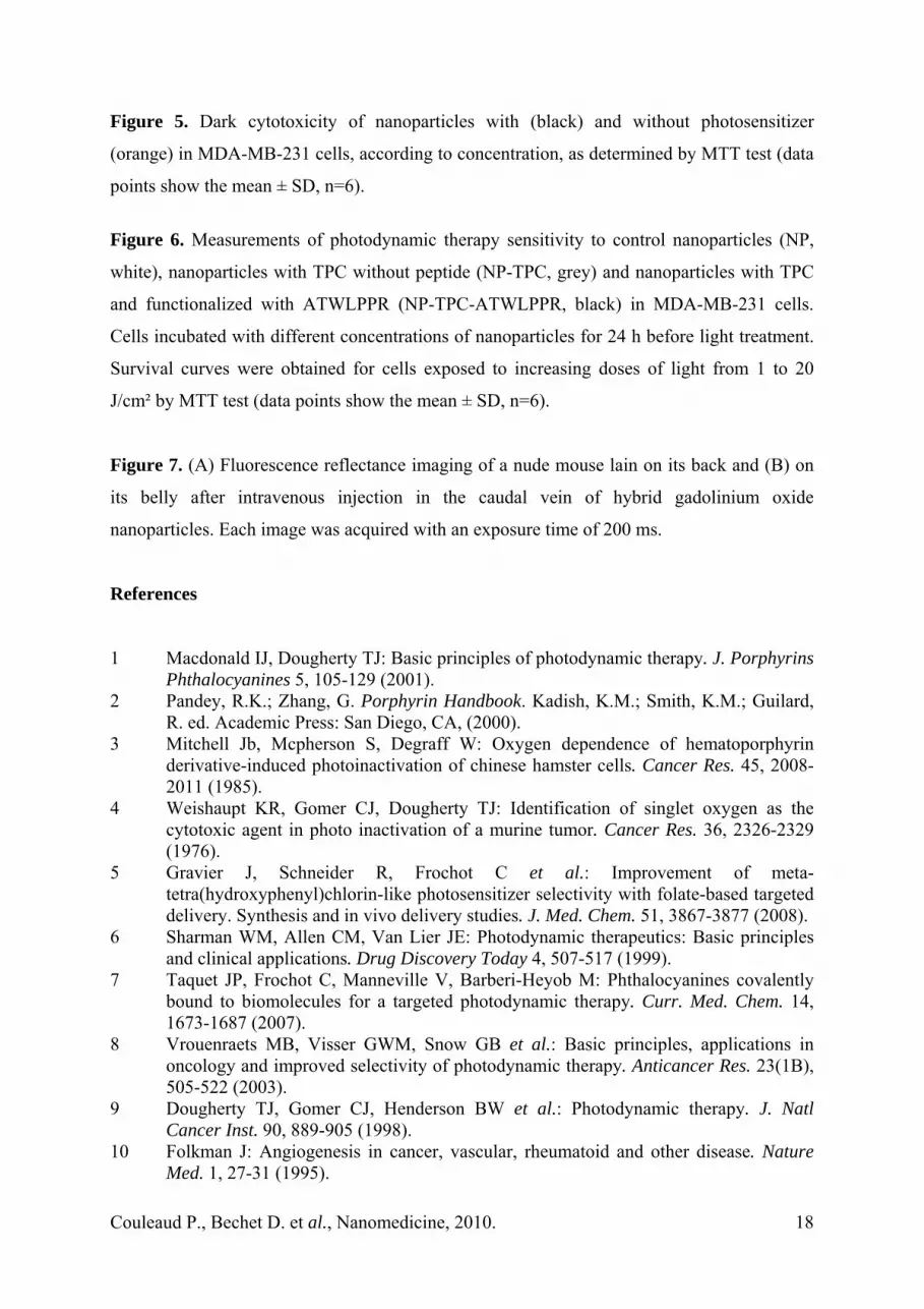
Figure 5. Dark cytotoxicity of nanoparticles with (black) and without photosensitizer
(orange) in MDA-MB-231 cells, according to concentration, as determined by MTT test (data
points show the mean ± SD, n=6).
Figure 6. Measurements of photodynamic therapy sensitivity to control nanoparticles (NP,
white), nanoparticles with TPC without peptide (NP-TPC, grey) and nanoparticles with TPC
and functionalized with ATWLPPR (NP-TPC-ATWLPPR, black) in MDA-MB-231 cells.
Cells incubated with different concentrations of nanoparticles for 24 h before light treatment.
Survival curves were obtained for cells exposed to increasing doses of light from 1 to 20
J/cm² by MTT test (data points show the mean ± SD, n=6).
Figure 7. (A) Fluorescence reflectance imaging of a nude mouse lain on its back and (B) on
its belly after intravenous injection in the caudal vein of hybrid gadolinium oxide
nanoparticles. Each image was acquired with an exposure time of 200 ms.
References 1 Macdonald IJ, Dougherty TJ: Basic principles of photodynamic therapy. J. Porphyrins
Phthalocyanines 5, 105-129 (2001). 2 Pandey, R.K.; Zhang, G. Porphyrin Handbook. Kadish, K.M.; Smith, K.M.; Guilard,
R. ed. Academic Press: San Diego, CA, (2000). 3 Mitchell Jb, Mcpherson S, Degraff W: Oxygen dependence of hematoporphyrin
derivative-induced photoinactivation of chinese hamster cells. Cancer Res. 45, 2008-2011 (1985).
4 Weishaupt KR, Gomer CJ, Dougherty TJ: Identification of singlet oxygen as the cytotoxic agent in photo inactivation of a murine tumor. Cancer Res. 36, 2326-2329 (1976).
5 Gravier J, Schneider R, Frochot C et al.: Improvement of meta-tetra(hydroxyphenyl)chlorin-like photosensitizer selectivity with folate-based targeted delivery. Synthesis and in vivo delivery studies. J. Med. Chem. 51, 3867-3877 (2008).
6 Sharman WM, Allen CM, Van Lier JE: Photodynamic therapeutics: Basic principles and clinical applications. Drug Discovery Today 4, 507-517 (1999).
7 Taquet JP, Frochot C, Manneville V, Barberi-Heyob M: Phthalocyanines covalently bound to biomolecules for a targeted photodynamic therapy. Curr. Med. Chem. 14, 1673-1687 (2007).
8 Vrouenraets MB, Visser GWM, Snow GB et al.: Basic principles, applications in oncology and improved selectivity of photodynamic therapy. Anticancer Res. 23(1B), 505-522 (2003).
9 Dougherty TJ, Gomer CJ, Henderson BW et al.: Photodynamic therapy. J. Natl Cancer Inst. 90, 889-905 (1998).
10 Folkman J: Angiogenesis in cancer, vascular, rheumatoid and other disease. Nature Med. 1, 27-31 (1995).
Couleaud P., Bechet D. et al., Nanomedicine, 2010.
18

11 Chen B, Pogue BW, Luna JM et al.: Tumor vascular permeabilization by vascular-targeting photosensitization: Effects, mechanism, and therapeutic implications. Clin. Cancer Res. 12, 917-923 (2006).
12 Fingar VH, Taber SW, Haydon PS et al.: Vascular damage after photodynamic therapy of solid tumors: A view and comparison of effect in pre-clinical and clinical models at the univeristy of louisville. In Vivo 14, 93-100 (2000).
13 Fingar VH, Wieman TJ, Wiehle SA et al.: The role of microvascular damage in photodynamic therapy: The effect of treatment on vessel constriction, permeability, and leukocyte adhesion. Cancer Res. 52, 4914-4921 (1992).
14 Frochot C, Stasio BD, Vanderesse R et al.: Interest of rgd-containing linear or cyclic peptide targeted tetraphenylchlorin as novel photosensitizers for selective photodynamic activity. Bioorg. Chem. 35, 205-220 (2007).
15 Huang Z, Chen Q, Luck D et al.: Studies of a vascular-acting photosensitizer, pd-bacteriopheophorbide (tookad), in normal canine prostate and spontaneous canine prostate cancer. Laser Surg. Med. 36, 390-397 (2005).
16 Mcmahon Ks, Wieman TJ, Moore PH et al.: Effects of photodynamic therapy using mono-l-aspartyl chlorin e6 on vessel constriction, vessel leakage, and tumor response. Cancer Res. 54, 5374-5379 (1994).
17 Wieman TJ, Mang TS, Fingar VH et al.: Effect of photodynamic therapy on blood flow in normal and tumor vessels. Surgery 104, 512-517 (1988).
18 Schneider R, Tirand L, Frochot C et al.: Recent improvements in the use of synthetic peptides for a selective photodynamic therapy. Anti-Cancer Agents Med.Chem. 6, 469-488 (2006).
19 Tirand L, Frochot C, Vanderesse R et al.: A peptide competing with vegf165 binding on neuropilin-1 mediates targeting of a chlorin-type photosensitizer and potentiates its photodynamic activity in human endothelial cells. J. Controlled Release 111, 153-164 (2006).
20 Thomas N, Tirand L, Chatelut E et al.: Tissue distribution and pharmacokinetics of an atwlppr-conjugated chlorin-type photosensitizer targeting neuropilin-1 in glioma-bearing nude mice. Photochem. Photobiol. Sci. 7, 433-441 (2008).
21 Bechet D, Tirand L, Faivre B et al.: Neuropilin-1 targeting photosensitization-induced early stages of thrombosis via tissue factor release. Pharm. Res. 27, 468-479 (2010).
22 Tirand L, Bastogne T, Bechet D et al.: Response surface methodology: An extensive potential to optimize in vivo photodynamic therapy conditions. Int. J. Radiat. Oncol. Biol. Phys. 75, 244-252 (2009).
23 Thomas N, Bechet D, Becuwe P et al.: Peptide-conjugated chlorin-type photosensitizer binds neuropilin-1 in vitro and in vivo. J. Photochem. Photobiol., B 96, 101-108 (2009).
24 Tirand L, Thomas N, Dodeller M et al.: Metabolic profile of a peptide-conjugated chlorin-type photosensitizer targeting neuropilin-1: An in vivo and in vitro study. Drug Metab. Dispos. 35(5), 806-813 (2007).
25 Bechet D, Couleaud P, Frochot C, et al.: Nanoparticles as vehicles for delivery of photodynamic therapy agents. Trends Biotechnol. 26, 612-621 (2008).
26 Brevet D, Gary-Bobo M, Raehm L et al.: Mannose-targeted mesoporous silica nanoparticles for photodynamic therapy. Chem. Commun. 12, 1475-1477 (2009).
27 Bridot JL, Faure AC, Laurent S et al.: Hybrid gadolinium oxide nanoparticles: Multimodal contrast agents for in vivo imaging. J. Am. Chem. Soc. 129, 5076-5084 (2007).
Couleaud P., Bechet D. et al., Nanomedicine, 2010.
19
28 Faure Ac, Dufort S, Josserand V et al.: Control of the in vivo biodistribution of hybrid nanoparticles with different poly(ethylene glycol) coatings. Small 5, 2565-2575 (2009).
29. Faure AC, Hoffmann C, Bazzi R et al.: Functionalization of luminescent aminated particles for facile bioconjugation. ACS Nano 2, 2273-2282 (2008).
30 Bazzi R, Flores MA, Louis C et al.: Synthesis and properties of europium-based phosphors on the nanometer scale: Eu2o3, gd2o3:Eu, and y2o3:Eu. J. Colloid. Interf. Sci. 273, 191-197 (2004).
31 Bazzi R, Flores-Gonzalez MA, Louis C et al.: Synthesis and luminescent properties of sub-5-nm lanthanide oxides nanoparticles. J. Lumin. 102, 445-450 (2003).
32 Louis C, Bazzi R, Marquette C et al.: Nanosized hybrid particles with double luminescence for biological labelling. Chem. Mater. 17, 1673-1682 (2005).
33 Whitlock Jr Hw, Hanauer R, Oester My, Bower Bk: Diimide reduction of porphyrins. J. Am. Chem. Soc. 91, 7485-7489 (1969).
34 Di Stasio B, Frochot C, Dumas D et al.: The 2-aminoglucosamide motif improves cellular uptake and photodynamic activity of tetraphenylporphyrin. Eur. J. Med. Chem. 40, 1111-1122 (2005).
35. Figueiredo TLC, Johnstone RAW, Santana Sørensen AMP et al.: Determination of fluorescence yields, singlet lifetimes and singlet oxygen yields of water-insoluble porphyrins and metalloporphyrins in organic solvents and in aqueous media. Photochem. Photobiol. 69, 517-528 (1999).
36 Derosa MC, Crutchley RJ: Photosensitized singlet oxygen and its applications. Coord. Chem. Rev. 233-234, 351-371 (2002).
37 Flors C, Fryer MJ, Waring J et al.: Imaging the production of singlet oxygen in vivo using a new fluorescent sensor, singlet oxygen sensor green®. J. Exp. Bot. 57, 1725-1734 (2006).
38 Thomas N, Pernot M, Vanderesse R et al.: Photodynamic therapy targeting neuropilin-1: Interest of pseudopeptides with improved stability properties. Biochem. Pharmacol. 80, 226-235 (2010).
39 Schneider R, Schmitt F, Frochot C et al.: Design, synthesis, and biological evaluation of folic acid targeted tetraphenylporphyrin as novel photosensitizers for selective photodynamic therapy. Bioorg. Med. Chem. 13, 2799-2808 (2005).
40 Jin ZH, Josserand V, Razkin J et al.: Noninvasive optical imaging of ovarian metastases using cy5-labeled raft-c(-rgdfk-)4. Mol. Imaging 5, 188-197 (2006).
41 Jin ZH, Razkin J, Josserand V et al.: In vivo noninvasive optical imaging of receptor-mediated rgd internalization using self-quenched cy5-labeled raft-c(-rgdfk-). Mol. Imaging 6, 43-55 (2007).
42 Bridot J-L, Dayde DR, C., Mandon C et al.: Hybrid gadolinium oxide nanoparticles combining imaging and therapy. J. Mater. Chem. 19, 2328-2335 (2009).
43 Aubin-Tam ME, Hamad-Schifferli K: Site-specific labeling of active proteins with gold nanoparticles. Mater. Res. Soc. Symp. Proc. Boston, MA, USA, 900, 24-29 (2005).
44 Ohulchanskyy TY, Roy I, Goswami LN et al.: Optically and magnetically dopedzorganically modified silica nanoparticles as efficient magnetically guided biomarkers for two-photon imaging of live cancer cells. Nano Lett. 7, 2835-2842 (2007).
45 Roy I, Ohulchanskyy TY, Pudavar HE et al.: Ceramic-based nanoparticles entrapping water-insoluble photosensitizing anticancer drugs: A novel drug−carrier system for photodynamic therapy. J. Am. Chem. Soc. 125, 7860-7865 (2003).
Couleaud P., Bechet D. et al., Nanomedicine, 2010.
20

46 Yan F, Kopelman R: The embedding of meta-tetra(hydroxyphenyl)-chlorin into silica nanoparticle platforms for photodynamic therapy and their singlet oxygen production and ph-dependent optical properties. Photochem. Photobiol. 78, 587-591 (2003).
47 Chen ZL, Sun Y, Huang P et al.: Studies on preparation of photosensitizer loaded magnetic silica nanoparticles and their anti-tumor effects for targeting photodynamic therapy Nanoscale Res. Lett. 4, 400-408 (2009).
48 He X, Wu X, Wang K et al.: Methylene blue-encapsulated phosphonate-terminated silica nanoparticles for simultaneous in vivo imaging and photodynamic therapy. Biomaterials 30, 5601-5609 (2009).
49 Qian J, Gharibi A, HE S: Colloidal mesoporous silica nanoparticles with protoporphyrin ix encapsulated for photodynamic therapy. J. Biomed. Opt. 14, 014012-014016 (2009).
50 Tu HL, Lin YS, Lin HY et al.: In vitro studies of functionalized mesoporous silica nanoparticles for photodynamic therapy Adv. Mater. 21, 172-177 (2009).
51 Zhang R, Wu C, Tong L, Tang B, Xu Qh: Multifunctional core-shell nanoparticles as highly efficient imaging and photosensitizing agents. Langmuir 25, 10153-10158 (2009).
52 Zhao B, Yin JJ, Bilski PJ et al.: Enhanced photodynamic efficacy towards melanoma cells by encapsulation of pc4 in silica nanoparticles. Toxicol. Appl. Pharmacol. 241, 163-172 (2009).
53 Liu F, Zhou X, Chen Z et al.: Preparation of purpurin-18 loaded magnetic nanocarriers in cottonseed oil for photodynamic therapy. Mater. Lett. 62, 2844-2847 (2008).
54 Zhou J, Zhou L, Dong C et al.: Preparation and photodynamic properties of water-soluble hypocrellin a-silica nanospheres. Mater. Lett. 62, 2910-2913 (2008).
55 Cheng SH, Lee CH, Yang CS et al.: Mesoporous silica nanoparticles functionalized with an oxygen-sensing probe for cell photodynamic therapy : Potential cancer theranostics. J. Mater. Chem. 19, 1252-1257 (2009).
56 Kim S, Ohulchanskyy TY, Bharali DC et al.: Organically modified silica nanoparticles with covalently incorporated photosensitizer for photodynamic therapy of cancer. Phys. Chem. C 113, 12641-12644 (2009).
57 Tada DB, Vono LLR, Duarte EL et al.: Methylene blue-containing silica-coated magnetic particles: A potential magnetic carrier for photodynamic therapy. Langmuir 23, 8194-8199 (2007).
58 Wieder ME, Hone DC, Cook MJ et al.: Intracellular photodynamic therapy with photosensitizer-nanoparticle conjugates: Cancer therapy using a 'trojan horse'. Photochem. Photobiol. Sci. 5, 727-734 (2006).
59 Kopelman R, Lee Koo Y E, Philbert M et al:. Multifonctional nanoparticle platforms for in vivo MRI enhancement and photodynamic therapy of a rat brain cancer. J. Magn. Magn. Mat. 293, 404-410, (2005).
60 Ross B, Rehemtulla, A, Koo YEL et al.: Photonic and magnetic nanoexplorers for biomedical use: from subcellular imaging to cancer diagnostics and therapy. Proc. SPIE-Int. Soc. Opt. Eng. 5331, 76-83 (2004).
61 Koo YEL, Fan W, Hah H et al.: Photonic explorers based on multifunctional nanoplatforms for biosensing and photodynamic therapy. Appl. Opt.46,1924-1930 (2007)
62 Reddy GR, Bhojani MS, McConville P et al.: Vascular targeted nanoparticles for imaging and treatment of brain tumors. Clin. Cancer Res. 12, 6677-6686 (2006)
63 Choi SH, Liu W, Misra P et al.: Renal clearance of quantum dots. Nat. Biotechnol. 25, 1165-1170 (2007).
Couleaud P., Bechet D. et al., Nanomedicine, 2010.
21

64 Dobrovolskaia MA, Aggarwal P, Hall JB et al.: Preclinical studies to understand nanoparticle interaction with the immune system and its potential effects on nanoparticle biodistribution. Mol. Pharmaceutics 5, 487-495 (2008).
65 Alexis F, Pridgen E, Molnar LK et al.: Factors affecting the clearance and biodistribution of polymeric nanoparticles. Mol. Pharmaceutics 5, 505-515 (2008).
66 Li SD Huang L: Pharmacokinetics and biodistribution of nanoparticles. Mol. Pharmaceutics 5, 496-504 (2008).
67 Harris JM, Chess RB: Effect of pegylation on pharmaceuticals. Nat. Rev. Drug Discov. 2, 214-221 (2003).
68 Levchenko TS, Rammohan R, Lukyanov AN et al.: Liposome clearance in mice: The effect of a separate and combined presence of surface charge and polymer coating. Int. J. Pharm. 240, 95-102 (2002).
69 Kim D, Park S, Lee JH et al.: Antibiofouling polymer-coated gold nanoparticles as a contrast agent for in vivo x-ray computed tomography imaging. J. Am. Chem. Soc. 129, 7661-7665 (2007).
70 Van Schooneveld MM, Vucic E, Koole R et al.: Improved biocompatibility and pharmacokinetics of silica nanoparticles by means of a lipid coating: A multimodality investigation. Nano Lett. 8, 2517-2525 (2008).71
Couleaud P., Bechet D. et al., Nanomedicine, 2010.
22