Enhanced noscapine delivery using uPAR-targeted optical-MR imaging trackable nanoparticles for...
20
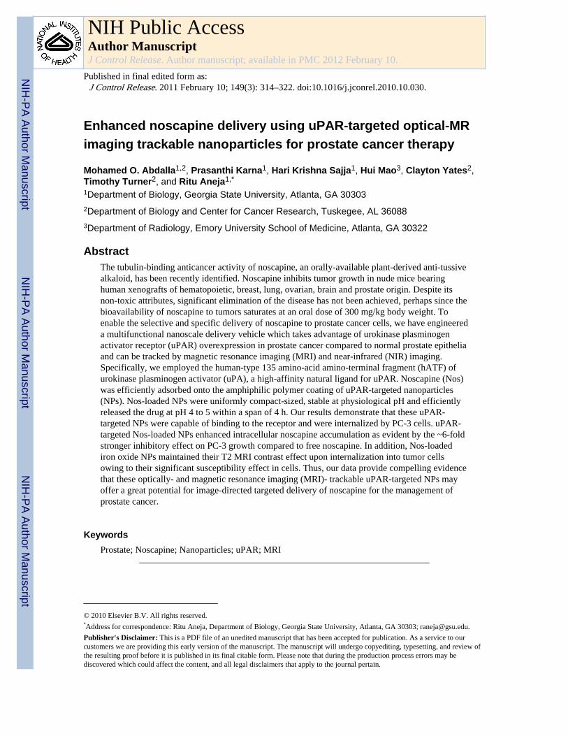
Enhanced noscapine delivery using uPAR-targeted optical-MR imaging trackable nanoparticles for prostate cancer therapy Mohamed O. Abdalla 1,2 , Prasanthi Karna 1 , Hari Krishna Sajja 1 , Hui Mao 3 , Clayton Yates 2 , Timothy Turner 2 , and Ritu Aneja 1,* 1 Department of Biology, Georgia State University, Atlanta, GA 30303 2 Department of Biology and Center for Cancer Research, Tuskegee, AL 36088 3 Department of Radiology, Emory University School of Medicine, Atlanta, GA 30322 Abstract The tubulin-binding anticancer activity of noscapine, an orally-available plant-derived anti-tussive alkaloid, has been recently identified. Noscapine inhibits tumor growth in nude mice bearing human xenografts of hematopoietic, breast, lung, ovarian, brain and prostate origin. Despite its non-toxic attributes, significant elimination of the disease has not been achieved, perhaps since the bioavailability of noscapine to tumors saturates at an oral dose of 300 mg/kg body weight. To enable the selective and specific delivery of noscapine to prostate cancer cells, we have engineered a multifunctional nanoscale delivery vehicle which takes advantage of urokinase plasminogen activator receptor (uPAR) overexpression in prostate cancer compared to normal prostate epithelia and can be tracked by magnetic resonance imaging (MRI) and near-infrared (NIR) imaging. Specifically, we employed the human-type 135 amino-acid amino-terminal fragment (hATF) of urokinase plasminogen activator (uPA), a high-affinity natural ligand for uPAR. Noscapine (Nos) was efficiently adsorbed onto the amphiphilic polymer coating of uPAR-targeted nanoparticles (NPs). Nos-loaded NPs were uniformly compact-sized, stable at physiological pH and efficiently released the drug at pH 4 to 5 within a span of 4 h. Our results demonstrate that these uPAR- targeted NPs were capable of binding to the receptor and were internalized by PC-3 cells. uPAR- targeted Nos-loaded NPs enhanced intracellular noscapine accumulation as evident by the ~6-fold stronger inhibitory effect on PC-3 growth compared to free noscapine. In addition, Nos-loaded iron oxide NPs maintained their T2 MRI contrast effect upon internalization into tumor cells owing to their significant susceptibility effect in cells. Thus, our data provide compelling evidence that these optically- and magnetic resonance imaging (MRI)- trackable uPAR-targeted NPs may offer a great potential for image-directed targeted delivery of noscapine for the management of prostate cancer. Keywords Prostate; Noscapine; Nanoparticles; uPAR; MRI © 2010 Elsevier B.V. All rights reserved. * Address for correspondence: Ritu Aneja, Department of Biology, Georgia State University, Atlanta, GA 30303; [email protected]. Publisher's Disclaimer: This is a PDF file of an unedited manuscript that has been accepted for publication. As a service to our customers we are providing this early version of the manuscript. The manuscript will undergo copyediting, typesetting, and review of the resulting proof before it is published in its final citable form. Please note that during the production process errors may be discovered which could affect the content, and all legal disclaimers that apply to the journal pertain. NIH Public Access Author Manuscript J Control Release. Author manuscript; available in PMC 2012 February 10. Published in final edited form as: J Control Release. 2011 February 10; 149(3): 314–322. doi:10.1016/j.jconrel.2010.10.030. NIH-PA Author Manuscript NIH-PA Author Manuscript NIH-PA Author Manuscript
Transcript of Enhanced noscapine delivery using uPAR-targeted optical-MR imaging trackable nanoparticles for...
Enhanced noscapine delivery using uPAR-targeted optical-MRimaging trackable nanoparticles for prostate cancer therapy
Mohamed O. Abdalla1,2, Prasanthi Karna1, Hari Krishna Sajja1, Hui Mao3, Clayton Yates2,Timothy Turner2, and Ritu Aneja1,*
1Department of Biology, Georgia State University, Atlanta, GA 303032Department of Biology and Center for Cancer Research, Tuskegee, AL 360883Department of Radiology, Emory University School of Medicine, Atlanta, GA 30322
AbstractThe tubulin-binding anticancer activity of noscapine, an orally-available plant-derived anti-tussivealkaloid, has been recently identified. Noscapine inhibits tumor growth in nude mice bearinghuman xenografts of hematopoietic, breast, lung, ovarian, brain and prostate origin. Despite itsnon-toxic attributes, significant elimination of the disease has not been achieved, perhaps since thebioavailability of noscapine to tumors saturates at an oral dose of 300 mg/kg body weight. Toenable the selective and specific delivery of noscapine to prostate cancer cells, we have engineereda multifunctional nanoscale delivery vehicle which takes advantage of urokinase plasminogenactivator receptor (uPAR) overexpression in prostate cancer compared to normal prostate epitheliaand can be tracked by magnetic resonance imaging (MRI) and near-infrared (NIR) imaging.Specifically, we employed the human-type 135 amino-acid amino-terminal fragment (hATF) ofurokinase plasminogen activator (uPA), a high-affinity natural ligand for uPAR. Noscapine (Nos)was efficiently adsorbed onto the amphiphilic polymer coating of uPAR-targeted nanoparticles(NPs). Nos-loaded NPs were uniformly compact-sized, stable at physiological pH and efficientlyreleased the drug at pH 4 to 5 within a span of 4 h. Our results demonstrate that these uPAR-targeted NPs were capable of binding to the receptor and were internalized by PC-3 cells. uPAR-targeted Nos-loaded NPs enhanced intracellular noscapine accumulation as evident by the ~6-foldstronger inhibitory effect on PC-3 growth compared to free noscapine. In addition, Nos-loadediron oxide NPs maintained their T2 MRI contrast effect upon internalization into tumor cellsowing to their significant susceptibility effect in cells. Thus, our data provide compelling evidencethat these optically- and magnetic resonance imaging (MRI)- trackable uPAR-targeted NPs mayoffer a great potential for image-directed targeted delivery of noscapine for the management ofprostate cancer.
KeywordsProstate; Noscapine; Nanoparticles; uPAR; MRI
© 2010 Elsevier B.V. All rights reserved.*Address for correspondence: Ritu Aneja, Department of Biology, Georgia State University, Atlanta, GA 30303; [email protected]'s Disclaimer: This is a PDF file of an unedited manuscript that has been accepted for publication. As a service to ourcustomers we are providing this early version of the manuscript. The manuscript will undergo copyediting, typesetting, and review ofthe resulting proof before it is published in its final citable form. Please note that during the production process errors may bediscovered which could affect the content, and all legal disclaimers that apply to the journal pertain.
NIH Public AccessAuthor ManuscriptJ Control Release. Author manuscript; available in PMC 2012 February 10.
Published in final edited form as:J Control Release. 2011 February 10; 149(3): 314–322. doi:10.1016/j.jconrel.2010.10.030.
NIH
-PA Author Manuscript
NIH
-PA Author Manuscript
NIH
-PA Author Manuscript
1. IntroductionDespite advances in diagnosis and treatment, prostate cancer continues to be one of thedeadliest cancers and ranks the second most frequent cause of cancer-related deaths in malesin the United States [1]. Although current chemotherapeutics including docetaxel haveshown promise for the treatment of hormone-refractory prostate cancer, toxicity has been aserious limitation associated with systemic chemotherapy [2]. Thus, novel strategiesincluding targeted therapies are gaining importance and are being intensively investigated[3–4].
Multifunctional iron oxide nanoparticles (IO NPs) have recently emerged as attractivecandidates for cancer chemotherapeutic approaches that obviate toxicity issues and displayimproved tumor targeting [3, 5–9]. A key component of multifunctional NPs is the tumor-targeting moiety that guides them to cancer-specific receptors with high-affinity andspecificity [10–12]. A specific prostate cancer target and a useful biomarker, the urokinase-type plasminogen activator (uPA) and its receptor (uPAR) are selectively overexpressed athigh levels in invasive prostate cancer, sparing the normal prostate epithelia[13]. Owing tothe differential expression of uPA and uPAR in cancer tissues relative to normal tissues,they are being employed for potential prognostic and/or therapeutic applications [14–16].The amino terminal fragment (ATF) of uPA, which consists of 135 amino acids is, inparticular, a desirable homing moiety as it contains all the determinants required for uPARbinding [17–19]. Recently, Yang et al. reported murine ATF as a targeting moiety to guideiron oxide NPs for in vivo imaging of breast cancer [20]. Since ATF is a remarkabletargeting moiety for tumor cells, it can be advantageously employed as a therapeutic-vehicleto steer ‘drug-loaded’ NPs selectively to tumor sites.
Noscapine, an antitussive agent, has been discovered as a tubulin-binding anticancer drug,and is in Phase I/II clinical trials for the treatment of multiple myeloma. Noscapine does notchange the steady state monomer/polymer ratio of tubulin [21–23]. Thus, noscapine does notcause any hemo-, immuno- and neuronal toxicity [21]. This is a unique-edge over currently-available antimicrotubule drugs that either overpolymerize microtubules (taxanes) ordepolymerize them (vincas) and hence cause various toxicities such as leucocytopenias, andperipheral neuropathies [24–27]. Extensive studies have reported the potential usefulness ofnoscapine in the therapy of lymphomas, melanoma, prostate and lung cancers [28–31]. Thepharmacokinetics of noscapine has also been reported [32] showing that the orallyadministered drug peaks within 2–3 h and is not detectable after 6 h, indicating the shortcirculation time of noscapine in the system [33–34].
Although noscapine has been shown to significantly inhibit tumor growth in preclinical micemodels [21–22, 31, 35], near-complete tumor elimination has not been achieved even afterincreasing the dosage to 450 mg/kg. This is perhaps due to its shorter circulation time aswell as to the possible saturation of drug uptake by tumor cells. In this study, we present thedesign, preparation, optimization and characterization of multifunctional nanoparticles thatsignificantly enhanced noscapine uptake by making noscapine ‘targetable’ to cancer cells.We report the development of a multifunctional nanoparticle delivery vehicle which can betracked by magnetic resonance imaging (MRI) and near infrared (NIR) imaging and takesadvantage of uPAR overexpression to selectively and preferentially deliver noscapine inprostate cancer cells. This novel delivery approach allows for enhanced intracellularnoscapine accumulation to selectively cause significant growth inhibition and cell death inprostate cancer cells.
Abdalla et al. Page 2
J Control Release. Author manuscript; available in PMC 2012 February 10.
NIH
-PA Author Manuscript
NIH
-PA Author Manuscript
NIH
-PA Author Manuscript
2. Materials and Methods2.1. Chemicals and reagents
Polymer coated IO NPs with carboxylic acid reactive groups, and the conjugation kit werefrom Ocean Nanotech (Springdale, AR). Noscapine hydrochloride, Isopropyl-β-D-thiogalactopyranoside (IPTG), N-hydroxysulfosuccinimide (Sulfo-NHS), 1-ethyl-3-[3-dimethylaminopropyl]carbodiimide (EDAC), tris(2-carboxyethyl)-phosphine (TCEP) werefrom Sigma (St. Louis, MO). Cy5.5 monomaleimide was from GE Healthcare (Piscataway,NJ). The peptide, human-type ATF (hATF) of the uPA, was produced and purified.
2.2. Cancer cell linesHuman prostate carcinoma (LNCaP, DU-145 and PC-3) cells were cultured in RPMI-1640supplemented with 10% FBS. Human prostate carcinoma, RC-77T/E, and normal prostateepithelial cells, RC-77N/E, were maintained in Gibco Keratinocyte-SFM medium(Invitrogen, Carlsbad, CA) supplemented with EGF (human recombinant) and bovinepituitary extract. Human embryonic kidney cells (HEK293) were cultured in DMEM.
2.3. Preparation and characterization of hATF-Cy5.5-IO-Nos NPs2.3.1 Production of recombinant hATF—A cDNA fragment encoding the first 135amino acids of human uPA was isolated by PCR amplification and cloned into pET-20(b)expression vector. Initial plasmid cloning and sequence verification was carried out in E.coli Top 10 F′ cells. After the plasmid was verified to contain the correct hATF sequence,the plasmid was purified and transformed into E. coli BL-21 for peptide expression usingIPTG induction and purification under native conditions using FPLC (AmershamBioSciences).
2.3.2. hATF-Cy5.5 conjugation—Cy5.5 monomaleimide was used to conjugatefluorescent Cy5.5 dye with hATF at cysteine residues using the manufacturer’s protocol (GEHealthcare, Piscataway, NJ). Free dye molecules were separated using Nanosep 3Kcentrifugation column (Pall Corp, Ann Arbor, MI). The prepared hATF-Cy5.5 conjugatewas PBS washed and used immediately or stored at 4°C.
2.3.3. Conjugation of hATF or hATF-Cy5.5 complex to IO NPs—We used ~10 nmcore size IO NPs for this study. The core size and hydrodynamic size of IO NPs weremeasured using transmission electron microscopy (TEM) and light scattering scan,respectively. The particles were coated with amphiphilic triblock polymers which stabilizeIO NPs in water and provide reactive carboxyl groups on the particle surface forbioconjugation. hATF peptides were conjugated to the surface of IO NPs via cross -linkingof carboxyl groups to amino side groups on hATF peptides. Briefly, polymer-coated IO NPswere activated with EDAC and sulfo-NHS for 15 min in 20 mM borate buffer (pH 5.0). Theactivated IO NPs were purified using Nanosep 100k columns and reacted with hATF orhATF-Cy5.5 complex (ratio of 1:10) in 10 mM borate buffer (pH 8.5) for 2h at roomtemperature and then overnight at 4°C to generate hATF-IO or hATF-Cy5.5-IO NPs. Theresulting Cy5.5-hATF-IO or hATF-IO were separated from the reaction buffer usingNanosep 100K column. The conjugates were purified by washing with PBS buffer followedby resuspension in sterile distilled water, and stored at 4°C.
2.3.4. Particle size and zeta potential measurements—To determine the size of thenon-targeted IO NPs (NT-IO NPs), hATF-Cy5.5-IO and hATF-Cy5.5-IO-Nos, TEM wasperformed using negative staining method (TEM LEO906E, Oberkochen, Germany). Thesize of NPs was determined from TEM images. The mean hydrodynamic particle size andzeta potential of the various NPs were determined using dynamic laser scattering (Zetasizer-
Abdalla et al. Page 3
J Control Release. Author manuscript; available in PMC 2012 February 10.
NIH
-PA Author Manuscript
NIH
-PA Author Manuscript
NIH
-PA Author Manuscript
II, Malvern Instruments, Worcestershire, UK). All measurements were made at a scatteringangle of 90° at 25°C.
2.3.5. Encapsulation of Noscapine (Nos) onto hATF-Cy5.5-IO NPs—An aqueoussolution of noscapine (1 mg/mL) was added to NT-IO or hATF-Cy5.5-IO NPs at a ratio of 1mg drug to 3 mg of iron (Fe) and the mixture was rotated at RT for 4h and then at 4°Covernight. After filtration of NPs through the 100K Nanosep column, the free drug wasanalyzed by HPLC (Agilent 1100). Encapsulation efficiency of noscapine was calculated asthe mass ratio of the amount of drug entrapped in nanoparticles to the initial amount used indrug loading. The number of Nos molecules encapsulated in an IO nanoparticle was derivedby dividing the total number of Nos molecules by the total number of IO NPs in eachsample. The total number of IO NPs was determined by measuring the optical density (OD)at 500 nm for Fe concentration. The concentration of Fe was then used to calculate thenumber of IO NPs using a standard formula.
2.3.6. pH-dependent drug release—The pH-dependent drug release was determined byincubating drug containing NPs in solutions of different pH (4, 5, 6, or 7) and measuringNos concentration in the flow-through by HPLC. The free drug molecules in the buffer werethen separated from the NT-IO and hATF-IO-Cy5.5NPs using 100K Nanosep column. Theamount of free Nos was calculated from a standard curve of drug concentration and HPLCsignal intensity.
2.4. hATF-mediated binding efficacy of NPs to PC-3 Cells2.4.1. siRNA knockdown of uPAR expression—Human uPAR siRNA (Santa Cruz,CA) is a pool of three target-specific 20–25 nt siRNAs designed to knockdown uPAR geneexpression. Cells were transfected with the pool of three target-specific 20–25 nt siRNAs tohuman uPAR according to the manufacturer’s protocol. A scrambled siRNA sequence wasused in parallel as a control. After 48 h, uPAR levels decreased by ~80–90% as confirmedby western blotting.
2.4.2 Western blotting—LNCaP, DU-145, PC-3, RC-77T/E, control or uPAR siRNAtransfected PC-3 cell lysates were subjected to immunoblotting. Membranes were probedwith rabbit anti-uPAR antibody (American Diagnostica, Greenwich, CT) followed by HRP-conjugated secondary antibody (Santa Cruz, CA). uPAR was detected bychemiluminescence method.
2.4.3. Prussian blue staining—Prussian blue staining was used to confirm presence ofiron in cells treated with IO NPs. Control or uPAR siRNA transfected PC-3 cells oruntransfected LNCaP cells were incubated with 30 pmole/mL of NT-IO or hATF-Cy5.5-IONPs for 4h followed by staining with Prussian blue (1:1 mixture of 5% potassiumferrocyanide and 5% HCl) for 4h at RT. Bright field images were obtained and blue-stainedcells were quantified and represented as percentage of positive cells compared to totalnumber of cells.
2.4.4. In vitro near-infrared optical imaging—PC-3 cells cultured in 12-well plateswere incubated with various concentrations (20, 30, 40 pmole/mL) of hATF-Cy5.5-IO NPsfor 4h at 37°C. Control samples were incubated with equal amounts of hATF-IO NPs atsimilar conditions. Cells were then washed, fixed and imaged using a Perkin-ElmerUltraview ERS microscope with an excitation wavelength of 650 nm and an emission of 680nm.
Abdalla et al. Page 4
J Control Release. Author manuscript; available in PMC 2012 February 10.
NIH
-PA Author Manuscript
NIH
-PA Author Manuscript
NIH
-PA Author Manuscript
2.4.5. In vitro MRI Scan—PC-3, LNCaP and HEK293 cells were incubated with 40pmole/mL of hATF-Cy5.5-IO or NT-IO NPs for 2h at 37°C. Cells were washed, trypsinized,collected and then embedded in 1% agarose in 1.5 mL eppendorf tubes. Samples werescanned using a 3-T MRI scanner (Siemens Healthcare) using T1-weighted gradient echoand multi-echo T2-weighted fast-spin echo imaging sequences. T2 values of each samplewere calculated from obtained multi-echo (TEi, i=20, range from 10–200 ms, interval=10ms). Transverse relaxation times, T2, of each sample were calculated by fitting decay curveon a pixel-by-pixel basis using the nonlinear mono-exponential algorithm of Mi= M0 * exp(−TEi/T2), in which Mo is the MRI signal intensity at TE of 0 and Mi is the MRI signalintensity at a selected TE.
2.6. Cytotoxicity assayRC-77NE, PC-3 or uPAR siRNA transfected cells were treated with serum-free mediumcontaining NT-IO-Nos or hATF-Cy5.5-IO-Nos NPs, or free Nos (10 μM). Control groupswere treated with NT-IO or hATF-IO-NPs without Nos that had equal amounts of IO as testsamples. Cells were incubated for 4h, followed by serum addition to the medium for anadditional 48h. Percentage of tumor cell death was determined by crystal violet assay. Theoptical density was read at 590 nm using a microplate reader (SpectroMax, MolecularDevices). Absorbance values were normalized to control values to obtain the percentage ofviable cells.
2.7. Statistical analysesData are presented as mean ± SEM of at least 3 separate experiments performed in triplicate.P<0.05 was considered statistically significant.
3. Results and Discussion3.1. Design, preparation, characterization and optimization of multifunctional NPs
We have designed, developed and optimized ‘multifunctional’ NPs using a recombinantpeptide hATF of uPA conjugated to magnetic IO NPs. The NIR imaging dye Cy5.5 wasconjugated to cysteine residues of hATF. Subsequently, using amino groups of aminoterminus and those of lysine residues from hATF, the hATF-Cy5.5 bi-conjugate wascoupled with carboxyl ends offered by the amphiphilic polymer coating on IO core to formtri-conjugate hATF-Cy5.5-IO NPs. Finally, noscapine was loaded on the tri-conjugatemolecule by physical adsorption onto the IO NP polymer coating (Scheme 1).
3.1.1. Size characterization of various conjugates3.1.1.1. Transmission electron microscopy (TEM): Effective drug delivery to tumor tissuerequires appropriately sized NPs that can stay in the blood-stream for requisite time toselectively release their payload at the tumor site [36]. Since elevated levels of vascularpermeability characterize most solid tumors, it is pertinent for NPs to possess a compact sizeto steer through immature and leaky tumor vessels and proficiently enter tumor mass [37].Thus, ideally NPs should be large enough (>10 nm) to prevent their rapid leakage into bloodcapillaries but small enough (<100 nm) to escape capture by macrophages that are lodged inthe reticuloendothelial system [36, 38]. We first examined the size of NT- IO NPs usingTEM to assess increase in their size at varying stages of each conjugation step. Fig. 1 (left)presents TEM image of NT-IO NPs, which confirmed the uniformity of the core IO NPswith an average diameter of 11.2 ± 2.5 nm. Next, we attempted to visualize the size of thepolymer coating surrounding the IO core. To this end, we stained NT-IO NPs usingphosphotungstic acid to contrast the thin polymer layer with an optically opaque fluid. Fig. 2(middle) shows negatively stained NT-IO NPs that determined the polymer coating to be at
Abdalla et al. Page 5
J Control Release. Author manuscript; available in PMC 2012 February 10.
NIH
-PA Author Manuscript
NIH
-PA Author Manuscript
NIH
-PA Author Manuscript
an average of 4.2 nm making the overall size of these particles to be 15 ± 3.1 nm. It was notpossible to determine the exact increase in size of the polymer coated IO NPs afterconjugation of hATF-Cy5.5 and drug loading (Fig. 1, right).
3.1.1.2. Dynamic light scattering (DLS): To measure size of the prepared NP conjugates,we used DLS to measure size of NP conjugates that provides size distribution of NPs inaddition to their hydrodynamic size. The size distribution reveals formation of anyaggregates upon conjugation of protein and drug loading. Table 1 presents hydrodynamicsize data of NT-IO, hATF-Cy5.5-IO, hATF-Cy5.5-IO-Nos NPs. NPs retained a narrow sizedistribution after conjugation of peptide-dye and drug loading. As expected, the size of NT-IO NPs (21.36 ± 1.6 nm) increased upon conjugation of hATF-Cy5.5 (35.21 ± 3.7 nm) totheir outer surface while it remained relatively constant upon drug loading (35.62 ± 4.1 nm).In general, overall size of the nanoscale drug delivery vehicle was uniform and within thedesired size range of 10–100 nm for optimal drug delivery [36, 38].
3.1.2. Surface charges of NPs at varying stages of build-up—The surface chargeof NPs determines their circulation time in blood stream and is a measure of NP stability inaqueous solution and their interaction with the cell membrane [39]. Table 1 collates zetapotential measurements. The NT-IO NPs had a negative charge of −52.41 ± 3.1 mV on thesurface, which is due to carboxylic groups of polymer coating. This value correlated withthe value (−51.41 mV) provided in the Certificate of Analysis of NT-IO NPs from themanufacturer. Conjugation of hATF-Cy5.5 to some of the carboxylic ends resulted in adecrease of surface charge of NPs to −33.4 ± 5.61 mV for hATF-Cy5.5-IO NPs. Theloading of hydrophobic Nos on the hATF-Cy5.5-IO NPs further decreased the surfacecharge to −21.6 ± 3.3 mV. Despite surface charge decrease of hATF-Cy5.5-IO-Nos NPs,they were well-dispersed in sterile water with no observable precipitation for a few months.The stability of drug-loaded NPs in aqueous media is crucial for potential biomedicalapplications as no vehicle solution is needed to inject them. Moreover, surface charge datareflect that hydrophilicity of NPs did not decrease which in clinical settings may preventtheir opsonization by plasma proteins and elongate their blood circulation time.
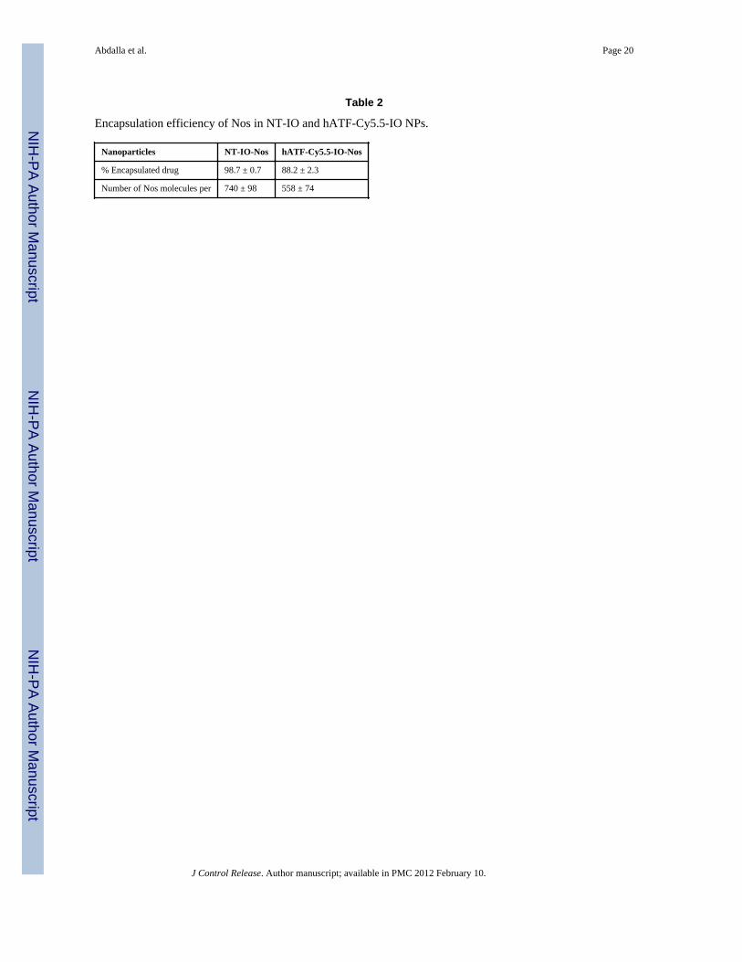
3.1.3 Efficiency of noscapine loading—Incorporation of sufficient drug amounts ontoNPs is vital for effective therapy. Based on drug properties, it can either be covalentlyconjugated to the polymer coating of NPs or can be physically adsorbed onto the polymercoating [12, 40]. A downside of covalent linking is that it may limit the amount of drugmolecules conjugated to each NP as well as present a low efficiency of drug release uponcell entry. Physical adsorption of drug molecules onto NPs, on the contrary, may ferry moredrug molecules into cells and release the drug efficiently. Thus, we next investigated theefficiency of noscapine encapsulation in the prepared uPAR-targeted NPs. Using a ratio of 1mg drug to 3 mg IO, we were able to encapsulate 98.7% of Nos onto the NT-IO NPs (Table2) as determined by HPLC analysis. This high drug encapsulation efficiency may perhaps bedue to a favorable interaction between the hydrophobic noscapine molecules and thehydrophobic segment of the amphiphilic polymer coating of NPs. We observed that therewas a 10% decrease in efficiency of drug encapsulation when Nos was loaded afterconjugation of hATF-Cy5.5 to the surface of IO NPs. The decrease in drug encapsulationwas perhaps attributed to steric hindrance to the drug molecules caused by the spaceoccupied by the hATF-Cy5.5 conjugates on the surface of IO NPs [41]. Table 2 also showsthe number of Nos molecules adsorbed per IO NPs in NT IO NPs as well as in NPs with theconjugated targeting moiety-dye (hATF-Cy5.5). There was a ~24% percent reduction in thenumber of Nos molecules adsorbed on the surface of NT-IO and hATF-Cy5.5-IO NPs. Thedifference in percent drug encapsulation and percent decrease in the number of adsorbed
Abdalla et al. Page 6
J Control Release. Author manuscript; available in PMC 2012 February 10.
NIH
-PA Author Manuscript
NIH
-PA Author Manuscript
NIH
-PA Author Manuscript
Nos molecules per IO NP may perhaps be due to uneven drug encapsulation in a givensample.
3.1.4. pH-dependent drug release characteristics from multifunctional NPs—Itis recognizable that the efficiency of any drug delivery vehicle is best evaluated by its abilityto release the ‘payload’ drug into cells and not systemically in the patient’s circulationsystem. To simulate physiological conditions, we examined the amount of Nos that can bereleased from the NPs at pH 7. Slightly acidic pH conditions simulated the environment oftumor interstitium, while more acidic pH was more representative of the pH in intracellularvesicles such as endosomes and lysosomes. Fig. 2A shows overlays of HPLCchromatograms to depict the amount of Nos detected in the supernatant of hATF-Cy5.5-IO-Nos NPs at various pHs. At pH 7, no peak was detected for Nos indicating that the drug wasnot released due to its strong adsorption to the polymer coating on the IO NPs (Fig. 2B).However, when the pH was lowered, the amount of detected Nos increased with the increasein the acidity of the medium. The area under the peak was used to calculate the percentageof drug release from the NT-IO-Nos or hATF-Cy5.5-IO-Nos NPs (Fig. 2B). While no drugwas detected at pH 7, the NPs released about ~25–30% of their drug payload at pH 6. At pH5, the percentage of drug released from NPs increased to ~60%. At pH 4, over ~80% of drugmolecules were efficiently released from NPs. These results indicated strong adsorption ofdrug onto the IO NP surface, thus facilitating drugs release at acidic pH. Enhancement ofdrug release may be due to the onset of polymer degradation at lower pH or the breakage ofhydrophobic interactions between polymer and drug molecules. Another plausibleexplanation for enhanced drug release at lower pH exists based upon hydrogen bonding.Since the lower benzofuranone ring of noscapine has electronegative oxygen atoms(particularly the ‘two oxygens’ in the lactone ring of the molecule) that can participate inhydrogen bonding, the extent of hydrogen bonding of noscapine would reduce with decreasein pH (protonation) resulting in a pH dependent drug release. In conclusion, these datasuggested that the targeted NPs can serve as efficient carriers of drug payload that can bedelivered in acidic environments which particularly exist in the hypoxic tumor milieu.
3.2. Binding and internalization efficiency of hATF-targeted NPs3.2.1. Determination of uPAR expression in various prostate cancer cells—Previous studies have shown that PC-3 cells are highly metastatic, whereas DU145 andLNCaP cells are moderately and poorly metastatic, respectively [15, 42–43]. Because uPARis important for tumor invasion and metastasis, we first compared uPAR expression level invarious prostate cancer cell lines with different metastatic potentials. As shown in Fig. 3A,uPAR levels corresponded well with the metastatic potential of cells. uPAR expression wassignificantly higher in PC-3 and DU145 cells compared to LNCaP and RC-77T/E cells (Fig.3A). More importantly, the normal prostate cells, RC77N/E, showed very low levels ofuPAR expression, suggesting that uPAR is selectively overexpressed in cancer cells andalmost spares normal cells. Having identified expression levels of uPAR, we next evaluatedthe uptake efficiency of targeted and non-targeted IO NPs by these cells with varying uPARexpression.
3.2.2. Prussian blue staining—Previous reports show that the binding of ATF to uPARhas species specificity [18]. Thus, to preserve species specificity, we employed human ATFrather than murine ATF as previously reported [18]. The binding of hATF peptide to highuPAR-expressing PC-3 and low uPAR-expressing LNCaP cells was investigated.Subsequent to binding, the consequent internalization was examined by Prussian bluestaining that stains iron. Fig. 3B shows bright field images of PC-3 cells treated with NT-IOand hATF-Cy5.5-IO NPs. Low-uPAR expressing LNCaP cells were treated with hATF-Cy5.5-IO NPs. Fig. 3C presents quantitation data of percent blue-stained cells. PC-3 cells
Abdalla et al. Page 7
J Control Release. Author manuscript; available in PMC 2012 February 10.
NIH
-PA Author Manuscript
NIH
-PA Author Manuscript
NIH
-PA Author Manuscript
incubated with hATF-Cy5.5-IO NPs showed a high level of iron staining (>80% blue-stained cells) both intra and extracellularly which suggested that targeted NPs were able tobind and be internalized by the uPAR-overexpressing PC-3 cells. PC-3 cells incubated withNT-IO exhibited a very low level of non-specific iron staining. Next, we performed anindirect experiment to demonstrate that the internalization of targeted NPs is mediated bythe interaction of hATF with uPAR displayed on PC-3 cells. To this end, we saturated uPARsites on PC-3 cells by adding an excess of free hATF peptide (1h) prior to addition oftargeted NPs. Fig. 3B (bottom left) shows micrographs of uPAR-saturated PC-3 cells thatexhibited a much lower iron staining compared to non-saturated PC-3 cells (<20% blue-stained cells). Next, we incubated low uPAR-expressing LNCaP cells with targeted NPs.Fig. 3B (bottom right) shows micrographs of LNCaP cells with insignificant amount ofscattered non-specific iron staining (<5% blue-stained cells). These data suggested that highuPAR expression in PC-3 cells facilitated binding and internalization of targeted NPs.
3.2.3. In vitro near-infrared (NIR) optical imaging—We further confirmed thebinding and internalization of the targeted multifunctional NPs in PC-3 cells using confocalmicroscopy by imaging the NIR-dye, Cy5.5 which was conjugated to the hATF peptide. Tothis end, PC-3 cells were treated with increasing amounts of the hATF-Cy5.5-IO NPs andDIC (differential interference contrast) as well as NIR images were acquired (Fig. 4A, B).Our results showed an increase in the number of Cy5.5-positive cells with increase inconcentration of iron oxide in the hATF-Cy5.5-IO NPs (Fig. 4B). Quantitation of Cy5.5-positive cells is shown in Fig. 4B. The percentage of Cy5.5 cells increased from ~45% to~60% on increasing the dose level from 20 pmol to 30 pmol and peaked to a maximum of~90% at 40 pmol of iron oxide in the NPs. This data validated the high-affinity bindingbetween hATF guided IO NPs and uPAR displayed on the surface of tumor cells. As anegative control, we treated PC-3 cells with hATF-IO NPs and as expected, there was noNIR signal due to absence of Cy5.5 moiety. The incorporation of Cy5.5 in NPs might offer asimple and sensitive approach to quantify tumor targeting and to monitor distribution of NPsin small animal models. Furthermore, the multifunctional hATF-Cy5.5-IO NPs offer theprospect to image both primary and metastatic tumor lesions with hATF-guided NPs thattarget high-uPAR expressing prostate cancer at their invasive sites.
3.2.4. In vitro magnetic resonance imaging (MRI)—Multifunctional nanoparticlescan serve to simultaneously deliver anticancer drugs and allow tumor imaging and are beingincreasingly investigated for their improved therapeutic efficacy and lower toxicity.Superparamagnetic IO NPs are widely used as contrast agents in MRI because of theirnegative enhancement effect on T2-weighted sequences. Our multifunctional NPs includedthe iron oxide core which typically induces reduction of transverse relaxation time and T2-weighted contrast change. The signal drops in MRI allow for tracking the targeted drugdelivery using clinically capable MRI systems. MRI scans of untreated as well as treatedPC-3 cells with NT-IO and hATF-Cy5.5-IO NPs are shown in Fig. 5A. LNCaP and HEK293treated with hATF-Cy5.5-IO NPs were used as controls. The MR images demonstratedsignificant target specificity. A pronounced reduction of T2 values was detected in PC-3cells treated with the hATF-Cy5.5-IO NPs. This indicated that high amounts of IO arebound to high uPAR-expressing PC-3 cells. On the other hand, both LNCaP and HEK293cells incubated with hATF-Cy5.5-IO NPs did not exhibit the MRI signal drop with T2weighted contrast and reduction of T2 values. These results corroborated our Prussian bluestaining and NIR optical imaging data emphasizing that uPAR expression was essential forsuccessful binding and internalization of the targeted NPs by PC-3 cells. Fig. 5B presentsMRI signal differences of different samples in T2-weighted images which correlated wellwith the reduction in the T2 values of different samples. We found that PC-3 cells treatedwith targeted hATF-Cy5.5-IO NPs appeared dark in the T2-weighted MR image due to the
Abdalla et al. Page 8
J Control Release. Author manuscript; available in PMC 2012 February 10.
NIH
-PA Author Manuscript
NIH
-PA Author Manuscript
NIH
-PA Author Manuscript
presence of high level of hATF-IONPs (Fig. 5B). On the contrary, untreated PC-3, NT-IONPs treated PC-3, and hATF-Cy5.5-IO NPs treated LNCaP and HEK293 cells remainedbright and indistinguishable due to the absence of iron.
3.3. uPAR-targeted noscapine-loaded IO NPs exert a robust cytotoxic effectTo evaluate the selective advantage of targeted NPs to enhance noscapine delivery, weexamined the cytotoxic effect of Nos-loaded targeted or non-targeted IO NPs on PC-3 cells.The hATF-Cy5.5-IO-Nos NPs caused ~60% cell death compared to NT-IO-Nos. However,free Nos at the same concentration (10 μM) did not show pronounced cell death. Next, wemeasured concentration of free noscapine required to achieve ~60% cell death. To this end,we conducted a dose-dependent experiment by treating PC-3 cells with 10–100 μMconcentration of free Nos. As shown in Fig 6B, the equivalent dose of free Nos to achieve~60% cell death was 60 μM. Thus, the uPAR-targeted NPs caused ~6-fold enhancement incell death compared to the free drug. This enhancement may be ascribed to the efficientintracellular drug delivery by the targeted NPs. Next, we used human uPAR siRNA toknockdown uPAR gene expression in PC-3 cells. The efficiency of gene knockdown wasconfirmed by immunoblot analysis as shown in Fig 6C. The normal prostate epithelial cells,RC77-N/E, did not express a detectable amount of this protein. Next, we determined thecytotoxicity of hATF-Cy5.5-IO Nos NPs on PC-3 and normal prostate RC-77N/E cells withsilenced uPAR expression. Fig 6D showed that ~20% cell death was observed in PC-3 cellswith silenced uPAR and no almost cell death was seen in RC77-N/E. These experimentsconfirmed the targeted delivery of noscapine via the uPA-uPAR system.
4. ConclusionWe have demonstrated the design, development, characterization and optimization ofnoscapine-loaded uPAR-targeted iron oxide NPs for efficient delivery of the noscapine intoprostate cancer cells. Guided by the highly specific and selective ATF of the uPA receptor,the NPs bind to uPA receptor and were internalized by PC-3 cells. The targeted release ofnoscapine from NPs resulted in a ~6-fold higher drug efficacy compared to free drug. Inconclusion, these multifunctional NPs offer NIR-MRI dual imaging functionality providedby the Cy5.5 (NIR) and the iron oxide core (MRI) in addition to targeted drug delivery.
AcknowledgmentsThis work was supported by grants to RA from NCI/NIH, DoD and GRA. TT and CY acknowledge grant supportfrom NCI/NIH and NIH/RCMI.
References1. Jemal A, Siegel R, Ward E, Hao Y, Xu J, Thun MJ. Cancer Statistics, 2009. CA Cancer J Clin.
2009; 59:225–249. [PubMed: 19474385]2. Petrylak DP. The treatment of hormone-refractory prostate cancer: docetaxel and beyond. Rev Urol.
2006; 8(Suppl 2):S48–55. [PubMed: 17021642]3. Sajja HK, East MP, Mao H, Wang YA, Nie S, Yang L. Development of multifunctional
nanoparticles for targeted drug delivery and noninvasive imaging of therapeutic effect. Curr DrugDiscov Technol. 2009; 6:43–51. [PubMed: 19275541]
4. Kim K, Kim JH, Park H, Kim YS, Park K, Nam H, Lee S, Park JH, Park RW, Kim IS, Choi K, KimSY, Kwon IC. Tumor-homing multifunctional nanoparticles for cancer theragnosis: Simultaneousdiagnosis, drug delivery, and therapeutic monitoring. J Control Release. 2010; 146:219–227.[PubMed: 20403397]
5. Xie J, Huang J, Li X, Sun S, Chen X. Iron oxide nanoparticle platform for biomedical applications.Curr Med Chem. 2009; 16:1278. [PubMed: 19355885]
Abdalla et al. Page 9
J Control Release. Author manuscript; available in PMC 2012 February 10.
NIH
-PA Author Manuscript
NIH
-PA Author Manuscript
NIH
-PA Author Manuscript
6. Gupta AK, Naregalkar RR, Vaidya VD, Gupta M. Recent advances on surface engineering ofmagnetic iron oxide nanoparticles and their biomedical applications. Nanomedicine. 2007; 2:23–39.[PubMed: 17716188]
7. Corot C, Robert P, Idée JM, Port M. Recent advances in iron oxide nanocrystal technology formedical imaging. Adv Drug Deliv Rev. 2006; 58:1471–1504. [PubMed: 17116343]
8. Guthi JS, Yang SG, Huang G, Li S, Khemtong C, Kessinger CW, Peyton M, Minna JD, Brown KC,Gao J. MRI-visible micellar nanomedicine for targeted drug delivery to lung cancer cells. MolPharm. 7:32–40. [PubMed: 19708690]
9. Kumar A, Jena PK, Behera S, Lockey RF, Mohapatra S. Multifunctional magnetic nanoparticles fortargeted delivery. Nanomedicine. 6:64–69. [PubMed: 19446653]
10. Yang L, Mao H, Wang YA, Cao Z, Peng X, Wang X, Duan H, Ni C, Yuan Q, Adams G, SmithMQ, Wood WC, Gao X, Nie S. Single chain epidermal growth factor receptor antibody conjugatednanoparticles for in vivo tumor targeting and imaging. Small. 2009; 5:235–243. [PubMed:19089838]
11. Cho K, Wang X, Nie S, Chen ZG, Dong DM. Therapeutic Nanoparticles for Drug Delivery inCancer. Clin Cancer Res. 2008; 14:131–1316.
12. Brannon-Peppas L, Blanchette JO. Nanoparticle and targeted systems for cancer therapy. AdvDrug Deliv Rev. 2004; 56:1649–1659. [PubMed: 15350294]
13. Li Y, Cozzi PJ. Targeting uPA/uPAR in prostate cancer. Cancer Treat Rev. 2007; 33:521–527.[PubMed: 17658220]
14. Ge Y, Elghetany MT. Urokinase plasminogen activator receptor (CD87): Something old,something new. Lab Hematol. 2003; 9:67–71. [PubMed: 12828301]
15. Pulukuri SM, Gondi CS, Lakka SS, Jutla A, Estes N, Gujrati M, Rao JS. RNA interference-directed knockdown of urokinase plasminogen activator and urokinase plasminogen activatorreceptor inhibits prostate cancer cell invasion, survival, and tumorigenicity in vivo. J Biol Chem.2005; 280:36529–36540. [PubMed: 16127174]
16. Sheng S. The urokinase-type plasminogen activator system in prostate cancer metastasis. CancerMetastasis Rev. 2001; 20(3–4):287–296. [PubMed: 12085967]
17. Hoyer-Hansen G, Ploug M, Behrendt N, Ronne E, Dano K. Cell-surface acceleration of urokinase-catalyzed receptor cleavage. Eur J Biochem. 1997; 243:21–26. [PubMed: 9030717]
18. Li H, Soria C, Griscelli F, Opolon P, Soria J, Yeh P, Legrand C, Vannier JP, Belin D, PerricaudetM, Lu H. Amino-terminal fragment of urokinase inhibits tumor cell invasion in vitro and in vivo:Respective contribution of the urokinase plasminogen activator receptor-dependent or -independent pathway. Hum Gene Ther. 2005; 16:1157–1167. [PubMed: 16218777]
19. Li Y, Cozzi PJ. Targeting uPA/uPAR in prostate cancer. Cancer Treat Rev. 2007; 33:521–527.[PubMed: 17658220]
20. Yang L, Mao H, Cao Z, Wang YA, Peng X, Wang X, Sajja HK, Wang L, Duan H, Ni C, StaleyCA, Wood WC, Gao X, Nie S. Molecular imaging of pancreatic cancer in an animal model usingtargeted multifunctional nanoparticles. Gastroenterology. 2009; 136:1514–1525. [PubMed:19208341]
21. Landen JW, Hau V, Wang M, Davis T, Ciliax B, Wainer BH, Van Meir EG, Glass JD, Joshi HC,Archer DR. Noscapine crosses the blood-brain barrier and inhibits glioblastoma growth. ClinCancer Res. 2004; 10:5187–5201. [PubMed: 15297423]
22. Ke Y, Ye Y, Grossniklaus HE, Archer DA, Joshi HJ, Kapp JA. Noscapine inhibits tumor growthwith little toxicity to normal tissues or inhibition of immune responses. Cancer Immunology,Immunotherapy. 2000; 49(4–5):217–225.
23. Ye K, Ke Y, Keshava N, Shanks J, Kapp JA, Tekmal RR, Petros J, Joshi HC. Opium alkaloidnoscapine is an antitumor agent that arrests metaphase and induces apoptosis in dividing cells.Proc Natl Acad Sci U S A. 1998; 95:1601–1606. [PubMed: 9465062]
24. Sioka C, Kyritsis A. Central and peripheral nervous system toxicity of common chemotherapeuticagents. Cancer Chemotherapy and Pharmacology. 2009; 63:761–767. [PubMed: 19034447]
25. Perez EA. Microtubule inhibitors: Differentiating tubulin-inhibiting agents based on mechanismsof action, clinical activity, and resistance. Molecular Cancer Therapeutics. 2009; 8:2086–2095.[PubMed: 19671735]
Abdalla et al. Page 10
J Control Release. Author manuscript; available in PMC 2012 February 10.
NIH
-PA Author Manuscript
NIH
-PA Author Manuscript
NIH
-PA Author Manuscript
26. Verstappen CCP, Heimans JJ, Hoekman K, Postma TJ. Neurotoxic complications of chemotherapyin patients with Cancer: clinical signs and optimal management. Drugs. 2003; 63:1549–1563.[PubMed: 12887262]
27. Park SB, Krishnan AV, Lin CS, Goldstein D, Friedlander M, Kiernan MC. Mechanisms underlyingchemotherapy-induced neurotoxicity and the potential for neuroprotective strategies. Curr MedChem. 2008; 15:3081–3084. [PubMed: 19075655]
28. Ke Y, Ye K, Grossniklaus HE, Archer DR, Joshi HC, Kapp JA. Noscapine inhibits tumor growthwith little toxicity to normal tissues or inhibition of immune responses. Cancer ImmunologyImmunotherapy. 2000; 49:217–225.
29. Zhou J, Liu M, Aneja R, Chandra R, Joshi HC. Enhancement of paclitaxel-induced microtubulestabilization, mitotic arrest, and apoptosis by the microtubule-targeting agent EM012. BiochemPharmacol. 2004; 68:2435–2441. [PubMed: 15548390]
30. Barken I, Geller J, Rogosnitzky M. Prophylactic noscapine therapy inhibits human prostate cancerprogression and metastasis in a mouse model. Anticancer Res. 30:399–401. [PubMed: 20332445]
31. Jackson T, Chougule M, Ichite N, Patlolla R, Singh M. Antitumor activity of noscapine in humannon-small cell lung cancer xenograft model. Cancer Chemotherapy and Pharmacology. 2008;63:117–126. [PubMed: 18338172]
32. Bitoun E, Micheloni A, Lamant L, Bonnart C, Tartaglia-Polcini A, Cobbold C, Al Saati T, MariottiF, Mazereeuw-Hautier J, Boralevi F, Hohl D, Harper J, Bodemer C, D’Alessio M, Hovnanian A.LEKTI proteolytic processing in human primary keratinocytes, tissue distribution and defectiveexpression in Netherton syndrome. Hum Mol Genet. 2003; 12:2417–2430. [PubMed: 12915442]
33. Dahlstrom B, Mellstrand T, Lofdahl CG, Johansson M. Pharmacokinetic properties of noscapine.Eur J Clin Pharmacol. 1982; 22:535–539. [PubMed: 7128665]
34. Karlsson MO, Dahlstrom B, Eckernas SA, Johansson M, Alm AT. Pharmacokinetics of oralnoscapine. Eur J Pharm Biopharm. 1990; 39:275–279.
35. Ye K, Ke Y, Keshava N, Shanks J, Kapp JA, Tekmal RR, Petros J, Joshi HC. Opium alkaloidnoscapine is an antitumor agent that arrests metaphase and induces apoptosis in dividing cells.Proc Natl Acad Sci USA. 1998; 95:1601–1606. [PubMed: 9465062]
36. Gaumet M, Vargas A, Gurny R, Delie F. Nanoparticles for drug delivery: The need for precision inreporting particle size parameters. Eur J Pharm Biopharm. 2008; 69:1–9. [PubMed: 17826969]
37. Veiseh O, Gunn JW, Zhang M. Design and fabrication of magnetic nanoparticles for targeted drugdelivery and imaging. Advanced Drug Delivery Reviews. 2010; 62:284–304. [PubMed:19909778]
38. Cho K, Wang X, Nie S, Chen ZG, Shin DM. Therapeutic nanoparticles for drug delivery in cancer.Clin Cancer Res. 2008; 14:1310–1316. [PubMed: 18316549]
39. Gupta AK, Wells S. Surface-modified superparamagnetic nanoparticles for drug delivery:preparation, characterization, and cytotoxicity studies. IEEE Trans Nanobioscience. 2004; 3:66–73. [PubMed: 15382647]
40. Zavisova V, Koneracka M, Strbak O, Tomasovicova N, Kopcansky P, Timko M, Vavra I.Encapsulation of indomethacin in magnetic biodegradable polymer nanoparticles. J Magn MagnMater. 2007; 311:379–382.
41. Yang L, Cao Z, Sajja HK, Mao H, Wang L, Geng H, Xu H, Jiang T, Wood WC, Nie SWY.Development of receptor targeted magnetic iron oxide nanoparticles for efficient drug delivery andtumor Iimaging. J Biomed Nanotech. 2008; 4:1–11.
42. Rajvir D, Hae Duck P, John C, Robert LV, George F, Perinchery N. Inhibition of tumorigenicpotential and prostate-specific antigen expression in LNCaP human prostate cancer cell line by 13-cis-retinoic acid. Int J Cancer. 1994; 59:126–132. [PubMed: 7523313]
43. Laniado ME, Lalani EN, Fraser SP, Grimes JA, Bhangal G, Djamgoz MB, Abel PD. Expressionand functional analysis of voltage-activated Na+ channels in human prostate cancer cell lines andtheir contribution to invasion in vitro. Am J Pathol. 1997; 150:1213–1221. [PubMed: 9094978]
Abdalla et al. Page 11
J Control Release. Author manuscript; available in PMC 2012 February 10.
NIH
-PA Author Manuscript
NIH
-PA Author Manuscript
NIH
-PA Author Manuscript
Fig. 1.TEM micrographs of polymer coated IO NPs before and after conjugation of peptide-dyeconjugates. Left, NT-IO (Bar=200 nm). Middle, NT-IO-Stained: negative stained IO NPsshowed the polymer layer surrounding the IO core (Bar=200 nm). Right, hATF-Cy5.5-IO-Stained: negative-stained polymer coated IO NPs after conjugation of peptide-dye showedroughness on the edge of polymer coating thus limiting exact NP size determination even athigher magnification (Bar= 100 nm).
Abdalla et al. Page 12
J Control Release. Author manuscript; available in PMC 2012 February 10.
NIH
-PA Author Manuscript
NIH
-PA Author Manuscript
NIH
-PA Author Manuscript
Fig. 2.Examination of pH-dependent release of Nos from targeted and non-targeted NPs. 10 μL ofNT-IO-Nos or hATF-Cy5.5-IO-Nos NPs were placed in different buffer solutions with pH4, 5, 6 and 7 for 2h at 37°C. A, Overlay of HPLC chromatograms illustrate the amount ofNos released from hATF-Cy5.5-IO-Nos NPs in buffers with different pHs. While nopronounced peak was detected for Nos at pH 7, a Nos peak was detected and increased insize at lower pHs. B, Percentage of drug released from NT-IO-Nos and hATF-Cy5.5-IO-NosNPs calculated from the area under the peak of the HPLC chromatograms.
Abdalla et al. Page 13
J Control Release. Author manuscript; available in PMC 2012 February 10.
NIH
-PA Author Manuscript
NIH
-PA Author Manuscript
NIH
-PA Author Manuscript
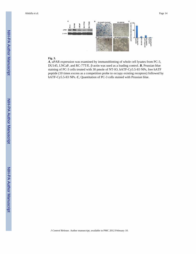
Fig. 3.A, uPAR expression was examined by immunoblotting of whole cell lysates from PC-3,DU145, LNCaP, and RC-77T/E. β-actin was used as a loading control. B, Prussian bluestaining of PC-3 cells treated with 30 pmole of NT-IO, hATF-Cy5.5-IO NPs, free hATFpeptide (10 times excess as a competition probe to occupy existing receptors) followed byhATF-Cy5.5-IO NPs. C, Quantitation of PC-3 cells stained with Prussian blue.
Abdalla et al. Page 14
J Control Release. Author manuscript; available in PMC 2012 February 10.
NIH
-PA Author Manuscript
NIH
-PA Author Manuscript
NIH
-PA Author Manuscript
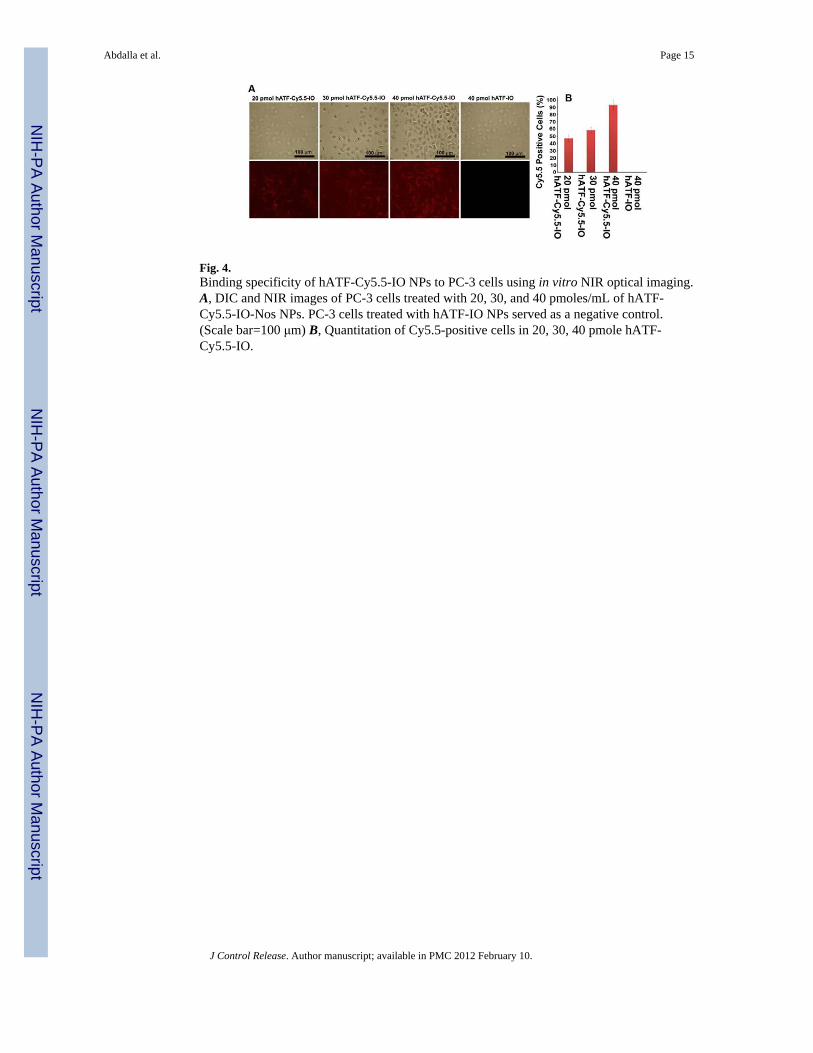
Fig. 4.Binding specificity of hATF-Cy5.5-IO NPs to PC-3 cells using in vitro NIR optical imaging.A, DIC and NIR images of PC-3 cells treated with 20, 30, and 40 pmoles/mL of hATF-Cy5.5-IO-Nos NPs. PC-3 cells treated with hATF-IO NPs served as a negative control.(Scale bar=100 μm) B, Quantitation of Cy5.5-positive cells in 20, 30, 40 pmole hATF-Cy5.5-IO.
Abdalla et al. Page 15
J Control Release. Author manuscript; available in PMC 2012 February 10.
NIH
-PA Author Manuscript
NIH
-PA Author Manuscript
NIH
-PA Author Manuscript
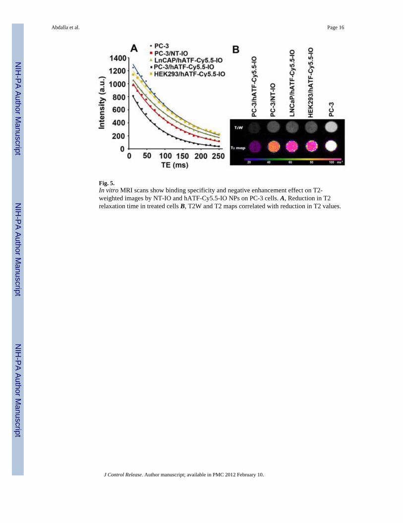
Fig. 5.In vitro MRI scans show binding specificity and negative enhancement effect on T2-weighted images by NT-IO and hATF-Cy5.5-IO NPs on PC-3 cells. A, Reduction in T2relaxation time in treated cells B, T2W and T2 maps correlated with reduction in T2 values.
Abdalla et al. Page 16
J Control Release. Author manuscript; available in PMC 2012 February 10.
NIH
-PA Author Manuscript
NIH
-PA Author Manuscript
NIH
-PA Author Manuscript
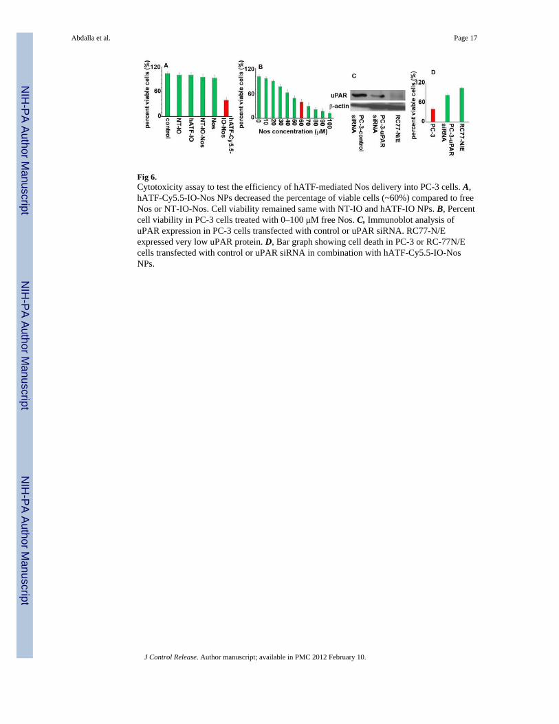
Fig 6.Cytotoxicity assay to test the efficiency of hATF-mediated Nos delivery into PC-3 cells. A,hATF-Cy5.5-IO-Nos NPs decreased the percentage of viable cells (~60%) compared to freeNos or NT-IO-Nos. Cell viability remained same with NT-IO and hATF-IO NPs. B, Percentcell viability in PC-3 cells treated with 0–100 μM free Nos. C, Immunoblot analysis ofuPAR expression in PC-3 cells transfected with control or uPAR siRNA. RC77-N/Eexpressed very low uPAR protein. D, Bar graph showing cell death in PC-3 or RC-77N/Ecells transfected with control or uPAR siRNA in combination with hATF-Cy5.5-IO-NosNPs.
Abdalla et al. Page 17
J Control Release. Author manuscript; available in PMC 2012 February 10.
NIH
-PA Author Manuscript
NIH
-PA Author Manuscript
NIH
-PA Author Manuscript
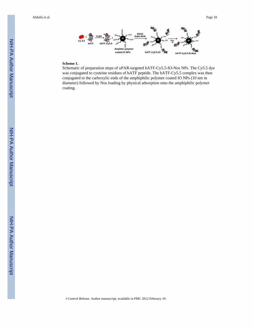
Scheme 1.Schematic of preparation steps of uPAR-targeted hATF-Cy5.5-IO-Nos NPs. The Cy5.5 dyewas conjugated to cysteine residues of hATF peptide. The hATF-Cy5.5 complex was thenconjugated to the carboxylic ends of the amphiphilic polymer coated IO NPs (10 nm indiameter) followed by Nos loading by physical adsorption onto the amphiphilic polymercoating.
Abdalla et al. Page 18
J Control Release. Author manuscript; available in PMC 2012 February 10.
NIH
-PA Author Manuscript
NIH
-PA Author Manuscript
NIH
-PA Author Manuscript

NIH
-PA Author Manuscript
NIH
-PA Author Manuscript
NIH
-PA Author Manuscript
Abdalla et al. Page 19
Table 1
Size and zeta potential characterization of NT-IO, hATF-Cy5.5-IO, and hATF-Cy5.5-IO-Nos NPs
Nanoparticles Size (nm) Zeta Potential (mV)
TEM DLS
NT-IO 14.4 ± 2.1 21.36 ± 1.6 −52.41 ± 3.1
hATF-Cy5.5-IO ------ 35.21± 3.7 −33.4 ± 5.61
hATF-Cy5.5-IO-Nos ------ 35.62 ± 4.1 −21.6 ± 3.3
J Control Release. Author manuscript; available in PMC 2012 February 10.
NIH
-PA Author Manuscript
NIH
-PA Author Manuscript
NIH
-PA Author Manuscript
Abdalla et al. Page 20
Table 2
Encapsulation efficiency of Nos in NT-IO and hATF-Cy5.5-IO NPs.
Nanoparticles NT-IO-Nos hATF-Cy5.5-IO-Nos
% Encapsulated drug 98.7 ± 0.7 88.2 ± 2.3
Number of Nos molecules per 740 ± 98 558 ± 74
J Control Release. Author manuscript; available in PMC 2012 February 10.