Inhibitory input from slowly adapting lung stretch receptors to retrotrapezoid nucleus...
16
J Physiol 580.1 (2007) pp 285–300 285 Inhibitory input from slowly adapting lung stretch receptors to retrotrapezoid nucleus chemoreceptors Thiago S. Moreira 1,2 , Ana C. Takakura 1,2 , Eduardo Colombari 2 , Gavin H. West 1 and Patrice G. Guyenet 1 1 Department of Pharmacology, University of Virginia, Charlottesville, VA, 22908, USA 2 Department of Physiology, UNIFESP-EPM, S˜ ao Paulo, SP, 04023-060, Brazil The retrotrapezoid nucleus (RTN) contains CO 2 -activated interneurons with properties consistent with central respiratory chemoreceptors. These neurons are glutamatergic and express the transcription factor Phox2b. Here we tested whether RTN neurons receive an input from slowly adapting pulmonary stretch receptors (SARs) in halothane-anaesthetized ventilated rats. In vagotomized rats, RTN neurons were inhibited to a variable extent by stimulating myelinated vagal afferents using the lowest intensity needed to inhibit the phrenic nerve discharge (PND). In rats with intact vagus nerves, RTN neurons were inhibited, also to a variable extent, by increasing positive end-expiratory pressure (PEEP; 2–6 cmH 2 O). The cells most sensitive to PEEP were inhibited during each lung inflation at rest and were instantly activated by stopping ventilation. Muscimol (GABA-A agonist) injection in or next to the solitary tract at area postrema level desynchronized PND from ventilation, eliminated the lung inflation-synchronous inhibition of RTN neurons and their steady inhibition by PEEP but did not change their CO 2 sensitivity. Muscimol injection into the rostral ventral respiratory group eliminated PND but did not change RTN neuron response to either lung inflation, PEEP increases, vagal stimulation or CO 2 . Generalized glutamate receptor blockade with intracerebroventricular (I.C.V .) kynurenate eliminated PND and the response of RTN neurons to lung inflation but did not change their CO 2 sensitivity. PEEP-sensitive RTN neurons expressed Phox2b. In conclusion, RTN chemoreceptors receive an inhibitory input from myelinated lung stretch receptors, presumably SARs. The lung input to RTN may be di-synaptic with inhibitory pump cells as sole interneurons. (Received 21 November 2006; accepted after revision 23 January 2007; first published online 25 January 2007) Corresponding author P. G. Guyenet: Department of Pharmacology, University of Virginia Health System, PO Box 800735, 1300 Jefferson Park Avenue, Charlottesville, VA 22908-0735, USA. Email: [email protected] The rat retrotrapezoid nucleus (RTN) is a cluster of CO 2 -sensitive glutamatergic neurons located at the ventral surface of the medulla oblongata within a region involved in respiratory chemoreception (Loeschcke, 1982; Eldridge et al. 1985; Akilesh et al. 1997; Ho et al. 2001; Nattie, 2001a; Okada et al. 2002; Feldman et al. 2003; Mulkey et al. 2004; Putnam et al. 2004; Ritucci et al. 2005; Stornetta et al. 2006). RTN neurons selectively innervate the pontomedullary regions that contain the respiratory pattern generator (Smith et al. 1989; Cream et al. 2002; Mulkey et al. 2004; Rosin et al. 2006). RTN neurons are activated by acidification in vitro (Mulkey et al. 2004, 2006; Ritucci et al. 2005; Guyenet et al. 2005a,b) and their main source of drive under anaesthesia appears to be their intrinsic response to the pH of the surrounding parenchyma (Mulkey et al. T. S. Moreira and A. C. Takakura contributed equally to this study. 2004; Guyenet et al. 2005a). RTN neurons can also be activated by peripheral chemoreceptor stimulation and they receive inhibitory inputs from several components of the respiratory pattern generator (Guyenet et al. 2005a; Takakura et al. 2006). The known anatomical and physiological properties of RTN neurons are thus consistent with the notion that these cells function as a chemosensory integrating centre that drives the ponto-medullary respiratory network. Slowly adapting lung stretch receptors (SARs) influence a variety of respiratory and autonomic outflows and the activation of these receptors often opposes the cardiorespiratory effects produced by chemoreceptor stimulation (Hayashi et al. 1996; Coleridge & Coleridge, 2001; Vatner & Uemura, 2001; Kubin et al. 2006). Given that RTN neurons are excited both by brain P CO 2 and by carotid body stimulation, it is reasonable to assume that these neurons could be subject to an inhibitory control by lung stretch afferents. The present study is designed C 2007 The Authors. Journal compilation C 2007 The Physiological Society DOI: 10.1113/jphysiol.2006.125336
-
Upload
independent -
Category
Documents
-
view
5 -
download
0
Transcript of Inhibitory input from slowly adapting lung stretch receptors to retrotrapezoid nucleus...

J Physiol 580.1 (2007) pp 285–300 285
Inhibitory input from slowly adapting lung stretchreceptors to retrotrapezoid nucleus chemoreceptors
Thiago S. Moreira1,2, Ana C. Takakura1,2, Eduardo Colombari2, Gavin H. West1 and Patrice G. Guyenet1
1Department of Pharmacology, University of Virginia, Charlottesville, VA, 22908, USA2Department of Physiology, UNIFESP-EPM, Sao Paulo, SP, 04023-060, Brazil
The retrotrapezoid nucleus (RTN) contains CO2-activated interneurons with properties
consistent with central respiratory chemoreceptors. These neurons are glutamatergic and express
the transcription factor Phox2b. Here we tested whether RTN neurons receive an input from
slowly adapting pulmonary stretch receptors (SARs) in halothane-anaesthetized ventilated rats.
In vagotomized rats, RTN neurons were inhibited to a variable extent by stimulating myelinated
vagal afferents using the lowest intensity needed to inhibit the phrenic nerve discharge (PND).
In rats with intact vagus nerves, RTN neurons were inhibited, also to a variable extent, by
increasing positive end-expiratory pressure (PEEP; 2–6 cmH2O). The cells most sensitive to
PEEP were inhibited during each lung inflation at rest and were instantly activated by stopping
ventilation. Muscimol (GABA-A agonist) injection in or next to the solitary tract at area
postrema level desynchronized PND from ventilation, eliminated the lung inflation-synchronous
inhibition of RTN neurons and their steady inhibition by PEEP but did not change their CO2
sensitivity. Muscimol injection into the rostral ventral respiratory group eliminated PND but did
not change RTN neuron response to either lung inflation, PEEP increases, vagal stimulation or
CO2. Generalized glutamate receptor blockade with intracerebroventricular (I.C.V.) kynurenate
eliminated PND and the response of RTN neurons to lung inflation but did not change their CO2
sensitivity. PEEP-sensitive RTN neurons expressed Phox2b. In conclusion, RTN chemoreceptors
receive an inhibitory input from myelinated lung stretch receptors, presumably SARs. The lung
input to RTN may be di-synaptic with inhibitory pump cells as sole interneurons.
(Received 21 November 2006; accepted after revision 23 January 2007; first published online 25 January 2007)
Corresponding author P. G. Guyenet: Department of Pharmacology, University of Virginia Health System, PO Box
800735, 1300 Jefferson Park Avenue, Charlottesville, VA 22908-0735, USA. Email: [email protected]
The rat retrotrapezoid nucleus (RTN) is a cluster ofCO2-sensitive glutamatergic neurons located at the ventralsurface of the medulla oblongata within a region involvedin respiratory chemoreception (Loeschcke, 1982; Eldridgeet al. 1985; Akilesh et al. 1997; Ho et al. 2001; Nattie, 2001a;Okada et al. 2002; Feldman et al. 2003; Mulkey et al. 2004;Putnam et al. 2004; Ritucci et al. 2005; Stornetta et al. 2006).RTN neurons selectively innervate the pontomedullaryregions that contain the respiratory pattern generator(Smith et al. 1989; Cream et al. 2002; Mulkey et al.2004; Rosin et al. 2006). RTN neurons are activated byacidification in vitro (Mulkey et al. 2004, 2006; Ritucci et al.2005; Guyenet et al. 2005a,b) and their main source of driveunder anaesthesia appears to be their intrinsic responseto the pH of the surrounding parenchyma (Mulkey et al.
T. S. Moreira and A. C. Takakura contributed equally to this study.
2004; Guyenet et al. 2005a). RTN neurons can also beactivated by peripheral chemoreceptor stimulation andthey receive inhibitory inputs from several componentsof the respiratory pattern generator (Guyenet et al.2005a; Takakura et al. 2006). The known anatomical andphysiological properties of RTN neurons are thusconsistent with the notion that these cells functionas a chemosensory integrating centre that drives theponto-medullary respiratory network.
Slowly adapting lung stretch receptors (SARs) influencea variety of respiratory and autonomic outflows andthe activation of these receptors often opposes thecardiorespiratory effects produced by chemoreceptorstimulation (Hayashi et al. 1996; Coleridge & Coleridge,2001; Vatner & Uemura, 2001; Kubin et al. 2006). Giventhat RTN neurons are excited both by brain PCO2
and bycarotid body stimulation, it is reasonable to assume thatthese neurons could be subject to an inhibitory controlby lung stretch afferents. The present study is designed
C© 2007 The Authors. Journal compilation C© 2007 The Physiological Society DOI: 10.1113/jphysiol.2006.125336
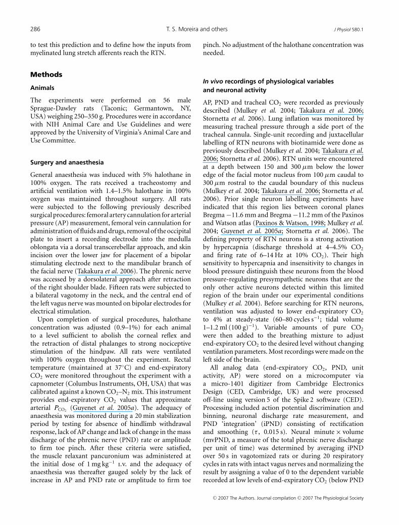
286 T. S. Moreira and others J Physiol 580.1
to test this prediction and to define how the inputs frommyelinated lung stretch afferents reach the RTN.
Methods
Animals
The experiments were performed on 56 maleSprague-Dawley rats (Taconic; Germantown, NY,USA) weighing 250–350 g. Procedures were in accordancewith NIH Animal Care and Use Guidelines and wereapproved by the University of Virginia’s Animal Care andUse Committee.
Surgery and anaesthesia
General anaesthesia was induced with 5% halothane in100% oxygen. The rats received a tracheostomy andartificial ventilation with 1.4–1.5% halothane in 100%oxygen was maintained throughout surgery. All ratswere subjected to the following previously describedsurgical procedures: femoral artery cannulation for arterialpressure (AP) measurement, femoral vein cannulation foradministration of fluids and drugs, removal of the occipitalplate to insert a recording electrode into the medullaoblongata via a dorsal transcerebellar approach, and skinincision over the lower jaw for placement of a bipolarstimulating electrode next to the mandibular branch ofthe facial nerve (Takakura et al. 2006). The phrenic nervewas accessed by a dorsolateral approach after retractionof the right shoulder blade. Fifteen rats were subjected toa bilateral vagotomy in the neck, and the central end ofthe left vagus nerve was mounted on bipolar electrodes forelectrical stimulation.
Upon completion of surgical procedures, halothaneconcentration was adjusted (0.9–1%) for each animalto a level sufficient to abolish the corneal reflex andthe retraction of distal phalanges to strong nociceptivestimulation of the hindpaw. All rats were ventilatedwith 100% oxygen throughout the experiment. Rectaltemperature (maintained at 37◦C) and end-expiratoryCO2 were monitored throughout the experiment with acapnometer (Columbus Instruments, OH, USA) that wascalibrated against a known CO2–N2 mix. This instrumentprovides end-expiratory CO2 values that approximatearterial PCO2
(Guyenet et al. 2005a). The adequacy ofanaesthesia was monitored during a 20 min stabilizationperiod by testing for absence of hindlimb withdrawalresponse, lack of AP change and lack of change in the massdischarge of the phrenic nerve (PND) rate or amplitudeto firm toe pinch. After these criteria were satisfied,the muscle relaxant pancuronium was administered atthe initial dose of 1 mg kg−1
i.v. and the adequacy ofanaesthesia was thereafter gauged solely by the lack ofincrease in AP and PND rate or amplitude to firm toe
pinch. No adjustment of the halothane concentration wasneeded.
In vivo recordings of physiological variablesand neuronal activity
AP, PND and tracheal CO2 were recorded as previouslydescribed (Mulkey et al. 2004; Takakura et al. 2006;Stornetta et al. 2006). Lung inflation was monitored bymeasuring tracheal pressure through a side port of thetracheal cannula. Single-unit recording and juxtacellularlabelling of RTN neurons with biotinamide were done aspreviously described (Mulkey et al. 2004; Takakura et al.2006; Stornetta et al. 2006). RTN units were encounteredat a depth between 150 and 300 μm below the loweredge of the facial motor nucleus from 100 μm caudal to300 μm rostral to the caudal boundary of this nucleus(Mulkey et al. 2004; Takakura et al. 2006; Stornetta et al.2006). Prior single neuron labelling experiments haveindicated that this region lies between coronal planesBregma −11.6 mm and Bregma −11.2 mm of the Paxinosand Watson atlas (Paxinos & Watson, 1998; Mulkey et al.2004; Guyenet et al. 2005a; Stornetta et al. 2006). Thedefining property of RTN neurons is a strong activationby hypercapnia (discharge threshold at 4–4.5% CO2
and firing rate of 6–14 Hz at 10% CO2). Their highsensitivity to hypercapnia and insensitivity to changes inblood pressure distinguish these neurons from the bloodpressure-regulating presympathetic neurons that are theonly other active neurons detected within this limitedregion of the brain under our experimental conditions(Mulkey et al. 2004). Before searching for RTN neurons,ventilation was adjusted to lower end-expiratory CO2
to 4% at steady-state (60–80 cycles s−1; tidal volume1–1.2 ml (100 g)−1). Variable amounts of pure CO2
were then added to the breathing mixture to adjustend-expiratory CO2 to the desired level without changingventilation parameters. Most recordings were made on theleft side of the brain.
All analog data (end-expiratory CO2, PND, unitactivity, AP) were stored on a microcomputer viaa micro-1401 digitizer from Cambridge ElectronicsDesign (CED, Cambridge, UK) and were processedoff-line using version 5 of the Spike 2 software (CED).Processing included action potential discrimination andbinning, neuronal discharge rate measurement, andPND ‘integration’ (iPND) consisting of rectificationand smoothing (τ , 0.015 s). Neural minute × volume(mvPND, a measure of the total phrenic nerve dischargeper unit of time) was determined by averaging iPNDover 50 s in vagotomized rats or during 20 respiratorycycles in rats with intact vagus nerves and normalizing theresult by assigning a value of 0 to the dependent variablerecorded at low levels of end-expiratory CO2 (below PND
C© 2007 The Authors. Journal compilation C© 2007 The Physiological Society

J Physiol 580.1 RTN and vagus input 287
threshold) and a value of 1 at the highest level of PCO2
investigated (between 9.5 and 10%). The CED softwarewas also used for acquisition of peri-event histograms ofneuronal activity and peri-event averages of iPND, trachealCO2, or tracheal pressure. The peri-event histogramsof neuronal single-unit activity were triggered either oniPND or on the tracheal pressure trace. Each histogramrepresents the summation of at least 100 central respiratoryor ventilation cycles (350–800 action potentials perhistogram).
The steady-state relationship between RTN neuronalactivity and end-expiratory CO2 was obtained by steppingthe inspired CO2 level to various values for a minimumof 3 min and up to 5 min. The mean discharge rate ofthe neuron was measured during the last 30 s of eachstep at which time end-expiratory CO2 and the dischargeof the neuron appeared to have reached equilibrium.End-expiratory CO2 was measured by averaging themaximum values recorded from 10 consecutive breathsat the midpoint of the time interval sampled.
In rats with intact vagus nerves, lung mechano-receptors were activated by transiently elevating positiveend-expiratory pressure (PEEP) (5–20 s) from a restinglevel of +1 cmH2O to +2, +4 or +6 cmH2O. Invagotomized rats, lung mechanoreceptor stimulation wasapproximated by stimulating the central end of the leftvagus nerve for 5 s at 10 Hz with pulses of 0.1 ms. Theshort pulse duration was designed to favour activationof myelinated fibres (Hayashi et al. 1996). Currentintensity was kept between threshold and twice threshold,the threshold being defined as the intensity necessaryto produce approximately 30–60% reduction in PNDamplitude but no central apnoea.
Pharmacological treatments
The GABA-mimetic drug muscimol (Sigma ChemicalsCo., St Louis, MO, USA; 1.75 mm in sterile saline pH 7.4)was injected intraparenchymally while a single RTNneuron was being recorded. The muscimol solutioncontained a 5% dilution of fluorescent latex microbeads(Lumafluor, New City, NY, USA) for later histologicalidentification of the injection sites (Moreira et al.2006). Muscimol was pressure injected bilaterally throughsingle-barrel glass pipettes with a 20 μm tip diameter.The injection volume (30 nl in 5 s; 50 pmol per side) wasdetermined by following the downward movement of themeniscus within the cylindrical portion of the pipetteusing a microscope fitted with a calibrated eye-piece(estimated accuracy: ±5 nl). The glass pipettes used fordrug injection allowed recording of multiunit neuronalactivity. This property was used to direct the electrode tipto the rostral ventral respiratory group (rVRG) by locatinginspiratory-related field potentials. This region was found
500 μm rostral to the calamus scriptorius, 1.8 mm lateralto midline and 1.9–2.2 mm below the dorsal surface of thebrainstem using electrodes angled 20 deg forward. Afterdirecting the tip of the muscimol pipette to the rVRG,we searched for a suitable RTN unit. The first muscimolinjection was made into the predetermined site while theRTN unit was being recorded. The second injection wasplaced 1–2 min later in the symmetric brain location basedon the stereotaxic coordinates of the first one, also whilethe cell was recorded. The injections rarely lead to the lossof the recorded RTN unit and all reported units were keptfor at least 20 min after drug injection. Due to the longduration of action of muscimol, a single RTN neuron wassubjected to this experimental protocol in any given rat.The protocol used to test the effect of muscimol injectioninto the nucleus of the solitary tract (NTS) was the sameexcept that the injections were made 0.5 mm below thesurface of the brain using surface landmarks to identifythe insertion point of the pipette (500 μm rostral to thecalamus scriptorius, 0.8 mm lateral to midline).
Kynurenate, a broad-spectrum antagonist of glutamateionotropic receptors (KYN; Sigma Chemicals Co.), wasadministered into the fourth ventricle while an RTNunit was being recorded. Unit recording was typicallymaintained during and for at least 30 min after druginjection. Kynurenate was prepared as a 0.5 m pH 7.3 stocksolution and diluted 50% just before use with normalbicarbonate Ringer solution of the following composition(mm): 130 NaCl, 3 KCl, 2 MgCl2, 2 CaCl2, 1.25 NaH2PO4,26 NaHCO3 and 10 glucose. The solution was slowlyinjected (1–2 min; 60 μl; 15 μmol) into the cerebrospinalfluid via a needle inserted through the atlanto-occipitalmembrane. The needle shaft was sealed to the membranewith cyanoacrylate prior to performing the injection. KYNinjected in this manner initially bathes the lower and uppersurfaces of the brainstem and upper spinal cord. Howdeep within the brain the drug eventually penetrates isunknown; however, within minutes, it eliminates PND, therespiratory discharge of neurons located deep within theventrolateral medulla (Mulkey et al. 2004) and all knownsensory inputs to the blood pressure-regulating neuronsof the ventrolateral medulla (Sun et al. 1988). Note thatif KYN had equilibrated within 2 cm3 of brain tissue themean concentration of the drug would be 7.5 mm whichis five times more than necessary to block all forms ofionotropic glutamate receptors in vitro.
Histology
At the end of the experiment the rat was deeplyanaesthetized with halothane and perfused transcardiallywith heparinised phosphate-buffered saline (pH 7.4)followed by paraformaldehyde (4% in 0.1 m phosphatebuffer, pH 7.4). All histochemical procedures were done
C© 2007 The Authors. Journal compilation C© 2007 The Physiological Society

288 T. S. Moreira and others J Physiol 580.1
using 30-μm-thick free-floating sections according topreviously described protocols (Takakura et al. 2006;Stornetta et al. 2006). Cells labelled with biotinamide wereidentified by incubating the sections with streptavidinconjuguated with Alexa-488. The transcription factorPhox2b was detected as previously described using a rabbitpolyclonal antibody (1 : 800 for 48–72 h followed by aCy3-tagged donkey anti-rabbit IgG at 1 : 200; Jackson)(Stornetta et al. 2006). The antibody (a gift from J.-F.Brunet, Ecole Normale Superieure, Paris, France) wasraised against the 14 amino acid C-terminal sequence ofthe Phox2b protein and its specificity has been previouslyestablished for both mouse and rat (Pattyn et al. 1997;Stornetta et al. 2006).
Cell mapping and imaging
The computer-assisted mapping technique designedto map the location of drug injection sites andbiotinamide-labelled neurons has been described in detailpreviously (Stornetta & Guyenet, 1999). Section alignmentbetween brains was done relative to a reference section.To align sections around the RTN level, the most caudalsection containing an identifiable cluster of facial motorneurons was identified in each brain and assigned the level11.6 mm caudal to Bregma (Bregma−11.6 mm) accordingto the atlas of Paxinos & Watson (1998). Levels rostral orcaudal to this reference section were determined by addinga distance corresponding to the interval between sectionsmultiplied by the number of intervening sections. Thesame method was also used to identify the Bregma level ofthe muscimol injections targeted to the rVRG. When theobject of the experiment was to locate injection sites at theventrolateral NTS level, the reference section used to alignall others was the one closest to the mid-area postremalevel (Bregma −13.8 mm).
The Neurolucida files (MicroBrightField, Williston, VT,USA) were exported to the Canvas 9 software drawingprogram (ACD Systems of America, Miami, FL, USA)for final modifications. Photographs were taken with a12-bit colour CCD camera (CoolSnap, Roper Scientific,Tuscon, AZ, USA; resolution 1392 × 1042 pixels). IPLabsoftware (Scanalytics, Rockville, MD, USA) was usedfor merging of colour channels in photographs of duallabelling experiments.
The neuroanatomical nomenclature is after Paxinos &Watson (1998).
Statistics
Statistical analysis was done with Sigma Stat version 3.0(Jandel Corporation, Point Richmond, CA, USA). Dataare reported as means ± standard error of the mean(s.e.m.). Paired t test, one- and two-way repeated measure
parametric ANOVA followed by the Tukey multiplecomparisons test were used as appropriate. Significancewas set at P < 0.05.
Results
Effect of lung inflation or low-intensity stimulationof a vagus nerve on RTN neurons
Two standard protocols were used to determine whetherRTN neurons receive input from lung stretch receptors(Hayashi et al. 1996). First, we tested the effect produced byincreasing end-expiratory pressure on RTN neurons in ratswith intact vagus nerves. Second, we examined the effectof low-intensity vagus nerve stimulation on RTN neuronsin vagotomized rats. In all cases, RTN was identified by itslocation 150–300 μm below the caudal end of the facialmotor nucleus. This region was identified by recordingantidromic field potentials within this nucleus (Mulkeyet al. 2004; Guyenet et al. 2005a). The only other criteriafor inclusion in this study was that the cells be activeunder hypercapnia (6–14 Hz at 10% CO2) and silent inhypocapnia with a discharge threshold between 4 and 5%CO2 (Fig. 1A). The CO2 threshold of these neurons wasalways lower than that of the PND (Fig. 1A).
Raising positive end-expiratory pressure (PEEP) froma resting level of +1 to +2, +4 and +6 cmH2O in ratswith intact vagus nerves produced graded degrees ofinhibition of PND and inhibited RTN neurons to a variableextent (0–100%) depending on the cell (Fig. 1B1 and B2).Figure 1B1 illustrates the case of an RTN neuron thathad an above average response to a rise in PEEP. At thepopulation level (36 neurons from 28 rats), RTN neuronswere significantly inhibited by increasing PEEP and themagnitude of the inhibition increased with the amount ofpositive pressure applied (Fig. 1B3). The PEEP thresholdfor reduction of RTN unit activity and PND inhibitionappeared similar but the percentage reduction in RTN unitactivity was smaller on average than the reduction in PND(Fig. 1B2 and B3).
The effect produced by left vagus nerve stimulation wasexamined in 18 RTN cells from 15 bilaterally vagotomizedrats. RTN neurons were recorded ipsilateral to thestimulated vagus nerve and end-expiratory PCO2
was set ata level that produced a robust activation of both PND andRTN neurons (7.5–8.5% CO2). Vagus nerve stimulationwas normalized between animals using its effect on PNDas reference. The response curve for PND inhibition wassteep with only a twofold difference between the intensitythat slowed PND and caused about 50% inhibition ofits amplitude, defined here as threshold intensity, andthe intensity that caused central apnoea (Fig. 1C1 andC2). This narrow range of intensity (typically 0.1–0.2 mA)was selectively used to examine the impact of vagusnerve stimulation on the discharge of RTN neurons. RTN
C© 2007 The Authors. Journal compilation C© 2007 The Physiological Society

J Physiol 580.1 RTN and vagus input 289
neurons were inhibited to a variable degree by vagus nervestimulation with detectable effects at the threshold forPND inhibition for the most sensitive neurons (Fig. 1C1).At the population level, inhibition of RTN neurons wassignificant and was a graded function of the stimulusintensity (18 cells; Fig. 1C3).
For comparative purposes, we examined the effectof lung inflation (+2, +4 and +6 cmH2O) on thebarosensitive blood pressure-regulating neurons of therostroventrolateral medulla (RVLM) (Guyenet, 2006)
A B1
B2
C1 C2 C3
B3
Figure 1. Inhibition of RTN CO2-sensitive neurons by lung inflation or low-intensity vagus nervestimulationA, example of one RTN neuron exposed to various levels of end-expiratory CO2 in a rat with intact vagus nerves.B1, effect of lung inflation (+2, +4 and +6 cmH2O positive end-expiratory pressure, PEEP) on the neuron shownin A. B2, effect of lung inflation (+2, +4 and +6 cmH2O) on neural minute × volume (mvPND) in a group of 28rats with intact vagus nerves. B3, average effect of lung inflation (+2, +4 and +6 cmH2O) on the discharge rateof 36 RTN CO2-activated neurons sampled in these rats (∗P < 0.05 relative to resting; RM ANOVA). C1, exampleof one RTN neuron recorded on the side ipsilateral to the stimulated vagal nerve in a vagotomized rat. This RTNneuron was detectably inhibited at the threshold (T) for PND inhibition. C2, group data showing the effect ofvagus nerve stimulation (T, 1.5T and 2T) on mvPND (neural minute × volume) in 15 vagotomized rats. C3, groupdata showing the effect of vagus nerve stimulation (T, 1.5T and 2T) on the discharge rate of 18 RTN neurons in 15vagotomized rats (one-way RM ANOVA: ∗P < 0.05 compared with resting). Abbreviations: AP, arterial pressure;iPND, integrated phrenic nerve discharge; Tp, tracheal pressure.
(10 neurons from 6 rats with intact vagus nerves) andon a variety of respiratory-related cells located in therVRG (24 cells in 7 rats with intact vagus nerves). TheBP-regulating neurons (N = 10) were unaffected by lunginflation up to 6 cmH2O but every respiratory-relatedneuron recorded within the rVRG region was profoundlyimpacted by lung inflation. Inspiratory-related neurons (4inspiratory-augmenting, 3 inspiratory, 3 early inspiratoryand 2 late-inspiratory) and expiratory-related neurons(3 expiratory-augmenting and 2 expiratory throughout)
C© 2007 The Authors. Journal compilation C© 2007 The Physiological Society

290 T. S. Moreira and others J Physiol 580.1
were inhibited to the same degree as the PND amplitude(data not shown). Both classes of cells and the PND werealways silenced by 6 cmH2O PEEP (results not illustrated).As expected (Hayashi et al. 1996) post-inspiratory neurons(N = 7) were uniformly activated by lung inflation. Theiractivity remained phasic at low levels of PEEP and usuallybecame tonic at 6 cmH2O (results not illustrated).
Sensitivity of RTN neurons to increased PEEPcorrelates with magnitude of phasic inhibitionduring each lung inflation at rest
Peri-event histograms of the neuronal discharge at rest(+ 1 cmH2O PEEP) were obtained in the 36 RTN neuronsrecorded in rats with intact vagus nerves to seek whetherany particular feature of their respiratory modulationcould predict the effect produced by increasing PEEP.The RTN cells that were most strongly inhibited byincreasing PEEP had an especially marked reduction in
A1
A2
B1
B2
A3
C
Figure 2. Respiratory pattern of RTN neurons inhibited by lung inflationA, example of an RTN neuron that was strongly inhibited by increasing PEEP. A1, the neuron was instantly activatedby interrupting the ventilator and became tonically active. A2, the neuron was 100% inhibited by lung inflation(+6 cmH2O PEEP). A3, peri-event histogram triggered on the tracheal pressure shows that the discharge probabilityof the neuron is zero at peak lung inflation. B, example of a different RTN neuron that was almost unaffected byincreasing end-expiratory pressure. B1, a 6 cm increase in end-expiratory pressure produces little effect on this cell.B2, peri-event histogram triggered on the tracheal pressure showing absence of periodic slowing of the cell duringlung inflation. C, correlation between the magnitude of the inhibition observed during each inflation cycle at rest(% modulation; abscissa) and the percentage reduction in mean firing rate caused by raising PEEP to 6 cmH2O(ordinate; 36 neurons). The first parameter (% modulation) was derived from peri-event-triggered histograms suchas shown in A3. The value represents 1 minus the ratio between the discharge rate of the neuron at the peak ofthe tracheal pressure and the maximum discharge rate wherever it occurs during the respiratory cycle. For examplethe ratio was 1 (100%) in histogram A3 and 0 in histogram B2. The histograms were obtained at normal tidalvolume and in the presence of 1 cm end-expiratory pressure. The 95% confidence intervals are also shown on thegraph.
discharge probability during each inflation of the lungsas monitored by the rise in tracheal pressure. In the mostextreme cases the cells had a phasic (ON–OFF) dischargeat rest (Fig. 2A1–3). Such cells were also strikinglyand immediately activated by interrupting the ventilatorindicating that their decreased discharge probabilityduring each lung inflation was due to an inhibition oftheir activity (Fig. 2A1). Immediately after the ventilatorwas stopped, the discharge of these neurons becamevery regular (Fig. 2A1) and central respiratory generatorentrainment was weak at best as is typically the case of mostRTN neurons recorded in vagotomized rats (Takakura et al.2006). This observation suggests that, although the risein tracheal pressure coincides with the post-inspiratoryphase of the central respiratory cycle in our preparation,the periodic inhibition of RTN neurons observed duringcentral post-inspiration is primarily due to a lung afferentinput rather than inhibition from the central patterngenerator. The RTN neurons that were not detectably
C© 2007 The Authors. Journal compilation C© 2007 The Physiological Society

J Physiol 580.1 RTN and vagus input 291
inhibited during the rise in tracheal pressure respondedminimally or not at all to increases in end-expiratorypressure (Fig. 2B). Note that the neuron illustrated inFig. 2B1 was inhibited only during the early inspiratoryphase of the central respiratory cycle, and not duringlung inflation (Fig. 2B2). A robust correlation was foundbetween the magnitude of the phasic inhibition observedduring each lung inflation at rest (1 cmH2O PEEP) andthe percentage inhibition of the mean firing rate causedby raising PEEP to 6 cmH2O (r2, 0.76; F (1, 35) = 105.4;P < 0.001; 36 neurons; Fig. 2C. This correlation indicatesthat the RTN neurons that are most strongly inhibited byincreasing PEEP also receive the strongest phasic inhibitoryinput from lung mechanoreceptors at rest. The variabilityin the strength of the lung input was cell-dependent, notpreparation-dependent, because RTN neurons with andwithout marked tracheal pressure synchronous inhibitioncould be found in the same animals.
Inhibition of RTN neurons by lung inflation or vagusnerve stimulation persists after muscimol injectioninto rVRG
These experiments were designed to test whether theinhibition of RTN neurons by phasic lung inflation, PEEPincrease or low-intensity vagal stimulation requires thatthe central respiratory pattern generator be active. Tothat effect, we determined whether bilateral injectionof muscimol into rVRG changes the inhibition of RTNneurons caused by lung inflation (rats with intact vagusnerves) or vagus nerve stimulation (vagotomized rats).The experiment relies on the likely assumption that suchinjections silence the central respiratory pattern generator(CPG). In all cases the experiment consisted of testing theproperty of the same RTN neuron before and after bilateralinjection of muscimol. All neuronal recordings were keptfor at least 30 min after the injections.
The effect of lung inflation was tested in eight ratswith intact vagus nerves (subset of the rats described inthe first paragraph of the Results). End-expiratory CO2
was preset at a level that produced a robust activation ofboth PND and RTN neurons (7.5–9.5% CO2) and leftat that level for the rest of the experiment. The neuronswere initially selected for their robust response to lunginflation (> 50% inhibition at 6 cmH2O; Fig. 3A, top) andwere also markedly inhibited during each lung inflationat rest (Fig. 3A, bottom). As expected, bilateral injectionof muscimol into rVRG eliminated the PND and elevatedblood pressure (Takakura et al. 2006). However, muscimoldid not change the resting discharge of the RTN neuronssignificantly (Fig. 3A and D) nor their response to raisingend-expiratory pressure (Fig. 3A and D). The phasicinhibition of these RTN neurons during each lung inflationalso persisted after muscimol injection (Fig. 3A and B).
Prior to muscimol injection, lung inflation inhibited PNDas usual in these eight rats (Fig. 3C). The location of themuscimol injection sites are shown in coronal projection inFig. 3E and their rostro-caudal scatter is shown in Fig. 3F .The fluorescent beads co-injected with the muscimolextended for 250–300 μm on either side of the injectioncentre. The area of diffusion of these beads may be anunderestimate of the region where neurons are silenced bymuscimol. Although the exact region of influence of thedrug could not be accurately determined, it is unlikely tohave extended rostrally as far as the Botzinger section of theventral respiratory column because injection of muscimolat this level would have decreased blood pressure, notincreased it. The centre of the injections was locatedwhere the bulbospinal inspiratory-augmenting neuronsthat define the rVRG are most concentrated in rats of thesame size (Stornetta et al. 2003).
The experiments on vagotomized rats were also carriedout in eight rats (subset of the animals described in thefirst paragraph of the Results). We selected in each case anRTN neuron that was robustly inhibited by vagus nervestimulation. As usual, muscimol eliminated PND andraised BP (Fig. 4A). Consistent with the results obtainedin rats with intact vagus nerves, the basal discharge ofRTN neurons was unaffected by muscimol (Fig. 4A andC) and the inhibitory effect of vagus nerve stimulation onRTN neurons was also unchanged (Fig. 4A and C). Thelocation of the eight muscimol injection sites is shownin a representative coronal section in Fig. 4D and theirrostro-caudal scatter is shown in Fig. 4E. The injectionsites were in the same general location as in the experimentsperformed in rats with intact vagus nerves.
These results demonstrate that the resting activityof the RTN neurons with the most pronounced lungmechanoreceptor input is unaffected by silencing neuronswithin the rVRG region. The data also suggest thatRTN neuron inhibition by lung mechanoreceptors doesnot require an active respiratory pattern generator andtherefore could be due to a direct inhibitory input fromthe NTS.
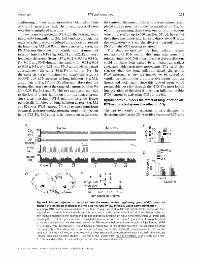
Bilateral injection of muscimol into the region of theNTS that contains inhibitory pump cells eliminatesthe effect of lung inflation on RTN neurons
Pump cells are the second-order neurons that relay inputsfrom SARs to the ventral respiratory column and the pons.These cells are located close to the tractus solitarius at areapostrema level whereas the cells that respond to rapidlyadapting lung stretch receptors are predominantly locatedin the commissural nucleus (Hayashi et al. 1996; Ezureet al. 2002; Ezure & Tanaka, 2004; Kubin et al. 2006).To further support the notion that SARs are responsiblefor the inhibition of RTN neurons by lung inflation,
C© 2007 The Authors. Journal compilation C© 2007 The Physiological Society
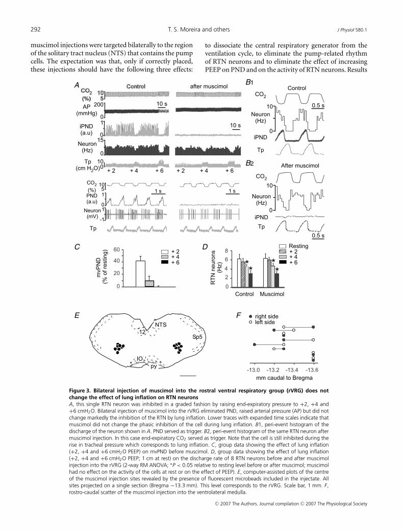
292 T. S. Moreira and others J Physiol 580.1
muscimol injections were targeted bilaterally to the regionof the solitary tract nucleus (NTS) that contains the pumpcells. The expectation was that, only if correctly placed,these injections should have the following three effects:
1
1
A
C
E F
D
B1
B2
Figure 3. Bilateral injection of muscimol into the rostral ventral respiratory group (rVRG) does notchange the effect of lung inflation on RTN neuronsA, this single RTN neuron was inhibited in a graded fashion by raising end-expiratory pressure to +2, +4 and+6 cmH2O. Bilateral injection of muscimol into the rVRG eliminated PND, raised arterial pressure (AP) but did notchange markedly the inhibition of the RTN by lung inflation. Lower traces with expanded time scales indicate thatmuscimol did not change the phasic inhibition of the cell during lung inflation. B1, peri-event histogram of thedischarge of the neuron shown in A. PND served as trigger. B2, peri-event histogram of the same RTN neuron aftermuscimol injection. In this case end-expiratory CO2 served as trigger. Note that the cell is still inhibited during therise in tracheal pressure which corresponds to lung inflation. C, group data showing the effect of lung inflation(+2, +4 and +6 cmH2O PEEP) on mvPND before muscimol. D, group data showing the effect of lung inflation(+2, +4 and +6 cmH2O PEEP; 1 cm at rest) on the discharge rate of 8 RTN neurons before and after muscimolinjection into the rVRG (2-way RM ANOVA; ∗P < 0.05 relative to resting level before or after muscimol; muscimolhad no effect on the activity of the cells at rest or on the effect of PEEP). E, computer-assisted plots of the centreof the muscimol injection sites revealed by the presence of fluorescent microbeads included in the injectate. Allsites projected on a single section (Bregma −13.3 mm). This level corresponds to the rVRG. Scale bar, 1 mm. F,rostro-caudal scatter of the muscimol injection into the ventrolateral medulla.
to dissociate the central respiratory generator from theventilation cycle, to eliminate the pump-related rhythmof RTN neurons and to eliminate the effect of increasingPEEP on PND and on the activity of RTN neurons. Results
C© 2007 The Authors. Journal compilation C© 2007 The Physiological Society
J Physiol 580.1 RTN and vagus input 293
conforming to these expectations were obtained in 6 outof 9 rats (1 neuron per rat). The three unsuccesful caseswere due to misplaced injections.
In each case, we selected an RTN unit that was markedlyinhibited by lung inflation (Fig. 5A1) and, accordingly, theunits were also markedly inhibited during each inflation ofthe lungs (Fig. 5A2 and B2). In the six successful cases, thePND became dissociated from ventilation after muscimolinjection into the NTS (Fig. 5A2, B1 and B2). Respiratoryfrequency decreased (from 1.17 ± 0.02 to 0.79 ± 0.1 Hz;P < 0.01) and PND duration increased (from 0.34 ± 0.04to 0.63 ± 0.1 s; P < 0.01) but PND amplitude remainedapproximately the same (98 ± 4% of control; NS). Inthe same six cases, muscimol eliminated the responseof PND and RTN neurons to lung inflation (Fig. 5A1;group data in Fig. 5C and D). Muscimol also raised theresting discharge rate of the sampled neurons by 46 ± 3%(P < 0.05; Fig. 5A1 and D). This rise was presumably dueto the loss of phasic inhibition from the lung afferentssince after muscimol, RTN neurons were no longerperiodically inhibited by lung inflation at rest (Fig. 5A2and B2). Most RTN neurons (5/6) still retained some formof central respiratory modulation after muscimol injectionin the NTS (Fig. 5A2 and B1). In these six successful cases,
A
D E
B
C
Figure 4. Bilateral injection of muscimol into the rostral ventral respiratory group (rVRG) does notchange the inhibition of chemosensitive RTN neurons by low-intensity vagus nerve stimulationA, a single RTN neuron was exposed to various levels of vagus nerve stimulation (T, threshold). Muscimol was theninjected into the ventrolateral medulla on both sides causing a disappearance of PND. Muscimol had no effect onthe resting discharge of the neuron and did not change its inhibition by vagus nerve stimulation. B, group datashowing the effect of vagus stimulation on mvPND before muscimol (n = 8 rats). C, group data showing the effectof vagus stimulation on the discharge rate of the RTN neurons before and after muscimol injection into rVRG(n = 8 rats; 2-way RM ANOVA; ∗P < 0.05 relative to resting level before or after muscimol; muscimol had no effecton the activity of the cells at rest or on the effect of vagus nerve stimulation). D, computer-assisted plots of thecentre of the muscimol injection sites revealed by the presence of fluorescent microbeads included in the injectate(coronal projection on plane Bregma −13.3 mm of the Paxinos atlas (Paxinos & Watson, 1998)). Scale bar, 1 mm.E, rostro-caudal scatter of muscimol injection into the ventrolateral medulla.
the centres of the muscimol injections were symmetricallyplaced in close proximity of the tractus solitarius (Fig. 5E,•). In the remaining three cases, one or both injectionswere misplaced by up to 500 μm (Fig. 5E, �). In each ofthese three cases, muscimol failed to dissociate PND fromthe ventilation cycle and the effect of lung inflation onPND and the RTN neurons persisted.
The disappearance of the lung inflation-relatedoscillations of RTN neuron discharges after muscimolinjection into the NTS demonstrates that these oscillationscould not have been caused by a mechanical artifactassociated with respiratory movements. The result alsosuggests that the lung inflation-related changes inRTN neuronal activity are unlikely to be caused byventilation-synchronous proprioceptive inputs from thethorax and neck region since this type of input wouldpresumably not relay through the NTS. The most logicalinterpretation of the data is that lung inflation inhibitsRTN neurons by activating NTS pump cells.
Kynurenate I.C.V. blocks the effect of lung inflation onRTN neurons but spares the effect of CO2
The last two series of experiments were designed todetermine whether the CO2-sensitive neurons of RTN with
C© 2007 The Authors. Journal compilation C© 2007 The Physiological Society

294 T. S. Moreira and others J Physiol 580.1
prominent input from lung stretch receptors possess twoadditional characteristics previously attributed to RTNchemoreceptors at large, namely whether their sensitivityto CO2 resists glutamate ionotropic receptor blockadewithin the pontomedullary region (Mulkey et al. 2004)and whether they express the transcription factor Phox2b(Stornetta et al. 2006).
In four rats, the broad-spectrum glutamate receptorantagonist kynurenate (KYN) was administered into the
1
1
1
A1 A2
B1
C D E
B2
Figure 5. Bilateral injection of muscimol close to the tractus solitarius eliminates the effect of lunginflation on RTN neuronsA1, this RTN neuron was exposed to various levels of lung inflation (+2, +4 and +6 cmH2O). Muscimol injectionnext to the tractus solitarius on both sides raised the arterial pressure (AP) and eliminated the inhibition producedby lung inflation on PND and RTN neurons. A2, the RTN neuron had a clear pump rhythm before muscimol injectioninto the NTS. Muscimol injection into NTS dissociated the central respiratory generator from the ventilation cycleand eliminated the pump-related rhythm of the RTN neuron. B1, PND-triggered activity histogram of the RTNneuron shown in A, before and after muscimol was injected in the NTS. B2, tracheal pressure-triggered activityhistogram of the same RTN neuron before and after muscimol injection. C, group data showing the effect of lunginflation (+2, +4 and +6 cmH2O PEEP) on mvPND before and after muscimol into the NTS. Lung inflation had noeffect after muscimol (RM ANOVA). D, group data showing the effect of lung inflation (+2, +4 and +6 cmH2O)on the discharge rate of RTN neurons before and after muscimol into the NTS. The effect of lung inflation wassignificant before muscimol but not after (2-way RM ANOVA; ∗P < 0.05 relative to resting; +P < 0.05 relativeto resting discharge before muscimol). E, computer-assisted plots of the centre of the muscimol injection sitesrevealed by the presence of fluorescent microbeads included in the injectate. All succesful injections were withinor in close proximity of the solitary tract. Scale bar, 1 mm.
fourth ventricle while recording the response of a singleRTN neuron to hypercapnia and lung inflation. The fourRTN neurons selected for study were phasically inhibitedduring lung inflation at rest (3 totally inhibited, Fig. 6B1,and one partially inhibited) and were markedly inhibitedby increasing PEEP to 6 cmH2O (Fig. 6A and B1). Eachneuron responded in the manner illustrated in Fig. 6. Asshown before (Mulkey et al. 2004), KYN eliminated PNDwithin minutes after injection, regardless of the CO2 level
C© 2007 The Authors. Journal compilation C© 2007 The Physiological Society

J Physiol 580.1 RTN and vagus input 295
(Fig. 6A). KYN slightly increased the RTN neuron’s restingdischarge rate, blocked its inhibitory response to increasingPEEP (Fig. 6A) and eliminated the periodic inhibition ofthe cell coinciding with each inflation of the lungs (Fig. 6Aand B). The steady-state response of the RTN neuron tograded levels of CO2 was changed in the manner shown inFig. 6C. The CO2 threshold was not noticeably affected butthe cell discharged at a higher rate at all levels of CO2. TheCO2 threshold was similarly unaffected in the other threecases. At high CO2 the discharge rate was elevated above
A B1
B2
C D
Figure 6. Kynurenate blocks the response to lung inflation but not the CO2 sensitivity of RTN neuronsA, example of one RTN neuron recorded before and after I.C.V. injection of 15 μmol of kynurenate (KYN). KYNeliminated PND, reduced BP, eliminated the neuron’s response to lung inflation but had minimal effect on the cell’sresponse to CO2. The expanded scale insets show the discharge pattern of the neuron before and after kynurenate.B1, peri-event histogram triggered on tracheal pressure demonstrating that, before kynurenate, the cell stops firingduring each inflation of the lungs at rest (1 cmH2O PEEP). B2, similar histogram done after kynurenate showingthat the cell is no longer inhibited by lung inflation. C, steady-state relationship between the activity of the cellshown in A and B and end-expiratory CO2 before and after KYN. D, resting discharge rate of 4 RTN neuronsrecorded at two levels of end-expiratory CO2 before and after I.C.V. kynurenate.
control only in two of the three other cells. Figure 6D showsthe discharge rate of the four cells at two representativelevels of CO2. The increase in discharge rate at high CO2
was not statistically significant.
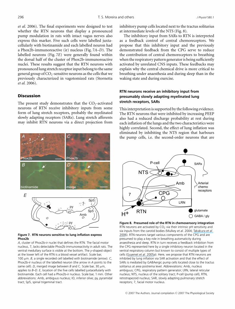
RTN neurons sensitive to lung inflation expressPhox2b
In vagotomized rats, generic CO2-activated neurons ofRTN express the transcription factor Phox2b (Stornetta
C© 2007 The Authors. Journal compilation C© 2007 The Physiological Society
296 T. S. Moreira and others J Physiol 580.1
et al. 2006). The final experiments were designed to testwhether the RTN neurons that display a pronouncedpump modulation in rats with intact vagus nerves alsoexpress this marker. Five such cells were labelled juxta-cellularly with biotinamide and each labelled neuron hada Phox2b-immunoreactive (ir) nucleus (Fig. 7A–D). Thelabelled neurons (Fig. 7E) were generally found withinthe dorsal half of the cluster of Phox2b-immunoreactivenuclei. These results suggest that the RTN neurons withpronounced lung stretch receptor input belong to the samegeneral group of CO2-sensitive neurons as the cells that wepreviously characterized in vagotomized rats (Stornettaet al. 2006).
Discussion
The present study demonstrates that the CO2-activatedneurons of RTN receive inhibitory inputs from someform of lung stretch receptors, probably the myelinatedslowly adapting receptors (SARs). Lung stretch afferentsmay inhibit RTN neurons via a direct projection from
A
B
E
C
D
Figure 7. RTN neurons sensitive to lung inflation expressPhox2bA, cluster of Phox2b-ir nuclei that defines the RTN. The facial motornucleus, 7, lacks detectable Phox2b immunoreactivity in adult rats. Theventral medullary surface is visible at the bottom. The y-shaped objectat the lower left of the RTN is a blood vessel artifact. Scale bar,100 μm. B, a single recorded cell labelled with biotinamide (arrow). C,Phox2b-ir nucleus of the labelled neuron (the arrow in A points to thesame cell). D, merged image between B and C. Scale bar, 30 μm,applies to B–D. E, location of the five cells labelled juxtacellularly withbiotinamide. Each cell had a Phox2b-ir nucleus. Scale bar, 1 mm. Otherabbreviations: Amb, ambiguus nucleus; IO, inferior olive; py, pyramidaltract; Sp5, spinal trigeminal tract.
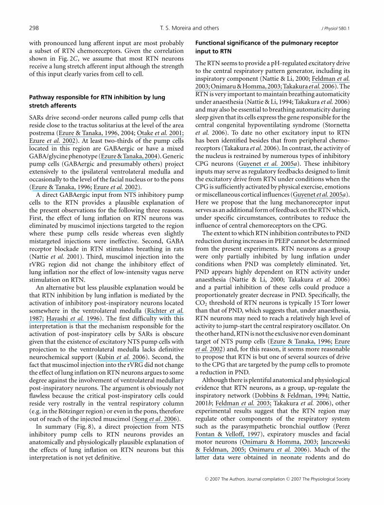
inhibitory pump cells located next to the tractus solitariusat intermediate levels of the NTS (Fig. 8).
The inhibitory input from SARs to RTN is interpretedas a feedback control of central chemoreceptors. Wepropose that this inhibitory input and the previouslydemonstrated feedback from the CPG serve to reducethe contribution of central chemoreceptors to breathingwhen the respiratory pattern generator is being sufficientlyactivated by unrelated CNS inputs. These feedbacks mayexplain why the central chemical drive is more critical tobreathing under anaesthesia and during sleep than in thewaking state and during exercise.
RTN neurons receive an inhibitory input frompresumably slowly adapting myelinated lungstretch receptors, SARs
This interpretation is supported by the following evidence.The RTN neurons that were inhibited by increasing PEEPalso had a reduced discharge probability at rest duringeach inflation of the lungs and the two characteristics werehighly correlated. Second, the effect of lung inflation waseliminated by inhibiting the NTS region that harboursthe pump cells, i.e. the second-order neurons that are
Figure 8. Presumed role of the RTN in chemosensory integrationRTN neurons are activated by CO2 via their intrinsic pH sensitivity andvia inputs from the carotid bodies (Mulkey et al. 2004; Takakura et al.2006). RTN neurons target various components of the CPG and arepresumed to play a key role in breathing automaticity duringanaesthesia and sleep. RTN in turn receives a feedback inhibition fromthe CPG represented here by a single inhibitory neuron located in theventral respiratory column but known to consist of multiple types ofcells (Guyenet et al. 2005a). Here, we propose that RTN neurons areinhibited by lung inflation via SAR activation and that the effect ofSARs is mediated by GABAergic pump cells located close to the tractussolitarius at area postrema level. Abbreviations: Amb, nucleusambiguus; CPG, respiratory pattern generator; LRN, lateral reticularnucleus; NTS, nucleus of the solitary tract; P-cell (pump cell), RTN,retrotrapezoid nucleus; SAR, slowly adapting pulmonary stretchreceptors; 7, facial motor nucleus.
C© 2007 The Authors. Journal compilation C© 2007 The Physiological Society

J Physiol 580.1 RTN and vagus input 297
phasically activated by slowly adapting lung stretchreceptors during each inflation of the lungs (Otakeet al. 2001; Ezure et al. 2002; Ezure & Tanaka, 2004;Kubin et al. 2006). Third, the RTN neurons that werestrongly inhibited by lung inflation were also activated byinterrupting ventilation. The instantaneous nature of thisactivation excludes the possibility that it might have beendue to hypercarbia or hypoxia. Fourth, the dischargeof the RTN neurons became markedly more regularafter the interruption of ventilation indicating that theirreduced discharge probability during lung inflation wasnot due to an inhibitory input from the central respiratorypattern generator. Fifth, the animals had a tracheostomy,therefore ventilation is unlikely to have resulted inperiodic activation of mechanoreceptors located elsewherealong the respiratory system than in the lungs. Finally, invagotomized rats, many RTN neurons were inhibited bystimulating the vagus nerve using short-duration pulsesand intensities commensurate with those sufficient toinhibit PND. The short pulses and low intensity requiredto inhibit PND and RTN neurons suggest that myelinatedaxons must have been selectively activated. Consistentwith this interpretation, our vagal stimulations producedno change in blood pressure. Under anaesthesia, changesin blood pressure are known to require vagal C fibreactivation (Sun & Guyenet, 1987).
A contribution of rapidly adapting receptors (RARs) tothe response of RTN neurons to vagal nerve stimulationcannot be completely eliminated because these lungreceptors are also myelinated (Coleridge & Coleridge,2001), including in rats (Ho et al. 2001). Also, rat RARshave a phasic discharge that is synchronized with trachealpressure at normal levels of lung inflation (Ho et al.2001); therefore their pattern of activity is theoreticallyappropriate to trigger a lung-inflation synchronousinhibition of RTN neurons. Yet, two observations reducethe plausibility that RARs are major contributors to theinhibition of RTN neurons by lung inflation. The first oneis the sustained nature of RTN neuron inhibition causedby increasing PEEP (> 5 s). The second one is that theNTS neurons that receive RAR input are located in thecommissural nucleus (Kubin et al. 2006). These neuronswould presumably not have been affected by the muscimolinjections that we targeted to the more rostral and lateralregion of the NTS where the SAR second-order neurons(pump cells) reside.
In conclusion, the data favour the interpretation thatSARs, not RARs, are the main form of lung stretch receptorresponsible for inhibiting RTN during lung inflation.
Relationship with prior studies
The existence of lung afferent inputs to neurons located inthe region of the cat RTN has not been reported previously
(Connelly et al. 1990; Nattie et al. 1993; Bodineau et al.2000a,b). In our earliest studies of the RTN performedin rats with intact vagus nerves (Mulkey et al. 2004;Guyenet et al. 2005a), we did encounter CO2-sensitiveRTN cells with respiratory-modulated discharge patternssimilar to those reported in the present study. However,in this work, the cells with the most phasic dischargepattern (ON–OFF) at rest were passed over, in truthbecause such cells did not fit our preconceived ideas ofwhat central chemoreceptor neurons should look like.Cells with marked but still incomplete reduction of firingduring one phase of the respiratory cycle were studied andthese cells were usually found to have their discharge nadirduring the post-inspiratory period (Guyenet et al. 2005a).We assumed that this pattern was caused by an inputfrom post-inhibitory neurons that are part of the centralrespiratory pattern generator because post-inspiratoryinhibition is also observed in a subset of RTN neuronsrecorded in vagotomized rats (Guyenet et al. 2005a).However, since tracheal pressure was not measured inour prior studies, we missed the fact that lung inflationcoincides with the central post-inspiratory phase inventilated rats with intact vagus nerves. In view of thepresent results, we conclude that, in artificially ventilatedrats with intact vagus nerves, the RTN neurons withthe most pronounced inhibition during the period thatimmediately follows the PND derive this characteristicprimarily from a lung afferent input.
RTN neurons with strong or weak lung afferent inputare both subsets of central chemoreceptors
The present evidence suggests that the CO2-activatedneurons of RTN with pronounced lung afferent inputare simply a subset of RTN chemoreceptors. Like therest of the CO2-activated neurons, the RTN neuronswith pronounced stretch afferent input are active belowthe phrenic apnoea threshold, their resting activity isunaffected by bilateral injection of muscimol into therVRG region (Takakura et al. 2006) and they expressPhox2b (Stornetta et al. 2006). This latter result alsostrongly suggests that the RTN neurons with pronouncedlung stretch afferent input are also glutamatergic sinceall Phox2b-expressing neurons within the RTN containVGLUT2 mRNA (Stornetta et al. 2006). Finally, like therest of the RTN neurons, the neurons with pronouncedlung stretch afferent input develop a remarkably regulardischarge after attenuation of excitatory transmission withi.c.v. injection of kynurenate and, in the presence ofthis blocker, they continue to encode PCO2
in a mannerconsistent with what would be expected of neurons thatderive their CO2 sensitivity from an intrinsic sensitivity toacid (Mulkey et al. 2004, 2006; Guyenet et al. 2005a). Basedon these similarities, we conclude that the RTN neurons
C© 2007 The Authors. Journal compilation C© 2007 The Physiological Society
298 T. S. Moreira and others J Physiol 580.1
with pronounced lung afferent input are most probablya subset of RTN chemoreceptors. Given the correlationshown in Fig. 2C, we assume that most RTN neuronsreceive a lung stretch afferent input although the strengthof this input clearly varies from cell to cell.
Pathway responsible for RTN inhibition by lungstretch afferents
SARs drive second-order neurons called pump cells thatreside close to the tractus solitarius at the level of the areapostrema (Ezure & Tanaka, 1996, 2004; Otake et al. 2001;Ezure et al. 2002). At least two-thirds of the pump cellslocated in this region are GABAergic or have a mixedGABA/glycine phenotype (Ezure & Tanaka, 2004). Genericpump cells (GABAergic and presumably others) projectextensively to the ipsilateral ventrolateral medulla andoccasionally to the level of the facial nucleus or to the pons(Ezure & Tanaka, 1996; Ezure et al. 2002).
A direct GABAergic input from NTS inhibitory pumpcells to the RTN provides a plausible explanation ofthe present observations for the following three reasons.First, the effect of lung inflation on RTN neurons waseliminated by muscimol injections targeted to the regionwhere these pump cells reside whereas even slightlymistargeted injections were ineffective. Second, GABAreceptor blockade in RTN stimulates breathing in rats(Nattie et al. 2001). Third, muscimol injection into therVRG region did not change the inhibitory effect oflung inflation nor the effect of low-intensity vagus nervestimulation on RTN.
An alternative but less plausible explanation would bethat RTN inhibition by lung inflation is mediated by theactivation of inhibitory post-inspiratory neurons locatedsomewhere in the ventrolateral medulla (Richter et al.1987; Hayashi et al. 1996). The first difficulty with thisinterpretation is that the mechanism responsible for theactivation of post-inspiratory cells by SARs is obscuregiven that the existence of excitatory NTS pump cells withprojection to the ventrolateral medulla lacks definitiveneurochemical support (Kubin et al. 2006). Second, thefact that muscimol injection into the rVRG did not changethe effect of lung inflation on RTN neurons argues to somedegree against the involvement of ventrolateral medullarypost-inspiratory neurons. The argument is obviously notflawless because the critical post-inspiratory cells couldreside very rostrally in the ventral respiratory column(e.g. in the Botzinger region) or even in the pons, thereforeout of reach of the injected muscimol (Song et al. 2006).
In summary (Fig. 8), a direct projection from NTSinhibitory pump cells to RTN neurons provides ananatomically and physiologically plausible explanation ofthe effects of lung inflation on RTN neurons but thisinterpretation is not yet definitive.
Functional significance of the pulmonary receptorinput to RTN
The RTN seems to provide a pH-regulated excitatory driveto the central respiratory pattern generator, including itsinspiratory component (Nattie & Li, 2000; Feldman et al.2003; Onimaru & Homma, 2003; Takakura et al. 2006). TheRTN is very important to maintain breathing automaticityunder anaesthesia (Nattie & Li, 1994; Takakura et al. 2006)and may also be essential to breathing automaticity duringsleep given that its cells express the gene responsible for thecentral congenital hypoventilating syndrome (Stornettaet al. 2006). To date no other excitatory input to RTNhas been identified besides that from peripheral chemo-receptors (Takakura et al. 2006). In contrast, the activity ofthe nucleus is restrained by numerous types of inhibitoryCPG neurons (Guyenet et al. 2005a). These inhibitoryinputs may serve as regulatory feedbacks designed to limitthe excitatory drive from RTN under conditions when theCPG is sufficiently activated by physical exercise, emotionsor miscellaneous cortical influences (Guyenet et al. 2005a).Here we propose that the lung mechanoreceptor inputserves as an additional form of feedback on the RTN which,under specific circumstances, contributes to reduce theinfluence of central chemoreceptors on the CPG.
The extent to which RTN inhibition contributes to PNDreduction during increases in PEEP cannot be determinedfrom the present experiments. RTN neurons as a groupwere only partially inhibited by lung inflation underconditions when PND was completely eliminated. Yet,PND appears highly dependent on RTN activity underanaesthesia (Nattie & Li, 2000; Takakura et al. 2006)and a partial inhibition of these cells could produce aproportionately greater decrease in PND. Specifically, theCO2 threshold of RTN neurons is typically 15 Torr lowerthan that of PND, which suggests that, under anaesthesia,RTN neurons may need to reach a relatively high level ofactivity to jump-start the central respiratory oscillator. Onthe other hand, RTN is not the exclusive nor even dominanttarget of NTS pump cells (Ezure & Tanaka, 1996; Ezureet al. 2002) and, for this reason, it seems more reasonableto propose that RTN is but one of several sources of driveto the CPG that are targeted by the pump cells to promotea reduction in PND.
Although there is plentiful anatomical and physiologicalevidence that RTN neurons, as a group, up-regulate theinspiratory network (Dobbins & Feldman, 1994; Nattie,2001b; Feldman et al. 2003; Takakura et al. 2006), otherexperimental results suggest that the RTN region mayregulate other components of the respiratory systemsuch as the parasympathetic bronchial outflow (PerezFontan & Velloff, 1997), expiratory muscles and facialmotor neurons (Onimaru & Homma, 2003; Janczewski& Feldman, 2005; Onimaru et al. 2006). Much of thelatter data were obtained in neonate rodents and do
C© 2007 The Authors. Journal compilation C© 2007 The Physiological Society

J Physiol 580.1 RTN and vagus input 299
not specifically implicate the cells that we have definedas RTN neurons in the adult. In the neonate, theRTN region contains a neural oscillator or part ofan oscillator suspected to control expiratory musclesselectively (Janczewski & Feldman, 2005; Feldman & DelNegro, 2006). However, RTN as we defined it, is unlikely tobe part of such an expiratory oscillator because expiratoryactivity ought to be increased by SAR activation (Hayashiet al. 1996; Feldman & Del Negro, 2006) whereas RTNneurons were exclusively inhibited by this stimulus.
In conclusion, the RTN contains a population ofglutamatergic, Phox2b-expressing interneurons that areactivated by local changes in pH and receive excitatoryinputs from peripheral chemoreceptors. RTN neurons arealso inhibited by the activation of myelinated lung stretchreceptors, most probably of the slowly adapting variety.The SAR input may reach RTN via a direct projectionfrom the inhibitory pump cells of the ventrolateral NTS.The SAR input to RTN, along with the feedback fromthe CPG, may serve to reduce the contribution of centralchemoreceptors to breathing when the pattern generatoris sufficiently activated by other sources of input.
References
Akilesh MR, Kamper M, Li A & Nattie EE (1997). Effects ofunilateral lesions of retrotrapezoid nucleus on breathing inawake rats. J Appl Physiol 82, 469–479.
Bodineau L, Frugiere A, Marlot D & Wallois F (2000a).Connections between retrotrapezoid nucleus and nucleustractus solitarii in cat. Neurosci Lett 280, 111–114.
Bodineau L, Frugiere A, Marlot D & Wallois F (2000b). Effectof hypoxia on the activity of respiratory and non-respiratorymodulated retrotrapezoid neurons of the cat. Auton Neurosci86, 70–77.
Coleridge HM & Coleridge JC (2001). Afferent innervation oflungs, airways, and pulmonary artery. In Reflex Control of theCirculation, ed. Zucker IH & Gilmore JP, pp. 579–607. CRCPress, Boca Raton.
Connelly CA, Ellenberger HH & Feldman JL (1990).Respiratory activity in retrotrapezoid nucleus in cat.Am J Physiol 258, L33–L44.
Cream C, Li A & Nattie E (2002). The retrotrapezoid nucleus(RTN): local cytoarchitecture and afferent connections.Respir Physiol Neurobiol 130, 121–137.
Dobbins EG & Feldman JL (1994). Brainstem networkcontrolling descending drive to phrenic motoneurons in rat.J Comp Neurol 347, 64–86.
Eldridge FL, Kiley JP & Millhorn DE (1985). Respiratoryresponses to medullary hydrogen ion changes in cats:different effects of respiratory and metabolic acidoses.J Physiol 358, 285–297.
Ezure K & Tanaka I (1996). Pump neurons of the nucleus of thesolitary tract project widely to the medulla. Neurosci Lett215, 123–126.
Ezure K & Tanaka I (2004). GABA, in some cases together withglycine, is used as the inhibitory transmitter by pump cells inthe Hering-Breuer reflex pathway of the rat. Neurosci 127,409–417.
Ezure K, Tanaka I, Saito Y & Otake K (2002). Axonalprojections of pulmonary slowly adapting receptor relayneurons in the rat. J Comp Neurol 446, 81–94.
Feldman JL & Del Negro CA (2006). Looking for inspiration:new perspectives on respiratory rhythm. Nat Rev Neurosci 7,232–242.
Feldman JL, Mitchell GS & Nattie EE (2003). Breathing:rhythmicity, plasticity, chemosensitivity. Annu Rev Neurosci26, 239–266.
Guyenet PG (2006). The sympathetic control of blood pressure.Nat Rev Neurosci 7, 335–346.
Guyenet PG, Mulkey DK, Stornetta RL & Bayliss DA (2005a).Regulation of ventral surface chemoreceptors by thecentral respiratory pattern generator. J Neurosci 25,8938–8947.
Guyenet PG, Stornetta RL, Bayliss DA & Mulkey DK (2005b).Retrotrapezoid nucleus: a litmus test for the identification ofcentral chemoreceptors. Exp Physiol 90, 247–253.
Hayashi F, Coles SK & McCrimmon DR (1996). Respiratoryneurons mediating the Breuer-Hering reflex prolongation ofexpiration in rat. J Neurosci 16, 6526–6536.
Ho CY, Gu Q, Lin YS & Lee LY (2001). Sensitivity of vagalafferent endings to chemical irritants in the rat lung. RespirPhysiol 127, 113–124.
Janczewski WA & Feldman JL (2005). Distinct rhythmgenerators for inspiration and expiration in the juvenile rat.J Physiol 570, 407–420.
Kubin L, Alheid GF, Zuperku EJ & McCrimmon DR (2006).Central pathways of pulmonary and lower airway vagalafferents. J Appl Physiol 101, 618–627.
Loeschcke HH (1982). Central chemosensitivity and thereaction theory. J Physiol 332, 1–24.
Moreira TS, Takakura AC, Colombari E & Guyenet PG (2006).Central chemoreceptors and sympathetic vasomotoroutflow. J Physiol 577, 369–386.
Mulkey DK, Mistry AM, Guyenet PG & Bayliss DA (2006).Purinergic P2 receptors modulate excitability but do notmediate pH sensitivity of RTN respiratory chemoreceptors.J Neurosci 26, 7230–7233.
Mulkey DK, Stornetta RL, Weston MC, Simmons JR, Parker A,Bayliss DA et al. (2004). Respiratory control by ventralsurface chemoreceptor neurons in rats. Nat Neurosci 7,1360–1369.
Nattie EE (2001a). Central chemosensitivity, sleep, andwakefulness. Respir Physiol 129, 257–268.
Nattie EE (2001b). Chemoreception and tonic drive in theretrotrapezoid nucleus (RTN) region of the awake rat:bicuculline and muscimol dialysis in the RTN. Adv Exp MedBiol 499, 27–32.
Nattie EE, Fung ML, Li A & St John WM (1993). Responses ofrespiratory modulated and tonic units in the retrotrapezoidnucleus to CO2. Respir Physiol 94, 35–50.
Nattie EE & Li A (1994). Retrotrapezoid nucleus lesionsdecrease phrenic activity and CO2 sensitivity in rats. RespirPhysiol 97, 63–77.
Nattie E & Li A (2000). Muscimol dialysis in the retrotrapezoidnucleus region inhibits breathing in the awake rat. J ApplPhysiol 89, 153–162.
Nattie E, Shi J & Li A (2001). Bicuculline dialysis in theretrotrapezoid nucleus (RTN) region stimulates breathing inthe awake rat. Respir Physiol 124, 179–193.
C© 2007 The Authors. Journal compilation C© 2007 The Physiological Society
300 T. S. Moreira and others J Physiol 580.1
Okada Y, Chen Z, Jiang W, Kuwana S & Eldridge FL (2002).Anatomical arrangement of hypercapnia-activated cells inthe superficial ventral medulla of rats. J Appl Physiol 93,427–439.
Onimaru H & Homma I (2003). A novel functional neurongroup for respiratory rhythm generation in the ventralmedulla. J Neurosci 23, 1478–1486.
Onimaru H, Kumagawa Y & Homma I (2006). Respiration-related rhythmic activity in the rostral medulla of newbornrats. J Neurophysiol 96, 55–61.
Otake K, Nakamura Y, Tanaka I & Ezure K (2001). Morphologyof pulmonary rapidly adapting receptor relay neurons in therat. J Comp Neurol 430, 458–470.
Pattyn A, Morin X, Cremer H, Goridis C & Brunet JF (1997).Expression and interactions of the two closely relatedhomeobox genes Phox2a and Phox2b during neurogenesis.Development 124, 4065–4075.
Paxinos G & Watson C (1998). The Rat Brain in StereotaxicCoordinates. Academic Press, San Diego.
Perez Fontan JJ & Velloff CR (1997). Neuroanatomicorganization of the parasympathetic bronchomotor systemin developing sheep. Am J Physiol 273, R121–R133.
Putnam RW, Filosa JA & Ritucci NA (2004). Cellularmechanisms involved in CO2 and acid signaling inchemosensitive neurons. Am J Physiol Cell Physiol 287,C1493–C1526.
Richter DW, Ballantyne D & Remmers JE (1987). Thedifferential organization of medullary post-inspiratoryactivities. Pflugers Arch 410, 420–427.
Ritucci NA, Erlichman JS, Leiter JC & Putnam RW (2005). TheResponse of membrane potential (Vm) and intracellular pH(pHi) to hypercapnia in neurons and astrocytes from ratretrotrapezoid nucleus (RTN). Am J Physiol Regul IntegrComp Physiol 289, R851–R861.
Rosin DL, Chang DA & Guyenet PG (2006). Afferent andefferent connections of the rat retrotrapezoid nucleus.J Comp Neurol 499, 64–89.
Smith JC, Morrison DE, Ellenberger HH, Otto MR & FeldmanJL (1989). Brainstem projections to the major respiratoryneuron populations in the medulla of the cat. J Comp Neurol281, 69–96.
Song G, Yu Y & Poon CS (2006). Cytoarchitecture ofpneumotaxic integration of respiratory and nonrespiratoryinformation in the rat. J Neurosci 26, 300–310.
Stornetta RL & Guyenet PG (1999). Distribution of glutamicacid decarboxylase mRNA-containing neurons in ratmedulla projecting to thoracic spinal cord in relation tomonoaminergic brainstem neurons. J Comp Neurol 407,367–380.
Stornetta RL, Moreira TS, Takakura AC, Kang BJ, Chang DA,West GH et al. (2006). Selective expression of Phox2b bybrainstem neurons involved in chemosensory integration inthe adult rat. J Neurosci 26, 10305–10314.
Stornetta RL, Sevigny CP & Guyenet PG (2003). Inspiratoryaugmenting bulbospinal neurons express both glutamatergicand enkephalinergic phenotypes. J Comp Neurol 455,113–124.
Sun MK & Guyenet PG (1987). Arterial baroreceptor and vagalinputs to sympathoexcitatory neurons in rat medulla.Am J Physiol Regul Integr Comp Physiol 252, R699–R709.
Sun MK, Hackett JT & Guyenet PG (1988).Sympathoexcitatory neurons of rostral ventrolateral medullaexhibit pacemaker properties in the presence of aglutamate-receptor antagonist. Brain Res 438, 23–40.
Takakura AC, Moreira TS, Colombari E, West GH, StornettaRL & Guyenet PG (2006). Peripheral chemoreceptor inputsto retrotrapezoid nucleus (RTN) CO2-sensitive neurons inrats. J Physiol 572, 503–523.
Vatner SF & Uemura N (2001). Integrative cardiovascularcontrol by pulmonary inflation reflexes. In Reflex Control ofthe Circulation, ed. Zucker IH & Gilmore JP, pp. 609–626.CRC Press, Boca Raton.
Acknowledgements
This research was supported by grants from the National
Institutes of Health to P.G.G. (HL 74011 and HL 28785) and
Coordenacao de Aperfeicoamento de Pessoal de Nıvel Superior
to A.C.T. (BEX 4402/05-7).
C© 2007 The Authors. Journal compilation C© 2007 The Physiological Society