Chemokine Sequestration by Viral Chemoreceptors as a Novel Viral Escape Strategy: Withdrawal of...
12
855 J. Exp. Med. The Rockefeller University Press • 0022-1007/98/09/855/12 $2.00 Volume 188, Number 5, September 7, 1998 855–866 http://www.jem.org Chemokine Sequestration by Viral Chemoreceptors as a Novel Viral Escape Strategy: Withdrawal of Chemokines from the Environment of Cytomegalovirus-infected Cells By Bahram Bodaghi,* Thomas R. Jones, ‡ Donato Zipeto,* Claudio Vita, § Lei Sun, ‡ Lysiane Laurent,* Fernando Arenzana-Seisdedos,* Jean-Louis Virelizier,* and Susan Michelson* From the *Unite d’Immunologie Virale, Institut Pasteur, 75724 Paris, France; the ‡ Department of Molecular Biology, Infectious Disease Section,Wyeth-Ayerst Research, Pearl River, New York 10965; and the § Département d’Ingénierie et d’Etudes des Protéines, CEA Saclay, 91190 Gif-sur-Yvette, France Summary Human cytomegalovirus (HCMV), a betaherpesvirus, has developed several ways to evade the immune system, notably downregulation of cell surface expression of major histocompatibility complex class I heavy chains. Here we report that HCMV has devised another means to com- promise immune surveillance mechanisms. Extracellular accumulation of both constitutively produced monocyte chemoattractant protein (MCP)-1 and tumor necrosis factor–superin- duced RANTES (regulated on activation, normal T cell expressed and secreted) was downreg- ulated in HCMV-infected fibroblasts in the absence of transcriptional repression or the expres- sion of polyadenylated RNA for the cellular chemokine receptors CCR-1, CCR-3, and CCR-5. Competitive binding experiments demonstrated that HCMV-infected cells bind RANTES, MCP-1, macrophage inflammatory protein (MIP)-1b, and MCP-3, but not MCP-2, to the same receptor as does MIP-1a, which is not expressed in uninfected cells. HCMV en- codes three proteins with homology to CC chemokine receptors: US27, US28, and UL33. Cells infected with HCMV mutants deleted of US28, or both US27 and US28 genes, failed to downregulate extracellular accumulation of either RANTES or MCP-1. In contrast, cells in- fected with a mutant deleted of US27 continues to bind and downregulate those chemokines. Depletion of chemokines from the culture medium was at least partially due to continuous in- ternalization of extracellular chemokine, since exogenously added, biotinylated RANTES ac- cumulated in HCMV-infected cells. Thus, HCMV can modify the chemokine environment of infected cells through intense sequestering of CC chemokines, mediated principally by expres- sion of the US28-encoded chemokine receptor. Key words: RANTES • human cytomegalovirus • chemokine receptors • sequestration • monocyte chemoattractant protein 1 E fficient control of persistent viral infections relies on the specific recognition of viral antigens by T lympho- cytes and/or NK cells. These cells are activated by inflam- matory cytokines. However, once activated they must be mobilized to the sites of infection. Such mobilization de- pends on the creation of gradients of chemokines that not only chemoattract effector leukocytes, but also play a sup- plementary role in stimulating the effector mechanisms of these cells once they are targeted to sites of infection (1). Numerous viruses persist in host organisms for the re- mainder of the individual’s life. This persistence requires the virus to be able to evade eradication by the immune system and reflect a co-evolution of the infectious agent with its host. A number of viruses have developed a variety of mechanisms for immune evasion (2–5). Some of these viruses confer lifelong immunity, as assessed by persistent immunological markers of primary infection, without com- plete eradication of the virus in question. To accomplish this feat, they have acquired genes that mimic host genes involved in the immune response (for review see references in 5, 6) and have products that can interfere with numerous host cell defense mechanisms (complement activation, in-
Transcript of Chemokine Sequestration by Viral Chemoreceptors as a Novel Viral Escape Strategy: Withdrawal of...

855
J. Exp. Med.
The Rockefeller University Press • 0022-1007/98/09/855/12 $2.00Volume 188, Number 5, September 7, 1998 855–866http://www.jem.org
Chemokine Sequestration by Viral Chemoreceptors as aNovel Viral Escape Strategy: Withdrawal of Chemokinesfrom the Environment of Cytomegalovirus-infected Cells
By Bahram Bodaghi,
*
Thomas R. Jones,
‡
Donato Zipeto,
*
Claudio Vita,
§
Lei Sun,
‡
Lysiane Laurent,
*
Fernando Arenzana-Seisdedos,
*
Jean-Louis Virelizier,
*
and Susan Michelson
*
From the
*
Unite d’Immunologie Virale, Institut Pasteur, 75724 Paris, France; the
‡
Department of Molecular Biology, Infectious Disease Section, Wyeth-Ayerst Research, Pearl River, New York 10965; and the
§
Département d’Ingénierie et d’Etudes des Protéines, CEA Saclay, 91190 Gif-sur-Yvette, France
Summary
Human cytomegalovirus (HCMV), a betaherpesvirus, has developed several ways to evade theimmune system, notably downregulation of cell surface expression of major histocompatibilitycomplex class I heavy chains. Here we report that HCMV has devised another means to com-promise immune surveillance mechanisms. Extracellular accumulation of both constitutivelyproduced monocyte chemoattractant protein (MCP)-1 and tumor necrosis factor–superin-duced RANTES (regulated on activation, normal T cell expressed and secreted) was downreg-ulated in HCMV-infected fibroblasts in the absence of transcriptional repression or the expres-sion of polyadenylated RNA for the cellular chemokine receptors CCR-1, CCR-3, andCCR-5. Competitive binding experiments demonstrated that HCMV-infected cells bindRANTES, MCP-1, macrophage inflammatory protein (MIP)-1
b
, and MCP-3, but not MCP-2,to the same receptor as does MIP-1
a
, which is not expressed in uninfected cells. HCMV en-codes three proteins with homology to CC chemokine receptors: US27, US28, and UL33.Cells infected with HCMV mutants deleted of US28, or both US27 and US28 genes, failed todownregulate extracellular accumulation of either RANTES or MCP-1. In contrast, cells in-fected with a mutant deleted of US27 continues to bind and downregulate those chemokines.Depletion of chemokines from the culture medium was at least partially due to continuous in-ternalization of extracellular chemokine, since exogenously added, biotinylated RANTES ac-cumulated in HCMV-infected cells. Thus, HCMV can modify the chemokine environment ofinfected cells through intense sequestering of CC chemokines, mediated principally by expres-sion of the US28-encoded chemokine receptor.
Key words: RANTES • human cytomegalovirus • chemokine receptors • sequestration • monocyte chemoattractant protein 1
E
fficient control of persistent viral infections relies onthe specific recognition of viral antigens by T lympho-
cytes and/or NK cells. These cells are activated by inflam-matory cytokines. However, once activated they must bemobilized to the sites of infection. Such mobilization de-pends on the creation of gradients of chemokines that notonly chemoattract effector leukocytes, but also play a sup-plementary role in stimulating the effector mechanisms ofthese cells once they are targeted to sites of infection (1).
Numerous viruses persist in host organisms for the re-mainder of the individual’s life. This persistence requires
the virus to be able to evade eradication by the immunesystem and reflect a co-evolution of the infectious agentwith its host. A number of viruses have developed a varietyof mechanisms for immune evasion (2–5). Some of theseviruses confer lifelong immunity, as assessed by persistentimmunological markers of primary infection, without com-plete eradication of the virus in question. To accomplishthis feat, they have acquired genes that mimic host genesinvolved in the immune response (for review see referencesin 5, 6) and have products that can interfere with numeroushost cell defense mechanisms (complement activation, in-

856
Sequestration of CC Chemokines by Cytomegalovirus-infected Cells
terferon inhibition, antibody recognition, growth factorsand cytokines, etc.)
Notable among such persistent viruses are the herpesvi-ruses (2, 4, 7). After primary infection, these viruses be-come permanent residents of their hosts. In the majority ofcases, primary infection is clinically inapparent. Pathophys-iological manifestations are only apparent upon immuno-depression of the host, whether natural (pregnancy), iatro-genic (organ transplantation), or acquired (infection withHIV). From a purely virological point of view, this impliesthat these viruses have developed a low profile as concernsthe immune system.
Herpesviruses are among the largest viruses known. Theirgenomes have the capacity to encode from 80 to
.
200proteins (7). They have evolved with their hosts for mil-lions of years (8). The target cells of viral latency are some-times very limited, for instance for herpes simplex virus(neurons) and Epstein-Barr virus (B lymphocytes). Latencytargets of viruses like CMV (9–11), appear much broader.In this case, the virus has probably adopted multiple strate-gies for evading immune surveillance, since it resides in avariety of functionally different cell types.
Human CMV (HCMV),
1
a betaherpesvirus, has devel-oped a number of strategies which may limit its recognitionby the host’s immune system and its eradication. It inducesexpression of Fc receptors (12–15) and upregulates expres-sion of complement regulatory proteins CD55 and CD46(16), and incorporates CD55 and CD59 into the virion en-velope (17). The HCMV genome also contains a numberof genes that code for factors capable of interfering withimmune surveillance. Most renowned are the four genes(US2, US3, US6, and US11) that code for proteins thatprevent presentation of HLA class I molecules at surface ofinfected cells (18–23). HCMV also encodes its own HLAclass I–like molecule that recently has been shown to act asa decoy for NK cells (24, 25). Finally, HCMV virus poten-tially encodes a protein homologous to the variable andconstant regions of the TCR-
g
chain (UL20) (26), thefunction of which has yet to be determined.
In addition to the aforementioned genes, HCMV pos-sesses three genes, designated UL33, US27, and US28, thatencode proteins homologous to the seven transmembranedomain CC chemokine receptors (27, 28). To date, onlyone of these receptors, US28, has been shown to be a func-tional CC chemokine receptor (29, 30).
In this study, we explored the functionality of US28 andUS27 in the context of HCMV-infected cells and we showthat constitutively produced or cytokine-superinduced CCchemokines are depleted from infected cell medium. Ourdata suggest that the HCMV-encoded US28, and perhaps
US27, G protein–coupled receptor homologues have a rolein this process. The results indicate that HCMV has devisedanother strategy that may play a role in immune systemevasion by reducing the availability of host chemokines.
Materials and Methods
DNA Sequence.
The nucleotide numbering system of Cheeet al. (28) was used for the HCMV strain AD169 DNA sequence(available from EMBL/GenBank/DDBJ under accession numberX17403).
Cells.
Human foreskin fibroblast (HFF) cells and human dip-loid lung fibroblast (MRC-5 and WI-38) cells were used in thisstudy. All cells were grown in DMEM containing 5–10% FCS.Cells were consistently negative for mycoplasma.
Virus.
The AD169 strain of HCMV was used throughout aswild-type HCMV and as the parent strain for the development ofthe recombinant mutant viruses described below. A low passageHCMV clinical strain and simian CMV were provided by J. Nel-son (Oregon Health Sciences University, Portland, Oregon) andW. Gibson (Johns Hopkins University School of Medicine, Balti-more, MD), respectively. Viral growth, titering, and the produc-tion of stocks in fibroblasts have been described previously (31).Mutant viruses were derived by insertion of a 2.85-kb
b
-gluc-uronidase (
b
-gluc) expression cassette (
b
-gluc is under the controlof the early HCMV 2.7-kb promoter and the herpes simplex vi-rus type 1 thymidine kinase polyadenylation signal) into clonedviral target genes and subsequent homologous recombination intothe viral genome, using a strategy described previously (18, 20,32). Control mutant virus RV134 contains the
b
-gluc cassette in-sert within the US9-US10 intergenic region at base 199,021 (32),such that no genes or transcription units were interrupted (33).The
b
-gluc cassette insertion in RV91 is at base 218,251, disrupt-ing the 1.09-kb US27 gene
z
0.35 kb from its translation initia-tion codon. RV92 contains the
b
-gluc cassette in place of a 0.20-kbNH
2
-terminal region (bases 219,426–219,629), 0.23 kb fromthe translation initiation codon of the 1.06-kb US28 gene. The
b
-gluc cassette insertion in RV101 replaces bases 218,251–219,629, thereby deleting the 0.74- and 0.43-kb regions of US27and US28, respectively. Plaques containing
b
-gluc–expressing vi-rus were purified and the proper genomic organization of eachmutant was determined by DNA blot hybridization (32) to revealdiagnostic BamHI and EcoRI restriction fragment changes. Viralinfections for single cycle growth analyses in HFFs were done at amultiplicity of infection of 2; total virus yield was assessed fromday 2 to day 7 postinfection (pi), as described previously (32).
Measurement of Chemokines.
RANTES (regulated on activa-tion, normal T cell expressed and secreted) and monocytechemoattractant protein (MCP)-1 were measured by ELISA us-ing kits (purchased from Medgénix-Biosources [Rungis, France]or R&D Biotechnology [Abingdon, UK] for RANTES, and Hy-Cult Biotechnology [Uden, The Netherlands] and R&D Bio-technology for MCP-1) according to the manufacturers’ instruc-tions. Intracellular RANTES was measured in cytosolic fractionsafter treatment of cells with acid-glycine (34) buffer for 3 min atroom temperature, trypsinization, and lysis in 10 mM hepes, pH 8,1 mM EDTA, 50 mM NaCl, 0.5 M sucrose, 0.5% NP-40, 1 mMPMSF, and 1% digitonin. All results are expressed as picogram ofchemokine per milliliter.
Chemokines and Specific Antibodies.
Anti–human RANTES andanti–human MCP-1 were purchased from R&D Biotechnology.For infected cell binding studies,
125
I-labeled MIP-1
a
(2,200Ci/
1
Abbreviations used in this paper:
b
-gluc,
b
-glucuronidase; Fmoc, fluorenyl-methyloxycarbonyl; HCMV, human cytomegalovirus; HFF, human fore-skin fibroblast; MIP, macrophage inflammatory protein; MCP, monocytechemoattractant protein; pi, postinfection; RANTES, regulated on acti-vation, normal T cell expressed and secreted; RANTES-B, biotinylatedrecombinant human RANTES; RT-PCR, reverse transcriptase PCR.

857
Bodaghi et al.
mmol) was purchased from NEN
Life Science Products (Bos-ton, MA); unlabeled chemokines MCP-1, MCP-2, MCP-3,RANTES, MIP-1
a
, and MIP-1
b
were purchased from PeproTech (Rocky Hills, NJ).
Biotinylation of RANTES.
RANTES was obtained by solidphase synthesis, using the fluorenylmethyloxycarbonyl (Fmoc)-chemistry on a fully automated Synthesizer (Applied Biosystemsmodel 433A; PE Applied Biosystems, Foster City, CA), and bi-otin was specifically incorporated after completion of the synthe-sis, according to the protocol described previously (35). Purityand identity of the product were assessed by analytical HPLC,capillary electrophoresis, amino acid analysis, and electrospraymass spectrometry.
Chemokine Binding Experiments.
All assays were done in 24-well tissue culture plates containing
z
1.5
3
10
5
HFF cells perwell as a confluent monolayer. Growth media was removed fromuninfected or virus-infected (multiplicity of infection 2.5) HFFcells at the desired time. Each well was washed two times withcold (4
8
C) PBS. 0.25 ml of cold binding buffer (DMEM contain-ing 25 mM Hepes and 1 mg/ml BSA) was added to each well.Where indicated, cold competitor chemokine was added (4
m
lper well) and the plate was incubated at 4
8
C for 15 min with gen-tle rocking. Then, 2.5
m
l of
125
I-labeled MIP-1
a
(0.25 nM finalconcentration) was added to each well and the plate was incu-bated at 4
8
C for 6 h. The media was removed and the monolayerwas washed twice with cold binding buffer, then once with coldPBS containing 1 mg/ml BSA. 1 ml of extraction buffer (20 mMsodium phosphate, pH 7.5, with 1% Triton X-100) was added toeach well and incubated at 37
8
C for 30 min. The extracts werethen counted in a gamma radiation counter (model 1272; LBK;Valac, Gaithersburg, MD).
Immunoblot Analysis.
HCMV UL80 protease expression wasassessed by ECL (Amersham Pharmacia Biotech, Little Chalfont,Buckinghamshire, UK) immunoblot analysis using UL80 16Krabbit polyclonal primary antibody, as described previously (36).
Induction of RANTES Production by TNF Treatment of Fibro-blasts.
Fibroblasts were seeded at 10
5
cells/well of 24-wellplates. The next day, some cells were infected with HCMV (mul-tiplicity of infection 5). Where indicated, uninfected or infectedcells were treated with 5 ng/ml TNF-
a
. TNF-
a
treatment of un-infected and infected cells was continuous for 24, 48, or 72 h.Culture supernatants were collected at 0–24, 24–48, and 48–72 hafter infection and/or the beginning of treatment, clarified, andfrozen at
2
20
8
C before measuring RANTES. Cells were re-fedwith fresh medium, either with or without TNF-
a
, dependingon the length of treatment, as indicated in the legend of Fig. 1.
Adsorption of RANTES by Infected Cells.
Before adsorption, su-pernatants were collected and cells were washed once with PBSand then incubated for 3 min at room temperature in acid-gly-cine buffer (34) to remove residual receptor-bound chemokines.Cells were washed once again with PBS before incubation withRANTES. Recombinant human RANTES (rRANTES) at 500pg/ml was adsorbed to uninfected cells or cells infected for 24,48, or 72 h after infection at 37
8
C for time intervals indicated inResults. Cell medium was then sampled, clarified by centrifuga-tion at 5,000 rpm for 5 min, and maintained at
2
20
8
C beforemeasurement of RANTES. The starting RANTES solution wasalso frozen.
For internalization of biotinylated-rRANTES (RANTES-B),48 h after infection with recombinant and wild-type HCMV,cells were washed and treated with acid-glycine buffer as above,and then incubated with 100 nM RANTES-B for 3 h at 37
8
C.Cells were washed once with PBS, incubated 5 min with glycine-
buffer to remove membrane-bound biotinylated-rRANTES, andthen lysed in high salt buffer (50 mM Tris, pH 8, 300 mM NaCl,10% (vol/vol) glycerol, 0.5% NP-40, 1% digitonin, and 1 mMPMSF). Proteins were separated in 15% SDS-PAGE and trans-ferred to reinforced nitrocellulose (Sartorius, Göttingen, Ger-many). Blots were saturated with 3% cold-water fish gelatin, 1%BSA (both from Sigma Chemical Co., St. Louis, MO) in PBS/Tween 20 (0.1%). Blots were then incubated for 1 h at roomtemperature with a 1:300 dilution of peroxidase-labeled streptavi-din (Amersham Pharmacia Biotech) in the same buffer before be-ing developed by enhanced chemiluminescence (Super ECL;Pierce Chemical Co., Rockford, IL).
RNA Analysis.
Expression of transcripts from HCMV UL33,US27, and US28 genes were analyzed by RNA blot hybridiza-tion using riboprobes, as described previously (33). Reverse tran-scriptase (RT)-PCR was used to examine expression of cellularchemokines and chemokine receptors. For RT-PCR, RNA wasextracted using the RNAeasy
kit (QIAGEN S.A., Courta-boeuf, France), and polyadenylated mRNA was purified usingthe Oligotex Direct mRNA kit (QIAGEN S.A.). This RNA wasthen treated with RNAse-free DNAse I (Boehringer Mannheim,Meylan, France) for 1 h at 37
8
C and repurified using the RNA-easy
kit, and the yield was quantified spectrophotometrically.RT-PCR was conducted using 500 ng of RNA as described byKavasaki (37). As a positive control, human
b
-actin RNA was ret-rotranscribed and amplified in parallel for each sample. As a nega-tive control, an identical amount of RNA was amplified withoutbeing retrotranscribed. PCR was conducted for 35 cycles accord-ing to the protocol described by Michelson et al. (38). Transcrip-tion of polyadenylated RNA was performed by RT-PCR forRANTES and MCP-1 at 48 and 72 h after HCMV infection us-ing the following primers: RANTES sense, 5
9
CGG GAT CCATGA AGG TCT CCG CGG CA 3
9
; RANTES antisense, 5
9
CGG AAT TCC TAG CTC ATC TCC AAA GA 3
9
; MCP-1sense, 5
9
GCC GCC CTT CTG TGC CTG CTG CTC ATAGCA 3
9
; and MCP-1 antisense, 5
9
GGG GTA GAA CTG TGGTTC AAG AGG AAA AG 3
9
. Transcription analysis of cellularCC chemokine receptors CCR1, CCR3, and CCR5 and polyARNA for CCR1 was performed using whole cell RNA extractedand from uninfected cells and cells infected with HCMV for ei-ther 4 or 24 h. Primers for these receptors were as follows: CCR-1sense, 5
9
AAGATTCTGCTAAGACGACC 3
9
;
antisense, 5
9
ATCACCTCCGTCACTTGC 3
9
;
CCR-3 sense, 5
9
GAATGA-CCATCTTCTGTCTCG 3
9
;
antisense, 5
9
GAAGGATAGCC-ACATTGTAGG 3
9
;
CCR-5 sense, 5
9
CTGACATCTACCTG-CTCAACC 3
9
; and antisense, 5
9
GAAGATTCCAGAGAA-GAAGCC 3
9
.
Results
Depletion of TNF-induced RANTES from Medium ofHCMV-infected Fibroblasts.
HCMV infection of HFF cellsinduces RANTES production, but as the virus replicationcycle progresses, extracellular RANTES is no longer de-tected, although RANTES transcription continues (38).To see if cytokine-superinduced RANTES production issimilarly depleted from HCMV-infected cell medium, un-infected and infected HFF cells were treated continuouslywith TNF-
a
for 24, 48, or 72 h and culture medium wascollected at the indicated intervals (Fig. 1). At each time in-terval, cells were washed and re-fed medium with or with-

858
Sequestration of CC Chemokines by Cytomegalovirus-infected Cells
out TNF (5 ng/ml), as indicated in the legend to Fig. 1.Uninfected, nontreated cells produced no detectable extra-cellular RANTES throughout the observation period. Incontrast, RANTES production was induced by HCMV in-fection at 24 h pi, but decreased to undetectable levels by72 h pi (Fig. 1
A
), in accordance with our earlier findings(38). Medium from uninfected cells treated for 24 h withTNF-
a
contained
z
200 pg/ml of RANTES (Fig. 1
B
).This production increased slightly after the removal ofTNF-
a
. When HCMV-infected cells were treated withTNF-
a
for 24 h (starting from the time of virus adsorptionto cells), the level of extracellular RANTES accumulationwithin the 0–24 h period was
z
20% higher than that pro-duced by nontreated, infected cells (1,900 pg/ml comparedwith 1,500 pg/ml). However, as a function of time afterinfection, extracellular RANTES accumulation decreasedmarkedly in the medium of all infected cell cultures, drop-ping to below 400 pg/ml. When TNF-
a
treatment wasmaintained for 48 or 72 h, RANTES accumulation in-creased steadily in the medium of uninfected cells (Fig. 1,
C and D). From a baseline of 200 pg/ml after 24 h of treat-ment, RANTES levels increased to 1,300 and 1,500 pg/mlby 72 h when cells were continuously treated for 48 and72 h, respectively. In contrast, in the medium of simi-larly treated HCMV-infected cells, extracellular levels ofRANTES declined steadily from 1,950 pg/ml to z650 pg/ml, a threefold decrease. This decrease was not attributableto repression of RANTES transcription, as shown by RT-PCR analysis of polyadenylated RNA from infected cells(Fig. 2).
Depletion of Constitutively Produced MCP-1 from Medium ofHCMV-infected Fibroblasts. During the course of our stud-
ies, we observed that our HFF cells produced MCP-1 con-stitutively (Fig. 2). Therefore, we asked whether HCMVinfection could also lead to the depletion of this constitu-tively produced CC chemokine. Infection of HFF cellswith either pelleted AD169 virions or whole AD169 inoc-ulum led to a 10- and 20-fold reduction, respectively, ofextracellular MCP-1 by 72 h pi (Table 1). This did not oc-cur when fibroblasts were incubated with virus-free super-natant or infected with heat- or UV-inactivated virus. Twoother human diploid fibroblast lines that we examined,MRC-5 and WI-38, also produced MCP-1 constitutively.Infection of both these lung fibroblasts with whole viral in-oculum resulted in a 5–10-fold reduction in extracellularMCP-1 levels by 48 h pi (data not shown). Analysis byRT-PCR of polyadenylated RNA extracted from unin-
Figure 1. Uninfected and in-fected cells were incubated withTNF (5 ng/ml) from time 0 (rel-ative to the start of infection) forvarying periods of time: A, notreatment with TNF; B, treatedwith TNF from 0 to 24 h; C,treated with TNF from 0 to 48 h;D, treated with TNF from 0 to72 h. RANTES production wasmeasured in culture supernatantscollected from infected and un-infected cells at the intervals in-dicated in the x axis (0–24, 24–48, and 48–72 h relative to thestart of infection or the begin-ning of treatment). At each time,cells were washed and re-fedmedium with or without TNF asindicated. The experiment wasrun in triplicate. ExtracellularRANTES (pg/ml) was quanti-fied by ELISA.
Figure 2. Cells were infected with wild-type HCMV for the times in-dicated (PI) or were uninfected (NI). Poly A RNA was extracted, reversetranscribed, and amplified by PCR using primers specific for RANTES,MCP-1, or actin. PC, positive control of the corresponding cDNAcloned in pCDNA; NC, the negative control consisting of nonretrotrans-cribed poly A RNA. Molecular weight markers were the 100-bp ladder.

859 Bodaghi et al.
fected and HCMV-infected HFF cells demonstrated thatdepletion of MCP-1 from culture medium was not due tocessation of transcription (Fig. 2).
Measurement of Intracellular Levels of RANTES and MCP-1.To determine if these chemokines could be detected intra-cellularly, supernatants of uninfected and infected cellswere collected. The corresponding cells were incubated inacid-glycine buffer to remove receptor-bound chemokinefollowed by extraction of a cytoplasmic fraction (see Mate-rials and Methods). RANTES and MCP-1 were measuredin duplicate supernatant/cytoplasm pairs by ELISA. At 24 hpi (Table 2), when extracellular chemokine levels are high-est, intracellular chemokine represented 32 and 17% of ex-tracellular RANTES and MCP-1 levels, respectively. At 48 hpi, the levels of intracellular chemokines were below thelevel of detection by ELISA.
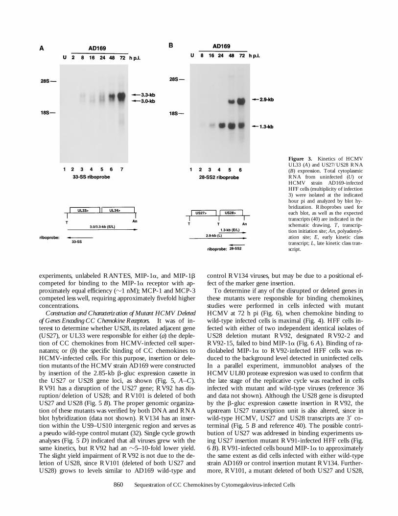
Chemokine Binding to HCMV-infected Cells. The HCMVgenome encodes three proteins with homology to CC typechemokine receptors: UL33, US27, and US28 (39). To de-termine when these genes may function during the courseof the viral replicative cycle, their expression in HCMVstrain AD169-infected HFF cells was examined by RNAblot hybridization (Fig. 3). A previous study indicated thateach of these genes was expressed by late times pi (40). Ouranalyses revealed that both UL33 (3.0–3.3 kb) and US28(1.3 kb) transcripts are detected much earlier, by 8 h pi, andincrease in abundance to 72 h pi (i.e., early and late timespi, respectively). In contrast, US27 (2.9 kb) RNA was de-tected only at late times pi (i.e., 48–72 h pi). US27 RNA isdetected with the same probe used for US28, since thetranscript that initiates just upstream of US27 reads throughboth US27 and US28 (40) (Fig. 3 B).
Of the three HCMV-encoded G protein–coupled CCchemokine receptor homologues, only the US28 geneproduct has been shown to be a functional chemokine re-ceptor, and only in transfected cells (29, 30). These reports
indicated that, at least in the absence of other HCMV geneproducts, the US28-encoded receptor could bind the CCchemokines MIP-1a, MIP-1b, MCP-1, and RANTES. Wesought to extend these observations by examining thebinding of the CC chemokine, MIP-1a, to HCMV strainAD169-infected HFF cells at 24, 48, and 72 h pi. (Fig. 4A). Uninfected fibroblasts did not bind MIP-1a, whereasbinding to HCMV-infected cells was detected at all threetimes, increasing from early (24 h) to late times (72 h) pi.Indicative of specificity, this binding was reduced to back-ground levels (i.e., 10–40-fold reduction) in the presenceof 400-fold excess unlabeled MIP-1a, similar to those lev-els detected in uninfected cells. Thus, in both qualitativeand quantitative terms, the early-late binding kinetics ofchemokines to HCMV-infected cells coincides with theearly-late expression kinetics of the US28 transcription unit(Fig. 3 B). Similar chemokine binding experiments wereperformed using a recent clinical isolate of HCMV andsimian CMV. Specific MIP-1a binding to fibroblasts in-fected with these CMVs was also observed (data notshown). Thus, CC chemokine binding in cells infectedwith at least primate CMVs is conserved and may be func-tionally important.
To examine the breadth of CC chemokine binding inHCMV-infected cells to the MIP-1a receptor, competitionbinding studies were performed in the presence of 800-foldexcess unlabeled heterologous chemokines (Fig. 4 B). Thisexperiment indicated that the CC chemokines MIP-1b,RANTES, MCP-1, and MCP-3 caused the displacementof .95% of MIP-1a binding, and thus bind to the samereceptor as does MIP-1a. In contrast, MCP-2 did notcompete with MIP-1a, as indicated by ,5% displacement.Furthermore, additional binding experiments indicated thatcompetition for the MIP-1a receptor in HCMV-infectedHFF cells by heterologous CC chemokines occurred in adose-dependent fashion as expected for receptor–ligandinteractions (data not shown). The relative affinity ofeach chemokine for the MIP-1a receptor was estimatedbased on the concentration of cold competitor required todisplace 50% of bound 125I-labeled MIP-1a. In these
Table 1. Extracellular Production of MCP-1 after HCMV Infection of Fibroblasts
Fibroblasts
Extracellular production of MCP-1
0–8 h‡ 8–24 h‡ 24–48 h‡ 48–72 h‡
pg/mlUninfected 5,615 11,840 9,938 10,879Virus-free supernatant 10,016 10,738 10,972 12,131
Infected with:UV-inactivated 9,906 10,572 10,889 10,555Heat-inactivated 10,946 10,551 10,136 12,021Pelleted virus 8,992 9,377 5,329 1,157Whole inoculum 10,811 10,000 4,301 570
*Representative of two experiments.‡Time intervals at which supernatants were collected. At each time in-terval, cells were washed and re-fed growth medium.
Table 2. Measurement of Intracellular RANTES and MCP-1
Infection
RANTES MCP-1
Extracellular Intracellular Extracellular Intracellular
pg/ml pg/mlnone 0 0 2,306 94AD169 994 320 2,423 418
At 24 h after infection, culture supernatants were collected and cellswere treated with glycine buffer to strip cell membranes of chemokinesbefore lysing. Concentrations of chemokine are expressed as picogram/milliliter. All points represent the average of duplicate supernatant/cy-toplasm pairs.
860 Sequestration of CC Chemokines by Cytomegalovirus-infected Cells
experiments, unlabeled RANTES, MIP-1a, and MIP-1bcompeted for binding to the MIP-1a receptor with ap-proximately equal efficiency (z1 nM); MCP-1 and MCP-3competed less well, requiring approximately fivefold higherconcentrations.
Construction and Characterization of Mutant HCMV Deletedof Genes Encoding CC Chemokine Receptors. It was of in-terest to determine whether US28, its related adjacent gene(US27), or UL33 were responsible for either (a) the deple-tion of CC chemokines from HCMV-infected cell super-natants; or (b) the specific binding of CC chemokines toHCMV-infected cells. For this purpose, insertion or dele-tion mutants of the HCMV strain AD169 were constructedby insertion of the 2.85-kb b-gluc expression cassette inthe US27 or US28 gene loci, as shown (Fig. 5, A–C).RV91 has a disruption of the US27 gene; RV92 has dis-ruption/deletion of US28; and RV101 is deleted of bothUS27 and US28 (Fig. 5 B). The proper genomic organiza-tion of these mutants was verified by both DNA and RNAblot hybridization (data not shown). RV134 has an inser-tion within the US9–US10 intergenic region and serves asa pseudo wild-type control mutant (32). Single cycle growthanalyses (Fig. 5 D) indicated that all viruses grew with thesame kinetics, but RV92 had an z5–10-fold lower yield.The slight yield impairment of RV92 is not due to the de-letion of US28, since RV101 (deleted of both US27 andUS28) grows to levels similar to AD169 wild-type and
control RV134 viruses, but may be due to a positional ef-fect of the marker gene insertion.
To determine if any of the disrupted or deleted genes inthese mutants were responsible for binding chemokines,studies were performed in cells infected with mutantHCMV at 72 h pi (Fig. 6), when chemokine binding towild-type infected cells is maximal (Fig. 4). HFF cells in-fected with either of two independent identical isolates ofUS28 deletion mutant RV92, designated RV92-2 andRV92-15, failed to bind MIP-1a (Fig. 6 A). Binding of ra-diolabeled MIP-1a to RV92-infected HFF cells was re-duced to the background level detected in uninfected cells.In a parallel experiment, immunoblot analyses of theHCMV UL80 protease expression was used to confirm thatthe late stage of the replicative cycle was reached in cellsinfected with mutant and wild-type viruses (reference 36and data not shown). Although the US28 gene is disruptedby the b-gluc expression cassette insertion in RV92, theupstream US27 transcription unit is also altered, since inwild-type HCMV, US27 and US28 transcripts are 39 co-terminal (Fig. 5 B and reference 40). The possible contri-bution of US27 was addressed in binding experiments us-ing US27 insertion mutant RV91-infected HFF cells (Fig.6 B). RV91-infected cells bound MIP-1a to approximatelythe same extent as did cells infected with either wild-typestrain AD169 or control insertion mutant RV134. Further-more, RV101, a mutant deleted of both US27 and US28,
Figure 3. Kinetics of HCMVUL33 (A) and US27/US28 RNA(B) expression. Total cytoplasmicRNA from uninfected (U) orHCMV strain AD169-infectedHFF cells (multiplicity of infection3) were isolated at the indicatedhour pi and analyzed by blot hy-bridization. Riboprobes used foreach blot, as well as the expectedtranscripts (40) are indicated in theschematic drawing. T, transcrip-tion initiation site; An, polyadenyl-ation site; E, early kinetic classtranscript; L, late kinetic class tran-script.

861 Bodaghi et al.
also failed to bind MIP-1a. In contrast, as an additionalcontrol, cells infected with RV11, a mutant with a b-glucinsertion replacing an NH2-terminal portion of UL33 withunimpaired growth kinetics (Jones, T.R., unpublisheddata), retained full ability to bind MIP-1a (Fig. 6 B). ByRNA blot hybridization, each of the HCMV mutants thatbound the CC chemokine MIP-1a at late times pi had anintact US28 transcription unit which was expressed atwild-type levels (data not shown). Therefore, the data are
indicative that the US28-encoded receptor is in large partresponsible for CC chemokine binding in HCMV-infectedcells.
Role of Receptors Encoded by US28 in the Depletion ofRANTES and MCP-1 after Infection. Three human cellu-lar receptors, CCR1, CCR3, and CCR5, have beenshown to bind CC chemokines, including RANTES andMCP-1 (41). However, by RT-PCR, we were unable todetect any transcripts for CCR3 and CCR5 in total RNAor of CCR1 in the polyadenylated fraction from either un-infected or HCMV-infected HFF cells (data not shown).Therefore, the early-late expression kinetics of the HCMVUS28-encoded receptor (Fig. 3 B), in conjunction with itscapability to bind RANTES and MCP-1 (Figs. 4 and 6),suggest that it may play a role in the depletion of thesechemokines from the media of HCMV-infected cells (Ta-ble 1 and Fig. 1). To directly study the HCMV encodedCC chemokine receptor homologues in chemokine deple-tion, the extracellular levels of RANTES and MCP-1 inthe culture medium of HFF cells infected with each of theCC chemokine receptor mutants were examined (Fig. 7).Infection with RV92 or RV101, both US28 deletion mu-tants, failed to lead to the extensive depletion of eitherRANTES (Fig. 7 A) or MCP-1 (Fig. 7 B) from the culturemedium. In contrast, infection with RV91 led to a markedreduction of extracellular RANTES and MCP-1, almostsimilar to that observed in cells infected with wild-typeHCMV. Comparable effects on extracellular RANTESand MCP-1 levels were observed in other fibroblasts,MRC-5 and WI-38, infected with these mutant viruses(data not shown). Consistent with its ability to bind CCchemokines, these results strongly suggest that US28 plays amajor role in the depletion of CC chemokines from theextracellular media of HCMV-infected cells.
Internalization of Exogenously Added RANTES by InfectedCells. Previously, we demonstrated by immunofluores-cence that RANTES accumulated within HCMV-infectedcells concomitant with its depletion from culture medium.Additional experiments indicated that this was not due tosoluble proteases (38). These observations raised the fol-lowing possibilities: (a) RANTES or MCP-1 is being re-tained intracellularly by HCMV-encoded receptors as thesechemokines are synthesized and/or (b) newly synthesizedchemokines are released from the infected cell, but thatHCMV-encoded cell receptors expressed at the cell surface
Figure 4. Time course and specificity of MIP-1areceptor expression. (A) 125I-labeled MIP-1a bind-ing to uninfected (UNINF) or HCMV strainAD169-infected HFF cells was examined at the in-dicated time pi, in either the absence or presence ofexcess unlabeled MIP-1a (h1MIP-1a) at a finalconcentration of 100 nM. cpm of bound radiolabeledMIP-1a are given above each bar. (B) Percentageof displaceable binding of 125I-labeled MIP-1a us-ing the indicated unlabeled chemokine (200 nM fi-nal concentration) in infected HFF cells at 72 h pi.
Figure 5. Genome organization and growth of HCMV mutants. Theorganization of the HCMV genome (A) and the BamHI-S fragment (B),which contains the US27 and US28 genes, are shown in the schematicdrawing. In B, the US27 and US28 transcripts are shown, as well as thesites of the b-gluc expression cassette insertions in RV91, RV92, orRV101. The arrowhead is the site of a point insertion (RV91) and darkrectangles indicate deletions (RV92 and RV101). (C) Schematic drawingof the 2.85-kb b-gluc expression cassette used in the construction ofHCMV mutants. (D) Single cycle growth analysis of HCMV mutants.

862 Sequestration of CC Chemokines by Cytomegalovirus-infected Cells
are undergoing continual binding and rapid internalizationof the chemokines.
To examine the possibility that chemokines releasedfrom HCMV-infected cells may be subsequently bound byHCMV-encoded receptors, several experiments were per-formed. The first experiment was based on the observation
that chemokine expression can be induced by HCMV in-fection of fibroblasts and accumulates in the extracellularmedium at 48–72 h pi of cells infected with US28 mutants(Fig. 7). Conditioned medium collected at 72 h pi fromcells infected with US28-negative mutants or US28-posi-tive virus infected cells as control was used as a cold com-petitor in an MIP-1a–binding assay to wild-type HCMV-infected cells (Table 3). Relative to fresh medium, mediaconditioned on cells infected with US28-negative mutantsinhibited 48% of total MIP-1a binding. In contrast, mediaconditioned on US28-positive cells inhibited MIP-1abinding by ,5%.
In a second experiment, exogenous rRANTES wasadded to HCMV-infected HFF cells to see if it would beremoved from the medium. At 48 h pi, medium was col-lected and cells were washed with PBS, then treated with alow pH glycine buffer to strip the membrane of anychemokine (34). Exogenous rRANTES was then added toculture medium and cells were incubated for differenttimes at 378C before measuring the amount of RANTES
Figure 6. MIP-1a binding by cells infected with HCMV mutants. Uninfected or HCMV-infected HFF cells (multiplicity of infection 2.5) were analyzedfor their ability to bind 125I-labeled MIP-1a at 72 h pi. RV92-2 and RV92-15 are independent isolates containing the identical US28 deletion. In some exper-iments, excess unlabeled MIP-1a (200 nM final concentration) was used (AD1691MIP-1a). cpm of bound radiolabeled MIP-1a are given above each bar.
Figure 7. (A) RANTES expressed in picogram/milliliter or (B) MCP-1expressed in nanogram/milliliter were measured by ELISA in superna-tants collected at 0–24 h (black bars), 24–48 (white bars), and 48–72 h (graybars) after infection from either uninfected cells (NF) or cells infected withwild-type or mutant HCMVs: AD (wild-type), RV91 (DUS27), RV92(DUS28), RV101 (DUS27/DUS28). At each interval, cells were washedand re-fed growth medium.
Table 3. Adsorption of MIP-1a from Conditioned Media
Conditionedmedium source
Cpm MIP-1abound
(standard deviation)
Percentage ofreduction of
MIP-1a binding
None (fresh medium) 27,164 (1,750) NoneUS28-positive cells 25,962 (4,362) 4.5US28-negative cells 14,090 (1,802) 48.2
Target cells in the MIP-1a binding assay were HCMV strain AD169-infected HFF cells at 72 h pi. The binding assay was done as describedin Materials and Methods using 0.25 nM of radiolabeled MIP-1a perwell, except that 0.125 ml of conditioned medium replaced an equalvolume of cold binding buffer. No attempt was made to removechemokines from the infected cell membranes before the initiation ofthe binding assay. Each data point represents the mean and standard de-viation derived from three to four plates, as follows: US28-positive in-fected cell–conditioned medium was from one plate each of HFF cellsinfected with wild-type strains AD169, RV134, RV91, and RV11.US28-negative infected cell–conditioned medium was from cells in-fected with RV92 (one plate) and RV101 (two plates).

863 Bodaghi et al.
remaining in the medium. Incubation of HCMV-infectedfibroblasts with 600 pg/ml of rRANTES for 30 min or 3 hresulted in a 12 and a 72% reduction, respectively, of theexogenously added chemokine. Similar experiments werecarried out on cells infected for 72 h. An average (two ex-periments run in duplicate) of 59% of added rRANTESdisappeared from the medium of infected cells after a 3-hadsorption period at 378C. In all experiments, RANTESwas not detected in the medium sampled before startingthe adsorption period (i.e., before the addition of exoge-nous rRANTES). Taken together, these experiments indicatethat chemokines synthesized and released from HCMV-infected cells can bind to, at least, the US28-encoded re-ceptor on the surface of these cells.
It is known that when a chemokine contacts its receptor,both are internalized and ligand is released from the recep-tor, which then recirculates to the cell surface (42). To de-termine whether extracellular RANTES was being inter-nalized from the cell surface after receptor binding, uninfectedcells or HCMV-infected cells (at 48 h pi) were incubatedwith RANTES-B for 3 h at 378C, washed to remove cellsurface (noninternalized) chemokine, then analyzed by im-munoblot using peroxidase-labeled streptavidin, as describedin Materials and Methods (Fig. 8). The results demonstratethat RANTES-B was efficiently internalized by cells in-fected with wild-type HCMV, less so by cells infected withthe mutants RV91 and RV92, and not at all by RV101-infected cells or uninfected fibroblasts.
Discussion
After primary infection, HCMV, like other herpesvi-ruses, establishes itself in a latent or persistent state and re-sides in its host for the remainder of the host’s lifetime (43).To facilitate this state, HCMV has several means designedseemingly to avoid elimination of infected cells by the im-mune surveillance system. HCMV encodes at least fourgenes whose products play a role in the downregulation ofsurface expression of HLA class I molecules (9, 10, 18–23).
In addition, HCMV infection can interfere with normalcytokine production (44) and upregulate production ofsuch lymphostatic factors as TGF-b (45) and IL-1Ra (46).
However, the HCMV genome also carries three geneswith homology to CCRs (UL33, US27, and US28; refer-ence 39). The role of these receptors in the life cycle ofhuman HCMV in vivo has not yet been determined.HCMV UL33 was shown to be a virion envelope protein,but its function remains unknown (27). The murine cy-tomegalovirus homologue of HCMV UL33, designatedM33 (47), may play a role in the establishment of latency ofmurine HCMV in salivary glands (48). Unfortunately, mu-rine HCMV does not possess homologues of US27 andUS28. Of the HCMV CCR homologues, only US28 hasbeen shown to be functional to date. In transfected mam-malian cells, US28 mobilizes Ca21 in response to RANTES,MIP-1a, and MCP-1 (29, 30); in Hela and astrocytomacells, it can act as a coreceptor for HIV entry (49). How-ever, these studies do not shed light on the function ofUS28 in the context of HCMV infection.
In the context of HCMV-infected cells, we show herethat the product of the HCMV US28 gene plays a promi-nent role in binding and sequestering of extracellular CCchemokines. Through the use of specific HCMV gene dis-ruption/deletion recombinant mutants, the US28-encodedreceptor was shown to be the only MIP-1a binding recep-tor expressed in HCMV-infected cells. Cells infected witha US27 mutant (RV91) or a UL33 mutant (RV11), but nota US28 mutant (RV92) or a US271US28 mutant (RV101),retained ability to bind the CC chemokine MIP-1a. Inconjunction with our inability to detect CCR polyadenyl-ated transcripts in infected cells, this observation effectivelyrules out the possibility that HCMV infection induces ex-pression of cellular MIP-1a receptors. In addition to MIP-1abinding in HCMV-infected cells, the US28-encoded re-ceptor binds other CC chemokines, including MIP-1b,RANTES, and MCP-1, consistent with the binding ob-served in US28-transfected cells (29, 30). We have ex-tended these observations in that the US28-encoded re-ceptor also binds MCP-3, but not MCP-2, both CCchemokines. Interestingly, at the amino acid level MCP-2is highly homologous to MCP-1 and MCP-3 (62 and 60%,respectively; reference 41).
Cell surface expression of the US28-encoded receptorresults in the depletion of CC chemokines, especiallyRANTES and MCP-1, from the medium of HCMV-infected cells. Cells infected with mutants that expressedthe other putative chemokine receptors, US27 or UL33(i.e., RV101 and RV92), but lacked US28 expression,failed to efficiently deplete chemokines from the medium(Fig. 7). Furthermore, cells infected with viruses that ex-pressed US28 internalized exogenous RANTES, whereasuninfected cells or cells infected with RV101, deleted ofboth US27/US28, failed to internalize added RANTES(Fig. 8). Taken together, these data indicate that UL33,which is present and transcribed at wild-type levels in cellsinfected with RV101 (data not shown), does not play a rolein the sequestration of RANTES or MCP-1.
Figure 8. Fibroblasts infected for 48 h with wild-type (AD) or mutantHCMVs (RV101, RV92, RV91) were washed with PBS, incubated for 3min at room temperature with low pH glycine buffer to strip membranesof chemokine, and then incubated with biotinylated RANTES (100 nM)for 3 h at 378C. Cells were then washed again with PBS, treated withlow-pH glycine buffer, trypsinized, and extracted in a high salt buffer.Extracts were separated in 15% SDS-PAGE and transferred to nitrocellu-lose. After saturation with 3% gelatin/1% BSA in PBS/0.1% Tween 20,blots were incubated with peroxidase-labeled streptavidin (1:400) and de-veloped using a chemiluminescence technique for 5 s. RANTES-B(Rantes-B, 125 ng) was run in parallel. AD 5 infected with wild-typeHCMV; RV91 5 infected with DUS27 mutant HCMV; RV92 5 in-fected with DUS28 mutant HCMV; RV101 5 infected with DUS27/DUS28 mutant HCMV; NF 5 uninfected cell extract. Representative oftwo experiments.

864 Sequestration of CC Chemokines by Cytomegalovirus-infected Cells
Remarkably, the ability to internalize RANTES did notstrictly correlate with US28 expression. Cells infected withUS28 mutant RV92 or US27 mutant RV91 internalizedRANTES, although to a lesser extent than cells infectedwith wild-type HCMV. Although it is clear from our ex-periments that the US28-encoded receptor functions tobind and internalize CC chemokines in HCMV-infectedcells, the US27-encoded receptor may also contribute tothese processes. Thus, it is possible that US28 is not theonly HCMV-encoded receptor that binds and internalizesRANTES. The US27-encoded receptor may also bind/in-ternalize RANTES, but has differential affinity for MIP-1a(i.e., the US28-encoded receptor, but not the US27-encoded receptor, binds MIP-1a). We are currently inves-tigating the possibility that the US27-encoded receptor is alow affinity receptor for some CC chemokines.
We propose that a role for the chemokine receptors ex-pressed in HCMV-infected cells may be the ability to se-quester chemokines from the extracellular milieu. We dem-onstrated recently (38), and confirm here, that chemokinesaccumulate intracellularly in HCMV-infected cells. Thisappears to be partially due to continual internalization fromthe exterior, as shown here, since in HCMV-infected cells(a) exogenously added RANTES disappeared from cellmedium and (b) added biotinylated RANTES was foundintracellularly. We cannot exclude the possibility that en-dogenously produced chemokines may also be sequesteredintracellularly without ever being secreted. When exposedto ligand, the US28-encoded receptor in transfected cellscan initiate a transmembrane signal that results in secondmessenger signaling, including calcium flux (29, 30). Thismay also occur in HCMV-infected cells and alter cell phys-iology, thereby affecting replication of the virus. However,the presence or absence of chemokines had no detectableeffect on the growth properties of HCMV wild-type in fi-broblasts compared with US271US28 mutant RV101(Jones, T.R., unpublished data). Additionally, Pertussis
toxin, which interferes with signaling of some cellularchemokine receptors (41, 50), likewise had no effect ongrowth of these viruses (Jones, T.R., unpublished data).
Induction and disappearance of RANTES are two sepa-rable phenomena. Induction occurs independently of anactive CMV genome upon contact/penetration of virusinto fibroblasts, as illustrated by the fact that UV-inacti-vated virus induces RANTES production. Induction couldtherefore be due to contact of viral elements with cellmembranes and/or introduction of viral proteins and DNAinto the cell. Hence, even an abortive infection in the ab-sence of viral genome expression might induce productionof RANTES, which could in turn induce recruitment oflymphocytes.
On the other hand, disappearance of RANTES as infec-tion progresses does depend on viral genome expression,since it does not occur when cells are infected with UV-inactivated virus. Hence, when CMV genome expressionprogresses and US28 and US27 are encoded, CC chemo-kines, whether induced by virus contact (RANTES) or not(MCP-1), are sequestered from the infected cell environ-ment. Such sequestration could prevent not only lymphocyterecruitment, but also stimulation of effector mechanisms oflymphocytes in the neighborhood of infected cells. The ca-pacity of HCMV infected cells to sequester CC chemo-kines could potentially perturb local immune responses byaltering their concentration in the proximal extracellularmilieu. CC chemokines have been shown to play a role ininflammation and infiltration by lymphocytes, monocytes,eosinophils, and basophils at disease sites (41, 51). They arealso important in activating the cytotoxic potential of CD81
lymphocytes (52, 53) and NK cells (1). Thus, this sequestra-tion could have a negative effect on the attraction of leuko-cytes to sites of CMV infection and on the immune func-tion of leukocytes in the environment of infected cells.Hence, this CMV strategy would help the virus to go un-noticed and thereby escape immune surveillance.
This work was supported by the Agence Nationale de Recherche sur le Syndrome d’Immunodéficience Ac-quise and by the following Biomed 2 European Concerted Action projects: Infections with HCMV in theImmunocompromised Host and ROCIO II. B. Bodaghi is a beneficiary of study grants from the Fondationpour la Recherche Médicale and the Fonds d’Etudes du Corps Médical de l’Assistance Publique des Hôpi-taux de Paris. D. Zipeto is a beneficiary of a Training and Mobility grant (no. ERBFMBICT961426) fromthe European Commission.
Address correspondence to Susan Michelson, Unité d’Immunologie Virale, Institut Pasteur, 28 rue du Dr.Roux, 75724 Paris Cédex 15, France. Phone: 33-1-45-68-82-64; Fax: 33-1-45-68-89-41; E-mail: [email protected]
Received for publication 6 April 1998 and in revised form 8 June 1998.
References1. Loetscher, P., M. Seitz, M. Baggiolini, and B. Moser. 1996.
Interleukin 2 regulates CC chemokine receptor expressionand chemotactic responsiveness in T lymphocytes. J. Exp.Med. 184:569–577.
2. Davis-Poynter, N.J., and H.E. Farrell. 1996. Masters of de-ception: a review of herpesvirus immune evasion strategies.Immunol. Cell. Biol. 74:513–522.
3. Moore, P.S., C. Boshoff, R.A. Weiss, and Y. Chang. 1996.
865 Bodaghi et al.
Molecular mimicry of human cytokine and cytokine responsepathway genes by KSHV. Science. 274:1739–1744.
4. Murphy, P.M. 1994. Molecular piracy of chemokine recep-tors by herpesviruses. Infect. Agents Dis. 3:137–154.
5. Smith, G.L. 1996. Virus proteins that bind cytokines,chemokines or interferons. Curr. Opin. Immunol. 8:467–471.
6. Smith, G.L. 1994. Viral strategies for evasion of the host re-sponse to infection. Trends Microbiol. 2:81–88.
7. Roizman, B.H. 1996. Herpesviridae. In Virology. 3rd. ed.B.N. Fields, D.M. Knipe, and P.M. Howley, editors. Lippin-cott-Raven Publishers, Philadelphia, PA. 2221–2230.
8. McGeoch, D.J., S. Cook, A. Dolan, F.E. Jamieson, and E.A.Telford. 1995. Molecular phylogeny and evolutionary time-scale for the family of mammalian herpesviruses. J. Mol. Biol.247:443–458.
9. Hendrix, M.G.R., E. Beuken, R.L. Slobbe, and C.A.Bruggeman. 1996. Detection and sequence analysis of themajor immediate early and pp150 gene of latent human cy-tomegalovirus in spleen, liver and kidney tissues of traumavictims. J. Med. Virol. 50:193–197.
10. Myerson, D., R.C. Hackman, J.A. Nelson, D.C. Ward, andJ.K. McDougall. 1984. Widespread presence of histologicallyoccult cytomegalovirus. Hum. Pathol. 15:430–439.
11. Toorkey, C.B., and D.R. Carrigan. 1989. Immunohis-tochemical detection of an immediate early antigen of humancytomegalovirus in normal tissues. J. Infect. Dis. 160:741–751.
12. Furukawa, T., E. Hornberger, S. Sakuma, and S.A. Plotkin.1975. Demonstration of immunoglobulin G receptors in-duced by human cytomegalovirus. J. Clin. Microbiol. 2:332–336.
13. MacCormac, L.P., and J.E. Grundy. 1996. Human cytomeg-alovirus induces an Fc gamma receptor (FcgR) in endothelialcells and fibroblasts that is distinct from the human cellularFcgRs. J. Infect. Dis. 174:1151–1161.
14. Stannard, L.M., and D.R. Hardie. 1991. An Fc receptor forhuman immunoglobulin G is located within the tegument ofhuman cytomegalovirus. J. Virol. 65:3411–3415.
15. Xu, B., T. Murayama, K. Ishida, and T. Furukawa. 1989.Characterization of IgG Fc receptors induced by human cy-tomegalovirus. J. Gen. Virol. 70:893–900.
16. Spiller, O.B., B.P. Morgan, F. Tufaro, and D.V. Devine.1996. Altered expression of host-encoded complement regu-lators on human cytomegalovirus-infected cells. Eur. J. Immu-nol. 26:1532–1538.
17. Spear, G.T., N.S. Lurain, C.J. Parker, M. Ghassemi, G.H.Payne, and M. Saifuddin. 1995. Host cell–derived comple-ment control proteins CD55 and CD59 are incorporated intothe virions of two unrelated enveloped viruses. Human T cellleukemia/lymphoma virus type I (HTLV-I) and human cy-tomegalovirus (HCMV). J. Immunol. 155:4376–4381.
18. Jones, T.R., L.K. Hanson, L. Sun, J.S. Slater, R.M. Stenberg,and A.E. Campbell. 1995. Multiple independent loci withinthe human cytomegalovirus unique short region down-regu-late expression of major histocompatibility complex class Iheavy chains. J. Virol. 69:4830–4841.
19. Jones, T.R., E. Wiertz, L. Sun, K.N. Fish, J.A. Nelson, andH.L. Ploegh. 1996. Human cytomegalovirus US3 impairstransport and maturation of major histocompatibility com-plex class I heavy chains. Proc. Natl. Acad. Sci. USA. 93:11327–11333.
20. Jones, T.R., and L. Sun. 1997. Human cytomegalovirus US2destabilizes major histocompatibility complex class I heavychains. J. Virol. 71:2970–2979.
21. Wiertz, E., D. Tortorella, M. Bogyo, J. Yu, W. Mothes,
T.R. Jones, T.A. Rapoport, and H.L. Ploegh. 1996. Sec61-mediated transfer of a membrane protein from the endoplas-mic reticulum to the proteasome for destruction. Nature. 384:432–438.
22. Ahn, K., A. Gruhler, B. Galocha, T.R. Jones, E. Wiertz,H.L. Ploegh, P.A. Peterson, Y. Yang, and K. Fruh. 1997.The ER-luminal domain of the HCMV glycoprotein US6inhibits peptide translocation by TAP. Immunity. 6:613–621.
23. Wiertz, E., T.R. Jones, L. Sun, M. Bogyo, H.J. Geuze, andH.L. Ploegh. 1996. The human cytomegalovirus US11 geneproduct dislocates MHC class I heavy chains from the endo-plasmic reticulum to the cytosol. Cell. 84:769–779.
24. Cosman, D., N. Fanger, L. Borges, M. Kubin, W. Chin, L.Peterson, and M.L. Hsu. 1997. A novel immunoglobulin su-perfamily receptor for cellular and viral MHC class I mole-cules. Cell. 7:273–282.
25. Reyburn, H.T., O. Mandelboim, M. Valesgomez, D.M.Davis, L. Pazmany, and J.L. Strominger. 1997. The class IMHC homologue of human cytomegalovirus inhibits attackby natural killer cells. Nature. 386:514–517.
26. Beck, S., and B. Barrell. 1991. An HCMV reading framewhich has similarity with both the V and C regions of theTCR gamma chain. DNA Seq. 2:33–38.
27. Margulies, B.J., H. Browne, and W. Gibson. 1996. Identifi-cation of the human cytomegalovirus G protein–coupled re-ceptor homologue encoded by Ul33 in infected cells and en-veloped virus particles. Virology. 225:111–125.
28. Chee, M.S., A.T. Bankier, S. Beck, R. Bohni, C.M. Brown,R. Cerny, T. Horsnell, C.A. Hutchison III, T. Kouzarides,J.A. Martignetti, et al. 1990. Analysis of the protein-codingcontent of the sequence of human cytomegalovirus strainAD169. Curr. Top. Microbiol. Immunol. 154:125–169.
29. Neote, K., D. DiGregorio, J.Y. Mak, R. Horuk, and T.J.Schall. 1993. Molecular cloning, functional expression, andsignaling characteristics of a C-C chemokine receptor. Cell.72:415–425.
30. Gao, J.L., and P.M. Murphy. 1994. Human cytomegalovirusopen reading frame US28 encodes a functional beta chemo-kine receptor. J. Biol. Chem. 269:28539–28542.
31. Alcami, J., T. Barzu, and S. Michelson. 1991. Induction of anendothelial cell growth factor by human cytomegalovirus in-fection of fibroblasts. J. Gen. Virol. 72:2765–2770.
32. Jones, T.R., V.P. Muzithras, and Y. Gluzman. 1991. Re-placement mutagenesis of the human cytomegalovirus ge-nome: US10 and US11 gene products are nonessential. J. Vi-rol. 65:5860–5872.
33. Jones, T.R., and V.P. Muzithras. 1991. Fine mapping oftranscripts expressed from the US6 gene family of human cy-tomegalovirus strain AD169. J. Virol. 65:2024–2036.
34. Samanta, A.K., J.J. Oppenheim, and K. Matsushima. 1990.Interleukin 8 (monocyte-derived neutrophil chemotactic fac-tor) dynamically regulates its own receptor expression on hu-man neutrophils. J. Biol. Chem. 265:183–189.
35. Ylisastigui, L., J. Vizzavona, E. Drakopoulou, P. Pain-davoine, C. Calvo, M. Parmentier, J.C. Gluckman, C. Vita,and A. Benjouad. 1998. Synthetic full-length RANTES andtruncated RANTES inhibit human immunodeficiency virustype 1 infection of primary macrophages. AIDS. In press.
36. Jones, T.R., L. Sun, G.A. Bebernitz, V.P. Muzithras, H.J.Kim, S.H. Johnston, and E.Z. Baum. 1994. Proteolytic activ-ity of human cytomegalovirus UL80 protease cleavage sitemutants. J. Virol. 68:3742–3752.
37. Kavasaki, E.S. 1990. Amplification of RNA. In PCR Proto-

866 Sequestration of CC Chemokines by Cytomegalovirus-infected Cells
cols: A Guide to Methods and Application. D.H. Gelfand,M.A. Innis, J.J. Snisky, and T.J. White, editors. AcademicPress Inc., San Diego, CA. 21–27.
38. Michelson, S., P. Dalmonte, D. Zipeto, B. Bodaghi, L. Lau-rent, E. Oberlin, F. Arenzana-Seisdedos, J.L. Virelizier, andM.P. Landini. 1997. Modulation of RANTES production byhuman cytomegalovirus infection of fibroblasts. J. Virol. 71:6495–6500.
39. Chee, M.S., S.C. Satchwell, E. Preddie, K.M. Weston, andB.G. Barrell. 1990. Human cytomegalovirus encodes three Gprotein–coupled receptor homologues. Nature. 344:774–777.
40. Welch, A.R., L.M. McGregor, and W. Gibson. 1991. Cy-tomegalovirus homologs of cellular G protein–coupled re-ceptor genes are transcribed. J. Virol. 65:3915–3918.
41. Baggiolini, M., B. Dewald, and B. Moser. 1994. Interleukin–8and related chemotactic cytokines—CXC and CC chemo-kines. Adv. Immunol. 55:97–179.
42. Amara, A., S.L. Gall, O. Schwartz, J. Salamero, M. Montes,P. Loetscher, M. Baggiolini, J.L. Virelizier, and F. Arenzana-Seisdedos. 1997. HIV coreceptor downregulation as antiviralprinciple: SDF-1a–dependent internalization of the chemo-kine receptor CXCR4 contributes to inhibition of HIV rep-lication. J. Exp. Med. 186:139–146.
43. Britt, W.J., and C.A. Alford. 1996. Cytomegalovirus. In Vi-rology. 3rd edition. B.N. Field, D.M. Knipe, and P.M.Howley, editors. Lippincott-Raven Publishers, Philadelphia,PA. 2493–2523.
44. Michelson, S. 1998. Mechanisms of immunosuppression byhuman cytomegalovirus. In CMV-Related Immunopathol-ogy, Volume 21. H.F. Rabenau, M. Scholz, H.W. Doerr,and J. Cinalti, Jr., editors. Karger, Basel. 12–28.
45. Michelson, S., J. Alcami, S.J. Kim, D. Danielpour, F. Bache-lerie, L. Picard, C. Bessia, C. Paya, and J.L. Virelizier. 1994.Human cytomegalovirus infection induces transcription andsecretion of transforming growth factor beta 1. J. Virol. 68:
5730–5737.46. Kline, J.N., L.J. Geist, M.M. Monick, M.F. Stinski, and
G.W. Hunninghake. 1994. Regulation of expression of theIL-1 receptor antagonist (IL-1ra) gene by products of the hu-man cytomegalovirus immediate early genes. J. Immunol. 152:2351–2357.
47. Macdonald, M.R., X.Y. Li, and H.W. Virgin. 1997. Late ex-pression of a beta chemokine homolog by murine cytomega-lovirus. J. Virol. 71:1671–1678.
48. Davis-Poynter, N.J., D.M. Lynch, H. Vally, G.R. Shellam,W.D. Rawlinson, B.G. Barrell, and H.E. Farrell. 1997. Iden-tification and characterization of a G protein–coupled recep-tor homolog encoded by murine cytomegalovirus. J. Virol.71:1521–1529.
49. Pleskoff, O., C. Treboute, A. Brelot, N. Heveker, M. Se-man, and M. Alizon. 1997. Identification of a chemokine re-ceptor encoded by human cytomegalovirus as a cofactor forHIV-1 entry. Science. 276:1874–1878.
50. Sozzani, S., D. Zhou, M. Locati, M. Rieppi, P. Proost, M.Magazin, N. Vita, J. van Damme, and A. Mantovani. 1994.Receptors and transduction pathways for monocyte chemo-tactic protein-2 and monocyte chemotactic protein-3. Simi-larities and differences with MCP-1. J. Immunol. 152:3615–3622.
51. Baggiolini, M., B. Dewald, and B. Moser. 1997. Humanchemokines: an update. Annu. Rev. Immunol. 15:675–705.
52. Taub, D.D., S.M. Turcovski-Corrales, M.L. Key, D.L.Longo, and W.J. Murphy. 1996. Chemokines and T lym-phocyte activation: I. Beta chemokines costimulate human Tlymphocyte activation in vitro. J. Immunol. 156:2095–2103.
53. Taub, D.D., J.R. Ortaldo, S.M. Turcovski-Corrales, M.L.Key, D.L. Longo, and W.J. Murphy. 1996. Beta chemokinescostimulate lymphocyte cytolysis, proliferation, and lym-phokine production. J. Leukocyte Biol. 59:81–89.