NADPH-diaphorase activity and nitric oxide synthase isoforms in the trophoblast of Calomys callosus
F. Mele et al. (Eds.): BVAI 2007, LNCS 4729, pp. 11–20, 2007. © Springer-Verlag Berlin Heidelberg 2007
Diffuse Nerve Net of Hydra Revealed by NADPH-Diaphorase Histochemical Labeling
Luigia Cristino, Vittorio Guglielmotti, Carlo Musio, and Silvia SantilloCA
Istituto di Cibernetica “Eduardo Caianiello” del CNR, Via Campi Flegrei 34, I-80078 Pozzuoli (Napoli), Italy
{l.cristino,v.guglielmotti,c. musio,CAs.santillo}@cib.na.cnr.it
Abstract. The processing of the information coming from the external environment, including the interactions between molecular and cellular key-players involved in, is perhaps the “hard problem” in the cybernetic approach to the nervous system. As a whole, this information shapes the behavioral activity of an organism. The problem is faced considering the information processing flow in action from the lower organisms’ nervous elements to the higher cognitive levels of man. The cnidarian Hydra is the first organism of the zoological scale in which a nervous system is encountered. It is composed by isolated nerve cells scattered throughout the animal body constituting a diffuse nerve net for the input-output activity. In this paper is reported, for the first time in Hydra nerve net, the histochemical indication of a NADPH-diaphorase (NADPH-d) activity as putative marker of nitric oxide synthase (NOS) activity. The identification and the tentative localization of nitric oxide (NO) in Hydra is discussed in the light of the emerging role that such a signaling molecule exerts in sensory (visual particularly) and motor neural systems.
1 Introduction
Comparative Neurobiology’s main goal regards the solving of cutting-edge queries on the evolution of the nervous system. In particular, how does a simple nervous system, without centralization and with a rough isotropic distribution of its elements, generate and modulate a periodic behavior according to the environmental issues and demands? Also, are the key-elements of neurotransmission and signaling pathways already expressed and localized, at different cellular levels, in so-called simple animal model and hence phylogenetically conserved in higher organisms?
Local circuits, or networks, in the central nervous system are responsible for considerable information processing and integration in which sensory information is transformed into appropriate motor outputs. The output of a network depends not only on its inputs, but also on various modulatory mechanisms that modify the neural signal at any level within the network [1].
The neural net of the diploblastic fresh-water coelenterate Hydra (Cnidaria, Hydrozoa), with a great economy of specialized structures, enables Hydra to produce its periodic shortening-elongation behavior, which has a high biological significance for its survival, being linked to osmoregulation and locomotion related to feeding.
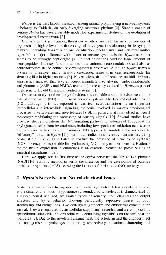
12 L. Cristino et al.
Hydra is the first known metazoan among animal phyla having a nervous system; it belongs to Cnidaria, an early-diverging metazoan phylum [2]. Since a couple of century Hydra has been a suitable model for experimental studies on the evolution of developmental mechanisms [3].
Cnidaria (and Hydra among them) nerve nets share with the nervous systems of organisms at higher levels in the zoological phylogenetic scale many basic synaptic features, including transmission and conduction mechanisms, and neurotransmitter types [4]. A major difference with bilaterian nervous systems is that Hydra nerve net seems to be strongly peptidergic [5]. In fact cnidarians produce large amount of neuropeptides that may function as neurotransmitters, neuromodulators and also as neurohormones in the control of developmental processes. Although Hydra nervous system is primitive, many neurons co-express more than one neuropeptide for signaling like in higher animals [6]. Nevertheless, data collected by multidisciplinary approaches indicate that several neurotransmitters like glycine, endocannabinoids, and glutamate (AMPA and NMDA receptors) have early evolved in Hydra as part of phylogenetically old behavioral control systems [7].
On the contrary, a minor body of evidence is available about the existence and the role of nitric oxide (NO) in cnidarian nervous systems. The free radical nitric oxide (NO), although it is not reported as classical neurotransmitter, is an important intracellular and intercellular signaling molecule involved in various physiological processes in vertebrates and invertebrates [8-9]. In particular it is involved as neural messenger modulating the processing of sensory signals [10]. Several studies have provided strong indications that NO signaling pathway is widespread throughout the phylogenetic scale from invertebrates, including few species of cnidarian (see section 3), to higher vertebrates and mammals. NO appears to modulate the response to “olfactory” stimuli in Hydra [11], but initial studies on different cnidarians, including Hydra itself [12-13], have failed to confirm the presence of nitric oxide synthase (NOS, the enzyme responsible for synthesising NO) in any of their neurons. Evidence for the nNOS expression in cnidarians is an essential element to prove NO as an ancestral neurotransmitter.
Here, we apply, for the first time to the Hydra nerve net, the NADPH-diaphorase (NADPH-d) staining method to verify the presence and the distribution of putative nitric oxide synthase (NOS) assessing the location of nitric oxide (NO) activity.
2 Hydra’s Nerve Net and Neurobehavioral Issues
Hydra is a sessile diblastic organism with radial symmetry. It has a coelenteron and, at the distal end, a mouth (hypostome) surrounded by tentacles. It is characterized by a simple neural net (46), by limited types of sensory input channels and output effectors and by a behavior showing periodically repetitive phases of body shortenings and elongations. Two cell-layers (ectoderm and endoderm) constitute the animal. They are separated by an acellular supporting mesoglea, and are composed by epitheliomuscular cells, i.e. epithelial cells containing myofibrils on the face near the mesoglea [2]. Due to the myofibril arrangement, the ectoderm and the endoderm act like an agonist/antagonist system, running respectively the animal shortening and
Diffuse Nerve Net of Hydra Revealed by NADPH-Diaphorase Histochemical Labeling 13
elongation. The ectoderm plays also an ionic active transport in order to balance the ion loss while the endoderm has also the digestive function.
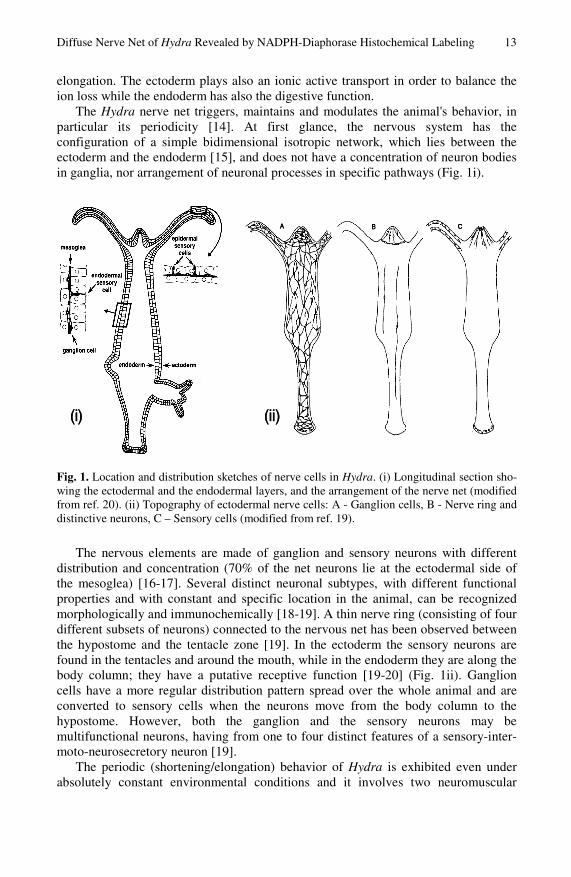
The Hydra nerve net triggers, maintains and modulates the animal's behavior, in particular its periodicity [14]. At first glance, the nervous system has the configuration of a simple bidimensional isotropic network, which lies between the ectoderm and the endoderm [15], and does not have a concentration of neuron bodies in ganglia, nor arrangement of neuronal processes in specific pathways (Fig. 1i).
(ii)(i) (ii)(i)
Fig. 1. Location and distribution sketches of nerve cells in Hydra. (i) Longitudinal section sho-wing the ectodermal and the endodermal layers, and the arrangement of the nerve net (modified from ref. 20). (ii) Topography of ectodermal nerve cells: A - Ganglion cells, B - Nerve ring and distinctive neurons, C – Sensory cells (modified from ref. 19).
The nervous elements are made of ganglion and sensory neurons with different distribution and concentration (70% of the net neurons lie at the ectodermal side of the mesoglea) [16-17]. Several distinct neuronal subtypes, with different functional properties and with constant and specific location in the animal, can be recognized morphologically and immunochemically [18-19]. A thin nerve ring (consisting of four different subsets of neurons) connected to the nervous net has been observed between the hypostome and the tentacle zone [19]. In the ectoderm the sensory neurons are found in the tentacles and around the mouth, while in the endoderm they are along the body column; they have a putative receptive function [19-20] (Fig. 1ii). Ganglion cells have a more regular distribution pattern spread over the whole animal and are converted to sensory cells when the neurons move from the body column to the hypostome. However, both the ganglion and the sensory neurons may be multifunctional neurons, having from one to four distinct features of a sensory-inter-moto-neurosecretory neuron [19].
The periodic (shortening/elongation) behavior of Hydra is exhibited even under absolutely constant environmental conditions and it involves two neuromuscular
14 L. Cristino et al.
pacemaker groups that are in a mutual inhibitory interaction [21]. The behavior is completely absent in nerve-free preparations where neurons are experimentally lost.
Hydra responds to light by body contractions and tactic movements [22], showing extraocular photoreception [23-24] since it does not have conventional visual structures; single or clustered photosensitive cells have not yet been identified as well [25]. Our previous studies proved a photo-modulation of the bioelectric correlates of the animal’s behavior [25-26]. Hydra’s behavioral action spectrum has been elucidated, indicating red blindness [22, 25] and two opposite peaks of two opposite responses around 450 nm and 550 nm (corresponding to the max and min duration of the behavioral sequence in undisturbed conditions) [25]. By polyclonal antibodies against squid rhodopsin, we identified an opsin-like protein likely localized in epidermal sensory nerve cells [27], though a possible location in ecto/endodermal epithelial cells or ectodermal ganglion cells cannot be excluded. Molecular insights into Hydra opsin(s) and their possible light-inducible and clock-controlled expressions are ongoing [28].
3 Nitric Oxide: A Ubiquitous Ancient Signaling Molecule
NO is a short-lived molecule with a high diffusion coefficient that crosses cell membranes readily, thus spreading quickly around its site of generation [29]. It is generated by the enzyme NO synthase (NOS). Two major forms of NOS are known: the constitutive (cNOS) and the inducible ones (iNOS). cNOS accounts for the isoforms found in neuronal and endothelial cells (nNOS and eNOS) while iNOS has its localization in macrophages (mNOS). All isoforms of NOS use arginine as substrate, form citrulline and NO and require nicotinamide adenine dinucleotide phosphate (NADPH) as electron donor. The primary structure of brain NOS [30] reveled binding consensus sequences for calmodulin (CaM), FMN, FAD and NADPH displaying a close homology only with cytochrome P-450 reductase (CPR). NOS and CPR are unique in possessing all these binding sites in the same polypeptide to constitute an electron transport chain. The activity of cNOS is regulated by Ca2+/CaM signal. The CaM-complex bent to the enzyme aligns the domini allowing the electrons flux from the reductase domain to the oxygenase one. The iNOS is always active due to its permanent link to the Ca2+/CaM complex, having for it a high affinity. Inside its target cells, NO can activate soluble guanylate cyclase (sGC) and thereby increase the level of the second messenger guanosine 3’,5’-cyclic monophosphate (cGMP) [30].
Due to the low homology between known invertebrate and mammalian NOS isoforms, the use of mammalian anti-NOS antisera in invertebrate preparations can provide contradictory results [31]. On the contrary, NADPH-d activity has been widely used as a marker for nNOS activity in vertebrate and invertebrate preparations: this technique remains one of the most suitable procedures to screen for NOS-containing cells. The use of NADPH-d histochemical technique (that catalyzes the NADPH-dependent conversion of a soluble salt in an insoluble visible one) as a marker for nNOS in paraformaldehyde fixed tissues is definitely accepted [32] and is producing a large data collection indicating various signaling function for NO in central nervous and peripheral tissues of the major bilaterian invertebrate groups [33].
Diffuse Nerve Net of Hydra Revealed by NADPH-Diaphorase Histochemical Labeling 15
Nitric oxide (NO) is thought to play essential roles in signaling from the early stages of the evolution of life [33]. It has been largely identified in mammals in which it supports important functions as neurotransmitter and neuromodulator [34], as well as in major invertebrate phyla such as mollusks, annelids, arthropods and cnidarians [7, 12, 33], and in basal eukaryotes such as fungi and plants [35].
Various functions for NO-cGMP pathway have been proved in learning and memory [36], feeding [11, 37], olfaction [38], and regulation of smooth muscle tone [39]. Unexpectedly, NO supplies both sensory and motor modalities for the same behavior but exerted with different strategies. In gastropod feeding, NADPH-d labeling was reported in peripheral putative sensory cells in the herbivorous Aplysia and in central motoneurons in the predatory Pleurobranchaea californica [40].
Among sensory systems [10], NO plays a crucial control at every level of vision processing [41]. In the vertebrate retina NO/cGMP signaling modulates ion channel functions of photoreceptors [42]. In lower vertebrates NO provides signals in the earliest stages of retinal development [43]. In invertebrates, NADPH staining of the visual structures of insects indicates that NO is generally implicated in visual development and processing [10, 44] while it is a necessary key-player in visual learning tasks in Octopus [36]. Recently, it was reported that the NOS/cGMP pathway mediates the entrainment of light responses of the circadian clock in mammals [45].
4 Materials and Methods
About fifty specimens of Hydra vulgaris were used for all experiments. Stock cultures were maintained in a medium containing CaCl2 1 x 10⁻³ M and EDTA 1.25 x 10⁻⁵ M at constant 17±1 °C under a 600 lx 12:12 h light/dark cycle. Animals were fed twice a week with Artemia salina nauplii and washed 4 h after the meal. Experimental animals were starved three weeks before the fixative procedure. To move the specimens during each step, before of xylene, a plastic cell strainer was used (BD Falcon). Whole animals were relaxed in a medium solution of 2% ethyluretane (Carlo Erba Reagents) for two minutes and fixed in 4% paraformaldehyde in 0.1 M phosphate buffer (Sigma), pH 7.4 for two hours at 4°C. After, were rinsed thoroughly in phosphate buffer at pH 7.4 (PB) for one hour at room temperature and the last five min. in 0.3% Triton-X 100 in PB. Subsequently, were incubated at 37°C for one hour in the dark in a solution of 1 mg/ml ß-NADPH (Sigma), 0.4 mg/ml (2-2'-benzothiazolyl)-5-styryl-3-(4'-phthalhydrazidyl)-tetrazolium chloride (BSPT) (Sigma) as substrate in 200 μl of N,N-dimethylformamide (ICN Biomedicals), 0.3% Triton-X100 in PB. After incubation, the (whole) animals were rinsed consecutively in PB, in H2O, each for half hour at room temperature, dehydrated in ascending alcohol and finally transferred in xilene for half hour and collected on a coverglass. When the excess of xylene was evaporated the specimens were covered with dense DPX (Aldrich). For the observation at the microscope, the specimens protruding the coverglass were inserted in the hole (ø 12 mm) of a plastic slide, in order to protect them by damage and to leave the free surface of the coverglass to the observation. The slides were observed with a Zeiss Axioskop 2 brightfield microscope and the images were photographed by using a digital camera (Leica DFC 320). Contrast and brightness were adjusted using Adobe PhotoShop 7.0 software (Adobe System).
16 L. Cristino et al.
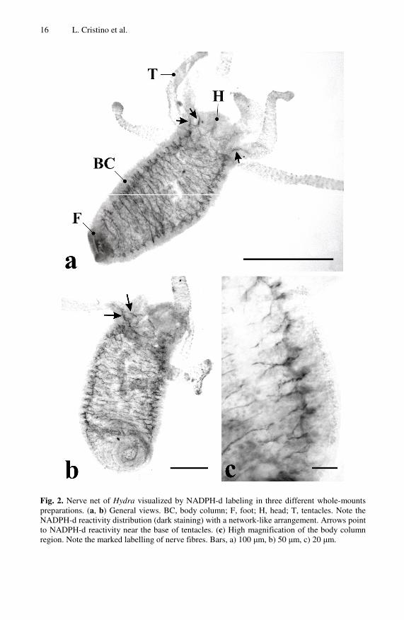
Fig. 2. Nerve net of Hydra visualized by NADPH-d labeling in three different whole-mounts preparations. (a, b) General views. BC, body column; F, foot; H, head; T, tentacles. Note the NADPH-d reactivity distribution (dark staining) with a network-like arrangement. Arrows point to NADPH-d reactivity near the base of tentacles. (c) High magnification of the body column region. Note the marked labelling of nerve fibres. Bars, a) 100 μm, b) 50 μm, c) 20 μm.
Diffuse Nerve Net of Hydra Revealed by NADPH-Diaphorase Histochemical Labeling 17
5 Results and Discussion
As stated before, the enzyme NOS can be localized by NADPH-d histochemistry. Here we have applied this technique to whole-mount preparations of Hydra and revealed a possible NO distribution within the nerve net. The salt substrate BSPT employed in this study enabled to reveal a more precise localization of the diaphorase reaction, because yields a formazan precipitate with osmiophilic and solvent resistant properties which are different from the classic nitroblue tetrazolium salt (NBT) [46]. This substrate is frequently used in the histological procedures for ultrastructural studies of the nervous system [47]. We used hydra whole-mounts because histological sections were less suitable for our purpose due to the thin animal’s thickness.
Widespread distribution of NADPH-d is associated with diffuse nerve fibers but not with identified cell body groups (Fig. 2). Consistent staining patterns are regularly diffused along the body column (Fig 2c) while no marked zones are showed in the tentacles with few exceptions represented at the base of tentacles closer to the head (Fig 2a, b). Similarly, no remarkable labeling was observed in the head (nerve ring neurons included) and foot (peduncle and pedal disk included) regions.
In a first instance, the identification of a sharp cellular localization to NADPH-d labeling becomes hazy. This kind of uncertainty is already known for whole-mount preparations of Hydra. In fact, Arg-vasopressin-like immunoreactive peptides have been identified in nerve cell subsets throughout the Hydra body but with no clear identification of the cell and tissue layer types [48].
Therefore, the attribution of the reactivity pattern to the ectoderm or the endoderm is rather difficult. Our results could fit the distribution pattern and the fiber arrangement of the ectodermal ganglion cells showed in Fig 1ii, although in our experiments no striking labeling is present along the tentacles. According to the latter, it is difficult to report a localization of ectodermal sensory cells for NOS activity because that type of nerve cells is mostly present in tentacles. On the contrary, due to the higher percentage of sensory cells in the endoderm of the body column, the localization of NO activity in such cells cannot be excluded at all. Notably, the distribution pattern found by us has some similarities with the spatial pattern of the CC04+ neurons reported as ectoderm ganglion cells in H. viridissima [49].
Nevertheless, our data prove that the distribution of NADPH-d activity in Hydra, being restricted to the nerve net only, is certainly indicative of the NO presence at neural level.
Accordingly, the identification of NO-sensitive neural elements is needed to delineate the function of NO in Hydra nerve net and its possible involvement in signal processing of motor and/or sensory information. So far, in Cnidaria NO has the following multifunctional roles in effector systems [7]: 1) control of the tentacles’ movements in the GSH feeding response of Hydra [11]; 2) activation of the slow swimming in Aglantha [50]; 3) modulation of the peristaltic muscle contractions in Renilla [51]; and 4) triggering of the nematocyst discharge in Aiptasia [13]. Only items 2 and 3 reported histochemical evidence for NO.
A role of NO in sensory signals should be deepened also in Hydra. The already proposed olfactive task [11] would need further evidences and, presently, cannot be entirely confirmed by our data as tentacles are barely stained. Moreover, an involvement in photosensitive issues seems fascinating. We noted that the distribution
18 L. Cristino et al.
of NADPH-d showed here resembles to some extent the fluorescence pattern of the opsin-like protein identified by us [27]. As part of our research on Hydra phototransductive processes, searching for putative second messengers of the signal cascade we have obtained by immunohistochemistry early results on the presence of cGMP (unpublished data). By comparison with data showed here, it could be interesting verify if putative photosensitive cells, being likely multifunctional, use cGMP as effector belonging - or not - to the cGMP/NO signal transduction pathway.
A role of NO in the developmental dynamics of neurons and in the mechanisms controlling nerve net formation could be hypothesized too. As known in Hydra, neurons are continuously renewed and differentiated from interstitial undifferentiated cells [19]. As a result of a steady state of production/loss, neurons are continuously displaced changing their location (with the exception of nerve ring neurons) [20]. The displacement provides metastable cell phenotypes as many neurons switch their typology, undergoing morphological and immunochemical transformations [19-20]. So NO as small highly flowing molecule could intervene to modulate the complex signaling belonging to those dynamics. This role occurs in lower vertebrates where nNOS expression is indicative of neuroprotective potential effects after neuronal damage [52] and during development of the nervous system [53].
Finally, our data set in a primitive nervous system (likely the oldest one) the histochemical presence of NO acting as ancestral neural messenger with yet unknown physiological tasks. Next investigations and experimental approaches will be hopefully addressed to reveal and clarify the role of that intriguing signaling system.
Acknowledgements
We are grateful to Antonio Cotugno for his skilful work with hydra cultures.
References
1. Katz, P.S.: Evolution and development of neural circuits in invertebrates. Curr. Opin. Neurobiol. 17, 59–64 (2007)
2. Lentz, T.L.: The cell biology of hydra. John Wiley, New York (1966) 3. Steele, R.E.: Developmental signaling in Hydra. Dev. Biol. 248, 199–219 (2002) 4. Mackie, G.O.: The elementary nervous system revisited. Amer. Zool. 30, 907–920 (1990) 5. Grimmelikhuijzen, C., et al.: Neuropeptides in cnidarians. Can. J. Zool. 80, 1690–1702
(2002) 6. Hansen, G.N., Williamson, M., Grimmelikhuijzen, C.J.: A new case of neuropeptide
coexpression (RGamide and LWamides) in Hydra, found by whole-mount, two-color double-labeling in situ hybridization. Cell Tissue Res. 308, 157–165 (2002)
7. Kass-Simon, G., Pierobon, P.: Cnidarian chemical neurotransmission, an updated overview. Comp. Biochem. Physiol. A 146, 9–25 (2007)
8. Moncada, S., Palmer, R.M., Higgs, E.A.: Nitric oxide: physiology, pathophysiology, and pharmacology. Pharmacol. Rev. 43, 109–142 (1991)
9. Palumbo, A.: Nitric oxide in marine invertebrates: a comparative perspective. Comp. Biochem. Physiol. A 142, 241–248 (2005)
10. Ott, S., Burrows, M., Elphick, M.E.: The neuroanatomy of nitric oxide-cGMP signaling in the locust: functional implications for sensory systems. Amer. Zool. 41, 321–331 (2001)
Diffuse Nerve Net of Hydra Revealed by NADPH-Diaphorase Histochemical Labeling 19
11. Colasanti, M., Venturini, G., Merante, A., et al.: Nitric oxide involvement in Hydra vulgaris very primitive olfactorylike system. J. Neurosci. 17, 493–499 (1997)
12. Elofsson, R., Carlberg, M., Moroz, L., et al.: Is nitric oxide (NO) produced by invertebrate neurones? Neuroreport 4, 279–282 (1993)
13. Salleo, A., Giovanni, M., Barra, P., Calabrese, L.: The discharge mechanism of acomitial nematocytes involves the release of nitric oxide. J. Exp. Biol. 199, 1261–1267 (1996)
14. Taddei-Ferretti, C., Musio, C.: The neural net of Hydra and the modulation of its periodic activity. In: Mira, J.M. (ed.) IWANN 1999. LNCS, vol. 1606, pp. 123–137. Springer, Heidelberg (1999)
15. Burnett, A.L., Diehl, N.A.: The nervous system of Hydra. I. Types, distribution and origin of nerve elements. J. Exp. Zool. 157, 217–226 (1964)
16. Tardent, P., Weber, C.: A qualitative and quantitative inventory of nervous cells in Hydra. In: Mackie, G. (ed.) Coelenterate ecology & behaviour, pp. 501–512. Plenum, New York (1976)
17. Epp, L., Tardent, P.: The distribution of nerve cells in Hydra attenuata Pall. Wilhelm Roux’s Arch. 185, 185–193 (1978)
18. Grimmelikhuijzen, C.J.P., Westfall, J.A.: The nervous systems of Cnidarians. In: Breidbach, O., Kutsch, W. (eds.) The nervous systems of invertebrates, pp. 7–24. Birkhäuser, Basel (1995)
19. Koizumi, O.: Developmental neurobiology of hydra. Can. J. Zool. 80, 1678–1689 (2002) 20. Bode, H.R.: Continuous conversion of neuron phenotype in hydra. Trends Genetics 8,
279–284 (1992) 21. Passano, L.M., McCullough, C.B.: Pacemaker hierarchies controlling the behaviour of
hydras. Nature 199, 1174–1175 (1963) 22. Passano, L.M., McCullough, C.B.: The light response and the rhythmic potentials in
Hydra. Proc. Natl. Acad. Sci. USA 48, 1376–1382 (1962) 23. Musio, C.: Extraocular photosensitivity in invertebrates. In: Taddei-Ferretti, C. (ed.)
Biophysics of Photoreception, pp. 245–262. World Scientific, Singapore (1997) 24. Martin, V.J.: Photoreceptors of cnidarians. Can. J. Zool. 80, 1703–1722 (2002) 25. Taddei-Ferretti, C., Musio, C.: Photobehaviour of Hydra and correlated mechanisms: a
case of extraocular photosensitivity. J. Photochem. Photobiol. B: Biol. 55, 88–101 (2000) 26. Taddei-Ferretti, C., Musio, C., Santillo, S., Cotugno, A.: The photobiology of Hydra’s
periodic activity. Hydrobiologia 530/531, 129–134 (2004) 27. Musio, C., Santillo, S., Taddei-Ferretti, C., Robles, L.J., et al.: First identification and
localization of a visual pigment in Hydra. J. Comp. Physiol. 187A, 79–81 (2001) 28. Santillo, S., Orlando, P., De Petrocellis, L., Cristino, L., Guglielmotti, V., Musio, C.:
Evolving visual pigments: Hints from the opsin-based proteins in a phylogenetically old eyeless invertebrate. BioSystems 86, 3–17 (2006)
29. Palmer, R.M., Ferrige, A.G., Moncada, S.: Nitric oxide release accounts for the biological activity of endothelium-derived relaxing factor. Nature 327, 524–526 (1987)
30. Bredt, D.S., Snyder, S.H.: Nitric oxide, a novel neuronal messenger. Neuron 8, 3–11 (1992)
31. Moroz, L.L., Winlow, W., Turner, R.W., et al.: Nitric oxide synthase-immunoreactive cells in the CNS and periphery of Lymnaea. Neuroreport 5, 1277–1280 (1994)
32. Hope, B.T., Michael, G.J., Knigge, K.M., Vincent, S.R.: Neuronal NADPH-diaphorase is a nitric oxide synthase. Proc. Natl Acad. Sci. USA 88, 2811–2814 (1991)
33. Moroz, L.L.: Gaseous transmission across time and species. Am. Zool. 41, 304–320 (2001) 34. Garthwaite, J., Boulton, C.L.: Nitric oxide signaling in the central nervous system. Annu.
Rev. Physiol. 57, 683–706 (1995)
20 L. Cristino et al.
35. Ninnemann, H., Maier, J.: Indications for the occurrence of nitric oxide synthases in fungi and plants and the involvement in photoconidiation of Neurospora crassa. Photochem. Photobiol. 64, 393–398 (1996)
36. Robertson, J.D., Bonaventura, J., Kohm, A., Hiscat, M.: Nitric oxide is necessary for visual learning in Octopus vulgaris. Proc. R. Soc. Lond., B Biol. Sci. 263, 1739–1743 (1996)
37. Elphick, M.R., Kemenes, G., Staras, K., O’Shea, M.: Behavioural role for nitric oxide in chemosensory activation of feeding in a mollusc. J. Neurosci. 15, 7653–7664 (1995)
38. Gelperin, A.: Nitric oxide mediates network oscillations of olfactory interneurons in a terrestrial mollusc. Nature 369, 61–63 (1994)
39. Bult, H., Boeckxstaens, G.E., Pelckmans, P.A., et al.: Nitric oxide as an inhibitory non-adrenergic non-cholinergic neurotransmitter. Nature 345, 346–347 (1990)
40. Moroz, L.L., Gillette, R.: NADPH-d localization in the CNS and peripheral tissues of the predatory sea-slug Pleurobranchaea californica. J. Comp. Neurol. 367, 607–622 (1996)
41. Cudeiro, J., Rivadulla, C.: Sight and insight - on the physiological role of nitric oxide in the visual system. Trends Neurosci. 22, 109–116 (1999)
42. Kurenny, D.E., Moroz, L.L., Turner, R.W., et al.: Modulation of ion channels in rod photoreceptors by nitric oxide. Neuron 13, 315–324 (1994)
43. Florenzano, F., Guglielmotti, V.: Selective NADPH-diaphorase histochemical labeling of Müller radial processes and photoreceptors in the earliest stages of retinal development in the tadpole. Neurosci. Lett. 292, 187–190 (2000)
44. Gibbs, S.M.: Regulation of Drosophila visual system development by nitric oxide and cGMP. Amer. Zool. 41, 268–281 (2001)
45. Golombek, D.A., Agostino, P.V., Plano, S.A., Ferreyra, G.A.: Signaling in the mammalian circadian clock: the NO/cGMP pathway. Neurochem. Internat. 45, 929–936 (2004)
46. Kalina, M., Plapinger, R.E., Hoshino, Y., Seligman, A.M.: Nonosmiophilic tetrazolium salts that yield osmiophilic, lipophobic formazans for ultrastructural localization of dehydrogenase activity. J. Histochem. Cytochem. 20, 685–695 (1972)
47. Altman, F.P.: Tetrazolium salts and formazans. Prog. Histochem. Cytochem. 9, 1–56 (1976)
48. Morishita, F., Nitagai, Y., Furukawa, Y., Matsushima, O., Takahashi, T., et al.: Identification of a vasopressin-like immunoreactive substance in hydra. Peptides 24, 17–26 (2003)
49. Sakaguchi, M., Mizusina, A., Kobayakawa, Y.: Structure, development, and maintenance of the nerve net of the body column in Hydra. J. Comp. Neurol. 373, 41–54 (1996)
50. Moroz, L.L., Meech, R.W., Sweedler, J.V., Mackie, G.O.: Nitric oxide regulates swimming in the jellyfish Aglantha digitale. J. Comp. Neurol. 471, 26–36 (2004)
51. Anctil, M., Poulain, I., Pelletier, C.: NO modulates peristaltic muscle activity associated with fluid circulation in the sea pansy Renilla koellikeri. J. Exp. Biol. 208, 2005–2017 (2005)
52. Cristino, L., Pica, A., Della Corte, F., Bentivoglio, M.: Plastic changes and nitric oxide synthase induction in neurons which innervate the regenerated tail of the lizard Gekko gecko: I. The response of spinal motoneurons to tail amputation and regeneration. J. Comp. Neurol. 417, 60–72 (2000)
53. Cristino, L., Florenzano, F., Bentivoglio, M., Guglielmotti, V.: Nitric oxide synthase expression and cell changes in dorsal root ganglia and spinal dorsal horn of developing and adult Rana esculenta indicate a role of nitric oxide in limb metamorphosis. J. Comp. Neurol. 472, 423–436 (2004)
Copyright © 2022 FDOKUMEN