Distribution of NADPH-diaphorase in the superior colliculus of Cebus monkeys, and co-localization...
9
Distribution of NADPH-diaphorase in the superior colliculus of Cebus monkeys, and co-localization with calcium-binding proteins J.G.M. Soares, R. Mendez-Otero, Ricardo Gattass * Programa de Neurobiologia, Instituto de Biofı ´sica Carlos Chagas Filho, Universidade Federal do Rio de Janeiro, Bloco G, CCS, Ilha do Funda ˜o, Rio de Janeiro, RJ 21949-900, Brazil Received 4 October 2002; accepted 7 April 2003 Abstract We examined the distribution of the enzyme dihydronicotinamide adenine dinucleotide phosphate-diaphorase (NADPH-d) in the superior colliculus (SC) of the New World monkey Cebus apella , and the co-localization of this enzyme with the calcium-binding proteins (CaBPs) calbindin-D28K, parvalbumin and calretinin. Despite the intensely labeled neuropil, rare NADPH-d-positive cells were observed in the stratum griseum superficiale (SGS). Most of the labeled cells in the SC were found in the intermediate layers, with a great number also in the deeper layers. This pattern is very similar to that described in the opossum (Didelphis marsupialis ) and in the cat, and different from the pattern found in the rat, which shows labeled cells mainly in the SGS. Cells doubly stained for NADPH-d and CaBPs were observed throughout the SC, although in a small number. Of the NADPH-d-positive cells, 20.3% were doubly labeled for NADPH-d and parvalbumin, 10.2% revealed co-localization with calretinin, and 5.6% with calbindin. The low number of double-stained cells for NADPH-d and the CaBPs indicates that these molecules must participate in different functional circuits within the SC. # 2003 Elsevier Science Ireland Ltd and the Japan Neuroscience Society. All rights reserved. Keywords: Superior colliculus; NADPH-diaphorase; NO synthase; Calcium-binding proteins; Calbindin; Calretinin; Parvalbumin; Primates 1. Introduction NADPH-diaphorase (NADPH-d) histochemistry stains selective cell populations, by the NADPH-depen- dent reduction of tetrazolium salts to visible formazans, producing a Golgi-like image of cells (Thomas and Pearse, 1961). Various studies have shown the localiza- tion of diaphorase within neurons, which also contain various neuroactive substances, including somatostatin, neuropeptide Y, substance P and acetylcholine, among others. This co-localization is highly selective to specific population of cells in different locations in the central nervous system (CNS) (Vincent et al., 1983a,b; Sandell, 1985). Other studies have demonstrated that in the nervous system NADPH-d is a nitric oxide (NO) synthase responsible for the formation of NO from arginine. NO is a neuronal messenger molecule pro- duced in response to an increase in intracellular calcium ions that interact with guanylate cyclase and increase levels of cyclic guanosine monophosphate (cGMP) in target cells (Knowles et al., 1989; Hope et al., 1991; Dawson et al., 1991). Previous works have mapped this neural messenger system in different regions of the brain of vertebrate species (Sandell, 1986; Volchan and Franca, 1994; Satoh et al., 1995; Costa et al., 1996; Franca et al., 1997; Vargas et al., 1998; Soares et al., 2001a). Some studies have demonstrated that the distribution pattern of NADPH-d-positive neurons in the superior colliculus (SC) varies among species. For example, in rats NADPH-d-positive neurons were observed mainly in the superficial layers, while in the deep layers, a few multipolar NADPH-d-positive cells occurred in groups continuous with dense aggregations of cells in the lateral periaqueductal gray matter (Leigh et al., 1990; Vincent and Kimura, 1992; Teno ´rio et al., 1995). In cats few labeled cells were found within the stratum zonale (SZ). Cellular labeling was sparse within the stratum griseum * Corresponding author. Tel.: /55-21-2562-6561; fax: /55-21- 2280-8193. E-mail address: [email protected] (R. Gattass). Neuroscience Research 46 (2003) 475 /483 www.elsevier.com/locate/neures 0168-0102/03/$ - see front matter # 2003 Elsevier Science Ireland Ltd and the Japan Neuroscience Society. All rights reserved. doi:10.1016/S0168-0102(03)00125-1
-
Upload
independent -
Category
Documents
-
view
3 -
download
0
Transcript of Distribution of NADPH-diaphorase in the superior colliculus of Cebus monkeys, and co-localization...

Distribution of NADPH-diaphorase in the superior colliculus ofCebus monkeys, and co-localization with calcium-binding proteins
J.G.M. Soares, R. Mendez-Otero, Ricardo Gattass *
Programa de Neurobiologia, Instituto de Biofısica Carlos Chagas Filho, Universidade Federal do Rio de Janeiro, Bloco G, CCS, Ilha do Fundao,
Rio de Janeiro, RJ 21949-900, Brazil
Received 4 October 2002; accepted 7 April 2003
Neuroscience Research 46 (2003) 475�/483
www.elsevier.com/locate/neures
Abstract
We examined the distribution of the enzyme dihydronicotinamide adenine dinucleotide phosphate-diaphorase (NADPH-d) in the
superior colliculus (SC) of the New World monkey Cebus apella , and the co-localization of this enzyme with the calcium-binding
proteins (CaBPs) calbindin-D28K, parvalbumin and calretinin. Despite the intensely labeled neuropil, rare NADPH-d-positive cells
were observed in the stratum griseum superficiale (SGS). Most of the labeled cells in the SC were found in the intermediate layers,
with a great number also in the deeper layers. This pattern is very similar to that described in the opossum (Didelphis marsupialis )
and in the cat, and different from the pattern found in the rat, which shows labeled cells mainly in the SGS. Cells doubly stained for
NADPH-d and CaBPs were observed throughout the SC, although in a small number. Of the NADPH-d-positive cells, 20.3% were
doubly labeled for NADPH-d and parvalbumin, 10.2% revealed co-localization with calretinin, and 5.6% with calbindin. The low
number of double-stained cells for NADPH-d and the CaBPs indicates that these molecules must participate in different functional
circuits within the SC.
# 2003 Elsevier Science Ireland Ltd and the Japan Neuroscience Society. All rights reserved.
Keywords: Superior colliculus; NADPH-diaphorase; NO synthase; Calcium-binding proteins; Calbindin; Calretinin; Parvalbumin; Primates
1. Introduction
NADPH-diaphorase (NADPH-d) histochemistry
stains selective cell populations, by the NADPH-depen-
dent reduction of tetrazolium salts to visible formazans,
producing a Golgi-like image of cells (Thomas and
Pearse, 1961). Various studies have shown the localiza-
tion of diaphorase within neurons, which also contain
various neuroactive substances, including somatostatin,
neuropeptide Y, substance P and acetylcholine, among
others. This co-localization is highly selective to specific
population of cells in different locations in the central
nervous system (CNS) (Vincent et al., 1983a,b; Sandell,
1985). Other studies have demonstrated that in the
nervous system NADPH-d is a nitric oxide (NO)
synthase responsible for the formation of NO from
arginine. NO is a neuronal messenger molecule pro-
duced in response to an increase in intracellular calcium
ions that interact with guanylate cyclase and increase
levels of cyclic guanosine monophosphate (cGMP) in
target cells (Knowles et al., 1989; Hope et al., 1991;
Dawson et al., 1991).
Previous works have mapped this neural messenger
system in different regions of the brain of vertebrate
species (Sandell, 1986; Volchan and Franca, 1994; Satoh
et al., 1995; Costa et al., 1996; Franca et al., 1997;
Vargas et al., 1998; Soares et al., 2001a). Some studies
have demonstrated that the distribution pattern of
NADPH-d-positive neurons in the superior colliculus
(SC) varies among species. For example, in rats
NADPH-d-positive neurons were observed mainly in
the superficial layers, while in the deep layers, a few
multipolar NADPH-d-positive cells occurred in groups
continuous with dense aggregations of cells in the lateral
periaqueductal gray matter (Leigh et al., 1990; Vincent
and Kimura, 1992; Tenorio et al., 1995). In cats few
labeled cells were found within the stratum zonale (SZ).
Cellular labeling was sparse within the stratum griseum
* Corresponding author. Tel.: �/55-21-2562-6561; fax: �/55-21-
2280-8193.
E-mail address: [email protected] (R. Gattass).
0168-0102/03/$ - see front matter # 2003 Elsevier Science Ireland Ltd and the Japan Neuroscience Society. All rights reserved.
doi:10.1016/S0168-0102(03)00125-1
superficiale (SGS) and the stratum opticum (SO). By
contrast, fiber and cell labeling was more intense in the
stratum griseum intermediale (SGI) and in the deep
layers (Scheiner et al., 2000). Similarly, in opossums the
number of NADPH-d stained cells was low in the SGS
and SO, but high in the intermediate and deep layers
(Giraldi-Guimaraes et al., 1999). Finally, in humans, the
SC showed a high density of small NOS-positive cells in
the SGS, and medium-sized, lightly staining neurons in
the SGI and stratum griseum profundum (SGP) (Egber-
ongbe et al., 1994). As in the rat and cat, a lattice of
NADPH-d-positive fibers was described in the inter-
mediate gray layers of the SC of the monkey (Wallace,
1988). However, at this moment there is not a detailed
description of the distribution of NADPH-d-positive
neurons throughout the layers of the monkey SC. Here,
we examine the distribution of the enzyme NADPH-
diaphorase in the SC of the New World monkey Cebus
apella .
In a previous study we described the distribution of
the calcium binding proteins (CaBPs) calbindin-D28K,
parvalbumin and calretinin in the SC of the Cebus
monkey (Soares et al., 2001b). These CaBPs are
implicated in the buffering and transport of calcium in
the CNS, where they are present in distinct subpopula-
tions of neurons (Jones and Hendry, 1989; Celio, 1990).
Inasmuch as the activation of the NO synthase is related
to calcium influx (Garthwaite et al., 1988; Bredt and
Snyder, 1989), another goal of this study is to compare
the distribution of the NADPH-d with that of the
CaBPs using histochemical-immunocytochemical dou-
ble-staining technique.
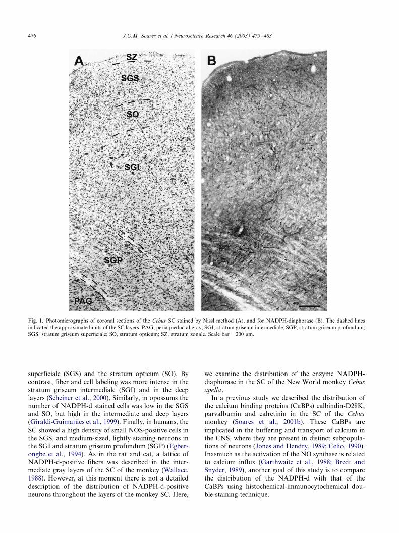
Fig. 1. Photomicrographs of coronal sections of the Cebus SC stained by Nissl method (A), and for NADPH-diaphorase (B). The dashed lines
indicated the approximate limits of the SC layers. PAG, periaqueductal gray; SGI, stratum griseum intermediale; SGP, stratum griseum profundum;
SGS, stratum griseum superficiale; SO, stratum opticum; SZ, stratum zonale. Scale bar�/200 mm.
J.G.M. Soares et al. / Neuroscience Research 46 (2003) 475�/483476

2. Materials and methods
Eight adult C. apella monkeys of both sexes weighing
between 1.2 and 2.6 kg were used in this study. All
experimental protocols were conducted following the
NIH guidelines for animal research and they were
approved by the committee for animal care and use of
the Instituto de Biofısica Carlos Chagas Filho, UFRJ.
The animals were deeply anesthetized with sodium
pentobarbital (30 mg/kg) and perfused with normal
saline followed by 2% paraformaldehyde in phosphate-
buffered saline (PBS); 2% paraformaldehyde in PBS�/
2.5% glycerol; PBS�/5% glycerol; and PBS�/10% gly-
cerol. Serial 40 mm thick sections were obtained using a
cryostat. Adjacent series were stained for cell bodies
with cresyl violet, NADPH-diaphorase histochemistry,
and for immunocytochemistry for calbindin, parvalbu-
min and calretinin. In four animals double staining of
NADPH-d with each one of those three CaBPs were
made in adjacent sections.
For NADPH-d histochemistry, free-floating sections
were washed in PBS and reacted at 37 8C in a solution
containing 1 mM b-NADPH, reduced form, 0.4 mM
nitrobluetetrazolium (NBT), 10% dimethyl-sufoxide
(DMSO) and 0.3% triton X-100 in PBS, pH 7.4, for
1�/2 h. For double labeling, sections reacted for
NADPH-d were washed in PBS and then immunor-
eacted as described below.
For immunocytochemical reactions, sections were
incubated overnight with calbindin-D28K (1:2500),
parvalbumin (1:3000) monoclonal antibody or calretinin
(1:2000) polyclonal antibody (Swant�/Swin Antibodies),
in a solution containing 0.05% of bovine albumin and
0.3% of triton X-100 in 0.01 M PBS, pH 7.4. They were
then incubated for an additional hour in biotinylated
anti-mouse or anti-rabbit secondary antibody, and then
processed by the avidin�/biotin method with ABC kits
(Vector) and diaminobenzidine. The sections were
rinsed in PBS, mounted on gelatin-coated slides, dehy-
drated and coverslipped. Control sections were prepared
by omitting the primary antibody in the incubation
solution. These sections showed no specific staining.
Sections were examined under brightfield microscopy
and photomicrographs were obtained using a Zeiss
Axiocam attached to the microscope and processed
using Adobe Photoshop version 5.0. The percentage of
double-stained cells was calculated relative to the
number of NADPH-d-positive cells counted in one
coronal section at the middle level of the SC, in two
animals where the blue-colored cells of the reaction for
NADPH-d and the brown-colored cells of immunocy-
tochemical reaction for CABPs were clearly distinguish-
able. At least 100 NADPH-d-positive cells were
randomly selected throughout all layers of the SC in
each section and analyzed for co-localization.
3. Results
Fig. 1A shows a coronal section of the Cebus SC
stained by the Nissl method, where approximate limits
of the layers were delineated. The pattern revealed by
the NADPH-d histochemistry in an adjacent section of
the SC is shown in the Fig. 1B. Fig. 2 shows a schematic
representation of the distribution of labeled cells
throughout the layers of the SC at caudal (A) and
rostral (B) levels. We observe a greater density of labeled
cells in caudal than in rostral sections. In addition, there
is a greater concentration of cells in the medial portions
of the deeper layers throughout the rostro-caudal extent.
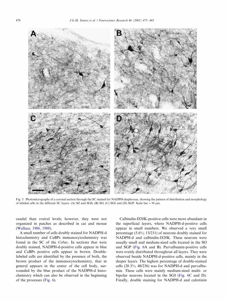
Rare multipolar and bipolar NADPH-d-positive cells
were found in the SGS, which, however, depicts a heavy
stained neuropil (Fig. 3A). These cells were mainly small
(10�/20 mm) in size with large dendrites pointing toward
the SZ (Fig. 4A and B). In the SO we also observe a
small number of small and medium-sized (20�/30 mm)
multipolar, horizontal and vertical bipolar cells (Fig. 3B,
Fig. 4C and D).
Numerous small to medium-sized neurons, of all
morphologies, were found in the SGI (Fig. 3C, Fig. 4E
and F). Finally, the SGP is the region that presents the
greatest density of labeled neurons, mostly medium
multipolar and vertical bipolar cells (Fig. 3D, Fig. 4G
and H). A net of high activity NADPH-d fibers was
found within the intermediate and mainly in the deeper
layers of the SC (Fig. 5). These fibers were denser at
Fig. 2. Schematic representation of the distribution of labeled cells
(dots) for NADPH-diaphorase in two coronal sections, in caudal (A)
and rostral (B) portions of the SC. Scale bar�/500 mm.
J.G.M. Soares et al. / Neuroscience Research 46 (2003) 475�/483 477
caudal than rostral levels; however, they were not
organized in patches as described in cat and mouse
(Wallace, 1986, 1988).
A small number of cells doubly stained for NADPH-d
histochemistry and CaBPs immunocytochemistry was
found in the SC of the Cebus . In sections that were
doubly stained, NADPH-d-positive cells appear in blue
and CaBPs positive cells appear in brown. Double-
labeled cells are identified by the presence of both, the
brown product of the immunocytochemistry, that in
general appears in the center of the cell body, sur-
rounded by the blue product of the NADPH-d histo-
chemistry which can also be observed in the beginning
of the processes (Fig. 6).
Calbindin-D28K-positive cells were more abundant in
the superficial layers, where NADPH-d-positive cells
appear in small numbers. We observed a very small
percentage (5.6%; 13/231) of neurons doubly stained for
NADPH-d and calbindin-D28K. These neurons were
usually small and medium-sized cells located in the SO
and SGP (Fig. 6A and B). Parvalbumin-positive cells
were evenly distributed throughout all layers. They were
observed beside NADPH-d-positive cells, mainly in the
deeper layers. The highest percentage of double-stained
cells (20.3%; 48/236) was for NADPH-d and parvalbu-
min. These cells were mainly medium-sized multi- or
bipolar neurons located in the SGI (Fig. 6C and D).
Finally, double staining for NADPH-d and calretinin
Fig. 3. Photomicrographs of a coronal section through the SC stained for NADPH-diaphorase, showing the pattern of distribution and morphology
of labeled cells in the different SC layers. (A) SZ and SGS; (B) SO; (C) SGI and (D) SGP. Scale bar�/50 mm.
J.G.M. Soares et al. / Neuroscience Research 46 (2003) 475�/483478

was detected in a small percentage of neurons (10.2%;
24/235), mainly bipolar or multipolar in shape,
and located in intermediary and deep layers (Fig. 6E
and F).
4. Discussion
The present study reveals a small number of
NADPH-d-positive cells in the SGS, which shows,
Fig. 4. Higher magnification photomicrographs depicting NADPH-d-positive cell types in various layers of the SC. (A, B) SGS; (C, D) SO; (E, F)
SGI and (G, H) SGP. Scale bar�/30 mm.
J.G.M. Soares et al. / Neuroscience Research 46 (2003) 475�/483 479

however, an intensely labeled neuropil. Most of the
labeled cells were found in the intermediate and deep
layers, with a greater density in the medial portion of the
SC. This pattern is very similar to that described for
both the opossum Didelphis marsupialis (Giraldi-Gui-
maraes et al., 1999) and the cat (Scheiner et al., 2000),
however, differs from the results of the rat: labeled cells
are located mainly in the superficial layers (Gonzales-
Hernandez et al., 1992; Tenorio et al., 1995, 1996).
These results suggest inter-species differences concerning
the connections and functions of cells of the various
layers of the SC.
Wallace (1986, 1988) studied the occurrence of
patches of high NADPH-d activity forming a lattice
within the SGI in various species. Those lattices were
prominent in the mouse and cat, occurred more faintly
in the monkey (Macaca fascicularis ) and were not found
in human. In Cebus monkeys we observed a net of fibers
with high enzyme activity in the SGI and mainly in the
SGP, but they did not form patches as the ones
described in the mouse and cat.
The mammalian SC has a high concentration of NO
synthase (Wallace, 1986, 1988; Vincent and Kimura,
1992), but very little is known about the function of NO
in this structure. The study of Wallace (1986) in the
mouse and rat showed that the interruption of the
afferent pathways from the brainstem abolishes the
lattices of high diaphorase activity in the intermediate
layers of the SC, without altering the enzyme activity in
the superficial layers. The presence of NADPH-d
amorphous reaction product in the superficial layers of
the SC also is not abolished by eye enucleation (Tenorio
et al., 1998), and even increases after optic nerve
transection (Yan et al., 1995). A possible source for
this staining could be projections from cells intrinsic to
the deeper portions of the colliculus that contain high
diaphorase activity.
Various works have demonstrated the chemical di-
versity of NO-synthesizing neurons (Vincent et al.,
1983a,b; Sandell, 1985; Wong-Riley et al., 1998). Co-
localization of NADPH-d and the CaBPs has been
studied in various regions of the brain. The coexistence
of NADPH-d with calbindin-D28K was studied in the
magnocellular secretory nuclei of the rat hypothalamus,
where a selective pattern of distribution and only a
partial coexistence was found, suggesting that they are
involved in specific physiological functions shared by
restricted neuronal cell populations (Alonso et al.,
1992).
In the monkey cerebral cortex, co-localization was
found between NADPH-d and calbindin-D28K in all
nonpyramidal small cells (type II) and in 4% of
nonpyramidal large (type I) NADPH-d neurons, but
not between NADPH-d and parvalbumin (Yan et al.,
1996). The rat striatal NOS-positive neurons lack
parvalbumin and calretinin although some of these cells
contain calbindin-D28K (Kita et al., 1990; Bennett and
Bolam, 1993a,b). In the SC of rats, NOS-positive
neurons do not contain calbindin-D28K, and receive
glutamatergic inputs; moreover, some of these neurons
express GABA (20%) or parvalbumin (15%) (Soares-
Mota et al., 2001). However, Gonzales-Soriano et al.
(2002) demonstrated a lack of colocalization of parval-
bumin and NADPH-d, and the presence of double-
labeled neurons for calbindin-D28K and NADPH-d
(10%) in the SC of rabbits. This co-localization of
NADPH-d and calbindin-D28K or parvalbumin in
some cells could imply that these CaBPs may participate
in the regulation of the synthesis of the NO by changing
the concentration of calcium in the cytoplasm of the cell
(Garthwaite et al., 1988; Bredt and Snyder, 1989).
Fig. 5. Photomicrograph of the deep layers of the SC showing a net of labeled fibers for NADPH-d. Scale bar�/100 mm.
J.G.M. Soares et al. / Neuroscience Research 46 (2003) 475�/483480
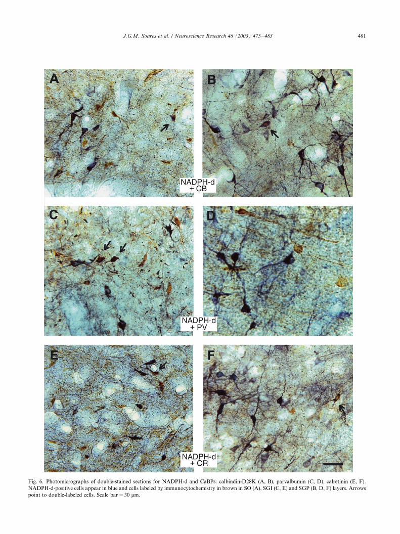
Fig. 6. Photomicrographs of double-stained sections for NADPH-d and CaBPs: calbindin-D28K (A, B), parvalbumin (C, D), calretinin (E, F).
NADPH-d-positive cells appear in blue and cells labeled by immunocytochemistry in brown in SO (A), SGI (C, E) and SGP (B, D, F) layers. Arrows
point to double-labeled cells. Scale bar�/30 mm.
J.G.M. Soares et al. / Neuroscience Research 46 (2003) 475�/483 481

However, most of the NADPH-d-positive neurons may
use other CaBPs, such as calmodulin, or may have a
different mechanism for the control of the calcium
concentration.In this study we have shown that only a small
percentage of NADPH-d-positive neurons in the SC of
Cebus monkeys also express parvalbumin, calbindin-
D28K or calretinin. This small degree of co-localization
of NADPH-d and CaBPs in the SC of Cebus monkeys
suggests that these proteins may be active as indepen-
dent elements in the primate SC.
Acknowledgements
The authors are grateful to Dr A P.B. Souza and DrM.G.P. Rosa for comments on the article, to E.S. da
Silva Filho, L.H. Pontes and M.T. Monteiro for
technical assistance, and to P. Coutinho and G.
Coutinho for animal care. This research was supported
by grants from PRONEX, CNPq, FAPERJ, FUJB.
References
Alonso, J.R., Sanchez, F., Arevalo, R., Carretero, J., Aijon, J.,
Vasquez, R., 1992. CABP D-28k and NADPH-diaphorase coex-
istence in the magnocellular neurosecretory nuclei. NeuroReport 3,
249�/252.
Bennett, B.D., Bolam, J.P., 1993a. Characterization of calretinin-
immunoreactive structures in the striatum of the rat. Brain Res.
609, 137�/148.
Bennett, B.D., Bolam, J.P., 1993b. Two populations of calbindin
D28k-immunoreactive neurons in the striatum of the rat. Brain
Res. 610, 305�/310.
Bredt, D.S., Snyder, S.H., 1989. Nitric oxide mediates glutamate-
linked enhancement of cGMP levels in the cerebellum. Proc. Natl.
Acad. Sci. USA 86, 9030�/9033.
Celio, M.R., 1990. Calbindin D-28k and parvalbumin in the rat
nervous system. Neuroscience 35, 375�/475.
Costa, E.T., Nascimento, J.L.M., Picanco-Diniz, C.W., Quaresma,
J.A.S., Silva-Filho, M., 1996. Histochemical characterization of
NADPH-diaphorase activity in area 17 of diurnal and nocturnal
primates and rodents. Braz. J. Med. Res. 29, 1355�/1362.
Dawson, T.M., Bredt, D.S., Fotuhi, M., Hwang, P.M., Snyder, S.H.,
1991. Nitric oxide synthase and neuronal NADPH diaphorase are
identical in brain and peripheral tissues. Proc. Natl. Acad. Sci.
USA 88, 7797�/7801.
Egberongbe, Y.I., Gentleman, S.M., Falkai, P., Bogerts, B., Polak,
J.M., Roberts, G.W., 1994. The distribution of nitric oxide
synthase immunoreactivity in the human brain. Neuroscience 59,
561�/578.
Franca, J.G., do-Nascimento, J.L., Picanco-Diniz, C.W., Quaresma,
J.A., Silva, A.L., 1997. NADPH-diaphorase activity in area 17 of
the squirrel monkey visual cortex: neuropil pattern, cell morphol-
ogy and laminar distribution. Braz. J. Med. Biol. Res. 30, 1093�/
1105.
Garthwaite, J., Charles, S.L., Chess-Williams, R., 1988. Endothelium-
derived relaxing factor release on activation of NMDA receptors
suggests role as intercellular messenger in the brain. Nature 336,
385�/388.
Giraldi-Guimaraes, A., Tenorio, F., Bruning, G., Mayer, B., Mendez-
Otero, R., Cavalcante, L.A., 1999. Nitric oxide synthase expression
in the opossum superior colliculus: a histochemical, immunohisto-
chemical and biochemical study. Brain Behav. Evol. 54, 303�/313.
Gonzales-Hernandez, T., Conde-Sendin, M., Meyer, G., 1992. Lami-
nar distribution and morphology of NADPH-diaphorase contain-
ing neurons in the superior colliculus and underlying
periaqueductal gray of the rat. Anat. Embryol. 186, 245�/250
(Berlin).
Gonzales-Soriano, J., Contreras-Rodrigues, J., Martinez-Sainz, P.,
Martin-Palacios, S., Marin-Garcia, P., Rodriguez-Veiga, E., 2002.
NADPH- diaphorase distribution in the rabbit superior colliculus
and co-localization with calcium-binding proteins. J. Anat. 200,
297�/308.
Hope, B.T., Michael, G.J., Knigge, K.M., Vincent, S.R., 1991.
Neuronal NADPH diaphorase is a nitric oxide synthase. Proc.
Natl. Acad. Sci. USA 88, 2811�/2814.
Jones, E.G., Hendry, S.H.C., 1989. Differential calcium binding
protein immunoreactivity distinguishes classes of relay neurons in
monkey thalamic nuclei. Eur. J. Neurosci. 1, 222�/246.
Kita, H., Kosaka, T., Heizmann, C.W., 1990. Parvalbumin-immunor-
eactive neurons in the rat neostriatum: a light and electron
microscopic study. Brain Res. 536, 1�/15.
Knowles, R.G., Palacios, M., Palmer, R.M.J., Moncada, S., 1989.
Formation of nitric oxide from L-arginine in the central nervous
system: a transduction mechanism for stimulation of the soluble
guanylate cyclase. Proc. Natl. Acad. Sci. USA 86, 5159�/5162.
Leigh, P.N., Connick, J.H., Stone, T.W., 1990. Distribution of
NADPH diaphorase positive cells in the rat brain. Comp. Biochem.
Physiol. 97C, 259�/264.
Sandell, J.H., 1985. NADPH diaphorase cells in the mammalian inner
retina. J. Comp. Neurol. 238, 466�/472.
Sandell, J.H., 1986. NADPH diaphorase histochemistry in the
macaque striate cortex. J. Comp. Neurol. 251, 388�/397.
Satoh, K., Aral, R., Ikemoto, K., Narita, M., Nagai, T., Ohshima, H.,
Kitahama, K., 1995. Distribution of nitric oxide synthase in the
central nervous system of Macaca fuscata : subcortical regions.
Neuroscience 66, 685�/696.
Scheiner, C., Arceneaux, R., Guido, W., Kratz, K., Mize, R., 2000.
Nitric oxide synthase distribution in cat superior colliculus and co-
localization with choline acetyltransferase. J. Chem. Neuroanat.
18, 147�/159.
Soares, J.G.M., Gattass, R., Souza, A.P.B., Rosa, M.G.P., Fiorani,
M., Jr, Brandao, B.L., 2001a. Connectional and neurochemical
subdivisions of the pulvinar in Cebus monkeys. Visual Neurosci.
18, 25�/41.
Soares, J.G.M., Botelho, E.P., Gattass, R., 2001b. Distribution of
calbindin, parvalbumin and calretinin in the superior colliculus and
lateral geniculate nucleus of Cebus apella monkeys. J. Chem.
Neuroanat. 22, 139�/146.
Soares-Mota, M., Henze, I., Mendez-Otero, R., 2001. Nitric oxide
synthase-positive neurons in the rat superior colliculus: colocaliza-
tion of NOS with NMDAR1 glutamate receptor, GABA, and
parvalbumin. J. Neurosci. Res. 64, 501�/507.
Tenorio, F., Giraldi-Guimaraes, A., Mendez-Otero, R., 1995. Devel-
opmental changes of nitric oxide synthase in the rat superior
colliculus. J. Neurosci. Res. 42, 633�/637.
Tenorio, F., Giraldi-Guimaraes, A., Mendez-Otero, R., 1996. Mor-
phology of NADPH-diaphorase-positive cells in the retinoceptive
layers of the developing rat superior colliculus. Int. J. Dev.
Neurosci. 14, 1�/10.
Tenorio, F., Giraldi-Guimaraes, A., Santos, H.R., Cintra, W.M.,
Mendez-Otero, R., 1998. Eye enucleation alters intracellular
distribution of NO synthase in the superior colliculus. NeuroRe-
port 9, 145�/148.
Thomas, E., Pearse, A.G.E., 1961. The fine localization of dehydro-
genases in the nervous system. Histochemical 2, 266�/282.
J.G.M. Soares et al. / Neuroscience Research 46 (2003) 475�/483482
Vargas, C.D., Sousa, A.O., Bittencourt, F.L., Santos, C.M., Pereira,
A., Jr, Bernardes, R.F., Rocha-Miranda, C., Volchan, E., 1998.
Cytochrome oxidase and NADPH-diaphorase on the afferent relay
branch of the optokinetic reflex in the opossum. J. Comp. Neurol.
398, 206�/224.
Vincent, S.R., Kimura, H., 1992. Histochemical mapping of nitric
oxide synthase in the rat brain. Neuroscience 46, 755�/784.
Vincent, S.R., Johanson, O., Hokfelt, T., Skirboll, L., Elde, R.P.,
Terenius, L., Kimmel, J., Goldstein, M., 1983a. NADPH-diaphor-
ase: a selective histochemical marker for striatal neurons containing
both somatostatin- and avian pancreatic polypeptide (APP)-like
immunoreactivities. J. Comp. Neurol. 217, 252�/263.
Vincent, S.R., Satoh, K., Armstrong, D.M., Fibiger, H.C., 1983b.
NADPH-diaphorase: a selective histochemical marker for the
cholinergic neurons of the pontine reticular formation. Neurosci.
Lett. 43, 31�/36.
Volchan, E., Franca, J.G., 1994. Distribution of NADPH-diaphorase-
positive neurons in the opossum neocortex. Braz. J. Med. Biol. Res.
27, 2431�/2435.
Wallace, M.N., 1986. Spatial relationship of NADPH-diaphorase and
acetylcholinesterase lattices in the rat and mouse superior collicu-
lus. Neuroscience 19, 381�/391.
Wallace, M.N., 1988. Lattices of high histochemical activity occurred
in the human, monkey and cat superior colliculus. Neuroscience 25,
569�/583.
Wong-Riley, M.T.T., Hung, Z., Liebl, W., Nie, F., Xu, H., Zhang, C.,
1998. Neurochemical organization of the macaque retina: effect of
TTX on levels and gene expression of cytochrome oxidase and
nitric oxide synthase and on the immunoreactivity of Na�/ K-
ATPase and NMDA receptor subunit I. Vis. Res. 38, 1455�/1477.
Yan, X.X., Garey, L.J., Liang, Y., Von Bussmann, K.A., Jen, L.S.,
1995. Increased expression of NADPH-diaphorase in visual centres
after unilateral optic nerve transection in the rat. J. Brain Res. 36,
485�/488.
Yan, X.X., Jen, L.S., Garey, L.J., 1996. NADPH-diaphorase-positive
neurons in primate cerebral cortex colocalize with GABA and
calcium-binding proteins. Cerebral Cortex 6, 524�/529.
J.G.M. Soares et al. / Neuroscience Research 46 (2003) 475�/483 483