Diffuse Nerve Net of Hydra Revealed by NADPH-Diaphorase Histochemical Labeling
Upload
independentCategory
view
0download
0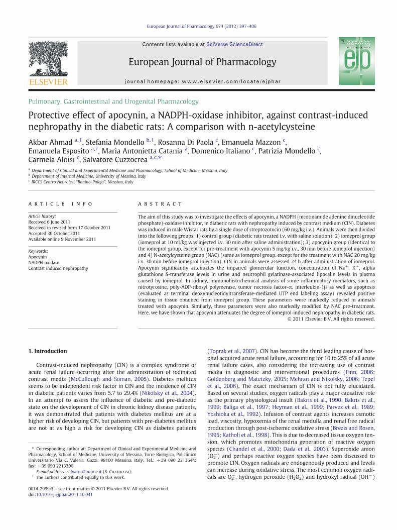
European Journal of Pharmacology 674 (2012) 397–406
Contents lists available at SciVerse ScienceDirect
European Journal of Pharmacology
j ourna l homepage: www.e lsev ie r .com/ locate /e jphar
Pulmonary, Gastrointestinal and Urogenital Pharmacology
Protective effect of apocynin, a NADPH-oxidase inhibitor, against contrast-inducednephropathy in the diabetic rats: A comparison with n-acetylcysteine
Akbar Ahmad a,1, Stefania Mondello b,1, Rosanna Di Paola c, Emanuela Mazzon c,Emanuela Esposito a,c, Maria Antonietta Catania a, Domenico Italiano c, Patrizia Mondello c,Carmela Aloisi c, Salvatore Cuzzocrea a,c,⁎a Department of Clinical and Experimental Medicine and Pharmacology, School of Medicine, Messina, Italyb Department of Internal Medicine, University of Messina, Italyc IRCCS Centro Neurolesi “Bonino-Pulejo”, Messina, Italy
⁎ Corresponding author at: Department of Clinical anPharmacology, School of Medicine, University of MessiUniversitario Via C. Valeria, Gazzi, 98100 Messina, Itfax: +39 090 2213300.
E-mail address: [email protected] (S. Cuzzocrea).1 The authors contributed equally to this work.
0014-2999/$ – see front matter © 2011 Elsevier B.V. Alldoi:10.1016/j.ejphar.2011.10.041
a b s t r a c t
a r t i c l e i n f oArticle history:Received 6 June 2011Received in revised form 17 October 2011Accepted 30 October 2011Available online 9 November 2011
Keywords:ApocyninNADPH-oxidaseContrast induced nephropathy
The aim of this study was to investigate the effects of apocynin, a NADPH (nicotinamide adenine dinucleotidephosphate)-oxidase inhibitor, in diabetic rats with nephropathy induced by contrast medium (CIN). Diabeteswas induced in male Wistar rats by a single dose of streptozotocin (60 mg/kg i.v.). Animals were then dividedinto the following groups: 1) control group (diabetic rats treated i.v. with saline solution); 2) iomeprol group(iomeprol at 10 ml/kg was injected i.v. 30 min after saline administration); 3) apocynin group (identical tothe iomeprol group, except for pre-treatment with apocynin 5 mg/kg i.v., 30 min before iomeprol injection)and 4) N-acetylcysteine group (NAC) (same as iomeprol group, except for the treatment with NAC 20 mg/kgi.v. 30 min before iomeprol injection). CIN in animals were assessed 24 h after administration of iomeprol.Apocynin significantly attenuates the impaired glomerular function, concentration of Na+, K+, alphaglutathione S-transferase levels in urine and neutrophil gelatinase-associated lipocalin levels in plasmacaused by iomeprol. In kidney, immunohistochemical analysis of some inflammatory mediators, such asnitrotyrosine, poly-ADP-ribosyl polymerase, tumor necrosis factor-α, interleukin-1β as well as apoptosis(evaluated as terminal deoxynucleotidyltransferase-mediated UTP end labeling assay) revealed positivestaining in tissue obtained from iomeprol group. These parameters were markedly reduced in animalstreated with apocynin. Similarly, these parameters were also markedly modified by NAC pre-treatment.Here, we have shown that apocynin attenuates the degree of iomeprol-induced nephropathy in diabetic rats.
© 2011 Elsevier B.V. All rights reserved.
1. Introduction
Contrast-induced nephropathy (CIN) is a complex syndrome ofacute renal failure occurring after the administration of iodinatedcontrast media (McCullough and Soman, 2005). Diabetes mellitusseems to be independent risk factor in CIN and the incidence of CINin diabetic patients varies from 5.7 to 29.4% (Nikolsky et al., 2004).In an attempt to assess the influence of diabetic and pre-diabeticstate on the development of CIN in chronic kidney disease patients,it was demonstrated that patients with diabetes mellitus are at ahigher risk of developing CIN, but patients with pre-diabetes mellitusare not at as high a risk for developing CIN as diabetes patients
d Experimental Medicine andna, Torre Biologica, Policlinicoaly. Tel.: +39 090 2213644;
rights reserved.
(Toprak et al., 2007). CIN has become the third leading cause of hos-pital acquired acute renal failure, accounting for 10 to 25% of all acuterenal failure cases, also considering the increasing use of contrastmedia in diagnostic and interventional procedures (Finn, 2006;Goldenberg and Matetzky, 2005; Mehran and Nikolsky, 2006; Tepelet al., 2006). The exact mechanism of CIN is not fully elucidated.Based on several studies, oxygen radicals play a major causative roleas the primary physiological insult (Bakris et al., 1990; Bakris et al.,1999; Baliga et al., 1997; Heyman et al., 1999; Parvez et al., 1989;Yoshioka et al., 1992). Infusion of contrast agents increases osmoticload, viscosity, hypoxemia of the renal medulla and renal free radicalproduction through post-ischemic oxidative stress (Brezis and Rosen,1995; Katholi et al., 1998). This is due to decreased tissue oxygen ten-sion, which promotes mitochondria generation of reactive oxygenspecies (Chandel et al., 2000; Dada et al., 2003). Superoxide anion(O2
−) and perhaps reactive oxygen species have been discussed topromote CIN. Oxygen radicals are endogenously produced and levelscan increase during oxidative stress. The most common oxygen radi-cals are O2
−, hydrogen peroxide (H2O2) and hydroxyl radical (OH−)
398 A. Ahmad et al. / European Journal of Pharmacology 674 (2012) 397–406
(Schnackenberg, 2002). O2− and OH− are more reactive than H2O2,
which is not a radical, but exhibits greater membrane permeability.O2− rapidly scavenges nitric oxide (NO) and could therefore blunt
NO activity in the renal microvascular. Since NO inhibits oxygenconsumption, it is tempting to speculate that reduced (scavenged)NO during diabetes elevates oxygen consumption, thereby leadingto reduced partial oxygen pressure values with consequences forendothelial–epithelial structure and function. Reactive oxygenspecies may play a role in the effects of various vasoconstrictorsthat have been considered important for the development of CIN.
Apocynin (4-hydroxy-3-methoxy-acetophenone) is a constituentof the Himalayan herb Picrorhiza kurroa Royle (Scrophulariaceae)that is well known in traditional Indian medicine (Ayurveda). It isan acetophenone to which a range of biological activities is attributed(Hougee et al., 2006). It is a prodrug that is converted by peroxidase-mediated oxidation to a dimer, which has been shown to be moreefficient than apocynin itself (Johnson et al., 2002). It has been usedas an efficient inhibitor of the nicotinamide adenine dinucleotidephosphate (NADPH)-oxidase in many experimental model such ascolitis, rheumatoid arthritis, ischemia reperfusion and lung injury(Dodd-o et al., 2004).
Based on these observations, this study was to investigate the po-tential therapeutic role of apocynin, a reversible inhibitor of NADPHoxidase, on contrast medium-induced nephropathy in diabetic rats.
2. Materials and methods
2.1. Animals
Male Wistar rats (150–200 g; Harlan Nossan; Italy) were housedin a controlled environment and provided with standard rodentchow and water. Animal care was in compliance with Italian regula-tions on protection of animals used for experimental and otherscientific purposes (D.M.116192) as well as with the EEC regulations(O.J. of E.C. L 358/1 12/18/1986).
2.2. Induction of experimental diabetes
After 12 h of fasting, the animals received a single 60 mg/kg intra-venous injection of streptozotocin (Sigma, St. Louis, MO) in 10 mMsodium citrate buffer, pH 4.5 (see figure). After 24 h, animals withblood glucose levels greater than 250 mg/dl were considered diabetic.We performed all of experiments with contrast medium 10 daysfollowing the induction of diabetes. We confirmed the diabetic statea evaluating daily the blood glucose levels.
2.3. Contrast-induced nephropathy (CIN)
Male diabetic rats were randomly allocated into the followinggroups: (1) Control group. Rats were treated intravenously (i.v.) withsaline solution (n=5); (2) Iomeprol group. Rats were administerediomeprol (10 ml/kg i.v.) 30 min after saline administration (n=5);(3) Apocynin group. Identical to the Iomeprol group, except for theadministration of apocynin (5 mg/kg i.v.) instead of saline administra-tion (n=5); (4) N-acetylcysteine (NAC) group. Identical to Iomeprolgroup, except for the administration of NAC (20 mg/kg i.v.) insteadof saline administration (n=5).
In this experiment, the dose of apocynin was chosen in agreementwith our previous studies (Genovese et al., 2011; Impellizzeri et al.,2011; Paterniti et al., 2010).
2.4. Measurement of biochemical parameters
After 24 h, the urine samples were collected and blood sampleswere obtained via the lateral tail vein into S1/3 tubes containingserum gel. The sampleswere centrifuged (6000 g for 3 min) to separate
plasma. All plasma samples were analyzed for biochemical parameterswithin 24 h after collection. Concentrations of urea and creatinine inplasma and urine were measured as indicators of impaired glomerularfunction. Creatinine clearance (ml/min) was calculated using thefollowing formula (=UV/P), where U refers to creatinine concentrationin urine (mg/dl), V to urine volume/min (ml/min) and P to serumcreatinine (mg/dl). A kidney injury biomarker called “Neutrophilgelatinase-associated lipocalin” (NGAL) was evaluated as indicated bythe commercial kit in plasma and urine. Urine concentration of αGSTas well as urine and plasma concentration of NGAL were evaluated asindicated by the commercial kit. Na+ and K+ concentrations in plasmawere determined by flame photometry.
2.5. Histological evaluation
Twenty-four hours after administration of saline or apocynin orNAC, animalswere sacrificed and a 5 μmsection of kidneywas removedand placed in formalin and processed through to wax. Five millimetersections were cut and stained with hematoxylin and eosin. Histologicalassessment of outer medulla damage was examined by an experiencedmorphologist, who was not aware of the sample identity. The criteriafor injury/necrosis were the following: 0=normal histology;1=minor edema, minor cell swelling; 2=hemorrhage, moderateedema, moderate cells vacuolization and swelling; 3=moderate hem-orrhage, moderate edema, moderate cells vacuolization, swelling andchromatin alteration; 4=severe edema, severe cells vacuolization,swelling and chromatin alteration, presence of necrosis spot; 5=severeedema, severe cytoplasmic vacuolar changes, swelling and chromatinalteration, intratubular cast formation, renal tubular architecture,luminal congestion and severe necrosis.
2.6. Immunohistochemical localization of nitrotyrosine, peroxisomeproliferators-activated receptors (PAR), tumor necrosis factor (TNF)-α,interleukin (IL)-1β
At the end of experiment, tissues were fixed in 10% (w/v)PBS-buffered formalin and 8 μm sections were prepared from paraffinembedded tissues. After deparaffinization, endogenous peroxidasewas quenched with 0.3% (v/v) hydrogen peroxide in 60% (v/v) metha-nol for 30 min. The sections were permeabilized with 0.1% (v/v) TritonX-100 in PBS for 20 min. Non-specific adsorption was minimized byincubating the section in 2% (v/v) normal goat serum in PBS for20 min. Endogenous biotin or avidin binding sites were blocked bysequential incubation for 15 min with avidin and biotin (Vector Labo-ratories, Burlingame, CA). The sections were then incubated overnightwith 1:1000 dilution of primary anti-nitrotyrosine antibody (Millipore,1:500 in PBS, v/v), anti-poly (ADP)-ribose (PAR) antibody (Santa CruzBiotechnology, 1:500 in PBS, v/v), anti-TNF-α antibody (Santa CruzBiotechnology, 1:500 in PBS, v/v), anti-IL-1β antibody (Santa CruzBiotechnology, 1:500 in PBS, v/v). Controls included buffer alone ornon-specific purified rabbit Immunoglobulin G. Specific labeling wasdetected with a biotin-conjugated specific secondary anti-IgG and avi-din–biotin peroxidase complex (Vector Laboratories, Burlingame, CA).In order to confirm that the immunoreactions for the nitrotyrosinewere specific, some sections were also incubated with the primaryantibody (anti-nitrotyrosine) in the presence of excess nitrotyrosine(10 mM) to verify the binding specificity. To verify the binding specific-ity for nitrotyrosine, PARP, TNF-α and IL-1β, some sections were alsoincubated with primary antibody only (no secondary antibody) orwith secondary antibody only (no primary antibody). In thesesituations, no positive staining was found in the sections indicatingthat the immunoreactions were positive in all the experiments carriedout. Immunocytochemistry photographs (n=5) were assessed bydensitometry. The assay was carried out by using Optilab Graftek soft-ware on a Macintosh personal computer (CPU G3-266). All the
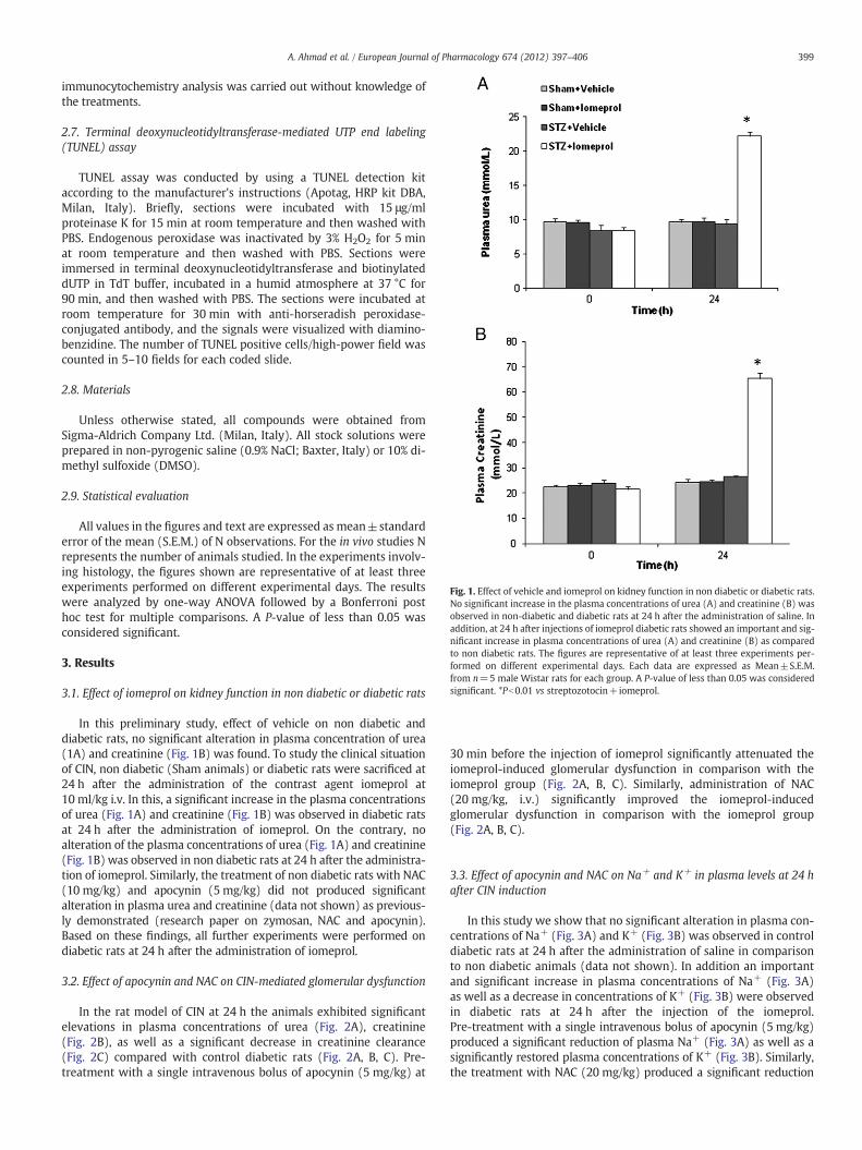
Fig. 1. Effect of vehicle and iomeprol on kidney function in non diabetic or diabetic rats.No significant increase in the plasma concentrations of urea (A) and creatinine (B) wasobserved in non-diabetic and diabetic rats at 24 h after the administration of saline. Inaddition, at 24 h after injections of iomeprol diabetic rats showed an important and sig-nificant increase in plasma concentrations of urea (A) and creatinine (B) as comparedto non diabetic rats. The figures are representative of at least three experiments per-formed on different experimental days. Each data are expressed as Mean±S.E.M.from n=5 male Wistar rats for each group. A P-value of less than 0.05 was consideredsignificant. *Pb0.01 vs streptozotocin+iomeprol.
399A. Ahmad et al. / European Journal of Pharmacology 674 (2012) 397–406
immunocytochemistry analysis was carried out without knowledge ofthe treatments.
2.7. Terminal deoxynucleotidyltransferase-mediated UTP end labeling(TUNEL) assay
TUNEL assay was conducted by using a TUNEL detection kitaccording to the manufacturer's instructions (Apotag, HRP kit DBA,Milan, Italy). Briefly, sections were incubated with 15 μg/mlproteinase K for 15 min at room temperature and then washed withPBS. Endogenous peroxidase was inactivated by 3% H2O2 for 5 minat room temperature and then washed with PBS. Sections wereimmersed in terminal deoxynucleotidyltransferase and biotinylateddUTP in TdT buffer, incubated in a humid atmosphere at 37 °C for90 min, and then washed with PBS. The sections were incubated atroom temperature for 30 min with anti-horseradish peroxidase-conjugated antibody, and the signals were visualized with diamino-benzidine. The number of TUNEL positive cells/high-power field wascounted in 5–10 fields for each coded slide.
2.8. Materials
Unless otherwise stated, all compounds were obtained fromSigma-Aldrich Company Ltd. (Milan, Italy). All stock solutions wereprepared in non-pyrogenic saline (0.9% NaCl; Baxter, Italy) or 10% di-methyl sulfoxide (DMSO).
2.9. Statistical evaluation
All values in the figures and text are expressed as mean±standarderror of the mean (S.E.M.) of N observations. For the in vivo studies Nrepresents the number of animals studied. In the experiments involv-ing histology, the figures shown are representative of at least threeexperiments performed on different experimental days. The resultswere analyzed by one-way ANOVA followed by a Bonferroni posthoc test for multiple comparisons. A P-value of less than 0.05 wasconsidered significant.
3. Results
3.1. Effect of iomeprol on kidney function in non diabetic or diabetic rats
In this preliminary study, effect of vehicle on non diabetic anddiabetic rats, no significant alteration in plasma concentration of urea(1A) and creatinine (Fig. 1B) was found. To study the clinical situationof CIN, non diabetic (Sham animals) or diabetic rats were sacrificed at24 h after the administration of the contrast agent iomeprol at10 ml/kg i.v. In this, a significant increase in the plasma concentrationsof urea (Fig. 1A) and creatinine (Fig. 1B) was observed in diabetic ratsat 24 h after the administration of iomeprol. On the contrary, noalteration of the plasma concentrations of urea (Fig. 1A) and creatinine(Fig. 1B) was observed in non diabetic rats at 24 h after the administra-tion of iomeprol. Similarly, the treatment of non diabetic rats with NAC(10 mg/kg) and apocynin (5 mg/kg) did not produced significantalteration in plasma urea and creatinine (data not shown) as previous-ly demonstrated (research paper on zymosan, NAC and apocynin).Based on these findings, all further experiments were performed ondiabetic rats at 24 h after the administration of iomeprol.
3.2. Effect of apocynin and NAC on CIN-mediated glomerular dysfunction
In the rat model of CIN at 24 h the animals exhibited significantelevations in plasma concentrations of urea (Fig. 2A), creatinine(Fig. 2B), as well as a significant decrease in creatinine clearance(Fig. 2C) compared with control diabetic rats (Fig. 2A, B, C). Pre-treatment with a single intravenous bolus of apocynin (5 mg/kg) at
30 min before the injection of iomeprol significantly attenuated theiomeprol-induced glomerular dysfunction in comparison with theiomeprol group (Fig. 2A, B, C). Similarly, administration of NAC(20 mg/kg, i.v.) significantly improved the iomeprol-inducedglomerular dysfunction in comparison with the iomeprol group(Fig. 2A, B, C).
3.3. Effect of apocynin and NAC on Na+ and K+ in plasma levels at 24 hafter CIN induction
In this study we show that no significant alteration in plasma con-centrations of Na+ (Fig. 3A) and K+ (Fig. 3B) was observed in controldiabetic rats at 24 h after the administration of saline in comparisonto non diabetic animals (data not shown). In addition an importantand significant increase in plasma concentrations of Na+ (Fig. 3A)as well as a decrease in concentrations of K+ (Fig. 3B) were observedin diabetic rats at 24 h after the injection of the iomeprol.Pre-treatment with a single intravenous bolus of apocynin (5 mg/kg)produced a significant reduction of plasma Na+ (Fig. 3A) as well as asignificantly restored plasma concentrations of K+ (Fig. 3B). Similarly,the treatment with NAC (20 mg/kg) produced a significant reduction
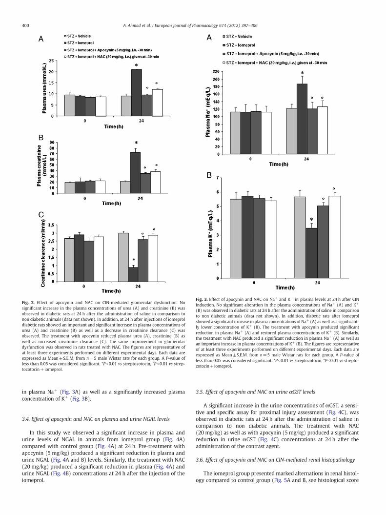
Fig. 3. Effect of apocynin and NAC on Na+ and K+ in plasma levels at 24 h after CINinduction. No significant alteration in the plasma concentrations of Na+ (A) and K+
(B) was observed in diabetic rats at 24 h after the administration of saline in comparisonto non diabetic animals (data not shown). In addition, diabetic rats after iomeprolshowed a significant increase in plasma concentrations of Na+ (A) aswell as a significant-ly lower concentration of K+ (B). The treatment with apocynin produced significantreduction in plasma Na+ (A) and restored plasma concentrations of K+ (B). Similarly,the treatment with NAC produced a significant reduction in plasma Na+ (A) as well asan important increase in plasma concentrations of K+ (B). The figures are representativeof at least three experiments performed on different experimental days. Each data areexpressed as Mean±S.E.M. from n=5 male Wistar rats for each group. A P-value ofless than 0.05 was considered significant. *Pb0.01 vs streptozotocin, °Pb0.01 vs strepto-zotocin+iomeprol.
Fig. 2. Effect of apocynin and NAC on CIN-mediated glomerular dysfunction. Nosignificant increase in the plasma concentrations of urea (A) and creatinine (B) wasobserved in diabetic rats at 24 h after the administration of saline in comparison tonon diabetic animals (data not shown). In addition, at 24 h after injections of iomeproldiabetic rats showed an important and significant increase in plasma concentrations ofurea (A) and creatinine (B) as well as a decrease in creatinine clearance (C) wasobserved. The treatment with apocynin reduced plasma urea (A), creatinine (B) aswell as increased creatinine clearance (C). The same improvement in glomerulardysfunction was observed in rats treated with NAC. The figures are representative ofat least three experiments performed on different experimental days. Each data areexpressed as Mean±S.E.M. from n=5 male Wistar rats for each group. A P-value ofless than 0.05 was considered significant. *Pb0.01 vs streptozotocin, °Pb0.01 vs strep-tozotocin+iomeprol.
400 A. Ahmad et al. / European Journal of Pharmacology 674 (2012) 397–406
in plasma Na+ (Fig. 3A) as well as a significantly increased plasmaconcentration of K+ (Fig. 3B).
3.4. Effect of apocynin and NAC on plasma and urine NGAL levels
In this study we observed a significant increase in plasma andurine levels of NGAL in animals from iomeprol group (Fig. 4A)compared with control group (Fig. 4A) at 24 h. Pre-treatment withapocynin (5 mg/kg) produced a significant reduction in plasma andurine NGAL (Fig. 4A and B) levels. Similarly, the treatment with NAC(20 mg/kg) produced a significant reduction in plasma (Fig. 4A) andurine NGAL (Fig. 4B) concentrations at 24 h after the injection of theiomeprol.
3.5. Effect of apocynin and NAC on urine αGST levels
A significant increase in the urine concentrations of αGST, a sensi-tive and specific assay for proximal injury assessment (Fig. 4C), wasobserved in diabetic rats at 24 h after the administration of saline incomparison to non diabetic animals. The treatment with NAC(20 mg/kg) as well as with apocynin (5 mg/kg) produced a significantreduction in urine αGST (Fig. 4C) concentrations at 24 h after theadministration of the contrast agent.
3.6. Effect of apocynin and NAC on CIN-mediated renal histopathology
The iomeprol group presented marked alternations in renal histol-ogy compared to control group (Fig. 5A and B, see histological score
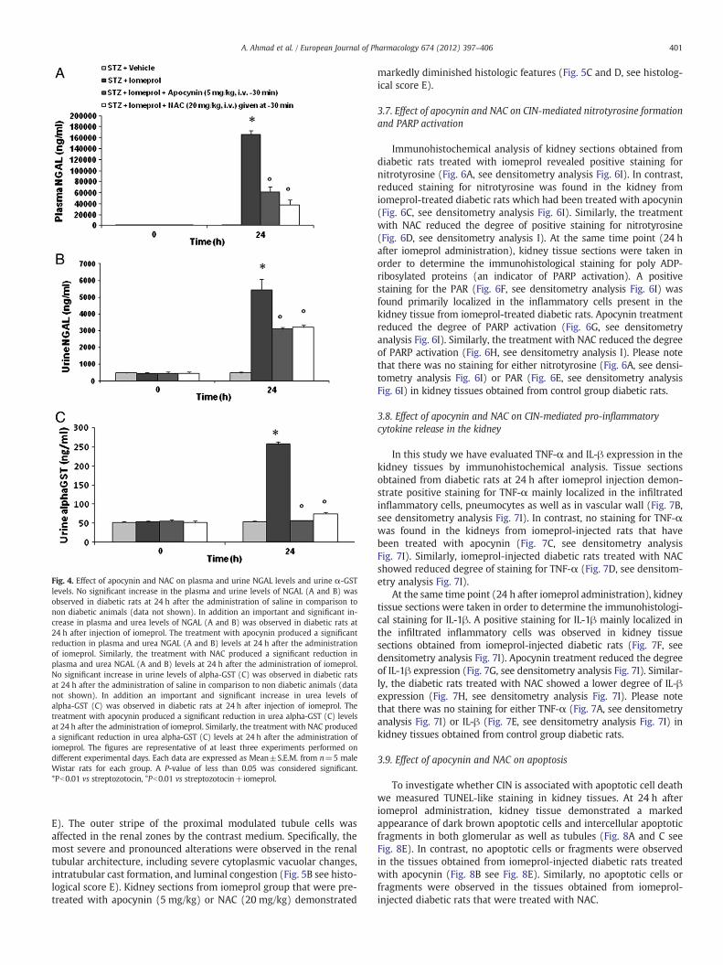
Fig. 4. Effect of apocynin and NAC on plasma and urine NGAL levels and urine α-GSTlevels. No significant increase in the plasma and urine levels of NGAL (A and B) wasobserved in diabetic rats at 24 h after the administration of saline in comparison tonon diabetic animals (data not shown). In addition an important and significant in-crease in plasma and urea levels of NGAL (A and B) was observed in diabetic rats at24 h after injection of iomeprol. The treatment with apocynin produced a significantreduction in plasma and urea NGAL (A and B) levels at 24 h after the administrationof iomeprol. Similarly, the treatment with NAC produced a significant reduction inplasma and urea NGAL (A and B) levels at 24 h after the administration of iomeprol.No significant increase in urine levels of alpha-GST (C) was observed in diabetic ratsat 24 h after the administration of saline in comparison to non diabetic animals (datanot shown). In addition an important and significant increase in urea levels ofalpha-GST (C) was observed in diabetic rats at 24 h after injection of iomeprol. Thetreatment with apocynin produced a significant reduction in urea alpha-GST (C) levelsat 24 h after the administration of iomeprol. Similarly, the treatment with NAC produceda significant reduction in urea alpha-GST (C) levels at 24 h after the administration ofiomeprol. The figures are representative of at least three experiments performed ondifferent experimental days. Each data are expressed as Mean±S.E.M. from n=5 maleWistar rats for each group. A P-value of less than 0.05 was considered significant.*Pb0.01 vs streptozotocin, °Pb0.01 vs streptozotocin+iomeprol.
401A. Ahmad et al. / European Journal of Pharmacology 674 (2012) 397–406
E). The outer stripe of the proximal modulated tubule cells wasaffected in the renal zones by the contrast medium. Specifically, themost severe and pronounced alterations were observed in the renaltubular architecture, including severe cytoplasmic vacuolar changes,intratubular cast formation, and luminal congestion (Fig. 5B see histo-logical score E). Kidney sections from iomeprol group that were pre-treated with apocynin (5 mg/kg) or NAC (20 mg/kg) demonstrated
markedly diminished histologic features (Fig. 5C and D, see histolog-ical score E).
3.7. Effect of apocynin and NAC on CIN-mediated nitrotyrosine formationand PARP activation
Immunohistochemical analysis of kidney sections obtained fromdiabetic rats treated with iomeprol revealed positive staining fornitrotyrosine (Fig. 6A, see densitometry analysis Fig. 6I). In contrast,reduced staining for nitrotyrosine was found in the kidney fromiomeprol-treated diabetic rats which had been treated with apocynin(Fig. 6C, see densitometry analysis Fig. 6I). Similarly, the treatmentwith NAC reduced the degree of positive staining for nitrotyrosine(Fig. 6D, see densitometry analysis I). At the same time point (24 hafter iomeprol administration), kidney tissue sections were taken inorder to determine the immunohistological staining for poly ADP-ribosylated proteins (an indicator of PARP activation). A positivestaining for the PAR (Fig. 6F, see densitometry analysis Fig. 6I) wasfound primarily localized in the inflammatory cells present in thekidney tissue from iomeprol-treated diabetic rats. Apocynin treatmentreduced the degree of PARP activation (Fig. 6G, see densitometryanalysis Fig. 6I). Similarly, the treatment with NAC reduced the degreeof PARP activation (Fig. 6H, see densitometry analysis I). Please notethat there was no staining for either nitrotyrosine (Fig. 6A, see densi-tometry analysis Fig. 6I) or PAR (Fig. 6E, see densitometry analysisFig. 6I) in kidney tissues obtained from control group diabetic rats.
3.8. Effect of apocynin and NAC on CIN-mediated pro-inflammatorycytokine release in the kidney
In this study we have evaluated TNF-α and IL-β expression in thekidney tissues by immunohistochemical analysis. Tissue sectionsobtained from diabetic rats at 24 h after iomeprol injection demon-strate positive staining for TNF-α mainly localized in the infiltratedinflammatory cells, pneumocytes as well as in vascular wall (Fig. 7B,see densitometry analysis Fig. 7I). In contrast, no staining for TNF-αwas found in the kidneys from iomeprol-injected rats that havebeen treated with apocynin (Fig. 7C, see densitometry analysisFig. 7I). Similarly, iomeprol-injected diabetic rats treated with NACshowed reduced degree of staining for TNF-α (Fig. 7D, see densitom-etry analysis Fig. 7I).
At the same time point (24 h after iomeprol administration), kidneytissue sections were taken in order to determine the immunohistologi-cal staining for IL-1β. A positive staining for IL-1β mainly localized inthe infiltrated inflammatory cells was observed in kidney tissuesections obtained from iomeprol-injected diabetic rats (Fig. 7F, seedensitometry analysis Fig. 7I). Apocynin treatment reduced the degreeof IL-1β expression (Fig. 7G, see densitometry analysis Fig. 7I). Similar-ly, the diabetic rats treated with NAC showed a lower degree of IL-βexpression (Fig. 7H, see densitometry analysis Fig. 7I). Please notethat there was no staining for either TNF-α (Fig. 7A, see densitometryanalysis Fig. 7I) or IL-β (Fig. 7E, see densitometry analysis Fig. 7I) inkidney tissues obtained from control group diabetic rats.
3.9. Effect of apocynin and NAC on apoptosis
To investigate whether CIN is associated with apoptotic cell deathwe measured TUNEL-like staining in kidney tissues. At 24 h afteriomeprol administration, kidney tissue demonstrated a markedappearance of dark brown apoptotic cells and intercellular apoptoticfragments in both glomerular as well as tubules (Fig. 8A and C seeFig. 8E). In contrast, no apoptotic cells or fragments were observedin the tissues obtained from iomeprol-injected diabetic rats treatedwith apocynin (Fig. 8B see Fig. 8E). Similarly, no apoptotic cells orfragments were observed in the tissues obtained from iomeprol-injected diabetic rats that were treated with NAC.
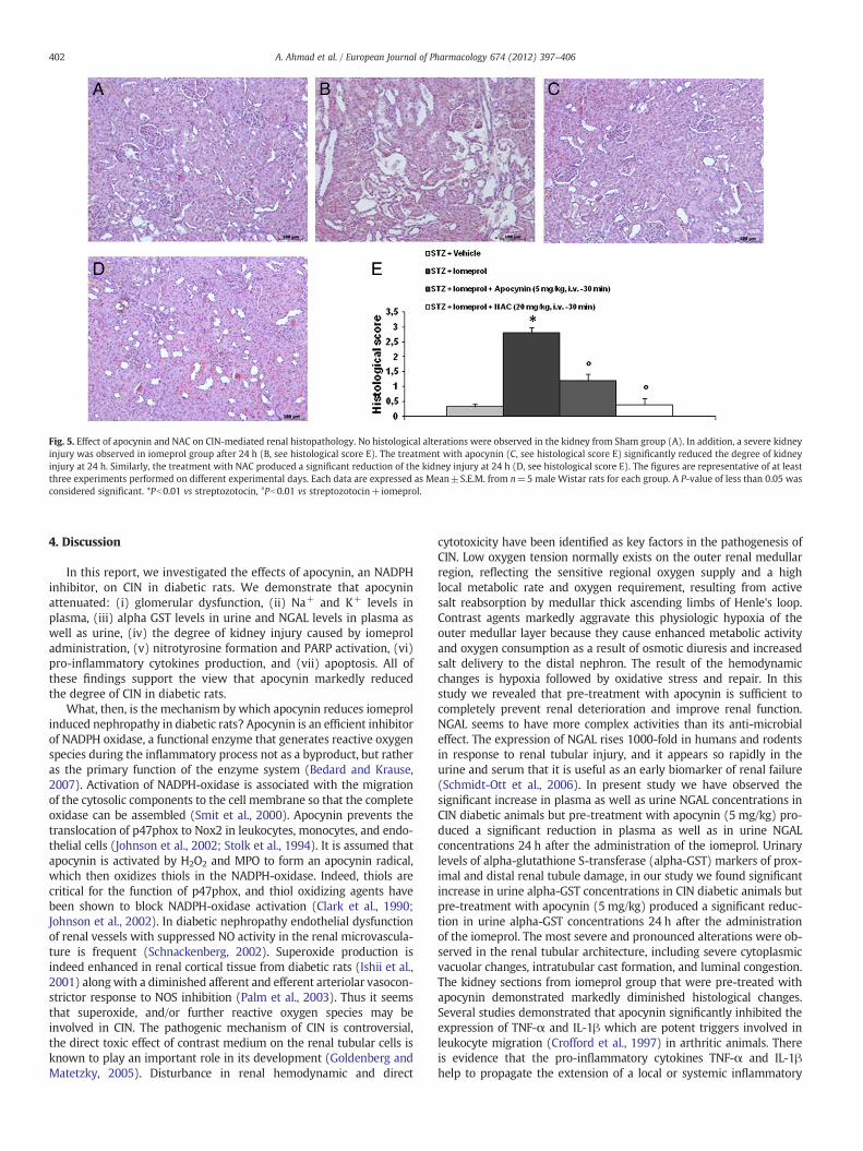
Fig. 5. Effect of apocynin and NAC on CIN-mediated renal histopathology. No histological alterations were observed in the kidney from Sham group (A). In addition, a severe kidneyinjury was observed in iomeprol group after 24 h (B, see histological score E). The treatment with apocynin (C, see histological score E) significantly reduced the degree of kidneyinjury at 24 h. Similarly, the treatment with NAC produced a significant reduction of the kidney injury at 24 h (D, see histological score E). The figures are representative of at leastthree experiments performed on different experimental days. Each data are expressed as Mean±S.E.M. from n=5 male Wistar rats for each group. A P-value of less than 0.05 wasconsidered significant. *Pb0.01 vs streptozotocin, °Pb0.01 vs streptozotocin+iomeprol.
402 A. Ahmad et al. / European Journal of Pharmacology 674 (2012) 397–406
4. Discussion
In this report, we investigated the effects of apocynin, an NADPHinhibitor, on CIN in diabetic rats. We demonstrate that apocyninattenuated: (i) glomerular dysfunction, (ii) Na+ and K+ levels inplasma, (iii) alpha GST levels in urine and NGAL levels in plasma aswell as urine, (iv) the degree of kidney injury caused by iomeproladministration, (v) nitrotyrosine formation and PARP activation, (vi)pro-inflammatory cytokines production, and (vii) apoptosis. All ofthese findings support the view that apocynin markedly reducedthe degree of CIN in diabetic rats.
What, then, is the mechanism by which apocynin reduces iomeprolinduced nephropathy in diabetic rats? Apocynin is an efficient inhibitorof NADPH oxidase, a functional enzyme that generates reactive oxygenspecies during the inflammatory process not as a byproduct, but ratheras the primary function of the enzyme system (Bedard and Krause,2007). Activation of NADPH-oxidase is associated with the migrationof the cytosolic components to the cell membrane so that the completeoxidase can be assembled (Smit et al., 2000). Apocynin prevents thetranslocation of p47phox to Nox2 in leukocytes, monocytes, and endo-thelial cells (Johnson et al., 2002; Stolk et al., 1994). It is assumed thatapocynin is activated by H2O2 and MPO to form an apocynin radical,which then oxidizes thiols in the NADPH-oxidase. Indeed, thiols arecritical for the function of p47phox, and thiol oxidizing agents havebeen shown to block NADPH-oxidase activation (Clark et al., 1990;Johnson et al., 2002). In diabetic nephropathy endothelial dysfunctionof renal vessels with suppressed NO activity in the renal microvascula-ture is frequent (Schnackenberg, 2002). Superoxide production isindeed enhanced in renal cortical tissue from diabetic rats (Ishii et al.,2001) alongwith a diminished afferent and efferent arteriolar vasocon-strictor response to NOS inhibition (Palm et al., 2003). Thus it seemsthat superoxide, and/or further reactive oxygen species may beinvolved in CIN. The pathogenic mechanism of CIN is controversial,the direct toxic effect of contrast medium on the renal tubular cells isknown to play an important role in its development (Goldenberg andMatetzky, 2005). Disturbance in renal hemodynamic and direct
cytotoxicity have been identified as key factors in the pathogenesis ofCIN. Low oxygen tension normally exists on the outer renal medullarregion, reflecting the sensitive regional oxygen supply and a highlocal metabolic rate and oxygen requirement, resulting from activesalt reabsorption by medullar thick ascending limbs of Henle's loop.Contrast agents markedly aggravate this physiologic hypoxia of theouter medullar layer because they cause enhanced metabolic activityand oxygen consumption as a result of osmotic diuresis and increasedsalt delivery to the distal nephron. The result of the hemodynamicchanges is hypoxia followed by oxidative stress and repair. In thisstudy we revealed that pre-treatment with apocynin is sufficient tocompletely prevent renal deterioration and improve renal function.NGAL seems to have more complex activities than its anti-microbialeffect. The expression of NGAL rises 1000-fold in humans and rodentsin response to renal tubular injury, and it appears so rapidly in theurine and serum that it is useful as an early biomarker of renal failure(Schmidt-Ott et al., 2006). In present study we have observed thesignificant increase in plasma as well as urine NGAL concentrations inCIN diabetic animals but pre-treatment with apocynin (5 mg/kg) pro-duced a significant reduction in plasma as well as in urine NGALconcentrations 24 h after the administration of the iomeprol. Urinarylevels of alpha-glutathione S-transferase (alpha-GST) markers of prox-imal and distal renal tubule damage, in our study we found significantincrease in urine alpha-GST concentrations in CIN diabetic animals butpre-treatment with apocynin (5 mg/kg) produced a significant reduc-tion in urine alpha-GST concentrations 24 h after the administrationof the iomeprol. The most severe and pronounced alterations were ob-served in the renal tubular architecture, including severe cytoplasmicvacuolar changes, intratubular cast formation, and luminal congestion.The kidney sections from iomeprol group that were pre-treated withapocynin demonstrated markedly diminished histological changes.Several studies demonstrated that apocynin significantly inhibited theexpression of TNF-α and IL-1β which are potent triggers involved inleukocyte migration (Crofford et al., 1997) in arthritic animals. Thereis evidence that the pro-inflammatory cytokines TNF-α and IL-1βhelp to propagate the extension of a local or systemic inflammatory
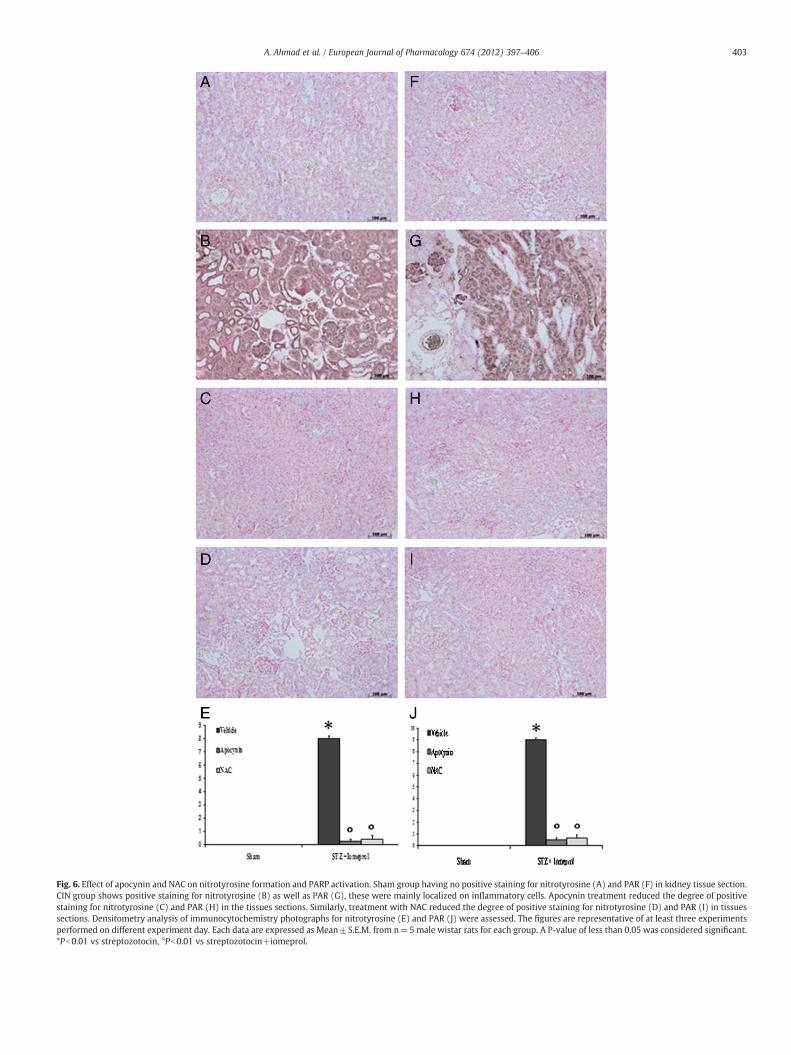
Fig. 6. Effect of apocynin and NAC on nitrotyrosine formation and PARP activation. Sham group having no positive staining for nitrotyrosine (A) and PAR (F) in kidney tissue section.CIN group shows positive staining for nitrotyrosine (B) as well as PAR (G), these were mainly localized on inflammatory cells. Apocynin treatment reduced the degree of positivestaining for nitrotyrosine (C) and PAR (H) in the tissues sections. Similarly, treatment with NAC reduced the degree of positive staining for nitrotyrosine (D) and PAR (I) in tissuessections. Densitometry analysis of immunocytochemistry photographs for nitrotyrosine (E) and PAR (J) were assessed. The figures are representative of at least three experimentsperformed on different experiment day. Each data are expressed as Mean±S.E.M. from n=5male wistar rats for each group. A P-value of less than 0.05 was considered significant.*Pb0.01 vs streptozotocin, °Pb0.01 vs streptozotocin+iomeprol.
403A. Ahmad et al. / European Journal of Pharmacology 674 (2012) 397–406
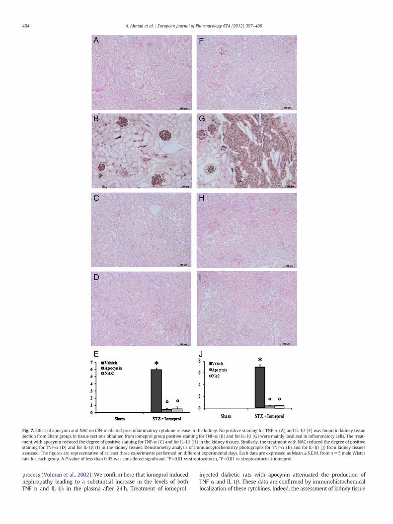
Fig. 7. Effect of apocynin and NAC on CIN-mediated pro-inflammatory cytokine release in the kidney. No positive staining for TNF-α (A) and IL-1β (F) was found in kidney tissuesection from Sham group. In tissue sections obtained from iomeprol group positive staining for TNF-α (B) and for IL-1β (G) were mainly localized in inflammatory cells. The treat-ment with apocynin reduced the degree of positive staining for TNF-α (C) and for IL-1β (H) in the kidney tissues. Similarly, the treatment with NAC reduced the degree of positivestaining for TNF-α (D) and for IL-1β (I) in the kidney tissues. Densitometry analysis of immunocytochemistry photographs for TNF-α (E) and for IL-1β (J) from kidney tissuesassessed. The figures are representative of at least three experiments performed on different experimental days. Each data are expressed as Mean±S.E.M. from n=5 male Wistarrats for each group. A P-value of less than 0.05 was considered significant. *Pb0.01 vs streptozotocin, °Pb0.01 vs streptozotocin+iomeprol.
404 A. Ahmad et al. / European Journal of Pharmacology 674 (2012) 397–406
process (Volman et al., 2002). We confirm here that iomeprol inducednephropathy leading to a substantial increase in the levels of bothTNF-α and IL-1β in the plasma after 24 h. Treatment of iomeprol-
injected diabetic rats with apocynin attenuated the production ofTNF-α and IL-1β. These data are confirmed by immunohistochemicallocalization of these cytokines. Indeed, the assessment of kidney tissue
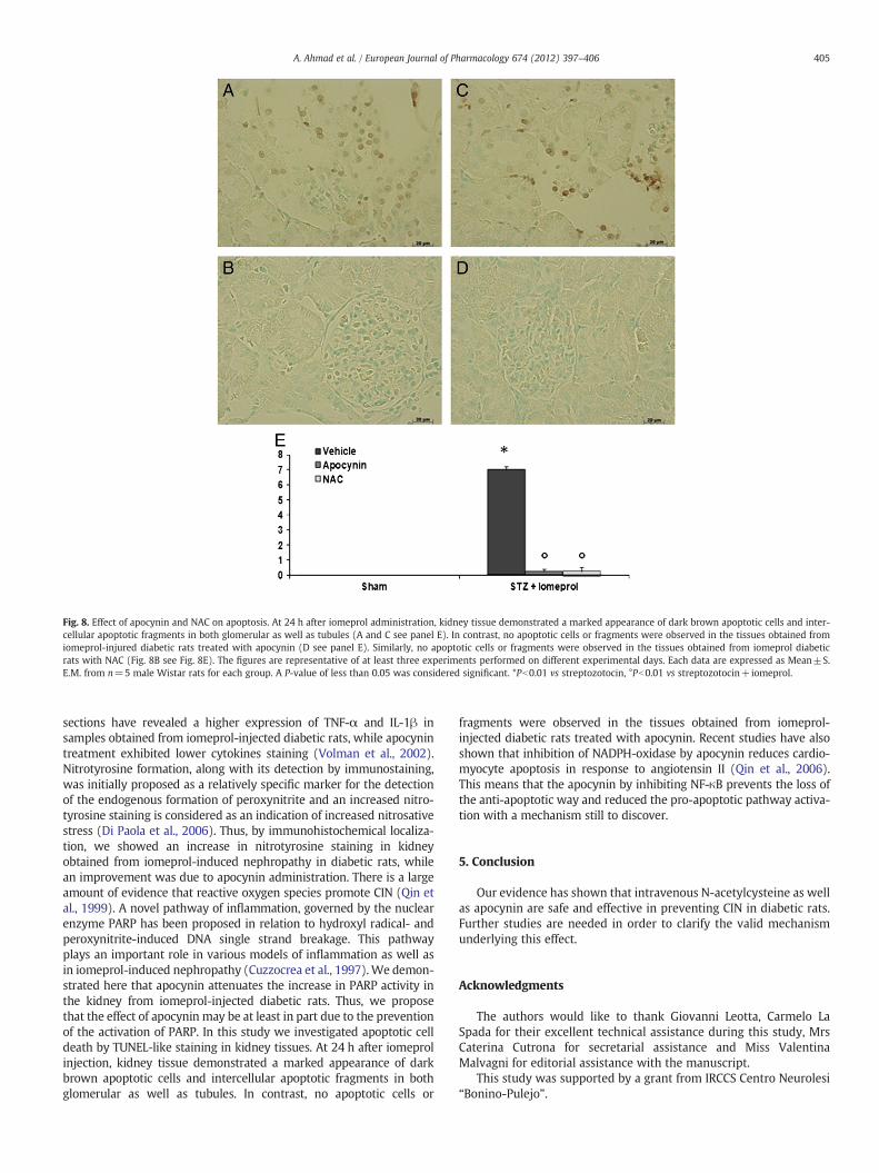
Fig. 8. Effect of apocynin and NAC on apoptosis. At 24 h after iomeprol administration, kidney tissue demonstrated a marked appearance of dark brown apoptotic cells and inter-cellular apoptotic fragments in both glomerular as well as tubules (A and C see panel E). In contrast, no apoptotic cells or fragments were observed in the tissues obtained fromiomeprol-injured diabetic rats treated with apocynin (D see panel E). Similarly, no apoptotic cells or fragments were observed in the tissues obtained from iomeprol diabeticrats with NAC (Fig. 8B see Fig. 8E). The figures are representative of at least three experiments performed on different experimental days. Each data are expressed as Mean±S.E.M. from n=5 male Wistar rats for each group. A P-value of less than 0.05 was considered significant. *Pb0.01 vs streptozotocin, °Pb0.01 vs streptozotocin+iomeprol.
405A. Ahmad et al. / European Journal of Pharmacology 674 (2012) 397–406
sections have revealed a higher expression of TNF-α and IL-1β insamples obtained from iomeprol-injected diabetic rats, while apocynintreatment exhibited lower cytokines staining (Volman et al., 2002).Nitrotyrosine formation, along with its detection by immunostaining,was initially proposed as a relatively specific marker for the detectionof the endogenous formation of peroxynitrite and an increased nitro-tyrosine staining is considered as an indication of increased nitrosativestress (Di Paola et al., 2006). Thus, by immunohistochemical localiza-tion, we showed an increase in nitrotyrosine staining in kidneyobtained from iomeprol-induced nephropathy in diabetic rats, whilean improvement was due to apocynin administration. There is a largeamount of evidence that reactive oxygen species promote CIN (Qin etal., 1999). A novel pathway of inflammation, governed by the nuclearenzyme PARP has been proposed in relation to hydroxyl radical- andperoxynitrite-induced DNA single strand breakage. This pathwayplays an important role in various models of inflammation as well asin iomeprol-induced nephropathy (Cuzzocrea et al., 1997).We demon-strated here that apocynin attenuates the increase in PARP activity inthe kidney from iomeprol-injected diabetic rats. Thus, we proposethat the effect of apocynin may be at least in part due to the preventionof the activation of PARP. In this study we investigated apoptotic celldeath by TUNEL-like staining in kidney tissues. At 24 h after iomeprolinjection, kidney tissue demonstrated a marked appearance of darkbrown apoptotic cells and intercellular apoptotic fragments in bothglomerular as well as tubules. In contrast, no apoptotic cells or
fragments were observed in the tissues obtained from iomeprol-injected diabetic rats treated with apocynin. Recent studies have alsoshown that inhibition of NADPH-oxidase by apocynin reduces cardio-myocyte apoptosis in response to angiotensin II (Qin et al., 2006).This means that the apocynin by inhibiting NF-κB prevents the loss ofthe anti-apoptotic way and reduced the pro-apoptotic pathway activa-tion with a mechanism still to discover.
5. Conclusion
Our evidence has shown that intravenous N-acetylcysteine as wellas apocynin are safe and effective in preventing CIN in diabetic rats.Further studies are needed in order to clarify the valid mechanismunderlying this effect.
Acknowledgments
The authors would like to thank Giovanni Leotta, Carmelo LaSpada for their excellent technical assistance during this study, MrsCaterina Cutrona for secretarial assistance and Miss ValentinaMalvagni for editorial assistance with the manuscript.
This study was supported by a grant from IRCCS Centro Neurolesi“Bonino-Pulejo”.
406 A. Ahmad et al. / European Journal of Pharmacology 674 (2012) 397–406
References
Bakris, G.L., Lass, N., Gaber, A.O., Jones, J.D., Burnett Jr., J.C., 1990. Radiocontrastmedium-induced declines in renal function: a role for oxygen free radicals. Am. J.Physiol. 258, F115–F120.
Bakris, G.L., Lass, N.A., Glock, D., 1999. Renal hemodynamics in radiocontrast medium-induced renal dysfunction: a role for dopamine-1 receptors. Kidney Int. 56,206–210.
Baliga, R., Ueda, N., Walker, P.D., Shah, S.V., 1997. Oxidant mechanisms in toxic acuterenal failure. Am. J. Kidney Dis. 29, 465–477.
Bedard, K., Krause, K.H., 2007. The NOX family of ROS-generating NADPH oxidases:physiology and pathophysiology. Physiol. Rev. 87, 245–313.
Brezis, M., Rosen, S., 1995. Hypoxia of the renal medulla—its implications for disease. N.Engl. J. Med. 332, 647–655.
Chandel, N.S., McClintock, D.S., Feliciano, C.E., Wood, T.M., Melendez, J.A., Rodriguez, A.M.,Schumacker, P.T., 2000. Reactive oxygen species generated at mitochondrial complexIII stabilize hypoxia-inducible factor-1alpha during hypoxia: a mechanism of O2
sensing. J. Biol. Chem. 275, 25130–25138.Clark, R.A., Volpp, B.D., Leidal, K.G., Nauseef, W.M., 1990. Two cytosolic components of
the human neutrophil respiratory burst oxidase translocate to the plasmamembrane during cell activation. J. Clin. Invest. 85, 714–721.
Crofford, L.J., Tan, B., McCarthy, C.J., Hla, T., 1997. Involvement of nuclear factor kappa Bin the regulation of cyclooxygenase-2 expression by interleukin-1 in rheumatoidsynoviocytes. Arthritis Rheum. 40, 226–236.
Cuzzocrea, S., Zingarelli, B., Sautebin, L., Rizzo, A., Crisafulli, C., Campo, G.M., Costantino,G., Calapai, G., Nava, F., Di Rosa, M., Caputi, A.P., 1997. Multiple organ failurefollowing zymosan-induced peritonitis is mediated by nitric oxide. Shock 8,268–275.
Dada, L.A., Chandel, N.S., Ridge, K.M., Pedemonte, C., Bertorello, A.M., Sznajder, J.I.,2003. Hypoxia-induced endocytosis of Na, K-ATPase in alveolar epithelial cells ismediated by mitochondrial reactive oxygen species and PKC-zeta. J. Clin. Invest.111, 1057–1064.
Di Paola, R., Esposito, E., Mazzon, E., Genovese, T., Muia, C., Crisafulli, C., Malleo, G.,Sessa, E., Meli, R., Cuzzocrea, S., 2006. Absence of peroxisome proliferators-activated receptors (PPAR)alpha enhanced the multiple organ failure induced byzymosan. Shock 26, 477–484.
Dodd-o, J.M., Welsh, L.E., Salazar, J.D., Walinsky, P.L., Peck, E.A., Shake, J.G., Caparrelli,D.J., Ziegelstein, R.C., Zweier, J.L., Baumgartner, W.A., Pearse, D.B., 2004. Effect ofNADPH oxidase inhibition on cardiopulmonary bypass-induced lung injury. Am.J. Physiol. Heart Circ. Physiol. 287, H927–H936.
Finn, W.F., 2006. The clinical and renal consequences of contrast-induced nephropathy.Nephrol. Dial. Transplant. 21, i2–i10.
Genovese, T., Mazzon, E., Paterniti, I., Esposito, E., Bramanti, P., Cuzzocrea, S., 2011.Modulation of NADPH oxidase activation in cerebral ischemia/reperfusion injuryin rats. Brain Res. 1372, 92–102.
Goldenberg, I., Matetzky, S., 2005. Nephropathy induced by contrast media: pathogenesis,risk factors and preventive strategies. Cmaj 172, 1461–1471.
Heyman, S.N., Reichman, J., Brezis, M., 1999. Pathophysiology of radiocontrastnephropathy: a role for medullary hypoxia. Invest. Radiol. 34, 685–691.
Hougee, S., Hartog, A., Sanders, A., Graus, Y.M., Hoijer, M.A., Garssen, J., van den Berg,W.B., van Beuningen, H.M., Smit, H.F., 2006. Oral administration of the NADPH-oxidase inhibitor apocynin partially restores diminished cartilage proteoglycansynthesis and reduces inflammation in mice. Eur. J. Pharmacol. 531, 264–269.
Impellizzeri, D., Esposito, E., Mazzon, E., Paterniti, I., Di Paola, R., Bramanti, P., Cuzzo-crea, S., 2011. Effect of apocynin, a NADPH oxidase inhibitor, on acute lung inflam-mation. Biochem. Pharmacol. 81, 636–648.
Ishii, N., Patel, K.P., Lane, P.H., Taylor, T., Bian, K., Murad, F., Pollock, J.S., Carmines, P.K.,2001. Nitric oxide synthesis and oxidative stress in the renal cortex of rats withdiabetes mellitus. J. Am. Soc. Nephrol. 12, 1630–1639.
Johnson, D.K., Schillinger, K.J., Kwait, D.M., Hughes, C.V., McNamara, E.J., Ishmael, F.,O'Donnell, R.W., Chang, M.M., Hogg, M.G., Dordick, J.S., Santhanam, L., Ziegler,L.M., Holland, J.A., 2002. Inhibition of NADPH oxidase activation in endothelialcells by ortho-methoxy-substituted catechols. Endothelium 9, 191–203.
Katholi, R.E., Woods Jr., W.T., Taylor, G.J., Deitrick, C.L., Womack, K.A., Katholi, C.R.,McCann, W.P., 1998. Oxygen free radicals and contrast nephropathy. Am. J. KidneyDis. 32, 64–71.
McCullough, P.A., Soman, S.S., 2005. Contrast-induced nephropathy. Crit. Care Clin. 21,261–280.
Mehran, R., Nikolsky, E., 2006. Contrast-induced nephropathy: definition, epidemiology,and patients at risk. Kidney Int. Suppl. S11–S15.
Nikolsky, E., Mehran, R., Turcot, D., Aymong, E.D., Mintz, G.S., Lasic, Z., Lansky, A.J.,Tsounias, E., Moses, J.W., Stone, G.W., Leon, M.B., Dangas, G.D., 2004. Impact ofchronic kidney disease on prognosis of patients with diabetes mellitus treatedwith percutaneous coronary intervention. Am. J. Cardiol. 94, 300–305.
Palm, F., Cederberg, J., Hansell, P., Liss, P., Carlsson, P.O., 2003. Reactive oxygen speciescause diabetes-induced decrease in renal oxygen tension. Diabetologia 46,1153–1160.
Parvez, Z., Rahman, M.A., Moncada, R., 1989. Contrast media-induced lipid peroxida-tion in the rat kidney. Invest. Radiol. 24, 697–702.
Paterniti, I., Galuppo, M., Mazzon, E., Impellizzeri, D., Esposito, E., Bramanti, P., Cuzzo-crea, S., 2010. Protective effects of apocynin, an inhibitor of NADPH oxidase activ-ity, in splanchnic artery occlusion and reperfusion. J. Leukoc. Biol. 88, 993–1003.
Qin, F., Patel, R., Yan, C., Liu, W., 2006. NADPH oxidase is involved in angiotensinII-induced apoptosis in H9C2 cardiac muscle cells: effects of apocynin. Free Radic.Biol. Med. 40, 236–246.
Qin, S., Ding, J., Takano, T., Yamamura, H., 1999. Involvement of receptor aggregationand reactive oxygen species in osmotic stress-induced Syk activation in B cells.Biochem. Biophys. Res. Commun. 262, 231–236.
Schmidt-Ott, K.M., Mori, K., Kalandadze, A., Li, J.Y., Paragas, N., Nicholas, T., Devarajan,P., Barasch, J., 2006. Neutrophil gelatinase-associated lipocalin-mediated irontraffic in kidney epithelia. Curr. Opin. Nephrol. Hypertens. 15, 442–449.
Schnackenberg, C.G., 2002. Physiological and pathophysiological roles of oxygen radi-cals in the renal microvasculature. Am. J. Physiol. Regul. Integr. Comp. Physiol.282, R335–R342.
Smit, H.F., Kroes, B.H., van den Berg, A.J., van der Wal, D., van denWorm, E., Beukelman,C.J., van Dijk, H., Labadie, R.P., 2000. Immunomodulatory and anti-inflammatoryactivity of Picrorhiza scrophulariiflora. J. Ethnopharmacol. 73, 101–109.
Stolk, J., Hiltermann, T.J., Dijkman, J.H., Verhoeven, A.J., 1994. Characteristics of the in-hibition of NADPH oxidase activation in neutrophils by apocynin, a methoxy-substituted catechol. Am. J. Respir. Cell Mol. Biol. 11, 95–102.
Tepel, M., Aspelin, P., Lameire, N., 2006. Contrast-induced nephropathy: a clinical andevidence-based approach. Circulation 113, 1799–1806.
Toprak, O., Cirit, M., Yesil, M., Bayata, S., Tanrisev, M., Varol, U., Ersoy, R., Esi, E., 2007.Impact of diabetic and pre-diabetic state on development of contrast-induced ne-phropathy in patients with chronic kidney disease. Nephrol. Dial. Transplant. 22,819–826.
Volman, T.J., Hendriks, T., Verhofstad, A.A., Kullberg, B.J., Goris, R.J., 2002. Improvedsurvival of TNF-deficient mice during the zymosan-induced multiple organ dysfunc-tion syndrome. Shock 17, 468–472.
Yoshioka, T., Fogo, A., Beckman, J.K., 1992. Reduced activity of antioxidant enzymes un-derlies contrast media-induced renal injury in volume depletion. Kidney Int. 41,1008–1015.
Copyright © 2022 FDOKUMEN