β-adaptin: Key molecule for microglial scavenger receptor function under oxidative stress
Upload
independentCategory
view
0download
0
Senile plaques in the Alzheimer's disease (AD) are formed by aggregation of β-amyloid (Aβ) peptide. Aβ peptide has been shown to activate microglia and stimulate their production of inflammatory factors, such as cytokines. In the AD brain, the continued presence of amyloid plaques may keep microglia persistently activated, leading to chronic inflammation in the CNS. It is well established that α-melanocyte-stimulating hormone (α-MSH) gives rise to anti-inflammatory and anti-pyretic effects. The biological activities of α-MSH are mediated by one or more of the melanocortin receptor (MCR) subtypes, i.e., MCR1 - MCR5. The aim of the pres-ent study was to determine the effect of α-MSH alone and on Aβ-activated microglial cells with regard to the secretion of inflammatory cytokines, such as interleukin-6 (IL-6), and to determine which recep-tor subtype mediates the effects of α-MSH. The human microglial cell line, CHME3, was incubated for 24 h with freshly dissolved Aβ1-40, interferon-γ (IFN-γ) and/or α-MSH. Freshly dissolved Aβ1-40 (5-60 µM) resulted in a dose-dependent decrease in cell viability, along with a dose-dependent increase in IL-6 release. Neither IFN-γ nor α-MSH affected the Aβ-induced secretion of IL-6, but resulted in a dose-dependent increase in basal IL-6 release. Agouti, the endogenous antagonist of MCR1 and 4, further increased the α-MSH-induced secretion of IL-6. RT-PCR showed the expression of MCR1, MCR3, MCR4 and MCR5 mRNA. The combined data suggest that the effect of α-MSH in increasing IL-6 release from the human microglial cell line is mediated by MCR3 or MCR5.
Keywords: Alzheimer's disease; Cell viability; Interferon; Interleukin; Melanocortin receptors
INTRODUCTION
Alzheimer's disease (AD) is a progressive neurode-generative disorder and the most common form of dementia in the elderly. The senile plaques in AD are formed by aggregation of β-amyloid (Aβ) peptide, a 39-42 amino acid cleavage product of β-amyloid precursor protein (APP). Senile plaques in the AD brain are surrounded by activated astrocytes and microglia, the source of inflammatory and cytotoxic factors. The reactive glial cells produce cytokines such as interleukin-1 (IL-1β), interleukin-6 (IL-6) and tumour necrosis factor-α (TNF-α) (Griffin et al., 1989; Bauer et al., 1991; Dickson et al., 1993), as well as chemokines, complement factors and acute phase reactants (Vandenabeele and Fiers, 1991; Eikelenboom et al., 1994; Neuroinflammation working group, 2000; Galimberti et al., 2003; McGeer and McGeer, 2003). Furthermore, elevated levels of IL-1β (Cacabelos et al., 1991; Blum-Degen et al., 1995), TNF-α (Tarkowski et al., 1999) and chemokines (Galimberti et al., 2003), have been observed in serum and cerebrospinal fluid (CSF) from AD-patients, whereas the levels of IL-6 have been shown to be either increased or unaltered as compared to healthy, age-matched controls (Blum-Degen et al., 1995; Hampel et al., 1997; Martinez et al., 2000; Neuroinflammation working group, 2000). Significant activation of microglia appears to occur at an early stage of the disease, i.e., before severe cogni-tive decline has occurred (Vehmas et al., 2003). There is ample evidence that Aβ peptide stimulates the activation of microglia, leading to increased synthe-sis and release of pro-inflammatory cytokines (Araujo and Cotman, 1992; Del Bo et al., 1995; Lindberg et al., 2005), as well as cytotoxic factors such as proteolytic enzymes, nitric oxide (NO), quinolinic acid, glutamate,
F.P. Graham Publishing Co.
Cytokine Production by a Human Microglial Cell Line: Effects of β-Amyloid and α-Melanocyte-Stimulating Hormone
CATHARINA LINDBERGa, ERIK HJORTHa, CLAES POSTb, BENGT WINBLADa
and MARIANNE SCHULTZBERGa,*
aKarolinska Institutet, Neurotec Department, Division of Experimental Geriatrics, Karolinska University Hospital Huddinge, Novum, SE-141 86 Stockholm, Sweden; bGastrotech Pharma, Nyhavn 43B, DK-1051, Copenhagen K, Denmark. [email protected]
(Received 16 May 2005; Revised 17 August 2005; In final form 17 August 2005)
*Corresponding author. Tel.: +46 8 585-83889; FAX: +46 8 585-83850; E-mail: [email protected] 1029 8428 print/ ISSN 1476-3524 online. © 2005 FP Graham Publishing Co., www.NeurotoxicityResearch.com
Neurotoxicity Research, 2005, VOL. 8(3,4). pp. 267-276

and reactive oxygen species (ROS) (see Benveniste et al., 2001). Some of these microglial products may be responsible for the indirect neurotoxic effects of Aβ (Selkoe, 1994; Giulian et al., 1996). It is well established that α-melanocyte-stimulating hormone (α-MSH) gives rise to anti-inflammatory (see Catania and Lipton, 1998) and anti-pyretic effects (Glyn and Lipton, 1981). For example, α-MSH has been shown to inhibit hyperalgesia induced by the pro-inflammatory cytokine IL-1β in rats (Oka et al., 1996). The biological activities of α-MSH are mediated by one or more of the melanocortin receptor (MCR) sub-types, i.e., MCR 1 - MCR 5 (see Tatro, 1996; Adan and Gispen, 1997; Wikberg, 1999; Cone, 2005). The most abundant subtypes in the brain are MCR3 and MCR 4, whereas the expression of MCR5 is lower. MCR1 is mainly expressed in monocytes (Rajora et al., 1996), and MCR2 mainly in the adrenal cortex (Mountjoy et al., 1992; Cammas et al., 1995). The aims of this study were to investigate the effects of Aβ1-40 on a human microglial cell line with regard to the secretion of cytokines and cell viability. The pos-sibility to modulate the microglial responses to Aβ1-40 with α-MSH was analysed, as well as the effects of α-MSH on microglia under basal conditions.
METHODS
MaterialsHuman Aβ1-40, in a lyophilised trifluoroacetate (TFA) salt, was purchased from rPeptide (Athens, GA, USA). α-MSH was obtained from Melacure AB (Uppsala, Sweden) or Bachem Holding AG (Germany), and agou-ti from Phoenix Pharmaceuticals, Inc (Belmont, USA). Interferon-γ (IFN-γ), 3-(4,5-dimethylthiazol-2-yl)-2-5-diphenyl tetrazolium bromide (MTT), H89, dimethyl sulfoxide (DMSO), and GeneEluteTM Mammalian Total RNA Miniprep Kit, were obtained from Sigma-Aldrich, Inc. (Stockholm, Sweden). IL-6 Quantikine Duoset, TNF-α and IL-1β high sensitivity (HS) kits, and IL-1α and caspase-1 ELISA kits were obtained from R&D Systems Europe, Ltd (Oxon, UK). Dulbecco's modi-fied Eagle medium (DMEM) with high glucose, foetal bovine serum (FBS), gentamicin, L-glutamine, TryPLE and Taq-platinum were purchased from Invitrogen, Life technologies (Stockholm, Sweden). RQ1 RNase-free DNase, AMV Reverse Transcription System and Nuclease-free dH2O were purchased from SDS Biosciences (Falkenberg, Sweden). All other organic reagents were purchased from VWR International AB (Stockholm, Sweden).
Cell CulturesThe CHME-3 microglial cell line was a kind gift from Professor M. Tardieu (Université Paris Sud, France). Human primary microglia from 8 - 10 week embryos were immortalised by transfection with the SV40 large T antigen (Janabi et al., 1995). The cells were grown in DMEM-containing high glucose (4.5 g/l), L-glutamine, 10% foetal calf serum and gentamicin, and maintained at 37°C in a humidified atmosphere with 5% CO2. The CHME-3 cells were tested for and free of mycoplasmic infection.
Cell StimulationCHME-3 cells were seeded into 24-well plates at a concentration of 2.5 x 10⁴ cells per well and cultured in serum-containing medium for 24 h. In order to treat the microglia, the culture medium was removed and replaced by fresh medium without serum and antibiot-ics. A dose-response curve with 5-60 µM freshly dis-solved Aβ1-40 was performed. The concentrations of 20 and 40 µM Aβ1-40 were used for a time-response curve between 5 and 48 h. Dose-response curves of 1 - 100 µM IFN-γ, 0.001 - 20 µM α-MSH and 0.1- 20 µM agouti and combination treatments were performed for 24 h. The Aβ1-40 peptide was dissolved in distilled H2O and then diluted in medium immediately prior to use. The IFN-γ, agouti and H89 were dissolved in serum-free medium, and α-MSH was dissolved in saline, prior to incubation with the microglial cells. Co-stimulation with Aβ and IFN-γ were performed in the presence or absence of α-MSH. The stimulation with α-MSH was also performed in the presence of agouti or the protein kinase A (PKA) inhibitor H89. Each condition was carried out in 4 wells (3 for treatment with agouti) and each type of treatment was performed in 3 independent experiments (4 for IFN-γ).
Measurements of Cytokine SecretionThe culture medium was collected after 24 h of incuba-tion, or at the time points 5, 8, 24, 30 and 48h for the time-response curve, snap frozen in liquid nitrogen and stored at -70°C until use. All samples were centrifuged before analysis. Secretion of IL-6, TNF-α, IL-1α, IL-1β and caspase-1 to the culture medium was assessed by commercially available ELISA-kits, according to the manufacturer's protocols. For each set of experiments, standard curves were run. The secretion of IL-6 after treatment with the different substances was normalised against the basal secretion, i.e., after vehicle treatment.
Cell ViabilityThe cell viability was analysed using the colometric
C. LINDBERG et al.268

MTT assay. MTT was dissolved in serum-free culture medium to a final concentration of 0.3 mg/ml and then added to the cells for 1 h at 37°C. The medium was then aspirated and the insoluble product remaining in the culture well was dissolved in DMSO. The resulting solutions were analysed in duplicates in a spectropho-tometer at 592 nm, the absorbance maximum for the coloured product of reduced MTT, formed by the activ-ity of mitochondrial dehydrogenases in viable cells. The OD values of the treated cells were normalised against the controls.
Reverse Transcription-Polymerase Chain Reaction (RT-PCR)RT-PCR was used to analyse the expression of MCR
subtypes in the CHME-3 cells. Cells from six dif-ferent passages in six independent experiments were detached with TryPLE, centrifuged for 5 min at 1000g in a Wifug centrifuge and the pellets were then washed with phosphate-buffered saline (PBS). The pellets were dissolved with the lysis solution supplied with the GeneEluteTM Mammalian Total RNA Miniprep Kit and RNA extraction was performed according to the manufacturer's protocol. The resulting RNA extracts were then digested with RQ1 RNase-free DNase. First strand synthesis was performed using AMV Reverse Transcription System and the resulting cDNA was amplified with Taq-platinum in an Eppendorff Mastercycler Gradient PCR machine with the follow-ing program: (1) 94°C 5 min; (2) 94°C 30 sec; (3) X°C
EFFECTS OF Aβ AND α-MSH ON HUMAN MICROGLIA 269
FIGURE 1 A-F Effects on the release of IL-6 (A,C,E) and cell viability (B,D,F) in cultures of the human microglial cell line CHME-3 exposed to Aβ1-40 (A,B), IFN-γ (C,D) or 10 µM Aβ + IFN-γ (E,F). The amount of cytokine release and cell viability after treatment is expressed as % of vehicle, which is set to 100%. Vehicle (indicated by bars with thick stripes) = 0 µM Aβ1-40 (A,B), 0 µM IFN-γ (C,D), and 0 µM Aβ1-40 + 0 µM IFN-γ (E,F). Error bars represent standard deviation. Statistical significance is defined as p <0.05 as compared to vehicle (indicated by *). The non-parametric Kruskal-Wallis test and the Mann-Whitney test were used for statistical analyses. Aβ1-40 = β-amyloid; IFN-γ = interferon-γ; IL-6 = interleukin-6; MTT = 3-(4,5-dimethylthiazol-2-yl)-2-5-diphenyl tetrazolium bromide.

1 min; (4) 72°C 1 min (the (2) - (4) steps were repeated with X being 63°C for the first cycle and 58°C for the last cycle with decrements of 0.5°C in between); (5) 94°C 30 sec; (6) 58°C 1 min; (7) 72°C 1 min ((5) - (7) were repeated 25 times); (8) 72°C 7 min; (9) 4°C (infinity). The primer sequences used were the same as those previously reported by Böhm et al. (Böhm et al., 2002). The primers were used in a concentration of 0.02 µM. Positive controls (genomic DNA extracted from the human microglial cell line) and negative controls (DNase-digested RNA-extract not subjected to reverse
transcription) were included in all reactions. The PCR-products were analysed on a 1.5% agarose gel stained with 10 µg/ml ethidium bromide. The results were documented with a Fujifilm Diana 1 CCD-camera con-nected to a computer with Diana 1.6 software.
Statistical AnalysisData were analysed with the non-parametric Kruskal-Wallis test. The Mann-Whitney test was used for the statistical comparisons between groups. The statistical significance was established at a level of p <0.05.
C. LINDBERG et al.270
FIGURE 2 A-F Effects on the release of IL-6 (A,C,E) and cell viability (B,D,F) in cultures of the human microglial cell line CHME-3 exposed to α-MSH (A,B), 10 µM Aβ1-40 + α-MSH (C,D) or 10 µM Aβ1-40 + 100 ng/ml IFN-γ + α-MSH (E,F). The amount of cytokine release and cell viability after treatment is expressed as % of vehicle, which is set to 100%. Vehicle (indicated by bars with thick stripes) = 0 µM α-MSH (A,B), 0 µM Aβ1-40 + 0 µM α-MSH (C,D) and 0 µM Aβ1-40 + 0 µM IFN-γ (E,F). Error bars represent standard deviation. Statistical significance is defined as p <0.05 as compared to vehicle (indicated by *). The non-parametric Kruskal-Wallis test and the Mann-Whitney test were used for statistical analyses. Aβ1-40 = β-amyloid1-40; α-MSH = α-melanocyte-stimulating hormone; IFN-γ = interferon- γ; IL-6 = interleukin-6; MTT = 3-(4,5-dimethylthiazol-2-yl)-2-5-diphenyl tetrazolium bromide.
RESULTS
The effects of α-MSH on the secretion of IL-6 and on cellular viability were assessed in a human microglial cell line, CHME3, both under basal conditions and in cells activated with Aβ1-40, and/or the inflammatory cytokine IFN-γ. In addition, the cellular expression of different receptors for α-MSH, MCRs, was analysed.
Cytokine SecretionThe time-response curve for Aβ1-40 showed a small increase in the secretion of IL-6 from the human
microglia already after 5 h incubation with 20 or 40 µM Aβ1-40, with a continuous increase up to 48 h (data not shown). Incubation of the cells with Aβ1-40 at 10, 20, 40 or 60 µM or with vehicle, showed a very low, or non-detectable, secretion of TNF-α, IL-1α, IL-1β and caspase-1 (data not shown), and these proteins were not analysed further. Activation of the microglia with freshly dissolved Aβ1-40 (FIG. 1A) or the incubation with IFN-γ (FIG. 1C), resulted in a dose-dependent increase in IL-6 secretion as compared to basal secretion. The dose of 10 µM Aβ1-40 resulted in about 100%
EFFECTS OF Aβ AND α-MSH ON HUMAN MICROGLIA 271
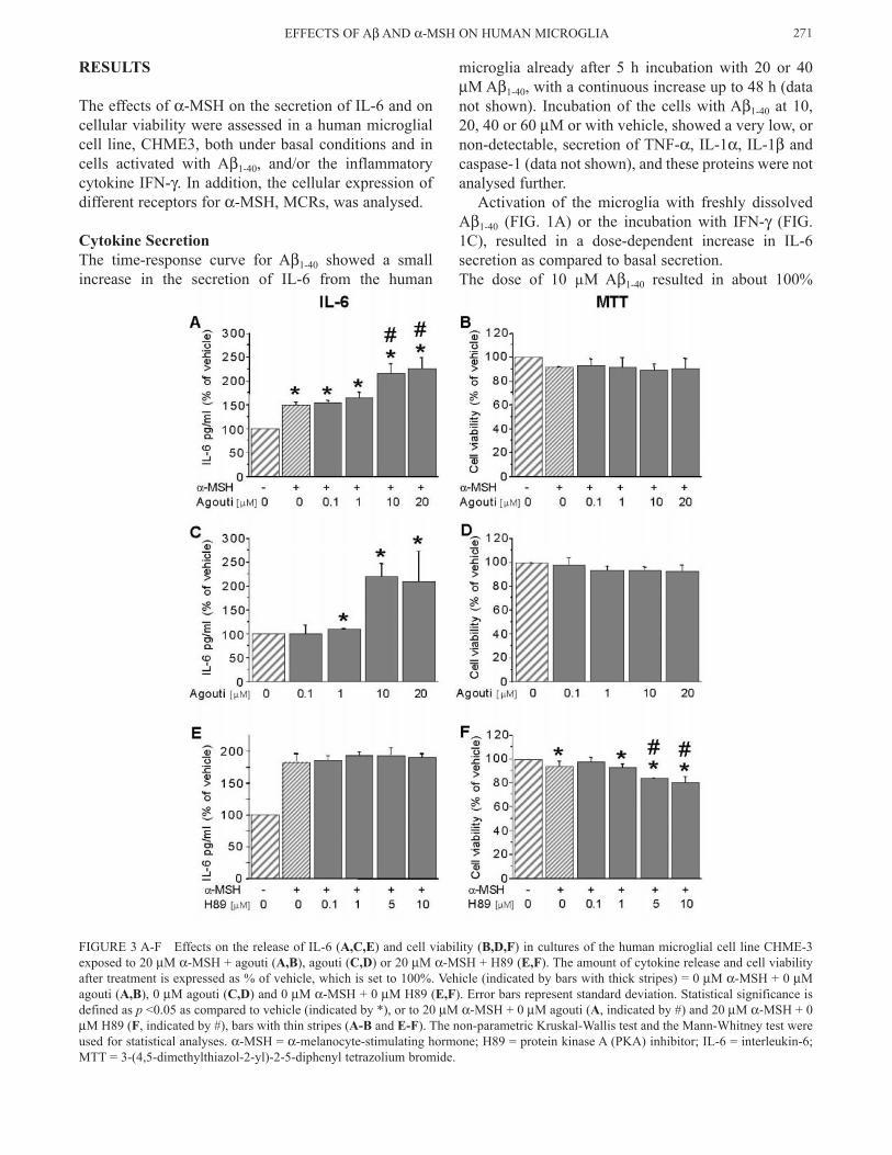
FIGURE 3 A-F Effects on the release of IL-6 (A,C,E) and cell viability (B,D,F) in cultures of the human microglial cell line CHME-3 exposed to 20 µM α-MSH + agouti (A,B), agouti (C,D) or 20 µM α-MSH + H89 (E,F). The amount of cytokine release and cell viability after treatment is expressed as % of vehicle, which is set to 100%. Vehicle (indicated by bars with thick stripes) = 0 µM α-MSH + 0 µM agouti (A,B), 0 µM agouti (C,D) and 0 µM α-MSH + 0 µM H89 (E,F). Error bars represent standard deviation. Statistical significance is defined as p <0.05 as compared to vehicle (indicated by *), or to 20 µM α-MSH + 0 µM agouti (A, indicated by #) and 20 µM α-MSH + 0 µM H89 (F, indicated by #), bars with thin stripes (A-B and E-F). The non-parametric Kruskal-Wallis test and the Mann-Whitney test were used for statistical analyses. α-MSH = α-melanocyte-stimulating hormone; H89 = protein kinase A (PKA) inhibitor; IL-6 = interleukin-6; MTT = 3-(4,5-dimethylthiazol-2-yl)-2-5-diphenyl tetrazolium bromide.

increase in IL-6 release, and 30-40% reduction in cell viability (FIGs. 1 and 2). This dose of Aβ (10 µM) was used in the co-stimulatory experiments, since higher doses of Aβ1-40 resulted in considerably larger reduction in cell viability. Co-stimulation of the microglia with Aβ1-40 (10 µM) and IFN-γ (1-100 ng/ml) did not result in a further increase in the IL-6 secretion, as compared to incuba-tion with either Aβ1-40 or IFN-γ alone (see FIG. 1E). α-MSH, at 0.001-100 µM concentration, did not affect the secretion of IL-6 from the human microglial cells in response to Aβ or Aβ + IFN-γ (see FIG. 2C and 2E, respectively), whereas a small reduction in the basal release of IL-6 was observed at low concentra-tions (0.01 and 0.1 µM) of α-MSH (FIG. 2A). Higher concentrations of α-MSH (1 and 20 µM) resulted in a marked increase in the basal IL-6 secretion (FIG. 2A). Incubation with agouti resulted in a dose-dependent increase in the secretion of IL-6 induced by 20 µM α-MSH (FIG. 3A), as well as in the basal secretion of IL-6 (FIG. 3C). The PKA-inhibitor H89 (0.1-10 µM) had no effect on IL-6 release induced by 20 µM α-MSH (FIG. 3E).
Cell ViabilityThe time-response curve for Aβ1-40 showed a marked decrease in cell viability of the human microglia after 24 h incubation at the concentration of 20 and 40 µM Aβ1-40, and was further decreased at 48 h (data not shown). Freshly dissolved Aβ1-40 (5-60 µM) resulted in a decreased viability of the human microglial cells in
a dose-dependent manner (FIG. 1B). Incubation with IFN-γ alone (1-100 ng/ml) did not affect the cell viabil-ity (FIG. 1D), neither did IFN-γ affect the reduction in cell viability induced by 10 µM Aβ1-40 (FIG. 1F). α-MSH at concentrations 0.001-100 µM did not affect basal cell viability (FIG. 2B), nor the reduced cell viability observed after incubation Aβ (FIG. 2D) or Aβ + IFN-γ (FIG. 2F). Co-incubation with agouti (0.1-20 µM) and α-MSH (20 µM) induced a small, although not significant, reduction in the cell viability (FIG. 3B). Incubation with α-MSH (20 µM) and H89 resulted in a small dose-dependent decrease in the cell viability, as compared to vehicle or incubation with 20 µM α-MSH alone (FIG. 3F).
Expression of MCR SubtypesRT-PCR analysis of RNA extracted from cells of the human microglial CHME3 cell line showed expression of MCR1, MCR3, MCR4 and MCR5 mRNA (FIG. 4). MCR4 mRNA was constitutively expressed in all of the 6 different passages analysed, whereas mRNA for MCR1 (4 of 6 experiments), MCR3 (1 of 6) and MCR5 (3 of 6) had varying expression. The size of the PCR products for each MCR subtype corresponded to the expected size, i.e., 416 bp for MCR1, 366 bp for MCR3, 566 bp for MCR4, and 357 bp for MCR5, and to the size of the positive controls consisting of genomic DNA from the same human microglial cell line (FIG. 4). No signal was observed in the negative controls treated with DNase (not shown).
C. LINDBERG et al.272
FIGURE 4 A-B RT-PCR analysis of total RNA extracted from cells of the human microglial cell line CHME3 using primers specific for the melanocortin receptor (MCR) subtypes MCR1, MCR3, MCR4 and MCR5, respectively. (A) The size of the bands is indicated by the number of base pairs (bp). Lane 1 = base pair ladder; lanes 2, 4, 6 and 8 = PCR-product from genomic DNA for MCR1, 3, 4 and 5, respectively; lanes 3, 5, 7 and 9 = RT-PCR-product from extracted RNA-samples for MCR1, 3, 4 and 5, respectively. (B) The figure demonstrates the same experiment after longer exposure to visualise the weak band for MCR3 mRNA. PC = positive control, i.e., genomic DNA extracted from the CHME3 microglial cells; S = sample of RNA extracted from the CHME3 microglial cells.

DISCUSSION
The presence of activated microglia within and around senile plaques suggests that Aβ, the major component in plaques, is involved in the activation of microglial cells in AD. In vitro studies provide evidence that Aβ peptides induce cytokine synthesis and release from microglial cells (Araujo and Cotman, 1992; Meda et al., 1995; Murphy et al., 1998; Combs et al., 2001; Szczepanik et al., 2001; Lee et al., 2002; Takata et al., 2003; Lindberg et al., 2005). In the present study, we have investigated Aβ1-40-induced activation of the human CHME3 microglial cell line and the effects thereupon of α-MSH, a neuropeptide with anti-inflam-matory properties (see Catania and Lipton, 1998), with regard to cytokine production and cell viability. Freshly dissolved Aβ1-40 stimulated the release of IL-6 from these cells in a dose-dependent manner, in accordance with studies using pre-aggregated Aβ1-42 on human pri-mary microglia (Lue et al., 2001). α-MSH did not affect the release of IL-6 from human microglial cells stimulated with Aβ1-40, or with Aβ1-40 and IFN-γ. In studies on the aggregated form of Aβ1-42, the major form in amyloid plaques in the AD brain, α-MSH had an inhibitory effect on the release of TNF-α and NO (Galimberti et al., 1999). Furthermore, α-MSH reduced the release of IL-6, TNF-α and NO, from murine microglia activated with LPS and IFN-γ (Delgado et al., 1998). A possible explanation for the different effects of α-MSH is a species difference. To our knowledge, studies on the effects of α-MSH on microglia have, so far, been performed exclusively on murine cells (Delgado et al., 1998; Galimberti et al., 1999). Furthermore, the effects of α-MSH on Aβ-induced cytokine release may depend on the Aβ-form used. Indeed, previous studies on the effects of Aβ-pep-tides on microglia from different species have shown differential effects on cytokine release, depending on the aggregational form, and on the use of either Aβ1-40 or Aβ1-42 (Araujo and Cotman, 1992; Meda et al., 1995; Murphy et al., 1998; Combs et al., 2001; Szczepanik et al., 2001; Lee et al., 2002; Takata et al., 2003; Lindberg et al., 2005). Freshly dissolved Aβ1-40 was chosen for the present study since freshly dissolved Aβ-peptide was more potent than the fibrillar form and a further aggregated form, so called βamy balls (Westlind-Danielsson and Arnerup, 2001), in stimulating the release of pro-inflammatory cytokines from primary rat microglial cultures (Lindberg et al., 2005). Aβ peptides are highly aggregational-prone, Aβ1-42 more so than Aβ1-40 (Jarrett et al., 1993). The preparation of freshly dissolved Aβ1-40 used in the present study was shown previously to contain mainly oligomeric forms, such as
tetramers, hexamers, dodecamers and 24-mers, while the preparation of freshly dissolved Aβ1-42 was shown to contain a mixture of oligomers and fibrils during 24h of incubation in culture medium (Lindberg et al., 2005). Based on the findings that the oligomeric forms, i.e., so called Aβ-derived diffusible ligands (ADDLs), believed to represent the most neurotoxic forms (Aksenova et al., 1996; Hartley et al., 1999; Dahlgren et al., 2002; Selkoe, 2002; Hoshi et al., 2003), Aβ1-40, and not Aβ1-42, was chosen for the present study. The human microglial cell line responded to the preparation of freshly dissolved Aβ1-40 with a markedly reduced cell viability. This reduction in cell viability was not affected by IFN-γ or α-MSH. Interestingly, inhibition of PKA with H89, together with α-MSH, gave rise to a decrease in cell viability. PKA, a downstream mediator of MCR signalling, has been shown to activate murine microglial cells not previously activated (Min et al., 2004). However, inhibition of PKA did not reduce the α-MSH-induced release of IL-6 from the human microglia. These findings indicate that PKA-signalling is involved in the reduction of cell viability, but not in mediating the release of IL-6. Although increased cAMP-signalling upon α-MSH stimulation inhibits the activation of murine microglia (Konda et al., 1994; Delgado et al., 1998), increased cAMP may have both stimulatory (Pyo et al., 1999; Marcus et al., 2003; Woo et al., 2004) and inhibitory (Yoshimura et al., 1997; Si et al., 1998) effects on the release of inflammatory factors. Monocytes have previously been shown to express the MCR1, 3 and 5 subtypes (Rajora et al., 1996; Ichiyama et al., 2000), and we now show the expres-sion of mRNA for these receptors, as well as MCR4, in the human microglial cell line, suggesting that all of these receptors are possible candidates for mediat-ing the effects of α-MSH on these cells. However, the endogenous antagonist of α-MSH-mediated activ-ity, agouti, specific for MCR1 and MCR4 (Dinulescu and Cone, 2000), did not inhibit the α-MSH-induced release of IL-6. Instead, agouti further increased the α-MSH-induced release of IL-6. These findings would seem to indicate that α-MSH-induced secretion of IL-6 was mediated by MCR3 and/or MCR5. Agouti has some affinity for both of these MCRs (Dinulescu and Cone, 2000), but further studies will be necessary to establish whether it is sufficient to affect the α-MSH-induced stimulation of the human microglia. There is ample evidence for anti-inflammatory effects of α-MSH (see Catania and Lipton, 1998), and the present finding that α-MSH stimulates, rather than inhibits the release of IL-6, is intriguing. Many stud-ies show neuroprotective and regenerative activities of α-MSH (see Gispen et al., 1994; Strand, 1999; Catania
EFFECTS OF Aβ AND α-MSH ON HUMAN MICROGLIA 273

et al., 2004) as well as of IL-6 (Maeda et al., 1994; Loddick et al., 1998; Streit et al., 2000; Poulsen et al., 2005) in the nervous system. Recent studies have dem-onstrated neuroprotective effects of α-MSH in models of cerebral ischemia (Huh et al., 1997; Hwang et al., 2004; Forslin Aronsson et al., submitted). Thus, it may be hypothesised that α-MSH, through the release of IL-6 from microglial cells, can have beneficial effects on neurons. The microglial cell line, CHME3, used in the pres-ent studies has previously been reported to express receptors for IFN-γ and to respond with increased production of major histocompatibility (MHC) class II molecules upon stimulation with IFN-γ (Janabi et al., 1995). The activation of these cells by IFN-γ was supported by our results showing a dose-dependent increase in the release of IL-6. However, unlike earlier studies on murine microglia (Gasic-Milenkovic et al., 2003), there was no potentiating effect of IFN-γ on the Aβ1-40-induced secretion of IL-6. This could be due to the fact that IFN-γ and IL-6 share the same signalling pathway, i.e., the JAK/STAT pathway (Heinrich et al., 1998; Platanias and Fish, 1999; Kerr et al., 2003). Previous characterisation of the CHME3 cells has also demonstrated that they have the same repertoire of che-mokine receptors as primary cultures of adult human microglia (Flynn et al., 2003), and that their responses to chemokines are similar to those of primary cultures of adult rat microglia (Cross and Woodroofe, 1999). This is interesting in view of the findings of increased chemokine levels in CSF samples from AD patients (Galimberti et al., 2003), and supports the relevance of using this cell line as a model for human microglia. The results from the present study support the hypoth-esis that Aβ1-40 activates microglial cells to produce IL-6 in the AD brain, which could further stimulate the production of Aβ1-40, via stimulation of APP synthesis and processing (Del Bo et al., 1995). This would pro-mote chronic inflammation that is believed to be part of AD brain pathology. Unexpectedly, α-MSH was not found to inhibit the release of IL-6, but resulted in increased basal secretion from the microglial cells. Further studies will be required to clarify whether or not this effect is responsible for the neuroprotective activities of α-MSH.
Acknowledgements
This work was supported by grants from the Swedish Research Council (072194), Stiftelsen för Gamla Tjänarinnor, Gun och Bertil Stohnes Stiftelse, Insamlingsstiftelsen för Alzheimer-och Demensforskn-
ing, Stiftelsen för åldersforskning and Stiftelsen Demensfonden.
References
Adan RA and WH Gispen (1997) Brain melanocortin receptors: from cloning to function. Peptides 18, 1279-1287.
Aksenova MV, MY Aksenov, DA Butterfield and JM Carney (1996) α-1-antichymotrypsin interaction with Aβ (1-40) inhibits fibril formation but does not affect the peptide toxicity. Neurosci. Lett. 211, 45-48.
Araujo DM and CW Cotman (1992) β-amyloid stimulates glial cells in vitro to produce growth factors that accumulate in senile plaques in Alzheimer's disease. Brain Res. 569, 141-145.
Bauer J, S Strauss, U Schreiter-Gasser, U Ganter, P Schlegel, I Witt, B Yolk and M Berger (1991) Interleukin-6 and α-2-macroglobu-lin indicate an acute-phase state in Alzheimer's disease cortices. FEBS Lett. 285, 111-114.
Benveniste EN, VT Nguyen and GM O'Keefe (2001) Immunological aspects of microglia: relevance to Alzheimer's disease. Neurochem. Int. 39, 381-391.
Blum-Degen D, T Muller, W Kuhn, M Gerlach, H Przuntek and P Riederer (1995) Interleukin-1β and interleukin-6 are elevated in the cerebrospinal fluid of Alzheimer's and de novo Parkinson's disease patients. Neurosci. Lett. 202, 17-20.
Böhm M, M Schiller, S Stander, H Seltmann, Z Li, T Brzoska, D Metze, HB Schiöth, A Skottner, K Seiffert, CC Zouboulis and TA Luger (2002) Evidence for expression of melanocortin-1 recep-tor in human sebocytes in vitro and in situ. J. Invert. Dermatol. 118, 533-539.
Cacabelos R, M Barquero, P Garcia, XA Alvarez and E Varela de Seijas (1991) Cerebrospinal fluid interleukin-1 beta (IL-1β) in Alzheimer's disease and neurological disorders. Methods Fund. Exp. Clin. Pharmacol. 13, 455-458.
Cammas FM, S Kapas, S Barker and AJ Clark (1995) Cloning, characterization and expression of a functional mouse ACTH receptor. Biochem. Biophys. Res. Commun. 212, 912-918.
Catania A and JM Lipton (1998) Peptide modulation of fever and inflammation within the brain. Ann. NY Acad. Sci. 856, 62-68.
Catania A, S Gatti, G Colombo and JM Lipton (2004) Targeting melanocortin receptors as a novel strategy to control inflamma-tion. Pharmacol. Rev. 56, 1-29.
Combs CK, JC Karlo, SC Kao and GE Landreth (2001) β-Amyloid stimulation of microglia and monocytes results in TNFα-depen-dent expression of inducible nitric oxide synthase and neuronal apoptosis. J. Neurosci. 21, 1179-1188.
Cone RD (2005) Anatomy and regulation of the central melanocor-tin system. Nat. Neurosci. 8, 571-578.
Cross AK and MN Woodroofe (1999) Chemokines induce migra-tion and changes in actin polymerization in adult rat brain microglia and human fetal microglial cell line in vitro. J. Neurosci. Res. 55, 17-23.
Dahlgren KN, AM Manelli, WB Stine Jr, LK Baker, GA Krafft and MJ LaDu (2002) Oligomeric and fibrillar species of amyloid- peptides differentially affect neuronal viability. J. Biol. Chem. 277, 32046-32053.
Del Bo R, N Angeretti, E Lucca, MG De Simoni and G Forloni (1995) Reciprocal control of inflammatory cytokines, IL-1 and IL-6, and β-amyloid production in cultures. Neurosci. Lett. 188, 70-74.
Delgado R, A Carlin, L Airaghi, MT Demitri, L Meda, D Galimberti, P Baron, JM Lipton and A Catania (1998) Melanocortin peptides
C. LINDBERG et al.274

inhibit production of proinflammatory cytokines and nitric oxide by activated microglia. J. Leukoc. Biol. 63, 740-745.
Dickson DW, SC Lee, LA Mattiace, SH Yen and C Brosnan (1993) Microglia and cytokines in neurological disease, with special reference to AIDS and Alzheimer's disease. Glia 7, 75-83.
Dinulescu DM and RD Cone (2000) Agouti and agouti-related pro-tein: analogies and contrasts. J. Biol. Chem. 275, 6695-6698.
Eikelenboom P, SS Zhan, WA van Gool and D Allsop (1994) Inflammatory mechanisms in Alzheimer's disease. Trends Pharmacol. Sci. 15, 447-450.
Flynn G, S Maru, J Loughlin, IA Romero and D Male (2003) Regulation of the chemokine receptor expression in human microglia and aastrocytes. J. Neuroimmunol. 136, 84-93.
Galimberti D, P Baron, L Meda, E Prat, E Scarpini, R Delgado, A Catania, JM Lipton and G Scarlato (1999) α-MSH peptides inhibit production of nitric oxide and tumor necrosis factor-α by microglial cells activated with β-amyloid and interferon γ. Biochem. Biophys. Res. Commun. 263, 251-256.
Galimberti D, N Schoonenboom, E Scarpini and P Scheltens (2003) Chemokines in serum and cerebrospinal fluid of Alzheimer's disease patients. Ann. Neurol. 53, 547-548.
Gasic-Milenkovic J, S Dukic-Stefanovic, W Deuther-Conrad, U Gärtner and G Münch (2003) β-amyloid peptide potentiates inflammatory responses induced by lipopolysaccharide, interfer-on-γ and 'advanced glycation endproducts' in a murine microglia cell line. Eur. J. Neurosci. 17, 813-821.
Gispen WH, J Verhaagen and D Bar (1994) ACTH/MSH-derived peptides and peripheral nerve plasticity: neuropathies, neuropro-tection and repair. Prog. Brain Res. 100, 223-229.
Giulian D, LJ Haverkamp, JH Yu, W Karshin, D Tom, J Li, J Kirkpatrick, LM Kuo and AE Roher (1996) Specific domains of β-amyloid from Alzheimer plaque elicit neuron killing in human microglia. J. Neurosci. 16, 6021-6037.
Glyn JR and JM Lipton (1981) Hypothermic and antipyretic effects of centrally administered ACTH (1-24) and α-melanotropin. Peptides 2, 177-187.
Griffin WS, LC Stanley, C Ling, L White, V MacLeod, LJ Perrot, CL White 3rd and C Araoz (1989) Brain interleukin 1 and S-100 immunoreactivity are elevated in Down syndrome and Alzheimer disease. Proc. Natl. Acad. Sci. USA 86, 7611-7615.
Hampel H, D Schoen, MJ Schwarz, HU Kotter, C Schneider, T Sunderland, R Dukoff, J Levy, F Padberg, S Stubner, K Buch, N Muller and HJ Moller (1997) Interleukin-6 is not altered in cerebrospinal fluid of first-degree relatives and patients with Alzheimer's disease. Neurosci. Lett. 228, 143-146.
Hartley DM, DM Walsh, CP Ye, T Diehl, S Vasquez, PM Vassilev, DB Teplow and DJ Selkoe (1999) Protofibrillar intermediates of amyloid β-protein induce acute electrophysiological changes and progressive neurotoxicity in cortical neurons. J. Neurosci. 19, 8876-8884.
Heinrich PC, I Behrmann, G Muller-Newen, F Schaper and L Graeve (1998) Interleukin-6-type cytokine signalling through the gp130/Jak/STAT pathway. Biochem. J. 334, 297-314.
Hoshi M, M Sato, S Matsumoto, A Noguchi, K Yasutake, N Yoshida and K Sato (2003) Spherical aggregates of β-amyloid (amylospheroid) show high neurotoxicity and activate tau protein kinase I/glycogen synthase kinase-3β. Proc. Natl. Acad. Sci. USA 100, 6370-6375.
Huh SK, JM Lipton and HH Batjer (1997) The protective effects of α-melanocyte stimulating hormone on canine brain stem isch-emia. Neurosurgery 40, 132-139.
Hwang IK, KY Yoo, JK Park, YS Nam, IS Lee, JH Kang, SY Choi, JY Lee, TC Kang and MH Won (2004) Ischemia-related changes
of adrenocorticotropic hormone immunoreactivity and its pro-tective effect in the gerbil hippocampus after transient forebrain ischemia. Neuroscience 126, 871-877.
Ichiyama T, S Sato, K Okada, A Catania and JM Lipton (2000) The neuroimmunomodulatory peptide α-MSH. Ann. NY Acad. Sci. 917, 221-226.
Janabi N, S Peudenier, B Heron, KH Ng and M Tardieu (1995) Establishment of human microglial cell lines after transfection of primary cultures of embryonic microglial cells with the SV40 large T antigen. Neurosci. Lett. 195, 105-108.
Jarrett JT, EP Berger and PT Lansbury Jr (1993) The carboxy termi-nus of the β amyloid protein is critical for the seeding of amyloid formation: implications for the pathogenesis of Alzheimer's dis-ease. Biochemistry 32, 4693-4697.
Kerr IM, AP Costa-Pereira, BF Lillemeier and B Strobl (2003) Of JAKs, STATs, blind watchmakers, jeeps and trains. FEBS Lett. 546, 1-5.
Konda Y, I Gantz, J DelValle, Y Shimoto, H Miwa and T Yamada (1994) Interaction of dual intracellular signaling pathways activated by the melanocortin-3 receptor. J. Biol. Chem. 269, 13162-13166.
Lee YB, A Nagai and SU Kim (2002) Cytokines, chemokines, and cytokine receptors in human microglia. J. Neurosci. Res. 69, 94-103.
Lindberg C, M-L Bardyl Selenica, A Westlind-Danielsson and M Schultzberg (2005) β-amyloid structure determines the nature of cytokine release from rat microglia. J. Mol. Neurosci. 27, 1-12.
Loddick SA, AV Turnbull and NJ Rothwell (1998) Cerebral inter-leukin-6 is neuroprotective during permanent focal cerebral isch-emia in the rat. J. Cereb. Blood Flow Metab. 18, 176-179.
Lue LF, R Rydel, EF Brigham, LB Yang, H Hampel, GM Murphy Jr, L Brachova, SD Yan, DG Walker, Y Shen and J Rogers (2001) Inflammatory repertoire of Alzheimer's disease and nondemented elderly microglia in vitro. Glia 35, 72-79.
Maeda Y, M Matsumoto, O Hori, K Kuwabara, S Ogawa, SD Yan, T Ohtsuki, T Kinoshita, T Kamada and DM Stern (1994) Hypoxia/reoxygenation-mediated induction of astrocyte inter-leukin 6: a paracrine mechanism potentially enhancing neuron survival. J. Exp. Med. 180, 2297-2308.
Marcus JS, SL Karackattu, MA Fleegal and C Sumners (2003) Cytokine-stimulated inducible nitric oxide synthase expression in astroglia: role of Erk mitogen-activated protein kinase and NF-κB. Glia 41, 152-160.
Martinez M, E Fernandez-Vivancos, A Frank, M De la Fuente and A Hernanz (2000) Increased cerebrospinal fluid fas (Apo-1) levels in Alzheimer's disease. Relationship with IL-6 concentrations. Brain Res. 869, 216-219.
McGeer EG and PL McGeer (2003) Inflammatory processes in Alzheimer's disease. Prog. Neuropsychopharmacol. Biol. Psychiatry 27, 741-749.
Meda L, MA Cassatella, GI Szendrei, L Otvos Jr, P Baron, M Villalba, D Ferrari and F Rossi (1995) Activation of microglial cells by β-amyloid protein and interferon-γ. Nature 374, 647-650.
Min KJ, MS Yang, I Jou and EH Joe (2004) Protein kinase A medi-ates microglial activation induced by plasminogen and ganglio-sides. Exp. Mol. Med. 36, 461-467.
Mountjoy KG, LS Robbins, MT Mortrud and RD Cone (1992) The cloning of a family of genes that encode the melanocortin recep-tors. Science 257, 1248-1251.
Murphy GM Jr, L Yang and B Cordell (1998) Macrophage colony-stimulating factor augments β-amyloid-induced interleukin-1, interleukin-6, and nitric oxide production by microglial cells. J.
EFFECTS OF Aβ AND α-MSH ON HUMAN MICROGLIA 275

Biol. Chem. 273, 20967-20971.Neuroinflammation working group, H Akiyama, S Barger, S
Barnum, B Bradt, J Bauer, GM Cole, NR Cooper, P Eikelenboom, M Emmerling, BL Fiebich, CE Finch, S Frautschy, WS Griffin, H Hampel, M Hull, G Landreth, L Lue, R Mrak, IR Mackenzie, PL McGeer, MK O'Banion, J Pachter, G Pasinetti, C Plata-Salaman, J Rogers, R Rydel, Y Shen, W Streit, R Strohmeyer, I Tooyoma, FL Van Muiswinkel, R Veerhuis, D Walker, S Webster, B Wegrzyniak, G Wenk and T Wyss-Coray (2000) Inflammation and Alzheimer's disease. Neurobiol. Aging 21, 383-421.
Oka T, K Oka, M Hosoi and T Hori (1996) Inhibition of peripheral interleukin-1β-induced hyperalgesia by the intracerebroventric-ular administration of diclofenac and α-melanocyte-stimulating hormone. Brain Res. 736, 237-242.
Platanias LC and EN Fish (1999) Signaling pathways activated by interferons. Exp. Hematol. 27, 1583-1592.
Poulsen CB, M Penkowa, R Borup, FC Nielsen, M Caceres, A Quintana, A Molinero, J Carrasco, M Giralt and J Hidalgo (2005) Brain response to traumatic brain injury in wild-type and interleukin-6 knockout mice: a microarray analysis. J. Neurochem. 92, 417-432.
Pyo H, I Jou, S Jung and E Joe (1999) cAMP potentiates β-amy-loid-induced nitric oxide release from microglia. Neuroreport 10, 37-40.
Rajora N, G Ceriani, A Catania, RA Star, MT Murphy and JM Lipton (1996) alpha-MSH production, receptors, and influence on neopterin in a human monocyte/macrophage cell line. J. Leukoc. Biol. 59, 248-253.
Selkoe DJ (1994) Cell biology of the amyloid β-protein precursor and the mechanism of Alzheimer's disease. Annu. Rev. Cell Biol. 10, 373-403.
Selkoe DJ (2002) Deciphering the genesis and fate of amyloid β-protein yields novel therapies for Alzheimer disease. J. Clin. Invest. 110, 1375-1381.
Si Q, Y Nakamura, T Ogata, K Kataoka and P Schubert (1998) Differential regulation of microglial activation by propentofyl-line via cAMP signaling. Brain Res. 812, 97-104.
Strand FL (1999) New vistas for melanocortins. Finally, an expla-nation for their pleiotropic functions. Ann. NY Acad. Sci. 897, 1-16.
Streit WJ, SD Hurley, TS McGraw and SL Semple-Rowland
(2000) Comparative evaluation of cytokine profiles and reactive gliosis supports a critical role for interleukin-6 in neuron-glia signaling during regeneration. J. Neurosci. Res. 61, 10-20.
Szczepanik AM, D Rampe and GE Ringheim (2001) Amyloid-β peptide fragments p3 and p4 induce pro-inflammatory cytokine and chemokine production in vitro and in vivo. J. Neurochem. 77, 304-317.
Takata K, Y Kitamura, M Umeki, D Tsuchiya, J Kakimura, T Taniguchi, PJ Gebicke-Haerter and S Shimohama S (2003) Possible involvement of small oligomers of amyloid-β peptides in 15-deoxy-∆12,14 prostaglandin J2-sensitive microglial acti-vation. J. Pharmacol. Sci. 91, 330-333.
Tarkowski E, K Blennow, A Wallin and A Tarkowski (1999) Intracerebral production of tumor necrosis factor-α, a local neu-roprotective agent, in Alzheimer disease and vascular dementia. J. Clin. Immunol. 19, 223-230.
Tatro JB (1996) Receptor biology of the melanocortins, a family of neuroimmunomodulatory peptides. Neuroimmunomodulation 3, 259-284.
Vandenabeele P and W Fiers (1991) Is amyloidogenesis during Alzheimer's disease due to an IL-1-/IL-6-mediated 'acute phase response' in the brain? Immunol. Today 12, 217-219.
Vehmas AK, CH Kawas, WF Stewart and JC Troncoso (2003) Immune reactive cells in senile plaques and cognitive decline in Alzheimer's disease. Neurobiol. Aging 24, 321-331.
Westlind-Danielsson A and G Arnerup (2001) Spontaneous in vitro formation of supramolecular β-amyloid structures, "βamy balls", by β-amyloid 1-40 peptide. Biochemistry 40, 14736-14743.
Wikberg JE (1999) Melanocortin receptors: perspectives for novel drugs. Eur. J. Pharmacol. 375, 295-310.
Woo MS, SH Jung, JW Hyun and HS Kim (2004) Differential regulation of inducible nitric oxide synthase and cytokine gene expression by forskolin and dibutyryl-cAMP in lipopolysac-charide-stimulated murine BV2 microglial cells. Neurosci. Lett. 356, 187-190.
Yoshimura T, C Kurita, T Nagao, E Usami, T Nakao, S Watanabe, J Kobayashi, F Yamazaki, H Tanaka, N Inagaki and H Nagai (1997) Inhibition of tumor necrosis factor-α and interleukin-1-β production by β-adrenoceptor agonists from lipopolysac-charide-stimulated human peripheral blood mononuclear cells. Pharmacology 54, 144-152.
C. LINDBERG et al.276
Copyright © 2022 FDOKUMEN