Keratinocyte-Melanocyte Interactions During Melanosome Transfer
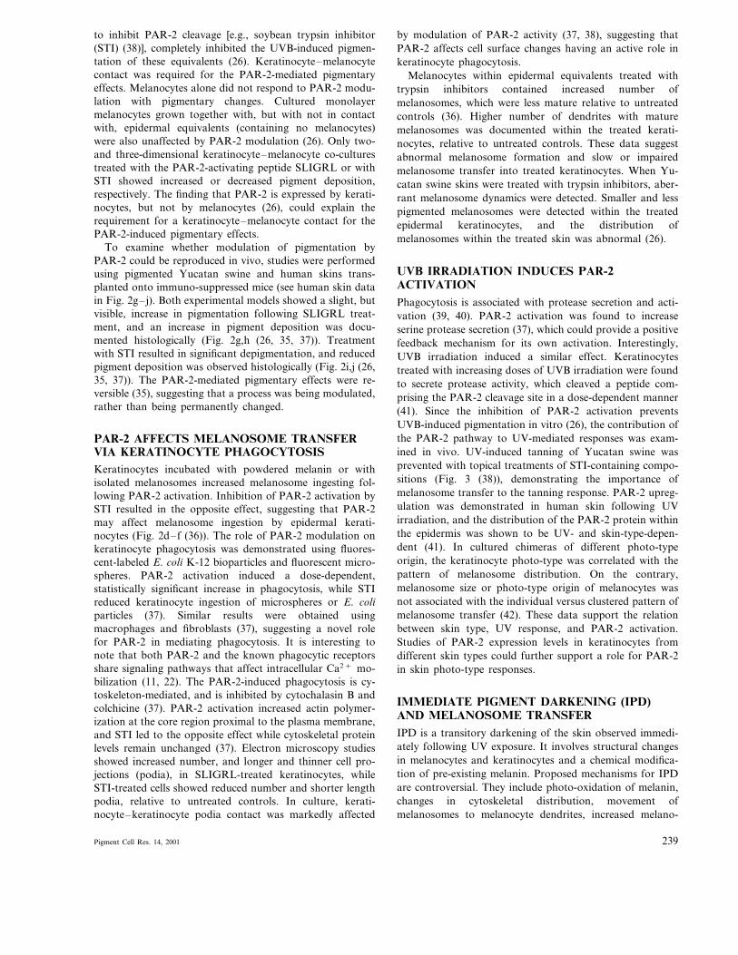
7
Copyright © Pigment Cell Res 2001 PIGMENT CELL RES 14: 236–242. 2001 ISSN 0893-5785 Printed in Ireland —all rights resered Invited Review Keratinocyte – Melanocyte Interactions During Melanosome Transfer MIRI SEIBERG Skin Research Center, J&J CPWW, Skillman, New Jersey *Address reprint requests to Dr. Miri Seiberg, Skin Research Center, J&J CPWW, 199 Grandiew Road, Skillman, New Jersey 08558. E-mail: mseiber@cpcus.jnj.com Received 14 May 2001; in final form 22 May 2001 The epidermal – melanin unit is composed of one melanocyte sis theory. PAR-2 controls melanosome ingestion and phago- cytosis by keratinocytes and exerts a regulatory role in skin and approximately 36 neighboring keratinocytes, working in pigmentation. Modulation of PAR-2 activity can enhance or synchrony to produce and distribute melanin. Melanin is decrease melanosome transfer and affects pigmentation only synthesized in melanosomes, transferred to the dendrite tips, when there is keratinocyte – melanocyte contact. Moreover, and translocated into keratinocytes, forming caps over the keratinocyte nuclei. The molecular and cellular mechanisms PAR-2 is induced by UV irradiation and inhibition of PAR-2 activation results in the prevention of UVB-induced tanning. involved in melanosome transfer and the keratinocyte – melanocyte interactions required for this process are not yet The role of PAR-2 in mediating UV-induced responses re- mains to be elucidated. completely understood. Suggested mechanisms of melanosome transfer include melanosome release and endocytosis, direct inoculation (‘injection’), keratinocyte – melanocyte membrane Key words: Melanosome transfer, PAR-2, Phagocytosis, Keratinocyte, Melanocyte fusion, and phagocytosis. Studies of the keratinocyte receptor protease-activated receptor-2 (PAR-2) support the phagocyto- nisms for melanosome transfer. These include the release of melanosomes into intercellular spaces followed by endocyto- sis, direct inoculation (‘injection’), keratinocyte – melanocyte membrane fusion, and phagocytosis. Using time-lapse mi- croscopy, Okazaki et al. (7) observed dendrites enfolded by recipient keratinocytes, which are then pinched off to form a cluster of melanosomes. The internalized dendrites were decomposed, leaving each melanosome aggregate sur- rounded by a single keratinocyte membrane. Single large melanosomes, or complexes of small melanosomes, were then dispersed from the aggregate into the keratinocyte cytoplasm, in a skin-type-dependent process defined as cy- tophagocytosis. Electron microscopy studies of photo-dam- aged Caucasian facial skin (8) suggested a similar mechanism, as well as keratinocyte – melanocyte membrane fusion, and exocytosis of single melanosomes (reviewed in (5)). However, a mechanistic understanding of the cellular interactions involved in pigment transfer has not been described. MELANOSOME TRANSFER, EARLY EM-BASED STUDIES The epidermal – melanin unit, a functional unit that pro- duces and distributes melanin, is composed of one melanocyte and approximately 36 neighboring keratinocytes (reviewed in (1, 2)). The process of melanogenesis is well documented and the regulation of pigment production by melanocytes has been heavily investigated (reviewed in (3)). Pigment-loaded melanosomes translocate from their site of origin in the perinuclear cytoplasm towards the melanocyte dendrite tips using microtubule-based and actin-based mo- tor proteins (4–6). They are then transferred into the recip- ient keratinocytes, where they are dispersed into the cytoplasm. However, the actual transfer of melanosomes into keratinocytes and the keratinocyte – melanocyte interac- tions during the transfer process are not well characterized. Early light and electron microscopy studies documented the membrane layering and organization during melanosome transfer, suggesting numerous possible mecha- Abbreiations – PAR, protease-activated receptor-2; STI, soybean trypsin inhibitor; IPD, immediate pigment darkening Pigment Cell Res. 14, 2001 236
-
Upload
independent -
Category
Documents
-
view
1 -
download
0
Transcript of Keratinocyte-Melanocyte Interactions During Melanosome Transfer

Copyright © Pigment Cell Res 2001PIGMENT CELL RES 14: 236–242. 2001ISSN 0893-5785Printed in Ireland—all rights reser�ed
Invited Review
Keratinocyte–Melanocyte Interactions During Melanosome Transfer
MIRI SEIBERG
Skin Research Center, J&J CPWW, Skillman, New Jersey*Address reprint requests to Dr. Miri Seiberg, Skin Research Center, J&J CPWW, 199 Grand�iew Road, Skillman, New Jersey 08558.E-mail: [email protected]
Received 14 May 2001; in final form 22 May 2001
The epidermal–melanin unit is composed of one melanocyte sis theory. PAR-2 controls melanosome ingestion and phago-cytosis by keratinocytes and exerts a regulatory role in skinand approximately 36 neighboring keratinocytes, working inpigmentation. Modulation of PAR-2 activity can enhance orsynchrony to produce and distribute melanin. Melanin isdecrease melanosome transfer and affects pigmentation onlysynthesized in melanosomes, transferred to the dendrite tips,when there is keratinocyte–melanocyte contact. Moreover,and translocated into keratinocytes, forming caps over the
keratinocyte nuclei. The molecular and cellular mechanisms PAR-2 is induced by UV irradiation and inhibition of PAR-2activation results in the prevention of UVB-induced tanning.involved in melanosome transfer and the keratinocyte–
melanocyte interactions required for this process are not yet The role of PAR-2 in mediating UV-induced responses re-mains to be elucidated.completely understood. Suggested mechanisms of melanosome
transfer include melanosome release and endocytosis, directinoculation (‘injection’), keratinocyte–melanocyte membrane Key words: Melanosome transfer, PAR-2, Phagocytosis,
Keratinocyte, Melanocytefusion, and phagocytosis. Studies of the keratinocyte receptorprotease-activated receptor-2 (PAR-2) support the phagocyto-
nisms for melanosome transfer. These include the release ofmelanosomes into intercellular spaces followed by endocyto-sis, direct inoculation (‘injection’), keratinocyte–melanocytemembrane fusion, and phagocytosis. Using time-lapse mi-croscopy, Okazaki et al. (7) observed dendrites enfolded byrecipient keratinocytes, which are then pinched off to forma cluster of melanosomes. The internalized dendrites weredecomposed, leaving each melanosome aggregate sur-rounded by a single keratinocyte membrane. Single largemelanosomes, or complexes of small melanosomes, werethen dispersed from the aggregate into the keratinocytecytoplasm, in a skin-type-dependent process defined as cy-tophagocytosis. Electron microscopy studies of photo-dam-aged Caucasian facial skin (8) suggested a similarmechanism, as well as keratinocyte–melanocyte membranefusion, and exocytosis of single melanosomes (reviewed in(5)). However, a mechanistic understanding of the cellularinteractions involved in pigment transfer has not beendescribed.
MELANOSOME TRANSFER, EARLYEM-BASED STUDIESThe epidermal–melanin unit, a functional unit that pro-duces and distributes melanin, is composed of onemelanocyte and approximately 36 neighboring keratinocytes(reviewed in (1, 2)). The process of melanogenesis is welldocumented and the regulation of pigment production bymelanocytes has been heavily investigated (reviewed in (3)).Pigment-loaded melanosomes translocate from their site oforigin in the perinuclear cytoplasm towards the melanocytedendrite tips using microtubule-based and actin-based mo-tor proteins (4–6). They are then transferred into the recip-ient keratinocytes, where they are dispersed into thecytoplasm. However, the actual transfer of melanosomesinto keratinocytes and the keratinocyte–melanocyte interac-tions during the transfer process are not well characterized.
Early light and electron microscopy studies documentedthe membrane layering and organization duringmelanosome transfer, suggesting numerous possible mecha-
Abbre�iations – PAR, protease-activated receptor-2; STI, soybean trypsin inhibitor; IPD, immediate pigment darkening
Pigment Cell Res. 14, 2001236

PHAGOCYTOSISPhagocytosis is the cellular internalization of particulatematerial of at least 0.5–1 �m in diameter. Phagocytosis isreceptor-mediated, and receptor activation leads to localreorganization of the actin cytoskeleton (9). Known phago-cytic receptors, such as the Fc� receptors, the complementreceptor 3, and the fibronectin receptors (reviewed in (9, 10)),activate tyrosine and serine– threonine kinases, mobilizeCa2+ from intracellular storage to the cytoplasm, and acti-vate arachidonate metabolism (reviewed in (11)). Phagocyto-sis is usually associated with macrophages, neutrophils, andmonocytes, all ‘professional’ phagocytes functioning to elim-inate infectious agents, apoptotic cells, and cellular debris(reviewed in (12)). However, epidermal keratinocytes wereshown to experimentally ingest latex beads (13), demonstrat-ing their phagocytic ability. The mechanism and regulationof keratinocyte phagocytosis and its role in epidermalhomeostasis are not yet completely understood.
PROTEASE-ACTIVATED RECEPTOR-2 (PAR-2)The PAR-2 (14) is a seven-transmembrane G-protein-cou-pled receptor that is related to but distinct from the thrombinreceptors (TRs, also called PAR-1, PAR-3, and PAR-4(15–17)). Cleavage of the extracellular domain of PARs byserine proteases exposes new N-termini, which then act astethered ligands (top panel of Fig. 1). Both TRs and PAR-2are activated by trypsin, but only TRs could be activated bythrombin (14, 18) and only PAR-2 is activated by mast celltryptase (19). Using synthetic peptides that correspond to thenewly created N-termini, PARs could also be activatedwithout receptor cleavage. SLIGRL and SLIGKV, themouse and human PAR-2-activating peptides, are specific forPAR-2 activation and are equipotent in the activation of thehuman PAR-2 (20, 21). The cleaved-activated receptors arephysically coupled to their tethered activating ligands. There-
fore, efficient mechanisms for signal termination were devel-oped, including cleavage of the tethered ligand, receptorphosphorylation and uncoupling from G-proteins, and endo-cytosis and degradation of the activated receptors (11, 22).The structure– function relationship between PAR-2 and itspeptide antagonists is not fully understood. Reduction ofpeptide length, chemical modifications, and amino acid sub-stitution studies yield different results in different biologicalsystems, suggesting tissue-specific PAR-2 subtypes (23). Theeffect of antagonist binding on receptor structure has notbeen studied, and the earliest events documented upon allPARs activation are Ca2+ mobilization and IP3 hydrolysis(reviewed in (22, 23)). PARs are expressed in multiple typesof cells and tissues and are involved in growth and develop-ment, mitogenesis, inflammatory response regulation, malig-nant transformation, vascular tonus, and blood pressureregulation. PAR-2 is expressed in keratinocytes (24, 25), butnot in melanocytes (26).
MELANOSOME TRANSFER IN EPIDERMALEQUIVALENTSThe experimental reconstruction of the epidermal melaninunit in vitro was pivotal to melanosome transfer studies.Using the air– liquid interface system (27), differentiatedkeratinocytes were grown in the presence of melanocytes,enabling scientists to combine microscopy with cellular andmolecular studies. Using three-dimensional keratinocyte–melanocyte cultures, Herlyn and Shih (28) suggested thatpigment donation occurs through the uptake of dendritefragments and phagocytosis of individual melanosomes, the-orizing that keratinocyte-produced factors regulate this pro-cess (29). Bessou et al. documented an increase inmelanosome transfer following UVB irradiation of reconsti-tuted epidermal cultures (30). Melanosomes, either individu-ally or in clumps, were demonstrated both in the basal layers
Fig. 1. A schematic representationof PAR-2 activation andinhibition and the effects onkeratinocyte phagocytosis andmelanosome transfer.
Pigment Cell Res. 14, 2001 237

Fig. 2. PAR-2 modulation affects pigmentation. (a)– (c) Keratinocyte–melanocyte monolayer co-cultures were treated with SLIGRL (10�M) or with STI (0.01%) for 3 days and stained with DOPA on the fourth day. (d)– (f) HaCaT keratinocytes were treated as indicated above,followed by incubation with isolated melanosomes, extensive washing, and Fontana & Masson (F&M) staining. (g)– (h) Lightly pigmentedhuman skins grafted on SCID mice were treated with vehicle (g) or SLIGRL (50 �M, h) for 8 or 9 weeks. F&M-stained histological sectionsdemonstrate increased pigment deposition and enhanced cap formation following SLIGRL treatment. Bar=10 �m. (i)– (j) Heavilypigmented human skins grafted on SCID mice were treated with vehicle (j) or STI (0.1%, i) for 8 or 9 weeks. F&M-stained histologicalsections demonstrate reduced pigment deposition following STI treatment.
and in the stratum corneum of these skin equivalents.Epidermal equivalents of different skin photo-types andchimeras were introduced next and were also generated fromouter root sheath cells combined with melanocytes (31, 32).The in vitro formation of nuclear melanin caps was alsoreported in epidermal equivalents, completing the process ofskin pigmentation in the reconstituted system (33). To-gether, these studies demonstrate melanosome synthesis,transport to keratinocytes, capping of keratinocyte nuclei,and tanning in epidermal equivalents, enabling the use ofsuch systems for mechanistic studies.
PAR-2 AFFECTS PIGMENTATIONStudies of skin homeostasis demonstrate an important bal-ance between serine proteases and their inhibitors, at both
structural and signaling levels (24, 34, 35). In the search forpotential interactions between G-protein-coupled signalingpathways and serine proteases in the skin, PARs wereevaluated for their possible effect on pigment modulation.PAR-2 was found to regulate pigmentation (Fig. 2a–c) byaffecting keratinocyte phagocytosis (Fig. 1 (26, 36–38)).PAR-2 activation increases the ability of keratinocytes toingest melanosomes (Fig. 2d– f), in a process that requireskeratinocyte–melanocyte contact. PAR-2 activation leads toskin darkening, and inhibition of PAR-2 activation by serineprotease inhibitors reduces pigment transfer and leads todepigmentation (Fig. 2g– j).
Epidermal equivalents containing melanocytes respond toPAR-2 activation by darkening, similar to their responsefollowing UVB irradiation (26). Trypsin inhibitors, known
Pigment Cell Res. 14, 2001238
to inhibit PAR-2 cleavage [e.g., soybean trypsin inhibitor(STI) (38)], completely inhibited the UVB-induced pigmen-tation of these equivalents (26). Keratinocyte–melanocytecontact was required for the PAR-2-mediated pigmentaryeffects. Melanocytes alone did not respond to PAR-2 modu-lation with pigmentary changes. Cultured monolayermelanocytes grown together with, but with not in contactwith, epidermal equivalents (containing no melanocytes)were also unaffected by PAR-2 modulation (26). Only two-and three-dimensional keratinocyte–melanocyte co-culturestreated with the PAR-2-activating peptide SLIGRL or withSTI showed increased or decreased pigment deposition,respectively. The finding that PAR-2 is expressed by kerati-nocytes, but not by melanocytes (26), could explain therequirement for a keratinocyte–melanocyte contact for thePAR-2-induced pigmentary effects.
To examine whether modulation of pigmentation byPAR-2 could be reproduced in vivo, studies were performedusing pigmented Yucatan swine and human skins trans-planted onto immuno-suppressed mice (see human skin datain Fig. 2g– j). Both experimental models showed a slight, butvisible, increase in pigmentation following SLIGRL treat-ment, and an increase in pigment deposition was docu-mented histologically (Fig. 2g,h (26, 35, 37)). Treatmentwith STI resulted in significant depigmentation, and reducedpigment deposition was observed histologically (Fig. 2i,j (26,35, 37)). The PAR-2-mediated pigmentary effects were re-versible (35), suggesting that a process was being modulated,rather than being permanently changed.
PAR-2 AFFECTS MELANOSOME TRANSFERVIA KERATINOCYTE PHAGOCYTOSISKeratinocytes incubated with powdered melanin or withisolated melanosomes increased melanosome ingesting fol-lowing PAR-2 activation. Inhibition of PAR-2 activation bySTI resulted in the opposite effect, suggesting that PAR-2may affect melanosome ingestion by epidermal kerati-nocytes (Fig. 2d– f (36)). The role of PAR-2 modulation onkeratinocyte phagocytosis was demonstrated using fluores-cent-labeled E. coli K-12 bioparticles and fluorescent micro-spheres. PAR-2 activation induced a dose-dependent,statistically significant increase in phagocytosis, while STIreduced keratinocyte ingestion of microspheres or E. coliparticles (37). Similar results were obtained usingmacrophages and fibroblasts (37), suggesting a novel rolefor PAR-2 in mediating phagocytosis. It is interesting tonote that both PAR-2 and the known phagocytic receptorsshare signaling pathways that affect intracellular Ca2+ mo-bilization (11, 22). The PAR-2-induced phagocytosis is cy-toskeleton-mediated, and is inhibited by cytochalasin B andcolchicine (37). PAR-2 activation increased actin polymer-ization at the core region proximal to the plasma membrane,and STI led to the opposite effect while cytoskeletal proteinlevels remain unchanged (37). Electron microscopy studiesshowed increased number, and longer and thinner cell pro-jections (podia), in SLIGRL-treated keratinocytes, whileSTI-treated cells showed reduced number and shorter lengthpodia, relative to untreated controls. In culture, kerati-nocyte–keratinocyte podia contact was markedly affected
by modulation of PAR-2 activity (37, 38), suggesting thatPAR-2 affects cell surface changes having an active role inkeratinocyte phagocytosis.
Melanocytes within epidermal equivalents treated withtrypsin inhibitors contained increased number ofmelanosomes, which were less mature relative to untreatedcontrols (36). Higher number of dendrites with maturemelanosomes was documented within the treated kerati-nocytes, relative to untreated controls. These data suggestabnormal melanosome formation and slow or impairedmelanosome transfer into treated keratinocytes. When Yu-catan swine skins were treated with trypsin inhibitors, aber-rant melanosome dynamics were detected. Smaller and lesspigmented melanosomes were detected within the treatedepidermal keratinocytes, and the distribution ofmelanosomes within the treated skin was abnormal (26).
UVB IRRADIATION INDUCES PAR-2ACTIVATIONPhagocytosis is associated with protease secretion and acti-vation (39, 40). PAR-2 activation was found to increaseserine protease secretion (37), which could provide a positivefeedback mechanism for its own activation. Interestingly,UVB irradiation induced a similar effect. Keratinocytestreated with increasing doses of UVB irradiation were foundto secrete protease activity, which cleaved a peptide com-prising the PAR-2 cleavage site in a dose-dependent manner(41). Since the inhibition of PAR-2 activation preventsUVB-induced pigmentation in vitro (26), the contribution ofthe PAR-2 pathway to UV-mediated responses was exam-ined in vivo. UV-induced tanning of Yucatan swine wasprevented with topical treatments of STI-containing compo-sitions (Fig. 3 (38)), demonstrating the importance ofmelanosome transfer to the tanning response. PAR-2 upreg-ulation was demonstrated in human skin following UVirradiation, and the distribution of the PAR-2 protein withinthe epidermis was shown to be UV- and skin-type-depen-dent (41). In cultured chimeras of different photo-typeorigin, the keratinocyte photo-type was correlated with thepattern of melanosome distribution. On the contrary,melanosome size or photo-type origin of melanocytes wasnot associated with the individual versus clustered pattern ofmelanosome transfer (42). These data support the relationbetween skin type, UV response, and PAR-2 activation.Studies of PAR-2 expression levels in keratinocytes fromdifferent skin types could further support a role for PAR-2in skin photo-type responses.
IMMEDIATE PIGMENT DARKENING (IPD)AND MELANOSOME TRANSFERIPD is a transitory darkening of the skin observed immedi-ately following UV exposure. It involves structural changesin melanocytes and keratinocytes and a chemical modifica-tion of pre-existing melanin. Proposed mechanisms for IPDare controversial. They include photo-oxidation of melanin,changes in cytoskeletal distribution, movement ofmelanosomes to melanocyte dendrites, increased melano-
Pigment Cell Res. 14, 2001 239

Fig. 3. PAR-2 is involved in UVB-mediated pigmentation. F&M-stained sections of UVB and vehicle-treated Yucatan swine skin (b) showincreased pigment deposition and cap formation relative to untreated control (a). UVB treatment following by daily STI treatment (c)prevented the UVB-induced tanning.
somes transfer, and changes in the melanosome distributionpattern within keratinocytes (43, 44).
Most studies of IPD employed UVA only. UVA-inducedIPD leads to photo-oxidation, resulting in darkening ofpreformed melanin and production of free radicals. UVA-induced IPD does not induce cytoskeletal changes inmelanocytes, does not affect melanosome transfer, andcould not be blocked by cytoskeletal disruptive agents (43).UVA-induced IPD results in no photo-protection, and itsbiological role and evolutionary value have been questioned.
Morphological changes in melanocytes and changes inmelanosome transfer and distribution within keratinocyteswere always associated with IPD, but not with UVA-in-duced IPD. We would like to speculate that UVB-inducedPAR-2 activation, which leads to enhanced melanosometransfer, might represent an early cutaneous response toUVB. UVB-induced PAR-2 activation could provide imme-diate photo-protection by the urgent transfer of availablemelanosomes, and could initiate a positive feedback re-sponse, which would enhance pigment production. The ki-netics of PAR-2 in UVB-induced immediate responses andin total spectrum UV-induced IPD remain to be studied.
OTHER KERATINOCYTE MOLECULESINVOLVED IN MELANOSOME TRANSFERKeratinocytes produce soluble factors that affect melanocyteproliferation, dendricity, and melanin synthesis. Condi-tioned media from UV-exposed keratinocytes is known tostimulate tyrosinase activity and melanogenesis. However,keratinocyte factors involved directly in melanosome trans-fer have not been described in detail.
The trafficking of proteins between intracellular compart-ments and their delivery to target organelles involves thedocking of secretory granules on the plasma membrane andthe subsequent fusion and release of granule contents. TheRab–SNARE hypothesis provides a mechanism for thespecific docking and fusion of transport vesicles with theirtarget membranes (reviewed in (45–47)). A similar mecha-nism has been implicated in intramelanocyte melanosome
transport, and Rab and SNARE proteins were identified onmelanosome membranes (for a recent review of melanosometransport see (48)). A simple extension of the Rab–SNAREhypothesis could explain other cellular processes, includinga fusion event between melanosome and keratinocyte mem-branes. In search for keratinocyte–melanocyte membranecontact molecules, the possible involvement of Rab andSNARE proteins in both melanosome transport and trans-fer has been suggested (49).
Lectins are carbohydrate-binding proteins involved in avariety of recognition processes. Small alterations in essen-tially the same tertiary structure result in a high level oflectin specificity. Cell surface carbohydrates and lectins playa critical role in cell–cell interactions, in cell adhesionmechanisms, and in sorting receptors through secretorypathways (reviewed in (50–52)). Similar to their traffickingfunction in the secretory pathway, a role for lectins andglycoproteins in melanosome transfer has been suggested(53). The addition of selected lectins to keratinocyte–melanocyte cultures inhibited melanosome transfer, possiblyby competition with membrane glycoproteins. However, thepossible inhibitory effect of lectins on other cell parametersremains to be investigated (53).
CONCLUSIONS AND FUTURE PERSPECTIVESMelanin synthesis within melanosomes and melanosomedistribution within the epidermis determine skin pigmenta-tion. The distribution of melanin within the epidermal–melanin unit is regulated, in part, by the keratinocytereceptor PAR-2. PAR-2 affects melanosome ingestion, andmodulation of PAR-2 activation affects melanosome trans-fer, contributing to the regulation of skin pigmentation(Figs 1 and 2). PAR-2 activation induces keratinocytephagocytosis, which affects cell podia morphology and cy-toskeleton reorganization. In vivo, PAR-2 activation resultsin increased melanin deposition and enhanced keratinocytecap formation. Inhibition of PAR-2 activation in vivo leadsto aberrant melanosome processing, abnormal melanosomedynamics, and atypical melanosome distribution, resulting
Pigment Cell Res. 14, 2001240

in depigmentation. UVB induces PAR-2 expression andactivation, and inhibition of PAR-2 activation preventsUVB-induced pigmentation in vitro and in swine skin (Fig.3). Studies are in progress to better understand the role ofPAR-2 in UV-mediated skin darkening. The possible in-volvement of PAR-2 in human skin-type responsivenessremains to be elucidated.
Over the past few years, significant advances have beenmade towards the understanding of melanosome transfer.This review focused on the understanding of normalmelanosome transfer in mammalian skin. Emphasis was puton keratinocyte–melanocyte interactions and on the regula-tory role of the transfer process in skin pigmentation andtanning. Future studies are likely to focus on the identifica-tion of key molecules involved in the dendrite–keratinocyteinteraction, the ‘glue’ molecules that enable keratinocytephagocytosis, and their regulation. Further studies shouldalso aim for a better understanding of keratinocyte phago-cytosis, the regulation of PAR-2 expression and signaling inkeratinocytes, the natural activators of PAR-2 in skin andtheir regulation, and the termination of PAR-2 signaling.The combination of electronics, optics, and software withmolecular and cellular biology techniques should enable theanalysis of candidate genes and mutations for theirmelanosome transfer ability in real time. Better understand-ing of the molecular mechanisms of melanosome transfercould result in better control of human skin color.
Acknowledgements – The author thanks all colleagues who havecontributed to these studies. Special thanks to Drs MagdalenaEisinger and Stanley Shapiro for their thoughts and enthusiasmthroughout the PAR-2 project.
REFERENCES1. Jimbow K. Current update and trends in melanin pigmentation
and melanin biology. Keio J Med 1995;44:9–182. Ortonne J-P. Clin Drug Invest 1995;2(Suppl 10):1–163. Hearing VJ, King RA. Determinants of skin color: Melanocytes
and melanization. In: Levine N. Pigmentation and PigmentaryDisorders. Boca Raton, FL: CRC Press; 1993. pp. 3–32
4. Lambert J, Vancoillie G, Naeyaert JM. Molecular motors andtheir role in pigmentation. Cell Mol Biol (Noisy-le-grand)1999;45:905–918
5. Jimbow K, Sugiyama S. Melanosomal translocation and trans-fer. In: Nordlund JJ, Boissy RE, Hearing VJ, King RA, Or-tonne J-P. The Pigmentary System. Oxford: Oxford UniversityPress; 1998. pp. 107–114
6. Wu X, Hammer JA. Making sense of melanosome dynamics inmouse melanocytes. Pigment Cell Res 2000;13:241–247
7. Okazaki K, Uzuka M, Morikawa F, Toda K, Seiji M. Transfermechanism of melanosomes in epidermal cell culture. J InvestDermatol 1976;67:541–547
8. Yamamoto O, Bhawan J. Three modes of melanosome trans-fers in Caucasian facial skin: Hypothesis based on an ultra-structural study. Pigment Cell Res 1994;7:158–169
9. Kwiatkowski K, Sobota A. Signaling pathways in phagocytosis.BioEssays 1999;21:422–431
10. Allen LA, Aderem A. Mechanisms in phagocytosis. Curr OpinImmunol 1996;8:36–40
11. Dery O, Bunnett NW. Proteinase-activated receptors: A grow-ing family of heptahelical receptors for thrombin, trypsin andtryptase. Biochem Soc Trans 1999;27:246–254
12. Brown EJ. Phagocytosis. BioEssays 1995;17:109–11713. Wolff K, Konrad K. Phagocytosis of latex beads by epidermal
keratinocytes in vivo. J Ultrastruct Res 1972;39:262–28014. Nystedt S, Emilsson K, Wahlestedt C, Sundelin J. Molecular
cloning of a proteinase activated receptor. Proc Natl Acad SciUSA 1994;91:9208–9212
15. Vu TK, Hung DT, Wheaton VI, Coughlin SR. Molecularcloning of a functional thrombin receptor reveals a novelproteolytic mechanism of receptor activation. Cell1991;64:1057–1068
16. Ishihara H, Connolly AJ, Zeng D, Kahn ML, Zheng YW,Timmons C, Tram T, Coughlin SR. Protease-activated receptor3 is a second thrombin receptor in humans. Nature 1997;386:502–506
17. Xu WF, Andersen H, Whitmore TE, Presnell SR, Yee DP,Ching A, Gilbert T, Davie EW. Cloning and characterization ofhuman protease-activated receptor-4. Proc Natl Acad Sci USA1998;95:6642–6646
18. Nystedt S, Emilsson K, Larsson AK, Strombeck B, Sundelin J.Molecular cloning and functional expression of the gene encod-ing the human proteinase-activated receptor 2. Eur J Biochem1995;232:84–89
19. Molino M, Baranthan ES, Numerof F, Clark J, Dreyer M,Cumashi A, Hoxie JA, Schechter N, Woolkalis M, Brass LF.Interactions of mast cell tryptase with thrombin receptors andPAR-2. J Biol Chem 1997;272:4043–4049
20. Nystedt S, Larsson AK, Aberg H, Sundelin J. The mouseproteinase-activated receptor-2 cDNA and gene. Molecularcloning and functional expression. J Biol Chem 1995;270:5950–5955
21. Bohm SK, Kong W, Bromme D, Smeekens SP, Anderson DC,Connolly A, Kahn M, Nelken NA, Coughlin SR, Payan DG,Bunnett NW. Molecular cloning, expression and potential func-tions of the human proteinase-activated receptor-2. Biochem J1996;314:1009–1016
22. Dery O, Corvera CU, Steinhoff M, Bunnett NW. Proteinase-activated receptors: Novel mechanisms of signaling by serineproteases. Am J Physiol 1998;247:C1429–C1452
23. Macfarlane SR, Seatter MJ, Kanke T, Hunter GD, Plevin R.Proteinase-activated receptors. Pharmacol Rev 2001;53:245–282
24. Marthinuss J, Andrade-Gordon P, Seiberg M. A secreted serineprotease can induce apoptosis in pam12 keratinocytes. CellGrow Differ 1995;6:807–816
25. Santulli RJ, Derian CK, Darrow AL, Tomko KA, Eckardt AJ,Seiberg M, Scarborough RM, Andrade-Gordon P. Evidence forthe presence of a protease-activated receptor distinct from thethrombin receptor in human keratinocytes. Proc Natl Acad SciUSA 1995;92:9151–9155
26. Seiberg M, Paine C, Sharlow E, Costanzo M, Andrade-GordonP, Eisinger M, Shapiro SS. The protease-activated receptor-2regulates pigmentation via keratinocyte-melanocyte interac-tions. Exp Cell Res 2000;254:25–32
27. Dosso MY, Moreno MG, Vinzens MF, Prunieras MM. Exper-imental model for the quantitative study of the transfer ofmelanin from melanocyte to keratinocyte in vitro. Ann Derma-tol Syphiligr (Paris) 1975;102:55–58
28. Herlyn M, Shih IM. Interactions of melanocytes and melanomacells with the microenvironment. Pigment Cell Res 1994;7:81–88
29. Valyi-Nagy IT, Murphy GF, Mancianti ML, Whitaker D,Herlyn M. Phenotypes and interactions of human melanocytesand keratinocytes in an epidermal reconstruction model. LabInvest 1990;62:314–324
30. Bessou S, Surleve-Bazeille JE, Sorbier E, Taieb A. Ex vivoreconstruction of the epidermis with melanocytes and the influ-ence of UVB. Pigment Cell Res 1995;8:241–249
31. Cario-Andre M, Bessou S, Gontier E, Maresca V, Picardo M,Taieb A. The reconstructed epidermis with melanocytes: A newtool to study pigmentation and photoprotection. Cell Mol Biol(Noisy-le-grand) 1999;45:931–942
32. Limat A, Salomon D, Carraux P, Saurat JH, Hunziker T.Human melanocytes grown in epidermal equivalents transfertheir melanin to follicular outer root sheath keratinocytes. ArchDermatol Res 1999;291:325–332
33. Gibbs S, Murli S, De Boer G, Mulder A, Mommaas AM,Ponec M. Melanosome capping of keratinocytes in pigmentedreconstructed epidermis-effect of ultraviolet radiation and 3-isobutyl-1-methyl-xanthine on melanogenesis. Pigment Cell Res2000;13:458–466
34. Seiberg M, Wisniewski S, Cauwenbergh G, Shapiro SS.Trypsin-induced follicular papilla apoptosis results in delayedhair growth and pigmentation. Dev Dynamics 1997;208:553–564
Pigment Cell Res. 14, 2001 241

35. Seiberg M, Siock P, Wisniewski S, Cauwenbergh G, ShapiroSS. The effects of trypsin on apoptosis, utriculi size, and skinelasticity in the Rhino mouse. J Invest Dermatol 1997;109:370–376
36. Seiberg M, Paine C, Sharlow E, Costanzo M, AndradeGordonP, Eisinger M, Shapiro SS. Inhibition of melanosome transferresults in skin lightening. J Invest Dermatol 2000;115:162–167
37. Sharlow E, Paine C, Eisinger M, Shapiro S, Seiberg M. Theprotease-activated receptor-2 upregulates keratinocyte phagocy-tosis. J Cell Sci 2000;113:3093–3101
38. Paine C, Sharlow E, Liebel F, Eisinger M, Shapiro S, SeibergM. An alternative approach to depigmentation by soybeanextracts via inhibition of the PAR-2 pathway. J Invest Derma-tol 2001;116:587–595
39. Ohlsson K, Lider C, Lundberg E, Axelsson L. Release ofcytokines and proteases from human peripheral blood mononu-clear and polymorphonuclear cells following phagocytosis andLPS and stimulation. Scand J Clin Lab Invest 1996;56:461–470
40. Smith ME, van der Maesen K, Somera FP. Macrophage andmicroglial responses to cytokines in vitro: Phagocytic activity,proteolytic enzyme release, and free radical production. J Neu-roscience Res 1998;54:68–78
41. Scott G, Deng A, Rodriguez-Burford C, Seiberg M, Han R,Babiarz L, Grizzle W, Bell W, Pentland. A protease-activatedreceptor-2 (PAR-2), a receptor involved in melanosome trans-fer, is upregulated in human skin by ultraviolet irradiation. JInvest Dermatol 2001; in press
42. Minwalla L, Zhao Y, I. Le Poole C, Wickett R, Boissy RE.Keratinocytes play a role in regulating distribution patterns ofrecipient melanosomes in vitro. J Invest Dermatol 2001; in press
43. Honigsmann H, Schuler G, Aberer W, Romani N, Wolff K.Immediate pigment darkening phenomenon. A reevaluation ofits mechanisms. J Invest Dermatol 1986;87:648–652
44. Routaboul C, Denis A, Vinche A. Immediate pigment darken-ing: Description, kinetic and biological function. Eur J Derma-tol 1999;9:95–99
45. Gerst JE. SNAREs and SNARE regulators in membrane fusionand exocytosis. Cell Mol Life Sci 1999;55:707–734
46. Novick P, Zerial M. The diversity of Rab proteins in vesicletransport. Curr Opin Cell Biol 1997;9:496–504
47. Watson EL. GTP-binding proteins and regulated exocytosis.Crit Rev Oral Biol Med 1999;10:284–306
48. Westbroek W, Lambert J, and Naeyaert JM, The dilute locusand Griscelli syndrome: Gateways towards a better understand-ing of melanosome transport. Pigment Cell Res 2001;14: inpress
49. Scott G, Zhao Q. Rab3a and SNARE proteins: Potentialregulators of melanosome movement. J Invest Dermatol2001;116:296–304
50. Vijayan M, Chandra N. Lectins. Curr Opin Struct Biol1999;9:707–714
51. Yamashita K, Hara-Kuge S, Ohkura T. Intracellular lectinsassociated with N-linked glycoprotein traffic. Biochim BiophysActa 1999;1473:147–160
52. Hauri H, Appenzeller C, Kuhn F, Nufer O. Lectins and trafficin the secretory pathway. FEBS Lett 2000;476:32–37
53. Minwalla L, Zhao Y, Cornelius J, Babcock GF, Wickett R, LePoole C, Boissy RE. Inhibition of melanosome transfer frommelanocytes to keratinocytes by lectins and neoglycoproteins inan in vitro model system. Pigment Cell Res 2001;14:185–194
Pigment Cell Res. 14, 2001242