Solution Manuals Of For More Solution Manuals Hand Books And Much Much More
Upload
independentCategory
view
1download
0
Granulocyte colony-stimulating factorreceptor signalling via Janus kinase2/signal transducer and activatorof transcription 3 in ovarian cancerJ Kumar1,2, F W Fraser1,2, C Riley3, N Ahmed3,4, D R McCulloch1,2 and A C Ward*,1,2
1School of Medicine, Deakin University, Geelong, Victoria 3216, Australia; 2Strategic Research Centre in Molecular and MedicalResearch, Deakin University, Geelong, Victoria 3216, Australia; 3Women’s Cancer Research Centre, Royal Women’s Hospital,Parkville, Victoria 3052, Australia and 4Department of Obstetrics and Gynaecology, University of Melbourne, Parkville,Victoria 3052, Australia
Background: Ovarian cancer remains a major cause of cancer mortality in women, with only limited understanding of diseaseaetiology at the molecular level. Granulocyte colony-stimulating factor (G-CSF) is a key regulator of both normal and emergencyhaematopoiesis, and is used clinically to aid haematopoietic recovery following ablative therapies for a variety of solid tumoursincluding ovarian cancer.
Methods: The expression of G-CSF and its receptor, G-CSFR, was examined in primary ovarian cancer samples and a panel ofovarian cancer cell lines, and the effects of G-CSF treatment on proliferation, migration and survival were determined.
Results: G-CSFR was predominantly expressed in high-grade serous ovarian epithelial tumour samples and a subset of ovariancancer cell lines. Stimulation of G-CSFR-expressing ovarian epithelial cancer cells with G-CSF led to increased migration andsurvival, including against chemotherapy-induced apoptosis. The effects of G-CSF were mediated by signalling via thedownstream JAK2/STAT3 pathway.
Conclusion: This study suggests that G-CSF has the potential to impact on ovarian cancer pathogenesis, and that G-CSFRexpression status should be considered in determining appropriate therapy.
Ovarian cancer is the most lethal gynaecological malignancy andthe fourth leading cause of cancer mortality in women (Gonzalez-Martin et al, 2010). This is largely due to the asymptomatic natureof the disease, such that women with ovarian cancer typicallypresent with advanced-stage disease, when the cancer has alreadyspread throughout the peritoneum and, in some cases, to distantsites (Lengyel, 2010). With aggressive surgical manipulation andsubsequent chemotherapy, most patients can return to a state ofmicroscopic disease with minimal residual tumours. However,such remission is commonly short-lived, with o40% survival at 5years, due to recurrent disease (Gonzalez-Martin et al, 2010). In
addition, much remains to be understood about the aetiology ofovarian cancer at the molecular level, which would form the basisof more effective therapeutic approaches.
The vast majority of ovarian cancers arise from malignanttransformation of either the ovarian surface epithelium (OSE)(Hudson et al, 2008) or the secretory cells of Fallopian tubeepithelium (FTE) (Colombo et al, 2010; Karst and Drapkin, 2010).The process by which it occurs remains poorly understood;however, it has been postulated that the repetitive inflammatorystress to OSE or FTE leads to the clonal expansion of OSE and/orFTE secretory cells forming a neoplastic precursor lesion, which
*Correspondence: Professor AC Ward; E-mail: [email protected]
Received 22 May 2013; revised 25 August 2013; accepted 4 October 2013; published online 12 November 2013
& 2014 Cancer Research UK. All rights reserved 0007 – 0920/14
FULL PAPER
Keywords: ovarian cancer; cytokine; cytokine receptor; JAK/STAT; G-CSF; G-CSFR; STAT3
British Journal of Cancer (2014) 110, 133–145 | doi: 10.1038/bjc.2013.673
www.bjcancer.com | DOI:10.1038/bjc.2013.673 133
ultimately gives rise to ovarian tumours (Colombo et al, 2010).One key event in the progression of the disease is the spread oftumour cells into the peritoneum where they survive as cellularaggregates or spheroids prior to attachment to a suitable secondarysite for further growth, invasion and metastasis (Burlesonet al, 2006; Shield et al, 2009). Tumour cells are known to secretecytokines and growth factors (Punnonen et al, 1998; Zeimet et al,1998; Zebrowski et al, 1999), resulting in autocrine and paracrineloops that are able to support the anchorage-independent growthof ovarian cellular aggregates and stimulate invasion (Ahmed et al,2005; Shield et al, 2007). Indeed, stimulation of the epidermalgrowth factor receptor (EGFR) has already been stronglyimplicated in the progression of this disease (Colomiere et al,2009; Xu et al, 2010; Zeineldin et al, 2010).
Cytokine receptors are divided into a number of groups basedon structural characteristics, which often translate into functionalsimilarities. This includes the so-called ‘interleukin-6 receptor(IL-6R) family’, which employ glycoprotein 130 (GP130) or relatedreceptor chains for signal transduction via the Janus kinase/Signaltransducer and activator of transcription (JAK2/STAT3) pathway.A number of studies have described roles for altered signallingfrom IL-6R family members as a contributing factor to a range ofmalignancies. For example, leukaemia inhibitor factor (LIF) hasbeen shown to stimulate growth of pancreatic carcinomas(Kamohara et al, 2007); both IL-6 and oncostatin M (OSM)are able to stimulate proliferation of prostate cancer cells(Godoy-Tundidor et al, 2005), while granulocyte colony-stimulat-ing factor (G-CSF) has similar roles in leukaemia (Katayama et al,1998) and squamous cell carcinoma (Hirai et al, 2001). Autocrineactivation appears to have a key role, as described for IL-6/IL-6R ina variety of tumours (Grivennikov and Karin, 2008) and G-CSF/G-CSFR during malignant transformation in bladder cancer(Chakraborty et al, 2004). Meanwhile, activation of the down-stream JAK2 and STAT3 has been shown to contribute to renal cellcarcinoma (Wu et al, 2007), lung cancer (Gao et al, 2007)and breast cancer (Sansone et al, 2007), as well as ovarian cancer(Rose-John et al, 2007; Colomiere et al, 2009).
We and others have also demonstrated a role for aberrant IL-6/IL-6R signalling in ovarian cancer (Wang et al, 2008; Colomiereet al, 2009). However, studies on the G-CSF/G-CSFR pathway havebeen more limited. This is significant because G-CSF has beenemployed to aid haematopoietic recovery following chemotherapyfor a range of malignancies including ovarian cancer (Bohlius et al,2003; Ray-Coquard et al, 2007). Therefore, the potential exists fordeleterious effects from use of G-CSF as part of ovarian cancertherapy. This current study demonstrates that both G-CSF andG-CSFR are expressed on high-grade ovarian tumours and ovariancancer cells, and that G-CSF/G-CSFR signalling via JAK2/STAT3participates in ovarian cancer cell migration and resistance againstapoptosis.
MATERIALS AND METHODS
Clinical samples. Ovarian epithelial tumours of different patho-logical grades were collected from patients diagnosed with high-grade ovarian carcinoma, after obtaining written informed consentunder protocols approved by the Human Research and EthicsCommittee (HREC #09/09) of The Royal Women’s Hospital,Melbourne, Australia, or via the Victorian Cancer Biobank andapproved by the Deakin University Human Research EthicsCommittee (DUHREC#2010-104). The histopathological diagnosisand tumour grades were determined by staff pathologists as part ofthe clinical diagnosis. Normal ovaries needed for controlcomparisons were obtained from patients undergoing surgery asa result of suspicious ultrasound images or a family history of
ovarian cancer. Specimens were snap-frozen under liquid nitrogenand subsequently stored at � 80 1C for expression analysis orpreserved in Tissue-Tek (Torrance, CA, USA) embedding mediumand frozen for immunohistochemical studies.
Cell lines. The human epithelial ovarian cancer lines TOV21G(Provencher et al, 2000), ES2, CAOV3, PA1 and SKOV3(Fogh et al, 1977) were obtained from American Type CultureCollection, while OVCA433, OVCA429 and HEY (Ahmed et al,2003) were obtained from Royal Women’s Hospital Melbourne.Cell lines were grown as a monolayer in 25 or 75 cm2 flasks (BDBiosciences, Sydney, NSW, Australia) in a 1 : 1 mixture of medium199 (Life Technologies, Melbourne, VIC, Australia) and MCDB105(Sigma Aldrich, Sydney, NSW, Australia), supplemented with 10%(v/v) heat-inactivated fetal bovine serum and 2 mM glutamine(Life Technologies, Rockville, MD, USA) at 37 1C with 5% CO2.These cells were treated with recombinant human IL-6(10 ng ml� 1) or G-CSF (10 ng ml� 1) (Cell Signaling, Beverly,MA, USA), cisplatin (5mg ml� 1, from saline solution) (Pfizer,Perth, WA, Australia) or paclitaxel (1 mg ml� 1, from salinesolution) (Hospira, Melbourne, VIC, Australia), with or withoutspecific inhibitors for JAK2 (WP1066, 2.5mM) and STAT3 (LLL12,2.5 mM) (BioVision, Mountain View, CA, USA).
Immunohistochemistry. The expression of G-CSFR and relativeactivation status of STAT3 were assessed on paraffin-embeddedprimary tumour samples by immunohistochemistry as describedpreviously (Colomiere et al, 2009). Briefly, tissue sections wereblocked (1 : 50 human serum) and incubated with primaryantibodies, washed and developed using appropriate secondaryantibodies and the Peroxidase Universal Kit (Dako LSAB,Cambellfield, VIC, Australia) after elimination of non-specificperoxidase activity. Slides were counterstained with 1% (w/v)haematoxylin to highlight cell nuclei. Optical image capture andanalysis was performed using AxioVision (Zeiss, North Ryde,NSW, Australia) and K5400 Zeiss software image analysis on aZeiss Axioskop2 Microscope. Ten fields from each section wereanalysed in a ‘blind’ fashion and the percent tissue stainingdetermined, which was scored as follows: 0 (p10%), 1 (X11–25%), 2 (X26–50%), 3 (X51–75%), 4(X76–90%) and 5 (X91–100%). Control slides were processed in the absence of primaryantibody, secondary antibody and chromogenic substrate, or withisotype control IgG to ensure specificity.
Flow cytometry. Cell lines were grown to confluence prior toharvest for fluorescence-activated cell sorter (FACS) analysis. Cellmonolayers were washed twice with phosphate buffered saline(PBS), detached with 0.25% (w/v) trypsin-EDTA solution, collectedby centrifugation and washed a further two times with PBS.Approximately 1� 106 cells were fixed using 4% (w/v) para-formaldehyde (PFA) and subsequently blocked for 30 min in PBScontaining 10% (v/v) goat serum. After incubation, the cells werewashed twice with PBS, incubated with anti-G-CSFR-PE antibody(AbCam, Cambridge, MA, USA) at a concentration of 1mg ml� 1
for 30 min, followed by three washes in PBS. Samples wereanalysed on FACS Canto (BD Bioscience) using FACS DIVAsoftware, in comparison to appropriate controls.
Protein extraction and western blot analysis. Proteins wereextracted using RIPA buffer containing complete proteaseinhibitors (Sigma Aldrich) according to the manufacturer’sinstructions. The concentration of the protein was estimatedusing a Pierce BCA Protein Determination Kit (Thermo Scientific,Asheville, NC, USA) following the manufacturer’s instructions,with bovine serum albumin used as standards, and theabsorbance at 562 nm determined using a Perkin Elmer VICTORX Multilabel Plate Reader (Perkin Elmer, Glen Waverly, VIC,Australia). Western blot was performed as previously described(Gits et al, 2006), using the following monoclonal antibodies: anti-
BRITISH JOURNAL OF CANCER G-CSFR signalling in ovarian cancer
134 www.bjcancer.com | DOI:10.1038/bjc.2013.673

phospho-STAT3 (Tyr 705), anti-total-STAT3, anti-BCL2, anti-phospho-ERK (Tyr 204), anti-total-ERK, anti-phosho-AKT(Ser 473), anti-total-AKT, anti-phospho-NFkB p65 (Ser 536),anti-total-NFkB p65, and anti-ERCC1 (Cell Signaling),anti-glyceraldehyde 3-phosphate dehydrogenase (GAPDH)(Millipore Technologies, Sydney, NSW, Australia) and anti-CRP,anti-MDR1 and anti-GST-pi (AbCam). The secondary antibodieswere anti-rabbit IRDye 680 CW (red) and anti-mouse IRDye 800CW (green) (LI-COR Biosciences, Lincoln, NE, USA).
EMSA. Nuclear extracts were prepared and analysed byelectrophoretic mobility shift assay (EMSA) using the m67high-affinity STAT-binding site probe, as described previously(Ward et al, 1999a).
ELISA. ELISA was performed using Human G-CSF QuantikineELISA Kit (R&D Systems, Minneapolis, MN, USA) as per themanufacturer’s instructions.
Cell migration assay. The migratory potential of ovarian cancercells was assessed as previously described (Lim et al, 2007). Briefly,cells were grown as a confluent monolayer in a six-well plate andthen wounded using a sterile 200 ml pipette tip. Three representa-tive fields were marked and imaged immediately after woundingand 15 h later following incubation with cytokines and inhibitors asappropriate. Photos were taken using Olympus SC20 camera(Olympus, Tokyo, Japan) and the width of the scratches wasmeasured at each time point and quantified using Cell Profilier(Lamprecht et al, 2007).
Growth assay. Cell growth rates in the presence and absence ofcytokines were assessed as previously described (Ouellet et al,2008). Briefly, 1� 105 cells per well were seeded onto a six-wellplate containing media with or without cytokines. Cells weretrypsinised and resuspended in medium for counting using ahemocytometer daily over a 5-day period. Each experiment wasperformed in triplicate for each harvest and repeated twice.
Chemotaxis assay. Chemotaxis assays were performed using atwo-chamber Transwell (Corning Inc, Tewksbury, MA, USA), asdescribed previously (Zhao et al, 2011), using a polycarbonate filterwith 3 mm pores. Cells were trypsinised and suspended in mediumcontaining 0.1% (v/v) FBS at a concentration of 1� 105 cells perwell. The cells were placed in the upper chamber and the mediumcontaining 0.1% (v/v) was placed in the lower chamber. After 4 h at37 1C, the cells in the upper chamber were wiped off with a cottonswab. The cells on the lower surface of the filter were fixed with 4%(w/v) PFA for 1 min at room temperature, permeabilised with100% (v/v) methanol, washed with PBS and stained with Giemsasolution (Sigma Aldrich). Migration was quantitated by selecting10 different views and the number of migrated cells was calculated.
Apoptosis assay. Apoptosis assays were performed using flowcytometry analysis of cells doubly stained with anti-AnnexinV-FITC and propidium iodide (PI) using an Apoptosis DetectionKit (BD Bioscience).
RNA extraction and semi-quantitative RT–PCR. Total RNA wasextracted from cells using Trizol (Life Technologies) according tothe manufacturer’s instruction. The concentration and purityof RNA were determined using a Nanodrop ND-1000 spectro-photometer (Thermo Scientific). A 0.5 mg per mg sample of RNAwas used for cDNA synthesis using an iScript cDNA synthesisKit (Bio-Rad, Gladesville, NSW, Australia) according to themanufacturer’s instructions. PCR reactions were performed on aMy-Cycler thermal cycler (Bio-Rad) using 25 ml reaction volumescontaining 500 ng cDNA template, 5.5 ml sterile nuclease-free water(Life Technologies), 12.5 ml 2�GoTaq Green Master Mix contain-ing TaqDNA polymerase, dNTPs, MgCl2 and reaction buffers atoptimal concentrations (Promega, Alexandria, NSW, Australia)
and 0.4 mM of each gene-specific forward and reverse primers.The following primer pairs were used:
G-CSFR (CSF3R) 50-CCTGGAGCTGAGAACTACCG-30/50-CTTCTGAAGGCAGGTGGAG-30;
G-CSF (CSF3) 50-CAGAGCTTCCTGGAGGTGTC-30/50-ATGGGAGGACAGGAGCTTTT-30;
IL-6R 50-CTCCTGCCAGTTAGCAGTCC-30/50-TCTTGCCAGGTGACACTGAG-30;
IL-6 50-TACCCCCAGGAGAAGATTCC-30/50-TTTTCTGCCAGTGCCTCTTT-30;
BCL2 50-GGATGCCTTTGTGGAACTGT-30/50-AGCCTGCAGCTTTGTTTCAT-30;
CRP 50-ATACCCAGGCCACAAGAGTG-30/50-ACGTCCTCTCAGCTTGGAAA-30;
b-ACTIN 50-GGACTTCGAGCAAGAGATGG-30/50-AGCACTGTGTTGGCGTACAG-30;
GAPDH (GAPD) 50-ATGTTCGTCATGGGTGTGAA-30/50-GTCTTCTGGGTGGCAGTGAT-30.
Amplification conditions were typically 94 1C for 2 min,followed by 94 1C for 1 min, 58 1C for 1.5 min and 72 1C for1 min for a total of 35 cycles, with a final condition of 72 1C for10 min, unless otherwise specified. Control reactions wereperformed using no RT-control to confirm the absence ofcontaminating genomic DNA, and without cDNA template toensure that amplicon products were not the result of contamina-tion or primer–dimer effects on RT samples. PCR products werevisualised on 1–3% (w/v) agarose gels containing SYBR Safe(Life Technologies) and imaged using a Chemidoc XRS MolecularImager System (Bio-Rad).
Quantitative reverse transcriptase PCR (qRT–PCR). Geneexpression was quantified by qRT–PCR on an Agilent StratageneMX3000P. Reactions (25 ml) contained 3.125 ml nuclease-free water,3.125 ml cDNA template (1 : 10 dilution), 12.5 ml iQ 2X SYBRGreen Supermix (Bio-Rad) and 3.125 ml each of forward andreverse primer (2.4 mM). Typical PCR conditions consisted of 95 1Cfor 30 s, followed by 45 cycles of 95 1C for 10 s, 60 1C for 30 s, 72 1Cfor 20 s, then 95 1C for 1 min, 58 1C for 1 min, and finallyincremental increases (0.5 1C) from 58 1C to 95 1C to establish themelting curve for each sample. Appropriate control reactions wereperformed to ensure products were not the result of DNAcontamination or due to primer–dimer formation. Data retrievedfrom these assays were analysed using the Livak method (Livak andSchmittgen, 2001).
Statistical analysis. All statistical analyses were performed usingGraphPad Prism 4 (GraphPad Software Ltd., La Jolla, CA, USA).Data were expressed as mean±s.e.m. The statistical correlation ofdata between groups was analysed by one-way analysis of variance(ANOVA) and a two-tailed Student’s t-test, where Po0.05 wasconsidered significant.
RESULTS
Expression of G-CSFR and its ligand in ovarian cancer. Toinvestigate the role of G-CSFR in ovarian cancer, semi-quantitativeRT–PCR was used to determine the expression of the genesencoding G-CSFR and its ligand G-CSF in a series of primaryovarian cancers of different grades in comparison to the genesencoding IL-6R and its ligand IL-6 (Figure 1A). This revealedstrong G-CSFR expression in three out of four Grade 3 seroussamples, with broad expression of G-CSF across all samples. IL-6Rand IL-6 were expressed in a range of samples, irrespective ofgrade. The involvement of G-CSFR was further investigated on apanel of Grade 3 ovarian tumours by qRT–PCR (Figure 1B), whichconfirmed a majority of samples showed elevated levels ofG-CSFR compared with normal controls. Immunohistochemistry
G-CSFR signalling in ovarian cancer BRITISH JOURNAL OF CANCER
www.bjcancer.com | DOI:10.1038/bjc.2013.673 135

Figure 1. Expression of G-CSF/G-CSFR in ovarian cancer. (A–B) Expression of G-CSF/G-CSFR in primary ovarian cancers. RNA derived from theindicated patient samples were analysed with primers specific for G-CSF, G-CSFR, IL-6, IL-6R and b-ACTIN as a control. This analysis used either semi-quantitative RT–PCR with expression of G-CSF, G-CSFR, IL-6 and IL-6R scored on a five-point scale on the indicated tumour samples (A), or byqRT–PCR for G-CSFR relative to b-ACTIN on Grades 3 tumour samples (B). (C–J) Detection of G-CSFR and phospho-STAT3 in ovarian cancer.Immunohistochemical staining of normal ovary (C, F), benign tumour (D, G) or Grade 3 (E, H) tumour samples with anti-G-CSFR (C–E) or anti-pSTAT3(F-H), as indicated. Arrows indicate scattered epithelial staining with both antibodies in Grade 3 tumours, and arrowheads indicate vessel-associatedstaining. Immunohistochemical staining with anti-G-CSFR or anti-pSTAT3 was scored on a scale of 0–5, and represented as a scatter-plot for normalovary, benign and pooled tumour groups, with the level of statistical significance indicated (I–J, *Po0.05, ns: not significant). (K–M) Expression ofG-CSF/G-CSFR in a panel of ovarian cancer cell lines. Cells were analysed by RT–PCR for expression of the indicated genes (K), subjected to FACSanalysis using anti-G-CSFR-PE (red line) or an isotype control (black line) (L), or conditioned media obtained and analysed for G-CSF by ELISA (M).
BRITISH JOURNAL OF CANCER G-CSFR signalling in ovarian cancer
136 www.bjcancer.com | DOI:10.1038/bjc.2013.673

on primary tissue sections confirmed an absence of G-CSFRexpression on normal ovaries (Figure 1C), occasional weak stainingon benign tumours (Figure 1D), but significant scattered epithelialstaining within a cohort of tumours (Figure 1E), along with somevessel-associated staining. In total, B60% (22 out of 36) of tumoursamples tested showed significant G-CSFR staining (scoreX2),compared with 0% (0 out of 11) in normal controls and 20% (2 outof 10) in benign tumours. Staining with anti-pSTAT3 revealed anoverall low level epithelial staining on normal ovary and benigntumours (Figures 1F and G), but significant staining of vessels(Figure 1G). In contrast, a cohort of Grade 1–3 tumours againexhibited strong scattered pSTAT3 epithelial staining (Figure 1H).Importantly, the majority of samples with a significant G-CSFRstaining score were also positive for pSTAT3 staining (Figures 1Iand J, and data not shown), consistent with active G-CSFRsignalling.
The analysis of G-CSFR and its ligand was extended to a panelof ovarian cancer cell lines, with expression of b-ACTIN used as acontrol (Figure 1K). This revealed four cell lines (HEY, OVCAR3,TOV21G and OVCA429) that were positive for G-CSFR expressionand all but two positive for G-CSF, confirming the two genes arenot co-ordinately regulated. All cell lines except CAOV3 werepositive for IL-6R and IL-6 was expressed in the majority,consistent with previous reports (Watson et al, 1990). FACSanalysis was used to confirm cell-surface expression of G-CSFR(Figure 1L), with strong staining observed in the four cell lines thatwere positive for G-CSFR expression by RT–PCR, but not the twoRT–PCR negative lines tested (OVCA433 and SKOV3). As it hasbeen previously reported that stimulation with IL-6 and EGF canaffect the expression of IL-6 family cytokine receptors and theirligands (Colomiere et al, 2009), we also examined the relativeexpression of G-CSF/G-CSFR and IL-6/IL-6R following treatmentwith IL-6 and G-CSF. However, only modest changes wereobserved (Supplementary Figure 1). Finally, conditioned mediawere analysed using a G-CSF-specific ELISA, which confirmedrobust production of G-CSF, particularly in HEY, OVCA429 andOVCA433 cells (Figure 1M), despite the latter not expressingG-CSFR.
G-CSF stimulates ovarian cancer cell migration. Havingestablished the expression of G-CSFR in primary ovarian cancersamples and ovarian cancer cell lines, we sought to investigate thefunctional significance of this with regard to key cancerphenotypes. We first examined migration using a wound-healingassay (Jones et al, 2008), which was performed either withoutcytokines or in the presence of G-CSF or IL-6 (Figure 2A).Migration of cells after 15 h was found to be increased upon G-CSFstimulation in the G-CSFR-positive cell lines (OVCA429, HEY andTOV21G) but not in the G-CSFR-negative cell lines. In contrast,IL-6 enhanced migration in each of the cell lines tested, all of whichwere IL-6R-positive.
We next explored whether the migration observed was part of achemotactic response using a Transwell assay to measuredirectional cell migration. Addition of G-CSF again increasedmigration in all G-CSFR-positive cell lines (Figure 2B). However,no significant difference in migration was observed whether theG-CSF was placed in the upper or lower chamber (data notshown), suggesting G-CSF-stimulated increased cell migration wasnot chemotactic in nature.
G-CSF does not affect proliferation but protects againstapoptosis. Cytokines are known to stimulate proliferation andsurvival in responsive cells. Therefore, we examined thoseparameters in ovarian cancer cell lines in the presence or absenceof cytokines. IL-6 elicited a significant increase in proliferation inall cell lines, but no enhancement in proliferation was observedwith G-CSF (Figure 2C). We next sought to investigate the possibleeffects of G-CSF on survival. However, the cell lines tested showed
very low levels of apoptosis, even in low serum, and so apoptosiswas induced by the addition of sodium azide (0.5%) in order toevaluate anti-apoptotic responses. G-CSF was able to providevariable but statistically significant protection against apoptosis inall G-CSFR-positive cell lines (Figure 2D). IL-6 also generallyenhanced survival in this setting.
Effects of G-CSF are mediated by the JAK2/STAT3 pathway.Granulocyte colony-stimulating factor is known to activate manydifferent signalling pathways, but principal among these is theJAK2/STAT3 pathway (Ward, 2007). To investigate whether thiswas functionally important in ovarian cancer cells, lysates wereprepared from cell lines stimulated with G-CSF or IL-6 as acontrol, and analysed for STAT3 activation using phospho-STAT3-specific antibodies (Figure 3A). Stimulation of theG-CSFR-positive cells OVCA429, HEY and TOV21G withG-CSF led to robust phosphorylation of STAT3, whereas theG-CSFR-negative OVCA433 and SKOV3 did not show STAT3phosphorylation upon G-CSF stimulation. In contrast, all cell linesshowed IL-6-induced STAT3 phosphorylation. Antibodies todetect total levels of STAT3 and GAPDH confirmed equivalentloading in each case. To verify the integrity of the JAK2/STAT3pathway in these cells, specific inhibitors for JAK2 and STAT3were used in combination with cytokine stimulation. Bothinhibitors effectively blocked STAT3 phosphorylation by bothcytokines (Figure 3B). The potential activation of ERK and AKTwas also examined using phospho-specific antibodies (Figure 3C).G-CSF induced strong activation of ERK, but not AKT, in thesecells, despite robust activation of AKT by IL-6. The additionof a JAK2 inhibitor was able to block the G-CSF-mediatedERK activation (Figure 3D), indicating it lay downstream of JAK2.To confirm that signalling via the JAK2-STAT3 pathway alsocontributed to the G-CSF-induced phenotypes, responsive cell lineswere re-analysed in combination with the specific inhibitors. Bothinhibitors were able to suppress G-CSF-mediated migration(Figure 3E and Supplementary Figure 2) and survival responses(data not shown).
We next examined whether the effects of G-CSF on STAT3phosphorylation translated into active STAT3-mediated transcrip-tion. Nuclear extracts were analysed by EMSA using a STAT3-binding site. G-CSF induced strong binding to this probe, whichwas also blocked by both JAK2 and STAT3 inhibitors (Figure 4A).The expression of two STAT3-responsive genes, CRP and BCL2,was then examined by RT–PCR. This revealed strong induction ofboth genes by G-CSF in the G-CSFR-positive cell lines, OVCA429,HEY and TOV21G, but not in the G-CSFR-negative cell linesOVCA433 and SKOV3 (Figure 4B). This was confirmed by bothqRT–PCR (Figure 4C) and western blot analysis (Figure 4D). Theaddition of JAK2 or STAT3 inhibitors effectively blocked theinduction of BCL2 (Figure 4E), confirming it as a STAT3 target.
Interaction between chemotherapy agents and the G-CSF/G-CSFR pathway. Both cisplatin and paclitaxel are commonand effective chemotherapy treatments for ovarian cancer patients(Neijt et al, 2000; Bookman, 2012). Hence, it was relevant toinvestigate the status of G-CSF and G-CSFR expression in responseto these chemotherapeutic agents in ovarian cancer cells. Additionof cisplatin resulted in increased expression of G-CSFR (nearlytwo- to fourfold) in the four cell lines tested and of G-CSF (nearlytwo- to fivefold) in three of these lines, as determined by real-timePCR (Figure 5A). This was further confirmed by ELISA on cellsupernatants, which identified increased levels of secreted G-CSFin two of the cell lines, albeit to a modest extent (Figure 5B), and byFACS analysis, which showed increased levels of cell-surfaceG-CSFR on the four cell lines identified by qRT–PCR, but notSKOV, CAOV3, ES2 or PA1 (Figure 5C, and data not shown).Addition of paclitaxel led to a similar induction of G-CSF andG-CSFR (Figures 5D and E). The transcription factor NFkB is
G-CSFR signalling in ovarian cancer BRITISH JOURNAL OF CANCER
www.bjcancer.com | DOI:10.1038/bjc.2013.673 137

known to be activated by chemotherapeutic agents (Lagunas andMelendez-Zajgla, 2008) and to regulate genes encoding cytokines(Lawrence, 2009), including G-CSF (Dunn et al, 1994). Treatmentwith either cisplatin or paclitaxel induced NFkB activation(Figure 5F), suggesting that it may be involved in the inductionof G-CSF and potentially G-CSFR.
Cisplatin was found to enhance wound closure to a similarextent of that induced by G-CSF (Figure 6A), which was blockedby inhibitors of the JAK2/STAT3 pathway (Figure 6A andSupplementary Figure 3), shown to be effective in this setting(Figure 6B). Both cisplatin and paclitaxel were also able to inducesignificant apoptosis in these cell lines (Figures 6C and D,respectively). This could be ameliorated by treatment withG-CSF (or IL-6), but not in the presence of STAT3 or JAK2inhibitors, consistent with robust activation of STAT3 by G-CSF in
combination with either agent (Figure 7A). In this context, ERKwas also activated by G-CSF, but not AKT (Figure 7B). Finally,G-CSF was able to induce MDR1, GST-pi and ERCC1 to variousextents in all three cell lines tested, including in the presence ofcisplatin or paclitaxel (Figure 7C), suggesting these proteins likelycontribute to the G-CSF-induced chemoresistance phenotype.
DISCUSSION
The G-CSFR is expressed on a range of haematopoietic cellsincluding haematopoietic stem cells, myeloid progenitors, matureneutrophilic granulocytes, monocytes and lymphocytes, as wellnon-haematopoietic tissues including cardiomyocytes, neuronal
HEY TOV21GOVCA429A0 h
Ctrl
G-CSF
IL-6W
ound
clos
ure
(%)
IL-6Ctrl
G-CSF
IL-6Ctrl
G-CSF
IL-6Ctrl
G-CSF
IL-6Ctrl
G-CSF
IL-6Ctrl
G-CSF
100
50
* * * * * * * *
0
100 100
50 50
0 0
100
50
0
100
50
0
0 h 0 h 0 h 0 h15 h 15 h 15 h 15 h 15 hOVCA433
HEY TOV21GOVCA429D
Apo
ptot
ic c
ells
(%
)
IL-6Ctrl
G-CSF
IL-6Ctrl
G-CSF
IL-6Ctrl
G-CSF
IL-6Ctrl
G-CSF
30
15
0
* ** *
* *
OVCA433
SKOV3
HEY TOV21GOVCA429
Days Days Days Days Days
C
Cel
l no.
(×
105 ) 6.0
3.5
1.0
6.0
3.5
1.0
6.0
3.5
1.0
6.0
3.5
1.0
6.0
3.5
1.00 1 2 3 0 1 2 3 0 1 2 3 0 1 2 3 0 1 2 3
* ** *
***
***
**
***
* **
*
**
OVCA433 SKOV3
HEY TOV21GOVCA429B
Cel
l no.
IL-6Ctrl
G-CSF
IL-6Ctrl
G-CSF
IL-6Ctrl
G-CSF
IL-6Ctrl
G-CSF
IL-6Ctrl
G-CSF
150 150 150
75
0
75 75
0 0
150
75
0
150
75
0
*
*
* **
**
*
OVCA433 SKOV3
Figure 2. Phenotypic effects of G-CSF on G-CSFR-positive ovarian cancer cells. (A) Wound-healing assay. Indicated cells were incubatedin a medium lacking cytokines (Ctrl) or containing G-CSF (10 ng ml� 1) or IL-6 (10 ng ml�1). Images are representative of single independentexperiments performed in triplicate. Graphs represent the mean percentage area of wound closure from three independent experiments(*Po0.05). (B) Transwell migration assay. Indicated cells were analysed for migration in a transwell chamber in the presence and absence of G-CSFand IL-6. Mean number of cells migrated through the chamber was counted from five different areas from three independent experiments(*Po0.05). (C) Proliferation assay. Indicated cells were incubated over a period of 5 days in the presence and absence of G-CSF and IL-6.The graph represents the mean cell count of three independent experiments (*Po0.05). (D) Apoptosis assay. Indicated cells were treated withsodium azide alone (Ctrl) or in the presence of G-CSF and IL-6. Graph shows the mean percentage of apoptotic cells (*Po0.05).
BRITISH JOURNAL OF CANCER G-CSFR signalling in ovarian cancer
138 www.bjcancer.com | DOI:10.1038/bjc.2013.673

precursors, endothelial cells and placental tissue (Touw and VanDe Geijn, 2007). Signalling through the G-CSFR has severalimportant functions including effective mobilisation of haemato-poietic progenitor cells and neutrophilic granulocytes from thebone marrow (Christopher and Link, 2007), as well as stimulationof myelopoiesis (Touw and Van De Geijn, 2007; Liongue et al,2009a). These properties have seen G-CSF used widely to restoreneutrophil numbers following chemotherapy, including for ovariancancer (Ray-Coquard et al, 2007). Therefore, it is of strong clinicalinterest whether G-CSF can exert additional effects on ovariancancer cells. In this study, we examined the expression of G-CSFR
in primary ovarian cancer samples and a panel of ovarian cancercell lines and determined the effects of G-CSF treatment onproliferation, migration and survival.
The G-CSFR has previously been implicated in a diverse rangeof malignancies. In the case of haematological cancers, itspathogenic effects are mediated via the expression of hyperactivetruncated forms, generated through either somatic mutation ordisrupted splicing (Liongue et al, 2009b). In solid tumours,mis-expression of both the G-CSFR and its ligand have beenimplicated. Thus, a significant proportion of invasive bladdercarcinoma cells have been shown to express both G-CSF and
OVCA429A
B
E
C
D
OVCA429
OVCA429
pSTAT3
Ctr
l
Ctr
lC
trl
J2I
+J2
I
+J2
I
G-C
SF
IL-6
G-CSF IL-6 G-CSF IL-6 G-CSF IL-6
Ctr
l
G-C
SF
G-C
SF
G-C
SF
G-C
SF
IL-6
Ctr
l
G-C
SF
IL-6
IL-6
Ctr
l
G-C
SF
IL-6
IL-6
IL-6
Ctr
l
G-C
SF
IL-6
Ctr
l
G-C
SF
IL-6
Ctr
l
G-C
SF
IL-6
STAT3
GAPDH
GAPDH
GAPDH
p-ERK
p-AKT
T-AKT
GAPDH
p-AKT
T-AKT
T-ERK
GAPDH
p-ERK
T-ERK
100**
** ** **
** **
* ** *
* * * ** * * *50
0
Ctrl
No In
h
No In
hS3I J2
IJ2
IS3I Ctrl
No In
h
No In
hS3I J2
IJ2
IS3I Ctrl
No In
h
No In
hS3I J2
IJ2
IS3I
OVCA433 SKOV3HEY
HEY
HEY
TOV21G
TOV21G
TOV21G
Wou
ndcl
osur
e (%
)
Ctr
l
S3I
S3I
J2I
J2I
No
Inh
No
Inh
DM
SO
G-CSF IL-6
pSTAT3
STAT3
GAPDH
pSTAT3
STAT3
GAPDH
pSTAT3
STAT3
GAPDH
pSTAT3
STAT3
GAPDH
pSTAT3
STAT3
GAPDH
OV
CA
429
OV
CA
433
HE
YTO
V21
G
Figure 3. G-CSFR signals via JAK2/STAT3 to mediate its effects on ovarian cancer cells. (A) Activation of STAT3 by G-CSF. Lysates from eitheruntreated (Ctrl) cells or those treated with G-CSF or IL-6 were immunoprecipitated with anti-STAT3 and analysed by western blot withanti-phospho-STAT3 and anti-STAT3 antibodies. The input lysate was analysed using anti-GAPDH to confirm equivalent amounts of protein wasexamined in each case. Experiments were performed thrice and blots are representative of one experiment. (B) G-CSF-induced STAT3 activation isblocked by specific inhibitors of the JAK2/STAT3 pathway. Cell lysates were prepared from cells either untreated or treated with G-CSF or IL-6 withor without specific inhibitors for STAT3 (S3I) or JAK2 (J2I), as indicated, and analysed for STAT3 phosphorylation as per panel (A). (C–D) G-CSF-induced ERK activation and its inhibition by a JAK2 inhibitor. Cell lysates were prepared from untreated (Ctrl) cells or those treated with G-CSF orIL-6, and analysed by western blot with anti-phospho-ERK, anti-ERK, anti-phospho-AKT, anti-AKT and anti-GAPDH (C). This analysis was repeatedfor OVCA429 cells in the presence of JAK2 inhibitor (J2I) (D). (E) Migration is blocked by specific inhibitors of the JAK2/STAT3 pathway. Woundclosure in OVCA429 cells by G-CSF and IL-6, with or without specific inhibitors for STAT3 or JAK2, as indicated (*Po0.05, **Po0.005, comparedwith control untreated cells).
G-CSFR signalling in ovarian cancer BRITISH JOURNAL OF CANCER
www.bjcancer.com | DOI:10.1038/bjc.2013.673 139

G-CSFR, with subsequent autocrine signalling contributing to theirproliferation and survival in vitro (possibly via STAT3), as well asthe size of their induced tumours in vivo (Chakraborty et al, 2004).Both G-CSF and G-CSFR have also been found to be expressed in aseries of Ewing’s sarcoma patient samples and cell lines, within vivo tumour growth significantly increased by G-CSF treatment(Morales-Arias et al, 2007). Similarly, dysplastic and squamous cellcarcinomas (SCCs) have been shown to exhibit higher expressionof G-CSF and G-CSFR than normal controls (Hirai et al, 2001).Granulocyte colony-stimulating factor has also been demonstratedto stimulate the migration of tumour cells derived frompatients with head and neck squamous cell carcinoma, withG-CSFR-positive tumours showing increased invasion (Gutschalket al, 2006).
The role of G-CSF/G-CSFR signalling in ovarian cancer hasremained controversial. Previous reports have shown that G-CSFRis expressed on primary ovarian carcinomas (Brandstetter et al,1998; Ninci et al, 2000; Brandstetter et al, 2001; Savarese et al,2001). G-CSF is often co-expressed in the cancer cells orsurrounding stroma, with the potential for both autocrine andparacrine activation (Savarese et al, 2001). However, the impor-tance of G-CSF expression is ambiguous, with one study suggestingthat it does not represent an adverse prognostic factor in ovariancancer (Munstedt et al, 2010), but another showing that overallsurvival was worse if present as part of a paracrine loop (Savarese
et al, 2001). Granulocyte colony-stimulating factor has also beendemonstrated to stimulate the proliferation of a subset of primaryovarian cells and cells lines (Connor et al, 1994; Spinner et al, 1995;Brandstetter et al, 1998), although in other ovarian cancer cellslines increased proliferation was only observed in synergy withEGF (Savarese et al, 2001), and in others there was either no effect(Brandstetter et al, 2001; Savarese et al, 2001), or indeed inhibition(Spinner et al, 1995). Consistent with these studies, our data failedto identify an effect of the G-CSF/G-CSFR pathway on ovariancancer cell proliferation. In contrast, our work has identified a rolefor G-CSF/G-CSFR signalling in ovarian cancer cell migration andsurvival – including in response to chemotherapy agents. This hasnot been reported previously, but has a parallel in a recent studythat autocrine IL-6R can confer chemoresistance, including tocisplatin, in ovarian cancer cells (Wang et al, 2010). Interestingly,G-CSF/G-CSFR signalling directly enhances the motility of humanneutrophils (Nakamae-Akahori et al, 2006) and is essential forthe directional migration of myeloid cells during embryonicdevelopment (Liongue et al, 2009b), and also has a key role inmyeloid cell survival (Eyles et al, 2006; Ward, 2007). Our datasuggest that these downstream functions are able to be ‘hijacked’ byovarian cancer cells.
Granulocyte colony-stimulating factor receptor signallinginvolves several distinct downstream intracellular signallingcascades, including the JAK2/STAT3 pathway, which ultimately
Ctr
l
No
Inh
No
Inh
S3I
S3I
J2I
J2I
IL-6G-CSF
OVCA429
OVCA429
OVCA429
OVCA429 HEY
HEY
HEY
TOV21G
TOV21G
TOV21G
OVCA433 SKOV3
Ctr
l
G-C
SF
IL-6
Ctr
l
G-C
SF
IL-6
Ctr
l
G-C
SF
IL-6
Ctr
l
Ctr
l
G-C
SF G
-CS
F
G-C
SF
+S
3I
G-C
SF
+J2
I
IL-6
IL-6
IL-6
+S
3I
IL-6
+J2
I
Ctr
l
G-C
SF
IL-6
Ctr
l
G-C
SF
IL-6
Ctr
l
G-C
SF
IL-6
Ctr
l
G-C
SF
IL-6
CRP
BCL2
β ACTIN
5.0
2.5
0.0
5.0
2.5
0.0
5.0
2.5
0.0
Rel
ativ
e ex
pres
sion
(fo
ld)
*
*
*
*
CtrlCRP
BCL2 CtrlCRP
BCL2 CtrlCRP
BCL2
* * **
* *
* *
CRP
BCL2
GAPDH
BCL2
GAPDH
BCL2
GAPDH
BCL2
GAPDHTOV
21G
HE
YO
VC
A42
9
A B
C
D E
Figure 4. Induction of STAT3 transcriptional activity by G-CSF. (A) Induction of STAT3 DNA binding by G-CSF. Nuclear extracts were preparedfrom OVCA429 cells either untreated (Ctrl) or stimulated by G-CSF or IL-6 alone (No Inh) or in combination with inhibitors for STAT3 (S3I) or JAK2(J2I), as indicated. (B–E) Expression of STAT3-responsive genes by G-CSF. Cells either untreated or treated with G-CSF or IL-6 were analysedby semi-quantitative RT–PCR (B), with the fold change determined by subsequent real-time RT–PCR (C, *Po0.05 compared with the control).Expression of the encoded proteins in these cells was determined by western blot analysis as indicated (D). For BCL2, this analysis was repeatedin the presence of inhibitors for JAK2 (J2I) and STAT3 (S3I) (E).
BRITISH JOURNAL OF CANCER G-CSFR signalling in ovarian cancer
140 www.bjcancer.com | DOI:10.1038/bjc.2013.673
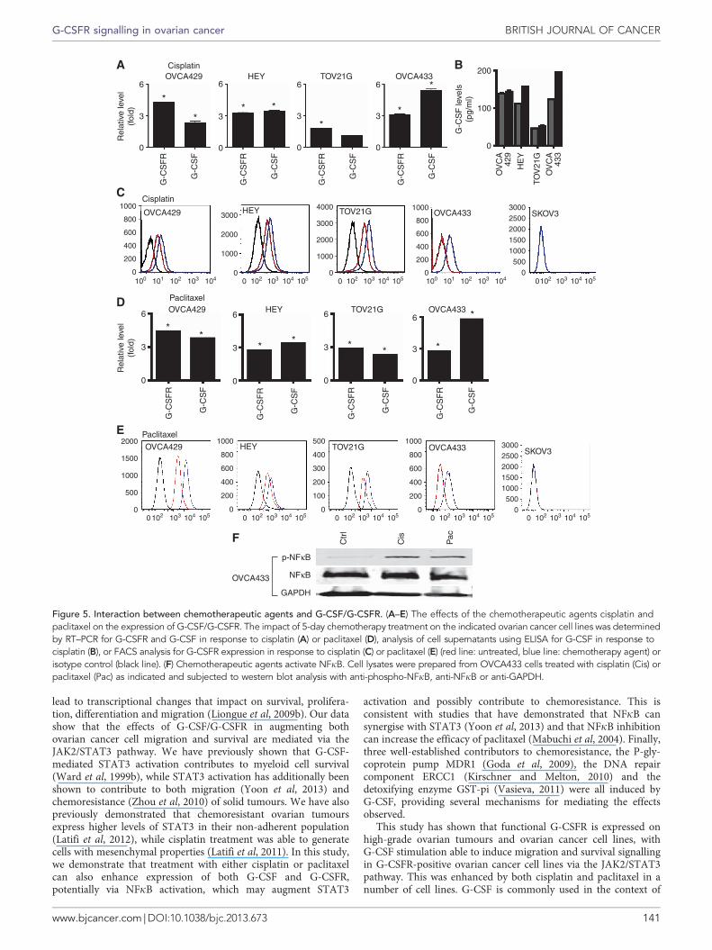
lead to transcriptional changes that impact on survival, prolifera-tion, differentiation and migration (Liongue et al, 2009b). Our datashow that the effects of G-CSF/G-CSFR in augmenting bothovarian cancer cell migration and survival are mediated via theJAK2/STAT3 pathway. We have previously shown that G-CSF-mediated STAT3 activation contributes to myeloid cell survival(Ward et al, 1999b), while STAT3 activation has additionally beenshown to contribute to both migration (Yoon et al, 2013) andchemoresistance (Zhou et al, 2010) of solid tumours. We have alsopreviously demonstrated that chemoresistant ovarian tumoursexpress higher levels of STAT3 in their non-adherent population(Latifi et al, 2012), while cisplatin treatment was able to generatecells with mesenchymal properties (Latifi et al, 2011). In this study,we demonstrate that treatment with either cisplatin or paclitaxelcan also enhance expression of both G-CSF and G-CSFR,potentially via NFkB activation, which may augment STAT3
activation and possibly contribute to chemoresistance. This isconsistent with studies that have demonstrated that NFkB cansynergise with STAT3 (Yoon et al, 2013) and that NFkB inhibitioncan increase the efficacy of paclitaxel (Mabuchi et al, 2004). Finally,three well-established contributors to chemoresistance, the P-gly-coprotein pump MDR1 (Goda et al, 2009), the DNA repaircomponent ERCC1 (Kirschner and Melton, 2010) and thedetoxifying enzyme GST-pi (Vasieva, 2011) were all induced byG-CSF, providing several mechanisms for mediating the effectsobserved.
This study has shown that functional G-CSFR is expressed onhigh-grade ovarian tumours and ovarian cancer cell lines, withG-CSF stimulation able to induce migration and survival signallingin G-CSFR-positive ovarian cancer cell lines via the JAK2/STAT3pathway. This was enhanced by both cisplatin and paclitaxel in anumber of cell lines. G-CSF is commonly used in the context of
6
3
0
G-C
SF
R
G-C
SF
6
3
0
G-C
SF
R
G-C
SF
6
3
0
G-C
SF
R
G-C
SF
6
3
0
G-C
SF
R
G-C
SF
G-C
SF
R
G-C
SF
Rel
ativ
e le
vel
(fol
d)
6
3
0
G-C
SF
R
G-C
SF
6
3
0
G-C
SF
R
G-C
SF
6
3
0
G-C
SF
R
G-C
SF
6
3
0
Rel
ativ
e le
vel
(fol
d)
*
** *
**
*OVCA433
OVCA433
OVCA433
OVCA433
TOV21G
TOV21G
TOV21G
TOV21G
Ctr
l
Cis
Pac
HEY
HEY
HEY
HEY
Cisplatin
Cisplatin
OVCA429
OVCA429
OVCA429
OVCA429
200
100
0
G-C
SF
leve
ls(p
g/m
l)
OV
CA
429
HE
Y
TOV
21G
OV
CA
433
SKOV3
SKOV3
1000
800
600
400
200
0100 101 102 103 104 100 101 102 103 1040 105102 103 104
0 105102 103 104 0 105102 103 104 0 105102 103 104 0 105102 103 104 0 105102 103 104
0 105102 103 104 0 105102 103 104
3000
2000
1000
0
4000
3000
2000
1000
0
1000
800
600
400
200
0
**
** *
* *
*
2000
1500
1000
500
0
1000
800
600
400
200
0
500
400
300
200
100
0
1000
800
600
400
200
0
30002500200015001000500
0
Paclitaxel
Paclitaxel
NFκB
p-NFκB
GAPDH
OVCA433
30002500200015001000500
0
A B
C
D
E
F
Figure 5. Interaction between chemotherapeutic agents and G-CSF/G-CSFR. (A–E) The effects of the chemotherapeutic agents cisplatin andpaclitaxel on the expression of G-CSF/G-CSFR. The impact of 5-day chemotherapy treatment on the indicated ovarian cancer cell lines was determinedby RT–PCR for G-CSFR and G-CSF in response to cisplatin (A) or paclitaxel (D), analysis of cell supernatants using ELISA for G-CSF in response tocisplatin (B), or FACS analysis for G-CSFR expression in response to cisplatin (C) or paclitaxel (E) (red line: untreated, blue line: chemotherapy agent) orisotype control (black line). (F) Chemotherapeutic agents activate NFkB. Cell lysates were prepared from OVCA433 cells treated with cisplatin (Cis) orpaclitaxel (Pac) as indicated and subjected to western blot analysis with anti-phospho-NFkB, anti-NFkB or anti-GAPDH.
G-CSFR signalling in ovarian cancer BRITISH JOURNAL OF CANCER
www.bjcancer.com | DOI:10.1038/bjc.2013.673 141

ovarian cancer to restore neutrophils ablated by standardchemotherapeutic drugs used to treat patients (Yamada et al,2001). However, if the tumour expresses G-CSFR, either basally orin response to chemotherapy, these treatments could contribute totumour development by increasing migration and/or survival,irrespective of G-CSF expression. Therefore, screening patienttumours for G-CSFR expression by histology or FACS-basedapproaches prior to administration of G-CSF could be beneficial,potentially indicating alternative therapies for patients withG-CSFR-positive tumours. Of relevance, co-administration of
granulocyte-macrophage CSF factor and recombinant interferongamma 1b in concert with carboplatin treatment in women withrecurrent ovarian cancer was shown to be efficacious and elicited afavourable haematological profile (Schmeler et al, 2009), providinga potential option in this regard. Finally, inhibitors of JAK2 andSTAT3 have now been developed and used in various clinicalsettings such as myeloproliferative disorders (Pardanani et al,2011). Consistent with our data, inhibition of JAK2/STAT3 hasbeen shown to induce apoptosis in ovarian tumours (Kandala andSrivastava, 2012) and increase the chemo-sensitivity of ovarian
120
60
0
120
60
0
120
60
0Wou
nd c
losu
re (
%)
Wou
nd c
losu
re (
%)
Ctr
lN
o In
hN
o In
hS
3I J2I
No
Inh
S3I J2
IC
trl
No
Inh
No
Inh
S3I J2
IN
o In
hS
3I J2I
Ctr
l
No
Inh
S3I J2I
No
Inh
S3I J2I
Ctr
l
No
Inh
S3I J2I
No
Inh
S3I J2I
Ctr
l
No
Inh
S3I J2I
No
Inh
S3I J2I
Ctr
l
No
Inh
S3I J2I
No
Inh
S3I J2I
Ctr
l
No
Inh
S3I J2I
No
Inh
S3I J2I
Ctr
l
No
Inh
S3I J2I
No
Inh
S3I J2I
Ctr
lN
o In
hN
o In
hS
3I J2I
No
Inh
S3I J2
I
**
* * *
* * * *
#
# # # #
# # # ## #
#
Cisplatin
Cisplatin
Cisplatin Cisplatin Cisplatin
Cisplatin
CisplatinG-CSF
G-CSF
G-CSF G-CSF G-CSF
G-CSFIL-6
IL-6
IL-6
G-CSF IL-6 G-CSF IL-6 G-CSF IL-6
IL-6 IL-6
IL-6OVCA429
OVCA429
OVCA429
OV
CA
429
HEY
HEY
HEY
HE
Y
TOV21G
TOV21G
TOV21G
TOV
21G
p-STAT3
STAT3
GAPDH
p-STAT3
STAT3
GAPDH
p-STAT3
STAT3
GAPDH
Ctr
l
GC
SF
GC
SF
+J2
I
GC
SF
+S
3I
IL-6
IL-6
+S
3I
IL-6
+J2
I
100
50
0
100
50
0
100
50
0* *A
popt
otic
cel
ls (
%)
100
50
0
100
50
0
100
50
0
Apo
ptot
ic c
ells
(%
)
* * * *
Paclitaxel Paclitaxel Paclitaxel
* * * * * *
A B
C
D
Figure 6. Effects of chemotherapeutic agents on cell survival and migration. (A) Wound-healing assay. Indicated cells were treated withcisplatin alone, or with G-CSF and IL-6 treatment, in the presence or absence of inhibitors for STAT3 (S3I) and JAK2 (J2I). Graph shows thepercentage wound closure, expressed as meanþ s.e.m. (*Po0.05 compared with control, #Po0.005 compared with cisplatin). (B) Induction ofSTAT3 activity. Cell lysates were prepared from OVCA429, HEY and TOV21G cells treated with cisplatin (Cis) as indicated, and subjected towestern blot analysis with anti-phospho-STAT3, anti-STAT3 and anti-GAPDH. (C–D) Apoptosis assay. Indicated cells were treated with12.5 mg ml� 1 cisplatin (C) or paclitaxel (D), or with G-CSF and IL-6 treatment, in the presence or absence of inhibitors for STAT3 and JAK2. Graphshows the percentage of apoptotic cells, expressed as meanþ s.e.m. (*Po0.005 compared with chemotherapeutic agent). For TOV21G cells,cisplatin was increased to 20mg ml� 1 in order to achieve significant levels of apoptosis.
BRITISH JOURNAL OF CANCER G-CSFR signalling in ovarian cancer
142 www.bjcancer.com | DOI:10.1038/bjc.2013.673

cancers (Hedvat et al, 2009; Lee et al, 2011). This suggests thatinhibitors of the JAK2/STAT3 pathway may also be efficacious inthe treatment of chemoresistant ovarian cancer.
ACKNOWLEDGEMENTS
We recognise the support of a Deakin University InternationalPostgraduate Scheme Award (to JK) and funding from both theGeelong Community Foundation and Atkins Charitable Trust(to ACW).
CONFLICT OF INTEREST
The authors declare no conflict of interest.
REFERENCES
Ahmed N, Riley C, Oliva K, Rice G, Quinn M (2005) Ascites inducesmodulation of alpha6beta1 integrin and urokinase plasminogen activatorreceptor expression and associated functions in ovarian carcinoma.Br J Cancer 92: 1475–1485.
Ahmed N, Riley C, Oliva K, Stutt E, Rice GE, Quinn MA (2003) Integrin-linked kinase expression increases with ovarian tumour grade and issustained by peritoneal tumour fluid. J Pathol 201: 229–237.
Bohlius J, Reiser M, Schwarzer G, Engert A (2003) Impact of granulocytecolony-stimulating factor (CSF) and granulocyte-macrophage CSF inpatients with malignant lymphoma: a systematic review. Br J Haematol122: 413–423.
Bookman MA (2012) First-line chemotherapy in epithelial ovarian cancer.Clin Obstet Gynecol 55: 96–113.
Brandstetter T, Ninci E, Bettendorf H, Perewusnyk G, Stolte J, HerchenbachD, Sellin D, Wagner E, Kochli OR, Bauknecht T (2001) Granulocytecolony-stimulating factor (G-CSF) receptor gene expression of ovariancarcinoma does not correlate with G-CSF caused cell proliferation. Cancer91: 1372–1383.
Brandstetter T, Ninci E, Falken U, Wagner E, Hess R, Bauknecht T (1998)rhG-CSF affects genes involved in mitogen signalling and early geneexpression in the ovarian cancer cell line HEY. Int J Cancer 75: 847–854.
Burleson KM, Boente MP, Pambuccian SE, Skubitz AP (2006)Disaggregation and invasion of ovarian carcinoma ascites spheroids.J Transl Med 4: 6.
Chakraborty A, White SM, Lerner SP (2004) Granulocyte colony-stimulatingfactor receptor signals for b1-integrin expression and adhesion in bladdercancer. Urology 63: 177–183.
Christopher MJ, Link DC (2007) Regulation of neutrophil homeostasis.Curr Opin Hematol 14: 3–8.
Colombo N, Peiretti M, Parma G, Lapresa M, Mancari R, Carinelli S, Sessa C,Castiglione M (2010) Newly diagnosed and relapsed epithelial ovariancarcinoma: ESMO Clinical Practice Guidelines for diagnosis, treatmentand follow-up. Ann Oncol 21(Suppl 5): v23–v30.
Colomiere M, Ward AC, Riley C, Trenerry MK, Cameron-Smith D, Findlay J,Ackland L, Ahmed N (2009) Cross talk of signals between EGFR andIL-6R through JAK2/STAT3 mediate epithelial-mesenchymal transition inovarian carcinomas. Br J Cancer 100: 134–144.
Connor JP, Squatrito RC, Terrell KL, Antisdel BJ, Buller RE (1994) In vitrogrowth effects of colony-stimulating factors in ovarain cancer. GynecolOncol 52: 347–352.
Dunn SM, Coles LS, Lang RK, Gerondakis S, Vadas MA, Shannon MF (1994)Requirement for nuclear factor (NF)-kappa B p65 and NF-interleukin-6binding elements in the tumor necrosis factor response region of thegranulocyte colony-stimulating factor promoter. Blood 83: 2469–2479.
Eyles JL, Roberts AW, Metcalf D, Wicks IP (2006) Granulocyte colony-stimulating factor and neutrophils–forgotten mediators of inflammatorydisease. Nat Clin Pract Rheumatol 2: 500–510.
Fogh J, Fogh JM, Orfeo T (1977) One hundred and twenty-seven culturedhuman tumor cell lines producing tumors in nude mice. J Natl Cancer Inst59: 221–226.
Gao SP, Mark KG, Leslie K, Pao W, Motoi N, Gerald WL, Travis WD,Bornmann W, Veach D, Clarkson B, Bromberg JF (2007) Mutations in theEGFR kinase domain mediates STAT3 activation via IL-6 production inhuman lung adenocarcinomas. J Clin Invest 117: 3846–3856.
Gits J, Van Leeuwen D, Carroll HP, Touw IP, Ward AC (2006) Multiplepathways contribute to the hyperproliferative responses fromtruncated granulocyte colony-stimulating factor receptors. Leukemia 20:2111–2118.
Goda K, Bacso Z, Szabo G (2009) Multidrug resistance through the spectacleof P-glycoprotein. Curr Cancer Drug Targets 9: 281–297.
Godoy-Tundidor S, Cavarretta IT, Fuchs D, Fiechtl M, Steiner H,Friedbichler K, Bartch G, Hobisch A, Culig Z (2005) Interleukin-6 andoncostatin M stimulation of proliferation of prostate cancer 22Rv1 cellsthrough signaling pathways of p38 mitogen-activated protein kinase andphosphatidylinositol 3-kinase. Prostate 64: 209–216.
G-C
SF
G-C
SF
G-C
SF
G-C
SF
G-C
SF
G-C
SF
G-C
SF
G-C
SF
G-C
SF
PacCis PacCisPacCisO
VC
A42
9
OV
CA
429
OV
CA
429
HE
Y
HE
Y
HE
Y
TOV
21G
TOV
21G
p-STAT3
STAT3
GAPDH
p-STAT3
STAT3
GAPDH
p-STAT3
STAT3
GAPDH
GAPDH
GAPDH
GAPDH
GAPDH
p-ERK
p-ERK
p-AKT
p-AKT
T-AKT
T-AKT
T-ERK
T-ERK
MDR1
MDR1
GST-pi
GST-pi
ERCC1
ERCC1
GAPDH
GAPDH
MDR1
GST-pi
ERCC1
GAPDH
A B C
Figure 7. Effects of chemotherapeutic agents on downstream pathways. Cell lysates were prepared from OVCA429, HEY and TOV21G cellstreated with cisplatin (Cis) or paclitaxel (Pac) as indicated, and subjected to western blot analysis with anti-phospho-STAT3, anti-STAT3 andanti-GAPDH (A), anti-phospho-ERK, anti-ERK, anti-phospho-AKT, anti-AKT and anti-GAPDH (B), or anti-MDR1, anti-GST-pi, anti-ERCC1 andanti-GAPDH (C).
G-CSFR signalling in ovarian cancer BRITISH JOURNAL OF CANCER
www.bjcancer.com | DOI:10.1038/bjc.2013.673 143

Gonzalez-Martin A, Toledo G, Chiva L (2010) Epithelial ovarian carcinoma:current evidences and future perspectives in the first-line setting.Clin Transl Oncol 12: 418–430.
Grivennikov S, Karin M (2008) Autocrine IL-6 signaling: a key event intumorigenesis? Cancer Cell 13: 7–9.
Gutschalk CM, Herold-Mende CC, Fusenig NE, Mueller MM (2006)Granulocyte colony-stimulating factor and granulocyte-macrophagecolony-stimulating factor promote malignant growth of cells from headand neck squamous cell carcinomas in vivo. Cancer Res 66: 8026–8036.
Hedvat M, Huszar D, Herrmann A, Gozgit JM, Schroeder A, Sheehy A,Buettner R, Proia D, Kowolik CM, Xin H, Armstrong B, Bebernitz G,Weng S, Wang L, Ye M, Mceachern K, Chen H, Morosini D, Bell K,Alimzhanov M, Ioannidis S, Mccoon P, Cao ZA, Yu H, Jove R, Zinda M(2009) The JAK2 inhibitor AZD1480 potently blocks Stat3 signaling andoncogenesis in solid tumors. Cancer Cell 16: 487–497.
Hirai K, Kumakiri M, Fujieda S, Sunaga H, Lao LM, Imamura Y, Ueda K,Fukuda M (2001) Expression of granulocyte colony-stimulating factorand its receptor in epithelial skin tumors. J Dermatol Sci 25: 179–188.
Hudson LG, Zeineldin R, Stack MS (2008) Phenotypic plasticity of neoplasticovarian epithelium: unique cadherin profiles in tumor progression. ClinExp Metastasis 25: 643–655.
Jones TR, Kang IH, Wheeler DB, Lindquist RA, Papallo A, Sabatini DM,Golland P, Carpenter AE (2008) CellProfiler Analyst: data exploration andanalysis software for complex image-based screens. BMC Bioinformatics 9:482.
Kamohara H, Ogawa M, Ishiko T, Sakamoto K, Baba H (2007) Leukemiainhibitory factor functions as a growth factor in pancreas carcinoma cells:involvement of regulation of LIF and its receptor expression. Int J Oncol30: 977–983.
Kandala PK, Srivastava SK (2012) Regulation of Janus-activated kinase-2(JAK2) by diindolylmethane in ovarian cancer in vitro and in vivo. DrugDiscov Ther 6: 94–101.
Karst AM, Drapkin R (2010) Ovarian cancer pathogenesis: a model inevolution. J Oncol 2010: 932371.
Katayama N, Kita K, Kawakami K, Mitani H, Sugawara T, Mizuno S,Yonezawa A, Nishii K, Miwa H, Wada H, Minami N, Shiku H (1998)Granulocyte colony-stimulating factor and its receptor in acutepromyelocytic leukemia. Am J Hematol 58: 31–35.
Kirschner K, Melton DW (2010) Multiple roles of the ERCC1-XPFendonuclease in DNA repair and resistance to anticancer drugs.Anticancer Res 30: 3223–3232.
Lagunas VM, Melendez-Zajgla J (2008) Nuclear Factor-kappa B as a resistancefactor to platinum-based antineoplasic drugs. Met Based Drugs 2008:576104.
Lamprecht MR, Sabatini DM, Carpenter AE (2007) CellProfiler: free,versatile software for automated biological image analysis. Biotechniques42: 71–75.
Latifi A, Abubaker K, Castrechini N, Ward AC, Liongue C, Dobill F, Kumar J,Thompson EW, Quinn MA, Findlay JK, Ahmed N (2011) Cisplatintreatment of primary and metastatic epithelial ovarian carcinomasgenerates residual cells with mesenchymal stem cell-like profile. J CellBiochem 112: 2850–2864.
Latifi A, Luwor RB, Bilandzic M, Nazaretian S, Stenvers K, Pyman J, Zhu H,Thompson EW, Quinn MA, Findlay JK, Ahmed N (2012) Isolation andcharacterization of tumor cells from the ascites of ovarian cancer patients:molecular phenotype of chemoresistant ovarian tumors. PLoS One 7:e46858.
Lawrence T (2009) The nuclear factor NF-kappaB pathway in inflammation.Cold Spring Harb Perspect Biol 1: a001651.
Lee ES, Ko KK, Joe YA, Kang SG, Hong YK (2011) Inhibition of STAT3reverses drug resistance acquired in temozolomide-resistant humanglioma cells. Oncol Lett 2: 115–121.
Lengyel E (2010) Ovarian cancer development and metastasis. Am J Pathol177: 1053–1064.
Lim R, Ahmed N, Borregaard N, Riley C, Wafai R, Thompson EW, QuinnMA, Rice GE (2007) Neutrophil gelatinase-associated lipocalin (NGAL) anearly-screening biomarker for ovarian cancer: NGAL is associated withepidermal growth factor-induced epithelio-mesenchymal transition.Int J Cancer 120: 2426–2434.
Liongue C, Hall C, O’connell B, Crozier P, Ward AC (2009a)Zebrafish granulocyte colony-stimulating factor receptor signallingpromotes myelopoiesis and myeloid cell migration. Blood 113:2535–2546.
Liongue C, Wright C, Russell AP, Ward AC (2009b) Granulocyte colony-stimulating factor receptor: stimulating granulopoiesis and much more.Int J Biochem Cell Biol 41: 2372–2375.
Livak KJ, Schmittgen TD (2001) Analysis of relative gene expression datausing real-time quantitative PCR and the 2�DDCT method. Methods 25:402–408.
Mabuchi S, Ohmichi M, Nishio Y, Hayasaka T, Kimura A, Ohta T, Kawagoe J,Takahashi K, Yada-Hashimoto N, Seino-Noda H, Sakata M, Motoyama T,Kurachi H, Testa JR, Tasaka K, Murata Y (2004) Inhibition of inhibitorof nuclear factor-kappaB phosphorylation increases the efficacy ofpaclitaxel in in vitro and in vivo ovarian cancer models. Clin Cancer Res10: 7645–7654.
Morales-Arias J, Meyers PA, Bolontrade MF, Rodriguez N, Zhou Z, Reddy K,Chou AJ, Koshkina NV, Kleinerman ES (2007) Expression of granulocyte-colony-stimulating factor and its receptor in human Ewing sarcomacells and patient tumor specimens: potential consequences of granulocyte-colony-stimulating factor administration. Cancer 110: 1568–1577.
Munstedt K, Hackethal A, Eskef K, Hrgovic I, Franke FE (2010) Prognosticrelevance of granulocyte colony-stimulating factor in ovarian carcinomas.Arch Gynecol Obstet 282: 301–305.
Nakamae-Akahori M, Kato T, Masuda S, Sakamoto E, Kutsuna H, Hato F,Nishizawa Y, Hino M, Kitagawa S (2006) Enhanced neutrophil motilityby granulocyte colony-stimulating factor: the role of extracellularsignal-regulated kinase and phosphatidylinositol 3-kinase. Immunology119: 393–403.
Neijt JP, Engelholm SA, Tuxen MK, Sorensen PG, Hansen M, Sessa C,De Swart CA, Hirsch FR, Lund B, Van Houwelingen HC (2000)Exploratory phase III study of paclitaxel and cisplatin versus paclitaxel andcarboplatin in advanced ovarian cancer. J Clin Oncol 18: 3084–3092.
Ninci EB, Brandstetter T, Meinhold-Heerlein I, Bettendorf H, Sellin D,Bauknecht T (2000) G-CSF receptor expression in ovarian cancer.Int J Gynecol Cancer 10: 19–26.
Ouellet V, Zietarska M, Portelance L, Lafontaine J, Madore J, Puiffe ML,Arcand SL, Shen Z, Hebert J, Tonin PN, Provencher DM, Mes-MassonAM (2008) Characterization of three new serous epithelial ovarian cancercell lines. BMC Cancer 8: 152.
Pardanani A, Gotlib JR, Jamieson C, Cortes JE, Talpaz M, Stone RM,Silverman MH, Gilliland DG, Shorr J, Tefferi A (2011) Safety and efficacyof TG101348, a selective JAK2 inhibitor, in myelofibrosis. J Clin Oncol 29:789–796.
Provencher DM, Lounis H, Champoux L, Tetrault M, Manderson EN,Wang JC, Eydoux P, Savoie R, Tonin PN, Mes-Masson AM (2000)Characterization of four novel epithelial ovarian cancer cell lines. In VitroCell Dev Biol Anim 36: 357–361.
Punnonen R, Teisala K, Kuoppala T, Bennett B, Punnonen J (1998) Cytokineproduction profiles in the peritoneal fluids of patients with malignant orbenign gynecologic tumors. Cancer 83: 788–796.
Ray-Coquard I, Paradiso D, Guastalla JP, Leduc B, Guichard F, Martin C,Chauvenet L, Haddad-Guichard Z, Lepille D, Orfeuvre H, Gautier H,Castera D, Pujade-Lauraine E (2007) Intensified dose ofcyclophosphomidee with G-CSF support versus standard dose combinedwith platinum in first-line treatment of advanced ovarian cancer: arandomised study from the GINECO group. Br J Cancer 97: 1200–1205.
Rose-John S, Waetzig GH, Scheller J, Grotzinger J, Seegert D (2007) TheIL-6/sIL-6R complex as a novel target for therapeutic approaches.Expert Opin Ther Targets 11: 613–624.
Sansone P, Storci G, Tavolari S, Guarnieri T, Giovannini C, Taffurelli M,Ceccarelli C, Santini D, Paterini P, Marcu KB, Chieco P, Bonafe M (2007)IL-6 triggers malignant features in mammospheres from humanductal breast carcinoma and normal mammary gland. J Clin Invest 117:3988–4002.
Savarese TM, Mitchell K, Mcquain C, Campbell CL, Guardiani R, Wuu J,Ollari C, Reale F, Nelson BE, Chen A, Quesenberry PJ (2001)Coexpression of granulocyte colony stimulating factor and its receptor inprimary ovarian carcinomas. Cancer Lett 162: 105–115.
Schmeler KM, Vadhan-Raj S, Ramirez PT, Apte SM, Cohen L, Bassett RL,Iyer RB, Wolf JK, Levenback CL, Gershenson DM, Freedman RS (2009) Aphase II study of GM-CSF and rIFN-gamma1b plus carboplatin for thetreatment of recurrent, platinum-sensitive ovarian, fallopian tube andprimary peritoneal cancer. Gynecol Oncol 113: 210–215.
Shield K, Ackland ML, Ahmed N, Rice GE (2009) Multicellular spheroids inovarian cancer metastases: biology and pathology. Gynecol Oncol 113:143–148.
BRITISH JOURNAL OF CANCER G-CSFR signalling in ovarian cancer
144 www.bjcancer.com | DOI:10.1038/bjc.2013.673

Shield K, Riley C, Quinn MA, Rice GE, Ackland ML, Ahmed N (2007)Alpha2beta1 integrin affects metastatic potential of ovarian carcinomaspheroids by supporting disaggregation and proteolysis. J Carcinog 6: 11.
Spinner DM, Brandstetter T, Kiechle-Schwarz M, Du Bois A, Angel P,Bauknecht T (1995) c-jun expression and growth stimulation in humanovarian carcinoma cell lines following exposure to cytokines. Int J Cancer63: 423–427.
Touw IP, Van De Geijn GJ (2007) Granulocyte colony-stimulating factor andits receptor in normal myeloid cell development, leukemia and relatedblood cell disorders. Front Biosci 12: 800–815.
Vasieva O (2011) The many faces of glutathione transferase pi. Curr Mol Med11: 129–139.
Wang J, Shi YK, Wu LY, Wang JW, Yang S, Yang JL, Zhang HZ, Liu SM(2008) Prognostic factors for ovarian metastases from primary gastriccancer. Int J Gynecol 18: 825–832.
Wang Y, Niu XL, Qu Y, Wu J, Zhu YQ, Sun WJ, Li LZ (2010) Autocrineproduction of interleukin-6 confers cisplatin and paclitaxel resistance inovarian cancer cells. Cancer Lett 295: 110–123.
Ward AC (2007) The role of the granulocyte colony-stimulating factorreceptor (G-CSF-R) in disease. Front Biosci 12: 608–618.
Ward AC, Hermans MHA, Smith L, Van Aesch YM, Schelen AM, AntonissenC, Touw IP (1999a) Tyrosine-dependent and independent mechanisms ofSTAT3 activation by the human granulocyte colony-stimulating factor(G-CSF) receptor are differentially utilized depending on G-CSFconcentration. Blood 93: 113–124.
Ward AC, Smith L, De Koning JP, Van Aesch Y, Touw IP (1999b) Multiplesignals mediate proliferation, differentiation and survival from thegranulocyte colony-stimulating factor receptor in myeloid 32D cells.J Biol Chem 274: 14956–14962.
Watson JM, Sensintaffar JL, Berek JS, Martinez-Maza O (1990) Constitutiveproduction of interleukin 6 by ovarian cancer cell lines and by primaryovarian tumor cultures. Cancer Res 50: 6959–6965.
Wu KL, Miao H, Khan S (2007) JAK kinases promote invasiveness inVHL-mediated renal cell carcinoma by a suppressor of cytokine signaling-regulated, HIF-independent mechanism. Am J Physiol Renal Physiol 293:F1836–F1846.
Xu Z, Jiang Y, Steed H, Davidge S, Fu Y (2010) TGFbeta and EGFsynergistically induce a more invasive phenotype of epithelial ovariancancer cells. Biochem Biophys Res Commun 401: 376–381.
Yamada Y, Tomonaga M, Fukuda H, Hanada S, Utsunomiya A, Tara M,Sano M, Ikeda S, Takatsuki K, Kozuru M, Araki K, Kawano F, Niimi M,Tobinai K, Hotta T, Shimoyama M (2001) A new G-CSF-supportedcombination chemotherapy, LSG15, for adult T-cell leukaemia-lymphoma: Japan Clinical Oncology Group Study 9303. Br J Haematol113: 375–382.
Yoon J, Cho SJ, Ko YS, Park J, Shin DH, Hwang IC, Han SY, Nam SY,Kim MA, Chang MS, Lee HS, Kim WH, Lee BL (2013) A synergisticinteraction between transcription factors nuclear factor-kappaB and signaltransducers and activators of transcription 3 promotes gastric cancercell migration and invasion. BMC Gastroenterol 13: 29.
Zebrowski BK, Liu W, Ramirez K, Akagi Y, Mills GB, Ellis LM (1999)Markedly elevated levels of vascular endothelial growth factor inmalignant ascites. Ann Surg Oncol 6: 373–378.
Zeimet AG, Widschwendter M, Knabbe C, Fuchs D, Herold M,Muller-Holzner E, Daxenbichler G, Offner FA, Dapunt O, Marth C (1998)Ascitic interleukin-12 is an independent prognostic factor in ovariancancer. J Clin Oncol 16: 1861–1868.
Zeineldin R, Muller CY, Stack MS, Hudson LG (2010) Targeting the EGFreceptor for ovarian cancer therapy. J Oncol 2010: 414676.
Zhao Q, Wang C, Zhu J, Wang L, Dong S, Zhang G, Tian J (2011) RNAi-mediated knockdown of cyclooxygenase2 inhibits the growth, invasionand migration of SaOS2 human osteosarcoma cells: a case control study.J Exp Clin Cancer Res 30: 26.
Zhou J, Ong CN, Hur GM, Shen HM (2010) Inhibition of the JAK-STAT3pathway by andrographolide enhances chemosensitivity of cancer cells todoxorubicin. Biochem Pharmacol 79: 1242–1250.
This work is published under the standard license to publish agree-ment. After 12 months the work will become freely available andthe license terms will switch to a Creative Commons Attribution-NonCommercial-Share Alike 3.0 Unported License.
Supplementary Information accompanies this paper on British Journal of Cancer website (http://www.nature.com/bjc)
G-CSFR signalling in ovarian cancer BRITISH JOURNAL OF CANCER
www.bjcancer.com | DOI:10.1038/bjc.2013.673 145
Copyright © 2022 FDOKUMEN