Proton Electroinsertion in Self-Assembled Materials for Neutralization Pseudocapacitors
Upload
malocasin88Category
view
0download
0
Neutralization of granulocyte macrophage colony-stimulating factor decreases amyloid beta 1-42 andsuppresses microglial activity in a transgenicmouse model of Alzheimer’s disease
Maria Manczak1, Peizhong Mao1, Kazuhiro Nakamura2, Christopher Bebbington3,
Byung Park4 and P. Hemachandra Reddy1,5,�
1Neurogenetics Laboratory and 2Central Autonomic Regulation Laboratory, Division of Neuroscience, Oregon
National Primate Research Center, 505 NW 185th Avenue, Beaverton, OR 97006, USA, 3KaloBios Pharmaceuticals
Inc., 260 E. Grand Avenue, South San Francisco, CA 94080, USA, 4Division of Biostatistics, Department of Public
Health and Preventive Medicine, Oregon Health and Science University, Portland, OR 97239, USA and 5Department
of Physiology and Pharmacology, Oregon Health and Science University, Portland, OR 97201, USA
Received April 29, 2009; Revised July 2, 2009; Accepted July 16, 2009
The purpose of our study was to investigate microglia and astrocytes that are associated with human mutantamyloid precursor protein and amyloid beta (Ab). We investigated whether the anti-granulocyte–macro-phage-colony stimulating factor (GM-CSF) antibody can suppress microglial activity and decrease Ab pro-duction in Alzheimer’s disease transgenic mice (Tg2576 line). An antibody to mouse GM-CSF wasintroduced by intracerebroventricular (ICV) injections into the brains of 10-month-old Tg2576 male mice.We assessed the effect of several GM-CSF-associated cytokines on microglial activities and their associationwith Ab using quantitative real-time RT–PCR, immunoblotting, immunohistochemistry analyses in anti-GM-CSF antibody-injected Tg2576 mice. Using sandwich ELISA technique, we measured intraneuronal Ab inTg2576 mice injected with GM-CSF antibody and PBS vehicle-injected control Tg2576 mice. Using double-labeling immunofluorescence analysis of intraneuronal Ab, Ab deposits and pro-inflammatory cytokines,we assessed the relationship between Ab deposits and microglial markers in the Tg2576 mice, and also inthe anti-GM-CSF antibody-injected Tg2576 mice. Our real-time RT–PCR analysis showed an increase inthe mRNA expression of IL6, CD11c, IL1b, CD40 and CD11b in the cerebral cortices of the Tg2576 mice com-pared with their littermate non-transgenic controls. Immunohistochemistry findings of microglial markersagreed with our real-time RT–PCR results. Interestingly, we found significantly decreased levels of activatedmicroglia and Ab deposits in anti-GM-CSF antibody-injected Tg2576 mice compared with PBS vehicle-injected Tg2576 mice. Findings from our real-time RT–PCR and immunoblotting analysis agreed withimmunohistochemistry results. Our double-labeling analyses of intraneuronal Ab and CD40 revealed thatintraneuronal Ab is associated with neuronal expression of CD40 in Tg2576 mice. Our quantitative sandwichELISA analysis revealed decreased levels of soluble Ab1-42 and increased levels of Ab1-40 in Tg2576 miceinjected with the anti-GM-CSF antibody, suggesting that anti-GM-CSF antibody alone decreases solubleAb1-42 production and suppresses microglial activity in Tg2576 mice. These findings indicating the abilityof the anti-GM-CSF antibody to reduce Ab1-42 and microglial activity in Tg2576 mice may have therapeuticimplications for Alzheimer’s disease.
�To whom correspondence should be addressed at: Neurogenetics Laboratory, Neuroscience Division, Oregon National Primate Research Center, WestCampus, Oregon Health and Science University, 505 NW 185th Avenue, Beaverton, OR 97006, USA. Tel: þ1 5034182625; Fax: þ1 503 418 2501;Email: [email protected]; [email protected]
# The Author 2009. Published by Oxford University Press. All rights reserved.For Permissions, please email: [email protected]
Human Molecular Genetics, 2009, Vol. 18, No. 20 3876–3893doi:10.1093/hmg/ddp331Advance Access published on July 19, 2009
INTRODUCTION
Alzheimer’s disease (AD) is a late-onset, progressive neurode-generative disorder characterized by the loss of memory andan impairment of multiple cognitive functions. The majorpathological features in the brains of AD patients are the pres-ence of extra-cellular amyloid beta (Ab) plaques and intra-cellular neurofibrillary tangles (1–5). Recent biochemical,molecular and gene expression studies of AD postmortembrains and AD transgenic mouse models revealed that multiplepathways are involved in AD pathogenesis. These pathwaysinclude synaptic failure (6–8), mitochondrial oxidativedamage (5,9–13) and inflammatory responses (3,14–20).Among these cellular changes, inflammatory responses arereported to be critically involved in AD pathogenesis (3).
Inflammation is caused by the proliferation of reactive astro-cytes and microglia that have been observed in the brains of ADpatients (14,21) and AD transgenic mice (15,16,22,23). Severalrecent studies found increased cytokines, including theGM-CSF, in the cerebrospinal fluid of AD patients (24,25). Fur-thermore, AD transgenic mice lines that overexpress amyloidprecursor protein (APP) and Ab exhibit significant cerebrovas-cular inflammation and microgliosis around areas of Ab plaquedeposition (26–28). In addition, the chronic administration ofibuprofen and other non-steroidal anti-inflammatory drugshave been reported to reduce Ab plaque pathology and Ablevels in the brains of AD mice (29,30).
Microglia cells are associated with most Ab plaques (31–33). Microglial activation involves proliferation of microgliacells, their homing to the site of injury and functionalchanges, including the release of cytotoxic and inflammatorymediators. Activated microglia may participate in the braintissue damage in patients with AD. In an early stage of AD,microglia cells may perform synaptic stripping, leading toextensive synaptic damage in AD brains. Activation of glialcells is accompanied by an upregulation of APP expression,leading to Ab accumulation in the chronic stage of thedisease (14). Microglia can also act as a cytotoxic effectorin cells by releasing proteases, reactive oxygen intermediatesand nitric oxide (34) and by mediating neuronal injury (35).In addition, microglia can participate in an inflammatoryresponse by acting as antigen-presenting cells to activate T-lymphocytes (36) and by producing pro-inflammatory cyto-kines, such as GM-CSF.
Among several cytokines found in the brains and cerebrosp-inal fluid of AD patients, GM-CSF is a pro-inflammatory cyto-kine, involved in the regulation of proliferation, differentiationand functional activities of granulocyte–macrophage popu-lations (37). GM-CSF infusion in the brain depicts a dramaticproliferation of a large number of microglial cells (38,39).Indeed, GM-CSF is one of the strongest microglial mitogens(34,40,41). In addition, withdrawal of GM-CSF significantlyenhances the death rate of microglial cells, as determined bya DNA fragmentation assay. Thus, increased levels ofGM-CSF found in the cerebral spinal fluid of AD patientsmay play a crucial role in inducing microgliosis that typicallyoccurs in the brains of patients with severe AD (24).
Recently, several studies focused on decreasing the Ab loadin AD transgenic mice by using active and passive immuniz-ations (42–45). Several other studies treated AD transgenic
mice using a combination of antibodies of Ab and cytokines(GM-CSF and IL4) (39,46,47). Kim et al. (46) focused onintranasal immunization of adenovirus vectors encodingGM-CSF and Ab in Tg2576 mice (an AD mouse model)and found decreased Ab loads in the brains of the vaccinatedTg2576 mice compared with those in the control Tg2576mice. DaSilva et al. (39) investigated the same combinationalimmunization approach (Ab/GM-CSF/IL4) in APP mice thatKim et al. (46) used and found Ab/GM-CSF/IL4 antibodiesdecreased �43% in the Ab plaque load of AD transgenicmice, suggesting that the combinational approach may beeffective in Ab immunotherapy. Frazer et al. (47) used anamplicon (HSV-IE-Ab(CMV)IL4) to co-deliver Ab1-42 andinterleukin-4 in a triple AD transgenic mice and foundincreased Ab-specific antibodies, improved learning andimproved functioning of memory and decreased AD pathologyin the HSV(IE)Ab(CMV)IL4-vaccinated mice compared withthe other experimental groups. However, it is unclear whetheranti-GM-CSF antibodies alone can suppress glial activity andcan decrease Ab pathology in AD transgenic mice.
We propose that anti-GM-CSF antibody suppresses micro-glial activity and decreases microglial-associated Ab pro-duction and deposits in the brains of AD mice. To test thesehypotheses, we investigated the microglial activity, Ab pro-duction and Ab deposits in a well-characterized APP trans-genic mice (Tg2576 mice) after treating them with theanti-GM-CSF anti-mouse antibody. We injected this antibodyinto the brains of 10-month-old Tg2576 mice and a PBSvehicle into the brains of 10-month-old Tg2576 mice as anegative control, using ICV injections. We measured (i)mRNA expression of markers of microglia, astrocytes andneurons using SYBR-Green chemistry-based quantitative real-time RT–PCR, (ii) immunoblotting analysis of microglialmarkers, (iii) soluble and insoluble Ab levels using sandwichELISA, (iv) immunohistochemistry and immunofluorescenceanalyses of several markers of microglia, astrocytes andneurons, (v) Ab deposits in the brains of Tg2576 mice injectedwith anti-GM-CSF antibody and/or the PBS vehicle, and (vi)examined the connection between intraneuronal Ab/Abdeposits and microglial activation.
RESULTS
mRNA expression of markers of microglia, astrocytes in10-month-old male Tg2576 mice and non-transgenic mice
The objective of our study was to investigate the markers ofmicroglia and astrocytes and their association with Abproduction and Ab deposits in AD transgenic mice. We investi-gated the extent of mRNA in the cerebral cortex for markers ofmicroglia, astrocytes and neurons (using quantitative real-timeRT–PCR) and soluble and insoluble Ab production (using sand-wich ELISA) and Ab deposits using immunohistochemistry andimmunofluorescence analyses in 10-month-old male Tg2576mice and age-matched, non-transgenic littermates.
As shown in Table 1, we found a 4.70-fold increase inmRNA levels for IL6, followed by a 4.02-fold increase forCD11c, a 2.24-fold increase for IL1b, a 1.39-fold increasefor CD40 and a 1.2-fold increase for CD11b in the cerebralcortex tissues of 10-month-old male Tg2576 mice compared
Human Molecular Genetics, 2009, Vol. 18, No. 20 3877

with the non-transgenic control mice, suggesting that inflam-matory responses are evident in overexpressed humanmutant APP transgenic mice. We found a 1.30-fold increasedmRNA expression for GFAP and a 1.14-fold increase forNeuN in the cerebral cortex tissues of 10-month-old Tg2576mice compared with the non-transgenic control mice. Wealso found mRNA levels unchanged or slightly decreased forseveral other cytokine markers, such as CD45, TNFa, gp91and MHCII in Tg2576 mice compared with the non-transgeniclittermates.
mRNA expression of markers of microglia, astrocytes andneurons in PBS-injected Tg2576 mice and anti-GM-CSFantibody-injected Tg2576 mice
We sought to determine whether anti-GM-CSF antibody cansuppress the activated microglial activity that was observedin overexpressed mutant human APP transgenic mice. There-fore, we investigated mRNA levels of pro-inflammatory cyto-kines in anti-GM-CSF antibody-injected Tg2576 micecompared with PBS-injected Tg2576 mice.
As shown in Table 1, interestingly, we found decreasedmRNA levels of IL1b, IL6, CD11b, CD11c and CD40 inanti-GM-CSF antibody-injected Tg2576 mice compared withthe PBS vehicle-injected Tg2576 mice. mRNA levels wereincreased for IL1b, IL6, CD11b, CD11c and CD40 in theTg2576 mice compared with the non-transgenic wild-typemice, and these increased mRNA levels were decreased inanti-GM-CSF antibody-injected Tg2576 mice, suggestingthat anti-GM-CSF antibody suppressed the activated microgliain the Tg2576 mice. We also noticed that slightly increasedmRNA levels for GFAP (1.64-fold) and MHCII-2(2.12-fold) in the anti-GM-CSF antibody-injected Tg2576mice compared with the PBS vehicle-injected Tg2576 mice.
Immunoblotting analysis of proteins of microglial markersin Tg2576 mice injected with GM-CSF antibody andTg2576 mice injected with PBS vehicle
To determine whether GM-CSF antibody reduces protein levelsof microglial markers that are associated with Ab deposits inAD, we performed immunoblotting analysis of markers ofmicroglia, IL1b, IL6, CD40 and CD11b in protein lysates pre-pared from cerebral cortices of Tg2576 mice injected withGM-CSF antibody and Tg2576 mice injected with PBSvehicle. As shown in Figure 1, we found decreased proteinlevels for CD40 (by 48%), followed by IL1b (43%), IL6(39%) and CD11b (11%) in Tg2576 mice injected withGM-CSF antibody compared with Tg2576 mice injected withPBS vehicle (control), suggesting that GM-CSF antibodyreduced proteins of microglia associated with Ab deposits inTg2576 mice. These findings agreed with the results of real-time RT–PCR and immunohistochemistry analysis.
Immunohistochemistry and immunofluorescence analysesof markers of microglia and GFAP in Tg2576 mice andnon-transgenic wild-type mice
To determine whether overexpressed mutant APP and Ab depos-its activate microglia and astrocytes, we conducted immunohis-tochemistry and immunofluorescence analyses of several glialmarkers, including IL1b, IL6, CD11b, CD11c, TNFa andCD40, in the Tg256 mice and in the non-transgenic, wild-typemice. We found increased immunoreactivity of IL1b (Fig. 2),IL6 (Supplementary Material, Fig. S1), CD11c (SupplementaryMaterial, Fig. S2) and CD11b (Supplementary Material,Fig. S3) in the hippocampal and cortical regions, where Abdeposits abundantly found in 10-month-old Tg2576 mice com-pared with the age-matched, non-transgenic mice (data notshown because of low or undetectable immunoreactivity).
Figure 1. Immunoblotting analysis of microglial proteins in Tg2576 miceinjected with GM-CSF antibody and Tg2576 mice injected with PBS. (A)Twenty micrograms of protein lysate was used from each sample, and immu-noblotting analysis was performed using antibodies of IL1b, IL6, CD40 andb-actin. Bottom panel represents the immunoblotting of b-actin for equalloading. (B) Densitometry values for microglial proteins IL1b, IL6 andCD40 in Tg2576 mice injected with GM-CSF antibody and Tg2576 miceinjected with PBS. As shown, we found decreased protein levels for CD40(by 48%), followed by IL1b (43%), IL6 (39%) and CD11b (11%) inTg2576 mice injected with GM-CSF antibody compared with Tg2576 miceinjected with PBS.
Table 1. mRNA expression of microglial and neuronal markers betweenTg2576 mice and age-matched littermates, and between PBS vehicle-injectedTg256 mice and anti-GM-CSF antibody-injected Tg2576 mice (b-actinnormalization)
Marker mRNA fold-change differencebetween 10-month-old Tg2576mice and 10-month-oldnon-transgenic littermates
mRNA fold-change differencebetween 10-month-oldPBS-injected Tg2576 mice andanti-GM-CSF antibody-injected10-month-old Tg2576 mice
TNFa 0.68 0.44IL1b 2.24 0.26IL6 4.70 0.05Gp91 0.40 1.47CD11b 1.20 0.11CD11c 4.02 1.48CD40 1.39 0.31CD45 0.90 0.56MHCII -2 0.45 2.12GFAP 1.30 1.64NeuN 1.14 1.43
One-fold mRNA expression means no change between Tg2576 mice andnon-transgenic littermates, and between PBS vehicle-injected Tg2576mice and anti-GM-CSF antibody-injected Tg2576 mice; .1-fold mRNAexpression indicates increased abundance to control mice and ,1-foldmRNA expression, decreased abundance to control mice.
3878 Human Molecular Genetics, 2009, Vol. 18, No. 20

Double-labeling analyses of intraneuronal Ab, Ab depositsand microglia, astrocytes and CD40
Our double-labeling immunohistochemistry and immunofluor-escence analyses of Ab deposits and activated microglia inTg2576 mice revealed: (i) Ab deposits are surrounded bymicroglia, marked by immunoreactivity of IL1b (Fig. 3A)and CD11b (Fig. 3B), (ii) Ab deposits are present withoutmicroglia marked by immunoreactivity of IL6 (Fig. 4A) andCD11b (Fig. 4B), (iii) Ab deposits are surrounded by astro-cytes (Fig. 5), suggesting that microglial and astrocytic acti-vation are dependent on the presence of Ab deposits, andalso Ab deposits are present without microglial association.
Our double-labeling analysis of intraneuronal Ab and CD40revealed that intraneuronal Ab is co-localized with the immu-noreactivity of CD40 in neurons (Fig. 6), suggesting thatco-localization of intraneuronal Ab and CD40 triggers abnor-mal APP processing and increased pro-inflammatory cytokineproduction in Tg2576 mice.
Double-labeling analyses of CD40 and NeuN
To determine whether CD40 is expressed in neurons, weperformed double-labeling analyses of NeuN and CD40 inthe brain sections from Tg2576 mice. Our double-labeling
immunohistochemistry and immmunofluorescence analysesrevealed that CD40 is expressed in some neurons andco-localized with NeuN (Fig. 7). However, we also foundseveral NeuN-positive neurons did not show CD40 expression,indicating that some neurons selectively expressed for CD40 inthe brains of Tg2576 mice. This finding further supports the pres-ence of intraneuronal expression of CD40 and Ab in Tg2576mice (Fig. 6).
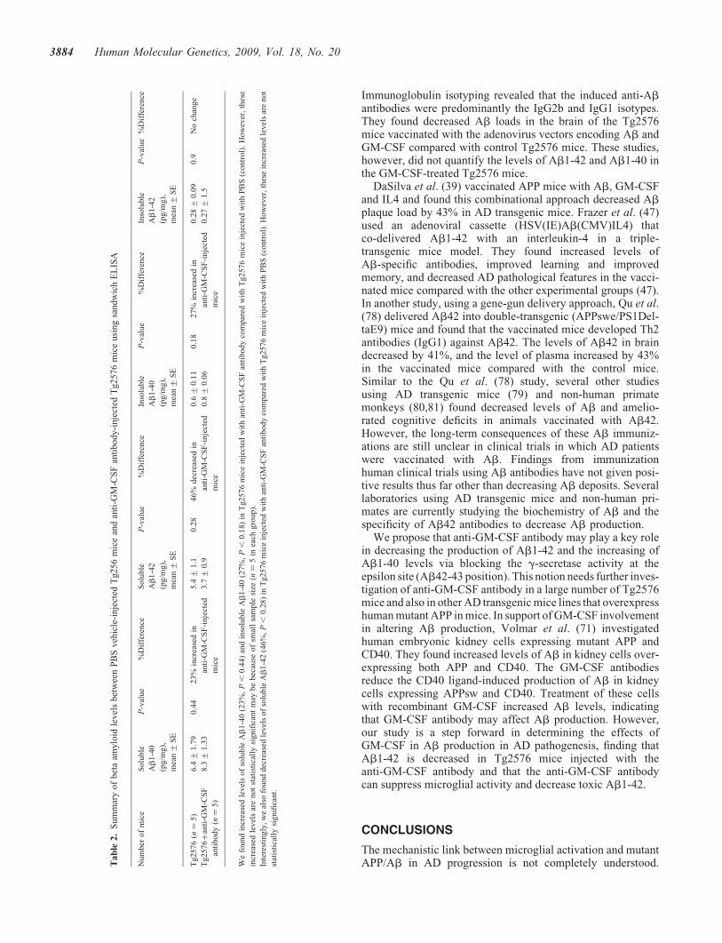
Ab production in anti-GM-CSF antibody-injected Tg2576mice and PBS-injected Tg2576 mice
To determine whether anti-GM-CSF antibody can reduceAb production, using sandwich ELISA we measuredboth soluble and insoluble Ab1-40 and Ab1-42 levelsin anti-GM-CSF antibody-injected Tg2576 mice andPBS-injected Tg2576 mice. Interestingly, for the first time,we found decreased levels of soluble Ab1-42 (46%) inTg2576 mice injected with anti-GM-CSF antibody comparedwith Tg2576 mice injected with PBS (control). However,these decreased levels of Ab1-42 in GM-CSF-injectedTg2576 mice are not statistically significant (P , 0.28)(Table 2), and this insignificant difference may be due to asmall number of animals studied in each group.
Figure 2. Immunoreactivity of IL1 in different brain regions of a representative Tg2576 mouse. Accumulated immunoreactivity was found in the cerebral cortex,hippocampus and ventricular regions of the brain in Tg2576 mice. Arrows indicate the increased immunoreactivity of IL1b in the hippocampus, layers 1–3 in thecortex and ventricular regions of the midbrain. (A) Photographed at �5 the original magnification, (B) �10, (C and D) �20, (E and F) �10.
Human Molecular Genetics, 2009, Vol. 18, No. 20 3879

In addition, we also found increased levels of soluble Ab1-40(23%) and insoluble Ab1-40 (27%) in anti-GM-CSF antibody-injected Tg2576 mice compared with the PBS-injected Tg2576mice. However, these increased levels of soluble Ab1-40 (P ,0.44) and insoluble Ab1-40 (P , 0.18) are not statisticallysignificant (Table 2).
However, overall, these findings indicate that theanti-GM-CSF antibody interferes with soluble Ab1-42 pro-duction and/or clear soluble Ab1-42 more rapidly in AD trans-genic mice, which in turn may have implications for the use ofthe anti-GM-CSF antibody as a therapy for AD patients.
Immunohistochemistry and immunofluorescence analysesof markers of microglia in anti-GM-CSF antibody-injectedTg256 mice and PBS-injected Tg2576 mice
To determine whether anti-GM-CSF antibody can suppressmicroglial activity (caused by overexpressed mutant APP andAbdeposits), we conducted immunohistochemistry and immuno-fluorescence analyses of several glial markers, including IL1b,IL6, CD11b, CD11c, TNFa and CD40 in PBS vehicle-injectedTg256 mice and anti-GM-CSF antibody-injected Tg2576 mice.
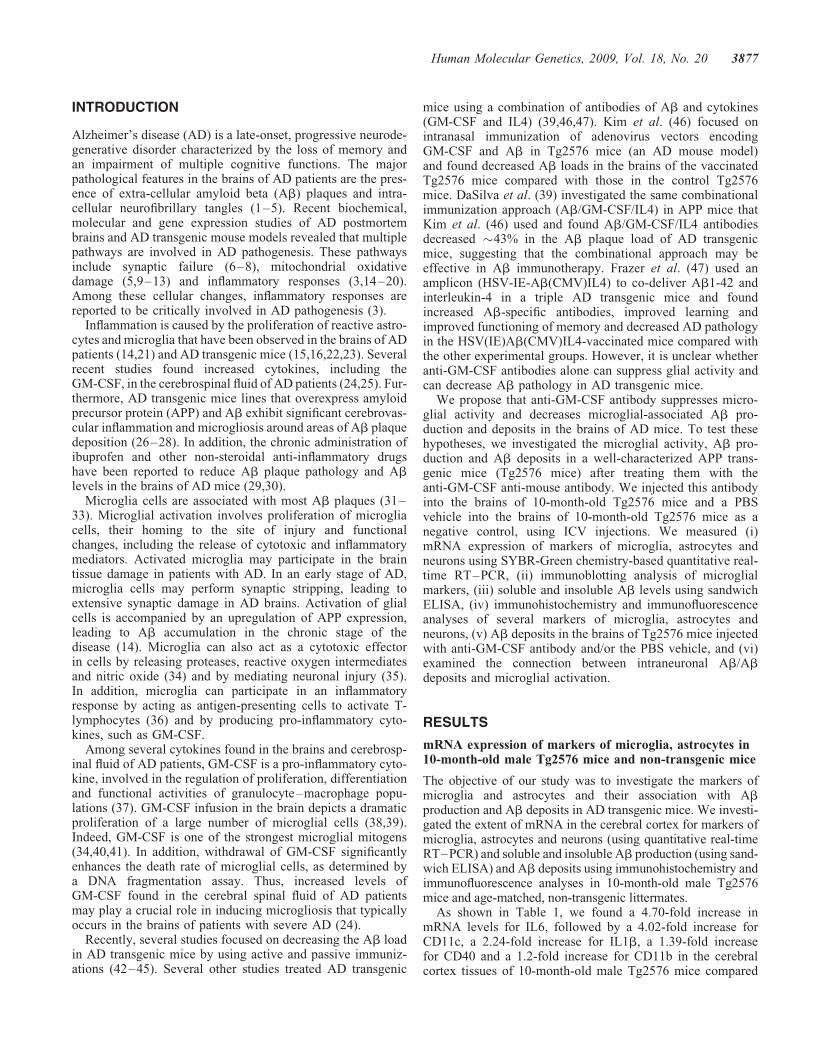
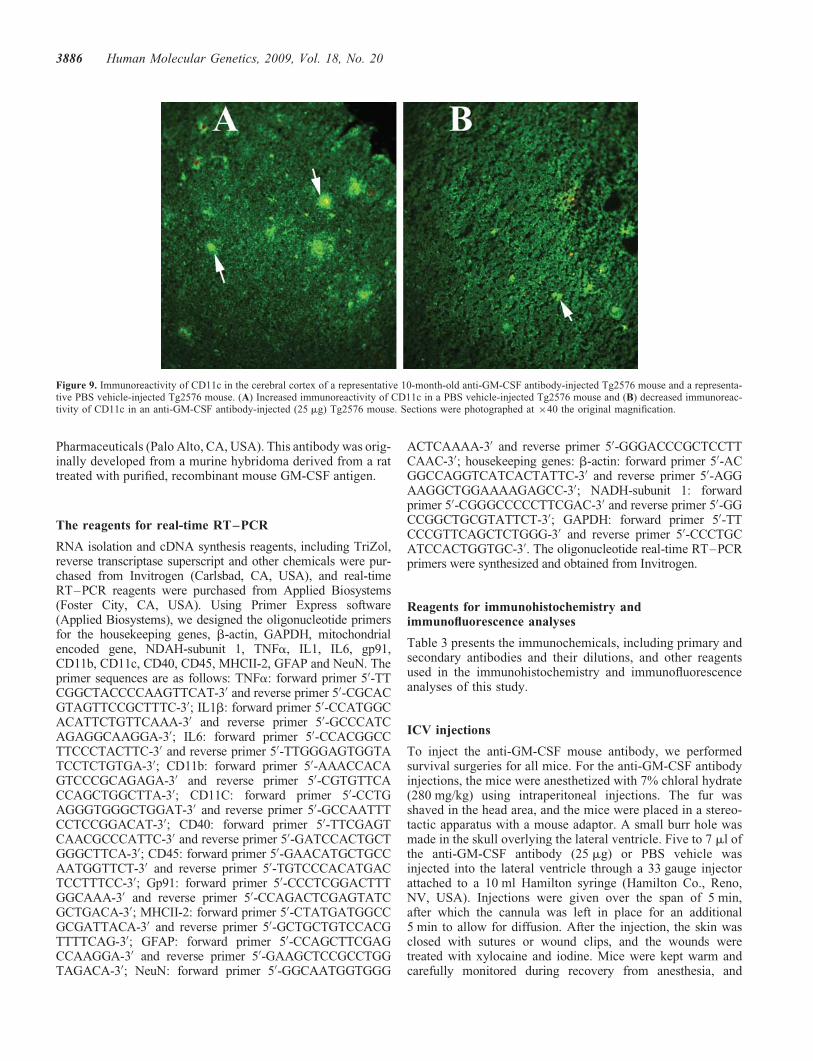
We found a statistically significant decreased immunoreactivesignal intensity for IL6 (P , 0.0016) (Fig. 8), CD11c (P ,0.0001) (Fig. 9) and CD40 (P , 0.0001) in anti-GM-CSFantibody-injected Tg2576 mice compared with PBS vehicle-injected Tg2576 mice, suggesting that GM-CSF antibody sup-press microglial activity in Tg2576 mice. We also founddecreased but not significant immunoreactivity for IL1b,CD11b, TNFa in anti-GM-CSF antibody-injected Tg2576mice compared with the PBS vehicle-injected Tg2576 mice .
We found slightly increased immunoreactivity of GFAP(Fig. 10) in anti-GM-CSF antibody-injected Tg2576 micecompared with the PBS vehicle-injected Tg2576 mice. Inter-estingly, we found slightly increased but not significant immu-noreactivity of GFAP in GM-CSF antibody-injected Tg2576mice compared with PBS vehicle-injected Tg2576 mice(Fig. 10), suggesting GM-CSF antibody has little or noeffect on GFAP.
Immunohistochemistry and immunofluorescence analysesof intraneuronal Ab and Ab deposits in anti-GM-CSFantibody-injected Tg256 mice and PBS-injected Tg2576mice
To determine whether the anti-GM-CSF antibody reduces Abdeposits, we conducted immunohistochemistry and immuno-fluorescence analyses of Ab deposits in PBS-injected Tg256mice and anti-GM-CSF antibody-injected Tg2576 mice.
We found a statistically significant decreased Ab deposits(P , 0.01) (Fig. 11) in anti-GM-CSF antibody-injectedTg2576 mice compared with PBS vehicle-injected Tg2576mice, suggesting that GM-CSF antibody decreases microglial-associated Ab deposits in Tg2576 mice.
DISCUSSION
The purpose of our study was to investigate pro-inflammatorycytokines that are associated with human APP Swedishmutation(s) in AD, and to investigate whether the anti-GM-CSFantibody can suppress microglial activity induced by human APPmutation in Tg2576 mice. Using ICV injections, we injectedthe anti-GM-CSF antibody or the PBS vehicle into thebrains of Tg2576 mice. Using quantitative real-time RT–PCR,
Figure 3. Double-labeling immunofluorescence analyses of Ab deposit andmicroglia marked by IL1b (A) and CD11b (B) in representative 10-month-oldTg2576 mouse. Ab deposits are associated with microglia. (Aa) Immunoreac-tivity of IL1, (Ab) surrounded by Ab deposit in the same section and (Ac)overlay of both IL1 and Ab deposit. Arrows indicate immunoreactivity ofIL1 (in white), and the arrowhead indicates Ab deposit. (Ba) Immunoreactiv-ity of CD11b, (Bb) surrounded by Ab deposit and (Bc) overlay of both CD11band Ab deposit. Arrows indicate immunoreactivity of CD11b (in white), andthe arrowhead indicate Ab deposit.
Figure 4. Double-labeling immunofluorescence analyses of Ab deposit andmicroglia marked by IL6 (A) and CD11b (B) in a representative10-month-oldTg2576 mouse. Ab deposit without microglia. (A) Microglia marked byimmunoreactivity of IL6 (a), Ab deposit without microglia in the samesection (b) and overlay of both IL6 and Ab deposit (c). (B) Immunoreactivityof microglia marked by CD11b present in the vicinity of Ab deposit (arrows inwhite). Ab deposits are present without microglial immunoreactivity ofCD11b (arrows in red).
3880 Human Molecular Genetics, 2009, Vol. 18, No. 20

we measured mRNA levels of several GM-CSF-associated cyto-kines in Tg2576 mice, non-transgenic wild-type mice, PBSvehicle-injected mice and anti-GM-CSF antibody-injectedTg2576 mice. We measured soluble and insoluble Ab1-40 andAb1-42 in PBS vehicle-injected and anti-GM-CSFantibody-injected Tg2576 mice. Using immunoblotting analysis,we studied the protein levels of microglial markers in PBSvehicle-injected and anti-GM-CSF antibody-injected Tg2576mice. Using immunohistochemistry and immunofluorescence,we assessed intraneuronal Ab and Ab deposits. Using double-labeling immunofluorescence analysis, we assessed the relation-ship between Ab deposits and markers of microglia and astro-cytes in anti-GM-CSF antibody-injected Tg2576 mice and PBSvehicle-injected Tg2576 mice. Our real-time RT–PCR analysisshowed an increased mRNA expression of IL6, CD11c, IL1,CD40 and CD11b in the cerebral cortices of the Tg2576 micecompared with the non-transgenic wild-type mice. Further, theanti-GM-CSF antibody significantly suppressed the humanAPPswe mutation and deposits of Ab-induced glial activity inthe brains of Tg2576 mice. Our immunoblotting and immunohis-tochemistry findings of microglial markers in Tg2576 miceinjected with GM-CSF antibody concur with our real-timeRT–PCR findings. In Tg2576 mice, we found Ab deposits aresurrounded by microglia and astrocytes, and intraneuronal Abco-localized with intraneuronal CD40. These results suggestthat intraneuronal expression of Ab may activate the expressionof CD40 in AD neurons. Interestingly for the first time, we founddecreased levels of soluble Ab1-42 and increased levels ofAb1-40 in Tg2576 mice injected with anti-GM-CSF antibody,indicating that anti-GM-CSF antibody clears the levels ofsoluble Ab1-42 more rapidly and/or decreases the productionof soluble Ab1-40.
IL6 and AD
IL6 is a pro-inflammatory cytokine that is secreted by T cellsand macrophages to stimulate immune response to trauma ortissue damage leading to inflammation. IL6 is activated inAD brains (48–50). In the present study, IL6 mRNA inTg2576 mice was the most upregulated among allpro-inflammatory cytokines that we examined in this study.This result is supported by several previous studies of ADpostmortem brains (48–50) and AD transgenic mice
(15,16,51–56). Ravaglia et al. (48) found increased serumIL6 associated with vascular dementia. Together, findingsfrom our study and the studies of AD postmortem brains,AD transgenic mice and serum from AD patients suggestthat increased IL6 is involved in AD progression.
Our immunohistochemistry analysis of IL6 in Tg2576 miceinjected with the anti-GM-CSF antibody and PBSvehicle-injected Tg2576 mice revealed that overexpressedIL6 is significantly suppressed by anti-GM-CSF antibody inthe brain regions, including cortex and hippocampus ofTg2576 mice (P , 0.0016), suggesting that anti-GM-CSFantibody is a potent suppresser of IL6 in Tg2576 mice. Ourreal-time RT–PCR (Table 1) and immunoblotting findings(Fig. 1) also showed decreased mRNA and protein levels ofIL6 in Tg2576 mice injected with GM-CSF antibody, whichfurther supports that GM-CSF antibody is a suppressor ofIL6 in AD transgenic mice.
CD11b and AD
CD11b is also known as heterodimeric integrin alpha-Mbeta-2 and is expressed on the surface of many leukocytesinvolved in the innate immune system, including monocytes,granulocytes, macrophages. CD11b mediates inflammationby regulating leukocyte adhesion and migration. CD11b isreported to be activated in AD brains, and in the presentstudy, we found increased mRNA expression in the cerebralcortex of Tg2576 mice compared with control mice. Wealso found increased immunoreactivity in the brains ofTg2576 mice, mainly in the vicinity of Ab deposits (Sup-plementary Material, Fig. S3), suggesting that CD11b is acti-vated in response to increased Ab deposits. Our findings aresupported by several previous studies in AD brains (57,58)and AD transgenic mice (54,59–61).
Our immunohistochemistry analysis of CD11b in Tg2576mice injected with anti-GM-CSF antibody showed decreasedbut not significant CD11b immunoreactivity in the hippocam-pal and cortical regions, particularly in the vicinity of Abdeposits, indicating that GM-CSF antibody suppresses themicroglial activity as shown by CD11b expression inTg2576 mice. Findings from our real-time RT–PCR(Table 1) and immunoblotting (Fig. 1) also showed decreasedmRNA and protein levels of CD11b in Tg2576 mice injected
Figure 5. Double-labeling immunofluorescence analyses of Ab deposit and astrocytes marked by glial fibrillary acidic protein (GFAP) in a representative10-month-old Tg2576 mouse. (A) Immunoreactivity of GFAP (in green), (B) immunoreactivity of Ab deposits (in red) of the same section and (C) overlayof GFAP and Ab deposits. Ab deposits are associated with astrocytes.
Human Molecular Genetics, 2009, Vol. 18, No. 20 3881

with GM-CSF antibody, which further supports that GM-CSFantibody is a suppressor of CD11b in AD transgenic mice.
CD11c and AD
CD11c is a type-I transmembrane protein found in dendriticcells, monocytes, macrophages, neutrophils and beta cells.Our finding of increased mRNA expression of CD11c inTg2576 mice relative to control mice has been reported in ADpostmortem brains (62,63) and AD transgenic mouse lines(60,64) and cell culture studies of microglia activated by aggre-gated Ab (65). de Groot et al. (66) developed cultures of micro-glial cells from rapid autopsies of the subcortical white matter,corpus callosum and frontal, temporal and occipital cortex(range 3–10 h) of non-demented elderly individuals and ADpatients and found that the adherent microglial cells wereimmunoreactive for CD68, CD45, CD11c and MHCIImarkers. Upon the stimulation with lipopolysaccharide, micro-glial cells secreted pro- and anti-inflammatory mediators, IL6,prostaglandin E2 and IL10, suggesting the functional capacityof cultured microglia from AD patients (66). These findingsfurther support our observations that CD11c is abnormallyexpressed in the Tg2576 mice. We found that upregulatedCD11c is significantly suppressed by the anti-GM-CSF anti-body in Tg2576 mice (P , 0.0001). Further, our real-timeRT–PCR findings (Table 1) also showed decreased mRNAlevels of CD11c in Tg2576 mice injected with GM-CSF anti-body, which further supports that GM-CSF antibody is a sup-pressor of CD11c in AD transgenic mice.
IL1b and AD
IL1b is produced by macrophages, monocytes and dendriticcells, and IL1b forms an important part of the inflammatoryresponse of the body against infection, particularly indisease state such as Alzheimer’s. IL1b increases theexpression of adhesion factors on endothelial cells to thecells that fight pathogens. In the present study, we foundincreased mRNA expression of IL1b in Tg2576 mice, andour findings are supported by data from AD patients (67).Increased levels of IL1b were found to be associated withplaques in AD (68). Further, Guillemin et al. (68) found thatAb induces IL1b mRNA expression in human fetal astrocytesand macrophages. Combs et al. (69) reported a functionalrelationship between Ab-dependent activation of microgliaand several characteristic markers of neuronal death occurringin AD brains. Findings from AD postmortem studies, together
with our present study, suggest that IL1b upregulation may bea characteristic of AD.
Our immunohistochemistry analysis of IL1b in Tg2576 miceinjected with GM-CSF antibody showed suppressed overex-pression of IL1b in Tg2576 mice, further suggesting that theanti-GM-CSF antibody may have anti-inflammatory therapeuticvalue in AD. Our real-time RT–PCR (Table 1) and immuno-blotting findings (Fig. 1) also showed decreased mRNA andprotein levels of IL1b in Tg2576 mice injected with GM-CSFantibody, which further supports that GM-CSF antibody is asuppressor of IL1b in AD transgenic mice.
CD40 and AD
The increased CD40 expression that we found in Tg2576 micecompared with the control mice is supported by several studies(70–72).
CD40 is a cell-surface marker and a member of the tumornecrosis factor receptor super-family, reported to play a rolein the metabolism of Ab in AD pathogenesis (70–72).Buchhave et al. (70) investigated the plasma levels ofsoluble CD40 and the soluble CD40 ligand in 136 subjectswith mild cognitive impairment (MCI) and in 30 age-matchedcontrols. Sixty of the 136 MCI cases progressed to AD duringa clinical follow-up period of 4–7 years. The baseline levelsof soluble CD40 were elevated in the MCI-AD cases com-pared with age-matched controls, suggesting that CD40 mayplay a role in the pathogenesis of early AD.
Ait-ghezala et al. (72) measured plasma Ab, soluble CD40and soluble CD40 ligand levels in 73 AD patients and com-pared their levels with those of 102 controls. They foundincreased levels of Ab1-40, insoluble CD40 and the solubleCD40 ligand in the AD patients, suggesting that CD40 andthe CD40 ligand are associated with AD progression.Recently, Volmer et al. (71) have shown increased levels ofAb in human embryonic kidney cells overexpressing bothmutant APP and CD40. Further, they demonstrated thatGM-CSF-neutralizing antibodies mitigate the CD40L-inducedproduction of Ab in human embryonic kidney cells expressinghuman mutant APP and CD40. They also showed that shRNAsilencing of the GM-CSF receptor gene significantly reducedAb levels below the baseline in non-stimulated HEK/APPsw/CD40 cells. Analysis of cell-surface proteins revealedthat silencing of the GM-CSF receptor also decreased APPendocytosis. Overall, findings from the Volmar et al. (71)study agree with our present study and suggest a role forGM-CSF in Tg2576 mice.
Figure 6. Double-labeling analysis of intraneuronal Ab and CD40 in a representative 10-month-old Tg2576 mouse. (A) Immunoreactivity of CD40 (in green),(B) intraneuronal Ab and Ab deposits (in red) of the same section and (C) overlay of CD40 and intraneuronal Ab. Arrows indicate localization of intraneuronalAb and CD40. Sections were photographed at �40 the original magnification.
3882 Human Molecular Genetics, 2009, Vol. 18, No. 20

Obregon et al. (73) used genetic and pharmacologicalapproaches to determine whether a CD40–CD40L blockadecould enhance the efficacy of an Ab1-42 immunization.They found that the genetic or pharmacological interruptionof the CD40–CD40L interaction enhanced Ab1-42 immuniz-ation efficacy and reduced cerebral amyloidosis in the PSAPPand Tg2576 mouse models of AD (73).
Laporte et al. (74) have generated APP and PSAPP mousemodels with a disrupted CD40 gene. They compared the patho-logical features of AD with appropriate controls and found allthese features reduced in mouse models deficient in CD40 com-pared with their controls that were not deficient in CD40,suggesting that CD40 signaling is required to allow the fullrepertoire of AD-like pathology in Tg2576 mice, and that theinhibition of the CD40 signaling pathway may be a potentialtherapeutic strategy in AD. In another study, Nichol et al.(75) investigated whether exercise could alter the immuneprofile in 17–19-month-old Tg2576 mice to a response thatreduces Ab pathology. They found decreased levels of ILband TNFa and increased levels of CD40 and MHCII in exer-cised animals compared with sedentary mice. These resultssuggest that CD40 and other cytokines are associated withAD pathogenesis.
Our immunohistochemistry analysis of CD40 in Tg2576mice injected with anti-GM-CSF antibody showed significantlydecreased CD40 immunoreactivity (P , 0.0001) in the hippo-campal and cortical regions, indicating that GM-CSF antibodysuppresses the microglial activity as shown by CD40 expressionin Tg2576 mice. Findings from our real-time RT–PCR(Table 1) and immunoblotting (Fig. 1) also showed decreasedmRNA and protein levels of CD40 in Tg2576 mice injectedwith GM-CSF antibody, which further supports that GM-CSFantibody is a suppressor of CD40 in AD transgenic mice.Overall, findings from AD postmortem brains, AD mice andcell-line studies on CD40 and GM-CSF support our presentstudy observations, which suggests that GM-CSF and CD40play a large role in AD pathogenesis.
Association of intraneuronal Ab and CD40
We found co-localization of CD40 and intraneuronal Ab inTg2576 mice. We further confirmed intraneuronal expressionof CD40 using double-labeling analyses of NeuN and CD40in brain sections from Tg2576 mice. However, there are nopublished reports in AD mice and AD patients on theco-localization of CD40 and intraneuronal Ab. In a recentpaper, Hou et al. (76) reported that CD40 is expressed in
neurons. The intraneuronal expression of CD40 and its con-nection with intraneuronal Ab in AD are still unclear.However, it is possible that the interaction between intraneur-onal Ab and CD40 may promote inflammation via cytokineactivation, in addition to increasing Ab deposits associatedwith cytokines. It is also possible that interaction betweenintraneuronal Ab and CD40 may promote abnormal APP pro-cessing and generate increased production of Ab1-42. We alsofound decreased co-localization of intraneuronal Ab inTg2576 mice injected with the anti-GM-CSF antibody,suggesting that GM-CSF antibody may decrease intraneuronalexpression of CD40 in Tg2576 mice.
GFAP and GM-CSF in Tg2576 mice
We found increased mRNA abundance of GFAP andincreased astrocytes in our immunohistochemistry in Tg2576mice compared with non-transgenic littermates, and our obser-vations are supported by Lim et al. (29). However, there is aslight increase of GFAP expression in Tg2576 mice injectedwith GM-CSF antibody in Tg2576 mice relative to PBSvehicle-injected Tg2576 mice. The precise link betweenincreased levels of GFAP and anti-GM-CSF antibodyin Tg2576 mice is unclear. Further research is needed toinvestigate the connection between increased GFAP andanti-GM-CSF antibody in APP mice.
However, as described earlier, we found suppressed microglialactivity and decreased intraneuronal Ab1-42 levels and Abdeposits in anti-GM-CSF antibody-injected Tg2576 mice relativeto PBS vehicle-injected Tg2576 mice, suggesting thatanti-GM-CSF antibody may have a potential therapeutic valuefor AD.
GM-CSF treatment and decreased Ab1-42
There are no published reports thus far to compare our presentfindings of decreased Ab1-42 levels and increased Ab1-40 inAD transgenic mice with the administration of the anti-GM-CSFantibody directly into the mouse brain. However, there have beenstudies conducted using an adenovirus vaccine of both GM-CSFand Ab (46,77). Zou et al. (77) injected vaccine-expressing4�Ab (1–15) and gene-adjuvant GM-CSF into Tg2576 mice.The adenovirus vaccine induced an Ab-specificIgG1-predominant humoral immune response and reduced Abdeposits in Tg2576 mice and reduced cognition deficits.
Using a vaccine with an adenovirus vector encoding GM-CSFand Ab, Kim et al. (46) studied Ab deposits in Tg2576 mice.
Figure 7. Double-labeling analysis of intraneuronal NeuN and CD40 in a representative 10-month-old Tg2576 mouse. (A) Immunoreactivity of NeuN (in green)in the cerebral cortex section, (B) intraneuronal CD40 (in red) of the same section and (C) overlay of NeuN and CD40. White arrows indicate co-localizedneurons with NeuN and CD40, and red arrows indicate neurons only with NeuN expression. Sections were photographed at �40 the original magnification.
Human Molecular Genetics, 2009, Vol. 18, No. 20 3883
Immunoglobulin isotyping revealed that the induced anti-Abantibodies were predominantly the IgG2b and IgG1 isotypes.They found decreased Ab loads in the brain of the Tg2576mice vaccinated with the adenovirus vectors encoding Ab andGM-CSF compared with control Tg2576 mice. These studies,however, did not quantify the levels of Ab1-42 and Ab1-40 inthe GM-CSF-treated Tg2576 mice.
DaSilva et al. (39) vaccinated APP mice with Ab, GM-CSFand IL4 and found this combinational approach decreased Abplaque load by 43% in AD transgenic mice. Frazer et al. (47)used an adenoviral cassette (HSV(IE)Ab(CMV)IL4) thatco-delivered Ab1-42 with an interleukin-4 in a triple-transgenic mice model. They found increased levels ofAb-specific antibodies, improved learning and improvedmemory, and decreased AD pathological features in the vacci-nated mice compared with the other experimental groups (47).In another study, using a gene-gun delivery approach, Qu et al.(78) delivered Ab42 into double-transgenic (APPswe/PS1Del-taE9) mice and found that the vaccinated mice developed Th2antibodies (IgG1) against Ab42. The levels of Ab42 in braindecreased by 41%, and the level of plasma increased by 43%in the vaccinated mice compared with the control mice.Similar to the Qu et al. (78) study, several other studiesusing AD transgenic mice (79) and non-human primatemonkeys (80,81) found decreased levels of Ab and amelio-rated cognitive deficits in animals vaccinated with Ab42.However, the long-term consequences of these Ab immuniz-ations are still unclear in clinical trials in which AD patientswere vaccinated with Ab. Findings from immunizationhuman clinical trials using Ab antibodies have not given posi-tive results thus far other than decreasing Ab deposits. Severallaboratories using AD transgenic mice and non-human pri-mates are currently studying the biochemistry of Ab and thespecificity of Ab42 antibodies to decrease Ab production.
We propose that anti-GM-CSF antibody may play a key rolein decreasing the production of Ab1-42 and the increasing ofAb1-40 levels via blocking the g-secretase activity at theepsilon site (Ab42-43 position). This notion needs further inves-tigation of anti-GM-CSF antibody in a large number of Tg2576mice and also in other AD transgenic mice lines that overexpresshuman mutant APP in mice. In support of GM-CSF involvementin altering Ab production, Volmar et al. (71) investigatedhuman embryonic kidney cells expressing mutant APP andCD40. They found increased levels of Ab in kidney cells over-expressing both APP and CD40. The GM-CSF antibodiesreduce the CD40 ligand-induced production of Ab in kidneycells expressing APPsw and CD40. Treatment of these cellswith recombinant GM-CSF increased Ab levels, indicatingthat GM-CSF antibody may affect Ab production. However,our study is a step forward in determining the effects ofGM-CSF in Ab production in AD pathogenesis, finding thatAb1-42 is decreased in Tg2576 mice injected with theanti-GM-CSF antibody and that the anti-GM-CSF antibodycan suppress microglial activity and decrease toxic Ab1-42.
CONCLUSIONS
The mechanistic link between microglial activation and mutantAPP/Ab in AD progression is not completely understood.T
ab
le2.
Sum
mar
yof
bet
aam
ylo
idle
vel
sbet
wee
nP
BS
veh
icle
-inje
cted
Tg256
mic
ean
dan
ti-G
M-C
SF
anti
body-i
nje
cted
Tg2576
mic
eusi
ng
sandw
ich
EL
ISA
Num
ber
of
mic
eS
olu
ble
Ab
1-4
0
(pg/m
g),
mea
n+
SE
P-v
alue
%D
iffe
rence
Solu
ble
Ab
1-4
2
(pg/m
g),
mea
n+
SE
P-v
alue
%D
iffe
rence
Inso
luble
Ab
1-4
0
(pg/m
g),
mea
n+
SE
P-v
alue
%D
iffe
rence
Inso
luble
Ab
1-4
2
(pg/m
g),
mea
n+
SE
P-v
alue
%D
iffe
rence
Tg2576
(n¼
5)
6.4+
1.7
90.4
423%
incr
ease
din
anti
-GM
-CS
F-i
nje
cted
mic
e
5.4+
1.1
0.2
846%
dec
reas
edin
anti
-GM
-CS
F-i
nje
cted
mic
e
0.6+
0.1
10.1
827%
incr
ease
din
anti
-GM
-CS
F-i
nje
cted
mic
e
0.2
8+
0.0
90.9
No
chan
ge
Tg2576þ
anti
-GM
-CS
F
anti
body
(n¼
5)
8.3+
1.3
33.7+
0.9
0.8+
0.0
60.2
7+
1.5
We
found
incr
ease
dle
vel
sof
solu
ble
Ab
1-4
0(2
3%
,P
,0.4
4)
and
inso
luble
Ab
1-4
0(2
7%
,P
,0.1
8)
inT
g2576
mic
ein
ject
edw
ith
anti
-GM
-CS
Fan
tibody
com
par
edw
ith
Tg2576
mic
ein
ject
edw
ith
PB
S(c
ontr
ol)
.H
ow
ever
,th
ese
incr
ease
dle
vel
sar
enot
stat
isti
call
ysi
gnifi
cant
may
be
bec
ause
of
smal
lsa
mple
size
(n¼
5in
each
gro
up).
Inte
rest
ingly
,w
eal
sofo
und
dec
reas
edle
vel
sof
solu
ble
Ab
1-4
2(4
6%
,P
,0.2
8)
inT
g2576
mic
ein
ject
edw
ith
anti
-GM
-CS
Fan
tibody
com
par
edw
ith
Tg2576
mic
ein
ject
edw
ith
PB
S(c
ontr
ol)
.H
ow
ever
,th
ese
incr
ease
dle
vel
sar
en
ot
stat
isti
call
ysi
gnifi
cant.
3884 Human Molecular Genetics, 2009, Vol. 18, No. 20

Further, the role of GM-CSF in activating pro-inflammatorycytokines in AD pathogenesis is still unclear. In the presentstudy, we aimed to understand the connection betweenGM-CSF and microglial activity in AD progression. We inves-tigated whether the anti-GM-CSF antibody can suppress micro-glial activity and decrease Ab production in Tg2576 mice. Weintroduced the mouse GM-CSF antibody or PBS vehicle intothe brains of 10-month-old male Tg2576 mice using ICV injec-tions. We assessed the effect of several GM-CSF-associatedpro-inflammatory cytokines on microglial activities and theirassociation with Ab using quantitative real-time RT–PCR,immunoblotting, immunohistochemistry and immunofluores-cence analyses in anti-GM-CSF antibody-injected Tg2576mice and PBS vehicle-injected control Tg2576 mice. Usingsandwich ELISA technique, we measured intraneuronal Ab inTg2576 mice injected with GM-CSF antibody and PBSvehicle-injected control Tg2576 mice. Using double-labelingimmunofluorescence analysis of intraneuronal Ab, Ab depositsand markers of microglia, and astrocytes, we assessed therelationship between intraneuronal Ab and microglialmarkers, and Ab deposits and pro-inflammatory cytokines inthe Tg2576 mice injected with PBS vehicle and also in theanti-GM-CSF antibody-injected Tg2576 mice.
Our real-time RT–PCR analysis showed an increase in themRNA expression of IL6, CD11c, IL1b, CD40 and CD11b inthe cerebral cortices of the Tg2576 mice compared with their lit-termate non-transgenic controls. Immunohistochemistry find-ings of pro-inflammatory cytokines agreed with our real-timeRT–PCR results. Interestingly, we found significantly decreasedlevels of activated microglia marked by CD11C, IL6 and CD40,and Ab deposits in anti-GM-CSF antibody-injected Tg2576mice compared with PBS vehicle-injected Tg2576 mice(negative controls). Findings from our real-time RT–PCR andimmunoblotting analysis agreed with the immunohistochemistryobservations. Our double-labeling analyses of intraneuronal Aband CD40 revealed that intraneuronal Ab is associated withneuronal expression of CD40 in Tg2576 mice. Our quantitativesandwich ELISA analysis revealed that decreased levels ofsoluble Ab1-42 and increased levels of Ab1-40 in Tg2576mice injected with the anti-GM-CSF antibody compared withTg2576 mice injected with PBS vehicle, suggesting thatanti-GM-CSF antibody decreases soluble Ab1-42 productionand suppresses microglial activity in Tg2576 mice. These
findings indicating the ability of the anti-GM-CSF antibody toreduce Ab1-42 and microglial activity in Tg2576 mice mayhave therapeutic implications for AD.
MATERIALS AND METHODS
Tg2576 mice
The Tg2576 mouse model was generated with the mutant humanAPP gene 695 amino acid isoform and with a double mutation(Lys670Asn and Met671Leu) (82). This highly expressed humanAPP transgenic mice model exhibits an age-dependent appear-ance of Ab plaques as well as a distribution of the Ab plaquethat is confined to the cerebral cortex and the hippocampus,sparing the striatum, the deep gray nuclei, and the brain stem.A correlation has been found between elevated amounts ofsoluble Ab and increased free-radical production (10). ThisTg2576 mouse model also parallels AD in that the Ab plaquesevoke a microglial reaction in their immediate vicinity.
Ten-month-old Tg2576 male mice (25–40 g) and age-matched, non-transgenic male littermates (‘controls’; 25–40 g) were purchased from Taconic Farms (New York, NY,USA) and housed at the animal facility of the Oregon NationalPrimate Center at Oregon Health and Science University(OHSU). We investigated 10-month-old Tg2576 mice (n ¼5) and age-matched, non-transgenic littermates (n ¼ 5) formRNA abundance using quantitative RT–PCR and assessedAb deposits, microglia and astrocytes using immunohisto-chemistry. Further, using ICV injections, we injectedanti-GM-CSF antibody into the brains of the Tg2576 mice(n ¼ 5) and PBS vehicle into the brains of the Tg2576 mice(n ¼ 5) as the ‘negative control’ and investigated mRNAabundance using quantitative RT–PCR, measured solubleand insoluble Ab1-40 and Ab1-42 using sandwich ELISA,and Ab deposits, assessed microglia and astrocytes usingimmunohistochemistry. The OHSU Institutional AnimalCare and Use Committee approved all procedures for animalcare according to the guidelines set forth by NIH.
The anti-GM-CSF antibody
The anti-GM-CSF anti-mouse antibody (MP1-22E9, R &DSystems, Minneapolis, MN, USA) was supplied by KaloBios
Figure 8. Immunoreactivity of IL6 in the cerebral cortex of a representative 10-month-old anti-GM-CSF antibody-injected Tg2576 mouse and a representativePBS vehicle-injected Tg2576 mouse. (A) Increased immunoreactivity of IL6 in a PBS vehicle-injected Tg2576 mouse and (B) decreased immunoreactivity ofIL6 in an anti-GM-CSF antibody-injected (25 mg) Tg2576 mouse. Sections were photographed at �40 the original magnification.
Human Molecular Genetics, 2009, Vol. 18, No. 20 3885
Pharmaceuticals (Palo Alto, CA, USA). This antibody was orig-inally developed from a murine hybridoma derived from a rattreated with purified, recombinant mouse GM-CSF antigen.
The reagents for real-time RT–PCR
RNA isolation and cDNA synthesis reagents, including TriZol,reverse transcriptase superscript and other chemicals were pur-chased from Invitrogen (Carlsbad, CA, USA), and real-timeRT–PCR reagents were purchased from Applied Biosystems(Foster City, CA, USA). Using Primer Express software(Applied Biosystems), we designed the oligonucleotide primersfor the housekeeping genes, b-actin, GAPDH, mitochondrialencoded gene, NDAH-subunit 1, TNFa, IL1, IL6, gp91,CD11b, CD11c, CD40, CD45, MHCII-2, GFAP and NeuN. Theprimer sequences are as follows: TNFa: forward primer 50-TTCGGCTACCCCAAGTTCAT-30 and reverse primer 50-CGCACGTAGTTCCGCTTTC-30; IL1b: forward primer 50-CCATGGCACATTCTGTTCAAA-30 and reverse primer 50-GCCCATCAGAGGCAAGGA-30; IL6: forward primer 50-CCACGGCCTTCCCTACTTC-30 and reverse primer 50-TTGGGAGTGGTATCCTCTGTGA-30; CD11b: forward primer 50-AAACCACAGTCCCGCAGAGA-30 and reverse primer 50-CGTGTTCACCAGCTGGCTTA-30; CD11C: forward primer 50-CCTGAGGGTGGGCTGGAT-30 and reverse primer 50-GCCAATTTCCTCCGGACAT-30; CD40: forward primer 50-TTCGAGTCAACGCCCATTC-30 and reverse primer 50-GATCCACTGCTGGGCTTCA-30; CD45: forward primer 50-GAACATGCTGCCAATGGTTCT-30 and reverse primer 50-TGTCCCACATGACTCCTTTCC-30; Gp91: forward primer 50-CCCTCGGACTTTGGCAAA-30 and reverse primer 50-CCAGACTCGAGTATCGCTGACA-30; MHCII-2: forward primer 50-CTATGATGGCCGCGATTACA-30 and reverse primer 50-GCTGCTGTCCACGTTTTCAG-30; GFAP: forward primer 50-CCAGCTTCGAGCCAAGGA-30 and reverse primer 50-GAAGCTCCGCCTGGTAGACA-30; NeuN: forward primer 50-GGCAATGGTGGG
ACTCAAAA-30 and reverse primer 50-GGGACCCGCTCCTTCAAC-30; housekeeping genes: b-actin: forward primer 50-ACGGCCAGGTCATCACTATTC-30 and reverse primer 50-AGGAAGGCTGGAAAAGAGCC-30; NADH-subunit 1: forwardprimer 50-CGGGCCCCCTTCGAC-30 and reverse primer 50-GGCCGGCTGCGTATTCT-30; GAPDH: forward primer 50-TTCCCGTTCAGCTCTGGG-30 and reverse primer 50-CCCTGCATCCACTGGTGC-30. The oligonucleotide real-time RT–PCRprimers were synthesized and obtained from Invitrogen.
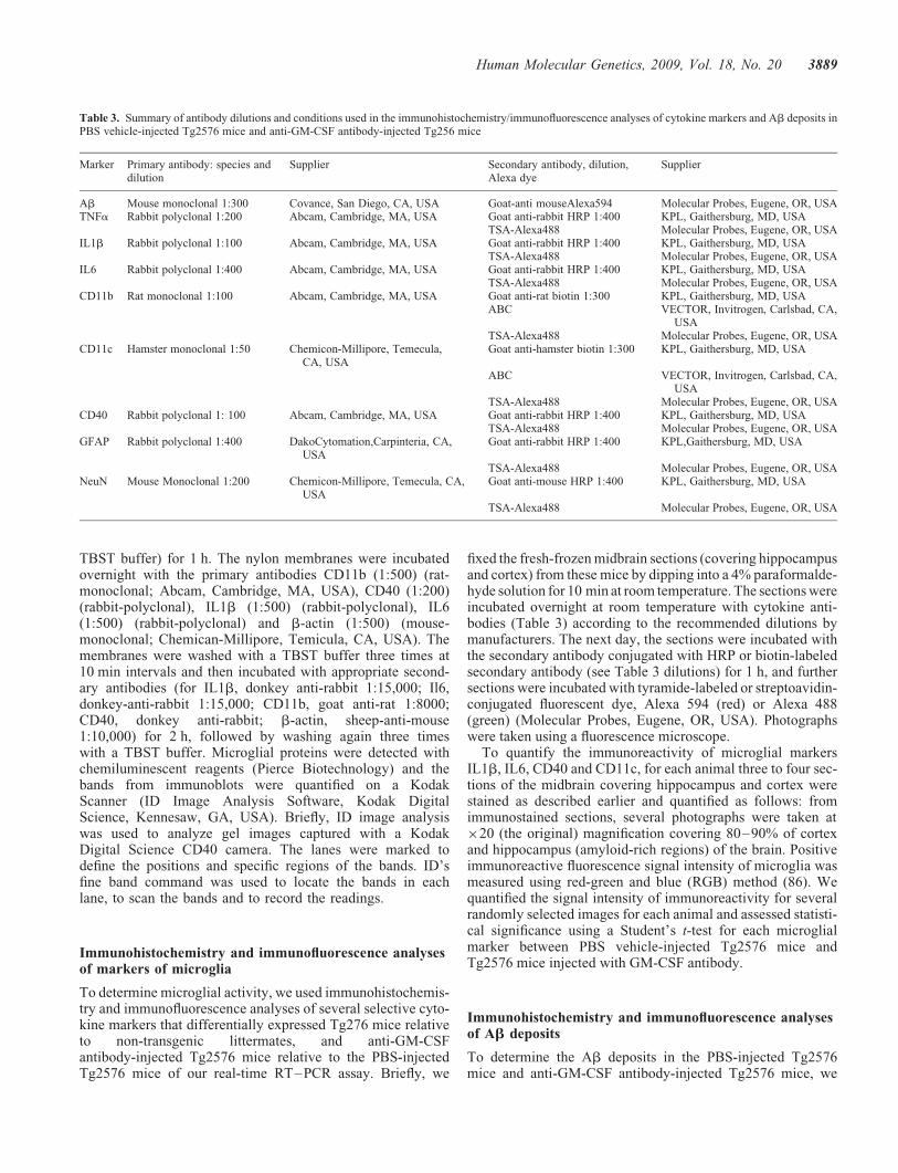
Reagents for immunohistochemistry andimmunofluorescence analyses
Table 3 presents the immunochemicals, including primary andsecondary antibodies and their dilutions, and other reagentsused in the immunohistochemistry and immunofluorescenceanalyses of this study.
ICV injections
To inject the anti-GM-CSF mouse antibody, we performedsurvival surgeries for all mice. For the anti-GM-CSF antibodyinjections, the mice were anesthetized with 7% chloral hydrate(280 mg/kg) using intraperitoneal injections. The fur wasshaved in the head area, and the mice were placed in a stereo-tactic apparatus with a mouse adaptor. A small burr hole wasmade in the skull overlying the lateral ventricle. Five to 7 ml ofthe anti-GM-CSF antibody (25 mg) or PBS vehicle wasinjected into the lateral ventricle through a 33 gauge injectorattached to a 10 ml Hamilton syringe (Hamilton Co., Reno,NV, USA). Injections were given over the span of 5 min,after which the cannula was left in place for an additional5 min to allow for diffusion. After the injection, the skin wasclosed with sutures or wound clips, and the wounds weretreated with xylocaine and iodine. Mice were kept warm andcarefully monitored during recovery from anesthesia, and
Figure 9. Immunoreactivity of CD11c in the cerebral cortex of a representative 10-month-old anti-GM-CSF antibody-injected Tg2576 mouse and a representa-tive PBS vehicle-injected Tg2576 mouse. (A) Increased immunoreactivity of CD11c in a PBS vehicle-injected Tg2576 mouse and (B) decreased immunoreac-tivity of CD11c in an anti-GM-CSF antibody-injected (25 mg) Tg2576 mouse. Sections were photographed at �40 the original magnification.
3886 Human Molecular Genetics, 2009, Vol. 18, No. 20

then placed in individual cages. Five days after the ICVinjections, the mice were re-anesthetized and sacrificed bycervical dislocation since maximum immunoreactivity ofmicroglial markers was reported at 4–5 days after GM-CSFinduction (83). The brains were collected from all mice andthe cerebral cortex (from the front brain) and cerebellumwere dissected from the brains separately. The soluble and inso-luble Ab levels were measured from cerebral cortex tissues.The midbrains (containing hippocampus, cerebral cortex andstriatum) were dissected and flash-frozen. Sections with10 mm-thickness were cut from the midbrain and used forimmunohistochemistry of cytokines and Ab deposits.
Quantification of mRNA expression of markers ofmicroglia, astrocytes and neurons using real-timeRT–PCR
Total RNA was isolated from dissected cerebral cortex tissuesfrom each group of experimental and control group mice usingthe TriZol reagent (Invitrogen). As shown in Table 2, mRNA
expression of several cytokines, GFAP and neuronal markerswas measured using SYBR-Green chemistry-based quantitat-ive real-time RT–PCR, as described by Manczak et al. (84).Briefly, 1 mg of DNAse-treated total RNA was used as startingmaterial, to which were added 1 ml of oligo (dT), 1 ml of10 mM dNTPs, 4 ml of 5� first-strand buffer, 4 ml of 25 mM
MgCl2, 2 ml of 0.1 M DTT and 1 ml of RNAse out. Thereagents RNA, oligo (dT) and dNTPs were mixed first,heated at 658C for 5 min and then chilled on ice until othercomponents were added. The samples were incubated at428C for 2 min. Then 1 ml of Superscript II (40 U/ml) (Invitro-gen) was added, and the samples were incubated at 428C for50 min. The reaction was inactivated by heating the contentsat 708C for 15 min.
Real-time quantitative PCR amplification reactions werecarried out in an ABI Prism 7900 sequence detection system(Applied Biosystems) in a 25 ml volume of total reactionmixture. The reaction mixture consisted of 1� PCR buffercontaining SYBR-Green; 3 mM MgCl2; 100 nM of eachprimer; 200 nM each of dATP, dGTP and dCTP; 400 nM of
Figure 10. Immunoreactivity of GFAP in a representative 10-month-old anti-GM-CSF antibody-injected Tg2576 mouse and a representative PBSvehicle-injected Tg2576 mouse. (A) Immunoreactivity of GFAP in a PBS vehicle-injected Tg2576 mouse, (B) increased immunoreactivity of GFAP in the cer-ebral cortex and hippocampus of an anti-GM-CSF antibody-injected Tg2576 mouse, (C) GFAP immunoreactivity of cerebral cortex from a PBS vehicle-injectedTg2576 mouse and (D) increased immunoreactivity of cerebral cortex in an anti-GM-CSF antibody-injected Tg2576 mouse. (A) and (B) were photographed at�5 the original magnification, and (C) and (D) at �20.
Human Molecular Genetics, 2009, Vol. 18, No. 20 3887

dUTP; 0.01 U/ml of AmpErase UNG and 0.05 U/ml of Ampli-Taq Gold (ABI). Twenty nanograms of cDNA template wasadded to each reaction mixture.
To determine the unregulated endogenous reference gene inAD mice, the CT values (cycle threshold values) of b-actin,GAPDH and NADH-subunit (mitochondrial-encoded gene)were tested. The CT value was the cycle number at whichthe fluorescence generated within a reaction crosses thethreshold within the linear phase of the amplification profile.The CT value is an important quantitative parameter in real-time PCR analysis (84,85). All reactions were carried out induplicate with a no-template control. The PCR conditionswere 508C for 2 min, 958C for 10 min, followed by 40cycles of 958C for 15 s and 608C for 1 min. The fluorescentspectra were recorded during the elongation phase of eachPCR cycle. To distinguish specific amplicons from non-specific amplifications, a dissociation curve was generated.The CT values were calculated with sequence-detectionsystem software V1.7 (Applied Biosystems) and an automaticsetting of baseline, which was the average value of PCR,cycles 3–15, plus CT generated 10 times its standard devi-ation. The amplification plots and CT values were exportedfrom the exponential phase of PCR directly into a MicrosoftExcel worksheet for further analysis.
The mRNA transcript level was normalized against b-actin,GAPDH and mitochondrial-encoded complex I gene—NADH-subunit 1 at each dilution. The standard curve wasthe normalized mRNA transcript level plotted against the log-value of the input cDNA concentration at each dilution. Tocompare b-actin, GAPDH and NADH-subunit 1 and cytokine
markers, relative quantification was performed according tothe CT method of ABI (84). Briefly, the comparative CT
method involved averaging duplicate samples taken as theCT values for b-actin, GAPDH and NADH-subunit 1 and cyto-kine markers. We used b-actin normalization in the presentstudy because b-actin CT values are similar for PBS-injectedTg2576 mice, anti-GM-CSF-injected Tg2576 mice and thenon-transgenic littermates (without the APP mutation). TheDCT value was obtained by subtracting the average b-actinCT value from the average CT value of cytokine markers.The present study used the average DCT of five animals asthe calibrator. The fold change was calculated according tothe formula 22(DDCT), where DDCT was the differencebetween DCT and the DCT calibrator value.
Immunoblotting analysis of microglial markers in Tg2576mice injected with PBS-vehicle and GM-CSFantibody-injected Tg2576 mice
To determine whether GM-CSF antibody suppressed theprotein levels of microglial markers that showed decreasedmRNA expression in our real-time RT–PCR in Tg2576mice injected with GM-CSF antibody, we studied immuno-blotting analysis of protein lysates from Tg2576 mice injectedwith GM-CSF antibody and Tg2576 mice injected with PBSvehicle. Twenty micrograms of protein lysate from cerebralcortex was resolved on 12% SDS–PAGE, and the resolvedproteins were transferred to nylon membranes (Novax Inc.,San Diego, CA, USA) that were then incubated at room temp-erature with a blocking buffer (5% dry milk dissolved in a
Figure 11. Immunoreactivity of Ab deposits in a representative 10-month-old anti-GM-CSF antibody-injected Tg2576 mouse and a representative PBSvehicle-injected Tg2576 mouse. The E610 antibody recognized both intraneuronal Ab and Ab deposits in mouse brain sections. (A) Ab deposits in the cerebralcortex and hippocampus of a PBS vehicle-injected Tg2576 mouse, (B) decreased Ab deposits in the cerebral cortex and hippocampus in an anti-GM-CSFantibody-injected mouse, (C) Ab deposits in the hippocampus of a PBS vehicle-injected Tg2576 mouse and (D) decreased Ab deposits in the hippocampusof an anti-GM-CSF antibody-injected mouse. (A) and (B) were photographed at �5 the original magnification, and (C) and (D), at �10.
3888 Human Molecular Genetics, 2009, Vol. 18, No. 20
TBST buffer) for 1 h. The nylon membranes were incubatedovernight with the primary antibodies CD11b (1:500) (rat-monoclonal; Abcam, Cambridge, MA, USA), CD40 (1:200)(rabbit-polyclonal), IL1b (1:500) (rabbit-polyclonal), IL6(1:500) (rabbit-polyclonal) and b-actin (1:500) (mouse-monoclonal; Chemican-Millipore, Temicula, CA, USA). Themembranes were washed with a TBST buffer three times at10 min intervals and then incubated with appropriate second-ary antibodies (for IL1b, donkey anti-rabbit 1:15,000; Il6,donkey-anti-rabbit 1:15,000; CD11b, goat anti-rat 1:8000;CD40, donkey anti-rabbit; b-actin, sheep-anti-mouse1:10,000) for 2 h, followed by washing again three timeswith a TBST buffer. Microglial proteins were detected withchemiluminescent reagents (Pierce Biotechnology) and thebands from immunoblots were quantified on a KodakScanner (ID Image Analysis Software, Kodak DigitalScience, Kennesaw, GA, USA). Briefly, ID image analysiswas used to analyze gel images captured with a KodakDigital Science CD40 camera. The lanes were marked todefine the positions and specific regions of the bands. ID’sfine band command was used to locate the bands in eachlane, to scan the bands and to record the readings.
Immunohistochemistry and immunofluorescence analysesof markers of microglia
To determine microglial activity, we used immunohistochemis-try and immunofluorescence analyses of several selective cyto-kine markers that differentially expressed Tg276 mice relativeto non-transgenic littermates, and anti-GM-CSFantibody-injected Tg2576 mice relative to the PBS-injectedTg2576 mice of our real-time RT–PCR assay. Briefly, we
fixed the fresh-frozen midbrain sections (covering hippocampusand cortex) from these mice by dipping into a 4% paraformalde-hyde solution for 10 min at room temperature. The sections wereincubated overnight at room temperature with cytokine anti-bodies (Table 3) according to the recommended dilutions bymanufacturers. The next day, the sections were incubated withthe secondary antibody conjugated with HRP or biotin-labeledsecondary antibody (see Table 3 dilutions) for 1 h, and furthersections were incubated with tyramide-labeled or streptoavidin-conjugated fluorescent dye, Alexa 594 (red) or Alexa 488(green) (Molecular Probes, Eugene, OR, USA). Photographswere taken using a fluorescence microscope.
To quantify the immunoreactivity of microglial markersIL1b, IL6, CD40 and CD11c, for each animal three to four sec-tions of the midbrain covering hippocampus and cortex werestained as described earlier and quantified as follows: fromimmunostained sections, several photographs were taken at�20 (the original) magnification covering 80–90% of cortexand hippocampus (amyloid-rich regions) of the brain. Positiveimmunoreactive fluorescence signal intensity of microglia wasmeasured using red-green and blue (RGB) method (86). Wequantified the signal intensity of immunoreactivity for severalrandomly selected images for each animal and assessed statisti-cal significance using a Student’s t-test for each microglialmarker between PBS vehicle-injected Tg2576 mice andTg2576 mice injected with GM-CSF antibody.
Immunohistochemistry and immunofluorescence analysesof Ab deposits
To determine the Ab deposits in the PBS-injected Tg2576mice and anti-GM-CSF antibody-injected Tg2576 mice, we
Table 3. Summary of antibody dilutions and conditions used in the immunohistochemistry/immunofluorescence analyses of cytokine markers and Ab deposits inPBS vehicle-injected Tg2576 mice and anti-GM-CSF antibody-injected Tg256 mice
Marker Primary antibody: species anddilution
Supplier Secondary antibody, dilution,Alexa dye
Supplier
Ab Mouse monoclonal 1:300 Covance, San Diego, CA, USA Goat-anti mouseAlexa594 Molecular Probes, Eugene, OR, USATNFa Rabbit polyclonal 1:200 Abcam, Cambridge, MA, USA Goat anti-rabbit HRP 1:400 KPL, Gaithersburg, MD, USA
TSA-Alexa488 Molecular Probes, Eugene, OR, USAIL1b Rabbit polyclonal 1:100 Abcam, Cambridge, MA, USA Goat anti-rabbit HRP 1:400 KPL, Gaithersburg, MD, USA
TSA-Alexa488 Molecular Probes, Eugene, OR, USAIL6 Rabbit polyclonal 1:400 Abcam, Cambridge, MA, USA Goat anti-rabbit HRP 1:400 KPL, Gaithersburg, MD, USA
TSA-Alexa488 Molecular Probes, Eugene, OR, USACD11b Rat monoclonal 1:100 Abcam, Cambridge, MA, USA Goat anti-rat biotin 1:300 KPL, Gaithersburg, MD, USA
ABC VECTOR, Invitrogen, Carlsbad, CA,USA
TSA-Alexa488 Molecular Probes, Eugene, OR, USACD11c Hamster monoclonal 1:50 Chemicon-Millipore, Temecula,
CA, USAGoat anti-hamster biotin 1:300 KPL, Gaithersburg, MD, USA
ABC VECTOR, Invitrogen, Carlsbad, CA,USA
TSA-Alexa488 Molecular Probes, Eugene, OR, USACD40 Rabbit polyclonal 1: 100 Abcam, Cambridge, MA, USA Goat anti-rabbit HRP 1:400 KPL, Gaithersburg, MD, USA
TSA-Alexa488 Molecular Probes, Eugene, OR, USAGFAP Rabbit polyclonal 1:400 DakoCytomation,Carpinteria, CA,
USAGoat anti-rabbit HRP 1:400 KPL,Gaithersburg, MD, USA
TSA-Alexa488 Molecular Probes, Eugene, OR, USANeuN Mouse Monoclonal 1:200 Chemicon-Millipore, Temecula, CA,
USAGoat anti-mouse HRP 1:400 KPL, Gaithersburg, MD, USA
TSA-Alexa488 Molecular Probes, Eugene, OR, USA
Human Molecular Genetics, 2009, Vol. 18, No. 20 3889
used immunohistochemistry and immunofluorescence ana-lyses, as described in Manczak et al. (10). Briefly, we fixedthe midbrain sections and incubated them overnight at roomtemperature with a mouse anti-human Ab antibody, E610(see Table 3 for dilutions). The next day, the sections wereincubated with the goat anti-mouse secondary antibody conju-gated with fluorescent dye Alexa 594 (red) (Molecular Probes)for 1 h at room temperature. Photographs were taken with afluorescence microscope.
To quantify the immunoreactivity of Ab deposits, for eachanimal three to four sections of the midbrain covering hippo-campus and cortex were stained as described earlier and usedfor quantification purpose. From these sections, several photo-graphs were taken at �20 magnification covering 80–90% ofcortex and hippocampus (amyloid-rich regions) of the brain.Positive immunoreactive fluorescence signal intensity of Abdeposits was measured using RGB method (86). We quantifiedthe signal intensity of immunoreactivity for several randomlyselected images for each animal and assessed statistical signifi-cance using a Student’s t-test for Ab deposits between PBSvehicle-injected Tg2576 mice and Tg2576 mice injectedwith GM-CSF antibody.
Double-labeling analysis of intraneuronal Ab and Abdeposits and markers of microglia
To determine the precise connection between intraneuronalAb and Ab deposits and microglia, we performed double-labeling immunohistochemistry and immunofluorescence ana-lyses using a mouse anti-human Ab antibody, E610, and anti-bodies of markers of microglia IL1b, IL6, CD11b, CD11c,CD40 and GFAP as described in Manczak et al. (10).Briefly, we fixed the midbrain sections and incubated themovernight at room temperature with a mouse anti-human Abantibody (see Table 3 for dilutions). The next day, the sectionswere incubated with the secondary antibody conjugated withthe fluorescent dye Alexa 594 (red) (Molecular Probes) for1 h at room temperature. For the second labeling, the sectionswere incubated overnight with primary antibodies of cyto-kines, IL1b, IL6, CD11b, CD11c, CD40 and GFAP (seeTable 3 for dilutions). On the third day, for cytokines, the sec-tions were incubated with biotin-labeled secondary antibodyfor 30 min, ABC complex for another 30 min and tyramide-labeled fluorescent dye Alexa 488 (green) for 10 min.
For GFAP, the sections were incubated with a secondaryantibody conjugated with HRP for 1 h at room temperature.Finally, the slides were incubated with tyramide-labeledAlexa 488 the (green). Photographs were taken with a fluor-escence microscope.
Double-labeling analysis of CD40 and neuronal marker,NeuN
To determine whether CD40 is expressed in neurons, we per-formed double-labeling immunohistochemistry and immuno-fluorescence analyses using rabbit anti-CD40 antibody andmouse anti-neuronal marker NeuN. We fixed the midbrainsections and incubated them overnight at room temperaturewith rabbit anti-CD40 antibody (see Table 3 for dilutions).The next day, the sections were incubated with the
biotin-labeled secondary antibody for 30 min, next with ABCcomplex for another 30 min and finally with the tyramide-labeled fluorescent dye Alexa 594 (red) (Molecular Probes)for 10 min at room temperature. For the second labeling, thesections were incubated overnight with mouse anti-NeuN anti-body (see Table 3 for dilutions). On the third day, the sectionswere incubated with secondary antibody conjugated with HRPfor 1 h at room temperature. Finally, the slides were incubatedwith the tyramide-labeled fluorescent dye Alexa 488 (green).Photographs were taken with a fluorescence microscope.
Measurement of soluble and insoluble Ab levels usingsandwich ELISA
To determine whether the anti-GM-CSF antibody decreasessoluble and insoluble Ab levels in Tg2576 mice, we measuredsoluble and insoluble Ab levels in PBS-injected Tg2576mice and anti-GM-CSF antibody-injected Tg2576 mice asdescribed in Manczak et al. (10). Briefly, cerebral cortextissues were homogenized in Tris-buffered saline, pH 8.0, con-taining protease inhibitors (20 mg/ml pepstatin A, aprotinin,phosphsoramidon and leupeptin; 0.5 mM PMSF; 1 mM
EGTA). Samples were sonicated briefly and centrifuged at10 000g for 20 min at 48C. Using ELISA, we determined thesoluble Ab fraction. To measure the insoluble Ab, were-suspended the pellet in 70% formic acid and again centri-fuged the pellet. The extract was neutralized with 0.25 M
Tris, pH 8.0, containing 30% acetonitrile and 5 M NaOHbefore we determined the insoluble Ab using ELISA. Foreach sample, we measured Ab1-40 and Ab1-42 using com-mercial colorimetric ELISA kits (Biosource International,Camarillo, CA, USA), specific for each species. A 96-wellplate washer and reader were used, according to the manufac-turer’s instructions. Each sample was run in duplicate. Theprotein concentration of the homogenates was determined bythe BCA method, and Ab was expressed as picogram of Abper milligram protein.
Statistical considerations
Statistical analyses were conducted for soluble and insolubleAb, in both treated and untreated Tg2576 mice withanti-GM-CSF antibody using a Student’s t-test. We also quan-tified the immunoreactivity of microglial markers IL1b, IL6,CD40 and CD11c between Tg2576 mice and Tg2576 miceinjected with GM-CSF antibody. For each microglialmarker, we assessed the statistical significance using a Stu-dent’s t-test. The statistical significance of Ab depositsbetween Tg2576 mice and Tg2576 mice injected withGM-CSF antibody was assessed using a Student’s t-test.
SUPPLEMENTARY MATERIAL
Supplementary Material is available at HMG online.
ACKNOWLEDGEMENTS
We thank Dr Shaun Morrison for allowing us to use the stereo-taxic apparatus for GM-CSF experiments conducted for this
3890 Human Molecular Genetics, 2009, Vol. 18, No. 20

study. We thank the scientists of KaloBios and Drs GeoffreyYarranton and Varghese Palath for their constructive com-ments on the manuscript.
Conflict of Interest statement. None declared.
FUNDING
The research presented in this paper was supported byKaloBios Pharmaceuticals via OHSU-Technology ResearchCollaborations Research Agreement and NIH grants(AG028072, AG026051 and RR00163).
REFERENCES
1. Selkoe, D.J. (2001) Alzheimer’s disease: genes, proteins, and therapy.Physiol. Rev., 81, 741–766.
2. Mattson, M.P. (2004) Pathways towards and away from Alzheimer’sdisease. Nature, 430, 631–639.
3. Wyss-Coray, T. (2006) Inflammation in Alzheimer disease: driving force,bystander or beneficial response? Nat. Med., 12, 1005–1015.
4. LaFerla, F.M., Green, K.N. and Oddo, S. (2007) Intracellularamyloid-beta in Alzheimer’s disease. Nat. Rev. Neurosci., 8, 499–509.
5. Reddy, P.H. and Beal, M.F. (2008) Amyloid beta, mitochondrialdysfunction and synaptic damage: implications for cognitive decline inaging and Alzheimer’s disease. Trends Mol. Med., 14, 45–53.
6. Selkoe, D.J. (2002) Alzheimer’s disease is a synaptic failure. Science,298, 789–791.
7. Reddy, P.H., Mani, G., Park, B.S., Jacques, J., Murdoch, G., Whetsell,W. Jr, Kaye, J. and Manczak, M. (2005) Differential loss of synapticproteins in Alzheimer’s disease: implications for synaptic dysfunction.J. Alzheimers Dis., 7, 103–117.
8. Reddy, P.H. and McWeeney, S. (2006) Mapping cellular transcriptosomesin autopsied Alzheimer’s disease subjects and relevant animal models.Neurobiol. Aging, 27, 1060–1077.
9. Reddy, P.H., McWeeney, S., Park, B.S., Manczak, M., Gutala, R.V.,Partovi, D., Jung, Y., Yau, V., Searles, R., Mori, M. and Quinn, J. (2004)Gene expression profiles of transcripts in amyloid precursor proteintransgenic mice: upregulation of mitochondrial metabolism and apoptoticgenes is an early cellular change in Alzheimer’s disease. Hum. Mol.Genet., 13, 1250–1240.
10. Manczak, M., Anekonda, T.S., Henson, E., Park, B.S., Quinn, J. andReddy, P.H. (2006) Mitochondria are a direct site of Abeta accumulationin Alzheimer’s disease neurons: implications for free radical generationand oxidative damage in disease progression. Hum. Mol. Genet., 15,1437–1449.
11. Devi, L., Prabhu, B.M., Galati, D.F., Avadhani, N.G. andAnandatheerthavarada, H.K. (2006) Accumulation of amyloid precursorprotein in the mitochondrial import channels of human Alzheimer’sdisease brain is associated with mitochondrial dysfunction. J. Neurosci.,26, 9057–9068.
12. Mattson, M.P., Gleichmann, M. and Cheng, A. (2008) Mitochondria inneuroplasticity and neurological disorders. Neuron, 60, 748–766.
13. Reddy, P.H. (2008) Mitochondrial medicine for aging andneurodegenerative diseases. Neuromolecular Med., 10, 291–315.
14. McGeer, E.G. and McGeer, P.L. (2003) Inflammatory processes inAlzheimer’s disease. Prog. Neuropsychopharmacol. Biol. Psychiatry, 27,741–749.
15. Mehlhorn, G., Hollborn, M. and Schliebs, R. (2000) Induction ofcytokines in glial cells surrounding cortical beta-amyloid plaques intransgenic Tg2576 mice with Alzheimer pathology. Int. J. Dev. Neurosci.,18, 423–431.
16. Apelt, J. and Schliebs, R. (2001) Beta-amyloid-induced glial expression ofboth pro- and anti-inflammatory cytokines in cerebral cortex of agedtransgenic Tg2576 mice with Alzheimer plaque pathology. Brain Res.,891, 21–30.
17. Abbas, N., Bednar, I., Mix, E., Marie, S., Paterson, D., Ljungberg, A.,Morris, C., Winblad, B., Nordberg, A. and Zhu, J. (2002) Up-regulation ofthe inflammatory cytokines IFN-gamma and IL-12 and down-regulation
of IL-4 in cerebral cortex regions of APP(SWE) transgenic mice.J. Neuroimmunol., 126, 50–57.
18. Li, R., Huang, Y.G., Fang, D. and Le, W.D. (2004) (-)-Epigallocatechingallate inhibits lipopolysaccharide-induced microglial activation andprotects against inflammation-mediated dopaminergic neuronal injury.J. Neurosci. Res., 78, 723–731.
19. Strohmeyer, R. and Rogers, J. (2001) Molecular and cellular mediators ofAlzheimer’s disease inflammation. J. Alzheimer’s Dis., 3, 131–157.
20. Wirths, O., Breyhan, H., Marcello, A., Cotel, M.C., Bruck, W. and Bayer,T.A. (2008) Inflammatory changes are tightly associated withneurodegeneration in the brain and spinal cord of the APP/PS1KI mousemodel of Alzheimer’s disease. Neurobiol. Aging, in press.
21. Banati, R.B. and Beyreuther, K. (1995) Alzheimer’s disease. In Kettenma,H. and Ransom, B.R. (eds), Neuroglia, Oxford University Press,New York/Oxford, pp. 1027–1043.
22. Matsuoka, Y., Picciano, M., La Francois, J. and Duff, K. (2001) Fibrillarbeta-amyloid evokes oxidative damage in a transgenic mouse model ofAlzheimer’s disease. Neuroscience, 104, 609–613.
23. Maier, M., Peng, Y., Jiang, L., Seabrook, T.J., Carroll, M.C. and Lemere,C.A. (2008) Complement C3 deficiency leads to accelerated amyloid betaplaque deposition and neurodegeneration and modulation of themicroglia/macrophage phenotype in amyloid precursor protein transgenicmice. J. Neurosci., 28, 6333–6341.
24. Tarkowski, E., Wallin, A., Regland, B., Blennow, K. and Tarkowski, A.(2001) Local and systemic GM-CSF increase in Alzheimer’s disease andvascular dementia. Acta Neurol. Scand., 103, 166–174.
25. Tarkowski, E., Andreasen, N., Tarkowski, A. and Blennow, K. (2003)Intrathecal inflammation precedes development of Alzheimer’s disease.J. Neurol. Neurosurg. Psychiatry, 74, 1200–1205.
26. Frautschy, S.A., Yang, F., Irrizarry, M., Hyman, B., Saido, T.C., Hsiao, K.and Cole, G.M. (1998) Microglial response to amyloid plaques in APPswtransgenic mice. Am. J. Pathol., 152, 307–317.
27. Stalder, M., Phinney, A., Probst, A., Sommer, B., Staufenbiel, M. andJucker, M. (1999) Association of microglia with amyloid plaques in brainsof APP23 transgenic mice. Am. J. Pathol., 154, 1673–1684.
28. Wegiel, J., Wang, K.C., Imaki, H., Rubenstein, R., Wronska, A.,Osuchowski, M., Lipinski, W.J., Walker, L.C. and LeVine, H. (2001) Therole of microglial cells and astrocytes in fibrillar plaque evolution intransgenic APP(SW) mice. Neurobiol. Aging, 22, 49–61.
29. Lim, G.P., Yang, F., Chu, T., Chen, P., Beech, W., Teter, B., Tran, T.,Ubeda, O., Ashe, K.H., Frautschy, S.A. and Cole, G.M. (2000) Ibuprofensuppresses plaque pathology and inflammation in a mouse model forAlzheimer’s disease. J. Neurosci., 20, 5709–5714.
30. Yan, Q., Zhang, J., Liu, H., Babu-Khan, S., Vassar, R., Biere, A.L.,Citron, M. and Landreth, G. (2003) Anti-inflammatory drug therapy altersbeta-amyloid processing and deposition in an animal model ofAlzheimer’s disease. J. Neurosci., 23, 7504–7509.
31. Itagaki, S., McGeer, P.L., Akiyama, H., Zhu, S. and Selkoe, D. (1989)Relationship of microglia and astrocytes to amyloid deposits of Alzheimerdisease. J. Neuroimmunol., 24, 173–182.
32. Mattiace, L.A., Davies, P., Yen, S.H. and Dickson, D.W. (1990) Microgliain cerebellar plaques in Alzheimer’s disease. Acta Neuropathol. (Berl.),80, 493–498.
33. Sturchler-Pierrat, C., Abramowski, D., Duke, M., Wiederhold, K.H.,Mistl, C., Rothacher, S., Ledermann, B., Burki, K., Frey, P., Paganetti,P.A. et al. (1997) Two amyloid precursor protein transgenic mousemodels with Alzheimer disease-like pathology. Proc. Natl Acad. Sci. USA,94, 13287–13292.
34. Gehrmann, J., Matsumoto, Y. and Kreutzberg, G.W. (1995) Microglia:intrinsic immuneffector cell of the brain. Brain Res. Brain Res. Rev., 20,269–287.
35. Chao, C.C., Hu, S., Molitor, T.W., Shaskan, E.G. and Peterson, P.K.(1992) Activated microglia mediate neuronal cell injury via a nitric oxidemechanism. J. Immunol., 149, 2736–2741.
36. McGeer, P.L., Itagaki, S., Tago, H. and McGeer, E.G. (1987) Reactivemicroglia in patients with senile dementia of the Alzheimer type arepositive for the histocompatibility glycoprotein HLA-DR. Neurosci Lett.,79, 195–200.
37. Rasko, J.E. and Gough, N.M. (1994) Granulocyte–macrocyte colonystimulating factor. In Thomson, A.W. (ed.), The Cytokine Handbook, 2ndedn. Academic Press, London. pp. 343–369.
Human Molecular Genetics, 2009, Vol. 18, No. 20 3891

38. Giulian, D., Li, J., Li, X., George, J. and Rutecki, P.A. (1994) The impactof microglia-derived cytokines upon gliosis in the CNS. Dev. Neurosci.,16, 128–136.
39. DaSilva, K., Brown, M.E., Westaway, D. and McLaurin, J. (2006)Immunization with amyloid-beta using GM-CSF and IL-4 reducesamyloid burden and alters plaque morphology. Neurobiol. Dis., 23,433–444.
40. Zaheer, A., Zaheer, S., Sahu, S.K., Knight, S., Khosravi, H., Mathur, S.N.and Lim, R. (2007) A novel role of glia maturation factor: induction ofgranulocyte–macrophage colony-stimulating factor and pro-inflammatorycytokines. J. Neurochem., 101, 364–376.
41. Reddy, P.H., Manczak, M., Zhao, W., Nakamura, K., Mao, P.,Bebbington, C.R., Yarranton, G.T. and Map, P. (2009) Granulocyte–macrophage colony-stimulating factor antibody suppresses microglialactivity: implications for anti-inflammatory effects in Alzheimer’s diseaseand multiple sclerosis. J. Neurochem., in press.
42. Billings, L.M., Oddo, S., Green, K.N., McGaugh, J.L. and LaFerla, F.M.(2005) Intraneuronal Abeta causes the onset of early Alzheimer’sdisease-related cognitive deficits in transgenic mice. Neuron, 45,675–688.
43. Schroeter, S., Khan, K., Barbour, R., Doan, M., Chen, M., Guido, T., Gill,D., Basi, G., Schenk, D., Seubert, P. and Games, D. (2008)Immunotherapy reduces vascular amyloid-beta in PDAPP mice.J. Neurosci., 28, 6787–6793.
44. Tucker, S.M., Borchelt, D.R. and Troncoso, J.C. (2008) Limited clearanceof pre-existing amyloid plaques after intracerebral injection of Abetaantibodies in two mouse models of Alzheimer disease. J. Neuropathol.
Exp. Neurol., 67, 30–40.
45. Gardberg, A.S., Dice, L.T., Ou, S., Rich, R.L., Helmbrecht, E., Ko, J.,Wetzel, R., Myszka, D.G., Patterson, P.H. and Dealwis, C. (2007)Molecular basis for passive immunotherapy of Alzheimer’s disease. Proc.
Natl Acad. Sci. USA, 104, 15659–15664.46. Kim, H.D., Kong, F.K., Cao, Y., Lewis, T.L., Kim, H., Tang, D.C. and
Fukuchi, K. (2004) Immunization of Alzheimer model mice withadenovirus vectors encoding amyloid beta-protein and GM-CSF reducesamyloid load in the brain. Neurosci. Lett., 370, 218–223.
47. Frazer, M.E., Hughes, J.E., Mastrangelo, M.A., Tibbens, J.L., Federoff,H.J. and Bowers, W.J. (2008) Reduced pathology and improvedbehavioral performance in Alzheimer’s disease mice vaccinated withHSV amplicons expressing amyloid-beta and interleukin-4. Mol. Ther.,16, 845–853.
48. Ravaglia, G., Forti, P., Maioli, F., Chiappelli, M., Montesi, F., Tumini, E.,Mariani, E., Licastro, F. and Patterson, C. (2007) Blood inflammatorymarkers and risk of dementia: The Conselice Study of Brain Aging.Neurobiol. Aging, 28, 1810–1820.
49. Lukiw, W.J. (2004) Gene expression profiling in fetal, aged, andAlzheimer hippocampus: a continuum of stress-related signaling.Neurochem. Res., 29, 1287–1297.
50. Papassotiropoulos, A., Hock, C. and Nitsch, R.M. (2001) Genetics ofinterleukin 6: implications for Alzheimer’s disease. Neurobiol. Aging, 22,863–871.
51. Baron, R., Nemirovsky, A., Harpaz, I., Cohen, H., Owens, T. andMonsonego, A. (2008) IFN-gamma enhances neurogenesis in wild-typemice and in a mouse model of Alzheimer’s disease. FASEB J., 22, 2843–2852.
52. Fan, R., Xu, F., Previti, M.L., Davis, J., Grande, A.M., Robinson, J.K. andVan Nostrand, W.E. (2007) Minocycline reduces microglial activationand improves behavioral deficits in a transgenic model of cerebralmicrovascular amyloid. J. Neurosci., 27, 3057–3063.
53. Burgess, B.L., McIsaac, S.A., Naus, K.E., Chan, J.Y., Tansley, G.H.,Yang, J., Miao, F., Ross, C.J., van Eck, M., Hayden, M.R. et al. (2006)Elevated plasma triglyceride levels precede amyloid deposition inAlzheimer’s disease mouse models with abundant A beta in plasma.Neurobiol. Dis., 24, 114–127.
54. Heneka, M.T., Sastre, M., Dumitrescu-Ozimek, L., Dewachter, I., Walter,J., Klockgether, T. and Van Leuven, F. (2005) Focal glial activationcoincides with increased BACE1 activation and precedes amyloid plaquedeposition in APP[V717I] transgenic mice. J. Neuroinflammation, 2, 22.
55. Lee, J., Chan, S.L. and Mattson, M.P. (2002) Adverse effect of apresenilin-1 mutation in microglia results in enhanced nitric oxide andinflammatory cytokine responses to immune challenge in the brain.Neuromolecular Med., 2, 29–45.
56. Tehranian, R., Hasanvan, H., Iverfeldt, K., Post, C. and Schultzberg, M.(2001) Early induction of interleukin-6 mRNA in the hippocampus andcortex of APPsw transgenic mice Tg2576. Neurosci. Lett., 301, 54–58.
57. McGeer, P.L., McGeer, E.G. and Yasojima, K. (2000) Alzheimer diseaseand neuroinflammation. J. Neural. Transm., 59 (suppl), 53–57.
58. Ryu, J.K. and McLarnon, J.G. (2008) A leaky blood-brain barrier,fibrinogen infiltration and microglial reactivity in inflamed Alzheimer’sdisease brain. J. Cell. Mol. Med., in press.
59. Dudal, S., Krzywkowski, P., Paquette, J., Morissette, C., Lacombe, D.,Tremblay, P. and Gervais, F. (2004) Inflammation occurs early during theAbeta deposition process in TgCRND8 mice. Neurobiol. Aging, 25,861–871.
60. Jana, M., Palencia, C.A. and Pahan, K. (2008) Fibrillar amyloid-betapeptides activate microglia via TLR2: implications for Alzheimer’sdisease. J. Immunol., 181, 7254–7262.
61. Zou, C.G., Zhao, Y.S., Gao, S.Y., Li, S.D., Cao, X.Z., Zhang, M. andZhang, K.Q. (2009) Homocysteine promotes proliferation and activationof microglia. Neurobiol. Aging., in press.
62. Akiyama, H. and McGeer, P.L. (1990) Brain microglia constitutivelyexpress beta-2 integrins. J. Neuroimmunol., 30, 81–93.
63. Sasaki, A., Nakazato, Y., Ogawa, A. and Sugihara, S. (1996) Theimmunophenotype of perivascular cells in the human brain. Pathol. Int.,46, 15–23.
64. Town, T., Laouar, Y., Pittenger, C., Mori, T., Szekely, C.A., Tan, J.,Duman, R.S. and Flavell, R.A. (2008) Blocking TGF-beta-Smad2/3 innateimmune signaling mitigates Alzheimer-like pathology. Nat. Med., 14,681–687.
65. Butovsky, O., Koronyo-Hamaoui, M., Kunis, G., Ophir, E., Landa, G.,Cohen, H. and Schwartz, M. (2006) Glatiramer acetate fights againstAlzheimer’s disease by inducing dendritic-like microglia expressinginsulin-like growth factor 1. Proc. Natl Acad. Sci. USA, 103, 11784–11789.
66. de Groot, C.J., Hulshof, S., Hoozemans, J.J. and Veerhuis, R. (2001)Establishment of microglial cell cultures derived from postmortem humanadult brain tissue: immunophenotypical and functional characterization.Microsc. Res. Tech., 54, 34–39.
67. Hallegua, D.S. and Weisman, M.H. (2002) Potential therapeutic uses ofinterleukin 1 receptor antagonists in human diseases. Ann. Rheum. Dis.,61, 960–967.
68. Guillemin, G.J., Williams, K.R., Smith, D.G., Smythe, G.A.,Croitoru-Lamoury, J. and Brew, B.J. (2003) Quinolinic acid in thepathogenesis of Alzheimer’s disease. Adv. Exp. Med. Biol., 527, 167–176.
69. Combs, C.K., Karlo, J.C., Kao, S.C. and Landreth, G.E. (2001)Beta-amyloid stimulation of microglia and monocytes results inTNFalpha-dependent expression of inducible nitric oxide synthase andneuronal apoptosis. J. Neurosci., 21, 1179–1188.
70. Buchhave, P., Janciauskiene, S., Zetterberg, H., Blennow, K., Minthon, L.and Hansson, O. (2009) Elevated plasma levels of soluble CD40 inincipient Alzheimer’s disease. Neurosci. Lett., 450, 56–59.
71. Volmar, C.H., Ait-Ghezala, G., Frieling, J., Paris, D. and Mullan, M.J.(2008) The granulocyte macrophage colony stimulating factor (GM-CSF)regulates amyloid beta (Abeta) production. Cytokine, 42, 336–344.
72. Ait-ghezala, G., Abdullah, L., Volmar, C.H., Paris, D., Luis, C.A.,Quadros, A., Mouzon, B., Mullan, M.A., Keegan, A.P., Parrish, J. et al.
(2008) Diagnostic utility of APOE, soluble CD40, CD40L, and Abeta1-40levels in plasma in Alzheimer’s disease. Cytokine, 44, 283–287.
73. Obregon, D., Hou, H., Bai, Y., Nikolic, W.V., Mori, T., Luo, D., Zeng, J.,Ehrhart, J., Fernandez, F., Morgan, D. et al. (2008) CD40L disruptionenhances Abeta vaccine-mediated reduction of cerebral amyloidosis whileminimizing cerebral amyloid angiopathy and inflammation. Neurobiol.Dis., 29, 336–353.
74. Laporte, V., Ait-Ghezala, G., Volmar, C.H. and Mullan, M. (2006) CD40deficiency mitigates Alzheimer’s disease pathology in transgenic mousemodels. J. Neuroinflammation, 3, 3.
75. Nichol, K.E., Poon, W.W., Parachikova, A.I., Cribbs, D.H., Glabe, C.G.and Cotman, C.W. (2008) Exercise alters the immune profile in Tg2576Alzheimer mice toward a response coincident with improved cognitiveperformance and decreased amyloid. J. Neuroinflammation, 5, 13.
76. Hou, H., Obregon, D., Lou, D., Ehrhart, J., Fernandez, F., Silver, A. andTan, J. (2008) Modulation of neuronal differentiation by CD40 isoforms.Biochem. Biophys. Res. Commun., 369, 641–647.
77. Zou, J., Yao, Z., Zhang, G., Wang, H., Xu, J., Yew, D.T. and Forster, E.L.(2008) Vaccination of Alzheimer’s model mice with adenovirus vector
3892 Human Molecular Genetics, 2009, Vol. 18, No. 20

containing quadrivalent foldable Abeta(1-15) reduces Abeta burden andbehavioral impairment without Abeta-specific T cell response. J. Neurol.Sci., 272, 87–98.
78. Qu, B.X., Xiang, Q., Li, L., Johnston, S.A., Hynan, L.S. and Rosenberg,R.N. (2007) Abeta42 gene vaccine prevents Abeta42 deposition in brainof double transgenic mice. J. Neurol. Sci., 260, 204–213.
79. Asami-Odaka, A., Obayashi-Adachi, Y., Matsumoto, Y., Takahashi, H.,Fukumoto, H., Horiguchi, T., Suzuki, N. and Shoji, M. (2005) Passiveimmunization of the Abeta42(43) C-terminal-specific antibody BC05 in amouse model of Alzheimer’s disease. Neurodegener. Dis., 2, 36–43.
80. Li, S.B., Wang, H.Q., Lin, X., Xu, J., Xie, Y., Yuan, Q.F. and Yao, Z.B.(2005) Specific humoral immune responses in rhesus monkeys vaccinatedwith the Alzheimer’s disease-associated beta-amyloid 1-15 peptidevaccine. Chin. Med. J. (Engl.), 118, 660–664.
81. Lemere, C.A., Beierschmitt, A., Iglesias, M., Spooner, E.T., Bloom, J.K.,Leverone, J.F., Zheng, J.B., Seabrook, T.J., Louard, D., Li, D. et al. (2004)Alzheimer’s disease abeta vaccine reduces central nervous system abeta levelsin a non-human primate, the Caribbean vervet. Am. J. Pathol., 165, 283–297.
82. Hsiao, K., Chapman, P., Nilsen, S., Eckman, C., Harigaya, Y., Younkin,S., Yang, F. and Cole, G. (1996) Correlative memory deficits. A betaelevation, and amyloid plaques in transgenic mice. Science, 274,99–102.
83. Raivich, G., Gehrmann, J. and Kreutzberg, G.W. (1991) Increase ofmacrophage colony-stimulating factor and granulocyte–macrophagecolony-stimulating factor receptors in the regenerating rat facial nucleus.J. Neurosci. Res., 30, 682–686.
84. Manczak, M., Park, B.S., Jung, Y. and Reddy, P.H. (2004) Differentialexpression of oxidative phosphorylation genes in patients withAlzheimer’s disease: implications for early mitochondrial dysfunction andoxidative damage. Neuromolecular Med., 5, 147–162.
85. Gutala, R.V. and Reddy, P.H. (2004) The use of real-time PCR analysis ina gene expression study of Alzheimer’s disease post-mortem brains.J. Neurosci. Methods, 132, 101–107.
86. Wilcock, D.M., Gordon, M.N. and Morgan, D. (2006) Quantification ofcerebral amyloid angiopathy and parenchymal amyloid plaques withCongo red histochemical stain. Nat. Protoc., 1, 1591–1155.
Human Molecular Genetics, 2009, Vol. 18, No. 20 3893
Copyright © 2022 FDOKUMEN