Automated analysis of neuronal morphology, synapse number and synaptic recruitment
Upload
khangminh22Category
view
2download
0
Washington University School of Medicine Washington University School of Medicine
Digital Commons@Becker Digital Commons@Becker
Open Access Publications
1-1-2021
Strategies and tools for studying microglial-mediated synapse Strategies and tools for studying microglial-mediated synapse
elimination and refinement elimination and refinement
Raffaella Morini Humanitas Clinical and Research Center - IRCCS
Matteo Bizzotto Humanitas University
Fabio Perrucci Humanitas University
Fabia Filipello Washington University School of Medicine in St. Louis
Michela Matteoli Humanitas Clinical and Research Center - IRCCS
Follow this and additional works at: https://digitalcommons.wustl.edu/open_access_pubs
Recommended Citation Recommended Citation Morini, Raffaella; Bizzotto, Matteo; Perrucci, Fabio; Filipello, Fabia; and Matteoli, Michela, ,"Strategies and tools for studying microglial-mediated synapse elimination and refinement." Frontiers in Immunology. 12,. . (2021). https://digitalcommons.wustl.edu/open_access_pubs/10292
This Open Access Publication is brought to you for free and open access by Digital Commons@Becker. It has been accepted for inclusion in Open Access Publications by an authorized administrator of Digital Commons@Becker. For more information, please contact [email protected].

REVIEWpublished: 23 February 2021
doi: 10.3389/fimmu.2021.640937
Frontiers in Immunology | www.frontiersin.org 1 February 2021 | Volume 12 | Article 640937
Edited by:
Rosa Chiara Paolicelli,
University of Lausanne, Switzerland
Reviewed by:
Ryuta Koyama,
The University of Tokyo, Japan
Urte Neniskyte,
Vilnius University, Lithuania
*Correspondence:
Fabia Filipello
Michela Matteoli
Specialty section:
This article was submitted to
Multiple Sclerosis and
Neuroimmunology,
a section of the journal
Frontiers in Immunology
Received: 12 December 2020
Accepted: 01 February 2021
Published: 23 February 2021
Citation:
Morini R, Bizzotto M, Perrucci F,
Filipello F and Matteoli M (2021)
Strategies and Tools for Studying
Microglial-Mediated Synapse
Elimination and Refinement.
Front. Immunol. 12:640937.
doi: 10.3389/fimmu.2021.640937
Strategies and Tools for StudyingMicroglial-Mediated SynapseElimination and RefinementRaffaella Morini 1, Matteo Bizzotto 1,2, Fabio Perrucci 1,2, Fabia Filipello 1,2,3* and
Michela Matteoli 1,4*
1 Laboratory of Pharmacology and Brain Pathology, Neurocenter, Humanitas Clinical and Research Center - IRCCS,
Rozzano, Italy, 2Department of Biomedical Sciences, Humanitas University, Pieve Emanuele, Italy, 3Department of
Psychiatry, Washington University School of Medicine, St. Louis, MO, United States, 4Consiglio Nazionale Delle Ricerche
(CNR), Institute of Neuroscience – URT Humanitas, Rozzano, Italy
The role of microglia in controlling synapse homeostasis is becoming increasingly
recognized by the scientific community. In particular, the microglia-mediated elimination
of supernumerary synapses during development lays the basis for the correct formation
of neuronal circuits in adulthood, while the possible reactivation of this process in
pathological conditions, such as schizophrenia or Alzheimer’s Disease, provides a
promising target for future therapeutic strategies. The methodological approaches
to investigate microglial synaptic engulfment include different in vitro and in vivo
settings. Basic in vitro assays, employing isolated microglia and microbeads, apoptotic
membranes, liposomes or synaptosomes allow the quantification of the microglia
phagocytic abilities, while co-cultures of microglia and neurons, deriving from either
WT or genetically modified mice models, provide a relatively manageable setting to
investigate the involvement of specific molecular pathways. Further detailed analysis in
mice brain is then mandatory to validate the in vitro assays as representative for the
in vivo situation. The present review aims to dissect the main technical approaches
to investigate microglia-mediated phagocytosis of neuronal and synaptic substrates in
critical developmental time windows.
Keywords: microglia, synaptic pruning, phagocytosis, confocal microscopy, flow cytometry
INTRODUCTION
Synapse formation is a critical step in the assembly of neuronal circuits. Both secreted andmembrane-associated proteins contribute to the formation and maturation of synapses. Theprocess of synaptogenesis is started when initial contacts between synaptic partners are establishedthrough filopodia, which lose their motility and become stabilized, to transform into synapticstructures. Synaptic contacts are generated in excess during the early phases of development andtherefore, at subsequent stages, the redundant, weak synapses are eliminated, while the moreactive are strengthened. This selective loss of synapses during a critical period is responsible forstructuring neuronal circuits for the remainder of life. In the last years, microglia have emerged asa key player in the process of synapse formation as well as in synapse elimination (1, 2).
Microglia, which derive from myeloid progenitors in the yolk sac, invade the brainaround embryonic day 9 in mice (3). As development proceeds, microglia acquire a highlyramified morphology with multiple, motile processes that continuously monitor the brain
Morini et al. Tools for Studying Synaptic Elimination
microenvironment and supervise the neuronal health state.The functional interactions of microglia with neuronsare spatially and temporally controlled and compriseseveral processes including phagocytosis of apoptotic cells,modulation of neurogenesis and regulation of myelinformation (4). Furthermore, microglia have a key role insynapse surveillance, which occurs through the frequent,transient physical interactions between these cells andsynapses (5, 6). Short contacts of dendrites by microglia inthe somatosensory cortex during the synaptogenesis periodwere shown to induce filopodia and dendritic spines, viacalcium-, actin- and neurotrophin-mediated mechanisms (7, 8),while microglia–spine contacts were associated to the abilityof microglia to phagocytose and eliminate synaptic material.To carry out these critical, diverse tasks, microglia assumedistinctive states that change over time and which are defined byunique molecular signatures over the course of development (9).
Since the 1970s, neuroscientists have known that synapticdensity in the brain changes with age. In 1983, the psychiatristIrwin Feinberg, at the University of California in San Francisco,described the reduction in spine density as synaptic “pruning”(10, 11). In this process, the removal of weaker structuresreallocates resources to those remaining, allowing them to growstronger and more stable (12, 13). With the clear evidence thatsynaptic activity guides proper pruning (14, 15), researchers’attention turned to uncovering the cellular mechanisms thatmight regulate the remodeling. In 2007, Stevens et al. identifiedan unexpected role for the classical complement cascade inCNS synapses elimination. In particular, they showed thatcomplement proteins opsonize or “tag” synapses in the brainduring a discrete window of postnatal development and thatthe complement proteins C1q and C3 were required for synapseelimination in the developing retinogeniculate pathway (16).These data, combined with the already described phagocyticcapacity of myeloid cells, led to the hypothesis that microgliamay have a role in phagocytic elimination of synapses as part ofthe widespread pruning of supernumerary synaptic connectionsduring development.
Consistent with their selective elimination, synapticcomponents were detected inside microglial phagocytic
Abbreviations: Atg7, Autophagy Related 7; C1q, Complement Component 1q;
C3, Complement Component 3; CD11b, Cluster of Differentiation 11b; CD200,
Cluster of Differentiation 200; CD206, Cluster of Differentiation 206; CD45,
Cluster of Differentiation 45; CD47, Cluster of Differentiation 47; CD68, Cluster
of Differentiation 68; CD86, Cluster of Differentiation 86; CSF-1, macrophage
colony-stimulating factor 1; CX3CL1, CX3C- chemokine ligand 1; Cx3CR1, C-X3-
C Motif Chemokine Receptor 1; EZH2, Enhancer of Zeste Homolog 2; FBS, Fetal
Bovine Serum; GM-CSF, macrophage colony-stimulating factor 1; GRN, Granulin;
IBA1, Ionized Calcium-Binding Adapter 1; IFNγ, Interferon γ; IGF-1, Insulin Like
Growth Factor 1; IL- 10, Interleukin 10; IL-1β, Interleukin 1 β; IL-34, Interleukin-
34; IL-4, Interleukin 4; iNOS, Cytokine-inducible Nitric Oxidase Synthase; LPS,
Lipopolysaccharide; PPARγ, Peroxisome Proliferator-activated receptor γ; PRC2,
Polycomb Repressive Complex 2; PSD-95, Post synaptic Density Protein 95; PTEN,
Phosphatase and Tensin Homolog; SIRPα, Signal Regulatory Protein α; SNAP25,
Synaptosomal Associated Protein; SZ, Schizophrenia; TDP-43, TAR DNA-binding
Protein; Tgfbr1, Transforming Growth Factor β receptor 1; Tgfbr2, Transforming
Growth Factor β receptor 2; TNFα, Tumor Necrosis Factor α; TREM2, Triggering
Receptor Expressed on Myeloid Cells 2; TTX, Tetrodotoxin; vGLUT1, Vescicular
Glutamate Transporter 1; SR-A, Scavenger Receptor A.
compartments. However, whether significant portions ofsynapses are engulfed or small (<1 um) synaptic membranecomponents are rapidly captured through a process namedtrogocytosis, is still debated (17). As expected, an excess ofimmature synapses was detected in mice lacking either thefractalkine receptor Cx3cr1, a chemokine receptor expressed bymicroglia in the brain (18), or complement components (15).The occurrence of supernumerary synapses was also detectedrecently in mice genetically lacking TREM2, an innate immunereceptor of the immunoglobulin superfamily, expressed bymicroglia in the central nervous system (CNS), and playing apivotal role in microglial cell activation, phagocytosis, survival,clustering to amyloid beta (Aβ) plaques [reviewed in (19)]. Theserecent findings, therefore unveiled TREM2 as a key microglialphagocytic receptor mediating the process of synapse eliminationduring neurodevelopment (20, 21). The presence of multiple tagsseems therefore to be required in order to univocally mark thesynapse to be eliminated, while additional protective moleculesavoid the inappropriate synapse removal. Among the latter, the“don’t eat me” signal CD47 and its receptor, signal regulatoryprotein α (SIRPα), were found to represent molecular brakes forexcessive pruning in the developing retinogeniculate system (22).
In the last years, evidence emerged that the mechanismsof synapse elimination, operating during development, canbecome aberrantly “reactivated,” and may possibly contributeto pathological synapse loss occurring in neurodegenerativediseases (23). Consistent with this view, both the complementcascade and TREM2 were found as implicated in AlzheimerDisease, with synaptic C1q being aberrantly elevated andcontributing to synapse loss (24, 25) and several TREM2variants being associated to the disease [reviewed in (26)].Also, a reduction in the synapse-protecting molecule CD47has been reported in patients with multiple sclerosis (27).Furthermore, several studies described an altered phagocyticfunction of microglia in Parkinson’s Disease [reviewed in (28)].The occurrence of a concomitant increase of “eat me” signalsand decrease of “don’t eat me” signals in these diseases, leadingto an aberrant microglial phagocytosis and producing synapticalterations, is becoming therefore a realistic possibility.
Based on these considerations and on the emerging role ofabnormal synapse elimination in neurodegenerative processes,we expect that this process will be an increasingly importantarea of future investigation, also as a potential therapeutic targetfor reducing excessive phagocytosis in pathological conditions.In this review, we intend to provide a survey of the differenttechnical approaches for studying, both in vitro and in vivo, thephagocytosis of neuronal and synaptic substrates by microglia.For each of these strategies, strengths and weaknesses will beevidenced, and possible resolution approaches will be proposed.
MICROGLIA SOURCES
Microglial Cell LinesDespite in vitro conditions clearly represent an over-simplifiedscenario, microglial cultures are doubtless a very useful tool tostudy phagocytosis, thanks to the possibility to control almostall the experimental settings. Immortalized cell lines are often
Frontiers in Immunology | www.frontiersin.org 2 February 2021 | Volume 12 | Article 640937

Morini et al. Tools for Studying Synaptic Elimination
chosen, due to their ability to proliferate and provide abundantmaterial when the use of animal models is not possible. Oneof the most frequently adopted cell line are BV2 cells, animmortalized murine cell line obtained by infecting primarymicroglia with J2 retrovirus carrying v-raf/v-myc oncogene (29).Transformed cells express several macrophage markers, as MAC-1, MAC-2 and IBA1 (30), and are able to develop an adequateresponse to classical stimuli. For example, LPS stimulates therelease of IL1β in BV2 cells (29) and Aβ fibrils promotephagocytosis (31–34). In addition to BV2, the most implementedmouse cell line is N9, which was developed by immortalizingmouse primary microglia with the v-myc or v-mil oncogenesof the avian retrovirus MH2. N9 cells share many phenotypicalfeatures with primary microglia cultures. Indeed, N9 cells expressthe microglial markers FcR, Mac-1, and F4/80 (35) and twopurinergic receptor subtypes, metabotropic (P2Y) and ionotropic(P2Z) (36). As for primary microglial cultures, they respond toTNFα stimulation with a reduction of the expression of the SR-Aand CD36 and also in Aβ uptake (37). Moreover, LPS stimulationinduces the release of IL-6, TNFα, and IL-1β in N9 cell line(35). Further additional cell lines include the colony stimulatingfactor-1 dependent EOC cells (38), C8-B4 and RA2 cell linewhich are not genetically modified (39–42). Although these cellshave been widely adopted in several studies, related in particularto inflammation (43), it is increasingly clear that data obtainedfrom cell lines need to be compared to results from primarymicroglia and in vivo models, to be considered as reliable (44).Indeed, prolonged culturing of cell lines can negatively influencetheir characteristics. After many generations, immortalized celllines can suffer of duplications or chromosomes rearrangement,therefore, mutations and epigenetic changes risk to accumulateover time (45, 46). Das et al. adopted an RNA-seq approachto finely distinguish the differences in gene expression betweenprimary cultured microglia cells and BV2 after LPS treatment(47). Primary microglia had a stronger response to the stimulusand the expression of numerous cytokines, chemokines andinterferon regulated genes was uniquely affected, for exampleIL12 and CCL5, whose increased levels have been associated toneuroinflammation [(48, 49) for a more detailed list, see (47)].A few years later, Butovsky et al., showed that the microglial celllines N9 and BV2 do not express any of the genes characteristicof the TGF-β–dependent adult microglia signature (50).
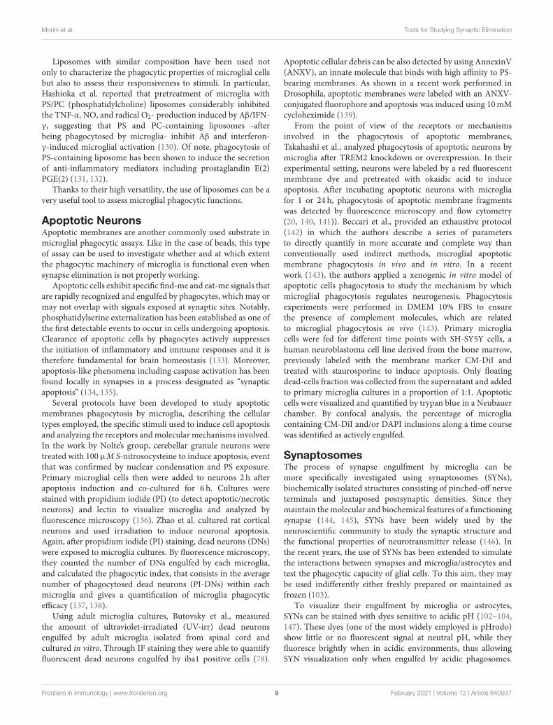
Primary Newborn MicrogliaRelative to cell lines, more advisable is the use of primarymicroglia, that can be isolated from embryos and newbornmouse pups in the P0–P4 time window (51, 52) (Figure 1).Dissociated cells are collected through enzymatic digestion ofmouse brains and seeded as mixed glial culture. Microgliagrowing on top of a confluent astrocyte layer, generally in 2weeks, are next purified through mechanical tapping of mixedglial culture [for a protocol see (53)]. After 2 h, microgliaattach to the bottom and, after replacement with fresh culturemedium, are ready to be used, starting from the next day. Theuse of primary microglia allows to perform in vitro assays incontrolled conditions, with a relatively short time interval fromthe cultured cell collection to their employment (39). Althoughrepresenting an advancement toward the use of immortalized
cell lines, the use of cultured primary microglia suffers ofimportant limitations. First, local environment is known to exerta profound influence on microglia, and indeed it is widelyrecognized that microglia quickly lose their transcriptionalphenotype after niche removal (54, 55). In addition, the useof media containing serum, which are usually adopted toensure vitality and proliferation of freshly isolated microglia,results in a low reproducibility of data, due to batch-to-batchheterogeneity (39). Since factors required for microglia survivalcan be found in media conditioned by astrocytes (56), a numberof protocols for culturing primary microglia from newborn miceuse mixed cultures composed by a confluent layer of astrocyteson which microglia grow in semi-suspension (21, 53). Althoughlisting the different methods for isolating and culturing primarymicroglia is not the purpose of this review, possible hints toat least partially overcome these issues are discussed in therelative chapter.
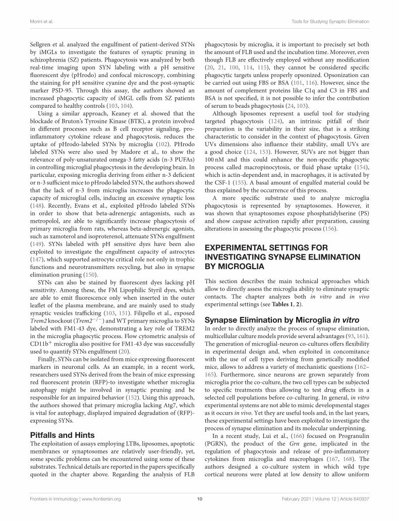
Primary Adult MicrogliaBecause of the clear evidence of the central role played bymicroglia during physiological and in pathological context,the possibility to isolate intact microglia from the adult brainhas become very appealing through the years and has beenpursued by many groups. Microglia isolation from the adultbrain presents some challenges, and several protocols have beenpublished and optimized along the way (Figure 2). One ofthe first studies describing a successful method for isolatingmicroglia from human and rat brain homogenates, was carriedout by M. L. Cuzner’s group in 1988 (57), followed by anotherwork from Volker Ter Meulen’s group a few years later (58).These protocols are based on an initial enzymatic digestionfollowed by separation steps using a Percoll gradient of variousdensities that allows separating myelin debris from nervouscells. Over the years, this procedure has been improved andoptimized. Indeed, while until 2015, homogenization of thewhole brain or of specific brain areas was mostly performedby enzymatic digestion (by using enzyme like Collagenase D,Dispase, Trypsin, and or Papain) carried at 37◦ or at roomtemperature (RT) (51, 59–66), more recently BenA. Barres’ groupmodified the existing microglia isolation protocols in order tominimize microglia activation during the isolation procedure.The whole procedure is now carried out under consistently coldconditions (on ice or at 4◦C) and the brains are mechanicallyhomogenized using a dounce homogenizer instead of undergoingto enzymatic digestion. Flow cytometry and RNAseq expressionof cell-type–specific markers showed that avoiding enzymesand maintaining cold temperatures throughout the wholeisolation process prevented transcriptional phenotypic changesand hyper activation of isolated microglia (9, 67). Furthermore,a reliable cell separation is now successfully obtained throughthe following three approaches: (1) Fluorescence activated cellsorting (FACS), (2) Magnetic-activated cell sorting (MACS), and(3) Immunopanning (Figure 2).
Fluorescence Activated Cell Sorting (FACS)This is the most widely used approach where microglia aresorted with a high cell purity from other major CNS cell typesthrough immune cell markers. CD45 and CD11b, which are
Frontiers in Immunology | www.frontiersin.org 3 February 2021 | Volume 12 | Article 640937

Morini et al. Tools for Studying Synaptic Elimination
FIGURE 1 | Primary microglia culture and main usages to assess the phagocytic process. Schematic figure depicting newborn mice (P0–P4) from pregnant female
used to obtain primary microglia culture. After dissection and enzymatic digestion of cortices and hippocampi, cells are resuspended in growth medium (usually
composed by either DMEM or EMEM with 10 to 20% of FBS), to sustain microglial growth. Cells are plated in T75 flasks and cultured for at least 10 days at 37◦C.
Microglia are subsequently collected either by vigorously tapping the flasks, by agitation at 230–245 rpm for 45min or through mild trypsinization. In some assays,
microglia are cultured alone or co-cultured with neurons and the cells are analyzed by fluorescent microscopy (e.g. to quantify neuronal spine number) or by
electrophysiology (top panel); in other assays, microglia phagocytic properties are assessed by feeding the cells with specific substrates: fluorescent beads,
synaptosomes, liposomes, or apoptotic neurons (bottom panel).
not present on the surface of other glial cells or neurons,are commonly used to identify microglia (20, 59, 60, 68–70). Microglia are CD45lowCD11b+ and can be thereforedistinguished from monocyte and macrophage populations(CD45highCD11b+) (58). However, since the separation basedon CD45 expression levels is not sufficient to cleanly separatemicroglia from all the other myeloid populations, such asneutrophils or choroid plexus macrophages, many groupsrecently invested increasing efforts in order to identify uniqueand highly specific markers to selectively distinguish microglia.The Transmembrane Protein 119 (tmem119) and the PurinergicReceptor P2y12 have been shown to be exclusively expressedby microglia and have been added to the sorting procedure(50, 67, 71). More recently, the hexosaminidase subunit beta(Hexb) has been described as a stably expressed microglia coregene, with a rather stable expression also during inflammationand neurodegeneration (72).
Magnetic-Activated Cell Sorting (MACS)This approach is based on the use of anti-CD11bimmunomagnetic beads. The anti-CD11b antibodies recognizeCD11b surface antigens on microglia by positive selection. Sincethese antibodies are conjugated to magnetic beads, they allowthe retention of labeled cells in a magnetic field. Therefore,this strategy efficiently selects CD11b+ cells over other majorCNS cell types, and the large majority of CD11b+ cells fromthe uninjured CNS are in fact microglia (63, 73). Myelin debriscan also be removed using the same immunomagnetic beadsapproach, instead of using Percoll gradient.
ImmunopanningIn this strategy, antibodies recognizing CD11b surface antigensare immobilized on a Petri dish and used to retain microglia frombrain single-cell suspensions. Panning is trivial, involving onlythree steps: (1) enzymatic preparation of a cell suspension, (2)
Frontiers in Immunology | www.frontiersin.org 4 February 2021 | Volume 12 | Article 640937

Morini et al. Tools for Studying Synaptic Elimination
FIGURE 2 | Schematic figure depicting current methods adopted to obtain freshly isolated microglia. Brains from adult mice are (1) homogenized with a dounce
homogenizer at 4◦C to avoid microglia activation or (2) enzymatically digested at 37◦C in the water bath, and filtered through a 70µm cell strainer. Myelin is removed
either by centrifugation with a Percoll gradient or by using anti-myelin magnetic cell sorting (MACS) antibodies. Then, microglia are sorted from other CNS cell types by
FACS through specific cell markers. Otherwise, microglia are isolated by MACS anti-CD11b antibodies. Microglia can also be isolated by immunopanning. In this
approach, the cells suspension is passed over a series of antibody coated dishes in order to remove contaminating cell types. Then, microglia are positively isolated in
the last coated dish.
passing this suspension over a series of antibody-coated dishes,and (3) removing the purified cells from the final dish. Thisprotocol has been less commonly used (74–76).
After isolation, fresh microglia from adult mice can becultured in vitro or directly assayed for their functional andphagocytic properties. Many protocols have been developed tomaintain adult microglia in culture for several days. Some groupsshowed the generation of pure microglia from adult mice andtheir maintenance in culture for more than 60 days startingfrom a mixed glial population treated with GM-CSF (77). Yet,these cultures maintain a high proliferative capacity, whichmightbe due to an immature phenotype of these cells, since adultmicroglia is not mitotically active nor proliferate in response toGM-CSF or M-CSF (50, 59, 78).
More recently, Bohlen et al., shed a clearer light on previousprocedures and proposed a new method to maintain adultmicroglia in culture. They were able to successfully culturemicroglia from juvenile or adult rat brains, but they observed
that microglia cultures from old mice (>P14) were not viable.Cultures from mice younger than <P14 were viable, althoughthe yields and survival rates were lower if compared to rat tissue.So, the authors concluded that microglia cultures from rat andmice should be performed starting from young animals since cellyield and viability drop with increasing animal age. Once isolated,microglia were maintained in culture in the presence of TGF-β2and IL-34 orM-CSF of absence of FBS (75). Due to the challengesof maintaining adult microglia in vitro and after the discoverythat microglia lose many of the core signature genes such asTmem119 and P2ry12 only after a few days in vitro (50, 56, 79),a limited number of studies have performed phagocytic assays onadult microglia cultures. Indeed, most of the phagocytic assayscurrently described in literature are performed using microgliaprepared from newborn mice as we described in the first section.This is still a reliable and useful system, in which the cells areeasier to obtain and can be cultured for a longer period of time(20, 21).
Frontiers in Immunology | www.frontiersin.org 5 February 2021 | Volume 12 | Article 640937

Morini et al. Tools for Studying Synaptic Elimination
Human Microglia-Like CellsRecently, different groups demonstrated the existence ofsignificant differences between murine and human microglia(79–81). This further highlighted the importance of finding newmodels to better understand the genetics and function of humanmicroglia. With this purpose, a large effort has been done bythe scientific community to generate human microglia-like cells(iMGLs) from human embryonic stem cells (ESCs) or by thereprogramming of adult cells (i.e., fibroblast or blood cells) intoinduced pluripotent stem cells (iPSCs) via the overexpression ofspecific transcription factors.
Several detailed methods for the generation of iMGLs havebeen published in the past 4–5 years (82–87). The commonthread of these new generation protocols is that the specific stepsthrough which iPSCs are differentiated into microglia-like cells,seek to mimic microglia ontogeny.
Indeed, developmental ontogeny studies showedthat microglia are of mesodermal origin, deriving fromerythromyeloid progenitor (EMP), that arise from the yolk-sac(3, 88, 89). Therefore, the new methods generate cells thattransition from iPSCs to primitive hematopoietic precursor cells(HPCs), EMPs, and, ultimately, microglia.
iMGLs phenotype has been shown to be induced byincubation of human iPSC-derived microglial and/ormacrophage progenitors with various combinations of cytokines,including high levels of CSF-1 and IL-34 (85); IL-34 andGM-CSF (83); and IL-3, IL-34 and GM- CSF (86). In order torecreate the brain environment, and to push iMGLs maturationfurther, some of these protocols also proposed to co-cultureiMGLs with neurons (84, 87) or to add further cytokines such asTGF-β1, CX3CL1 (also known as fractalkine), and CD200 whichare critical for microglia homeostasis and to mimic neuronalproximity (82) [reviewed in (90)].
Moreover, microglia-like cells from iPSCs allows thecomparison between healthy donors and patients withneurological disorders. This aspect is of primary interestand, together with further recent improvements, such as theaddition of iMGLs to iPSC-derived brain organoids or thexenotransplantation of HPCs/ iMGLs into mouse brain, makesiMGLs a powerful system to study properties and dysfunctionsof human microglia [reviewed in (91)].
Pitfalls and HintsAlthough primary microglia isolated from embryonic (92) orneonatal mice and rats are widely used as in vitro models,recently it has become evident that the tissue environmenthas a major impact on microglia transcriptome (50, 56, 79).Despite the important advances that have been made to improveculture conditions of microglia and iPSCs-derived microglialcells (39), in vitro microglia, although being informative andproviding a useful setting to dissect basic mechanisms andpossible dysfunctions of phagocytic microglia (20, 21, 50), is farfrom recapitulating the profile and function of microglia in theirphysiological environment.
As mentioned, the main limit of microglial culturing isthe wide adoption of medium containing serum for theirmaintenance. Fetal bovine serum is usually added to medium to
a final concentration of 10–20% in order to promote microgliaproliferation and survival (20, 21, 53). However, microglia arenot exposed to serum proteins in the brain and FBS perturbsmicroglia phenotype in vitro (39, 56), thus increasing the risksof in vitro artefacts. A solution to this problem has been providedby Bohlen et al., who identified in CSF-1, TGF-β and cholesterolthe minimum supplement requirement for microglia culturing,a condition which allows obtaining an in vitro model with asignificantly higher reproducibility (56).
The authors purified microglial cells from postnatal ratbrain by immunopanning, and quantified their viability 5 daysafter plating. As expected, microglia showed a high mortalitywhen serum was removed from the medium, but a robustpro-survival rate was reached by culturing microglial cell ina medium preconditioned by astrocytes. As CSF-1 and TGF-βwere not sufficient to promote microglia viability, they cleverlydissected the conditioned medium and added cholesterol asthe third key element, obtaining the so-called TIC medium(TGF-β2 2 ng/mL, IL-34 100 ng/mL, and cholesterol 1.5 mg/mL)(56). More recently, the Seker’s group established an innovativetri-culture of neurons, astrocytes and microglia adopting thesame cocktail used in TIC medium. Under these conditions,microglia showed a neuroprotective role when neurons wereexposed to excitotoxic events, and the response to externalstimuli mimicked neuroinflammatory responses better thanclassical co-cultures (93). Nevertheless, in contrast with Bohlen’sresults, cytokines detected in unstimulated TICmedium reflectedslightly inflamed state and microglia exhibited an amoeboidmorphology. As suggested by the authors, it should be consideredthat the two systems differ in the age of mice from which cultureswere obtained. Moreover, the tri-culture medium includes alsoB27 supplement, whose elements could influence microglial cellphenotype (56, 93).
Another critical issue in microglia culturing resides in theprocess of cells collection after in vitro maintenance. Microgliaproliferate in semi-suspension, above a layer of astrocyte andshaking of the culture support (flask or petri-dish) for a definedtime and speed is sufficient to detach microglia from theastrocytes layer, re-suspending them in the medium (20, 21, 53).After 10–15 days, the shaken culture will be re-populated bynew microglia that could in principle be employed for a newcell collection, the so-called “second-shaking.” A main pitfallin this process is the clonal-selection of a sub-population fromthe original culture, that makes it only partially comparableto a fresh culture (94). Moreover, the shaking process canstress the cells, inducing phenotypic variations in primarymicroglia. For these reasons, a mild trypsinization protocol hasbeen adopted as an alternative. Lin et al., compared shakingvs. mild trypsinization (95), demonstrating that microglialmorphology and cytokine expression vary depending on themethodology of isolation. Indeed, the shaking protocol induceda higher expression of microglia activation markers, iNOS,CD86, CD206, and arginase 1, together with pro-inflammatorycytokines, TNFα, IL-1β, IL-10, and IGF-1 (95), although bothconditions fully maintained microglia ability to respond toclassic stimuli, such as IL-4, LPS, and IFNγ (95). Interestingly,by analyzing a panel of genes commonly upregulated during
Frontiers in Immunology | www.frontiersin.org 6 February 2021 | Volume 12 | Article 640937

Morini et al. Tools for Studying Synaptic Elimination
aging or after LPS or Aβ stimulation, it was found that CD11bmagnetic-associated cell sorting (MACS) guarantees the highestexpression of Tgfbr1 and Tgfbr2 genes, and results in a “morequiescent” microglia phenotype, as compared to cells obtainedby mild trypsinization and shaking (96). Thus, cell manipulationcan heavily influence the microglia condition. Caution shouldtherefore be applied when performing experiments and well-defined control conditions are mandatory to be applied.
Moreover, as already mentioned, the microenvironmentexerts a strong effect on the microglia transcriptome (50, 56,79). In particular, human and mouse microglia in vitro culturesexhibit down-regulation of genes characteristic of the coretranscriptome signature of microglia and, on the other side,upregulate genes typically only observed in vivo in the contextof disease or injury (56, 70, 79).
Microglia isolation from the adult brain also presents severalchallenges. This is primarily due to the fact that microglia arehighly responsive to CNS tissue damage, which is inevitableduring their isolation, and easily undergo hyper-activation andgene transcription changes after manipulation (9, 67). Anotherreason is that the final yield obtained after isolation is very lowsince microglia only account for 5–12% of the total cells in thebrain (97–99) [the total yield per brain expected after isolationranges between 5 and 10× 104 cell from mice between postnatal(P) days 10 to P21] (75).
Furthermore, the procedures used to isolate and selectmicroglia from adult brain have some disadvantages that need tobe considered, especially depending on the use to be done withmicroglia after their sorting. FACS, the first-choice procedure,allows to obtain a very high cell purity and it is widely andsuccessfully used. Yet, it requires specific and very well-organizedFACS facilities, instrumentations that need to be always upto date, and specialized technicians able to manage and usesorters optimally. Another caveat of cell sorting is that thedetection antibodies remain bound to the cells at the end ofthe process, blocking the epitopes and potentially impacting cellfunction. Also, sorting procedures cause hydrodynamic stressto the cells, even though it has been demonstrated that thisdoes not affect cell structure or function. The second method,based on positive selection of CD11b+ cells through magneticbeads, is in fact highly effective but requires significant upfrontinvestment in reagents and equipment that are particularlyexpensive. A disadvantage of this approach is that positiveselection utilizes cell receptor antibodies to target the specificcell type of interest and may potentially turn on activationcascades through these receptors or cause receptor blockade andinhibit the downstream functions of the isolated cells. Moreover,these protocols do not separate microglia from barrier-associatedmacrophages, monocytes, neutrophils, or certain B cells alsopresent in the tissue. The third strategy, immunopanning,requires minimal reagent investment or specialized equipmentbut does not provide a high specificity. Indeed, separation ofdifferent myeloid populations is unlikely to be achievable usingimmunopanning due to the propensity of various myeloid cellpopulations to adhere to the panning dish, even dishes notcoated with antibodies. Moreover, this protocol requires cellstrypsinization but on the other side, avoids introduction of
magnetic particles in downstream applications. Therefore, thisapproach is not preferable if the final goal is to isolate a pure andhomogeneous microglia population.
IN VITRO AND EX VIVO ENGULFMENTASSAYS
Once microglia are isolated from brain and deposited in culture,their phagocytic properties can be evaluated using severaldifferent substrates (Figure 1). Below we describe some of theassays which can be used to test microglia phagocytosis. Althoughthe focus of this review is on microglial synapse elimination, inthis chapter the strategies and the tools that can be used to analyzethe basic phagocytic activity by microglia will be described (seeTable 1). These assays may provide suitable control conditions,needed to complement the study of synapse elimination bymicroglia. It is to be considered that the receptors and molecularmachineries that coordinate phagocytosis and digestion arelikely to differ depending on the specific substrate. Importantly,the substrates and phagocytic events described in this section
TABLE 1 | In vitro engulfment assays.
Samples Type of cells Engulfed
substrates
Techniques
adopted
References
Cell lines Microglia cell line
BV-2
Fluorescent
beads
Fluorescent
microscopy,
Flow
cytometry
(100)
Microglial cell
line
MMGT12
Fluorescent
beads
Flow
cytometry
(101)
Microglial cell
line
BV-2
Synaptosomes Fluorescent
microscopy
(102)
iPC-derived
microglia
Induced
Microglia
Like Cells (iMGL)
Synaptosomes Fluorescent
microscopy
(103, 104)
Primary
cultures
Newborn
microglia
Fluorescent
beads and
liposomes
(DiO Labeled)
Fluorescent
microscopy
(21)
fluorescent
beads
Fluorescent
microscopy,
Flow
cytometry
(20)
Fluorescent
bioparticles
Fluorescent
microscopy
(105)
Adult microglia Ultraviolet-
irradiated (UV-irr)
dead neurons
Fluorescent
microscopy
(78)
Fluorescent
microspheres
Fluorescent
microscopy
(62)
Macrophages Bacteria and
cancer cells
CyTOF (106)
The table reports some commonly used experimental approaches to study microglial
phagocytosis in vitro.
Frontiers in Immunology | www.frontiersin.org 7 February 2021 | Volume 12 | Article 640937

Morini et al. Tools for Studying Synaptic Elimination
can only partially model the process of synapses and neuritesphagocytosis in vivo.
Fluorescent BeadsLatex beads have been widely used to analyze the basic phagocyticprocess by microglia. This type of assay is advantageously usedto demonstrate that the phagocytic machinery of microgliais properly functional, even when synapse elimination maybe defective. Further, purification of phagosomes containingengulfed latex beads has allowed to understand the phagosomebiology on a biochemical and functional standpoint and to dissectthe sequential steps at the basis of this process (107–109).
The use of Fluorescent Latex Beads (FLB) has alloweda fast and quantitative analysis of phagocytosis in differentcell populations either by FACS (101, 110, 111) or by asimple count of FLB internalization by fluorescence or confocalmicroscopy (112, 113). FLB, which are routinely used to calibrateflow cytometers, may be excited by a specific wavelength or,alternatively, contain a mixture of fluorophores that enable themto be excited at any wavelength of UV and visible light. FLB havea wide range of sizes (the most commonly used range from 0.5 to6.0µm) and are inert, so they are not toxic and do not interferewith cell viability (100, 101). FLB may be either used withoutany modifications (20, 21, 100, 114, 115) or pre-opsonized withFBS or BSA to improve phagocytosis by microglia (101, 116),since it has been shown that the engulfment of synapses isstrictly dependent on complement proteins deposition, such asC1q and C3, and their interaction with microglial cells (24, 103).Moreover, beads can be also carboxylated so to add a negativecharge, a model that can be used tomimic negative surface chargeof phosphatidylserine-exposing cells (117). The engulfment rateis dependent on FLB concentration and incubation time (115).To precisely evaluate microglia phagocytic capacity, FLB amountand time of incubation need to be precisely set.
Pathogen-associated molecular patterns (PAMPs), such asLPS, significantly increase FLB internalization by microglia (101,115). Also, in line with the observation that FLB internalizationby microglia is accompanied by an increase in TNF-α andTGF-β production (69), MDG548, a neuroprotective PPARγ
agonist used for experimental Parkinson’s Disease treatment wasfound to increase FLB engulfment, while decreasing TNFα levels,thus providing a possible basis for PPARγ agonists protectiverole (101).
Besides PAMPs, basic phagocytic activity by microglia is alsoenhanced by neurodegeneration-associated molecular patterns(NAMPs), which include the Aβ and neurofibrillary tangles (118,119). This activation modifies microglial phenotype, turningthem into disease associated microglia (DAM) (120). In thisframework, Nagano et al., showed, by both confocal microscopyand flow cytometry analysis, that the presence of Aβ depositsis able to increase the engulfment rate of FLB in primaryrat microglia. This effect is reversed after the treatment withProstaglandin E2 (PGE2), through the involvement of microglialE-prostanoid receptor 2 (EP2) (121). Allendorf et al., confirmedthat treatment of primary rat microglia with pro-inflammatorystimuli such as LPS, Aβ or Tau induces an increase of FLBphagocytic activity (100). Finally, Yin et al. (111) have shown
that the inhibition of EZH2, the catalytic subunit core PRC2, agene involved in silencing a number of tumor suppressor genes,is able to increase the levels of pro-inflammatory cytokines andthe FLB phagocytic capacity of microglia, which are abundant inthe tumor environment (111, 122).
Of note, the engulfment of FLB can be also assessed in vivo.Hughes et al. (114) injected FLB [6µm] intrahippocampallyin ME7 mice, a model of prion disease, in order to studytheir engulfment capacity. They discovered that microglia inthe degenerating brain, internalize FLB and apoptotic cells,demonstrating that the phagocytic machinery of the microglia inME7 mice is properly functional.
LiposomesAn additional substrate that can be successfully exploited forphagocytosis assays are liposomes, or unilamellar vesicles (UVs)(123), a useful tool to specifically investigate the nature of“eat-me” signals which need to be exposed by the targetmembrane to allow microglial phagocytosis (21).
UVs can be distinguished in three categories dependingon their size: small UVs (SUV), large UVs (LUV) and giantUVs (GUV), having a diameter of 20–100, 100–1,000 nm,and 1–200µm, respectively. UVs stability depends on theexperimental conditions; indeed, oxygen reactive species reactwith unsaturated fatty acid chains, thus altering lipid propertiesand liposomes structures (124). This responsiveness to theenvironmental conditions has been exploited for improvingdrug delivery, through the generation of smart vesicles ableto deliver drugs to the target and release them only after alocal stimulus-response (124). One of the biggest advantagesof this tool is that UVs with virtually any composition can beprepared, enriching them with specific membrane proteins ordifferent lipids (125, 126). For example, in a recent paper, aconvenient protocol was published for the preparation of proteo-GUVs containing functionally active neuronal SNARE (solubleN-ethylmaleimide-sensitive factor activating protein receptor)proteins for the study of membrane fusion in vitro (125). Morerecently, the Matteoli’s group took advantage of DiO labeledliposomes composed of variable amounts of phosphatidylserine(PS) and cardiolipin (CL) to investigate whether exposedPS impacts microglia ability to engulf lipidic membranes(21). Specifically, the researchers incubated for 1 h liposomesendowed with different lipidic composition with microglial cellsisolated from mice either WT or genetically lacking TREM2,a receptor which shows high affinity for phospholipids asphosphatidylcholine and PS (127). Confocal analysis of liposomeengulfment inside CD68-positive phagolysosomal organelles inIba1-positive microglia (15, 21) exploiting the Bitplane Imarissoftware to generate a 3D reconstruction of the fluorescentsignal, allowed to demonstrate that the extent of PS positivelycorrelates with microglia phagocytosis (127). For this kindof experiments, attention should be paid to the type ofsolvents used for permeabilization before the staining (128).Specifically, the use of saponin allowed to selectively createpores the cholesterol shaft of the plasma membrane (129)but not in liposomes, thus avoiding loss of DiO signal fromliposomes (21).
Frontiers in Immunology | www.frontiersin.org 8 February 2021 | Volume 12 | Article 640937
Morini et al. Tools for Studying Synaptic Elimination
Liposomes with similar composition have been used notonly to characterize the phagocytic properties of microglial cellsbut also to assess their responsiveness to stimuli. In particular,Hashioka et al. reported that pretreatment of microglia withPS/PC (phosphatidylcholine) liposomes considerably inhibitedthe TNF-α, NO, and radical O2- production induced by Aβ/IFN-γ, suggesting that PS and PC-containing liposomes -afterbeing phagocytosed by microglia- inhibit Aβ and interferon-γ-induced microglial activation (130). Of note, phagocytosis ofPS-containing liposome has been shown to induce the secretionof anti-inflammatory mediators including prostaglandin E(2)PGE(2) (131, 132).
Thanks to their high versatility, the use of liposomes can be avery useful tool to assess microglial phagocytic functions.
Apoptotic NeuronsApoptotic membranes are another commonly used substrate inmicroglial phagocytic assays. Like in the case of beads, this typeof assay can be used to investigate whether and at which extentthe phagocytic machinery of microglia is functional even whensynapse elimination is not properly working.
Apoptotic cells exhibit specific find-me and eat-me signals thatare rapidly recognized and engulfed by phagocytes, which may ormay not overlap with signals exposed at synaptic sites. Notably,phosphatidylserine externalization has been established as one ofthe first detectable events to occur in cells undergoing apoptosis.Clearance of apoptotic cells by phagocytes actively suppressesthe initiation of inflammatory and immune responses and it istherefore fundamental for brain homeostasis (133). Moreover,apoptosis-like phenomena including caspase activation has beenfound locally in synapses in a process designated as “synapticapoptosis” (134, 135).
Several protocols have been developed to study apoptoticmembranes phagocytosis by microglia, describing the cellulartypes employed, the specific stimuli used to induce cell apoptosisand analyzing the receptors andmolecular mechanisms involved.In the work by Nolte’s group, cerebellar granule neurons weretreated with 100 µM S-nitrosocysteine to induce apoptosis, eventthat was confirmed by nuclear condensation and PS exposure.Primary microglial cells then were added to neurons 2 h afterapoptosis induction and co-cultured for 6 h. Cultures werestained with propidium iodide (PI) (to detect apoptotic/necroticneurons) and lectin to visualize microglia and analyzed byfluorescence microscopy (136). Zhao et al. cultured rat corticalneurons and used irradiation to induce neuronal apoptosis.Again, after propidium iodide (PI) staining, dead neurons (DNs)were exposed to microglia cultures. By fluorescence microscopy,they counted the number of DNs engulfed by each microglia,and calculated the phagocytic index, that consists in the averagenumber of phagocytosed dead neurons (PI-DNs) within eachmicroglia and gives a quantification of microglia phagocyticefficacy (137, 138).
Using adult microglia cultures, Butovsky et al., measuredthe amount of ultraviolet-irradiated (UV-irr) dead neuronsengulfed by adult microglia isolated from spinal cord andcultured in vitro. Through IF staining they were able to quantifyfluorescent dead neurons engulfed by iba1 positive cells (78).
Apoptotic cellular debris can be also detected by using AnnexinV(ANXV), an innate molecule that binds with high affinity to PS-bearing membranes. As shown in a recent work performed inDrosophila, apoptotic membranes were labeled with an ANXV-conjugated fluorophore and apoptosis was induced using 10mMcycloheximide (139).
From the point of view of the receptors or mechanismsinvolved in the phagocytosis of apoptotic membranes,Takahashi et al., analyzed phagocytosis of apoptotic neurons bymicroglia after TREM2 knockdown or overexpression. In theirexperimental setting, neurons were labeled by a red fluorescentmembrane dye and pretreated with okaidic acid to induceapoptosis. After incubating apoptotic neurons with microgliafor 1 or 24 h, phagocytosis of apoptotic membrane fragmentswas detected by fluorescence microscopy and flow cytometry(20, 140, 141)). Beccari et al., provided an exhaustive protocol(142) in which the authors describe a series of parametersto directly quantify in more accurate and complete way thanconventionally used indirect methods, microglial apoptoticmembrane phagocytosis in vivo and in vitro. In a recentwork (143), the authors applied a xenogenic in vitro model ofapoptotic cells phagocytosis to study the mechanism by whichmicroglial phagocytosis regulates neurogenesis. Phagocytosisexperiments were performed in DMEM 10% FBS to ensurethe presence of complement molecules, which are relatedto microglial phagocytosis in vivo (143). Primary microgliacells were fed for different time points with SH-SY5Y cells, ahuman neuroblastoma cell line derived from the bone marrow,previously labeled with the membrane marker CM-DiI andtreated with staurosporine to induce apoptosis. Only floatingdead-cells fraction was collected from the supernatant and addedto primary microglia cultures in a proportion of 1:1. Apoptoticcells were visualized and quantified by trypan blue in a Neubauerchamber. By confocal analysis, the percentage of microgliacontaining CM-DiI and/or DAPI inclusions along a time coursewas identified as actively engulfed.
SynaptosomesThe process of synapse engulfment by microglia can bemore specifically investigated using synaptosomes (SYNs),biochemically isolated structures consisting of pinched-off nerveterminals and juxtaposed postsynaptic densities. Since theymaintain themolecular and biochemical features of a functioningsynapse (144, 145), SYNs have been widely used by theneuroscientific community to study the synaptic structure andthe functional properties of neurotransmitter release (146). Inthe recent years, the use of SYNs has been extended to simulatethe interactions between synapses and microglia/astrocytes andtest the phagocytic capacity of glial cells. To this aim, they maybe used indifferently either freshly prepared or maintained asfrozen (103).
To visualize their engulfment by microglia or astrocytes,SYNs can be stained with dyes sensitive to acidic pH (102–104,147). These dyes (one of the most widely employed is pHrodo)show little or no fluorescent signal at neutral pH, while theyfluoresce brightly when in acidic environments, thus allowingSYN visualization only when engulfed by acidic phagosomes.
Frontiers in Immunology | www.frontiersin.org 9 February 2021 | Volume 12 | Article 640937
Morini et al. Tools for Studying Synaptic Elimination
Sellgren et al. analyzed the engulfment of patient-derived SYNsby iMGLs to investigate the features of synaptic pruning inschizophrenia (SZ) patients. Phagocytosis was analyzed by bothreal-time imaging upon SYN labeling with a pH sensitivefluorescent dye (pHrodo) and confocal microscopy, combiningthe staining for pH sensitive cyanine dye and the post-synapticmarker PSD-95. Through this assay, the authors showed anincreased phagocytic capacity of iMGL cells from SZ patientscompared to healthy controls (103, 104).
Using a similar approach, Keaney et al. showed that theblockade of Bruton’s Tyrosine Kinase (BTK), a protein involvedin different processes such as B cell receptor signaling, pro-inflammatory cytokine release and phagocytosis, reduces theuptake of pHrodo-labeled SYNs by microglia (102). PHrodolabeled SYNs were also used by Madore et al., to show therelevance of poly-unsaturated omega-3 fatty acids (n-3 PUFAs)in controlling microglial phagocytosis in the developing brain. Inparticular, exposing microglia deriving from either n-3 deficientor n-3 sufficientmice to pHrodo labeled SYN, the authors showedthat the lack of n-3 from microglia increases the phagocyticcapacity of microglial cells, inducing an excessive synaptic loss(148). Recently, Evans et al., exploited pHrodo labeled SYNsin order to show that beta-adrenergic antagonists, such asmetropolol, are able to significantly increase phagocytosis ofprimary microglia from rats, whereas beta-adrenergic agonists,such as xamoterol and isoproterenol, attenuate SYNs engulfment(149). SYNs labeled with pH sensitive dyes have been alsoexploited to investigate the engulfment capacity of astrocytes(147), which supported astrocyte critical role not only in trophicfunctions and neurotransmitters recycling, but also in synapseelimination pruning (150).
SYNs can also be stained by fluorescent dyes lacking pHsensitivity. Among these, the FM Lipophilic Styril dyes, whichare able to emit fluorescence only when inserted in the outerleaflet of the plasma membrane, and are mainly used to studysynaptic vesicles trafficking (103, 151). Filipello et al., exposedTrem2 knockout (Trem2−/−) andWTprimarymicroglia to SYNslabeled with FM1-43 dye, demonstrating a key role of TREM2in the microglia phagocytic process. Flow cytometric analysis ofCD11b+ microglia also positive for FM1-43 dye was successfullyused to quantify SYNs engulfment (20).
Finally, SYNs can be isolated frommice expressing fluorescentmarkers in neuronal cells. As an example, in a recent work,researchers used SYNs derived from the brain of mice expressingred fluorescent protein (RFP)-to investigate whether microgliaautophagy might be involved in synaptic pruning and beresponsible for an impaired behavior (152). Using this approach,the authors showed that primary microglia lacking Atg7, whichis vital for autophagy, displayed impaired degradation of (RFP)-expressing SYNs.
Pitfalls and HintsThe exploitation of assays employing LTBs, liposomes, apoptoticmembranes or synaptosomes are relatively user-friendly, yet,some specific problems can be encountered using some of thesesubstrates. Technical details are reported in the papers specificallyquoted in the chapter above. Regarding the analysis of FLB
phagocytosis by microglia, it is important to precisely set boththe amount of FLB used and the incubation time. Moreover, eventhough FLB are effectively employed without any modification(20, 21, 100, 114, 115), they cannot be considered specificphagocytic targets unless properly opsonized. Opsonization canbe carried out using FBS or BSA (101, 116). However, since theamount of complement proteins like C1q and C3 in FBS andBSA is not specified, it is not possible to infer the contributionof serum to beads phagocytosis (24, 103).
Although liposomes represent a useful tool for studyingtargeted phagocytosis (124), an intrinsic pitfall of theirpreparation is the variability in their size, that is a strikingcharacteristic to consider in the context of phagocytosis. GivenUVs dimensions also influence their stability, small UVs area good choice (124, 153). However, SUVs are not bigger than100 nM and this could enhance the non-specific phagocyticprocess called macropinocytosis, or fluid phase uptake (154),which is actin-dependent and, in macrophages, it is activated bythe CSF-1 (155). A basal amount of engulfed material could bethus explained by the occurrence of this process.
A more specific substrate used to analyze microgliaphagocytosis is represented by synaptosomes. However, itwas shown that synaptosomes expose phosphatidylserine (PS)and show caspase activation rapidly after preparation, causingalterations in assessing the phagocytic process (156).
EXPERIMENTAL SETTINGS FORINVESTIGATING SYNAPSE ELIMINATIONBY MICROGLIA
This section describes the main technical approaches whichallow to directly assess the microglia ability to eliminate synapticcontacts. The chapter analyzes both in vitro and in vivoexperimental settings (see Tables 1, 2).
Synapse Elimination by Microglia in vitroIn order to directly analyze the process of synapse elimination,multicellular culturemodels provide several advantages (93, 161).The generation of microglial-neuron co-cultures offers flexibilityin experimental design and, when exploited in concomitancewith the use of cell types deriving from genetically modifiedmice, allows to address a variety of mechanistic questions (162–165). Furthermore, since neurons are grown separately frommicroglia prior the co-culture, the two cell types can be subjectedto specific treatments thus allowing to test drug effects in aselected cell populations before co-culturing. In general, in vitroexperimental systems are not able to mimic developmental stagesas it occurs in vivo. Yet they are useful tools and, in the last years,these experimental settings have been exploited to investigate theprocess of synapse elimination and its molecular underpinning.
In a recent study, Lui et al., (166) focused on Progranulin(PGRN), the product of the Grn gene, implicated in theregulation of phagocytosis and release of pro-inflammatorycytokines from microglia and macrophages (167, 168). Theauthors designed a co-culture system in which wild typecortical neurons were plated at low density to allow uniform
Frontiers in Immunology | www.frontiersin.org 10 February 2021 | Volume 12 | Article 640937

Morini et al. Tools for Studying Synaptic Elimination
TABLE 2 | In vivo and ex vivo engulfment assays.
Models Brain areas Engulfed
substrates
Techniques
adopted
References
Cx3xr1-/-
mouse
Hippocampus SNAP25+,
PSD95+
synaptic
materials
Confocal
microscopy;
immune-gold
electron
microscopy
(18)
Itgam-/-;
C3-/- mouse
Visual System RGC inputs Confocal
microscopy
dimensional
(3D) surface
volume
rendering
(15)
Trem2-/-
mouse
Hippocampus PSD95+
synaptic
materials
Fluorescent
microscopy
(20)
Zebrafish Spinal cord Apoptotic
neuron
Fluorescent
microscopy,
3D rendering
(157)
Mertk-/-
Mouse
Cortex Apoptotic
neuron
Time-lapse
two-photon
imaging
(158)
Adult
microglia ex
vivo
Cortex Alexa-488
labeled
apoptotic (dNs)
or live neurons
Flow
cytometry
(70)
Amyloid beta
(through the
fluorescent
marker
Methoxy-XO4)
(159)
Synaptic
markers
VGLUT1 and
synaptophysin
(160)
The table reports some of the experimental approaches used to study microglia
engulfment in vivo and ex vivo in different animal models.
synapse development for 14 days in vitro (DIV14). Concurrently,microglia isolated from Grn+/+ or Grn−/− neonatal brainswere added to cortical neurons at a 1:3 microglia/neuron ratioand co-cultured for 72 h. Using a modified Sholl analysis tomeasure the density of synapses in the vicinity of microglia cellbodies and Imaris software to perform 3D reconstructions ofconfocal images, the authors quantified the amount of synapticmaterial within microglial phagolysosomes and demonstrated asignificant increase in synaptic pruning when neurons were co-cultured with microglia isolated from mice genetically lackingGrn. A similar approach was used by Filipello et al., to study therole of microglial TREM2 in synapse elimination. By co-culturingmicroglia with hippocampal neurons at a microglia to neuronratio of 1.5:1 for 24 h, and through the analysis of miniatureexcitatory post-synaptic currents (mEPSC) and dendritic spinesdensity, the authors demonstrated that microglia are able toreduce the density of excitatory synaptic contacts in vitro andthat microglial TREM2 is required for this process to occur(20). To better visualize neurons and spines, WT neurons were
GFP-transfected at DIV 11–12 before adding microglia to theco-culture. The use of transwell inserts between the two cell typesallowed to discriminate the effects of microglia that require thedirect contacts with neurons.
An additional study where microglia and neurons derivedfrom mutant or knock out mice were combined in mixedculture to investigate synapse elimination, focused on therole of PTEN, a well-recognized syndromic risk allele forautism spectrum disorder (169). Using co-cultures of primaryneurons and microglia from PtenWT/WT , PtenWT/m3m4, orPtenm3m4/m3m4 mice in different combinations, followed by co-localization of pre- and post-synaptic markers, the authorsdemonstrated that Ptenm3m4/m3m4 mutation results in increasedmicroglia-dependent synaptic pruning in vitro. Interestingly,the largest decrease in synaptic contact density was observedwhen Ptenm3m4/m3m4 neurons were cultured with Ptenm3m4/m3m4
microglia indicating an additive effect when the mutationoccurs in both cell types. A relevant technological additionof this study is the setting of a protocol which allows co-culturingmicroglia and neurons for a week in amicroglia/neuronratio 1:1 (i.e., a longer time compared to the generally usedgeneral protocols).
The co-culture setting allows to test pharmacological orexperimental treatments which reduce microglial phagocyticability. Inhibition of synapse phagocytosis in vitro was recentlydemonstrated upon the exposure of hippocampal neuronsto ANXV, an innate molecule that binds phosphatidylserine-bearingmembranes with high affinity, 15min before co-culturingthem with microglia. ANXV, by cloaking externalized PS,prevents its recognition by microglial TREM2 and preventssynapse elimination, as demonstrated by the lack of dendriticspine density and mEPSC frequency reduction. A similarapproach was taken in another recent work, where microglialcells were exposed to different treatments before being addedto neuronal cultures (100). Specifically, Allendorf et al.,demonstrated that LPS, fibrillar Aβ, phorbol 12-myristate 13-acetate (PMA) or rTAU protein induced removal of sialicacid residues in microglial cells. This resulted in an enhancedmicroglia ability to phagocytose neuronal components. Of noteneuronal phagocytosis was inhibited by a blocking antibodyagainst CD11b/CR3 (100).
Besides co-cultures of murine microglia and neurons,recent studies took advantage of the use of human cells.In a very interesting paper, Sellgren et al. developed andvalidated a high-throughput method for modeling synapticpruning in vitro, using cells derived from SZ patients orhealthy subjects (103). Specifically, the authors employed iPSC-derived-microglia like cells and iPSC-derived neurons, the lattergenerated from an inducible neurogenin 2 (NGN2) expressingstable NPC lines. After 21 days of neural differentiation,mature iMGLs derived from monocytes were added toneurons for 48 h. iMGLs, maintained under serum-free invitro conditions, were found to engulf synapses from iPSC-derived neural cultures, as assessed by live imaging of iPSC-derived neurons stained for Alexa Fluor 488-phalloidin and bymeasuring PSD-95 engulfment. Using this asset, the authorsdemonstrated a significantly higher, complement-dependent,
Frontiers in Immunology | www.frontiersin.org 11 February 2021 | Volume 12 | Article 640937

Morini et al. Tools for Studying Synaptic Elimination
uptake of synaptic structures when cells from SZ patients wereemployed (103).
Synapse Elimination by Microglia in vivoSince Ito et al. in 1998 isolated and identified a novel gene“the iba1 gene” specifically expressed in microglia, traditionally,Iba-1 antibodies have been used to label/stain microgliausing immunohistochemistry (170). Confocal laser scanningmicroscopy is frequently used to image fluorescently labeledmicroglia in tissue sections (fixed), retinal whole mounts (fixedor fresh) and organotypic brain slices (fresh) to investigatemicroglial density, morphology, distribution, and dynamicinteractions with different cell types (171, 172).
In the last decades, thanks to the advancement of highresolution live microscopy techniques, Iba1-positive microgliahave been characterized as highly motile cells, extending andretracting their processes as they survey the microenvironmentin the healthy brain (173). Both pre-synaptic boutons andpostsynaptic spines have been shown to be contacted bymicroglial processes (6, 174). In the visual cortex, the microglia-synapse contacts were examined in closer resolution using3D reconstruction serial electron microscopy (6). This studyrevealed that, in addition to pre- and postsynaptic specializations,microglial processes also contacted peri-synaptic astrocytes andthe synaptic cleft.
Subsequently, the close microglia-synapse contacts appearedto result in the shaping, or re-wiring, of neuronal circuits byphagocytosis of synaptic materials. The phagocytic propertiesof microglia have been extensively analyzed through differentmicroscopy-based approaches: confocal imaging, electronmicroscopy, two-photon microscopy and lightsheet microscopy(15, 173, 175–177). These techniques allow to visualize andquantify, in a very reliable manner, the material engulfed bymicroglia in the brain, generating a clear picture of the phagocyticprocess in specific time windows. 3D reconstruction of thephagocyte and its intracellular structures (e.g., phagolysosomesand other intracellular organelles) by softwares like Imaris, ilastik[(178); 1.3.2] and CellProfiler [(179); v3.0] has been successfullyused to generate very detailed images of phagocytic microgliaand to quantify the material internalized.
Two milestones articles first demonstrated, by electronmicroscopy and super-resolution confocal microscopy, thepresence of pre- and post-synaptic structures inside microglialphagolysosomes in different brain regions (mouse visual systemand hippocampus) during critical periods of synaptic refinement.In particular, in 2011, Paolicelli et al. spotted synaptic materialinside microglia, providing the demonstration that these cellsplay an active role in pruning synapses. Specific presynaptic(SNAP25) and postsynaptic (PSD95) proteins were identifiedinside microglial processes following synaptic contacts, byconfocal or immune-gold electron microscopy, respectively(18). Furthermore, disrupting the fractalkine (Cx3cl1/Cx3cr1)-mediated communication between microglia and neurons in anotherwise healthy mouse, resulted in brain circuits persisting asimmature into adulthood (18, 180, 181). In 2012, the Stevens’lab at Boston Children’s Hospital, found that, in the newbornmouse visual system, microglia can engulf synapses in the
lateral geniculate nucleus (LGN) through a process mediated byboth complement and neuronal activity. Using Cholera ToxinB Subunit (CTB) injections in Cx3cr1 gfp/+ mice, in whichmicroglia express GFP, the authors elegantly showed for the firsttime, by 3D reconstructions that microglia contain engulfed RGCinputs. By either silencing or promoting neuronal activity in oneeye using TTX or forskolin, respectively, they further showedthat microglia selectively prune the weaker inputs. Notably,by examining microglial engulfment in C3 mutants and C3-receptor mutants, Schafer et al. showed that this process criticallyrelies on the complement cascade. Of note, impaired microglialengulfment in both these mutants correlated with long-lastingdefects in the segregation of ipsi- and contralateral RGC inputsin the dLGN, with an increase in synaptic densities (15). Toconfirm that inputs are in fact phagocytosed by microglia,Schafer et al. introduced a staining of in situ microglia forthe phagolysosomal marker CD68, performing the subsequentcolocalization with synaptic materials. Only the synaptic materialinternalized in CD68-positive phagolysosomal structures wasconsidered for the analysis. A few years later, the same grouppublished a detailed methodology for imaging and quantitativelymeasuring engulfment using confocal microscopy combinedwith 3D surface volume rendering, amethodwhich is widely usedby the scientific community (182).
Still today these two papers represent the landmark forresearchers interested in studying microglia-mediated synapseelimination in vivo. Indeed, most if not all the subsequentstudies heavily relied on the methods introduced by thesepioneering works. Filipello et al. (20), used the same protocol ofengulfment analysis and quantification proposed by Schafer et al.to describe the role of TREM2 in regulating synapse phagocytosisduring hippocampal development. The same approaches wereused to demonstrate the role of CD47, a transmembraneimmunoglobulin superfamily protein that directly inhibitsphagocytosis by binding to its receptor, SIRPα, thus behavingas a “don’t eat me” signal during postnatal development (22).With the aim to detect the phagocytosis of a different substrate,a similar approach was also taken by Cignarella et al. whoanalyzed myelin engulfment and degradation by microglia in thecuprizone model of brain demyelination. By confocal analysisand subsequent 3D reconstruction, the authors showed thata TREM2 agonistic antibody enhanced myelin uptake anddegradation, resulting in accelerated myelin debris removal bymicroglia. Again, 3D reconstruction by the Imaris softwareof CD68 structures inside Iba1-positive microglia containingdMBP-positive myelin debris, was used as a consolidated methodof analysis (183).
Using time-lapse imaging, Weinhard et al., recently reportedthat, rather than removing the whole synaptic structure,microglia prune presynaptic structures through a selective partialphagocytic process termed trogocytosis, or “nibbling.” Theauthors studied microglia “nibbling” on presynaptic structuresof neurons in organotypic tissue culture, an ex vivo model thatpreserves tissue architecture important for microglia physiologyand offers the advantages of a tissue-relevant context effective instudying the synaptic elimination processes. Subsequent analysisof fixed hippocampal tissue from postnatal day 15 (P15) mice
Frontiers in Immunology | www.frontiersin.org 12 February 2021 | Volume 12 | Article 640937
Morini et al. Tools for Studying Synaptic Elimination
using quantitative confocal microscopy as well as correlativelight and electron microscopy, revealed that microglia onlycapture small (<1 um) presynaptic components though a processwhich involves the “sinking” of presynaptic structures into themicroglial cytoplasm prior to membrane closure. Conversely,pseudopodia, a hallmark of phagocytosis, were not observed (17).Further lines of investigation are expected to provide additionalinsights into the precise mechanisms by which microglia removeand digest synaptic contacts.
Facs-Based Microglia Phagocyitc Assaysex vivoThe analyses described in the previous paragraphs relyon the use of in vitro microglia, prepared as described inMicroglia Cell Lines, Primary Newborn Microglia, and HumanMicroglia-Like Cells sections. However, similar assays canalso be performed taking advantage of microglia freshlyisolated from the adult or juvenile brain and analyzedright away (see Primary Adult Microglia chapter). Thelatter setting maintains closer features to those of thesame cells when present in brain environment, despite ofthe isolation process and the consequent manipulation.In 2007, Biber’s group showed the possibility to isolatemicroglia from specific brain regions (optic nerve, striatum,hippocampus, cerebellum, spinal cord, cortex) and to quantifythe amount of fluorescent microspheres phagocytosis by confocalmicroscopy (62).
In the recent years, the use of flow cytometry has implementedmicroscopy techniques thus becoming a very useful approachto dissect the phagocytic properties of microglia not only in invitro assays but also using freshly isolated microglia ex vivo.This strategy was successfully used by Krasemann et al., whoidentified a role for apolipoprotein E (APOE) in regulating asubset of microglia, exhibiting a common neurodegenerative-associated phenotype (MGnD). To determine the mechanismsthrough which MGnD were induced during neurodegeneration,they injected Alexa-488 labeled apoptotic (dead, dNs) or liveneurons (Ns) into the cortex and hippocampus of naïve mice.In parallel, they also injected fluorescent E. coli or zymosanas a control. By gating the CD11b+ CD45low population theywere able to distinguish the phagocytic cells that internalized488-labeled apoptotic neurons (CD11b+ CD45low dNs-Alexa488+) vs. non-phagocytic microglia (CD11b+ CD45low dNs-Alexa 488−) (70).
A similar approach was used by Tejera and Heneka whoshowed in detail how to analyze Aβ phagocytosis by flowcytometry using microglia freshly isolated from adult mice.Mice were intraperitoneally injected with the Aβ fluorescentmarker Methoxy-XO4, and microglia were isolated througha Percoll gradient and directly analyzed by FACS. TheCD11b+CD45low population, also positive for Methoxy-XO4,represented microglia phagocytosing Aβ (159). Using a differentstrategy, Levey’s group validated a rapid flow cytometric assaysto test phagocytic capacity of acutely isolated CNS mononuclearphagocytes (MPs). MPs were isolated through a Percoll gradientand subsequently incubated with macroparticle and fibrillar
Aβ42 (fAβ42). Flow cytometric analysis revealed distinctphagocytic capacities of CD11b+CD45low and CD11b+CD45high
cells both in physiological condition and in disease models (184).The use of mass cytometry (CyTOF), a technique that
combines flow cytometry with mass spectrometry, has enableda high-dimensional analysis of cell surface markers, signalingmolecules and cytokines in brain myeloid cells at the single-celllevel (185–187). Because the method is largely unhampered byinterference from spectral overlap, it allows for the detectionof considerably more simultaneous parameters than doestraditional flow cytometry. This has facilitated the understandingof phenotypic diversity of mouse and human macrophages invitro and in vivo (188, 189). Interestingly, different macrophagephenotypes were found to have different phagocytic activities.In 2019, Schulz et al., created a functional assay to assessphagocytic activity of macrophages by mass cytometry. Thismethod combines an in-depth phenotypic characterization ofmacrophages based on the expression of 36 protein markerswith an analysis of biological function. The authors assessedthe abilities of macrophages activated in vitro under differentconditions to phagocytose bacteria and cancer cells. Bycorrelating the phagocytic activity with markers expression ofsingle cells, they defined characteristic signatures preferentiallyassociated with phagocytosis of specific targets. This strategy canbe also applied to better understand and link cell phenotype tophagocytic function in microglia in health and disease (106).
Pitfalls and HintsThe study of synapse elimination using co-cultures of neuronsand microglia requires specific attention especially in relationto the establishment of the adequate co-culture conditions. Inparticular, defining the optimal density of microglial cells andthe neuron/microglia ratio represents the most critical issue. Theoptimal ratio may vary depending on the experimental designand should be established accordingly. Another limitation to beconsidered is the limited time window (24–72 h) during whichthe microglia-neuron model can be maintained in co-culture.This limitation, which results from the fact that the two differentcell types prefer different culture conditions (56), discourage thesetting of experiments addressing processes which develop in thelong term. The limited time-scale of this model is due to thenegative effect of the continuous presence of microglia on theoverall health of the neurons and to the fact that the culturemedia contains a high concentration of serum used to supportthe microglia, likely causing the microglia to be in an alreadyactivated state.
To overcome this issue, recently, it has been developed a tri-culture system consisting of neurons, astrocytes, and microglia.Primary rat cortical cells were maintained in a serum-freeculture media developed to support all three cell types. It hasbeen demonstrated that adding astrocytes in the culture systemameliorates neurons conditions. This “tri-culture” system canbe maintained for at least 14 days in vitro (DIV), without anynegative effect of the continuous presence of microglia on theoverall health of the neurons (93).
Regarding the in vivo studies, one of the major risks associatedwith the study of microglia in vivo, is that manipulation of the
Frontiers in Immunology | www.frontiersin.org 13 February 2021 | Volume 12 | Article 640937

Morini et al. Tools for Studying Synaptic Elimination
CNS tissues (as an example, during brain slices preparation)can lead to tissue injury and subsequent microglia activation.To solve this issue, tissue clearing techniques coupled with lightsheet microscopy can be used to visualize microglia within intacttransparent CNS tissues. Although, so far, this technique has notbeen used to study synaptic pruning, it could be relevant in thefuture. Indeed, besides allowing an unbiased global investigation,the method will eliminate the need to perform histologicalsectioning [methods and applications reviewed in (190)].
Moreover, it needs to be considered that iba1 antibodieswhich have been traditionally used to label microglia in situ,also recognize border-associated macrophages (BAMs) as wellas subsets of peripheral myeloid cells. The possibility todifferentiate microglia from BAMs, which reside within themeninges, choroid plexus and brain perivascular spaces as wellas from circulating myeloid cells that infiltrate the CNS duringneuroinflammation, is therefore mandatory. More recent studieshave focused on identifying microglia-specific markers that canreliably distinguish microglia from other leukocytes, both inhealthy conditions and disease. Given the range of markers andantibodies that can be used to identify microglia, the choiceof targets needs to be carefully considered for each scientificquestion. Under this respect, recently described reporter micehave taken advantage of microglia-specific signature genes,including Tmem119eGFP (140), Tmem119TdTomato (141),Sall1GFP (142), and HexbTdTomato (159) mice are knock-instrains in which the expression of fluorescent reporter proteinsis largely restricted to microglia. Another critical point whenstudying microglia in vivo is most of the confocals microscopeshave limited imaging depth and require therefore the specimensto be sectioned (brain) or microdissected (retina). Also, imageacquisition can be slow and Photobleaching of tissue can occur,while fixation may affect MG morphology. Finally, althoughmodern microscopy provides a qualitative appraisal of synapticproteins inside microglia, yet they have some drawbacks fora fast and unbiased quantification. In particular, the spatialresolution of confocal microscopy may be insufficient to resolvemicroglial and synaptic structures when they are less than fewhundreds of nanometers apart from each other (17) [see (184)for an exhaustive review of several confocal, multiscale imagingmethods for brain research]. Further, although limitations dueto the fact that penetration of infrared light is limited to1,000µm in depth from the surface (191), in vivo two-photonexcitation microscopy enabled direct measurement of synapseturnover in mice at postnatal 2 and 3 weeks and obtained thereliable data of spine turnover in neocortical areas (192). Synapseturnover in the hippocampus and other subcortical areas canbe measured by endoscope technology, although this techniqueis less reliable than the two-photon imaging, mainly due to thelower resolution (192).
Given the technical limitations, the development of alternativeapproaches is currently in high demand (133). New technologiesthat could provide important advantages are holographicmicroscopy which brings the resolution of electron microscopyto the order of the Armstrong, and multi-isotope imagingmass spectrometry. The latter technique also allows to imageand quantify molecules and presents a great potential for
identifying new molecular targets in neuroimmunologicalfield (145).
The analysis of freshly isolated microglia by FACS-basedphagocytic assays ex vivo may pose a few specific problems. Asalready mentioned, it is critical to choose protocols generatingfreshly isolated microglia from the adult brain that avoidhyper-activation and stress of this type of cells. Again, flowcytometers need to be up to date, and FACS lasers should beoften calibrated and constantly maintained by specialists at theFACS facilities. The combination of antibodies used to stainmicroglia and to detect the phagocytic material need to be chosentaking into account fluorophore emission/excitation spectraoverlapping and the subsequent compensation. It is alwaysnecessary to add the proper isotype controls to the staining panelto discriminate unspecific signal. Negative and positive controlsand cells deriving from mice knockout for the specific gene ofinterest should be run in parallel when analyzing signal/proteinsthat have not been described before. Importantly, the gatingstrategy used to select CD11b+ CD45low microglia should becarefully chosen taking into account that other immune cells alsopositive for those markers are present in the brain parenchimaand meninges. Finally, when choosing antibodies to specificallytarget microglia (i.e., Tmem119, P2ry12) it is important toconsider that during pathological /inflammatory conditions thesemolecules can change being down or upregulated, thereforemaking it necessary to revise the gating strategy and theantibodies panel.
CONCLUSIONS
In the last years, several novel techniques and approacheshave been introduced which have significantly advancedthe study of how microglia cells, at specific stages ofbrain development, perform engulfment and elimination ofneuronal and synaptic components. Phagocytic assays employingliposomes, synaptosomes or apoptotic membranes allow thedissection of the molecular and lipidic components that directthe engulfment process. Co-cultures of neurons and microgliaderived from WT or genetically modified mouse models providethe possibility to successfully assess, by different methods, themolecular requirements and the functional consequences ofthe synapse elimination process. While these methods providesettings suited to easily investigate the mechanistic aspects ofthe process of microglia-mediated phagocytosis, they suffer fromthe major problem that isolated microglia do not maintain thephenotypic and functional features they have in the brain. Thein vitro assays need therefore to be combined with analysis inbrain sections or using microglia freshly isolated from the adultor juvenile brain and immediately analyzed, which allows tomaintain closer features to those of the same cells present inbrain environment. The use of flow cytometry has implementedconfocal and electron microscopy techniques, revealing as a veryuseful approach.
Finally, the possibility to generate microglia-like cells fromhuman embryonic stem cells or by the reprogramming of adultcells into induced pluripotent stem cells is providing new,
Frontiers in Immunology | www.frontiersin.org 14 February 2021 | Volume 12 | Article 640937

Morini et al. Tools for Studying Synaptic Elimination
important possibilities to investigate the process of neuronaland synaptic phagocytosis employing material derived fromhuman patients. It is expected that these methods will be soonimplemented by the possibility of incorporating the appropriatenumber of microglia-like cells derived from human embryonicstem cells into brain organoids, in order to obtain a cell typeratio comparable to that of the human brain and allowing atthe same time the microglia differentiation in a 3-dimensionalstructure. Together with the combined use of high resolutionmicroscopy, FACS and mass cytometry analysis, we can expectthat these approaches will represent a further step toward adeeper comprehension of the process of synapse elimination inhealthy or diseased contexts.
AUTHOR CONTRIBUTIONS
RM, FF, and MM designed the review outline. All the authorscontributed to writing and designing the scheme.
FUNDING
The work in our laboratory was supported by PRIN (Ministerodell’Istruzione dell’Università e della Ricerca, #2017A9MK4R);MinSal FR 2016 (Ministero della Salute #RF-201602361571) andCARIPLO grant 2018 (#2018-0364) to FF. FP was supportedby Fondazione Umberto Veronesi 2017 grant. Images createdwith BioRender.com.
REFERENCES
1. Allen NJ, Lyons DA. Glia as architects of central nervous system formation
and function. Science. (2018) 362:181–5. doi: 10.1126/science.aat0473
2. Reemst K, Noctor SC, Lucassen PJ, Hol EM. The indispensable roles of
microglia and astrocytes during brain development. Front Hum Neurosci.
(2016) 10:1–28. doi: 10.3389/fnhum.2016.00566
3. Ginhoux F, Greter M, Leboeuf M, Nandi S, See P, Gokhan S, et al.
Fate mapping analysis reveals that adult microglia derive from primitive
macrophages. Science. (2010) 330:841–5. doi: 10.1126/science.1194637
4. Schafer DP, Stevens B. Microglia function in central nervous system
development and plasticity. Cold Spring Harb Perspect Biol. (2015) 7:1–
8. doi: 10.1101/cshperspect.a020545
5. Wake H, Moorhouse AJ, Jinno S, Kohsaka S, Nabekura J. Resting
microglia directly monitor the functional state of synapses in vivo and
determine the fate of ischemic terminals. J Neurosci. (2009) 29:3974–
80. doi: 10.1523/JNEUROSCI.4363-08.2009
6. Tremblay ME, Lowery RL, Majewska AK. Microglial interactions
with synapses are modulated by visual experience. PLoS Biol. (2010)
8:e1000527. doi: 10.1371/journal.pbio.1000527
7. Miyamoto A, Wake H, Ishikawa AW, Eto K, Shibata K, Murakoshi H, et al.
Microglia contact induces synapse formation in developing somatosensory
cortex. Nat Commun. (2016) 7:12540. doi: 10.1038/ncomms12540
8. Parkhurst CN, Yang G, Ninan I, Savas JN, Yates JR III,
Lafaille JJ, et al. Microglia promote learning-dependent synapse
formation through brain-derived neurotrophic factor. Cell. (2013)
155:1596–609. doi: 10.1016/j.cell.2013.11.030
9. Hammond TR, Dufort C, Dissing-Olesen L, Giera S, Young A, Wysoker
A, et al. Single-Cell RNA sequencing of microglia throughout the mouse
lifespan and in the injured brain reveals complex cell-state changes.
Immunity. (2019) 50:253–71.e6. doi: 10.1016/j.immuni.2018.11.004
10. Feinberg I. Schizophrenia: caused by a fault in programmed
synaptic elimination during adolescence? J Psychiatr Res. (1982)
17:319–34. doi: 10.1016/0022-3956(82)90038-3
11. Huttenlocher PR. Synaptic density in human frontal cortex -
developmental changes and effects of aging. Brain Res. (1979)
163:195–205. doi: 10.1016/0006-8993(79)90349-4
12. Shatz CJ. The prenatal development of the cat’s retinogeniculate pathway. J
Neurosci. (1983) 3:482–99. doi: 10.1523/JNEUROSCI.03-03-00482.1983
13. Sretavan D, Shatz CJ. Prenatal development of individual
retinogeniculate axons during the period of segregation. Nature. (1984)
308:845–8. doi: 10.1038/308845a0
14. Li Y, Du X-F, Liu C-S, Wen Z-L, Du J-L. Reciprocal regulation between
resting microglial dynamics and neuronal activity in vivo. Dev Cell. (2012)
23:1189–202. doi: 10.1016/j.devcel.2012.10.027
15. Schafer DP, Lehrman EK, Kautzman AG, Koyama R, Mardinly AR,
Yamasaki R, et al. Microglia sculpt postnatal neural circuits in an
activity and complement-dependent manner. Neuron. (2012) 74:691–
705. doi: 10.1016/j.neuron.2012.03.026
16. Stevens B, Allen NJ, Vazquez LE, Howell GR, Christopherson KS, Nouri N,
et al. The classical complement cascade mediates CNS synapse elimination.
Cell. (2007) 131:1164–78. doi: 10.1016/j.cell.2007.10.036
17. Weinhard L, Di Bartolomei G, Bolasco G, Machado P, Schieber
NL, Neniskyte U, et al. Microglia remodel synapses by presynaptic
trogocytosis and spine head filopodia induction. Nat Commun. (2018)
9:1228. doi: 10.1038/s41467-018-03566-5
18. Paolicelli RC, Bolasco G, Pagani F, Maggi L, Scianni M, Panzanelli P, et al.
Synaptic pruning by microglia is necessary for normal brain development.
Science. (2011) 333:1456–8. doi: 10.1126/science.1202529
19. Ulland TK, Colonna M. TREM2 - a key player in microglial
biology and Alzheimer disease. Nat Rev Neurol. (2018) 14:667–
75. doi: 10.1038/s41582-018-0072-1
20. Filipello F, Morini R, Corradini I, Zerbi V, Canzi A, Michalski B, et al.
The microglial innate immune receptor TREM2 is required for synapse
elimination and normal brain connectivity. Immunity. (2018) 48:979–
91.e8. doi: 10.1016/j.immuni.2018.04.016
21. Scott-Hewitt N, Perrucci F, Morini R, Erreni M, Mahoney M,
Witkowska A, et al. Local externalization of phosphatidylserine
mediates developmental synaptic pruning by microglia. EMBO J. (2020)
39:e105380. doi: 10.15252/embj.2020105380
22. Lehrman EK, Wilton DK, Litvina EY, Welsh CA, Chang
ST, Frouin A, et al. CD47 protects synapses from excess
microglia-mediated pruning during development. Neuron. (2018)
100:120–34.e6. doi: 10.1016/j.neuron.2018.09.017
23. Salter MW, Stevens B. Microglia emerge as central players in brain disease.
Nat Med. (2017) 23:1018–27. doi: 10.1038/nm.4397
24. Hong S, Beja-Glasser VF, Nfonoyim BM, Frouin A, Li S, Ramakrishnan
S, et al. Complement and microglia mediate early synapse loss in
Alzheimer mouse models. Science. (2016) 352:712–6. doi: 10.1126/science.aa
d8373
25. Dejanovic B, Huntley MA, De Mazière A, Meilandt WJ, Wu T, Srinivasan
K, et al. Changes in the synaptic proteome in tauopathy and rescue of
tau-induced synapse loss by C1q antibodies. Neuron. (2018) 100:1322–
36.e7. doi: 10.1016/j.neuron.2018.10.014
26. Ulrich JD, Ulland TK, Colonna M, Holtzman DM. Elucidating
the role of TREM2 in Alzheimer’s disease. Neuron. (2017)
94:237–48. doi: 10.1016/j.neuron.2017.02.042
27. Han MH, Lundgren DH, Jaiswa S, Chao M, Graham KL, Garris
CS, et al. Janus-like opposing roles of CD47 in autoimmune brain
inflammation in humans and mice. J Exp Med. (2012) 209:1325–
34. doi: 10.1084/jem.20101974
28. Janda E, Boi L, Carta AR. Microglial phagocytosis and its regulation:
a therapeutic target in parkinson’s disease? Front Mol Neurosci. (2018)
11:144. doi: 10.3389/fnmol.2018.00144
29. Blasi E, Barluzzi R, Bocchini V, Mazzolla R, Bistoni F. Immortalization
of murine microglial cells by a v-raf/v-myc carrying retrovirus.
J Neuroimmunol. (1990) 27:229–37. doi: 10.1016/0165-5728(90)9
0073-V
Frontiers in Immunology | www.frontiersin.org 15 February 2021 | Volume 12 | Article 640937

Morini et al. Tools for Studying Synaptic Elimination
30. Bignami A, Eng LF, Dahl D, Uyeda CT. Localization of the glial fibrillary
acidic protein in astrocytes by immunofluorescence. Brain Res. (1972)
43:429–35. doi: 10.1016/0006-8993(72)90398-8
31. Mazaheri F, Snaidero N, Kleinberger G, Madore C, Daria A, Werner G, et al.
TREM2 deficiency impairs chemotaxis and microglial responses to neuronal
injury. EMBO Rep. (2017) 18:1186–98. doi: 10.15252/embr.201743922
32. Stansley B, Post J, Hensley K. A comparative review of cell culture systems
for the study of microglial biology in Alzheimer’s disease. J Neuroinflamm.
(2012) 9:115. doi: 10.1186/1742-2094-9-115
33. Boza-Serrano A, Reyes JF, Rey NL, Leffler H, Bousset L,
Nilsson U, et al. The role of Galectin-3 in α-synuclein-
induced microglial activation. Acta Neuropathol Commun. (2014)
2:156. doi: 10.1186/PREACCEPT-1285543917141325
34. Kopec KK, Carroll RT. Alzheimer’s beta-amyloid peptide 1-42 induces a
phagocytic response in murine microglia. J Neurochem. (1998) 71:2123–
31. doi: 10.1046/j.1471-4159.1998.71052123.x
35. Righi M, Mori L, De Libero G, Sironi M, Biondi A, Mantovani A, et al.
Monokine production by microglial cell clones. Eur J Immunol. (1989)
19:1443–8. doi: 10.1002/eji.1830190815
36. Ferrari D, Villalba M, Chiozzi P, Falzoni S, Ricciardi-Castagnoli P, Di
Virgilio F. Mouse microglial cells express a plasma membrane pore gated by
extracellular ATP. J Immunol. (1996) 156:1531–9.
37. Hickman SE, Allison EK, El Khoury J. Microglial dysfunction and defective
beta-amyloid clearance pathways in aging Alzheimer’s disease mice. J
Neurosci. (2008) 28:8354–60. doi: 10.1523/JNEUROSCI.0616-08.2008
38. Walker WS, Gatewood J, Olivas E, Askew D, Havenith CE. Mouse microglial
cell lines differing in constitutive and interferon-gamma-inducible antigen-
presenting activities for naive and memory CD4+ and CD8+ T cells. J
Neuroimmunol. (1995) 63:163–74. doi: 10.1016/0165-5728(95)00146-8
39. Timmerman R, Burm SM, Bajramovic JJ. An overview of in
vitro methods to study microglia. Front Cell Neurosci. (2018)
12:242. doi: 10.3389/fncel.2018.00242
40. Alliot F, Marty M-C, Cambier D, Pessac B. A spontaneously immortalized
mouse microglial cell line expressing CD4. Dev Brain Res. (1996) 95:140–
3. doi: 10.1016/0165-3806(96)00101-0
41. Takenouchi T, Ogihara K, Sato M, Kitani H. Inhibitory effects of U73122
and U73343 on Ca2+ influx and pore formation induced by the activation
of P2X7 nucleotide receptors in mouse microglial cell line. Biochim Biophys
Acta. (2005) 1726:177–86. doi: 10.1016/j.bbagen.2005.08.001
42. Sousa C, Biber K, Michelucci A. Cellular and molecular characterization
of microglia: a unique immune cell population. Front Immunol. (2017)
8:198. doi: 10.3389/fimmu.2017.00198
43. Tao X, Li N, Liu F, Hu Y, Liu J, Zhang Y-M. In vitro examination
of microglia-neuron crosstalk with BV2 cells, and primary
cultures of glia and hypothalamic neurons. Heliyon. (2018)
4:e00730. doi: 10.1016/j.heliyon.2018.e00730
44. Henn A, Lund S, Hedtjärn M, Schrattenholz A, Pörzgen P, Leist M. The
suitability of BV2 cells as alternative model system for primary microglia
cultures or for animal experiments examining brain inflammation. ALTEX.
(2009) 25:83–94. doi: 10.14573/altex.2009.2.83
45. Lorsch JR, Collins FS, Lippincott-Schwartz J. Cell biology. Fixing problems
with cell lines. Science. (2014) 346:1452–53. doi: 10.1126/science.1259110
46. Das A, Chai JC, Kim SH, Park KS, Lee YS, Jung KH, et al. Dual RNA
sequencing reveals the expression of unique transcriptomic signatures
in lipopolysaccharide-induced BV-2 microglial cells. PLoS ONE. (2015)
10:e0121117. doi: 10.1371/journal.pone.0121117
47. Das A, Kim SH, Arifuzzaman S, Yoon T, Chai JC, Lee YS, et al. Transcriptome
sequencing reveals that LPS-triggered transcriptional responses in
established microglia BV2 cell lines are poorly representative of primary
microglia. J Neuroinflamm. (2016) 13:182. doi: 10.1186/s12974-016-0644-1
48. Constantinescu CS, Goodman DB, Hilliard B, Wysocka M, Cohen JA.
Murine macrophages stimulated with central and peripheral nervous system
myelin or purified myelin proteins release inflammatory products. Neurosci
Lett. (2000) 287:171–4. doi: 10.1016/S0304-3940(00)01184-8
49. Proudfoot AEI, de Souza ALS, Muzio V. The use of chemokine
antagonists in EAE models. J Neuroimmunol. (2008) 198:27–
30. doi: 10.1016/j.jneuroim.2008.04.007
50. Butovsky O, Jedrychowski MP, Moore CS, Cialic R, Lanser AJ, Gabriely G,
et al. Identification of a unique TGF-β-dependent molecular and functional
signature in microglia.Nat Neurosci. (2014) 17:131–43. doi: 10.1038/nn.3599
51. Lee J-K, Tansey MG. Microglia isolation from adult mouse brain. Methods
Mol Biol. (2013) 1041:17–23. doi: 10.1007/978-1-62703-520-0_3
52. Harms AS, Tansey MG. Isolation of murine postnatal brain microglia
for phenotypic characterization using magnetic cell separation technology.
Methods Mol Biol. (2013) 1041:33–9. doi: 10.1007/978-1-62703-520-0_5
53. LianH, Roy E, ZhengH. Protocol for primarymicroglial culture preparation.
Bio Protocol. (2016) 6:e1989. doi: 10.21769/BioProtoc.1989
54. Holtman IR, Skola D, Glass CK. Transcriptional control of microglia
phenotypes in health and disease. J Clin Invest. (2017) 127:3220–
9. doi: 10.1172/JCI90604
55. Amit I, Winter DR, Jung S. The role of the local environment and epigenetics
in shaping macrophage identity and their effect on tissue homeostasis. Nat
Immunol. (2016) 17:18–25. doi: 10.1038/ni.3325
56. Bohlen CJ, Bennett FC, Tucker AF, Collins HY, Mulinyawe SB, Barres
BA. Diverse requirements for microglial survival, specification, and
function revealed by defined-medium cultures. Neuron. (2017) 94:759–
73.e8. doi: 10.1016/j.neuron.2017.04.043
57. Hayes GM, Woodroofe MN, Cuzner ML. Characterisation of microglia
isolated from adult human and rat brain. J Neuroimmunol. (1988) 19:177–
89. doi: 10.1016/0165-5728(88)90001-X
58. Sedgwick JD, Schwender S, Imrich H, Dörries R, Butcher GW, ter Meulen
V. Isolation and direct characterization of resident microglial cells from the
normal and inflamed central nervous system. Proc Natl Acad Sci USA. (1991)
88:7438–42. doi: 10.1073/pnas.88.16.7438
59. Ford AL, Goodsall AL, Hickey WF, Sedgwick JD. Normal adult ramified
microglia separated from other central nervous system macrophages by flow
cytometric sorting. Phenotypic differences defined and direct ex vivo antigen
presentation to myelin basic protein-reactive CD4+ T cells compared. J
Immunol. (1995) 154:4309–21.
60. De Groot CJ, Montagne L, Janssen I, Ravid R, Van Der Valk P, Veerhuis R.
Isolation and characterization of adult microglial cells and oligodendrocytes
derived from postmortem human brain tissue. Brain Res Brain Res Protoc.
(2000) 5:85–94. doi: 10.1016/S1385-299X(99)00059-8
61. Cardona AE, Huang D, Sasse ME, Ransohoff RM. Isolation of murine
microglial cells for RNA analysis or flow cytometry. Nat Protoc. (2006)
1:1947–51. doi: 10.1038/nprot.2006.327
62. de Haas AH, Boddeke HWGM, Brouwer N, Biber K. Optimized isolation
enables ex vivo analysis of microglia from various central nervous system
regions. Glia. (2007) 55:1374–84. doi: 10.1002/glia.20554
63. Nikodemova M, Watters JJ. Efficient isolation of live microglia with
preserved phenotypes from adult mouse brain. J Neuroinflamm. (2012)
9:147. doi: 10.1186/1742-2094-9-147
64. Yip PK, Kaan TKY, Fenesan D, Malcangio M. Rapid isolation and culture of
primary microglia from adult mouse spinal cord. J Neurosci Methods. (2009)
183:223–37. doi: 10.1016/j.jneumeth.2009.07.002
65. Garcia JA, Cardona SM, Cardona AE. Isolation and analysis of
mouse microglial cells. Curr Protoc Immunol. (2014) 104:14.35.1–
15. doi: 10.1002/0471142735.im1435s104
66. Joseph B, Venero JL. A brief overview of multitalented microglia. Methods
Mol Biol. (2013) 1041:3–8. doi: 10.1007/978-1-62703-520-0_1
67. Bennett ML, Bennett FC, Liddelow SA, Ajami B, Zamanian JL, Fernhoff
NB, et al. New tools for studying microglia in the mouse and human CNS.
Proc Natl Acad Sci USA. (2016) 113:E1738–46. doi: 10.1073/pnas.15255
28113
68. Olah M, Raj D, Brouwer N, De Haas AH, Eggen BJL, Den Dunnen WFA,
et al. An optimized protocol for the acute isolation of human microglia from
autopsy brain samples. Glia. (2012) 60:96–111. doi: 10.1002/glia.21251
69. Sierra A, Gottfried-Blackmore AC,McEwen BS, Bulloch K.Microglia derived
from agingmice exhibit an altered inflammatory profile.Glia. (2007) 55:412–
24. doi: 10.1002/glia.20468
70. Krasemann S, Madore C, Cialic R, Baufeld C, Calcagno N, El Fatimy R,
et al. The TREM2-APOE pathway drives the transcriptional phenotype of
dysfunctional microglia in neurodegenerative diseases. Immunity. (2017)
47:566–81.e9. doi: 10.1016/j.immuni.2017.08.008
Frontiers in Immunology | www.frontiersin.org 16 February 2021 | Volume 12 | Article 640937

Morini et al. Tools for Studying Synaptic Elimination
71. Haynes SE, Hollopeter G, Yang G, Kurpius D, DaileyME, GanW-B, et al. The
P2Y12 receptor regulates microglial activation by extracellular nucleotides.
Nat Neurosci. (2006) 9:1512–9. doi: 10.1038/nn1805
72. Masuda T, Amann L, Sankowski R, Staszewski O, Lenz M, D Errico P, et al.
Author correction: novel Hexb-based tools for studying microglia in the
CNS. Nat Immunol. (2020) 21:1302. doi: 10.1038/s41590-020-0774-6
73. Grabert K, McColl BW. Isolation and phenotyping of adult
mouse microglial cells. Methods Mol Biol. (2018) 1784:77–
86. doi: 10.1007/978-1-4939-7837-3_7
74. Barres BA. Designing and troubleshooting immunopanning protocols
for purifying neural cells. Cold Spring Harb Protoc. (2014) 2014:1342–
47. doi: 10.1101/pdb.ip073999
75. Bohlen CJ, Friedman BA, Dejanovic B, Sheng M. Microglia in brain
development, homeostasis, and neurodegeneration. Annu Rev Genet. (2019)
53:263–88. doi: 10.1146/annurev-genet-112618-043515
76. Tsatas O, Ghasemlou N. Isolation and RNA purification of
macrophages/microglia from the adult mouse spinal cord. J Immunol
Methods. (2020) 477:112678. doi: 10.1016/j.jim.2019.112678
77. Moussaud S, Draheim HJ. A new method to isolate microglia from adult
mice and culture them for an extended period of time. J Neurosci Methods.
(2010) 187:243–53. doi: 10.1016/j.jneumeth.2010.01.017
78. Butovsky O, Jedrychowski MP, Cialic R, Krasemann S, Murugaiyan G, Fanek
Z, et al. Targeting miR-155 restores abnormal microglia and attenuates
disease in SOD1 mice. Ann Neurol. (2015) 77:75–99. doi: 10.1002/ana.24304
79. Gosselin D, Skola D, Coufal NG, Holtman IR, Schlachetzki JCM,
Sajti E, et al. An environment-dependent transcriptional network
specifies human microglia identity. Science. (2017) 356:eaal3222–13.
doi: 10.1126/science.aal3222
80. Galatro TF, Holtman IR, Lerario AM, Vainchtein ID, Brouwer N,
Sola PR, et al. Transcriptomic analysis of purified human cortical
microglia reveals age-associated changes. Nat Neurosci. (2017) 20:1162–
71. doi: 10.1038/nn.4597
81. Smith AM, Dragunow M. The human side of microglia. Trends Neurosci.
(2014) 37:125–35. doi: 10.1016/j.tins.2013.12.001
82. Abud EM, Ramirez RN,Martinez ES, Healy LM, Nguyen CHH, Newman SA,
et al. iPSC-derived human microglia-like cells to study neurological diseases.
Neuron. (2017) 94:278–93.e9. doi: 10.1016/j.neuron.2017.03.042
83. Douvaras P, Sun B,WangM, Kruglikov I, Lallos G, ZimmerM, et al. Directed
differentiation of human pluripotent stem cells to microglia. Stem Cell Rep.
(2017) 8:1516–24. doi: 10.1016/j.stemcr.2017.04.023
84. Haenseler W, Sansom SN, Buchrieser J, Newey SE, Moore CS, Nicholls
FJ, et al. A highly efficient human pluripotent stem cell microglia model
displays a neuronal-co-culture-specific expression profile and inflammatory
response. Stem Cell Rep. (2017) 8:1727–42. doi: 10.1016/j.stemcr.2017.
05.017
85. Muffat J, Li Y, Yuan B, Mitalipova M, Omer A, Corcoran S, et al. Efficient
derivation of microglia-like cells from human pluripotent stem cells. Nat
Med. (2016) 22:1358–67. doi: 10.1038/nm.4189
86. Pandya H, Shen MJ, Ichikawa DM, Sedlock AB, Choi Y, Johnson KR, et al.
Differentiation of human and murine induced pluripotent stem cells to
microglia-like cells. Nat Neurosci. (2017) 20:753–9. doi: 10.1038/nn.4534
87. Takata K, Kozaki T, Lee CZW, Thion MS, Otsuka M, Lim S, et al. Induced-
pluripotent-stem-cell-derived primitive macrophages provide a platform
for modeling tissue-resident macrophage differentiation and function.
Immunity. (2017) 47:183–98.e6. doi: 10.1016/j.immuni.2017.06.017
88. Kierdorf K, Erny D, Goldmann T, Sander V, Schulz C, Perdiguero EG,
et al. Microglia emerge from erythromyeloid precursors via Pu.1- and Irf8-
dependent pathways. Nat Neurosci. (2013) 16:273–80. doi: 10.1038/nn.3318
89. Schulz C, Gomez Perdiguero E, Chorro L, Szabo-Rogers H,
Cagnard N, Kierdorf K, et al. A lineage of myeloid cells
independent of Myb and hematopoietic stem cells. Science. (2012)
336:86–90. doi: 10.1126/science.1219179
90. Pocock JM, Piers TM. Modelling microglial function with induced
pluripotent stem cells: an update. Nat Rev Neurosci. (2018) 19:445–
52. doi: 10.1038/s41583-018-0030-3
91. Hasselmann J, Blurton-Jones M. Human iPSC-derived microglia: a growing
toolset to study the brain’s innate immune cells. Glia. (2020) 68:721–
39. doi: 10.1002/glia.23781
92. Gingras M, Gagnon V, Minotti S, Durham HD, Berthod F. Optimized
protocols for isolation of primary motor neurons, astrocytes and microglia
from embryonic mouse spinal cord. J Neurosci Methods. (2007) 163:111–
8. doi: 10.1016/j.jneumeth.2007.02.024
93. Goshi N, Morgan RK, Lein PJ, Seker E. A primary neural cell culture model
to study neuron, astrocyte, andmicroglia interactions in neuroinflammation.
J Neuroinflamm. (2020) 17:155. doi: 10.1186/s12974-020-01819-z
94. Saura J, Tusell JM, Serratosa J. High-yield isolation of murine microglia by
mild trypsinization. Glia. (2003) 44:183–89. doi: 10.1002/glia.10274
95. Lin L, Desai R, Wang X, Lo EH, Xing C. Characteristics of primary rat
microglia isolated from mixed cultures using two different methods. J
Neuroinflamm. (2017) 14:1–0. doi: 10.1186/s12974-017-0877-7
96. He Y, Yao X, Taylor N, Bai Y, Lovenberg T, Bhattacharya A. RNA
sequencing analysis reveals quiescent microglia isolation methods from
postnatal mouse brains and limitations of BV2 cells. J Neuroinflamm. (2018)
15:153. doi: 10.1186/s12974-018-1195-4
97. Lawson LJ, Perry VH, Dri P, Gordon S. Heterogeneity in the distribution
andmorphology of microglia in the normal adult mouse brain.Neuroscience.
(1990) 39:151–70. doi: 10.1016/0306-4522(90)90229-W
98. Cuadros MA, Martin C, Coltey P, Almendros A, Navascués J. First
appearance, distribution, and origin of macrophages in the early
development of the avian central nervous system. J Comp Neurol. (1993)
330:113–29. doi: 10.1002/cne.903300110
99. Pan J, Ma N, Yu B, Zhang W, Wan J. Transcriptomic profiling
of microglia and astrocytes throughout aging. J Neuroinflamm. (2020)
17:97. doi: 10.1186/s12974-020-01774-9
100. Allendorf DH, Puigdellívol M, Brown GC. Activated microglia desialylate
their surface, stimulating complement receptor 3-mediated phagocytosis of
neurons. Glia. (2020) 68:989–98. doi: 10.1002/glia.23757
101. Lecca D, Janda E, Mulas G, Diana A, Martino C, Angius F, et al. Boosting
phagocytosis and anti-inflammatory phenotype in microglia mediates
neuroprotection by PPARγ agonist MDG548 in Parkinson’s disease models.
Br J Pharmacol. (2018) 175:3298–314. doi: 10.1111/bph.14214
102. Keaney J, Gasser J, Gillet G, Scholz D, Kadiu I. Inhibition of Bruton’s
tyrosine kinase modulates microglial phagocytosis: therapeutic implications
for Alzheimer’s disease. J Neuroimmune Pharmacol. (2019) 14:448–
61. doi: 10.1007/s11481-019-09839-0
103. Sellgren CM, Gracias J, Watmuff B, Biag JD, Thanos JM, Whittredge
PB, et al. Increased synapse elimination by microglia in schizophrenia
patient-derived models of synaptic pruning. Nat Neurosci. (2019) 22:374–
85. doi: 10.1038/s41593-018-0334-7
104. Sellgren CM, Sheridan SD, Gracias J, Xuan D, Fu T, Perlis RH. Patient-
specific models of microglia-mediated engulfment of synapses and neural
progenitors.Mol Psychiatry. (2017) 22:170–7. doi: 10.1038/mp.2016.220
105. Zabel MK, Zhao L, Zhang Y, Gonzalez SR, Ma W, Wang X, et al. Microglial
phagocytosis and activation underlying photoreceptor degeneration is
regulated by CX3CL1-CX3CR1 signaling in a mouse model of retinitis
pigmentosa. Glia. (2016) 64:1479–91. doi: 10.1002/glia.23016
106. Schulz D, Severin Y, Zanotelli VRT, Bodenmiller B. In-depth characterization
of monocyte-derived macrophages using a mass cytometry-based
phagocytosis assay. Sci Rep. (2019) 9:1925. doi: 10.1038/s41598-018-38127-9
107. Korn ED, Weisman RA. Phagocytosis of latex beads by Acanthamoeba. II.
Electron microscopic study of the initial events. J Cell Biol. (1967) 34:219–
27. doi: 10.1083/jcb.34.1.219
108. Desjardins M, Huber LA, Parton RG, Griffiths G. Biogenesis
of phagolysosomes proceeds through a sequential series of
interactions with the endocytic apparatus. J Cell Biol. (1994)
124:677–88. doi: 10.1083/jcb.124.5.677
109. Desjardins M, Griffiths G. Phagocytosis: latex leads the way. Curr Opin Cell
Biol. (2003) 15:498–503. doi: 10.1016/S0955-0674(03)00083-8
110. Pul R, Chittappen KP, Stangel M. Quantification of microglial phagocytosis
by a flow cytometer-based assay. Methods Mol Biol. (2013) 1041:121–
7. doi: 10.1007/978-1-62703-520-0_14
111. Yin Y, Qiu S, Li X, Huang B, Xu Y, Peng Y. EZH2 suppression in glioblastoma
shifts microglia toward M1 phenotype in tumor microenvironment. J
Neuroinflamm. (2017) 14:1–1. doi: 10.1186/s12974-017-0993-4
112. Dunn PA, Eaton WR, Lopatin ED, McEntire JE, Papermaster BW.
Lymphokine-stimulated macrophage phagocytosis of fluorescent
Frontiers in Immunology | www.frontiersin.org 17 February 2021 | Volume 12 | Article 640937
Morini et al. Tools for Studying Synaptic Elimination
microspheres: a rapid new assay. J Immunol Methods. (1983)
64:71–83. doi: 10.1016/0022-1759(83)90385-X
113. Burleson GR, Fuller LB, Ménache MG, Graham JA. Poly(I):poly(C)-
enhanced alveolar and peritoneal macrophage phagocytosis: quantification
by a new method utilizing fluorescent beads. Proc Soc Exp Biol Med Soc Exp
Biol Med. (1987) 184:468–76. doi: 10.3181/00379727-184-42501
114. Hughes MM, Field RH, Perry VH, Murray CL, Cunningham C. Microglia in
the degenerating brain are capable of phagocytosis of beads and of apoptotic
cells, but do not efficiently remove PrPSc, even upon LPS stimulation. Glia.
(2010) 58:2017–30. doi: 10.1002/glia.21070
115. Kobayashi Y, Inagawa H, Kohchi C, Okazaki K, Zhang R, Kobara H, et al.
Lipopolysaccharides derived from pantoea agglomerans can promote the
phagocytic activity of amyloid in mouse microglial cells. Anticancer Res.
(2017) 37:3917–20. doi: 10.21873/anticanres.11774
116. Ryu K-Y, Cho G-S, Piao HZ, Kim W-K. Role of TGF-β in
survival of phagocytizing microglia: autocrine suppression of
TNF-α production and oxidative stress. Exp Neurobiol. (2012)
21:151–7. doi: 10.5607/en.2012.21.4.151
117. Crespo-Castrillo A, Garcia-Segura L-M, Arevalo M-A. The synthetic steroid
tibolone exerts sex-specific regulation of astrocyte phagocytosis under basal
conditions and after an inflammatory challenge. J Neuroinflamm. (2020)
17:37. doi: 10.1186/s12974-020-1719-6
118. Huang Y, Mucke L. Alzheimer mechanisms and therapeutic strategies. Cell.
(2012) 148:1204–22. doi: 10.1016/j.cell.2012.02.040
119. Tanzi RE. A brief history of Alzheimer’s disease gene discovery. J Alzheimers
Dis. (2013) 33(Suppl. 1):S5–13. doi: 10.3233/JAD-2012-129044
120. Deczkowska A, Keren-Shaul H, Weiner A, Colonna M, Schwartz M,
Amit I. Disease-associated microglia: a universal immune sensor of
neurodegeneration. Cell. (2018) 173:1073–81. doi: 10.1016/j.cell.2018.05.003
121. Nagano T, Kimura SH, Takemura M. Prostaglandin E2 reduces amyloid β-
induced phagocytosis in cultured rat microglia. Brain Res. (2010) 1323:11–
7. doi: 10.1016/j.brainres.2010.01.086
122. Hambardzumyan D, Gutmann DH, Kettenmann H. The role of microglia
and macrophages in glioma maintenance and progression. Nat Neurosci.
(2016) 19:20–7. doi: 10.1038/nn.4185
123. Witkowska A, Jahn R. Rapid SNARE-mediated fusion of liposomes and
chromaffin granules with giant unilamellar vesicles. Biophys J. (2017)
113:1251–9. doi: 10.1016/j.bpj.2017.03.010
124. Rideau E, Dimova R, Schwille P, Wurm FR, Landfester K. Liposomes and
polymersomes: a comparative review towards cell mimicking. Chem Soc Rev.
(2018) 47:8572–610. doi: 10.1039/C8CS00162F
125. Witkowska A, Jablonski L, Jahn R. A convenient protocol for generating giant
unilamellar vesicles containing SNARE proteins using electroformation. Sci
Rep. (2018) 8:1–8. doi: 10.1038/s41598-018-27456-4
126. Roerdink F, Wassef NM, Richardson EC, Alving CR. Effects of negatively
charged lipids on phagocytosis of liposomes opsonized by complement.
Biochim Biophys Acta. (1983) 734:33–9. doi: 10.1016/0005-2736(83)90071-8
127. Wang Y, Cella M, Mallinson K, Ulrich JD, Young KL, Robinette ML, et al.
TREM2 lipid sensing sustains the microglial response in an Alzheimer’s
disease model. Cell. (2015) 160:1061–71. doi: 10.1016/j.cell.2015.01.049
128. Urbaneja MA, Goñi FM, Alonso A. Structural changes induced by Triton
X-100 on sonicated phosphatidylcholine liposomes. Eur J Biochem. (1988)
173:585–8. doi: 10.1111/j.1432-1033.1988.tb14039.x
129. Bangham AD. Physical structure and behavior of lipids and lipid enzymes.
Adv Lipid Res. (1963) 1:65–104. doi: 10.1016/B978-1-4831-9937-5.50008-9
130. Hashioka S, Han Y-H, Fujii S, Kato T, Monji A, Utsumi H, et al.
Phosphatidylserine and phosphatidylcholine-containing liposomes inhibit
amyloid beta and interferon-gamma-induced microglial activation. Free
Radic Biol Med. (2007) 42:945–54. doi: 10.1016/j.freeradbiomed.2006.12.003
131. Wu Z, Nakanishi H. Phosphatidylserine-containing liposomes: potential
pharmacological interventions against inflammatory and immune
diseases through the production of prostaglandin E(2) after uptake
by myeloid derived phagocytes. Arch Immunol Ther Exp. (2011)
59:195–201. doi: 10.1007/s00005-011-0123-4
132. Takayama F, Wu Z, Ma HM, Okada R, Hayashi Y, Nakanishi H. Possible
involvement of aiPLA2 in the phosphatidylserine-containing liposomes
induced production of PGE2 and PGD2 in microglia. J Neuroimmunol.
(2013) 262:121–4. doi: 10.1016/j.jneuroim.2013.06.011
133. Sierra A, Abiega O, Shahraz A, Neumann H. Janus-faced
microglia: beneficial and detrimental consequences of microglial
phagocytosis. Front Cell Neurosci. (2013) 7:6. doi: 10.3389/fncel.201
3.00006
134. Mattson MP, Keller JN, Begley JG. Evidence for synaptic apoptosis. Exp
Neurol. (1998) 153:35–48. doi: 10.1006/exnr.1998.6863
135. Nonaka S, Nakanishi H. Microglial clearance of focal apoptotic synapses.
Neurosci Lett. (2019) 707:134317. doi: 10.1016/j.neulet.2019.134317
136. Witting A, Müller P, Herrmann A, Kettenmann H, Nolte C.
Phagocytic clearance of apoptotic neurons by microglia/brain
macrophages in vitro: involvement of lectin-, integrin-, and
phosphatidylserine-mediated recognition. J Neurochem. (2000)
75:1060–70. doi: 10.1046/j.1471-4159.2000.0751060.x
137. Zhao X, Zhang L, Ting S-M, Aronowski J. Phagocytosis assay of microglia
for dead neurons in primary rat brain cell cultures. Bio-protocol. (2016)
6:e1795. doi: 10.21769/BioProtoc.1795
138. Zhao X, Wang H, Sun G, Zhang J, Edwards NJ, Aronowski J.
Neuronal interleukin-4 as a modulator of microglial pathways
and ischemic brain damage. J Neurosci. (2015) 35:11281–
91. doi: 10.1523/JNEUROSCI.1685-15.2015
139. McLaughlin CN, Perry-Richardson JJ, Coutinho-Budd JC, Broihier
HT. Dying neurons utilize innate immune signaling to prime
glia for phagocytosis during development. Dev Cell. (2019)
48:506–22.e6. doi: 10.1016/j.devcel.2018.12.019
140. Takahashi K, Rochford CDPP, Neumann H. Clearance of apoptotic neurons
without inflammation by microglial triggering receptor expressed on
myeloid cells-2. J Exp Med. (2005) 201:647–57. doi: 10.1084/jem.20041611
141. Neumann H, Takahashi K. Essential role of the microglial triggering
receptor expressed on myeloid cells-2 (TREM2) for central
nervous tissue immune homeostasis. J Neuroimmunol. (2007)
184:92–9. doi: 10.1016/j.jneuroim.2006.11.032
142. Beccari S, Diaz-Aparicio I, Sierra A. Quantifying microglial phagocytosis
of apoptotic cells in the brain in health and disease. Curr Protoc Immunol.
(2018) 122:e49. doi: 10.1002/cpim.49
143. Diaz-Aparicio I, Paris I, Sierra-Torre V, Plaza-Zabala A, Rodríguez-Iglesias
N, Márquez-Ropero M, et al. Microglia actively remodel adult hippocampal
neurogenesis through the phagocytosis secretome. J Neurosci. (2020)
40:1453–82. doi: 10.1523/JNEUROSCI.0993-19.2019
144. Ahmad F, Liu P. Synaptosome as a tool in Alzheimer’s disease research. Brain
Res. (2020) 1746:147009. doi: 10.1016/j.brainres.2020.147009
145. Evans GJO. The synaptosome as a model system for studying
synaptic physiology. Cold Spring Harb Protoc. (2015) 2015:421–
4. doi: 10.1101/pdb.top074450
146. Nicholls DG, Sihra TS. Synaptosomes possess an exocytotic pool of
glutamate. Nature. (1986) 321:772–3. doi: 10.1038/321772a0
147. Byun YG, Chung WS. A novel in vitro live-imaging assay of astrocyte-
mediated phagocytosis using pH indicator-conjugated synaptosomes. J Vis
Exp. (2018) 2018:56647. doi: 10.3791/56647
148. Madore C, Leyrolle Q, Morel L, Rossitto M, Greenhalgh AD, Delpech
JC, et al. Essential omega-3 fatty acids tune microglial phagocytosis of
synaptic elements in the mouse developing brain. Nat Commun. (2020)
11:6133. doi: 10.1038/s41467-020-19861-z
149. Evans AK, Ardestani PM, Yi B, Park HH, Lam RK, Shamloo M.
Beta-adrenergic receptor antagonism is proinflammatory and exacerbates
neuroinflammation in a mouse model of Alzheimer’s Disease. Neurobiol Dis.
(2020) 146:105089. doi: 10.1016/j.nbd.2020.105089
150. Chung WS, Clarke LE, Wang GX, Stafford BK, Sher A, Chakraborty C,
et al. Astrocytes mediate synapse elimination throughMEGF10 andMERTK
pathways. Nature. (2013) 504:394–400. doi: 10.1038/nature12776
151. Ivannikov M V, Sugimori M, Llinás RR. Synaptic vesicle exocytosis in
hippocampal synaptosomes correlates directly with total mitochondrial
volume. J Mol Neurosci. (2013) 49:223–30. doi: 10.1007/s12031-012-9848-8
152. Kim H-J, Cho M-H, Shim WH, Kim JK, Jeon E-Y, Kim D-H,
et al. Deficient autophagy in microglia impairs synaptic pruning
and causes social behavioral defects. Mol Psychiatry. (2017)
22:1576–84. doi: 10.1038/mp.2016.103
153. Jahn A, Vreeland WN, DeVoe DL, Locascio LE, Gaitan M. Microfluidic
directed formation of liposomes of controlled size. Langmuir. (2007)
23:6289–93. doi: 10.1021/la070051a
154. Lim JP, Gleeson PA. Macropinocytosis: an endocytic pathway
for internalising large gulps. Immunol Cell Biol. (2011) 89:836–
43. doi: 10.1038/icb.2011.20
Frontiers in Immunology | www.frontiersin.org 18 February 2021 | Volume 12 | Article 640937

Morini et al. Tools for Studying Synaptic Elimination
155. Lim JP, Gosavi P, Mintern JD, Ross EM, Gleeson PA. Sorting nexin 5
selectively regulates dorsal-ruffle-mediated macropinocytosis in primary
macrophages. J Cell Sci. (2015) 128:4407–19. doi: 10.1242/jcs.174359
156. Gylys KH, Fein JA, Wiley DJ, Cole GM. Rapid annexin-
V labeling in synaptosomes. Neurochem Int. (2004) 44:125–
31. doi: 10.1016/S0197-0186(03)00146-3
157. Morsch M, Radford R, Lee A, Don EK, Badrock AP, Hall TE, et al. In vivo
characterization of microglial engulfment of dying neurons in the zebrafish
spinal cord. Front Cell Neurosci. (2015) 9:321. doi: 10.3389/fncel.2015.00321
158. Damisah EC, Hill RA, Rai A, Chen F, Rothlin C V, Ghosh S, et al.
Astrocytes and microglia play orchestrated roles and respect phagocytic
territories during neuronal corpse removal in vivo. Sci Adv. (2020)
6:eaba3239. doi: 10.1126/sciadv.aba3239
159. Tejera D, HenekaMT. In vivo phagocytosis analysis of amyloid beta.Methods
Mol Biol. (2019) 2034:287–92. doi: 10.1007/978-1-4939-9658-2_21
160. Brioschi S, Zhou Y, Colonna M. Brain parenchymal and extraparenchymal
macrophages in development, homeostasis, and disease. J Immunol. (2020)
204:294–305. doi: 10.4049/jimmunol.1900821
161. Roqué PJ, Costa LG. Co-culture of neurons and microglia. Curr Protoc
Toxicol. (2017) 74:11.24.1–17. doi: 10.1002/cptx.32
162. Harry GJ, Kraft AD. Neuroinflammation and microglia: considerations and
approaches for neurotoxicity assessment. Expert Opin Drug Metab Toxicol.
(2008) 4:1265–277. doi: 10.1517/17425255.4.10.1265
163. Neher JJ, Neniskyte U, Zhao J-W, Bal-Price A, Tolkovsky AM,
Brown GC. Inhibition of microglial phagocytosis is sufficient
to prevent inflammatory neuronal death. J Immunol. (2011)
186:4973–83. doi: 10.4049/jimmunol.1003600
164. Fricker M, Oliva-Martín MJ, Brown GC. Primary phagocytosis of
viable neurons by microglia activated with LPS or Aβ is dependent
on calreticulin/LRP phagocytic signalling. J Neuroinflamm. (2012)
9:196. doi: 10.1186/1742-2094-9-196
165. Manelli AM, Bulfinch LC, Sullivan PM, LaDu MJ. Abeta42
neurotoxicity in primary co-cultures: effect of apoE isoform
and Abeta conformation. Neurobiol Aging. (2007) 28:1139–
147. doi: 10.1016/j.neurobiolaging.2006.05.024
166. Lui H, Zhang J, Makinson SR, Cahill MK, Kelley KW, Huang H-
Y, et al. Progranulin deficiency promotes circuit-specific synaptic
pruning by microglia via complement activation. Cell. (2016)
165:921–35. doi: 10.1016/j.cell.2016.04.001
167. Kao AW, Eisenhut RJ, Martens LH, Nakamura A, Huang A, Bagley
JA, et al. A neurodegenerative disease mutation that accelerates the
clearance of apoptotic cells. Proc Natl Acad Sci USA. (2011) 108:4441–
6. doi: 10.1073/pnas.1100650108
168. Martens LH, Zhang J, Barmada SJ, Zhou P, Kamiya S, Sun B, et al. Progranulin
deficiency promotes neuroinflammation and neuron loss following toxin-
induced injury. J Clin Invest. (2012) 122:3955–9. doi: 10.1172/JCI63113
169. Sarn N, Jaini R, Thacker S, Lee H, Dutta R, Eng C. Cytoplasmic-
predominant Pten increases microglial activation and synaptic pruning
in a murine model with autism-like phenotype. Mol Psychiatry. (2020).
doi: 10.1038/s41380-020-0681-0. [Epub ahead of print].
170. Ito D, Imai Y, Ohsawa K, Nakajima K, Fukuuchi Y, Kohsaka S. Microglia-
specific localisation of a novel calcium binding protein, Iba1.Mol Brain Res.
(1998) 57:1–9. doi: 10.1016/S0169-328X(98)00040-0
171. Lee JE, Liang KJ, Fariss RN, Wong WT. Ex vivo dynamic imaging of retinal
microglia using time-lapse confocal microscopy. Invest Ophthalmol Vis Sci.
(2008) 49:4169–76. doi: 10.1167/iovs.08-2076
172. Dailey ME, Eyo U, Fuller L, Hass J, Kurpius D. Imaging microglia in
brain slices and slice cultures. Cold Spring Harb Protoc. (2013) 2013:1142–
8. doi: 10.1101/pdb.prot079483
173. Nimmerjahn A, Kirchhoff F, Helmchen F. Neuroscience: resting microglial
cells are highly dynamic surveillants of brain parenchyma in vivo. Science.
(2005) 308:1314–8. doi: 10.1126/science.1110647
174. Stence N, Waite M, Dailey ME. Dynamics of microglial activation: a
confocal time-lapse analysis in hippocampal slices. Glia. (2001) 33:256–
66. doi: 10.1002/1098-1136(200103)33:3<256::AID-GLIA1024>3.0.CO;2-J
175. Bisht K, Sharma KP, Lecours C, Gabriela Sánchez M, El Hajj H, Milior
G, et al. Dark microglia: a new phenotype predominantly associated with
pathological states. Glia. (2016) 64:826–39. doi: 10.1002/glia.22966
176. Sipe GO, Lowery RL, Tremblay M, Kelly EA, Lamantia CE, Majewska AK.
Microglial P2Y12 is necessary for synaptic plasticity in mouse visual cortex.
Nat Commun. (2016) 7:10905. doi: 10.1038/ncomms10905
177. Villani A, Benjaminsen J, Moritz C, Henke K, Hartmann J,
Norlin N, et al. Clearance by microglia depends on packaging of
phagosomes into a unique cellular compartment. Dev Cell. (2019)
49:77–88.e7. doi: 10.1016/j.devcel.2019.02.014
178. Berg S, Kutra D, Kroeger T, Straehle CN, Kausler BX, Haubold C, et al. ilastik:
interactive machine learning for (bio)image analysis. Nat Methods. (2019)
16:1226–32. doi: 10.1038/s41592-019-0582-9
179. McQuin C, Goodman A, Chernyshev V, Kamentsky L, Cimini BA, Karhohs
KW, et al. CellProfiler 3.0: next-generation image processing for biology.
PLoS Biol. (2018) 16:1–7. doi: 10.1371/journal.pbio.2005970
180. Lauro C, Catalano M, Trettel F, Limatola C. Fractalkine in the nervous
system: neuroprotective or neurotoxic molecule? Ann N Y Acad Sci. (2015)
1351:141–8. doi: 10.1111/nyas.12805
181. Pawelec P, Ziemka-Nalecz M, Sypecka J, Zalewska T. The impact
of the CX3CL1/CX3CR1 axis in neurological disorders. Cells. (2020)
9:2277. doi: 10.3390/cells9102277
182. Schafer DP, Lehrman EK, Heller CT, Stevens B. An engulfment assay: a
protocol to assess interactions between CNS phagocytes and neurons. J Vis
Exp. (2014) 88:51482. doi: 10.3791/51482
183. Cignarella F, Filipello F, Bollman B, Cantoni C, Locca A, Mikesell R, et al.
TREM2 activation on microglia promotes myelin debris clearance and
remyelination in a model of multiple sclerosis. Acta Neuropathol. (2020)
140:513–34. doi: 10.1007/s00401-020-02193-z
184. Rangaraju S, Raza SA, Li NX, Betarbet R, Dammer EB, Duong D, et al.
Differential phagocytic properties of CD45(low) microglia and CD45(high)
brain mononuclear phagocytes-activation and age-related effects. Front
Immunol. (2018) 9:405. doi: 10.3389/fimmu.2018.00405
185. Bandura DR, Baranov VI, Ornatsky OI, Antonov A, Kinach R, Lou X, et al.
Mass cytometry: technique for real time single cell multitarget immunoassay
based on inductively coupled plasma time-of-flight mass spectrometry. Anal
Chem. (2009) 81:6813–22. doi: 10.1021/ac901049w
186. Ornatsky O, Bandura D, Baranov V, Nitz M, Winnik MA, Tanner S. Highly
multiparametric analysis by mass cytometry. J Immunol Methods. (2010)
361:1–20. doi: 10.1016/j.jim.2010.07.002
187. Bendall SC, Simonds EF, Qiu P, Amir ED, Krutzik PO, Finck R, et al.
Single-cell mass cytometry of differential immune and drug responses
across a human hematopoietic continuum. Science. (2011) 332:687–
96. doi: 10.1126/science.1198704
188. Guilliams M, Dutertre C-A, Scott CL, McGovern N, Sichien
D, Chakarov S, et al. Unsupervised high-dimensional analysis
aligns dendritic cells across tissues and species. Immunity. (2016)
45:669–84. doi: 10.1016/j.immuni.2016.08.015
189. Becher B, Schlitzer A, Chen J, Mair F, Sumatoh HR, Teng KWW, et al. High-
dimensional analysis of themurinemyeloid cell system.Nat Immunol. (2014)
15:1181–9. doi: 10.1038/ni.3006
190. Ueda HR, Ertürk A, Chung K, Gradinaru V, Chédotal A, Tomancak P, et al.
Publisher correction: tissue clearing and its applications in neuroscience.Nat
Rev Neurosci. (2020) 21:298. doi: 10.1038/s41583-020-0291-5
191. Helmchen F, Denk W. Deep tissue two-photon microscopy. Nat Methods.
(2005) 2:932–40. doi: 10.1038/nmeth818
192. Attardo A, Fitzgerald JE, Schnitzer MJ. Impermanence of
dendritic spines in live adult CA1 hippocampus. Nature. (2015)
523:592–6. doi: 10.1038/nature14467
Conflict of Interest: The authors declare that the research was conducted in the
absence of any commercial or financial relationships that could be construed as a
potential conflict of interest.
Copyright © 2021 Morini, Bizzotto, Perrucci, Filipello and Matteoli. This is an open-
access article distributed under the terms of the Creative Commons Attribution
License (CC BY). The use, distribution or reproduction in other forums is permitted,
provided the original author(s) and the copyright owner(s) are credited and that the
original publication in this journal is cited, in accordance with accepted academic
practice. No use, distribution or reproduction is permitted which does not comply
with these terms.
Frontiers in Immunology | www.frontiersin.org 19 February 2021 | Volume 12 | Article 640937
Copyright © 2022 FDOKUMEN