A multidimensional approach towards studying recurrent ...
503
A multidimensional approach towards studying recurrent Clostridium difficile infection Daniel Simon Pickering Submitted in accordance with the requirements for the degree of Doctor of Philosophy The University of Leeds Leeds Institute of Medical Research School of Medicine April, 2019
-
Upload
khangminh22 -
Category
Documents
-
view
1 -
download
0
Transcript of A multidimensional approach towards studying recurrent ...
A multidimensional approach towards studying recurrent Clostridium difficile infection
Daniel Simon Pickering
Submitted in accordance with the requirements for the degree
of
Doctor of Philosophy
The University of Leeds
Leeds Institute of Medical Research
School of Medicine
April, 2019

ii
The candidate confirms that the work submitted is his own, except where work which
has formed part of jointly-authored publications has been included. The contribution of
the candidate and the other authors to this work has been explicitly indicated below.
The candidate confirms that appropriate credit has been given within the thesis where
reference has been made to the work of others. The jointly authored publications are
referenced (1) (2) & (3) and the contributed work is summarised below;
1. CHILTON, C. H., PICKERING, D. S. & FREEMAN, J. 2017. Microbiological
factors affecting Clostridium difficile recurrence. Clinical microbiology and
infection: the official publication of the European Society of Clinical
Microbiology and Infectious Diseases. (Thesis introduction)
2. PICKERING, D. S., VERNON, J. J., FREEMAN, J., WILCOX, M. H. &
CHILTON, C. H. 2018. Investigating the effect of supplementation on
Clostridium difficile spore recovery in two solid agars. Anaerobe, 50, 38-43.
(Thesis Chapter 3A)
3. PICKERING, D. S., WILCOX, M. H. & CHILTON, C. H. 2018. Biofilm-
derived spores of Clostridioides (Clostridium) difficile exhibit increased
thermotolerance compared to planktonic spores. Anaerobe, 54, 169-171.
(Thesis Chapter 3B)
In reference (1) author contributions were as follows; drafting of manuscript (C.H.C,
D.S.P, J.F), manuscript revision (C.H.C, J.F), corresponding author (J.F). Sections
originally drafted by D.S.P have been utilised in the introductory chapter of this thesis.
Table 3.2.1, Figure 3.3.1, 3.3.7 & 3.3.8 are published in reference (2). Data,
methodology and ideas from the manuscript are also incorporated into this thesis.
Author contributions were as follows; study conception and design (D.S.P, J.J.V,
M.H.W, J.F,C.H.C), acquisition of data (D.S.P & J.J.V), analysis and interpretation of
data (D.S.P & J.J.V), drafting of manuscript (D.S.P), manuscript revision (J.F, C.H.C &
M.H.W).
Figures 3.7.2 & 3.7.3 are published in reference (3). Data, methodology and ideas from
the manuscript are also incorporated into this thesis. In this publication the author
contributions were as follows; study conception and design (D.S.P, M.H.W,C.H.C),
acquisition of data (D.S.P), analysis and interpretation of data (D.S.P), drafting of
manuscript (D.S.P), manuscript revision (C.H.C & M.H.W).
In thesis Chapter 2, statistical analysis was carried out by Professor Robert West,
Leeds Institute of Health Sciences, University of Leeds.

iii
In thesis Chapter 3B, Figure 3.7.3 was obtained by Mr Martin Fuller, Astbury Centre for
Structural Molecular Biology, University of Leeds.
In Chapter 4B, mass spectrometry (LC-MS/MS) and subsequent peptide/protein
identification with MaxQuant/Andromeda was performed by Dr Alexandre Zougman,
Clinical and Biomedical Proteomics Group, University of Leeds. In addition, I would like
to acknowledge the Healthcare Associated Infection Research Group for setting up and
maintaining the in vitro gut models.
The right of Daniel Simon Pickering to be identified as Author of this work has been
asserted by him in accordance with the Copyright, Designs and Patents Act 1988.
© 2019 The University of Leeds and Daniel Simon Pickering

iv
Acknowledgements
Embarking on a PhD is initially a daunting experience. Despite the seemingly endless
months of experimentation, perpetual writing and training, this has been far and away
the most enjoyable period of my life thus far. This is in no small part down to the
university, department and individuals that have surrounded me for the past 3 years. It
has been a privilege to work with such a large number of talented and passionate
individuals and I am truly grateful to the University of Leeds for giving me the
opportunity.
First and foremost, I want to thank my supervisor, Dr. Caroline Chilton. Our discussions
and your continued support have had the biggest impact on the work presented in this
thesis. You have an infectious passion for science and you have allowed me to develop
and mature into an independent researcher. I would also particularly like to thank
Professor Mark Wilcox, for giving me the opportunity to work in his department and for
his role as part of my supervisory team. Additionally, I would like to acknowledge the
support I received from Kerrie Davies, who was vital to my success in navigating the
ethical approval process.
I will miss everybody from the Healthcare Associated Infection Research Group, but
particularly my co-author and friend, Jonathan Vernon. I hope and expect that our
friendship will continue well into the future.
Finally, I could not have done this without the support of my parents and girlfriend.
When times are hard you lift me up and remind me of what is truly important.

v
Abstract
Clostridium difficile infection (CDI) is an infection of the gastrointestinal tract causing
symptoms ranging from mild diarrhoea to life-threatening toxic megacolon. Between
10-30% of patients suffer a recurrent episode (rCDI) after an initial episode. Some
patients develop multiple recurrent episodes, leading to unpleasant cycles of disease
and antimicrobial therapy. This thesis utilises a multidimensional approach to study
rCDI.
In Chapter 2, previously generated clinical data is used to assess the effect of
treatment delay on two outcomes; diarrhoeal duration and risk of recurrence. It was
hypothesised that delays initiating treatment result in increased symptom duration and
recurrence risk. Logistic regression models highlighted treatment delay has no
significant effect on diarrhoeal duration or recurrence risk. The only significant variable
associated with risk of recurrence was previous CDI (P<0.001). These findings suggest
clinicians should not be overly concerned by treatment delays in mild/moderate CDI.
In Chapter 3, the germination and thermotolerance properties of five strains of C.
difficile spores were investigated. In the nosocomial environment spores may be
reingested by the patient, germinate and initiate fulminant disease. Additionally, spores
can persist in the gastrointestinal tract and germinate in response to stimulatory cues.
C. difficile spore recovery was optimised by using variety of media and supplements.
The ribotype (RT) 078 strain germinated more efficiently in the absence of additional
supplementation. RT 027/078 strains were more thermotolerant. Intrinsic differences in
spore germination characteristics between clades could facilitate the increased ability
of some strains to cause rCDI.
In Chapter 4, an in vitro gut model was used to simulate rCDI. Previous research has
characterised changes in the microbiota that occur in response to antibiotics. In this
study a metaproteomic approach was utilised to study the overarching metabolic

vi
processes occurring during simulated rCDI. Although dysbiosis was evident, the
metaproteome remained fairly constant throughout simulated infection.

vii
Table of Contents
Acknowledgements .................................................................................. iv
Abstract ..................................................................................................... v
Table of Contents .......................................................................................vii
Table of Figures ..........................................................................................xi
Table of Tables ...........................................................................................xv
List of Abbreviations ................................................................................ xvi
Chapter 1 - Introduction ..............................................................................1
1.1 History & Presentation ......................................................................1
1.2 Epidemiology ....................................................................................2
1.3 Pathogenicity ....................................................................................4
1.4 Recurrence .......................................................................................6
1.5 Risk Factors ...................................................................................10
1.5.1 Potential C. difficile reservoirs ...............................................13
1.5.2 Treatment ..............................................................................14
1.6 Study aims ......................................................................................30
Chapter 2 – Treatment Delay and CDI ......................................................33
2.1 Introduction .....................................................................................33
2.2 Methods ..........................................................................................34
2.2.1 Study overview ......................................................................34
2.2.2 Participants and protocols .....................................................34
2.2.3 Ethics ....................................................................................35
2.2.4 Microbiological methods ........................................................35
2.2.5 Statistical analysis .................................................................36
2.3 Results ...........................................................................................39
2.3.1 Patient cohort characteristics ................................................39
2.3.2 Symptom duration analysis ...................................................42
2.3.3 Recurrence analysis ..............................................................45
2.3.4 Ribotype distribution ..............................................................46
2.4 Discussion ......................................................................................48
Chapter 3 A – Spore Germination and Recovery ....................................53
Background & Rationale .................................................................53
3.1.1 Germination mechanism .......................................................54

viii
3.1.2 Bile acids ............................................................................... 56
3.1.3 Amino acids .......................................................................... 58
3.1.4 Non-germinant receptor germinants...................................... 60
3.1.5 Optimising C. difficile recovery .............................................. 60
Methods ......................................................................................... 62
3.2.1 Production of Spores............................................................. 62
3.2.2 Phase Contrast Microscopy .................................................. 62
3.2.3 Spore Recovery on Solid Media ............................................ 63
3.2.4 Spore Germination in Broths ................................................. 63
3.2.5 Agar-incorporated minimum inhibitory concentration (MIC)
testing ................................................................................... 67
3.2.6 Minimum Inhibitory Concentration (MIC) Testing in Microbroths
67
3.2.7 C. difficile spore desiccation .................................................. 67
3.2.8 Statistical analysis ................................................................. 71
Results ........................................................................................... 71
3.3.1 Spore Recovery on Solid Media ............................................ 71
3.3.2 Spore Broth Pilot Study ......................................................... 74
3.3.3 Spore Germination in Broths ................................................. 76
3.3.4 C. difficile germination in the presence and absence of
additional supplementation ................................................... 80
3.3.5 Minimum Inhibitory Concentration (MIC) Testing .................. 83
3.3.6 C. difficile spore desiccation .................................................. 86
3.4 Discussion ...................................................................................... 89
3.4.1 Recovery of C. difficile on solid agar ..................................... 89
3.4.2 Germination of C. difficile in broths ....................................... 92
3.4.3 The inhibitory nature of L-amino acids .................................. 95
3.4.4 Desiccation of C. difficile spores ........................................... 97
Chapter 3 B – Heat treatment of Clostridium difficile spores .............. 100
3.5 Background & Rationale ............................................................... 100
3.5.1 Superdormancy ................................................................... 103
3.5.2 Biofilms ............................................................................... 104
3.6 Methods ....................................................................................... 106
3.6.1 Production of spores ........................................................... 106
3.6.2 Transmission electron microscopy (TEM) ........................... 107
3.6.3 Heat Treatment in PBS for 60 minutes................................ 107
3.6.4 Heat Treatment Prior to Broth Inoculation ........................... 108

ix
3.6.5 Reversibility of Spore Heat Treatment ................................ 108
3.6.6 Statistical Analysis .............................................................. 108
3.7 Results ......................................................................................... 109
3.7.1 Heat Treatment in PBS for 60 minutes ................................ 109
3.7.2 Transmission Electron Microscopy ...................................... 115
3.7.3 Heat Treatment Prior to Broth Inoculation ........................... 116
3.7.4 Reversibility of Spore Heat Treatment ................................ 120
3.8 Discussion .................................................................................... 122
3.8.1 Heat Treatment in PBS ....................................................... 122
3.8.2 Heat Treatment Prior to Broth Inoculation ........................... 127
3.9 Conclusion .................................................................................... 128
Chapter 4 A – Proteomics in an in vitro Clostridium difficile gut model
.......................................................................................................... 130
4.1 Background & Rationale ............................................................... 130
4.1.1 Proteomics approaches....................................................... 132
4.1.2 Peptide/Protein Identification ............................................... 137
4.1.3 C. difficile in vitro gut model ................................................ 138
4.2 In Vitro Gut Models ....................................................................... 140
4.2.1 Methods .............................................................................. 140
4.3 Quantification of Protein from the in vitro Gut Model Vessels ....... 145
4.3.1 Methods .............................................................................. 145
4.3.2 Results ................................................................................ 148
4.4 Discussion .................................................................................... 154
4.4.1 Quantification of Protein from the in vitro Gut Model Vessels
154
4.4.2 Isolation of Secreted Proteins from Gut Model Microorganisms
155
Chapter 4 B – rCDI Gut Models ............................................................... 157
4.5 Methods ........................................................................................ 157
4.5.1 Metaproteomic Analysis ...................................................... 163
4.6 Results ......................................................................................... 167
4.6.1 Model E – Multiple spore prep doses .................................. 167
4.6.2 Model F – Single spore prep dose ...................................... 171
4.6.3 Model G – FMT ................................................................... 176
4.6.4 Overall Taxonomic analysis ................................................ 181
4.6.5 Overall Functional analysis ................................................. 184
4.7 Discussion .................................................................................... 186

x
Chapter 5 - Conclusions .......................................................................... 193
References ............................................................................................... 200
Appendix A - Research Study Protocol…………………………………... 233
A.1 Research Study Protocol - Substudy…………………………….. 233
A.2 Research Study Protocol - Original study……………………….. 245
Appendix B - Media used for bacterial culture and enumeration……. 274
B.1 Solid agar……………………………………………………………. 274
B.1.1 Nutrient agar…………………………………………………….. 274
B.1.2 MacConkey agar…………………………………………………274
B.1.3 Kanamycin aesculin azide agar……………………………..... 274
B.1.4 Fastidious anaerobe agar……………………………………... 275
B.1.5 Bile aesculin agar………………………………………………. 276
B.1.6 LAMVAB agar…………………………………………………... 276
B.1.7 Beerens agar………………………………………………….... 277
B.1.8 CCEYL agar…………………………………………………….. 277
B.1.9 BHI agar…………………………………………………………. 278
B.2 Broth…………………………………………………………………. 278
B.2.1 Minimal media…………………………………………………... 279
B.2.2 Gut model media……………………………………………….. 279
Appendix C - Table of Proteins…………………………………………..… 281

xi
Table of Figures
Figure 1.1.1. Areas of study in this thesis and their relation to recurrent C.
difficile infection (rCDI)………………………………………………………. 32
Figure 2.2.1. The work flow used in statistical analysis for the clinical
study…………………………………………………………………………… 38
Figure 2.3.1. (a) & (b). Histograms showing the distribution of the study
variable………………………………………………………………………... 41
Figure 2.3.2. Duration of symptoms plotted against duration of symptom pre-
treatment on a scatter graph (n = 122)…………………………………….. 43
Figure 2.3.3. The frequency of PCR ribotypes (RTs) isolated from stool of
patients in the study population…………………………………………….. 47
Figure 3.1.1. Ultrastructural representation of a Clostridium difficile
spore…………………………………………………………………………... 55
Figure 3.1.2. Glycine molecule, showing alkyl chain skeleton, amine group
and hydroxyl groups. Diagram obtained from ChemDraw ®……………. 58
Figure 3.2.1. An overview of the methodology used in solid agar
experiments…………………………………………………………………... 65
Figure 3.2.2. An overview of the methodology used in this study for broth
experiments…………………………………………………………………... 66
Figure 3.2.3. The desiccator used to age C. difficile spores………….... 68
Figure 3.2.4. An overview of the methodology used to enumerated desiccated
spores directly on to solid agar…………………………………………….. 69
Figure 3.2.5. An overview of the methodology used to enumerate desiccated
spores after 90 minutes broth incubation…………………………………. 70
Figure 3.3.1. Mean (± SE) spore recovery of spores of five C. difficile strains
inoculated on to a variety of solid agars…………………………………... 73
Figure 3.3.2. Spore pilot study.……………………………………………. 75
Figure 3.3.3. Mean (± SE) TVC and spore counts of five C. difficile strains of
differing ribotypes (001, 015, 020, 027 & 078) germinated in two different
broths (BHI & Schaedler) in the presence of different germinants (lysozyme
(L), taurocholate (TC) & glycine (GLY))…………………………………... 78

xii
Figure 3.3.4. Phase contrast data for spores of five C. difficile strains of
different ribotypes (001, 015, 020, 027 & 078) of C. difficile incubated for 90
minutes in two broths (BHI & Schaedler) in the presence of a range of
germinants (lysozyme (L), taurocholate (TC) & glycine (GLY))……………… 79
Figure 3.3.5. Mean (± SE) TVC and spore counts of three C. difficile strains of
differing ribotypes (001, 027 & 078) germinated in two broths (BHI & BHI(S)).
BHI (S) is supplemented with 0.1 % taurocholate…………………………….. 81
Figure 3.3.6. Phase contrast data for spores of three C. difficile strains of
differing ribotypes (001, 027 & 078) incubated for 24 hours in two broths (BHI
& BHI(S)). BHI (S) is supplemented with 0.1% taurocholate………………… 82
Figure 3.3.7. MIC testing of spores of five C. difficile strains of differing
ribotypes (001, 015, 020, 027 & 078) in BHI containing increasing
concentrations of glycine (0, 1.5, 2.0 & 2.5%)……………………………….... 83
Figure 3.3.8. Mean (± SE) growth of five C. difficile strains of differing
ribotypes (001, 015, 020, 027 & 078) in BHI containing increasing
concentrations (0, 1, 2, 3 & 4%) of one of three amino acids (glycine, L-
histidine, L-phenyalanine)………………………………………..……………… 85
Figure 3.3.9. Mean (± SE) recovery of desiccated spores of four C. difficile
strains of differing ribotypes (001, 015, 020 & 078) on three solid agars
(BHI(S), CCEY &CCEYL)……………………………………………………….. 87
Figure 3.3.10. Germination of desiccated spores of four C. difficile strains of
differing ribotypes (001, 015, 020 & 078) in three liquid media (BHI, BHI(L) &
BHI(S))…………………………………………………………………………….. 88
Figure 3.7.1. Spore recovery of five C. difficile strains of differing ribotypes
(001, 015, 020, 027 & 078) heated for 60 minutes at 70/80 ° C……………. 111
Figure 3.7.2. Mean (± SE) spore recovery of five C. difficile strains of differing
ribotypes (001, 015, 020, 027 & 078) heated for 60 minutes at 80 ° C……. 114
Figure 3.7.3. Transmission electron microscopy (TEM) images (1000 X
magnification) of biofilm produced spores (A) and planktonic produced C.
difficile RT 027 spores (B)………………………………………………………. 115
Figure 3.7.4. Mean (± SE) TVC and spore counts of five C. difficile strains of
differing ribotypes (001, 015, 020, 027 & 078) incubated for 90 minutes in BHI
supplemented with 0.1 % taurocholate/ 0.4 % glycine………………………. 118
Figure 3.7.5. Percentage (± SE) of phase dark spores (PD), phase bright
spores (PB) and vegetative cells (VC) of five C. difficile strains of differing

xiii
ribotypes (001, 015, 020, 027 & 078) incubated for 90 minutes in BHI
supplemented with 0.1 % taurocholate/ 0.4 % glycine……………………… 119
Figure 3.7.6. Mean (± SE) TVC and spore counts of five C. difficile strains of
differing ribotypes (001, 015, 020, 027 & 078) incubated for 24 hours in BHI
supplemented with 0.1 % taurocholate/ 0.4 % glycine……………………… 121
Figure 4.1.1. An overview of the different proteomic approaches………… 135
Figure 4.2.1. C. difficile in vitro gut model…………………………………… 144
Figure 4.3.1. Mean (± SE) absorbance readings for protein standards of 10,
20, 40, 60, 80 and 125 μg/ml of bovine serum albumin (BSA)…………….. 149
Figure 4.3.2. Protein gel electrophoresis of proteins precipitated from Vessels
1, 2, & 3 of the in vitro gut model…………………………………………….... 150
Figure 4.3.3. Mean (± SE) recovery of obligate populations (total anaerobes,
Bacteroides, Bifidobacterium) facultative populations (facultative anaerobes,
Lactobacilli, Enterococci, Enterobacteriaceae) and spores (total spores)
before, immediately following and 2h post-centrifugation of gut model
fluid………………………………………………………………………………... 152
Figure 4.3.4. Mean (± SE) absorbance reading for protein standards of 0.2,
0.4 0.6, 0.8, 1.0 & 1.2 mg/ml of bovine serum albumin (BSA)……………… 153
Figure 4.5.1. Overviews of the timeline for the E, F & G in vitro C. difficile gut
models……………………………………………………………………………. 159
Figure 4.5.2. An overview of the methodology used to produce, isolate and
analyse in vitro gut model microbial proteins……………………………….... 162
Figure 4.5.3. Methodology used for the taxonomic and functional analysis of
MaxQuant output………………………………………………………………... 165
Figure 4.6.1. Anaerobe counts for Model E over the duration of the model
simulating CDI……………………………………………………………………. 169
Figure 4.6.2. Facultative anaerobe counts for Model E over the duration of the
model simulating CDI……………………………………………………………. 170
Figure 4.6.3. Anaerobe counts for Model F over the duration of the model
simulating CDI……………………………………………………………………. 173
Figure 4.6.4. Facultative anaerobe counts for Model F over the duration of the
model simulating CDI……………………………………………………………. 174
Figure 4.6.5. Anaerobe counts for Model G over the duration of the model
simulating CDI……………………………………………………………………. 179

xiv
Figure 4.6.6. Facultative anaerobe counts for Model G over the duration of the
model simulating CDI……………………………………………………….… 180
Figure 4.6.7. Tryptic peptides assignment using UniPept………………... 183
Figure 4.6.8. Functional annotation of proteins using the ‘pathway’ function of
UniProtKB/SwissProt………………………………………………………….. 185
xv
Table of Tables
Table 2.3.1. Demographics of patients enrolled in the clinical study………………. 40
Table 2.3.2. Coefficients entered in the multiple regression model for assessing the
effect of duration of diarrhoea pre-treatment on symptom duration……………...… 44
Table 2.3.3. Coefficients entered in the logistic regression model for assessing effect of
duration of diarrhoea pre-treatment on recurrence…………………………………... 45
Table 3.2.1. Solid agar plates utilised in C. difficile spore recovery experiments… 63
Table 3.2.2. Range of broths utilised in C. difficile spore germination experiments 65
Table 3.7.1. The model used to fit the data shown in Fig 3.7.1 (spore heat broth
experiments in PBS) with the corresponding r2 correlation coefficient value…..…. 112
Table 4.3.1. The different growth media utilised in bacterial identification and
enumeration from gut model sampling……………………………………...………… 146
Table 4.3.2. Mean (± SE) protein concentrations from Vessels 1, 2 & 3 of the gut
model……………………………………………………………………………………....148
Table 4.5.1. Samples taken from models E, F & G of the C. difficile gut models... 161
Table 4.5.2. An example of MaxQuant Excel output……………………………..…. 166

xvi
List of Abbreviations
CDI Clostridium difficile infection
AAD Antibiotic associated diarrhoea
ANOVA Analysis of variance
BHI Brain heart infusion
BSA Bovine serum albumin
CBA Columbia blood agar
CCEY Cycloserine-cefoxitine egg yolk
CCEYL Cycloserine-cefoxitine egg yolk lysozyme
CCFA Cycloserine-cefoxitin fructose agar
CD Clostridium difficile
CDRN Clostridium difficile ribotyping network
CDT Clostridium difficile binary toxin
CFU Colony forming units
CTAB Cetyl trimethylammonium bromide
DDSA Dodecenylsuccinic anhydride
DMP-30 2,4,6-Tris(dimethylaminomethyl)phenol
DNA Deoxyribonucleic acid
DPA Dipicolinic acid
ESI Electrospray ionisation
FASP Filter-aided sample preparation
FDR False discovery rate

xvii
FMT Faecal microbiota transplantation
GPMDB Global proteome machine database
HPLC High performance liquid chromatography
HT Heat treated
IBD Inflammatory bowel disease
iST inStage-Tip method
iST In-Stage tip method
LC-MS/MS Liquid chromatography- tandem mass spectrometry
LFQ Label free quantification
LLOD Lower limit of detection
MAb Monoclonal antibody
MALDI-MS Matrix-assisted laser desorption/ionisation
MIC Minimum inhibitory concentration
MLVA Multiple locus variable tandem repeat analysis
MOPS 3-(N-morpholino)propanesulfonic acid
NAP1 North American pulsed-field gel electrophoresis type 1
NCBI National centre for biotechnology information
NHT Non-heat treated
PB Phase bright
PBS Phosphate buffered saline
PCR Polymerase chain reaction

xviii
PD Phase dark
PDB Protein data bank
PMC Pseudomembranous colitis
PPI Proton pump inhibitor
QDS Quater die sumendum; four times daily
rCDI Recurrent Clostridium difficile infection
REA Restriction endonuclease analysis
RM-ANOVA Repeated measures analysis of variance
RNA Ribonucleic acid
RPM Revolutions per minute
RT Ribotype
RU Relative units
SCFA Short chain fatty acid
SDS Sodium dodecyl sulphate
SDS-PAGE Sodium dodecyl sulphate - polyacrylamide gel electrophoresis
SE Standard error
ST3 Single-pot solid-phase-enhanced sample preparation
STrap Suspension trapping method
TOF Time-of-flight
TSA Tryptone soy agar
TVC Total viable count
WBC White blood cells

xix
WGS Whole genome sequencing


1
Chapter 1 - Introduction
1.1 History & Presentation
Clostridium difficile is an anaerobic Gram-positive, spore forming bacillus first isolated
from the stool of an infant in 1935, originally being known as “Bacillus difficilis” (1). A
possible case of Clostridium difficile infection (CDI) was reported in 1892 in a patient
who developed diarrhoea after gastric surgery. Interestingly, the patient received a
local antiseptic (boric acid) prior to surgery (2). The increased association of
pseudomembranous colitis (PMC) with antibiotic treatment led to further work trying to
identify the aetiological agent (3). Work in the 1970s led to the conclusion that a
clostridial species present in the stool samples of four patients with PMC was
responsible for the cytotoxicity observed in tissue culture (4, 5). This was further
validated in hamster models. C. difficile was finally identified in 1978 as the agent
responsible for PMC; a toxigenic strain was isolated from a patient previously treated
with clindamycin (6). C. difficile toxin has subsequently found to be neutralised by the
actions of a Clostridium sordellii antitoxin, which is commonly used as a control in cell
cytotoxicity assays (7).
In the 1980s, environmental contamination and transmission of C. difficile within the
nosocomial environment was investigated due to the increasing incidence of CDI.
McFarland et al showed that 21 % of 311 patients culture negative at admission to a
single ward in an American hospital acquired C. difficile during hospitalisation (8). C.
difficile was also found on 59 % of the staff caring for culture positive patients. Since
then, many infection control measures have been implemented to decrease the
incidence of CDI, including patient isolation, staff gowning/gloving, improved hand
hygiene, environmental decontamination and antimicrobial stewardship (reducing use
of high-risk CDI antibiotics)(9).
CDI varies in presentation with mild diarrhoea being common, C. difficile has long been
identified as the main aetiological agent of PMC, a rarer and far more serious

2
complication of the disease (3). PMC can develop into toxic megacolon, often
considered a surgical emergency, with fatality rates quoted between 38-80% (10). CDI
is defined by the occurrence of symptoms (typically diarrhoea) in addition to one of the
following; detection of stool toxin, toxigenic C. difficile or colonoscopic evidence of PMC
(9). CDI rates have been suggested to be over-reported when methods that detect the
organism (e.g. nucleic acid amplification tests for toxin gene), as opposed to those that
target free faecal toxin, are utilised for diagnostic stool testing (11). Toxin detection
methods potentially differentiate between active CDI and asymptomatic colonisation of
the organism, and this is now reflected in the recommendation for multi-step algorithms
in diagnostic stool testing (9, 12).
CDI is a major healthcare burden, causing significant morbidity and mortality. The
latest figures from the Office of National Statistics indicate that C. difficile rates in
England and Wales are declining since 2008, with fewer death certificates mentioning
CDI (13). The case fatality rate (30 day all-cause mortality) has decreased from 26.3 %
in 2007/8 to 15.2 % in 2017/18 in England (14). This is likely due to the changing
distribution of C. difficile ribotypes and interventions emphasising antimicrobial
stewardship. CDI is still a major problem; one Scottish study reported the average cost
of caring for an inpatient with CDI was £7500 compared to £2800 for non-CDI case
matched controls (15, 16). Treatment costs associated with CDI are related to ICU
treatment, non-specialised hospital ward stays, diagnostic testing, CDI antibiotics and
the implementation of infection control measures. This is not to mention the economic
costs associated with decreased patient productivity (17).
1.2 Epidemiology
CDI was regarded as a primarily nosocomial pathogen in the 1990s, occurring rarely,
particularly in the community. However, in the 2000s marked increases in CDIs were
observed in some settings, driven often by outbreaks of the highly virulent restriction
endonuclease type B1, pulse field gel electrophoresis NAP1, PCR ribotype (RT) 027

3
strain (B1/NAP1/027), for example in North America and Europe (18). The UK
experienced an 027 outbreak at the Stoke-Mandeville hospital in 2003/2004 with 174
cases; CDI was a contributing factor to 19 deaths (19). CDI severity was also
increasing, coinciding with the 027 strain outbreaks (20-22). Thereafter, cases of CDI
in the UK have declined from 55,498 cases in 2007/2008 to 13,361 in 2013/2014 (23).
C. difficile is still a burden on the healthcare system with 12,480 cases reported in the
UK between April 2016 and March 2017(24). The proportion of cases assignable to the
027 ribotype has fallen since 2008, with an increase in the proportion of CDI
attributable to other ribotypes (25). One ribotype is usually responsible for outbreaks,
with increased antibiotic resistance being vital to the success of the strain in causing
disease (26).
There have been a number of factors proposed to have contributed to the emergence
of the RT 027 and RT 078 strains in recent years. The prescribing practices and
emergence of resistance to antimicrobial agents have been suggested as contributors
to the rise of epidemic strains. In the US, clindamycin resistance was found in a strain
responsible for an outbreak in four hospitals (27). Antibiotic stewardship and the
reduced use of fluoroquinolones in the UK coincided with a reduction in the proportion
of infections ascribed to the 027 strain (28, 29). Two 027 lineages have been described
(FQR1 and FQR2) both of which acquired a gyrA mutation encoding fluoroquinolone
resistance (30). Comparable stewardship measures have not been implemented in the
US, where numbers of CDIs continue to increase, with a substantial proportion still due
to the B1/NAP1/027 strain (31, 32). RT 106 is now the most commonly isolated strain
in CDI in the US (33). More recently, dietary trehalose has been postulated to play a
role in the rise of the RT 027 and RT 078 strains (34). Eight RT 027 and three RT 078
strains were found to exhibit improved growth in response to low concentrations of
trehalose, an effect not observed in other ribotypes. In a CDI mouse model, mortality
was greater in trehalose treated mice versus the control (34). The rise of the RT 027

4
and RT 078 strains is likely multifactorial, depending on factors including, but not
limited to, fluoroquinolone use.
Community-acquired CDI is now being reported with greater frequency; there is
evidence suggesting community-acquired CDI is somewhat underreported (35). In one
study in the Netherlands, 18 % of community cases were found to be patients under 20
(36). This is likely due to a lack of clinical suspicion in the community setting;
particularly in patients under the age of 65. In one American population study, 41% of
CDI cases were found to be in the community (37). The median age of CDI patients in
the community was younger (50 vs 72) and severe CDI was less likely (20 % vs. 31 %).
One case control study found that in community cases, approximately a third of
patients had not been hospitalised or taken an antibiotic course in the previous month
(38). Having contact with a child younger than two was associated with CDI (P = 0.02)
(38). More recently, Fawley et al have demonstrated the similarity in ribotype diversity
between community and hospital associated CDI, although 027 was found to dominate
in healthcare settings (P = 0.02)(39).
1.3 Pathogenicity
C. difficile produces up to three major toxins, which are responsible for the symptoms
observed in CDI. The most studied toxins are the enterotoxin A and cytotoxin B,
monoglucosyltransferases that activate pro-inflammatory signalling pathways leading to
cell death of colonocytes (40). The genes responsible for toxin A/B production, tcdA
and tcdB are located on the C. difficile pathogenicity locus, PaLoc. C. difficile strains
are differentiated into ‘toxinotypes’ based on variation in the pathogenicity locus,
PaLoc. Strains are compared to the reference strain VPI10463 (41).
The role of each toxin has long been debated, with earlier papers suggesting virulence
attributable to toxin A alone, with tcdB- tcdA+ mutants producing fulminant disease in
hamster models (42). However, contrasting results have validated the role of toxin B in
disease, with work suggesting toxin B alone can cause disease; increasing numbers of

5
tcdA- tcdB+ clinical strains have been isolated from patients (40).The use of a novel
gene knockout system (ClosTron) to produce isogenic mutants found both toxins could
produce in vitro cytotoxicity, which translated to disease in an in vivo hamster model
(43). Knockout of both genes created an avirulent strain. Both toxins are important and
should be considered in the virulence of C. difficile. It should also be noted that
differences in toxin A/B exist and have been utilised in the identification of different C.
difficile strains. Faecal toxin A/B levels have been correlated with clinical severity in
one study (44). Patients with severe disease were found to have significantly higher
faecal toxin levels. In Canada, an 027 strain (toxinotype III) responsible for outbreaks
of severe disease was found to have in vitro production of toxins A/B 16 and 23 %
higher than a collection of toxinotype 0 strains (22).
The PaLoc pathogenicity locus also contains three regulators, tcdR (positive regulator),
tcdC (negative regulator) and tcdE, as well as the toxin A/B genes. Initially the
importance of these regulatory genes was overlooked, but recent work has suggested
the presence of a truncated TcdC protein leads to increased toxin production and in
vitro cell toxicity (45).The clinical importance of this finding is unclear. A strain with a
partial tcdC deletion was isolated in 84.1 % of patients in an outbreak in Quebec (20).
However, a cohort study studying 199 CDI patient isolates found no association
between clinical severity of CDI and the presence of the tcdC deletion (46). This lack of
association is supported by other clinical studies to date (47, 48).
However, the picture is complicated by the presence of another more recently
discovered toxin. Some C. difficile strains also produce a binary toxin (CDT), an actin
specific ADP-ribosyltransferase, encoded by the cdtA and cdtB genes (18), outside the
PaLoc locus. This toxin was first discovered in 1988 by Popoff et al (49). Binary toxins
are a well-recognised group within the Clostridial family, with homologous toxins
produced by a number of species including Clostridium perfringens and Clostridium
botulinum (50, 51). The toxin is made up of two independent subunits, the enzymatic
portion (CDTa) and the component responsible for membrane binding (CDTb)(52).

6
Once bound, CDTa allows the cytosolic transit of CDTb, which disrupts the
organisation of the cell cytoskeleton (52). The role of CDT toxin in disease is unclear,
but the importance of this toxin is highlighted by a cohort of CDI patients infected with a
ribotype 033 strain that produces CDT in the absence of toxin A/B (53). More recently a
prospective multicentre study illustrated infection with binary toxin positive strains is
associated with increased all-cause mortality (54).
1.4 Recurrence
Recurrent CDI (rCDI) occurs in approximately 25 % of patients after successful
treatment with metronidazole or vancomycin (55). Patients may experience multiple
recurrences, requiring repeated cycles of antimicrobial therapy. This is a major patient
burden and healthcare cost. The reasons for recurrence are largely uncertain, with
some evidence indicating that long-term changes in indigenous populations secondary
to antibiotic use are responsible (56). Faecal microbiota transplantation (FMT),
otherwise known as faecal bacteriotherapy, has been recognised for several years as
an alternative treatment for rCDI. Superior cure rates compared to vancomycin have
been established (57). It is hypothesised restoration of a normal gut flora can prevent
reestablishment of CDI in the gut.
Other research has focused on the pathogen itself, with some C. difficile ribotypes
being documented more commonly in rCDI (58). It has been recognised that
germination of C. difficile spores is dependent on a number of factors, most notably
factors in the environment. The discovery and investigation of “superdormant spores” in
Bacillus species (59, 60) generates new questions about possible superdormancy in C.
difficile. Bacillus subtilis is well characterised and is used as a model organism to
investigate spore biology (61). Superdormant spores could persist in the environment
and increase the risk of future reinfection. The morphology and sporulation pathways of
B. subtilis and C. difficile have been shown to be very different (62). C. difficile spores

7
have a germination-specific protease receptor, CspC (63), compared with B.subtilis
that possesses three main germinant receptors, GerA, GerB and GerK (60).
Recurrence of Clostridium difficile infection can occur within two contexts; the
recrudescence of C. difficile spores persisting in the gut (relapse), or reinfection with
spores obtained from the environment (reinfection). Differentiating between the two is
challenging without further detailed analysis. Some evidence suggests a mixed picture,
with 33 % of recurrence attributable to different strains in one study (64). This picture is
further complicated by a proportion of patients harbouring mixed infection with distinct
C. difficile genotypes. Varying rates for recurrence due to relapse have been reported
in the literature, with relapse accounting for rates of ~52-88 % in recurrent CDI (65, 66).
The greatest risk of recurrence due to relapse is during the first 14 days after
successful treatment (67); greater time periods between initial and recurrent episodes
are associated with reinfection (68, 69). One study found the median time to a
recurrent episode of CDI was 26 vs 67.5 days (relapse vs reinfection) (69).
Differentiation between relapse and reinfection can be challenging, however, the
identification of reinfection within the nosocomial environment has important infection
control implications.
The use of PCR ribotyping has been hypothesised to lack the discriminatory power
required to detect reinfection with isolates genotypically similar to the original infecting
strain; several smaller studies using more discriminatory techniques suggested
reinfection accounting for ~50 % of cases of recurrence (65, 67). It had been previously
hypothesised that relapse may have been overestimated due to the use of less
discriminatory identification methods. However, one group comparing whole genome
sequencing (WGS) to PCR- based ribotyping in rCDI samples found consistency
between the results obtained. The majority of isolates causing relapse identified by
PCR ribotyping were within two single nucleotide variations of one another when
compared pairwise using WGS (70). Despite these findings, it was still concluded that
WGS is superior in discrimination between relapse and reinfection in CDI.

8
The increased presence of certain ribotypes in recurrent disease is increasingly being
documented. Several studies have shown restriction-endonuclease (REA) B1 strains to
be a risk factor for recurrence (58, 71, 72). Analysing CDI cases from 82 patients using
multi-locus variable number tandem repeat analysis (MLVA), initial infection with the
hypervirulent strain 027/B1/NAP1 was identified as a statistically significant risk factor
for relapse (P = 0.008) (68). This highlights not only an association of RT 027 strains
with recurrence, but recurrence due to relapsing disease. One suggestion for this
association could be increased sporulation in RT 027 isolates; increasing the potential
number of spores produced during infection within the host. Some work initially tried to
support this notion, but was limited by the small number of isolates tested (73). More
comprehensive work has since provided further evidence of hypervirulent RT 027
strains sporulating earlier and more extensively (74), although all of the above
experiments were in vitro, limiting the conclusions that can be drawn.
Increased frequency of other ribotypes has also been documented in the case of
relapse. RT 001 strains were found in 36 % (9/26) of relapsing patients in one Swedish
study (75). RT 001 strains are endemic and frequently encountered in Eastern Europe
(76). Interestingly, by comparing hospitalised patient data the same study theorised an
increased nosocomial transmission rate of RT 001. This reinforces the notion that
strains implicated in relapsing disease (RT 001, 027) (68, 75) could have enhanced
sporulation, thereby increasing spore levels in the host and the environment. However,
in Korea, a country with low RT 027 incidence, RT 017 and RT 018 strains were
associated with the highest rates of relapse in one study (69). Hypervirulence (as has
been postulated for RT027 strains) does not account for the high relapse rates
observed; RT 017 and RT 018 strains have not been implicated thus far in severe CDI.
A comparative assessment of these strains in relation to clinical outcome needs to be
carried out. Although strains may be associated with increased relapse rates, it is
imperative to consider the demographic of a population in which this is occurring, as

9
regional differences in prescribing and initial infection demographics may have a
bearing on recurrent CDI.
In the case of recrudescent disease, spores must remain in the host gut and proliferate
in response to favourable conditions. It has been demonstrated that C. difficile
vegetative cells can adhere to Caco-2 cells and extracellular proteins in vitro (77).
Contemporary work has also described spore adherence to Caco-2 cells, and has also
identified the two potential proteins responsible for this interaction (78). Additionally, in
C. difficile spores bound to Caco-2, HeLa and HT-29 cells, no significant germination
was observed (79). This concurs with the current evidence on germination that spores
germinate favourably in response to bile salts. The presence of human colonic
epithelial cells alone is not necessarily sufficient. Previous work has demonstrated the
persistence of two different morphotypes of C. difficile spores produced from one
culture (80). These two morphotypes were present in both biofilm and planktonic
cultures. The spores from biofilm cultures were found on average to have a thinner
exosporium compared to spores from planktonic cultures. It is reasonable to speculate
that spores produced in biofilms may have different properties from those produced
from planktonic cells, thereby altering the ability of spores to attach to host cells.
Although these experiments are in vitro, they suggest a potential mechanism of
recurrence whereby spores could be capable of prolonged attachment in the gut.
Although research has focused on the pathogen, host factors should not be ignored in
the context of recurrence. It has been shown previously that a strong immunological
response to toxin A in initial CDI reduces the chances a recurrent episode (81).
Approximately 60 % of the populations have serum IgG and IgA active against toxin A,
but only 2 % of the population are carriers (82). An antitoxin A vaccine trialled in 3 rCDI
patients produced statistically significant serum IgG levels and prevented recurrence in
all patients up to 22 months after (83). Low levels of serum antitoxin A and antitoxin B
have been associated with increased risk of recurrent disease (84). These studies
suggest that an inadequate response to initial CDI predispose to recurrent disease. If

10
high risk patients are identified at an early stage, steps may be taken (for instance,
careful antibiotic selection) to reduce the risk of rCDI. It could be the case that
susceptibility to CDI and subsequent rCDI may begin in childhood; a recent study found
high levels of toxin A/B antibodies in the sera of colonised infants (85).
One group characterised C-reactive protein (CRP) levels in response to initial and
recurrent episodes of C. difficile; their findings suggest patients suffering a relapse
produce statistically significantly lower levels of CRP in their first episode of CDI than
those suffering reinfection with a different strain (86). It may be that in patients with a
reduced immunological response against initial CDI, a failure in producing
immunological memory predisposes to future infection with the same strain.
Interestingly, it has been shown that commensal clostridia are able to modify and
manipulate the host innate immune system; germ-free deficient mice have a reduced
number of IgA-producing cells compared to those treated with commenal clostridia (87-
89). As well as the importance of the host immune response to C. difficile in predicting
rCDI, other species may modulate the immune response, providing a potential
explanation for the efficacy of faecal microbiota transplantation. Future work focusing
on the immunological component of infection could serve to provide clinicians with
diagnostic tools capable of predicting the risk of recurrent CDI.
1.5 Risk Factors
The greatest risk factor for initial episodes of CDI is the use of antibiotics. Hamster
models conducted in various research groups revealed this role in 1978 (3, 5). Later
investigation has proved supportive of this conclusion, with a broad range of antibiotic
classes provoking CDI in hamsters carrying C. difficile (5, 90). Clindamycin, an
antibiotic commonly used currently to simulate CDI in various in vivo and in vitro (91,
92) experiments was found early on to have a prolonged tendency compared to other
antibiotics to cause CDI. This has been replicated in more recent work investigating gut
microbial population changes after antibiotic administration (93). A meta-analysis

11
performed in 1998 conclusively associated antibiotics with risk of CDI (94). A
mechanism for C. difficile proliferation after antibiotic instillation has been
hypothesised; microflora disturbances secondary to antibiotic usage allow C. difficile
spores to germinate. Due to disruption of the existing populations, colonisation
resistance to C. difficile is lost and vegetative cells are able to proliferate.
Fluoroquinolones have been identified as high-risk antibiotics in predisposing to CDI,
with a range of other classes constituting an intermediate risk (95). As such, clinical
guidelines now recommend clinicians consider restricting the use of fluoroquinolones,
clindamycin, and cephalosporin use (9). Different classes of antibiotics have been
found to differentially affect bacterial gut populations, with some antibiotics being low
risk (e.g. gentamicin) because of little activity against anaerobes. In contrast, the
fluoroquinolone enrofloxacin is associated with changes in 32 different bacteria groups
(96, 97). Interestingly, one study found that antibiotic instillation in mice (kanamycin,
clindamycin, cefoperazone, vancomycin) was associated with a decrease in
Lachnospiraceae and Ruminococcaceae family organisms; concurrent metabolomics
revealed a decrease in the abundance of secondary bile acids, which are inhibitory to
C. difficile (98). Buffie et al have demonstrated that Clostridium scindens is protective
against CDI in mice due to its function; 7α-hydroxylation of primary to secondary bile
acids (93). Antibiotics may predispose to CDI by their differential effects on the gut
microbiota; different families are likely to be involved in dissimilar functional and
metabolic functions.
Another well recognised risk factor for CDI is advanced age. CDI rates were 13 times
greater in patients over the age of 65 vs patients in the 18-44 age range in 2011 in the
US (31, 99). Older patients are more likely to suffer from other diseases and the
association between age and CDI incidence is still statistically significant when
confounders are accounted for. It is unsurprising that advanced age is a risk factor for
CDI, the microbiome changes throughout human life, with an overall reduction in the
Shannon diversity, an index used to measure diversity within a bacterial community.

12
When compared with a younger population (30-60), older populations (70-100) have a
stepwise increase decade by decade in the proportion of Proteobacteria phylum
organisms, as well as an increased proportion of Bacteroidetes (100). However, large
scale studies assessing the effect of ageing on the microbiome of populations from
multiple demographics and locations have not been performed. As well as differences
in the microbiome, older individuals generally have an impaired immune response and
potentially lower levels of circulating antibodies against C. difficile (101) .
Diet is factor that has recently been considered in the pathogenesis of CDI. Zackular et
al used a mouse model to infer the detrimental effect of high concentrations of zinc
(1,000 mg/kg) on the diversity of the gut microbiome (102). In addition, a high zinc diet
caused an increase in colonic inflammation and increased toxin titres in CDI (102).
Dietary zinc binds to the protein calprotectin, a protective protein that sequesters
metals away from pathogens (103). Higher titres of calprotectin have been associated
with an increased severity of CDI (104, 105). These findings are isolated and must be
confirmed by more reliable study types. But it is unsurprising diet influences the
microbiome; individuals can be identified as having a ‘Western’ lifestyle with high
reliability on their gut microbiota alone (106). Diet is likely to be a factor influencing the
structure of the human gut microbiome.
In terms of risk factors for rCDI, a 2015 systematic review and meta-analysis combined
the findings of 33 eligible multivariate studies to elucidate relative risks (RR); age ≥ 65
years (RR 1.63), additional antibiotic during follow up (RR 1.76), PPI use (RR 1.58) and
renal insufficiency (RR 1.59) (107). Risk factors were only included in this systematic
analysis if they were present in 3 or more of the studies included in the analysis. Other
risk factors could therefore play a tangible role in rCDI. The multifactorial nature of
disease in individuals is further reflected in the inability of a model constructed from
150 variables to correctly predict disease recurrence (108).
Having had a previous episode of recurrent disease significantly increases your risk of
having a further recurrence, with two or more recurrences doubling the risk (101).

13
Repeated cycles of antibiotics will lead to a prolonged dysbiosis and predispose
individuals to rCDI through loss of colonisation resistance. As with initial CDI, rCDI risk
factors are likely to be multifactorial; a small study in humans found rCDI patients have
elevated primary bile acid levels in stool compared to initial CDI and controls (109).
Although meta-analyses have been performed providing estimates of risk, high levels
of bias and confounding still exist in in the evidence base used to generate measures
of risk (99).
1.5.1 Potential C. difficile reservoirs
The source of the pathogen in community acquired CDI is unclear, but a number of
potential reservoirs have been identified, including animals, the environment and food.
C. difficile has been isolated from domesticated pets and their living spaces (110-112),
horses, camels, donkeys, poultry and pigs (113). RT 078 strains have been isolated
from pigs with high prevalence (80 %) (113, 114). Recently, Knetsch et al (2018) used
whole genome phylogeny analysis to highlight high levels of geographical clustering
between human and animal derived RT 078 strains, with evidence of bidirectional
(animal to human and vice versa) and international transmission (115). This is an
important study indicative of the transmission of the highly pathogenic RT 078 between
humans and animals. Greater mortality rates have been reported in RT 078 when
compared with the hypervirulent RT 027 (116). The prevalence of infection with a RT
078 strain has increased, particularly in the Netherlands but also in the UK since the
mid-2000s (114).
Within the food industry C. difficile spores have been isolated from a variety of meat
products, cooked and uncooked, including ground beef, chicken, chorizo, sausage and
pork (117-119). Although many studies have not assessed overall spore burden within
meats, 20-60 spores/g have been reported previously (120). One study found a
greater prevalence of C. difficile in ‘ready-to-eat’ meats (47.8 %) when compared to

14
uncooked meats (40.0 %)(117). Sub lethal heat shock has also been found to ‘select’
for the RT 078 (121).
In addition to the isolation of C. difficile directly from food, one Western Australian
study isolated C. difficile from 26.7 % of gardening products (fertilisers and soil
conditioners) with 45.9 % of isolates demonstrating toxigenicity (122). The same group
also found a high prevalence of C. difficile (~30 %) on root vegetables from farmers
markets and retail stores in Western Australia, half of which were toxigenic strains
(123). In a French study, salads were also found to be a source of C. difficile. C. difficile
has also been isolated from swimming pools, lawns and soils (124, 125).
The potential interplay between food and animals is highlighted by the finding of C.
difficile in food consumed by pets (126-128). However, toxigenic C. difficile was only
isolated from one sample (1/25) compared to a 20 % rate for C. perfringens (5/25) in
one study (128). The use of fertilisers in the production of food produce also highlights
the ‘crossover’ and interplay between two identified reservoirs. Although C. difficile has
been isolated from animals and food products, both cooked and uncooked, further work
is required to demonstrate the relevance of these reservoirs in clinical disease in
humans. As well as the non-human reservoirs identified above, a substantial
percentage (~0-15%) of the human population are asymptomatic carriers of C. difficile
(129-132). Clearly interplay and crossover exists between the reservoirs identified
previously and the asymptomatic carriage of C. difficile in the human population.
To summarise, C. difficile is increasingly becoming a pathogen of concern in the
community. In the case of reinfection, it is possible some or all of the reservoirs
discussed could be implicated in rCDI. The isolation of C. difficile from cooked meats
suggests further detailed investigation of the effects of heat on C. difficile spores is
required.
1.5.2 Treatment
1.5.2.1 Antibiotics

15
Antibiotics are the standard treatment of choice for CDI, with vancomycin and
metronidazole emerging as first line antibiotics in the 1980s (133). Comparable rates of
disease resolution in first episodes of mild CDI are observed (98 % vancomycin vs 90
% metronidazole)(134). However, recent studies have established the inferiority of
metronidazole vs vancomycin in clinical success of treatment of CDI (P = 0.02, 72.7 %
vs 81.1 %) (134, 135). Historically, metronidazole has been used as a first line agent in
more moderate disease with vancomycin being reserved for more severe disease. The
same therapeutic agent was prescribed in the case of a recurrent episode (136). Based
on an evaluation of the evidence, clinical guidelines now recommend the use of
vancomycin or fidaxomicin over metronidazole in a first case of CDI (9). In the case of
recurrent episodes, vancomycin tapering/pulse therapy is recommended in the UK (9)
(137). This consideration is informed by the superiority of tapered and pulsed doses of
vancomycin in treating rCDI (101). However, it should be noted the evidence base is
weaker than in the case of recommendations made for initial episodes. Using pulsed or
tapered fidaxomicin dosing regimens has also proved to be successful; reducing C.
difficile and toxin levels in an in vitro gut model, perhaps reducing the potential for
recurrence (91). All fidaxomicin regimes were sufficient to resolve CDI. In the EXTEND
clinical study extended-pulsed fidaxomicin therapy was superior to vancomycin for
reducing recurrence (138).
Fidaxomicin, a macrocyclic narrow spectrum antibiotic previously known as OPT-80,
has emerged more recently as a new drug for CDI. Preliminary activity against 207 C.
difficile strains in vitro was observed in 2004 (139), and in recent times fidaxomicin has
demonstrated non-inferiority to vancomycin in clinical trials in the USA and also in
Europe (140). The main advantage of fidaxomicin treatment has been the reduced
incidence of recurrent disease. Microbiota disturbances produced by fidaxomicin are of
a reduced magnitude than those produced by vancomycin; particularly reductions in
Bacteroides and Prevotella genera organisms (141). Vancomycin has a greater effect
on the diversity of the microbiome resulting in a less diverse microbiota compared to

16
the use of fidaxomicin. The importance of the spared species and their significance in
recurrent CDI is the focus of ongoing research. It is hypothesised the reduced
microbiota disturbance promoted by fidaxomicin administration is responsible for the
decreased rates of recurrence observed. Although fidaxomicin decreases recurrence
rates, it is more expensive than the alternatives; however, a study in Canada estimated
each recurrence avoided cost $13,202 (142).
Other antibacterial agents that have been and continue to be studied for the treatment
of CDI include ramoplanin, teicoplanin, rifaximin, ridinilazole, nitrazoxanide, fusidic acid
and rifampin (143). Antibacterial agents that reduce the incidence of recurrent episodes
are of particular interest. Ramoplanin is a glycolipodepsipeptide antibiotic that binds
lipid II, thus preventing the formation of the cell wall. In 2004, a phase II trial found
rates of disease resolution to be comparable between vancomycin and ramoplanin
treated patients (84 % vs 86 %), with similar rates of recurrence. Due to the study
being open-label and harbouring a small n size, superiority of ramoplanin to
vancomycin could not be established. This data suggest that ramoplanin may not be
suitable in preventing recurrences. In vivo and in vitro observations support the efficacy
of ramoplanin, which has been found to be comparable to vancomycin in CDI
resolution in both hamster models and an in vitro gut model (144). Importantly
ramoplanin appeared to reduce spore shedding and decreased the recovery of spores
from stool when compared to vancomycin treatment. This phenomenon was recreated
in 2015; C. difficile spores exposed to 300 μg/ml ramoplanin showed no outgrowth
when plated on solid agar (145). Reduced spore load and recovery provide a feasible
mechanism for recurrence reduction, as is the case in fidaxomicin. Ramoplanin is yet to
be evaluated in phase III trials. Furthermore, ramoplanin derivatives have been isolated
from other members of the Actinomycetales order of bacteria; ramoplanin is produced
by Actinoplanes sp. ATCC 33076 (146). The closely related teicoplanin has been found
to be helpful in severe refractory CDI (147) and has previously been found to be
associated with reduced recurrence rates when compared with metronidazole, fusidic

17
acid and vancomycin (148). These compounds could be more efficacious than
ramoplanin in treating CDI; reducing the incidence of recurrence.
Several drugs in the rifamycin class have been investigated for their potential benefits
in rCDI, including rifaximin, rifampin and rifalazil. Rifamycins bind to prokaryotic DNA-
dependent RNA polymerase with high affinity, preventing RNA synthesis. Due to the
nature of this inhibitory mechanism, levels of resistance are high and as such
rifamycins are often used in combination with other antibiotics. Spontaneous mutations
in the rpoB gene (ribosomal polymerase gene) occur readily, mediating resistance. In a
retrospective analysis 53 % (17/35) of rCDI patients had no recurrence 12 weeks after
rifaximin therapy after routine metronidazole/vancomycin treatment (149). In an earlier
study, 7 out of 8 women who had previously suffered 4-8 rCDI episodes suffered no
further relapses after a two week course of rifaximin immediately following vancomycin
(150). Perhaps the most concerning discovery of this small study is the high rifaximin
MIC encountered in the patient who required a second round of rifaximin therapy.
Rifaximin has also been used as a first line agent in a prospective small open label
study; of the 8 patients who completed the study all were clinically cured and 7 were
free of recurrence up to 162 days post-CDI (151). The largest study to date compared
rifaximin vs placebo as a chaser therapy in a randomised, blinded pilot study enrolling
68 patients (152). Patients given rifaximin experienced a decreased recurrence rate (15
%) vs the placebo (31 %). Although promising, due to the lack of larger clinical trials
and the possibility of resistance, rifamycins such as rifaximin cannot currently be
recommended as a first line or chaser therapy for rCDI.
Oxazolidinones are another class of antibiotics that have shown promise in treating
CDI and preventing recurrence. This class of antibiotics exert their antimicrobial effects
by binding to the 50S subunit of the bacterial ribosome and preventing protein
synthesis. The oxazolidinone antibiotic cadazolid has been demonstrated to be highly
active against 100 C. difficile isolates including 30 epidemic strains; cadazolid also
proved to be effective in treating simulated CDI in an in vitro gut model, with no signs of

18
recurrence (153). A phase II randomised, double-blind study including 84 first
recurrence patients also illustrated the clinical non-inferiority of cadazolid to
vancomycin in the treatment of CDI/rCDI (154). In addition, cadazolid treated patients
harboured lower recurrence rates vs vancomycin (18.2 to 25.0 % versus 50 %).
However, a statement by Actelion indicated cadazolid reached the primary endpoint
(resolution of disease) in IMPACT 1 but not in IMPACT 2 (155). Both IMPACT 1 and
IMPACT 2 were phase III clinical trials. Due to cadazolid not reaching its primary
endpoint, its continued development is unlikely (156).
One of the most promising agents in development is ridinilazole. Ridinilazole
(SMT19969) is a small molecule antibiotic with a very narrow spectrum of activity (157,
158). The mechanism of action is not fully understood, but one study found cell division
ceased on exposure to ridinilazole (159). The same study also found ridinilazole
significantly reduced levels of both toxin A and toxin B at sub-MIC concentrations. In a
phase II trial (CoDIFy) recruiting 100 patients, recurrence rates were 14% for patients
treated with ridinilazole compared to 35 % in the vancomycin group (160). Ridinilazole
was also superior to vancomycin for sustained clinical cure. The antibiotic has also
been found to be well tolerated with adverse events reported to be mild in severity
(161). The high tolerability, narrow spectrum of action, efficacy in reducing recurrence
and low systemic absorption make this a promising potential treatment. Phase III
studies are planned to commence in 2019.
1.5.2.2 Faecal microbiota transplantation
FMT has been documented as a treatment since the 1950s for pseudomembranous
colitis (162), and is increasingly being evaluated as a CDI treatment, particularly for
patients exhibiting persistent rCDI. FMT alongside antimicrobial therapy is now
recommended by European guidelines for the treatment of non-responsive rCDI (163).
A wide variety of administration protocols have been utilised; FMT infuses donor faeces
either by nasogastric tube, colonoscopy or enema into the patient’s gastrointestinal

19
tract with the aim of reconstituting the patient’s microflora. Antibiotics have wide
ranging detrimental effects on the gut microflora, which is believed to interrupt the
‘colonisation resistance’ of the host to pathogens such as C. difficile. It is believed FMT
reconstitutes the patient’s gut with a ‘healthy’ microflora from a donor. When 4 patients
with rCDI treated with FMT were followed up for 84 days, 16s-rRNA sequencing
highlighted the similarity of patient and donor microbiome immediately after FMT (164).
Pre-FMT samples were found to have high levels of Proteobacteria and low levels of
Firmicutes and Bacteroidetes phyla organisms. Interestingly both donor and recipient
microbiome profiles diverged significantly over the long term. These results are limited
by the low number of patients in the study and the lack of diversity in donors; all
patients received FMT samples from the same donor.
Although FMT had been identified as a promising treatment for rCDI, up until 2011,
systematic reviews found there were no randomised controlled studies available
comparing FMT to other treatments (165, 166). Before 2012, ~13 different studies had
studied FMT as a treatment for rCDI (64), with cure rates ranging from 81-100 %. Since
2012, two randomised control trials have been carried out to assess FMT for treatment
of rCDI. The FECAL study was carried out in 2013, and involved randomly assigning
patients to three treatment arms; vancomycin treatment followed by bowel lavage and
duodenal infusion of donor faeces, vancomycin with bowel lavage, and vancomycin
alone (57). Eighty-one percent of patients had disease resolution in the duodenal
infusion group vs. 31 % and 23 % in the other groups, respectively. Although
promising, this study was open label, had a fairly low number of participants and also
excluded a number of groups from the study. The results of the second randomised
trial were published in 2016; a double-blind, randomised control trial comparing
autologous stool FMT (n = 24) to donor stool FMT (n=22) in for treatment of rCDI (167).
Overall, resolution of rCDI occurred in 91% of patients treated with donor faeces and
63% with their own. There were big differences in resolution rates of rCDI between
sites for autologous FMT, with one site reporting 90 %. It is unclear why recycling a

20
patient’s own stool via FMT could be curative for rCDI. Nevertheless, both of these
randomised control studies support the use of FMT for treatment of rCDI.
A systematic review found that differences did exist between the different methods of
transplant instillation; lower GI instillation had a resolution rate of 89-96 % vs 76 % in
upper GI infusion (168). There were also differences in resolution observed between
transplants prepared with different diluents (saline, water, milk) and in different
volumes. A statistically significant difference in efficacy has been demonstrated
between colonoscopy and nasogastric tube administration in an open label randomised
trial (169). However, it should be noted that this trial only involved 10 patients in each
arm and larger clinical trials are necessary.
The changes associated with antibiotics may be associated with a loss of metabolic
function in the microbiota. One study found that when comparing pre-FMT stool
samples to post-FMT and donor stool samples not only was there a statistically
significant decrease in the Shannon diversity index, but a significant shift in the bile
acid profile (170). The same group later confirmed this associated with in vitro studies;
10 C. difficile clinical isolates failed to germinate and outgrow in the same bile acid
profile environment as the post-FMT stool samples (171). Distinct differences in the bile
acid profiles of patients presenting with an initial case of CDI and rCDI have also been
investigated; a study involving 60 patients (20 CDI, 19 rCDI, 21 controls) managed to
distinguish CDI from rCDI patients correctly 84.2 % of the time (109) based on
deoxycholate: glycursodeoxycholate stool ratios alone.
These studies illustrate a potential metabolic mechanism for the efficacy of FMT, 7α-
hydroxylation of primary bile acids into inhibitory secondary bile acids. However, this
model is too simplistic as some primary bile acids are inhibitory to spore germination
(e.g. chenodeoxycholate) and some secondary bile acids are stimulatory to spore
germination (e.g. deoxycholate). In normal healthy patients chenodeoxycholate is
metabolised to another inhibitory bile acid, lithocholate. In antibiotic treated patients 7α-
hydroxylating species such as C. scindens may be absent, and chenodeoxycholate is

21
more rapidly taken up by colonocytes than cholate, ensuring a higher ratio of
cholate:chenodeoxycholate favouring germination (172). More recently taurocholate
mediated germination (0.1 %) been shown to be significantly different in a number of
clinical strains in the presence secondary bile acids; there are almost certainly
differences between the response of different strains (173). These results should be
considered carefully, as they are from in vitro work.
The creation of a donor bank of frozen faeces could be a viable option in the future
(Openbiome)(174). This would make FMT available for clinicians in their practice;
currently, outside of the US/Canada, FMT is not widely available. One study assessing
the viability of faeces frozen with 10 % glycerol for 6 months found no statistically
significant decrease of six bacterial groups (Bifidobacteria, E. coli, total coliforms,
Lactobacilli, total anaerobic bacteria, total aerobes), and stools frozen for >2 months
were used to clinically cure 16 patients (175). The same study also underlined that
faeces frozen in minimally nutritious conditions (saline alone) suffers microbial
degradation over time. Only 16 patients were included in this study and there was no
comparator. Other open label studies supported these findings; with an overall cure
rate of ~90 % achieved (169, 176). Interestingly, clinical cure was also observed in 3
children; the majority of studies only include adults. The aforementioned studies were
small pilot studies; the conclusions were quite limited due to low n size, no comparators
and no blinding. More recently, a double-blinded, randomised clinical trial compared
fresh and frozen faeces by enema for treating rCDI (177). Non-inferiority of frozen
faeces for clinical cure was observed (83.5 % for the frozen FMT group and 85.1 %
fresh faeces) indicating the feasibility of frozen faeces for FMT.
However, FMT should be used only with considerable deliberation. Clearly,
colonoscopy can only be carried out by a trained physician, and there is a small risk of
perforation and bowel injury. In addition, although donor faeces are screened for
infectious diseases, there is still a small transmission risk. The role of the microbiota in
gastrointestinal diseases and autoimmune disease is increasingly being appreciated

22
(178) and donors should not suffer from these ailments (179). Transplantation of a
‘normal’ individual’s microbiota into a patient could still represent a risk. A fundamental
and thorough understanding of the interactions in the gut is absent and needs further
investigation. In the future it may be possible for targeted therapy rather than the
‘shotgun’ approach of FMT. For example, same species strains from donor and
recipient have been found to coexist, but new species do not readily establish
themselves in the recipient gut microbiome (180). Detailed strain specific knowledge of
donor and recipient could allow targeted therapy for establishment of desirable
populations.
FMT is clearly an exciting and promising therapy for rCDI, but it is not appropriate for
the vast majority of patients. Microbiota-host interactions are still not fully understood,
and a risk of prospective problems such as metabolic syndrome, cancer and
cardiovascular risk in recipients is recognised. These considerations manifest in clinical
guidelines, where FMT is indicated only in cases where appropriate antibiotic regimens
have failed in 3 or more cases (9).
1.5.2.3 Probiotics
The term probiotic was introduced in 1965 by Lilly & Stillwell to describe protozoan
stimulatory molecules (181). The first instance of its contemporary usage was by
Parker in 1974 to describe growth enhancement in animals by microbial
supplementation (182). A historical definition of probiotic was devised in 1991 by Fuller;
“A live microbial feed supplement which beneficially affects the host animal by
improving its microbial balance”(183). This definition has some weaknesses, notably
what an improvement in ‘microbial balance’ actually entails. In contemporary
microbiology the term ‘probiotic’ has become somewhat controversial, particularly
regarding ethical implications in the food industry. There are widespread
misconceptions by the public due to the manufacturing of supplements that have
exploited the term probiotic. This has led to extensive discussion and deliberation by an
international expert panel on what is admissible as being labelled a ‘probiotic’ and what

23
is not (184). In addition, a 2016 systematic review of seven randomised control trials
found no significant differences in diversity of microbiota of healthy adults
supplemented with probiotics compared to placebo (185). Although no conclusive
evidence exists which demonstrates a beneficial effect of probiotics on healthy
patients, this does not discount probiotics being effective in the context of CDI.
A number of species have been investigated in the prevention of antibiotic associated
diarrhoea (AAD) including Lactobacillus, Bifidobacterium, Enterococcus, Bacillus and
Saccharomyces species. A systematic review and meta-analysis of 63 randomised
control trials found a relative risk ratio for AAD of 0.52 in patients treated with a
probiotic during concurrent antibiotic therapy (186). Although this indicates a beneficial
effect of probiotics on diarrhoea prevention, many of the studies included in the
analysis used different strains of the same species; varying formulations and patients
were often on different antibiotics and had diverse comorbidities. Differences in the
reconstitution of an antibiotic disrupted gut have been illustrated in a mouse model;
different Lactobacillus strains were more effective at rebuilding a diverse microbiome
(187). More research is required to individually elucidate the efficacy of each strain
used.
The inhibition of in vitro C. difficile growth by other microorganisms was reported in the
1980s (188). When the term probiotic is applied to C. difficile, it is generally considered
to describe populations that could; a) decrease the risk of developing CDI in antibiotic
induced dysbiosis, b) treat the underlying C. difficile infection or c) decrease the risk of
a recurrent episode. In the case of a) and c), the probiotic strains may counteract the
deleterious effects of antibiotics on the gut microflora and restore colonisation
resistance. In the case of b) any probiotic strains would be assumed to have an
inhibitory effect on some aspect of the C. difficile life cycle. In 2005, a meta-analysis
investigating the use of probiotics in the treatment or prevention of CDI found no
substantial evidence for probiotic use, possibly due a low number of eligible studies
and a high level of methodological heterogeneity (189). However, a more recent larger

24
meta-analysis (26 randomised control trials included) found that adjuvant probiotic
therapy significantly decreased the risks of both AAD and CDI by 45.8 and 60.5 %
respectively (190). This is in accordance with a previous meta-analysis in 2013 which
found a 64 % CDI reduction in adults and children (191). The timing of administration
has also been investigated with metaregression analysis of 19 studies finding a
decreasing efficacy of probiotics the longer probiotic administration is delayed from
antibiotic treatment onset (192). However, all of the cited studies suffer from the same
methodological flaw; primarily that they analyse studies using a wide range of different
species and strains. Further work is required to elucidate important species and at what
dose and duration they are efficacious in the prevention of CDI.
The mechanism of action of probiotics is unclear, particularly as different species and
strains have been used. Different mechanisms could exist for the different species
used. The strain used in a probiotic is important, different subspecies of Lactobacillus
have shown varying efficacy in the prevention of AAD. The PLACIDE study, a
randomised, double-blind placebo controlled trial found no effect of a mixture of
Lactobacillus and Bifidobacterium strains (two strains of Lactobacillus acidophilus,
Bifidobacterium bifidum and Bifidobacterium lactis) on prevention of AAD or CDI (193).
On the other hand, this study only investigated a few species and strains, and cannot
be extrapolated to discount other species/strains. One group illustrated in a mouse
model that some Lactobacillus strains offered protection through their high
fructooligosaccharide metabolism and cell adhesion properties (187). Efficacious
probiotics increase the diversity of the microbiota and consequently increase the
production of antimicrobial short chain fatty acids (SCFAs) which potentiates protection
against pathogenic species and fosters a diverse microbiota.
Pre-inoculation twice daily for two days with 107 vegetative cells of a non-toxigenic
strain of C. difficile (CD37) successfully prevented CDI in mice with a hypervirulent 027
(UK6) (194). This study had a low n (10) and was carried out in mice so it is unclear
how relatable this is to human patients. Bifidobacterium longum ATCC15707 inhibited

25
C. difficile growth in vitro and in vivo; mice receiving live cells of B. longum had a 70 %
survival rate vs 40 % in the case of CDI (195). Bifidobacterium longum produces a
range of acids including lactate, acetate and formate and in in vitro experiments pH
was deemed responsible for the inhibition of C. difficile growth. The non-pathogenic
yeast Saccharomyces boulardii has also been investigated a probiotic for CDI; in 1999
the proteolytic cleavage of toxin A/B by an S. boulardii protease was discovered (196).
Other mechanisms include colonic receptor destruction. In a double-blind randomised
control trial, S. boulardii administration alongside vancomycin produced a recurrence
rate of 16.7 % (3/18) vs 50 % (7/14) for vancomycin alone (P = 0.05)(197) .
As well as the administration of live vegetative organisms, there is increasing interest in
the administration of spore preparations. Spore preparations avoid the negative stigma
of FMT and also circumvent the unpleasant side effects of FMT such as ‘faecal
belching’. Spores also have an increased ability to survive the low pH inherent in the
stomach, in contrast to vegetative cells (198, 199). Stomach acid acts as a barrier to
bacterial entry and colonisation of the gastrointestinal tract (200). It is hypothesised
ingested spores germinate and reconstitute the gastrointestinal tract in vegetative form,
as in FMT. Recently, a novel spore preparation called SER-109 containing Firmicutes
organisms was produced from 7 healthy donors (201). A group of 30 rCDI patients
(mean previous rCDI episodes = 3) were treated with either a dose of ~1.9x10⁹ on two
days or 1.1x10⁸ spores on one day after successful antibiotic treatment of CDI. After 8
weeks follow-up, 86.7 % of patients experienced no recurrence. Unfortunately, this
work had major limitations; a lack of a suitable placebo arm and the open label design
of the study limit its impact. SER-109 failed to show efficacy in the phase II
ECOSPORTM study (202), probably because of issues concerning use of a suboptimal
CDI diagnosis method and a single rather than repeat dose of SER-109, but is now
undergoing phase III testing after modification. The initial study does provide further
evidence that spore preparations could be a feasible alternative to FMT in the future for
rCDI.

26
The literature discussed suggests a role for probiotics in the prevention of initial CDI.
Probiotics could not be recommended for all patients on antibiotics until further work is
carried out elucidating effective species and dosage. In addition, there is a scarcity of
literature regarding the use of probiotics in rCDI. The use of prophylactic antibiotics is
not recommended under current clinical guidelines due to the low quality and scarcity
of literature (9).
1.5.2.4 Alternatives
The efficacy of treatments including antimicrobials, probiotics and FMT are primarily
due to their effects on the microbial composition of the gut. This is achieved by
rectifying dysbiosis by reconstituting the microbiota with FMT or probiotics. However,
CDI and rCDI are mediated through the injurious actions of toxins on the large colon.
As such, a number of alternative therapies targeting the toxins themselves have been
investigated and some have shown limited success.
Direct neutralisation of the toxins has become an avenue of exploration. The
manufacture of monoclonal antibodies (MAbs) directed against the C. difficile toxins
has become of increasing interest; by blocking the actions of toxins disease cannot
recur. Three-thousand non-CDI human sera samples were tested for activity against
toxins A/B; 8 samples showed activity. B cells were isolated and the variable region of
the produced IgG was cloned and used to produce recombinant MAbs (203). Three
different antibodies were manufactured (2 anti-toxin B, 1 anti-toxin A) and administered
alone and in combination, protecting against mortality 100% in a hamster model when
used in combination. However, the MAbs offered no protection alone or in combination
when used against a more hypervirulent strain. Clearly, rodent models of CDI are
limited in scope; hamsters are exquisitely sensitive to toxigenic strains of C. difficile and
disease does not mirror that in humans. As well as targeting the toxin, some work has
shown a median decrease in recurrence of 2 days in hamsters treated with an anti-
spore immunoglobulin Y (chicken derived) (204), highlighting the potential for
antibodies targeting other components necessary to rCDI.

27
The effects of two MAbs (anti-toxin A - MK3415) (anti-toxin B – MK6072) on the
immune response in human colonocytes and peripheral blood monocytes has been
investigated. Both MAbs reduced the production of the proinflammatory cytokines TNF-
α and IL-1β in monocytes (205), providing evidence that MAbs can reduce the innate
immune response and conceivably reduce disease severity and mortality. Although this
therapy shows promise, some strains of hypervirulent C. difficile produce an
antigenically variable form of toxin (206). Further studies are required to elucidate toxin
variability, and the widespread feasibility of clinical use of MAbs, considering this. It is
unclear how long circulating antibodies could protect against disease, which is of
particular importance in rCDI. Over time levels of artificially administered antibodies
may drop, reducing their effectiveness in the case of recurrent disease. One toxin A
neutralising antibody (CDA1) failed to significantly reduce recurrence rates in a phase II
study (207). On the other hand, MK6072 (now renamed bezlotoxumab) has shown
suitability for further investigation (67).
The monoclonal antibody bezlotoxumab has shown particular promise for rCDI in
humans, with two phase III trials (MODIFY I & II) showing the superiority of
bezlotoxumab alongside antimicrobial therapy over a placebo in preventing recurrence
(16 % [61 of 383] vs. 28 % [109 of 395] in MODIFY I & 15 % [58 of 390] vs. 26 % [97 of
378] in MODIFY II) with 2655 patients enrolled (208). A subsequent post-hoc analysis
found a significant decrease in CDI-related readmissions (-53.4 % relative difference)
in the bezlotoxumab treated group (209). The dose-dependent neutralisation of toxins
A/B by a combination of bezlotoxumab-actoxumab was previously described in mouse
models of CDI (210); however, the MODIFY studies showed actoxumab to have no
significant effect on recurrence. Bezlotoxumab is now indicated as an adjuvant to
antibiotic therapy in patients at high risk of recurrence (9, 208).
In animal models, the vaccination of hosts against toxins A/B has been shown to
protect against CDI. The patient immune response to toxins A/B has long been
presented as protecting against recurrence and severe disease; one study found

28
higher serum IgG and faecal IgA levels in patients with a single episode of CDI versus
rCDI patients (211). Indeed, more recently studies on human sera have elucidated that
higher sera IgA/IgG levels directed against TcdA/TcdB are associated with a lower risk
of recurrent disease (84). Vaccinating against C. difficile toxins thereby developing
higher sera antibody titres against tcdA/B could be a useful strategy in decreasing
disease severity. Immunisation of hamsters against TcdA/TcdB was carried out in
1995, with intranasal, intraperitoneal and subcutaneous immunisation of inactivated
toxin A/B toxoid offering 100 % protection against death and 40% protection against
diarrhoea (212). Interestingly, the route of immunisation caused significantly different
outcomes; hamsters vaccinated rectally or intragastrically experienced no protection
against death, even after accounting for inactivation of the vaccine by acid/protease
degradation. High antibody responses correlated to protection against severe disease,
as noted in previous human studies. Vaccination against other antigenic components of
C. difficile is also feasible, facilitating an immune response against the pathogen itself.
Bacterial spores are one potential vehicle of foreign antigen carriage. Spore-based
vaccines have been shown to elicit systemic and local immune responses (213),
Potecki et al have managed to express the C. difficile flagella protein FliD alongside the
adjuvant, human IL-2 (214).
Non-antibody therapies targeting toxins A/B have also shown limited success. Five
toxin-binding agents have been identified and their mechanisms described;
cholestyramine, colestipol, tolevamer, and calcium aluminosilicate (215, 216). Oral
cholestyramine has been used in the past and is described in a limited number of case
studies (217, 218) for long term control of rCDI, but the evidence is inadequate to
recommend its use. Cholestyramine is not recommended in the UK (137) or Europe
(163) for the treatment of CDI or recurrent episodes. Like cholestyramine, colestipol
binds the C. difficile toxins and has theoretical use in managing rCDI. Unfortunately,
the one clinical study to date showed no difference in the toxin levels in patients’ faeces
when treated with colestipol (219). No further studies have investigated this agent.

29
Tolevamer is a toxin binding anionic polymer (216). In initial work using hamsters,
tolevamer was found to be 80-fold more effective than cholestyramine in blocking the
permeability inducing actions of toxin A in the ileum (220), by binding covalently to the
toxin. A phase II study of 289 patients found tolevamer to be non-inferior to vancomycin
in reaching diarrhoea resolution (221). In phase III trials, tolevamer proved to be inferior
to vancomycin and metronidazole in achieving clinical cure (44.2 % vs 77.2 % vs 81.1
%)(135). Of note, the human gut model (which will be used in my experimental studies)
correctly predicted that tolevamer would not be efficacious in humans (222). The
complex calcium aluminosilicate uniform particle size nonswelling M-1 (CAS UPSN M-
1) was described in 2015 and has shown binding affinity to toxin A/B at concentrations
matching those found in the stools of CDI patients. Currently the evidence for non-
antibiotic toxin binding resins such as those described is not sufficient to recommend
their use in the treatment of rCDI. Clinical studies are required for agents such as
calcium aluminosilicate to elucidate their safety profile and efficacy. It is realistic to
expect that the discovery of such agents will continue into the future.
In addition to toxin binding agents, some other therapies have been investigated for
preventing rCDI. Non-toxigenic C. difficile spores have been studied as a method of
preventing infection. It is believed non-toxigenic strains of C. difficile can fill the same
niche in the gastrointestinal tract as toxigenic strains; outcompeting them but without
producing disease-causing toxin. In a phase II clinical trial, C. difficile M3 non-toxigenic
spores were administered in two doses for 7 days (104 (n = 43) & 107/ day (n = 44)) in
initial or recurrent CDI patients alongside regular treatment (223). Recurrence occurred
in 5% (2/43) of patients treated with the higher 107/ day dose of spores vs 30 % (13/43)
in placebo treated patients. Although these results are promising, colonisation by the
M3 spores did not occur in all patients (69 %) and a higher level of recurrence (31 %)
was reported in patients who were not successfully colonised. Furthermore, horizontal
gene transfer between a toxigenic strain and 3 non-toxigenic strains of C. difficile has
been observed to take place in vitro (224). It is likely that this could occur in in patients,

30
providing a further limitation for the use of non-toxigenic C. difficile spores as an rCDI
treatment.
In addition to producing narrow spectrum antibiotics, research has focused on
producing compounds that ameliorate the microbiota disturbances produced by
antibiotic treatment. The β-lactams are commonly prescribed broad-spectrum
antibiotics; ceftriaxone is a well-known example associated with CDI risk (95) .
Intravenously administered antibiotics have previously been found to be excreted in
bile into the gastrointestinal tract (225). SYN-004 (ribaxamase) is a β -lactamase
designed to degrade systemically administered β-lactams entering the gut, hydrolysing
the amide bond of the β-lactam ring (226, 227). In phase 1 trials SYN-004 was shown
to have low systematic absorption and adverse event severity comparable to the
placebo group (228). A phase IIa study illustrated the degradation of biliary excreted
ceftriaxone by SYN-004 and validated the previously reported safety profile (229).
DAV132 is a product devised to ameliorate the deleterious effects on the gut microbiota
of commonly prescribed oral antibiotics including fluoroquinolones and cephalosporins.
DAV132 differentially releases an adsorbent (activated charcoal) on reaching the distal
colon that selectively adsorbs any free antibiotic (230). A clinical study including 44
healthy human volunteers found that when DAV132 was administered alongside
moxifloxacin, exposure of the microbiota in the large intestine to the antibiotic was
reduced by 99% (231). Trials of DAV132 involving patients at risk of C. difficile are still
to be undertaken. The results of these studies are promising; DAV132 is suitable for
co-administration with oral antibiotics absorbed in the small intestine. Protecting the gut
microbiota against disruption will reduce the risk of initial CDI and ultimately also rCDI
1.6 Study aims
This study sought to use a number of approaches to investigate rCDI. Several different
approaches were exploited to evaluate contributory factors in the development of rCDI.
Firstly, a pilot study using data generated from a previously ethically approved clinical

31
study evaluated the effect of treatment delay on symptom duration and recurrence
rates in CDI. The results from this pilot study could inform future clinical practice in
treatment of CDI.
Secondly, the behaviour of C. difficile spores in response to a number of conditions
was evaluated. The effect of changing germinant conditions, heat and environmental
ageing on C. difficile spore recovery and outgrowth was assessed. The findings of this
work could illuminate the role of environmental spores in causing recurrent disease in a
variety of environments. In the nosocomial environment, inadequate disinfection could
allow spores to persist in the environment, potentially altering their germination
efficiency. In the food industry, insufficient heat treatment of products could allow the
acquisition of C. difficile spores and cause disease in the community.
Finally, using a previously successful in vitro gut model to simulate rCDI, this study
sought to optimise a proteomic methodology for use alongside traditional culture-based
methods. Metaproteomics within the gut model provides extra information about the
metabolic ecological niche associated with antibiotic instillation and FMT.

32
Figure 1.1.1. Areas of study in the thesis and their relation to recurrent C.
difficile infection (rCDI).
Clinical pilot study (Chapter 1)
- Does later treatment increase the risk of later recurrence?
Metaproteomics (Chapter 3A/B)
- Are there bacterial metaproteomic 'profiles' associated with increased risk of rCDI?
- Are some bacterial metabolic processes protective against rCDI?
Spores (Chapters 2A/B)
- Is food potentially a source of rCDI in the community?
- Are "superdormant" spores important in the case of recrudescent disease or reinfection from the environment?
rCDI

33
Chapter 2 – Treatment Delay and CDI
2.1 Introduction
The clinical symptoms of Clostridium difficile infection (CDI) are mediated through the
action of secreted bacterial toxins (43). Toxins act on the mucosal epithelium of the
gastrointestinal tract causing oedema, inflammation and diarrhoea, and in severe
cases, colonic perforation and death (42). Severe dehydration can also lead to
hypokalaemia, hypotension and metabolic acidosis. The aims of antibiotic treatment of
CDI are to infection, theoretically reducing the risk of recurrence. It is hypothesised that
earlier treatment will reduce bacterial and toxin load. Reducing toxin production at an
earlier stage could reduce the duration of symptoms, decrease mortality and the risk of
recurrence.
Several studies have attempted to outline the reasons for delay in treating patients with
CDI, but none has looked at the impact this delay might have on patient outcomes
(137, 232, 233). Delays in providing stool sampling kits to patients were found to
increase time to treatment (232); incorrect labelling of samples had the same effect.
Delays in diagnosis may lead to inappropriate empirical therapy in mild/moderate CDI
(233). This is not an issue for severe disease where clinical suspicion facilitates
immediate management in the absence of laboratory results. The introduction of in-
house toxin testing significantly decreased the time to diagnosis and subsequent
treatment in one study (233). In-house stool culture was utilised before the introduction
of toxin testing. Although delayed treatment has not been associated with an increased
risk of complications in CDI (234), no study has directly assessed the effect of
treatment delay on symptom duration or recurrence risk.
The primary aim of this study was to investigate the effect of treatment delay on CDI
symptom duration and recurrence. It was hypothesised that patients who experienced
a delay in treatment initiation for CDI would suffer greater symptom duration (days of

34
diarrhoea) and an increased risk of recurrence. It is hypothesised that earlier treatment
will reduce the amount of toxin and spores produced. Reducing toxin levels will reduce
colonic inflammation and reported symptoms. Reducing spore levels could lead to a
reduced risk of recurrence due to decreased adherence of spores in the gut (in the
case of recrudescent disease) and a smaller number of environmental spores (in the
case of relapsing disease). To test these hypotheses, patient data generated from a
previous clinical trial were statistically analysed.
2.2 Methods
2.2.1 Study overview
This was a retrospective, non-interventional, survival analysis of the impact of treatment
delay on outcomes in inpatients diagnosed with CDI between January 2015 and
December 2016. Patient data for this study were generated from a previously ethically
approved study (REC reference number 14/NW/1398, Clinicaltrials.gov identifier
NCT02461901). Patients were recruited from four sites across the UK; Leeds Teaching
Hospitals NHS Trust (n = 202), St George’s Healthcare NHS Trust (n = 18), Bradford
Royal Infirmary (n = 27) and Guy's and St Thomas' NHS Foundation Trust (n = 7). A
complete study protocol for the original study can be found in Appendix A; information
most relevant to my current study is documented below.
2.2.2 Participants and protocols
The database consisted of information collected from patients (n = 254) who consented
to take part in the original study. Patients with CDI were identified by the detection of
toxin (cell cytotoxin assay) in stool samples submitted for laboratory testing. Patients
within a positive toxin result were considered eligible for the study. Adult patients (18-
100 y/o) suffering a first or recurrent episode of CDI treated with metronidazole,
vancomycin or fidaxomicin were recruited. CDI was defined as the presence of diarrhoea
(>3 unformed stools in 24 hours over the previous 7 days) with a positive cytotoxin assay
result. Patients treated with fidaxomicin in the three months prior to admission were

35
ineligible for recruitment; a detailed account of inclusion and exclusion criteria can be
found in Appendix A, A.1.
Patients were asked how many days of diarrhoea were experienced prior to diagnosis
and treatment. Whilst patients were admitted the number of daily episodes of diarrhoea
were documented. Markers of CDI severity were recorded (max total WBCs, serum
creatinine levels, radiological/clinical evidence of colitis and temperature >38.5 ° C).
Severe CDI was defined by the presence of one or both of the following at admission;
WBC count >15x109 /l or creatinine rise >50 % of baseline. These criteria were chosen
based on evidence highlighting the association between elevated WBC, serum
creatinine and mortality (8). The primary endpoint of the study was the duration of
symptoms (diarrhoea), measured in days following treatment initiation. Symptom
resolution was defined as <3 episodes of diarrhoea per day for 48 hours after treatment
initiation. The secondary endpoint was recurrence of infection (up to 28 days after
treatment completion).
2.2.3 Ethics
The current study received ethical approval prior to commencement (North East -
Newcastle & North Tyneside 1 Research Ethics Committee, REC Reference
18/NE/0054).
2.2.4 Microbiological methods
All sample processing for the original study was carried out at LTHT in a category 2
laboratory. Cell cytotoxin assay was performed to test for toxin in stool samples. Vero
cells were prepared as previously described by Crowther (235). Twenty-mililitres of
Dulbecoo’s Modified Eagles Medium (DMEM) (Sigma) supplemented with newborn calf
serum (50 ml) (Gibco, Paisley, UK), antibiotic/antimycotic solution (5 ml)(Sigma) and L-
glutamine (5 ml)(Sigma) was used to culture vero cells (African Green Monkey Kidney
Cells, ECACC 84113001) in a flat bottom tissue culture flask. Flasks were incubated at
37° C in 5 % CO2.

36
When Vero cells formed confluent monolayers (confirmed by microscopy; Olympus UK
Ltd, Middlesex, UK) the monolayer was harvested by removal of DMEM and rinsing
with 1 ml of Hanks Balanced Salt Solution (HBSS) (Sigma) containing trypsin-EDTA
(0.25 g/ L) (Sigma). Subsequently, 6 ml of HBSS-EDTA was added to the flask and
incubated for 10 minutes at 37° C at 5 % CO2. After the cells no longer adhered to the
flask, further passage was achieved by diluting the HBSS-EDTA cell mixture (1:20) in
DMEM in a 96F microtiter tray (Nunc). Vero cells were harvested (160 μl) and
inoculated into wells to which antitoxin would later be added. To other wells trypsinised
Vero cells (180 μl) were added. Trays were incubated for 2 days in 5 % CO2 at 37° C.
Sample supernatant and positive controls were serially diluted 10-fold in PBS to 10-5.
The positive control was produced from a 48 hour culture of C. difficile grown in BHI
broth. Serial dilutions were transferred to trays containing Vero cell monolayers.
Clostridium sordellii antitoxin (Prolab Diagnostics, Neston, UK) neutralised the cytotoxic
effects and ensured specificity of cell rounding to C. difficile. A positive test was
indicated by rounding of ~80 % of the Vero cells.
Polymerase chain reaction (PCR) ribotyping of isolates was carried out by the
Clostridium difficile ribotyping network (CDRN) using a previously described protocol
(236, 237). PCR product analysis was carried out using the ABI-PRISM 313xl
automated sequencer and fragment analysis system, a 16 capillary 36cm array with
POP-7 separation matrix (Life Technologies, Paisley, UK) and a GeneScan 600 LIZ as
an internal marker. Fluorescent signals were imported into BioNumerics v.7.1 (Applied
Maths, Sint-Martens-Latem, Belgium) and fragments sized using GeneMapper v.4.0
(Applied Biosystems, Life Technologies, Grand Island, NY). PCR ribotype band cluster
analysis was performed using the DICE similarity coefficient. UPMGA dendrograms
were used to represent relationships within BioNumerics v.7.1. Band profiles were
identified by comparison with the CDRN reference library.
2.2.5 Statistical analysis
37
Statistical analysis was carried out in Rstudio by Professor Robert West, University of
Leeds. Variables included in statistical analysis included patient age, sex, duration of
symptoms prior to treatment initiation, CDI severity, presence of prescribed concomitant
antibiotics and treatment group (vancomycin, metronidazole or fidaxomicin).
Interrogation of the dataset was not permissible prior to gaining ethical approval; initially
a survival analysis was planned to measure the effect of duration of diarrhoea pre-
treatment (days) on duration of symptoms. As censoring was not prevalent within the
dataset, a multiple linear regression model was used for symptom duration analysis.
Univariate (Pearson’s chi squared tests) and multivariate linear regression analyses
were used to identify factors associated with symptom duration and recurrence. Where
log transformations were employed, the raw value had 1 added prior to transformation
to avoid taking the logarithm of 0. The following potentially confounding variables were
included in the models; age, sex, gender, CDI severity, presence of prescribed
concomitant antibiotics, previous CDI episodes and treatment arm (vancomycin,
metronidazole or fidaxomicin).
Where data for confounding variables were missing (concomitant antibiotics, previous
CDI), patients were either removed from the analysis, or if a large quantity of data was
missing (severity) a separate ‘missing’ variable category was created alongside severe
and non-severe.

38
Patient records
n = 254
Excluded due to missing study
variable (days of diarrhoea
pre-treatment) and/or both
outcome variables
n = 81
n = 173
Symptom duration analysis
n = 119
Recurrence analysis
n = 160
Excluded due to missing
outcome variable
(symptom duration)
n = 51
Excluded due to
missing outcome
variable
(recurrence)
n = 10
Figure 2.2.1. The work flow used in statistical analysis for the clinical study. Patients were removed from analysis if study variable or outcome variable data were missing. Three patients were also removed from analysis due to missing data on confounding variables (previous CDI, presence of concomitant antibiotics). Data from a subpopulation of 160 patients were used for analysis relating to recurrence and 119 patients for
symptom duration analysis.
Excluded due to missing
confounding variables
Previous CDI (n = 1)
Concomitant antibiotics (n = 2)

39
2.3 Results
2.3.1 Patient cohort characteristics
Patient demographics were comparable between the cohorts included in statistical
analysis for both arms (Table 2.3.1). The median (range) age of the study population
was 77(21-96), 50 % of patients were male. CDI was classed as severe in 49 % of the
patients. Eleven (6.4 %) patients died during the study. Vancomycin was the most
commonly prescribed antibiotic, used in 44 % of cases. This is probably reflecting the
changing attitude of clinicians to metronidazole, given its decreased efficacy compared
with vancomycin, at least in severe disease (135, 238, 239). Most patients (66 %) in
the study population were receiving concomitant antibiotics at the time of CDI
diagnosis. The median duration of diarrhoea pre-treatment was 3 days (Table 2.3.1. &
Fig 2.3.2 (a)). Approximately half (51 %) of the patients reported having pre-treatment
duration of diarrhoea of 3 days or less.

40
Table 2.3.1. Demographics of patients enrolled in the study. Overall demographics of the subpopulations used in statistical analysis are shown.
Durations (in days) are reported the nearest whole number.
Study
population
(n = 170)
Symptom
duration
subpopulation
(n = 119)
Recurrence
subpopulation
(n = 160)
Variables
Age (years)
Median (range)
77 (21-96)
79 (23-96)
77 (21-96)
Sex Male (n, %)
Female (n, %)
85, 50
85, 50
58, 48
61, 52
81, 50
79, 50
CDI severity Severe (n, %)
Non-severe (n, %)
65, 49
69, 51
45, 50
44, 50
60, 50
60, 50
Concomitant antibiotics
Yes (n, %)
No (n, %)
112, 66
58, 34
81, 69
38, 32
104, 65
56, 35
Treatment arm Vancomycin (n, %)
Metronidazole (n, %)
Fidaxomicin (n, %)
74, 44
51, 30
48, 26
53, 43
36, 31
31, 26
69, 43
46, 29
45, 28
Duration of diarrhoea pre-treatment (days)
Median (range)
3 (0-60) 3 (0-60) 3 (0-60)
Outcomes
Symptom duration (days)
Median (range)
1 (0-9)
Recurrence Yes (n, %)
No (n, %)
12, 7
148, 93

41
Fre
qu
ency
Figure 2.3.1 (a) & (b). Histograms showing the distribution of the study variable (a; duration of diarrhoea pre-treatment) and one of the outcome variables (b; symptom duration). In (a) each bar represents the frequency for 5 days (0-5, 5-10, etc). In (b) each bar represents the frequency for 1 day (0-1, 1-2,
etc). Both distributions show positive skew.
Duration of diarrhoea pre-treatment/ days
Symptom duration/ days
a
b

42
2.3.2 Symptom duration analysis
The median symptom duration reported for the 119-patient cohort after starting
antibiotic therapy was 1 day, 49 % of patients reported having 0 days of symptom
duration (Table 2.3.1 & Fig 2.3.2). When a least squares regression line was fitted to a
scatter graph of symptom duration plotted against days of diarrhoea pre-treatment,
there was limited evidence of a small positive correlation. However, this fit might be
misleading due to the presence of high leverage outliers; to avoid this issue, data for
both study and outcome variables were log-transformed (Fig 2.3.2). The least squares
regression line for the transformed data demonstrated a negligible relationship between
duration of diarrhoea pre-treatment and symptom duration.
For completeness, a multiple regression model incorporated confounding variables as
well as the study and outcome variable used in initial analysis. This model indicated no
significant association between any of the potential risk factors (treatment group,
duration of pre-treatment diarrhoea, CDI severity, age. concomitant antibiotic, previous
CDI) and symptom duration (Table 2.3.2).

43
Figure 2.3.2. Duration of symptoms plotted against duration of symptoms pre-treatment on a scatter graph (n = 122). One was added to the In both graphs a least squares regression line of best fit has been fitted to the data. Data was transformed due to the presence of high-leverage outliers. The least squares regression line illustrates little evidence of association between duration of diarrhoea pre-treatment and symptom duration. The dark grey area indicates the confidence intervals.

44
Table 2.3.2. Coefficients entered in the multiple regression model for assessing effect of duration of diarrhoea pre-treatment on symptom duration; there is no statistical evidence of association between ln(duration of diarrhoea + 1) and any of the potential risk factors (treatment group, severity,
concomitant antibiotics, age, gender, previous CDI).
Coefficients Estimate Std. error t value P value
ln (duration of diarrhoea+1) 0.06 0.09 0.71 0.48
Treatment group
(vancomycin)
-0.11 0.18 -0.60 0.55
Treatment group
(metronidazole)
-0.13 0.16 -0.77 0.44
Severe (no) 0.14 0.17 0.78 0.43
Severe (yes) 0.25 0.17 1.45 0.15
Concomitant antibiotics (yes) -0.1 0.14 -0.67 0.50
Age 0.00 0.00 -0.81 0.42
Gender (male) -0.04 0.13 -0.3 0.77
Previous CDI (yes) 0.16 0.20 0.81 0.42

45
2.3.3 Recurrence analysis
Recurrence occurred in 11/160 (7 %) patients included in the analysis. There was no
significant association between duration of diarrhoea pre-treatment and recurrence (X2
= 0.62, df = 1, P = 0.43). A logistic regression model found that having a previous
episode of CDI was the only predictive risk factor for recurrent CDI from those included
(duration of diarrhoea pre-treatment, severity, concomitant antibiotics, age, gender,
previous CDI) (Table 2.3.3). This association was highly significant (P < 0.001).
Coefficients Estimate Std. error z value P value
Duration of diarrhoea pre-treatment -0.02 -0.02 0.06 0.76
Severe (no) 0.30 1.06 0.27 0.79
Severe (yes) 1.00 1.05 0.96 0.34
Concomitant antibiotics (yes) -0.42 0.84 -0.50 0.62
Age 0.01 0.03 0.36 0.72
Gender (male) 1.09 0.85 1.28 0.20
Previous CDI (yes) 3.50 0.80 4.35 <0.001***
Table 2.3.3. Coefficients entered in the logistic regression model for assessing effect of duration of diarrhoea pre-treatment on recurrence;
previous CDI is the only variable predictive of future recurrence; P< 0.001 ***.

46
2.3.4 Ribotype distribution
RT was not included as a variable in either of the models due to the heterogeneity and
low number of each RT (Fig 2.3.3). C. difficile strains of 34 different PCR RTs were
isolated from stool of the study population. A high degree of heterogeneity was
observed; RT 002 accounted for more than 10 % of isolates (14.3 %). RT 014, RT 015
and RT 078 strains accounted for 9.6 % of isolates each. Only one RT 027 strain was
isolated. Strains of more than one RT were isolated from 6 (3.5 %) patients in the study
population.

47
Frequency (%)
Fig
ure
2.3
.3. T
he f
req
ue
nc
y o
f P
CR
rib
oty
pe
s (
RT
) is
ola
ted
fro
m s
too
l o
f p
ati
en
ts i
n t
he
stu
dy
po
pu
lati
on
. H
igh
he
tero
ge
ne
ity w
as o
bse
rve
d,
wit
h n
o s
ing
le d
om
ina
tin
g R
T.
On
ly R
T 0
02
was
iso
late
d f
rom
th
e s
too
l o
f m
ore
th
an
10%
of
pati
en
ts w
ith
CD
I. T
he
iso
lati
on
of
mo
re t
ha
n o
ne
str
ain
of
dif
fere
nt
RT
s o
cc
urr
ed
in
6 (
3.5
%)
pa
tie
nts
.
48
2.4 Discussion
This study corroborates the previously reported observation that patients experiencing
their second (or more) episode of CDI are more likely to suffer relapse than those
suffering a first episode. Interestingly, treatment delay had no significant effect on
either of the outcome measures; symptom duration and recurrence (Tables 2.3.2 &
2.3.3). Delayed treatment having no significant association with symptom duration is
surprising. It was hypothesised that prompt treatment would reduce bacterial load and
reduce toxin levels; toxin A and B are pro-inflammatory and responsible for colonocyte
death, with both toxins being recognised as important to CDI pathology (43).
Faecal toxin levels have been associated with increased disease and symptom
severity, including increased diarrhoeal frequency (11, 44, 240). Some studies have
suggested treatment with vancomycin/metronidazole has no effect on toxin A/B
production and actually instigates increased spore production (241, 242). In the current
study, treatment arm (fidaxomicin, metronidazole or vancomycin) was not significantly
associated with symptom duration. Fidaxomicin has been found to repress toxin A and
B levels both clinically and in vitro, in contrast to vancomycin that repressed both toxins
at the midpoint, but lost this effect thereafter (243). The clinical study was relatively
small (n =34) and open label, limiting statistical power. Due to the toxin-repressing
effects of fidaxomicin, it was theorised that fidaxomicin treatment would have led to a
reduced duration of symptoms in the current study. A recent study suggests increased
binary toxin (CDT) levels are associated with CDI severity, but this was a cross-
sectional study with a small n size (244). Fidaxomicin has not been found to repress
levels of the CDT toxin. Disease severity was also not associated with symptom
duration.
Detailed comorbidity data were not collected as part of the original study; the clinical
markers used for CDI severity could have been influenced by concurrent disease and

49
infection. The majority of patients in the study were receiving concomitant antibiotic
therapy (Table 2.3.1). Forty-nine percent of the 119 cases used in symptom duration
analysis were reported to have symptom duration of 0 days after treatment initiation.
The criteria used to dictate symptom resolution may have been inappropriate; in some
patients, increased diarrhoeal frequency recurred after an initial period of symptom
resolution. More likely, the low threshold for stool sampling combined with the high
sensitivity of the cell cytotoxin assay allowed the detection of a large number of mild
cases of CDI (245). The data for duration of diarrhoea pre-treatment were collected
from medical notes, stool charts and patient discussions. A substantial proportion of
patients in the study reported long durations of diarrhoea preceding treatment (up to 60
days pre-treatment) (Fig 2.3.2). These diarrhoeal episodes are unlikely to be related to
CDI and could be more indicative of chronic disease (e.g. irritable bowel syndrome).
However, the majority of patients in this study reported 0 days of diarrhoea prior to
antibiotic therapy (Fig 2.3.2). This is counterintuitive given one of the inclusion criteria
for the original study (>3 unformed stools in 24 hours over the previous 7 days). One
explanation for this is that diarrhoea in mild CDI cases largely resolved before
treatment initiation. This indicates that treatment may not be required in some mild
cases of CDI.
There was no statistically significant evidence of an association between treatment
delay and risk of future recurrence (Table 2.3.3). Interestingly, this study validates
previous findings that having one or more recurrence of CDI is a strong predictor of
further future recurrence. Fidaxomicin has been extensively associated with a reduced
incidence of recurrence, as demonstrated recently by two randomised control trials
(246). Fidaxomicin also completely inhibited sporulation in vitro in stationary phase
vegetative cells of two strains of C. difficile at 1/4x MIC, an effect not observed with
vancomycin, metronidazole or control (247). In addition, spores treated with 200 mg/L
fidaxomicin for one hour prior to incubation in broth failed to outgrow into vegetative

50
cells after 48 hours (248) potentially reducing the risk of recrudescent (recurrent)
disease.
As discussed previously, the possible effects of fidaxomicin on toxin A/B levels in
relation to recurrence should not be ignored. Tolevamer is a non-antibiotic drug that
initially showed promise in treating CDI. The mechanism of action is not bactericidal; it
is a toxin binding anionic polymer (216). Although it was found to be inferior to
vancomycin and metronidazole in phase III clinical trials (135), it did show a decrease
in recurrence rates in patients who observed clinical cure on the drug. This suggests
that removal of toxin A/B may not be sufficient for clinical cure, but could reduce the
risk of recurrent CDI. Based on the evidence above, fidaxomicin is suggested to have
several mechanisms whereby recurrence risk is reduced. On the contrary, the current
study finds no association between treatment arm and risk of recurrence. This could be
a limitation of the small sample size. In any event, the finding that recurrence is
strongly associated with previous CDI is important. In future clinical policy may wish to
concentrate on prevention of initial infection as a strategy to reduce recurrence.
The recurrence rate for patients in the recurrence analysis was low at 7 % (Table
2.3.1). One possible explanation for this is the successful antimicrobial stewardship
programmes employed in the UK; the epidemiology of C. difficile infection has changed
considerably, with RT 027 strains no longer being the dominant ribotype (25, 28) (Fig
2.3.4). A number of studies, including randomised control trials have previously found
infection with RT 027 strains to be significantly associated with recurrence (58, 68, 71,
72, 249).It has been suggested RT 027 strains may have accelerated sporulation;
increasing number of spores produced during infection. A number of in vitro studies
have supported this notion (73, 74). Only one case of infection with RT 027 was
identified in the current study and no PCR ribotype dominated infection. RT 001, 017
and 018 have previously been associated with recurrent disease in Sweden and Korea
respectively (69, 75). These associations are not as strong as those described for RT
027 strains and the wide distribution of infection amongst different PCR ribotypes is

51
likely to minimise any potential effect in the current study.Perhaps more likely, the low
levels of recurrence in this study may highlight the fact that many of the patients
deemed to have CDI were in fact suffering from transient diarrhoea of an unrelated
cause. If this is the case, some of the patients reporting diarrhoea could not have a
recurrence, as they were not suffering from an initial episode.
A number of studies have investigated the reasons for delay in treatment for CDI. In
one cohort late and improper specimen collection were identified as a major source of
delay in treatment initiation (232). Importantly, all 22 physicians interviewed in this
study admitted the decision to start empirical treatment was influenced by whether or
not they expected results to be available within 6 hours. Clinical guidelines recommend
diagnostic confirmation of C. difficile before treatment initiation, but due to recognised
delays in testing diarrhoea, clinical suspicion is also considered suitable to start
treatment (137). These issues were highlighted previously and measures put into place
to reduce these delays (233). Research has looked at trying to decrease these delays
in diagnosis and treatment by use of different strategies such as algorithms and policy
changes, likely to accelerate the placement of appropriate infection control measures
and reduce nosocomial transmission. The results of the current study suggest further
strategies should focus on prevention of CDI, with rapid diagnosis and treatment
providing little tangible benefit in relation to symptom duration and future risk of
recurrence.
This study had a number of limitations. The original study was not powered for this sub
analysis. In future work, a bespoke trial designed for the specific hypotheses
formulated in this study would have more power. It would also allow the collection of
more relevant patient information. In particular, a more robust inclusion criteria whereby
patients with evidence of CDI can be identified would be beneficial. In this study, it is
likely a large number of the patients reporting transient diarrhoea did not have CDI, but
diarrhoea of an unrelated aetiology. More detailed information on patient comorbidities
would allow differentiation between transient diarrhoea and true CDI.

52
Patient data from the original cohort of 254 had to be filtered due to missing values for
both study (duration of diarrhoea prior to treatment) and outcome variables (relapse,
symptom duration) further reducing the power of the study and potentially introducing
systematic bias. Patients were only followed up for 28 days after finishing treatment,
the majority of relapses occur within 8 weeks after initial infection (83 %) (66).
Although the majority of relapses occur in the first 14 days after successful treatment
(67), patients may have suffered recurrent disease, due to reinfection or relapse, after
this 28 day window. More detailed information on patient comorbidities would have
allowed the calculation of a Charlson score, allowing inclusion of comorbidity data in
the statistical analysis. This would also increase confidence in the designation of
severity; the clinical markers used could be altered in response to disease unrelated to
CDI.
In summary, this study found no association between any of the risk factors and
symptom duration in CDI. This suggests clinicians should not be too concerned about
delays in diagnosis unless severe disease is suspected. When recurrence risk was
assessed the only factor predictive of CDI recurrence was previous CDI. Larger studies
will need to be carried out with a greater number of patients and improved data
collection; the current study was limited by the low study population size and
insufficient data collection. It is a possibility that the liberal inclusion criteria used in the
study identified patients who were not suffering from diarrhoea due to CDI, but were
experiencing transient diarrhoea from an unrelated cause.
Finance
This study was funded by the University of Leeds. The original study (Clinicaltrials.gov
identifier NCT02461901) was funded by Astellas Pharmaceuticals.

53
Chapter 3 A – Spore Germination and Recovery
Background & Rationale
C. difficile spores play a vital role in the transmission of CDI. Recurrent CDI is an
umbrella term encompassing two different mechanisms; recurrent disease due to
recrudescence of C. difficile spores persisting in the gut (relapse), or reinfection with
the same or different strain. Varying rates of relapse and reinfection have been
documented, with relapse accounting for ~52-88 % of recurrence (65, 66, 250). In both
the case of relapse and reinfection, spores play an important role. Reinfection occurs
due to the ingestion of C. difficile spores from the patient’s environmental surroundings,
which can occur with the same or a different strain. In the contrasting scenario of
relapsing disease, recrudescent C. difficile spores retained within the gut lumen
germinate and outgrow successfully in response to cues in the gut. Spores are
metabolically dormant, environmentally robust and are the main mode of transmission
of C. difficile.
The response of C. difficile spores to being left in the environment for extended periods
of time (environmental ageing) is of interest from both scientific and clinical
perspectives. Spores persist in the environment for extended periods of time and are
resistant to traditional cleaning agents, which is problematic in a hospital environment.
Spores left in the environment for extended periods of time could have a greater
likelihood of becoming superdormant. “Superdormant” spores are those described as
failing to germinate in response to the typical germinants, but still remaining in a viable
state (59). Comparison of hospital and laboratory cleaning agents suggests only
chlorine-releasing agents are effective at decontamination of spore contaminated
surfaces (251). It was appreciated early on in the 1980s that carriage of C. difficile
spores on hospital workers may be contributing to the high levels of new infection
(252). Reducing spore loads in the environment in hospitals is an important strategy for
combatting the incidence of rCDI. The discovery of potential ‘superdormant’ spores

54
creates yet more difficulty. In the laboratory spore activation by heat treatment can be
utilised to induce germination of spores (253), but this is not possible on the wards.
Novel strategies have been trialled to induce germination, for instance germination
solution sprays (254). The use of such sprays could reduce the need for more
corrosive detergents such as chlorine-based agents and peracetic acid, but are not
feasible in a hospital setting.
Spores are vital in the context of CDI, and the recent discovery of superdormant spores
raises issues particularly relevant to rCDI. Spores retained in the gut could be
responsible for recrudescent disease (relapse). Reinfection can occur by ingestion of
superdormant spores from the external environment.
3.1.1 Germination mechanism
Spore germination is a complex process, ultimately resulting in a proliferative
vegetative population. The ultrastructure of the C. difficile spore is multifaceted and
consists of numerous peptidoglycan and proteinaceous layers including the germ cell
wall, coat and exosporium surrounding a central Ca2+-dipicolinate (DPA) core (255).
High levels of Ca2+-DPA contribute to sustaining dormancy.
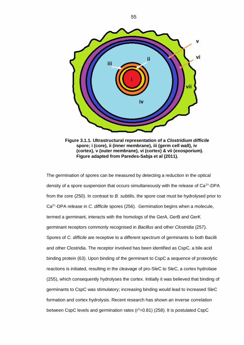
55
The germination of spores can be measured by detecting a reduction in the optical
density of a spore suspension that occurs simultaneously with the release of Ca2+-DPA
from the core (250). In contrast to B. subtilis, the spore coat must be hydrolysed prior to
Ca2+-DPA release in C. difficile spores (256). Germination begins when a molecule,
termed a germinant, interacts with the homologs of the GerA, GerB and GerK
germinant receptors commonly recognised in Bacillus and other Clostridia (257).
Spores of C. difficile are receptive to a different spectrum of germinants to both Bacilli
and other Clostridia. The receptor involved has been identified as CspC, a bile acid
binding protein (63). Upon binding of the germinant to CspC a sequence of proteolytic
reactions is initiated, resulting in the cleavage of pro-SleC to SleC, a cortex hydrolase
(255), which consequently hydrolyses the cortex. Initially it was believed that binding of
germinants to CspC was stimulatory; increasing binding would lead to increased SleC
formation and cortex hydrolysis. Recent research has shown an inverse correlation
between CspC levels and germination rates (r2=0.81) (258). It is postulated CspC
Figure 3.1.1. Ultrastructural representation of a Clostridium difficile spore; i (core), ii (inner membrane), iii (germ cell wall), iv (cortex), v (outer membrane), vi (cortex) & vii (exosporium).
Figure adapted from Paredes-Sabja et al (2011).

56
activates CspB, but CspB is unable to cleave pro-SleC unless GerS is present. The
concluding stage of germination is the release of a vegetative cell from the ruptured
spore coat/exosporium. Further work is required to elucidate these interactions and
refine the model currently proposed for C. difficile spore germination.
Recently it has been discovered that the pseudoprotease domain CspA and its fusion
to CspB is highly significant in regulation of the germination cascade, with a nonsense
mutation in cspBA reducing the efficiency of C. difficile spores to germinate (255).
CspBA is cleaved by YabG to form CspA and CspB, CspB is responsible for the
cleavage of pro-SleC to the active SleC. Additionally, CD0311, a protein named GerG
has found to be important in the germination process. GerG mutants are found to
require 10-times the levels of germinant to initiate germination, and it has been shown
that lower levels of CspC receptors are present in these mutants (259). It is believed
GerG has an important role in the incorporation of CspA, CspB and CspC into the
spore. GerG has been suggested to be a novel target for therapeutics, potentially
decreasing spore germination and the risk of recurrence. This is highly speculative and
requires more research.
Germination is a tightly regulated and complex process. The homology of this system
to other members of the Peptostreptococcaceae rather than Clostridiceceae family is in
accordance with the genomic data regarding reclassification of C. difficile to the
Peptostreptococcaceae family (260).
3.1.2 Bile acids
The germination of C. difficile spores is strongly dependent on environmental cues and
has been found to be altered in response to bile acids. In general, primary bile acids
are stimulatory to germination and secondary bile acids are inhibitory. However, there
are some exceptions. Ratios of primary: secondary bile acids along the gastrointestinal
tract could alter in response to environmental insult, facilitating C. difficile spore
germination. The primary bile acids are cholate, chenodeoxycholate, taurocholate and
glycocholate. Derivative secondary bile acids include deoxycholate. Previously, 0.1 %

57
taurocholate saturated BHIS (supplemented brain heart infusion) plates showed a 105
increase in recovery compared to controls (261). Early on, it was documented the
concentration and purity of the taurocholate preparations tested in vitro was important;
higher concentrations of sodium taurocholate inhibited vegetative cell division (262).
Initially, the mechanism of taurocholate-induced germination was unclear, but kinetic
data alluded to a sequential receptor based process involving glycine (263).
Taurocholate is now routinely used to germinate C. difficile spores.
The primary bile acid chenodeoxycholate is inhibitory to spore germination; incubation
of spores with 0.1 % chenodeoxycholate produced a 0.006 % recovery rate, compared
to 0.024 % with an equal 0.1 % mixture of cholate: chenodeoxycholate (264). Further
work indicated chenodeoxycholate derivatives are produced by 7α-hydroxylation, a
process carried out by Clostridium scindens, considered in recent times to be a
protective species in the microbiome (93, 264). Other work found chenodeoxycholate
to be stimulatory at 1-10 mmol/l to C. difficile germination, but also noted higher levels
of sodium taurocholate (>100 mmol/l) to be inhibitory (265).
Human bile acid perfusion studies have indicated a ~9-fold increase in the absorption
in the large intestine of chenodeoxycholate compared with cholate (266). This creates
a favourable environment for spore germination. Only single formulations were
perfused, ignoring any in vivo interactions between different bile acids. Experiments
using a mouse model also suggest a role for bile acids in germination of C. difficile
spores. Caecal extracts from antibiotic-treated mice produced colony formation levels
in C. difficile spores 50-65 times higher than from untreated mouse tissue (267). This
effect was lost when bile-acid binding cholestyramine was added to extracts. The study
concluded that increased ratio of primary: secondary bile acids in antibiotic treated
samples are responsible for the increase in CFU observed. However, there is no
discussion of the relative concentrations of individual primary bile acids. More recent
mice metabolomics data had a similar finding; a relative increase of primary bile acids

58
in antibiotic treated ‘CDI susceptible’ mice (268). The two studies used different
antibiotics to simulate CDI conditions (clindamycin vs. cefoperazone).
3.1.3 Amino acids
The amino acid glycine has been found to be a vital co-germinant; its inclusion with
taurocholate is a standard approach for maximising germination of C. difficile spores.
Glycine was utilised early on (1966) to increase germination of Clostridium botulinum
along with cysteine (269). Subsequently, the germination of C. difficile spores has been
improved using glycine with taurocholate (261, 265). The binding and functional groups
involved in the substrate-receptor interaction have been mapped by adding 30 glycine
derivatives to taurocholate treated spores and documenting germination rates (270).
The chemical manipulations targeted singular modifications in the carboxy/amine
groups or the alkyl chain of the glycine molecule (Fig 3.1.2).
By creating a derivative library, the authors were able to deduce that both the intact
carboxylate and amine groups are necessary for recognition by the germinant receptor,
as modifications of these groups produces a substantial decrease in germination rates.
The size of the methylene bridge between the functional groups does not appear to
Figure 3.1.2. Glycine molecule, showing alkyl chain skeleton, amine group and hydroxyl groups.
Diagram obtained from ChemDraw®

59
affect germination levels. Although glycine is the most explored and widely understood
amino acid used for C. difficile germination optimisation, this study provides evidence
of other amino acids for instance β-alanine being equally as effective at initiating
germination in C. difficile. One study identified an 80 % increase in germination rates
on addition of histidine (0.4 %) to taurocholate (0.1 %) with glycine (0.4 %)(271). L-
phenylalanine has been found to be as effective as glycine, which is surprising given
the initial hypothesis that the germinant receptor would not be able to accommodate
aromatic groups due to size (270). Multiple binding sites have been suggested, but
there is currently no evidence to support this. Unlike L-phenylalanine, histidine (0.4 %)
added to taurocholate (0.1 %) without glycine did not produce an increase in
germination; both L-phenylalanine and histidine are aromatic amino acids.
Recent developments (2018) have elucidated the hierarchical nature of amino acid
recognition in C. difficile spores. Shrestha & Sorg investigated the efficacy of various
amino acids (including D- alanine, D-serine & D-lysine) as co-germinants to taurocholate
(272). Glycine was found to be the most effective germinant, but importantly all amino
acids were stimulatory to spore germination to some extent at 37° C. Interestingly, the
author proposed that glycine being the smallest amino acid could contribute to its
effectiveness; small molecules can more easily reach the spore cortex. These findings
are congruent with those of Howerton et al (270), with aromatic residues being less
effective. Nevertheless, the mechanism of co-germinant binding is still unknown; it is
unclear whether signalling is mediated through a single receptor responsive to all
amino acids or numerous receptors for each individual amino acid.
Additionally, the role of calcium in spore germination has been investigated. Calcium
has been found to have a critical role in the activation of SleC, the cortex hydrolase.
When spores were incubated with taurocholate, glycine and EGTA (a calcium chelator)
germination was completely inhibited at all concentrations vs. taurocholate/ glycine
alone (273). It is hypothesised that individuals deficient in calcium absorption are at an
increased risk of CDI due to the stimulatory effects of superfluous intestinal calcium on

60
germination. This provides a potential mechanism for the association of protein pump
inhibitors (PPIs) with CDI. This risk may be amplified by the observation that the cell
membrane interactions of toxin A in binding to colonic cells is mediated by free calcium
ions (274).
3.1.4 Non-germinant receptor germinants
In addition to germinant receptor-derived germination, other factors have been
identified as facilitating increased recovery of C. difficile spores. In 2000 it was found
that incorporation of 5 mg/l of lysozyme into CCEY (without egg yolk) agar significantly
increased the recovery of C. difficile from environmental swabs compared to CCEY (24
% vs 11 %, P = 0.004) (275). This observation has been made previously with
increased recovery rates (10-47 %) observed in four strains recovered by sodium
thioglycollate-lysozyme treatment after heat treatment of bacterial suspensions at 90ºC
for 10 minutes (276). Pre-treatment with thioglycollate was not associated with
increased recovery rates. Lysozyme could be the mediator of germination in these
instances and not thioglycollate pre-treatment. One hypothesis is that lysozyme
mediates germination directly by enzymatically degrading the spore cortex. The above
study (276) also found 0.1 % taurocholate addition did not permit recovery of spores
subjected to the same heat treatment. This is not surprising considering the more
recent work indicating the importance of glycine as a co-germinant.
3.1.5 Optimising C. difficile recovery
Different solid media are used according to individual requirements for C. difficile
recovery. The appropriateness of a culture medium for C. difficile will depend on a
number of factors. Firstly, a medium must allow the germination and proliferation of C.
difficile spores. In order for substantial germination to take place, stimulatory
germinants must be present. The growth of other species must be suppressed by the
medium. CCEYL (cycloserine-cefoxitine, egg yolk and lysozyme) is a selective medium
most suitable for recovering C. difficile from faecal samples, and has recently been
shown to be the most sensitive and cost-efficient medium for isolating C. difficile from

61
stool samples when compared to cycloserine-cefoxitin fructose agar (CCFA), tryptone
soy agar (TSA) and ChromeID agar (277). Five-percent lysozyme was incorporated
due to evidence suggesting the increased recovery of environmental spores treated
with lysozyme (275). CDRN (C. difficile ribotyping network) use CCEYL (without egg
yolk) to isolate C. difficile from faeces due to the bacteriostatic action of cycloserine-
cefoxitin (39).
BHI agar (brain heart infusion) is used by the majority of research labs working with C.
difficile. Often taurocholate is incorporated (a primary bile acid) alongside glycine.
CCEYL also contains an unknown quantity of the primary bile acid cholate in its
ingredients. BHI is suited to pure culture of C. difficile, due to its non-selectivity (278). It
is unclear which conditions are optimal for spore recovery. Although primary bile acids
and amino acids are known to be important for inducing spore germination in C.
difficile, a direct comparison of CCEYL and supplemented BHI has not been
performed.
Although the importance of germinants and amino acids has been discussed in the
context of germination, one study found germination will occur spontaneously in the
absence of germinants in a subpopulation of spores (279) . This could be important in
experiments utilising spores that have been aged or left for extended periods of time in
the environment. Over time, a small population of spores are likely to spontaneously
germinate, decreasing spore numbers.
There were a number of aims for this study. Firstly, conditions optimal to C. difficile
spore recovery and outgrowth in liquid and on solid media were explored. C. difficile
germination and growth was assessed in two solid media and two liquid media with the
incorporation of different combinations and concentrations of germinants. Additionally,
this study highlighted the inhibitory nature of high concentrations of L-amino acids on C.
difficile vegetative growth. Five strains of different ribotypes were grown in broths with
increasing concentrations of 3 amino acids (glycine, L-phenylalanine and L-histidine).
Finally, the phenomenon of superdormancy was explored; spores were left in a

62
homemade desiccator for an extended period (6 months) to simulate environmental
ageing. It was hypothesised that environmental ageing would affect the ability of spores
to germinate in the presence of germinants.
Methods
3.2.1 Production of Spores
Spores of five PCR ribotypes (RT 001,015,020, 027 & 078) of C. difficile were prepared
as previously described (248). Briefly, 100 µl of a spore preparation was spread on to
Brazier’s CCEYL agar (Oxoid, UK) and grown anaerobically for 5 days in a Don Whitley
A95 anaerobic workstation. Growth was harvested and streaked on to 10 Columbia
Blood Agar (CBA) plates. CBA agar plate growth was removed through swabbing after
10 days of anaerobic incubation and suspended in 4 ml of 50 % ethanol to kill
vegetative cells. Spore stocks were serially diluted in phosphate-buffered saline
(PBS)(BioVision, USA) and enumerated on CCEYL agar.
All experiments were carried out in triplicate and all spores were fresh (<30 days old)
unless otherwise stated. In all experiments agar plates were incubated anaerobically at
37° C and counts of colony forming units (CFU) were carried out 48 hours post-
inoculation.
3.2.2 Phase Contrast Microscopy
Slides were prepared by spreading 50 µl of spore suspension uniformly over a
microscope slide and drying aerobically for 30 minutes at 50° C. Slides were overlaid
with 50 µl of Wilkins-Chalgren agar and dried for a further hour. Phase bright spores,
phase dark spores and vegetative cells were visualised in ten fields of view and
counted on a phase contrast microscope at 1000 X magnification. All entities were
counted in each field of view. In broth experiments one slide was prepared per
biological replicate (broth). Phase dark spores indicate those that have germinated,
phase bright spores have not germinated, and vegetative cells are the product of spore
germination and outgrowth.

63
3.2.3 Spore Recovery on Solid Media
Spore suspensions were serially diluted (10-fold) in PBS to 10-9. Twenty-microlitres of
each dilution was spread on to a range of solid agar plates (Table 3.2.1). Media (BHI
(Oxoid, UK) and CCEY (LabM)) was prepared in house according to manufacturer’s
instructions (see Appendix B, B.1.8 & B.1.9). In the case of additive preparation,
taurocholate and glycine (Oxoid, UK) were added prior to autoclaving, lysozyme was
added subsequently. Spores of five C. difficile ribotypes were utilised. An overview of
the methodology can be seen in Fig 3.2.1.
Table 3.2.1. Solid agar plates utilised in C. difficile spore recovery experiments. Media types and additives are shown
3.2.4 Spore Germination in Broths
Broths of 4.95 ml were prepared in glass Wassermans, autoclaved and pre-reduced
overnight in an anaerobic chamber. Media (BHI and Schaedler (Oxoid, UK)) was
prepared in house according to manufacturer’s instructions (see Appendix B, B.2). All
broths were carried out in biological duplicate unless otherwise stated. Broths utilised
can be seen below (Table 3.2.2). At time point 0 phase contrast microscopy was
Media Additional Additives
BHI
Nil
5% lysozyme
0.1% taurocholate, 0.4% glycine
1% taurocholate, 0.8% glycine
1% taurocholate, 4% glycine
CCEY
Nil
5% lysozyme
0.1% taurocholate, 0.4% glycine
1% taurocholate, 0.8% glycine
1% taurocholate. 4% glycine

64
carried out on spore suspensions. Subsequently, 50 µl of spore suspension was
aliquoted and incubated anaerobically in broth for 90 minutes. At 90 minutes, 20 µl of
broth was removed and serially diluted in PBS to 10-7 in technical triplicate. For spore
enumeration, broth (100 µl) was aliquoted into 100 µl of 100 % ethanol and after an
hour serially diluted in PBS to 10-4. Twenty-microlitres of each dilution were aliquoted
on to CCEYL agar. In addition, 500 µl of broth was removed and centrifuged at 9500 g
for 1 minute. The supernatant was removed and spores were resuspended in 50 µl of
PBS, which was spread on to a slide for phase contrast microscopy. An overview of the
methodology can be seen in Fig 3.2.3.

65
Table 3.2.2 Range of broths utilised in C. difficile spore germination experiments. 1 Broth types and additives are shown. 2
3 Broth Additional Additives
BHI
Nil
5 % lysozyme
0.1 % taurocholate, 0.4 % glycine
0.1 % taurocholate, 0.4 % histidine
0.1 % taurocholate, 0.4 % glycine, 0.4 % histidine
1% taurocholate, 4% glycine
Schaedler
Nil
5 % lysozyme
0.1 % taurocholate, 0.4 % glycine
1 % taurocholate, 4 % glycine
Figure 3.2.1. An overview of the methodology used in solid agar experiments.
20 μl 20 μl
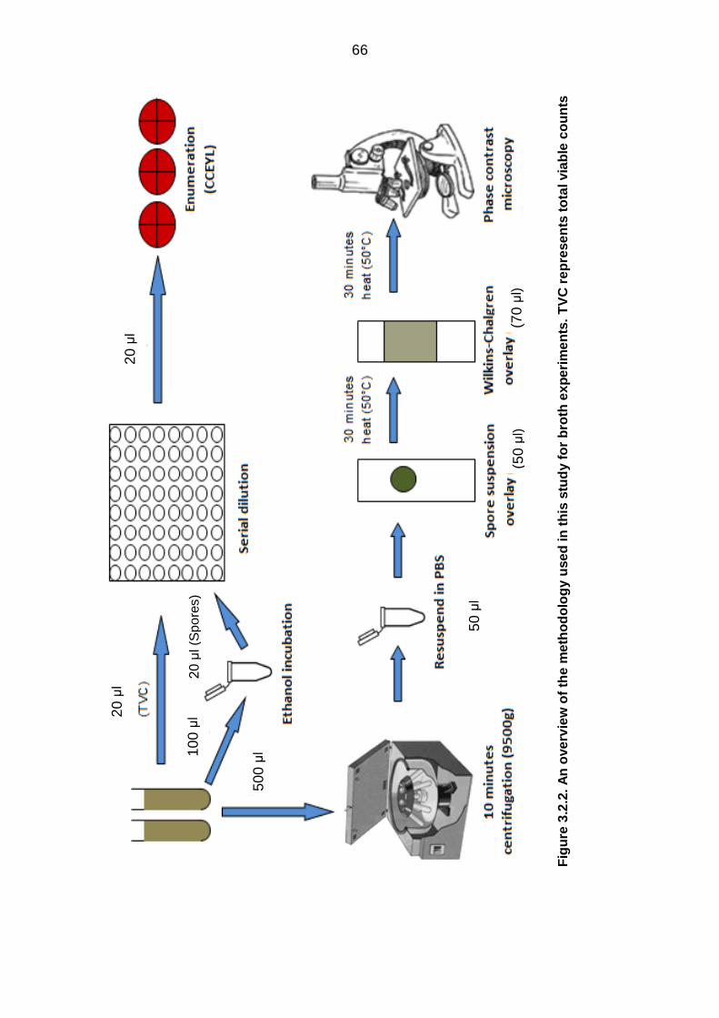
66
4
Fig
ure
3.2
.2.
An
ove
rvie
w o
f th
e m
eth
od
olo
gy u
se
d in
th
is s
tud
y f
or
bro
th e
xp
eri
me
nts
. T
VC
re
pre
se
nts
to
tal via
ble
co
un
ts
20
μl
10
0 μ
l
50
0 μ
l
20
μl
50
μl
(50
μl)
(70
μl)
20
μl (S
po
res)
67
3.2.5 Agar-incorporated minimum inhibitory concentration (MIC)
testing
The minimum inhibitory concentration (MIC) for glycine and taurocholate was tested
both alone an in combination (4:1 ratio) against C. difficile strains of five ribotypes (001,
015, 020, 027, 078). An agar-incorporation MIC methodology was utilised that has
been used previously by Baines et al (280). Test compounds were weighed out in
doubling concentrations and added to individual aliquots of Wilkins-Chalgren anaerobe
agar or CCEY agar. CCEY agar was supplemented with 2 % lysed, defibrinated horse
blood. C. difficile vegetative populations were grown up overnight in Schaedler’s broth
in an anaerobic cabinet. Both spore and vegetative (1ː10 dilution of 24-hour
Schaedler’s broth culture) populations of the five C. difficile strains were inoculated
(~104 cells) on to glycine/taurocholate incorporated agar. Inhibition of growth was
assessed after anaerobic incubation at 37° C for 48 hours, where the lowest
concentration at which visible C. difficile growth was inhibited was recorded as the MIC.
3.2.6 Minimum Inhibitory Concentration (MIC) Testing in
Microbroths
BHI broths with increasing concentrations (1, 2, 3, 4 %) of three amino acids (glycine,
L-histidine, L-phenylalanine) were prepared and 180 μl was aliquoted into a 96-well
plate. Twenty-microlitres of spore solution (~5x10⁵ /ml) were aliquoted into each well at
time point 0; five strains of different ribotypes were utilised (001, 015, 020, 027, 078).
At 0, 24- and 48-hours absorbance readings were determined at 595 nm in a Tecan
Infinite 200 Pro reader at 20° C and under 1 atm of pressure. Negative controls were
prepared for each concentration, and the absorbance for the blanks was subtracted
from the absorbance of the inoculated wells to determine an accurate absorbance
reading based on growth alone. All wells were prepared in triplicate.
3.2.7 C. difficile spore desiccation
One day old (1 ml) spores of four ribotypes (001, 015, 020 & 078) were aliquoted into
Eppendorfs in biological triplicate and left in a homemade desiccator (Fig 3.2.3). At the

68
0, 3- and 6-month time points spores were resuspended in the appropriate volume of
PBS and enumerated by serial dilution (10-5) on a range of solid media (CCEY, CCEYL
and BHI (0.1 % taurocholate, 0.4 % glycine)). At the stated time points, 20 µl of spores
were transferred to 180 µl of three different broths (BHI, BHI (5 % lysozyme) & BHI
(0.1% taurocholate, 0.4 % glycine)) in biological triplicate. After 3 hours incubation,
spores and TVCs were enumerated by serial dilution (10-5) on CCEYL agar as
previously documented. CCEYL plates were incubated anaerobically for 48 hours. An
overview of the methodology for the desiccation experiments can be found in Fig 3.2.4
& Fig 3.2.5.
Figure 3.2.3. The desiccator used to age C. difficile spores. The desiccator consisted of an air tight container filled with silica crystals to remove moisture from the air. Silica crystals in dehydrated form are blue but
become red after hydration.

69
1
Figure 3.2.4. An overview of the methodology used to enumerate desiccated spores directly on to solid agar. Spores were enumerated on three agars; CCEY, CCEYL & BHI. BHI was supplemented with 0.1 % taurocholate and 0.4 % glycine (BHI(S)). This diagram represents sampling for one ribotype. Four strains of differing ribotypes were utilised in total (001, 015,020 & 078).
Spores were enumerated at 0, 3, & 6 months.
20 μl 20 μl

70
2
Figure 3.2.5. An overview of the methodology used to enumerate desiccated spores after 90 minutes broth incubation. Spores were incubated in three different broths; BHI, BHI(L) & BHI(S). BHI was supplemented with 5 % lysozyme (BHI(L)) or 0.1 % taurocholate 0.4 % glycine (BHI(S)). After incubation spore and TVC counts were enumerated on CCEYL agar. This diagram represents sampling for one ribotype. Four strains of differing ribotypes were utilised in total (001, 015 020, & 078). Spores
were incubated in broths at 0, 3 & 6 months.
Spore
suspension
(PBS)
Broth
incubation
(90
minutes)
Serial
Dilution
Spore
enumeration
(CCEYL)
20µl
20µl 20µl 20 μl
20 μl 20 μl

71
3.2.8 Statistical analysis
Statistical analysis was carried out in on IBM SPSS Statistics 22. Prior to statistical
analyses data was assessed for normality and homogeneity of variance between
groups. Data normality was assessed using histograms and Kolmogorov-Smirnov
tests. Homogeneity of variance was assessed using Levene’s test; in the case of
significant differences in variance between groups Welch’s ANOVA was utilised. All
means are reported with standard error of the mean (SE). A statistical significance level
of <0.05 was adopted, <0.01 highly significant and <0.001 very highly significant. x̅
represents the mean of several specified ribotypes. Individual details of statistical
analysis are found with each experiment.
Results
3.3.1 Spore Recovery on Solid Media
Spore recovery varied considerably between different media types (Fig 3.3.1). In the
absence of supplementation, recovery was ~1 log10CFU/ml greater on CCEY vs BHI
media (range = 0.1 – 2.4 log10CFU/ml). The increased recovery of spores on CCEY vs
BHI was observed in all but the RT 078 strain (P > 0.05). Greatest spore recovery was
achieved in CCEY (x̅ = 8.20 ± 0.03 log10CFU/ml), CCEYL (x̅ = 8.26 ± 0.05
log10CFU/ml) and BHI supplemented with taurocholate (x̅ = 8.25 ± 0.06 log10CFU/ml).
The incorporation of 5% lysozyme had no substantial effect on the recovery of spores
in either media (CCEY x̅ = 8.20 ± 0.03 vs 8.26 ± 0.04 log10CFU/ml, BHI x̅ = 7.28 ± 0.20
log10CFU/ml vs 7.10 ± 0.18 log10CFU/ml) (P > 0.05). In BHI, supplementation with
taurocholate (0.1 % or 1 %) significantly increased the recovery of spores by on
average ~1 log10CFU/ml (range = 0 – 2.7 log10CFU/mL). Differences were observed
between strains; the RT 001 strain showed a very highly significant increase (5.80 ±
0.13 log10CFU/ml vs 8.45 ± 0.08 log10CFU/ml) (P<0.001), in contrast to the negligible

72
difference observed for the RT 078 strain (7.95 ± 0.05 log10CFU/ml vs 8.03 ± 0.04
log10CFU/ml) (P>0.05). On the contrary, taurocholate supplementation alone had no
substantial effect on the recovery of spores on CCEY media (x̅ = 8.20 ± 0.03
log10CFU/ml vs 8.29 ± 0.05 log10CFU/ml).
Incorporation of 4 % glycine alongside taurocholate caused a marked reduction in
spore recovery to below the lower limit of detection (1.22 log10CFU/ml) in both CCEY
and BHI. Lower concentrations of glycine (0.4 % & 0.8 %) had no substantial effect on
recovery in BHI, but in CCEY this decreased ~3 log10CFU/ml and ~6 log10CFU/ml,
respectively.

73
Fig
ure
3.3
.1.
Me
an
(± S
E)
sp
ore
re
co
very
(lo
g10C
FU
/ml)
of
sp
ore
s o
f fi
ve
C.
dif
fic
ile
str
ain
s o
f d
iffe
rin
g r
ibo
typ
es
(00
1,
01
5,
02
0,
027
& 0
78
) in
oc
ula
ted
on
to
a v
ari
ety
of
so
lid
ag
ars
. S
po
res w
ere
se
ria
lly d
ilu
ted
in
PB
S t
o 1
0-9
a
nd
ea
ch
dilu
tio
n s
pre
ad
on
to
ag
ar
pla
tes in
tri
plic
ate
. P
late
s w
ere
rea
d a
fte
r 4
8h
of
an
ae
rob
ic in
cu
ba
tio
n. T
C
= t
au
roc
ho
late
, G
LY
= g
lyc
ine
, L
= lyso
zy
me
, L
LO
D =
lo
we
r lim
ite
d o
f d
ete
cti
on
. G
rou
p m
ea
ns w
ere
co
mp
are
d
usin
g W
elc
h’s
AN
OV
A w
ith
Ga
me
s-H
ow
ell m
ult
iple
co
mp
ari
so
ns.
Sta
tis
tic
ally s
ign
ific
an
t d
iffe
ren
ce
s (
P<
0.0
5)
are
hig
hlig
hte
d w
ith
*,
ve
ry h
igh
ly s
ign
ific
an
t d
iffe
ren
ce
s (
P<
0.0
01
) w
ith
***
.

74
3.3.2 Spore Broth Pilot Study
RT 027 spore numbers showed a time dependent decrease (Fig 3.3.2), decreasing
~1log from 30 to 90 minutes (6.25 ± 0.02 log10CFU/ml vs 5.10 ± 0.03 log10CFU/ml; P<
0.001), indicating spore germination. Total viable counts (TVCs) remained relatively
stable between the 30- and 90-minute mark (7.02 ± 0.03 log10CFU/ml vs 7.06 ± 0.01
log10CFU/ml; P < 0.05). Spore numbers continued to decrease after 90 minutes at a
reduced rate; spore numbers decreased from 5.10 ± 0.03 log10CFU/ml to 4.87 ± 0.04
log10CFU/ml after 120 minutes.

75
.
Tim
e (
min
ute
s)
Fig
ure
3.3
.2.
Sp
ore
pilo
t stu
dy.
Me
an
(± S
E)
TV
C a
nd
sp
ore
co
un
ts o
f C
. d
iffi
cile
RT
027
in
cu
ba
ted
in
B
HI (s
up
ple
me
nte
d w
ith
0.1
% t
au
roc
ho
late
, 0.4
% g
lyc
ine
) fo
r 12
0 m
inu
tes
. B
roth
s w
ere
sa
mp
led
at
30
, 6
0,9
0 a
nd
12
0 m
inu
tes a
nd
se
ria
lly d
ilu
ted
in
PB
S t
o o
bta
into
tal v
iab
le c
ou
nts
(T
VC
). O
ne s
et
of
aliq
uo
ts w
ere
eth
an
ol sh
oc
ke
d in
50 %
eth
an
ol fo
r 60
min
ute
s p
rio
r to
se
ria
l d
ilu
tio
n i
n o
rde
r to
kill ve
ge
tati
ve
po
pu
lati
on
s a
nd
ob
tain
sp
ore
nu
mb
ers
. S
po
re le
ve
ls
de
cre
ase
d in
a t
ime
de
pe
nd
en
t m
an
ne
r. B
roth
s w
ere
ca
rrie
d o
ut
in b
iolo
gic
al tr
iplic
ate
an
d
sa
mp
led
in
te
ch
nic
al tr
iplic
ate
. R
M-A
NO
VA
wit
h T
uk
ey’s
mu
ltip
le c
om
pa
riso
ns w
as u
se
d f
or
sta
tisti
ca
l a
na
lys
is.
Ve
ry h
igh
ly s
ign
ific
an
t d
iffe
ren
ce
s (
P <
0.0
01
) a
re h
igh
lig
hte
d *
**.

76
3.3.3 Spore Germination in Broths
Spore germination was induced by the inclusion of taurocholate and glycine at either
concentration utilised (0.1 % taurocholate/ 0.4 % glycine, 1 % taurocholate/ 4 %
glycine) (Fig 3.3.3). In BHI, spore levels were significantly lower (~2 log10CFU/ml) in
taurocholate supplemented broths (x̅ = 4.32 ± 0.05 log10CFU/ml and 4.20 ± 0.07
log10CFU/ml) vs 5 % lysozyme (x̅ = 6.38 ± 0.06 log10CFU/ml) and non-supplemented (x̅
= 6.43 ± 0.06 log10CFU/ml; P<0.001) BHI broths. Incorporation of lysozyme did not
appear to have a substantial effect on the germination of spores. Total viable counts
were comparable between all of the BHI broths at 90 minutes (range; x̅ = 6.69 ± 0.09
log10CFU/ml – 6.75 ± 0.09 log10CFU/ml). In addition, germination of spores in
Schaedler broth was comparable to BHI, with similar levels of spore decrease in
taurocholate supplemented (x̅ = 4.22 ± 0.05 log10CFU/ml vs 4.26 ± 0.05 log10CFU/ml)
lysozyme supplemented (x̅ = 6.27 ± 0.06 log10CFU/ml vs 6.39 ± 0.06 log10CFU/ml;
P>0.05) and non-supplemented broths (x̅ = 6.28 ± 0.06 log10CFU/ml vs 6.43 ± 0.06
log10CFU/ml; P> 0.05). There was no significant difference between total viable counts
in any of the Schaedler broths at 90 minutes (range; x̅ = 6.67 ± 0.10 log10CFU/ml –
6.70 ± 0.06 log10CFU/ml; P = 0.97).
Phase contrast microscopy showed that in broths supplemented with taurocholate and
glycine, phase dark spores predominated after 90 minutes were (Fig 3.3.4). At the
zero-time point, the RT 078 strain contained a majority of phase dark spores compared
with phase bright spores in both BHI and Schaedlers (x̅ = 76.40 ± 2.00 % vs 7.45 ±
1.00 %; P< 0.001). In non-supplemented BHI broths, after 90 minutes incubation,
phase bright spores were more prevalent than phase dark spores or vegetative cells in
four of the strains (001, 015, 020 & 027) (x̅ = 70.7 ± 11.2 % vs 18.0 ± 8.3 % vs 11.4 ±
10.1 %). In contrast, in both sets of supplemented BHI broths, phase dark spores were
the predominant entity in all five strains with minimal levels of phase bright spores (<3
%). Vegetative populations made up a minority of the identified entities in all broths

77
(range = 9.2% - 30.1%). The trends observed in BHI were also seen in Schaedler
broths.

78
Figure 3.3.3. Mean (± SE) TVC and spore counts of five C. difficile strains of different ribotypes (001, 015, 020, 027 & 078) germinated in two different broths (BHI & SCH) in the presence of different germinants lysozyme (L), taurocholate (TC) & glycine (GLY). (L) indicates broths supplemented with lysozyme. 0.1 %/1 % indicates the taurocholate concentration, glycine concentrations were four times that of taurocholate (0.4 %/4 %). Broths were incubated for 90 minutes, after which aliquots were serially diluted in PBS to obtain total viable counts. One set of aliquots was ethanol shocked to obtain spore levels. TVC and spore counts for each broth at the 90-minute time point are presented in adjacent columns (e.g. BHI TVC, BHI Spores). Broths were carried out in biological duplicate and processed in technical triplicate. One-way ANOVA with Tukey’s multiple comparisons was used for statistical analysis. Very highly significant findings (P< 0.001)
are highlighted with ***.
Media
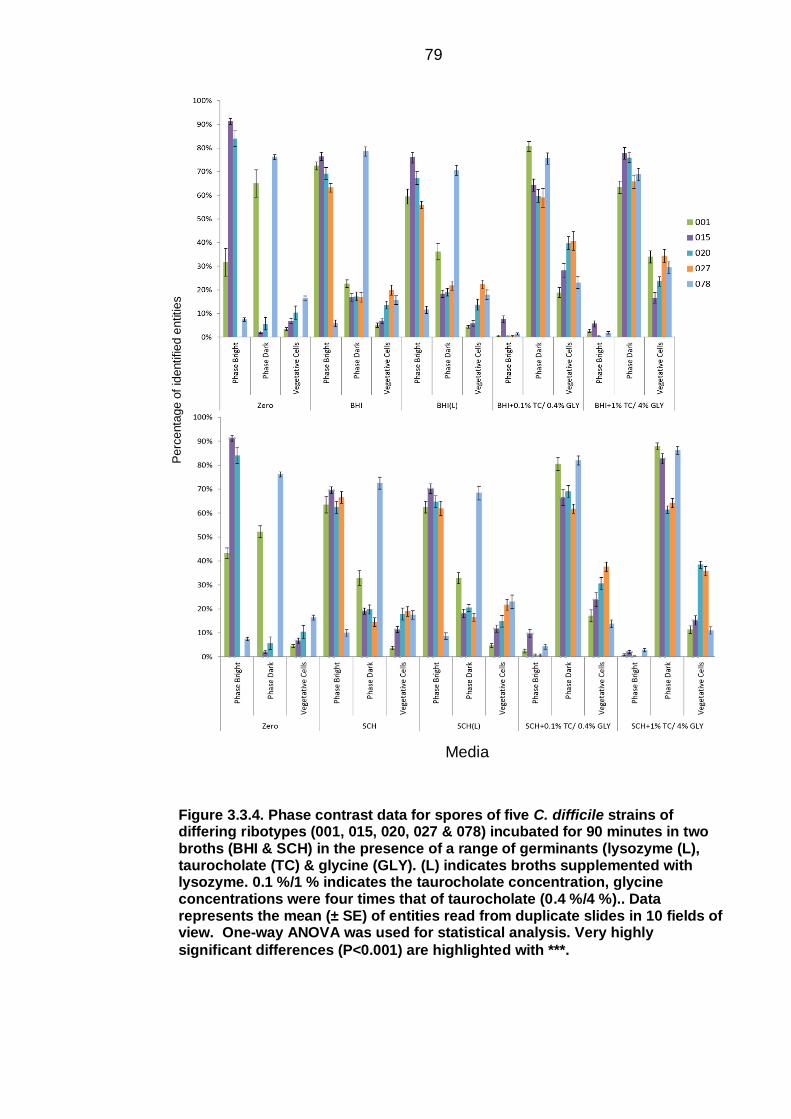
79
Figure 3.3.4. Phase contrast data for spores of five C. difficile strains of differing ribotypes (001, 015, 020, 027 & 078) incubated for 90 minutes in two broths (BHI & SCH) in the presence of a range of germinants (lysozyme (L), taurocholate (TC) & glycine (GLY). (L) indicates broths supplemented with lysozyme. 0.1 %/1 % indicates the taurocholate concentration, glycine concentrations were four times that of taurocholate (0.4 %/4 %).. Data represents the mean (± SE) of entities read from duplicate slides in 10 fields of view. One-way ANOVA was used for statistical analysis. Very highly
significant differences (P<0.001) are highlighted with ***.
Perc
enta
ge o
f id
entified e
ntities
Media

80
3.3.4 C. difficile germination in the presence and absence of
additional supplementation
After 24 hours of incubation, spore counts were lower in the BHI(S) broths compared to
BHI alone (x̄ = 3.79 ± 0.12 log10CFU/ml vs 5.08 ± 0.21 log10CFU/ml; P< 0.001) (Fig
3.3.5). Over the first 6 hours of incubation, a gradual increase in TVC to reach peak
numbers was observed in the supplemented BHI broth (x̄ = 6.46 ± 0.02 log10CFU/ml,
6.92 ± 0.09 log10CFU/ml, 7.57 ± 0.16 log10CFU/ml; P< 0.01). In contrast, the non-
supplemented BHI broth took the whole 24-hour period to reach peak recorded TVC
levels (x̄ = 6.43 ± 0.03 log10CFU/ml vs 7.95 ± 0.08 log10CFU/ml; P< 0.001).
Spore numbers decreased significantly in both supplemented and non-supplemented
broths after 3 hours but to different extents (x̄ = 5.97 ± 0.10 log10CFU/ml vs 4.04 ± 0.07
log10CFU/ml & 6.04 ± 0.08 log10CFU/ml vs 5.33 ± 0.29 log10CFU/ml; P < 0.05). RT 078
showed considerable germination of spores after 3 hours in the non-supplemented
broth compared with RT 001 and RT 027 (3.72 ± 0.06 log10CFU/ml vs 6.13 ± 0.13
log10CFU/ml; P< 0.001). At the equivalent time point all three strains (001, 027 & 078)
showed similar levels of germination in the supplemented broth (x̄ = 4.04 ± 0.07
log10CFU/ml). Although very significant, spore numbers at 0 vs 24 hours were only
slightly lower in RT 001 and RT 027 when incubated in the non-supplemented BHI
broth (x̄ = 6.15 ± 0.11 log10CFU/ml vs 5.70 ± 0.05 log10CFU/ml; P< 0.001). This
contrasts with RT 078, which showed a ~2 log10CFU/ml drop after 24 hours (5.84 ±
0.06 log10CFU/ml vs 3.85 ± 0.04 log10CFU/ml; P< 0.001).

81
Fig
ure
3.3
.5.
Me
an
(± S
E)
TV
C a
nd
sp
ore
co
un
ts o
f th
ree
C.
dif
fic
ile
str
ain
s o
f d
iffe
rin
g r
ibo
typ
es (
001
, 0
27
& 0
78
) o
f C
.
dif
fic
ile
germ
ina
ted
in
tw
o b
roth
s (
BH
I &
BH
I(S
)).
BH
I(S
) is
su
pp
lem
en
ted
wit
h 0
.1 %
ta
uro
ch
ola
te.
Aliq
uo
ts w
ere
ta
ke
n a
t 0,
3, 6
, &
24
-ho
ur
po
st-
ino
cu
lati
on
an
d e
nu
me
rate
d b
y s
eri
al d
ilu
tio
n o
n C
CE
YL
ag
ar.
On
e s
et
of
aliq
uo
ts w
ere
sh
oc
ke
d in
50 %
e
tha
no
l fo
r 1
ho
ur
to k
ill ve
ge
tati
ve
ce
lls a
nd
en
um
era
te s
po
re p
op
ula
tio
ns.
Bro
ths w
ere
ca
rrie
d o
ut
in b
iolo
gic
al d
up
lic
ate
a
nd
se
ria
lly d
ilu
tio
ns in
te
ch
nic
al tr
iplic
ate
. W
elc
h’s
in
de
pe
nd
en
t T
te
sts
we
re u
se
d t
o c
om
pa
re g
rou
ps (
BH
I vs B
HI(
S))
at
dif
fere
nt
tim
e p
oin
ts.
Ve
ry h
igh
ly s
ign
ific
an
t (P
< 0
.00
1)
dif
fere
nc
es a
re h
igh
lig
hte
d b
y *
**.
Mean CFU Counts (log10CFU/ml)
Tim
e (
ho
urs
)

82
Fig
ure
3.3
.6. P
ha
se
co
ntr
ast
da
ta f
or
sp
ore
s o
f th
ree
C. d
iffi
cile s
tra
ins o
f d
iffe
rin
g r
ibo
typ
es (
00
1,0
27
& 0
78
) in
cu
ba
ted
fo
r
24
ho
urs
in
tw
o b
roth
s (
BH
I &
BH
I(S
)).
BH
I(S
) w
as s
up
ple
me
nte
d w
ith
0.1
% t
au
roc
ho
late
. A
t 0
, 3
& 6
ho
urs
, 5
00
μl o
f b
roth
w
as c
en
trif
ug
ed
, re
su
sp
en
de
d in
50
μl o
f m
inim
al m
ed
ia a
nd
dri
ed
fo
r 3
0 m
inu
tes.
Su
bse
qu
en
tly s
lid
es w
ere
ove
rla
id w
ith
70
μ
l o
f W
ilk
ins-C
ha
lgre
n a
ga
r. S
ub
se
qu
en
tly p
hase
co
ntr
ast
mic
rosc
op
y w
as c
arr
ied
ou
t a
nd
ph
ase
bri
gh
t sp
ore
s,
ph
ase
da
rk
sp
ore
s a
nd
ve
ge
tati
ve
ce
lls w
ere
co
un
ted
. B
roth
s w
ere
carr
ied
ou
t in
bio
log
ica
l d
up
lic
ate
; d
ata
re
pre
se
nts
th
e m
ea
n (
± S
E)
of
en
titi
es r
ea
d f
rom
du
plic
ate
slid
es
(1
slid
e p
er
bro
th)
in 1
0 f
ield
s o
f v
iew
. S
lid
es f
or
BH
I(S
) a
t th
e 6
-ho
ur
tim
e p
oin
t w
ere
un
rea
da
ble
du
e t
o la
rge
nu
mb
ers
of
ve
ge
tati
ve
ce
lls.
T
ime
(h
ou
rs)
Percentage identified entities

83
3.3.5 Minimum Inhibitory Concentration (MIC) Testing
3.3.5.1 Agar-incorporated minimum inhibitory concentration testing
The MIC value for glycine was the same for all the strains utilised; 2% (20 g/L) (Fig
3.3.7). MIC values were equivalent in vegetative compared to spore populations (data
not shown). The inhibition was observed independently in glycine alone and in
combination with taurocholate.
001 015 020 027 078
C. difficile PCR ribotype
0
1.5
2.0
2.5 Gly
cin
e C
on
cen
trat
ion
(%
)
Figure 3.3.7. MIC testing of spores of five C. difficile strains of differing PCR ribotypes (001, 015, 020, 027 & 078) against increasing concentrations of glycine (0, 1.5, 2.0 & 2.5 %). Growth was substantially inhibited in all strains
at a glycine concentration of 2.0 %.

84
3.3.5.2 Minimum inhibitory concentrations testing in microbroths
At the highest concentration of amino acid (3%), growth of C. difficile was completely
inhibited by all three amino acids (glycine, L-histidine & L-phenylalanine) (Fig 3.3.8). L-
phenylalanine exerted the most potent effect against microbial growth, with growth
being very significantly inhibited at a concentration of 1 % (P< 0.001). Glycine inhibited
growth at 2 % (P< 0.001) and L-histidine was the least potent of the three amino acids,
with concentrations of 3 % required (P< 0.001).

85
Figure 3.3.8. Mean (± SE) growth of five C. difficile strains of differing ribotypes (001, 015, 020, 027 & 078) in BHI containing increasing concentrations (0, 1, 2, 3, & 4 %) of one of three amino acids (glycine, L-histidine, L-phenylalanine). Absorbance readings (595 nm) shown are at 0- and 24-hours post-spore inoculation. At higher concentrations of amino acids, growth is inhibited. One-way ANOVA with Tukey’s multiple comparisons was used for statistical analysis.
Very highly significant findings (P<0.001) are highlighted with ***.
*** *** ***
*** ***
*** *** ***
***
(595 n
m)

86
3.3.6 C. difficile spore desiccation
A statistically significant decrease in spore recovery was observed on solid agar when
the mean recovery of all five ribotypes was compared between the 0 and 6 month time
points (P< 0.01) (Fig 3.3.9). Spore recovery dropped in BHI(S) (8.78 ± 0.12log10CFU/ml
vs 8.61 ± 0.13log10CFU/ml), CCEY (8.75 ± 0.13 log10CFU/ml vs 8.58 ±
0.16log10CFU/ml) and CCEYL (8.75 ± 0.13 log10CFU/ml vs 8.63 ± 0.12 log10CFU/ml)
over the sixth month desiccation period. There was no difference in recovery between
the three different solid media at any of the time points.
Decreased TVCs were observed in desiccated spores incubated in BHI and BHI(L)
broths comparing the 0- and 6-month time period (Fig 3.3.10). At 6 months, BHI TVC
counts had decreased from 7.73 ± 0.02 log10CFU/ml to 7.37 ± 0.02 log10CFU/ml (P<
0.001) and 7.71 ± 0.03 log10CFU/ml to 7.41 ± 0.01 log10CFU/ml (P< 0.001) in BHI(L). A
similar time-dependent decrease in spores was observed in these broths. The same
decrease in TVC was not observed in BHI(S) broths, but a significant decrease in
spore numbers was seen at the 6-month time period (5.44 ± 0.05 log10CFU/ml vs 4.64
± 0.03 log10CFU/ml) (P< 0.001).

87
Fig
ure
3.3
.9.
Me
an
(± S
E)
rec
ove
ry o
f d
esic
ca
ted
sp
ore
s o
f fo
ur
C.
dif
ficil
e s
tra
ins o
f d
iffe
rin
g r
ibo
typ
es (
00
1,
01
5,
02
0,
078
) o
n t
hre
e s
olid
ag
ars
(B
HI(
S),
CC
EY
& C
CE
YL
). S
po
res w
ere
re
su
sp
en
de
d a
nd
rec
ove
red
on
so
lid
ag
ar
at
0,
3 &
6 m
on
ths.
Ex
peri
me
nt
wa
s c
arr
ied
ou
t in
bio
log
ica
l a
nd
te
ch
nic
al tr
iplic
ate
. S
po
res o
f a
ll r
ibo
typ
es s
ho
w a
sm
all b
ut
sig
nif
ica
nt
de
cre
ase in
re
co
ve
ry f
rom
0 t
o 6
mo
nth
s.
L=
lyso
zy
me
, S
= 0
.1%
ta
uro
ch
ola
te,
0.4
% g
lyc
ine
. S
tati
sti
ca
l a
na
lys
is i
nc
lud
ed
R
M-A
NO
VA
wit
h T
uk
ey’s
mu
ltip
le c
om
pa
riso
ns.
Hig
hly
sig
nif
ica
nt
(P<
0.0
1)
dif
fere
nce
s a
re h
igh
lig
hte
d b
y *
* a
nd
ve
ry
hig
hly
sig
nif
ica
nt
(P<
0.0
01
) d
iffe
ren
ce
s b
y *
**.
**
***
***
Mean CFU counts (log10CFU/ml)
Tim
e (
mo
nth
s)

88
***
***
***
Fig
ure
3.3
.10
. G
erm
ina
tio
n o
f d
esic
ca
ted
sp
ore
s o
f fo
ur
C.
dif
fic
ile
str
ain
s o
f d
iffe
rin
g r
ibo
typ
es (
001
, 0
15
, 0
20
, 0
78
) in
th
ree
liq
uid
me
dia
(B
HI, B
HI(
L)
& B
HI(
S))
. M
ea
n (
± S
E)
TV
C a
nd
sp
ore
va
lue
s a
re r
ep
rese
nte
d.
Bro
ths
we
re c
arr
ied
ou
t in
bio
log
ica
l tr
iplic
ate
an
d p
roc
esse
d in
te
ch
nic
al tr
iplic
ate
. L
= lyso
zy
me
, S
= 0
.1%
ta
uro
ch
ola
te,
0.4
% g
lyc
ine
. S
tati
sti
ca
l a
na
lys
is in
clu
de
d R
M-A
NO
VA
wit
h T
uk
ey’s
mu
ltip
le c
om
pa
riso
ns.
Very
hig
hly
sig
nif
ica
nt
(P<
0.0
01
) d
iffe
ren
ce
s a
re h
igh
lig
hte
d b
y *
**.
Me
dia
Mean CFU Counts (log10CFU/ml)

89
3.4 Discussion
3.4.1 Recovery of C. difficile on solid agar
A variety of supplementation regimens were implemented in two solid media in order to
optimise recovery in C. difficile spores of five C. difficile strains, with extensive variation
in recovery observed (Fig 3.3.1). CCEY and BHI were chosen due to their popularity as
media in culturing C. difficile. CCEY (cefoxitin-cycloserine egg yolk) agar has been
used by clinical laboratories to isolate C. difficile from stool samples; this has been
validated as a low-cost and highly sensitive medium for isolation of C. difficile (277) .
Cycloserine (an antibiotic active against Gram-negative species) and cefoxitin
(selective for enterococci and C. difficile) help to eliminate the background gut flora
(278). Supplemented BHI (brain-heart infusion) is regularly used for sub culturing C.
difficile. BHI is not selective and is often supplemented with taurocholate and glycine
to increase germination of C. difficile spores. It has been established that the primary
bile acids, including cholate, glycocholate and taurocholate contribute to increasing the
germination efficiency of C. difficile spores (62, 270); up to 105 greater in some studies
(261). The experimental evidence formed the basis for including this combination
(taurocholate & glycine) in the current work.
Taurocholate supplementation increased spore recovery on BHI but not
on CCEY
Taurocholate supplementation increased recovery in BHI by ~1 log (Fig 3.3.1). The
additional inclusion of glycine did not increase spore recovery further. Inclusion of
taurocholate at either concentration had no significant effect on the recovery of spores
on CCEY. The inherent presence of cholate, a primary bile acid stimulatory to C.
difficile germination in CCEY may explain the lack of effect of taurocholate inclusion. It
should be noted that the lowest concentration of taurocholate used in this study was
0.1 %, but lower concentrations (0.05 %) have been found to be equally effective in
some studies (281). The addition of glycine did not increase recovery further in BHI
90
supplemented with taurocholate (8.3 vs 8.2 log10CFU/ml). It is hypothesised that BHI
inherently contains a source of glycine sufficient to support the action of taurocholate in
stimulating germination. Additional supplementation of glycine could be considered
superfluous to requirements. Optimal concentrations for germination ranged between
~0.07-0.7 % for five identified amino acid co-germinants in one study (282). Although
higher glycine concentrations increase the rate of germination, overall levels of
germination are unlikely to be affected after sufficient time has passed to allow
germination (263).
Inter-strain variation in response to the addition of germinants was seen. RT 027 and
RT 078 recovery increased to a lesser extent compared to other strains when 0.1 %
taurocholate was added to non-supplemented media. Germination in the absence of
taurocholate of a RT 078 strain (M120) has been found previously in studies
investigating spore germination (173). On the contrary, in this study 078 recovery was
~0.5 log10CFU/ml higher in BHI plates supplemented with 1 % taurocholate and 0.8 %
glycine; suggesting that germination may be more tightly regulated in RT 078 and
require more glycine participation.
Remarkably, one study found that strains associated with more severe disease had
impaired germination in the presence of taurocholate alone (271). This suggests some
strains may have a highly regulated germination mechanism whereby spores only
germinate in the most favourable of conditions. Previously it has been found that some
strains will germinate in BHI alone in the absence of taurocholate. This phenomenon
did not appear to be RT dependent, suggesting germination responses are not global
within RTs (283). However, the ability of spores to germinate in the absence of
taurocholate has been questioned in recent work by Bhattacharjee et al (258). Different
strains were found to have dissimilar affinities for taurocholate. Bile acids are present in
blood and could be present in animal products such as media. Germination of spores
may be mediated by a high affinity for taurocholate at low concentrations. This provides
a potential explanation for why RT 078 germinated in the apparent absence of

91
taurocholate. The question still remains as to why RT 078 required greater
concentrations of glycine to optimise germination.
Higher concentrations of glycine were inhibitory to C. difficile
In the case of higher concentrations of germinants (1 % taurocholate, 4 % glycine)
spore recovery was inhibited to below the lower level of detection (Fig 3.3.1). A level of
inhibition was also visible in CCEY plates supplemented with lower concentrations, but
not in BHI. Initially taurocholate supplementation alone was not included in the
experiment; leading to the hypothesis that inhibition could be due to the increased
levels of primary bile acids in the media. As taurocholate and other primary bile acids
have been found to be inhibitory to a range of other species (284), it was hypothesised
higher concentrations could be toxic to C. difficile. The disparity between CCEY and
BHI could be explained by the presence of cholate in CCEY, producing a higher
concentration of inhibitory primary bile acid.
To investigate this hypothesis, taurocholate was included alone in the absence of
glycine. The hypothesis was rejected due to spore recovery returning to normal levels.
This led to a new hypothesis; glycine was the inhibitory agent in the media. Decreased
levels of glycine were chosen (0.8%) in addition to the previously utilised combination
(0.1 % taurocholate/ 0.4 % glycine & 1 % taurocholate, 4 % glycine) and in CCEY
recovery increased slightly to above the LLOD (1.22 log10CFU/ml) supporting the
inhibitory role of glycine. Glycine was explored further in order to determine its
inhibitory nature. Spore recovered was not inhibited in BHI media supplemented with
0.8 % glycine. This is likely due to the differing composition of the two media; CCEY
may inherently contain larger concentrations of glycine, negating the requirement of
supplementation to observe inhibition.
In summary, this work indicates the comparability of two solid media commonly used in
C. difficile research for spore recovery; supplemented BHI and CCEY/CCEYL.
Although CCEY and CCEYL recovery was comparable, CCEYL was used preferentially

92
in subsequent experiments for spore recovery due to being readily available.
Additionally, the inhibitory nature of glycine has been identified. The nature of inhibition
was unknown, and further work is required to elucidate the mechanism.
3.4.2 Germination of C. difficile in broths
The germination of C. difficile was investigated in liquid media, with the same
germinants and similar concentrations to those used in solid agar recovery experiments
(Fig 3.3.3). It was hypothesised recovery would be greatest in broths supplemented
with taurocholate and glycine. An initial spore pilot study using one strain (027) in 0.1 %
taurocholate/ 0.4 % glycine supplemented broths was carried out to evaluate
appropriate time-scales for sampling (Fig 3.3.2). At 90 minutes substantial germination
was evident; spore numbers decreased by ~1 log10CFU/ml from the 60-minute mark
and the ratio between TVC and spores increased. Sampling at 90 minutes was chosen
for future experiments, as levels of germination were substantial and this allowed time
for processing the broths without further significant germination.
Taurocholate and glycine are necessary for optimal germination of C.
difficile
Germination was greatest in the presence of taurocholate and glycine; increasing
concentrations above 0.1 % taurocholate/ 0.4 % glycine had no additional effect (Fig
3.3.3). Schaedler and BHI were comparable for germinating spores in liquid media.
Spores incubated in 5 % lysozyme did not show any additional germination compared
to non-supplemented media, as observed in the agar experiment. The spores used in
this experiment were pure and in large numbers, providing a potential explanation. The
lack of activity compared to traditional germinants (taurocholate & glycine) does
question the legitimacy of lysozyme as a germinant. Based on the contradictory
relationship between the literature and this dataset (275, 276, 285), the activity of
lysozyme as a germinant could be limited to situations where small numbers of spores
are present, particularly in the case of recovery from environmental specimens.
Lysozyme was used alone without cogerminants, which is another consideration.

93
Contrary to what was observed on solid agar, the higher concentration of glycine (4 %)
appeared to have no substantial effect on spore recovery in broths. This supports the
conclusions made previously that glycine acts on vegetative C. difficile cells. Ninety-
minutes in the broths is sufficient to elicit germination in the spores, but a large
proliferative population will take longer to develop. This notion is supported by the
phase contrast microscopy data; the majority (~75 %) of entities being identified in
supplemented broths were phase dark spores and a minority vegetative cells (~20 %)
at 90 minutes (Fig 3.3.4). Spores transition from phase bright spores to phase dark
spores upon germination (286). Following broth incubation spores were removed and
streaked on to CCEYL, a medium free from high concentrations of glycine. This
medium will allow vegetative cells to proliferative.
RT 078 presented with different germination characteristics
RT 078 showed some unanticipated characteristics. At the zero-hour time point the
phase contrast data were different to that of the other strains (Fig 3.3.4). Most of the
entities identified were phase-dark spores. There are several explanations; firstly, RT
078 spores may germinate more readily than other spores when left on the bench in 50
% ethanol. Work has shown that C. difficile spores will germinate in both anaerobic and
aerobic environments. One would expect substantial biological decay over time, which
has not been observed in the literature. RT 078 spores could have a divergent spore
coat with different optical properties than those of other strains. In theory, this would
allow identification of phase dark spores in the absence of germination. This seems
highly unlikely given that phase bright spores were observed, albeit in small numbers.
The mostly likely explanation is based on how the slides were processed. RT 078
spores may have germinated during the 50° C slide drying stage. This is the most likely
explanation given the incongruity between the phase contrast data and the broth
enumeration, where germination appeared comparable with the other strains.
C. difficile spores will germinate in the absence of additional
supplementation

94
When C. difficile spores of three RTs (001, 027 & 078) were incubated for 24 hours in
BHI and BHI(S), germination occurred more rapidly in the supplemented broths (Fig
3.3.5). The spores still germinated in BHI alone, but at a slower rate. At the end of
incubation spore levels were ~1 log10CFU/ml higher in non-supplemented BHI than in
BHI(S). A vegetative population was present in both sets of broths, suggesting a
minority of spores were germinating in the absence of supplementation. At the 0-time
point, it would be expected spore and TVC numbers would be similar due to lack of
germination. Spore germination began immediately in RT 001 in supplemented broths,
with a substantial drop in spore numbers. The same trend was seen in RT 078; RT 027
took 3 hours to begin germination. Most strikingly, RT 078 germinated immediately in
the absence of supplementation.
Previously a RT 027 strain was found to have increased germination in 0.1%
taurocholate compared to a RT 106 and a RT 078 strain (287). Strains associated with
severe CDI and recurrence were also found to have increased germination efficiencies.
It might be the case that hypervirulent RT 027 strains associated with recurrence have
a highly regulated germination cascade allowing the greatest chance of outgrowth. The
lack of requirement for high concentrations of taurocholate in RT 078 may illustrate
divergent epidemiology; with non-human animals being identified as a potential major
reservoir (288).
Previous studies have identified strains that appear to germinate in the absence of
stimulatory germinants and in the presence of inhibitory bile acids such as
chenodeoxycholate (283). Bhattacharjee et al suggested that media derived from
animal products contain a small amount of stimulatory bile acids (258). In the cited
study, strains appeared to be germinating in the absence of germinants; the CspC
germinant receptors had a higher affinity for stimulatory versus inhibitory bile acids.
These strains can germinate in lower concentrations of stimulatory bile acids, without
additional supplementation. In addition, RT 078 showed a decreased response to
additional supplementation with taurocholate. This could be due to being responsive to
95
lower levels of primary bile acids, which could be important in the case of recrudescent
disease.
Lysozyme did not increase spore recovery in either media
Lysozyme has been considered a C. difficile spore germinant for several decades
(285), with other studies elucidating its value in recovering environmentally aged and
heat treated spores (275, 276). While lysozyme is generally bactericidal, vegetative C.
difficile is highly resistant (289) possibly due to variants in the peptidoglycan wall (290).
However, in this study no beneficial effects of recovery on spores were observed in the
presence of lysozyme. Conversely, in one strain (078) the addition of lysozyme in BHI
appeared to inhibit recovery to some degree. However, this phenomenon was not seen
when the same strain was grown on CCEYL. This could indicate an unidentified
mechanism but is likely an artefact; there is no logical reason lysozyme would inhibit
the growth of RT 078 on BHI(L) agar, but not CCEYL.
Spores incubated in 5% lysozyme did not show any substantial germination additional
to that observed in non-supplemented media (Fig 3.3.3). This is in accordance to the
results seen on solid agar (Fig 3.3.1). The equivalent explanation can be given for this
being contrary to what is observed in the literature. The spores used in this experiment
were pure and in large numbers. However, the lack of any activity compared to the
traditional germinants (taurocholate & glycine) does question the legitimacy of
lysozyme as a germinant. Based on the contradictory relationship between the
literature (275, 276, 285) and this dataset, the use of lysozyme could be limited to
situations where small numbers of spores are present, particularly in the case of
recovery from environmental specimens. It may also be considered that lysozyme was
used alone in the current study; it could act as a co-germinant.
3.4.3 The inhibitory nature of L-amino acids
Further investigation of the inhibitory nature of glycine was required. Previously, high
concentrations of glycine (4 %) had been shown to be inhibitory to C. difficile recovered

96
on solid agar. It was necessary to use a gold-standard methodology of agar-
incorporated minimum inhibitory concentration testing to elucidate this interaction
further. Two scenarios were possible, glycine could be inhibitory to spore germination
or to vegetative cell proliferation. It was unclear whether this inhibition was unique to
glycine.
Glycine was inhibitory to C. difficile proliferation
Both spore and vegetative populations were inoculated on plates with increasing
concentrations of glycine and taurocholate. Agar-incorporated MIC testing with glycine
illustrated the inhibitory nature of glycine at 2.5 % in all five strains tested (Fig 3.3.7).
This inhibition was seen in both vegetative and spore populations, supporting the
conclusions made previously that glycine acts on vegetative C. difficile cell proliferation.
Glycine has been historically reported as being inhibitory to other bacterial species,
including E.coli (291), and the role of glycine has more recently been evaluated due to
its penicillin synergism in H. pylori eradication (292). It has even been investigated in
dentistry as a replacement for sodium bicarbonate in airbrushing dental appliances
(293). The mechanism of glycine inhibition is thought to involve the replacement of D-
alanine residues in tetrapeptides responsible for cell wall linkage in the bacterial cell
wall (294). These tetrapeptides hold together the glycan strands, and changes to the
terminal amino acid D-alanine result in defective linkage. It has also been observed that
the D-alanine substitution proposed can be induced by amino acids other than glycine,
for instance D-threonine, D-valine, D-leucine and D-methionine (295, 296). It is unclear if
this mechanism could account for the inhibition seen in C. difficile.
The picture is further complicated by the microbroth MIC results (Fig 3.3.8). In addition
to glycine two other amino acids, L-phenylalanine and L-histidine, exhibit inhibitory
properties against C. difficile growth. Previously, amino acids have been found to be
suppressive on the production of toxins A and B in one C. difficile strain (297). L-amino
acids are ubiquitous in nature, and their inhibitory nature has not been reported

97
previously. One explanation rests on the observation that the peptidoglycan cell wall is
different in C. difficile; extensive levels of 3-3 cross linkage catalysed by L, D-
transpeptidation are present in contrast to the 4-3 cross links produced by D,D-
transpeptidation in other bacteria (298). It seems feasible amino acid substitutions at
higher concentrations could be possible. Another potential solution rests with recent
work documenting the presence of a C. difficile alanine racemase on spores (299).
Alanine racemases are embedded on the spore coat of many spore forming bacteria,
and it is believed they facilitate the conversion of L-alanine to its enantiomer D-alanine.
Interestingly, the same work found the racemase would also accommodate L-serine
and its subsequent conversion (299). It is theoretically possible other such racemases
exist, or the currently identified racemases can accommodate additional L-amino acids.
This seems plausible and would form the basis for a possible mechanism for explaining
the inhibition seen in this work. Racemase activity could lead to L-D isomer conversion,
and as stated previously a potential mechanism of D-amino acid inhibition exists that
could explain the inhibition seen in this study. At present, the mechanism remains
unidentified and warrants further investigation.
3.4.4 Desiccation of C. difficile spores
Spores exhibited a significant time-dependent decrease in recovery
When spores of four ribotypes (001, 015, 020 & 078) were pelleted and left in a
homemade desiccator for 6 months, a decrease in spore recovery was observed on
both solid agar (BHI, CCEY, CCEYL) (Fig 3.3.9) and when spores were incubated in
broths (BHI, BHI(L), BHI(S)) (Fig 3.3.10). However, in the case of spore recovery on
solid agar there was no significant difference in spore recovery on the agars used. It
has been suggested that environmentally aged or distressed spores may require
additional germinants such as lysozyme to optimise C. difficile spore recovery (275),
with ageing suggested to induce spore “superdormacy”. Although spore recovery was
found to decrease over the 6-month time period, the use of 5 % lysozyme did not

98
mitigate this drop suggesting no role for superdormancy. The previous study
highlighting a role for lysozyme was recovering small numbers of C. difficile spores
from environmental samples (275). At 3 and 6 months, a small but visible amount of
spore pellet was not amenable to resuspension in PBS despite vigorous vortexing and
manual homogenisation; perhaps accounting for the observed drop in spore recovery.
Spore germination efficiency appeared to increase after 6 months of
desiccation
There was a ~1 log decrease in spore numbers at 6 months compared to the 0-month
time point (Fig 3.3.10). No corresponding decrease in TVC was observed. One
hypothesis is that this represents an increased germination efficiency of the spores at
the 6 month time point. Previous work studying three strains observed that R20291
exhibited increased germination efficiency after incubation at room temperature in PBS
for 4 months (300), however, the opposite effect was observed in two other strains
(M120 (PCR ribotype 078) and DK1 (unidentified PCR ribotype). The author suggested
R20291 spores could be exhibiting decreased superdormancy in response to age, the
reverse occurring in M120 and DK1 spores. Thus, increased spore germination
efficiency with age has been observed previously. It is hard to reconcile these findings;
the current study did not include a RT 027 strain and increased spore germination
efficiency is reported in all four strains used. Desiccation was also used in contrast to
storage in PBS, and it is conceivable that dry storage could have a considerable effect
on the spore exosporium and consequently the spore germinant response.
Superdormancy is of importance in rCDI for several reasons. Recurrence can occur
within two contexts; relapse and reinfection. In a patient who has not suffered a
previous episode of CDI, ingestion of spores may facilitate disease. Superdormant
spores with an increased germination efficiency will outgrow more rapidly, producing
more spores. The additional spores produced are likely to adhere to the colonic
epithelium and increase the risk of relapsing disease in the future. The same scenario
of increased germination of superdormant C. difficile spores could occur in patients

99
who reingest spores from the environment, facilitating initial infection or reinfection. The
findings presented in this thesis at least support the hypothesis that spores of some
strains may exhibit increased germination efficiency when ‘environmentally aged’.
Conclusion
C. difficile spores can be recovered optimally on solid agar using non-supplemented
CCEY or BHI supplemented with 0.1 % taurocholate. Additional amino acid
supplementation does not appear to be necessary. Germination of C. difficile is
optimised in broths by using the same combination; taurocholate appears to be the
important prerequisite for optimal recovery. Lysozyme is not an effective germinant
when used on large numbers of laboratory prepared pure spores. Glycine and two
other amino acids, L-phenylalanine and L-histidine were inhibitory to C. difficile growth.
The mechanism remains unidentified, but several hypotheses have been suggested.
Although interesting, it is unlikely that this phenomenon could be translated into a
clinical context due to difficulty in reaching therapeutic amino acid levels in the host.
Desiccation for 6 months (mimicking environmental ageing) revealed an increase in the
germination efficiency of spores. This could be of importance in the case of rCDI,
where ingestion of environmental spores leads to recurrence due to reinfection. One of
the most remarkable findings of this study highlights the divergent germination
characteristics of a RT 078 strain. This effect was observed when the strain was
recovered on solid agar and cultured in liquid media. Although experiments utilising
more RT 078 strains would strengthen these conclusions, it is feasible that the ability of
RT 078 to germinate in less selective conditions could explain its epidemiological
prevalence.

100
Chapter 3 B – Heat treatment of Clostridium
difficile spores
3.5 Background & Rationale
Bacterial spores have a high resistance to the damaging effects of heat. The effect of
heat on bacterial spores is of concern for two reasons. Spores of several spore-formers
(B. subtilis, C. perfringens) have been reported to be ‘activated’ in response to sub
lethal heat treatment (301-303) . The administration of heat at sub-lethal levels is
usually administered between 60-75° C for ~30 minutes. In one study C. difficile
germination was found to be increased by ~30 % in environmentally aged spores in
response to sub lethal heat treatment (253). The same effect was not observed in
freshly produced spores. Other studies have also failed to ‘heat activate’ freshly
produced C. difficile spores (304, 305). A number of different temperatures and
durations have been utilised in trying to optimise C. difficile germination, but no
consensus exists (254, 261).
The survival of clostridia spores at high temperatures is a concern in food processing.
Species of clinical concern include C. difficile, C. botulinum and C. perfringens. The
importance of heat resistance has been documented in the case of C. perfringens food
poisoning; spores of foodborne chromosomal CPE (chromosomal enterotoxin gene)
carrying isolates display increased heat resistance versus non-foodborne plasmid CPE
carrying isolates, allowing these spores to survive and cause human disease (306). C.
perfringens spores are particularly resistant to heat treatment; spores are able to
survive temperatures exceeding 100° C for short periods of time (307). The detrimental
effect of heat on microorganisms has been evaluated by the calculation of D and Z
values (308-310). The time taken for a 90 % (1 log) reduction in numbers is defined as
the D-value; the Z-value denotes the temperature increase required to decrease the D-
value by a magnitude of ten (i.e. a 10-fold decrease in the time taken for a reduction of
90 % reduction in numbers). The D value at 100 ° C for C. perfringens has been
101
reported at between 16-21 minutes and a D-value of 55 minutes at 85° C (311, 312). C.
perfringens spores were also found to exhibit higher thermal tolerance in response to
prior sublethal heat treatment (311).
Since the 1980s the effects of heat on C. difficile have been studied (276). The heat
resistance of C. difficile spores has been investigated, with some strains being shown
to resist high temperatures (90° C) for more than 10 minutes (313). One study
illustrated that C. difficile spores can survive extended heating at 71° C, a
recommended minimum cooking temperatures (253). Viable spores of 20 clinical
strains were isolated after heating at 71° C for two hours (314). The current guidelines
for reheating food in the UK recommend a temperature of 82 ºC, below that required
for the complete inactivation of C. perfringens spores (315). This is of lesser
importance in C. difficile, due to the discovery of lower D values vs other Clostridia (C.
botulinum, C. perfringens) (314). Although not fully appreciated, foodborne
transmission of C. difficile is possible and has been evaluated using thermal death
models (316).
The survival of C. difficile spores has been investigated in media other than
phosphate-buffered saline. One study compared the recovery of spores (four C. difficile
strains) after heat treatment at several temperatures for 10 minutes in peptone water or
fresh pork. Higher D-values were observed in the pork heated spores, suggesting the
environmental conditions and surrounding matrix could be buffering the heat and
subsequently protecting the spores (316). This phenomenon has been replicated in
other work (253). In addition, research using B. cereus indicates that matrix could have
altered the ability of the spore to recover subsequent to heat treatment (317). Many of
the reported D-values for C. difficile spores use only one medium, leading to a potential
underestimation of heat resistance. This is of importance in the food industry, where
higher temperatures than those reported in the literature are required in the case of
sterilisation of meat products.

102
Community acquired CDI accounted for ~32 % of cases in the United States in 2013
and whole genome sequencing has been used to recognise a lack of a clear
transmission pathway in a substantial number of CDI cases in the UK (318, 319). Food
could serve as one of a number of environmental reservoirs for C. difficile, including
soil, nosocomial surfaces, swimming pools and the household (124). C. difficile has
been isolated from a number of cooked meats and food products as well as raw meat
products (118, 119). In addition to being present in uncooked meat products such as
poultry, C. difficile spores have been isolated from raw ‘ready to eat’ vegetables in
France (320). However, only 2.9 % of samples (3/104) contained toxigenic C. difficile.
Conversely, the contamination of 26.8 % (22/82) of fertilisers with C. difficile illustrates
a possible mechanism of foodborne contamination in Western Australia (122). Further
work by the same group found a ~15 % contamination rate with toxigenic C. difficile in
root vegetables commonly sold in retail stores and farmers markets (123).
The thermal tolerance of C. difficile spores could potentially account for the
transmission of spores to patients through food. However, whilst food could serve as a
C. difficile reservoir and facilitate asymptomatic carriage in the community, a 2016
prospective study found C. difficile in only 0.2 % of hospital food (910 samples, 149
patients), with neither patient receiving the food becoming colonised (321). It is unlikely
that foodborne C. difficile plays a large role in nosocomial acquisition, but it should be
noted different practices and food preparation techniques are likely to be present in the
UK.
In summary, food is a possible reservoir of C. difficile. In the nosocomial environment,
food is unlikely to be a major source of CDI and subsequent potential recurrences.
Although no transmission has been demonstrated and no outbreaks are attributable to
foodborne CDI (288) , food may present a possible route of transmission, particularly in
the community setting. As the majority of recurrence due to relapse occurs within 14
days of the initial episode (67), any recurrent episodes attributable to food are likely to
be due to reinfection, potentially in the community. In the case of reinfection, patients

103
must be ingesting C. difficile spores from a source and food is one of many potential
reservoirs.
3.5.1 Superdormancy
Heat treatment of C. difficile spores has also become of interest due to the observed
effects of heat on spore germination and outgrowth. The phenomenon of
‘superdormancy’ was first recognised in Bacillus subtilis (59), and the mechanisms
have since been discovered to be similar in other Bacillus species including Bacillus
cereus and Bacillus megaterium (322). Superdormancy is a phenomenon in which a
small percentage of spores will fail to germinate in contrast to the vast majority.
Germination of these spores can take weeks or months and often requires non-
germinant-receptor based germinants, or a heterogenous mixture suggesting
superdormant spores require additional ‘signals’ to initiate germination. In 2009 a
nutrient exhaustion method was used by Ghosh et al. to produce and purify
superdormant spores of B. subtilis and B. megaterium (59), and a negative association
between germinant receptor numbers per spore and production of superdormant
spores was identified. Genetic differences did not account for superdormancy.
In one study C. difficile spores aged for > 20 weeks showed an increase in germination
efficiency compared to freshly produced spores when they were heated to 65° C or 71°
C for 30 minutes (253). Heat activation has been described for spores of several
species including Bacillus megaterium (323), Bacillus anthracis (324), Bacillus subtilis
(325), Clostridium perfringens (326) and Clostridium botulinum (327) . Conversely,
Wang et al. found heating spores of two strains of C. difficile at 65° C for 30 minutes
did not stimulate germination in C. difficile (304). Only two C. difficile strains were used,
and the isolates were from RTs of limited clinical significance (RT 060 & RT 031). Deng
et al (2017) found spore recovery to drop ~1 log after 3 months of storage in PBS
(328). However, one of the three strains demonstrated increased recovery after this
storage period, demonstrating a loss of superdormancy. Taken together, these findings
suggest superdormancy may be not only be strain dependent, but condition dependent.

104
Nevertheless, these findings are congruent with those of Rodriguez-Palacios et al.
(253), suggesting sub-lethal heat activation has no beneficial effect on the germination
efficiency of freshly produced C. difficile spores. However, the finding that aged spores
germinated more efficiently when heated at sub-lethal temperatures suggests the
possibility of ‘superdormant’ spores of C. difficile that are reactivated by the effects of
sub-lethal heat.
Should superdormancy exist in C. difficile spores, it may play a role in recurrent
disease. After successful treatment of a first episode of CDI, C. difficile spores could
persist in the patient’s gastrointestinal tract, posing no immediate risk. As the healthy
microflora reconstitutes, the gut becomes more nutrient rich including the production of
germinants. This could provide a niche for the germination and outgrowth of
superdormant spores, which require a greater array of germinants. More controlled
germination would also give superdormant spores an advantage over non-
superdormant spores in causing later disease. More highly regulated germination
would allow outgrowing into an environment only in peak optimal conditions. It would
also render the ‘activate to eradicate’ germination solutions for C. difficile spore
eradication in the nosocomial environmental implausible (254).
To conclude, superdormant spores may be important in the context of rCDI for several
reasons. Superdormant spores are more likely to persist in the gut of patients who
have had a first episode of CDI; which could later result in recrudescent disease. In
addition, sub-lethal heating could also ‘reactivate’ superdormant spores, which is of
importance in the case of potential foodborne transmission.
3.5.2 Biofilms
C. difficile forms biofilms in the gastrointestinal tract, serving as a potential reservoir of
spores. Many of the proteins associated with maximal biofilm formation such as the
flagella, Cwp84, and LuxS have been identified as virulence factors (329).
The existence of two morphotypes of C. difficile spores has been investigated in
previous work (80). Both spores produced within biofilms and by sporulating planktonic

105
cultures have been found to exhibit two morphotypes of spores; a ‘thin-exosporium’ or
‘thick-exosporium’ appearance. However, when spores from biofilm and sporulating
cultures were compared, biofilm-produced ‘thin-exosporium’’spores were found to have
a thinner exosporium. In addition, the two morphotypes appeared in a different ratio to
that in spores produced by planktonic culture. It is unclear what role if any the
differences in biofilm produced spores could have on recurrence. Further work will
need to be carried out investigating the attachment and persistence of these spores in
the gastrointestinal tract, using a wider variety of strains. Data on the germination
efficiencies and response to heat treatment would also serve to understand their role
more completely.
Curiously, one study found an inverse association between possessing an appendix
and rCDI risk (330). Eleven variables were retrospectively analysed in the case of 254
patients with CDI. The study was limited by its retrospective nature and data was only
obtained from one centre. Nevertheless, the appendix is an important site of biofilm
activity and is rich in lymphoid tissue, a prerequisite for immune function. A separate
study of 509 patients with CDI found a statistically significant increase in colectomy
amongst CDI patients who had previously had an appendicectomy (330). It is
hypothesised that the absence of an appendix allows more pervasive biofilm formation
by C. difficile, leading to a greater chance of recurrence due to persistence. In a recent
study, the importance of biofilms in CDI has been highlighted by the finding that
fidaxomicin has a greater ability to penetrate biofilms, coating spores (248) and killing
vegetative cells (331). Given the decreased recurrence rates observed with fidaxomicin
treatment, spores produced within biofilms may be an important target in preventing
rCDI.
In this work it is hypothesised that spores of different RTs have intrinsic differences in
their resistance to heat. In addition, it is theorised that environmentally aged spores
germinate in response to heat, thereby ‘reactivating’. Finally, it is hypothesised that

106
spores produced on solid agar are more heat resistant than those produced in liquid
media, complementing the findings of the existing literature.
3.6 Methods
All strains used were clinical isolates obtained from the Clostridium difficile ribotyping
network (CDRN). All experiments were carried out in biological duplicate (different
spore preparations) and technical triplicate (each biological replicate processed in
triplicate) unless otherwise stated. Spore viability was assessed by recovery on CCEYL
agar. CCEYL agar is comparable to BHI supplemented with taurocholate for recovery
of C. difficile spores (38). CCEYL inherently contains cholate, a stimulatory bile acid
comparable to taurocholate. Lysozyme was incorporated into CCEY due to previous
efficacy in recovering heat-treated spores and clinical isolates (21, 40).
3.6.1 Production of spores
3.6.1.1 Spores produced on solid media
Spores were produced as outlined in the germination experiments of Chapter 3A.
3.6.1.2 Spores produced in liquid media
Spores of five PCR RTs (001, 015, 020, 027 & 078) were produced as follows; ~5x10⁸
spores were aliquoted into 500 ml of BHI (supplemented with 0.1 % taurocholate) and
incubated anaerobically for 10 days. For all ribotypes, both biofilm-derived and
planktonically-derived spore populations were produced. For production of spores in
planktonic culture flasks were continuously shaken at 0.5 g for the duration of
incubation.
After 10 days, the contents were centrifuged at 3750 g. The spores were purified using
a modified protocol utilising HistoDenzTM (Sigma-Aldrich) as previously described (261).
Briefly, the pellet was resuspended in 400 μl of 20 % HistoDenzTM and layered on to
500 μl of 50 % HistoDenzTM. The solution was centrifuged at 15000 g for 15 minutes,
after which the supernatant containing vegetative cells and cell debris was carefully

107
removed. The pellet was washed three times in PBS and resuspended in 1 ml of PBS.
Spores were checked for purity by phase contrast microscopy.
3.6.2 Transmission electron microscopy (TEM)
Sample processing and image acquisition for TEM was carried out by Mr Martin Fuller
of the Astbury Biostructure Laboratory, University of Leeds.
Planktonic and biofilm produced spores of RT 027 were visualised by TEM. Spores
were fixed with 2.5 % glutaraldehyde in 0.1 M phosphate buffer for 150 minutes. Two
subsequent washes in 0.1 M phosphate buffer were performed. Osmium tetroxide (1
%) was used to stain samples overnight. Sample dehydration was performed by
incubation with an ascending alcohol series (20, 40, 60, 80, 100 %). Each step lasted
60 minutes. These steps were performed in Eppendorf tubes, with samples centrifuged
and resuspended after each stage.
Spores were embedded in an epoxy resin (AGAR Araldite CY212, Essex, UK) using an
accelerator (DMP30, Sigma Aldrich, Dorset, UK) and hardener (DDSA, Sigma Aldrich,
Dorset, UK) and left overnight to polymerise at 60° C (332). Samples were cut into thin
sections (~80-100 nm) using an ultramicrotome (Reichert Jung Ultracut E) which were
picked up on 3.05 mm copper grids. Grids were stained with saturated uranyl acetate
(120 minutes) and Reynolds lead citrate (30 minutes). Samples were visualised at a
maximum of 10000X direct magnification in the bright field setting on a JEOL JEM1400
TEM (Jeol, London, UK) at 120 kV. Images were taken on an AMT 1k CCD (AMT,
Suffolk, UK) using AMTv602 software
3.6.3 Heat Treatment in PBS for 60 minutes
Fifty-microlitres of spore suspension were aliquoted into 450 µl of PBS in an
Eppendorf. The final concentration of spores for heating was ~ 2x10⁷ spores/ ml.
Eppendorfs were transferred to a heat plate and heated for 1 hour aerobically under 1
atm of pressure. Heating at 50, 60, 70 and 80° C was performed. At time points 0, 15,
30, 45 and 60 minutes. Twenty-microlitre aliquots were removed and serially diluted in

108
PBS in a 96-well plate. Twenty-microlitres of the appropriate dilution were streaked on
to CCEYL agar.
3.6.4 Heat Treatment Prior to Broth Inoculation
Fifty-microlitres of spore suspension were aliquoted into 950 µl of PBS and heated at
50, 60, 70 or 80 ° C for 10 minutes aerobically on a heat plate. The final concentration
of spores for heating was ~107 spores/ ml. This concentration was in accordance with
previous work (333, 334). Subsequently the contents were transferred into a 4 ml BHI
broth to produce a final volume of 5 ml (0.1 % taurocholate, 0.4 % glycine). Broths
were incubated anaerobically for 90 minutes under 1 atm of pressure. At 90 minutes,
20 µl of broth was removed and serially diluted in PBS to 10-7 in technical triplicate.
One hundred microlitres of broth was aliquoted into 100 µl of 100 % ethanol and after
an hour serially diluted in PBS to 10-4. Twenty-microlitres of each dilution were
aliquoted on to CCEYL agar. In addition, 500 µl of broth was removed and centrifuged
at 9500 g for 1 minute. The supernatant was removed and spores were resuspended in
50 µl of PBS, which was spread on to a slide for phase contrast microscopy. A zero
time point aliquot was included for phase contrast microscopy.
3.6.5 Reversibility of Spore Heat Treatment
Fifty-microlitres of spore suspension were aliquoted into 950 µl of PBS and heated at
50, 60, 70 or 80° C for 10 minutes on a heat plate. The final concentration of spores for
heating was ~107 spores/ ml. Subsequently the contents were transferred into a 4 ml
BHI broth to produce a final volume of 5 ml (0.1 % taurocholate, 0.4 % glycine). At 24
and 48 hours, 20 µl of broth was removed and serially diluted in PBS to 10-7 in technical
triplicate. One hundred microlitres of broth was aliquoted into 100 µl of 100 % ethanol
and after an hour serially diluted in PBS to 10-4. Twenty-microlitres of each dilution
were aliquoted on to CCEYL agar.
3.6.6 Statistical Analysis

109
Statistical analysis was carried out by IBM SPSS version 22. Data normality was
assessed visually by histograms and statistically with Kolmogorov-Smirnov tests.
Homogeneity of variance between groups was assessed using Levene’s test. If the
assumption of homogeneity was violated, Welch’s ANOVA was utilised. x̄ represents
the mean of two or more specified ribotypes. All means are reported with the standard
error of the mean (SEM). P values <0.05 were considered significant, <0.01 highly
significant and <0.001 very highly significant.
For PBS heat treatment experiments, curves were added to the data using the GInaFiT
software (335). Due to the non-log-linear nature of spore inactivation observed,
calculation of D and Z-values was not performed.
3.7 Results
3.7.1 Heat Treatment in PBS for 60 minutes
3.7.1.1 Spores produced on solid agar
Heat treatment at 80° C was inhibitory to recovery in all five of the strains used (Fig
3.7.1). Spore recovery dropped significantly after 15 minutes for all of the strains
(P<0.001) and continued to decrease substantially for the next 15 minutes in all but the
001 and 078 strain. The minimum level of spore recovery was reached after 15 minutes
in 001, no further substantial decrease was observed. In the 078 strain, spore recovery
dropped only marginally after a further 15 minutes of heating (4.88 ± 0.06 log10CFU/ml
vs 4.70 ± 0.07 log10CFU/ml; P<0.001). After the 30-minute time point spore viability
recovery stabilised in all strains except the 078. In the 078-strain spore recovery
increased at 45 minutes vs the 30-minute time point (4.40 ± 0.07 log10CFU/ml vs 5.17 ±
0.04 log10CFU/ml; P<0.001). Two of the strains (015 & 020) exhibited a greater overall
decrease in spore recovery (x̄ = 7.46 ± 0.03 log10CFU/ml vs 4.18 ± 0.04 log10CFU/ml;
P<0.001) over the 60-minute time period. The other three strains (001, 027 & 078)
exhibited a smaller decrease in spore recovery (x̄= 7.58 ± 0.05 log10CFU/ml vs 5.09 ±
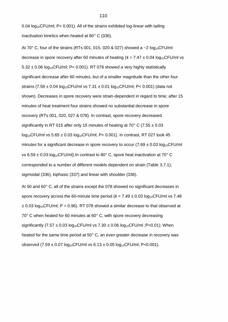
110
0.04 log10CFU/ml; P< 0.001). All of the strains exhibited log-linear with tailing
inactivation kinetics when heated at 80° C (336).
At 70° C, four of the strains (RTs 001, 015, 020 & 027) showed a ~2 log10CFU/ml
decrease in spore recovery after 60 minutes of heating (x̄ = 7.47 ± 0.04 log10CFU/ml vs
5.32 ± 0.06 log10CFU/ml; P< 0.001). RT 078 showed a very highly statistically
significant decrease after 60 minutes, but of a smaller magnitude than the other four
strains (7.59 ± 0.04 log10CFU/ml vs 7.31 ± 0.01 log10CFU/ml; P< 0.001) (data not
shown). Decreases in spore recovery were strain-dependent in regard to time; after 15
minutes of heat treatment four strains showed no substantial decrease in spore
recovery (RTs 001, 020, 027 & 078). In contrast, spore recovery decreased
significantly in RT 015 after only 15 minutes of heating at 70° C (7.55 ± 0.03
log10CFU/ml vs 5.65 ± 0.03 log10CFU/ml; P< 0.001). In contrast, RT 027 took 45
minutes for a significant decrease in spore recovery to occur (7.69 ± 0.03 log10CFU/ml
vs 6.59 ± 0.03 log10CFU/ml).In contrast to 80° C, spore heat inactivation at 70° C
corresponded to a number of different models dependent on strain (Table 3.7.1);
sigmoidal (336), biphasic (337) and linear with shoulder (336).
At 50 and 60° C, all of the strains except the 078 showed no significant decreases in
spore recovery across the 60-minute time period (x̄ = 7.49 ± 0.03 log10CFU/ml vs 7.48
± 0.03 log10CFU/ml; P = 0.96). RT 078 showed a similar decrease to that observed at
70° C when heated for 60 minutes at 60° C, with spore recovery decreasing
significantly (7.57 ± 0.03 log10CFU/ml vs 7.30 ± 0.06 log10CFU/ml ;P<0.01). When
heated for the same time period at 50° C, an even greater decrease in recovery was
observed (7.59 ± 0.07 log10CFU/ml vs 6.13 ± 0.05 log10CFU/ml; P<0.001).

111
Figure 3.7.1. Spore recovery of five C. difficile strains of differing ribotypes (001, 015, 020, 027 & 078) of C. difficile heated for 60 minutes at 70 or 80° C. Spores were enumerated at 0, 15, 30, 45 & 60 minutes. Experiments were carried out in biological duplicate and processed in technical triplicate. Spore recovery was compared between time points using RM-ANOVA with Tukey’s multiple comparisons. Statistically significant results (P< 0.05) are highlighted by *, highly significant (P < 0.01) by ** and very highly significant (P< 0.001) by ***. Curves of best fit were fitted using the GInaFiT Excel add-in. The models fitted included linear with shoulder, sigmoidal and biphasic (Table 3.7.1). The lower limit of detection for this experiment was 1.52
log10CFU/ml.
Me
an
sp
ore
re
co
ve
ry (
log
10C
FU
/ml)
Time (minutes)

112
Table 3.7.1. The model used to fit the data shown in Figure 3.7.1 (spore heat broth experiments in PBS) with the corresponding r2 correlation coefficient value (2 decimal places). Data for the 078 strain at 70° C was not modelled using GInaFiT software due to the small difference in recovery over 60 minutes observed (7.59 ± 0.04 log10CFU/ml vs 7.31 ± 0.01 log10CFU/ml). Model derivations are referenced in brackets.
Temperature/ ° C
70 80
Ribotype Model r2 Model r2
001 Sigmoidal
(336)
0.98 Linear with
tailing (336)
0.99
015 Biphasic (337) 0.97 Linear with
tailing
0.98
020 Sigmoidal 0.98 Linear with
tailing
0.99
027 Linear with
shoulder
0.98 Linear with
tailing
0.96
078 N/A N/A Linear with
tailing
0.94

113
3.7.1.2 Spores produced in liquid media
Significant decreases in spore recovery were observed in all spores tested after 60
minutes of 80° C heat treatment (Fig 3.7.2). Biofilm spores exhibited greater viability at
the 60-minute time point versus planktonic produced spores (x̄ = 5.62 ± 0.07 vs 4.49 ±
0.05log10CFU/ml; P< 0.001). The greatest decrease in spore viability in both biofilm and
planktonic spores was present after 15 minutes (x̄ = 7.47 ± 0.02 vs 5.79 ±
0.07log10CFU/ml & 7.42 ± 0.08 vs 4.96 ± 0.10log10CFU/ml; P< 0.001). A gradual
decline in spore recovery was observed in planktonic spores of RT 020 and RT 027
between 15 and 60 minutes (4.69 ± 0.02 vs 4.47 ± 0.03log10CFU/ml & 4.45 ± 0.04 vs 4.
13 ± 0.02log10CFU/ml). In contrast, biofilm spores of three strains (020, 027 & 078)
showed no significant difference in spore recovery at 15 vs 60 minutes (x̄ = 5.72 ± 0.09
vs 5.68 ± 0.08log10CFU/ml; P = 0.73). The most heat resistant spores of any type were
RT 078 biofilm spores (6.18 ± 0.03 vs x̄ = 4.90 ± 0.08log10CFU/ml; P< 0.001).
Planktonic RT 078 spores were also more heat resistant than planktonic spores of
other strains (4.84 ± 0.06 vs x̄ = 4.38 ± 0.20log10CFU/ml; P<0.001). No difference in
recovery was observed at any time point in spores at temperatures lower than 80° C
(50, 60, 70° C) (data not shown).

114
Mean spore recovery (log10CFU/ml)
Tim
e (
min
ute
s)
Fig
ure
3.7
.2.
Me
an
(± S
E)
sp
ore
rec
ove
ry o
f fo
ur
C.
dif
ficil
e s
tra
ins o
f d
iffe
rin
g r
ibo
typ
es (
00
1,
02
0, 02
7 &
07
8)
he
ate
d f
or
60
min
ute
s a
t 8
0°
C.
Bo
th b
iofi
lm a
nd
pla
nk
ton
ic c
ult
ure
pro
du
ce
d s
po
res a
re p
rese
nt.
Sp
ore
s w
ere
en
um
era
ted
at
0,
15
, 30
& 6
0 m
inu
tes.
Ex
pe
rim
en
ts w
ere
ca
rrie
d o
ut
in b
iolo
gic
al d
up
lic
ate
an
d p
roc
esse
d in
tec
hn
ica
l tr
iplic
ate
. S
po
re r
ec
ove
ry w
as c
om
pa
red
be
twe
en
tim
e p
oin
ts u
sin
g R
M-A
NO
VA
wit
h T
uke
y’s
mu
ltip
le c
om
pa
riso
ns.
Sta
tisti
ca
lly s
ign
ific
an
t (P
< 0
.05
) re
su
lts a
re h
igh
lig
hte
d b
y *
, h
igh
ly s
ign
ific
an
t (P
< 0
.01
) b
y *
* a
nd
ve
ry h
igh
ly
sig
nif
ica
nt
(P<
0.0
01
) b
y *
**.T
he
+ sym
bo
l is
us
ed
fo
r b
iofi
lm d
eri
ve
d s
po
res,
* fo
r p
lan
kto
nic
cu
ltu
re d
eri
ve
d s
po
res.
Th
e lo
we
r lim
it o
f d
ete
cti
on
fo
r th
is e
xp
eri
me
nt
wa
s 1
.52
lo
g10C
FU
/ml

115
3.7.2 Transmission Electron Microscopy
Endospores were observed in both biofilm and planktonic culture produced samples
(Fig 3.7.3). Two morphotypes of spore with differing exosporium sizes were observed
in both sets of spores. In addition, detached exosporium was visible in in both samples.
A B C
D
Figure 3.7.3. Transmission electron microscopy (TEM) images (1000X magnification) of biofilm produced spores (A) and planktonic culture produced C. difficile RT 027 spores (B). Both sets were produced from the 027 strain used previously in this study. Two spore morphotypes are visible in both; thick-exosporium spores are designated by red arrows, thin-exosporium morphotype spores by blue arrows. Detached exosporium was visible in micrographs of both samples (white arrows). Higher magnification (10000X) example images of thick (C) and thin-
exosporium (D) spores are presented.
A B
C
D

116
3.7.3 Heat Treatment Prior to Broth Inoculation
After aerobic heat treatment, spores were aliquoted into BHI containing taurocholate to
induce germination. The sensitivity of vegetative cells to ethanol allowed the
differentiation between non-germinating spores and germinating/vegetative cells. The
difference between total viable counts and spores indicates the number of spores that
have germinated. Spore recovery was inhibited to differing extents after heating for 10
minutes at 70 and 80° C (Fig 3.7.4). In contrast, comparable TVC and spore counts to
the control were observed at the 90-minute time point after spores were heated at 50
and 60 ° C ((TVC; x̄ = 6.63 ± 0.08 log10CFU/ml vs 6.63 ± 0.06 log10CFU/ml vs 6.68 ±
0.06 log10CFU/ml; P>0.05). On average spore counts were ~3 log10CFU/ml lower than
TVC in 50 and 60° C heat treated and control broths. TVC and spore counts were
similar to heat treatment at 50 and 60° C in three strains heated at 70° C (001, 015,
020). In contrast, when RT 027 and RT 078 were heated at 70 ° C, spore counts were
higher compared to the other strains (5.48 ± 0.04 log10CFU/ml & 4.78 ± 0.05
log10CFU/ml vs x̄ = 3.76 ± 0.05 log10CFU/ml).
When spores were heated at 80° C, RT 027 and RT 078 exhibited lower TVC (x̄ =4.05
± 0.05 log10CFU/ml vs 4.80 ± 0.12 log10CFU/ml; P< 0.001) and spore counts (3.56 ±
0.13 log10CFU/ml vs 4.69 ± 0.14 log10CFU/ml; P<0.001) compared with the other
strains. At 80° C TVC and spore counts were comparable (x̄ = 4.50 ± 0.10 vs 4.24 ±
0.14 log10CFU/ml; P = 0.14), indicating spores accounted for the majority of
enumerated entities.
Phase contrast microscopy revealed a very highly significant increase in visualised
phase bright spores in 80° C heat treated spores vs the control (x̄ =82.7 ± 1.1 % vs 0.8
± 0.3 %; P< 0.001) (Fig 3.7.5). Results were comparable between control and 50 and
60 ° C treated spores in all strains; most entities identified were phase dark
(germinated) spores (x̄ = 81.8 ± 1.3 %, 83.2 ± 1.3 %, and 84.9 ± 1.1 %). At 70 ° C
interstrain variation was observed; RT 027 and RT 078 showed similar levels of phase
bright and phase dark spores (x̄ = 50.6 ± 2.1 % vs 39.9 ± 1.8 %), the other strains (001,

117
015 & 020) showed results consistent with the control (x̄ = 0.3 ± 0.2 % vs 85.4 ± 1.1
%).

118
CFU counts (log10CFU/ml)
Fig
ure
3.7
.4 M
ea
n(±
SE
) T
VC
an
d s
po
re c
ou
nts
of
five
C.
dif
fic
ile
str
ain
s o
f d
iffe
rin
g r
ibo
typ
es
(0
01
, 0
15
, 0
20
, 0
27
&
078
) in
cu
ba
ted
fo
r 90
min
ute
s in
BH
I su
pp
lem
en
ted
wit
h 0
.1%
ta
uro
ch
ola
te/ 0
.4%
gly
cin
e.
Sp
ore
s w
ere
he
at
tre
ate
d a
t 5
0, 60
, 7
0 o
r 80
° C
fo
r 1
0 m
inu
tes p
rio
r to
bro
th in
oc
ula
tio
n.
TV
C/s
po
re c
ou
nts
are
als
o in
clu
de
d f
rom
th
e z
ero
tim
e
po
int
an
d f
rom
a c
on
tro
l b
roth
. B
roth
s w
ere
carr
ied
ou
t in
bio
log
ica
l d
up
lic
ate
an
d t
ec
hn
ica
l tr
iplic
ate
. T
VC
co
un
ts
we
re c
om
pa
red
usin
g W
elc
h’s
AN
OV
A w
ith
po
st-
ho
c G
am
es-H
ow
ell m
ult
iple
co
mp
ari
so
ns.
Ve
ry h
igh
ly s
ign
ific
an
t (P
< 0
.001
) d
iffe
ren
ce
s a
re h
igh
lig
hte
d w
ith
***
. M
iss
ing
da
ta (
078
ze
ro c
ou
nts
) is
du
e t
o b
ac
teri
al c
ult
ure
fa
ilu
re.
Th
e
low
er
lim
it o
f d
ete
cti
on
fo
r th
is e
xp
eri
me
nt
wa
s 1
.52
lo
g10C
FU
/ml.
119
Fig
ure
3.7
.5.
Pe
rce
nta
ge
(± S
E)
of
ph
ase d
ark
sp
ore
s (
PD
), p
ha
se b
rig
ht
sp
ore
s (
PB
) a
nd
ve
geta
tive
ce
lls (
VC
) o
f fi
ve
C.
dif
fic
ile
str
ain
s o
f d
iffe
rin
g r
ibo
typ
es (
00
1,
01
5, 0
20
, 0
27
& 0
78
) in
cu
ba
ted
fo
r 90
min
ute
s in
BH
I su
pp
lem
en
ted
wit
h 0
.1%
ta
uro
ch
ola
te/ 0
.4%
gly
cin
e.
Sp
ore
s w
ere
hea
t tr
ea
ted
at
50,
60
, 7
0 o
r 8
0°
C f
or
10 m
inu
tes p
rio
r to
bro
th in
oc
ula
tio
n.
Da
ta
rep
rese
nts
10
dif
fere
nt
fie
lds o
f vie
w a
nd
on
e s
lid
e w
as p
rep
are
d p
er
bro
th.
Bro
ths w
ere
ca
rrie
d o
ut
in b
iolo
gic
al d
up
lic
ate
a
nd
tec
hn
ica
l tr
iplic
ate
. P
D c
ou
nts
we
re a
sse
sse
d u
sin
g W
elc
h’s
AN
OV
A w
ith
po
st-
ho
c G
am
es-H
ow
ell m
ult
iple
c
om
pa
riso
ns.
Ve
ry h
igh
ly s
ign
ific
an
t (P
< 0
.001)
dif
fere
nc
es a
re in
dic
ate
d u
sin
g *
**.
Mis
sin
g d
ata
(01
5 c
on
tro
l) a
re d
ue t
o
ina
de
qu
ate
slid
e q
ua
lity
fo
r e
nti
ty v
isu
alisa
tio
n.
Te
mp
era
ture
/ °
C
Percentage identified entities

120
3.7.4 Reversibility of Spore Heat Treatment
When newly produced (<14 days old) spores were heat treated prior to broth
inoculation, an increased vegetative population was observed at 24 hours vs the non-
heat-treated control (Fig 3.7.6). This difference was very highly significant in three of
the strains (001, 027, 078) (x̄ =7.40 ± 0.03 log10CFU/ml vs 6.45 ± 0.04 log10CFU/ml; P<
0.001). This observation was also significant in RT 015 and RT 020, but to a lesser
extent (x̄ = 7.62 ± 0.03 log10CFU/ml vs 7.45 ± 0.04 log10CFU/ml; P< 0.05).
An increased vegetative population at 24 hours was also present when old (<12 weeks
old) spores were heated prior to broth incubation (Fig 3.7.6). A very highly significant
increase in TVCs was observed in heat-treated broths of all strains when compared to
non-heat-treated spores (7.81 ± 0.06 log10CFU/ml vs 6.76 ± 0.08 log10CFU/ml; P<
0.001).
The trend exists in both new and old spores, but differences between time points in
individual ribotypes existed. Vegetative populations only increased marginally in RT
015 and RT 020 in response to heat in new spores (x̄ = 7.61 ± 0.03 log10CFU/ml vs
7.45 ± 0.05 log10CFU/ml). However, a greater increase occurred in old heat-treated
spores (x̄ =7.60 ± 0.03 log10CFU/ml vs 6.33 ± 0.04 log10CFU/ml). The old non-heat-
treated spores of RT 015 and RT 020 produced a smaller vegetative population in
contrast to new spores. Spores of the other three strains (RTs 001, 027 & 078)
exhibited similar behaviour independent of age.

121
0
1
2
3
4
5
6
7
8
9
001
015
020
027
078
0
1
2
3
4
5
6
7
8
9
TVC Spores TVC Spores
NHT HT
CF
U C
ou
nts
(lo
g10C
FU
/ml)
*** * ** *** ***
*** *** *** *** ***
<14-day-old spores
>12-week-old spores
Figure 3.7.6. Mean (± SE) TVC and spore counts of five C. difficile strains of differing ribotypes (001, 015, 020, 027 & 078) 24 hours post-broth inoculation. Both new spores (< 14 days old) and old spores (> 12 weeks old) were utilised. Heat treated spores (HT) were heated for 10 minutes at 80° C, non-heat treated (NHT) received no treatment. A trend towards increased TVCs in heat treated samples was present. Broths were carried out in biological duplicate and technical triplicate. TVC means were compared using two-tailed paired T tests. Highly significant (P < 0.01) findings are highlighted by ** and very highly significant (P< 0.001) by ***. The lower limit of detection for this experiment was 1.52 log10CFU/ml.
122
3.8 Discussion
3.8.1 Heat Treatment in PBS
C. difficile spore heat inactivation is not adequately modelled by log-linear
kinetics
When spores were heated in PBS for 60 minutes, substantial variation was observed
(Fig 3.7.1). The differences observed were mainly strain-dependent; the observed
trends were consistent between different temperatures. Fifty and 60° C heat treatment
had no significant effect on four of the utilised strains (001, 015, 020 & 027). However,
at higher temperatures heat began to have a substantial effect on spore recovery.
Three of the strains (RTs 015, 020, and 027) took 0, 15 and 30 minutes respectively for
spore recovery to decrease at 70° C. If the time delay was due to a ‘lag phase’ such as
the Eppendorfs getting up to temperature, the same delay would be expected in all
strains. The heat took an additional 30 minutes to influence RT 027 compared to RT
015 strain at 70° C.
Traditionally, thermal inactivation of microorganisms has been illustrated using log-
linear kinetic models, based on the assumption of a homogeneous population, sharing
the same intrinsic heat resistance (338). Implicit in this model is the recognition of a
single-system that is responsible for heat resistance. This approach is particularly
prevalent in food microbiology and food safety, where it is possible to easily calculate
D-values and Z-values. The D-value is defined as the time taken for a 90% (or 1log)
reduction in numbers from the starting population, the Z-value is the temperature
increase required to decrease the D-value by a magnitude of ten. In fact, this method is
still being used to evaluate thermal resistance in C. difficile spores (253, 316).
Deviations from this model have been recognised even since the 1970s, although this
recognition has not translated into studies concerning C. difficile. In this study, 70° C
and 80° C heat curves were not adequately modelled using log-linear kinetics. In order

123
to accurately assess and model the thermal inactivation of C. difficile spores, a number
of different survival curves were used to describe the data (335).
GInaFit is a software that incorporates 9 different survival curves; linear, linear with
tailing, sigmoidal, linear with shoulder, biphasic, concave, biphasic with shoulder and
convex curves (335).The mathematic models derived by Geeraerd et al. are beyond
the scope of the current study, but are based on empirical datasets. In this study, the
largest interstrain difference in recovery occurred at 70° C. The survival curve at 70° C
for two of the strains (001 & 020) was best illustrated by the fitting of a sigmoidal curve,
for RT 027 a linear with shoulder curve, and for RT 078 a linear model (Fig 3.7.1 &
Table 3.7.1). Previously, differing heat inactivation kinetics and thermal resistance
values have been discovered when comparing 39 different strains of B. cereus (339).
Differing heat resistances have also been documented when comparing two strains of
C. botulinum (340).
It has been hypothesised that during the ‘shoulder’ phase in these curves, a protective
surrounding ensures the survival of the spores by buffering the deleterious effects of
heat (335). Once this protective matrix is destroyed/inactivated by sufficient heating,
log-linear heat inactivation of spores resumes. The spores used in this experiment
were not purified; it is plausible that proteinaceous cellular debris (in the form of dead
vegetative cells) acted as a protective matrix. However, it is still unclear why some of
the strains have a prolonged period of buffering, for example the 027 strain. Other
reasons for the presence of a shoulder in thermal inactivation curves have been
proposed; clumping, the inability of bacteria to continually synthesise protective
proteins over time and the cumulative damaging effect of heating over a sustained
period (336, 341). C. difficile spores are metabolically dormant; the synthesis of heat
shock proteins is irrelevant. Cetyl trimethylammonium bromide (CTAB) and sonication
have previously been used in this study to try and prevent spore clumping, with minimal
success (data not shown). The diverse aggregative properties of spores of different C.
difficile strains could be responsible for the differing resistances earlier in heating.

124
In addition, ‘tailing’ was observed in some strains at 70° C (Fig 3.7.1). Tailing has been
discussed since the 1970s (337), and its presence is not surprising in C. difficile spores
given it has been demonstrated to occur in spores of other species (342, 343).
Intrastrain variability is a possible mechanism by which tailing occurs; a subpopulation
of spores are maintained due to their higher intrinsic heat resistance. Spore clumping
was found to increase in Bacillus licheniformis spores after 20 minutes of heat
treatment, suggesting spore clumping could be involved in the tailing discussed. In
addition, spore surfaces became more hydrophobic after heat treatment (343). It
should be noted that the subpopulation of spores persisting in the ‘tail’ of the curve
account for less than 0.1 % of the starting inoculum.
At 80° C the thermal death curve for all strains was more typical and spore recovery
began to decrease as soon as heat treatment began. The greater thermal resistances
of RT 027 and RT 078 at 80° C found in our study are concordant with the findings of
recent work (121, 334). After initial screening, Rodriguez-Palacios et al (121) used a
multinomial logistic regression model to suggest the increased thermal resistance of a
RT 078 compared to two other strains when spores were heated in meat. Although
interesting, a larger sample size of ribotypes will be required to test this association.
Interstrain variability in C. difficile is something that has been observed previously in
the literature as early as 1985 (313). However, the reason for these differences is
largely unknown. Dipicolinic acid (DPA) is responsible for maintaining a dehydrated
core and is important in wet-heat resistance in B. subtilis (344). SpoVA is an ion
transporter responsible for the transport of DPA into the core in Clostridium
perfringens; spoVA mutants have a loss of wet-heat resistance as well as impaired
germination (345). Foodborne B. subtilis strains harbouring the spoVA2mob operon were
found to have increased heat activation requirements compared to non-foodborne
strains (346); high heat resistance is attributed to this operon (347). More recently, the
importance of DPA has been highlighted in C. difficile spoVAC and dpaAB mutants.
DpaAB is the gene from which an enzyme responsible for DPA synthesis is

125
synthesised. Both sets of mutants showed statistically significant decreases in wet-heat
resistance at temperatures above 50° C versus the wild-type (348). It is likely that
natural heterogeneity exists in different C. difficile strains of the levels of proteins
responsible for DPA synthesis and transport. On the contrary, Rose et al showed that
in B. subtilis wet-heat resistance is probably dependent on factors other than just spore
DPA content (349). This is likely true for C. difficile.
The cortex has also been suggested to play a role in heat resistance. The cortex
serves in an osmoregulatory capacity, expanding to accommodate superfluous water,
reducing spore water content (350). Rao et al discovered that decoated B. cereus, B
megaterium and B. subtilis spores had comparable heat resistances to wild-type
spores, suggesting reduced heat resistance was not due to the loss of the cortex, but
rather the loss of vital germination proteins (351). These studies have not been
replicated in C. difficile, but one study found that conditions of high osmolarity permitted
cortex degradation, but not DPA release in germinating C. difficile spores (352).
RT 078 exhibits different behaviour to all the other strains at 50° C. A number of
potential mechanisms have been proposed to explain differences in spore heat
resistance, including core DPA content, enzymes responsible for DPA transport, and
enzymatic degradation at high temperatures (344, 348). None of these factors seem to
address this RT 078-specific phenomenon. Previously, RT 078 has been found to be
phylogenetically dissimilar and highly divergent from other C. difficile strains based on
lineage (30). Based on the data it is plausible that in addition to the previously
discussed mechanisms, RT 078 possesses another mechanism of heat resistance that
50° C heat treatment inhibits.
The differences between our work and that of others in evaluating thermal resistance
can be attributed to a number of causes. Notably, the method of C. difficile spore
production differs considerably (253, 276, 313, 316, 348), with a variety of solid and
liquid media being utilised to produce the C. difficile spores. Spores produced in liquid
media in both Alicyclobacillus acidoterrestris (353) and Bacillus subtilis (349) have

126
been found to have a lower thermal resistance than their counterparts produced on
solid agar.
Biofilm produced C. difficile spores are more heat-resistant
After 60 minutes, the viability of biofilm produced spores was ~1 log10CFU/ml higher
than spores produced in planktonic culture (Fig 3.7.2). In addition, all of the spores
produced in liquid media were more heat resistant than those produced on solid agar,
which does not conform to the findings of the aforementioned studies (349, 353).
Transmission electron microscopy showed the presence of thin and thick-exosporium
spores in both samples (Fig 3.7.3). These observations were made previously in the
R20291 strain (80). Despite purification of spores by HistoDenz™, detached
exosporium was present in both samples. It is possible that an increased presence of
exosporium/extracellular matrix in the biofilm produced spores could result in a more
heat-resistant population. One study also found that C. difficile spores produced in
biofilms spores began to accumulate a surrounding ‘shroud’ that attached to the spore
after 7-14 days of incubation (354). This ‘layer’ was found to consist of dead cellular
debris, and it is hypothesised C. difficile spores accumulate this layer after mother cell
lysis (354). In addition, spores were found to be less responsive to germinants and
exhibited decreased germination. If the increased heat resistance of biofilm produced
spores is due to an extracellular matrix/ shroud or an intrinsic spore property, biofilm
spores in non-laboratory conditions are likely to retain this resistance. Biofilm produced
spores are still likely to exhibit increased heat resistance in non-laboratory scenarios.
On the other hand, in the first 15 minutes of 80° C heat treatment biofilm spores
exhibited log-linear inactivation kinetics. Previously, it has been suggested the
‘shoulder’ seen in some heat inactivation models is due to an extracellular matrix
buffering the effects of heat (341).
Unfortunately, the processing of other strains used in this study was not practicable
due to insufficient resources. In addition, no quantitative measure of exosporium size or

127
spore morphotype number was possible. Further work providing quantitative
measurements and exploring a range of ribotypes is needed to strengthen conclusions.
Despite these limitations, this study supports the existence of two distinct C. difficile
spore morphotypes. Building on previous work, it is suggested biofilm produced C.
difficile spores are more environmentally robust and could exhibit increased levels of
superdormancy in addition to increased heat resistance (354).
3.8.2 Heat Treatment Prior to Broth Inoculation
80° C heat treatment inhibited initial outgrowth but promoted later
outgrowth
Both freshly produced (<7 days old) and environmentally aged (>3 months old) were
heat treated prior to broth inoculation (Fig 3.7.4). Initially this experiment was
performed to assess the immediate effect of heat on spore recovery and outgrowth (90-
minute incubation times), but the longer term effects of heat on spore recovery were
also documented (24 and 48 hour incubation times). In both new and old spores, the
same trends were observed. When spores were heat treated and left for 90 minutes,
spore outgrowth was inhibited compared to heat treatment at lower temperatures and
the control. Both TVCs and spore counts decreased, indicating a global decrease in
spore recovery. On the other hand, when spores were left for a longer time period, at
24 hours the heat-treated samples contained higher levels of vegetative cells and
comparable levels of spores to non-heat treated samples (Fig 3.7.6).
Initially these results appear inconsistent, if heat is inhibiting spore germination at an
early stage, it is unclear why a more rapidly growing vegetative population is present at
a later point. One study also observed that heat treatment at 85° C decreased
recovery, but this was due to an impairment of vegetative growth and not germination
(253). This is in accordance in the current study, where spores were able to revert to
vegetative growth. It could be hypothesised the heat treatment causes a greater
number of spores to germinate, but there is an initial lag in outgrowth due to the heat
treatment. Contrarily, the phase contrast data illustrates a shift from the predominance

128
of phase dark spores at 70° C to phase bright spores at 80° C, indicating an inhibition
of the transition from phase bright to phase dark i.e. germination (Fig 3.7.5).
Heat activation has commonly been used as a strategy to increase spore germination
in B. subtilis (301) and more recently C. difficile in older spores. The previously cited
study also found that aged spores (>20 weeks old) exhibited increased recovery (30 %
increase) in response to heat treatment at 63° C vs freshly produced spores (253). In
the current study, both newly produced and environmentally aged spores demonstrated
increased outgrowth in response to heat treatment, albeit in producing a proliferative
vegetative population. Heat is suggested to mediate its stimulatory effects by inducing
germinant receptor conformational changes. An absence of inner membrane bound
germinant receptors in C. difficile spores is proposed to explain the lack of effect of
heat (62, 355). This hypothesis is consistent with work demonstrating heat has no
activation effect on germinant-receptor independent germination in B. subtilis (356,
357). Although the results of the current study suggest heat has an effect on the
outgrowth and subsequent proliferation of new and old C. difficile spore populations,
this effect is not necessarily mediated through spore germination directly. The results
are therefore not contradictory to those cited above.
3.9 Conclusion
To summarise, 80° C heat treatment appears to have an initial inhibitory effect on
spore recovery and outgrowth, which then reverts, with a more rapidly proliferating
vegetative population being produced. This effect is seen independently of spore age,
at least up to 3 months. The log-linear inactivation kinetics typically described in
microbial heat decay experiments was not observed in this study. A range of kinetic
inactivation models were fitted to the data dependent on strain and temperature (70/80
° C), consistent with other work (339, 340).
Spore heat resistance differed between spores produced on solid agar (CCEYL) and
those produced in liquid media (supplemented BHI). Contrary to the findings of

129
previous studies (349, 353), spores produced in liquid media were more heat resistant.
Biofilm spores appear to have a greater capacity for withstanding heat than spores
produced in planktonic culture, but this effect needs further exploration. The findings of
this work illustrate that heat ‘activates’ spores in a way not previously observed.
Although spores are initially inhibited by the injurious effects of heat treatment,
vegetative population proliferation is promoted at a later time point, independent of
spore age.
At recommended cooking temperatures, spores can persist in food and even
reactivate, providing a potential C. difficile reservoir for community acquired CDI.This is
particularly important in the case of biofilm-produced spores, which exhibit increased
thermal resistance. Although the majority of spores were inactivated temporarily by 80°
C heat treatment, a subpopulation persisted. Spores present in food prior to cooking
may survive the cooking process and potentially cause initial or recurrent CDI.
Combined with the findings of chapter 3A, superdormant spores may be able to survive
minimum recommended cooking temperatures and exhibit increased germination
efficiency when ingested into the bile-rich gastrointestinal tract. Conversely, it seems
unlikely that spores could be present in food after cooking unless they persisted the
cooking process.

130
Chapter 4 A – Proteomics in an in vitro Clostridium difficile gut
model
4.1 Background & Rationale
Research fields such as proteomics, metabolomics and transcriptomics are becoming
increasingly important in the investigation of the human microbiome. Comprehensive
sequencing studies investigating the composition of the human microbiome have been
very successful, mapping a total of 3.3 million bacterial genes (358). Metagenomic
studies have identified the potential major biosynthetic functions of the gut, with genes
responsible for methane production, carbohydrate metabolism and vitamin biosynthesis
recognised (359). These studies are useful in identifying constituent differences
between microbiomes, but do not quantify the expression of genes within the
ecosystem and overall functionality. The human microbiota provides important
functions in the host including nutrient processing (360), priming of the immune system
(361), and defence against pathogens (362). Consequently, potential relationships
between microbial metabolism and human disease are increasingly being recognised.
Metabolomics and proteomics are increasingly being investigated and represent an
important step in characterising the human gastrointestinal tract in health and disease.
One well documented use of metabolomics is the analysis of short chain fatty acids
(SCFAs) such as propionate and butyrate. SCFAs are produced by the fermentation of
fibre in the human gastrointestinal tract by several bacterial species (363). Defects in
this process have been associated with a number of diseases, for instance IBD (363),
and SCFAs are suggested to influence diabetic control in type 2 diabetes (364).
Currently, literature is available linking varying diseases with the metabolic products of
the microbiota. However, reported studies are largely inferential, as causality is difficult
to establish without time consuming and potentially unethical longitudinal studies.
131
Proteomics is used to quantify total gene translation products (365). By integrating this
data with metagenomics and metatranscriptomics data, additional information about
the functions carried out by the microbiota is obtained. Metaproteomics has
advantages over metagenomics alone; the gene products obtained can be from dead,
dormant or living cells and measurement of gene expression is lacking. The
‘metaproteome’ refers to the complex set of proteins expressed and produced by a
microbial ecosystem. Historically, proteomics has involved the extraction and
separation of proteins using gel electrophoresis. This method is limited in its ability to
discriminate and separate proteins in highly heterogenous mixtures such as those
produced by the human gut microbiota. In 2009 high throughput sequencing (liquid
chromatography followed by tandem mass spectrometry) was used for the first time on
two faecal samples from monozygotic twins in investigating the metaproteome (366),
which was found to consist of proteins predominantly obtained from Bacteroidetes and
Firmicutes organisms. Analogous approaches are now routinely used in defining
complex ecosystems. As expected, the spectra obtained mapped predominantly to
Bacteroides and Firmicutes organism proteins.
Proteomics-based approaches have been used for a number of purposes in
investigating C. difficile. One study used SDS-PAGE and mass spectrometry to
separate and identify 42 C. difficile cell wall proteins that were immunoreactive, giving
a deeper understanding of the pathogenesis and subsequent immune response to CDI
(367). The changes exhibited in the proteome of Caco-2 cells in response to toxin A
have also been investigated. Using LC-MS/MS and SILAC, incubation of cells with
toxin A for 24 hours caused a significant difference in the expression of cytoskeletal
proteins, underlining the rearrangement of cell microarchitecture and the loss of tight
cell-cell junctions (368). LC-MS/MS has also proved useful in quantifying the changes
in protein expression in strain CD630 heated at 41° C vs 37° C (369). However, it
should be noted that the above work used single strains, which means there is a
reasonable possibility that inter-strain differences have been missed.

132
The hypervirulence of some strains has also been investigated in the context of
proteomics, protein expression quantified in response to osmotic shock and nutrient
shift revealed increased response coordination of gene networks in two hypervirulent
strains vs two historically prevalent, non-hypervirulent strains (370). This study
elucidates a potential mechanism for the hypervirulence of some strains, in comparison
to the descriptive information gained in metagenomics studies. Furthermore, this study
highlighted the heterogeneity in protein expression in isolates grown in different media,
an observation that has been validated in other work (371), thus providing a possible
explanation for differences seen in heat resistance, germination and outgrowth of
spores grown on different media (327, 349, 353, 371). The unique nature of the C.
difficile spore coat has likewise been highlighted by the finding of 29 unique spore
proteins (333). Proteomics is beginning to be used successfully as a tool for probing an
explaining some of the behavioural differences observed between C. difficile strains.
4.1.1 Proteomics approaches
Two approaches are employed in proteomics; top-down proteomics or bottom-up
proteomics. In top-down proteomics, complete proteins are analysed in contrast to
bottom-up proteomics whereby proteolytic digestion is employed and the resulting
peptide fragments are analysed. Both approaches have advantages and weaknesses
which will be discussed.
Historically in top-down proteomics 2D electrophoresis has been utilised. After
electrophoresis, bands are excised, proteolysed and subsequently analysed by mass
spectrometry. This approach has had limited success, being able to interrogate
hundreds of mouse brain proteins (in this case by MALDI-MS), representing a low
percentage of the total proteome (372). Unfortunately this method favours the detection
of highly abundant soluble proteins, and is not adequate to detect membrane bound
proteins (373). It should also be noted some of the literature has taken issue with this
methodology as being described as ‘top-down’ due to the proteolysis step prior to mass
spectrometry (374). Rather than using gel electrophoresis, proteins can be ionised

133
directly in the mass spectrometer, advantageous due to the possible detection of post-
translational modifications, which is not possible in bottom-up proteomics. However,
the use of top-down approaches has commonly been limited to simple mixtures of
proteins, and for peptides larger than 50 kDa, fragmentation sequencing becomes
difficult (375). Due to the large size of protein ions, the ionisation, fragmentation and
separation stages can be difficult to achieve in top-down proteomics (376)
Bottom-up proteomics is the most widely used approach in MS workflows in
contemporary proteomics research. In general, proteins are cleaved into peptide
fragments by enzymatic digestion prior to MS analysis, usually by trypsin. Gel
electrophoresis can be used prior to MS analysis, or the total protein mixture can be
proteolysed known as ‘shotgun proteomics’ (376). As stated previously, one of the
difficulties in bottom-up proteomics is solubilising all proteins regardless of their
hydrophobicity. Traditionally, SDS-PAGE has been used to separate proteins followed
by proteolysis and mass spectrometry. In-solution digestion has also been utilised, but
detergents are required and can interfere with later proteolytic cleavage and must be
removed prior to analysis (377). A quick, inexpensive and efficient method of sample
preparation that is free from the solubilisation issues of gel electrophoresis is required.
A number of gel-free protein preparation methods have been described in the literature,
including filter-aided sample preparation (FASP), in-StageTip method (iST), single-pot
solid-phase-enhanced sample preparation (ST3) and the suspension trapping method
(STrap). One study found all of these methods to be comparable in terms of
performance when starting with 20 μg of protein extract, but the precision of FASP fell
drastically when the amount of starting material was decreased to 10 μg (378).
FASP is a method described in 2009 by Wisniewski et al (379). In summary, samples
are solubilised in SDS, concentrated, retained and subsequently processed in the
molecular weight cut off filter or ‘reactor’. This method allows the removal of detergents
and is inexpensive. Several studies have modified the FASP protocol, increasing

134
peptide retention and decreasing throughput times (380, 381). Notably, in 2014
Zougman et al developed the ‘STrap method’, capable of quicker processing times and
avoids the temperamental nature of the membrane filters used in the FASP method
(377). In this method, the acidified-protein mixture is added to a methanolic solution in
an S-tip. A fine particulate protein suspension is created, which is amenable to
trypsinisation. The suspension is trapped in the quartz filtration material of the tip. The
contaminating solution is removed; the protein is trypsinised and subsequently eluted
and concentrated in the C18 filter ready for MS. The iST method also uses an S-tip as
its basis, but does not require solvents or SDS; strong cation exchange resins are used
for peptide separation (382) .
In addition to top-down and bottom-up proteomics, some research has tried to use a
hybrid approach of ‘middle-down’ proteomics. In these workflows proteases such as
Asp-N and Glu-C are utilised to produce medium sized peptide fragments. The
advantage of this approach is the increased proteome coverage in medium sized
proteins (3.0 kDa < MW < 10 kDa)(383). An overview of the three approaches can be
viewed below (Fig 4.1.1).

135
F
igu
re 4
.1.1
. A
n o
ve
rvie
w o
f th
e d
iffe
ren
t p
rote
om
ic a
pp
roa
ch
es.
Bo
tto
m-u
p a
nd
mid
dle
-do
wn
ap
pro
ac
he
s u
tili
se
pro
teo
lys
is p
rio
r to
fr
ag
me
nt
se
pa
rati
on
an
d M
S a
na
lysis
. T
op
-do
wn
ap
pro
ac
he
s d
o n
ot
use
pro
teo
lys
is p
rio
r to
MS
an
aly
sis
. A
t th
e s
ep
ara
tio
n a
nd
MS
sta
ge
s d
iffe
ren
t in
str
um
en
ts c
an
be
use
d, b
ut
the
pri
nc
iple
s r
em
ain
th
e s
am
e.

136
Mass spectrometry-based methods are now used routinely in the identification of
complex protein mixtures. There are a wide variety of methodologies utilised,
depending on the sample being investigated. Any mass spectrometer consists of three
vital components, an ion source (ionises the analytes), a mass analyser (measures the
mass: charge ratio of incoming ions) and a detector (identifies the number of ions of a
particular m/z ratio)(384).
There are two main approaches to ionisation; electrospray ionisation (ESI)(385) and
matrix assisted laser desorption/ionisation (MALDI)(386). ESI is routinely coupled to
liquid chromatography separation techniques such as HPLC and is only possible in the
case of a liquid matrix. ESI is the preferred method for the analysis of complex
mixtures; it is most often coupled with other mass spectrometry tools in the case of
large-scale bacterial proteomics studies. On the other hand, MALDI uses laser pulses
to produce ions from a crystalline matrix and is preferred for simple protein mixtures.
MALDI is routinely coupled to time-of-flight (TOF) analysers for the identification of
microorganisms in microbiology (387). The ionisation source must be coupled to an
analyser of which several types. In its simplest form, an ion trap is anything that uses
magnetic or electric fields to trap charged particles. Historically, three different ion traps
have been described into which all instruments fall; Paul trap, Penning trap and the
Kingdon trap.
A number of different configurations exist including linear ion traps, quadrupole and
Fourier transform ion cyclotrons (FT-MS)(384). In most contemporary instruments
these analysers can be combined to overcome individual limitations. The orbitrap is an
example of a FT-MS instrument and is a Kingdon trap. Orbitraps are a relatively newly
developed tool and have the advantage of being able to discern ions of close m/z ratios
(388). It consists of an outer and inner electrode; ions are injected into the trap from a
C-trap. The ions move around the inner electrode both axially and horizontally; part of

137
the outer electrode can detect the movement of the ions and use the Fourier
transformation to convert this to a mass spectrum.
4.1.2 Peptide/Protein Identification
Undoubtedly the most important step in metaproteomics is database selection. In
general, there are three options; public reference, matched or ‘pseudo’-metagenomic
databases (389). A matched database utilises synchronised metagenomics, identifying
the species present within a sample and thereby eliminating superfluous sequences
from the metaproteomic analysis. In contrast, reference databases may contain
sequences not present in the sample. Unfortunately, there is no way to discern which
species are present within a sample. As metagenomics is expensive 16s-rRNA
sequencing can be utilised alongside metaproteomics to produce a ‘pseudo’-
metagenome that contains only reference database sequences of relevant species
(365, 366, 390).
A variety of public reference databases exist. Some of the most commonly used
databases include UniProt, ENSEMBL (392), NCBI RefSeq (393) and UniRef(394)
(395).UniProt has two sections; UniProtKB/SwissProt and UniProtKB/trEMBL (391).
UniProtKB/SwissProt is manually curated and of a high quality, sequences are non-
redundant. In contrast, UniProtKB/trEMBL is automatically annotated, containing
possible redundant sequences and potentially hypothetical proteins. The UniRef
database is different from the above in that it clusters all fragments with more than 11
residues into one entry. In practice, this means that peptides that match to more than
one organism will be combined in the same entry (396). UniRef is a useful database for
family classification and inference of functional groups. However, clustering related
sequences is not necessarily desirable when conducting large scale gut microbiota
studies.
In addition to the selection of a database, a search algorithm is needed to implement
the matching of spectrum with theoretical peptides. For tandem MS data, database

138
sequences are in silico digested during analysis; the resulting theoretical spectra are
compared to the experimentally derived spectra. A number of different search
algorithms are available for matching spectra to peptides, the most common being
Mascot and SEQUEST (397). Another prominent example is X! Tandem (398). More
recently, the Andromeda search engine has gained popularity, probably due to being
open source and user friendly for non-specialists (399). Andromeda is a search engine
implemented in the MaxQuant software suite. Like Mascot, Andromeda uses a
probability-based approach to match theoretical sequences to mass spectra (400).
After peptide identification, the next step is protein inference. Protein inference
presents another challenge due to the existence of ‘degenerate’ peptides. Degenerate
peptides are shared between proteins in differing species (401, 402). Many of these
identifications can be erroneous, particularly if a non-specific public database is being
used. Many proteomics packages will report proteins in groups according to shared
peptides, with journals requiring that only the first protein identification (with the most
peptide matches) be reported in publications in line with the rule of parsimony (403).
4.1.3 C. difficile in vitro gut model
The in vitro gut model presents a unique opportunity to simulate CDI and subsequent
recurrence. It has been utilised extensively (91, 92, 153, 404). In general terms, the in
vitro gut model has been used to evaluate the efficacy of antibiotic agents in the
initiation or treatment of CDI, assessing the effects on both C. difficile and the normal
flora. Traditionally, a series of selective solid agars have been utilised to monitor
bacterial populations over time. Microbial culture offers an easy and relatively
inexpensive method of elucidation of a wide range of bacterial populations over a
prolonged period. However, in recent years molecular techniques have superseded
traditional culture-based methods in extending our knowledge of the diverse microbial
community along the gastrointestinal tract. Although sequencing based approaches are
powerful, metagenomics has been found to overlook minor populations thus
systematically decreasing the observed population diversity (405). In order to maximise

139
knowledge about phylogenetic and functional aspects of a microbial community, both
culture and sequence-based technologies should be combined to mitigate the
weaknesses of one another (406). This approach has been highlighted previously,
where ~90 ‘unculturable’ species were isolated with a culture based approach (407).
A novel approach to the in vitro gut model would be the combination of traditional
culture-based approaches with metaproteomics. Combining the two might give useful
insight into microbiological factors that predispose to rCDI. Metaproteomics can be
seen as a bridge between culture based and metagenomic approaches to the gut
microbiota. In the context of rCDI and the in vitro gut model, culture based and
metagenomics approaches can be combined for a greater appreciation of microbiota
dynamic throughout disease. However, metaproteomics has the added ability of
potentially identifying the overarching metabolic niche created during infection by
antibiotics. Metabolic processes performed by particular groups of bacteria could
create an environment suitable for C. difficile proliferation. Although host factors are
absent in the in vitro gut model, studies have shown that some species of bacteria may
modulate host immune function. Of particular interest Bifidobacterium longum DJ010A
and Bacteroides fragilis YCH46 were found to produce peptides (FR-17 & LR-17) that
modulate IL-22 induction, a cytokine important in promoting the integrity of the gut
epithelium and protecting against pathogens such as C. difficile (408). A database
containing over 300 milllion peptide entries (Mechanism of Action of the Human
Microbiome (MAHMI)) now exists to try and identify the immunoregulatory functions of
proteins secreted by the gut microbiota (409).
Adopting a proteomics approach within the in vitro gut model presents a number of
technical challenges. The proteinaceous nature of the media feeding the model means
samples cannot be taken directly for LC-MS/MS analysis; media peptides are
contaminants. In addition, the bacterially produced proteins could be at such low
concentrations that instruments used for LC-MS/MS are not sufficiently sensitive for

140
peptide identification. As well as the experimental difficulties encountered, microbial
metaproteomics is still a field in its infancy. A variety of approaches exist for accurately
aligning peptide sequences for protein identification. A diverse range of databases are
utilised and this has been found to have a substantial influence on the results of
metaproteomic studies (390).
Chapter 4A investigated a methodology for isolating secreted bacterial proteins from in
vitro gut model populations. The baseline protein levels in Vessels 1, 2 and 3 were
determined by acetone precipitation and Bradford assay. Vessels 1, 2 and 3 of the gut
model simulated the changing conditions encountered down the gastrointestinal tract.
Subsequently a minimal media method was devised to isolate and concentrate
bacterial proteins. Bacterial viability using this method was validated. Chapter 4B
utilised this novel methodology to isolate bacterial proteins from different stages of
simulated C. difficile infection in three recurrence gut models. Bacterial proteins were
analysed by mass spectrometry using a bottom-up proteomics approach. The
taxonomic and functional alterations in the metaproteome at each stage of infection
were presented.
4.2 In Vitro Gut Models
4.2.1 Methods
The in vitro gut model used in these experiments is a triple-stage chemostat model (Fig
4.2.1). The maintenance and setting up of the in vitro gut model has been extensively
described previously (235). It consists of three glass Vessels (Soham Scientific, Ely,
UK) connected in a Weir cascade based on that of MacFarlane et al (410) . The
volumes of Vessels 1, 2 and 3 are 280 ml, 300 ml and 300 ml respectively. A circulated
heated water bath (Grant Instruments, Cambridgeshire, UK) connected to a jacketed
system allowed Vessels to be kept at 37° C. The system was kept anaerobic by a
continuous source of oxygen free nitrogen in all Vessels. The Vessels were kept at pH
levels reflective of the increasing alkalinity of the gastrointestinal tract; 5.5 (± 0.2), 6.2

141
(± 0.2), 6.8 (± 0.2) and pH was monitored by probes (P200 chemotrode, Hamilton,
USA) and maintained by the addition of sodium hydroxide/ hydrochloric acid,
administered by a controller unit (Biosolo, Brighton Systems, UK). A peristaltic pump
was used to top feed a complex growth medium through the Weir cascade; a flow rate
of 13.2 ml/h was achieved.
Populations of various microorganisms were monitored daily by the use of a range of
selective media (Table 4.2.1).
4.2.1.1 Gut Model Preparation
Pooled faecal emulsion (150 ml) was added to each of the gut model Vessels. Growth
media was added to fill Vessel 1 to ~280 ml, after which pumping of media through the
Weir cascade was initiated. Faeces were obtained from three healthy donors.
Antimicrobial therapy in the previous 2 months was an exclusion criterion for donation.
Samples were transported in an anaerobic zip lock bag (Benton Dickinson, Sparks,
MD, USA) within 12 hours for storage in an anaerobic cabinet. Donor faeces were
screened for C. difficile prior to use by anaerobic incubation for 48 hours in duplicate on
CCEYL. Samples positive for C. difficile were not utilised in models. Emulsions were
prepared by the suspension of 10 % w/v faeces in pre-reduced PBS. A smooth slurry
was created by emulsification of the suspension in a stomacher (Stomacher Lab-
Blender 400, Borolabs, Aldermaston, UK). The slurry was passed through a muslin
cloth (Bigger Trading Limited, Watford, UK). Pooled faecal emulsions were flash frozen
in liquid nitrogen and stored at -80° C as previously described (175). The use of the
same donor faeces increased homogeneity between the three models used in the
study.
4.2.1.2 Gut Model Media Preparation
Gut model media was prepared in 2 L Büchner flasks. The complex media (for
ingredients see Appendix B, B.2.2) was sterilised by autoclaving (Priorclave, London,
UK) at 123° C for 15 minutes. After sterilisation, 5 mg/L resazurin (Sigma Aldrich) and

142
0.4 g/L glucose were added to the media through a 0.22 µm filter syringe (Merck
Millipore, Feltham, UK).
4.2.1.3 Bacterial Population Enumeration
A range of organisms were enumerated on selective and non-selective agars (Table
4.2.1). A full list of ingredients and agars can be found in Appendix B, B.1. From each
vessel of the gut model 1 ml of fluid was removed from the outlet port. Gut model fluid
was serially diluted in pre-reduced peptone water (Oxoid, Basingstoke, UK) to 10-7.
Four appropriate dilutions were spread on to quarter plates in technical triplicate. Both
facultative and strict anaerobic populations were enumerated. Strict anaerobes were
incubated in an anaerobic chamber; facultative anaerobes (aerobes) were incubated
aerobically at 37° C. for 48 hours. C. difficile spore counts were obtained by diluting gut
model fluid in a 1:1 ratio in 100 % ethanol. After 1-hour spores were serially diluted and
incubated on agar as described above.
After 48 hours of incubation, bacterial colonies were identified and counts were
transformed to log10CFU/ml by multiplying by 50 and 10X, where X is the logarithmic
dilution factor. In the case of uncertainty regarding bacterial colony identification,
MALDI-TOF was utilised as a second means of identification.
4.2.1.4 Cytotoxicity assay
Vero cells were prepared as previously described by Crowther (235). Twenty-mililitres
of Dulbecoo’s Modified Eagles Medium (DMEM) (Sigma) supplemented with newborn
calf serum (50 ml) (Gibco, Paisley, UK), antibiotic/antimycotic solution (5 ml)(Sigma)
and L-glutamine (5 ml)(Sigma) was used to culture vero cells (African Green Monkey
Kidney Cells, ECACC 84113001) in a flat bottom tissue culture flask. Flasks were
incubated at 37° C in 5 % CO2.

143
When Vero cells formed confluent monolayers (confirmed by microscopy; Olympus UK
Ltd, Middlesex, UK) the monolayer was harvested by removal of DMEM and rinsing
with 1 ml of Hanks Balanced Salt Solution (HBSS) (Sigma) containing trypsin-EDTA
(0.25 g/ L) (Sigma). Subsequently, 6 ml of HBSS-EDTA was added to the flask and
incubated for 10 minutes at 37° C at 5 % CO2. After the cells no longer adhered to the
flask, further passage was achieved by diluting the HBSS-EDTA cell mixture (1:20) in
DMEM in a 96F microtiter tray (Nunc). Vero cells were harvested (160 μl) and
inoculated into wells to which antitoxin would later be added. To other wells trypsinised
Vero cells (180 μl) were added. Trays were incubated for 2 days in 5 % CO2 at 37° C.
Sample supernatant and positive controls were serially diluted 10-fold in PBS to 10-5.
The positive control was produced from a 48 hour culture of C. difficile grown in BHI
broth. Serial dilutions were transferred to trays containing Vero cell monolayers.
Clostridium sordellii antitoxin (Prolab Diagnostics, Neston, UK) neutralised the cytotoxic
effects and ensured specificity of cell rounding to C. difficile. A positive test was
indicated by rounding of ~80 % of the Vero cells. Cytotoxin quantity (in relative units;
RU) was assigned based on the greatest dilution a positive test was observed (i.e. 10 -5
+ve = 5 RU, 10-1 +ve = 1 RU).
4.2.1.5 C. difficile spore production
Spores of Clostridium difficile strain 210 (PCR ribotype 027) were prepared as
previously described (333). C. difficile spores were incubated in BHI broth
anaerobically at 37° C for 6 days preceding overnight benchtop aerobic incubation.
Growth was harvested by incubation with PBS supplemented with 10 mg/ml lysozyme
at 37° C overnight and subsequent centrifugation. Samples were resuspended in PBS
supplemented with 20 ng/ml protease K and 200 nM EDTA to digest cellular debris.
Sucrose gradient centrifugation was used to separate vegetative cells/cellular debris
from spores. Spores were washed twice before resuspension in 30 ml PBS. After
enumeration spore concentrations were adjusted to ~107 spores/ml.

144
Figure 4.2.1. C. difficile in vitro gut model. Vessels 1 (V1), 2 (V2) & 3 (V3) are highlighted. They simulate the changing conditions encountered along the gastrointestinal tract. Image taken by the Healthcare Associated
Infection Research Group.
V1
V2
V3

145
4.3 Quantification of Protein from the in vitro Gut Model
Vessels
4.3.1 Methods
4.3.1.1 Acetone Protein Precipitation
One-millilitre aliquots were taken from Vessel 1, 2 and 3 of the gut model. Samples
were centrifuged at 9500 g for 10 minutes and the supernatant filter sterilised through a
0.22 µm filter and transferred to a sterile tube. Four volumes of chilled (-20° C) 80 %
acetone were added to the supernatant and the sample was left overnight at -20° C.
Subsequently the samples were centrifuged at 9500 g for 10 minutes to produce a
protein pellet. The pellet was stored at -80° C until required.
4.3.1.2 Bradford Assay
The Bradford assay was performed using the protocol from Bio Basic Inc. Protein
standards of 10, 20, 40, 60, 80, 100 and 125 µg/ml were prepared using Bovine Serum
Albumin (BSA) (Sigma Aldrich, UK). Briefly, 1 ml of Bradford Reagent (BioBasic, UK)
(linear range 10-150 µg/ml) was added to 100 µl of protein standard, vortexed and
incubated at room temperature for 10 minutes. One-millilitre was transferred to a
microcuvette and read at 595 nm in a Thermoscientific Genesys 20™
spectrophotometer. Absorbance values were plotted against protein standards to
produce a standard curve.
4.3.1.3 Isolation of Secreted Proteins from Gut Model Microorganisms
4.3.1.4 Minimal Media Resuspension Method
On two consecutive days three 1 ml aliquots were taken from Vessel 3 of the gut
model. Enumeration of gut flora on selective media (Table 4.3.1; full list of media
ingredients in Appendix B, B.2.2) was carried out immediately on the first 1 ml aliquot.
The remaining aliquots were centrifuged at 9500 g for 1 minute and the resulting
supernatant was discarded. Bacterial pellets were washed three times and

146
resuspended by using a loop and vigorous vortexing in 1 ml of minimal media. One
aliquot was sampled immediately after washing and the remaining aliquot was sampled
after anaerobic incubation for 1 hour. All agar plates for bacterial enumeration were
incubated anaerobically in triplicate for 48 hours post-inoculation.
Table 4.3.1 The different growth media utilised in bacterial identification and enumeration from gut model sampling.
Media Media Selectivity
Nutrient agar (NA) Facultative anaerobes/
aerobes
MacConkey agar (MAC) Enterobacteriaceae
Kanamycin aesculin azide
agar (KAA);
Enterococcus sp.
Fastidious anaerobe agar
(FAA); 5 % horse blood
Anaerobes
Fastidious anaerobe agar
(FAA)
Spore Counts
Bacteroides bile aesculin
agar (BBE);
Bacteroides sp.
LAMVAB agar Lactobacillus sp.
Beerens agar (BEER) Bifidobacterium sp.

147
4.3.1.5 Protein Quantification and Concentration
Samples of 15ml were taken from Vessel 3 of the in vitro gut model. Samples were
divided into 15 Eppendorf tubes and centrifuged for 1 minute at 9500 g. The minimal
media resuspension method was utilised as previously described. After 2 hours
anaerobic incubation the Eppendorfs were removed and centrifuged at 9500 g for 1
minute. Subsequently the supernatant was filter sterilised through a 0.22 µm filter and
stored at 4° C until required.
Supernatants were concentrated using Amicon Ultra-15 spin concentrators (MWCO 3
kDA). Samples were centrifuged at 11400 g for 60 minutes in a Beckman Coulter X12
centrifuge. The filtrate was discarded and the retentate stored in a sterile Eppendorf at
4° C. Further validation of protein concentration was carried out by Bradford Assay.
Alfa Aesar Bradford Reagent was utilised (linear range 100-1500 µg/ml). Protein
standards of 200, 400, 600, 800, 1000 and 1200 µg were prepared using BSA (Sigma
Aldrich, UK). Briefly, 3 ml of Bradford Reagent was added to 100 µl of concentrated
sample in a microcuvette and incubated at room temperature for 10 minutes.
Microcuvettes were read at 595 nm in a Thermoscientific Genesys 20™
spectrophotometer. Absorbance values were plotted against protein standards to
produce a standard curve.
To further characterise and validate the presence of bacterially secreted proteins; SDS-
PAGE was carried out in NuPAGE™ Novex™ 4-12 % Bis-Tris Protein Gels. Sixteen-
microlitres of NuPAGE® LDS Sample Buffer was added to 4 μl of sample and
subsequently heated at 100° C for 20 minutes. Twenty-microlitres of each sample was
loaded into each well, with SeeBlue® Plus2 being used as a protein standard. Gels
were run at 200V for 50 minutes, using NuPAGE® MOPS SDS Running Buffer. After
50 minutes, the gel was removed and washed three times for 10 minutes in sterile
deionised water. Gels were stained for 1 hour with SimplyBlue™ SafeStain, after which

148
stain was removed and one further wash in sterile deionised water was carried out to
minimise background staining.
4.3.2 Results
4.3.2.1 Bradford Assay
A statistically significant increase in protein concentrations occurred from Vessel 1 to
Vessel 3 of the gut model (19.8 ± 1.6 vs 69.0 ± 0.6 μg/ml) (P < 0.05) (Table 4.3.2).
Protein concentrations were calculated using the equation of the standard curve (y =
0.0014x) plotted for BSA standards (Fig 4.3.1).
Table 4.3.2. Mean (± SE) protein concentrations from Vessels 1, 2 & 3 of the gut model. Values represent the results of triplicate experiments. Groups were compared using the Kruskal-Wallis H test with post-hoc multiple comparisons. Statistically significant differences (P< 0.05) are highlighted using *. Statistically significant differences in protein concentrations between vessels 1 and 3 were observed.
Vessel Mean (± SE) Protein
Concentration (μg/ml)
1 19.8 ± 1.6
2 53.8 ± 1.7
3 69.0 ± 0.6
*

149
Fig
ure
4.3
.1.
Me
an
(± S
E)
ab
so
rba
nc
e r
ea
din
gs f
or
pro
tein
sta
nd
ard
s o
f 10
, 2
0,
40
, 6
0,
80
, 10
0 a
nd
12
5 µ
g/m
l o
f b
ovin
e s
eru
m
alb
um
in (
BS
A).
O
ne
millilitr
e o
f B
rad
ford
rea
ge
nt
wa
s a
dd
ed
to
10
0µ
l o
f p
rote
in s
tan
da
rd,
vo
rte
xe
d a
nd
in
cu
ba
ted
fo
r 10
m
inu
tes.
Aft
er
10
min
ute
s a
bso
rba
nce
wa
s r
ead
at
59
5 n
m.
Pro
tein
sta
nd
ard
s w
ere
pre
pa
red
an
d r
ea
d in
tri
plic
ate
. A
lin
ea
r sta
nd
ard
lin
e o
f b
est
fit
wit
h t
he
eq
ua
tio
n y
= 0
.001
4x w
as f
itte
d t
o t
he
da
ta.
Absorbance/ (595 nm)

150
4.3.2.2 SDS-PAGE
The presence of a variety of protein bands could be identified in samples originating
from Vessels 2 and 3 of the gut model, but not in Vessel 1 (Fig 4.3.2). No protein bands
were observed for Vessel 1 samples. Decreased visual band intensity was observed in
samples from Vessels 2 and 3 compared to the protein ladder.
Ladder V1 V2 V3
Figure 4.3.2. Protein gel electrophoresis of proteins precipitated from gut fluid from Vessels 1, 2 & 3 of the in vitro gut model. V1 = Vessel 1, V2 = Vessel 2, V3 = Vessel 3. Low levels of banding can be seen in Vessels 2 & 3 lanes,
but not in Vessel 1.

151
4.3.2.3 Isolation of Secreted Proteins from Gut Model Microorganisms
4.3.2.4 Minimal Media Resuspension Method
Centrifugation and resuspension in minimal media had no significant effect on the
majority of groups recovered on selective media between any of the utilised time points
(Fig 4.3.3). Total anaerobe numbers remained constant pre-centrifugation and 2h post-
centrifugation (8.68 ± 0.11 log10CFU/ml vs 8.71 ± 0.02 log10CFU/ml). Significant
differences occurred due to centrifugation and/or incubation in the Enterococci and
total spore groups. Enterococci recovery increased from pre-centrifugation to 2h post-
centrifugation (6.03 ± 0.14 log10CFU/ml vs 6.67 ± 0.04 log10CFu/ml) (P < 0.001). In
contrast, a significant decrease in total spore counts was detected across the three
treatment groups (before, immediately following and 2 hours post-centrifugation), (3.97
± 0.19 log10CFU/ml vs 2.89 ± 0.05 log10CFU/ml) (P < 0.05). All other organisms showed
stable counts across all three treatments with no substantial variation.

152
***
*
Fig
ure
4.3
.3.
Me
an
(± S
E)
rec
ove
ry o
f o
blig
ate
po
pu
lati
on
s (
tota
l a
na
ero
be
s,
Ba
cte
roid
es,
Bif
ido
ba
cte
riu
m),
fa
cu
lta
tive
p
op
ula
tio
ns (
fac
ult
ati
ve
an
ae
rob
es,
La
cto
ba
cilli,
En
tero
co
cc
i, E
nte
rob
ac
teri
ac
ea
e)
an
d s
po
res (
tota
l sp
ore
s)
be
fore
, im
me
dia
tely
fo
llo
win
g a
nd
2h
po
st-
ce
ntr
ifu
ga
tio
n o
f g
ut
mo
de
l fl
uid
. C
en
trif
ug
ati
on
an
d s
ub
se
qu
en
t in
cu
ba
tio
n h
ad
no
sig
nif
ica
nt
eff
ec
t o
n p
op
ula
tio
ns e
xc
ep
t E
nte
roc
occ
i a
nd
to
tal sp
ore
s (
P<
0.0
5).
Co
un
ts w
ere
co
mp
are
d u
sin
g K
rusk
al-
Wa
llis
H t
ests
wit
h p
air
wis
e m
ult
iple
co
mp
ari
so
ns.
Sig
nif
ica
nt
resu
lts (
P<
0.0
5)
are
hig
hlig
hte
d w
ith
*,
ve
ry s
ign
ific
an
t (P
<
0.0
1)
wit
h *
* a
nd
hig
hly
sig
nif
ica
nt
wit
h *
**.
Th
e e
xp
eri
me
nt
wa
s c
arr
ied
ou
t in
bio
log
ica
l d
up
lic
ate
an
d t
ec
hn
ica
l tr
iplic
ate
.
En
tity

154
Fig
ure
4.3
.4.
Me
an
(± S
E)
ab
so
rba
nc
e r
ea
din
gs f
or
pro
tein
sta
nd
ard
s o
f 0
.2, 0
.4, 0
.6, 0
.8, 1
.0 a
nd
1. 2
mg
/ml o
f b
ov
ine
se
rum
alb
um
in (
BS
A).
On
e-m
illilitr
e o
f B
rad
ford
re
ag
en
t w
as a
dd
ed
to
100
µl o
f p
rote
in s
tan
da
rd,
vo
rtex
ed
an
d
inc
ub
ate
d f
or
10
min
ute
s.
Aft
er
10
min
ute
s a
bs
orb
an
ce
wa
s r
ea
d a
t 59
5 n
m. P
rote
in s
tan
da
rds w
ere
pre
pa
red
an
d r
ea
d
in t
rip
lic
ate
. A
lin
ea
r sta
nd
ard
lin
e o
f b
est
fit
wit
h t
he
eq
ua
tio
n y
= 0
.34
6x w
as f
it t
o t
he
da
ta. T
his
eq
ua
tio
n w
as u
se
d t
o
de
term
ine
th
e a
pp
rox
ima
te c
on
ce
ntr
ati
on
of
pro
tein
in
sp
in f
ilte
r c
on
ce
ntr
ate
d s
am
ple
s.
Absorbance (595 nm)

154
4.4 Discussion
4.4.1 Quantification of Protein from the in vitro Gut Model Vessels
The Bradford assay and gel electrophoresis highlighted the presence of substantial
levels of protein in all three Vessels of the gut model. Although counterintuitive, the
Bradford assay indicated an increase in protein levels from Vessel 1 to Vessel 3.
Initially it was hypothesised protein levels would decrease from Vessel 1 to Vessel 3,
due to the uptake of media proteins by the bacterial populations. There are possible
explanations as to why this occurred. It is likely that bacteria are using up the peptides
in the media to produce more complex proteins. These proteins bind with greater
affinity to the Coomassie blue dye; Coomassie blue interacts primarily with arginine
and to a lesser extent aromatic residues (411). In addition, the increasing alkalinity
across the gut model from Vessel 1 to Vessel 3 could have affected the binding of the
acidic dye.
Although the presence of protein was highlighted in the gut model Vessels, these
absolute values are unlikely to be accurate. The optimal method for producing a
standard curve is against standards of the isolated protein you are quantifying. This
was not feasible due to the heterogeneous nature of the gut model protein. BSA was
used, and its use as a standard has been questioned by the proteomic field with protein
underestimations long reported (412). Additionally, although acetone precipitation is
effective, 100 % precipitation is not achievable; concentrations are likely to be
underestimated. All these factors could have impacted on the reported measurements
and are acknowledged.
In summary, these experiments demonstrate the viability of isolating proteins from the
gut model Vessels. However, to isolate bacterially produced peptides and avoid media
contaminants, another methodology is required.

155
4.4.2 Isolation of Secreted Proteins from Gut Model
Microorganisms
Isolation of bacterial populations from the gut model fluid by repeated washing steps
and resuspension removed the media contaminants. The minimal media utilised (M9
salts solution) for bacterial resuspension contained no peptides but allows the survival
of bacteria due to glucose availability. By isolating bacteria and removing the
contaminating gut model media, the bacteria can respire whilst still producing proteins.
If the process by which bacteria are isolated is not bactericidal, it could be an effective
strategy for isolating bacterial proteins at various points during simulated rCDI infection.
When the effect of centrifugation and subsequent resuspension and incubation in
minimal media was assessed, most cultured groups were not significantly affected.
However, spore populations were found to decrease significantly in response. Spore
germination during the incubation period is the most plausible explanation for this
decrease; the greatest decrease in spore recovery was observed in the samples
incubated for 2 hours anaerobically. The statistically significant increase observed in
Enterococci is more difficult to account for. Unavoidably, samples were processed
aerobically during the centrifugation and resuspension steps during this optimisation
process. It is possible that the death of strict anaerobes created a niche that the
facultative anaerobic Enterococci could utilise to their advantage, thereby dividing and
becoming a more prominent population. However, these data highlight the feasibility of
using the minimal media resuspension for bacterial protein isolation; the methodology
is not detrimental to bacterial cell viability.
In addition to assessing the effect of the minimal media resuspension method on cell
viability, concentration and quantification of protein levels were necessary prior to LC-
MS/MS. In this instance, a 3 kDa membrane spin concentrator was utilised. The 3 kDa
cut-off was chosen due to the heterogeneous nature of the proteins produced; a more
selective filter would have systematically removed any smaller peptides. If this method
identified important groups of similar proteins associated with rCDI a more selective

156
filter with a higher molecular weight cut off (MWCO) could be utilised. Concentration of
~15 ml of supernatant produced ~500 μl of supernatant with a concentration of ~500
μg/ml of protein. Approximately 250 μg of protein is ample for downstream LC-MS/MS.
In this study a minimal media resuspension method has been optimised for isolation of
bacterial proteins from the in vitro gut model. It successfully removes the peptide
impurities associated with media and permits bacterial cell viability. Additionally,
concentration of protein by spin centrifugation produces a total protein weight of
approximately 250 μg. As such, this methodology can be carried forward for produce
subsequent samples for LC-MS/MS analysis.
.

157
Chapter 4 B – rCDI Gut Models
4.5 Methods
Samples for analysis were taken from three gut models (E, F & G), the running of
which was funded by Seres Therapeutics. The current study was not directly financially
supported by Seres Therapeutics. The models were seeded initially with the same
donor faeces.
Sixteen-millilitres of gut model fluid was taken from Vessel 3 of models E, F and G.
This was divided into eight 2 ml aliquots in Eppendorfs. All processing was undertaken
anaerobically at 37° C. Eppendorfs were centrifuged for 1 minute at 9500 g. The
supernatant was discarded and the pellet resuspended in 2 ml of PBS by loop
homogenisation and vortexing. The washing step was repeated three times. After the
final resuspension step the Eppendorfs were incubated for two hours. After incubation
the supernatant was filter sterilised through a 0.22 μm filter syringe. After sterilisation
the supernatant (~16 ml per model) was concentrated in an Amicon Ultra-15 3 kDa
spin filter by centrifugation at 11400 g for 60 minutes in a Beckman Coulter X12
centrifuge. The filtrate was discarded and the retentate stored at 4° C.
Samples were taken to Dr Alexandre Zougman at the University of Leeds for processing
and subsequent LC-MS/MS by the STRap method (377). The S-tip was comprised of a
combination of QM-A (Whatman), MK360 and Empore C18 (3M) plugs in a pipette tip.
Membranes were placed in the pipette tip using 1/16’ PEEK tubing (1535, Upchurch
Scientific). The O-tube consisted of a 1.5 ml microcentrifuge tube (Sarstedt) with an
artificially punctured lid. The S-tip was inserted in to the O-tube and 120 μl of 90 %
methanol, 100 mM Tris/HCl was added to the S-tip. After 1 minute 2 μl of 12.15 %
phosphoric acid solution (in H2O) was added to 18 μl of the sample. The sample was
added to the S-tip and centrifuged for 2 minutes at 2800 g. The filtrate was discarded
and 70 μl 90% methanol, 100 mM Tris/HCl was added before a further centrifugation

158
step for 45 seconds at 2800 g. Thirty-microlitres of 50 mM ammonium bicarbonate
solution (in H2O) was added and the contents centrifuged for 30 seconds at 2800 g.
Twenty-two microliters of 0.033 μg/μl trypsin (V5111, Promega) and incubated for 60
minutes at 47° C in a heat block (PHMT, Grant). After incubation, the S-tip was removed
from the spin-unit and 50 μl of 50 mM ammonium bicarbonate solution (in H2O) was
added. The spin-unit was centrifuged for 60 seconds at 2300 g. One-hundred microliters
of 0.5% trifluoroacetic acid (in H2O) was added to the S-tip and centrifuged for 90
seconds at 2500 g. The S-tip was then placed in a fresh O-tube and 80 μl of 70 %
acetonitrile, 0.5 % formic acid (in H2O) was added and centrifuged for 5 seconds at
2500g. After 30 seconds a further centrifugation of 1 minute at 2500 g was undertaken.
The eluate was concentrated in a SpeedVac to a final volume of 5 – 12 μl. If required,
the concentrated peptides were diluated to the required volume with 0.2 % formic acid
(in H2O).
A schematic outlining the time scales and treatments involved in each of the three gut
models (E, F & G) is outlined in Figure 4.5.1. A table of samples taken is shown (Table
4.5.1)An overview of the optimised methodology utilised for these set of experiments can
be also seen below in Figure 4.5.2.

159
Cou
nts
(lo
g10C
FU
/ml)
Cyto
toxin
titre (re
lativ
e u
nits
)

160
Figure 4.5.1. Overviews of the timeline for the E, F & G in vitro C. difficile recurrence gut models. The graphs show the daily average total viable counts of C. difficile (spores & vegetative cells) and toxin levels in the three models. All the models underwent an adjustment ‘steady state’ period (i) prior to the establishment of a C. difficile reservoir (ii). Two doses of RT 027 C. difficile spores (1.7 x 10⁷ spores/ml) were added 7 days apart to establish a reservoir. C. difficile infection was induced by a 7-day course of clindamycin (iii) (33.9 mg/L Q.D.S). Simulated CDI (iv) was treated with vancomycin (v) (125 mg/L Q.D.S) for 7 days. After treatment each model received an additional treatment (vi); three 10 ml spore prep (SER-109) doses (model E), a single 10 ml spore prep (SER-109) preparation (model F) or simulated faecal microbiota transplantation (model G). In the proceeding
period (vii) model F was the only model for recurrence to take place.

161
Table 1.5.1. Samples taken from models E, F, & G of the C. difficile gut models. Stage of infection is presented with the model day. Roman numerals correspond to Figure 3.2.1. Intotal, 15 samples were taken for MS analysis; 3 from steady state (E, F, G), 3 from the dysbiotic niche created by clindamycin (E, F, G), 3 from during CDI (E, F, G), 2 from after vancomycin treatment (F, G), 1 from after multiple (3) spore prep infusions (E) 1 from after FMT (G) & 1 rCDI sample (F). Recurrence of CDI occurred in one model (F). Where samples are absent (E – CDI, F – post-spore prep) this is due to inadequate protein concentrations
Model(s) Stage of Infection Model day
E,F,G Steady state (ii) - first dose
C. difficile spores 30
E,F,G Last day of Clindamycin
instillation (iii) 42
F,G CDI (iv) 57
E,F CDI (iv) (E), last day of
vancomycin dosing (v) (F) 65
G +3 days from last day of
vancomycin dosing (v) (G), 70
G +4 days from FMT (vi) (G) 74
E Last day of vancomycin
dosing (vi) (E) 77
E Post-spore prep (vi) 87
F rCDI (vii) 95

162
Fig
ure
4.5
.2.
An
ove
rvie
w o
f th
e m
eth
od
olo
gy u
se
d t
o p
rod
uce
, is
ola
te a
nd
an
aly
se
in
vit
ro g
ut
mo
de
l m
icro
bia
l se
cre
ted
pro
tein
s.
Bri
efl
y,
gu
t m
od
el fl
uid
wa
s s
ep
ara
ted
in
to E
pp
en
do
rfs a
nd
wa
sh
ed
th
ree
tim
es in
min
ima
l m
ed
ia.
Aft
er
2 h
ou
rs a
nae
rob
ic in
cu
ba
tio
n in
min
ima
l m
ed
ia,
the
su
pe
rna
tan
t w
as s
teri
lise
d a
nd
co
nc
en
tra
ted
.
Th
e c
on
ce
ntr
ate
d s
up
ern
ata
nt
wa
s p
rep
are
d f
or
LC
-MS
/MS
by t
he
ST
rap
me
tho
d.
163
4.5.1 Metaproteomic Analysis
Peptide identification and protein assignment were obtained by searching mass spectra
against the manually curated UniProtKB/SwissProt complete bacterial database in
MaxQuant version 1.6.1.0. The algorithm incorporated into MaxQuant was used for
these purposes (Andromeda). The following parameters were employed in MaxQuant;
a MS scan mass tolerance of 7 ppm, a fragment mass tolerance for MS/MS of 0.5 Da,
protein N-terminal acetylation, oxidation of methionine and carbamidomethylation of
cysteine were set as variable modifications. The maximum false discovery rate (FDR)
for proteins/peptides was set at 0.1.
An overview of metaproteomic analysis can be seen below (Fig 4.5.3). For taxonomic
analysis, the tryptic peptides were analysed by the MetaProteomics analysis module in
Unipept 3.3.5. Unipept taxonomically assigns peptides based on a lowest common
ancestor approach. Isoleucine and leucine were equated, duplicate peptides were
removed and the advanced missed cleavage handling function was applied. For
functional analysis of proteins, in line with the rule of parsimony, only the first protein
from each assigned protein group was taken for functional analysis. It is possible for
unique peptides to match to more than one protein, particularly in different organisms.
As such, protein groups (proteins sharing the same peptides) can contain more than
one protein ID (Table 4.5.2). As a rule of parsimony, only the first protein ID with the
highest indication is reported. This is in accordance with Molecular & Cellular
Proteomics guidelines (403). However, a full table of reported protein groups can be
found in Appendix C. Protein function was allocated using the UniProtKB/SwissProt
database.
This workflow is based on the work of Tanca et al (413).

164
4.5.1.1 Culture on selective media
As detailed previously, selected bacterial populations were enumerated throughout the
duration of the gut models. On days microbial culture data was absent, data from the
proceeding nearest day were utilised. In this instance the day of the model is indicated
for transparency. For detailed gut model methodologies refer to 4.2.1.

165
Figure 4.5.3. Methodology used for the taxonomic and functional analysis of MaxQuant output. The Andromeda search engine was used to match mass spectra to the UniProtKB/SwissProt bacterial database. The tryptic peptide list was used to assign peptide taxonomy based on a lowest common ancestor (LCA) approach in UniPept 3.3.5. The first inferred protein from each MaxQuant output group was used for functional annotation of proteins in
UniProtKB/SwissProt.

166
Ta
ble
4.5
.2.
An
ex
am
ple
of
Ma
xQ
ua
nt
Ex
ce
l o
utp
ut.
Co
lum
ns d
oc
um
en
tin
g e
vid
en
ce
fo
r p
ep
tid
e a
ssig
nm
en
t (L
FQ
in
ten
sit
y,
Q-
sc
ore
s)
ha
ve
bee
n c
olla
pse
d.
Mu
ltip
le p
rote
ins m
ay b
e p
rese
nt
in t
he
sa
me
gro
up
(fo
r e
xa
mp
le,
ce
ll A
4).
P
rote
ins in
th
e s
am
e g
rou
p a
re id
en
tifi
ed
by t
he
sa
me
nu
mb
er
of
pe
pti
de
s o
r le
ss.
Un
iqu
e p
ep
tid
es (
co
lum
n D
) in
dic
ate
th
e n
um
be
r o
f p
ep
tid
es u
niq
ue
to
th
at
gro
up
. S
eq
ue
nc
e c
ove
rag
e in
dic
ate
s t
he p
erc
en
tag
e o
f th
e t
he
ore
tica
l p
rote
in
se
qu
en
ce
co
vere
d b
y t
he id
en
tifi
ed
pe
pti
de
s.
Mo
l.w
eig
ht
ind
ica
tes t
he
mo
lec
ula
r w
eig
ht
of
the
th
eo
reti
ca
l p
rote
in.
As a
ru
le o
f p
ars
imo
ny,
on
ly t
he
fir
st
pro
tein
(e
.g.
P2
29
83
in
A4
) in
th
e g
rou
p is t
ak
en
fo
r fu
rth
er
an
aly
sis
. A
s p
rote
ins w
ith
in
a g
rou
p a
re o
rdere
d a
cc
ord
ing
to
th
e n
um
be
r o
f p
ep
tid
es,
the
pre
se
nc
e o
f th
e f
irst
pro
tein
can
th
eo
reti
ca
lly a
cc
ou
nt
for
the
pre
se
nc
e o
f a
ll t
he
pe
pti
de
s w
ith
in a
sa
mp
le. T
his
me
tho
do
log
y w
as u
tili
se
d f
or
fun
cti
on
al a
nn
ota
tio
n o
f p
rote
ins.

167
4.6 Results
4.6.1 Model E – Multiple spore prep doses
579 bacterial tryptic peptides were identified in steady state. Peptides were assigned to
Firmicutes (13.0 %), Bacteroidetes (0.9 %), Actinobacteria (41.1 %) and Proteobacteria
(2.5 %). The majority of peptides could not be assigned below the Bacteria LCA level
(60.0 %). The steady state sample corresponds to day 30 of the gut model. At day 30
of the model, high levels of anaerobes were reported (Fig 4.6.1); total anaerobes (8.74
± 0.14 log10CFU/ml) ,Clostridia (7.45 ± 0.05 log10CFU/ml), Bifidobacteria (7.26 ± 0.04
log10CFU/ml). Substantially lower levels of B. fragilis were reported (5.50 log10CFU/ml).
In facultative anaerobes, low levels of Enterococci (5.13 ± 0.08 log10CFU/ml) and high
levels of and Lactobacilli (7.26 ± 0.04 log10CFU/ml) were reported (Fig 4.6.2). Lactose-
fermenting Enterobacteriaceae (LFAs) were enumerated at 6.61 ± 0.02 log10CFU/ml.
At the end of clindamycin exposure, 773 tryptic peptides were identified. An increase in
the proportion of proteins assigned to the Proteobacteria (28.2 %) and Firmicutes phyla
(24.3 %) steady state was observed. A large decrease in the proportion of
Actinobacteria assignments was observed (0.1 vs 41.1 %). 47.0 % of peptides could
not be distinguished further below the Bacteria superkingdom. This proteomics sample
was taken on day 42 of the gut model. On day 42 reductions in strict anaerobes,
Clostridia (7.45 ± 0.05 vs 3.57 ± 0.04 log10CFU/ml) and Bifidobacteria (7.26 ± 0.04 vs
below the LLOD) were observed vs day 30 (steady state). B. fragilis populations were
not altered substantially by clindamycin exposure (5.50 vs 5.49 ± 0.03 log10CFU/ml).
Populations of the facultative anaerobic Lactobacilli and Enterococci were comparable
to steady state. In contrast, a substantial increase in lactose-fermenting
Enterobacteriaceae (6.61 ± 0.02 vs 8.38 ± 0.10 log10CFU/ml) was evident.
The sample corresponding to day 65 was taken towards the end of CDI. Peak C.
difficile TVC counts (4.75 ± 0.05 log10CFU/ml) occurred on day 64 with peak toxin
levels (3 relative units) following on day 66 (Fig 4.5.1). A total of 646 bacterial tryptic

168
peptides were assigned. The proportion of protein identifications decreased from
Firmicutes (19.5 vs 24.3 %) and Proteobacteria (13.6 vs 28.2 %) phyla organisms vs
post-clindamycin. The proportion of Bacteroidetes identifications increased to 12.7 %.
53.4 % of peptides could not be distinguished below the Bacteria superkingdom level.
Compared to post-clindamycin the number of strict anaerobes identified by culture
increased substantially (8.54 ± 0.03 vs 9.17 ± 0.01 log10CFU/ml), with increases in
B.fragilis (5.47 ± 0.03 vs 8.18 ± 0.07 log10CFU/ml) and Clostridial (3.57 ± 0.04 vs 6.50 ±
0.06 log10CFU/ml) populations. Interestingly, although Bifidobacteria populations
increased in to the earlier stages of CDI (days 46-64) to a maximum (5.00 ± 0.01
log10CFU/ml), by day 65 Bifidobacteria populations were undetectable. In contrast,
facultative anaerobic levels decreased (8.56 ± 0.07 vs 7.78 ± 0.06 log10CFU/ml), with a
corresponding decrease in lactose-fermenting Enterobacteriaceae (8.38 ± 0.04 vs 7.40
± 0.02 log10CFU/ml). E. faecalis numbers increased ~10-fold (5.21 ± 0.10 vs 6.18 ±
0.08 log10CFU/ml) whilst Lactobacilli numbers remained comparable to post-
clindamycin levels (7.32 ± 0.04 vs 7.60 ± 0.25 log10CFu/ml).
Although a post-vancomycin sample was produced for model E, protein concentrations
were too low for LC-MS/MS even after concentration (<30 µg/ml).
In the sample produced after multiple doses of an undefined spore prep, 470 tryptic
peptides were processed and assigned as follows; Firmicutes (19.4 %), Bacteroidetes
(20.9 %), Actinobacteria (0.4 %) and Proteobacteria (12.3 %). 47.0 % of peptides could
not be distinguished further below the Bacteria superkingdom level. This sample
corresponds to day 87 of the model.

169

170

171
4.6.2 Model F – Single spore prep dose
476 peptides were assigned from the LC-MS/MS sample from steady state. The
identified peptides were from Firmicutes (25.9 %) Actinobacteria (10.5 %),
Bacteroidetes (7.8 %) and Proteobacteria (17.4 %) phyla. Thirty-eight percent of
peptides could not be assigned below the Bacteria superkingdom level. This sample
corresponds to day 30 of the model. At day 30 culture revealed total strict anaerobe
levels of 8.88 ± 0.08 log10CFU/ml with levels of B.fragilis (6.60 log10CFU/ml),
Bifidobacteria (8.83 ± 0.06 log10CFU/ml) and Clostridia (7.88 ± 0.08 log10CFU/ml),
respectively (Fig 4.6.3. On day 29, total levels of facultative anaerobes were
enumerated at 7.52 ± 0.02 log10CFU/ml) with populations of lactose-fermenting
Enterobacteriacae (7.77 ± 0.01 log10CFU/ml), Enterococci (5.82 ± 0.01 log10CFU/ml)
and Lactobacilli (8.12 ± 0.01 log10CFU/ ml) (Fig 4.6.4).
At the end of clindamycin exposure, 771 peptides were identified. A decrease in the
proportion of proteins assigned to the Proteobacteria phylum vs steady state was
observed (13.7 vs 17.4 %) as well as in Firmicutes (13.7 vs 25.9 %))and Actinobacteria
(10.5 vs 0.3 %) assignments. Interestingly, Bacteroidetes assignments increased (18.3
vs 7.8 %). 53.8 % of peptides could not be distinguished below the Bacteria
superkingdom level. This sample corresponds to day 42 of the model. Microbial culture
revealed a total strict anaerobe population of 8.98 ± 0.03 log10CFU/ml, with comparable
levels of B.fragilis (6.60 vs 6.72 ± 0.03 log10CFU/ml) but decreased levels of
Bifidobacteria (8.83 ± 0.06 log10CFU/ml vs below LLOD) and Clostridia (7.88 ± 0.08 vs
3.41 ± 0.03 log10CFU/ml) compared to steady state. Total facultative anaerobes
increased vs steady (7.52 ± 0.02 vs 8.19 ± 0.02 log10CFU/ml), with similar levels of
lactose-fermenting Enterobacteriaceae (7.77 ± 0.01 vs 8.05 ± 0.04 log10CFU/ml),
increased levels of Enterococci (5.82 ± 0.02 vs 6.92 ± 0.01 log10CFU/ml) and a
considerable reduction in Lactobacilli (8.12 ± 0.10 vs 5.85 ± 0.05 log10CFU/ml).

172
A total of 367 peptides were identified during CDI. The proportion of peptide
assignments increased from Firmicutes (42.0 vs 13.7 %). Bacteroidetes (7.4 vs 18.3 %)
and Proteobacteria (5.7 vs 13.7 %) proportions dropped vs post-clindamycin. 43.9 % of
peptides could not be distinguished below the Bacteria superkingdom level. This
sample corresponds to day 57 of the model. Toxin detection began at day 47, with
peak toxin detected at days 51 and 56 (Fig 4.5.1) and the highest C. difficile TVC
counts at day 50 (Fig 4.5.1). When compared to the post-clindamycin sample, microbial
culture revealed an increase in total strict anaerobes (8.93 ± 0.07 vs 9.33 ± 0.07
log10CFU/ml) with similar levels of Bacteroidetes (6.92 ± 0.08 vs 7.22 ± 0.01
log10CFU/ml) and Clostridia (6.12 ± 0.10 vs 6.00 ± 0.14 log10CFU/ml), but a substantial
increase in Bifidobacterium from below the LLOD ( <LLOD vs 5.37 ± 0.30
log10CFU/ml).
In the post-vancomycin sample, 594 peptides were identified; Firmicutes (8.9 %),
Bacteroidetes (1.9 %), Actinobacteria (0.0 %) and Proteobacteria (42.1 %). The
proportion of all phyla decreaed compared to Proteobacteria. 46.0 % of peptides could
not be distinguished below the Bacteria superkingdom level. The sample was taken on
day 65 of the model. A decrease in total strict anaerobes (9.33 ± 0.07 vs 8.53 ± 0.03
log10CFU/ml) was observed compared to the sample taken during CDI, with >2.5 log
decreases in Bifidobacteria (5.37 ± 0.30 log10CFU/ml vs <LLOD), Bacteroidetes (7.22 ±
0.01 log10CFU/ml vs <LLOD) and Clostridia (6.00 ± 0.14 vs 3.37 ± 0.30 log10CFU/ml).
Despite combination of several samples, protein levels in Vessel 3 of model F were too
low even after spin concentration. Therefore, no results are available for this time point.
In the sample taken after recurrence, 363 peptides were identified; Firmicutes (56.2 %),
Bacteroidetes (1.1 %), Actinobacteria (3.3 %) and Proteobacteria (4.7 %). 34.7 % of
peptides could not be distinguished below the Bacteria superkingdom level. This
sample was taken on the last day of the model (day 95). The recurrence of CDI is
illustrated by the high levels of C. difficile vegetative cells on day 80 and the

173
corresponding peak toxin levels on day 84. The increased level of C. difficile vegetative
cells compared to spores is indicative of germination.

174

175

176
4.6.3 Model G – FMT
453 peptides were identified from the LC-MS/MS sample from steady state. The
identified peptides were assigned to Firmicutes (33.8 %) Actinobacteria (8.6%),
Bacteroidetes (7.3 %) and Proteobacteria (13.2 %) phyla organisms. 37.1% of peptides
could not be distinguished below the Bacteria superkingdom level. The steady state
sample was taken on day 30 of the model. Microbial culture revealed high levels of
total strict anaerobes (9.16 ± 0.05 log10CFU/ml), with substantial populations of
Bacteroidetes (7.67 log10CFU/ml), Bifidobacteria (6.90 ± 0.04 log10CFU/ml) and
Clostridia (8.18 ± 0.21 log10CFU/ml) (Fig 4.6.5). Total facultative anaerobes were
enumerated at 8.11 ± 0.05 log10CFU/ml, with considerable levels of lactose-fermenting
Enterobacteriaceae (7.77 ± 0.03 log10CFU/ml), Enterococci (5.73 ± 0.15 log10CFU/ml)
and Lactobacilli (7.15 ± 0.07 log10CFU/ml) (Fig 4.6.6)
At the end of clindamycin exposure, 443 peptides groups were identified. An increase
in the proportion of proteins assigned to the Proteobacteria phylum (21.7 vs 13.2 %) vs
steady state was observed. The proportion of assignments to other groups decreased
when compared to steady state; Firmicutes (19.6 vs 33.8 %), Actinobacteria (0.0 vs 8.6
%) and Bacteroidetes (2.3 vs 7.3 %). 55.8 % of peptides could not be distinguished
below the Bacteria superkingdom level. The post-clindamycin sample was taken on
day 45 of the model. On day 44, compared to day 30 (steady state) total strict
anaerobe levels decreased (9.16 ± 0.05 vs 8.62 ± 0.10 log10CFU/ml) with reductions in
Bifidobacteria (6.90 ± 0.04 log10CFU/ml vs <LLOD) and Clostridia (8.18 ± 0.21 vs 5.82
± 0.10 log10CFU/ml) observed. In contrast, total facultative anaerobes were comparable
(8.11 ± 0.05 vs 8.23 ± 0.06 log10CFU/ml), with similar levels of lactose-fermenting
Enterobacteriaceae (7.77 ± 0.03 vs 7.73 ± 0.10 log10CFU/ml), Enterococci (5.73 ± 0.15
vs 5.62 ± 0.09 log10CFU/ml) and Lactobacilli (7.15 ± 0.07 vs 7.43 ± 0.09 log10CFU/ml)
vs steady state.

177
A total of 594 peptides were assigned from the sample taken from during CDI. The
proportion of peptide assignments increased from Firmicutes (31.7 vs 19.6 %)
Bacteroidetes (9.1 vs 2.3 %) and Actinobacteria (0.0 vs 0.7 %) phyla organisms vs the
post-clindamycin sample. The proportion of Proteobacteria phylum peptide
assignments decreased (15.5 vs 21.7 %). 41.0 % of peptides could not be
distinguished below the Bacteria superkingdom level. The sample for CDI was taken on
day 57. Compared to day 44 (post-clindamycin), total strict anaerobe populations were
higher (8.62 ± 0.10 vs 9.14 ± 0.14 log10CFU/ml) with increased levels of Bacteroidetes
(7.59 ± 0.02 vs 7.87 ± 0.10 log10CFU/ml) and Bifidobacteria (<LLOD vs 5.83 ± 0.05
log10CFU/ml), and comparable levels of Clostridia (5.82 ± 0.10 vs 5.52 log10CFU/ml) on
day 56. In contrast, over the same time period total facultative anaerobes decreased
(8.23 ± 0.06 vs 7.32 ± 0.02log10 CFU/ml), with reductions in lactose-fermenting
Enterobacteriaceae (7.73 ± 0.10 vs 6.63 ± 0.02 log10CFU/ml) and increases in
Enterococci (5.62 ± 0.09 vs 6.53 ± 0.04 log10CFU/ml). Lactobacilli populations
remained comparable to post-clindamycin (7.43 ± 0.09 vs 7.49 ± 0.02 log10CFU/ml).
In the post-vancomycin sample, 594 peptides were identified and assigned. Decreases
in assignment of peptides occurred in all but the Proteobacteria phyla (42.1 vs 15.5 %);
Firmicutes (8.9 vs 31.7 %), Bacteroidetes (1.9 vs 9.1 %) and Actinobacteria (0.0 vs 0.7
%). 46.0 % of peptides could not be assigned to a lower level than the Bacteria
superkingdom. This sample was taken on day 70 of the model. Compared to CDI, a
small decrease in total strict anaerobes was observed (9.14 ± 0.14 vs 8.62 ± 0.01
log10CFU/ml) when cells were cultured on selective media. Bacteroidetes (7.87 ± 0.10
log10CFU/ml vs <LLOD) and Bifidobacteria (5.83 ± 0.05 log10CFU/ml vs <LLOD)
numbers both dropped below the LLOD, Clostridia numbers decreased marginally
(5.52 vs 5.22 log10CFU/ml). In contrast, total facultative anaerobes increased ~2.5
log10CFU/ml, with substantial increases in lactose-fermenting Enterobacteriaceae (6.38
± 0.02 vs 8.91 ± 0.03 log10CFU/ml)) and Lactobacilli (7.49 ± 0.02 vs 9.15 ± 0.05

178
log10CFU/ml). Enterococci populations decreased (6.53 ± 0.04 vs 3.92 ± 0.10
log10CFU/ml).
In the post-FMT sample, 179 peptides were identified and assigned; Firmicutes (56.2
%), Bacteroidetes(1.1 %), Actinobacteria (3.3 %) and Proteobacteria (4.7 %). 34.7 % of
peptides could not be assigned to a lower level than the Bacteria superkingdom. This
sample was taken on day 74 of the model.

179

180

181
4.6.4 Overall Taxonomic analysis
The taxonomic assignment of tryptic peptides from all stages of infection in all three
models are visualised below (Fig 4.6.7). 1289 tryptic bacterial peptides were assigned
from all 3 model samples in steady state. Peptides were assigned to Firmicutes (24.2 ±
6.1 %), Bacteroidetes (5.3 ± 2.2 %), Actinobacteria (20.1 ± 10.5 %) and Proteobacteria
(11.0 ± 4.5 %). A large percentage of peptides could not be assigned below the
Bacteria LCA level (39.1 ± 1.6 %).
At the end of clindamycin exposure, 1369 bacterial tryptic peptides were analysed. An
increase in the proportion of proteins assigned to the Proteobacteria (21.2 ± 4.2 %)
was observed and a substantial decrease in the proportion of Actinobacteria peptides
(0.1 ± 0.1 %). Levels of Firmicutes (19.2 ± 3.1 %) and Bacteroidetes (6.9 ± 5.7 %)
assignments were comparable to steady state. 52.2 ± 2.7 % of peptides could not be
assigned below the Bacteria superkingdom.
CDI is demonstrated by the presence of high toxin in all three models after the end of
clindamycin exposure (Fig 4.5.1). In samples taking during CDI, a total of 990 bacterial
tryptic peptides were analysed. Compared to post-clindamycin, more peptides were
assigned to Firmicutes (31.1 ± 6.5 %) and less to Proteobacteria (11.6 ± 3.0 %) phyla
organisms. Actinobacteria (0.5 ± 0.2 %) and Bacteroidetes (9.7 ± 1.6 %) assignments
were comparable. 46.1 ± 3.7 % of peptides could not be assigned below the Bacteria
superkingdom.
At the end of vancomycin treatment, 876 bacterial peptides were analysed. Only the
results from two samples are presented (models F & G) as the sample from model E
failed LC-MS/MS due to low protein concentration. Compared to CDI samples, a
decrease in Firmicutes (15.7 ± 6.8 %) and Bacteroides (1.2 ± 0.8 %) assignments were
observed. Proteobacteria assignments increased substantially (39.3 ± 2.9 %). 43.0 ±
3.0 % of peptides could not be assigned below the Bacteria superkingdom.

182
Compared with the post-vancomycin samples, the sample proceeding multiple spore
doses (model E) had substantially increased levels of Bacteroidetes (20.9 %) assigned
peptides. Proteobacteria assignments were substantially decreased (12.3 %) whilst
Firmicutes (19.4 %) and Actinobacteria (0.4 %) assignments were comparable with
post-vancomycin. 47.0 % of peptides could not be assigned below the Bacteria
superkingdom. In comparison, the post-FMT sample had increased levels of Firmicutes
(59.0 %), with low levels of Bacteroidetes (1.7 %), Actinobacteria (0.6 %) and
Proteobacteria (5.6 %) assignments. Interestingly, the sample taken late in recurrence
(model F) had a similar profile; Firmicutes (56.2 %), Bacteroidetes (1.1 %),
Actinobacteria (3.3 %) and Proteobacteria (4.7 %). However, at the class level a
greater proportion of peptides were assigned to clostridia (77.4 %) in the post-FMT
sample compared to the rCDI sample (45.1 %). Bacilli assignment accounted for the
remaining peptides. Levels of peptides unassigned below the Bacteria level were also
similar between the post-FMT and rCDI samples (33.0, 34.7 %).

183
Fig
ure
4.6
.7. T
ryp
tic
pe
pti
de
assig
nm
en
t u
sin
g U
niP
ep
t . T
he t
ryp
tic
pe
pti
de
lis
t w
as g
en
era
ted
fro
m s
ea
rch
ing
ma
ss s
pe
ctr
a a
ga
inst
the
Un
iPro
tKB
/Sw
issP
rot
ba
cte
ria
l d
ata
ba
se
usin
g M
ax
Qu
an
t/A
nd
rom
ed
a.
Da
ta f
rom
th
ree
in
vit
ro g
ut
mo
de
ls (
E,F
,G)
at
va
rio
us
sta
ge
s o
f in
fec
tio
n a
re s
ho
wn
. U
niP
ep
t a
ssig
ns p
ep
tid
es t
ax
on
om
ica
lly b
ase
d o
n a
lo
we
st
co
mm
on
an
ce
sto
r (L
CA
) a
pp
roa
ch
. U
niP
ep
t fi
nd
s a
ll in
sta
nc
es o
f a
pe
pti
de
wit
hin
th
e U
niP
rotK
B/t
rEM
BL
da
tab
ase
. O
nly
assig
nm
en
t a
t th
e p
hyla
le
ve
l is
illu
str
ate
d.
‘Bac
teri
a L
CA
’ in
dic
ate
s p
ep
tid
es c
an
no
t b
e a
ssig
ne
d m
ore
sp
ec
ific
ally b
elo
w t
he
Bac
teri
a s
up
erk
ing
do
m.

184
4.6.5 Overall Functional analysis
The functional assignment of proteins to metabolic pathways can be seen below (Fig
4.6.8). The dominant metabolic processes in all models throughout all stages of
simulated infection were carbohydrate degradation, carbohydrate biosynthesis and
amino acid biosynthesis. The overall metabolic profile of the models is not changing
substantially over time with an obvious trend. However, in model G clindamycin
exposure caused proteins involved in carbohydrate degradation/biosynthesis to
decrease from 73.5 % of the total to 56.5 %. At the end of recurrence, 62.5 % of
isolated proteins were involved in carbohydrate degradation/biosynthesis compared to
51.2 % following vancomycin treatment. Interestingly, proteins involved in antibiotic
biosynthesis were only present in post-vancomycin samples.

185
F
igu
re 4
.6.8
. F
un
cti
on
al a
nn
ota
tio
n o
f p
rote
ins u
sin
g t
he
‘p
ath
wa
y’
fun
cti
on
of
Un
iPro
tKB
/Sw
issP
rot.
Pe
pti
de
id
en
tifi
ca
tio
n a
nd
pro
tein
in
fere
nc
e w
ere
ac
hie
ve
d b
y s
ea
rch
ing
ma
ss s
pe
ctr
a a
ga
inst
the
bac
teri
al
Un
iPro
tKB
/Sw
issP
rot
da
tab
ase
in
Ma
xQ
ua
nt/
An
dro
me
da
. A
cc
ord
ing
to
th
e r
ule
of
pa
rsim
on
y,
an
d in
ac
co
rda
nce
w
ith
Mo
lec
ula
r &
Ce
llu
lar
Pro
teo
mic
s g
uid
elin
es,
on
ly t
he
fir
st
pro
tein
fro
m e
ac
h g
rou
p w
ere
uti
lise
d f
or
fun
cti
on
al
an
no
tati
on
. D
ata
fro
m t
hre
e in
vit
ro g
ut
mo
de
ls (
E,F
,G)
at
vari
ou
s s
tag
es o
f in
fec
tio
n a
re s
ho
wn
.

186
4.7 Discussion
The metaproteome varied considerably between different models and time points (Fig
4.6.7). Despite using the same frozen faecal material to seed the models, the
metaproteome differed considerably between models in steady state. Using frozen
faeces for FMT proved non-inferior to fresh faeces in resolving rCDI in a double-blind
randomised control trial (177). In model E there was a high abundance of
Actinobacteria and lower levels of Firmicutes and Bacteroidetes peptides compared to
models F. This is in accordance with culture data documenting high levels of
Bifidobacteria (7.26 ± 0.04 log10CFU/ml) and low levels of B.fragilis group organisms
(5.50 log10CFU/ml) (Fig 4.6.1). Models G and F appear to be more consistent with
previous data illustrating human faeces to be dominated by Firmicutes and
Bacteroidetes phyla organisms, with low levels of Proteobacteria (414) (Fig 4.6.7). In
experiments using Drosophila melanogaster (415, 416) and Caenorhabditis elegans
(416, 417) to study gut colonisation it was highlighted that initial gut bacterial
colonisation is stochastic like a ‘lottery’; different outcomes can arise from the same
starting inoculum. Some bacterial species may have a greater chance of colonisation
due to their ability to persist in the gut and resist displacement from ingested microbes
(415-417). Although the in vitro gut model has no immune component or external
bacterial displacement, the stochastic nature of colonisation may account for
differences between models in steady state. The high levels of Proteobacteria
identifications in steady state are consistent with previous gut model data where
substantial numbers of lactose-fermenting Enterobacteriaceae were isolated (91, 92,
286, 404). The initial faecal processing required for seeding of the models exposes
faecal bacterial populations to a brief aerobic period. In addition, sampling leads to
transient oxygenation of the gut model Vessels, potentially accounting for the increased
Proteobacteria presence.

187
After clindamycin treatment, the number of Actinobacteria assignments decreased
substantially in all the models, most notably in model E (Fig 4.6.7). These findings are
consistent with culture data where Bifidobacteria levels dropped substantially (≤ 5
log10CFU/ml) to below the lower limit of detection in all three models (Figs 4.6.2, 4.6.4,
4.6.6). Bifidobacteria species are particularly susceptible to clindamycin as illustrated
by MIC testing in a wide range of species (418). A trend of increased Proteobacteria
assignments was observed. This is expected due to the sensitivity of a range of
anaerobes to clindamycin including streptococci, pneumococci, staphylococci and
B.fragilis. In contrast clindamycin has practically no activity against the facultatively
anaerobic organisms. As well as various classes of Proteobacteria, clindamycin also
has no activity against some Firmicutes such as enterococci (419). In mice models, a
single dose of clindamycin was found to induce Enterobacteriaceae predominance in
conjunction with an overall decreased biodiversity as demonstrated by 16s-rRNA
sequencing (420). Interestingly, Bacteroidetes assignments increased proportionally in
model F. This coincided with decreased levels of Firmicutes peptides; coinciding with
substantial drops in both clostridia (7.88 ± 0.08 vs 3.41 ± 0.03 log10CFU/ml) and
Lactobacilli (8.12 ± 0.10 vs 5.85 ± 0.05 log10CFU/ml) on culture media (Fig 4.6.3). The
increased proportion of Bacteroidetes peptide assignments can be accounted for by
the overall reduction in Firmicutes organisms, as demonstrated by the culture data. It is
unclear why B. fragilis numbers remained relatively unaffected by clindamycin, but
reduced susceptibility to clindamycin has been documented in 21.2 % of faecal isolates
previously (421). In model E, the relative increase in Firmicutes peptide assignments
can be accounted for by the drastic reductions in Bifidobacteria. The differences
between the models after clindamycin exposure are likely due in part to variable initial
steady state populations.
After the cessation of clindamycin, high C. difficile toxin levels in all three models were
indicative of CDI (Fig 4.5.1). The end of antibiotic pressure allowed the recovery of
Bacteroidetes, Firmicutes and Actinobacteria organisms as exemplified by increased

188
peptide assignments. The recovery of Firmicutes and Bacteroidetes organisms during
CDI and the subsequent dominance of Proteobacteria (entirely Gammaproteobacteria)
after vancomycin treatment raises questions; vancomycin treatment could be
precipitating rCDI by its deleterious effects.
Recurrent CDI only occurred in model F, as illustrated by high C. difficile TVCs and
toxin on day 78 (Fig 4.5.1). It should be noted C. difficile TVC counts increased to ~2
log10CFU/ml above spore levels prior to the end of model E. Despite the absence of
toxin, recurrence might have occurred if the model had been left for a longer time
period. In contrast, model G showed no signs of resurgence in C. difficile vegetative
populations. Interestingly the metaproteomic composition of the post-FMT (model G)
and post-multiple spore dose (E) samples were substantially different; model E was
dominated by Bacteroidetes peptides compared to the high levels of Firmicutes peptide
assignments observed in model G (Fig 4.6.7).
One would not expect the spore preparation to directly increase levels of Bacteroidetes
phyla organisms as they are non-spore formers. Specific Firmicutes assignments in
model E were a mixture of bacilli and clostridia contrasting with the dominance of
clostridia in model G. Culture data shows higher levels of B. fragilis group organisms in
model G and clostridia levels ~3 log10CFU/ml higher than in model E. These results are
concordant with previous studies documenting an increase in Clostridium species as a
result of FMT (164). One study by Wang et al found that prior to recurrence, patients
had a paucity of Lachnospiraceae and Ruminococcaceae family organisms in their gut
compared to patients who did not suffer a further recurrence (422). Interestingly, the
metaproteomes of the post-FMT sample and the sample taken from late in rCDI from
model F are comparable. Despite this, culture data for the rCDI sample demonstrate
lower levels of clostridia and B. fragilis group organisms compared with post-FMT.
The success of both FMT and the unknown spore preparation illustrates the underlying
complexity of achieving colonisation resistance against C. difficile. A number of

189
mechanisms have been suggested; out competition by other bacterial species,
community regulation by bacteriophage competition, production of specific
antimicrobial compounds and production of unfavourable metabolic substrates (for
example, short-chain fatty acids)(423, 424). The primary mechanism of efficacy for
FMT remains unclear and is likely to be multifactorial (424). Previous work has
demonstrated the efficacy of C. scindens in preventing C. difficile infection in mice due
to an ability to convert primary to secondary bile acids (93, 172). Short-chain fatty acids
(butyrate, acetate, and propionate) have very recently been shown to be upregulated
after FMT for rCDI in humans (425). Interestingly, one publication illustrates a
paradoxical increase in levels of SCFAs in rCDI compared to after FMT transplant in 10
patients (426). Some bacteria are known to produce bacteriocins that are inhibitory to
C. difficile growth. An example of this is thuricin CD, a post-translationally modified
bacteriocin produced by Bacillus thuringiensis (427). A combination of mechanisms is
assumed to underlie colonisation resistance; a notion supported by the data in this
study.
Functional analysis illustrated a large percentage of proteins were involved in
carbohydrate metabolism (Fig 4.6.8). This was true for all stages of each gut model.
Previous human metaproteome work has shown that proteins involved in carbohydrate
metabolism are isolated in greater abundance than would be expected from
corresponding metagenomics data (366). In addition, another study presented
concordant data highlighting a stable set of ~1000 peptides in faecal samples collected
from three human individuals over a 12 month period (428). A stable ‘core’ accounting
for ~10 % of peptides was associated with carbohydrate metabolism/degradation was
present, supportive of the functional data presented in the current study. On the other
hand, it is plausible that bacterial resuspension in a minimal media containing only
glucose as an energy source created an artificial environment that altered bacterial
metabolism. In contrast to metagenomics work suggesting an increased abundance of

190
genes involved in particular metabolic pathways, the current study suggests the overall
functional processes of the microbiome remain undisturbed by antibiotics (429).
There are limitations in this study that could be addressed in future work. Initially, this
study sought to characterise the secreted proteins from gut model bacterial
populations. Cell disruption due to repeated centrifugation and resuspension in
processing is a limitation, but centrifugation cannot be avoided. A methodology that
removes proteinaceous media, allows continued bacterial metabolism whilst also
facilitating supernatant isolation and concentration is impracticable. Bead-beating,
freeze-thawing and cell lysis are routinely used to characterise the metaproteome in
bacteria, including spores (430-432). Bead-beading facilitates the mechanical
disruption and fragmentation of the bacterial cell wall, allowing the release of the
cytosolic contents. If bead-beating had been utilised in the current study, a larger
number of proteins could have been identified. However, it is probable that the
increased kinetic energy inherent in bead-beating protocols could lead to extensive cell
wall fragmentation. Cell wall proteins do not reflect the metabolic activity of a cell and
could distort results without extensive filtering in analysis.
The UniProtKB/SwissProt database was used to generate the tryptic peptide list
necessary for taxonomic analysis. In the original methodology of Tanca et al, the much
larger UniProtKB/trEMBL database was utilised (413). UniProtKB/trEMBL has ~200X
the number of sequences present in UniProtKB/SwissProt (433). This difference is
accounted for by the fact UniProtKB/SwissProt is manually annotated, focuses on
protein annotation from ‘model organisms’ and is non-redundant. Non-redundancy in
this sense is defined by one record accounting for all protein sequences produced from
one gene in one species (434). This is in contrast to UniProtKB/trEMBL where different
protein isoforms are stored in different entries despite being produced by the same
gene in the same species (433). The higher quality of UniProtKB/SwissProt makes it

191
suitable for functional analysis. Despite being of lower quality, UniProtKB/trEMBL
would have generated a much larger and more representative tryptic peptide list.
The use of such a large database in MaxQuant (~10 gb) was impracticable in the
current study due to vast running times and lack of computational power. Multiple
processing nodes (a computer cluster) would be necessary and large databases still
pose problems with controlling the FDR (390, 435). Additionally, although the LCA
approach devised in UniPept is robust, peptides can be largely conserved between
different species. This creates many peptides that cannot be discriminated below the
bacteria superkingdom as illustrated in Fig 4.6.8. Metaproteomes may appear to be
similar (for instance in model E; CDI and post- multiple spore prep samples), but this
could be purely due to a lack of taxonomic discrimination below the phylum level.
Despite these limitations, the taxonomic analysis presented in this study is likely to be
representative in proportion to the true populations. Variations in taxonomic assignment
between samples correlated well with the culture data. The problems outlined are
inherent in gut metaproteomic studies.
In summary, this chapter presents the optimisation and deployment of a novel
methodology for evaluating the metaproteome of the gut microbiota communities within
an in vitro model of C. difficile infection. Studying the active metabolic components of
bacterial populations circumvents some of the limitations inherent in culture-based and
metagenomics-based approaches. Although additional biological replicates would have
been beneficial, this work highlights the complex nature of colonisation resistance and
suggests a holistic approach to preventing rCDI should be considered rather than
focussing on the relevance of any single species. Antibiotic instillation did not
substantially alter the overall metabolic profile of the microbiome. Vancomycin
treatment substantially increased the metaproteomic representation of Proteobacteria,
underlying the paradoxical notion of treating CDI with antibiotics. As well as highlighting

192
relevant findings, this methodology provides a springboard that can be built upon in
future experiments.

193
Chapter 5 - Conclusions
rCDI is still a major problem for health systems, with up to 35 % of patients suffering at
least one recurrence after an initial episode (65, 208, 436). Patients suffering from rCDI
are at increased risk of developing further recurrences, potentially leading to a cycle of
intractable recurrent disease. This thesis employs a multidimensional approach to
studying the factors important in rCDI. Firstly, statistical methods are employed to
assess the effect of treatment delay on risk of recurrence in a clinical data set produced
from a previous study. This could inform future clinical practice in CDI. Secondly, the
optimal germination conditions for C. difficile are investigated to elucidate the
conditions necessary in the host. The effects of heat on C. difficile spores were also
studied, as food is a potential reservoir of C. difficile spores necessary for rCDI. Finally,
a novel metaproteomic approach was utilised in a previously validated in vitro gut
model to study the overarching metabolic processes inherent during simulated CDI.
In Chapter 2, a statistical analysis was carried out to assess the effect of treatment
delay on C. difficile recurrence and symptom duration. Previously it has been shown
that delays in treatment initiation exist due to obtaining and processing faecal samples
(233). Symptom duration had no correlation with any of the variables inputted into the
analysis, including time to treatment. Unsurprisingly, recurrence was significantly
associated with previous CDI. To the author’s knowledge this is the first-time treatment
delay has been assessed in relation to rCDI. These data suggest that in mild CDI,
clinicians need not be overly concerned if delays in initiating treatment occur; there was
no significant correlation between treatment delay and either of the two outcome
variables (recurrence and symptom duration).
In a large proportion of cases diarrhoea was found to have resolved by the time that
treatment was initiated. It could be the case that the inclusion criteria in this study were
too liberal, identifying transient episodes of diarrhoea before a positive diagnostic
sample for CDI was obtained. These transient episodes are unlikely to be CDI, but

194
rather due to comorbidities commonly experienced by patients in the older population.
In contrast to the findings of other studies, no difference in recurrence rates were
observed between different treatment arms. In line with earlier data, ribotype
distribution was varied with no ribotype accounting for a majority of infections (25). The
original study was not powered for the analysis carried out in this thesis which weakens
the reliability of any conclusions. More detailed data collection on concomitant
antibiotics and Charlson scores would be beneficial and increase the reliability of the
results. Larger studies need to be carried out with greater patient numbers and
comprehensive data collection to confirm these findings. These results highlight the
complex aetiology of disease with patient, environmental and microbial factors
contributing. Reducing delays in treatment initiation is unlikely to significantly decrease
recurrence rates, but the limitations of this study should be considered; if patients with
‘transient’ diarrhoea were not included in any future it could affect the risk of
recurrence.
In Chapter 3A, the conditions necessary for spore germination were investigated. rCDI
can be due to germination of persistent spores in the gastrointestinal tract (relapse) or
by ingestion of spores from the environment (reinfection). This chapter sought to
investigate the conditions necessary for optimal germination of C. difficile spores in
vitro, gaining an insight into those necessary in the host and also informing future
practice in C. difficile experimentation. C. difficile spores were also left for an extended
period of time in a desiccator to simulate environmental ageing. Superdormant spores
obtained from the hospital environment may exhibit altered germination characteristics,
potentially increasing or decreasing the risk of rCDI. Ingested superdormant spores
may increase the risk of both recrudescent and relapsing disease. In recrudescent
disease, superdormant spores exhibit an increased rate of germination, producing a
rapidly proliferative population. Theoretically spore numbers increase, and spores are
able to adhere to the colonic epithelium (78), increasing the risk of subsequent
outgrowth and recurrent disease. In the case of relapsing disease, the increased levels

195
of spore numbers will lead to increased environmental decontamination and an
increased risk of reingestion.
This study highlights the comparability of CCEY to supplemented BHI in the recovery of
C. difficile spores. Although a number of media have been previously compared (277,
281), supplemented BHI is routinely used in C. difficile germination experiments (62,
261, 272, 282). CCEY has the added advantage of inhibiting other bacterial species. In
addition, for the first time the inhibitory nature of high concentrations of three amino
acids on C. difficile spore germination is shown. The high concentrations used in this
study are unlikely to be achievable in the human gastrointestinal tract. However, the
paradoxical inhibition of spore germination at high concentrations of glycine, a
stimulatory ‘co-germinant’ at lower concentrations is of scientific interest. Further
investigation beyond the current thesis may yield novel insights into the germination
machinery in C. difficile spores. Further work should assess the nature of the inhibition
described in this study, using a wider variety of amino acids. Molecular characterisation
of the inhibition will allow the development of targeted therapeutics that can arrest the
growth of planktonic C. difficile.
After incubation in a homemade desiccator for 6 months, increased germination
efficiency in broths was observed in all four RTs used. C. difficile spores present in a
hospital environment due for a prolonged period of time due to poor decontamination
methods may exhibit a similar alteration in germination characteristics. As suggested
previously, an increased germination propensity could result in an increased ability to
proliferate in the gastrointestinal tract if ingested. Further work utilising a wider array of
strains, time points and conditions would be beneficial in testing this hypothesis.
Environmentally ageing spores and subsequent observation of behaviour in the
presence of germinants and colonic human cell lines could be an avenue of exploration
in the future. Although altered germination characteristics after ambient storage have
been observed previously, the current study attempted to accelerate this process. A
wider range of recovery media (solid and liquid) were utilised, thereby eliminating the

196
need for lysozyme. Lysozyme has only shown efficacy in inducing cell outgrowth when
small numbers of environmental spores were encountered (275).
In Chapter 3B, the effects of wet heat on C. difficile spore recovery and germination are
documented. The direct effect of continuous heat (up to 60 minutes) on spore recovery
was examined. ‘Heat activation’ has been discussed in the literature, with some
research failing to find such an effect in C. difficile spores (121, 253, 316, 325). The
current study documents the transient and persisting effects of heat on the germination
of C. difficile spores. C. difficile spores have been found in a range of foods, sub-lethal
heat treatment may lead to their persistence in food (123, 126, 320, 321). Spores
exhibiting increased germination efficiency in response to heat may germinate more
efficiently in the gastrointestinal tract, leading to an increased risk of clinical disease,
contributing to rCDI. In recrudescent disease, the increased spore load produced by a
more rapidly proliferative population will increase the adherence of spores to the
colonic epithelium, increasing the risk of recurrent disease.
RT 027 and RT 078 demonstrated increased resistance to heat at higher temperatures
(70/80° C). If foodborne C. difficile spores are heated at high temperatures, RT 027 and
RT 078 spores may be more likely to survive. If food is indeed a significant reservoir for
C. difficile spores, differences in thermal resistance between clades could allow some
RTs to survive and proliferate, hence allowing a greater propensity to cause rCDI. In
particular, RT 078 was able to germinate more efficiently at lower concentrations of
germinants. In an otherwise healthy individual, RT 078 C. difficile spores could be
ingested in food. After ingestion, the lower germinant requirements allow the rapid
germination and outgrowth of RT 078 spores. The high spore number facilitates
adherence to the colonic epithelium. After successful antimicrobial treatment, spores
are able to persist in the gastrointestinal tract and increase the risk of recurrent
infection.
Heating for 10 minutes before broth incubation allowed the effect of heat on
germination at various time points to be assessed. Previously heat has been

197
hypothesised to ‘activate’ spores and increase germination (253, 301, 324, 325). In the
current study heat did not significantly increase the initial rate of germination but did
produce a larger proliferative vegetative population at 24 hours. C. difficile spores may
resist the deleterious effects of initial heat treatment and subsequently produce a larger
bacterial population. Germination rates were also found to be increased in some of the
strains in ‘old’ spores compared to newly produced spores. The mechanisms behind
this phenomenon are not clear and require elaboration. These findings reinforce the
idea that C. difficile spores aged in the environment may exhibit altered germination
characteristics when reingested. Increased proliferation in the gastrointestinal tract may
produce increased levels of spores, increasing epithelial adherence in the gut and
enabling subsequent recurrent infection. Further work should utilise a wider range of
strains. The clinical effects of differences in germination between spores of different
ages could be observed in the simulated gut model utilised in this thesis.
In Chapter 4A, a methodology was devised to separate and concentrate secreted
proteins from bacterial populations in an in vitro gut model. Proteins were successfully
concentrated and validated; the majority of proteins were cytosolic, indicating spillage
of cystolic bacterial cell contents into the solution during processing. The author still
believes this to be a useful methodology as bead-beating would have led to a higher
number of cell wall protein identifications in addition to those identified in this study.
These cell wall proteins would not be reflective of the cytoplasmic proteins important in
cell metabolism. This is the first time a metaproteomic approach has been adopted in
the in vitro gut model described (91, 92, 153). In future work, the effect on peptide
identification of protocols involving bead-beating and/or cell lysis should be compared
to the methodology presented here. This was not achievable within the current thesis
due to the high costs associated with mass spectrometry, decreased costs may allow
this in future. The methodology described was taken forward to investigate the
overarching metabolic processes and taxonomic shifts in different stages of simulated
rCDI.

198
In Chapter 4B, the devised methodology was used to assess the effect of antibiotic
therapy, FMT and unknown spore prep on the taxonomy and functionality of the
microbiome in simulated infection. Interestingly, even at the zero-time point variations
in phyla abundance were noted. However, some common trends were observed. The
relative abundance of Proteobacteria phyla organisms increased in response to both
clindamycin and vancomycin. The metabolic processes occurring remained relatively
constant across all time points. During CDI, levels of Firmicutes and Bacteroidetes
phyla organisms increased. As suggested in Chapter 2, it could be possible that
vancomycin treatment is actually precipitating recurrence by producing a more severe
and prolonged dysbiosis. This is strengthened by the fact that both FMT and the
unknown spore prep prevented recurrence. Although different in composition, both
FMT and the multiple spore prep increased the levels of Firmicutes and/or
Bacteroidetes phyla organisms.
The culture data were largely complementary to the metaproteomics data. In particular,
antibiotic treatment was associated with reductions in the numbers of Clostridia and
Bacilli. An increase in the number of lactose-fermenting Enterobacteriaceae was
observed. In contrast, the FMT treatment was found to increase levels of B. fragilis,
which has previously been found to prevent CDI in a mice model (437). The unknown
spore preparation produced increased levels of both Clostridia and Bacteroides
organisms. C. scindens has been extensively associated with a reduced risk of
infection (170, 438). The divergent nature of the alterations produced by the FMT and
spore preparation highlight the multifactorial nature of colonisation resistance. Further
work should consider the microbiome of the gastrointestinal tract as a functional unit,
rather than a taxonomic unit. This study provides the first step in utilising such an
approach in a previously validated in vitro gut model.
In summary, this thesis approaches the problem of rCDI with a multidimensional
approach. In all three approaches the complex interplay between different factors in the
onset of recurrent disease is highlighted. In Chapter 2, host factors and approaches to
199
treatment are investigated. The findings of this chapter should encourage larger
analyses to assess the effect of treatment delay on recurrence risk. In Chapter 3, C.
difficile spores and factors involved in their germination are examined. Food as a
potential reservoir for C. difficile should be taken seriously; the stimulatory effects of
heat in aged spores should try to be recreated in other media including food. In
Chapter 4, the functionality of the microbiome in different stages of simulated infection
is investigated. The metaproteomic approach utilised in this thesis can be used in
future gut models.

200
References
1. Hall, I.C. and O'Toole, E. Intestinal flora in new-born infants - with a description of a new pathogenic anaerobe, Bacillus difficilis. American Journal of Diseases of Children. 1935, 49(2), pp.390-402.
2. Finney, J.M.T. Classic articles in colonic and rectal surgery - Gastroenterostomy for cicatrizing ulcer of the pylorus (Reprinted). Diseases of the Colon & Rectum. 1986, 29(3), pp.218-221.
3. Larson, H.E., Price, A.B., Honour, P. and Borriello, S.P. Clostridium difficile and aetiology of pseudomembranous colitis
The Lancet. 1978, 311(8073), pp.1063-1066. 4. Bartlett, J.G., Chang, T.W., Gurwith, M., Gorbach, S.L. and Onderdonk,
A.B. Antibiotic-associated pseudomembranous colitis due to toxin-producing Clostridia. New England Journal of Medicine. 1978, 298(10),
pp.531-534. 5. Bartlett, J.G., Chang, T.W., Moon, N. and Onderdonk, A.B. Antibiotic-
induced lethal enterocolitis in hamsters: studies with eleven agents and evidence to support the pathogenic role of toxin-producing Clostridia. Am J Vet Res. 1978, 39(9), pp.1525-1530.
6. George, W.L., Goldstein, E.J.C., Sutter, V.L., Ludwig, S.L. and Finegold, S.M. Etiology of antimicrobial-agent-associated colitis. Lancet. 1978, 1(8068), pp.802-803.
7. Maniar, A.C., Williams, T.W. and Hammond, G.W. Detection of Clostridium difficile toxin in various tissue culture monolayers. Journal of Clinical Microbiology. 1987, 25(10), pp.1999-2000.
8. McFarland, L.V., Mulligan, M.E., Kwok, R.Y.Y. and Stamm, W.E. Nosocomial acquisition of Clostridium difficile infection. New England Journal of Medicine. 1989, 320(4), pp.204-210.
9. McDonald, L.C., Gerding, D.N., Johnson, S., Bakken, J.S., Carroll, K.C., Coffin, S.E., Dubberke, E.R., Garey, K.W., Gould, C.V., Kelly, C., Loo, V., Shaklee Sammons, J., Sandora, T.J. and Wilcox, M.H. Clinical Practice Guidelines for Clostridium difficile Infection in Adults and Children: 2017 Update by the Infectious Diseases Society of America (IDSA) and Society for Healthcare Epidemiology of America (SHEA). Clinical Infectious Diseases. 2018, pp.cix1085-cix1085.
10. Sayedy, L., Kothari, D. and Richards, R.J. Toxic megacolon associated Clostridium difficile colitis. World journal of gastrointestinal endoscopy. 2010, 2(8), pp.293-297.
11. Planche, T.D., Davies, K.A., Coen, P.G., Finney, J.M., Monahan, I.M., Morris, K.A., O'Connor, L., Oakley, S.J., Pope, C.F., Wren, M.W., Shetty, N.P., Crook, D.W. and Wilcox, M.H. Differences in outcome according to Clostridium difficile testing method: a prospective multicentre diagnostic validation study of C difficile infection. Lancet Infect Dis. 2013, 13(11), pp.936-945.
12. Davies, K., Davis, G., Barbut, F., Eckert, C., Petrosillo, N. and Wilcox, M.H. Variability in testing policies and impact on reported Clostridium difficile infection rates: results from the pilot Longitudinal European Clostridium difficile Infection Diagnosis surveillance study (LuCID). Eur J Clin Microbiol Infect Dis. 2016, 35(12), pp.1949-1956.

201
13. Office For National Statistics. Deaths involving Clostridium difficile. [Online]. 2017. [Accessed]. Available from: https://www.ons.gov.uk/peoplepopulationandcommunity/birthsdeathsandmarriages/deaths/datasets/deathsinvolvingclostridiumdifficilereferencetables
14. Public Health England. MRSA, MSSA, Gram-negative bacteraemia and CDI: 30-day all-cause fatality 2018.
15. Leonard, A., Robertson, C., Pan, J., Ford, I., McCowan, C., Walker, A. et al,. Clinical outcomes following hospitalization with Clostridium difficile. In: ECCMID, Vienna. 2017.
16. Davies, K., Freeman, J. and Mayor, S. Update on Clostridium difficile from the European Congress of Clinical Microbiology and Infectious Diseases (ECCMID), Vienna, Austria, April 22 2013;25, 2017. Journal of Hospital Infection. 2018, 98(1), pp.1-3.
17. Heimann, S.M., Cruz Aguilar, M.R., Mellinghof, S. and Vehreschild, M.J.G.T. Economic burden and cost-effective management of Clostridium difficile infections. Médecine et Maladies Infectieuses. 2018, 48(1), pp.23-29.
18. Kuijper, E.J., Coignard, B., Tull, P., Esgcd and Ecdc. Emergence of Clostridium difficile-associated disease in North America and Europe. Clinical Microbiology and Infection. 2006, 12, pp.2-18.
19. Commission for Healthcare Audit and Inspection. Investigation into outbreaks of Clostridium difficile at Stoke Mandeville Hospital, Buckinghamshire Hospitals NHS Trust. 2006.
20. Loo, V.G., Poirier, L., Miller, M.A., Oughton, M., Libman, M.D., Michaud, S., Bourgault, A.M., Nguyen, T., Frenette, C., Kelly, M., Vibien, A., Brassard, P., Fenn, S., Dewar, K., Hudson, T.J., Horn, R., Rene, P., Monczak, Y. and Dascal, A. A predominantly clonal multi-institutional outbreak of Clostridium difficile-associated diarrhea with high morbidity and mortality. New England Journal of Medicine. 2005, 353(23),
pp.2442-2449. 21. McDonald, L.C., Killgore, G.E., Thompson, A., Owens, R.C., Kazakova,
S.V., Sambol, S.P., Johnson, S. and Gerding, D.N. An epidemic, toxin gene-variant strain of Clostridium difficile. New England Journal of Medicine. 2005, 353(23), pp.2433-2441.
22. Warny, M., Pepin, J., Fang, A., Killgore, G., Thompson, A., Brazier, J., Frost, E. and McDonald, L.C. Toxin production by an emerging strain of Clostridium difficile associated with outbreaks of severe disease in North America and Europe. Lancet. 2005, 366(9491), pp.1079-1084.
23. PHE. MRSA, MSSA and E. coli bacteraemia and C. difficile infection: annual epidemiological commentary. Public Health England, July 2015.
24. Public Health England. C. difficile infections: quarterly counts by acute trust and CCG, and financial year counts and rates by acute trust and CCG, up to financial year 2016 to 2017. [Press release]. 2017. Available
25. Public Health England. Clostridium difficile Ribotyping Network (CDRN) for England and Northern Ireland Biennial Report (2013-2015). 2016.
26. Freeman, J., Vernon, J., Pilling, S., Morris, K., Nicholson, S., Shearman, S., Longshaw, C. and Wilcox, M.H. The ClosER study: results from a three-year pan-European longitudinal surveillance of antibiotic resistance among prevalent Clostridium difficile ribotypes, 2011-2014. Clin Microbiol Infect. 2018, 24(7), pp.724-731.

202
27. Johnson, S., Samore, M.H., Farrow, K.A., Killgore, G.E., Tenover, F.C., Lyras, D., Rood, J.I., DeGirolami, P., Baltch, A.L., Rafferty, M.E., Pear, S.M. and Gerding, D.N. Epidemics of diarrhea caused by a clindamycin-resistant strain of Clostridium difficile in four hospitals. New England Journal of Medicine. 1999, 341(22), pp.1645-1651.
28. Wilcox, M.H., Shetty, N., Fawley, W.N., Shemko, M., Coen, P., Birtles, A., Cairns, M., Curran, M.D., Dodgson, K.J., Green, S.M., Hardy, K.J., Hawkey, P.M., Magee, J.G., Sails, A.D. and Wren, M.W.D. Changing Epidemiology of Clostridium difficile Infection Following the Introduction of a National Ribotyping-Based Surveillance Scheme in England. Clinical Infectious Diseases. 2012, 55(8), pp.1056-1063.
29. Dingle, K.E., Didelot, X., Quan, T.P., Eyre, D.W., Stoesser, N., Golubchik, T., Harding, R.M., Wilson, D.J., Griffiths, D., Vaughan, A., Finney, J.M., Wyllie, D.H., Oakley, S.J., Fawley, W.N., Freeman, J., Morris, K., Martin, J., Howard, P., Gorbach, S., Goldstein, E.J.C., Citron, D.M., Hopkins, S., Hope, R., Johnson, A.P., Wilcox, M.H., Peto, T.E.A., Walker, A.S. and Crook, D.W. Effects of control interventions on Clostridium difficile infection in England: an observational study. Lancet Infect Dis. 2017, 17(4), pp.411-421.
30. He, M., Sebaihia, M., Lawley, T.D., Stabler, R.A., Dawson, L.F., Martin, M.J., Holt, K.E., Seth-Smith, H.M.B., Quail, M.A., Rance, R., Brooks, K., Churcher, C., Harris, D., Bentley, S.D., Burrows, C., Clark, L., Corton, C., Murray, V., Rose, G., Thurston, S., van Tonder, A., Walker, D., Wren, B.W., Dougan, G. and Parkhill, J. Evolutionary dynamics of Clostridium difficile over short and long time scales. Proceedings of the National Academy of Sciences of the United States of America. 2010, 107(16),
pp.7527-7532. 31. Lessa, F.C., Mu, Y., Bamberg, W.M., Beldavs, Z.G., Dumyati, G.K.,
Dunn, J.R., Farley, M.M., Holzbauer, S.M., Meek, J.I., Phipps, E.C., Wilson, L.E., Winston, L.G., Cohen, J.A., Limbago, B.M., Fridkin, S.K., Gerding, D.N. and McDonald, L.C. Burden of Clostridium difficile Infection in the United States. New England Journal of Medicine. 2015, 372(9), pp.825-834.
32. Donskey, C.J. Fluoroquinolone restriction to control fluoroquinolone-resistant Clostridium difficile. Lancet Infectious Diseases. 2017, 17(4), pp.353-354.
33. Kociolek, L.K., Ozer, E.A., Gerding, D.N., Hecht, D.W., Patel, S.J. and Hauser, A.R. Whole-genome analysis reveals the evolution and transmission of an MDR DH/NAP11/106 Clostridium difficile clone in a paediatric hospital. J Antimicrob Chemother. 2018, 73(5), pp.1222-1229.
34. Collins, J., Robinson, C., Danhof, H., Knetsch, C.W., van Leeuwen, H.C., Lawley, T.D., Auchtung, J.M. and Britton, R.A. Dietary trehalose enhances virulence of epidemic Clostridium difficile. Nature. 2018, 553(7688), pp.291-+.
35. Alcala, L., Martin, A., Marin, M., Sanchez-Somolinos, M., Catalan, P., Pelaez, T. and Bouza, E. The undiagnosed cases of Clostridium difficile infection in a whole nation: where is the problem? Clin Microbiol Infect. 2012, 18(7), pp.E204-213.
36. Bauer, M.P., Veenendaal, D., Verhoef, L., Bloembergen, P., van Dissel, J.T. and Kuijper, E.J. Clinical and microbiological characteristics of community-onset Clostridium difficile infection in The Netherlands. Clinical Microbiology and Infection. 2009, 15(12), pp.1087-1092.

203
37. Khanna, S., Pardi, D.S., Aronson, S.L., Kammer, P.P., Orenstein, R., St Sauver, J.L., Harmsen, W.S. and Zinsmeister, A.R. The Epidemiology of Community-Acquired Clostridium difficile Infection: A Population-Based Study. American Journal of Gastroenterology. 2012, 107(1), pp.89-95.
38. Wilcox, M.H., Mooney, L., Bendall, R., Settle, C.D. and Fawley, W.N. A case-control study of community-associated Clostridium difficile infection. Journal of Antimicrobial Chemotherapy. 2008, 62(2), pp.388-396.
39. Fawley, W.N., Davies, K.A., Morris, T., Parnell, P., Howe, R., Wilcox, M.H. Enhanced surveillance of Clostridium difficile infection occurring outside hospital, England, 2011 to 2013. Eurosurveillance. 2016, 21(29),
pp.48-57. 40. Lyras, D., O'Connor, J.R., Howarth, P.M., Sambol, S.P., Carter, G.P.,
Phumoonna, T., Poon, R., Adams, V., Vedantam, G., Johnson, S., Gerding, D.N. and Rood, J.I. Toxin B is essential for virulence of Clostridium difficile. Nature. 2009, 458(7242), pp.1176-1181.
41. Rupnik, M. and Janezic, S. An Update onClostridium difficile Toxinotyping. Journal of Clinical Microbiology. 2016, 54(1), pp.13-18.
42. Voth, D.E. and Ballard, J.D. Clostridium difficile toxins: mechanism of action and role in disease. Clin Microbiol Rev. 2005, 18(2), pp.247-263.
43. Kuehne, S.A., Cartman, S.T., Heap, J.T., Kelly, M.L., Cockayne, A. and Minton, N.P. The role of toxin A and toxin B in Clostridium difficile infection. Nature. 2010, 467(7316), pp.711-713.
44. Akerlund, T., Svenungsson, B., Lagergren, A. and Burman, L.G. Correlation of disease severity with fecal toxin levels in patients with Clostridium difficile-associated diarrhea and distribution of PCR ribotypes and toxin yields in vitro of corresponding isolates. J Clin Microbiol. 2006, 44(2), pp.353-358.
45. Spigaglia, P. and Mastrantonio, P. Molecular analysis of the pathogenicity locus and polymorphism in the putative negative regulator of toxin production (TcdC) among Clostridium difficile clinical isolates. Journal of Clinical Microbiology. 2002, 40(9), pp.3470-3475.
46. Tschudin-Sutter, S., Braissant, O., Erb, S., Stranden, A., Bonkat, G., Frei, R. and Widmer, A.F. Growth Patterns of Clostridium difficile - Correlations with Strains, Binary Toxin and Disease Severity: A Prospective Cohort Study. Plos One. 2016, 11(9), p.10.
47. Goldenberg, S.D. and French, G.L. Lack of association of tcdC type and binary toxin status with disease severity and outcome in toxigenic Clostridium difficile. Journal of Infection. 2011, 62(5), pp.355-362.
48. Verdoorn, B.P., Orenstein, R., Rosenblatt, J.E., Sloan, L.M., Schleck, C.D., Harmsen, W.S., Nyre, L.M. and Patel, R. High prevalence of tcdC deletion-carrying Clostridium difficile and lack of association with disease severity. Diagn Microbiol Infect Dis. 2010, 66(1), pp.24-28.
49. Popoff, M.R. and Boquet, P. Clostridium-Spiroform toxin is a binary toxin which ADP-ribosylates cellular actin. Biochemical and Biophysical Research Communications. 1988, 152(3), pp.1361-1368.
50. Petit, L., Gibert, M. and Popoff, M.R. Clostridium perfringens: toxinotype and genotype. Trends in Microbiology. 1999, 7(3), pp.104-110.
51. Hatheway, C.L. Toxigenic Clostridia. Clin Microbiol Rev. 1990, 3(1), pp.66-98.
52. Goncalves, C., Decre, D., Barbut, F., Burghoffer, B. and Petit, J.C. Prevalence and characterization of a binary toxin (actin-specific ADP-

204
ribosyltransferase) from Clostridium difficile. Journal of Clinical Microbiology. 2004, 42(5), pp.1933-1939.
53. Eckert, C., Emirian, A., Le Monnier, A., Cathala, L., De Montclos, H., Goret, J., Berger, P., Petit, A., De Chevigny, A., Jean-Pierre, H., Nebbad, B., Camiade, S., Meckenstock, R., Lalande, V., Marchandin, H. and Barbut, F. Prevalence and pathogenicity of binary toxin–positive Clostridium difficile strains that do not produce toxins A and B. New Microbes and New Infections. 2015, 3, pp.12-17.
54. Berry, C.E., Davies, K.A., Owens, D.W. and Wilcox, M.H. Is there a relationship between the presence of the binary toxin genes in Clostridium difficile strains and the severity of C. difficile infection (CDI)? Eur J Clin Microbiol Infect Dis. 2017, 36(12), pp.2405-2415.
55. Zilberberg, M.D., Reske, K., Olsen, M., Yan, Y. and Dubberke, E.R. Risk factors for recurrent Clostridium difficile infection (CDI) hospitalization among hospitalized patients with an initial CDI episode: a retrospective cohort study. BMC Infectious Diseases. 2014, 14(1), pp.1-8.
56. Chang, J.Y., Antonopoulos, D.A., Kalra, A., Tonelli, A., Khalife, W.T., Schmidt, T.M. and Young, V.B. Decreased diversity of the fecal microbiome in recurrent Clostridium difficile-associated diarrhea. Journal of Infectious Diseases. 2008, 197(3), pp.435-438.
57. van Nood, E., Dijkgraaf, M.G.W. and Keller, J.J. Duodenal Infusion of Feces for Recurrent Clostridium difficile. New England Journal of Medicine. 2013, 368(22), pp.2145-2145.
58. Richardson, C., Kim, P., Lee, C., Bersenas, A. and Weese, J.S. Comparison of Clostridium difficile isolates from individuals with recurrent and single episode of infection. Anaerobe. 2015, 33, pp.105-108.
59. Ghosh, S. and Setlow, P. Isolation and Characterization of Superdormant Spores of Bacillus Species. Journal of Bacteriology. 2009, 191(6),
pp.1787-1797. 60. Chen, Y., Ray, W.K., Helm, R.F., Melville, S.B. and Popham, D.L. Levels
of Germination Proteins in Bacillus subtilis Dormant, Superdormant, and Germinating Spores. Plos One. 2014, 9(4), p.10.
61. Michna, R.H., Commichau, F.M., Tödter, D., Zschiedrich, C.P. and Stülke, J. SubtiWiki–a database for the model organism Bacillus subtilis that links pathway, interaction and expression information. Nucleic Acids Research. 2014, 42(Database issue), pp.D692-D698.
62. Paredes-Sabja, D., Shen, A. and Sorg, J.A. Clostridium difficile spore biology: sporulation, germination, and spore structural proteins. Trends Microbiol. 2014, 22(7), pp.406-416.
63. Francis, M.B., Allen, C.A., Shrestha, R. and Sorg, J.A. Bile Acid Recognition by the Clostridium difficile Germinant Receptor, CspC, Is Important for Establishing Infection. Plos Pathogens. 2013, 9(5), p.9.
64. Bakken, J.S., Borody, T., Brandt, L.J., Brill, J.V., Demarco, D.C., Franzos, M.A., Kelly, C., Khoruts, A., Louie, T., Martinelli, L.P., Moore, T.A., Russell, G., Surawicz, C. Treating Clostridium difficile Infection With Fecal Microbiota Transplantation. Clinical Gastroenterology and Hepatology. 2011, 9(12), pp.1044-1049.
65. Barbut, F., Richard, A., Hamadi, K., Chomette, V., Burghoffer, B. and Petit, J.C. Epidemiology of recurrences or reinfections of Clostridium difficile-associated diarrhea. Journal of Clinical Microbiology. 2000, 38(6), pp.2386-2388.

205
66. Kamboj, M., Khosa, P., Kaltsas, A., Babady, N.E., Son, C. and Sepkowitz, K.A. Relapse versus reinfection: surveillance of Clostridium difficile infection. Clinical Infectious Diseases. 2011, 53(10), pp.1003-1006.
67. Wilcox, M.H., Fawley, W.N., Settle, C.D. and Davidson, A. Recurrence of symptoms in Clostridium difficile infection - relapse or reinfection? Journal of Hospital Infection. 1998, 38(2), pp.93-100.
68. Marsh, J.W., Arora, R., Schlackman, J.L., Shutt, K.A., Curry, S.R. and Harrison, L.H. Association of Relapse of Clostridium difficile Disease with BI/NAP1/027. Journal of Clinical Microbiology. 2012, 50(12), pp.4078-
4082. 69. Kim, J., Seo, M.R., Kang, J.O., Kim, Y., Hong, S.P. and Pai, H. Clinical
characteristics of relapses and re-infections in Clostridium difficile infection. Clinical Microbiology and Infection. 2014, 20(11), pp.1198-
1204. 70. Mac Aogain, M., Moloney, G., Kilkenny, S., Kelleher, M., Kelleghan, M.,
Boyle, B. and Rogers, T.R. Whole-genome sequencing improves discrimination of relapse from reinfection and identifies transmission events among patients with recurrent Clostridium difficile infections. Journal of Hospital Infection. 2015, 90(2), pp.108-116.
71. Figueroa, I., Johnson, S., Sambol, S.P., Goldstein, E.J., Citron, D.M. and Gerding, D.N. Relapse versus reinfection: recurrent Clostridium difficile infection following treatment with fidaxomicin or vancomycin. Clinical Infectious Diseases. 2012, 55 Suppl 2, pp.S104-109.
72. Eyre, D.W., Walker, A.S., Griffiths, D., Wilcox, M.H., Wyllie, D.H., Dingle, K.E., Crook, D.W. and Peto, T.E. Clostridium difficile mixed infection and reinfection. Journal of Clinical Microbiology. 2012, 50(1), pp.142-144.
73. Akerlund, T., Persson, I., Unemo, M., Noren, T., Svenungsson, B., Wullt, M. and Burman, L.G. Increased sporulation rate of epidemic Clostridium difficile type 027/NAP1. Journal of Clinical Microbiology. 2008, 46(4),
pp.1530-1533. 74. Merrigan, M., Venugopal, A., Mallozzi, M., Roxas, B., Viswanathan, V.K.,
Johnson, S., Gerding, D.N. and Vedantam, G. Human Hypervirulent Clostridium difficile Strains Exhibit Increased Sporulation as Well as Robust Toxin Production. Journal of Bacteriology. 2010, 192(19), pp.4904-4911.
75. Magnusson, C., Wullt, M., Lofgren, S., Iveroth, P., Akerlund, T. and Matussek, A. Ribotyping of Clostridium difficile strains associated with nosocomial transmission and relapses in a Swedish County. Apmis. 2013, 121(2), pp.153-157.
76. Freeman, J., Vernon, J., Morris, K., Nicholson, S., Todhunter, S., Longshaw, C. and Wilcox, M.H. Pan-European longitudinal surveillance of antibiotic resistance among prevalent Clostridium difficile ribotypes. Clinical Microbiology and Infection. 2015, 21(3), pp.248.e249-248.e216.
77. Cerquetti, M., Serafino, A., Sebastianelli, A. and Mastrantonio, P. Binding of Clostridium difficile to Caco-2 epithelial cell line and to extracellular matrix proteins. FEMS Immunology & Medical Microbiology. 2002, 32(3),
pp.211-218. 78. Paredes-Sabja, D. and Sarker, M.R. Adherence of Clostridium difficile
spores to Caco-2 cells in culture. Journal of Medical Microbiology. 2012, 61(Pt 9), pp.1208-1218.

206
79. Paredes-Sabja, D. and Sarker, M.R. Germination response of spores of the pathogenic bacterium Clostridium perfringens and Clostridium difficile to cultured human epithelial cells. Anaerobe. 2011, 17(2), pp.78-84.
80. Pizarro-Guajardo, M., Calderon-Romero, P. and Paredes-Sabja, D. Ultrastructure Variability of the Exosporium Layer of Clostridium difficile Spores from Sporulating Cultures and Biofilms. Appl Environ Microbiol. 2016, 82(19), pp.5892-5898.
81. Kyne, L., Warny, M., Qamar, A. and Kelly, C.P. Association between antibody response to toxin A and protection against recurrent Clostridium difficile diarrhoea. Lancet. 2001, 357(9251), pp.189-193.
82. Giannasca, P.J. and Warny, M. Active and passive immunization against Clostridium difficile diarrhea and colitis (Reprinted from 22(7)848 Feb 17 2004). Vaccine. 2004, 22, pp.848-856.
83. Sougioultzis, S., Kyne, L., Drudy, D., Keates, S., Maroo, S., Pothoulakis, C., Giannasca, P.J., Lee, C.K., Warny, M., Monath, T.P. and Kelly, C.P. Clostridium difficile toxoid vaccine in recurrent C-difficile-associated diarrhea. Gastroenterology. 2005, 128(3), pp.764-770.
84. Bauer, M.P., Nibbering, P.H., Poxton, I.R., Kuijper, E.J. and van Dissel, J.T. Humoral immune response as predictor of recurrence in Clostridium difficile infection. Clinical Microbiology and Infection. 2014, 20(12),
pp.1323-1328. 85. Kociolek, L.K., Espinosa, R.O., Gerding, D.N., Hauser, A.R., Ozer, E.A.,
Budz, M., Balaji, A., Chen, X., Tanz, R.R., Yalcinkaya, N., Conner, M.E., Savidge, T. and Kelly, C.P. Natural Clostridioides difficile toxin immunization in colonized infants. Clin Infect Dis. 2019.
86. Chen, K., d'Arc, S., Setty, N., Bamford, K., Fairweather, N. and Tyrrell-Price, J. In recurrent C. difficile, the CRP response to the primary C. difficile infection predicts whether the same strain or a different strain will cause a second infection. Digestive Diseases & Sciences. 2013, 58(6), pp.1683-1688.
87. Lopetuso, L.R., Scaldaferri, F., Petito, V. and Gasbarrini, A. Commensal Clostridia: leading players in the maintenance of gut homeostasis. Gut Pathog. 2013, 5(1), p.23.
88. Itoh, K. and Mitsuoka, T. Characterization of clostridia isolated from faeces of limited flora mice and their effect on caecal size when associated with germ-free mice. Lab Anim. 1985, 19(2), pp.111-118.
89. Bibbo, S., Lopetuso, L.R., Ianiro, G., Di Rienzo, T., Gasbarrini, A. and Cammarota, G. Role of Microbiota and Innate Immunity in Recurrent Clostridium difficile Infection. Journal of Immunology Research. 2014, p.8.
90. Larson, H.E. and Borriello, S.P. Quantitative study of antibiotic-induced susceptibility to Clostridium difficile enterocecitis in hamsters. Antimicrobial Agents and Chemotherapy. 1990, 34(7), pp.1348-1353.
91. Chilton, C.H., Crowther, G.S., Todhunter, S.L., Ashwin, H., Longshaw, C.M., Karas, A. and Wilcox, M.H. Efficacy of alternative fidaxomicin dosing regimens for treatment of simulated Clostridium difficile infection in an in vitro human gut model. J Antimicrob Chemother. 2015, 70(9),
pp.2598-2607. 92. Chilton, C.H., Freeman, J., Crowther, G.S., Todhunter, S.L. and Wilcox,
M.H. Effectiveness of a short (4 day) course of oritavancin in the treatment of simulated Clostridium difficile infection using a human gut

207
model. Journal of Antimicrobial Chemotherapy. 2012, 67(10), pp.2434-2437.
93. Buffie, C.G., Bucci, V., Stein, R.R., McKenney, P.T., Ling, L., Gobourne, A., No, D., Liu, H., Kinnebrew, M., Viale, A., Littmann, E., van den Brink, M.R., Jenq, R.R., Taur, Y., Sander, C., Cross, J.R., Toussaint, N.C., Xavier, J.B. and Pamer, E.G. Precision microbiome reconstitution restores bile acid mediated resistance to Clostridium difficile. Nature. 2015, 517(7533), pp.205-208.
94. Bignardi, G.E. Risk factors for Clostridium difficile infection. J Hosp Infect. 1998, 40(1), pp.1-15.
95. Pepin, J., Saheb, N., Coulombe, M.A., Alary, M.E., Corriveau, M.P., Authier, S., Leblanc, M., Rivard, G., Bettez, M., Primeau, V., Nguyen, M., Jacob, C.E. and Lanthier, L. Emergence of fluoroquinolones as the predominant risk factor for Clostridium difficile-associated diarrhea: A cohort study during an epidemic in Quebec. Clinical Infectious Diseases. 2005, 41(9), pp.1254-1260.
96. Chen, T., Li, S. and Wei, H. Antibiotic Resistance Capability of Cultured Human Colonic Microbiota Growing in a Chemostat Model. Applied Biochemistry and Biotechnology. 2014, 173(3), pp.765-774.
97. Ferrer, M., Méndez-García, C., Rojo, D., Barbas, C. and Moya, A. Antibiotic use and microbiome function. Biochemical Pharmacology. 2017, 134, pp.114-126.
98. Theriot, C.M., Bowman, A.A. and Young, V.B. Antibiotic-Induced Alterations of the Gut Microbiota Alter Secondary Bile Acid Production and Allow for Clostridium difficile Spore Germination and Outgrowth in the Large Intestine. Msphere. 2016, 1(1).
99. Eze, P., Balsells, E., Kyaw, M.H. and Nair, H. Risk factors for Clostridium difficile infections – an overview of the evidence base and challenges in data synthesis. Journal of Global Health. 2017, 7(1), p.010417.
100. Odamaki, T., Kato, K., Sugahara, H., Hashikura, N., Takahashi, S., Xiao, J.-z., Abe, F. and Osawa, R. Age-related changes in gut microbiota composition from newborn to centenarian: a cross-sectional study. Bmc Microbiology. 2016, 16, p.90.
101. McFarland, L.V., Elmer, G.W. and Surawicz, C.M. Breaking the cycle: Treatment strategies for 163 cases of recurrent Clostridium difficile disease. American Journal of Gastroenterology. 2002, 97(7), pp.1769-
1775. 102. Zackular, J.P., Moore, J.L., Jordan, A.T., Juttukonda, L.J., Noto, M.J.,
Nicholson, M.R., Crews, J.D., Semler, M.W., Zhang, Y., Ware, L.B., Washington, M.K., Chazin, W.J., Caprioli, R.M. and Skaar, E.P. Dietary zinc alters the microbiota and decreases resistance to Clostridium difficile infection. Nature Medicine. 2016, 22(11), pp.1330-1334.
103. Wakeman, C.A., Moore, J.L., Noto, M.J., Zhang, Y.F., Singleton, M.D., Prentice, B.M., Gilston, B.A., Doster, R.S., Gaddy, J.A., Chazin, W.J., Caprioli, R.M. and Skaar, E.P. The innate immune protein calprotectin promotes Pseudomonas aeruginosa and Staphylococcus aureus interaction. Nature Communications. 2016, 7, p.12.
104. Kim, J., Kim, H., Oh, H.J., Kim, H.S., Hwang, Y.J., Yong, D., Jeong, S.H. and Lee, K. Fecal Calprotectin Level Reflects the Severity of Clostridium difficile Infection. Annals of Laboratory Medicine. 2017, 37(1), pp.53-57.
105. Peretz, A., Tkhawkho, L., Pastukh, N., Brodsky, D., Halevi, C.N. and Nitzan, O. Correlation between fecal calprotectin levels, disease severity
208
and the hypervirulent ribotype 027 strain in patients with Clostridium difficile infection. BMC Infectious Diseases. 2016, 16, p.5.
106. Yatsunenko, T., Rey, F.E., Manary, M.J., Trehan, I., Dominguez-Bello, M.G., Contreras, M., Magris, M., Hidalgo, G., Baldassano, R.N., Anokhin, A.P., Heath, A.C., Warner, B., Reeder, J., Kuczynski, J., Caporaso, J.G., Lozupone, C.A., Lauber, C., Clemente, J.C., Knights, D., Knight, R. and Gordon, J.I. Human gut microbiome viewed across age and geography. Nature. 2012, 486(7402), pp.222-227.
107. Deshpande, A., Pasupuleti, V., Thota, P., Pant, C., Rolston, D.D.K., Hernandez, A.V., Donskey, C.J. and Fraser, T.G. Risk Factors for Recurrent Clostridium difficile Infection: A Systematic Review and Meta-Analysis. Infection Control and Hospital Epidemiology. 2015, 36(4),
pp.452-460. 108. Escobar, G.J., Baker, J.M., Kipnis, P., Greene, J.D., Mast, T.C., Gupta,
S.B., Cossrow, N., Mehta, V., Liu, V. and Dubberke, E.R. Prediction of Recurrent Clostridium Difficile Infection Using Comprehensive Electronic Medical Records in an Integrated Healthcare Delivery System. Infection Control and Hospital Epidemiology. 2017, 38(10), pp.1196-1203.
109. Allegretti, J.R., Kearney, S., Li, N., Bogart, E., Bullock, K., Gerber, G.K., Bry, L., Clish, C.B., Alm, E. and Korzenik, J.R. Recurrent Clostridium difficile infection associates with distinct bile acid and microbiome profiles. Alimentary Pharmacology & Therapeutics. 2016, 43(11),
pp.1142-1153. 110. Songer, J.G. Clostridial enteric diseases of domestic animals. Clin
Microbiol Rev. 1996, 9(2), pp.216-234. 111. Hirsh, D.C., Biberstein, E.L. and Jang, S.S. Obligate anaerobes in clinical
veterinary practice. Journal of Clinical Microbiology. 1979, 10(2), pp.188-191.
112. Alvarez-Perez, S., Blanco, J.L., Harmanus, C., Kuijper, E.J. and Garcia, M.E. Prevalence and characteristics of Clostridium perfringens and Clostridium difficile in dogs and cats attended in diverse veterinary clinics from the Madrid region. Anaerobe. 2017, 48, pp.47-55.
113. Keel, K., Brazier, J.S., Post, K.W., Weese, S. and Songer, J.G. Prevalence of PCR ribotypes among Clostridium difficile isolates from pigs, calves, and other species. Journal of Clinical Microbiology. 2007, 45(6), pp.1963-1964.
114. Goorhuis, A., Bakker, D., Corver, J., Debast, S.B., Harmanus, C., Notermans, D.W., Bergwerff, A.A., Dekker, F.W. and Kuijper, E.J. Emergence of Clostridium difficile infection due to a new hypervirulent strain, polymerase chain reaction ribotype 078. Clinical Infectious Diseases. 2008, 47(9), pp.1162-1170.
115. Knetsch, C.W., Kumar, N., Forster, S.C., Connor, T.R., Browne, H.P., Harmanus, C., Sanders, I.M., Harris, S.R., Turner, L., Morris, T., Perry, M., Miyajima, F., Roberts, P., Pirmohamed, M., Songer, J.G., Weese, J.S., Indra, A., Corver, J., Rupnik, M., Wren, B.W., Riley, T.V., Kuijper, E.J. and Lawley, T.D. Zoonotic Transfer of Clostridium difficile Harboring Antimicrobial Resistance between Farm Animals and Humans. Journal of Clinical Microbiology. 2018, 56(3), p.8.
116. Walker, A.S., Eyre, D.W., Wyllie, D.H., Dingle, K.E., Griffiths, D., Shine, B., Oakley, S., O'Connor, L., Finney, J., Vaughan, A., Crook, D.W., Wilcox, M.H., Peto, T.E.A. and Infect Oxfordshire Res Database, I.O. Relationship Between Bacterial Strain Type, Host Biomarkers, and

209
Mortality in Clostridium difficile Infection. Clinical Infectious Diseases. 2013, 56(11), pp.1589-1600.
117. Songer, J.G., Trinh, H.T., Killgore, G.E., Thompson, A.D., McDonald, L.C. and Limbago, B.M. Clostridium difficile in Retail Meat Products, USA, 2007. Emerging Infectious Diseases. 2009, 15(5), pp.819-821.
118. Rodriguez-Palacios, A., Staempfli, H.R., Duffield, T. and Weese, J.S. Clostridium difficile in retail ground meat, Canada. Emerging Infectious Diseases. 2007, 13(3), pp.485-487.
119. Weese, J.S., Reid-Smith, R.J., Avery, B.P. and Rousseau, J. Detection and characterization of Clostridium difficile in retail chicken. Letters in Applied Microbiology. 2010, 50(4), pp.362-365.
120. Weese, J.S., Avery, B.P., Rousseau, J. and Reid-Smith, R.J. Detection and Enumeration of Clostridium difficile Spores in Retail Beef and Pork. Appl Environ Microbiol. 2009, 75(15), pp.5009-5011.
121. Rodriguez-Palacios, A., Ilic, S. and LeJeune, J.T. Subboiling Moist Heat Favors the Selection of Enteric Pathogen Clostridium difficile PCR Ribotype 078 Spores in Food. Canadian Journal of Infectious Diseases & Medical Microbiology. 2016, p.8.
122. Lim, S.-C., Moono, P. and Riley, T.V. Clostridium difficile found in gardening products: innocent bystander or the cause of community-acquired C. difficile infection through contamination of foods and environments? In: The Australian Society for Microbiology Annual Scientific Meeting, July 2016, Australia. 2016.
123. Lim, S.C., Foster, N.F., Elliott, B. and Riley, T.V. High prevalence of Clostridium difficile on retail root vegetables, Western Australia. Journal of applied microbiology. 2018, 124(2), pp.585-590.
124. AlSaif, N. and Brazier, J.S. The distribution of Clostridium difficile in the environment of South Wales. Journal of Medical Microbiology. 1996, 45(2), pp.133-137.
125. Moono, P., Lim, S.C. and Riley, T.V. High prevalence of toxigenic Clostridium difficile in public space lawns in Western Australia. Scientific Reports. 2017, 7, p.7.
126. Gould, L.H. and Limbago, B. Clostridium difficile in Food and Domestic Animals: A New Foodborne Pathogen? Clinical Infectious Diseases. 2010, 51(5), pp.577-582.
127. Broda, D.M., DeLacy, K.M., Bell, R.G., Braggins, T.J. and Cook, R.L. Psychrotrophic Clostridium spp associated with 'blown pack' spoilage of chilled vacuum-packed red meats and dog rolls in gas-impermeable plastic casings. International Journal of Food Microbiology. 1996, 29(2-3), pp.335-352.
128. Weese, J.S., Rousseau, J. and Arroyo, L. Bacteriological evaluation of commercial canine and feline raw diets. Canadian Veterinary Journal-Revue Veterinaire Canadienne. 2005, 46(6), pp.513-516.
129. Galdys, A.L., Nelson, J.S., Shutt, K.A., Schlackman, J.L., Pakstis, D.L., Pasculle, A.W., Marsh, J.W., Harrison, L.H. and Curry, S.R. Prevalence and Duration of Asymptomatic Clostridium difficile Carriage among Healthy Subjects in Pittsburgh, Pennsylvania. Journal of Clinical Microbiology. 2014, 52(7), pp.2406-2409.
130. Viscidi, R., Willey, S. and Bartlett, J.G. Isolation rates and toxigenic potential of Clostridium difficile isolates from various patient populations. Gastroenterology. 1981, 81(1), pp.5-9.

210
131. Ozaki, E., Kato, H., Kita, H., Karasawa, T., Maegawa, T., Koino, Y., Matsumoto, K., Takada, T., Nomoto, K., Tanaka, R. and Nakamura, S. Clostridium difficile colonization in healthy adults: transient colonization and correlation with enterococcal colonization. Journal of Medical Microbiology. 2004, 53(Pt 2), pp.167-172.
132. Furuya-Kanamori, L., Marquess, J., Yakob, L., Riley, T.V., Paterson, D.L., Foster, N.F., Huber, C.A. and Clements, A.C.A. Asymptomatic Clostridium difficile colonization: epidemiology and clinical implications. BMC Infectious Diseases. 2015, 15, p.516.
133. George, W.L., Rolfe, R.D. and Finegold, S.M. Treatment and prevention of antimicrobial agent-induced colitis and diarrhea. Gastroenterology. 1980, 79(2), pp.366-372.
134. Zar, F.A., Bakkanagari, S.R., Moorthi, K.M.L.S.T. and Davis, M.B. A Comparison of Vancomycin and Metronidazole for the Treatment of Clostridium difficile–Associated Diarrhea, Stratified by Disease Severity. Clinical Infectious Diseases. 2007, 45(3), pp.302-307.
135. Johnson, S., Louie, T.J., Gerding, D.N., Cornely, O.A., Chasan-Taber, S., Fitts, D., Gelone, S.P., Broom, C. and Davidson, D.M. Vancomycin, metronidazole, or tolevamer for Clostridium difficile infection: results from two multinational, randomized, controlled trials. Clin Infect Dis. 2014, 59(3), pp.345-354.
136. Cohen, S.H., Gerding, D.N., Johnson, S., Kelly, C.P., Loo, V.G., McDonald, L.C., Pepin, J. and Wilcox, M.H. Clinical Practice Guidelines for Clostridium difficile Infection in Adults: 2010 Update by the Society for Healthcare Epidemiology of America (SHEA) and the Infectious Diseases Society of America (IDSA). Infection Control and Hospital Epidemiology. 2010, 31(5), pp.431-455.
137. Public Health England. Updated guidance on the management and treatment of Clostridium difficile infection. Public Health England, 2013.
138. Guery, B., Menichetti, F., Anttila, V.J., Adomakoh, N., Aguado, J.M., Bisnauthsing, K., Georgopali, A., Goldenberg, S.D., Karas, A., Kazeem, G., Longshaw, C., Palacios-Fabrega, J.A., Cornely, O.A. and Vehreschild, M. Extended-pulsed fidaxomicin versus vancomycin for Clostridium difficile infection in patients 60 years and older (EXTEND): a randomised, controlled, open-label, phase 3b/4 trial. Lancet Infect Dis. 2018, 18(3), pp.296-307.
139. Ackermann, G., Loffler, B., Adler, D. and Rodloff, A.C. In vitro activity of OPT-80 against Clostridium difficile. Antimicrobial Agents and Chemotherapy. 2004, 48(6), pp.2280-2282.
140. Cornely, O.A., Crook, D.W., Esposito, R., Poirier, A., Somero, M.S., Weiss, K., Sears, P., Gorbach, S. Fidaxomicin versus vancomycin for infection with Clostridium difficile in Europe, Canada, and the USA: a double-blind, non-inferiority, randomised controlled trial. Lancet Infectious Diseases. 2012, 12(4), pp.281-289.
141. Louie, T.J., Cannon, K., Byrne, B., Emery, J., Ward, L., Eyben, M. and Krulicki, W. Fidaxomicin Preserves the Intestinal Microbiome During and After Treatment of Clostridium difficile Infection (CDI) and Reduces Both Toxin Reexpression and Recurrence of CDI. Clinical Infectious Diseases. 2012, 55, pp.S132-S142.
142. Wagner, M., Lavoie, L. and Goetghebeur, M. Clinical and economic consequences of vancomycin and fidaxomicin for the treatment of

211
Clostridium difficile infection in Canada. Canadian Journal of Infectious Diseases & Medical Microbiology. 2014, 25(2), pp.87-94.
143. Hedge, D.D., Strain, J.D., Heins, J.R. and Farver, D.K. New advances in the treatment of Clostridium difficile infection (CDI). Ther Clin Risk Manag. 2008, 4(5), pp.949-964.
144. Freeman, J., Baines, S.D., Jabes, D. and Wilcox, M.H. Comparison of the efficacy of ramoplanin and vancomycin in both in vitro and in vivo models of clindamycin-induced Clostridium difficile infection. Journal of Antimicrobial Chemotherapy. 2005, 56(4), pp.717-725.
145. Kraus, C.N., Lyerly, M.W. and Carman, R.J. Ambush of Clostridium difficile Spores by Ramoplanin: Activity in an In Vitro Model. Antimicrobial Agents and Chemotherapy. 2015, 59(5), pp.2525-2530.
146. de la Cruz, M., González, I., Parish, C.A., Onishi, R., Tormo, J.R., Martín, J., Peláez, F., Zink, D., El Aouad, N., Reyes, F., Genilloud, O. and Vicente, F. Production of Ramoplanin and Ramoplanin Analogs by Actinomycetes. Frontiers in Microbiology. 2017, 8, p.343.
147. Popovic, N., Korac, M., Nesic, Z., Milosevic, B., Urosevic, A., Jevtovic, D., Pelemis, M., Delic, D., Prostran, M. and Milosevic, I. Oral teicoplanin for successful treatment of severe refractory Clostridium difficile infection. Journal of Infection in Developing Countries. 2015, 9(10), pp.1062-1067.
148. Wenisch, C., Parschalk, B., Hasenhundl, M., Hirschl, A.M. and Graninger, W. Comparison of vancomycin, teicoplanin, metronidazole, and fusidic acid for the treatment of Clostridium difficile-associated diarrhea. Clinical Infectious Diseases. 1996, 22(5), pp.813-818.
149. Mattila, E., Arkkila, P., Mattila, P.S., Tarkka, E., Tissari, P. and Anttila, V.J. Rifaximin in the treatment of recurrent Clostridium difficile infection. Alimentary Pharmacology & Therapeutics. 2013, 37(1), pp.122-128.
150. Johnson, S., Schriever, C., Galang, M., Kelly, C.P. and Gerding, D.N. Interruption of recurrent Clostridium difficile - Associated diarrhea episodes by serial therapy with vancomycin and rifaximin. Clinical Infectious Diseases. 2007, 44(6), pp.846-848.
151. Rubin, D.T., Sohi, S., Glathar, M., Thomas, T., Yadron, N. and Surma, B.L. Rifaximin Is Effective for the Treatment of Clostridium difficile—Associated Diarrhea: Results of an Open-Label Pilot Study. Gastroenterology Research and Practice. 2011, 2011, p.106978.
152. Garey, K.W., Ghantoji, S.S., Shah, D.N., Habib, M., Arora, V., Jiang, Z.D. and DuPont, H.L. A randomized, double-blind, placebo-controlled pilot study to assess the ability of rifaximin to prevent recurrent diarrhoea in patients with Clostridium difficile infection. Journal of Antimicrobial Chemotherapy. 2011, 66(12), pp.2850-2855.
153. Chilton, C.H., Crowther, G.S., Baines, S.D., Todhunter, S.L., Freeman, J., Locher, H.H., Athanasiou, A. and Wilcox, M.H. In vitro activity of cadazolid against clinically relevant Clostridium difficile isolates and in an in vitro gut model of C. difficile infection. Journal of Antimicrobial Chemotherapy. 2014, 69(3), pp.697-705.
154. Louie, T., Nord, C.E., Talbot, G.H., Wilcox, M., Gerding, D.N., Buitrago, M., Kracker, H., Charef, P. and Cornely, O.A. Multicenter, Double-Blind, Randomized, Phase 2 Study Evaluating the Novel Antibiotic Cadazolid in Patients with Clostridium difficile Infection. Antimicrobial Agents and Chemotherapy. 2015, 59(10), pp.6266-6273.
155. Actelion Ltd. Actelion provides an update on the Phase III IMPACT program with cadazolid in CDAD. [Press release]. 2017.

212
156. Gerding, D.N., Cornely, O.A., Grill, S., Kracker, H., Marrast, A.C., Nord, C.E., Talbot, G.H., Buitrago, M., Gheorghe Diaconescu, I., Murta de Oliveira, C., Preotescu, L., Pullman, J., Louie, T.J. and Wilcox, M.H. Cadazolid for the treatment of Clostridium difficile infection: results of two double-blind, placebo-controlled, non-inferiority, randomised phase 3 trials. The Lancet Infectious Diseases. 2019.
157. Martin, J. and Wilcox, M. New and emerging therapies for Clostridium difficile infection. Curr Opin Infect Dis. 2016, 29(6), pp.546-554.
158. Goldstein, E.J.C., Citron, D.M. and Tyrrell, K.L. Comparative In Vitro Activities of SMT19969, a New Antimicrobial Agent, against 162 Strains from 35 Less Frequently Recovered Intestinal Clostridium Species: Implications for Clostridium difficile Recurrence. Antimicrobial Agents and Chemotherapy. 2014, 58(2), pp.1187-1191.
159. Basseres, E., Endres, B.T., Khaleduzzaman, M., Miraftabi, F., Alam, M.J., Vickers, R.J. and Garey, K.W. Impact on toxin production and cell morphology in Clostridium difficile by ridinilazole (SMT19969), a novel treatment for C. difficile infection. J Antimicrob Chemother. 2016, 71(5), pp.1245-1251.
160. Vickers, R.J., Tillotson, G.S., Nathan, R., Hazan, S., Pullman, J., Lucasti, C., Deck, K., Yacyshyn, B., Maliakkal, B., Pesant, Y., Tejura, B., Roblin, D., Gerding, D.N., Wilcox, M.H., Bhan, A., Campbell, W., Chopra, T., Deck, K., Golan, Y., Gordon, I., Kamepalli, R., Khanna, S., Lee, C., Lucasti, C., Maliakkal, B., Minang, I., Mullane, K., Nathan, R., Oughton, M., Pesant, Y., Phillips, J., Pullman, J., Riska, P., Schrock, C., Siegel, J., Steinberg, A., Talan, D., Tamang, S., Tan, M., Weiss, K., Wang, C., Yacyshyn, B., Young, J.-A. and Zenilman, J. Efficacy and safety of ridinilazole compared with vancomycin for the treatment of Clostridium difficile infection: a phase 2, randomised, double-blind, active-controlled, non-inferiority study. The Lancet Infectious Diseases. 2017, 17(7), pp.735-744.
161. Vickers, R., Robinson, N., Best, E., Echols, R., Tillotson, G. and Wilcox, M. A randomised phase 1 study to investigate safety, pharmacokinetics and impact on gut microbiota following single and multiple oral doses in healthy male subjects of SMT19969, a novel agent for Clostridium difficile infections. BMC Infect Dis. 2015, 15, p.91.
162. Eiseman, B., Silen, W., Bascom, G.S. and Kauvar, A.J. Fecal enema as an adjunct in the treatment of pseudomembranous colitis. Surgery. 1958, 44(5), pp.854-859.
163. Debast, S.B., Bauer, M.P., Kuijper, E.J. and Comm. European Society of Clinical Microbiology and Infectious Diseases: update of the treatment guidance document for Clostridium difficile infection. Clinical Microbiology and Infection. 2014, 20, pp.1-26.
164. Weingarden, A., Gonzalez, A., Vazquez-Baeza, Y., Weiss, S., Humphry, G., Berg-Lyons, D., Knights, D., Unno, T., Bobr, A., Kang, J., Khoruts, A., Knight, R. and Sadowsky, M.J. Dynamic changes in short- and long-term bacterial composition following fecal microbiota transplantation for recurrent Clostridium difficile infection. Microbiome. 2015, 3, p.8.
165. Guo, B., Harstall, C., Louie, T., van Zanten, S.V. and Dieleman, L.A. Systematic review: faecal transplantation for the treatment of Clostridium difficile-associated disease. Alimentary Pharmacology & Therapeutics. 2012, 35(8), pp.865-875.

213
166. Gough, E., Shaikh, H. and Manges, A.R. Systematic Review of Intestinal Microbiota Transplantation (Fecal Bacteriotherapy) for Recurrent Clostridium difficile Infection. Clinical Infectious Diseases. 2011, 53(10), pp.994-1002.
167. Kelly, C.R., Khoruts, A., Staley, C., Sadowsky, M.J., Abd, M., Alani, M., Bakow, B., Curran, P., McKenney, J., Tisch, A., Reinert, S.E., Machan, J.T. and Brandt, L.J. Effect of Fecal Microbiota Transplantation on Recurrence in Multiply Recurrent Clostridium difficile Infection. Annals of Internal Medicine. 2016, 165(9), pp.609-616.
168. Gough, E., Shaikh, H. and Manges, A.R. Systematic review of intestinal microbiota transplantation (fecal bacteriotherapy) for recurrent Clostridium difficile infection. Clin Infect Dis. 2011, 53(10), pp.994-1002.
169. Youngster, I., Sauk, J., Pindar, C., Wilson, R.G., Kaplan, J.L., Smith, M.B., Alm, E.J., Gevers, D., Russell, G.H. and Hohmann, E.L. Fecal Microbiota Transplant for Relapsing Clostridium difficile Infection Using a Frozen Inoculum From Unrelated Donors: A Randomized, Open-Label, Controlled Pilot Study. Clinical Infectious Diseases. 2014, 58(11), pp.1515-1522.
170. Weingarden, A.R., Chen, C., Bobr, A., Yao, D., Lu, Y.W., Nelson, V.M., Sadowsky, M.J. and Khoruts, A. Microbiota transplantation restores normal fecal bile acid composition in recurrent Clostridium difficile infection. American Journal of Physiology-Gastrointestinal and Liver Physiology. 2014, 306(4), pp.G310-G319.
171. Weingarden, A.R., Dosa, P.I., DeWinter, E., Steer, C.J., Shaughnessy, M.K., Johnson, J.R., Khoruts, A. and Sadowsky, M.J. Changes in Colonic Bile Acid Composition following Fecal Microbiota Transplantation Are Sufficient to Control Clostridium difficile Germination and Growth. Plos One. 2016, 11(1).
172. Sorg, J.A. and Sonenshein, A.L. Inhibiting the Initiation of Clostridium difficile Spore Germination using Analogs of Chenodeoxycholic Acid, a Bile Acid. Journal of Bacteriology. 2010, 192(19), pp.4983-4990.
173. Thanissery, R., Winston, J.A. and Theriot, C.M. Inhibition of spore germination, growth, and toxin activity of clinically relevant C. difficile strains by gut microbiota derived secondary bile acids. Anaerobe. 2017, 45, pp.86-100.
174. Kazerouni, A., Burgess, J., Burns, L.J. and Wein, L.M. Optimal screening and donor management in a public stool bank. Microbiome. 2015, 3, pp.75-75.
175. Costello, S.P., Conlon, M.A., Vuaran, M.S., Roberts-Thomson, I.C. and Andrews, J.M. Faecal microbiota transplant for recurrent Clostridium difficile infection using long-term frozen stool is effective: clinical efficacy and bacterial viability data. Alimentary Pharmacology & Therapeutics. 2015, 42(8), pp.1011-1018.
176. Youngster, I., Russell, G.H., Pindar, C., Ziv-Baran, T., Sauk, J. and Hohmann, E.L. Oral, Capsulized, Frozen Fecal Microbiota Transplantation for Relapsing Clostridium difficile Infection. Jama-Journal of the American Medical Association. 2014, 312(17), pp.1772-1778.
177. Lee, C.H., Steiner, T., Petrof, E.O., Smieja, M., Roscoe, D., Nematallah, A., Weese, J.S., Collins, S., Moayyedi, P., Crowther, M., Ropeleski, M.J., Jayaratne, P., Higgins, D., Li, Y.F., Rau, N.V. and Kim, P.T. Frozen vs Fresh Fecal Microbiota Transplantation and Clinical Resolution of Diarrhea in Patients With Recurrent Clostridium difficile Infection A

214
Randomized Clinical Trial. Jama-Journal of the American Medical Association. 2016, 315(2), pp.142-149.
178. Morgan, X.C., Tickle, T.L., Sokol, H., Gevers, D., Devaney, K.L., Ward, D.V., Reyes, J.A., Shah, S.A., LeLeiko, N., Snapper, S.B., Bousvaros, A., Korzenik, J., Sands, B.E., Xavier, R.J. and Huttenhower, C. Dysfunction of the intestinal microbiome in inflammatory bowel disease and treatment. Genome Biology. 2012, 13(9), p.18.
179. van Nood, E., Speelman, P., Nieuwdorp, M. and Keller, J. Fecal microbiota transplantation: facts and controversies. Current Opinion in Gastroenterology. 2014, 30(1), pp.34-39.
180. Li, S.S., Zhu, A., Benes, V., Costea, P.I., Hercog, R., Hildebrand, F., Huerta-Cepas, J., Nieuwdorp, M., Salojarvi, J., Voigt, A.Y., Zeller, G., Sunagawa, S., de Vos, W.M. and Bork, P. Durable coexistence of donor and recipient strains after fecal microbiota transplantation. Science. 2016, 352(6285), pp.586-589.
181. Lilly, D.M. and Stillwell, R.H. Probiotics: growth-promoting factors produced by microorganisms. Science. 1965, 147(3659), pp.747-748.
182. Parker, R.B. Probiotics, the other half of the antibiotics story. Animal Nutrition Health. 1974, 29, pp.4–8.
183. Fuller, R. Probiotics in human medicine. Gut. 1991, 32(4), pp.439-442.
184. Hill, C., Guarner, F., Reid, G., Gibson, G.R., Merenstein, D.J., Pot, B., Morelli, L., Canani, R.B., Flint, H.J., Salminen, S., Calder, P.C. and Sanders, M.E. The International Scientific Association for Probiotics and Prebiotics consensus statement on the scope and appropriate use of the term probiotic. Nature Reviews Gastroenterology & Hepatology. 2014, 11(8), pp.506-514.
185. Kristensen, N.B., Bryrup, T., Allin, K.H., Nielsen, T., Hansen, T.H. and Pedersen, O. Alterations in fecal microbiota composition by probiotic supplementation in healthy adults: a systematic review of randomized controlled trials0. Genome Medicine. 2016, 8, p.11.
186. Hempel, S., Newberry, S.J., Maher, A.R., Wang, Z., Miles, J.N.V., Shanman, R., Johnsen, B. and Shekelle, P.G. Probiotics for the Prevention and Treatment of Antibiotic-Associated Diarrhea A Systematic Review and Meta-analysis. Jama-Journal of the American Medical Association. 2012, 307(18), pp.1959-1969.
187. Shi, Y., Zhai, Q.X., Li, D.Y., Mao, B.Y., Liu, X.M., Zhao, J.X., Zhang, H. and Chen, W. Restoration of cefixime-induced gut microbiota changes by Lactobacillus cocktails and fructooligosaccharides in a mouse model. Microbiological Research. 2017, 200, pp.14-24.
188. Borriello, S.P. The application of bacterial antagonism in the prevention and treatment of Clostridium difficile infection in the gut. Anaerobes Today. 1988, pp.195-202.
189. Dendukuri, N., Costa, V., McGregor, M. and Brophy, J.M. Probiotic therapy for the prevention and treatment of Clostridium difficile-associated diarrhea: a systematic review (vol 173, pg 167-170, 2005). Canadian Medical Association Journal. 2005, 173(4), pp.345-345.
190. Lau, C.S. and Chamberlain, R.S. Probiotics Are Effective At Preventing Clostridium difficile-Associated Diarrhea in Both Adult and Pediatric Populations: A Meta-Analysis. Gastroenterology. 2015, 148(4), pp.S333-
S334. 191. Goldenberg, J.Z., Ma, S.S.Y., Saxton, J.D., Martzen, M.R., Vandvik,
P.O., Thorlund, K., Guyatt, G.H. and Johnston, B.C. Probiotics for the

215
prevention of Clostridium difficile-associated diarrhea in adults and children. Cochrane Database of Systematic Reviews. 2013, (5), p.152.
192. Shen, N.T., Maw, A., Tmanova, L.L., Pino, A., Ancy, K., Crawford, C.V., Simon, M.S. and Evans, A.T. Timely Use of Probiotics in Hospitalized Adults Prevents Clostridium difficile Infection: A Systematic Review With Meta-Regression Analysis. Gastroenterology. 2017, 152(8), pp.1889-
1900. 193. Allen, S.J., Wareham, K., Wang, D.L., Bradley, C., Hutchings, H., Harris,
W., Dhar, A., Brown, H., Foden, A., Gravenor, M.B. and Mack, D. Lactobacilli and bifidobacteria in the prevention of antibiotic-associated diarrhoea and Clostridium difficile diarrhoea in older inpatients (PLACIDE): a randomised, double-blind, placebo-controlled, multicentre trial. Lancet. 2013, 382(9900), pp.1249-1257.
194. Zhang, K.S., Zhao, S., Wang, Y.K., Zhu, X.J., Shen, H., Chen, Y.G. and Sun, X.M. The non-toxigenic Clostridium difficile CD37 protects mice against infection with a BI/NAP1/027 type of C. difficile strain. Anaerobe. 2015, 36, pp.49-52.
195. Yun, B., Song, M., Park, D.J. and Oh, S. Beneficial Effect of Bifidobacterium longum ATCC 15707 on Survival Rate of Clostridium difficile Infection in Mice. Korean Journal for Food Science of Animal Resources. 2017, 37(3), pp.368-375.
196. Castagliuolo, I., Riegler, M.F., Valenick, L., LaMont, J.T. and Pothoulakis, C. Saccharomyces boulardii protease inhibits the effects of Clostridium difficile toxins A and B in human colonic mucosa. Infection and Immunity. 1999, 67(1), pp.302-307.
197. Surawicz, C.M., McFarland, L.V., Greenberg, R.N., Rubin, M., Fekety, R., Mulligan, M.E., Garcia, R.J., Brandmarker, S., Bowen, K., Borjal, D. and Elmer, G.W. The search for a better treatment for recurrent Clostridium difficile disease: Use of high-dose vancomycin combined with Saccharomyces boulardii. Clinical Infectious Diseases. 2000, 31(4),
pp.1012-1017. 198. Clavel, T., Carlin, F., Lairon, D., Nguyen-The, C. and Schmitt, P. Survival
of Bacillus cereus spores and vegetative cells in acid media simulating human stomach. J Appl Microbiol. 2004, 97(1), pp.214-219.
199. Berthold-Pluta, A., Pluta, A., Molska, I. and Dolega, E. Study on the survival of Bacillus cereus in media simulating the human stomach environment. Medycyna Weterynaryjna. 2014, 70(7), pp.437-441.
200. Tennant, S.M., Hartland, E.L., Phumoonna, T., Lyras, D., Rood, J.I., Robins-Browne, R.M. and van Driel, I.R. Influence of Gastric Acid on Susceptibility to Infection with Ingested Bacterial Pathogens. Infection and Immunity. 2008, 76(2), pp.639-645.
201. Khanna, S., Pardi, D.S., Kelly, C.R., Kraft, C.S., Dhere, T., Henn, M.R., Lombardo, M.-J., Vulic, M., Ohsumi, T., Winkler, J., Pindar, C., McGovern, B.H., Pomerantz, R.J., Aunins, J.G., Cook, D.N. and Hohmann, E.L. A Novel Microbiome Therapeutic Increases Gut Microbial Diversity and Prevents Recurrent Clostridium difficile Infection. The Journal of infectious diseases. 2016, 214(2), pp.173-181.
202. Seres Therapeutics. Seres Therapeutics Announces Key Findings from SER-109 Phase 2 Study Analyses. [Press release]. 2017.
203. Anosova, N.G., Cole, L.E., Li, L., Zhang, J.R., Brown, A.M., Mundle, S., Zhang, J.X., Ray, S., Ma, F.Q., Garrone, P., Bertraminelli, N., Kleanthous, H. and Anderson, S.F. A Combination of Three Fully Human

216
Toxin A- and Toxin B-Specific Monoclonal Antibodies Protects against Challenge with Highly Virulent Epidemic Strains of Clostridium difficile in the Hamster Model. Clinical and Vaccine Immunology. 2015, 22(7), pp.711-725.
204. Pizarro-Guajardo, M., Diaz-Gonzalez, F., Alvarez-Lobos, M. and Paredes-Sabja, D. Characterization of Chicken IgY Specific to Clostridium difficile R20291 Spores and the Effect of Oral Administration in Mouse Models of Initiation and Recurrent Disease. Frontiers in Cellular and Infection Microbiology. 2017, 7, p.16.
205. Koon, H.W., Shih, D.Q., Hing, T.C., Yoo, J.H., Ho, S., Chen, X., Kelly, C.P., Targan, S.R. and Pothoulakis, C. Human Monoclonal Antibodies against Clostridium difficile Toxins A and B Inhibit Inflammatory and Histologic Responses to the Toxins in Human Colon and Peripheral Blood Monocytes. Antimicrobial Agents and Chemotherapy. 2013, 57(7),
pp.3214-3223. 206. Lanis, J.M., Heinlen, L.D., James, J.A. and Ballard, J.D. Clostridium
difficile 027/BI/NAP1 Encodes a Hypertoxic and Antigenically Variable Form of TcdB. Plos Pathogens. 2013, 9(8), p.11.
207. Leav, B.A., Blair, B., Leney, M., Knauber, M., Reilly, C., Lowy, I., Gerding, D.N., Kelly, C.P., Katchar, K., Baxter, R., Ambrosino, D. and Molrine, D. Serum anti-toxin B antibody correlates with protection from recurrent Clostridium difficile infection (CDI). Vaccine. 2010, 28(4),
pp.965-969. 208. Wilcox, M.H., Gerding, D.N., Poxton, I.R., Kelly, C., Nathan, R., Birch, T.,
Cornely, O.A., Rahav, G., Bouza, E., Lee, C., Jenkin, G., Jensen, W., Kim, Y.S., Yoshida, J., Gabryelski, L., Pedley, A., Eves, K., Tipping, R., Guris, D., Kartsonis, N., Dorr, M.B. Bezlotoxumab for Prevention of Recurrent Clostridium difficile Infection. New England Journal of Medicine. 2017, 376(4), pp.305-317.
209. Prabhu, V.S., Cornely, O.A., Golan, Y., Dubberke, E.R., Heimann, S.M., Hanson, M.E., Liao, J., Pedley, A., Dorr, M.B. and Marcella, S. Thirty-Day Readmissions in Hospitalized Patients Who Received Bezlotoxumab With Antibacterial Drug Treatment for Clostridium difficile Infection. Clinical Infectious Diseases. 2017, 65(7), pp.1218-1221.
210. Yang, Z.Y., Ramsey, J., Hamza, T., Zhang, Y.R., Li, S., Yfantis, H.G., Lee, D., Hernandez, L.D., Seghezzi, W., Furneisen, J.M., Davis, N.M., Therien, A.G. and Feng, H.P. Mechanisms of Protection against Clostridium difficile Infection by the Monoclonal Antitoxin Antibodies Actoxumab and Bezlotoxumab. Infection and Immunity. 2015, 83(2), pp.822-831.
211. Warny, M., Vaerman, J.P., Avesani, V. and Delmee, M. Human antibody response to Clostridium difficile toxin A in relation to clinical course of infection. Infection and Immunity. 1994, 62(2), pp.384-389.
212. Torres, J.F., Lyerly, D.M., Hill, J.E. and Monath, T.P. Evaluation of formalin-inactivated Clostridium difficile vaccines administered by parenteral and mucosal routes of immunization in hamsters. Infection and Immunity. 1995, 63(12), pp.4619-4627.
213. Barnes, A.G.C., Cerovic, V., Hobson, P.S. and Klavinskis, L.S. Bacillus subtilis spores: A novel microparticle adjuvant which can instruct a balanced Th1 and Th2 immune response to specific antigen. European Journal of Immunology. 2007, 37(6), pp.1538-1547.

217
214. Potocki, W., Negri, A., Peszynska-Sularz, G., Hinc, K., Obuchowski, M. and Iwanicki, A. The combination of recombinant and non-recombinant Bacillus subtilis spore display technology for presentation of antigen and adjuvant on single spore. Microbial Cell Factories. 2017, 16, p.11.
215. Sturino, J.M., Pokusaeva, K. and Carpenter, R. Effective Sequestration of Clostridium difficile Protein Toxins by Calcium Aluminosilicate. Antimicrobial Agents and Chemotherapy. 2015, 59(12), pp.7178-7183.
216. Weiss, K. Toxin-binding treatment for Clostridium difficile: a review including reports of studies with tolevamer. Int J Antimicrob Agents. 2009, 33(1), pp.4-7.
217. Moncino, M.D. and Falletta, J.M. Multiple relapses of Clostridium difficile associated diarrhea in a cancer patient- successful control with long-term cholestyramine therapy. American Journal of Pediatric Hematology Oncology. 1992, 14(4), pp.361-364.
218. Kunimoto, D. and Thomson, A.B.R. Recurrent Clostridium difficile colitis responding to Cholestyramine. Digestion. 1986, 33(4), pp.225-228.
219. Mogg, G.A.G., George, R.H., Youngs, D., Johnson, M., Thompson, H., Burdon, D.W. and Keighley, M.R.B. Randomized controlled trial of colestipol in antibiotic associated colitis. British Journal of Surgery. 1982, 69(3), pp.137-139.
220. Kurtz, C.B., Cannon, E.P., Brezzani, A., Pitruzzello, M., Dinardo, C., Rinard, E., Acheson, D.W.K., Fitzpatrick, R., Kelly, P., Shackett, K., Papoulis, A.T., Goddard, P.J., Barker, R.H., Palace, G.P. and Klinger, J.D. GT160-246, a toxin binding polymer for treatment of Clostridium difficile colitis. Antimicrobial Agents and Chemotherapy. 2001, 45(8), pp.2340-2347.
221. Louie, T.J., Peppe, J., Watt, C.K., Johnson, D., Mohammed, R., Dow, G., Weiss, K., Simon, S., John, J.F., Garber, G., Chasan-Taber, S., Davidson, D.M. Tolevamer, a novel nonantibiotic polymer, compared with vancomycin in the treatment of mild to moderately severe Clostridium difficile - Associated diarrhea. Clinical Infectious Diseases. 2006, 43(4), pp.411-420.
222. Baines, S.D., Freeman, J. and Wilcox, M.H. Tolevamer is not efficacious in the neutralization of cytotoxin in a human gut model of Clostridium difficile infection. Antimicrob Agents Chemother. 2009, 53(5), pp.2202-2204.
223. Gerding, D.N., Meyer, T., Lee, C., Cohen, S.H., Murthy, U.K., Poirier, A., Van Schooneveld, T.C., Pardi, D.S., Ramos, A., Barron, M.A., Chen, H.Z. and Villano, S. Administration of Spores of Nontoxigenic Clostridium difficile Strain M3 for Prevention of Recurrent C. difficile Infection A Randomized Clinical Trial. Jama-Journal of the American Medical Association. 2015, 313(17), pp.1719-1727.
224. Brouwer, M.S.M., Roberts, A.P., Hussain, H., Williams, R.J., Allan, E. and Mullany, P. Horizontal gene transfer converts non-toxigenic Clostridium difficile strains into toxin producers. Nature Communications. 2013, 4, p.6.
225. Karachalios, G. and Charalabopoulos, K. Biliary excretion of antimicrobial drugs. Chemotherapy. 2002, 48(6), pp.280-297.
226. Deshpande, A.D., Baheti, K.G. and Chatterjee, N.R. Degradation of beta-lactam antibiotics. Current Science. 2004, 87(12), pp.1684-1695.
227. Bristol, A., Hubert, S., Hofmann, F. and Baer, H. Formulation development of SYN-004 (ribaxamase) oral solid dosage form, a beta-

218
lactamase to prevent intravenous antibiotic-associated dysbiosis of the colon. Int J Pharm. 2017, 534(1-2), pp.25-34.
228. Roberts, T., Kokai-Kun, J.F., Coughlin, O., Lopez, B.V., Whalen, H., Bristol, J.A., Hubert, S., Longstreth, J., Lasseter, K. and Sliman, J. Tolerability and Pharmacokinetics of SYN-004, an Orally Administered beta-Lactamase for the Prevention of Clostridium difficile-Associated Disease and Antibiotic-Associated Diarrhea, in Two Phase 1 Studies. Clin Drug Investig. 2016, 36(9), pp.725-734.
229. Kokai-Kun, J.F., Roberts, T., Coughlin, O., Sicard, E., Rufiange, M., Fedorak, R., Carter, C., Adams, M.H., Longstreth, J., Whalen, H. and Sliman, J. The Oral β-Lactamase SYN-004 (Ribaxamase) Degrades Ceftriaxone Excreted into the Intestine in Phase 2a Clinical Studies. Antimicrobial Agents and Chemotherapy. 2017, 61(3), pp.e02197-02116.
230. de Gunzburg, J., Ducher, A., Modess, C., Wegner, D., Oswald, S., Dressman, J., Augustin, V., Feger, C., Andremont, A., Weitschies, W. and Siegmund, W. Targeted adsorption of molecules in the colon with the novel adsorbent-based medicinal product, DAV132: A proof of concept study in healthy subjects. J Clin Pharmacol. 2015, 55(1), pp.10-
16. 231. de Gunzburg, J., Ghozlane, A., Ducher, A., Le Chatelier, E., Duval, X.,
Ruppe, E., Armand-Lefevre, L., Sablier-Gallis, F., Burdet, C., Alavoine, L., Chachaty, E., Augustin, V., Varastet, M., Levenez, F., Kennedy, S., Pons, N., Mentre, F. and Andremont, A. Protection of the Human Gut Microbiome From Antibiotics. J Infect Dis. 2018, 217(4), pp.628-636.
232. Kundrapu, S., Jury, L.A., Sitzlar, B., Sunkesula, V.C.K., Sethi, A.K. and Donskey, C.J. Easily Modified Factors Contribute to Delays in Diagnosis of Clostridium difficile Infection: a Cohort Study and Intervention. Journal of Clinical Microbiology. 2013, 51(7), pp.2365-2370.
233. Frenz, M.B. and McIntyre, A.S. Reducing delays in the diagnosis and treatment of Clostridium difficile diarrhoea. Qjm-an International Journal of Medicine. 2003, 96(8), pp.579-582.
234. Manek, K., Williams, V., Callery, S. and Daneman, N. Reducing the risk of severe complications among patients with Clostridium difficile infection. Canadian Journal of Gastroenterology. 2011, 25(7), pp.368-
372. 235. Crowther, G.S. Development and Characterisation of an in vitro Human
Gut Model to Study the Biofilm Mode of Growth of Clostridium difficile and the Indigenous Gut Microbiota. Doctor of Philosophy thesis, University of Leeds, 2013.
236. Salazar, C.L., Reyes, C., Atehortua, S., Sierra, P., Correa, M.M., Paredes-Sabja, D., Best, E., Fawley, W.N., Wilcox, M. and González, Á. Molecular, microbiological and clinical characterization of Clostridium difficile isolates from tertiary care hospitals in Colombia. Plos One. 2017, 12(9), p.e0184689.
237. Fawley, W.N., Knetsch, C.W., MacCannell, D.R., Harmanus, C., Du, T., Mulvey, M.R., Paulick, A., Anderson, L., Kuijper, E.J. and Wilcox, M.H. Development and Validation of an Internationally-Standardized, High-Resolution Capillary Gel-Based Electrophoresis PCR-Ribotyping Protocol for Clostridium difficile. Plos One. 2015, 10(2), p.e0118150.
238. Kuijper, E.J. and Wilcox, M.H. Decreased Effectiveness of Metronidazole for the Treatment of Clostridium difficile Infection? Clinical Infectious Diseases. 2008, 47(1), pp.63-65.

219
239. Stevens, V.W., Nelson, R.E., Schwab-Daugherty, E.M. and et al. Comparative effectiveness of vancomycin and metronidazole for the prevention of recurrence and death in patients with Clostridium difficile infection. Jama Internal Medicine. 2017, 177(4), pp.546-553.
240. Polage, C.R., Chin, D.L., Leslie, J.L., Tang, J., Cohen, S.H. and Solnick, J.V. Outcomes in patients tested for Clostridium difficile toxins. Diagn Microbiol Infect Dis. 2012, 74(4), pp.369-373.
241. Mathur, T., Kumar, M., Barman, T.K., RamKumar, G., Kalia, V., Singhal, S., Raj, V.S., Upadhyay, D.J., Das, B. and Bhatnagar, P.K. Activity of RBx 11760, a novel biaryl oxazolidinone, against Clostridium difficile. Journal of Antimicrobial Chemotherapy. 2011, 66(5), pp.1087-1095.
242. Aldape, M.J., Heeney, D.D., Bryant, A.E. and Stevens, D.L. Tigecycline suppresses toxin A and B production and sporulation in Clostridium difficile. Journal of Antimicrobial Chemotherapy. 2015, 70(1), pp.153-159.
243. Thabit, A.K., Alam, M.J., Khaleduzzaman, M., Garey, K.W. and Nicolau, D.P. A pilot study to assess bacterial and toxin reduction in patients with Clostridium difficile infection given fidaxomicin or vancomycin. Annals of Clinical Microbiology and Antimicrobials. 2016, 15, p.7.
244. Cohen, N.A., Miller, T., Na'aminh, W., Hod, K., Adler, A., Cohen, D., Guzner-Gur, H., Santo, E., Halpern, Z., Carmeli, Y. and Maharshak, N. Clostridium Difficile Fecal Toxin Level is Associated with Disease Severity and Prognosis. Gastroenterology. 2017, 152(5), p.S339.
245. Musher, D.M., Manhas, A., Jain, P., Nuila, F., Waqar, A., Logan, N., Marino, B. and Graviss, E.A. Detection of Clostridium difficile toxin: comparison of enzyme immunoassay results with results obtained by cytotoxicity assay. J Clin Microbiol. 2007, 45(8), pp.2737-2739.
246. Crook, D.W., Walker, A.S., Kean, Y., Weiss, K., Cornely, O.A., Miller, M.A., Esposito, R., Louie, T.J., Stoesser, N.E., Young, B.C., Angus, B.J., Gorbach, S.L. and Peto, T.E. Fidaxomicin versus vancomycin for Clostridium difficile infection: meta-analysis of pivotal randomized controlled trials. Clin Infect Dis. 2012, 55 Suppl 2, pp.S93-103.
247. Babakhani, F., Bouillaut, L., Gomez, A., Sears, P., Nguyen, L. and Sonenshein, A.L. Fidaxomicin Inhibits Spore Production in Clostridium difficile. Clinical Infectious Diseases. 2012, 55, pp.S162-S169.
248. Chilton, C.H., Crowther, G.S., Ashwin, H., Longshaw, C.M. and Wilcox, M.H. Association of Fidaxomicin with C. difficile Spores: Effects of Persistence on Subsequent Spore Recovery, Outgrowth and Toxin Production. Plos One. 2016, 11(8), p.13.
249. Petrella, L.A., Sambol, S.P., Cheknis, A., Nagaro, K., Kean, Y., Sears, P.S., Babakhani, F., Johnson, S. and Gerding, D.N. Decreased cure and increased recurrence rates for Clostridium difficile infection caused by the epidemic C. difficile BI strain. Clin Infect Dis. 2012, 55(3), pp.351-357.
250. Setlow, P. Spore germination. Curr Opin Microbiol. 2003, 6(6), pp.550-556.
251. Vohra, P. and Poxton, I.R. Efficacy of decontaminants and disinfectants against Clostridium difficile. Journal of Medical Microbiology. 2011, 60(8),
pp.1218-1224. 252. Fekety, R., Kim, K.-H., Brown, D., Batts, D.H., Cudmore, M. and Silva Jr,
J. Epidemiology of antibiotic-associated colitis: Isolation of Clostridium difficile from the hospital environment. The American Journal of Medicine. 1981, 70(4), pp.906-908.

220
253. Rodriguez-Palacios, A. and LeJeune, J.T. Moist-Heat Resistance, Spore Aging, and Superdormancy in Clostridium difficile. Appl Environ Microbiol. 2011, 77(9), pp.3085-3091.
254. Nerandzic, M.M. and Donskey, C.J. Activate to eradicate: inhibition of Clostridium difficile spore outgrowth by the synergistic effects of osmotic activation and nisin. Plos One. 2013, 8(1), p.e54740.
255. Kevorkian, Y., Shirley, D.J. and Shen, A. Regulation of Clostridium difficile spore germination by the CspA pseudoprotease domain. Biochimie. 2016, 122, pp.243-254.
256. Francis, M.B., Allen, C.A. and Sorg, J.A. Spore Cortex Hydrolysis Precedes Dipicolinic Acid Release during Clostridium difficile Spore Germination. Journal of Bacteriology. 2015, 197(14), pp.2276-2283.
257. Hudson, K.D., Corfe, B.M., Kemp, E.H., Feavers, I.M., Coote, P.J. and Moir, A. Localization of GerAA and GerAC germination proteins in the Bacillus subtilis spore. Journal of Bacteriology. 2001, 183(14), pp.4317-4322.
258. Bhattacharjee, D., Francis, M.B., Ding, X.C., McAllister, K.N., Shrestha, R. and Sorg, J.A. Reexamining the Germination Phenotypes of Several Clostridium difficile Strains Suggests Another Role for the CspC Germinant Receptor. Journal of Bacteriology. 2016, 198(5), pp.777-786.
259. Donnelly, M.L., Li, W., Li, Y.Q., Hinkel, L., Setlow, P. and Shen, A. A Clostridium difficile-Specific, Gel-Forming Protein Required for Optimal Spore Germination. Mbio. 2017, 8(1), p.17.
260. Yutin, N. and Galperin, M.Y. A genomic update on clostridial phylogeny: Gram-negative spore-formers and other misplaced clostridia. Environmental microbiology. 2013, 15(10), pp.2631-2641.
261. Sorg, J.A. and Sonenshein, A.L. Bile salts and glycine as cogerminants for Clostridium difficile spores. Journal of Bacteriology. 2008, 190(7),
pp.2505-2512. 262. Wilson, K.H. Efficiency of various bile-salt preparations for stimulation of
Clostridium difficile spore germination. Journal of Clinical Microbiology. 1983, 18(4), pp.1017-1019.
263. Ramirez, N., Liggins, M. and Abel-Santos, E. Kinetic Evidence for the Presence of Putative Germination Receptors in Clostridium difficile Spores. Journal of Bacteriology. 2010, 192(16), pp.4215-4222.
264. Sorg, J.A. and Sonenshein, A.L. Chenodeoxycholate Is an Inhibitor of Clostridium difficile Spore Germination. Journal of Bacteriology. 2009, 191(3), pp.1115-1117.
265. Wheeldon, L.J., Worthington, T., Hilton, A., Elliott, T.S.J. and Lambert, P.A. Physical and chemical factors influencing the germination of Clostridium difficile spores. Journal of applied microbiology. 2008, 105(6), pp.2223-2230.
266. Mekhjian, H.S., Phillips, S.F. and Hofmann, A.F. Colonic absorption of unconjugated bile acids: perfusion studies in man. Dig Dis Sci. 1979, 24(7), pp.545-550.
267. Giel, J.L., Sorg, J.A., Sonenshein, A.L. and Zhu, J. Metabolism of Bile Salts in Mice Influences Spore Germination in Clostridium difficile. Plos One. 2010, 5(1), p.7.
268. Theriot, C.M., Koenigsknecht, M.J., Carlson, P.E., Hatton, G.E., Nelson, A.M., Li, B., Huffnagle, G.B., Li, J.Z. and Young, V.B. Antibiotic-induced shifts in the mouse gut microbiome and metabolome increase

221
susceptibility to Clostridium difficile infection. Nature Communications. 2014, 5, p.10.
269. Ward, B.Q. and Carroll, B.J. Spore germination and vegetative growth of Clostridium botulinum type E in synthetic media. Can J Microbiol. 1966, 12(6), pp.1145-1156.
270. Howerton, A., Ramirez, N. and Abel-Santos, E. Mapping Interactions between Germinants and Clostridium difficile Spores. Journal of Bacteriology. 2011, 193(1), pp.274-282.
271. Carlson, P.E., Kaiser, A.M., McColm, S.A., Bauer, J.M., Young, V.B., Aronoff, D.M. and Hanna, P.C. Variation in germination of Clostridium difficile clinical isolates correlates to disease severity. Anaerobe. 2015, 33, pp.64-70.
272. Shrestha, R. and Sorg, J.A. Hierarchical recognition of amino acid co-germinants during Clostridioides difficile spore germination. Anaerobe. 2017, 49, pp.41-47.
273. Kochan, T.J., Somers, M.J., Kaiser, A.M., Shoshiev, M.S., Hagan, A.K., Hastie, J.L., Giordano, N.P., Smith, A.D., Schubert, A.M., Carlson, P.E., Jr. and Hanna, P.C. Intestinal calcium and bile salts facilitate germination of Clostridium difficile spores. Plos Pathogens. 2017, 13(7), p.e1006443.
274. Demarest, S.J., Salbato, J., Elia, M., Zhong, J.P., Morrow, T., Holland, T., Kline, K., Woodnutt, G., Kimmel, B.E. and Hansen, G. Structural characterization of the cell wall binding domains of Clostridium difficile toxins A and B; Evidence that Ca2+ plays a role in toxin A cell surface association. Journal of Molecular Biology. 2005, 346(5), pp.1197-1206.
275. Wilcox, M.H., Fawley, W.N. and Parnell, P. Value of lysozyme agar incorporation and alkaline thioglycollate exposure for the environmental recovery of Clostridium difficile. Journal of Hospital Infection. 2000, 44(1), pp.65-69.
276. Kamiya, S., Yamakawa, K., Ogura, H. and Nakamura, S. Recovery of spores of Clostridium difficile altered by heat or alkali. Journal of Medical Microbiology. 1989, 28(3), pp.217-221.
277. Lister, M., Stevenson, E., Heeg, D., Minton, N.P. and Kuehne, S.A. Comparison of culture based methods for the isolation of Clostridium difficile from stool samples in a research setting. Anaerobe. 2014, 28,
pp.226-229. 278. Edwards, A.N., Suarez, J.M. and McBride, S.M. Culturing and
Maintaining Clostridium difficile in an Anaerobic Environment. Jove-Journal of Visualized Experiments. 2013, (79), p.8.
279. Kevorkian, Y. and Shen, A. Revisiting the role of Csp family proteins in regulating Clostridium difficile spore germination. J Bacteriol. 2017 Oct 17;199(22).
280. Baines, S.D., O’Connor, R., Freeman, J., Fawley, W.N., Harmanus, C., Mastrantonio, P., Kuijper, E.J. and Wilcox, M.H. Emergence of reduced susceptibility to metronidazole in Clostridium difficile. Journal of Antimicrobial Chemotherapy. 2008, 62(5), pp.1046-1052.
281. Nerandzic, M.M. and Donskey, C.J. Effective and Reduced-Cost Modified Selective Medium for Isolation of Clostridium difficile. Journal of Clinical Microbiology. 2009, 47(2), pp.397-400.
282. Wheeldon, L.J., Worthington, T. and Lambert, P.A. Histidine acts as a co-germinant with glycine and taurocholate for Clostridium difficile spores. J Appl Microbiol. 2011, 110(4), pp.987-994.

222
283. Heeg, D., Burns, D.A., Cartman, S.T. and Minton, N.P. Spores of Clostridium difficile Clinical Isolates Display a Diverse Germination Response to Bile Salts. Plos One. 2012, 7(2), p.9.
284. Begley, M., Gahan, C.G.M. and Hill, C. The interaction between bacteria and bile. Fems Microbiology Reviews. 2005, 29(4), pp.625-651.
285. Ionesco, H. Initiation of germination from Clostridium difficile spores by lysozyme. Comptes Rendus Hebdomadaires Des Seances De L Academie Des Sciences Serie D. 1978, 287(6), pp.659-661.
286. Chilton, C.H., Freeman, J., Baines, S.D., Crowther, G.S., Nicholson, S. and Wilcox, M.H. Evaluation of the effect of oritavancin on Clostridium difficile spore germination, outgrowth and recovery. Journal of Antimicrobial Chemotherapy. 2013, 68(9), pp.2078-2082.
287. Moore, P., Kyne, L., Martin, A. and Solomon, K. Germination efficiency of clinical Clostridium difficile spores and correlation with ribotype, disease severity and therapy failure. Journal of Medical Microbiology. 2013, 62, pp.1405-1413.
288. Hensgens, M.P.M., Keessen, E.C., Squire, M.M., Riley, T.V., Koene, M.G.J., de Boer, E., Lipman, L.J.A., Kuijper, E.J. Clostridium difficile infection in the community: a zoonotic disease? Clinical Microbiology and Infection. 2012, 18(7), pp.635-645.
289. Ho, T.D. and Ellermeier, C.D. PrsW Is Required for Colonization, Resistance to Antimicrobial Peptides, and Expression of Extracytoplasmic Function sigma Factors in Clostridium difficile. Infection and Immunity. 2011, 79(8), pp.3229-3238.
290. Boneca, I.G., Dussurget, O., Cabanes, D., Nahori, M.A., Sousa, S., Lecuit, M., Psylinakis, E., Bouriotis, V., Hugot, J.P., Giovannini, M., Coyle, A., Bertin, J., Namane, A., Rousselle, J.C., Cayet, N., Prevost, M.C., Balloy, V., Chignard, M., Philpottt, D.J., Cossart, P. and Girardin, S.E. A critical role for peptidoglycan N-deacetylation in Listeria evasion from the host innate immune system. Proceedings of the National Academy of Sciences of the United States of America. 2007, 104(3), pp.997-1002.
291. Hishinuma, F., Izaki, K. and Takahashi, H. Effects of Glycine and d-Amino Acids on Growth of Various Microorganisms. Agricultural and Biological Chemistry. 1969, 33(11), pp.1577-1586.
292. Minami, M., Ando, T., Hashikawa, S.N., Torii, K., Hasegawa, T., Israel, D.A., Ina, K., Kusugami, K., Goto, H. and Ohta, M. Effect of glycine on Helicobacter pylori in vitro. Antimicrob Agents Chemother. 2004, 48(10),
pp.3782-3788. 293. Cochis, A., Fini, M., Carrassi, A., Migliario, M., Visai, L. and Rimondini, L.
Effect of air polishing with glycine powder on titanium abutment surfaces. Clinical Oral Implants Research. 2013, 24(8), pp.904-909.
294. Hammes, W., Schleife.Kh and Kandler, O. Mode of action of glycine on biosynthesis of peptidoglycan. Journal of Bacteriology. 1973, 116(2), pp.1029-1053.
295. Trippen, B., Hammes, W.P., Schleifer, K.H. and Kandler, O. Mode of action of D-amino acids on the biosynthesis of peptidoglycan Arch Microbiol. 1976, 109(3), pp.247-261.
296. Caparros, M., Pisabarro, A.G. and de Pedro, M.A. Effect of D-amino acids on structure and synthesis of peptidoglycan in Escherichia coli. J Bacteriol. 1992, 174(17), pp.5549-5559.

223
297. Karlsson, S., Burman, L.G. and Akerlund, T. Suppression of toxin production in Clostridium difficile VPI 10463 by amino acids. Microbiology-Uk. 1999, 145, pp.1683-1693.
298. Peltier, J., Courtin, P., El Meouche, I., Lemee, L., Chapot-Chartier, M.P. and Pons, J.L. Clostridium difficile Has an Original Peptidoglycan Structure with a High Level of N-Acetylglucosamine Deacetylation and Mainly 3-3 Cross-links. Journal of Biological Chemistry. 2011, 286(33), pp.29053-29062.
299. Shrestha, R., Lockless, S.W. and Sorg, J.A. A Clostridium difficile alanine racemase affects spore germination and accommodates serine as a substrate. 2017, 292(25), pp.10735-10742.
300. Deng, K., Plaza-Garrido, A., Torres, J.A. and Paredes-Sabja, D. Survival of Clostridium difficile spores at low temperatures. Food Microbiology. 2015, 46, pp.218-221.
301. Luu, S., Cruz-Mora, J., Setlow, B., Feeherry, F.E., Doona, C.J. and Setlow, P. The Effects of Heat Activation on Bacillus Spore Germination, with Nutrients or under High Pressure, with or without Various Germination Proteins. Appl Environ Microbiol. 2015, 81(8), pp.2927-
2938. 302. Setlow, P. Germination of spores of Bacillus species: what we know and
do not know. J Bacteriol. 2014, 196(7), pp.1297-1305.
303. Wang G., Zhang P., Paredes‐Sabja D., Green C., Setlow P., Sarker M.R. and Li Y.‐Q. Analysis of the germination of individual Clostridium perfringens spores and its heterogeneity. Journal of applied microbiology. 2011, 111(5), pp.1212-1223.
304. Wang, S., Shen, A., Setlow, P. and Li, Y.Q. Characterization of the Dynamic Germination of Individual Clostridium difficile Spores Using Raman Spectroscopy and Differential Interference Contrast Microscopy. Journal of Bacteriology. 2015, 197(14), pp.2361-2373.
305. Dembek, M., Stabler, R.A., Witney, A.A., Wren, B.W. and Fairweather, N.F. Transcriptional Analysis of Temporal Gene Expression in Germinating Clostridium difficile 630 Endospores. Plos One. 2013, 8(5),
p.12. 306. Sarker, M.R., Shivers, R.P., Sparks, S.G., Juneja, V.K. and McClane,
B.A. Comparative experiments to examine the effects of heating on vegetative cells and spores of Clostridium perfringens isolates carrying plasmid genes versus chromosomal enterotoxin genes. Appl Environ Microbiol. 2000, 66(8), pp.3234-3240.
307. Byrne, B., Dunne, G. and Bolton, D.J. Thermal inactivation of Bacillus cereus and Clostridium perfringens vegetative cells and spores in pork luncheon roll. Food Microbiology. 2006, 23(8), pp.803-808.
308. Michels, M.J.M. and Visser, F.M.W. Occurence and thermoresistance of spores of psychrophilic and psychrotrophic aerobic spore-formers in soil and foods. Journal of Applied Bacteriology. 1976, 41(1), pp.1-11.
309. Brown, K.L. Control of bacterial spores. Br Med Bull. 2000, 56(1), pp.158-171.
310. Scheldeman, P., Herman, L., Foster, S. and Heyndrickx, M. Bacillus sporothermodurans and other highly heat-resistant spore formers in milk. J Appl Microbiol. 2006, 101(3), pp.542-555.
311. Juneja, V.K., Novak, J.S., Huang, L.H. and Eblen, B.S. Increased thermotolerance of Clostridium perfringens spores following sublethal heat shock. Food Control. 2003, 14(3), pp.163-168.

224
312. Heredia, N.L., Garcia, G.A., Luevanos, R., Labbe, R.G. and GarciaAlvarado, J.S. Elevation of the heat resistance of vegetative cells and spores of Clostridium perfringens type A by sublethal heat shock. Journal of Food Protection. 1997, 60(8), pp.998-1000.
313. Nakamura, S., Yamakawa, K., Izumi, J., Nakashio, S. and Nishida, S. Geminability and heat-resistance of spores of Clostridium difficile strains. Microbiology and Immunology. 1985, 29(2), pp.113-118.
314. Rodriguez-Palacios, A., Reid-Smith, R.J., Staempfli, H.R. and Weese, J.S. Clostridium difficile survives minimal temperature recommended for cooking ground meats. Anaerobe. 2010, 16(5), pp.540-542.
315. Foods Standards Agency. Guidance on Temperature Control
Legislation in the United Kingdom. 2016. 316. Redondo-Solano, M., Burson, D.E. and Thippareddi, H. Thermal
Resistance of Clostridium difficile Spores in Peptone Water and Pork Meat. Journal of Food Protection. 2016, 79(9), pp.1468-1474.
317. Warda, A.K., Tempelaars, M.H., Abee, T. and Groot, M.N.N. Recovery of Heat Treated Bacillus cereus Spores Is Affected by Matrix Composition and Factors with Putative Functions in Damage Repair. Frontiers in Microbiology. 2016, 7, p.10.
318. Lessa, F.C. Community-associated Clostridium difficile infection: how real is it? Anaerobe. 2013, 24, pp.121-123.
319. Eyre, D.W., Cule, M.L., Wilson, D.J., Griffiths, D., Vaughan, A., O’Connor, L., Ip, C.L.C., Golubchik, T., Batty, E.M., Finney, J.M., Wyllie, D.H., Didelot, X., Piazza, P., Bowden, R., Dingle, K.E., Harding, R.M., Crook, D.W., Wilcox, M.H., Peto, T.E.A. and Walker, A.S. Diverse Sources of C. difficile Infection Identified on Whole-Genome Sequencing. The New England journal of medicine. 2013, 369(13), p.10.1056/NEJMoa1216064.
320. Eckert, C., Burghoffer, B. and Barbut, F. Contamination of ready-to-eat raw vegetables with Clostridium difficile in France. Journal of Medical Microbiology. 2013, 62, pp.1435-1438.
321. Kwon, J.H., Lanzas, C., Reske, K.A., Hink, T., Seiler, S.M., Bommarito, K.M., Burnham, C.A.D. and Dubberke, E.R. An Evaluation of Food as a Potential Source for Clostridium difficile Acquisition in Hospitalized Patients. Infection Control and Hospital Epidemiology. 2016, 37(12), pp.1401-1407.
322. Ghosh, S. and Setlow, P. The preparation, germination properties and stability of superdormant spores of Bacillus cereus. Journal of applied microbiology. 2010, 108(2), pp.582-590.
323. Levinson, H.S. and Hyatt, M.T. Effects of temperature on activation, germination, and outgrowth of Bacillus megaterium spores. J Bacteriol. 1970, 101(1), pp.58-64.
324. Turnbull, P.C., Frawley, D.A. and Bull, R.L. Heat activation/shock temperatures for Bacillus anthracis spores and the issue of spore plate counts versus true numbers of spores. J Microbiol Methods. 2007, 68(2),
pp.353-357. 325. Ghosh, S., Zhang, P.F., Li, Y.Q. and Setlow, P. Superdormant Spores of
Bacillus Species Have Elevated Wet-Heat Resistance and Temperature Requirements for Heat Activation. Journal of Bacteriology. 2009, 191(18), pp.5584-5591.

225
326. Paredes-Sabja, D., Torres, J.A., Setlow, P. and Sarker, M.R. Clostridium perfringens spore germination: characterization of germinants and their receptors. J Bacteriol. 2008, 190(4), pp.1190-1201.
327. Montville, T.J. Effect of plating medium on heat activation requirement of Clostridium botulinum spores. Appl Environ Microbiol. 1981, 42(4), pp.734-736.
328. Deng, K., Talukdar, P.K., Sarker, M.R., Paredes-Sabja, D. and Torres, J.A. Survival of Clostridium difficile spores at low water activity. Food Microbiology. 2017, 65, pp.274-278.
329. Dapa, T., Leuzzi, R., Ng, Y.K., Baban, S.T., Adamo, R., Kuehne, S.A., Scarselli, M., Minton, N.P., Serruto, D. and Unnikrishnan, M. Multiple Factors Modulate Biofilm Formation by the Anaerobic Pathogen Clostridium difficile. Journal of Bacteriology. 2013, 195(3), pp.545-555.
330. Im, G.Y., Modayil, R.J., Lin, C.T., Geier, S.J., Katz, D.S., Feuerman, M. and Grendell, J.H. The Appendix May Protect Against Clostridium difficile Recurrence. Clinical Gastroenterology and Hepatology. 2011, 9(12),
pp.1072-1077. 331. James, G.A., Chesnel, L., Boegli, L., Pulcini, E.D., Fisher, S. and
Stewart, P.S. Analysis of Clostridium difficile biofilms: imaging and antimicrobial treatment. Journal of Antimicrobial Chemotherapy. 2018, 73(1), pp.102-108.
332. Luft, J.H. Improvements in epoxy resin embedding methods. J Biophys Biochem Cytol. 1961, 9, pp.409-414.
333. Lawley, T.D., Croucher, N.J., Yu, L., Clare, S., Sebaihia, M., Goulding, D., Pickard, D.J., Parkhill, J., Choudhary, J. and Dougan, G. Proteomic and Genomic Characterization of Highly Infectious Clostridium difficile 630 Spores. Journal of Bacteriology. 2009, 191(17), pp.5377-5386.
334. Edwards, A.N., Karim, S.T., Pascual, R.A., Jowhar, L.M., Anderson, S.E. and McBride, S.M. Chemical and Stress Resistances of Clostridium difficile Spores and Vegetative Cells. Frontiers in Microbiology. 2016, 7,
p.13. 335. Geeraerd, A.H., Valdramidis, V. and Van Impe, J.F. GInaFiT, a freeware
tool to assess non-log-linear microbial survivor curves. International Journal of Food Microbiology. 2005, 102(1), pp.95-105.
336. Geeraerd, A.H., Herremans, C.H. and Van Impe, J.F. Structural model requirements to describe microbial inactivation during a mild heat treatment. International Journal of Food Microbiology. 2000, 59(3), pp.185-209.
337. Cerf, O. Tailing of survival curves of bacterial spores. Journal of Applied Bacteriology. 1977, 42(1), pp.1-19.
338. Cole, M.B., Davies, K.W., Munro, G., Holyoak, C.D. and Kilsby, D.C. A vitalistic model to describe the thermal inactivation of Listeria monocytogenes. Journal of Industrial Microbiology. 1993, 12(3), pp.232-
239. 339. Luu-Thi, H., Khadka, D.B. and Michiels, C.W. Thermal inactivation
parameters of spores from different phylogenetic groups of Bacillus cereus. International Journal of Food Microbiology. 2014, 189, pp.183-
188. 340. Kort, R., O'Brien, A.C., van Stokkum, I.H., Oomes, S.J., Crielaard, W.,
Hellingwerf, K.J. and Brul, S. Assessment of heat resistance of bacterial spores from food product isolates by fluorescence monitoring of

226
dipicolinic acid release. Appl Environ Microbiol. 2005, 71(7), pp.3556-3564.
341. Bevilacqua, A., Speranza, B., Sinigaglia, M. and Corbo, M.R. A Focus on the Death Kinetics in Predictive Microbiology: Benefits and Limits of the Most Important Models and Some Tools Dealing with Their Application in Foods. Foods. 2015, 4(4), pp.565-580.
342. Wang, B.S., Li, B.S., Zeng, Q.X., Huang, J., Ruan, Z., Zhu, Z.W. and Li, L. Inactivation kinetics and reduction of Bacillus coagulans spores by the combination of high pressure and moderate heat. Journal of Food Process Engineering. 2009, 32(5), pp.692-708.
343. Furukawa, S., Narisawa, N., Watanabe, T., Kawarai, T., Myozen, K., Okazaki, S., Ogihara, H. and Yamasaki, M. Formation of the spore clumps during heat treatment increases the heat resistance of bacterial spores. International Journal of Food Microbiology. 2005, 102(1), pp.107-
111. 344. Paidhungat, M., Setlow, B., Driks, A. and Setlow, P. Characterization of
spores of Bacillus subtilis which lack dipicolinic acid. J Bacteriol. 2000, 182(19), pp.5505-5512.
345. Paredes-Sabja, D., Setlow, B., Setlow, P. and Sarker, M.R. Characterization of Clostridium perfringens spores that lack spoVA proteins and dipicolinic acid. Journal of Bacteriology. 2008, 190(13), pp.4648-4659.
346. Krawczyk, A.O., de Jong, A., Omony, J., Holsappel, S., Wells-Bennik, M.H., Kuipers, O.P. and Eijlander, R.T. Spore Heat Activation Requirements and Germination Responses Correlate with Sequences of Germinant Receptors and with the Presence of a Specific spoVA(2mob) Operon in Foodborne Strains of Bacillus subtilis. Appl Environ Microbiol. 2017, 83(7).
347. Berendsen, E.M., Boekhorst, J., Kuipers, O.P. and Wells-Bennik, M.H.J. A mobile genetic element profoundly increases heat resistance of bacterial spores. Isme Journal. 2016, 10(11), pp.2633-2642.
348. Donnelly, M.L., Fimlaid, K.A. and Shen, A. Characterization of Clostridium difficile Spores Lacking Either SpoVAC or Dipicolinic Acid Synthetase. Journal of Bacteriology. 2016, 198(11), pp.1694-1707.
349. Rose, R., Setlow, B., Monroe, A., Mallozzi, M., Driks, A. and Setlow, P. Comparison of the properties of Bacillus subtilis spores made in liquid or on agar plates. Journal of applied microbiology. 2007, 103(3), pp.691-699.
350. Gould, G.W. and Dring, G.J. Mechanisms of Spore Heat Resistance. In: Rose, A.H. and Tempest, D.W. eds. Advances in Microbial Physiology. Academic Press, 1974, pp.137-164.
351. Rao, L., Liao, X. and Setlow, P. Bacillus spore wet heat resistance and evidence for the role of an expanded osmoregulatory spore cortex. Letters in Applied Microbiology. 2016, 63(4), pp.247-253.
352. Francis, M.B. and Sorg, J.A. Dipicolinic Acid Release by Germinating Clostridium difficile Spores Occurs through a Mechanosensing Mechanism. Msphere. 2016, 1(6), p.13.
353. Molva, C. and Baysal, A.H. Effect of sporulation medium on wet-heat resistance and structure of Alicyclobacillus acidoterrestris DSM 3922-type strain spores and modeling of the inactivation kinetics in apple juice. International Journal of Food Microbiology. 2014, 189, pp.82-88.

227
354. Semenyuk, E.G., Laning, M.L., Foley, J., Johnston, P.F., Knight, K.L., Gerding, D.N. and Driks, A. Spore Formation and Toxin Production in Clostridium difficile Biofilms. Plos One. 2014, 9(1), p.14.
355. Setlow, P., Wang, S.W. and Li, Y.Q. Germination of Spores of the Orders Bacillales and Clostridiales. In: Gottesman, S. ed. Annual Review of Microbiology, Vol 71. Palo Alto: Annual Reviews, 2017, pp.459-477.
356. Wang, S.W., Setlow, P. and Li, Y.Q. Slow Leakage of Ca-Dipicolinic Acid from Individual Bacillus Spores during Initiation of Spore Germination. Journal of Bacteriology. 2015, 197(6), pp.1095-1103.
357. Wang, S.W., Faeder, J.R., Setlow, P. and Li, Y.Q. Memory of Germinant Stimuli in Bacterial Spores. Mbio. 2015, 6(6), p.10.
358. Qin, J.J., Li, R.Q., Raes, J., Arumugam, M., Burgdorf, K.S., Manichanh, C., Nielsen, T., Pons, N., Levenez, F., Yamada, T., Mende, D.R., Li, J.H., Xu, J.M., Li, S.C., Li, D.F., Cao, J.J., Wang, B., Liang, H.Q., Zheng, H.S., Xie, Y.L., Tap, J., Lepage, P., Bertalan, M., Batto, J.M., Hansen, T., Le Paslier, D., Linneberg, A., Nielsen, H.B., Pelletier, E., Renault, P., Sicheritz-Ponten, T., Turner, K., Zhu, H.M., Yu, C., Li, S.T., Jian, M., Zhou, Y., Li, Y.R., Zhang, X.Q., Li, S.G., Qin, N., Yang, H.M., Wang, J., Brunak, S., Dore, J., Guarner, F., Kristiansen, K., Pedersen, O., Parkhill, J., Weissenbach, J., Bork, P., Ehrlich, S.D., Wang, J. and Meta, H.I.T.C. A human gut microbial gene catalogue established by metagenomic sequencing. Nature. 2010, 464(7285), pp.59-U70.
359. Gill, S.R., Pop, M., DeBoy, R.T., Eckburg, P.B., Turnbaugh, P.J., Samuel, B.S., Gordon, J.I., Relman, D.A., Fraser-Liggett, C.M. and Nelson, K.E. Metagenomic analysis of the human distal gut microbiome. Science. 2006, 312(5778), pp.1355-1359.
360. Valdes, A.M., Walter, J., Segal, E. and Spector, T.D. Role of the gut microbiota in nutrition and health. 2018, 361, p.k2179.
361. Bengmark, S. Gut microbiota, immune development and function. Pharmacol Res. 2013, 69(1), pp.87-113.
362. Lawley, T.D. and Walker, A.W. Intestinal colonization resistance. Immunology. 2013, 138(1), pp.1-11.
363. Gilbert, J.A., Quinn, R.A., Debelius, J., Xu, Z.J.Z., Morton, J., Garg, N., Jansson, J.K., Dorrestein, P.C. and Knight, R. Microbiome-wide association studies link dynamic microbial consortia to disease. Nature. 2016, 535(7610), pp.94-103.
364. Puertollano, E., Kolida, S. and Yaqoob, P. Biological significance of short-chain fatty acid metabolism by the intestinal microbiome. Current Opinion in Clinical Nutrition and Metabolic Care. 2014, 17(2), pp.139-144.
365. Xiong, W., Abraham, P.E., Li, Z., Pan, C. and Hettich, R.L. Microbial metaproteomics for characterizing the range of metabolic functions and activities of human gut microbiota. Proteomics. 2015, 15.
366. Verberkmoes, N.C., Russell, A.L., Shah, M., Godzik, A., Rosenquist, M., Halfvarson, J., Lefsrud, M.G., Apajalahti, J., Tysk, C., Hettich, R.L. and Jansson, J.K. Shotgun metaproteomics of the human distal gut microbiota. Isme Journal. 2009, 3(2), pp.179-189.
367. Wright, A., Drudy, D., Kyne, L., Brown, K. and Fairweather, N.F. Immunoreactive cell wall proteins of Clostridium difficile identified by human sera. Journal of Medical Microbiology. 2008, 57(6), pp.750-756.
368. Zeiser, J., Gerhard, R., Just, I. and Pich, A. Substrate Specificity of Clostridial Glucosylating Toxins and Their Function on Colonocytes

228
Analyzed by Proteomics Techniques. Journal of Proteome Research. 2013, 12(4), pp.1604-1618.
369. Jain, S., Graham, C., Graham, R.L.J., McMullan, G. and Ternan, N.G. Quantitative Proteomic Analysis of the Heat Stress Response in Clostridium difficile Strain 630. Journal of Proteome Research. 2011, 10(9), pp.3880-3890.
370. Chen, J.W., Scaria, J., Mao, C.H., Sobra, B., Zhang, S., Lawley, T. and Chang, Y.F. Proteomic Comparison of Historic and Recently Emerged Hypervirulent Clostridium difficile Strains. Journal of Proteome Research. 2013, 12(3), pp.1151-1161.
371. Otto, A., Maass, S., Lassek, C., Becher, D., Hecker, M., Riedel, K. and Sievers, S. The protein inventory of Clostridium difficile grown in complex and minimal medium. Proteomics Clinical Applications. 2016, 10(9-10), pp.1068-1072.
372. Gauss, C., Kalkum, M., Lowe, M., Lehrach, H. and Klose, J. Analysis of the mouse proteome. (I) - Brain proteins: Separation by two-dimensional electrophoresis and identification by mass spectrometry and genetic variation. Electrophoresis. 1999, 20(3), pp.575-600.
373. Resing, K.A. and Ahn, N.G. Proteomics strategies for protein identification. FEBS Letters. 2005, 579(4), pp.885-889.
374. Wehr, T. Top-down versus bottom-up approaches in proteomics. Lc Gc North America. 2006, 24(9), pp.1004-1010.
375. Loo, J.A., Krull, I.S. and Rathore, A. The Current and Future State of Top-Down Protein Sequencing by ESI-Tandem Mass Spectrometry. Lc Gc North America. 2016, 34(7), pp.492-499.
376. Zhang, Y.Y., Fonslow, B.R., Shan, B., Baek, M.C. and Yates, J.R. Protein Analysis by Shotgun/Bottom-up Proteomics. Chemical Reviews. 2013, 113(4), pp.2343-2394.
377. Zougman, A., Selby, P.J. and Banks, R.E. Suspension trapping (STrap) sample preparation method for bottom-up proteomics analysis. Proteomics. 2014, 14(9), pp.1006-1000.
378. Sielaff, M., Kuharev, J., Bohn, T., Hahlbrock, J., Bopp, T., Tenzer, S. and Distler, U. Evaluation of FASP, SP3, and iST Protocols for Proteomic Sample Preparation in the Low Microgram Range. Journal of Proteome Research. 2017.
379. Wisniewski, J.R., Zougman, A., Nagaraj, N. and Mann, M. Universal sample preparation method for proteome analysis. Nature Methods. 2009, 6(5), pp.359-U360.
380. Potriquet, J., Laohaviroj, M., Bethony, J.M. and Mulvenna, J. A modified FASP protocol for high-throughput preparation of protein samples for mass spectrometry. Plos One. 2017, 12(7), p.e0175967.
381. Erde, J., Loo, R.R.O. and Loo, J.A. Enhanced FASP (eFASP) to Increase Proteome Coverage and Sample Recovery for Quantitative Proteomic Experiments. Journal of Proteome Research. 2014, 13(4), pp.1885-1895.
382. Kulak, N.A., Pichler, G., Paron, I., Nagaraj, N. and Mann, M. Minimal, encapsulated proteomic-sample processing applied to copy-number estimation in eukaryotic cells. Nat Meth. 2014, 11(3), pp.319-324.
383. Cristobal, A., Marino, F., Post, H., van den Toorn, H.W.P., Mohammed, S. and Heck, A.J.R. Toward an Optimized Workflow for Middle-Down Proteomics. Analytical chemistry. 2017, 89(6), pp.3318-3325.

229
384. Aebersold, R. and Mann, M. Mass spectrometry-based proteomics. Nature. 2003, 422(6928), pp.198-207.
385. Fenn, J.B., Mann, M., Meng, C.K., Wong, S.F. and Whitehouse, C.M. Electrospray ionization for mass spectrometry of large biomolecules. Science. 1989, 246(4926), pp.64-71.
386. Karas, M. and Hillenkamp, F. Laser desorption ionization of proteins with molecular masses exceeding 10,000 daltons. Analytical chemistry. 1988, 60(20), pp.2299-2301.
387. Singhal, N., Kumar, M., Kanaujia, P.K. and Virdi, J.S. MALDI-TOF mass spectrometry: an emerging technology for microbial identification and diagnosis. Frontiers in Microbiology. 2015, 6, p.791.
388. Zubarev, R.A. and Makarov, A. Orbitrap Mass Spectrometry. Analytical chemistry. 2013, 85(11), pp.5288-5296.
389. Lee, P.Y., Chin, S.F., Neoh, H.M. and Jamal, R. Metaproteomic analysis of human gut microbiota: where are we heading? Journal of Biomedical Science. 2017, 24, p.8.
390. Tanca, A., Palomba, A., Fraumene, C., Pagnozzi, D., Manghina, V., Deligios, M., Muth, T., Rapp, E., Martens, L., Addis, M.F. and Uzzau, S. The impact of sequence database choice on metaproteomic results in gut microbiota studies. Microbiome. 2016, 4(1), p.51.
391. Apweiler, R., Bairoch, A., Wu, C.H., Barker, W.C., Boeckmann, B., Ferro, S., Gasteiger, E., Huang, H.Z., Lopez, R., Magrane, M., Martin, M.J., Natale, D.A., O'Donovan, C., Redaschi, N. and Yeh, L.S.L. UniProt: the Universal Protein knowledgebase. Nucleic Acids Research. 2004, 32,
pp.D115-D119. 392. Yates, A. and Akanni, W. Ensembl 2016. 2016, 44(D1), pp.D710-716.
393. Pruitt, K.D., Tatusova, T. and Maglott, D.R. NCBI Reference Sequence (RefSeq): a curated non-redundant sequence database of genomes, transcripts and proteins. Nucleic Acids Res. 2005, 33(Database issue), pp.D501-504.
394. Suzek, B.E., Huang, H., McGarvey, P., Mazumder, R. and Wu, C.H. UniRef: comprehensive and non-redundant UniProt reference clusters. Bioinformatics. 2007, 23(10), pp.1282-1288.
395. Muth, T., Renard, B.Y. and Martens, L. Metaproteomic data analysis at a glance: advances in computational microbial community proteomics. Expert Review of Proteomics. 2016, 13(8), pp.757-769.
396. Suzek, B.E., Wang, Y.Q., Huang, H.Z., McGarvey, P.B., Wu, C.H. and UniProt, C. UniRef clusters: a comprehensive and scalable alternative for improving sequence similarity searches. Bioinformatics. 2015, 31(6), pp.926-932.
397. Eng, J.K., Mccormack, A.L. and Yates, J.R. An approach to correlate tandem mass spectral data of peptides with amino acid sequences in a protein database. J Amer Soc Mass Spec. 1994, 5.
398. Fenyö, D. and Beavis, R.C. A Method for Assessing the Statistical Significance of Mass Spectrometry-Based Protein Identifications Using General Scoring Schemes. Analytical chemistry. 2003, 75(4), pp.768-774.
399. Verheggen, K., Ræder, H., Berven, F.S., Martens, L., Barsnes, H. and Vaudel, M. Anatomy and evolution of database search engines—a central component of mass spectrometry based proteomic workflows. Mass Spectrom Rev. 2017 Sep 13. doi: 10.1002/mas.21543.

230
400. Cox, J., Neuhauser, N., Michalski, A., Scheltema, R.A., Olsen, J.V. and Mann, M. Andromeda: A Peptide Search Engine Integrated into the MaxQuant Environment. Journal of Proteome Research. 2011, 10(4), pp.1794-1805.
401. Palomba, A. Development of new technologies to study gut microbiomes. PhD thesis, University of Sassari.
402. Muth, T., Benndorf, D., Reichl, U., Rapp, E. and Martens, L. Searching for a needle in a stack of needles: challenges in metaproteomics data analysis. Molecular Biosystems. 2013, 9(4), pp.578-585.
403. Bradshaw, R.A., Burlingame, A.L., Carr, S. and Aebersold, R. Reporting protein identification data: the next generation of guidelines. Mol Cell Proteomics. 2006, 5(5), pp.787-788.
404. Chilton, C.H., Crowther, G.S., Freeman, J., Todhunter, S.L., Nicholson, S., Longshaw, C.M. and Wilcox, M.H. Successful treatment of simulated Clostridium difficile infection in a human gut model by fidaxomicin first line and after vancomycin or metronidazole failure. Journal of Antimicrobial Chemotherapy. 2014, 69(2), pp.451-462.
405. Sankar, S.A., Lagier, J.C., Pontarotti, P., Raoult, D. and Fournier, P.E. The human gut microbiome, a taxonomic conundrum. Systematic and Applied Microbiology. 2015, 38(4), pp.276-286.
406. Walker, A.W., Duncan, S.H., Louis, P. and Flint, H.J. Phylogeny, culturing, and metagenomics of the human gut microbiota. Trends Microbiol. 2014, 22(5), pp.267-274.
407. Browne, H.P., Forster, S.C., Anonye, B.O., Kumar, N., Neville, B.A., Stares, M.D., Goulding, D. and Lawley, T.D. Culturing of 'unculturable' human microbiota reveals novel taxa and extensive sporulation. Nature. 2016, 533(7604), pp.543-546.
408. Hidalgo-Cantabrana, C., Moro-García, M.A., Blanco-Míguez, A., Fdez-Riverola, F., Lourenço, A., Alonso-Arias, R. and Sánchez, B. In Silico Screening of the Human Gut Metaproteome Identifies Th17-Promoting Peptides Encrypted in Proteins of Commensal Bacteria. Frontiers in Microbiology. 2017, 8, p.1726.
409. Blanco-Miguez, A., Gutierrez-Jacome, A., Fdez-Riverola, F., Lourenco, A. and Sanchez, B. MAHMI database: a comprehensive MetaHit-based resource for the study of the mechanism of action of the human microbiota. Database (Oxford). 2017, 2017.
410. Macfarlane, G.T., Macfarlane, S. and Gibson, G.R. Validation of a Three-Stage Compound Continuous Culture System for Investigating the Effect of Retention Time on the Ecology and Metabolism of Bacteria in the Human Colon. Microbial Ecology. 1998, 35(2), pp.180-187.
411. Chevalier, F. Standard Dyes for Total Protein Staining in Gel-Based Proteomic Analysis. Materials. 2010, 3(10), pp.4784-4792.
412. Stoscheck, C.M. Quantitation of protein. Methods Enzymol. 1990, 182,
pp.50-68. 413. Tanca, A., Palomba, A., Deligios, M., Cubeddu, T., Fraumene, C., Biosa,
G., Pagnozzi, D., Addis, M.F. and Uzzau, S. Evaluating the Impact of Different Sequence Databases on Metaproteome Analysis: Insights from a Lab-Assembled Microbial Mixture. Plos One. 2013, 8(12), p.14.
414. Ottman, N., Smidt, H., de Vos, W.M. and Belzer, C. The function of our microbiota: who is out there and what do they do? Frontiers in Cellular and Infection Microbiology. 2012, 2, p.11.

231
415. Obadia, B., Güvener, Z.T., Zhang, V., Ceja-Navarro, J.A., Brodie, E.L., Ja, W.W. and Ludington, W.B. Probabilistic Invasion Underlies Natural Gut Microbiome Stability. Current Biology. 2017, 27(13), pp.1999-2006.e1998.
416. Lazzaro, B.P. and Fox, G.M. Host–Microbe Interactions: Winning the Colonization Lottery. Current Biology. 2017, 27(13), pp.R642-R644.
417. Vega, N.M. and Gore, J. Stochastic assembly produces heterogeneous communities in the Caenorhabditis elegans intestine. Plos Biology. 2017, 15(3), p.e2000633.
418. Moubareck, C., Gavini, F., Vaugien, L., Butel, M.J. and Doucet-Populaire, F. Antimicrobial susceptibility of bifidobacteria. Journal of Antimicrobial Chemotherapy. 2005, 55(1), pp.38-44.
419. Smieja, M. Current indications for the use of clindamycin: A critical review. The Canadian journal of infectious diseases = Journal canadien des maladies infectieuses. 1998, 9(1), pp.22-28.
420. Buffie, C.G., Jarchum, I., Equinda, M., Lipuma, L., Gobourne, A., Viale, A., Ubeda, C., Xavier, J. and Pamer, E.G. Profound Alterations of Intestinal Microbiota following a Single Dose of Clindamycin Results in Sustained Susceptibility to Clostridium difficile-Induced Colitis. Infection and Immunity. 2012, 80(1), pp.62-73.
421. Sydenham, T.V., Jensen, B.H., Petersen, A.M., Krogfelt, K.A. and Justesen, U.S. Antimicrobial resistance in the Bacteroides fragilis group in faecal microbiota from healthy Danish children. International Journal of Antimicrobial Agents. 2017, 49(5), pp.573-578.
422. Wang, G., Li, E., Mesbah, Z., Zhao, L., Kozlov, A. and Antharam, V. Alterations in the Gut Microbiome in Recurrent Clostridium difficile Infection. Open Forum Infectious Diseases. 2014, 1(Suppl 1), pp.S1-S1.
423. Kelly, C.R., Kahn, S., Kashyap, P., Laine, L., Rubin, D., Atreja, A., Moore, T. and Wu, G. Update on Fecal Microbiota Transplantation 2015: Indications, Methodologies, Mechanisms, and Outlook. Gastroenterology. 2015, 149(1), pp.223-237.
424. Baktash, A., Terveer, E.M., Zwittink, R.D., Hornung, B.V.H., Corver, J., Kuijper, E.J. and Smits, W.K. Mechanistic Insights in the Success of Fecal Microbiota Transplants for the Treatment of Clostridium difficile Infections. Frontiers in Microbiology. 2018, 9(1242).
425. Seekatz, A.M., Theriot, C.M., Rao, K., Chang, Y.-M., Freeman, A.E., Kao, J.Y. and Young, V.B. Restoration of short chain fatty acid and bile acid metabolism following fecal microbiota transplantation in patients with recurrent Clostridium difficile infection. Anaerobe. 2018.
426. Brown, J.R., Flemer, B., Joyce, S.A., Zulquernain, A., Sheehan, D., Shanahan, F. and O'Toole, P.W. Changes in microbiota composition, bile and fatty acid metabolism, in successful faecal microbiota transplantation for Clostridioides difficile infection. BMC Gastroenterol. 2018, 18(1),
p.131. 427. Rea, M.C., Sit, C.S., Clayton, E., O'Connor, P.M., Whittal, R.M., Zheng,
J., Vederas, J.C., Ross, R.P. and Hill, C. Thuricin CD, a posttranslationally modified bacteriocin with a narrow spectrum of activity against Clostridium difficile. Proceedings of the National Academy of Sciences of the United States of America. 2010, 107(20), pp.9352-9357.
428. Kolmeder, C.A., Been, M.M., Nikkilä, J., Ritamo, I., Mättö, J., Valmu, L., Salojärvi, J., Palva, A., Salonen, A. and Vos, W.M. Comparative metaproteomics and diversity analysis of human intestinal microbiota

232
testifies for its temporal stability and expression of core functions. Plos One. 2012, 7.
429. Pérez-Cobas, A.E., Artacho, A., Ott, S.J., Moya, A., Gosalbes, M.J. and Latorre, A. Structural and functional changes in the gut microbiota associated to Clostridium difficile infection. Frontiers in Microbiology. 2014, 5(335).
430. Tanca, A., Palomba, A., Pisanu, S., Deligios, M., Fraumene, C., Manghina, V., Pagnozzi, D., Addis, M.F. and Uzzau, S. A straightforward and efficient analytical pipeline for metaproteome characterization. Microbiome. 2014, 2, p.16.
431. Kaiser, B.L.D., Wunschel, D.S., Sydor, M.A., Warner, M.G., Wahl, K.L. and Hutchison, J.R. Improved proteomic analysis following trichloroacetic acid extraction of Bacillus anthracis spore proteins. J Microbiol Methods. 2015, 118, pp.18-24.
432. Wu, J., Zhu, J.H., Yin, H.D., Liu, X.H., An, M.R., Pudlo, N.A., Martens, E.C., Chen, G.Y. and Lubman, D.M. Development of an Integrated Pipeline for Profiling Microbial Proteins from Mouse Fecal Samples by LC-MS/MS. Journal of Proteome Research. 2016, 15(10), pp.3635-3642.
433. EMBL-EBI. UniProtKB/TrEMBL Protein Database Statistics. 2017. 434. EMBL-EBI. UniProtKB/SwissProt Protein Database Statistics. 2017. 435. Zhang, X., Ning, Z., Mayne, J., Moore, J.I., Li, J., Butcher, J., Deeke,
S.A., Chen, R., Chiang, C.-K., Wen, M., Mack, D., Stintzi, A. and Figeys, D. MetaPro-IQ: a universal metaproteomic approach to studying human and mouse gut microbiota. Microbiome. 2016, 4.
436. Cornely, O.A., Miller, M.A., Louie, T.J., Crook, D.W. and Gorbach, S.L. Treatment of first recurrence of Clostridium difficile infection: fidaxomicin versus vancomycin. Clin Infect Dis. 2012, 55 Suppl 2, pp.S154-161.
437. Deng, H., Yang, S., Zhang, Y., Qian, K., Zhang, Z., Liu, Y., Wang, Y., Bai, Y., Fan, H., Zhao, X. and Zhi, F. Bacteroides fragilis Prevents Clostridium difficile Infection in a Mouse Model by Restoring Gut Barrier and Microbiome Regulation. Frontiers in Microbiology. 2018, 9, pp.2976-2976.
438. Winston, J.A. and Theriot, C.M. Impact of microbial derived secondary bile acids on colonization resistance against Clostridium difficile in the gastrointestinal tract. Anaerobe. 2016, 41, pp.44-50.

233
Appendix A
A.1 Research Study Protocol - Substudy
Short Title: Does treatment delay affect symptom duration in CDI?
Full Title: Does delay in treatment initiation have an effect on duration of
diarrhoea or future recurrence risk in Clostridium difficile infection (CDI)?
Version 1.2. 2/1/2018

234
Study Personnel:
Principal Investigator
Mr Daniel Pickering
PhD Student
Department of Microbiology
Old Medical School
Thoresby Place
Leeds, LS1 3EX
Tel: 01133928665
E-mail: [email protected]
Educational Supervisors;
Professor Mark Wilcox
Consultant/Clinical Lead for Microbiology (Leeds Teaching Hospitals NHS Trust)
Professor of Medical Microbiology (University of Leeds)
Lead on C. difficile infection (Public Health England)
Department of Microbiology
Old Medical School
Thoresby Place
Leeds, LS1 3EX
Tel: 0113 3926818
E-mail: [email protected]

235
Dr Caroline Chilton
Academic Fellow
University of Leeds
Department of Microbiology
Old Medical School
Thoresby Place
Leeds, LS1 3EX
E-mail: [email protected]
Dr Jonathan Sandoe
Consultant Medical Microbiologist
Associate Professor of Medical Microbiology (University of Leeds)
Department of Microbiology
Old Medical School
Thoresby Place
Leeds, LS1 3EX
Tel: 0113 3922893
E-mail: [email protected]
Statistician:
Professor Robert West
Worsley Building
Leeds Institute of Health Sciences
Faculty of Medicine and Health
University of Leeds
Tel: 0113 343 2735
Email; [email protected]

236
Study summary:
Introduction: Clostridium difficile infection (CDI) is treated with three antibiotics;
metronidazole, vancomycin or fidaxomicin. The most commonly reported symptom in
CDI is diarrhoea (≥3 unformed stools/ day). Delays in treatment initiation can occur for
a number of reasons. In mild cases of CDI, delays in diagnostic testing can lead to a
delay in the patient receiving treatment. Although a small number of studies have been
carried out assessing the reasons for treatment delay, no studies to date have
assessed the impact of treatment delay on the duration of symptoms in patients, or the
risk of future recurrence. This study aims to investigate the effect of treatment delay on
two outcomes; symptom duration and disease recurrence.
Methods: This is a retrospective, non-interventional study performing analyses on data
already collected as part of a previously ethically approved study (REC reference
number 14/NW/1398, Clinicaltrials.gov identifier NCT02461901). The primary endpoint
is the duration of symptoms, measured in days previous to treatment. The secondary
endpoints are mortality (up to 30 days following treatment initiation) and recurrence of
infection (up to 28 days after treatment completion). Using the database, Kaplan-Meier
log-rank tests will be performed and a Cox regression model using the following
variables will be analysed; patient age; sex, duration of symptoms prior to treatment
initiation, disease severity score, modified comorbidity score and antimicrobial agents
used for therapy. The database will be anonymised to the principal investigator by
removal of patient identifiable information.

237
Introduction:
Clostridium difficile is a pathogen that causes potentially life-threatening diarrhoea. CDI
can result in pseudomembranous colitis and subsequent toxic megacolon, a surgical
emergency that carries a high risk of mortality (10). Varying morality rates have been
reported for toxic megacolon (38-80%)(10). C. difficile is still a major burden on the
healthcare system, there were 12,480 cases reported in the UK between April 2016
and March 2017(24). In addition, a significant proportion of patients (~25%)(55) who
are successfully treated for an initial episode of CDI will have at least one recurrent
episode. A small number of patients will suffer from multiple recurrences, leading to
increased hospitalisation and antibiotic administration.
The clinical symptoms of CDI are mediated through the action of secreted bacterial
toxins (43). Toxins act on the mucosal epithelium of the gastrointestinal tract causing
oedema, inflammation and diarrhoea, and in severe cases, colonic perforation and
death (42). Severe dehydration can also lead to hypokalaemia, hypotension and
metabolic acidosis. We hypothesise that treating CDI at an earlier stage of infection
could reduce the duration of symptoms, decrease mortality and the risk of recurrence.
Several studies have attempted to outline the reasons for delay in treating patients with
CDI, but none have looked at the impact this delay might have on patient outcomes
(137, 232, 233).
The primary aim of this study is to investigate the effect of treatment delay on duration
of diarrhoea. The secondary aims are to investigate its impact on recurrence of disease
and mortality.

238
Hypotheses:
Delayed treatment initiation results in an increased time to resolution of diarrhoea in CDI.
Delayed treatment initiation in CDI results in an increased risk of future recurrent disease.
Study aim and objectives:
Aims:
To determine the effect of treatment delay on symptom duration and recurrence
rates in CDI.
Objectives:
To evaluate how time to treatment (in days) impacts on the duration of
diarrhoea in patients with CDI.
To evaluate how time to treatment (in days) impacts on the future risk of
recurrence in patients with CDI.
Study end points:
Primary endpoint:
The duration of symptoms, measured as the time from initiation of therapy to
resolution of diarrhoea. Resolution of diarrhea was defined as <3 stools per day
(Bristol stool chart T5-T7).
Secondary endpoints:
Mortality (all cause, within the period from initiation of therapy to 30 days
following therapy).
Recurrence of CDI, up to day 28 after completion of treatment (defined as
recurrence of diarrhoea with further toxin positive stool sample).

239
Study design:
A retrospective, observational, non-interventional study.
Study locations:
. Data from the original study (REC reference number 14/NW/1398, Clinicaltrials.gov
identifier NCT02461901) was generated from the following centres;
Three UK teaching hospital centres:
Leeds Teaching Hospitals NHS Trust (LTHT)
Bradford Royal Infirmary
St George’s Healthcare NHS Trust (SGHT)
The analysis of this data for the current study will be carried out at the University of
Leeds.
Ethics:
This study will use patient data already generated from a previous study;REC
reference number 14/NW/1398, Clinicaltrials.gov identifier NCT02461901. The original
database is pseudonymised, using patient study identifiers, and is under the custody of
the project coordinator (Mrs Kerrie Davies). All patient identifiable information will be
removed from the dataset, by Kerrie Davies, before supply for this current study.
Therefore the dataset for this study will be fully anonymised to the principal
investigator. Due to the retrospective nature of this study, no additional patient
recruitment is necessary and no additional patient data will be collected.

240
Patient identification, inclusion and exclusion criteria:
Identification of participants:
The nature of this study means that no new patient recruitment is necessary; the study
data have already been generated from the previous study (n = 254).
(The recruitment process of the original study NCT02461901 was;
Research nurses at each centre used the microbiology laboratory information system
to identify positive C. difficile toxin results (tested by cell cytotoxin assay) in routine
stool samples from in-patients at their hospital(s) who were aged ≥ 18 years old.)
Inclusion criteria:
All participants with a full set of relevant study data from the original study will be
included (NCT02461901).
(The inclusion criteria for the original study (NCT02461901) are outlined below;
The presence of diarrhoea, defined as three or more episodes of unformed
stools (Bristol stool type 5-7) in 24 hours at any point in the last 7 days
Prescribed CDI-specific treatment (fidaxomicin, oral vancomycin or
metronidazole))
Exclusion criteria:
Nil – there are no exclusion criteria for this study.
(Exclusion criteria for the original study (NCT02461901) were as follows;
The patient’s clinical care team believes it would be inappropriate to include him or
her, e.g. to avoid disturbing a terminally ill patient
In a patient on vancomycin or metronidazole, treatment with fidaxomicin within the
previous 3 months (given evidence that fidaxomicin may persist in the gut after
treatment (248)
In a patient on fidaxomicin, treatment with greater than 24 hours of metronidazole
or vancomycin immediately prior to starting fidaxomicin

241
In the skin swab and faecal sample sub-study, patients unable/unwilling to give
informed consent and with no opportunity to obtain consultee approval
In the skin swab and faecal sample sub-study, patients who are non-English
speakers (unless a member of hospital staff is available to act as a translator)
Patient recruitment and consent:
Details of the patient recruitment in the original study can be found in the adjoined
protocol for the original study NCT0246190 (Appendix A).
No additional recruitment or consent is planned for the present study, which is justified
in the following section. At the time of recruitment, patients did not consent for their
data to be used within the remits of the current study. As it would not be practicable for
retrospective consent to be taken for the purposes of the current study, and the
Declaration of Helsinki (statement 32) states that ethical approval may be considered in
"exceptional situations where consent would be impossible or impracticable to obtain
for such research", we hope to be able to undertake this additional analysis without
additional consent. Additionally, this study does not require the generation of any new
data and poses no significant risk of harm to patients who were part of the original
study.
Patient withdrawal from the study:
All patient data generated from the original study will be included in answering the
central research questions in this study. Patients will therefore be unable to withdraw.
In the original study (NCT02461901) for patients who withdrew for any reason, data
collected up to the point of withdrawal was included in the final analysis.If an enrolled
patient was switched from either metronidazole or vancomycinto fidaxomicin, after
receiving more than 24 hours of either metronidazole or vancomycin he/she was
withdrawn from the study, but data collected to that point was be included in the
analysis. Enrolled patients who received any fidaxomicin before being switched to
metronidazole or vancomycin, were excluded. Patients who were switched between
metronidazole and vancomycin, or simultaneous prescribed both agents, remained in
the study.
Sample size calculation:
The sample size is opportunistic, based on the number of patients recruited to the
original study NCT0246190. A post-hoc analysis will be undertaken to determine the

242
statistical power of the estimates. It is not possible to interrogate the data before ethical
approval is obtained, but a guide to the power for statistical analysis is guided by the
following consideration. The patients might be split by delay into two groups: those
treated within 3 days and those with a delay of more than 3 days. We anticipate
around 76 patient (30%) in the first group and 178 in the second. The duration of
symptoms will have a skewed distribution which might be transformed to normality
(perhaps a log transformation). In that case the sample size of 76 + 178 provides 80%
power to detect a standardised difference of 0.386 and 90% power a difference of
0.446. The power will be reduced a little by adjustment for confounders but increase
by the use of survival analysis which can take the treatment delay to be a continuous
variable. Overall therefore we anticipate that this study should have sufficient power to
provide robust clinical findings for appropriate effect sizes.
Study duration:
The study will be carried out over the remaining duration of the principal investigator’s
Doctor of Philosophy degree (1/12/2017 - 01/10/2019).
Data collection:
No additional data collection will be carried out in this study. Statistical analysis will be
carried out on an already existing database from a previous ethically approved study
NCT02461901. The original database is pseudonymised, using patient study
identifiers, and is under the custody of the project coordinator (Mrs Kerrie Davies). All
patient identifiable information has been removed from the dataset, by Kerrie Davies,
before supply for this current study. The principal investigator does not have access to
the link-back information tying a patient to their study identifier. The data will therefore
be anonymised to the principal investigator.
Data storage and transfer:
Patient confidentiality will be preserved throughout the study; the database from the
original study will be anonymised to the principal investigator. There will be no way for
the principal investigator to access any patient information in the original database. The
anonymised database will be kept on a secure password protected server at the
University of Leeds. Data will be transferred to the statistician for analysis on an
encrypted USB memory stick.

243
Data generated for the present study will be stored for the remaining duration of the
principal investigator’s Doctor of Philosophy degree (1/10/2019). After this date data
will no longer be available to the principal investigator.
Statistical analysis:
A survival analysis will be used to measure the effect of duration of symptoms prior to
initiation of therapy (days) on duration of symptoms, mortality and relapse. Analyses to
be included are Kaplan-Meier with log-rank tests and either a Cox regression model
(should a proportional hazards assumption be valid) or an accelerated time model. The
following potentially confounding variables will be included in the model; age, sex, CDI
severity score, comorbidity score, treatment arm (vancomycin/metronidazole or
fidaxomicin) and site.
Project Management:
Mr Daniel Pickering will be the Principal investigator for the study. Throughout this study
he will be under the supervision of Professor Mark Wilcox, Mrs Kerrie Davies and Dr
Jonathan Sandoe, all of whom have previously been involved in managing numerous,
large national and international research studies.
Reporting/Publication:
The study results will be submitted for publication as oral or poster presentations at
international conferences of microbiology/infectious diseases and then in peer
reviewed scientific journals.
Finance:
The project is being funded by the University of Leeds as part of a PhD degree.

244
The original study was funded by Astellas Pharma Europe Ltd; permission has been
granted for the data to be used for the purposes of the current study.

245
A.2 Research Study Protocol – Original Study
Short Title: Does fidaxomicin therapy reduce spread of Clostridium difficile?
Full Title: Does using fidaxomicinto treat Clostridium difficile infection (CDI)
reduce the recovery of C. difficile from patients’ faeces, skin and their immediate
environment, compared to treatment with vancomycin or metronidazole?
Version 10.4, 26/09/2017

246
Study Personnel:
Chief Investigator (and Principle Investigator at Leeds Teaching Hospitals NHS
Trust)
Professor Mark Wilcox
Consultant/Clinical Lead for Microbiology (Leeds Teaching Hospitals NHS Trust)
Professor of Medical Microbiology (University of Leeds)
Lead on C. difficile infection (Public Health England)
Department of Microbiology
Old Medical School
Thoresby Place
Leeds, LS1 3EX
Tel: 0113 3926818
E-mail: [email protected]
Principle Investigator (St George’s Healthcare NHS Trust):
Dr Timothy Planche
Senior Lecturer and Honorary Consultant/Clinical Lead for Medical Microbiology
Department of Microbiology
St George’s Healthcare NHS Trust
Blackshaw Road
Tooting
London, SW17 0QT
Tel: 020 8725 2683
E-mail: [email protected]

247
Principle Investigator (Bradford Teaching Hospitals NHS Foundation Trust):
Dr Phil Stanley
Consultant in Infectious Diseases, Director of Infection Prevention and Control
Bradford Royal Infirmary,
Duckworth Lane,
Bradford, BD9 6RJ
Tel: 01274 364049
E-mail: [email protected]
Principle Investigator (Guy’s and St Thomas’ NHS Foundation Trust):
Dr Simon Goldenberg
Consultant Medical Microbiologist
Honorary Senior Lecturer (King’s College London)
St Thomas’ Hospital
Westminster Bridge Road
London, SE1 7EH
Tel: 020 7188 3152
Email: [email protected]

248
Co-investigator (Leeds Teaching Hospitals NHS Trust):
Dr Jonathan Sandoe
Consultant Medical Microbiologist
Associate Professor of Medical Microbiology (University of Leeds)
Department of Microbiology
Old Medical School
Thoresby Place
Leeds, LS1 3EX
Tel: 0113 3922893
E-mail: [email protected]
Study Co-ordinator (until 31/08/2016):
Dr Damian Mawer
Research Fellow
Department of Microbiology
Old Medical School
Thoresby Place
Leeds, LS1 3EX
Tel: 0113 3926818
E-mail: [email protected]

249
Study Co-ordinator (from 01/09/2016):
Mrs Kerrie Davies
Clinical Scientist
Visiting Research Fellow (University of Leeds)
Department of Microbiology
Old Medical School
Thoresby Place
Leeds, LS1 3EX
Tel: 0113 3928663
E-mail: [email protected]
Statisticians:
Dr Sarah Walker
Oxford Biomedical Research Centre
Microbiology, Level 6
John Radcliffe Hospital
Headley Way
Oxford, OX3 9DU
Tel: 01865 851180
Email: [email protected]

250
Study summary:
Introduction: Current infection control measures for Clostridium difficile infection (CDI)
focus on the isolation of symptomatic patients, along with environmental
decontamination, to prevent secondary cases through the ingestion of spores by
vulnerable individuals. Despite these efforts, onward transmission is still seen. Clinical
and in vitro studies have shown that fidaxomicin produces a greater and more
prolonged reduction in C. difficile spore counts, in the faeces of patients with CDI, than
vancomycin. This study aims to investigate whether there is also a difference in
contamination of the surrounding environment and on the skin of CDI patients treated
with fidaxomicin compared to vancomycin or metronidazole, as this could influence the
risk of onward transmission.
Methods: This is a prospective, observational study to be carried out in two teaching
hospital NHS trusts. The co-primary endpoints are the presence of environmental and
skin contamination with C. difficile spores during and following treatment for CDI.
Secondary endpoints are C. difficile spore counts in patient faeces and absolute spore
counts from skin swabs. As no consent is required for environmental screening, all
hospital patients with CDI (aged ≥16) during the study period will have swabs of their
environment taken following diagnosis, every 2-3 days during treatment, at the end of
treatment and on days 7, 14 and 28 post treatment. To achieve appropriate statistical
power, informed consent (or consultee approval in those lacking capacity) needs to be
obtained from 100 hospital patients with CDI (comprising 40 receiving fidaxomicin and
60 receiving vancomycin or metronidazole) for skin swab and faecal sampling across
the two study centres. Skin swabbing will take place at the same time as environmental
sampling; stool samples will be collected as close as possible to these dates. All
sampling will cease early if the patient is discharged. Data on relevant demographic
factors, comorbidities and clinical markers of severe CDI will be collected from medical
records.
The flow-diagram in Appendix 1 further outlines the study method.

251
Introduction:
Clostridium difficile is a spore forming Gram positive bacillus. Its spores are resistant to
many disinfectants and can survive for prolonged periods in the environment 1. It is
estimated that patients with C. difficile infection (CDI) excrete between 1x10⁴ and 1x10⁷
of C. difficile per gram of faeces 2. Numerous studies have shown that the environment
around CDI cases is frequently contaminated with spores 3-5, in part due to
aerosolisation of the organism during each episode of diarrhoea 6. This contamination
may be sustained by the fact that patients often continue to shed spores for a
considerable time, even after their symptoms have resolved 7. It is contact with these
spores in the environment that is the likely source for secondary cases of CDI in
hospital settings 8. For these reasons, prompt isolation of symptomatic cases and
adequate environmental decontamination are two central recommendations for
preventing onward transmission of C. difficile 9,10.
In 2011/12, the American and European regulatory authorities approved the use of a
novel macrocyclic antibiotic, fidaxomicin, for the treatment of CDI 11,12. This followed the
publication of two large, phase 3 clinical trials, which showed that fidaxomicin was non-
inferior to vancomycin in the initial clinical cure of CDI 13,14. Of note, there were
significantly fewer recurrences among patients receiving fidaxomicin (26% for
vancomycin vs 14% for fidaxomicin). This is an important finding as treatment with
metronidazole or vancomycin is associated with a recurrence rate of approximately
20% 15.
The mechanism by which fidaxomicin prevents recurrences of CDI is unclear, but could
be related to one or more of the following. In vitro, fidaxomicin has been shown to
inhibit the outgrowth of C. difficile spores, possibly due to its ability to adhere to the
spore coat 16. In an artificial gut model of CDI, fidaxomicin achieved an intraluminal
concentration that was well above the minimum inhibitory concentration for C. difficile
and these levels were sustained for at least three weeks after it is instilled, perhaps due
to sequestration within biofilms 12. In a Phase II clinical study, fidaxomicin treated
patients had a significantly lower mean spore count at day 11-18 post treatment,
compared to those who had received vancomycin (3.1 log10 CFU/g of faeces versus
5.4 log10 CFU/g, respectively) 17. This study also showed that the drug is relatively
sparing of other gut microflora 17.

252
Given that fidaxomicin reduces spore counts in the faeces of treated patients and
inhibits spore outgrowth, it can be postulated that there may be less contamination with
C. difficile on the skin of patients and in the surrounding environment, both during
treatment and in the period immediately afterwards, when patients often continue to
shed spores. If it is the case, treatment with fidaxomicin may help reduce onward
transmission of C. difficile to other patients.
Hypotheses:
1. Treatment of CDI with fidaxomicin results in lower rates of recovery of C.
difficile from patient’s faeces, skin and the immediate environment, compared to
treatment with either vancomycin or metronidazole.
Study aim and objectives:
Aims:
To determine if there is a difference in C. difficile shedding and contamination of
the skin and immediate environment, between CDI patients treated with
fidaxomicin and those treated with either vancomycin or metronidazole.
Objectives:
To measure rates of contamination with C .difficile spores in the immediate
environment around CDI patients, both during and after treatment with
fidaxomicin, vancomycin or metronidazole.
To measure rates and absolute levels of contamination with C. difficile spores
on the skin of CDI patients both during and after treatment with fidaxomicin,
vancomycin or metronidazole.
To measure the concentration of C. difficile spores in the faeces of CDI patients
both during and after treatment with fidaxomicin, vancomycin or metronidazole
To measure the concentration of fidaxomicin, vancomycin and metronidazole in
faecal samples during and after treatment
For all of these parameters, to investigate how they change with time during
and after CDI treatment

253
Study end points:
Co-Primary endpoints:
The presence of environmental contamination with C. difficile spores during and
following treatment with fidaxomicin, vancomycin or metronidazole.
The presence of skin contamination with C. difficile spores during and following
treatment with fidaxomicin, vancomycin or metronidazole.
Secondary endpoints:
C.difficile spore counts in the faeces of CDI patients before, during and after
treatment with fidaxomicin, vancomycin or metronidazole.
Total C. difficile spore counts from skin swab samples during and following
treatment with fidaxomicin, vancomycin or metronidazole.
Study design:
A prospective, observational study.
Study locations:
Two UK teaching hospital centres:
Leeds Teaching Hospitals NHS Trust (LTHT)
St George’s Healthcare NHS Trust (SGHT)
Study blinding:
Laboratory staff will be blinded to the CDI-specific drug treatment that each patient is
on.

254
Patient identification, inclusion and exclusion criteria:
Identification of potential subjects:
Research nurses at each centre will use the microbiology laboratory information
system to identify positive C. difficile toxin results (tested by cell cytotoxin assay) in
routine stool samples from in-patients at their hospital(s) who are aged ≥ 18 years old.
Inclusion criteria:
Study staff will then confirm (using medical records) if the patient meets the criteria for
inclusion in the study. These are:
The presence of diarrhoea, defined as three or more episodes of unformed
stools (Bristol stool type 5-7) in 24 hours at any point in the last 7 days
Prescribed CDI-specific treatment (fidaxomicin, oral vancomycin or
metronidazole)
Exclusion criteria:
The patient’s clinical care team believes it would be inappropriate to include him or
her, e.g. to avoid disturbing a terminally ill patient
In a patient on vancomycin or metronidazole, treatment with fidaxomicin within the
previous 3 months (given evidence that fidaxomicin may persist in the gut after
treatment 16)
In a patient on fidaxomicin, treatment with greater than 24 hours of metronidazole
or vancomycin immediately prior to starting fidaxomicin
In the skin swab and faecal sample sub-study, patients unable/unwilling to give
informed consent and with no opportunity to obtain consultee approval
In the skin swab and faecal sample sub-study, patients who are non-English
speakers (unless a member of hospital staff is available to act as a translator)
Patient recruitment and consent:
In keeping with other environmental studies informed consent will not be obtained for
sampling of the hospital environment around treated-CDI cases and the collection of a
limited amount of associated patient information 18. All patients who meet the inclusion
criteria will therefore be included in this aspect of the study. Patient-identifiable records

255
will only be accessed by a member of the patient’s care team (a research nurse or
doctor working for the Microbiology department, which provide diagnostic services). In
addition data will be anonymised before analysis and the study will have no influence
on patient care.
Skin swabbing and additional stool sampling represent interventions not usually
performed on CDI cases. Informed consent will therefore be sought from all CDI
patients for these interventions and the collection of related patient-identifiable data.
Each patient who meets the inclusion criteria will be approached by a research nurse
or doctor within 24 hours of the diagnosis (extending up to 72 hours if the diagnosis is
made at the weekend). During the initial visit, the research nurse or doctor will explain
of all aspects of the study, including the consent process, and provide a patient
information leaflet (PIL). The PIL will also describe the study, what the patient is being
asked to provide consent for, and how he/she can communicate again with the study
team. The information provided about the study will include its aims, methods, funding,
anticipated benefits, withdrawing from the study and the potential negative
consequences of involvement (e.g. inconvenience of having stool and skin swab
sampling). It will be made clear that the study will not influence care and that
environmental swabbing will not require consent. Non-English speaking patients will be
included only if a member of staff is available to act as a translator.
The first set of environmental swabs will also be taken during the initial visit.
Patients will not be asked to give consent at the first visit. Unless they have declined to
be involved the research nurse or doctor will return, at the earliest the following day, to
answer any questions that the patient has and to obtain their consent, if given. Patients
will still be included if they consent to either skin swabbing or faecal sample collection,
but not both.
The research nurses and doctor will be trained in Good Clinical Practice and the
assessment of mental capacity In line with the Mental Capacity Act (2005) all patients
will be assumed to have capacity to consent for the study, unless there is evidence that
they do not. Assessment of capacity will be done primarily by direct discussion with the
patient. If needed, the research doctor or nurse will also discuss the issue with the
clinical team and review the medical notes for relevant factors.

256
Inclusion of Incapacitated Adults:
Advanced age is a well-recognised risk factor for CDI, which therefore
disproportionately affects the elderly, particularly those in hospital 19. In this group,
delirium causing mental incapacity (due to acute illness or underlying chronic disease),
occurs in 14-56% 20. To exclude those patients who lack capacity to provide informed
consent, from a study aiming to improve the management of CDI, would threaten the
validity of the results and their applicability to patient care more generally. Furthermore,
this study involves neither invasive tests nor risk to the patient. The planned
interventions (skin swabbing and stool sample collection) should cause minimal
inconvenience. Therefore patients who lack mental capacity (as defined by the Mental
Capacity Act 2005) should be included if consultee approval can be obtained.
Having identified that a patient lacks capacity, the research nurse or doctor will
approach any relative or friend who is visiting at that time. If there is no one present,
they will make three attempts to contact, by telephone, the relatives/close friends
whose details are on the hospital next of kin form. If the relative/close friend, when
contacted, declines to act as a personal consultee he/she will be asked to suggest
another individual who might be willing to take on the responsibility.
If a relative/close friend agrees to act as a personal consultee, the research team
member will arrange a time to meet in person, preferably the same day. At that meeting
they will explain the role and responsibility of a personal consultee, discuss the study
and go through the written information, including the form that personal consultees
need to sign. If the relative or friend does not attend, the research team member will
attempt to meet him/her on up to two further occasions.
If no personal consultee is available, or it has not been possible to meet with a potential
consultee, by the start of day 3 of a patient’s CDI treatment a nominated consultee will
be approached. This is to ensure that a skin swab set can be taken within the first 3
days of treatment, as per the schedule outlined below (pages17-18), For this study the
consultant in charge of the patient’s care will be approached to act as a nominated
consultee, as he/she will have no connection with the project. He/she will be sent a
copy of the protocol and PIL by email, and will then meet with a member of the
research team to go through consultee form if he/she is satisfied that it is appropriate
for the patient to participate in the study.

257
Whenever possible conversations regarding consultee approval will take place in the
presence of the patient, to ensure he/she is involved in the process and does not
actively decline to participate in the study.
As capacity can fluctuate it will be reassessed by the research nurse or doctor at each
study visit. If a subject who initially lacked mental capacity regains it during the study,
he/she will be informed of the study and their involvement though consultee approval.
He/she will be asked to give informed consent if he/she wants to remain in the trial. If
he/she wishes to withdraw from the trial, no further trial related procedures will be
performed, but data to that point will be used in analysis. Data from any patient who
dies before regaining capacity (but for whom there was consultee approval for
involvement) will be included in analysis.
If a patient looses capacity during the study consultee approval will be sought for their
continued involvement, following the process outlined above.
Original consent and consultee approval forms will be held by the research team.
Copies will be given to the patient/consultee and also filed in the patient’s notes, along
with a PIL so that the clinical team can contact the research team if needed.
Patient withdrawal from the study:
Any patient (or consultee for those who lack capacity) will be free to refuse participation
in all or any part of the trial, at any time and for any reason, without affecting their
treatment. However, if a patient decides to withdraw from the study, data collected up
to the point of withdrawal from the study will be included in the final analysis. This will
be made clear during the initial visit by the research nurse and in the PIL.
All sampling and data collection will cease if the patient is discharged from hospital or
dies before the end of their planned involvement in the study. Data collected up to the
point of discharge will be included in the analysis.

258
If an enrolled patient is switched from either metronidazole or vancomycinto
fidaxomicin, after receiving more than 24 hours of either metronidazole or vancomycin
he/she will be withdrawn from the study, but data collected to that point will be included
in the analysis. Enrolled patients who have received any fidaxomicin before being
switched to metronidazole or vancomycin, will be excluded. Patients who are switched
between metronidazole and vancomycin, or simultaneous prescribed both agents, will
remain in the study.
Sample size calculation:
Primary endpoints:
The co-primary endpoints of the study are skin and environmental contamination.
These two outcomes have been chosen as they are considered of equal importance in
this study. Whilst it could be argued that there might be a relationship between patient
skin contamination and subsequent environmental contamination, each of the two
outcomes is likely to be sufficiently influenced by different variables to allow them to be
considered independent from one another. For example, only environmental
contamination will be affected by different surface materials, cleaning products and
procedures, and by C. difficile spore contamination by different patients (who may have
contaminated the ward before the study period, given the ability of spores to persist
long term). As these outcomes are being considered independent from each other, no
adjustment to the alpha value has been made when performing power calculations.
For skin contamination, assuming patients contribute 4 samples at approximate 2-3
day intervals through to the end of treatment, and that with metronidazole/vancomycin,
the proportions with recoverable C. difficile are approximately 90%, 75%, 60% and
45% following Sethi et al 7, then including a total of 60 patients treated with
vancomycin/metronidazole and 40 patients treated with fidaxomicin (approximate
anticipated ratio) would provide >80% power to detect a 50% faster decline in
colonisation (OR(fidaxomicin vs vancomycin/metronidazole)=0.5; two-sided
alpha=0.05), assuming 5% and 10% drop-out at the third and fourth time points
respectively. Recruitment would continue until at least 40 fidaxomicin patients have
been included, or until a total of 100 patients have been included, whichever occurs
first, in order to ensure that a reasonable number with fidaxomicin are included whilst
retaining sufficient power.

259
Summary data as of June 2016 indicate that the proportion of enrolled patients
receiving fidaxomicin is 32% rather than 40% as anticipated, and further that missing
samples are more common (percentage with the first four samples is approximately
35%, 70%, 70% and 60%, vs predicted 100%, 100%, 95%, 90%; a much larger
proportion than expected are missing their baseline sample in particular). To retain the
same power to detect the same difference in decline in colonisation above, using these
revised proportions receiving fidaxomicin and providing samples, requires the sample
size to be increased to 120 patients, of whom at least 38 would be expected to be
receiving fidaxomicin.
The environment of all CDI cases during the study period will be sampled. It is
anticipated that 50% of patients will consent to skin and stool sampling. Therefore, if
the study aims to recruit at least 100 patients to this latter group, environmental
samples will be obtained from a total of at least 200 CDI cases overall. This number will
provide >80% power to detect a 33% faster decline in environmental contamination
(OR (fidaxomicin vs vancomycin/metronidazole) =0.67; two-sided alpha=0.05).
Secondary endpoints:
Skin contamination is a binary outcome variable, and therefore power to detect
differences between fidaxomicin and vancomycin/metronidazole would be expected to
be higher for the continuous secondary outcomes of skin and faecal spore counts.
Study duration:
From April to September 2014 there were approximately 140 cases of CDI in patients
aged ≥ 18 years old across the two hospital trusts involved in the study. We estimate
that 10% of these patients meet the exclusion criteria,10% do not receive treatment,
and that CDI rates may be falling by up to 20% annually. Making these allowances we
anticipate approximately 170 patients will be recruited into the environmental sampling
arm of the study in 12 months. If 50% of these patients consent to skin swabbing and
faecal sample collection we estimate that it will take approximately 15 months to recruit
the 100 patients required in this arm of the study.

260
At the beginning of March 2016 a decision was made to extend study recruitment until
the end of December 2016 on the basis of both slower than anticipated recruitment to
the environmental sampling arm and only approximately 1/3 of these patients
consenting to skin swabbing and faecal sample collection. A timeline for the study can
be found in Appendix 2.
Data collection:
The following patient data will be collected by a research nurse or doctor using
standardised case report forms (CRF), one for patient identifiable information and one
for clinical data. It will be obtained from medical and nursing records, the microbiology
laboratory information system, discussion with the patient and his or her
nursing/medical team. The clinical data CRF will be updated at each patient visit.
1. Patient demographics:
a. Name
b. Date of birth (as a second identifier; only age, which is not considered
identifiable, will be used in the analysis)
c. Sex
d. Dates of admission and discharge
e. Specialty (at time of diagnosis and any subsequent changes)
f. Ward (at time of diagnosis and any subsequent moves)
2. Patient medical history:
a. Number and dates of previous episodes of CDI
b. History of any gastrointestinal disease
c. History of immunosuppression (defined as the presence of one or more
of the following: acquired immune deficiency syndrome (AIDS), solid
organ or haematopoietic stem cell transplant, neutropenia,
immunosuppressive drug or systemic corticosteroid for >1 month,
corticosteroid >10mg or cytotoxic chemotherapy in the last 2 months)
d. Presence of faecal incontinence (during CDI episode)
3. Patient medication (during CDI episode):
a. Antimicrobials
b. Chemotherapy
c. Peristaltic agents
d. Enteral feeding

261
e. Laxatives
f. Proton pump inhibitors
4. CDI episode:
a. Duration of diarrhoea (days)
b. Daily frequency of diarrhoea
c. Antimicrobial treatment (drug, dose and duration)
d. CDI severity markers at onset of episode (as defined by Department of
Health criteria 21) :
i. temperature above 38.5 (within 48 hours before/after diagnosis)
ii. clinical or radiological evidence of colitis or toxic megacolon
(clinical = abdominal pain/tenderness/distension or absent bowel
sounds; radiological = imaging report by consultant radiologist
documenting colitis/toxic megacolon)
iii. total white cell count (x109/L; within 48 hours before/after
diagnosis, if available)
iv. serum creatinine (µmol/L; within 48 hours before/after diagnosis,
if available)
Note no additional diagnostic tests will be done as a part of this protocol.
The results of tests done for clinical purposes will be recorded.
e. Recurrence of CDI (up to day 28 after completion of treatment; defined
as recurrence of diarrhoea with further toxin positive stool sample)
5. Diagnostic stool sampling:
a. Date of collection of C. difficile toxin positive stool collection
b. Date of C. difficile toxin positive stool result
c. Results of any other investigations performed on the stool sample
6. In environmental study:
a. Ward hygiene (decontamination practice for CDI cases (cleaning
frequency, disinfectant used), time between cleaning and sample
collection)
b. Presence of CDI outbreak on the ward
c. Environmental sampling (sites sampled and day (of CDI episode)
sampling performed)
7. In skin swab and stool sample study:
a. Stool sampling (collection date(s) of further stool samples and Bristol
stool type)
b. Patient skin sampling (sites sampled and day (of CDI episode) sampling
performed)

262
c. Patient bathing (time between bathing and skin sampling in hours)
d. Patient hand washing (time between hand washing and hand swabbing
in hours)
Data storage and transfer:
Patient confidentiality will be preserved throughout the study. Each patient will be
assigned a unique study number at enrolment to allow data to be anonymised. The
only staff members who will have access to the data before it is anonymised are the
research nurses and doctors, who are NHS employees and part of the direct care
team.
One CRF will be used to collect patient-identifiable data. This will also link participants
to their unique study number. The number will be the only identifier used on a separate
CRF for collecting clinical data, on the electronic database, on study samples and in
the analysis of results.
Consent and consultee approval forms will be kept with CRFs in a locked cabinet in
secure offices of the Microbiology Department at each study site. Electronic data held
at each study site will be kept on a password-protected database that will be hosted on
encrypted servers within the NHS firewall. It will be backed up daily.
Patient identifiable data will be kept for 6-12 months after the study ends. All other
study data will be kept for 10 years. The end of the study is defined as the date of
collection of the last sample set from the final patient recruited.
Data transfer between study sites will be required for analysis. Only anonymised data
will be transferred via password protected, secure email accounts. These will be
accessed from computers that are within the NHS firewall, thus ensuring that the data
remains encrypted during transfer.

263
Environmental sampling (all patients):
Environmental sampling will be performed before treatment (if feasible, or a soon as
possible after it has started) then every 2-3 days during treatment. Samples will also
be collected on the day treatment ends, and at days 7± 3 days, 14± 3 days and 28± 5
days thereafter. Sampling will not continue if a patient dies or leaves hospital before
their study participation is due to end.
Samples will be obtained by a research nurse or doctor using a flocked swab (Copan,
Brescia, Italy) moistened with sterile water. The researcher will wear sterile gloves
whilst doing the sampling. Used swabs will be labelled with the patient’s study number,
sample site and date, and then transported to the laboratory.
Environmental surfaces to be tested are as follows:
Bed rail
Bedside table
Call bell
Commode/toilet seat
Floor area (parallel to right hand bed edge at level of the bottom wheel)
A 5 x 20cm2 area of flat surfaces will be measured out and swabbed, whilst the entire
surface of the call bell will be included. A 13.3cm length of the bed rail will be measured
and the entire surface area swabbed. All study team members who collect
environmental swabs will receive training to ensure sampling is standardised.
In the laboratory, samples will be culture to detect the presence of C. difficile spores.
Isolates will then undergo ribotyping (see laboratory manual v3.0, 28/1/15).
Skin sampling (consented patients):
Patient’s skin surfaces will be sampled on the same days as their environment (see
above). Sampling will be done by a research nurse or doctor wearing gloves (which will
be changed between sampling each site). The areas to be included are 7:
264
Groin (right hand side)
Lower abdomen (defined as the area directly below the umbilicus)
Dominant hand
Sites will be sampled using a flocked swab (pre-moistened with 1ml sterile water). A 5
x 20cm2 area of the groin and abdomen will be sampled (marked out using a template),
whilst the whole of the patient’s dominant hand will be swabbed. Used swabs will be
transported to the lab in a sterile container labelled with the patient’s study number. All
study team members who collect skin samples will receive training to ensure sampling
is standardised. In the laboratory samples will be culture for C. difficile spores (both
their presence and absolute number) and isolates will be ribotyped. Detailed
information can be found in the laboratory manual v3.0, 28/1/15.
Faecal sampling (consented patients):
The initial stool sample with a positive test for C. difficile toxin will be cultured to provide
pre-treatment spore levels. Further stool samples will then be collected every 2-3 days
during treatment, at the end of treatment, and at 7 ± 3 days, 14 ± 3 days and 28 ± 5
days after treatment has been completed. To preserve their dignity, patients who can
toilet independently will be asked if they wish to collect the sample themselves. Those
who decline to collect their own samples or who require assistance with toileting will
have them collected by ward nursing staff. All samples will be submitted to the
microbiology laboratory using a study-specific collection pot and proforma. Research
nurses will ensure there is a supply of these on the ward, pre-labelled with the patient’s
study number, and will remind patients/nursing staff on days when samples are to be
collected.
In the laboratory faecal samples will be processed to measure the concentration of C.
difficile spores they contain. In consenting patients who are not taking other
antimicrobials they will also be processed for the concentration of fidaxomicin,
vancomycin or metronidazole. Detailed information can be found in the laboratory
manual v3.0, 28/1/15.

265
The initial stool sample with a positive test for C. difficile toxin will also be required from
patients in the environmental arm of the study. A portion of this sample will be cultured
for C. difficile which will then undergo ribotyping and, if necessary, multiple locus
variable number tandem repeat (MLVA) analysis, to allow comparison with any C.
difficile isolates obtained from the patient’s environment. Consent will not be requested
for using the samples as these typing methods form part of standard Infection Control
practice in hospitals for controlling CDI. Members of the research team will use limited
personal data in order to identify the stool samples in the microbiology laboratory.
Those accessing this data and the samples will be employees of the microbiology
department and therefore members of the clinical care team providing diagnostic
services to the patinet.
Sample storage:
Anonymised environmental and skin swab samples will be stored at 5oC until testing. A
portion of the faecal sample will be frozen at -20oC within 2 hours of receipt for
antibiotic concentration testing. The remainder will be stored at 5oC. Refrigerators and
freezers will be located within the Departments of Microbiology at Leeds General
Infirmary and St George’s Hospital. In accordance with the Human Tissue Act 2004
these samples will be destroyed at the end of the study. C. difficile isolates will be
stored at -20oC in nutrient broth with 10% glycerol.
Statistical analysis:
For comparisons between treatment with fidaxomicin or vancomycin/metronidazole,
any patient who does not receive one of the antibiotics for at least 48 hours will be
excluded from the final analysis.
In patients receiving fidaxomicin/vancomycin/metronidazole for >=48 h, the following
outcomes will be compared between fidaxomicin and vancomycin/metronidazole
treatment groups over time:
Percentage of skin samples that are positive for C. difficile spores
Percentage of environmental samples that are positive for C. difficile spores
C. difficile total spore counts in skin samples
266
C. difficile spore counts in faecal samples
All total spore counts will be log10 transformed for normality. For each outcome, means
(+/- standard error of the mean) or percentages (+/- 95% confidence intervals) as
relevant will be calculated in each group at each time point (based on observed values)
and plotted to describe the impact of time. The treatment groups will be compared
using methods which adjust for the repeated measures nature of the data. The primary
analysis will use generalised estimating equations (binomial response, logit link,
independent correlation structure) and will include a categorical factor to test for
differences at each time point through to the end of treatment, based on a global test of
difference. This test does not make any assumptions about the relationship between
time and each outcome in each treatment group, and, for example, can accommodate
a small difference which widens and then narrows again, or a simple difference in rate
of change over time. If the global test for difference reaches statistical significance
(p<0.05), then pairwise tests comparing treatment groups will be conducted at each
time point to quantify the duration of differences in contamination rates between
groups. If the descriptive analysis suggests that a linear time effect is plausible, models
also fitting a constant slope (linear function of time) in each treatment group will also be
fitted as this analysis will provide more power if the underlying model is not mis-
specified. In this situation, secondary analyses will also consider different models for
the data, specifically mixed or random effects models.
Primary analyses will restrict to the period over which treatment is given, because this
will typically be 10 days in all patients (regardless of treatment group) and sampling
should be fairly complete during this period as patients will likely remain in hospital.
Secondary analyses will include all observed time points; of note, if fidaxomicin
reduces recurrence, fewer post-treatment observations might be expected in this group
as patients may be more likely to be discharged. Time to last sample will be compared
between randomised groups to explore the potential for this type of selection bias using
Kaplan-Meier and log-rank tests.
Baseline characteristics will be compared between fidaxomicin and
vancomycin/metronidazole groups. Because the study is non-randomised, there is the
potential for confounding to influence the comparisons of primary and secondary
outcomes described above. It is acknowledged, for example, that differences in
environmental cleaning protocols between study centres may introduce inter-site

267
variations. Therefore analyses will also be conducted adjusting for effects of any
baseline characteristics that may differ between the treatment groups (using a less
strict criteria of p<0.1 to ensure that moderate confounding is not influencing results),
allowing effects of baseline factors both on the initial t=0 measurement, and the
measurements at subsequent timepoints.
Project management:
Professor Mark Wilcox will be the Chief Investigator and the Principle Investigator at
LTHT. Dr Tim Planche will be the Principle Investigator at SGHT. Both of these
individuals have previously been involved in managing numerous, large national and
international research studies. A research registrar based at LTHT will co-ordinate the
day to day running of the study. Microbiology research nurses at each centre will recruit
patients, collect environmental and skin samples, co-ordinate the collection of stool
samples, complete CRFs and input the data from them on to the electronic database.
Processing of samples will be performed at LTHT by research laboratory staff.
The study will comply with the principles of Good Clinical Practice, the Mental Capacity
Act (2005), the Data Protection Act (1998) and NHS research governance.
Reporting:
This is an observational study. Results will be reported to the sponsor but not to
clinicians. A summary sheet of the study outcomes will be available for study
participants if requested.

268
Publication:
The study results will be submitted for publication as oral or poster presentations at
international conferences of microbiology/infectious diseases and then in peer
reviewed scientific journals. All patient data in any publications will be fully anonymised.
Finance:
The project is being funded by Astellas Pharma Europe Ltd.

269
References:
1. Leggett MJ, McDonnell G, Denyer SP, Setlow P , Maillard JY. Bacterial spore
structures and their protective role in biocide resistance, J Appl Microbiol.
2012;113:485-498.
2. Mulligan ME, Rolfe RD, Finegold SM, Goerge WL. Contamination of a hospital
environment by Clostridium difficile. Curr Microbiol. 1979;3:173-175.
3. McFarland LV, Mulligan ME, Kwok RY, Stam WE. Nosocomial acquisition of
Clostridium difficile infection. N Engl J Med. 1989;320:204-210.
4. Samore MH, Venkataraman L, DeGirolami PC, Arbeit RD, Karchmer AW.
Clinical and molecular epidemiology of sporadic and clustered cases of
nosocomial Clostridium difficile diarrhea. Am J Med. 1996;100:32-40.
5. Riggs MM, Sethi AK, Zabarsky TF, Eckstein EC, Jump RL, Donskey CJ.
Asymptomatic carriers are a potential source for transmission of epidemic and
nonepidemic Clostridium difficile strains among long-term care facility residents.
Clin Infect Dis. 2007;45:992-998.
6. Best EL, Fawley WN, Parnell P, Wilcox MH. The potential for airborne dispersal
of Clostridium difficile from symptomatic patients. Clin Infect Dis. 2010;50:1450-
1457.
7. Sethi AK, Al-Nassir WN, Nerandzic MM, Bobulsky GS, Donskey CJ.
Persistence of skin contamination and environmental shedding of Clostridium
difficile during and after treatment of C. difficile infection. Infect Control Hosp
Epidemiol. 2010;31:21-27.
8. Vonberg RP, Kuijper EJ, Wilcox MH et al. Infection control measures to limit the
spread of Clostridium difficile. Clin Microbiol Infect. 2008;14 (Suppl 5):2-20.
Johnson S. Recurrent Clostridium difficile infection: a review of risk factors,
treatments and outcomes. J Infect. 2009;58:403-410.
9. Department of Health. Clostridium difficile infection: how to deal with the
problem. Available at:

270
http://www.hpa.org.uk/webc/HPAwebFile/HPAweb_C/1232006607827.
[Accessed 27th May 2013].
10. Cohen SH, Derding DN, Johnson S et al. Clinical practice guidelines for
Clostridium difficile infection in adults: 2010 update by the Society for
Healthcare Epidemiology of America (SHEA) and the Infectious Diseases
Society of America (IDSA). Infect Control Hospital Epidemiol. 2010;31:431-455.
11. Optimer Pharmaceuticals. Optimer Pharmaceuticals and Astellas Receive
Positive Opinion from CHMP for European Approval of DIFICID TM. Available at:
http://www.reuters.com/article/2011/09/23/idUS113165+23-Sep-
2011+PRN20110923. [Accessed 13th June 2013].
12. Louie TJ, Miller MA, Mullane KM et al. Fidaxomicin versus vancomycin for
Clostridium difficile infection. N Engl J Med. 2011;364:422-431.
13. Cornely OA, Crook DW, Esposito R et al. Fidaxomicin versus vancomycin for
infection with Clostridium difficile in Europe, Canada and the USA: a double-
blind, non-inferiority, randomised controlled trial. Lancet Infect Dis.
2012;12:281-289.
14. Louie T, Emery J, Krulicki W et al. OPT-80 eliminates Clostridium difficile and is
sparing of Bateroides species during treatment of C. difficile infection.
Antimicrob Agent Chemother. 2009;53:261-263.
15. Federal Drug Administration. FDA approves treatment for Clostridium difficile
infection. Available at:
http://www.fda.gov/NewsEvents/Newsroom/PressAnnouncements/ucm257024.
htm. [Accessed 13th June 2013].
16. Chilton CH, Crowther GS, Todhunter SL, Nicholson S, Longshaw C, Wilcox
MH. Fidaxomicin persistent in an in-vitro human gut model and on Clostridium
difficile spores. Poster session presented at: ECCMID 2013. 23rd European
Congress of Clinical Microbiology and Infectious Diseases; 2013 Apr 27-30;
Berlin.

271
17. Allen CA, Babakhani F, Sears P, Nguyen L, Sorg JA. Both fidaxomicin and
vancomycin inhibit outgrowth of Clostridium difficile spores. Antimicrob Agents
Chemother. 2013;57:664-667.
18. Wilson APR, Smyth D, Moore G et al. The impact of enhanced cleaning within
the intensive care unit on contamination of the near-patient environment with
hospital pathogens: a randomized crossover study in critical care units in two
hospitals. Crit Care Med. 2011;39:651-658.
19. Johnson S. Recurrent Clostridium difficile infection: a review of risk factors,
treatments and outcomes. J Hosp Infect. 2009;58:403-410.
20. Alvarez Fernandez B, Formiga F, Gomez R. Delerium in hospitalised older
persons: review. J Nutr Health Aging. 2008;12:246-251.
21. Public Health England. Updated guidance on the management and treatment of
Clostridium difficile infection. Available at:
http://www.hpa.org.uk/webc/HPAwebFile/HPAweb_C/1317138914904.
[Accessed 24th July 2013].

272

273
Appendix 2: Study timeline
Study phase
Set up Start of
recruitment
Complete
recruitment
Analysis and
write up
Timescale
(completion date)
Dec 2014 Jan 2015 December
2016
March 2017

274
Appendix B Media used for bacterial culture and incubation
B.1 Solid agar
All media were prepared according to manufacturer’s instructions unless otherwise
instructed, before being autoclaved at 121° C for 15 minutes. The reagents and media
prepared have been reported previously (235).
B.1.1 Nutrient agar (CM0003, Oxoid)
Ingredients g/ litre
`Lab-Lemco’ powder 1.0
Agar 15.0
Peptone 5.0
Sodium chloride 5.0
Yeast extract 2.0
B.1.2 MacConkey agar (CM0115, Oxoid)
Ingredients g/ litre
Agar 15.0
Bile salts no.3 1.5
Crystal violet 0.01
Lactose 10.0
Neutral red 0.03
Peptone 20.0
Sodium chloride 5.0
B.1.3 Kanamycin aesculin azide agar (CM0591, Oxoid)
The following were added in addition to the agar base; 10 mg/L nalidixic acid, 10 mg/L
aztreonam, 20 mg/L kanamycin and 1 mg/L lincomycin.

275
Ingredients g/ litre
Aesculin 1.0
Agar 10.0
Ferric ammonium citrate 0.5
Mix for streptococci 0.6
Sodium azide 0.15
Sodium chloride 5.0
Sodium citrate 1.0
Starch 0.6
Tryptone 18.0
Yeast extract 5.0
B.1.4 Fastidious anaerobe agar (LAB090, LabM)
The following were added in addition to the agar base; 5 % horse blood.
Ingredients g/ litre
Agar 12.0
Arginine 1.0
Ferric pyrophosphate 0.3
Glucose 1.0
Haemin 0.005
L-cysteine HCl 0.4
Peptone mix 23.0
Sodium bicarbonate 0.4
Sodium chloride 5.0
Sodium pyruvate 1.0
Sodium succinate 0.5
Starch 1.0
Vitamin K 0.004

276
B.1.5 Bile aesculin agar (CM888, Oxoid)
The following were added in addition to the agar base; 2 % haemin and 0.002 %
vitamin K1.
Ingredients g/ litre
Peptone 14.0
Bile salts 15.0
Ferric citrate 0.5
Aesculin 1.0
Agar 14.0
B.1.6 LAMVAB Agar
LAMVAB agar was prepared by mixing two solutions (X + Y). Solution X (500 ml) was
prepared with 104.4 g/L MRS Broth (CM359, Oxoid), supplemented with 0.5 g/L
cysteine hydrochloride (C1276, Sigma).
MRS Broth
Ingredients g/ litre
Dipotassium hydrogen phosphate 2.0
Glucose 20.0
Lab-Lemco powder 8.0
Magnesium sulphate 4H20 0.05
Magnesium sulphate 7H20 0.2
Peptone 10.0
Sodium acetate 5.0

277
Sorbitan mono-oleate 1 ml
Triammonium citrate 2.0
Yeast extract 4.0
Solution X (500 ml) was 40 g/L Agar technical 3 (LP0013, Sigma).
After autoclaving, solution X + Y were mixed and 1ml vancomycin (V2002, Sigma) was
added. The pH of the solution was adjusted to 5.0 +/- 0.1 using 4 M HCl (Sigma).
B.1.7 Beerens Agar
The following were added in addition to the Columbia agar base; 5 g/L glucose (G7528,
Sigma), 0.5 g/L cysteine HCl (C1276, Sigma). After heating the mixture to 100° C it
was cooled to 55° C, subsequent to which 5 ml/L propionic acid (P1386, Sigma)
was added to adjust the mixture to pH 5.
Columbia agar base
Ingredients g/ litre
Agar 10.0
Sodium chloride 5.0
Special peptone 23.0
Starch 1.0
B.1.8 CCEYL Agar (LAB 160, LabM)
The following were added in addition to the agar base; 8 mg/L cefoxitin & 250 mg/L
cycloserine (X093, LabM), 2 % lysed horse blood (BHB400, E & O) and 5 mg/L
lysozyme (L6876, Sigma).

278
Ingredients g/ litre
Agar 10.0
Gelatin peptone 10.0
Glucose 1.0
Haemin 0.005
L-arginine 1.0
Menadione 0.0005
Sodium chloride 5.0
Sodium pyruvate 1.0
Tryptone 10.0
Yeast extract 5.0
B.1.9 BHI Agar (Oxoid)
Ingredients g/ litre
Agar 10.0
Beef heart infusion solids 5.0
Brain infusion solids 12.5
Disodium phosphate 2.5
Glucose 2.0
Proteose peptone 10.0
Sodium chloride 5.0
B.2 Broth
BHI and CCEYL broth have the same ingredients as their agar counterparts, minus the
addition of agar.
B.2.1 Minimal Media

279
Minimal media was used for incubating gut model bacterial populations. All ingredients
were added prior to autoclaving except glucose, calcium chloride and magnesium
sulphate which were dissolved in dH20 and sterilised through a 0.22μm syringe filter.
After autoclaving, 200ml of 5X minimal salts solution was transferred to 800ml of sterile
distilled H2O, producing a solution with the following concentration of ingredients;
Ingredients g/ litre
Ammonium chloride 1.3
Calcium chloride 0.01
Glucose 0.7
Magnesium sulphate 0.2
Potassium dihydrogenphosphate 3.8
Sodium chloride 0.6
Sodium phosphate 5H20 16.0
B.2.2 Gut Model Vessel Media
Ingredients Manufacturer g/ litre
Arabinogalactan Sigma 1.0
Bile salts no.3 Sigma 0.5
Calcium chloride Sigma 0.01
Cysteine HCl Sigma 0.5
Di-potassium
monohydrogen phosphate
AnalR 0.04
Glucose Sigma 0.4
Haemin Sigma 0.005
Magnesium sulphate Sigma 0.01
Pectin Oxoid 2.0
Peptone water Oxoid 2.0
Potassium dihydrogen
phosphate
Sigma 0.04

280
Sodium chloride Sigma 0.1
Sodium hydrogen
carbonate
Sigma 2.0
Starch Fisher 3.0
Tween 80 Sigma 2.0 mL/ L
Vitamin K Sigma 10 ul/L
Yeast extract Oxoid 2.0

281
Appendix C Table of Proteins
Protein IDs Fasta headers
Mol. weight [kDa]
P24295
>sp|P24295|DHE2_CLOSY NAD-specific glutamate dehydrogenase OS=Clostridium symbiosum GN=gdh PE=1 SV=4
49.295
Q042T5
>sp|Q042T5|EFTU_LACGA Elongation factor Tu OS=Lactobacillus gasseri (strain ATCC 33323 / DSM 20243 / JCM 1131 / NCIMB 11718 / AM63) GN=tuf PE=3 SV=2
43.677
P22983;Q92HI8;Q4ULI7;Q1RH78;Q9ZD55;Q68WP2;Q59754
>sp|P22983|PPDK_CLOSY Pyruvate, phosphate dikinase OS=Clostridium symbiosum GN=ppdK PE=1 SV=5
96.653
Q74JU6
>sp|Q74JU6|EFTU_LACJO Elongation factor Tu OS=Lactobacillus johnsonii (strain CNCM I-12250 / La1 / NCC 533) GN=tuf PE=3 SV=1
43.664
Q042F2
>sp|Q042F2|PGK_LACGA Phosphoglycerate kinase OS=Lactobacillus gasseri (strain ATCC 33323 / DSM 20243 / JCM 1131 / NCIMB 11718 / AM63) GN=pgk PE=3 SV=1
43.043
Q042F4;C0QI43;B4U9X7;O32513;C4XLR9
>sp|Q042F4|ENO2_LACGA Enolase 2 OS=Lactobacillus gasseri (strain ATCC 33323 / DSM 20243 / JCM 1131 / NCIMB 11718 / AM63) GN=eno2 PE=1 SV=1
46.91
Q046C7;Q74L90;Q1GBM0;Q1WVA0;Q9ZEU4;Q6YQV9;Q2NJ19;B3QZH4;Q9Z9L7;Q5WLR5;Q250N5;B8G1W3;Q8CQ82;Q6GJC1;Q6GBU0;Q5HRK5;Q5HIC8;Q4L3K8;Q2YSB4;Q2G0N1;Q2FJ93;P68791;P68790;P68789;P68788;A7WYX4;A6TZ24;A6QEJ9;A5IQA1;Q839G9;Q49V57;
>sp|Q046C7|EFG_LACGA Elongation factor G OS=Lactobacillus gasseri (strain ATCC 33323 / DSM 20243 / JCM 1131 / NCIMB 11718 / AM63) GN=fusA PE=3 SV=1;>sp|Q74L90|EFG_LACJO Elongation factor G OS=Lactobacillus johnsonii
76.947

282
Q88XY8;Q03PV4;Q11QB0;B3EUF3
(strain CNCM I-12250 / La1 / NCC 533) GN
Q042F3
>sp|Q042F3|TPIS_LACGA Triosephosphate isomerase OS=Lactobacillus gasseri (strain ATCC 33323 / DSM 20243 / JCM 1131 / NCIMB 11718 / AM63) GN=tpiA PE=3 SV=1
27.294
P62413
>sp|P62413|PGK_LACJO Phosphoglycerate kinase OS=Lactobacillus johnsonii (strain CNCM I-12250 / La1 / NCC 533) GN=pgk PE=3 SV=1
43.018
P62052
>sp|P62052|LDH1_LACJO L-lactate dehydrogenase 1 OS=Lactobacillus johnsonii (strain CNCM I-12250 / La1 / NCC 533) GN=ldh1 PE=3 SV=1
35.079
C4Z2R9;Q1GP97
>sp|C4Z2R9|EFTU_EUBE2 Elongation factor Tu OS=Eubacterium eligens (strain ATCC 27750 / VPI C15-48) GN=tuf PE=3 SV=1
43.997
Q74K79
>sp|Q74K79|TPIS_LACJO Triosephosphate isomerase OS=Lactobacillus johnsonii (strain CNCM I-12250 / La1 / NCC 533) GN=tpiA PE=3 SV=1
27.338
Q043Z5
>sp|Q043Z5|ENO1_LACGA Enolase 1 OS=Lactobacillus gasseri (strain ATCC 33323 / DSM 20243 / JCM 1131 / NCIMB 11718 / AM63) GN=eno1 PE=3 SV=1
46.652
P19413
>sp|P19413|BAIF_CLOSV Bile acid-CoA transferase OS=Clostridium scindens (strain JCM 10418 / VPI 12708) GN=baiF PE=1 SV=3
47.469
Q74K78
>sp|Q74K78|ENO1_LACJO Enolase 1 OS=Lactobacillus johnsonii (strain CNCM I-12250 / La1 / NCC 533) GN=eno1 PE=3 SV=1
47.071
Q74IV0
>sp|Q74IV0|ENO3_LACJO Enolase 3 OS=Lactobacillus johnsonii (strain CNCM I-12250 / La1 / NCC 533) GN=eno3 PE=3 SV=1
46.633
A8YXK3
>sp|A8YXK3|EFG_LACH4 Elongation factor G OS=Lactobacillus helveticus (strain DPC 4571) GN=fusA PE=3 SV=1
76.821
A9KRZ4
>sp|A9KRZ4|EFTU_LACP7 Elongation factor Tu OS=Lachnoclostridium
43.856

283
phytofermentans (strain ATCC 700394 / DSM 18823 / ISDg) GN=tuf PE=3 SV=1
Q5FMJ3
>sp|Q5FMJ3|GPMA_LACAC 2,3-bisphosphoglycerate-dependent phosphoglycerate mutase OS=Lactobacillus acidophilus (strain ATCC 700396 / NCK56 / N2 / NCFM) GN=gpmA PE=3 SV=1
26.525
Q5FM92
>sp|Q5FM92|EFG_LACAC Elongation factor G OS=Lactobacillus acidophilus (strain ATCC 700396 / NCK56 / N2 / NCFM) GN=fusA PE=3 SV=1
76.853
P32370
>sp|P32370|BAIH_CLOSV NADH-dependent flavin oxidoreductase OS=Clostridium scindens (strain JCM 10418 / VPI 12708) GN=baiH PE=3 SV=1
72.029
P19337
>sp|P19337|BAIA2_CLOSV 3alpha-hydroxy bile acid-CoA-ester 3-dehydrogenase 2 OS=Clostridium scindens (strain JCM 10418 / VPI 12708) GN=baiA2 PE=1 SV=1
26.538
Q74LL9
>sp|Q74LL9|GPMA1_LACJO 2,3-bisphosphoglycerate-dependent phosphoglycerate mutase 1 OS=Lactobacillus johnsonii (strain CNCM I-12250 / La1 / NCC 533) GN=gpmA1 PE=3 SV=1
26.608
P30901
>sp|P30901|LDHD_LACHE D-lactate dehydrogenase OS=Lactobacillus helveticus PE=1 SV=2
37.779
P07914
>sp|P07914|BAIA1_CLOSV 3alpha-hydroxy bile acid-CoA-ester 3-dehydrogenase 1/3 OS=Clostridium scindens (strain JCM 10418 / VPI 12708) GN=baiA1 PE=1 SV=3
26.657
Q02T82;P09591;A6UZH4;Q0ABH7;A5EX84;Q0AIJ7;Q0AF46;A1WVD6;A1WVC4;Q83ES6;A9NAK7;A9KD33;Q981F7;A1B002;Q92GW4;Q8KTA6;Q8KTA3;Q8KTA1;Q8KT99;Q8KT97;Q8KT95;P48865;P0A3B0;P0A3A9;C4K2I2;C3PPA9;B0BUR2;A8GT71;A8F2E9;Q9P9Q9;Q877P8;Q81ZS3;Q6FF97;Q31IY4;Q4FQG6;Q1Q8P2;Q7UMZ0
>sp|Q02T82|EFTU_PSEAB Elongation factor Tu OS=Pseudomonas aeruginosa (strain UCBPP-PA14) GN=tuf1 PE=1 SV=1;>sp|P09591|EFTU_PSEAE Elongation factor Tu OS=Pseudomonas aeruginosa (strain ATCC 15692 / DSM 22644 / CIP 104116 / JCM 14847 / LMG 12228 / 1C / PRS 1
43.369

284
Q5FL50
>sp|Q5FL50|PGK_LACAC Phosphoglycerate kinase OS=Lactobacillus acidophilus (strain ATCC 700396 / NCK56 / N2 / NCFM) GN=pgk PE=3 SV=1
42.818
Q5FL49
>sp|Q5FL49|TPIS_LACAC Triosephosphate isomerase OS=Lactobacillus acidophilus (strain ATCC 700396 / NCK56 / N2 / NCFM) GN=tpiA PE=3 SV=1
27.502
Q5FKM6;A8YUV4
>sp|Q5FKM6|ENO_LACAC Enolase OS=Lactobacillus acidophilus (strain ATCC 700396 / NCK56 / N2 / NCFM) GN=eno PE=3 SV=1;>sp|A8YUV4|ENO_LACH4 Enolase OS=Lactobacillus helveticus (strain DPC 4571) GN=eno PE=3 SV=1
46.625
Q02H55;P30718;B7UZG3;A6VB57;Q8GBB4;Q1I5E2;Q88N55;B1J3K5;A5W8M6;B0KFQ2;A4XYM0;A4VP82;Q4K764;Q87X14;Q4ZP20;Q48EI5;P48216;O33500;Q2SDG0;Q8RIT7;Q8PPZ1;Q8PD23;Q5GUT1;Q4UZA7;Q3BY61;Q2NY29;B2SJG4;B0RN52;C5BP08;B3DZP5
>sp|Q02H55|CH60_PSEAB 60 kDa chaperonin OS=Pseudomonas aeruginosa (strain UCBPP-PA14) GN=groL PE=3 SV=1;>sp|P30718|CH60_PSEAE 60 kDa chaperonin OS=Pseudomonas aeruginosa (strain ATCC 15692 / DSM 22644 / CIP 104116 / JCM 14847 / LMG 12228 / 1C / PRS 101 / P
57.085
A6LPP6;P18906
>sp|A6LPP6|EFTU_CLOB8 Elongation factor Tu OS=Clostridium beijerinckii (strain ATCC 51743 / NCIMB 8052) GN=tuf1 PE=3 SV=1
43.623
Q189R2
>sp|Q189R2|FTHS_PEPD6 Formate--tetrahydrofolate ligase OS=Peptoclostridium difficile (strain 630) GN=fhs PE=3 SV=1
59.985
Q5FKR8
>sp|Q5FKR8|EFTU_LACAC Elongation factor Tu OS=Lactobacillus acidophilus (strain ATCC 700396 / NCK56 / N2 / NCFM) GN=tuf PE=3 SV=1
43.579
Q1GB26;Q04BH1;O32756;Q8GIZ5;Q03UX8
>sp|Q1GB26|PGK_LACDA Phosphoglycerate kinase OS=Lactobacillus delbrueckii subsp. bulgaricus (strain ATCC 11842 / DSM 20081 / JCM 1002 / NBRC 13953 / NCIMB 11778) GN=pgk PE=3 SV=1;>sp|Q04BH1|PGK_LACDB Phosphoglycerate kinase
42.711
285
OS=Lactobacillus delbrueckii sub
C4Z3R4;A8ZNW7;Q05972;A6QBP8;Q46J70;Q0I7U3;B0CEZ1;A7I0W5;A2C4I2;Q8YQZ8;Q7TUS4;Q7TU44;Q7TTX1;Q3M704;Q3AZK3;Q3AHM4;Q2JUN7;Q2JL43;Q10WQ4;P22879;P12834;A7ZCV2;A7GZ43;A5GNA9;A2C6Z6;A2BYG1;Q8DMD4;Q7TV93;Q5HTP2;Q318V6;O69289;O50323;A8G6T6;A8FMS6;A7H2F8;A3PES4;A2BT10;A1W0K4;A0RNU3;Q93GW2;A6Q2B4;A5GV53;Q7V2M3;A8G3G6;A3PBH4;A2BPT4;Q31C83;A2BVB2
>sp|C4Z3R4|CH60_EUBE2 60 kDa chaperonin OS=Eubacterium eligens (strain ATCC 27750 / VPI C15-48) GN=groL PE=3 SV=1
57.395
Q043V2;Q74JM8
>sp|Q043V2|PFKA_LACGA ATP-dependent 6-phosphofructokinase OS=Lactobacillus gasseri (strain ATCC 33323 / DSM 20243 / JCM 1131 / NCIMB 11718 / AM63) GN=pfkA PE=3 SV=1;>sp|Q74JM8|PFKA_LACJO ATP-dependent 6-phosphofructokinase OS=Lactobacillus johnsonii (strai
34.296
P27346
>sp|P27346|DHE2_CLODI NAD-specific glutamate dehydrogenase OS=Clostridioides difficile GN=gluD PE=3 SV=1
46.015
A8YUE4;Q88YH4
>sp|A8YUE4|TPIS_LACH4 Triosephosphate isomerase OS=Lactobacillus helveticus (strain DPC 4571) GN=tpiA PE=3 SV=1
27.611
A8YUE3;B2GAL8;B2UKW8;B1MW69
>sp|A8YUE3|PGK_LACH4 Phosphoglycerate kinase OS=Lactobacillus helveticus (strain DPC 4571) GN=pgk PE=3 SV=1
42.834
Q8XFP8;Q0TMN0;Q0SQC8;Q877L9;A5N4N1
>sp|Q8XFP8|EFTU_CLOPE Elongation factor Tu OS=Clostridium perfringens (strain 13 / Type A) GN=tufA PE=3 SV=1;>sp|Q0TMN0|EFTU_CLOP1 Elongation factor Tu OS=Clostridium perfringens (strain ATCC 13124 / DSM 756 / JCM 1290 / NCIMB 6125 / NCTC 8237 / Type A) GN
43.557
Q9I3D1;P14218;P18925;P31052
>sp|Q9I3D1|DLDH2_PSEAE Dihydrolipoyl dehydrogenase OS=Pseudomonas aeruginosa (strain ATCC 15692 / DSM
50.164

286
22644 / CIP 104116 / JCM 14847 / LMG 12228 / 1C / PRS 101 / PAO1) GN=lpdG PE=1 SV=1;>sp|P14218|DLDH_PSEFL Dihydrolipoyl dehydrogenase OS=Pseudomonas fluor
Q9XD38;Q72NF9;Q055E6;Q04PT6
>sp|Q9XD38|EFTU_LEPIN Elongation factor Tu OS=Leptospira interrogans serogroup Icterohaemorrhagiae serovar Lai (strain 56601) GN=tuf PE=3 SV=1;>sp|Q72NF9|EFTU_LEPIC Elongation factor Tu OS=Leptospira interrogans serogroup Icterohaemorrhagiae serovar copenh
43.574
Q93GB7;Q7VC41;B0S1G9;Q898R2
>sp|Q93GB7|TPIS_LACDL Triosephosphate isomerase OS=Lactobacillus delbrueckii subsp. lactis GN=tpiA PE=1 SV=1
27.481
Q74K31
>sp|Q74K31|G6PI_LACJO Glucose-6-phosphate isomerase OS=Lactobacillus johnsonii (strain CNCM I-12250 / La1 / NCC 533) GN=pgi PE=3 SV=1
49.91
Q18AR0;P45362
>sp|Q18AR0|THLA_PEPD6 Acetyl-CoA acetyltransferase OS=Peptoclostridium difficile (strain 630) GN=thlA PE=1 SV=1
40.86
Q042K0
>sp|Q042K0|G6PI_LACGA Glucose-6-phosphate isomerase OS=Lactobacillus gasseri (strain ATCC 33323 / DSM 20243 / JCM 1131 / NCIMB 11718 / AM63) GN=pgi PE=3 SV=1
49.856
B7GU46
>sp|B7GU46|EFTU_BIFLS Elongation factor Tu OS=Bifidobacterium longum subsp. infantis (strain ATCC 15697 / DSM 20088 / JCM 1222 / NCTC 11817 / S12) GN=tuf PE=3 SV=1
43.879
Q8A414;Q9PP01;Q5HUM7;B9KD79;A8FLY1;A7H3B9;A1VZR5;A4IRZ2;Q7MQ03
>sp|Q8A414|PCKA_BACTN Phosphoenolpyruvate carboxykinase (ATP) OS=Bacteroides thetaiotaomicron (strain ATCC 29148 / DSM 2079 / NCTC 10582 / E50 / VPI-5482) GN=pckA PE=3 SV=1
59.163
B2G6R2;A5VJ92
>sp|B2G6R2|EFTU_LACRJ Elongation factor Tu OS=Lactobacillus reuteri (strain JCM 1112) GN=tuf PE=3
43.432

287
SV=1;>sp|A5VJ92|EFTU_LACRD Elongation factor Tu OS=Lactobacillus reuteri (strain DSM 20016) GN=tuf PE=3 SV=1
B2UYA3;B2TIG8
>sp|B2UYA3|RPOB_CLOBA DNA-directed RNA polymerase subunit beta OS=Clostridium botulinum (strain Alaska E43 / Type E3) GN=rpoB PE=3 SV=1;>sp|B2TIG8|RPOB_CLOBB DNA-directed RNA polymerase subunit beta OS=Clostridium botulinum (strain Eklund 17B / Type B) GN=
139.57
A6LPQ4;Q0TMN8;Q0SQD6;P0C2E7;Q890N4;Q250P0;B8G1V8;Q3A9Q7;Q2RFN9;A4J103;B0TC48;A5D5I2;C1FMV9;B1IGG2;A7FZ77;A5I7L4;B9DYA1;A7GJ82;A5N4N9;B1KSN3;Q97EG9;C3KVQ9;A0PXT8;A6TWJ0;Q67JT3;A3DIZ4;Q8R7U6;B0KCJ2;B0K5G8;Q18CF1;Q2JX64;Q2JJ19;Q9RQZ7;Q46J22;A2C4N2;Q7VA29;A9BCH6;Q7V5P1;Q7V006;Q7U8K4;Q3AZA4;Q3AHX5;Q318Q7;Q0I7L7;A8G6Y6;A5GVF3;A5GNH3;A3PEX4;A2C6S8;A2BYL1;A2BT61;Q110H1;P77965;P59643;Q73JJ7;Q8F0S2;Q72UA8;Q054E2;Q04QI9;Q9KK59;B0SSI4;B0SAG1
>sp|A6LPQ4|RPOB_CLOB8 DNA-directed RNA polymerase subunit beta OS=Clostridium beijerinckii (strain ATCC 51743 / NCIMB 8052) GN=rpoB PE=3 SV=1;>sp|Q0TMN8|RPOB_CLOP1 DNA-directed RNA polymerase subunit beta OS=Clostridium perfringens (strain ATCC 13124 / DSM
138.83
O32755;P58072
>sp|O32755|G3P_LACDE Glyceraldehyde-3-phosphate dehydrogenase OS=Lactobacillus delbrueckii subsp. bulgaricus GN=gap PE=3 SV=1
36.564
B9MQG5;A4XI30
>sp|B9MQG5|RPOB_CALBD DNA-directed RNA polymerase subunit beta OS=Caldicellulosiruptor bescii (strain ATCC BAA-1888 / DSM 6725 / Z-1320) GN=rpoB PE=3 SV=1;>sp|A4XI30|RPOB_CALS8 DNA-directed RNA polymerase subunit beta OS=Caldicellulosiruptor saccharolyticu
138.42
B2GBC2
>sp|B2GBC2|EFTU_LACF3 Elongation factor Tu OS=Lactobacillus fermentum (strain NBRC 3956 / LMG 18251) GN=tuf PE=3 SV=1
43.474
A8MLD2
>sp|A8MLD2|RPOB_ALKOO DNA-directed RNA polymerase subunit beta OS=Alkaliphilus
139.08
288
oremlandii (strain OhILAs) GN=rpoB PE=3 SV=1
Q1GAQ0;Q04B37
>sp|Q1GAQ0|EFTU_LACDA Elongation factor Tu OS=Lactobacillus delbrueckii subsp. bulgaricus (strain ATCC 11842 / DSM 20081 / JCM 1002 / NBRC 13953 / NCIMB 11778) GN=tuf PE=3 SV=1;>sp|Q04B37|EFTU_LACDB Elongation factor Tu OS=Lactobacillus delbrueckii subsp.
43.28
Q18CF4
>sp|Q18CF4|EFG_PEPD6 Elongation factor G OS=Peptoclostridium difficile (strain 630) GN=fusA PE=3 SV=1
75.896
P42480;A0M3Z6;Q11Q98
>sp|P42480|EFTU_PAROE Elongation factor Tu OS=Parahymenobacter ocellatus GN=tuf PE=3 SV=1
43.038
B8I5N7;Q8DVV4;Q67JU0;Q927I5;Q8Y421;Q71WB8;C1KZK7;B8DAY6;A0ALY9;Q6MU82;Q2SSW9;Q8ETY5;Q8E3E7;Q8DXS7;Q5XDW4;Q48VB6;Q3JZB5;Q2RFP4;Q1JNH7;Q1JIM6;Q1J8I4;P69948;P69946;P0DA85;P0DA84;C0MF25;C0M937;B9DVS2;B5XJR1;B4U0V9;A5D5I7;A2RCI2;Q5M2M6;Q5LY21;Q04MH7;Q03IS1;P64023;P64022;C1CPE5;C1CIF3;C1CC62;C1CB46;B8ZKU0;B5E6U5;B2ISJ9;B1I8Z9;A8AUR6;A4VYX6;A4VSN3;A3CQM2;A8M532;A4XBP9;A9B746;A7NR66;A5USJ2;B9LJC8;B8G6S9;A9WH62;Q8KAG9;Q3B6G4;B4S5N0;B3QR64;B3EP64;A4SCQ6;Q1AU26
>sp|B8I5N7|EFG_CLOCE Elongation factor G OS=Clostridium cellulolyticum (strain ATCC 35319 / DSM 5812 / JCM 6584 / H10) GN=fusA PE=3 SV=1;>sp|Q8DVV4|EFG_STRMU Elongation factor G OS=Streptococcus mutans serotype c (strain ATCC 700610 / UA159) GN=fusA PE=3 S
76.44
A9KRZ3;A4T1R3;Q1BDD4;B1MGH8;A3PV95;A1UBL0
>sp|A9KRZ3|EFG_LACP7 Elongation factor G OS=Lachnoclostridium phytofermentans (strain ATCC 700394 / DSM 18823 / ISDg) GN=fusA PE=3 SV=1
78.188
Q47JA5
>sp|Q47JA5|EFTU_DECAR Elongation factor Tu OS=Dechloromonas aromatica (strain RCB) GN=tuf1 PE=3 SV=1
43.161
Q2GFN6;Q5FCW3;Q5HAS0;Q5FFE6;Q3YRK7;Q5PBH1
>sp|Q2GFN6|EFTU_EHRCR Elongation factor Tu OS=Ehrlichia chaffeensis (strain ATCC CRL-10679 / Arkansas) GN=tuf1 PE=3 SV=1;>sp|Q5FCW3|EFTUL_EHRRW Putative elongation factor
43.386

289
Tu-like protein OS=Ehrlichia ruminantium (strain Welgevonden) GN=ERWE_CDS_01630 PE=5
Q21SF0;Q21RV6
>sp|Q21SF0|EFTU1_RHOFT Elongation factor Tu 1 OS=Rhodoferax ferrireducens (strain ATCC BAA-621 / DSM 15236 / T118) GN=tuf1 PE=3 SV=1;>sp|Q21RV6|EFTU2_RHOFT Elongation factor Tu 2 OS=Rhodoferax ferrireducens (strain ATCC BAA-621 / DSM 15236 / T118) GN=tuf2
42.991
Q18CE4
>sp|Q18CE4|EFTU_PEPD6 Elongation factor Tu OS=Peptoclostridium difficile (strain 630) GN=tuf1 PE=3 SV=1
44.026
A9KSJ1
>sp|A9KSJ1|CH60_LACP7 60 kDa chaperonin OS=Lachnoclostridium phytofermentans (strain ATCC 700394 / DSM 18823 / ISDg) GN=groL PE=3 SV=1
57.062
Q8G756
>sp|Q8G756|RS6_BIFLO 30S ribosomal protein S6 OS=Bifidobacterium longum (strain NCC 2705) GN=rpsF PE=3 SV=1
11.174
Q8G5B7;B3DT29
>sp|Q8G5B7|EFTU_BIFLO Elongation factor Tu OS=Bifidobacterium longum (strain NCC 2705) GN=tuf PE=3 SV=1;>sp|B3DT29|EFTU_BIFLD Elongation factor Tu OS=Bifidobacterium longum (strain DJO10A) GN=tuf PE=3 SV=1
43.936
Q1G9S9;Q049Y3
>sp|Q1G9S9|ENO_LACDA Enolase OS=Lactobacillus delbrueckii subsp. bulgaricus (strain ATCC 11842 / DSM 20081 / JCM 1002 / NBRC 13953 / NCIMB 11778) GN=eno PE=3 SV=1;>sp|Q049Y3|ENO_LACDB Enolase OS=Lactobacillus delbrueckii subsp. bulgaricus (strain ATCC BAA-
46.34
P34038
>sp|P34038|KPYK_LACDE Pyruvate kinase OS=Lactobacillus delbrueckii subsp. bulgaricus GN=pyk PE=1 SV=3
62.919
P26297 >sp|P26297|LDHD_LACDA D-lactate dehydrogenase
37.049

290
OS=Lactobacillus delbrueckii subsp. bulgaricus (strain ATCC 11842 / DSM 20081 / JCM 1002 / NBRC 13953 / NCIMB 11778) GN=ldhA PE=1 SV=3
C4Z5P8
>sp|C4Z5P8|UXAC_EUBE2 Uronate isomerase OS=Eubacterium eligens (strain ATCC 27750 / VPI C15-48) GN=uxaC PE=3 SV=1
53.929
Q64MV4;Q5L7N5
>sp|Q64MV4|PCKA_BACFR Phosphoenolpyruvate carboxykinase (ATP) OS=Bacteroides fragilis (strain YCH46) GN=pckA PE=3 SV=1;>sp|Q5L7N5|PCKA_BACFN Phosphoenolpyruvate carboxykinase (ATP) OS=Bacteroides fragilis (strain ATCC 25285 / DSM 2151 / JCM 11019 / NCTC 93
59.027
A6LFQ4
>sp|A6LFQ4|PCKA_PARD8 Phosphoenolpyruvate carboxykinase (ATP) OS=Parabacteroides distasonis (strain ATCC 8503 / DSM 20701 / CIP 104284 / JCM 5825 / NCTC 11152) GN=pckA PE=3 SV=1
58.959
Q03AK4;B3WCW7;Q3A578;Q8GR70;Q8DTS9;Q9XDS7;Q97QS2;Q8DPS0;Q5M561;Q5M0M5;Q04KG2;Q03LI0;C1CRM6;C1CKJ0;C1CEB3;C1C7C0;B8ZPW9;B5E4P1;B2IPX8;B1IBR3;A8AY46;A3CMA7;Q5XD01;Q48UF7;Q3K2B2;Q1JML5;Q1JHQ6;Q1JCN8;Q1J7I5;P69951;P69949;P64081;P64080;P0DA95;P0DA94;C0MH89;C0M6K5;B9DRR9;B5XKM7;B4U2B8;A4W2T1;A2RFE3;Q8RP81
>sp|Q03AK4|ENO_LACP3 Enolase OS=Lactobacillus paracasei (strain ATCC 334 / BCRC 17002 / CIP 107868 / KCTC 3260 / NRRL B-441) GN=eno PE=3 SV=1;>sp|B3WCW7|ENO_LACCB Enolase OS=Lactobacillus casei (strain BL23) GN=eno PE=3 SV=1
47.086
Q8A0W0;Q25QU7;A5FHP3
>sp|Q8A0W0|MDH_BACTN Malate dehydrogenase OS=Bacteroides thetaiotaomicron (strain ATCC 29148 / DSM 2079 / NCTC 10582 / E50 / VPI-5482) GN=mdh PE=3 SV=1
32.763
Q03AK6;B3WCW5
>sp|Q03AK6|PGK_LACP3 Phosphoglycerate kinase OS=Lactobacillus paracasei (strain ATCC 334 / BCRC 17002 / CIP 107868 / KCTC 3260 / NRRL B-441) GN=pgk PE=3 SV=1;>sp|B3WCW5|PGK_LACCB Phosphoglycerate kinase
42.237

291
OS=Lactobacillus casei (strain BL23) GN=pgk PE=3 SV=1
Q035Y9;O32847;B3W9W7;Q1WSW0;Q88YM5;Q38YR7;Q9AME7;Q8CX22;Q8CX00;Q3JYQ0;C0MES3;C0M7S3;B4U081;B9DW28;Q2JKV7;Q5M670;Q5M1M9;Q03MK3;Q2JXD4;Q8NZ56;Q5X9L8;P69883;P0DA23;P0DA22;A2RGR1;Q48R03;Q1JJL5;Q1JEL5;Q1J9G4;Q1J4D1;Q3AUZ9
>sp|Q035Y9|CH60_LACP3 60 kDa chaperonin OS=Lactobacillus paracasei (strain ATCC 334 / BCRC 17002 / CIP 107868 / KCTC 3260 / NRRL B-441) GN=groL PE=3 SV=1;>sp|O32847|CH60_LACZE 60 kDa chaperonin OS=Lactobacillus zeae GN=groL PE=3 SV=1;>sp|B3W9W7|CH60_LACCB
57.427
Q9KJV3;P0A439;P0A438
>sp|Q9KJV3|PTHP_LACCA Phosphocarrier protein HPr OS=Lactobacillus casei GN=ptsH PE=1 SV=1
9.2534
Q9KJ23;Q045Q8
>sp|Q9KJ23|CH60_LACJO 60 kDa chaperonin OS=Lactobacillus johnsonii (strain CNCM I-12250 / La1 / NCC 533) GN=groL PE=3 SV=2;>sp|Q045Q8|CH60_LACGA 60 kDa chaperonin OS=Lactobacillus gasseri (strain ATCC 33323 / DSM 20243 / JCM 1131 / NCIMB 11718 / AM63) GN=g
57.517
Q5L890;P33165
>sp|Q5L890|EFTU_BACFN Elongation factor Tu OS=Bacteroides fragilis (strain ATCC 25285 / DSM 2151 / JCM 11019 / NCTC 9343) GN=tuf PE=3 SV=1;>sp|P33165|EFTU_BACFR Elongation factor Tu OS=Bacteroides fragilis (strain YCH46) GN=tuf PE=3 SV=1
43.58
A6KYK9;B2RL52
>sp|A6KYK9|EFTU_BACV8 Elongation factor Tu OS=Bacteroides vulgatus (strain ATCC 8482 / DSM 1447 / JCM 5826 / NBRC 14291 / NCTC 11154) GN=tuf PE=3 SV=1;>sp|B2RL52|EFTU_PORG3 Elongation factor Tu OS=Porphyromonas gingivalis (strain ATCC 33277 / DSM 20709 / C
43.563
Q8RQP4;P31026
>sp|Q8RQP4|DHE4_COREF NADP-specific glutamate dehydrogenase OS=Corynebacterium efficiens (strain DSM 44549 / YS-314 / AJ 12310 / JCM 11189 / NBRC 100395) GN=gdh PE=3
48.962

292
SV=2;>sp|P31026|DHE4_CORGL NADP-specific glutamate dehydrogenase OS=Corynebacterium glutam
A6LE88;Q8A463;P42474
>sp|A6LE88|EFTU_PARD8 Elongation factor Tu OS=Parabacteroides distasonis (strain ATCC 8503 / DSM 20701 / CIP 104284 / JCM 5825 / NCTC 11152) GN=tuf PE=3 SV=1
43.61
A0PXT1
>sp|A0PXT1|EFTU_CLONN Elongation factor Tu OS=Clostridium novyi (strain NT) GN=tuf1 PE=3 SV=1
43.095
Q9Z9L6
>sp|Q9Z9L6|EFTU_BACHD Elongation factor Tu OS=Bacillus halodurans (strain ATCC BAA-125 / DSM 18197 / FERM 7344 / JCM 9153 / C-125) GN=tuf PE=3 SV=1
43.383
P11221
>sp|P11221|OPRI_PSEAE Major outer membrane lipoprotein OS=Pseudomonas aeruginosa (strain ATCC 15692 / DSM 22644 / CIP 104116 / JCM 14847 / LMG 12228 / 1C / PRS 101 / PAO1) GN=oprI PE=3 SV=1
8.8348
P07515;Q9ZAD9;O07125
>sp|P07515|PTHP_ENTFA Phosphocarrier protein HPr OS=Enterococcus faecalis (strain ATCC 700802 / V583) GN=ptsH PE=1 SV=2;>sp|Q9ZAD9|PTHP_LACLC Phosphocarrier protein HPr OS=Lactococcus lactis subsp. cremoris GN=ptsH PE=3 SV=1
9.3205
A1USL2;A1USC1
>sp|A1USL2|EFTU2_BARBK Elongation factor Tu 2 OS=Bartonella bacilliformis (strain ATCC 35685 / KC583) GN=tuf2 PE=3 SV=1;>sp|A1USC1|EFTU1_BARBK Elongation factor Tu 1 OS=Bartonella bacilliformis (strain ATCC 35685 / KC583) GN=tuf1 PE=3 SV=1
42.786
Q1QN33;B6JET0;Q89J81;Q6N4T4;Q3SSW9;Q2IXR3;Q211E5;Q134S6;Q07KL5;B3QBY3;A5ELN0;B8IS82;B8ELG6;B7L0Q8;B2IK59;B1ZLK1;B1LWS3;B0UHX2;A9W4P8;A8IAT3;A7IFX8;Q9A3K4;B8H414;B4R8L3;B0SUQ6;Q2IJ93;Q5LMR4;Q160Y3;A8LM45;Q28UW8;Q1GK42;Q8EX19;P75544;P47335;Q8KQB3;Q6
>sp|Q1QN33|EFG_NITHX Elongation factor G OS=Nitrobacter hamburgensis (strain DSM 10229 / NCIMB 13809 / X14) GN=fusA PE=3 SV=1;>sp|B6JET0|EFG_OLICO Elongation factor G OS=Oligotropha carboxidovorans (strain ATCC
75.693

293
FZB9;B0CH35;A9IW31;Q11HP9;Q92QH2;Q8UE15;Q1MIE4;P70782;C3MAX7;B9JVN4;B9JDS6;B5ZYT2;A6U856;Q3J5S5;B9KL88;A1B023
49405 / DSM 1227 / KCTC 32145 / OM5) GN=fusA
Q1MPT8
>sp|Q1MPT8|EFTU_LAWIP Elongation factor Tu OS=Lawsonia intracellularis (strain PHE/MN1-00) GN=tuf PE=3 SV=1
43.589
Q02TT1;B7V467;A6UYU5;Q9I636;C0ZZZ0;Q1B860;O32913;B8ZSN3;B2HSY2;A3Q0E7;A1UGU7;A0PSI5;Q73ZQ2;A1T9P9;A0QGM8;B1MB69;A4TAC4;P9WK17;P9WK16;P0A5J5;C1APB4;A5U3K4;A1KJP9;Q98DK4;B9JG75;Q937W7;Q92TA4;Q2KE51;Q1MNB0;B5ZVA2;B3PWJ0;Q9AE55;C3MBD2;A6UEL4;Q88QX8;Q88AB2;Q4K4S5;Q3K5N4;O05137;Q5YWU1;B9JXY6;Q9KB03;Q8YIR3;Q8FZ50;Q57BM8;Q2YRK5;C0RES0;B2S793;A9WWC9;A9M752;A6WYD2;A5VS09;Q8UJ85
>sp|Q02TT1|MASZ_PSEAB Malate synthase G OS=Pseudomonas aeruginosa (strain UCBPP-PA14) GN=glcB PE=3 SV=1;>sp|B7V467|MASZ_PSEA8 Malate synthase G OS=Pseudomonas aeruginosa (strain LESB58) GN=glcB PE=3 SV=1;>sp|A6UYU5|MASZ_PSEA7 Malate synthase G OS=Pseudomon
78.631
P42475
>sp|P42475|EFTU_FIBSS Elongation factor Tu OS=Fibrobacter succinogenes (strain ATCC 19169 / S85) GN=tuf1 PE=3 SV=2
43.286
O66131;Q9WX53;Q81GZ2;Q6HMD5;Q63EX2;Q5L091;C5DA06;C3LCY4;C1EKE4;B9IT52;B7JCV9;B7IKB1;B7HYU3;B7HG04;A9VI58;A0RAP3;Q8R8J4;B0K754;B0K643;Q9KDW8;Q893Q3;Q3AB25;A6TKR6;Q8ENK7;Q1WVJ0;A8MG11;P18157
>sp|O66131|GLPK_THETH Glycerol kinase OS=Thermus thermophilus GN=glpK PE=1 SV=1;>sp|Q9WX53|GLPK_THEAQ Glycerol kinase OS=Thermus aquaticus GN=glpK PE=1 SV=1;>sp|Q81GZ2|GLPK_BACCR Glycerol kinase OS=Bacillus cereus (strain ATCC 14579 / DSM 31 / JCM 2152 / N
54.836
A9KJL3
>sp|A9KJL3|RPOC_LACP7 DNA-directed RNA polymerase subunit beta OS=Lachnoclostridium phytofermentans (strain ATCC 700394 / DSM 18823 / ISDg) GN=rpoC PE=3 SV=1
140.84
A8YUS2
>sp|A8YUS2|EFTU_LACH4 Elongation factor Tu OS=Lactobacillus helveticus (strain DPC 4571) GN=tuf PE=3 SV=1
43.566
A7HBL7
>sp|A7HBL7|EFTU_ANADF Elongation factor Tu OS=Anaeromyxobacter sp. (strain Fw109-5) GN=tuf1 PE=3 SV=1
43.397

294
Q9I0K4
>sp|Q9I0K4|ACEA_PSEAE Isocitrate lyase OS=Pseudomonas aeruginosa (strain ATCC 15692 / DSM 22644 / CIP 104116 / JCM 14847 / LMG 12228 / 1C / PRS 101 / PAO1) GN=PA2634 PE=1 SV=1
58.886
Q8G3Z7;A1A091
>sp|Q8G3Z7|IF1_BIFLO Translation initiation factor IF-1 OS=Bifidobacterium longum (strain NCC 2705) GN=infA PE=3 SV=1;>sp|A1A091|IF1_BIFAA Translation initiation factor IF-1 OS=Bifidobacterium adolescentis (strain ATCC 15703 / DSM 20083 / NCTC 11814 / E194
8.3777
Q74LB6;Q046F1;Q5WB51;Q9K6E9;Q8EM58;Q71WN0;P0A486;P0A485;C1KYW5;B8DBG1;Q1GBQ0;Q04C49;Q034T0;B3WAR8;Q38V50;A0ALN3
>sp|Q74LB6|RL31B_LACJO 50S ribosomal protein L31 type B OS=Lactobacillus johnsonii (strain CNCM I-12250 / La1 / NCC 533) GN=rpmE2 PE=3 SV=1;>sp|Q046F1|RL31B_LACGA 50S ribosomal protein L31 type B OS=Lactobacillus gasseri (strain ATCC 33323 / DSM 20243 / JC
9.3815
Q74L76;Q046B2
>sp|Q74L76|RS8_LACJO 30S ribosomal protein S8 OS=Lactobacillus johnsonii (strain CNCM I-12250 / La1 / NCC 533) GN=rpsH PE=3 SV=1;>sp|Q046B2|RS8_LACGA 30S ribosomal protein S8 OS=Lactobacillus gasseri (strain ATCC 33323 / DSM 20243 / JCM 1131 / NCIMB 11718
14.655
Q74K59;Q042H3
>sp|Q74K59|GLMM_LACJO Phosphoglucosamine mutase OS=Lactobacillus johnsonii (strain CNCM I-12250 / La1 / NCC 533) GN=glmM PE=3 SV=2;>sp|Q042H3|GLMM_LACGA Phosphoglucosamine mutase OS=Lactobacillus gasseri (strain ATCC 33323 / DSM 20243 / JCM 1131 / NCIMB 11
49.164
Q74JY1
>sp|Q74JY1|FTSZ_LACJO Cell division protein FtsZ OS=Lactobacillus johnsonii (strain CNCM I-12250 / La1 / NCC 533) GN=ftsZ PE=3 SV=1
48.861
Q74JD5;Q5FK05;Q043J4;A8YVH1 >sp|Q74JD5|PPAC_LACJO Probable manganese-
34.232

295
dependent inorganic pyrophosphatase OS=Lactobacillus johnsonii (strain CNCM I-12250 / La1 / NCC 533) GN=ppaC PE=3 SV=1;>sp|Q5FK05|PPAC_LACAC Probable manganese-dependent inorganic pyrophosphatase OS=Lactobacillus ac
Q06700
>sp|Q06700|GCDA_ACIFV Glutaconyl-CoA decarboxylase subunit alpha OS=Acidaminococcus fermentans (strain ATCC 25085 / DSM 20731 / VR4) GN=gcdA PE=1 SV=1
64.346
Q046B1;Q74L75;Q63Q26;Q62GM0;Q3SLN4;Q3JMS8;Q39KF2;Q2SU42;Q1BRW3;Q13TI5;Q0BJ31;B4E5D5;B2T736;B2JI50;B1YRP4;B1JU37;B0TLZ7;A9ADK8;A4JAQ5;A3Q997;A3P098;A3NEG4;A3MRW9;A2S7J1;A1V888;A0K3P0;Q8XV27;Q5P317;Q2L270;B3PK52;A9IHT3;A6T3I9
>sp|Q046B1|RL6_LACGA 50S ribosomal protein L6 OS=Lactobacillus gasseri (strain ATCC 33323 / DSM 20243 / JCM 1131 / NCIMB 11718 / AM63) GN=rplF PE=3 SV=1;>sp|Q74L75|RL6_LACJO 50S ribosomal protein L6 OS=Lactobacillus johnsonii (strain CNCM I-12250 / La1 / N
19.366
Q046A5;Q74L69
>sp|Q046A5|KAD_LACGA Adenylate kinase OS=Lactobacillus gasseri (strain ATCC 33323 / DSM 20243 / JCM 1131 / NCIMB 11718 / AM63) GN=adk PE=3 SV=1;>sp|Q74L69|KAD_LACJO Adenylate kinase OS=Lactobacillus johnsonii (strain CNCM I-12250 / La1 / NCC 533) GN=adk PE
24.228
Q045X9
>sp|Q045X9|SYE_LACGA Glutamate--tRNA ligase OS=Lactobacillus gasseri (strain ATCC 33323 / DSM 20243 / JCM 1131 / NCIMB 11718 / AM63) GN=gltX PE=3 SV=1
57.11
Q044A9;Q74IT6
>sp|Q044A9|DNAK_LACGA Chaperone protein DnaK OS=Lactobacillus gasseri (strain ATCC 33323 / DSM 20243 / JCM 1131 / NCIMB 11718 / AM63) GN=dnaK PE=3 SV=1;>sp|Q74IT6|DNAK_LACJO Chaperone protein DnaK OS=Lactobacillus johnsonii (strain CNCM I-12250 / La1 / NCC
66.911
P62415 >sp|P62415|PGK_MYCMS Phosphoglycerate kinase
44.414

296
OS=Mycoplasma mycoides subsp. mycoides SC (strain PG1) GN=pgk PE=3 SV=1
P19412
>sp|P19412|BAIE_CLOSV Bile acid 7-alpha dehydratase OS=Clostridium scindens (strain JCM 10418 / VPI 12708) GN=baiE PE=1 SV=1
19.533
O32765;Q8CMZ0;Q5HL31;Q8NUM9;Q6GDK1;Q6G674;Q5HCV0;Q2YWF6;Q2G1Y5;Q2FDQ7;P99119;P65258;P0C7U6;A7X6Y1;A6U4Y2;A6QK89;A5IW27;Q03DZ7;Q59645
>sp|O32765|LDH_LACHE L-lactate dehydrogenase OS=Lactobacillus helveticus GN=ldh PE=1 SV=1
35.111
B7GTK2
>sp|B7GTK2|ENO_BIFLS Enolase OS=Bifidobacterium longum subsp. infantis (strain ATCC 15697 / DSM 20088 / JCM 1222 / NCTC 11817 / S12) GN=eno PE=3 SV=1
46.623
B0KC73;B9MQ10;A8AWW6;A4XHT8;A3CNY4;Q8DT23;A3DHM4;Q38YF8;C0MCM1;C0M9M7;B4U228;Q7NHG0;B9DSM6;Q97RN9;Q8DQH0;Q0AXL1;Q04LE0;C1CQM2;C1CJL0;C1CDB0;C1C6A5;B8ZNB7;B5E364;B2INE5;B1IAT8;A4W351;A4VWU8;Q5M434;Q5LZI0;Q99Z77;Q8P0G6;Q8E5Y0;Q8E0A3;Q5XBJ6;Q48SU7;Q3K1M7;Q1JL80;Q1JGA1;Q1JB36;Q1J626;P0DF55;P0DF54;B5XM14;A2RE06;Q9CEE0;Q02WN8;A2RN40;Q8DK88;B1XPB0;B7KFF5;Q3MF32;B8HWM0
>sp|B0KC73|METK_THEP3 S-adenosylmethionine synthase OS=Thermoanaerobacter pseudethanolicus (strain ATCC 33223 / 39E) GN=metK PE=3 SV=1;>sp|B9MQ10|METK_CALBD S-adenosylmethionine synthase OS=Caldicellulosiruptor bescii (strain ATCC BAA-1888 / DSM 6725 / Z-1
43.302
A8YW74
>sp|A8YW74|RTPR_LACH4 Adenosylcobalamin-dependent ribonucleoside-triphosphate reductase OS=Lactobacillus helveticus (strain DPC 4571) GN=rtpR PE=3 SV=1
83.182
A8YVR9;Q5FJM3
>sp|A8YVR9|RS2_LACH4 30S ribosomal protein S2 OS=Lactobacillus helveticus (strain DPC 4571) GN=rpsB PE=3 SV=1;>sp|Q5FJM3|RS2_LACAC 30S ribosomal protein S2 OS=Lactobacillus acidophilus (strain ATCC 700396 / NCK56 / N2 / NCFM) GN=rpsB PE=3 SV=1
28.754
A8YV22;Q5FKG6;Q836R3
>sp|A8YV22|PFKA_LACH4 ATP-dependent 6-phosphofructokinase OS=Lactobacillus helveticus (strain DPC 4571) GN=pfkA PE=3 SV=1;>sp|Q5FKG6|PFKA_LAC
34.334

297
AC ATP-dependent 6-phosphofructokinase OS=Lactobacillus acidophilus (strain ATCC 700396 / NCK56 / N2 / NCFM) GN=pfk
A3DHP0;Q184E7
>sp|A3DHP0|VATA_CLOTH V-type ATP synthase alpha chain OS=Clostridium thermocellum (strain ATCC 27405 / DSM 1237 / NBRC 103400 / NCIMB 10682 / NRRL B-4536 / VPI 7372) GN=atpA PE=3 SV=1;>sp|Q184E7|VATA_PEPD6 V-type ATP synthase alpha chain OS=Peptoclostridiu
65.363
A1A143;B8DTI9
>sp|A1A143|ENO_BIFAA Enolase OS=Bifidobacterium adolescentis (strain ATCC 15703 / DSM 20083 / NCTC 11814 / E194a) GN=eno PE=3 SV=1
46.48
A1A0T1;B8DTV7;P09953;C5CC66;C5C0J3;B8HD11;A9WSW5;A1R8U9;A0JZ88;B1ZPC5;B0RB36;A5CUB6
>sp|A1A0T1|EFTU_BIFAA Elongation factor Tu OS=Bifidobacterium adolescentis (strain ATCC 15703 / DSM 20083 / NCTC 11814 / E194a) GN=tuf PE=3 SV=1
44.105
P94598;P95544
>sp|P94598|DHE3_BACTN Glutamate dehydrogenase OS=Bacteroides thetaiotaomicron (strain ATCC 29148 / DSM 2079 / NCTC 10582 / E50 / VPI-5482) GN=gdhA PE=3 SV=2
49.016
O66214;Q7MAZ7;A1SXK4;A1ST72;A6VWY0;B0VSP5;Q6F8P6;B7I618;B7GY36;B0VDR6;A3M836;B2HXB6
>sp|O66214|CH60_RAOOR 60 kDa chaperonin (Fragment) OS=Raoultella ornithinolytica GN=groL PE=3 SV=1
56.361
B5Y368;A6TH53;O66026
>sp|B5Y368|CH60_KLEP3 60 kDa chaperonin OS=Klebsiella pneumoniae (strain 342) GN=groL PE=3 SV=1;>sp|A6TH53|CH60_KLEP7 60 kDa chaperonin OS=Klebsiella pneumoniae subsp. pneumoniae (strain ATCC 700721 / MGH 78578) GN=groL PE=3 SV=1;>sp|O66026|CH60_KLEPN 60 k
57.125
Q039K9;B3WE38
>sp|Q039K9|EFTU_LACP3 Elongation factor Tu OS=Lactobacillus paracasei (strain ATCC 334 / BCRC 17002 / CIP 107868 / KCTC 3260 / NRRL B-441) GN=tuf PE=3
43.573

298
SV=1;>sp|B3WE38|EFTU_LACCB Elongation factor Tu OS=Lactobacillus casei (strain BL23) GN=tuf PE=3 SV=1
O66218
>sp|O66218|CH60_PANAN 60 kDa chaperonin (Fragment) OS=Pantoea ananas GN=groL PE=3 SV=1
56.816
O66212;A8AMQ6;A8G8S7;Q1C0Y0;Q8ZIY3;Q66FD5;Q1CED4;B2K1Y4;B1JMR1;A9QYQ1;A7FN01;A4TRR0;P48219;A1JIP3
>sp|O66212|CH60_RAOPL 60 kDa chaperonin (Fragment) OS=Raoultella planticola GN=groL PE=3 SV=1;>sp|A8AMQ6|CH60_CITK8 60 kDa chaperonin OS=Citrobacter koseri (strain ATCC BAA-895 / CDC 4225-83 / SGSC4696) GN=groL PE=3 SV=1;>sp|A8G8S7|CH60_SERP5 60 kDa chaper
56.654
Q88YH3;Q7UIR2
>sp|Q88YH3|ENO1_LACPL Enolase 1 OS=Lactobacillus plantarum (strain ATCC BAA-793 / NCIMB 8826 / WCFS1) GN=eno1 PE=3 SV=1
48.029
A7MKI5;Q83JC4;Q5PIW4;Q57H76;Q3YWT3;Q3YV04;Q32B27;Q31VV0;Q0SY20;P0A1H6;P0A1H5;A9MT05;A9MHG0;Q1R5Y2;Q1R5U4;Q0TCC0;Q0TA85;P0CE48;P0CE47;P0A6N3;P0A6N2;B1IVA7;B1IPW0;A8A779;A8A5E6;A7ZSL4;A1AIF3;A1AGM6;Q0SZX8;A7ZUJ2;A1JS52;A4SHU2;B0TM14;A8GYW2
>sp|A7MKI5|EFTU_CROS8 Elongation factor Tu OS=Cronobacter sakazakii (strain ATCC BAA-894) GN=tuf1 PE=3 SV=1;>sp|Q83JC4|EFTU_SHIFL Elongation factor Tu OS=Shigella flexneri GN=tufA PE=3 SV=3;>sp|Q5PIW4|EFTU_SALPA Elongation factor Tu OS=Salmonella paratyphi
43.204
Q64P62;Q5L8Z8
>sp|Q64P62|MDH_BACFR Malate dehydrogenase OS=Bacteroides fragilis (strain YCH46) GN=mdh PE=3 SV=1;>sp|Q5L8Z8|MDH_BACFN Malate dehydrogenase OS=Bacteroides fragilis (strain ATCC 25285 / DSM 2151 / JCM 11019 / NCTC 9343) GN=mdh PE=3 SV=1
32.676
Q64NJ8;Q5L898;A6GYT9;B1ZPB7;B3E164;Q2S1Q6;B3QQS1;B3QYL5;B8G4U8;A7NJM0;B9LL90;A9WH11;A5USR6;A9B6J1;O33431;A8Z5T2
>sp|Q64NJ8|RPOC_BACFR DNA-directed RNA polymerase subunit beta OS=Bacteroides fragilis (strain YCH46) GN=rpoC PE=3 SV=1;>sp|Q5L898|RPOC_BACFN DNA-directed RNA polymerase subunit beta OS=Bacteroides fragilis (strain ATCC 25285 / DSM 2151 / JCM 11019 / NCT
158.6

299
B2GAI0;Q03H05;Q04E64;B2G5X7;A5VIE9;Q9WYX6;Q9EZV1;B1L8Y8;A7HNA3;A5IJR6;Q2RL13;B7IFA6;A8F401;A6LJ30;Q1AXU6;Q5SLM2;P61490
>sp|B2GAI0|CH60_LACF3 60 kDa chaperonin OS=Lactobacillus fermentum (strain NBRC 3956 / LMG 18251) GN=groL PE=3 SV=1;>sp|Q03H05|CH60_PEDPA 60 kDa chaperonin OS=Pediococcus pentosaceus (strain ATCC 25745 / CCUG 21536 / LMG 10740 / 183-1w) GN=groL PE=3 SV=1;>
56.856
Q9KKF0;Q18CT5;Q47LP1;A0LR17;A8LYN0;A4X1K4;O33659;A1SNU4;P0CY97;P0CY96;Q2JFC5;B8E1A9;B5YDR9;A9WN14;Q2J4P8;Q0RB64;A0LRS7;Q0RQ25;Q0RBS5
>sp|Q9KKF0|CH60_CLODI 60 kDa chaperonin OS=Clostridioides difficile GN=groL PE=3 SV=1;>sp|Q18CT5|CH60_PEPD6 60 kDa chaperonin OS=Peptoclostridium difficile (strain 630) GN=groL PE=3 SV=1
57.676
Q8R7U7
>sp|Q8R7U7|RPOC_CALS4 DNA-directed RNA polymerase subunit beta OS=Caldanaerobacter subterraneus subsp. tengcongensis (strain DSM 15242 / JCM 11007 / NBRC 100824 / MB4) GN=rpoC PE=3 SV=1
133.28
B1YEP6
>sp|B1YEP6|CH60_EXIS2 60 kDa chaperonin OS=Exiguobacterium sibiricum (strain DSM 17290 / JCM 13490 / 255-15) GN=groL PE=3 SV=1
57.875
A8EZL8
>sp|A8EZL8|EFTU_RICCK Elongation factor Tu OS=Rickettsia canadensis (strain McKiel) GN=tuf PE=3 SV=1
42.864
Q9PPW7;B5ZC32;B1AJG4;Q7NAV3
>sp|Q9PPW7|EFG_UREPA Elongation factor G OS=Ureaplasma parvum serovar 3 (strain ATCC 700970) GN=fusA PE=3 SV=1;>sp|B5ZC32|EFG_UREU1 Elongation factor G OS=Ureaplasma urealyticum serovar 10 (strain ATCC 33699 / Western) GN=fusA PE=3 SV=1;>sp|B1AJG4|EFG_UREP
76.429
Q932F8;Q8CQ84;Q6GJC6;Q6GBU5;Q5HRL0;Q5HID3;Q4L3K3;Q49V52;Q2YSB9;Q2FJ98;P60279;P60278;P47768;A8YZP0;A7WYW7;A6TZ19;A6QEJ4;A5IQ96;B9DKV0
>sp|Q932F8|RPOB_STAAM DNA-directed RNA polymerase subunit beta OS=Staphylococcus aureus (strain Mu50 / ATCC 700699) GN=rpoB PE=3 SV=1;>sp|Q8CQ84|RPOB_STA
133.24

300
ES DNA-directed RNA polymerase subunit beta OS=Staphylococcus epidermidis (strain ATCC 12228) GN=rpoB P
Q8VT58;Q8KJ20;Q8KJ18;Q8KJ16;Q04IQ3;P0A336;P0A335;C1CTD6;C1CML7;C1CGD7;C1C9H6;B8ZNK9;B5E223;B2ILZ5;B1I8B2;A8AZE1;A3CKI1;Q93EU6;Q8CWW6
>sp|Q8VT58|CH60_STRGN 60 kDa chaperonin OS=Streptococcus gordonii GN=groL PE=3 SV=1;>sp|Q8KJ20|CH60_STRAP 60 kDa chaperonin OS=Streptococcus anginosus GN=groL PE=3 SV=1;>sp|Q8KJ18|CH60_STRCV 60 kDa chaperonin OS=Streptococcus constellatus GN=groL PE=3 SV=1
56.769
Q88YH5;Q1WTB5
>sp|Q88YH5|PGK_LACPL Phosphoglycerate kinase OS=Lactobacillus plantarum (strain ATCC BAA-793 / NCIMB 8826 / WCFS1) GN=pgk PE=3 SV=1
42.796
Q6GIL8;Q6GB58;Q5HHP5;P99136;P0A038;P0A037;P0A036
>sp|Q6GIL8|G3P1_STAAR Glyceraldehyde-3-phosphate dehydrogenase 1 OS=Staphylococcus aureus (strain MRSA252) GN=gapA1 PE=1 SV=1;>sp|Q6GB58|G3P1_STAAS Glyceraldehyde-3-phosphate dehydrogenase 1 OS=Staphylococcus aureus (strain MSSA476) GN=gapA1 PE=3 SV=1;>sp|
36.28
Q67KB8
>sp|Q67KB8|CH60_SYMTH 60 kDa chaperonin OS=Symbiobacterium thermophilum (strain T / IAM 14863) GN=groL PE=3 SV=1
57.897
Q60024;B0KBR3;B0K3P6
>sp|Q60024|CH60_THEBR 60 kDa chaperonin OS=Thermoanaerobacter brockii GN=groL PE=1 SV=2;>sp|B0KBR3|CH60_THEP3 60 kDa chaperonin OS=Thermoanaerobacter pseudethanolicus (strain ATCC 33223 / 39E) GN=groL PE=3 SV=1;>sp|B0K3P6|CH60_THEPX 60 kDa chaperonin OS=Th
58.059
Q03A55;B3WDC0;Q03QV7;Q03Z94;B1MX40
>sp|Q03A55|G6PI_LACP3 Glucose-6-phosphate isomerase OS=Lactobacillus paracasei (strain ATCC 334 / BCRC 17002 / CIP 107868 / KCTC 3260 / NRRL B-441) GN=pgi PE=3
49.322

301
SV=1;>sp|B3WDC0|G6PI_LACCB Glucose-6-phosphate isomerase OS=Lactobacillus casei (strain BL23) GN
C4K4F8
>sp|C4K4F8|EFTU_HAMD5 Elongation factor Tu OS=Hamiltonella defensa subsp. Acyrthosiphon pisum (strain 5AT) GN=tuf PE=3 SV=1
43.486
B9MQH0;A4XI36
>sp|B9MQH0|EFG_CALBD Elongation factor G OS=Caldicellulosiruptor bescii (strain ATCC BAA-1888 / DSM 6725 / Z-1320) GN=fusA PE=3 SV=1;>sp|A4XI36|EFG_CALS8 Elongation factor G OS=Caldicellulosiruptor saccharolyticus (strain ATCC 43494 / DSM 8903 / Tp8T 6331)
77.22
A8MLD3;B0S2E5
>sp|A8MLD3|RPOC_ALKOO DNA-directed RNA polymerase subunit beta OS=Alkaliphilus oremlandii (strain OhILAs) GN=rpoC PE=3 SV=1;>sp|B0S2E5|RPOC_FINM2 DNA-directed RNA polymerase subunit beta OS=Finegoldia magna (strain ATCC 29328) GN=rpoC PE=3 SV=2
132.05
A6LEJ3
>sp|A6LEJ3|EFG_PARD8 Elongation factor G OS=Parabacteroides distasonis (strain ATCC 8503 / DSM 20701 / CIP 104284 / JCM 5825 / NCTC 11152) GN=fusA PE=3 SV=1
78.377
A6L903
>sp|A6L903|MDH_PARD8 Malate dehydrogenase OS=Parabacteroides distasonis (strain ATCC 8503 / DSM 20701 / CIP 104284 / JCM 5825 / NCTC 11152) GN=mdh PE=3 SV=1
32.848
Q9HWC9;Q02T86;A6UZI2;A4VHM4;A4XZ96;Q889X7;Q4ZMN8;Q4K527;Q48D30;Q3K5Y2;C3K2Y2
>sp|Q9HWC9|RPOC_PSEAE DNA-directed RNA polymerase subunit beta OS=Pseudomonas aeruginosa (strain ATCC 15692 / DSM 22644 / CIP 104116 / JCM 14847 / LMG 12228 / 1C / PRS 101 / PAO1) GN=rpoC PE=3 SV=1;>sp|Q02T86|RPOC_PSEAB DNA-directed RNA polymerase subunit
154.38
Q9HVC4;Q02G03;B7V0L7;A6VC67 >sp|Q9HVC4|RL25_PSEAE 50S ribosomal protein L25
21.962

302
OS=Pseudomonas aeruginosa (strain ATCC 15692 / DSM 22644 / CIP 104116 / JCM 14847 / LMG 12228 / 1C / PRS 101 / PAO1) GN=rplY PE=3 SV=1;>sp|Q02G03|RL25_PSEAB 50S ribosomal protein L25 OS=Pseudomonas aeruginosa
Q9HVA2;Q02FX9;B7V1A6;A6VCE7;A4VPI6;C1DFH7;Q7WCP6;Q7W566;Q7VZU4;Q2KWH7;A9IGJ3
>sp|Q9HVA2|ILVC_PSEAE Ketol-acid reductoisomerase (NADP(+)) OS=Pseudomonas aeruginosa (strain ATCC 15692 / DSM 22644 / CIP 104116 / JCM 14847 / LMG 12228 / 1C / PRS 101 / PAO1) GN=ilvC PE=1 SV=1;>sp|Q02FX9|ILVC_PSEAB Ketol-acid reductoisomerase (NADP(+)) O
36.424
Q9HI36
>sp|Q9HI36|MAJE_PSEAE Major exported protein OS=Pseudomonas aeruginosa (strain ATCC 15692 / DSM 22644 / CIP 104116 / JCM 14847 / LMG 12228 / 1C / PRS 101 / PAO1) GN=hcpA PE=1 SV=1
19.091
Q93G07
>sp|Q93G07|CH60_LACAC 60 kDa chaperonin OS=Lactobacillus acidophilus (strain ATCC 700396 / NCK56 / N2 / NCFM) GN=groL PE=3 SV=2
57.82
Q8G3N6
>sp|Q8G3N6|IMDH_BIFLO Inosine-5-monophosphate dehydrogenase OS=Bifidobacterium longum (strain NCC 2705) GN=guaB PE=3 SV=1
58.262
Q88UI4;Q38WF1;Q1WUR2;Q83XM3;B2G649;A5VIL5
>sp|Q88UI4|G6PI_LACPL Glucose-6-phosphate isomerase OS=Lactobacillus plantarum (strain ATCC BAA-793 / NCIMB 8826 / WCFS1) GN=pgi PE=3 SV=1;>sp|Q38WF1|G6PI_LACSS Glucose-6-phosphate isomerase OS=Lactobacillus sakei subsp. sakei (strain 23K) GN=pgi PE=3 SV=1
49.846
Q877T5;Q8DD27;Q8DCQ7;Q7MGR1;A7MXE4
>sp|Q877T5|EFTU_VIBPA Elongation factor Tu OS=Vibrio parahaemolyticus serotype O3:K6 (strain RIMD 2210633) GN=tufA PE=3 SV=1;>sp|Q8DD27|EFTU1_VIBVU Elongation factor Tu 1 OS=Vibrio vulnificus (strain
43.152

303
CMCP6) GN=tuf1 PE=3 SV=2;>sp|Q8DCQ7|EFTU2_VIBVU Elongati
Q81VT2;Q814C4;Q73F98;Q6HPR0;Q63H92;C3P9Q3;C3LJ80;C1ET37;B9IZJ2;B7JKB7;B7IT17;B7HQU2;B7HJ46;A0R8H8;A9VP75
>sp|Q81VT2|EFTU_BACAN Elongation factor Tu OS=Bacillus anthracis GN=tuf PE=3 SV=1;>sp|Q814C4|EFTU_BACCR Elongation factor Tu OS=Bacillus cereus (strain ATCC 14579 / DSM 31 / JCM 2152 / NBRC 15305 / NCIMB 9373 / NRRL B-3711) GN=tuf PE=3 SV=1;>sp|Q73F98|EFTU
42.938
Q6D7E3;C6DCF6;A8GBA2;A4W897;Q3Z455;Q32IH0;Q324G4;Q0T6Y5;P62710;B7LK04;B2TUY6;Q0TJU6;P62709;P62708;P62707;C4ZXS6;B7ULM8;B7NNH7;B7N9Z7;B7MPN9;B7MGL2;B7M6B8;B7LAF6;B6I7Q9;B5YRF2;B1X786;B1LM46;B1IXY1;A7ZY11;A7ZJD0;A1A8Z8;B2S101;A1R083;B5RQ00;B5RMK4;B4EST0;Q929G8;Q8Y571;Q71XG0;C1KXG0;B8DFA5;A0AKV8;Q7VR80;Q492W5;A6LUA1;C6E639;B5EC38;Q732Z5;Q97FJ6;Q81DD2;Q737X5;Q6KSL4;Q6HIL9;Q63B92;C3PAW8;C3LIE5;C1EUQ5;B9J102;B7JPK2;B7IX37;B7HS46;B7H7P4;A7GPN5;A1WDX2;A0RE96;Q8KFC8;Q74CR0;Q3B5J2;Q3AU60;Q39V40;Q21YW0;C5CWV9;B9MEZ2;B3QPN8;B3EFK8;B1Y3R5;A4SDM0;A1WBJ3;A1TTW5;A1BE55;Q6NJL2;Q6MJP3;Q660L2;Q0SMJ5;Q01YD0;O51602;C4LLD4;C0QV47;B7J2L3;B5Y7Q7;B3DZZ7;Q8R7C8;Q82TU0;Q6AAU8;C4K389;B4SEI0;B3QVL0;B1GZZ1;A1K9B9;Q8FSH0;C5BEL3;Q2Y9Z7;B0KBW9;B0K4E2;Q4JSW4
>sp|Q6D7E3|GPMA_PECAS 2,3-bisphosphoglycerate-dependent phosphoglycerate mutase OS=Pectobacterium atrosepticum (strain SCRI 1043 / ATCC BAA-672) GN=gpmA PE=3 SV=1;>sp|C6DCF6|GPMA_PECCP 2,3-bisphosphoglycerate-dependent phosphoglycerate mutase OS=Pectobacte
28.423
Q6ACZ0
>sp|Q6ACZ0|EFTU_LEIXX Elongation factor Tu OS=Leifsonia xyli subsp. xyli (strain CTCB07) GN=tuf PE=3 SV=1
43.394
Q67Q63;A0Q880;Q662C3;Q0SNS0;Q5NIE5;Q2A1W9;Q14JU8;Q0BKK9;B2SF32;A7NE12;A4IW85
>sp|Q67Q63|GLPK_SYMTH Glycerol kinase OS=Symbiobacterium thermophilum (strain T / IAM 14863) GN=glpK PE=3 SV=1;>sp|A0Q880|GLPK_FRATN Glycerol kinase OS=Francisella tularensis subsp. novicida (strain U112) GN=glpK PE=3
54.499

304
SV=1;>sp|Q662C3|GLPK_BORBP Glycerol ki
Q59309;O52631
>sp|Q59309|G3P_CLOPA Glyceraldehyde-3-phosphate dehydrogenase OS=Clostridium pasteurianum GN=gap PE=1 SV=1;>sp|O52631|G3P_CLOAB Glyceraldehyde-3-phosphate dehydrogenase OS=Clostridium acetobutylicum (strain ATCC 824 / DSM 792 / JCM 1419 / LMG 5710 / VKM B-
36.078
Q3A6R2;Q3A6P9;P50065
>sp|Q3A6R2|EFTU1_PELCD Elongation factor Tu 1 OS=Pelobacter carbinolicus (strain DSM 2380 / NBRC 103641 / GraBd1) GN=tuf1 PE=3 SV=1;>sp|Q3A6P9|EFTU2_PELCD Elongation factor Tu 2 OS=Pelobacter carbinolicus (strain DSM 2380 / NBRC 103641 / GraBd1) GN=tuf2 PE
43.586
Q30X04;Q1MPW9
>sp|Q30X04|RPOC_DESAG DNA-directed RNA polymerase subunit beta OS=Desulfovibrio alaskensis (strain G20) GN=rpoC PE=3 SV=1;>sp|Q1MPW9|RPOC_LAWIP DNA-directed RNA polymerase subunit beta OS=Lawsonia intracellularis (strain PHE/MN1-00) GN=rpoC PE=3 SV=1
154.09
Q2YAZ9
>sp|Q2YAZ9|EFTU_NITMU Elongation factor Tu OS=Nitrosospira multiformis (strain ATCC 25196 / NCIMB 11849 / C 71) GN=tuf1 PE=3 SV=1
42.925
Q2NWR5
>sp|Q2NWR5|RPOC_SODGM DNA-directed RNA polymerase subunit beta OS=Sodalis glossinidius (strain morsitans) GN=rpoC PE=3 SV=1
155.49
Q2LQ86;Q1Q8Q0;Q4FQH4;A5WH34
>sp|Q2LQ86|RPOC_SYNAS DNA-directed RNA polymerase subunit beta OS=Syntrophus aciditrophicus (strain SB) GN=rpoC PE=3 SV=1;>sp|Q1Q8Q0|RPOC_PSYCK DNA-directed RNA polymerase subunit beta OS=Psychrobacter cryohalolentis (strain K5) GN=rpoC PE=3 SV=1;>sp|Q4F
153.99

305
Q1WSY0
>sp|Q1WSY0|ENO_LACS1 Enolase OS=Lactobacillus salivarius (strain UCC118) GN=eno PE=3 SV=1
48.052
Q0WD32
>sp|Q0WD32|IMDH_YERPE Inosine-5-monophosphate dehydrogenase OS=Yersinia pestis GN=guaB PE=3 SV=1
51.824
Q046D1;Q74L94;B2G8Y5;A5VLL3;B2GDX6
>sp|Q046D1|RPOC_LACGA DNA-directed RNA polymerase subunit beta OS=Lactobacillus gasseri (strain ATCC 33323 / DSM 20243 / JCM 1131 / NCIMB 11718 / AM63) GN=rpoC PE=3 SV=1;>sp|Q74L94|RPOC_LACJO DNA-directed RNA polymerase subunit beta OS=Lactobacillus john
136.37
Q03AK5;B3WCW6
>sp|Q03AK5|TPIS_LACP3 Triosephosphate isomerase OS=Lactobacillus paracasei (strain ATCC 334 / BCRC 17002 / CIP 107868 / KCTC 3260 / NRRL B-441) GN=tpiA PE=3 SV=1;>sp|B3WCW6|TPIS_LACCB Triosephosphate isomerase OS=Lactobacillus casei (strain BL23) GN=tpiA P
26.979
P94316
>sp|P94316|DHE2_BACFR NAD-specific glutamate dehydrogenase OS=Bacteroides fragilis (strain YCH46) GN=gdhB PE=2 SV=2
48.39
P0ADG9;P0ADG8;P0ADG7;Q9L6B7;P44334;P49058;Q9ZL14;P56088;Q9KGN8;Q8NY70;Q8CMQ7;Q6GJQ7;Q6GC82;Q5HRX2;Q5HIQ7;Q4L385;Q49UU8;Q2YVL6;Q2G0Y7;Q2FJM6;P99106;P65169;P31002;Q5X9A3;P68839;P0DB89;P0DB88;P0C0H7;P0C0H6;Q8KCW4;O50316;Q9KH33;Q7UJL3;P21879
>sp|P0ADG9|IMDH_SHIFL Inosine-5-monophosphate dehydrogenase OS=Shigella flexneri GN=guaB PE=3 SV=1;>sp|P0ADG8|IMDH_ECO57 Inosine-5-monophosphate dehydrogenase OS=Escherichia coli O157:H7 GN=guaB PE=3 SV=1;>sp|P0ADG7|IMDH_ECOLI Inosine-5-monophosphate de
52.022
O68324;A8YTH8
>sp|O68324|CH60_LACHE 60 kDa chaperonin OS=Lactobacillus helveticus GN=groL PE=3 SV=1;>sp|A8YTH8|CH60_LACH4 60 kDa chaperonin OS=Lactobacillus helveticus (strain DPC 4571) GN=groL PE=3 SV=1
57.638
C4Z0Q6
>sp|C4Z0Q6|PCKA_EUBE2 Phosphoenolpyruvate carboxykinase (ATP) OS=Eubacterium eligens (strain
58.996

306
ATCC 27750 / VPI C15-48) GN=pckA PE=3 SV=1
B7K834
>sp|B7K834|EFTU_CYAP7 Elongation factor Tu OS=Cyanothece sp. (strain PCC 7424) GN=tuf PE=3 SV=1
44.856
B2GAM0;Q03GW5
>sp|B2GAM0|ENO_LACF3 Enolase OS=Lactobacillus fermentum (strain NBRC 3956 / LMG 18251) GN=eno PE=3 SV=1;>sp|Q03GW5|ENO_PEDPA Enolase OS=Pediococcus pentosaceus (strain ATCC 25745 / CCUG 21536 / LMG 10740 / 183-1w) GN=eno PE=3 SV=1
47.79
B1HMZ1
>sp|B1HMZ1|EFG_LYSSC Elongation factor G OS=Lysinibacillus sphaericus (strain C3-41) GN=fusA PE=3 SV=1
76.432
B0RB35;A5CUB7
>sp|B0RB35|EFG_CLAMS Elongation factor G OS=Clavibacter michiganensis subsp. sepedonicus (strain ATCC 33113 / DSM 20744 / JCM 9667 / LMG 2889 / C-1) GN=fusA PE=3 SV=1;>sp|A5CUB7|EFG_CLAM3 Elongation factor G OS=Clavibacter michiganensis subsp. michiganensi
77.46
B0CCD0
>sp|B0CCD0|EFTU_ACAM1 Elongation factor Tu OS=Acaryochloris marina (strain MBIC 11017) GN=tuf PE=3 SV=1
44.773
A8YXJ9;Q1GBM4;Q04C21
>sp|A8YXJ9|RPOC_LACH4 DNA-directed RNA polymerase subunit beta OS=Lactobacillus helveticus (strain DPC 4571) GN=rpoC PE=3 SV=1
135.72
A6KYK3;A6LE81;Q7MX27;B2RL45;Q9F3X8;A6GYU0;A5FIJ3;A0M3Y9
>sp|A6KYK3|RPOB_BACV8 DNA-directed RNA polymerase subunit beta OS=Bacteroides vulgatus (strain ATCC 8482 / DSM 1447 / JCM 5826 / NBRC 14291 / NCTC 11154) GN=rpoB PE=3 SV=1;>sp|A6LE81|RPOB_PARD8 DNA-directed RNA polymerase subunit beta OS=Parabacteroides di
142.48
A1A316;Q8G515;B3DTE1;Q7NDF8;P14563;B8HTV9;B0C2D1;Q5MZ22;P42079;Q3M5C9;P22704;B7KK78;B7JWQ8;B2J199;P74177;B0JSN
>sp|A1A316|RPOC_BIFAA DNA-directed RNA polymerase subunit beta OS=Bifidobacterium
148.15

307
9;Q2JQT5;Q2JJ18;B1WP07;A9BCH5;Q7V5P2;Q7V007;Q7U8K3;Q55346;Q3AZA3;Q3AHX6;Q318Q8;Q0I7L8;P42076;A8G6Y5;A5GVF1;A3PEX3;A2C6S9;A2BYL0;A2BT60;Q46J23;A2C4N1;Q55085;Q110H2;Q3ZX00;Q3Z8V3;A5FRK6;Q4FLL3;B2UQY1;B0TX11;Q5NID1;Q2A1M8;Q14JT4;Q0BKC6;B2SFD7;A4IWA0;A0Q866;B4U738;Q9X6Y2;B1GZ76
adolescentis (strain ATCC 15703 / DSM 20083 / NCTC 11814 / E194a) GN=rpoC PE=3 SV=1
Q9PMC8;Q5HSP5;A8FNK1;A7H5Q5;A1W1E0
>sp|Q9PMC8|Y1541_CAMJE UPF0271 protein Cj1541 OS=Campylobacter jejuni subsp. jejuni serotype O:2 (strain ATCC 700819 / NCTC 11168) GN=Cj1541 PE=3 SV=1;>sp|Q5HSP5|Y1712_CAMJR UPF0271 protein CJE1712 OS=Campylobacter jejuni (strain RM1221) GN=CJE1712 PE=3 SV
28.084
Q9I4Z4
>sp|Q9I4Z4|PAL_PSEAE Peptidoglycan-associated lipoprotein OS=Pseudomonas aeruginosa (strain ATCC 15692 / DSM 22644 / CIP 104116 / JCM 14847 / LMG 12228 / 1C / PRS 101 / PAO1) GN=pal PE=3 SV=1
17.925
Q9HYR9
>sp|Q9HYR9|CLPP2_PSEAE ATP-dependent Clp protease proteolytic subunit 2 OS=Pseudomonas aeruginosa (strain ATCC 15692 / DSM 22644 / CIP 104116 / JCM 14847 / LMG 12228 / 1C / PRS 101 / PAO1) GN=clpP2 PE=3 SV=1
22.142
Q8RHF4;A6LJS5;A7HLZ4;Q8R7L3;B0KC36;B0K5A5;Q2RM91;P21164
>sp|Q8RHF4|FTHS_FUSNN Formate--tetrahydrofolate ligase OS=Fusobacterium nucleatum subsp. nucleatum (strain ATCC 25586 / CIP 101130 / JCM 8532 / LMG 13131) GN=fhs PE=3 SV=1;>sp|A6LJS5|FTHS_THEM4 Formate--tetrahydrofolate ligase OS=Thermosipho melanesiensis
58.32
Q8RCE4;Q03VI4;B1N092;Q03AU4;B3WCC9
>sp|Q8RCE4|METK_CALS4 S-adenosylmethionine synthase OS=Caldanaerobacter subterraneus subsp. tengcongensis (strain DSM 15242 / JCM 11007 / NBRC 100824 / MB4) GN=metK PE=3 SV=1;>sp|Q03VI4|METK_LEUMM S-adenosylmethionine
43.46

308
synthase OS=Leuconostoc mesenteroides
Q8G759;B7GTS5;B3DU42
>sp|Q8G759|RL9_BIFLO 50S ribosomal protein L9 OS=Bifidobacterium longum (strain NCC 2705) GN=rplI PE=3 SV=1;>sp|B7GTS5|RL9_BIFLS 50S ribosomal protein L9 OS=Bifidobacterium longum subsp. infantis (strain ATCC 15697 / DSM 20088 / JCM 1222 / NCTC 11817 / S12
15.445
Q8G510;B7GUH2;B3DTE6;A1A321
>sp|Q8G510|Y1209_BIFLO UPF0210 protein BL1209 OS=Bifidobacterium longum (strain NCC 2705) GN=BL1209 PE=3 SV=1;>sp|B7GUH2|Y2054_BIFLS UPF0210 protein Blon_2054/BLIJ_2131 OS=Bifidobacterium longum subsp. infantis (strain ATCC 15697 / DSM 20088 / JCM 1222 / N
47.605
Q89S69
>sp|Q89S69|SYGB_BRADU Glycine--tRNA ligase beta subunit OS=Bradyrhizobium diazoefficiens (strain JCM 10833 / IAM 13628 / NBRC 14792 / USDA 110) GN=glyS PE=3 SV=1
76.433
Q84BU4
>sp|Q84BU4|DNAK_LACAC Chaperone protein DnaK OS=Lactobacillus acidophilus (strain ATCC 700396 / NCK56 / N2 / NCFM) GN=dnaK PE=3 SV=1
66.323
Q833I9;Q8R965;B3QLX7
>sp|Q833I9|PGK_ENTFA Phosphoglycerate kinase OS=Enterococcus faecalis (strain ATCC 700802 / V583) GN=pgk PE=3 SV=1;>sp|Q8R965|PGK_CALS4 Phosphoglycerate kinase OS=Caldanaerobacter subterraneus subsp. tengcongensis (strain DSM 15242 / JCM 11007 / NBRC 10082
42.397
Q74M28;Q047E8
>sp|Q74M28|RS6_LACJO 30S ribosomal protein S6 OS=Lactobacillus johnsonii (strain CNCM I-12250 / La1 / NCC 533) GN=rpsF PE=3 SV=1;>sp|Q047E8|RS6_LACGA 30S ribosomal protein S6
11.57

309
OS=Lactobacillus gasseri (strain ATCC 33323 / DSM 20243 / JCM 1131 / NCIMB 11718
Q74L27
>sp|Q74L27|SYE_LACJO Glutamate--tRNA ligase OS=Lactobacillus johnsonii (strain CNCM I-12250 / La1 / NCC 533) GN=gltX PE=3 SV=1
57.094
Q74KS1;Q045L5;Q5FIP3;A8YWU4
>sp|Q74KS1|SYL_LACJO Leucine--tRNA ligase OS=Lactobacillus johnsonii (strain CNCM I-12250 / La1 / NCC 533) GN=leuS PE=3 SV=1;>sp|Q045L5|SYL_LACGA Leucine--tRNA ligase OS=Lactobacillus gasseri (strain ATCC 33323 / DSM 20243 / JCM 1131 / NCIMB 11718 / AM63)
92.334
Q74IC0;Q041U6
>sp|Q74IC0|SYT_LACJO Threonine--tRNA ligase OS=Lactobacillus johnsonii (strain CNCM I-12250 / La1 / NCC 533) GN=thrS PE=3 SV=1;>sp|Q041U6|SYT_LACGA Threonine--tRNA ligase OS=Lactobacillus gasseri (strain ATCC 33323 / DSM 20243 / JCM 1131 / NCIMB 11718 / AM
73.41
Q74I52;Q041K5;Q1GBF4;Q04BV7
>sp|Q74I52|GATA_LACJO Glutamyl-tRNA(Gln) amidotransferase subunit A OS=Lactobacillus johnsonii (strain CNCM I-12250 / La1 / NCC 533) GN=gatA PE=3 SV=1;>sp|Q041K5|GATA_LACGA Glutamyl-tRNA(Gln) amidotransferase subunit A OS=Lactobacillus gasseri (strain ATCC
52.277
Q6MMS8
>sp|Q6MMS8|RIMP_BDEBA Ribosome maturation factor RimP OS=Bdellovibrio bacteriovorus (strain ATCC 15356 / DSM 50701 / NCIB 9529 / HD100) GN=rimP PE=3 SV=1
19.552
Q6AP86
>sp|Q6AP86|EFTU1_DESPS Elongation factor Tu 1 OS=Desulfotalea psychrophila (strain LSv54 / DSM 12343) GN=tuf1 PE=3 SV=1
43.425
Q6A6N4
>sp|Q6A6N4|RL29_PROAC 50S ribosomal protein L29 OS=Propionibacterium acnes (strain KPA171202 / DSM 16379) GN=rpmC PE=3 SV=1
8.885

310
Q5XDW3;P68777;P0DB19;P0DB18;P0C0G7;P0C0G6
>sp|Q5XDW3|G3P_STRP6 Glyceraldehyde-3-phosphate dehydrogenase OS=Streptococcus pyogenes serotype M6 (strain ATCC BAA-946 / MGAS10394) GN=gap PE=1 SV=3;>sp|P68777|G3P_STRP8 Glyceraldehyde-3-phosphate dehydrogenase OS=Streptococcus pyogenes serotype M18 (str
35.942
Q5N0G7;Q31LF9;Q6N976;B3Q8J5;Q2ITA9;Q20ZU5;Q13B10;A5W384
>sp|Q5N0G7|PHK_SYNP6 Probable phosphoketolase OS=Synechococcus sp. (strain ATCC 27144 / PCC 6301 / SAUG 1402/1) GN=syc2013_c PE=3 SV=1;>sp|Q31LF9|PHK_SYNE7 Probable phosphoketolase OS=Synechococcus elongatus (strain PCC 7942) GN=Synpcc7942_2080 PE=3 SV=1;>
89.025
Q5FMX8;Q03PB4
>sp|Q5FMX8|RTPR_LACAC Adenosylcobalamin-dependent ribonucleoside-triphosphate reductase OS=Lactobacillus acidophilus (strain ATCC 700396 / NCK56 / N2 / NCFM) GN=rtpR PE=3 SV=1
83.52
Q5FM77;A8YXL9;Q1GBK4;Q04C01;Q38US5
>sp|Q5FM77|RS8_LACAC 30S ribosomal protein S8 OS=Lactobacillus acidophilus (strain ATCC 700396 / NCK56 / N2 / NCFM) GN=rpsH PE=3 SV=1;>sp|A8YXL9|RS8_LACH4 30S ribosomal protein S8 OS=Lactobacillus helveticus (strain DPC 4571) GN=rpsH PE=3 SV=1
14.532
Q5FM12;Q74L08;Q045V5;A8YTF2;Q03ST6
>sp|Q5FM12|RL7_LACAC 50S ribosomal protein L7/L12 OS=Lactobacillus acidophilus (strain ATCC 700396 / NCK56 / N2 / NCFM) GN=rplL PE=3 SV=1;>sp|Q74L08|RL7_LACJO 50S ribosomal protein L7/L12 OS=Lactobacillus johnsonii (strain CNCM I-12250 / La1 / NCC 533) GN=
12.493
Q5FJM5
>sp|Q5FJM5|PYRH_LACAC Uridylate kinase OS=Lactobacillus acidophilus (strain ATCC 700396 / NCK56 / N2 / NCFM) GN=pyrH PE=3 SV=1
25.832

311
Q5FJG2;Q74IL7
>sp|Q5FJG2|EFP2_LACAC Elongation factor P 2 OS=Lactobacillus acidophilus (strain ATCC 700396 / NCK56 / N2 / NCFM) GN=efp2 PE=3 SV=1
20.862
Q59636;Q02RW1;B7UWI4;A6V0V6;C1DE61;A4XY36;A4VNX5
>sp|Q59636|NDK_PSEAE Nucleoside diphosphate kinase OS=Pseudomonas aeruginosa (strain ATCC 15692 / DSM 22644 / CIP 104116 / JCM 14847 / LMG 12228 / 1C / PRS 101 / PAO1) GN=ndk PE=3 SV=2;>sp|Q02RW1|NDK_PSEAB Nucleoside diphosphate kinase OS=Pseudomonas aerug
15.592
Q59199;P52694
>sp|Q59199|G3P_BACFR Glyceraldehyde-3-phosphate dehydrogenase OS=Bacteroides fragilis (strain YCH46) GN=gap PE=3 SV=2;>sp|P52694|G3P_RALSO Glyceraldehyde-3-phosphate dehydrogenase OS=Ralstonia solanacearum (strain GMI1000) GN=gapA PE=3 SV=2
35.662
Q59112
>sp|Q59112|GCTB_ACIFV Glutaconate CoA-transferase subunit B OS=Acidaminococcus fermentans (strain ATCC 25085 / DSM 20731 / VR4) GN=gctB PE=1 SV=3
29.166
Q4JT41;C4LL63;Q8FS84;Q6NJD5;P42439;A4QBH0;B1VET1
>sp|Q4JT41|EFTU_CORJK Elongation factor Tu OS=Corynebacterium jeikeium (strain K411) GN=tuf PE=3 SV=1;>sp|C4LL63|EFTU_CORK4 Elongation factor Tu OS=Corynebacterium kroppenstedtii (strain DSM 44385 / JCM 11950 / CIP 105744 / CCUG 35717) GN=tuf PE=3 SV=1;>sp
43.914
Q38US0;Q034Z1;Q74L81;Q5FM82;Q1GBL0;Q04C07;A8YXL3;A4WVK0;A8LM65
>sp|Q38US0|RL29_LACSS 50S ribosomal protein L29 OS=Lactobacillus sakei subsp. sakei (strain 23K) GN=rpmC PE=3 SV=1;>sp|Q034Z1|RL29_LACP3 50S ribosomal protein L29 OS=Lactobacillus paracasei (strain ATCC 334 / BCRC 17002 / CIP 107868 / KCTC 3260 / NRRL B-44
7.6568
Q1GAP9;Q04B36
>sp|Q1GAP9|TIG_LACDA Trigger factor OS=Lactobacillus delbrueckii subsp. bulgaricus
48.63

312
(strain ATCC 11842 / DSM 20081 / JCM 1002 / NBRC 13953 / NCIMB 11778) GN=tig PE=3 SV=1;>sp|Q04B36|TIG_LACDB Trigger factor OS=Lactobacillus delbrueckii subsp. bulgaricus (st
Q18BQ3
>sp|Q18BQ3|Y1384_PEPD6 UPF0271 protein CD630_13840 OS=Peptoclostridium difficile (strain 630) GN=CD630_13840 PE=3 SV=1
29.129
Q0AUH8;Q0AUG3
>sp|Q0AUH8|EFTU1_SYNWW Elongation factor Tu 1 OS=Syntrophomonas wolfei subsp. wolfei (strain DSM 2245B / Goettingen) GN=tuf1 PE=3 SV=1;>sp|Q0AUG3|EFTU2_SYNWW Elongation factor Tu 2 OS=Syntrophomonas wolfei subsp. wolfei (strain DSM 2245B / Goettingen) GN=t
44.179
Q044C7
>sp|Q044C7|RRF_LACGA Ribosome-recycling factor OS=Lactobacillus gasseri (strain ATCC 33323 / DSM 20243 / JCM 1131 / NCIMB 11718 / AM63) GN=frr PE=3 SV=1
20.358
Q042I3;P62037
>sp|Q042I3|Y1276_LACGA Probable transcriptional regulatory protein LGAS_1276 OS=Lactobacillus gasseri (strain ATCC 33323 / DSM 20243 / JCM 1131 / NCIMB 11718 / AM63) GN=LGAS_1276 PE=3 SV=1;>sp|P62037|Y904_LACJO Probable transcriptional regulatory protein L
26.562
Q00752;P94360;O32151
>sp|Q00752|MSMK_STRMU Multiple sugar-binding transport ATP-binding protein MsmK OS=Streptococcus mutans serotype c (strain ATCC 700610 / UA159) GN=msmK PE=3 SV=1;>sp|P94360|MSMX_BACSU Maltodextrin import ATP-binding protein MsmX OS=Bacillus subtilis (strai
41.963
P62053;P59390
>sp|P62053|LDH2_LACJO L-lactate dehydrogenase 2 OS=Lactobacillus johnsonii (strain CNCM I-12250 / La1 / NCC 533) GN=ldh2 PE=3 SV=1;>sp|P59390|LDH2_LACPL L-lactate dehydrogenase 2 OS=Lactobacillus plantarum
33.283

313
(strain ATCC BAA-793 / NCIMB 8826 / WCFS1) GN=ldh2
P59159;B7GUR7;B3DQI6;Q5YP50
>sp|P59159|GPMA_BIFLO 2,3-bisphosphoglycerate-dependent phosphoglycerate mutase OS=Bifidobacterium longum (strain NCC 2705) GN=gpmA PE=3 SV=1;>sp|B7GUR7|GPMA_BIFLS 2,3-bisphosphoglycerate-dependent phosphoglycerate mutase OS=Bifidobacterium longum subsp. i
27.605
P53641;Q88PD5;P09223
>sp|P53641|SODF_PSEAE Superoxide dismutase [Fe] OS=Pseudomonas aeruginosa (strain ATCC 15692 / DSM 22644 / CIP 104116 / JCM 14847 / LMG 12228 / 1C / PRS 101 / PAO1) GN=sodB PE=3 SV=3;>sp|Q88PD5|SODF_PSEPK Superoxide dismutase [Fe] OS=Pseudomonas putida (st
21.351
P52042
>sp|P52042|ACDS_CLOAB Acyl-CoA dehydrogenase, short-chain specific OS=Clostridium acetobutylicum (strain ATCC 824 / DSM 792 / JCM 1419 / LMG 5710 / VKM B-1787) GN=bcd PE=1 SV=1
41.386
P45364
>sp|P45364|HBD_CLODI 3-hydroxybutyryl-CoA dehydrogenase OS=Clostridioides difficile GN=hbd PE=3 SV=1
30.667
P41791;P0ABF5;P0ABF4
>sp|P41791|EUTM_SALTY Ethanolamine utilization protein EutM OS=Salmonella typhimurium (strain LT2 / SGSC1412 / ATCC 700720) GN=eutM PE=3 SV=1;>sp|P0ABF5|EUTM_ECOL6 Ethanolamine utilization protein EutM OS=Escherichia coli O6:H1 (strain CFT073 / ATCC 700928
9.8424
P19410
>sp|P19410|BAICD_CLOSV Probable oxidoreductase BaiCD OS=Clostridium scindens (strain JCM 10418 / VPI 12708) GN=baiCD PE=3 SV=2
70.273
P11569
>sp|P11569|HGDA_ACIFV (R)-2-hydroxyglutaryl-CoA dehydratase subunit alpha OS=Acidaminococcus fermentans (strain ATCC 25085
53.896

314
/ DSM 20731 / VR4) GN=hgdA PE=1 SV=3
C4Z987
>sp|C4Z987|Y1565_AGARV UPF0210 protein EUBREC_1565 OS=Agathobacter rectalis (strain ATCC 33656 / DSM 3377 / JCM 17463 / KCTC 5835 / VPI 0990) GN=EUBREC_1565 PE=3 SV=1
47.419
C4Z2T8;A9KJI6
>sp|C4Z2T8|RL29_EUBE2 50S ribosomal protein L29 OS=Eubacterium eligens (strain ATCC 27750 / VPI C15-48) GN=rpmC PE=3 SV=1;>sp|A9KJI6|RL29_LACP7 50S ribosomal protein L29 OS=Lachnoclostridium phytofermentans (strain ATCC 700394 / DSM 18823 / ISDg) GN=rpmC P
7.5736
B9L7K0
>sp|B9L7K0|EFG_NAUPA Elongation factor G OS=Nautilia profundicola (strain ATCC BAA-1463 / DSM 18972 / AmH) GN=fusA PE=3 SV=1
77.727
B8CM45;A8H995
>sp|B8CM45|GLPK_SHEPW Glycerol kinase OS=Shewanella piezotolerans (strain WP3 / JCM 13877) GN=glpK PE=3 SV=1;>sp|A8H995|GLPK_SHEPA Glycerol kinase OS=Shewanella pealeana (strain ATCC 700345 / ANG-SQ1) GN=glpK PE=3 SV=1
53.932
B7GQU7;Q8G6D6;B3DRV9
>sp|B7GQU7|PGK_BIFLS Phosphoglycerate kinase OS=Bifidobacterium longum subsp. infantis (strain ATCC 15697 / DSM 20088 / JCM 1222 / NCTC 11817 / S12) GN=pgk PE=3 SV=1;>sp|Q8G6D6|PGK_BIFLO Phosphoglycerate kinase OS=Bifidobacterium longum (strain NCC 2705) G
41.895
B7GNG8
>sp|B7GNG8|RL7_BIFLS 50S ribosomal protein L7/L12 OS=Bifidobacterium longum subsp. infantis (strain ATCC 15697 / DSM 20088 / JCM 1222 / NCTC 11817 / S12) GN=rplL PE=3 SV=1
13.22
B3PME9
>sp|B3PME9|EFG_MYCA5 Elongation factor G OS=Mycoplasma arthritidis
77.77

315
(strain 158L3-1) GN=fusA PE=3 SV=1
B3EP63;Q3B6G3;B4S5M9
>sp|B3EP63|EFTU_CHLPB Elongation factor Tu OS=Chlorobium phaeobacteroides (strain BS1) GN=tuf PE=3 SV=1;>sp|Q3B6G3|EFTU_CHLL7 Elongation factor Tu OS=Chlorobium luteolum (strain DSM 273 / 2530) GN=tuf PE=3 SV=1;>sp|B4S5M9|EFTU_PROA2 Elongation factor Tu OS
43.11
B2G8Y0;A5VLK8
>sp|B2G8Y0|EFG_LACRJ Elongation factor G OS=Lactobacillus reuteri (strain JCM 1112) GN=fusA PE=3 SV=1;>sp|A5VLK8|EFG_LACRD Elongation factor G OS=Lactobacillus reuteri (strain DSM 20016) GN=fusA PE=3 SV=1
76.77
B1KTJ6;B1IDB7;A7G9Y1;A7FQN8
>sp|B1KTJ6|TPIS_CLOBM Triosephosphate isomerase OS=Clostridium botulinum (strain Loch Maree / Type A3) GN=tpiA PE=3 SV=1;>sp|B1IDB7|TPIS_CLOBK Triosephosphate isomerase OS=Clostridium botulinum (strain Okra / Type B1) GN=tpiA PE=3 SV=1;>sp|A7G9Y1|TPIS_CLOB
27.337
A9KQ65;Q9PHN5;Q5HVD9;A8FL53;A1VYZ2
>sp|A9KQ65|ILVC_LACP7 Ketol-acid reductoisomerase (NADP(+)) OS=Lachnoclostridium phytofermentans (strain ATCC 700394 / DSM 18823 / ISDg) GN=ilvC PE=3 SV=1;>sp|Q9PHN5|ILVC_CAMJE Ketol-acid reductoisomerase (NADP(+)) OS=Campylobacter jejuni subsp. jejuni ser
36.771
A8YVR7;Q74IR8;Q1G9N8;Q049U4;Q044C8
>sp|A8YVR7|PYRH_LACH4 Uridylate kinase OS=Lactobacillus helveticus (strain DPC 4571) GN=pyrH PE=3 SV=1
25.857
A8YVQ3;Q1G9R2;Q049W6;Q1WUE8
>sp|A8YVQ3|DNAK_LACH4 Chaperone protein DnaK OS=Lactobacillus helveticus (strain DPC 4571) GN=dnaK PE=3 SV=1
65.7
A8YTD2;A9NEI5;B1YGS7;A8F960;A7Z0L4;Q839V7;Q81VV3;Q81J61;Q73FB9;Q6HPT0;Q65PD0;Q63HB
>sp|A8YTD2|SYE_LACH4 Glutamate--tRNA ligase OS=Lactobacillus helveticus
57.621

316
2;A9VNA0;A7GJZ9;A0R8F9;Q38YY5;Q1WSS4;Q03E40;Q88YY0;Q035R5;B3WA39;B2GAB3;Q1G8Y5;Q048S3;O86083;P22250;Q1AU10;Q4UME8;A8GPB2;A8EY39;A5FZX8;Q68WB4;Q9ZCT8;Q0BTZ0;B3CST4;A5CD02;Q9CDZ7;Q02W76;A2RNK5;A4W496;Q0C1A6;Q8EU02;P22249;A4IJG6;Q5L431;Q92F38;Q8YAB3;Q724H5;A0AF36;Q03SU7;Q04GX5;Q03ZR1;B1MWU1;Q73R87;Q92H06;B0BUL7;A8GT27;A8F2B0
(strain DPC 4571) GN=gltX PE=3 SV=1
A6LR06
>sp|A6LR06|TPIS_CLOB8 Triosephosphate isomerase OS=Clostridium beijerinckii (strain ATCC 51743 / NCIMB 8052) GN=tpiA PE=3 SV=1
26.894
A5V9S7
>sp|A5V9S7|SECA_SPHWW Protein translocase subunit SecA OS=Sphingomonas wittichii (strain RW1 / DSM 6014 / JCM 10273) GN=secA PE=3 SV=1
102.11
A5EED4
>sp|A5EED4|SYGB_BRASB Glycine--tRNA ligase beta subunit OS=Bradyrhizobium sp. (strain BTAi1 / ATCC BAA-1182) GN=glyS PE=3 SV=1
76.575
A1A3P5;Q8G6W1;B3DNP9;B7GT47;B8DT62;A6WFV7;Q6AC76;Q6F149;P05646;Q8RH05;B1YKS9;Q81LS2;Q818E9;Q730M1;Q6HDK7;Q634M7;C3P8M0;C3L5R7;C1ESK8;B9IY81;B7JN39;B7IYG7;B7HPL3;B7HCU0;A9VHU1;A7GT08;A0RIT3;P0CY99;P0CY98;B1VMF3;Q54215;Q05558;A1SPX5;Q82EX9;A0K1L3;B2GGP0;B8H6Q1;C5C3P2;Q30Q10;B6YRF7;Q2VYT1
>sp|A1A3P5|DNAK_BIFAA Chaperone protein DnaK OS=Bifidobacterium adolescentis (strain ATCC 15703 / DSM 20083 / NCTC 11814 / E194a) GN=dnaK PE=3 SV=1;>sp|Q8G6W1|DNAK_BIFLO Chaperone protein DnaK OS=Bifidobacterium longum (strain NCC 2705) GN=dnaK PE=3 SV=1;>
66.839
A1A317;Q8G514;B7GUG7;B3DTE2;A6W5T0;A0JZ93;A1R8V4;Q0RRT0;A8LC64;Q47LI5;Q6A6K6;Q9L0L0;Q82DQ5;Q5YQP4;Q5YPE0;Q4JT32;A4FPP3;Q8NT26;A4QBG2;A1SEK1;A1T4J2;Q0SFB3;C4LL71;C1AYV9;C0ZVQ6;Q50388;P60281;Q8FS97;Q1BDF0;A3PV78;A1UBJ3;A4T1P4;Q73SE4;P60280;A9WSY0;A0QL49;B2HSJ3;A0PM24;A5U052;P9WGY9;P9WGY8;P30760;P0A681;B8ZSC7;A1KGE7;Q2JFI5;Q93GF2;P59642;B0RB25;A5CUC7;Q6ACX5;Q9L637;A0LRL3;B1VES1
>sp|A1A317|RPOB_BIFAA DNA-directed RNA polymerase subunit beta OS=Bifidobacterium adolescentis (strain ATCC 15703 / DSM 20083 / NCTC 11814 / E194a) GN=rpoB PE=3 SV=2;>sp|Q8G514|RPOB_BIFLO DNA-directed RNA polymerase subunit beta OS=Bifidobacterium longum (
131.48
A1A1I2
>sp|A1A1I2|RRF_BIFAA Ribosome-recycling factor OS=Bifidobacterium adolescentis (strain ATCC
20.154

317
15703 / DSM 20083 / NCTC 11814 / E194a) GN=frr PE=3 SV=1
A1A033;Q8G443;B8DVY5;B3DPX4
>sp|A1A033|RL7_BIFAA 50S ribosomal protein L7/L12 OS=Bifidobacterium adolescentis (strain ATCC 15703 / DSM 20083 / NCTC 11814 / E194a) GN=rplL PE=3 SV=1;>sp|Q8G443|RL7_BIFLO 50S ribosomal protein L7/L12 OS=Bifidobacterium longum (strain NCC 2705) GN=rplL P
13.249
Q8A753;Q7MU77
>sp|Q8A753|PGK_BACTN Phosphoglycerate kinase OS=Bacteroides thetaiotaomicron (strain ATCC 29148 / DSM 2079 / NCTC 10582 / E50 / VPI-5482) GN=pgk PE=3 SV=1
45.026
Q8A6P8
>sp|Q8A6P8|CH60_BACTN 60 kDa chaperonin OS=Bacteroides thetaiotaomicron (strain ATCC 29148 / DSM 2079 / NCTC 10582 / E50 / VPI-5482) GN=groL PE=3 SV=1
58.202
G8JZS4
>sp|G8JZS4|SUSB_BACTN Glucan 1,4-alpha-glucosidase SusB OS=Bacteroides thetaiotaomicron (strain ATCC 29148 / DSM 2079 / NCTC 10582 / E50 / VPI-5482) GN=susB PE=1 SV=1
84.377
Q8A8L4
>sp|Q8A8L4|SERC_BACTN Phosphoserine aminotransferase OS=Bacteroides thetaiotaomicron (strain ATCC 29148 / DSM 2079 / NCTC 10582 / E50 / VPI-5482) GN=serC PE=3 SV=1
39.434
Q8A474;Q64NK6;Q5L8A7;A6KYJ7
>sp|Q8A474|EFG_BACTN Elongation factor G OS=Bacteroides thetaiotaomicron (strain ATCC 29148 / DSM 2079 / NCTC 10582 / E50 / VPI-5482) GN=fusA PE=3 SV=1;>sp|Q64NK6|EFG_BACFR Elongation factor G OS=Bacteroides fragilis (strain YCH46) GN=fusA PE=3 SV=1;>sp|Q5
77.587
Q8A5W2;A6L7P7;Q97FP8;Q8XI54;Q0TN51;Q0SQS9
>sp|Q8A5W2|G6PI_BACTN Glucose-6-phosphate isomerase OS=Bacteroides
48.89

318
thetaiotaomicron (strain ATCC 29148 / DSM 2079 / NCTC 10582 / E50 / VPI-5482) GN=pgi PE=1 SV=1
P31242;A6TGU6;Q56850;A1JRU8;A8GKC3;Q8ZAS9;Q664X6;Q1CNR7;Q1CC20;A7FDH5;A4TH46;Q7N987;A7MPN7;Q8Z1T9;Q5PKZ7;B5BJV4;B4TQP9;B4TDL4;B4T1S6
>sp|P31242|LAMB_KLEPN Maltoporin OS=Klebsiella pneumoniae GN=lamB PE=3 SV=1;>sp|A6TGU6|LAMB2_KLEP7 Maltoporin 2 OS=Klebsiella pneumoniae subsp. pneumoniae (strain ATCC 700721 / MGH 78578) GN=lamB2 PE=3 SV=1
47.804
Q88VE0
>sp|Q88VE0|EFTU_LACPL Elongation factor Tu OS=Lactobacillus plantarum (strain ATCC BAA-793 / NCIMB 8826 / WCFS1) GN=tuf PE=3 SV=1
43.377
P18815
>sp|P18815|MALE_KLEAE Maltose-binding periplasmic protein OS=Klebsiella aerogenes GN=malE PE=3 SV=1
43.136
P0AEY0;P0AEX9
>sp|P0AEY0|MALE_ECO57 Maltose-binding periplasmic protein OS=Escherichia coli O157:H7 GN=malE PE=1 SV=1;>sp|P0AEX9|MALE_ECOLI Maltose-binding periplasmic protein OS=Escherichia coli (strain K12) GN=malE PE=1 SV=1
43.387
B2VL84
>sp|B2VL84|CH60_ERWT9 60 kDa chaperonin OS=Erwinia tasmaniensis (strain DSM 17950 / CIP 109463 / Et1/99) GN=groL PE=3 SV=1
57.374
A6LQ87
>sp|A6LQ87|CH60_CLOB8 60 kDa chaperonin OS=Clostridium beijerinckii (strain ATCC 51743 / NCIMB 8052) GN=groL PE=3 SV=1
57.553
A6L5A6
>sp|A6L5A6|SERC_BACV8 Phosphoserine aminotransferase OS=Bacteroides vulgatus (strain ATCC 8482 / DSM 1447 / JCM 5826 / NBRC 14291 / NCTC 11154) GN=serC PE=3 SV=1
39.555
P24017
>sp|P24017|OMPA_KLEPN Outer membrane protein A OS=Klebsiella pneumoniae GN=ompA PE=1 SV=2
37.061
P19576
>sp|P19576|MALE_SALTY Maltose-binding periplasmic protein OS=Salmonella
43.18

319
typhimurium (strain LT2 / SGSC1412 / ATCC 700720) GN=malE PE=1 SV=2
P07206
>sp|P07206|PULA_KLEPN Pullulanase OS=Klebsiella pneumoniae GN=pulA PE=1 SV=2
118.1
B2UZ02;B2TIX0
>sp|B2UZ02|CH60_CLOBA 60 kDa chaperonin OS=Clostridium botulinum (strain Alaska E43 / Type E3) GN=groL PE=3 SV=1;>sp|B2TIX0|CH60_CLOBB 60 kDa chaperonin OS=Clostridium botulinum (strain Eklund 17B / Type B) GN=groL PE=3 SV=1
57.82
A6L2X7
>sp|A6L2X7|DNAK_BACV8 Chaperone protein DnaK OS=Bacteroides vulgatus (strain ATCC 8482 / DSM 1447 / JCM 5826 / NBRC 14291 / NCTC 11154) GN=dnaK PE=3 SV=1
68.354
A0Q2T1;P48212;B9MLY9;A4XJ09;Q7MAE3
>sp|A0Q2T1|CH60_CLONN 60 kDa chaperonin OS=Clostridium novyi (strain NT) GN=groL PE=3 SV=1
58.151
Q64UR4;Q5LDN9
>sp|Q64UR4|SERC_BACFR Phosphoserine aminotransferase OS=Bacteroides fragilis (strain YCH46) GN=serC PE=3 SV=1;>sp|Q5LDN9|SERC_BACFN Phosphoserine aminotransferase OS=Bacteroides fragilis (strain ATCC 25285 / DSM 2151 / JCM 11019 / NCTC 9343) GN=serC PE=3 S
39.196
Q64PM7;Q5L9E3
>sp|Q64PM7|G6PI_BACFR Glucose-6-phosphate isomerase OS=Bacteroides fragilis (strain YCH46) GN=pgi PE=3 SV=1;>sp|Q5L9E3|G6PI_BACFN Glucose-6-phosphate isomerase OS=Bacteroides fragilis (strain ATCC 25285 / DSM 2151 / JCM 11019 / NCTC 9343) GN=pgi PE=3 SV=1
48.749
P0ABK6;P0ABK5
>sp|P0ABK6|CYSK_ECO57 Cysteine synthase A OS=Escherichia coli O157:H7 GN=cysK PE=3 SV=2;>sp|P0ABK5|CYSK_ECOLI Cysteine synthase A OS=Escherichia coli (strain K12) GN=cysK PE=1 SV=2
34.489

320
P0A1E4;P0A1E3;P57171;Q8KA48
>sp|P0A1E4|CYSK_SALTI Cysteine synthase A OS=Salmonella typhi GN=cysK PE=3 SV=2;>sp|P0A1E3|CYSK_SALTY Cysteine synthase A OS=Salmonella typhimurium (strain LT2 / SGSC1412 / ATCC 700720) GN=cysK PE=1 SV=2
34.535
A6LPQ8
>sp|A6LPQ8|EFG_CLOB8 Elongation factor G OS=Clostridium beijerinckii (strain ATCC 51743 / NCIMB 8052) GN=fusA PE=3 SV=1
75.866
Q8AAP2;A6L7J7
>sp|Q8AAP2|SYT_BACTN Threonine--tRNA ligase OS=Bacteroides thetaiotaomicron (strain ATCC 29148 / DSM 2079 / NCTC 10582 / E50 / VPI-5482) GN=thrS PE=3 SV=1
74.552
Q8A4N6;Q64N73;Q5L7Z7;Q7MW79;B2RIW6;B6YRB0;Q3APY4;B4SDX4;Q3B2E2;B3QM09;A1BDY1;Q8KBY3;B4S5G5;B3EH06
>sp|Q8A4N6|PNP_BACTN Polyribonucleotide nucleotidyltransferase OS=Bacteroides thetaiotaomicron (strain ATCC 29148 / DSM 2079 / NCTC 10582 / E50 / VPI-5482) GN=pnp PE=3 SV=1;>sp|Q64N73|PNP_BACFR Polyribonucleotide nucleotidyltransferase OS=Bacteroides fragi
78.367
Q8A0Z3
>sp|Q8A0Z3|EFTS_BACTN Elongation factor Ts OS=Bacteroides thetaiotaomicron (strain ATCC 29148 / DSM 2079 / NCTC 10582 / E50 / VPI-5482) GN=tsf PE=3 SV=1
35.897
Q8A004;Q64XV7;Q5LH03
>sp|Q8A004|PUR7_BACTN Phosphoribosylaminoimidazole-succinocarboxamide synthase OS=Bacteroides thetaiotaomicron (strain ATCC 29148 / DSM 2079 / NCTC 10582 / E50 / VPI-5482) GN=purC PE=3 SV=1;>sp|Q64XV7|PUR7_BACFR Phosphoribosylaminoimidazole-succinocarboxam
35.765
Q891G4;Q24QE3;B8FNT7
>sp|Q891G4|CH60_CLOTE 60 kDa chaperonin OS=Clostridium tetani (strain Massachusetts / E88) GN=groL PE=3 SV=1
58.466

321
P24748
>sp|P24748|G3P_CITFR Glyceraldehyde-3-phosphate dehydrogenase (Fragment) OS=Citrobacter freundii GN=gap PE=3 SV=1
31.477
Q8A0U2
>sp|Q8A0U2|TPIS_BACTN Triosephosphate isomerase OS=Bacteroides thetaiotaomicron (strain ATCC 29148 / DSM 2079 / NCTC 10582 / E50 / VPI-5482) GN=tpiA PE=3 SV=1
26.801
Q89Z05
>sp|Q89Z05|ENO_BACTN Enolase OS=Bacteroides thetaiotaomicron (strain ATCC 29148 / DSM 2079 / NCTC 10582 / E50 / VPI-5482) GN=eno PE=3 SV=1
46.158
Q64VP2;Q5LEQ6;Q7MVQ5;B2RJD6;Q11UK3;A6GY22;A5FP01
>sp|Q64VP2|SYT_BACFR Threonine--tRNA ligase OS=Bacteroides fragilis (strain YCH46) GN=thrS PE=3 SV=1;>sp|Q5LEQ6|SYT_BACFN Threonine--tRNA ligase OS=Bacteroides fragilis (strain ATCC 25285 / DSM 2151 / JCM 11019 / NCTC 9343) GN=thrS PE=3 SV=1
74.405
P24164
>sp|P24164|G3P_KLEPN Glyceraldehyde-3-phosphate dehydrogenase (Fragment) OS=Klebsiella pneumoniae GN=gap PE=3 SV=2
32.305
P0AE11;P0AE10;P0AE09;P0AE08
>sp|P0AE11|AHPC_SHIFL Alkyl hydroperoxide reductase C OS=Shigella flexneri GN=ahpC PE=3 SV=2;>sp|P0AE10|AHPC_ECO57 Alkyl hydroperoxide reductase C OS=Escherichia coli O157:H7 GN=ahpC PE=3 SV=2;>sp|P0AE09|AHPC_ECOL6 Alkyl hydroperoxide reductase C OS=Escher
20.761
P0A252;P0A251
>sp|P0A252|AHPC_SALTI Alkyl hydroperoxide reductase C OS=Salmonella typhi GN=ahpC PE=3 SV=2;>sp|P0A251|AHPC_SALTY Alkyl hydroperoxide reductase C OS=Salmonella typhimurium (strain LT2 / SGSC1412 / ATCC 700720) GN=ahpC PE=1 SV=2
20.747
P09373;P42632
>sp|P09373|PFLB_ECOLI Formate acetyltransferase 1 OS=Escherichia coli (strain K12) GN=pflB PE=1 SV=2
85.356

322
P07811
>sp|P07811|PULA_KLEAE Pullulanase OS=Klebsiella aerogenes GN=pulA PE=1 SV=1
119.33
O83023;Q0I554;B0UWS7;A6VKV4
>sp|O83023|PCKA_SELRU Phosphoenolpyruvate carboxykinase (ATP) OS=Selenomonas ruminantium GN=pckA PE=3 SV=1
59.705
B4EXE2
>sp|B4EXE2|CH60_PROMH 60 kDa chaperonin OS=Proteus mirabilis (strain HI4320) GN=groL PE=3 SV=1
57.651
B0TVL3;A8H8W3;A8FQY1;B8CID3;B1KIR6;Q8KIX0;Q07WX7
>sp|B0TVL3|CH60_SHEHH 60 kDa chaperonin OS=Shewanella halifaxensis (strain HAW-EB4) GN=groL PE=3 SV=1;>sp|A8H8W3|CH60_SHEPA 60 kDa chaperonin OS=Shewanella pealeana (strain ATCC 700345 / ANG-SQ1) GN=groL PE=3 SV=1;>sp|A8FQY1|CH60_SHESH 60 kDa chaperonin OS
57.302
A8AQM8;B2VK36;C4K4F9;Q8ZJB3;Q664R6;Q1CCT8;Q1C2U0;B2K5N5;B1JIV5;A9R462;A7FNN9;A4TGY6;C4LBU4;Q7NQF0;Q7N9B2;Q7VNA2;Q057A1;Q492B1;C1DAR4;A5CXN7;Q8D3H2;P59451;Q72CI3;A1VEB9;Q5P335;Q8DCQ8;Q87L45;Q7MH42;C5CP58;A4SUV8;A1AVJ7;Q8XRM7;Q7WRC7;Q7W2F8;Q7VTD5;Q63Q08;Q62GK2;Q3JMR0;Q39KH0;Q2SU24;Q2L2H1;Q1BRU5;Q13TG7;Q12GX4;A2SLG0;A2S7H3;A1WHC2;A1TJ04;Q8K948;P57593;B8D9V0;B8D852;B9MB70;A1W2Q4;Q46WE0;O50565;Q8XV10;Q1LI29;Q4FQG5;Q1Q8P1;Q7VRN9;B7I7S1;B7GYM8;B2HUQ4;B0VTG3;B0V8Y3;A3M306;Q1MPS9;B8DN94;Q4FLL6;Q30Z38;Q3SLQ2;Q47JA6;Q1H4P0;A1KB30;Q9KUZ7;Q8EK71;Q605A9;Q5E8B9;Q12SW2;Q0I0A8;Q0HNU0;Q089Q7;Q21RV5;B1Y7G9;B1XSP8;Q13UU8;Q15YA7;A4G9U1;Q7W455;Q2KV83;Q7WFL2
>sp|A8AQM8|EFG_CITK8 Elongation factor G OS=Citrobacter koseri (strain ATCC BAA-895 / CDC 4225-83 / SGSC4696) GN=fusA PE=3 SV=1
77.459
A6KXA0
>sp|A6KXA0|CH60_BACV8 60 kDa chaperonin OS=Bacteroides vulgatus (strain ATCC 8482 / DSM 1447 / JCM 5826 / NBRC 14291 / NCTC 11154) GN=groL PE=3 SV=1
58.143
Q9HWD2;Q4K530;A4XZ93;Q88QN8;Q4ZMP1;Q48D33;C3K2X9;A8Z6I6;A7GZJ4;Q5NHX0;Q2A5H2;Q14J
>sp|Q9HWD2|EFG1_PSEAE Elongation factor G 1 OS=Pseudomonas aeruginosa
77.784

323
C2;Q0BNS9;B2SDY7;A7N9S4;A4IZT6;A0Q4I1;B0U0Z1
(strain ATCC 15692 / DSM 22644 / CIP 104116 / JCM 14847 / LMG 12228 / 1C / PRS 101 / PAO1) GN=fusA PE=1 SV=1;>sp|Q4K530|EFG_PSEF5 Elongation factor G OS=Pseudomonas fluorescens (strain A
Q8XJW5;Q0TPW7;Q0SSI2;A7GGL4;A7FWQ7;A5I557;Q97QA8;C1CES2;B8ZK31;B5E552;B1ICC9;A0PZC6;B2UWY4;B2TP91;B1KXT6;B1IJM8;Q08636;A8AUJ7;A3CK48;Q72J72;Q56403;B8CZG8;B0K8E8;B0K5J0;Q9A1Q3;Q8P2U6;Q5XE50;Q48VL3;Q1JNS8;Q1JIX5;Q1JDX0;Q1J8S5;P0DA07;P0DA06;B5XJH3;A2RC97;Q896K4;Q891P1;Q834X9
>sp|Q8XJW5|VATA_CLOPE V-type ATP synthase alpha chain OS=Clostridium perfringens (strain 13 / Type A) GN=atpA PE=3 SV=1;>sp|Q0TPW7|VATA_CLOP1 V-type ATP synthase alpha chain OS=Clostridium perfringens (strain ATCC 13124 / DSM 756 / JCM 1290 / NCIMB 6125 /
64.872
Q8KNX9;Q5LG64;A6L3M9;B8I4U1
>sp|Q8KNX9|ENO_BACFR Enolase OS=Bacteroides fragilis (strain YCH46) GN=eno PE=3 SV=1;>sp|Q5LG64|ENO_BACFN Enolase OS=Bacteroides fragilis (strain ATCC 25285 / DSM 2151 / JCM 11019 / NCTC 9343) GN=eno PE=3 SV=1;>sp|A6L3M9|ENO_BACV8 Enolase OS=Bacteroides vu
46.403
Q8ABB0
>sp|Q8ABB0|HIS1_BACTN ATP phosphoribosyltransferase OS=Bacteroides thetaiotaomicron (strain ATCC 29148 / DSM 2079 / NCTC 10582 / E50 / VPI-5482) GN=hisG PE=3 SV=1
31.333
Q8AB53
>sp|Q8AB53|Y258_BACTN Putative glucosamine-6-phosphate deaminase-like protein BT_0258 OS=Bacteroides thetaiotaomicron (strain ATCC 29148 / DSM 2079 / NCTC 10582 / E50 / VPI-5482) GN=BT_0258 PE=3 SV=1
75.265
Q8A9K9;Q64U07;Q5LCU8
>sp|Q8A9K9|SYI_BACTN Isoleucine--tRNA ligase OS=Bacteroides thetaiotaomicron (strain ATCC 29148 / DSM 2079 / NCTC 10582 / E50 / VPI-5482) GN=ileS PE=3 SV=1
132.49
Q8A9J0;A6L4U5
>sp|Q8A9J0|UXAB_BACTN Altronate oxidoreductase OS=Bacteroides thetaiotaomicron (strain ATCC 29148 / DSM 2079 / NCTC
53.962

324
10582 / E50 / VPI-5482) GN=uxaB PE=3 SV=1
Q8A9B8;Q5LCH4;P0CJ84;E1WNR6
>sp|Q8A9B8|HTPG_BACTN Chaperone protein HtpG OS=Bacteroides thetaiotaomicron (strain ATCC 29148 / DSM 2079 / NCTC 10582 / E50 / VPI-5482) GN=htpG PE=3 SV=1;>sp|Q5LCH4|HTPG_BACFN Chaperone protein HtpG OS=Bacteroides fragilis (strain ATCC 25285 / DSM 2151 /
78.436
Q8A4A2;Q64NN5;Q5L8D6;A6KYH0
>sp|Q8A4A2|RPOA_BACTN DNA-directed RNA polymerase subunit alpha OS=Bacteroides thetaiotaomicron (strain ATCC 29148 / DSM 2079 / NCTC 10582 / E50 / VPI-5482) GN=rpoA PE=3 SV=1;>sp|Q64NN5|RPOA_BACFR DNA-directed RNA polymerase subunit alpha OS=Bacteroides fr
37.448
Q8A468;Q64NJ6;Q5L896;A6KYK4;Q11QA4;Q15YB2;A6LE82
>sp|Q8A468|RL7_BACTN 50S ribosomal protein L7/L12 OS=Bacteroides thetaiotaomicron (strain ATCC 29148 / DSM 2079 / NCTC 10582 / E50 / VPI-5482) GN=rplL PE=3 SV=1;>sp|Q64NJ6|RL7_BACFR 50S ribosomal protein L7/L12 OS=Bacteroides fragilis (strain YCH46) GN=rpl
12.679
Q8A1D5;Q64Z17;Q5LI12;A6KYP1
>sp|Q8A1D5|PYRE_BACTN Orotate phosphoribosyltransferase OS=Bacteroides thetaiotaomicron (strain ATCC 29148 / DSM 2079 / NCTC 10582 / E50 / VPI-5482) GN=pyrE PE=3 SV=1;>sp|Q64Z17|PYRE_BACFR Orotate phosphoribosyltransferase OS=Bacteroides fragilis (strain Y
23.453
Q8A155
>sp|Q8A155|PUR9_BACTN Bifunctional purine biosynthesis protein PurH OS=Bacteroides thetaiotaomicron (strain ATCC 29148 / DSM 2079 / NCTC 10582 / E50 / VPI-5482) GN=purH PE=3 SV=1
55.879
Q88YE7
>sp|Q88YE7|GLMS_LACPL Glutamine--fructose-6-phosphate aminotransferase
65.466

325
[isomerizing] OS=Lactobacillus plantarum (strain ATCC BAA-793 / NCIMB 8826 / WCFS1) GN=glmS PE=3 SV=2
Q64U78;Q5LD58;Q8A9S7;A6L5K3;Q7MXW0;B2RGR2
>sp|Q64U78|GLYA_BACFR Serine hydroxymethyltransferase OS=Bacteroides fragilis (strain YCH46) GN=glyA PE=3 SV=1;>sp|Q5LD58|GLYA_BACFN Serine hydroxymethyltransferase OS=Bacteroides fragilis (strain ATCC 25285 / DSM 2151 / JCM 11019 / NCTC 9343) GN=glyA PE=3
46.857
Q64P83;Q5L923;A6KXL2
>sp|Q64P83|TPIS_BACFR Triosephosphate isomerase OS=Bacteroides fragilis (strain YCH46) GN=tpiA PE=3 SV=1;>sp|Q5L923|TPIS_BACFN Triosephosphate isomerase OS=Bacteroides fragilis (strain ATCC 25285 / DSM 2151 / JCM 11019 / NCTC 9343) GN=tpiA PE=3 SV=1
26.593
Q64P30;Q5L8W8;A6LHM8
>sp|Q64P30|EFTS_BACFR Elongation factor Ts OS=Bacteroides fragilis (strain YCH46) GN=tsf PE=3 SV=1;>sp|Q5L8W8|EFTS_BACFN Elongation factor Ts OS=Bacteroides fragilis (strain ATCC 25285 / DSM 2151 / JCM 11019 / NCTC 9343) GN=tsf PE=3 SV=1
36.014
Q5L9Q6
>sp|Q5L9Q6|DAPDH_BACFN Meso-diaminopimelate D-dehydrogenase OS=Bacteroides fragilis (strain ATCC 25285 / DSM 2151 / JCM 11019 / NCTC 9343) GN=ddh PE=1 SV=1
32.327
Q1WU83
>sp|Q1WU83|EFTU_LACS1 Elongation factor Tu OS=Lactobacillus salivarius (strain UCC118) GN=tuf PE=3 SV=1
43.273
Q03SL5
>sp|Q03SL5|ENO_LACBA Enolase OS=Lactobacillus brevis (strain ATCC 367 / JCM 1170) GN=eno PE=3 SV=1
47.675
P24016
>sp|P24016|OMPA_CITFR Outer membrane protein A (Fragment) OS=Citrobacter freundii GN=ompA PE=3 SV=1
25.663
P12267 >sp|P12267|FM3_KLEPN Fimbrial subunit type 3
20.695

326
OS=Klebsiella pneumoniae GN=mrkA PE=3 SV=1
P02938;Q8XFI1;Q7CQN4;Q5PH64;P69780;P69778;P69777;P69776;Q6D622;Q8Z6K1;Q5PH62;Q8ZDZ6;Q66A25;P02939;Q5PH63;Q8ZPP9
>sp|P02938|LPP_SERMA Major outer membrane lipoprotein OS=Serratia marcescens GN=lpp PE=3 SV=1;>sp|Q8XFI1|LPP1_SALTI Major outer membrane lipoprotein 1 OS=Salmonella typhi GN=lpp1 PE=3 SV=1;>sp|Q7CQN4|LPP1_SALTY Major outer membrane lipoprotein 1 OS=Salmone
8.2391
C3L3G7;C1FVU0;B1KZN7;B1ILM3;A7GHH6;A7FXL5;A5I640;B9E041;A5N6M2;Q0TNS7;Q0SRE3;P26823;Q892R0
>sp|C3L3G7|DNAK_CLOB6 Chaperone protein DnaK OS=Clostridium botulinum (strain 657 / Type Ba4) GN=dnaK PE=3 SV=1;>sp|C1FVU0|DNAK_CLOBJ Chaperone protein DnaK OS=Clostridium botulinum (strain Kyoto / Type A2) GN=dnaK PE=3 SV=1;>sp|B1KZN7|DNAK_CLOBM Chaperone
66.841
A6LTP1;B2V351;B2TN05
>sp|A6LTP1|SYD_CLOB8 Aspartate--tRNA ligase OS=Clostridium beijerinckii (strain ATCC 51743 / NCIMB 8052) GN=aspS PE=3 SV=1;>sp|B2V351|SYD_CLOBA Aspartate--tRNA ligase OS=Clostridium botulinum (strain Alaska E43 / Type E3) GN=aspS PE=3 SV=1;>sp|B2TN05|SYD_C
68.01
A6L9S2
>sp|A6L9S2|SYT_PARD8 Threonine--tRNA ligase OS=Parabacteroides distasonis (strain ATCC 8503 / DSM 20701 / CIP 104284 / JCM 5825 / NCTC 11152) GN=thrS PE=3 SV=1
74.917
Q9I5Z0;Q02TL9;B7V4D3;A6UZ09;C1DKE3
>sp|Q9I5Z0|METK_PSEAE S-adenosylmethionine synthase OS=Pseudomonas aeruginosa (strain ATCC 15692 / DSM 22644 / CIP 104116 / JCM 14847 / LMG 12228 / 1C / PRS 101 / PAO1) GN=metK PE=3 SV=1;>sp|Q02TL9|METK_PSEAB S-adenosylmethionine synthase OS=Pseudomonas ae
42.709
Q9HVN5;Q889C2;Q88Q71;Q9JYQ8;Q9JTP9;Q7NWN7;Q7V2A3;Q82SD8;Q9CB26;Q73T66;P9WPD1;P9WPD0;P63287;Q73IE4;Q9RA63;Q
>sp|Q9HVN5|CLPB_PSEAE Chaperone protein ClpB OS=Pseudomonas aeruginosa (strain ATCC 15692 / DSM
95.004

327
72IK9;Q8DEV2;Q7MNK1;Q8EBE6;Q9A9T4;Q9PGC1;Q8PHQ4;Q8P6A0;Q87AX8;Q8XZR0;Q7U637;Q7VBL0;Q7WHB6;Q7W9E6;Q7VYV6;Q7V8B1;Q929G7;Q8Y570;Q81GM5;Q71XF9;G2K265;Q9CFF3;Q88VX7;O68185;Q98G96;Q92MK7;Q831Y7;Q99VB5;Q8NXE7;Q8CPT5;Q7A6G6;Q6GIB2;Q6GAV1;Q5HQI5;Q5HHB0;Q7CU92;Q8KA87;Q8EW28;P47597;P75247;Q7NAZ3;Q9S5Z2;Q9CI09;P35594;Q8EU05;Q8CQ88;Q5HRM8;Q99W78;Q8NXY8;Q7A797;Q6GJE4;Q6GBW3;Q2YSD6;Q2G0P5;Q2FJB5;P0C281;Q49V34;Q4L3I4;P9WPC9;P9WPC8;P24428;P0A523;A0R574;Q6NF05;Q9RVI3;Q8FM94;P53532;Q7X2S8;Q6MIV0;Q9Z6E4;Q92JK8;Q4UN57;Q1RGR1;Q73K92;Q8F509;Q72QU2;Q97KG0;Q82EU9;Q8DJ40;Q7NFE9;P74361;O87444;Q8YJ91;Q7CEG6;P53533;O83110;Q89UL2;Q6N1H2;Q8YUL9;Q7UM33;Q8G4X4;P74459;O31673;P37571
22644 / CIP 104116 / JCM 14847 / LMG 12228 / 1C / PRS 101 / PAO1) GN=clpB PE=3 SV=1
Q8ZKP7;Q8Z2Y2;Q5PJE0;Q57HD6;B5RF90;B5QX14;B5FPS9;B5F0R2;B5BJJ8;B4TPU2;B4TCL7;B4T0S7;A9MZG8;Q7X222;Q3YV59;Q32A84;Q31U71;Q0SZA9;P0A861;B7LVC6;B2TVR1;Q1R3Z9;Q0TAE5;P0A860;P0A859;P0A858;B7NU88;B7NFL6;B7MI51;B7M6X0;B6I4R2;B5YZ57;B1XB85;B1LNM2;B1IVG0;A8A724;A7ZUD3;A1AI95;Q8ZJK9;Q66GA1;Q1CD40;Q1C2A4;B2JZB1;B1JQ90;A9R6B0;A8GLA8;A7FCW8;A4TSA1;A1JHZ7;Q6CZ81;Q2NQX4;B2VF53;B4F161
>sp|Q8ZKP7|TPIS_SALTY Triosephosphate isomerase OS=Salmonella typhimurium (strain LT2 / SGSC1412 / ATCC 700720) GN=tpiA PE=3 SV=1;>sp|Q8Z2Y2|TPIS_SALTI Triosephosphate isomerase OS=Salmonella typhi GN=tpiA PE=3 SV=1;>sp|Q5PJE0|TPIS_SALPA Triosephosphate is
26.916
Q8XJ28;Q0TP28;Q0SRP8
>sp|Q8XJ28|SYD_CLOPE Aspartate--tRNA ligase OS=Clostridium perfringens (strain 13 / Type A) GN=aspS PE=3 SV=1;>sp|Q0TP28|SYD_CLOP1 Aspartate--tRNA ligase OS=Clostridium perfringens (strain ATCC 13124 / DSM 756 / JCM 1290 / NCIMB 6125 / NCTC 8237 / Type A)
68.025
Q8AA75
>sp|Q8AA75|PYRG_BACTN CTP synthase OS=Bacteroides thetaiotaomicron (strain ATCC 29148 / DSM 2079 / NCTC 10582 / E50 / VPI-5482) GN=pyrG PE=3 SV=1
59.864

328
Q8A9E3;A6LBU6;A6L585;Q64TQ1;Q5LCK2;Q7MXM0;B2RHE0
>sp|Q8A9E3|SYD_BACTN Aspartate--tRNA ligase OS=Bacteroides thetaiotaomicron (strain ATCC 29148 / DSM 2079 / NCTC 10582 / E50 / VPI-5482) GN=aspS PE=3 SV=1;>sp|A6LBU6|SYD_PARD8 Aspartate--tRNA ligase OS=Parabacteroides distasonis (strain ATCC 8503 / DSM 207
66.563
Q8A469;Q64NJ7;Q5L897;Q11QA5
>sp|Q8A469|RPOB_BACTN DNA-directed RNA polymerase subunit beta OS=Bacteroides thetaiotaomicron (strain ATCC 29148 / DSM 2079 / NCTC 10582 / E50 / VPI-5482) GN=rpoB PE=3 SV=1;>sp|Q64NJ7|RPOB_BACFR DNA-directed RNA polymerase subunit beta OS=Bacteroides frag
142.49
Q8A1E9
>sp|Q8A1E9|AOTC_BACTN N-acetylornithine carbamoyltransferase OS=Bacteroides thetaiotaomicron (strain ATCC 29148 / DSM 2079 / NCTC 10582 / E50 / VPI-5482) GN=argF PE=1 SV=1
36.382
Q8A0B5
>sp|Q8A0B5|KDUI2_BACTN 4-deoxy-L-threo-5-hexosulose-uronate ketol-isomerase 2 OS=Bacteroides thetaiotaomicron (strain ATCC 29148 / DSM 2079 / NCTC 10582 / E50 / VPI-5482) GN=kduI2 PE=3 SV=2
33.05
Q81VE1;Q73ER9;Q6HPC7;Q63GV7;C3PAV1;C3L507;C1EUB1;B9J1H2;B7JM60;B7HS05;A0R8W4;Q814B0;B7H4Q7;Q4MPR6;B7IUT0;A9VQG8;A7GKG0;Q5L3E6;Q07201;C5D4F4;A4IJV3;P26209;Q8VV84;Q9AGE6;Q929V0;Q71XU6;C1KX21;B8DH59;A0AKH5;Q65MZ8;A8FAG3;A7Z207;P28598;Q0C0T0
>sp|Q81VE1|CH60_BACAN 60 kDa chaperonin OS=Bacillus anthracis GN=groL PE=3 SV=1;>sp|Q73ER9|CH60_BACC1 60 kDa chaperonin OS=Bacillus cereus (strain ATCC 10987 / NRS 248) GN=groL PE=3 SV=2;>sp|Q6HPC7|CH60_BACHK 60 kDa chaperonin OS=Bacillus thuringiensis sub
57.431
Q81K75;Q816G0;Q72YI4;Q6HC08;Q632G4;B7JDF6;B7IMR7;B7HTY6;B7HBD4;A6TN07;A0RK94;P13376;A6L8C4;Q5KVS7;P13375;A4ISB8;Q180C9;A9VMW5;A7GUA6;Q8R924;Q5WDX0
>sp|Q81K75|G6PI_BACAN Glucose-6-phosphate isomerase OS=Bacillus anthracis GN=pgi PE=1 SV=1;>sp|Q816G0|G6PI_BACCR Glucose-6-phosphate isomerase OS=Bacillus cereus (strain ATCC 14579 / DSM 31 /
50.344

329
JCM 2152 / NBRC 15305 / NCIMB 9373 / NRRL B-3711) GN=pgi PE=3 SV
Q7CQ01;Q7AMH5
>sp|Q7CQ01|CLPB_SALTY Chaperone protein ClpB OS=Salmonella typhimurium (strain LT2 / SGSC1412 / ATCC 700720) GN=clpB PE=3 SV=1;>sp|Q7AMH5|CLPB_SALTI Chaperone protein ClpB OS=Salmonella typhi GN=clpB PE=3 SV=1
95.436
Q74X11;Q9CKC0;P44403;Q6LMY0;Q7VNH1;Q83F55;Q9KU18;Q87S63
>sp|Q74X11|CLPB_YERPE Chaperone protein ClpB OS=Yersinia pestis GN=clpB PE=3 SV=2;>sp|Q9CKC0|CLPB_PASMU Chaperone protein ClpB OS=Pasteurella multocida (strain Pm70) GN=clpB PE=3 SV=1;>sp|P44403|CLPB_HAEIN Chaperone protein ClpB OS=Haemophilus influenzae (
95.643
Q71WX1;P64075;P64074;C1KY94;B8DDA1;A0ALD9
>sp|Q71WX1|ENO_LISMF Enolase OS=Listeria monocytogenes serotype 4b (strain F2365) GN=eno PE=3 SV=1;>sp|P64075|ENO_LISIN Enolase OS=Listeria innocua serovar 6a (strain ATCC BAA-680 / CLIP 11262) GN=eno PE=3 SV=1;>sp|P64074|ENO_LISMO Enolase OS=Listeria mono
46.486
Q65W89
>sp|Q65W89|EFG_MANSM Elongation factor G OS=Mannheimia succiniciproducens (strain MBEL55E) GN=fusA PE=3 SV=1
77.226
Q64XP2;Q5LGU0;Q8A094;A6L7Q8;A6LHV2
>sp|Q64XP2|NAGB_BACFR Glucosamine-6-phosphate deaminase OS=Bacteroides fragilis (strain YCH46) GN=nagB PE=3 SV=1;>sp|Q5LGU0|NAGB_BACFN Glucosamine-6-phosphate deaminase OS=Bacteroides fragilis (strain ATCC 25285 / DSM 2151 / JCM 11019 / NCTC 9343) GN=nagB
29.895
Q64U74;Q5LD54;A6L5J9;Q8A9S3;Q7MX58
>sp|Q64U74|PYRB_BACFR Aspartate carbamoyltransferase OS=Bacteroides fragilis (strain YCH46) GN=pyrB PE=3 SV=1;>sp|Q5LD54|PYRB_BACFN Aspartate
35.577

330
carbamoyltransferase OS=Bacteroides fragilis (strain ATCC 25285 / DSM 2151 / JCM 11019 / NCTC 9343) GN=pyrB PE=3 S
Q5F541;P57006;P48215;P42385;P29842;B4RRA1;A9M0Q6;A1KW52;Q2KXZ3;A9I685;C5CPP8
>sp|Q5F541|CH60_NEIG1 60 kDa chaperonin OS=Neisseria gonorrhoeae (strain ATCC 700825 / FA 1090) GN=groL PE=3 SV=1;>sp|P57006|CH60_NEIMA 60 kDa chaperonin OS=Neisseria meningitidis serogroup A / serotype 4A (strain Z2491) GN=groL PE=3 SV=1;>sp|P48215|CH60_N
57.35
Q03YI2;B1MY04
>sp|Q03YI2|EFTU_LEUMM Elongation factor Tu OS=Leuconostoc mesenteroides subsp. mesenteroides (strain ATCC 8293 / NCDO 523) GN=tuf PE=3 SV=1;>sp|B1MY04|EFTU_LEUCK Elongation factor Tu OS=Leuconostoc citreum (strain KM20) GN=tuf PE=3 SV=1
43.371
P94166;B3H1P4;B0BPV1;A3N120;P31294
>sp|P94166|CH60_ACTPL 60 kDa chaperonin OS=Actinobacillus pleuropneumoniae GN=groL PE=3 SV=3;>sp|B3H1P4|CH60_ACTP7 60 kDa chaperonin OS=Actinobacillus pleuropneumoniae serotype 7 (strain AP76) GN=groL PE=3 SV=1;>sp|B0BPV1|CH60_ACTPJ 60 kDa chaperonin OS=Ac
57.644
P63286;P63285;P63284;Q7UBW5;Q7N788
>sp|P63286|CLPB_ECOL6 Chaperone protein ClpB OS=Escherichia coli O6:H1 (strain CFT073 / ATCC 700928 / UPEC) GN=clpB PE=3 SV=1;>sp|P63285|CLPB_ECO57 Chaperone protein ClpB OS=Escherichia coli O157:H7 GN=clpB PE=3 SV=1;>sp|P63284|CLPB_ECOLI Chaperone protein
95.584
P56512;P56511
>sp|P56512|LDH1_LACPL L-lactate dehydrogenase 1 OS=Lactobacillus plantarum (strain ATCC BAA-793 / NCIMB 8826 / WCFS1) GN=ldh1 PE=3 SV=2;>sp|P56511|LDH_LACPE L-lactate dehydrogenase
34.205

331
OS=Lactobacillus pentosus GN=ldh PE=1 SV=1
P0CJ83;E1WS50
>sp|P0CJ83|FTN_BACFR Bacterial non-heme ferritin OS=Bacteroides fragilis (strain YCH46) GN=ftnA PE=1 SV=1;>sp|E1WS50|FTN_BACF6 Bacterial non-heme ferritin OS=Bacteroides fragilis (strain 638R) GN=ftnA PE=2 SV=1
18.064
P0A9Q8;P0A9Q7
>sp|P0A9Q8|ADHE_ECO57 Aldehyde-alcohol dehydrogenase OS=Escherichia coli O157:H7 GN=adhE PE=3 SV=2;>sp|P0A9Q7|ADHE_ECOLI Aldehyde-alcohol dehydrogenase OS=Escherichia coli (strain K12) GN=adhE PE=1 SV=2
96.126
P09146
>sp|P09146|OMPA_KLEAE Outer membrane protein A OS=Klebsiella aerogenes GN=ompA PE=3 SV=1
37.575
P03833
>sp|P03833|NIFJ_KLEPN Pyruvate-flavodoxin oxidoreductase OS=Klebsiella pneumoniae GN=nifJ PE=3 SV=2
128.04
P00343;Q01462;P04034
>sp|P00343|LDH_LACCA L-lactate dehydrogenase OS=Lactobacillus casei GN=ldh PE=1 SV=3
35.53
O86428
>sp|O86428|ILVE_PSEAE Branched-chain-amino-acid aminotransferase OS=Pseudomonas aeruginosa (strain ATCC 15692 / DSM 22644 / CIP 104116 / JCM 14847 / LMG 12228 / 1C / PRS 101 / PAO1) GN=ilvE PE=1 SV=2
34.084
O09460
>sp|O09460|PCKA_ANASU Phosphoenolpyruvate carboxykinase (ATP) OS=Anaerobiospirillum succiniciproducens GN=pckA PE=1 SV=1
58.642
C4ZBL1
>sp|C4ZBL1|PCKA_AGARV Phosphoenolpyruvate carboxykinase (ATP) OS=Agathobacter rectalis (strain ATCC 33656 / DSM 3377 / JCM 17463 / KCTC 5835 / VPI 0990) GN=pckA PE=3 SV=1
59.052
C3L0B0;C1FS53;B1KWK1;B1II43;A7FVY7;A5I4I7
>sp|C3L0B0|PNP_CLOB6 Polyribonucleotide nucleotidyltransferase
77.412

332
OS=Clostridium botulinum (strain 657 / Type Ba4) GN=pnp PE=3 SV=1;>sp|C1FS53|PNP_CLOBJ Polyribonucleotide nucleotidyltransferase OS=Clostridium botulinum (strain Kyoto / Type A2) GN=pnp PE=3 SV
B7NS00;B1LPK1;Q83IP5;Q3YUU9;Q0SXQ2;Q8CVI4;Q1R3Q0;Q0TA25;P02943;C5A130;B7UPJ7;B7NFY0;B7N2P4;B7MJ27;B7M7U8;B7LAY3;B6I5Q0;B1XC35;B1IUL5;A8A7D6;A7ZUQ8;A1AIL4;Q8X5W7;B5Z177;B7LL09
>sp|B7NS00|LAMB_ECO7I Maltoporin OS=Escherichia coli O7:K1 (strain IAI39 / ExPEC) GN=lamB PE=3 SV=1;>sp|B1LPK1|LAMB_ECOSM Maltoporin OS=Escherichia coli (strain SMS-3-5 / SECEC) GN=lamB PE=3 SV=1;>sp|Q83IP5|LAMB_SHIFL Maltoporin OS=Shigella flexneri GN=lam
49.928
B7NDU8;Q83JC3;Q3YWT2;Q32B26;Q31VU9;Q0SZX7;B7LS46;B2U2U7;Q1R5U3;Q0TCB9;P0A6N0;P0A6M9;P0A6M8;C4ZUJ5;B7UK50;B7NLP5;B7N0X6;B7MCV5;B7M1P1;B7L4L1;B6I240;B5YTP7;B1X6J0;B1LHE0;B1IPV9;A8A5E7;A7ZSL5;A1AGM7
>sp|B7NDU8|EFG_ECOLU Elongation factor G OS=Escherichia coli O17:K52:H18 (strain UMN026 / ExPEC) GN=fusA PE=3 SV=1;>sp|Q83JC3|EFG_SHIFL Elongation factor G OS=Shigella flexneri GN=fusA PE=3 SV=3;>sp|Q3YWT2|EFG_SHISS Elongation factor G OS=Shigella sonnei (
77.638
B5Y242;A6T4F4;Q8Z9R1;Q5PDJ5;Q57TP3;Q56073;C0Q4F3;B5RF08;B5R5I2;B5FHA6;B5F6Y8;B5BLH8;B4TVZ5;B4TIB4;B4T6D6;A9MXI2;A9MR77;A7MIK5;Q2NVZ1;Q1LST3;C4K3I6
>sp|B5Y242|DNAK_KLEP3 Chaperone protein DnaK OS=Klebsiella pneumoniae (strain 342) GN=dnaK PE=3 SV=1;>sp|A6T4F4|DNAK_KLEP7 Chaperone protein DnaK OS=Klebsiella pneumoniae subsp. pneumoniae (strain ATCC 700721 / MGH 78578) GN=dnaK PE=3 SV=1;>sp|Q8Z9R1|DNAK_
69.149
B5Y1K1;A6T4X2
>sp|B5Y1K1|EFTS_KLEP3 Elongation factor Ts OS=Klebsiella pneumoniae (strain 342) GN=tsf PE=3 SV=1;>sp|A6T4X2|EFTS_KLEP7 Elongation factor Ts OS=Klebsiella pneumoniae subsp. pneumoniae (strain ATCC 700721 / MGH 78578) GN=tsf PE=3 SV=1
30.45
B3H1D9;B0BP80;A3N0G0;B8F4W1;Q7VLT3
>sp|B3H1D9|PCKA_ACTP7 Phosphoenolpyruvate carboxykinase (ATP) OS=Actinobacillus pleuropneumoniae serotype 7
59.426

333
(strain AP76) GN=pckA PE=3 SV=1;>sp|B0BP80|PCKA_ACTPJ Phosphoenolpyruvate carboxykinase (ATP) OS=Actinobacillus pleuropneumoniae serotype 3 (strain
B2UYA2;B2TIG7
>sp|B2UYA2|RL7_CLOBA 50S ribosomal protein L7/L12 OS=Clostridium botulinum (strain Alaska E43 / Type E3) GN=rplL PE=3 SV=1;>sp|B2TIG7|RL7_CLOBB 50S ribosomal protein L7/L12 OS=Clostridium botulinum (strain Eklund 17B / Type B) GN=rplL PE=3 SV=1
12.537
B2UY23;B2TPX4
>sp|B2UY23|PGK_CLOBA Phosphoglycerate kinase OS=Clostridium botulinum (strain Alaska E43 / Type E3) GN=pgk PE=3 SV=1;>sp|B2TPX4|PGK_CLOBB Phosphoglycerate kinase OS=Clostridium botulinum (strain Eklund 17B / Type B) GN=pgk PE=3 SV=1
42.471
B2UWY3;B2TP90
>sp|B2UWY3|VATB_CLOBA V-type ATP synthase beta chain OS=Clostridium botulinum (strain Alaska E43 / Type E3) GN=atpB PE=3 SV=1;>sp|B2TP90|VATB_CLOBB V-type ATP synthase beta chain OS=Clostridium botulinum (strain Eklund 17B / Type B) GN=atpB PE=3 SV=1
51.081
A7MKJ6
>sp|A7MKJ6|EFG_CROS8 Elongation factor G OS=Cronobacter sakazakii (strain ATCC BAA-894) GN=fusA PE=3 SV=1
77.679
A6LFK9
>sp|A6LFK9|PNP_PARD8 Polyribonucleotide nucleotidyltransferase OS=Parabacteroides distasonis (strain ATCC 8503 / DSM 20701 / CIP 104284 / JCM 5825 / NCTC 11152) GN=pnp PE=3 SV=1
82.018
A4W6D5
>sp|A4W6D5|DNAK_ENT38 Chaperone protein DnaK OS=Enterobacter sp. (strain 638) GN=dnaK PE=3 SV=1
69.432
A1AST1
>sp|A1AST1|CH60_PELPD 60 kDa chaperonin OS=Pelobacter propionicus (strain DSM 2379 /
58.844

334
NBRC 103807 / OttBd1) GN=groL PE=3 SV=1
Q9ZML6
>sp|Q9ZML6|DXR_HELPJ 1-deoxy-D-xylulose 5-phosphate reductoisomerase OS=Helicobacter pylori (strain J99 / ATCC 700824) GN=dxr PE=3 SV=1
40.154
Q9Z6B9;A4WG77
>sp|Q9Z6B9|TPIS_ENTCL Triosephosphate isomerase OS=Enterobacter cloacae GN=tpiA PE=3 SV=1;>sp|A4WG77|TPIS_ENT38 Triosephosphate isomerase OS=Enterobacter sp. (strain 638) GN=tpiA PE=3 SV=1
26.913
Q9XCB1
>sp|Q9XCB1|DNAK_RHOMR Chaperone protein DnaK OS=Rhodothermus marinus GN=dnaK PE=3 SV=1
70.2
Q9I589;Q02I11
>sp|Q9I589|CBPD_PSEAE Chitin-binding protein CbpD OS=Pseudomonas aeruginosa (strain ATCC 15692 / DSM 22644 / CIP 104116 / JCM 14847 / LMG 12228 / 1C / PRS 101 / PAO1) GN=cbpD PE=1 SV=1;>sp|Q02I11|CBPD_PSEAB Chitin-binding protein CbpD OS=Pseudomonas aerugi
41.916
Q9I3F5;Q8ZP52;P37032;P25516
>sp|Q9I3F5|ACNA_PSEAE Aconitate hydratase A OS=Pseudomonas aeruginosa (strain ATCC 15692 / DSM 22644 / CIP 104116 / JCM 14847 / LMG 12228 / 1C / PRS 101 / PAO1) GN=acnA PE=3 SV=1
99.147
Q9I3D2;Q92J43;Q68XI8;P20708;Q9ZDY4;Q4UKI7;P52993
>sp|Q9I3D2|ODO2_PSEAE Dihydrolipoyllysine-residue succinyltransferase component of 2-oxoglutarate dehydrogenase complex OS=Pseudomonas aeruginosa (strain ATCC 15692 / DSM 22644 / CIP 104116 / JCM 14847 / LMG 12228 / 1C / PRS 101 / PAO1) GN=sucB PE=3 SV=1
42.887
Q9HZQ8;Q02PA2
>sp|Q9HZQ8|LAP_PSEAE Aminopeptidase OS=Pseudomonas aeruginosa (strain ATCC 15692 / DSM 22644 / CIP 104116 / JCM 14847 / LMG 12228 / 1C / PRS 101 / PAO1) GN=lap PE=1 SV=1;>sp|Q02PA2|LAP_PSEAB
57.511

335
Aminopeptidase OS=Pseudomonas aeruginosa (strain UCBPP-PA14) GN=l
Q9HZJ2;Q02PH8;B7UYR6;A6V382;B1J5A5;A4XSM8;Q9AHY3;Q93Q12;Q88L02;Q4KFC4;Q3K9D8;Q1I7D4;P28793;C3K613;B0KH74;A5W6H0;Q87ZB2;Q4ZRA0;Q48GW3
>sp|Q9HZJ2|FADB_PSEAE Fatty acid oxidation complex subunit alpha OS=Pseudomonas aeruginosa (strain ATCC 15692 / DSM 22644 / CIP 104116 / JCM 14847 / LMG 12228 / 1C / PRS 101 / PAO1) GN=fadB PE=3 SV=1;>sp|Q02PH8|FADB_PSEAB Fatty acid oxidation complex subun
76.953
Q9F166
>sp|Q9F166|PTHP_BACTI Phosphocarrier protein HPr OS=Bacillus thuringiensis subsp. israelensis GN=ptsH PE=1 SV=1
9.2384
Q9CKR4
>sp|Q9CKR4|PCKA_PASMU Phosphoenolpyruvate carboxykinase (ATP) OS=Pasteurella multocida (strain Pm70) GN=pckA PE=3 SV=1
58.879
Q9AE24
>sp|Q9AE24|RPRY_BACFR Transcriptional regulatory protein RprY OS=Bacteroides fragilis (strain YCH46) GN=rprY PE=3 SV=2
27.177
Q98QY7
>sp|Q98QY7|DNAK_MYCPU Chaperone protein DnaK OS=Mycoplasma pulmonis (strain UAB CTIP) GN=dnaK PE=3 SV=1
65.52
Q97D83;Q97D82
>sp|Q97D83|RRBR2_CLOAB Reverse rubrerythrin-2 OS=Clostridium acetobutylicum (strain ATCC 824 / DSM 792 / JCM 1419 / LMG 5710 / VKM B-1787) GN=rbr3B PE=1 SV=1;>sp|Q97D82|RRBR1_CLOAB Reverse rubrerythrin-1 OS=Clostridium acetobutylicum (strain ATCC 824 / DSM
20.107
Q927A3;Q8Y3T8;Q71W21;Q9K6E4;A8FIE1;A7Z9T0;Q8CX76;Q5KUG6;Q24ML5;C5D9P4;B8FZ81;A4ITL5;Q899F3;Q81V33;Q81HW6;Q73DH4;Q6HNE4;Q63FX6;B1YEL0;A7GLA6;Q5WB47;C3KTY4;C1FL18;B1L010;B1IJR0;A9KPQ3;A7GCY3;A7FTH1;A5I1C9;Q81MY9;Q81B21;P19669
>sp|Q927A3|TAL1_LISIN Probable transaldolase 1 OS=Listeria innocua serovar 6a (strain ATCC BAA-680 / CLIP 11262) GN=tal1 PE=3 SV=1;>sp|Q8Y3T8|TAL1_LISMO Probable transaldolase 1 OS=Listeria monocytogenes serovar 1/2a (strain ATCC BAA-679 / EGD-e) GN=tal1 P
23.154
Q8ZJ87;Q7MYH5;Q664U6;Q5PK10;Q57J56;Q3YWW4;Q32B56;Q31V
>sp|Q8ZJ87|RPOA_YERPE DNA-directed RNA polymerase
36.508

336
Y1;Q1CCW8;Q1C2X1;Q0T007;P0A7Z9;P0A7Z8;P0A7Z7;A8GKH3;A8AQJ0;A7MPF8;A7FNL0;A6TEU8;A4WFA3;A4TH15;A1JS01;Q1R637;Q0TCG6;P0A7Z6;P0A7Z5;P0A7Z4;A8A5A0;A7ZSI4;A1AGI6;Q6CZZ5;Q2NQP7;Q9S0Q8;Q8EK47;Q12ST4;Q0I080;Q0HNR2;Q089M9;C4L7V5;A6WHV3;A4YBV8;A4SSY1;A3Q9A7;A3DA47;A1S243;A1RED9;A0KRP9;A0KF45;Q4QM97;P43737;A5UHV5;A5UDS2;Q5E889;B6EPU9;B5FGE1;Q9KP08;Q8DE63;Q87SZ0;Q7MPG4;C3LRN3;B7VLD2;A7N0H8;A5F572;Q1LTB3;A1T0B7;P74963;Q6LV91;P57566;Q7VKF8;Q65QY0;P57941;A6VLL3;O69232;P59455
subunit alpha OS=Yersinia pestis GN=rpoA PE=3 SV=1;>sp|Q7MYH5|RPOA_PHOLL DNA-directed RNA polymerase subunit alpha OS=Photorhabdus luminescens subsp. laumondii (strain DSM 15139 / CIP 105565 / TT01) GN=rpoA
Q8ZIL4;P63738;Q8Z9L7;P14846;Q8FLB0;P63737;P00968;Q88DU6;Q87WP4;Q8RSS3;Q9JXW8;Q9JW02;Q59599;Q87EB8
>sp|Q8ZIL4|CARB_YERPE Carbamoyl-phosphate synthase large chain OS=Yersinia pestis GN=carB PE=3 SV=3;>sp|P63738|CARB_SHIFL Carbamoyl-phosphate synthase large chain OS=Shigella flexneri GN=carB PE=3 SV=2;>sp|Q8Z9L7|CARB_SALTI Carbamoyl-phosphate synthase lar
118.24
Q8Z7N9;B5R6H0;B5R056;B5FR47;B5F202;B4TSN0;B4TEP2;B4T2V2;A9N6R4;A9MH45;Q5PG91;B5BBG6;Q8ZQ40;C0Q886
>sp|Q8Z7N9|NQOR_SALTI NAD(P)H dehydrogenase (quinone) OS=Salmonella typhi GN=STY1155 PE=3 SV=3;>sp|B5R6H0|NQOR_SALG2 NAD(P)H dehydrogenase (quinone) OS=Salmonella gallinarum (strain 287/91 / NCTC 13346) GN=SG1008 PE=3 SV=1;>sp|B5R056|NQOR_SALEP NAD(P)H deh
20.837
Q8XZJ0
>sp|Q8XZJ0|EFTS_RALSO Elongation factor Ts OS=Ralstonia solanacearum (strain GMI1000) GN=tsf PE=3 SV=2
31.134
Q8XL57
>sp|Q8XL57|PFKA2_CLOPE ATP-dependent 6-phosphofructokinase 2 OS=Clostridium perfringens (strain 13 / Type A) GN=pfkA2 PE=3 SV=1
39.514
Q8XJ32;Q0TP32;Q0SRQ2;Q8EM73;Q5FMC0;B2V398;B2TN52;Q8CRN3;Q88UT5;Q831F9;Q6GEW2;Q6G7J7;Q6F211;Q5HMB0;Q5HE87;Q
>sp|Q8XJ32|GLYA_CLOPE Serine hydroxymethyltransferase OS=Clostridium perfringens
45.188

337
49Z60;Q2YUJ1;Q2FWE5;Q2FF15;P99091;P66804;P66803;A8YY80;A7X4V7;A6U3J8;A6QIV7;A5IUQ8;Q6MS85;Q2ST43;Q9CHW7;Q031D7;A2RIS0;Q5M4W1;Q5M0B4;Q03L77;Q1WTR3;Q99ZP1;Q8P122;Q8E5C6;Q8DZM7;Q5XC65;Q3K122;P0DF71;P0DF70;B5XLJ2;A2REH5;C0MF11;C0M6L7;B9DS48;B4U313;Q48TK6;Q1JLP8;Q1JGU8;Q1JBR5;Q1J6L7
(strain 13 / Type A) GN=glyA PE=3 SV=1;>sp|Q0TP32|GLYA_CLOP1 Serine hydroxymethyltransferase OS=Clostridium perfringens (strain ATCC 13124 / DSM 756 / JCM 1290 / NCIMB 6125 /
Q8XHJ2;Q0TMG2;Q0SQ60;Q8R751;B2UXS5;B2TI06;B0KBF4;B0K460;Q97E91;C3KWA2;C1FNF2;B1KTE8;B1IH03;A7GJE0;A7FPK3;A5I7S1;A5N4I4;A0PXK7
>sp|Q8XHJ2|SP5G_CLOPE Putative septation protein SpoVG OS=Clostridium perfringens (strain 13 / Type A) GN=spoVG PE=3 SV=1;>sp|Q0TMG2|SP5G_CLOP1 Putative septation protein SpoVG OS=Clostridium perfringens (strain ATCC 13124 / DSM 756 / JCM 1290 / NCIMB 6125
10.299
Q8XH44
>sp|Q8XH44|SSB_CLOPE Single-stranded DNA-binding protein OS=Clostridium perfringens (strain 13 / Type A) GN=ssb PE=3 SV=1
16.023
Q8R967;B5RRF1;B5RLS2;Q0SNH5;O51312;B7J1R2;B2S044;Q661T0
>sp|Q8R967|ENO_CALS4 Enolase OS=Caldanaerobacter subterraneus subsp. tengcongensis (strain DSM 15242 / JCM 11007 / NBRC 100824 / MB4) GN=eno PE=3 SV=1;>sp|B5RRF1|ENO_BORRA Enolase OS=Borrelia recurrentis (strain A1) GN=eno PE=3 SV=1;>sp|B5RLS2|ENO_BORDL En
46.296
Q8KCH7
>sp|Q8KCH7|TPIS_CHLTE Triosephosphate isomerase OS=Chlorobium tepidum (strain ATCC 49652 / DSM 12025 / NBRC 103806 / TLS) GN=tpiA PE=3 SV=1
25.956
Q8CVW1
>sp|Q8CVW1|OMPC_ECOL6 Outer membrane protein C OS=Escherichia coli O6:H1 (strain CFT073 / ATCC 700928 / UPEC) GN=ompC PE=1 SV=1
41.225
Q8AAW1;A6L2R5
>sp|Q8AAW1|ARAA_BACTN L-arabinose isomerase OS=Bacteroides thetaiotaomicron (strain ATCC 29148 / DSM 2079 / NCTC 10582 / E50 / VPI-5482) GN=araA PE=3
57.174

338
SV=1;>sp|A6L2R5|ARAA_BACV8 L-arabinose isomerase OS=Bacteroides vulgatus (strain ATCC 8482 / DSM 1447 / JC
Q8A9X8
>sp|Q8A9X8|HCP_BACTN Hydroxylamine reductase OS=Bacteroides thetaiotaomicron (strain ATCC 29148 / DSM 2079 / NCTC 10582 / E50 / VPI-5482) GN=hcp PE=3 SV=1
59.839
Q8A9J2;A6L4U4;B3PBK5;B1ZP77
>sp|Q8A9J2|UXAC_BACTN Uronate isomerase OS=Bacteroides thetaiotaomicron (strain ATCC 29148 / DSM 2079 / NCTC 10582 / E50 / VPI-5482) GN=uxaC PE=3 SV=2;>sp|A6L4U4|UXAC_BACV8 Uronate isomerase OS=Bacteroides vulgatus (strain ATCC 8482 / DSM 1447 / JCM 5826 /
54.316
Q8A6P7;Q64QU1;Q5LAF5;A6KXA1;Q052X8;Q04S02;P61437;P61436;B0SKU1;B0SCB9
>sp|Q8A6P7|CH10_BACTN 10 kDa chaperonin OS=Bacteroides thetaiotaomicron (strain ATCC 29148 / DSM 2079 / NCTC 10582 / E50 / VPI-5482) GN=groS PE=3 SV=1;>sp|Q64QU1|CH10_BACFR 10 kDa chaperonin OS=Bacteroides fragilis (strain YCH46) GN=groS PE=3 SV=1;>sp|Q5LA
9.6602
Q8A624
>sp|Q8A624|PFKA2_BACTN ATP-dependent 6-phosphofructokinase 2 OS=Bacteroides thetaiotaomicron (strain ATCC 29148 / DSM 2079 / NCTC 10582 / E50 / VPI-5482) GN=pfkA2 PE=3 SV=1
35.257
Q8A608;B6YQV1
>sp|Q8A608|ILVD_BACTN Dihydroxy-acid dehydratase OS=Bacteroides thetaiotaomicron (strain ATCC 29148 / DSM 2079 / NCTC 10582 / E50 / VPI-5482) GN=ilvD PE=3 SV=1
64.045
Q8A525;Q0AW22;B1ZNB4
>sp|Q8A525|GUAA2_BACTN Putative GMP synthase [glutamine-hydrolyzing] 2 OS=Bacteroides thetaiotaomicron (strain ATCC 29148 / DSM 2079 / NCTC
48.285

339
10582 / E50 / VPI-5482) GN=guaA2 PE=5 SV=1
Q8A407;Q64MT2;Q83A77;B6J6H1;B6J3R0;A9KD88;Q9ZNA5;P28183;O50562;Q5NR48;Q2G6T1;B8IPU4;B7KSJ4;B1ZLX0;B0UM37;A9VYP7;Q6N2N5;B3QJT3;Q8KEG8;Q3B532;Q3AQC2;Q2IZR1;Q13AQ5;B4SD43;B3QMF5;B3EDY3;A4SF77;A1BEZ2;Q1GWT5;C6C1F4;Q89HP6;A5ENA7;A0A087WNH6;Q01VU1;Q72EH1;Q30WL8;B8DR41;A1VFZ7;Q9PEJ1;Q8PP84;Q8PCH5;Q87EI8;Q4UQZ8;Q3BXC6;Q2NZE3;B2SPN6;B2I7N4;B0U232
>sp|Q8A407|SAHH_BACTN Adenosylhomocysteinase OS=Bacteroides thetaiotaomicron (strain ATCC 29148 / DSM 2079 / NCTC 10582 / E50 / VPI-5482) GN=ahcY PE=3 SV=1;>sp|Q64MT2|SAHH_BACFR Adenosylhomocysteinase OS=Bacteroides fragilis (strain YCH46) GN=ahcY PE=3 SV=
52.739
Q8A2F4
>sp|Q8A2F4|SYC_BACTN Cysteine--tRNA ligase OS=Bacteroides thetaiotaomicron (strain ATCC 29148 / DSM 2079 / NCTC 10582 / E50 / VPI-5482) GN=cysS PE=3 SV=1
55.996
Q8A2E6
>sp|Q8A2E6|ACP_BACTN Acyl carrier protein OS=Bacteroides thetaiotaomicron (strain ATCC 29148 / DSM 2079 / NCTC 10582 / E50 / VPI-5482) GN=acpP PE=3 SV=1
8.4763
Q8A287
>sp|Q8A287|GPMI_BACTN 2,3-bisphosphoglycerate-independent phosphoglycerate mutase OS=Bacteroides thetaiotaomicron (strain ATCC 29148 / DSM 2079 / NCTC 10582 / E50 / VPI-5482) GN=gpmI PE=3 SV=1
55.645
Q8A1G8
>sp|Q8A1G8|ACKA_BACTN Acetate kinase OS=Bacteroides thetaiotaomicron (strain ATCC 29148 / DSM 2079 / NCTC 10582 / E50 / VPI-5482) GN=ackA PE=3 SV=1
43.34
Q8A1G1
>sp|Q8A1G1|SUSC_BACTN TonB-dependent receptor SusC OS=Bacteroides thetaiotaomicron (strain ATCC 29148 / DSM 2079 / NCTC 10582 / E50 / VPI-5482) GN=susC PE=1 SV=1
111.14
Q8A1G0
>sp|Q8A1G0|SUSA_BACTN Neopullulanase SusA OS=Bacteroides thetaiotaomicron (strain ATCC 29148 / DSM 2079 / NCTC 10582 / E50 / VPI-5482) GN=susA PE=3 SV=1
71.183

340
Q8A015
>sp|Q8A015|LPXZ_BACTN Bifunctional enzyme LpxC/FabZ OS=Bacteroides thetaiotaomicron (strain ATCC 29148 / DSM 2079 / NCTC 10582 / E50 / VPI-5482) GN=lpxC/fabZ PE=3 SV=1
51.6
Q84FI0;B5XYT2;A6T6Q6
>sp|Q84FI0|DPS_KLEPN DNA protection during starvation protein OS=Klebsiella pneumoniae GN=dps PE=3 SV=1;>sp|B5XYT2|DPS_KLEP3 DNA protection during starvation protein OS=Klebsiella pneumoniae (strain 342) GN=dps PE=3 SV=1;>sp|A6T6Q6|DPS_KLEP7 DNA protection
18.752
Q83N78;Q83FI1;Q8RHQ8;Q7VQF3;Q826F2
>sp|Q83N78|CLPB_TROW8 Chaperone protein ClpB OS=Tropheryma whipplei (strain TW08/27) GN=clpB PE=3 SV=1;>sp|Q83FI1|CLPB_TROWT Chaperone protein ClpB OS=Tropheryma whipplei (strain Twist) GN=clpB PE=3 SV=1
77.781
Q82K46;B1VYP2
>sp|Q82K46|SYP1_STRAW Proline--tRNA ligase 1 OS=Streptomyces avermitilis (strain ATCC 31267 / DSM 46492 / JCM 5070 / NBRC 14893 / NCIMB 12804 / NRRL 8165 / MA-4680) GN=proS1 PE=3 SV=1;>sp|B1VYP2|SYP_STRGG Proline--tRNA ligase OS=Streptomyces griseus subsp.
61.193
Q82JK7
>sp|Q82JK7|DNLJ_STRAW DNA ligase OS=Streptomyces avermitilis (strain ATCC 31267 / DSM 46492 / JCM 5070 / NBRC 14893 / NCIMB 12804 / NRRL 8165 / MA-4680) GN=ligA PE=3 SV=1
80.422
Q7MYB3
>sp|Q7MYB3|TPIS_PHOLL Triosephosphate isomerase OS=Photorhabdus luminescens subsp. laumondii (strain DSM 15139 / CIP 105565 / TT01) GN=tpiA PE=3 SV=1
26.808
Q6Y1R6;Q6XZR0;Q3Z3X3;Q32I91;Q323Y1;P0ABT4;B7LMB6;B2TVB6;Q1REB2;Q0TJN6;P0ABT3;P0ABT2;C4ZXY4;B7UM08;B7NNP4;B7NAB2;B7MQR6;B7MGS0;B7M787;B7LC95;B6I7W9;B5YSA4;B1X7E2;B1
>sp|Q6Y1R6|DPS_PROHU DNA protection during starvation protein OS=Proteus hauseri GN=dps PE=3 SV=1;>sp|Q6XZR0|DPS_KLUCR DNA protection during
18.722
341
LMA4;B1IXF6;A7ZY70;A7ZJM7;Q8FJM0;Q8XF78;Q84FI1;Q7CQV9;Q5PG12;Q57RC9;C0PX17;B5R797;B5QXT6;B5FP96;B5F0B0;B5BBZ6;B4TQX5;B4TC86;B4T089;A9MST4;A9MIS0;A8GBU5;Q84AP1;Q84AP0;Q6D3H7;C6DEE2;A1JU34
starvation protein OS=Kluyvera cryocrescens GN=dps PE=3 SV=1;>sp|Q3Z3X3|DPS_SHISS DNA protection during starvation
Q6NCX8;B3Q978
>sp|Q6NCX8|DAPB_RHOPA 4-hydroxy-tetrahydrodipicolinate reductase OS=Rhodopseudomonas palustris (strain ATCC BAA-98 / CGA009) GN=dapB PE=3 SV=1;>sp|B3Q978|DAPB_RHOPT 4-hydroxy-tetrahydrodipicolinate reductase OS=Rhodopseudomonas palustris (strain TIE-1) GN=
27.783
Q64PS6;Q5L9I8;A6L3E7
>sp|Q64PS6|ILVD_BACFR Dihydroxy-acid dehydratase OS=Bacteroides fragilis (strain YCH46) GN=ilvD PE=3 SV=1;>sp|Q5L9I8|ILVD_BACFN Dihydroxy-acid dehydratase OS=Bacteroides fragilis (strain ATCC 25285 / DSM 2151 / JCM 11019 / NCTC 9343) GN=ilvD PE=3 SV=1;>sp|
64.37
Q64MX8;Q5L7Q8
>sp|Q64MX8|SYR_BACFR Arginine--tRNA ligase OS=Bacteroides fragilis (strain YCH46) GN=argS PE=3 SV=1;>sp|Q5L7Q8|SYR_BACFN Arginine--tRNA ligase OS=Bacteroides fragilis (strain ATCC 25285 / DSM 2151 / JCM 11019 / NCTC 9343) GN=argS PE=3 SV=1
66.759
Q5PGJ4;Q57R48;Q3Z3N6;Q32E03;Q323L9;Q0T8P2;P69227;P69226;P69225;A9N7Y7;A9MI00;A8AIJ9;A7MEQ6;A6T6Y1;Q1RE30;Q0TJG6;P69224;P69223;P69222;A7ZYI7;A7ZJV1;Q8D2W3;Q7VR35;Q6LT12;Q6D3U1;Q66CK8;Q492S0;Q480P2;Q2NTZ7;Q1CGE2;Q1CA94;Q0A8P2;P65115;P65114;A8GCD9;A7FJZ1;A4W8Q8;A4TN38;A1JMD0
>sp|Q5PGJ4|IF1_SALPA Translation initiation factor IF-1 OS=Salmonella paratyphi A (strain ATCC 9150 / SARB42) GN=infA PE=3 SV=1;>sp|Q57R48|IF1_SALCH Translation initiation factor IF-1 OS=Salmonella choleraesuis (strain SC-B67) GN=infA PE=3 SV=1;>sp|Q3Z3N6|
8.2816
Q5PD74;Q57T28;Q3Z5H8;Q32JS9;Q325W0;P0A6Q9;P0A1I0;P0A1H9;C0Q6K3;B7LW77;B5Y1J1;B5RHG5;B5R419;B5FJ27;B5F8U1;B5BAN7;B4TYE0;B4TK55;B4SV09;B2U323;A9N0T0;A9MPI1;A6T4Y2;A4W6S5;Q0TLF3;P0A6Q8;P0A6Q7;P0A6Q6;C4ZRS2;B7UJ81;B7NIE2;B7N847;B7MP40;B7MBG1;B7M1Y3;B7L
>sp|Q5PD74|FABZ_SALPA 3-hydroxyacyl-[acyl-carrier-protein] dehydratase FabZ OS=Salmonella paratyphi A (strain ATCC 9150 / SARB42) GN=fabZ PE=3 SV=1;>sp|Q57T28|FABZ_SALCH 3-hydroxyacyl-[acyl-carrier-protein] dehydratase FabZ
16.999

342
GP2;B6HZF4;B5Z0F9;B1XD49;B1LGY2;B1IQG1;A7ZWC6;A7ZHS0;B2VHX7;A7MI19
OS=Salmonella choleraesuis (stra
Q5PD62;Q57T38;P64053;P64052;B5RHF5;B5R3I3;B5FJ17;B5F8T1;B5BAM7;B4TXS2;B4TK45;B4SUZ9;A8ALC0
>sp|Q5PD62|EFTS_SALPA Elongation factor Ts OS=Salmonella paratyphi A (strain ATCC 9150 / SARB42) GN=tsf PE=3 SV=1;>sp|Q57T38|EFTS_SALCH Elongation factor Ts OS=Salmonella choleraesuis (strain SC-B67) GN=tsf PE=3 SV=2;>sp|P64053|EFTS_SALTI Elongation factor
30.386
Q5L8L6;Q8A129;Q64NW2
>sp|Q5L8L6|CLPP_BACFN ATP-dependent Clp protease proteolytic subunit OS=Bacteroides fragilis (strain ATCC 25285 / DSM 2151 / JCM 11019 / NCTC 9343) GN=clpP PE=3 SV=1;>sp|Q8A129|CLPP_BACTN ATP-dependent Clp protease proteolytic subunit OS=Bacteroides thetai
22.858
Q5FM65;A8YXN0
>sp|Q5FM65|RPOA_LACAC DNA-directed RNA polymerase subunit alpha OS=Lactobacillus acidophilus (strain ATCC 700396 / NCK56 / N2 / NCFM) GN=rpoA PE=3 SV=1;>sp|A8YXN0|RPOA_LACH4 DNA-directed RNA polymerase subunit alpha OS=Lactobacillus helveticus (strain DPC
34.956
Q51567;Q9AKE1;Q92I22;O08371;P45102
>sp|Q51567|SUCD_PSEAE Succinate--CoA ligase [ADP-forming] subunit alpha OS=Pseudomonas aeruginosa (strain ATCC 15692 / DSM 22644 / CIP 104116 / JCM 14847 / LMG 12228 / 1C / PRS 101 / PAO1) GN=sucD PE=1 SV=2
30.266
Q493I4
>sp|Q493I4|RPOA_BLOPB DNA-directed RNA polymerase subunit alpha OS=Blochmannia pennsylvanicus (strain BPEN) GN=rpoA PE=3 SV=1
37.072
Q48473;P0A264;P0A263;P21420
>sp|Q48473|OMPC_KLEPN Outer membrane protein C OS=Klebsiella pneumoniae GN=ompC PE=1 SV=1
39.663
Q47VD0
>sp|Q47VD0|PCKA_COLP3 Phosphoenolpyruvate carboxykinase (ATP)
59.306

343
OS=Colwellia psychrerythraea (strain 34H / ATCC BAA-681) GN=pckA PE=3 SV=1
Q3Z5I8;Q32JT9;Q325X0;Q0T839;P0A6P4;B7LWA9;B2U313;Q1RG19;Q0TLG3;P0A6P3;P0A6P2;P0A6P1;C4ZRR2;B7UIL5;B7NID1;B7N837;B7MP30;B7MBF1;B7M1B0;B7LGN1;B6HZE4;B5Z0E9;B1XD39;B1LGX2;B1IQH1;A7ZWB6;A7ZHR0;A8GIE5;Q6D8E2;C6DAI4;B2VE09;B7IH86;A7HJI3;B0SM65;B0SDN6;A8ZTM1;Q8RA22;B8DSA5;Q2LTQ6;Q2RJP2;B9MKQ0;B0K9R5;B0K1P8;A4XM01;Q0AYK3;A6TRM2;A8MHH0;Q7VCB5;Q3AB78;Q24UF8;B8FRG4;A4J5Z2;Q7TV13;Q7TUA9;Q31BC3;Q0IAN8;P74070;B1XQQ0;A9BAS8;A8G4D2;A3PCG2;A2CA99;A2BW76;A2BQP0;Q7U794;Q3AXJ0;Q2IMM0;Q116Q3;B8J9V0;B4UMB7;A7H715;A5GLD9;Q3AKA4;Q5N1Q1;Q31K58;B0S185;B0C074;B0JTL3;Q2JLB2;B8HXK3;Q2JQK3;B8CQ84;B1KNU2;A8FY41;A3QGA2;Q8EGH4;Q12NY6;Q0HT63;Q0HGV6;Q085E1;P61330;B8E7R5;B0TP82;A9KUK6;A6WLA7;A4Y544;A3D2K6;A1S4P1;A1RLM6;A0KZ22;Q8ZH65;Q667J0;Q1CFE7;Q1CAN4;C5BHB7;B2JZ33;B1JQG1;A9R395;A7FFH0;A4TL91;A1JP81;B7K735;Q8CPG8;Q5QXS1;Q5HPT4;Q4L5V9;Q49X42;Q9KA64;Q8NWZ6;Q6GHH8;Q6G9V6;Q65JJ8;Q5HGH4;Q2YXL1;Q2FZ23;Q2FHI1;P99171;P64054;B9EBD7;A8Z3T8;A8FDB4;A7Z4S1;A7X1N5;A6U176;A6QGF7;A5ISE1;Q92B02;Q8Y6M7;Q8EQV2;Q71Z12;Q5WFS9;Q5L0K1;Q1MRE2;C5D9B6;C1KVV3;B8DFR4;A4IMC4;A0AJB0;Q81WK9;Q812X3;Q732P3;Q6HEY9;Q636K0;C3P5M8;C3L7A1;C1EP50;C0ZF65;B9IVB5;B7JJA4;B7IUI5;B7HLF9;B7HDU9;B1HQZ1;A9VT64;A7GRF6;A0RHJ9;Q8DIA3;A5FZ68;Q8XJQ7;Q3A396;Q0TPQ4;Q0SSC1;B2V4F5;B2TJ41;Q67PB6;A6Q584;B9E1I9;B7KZG1;B1ZLB6;A9W4G4;A5N829;A0Q0R9;Q9X5E8;Q92Q54;C3MBQ3;C3L0D2;C1FSK4;B9L8B2;B8IQY5;B6ISV0;B1KWM4;B1II65;B0UCS1;A8I464;A7INR5;A7GG22;A7FPZ7;A6U8K3;A5I4L2;Q2K8Y6;Q2G8K9;Q1MH53;Q1GRQ1;B5ZN84;B3PYP3;B1LTQ7;Q9ZE60;Q92JF4;Q7PAL9;Q68XV6;Q4UND7;Q1RH00;C4K1A1;C3PM
>sp|Q3Z5I8|EFTS_SHISS Elongation factor Ts OS=Shigella sonnei (strain Ss046) GN=tsf PE=3 SV=1;>sp|Q32JT9|EFTS_SHIDS Elongation factor Ts OS=Shigella dysenteriae serotype 1 (strain Sd197) GN=tsf PE=3 SV=1;>sp|Q325X0|EFTS_SHIBS Elongation factor Ts OS=Shigel
30.423

344
C3;B0BW39;A8GUK0;A8GQP8;A8GM33;A8F0J0;A8EXF1;B8EKA0;Q2W4C4;Q8YMY3;Q3MBF4;B2J6U8;P80700
Q3AC47
>sp|Q3AC47|ACKA_CARHZ Acetate kinase OS=Carboxydothermus hydrogenoformans (strain ATCC BAA-161 / DSM 6008 / Z-2901) GN=ackA PE=3 SV=1
43.868
Q31WI5;Q0T0X4;P0A7A1;Q8XD03;Q0TDT0;P0A7A0;P0A799;B1IT77;A7ZR34;A1AFB0;Q3YXU6;Q32BY6;A8A466;Q4QN23;P43726;A5UH30;A5U9X0;P57973;Q65W08;A6VLR3;B3H222;B0BQI3;A3N1Q1;B4EUH8;A8GIV0;Q8DCA0;Q87LL1;Q7MHL1;Q8GF87;Q7N7Z5;C4LE53;A4SRF1;A0KGD3;B8GP44;C1DKE9;Q9I5Y4;Q02TL3;B7V4D9;A6UZ15;P0C6Q3;A5F9G2;Q9PF55;Q8PHB2;Q8P5Z4;Q87AH8;Q5GXA2;Q4UY23;Q3BPW7;Q2P0F7;B4STV2;B2SI21;B2FSF3;B0RPF1;Q12QA3;Q3SMD2;Q1GZ23;A1U543;A7MTQ1;A4XPG4;Q3K5F2;Q88D64;Q88AK3;Q48CH7;Q1I3Y0;B1J302;B0KLY7;A5W9Z6;Q5E7Q9;B6EMW2;B5F9T5;Q0VL87;Q2SLS9;Q8K9B3;Q3ILL7;P62418;Q6FB08;B7I501;B7H3N8;B2HZB6;B0VMX3;B0VD03;A3M4X6;Q7VRG3
>sp|Q31WI5|PGK_SHIBS Phosphoglycerate kinase OS=Shigella boydii serotype 4 (strain Sb227) GN=pgk PE=3 SV=1;>sp|Q0T0X4|PGK_SHIF8 Phosphoglycerate kinase OS=Shigella flexneri serotype 5b (strain 8401) GN=pgk PE=3 SV=1;>sp|P0A7A1|PGK_SHIFL Phosphoglycerate ki
41.118
Q317B9
>sp|Q317B9|PURA_DESAG Adenylosuccinate synthetase OS=Desulfovibrio alaskensis (strain G20) GN=purA PE=3 SV=1
46.375
Q2NQI3
>sp|Q2NQI3|PCKA_SODGM Phosphoenolpyruvate carboxykinase (ATP) OS=Sodalis glossinidius (strain morsitans) GN=pckA PE=3 SV=1
59.266
Q2JFH9;Q0RRS4;A8LC59;A8EXK1;Q9PJV6;Q824G0;Q5L6S5;Q3KLR3;Q253F1;O84444;B0BC74;B0B809;Q8KTB7;Q8KTB0;A8GV17;A8F0P0;Q1RHC3;Q92J93;Q8KTC1;Q8KTB9;Q8KTB8;Q8KTB6;Q8KTB4;Q8KTB2;Q8KTA8;P41084;C4K1P6;C3PMH0;B0BWA2;A8GQV7
>sp|Q2JFH9|EFG_FRACC Elongation factor G OS=Frankia casuarinae (strain DSM 45818 / CECT 9043 / CcI3) GN=fusA PE=3 SV=1;>sp|Q0RRS4|EFG_FRAAA Elongation factor G OS=Frankia alni (strain ACN14a) GN=fusA PE=3 SV=1;>sp|A8LC59|EFG_FRASN Elongation factor G OS=Fr
76.734
Q0TMS5;Q0SQH3;P0C2E6
>sp|Q0TMS5|RPOA_CLOP1 DNA-directed RNA polymerase subunit alpha OS=Clostridium
35.176

345
perfringens (strain ATCC 13124 / DSM 756 / JCM 1290 / NCIMB 6125 / NCTC 8237 / Type A) GN=rpoA PE=3 SV=1;>sp|Q0SQH3|RPOA_CLOPS DNA-directed RNA polymerase subunit alpha OS=Clost
Q07064;Q1WTW0;B2IC30;Q97EB3;Q8XHL4;Q5XZD9;Q59925;Q0TMI3;Q0SQ82;P13419;B8I3S9;B2UXU5;B2TI29;A6LPL1;A3DI22;Q1GE26;Q160C2;Q92AD2;Q8Y624;Q71YD9;C1KWH6;B8DDM6;A0AJY2;A5WI04
>sp|Q07064|FTHS_CLOCY Formate--tetrahydrofolate ligase OS=Clostridium cylindrosporum GN=fhs PE=3 SV=1;>sp|Q1WTW0|FTHS_LACS1 Formate--tetrahydrofolate ligase OS=Lactobacillus salivarius (strain UCC118) GN=fhs PE=3 SV=1;>sp|B2IC30|FTHS_BEII9 Formate--tetrahy
59.096
Q04FQ4
>sp|Q04FQ4|EFTU_OENOB Elongation factor Tu OS=Oenococcus oeni (strain ATCC BAA-331 / PSU-1) GN=tuf PE=3 SV=1
43.624
Q035Y8;O32846;B3W9W8
>sp|Q035Y8|CH10_LACP3 10 kDa chaperonin OS=Lactobacillus paracasei (strain ATCC 334 / BCRC 17002 / CIP 107868 / KCTC 3260 / NRRL B-441) GN=groS PE=3 SV=1;>sp|O32846|CH10_LACZE 10 kDa chaperonin OS=Lactobacillus zeae GN=groS PE=3 SV=1;>sp|B3W9W8|CH10_LACCB
10.039
Q035A9;B3WAJ2;Q839D9;Q38UT7;Q88XW0;Q1WSB6;Q04ML1;P66709;P66708;Q9CDY3;Q8DS36;Q5M2D9;Q5LXT7;Q03IH7;Q02W51;A8AZJ9;A2RNM7;Q8CRI4;Q71WH2;Q6GEK9;Q6G797;Q65P79;Q5WLN5;Q5L3R2;Q5HM25;Q5HDY4;Q4L888;Q49ZE2;Q2YYM2;Q2FW32;Q2FER5;Q04G60;Q03ZM0;Q03EE2;P66707;P66706;P66705;P66700;P66699;O50634;C1KZF5;C0ZIK7;B9E9L7;B8DB34;B2G8V2;A8F9B2;A7Z0R5;A7X5C4;A6QJ66;A5VLH9;A4IJL4;A0ALU2;B2A4Q0;A6TWF3;Q0AUK9;Q03PY3;A8MLG9;P20429
>sp|Q035A9|RPOA_LACP3 DNA-directed RNA polymerase subunit alpha OS=Lactobacillus paracasei (strain ATCC 334 / BCRC 17002 / CIP 107868 / KCTC 3260 / NRRL B-441) GN=rpoA PE=3 SV=1;>sp|B3WAJ2|RPOA_LACCB DNA-directed RNA polymerase subunit alpha OS=Lactobacill
34.758
Q034X8;B3WAM2;Q04ED6;Q73IX7;Q3AW54;Q1D777;C0R543;Q748Y8;Q2RQV7;Q31PV4;P18667;Q2S909;Q03ZQ2;B1MW21;Q5QWB4
>sp|Q034X8|EFG_LACP3 Elongation factor G OS=Lactobacillus paracasei (strain ATCC 334 / BCRC 17002 / CIP 107868 / KCTC 3260 / NRRL B-441) GN=fusA PE=3
76.819

346
SV=1;>sp|B3WAM2|EFG_LACCB Elongation factor G OS=Lactobacillus casei (strain BL23) GN=fusA PE=3 SV=1;>sp|Q
Q02UU0
>sp|Q02UU0|AHPC_PSEAB Alkyl hydroperoxide reductase C OS=Pseudomonas aeruginosa (strain UCBPP-PA14) GN=ahpC PE=1 SV=1
20.541
Q02T55;O52760;A6UZL3;Q8L2F8;A4VHQ5;B3PK62;Q88QL1;Q889U6;Q4ZMR8;Q4K557;Q48D61;Q3K612;Q1IFU1;A5VXS2;A4XZ65;Q5P307;A1KB01;B1Y7M9;Q3IJI6;Q21M33;Q488Y8;Q15X48;Q057C9;A3N382;Q5ZYL8;Q5X834;Q5WZI7;A5IHP1;Q7VQC3;Q1R0F0;B8GV33;Q2S937;A1TYM2;Q47J77;Q63Q37;Q62GN1;Q3JMT9;Q39KE1;Q2SU53;Q1BRX4;Q13TJ6;Q0BJ20;B4E5E6;B2T725;B2JI39;B1YRQ5;B1JU48;A9ADL9;A6T3H8;A4JAR6;A4G9R3;A3P087;A3NEF3;A3MRY0;A2S7K2;A1V877;A0K3Q1;A1AVM5;Q8XV38;Q46WG9;Q1LI63;Q0K645;B3R7E3;B2UEJ3;B1XSS7;A4SUY6;Q83EQ2;B6J5F7;B6J238;A9NAZ5;A9KD06;Q7NQH7;C1DAU3;Q8D1Y8;Q605D7;Q5F5V2;P66704;P66703;A1SXW8;A1KRJ9;Q2L238;Q1H4L2;P0A4E7;P0A4E6;P0A4E5;A9IHR6;Q31IV8;Q0I137;Q21QP9;Q12G77;C5CQ75;Q5QXV8;B9MBW2;A1WK92;A1W333;A1VJ40;A1TJU2;Q0ABF0;Q3J8T8;A1WV97;A9BRX5;A2SLD1;Q2YAX2;Q6F7T7;Q4FUD1;Q1QDG1;B7IA14;B7GW27;B2HZ83;B0VQU3;B0V6U7;A5WCL4;A3M959
>sp|Q02T55|RPOA_PSEAB DNA-directed RNA polymerase subunit alpha OS=Pseudomonas aeruginosa (strain UCBPP-PA14) GN=rpoA PE=3 SV=1;>sp|O52760|RPOA_PSEAE DNA-directed RNA polymerase subunit alpha OS=Pseudomonas aeruginosa (strain ATCC 15692 / DSM 22644 / CIP 1
36.649
Q02QC9;P35483
>sp|Q02QC9|PPBH_PSEAB Alkaline phosphatase H OS=Pseudomonas aeruginosa (strain UCBPP-PA14) GN=phoA PE=1 SV=1;>sp|P35483|PPBH_PSEAE Alkaline phosphatase H OS=Pseudomonas aeruginosa (strain ATCC 15692 / DSM 22644 / CIP 104116 / JCM 14847 / LMG 12228 / 1C / P
50.392
Q02DZ3;P0DMR4;G3XDA8
>sp|Q02DZ3|PSTS_PSEAB Phosphate-binding protein PstS OS=Pseudomonas aeruginosa (strain UCBPP-PA14) GN=pstS PE=1 SV=1;>sp|P0DMR4|PSTS_PSEAI Phosphate-binding protein
34.473

347
PstS OS=Pseudomonas aeruginosa GN=pstS PE=1 SV=1;>sp|G3XDA8|PSTS_PSEAE Phosphate-binding pr
P77983;Q8Z6K2;P0AD62;P0AD61;O30853
>sp|P77983|KPYK1_SALTY Pyruvate kinase I OS=Salmonella typhimurium (strain LT2 / SGSC1412 / ATCC 700720) GN=pykF PE=3 SV=2;>sp|Q8Z6K2|KPYK1_SALTI Pyruvate kinase I OS=Salmonella typhi GN=pykF PE=3 SV=1;>sp|P0AD62|KPYK1_ECO57 Pyruvate kinase I OS=Escherichi
50.657
P76015
>sp|P76015|DHAK_ECOLI PEP-dependent dihydroxyacetone kinase, dihydroxyacetone-binding subunit DhaK OS=Escherichia coli (strain K12) GN=dhaK PE=1 SV=2
38.215
P75089
>sp|P75089|ALF_MYCPN Fructose-bisphosphate aldolase OS=Mycoplasma pneumoniae (strain ATCC 29342 / M129) GN=fba PE=3 SV=1
31.068
P53638
>sp|P53638|SODF_BACFR Superoxide dismutase [Fe] OS=Bacteroides fragilis (strain YCH46) GN=sodB PE=3 SV=2
21.771
P52041
>sp|P52041|HBD_CLOAB 3-hydroxybutyryl-CoA dehydrogenase OS=Clostridium acetobutylicum (strain ATCC 824 / DSM 792 / JCM 1419 / LMG 5710 / VKM B-1787) GN=hbd PE=1 SV=2
30.582
P45359;Q8CQN7;Q5HS07;P45855;P76461
>sp|P45359|THLA_CLOAB Acetyl-CoA acetyltransferase OS=Clostridium acetobutylicum (strain ATCC 824 / DSM 792 / JCM 1419 / LMG 5710 / VKM B-1787) GN=thlA PE=1 SV=1;>sp|Q8CQN7|THLA_STAES Probable acetyl-CoA acyltransferase OS=Staphylococcus epidermidis (strai
41.24
P38100;O67869;Q9RWK0;Q5SKN1;P96495;Q1IWM0;C1CXR4;Q9KPH9;Q8K9Z7;Q8DEM2;Q87SF3;Q7MNU0;P57244;Q1D6Y8;Q9PIL7;C1F1S6;Q9WZ27;Q9A4D6;B9KB91;B1L8T8;A5IJL8;O50236;Q8YIC2;Q8UDE9;Q8FZJ3;Q92PZ4;Q98I87
>sp|P38100|CARB_PSEAE Carbamoyl-phosphate synthase large chain OS=Pseudomonas aeruginosa (strain ATCC 15692 / DSM 22644 / CIP 104116 / JCM 14847 / LMG 12228 / 1C /
117.33

348
PRS 101 / PAO1) GN=carB PE=3 SV=3
P33109
>sp|P33109|ASPA_SERMA Aspartate ammonia-lyase OS=Serratia marcescens GN=aspA PE=3 SV=1
52.543
P27302;Q87LK8;Q7MDD4;Q5DZP0;Q5E7R1;Q9KLW7;Q9KUP2
>sp|P27302|TKT1_ECOLI Transketolase 1 OS=Escherichia coli (strain K12) GN=tktA PE=1 SV=5
72.211
P23847
>sp|P23847|DPPA_ECOLI Periplasmic dipeptide transport protein OS=Escherichia coli (strain K12) GN=dppA PE=1 SV=1
60.293
P21179
>sp|P21179|CATE_ECOLI Catalase HPII OS=Escherichia coli (strain K12) GN=katE PE=1 SV=1
84.162
P19543
>sp|P19543|NIFJ_ENTAG Pyruvate-flavodoxin oxidoreductase OS=Enterobacter agglomerans GN=nifJ PE=3 SV=3
128.24
P17584
>sp|P17584|DHD2_LACPA D-2-hydroxyisocaproate dehydrogenase OS=Lactobacillus paracasei PE=1 SV=2
36.893
P17215;P25399;P0AD98;P0AD97;P0AD96
>sp|P17215|LIVJ_SALTY Leu/Ile/Val/Thr-binding protein OS=Salmonella typhimurium (strain LT2 / SGSC1412 / ATCC 700720) GN=livJ PE=1 SV=1;>sp|P25399|LIVJ_CITFR Leu/Ile/Val-binding protein OS=Citrobacter freundii GN=livJ PE=3 SV=1;>sp|P0AD98|LIVJ_ECO57 Leu/Il
38.787
P16481;P0AA09;P0AA08;P0AA07;P0AA06;P0AA05;P0AA04
>sp|P16481|PTHP_KLEPN Phosphocarrier protein HPr OS=Klebsiella pneumoniae GN=ptsH PE=3 SV=1
9.1193
P14407
>sp|P14407|FUMB_ECOLI Fumarate hydratase class I, anaerobic OS=Escherichia coli (strain K12) GN=fumB PE=1 SV=2
60.105
P13794
>sp|P13794|PORF_PSEAE Outer membrane porin F OS=Pseudomonas aeruginosa (strain ATCC 15692 / DSM 22644 / CIP 104116 / JCM 14847 / LMG 12228 / 1C / PRS 101 / PAO1) GN=oprF PE=1 SV=1
37.639

349
P0DJO6;I2BAK7;P04845
>sp|P0DJO6|OMPA_SHIBL Outer membrane protein A (Fragment) OS=Shimwellia blattae GN=ompA PE=3 SV=1;>sp|I2BAK7|OMPA_SHIBC Outer membrane protein A OS=Shimwellia blattae (strain ATCC 29907 / DSM 4481 / JCM 1650 / NBRC 105725 / CDC 9005-74) GN=ompA PE=3 SV=1;>
25.917
P0ADB3;P0ADB2;P0ADB1
>sp|P0ADB3|OSME_SHIFL Osmotically-inducible putative lipoprotein OsmE OS=Shigella flexneri GN=osmE PE=3 SV=1;>sp|P0ADB2|OSME_ECO57 Osmotically-inducible putative lipoprotein OsmE OS=Escherichia coli O157:H7 GN=osmE PE=3 SV=1;>sp|P0ADB1|OSME_ECOLI Osmotical
12.021
P0AC40;Q8XDS0;P0AC39;P0AC38;P44324;P07346
>sp|P0AC40|ASPA_SHIFL Aspartate ammonia-lyase OS=Shigella flexneri GN=aspA PE=3 SV=1;>sp|Q8XDS0|ASPA_ECO57 Aspartate ammonia-lyase OS=Escherichia coli O157:H7 GN=aspA PE=3 SV=2;>sp|P0AC39|ASPA_ECOL6 Aspartate ammonia-lyase OS=Escherichia coli O6:H1 (strain
52.356
P0AA30;P0AA29;P0AA28;P0AA27;P0AA26;P0AA25;P52233
>sp|P0AA30|THIO_SHIFL Thioredoxin 1 OS=Shigella flexneri GN=trxA PE=3 SV=2;>sp|P0AA29|THIO_SALTI Thioredoxin 1 OS=Salmonella typhi GN=trxA PE=1 SV=2;>sp|P0AA28|THIO_SALTY Thioredoxin 1 OS=Salmonella typhimurium (strain LT2 / SGSC1412 / ATCC 700720) GN=trxA
11.806
P0A914;P0A913;P0A912
>sp|P0A914|PAL_SHIFL Peptidoglycan-associated lipoprotein OS=Shigella flexneri GN=pal PE=3 SV=1;>sp|P0A913|PAL_ECO57 Peptidoglycan-associated lipoprotein OS=Escherichia coli O157:H7 GN=pal PE=3 SV=1;>sp|P0A912|PAL_ECOLI Peptidoglycan-associated lipoprotein
18.824

350
P0A338;P0A337
>sp|P0A338|CH602_THEVL 60 kDa chaperonin 2 OS=Thermosynechococcus vulcanus GN=groL2 PE=3 SV=1;>sp|P0A337|CH602_THEEB 60 kDa chaperonin 2 OS=Thermosynechococcus elongatus (strain BP-1) GN=groL2 PE=3 SV=1
57.102
P0A2F5;P0A2F4;P0AGD6;P0AGD5;P0AGD4;P0AGD3
>sp|P0A2F5|SODF_SALTI Superoxide dismutase [Fe] OS=Salmonella typhi GN=sodB PE=3 SV=2;>sp|P0A2F4|SODF_SALTY Superoxide dismutase [Fe] OS=Salmonella typhimurium (strain LT2 / SGSC1412 / ATCC 700720) GN=sodB PE=3 SV=2;>sp|P0AGD6|SODF_SHIFL Superoxide dismuta
21.308
P0A2C6;P0A2C5
>sp|P0A2C6|RBSB_SALTI Ribose import binding protein RbsB OS=Salmonella typhi GN=rbsB PE=3 SV=1;>sp|P0A2C5|RBSB_SALTY Ribose import binding protein RbsB OS=Salmonella typhimurium (strain LT2 / SGSC1412 / ATCC 700720) GN=rbsB PE=1 SV=1
30.962
P0A1V7;P0A1V6;P0A719;P0A718;P0A717;Q8ZEY2;Q7N590;Q1LTH2;P57266;Q7VR76;Q8DFF5;Q87RN8;Q7MMZ1;Q8K9X2;P59512;Q9CP22;Q8EAQ9;Q7VL55
>sp|P0A1V7|KPRS_SALTI Ribose-phosphate pyrophosphokinase OS=Salmonella typhi GN=prs PE=3 SV=2;>sp|P0A1V6|KPRS_SALTY Ribose-phosphate pyrophosphokinase OS=Salmonella typhimurium (strain LT2 / SGSC1412 / ATCC 700720) GN=prs PE=1 SV=2;>sp|P0A719|KPRS_ECO57 Ri
34.216
P06996;Q8XE41
>sp|P06996|OMPC_ECOLI Outer membrane protein C OS=Escherichia coli (strain K12) GN=ompC PE=1 SV=1;>sp|Q8XE41|OMPC_ECO57 Outer membrane protein C OS=Escherichia coli O157:H7 GN=ompC PE=3 SV=1
40.368
P06959
>sp|P06959|ODP2_ECOLI Dihydrolipoyllysine-residue acetyltransferase component of pyruvate dehydrogenase complex OS=Escherichia coli
66.095

351
(strain K12) GN=aceF PE=1 SV=3
P02925;P44737
>sp|P02925|RBSB_ECOLI Ribose import binding protein RbsB OS=Escherichia coli (strain K12) GN=rbsB PE=1 SV=1
30.95
P02924
>sp|P02924|ARAF_ECOLI L-arabinose-binding periplasmic protein OS=Escherichia coli (strain K12) GN=araF PE=1 SV=2
35.54
P00363;P20922
>sp|P00363|FRDA_ECOLI Fumarate reductase flavoprotein subunit OS=Escherichia coli (strain K12) GN=frdA PE=1 SV=3;>sp|P20922|FRDA_PROVU Fumarate reductase flavoprotein subunit OS=Proteus vulgaris GN=frdA PE=3 SV=1
65.971
O83553
>sp|O83553|PFP_TREPA Pyrophosphate--fructose 6-phosphate 1-phosphotransferase OS=Treponema pallidum (strain Nichols) GN=pfp PE=1 SV=1
62.426
O08439;O08458;P45482
>sp|O08439|FTSZ_ENTFA Cell division protein FtsZ OS=Enterococcus faecalis (strain ATCC 700802 / V583) GN=ftsZ PE=3 SV=2;>sp|O08458|FTSZ_ENTHR Cell division protein FtsZ OS=Enterococcus hirae GN=ftsZ PE=3 SV=2;>sp|P45482|FTSZ_NOSS1 Cell division protein Fts
44.147
B8J4A8
>sp|B8J4A8|SAT_DESDA Sulfate adenylyltransferase OS=Desulfovibrio desulfuricans (strain ATCC 27774 / DSM 6949) GN=sat PE=3 SV=1
46.912
B7NMG6;B7NE08;B7N136;B7MDM9;B1LHK1;A1AGS9;Q83J96;Q3YWM2;Q31VN1;B2U3L1;Q8FCU4;Q0TC64;P22259;C4ZUQ8;B7UKA7;B7M1V7;B7L4T1;B6I2W3;B1X750;B1IP62;A8A5L1;A7ZST1;Q0SZR5;Q8X733;B5YTV3;Q32AL7;B7LSA4;Q6LLS2;Q87TE1;Q5E1X3;B5FCE7;Q9KNK0;C3LSG5;A5F4Q4;B7VHF2;B6EGP7;Q8DDS6
>sp|B7NMG6|PCKA_ECO7I Phosphoenolpyruvate carboxykinase (ATP) OS=Escherichia coli O7:K1 (strain IAI39 / ExPEC) GN=pckA PE=3 SV=1;>sp|B7NE08|PCKA_ECOLU Phosphoenolpyruvate carboxykinase (ATP) OS=Escherichia coli O17:K52:H18 (strain UMN026 / ExPEC) GN=pckA P
59.615
B7LUY6;Q8FF10;Q0TEQ9;B7UH18;B7NRN5;B7N6H0;B7MIR4;B1LP92;A1AEA9;Q32CU0;Q8XFE0;Q7C
>sp|B7LUY6|GRCA_ESCF3 Autonomous glycyl radical cofactor OS=Escherichia
14.268

352
Q05;Q5PLH7;Q57L55;Q3YYT6;Q31XQ5;C0PVZ1;B5RD61;B5QTW0;B5FRD8;B5F227;B5BAR8;B4TS28;B4TE27;B4T289;B2TYK2;A9N0W4;A9MGW0;A8AD08;A4WDE8;P68067;P68066;C4ZYK2;B7MYL2;B7M8J4;B7LDH2;B6I5F4;B5Z153;B1XBQ6;B1IVP8;A8A391;A7ZQ24;Q9CPH6;Q83K21;Q6D209;Q65VN1;Q4QPM7;Q0T1S7;Q0I272;P44455;P18953;C6DC10;C5BAK4;B8F6C4;B0UWA8;A8GI38;A7MH19;A6VLV5;A5UFJ0;A5UBC1;A1JKI9;Q7VMC2;B3H0M3;B0BTB3;A3MZ79;Q8DEP5;Q87SC7;Q7MNR2;A7MS83;Q8ZD84;Q667T8;Q1CKF7;Q1C569;B4F057;B2VED1;B2KA62;B1JRB2;A9R3Y7;A7FFS5;A4TKZ1
fergusonii (strain ATCC 35469 / DSM 13698 / CDC 0568-73) GN=grcA PE=3 SV=1;>sp|Q8FF10|GRCA_ECOL6 Autonomous glycyl radical cofactor OS=Escherichia coli O6:H1 (strain CFT073 / ATCC 7009
B5XY03;A4W5E0;Q8ZAS2;Q664W9;Q1CNS4;Q1CC27;B2K4Y6;B1JJM7;A9R536;A7FDG8;A4TH39;A1JRV9;B3GY24;B0BQ77;A3N1E1;Q6D022;Q2NR04;C6DFP4;B2VKB1;Q7MZB4;A8GKD1;Q9CNL2;C5B6Z1;Q4QL07;P44312;A5UED2;A5UCG6;A1SRS5;Q7VNR9;B8F632;Q2RQ51;B4EYR8;Q65TC2;Q0I3C7;B0UUD4;Q6LM51;Q5E847;B7VI73;B6ELK8;B5FGI2;A7MSY9;Q1LU73
>sp|B5XY03|G6PI_KLEP3 Glucose-6-phosphate isomerase OS=Klebsiella pneumoniae (strain 342) GN=pgi PE=3 SV=1
61.297
B5XXP0
>sp|B5XXP0|NQOR_KLEP3 NAD(P)H dehydrogenase (quinone) OS=Klebsiella pneumoniae (strain 342) GN=KPK_3530 PE=3 SV=1
20.895
B5XUB8
>sp|B5XUB8|PGK_KLEP3 Phosphoglycerate kinase OS=Klebsiella pneumoniae (strain 342) GN=pgk PE=3 SV=1
41.091
B5XNF9;A6TCJ1
>sp|B5XNF9|GRCA_KLEP3 Autonomous glycyl radical cofactor OS=Klebsiella pneumoniae (strain 342) GN=grcA PE=3 SV=1;>sp|A6TCJ1|GRCA_KLEP7 Autonomous glycyl radical cofactor OS=Klebsiella pneumoniae subsp. pneumoniae (strain ATCC 700721 / MGH 78578) GN=grcA PE
14.243
B2V2I5;B2TLZ7
>sp|B2V2I5|DNAK_CLOBA Chaperone protein DnaK OS=Clostridium botulinum (strain Alaska E43 / Type E3) GN=dnaK PE=3 SV=1;>sp|B2TLZ7|DNAK_CLO
65.82

353
BB Chaperone protein DnaK OS=Clostridium botulinum (strain Eklund 17B / Type B) GN=dnaK PE=3 SV=1
B2UZ05;B2TIX3;Q97FM9;C4ZCQ4;C4Z3X1;A7FYP0;A5I720;A0Q2S8
>sp|B2UZ05|GUAA_CLOBA GMP synthase [glutamine-hydrolyzing] OS=Clostridium botulinum (strain Alaska E43 / Type E3) GN=guaA PE=3 SV=1;>sp|B2TIX3|GUAA_CLOBB GMP synthase [glutamine-hydrolyzing] OS=Clostridium botulinum (strain Eklund 17B / Type B) GN=guaA PE=
56.882
B2KAX0
>sp|B2KAX0|DNAK_ELUMP Chaperone protein DnaK OS=Elusimicrobium minutum (strain Pei191) GN=dnaK PE=3 SV=1
66.365
B2G669;A5VIN9
>sp|B2G669|ARCA_LACRJ Arginine deiminase OS=Lactobacillus reuteri (strain JCM 1112) GN=arcA PE=3 SV=1;>sp|A5VIN9|ARCA_LACRD Arginine deiminase OS=Lactobacillus reuteri (strain DSM 20016) GN=arcA PE=3 SV=1
46.242
B2G653;A5VIM0
>sp|B2G653|OTC_LACRJ Ornithine carbamoyltransferase OS=Lactobacillus reuteri (strain JCM 1112) GN=arcB PE=3 SV=1;>sp|A5VIM0|OTC_LACRD Ornithine carbamoyltransferase OS=Lactobacillus reuteri (strain DSM 20016) GN=arcB PE=3 SV=1
37.559
B0UQS5
>sp|B0UQS5|GLSA_METS4 Glutaminase OS=Methylobacterium sp. (strain 4-46) GN=glsA PE=3 SV=1
33.331
A9HK37
>sp|A9HK37|CH601_GLUDA 60 kDa chaperonin 1 OS=Gluconacetobacter diazotrophicus (strain ATCC 49037 / DSM 5601 / PAl5) GN=groL1 PE=3 SV=1
57.917
A8AIW9;A4W8F0;A7MEY6
>sp|A8AIW9|DPS_CITK8 DNA protection during starvation protein OS=Citrobacter koseri (strain ATCC BAA-895 / CDC 4225-83 / SGSC4696) GN=dps PE=3 SV=1;>sp|A4W8F0|DPS_ENT38 DNA protection during
18.706

354
starvation protein OS=Enterobacter sp. (strain 638) GN=dps PE=3 S
A7NIK8;A5V0S2;A9B6A4
>sp|A7NIK8|CH601_ROSCS 60 kDa chaperonin 1 OS=Roseiflexus castenholzii (strain DSM 13941 / HLO8) GN=groL1 PE=3 SV=1;>sp|A5V0S2|CH602_ROSS1 60 kDa chaperonin 2 OS=Roseiflexus sp. (strain RS-1) GN=groL2 PE=3 SV=1;>sp|A9B6A4|CH60_HERA2 60 kDa chaperonin OS=He
58.586
A7MJQ4
>sp|A7MJQ4|PGK_CROS8 Phosphoglycerate kinase OS=Cronobacter sakazakii (strain ATCC BAA-894) GN=pgk PE=3 SV=1
41.277
A7GG00
>sp|A7GG00|PNP_CLOBL Polyribonucleotide nucleotidyltransferase OS=Clostridium botulinum (strain Langeland / NCTC 10281 / Type F) GN=pnp PE=3 SV=1
77.555
A6TCA1;A9MHQ2;Q8ZN71;Q8Z4R8;Q5PLS1;B5BB16;B4TR62;B7LKG3;A7MP70;Q3YZ74;Q32D87;Q31Y13;Q0T239;P0A6L3;B2TXQ4;P63944;P63943;P0A6L2;B7NQL6;B7MYB7;B7M7I1;B7LCL6;B6I548;B5Z015;B1LNC8;B1IWI1;A8A2X1;A7ZPS4;B2VE56
>sp|A6TCA1|DAPA_KLEP7 4-hydroxy-tetrahydrodipicolinate synthase OS=Klebsiella pneumoniae subsp. pneumoniae (strain ATCC 700721 / MGH 78578) GN=dapA PE=3 SV=1;>sp|A9MHQ2|DAPA_SALAR 4-hydroxy-tetrahydrodipicolinate synthase OS=Salmonella arizonae (strain ATC
31.162
A6M1Y8;B2V358;B2TN12
>sp|A6M1Y8|GLPK_CLOB8 Glycerol kinase OS=Clostridium beijerinckii (strain ATCC 51743 / NCIMB 8052) GN=glpK PE=3 SV=1
55.547
A6LUK9;A9KSH6;Q8F6A0;Q72PY2;Q04ZF5;Q04R46;A4W0W9;A4VUM9
>sp|A6LUK9|GLYA_CLOB8 Serine hydroxymethyltransferase OS=Clostridium beijerinckii (strain ATCC 51743 / NCIMB 8052) GN=glyA PE=3 SV=1;>sp|A9KSH6|GLYA_LACP7 Serine hydroxymethyltransferase OS=Lachnoclostridium phytofermentans (strain ATCC 700394 / DSM 18823
44.986
A6LSR0;B2V4H5;B2TJ61;Q5WFU8
>sp|A6LSR0|PNP_CLOB8 Polyribonucleotide nucleotidyltransferase
76.981

355
OS=Clostridium beijerinckii (strain ATCC 51743 / NCIMB 8052) GN=pnp PE=3 SV=1;>sp|B2V4H5|PNP_CLOBA Polyribonucleotide nucleotidyltransferase OS=Clostridium botulinum (strain Alaska E43 / Type E
A6LR05;A0PYP1;C1FQW3;B1IDB6;A7G9Y0;A7FQN7;A5HYC0;A5CYN8;Q3B116;A4SH15;C3KYR4;B9E6B4;A5N2N8;Q181T8
>sp|A6LR05|PGK_CLOB8 Phosphoglycerate kinase OS=Clostridium beijerinckii (strain ATCC 51743 / NCIMB 8052) GN=pgk PE=3 SV=1
41.877
A6LA97
>sp|A6LA97|PYRB_PARD8 Aspartate carbamoyltransferase OS=Parabacteroides distasonis (strain ATCC 8503 / DSM 20701 / CIP 104284 / JCM 5825 / NCTC 11152) GN=pyrB PE=3 SV=1
35.1
A6L1V8;Q8A6L7
>sp|A6L1V8|LEUC_BACV8 3-isopropylmalate dehydratase large subunit OS=Bacteroides vulgatus (strain ATCC 8482 / DSM 1447 / JCM 5826 / NBRC 14291 / NCTC 11154) GN=leuC PE=3 SV=1;>sp|Q8A6L7|LEUC_BACTN 3-isopropylmalate dehydratase large subunit OS=Bacteroides
50.046
A6L1L8;Q8A0M6;Q64YB4;Q5LHE6;B0S104;A4XKP4;A5UY81
>sp|A6L1L8|SYA_BACV8 Alanine--tRNA ligase OS=Bacteroides vulgatus (strain ATCC 8482 / DSM 1447 / JCM 5826 / NBRC 14291 / NCTC 11154) GN=alaS PE=3 SV=1;>sp|Q8A0M6|SYA_BACTN Alanine--tRNA ligase OS=Bacteroides thetaiotaomicron (strain ATCC 29148 / DSM 2079 /
97.261
A6GYJ4
>sp|A6GYJ4|PUR7_FLAPJ Phosphoribosylaminoimidazole-succinocarboxamide synthase OS=Flavobacterium psychrophilum (strain JIP02/86 / ATCC 49511) GN=purC PE=3 SV=1
36.21
A1TTM6
>sp|A1TTM6|SYS_ACIAC Serine--tRNA ligase OS=Acidovorax citrulli (strain AAC00-1) GN=serS PE=3 SV=1
48.487
A0QWW2
>sp|A0QWW2|G3P_MYCS2 Glyceraldehyde-3-phosphate dehydrogenase OS=Mycobacterium smegmatis
35.947
356
(strain ATCC 700084 / mc(2)155) GN=gapA PE=1 SV=1
A0L8B0
>sp|A0L8B0|HTPG_MAGMM Chaperone protein HtpG OS=Magnetococcus marinus (strain ATCC BAA-1437 / JCM 17883 / MC-1) GN=htpG PE=3 SV=1
72.967
V3TQ67
>sp|V3TQ67|FRDA_SERS3 Fumarate reductase flavoprotein subunit OS=Serratia sp. (strain ATCC 39006) GN=frdA PE=1 SV=1
66.096
T2G6Z9
>sp|T2G6Z9|APRA_DESGI Adenylylsulfate reductase subunit alpha OS=Desulfovibrio gigas GN=aprA PE=1 SV=1
74.879
Q9ZKQ6;O25598
>sp|Q9ZKQ6|Y944_HELPJ RutC family protein jhp_0879 OS=Helicobacter pylori (strain J99 / ATCC 700824) GN=jhp_0879 PE=3 SV=1;>sp|O25598|Y944_HELPY RutC family protein HP_0944 OS=Helicobacter pylori (strain ATCC 700392 / 26695) GN=HP_0944 PE=3 SV=1
13.4
Q9ZKD8;P55990
>sp|Q9ZKD8|DHE4_HELPJ NADP-specific glutamate dehydrogenase OS=Helicobacter pylori (strain J99 / ATCC 700824) GN=gdhA PE=3 SV=1;>sp|P55990|DHE4_HELPY NADP-specific glutamate dehydrogenase OS=Helicobacter pylori (strain ATCC 700392 / 26695) GN=gdhA PE=3 SV=
49.49
Q9ZFV8
>sp|Q9ZFV8|MAO2_SALTY NADP-dependent malic enzyme OS=Salmonella typhimurium (strain LT2 / SGSC1412 / ATCC 700720) GN=maeB PE=3 SV=2
82.321
Q9ZF60
>sp|Q9ZF60|GLTI_SALTY Glutamate/aspartate import solute-binding protein OS=Salmonella typhimurium (strain LT2 / SGSC1412 / ATCC 700720) GN=gltI PE=3 SV=3
33.402
Q9ZF44;Q6ACE6;Q92AP4;Q8Y6C6;Q71YQ4;Q9KF52;P12039
>sp|Q9ZF44|PUR2_LACLA Phosphoribosylamine--glycine ligase OS=Lactococcus lactis subsp. lactis (strain IL1403) GN=purD PE=3 SV=2;>sp|Q6ACE6|PUR2_LEIX
44.288

357
X Phosphoribosylamine--glycine ligase OS=Leifsonia xyli subsp. xyli (strain CTCB07) GN=purD PE=3 SV=1;>sp|Q9
Q9ZF28;Q9ZF25;A4WEY3;Q3YX73;Q32BG5;B7LR37;B2U207;Q9ZF31;Q8Z3H7;Q5PLB0;Q57JH9;C0PZ54;B5REN6;B5QZV8;B5FI13;B5F6T8;B5BGJ5;B4TWD8;B4TJ06;B4T6Z8;A9N732;A9MP36;A8AQ58;B5XSX4;A6TEI7;Q1R6H0;Q0TCU1;P59587;P0A706;P0A705;C4ZSQ9;B7UJ63;B7NKN7;B7NDF4;B7N0V3;B7MB89;B7M076;B7LHN3;B6I1P3;B5YS58;B1XGY0;B1LFS0;B1IQV3;A8A4Y4;A7ZS65;A1AG73
>sp|Q9ZF28|IF2_KLEOX Translation initiation factor IF-2 OS=Klebsiella oxytoca GN=infB PE=3 SV=1;>sp|Q9ZF25|IF2_ENTCL Translation initiation factor IF-2 OS=Enterobacter cloacae GN=infB PE=3 SV=1;>sp|A4WEY3|IF2_ENT38 Translation initiation factor IF-2 OS=Ent
98.094
Q9Z4H7
>sp|Q9Z4H7|HTRA_LACHE Serine protease Do-like HtrA OS=Lactobacillus helveticus GN=htrA PE=3 SV=2
42.646
Q9XCA1;B5XTI2;A6TFL4;A4W527;Q83PP2;Q5PC05;Q3YVY3;Q329N6;Q31V04;Q0SYE8;P67913;P67912;B7LVH8;B5RGG8;B5R5E3;B5FLI8;B5EXC5;B5BHZ3;B4TZW1;B4T9A3;B4SXC1;B2U5D7;A9MVL2;A9MKQ6;A7MQ91;Q8FCA0;Q1R4X2;Q0TBI8;P67911;P67910;C4ZXL1;B7ULH4;B7NPC7;B7NES6;B7N1S3;B7MFI2;B7M4A5;B7L745;B6I3J9;B5YWC0;B1X953;B1IZH2;A8A683;A7ZTH2;A1AHF5;A8GLC8;Q57IC3;C0Q1V2;A8ARK8;B4F132;B1LK58
>sp|Q9XCA1|HLDD_KLEPN ADP-L-glycero-D-manno-heptose-6-epimerase OS=Klebsiella pneumoniae GN=hldD PE=3 SV=2;>sp|B5XTI2|HLDD_KLEP3 ADP-L-glycero-D-manno-heptose-6-epimerase OS=Klebsiella pneumoniae (strain 342) GN=hldD PE=3 SV=1;>sp|A6TFL4|HLDD_KLEP7 ADP-L-g
34.965
Q9X6N0;P0A2I0;P0A2H9
>sp|Q9X6N0|DSBA_SALTI Thiol:disulfide interchange protein DsbA OS=Salmonella typhi GN=dsbA PE=3 SV=1;>sp|P0A2I0|DSBA_SALEN Thiol:disulfide interchange protein DsbA OS=Salmonella enteritidis GN=dsbA PE=3 SV=1;>sp|P0A2H9|DSBA_SALTY Thiol:disulfide interchang
22.91
Q9WY52;B9K6R9;B9E7A4;B1L9U3;A8MLY0;A5IKL2;Q8DTX6;Q9L9E3;Q03KB7
>sp|Q9WY52|PFKA_THEMA ATP-dependent 6-phosphofructokinase OS=Thermotoga maritima (strain ATCC 43589 / MSB8 / DSM 3109 / JCM 10099) GN=pfkA PE=1 SV=1;>sp|B9K6R9|PFKA_THENN ATP-dependent 6-phosphofructokinase OS=Thermotoga neapolitana (strain ATCC 49049 / DS
34.487
Q9S508;Q02L47;O69753;Q51788 >sp|Q9S508|PHZB2_PSEAE Phenazine biosynthesis protein
19.027

358
PhzB 2 OS=Pseudomonas aeruginosa (strain ATCC 15692 / DSM 22644 / CIP 104116 / JCM 14847 / LMG 12228 / 1C / PRS 101 / PAO1) GN=phzB2 PE=1 SV=1;>sp|Q02L47|PHZB2_PSEAB Phenazine biosynthesis protein PhzB
Q9RWN1
>sp|Q9RWN1|PFKA_DEIRA ATP-dependent 6-phosphofructokinase OS=Deinococcus radiodurans (strain ATCC 13939 / DSM 20539 / JCM 16871 / LMG 4051 / NBRC 15346 / NCIMB 9279 / R1 / VKM B-1422) GN=pfkA PE=3 SV=2
34.62
Q9REU4;Q93M78;B8DTZ2;Q9EY76;Q8G879;B7GPR8;B3DPK4;Q9KI57;Q9KI71
>sp|Q9REU4|CH60_BIFAA 60 kDa chaperonin OS=Bifidobacterium adolescentis (strain ATCC 15703 / DSM 20083 / NCTC 11814 / E194a) GN=groL PE=3 SV=3
56.247
Q9PPU9;B5ZC49;B1AJI2
>sp|Q9PPU9|RL11_UREPA 50S ribosomal protein L11 OS=Ureaplasma parvum serovar 3 (strain ATCC 700970) GN=rplK PE=3 SV=1;>sp|B5ZC49|RL11_UREU1 50S ribosomal protein L11 OS=Ureaplasma urealyticum serovar 10 (strain ATCC 33699 / Western) GN=rplK PE=3 SV=1;>sp|B
16.336
Q9PL86;Q822B2
>sp|Q9PL86|IF3_CHLMU Translation initiation factor IF-3 OS=Chlamydia muridarum (strain MoPn / Nigg) GN=infC PE=3 SV=1;>sp|Q822B2|IF3_CHLCV Translation initiation factor IF-3 OS=Chlamydophila caviae (strain GPIC) GN=infC PE=3 SV=1
21.068
Q9L9I7;Q8Z326;Q83PB8;Q5PKA6;Q57H61;Q3YUZ0;Q32AG7;Q31U03;Q0SY06;C0Q2S6;B7LUK8;B5XYE4;B5FQK9;B5F1H7;B5BJR3;B4TQK4;B4TCT3;B4T0Z6;B2TWI1;A9N0K6;A9MHD6;A6TGQ1;Q8X6X9;Q8FB77;Q0TA71;P30136;C5A0T5;B7UPE9;B7NRS7;B7NFT5;B7MRC0;B7MIY0;B7M7Q3;B7LA88;B6I5K5;B5Z090;B1XBZ7;B1LNU6;B1IUQ3;A8A794;A7ZUK9;A1AIG5
>sp|Q9L9I7|THIC_SALTY Phosphomethylpyrimidine synthase OS=Salmonella typhimurium (strain LT2 / SGSC1412 / ATCC 700720) GN=thiC PE=1 SV=1;>sp|Q8Z326|THIC_SALTI Phosphomethylpyrimidine synthase OS=Salmonella typhi GN=thiC PE=3
70.844

359
SV=1;>sp|Q83PB8|THIC_SHIFL Phos
Q9L6T3;Q7CPD8;Q5PJY9;Q57HV1;P0ADN5;P0ADN4;P0ADN3;P0ADN2
>sp|Q9L6T3|YIFE_SALTI UPF0438 protein YifE OS=Salmonella typhi GN=yifE PE=3 SV=1;>sp|Q7CPD8|YIFE_SALTY UPF0438 protein YifE OS=Salmonella typhimurium (strain LT2 / SGSC1412 / ATCC 700720) GN=yifE PE=3 SV=1;>sp|Q5PJY9|YIFE_SALPA UPF0438 protein YifE OS=Salm
13.077
Q9L6L4;Q7CPD4;Q5PKQ1;Q57HM5;C0Q3F4;B5XYG9;B5RFL5;B5QW86;B5FNX9;B5EZW1;B5BIZ1;B4TNZ2;B4TBS6;B4SZ86;A9MYB1;A8ACZ6;A6TGM5;Q83PG0;Q3YVC0;Q32A22;Q31UE2;B7LTY8;B2TVJ6;Q8X8I1;Q8FBI1;Q1R465;Q0TAK9;P21165;C5A021;B7UNH5;B7NV19;B7NFE8;B7N2F3;B7MHD2;B7M650;B7L9A5;B6I4I7;B5YY94;B1XAK9;B1LM33;B1IW60;A8A6V2;A7ZU52;A1AI34
>sp|Q9L6L4|PEPQ_SALTI Xaa-Pro dipeptidase OS=Salmonella typhi GN=pepQ PE=3 SV=1;>sp|Q7CPD4|PEPQ_SALTY Xaa-Pro dipeptidase OS=Salmonella typhimurium (strain LT2 / SGSC1412 / ATCC 700720) GN=pepQ PE=3 SV=1;>sp|Q5PKQ1|PEPQ_SALPA Xaa-Pro dipeptidase OS=Salmone
50.17
Q9KUF0;Q8ZB62;Q8DEJ0;Q87SI4;Q83Q07;Q7N079;Q7MNX0;Q6DAE6;Q665L0;Q5PJS6;Q5E2N0;Q57JC4;Q3YX18;Q32BA9;Q31W98;Q2NWI3;Q1CE09;Q1C1G9;Q0T058;P66644;P66643;C6DIQ2;C5B748;C3LS61;C0PZN9;B7VIY4;B7LRJ8;B6ELJ4;B5XSS2;B5R0L7;B5FIS2;B5FB54;B5F7K4;B5BGP9;B4TWJ4;B4TJR8;B4T754;B2U1V5;B2K3Z6;B1JL67;A9R1R9;A9N839;A9MNY1;A8GK02;A8AQC0;A7MWN0;A7FDX5;A6TEN8;A5F998;A4WF42;A4THJ2;A4SHZ5;A1JR92;A0KPZ2;Q1R6B0;Q0TCN6;P0A7X5;P0A7X4;P0A7X3;C4ZSW8;B7UJW2;B7NKU2;B7NDK8;B7N102;B7MBZ1;B7M0U2;B7LHT8;B6I1U8;B5YSV6;B1XHK3;B1LGJ6;B1IQP9;A8A539;A7ZSC4;A1AGC3
>sp|Q9KUF0|RS9_VIBCH 30S ribosomal protein S9 OS=Vibrio cholerae serotype O1 (strain ATCC 39315 / El Tor Inaba N16961) GN=rpsI PE=3 SV=1;>sp|Q8ZB62|RS9_YERPE 30S ribosomal protein S9 OS=Yersinia pestis GN=rpsI PE=3 SV=1;>sp|Q8DEJ0|RS9_VIBVU 30S ribosomal p
14.686
Q9KTW2;Q8DF07;Q87S07;Q7MNE1;Q5E763;C3LT21;B7VJU8;B6EGZ6;B5FAY1;A7MU26;A5F3F1;Q82XZ6;Q0ADW4;Q8PK88;Q8P8Q6;Q5NYE5;Q5H0S2;Q4UVC5;Q3BSP7;Q2P3Q9;B4SSX7;B2SLG1;B2FNS1;B0RSB6;A1K5U3;Q9PAR6;Q87BK6;Q2Y9R9;B2I6W2;B0U3R8;Q4QNW4;Q0I2D2;P44335;B0USI5;A5UG27;A5UAR3;Q8ZN60;Q8ZCU4;Q8Z4Q3;Q8EC52;Q83QL1;Q668A8;
>sp|Q9KTW2|GUAA_VIBCH GMP synthase [glutamine-hydrolyzing] OS=Vibrio cholerae serotype O1 (strain ATCC 39315 / El Tor Inaba N16961) GN=guaA PE=3 SV=1;>sp|Q8DF07|GUAA_VIBVU GMP synthase [glutamine-hydrolyzing] OS=Vibrio vulnificus (strain CMCP6) GN=guaA PE=
57.763

360
Q5PI52;Q57LJ8;Q47WD1;Q3YZ45;Q3IHJ1;Q32D55;Q31XY3;Q2NS52;Q1CK82;Q1C5J6;Q15R63;Q12PS0;Q0T212;Q0HX50;Q0HKV2;Q085S4;C5BER5;C4L8C4;C4K7H1;C0PYP4;B8E9T4;B8CKS4;B7LKD4;B5XNM4;B5RCX9;B5R569;B5FR52;B5F185;B5BAZ5;B4TR83;B4TD82;B4T0N7;B4RV80;B4EY59;B2TXT2;B2K9P0;B1KLC1;B1JSB0;B0TLJ4;A9R7Z2;A9N218;A9MHM5;A9KWW6;A8H254;A8GHV1;A8FY04;A8AD80;A7MGU1;A7FFZ9;A6WQP0;A6TCC2;A4Y8T3;A4WD82;A4TMS8;A3QCH0;A3D6V2;A1S856;A1RHR0;A1JKT2;A0KUK2;Q1H280;Q6LU31;Q13XE2;B2T5G8;B2JIA0;B1XTW6;A4SYS2;Q39F73;Q1BHF2;Q0BE45;B4EC68;B1YS44;B1JUC0;A6SZM5;A4JF45;A4G4U7;A0K8B3;Q1R8M9;Q0TEY1;P64295;P64294;P04079;C4ZX82;B7UGU5;B7NQV3;B7N691;B7MYD5;B7MHY8;B7M7L1;B7LCP7;B6I579;B5Z0X6;B1XAY2;B1LNF9;B1IWF4;A8A313;A7ZPV1;A1AE43;Q1LSJ1
Q9KPW3;C3LQ21;A5F629
>sp|Q9KPW3|FABZ_VIBCH 3-hydroxyacyl-[acyl-carrier-protein] dehydratase FabZ OS=Vibrio cholerae serotype O1 (strain ATCC 39315 / El Tor Inaba N16961) GN=fabZ PE=3 SV=1;>sp|C3LQ21|FABZ_VIBCM 3-hydroxyacyl-[acyl-carrier-protein] dehydratase FabZ OS=Vibrio cho
17.278
Q9KPE9
>sp|Q9KPE9|XERD_VIBCH Tyrosine recombinase XerD OS=Vibrio cholerae serotype O1 (strain ATCC 39315 / El Tor Inaba N16961) GN=xerD PE=3 SV=1
34.564
Q9KNS1;Q7MZX9;Q5PL77;Q3YUJ2;Q3IFP5;Q328H8;Q31T82;Q2NW91;Q0SXD0;Q0I4U4;P64037;P64036;P57811;P0A6N7;C3LS89;C0Q6A6;B7LLS9;B5Y354;B5R995;B5R009;B5FRK6;B5F2L4;B5BKF8;B4TSD0;B4TF84;B4T2P5;B4EXC9;B2VL81;B2TY23;B0UWM5;A9MFR5;A8AMQ1;A7MMC3;A6TH65;A5F4X8;Q5QVT8;Q1R3B2;Q0T9P4;P0A6N6;P0A6N5;P0A6N4;C5A1D9;B7UPW7;B7NTK6;B7NG85;B7MSG8;B7MKV2;B7M8R0;B7LC03;B6I254;B5Z2F6;B1XDQ0;B1LQG8;B1ITQ1;A8A7P4;A7ZV18;A1AJ55
>sp|Q9KNS1|EFP_VIBCH Elongation factor P OS=Vibrio cholerae serotype O1 (strain ATCC 39315 / El Tor Inaba N16961) GN=efp PE=3 SV=1;>sp|Q7MZX9|EFP_PHOLL Elongation factor P OS=Photorhabdus luminescens subsp. laumondii (strain DSM 15139 / CIP 105565 / TT01)
20.576

361
Q9KFI7
>sp|Q9KFI7|UXAB_BACHD Altronate oxidoreductase OS=Bacillus halodurans (strain ATCC BAA-125 / DSM 18197 / FERM 7344 / JCM 9153 / C-125) GN=uxaB PE=3 SV=1
58.449
Q9K8D2;Q9EYQ9;Q6GI02;Q6GAD1;Q5HH02;P99143;P23534;P0A0E3;P0A0E2;P0A0E1
>sp|Q9K8D2|PTHP_BACHD Phosphocarrier protein HPr OS=Bacillus halodurans (strain ATCC BAA-125 / DSM 18197 / FERM 7344 / JCM 9153 / C-125) GN=ptsH PE=3 SV=1;>sp|Q9EYQ9|PTHP_STAXY Phosphocarrier protein HPr OS=Staphylococcus xylosus GN=ptsH PE=1 SV=1;>sp|Q6GI
9.1243
Q9K7L8
>sp|Q9K7L8|G6PI_BACHD Glucose-6-phosphate isomerase OS=Bacillus halodurans (strain ATCC BAA-125 / DSM 18197 / FERM 7344 / JCM 9153 / C-125) GN=pgi PE=3 SV=1
50.428
Q9I7C4
>sp|Q9I7C4|DPO3B_PSEAE Beta sliding clamp OS=Pseudomonas aeruginosa (strain ATCC 15692 / DSM 22644 / CIP 104116 / JCM 14847 / LMG 12228 / 1C / PRS 101 / PAO1) GN=dnaN PE=1 SV=1
40.694
Q9I747
>sp|Q9I747|HCP1_PSEAE Protein hcp1 OS=Pseudomonas aeruginosa (strain ATCC 15692 / DSM 22644 / CIP 104116 / JCM 14847 / LMG 12228 / 1C / PRS 101 / PAO1) GN=hcp1 PE=1 SV=1
17.414
Q9I6M5
>sp|Q9I6M5|DAVD_PSEAE Glutarate-semialdehyde dehydrogenase DavD OS=Pseudomonas aeruginosa (strain ATCC 15692 / DSM 22644 / CIP 104116 / JCM 14847 / LMG 12228 / 1C / PRS 101 / PAO1) GN=davD PE=1 SV=1
51.622
Q9I6M4;Q88RB9
>sp|Q9I6M4|DAVT_PSEAE 5-aminovalerate aminotransferase DavT OS=Pseudomonas aeruginosa (strain ATCC 15692 / DSM 22644 / CIP 104116 / JCM 14847 / LMG 12228 / 1C / PRS 101 / PAO1) GN=davT PE=1 SV=1
45.22
Q9I5Y1;O87796
>sp|Q9I5Y1|ALF_PSEAE Fructose-bisphosphate aldolase OS=Pseudomonas aeruginosa
38.573

362
(strain ATCC 15692 / DSM 22644 / CIP 104116 / JCM 14847 / LMG 12228 / 1C / PRS 101 / PAO1) GN=fba PE=3 SV=1;>sp|O87796|ALF_PSEST Fructose-bisphosphate aldolase OS=Pseudomonas stu
Q9I2V5
>sp|Q9I2V5|ACNB_PSEAE Aconitate hydratase B OS=Pseudomonas aeruginosa (strain ATCC 15692 / DSM 22644 / CIP 104116 / JCM 14847 / LMG 12228 / 1C / PRS 101 / PAO1) GN=acnB PE=3 SV=1
93.627
Q9I2U2;Q02KU3;B7VB77;A6V720;Q87YR5;Q4ZVM8;Q4K9J5;Q48KZ1;Q3K9W8;C3JYK1;A4XTZ4;C1DHE8;Q88KJ1;Q1ICA9;B1J691;B0KJG5;A5W636
>sp|Q9I2U2|TIG_PSEAE Trigger factor OS=Pseudomonas aeruginosa (strain ATCC 15692 / DSM 22644 / CIP 104116 / JCM 14847 / LMG 12228 / 1C / PRS 101 / PAO1) GN=tig PE=3 SV=1;>sp|Q02KU3|TIG_PSEAB Trigger factor OS=Pseudomonas aeruginosa (strain UCBPP-PA14) GN=t
48.581
Q9HZP7
>sp|Q9HZP7|ETFA_PSEAE Electron transfer flavoprotein subunit alpha OS=Pseudomonas aeruginosa (strain ATCC 15692 / DSM 22644 / CIP 104116 / JCM 14847 / LMG 12228 / 1C / PRS 101 / PAO1) GN=etfA PE=3 SV=1
31.422
Q9HZP6
>sp|Q9HZP6|ETFB_PSEAE Electron transfer flavoprotein subunit beta OS=Pseudomonas aeruginosa (strain ATCC 15692 / DSM 22644 / CIP 104116 / JCM 14847 / LMG 12228 / 1C / PRS 101 / PAO1) GN=etfB PE=3 SV=1
26.376
Q9HZJ3;Q4ZRA1;Q4KFC3;Q02PH7;A6V383
>sp|Q9HZJ3|FADA_PSEAE 3-ketoacyl-CoA thiolase OS=Pseudomonas aeruginosa (strain ATCC 15692 / DSM 22644 / CIP 104116 / JCM 14847 / LMG 12228 / 1C / PRS 101 / PAO1) GN=fadA PE=3 SV=1;>sp|Q4ZRA1|FADA_PSEU2 3-ketoacyl-CoA thiolase OS=Pseudomonas syringae pv. s
41.643
Q9HZA7;Q02PS5;B1H0E3;Q8KC71;Q3ATW0;B4SEP9;B4S459;B3QL38;B3EFW3;A1BI17;Q3B5Y0;B4R
>sp|Q9HZA7|ACCD_PSEAE Acetyl-coenzyme A carboxylase carboxyl transferase subunit
31.843

363
UR0;A4SD85;Q3A6L1;Q15VG1;B5EAK5;Q31HH2;A1AN64;Q47XK5;A9G853;Q8D2P4;Q7VRV0;A4SNT7;A0KLN6;Q47HQ3;Q1BM67;B4EFK3;B3R112;B2JQF1;A9AMA4;A6V2W2;Q3IF41;Q5QUD9;Q492H4;A6VP44;A4XVV4;Q65TC9;Q1LTA5;Q4QJY5;P43778;A5UF50;A5UBS0;Q88LD9;Q1ICS2;B3H159;B1J539;B0KF95;B0BNR1;A5W6X9;A4VKF5;A3MZZ7;Q0I3I5;B0UU81;A4WCV0;B2VJ00;A8GH40;A1JL77;Q9CN12;Q8XFJ5;Q83KA2;Q7CQ41;Q668X1;Q5PCV4;Q57LY8;Q3YZP5;Q32DM0;Q31YE2;Q1CHM2;Q1C680;Q0WDC3;Q0T2G9;B5RCJ1;B5R347;B5FPL0;B5EZQ0;B5BCI0;B4TQA2;B4TBN5;B4SZN8;B2TW98;B2K8H3;B1JGI4;A9R7U5;A9N478;A9MJ59;A8ADR3;A7FGM3;A4TM62;Q6D2N9;Q87YI2;Q4ZVW0;Q48L24;Q3KF15;Q2NSI3;A7MH52;A6TC02;Q7UHX0;Q7N2B5;B4EZF5;C4Z8P1;Q8FFH5;Q1R996;Q0TFD0;P0A9Q6;P0A9Q5;B1X927;B1LLS1;B1IXM6;A8A2I5;A7ZPD1;A1ADG5
beta OS=Pseudomonas aeruginosa (strain ATCC 15692 / DSM 22644 / CIP 104116 / JCM 14847 / LMG 12228 / 1C / PRS 101 / PAO1) GN=accD PE=1 SV=1;>sp|Q02PS5|ACCD_PSEAB Acetyl-coenzy
Q9HZ71;Q9JZ44
>sp|Q9HZ71|RS1_PSEAE 30S ribosomal protein S1 OS=Pseudomonas aeruginosa (strain ATCC 15692 / DSM 22644 / CIP 104116 / JCM 14847 / LMG 12228 / 1C / PRS 101 / PAO1) GN=rpsA PE=3 SV=1
61.869
Q9HXY7;Q02RB7;B7V7U3;A6V1E3;Q4KHG5;Q3KHA1
>sp|Q9HXY7|FABZ_PSEAE 3-hydroxyacyl-[acyl-carrier-protein] dehydratase FabZ OS=Pseudomonas aeruginosa (strain ATCC 15692 / DSM 22644 / CIP 104116 / JCM 14847 / LMG 12228 / 1C / PRS 101 / PAO1) GN=fabZ PE=1 SV=1;>sp|Q02RB7|FABZ_PSEAB 3-hydroxyacyl-[acyl-car
16.773
Q9HWX5;Q02SM0;B7V7Q5;A6V049;Q88QH6;Q889Q6;Q1IFM0;B0KL70;A5VXW0;A4VHT4;A4XZ35
>sp|Q9HWX5|RISB_PSEAE 6,7-dimethyl-8-ribityllumazine synthase OS=Pseudomonas aeruginosa (strain ATCC 15692 / DSM 22644 / CIP 104116 / JCM 14847 / LMG 12228 / 1C / PRS 101 / PAO1) GN=ribH PE=3 SV=1;>sp|Q02SM0|RISB_PSEAB 6,7-dimethyl-8-ribityllumazine syntha
16.413
Q9HV59;Q02FT2;B7V1F2;A6VCJ6;Q88DW0;Q1IF39;O87792;B1J2B3;
>sp|Q9HV59|PNP_PSEAE Polyribonucleotide
75.452

364
B0KHX3;A5W983;Q87WQ8;Q4ZNR6;Q48E81;Q3KI80;C3K255;A4VPN6;C1DFK5;A4XYD6;Q4KIF2;B1XUJ5;A4SXQ7
nucleotidyltransferase OS=Pseudomonas aeruginosa (strain ATCC 15692 / DSM 22644 / CIP 104116 / JCM 14847 / LMG 12228 / 1C / PRS 101 / PAO1) GN=pnp PE=3 SV=1;>sp|Q02FT2|PNP_PSEAB Polyribonucleotide nucleotidyltransfer
Q9HV43;Q02FR1;B7V1H3;A6VCL8;A4VPQ5;Q4KIH1;B0KIS5;A5W9A3;Q8DF66;Q7MN85;B7VJW9;Q3KIA0;C3K275;A7MWW0;O87384;Q88DU2;Q1IF59;Q6LS31;A1U614
>sp|Q9HV43|DNAK_PSEAE Chaperone protein DnaK OS=Pseudomonas aeruginosa (strain ATCC 15692 / DSM 22644 / CIP 104116 / JCM 14847 / LMG 12228 / 1C / PRS 101 / PAO1) GN=dnaK PE=3 SV=1;>sp|Q02FR1|DNAK_PSEAB Chaperone protein DnaK OS=Pseudomonas aeruginosa (stra
68.402
Q9HUN2;Q02F86;B7V1Z5;A6VD45
>sp|Q9HUN2|RL9_PSEAE 50S ribosomal protein L9 OS=Pseudomonas aeruginosa (strain ATCC 15692 / DSM 22644 / CIP 104116 / JCM 14847 / LMG 12228 / 1C / PRS 101 / PAO1) GN=rplI PE=3 SV=1;>sp|Q02F86|RL9_PSEAB 50S ribosomal protein L9 OS=Pseudomonas aeruginosa (st
15.532
Q9HUC8;Q02EW6;B7V3E4;A6VDH2;Q4KJK0;Q3KJB3;A4XPN9
>sp|Q9HUC8|SYR_PSEAE Arginine--tRNA ligase OS=Pseudomonas aeruginosa (strain ATCC 15692 / DSM 22644 / CIP 104116 / JCM 14847 / LMG 12228 / 1C / PRS 101 / PAO1) GN=argS PE=3 SV=1;>sp|Q02EW6|SYR_PSEAB Arginine--tRNA ligase OS=Pseudomonas aeruginosa (strain U
65.199
Q9HT20;Q02DF4;B7V791;A6VF32;Q4K3A9;Q1I2I7;B1JFU1;Q3K441;C3K1E6;A4Y187;Q88BX4;B0KRA8;A5WBA3;A4VS62;C1D5G2;Q87TT4;Q4ZL24;Q48BG5;Q0AJB0;Q2YCA3;Q7P095;Q477Z1;Q12GQ0;A9BPU7;A2SC70;A1WF58;A1VIV2;A1TJ41;A2S6J8;P42468;Q63PI0;Q62FR5;Q3JXV8;Q39KX6;Q2STE9;Q1BRB0;Q13SQ2;Q0BJL5;B4EEY9;B1YQL4;B1JSV7;A9AJG4;A4JA35;A3P0Z0;A3NF40;A3MQJ9;A1V8T1;A0K2Y3;A6T470;A4GAG9;Q223D6
>sp|Q9HT20|ATPB_PSEAE ATP synthase subunit beta OS=Pseudomonas aeruginosa (strain ATCC 15692 / DSM 22644 / CIP 104116 / JCM 14847 / LMG 12228 / 1C / PRS 101 / PAO1) GN=atpD PE=3 SV=1;>sp|Q02DF4|ATPB_PSEAB ATP synthase subunit beta OS=Pseudomonas aeruginosa
49.499

365
Q9HT18;Q02DF2;B7V793;A6VF34;A4Y189;A5WBV9;A4VS64;Q12GQ2
>sp|Q9HT18|ATPA_PSEAE ATP synthase subunit alpha OS=Pseudomonas aeruginosa (strain ATCC 15692 / DSM 22644 / CIP 104116 / JCM 14847 / LMG 12228 / 1C / PRS 101 / PAO1) GN=atpA PE=3 SV=1;>sp|Q02DF2|ATPA_PSEAB ATP synthase subunit alpha OS=Pseudomonas aerugino
55.393
Q9CL47;Q7VKF0;Q6CZY7;Q65QX2;Q4QMA4;Q2NQN9;Q0I145;P44374;B0UX31;A7MPG7;A6VLK5;A5UHU8;A5UDT0;A4WFB1;A3N374;Q8ZJ95;Q664T8;Q5PK01;Q57J49;Q3YWV6;Q32B48;Q31VX3;Q1CCW1;Q1C2W4;Q0SZZ9;P0A7W6;P0A7W5;P0A7W4;A9MSY1;A9MN66;A8GKI0;A8AQJ8;A7FNL7;A6TEV5;A4TH09;A1JS11;Q1R627;Q0TCF8;P0A7W3;P0A7W2;P0A7W1;B1IPZ6;A8A5A8;A7ZSJ2;A1AGJ2;Q6LV99;Q1R0F8;A1TYL4;Q5QXW2;Q5E897;B7VLE0;B6EPU2;B5FG26;Q2S929;Q1LTC1;A1T0C5;Q488Z6;A5EXA1
>sp|Q9CL47|RS5_PASMU 30S ribosomal protein S5 OS=Pasteurella multocida (strain Pm70) GN=rpsE PE=3 SV=1;>sp|Q7VKF0|RS5_HAEDU 30S ribosomal protein S5 OS=Haemophilus ducreyi (strain 35000HP / ATCC 700724) GN=rpsE PE=3 SV=1;>sp|Q6CZY7|RS5_PECAS 30S ribosomal
17.442
Q9CHU6;P96789;Q931R3;Q8CP47;Q6GGI7;Q6G954;Q5HP42;Q5HFR2;P63335;P63334;P21577;Q9Z8I3;P52208;Q7VMX4;P70718;P43774;O83351;P12013;P80859
>sp|Q9CHU6|6PGD_LACLA 6-phosphogluconate dehydrogenase, decarboxylating OS=Lactococcus lactis subsp. lactis (strain IL1403) GN=gnd PE=3 SV=1;>sp|P96789|6PGD_LACLM 6-phosphogluconate dehydrogenase, decarboxylating OS=Lactococcus lactis subsp. cremoris (stra
52.424
Q9CDS1;P59200;P59199;O32801
>sp|Q9CDS1|DPO1_LACLA DNA polymerase I OS=Lactococcus lactis subsp. lactis (strain IL1403) GN=polA PE=3 SV=1;>sp|P59200|DPO1_STRR6 DNA polymerase I OS=Streptococcus pneumoniae (strain ATCC BAA-255 / R6) GN=polA PE=3 SV=1;>sp|P59199|DPO1_STRPN DNA polymeras
98.732
Q9AGA6;P31450
>sp|Q9AGA6|AGLB_KLEPN 6-phospho-alpha-glucosidase OS=Klebsiella pneumoniae GN=aglB PE=1 SV=1
49.255
Q98QN2
>sp|Q98QN2|RL27_MYCPU 50S ribosomal protein L27 OS=Mycoplasma pulmonis
9.5037

366
(strain UAB CTIP) GN=rpmA PE=3 SV=1
Q98FG0
>sp|Q98FG0|ISPG_RHILO 4-hydroxy-3-methylbut-2-en-1-yl diphosphate synthase (flavodoxin) OS=Rhizobium loti (strain MAFF303099) GN=ispG PE=3 SV=1
44.705
Q97L67;O34354
>sp|Q97L67|UXAB_CLOAB Altronate oxidoreductase OS=Clostridium acetobutylicum (strain ATCC 824 / DSM 792 / JCM 1419 / LMG 5710 / VKM B-1787) GN=uxaB PE=3 SV=1;>sp|O34354|UXAB_BACSU Altronate oxidoreductase OS=Bacillus subtilis (strain 168) GN=uxaB PE=2 SV=1
55.25
Q97HT8;Q97CX3;Q899R2
>sp|Q97HT8|SSB2_CLOAB Single-stranded DNA-binding protein 2 OS=Clostridium acetobutylicum (strain ATCC 824 / DSM 792 / JCM 1419 / LMG 5710 / VKM B-1787) GN=ssb2 PE=3 SV=1;>sp|Q97CX3|SSB3_CLOAB Single-stranded DNA-binding protein 3 OS=Clostridium acetobutyl
14.98
Q93GI5
>sp|Q93GI5|BGAL_BIFLI Beta-galactosidase III OS=Bifidobacterium longum subsp. infantis GN=beta-galIII PE=1 SV=1
77.452
Q92SI7;A6U5H0
>sp|Q92SI7|MIAB_RHIME tRNA-2-methylthio-N(6)-dimethylallyladenosine synthase OS=Rhizobium meliloti (strain 1021) GN=miaB PE=3 SV=1;>sp|A6U5H0|MIAB_SINMW tRNA-2-methylthio-N(6)-dimethylallyladenosine synthase OS=Sinorhizobium medicae (strain WSM419) GN=miaB
52.261
Q92QL0
>sp|Q92QL0|YQGF_RHIME Putative pre-16S rRNA nuclease OS=Rhizobium meliloti (strain 1021) GN=R01309 PE=3 SV=1
17.977
Q92N73;Q8YIG1;Q8UDA9;Q8FZF0;Q89DJ1
>sp|Q92N73|KPRS_RHIME Ribose-phosphate pyrophosphokinase OS=Rhizobium meliloti (strain 1021) GN=prs PE=3 SV=1;>sp|Q8YIG1|KPRS_BRUME Ribose-phosphate pyrophosphokinase OS=Brucella melitensis biotype
33.521

367
1 (strain 16M / ATCC 23456 / NCTC 10094) GN=prs PE=3 SV=2;
Q92CU3;Q8Y825;Q720Y0;Q2NRT4;C1L207;B8DCC1;A0AHK4;A7MNW9;A4W9K3;A8AHD1
>sp|Q92CU3|NADE_LISIN NH(3)-dependent NAD(+) synthetase OS=Listeria innocua serovar 6a (strain ATCC BAA-680 / CLIP 11262) GN=nadE PE=3 SV=1;>sp|Q8Y825|NADE_LISMO NH(3)-dependent NAD(+) synthetase OS=Listeria monocytogenes serovar 1/2a (strain ATCC BAA-679
30.485
Q92A41;Q8Y5T8;P37535
>sp|Q92A41|Y2081_LISIN Uncharacterized protein Lin2081 OS=Listeria innocua serovar 6a (strain ATCC BAA-680 / CLIP 11262) GN=lin2081 PE=3 SV=1;>sp|Q8Y5T8|Y1967_LISMO Uncharacterized protein Lmo1967 OS=Listeria monocytogenes serovar 1/2a (strain ATCC BAA-679
45.476
Q928K4;Q8Y4K4
>sp|Q928K4|DCEC_LISIN Probable glutamate decarboxylase gamma OS=Listeria innocua serovar 6a (strain ATCC BAA-680 / CLIP 11262) GN=lin2528 PE=3 SV=1;>sp|Q8Y4K4|DCEC_LISMO Probable glutamate decarboxylase gamma OS=Listeria monocytogenes serovar 1/2a (strain
53.689
Q8ZRS8
>sp|Q8ZRS8|ACNB_SALTY Aconitate hydratase B OS=Salmonella typhimurium (strain LT2 / SGSC1412 / ATCC 700720) GN=acnB PE=1 SV=1
93.528
Q8ZRC9;Q5PFQ2;Q57SC9;B5Y0W4;B5FKU0;B5EXH5;B4TMB5;B4T9D0;B4SWS7;A9MWZ5;A8AK28;A6T5G0;Q8Z8W2;A4W797
>sp|Q8ZRC9|YAJQ_SALTY UPF0234 protein YajQ OS=Salmonella typhimurium (strain LT2 / SGSC1412 / ATCC 700720) GN=yajQ PE=3 SV=2;>sp|Q5PFQ2|YAJQ_SALPA UPF0234 protein YajQ OS=Salmonella paratyphi A (strain ATCC 9150 / SARB42) GN=yajQ PE=3 SV=2;>sp|Q57SC9|YAJQ_
18.319
Q8ZQX7;Q8Z8G0;Q5PCH6;Q57RQ0;C0PWA5;B5R824;B5QWC8;B5FNB9;B5EZC1;B5BCC5;B4TPZ8;B4TB82;B4SYN7;A9MUG8;A8AJE0;A4W844;A8GB41
>sp|Q8ZQX7|NAGB_SALTY Glucosamine-6-phosphate deaminase OS=Salmonella typhimurium (strain LT2 / SGSC1412 / ATCC 700720) GN=nagB PE=3
29.632

368
SV=1;>sp|Q8Z8G0|NAGB_SALTI Glucosamine-6-phosphate deaminase OS=Salmonella typhi GN=nagB PE=3 SV=1;>sp|Q5PCH6|NAGB_SALPA Gl
Q8ZQU3;P0AC43;P0AC42;P0AC41;G4V4G6
>sp|Q8ZQU3|SDHA_SALTY Succinate dehydrogenase flavoprotein subunit OS=Salmonella typhimurium (strain LT2 / SGSC1412 / ATCC 700720) GN=sdhA PE=3 SV=1;>sp|P0AC43|SDHA_ECO57 Succinate dehydrogenase flavoprotein subunit OS=Escherichia coli O157:H7 GN=sdhA PE=3
64.461
Q8ZQU2;P07014
>sp|Q8ZQU2|SDHB_SALTY Succinate dehydrogenase iron-sulfur subunit OS=Salmonella typhimurium (strain LT2 / SGSC1412 / ATCC 700720) GN=sdhB PE=3 SV=2;>sp|P07014|SDHB_ECOLI Succinate dehydrogenase iron-sulfur subunit OS=Escherichia coli (strain K12) GN=sdhB P
26.733
Q8ZQT5;Q8Z8C0;Q83MM7;Q5PCN2;Q3Z464;Q324H2;Q0T6X7;B5R699;B5QX24;B5BC62;B2TUC6;Q1REI5;Q0TJV4;P0A857;P0A856;P0A855;C4ZWL5;B7ULM0;B7NMV9;B7N9Y9;B7MPN1;B7MFZ3;B7M6B0;B7LAE7;B5YRE4;B1X6S1;B1IXY9;A7ZJC2;Q32II1;Q57RK3;A4W889;Q934G6;Q7N6T6;Q6D7F2;Q2NUL4;C6DCE7;C5BEL8;A8GB93;Q8ZGZ1;Q66D90;Q1CFM9;Q1CAF2;B2VBQ7;B2K8G1;B1JG47;A9R2G2;A7FKQ3;A4TNS8;A1JRK8
>sp|Q8ZQT5|TOLB_SALTY ProteintolB OS=Salmonella typhimurium (strain LT2 / SGSC1412 / ATCC 700720) GN=tolB PE=3 SV=1;>sp|Q8Z8C0|TOLB_SALTI ProteintolB OS=Salmonella typhi GN=tolB PE=3 SV=1;>sp|Q83MM7|TOLB_SHIFL ProteintolB OS=Shigella flexneri GN=tolB PE
46.148
Q8ZPV9
>sp|Q8ZPV9|YEAD_SALTY Putative glucose-6-phosphate 1-epimerase OS=Salmonella typhimurium (strain LT2 / SGSC1412 / ATCC 700720) GN=yeaD PE=1 SV=1
32.56
Q8ZNW0;P21599;Q8K9M3
>sp|Q8ZNW0|KPYK2_SALTY Pyruvate kinase II OS=Salmonella typhimurium (strain LT2 / SGSC1412 / ATCC 700720) GN=pykA PE=3 SV=3;>sp|P21599|KPYK2_ECOLI Pyruvate kinase II OS=Escherichia coli (strain K12) GN=pykA PE=1 SV=3
51.387
Q8ZNN4;Q8Z5C3;Q83KH0;Q5PJ43;Q57MI5;Q3Z096;Q32EI8;Q323B4
>sp|Q8ZNN4|SYM_SALTY Methionine--tRNA ligase
76.273

369
;Q0T324;C0Q0Y1;B7LV75;B5XP89;B5RBZ5;B5R0E7;B5FMX1;B5EXZ5;B5BE81;B4TNL2;B4T9X0;B4SXY7;B2TVX7;A9N7I2;A9MKV0;A8AEB2;A7MHL3;A6TBK7;Q8X7E7;Q8FFX8;Q1R9V8;Q0TFX8;P00959;C4ZSJ8;B7UFD1;B7NPL8;B7NCE4;B7MX36;B7MEG9;B7M4V9;B7L9Y6;B6HYV5;B5YV64;B1X7K4;B1LN54;B1IYW7;A8A1X8;A7ZNT3;A1ACX8;Q1LT75;Q8ZG01;Q7N6J6;Q66C72;Q1CGU4;Q1C9T8;B4ESY6;B2JZK3;B1JPY8;A9R2M7;A8GC37;A7FJJ6;A4TKM9;A1JTX6;Q6D7B6;C6DCI4;B2VIF6;A4WCF6;Q2NUD0;Q0VP51
OS=Salmonella typhimurium (strain LT2 / SGSC1412 / ATCC 700720) GN=metG PE=3 SV=3;>sp|Q8Z5C3|SYM_SALTI Methionine--tRNA ligase OS=Salmonella typhi GN=metG PE=3 SV=3;>sp|Q83KH0|SYM_SHIFL Methionine--tRNA ligase O
Q8ZND6
>sp|Q8ZND6|PTA_SALTY Phosphate acetyltransferase OS=Salmonella typhimurium (strain LT2 / SGSC1412 / ATCC 700720) GN=pta PE=1 SV=1
77.277
Q8ZN40;Q8Z4N0;Q5PNG1;Q57LG9;C6DBJ1;B5XNJ7;B5RD12;B5R5A2;B5FR85;B5F1C0;B5BAW6;B4TRX5;B4TDB6;B4T0S2;A9N1X5;B2VI32;Q6D259;A9MHJ4;A6TCF1;A4WDB1;Q0T1Y9;P0A6C0;C0PYK7;B7LKA9;B2TXV5;Q0TEV5;P0A6B9;P0A6B8;P0A6B7;C4ZXA5;B7UGX6;B7NRH9;B7N6B7;B7MYG3;B7MIM0;B7M7N3;B7LDC2;B6I5A2;B5Z104;B1XB05;B1LNI6;B1IWD1;A8A336;A7ZPX4;Q7N224;Q57337;Q0I1L2;P57803;C5BEU5;B0UVL5;A7MGX8;A6VMN7;A5UGI1;A5UAA8;A8GHY3;Q7VMA9;B8F356
>sp|Q8ZN40|ISCS_SALTY Cysteine desulfurase IscS OS=Salmonella typhimurium (strain LT2 / SGSC1412 / ATCC 700720) GN=iscS PE=3 SV=1;>sp|Q8Z4N0|ISCS_SALTI Cysteine desulfurase IscS OS=Salmonella typhi GN=iscS PE=3 SV=1;>sp|Q5PNG1|ISCS_SALPA Cysteine desulfura
45.092
Q8ZM06;P58744
>sp|Q8ZM06|DKGA_SALTY 2,5-diketo-D-gluconic acid reductase A OS=Salmonella typhimurium (strain LT2 / SGSC1412 / ATCC 700720) GN=dkgA PE=3 SV=1;>sp|P58744|DKGA_SALTI Putative 2,5-diketo-D-gluconic acid reductase A OS=Salmonella typhi GN=dkgA PE=5 SV=2
30.995
Q8ZLM7;Q8Z1W9;Q83PZ1;Q5PIT8;Q57J64;Q3YWX3;Q32B63;Q31VZ0;Q0T016;B7LRQ3;B5RH49;B5R1E3;B5FJI2;B5F7R3;B5BGV3;B4TXB0;B4TJX7;B4SUQ8;B2VK93;A9N8B1;A9MN80;A8GKG5;A8AQI1;Q8ZJ79;Q7MYI2;Q664V4;Q1CCX6;Q1C2X9;B2K504;B1JJH8;A9R927;A7FNK2;A4TH23;A1JRZ1;Q1R646;Q0TCH5;P0A6K5;P0A6K4;P0A6K3
>sp|Q8ZLM7|DEF_SALTY Peptide deformylase OS=Salmonella typhimurium (strain LT2 / SGSC1412 / ATCC 700720) GN=def PE=3 SV=1;>sp|Q8Z1W9|DEF_SALTI Peptide deformylase OS=Salmonella typhi GN=def PE=3 SV=1;>sp|Q83PZ1|DEF_SHIFL
19.282

370
;C4ZUE1;B7UK10;B7NLK6;B7NDQ8;B7N171;B7MCQ2;B7M0Z2;B7LHY3;B6I200;B5YT06;B1X6D9;B1LGP3;B1IQ13;A8A591;A1AGH8
Peptide deformylase OS=Shigella fle
Q8ZLM1;Q8Z1X6;Q5PK07;Q2NQP4
>sp|Q8ZLM1|RS13_SALTY 30S ribosomal protein S13 OS=Salmonella typhimurium (strain LT2 / SGSC1412 / ATCC 700720) GN=rpsM PE=3 SV=3;>sp|Q8Z1X6|RS13_SALTI 30S ribosomal protein S13 OS=Salmonella typhi GN=rpsM PE=3 SV=3;>sp|Q5PK07|RS13_SALPA 30S ribosomal prot
13.161
Q8ZLG5;Q8Z235;Q83PV3;Q5PM06;Q57IT8;Q3YW93;Q32AV3;Q31VJ1;Q0SZN2;A9MTV4;A9MMA0;A6TF51;A4WFL5;Q8X6X6;Q8FCR7;Q1R5J4;Q0TC27;P07762;B1IP32;A8A5P2;A7ZSW5;A1AGW5;O66936;A5EPZ7;A4Z005;Q3STC2;A8AQY3
>sp|Q8ZLG5|GLGB_SALTY 1,4-alpha-glucan branching enzyme GlgB OS=Salmonella typhimurium (strain LT2 / SGSC1412 / ATCC 700720) GN=glgB PE=3 SV=1;>sp|Q8Z235|GLGB_SALTI 1,4-alpha-glucan branching enzyme GlgB OS=Salmonella typhi GN=glgB PE=3 SV=1;>sp|Q83PV3|GLG
84.274
Q8ZLD7;Q8Z268;P0AED4;P0AED3;P0AED2;P0AED1;P0AED0;Q8ZA49
>sp|Q8ZLD7|USPA_SALTY Universal stress protein A OS=Salmonella typhimurium (strain LT2 / SGSC1412 / ATCC 700720) GN=uspA PE=3 SV=3;>sp|Q8Z268|USPA_SALTI Universal stress protein A OS=Salmonella typhi GN=uspA PE=3 SV=3;>sp|P0AED4|USPA_SHISO Universal stress
16.08
Q8ZL56;Q8Z2F0;Q5PBZ2;Q57IC9;A9MVK6;Q329P2;P59176;A4W533;Q8ZJN0;Q66GC2;Q1CD17;Q1C282;A7MID1;A7FCU8;A4TSC2;A1JHY1
>sp|Q8ZL56|GPMI_SALTY 2,3-bisphosphoglycerate-independent phosphoglycerate mutase OS=Salmonella typhimurium (strain LT2 / SGSC1412 / ATCC 700720) GN=gpmI PE=3 SV=1;>sp|Q8Z2F0|GPMI_SALTI 2,3-bisphosphoglycerate-independent phosphoglycerate mutase OS=Salmone
56.254
Q8ZL52;Q8Z2F4;Q5PC07;Q57IC5;Q3YVY1;Q329N8;Q31V06;Q0SYE5;P59409;C0Q1V0;B7LVH6;B5XTI4;B5RGH0;B5R5E1;B5FLI6;B5EXC3;B5BHZ1;B4TZV9;B4T9A1;B4SXB9;B2U5D5;A9MVL0;A9MKQ8;A8ARK6;A7MID0;A6TFL2;Q8XEJ1;Q8FCA2;Q1R4X4;Q0TBJ0;P07913;C4ZXK8;B7ULH2;B7NPC5;B7NES3;B7
>sp|Q8ZL52|TDH_SALTY L-threonine 3-dehydrogenase OS=Salmonella typhimurium (strain LT2 / SGSC1412 / ATCC 700720) GN=tdh PE=3 SV=1;>sp|Q8Z2F4|TDH_SALTI L-threonine 3-dehydrogenase OS=Salmonella typhi GN=tdh PE=3
37.212

371
N1S0;B7MFI0;B7M4A3;B7L742;B6I3J6;B5YWB7;B1X950;B1LK55;B1IZH4;A8A681;A7ZTH0;A1AHF3
SV=1;>sp|Q5PC07|TDH_SALPA L-threonine 3-dehyd
Q8ZKW8;Q8Z9S5;Q6CYJ4;Q663Q7;Q5PKX1;Q57HX8;Q3YVN7;Q329S2;Q31UN3;Q1CCH4;Q1C094;Q0SYU3;P0ABA9;C6DJH1;C5BF39;C0Q2N3;B7LK78;B5XZM3;B5RFW2;B5QUS5;B5FN34;B5EYZ7;B5BIN7;B4TN32;B4TAX3;B4SYD2;B2TUP2;B2K846;B1JRN1;A9R5U0;A9MXA7;A9MJR8;A8G7M7;A8ACN7;A7MMX0;A7FPE1;A6TG37;A4WGF4;A4TSJ2;A1JTC7;B2VCA5;Q1R4K1;Q0TAX6;P0ABA8;P0ABA7;P0ABA6;C4ZZ11;B7UMJ8;B7NR35;B7NF49;B7N2H2;B7MGF3;B7M589;B7L880;B6I3X0;B5YXD7;B1X9W1;B1LL60;B1IX05;A8A6J6;A7ZTU5;A1AHR5;C4LDW1;Q8Z2Q5;Q7NA93;Q2NQ87;A4STP4;A0KQX9;A8G1W6
>sp|Q8ZKW8|ATPG_SALTY ATP synthase gamma chain OS=Salmonella typhimurium (strain LT2 / SGSC1412 / ATCC 700720) GN=atpG PE=2 SV=1;>sp|Q8Z9S5|ATPG_YERPE ATP synthase gamma chain OS=Yersinia pestis GN=atpG PE=3 SV=1;>sp|Q6CYJ4|ATPG_PECAS ATP synthase gamma ch
31.555
Q8ZKQ0;Q8Z2X9;Q7CG47;Q66EZ3;Q5PJE3;Q57HD9;Q1CN19;Q1C134;B2K3F7;B1JLQ4;A9R0S6;A9MZG5;A7FMK1;A4TQL9;A1JJ51;Q0T4L4;Q7N2D6;A6TEB4;A4WER0;Q8XAZ1;P76143;B1XEA5;B1LF98
>sp|Q8ZKQ0|LSRF_SALTY 3-hydroxy-5-phosphonooxypentane-2,4-dione thiolase OS=Salmonella typhimurium (strain LT2 / SGSC1412 / ATCC 700720) GN=lsrF PE=2 SV=1;>sp|Q8Z2X9|LSRF_SALTI 3-hydroxy-5-phosphonooxypentane-2,4-dione thiolase OS=Salmonella typhi GN=lsrF
31.742
Q8ZKL8;Q8Z309;Q5PK75;P59310;P57907;Q8X732;P59306;P11446
>sp|Q8ZKL8|ARGC_SALTY N-acetyl-gamma-glutamyl-phosphate reductase OS=Salmonella typhimurium (strain LT2 / SGSC1412 / ATCC 700720) GN=argC PE=1 SV=1;>sp|Q8Z309|ARGC_SALTI N-acetyl-gamma-glutamyl-phosphate reductase OS=Salmonella typhi GN=argC PE=3 SV=1;>sp|
35.949
Q8ZK29;Q6DA52;Q5PJD4;Q57GC3;Q3YU89;Q328S1;Q0SXK0;C6DJN2;C0Q7D6;B7LMT2;B5Y2T8;B5R9L5;B5R1K2;B5FSH0;B5F465;B5BKS6;B4TTA0;B4TG58;B4T3M4;A9N680;A9MET9;A8AMA5;A6THI2;A4WF25;Q1R2Z4;Q0T9D1;P68768;P68767;P68766;C4ZRD1;B7UQR7;B7NUH4;B7NGJ2;B7MSZ3;B7MLR5;B7M9L9;B7LCX0;B6I2H3;B5Z3L7;B1XEN9;B1LRE5;B1ISB1;A8A816;A7ZVE0;A1AJG3;Q83P64;Q31TK2;B2VL42;B2TYY4;Q8Z116;C5BB
>sp|Q8ZK29|AMPA_SALTY Probable cytosol aminopeptidase OS=Salmonella typhimurium (strain LT2 / SGSC1412 / ATCC 700720) GN=pepA PE=3 SV=1;>sp|Q6DA52|AMPA_PECAS Probable cytosol aminopeptidase OS=Pectobacterium atrosepticum (strain SCRI 1043 / ATCC BAA-672) G
54.889

372
Y4;Q8ZBH3;Q66F09;Q1CM01;Q1C3R8;B2K3E9;B1JLS9;A9R5F5;A8G978;A7FML9;A4TQP6;A1JJ31;Q7MZ27;C4LA51;Q5QY05;Q488M4;Q15PX4;B4F2N1;Q8DCE5;Q87LG8;Q7MHG4;Q6LUW0;Q5E7T8;B6EMT2;B5F9Q6;A7MSE5;Q2NR41;P0C6E1;C3LR42;A5F5D8;A1SRZ1
Q8ZJV8;Q8Z0U3;B5FTC5;B4TGZ9;Q83P02;Q3YU12;Q327L5;Q31SV8;Q0SX30;B7LNS1;B2TZR4;Q8XB36;Q0T8T2;P0A6L1;P0A6L0;C4ZT63;B7UR09;B7NW61;B7NH49;B7N2V7;B7MNI8;B7LXU3;B7LEM7;B6I6M8;B5Z4R3;B1XFJ1;B1LEI6;B1IS38;A8A8B0;A7ZVS4;A1AJU8;Q8DBT2;Q7MI38
>sp|Q8ZJV8|DEOC_SALTY Deoxyribose-phosphate aldolase OS=Salmonella typhimurium (strain LT2 / SGSC1412 / ATCC 700720) GN=deoC PE=1 SV=2;>sp|Q8Z0U3|DEOC_SALTI Deoxyribose-phosphate aldolase OS=Salmonella typhi GN=deoC PE=3 SV=2;>sp|B5FTC5|DEOC_SALDC Deoxyrib
27.684
Q8ZJV7;Q8Z0U2;Q83P00;Q5PK20;Q3YU09;Q31SV5;Q0SX27;B7LNS4;B5F527;B5BAK0;B4TU44;B4TH02;B4T4H3;B2TZR7;A9MRA4;A7MIG7;Q8FA51;Q0T8S9;P0ABP9;P0ABP8;C4ZT66;B7UR12;B7NW64;B7NH52;B7N2V8;B7MNJ1;B7LXU6;B7LEN0;B6I6N1;B5Z4R6;B1XFJ4;B1LEI9;B1IS35;A8A8B3;A7ZVS7;Q57G38;Q327L2;B5R2J9;B5FTC8;A9N7E3;A8ALX7;B5Y274;A4W6A1;C5BHJ5;Q7N930;B2VH53;B5R9V2;Q8ZIQ2;Q66EV7;Q1CMY7;Q1C166;B2K3J1;B1JL34;A9R046;A7FMH2;A4TQJ0;Q9CLE6;Q4QN30;Q0I1K5;P44417;B0UVM2;A5UH23;A5U9X5;Q6D989;C6DKM0;Q59482;B8F672;A1JJA0
>sp|Q8ZJV7|DEOD_SALTY Purine nucleoside phosphorylase DeoD-type OS=Salmonella typhimurium (strain LT2 / SGSC1412 / ATCC 700720) GN=deoD PE=3 SV=1;>sp|Q8Z0U2|DEOD_SALTI Purine nucleoside phosphorylase DeoD-type OS=Salmonella typhi GN=deoD PE=3 SV=1;>sp|Q83P
25.978
Q8ZJG9;Q664K7;Q1CCM6;Q1C2M9;B2K5U8;B1JI04;A9R4C2;A7FNU8;A4TGS8
>sp|Q8ZJG9|PCKA_YERPE Phosphoenolpyruvate carboxykinase (ATP) OS=Yersinia pestis GN=pckA PE=3 SV=1;>sp|Q664K7|PCKA_YERPS Phosphoenolpyruvate carboxykinase (ATP) OS=Yersinia pseudotuberculosis serotype I (strain IP32953) GN=pckA PE=3 SV=1;>sp|Q1CCM6|PCKA_YE
59.348
Q8ZIN2;Q66ET5;B2K3L5;B1JL09;A9R021;A7FME9;A1JJD0
>sp|Q8ZIN2|TAL_YERPE Transaldolase OS=Yersinia pestis GN=tal PE=3 SV=1;>sp|Q66ET5|TAL_YERPS Transaldolase OS=Yersinia pseudotuberculosis serotype I (strain IP32953) GN=tal PE=3
35.049

373
SV=1;>sp|B2K3L5|TAL_YERPB Transaldolase OS=Yersinia pseudotuberculosis serotype
Q8ZH40;Q8ZRN1;Q8Z992
>sp|Q8ZH40|METQ_YERPE D-methionine-binding lipoprotein MetQ OS=Yersinia pestis GN=metQ PE=3 SV=1
29.376
Q8ZDX0;Q669Z5;Q1CIG8;B2K664;B1JJ21;A9R0A4;A7FHG5;A4TIL5;A1JML2
>sp|Q8ZDX0|SYFA_YERPE Phenylalanine--tRNA ligase alpha subunit OS=Yersinia pestis GN=pheS PE=3 SV=1;>sp|Q669Z5|SYFA_YERPS Phenylalanine--tRNA ligase alpha subunit OS=Yersinia pseudotuberculosis serotype I (strain IP32953) GN=pheS PE=3 SV=1;>sp|Q1CIG8|SYFA_
37.139
Q8ZBB3;A9R594
>sp|Q8ZBB3|GREA_YERPE Transcription elongation factor GreA OS=Yersinia pestis GN=greA PE=3 SV=1;>sp|A9R594|GREA_YERPG Transcription elongation factor GreA OS=Yersinia pestis bv. Antiqua (strain Angola) GN=greA PE=3 SV=1
17.778
Q8Z9I1;Q83SP1;Q5PDG2;Q57TE7;Q3Z5T7;Q32K21;Q326G2;P37412;Q8X9Z9;Q8FL76;P30125;Q1RGC4;Q65V05;Q4QLS3;P43860;Q6LV25;Q9KP82;Q8ZIG9;Q8DEE0;Q87SS8;Q7MP78;Q6D0G7;Q66EM2;Q5E857;Q2NVW4
>sp|Q8Z9I1|LEU3_SALTI 3-isopropylmalate dehydrogenase OS=Salmonella typhi GN=leuB PE=3 SV=1;>sp|Q83SP1|LEU3_SHIFL 3-isopropylmalate dehydrogenase OS=Shigella flexneri GN=leuB PE=3 SV=3;>sp|Q5PDG2|LEU3_SALPA 3-isopropylmalate dehydrogenase OS=Salmonella par
39.619
Q8Z6I4;Q5PH85;Q57PU8;P15434;Q8ZDX1;Q669Z6;Q2NT27;Q7N3Q1;Q65TL3;P57859;P43820;P37984
>sp|Q8Z6I4|SYFB_SALTI Phenylalanine--tRNA ligase beta subunit OS=Salmonella typhi GN=pheT PE=3 SV=1;>sp|Q5PH85|SYFB_SALPA Phenylalanine--tRNA ligase beta subunit OS=Salmonella paratyphi A (strain ATCC 9150 / SARB42) GN=pheT PE=3 SV=1;>sp|Q57PU8|SYFB_SALCH
87.326
Q8Z6F6;P15111
>sp|Q8Z6F6|DHE4_SALTI NADP-specific glutamate dehydrogenase OS=Salmonella typhi GN=gdhA PE=3 SV=1;>sp|P15111|DHE4_SALTY NADP-specific glutamate dehydrogenase OS=Salmonella typhimurium (strain LT2 /
48.557

374
SGSC1412 / ATCC 700720) GN=gdhA PE=3 SV=2
Q8Z320;Q5PK93;Q57H69;P06173;C0Q2R7;B5QYD8;B5FQJ9;B5F0W7;B5BJQ3;B4TQJ5;B4TCS4;B4T0Y9;A9N0J4;A9MHF1;A8AKT9;B5XYF5;A6TGP0;B5RFK1;A7MQQ9;Q3YUZ7;Q32AF9;Q31U10;Q0SY13;P0A8V5;B7LUL5;B2TWH3;Q1R5V3;Q0TA78;P0A8V4;P0A8V3;P0A8V2;C5A0S7;B7UPE2;B7NRR5;B7NFS7;B7MRB3;B7MIX3;B7M734;B7LA80;B6I5J7;B5Z083;B1XBY9;B1LNT9;B1IUR0;A8A786;A7ZUK1;A1AIF9;Q2NWR6;A1JII0;C5BHE3;B4EYU9;Q8ZAP5;Q66FQ2;Q1CN78;Q1C1U1;B2K113;B1JJJ9;A9R0H8;A7FNI3;A4TS29;A8G8E7;Q8D233;P57146;B8D8J6;B8D6V0;Q1LSX7;Q7VKL7;Q7N9A4;Q65W41;P41184;A6VKC5;Q4QN33;P43738;A5UH20;A5U9X8;Q9CK91;Q0I5B7;B0URZ6;B0BSF5;A3N325;B8F741;Q5QWA5;Q058E5;A1T065;Q5NID2;Q2A1M7;Q14JT5;Q0BKC5;B2SFD6;B0TX10;A7NEC0;A4IW99;A0Q867
>sp|Q8Z320|RPOB_SALTI DNA-directed RNA polymerase subunit beta OS=Salmonella typhi GN=rpoB PE=3 SV=1;>sp|Q5PK93|RPOB_SALPA DNA-directed RNA polymerase subunit beta OS=Salmonella paratyphi A (strain ATCC 9150 / SARB42) GN=rpoB PE=3 SV=1;>sp|Q57H69|RPOB_SALC
150.63
Q8Z2Y6;Q8ZKP3;Q5PIS3;Q57HD1;C0Q436;B5RF85;B5QXL5;B5FPT4;B5F0R7;B5BJK3;B4TPU7;B4TCM2;B4T0T2;A9MZH4;A9MI40;A8AL00;A4WG72;B5XTD4;A6TFR2;Q7UB84;Q32A92;Q31U64;B7LUS9;B2TWC2;Q8FBC3;Q0TAD8;P0A6F4;P0A6F3;C5A093;B7UNP6;B7NU81;B7NFM3;B7N2R7;B7MI58;B7M6X7;B7LA23;B5YZ64;B1XB92;B1LNM9;B1IVF3;A8A731;A7ZUE0
>sp|Q8Z2Y6|GLPK_SALTI Glycerol kinase OS=Salmonella typhi GN=glpK PE=3 SV=3;>sp|Q8ZKP3|GLPK_SALTY Glycerol kinase OS=Salmonella typhimurium (strain LT2 / SGSC1412 / ATCC 700720) GN=glpK PE=3 SV=3;>sp|Q5PIS3|GLPK_SALPA Glycerol kinase OS=Salmonella paratyph
55.922
Q8Z233;Q5PM08;Q57IU0;P05415;C0Q0L0;B5R395;B5F8Q2;B5BHI0;B4TY87;B4T868;B4SVN3;A9MTV2;A9MMA2;A7MGF4;Q3YW95;Q32AV5;Q0SZN4;P0A6V4;B7LSE1;B5XTQ9;B5FKF5;A4WFL3;Q1R5J6;Q0TC29;P0A6V3;P0A6V2;P0A6V1;C4ZVY0;B7UKY7;B7NMJ5;B7NE40;B7N1M2;B7MDR5;B7M2J3;B7L4W0;B6I2Z6;B5YUI6;B1X775;B1LI91;B1IP34;A8A5P0;A7ZSW3;Q31VJ3;B2U4G2;A6TF49
>sp|Q8Z233|GLGC_SALTI Glucose-1-phosphate adenylyltransferase OS=Salmonella typhi GN=glgC PE=3 SV=1;>sp|Q5PM08|GLGC_SALPA Glucose-1-phosphate adenylyltransferase OS=Salmonella paratyphi A (strain ATCC 9150 / SARB42) GN=glgC PE=3 SV=1;>sp|Q57IU0|GLGC_SALCH
48.435
Q8Z123;Q3YU98;Q328S8;Q31TJ4;Q08016;Q8ZBG8;Q66F14;Q1CM06;A7FMM5;A4TQQ1;Q83IM2;Q0SXJ4;Q8XCB8;Q8FAD6;P04391;B7NGI6;B7MLQ9;B7M9L3;B7LCW4;B1ISV4;A8A810;A7ZVD3
>sp|Q8Z123|OTC_SALTI Ornithine carbamoyltransferase OS=Salmonella typhi GN=argI PE=3 SV=3;>sp|Q3YU98|OTC_SHISS Ornithine carbamoyltransferase OS=Shigella sonnei (strain
36.713

375
Ss046) GN=argI PE=3 SV=1;>sp|Q328S8|OTC_SHIDS Ornithine carbamoyltransferase OS=Shigell
Q8XNH2
>sp|Q8XNH2|PFKA1_CLOPE ATP-dependent 6-phosphofructokinase 1 OS=Clostridium perfringens (strain 13 / Type A) GN=pfkA1 PE=3 SV=1
34.066
Q8XKU1;Q0TQY8;Q0STD6;O52633
>sp|Q8XKU1|TPIS_CLOPE Triosephosphate isomerase OS=Clostridium perfringens (strain 13 / Type A) GN=tpiA PE=1 SV=1;>sp|Q0TQY8|TPIS_CLOP1 Triosephosphate isomerase OS=Clostridium perfringens (strain ATCC 13124 / DSM 756 / JCM 1290 / NCIMB 6125 / NCTC 8237 /
27.058
Q8XKU0;Q0TQY7;Q0STD5
>sp|Q8XKU0|PGK_CLOPE Phosphoglycerate kinase OS=Clostridium perfringens (strain 13 / Type A) GN=pgk PE=3 SV=1;>sp|Q0TQY7|PGK_CLOP1 Phosphoglycerate kinase OS=Clostridium perfringens (strain ATCC 13124 / DSM 756 / JCM 1290 / NCIMB 6125 / NCTC 8237 / Type A)
42.679
Q8XHR7;Q0TMN7;Q0SQD5;A0PXT7
>sp|Q8XHR7|RL7_CLOPE 50S ribosomal protein L7/L12 OS=Clostridium perfringens (strain 13 / Type A) GN=rplL PE=3 SV=1;>sp|Q0TMN7|RL7_CLOP1 50S ribosomal protein L7/L12 OS=Clostridium perfringens (strain ATCC 13124 / DSM 756 / JCM 1290 / NCIMB 6125 / NCTC 823
12.535
Q8XGX4;Q7CPE2;Q57HX9;Q3YVN6;Q329S1;Q31UN2;Q0SYU4;P0ABB7;C0Q2N2;B7LK77;B5RFW3;B5QUS4;B5FN33;B5EYZ6;B4TN31;B4TAX2;B4SYD1;B2TUP3;A9MXA6;A9MJR9;A8ACN6;Q1R4K2;Q0TAX7;P0ABB6;P0ABB5;P0ABB4;C4ZZ10;B7UMJ7;B7NR34;B7NF48;B7N2H1;B7MGF2;B7M588;B7L882;B6I3W9;B5YXD6;B1X9W0;B1LL59;B1IX06;A8A6J5;A7ZTU4;A1AHR4;Q5PKX2;B5BIN6;C6DJH2;Q6CYJ5;Q2NQ86;C5BF40;B2VCA4;Q4QN64;P43715;A5UGY9;A5UA11;Q7NA94
>sp|Q8XGX4|ATPB_SALTI ATP synthase subunit beta OS=Salmonella typhi GN=atpD PE=3 SV=1;>sp|Q7CPE2|ATPB_SALTY ATP synthase subunit beta OS=Salmonella typhimurium (strain LT2 / SGSC1412 / ATCC 700720) GN=atpD PE=3 SV=1;>sp|Q57HX9|ATPB_SALCH ATP synthase subun
50.283

376
;Q7CFM8;Q663Q8;Q1CCH5;Q1C095;B4F0E7;B2K847;B1JRN2;A9R5T9;A7FPE0;A4TSJ3;A1JTC6;Q7VPP0;Q0I5X3;B3H2P3;B0UWG5;A3N2U4;A6W3S8;A8G7M8;A4STP3;A0KQX8;Q9CKW1;B8F774;B0BRX2;Q65Q07;A6VL57;Q3IK50;Q3J6N1;Q5ZRA1;Q5X0P3;Q5WSG8;A5III3;Q60CR4;Q0VKX4;Q48AW0;Q1QSD0;B8GRB8;A1SBU0;Q07VU4;B8EDV0;A9KX06;A6WUJ0;A4YCH8;A3DAR4;A1RQB0;Q8E8C0;Q0HPG1;Q0HD79;A0L2S8;B8CVU5;B1KQ34;B0TQF4;A8HAG3;A8G1W5;A3QJR0;Q2S6P1;B3PIS7;C4LDW0;Q12HQ1;Q89B39;B0U598;Q07232;Q7WEM9;Q7W3B0;Q7VU44;Q2KU36;A9HY42;Q21DK8;C5BKJ5;Q5NIK3;Q2A1I2;Q14K06;Q0BK84;B2SEY1;A7NEH4;A4IW24;A0Q8D9;Q83AF5;A9NBD0;A9KBF7;A1U7H4;Q1LTV4;B0TWS7;A8EV70;A7ZC37;Q1GXN0;Q1CSD5;Q17Y78;Q5P4E2;B1XSD4;A4SUT4;A1K1S2;P42470;Q8XU76;Q46VY0;Q1LHL0;Q0K5M7;B3R7L5;Q30QQ1;B9MBA3;A1W2T7;Q9ZK81;P55988;B6JMX2;B5Z8D0;Q4FQ37;Q1Q899;B9KES3
Q8XGN7;Q7CQ50;Q5PN54;Q57M29;Q3YZS3;Q32DQ2;Q31YH4;Q0T2K0;P0AFD0;C0Q040;B7LM45;B5RCF2;B5R308;B5FPG8;B5EZK8;B5BCL9;B4TPK9;B4TBJ3;B4SYZ8;B2TW68;A9N581;A9MJ98;C4LB32;Q87ZQ8;Q7N2I8;Q4ZRJ2;Q4K9T5;Q48H53;Q3KA62;Q2NSK0;C5B8I5;C3JY82;B5XNV5;B4EZC9;A8GH15;A8ADV2;A7MH23;A6TBX3;A4WCR6;Q9I0K0;Q7CJ93;Q669A1;Q1CHQ2;Q1C6B0;Q02ND0;B7VAR4;B2K820;B1JGL4;A9R6L9;A6V4E8;A4TM35;A1JLG4;Q1QST3;A7FGQ6;Q1R9D0;Q0TFF9;P0AFC9;P0AFC8;P0AFC7;C4ZUD0;B7UFU5;B7NNW6;B7N5P9;B7MXW6;B7MG51;B7M5W6;B7LBP8;B6I7N7;B5YXS7;B1X8Z9;B1LLP0;B1IXQ5;A8A2F6;A7ZPA1;A1ADD5
>sp|Q8XGN7|NUOB_SALTI NADH-quinone oxidoreductase subunit B OS=Salmonella typhi GN=nuoB PE=3 SV=1;>sp|Q7CQ50|NUOB_SALTY NADH-quinone oxidoreductase subunit B OS=Salmonella typhimurium (strain LT2 / SGSC1412 / ATCC 700720) GN=nuoB PE=3 SV=1;>sp|Q5PN54|NUOB_
25.089
Q8XEY8;Q7CPZ4;Q5PFG9;B5XVJ9;B5RD90;B5QUG9;B5BE99;B4TS61;B4TE57;B4T2B9;A6TCM1;A4WDH8
>sp|Q8XEY8|GRPE_SALTI Protein GrpE OS=Salmonella typhi GN=grpE PE=3 SV=1;>sp|Q7CPZ4|GRPE_SALTY Protein GrpE OS=Salmonella typhimurium (strain LT2 / SGSC1412 / ATCC 700720) GN=grpE PE=3 SV=1;>sp|Q5PFG9|GRPE_SALPA Protein GrpE
21.841
377
OS=Salmonella paratyphi A (stra
Q8XEV3;Q7CR87;Q5PDE6;Q57TG8;Q3Z5V6;Q32K41;Q326I0;P0ABZ9;Q1RGE4;P0ABZ8;P0ABZ7;P0ABZ6
>sp|Q8XEV3|SURA_SALTI Chaperone SurA OS=Salmonella typhi GN=surA PE=3 SV=1;>sp|Q7CR87|SURA_SALTY Chaperone SurA OS=Salmonella typhimurium (strain LT2 / SGSC1412 / ATCC 700720) GN=surA PE=3 SV=1;>sp|Q5PDE6|SURA_SALPA Chaperone SurA OS=Salmonella paratyphi A
47.25
Q8XD64;Q46814
>sp|Q8XD64|XDHD_ECO57 Probable hypoxanthine oxidase XdhD OS=Escherichia coli O157:H7 GN=xdhD PE=3 SV=1;>sp|Q46814|XDHD_ECOLI Probable hypoxanthine oxidase XdhD OS=Escherichia coli (strain K12) GN=xdhD PE=3 SV=1
103.59
Q8XCJ6;P0AC54;P0AC53
>sp|Q8XCJ6|G6PD_ECO57 Glucose-6-phosphate 1-dehydrogenase OS=Escherichia coli O157:H7 GN=zwf PE=3 SV=1;>sp|P0AC54|G6PD_ECOL6 Glucose-6-phosphate 1-dehydrogenase OS=Escherichia coli O6:H1 (strain CFT073 / ATCC 700928 / UPEC) GN=zwf PE=3 SV=1;>sp|P0AC53|G6PD
55.732
Q8XBT3;Q8FK07;P39177;Q83M07
>sp|Q8XBT3|USPG_ECO57 Universal stress protein G OS=Escherichia coli O157:H7 GN=uspG PE=3 SV=1;>sp|Q8FK07|USPG_ECOL6 Universal stress protein G OS=Escherichia coli O6:H1 (strain CFT073 / ATCC 700928 / UPEC) GN=uspG PE=3 SV=2;>sp|P39177|USPG_ECOLI Universal
15.936
Q8X9B6;P24182
>sp|Q8X9B6|ACCC_ECO57 Biotin carboxylase OS=Escherichia coli O157:H7 GN=accC PE=3 SV=1;>sp|P24182|ACCC_ECOLI Biotin carboxylase OS=Escherichia coli (strain K12) GN=accC PE=1 SV=2
49.336
Q8X6C0;Q8FE91;Q46803
>sp|Q8X6C0|YGEW_ECO57 Putative carbamoyltransferase YgeW OS=Escherichia coli O157:H7 GN=ygeW PE=3 SV=1;>sp|Q8FE91|YGEW_ECO
44.186
378
L6 Putative carbamoyltransferase YgeW OS=Escherichia coli O6:H1 (strain CFT073 / ATCC 700928 / UPEC) GN=ygeW PE=3 SV=1;>sp|Q46803|YGEW
Q8UJ19
>sp|Q8UJ19|SLYX_AGRFC Protein SlyX homolog OS=Agrobacterium fabrum (strain C58 / ATCC 33970) GN=slyX PE=3 SV=2
8.1341
Q8RLE0
>sp|Q8RLE0|KITH_MYCGA Thymidine kinase OS=Mycoplasma gallisepticum (strain R(low / passage 15 / clone 2)) GN=tdk PE=3 SV=2
25.259
Q8R753;Q8KCQ2;Q88Z84
>sp|Q8R753|KPRS_CALS4 Ribose-phosphate pyrophosphokinase OS=Caldanaerobacter subterraneus subsp. tengcongensis (strain DSM 15242 / JCM 11007 / NBRC 100824 / MB4) GN=prs PE=3 SV=1;>sp|Q8KCQ2|KPRS_CHLTE Ribose-phosphate pyrophosphokinase OS=Chlorobium tepidu
34.174
Q8KAH0;P42473;B4SBU5;B3EH93;A4SCQ7;A1BJ36;O50340;C5CGR6;A9BHA7
>sp|Q8KAH0|EFTU_CHLTE Elongation factor Tu OS=Chlorobium tepidum (strain ATCC 49652 / DSM 12025 / NBRC 103806 / TLS) GN=tuf PE=3 SV=1
42.899
Q8K9G0
>sp|Q8K9G0|RS9_BUCAP 30S ribosomal protein S9 OS=Buchnera aphidicola subsp. Schizaphis graminum (strain Sg) GN=rpsI PE=3 SV=1
14.891
Q8GBW6
>sp|Q8GBW6|12S_PROFR Methylmalonyl-CoA carboxyltransferase 12S subunit OS=Propionibacterium freudenreichii subsp. shermanii PE=1 SV=3
65.926
Q8G863;B7GPT0;B3DPM0
>sp|Q8G863|SYDND_BIFLO Aspartate--tRNA(Asp/Asn) ligase OS=Bifidobacterium longum (strain NCC 2705) GN=aspS PE=3 SV=1;>sp|B7GPT0|SYDND_BIFLS Aspartate--tRNA(Asp/Asn) ligase OS=Bifidobacterium longum subsp. infantis (strain ATCC 15697 / DSM 20088 / JCM 1222
66.935

379
Q8G7W9
>sp|Q8G7W9|SYG_BIFLO Glycine--tRNA ligase OS=Bifidobacterium longum (strain NCC 2705) GN=glyQS PE=3 SV=1
55.908
Q8G7I6;Q47IJ3;Q3IKH4;Q6A5X5
>sp|Q8G7I6|G6PI_BIFLO Glucose-6-phosphate isomerase OS=Bifidobacterium longum (strain NCC 2705) GN=pgi PE=3 SV=1
62.997
Q8G7C3;B7GU03;B3DTU0
>sp|Q8G7C3|GPDA_BIFLO Glycerol-3-phosphate dehydrogenase [NAD(P)+] OS=Bifidobacterium longum (strain NCC 2705) GN=gpsA PE=3 SV=1;>sp|B7GU03|GPDA_BIFLS Glycerol-3-phosphate dehydrogenase [NAD(P)+] OS=Bifidobacterium longum subsp. infantis (strain ATCC 15697
34.616
Q8G784;C6A9K7
>sp|Q8G784|GLGE_BIFLO Alpha-1,4-glucan:maltose-1-phosphate maltosyltransferase OS=Bifidobacterium longum (strain NCC 2705) GN=glgE PE=3 SV=1;>sp|C6A9K7|GLGE_BIFLB Alpha-1,4-glucan:maltose-1-phosphate maltosyltransferase OS=Bifidobacterium animalis subsp. l
82.904
Q8G769;B7GTU7;B3DU31
>sp|Q8G769|GATB_BIFLO Aspartyl/glutamyl-tRNA(Asn/Gln) amidotransferase subunit B OS=Bifidobacterium longum (strain NCC 2705) GN=gatB PE=3 SV=1;>sp|B7GTU7|GATB_BIFLS Aspartyl/glutamyl-tRNA(Asn/Gln) amidotransferase subunit B OS=Bifidobacterium longum subsp.
55.057
Q8G6Z9
>sp|Q8G6Z9|PEPD_BIFLO Dipeptidase OS=Bifidobacterium longum (strain NCC 2705) GN=pepD PE=1 SV=1
59.75
Q8G6V1;B8DVE0;Q8G6V2
>sp|Q8G6V1|ILVC2_BIFLO Ketol-acid reductoisomerase (NADP(+)) 2 OS=Bifidobacterium longum (strain NCC 2705) GN=ilvC2 PE=3 SV=1
38.551

380
Q8G6C2;B7GR13;B3DRX5
>sp|Q8G6C2|SYT_BIFLO Threonine--tRNA ligase OS=Bifidobacterium longum (strain NCC 2705) GN=thrS PE=3 SV=1;>sp|B7GR13|SYT_BIFLS Threonine--tRNA ligase OS=Bifidobacterium longum subsp. infantis (strain ATCC 15697 / DSM 20088 / JCM 1222 / NCTC 11817 / S12) GN
76.834
Q8G6B1;B3DRY6
>sp|Q8G6B1|PUR9_BIFLO Bifunctional purine biosynthesis protein PurH OS=Bifidobacterium longum (strain NCC 2705) GN=purH PE=3 SV=1;>sp|B3DRY6|PUR9_BIFLD Bifunctional purine biosynthesis protein PurH OS=Bifidobacterium longum (strain DJO10A) GN=purH PE=3 SV=
58.412
Q8G5X7;Q2G6R7;Q47QN2;Q2J878
>sp|Q8G5X7|PYRG_BIFLO CTP synthase OS=Bifidobacterium longum (strain NCC 2705) GN=pyrG PE=3 SV=1
60.982
Q8G5P4;C5CBZ4;Q6AD51;Q891G7;Q5L3E1;B0S0S7;Q81VE0;Q73ER7;B7JM61;B7IUT1;B7H4Q8;A9VQG9;A7GKG1;Q9KF78;Q8NY69;Q8CMQ8;Q6GJQ6;Q6GC81;Q65MU0;Q5HRX1;Q5HIQ6;Q4L386;Q49UU9;Q2YVL5;Q2G0Y6;Q2FJM5;P99105;P64296;B9E8Y0;B9DLM7;A8Z0R1;A8FAH5;A7Z235;A7WY93;A6TYP2;A6QE71;A5IPX0;Q720X7;B1YIZ1;Q81IS3;Q6HPC6;Q63GV4;Q5WJI0;C3PBL1;C3L508;C1EUB4;A0R8W7;Q5FMD6;Q74LF7;Q38ZE1;Q03H14;B2G994;A5VLY3;Q92CU0;Q8Y822;Q88Y74;Q1WRY8;A0AHK7;Q839J8;Q8NSR1;Q8FRZ3;A4QBV0;O52831;Q82HM9;Q73U79;P9WMS7;P9WMS6;P60499;P0A5A2;C1AHL0;B2HDP0;A5U871;A1KP87;Q9L0H2;Q4JTG0;P46810;B8ZUE2;P29727
>sp|Q8G5P4|GUAA_BIFLO GMP synthase [glutamine-hydrolyzing] OS=Bifidobacterium longum (strain NCC 2705) GN=guaA PE=3 SV=1;>sp|C5CBZ4|GUAA_MICLC GMP synthase [glutamine-hydrolyzing] OS=Micrococcus luteus (strain ATCC 4698 / DSM 20030 / JCM 1464 / NBRC 3333 /
57.865
Q8G5P2
>sp|Q8G5P2|KPRS_BIFLO Ribose-phosphate pyrophosphokinase OS=Bifidobacterium longum (strain NCC 2705) GN=prs PE=3 SV=2
36.863
Q8G5F3;B3DSY6;B7GTN9;A9WQ91;A6WCT0
>sp|Q8G5F3|ARLY_BIFLO Argininosuccinate lyase
53.391

381
OS=Bifidobacterium longum (strain NCC 2705) GN=argH PE=3 SV=1;>sp|B3DSY6|ARLY_BIFLD Argininosuccinate lyase OS=Bifidobacterium longum (strain DJO10A) GN=argH PE=3 SV=1;>sp|B7GTN9|ARLY_BIFLS Argininosuccinate ly
Q8G568;B7GUC1;B3DT87
>sp|Q8G568|LUXS_BIFLO S-ribosylhomocysteine lyase OS=Bifidobacterium longum (strain NCC 2705) GN=luxS PE=3 SV=2;>sp|B7GUC1|LUXS_BIFLS S-ribosylhomocysteine lyase OS=Bifidobacterium longum subsp. infantis (strain ATCC 15697 / DSM 20088 / JCM 1222 / NCTC 118
18.451
Q8G533;B3DTC2;B7GUF2;B8DWH9
>sp|Q8G533|GLMM_BIFLO Phosphoglucosamine mutase OS=Bifidobacterium longum (strain NCC 2705) GN=glmM PE=3 SV=1;>sp|B3DTC2|GLMM_BIFLD Phosphoglucosamine mutase OS=Bifidobacterium longum (strain DJO10A) GN=glmM PE=3 SV=1;>sp|B7GUF2|GLMM_BIFLS Phosphoglucosami
48.66
Q8G500;B7GUI8;B3DTF8
>sp|Q8G500|LEU3_BIFLO 3-isopropylmalate dehydrogenase OS=Bifidobacterium longum (strain NCC 2705) GN=leuB PE=3 SV=1;>sp|B7GUI8|LEU3_BIFLS 3-isopropylmalate dehydrogenase OS=Bifidobacterium longum subsp. infantis (strain ATCC 15697 / DSM 20088 / JCM 1222 /
37.246
Q8G484;A1A1I1
>sp|Q8G484|PYRH_BIFLO Uridylate kinase OS=Bifidobacterium longum (strain NCC 2705) GN=pyrH PE=3 SV=1;>sp|A1A1I1|PYRH_BIFAA Uridylate kinase OS=Bifidobacterium adolescentis (strain ATCC 15703 / DSM 20083 / NCTC 11814 / E194a) GN=pyrH PE=3 SV=1
26.619

382
Q8G3W7;B7GN83;B3DQG7;A1A0B7
>sp|Q8G3W7|SYS_BIFLO Serine--tRNA ligase OS=Bifidobacterium longum (strain NCC 2705) GN=serS PE=3 SV=1;>sp|B7GN83|SYS_BIFLS Serine--tRNA ligase OS=Bifidobacterium longum subsp. infantis (strain ATCC 15697 / DSM 20088 / JCM 1222 / NCTC 11817 / S12) GN=serS
47.956
Q8FMB0;Q6NF38;Q4JXT3;B1VHT9;A4QHG2;Q9RHX5;Q8NM16;C3PJK0
>sp|Q8FMB0|PURA_COREF Adenylosuccinate synthetase OS=Corynebacterium efficiens (strain DSM 44549 / YS-314 / AJ 12310 / JCM 11189 / NBRC 100395) GN=purA PE=3 SV=1;>sp|Q6NF38|PURA_CORDI Adenylosuccinate synthetase OS=Corynebacterium diphtheriae (strain ATCC
46.785
Q8FAT5;P0A8N6;P0A8N5;Q8ZHK5;Q8Z3X8;Q83JU6;Q6D945;Q666T3;Q5PL30;Q57K76;Q32BV3;Q31WF2;Q1CF16;Q1CB23;Q0T106;P28354;C6D8Z6;C0PY12;B5RE01;B5QXG7;B5FUF3;B5F5G4;B5BFK5;B4TUQ8;B4TGW0;B4T535;B2U0Q7;B2K0N6;B1JPH6;A9R4M4;A9N3L6;A9MRI4;A8GIP4;A8AP96;A7MR65;A7FF39;A4WE42;A4TIC5;A1JPL4;B2VF45;Q8XD57;P0A8N4;P0A8N3
>sp|Q8FAT5|SYK2_ECOL6 Lysine--tRNA ligase, heat inducible OS=Escherichia coli O6:H1 (strain CFT073 / ATCC 700928 / UPEC) GN=lysU PE=3 SV=3;>sp|P0A8N6|SYK2_ECO57 Lysine--tRNA ligase, heat inducible OS=Escherichia coli O157:H7 GN=lysU PE=3 SV=2;>sp|P0A8N5|SY
57.854
Q8F746;Q72PA2
>sp|Q8F746|SUCC_LEPIN Succinate--CoA ligase [ADP-forming] subunit beta OS=Leptospira interrogans serogroup Icterohaemorrhagiae serovar Lai (strain 56601) GN=sucC PE=3 SV=1;>sp|Q72PA2|SUCC_LEPIC Succinate--CoA ligase [ADP-forming] subunit beta OS=Leptospira
41.89
Q8ET56
>sp|Q8ET56|GLGC_OCEIH Glucose-1-phosphate adenylyltransferase OS=Oceanobacillus iheyensis (strain DSM 14371 / CIP 107618 / JCM 11309 / KCTC 3954 / HTE831) GN=glgC PE=3 SV=1
43.424
Q8ELI2;Q97TN4;Q8RGH3;Q73QV3;A6LXU8;Q8NUM5;Q8CMY5;Q6GDJ7;Q6G670;Q5HL21;Q5HCU6;Q4L9B6;Q2YWF3;Q2FV17;Q2FDQ4;Q07159;P99117;P67472;A8Z3K5;A7X6Y6;A6U4Y6;A6QK93;A5IW31
>sp|Q8ELI2|ALF1_OCEIH Fructose-bisphosphate aldolase class 1 OS=Oceanobacillus iheyensis (strain DSM 14371 / CIP 107618 / JCM 11309 / KCTC 3954 / HTE831) GN=fda PE=3
32.819

383
SV=1;>sp|Q97TN4|ALF1_CLOAB Fructose-bisphosphate aldolase class 1 OS=Clostridium acetobuty
Q8E7S7;Q3K3V5
>sp|Q8E7S7|RS8_STRA3 30S ribosomal protein S8 OS=Streptococcus agalactiae serotype III (strain NEM316) GN=rpsH PE=3 SV=1;>sp|Q3K3V5|RS8_STRA1 30S ribosomal protein S8 OS=Streptococcus agalactiae serotype Ia (strain ATCC 27591 / A909 / CDC SS700) GN=rpsH PE
14.785
Q8DFM0;Q7MMR7;B7VII6;Q87RH5
>sp|Q8DFM0|HTPG_VIBVU Chaperone protein HtpG OS=Vibrio vulnificus (strain CMCP6) GN=htpG PE=3 SV=1;>sp|Q7MMR7|HTPG_VIBVY Chaperone protein HtpG OS=Vibrio vulnificus (strain YJ016) GN=htpG PE=3 SV=2;>sp|B7VII6|HTPG_VIBTL Chaperone protein HtpG OS=Vibrio tas
72.343
Q8CS54;Q6GFY8;Q6G8K8;Q5HNI5;Q5HF54;Q4L760;Q49YF2;Q2YTH0;Q2FXK6;Q2FG18;P66564;P66563;P66562;A8Z2N6;A7X3F0;A6U2I6;A6QHQ3;A5ITP2;B9DN91
>sp|Q8CS54|RS4_STAES 30S ribosomal protein S4 OS=Staphylococcus epidermidis (strain ATCC 12228) GN=rpsD PE=3 SV=1;>sp|Q6GFY8|RS4_STAAR 30S ribosomal protein S4 OS=Staphylococcus aureus (strain MRSA252) GN=rpsD PE=3 SV=1;>sp|Q6G8K8|RS4_STAAS 30S ribosomal p
23.105
Q8ABA9;Q64RE7;Q5LAZ8
>sp|Q8ABA9|HISX_BACTN Histidinol dehydrogenase OS=Bacteroides thetaiotaomicron (strain ATCC 29148 / DSM 2079 / NCTC 10582 / E50 / VPI-5482) GN=hisD PE=3 SV=1;>sp|Q64RE7|HISX_BACFR Histidinol dehydrogenase OS=Bacteroides fragilis (strain YCH46) GN=hisD PE=3
46.246
Q8AA39;Q64T65;Q5LC76
>sp|Q8AA39|SYFB_BACTN Phenylalanine--tRNA ligase beta subunit OS=Bacteroides thetaiotaomicron (strain ATCC 29148 / DSM 2079 / NCTC 10582 / E50 / VPI-5482) GN=pheT PE=3 SV=1;>sp|Q64T65|SYFB_BACF
90.919

384
R Phenylalanine--tRNA ligase beta subunit OS=Bacteroides fragil
Q8A7M7
>sp|Q8A7M7|SSB_BACTN Single-stranded DNA-binding protein OS=Bacteroides thetaiotaomicron (strain ATCC 29148 / DSM 2079 / NCTC 10582 / E50 / VPI-5482) GN=ssb PE=3 SV=1
17.762
Q8A7B8
>sp|Q8A7B8|FOLD_BACTN Bifunctional protein FolD OS=Bacteroides thetaiotaomicron (strain ATCC 29148 / DSM 2079 / NCTC 10582 / E50 / VPI-5482) GN=folD PE=3 SV=1
31.645
Q8A6N4;A6L1X1
>sp|Q8A6N4|PURA_BACTN Adenylosuccinate synthetase OS=Bacteroides thetaiotaomicron (strain ATCC 29148 / DSM 2079 / NCTC 10582 / E50 / VPI-5482) GN=purA PE=3 SV=1;>sp|A6L1X1|PURA_BACV8 Adenylosuccinate synthetase OS=Bacteroides vulgatus (strain ATCC 8482 / D
46.789
Q8A602;A6L3D0
>sp|Q8A602|AROC_BACTN Chorismate synthase OS=Bacteroides thetaiotaomicron (strain ATCC 29148 / DSM 2079 / NCTC 10582 / E50 / VPI-5482) GN=aroC PE=3 SV=1;>sp|A6L3D0|AROC_BACV8 Chorismate synthase OS=Bacteroides vulgatus (strain ATCC 8482 / DSM 1447 / JCM 58
39.115
Q8A455;Q64NI3;Q5L883;Q2S1L8;A6TWK7;Q7MUF7;A6L0E8;B2RI67;Q11PQ4
>sp|Q8A455|SYE_BACTN Glutamate--tRNA ligase OS=Bacteroides thetaiotaomicron (strain ATCC 29148 / DSM 2079 / NCTC 10582 / E50 / VPI-5482) GN=gltX PE=3 SV=1;>sp|Q64NI3|SYE_BACFR Glutamate--tRNA ligase OS=Bacteroides fragilis (strain YCH46) GN=gltX PE=3 SV=1;
57.733
Q8A3M1;Q64MP7;Q5L7I8;A6KYR3;A6LFP0;Q7MXK7;B2RHF5;A0M5Z9
>sp|Q8A3M1|SYM_BACTN Methionine--tRNA ligase OS=Bacteroides thetaiotaomicron (strain ATCC 29148 / DSM 2079 / NCTC 10582 / E50 / VPI-5482)
77.646

385
GN=metG PE=3 SV=1;>sp|Q64MP7|SYM_BACFR Methionine--tRNA ligase OS=Bacteroides fragilis (strain YCH46) GN=metG PE=3 SV=
Q8A1G3
>sp|Q8A1G3|SUSG_BACTN Alpha-amylase SusG OS=Bacteroides thetaiotaomicron (strain ATCC 29148 / DSM 2079 / NCTC 10582 / E50 / VPI-5482) GN=susG PE=1 SV=1
77.958
Q8A1G2
>sp|Q8A1G2|SUSD_BACTN Starch-binding protein SusD OS=Bacteroides thetaiotaomicron (strain ATCC 29148 / DSM 2079 / NCTC 10582 / E50 / VPI-5482) GN=susD PE=1 SV=1
62.308
Q8A1A7;Q64YZ5;Q5LHZ1;A6KXY5
>sp|Q8A1A7|ARGC_BACTN N-acetyl-gamma-glutamyl-phosphate reductase OS=Bacteroides thetaiotaomicron (strain ATCC 29148 / DSM 2079 / NCTC 10582 / E50 / VPI-5482) GN=argC PE=3 SV=1;>sp|Q64YZ5|ARGC_BACFR N-acetyl-gamma-glutamyl-phosphate reductase OS=Bacteroide
35.838
Q8A1A2
>sp|Q8A1A2|RHAA_BACTN L-rhamnose isomerase OS=Bacteroides thetaiotaomicron (strain ATCC 29148 / DSM 2079 / NCTC 10582 / E50 / VPI-5482) GN=rhaA PE=3 SV=1
47.481
Q8A137
>sp|Q8A137|FABH2_BACTN 3-oxoacyl-[acyl-carrier-protein] synthase 3 protein 2 OS=Bacteroides thetaiotaomicron (strain ATCC 29148 / DSM 2079 / NCTC 10582 / E50 / VPI-5482) GN=fabH2 PE=3 SV=1
36.978
Q8A135
>sp|Q8A135|DER_BACTN GTPase Der OS=Bacteroides thetaiotaomicron (strain ATCC 29148 / DSM 2079 / NCTC 10582 / E50 / VPI-5482) GN=der PE=3 SV=1
49.722
Q8A0Z8;Q64P24;Q5L8W2
>sp|Q8A0Z8|SYN_BACTN Asparagine--tRNA ligase OS=Bacteroides thetaiotaomicron (strain ATCC 29148 / DSM 2079 / NCTC
53.295

386
10582 / E50 / VPI-5482) GN=asnS PE=3 SV=1;>sp|Q64P24|SYN_BACFR Asparagine--tRNA ligase OS=Bacteroides fragilis (strain YCH46) GN=asnS PE=3 SV=
Q89YZ7
>sp|Q89YZ7|PEPT_BACTN Peptidase T OS=Bacteroides thetaiotaomicron (strain ATCC 29148 / DSM 2079 / NCTC 10582 / E50 / VPI-5482) GN=pepT PE=3 SV=1
45.178
Q89G50;Q3SQ46;Q1QJU8;Q07PJ7;A4YZA6
>sp|Q89G50|ILVC_BRADU Ketol-acid reductoisomerase (NADP(+)) OS=Bradyrhizobium diazoefficiens (strain JCM 10833 / IAM 13628 / NBRC 14792 / USDA 110) GN=ilvC PE=3 SV=1;>sp|Q3SQ46|ILVC_NITWN Ketol-acid reductoisomerase (NADP(+)) OS=Nitrobacter winogradskyi (s
36.887
Q89AK1
>sp|Q89AK1|G3P_BUCBP Glyceraldehyde-3-phosphate dehydrogenase OS=Buchnera aphidicola subsp. Baizongia pistaciae (strain Bp) GN=gapA PE=3 SV=1
36.805
Q89A90;P63238;P63237
>sp|Q89A90|CSPE_BUCBP Cold shock-like protein CspE OS=Buchnera aphidicola subsp. Baizongia pistaciae (strain Bp) GN=cspE PE=3 SV=3;>sp|P63238|CSPE_BUCAP Cold shock-like protein CspE OS=Buchnera aphidicola subsp. Schizaphis graminum (strain Sg) GN=cspE PE=3
7.4272
Q899D9
>sp|Q899D9|SYM_CLOTE Methionine--tRNA ligase OS=Clostridium tetani (strain Massachusetts / E88) GN=metG PE=3 SV=1
73.21
Q899C3;A0Q3B0;Q8XHE4
>sp|Q899C3|SYI_CLOTE Isoleucine--tRNA ligase OS=Clostridium tetani (strain Massachusetts / E88) GN=ileS PE=3 SV=2;>sp|A0Q3B0|SYI_CLONN Isoleucine--tRNA ligase OS=Clostridium novyi (strain NT) GN=ileS PE=3 SV=1;>sp|Q8XHE4|SYI_CLOPE Isoleucine--tRNA ligase O
120.32
Q896K3
>sp|Q896K3|VATB1_CLOTE V-type ATP synthase beta chain 1 OS=Clostridium tetani (strain
51.213

387
Massachusetts / E88) GN=atpB1 PE=3 SV=1
Q88YX0;Q035U8;B3WA09;Q38V05;Q1WST2;Q03ST9;Q03E48;B2GAC2;B2G5T1;A5VIA4
>sp|Q88YX0|RL11_LACPL 50S ribosomal protein L11 OS=Lactobacillus plantarum (strain ATCC BAA-793 / NCIMB 8826 / WCFS1) GN=rplK PE=3 SV=1;>sp|Q035U8|RL11_LACP3 50S ribosomal protein L11 OS=Lactobacillus paracasei (strain ATCC 334 / BCRC 17002 / CIP 107868 /
14.76
Q88YD2;Q81MN9;Q819I5
>sp|Q88YD2|PPK1_LACPL Polyphosphate kinase OS=Lactobacillus plantarum (strain ATCC BAA-793 / NCIMB 8826 / WCFS1) GN=ppk PE=3 SV=1;>sp|Q81MN9|PPK1_BACAN Polyphosphate kinase OS=Bacillus anthracis GN=ppk PE=3 SV=1;>sp|Q819I5|PPK1_BACCR Polyphosphate kinase O
82.226
Q88XY0;Q1WS96;A5VLJ9;Q74L83;Q046B9
>sp|Q88XY0|RS3_LACPL 30S ribosomal protein S3 OS=Lactobacillus plantarum (strain ATCC BAA-793 / NCIMB 8826 / WCFS1) GN=rpsC PE=3 SV=1;>sp|Q1WS96|RS3_LACS1 30S ribosomal protein S3 OS=Lactobacillus salivarius (strain UCC118) GN=rpsC PE=3 SV=1;>sp|A5VLJ9|RS3
24.208
Q88P53;Q936V7;P08308
>sp|Q88P53|OTCC_PSEPK Ornithine carbamoyltransferase, catabolic OS=Pseudomonas putida (strain ATCC 47054 / DSM 6125 / NCIMB 11950 / KT2440) GN=arcB PE=3 SV=3;>sp|Q936V7|OTCC_PSEME Ornithine carbamoyltransferase, catabolic OS=Pseudomonas mendocina GN=arcB P
37.911
Q83S79;P33086;P37736;P33117;O24755;P0C6C8;A5F6G4;P45599;P0A9B1;P0A9B0;P0A9A9
>sp|Q83S79|FUR_SHIFL Ferric uptake regulation protein OS=Shigella flexneri GN=fur PE=3 SV=1;>sp|P33086|FUR_YERPE Ferric uptake regulation protein OS=Yersinia pestis GN=fur PE=3 SV=2;>sp|P37736|FUR_VIBA7 Ferric uptake regulation protein OS=Vibrio anguillaru
16.724
Q83P33;Q3YUG6;Q328F6;Q31TA7;Q0SXA5;B7LLV9;B2TY52;Q1R383
>sp|Q83P33|PURA_SHIFL Adenylosuccinate synthetase
47.314

388
;Q0T9L6;P0A7D6;P0A7D5;P0A7D4;C4ZR54;B7UQI6;B7NTN3;B7NGB1;B7MSJ5;B7MKY1;B7M8T7;B7LC36;B6I281;B5Z2I3;B1XDS8;B1LQJ7;B1IT29;A8A7S2;A7ZV47;A1AJ82;Q5PL58;Q57GL4;P65883;P65882;C0Q6D4;B5R9C3;B5R0P3;B5FRN3;B5F392;B5BKI5;B4TSF7;B4TFB1;B4T2S2;A9N4Z1;O30549;A5WGF9;Q9PG47;Q8PNB5;Q8PBR6;Q88DD8;Q87B33;Q5H4F2;Q4URT6;Q3BWF4;Q2P782;Q1I454;Q0VMF3;C1DLQ8;B8GND3;B4SS80;B2SNJ3;B2I7V8;B1JAI0;B0U498;B0RYD5;B0KKZ0;A5W9S5;Q8EAG5;Q12RX5;Q07XS1;B8CIP5;B1KIH9;B0TUV4;A8H8L8;A3QI59;A1SA65;Q9KNX8;Q8ZIV7;Q82V29;Q7MAX9;Q6LM35;Q66FB0;Q606N8;Q5QW95;Q3SL57;Q2SBC8;Q1CEG0;Q1C105;Q15ZE1;Q0AHF5;C4L9M5;C3LRR4;B4S195;B4F271;B2K2K4;B1JMN3;A9QYM6;A9MFN5;A8AMM3;A7FMX4;A6VYL0;A5F534;A4TRN4;A1JIS0;C4K3C2;A1SZK6;Q8DCU4;Q7MH07;Q5E2D3;P40607;Q6FCS7;Q31GN4
OS=Shigella flexneri GN=purA PE=3 SV=1;>sp|Q3YUG6|PURA_SHISS Adenylosuccinate synthetase OS=Shigella sonnei (strain Ss046) GN=purA PE=3 SV=1;>sp|Q328F6|PURA_SHIDS Adenylosuccinate synthetase OS=Shigella dys
Q83MH2;Q3Z5Y4;Q32K71;Q326J7;B7LVN2;B2U245;Q8XA49;Q8FLB7;Q1RGH6;Q0TLW5;P00956;C4ZPV2;B7UI71;B7NHD0;B7N7Q0;B7MNN1;B7MAE6;B7M0C2;B7L4E8;B6HZ21;B5YYB8;B1XBF1;B1LFV6;B1IRE9;A7ZVX4;A7ZHB5;A1A774;A7MIM3;B5Y235;A8ALT6;P13502;B4F2T5
>sp|Q83MH2|SYI_SHIFL Isoleucine--tRNA ligase OS=Shigella flexneri GN=ileS PE=3 SV=1;>sp|Q3Z5Y4|SYI_SHISS Isoleucine--tRNA ligase OS=Shigella sonnei (strain Ss046) GN=ileS PE=3 SV=1;>sp|Q32K71|SYI_SHIDS Isoleucine--tRNA ligase OS=Shigella dysenteriae seroty
104.27
Q83LX5;Q3Z4F0;Q32IT9;Q324Q2;Q0T6Q2;B7LLH1;B5XZR2;B2TU77;A8AJG0;A7MQS0;A6T6A3;Q8XBN8;Q8FJY9;Q1RER9;Q0TK31;P07813;C4ZWC9;B7UKT2;B7NM01;B7N9P8;B7MRS9;B7MFR5;B7M5G9;B7L9I6;B6I153;B5YQJ4;B1LL91;B1IYG6;A7ZXR8;A7ZJ31;A1A8R7;B3H1L1;Q87RQ0;Q9KTE6;Q6LN98;Q5E6U8;B7VKF7;B6EIN2;B5FBK7;A5F2X0;Q8ZQZ6;Q8Z8H5;Q6D7L6;Q65VR5;Q5PM88;Q57RS7;Q2NUU7;Q0I5C5;P57923;C6DBW8;C0PW78;B5R7Z6;B5QVP9;B5FMP4;B5EZ89;B5BCE6;B4TPX5;B4TB51;B4SYK7;B2VBM4;B0URM9;A9MUK3;A9MKC8;A6VMB8;Q7VM66;B0BPF7;A3N0N2;A7MY86;A4W827
>sp|Q83LX5|SYL_SHIFL Leucine--tRNA ligase OS=Shigella flexneri GN=leuS PE=3 SV=2;>sp|Q3Z4F0|SYL_SHISS Leucine--tRNA ligase OS=Shigella sonnei (strain Ss046) GN=leuS PE=3 SV=1;>sp|Q32IT9|SYL_SHIDS Leucine--tRNA ligase OS=Shigella dysenteriae serotype 1 (str
97.25
389
Q83L36;Q3Z261;Q32FI6;Q321K5;Q8XE32;P59664;P07395
>sp|Q83L36|SYFB_SHIFL Phenylalanine--tRNA ligase beta subunit OS=Shigella flexneri GN=pheT PE=3 SV=1;>sp|Q3Z261|SYFB_SHISS Phenylalanine--tRNA ligase beta subunit OS=Shigella sonnei (strain Ss046) GN=pheT PE=3 SV=1;>sp|Q32FI6|SYFB_SHIDS Phenylalanine--tRNA
87.41
Q83IT3;Q3YV31;Q31U45;Q0SY48;B2TWE0;Q8FBA9;Q7A978;Q1R3X0;Q0TAB7;P13029;B1XBA8;B1LNQ3;B1IVD5;A8A750;A7ZUG1;A1AIC1;A4WG57
>sp|Q83IT3|KATG_SHIFL Catalase-peroxidase OS=Shigella flexneri GN=katG PE=3 SV=4;>sp|Q3YV31|KATG_SHISS Catalase-peroxidase OS=Shigella sonnei (strain Ss046) GN=katG PE=3 SV=1;>sp|Q31U45|KATG_SHIBS Catalase-peroxidase OS=Shigella boydii serotype 4 (strain S
80.088
Q83IR9;Q3YUX8;Q32AH9;Q31TZ1;Q0SXZ4;B7LUJ6;B2TWJ3;A9MHC3;A8AKS0;A6TGR3;Q8X611;Q8FB68;Q1R5X1;Q0TA59;P15639;C5A0U7;B7UPG1;B7NRT9;B7NFU7;B7MRD0;B7MIZ2;B7M7R5;B7LAV3;B6I5L8;B5Z0A3;B1XC09;B1LPG6;B1IUP1;A7ZUM3;A1AIH7;Q8Z335;Q5PKA9;P26978;C0Q2T8;B5XYD2;B5RFH8;B5QYG1;B5FQM1;B5F1I9;B5BJS5;B4TQL6;B4TDF3;B4T108;A9N0L8;A8A7A6;Q16CE0;Q7N954;Q6DAL2;Q2NWQ7;C6DHT5;B2VG81;A8G8G3;A7MJ89;Q9KV80;C4LA41;C3LQN2;A5F3U8
>sp|Q83IR9|PUR9_SHIFL Bifunctional purine biosynthesis protein PurH OS=Shigella flexneri GN=purH PE=3 SV=1;>sp|Q3YUX8|PUR9_SHISS Bifunctional purine biosynthesis protein PurH OS=Shigella sonnei (strain Ss046) GN=purH PE=3 SV=1;>sp|Q32AH9|PUR9_SHIDS Bifunct
57.358
Q83BM9;Q3J7V7;B6J4H5;B6IZ25;A9N8Z4;A9KBI0;A4XQK6;Q9HVT8;Q02GV8;B8GL95;B7V023;A6VBJ8;Q47JV4;A1U486;Q1GYD3
>sp|Q83BM9|GATA_COXBU Glutamyl-tRNA(Gln) amidotransferase subunit A OS=Coxiella burnetii (strain RSA 493 / Nine Mile phase I) GN=gatA PE=3 SV=1;>sp|Q3J7V7|GATA_NITOC Glutamyl-tRNA(Gln) amidotransferase subunit A OS=Nitrosococcus oceani (strain ATCC 19707 /
52.561
Q839E0;Q035A8;B3WAJ3;Q38UT6
>sp|Q839E0|RS11_ENTFA 30S ribosomal protein S11 OS=Enterococcus faecalis (strain ATCC 700802 / V583) GN=rpsK PE=3 SV=1;>sp|Q035A8|RS11_LACP3 30S ribosomal protein S11 OS=Lactobacillus paracasei
13.713
390
(strain ATCC 334 / BCRC 17002 / CIP 107868 / KCTC 3260 / NRRL
Q821B1;Q3YZZ8;Q31Z32;Q0T2R6;B7LJV0;B2TV26;Q8XE46;Q8CVW3;Q1R9K8;Q0TFN2;P23827;C4ZU52;B7UFM3;B7NN20;B7N5H1;B7MXP0;B7MFC2;B7M5Q0;B7LAN3;B6I1A7;B5YX01;B1X8A5;B1LKV6;B1IY63;A8A271;A7ZP31
>sp|Q821B1|ECOT_SHIFL Ecotin OS=Shigella flexneri GN=eco PE=3 SV=2;>sp|Q3YZZ8|ECOT_SHISS Ecotin OS=Shigella sonnei (strain Ss046) GN=eco PE=3 SV=1;>sp|Q31Z32|ECOT_SHIBS Ecotin OS=Shigella boydii serotype 4 (strain Sb227) GN=eco PE=3 SV=1;>sp|Q0T2R6|ECOT_SH
18.226
Q81VQ4;Q81J18;Q73F69;Q6HPN2;Q63H64;A7GK47;A0R8K6
>sp|Q81VQ4|RPOA_BACAN DNA-directed RNA polymerase subunit alpha OS=Bacillus anthracis GN=rpoA PE=3 SV=1;>sp|Q81J18|RPOA_BACCR DNA-directed RNA polymerase subunit alpha OS=Bacillus cereus (strain ATCC 14579 / DSM 31 / JCM 2152 / NBRC 15305 / NCIMB 9373 / NR
34.935
Q81K90;Q816H3;Q45099;P32081
>sp|Q81K90|CSPD_BACAN Cold shock-like protein CspD OS=Bacillus anthracis GN=cspD PE=3 SV=1;>sp|Q816H3|CSPD_BACCR Cold shock-like protein CspD OS=Bacillus cereus (strain ATCC 14579 / DSM 31 / JCM 2152 / NBRC 15305 / NCIMB 9373 / NRRL B-3711) GN=cspD PE=3 SV
7.2389
Q7VR21
>sp|Q7VR21|ACP_BLOFL Acyl carrier protein OS=Blochmannia floridanus GN=acpP PE=3 SV=1
8.7006
Q7VL82;A6VM31;Q6LN26;Q8ZH64;Q8DBF9;Q7MIG2;Q6D8E1;Q667J1;Q5PD61;Q57T37;Q3Z5I7;Q32JT8;Q325W9;Q2NRK9;Q1CFE9;Q1CAN2;Q0T838;P65934;P65933;P0A7F2;A8GIE4;A8ALB8;A7MGT0;A7FFH1;A6T4X3;A4W6R6;A4TL89;Q7N8P5;Q9KPV4;Q87ME0;Q5E3E1;Q12NY5;A7N1X5;A5F619;A4SQI0;A0KHG5;Q5QXS2;Q485G8;Q15WG1;A3QGA1;A1JP80;Q3IIX6;A1SYW1;Q1RG18;Q0TLG2;P0A7F1;P0A7F0;P0A7E9;A7ZWB7;A7ZHR1;A1A7L5
>sp|Q7VL82|PYRH_HAEDU Uridylate kinase OS=Haemophilus ducreyi (strain 35000HP / ATCC 700724) GN=pyrH PE=3 SV=1;>sp|A6VM31|PYRH_ACTSZ Uridylate kinase OS=Actinobacillus succinogenes (strain ATCC 55618 / 130Z) GN=pyrH PE=3 SV=1;>sp|Q6LN26|PYRH_PHOPR Uridylat
25.783
Q7VH96
>sp|Q7VH96|EFTS_HELHP Elongation factor Ts OS=Helicobacter hepaticus
39.669

391
(strain ATCC 51449 / 3B1) GN=tsf PE=3 SV=1
Q7U581
>sp|Q7U581|SYE_SYNPX Glutamate--tRNA ligase OS=Synechococcus sp. (strain WH8102) GN=gltX PE=3 SV=1
53.328
Q7TUA3
>sp|Q7TUA3|FMT_PROMP Methionyl-tRNA formyltransferase OS=Prochlorococcus marinus subsp. pastoris (strain CCMP1986 / NIES-2087 / MED4) GN=fmt PE=3 SV=1
37.566
Q7NAV2
>sp|Q7NAV2|RS7_MYCGA 30S ribosomal protein S7 OS=Mycoplasma gallisepticum (strain R(low / passage 15 / clone 2)) GN=rpsG PE=3 SV=1
17.682
Q7NA97;Q8Z9S8;Q663R1
>sp|Q7NA97|GLMS_PHOLL Glutamine--fructose-6-phosphate aminotransferase [isomerizing] OS=Photorhabdus luminescens subsp. laumondii (strain DSM 15139 / CIP 105565 / TT01) GN=glmS PE=3 SV=3;>sp|Q8Z9S8|GLMS_YERPE Glutamine--fructose-6-phosphate aminotransferas
66.946
Q7N8C9;A8AKE2;A1JJH3;Q0I3R5;B0UTZ3
>sp|Q7N8C9|TNAA_PHOLL Tryptophanase OS=Photorhabdus luminescens subsp. laumondii (strain DSM 15139 / CIP 105565 / TT01) GN=tnaA PE=3 SV=1;>sp|A8AKE2|TNAA_CITK8 Tryptophanase OS=Citrobacter koseri (strain ATCC BAA-895 / CDC 4225-83 / SGSC4696) GN=tnaA PE=3
52.276
Q7MYF7;P46172;Q8ZJA6;Q664S7;Q1CCV0;B5XNA0;B2VK58;B2K5M4;B1JIW7;A9R902;A8GKJ1;A7FNM9;A6TEW6;A4TGZ8;A1JS29;Q6CZX6;Q5PIV8;Q57J38;Q3YWU5;Q32B37;Q31VW2;Q0SZY8;P59184;P0A7V7;P0A7V6;C6DG68;C5BGL9;C0Q0B0;B7LRT0;B5RH21;B5R285;B5FJK8;B5F7T9;B5BGY0;B4TXD6;B4TKK9;B4SUT4;B2U2T2;A9MSZ2;A9MN54;A8AQL0;A7MPI5;A4WFC2;A4ST00;A0KF27;Q1R612;Q0TCE7;P0A7V5;P0A7V4;P0A7V3;C4ZUG9;B7UK38;B7NLN3;B7NDT5;B7N198;B7MCS9;B7M1M8;B7L4K3;B6I228;B5YTN5;B1LHC8;B1IPY5;A8A5B9;A7ZSK3;A1AGK2
>sp|Q7MYF7|RS3_PHOLL 30S ribosomal protein S3 OS=Photorhabdus luminescens subsp. laumondii (strain DSM 15139 / CIP 105565 / TT01) GN=rpsC PE=3 SV=1;>sp|P46172|RS3_BUCAK 30S ribosomal protein S3 (Fragment) OS=Buchnera aphidicola subsp. Acyrthosiphon kondoi
26.071

392
Q7MY53
>sp|Q7MY53|SECB_PHOLL Protein-export protein SecB OS=Photorhabdus luminescens subsp. laumondii (strain DSM 15139 / CIP 105565 / TT01) GN=secB PE=3 SV=1
17.521
Q7MWI7;B2RII9
>sp|Q7MWI7|TPIS_PORGI Triosephosphate isomerase OS=Porphyromonas gingivalis (strain ATCC BAA-308 / W83) GN=tpiA PE=3 SV=1;>sp|B2RII9|TPIS_PORG3 Triosephosphate isomerase OS=Porphyromonas gingivalis (strain ATCC 33277 / DSM 20709 / CIP 103683 / JCM 12257 /
26.819
Q7MW43;B2RJ01
>sp|Q7MW43|NAGB_PORGI Glucosamine-6-phosphate deaminase OS=Porphyromonas gingivalis (strain ATCC BAA-308 / W83) GN=nagB PE=3 SV=1;>sp|B2RJ01|NAGB_PORG3 Glucosamine-6-phosphate deaminase OS=Porphyromonas gingivalis (strain ATCC 33277 / DSM 20709 / CIP 10368
29.281
Q7MTM3;B2RLY2;A6LEI1
>sp|Q7MTM3|RL14_PORGI 50S ribosomal protein L14 OS=Porphyromonas gingivalis (strain ATCC BAA-308 / W83) GN=rplN PE=3 SV=1;>sp|B2RLY2|RL14_PORG3 50S ribosomal protein L14 OS=Porphyromonas gingivalis (strain ATCC 33277 / DSM 20709 / CIP 103683 / JCM 12257 /
13.065
Q7MB46;Q492P4;Q1LT61;B8F8M2
>sp|Q7MB46|FABA_PHOLL 3-hydroxydecanoyl-[acyl-carrier-protein] dehydratase OS=Photorhabdus luminescens subsp. laumondii (strain DSM 15139 / CIP 105565 / TT01) GN=fabA PE=3 SV=1;>sp|Q492P4|FABA_BLOPB 3-hydroxydecanoyl-[acyl-carrier-protein] dehydratase OS=B
18.999
Q7MA35
>sp|Q7MA35|DNAK_WOLSU Chaperone protein DnaK OS=Wolinella succinogenes (strain ATCC 29543 / DSM 1740 / LMG 7466 / NCTC 11488 / FDC 602W) GN=dnaK PE=3 SV=1
68.39
393
Q7CP78
>sp|Q7CP78|RIDA_SALTY 2-iminobutanoate/2-iminopropanoate deaminase OS=Salmonella typhimurium (strain LT2 / SGSC1412 / ATCC 700720) GN=ridA PE=1 SV=1
13.575
Q74LS5;Q046V3
>sp|Q74LS5|DDL_LACJO D-alanine--D-alanine ligase OS=Lactobacillus johnsonii (strain CNCM I-12250 / La1 / NCC 533) GN=ddl PE=3 SV=1;>sp|Q046V3|DDL_LACGA D-alanine--D-alanine ligase OS=Lactobacillus gasseri (strain ATCC 33323 / DSM 20243 / JCM 1131 / NCIMB 1
40.39
Q74L95;Q046D2
>sp|Q74L95|RPOB_LACJO DNA-directed RNA polymerase subunit beta OS=Lactobacillus johnsonii (strain CNCM I-12250 / La1 / NCC 533) GN=rpoB PE=3 SV=1;>sp|Q046D2|RPOB_LACGA DNA-directed RNA polymerase subunit beta OS=Lactobacillus gasseri (strain ATCC 33323 / D
135.81
Q74L58
>sp|Q74L58|RL13_LACJO 50S ribosomal protein L13 OS=Lactobacillus johnsonii (strain CNCM I-12250 / La1 / NCC 533) GN=rplM PE=3 SV=1
16.402
Q74K17;Q042L3;P17674;Q88UU1;Q9CER8;Q02XA3;A4W1V9;A4VVK1;A2RMI4;Q9PJ21;Q8E5V0;Q8E074;Q5HX61;Q3K1J7;Q1JHN7;P95787;C0MH19;C0M718;B9KES1;B9DRT4;B4U2D9;A8FJR0;A7H1H9;A2RFC4;A1VXI8;Q9A0I9;Q8P1K6;Q81JZ3;Q72XE6;Q6HAX7;Q6GEX0;Q6G7K5;Q630U1;Q5XCY2;Q5HE95;Q4L7Y6;Q48UD5;Q2YUJ9;Q2FWE8;Q2FF22;Q1JMJ1;Q1JCL5;Q1J7G1;P99111;P63676;P63675;P0DA03;P0DA02;C4KYS5;C3P1F6;C3LFI1;C1F0N0;B9IRT9;B7JGN2;B7IQW0;B7HY67;B7HFK4;B5XKP9;B1YMR6;B1HM54;A9VSA5;A8YY72;A7X4U5;A6U3J0;A6QIU9;A5IUQ0;A4ITJ1;A0RL97;Q8CNJ5;Q5HMB7;Q49Z52;Q1WUC8;B9E8E8;Q927W2;Q8Y4C0;Q71WP7;A0ALL5;Q9Z689;Q814W0;A8EV72;A7ZC35;A7I175;A7H019;A6Q4C2;B7GMF5;A9H9A4;Q831A3;P26679;Q5KUJ1;P42005;P09219;C5D992;Q9ZK79;Q1CSD3;Q17Y80;P55987;B6JMX4;B5Z8D2;B2UUP2;Q30QP9;Q03EL2;Q6MG
>sp|Q74K17|ATPA_LACJO ATP synthase subunit alpha OS=Lactobacillus johnsonii (strain CNCM I-12250 / La1 / NCC 533) GN=atpA PE=3 SV=1;>sp|Q042L3|ATPA_LACGA ATP synthase subunit alpha OS=Lactobacillus gasseri (strain ATCC 33323 / DSM 20243 / JCM 1131 / NCIMB
55.137

394
M5;Q03A20;B3WDL6;Q38WK3;Q03QY6
Q74JZ8;Q5FKW5
>sp|Q74JZ8|SYV_LACJO Valine--tRNA ligase OS=Lactobacillus johnsonii (strain CNCM I-12250 / La1 / NCC 533) GN=valS PE=3 SV=1;>sp|Q5FKW5|SYV_LACAC Valine--tRNA ligase OS=Lactobacillus acidophilus (strain ATCC 700396 / NCK56 / N2 / NCFM) GN=valS PE=3 SV=1
101.24
Q74JX6
>sp|Q74JX6|SYI_LACJO Isoleucine--tRNA ligase OS=Lactobacillus johnsonii (strain CNCM I-12250 / La1 / NCC 533) GN=ileS PE=3 SV=2
105.96
Q74JC1;Q5FJY2;Q1GA94;Q043I0
>sp|Q74JC1|FTHS_LACJO Formate--tetrahydrofolate ligase OS=Lactobacillus johnsonii (strain CNCM I-12250 / La1 / NCC 533) GN=fhs PE=3 SV=1;>sp|Q5FJY2|FTHS1_LACAC Formate--tetrahydrofolate ligase 1 OS=Lactobacillus acidophilus (strain ATCC 700396 / NCK56 / N2
60.474
Q73PN3
>sp|Q73PN3|EFTU_TREDE Elongation factor Tu OS=Treponema denticola (strain ATCC 35405 / CIP 103919 / DSM 14222) GN=tuf PE=3 SV=1
43.788
Q72HB8;P96077
>sp|Q72HB8|RF1_THET2 Peptide chain release factor 1 OS=Thermus thermophilus (strain HB27 / ATCC BAA-163 / DSM 7039) GN=prfA PE=1 SV=1;>sp|P96077|RF1_THET8 Peptide chain release factor 1 OS=Thermus thermophilus (strain HB8 / ATCC 27634 / DSM 579) GN=prfA PE
40.092
Q6N3N0;Q2IZ52;Q07K70;B3QGK4
>sp|Q6N3N0|URE3_RHOPA Urease subunit gamma OS=Rhodopseudomonas palustris (strain ATCC BAA-98 / CGA009) GN=ureA PE=3 SV=1;>sp|Q2IZ52|URE3_RHOP2 Urease subunit gamma OS=Rhodopseudomonas palustris (strain HaA2) GN=ureA PE=3 SV=1;>sp|Q07K70|URE3_RHOP5 Urease s
11.088

395
Q6DAN3
>sp|Q6DAN3|RL1_PECAS 50S ribosomal protein L1 OS=Pectobacterium atrosepticum (strain SCRI 1043 / ATCC BAA-672) GN=rplA PE=3 SV=1
24.781
Q6D8D2
>sp|Q6D8D2|FABZ_PECAS 3-hydroxyacyl-[acyl-carrier-protein] dehydratase FabZ OS=Pectobacterium atrosepticum (strain SCRI 1043 / ATCC BAA-672) GN=fabZ PE=3 SV=1
17.105
Q6D400;C6DF64;A7MES8;Q83LP3;Q3Z3L5;Q32E24;Q31YU4;B2TUH4;A8AIH6;Q8XEA7;Q8FJB7;Q1RDV1;Q0TJE6;P23721;C4ZQ33;B7UMZ3;B7NM68;B7NAQ6;B7MS22;B7MHL6;B7M835;B7LD99;B6I8X9;B5YT41;B1X847;B1LJV7;B1IW24;A7ZYL0;A7ZJZ6;A1A9I4
>sp|Q6D400|SERC_PECAS Phosphoserine aminotransferase OS=Pectobacterium atrosepticum (strain SCRI 1043 / ATCC BAA-672) GN=serC PE=3 SV=1;>sp|C6DF64|SERC_PECCP Phosphoserine aminotransferase OS=Pectobacterium carotovorum subsp. carotovorum (strain PC1) GN=se
40.276
Q6D1I0;Q5PF80;Q57ST7;P0A278;P0A277;C0Q6T6;B5Y1E0;B5R5R4;B5R4S1;B5FJW9;B5EWK0;B5BDQ2;B4TZ85;B4T7Q0;B4SVV9;A9MY09;A9MNR9;A8AKQ0;A7MEN2;A6T532;A4W6X0;Q2NVF2;C6DCY0;B2VHN3;Q3Z599;Q32J21;Q325P8;Q0T7Q9;P0A9M7;B7LNG2;B2U3S9;Q7N7B4;Q8FKM7;Q1RFT3;Q0TL78;P0A9M6;P0A9M5;C4ZT95;B7UJC6;B7NK87;B7N8G9;B7MQ74;B7MC88;B7M267;B7L3Z0;B6I018;B5Z1I2;B1XDY1;B1LHT4;B1J0Z6;A7ZWK1;A7ZHZ7;A1A7U9;C5B9M4;A8GAD0;B4EUU7
>sp|Q6D1I0|XGPT_PECAS Xanthine phosphoribosyltransferase OS=Pectobacterium atrosepticum (strain SCRI 1043 / ATCC BAA-672) GN=gpt PE=3 SV=1;>sp|Q5PF80|XGPT_SALPA Xanthine phosphoribosyltransferase OS=Salmonella paratyphi A (strain ATCC 9150 / SARB42) GN=gpt
17.014
Q6D1G2;Q48389;C6DCZ3;B5XZU3;B2VIR2;A8GAA9;A8ANI1;A6T673;A4W7Z3
>sp|Q6D1G2|MTNC_PECAS Enolase-phosphatase E1 OS=Pectobacterium atrosepticum (strain SCRI 1043 / ATCC BAA-672) GN=mtnC PE=3 SV=1;>sp|Q48389|MTNC_KLEOX Enolase-phosphatase E1 OS=Klebsiella oxytoca GN=mtnC PE=3 SV=1;>sp|C6DCZ3|MTNC_PECCP Enolase-phosphatase E
25.874
Q6CZX7;C6DG67;C5BGL8;B5XNA1;B2VK57;A8GKJ0;A6TEW5;A4WFC1
>sp|Q6CZX7|RL16_PECAS 50S ribosomal protein L16 OS=Pectobacterium
15.321

396
atrosepticum (strain SCRI 1043 / ATCC BAA-672) GN=rplP PE=3 SV=1;>sp|C6DG67|RL16_PECCP 50S ribosomal protein L16 OS=Pectobacterium carotovorum subsp. carotovorum (strain PC1) GN=rplP PE=3 SV
Q6CZK2;C6DH77;B2T2Z5;Q141E6;B2JCH8;Q2A4U5;Q0BN65;B2SFM9;A7NAI4;A4IZK0;A0Q595
>sp|Q6CZK2|GLGC_PECAS Glucose-1-phosphate adenylyltransferase OS=Pectobacterium atrosepticum (strain SCRI 1043 / ATCC BAA-672) GN=glgC PE=3 SV=1;>sp|C6DH77|GLGC_PECCP Glucose-1-phosphate adenylyltransferase OS=Pectobacterium carotovorum subsp. carotovorum
47.812
Q68XM0
>sp|Q68XM0|DAPB_RICTY 4-hydroxy-tetrahydrodipicolinate reductase OS=Rickettsia typhi (strain ATCC VR-144 / Wilmington) GN=dapB PE=3 SV=1
26.577
Q65RV7;A6VML2;Q9CM20;Q3IKQ2;Q12PB2;Q15RT6;Q47XA7
>sp|Q65RV7|HTPG_MANSM Chaperone protein HtpG OS=Mannheimia succiniciproducens (strain MBEL55E) GN=htpG PE=3 SV=1;>sp|A6VML2|HTPG_ACTSZ Chaperone protein HtpG OS=Actinobacillus succinogenes (strain ATCC 55618 / 130Z) GN=htpG PE=3 SV=1;>sp|Q9CM20|HTPG_PASMU
71.305
Q65PY6
>sp|Q65PY6|TRMA_MANSM tRNA/tmRNA (uracil-C(5))-methyltransferase OS=Mannheimia succiniciproducens (strain MBEL55E) GN=trmA PE=3 SV=1
41.948
Q64QR7;Q5LAD5;B6YR45;A6LIF5;A6H0U4;A5FHR3;A0LZF7
>sp|Q64QR7|PURA_BACFR Adenylosuccinate synthetase OS=Bacteroides fragilis (strain YCH46) GN=purA PE=3 SV=1;>sp|Q5LAD5|PURA_BACFN Adenylosuccinate synthetase OS=Bacteroides fragilis (strain ATCC 25285 / DSM 2151 / JCM 11019 / NCTC 9343) GN=purA PE=3 SV=1
46.799
Q63WJ7;Q62HK4;Q3JV86;Q2T0I7 >sp|Q63WJ7|EFG1_BURPS Elongation factor G 1
77.807

397
OS=Burkholderia pseudomallei (strain K96243) GN=fusA1 PE=3 SV=1;>sp|Q62HK4|EFG1_BURMA Elongation factor G 1 OS=Burkholderia mallei (strain ATCC 23344) GN=fusA1 PE=3 SV=1;>sp|Q3JV86|EFG1_BURP1 Elongation factor G
Q60A10
>sp|Q60A10|RL11_METCA 50S ribosomal protein L11 OS=Methylococcus capsulatus (strain ATCC 33009 / NCIMB 11132 / Bath) GN=rplK PE=3 SV=1
15.016
Q60151
>sp|Q60151|GSHR_STRTR Glutathione reductase OS=Streptococcus thermophilus GN=gor PE=3 SV=1
48.711
Q5XAQ7
>sp|Q5XAQ7|HPF_STRP6 Ribosome hibernation promotion factor OS=Streptococcus pyogenes serotype M6 (strain ATCC BAA-946 / MGAS10394) GN=hpf PE=1 SV=1
21.1
Q5U924
>sp|Q5U924|HADB_CLODI (R)-2-hydroxyisocaproyl-CoA dehydratase alpha subunit OS=Clostridioides difficile GN=hadB PE=1 SV=1
46.333
Q5U923
>sp|Q5U923|HADC_CLODI (R)-2-hydroxyisocaproyl-CoA dehydratase beta subunit OS=Clostridioides difficile GN=hadC PE=1 SV=1
42.365
Q5QY51
>sp|Q5QY51|IXTPA_IDILO dITP/XTP pyrophosphatase OS=Idiomarina loihiensis (strain ATCC BAA-735 / DSM 15497 / L2-TR) GN=IL1979 PE=3 SV=1
21.889
Q5QXT4;B2GAT4;A9BDM9;Q9PET2;Q9HVI7;Q8PPE3;Q8PCN4;Q88Q27;Q87WC1;Q87AS2;Q5ZXK6;Q5X722;Q5WYH4;Q5NFJ3;Q5GW07;Q4ZNH2;Q4UQT6;Q4K5R9;Q48DU7;Q3K6J0;Q3BXI8;Q2NZ83;Q2A498;Q14GZ5;Q0BMN1;C1DEQ3;B4SJB0;B2SNV6;B2SGE5;B2I8R0;B2FNK2;B0U4K9;B0TYH3;B0RVE1;A7NB66;A5IGI2;A4VI36;A4IXD7;A3QC57;A1TYW8;A0Q7C5;Q3II23;Q2S9R4;Q15WB3;Q0VMH4;B8CJM7;B4RV95;B1KJJ9;B0TJY5;A8H1Q0;A8FSQ9;Q83BT3;Q488N6;B6J8Q9;B6IZ80;A9N8T8;A9KBN4;Q21NP8;A1SUU0;C5BS91;Q6CZV5;Q2S4G9
>sp|Q5QXT4|GLYA_IDILO Serine hydroxymethyltransferase OS=Idiomarina loihiensis (strain ATCC BAA-735 / DSM 15497 / L2-TR) GN=glyA PE=3 SV=1;>sp|B2GAT4|GLYA_LACF3 Serine hydroxymethyltransferase OS=Lactobacillus fermentum (strain NBRC 3956 / LMG 18251) GN=gl
45.536

398
Q5QWR2
>sp|Q5QWR2|HTPG_IDILO Chaperone protein HtpG OS=Idiomarina loihiensis (strain ATCC BAA-735 / DSM 15497 / L2-TR) GN=htpG PE=3 SV=1
72.889
Q5PL63;P0A1D6;P0A1D5;B5R990;B5R004;B5FRK1;B5F2K9;B5BKF3;B4TSC5;B4TF79;A9MFS0;Q6D9J1;Q2NW95;C6DKC6;B4EXE3;A8G8S6;A7MMB9;Q9F4E6;P25749;O51831;B8D8I1;B8D6T5;Q7MAZ6;P48228;A1JIP2;Q9F4F4;Q9F4F2;Q9F4F0;Q9F4E8;Q9F4E4;Q9ANS0;Q493W8;Q058F4;P59525;C5BDK4;B2VL85
>sp|Q5PL63|CH10_SALPA 10 kDa chaperonin OS=Salmonella paratyphi A (strain ATCC 9150 / SARB42) GN=groS PE=3 SV=1;>sp|P0A1D6|CH10_SALTI 10 kDa chaperonin OS=Salmonella typhi GN=groS PE=3 SV=1;>sp|P0A1D5|CH10_SALTY 10 kDa chaperonin OS=Salmonella typhimurium
10.318
Q5PKX3;Q57HY0;P0A1B8;P0A1B7;C0Q2N1;B5XZM5;B5RFW4;B5QUS3;B5EYZ5;B5BIN5;B4SYD0;A9MJS1;A8ACN5;A7MMW8;A6TG35;Q6CYJ6;Q3YVN5;Q329S0;Q31UN1;Q0SYU5;P0A6E8;B7LK76;B2TUP4;A4WGF6;Q1R4K3;Q0TAX8;P58646;P0A6E7;P0A6E6;C4ZZ09;B7UMJ6;B7NR33;B7NF47;B7N2H0;B7MGF1;B7M587;B7L881;B6I3W8;B5YXD5;B1X9V9;B1LL58;B1IX07;A8A6J4;A7ZTU3
>sp|Q5PKX3|ATPE_SALPA ATP synthase epsilon chain OS=Salmonella paratyphi A (strain ATCC 9150 / SARB42) GN=atpC PE=3 SV=1;>sp|Q57HY0|ATPE_SALCH ATP synthase epsilon chain OS=Salmonella choleraesuis (strain SC-B67) GN=atpC PE=3 SV=1;>sp|P0A1B8|ATPE_SALTI ATP
15.064
Q5PK94;Q57H70;Q3YUZ8;P0A2A0;P0A299;C0Q2R6;B5XYF6;B5RFK2;B5QYD7;B5FQJ8;B5F0W6;B5BJQ2;B4TQJ4;B4TCS3;B4T0Y8;A9N0J3;A8AKU0;A6TGN9;A4W5A6;A7MQP6;A9MHF3;Q32AF8;Q31U11;Q0SY14;P0A7K5;B7LUL6;B4EYV0;B2VG95;B2TWH2;A8G8E6;Q6DAN1;C6DHR4;B8F6N0;B3GYU7;B0BSE8;A3N319;Q1R5V1;Q0TA79;P0A7K4;P0A7K3;P0A7K2;C5A0S6;B7UPE1;B7NRR4;B7NFS6;B7MRB2;B7MIX2;B7M733;B7LA79;B6I5J6;B5Z082;B1XBY8;B1LNT8;B1IUR1;A8A785;A7ZUK0
>sp|Q5PK94|RL7_SALPA 50S ribosomal protein L7/L12 OS=Salmonella paratyphi A (strain ATCC 9150 / SARB42) GN=rplL PE=3 SV=3;>sp|Q57H70|RL7_SALCH 50S ribosomal protein L7/L12 OS=Salmonella choleraesuis (strain SC-B67) GN=rplL PE=3 SV=3;>sp|Q3YUZ8|RL7_SHISS 50
12.299
Q5PK21;Q57G39;P63924;P63923;C0Q7M5;B5R9V1;B5R2J8;B5F526;B5BAJ9;B4TU43;B4TH01;B4T4H2;A9N7E1;A9MRA5;B5FTC7;A4W6A0;C6DKL9;A8G9H8;A1JJ99
>sp|Q5PK21|DEOB_SALPA Phosphopentomutase OS=Salmonella paratyphi A (strain ATCC 9150 / SARB42) GN=deoB PE=3 SV=1;>sp|Q57G39|DEOB_SALCH Phosphopentomutase OS=Salmonella choleraesuis (strain SC-B67) GN=deoB PE=3 SV=1;>sp|P63924|DEOB_SALTI Phosphopentomutase
44.303
Q5PI26;Q57LQ8;Q3YZA9;Q32DB9;Q31Y46;Q0T275;P63424;P63423;P63422;Q1R8T7;Q0TF35;P76539;
>sp|Q5PI26|YPEA_SALPA Acetyltransferase YpeA OS=Salmonella paratyphi A
16.284

399
P63421;P63420;A1ADU8;Q6D8U7;Q2NS89
(strain ATCC 9150 / SARB42) GN=ypeA PE=3 SV=1;>sp|Q57LQ8|YPEA_SALCH Acetyltransferase YpeA OS=Salmonella choleraesuis (strain SC-B67) GN=ypeA PE=3 SV=2;>sp|Q3YZA9|YPEA_SHISS Acetyltrans
Q5PGI6;Q57R38;Q3Z3M7;Q32E12;Q323L0;Q0T8M9;P67564;P67563;P0A8L4;C0PXT0;B7LN61;B5R8I1;B5QYP4;B5FQ36;B5F146;B5BBR2;B4TRS5;B4TD24;B4T128;B2TUI5;A9N7X6;A8AII9;Q1RE21;Q0TJF7;P0A8L3;P0A8L2;P0A8L1;C4ZQ19;B7UMY4;B7NM82;B7NAP4;B7MRV6;B7MHK5;B7M821;B7LD85;B6I8W4;B5YT28;B1X833;B1LJX0;B1IWN0;A7ZYJ7;A7ZJW2;A1A9D6;A9MHZ1;Q7N6E7;C5BE90
>sp|Q5PGI6|SYS_SALPA Serine--tRNA ligase OS=Salmonella paratyphi A (strain ATCC 9150 / SARB42) GN=serS PE=3 SV=1;>sp|Q57R38|SYS_SALCH Serine--tRNA ligase OS=Salmonella choleraesuis (strain SC-B67) GN=serS PE=3 SV=1;>sp|Q3Z3M7|SYS_SHISS Serine--tRNA ligase
48.58
Q5PFN4;Q57SB5;Q3Z4W6;Q32JJ3;Q325G4;Q0T7E6;P0A6H0;P0A1D8;P0A1D7;B5QTJ6;B5BD83;B4TMC6;B4T9E3;B4SWU1;Q1RF98;Q0TKK4;P0A6G9;P0A6G8;P0A6G7;B1J011;A7ZX95;A7ZIJ5;A1A8A6
>sp|Q5PFN4|CLPP_SALPA ATP-dependent Clp protease proteolytic subunit OS=Salmonella paratyphi A (strain ATCC 9150 / SARB42) GN=clpP PE=3 SV=1;>sp|Q57SB5|CLPP_SALCH ATP-dependent Clp protease proteolytic subunit OS=Salmonella choleraesuis (strain SC-B67) GN=
23.158
Q5PFK8;Q57S76;P0A1V5;P0A1V4;C0Q812;B5R612;B5QU77;B5FLJ7;B5EXN0;B5BD44;B4TMG6;B4T9I1;B4SWY1;A8AJW9
>sp|Q5PFK8|KAD_SALPA Adenylate kinase OS=Salmonella paratyphi A (strain ATCC 9150 / SARB42) GN=adk PE=3 SV=1;>sp|Q57S76|KAD_SALCH Adenylate kinase OS=Salmonella choleraesuis (strain SC-B67) GN=adk PE=3 SV=1;>sp|P0A1V5|KAD_SALTI Adenylate kinase OS=Salmonel
23.488
Q5PF18;Q57KU7;Q3YYG7;Q32CN2;Q31X61;Q0T1C2;P69918;P69917;P69916;C0PWL9;B7LW33;B5XVB9;B5RDF1;B5QV75;B5FSX7;B5F349;B5BEN4;B4TT03;B4TF10;B4T397;B2U050;A9N0C0;A9MFZ2;A8ANQ0;A7MJ32;A6TCV8;A4WDQ5;Q1R805;Q0TEI4;P69915;P69914;P69913;C4ZYU1;B7UHB4;B7NSH6;B7N6S6;B7MYZ3;B7MKG5;B7M9D3;B7LEA7;B6I682;B5Z2A6;B1XCM4;B1LQ13;B1IUY3;A8A3H3;A7ZQC4
>sp|Q5PF18|CSRA_SALPA Carbon storage regulator OS=Salmonella paratyphi A (strain ATCC 9150 / SARB42) GN=csrA PE=3 SV=1;>sp|Q57KU7|CSRA_SALCH Carbon storage regulator OS=Salmonella choleraesuis (strain SC-B67) GN=csrA PE=3 SV=1;>sp|Q3YYG7|CSRA_SHISS Carbon
6.8558
Q5PEH4;Q57KH0;P64077;P64076;C0PXD5;B5RDS5;B5QW40;B5F4N9;B5BF02;B4TTY5;B4TFZ1;B4T48
>sp|Q5PEH4|ENO_SALPA Enolase OS=Salmonella paratyphi A (strain ATCC 9150 /
45.598

400
3;A9N2F4;A9MF11;A7MQZ0;B5FTU9;B4EUF7;Q9KPC5;C3LQZ0;A5F5I3;C4LBR1;Q4QLX6;P43806;A5UI73;A5UDD6
SARB42) GN=eno PE=3 SV=3;>sp|Q57KH0|ENO_SALCH Enolase OS=Salmonella choleraesuis (strain SC-B67) GN=eno PE=3 SV=1;>sp|P64077|ENO_SALTI Enolase OS=Salmonella typhi GN=eno PE=3 SV=2;>
Q5NG17;Q2A3F9;Q14HG9;Q0BLX1;B2SGI4;A7NC76;A4IXY7;A0Q6E3
>sp|Q5NG17|TTCA1_FRATT tRNA 2-thiocytidine biosynthesis protein TtcA 1 OS=Francisella tularensis subsp. tularensis (strain SCHU S4 / Schu 4) GN=ttcA1 PE=3 SV=1;>sp|Q2A3F9|TTCA2_FRATH tRNA 2-thiocytidine biosynthesis protein TtcA 2 OS=Francisella tularensis
30.896
Q5LMA7
>sp|Q5LMA7|CLPS_RUEPO ATP-dependent Clp protease adapter protein ClpS OS=Ruegeria pomeroyi (strain ATCC 700808 / DSM 15171 / DSS-3) GN=clpS PE=3 SV=1
13.118
Q5LH68
>sp|Q5LH68|MGP_BACFN 4-O-beta-D-mannosyl-D-glucose phosphorylase OS=Bacteroides fragilis (strain ATCC 25285 / DSM 2151 / JCM 11019 / NCTC 9343) GN=BF0772 PE=1 SV=1
43.885
Q5L301;A4IK95
>sp|Q5L301|LUTB_GEOKA Lactate utilization protein B OS=Geobacillus kaustophilus (strain HTA426) GN=lutB PE=3 SV=1;>sp|A4IK95|LUTB_GEOTN Lactate utilization protein B OS=Geobacillus thermodenitrificans (strain NG80-2) GN=lutB PE=3 SV=1
53.088
Q5KX76
>sp|Q5KX76|GCST_GEOKA Aminomethyltransferase OS=Geobacillus kaustophilus (strain HTA426) GN=gcvT PE=3 SV=1
39.762
Q5FN09
>sp|Q5FN09|RS6_LACAC 30S ribosomal protein S6 OS=Lactobacillus acidophilus (strain ATCC 700396 / NCK56 / N2 / NCFM) GN=rpsF PE=3 SV=1
11.336
Q5FM97
>sp|Q5FM97|RPOB_LACAC DNA-directed RNA polymerase subunit beta OS=Lactobacillus acidophilus (strain ATCC 700396 / NCK56 / N2 / NCFM) GN=rpoB PE=3 SV=1
135.82
401
Q5FM87;Q38UR5;Q839G1;Q5WLQ9;A9NED6;Q97SV2;Q97EI1;Q8E7T5;Q8E2C8;Q8CWV5;Q71WE9;Q5XED2;Q5M2B6;Q5LXR4;Q48VU6;Q3K3W6;Q1WS93;Q1JP14;Q1JJ59;Q1JE55;Q1J911;Q04MN3;Q03ZP2;Q03IF4;P60435;P60434;P60426;P60425;P0DE35;P0DE34;C1KZH7;C1CP91;C1CIA0;C1CC09;C1CAL5;C0MCB3;C0M6X2;B9DYB2;B9DSV3;B8ZKG0;B5XJ39;B5E6F8;B4U503;B2UYB3;B2TIH8;B2IS43;B1MW11;B1I8K1;A8AZM2;A6LPR4;A5N4Q0;A4VYP6;A4VSF7;A3CK66;A2RC17;A0PXU9;A0ALW5;Q1GBL5;Q04C12;Q034Y6;B3WAL4;Q88XY3;Q04G82;B2GDW6;B2G8X5;A9KJJ1;A5VLK2;Q601L2;Q4AAE3;Q4A8H4;Q03PW0;Q03EB9
>sp|Q5FM87|RL2_LACAC 50S ribosomal protein L2 OS=Lactobacillus acidophilus (strain ATCC 700396 / NCK56 / N2 / NCFM) GN=rplB PE=3 SV=1
30.34
Q5FM80;A8YXL5
>sp|Q5FM80|RL14_LACAC 50S ribosomal protein L14 OS=Lactobacillus acidophilus (strain ATCC 700396 / NCK56 / N2 / NCFM) GN=rplN PE=3 SV=1;>sp|A8YXL5|RL14_LACH4 50S ribosomal protein L14 OS=Lactobacillus helveticus (strain DPC 4571) GN=rplN PE=3 SV=1
13.161
Q5FM78;Q74L78;Q046B3;A8YXL7
>sp|Q5FM78|RL5_LACAC 50S ribosomal protein L5 OS=Lactobacillus acidophilus (strain ATCC 700396 / NCK56 / N2 / NCFM) GN=rplE PE=3 SV=1;>sp|Q74L78|RL5_LACJO 50S ribosomal protein L5 OS=Lactobacillus johnsonii (strain CNCM I-12250 / La1 / NCC 533) GN=rplE PE=
20.264
Q5FM76;A8YXM0
>sp|Q5FM76|RL6_LACAC 50S ribosomal protein L6 OS=Lactobacillus acidophilus (strain ATCC 700396 / NCK56 / N2 / NCFM) GN=rplF PE=3 SV=1;>sp|A8YXM0|RL6_LACH4 50S ribosomal protein L6 OS=Lactobacillus helveticus (strain DPC 4571) GN=rplF PE=3 SV=1
19.172
Q5FM59;A8YTB1
>sp|Q5FM59|RL13_LACAC 50S ribosomal protein L13 OS=Lactobacillus acidophilus (strain ATCC 700396 / NCK56 / N2 / NCFM) GN=rplM PE=3 SV=1;>sp|A8YTB1|RL13_LACH4 50S ribosomal protein L13 OS=Lactobacillus helveticus
16.432

402
(strain DPC 4571) GN=rplM PE=3 SV=1
Q5FLW6
>sp|Q5FLW6|SYA_LACAC Alanine--tRNA ligase OS=Lactobacillus acidophilus (strain ATCC 700396 / NCK56 / N2 / NCFM) GN=alaS PE=3 SV=1
98.286
Q5FLL0;A8YTZ7;Q041K6;P61344
>sp|Q5FLL0|GATB_LACAC Aspartyl/glutamyl-tRNA(Asn/Gln) amidotransferase subunit B OS=Lactobacillus acidophilus (strain ATCC 700396 / NCK56 / N2 / NCFM) GN=gatB PE=3 SV=1;>sp|A8YTZ7|GATB_LACH4 Aspartyl/glutamyl-tRNA(Asn/Gln) amidotransferase subunit B OS=Lac
53.822
Q5FL75;A8YUC4;Q74KA5;Q042C9;Q03GZ8;Q88YL7;Q38YD2;Q1WSW8;Q03SQ0;Q03AP4;B3WCM7;B2G5Y8;A5VIG0;Q04ED0;Q1GB45;Q04BJ2;Q81X26;Q815G7;Q72XS9;Q6HB99;Q631G4;B7IPV1;B7HEI8;A0RKX7;Q97PD6;Q927Y3;Q8DNT8;Q5KV94;Q04J70;C1CSW3;C1CM38;C1C8U0;B5E749;A4IST9;A0ALJ4;Q9K6W8;Q9AET4;A8AVC1;Q99Y96;Q8NZK2;Q8DSF0;Q5XAA2;Q48RM6;Q1JK98;Q1JF92;Q1JA48;Q1J543;P0DF67;P0DF66;B5XI23;A3CLD3;A2RCT1;Q65EC5;Q5WDF8;A8FHW5;A7Z999;Q8E3M6;Q8DY07;Q3JZK2;P47994;C0MEB9;C0M683;B9EAE8;B9DVI5;B4U176;Q99VM2;Q7A6R5;Q7A1G4;Q6GIN8;Q6GB77;Q5HHR7;Q49VV2;Q2YSH6;Q2FIN8;O06446;A7WZP8;A6QF62;Q8CPZ2;Q5HQX6;A4W3N7;A4VXE1;Q834A7;Q4L4H8;A4XJ42;Q5M2S3;Q5LY68;Q03IX4;Q7VJC6;A6LKK5;A0RNI3;Q30RR0;A9KS12;Q7M919;Q5HUL7;Q0P9V7;A8FLZ7;A7H409;A1VZT4;B9KFM5;A8F530;Q9ZL57;Q1CT83;Q17XE2;O25475;B6JM18;B5Z7E8;B2UT44;A7ZD16;A7GYW4;A8EUE3;A7I1V8;Q6MR29;Q3A245;Q8RCB4;Q1AVJ6;Q8YMS8;Q3MB92;B2IUA9;Q55709;B8HSJ5;B1X0K6;B7K818;B7K110;B1XL02;Q46IG8;B4U965;Q2JW99;Q2JJ09;A5URI4;B3ECJ8;B4S7J9;Q4A6S2;P28366
>sp|Q5FL75|SECA_LACAC Protein translocase subunit SecA OS=Lactobacillus acidophilus (strain ATCC 700396 / NCK56 / N2 / NCFM) GN=secA PE=3 SV=1;>sp|A8YUC4|SECA_LACH4 Protein translocase subunit SecA OS=Lactobacillus helveticus (strain DPC 4571) GN=secA PE=3
91.554
Q5FL70;A8YUC9 >sp|Q5FL70|GPDA_LACAC Glycerol-3-phosphate
36.493

403
dehydrogenase [NAD(P)+] OS=Lactobacillus acidophilus (strain ATCC 700396 / NCK56 / N2 / NCFM) GN=gpsA PE=3 SV=1;>sp|A8YUC9|GPDA_LACH4 Glycerol-3-phosphate dehydrogenase [NAD(P)+] OS=Lactobacillus helveticus (strai
Q5FL04;Q97NG0;Q8VVB7;Q8DN74;Q5M1N9;Q04IA2;Q03ML5;B5E379;B2IMZ0;B1I9D8;A8AUL5;Q03EI3
>sp|Q5FL04|G6PI_LACAC Glucose-6-phosphate isomerase OS=Lactobacillus acidophilus (strain ATCC 700396 / NCK56 / N2 / NCFM) GN=pgi PE=3 SV=1
49.506
Q5FKZ0;Q1GAX5;Q04BB3;Q74K25;Q042K5;A8YUJ1
>sp|Q5FKZ0|RF1_LACAC Peptide chain release factor 1 OS=Lactobacillus acidophilus (strain ATCC 700396 / NCK56 / N2 / NCFM) GN=prfA PE=3 SV=1;>sp|Q1GAX5|RF1_LACDA Peptide chain release factor 1 OS=Lactobacillus delbrueckii subsp. bulgaricus (strain ATCC 1184
41.522
Q5FKY2;A8YUJ9;Q1GAW7;Q04BA5;B1MW87;Q03V27;Q04G22
>sp|Q5FKY2|ATPA_LACAC ATP synthase subunit alpha OS=Lactobacillus acidophilus (strain ATCC 700396 / NCK56 / N2 / NCFM) GN=atpA PE=2 SV=1;>sp|A8YUJ9|ATPA_LACH4 ATP synthase subunit alpha OS=Lactobacillus helveticus (strain DPC 4571) GN=atpA PE=3 SV=1;>sp|Q1
54.932
Q5FKY0;A8YUK1;Q9A0I7;Q97PT6;Q8P1K5;Q8E5U8;Q8E072;Q8DP44;Q831A5;Q5XCY0;Q5M5J1;Q5M104;Q48UD3;Q3K1J5;Q1JMI9;Q1JHN5;Q1JCL3;Q1J7F9;Q04HT9;Q03LX3;P95789;P43451;P0DA05;P0DA04;C1CSC8;C1CLK6;C1CF93;C1C899;C0MH17;C0M720;B9DRT6;B8ZLA9;B5E670;B4U2E1;B2IQX0;B1ICS9;A8AYG1;A4W1V7;A4VVJ9;A3CM14;A2RFC2;Q74K15;Q042L5;B5XKQ1;Q9CES0;Q02XA5;A2RMI2;B9DME3;A6TK65;B0THN2;Q2RV18;P05038;Q6MS94;Q2ST34;A7IH31;A1UR49
>sp|Q5FKY0|ATPB_LACAC ATP synthase subunit beta OS=Lactobacillus acidophilus (strain ATCC 700396 / NCK56 / N2 / NCFM) GN=atpD PE=2 SV=1;>sp|A8YUK1|ATPB_LACH4 ATP synthase subunit beta OS=Lactobacillus helveticus (strain DPC 4571) GN=atpD PE=3 SV=1;>sp|Q9A0
52.215
Q5FKS2
>sp|Q5FKS2|RS20_LACAC 30S ribosomal protein S20 OS=Lactobacillus acidophilus (strain ATCC 700396 / NCK56 / N2 / NCFM) GN=rpsT PE=3 SV=1
9.481

404
Q5FKR7
>sp|Q5FKR7|TIG_LACAC Trigger factor OS=Lactobacillus acidophilus (strain ATCC 700396 / NCK56 / N2 / NCFM) GN=tig PE=3 SV=1
49.305
Q5FKK7
>sp|Q5FKK7|LDH2_LACAC L-lactate dehydrogenase 2 OS=Lactobacillus acidophilus (strain ATCC 700396 / NCK56 / N2 / NCFM) GN=ldh2 PE=3 SV=1
33.349
Q5FK48;Q049W0;A8YXQ1;Q88YI6;Q5QHW1;B2GE54;B2G970;A5VLV3
>sp|Q5FK48|LUXS_LACAC S-ribosylhomocysteine lyase OS=Lactobacillus acidophilus (strain ATCC 700396 / NCK56 / N2 / NCFM) GN=luxS PE=3 SV=1
17.665
Q5FJU0;A8YVM4;Q04A23;Q038U3;B3WEK6;Q03F66;B2G6Z6;A5VJI1;B2GBZ7;Q88VS3
>sp|Q5FJU0|SYGB_LACAC Glycine--tRNA ligase beta subunit OS=Lactobacillus acidophilus (strain ATCC 700396 / NCK56 / N2 / NCFM) GN=glyS PE=3 SV=1;>sp|A8YVM4|SYGB_LACH4 Glycine--tRNA ligase beta subunit OS=Lactobacillus helveticus (strain DPC 4571) GN=glyS PE
78.622
Q5FJT2;A8YVN2;Q74IY4;Q042X7;Q831T1
>sp|Q5FJT2|PDRP_LACAC Putative pyruvate, phosphate dikinase regulatory protein OS=Lactobacillus acidophilus (strain ATCC 700396 / NCK56 / N2 / NCFM) GN=LBA1206 PE=3 SV=1;>sp|A8YVN2|PDRP_LACH4 Putative pyruvate, phosphate dikinase regulatory protein OS=Lact
31.258
Q5FJK8;A8YVT1;Q1G9L7;Q049S3;Q92AM1;Q81WJ6;Q819W6;Q732N0;Q71YM9;Q6HEX5;Q636I6;O53083;C3P5P1;C3L787;C1KW86;C1EP64;B9IVC9;B8DDW6;B7JJS6;B7IUJ9;B7HLH3;B7HDW3;B1HQI4;A9VT78;A0RHL3;A0AJP5;Q833P5;Q38XQ6;Q038K1;B3WET9;A8MHC5;B1YIM3;Q88WJ1;Q03RU8;Q03FW2;B1MZW8
>sp|Q5FJK8|RL19_LACAC 50S ribosomal protein L19 OS=Lactobacillus acidophilus (strain ATCC 700396 / NCK56 / N2 / NCFM) GN=rplS PE=3 SV=1;>sp|A8YVT1|RL19_LACH4 50S ribosomal protein L19 OS=Lactobacillus helveticus (strain DPC 4571) GN=rplS PE=3 SV=1;>sp|Q1G9
13.057
Q5FIW3
>sp|Q5FIW3|SYT_LACAC Threonine--tRNA ligase OS=Lactobacillus acidophilus (strain ATCC 700396 / NCK56 / N2 / NCFM) GN=thrS PE=3 SV=1
73.642
Q5FIS5;A8YWH0 >sp|Q5FIS5|TRMB_LACAC tRNA (guanine-N(7)-)-
25.359

405
methyltransferase OS=Lactobacillus acidophilus (strain ATCC 700396 / NCK56 / N2 / NCFM) GN=trmB PE=3 SV=1;>sp|A8YWH0|TRMB_LACH4 tRNA (guanine-N(7)-)-methyltransferase OS=Lactobacillus helveticus (strain DPC 4571)
Q5FIN5;A8YWU9;Q74HZ5;Q041F5;Q6MUG5;Q6F2A0;Q2SR32;B8J4S2;Q88XZ7;Q03EA5;Q316Z9;Q037M7;B3W8V7;Q8NYY2;Q8CU95;Q6GKT6;Q6GD81;Q5HK07;Q5HJY7;Q4LAK8;Q2FKP7;P99178;P95689;P61083;A8YYT2;A7WWP0;A6TXF9;A6QD48;A5INQ0;Q1G8N2;Q048F6
>sp|Q5FIN5|SYS_LACAC Serine--tRNA ligase OS=Lactobacillus acidophilus (strain ATCC 700396 / NCK56 / N2 / NCFM) GN=serS PE=3 SV=1;>sp|A8YWU9|SYS_LACH4 Serine--tRNA ligase OS=Lactobacillus helveticus (strain DPC 4571) GN=serS PE=3 SV=1;>sp|Q74HZ5|SYS_LACJO S
49.616
Q5FI54;Q1G8B5;Q047T2
>sp|Q5FI54|MURE_LACAC UDP-N-acetylmuramyl-tripeptide synthetase OS=Lactobacillus acidophilus (strain ATCC 700396 / NCK56 / N2 / NCFM) GN=murE PE=3 SV=1
57.633
Q59800
>sp|Q59800|G3P_KITAU Glyceraldehyde-3-phosphate dehydrogenase OS=Kitasatospora aureofaciens GN=gap PE=3 SV=1
35.312
Q59727
>sp|Q59727|PHTD_COMTE 4,5-dihydroxyphthalate decarboxylase OS=Comamonas testosteroni GN=phtD PE=4 SV=1
37.156
Q59677
>sp|Q59677|MUTB_PORGI Methylmalonyl-CoA mutase large subunit OS=Porphyromonas gingivalis (strain ATCC BAA-308 / W83) GN=mutB PE=3 SV=1
78.702
Q59643
>sp|Q59643|HEM2_PSEAE Delta-aminolevulinic acid dehydratase OS=Pseudomonas aeruginosa (strain ATCC 15692 / DSM 22644 / CIP 104116 / JCM 14847 / LMG 12228 / 1C / PRS 101 / PAO1) GN=hemB PE=1 SV=1
37.037
Q59485;A8YVY9;Q9Z5K9;Q1G9E8;Q049K4;P40334
>sp|Q59485|PEPX_LACHE Xaa-Pro dipeptidyl-peptidase OS=Lactobacillus helveticus GN=pepX PE=1 SV=1;>sp|A8YVY9|PEPX_LACH4 Xaa-Pro dipeptidyl-peptidase
90.486

406
OS=Lactobacillus helveticus (strain DPC 4571) GN=pepX PE=3 SV=1
Q59477;P45513
>sp|Q59477|DHAT_KLEPN 1,3-propanediol dehydrogenase OS=Klebsiella pneumoniae GN=dhaT PE=1 SV=1
41.465
Q59111
>sp|Q59111|GCTA_ACIFV Glutaconate CoA-transferase subunit A OS=Acidaminococcus fermentans (strain ATCC 25085 / DSM 20731 / VR4) GN=gctA PE=1 SV=3
35.721
Q57K60;C0PY28;B5RE16;B5QXI2;B5FUG8;B4TGX5;B4T550;A4WE57;Q5PJG4;P64223;P64222;B5F5I0;B5BFM0;B4TV24;A9N3N3;A9MRH0;A8APB4
>sp|Q57K60|GCST_SALCH Aminomethyltransferase OS=Salmonella choleraesuis (strain SC-B67) GN=gcvT PE=3 SV=1;>sp|C0PY28|GCST_SALPC Aminomethyltransferase OS=Salmonella paratyphi C (strain RKS4594) GN=gcvT PE=3 SV=1;>sp|B5RE16|GCST_SALG2 Aminomethyltransferase
40.217
Q56131;P40827;P39700;P0ADA4;P0ADA3
>sp|Q56131|NLPD_SALTI Murein hydrolase activator NlpD OS=Salmonella typhi GN=nlpD PE=3 SV=2;>sp|P40827|NLPD_SALTY Murein hydrolase activator NlpD OS=Salmonella typhimurium (strain LT2 / SGSC1412 / ATCC 700720) GN=nlpD PE=3 SV=2;>sp|P39700|NLPD_SALDU Murein
39.182
Q53560;P0A393;P0A392;P13154;P54531
>sp|Q53560|DHLE_BACLI Leucine dehydrogenase OS=Bacillus licheniformis GN=ldh PE=1 SV=1;>sp|P0A393|DHLE_BACCE Leucine dehydrogenase OS=Bacillus cereus GN=ldh PE=1 SV=1;>sp|P0A392|DHLE_BACCR Leucine dehydrogenase OS=Bacillus cereus (strain ATCC 14579 / DSM 3
40.04
Q51772;P94188;Q52109;P94702
>sp|Q51772|MERA_PSEFL Mercuric reductase OS=Pseudomonas fluorescens GN=merA PE=3 SV=1;>sp|P94188|MERA_ALCSP Mercuric reductase OS=Alcaligenes sp. GN=merA PE=3 SV=1;>sp|Q52109|MERA_ACIC
57.566

407
A Mercuric reductase OS=Acinetobacter calcoaceticus GN=merA PE=3 SV=1;>s
Q51561;Q02T87;B7V637;A6UZI1;A4XZ97;C1DKK5;A4VHM3;Q88QP2;Q889X8;Q4ZMN7;Q4K526;Q48D29;Q3K5Y1;Q1IFX3;C3K2Y3;B1JDX1;B0KK60;A5VXP0;A1TYJ0;A3M1G3;Q6FF90;B7I360;B7H1J8;B2I1Z1;B0VLZ6;B0VDI0;Q5ZYQ0;Q5X866;Q5WZL9;O86094;A5IHS1
>sp|Q51561|RPOB_PSEAE DNA-directed RNA polymerase subunit beta OS=Pseudomonas aeruginosa (strain ATCC 15692 / DSM 22644 / CIP 104116 / JCM 14847 / LMG 12228 / 1C / PRS 101 / PAO1) GN=rpoB PE=3 SV=2;>sp|Q02T87|RPOB_PSEAB DNA-directed RNA polymerase subunit
150.84
Q4QJQ4;P44291;A5UBI8
>sp|Q4QJQ4|ETCG_HAEI8 Electron transport complex subunit G OS=Haemophilus influenzae (strain 86-028NP) GN=rnfG PE=3 SV=1;>sp|P44291|ETCG_HAEIN Electron transport complex subunit G OS=Haemophilus influenzae (strain ATCC 51907 / DSM 11121 / KW20 / Rd) GN=HI_
22.952
Q49X88
>sp|Q49X88|MUTS_STAS1 DNA mismatch repair protein MutS OS=Staphylococcus saprophyticus subsp. saprophyticus (strain ATCC 15305 / DSM 20229) GN=mutS PE=3 SV=1
101.23
Q49419
>sp|Q49419|Y328_MYCGE Uncharacterized protein MG328 OS=Mycoplasma genitalium (strain ATCC 33530 / G-37 / NCTC 10195) GN=MG328 PE=4 SV=1
88.406
Q48558
>sp|Q48558|PEPDA_LACHE Dipeptidase A OS=Lactobacillus helveticus GN=pepDA PE=1 SV=1
53.512
Q48436;Q04520
>sp|Q48436|BUDC_KLEPN Diacetyl reductase [(S)-acetoin forming] OS=Klebsiella pneumoniae GN=budC PE=1 SV=2
26.642
Q47454
>sp|Q47454|PCOC_ECOLX Copper resistance protein C OS=Escherichia coli GN=pcoC PE=1 SV=1
13.256
Q46289
>sp|Q46289|KPYK_CLOPE Pyruvate kinase OS=Clostridium perfringens (strain 13 / Type A) GN=pykF PE=3 SV=2
52.081
Q46130 >sp|Q46130|ABGA_CLOLO 6-phospho-beta-glucosidase
54.413

408
OS=Clostridium longisporum GN=abgA PE=3 SV=1
Q3ZWQ1;Q3ZA07;A5FP73
>sp|Q3ZWQ1|SYL_DEHMC Leucine--tRNA ligase OS=Dehalococcoides mccartyi (strain CBDB1) GN=leuS PE=3 SV=1;>sp|Q3ZA07|SYL_DEHM1 Leucine--tRNA ligase OS=Dehalococcoides mccartyi (strain ATCC BAA-2266 / KCTC 15142 / 195) GN=leuS PE=3 SV=1;>sp|A5FP73|SYL_DEHMB Le
92.973
Q3Z8V4;Q3ZX01;A5FRK5
>sp|Q3Z8V4|RPOB_DEHM1 DNA-directed RNA polymerase subunit beta OS=Dehalococcoides mccartyi (strain ATCC BAA-2266 / KCTC 15142 / 195) GN=rpoB PE=3 SV=1;>sp|Q3ZX01|RPOB_DEHMC DNA-directed RNA polymerase subunit beta OS=Dehalococcoides mccartyi (strain CBDB1)
141.36
Q3Z606;P0A872;P0A871;P0A870;Q326L3;Q32KB0;K0BE10;Q8FLD1;Q9S0X4;Q9KLW8;Q8D6H9;Q87GY5;Q7MDD5;Q6LLF0;Q6D8W0;Q3YZ89;Q1H0R4;C3LVN8;B6ERE2;A7N1Z7;A5F028;Q0I1U0;B0UV30;P0A869;P0A868;P0A867;Q83QM8;A1S414;C4K4G2
>sp|Q3Z606|TAL1_SHISS Transaldolase 1 OS=Shigella sonnei (strain Ss046) GN=tal1 PE=3 SV=1;>sp|P0A872|TALB_SHIFL Transaldolase B OS=Shigella flexneri GN=talB PE=3 SV=2;>sp|P0A871|TALB_ECO57 Transaldolase B OS=Escherichia coli O157:H7 GN=talB PE=3 SV=2;>sp|P
35.219
Q3Z583;A7KGA5;A6TI29;A7ZWZ1;A7ZI91;Q8X685;B7UJI9;B7N8Q1;B5Z2P7;P00722;B1J0T5;Q8VNN2;Q32JB6;B1LIM9;Q8FKG6;Q1RFJ2;Q0TKT1;A1A831;A8AKB8;A9R0J8;A1JTC4;Q7CIZ3;Q1CI76;Q1C6T8;A4TLL5;Q669R9;B2K6E6;B1JI86;A7FH78
>sp|Q3Z583|BGAL_SHISS Beta-galactosidase OS=Shigella sonnei (strain Ss046) GN=lacZ PE=3 SV=1;>sp|A7KGA5|BGAL2_KLEPN Beta-galactosidase OS=Klebsiella pneumoniae GN=lacZ PE=3 SV=1;>sp|A6TI29|BGAL2_KLEP7 Beta-galactosidase 2 OS=Klebsiella pneumoniae subsp. pn
116.31
Q3Z4Z4;Q32JG9;Q325I6;P61718;B7LMH2;B5Y0X6;B2U4L8;A8AK39;A6T5E8;Q0TKM6;P61717;P61715;P61714;C4ZTH2;B7UJN8;B7NJ82;B7N8W7;B7MQD0;B7MD73;B7M3Q4;B7L649;B6HZL6;B5Z3R9;B1XF03;B1LJG5;B1J034;A7ZX67;A7ZIG8;A7MFG5;B4EU19;A8GAN7;A1JNS3;Q5PFS8;Q57SE7;P66039;P66038;C5BCH5;C0Q7U2;B5R6R8;B5Q
>sp|Q3Z4Z4|RISB_SHISS 6,7-dimethyl-8-ribityllumazine synthase OS=Shigella sonnei (strain Ss046) GN=ribH PE=3 SV=1;>sp|Q32JG9|RISB_SHIDS 6,7-dimethyl-8-ribityllumazine synthase OS=Shigella dysenteriae serotype 1 (strain Sd197) GN=ribH PE=3 SV=1;>sp|Q325I6|R
16.156

409
TG5;B5FKS2;B5EXF8;B5BDB5;B4TM97;B4T8Q8;B4SWQ9;B2VHS9;Q6D848;C6DB33
Q3Z0G5;Q32EE9;Q323J2;P59401;Q8X8T3;Q8FG52;P06988
>sp|Q3Z0G5|HISX_SHISS Histidinol dehydrogenase OS=Shigella sonnei (strain Ss046) GN=hisD PE=3 SV=1;>sp|Q32EE9|HISX_SHIDS Histidinol dehydrogenase OS=Shigella dysenteriae serotype 1 (strain Sd197) GN=hisD PE=3 SV=1;>sp|Q323J2|HISX_SHIBS Histidinol dehydroge
46.131
Q3YY77;Q32CD6;Q31XL1;P0A6Q2;B7LWP5;B2TZF4;Q1R7R4;Q0TE80;P0A6Q1;P0A6Q0;P0A6P9;C4ZZT2;B7UHJ5;B7NV69;B7N715;B7MZ75;B7MLA0;B7LXJ5;B7LEJ8;B6I6H5;B5Z3E3;B1XDI9;B1LQB2;B1IU62;A8A3R4;A7ZQM2;A1AEW7;Q0T1P7;Q6D182;B2VFY8;C6DDJ5;Q8ZBN2;Q1CLT2;Q1C3Y6;A9R1D1;A4TPY1;A8G9W1;Q66ED8;B2K561;B1JK09;A7FLZ5;A1JJR4;Q8GE63;Q6LMT1;A4SRC1;A0KGH3;Q8DC62;Q87LQ0;Q7MHQ1;A6VR00;Q7N835;B6EKL8;Q7VNM6;Q0I1Z1;P57975;B0UV89;Q65VZ7;B3GY00;B0BQ53;A3N1B9;B8F8L5;A4Y943;A1RHF3;A6VUU9;Q0VQD6;Q15QR6;B4RVU5;Q8EBR0;Q0HXH0;Q0HL72;A1S4D7;B8E8T1;A9KYH0;A6WR28;A3D795;Q3IDM2;B8CJP6;Q9JZ53;Q9JU46;Q886M3;Q5F8Z2;Q4ZWQ8;Q48F79;Q21LC2;B4RMD8;A9LZL4;A1KUB6;Q9HXZ5;Q88MF9;Q4KHF6;Q3KH92;Q39T27;Q1I646;Q02RA7;B7V7V4;B7I918;B7H227;B3PJB3;B2I2A5;B1JB38;B0KSB9;A6V1F3;A4XWS1;A3M5Y1;Q12PZ4;B0VQI4;B0V677;A3QC77;A0KU82;Q47WR1;A1SSQ7;Q4FR74;A5WG13;Q7VIH4;C6E471;B5EGF2;A5GEW5;Q67SV9;Q5R143;C6BSL8;B9M3M1;Q2SKX0;Q6FAT9;Q316Q0;B3E2S8;B1KPT6;B0TK04;A8H1S5;A8FSS8;Q1MS78;Q086B0;Q1Q9K6
>sp|Q3YY77|ENO_SHISS Enolase OS=Shigella sonnei (strain Ss046) GN=eno PE=3 SV=1;>sp|Q32CD6|ENO_SHIDS Enolase OS=Shigella dysenteriae serotype 1 (strain Sd197) GN=eno PE=3 SV=1;>sp|Q31XL1|ENO_SHIBS Enolase OS=Shigella boydii serotype 4 (strain Sb227) GN=eno
45.698
Q3YXD9;Q31WU2;Q0T0H6;P0A8G5;B7LMX3;Q8XAI7;Q0TD14;P0A8G4;P0A8G3;C4ZR08;B7UIZ9;B7NJV5;B7ND80;B7N095;B7MB25;B7LZZ6;B7LH20;B6I464;B5YRY4;B1XG96;B1LFJ0;B1IRM5;A8A4Q0;A7ZRX6;A1AG09;B5XTW7;B2U1U1;Q6D9H0;C6DKE7;Q8ZIC6;Q665N8;
>sp|Q3YXD9|UXAC_SHISS Uronate isomerase OS=Shigella sonnei (strain Ss046) GN=uxaC PE=3 SV=1;>sp|Q31WU2|UXAC_SHIBS Uronate isomerase OS=Shigella boydii serotype 4 (strain Sb227) GN=uxaC PE=3
53.987

410
Q1CMK0;Q1C300;C5BH54;B2K3C6;B1JL96;A9R1P0;A7FE06;A4THL9;A1JR25;A8GJX5;Q65V59;Q4QPJ6;B0URH4;A6VKM8;A5UFL7;Q8ZM23;Q8Z3R7;Q8D556;Q87FH3;Q7MBZ3;Q5PMP8;Q57JX8;C0PYB6;B5RE96;B5QYB2;B5FV01;B5F623;B5BFV0;B4TVB3;B4THM8;B4T5P8;A9N4U7;A9MQL9;Q7N9X5
SV=1;>sp|Q0T0H6|UXAC_SHIF8 Uronate isomerase OS=Shigella flexneri
Q3YX68;Q32BG2;Q31W50;Q0T0B0;P59609;B7LR40;B2U204;Q8X9M0;Q1R6G5;Q0TCT8;P0A6E5;P0A6E4;C4ZSR2;B7UJ66;B7NKP0;B7NDF7;B7N0V6;B7MB92;B7M079;B7LHN6;B6I1P6;B5YS62;B1XGY3;B1LFS3;B1IQV0;A8A4Y7;A7ZS69;A1AG76;Q8Z3H5;B5QZW1;B5FI16;B5F6U1;B4T702;B5XSX1;A8AQ63;A6TEJ0;A9MP33;P0C1A0;Q65SH4;Q4QJM0;Q392V6;Q0I4M1;P44315;B2JP23;B0UW58;A9BM60;A6VN06;A5UFF8;A5UBF3;Q8XWC1;Q7WKW7;Q7W7H8;Q7VTJ9;Q6NCS7;Q21DC6;Q1BLH4;Q13D88;Q0B4C4;Q07VN9;P59608;P59607;P57877;C1F510;C1A3S5;B6JAT0;B4EM48;B3Q9D3;B2UBA3;B1Z0E2;B1K5H3;A9IQ90;A4JLD9;A4G3H1;A1VL71;A0AYH4;Q62EQ4;Q3JWY8;Q2T1W2;Q12D55;C5CUG9;A3NQI1;A3N4T7;A3MNB7;A2SK99;A2S7V6;A1V7X3;Q9JXC1;Q9JWM1;Q8ZLT0;Q5F5G5;B4TWE1;B4TJ09;B4RQS8;A9N735;A9M4B1;A6SW90;A3M3K6;A1KWJ8;B9MBJ2;A4WEY6;A1W9F9;A6W614;A4FDS0;A1SJJ3;Q06734
>sp|Q3YX68|ASSY_SHISS Argininosuccinate synthase OS=Shigella sonnei (strain Ss046) GN=argG PE=3 SV=1;>sp|Q32BG2|ASSY_SHIDS Argininosuccinate synthase OS=Shigella dysenteriae serotype 1 (strain Sd197) GN=argG PE=3 SV=1;>sp|Q31W50|ASSY_SHIBS Argininosuccinat
49.928
Q3YVX7;Q31V09;Q8XDE9;Q8FCA6;Q0TBJ4;P37689;B1IZH8;A8A677;A7ZTG6;A1AHE9
>sp|Q3YVX7|GPMI_SHISS 2,3-bisphosphoglycerate-independent phosphoglycerate mutase OS=Shigella sonnei (strain Ss046) GN=gpmI PE=3 SV=1;>sp|Q31V09|GPMI_SHIBS 2,3-bisphosphoglycerate-independent phosphoglycerate mutase OS=Shigella boydii serotype 4 (strain Sb
56.109
Q3YV62;Q32A80;Q31U74;P0A798;C4LEQ5;B7LVC9;B5XZ40;B2TVQ8;A6TGB8;Q8FBD0;Q0TAE8;P0A797;P0A796;C5A083;B7UNN6;B7NU91;B7NFL3;B7N2Q7;B7MI48;B7M6W7;B7LA13;B6I4Q8;B5YZ53;B1XB82;B1LNL9;B1IVG3;A8A720;A7ZUC9;Q9Z6C3;Q9KNP2;Q8K9N0;Q8DCY1;Q87KX0;Q7MGW6;Q6CZ46;Q5PIR6;Q57HF3;P65693;P65692;P59563;P57391;C6DHK2;C5BC26;
>sp|Q3YV62|PFKA_SHISS ATP-dependent 6-phosphofructokinase isozyme 1 OS=Shigella sonnei (strain Ss046) GN=pfkA PE=3 SV=1;>sp|Q32A80|PFKA_SHIDS ATP-dependent 6-phosphofructokinase isozyme 1 OS=Shigella dysenteriae serotype 1 (strain Sd197) GN=pfkA PE=3 SV=1;
34.842

411
C0Q414;B8D996;B8D7J8;B5RFA9;B5QWZ5;B5FPR2;B5F0P6;B5BJI2;B4TPS6;B4TCK0;B4T049;B2VF62;A9MZE7;A9MI50;A8AL13;A7MQ83;A4WG81;Q8ZJL6;Q66GA8;Q1CD33;Q1C297;C4K7E1;B2JZA3;B1JQU4;A9R6A3;A7FCW1;A4TSA7;A1JHZ3;B8D1K5
Q3YV16;Q32AB5;Q31U28;Q0SY34;P59619;B7LUN5;B5XZ16;B2TWF5;A9MHH3;A8AKW2;A6TGE4;Q8ZKL6;Q8Z311;Q5PK73;Q57H93;C0Q476;B5RF45;B5QXQ5;B5FPX5;B5F0U9;B5BJN4;B4TQH6;B4TCQ6;B4T0X1;A9N0H0;Q8X730;Q8FB96;Q1R3V1;Q0TAA0;P11447;C5A0Q9;B7UNT6;B7NU40;B7NFR0;B7MR51;B7MI96;B7M714;B7LA60;B6I5H6;B5Z062;B1XBC5;B1IVB8;A8A768;A7ZUH8;A1AID8;A8GL81;A4WG51
>sp|Q3YV16|ARLY_SHISS Argininosuccinate lyase OS=Shigella sonnei (strain Ss046) GN=argH PE=3 SV=1;>sp|Q32AB5|ARLY_SHIDS Argininosuccinate lyase OS=Shigella dysenteriae serotype 1 (strain Sd197) GN=argH PE=3 SV=1;>sp|Q31U28|ARLY_SHIBS Argininosuccinate lyas
50.23
Q3YUW0;Q328X7;Q31TX1;B2TX50;Q8FB44;Q1R3R3;Q0TA36;P0A6T2;P0A6T1;C5A0W2;B7UPI6;B7NRZ0;B7MRF7;B7MJ15;B7M7T4;B7LAX0;B6I5N7;B5Z0C4;B1XC24;B1LPI9;B1IUM7;A8A7C4;A7ZUP3;A1AIK3;B7LKZ8;Q83IN9;Q0SXP3;B7NFW8;A7MPC2
>sp|Q3YUW0|G6PI_SHISS Glucose-6-phosphate isomerase OS=Shigella sonnei (strain Ss046) GN=pgi PE=3 SV=1;>sp|Q328X7|G6PI_SHIDS Glucose-6-phosphate isomerase OS=Shigella dysenteriae serotype 1 (strain Sd197) GN=pgi PE=3 SV=1;>sp|Q31TX1|G6PI_SHIBS Glucose-6-ph
61.529
Q3YUE7;Q328J9;Q31TD1;Q0SX85;P0A4D2;B7LLY2;B2TY73;Q1R360;P0A4D1;P0A4D0;C4ZR77;B7UQK9;B7NTQ6;B7NGD4;B7MST0;B7MLK5;B7M9G2;B7LCQ6;B6I2A6;B5Z2K6;B1XDV1;B1LQM0;B1IT06;A8A7U6;A7ZV71;A1AJA5;P02358
>sp|Q3YUE7|RS6_SHISS 30S ribosomal protein S6 OS=Shigella sonnei (strain Ss046) GN=rpsF PE=3 SV=1;>sp|Q328J9|RS6_SHIDS 30S ribosomal protein S6 OS=Shigella dysenteriae serotype 1 (strain Sd197) GN=rpsF PE=3 SV=1;>sp|Q31TD1|RS6_SHIBS 30S ribosomal protein S
15.187
Q3SVL8;Q1QQV5;B6J9Z3;Q6NCH9;Q2J2Q3;Q21BW8;Q13EG6;Q07VE4;B3QAG7;Q89XZ3;A5ETA3;A4YKZ2
>sp|Q3SVL8|RL28_NITWN 50S ribosomal protein L28 OS=Nitrobacter winogradskyi (strain ATCC 25391 / DSM 10237 / CIP 104748 / NCIMB 11846 / Nb-255) GN=rpmB PE=3 SV=1;>sp|Q1QQV5|RL28_NITHX 50S ribosomal protein L28 OS=Nitrobacter hamburgensis (strain DSM 10229
10.855
Q3IIX4
>sp|Q3IIX4|RS2_PSEHT 30S ribosomal protein S2 OS=Pseudoalteromonas
26.843

412
haloplanktis (strain TAC 125) GN=rpsB PE=3 SV=1
Q3A4A7
>sp|Q3A4A7|IF2_PELCD Translation initiation factor IF-2 OS=Pelobacter carbinolicus (strain DSM 2380 / NBRC 103641 / GraBd1) GN=infB PE=3 SV=1
102.91
Q38WN0
>sp|Q38WN0|GCH1_LACSS GTP cyclohydrolase 1 OS=Lactobacillus sakei subsp. sakei (strain 23K) GN=folE PE=3 SV=1
21.753
Q38UM4
>sp|Q38UM4|SYS_LACSS Serine--tRNA ligase OS=Lactobacillus sakei subsp. sakei (strain 23K) GN=serS PE=3 SV=1
47.704
Q32DE6;A8ADJ2;Q8XBN2;Q8FFC9;Q1R8W9;Q0TF64;P04805;B6I6U7;B5YZV2;B1X9R9;B1LMJ5;B1IX65;A8A2Q2;A7ZPK7;A1ADS2;Q6D206;Q3YZD6;B2TX02;Q83K84;Q5PNE5;Q57LU0;Q0T2A3;P0A2K4;P0A2K3;B5RCP1;B5R3U6;B5FQB0;B5F0E6;B5BB76;B4TQE9;B4TCE7;B4SZT6;A9N372;A9MIG3;A6TC43;Q7N6Y2;B4EZQ7;P0C6Q1;A5F649
>sp|Q32DE6|SYE_SHIDS Glutamate--tRNA ligase OS=Shigella dysenteriae serotype 1 (strain Sd197) GN=gltX PE=3 SV=1;>sp|A8ADJ2|SYE_CITK8 Glutamate--tRNA ligase OS=Citrobacter koseri (strain ATCC BAA-895 / CDC 4225-83 / SGSC4696) GN=gltX PE=3 SV=1;>sp|Q8XBN2|SY
53.813
Q327L3;Q31SV6;Q0SX28;P0A6K9;B7LNS3;B2TZR6;Q1R260;Q0T8T0;P0A6K8;P0A6K7;P0A6K6;C4ZT65;B7UR11;B7NW63;B7NH51;B7MTC9;B7MNJ0;B7LXU5;B7LEM9;B6I6N0;B5Z4R5;B1XFJ3;B1LEI8;B1IS36;A8A8B2;A7ZVS6;A1AJV0;Q3YU10;Q8DBT0;Q87M24;Q7MI40;A7MUW4
>sp|Q327L3|DEOB_SHIDS Phosphopentomutase OS=Shigella dysenteriae serotype 1 (strain Sd197) GN=deoB PE=3 SV=1;>sp|Q31SV6|DEOB_SHIBS Phosphopentomutase OS=Shigella boydii serotype 4 (strain Sb227) GN=deoB PE=3 SV=1;>sp|Q0SX28|DEOB_SHIF8 Phosphopentomutase OS
44.381
Q31FN8
>sp|Q31FN8|FLIE_THICR Flagellar hook-basal body complex protein FliE OS=Thiomicrospira crunogena (strain XCL-2) GN=fliE PE=3 SV=1
11.978
Q2SDQ4
>sp|Q2SDQ4|URED_HAHCH Urease accessory protein UreD OS=Hahella chejuensis (strain KCTC 2396) GN=ureD PE=3 SV=2
34.357
Q2S235
>sp|Q2S235|RL19_SALRD 50S ribosomal protein L19 OS=Salinibacter ruber (strain DSM 13855 / M31) GN=rplS PE=3 SV=1
13.087

413
Q2RKX4
>sp|Q2RKX4|DNAK_MOOTA Chaperone protein DnaK OS=Moorella thermoacetica (strain ATCC 39073 / JCM 9320) GN=dnaK PE=3 SV=1
66.214
Q2RH96
>sp|Q2RH96|PCKA2_MOOTA Phosphoenolpyruvate carboxykinase (ATP) 2 OS=Moorella thermoacetica (strain ATCC 39073 / JCM 9320) GN=pckA2 PE=3 SV=1
57.521
Q2LVS9
>sp|Q2LVS9|LON_SYNAS Lon protease OS=Syntrophus aciditrophicus (strain SB) GN=lon PE=3 SV=1
88.438
Q2JH49
>sp|Q2JH49|DNAJ_SYNJB Chaperone protein DnaJ OS=Synechococcus sp. (strain JA-2-3Ba(2-13)) GN=dnaJ PE=3 SV=1
42.763
Q2GCD7;Q5NPK5;A5VBZ8
>sp|Q2GCD7|RPOB_NOVAD DNA-directed RNA polymerase subunit beta OS=Novosphingobium aromaticivorans (strain ATCC 700278 / DSM 12444 / CIP 105152 / NBRC 16084 / F199) GN=rpoB PE=3 SV=1;>sp|Q5NPK5|RPOB_ZYMMO DNA-directed RNA polymerase subunit beta OS=Zymomona
154.13
Q215W3
>sp|Q215W3|GATB_RHOPB Aspartyl/glutamyl-tRNA(Asn/Gln) amidotransferase subunit B OS=Rhodopseudomonas palustris (strain BisB18) GN=gatB PE=3 SV=1
53.377
Q1R185;Q21CF0;Q2G479
>sp|Q1R185|KATG_CHRSD Catalase-peroxidase OS=Chromohalobacter salexigens (strain DSM 3043 / ATCC BAA-138 / NCIMB 13768) GN=katG PE=3 SV=1;>sp|Q21CF0|KATG_RHOPB Catalase-peroxidase OS=Rhodopseudomonas palustris (strain BisB18) GN=katG PE=3 SV=1;>sp|Q2G479|K
80.004
Q1QEB6
>sp|Q1QEB6|ACSA_PSYCK Acetyl-coenzyme A synthetase OS=Psychrobacter cryohalolentis (strain K5) GN=acsA PE=3 SV=1
72.312

414
Q1IPQ9
>sp|Q1IPQ9|CINAL_KORVE CinA-like protein OS=Koribacter versatilis (strain Ellin345) GN=Acid345_2140 PE=3 SV=1
45.283
Q1GBM5;Q04C22
>sp|Q1GBM5|RPOB_LACDA DNA-directed RNA polymerase subunit beta OS=Lactobacillus delbrueckii subsp. bulgaricus (strain ATCC 11842 / DSM 20081 / JCM 1002 / NBRC 13953 / NCIMB 11778) GN=rpoB PE=3 SV=1;>sp|Q04C22|RPOB_LACDB DNA-directed RNA polymerase subunit
135.84
Q1GBM1;Q04C18
>sp|Q1GBM1|RS7_LACDA 30S ribosomal protein S7 OS=Lactobacillus delbrueckii subsp. bulgaricus (strain ATCC 11842 / DSM 20081 / JCM 1002 / NBRC 13953 / NCIMB 11778) GN=rpsG PE=3 SV=1;>sp|Q04C18|RS7_LACDB 30S ribosomal protein S7 OS=Lactobacillus delbrueckii
17.95
Q1GBL2;Q04C09;Q5FM84;A8YXL1;Q9CDW8;Q5M2B9;Q5LXR7;Q03IF7;Q02W30;P59186;A2RNP9;Q034Y9
>sp|Q1GBL2|RS3_LACDA 30S ribosomal protein S3 OS=Lactobacillus delbrueckii subsp. bulgaricus (strain ATCC 11842 / DSM 20081 / JCM 1002 / NBRC 13953 / NCIMB 11778) GN=rpsC PE=3 SV=1;>sp|Q04C09|RS3_LACDB 30S ribosomal protein S3 OS=Lactobacillus delbrueckii
24.853
Q1GAY0;Q8ENY8
>sp|Q1GAY0|G6PI_LACDA Glucose-6-phosphate isomerase OS=Lactobacillus delbrueckii subsp. bulgaricus (strain ATCC 11842 / DSM 20081 / JCM 1002 / NBRC 13953 / NCIMB 11778) GN=pgi PE=3 SV=1
49.288
Q1GAR4;Q04B51
>sp|Q1GAR4|DEF_LACDA Peptide deformylase OS=Lactobacillus delbrueckii subsp. bulgaricus (strain ATCC 11842 / DSM 20081 / JCM 1002 / NBRC 13953 / NCIMB 11778) GN=def PE=3 SV=1;>sp|Q04B51|DEF_LACDB Peptide deformylase OS=Lactobacillus delbrueckii subsp. bulg
20.62
Q1G937;Q048Y3;B5YJN3;B1GYR7;P23033;B2S1X8;A9WFA3;Q6MRI1;A7NMS9;A5US83;B8FM86;Q1D2S1;Q2IZ16;Q130Z3;Q4FPA5
>sp|Q1G937|CH60_LACDA 60 kDa chaperonin OS=Lactobacillus delbrueckii subsp. bulgaricus (strain ATCC
57.331

415
11842 / DSM 20081 / JCM 1002 / NBRC 13953 / NCIMB 11778) GN=groL PE=3 SV=1;>sp|Q048Y3|CH60_LACDB 60 kDa chaperonin OS=Lactobacillus delbrueckii subsp. bulga
Q1BZE9;A0K4A1
>sp|Q1BZE9|ZAPD_BURCA Cell division protein ZapD OS=Burkholderia cenocepacia (strain AU 1054) GN=zapD PE=3 SV=1;>sp|A0K4A1|ZAPD_BURCH Cell division protein ZapD OS=Burkholderia cenocepacia (strain HI2424) GN=zapD PE=3 SV=1
28.934
Q1BEM5;A3PTW3;A1UAA8
>sp|Q1BEM5|ACKA_MYCSS Acetate kinase OS=Mycobacterium sp. (strain MCS) GN=ackA PE=3 SV=1;>sp|A3PTW3|ACKA_MYCSJ Acetate kinase OS=Mycobacterium sp. (strain JLS) GN=ackA PE=3 SV=1;>sp|A1UAA8|ACKA_MYCSK Acetate kinase OS=Mycobacterium sp. (strain KMS) GN=ackA
42.76
Q18C83
>sp|Q18C83|ILVC_PEPD6 Ketol-acid reductoisomerase (NADP(+)) OS=Peptoclostridium difficile (strain 630) GN=ilvC PE=3 SV=1
36.877
Q18B36;B1GZX0
>sp|Q18B36|ACKA_PEPD6 Acetate kinase OS=Peptoclostridium difficile (strain 630) GN=ackA PE=3 SV=1
43.32
Q189R8
>sp|Q189R8|FOLD_PEPD6 Bifunctional protein FolD OS=Peptoclostridium difficile (strain 630) GN=folD PE=3 SV=2
30.913
Q186R1
>sp|Q186R1|THIG_PEPD6 Thiazole synthase OS=Peptoclostridium difficile (strain 630) GN=thiG PE=3 SV=1
27.558
Q186R0
>sp|Q186R0|THIC_PEPD6 Phosphomethylpyrimidine synthase OS=Peptoclostridium difficile (strain 630) GN=thiC PE=3 SV=1
48.45
Q184E4
>sp|Q184E4|VATD_PEPD6 V-type ATP synthase subunit D OS=Peptoclostridium difficile
25.585

416
(strain 630) GN=atpD PE=3 SV=1
Q17ZY4;A8DEZ8
>sp|Q17ZY4|PRAC_PEPD6 Proline racemase OS=Peptoclostridium difficile (strain 630) GN=CD630_32370 PE=3 SV=1;>sp|A8DEZ8|PRAC_CLODI Proline racemase OS=Clostridioides difficile PE=1 SV=1
36.22
Q165D5;Q1IE16;B0KUG0;A8F679;Q51390
>sp|Q165D5|GLPK_ROSDO Glycerol kinase OS=Roseobacter denitrificans (strain ATCC 33942 / OCh 114) GN=glpK PE=3 SV=1;>sp|Q1IE16|GLPK_PSEE4 Glycerol kinase OS=Pseudomonas entomophila (strain L48) GN=glpK PE=3 SV=1;>sp|B0KUG0|GLPK_PSEPG Glycerol kinase OS=Pseu
53.685
Q12MB3
>sp|Q12MB3|PEPE_SHEDO Peptidase E OS=Shewanella denitrificans (strain OS217 / ATCC BAA-1090 / DSM 15013) GN=pepE PE=3 SV=1
25.833
Q11QQ3
>sp|Q11QQ3|MDH_CYTH3 Malate dehydrogenase OS=Cytophaga hutchinsonii (strain ATCC 33406 / NCIMB 9469) GN=mdh PE=3 SV=1
33.35
Q10744;Q48543
>sp|Q10744|PEPC_LACHE Aminopeptidase C OS=Lactobacillus helveticus GN=pepC PE=3 SV=1
51.399
Q10730
>sp|Q10730|AMPN_LACHE Aminopeptidase N OS=Lactobacillus helveticus GN=pepN PE=3 SV=1
95.836
Q0VQE1
>sp|Q0VQE1|ACCA_ALCBS Acetyl-coenzyme A carboxylase carboxyl transferase subunit alpha OS=Alcanivorax borkumensis (strain ATCC 700651 / DSM 11573 / NCIMB 13689 / SK2) GN=accA PE=3 SV=1
35.718
Q0I2M9;B0UTU8
>sp|Q0I2M9|SYN_HAES1 Asparagine--tRNA ligase OS=Haemophilus somnus (strain 129Pt) GN=asnS PE=3 SV=1;>sp|B0UTU8|SYN_HISS2 Asparagine--tRNA ligase OS=Histophilus somni (strain 2336) GN=asnS PE=3 SV=1
52.796

417
Q0HVI0
>sp|Q0HVI0|CHEB2_SHESR Chemotaxis response regulator protein-glutamate methylesterase 2 OS=Shewanella sp. (strain MR-7) GN=cheB2 PE=3 SV=1
37.905
Q0BIM8
>sp|Q0BIM8|TRPC_BURCM Indole-3-glycerol phosphate synthase OS=Burkholderia ambifaria (strain ATCC BAA-244 / AMMD) GN=trpC PE=3 SV=1
28.551
Q0AYS2
>sp|Q0AYS2|SYH_SYNWW Histidine--tRNA ligase OS=Syntrophomonas wolfei subsp. wolfei (strain DSM 2245B / Goettingen) GN=hisS PE=3 SV=1
48.266
Q0AVM3
>sp|Q0AVM3|THLA_SYNWW Acetyl-CoA acetyltransferase OS=Syntrophomonas wolfei subsp. wolfei (strain DSM 2245B / Goettingen) GN=Swol_1934 PE=1 SV=1
41.282
Q0AVM1;P52046
>sp|Q0AVM1|CRCH_SYNWW Crotonyl-CoA hydratase OS=Syntrophomonas wolfei subsp. wolfei (strain DSM 2245B / Goettingen) GN=Swol_1936 PE=1 SV=1;>sp|P52046|CRT_CLOAB Short-chain-enoyl-CoA hydratase OS=Clostridium acetobutylicum (strain ATCC 824 / DSM 792 / JCM 1
27.944
Q08518;Q9KUW5;P0A699;P0A196;P0A195;Q9JUS4;Q9JZP1;Q50968;Q8X5U9;Q8FB02;P0A698;Q8DCJ3;Q87LA0;Q7MHB5;Q7VLW2;P57979;Q88QK7;Q9HWG0;Q8ZJ07;O51777;Q9PAR9;Q87BK9;Q8PN26;Q8PBH3;Q829X3;Q9Z507
>sp|Q08518|UVRA_VITST UvrABC system protein A (Fragment) OS=Vitreoscilla stercoraria GN=uvrA PE=3 SV=1;>sp|Q9KUW5|UVRA_VIBCH UvrABC system protein A OS=Vibrio cholerae serotype O1 (strain ATCC 39315 / El Tor Inaba N16961) GN=uvrA PE=3 SV=1;>sp|P0A699|UVRA_
61.917
Q06879
>sp|Q06879|NIFJ_NOSS1 Pyruvate-flavodoxin oxidoreductase OS=Nostoc sp. (strain PCC 7120 / SAG 25.82 / UTEX 2576) GN=nifJ PE=2 SV=2
132.17
Q05619;B2UYH0;B2TIN4;B8DFV8;Q45829;Q890U0;Q0TMV4;Q0SQK0;P0C2D8
>sp|Q05619|BUK_CLOB8 Butyrate kinase OS=Clostridium beijerinckii (strain ATCC 51743 / NCIMB 8052) GN=buk PE=3 SV=1;>sp|B2UYH0|BUK_CLOBA Probable butyrate kinase OS=Clostridium botulinum
38.433

418
(strain Alaska E43 / Type E3) GN=buk PE=3 SV=1;>sp|B2TIN4|BUK_CLOBB Pr
Q05203;P80583;P02968
>sp|Q05203|FLA_BACHD Flagellin OS=Bacillus halodurans (strain ATCC BAA-125 / DSM 18197 / FERM 7344 / JCM 9153 / C-125) GN=hag PE=1 SV=1;>sp|P80583|FLA_CLOTY Flagellin (Fragment) OS=Clostridium tyrobutyricum GN=fla PE=1 SV=2;>sp|P02968|FLA_BACSU Flagellin O
30.009
Q05097
>sp|Q05097|PA1L_PSEAE PA-I galactophilic lectin OS=Pseudomonas aeruginosa (strain ATCC 15692 / DSM 22644 / CIP 104116 / JCM 14847 / LMG 12228 / 1C / PRS 101 / PAO1) GN=lecA PE=1 SV=2
12.893
Q04EV5
>sp|Q04EV5|ARCA_OENOB Arginine deiminase OS=Oenococcus oeni (strain ATCC BAA-331 / PSU-1) GN=arcA PE=3 SV=1
46.721
Q043G9;Q74JA9;P54262;Q88Y40
>sp|Q043G9|SYN_LACGA Asparagine--tRNA ligase OS=Lactobacillus gasseri (strain ATCC 33323 / DSM 20243 / JCM 1131 / NCIMB 11718 / AM63) GN=asnS PE=3 SV=1;>sp|Q74JA9|SYN_LACJO Asparagine--tRNA ligase OS=Lactobacillus johnsonii (strain CNCM I-12250 / La1 / NCC
50.282
Q03E93
>sp|Q03E93|RNY2_PEDPA Ribonuclease Y 2 OS=Pediococcus pentosaceus (strain ATCC 25745 / CCUG 21536 / LMG 10740 / 183-1w) GN=rny2 PE=3 SV=1
58.703
Q03D47;B3W6R4
>sp|Q03D47|RS6_LACP3 30S ribosomal protein S6 OS=Lactobacillus paracasei (strain ATCC 334 / BCRC 17002 / CIP 107868 / KCTC 3260 / NRRL B-441) GN=rpsF PE=3 SV=1;>sp|B3W6R4|RS6_LACCB 30S ribosomal protein S6 OS=Lactobacillus casei (strain BL23) GN=rpsF PE=3
11.572

419
Q03CR5;B3W7C1
>sp|Q03CR5|DDL_LACP3 D-alanine--D-alanine ligase OS=Lactobacillus paracasei (strain ATCC 334 / BCRC 17002 / CIP 107868 / KCTC 3260 / NRRL B-441) GN=ddl PE=3 SV=1;>sp|B3W7C1|DDL_LACCB D-alanine--D-alanine ligase OS=Lactobacillus casei (strain BL23) GN=ddl P
39.02
Q03AF5;B3WD20
>sp|Q03AF5|Y1022_LACP3 Probable transcriptional regulatory protein LSEI_1022 OS=Lactobacillus paracasei (strain ATCC 334 / BCRC 17002 / CIP 107868 / KCTC 3260 / NRRL B-441) GN=LSEI_1022 PE=3 SV=1;>sp|B3WD20|Y1186_LACCB Probable transcriptional regulatory p
27.106
Q03AC0;B3WD57
>sp|Q03AC0|GATA_LACP3 Glutamyl-tRNA(Gln) amidotransferase subunit A OS=Lactobacillus paracasei (strain ATCC 334 / BCRC 17002 / CIP 107868 / KCTC 3260 / NRRL B-441) GN=gatA PE=3 SV=1;>sp|B3WD57|GATA_LACCB Glutamyl-tRNA(Gln) amidotransferase subunit A OS=Lac
51.332
Q03A25;B3WDL1;Q93CX7;Q38WJ8
>sp|Q03A25|UPP_LACP3 Uracil phosphoribosyltransferase OS=Lactobacillus paracasei (strain ATCC 334 / BCRC 17002 / CIP 107868 / KCTC 3260 / NRRL B-441) GN=upp PE=3 SV=1;>sp|B3WDL1|UPP_LACCB Uracil phosphoribosyltransferase OS=Lactobacillus casei (strain BL23
22.808
Q039P0;B3WE10
>sp|Q039P0|Y1299_LACP3 UPF0356 protein LSEI_1299 OS=Lactobacillus paracasei (strain ATCC 334 / BCRC 17002 / CIP 107868 / KCTC 3260 / NRRL B-441) GN=LSEI_1299 PE=3 SV=1;>sp|B3WE10|Y1530_LACCB UPF0356 protein LCABL_15300 OS=Lactobacillus casei (strain BL23)
8.2653
Q038N3;B3WEQ7;P17820 >sp|Q038N3|DNAK_LACP3 Chaperone protein DnaK
67.563

420
OS=Lactobacillus paracasei (strain ATCC 334 / BCRC 17002 / CIP 107868 / KCTC 3260 / NRRL B-441) GN=dnaK PE=3 SV=1;>sp|B3WEQ7|DNAK_LACCB Chaperone protein DnaK OS=Lactobacillus casei (strain BL23) GN=dnaK PE=3 SV
Q038L5;B3WES5;Q38W67;A8YVR6
>sp|Q038L5|RRF_LACP3 Ribosome-recycling factor OS=Lactobacillus paracasei (strain ATCC 334 / BCRC 17002 / CIP 107868 / KCTC 3260 / NRRL B-441) GN=frr PE=3 SV=1;>sp|B3WES5|RRF_LACCB Ribosome-recycling factor OS=Lactobacillus casei (strain BL23) GN=frr PE=3
20.593
Q035V6;B3WA01
>sp|Q035V6|RL7_LACP3 50S ribosomal protein L7/L12 OS=Lactobacillus paracasei (strain ATCC 334 / BCRC 17002 / CIP 107868 / KCTC 3260 / NRRL B-441) GN=rplL PE=3 SV=1;>sp|B3WA01|RL7_LACCB 50S ribosomal protein L7/L12 OS=Lactobacillus casei (strain BL23) GN=rp
12.518
Q034Z7;B3WAK4
>sp|Q034Z7|RS8_LACP3 30S ribosomal protein S8 OS=Lactobacillus paracasei (strain ATCC 334 / BCRC 17002 / CIP 107868 / KCTC 3260 / NRRL B-441) GN=rpsH PE=3 SV=1;>sp|B3WAK4|RS8_LACCB 30S ribosomal protein S8 OS=Lactobacillus casei (strain BL23) GN=rpsH PE=3
14.774
Q03234;Q03A18;B3WDL8;Q04G20;A7I177;Q7VJ21;A6QB59;P41168;Q2GD08;A0LDA0;O67828;O50292;Q2GKK8;Q5PAN2;A9H9A8;Q2RZV3;Q1AVH9;Q5HB71;Q5FGY3;Q3YS09;Q2GGP9;Q6G1W9;Q6FYM3
>sp|Q03234|ATPB_LACCA ATP synthase subunit beta OS=Lactobacillus casei GN=atpD PE=3 SV=1;>sp|Q03A18|ATPB_LACP3 ATP synthase subunit beta OS=Lactobacillus paracasei (strain ATCC 334 / BCRC 17002 / CIP 107868 / KCTC 3260 / NRRL B-441) GN=atpD PE=3 SV=1;>sp|B
51.456
Q02RC7;O82851;B7V7F8;A6V1D3;C1DSU5
>sp|Q02RC7|EFTS_PSEAB Elongation factor Ts OS=Pseudomonas aeruginosa (strain UCBPP-PA14) GN=tsf PE=3 SV=1;>sp|O82851|EFTS_PSEAE Elongation factor Ts OS=Pseudomonas aeruginosa
30.653

421
(strain ATCC 15692 / DSM 22644 / CIP 104116 / JCM 14847 / LMG 12228 / 1C / PRS 10
Q02PG5
>sp|Q02PG5|GAP2_PSEAB Glyceraldehyde-3-phosphate dehydrogenase-like protein OS=Pseudomonas aeruginosa (strain UCBPP-PA14) GN=gap2 PE=1 SV=1
50.082
Q02NB5;Q9ZH99
>sp|Q02NB5|IDH_PSEAB Isocitrate dehydrogenase [NADP] OS=Pseudomonas aeruginosa (strain UCBPP-PA14) GN=icd PE=1 SV=1;>sp|Q9ZH99|IDH_COXBU Isocitrate dehydrogenase [NADP] OS=Coxiella burnetii (strain RSA 493 / Nine Mile phase I) GN=icd PE=1 SV=1
45.577
Q02KR1
>sp|Q02KR1|PPSA_PSEAB Phosphoenolpyruvate synthase OS=Pseudomonas aeruginosa (strain UCBPP-PA14) GN=ppsA PE=1 SV=1
85.816
Q02K94
>sp|Q02K94|FABB_PSEAB 3-oxoacyl-[acyl-carrier-protein] synthase 1 OS=Pseudomonas aeruginosa (strain UCBPP-PA14) GN=fabB PE=1 SV=1
42.878
Q02E40;P26276
>sp|Q02E40|ALGC_PSEAB Phosphomannomutase/phosphoglucomutase OS=Pseudomonas aeruginosa (strain UCBPP-PA14) GN=algC PE=1 SV=2;>sp|P26276|ALGC_PSEAE Phosphomannomutase/phosphoglucomutase OS=Pseudomonas aeruginosa (strain ATCC 15692 / DSM 22644 / CIP 104116 /
50.295
Q022G3
>sp|Q022G3|OBG_SOLUE GTPase Obg OS=Solibacter usitatus (strain Ellin6076) GN=obg PE=3 SV=1
36.446
Q01770;A8ZYF3;P31101;Q97DP4;Q898N5
>sp|Q01770|HCP_DESDA Hydroxylamine reductase OS=Desulfovibrio desulfuricans (strain ATCC 27774 / DSM 6949) GN=hcp PE=1 SV=2;>sp|A8ZYF3|HCP_DESOH Hydroxylamine reductase OS=Desulfococcus oleovorans (strain DSM 6200 / Hxd3) GN=hcp PE=3 SV=1;>sp|P31101|HCP_DE
58.659

422
Q01234
>sp|Q01234|NFSB_ENTCL Oxygen-insensitive NAD(P)H nitroreductase OS=Enterobacter cloacae GN=nfsB PE=1 SV=1
23.95
Q00767;P40171;Q82DI5;A4J8H4
>sp|Q00767|CH601_STRAL 60 kDa chaperonin 1 OS=Streptomyces albus G GN=groL1 PE=1 SV=3;>sp|P40171|CH601_STRCO 60 kDa chaperonin 1 OS=Streptomyces coelicolor (strain ATCC BAA-471 / A3(2) / M145) GN=groL1 PE=3 SV=3;>sp|Q82DI5|CH601_STRAW 60 kDa chaperonin 1 O
56.715
P9WQH7;P9WQH6
>sp|P9WQH7|PCC5_MYCTU Probable propionyl-CoA carboxylase beta chain 5 OS=Mycobacterium tuberculosis (strain ATCC 25618 / H37Rv) GN=accD5 PE=1 SV=1;>sp|P9WQH6|PCC5_MYCTO Probable propionyl-CoA carboxylase beta chain 5 OS=Mycobacterium tuberculosis (strain C
59.354
P9WGW5;P9WGW4;P59975
>sp|P9WGW5|RTCB_MYCTU RNA-splicing ligase RtcB OS=Mycobacterium tuberculosis (strain ATCC 25618 / H37Rv) GN=rtcB PE=2 SV=1;>sp|P9WGW4|RTCB_MYCTO RNA-splicing ligase RtcB OS=Mycobacterium tuberculosis (strain CDC 1551 / Oshkosh) GN=rtcB PE=2 SV=1;>sp|P59975
45.527
P94870;P94869
>sp|P94870|PEPE_LACHE Aminopeptidase E OS=Lactobacillus helveticus GN=pepE PE=1 SV=1
50.022
P85098;Q83RN5;P11349
>sp|P85098|NARH_BRASZ Respiratory nitrate reductase beta chain (Fragments) OS=Bradyrhizobium sp. GN=narH PE=1 SV=1;>sp|Q83RN5|NARH_SHIFL Respiratory nitrate reductase 1 beta chain OS=Shigella flexneri GN=narH PE=3 SV=1;>sp|P11349|NARH_ECOLI Respiratory nit
29.401
P83513
>sp|P83513|XY11A_PSEXY Bifunctional xylanase/deacetylase OS=Pseudobutyrivibrio
65.922

423
xylanivorans GN=xyn11A PE=1 SV=2
P81284
>sp|P81284|CH60_TANFO 60 kDa chaperonin OS=Tannerella forsythia GN=groL PE=1 SV=3
57.971
P80019;Q88VY1
>sp|P80019|PFKA_LACDE ATP-dependent 6-phosphofructokinase OS=Lactobacillus delbrueckii subsp. bulgaricus GN=pfkA PE=1 SV=2;>sp|Q88VY1|PFKA_LACPL ATP-dependent 6-phosphofructokinase OS=Lactobacillus plantarum (strain ATCC BAA-793 / NCIMB 8826 / WCFS1) GN=pf
34.008
P76558;P43837
>sp|P76558|MAO2_ECOLI NADP-dependent malic enzyme OS=Escherichia coli (strain K12) GN=maeB PE=1 SV=1
82.416
P76513
>sp|P76513|YFDQ_ECOLI Uncharacterized protein YfdQ OS=Escherichia coli (strain K12) GN=yfdQ PE=4 SV=1
30.442
P76177
>sp|P76177|YDGH_ECOLI Protein YdgH OS=Escherichia coli (strain K12) GN=ydgH PE=1 SV=1
33.903
P76149
>sp|P76149|SAD_ECOLI Succinate semialdehyde dehydrogenase [NAD(P)+] Sad OS=Escherichia coli (strain K12) GN=sad PE=1 SV=2
49.717
P76002
>sp|P76002|PLIG_ECOLI Inhibitor of g-type lysozyme OS=Escherichia coli (strain K12) GN=pliG PE=1 SV=1
14.906
P73313;B7K241;B1WQR7;Q8DMM5;Q18CG4;B1I1J5;B0TC63;A6LPR8;Q250M5;B8I7Y6;B8G1X3;A3DJH9;Q97EI5;C3KVP4;C1FMU4;B9MKH4;B9DYB6;B1KSL8;B1IGE7;A8MLE7;A7GJ67;A7FZ62;A5N4Q4;A5I7J9;A4XLS3;A0PXV3
>sp|P73313|RL16_SYNY3 50S ribosomal protein L16 OS=Synechocystis sp. (strain PCC 6803 / Kazusa) GN=rplP PE=3 SV=1;>sp|B7K241|RL16_CYAP8 50S ribosomal protein L16 OS=Cyanothece sp. (strain PCC 8801) GN=rplP PE=3 SV=1;>sp|B1WQR7|RL16_CYAA5 50S ribosomal prot
16.035
P72173
>sp|P72173|AAT_PSEAE Aspartate aminotransferase OS=Pseudomonas aeruginosa (strain ATCC 15692 / DSM 22644 / CIP 104116 / JCM 14847 / LMG 12228 / 1C / PRS 101 / PAO1) GN=aspC PE=3 SV=2
43.319

424
P71296
>sp|P71296|YAGM_ECOLI Uncharacterized protein YagM OS=Escherichia coli (strain K12) GN=yagM PE=4 SV=1
32.678
P69912;P69911;P69910;P69909;P69908;Q83PR1;Q8FHG5;P58228
>sp|P69912|DCEB_SHIFL Glutamate decarboxylase beta OS=Shigella flexneri GN=gadB PE=3 SV=1;>sp|P69911|DCEB_ECO57 Glutamate decarboxylase beta OS=Escherichia coli O157:H7 GN=gadB PE=3 SV=1;>sp|P69910|DCEB_ECOLI Glutamate decarboxylase beta OS=Escherichia col
52.668
P69800;P69799;P69798;P69797
>sp|P69800|PTNAB_SHIFL PTS system mannose-specific EIIAB component OS=Shigella flexneri GN=manX PE=3 SV=2;>sp|P69799|PTNAB_ECO57 PTS system mannose-specific EIIAB component OS=Escherichia coli O157:H7 GN=manX PE=3 SV=2;>sp|P69798|PTNAB_ECOL6 PTS system man
35.047
P67271;P67270;P0AFP8;P0AFP7;P0AFP6
>sp|P67271|GCH1L_SALTI GTP cyclohydrolase 1 type 2 homolog OS=Salmonella typhi GN=ybgI PE=3 SV=1;>sp|P67270|GCH1L_SALTY GTP cyclohydrolase 1 type 2 homolog OS=Salmonella typhimurium (strain LT2 / SGSC1412 / ATCC 700720) GN=ybgI PE=3 SV=1;>sp|P0AFP8|GCH1L_E
26.945
P67094;P67093
>sp|P67094|USPG_SALTI Universal stress protein G OS=Salmonella typhi GN=uspG PE=3 SV=1;>sp|P67093|USPG_SALTY Universal stress protein G OS=Salmonella typhimurium (strain LT2 / SGSC1412 / ATCC 700720) GN=uspG PE=3 SV=1
15.901
P67092;P67091;P0A4P8;P9WFD5;P9WFD4;Q0TI19;P37903;P0A4P7;P0A4P6
>sp|P67092|USPF_SALTI Universal stress protein F OS=Salmonella typhi GN=uspF PE=3 SV=1;>sp|P67091|USPF_SALTY Universal stress protein F OS=Salmonella typhimurium (strain LT2 / SGSC1412 / ATCC 700720) GN=uspF PE=1 SV=1;>sp|P0A4P8|USPF_SHIFL Universal stress
15.714

425
P65809;P65808;P65807
>sp|P65809|YGEY_ECO57 Uncharacterized protein YgeY OS=Escherichia coli O157:H7 GN=ygeY PE=3 SV=1;>sp|P65808|YGEY_ECOL6 Uncharacterized protein YgeY OS=Escherichia coli O6:H1 (strain CFT073 / ATCC 700928 / UPEC) GN=ygeY PE=3 SV=1;>sp|P65807|YGEY_ECOLI Uncha
44.803
P65765;P65764;P45523
>sp|P65765|FKBA_ECO57 FKBP-type peptidyl-prolyl cis-trans isomerase FkpA OS=Escherichia coli O157:H7 GN=fkpA PE=3 SV=1;>sp|P65764|FKBA_ECOL6 FKBP-type peptidyl-prolyl cis-trans isomerase FkpA OS=Escherichia coli O6:H1 (strain CFT073 / ATCC 700928 / UPEC) G
28.912
P65749;P65748;Q8FAG0;P0A7B0;P0A7A9
>sp|P65749|IPYR_SALTI Inorganic pyrophosphatase OS=Salmonella typhi GN=ppa PE=3 SV=2;>sp|P65748|IPYR_SALTY Inorganic pyrophosphatase OS=Salmonella typhimurium (strain LT2 / SGSC1412 / ATCC 700720) GN=ppa PE=3 SV=2;>sp|Q8FAG0|IPYR_ECOL6 Inorganic pyrophosph
19.676
P63412;P63411;P0A6A5;P0A6A4;P0A6A3;A7MH28;A8GH24;Q9KT07;Q8ZDJ6;Q7N2I1;Q6D2Q6;Q668Z0;Q1CHP3;Q1C6A1;B4EZD9;B2VIP6;B2K830;B1JGK4;A9R6M9;A7FGP4;A4TM44;Q8DAH8;Q87MZ4;Q7MJM8;Q6LNF6;Q5E6L4;B1KJX4;A8H356;A7MVE1;A3QFL1;Q8ED55;Q0HWE9;Q0HK48;A0KVG7;Q2NSJ4;Q1LT88;Q080J4;C6DA54;A4Y819;A1RIH0
>sp|P63412|ACKA_SALTI Acetate kinase OS=Salmonella typhi GN=ackA PE=3 SV=1;>sp|P63411|ACKA_SALTY Acetate kinase OS=Salmonella typhimurium (strain LT2 / SGSC1412 / ATCC 700720) GN=ackA PE=1 SV=1;>sp|P0A6A5|ACKA_ECO57 Acetate kinase OS=Escherichia coli O157:
43.257
P61342;A4QDM0
>sp|P61342|GATB_CORDI Aspartyl/glutamyl-tRNA(Asn/Gln) amidotransferase subunit B OS=Corynebacterium diphtheriae (strain ATCC 700971 / NCTC 13129 / Biotype gravis) GN=gatB PE=3 SV=1;>sp|A4QDM0|GATB_CORGB Aspartyl/glutamyl-tRNA(Asn/Gln) amidotransferase subu
54.614

426
P58581;P58580;Q83Q91;P58582;P58578;Q8FE30;P04425
>sp|P58581|GSHB_SALTI Glutathione synthetase OS=Salmonella typhi GN=gshB PE=3 SV=1;>sp|P58580|GSHB_SALTY Glutathione synthetase OS=Salmonella typhimurium (strain LT2 / SGSC1412 / ATCC 700720) GN=gshB PE=3 SV=1;>sp|Q83Q91|GSHB_SHIFL Glutathione synthetase O
35.379
P57958;P57927
>sp|P57958|TKT2_PASMU Transketolase 2 OS=Pasteurella multocida (strain Pm70) GN=tktB PE=3 SV=1;>sp|P57927|TKT1_PASMU Transketolase 1 OS=Pasteurella multocida (strain Pm70) GN=tktA PE=3 SV=1
73.354
P57508;P0A3B4;Q8K9C8;P44910;P32132;P0A3B3;P0A3B2;P0A3B1;Q89AC9;O07631
>sp|P57508|TYPA_BUCAI GTP-binding protein TypA/BipA homolog OS=Buchnera aphidicola subsp. Acyrthosiphon pisum (strain APS) GN=typA PE=3 SV=1;>sp|P0A3B4|TYPA_SHIFL GTP-binding protein TypA/BipA OS=Shigella flexneri GN=typA PE=3 SV=1;>sp|Q8K9C8|TYPA_BUCAP GT
68.506
P54334
>sp|P54334|XKDO_BACSU Phage-like element PBSX protein XkdO OS=Bacillus subtilis (strain 168) GN=xkdO PE=4 SV=2
145.22
P54298;A7MRY3
>sp|P54298|LUXM_VIBHA Acyl-homoserine-lactone synthase LuxM OS=Vibrio harveyi GN=luxM PE=1 SV=2;>sp|A7MRY3|LUXM_VIBCB Acyl-homoserine-lactone synthase LuxM OS=Vibrio campbellii (strain ATCC BAA-1116 / BB120) GN=luxM PE=3 SV=1
46.371
P54226;E3VWI2
>sp|P54226|G3P1_STRAE Glyceraldehyde-3-phosphate dehydrogenase 1 OS=Streptomyces arenae GN=gap1 PE=1 SV=1;>sp|E3VWI2|G3P2_STRAE Glyceraldehyde-3-phosphate dehydrogenase 2 OS=Streptomyces arenae GN=gap2 PE=1 SV=1
34.945

427
P53578
>sp|P53578|FIXB_CLOSA Protein FixB OS=Clostridium saccharobutylicum GN=fixB PE=3 SV=1
35.68
P53381
>sp|P53381|APBC_CLOPE Iron-sulfur cluster carrier protein OS=Clostridium perfringens (strain 13 / Type A) GN=mrp PE=3 SV=2
30.835
P52937;P52936
>sp|P52937|SP0A_CLOBU Stage 0 sporulation protein A homolog (Fragment) OS=Clostridium butyricum GN=spo0A PE=3 SV=1;>sp|P52936|SP0A_CLOB8 Stage 0 sporulation protein A homolog OS=Clostridium beijerinckii (strain ATCC 51743 / NCIMB 8052) GN=spo0A PE=3 SV=2
18.134
P52662;Q8VWE6;P36771
>sp|P52662|PECT_DICD3 HTH-type transcriptional regulator PecT OS=Dickeya dadantii (strain 3937) GN=pecT PE=4 SV=1;>sp|Q8VWE6|LRHA_ECO57 Probable HTH-type transcriptional regulator LrhA OS=Escherichia coli O157:H7 GN=lrhA PE=3 SV=1;>sp|P36771|LRHA_ECOLI Pro
34.756
P51591
>sp|P51591|RUBY_CLOPE Rubrerythrin OS=Clostridium perfringens (strain 13 / Type A) GN=rbr PE=3 SV=2
22.158
P51181;P80885
>sp|P51181|KPYK_BACLI Pyruvate kinase OS=Bacillus licheniformis GN=pyk PE=3 SV=1;>sp|P80885|KPYK_BACSU Pyruvate kinase OS=Bacillus subtilis (strain 168) GN=pyk PE=1 SV=2
61.942
P47234
>sp|P47234|LACY_CITFR Lactose permease OS=Citrobacter freundii GN=lacY PE=3 SV=1
46.537
P45511
>sp|P45511|GLDA_CITFR Glycerol dehydrogenase OS=Citrobacter freundii GN=dhaD PE=1 SV=1
39.018
P45361
>sp|P45361|CRT_CLODI Short-chain-enoyl-CoA hydratase (Fragment) OS=Clostridioides difficile GN=crt PE=3 SV=1
16.581
P44271
>sp|P44271|Y1603_HAEIN UPF0111 protein HI_1603 OS=Haemophilus influenzae
26.482

428
(strain ATCC 51907 / DSM 11121 / KW20 / Rd) GN=HI_1603 PE=1 SV=1
P43099;P44317;P21169;P24169
>sp|P43099|DCOR_LACS3 Inducible ornithine decarboxylase OS=Lactobacillus sp. (strain 30a) GN=odcI PE=1 SV=2
82.687
P42953
>sp|P42953|TAGG_BACSU Teichoic acid translocation permease protein TagG OS=Bacillus subtilis (strain 168) GN=tagG PE=1 SV=1
32.184
P41576;P37754;P41583;P41577;P41575
>sp|P41576|6PGD_KLEPN 6-phosphogluconate dehydrogenase, decarboxylating OS=Klebsiella pneumoniae GN=gnd PE=3 SV=2;>sp|P37754|6PGD9_ECOLX 6-phosphogluconate dehydrogenase, decarboxylating OS=Escherichia coli GN=gnd PE=3 SV=1
51.327
P40947;Q8K933;P57610;P28046
>sp|P40947|SSB_PSEAE Single-stranded DNA-binding protein OS=Pseudomonas aeruginosa (strain ATCC 15692 / DSM 22644 / CIP 104116 / JCM 14847 / LMG 12228 / 1C / PRS 101 / PAO1) GN=ssb PE=1 SV=3;>sp|Q8K933|SSB_BUCAP Single-stranded DNA-binding protein OS=Buchn
18.557
P40720;Q8X4P8;P0AC34;P0AC33
>sp|P40720|FUMA_SALTY Fumarate hydratase class I, aerobic OS=Salmonella typhimurium (strain LT2 / SGSC1412 / ATCC 700720) GN=fumA PE=3 SV=3;>sp|Q8X4P8|FUMA_ECO57 Fumarate hydratase class I, aerobic OS=Escherichia coli O157:H7 GN=fumA PE=3 SV=3;>sp|P0AC34|F
63.824
P39384
>sp|P39384|YJIM_ECOLI Putative dehydratase subunit YjiM OS=Escherichia coli (strain K12) GN=yjiM PE=3 SV=2
42.742
P39364
>sp|P39364|SGCQ_ECOLI Putative sgc region protein SgcQ OS=Escherichia coli (strain K12) GN=sgcQ PE=3 SV=1
29.354
P39325
>sp|P39325|YTFQ_ECOLI ABC transporter periplasmic-binding protein YtfQ OS=Escherichia
34.344

429
coli (strain K12) GN=ytfQ PE=1 SV=1
P39265
>sp|P39265|ALSB_ECOLI D-allose-binding periplasmic protein OS=Escherichia coli (strain K12) GN=alsB PE=1 SV=1
32.91
P37902
>sp|P37902|GLTI_ECOLI Glutamate/aspartate import solute-binding protein OS=Escherichia coli (strain K12) GN=gltI PE=1 SV=2
33.42
P37798
>sp|P37798|ACCC_PSEAE Biotin carboxylase OS=Pseudomonas aeruginosa (strain ATCC 15692 / DSM 22644 / CIP 104116 / JCM 14847 / LMG 12228 / 1C / PRS 101 / PAO1) GN=accC PE=1 SV=1
48.887
P37755
>sp|P37755|RFBK9_ECOLX Phosphomannomutase OS=Escherichia coli GN=manB PE=3 SV=1
50.423
P37647;E0J5J4
>sp|P37647|KDGK_ECOLI 2-dehydro-3-deoxygluconokinase OS=Escherichia coli (strain K12) GN=kdgK PE=1 SV=1;>sp|E0J5J4|KDGK_ECOLW 2-dehydro-3-deoxygluconokinase OS=Escherichia coli (strain ATCC 9637 / CCM 2024 / DSM 1116 / NCIMB 8666 / NRRL B-766 / W) GN=kdgK
33.962
P37450
>sp|P37450|PDUC_SALTY Propanediol dehydratase large subunit OS=Salmonella typhimurium (strain LT2 / SGSC1412 / ATCC 700720) GN=pduC PE=1 SV=3
60.362
P37424;P37425;P0ACJ4;P0ACJ3;P0A2S0;P45265;P0ACJ2;P0ACJ1;P0ACJ0
>sp|P37424|LRP_KLEPN Leucine-responsive regulatory protein OS=Klebsiella pneumoniae GN=lrp PE=3 SV=3;>sp|P37425|LRP_SERMA Leucine-responsive regulatory protein OS=Serratia marcescens GN=lrp PE=3 SV=3;>sp|P0ACJ4|LRP_KLEAE Leucine-responsive regulatory prote
18.873
P37419;P0AB79;P0AB78;P0AB77
>sp|P37419|KBL_SALTY 2-amino-3-ketobutyrate coenzyme A ligase OS=Salmonella typhimurium (strain LT2 / SGSC1412 / ATCC 700720)
43.031

430
GN=kbl PE=3 SV=2;>sp|P0AB79|KBL_SHIFL 2-amino-3-ketobutyrate coenzyme A ligase OS=Shigella flexneri GN=kbl PE=3 SV=1;>sp|P0AB78|KB
P36938
>sp|P36938|PGM_ECOLI Phosphoglucomutase OS=Escherichia coli (strain K12) GN=pgm PE=1 SV=1
58.36
P36683
>sp|P36683|ACNB_ECOLI Aconitate hydratase B OS=Escherichia coli (strain K12) GN=acnB PE=1 SV=3
93.497
P35596
>sp|P35596|GLPO_STRPN Alpha-glycerophosphate oxidase OS=Streptococcus pneumoniae serotype 4 (strain ATCC BAA-334 / TIGR4) GN=glpO PE=3 SV=2
66.795
P35482
>sp|P35482|PPBL_PSEAE Alkaline phosphatase L OS=Pseudomonas aeruginosa (strain ATCC 15692 / DSM 22644 / CIP 104116 / JCM 14847 / LMG 12228 / 1C / PRS 101 / PAO1) GN=phoA2 PE=1 SV=2
37.885
P30860
>sp|P30860|ARTJ_ECOLI ABC transporter arginine-binding protein 1 OS=Escherichia coli (strain K12) GN=artJ PE=1 SV=2
26.829
P30859
>sp|P30859|ARTI_ECOLI Putative ABC transporter arginine-binding protein 2 OS=Escherichia coli (strain K12) GN=artI PE=1 SV=3
26.929
P29847;P0A9D7;P0A9D6;P0A9D5;P0A9D4
>sp|P29847|CYSE_SALTY Serine acetyltransferase OS=Salmonella typhimurium (strain LT2 / SGSC1412 / ATCC 700720) GN=cysE PE=1 SV=1;>sp|P0A9D7|CYSE_SHIFL Serine acetyltransferase OS=Shigella flexneri GN=cysE PE=3 SV=1;>sp|P0A9D6|CYSE_ECO57 Serine acetyltransf
29.29
P28997
>sp|P28997|DHE2_PEPAS NAD-specific glutamate dehydrogenase OS=Peptoniphilus asaccharolyticus PE=1 SV=1
46.513
P27735 >sp|P27735|THRC_SERMA Threonine synthase
47.093

431
OS=Serratia marcescens GN=thrC PE=3 SV=1
P26975;P28581
>sp|P26975|PHON_PROST Non-specific acid phosphatase OS=Providencia stuartii GN=phoN PE=3 SV=1;>sp|P28581|PHOC_MORMO Major phosphate-irrepressible acid phosphatase OS=Morganella morganii GN=phoC PE=1 SV=1
27.043
P26478;P03841
>sp|P26478|MALM_SALTY Maltose operon periplasmic protein OS=Salmonella typhimurium (strain LT2 / SGSC1412 / ATCC 700720) GN=malM PE=3 SV=2;>sp|P03841|MALM_ECOLI Maltose operon periplasmic protein OS=Escherichia coli (strain K12) GN=malM PE=3 SV=1
31.823
P25762;P0A2F7;P0A2F6;Q8ZJ06;Q87LA3;Q9KUW2;Q9RHF4;P0AGE3;Q8DCJ0;Q8EA81;P28045;P28044;P18022;P0AGE2;P0AGE1;P0AGE0;P28043
>sp|P25762|SSB_SERMA Single-stranded DNA-binding protein OS=Serratia marcescens GN=ssb PE=1 SV=2;>sp|P0A2F7|SSB1_SALTI Single-stranded DNA-binding protein 1 OS=Salmonella typhi GN=ssb PE=3 SV=2;>sp|P0A2F6|SSB1_SALTY Single-stranded DNA-binding protein 1 OS
18.808
P25253
>sp|P25253|OMPX_ENTCL Outer membrane protein X OS=Enterobacter cloacae GN=ompX PE=1 SV=1
18.653
P23925
>sp|P23925|DGAL_CITFR D-galactose-binding periplasmic protein OS=Citrobacter freundii GN=mglB PE=3 SV=1
35.816
P23869
>sp|P23869|PPIB_ECOLI Peptidyl-prolyl cis-trans isomerase B OS=Escherichia coli (strain K12) GN=ppiB PE=1 SV=2
18.153
P23843
>sp|P23843|OPPA_ECOLI Periplasmic oligopeptide-binding protein OS=Escherichia coli (strain K12) GN=oppA PE=1 SV=2
60.898
P23538
>sp|P23538|PPSA_ECOLI Phosphoenolpyruvate synthase OS=Escherichia coli (strain K12) GN=ppsA PE=1 SV=5
87.434
P21883;P11961 >sp|P21883|ODP2_BACSU Dihydrolipoyllysine-residue
47.538

432
acetyltransferase component of pyruvate dehydrogenase complex OS=Bacillus subtilis (strain 168) GN=pdhC PE=1 SV=2;>sp|P11961|ODP2_GEOSE Dihydrolipoyllysine-residue acetyltransferase component of pyruvate de
P21631
>sp|P21631|SUMT_PSEDE Uroporphyrinogen-III C-methyltransferase OS=Pseudomonas denitrificans GN=cobA PE=1 SV=1
29.252
P21184;P72151
>sp|P21184|FLICA_PSEAI A-type flagellin OS=Pseudomonas aeruginosa GN=fliC PE=1 SV=3;>sp|P72151|FLICB_PSEAE B-type flagellin OS=Pseudomonas aeruginosa (strain ATCC 15692 / DSM 22644 / CIP 104116 / JCM 14847 / LMG 12228 / 1C / PRS 101 / PAO1) GN=fliC PE=1 SV
40.066
P19926
>sp|P19926|AGP_ECOLI Glucose-1-phosphatase OS=Escherichia coli (strain K12) GN=agp PE=1 SV=1
45.682
P17838
>sp|P17838|FMP1_PSEAI Fimbrial protein OS=Pseudomonas aeruginosa GN=pilA PE=1 SV=1
16.175
P15288
>sp|P15288|PEPD_ECOLI Cytosol non-specific dipeptidase OS=Escherichia coli (strain K12) GN=pepD PE=1 SV=3
52.915
P14165
>sp|P14165|CISY_PSEAE Citrate synthase OS=Pseudomonas aeruginosa (strain ATCC 15692 / DSM 22644 / CIP 104116 / JCM 14847 / LMG 12228 / 1C / PRS 101 / PAO1) GN=gltA PE=1 SV=2
47.694
P14062
>sp|P14062|6PGD_SALTY 6-phosphogluconate dehydrogenase, decarboxylating OS=Salmonella typhimurium (strain LT2 / SGSC1412 / ATCC 700720) GN=gnd PE=3 SV=1
51.395
P13981;Q88P52;P41142
>sp|P13981|ARCA_PSEAE Arginine deiminase OS=Pseudomonas aeruginosa (strain ATCC 15692 / DSM 22644 / CIP 104116 / JCM 14847 / LMG 12228 / 1C / PRS 101 / PAO1) GN=arcA PE=1 SV=2;>sp|Q88P52|ARCA_PSE
46.435

433
PK Arginine deiminase OS=Pseudomonas putida (strain ATCC 4705
P11570
>sp|P11570|HGDB_ACIFV (R)-2-hydroxyglutaryl-CoA dehydratase subunit beta OS=Acidaminococcus fermentans (strain ATCC 25085 / DSM 20731 / VR4) GN=hgdB PE=1 SV=3
42.028
P0DOV9;P94428;P25526
>sp|P0DOV9|SLAD_PSEPU 3-sulfolactaldehyde dehydrogenase OS=Pseudomonas putida GN=PpSQ1_00395 PE=1 SV=1;>sp|P94428|GABD_BACSU Succinate-semialdehyde dehydrogenase [NADP(+)] OS=Bacillus subtilis (strain 168) GN=gabD PE=1 SV=1;>sp|P25526|GABD_ECOLI Succinate-
52.148
P0DM31;E6ER18
>sp|P0DM31|ENO_ENTFA Enolase OS=Enterococcus faecalis (strain ATCC 700802 / V583) GN=eno PE=3 SV=1;>sp|E6ER18|ENO_ENTFT Enolase OS=Enterococcus faecalis (strain TX4000 / JH2-2) GN=eno PE=1 SV=1
46.511
P0CW93;E8ME30;B8DSV5
>sp|P0CW93|LDH2_BIFLO L-lactate dehydrogenase 2 OS=Bifidobacterium longum (strain NCC 2705) GN=ldh2 PE=1 SV=1;>sp|E8ME30|LDH2_BIFL2 L-lactate dehydrogenase 2 OS=Bifidobacterium longum subsp. longum (strain ATCC 15707 / DSM 20219 / JCM 1217 / NCTC 11818 / E
34.24
P0AGK7;P0AGK6;P0AGK5;P0AGK4
>sp|P0AGK7|YHBY_SHIFL RNA-binding protein YhbY OS=Shigella flexneri GN=yhbY PE=4 SV=1;>sp|P0AGK6|YHBY_ECO57 RNA-binding protein YhbY OS=Escherichia coli O157:H7 GN=yhbY PE=4 SV=1;>sp|P0AGK5|YHBY_ECOL6 RNA-binding protein YhbY OS=Escherichia coli O6:H1 (str
10.784
P0AGF1;P0AGF0;P0AGE9;Q1RH56;P53591
>sp|P0AGF1|SUCD_ECO57 Succinate--CoA ligase [ADP-forming] subunit alpha OS=Escherichia coli O157:H7 GN=sucD PE=3
29.777

434
SV=2;>sp|P0AGF0|SUCD_ECOL6 Succinate--CoA ligase [ADP-forming] subunit alpha OS=Escherichia coli O6:H1 (strain CFT073 / ATCC 700928 / UPEC) GN=
P0AG83;P0AG82
>sp|P0AG83|PSTS_SHIFL Phosphate-binding protein PstS OS=Shigella flexneri GN=pstS PE=3 SV=1;>sp|P0AG82|PSTS_ECOLI Phosphate-binding protein PstS OS=Escherichia coli (strain K12) GN=pstS PE=1 SV=1
37.023
P0AG79;P02906;P0AG78
>sp|P0AG79|SUBI_SHIFL Sulfate-binding protein OS=Shigella flexneri GN=sbp PE=3 SV=1;>sp|P02906|SUBI_SALTY Sulfate-binding protein OS=Salmonella typhimurium (strain LT2 / SGSC1412 / ATCC 700720) GN=sbp PE=1 SV=3;>sp|P0AG78|SUBI_ECOLI Sulfate-binding protein
36.659
P0AG70;P0AG69;P0AG68;P0AG67;P37985;P14128;P57395;Q44653
>sp|P0AG70|RS1_SHIFL 30S ribosomal protein S1 OS=Shigella flexneri GN=rpsA PE=3 SV=1;>sp|P0AG69|RS1_ECO57 30S ribosomal protein S1 OS=Escherichia coli O157:H7 GN=rpsA PE=3 SV=1;>sp|P0AG68|RS1_ECOL6 30S ribosomal protein S1 OS=Escherichia coli O6:H1 (strain
61.157
P0AFL0;P0AFK9;P0A2C8;P0A2C7
>sp|P0AFL0|POTD_SHIFL Spermidine/putrescine-binding periplasmic protein OS=Shigella flexneri GN=potD PE=3 SV=1;>sp|P0AFK9|POTD_ECOLI Spermidine/putrescine-binding periplasmic protein OS=Escherichia coli (strain K12) GN=potD PE=1 SV=1;>sp|P0A2C8|POTD_SALTI
38.867
P0AFJ0;P0AFI9
>sp|P0AFJ0|PERM_ECO57 Putative permease PerM OS=Escherichia coli O157:H7 GN=perM PE=3 SV=1;>sp|P0AFI9|PERM_ECOLI Putative permease PerM OS=Escherichia coli (strain K12) GN=perM PE=3 SV=1
39.194

435
P0AFH9;P0AFH8
>sp|P0AFH9|OSMY_ECOL6 Osmotically-inducible protein Y OS=Escherichia coli O6:H1 (strain CFT073 / ATCC 700928 / UPEC) GN=osmY PE=3 SV=1;>sp|P0AFH8|OSMY_ECOLI Osmotically-inducible protein Y OS=Escherichia coli (strain K12) GN=osmY PE=1 SV=1
21.073
P0AFG9;P0AFG8;P57301;Q8K9T9;P45119;Q89AR0;Q59097
>sp|P0AFG9|ODP1_ECO57 Pyruvate dehydrogenase E1 component OS=Escherichia coli O157:H7 GN=aceE PE=1 SV=2;>sp|P0AFG8|ODP1_ECOLI Pyruvate dehydrogenase E1 component OS=Escherichia coli (strain K12) GN=aceE PE=1 SV=2
99.667
P0AF95;P0AF94;P0AF93
>sp|P0AF95|RIDA_SHIFL 2-iminobutanoate/2-iminopropanoate deaminase OS=Shigella flexneri GN=yjgF PE=3 SV=2;>sp|P0AF94|RIDA_ECOL6 2-iminobutanoate/2-iminopropanoate deaminase OS=Escherichia coli O6:H1 (strain CFT073 / ATCC 700928 / UPEC) GN=yjgF PE=3 SV=2;>s
13.611
P0AEU2;P0AEU1;P0AEU0
>sp|P0AEU2|HISJ_ECO57 Histidine-binding periplasmic protein OS=Escherichia coli O157:H7 GN=hisJ PE=3 SV=1;>sp|P0AEU1|HISJ_ECOL6 Histidine-binding periplasmic protein OS=Escherichia coli O6:H1 (strain CFT073 / ATCC 700928 / UPEC) GN=hisJ PE=3 SV=1;>sp|P0AEU
28.483
P0AET4;P0AET3;P0AET2
>sp|P0AET4|HDEB_SHIFL Acid stress chaperone HdeB OS=Shigella flexneri GN=hdeB PE=3 SV=1;>sp|P0AET3|HDEB_ECOL6 Acid stress chaperone HdeB OS=Escherichia coli O6:H1 (strain CFT073 / ATCC 700928 / UPEC) GN=hdeB PE=3 SV=1;>sp|P0AET2|HDEB_ECOLI Acid stress chap
12.043
P0AES5;P37411;P0AES4
>sp|P0AES5|GYRA_SHIFL DNA gyrase subunit A OS=Shigella flexneri GN=gyrA PE=1 SV=1;>sp|P37411|GYRA_SALTY DNA gyrase subunit A OS=Salmonella typhimurium
96.962

436
(strain LT2 / SGSC1412 / ATCC 700720) GN=gyrA PE=1 SV=3;>sp|P0AES4|GYRA_ECOLI DNA gyrase subunit A OS=Esc
P0AES3;P0AES2;Q6FFQ2;P42238
>sp|P0AES3|GUDD_ECO57 Glucarate dehydratase OS=Escherichia coli O157:H7 GN=gudD PE=3 SV=2;>sp|P0AES2|GUDD_ECOLI Glucarate dehydratase OS=Escherichia coli (strain K12) GN=gudD PE=1 SV=2;>sp|Q6FFQ2|GUDD_ACIAD Glucarate dehydratase OS=Acinetobacter baylyi (st
49.14
P0AEQ5;P0AEQ4;P0AEQ3
>sp|P0AEQ5|GLNH_ECO57 Glutamine-binding periplasmic protein OS=Escherichia coli O157:H7 GN=glnH PE=3 SV=1;>sp|P0AEQ4|GLNH_ECOL6 Glutamine-binding periplasmic protein OS=Escherichia coli O6:H1 (strain CFT073 / ATCC 700928 / UPEC) GN=glnH PE=3 SV=1;>sp|P0AEQ
27.19
P0AEN0;P0AEM9
>sp|P0AEN0|FLIY_ECOL6 L-cystine-binding protein FliY OS=Escherichia coli O6:H1 (strain CFT073 / ATCC 700928 / UPEC) GN=fliY PE=3 SV=1;>sp|P0AEM9|FLIY_ECOLI L-cystine-binding protein FliY OS=Escherichia coli (strain K12) GN=fliY PE=1 SV=1
29.039
P0AEE7;P0AEE6;P0AEE5
>sp|P0AEE7|DGAL_SHIFL D-galactose-binding periplasmic protein OS=Shigella flexneri GN=mglB PE=3 SV=1;>sp|P0AEE6|DGAL_ECOL6 D-galactose-binding periplasmic protein OS=Escherichia coli O6:H1 (strain CFT073 / ATCC 700928 / UPEC) GN=mglB PE=3 SV=1;>sp|P0AEE5|D
35.712
P0AE21;P0AE20;P0AE19;P0AE18;Q8K9T1;P57324;P0A1X7;P0A1X6;Q89AP3
>sp|P0AE21|MAP1_SHIFL Methionine aminopeptidase OS=Shigella flexneri GN=map PE=3 SV=1;>sp|P0AE20|MAP1_ECO57 Methionine aminopeptidase OS=Escherichia coli O157:H7 GN=map PE=3 SV=1;>sp|P0AE19|MAP1_ECO
29.33

437
L6 Methionine aminopeptidase OS=Escherichia coli O6:H1 (st
P0ADA6;P0ADA5
>sp|P0ADA6|YAJG_ECOL6 Uncharacterized lipoprotein YajG OS=Escherichia coli O6:H1 (strain CFT073 / ATCC 700928 / UPEC) GN=yajG PE=3 SV=1;>sp|P0ADA5|YAJG_ECOLI Uncharacterized lipoprotein YajG OS=Escherichia coli (strain K12) GN=yajG PE=3 SV=1
20.95
P0AD60;P0AD59
>sp|P0AD60|IVY_ECO57 Inhibitor of vertebrate lysozyme OS=Escherichia coli O157:H7 GN=ivy PE=3 SV=1;>sp|P0AD59|IVY_ECOLI Inhibitor of vertebrate lysozyme OS=Escherichia coli (strain K12) GN=ivy PE=1 SV=1
16.872
P0ACK1;P0A2T7;P0A2T6;P0ACK0;P0ACJ9;P0ACJ8;O05689
>sp|P0ACK1|CRP_SHIFL cAMP-activated global transcriptional regulator CRP OS=Shigella flexneri GN=crp PE=3 SV=1;>sp|P0A2T7|CRP_KLEAE cAMP-activated global transcriptional regulator CRP OS=Klebsiella aerogenes GN=crp PE=1 SV=1;>sp|P0A2T6|CRP_SALTY cAMP-activ
23.64
P0ACA6;P0ACA5;P0ACA4;P0ACA3;Q9CNB0;P45207;P31784;Q7VLK4
>sp|P0ACA6|SSPA_SHIFL Stringent starvation protein A OS=Shigella flexneri GN=sspA PE=3 SV=2;>sp|P0ACA5|SSPA_ECO57 Stringent starvation protein A OS=Escherichia coli O157:H7 GN=sspA PE=3 SV=2;>sp|P0ACA4|SSPA_ECOL6 Stringent starvation protein A OS=Escherich
24.305
P0AC87;P0AC86
>sp|P0AC87|PHSG_SHIFL Glycogen phosphorylase OS=Shigella flexneri GN=glgP PE=3 SV=1;>sp|P0AC86|PHSG_ECOLI Glycogen phosphorylase OS=Escherichia coli (strain K12) GN=glgP PE=3 SV=1
93.171
P0AC50;P0AC49;P0AC48;P0AC47;P20921
>sp|P0AC50|FRDB_SHIFL Fumarate reductase iron-sulfur subunit OS=Shigella flexneri GN=frdB PE=3 SV=2;>sp|P0AC49|FRDB_ECO57 Fumarate reductase iron-
27.123

438
sulfur subunit OS=Escherichia coli O157:H7 GN=frdB PE=3 SV=2;>sp|P0AC48|FRDB_ECOL6 Fumarate reductase iron-sul
P0ABS4;P0A1G6;P0A1G5;P0ABS3;P0ABS2;P0ABS1;Q8K9U5
>sp|P0ABS4|DKSA_SHIFL RNA polymerase-binding transcription factor DksA OS=Shigella flexneri GN=dksA PE=3 SV=1;>sp|P0A1G6|DKSA_SALTI RNA polymerase-binding transcription factor DksA OS=Salmonella typhi GN=dksA PE=3 SV=1;>sp|P0A1G5|DKSA_SALTY RNA polymerase-
17.528
P0ABQ3;P0ABQ2
>sp|P0ABQ3|GARR_ECOL6 2-hydroxy-3-oxopropionate reductase OS=Escherichia coli O6:H1 (strain CFT073 / ATCC 700928 / UPEC) GN=garR PE=3 SV=1;>sp|P0ABQ2|GARR_ECOLI 2-hydroxy-3-oxopropionate reductase OS=Escherichia coli (strain K12) GN=garR PE=1 SV=1
30.427
P0ABD4;P0ABD3;O68926;O68935;O50172
>sp|P0ABD4|BFR_ECOL6 Bacterioferritin OS=Escherichia coli O6:H1 (strain CFT073 / ATCC 700928 / UPEC) GN=bfr PE=3 SV=1;>sp|P0ABD3|BFR_ECOLI Bacterioferritin OS=Escherichia coli (strain K12) GN=bfr PE=1 SV=1;>sp|O68926|BFR_SALTY Bacterioferritin OS=Salmonell
18.495
P0AB73;P0AB72;P0AB71
>sp|P0AB73|ALF_SHIFL Fructose-bisphosphate aldolase class 2 OS=Shigella flexneri GN=fbaA PE=3 SV=2;>sp|P0AB72|ALF_ECO57 Fructose-bisphosphate aldolase class 2 OS=Escherichia coli O157:H7 GN=fbaA PE=3 SV=2;>sp|P0AB71|ALF_ECOLI Fructose-bisphosphate aldolase
39.147
P0A9Z0;P0A9Y9;P0A9Y8;P0A9Y7;P0A9Y6;E0J500;Q83RI9;Q56922
>sp|P0A9Z0|CSPC_SALTI Cold shock-like protein CspC OS=Salmonella typhi GN=cspC PE=3 SV=2;>sp|P0A9Y9|CSPC_SALTY Cold shock-like protein CspC OS=Salmonella typhimurium (strain LT2 / SGSC1412 / ATCC 700720) GN=cspC PE=3 SV=2;>sp|P0A9Y8|CSPC_ECO57 Cold shock-l
7.4023

439
P0A9S6;P0A9S5;P50173
>sp|P0A9S6|GLDA_ECOL6 Glycerol dehydrogenase OS=Escherichia coli O6:H1 (strain CFT073 / ATCC 700928 / UPEC) GN=gldA PE=3 SV=1;>sp|P0A9S5|GLDA_ECOLI Glycerol dehydrogenase OS=Escherichia coli (strain K12) GN=gldA PE=1 SV=1;>sp|P50173|GLDA_PSEPU Glycerol deh
38.712
P0A9R1;P0A1F9;P0A1F8;Q8FCR6;P0A9R0;P0A9Q9
>sp|P0A9R1|DHAS_SHIFL Aspartate-semialdehyde dehydrogenase OS=Shigella flexneri GN=asd PE=3 SV=1;>sp|P0A1F9|DHAS_SALTI Aspartate-semialdehyde dehydrogenase OS=Salmonella typhi GN=asd PE=3 SV=1;>sp|P0A1F8|DHAS_SALTY Aspartate-semialdehyde dehydrogenase OS=S
40.017
P0A9Q4;P0A9Q3;P0A9Q2;P0A9Q1
>sp|P0A9Q4|ARCA_SHIFL Aerobic respiration control protein ArcA OS=Shigella flexneri GN=arcA PE=3 SV=1;>sp|P0A9Q3|ARCA_ECO57 Aerobic respiration control protein ArcA OS=Escherichia coli O157:H7 GN=arcA PE=3 SV=1;>sp|P0A9Q2|ARCA_ECOL6 Aerobic respiration con
27.292
P0A9P5;P0A9P4;Q9KSS4;P43788;P80892;Q93HX6;P39916
>sp|P0A9P5|TRXB_ECO57 Thioredoxin reductase OS=Escherichia coli O157:H7 GN=trxB PE=3 SV=2;>sp|P0A9P4|TRXB_ECOLI Thioredoxin reductase OS=Escherichia coli (strain K12) GN=trxB PE=1 SV=2;>sp|Q9KSS4|TRXB_VIBCH Thioredoxin reductase OS=Vibrio cholerae serotype
34.623
P0A9P3;P0A9P2;P0A9P1;P0A9P0;Q9KPF6
>sp|P0A9P3|DLDH_SHIFL Dihydrolipoyl dehydrogenase OS=Shigella flexneri GN=lpdA PE=3 SV=2;>sp|P0A9P2|DLDH_ECO57 Dihydrolipoyl dehydrogenase OS=Escherichia coli O157:H7 GN=lpdA PE=3 SV=2;>sp|P0A9P1|DLDH_ECOL6 Dihydrolipoyl dehydrogenase OS=Escherichia coli O
50.688
P0A9M9;P0A9M8;Q7CJ96;Q820S1;Q9RY77;Q1J2D0
>sp|P0A9M9|PTA_SHIFL Phosphate acetyltransferase OS=Shigella flexneri GN=pta
77.171

440
PE=3 SV=2;>sp|P0A9M8|PTA_ECOLI Phosphate acetyltransferase OS=Escherichia coli (strain K12) GN=pta PE=1 SV=2
P0A9L2;P0A9L1;P0A9L0;P0A9K9
>sp|P0A9L2|SLYD_SHIFL FKBP-type peptidyl-prolyl cis-trans isomerase SlyD OS=Shigella flexneri GN=slyD PE=3 SV=1;>sp|P0A9L1|SLYD_ECO57 FKBP-type peptidyl-prolyl cis-trans isomerase SlyD OS=Escherichia coli O157:H7 GN=slyD PE=3 SV=1;>sp|P0A9L0|SLYD_ECOL6 FKB
20.853
P0A9K6;P0A9K5;P0A9K4;P0A9K3
>sp|P0A9K6|PHOL_SHIFL PhoH-like protein OS=Shigella flexneri GN=ybeZ PE=3 SV=2;>sp|P0A9K5|PHOL_ECO57 PhoH-like protein OS=Escherichia coli O157:H7 GN=ybeZ PE=3 SV=2;>sp|P0A9K4|PHOL_ECOL6 PhoH-like protein OS=Escherichia coli O6:H1 (strain CFT073 / ATCC 700
39.038
P0A9J7;P0A9J6
>sp|P0A9J7|RBSK_ECO57 Ribokinase OS=Escherichia coli O157:H7 GN=rbsK PE=3 SV=1;>sp|P0A9J6|RBSK_ECOLI Ribokinase OS=Escherichia coli (strain K12) GN=rbsK PE=1 SV=1
32.29
P0A9C8;P0A9C7;P0A9C6;P0A9C5
>sp|P0A9C8|GLN1B_SHIFL Glutamine synthetase OS=Shigella flexneri GN=glnA PE=3 SV=2;>sp|P0A9C7|GLN1B_ECO57 Glutamine synthetase OS=Escherichia coli O157:H7 GN=glnA PE=3 SV=2;>sp|P0A9C6|GLN1B_ECOL6 Glutamine synthetase OS=Escherichia coli O6:H1 (strain CFT07
51.903
P0A9A1;P0A9A0;P0A999;P0A998
>sp|P0A9A1|FTNA_SHIFL Bacterial non-heme ferritin OS=Shigella flexneri GN=ftnA PE=3 SV=1;>sp|P0A9A0|FTNA_ECO57 Bacterial non-heme ferritin OS=Escherichia coli O157:H7 GN=ftnA PE=3 SV=1;>sp|P0A999|FTNA_ECOL
19.424

441
6 Bacterial non-heme ferritin OS=Escherichia coli O
P0A992;P0A991;O84217;Q9Z8Q7;Q9PKH8
>sp|P0A992|ALF1_ECOL6 Fructose-bisphosphate aldolase class 1 OS=Escherichia coli O6:H1 (strain CFT073 / ATCC 700928 / UPEC) GN=fbaB PE=3 SV=2;>sp|P0A991|ALF1_ECOLI Fructose-bisphosphate aldolase class 1 OS=Escherichia coli (strain K12) GN=fbaB PE=1 SV=2
38.109
P0A958;P0A957;P0A956;P0A955
>sp|P0A958|ALKH_SHIFL KHG/KDPG aldolase OS=Shigella flexneri GN=eda PE=3 SV=1;>sp|P0A957|ALKH_ECO57 KHG/KDPG aldolase OS=Escherichia coli O157:H7 GN=eda PE=3 SV=1;>sp|P0A956|ALKH_ECOL6 KHG/KDPG aldolase OS=Escherichia coli O6:H1 (strain CFT073 / ATCC 70092
22.284
P0A907;P0A1X1;P0A1X0;P0A906;P0A905
>sp|P0A907|SLYB_SHIFL Outer membrane lipoprotein SlyB OS=Shigella flexneri GN=slyB PE=3 SV=1;>sp|P0A1X1|SLYB_SALTI Outer membrane lipoprotein SlyB OS=Salmonella typhi GN=slyB PE=3 SV=1;>sp|P0A1X0|SLYB_SALTY Outer membrane lipoprotein SlyB OS=Salmonella typ
15.601
P0A904;P0A903
>sp|P0A904|BAMC_SHIFL Outer membrane protein assembly factor BamC OS=Shigella flexneri GN=bamC PE=3 SV=1;>sp|P0A903|BAMC_ECOLI Outer membrane protein assembly factor BamC OS=Escherichia coli (strain K12) GN=bamC PE=1 SV=1
36.842
P0A866;P0A865;P0A864;P0A863;P0A862
>sp|P0A866|TPX_SHIDY Thiol peroxidase OS=Shigella dysenteriae GN=tpx PE=3 SV=2;>sp|P0A865|TPX_SHIFL Thiol peroxidase OS=Shigella flexneri GN=tpx PE=3 SV=2;>sp|P0A864|TPX_ECO57 Thiol peroxidase OS=Escherichia coli O157:H7 GN=tpx PE=3 SV=2;>sp|P0A863|TPX_ECO
17.835

442
P0A330;P0A329;P0A328
>sp|P0A330|CCMK_SYNPW Carbon dioxide-concentrating mechanism protein CcmK OS=Synechococcus sp. (strain WH7803) GN=ccmK PE=3 SV=1;>sp|P0A329|CCMK_SYNPX Carbon dioxide-concentrating mechanism protein CcmK OS=Synechococcus sp. (strain WH8102) GN=ccmK PE=3 SV=
10.584
P0A2D0;P0A2C9;P0AEK3;P0AEK2
>sp|P0A2D0|FABG_SALTI 3-oxoacyl-[acyl-carrier-protein] reductase FabG OS=Salmonella typhi GN=fabG PE=3 SV=1;>sp|P0A2C9|FABG_SALTY 3-oxoacyl-[acyl-carrier-protein] reductase FabG OS=Salmonella typhimurium (strain LT2 / SGSC1412 / ATCC 700720) GN=fabG PE=1 S
25.545
P0A284;P0A283;P69785;P69784;P69783
>sp|P0A284|PTGA_SALTI PTS system glucose-specific EIIA component OS=Salmonella typhi GN=crr PE=1 SV=2;>sp|P0A283|PTGA_SALTY PTS system glucose-specific EIIA component OS=Salmonella typhimurium (strain LT2 / SGSC1412 / ATCC 700720) GN=crr PE=1 SV=2;>sp|P697
18.247
P0A250;P0A249;P08839;P43922;Q8KA50;Q9WXI6;Q89B04
>sp|P0A250|PT1_SALTI Phosphoenolpyruvate-protein phosphotransferase OS=Salmonella typhi GN=ptsI PE=3 SV=1;>sp|P0A249|PT1_SALTY Phosphoenolpyruvate-protein phosphotransferase OS=Salmonella typhimurium (strain LT2 / SGSC1412 / ATCC 700720) GN=ptsI PE=1 SV=1;
63.368
P0A1Y9;P0A1Y8
>sp|P0A1Y9|NUOE_SALTI NADH-quinone oxidoreductase subunit E OS=Salmonella typhi GN=nuoE PE=3 SV=1;>sp|P0A1Y8|NUOE_SALTY NADH-quinone oxidoreductase subunit E OS=Salmonella typhimurium (strain LT2 / SGSC1412 / ATCC 700720) GN=nuoE PE=3 SV=1
18.602
P0A1W7;P0A1W6;P04816
>sp|P0A1W7|LIVK_SALTI Leucine-specific-binding protein OS=Salmonella typhi GN=livK PE=3 SV=1;>sp|P0A1W6|LIVK_SALT
39.4

443
Y Leucine-specific-binding protein OS=Salmonella typhimurium (strain LT2 / SGSC1412 / ATCC 700720) GN=livK PE=1 SV=1;>sp|P04816|LIVK_ECOLI Leuc
P0A1P7;P0A1P6
>sp|P0A1P7|GLN1B_SALTI Glutamine synthetase OS=Salmonella typhi GN=glnA PE=1 SV=2;>sp|P0A1P6|GLN1B_SALTY Glutamine synthetase OS=Salmonella typhimurium (strain LT2 / SGSC1412 / ATCC 700720) GN=glnA PE=1 SV=2
51.785
P0A1F7;P0A1F6;P12758;P43770
>sp|P0A1F7|UDP_SALTI Uridine phosphorylase OS=Salmonella typhi GN=udp PE=3 SV=2;>sp|P0A1F6|UDP_SALTY Uridine phosphorylase OS=Salmonella typhimurium (strain LT2 / SGSC1412 / ATCC 700720) GN=udp PE=1 SV=2;>sp|P12758|UDP_ECOLI Uridine phosphorylase OS=Escher
27.139
P0A1A6;P0A1A5;P0AB82;P0AB81;P0AB80
>sp|P0A1A6|ILVE_SALTI Branched-chain-amino-acid aminotransferase OS=Salmonella typhi GN=ilvE PE=3 SV=2;>sp|P0A1A5|ILVE_SALTY Branched-chain-amino-acid aminotransferase OS=Salmonella typhimurium (strain LT2 / SGSC1412 / ATCC 700720) GN=ilvE PE=1 SV=2;>sp|P0
34.052
P09831
>sp|P09831|GLTB_ECOLI Glutamate synthase [NADPH] large chain OS=Escherichia coli (strain K12) GN=gltB PE=1 SV=3
163.3
P09394
>sp|P09394|GLPQ_ECOLI Glycerophosphodiester phosphodiesterase, periplasmic OS=Escherichia coli (strain K12) GN=glpQ PE=1 SV=2
40.843
P09148;P22714;P31764
>sp|P09148|GAL7_ECOLI Galactose-1-phosphate uridylyltransferase OS=Escherichia coli (strain K12) GN=galT PE=1 SV=2;>sp|P22714|GAL7_SALTY Galactose-1-phosphate
39.645

444
uridylyltransferase OS=Salmonella typhimurium (strain LT2 / SGSC1412 / ATCC 700720) GN=galT PE=3
P08331
>sp|P08331|CPDB_ECOLI 2,3-cyclic-nucleotide 2-phosphodiesterase/3-nucleotidase OS=Escherichia coli (strain K12) GN=cpdB PE=1 SV=2
70.831
P08200;P50214;P80046;P39126
>sp|P08200|IDH_ECOLI Isocitrate dehydrogenase [NADP] OS=Escherichia coli (strain K12) GN=icd PE=1 SV=1
45.756
P07094
>sp|P07094|PHSM_KLEPN Maltodextrin phosphorylase (Fragment) OS=Klebsiella pneumoniae GN=malP PE=3 SV=1
12.325
P06999
>sp|P06999|PFKB_ECOLI ATP-dependent 6-phosphofructokinase isozyme 2 OS=Escherichia coli (strain K12) GN=pfkB PE=1 SV=2
32.456
P06960
>sp|P06960|OTC2_ECOLI Ornithine carbamoyltransferase subunit F OS=Escherichia coli (strain K12) GN=argF PE=1 SV=4
36.827
P06202
>sp|P06202|OPPA_SALTY Periplasmic oligopeptide-binding protein OS=Salmonella typhimurium (strain LT2 / SGSC1412 / ATCC 700720) GN=oppA PE=1 SV=2
61.291
P06175;P06179;P06178;P52615;P06176;P06177;P52616;Q56826;P42272;P42273;Q06983;Q06982;Q06981;Q06973;Q06972;Q06971;Q06970;Q06969;O52959;Q06974;Q06968;Q08860;P04949
>sp|P06175|FLIC_SALRU Flagellin OS=Salmonella rubislaw GN=fliC PE=3 SV=2;>sp|P06179|FLIC_SALTY Flagellin OS=Salmonella typhimurium (strain LT2 / SGSC1412 / ATCC 700720) GN=fliC PE=1 SV=4;>sp|P06178|FLIC_SALPA Flagellin OS=Salmonella paratyphi A (strain ATC
51.416
P04825
>sp|P04825|AMPN_ECOLI Aminopeptidase N OS=Escherichia coli (strain K12) GN=pepN PE=1 SV=2
98.918
P02910
>sp|P02910|HISJ_SALTY Histidine-binding periplasmic protein OS=Salmonella typhimurium (strain LT2 / SGSC1412 / ATCC 700720) GN=hisJ PE=1 SV=1
28.379
445
P00934
>sp|P00934|THRC_ECOLI Threonine synthase OS=Escherichia coli (strain K12) GN=thrC PE=1 SV=1
47.113
P00862
>sp|P00862|DCHS_LACS3 Histidine decarboxylase proenzyme OS=Lactobacillus sp. (strain 30a) GN=hdcA PE=1 SV=2
34.233
P00805
>sp|P00805|ASPG2_ECOLI L-asparaginase 2 OS=Escherichia coli (strain K12) GN=ansB PE=1 SV=2
36.85
P00509;Q56114;P58661
>sp|P00509|AAT_ECOLI Aspartate aminotransferase OS=Escherichia coli (strain K12) GN=aspC PE=1 SV=1;>sp|Q56114|AAT_SALTI Aspartate aminotransferase OS=Salmonella typhi GN=aspC PE=3 SV=2;>sp|P58661|AAT_SALTY Aspartate aminotransferase OS=Salmonella typhimuri
43.573
P00490
>sp|P00490|PHSM_ECOLI Maltodextrin phosphorylase OS=Escherichia coli (strain K12) GN=malP PE=1 SV=7
90.521
P00370;P14657
>sp|P00370|DHE4_ECOLI NADP-specific glutamate dehydrogenase OS=Escherichia coli (strain K12) GN=gdhA PE=1 SV=1
48.581
P00282;B3EWN9
>sp|P00282|AZUR_PSEAE Azurin OS=Pseudomonas aeruginosa (strain ATCC 15692 / DSM 22644 / CIP 104116 / JCM 14847 / LMG 12228 / 1C / PRS 101 / PAO1) GN=azu PE=1 SV=2;>sp|B3EWN9|AZUR_PSEAI Azurin OS=Pseudomonas aeruginosa PE=1 SV=1
16.008
O69395;P74841;Q47066;P28585;E1ANH6
>sp|O69395|BLT2_ECOLX Beta-lactamase Toho-2 OS=Escherichia coli GN=bla PE=3 SV=1
30.745
O68883;P0ABH8;P0ABH7;P20901;P51031;P20902
>sp|O68883|CISY_SALTY Citrate synthase OS=Salmonella typhimurium (strain LT2 / SGSC1412 / ATCC 700720) GN=gltA PE=3 SV=1;>sp|P0ABH8|CISY_ECOL6 Citrate synthase OS=Escherichia coli O6:H1 (strain CFT073 / ATCC 700928 /
48.106

446
UPEC) GN=gltA PE=3 SV=1;>sp|P0ABH7|CISY
O67820;P9WKI7;P9WKI6;P65168
>sp|O67820|IMDH_AQUAE Inosine-5-monophosphate dehydrogenase OS=Aquifex aeolicus (strain VF5) GN=guaB PE=3 SV=1;>sp|P9WKI7|IMDH_MYCTU Inosine-5-monophosphate dehydrogenase OS=Mycobacterium tuberculosis (strain ATCC 25618 / H37Rv) GN=guaB PE=1 SV=1;>sp|P9W
53.4
O53079
>sp|O53079|CILA_LEUMC Citrate lyase alpha chain OS=Leuconostoc mesenteroides subsp. cremoris GN=citF PE=3 SV=1
55.067
O52836
>sp|O52836|TETW_BUTFI Tetracycline resistance protein TetW OS=Butyrivibrio fibrisolvens GN=tetW PE=3 SV=1
71.294
O52762
>sp|O52762|CATA_PSEAE Catalase OS=Pseudomonas aeruginosa (strain ATCC 15692 / DSM 22644 / CIP 104116 / JCM 14847 / LMG 12228 / 1C / PRS 101 / PAO1) GN=katA PE=1 SV=1
55.588
O52402
>sp|O52402|ALF_EDWI9 Fructose-bisphosphate aldolase OS=Edwardsiella ictaluri (strain 93-146) GN=fba PE=3 SV=2
39.154
O08444
>sp|O08444|UDP_KLEAE Uridine phosphorylase OS=Klebsiella aerogenes GN=udp PE=3 SV=1
27.034
O05508
>sp|O05508|GMUD_BACSU 6-phospho-beta-glucosidase GmuD OS=Bacillus subtilis (strain 168) GN=gmuD PE=1 SV=1
54.333
J7T0S1
>sp|J7T0S1|CSXB_CLOS1 Exosporium protein B OS=Clostridium sporogenes (strain ATCC 15579) GN=csxB PE=1 SV=1
16.609
G8JZT0
>sp|G8JZT0|SUSE_BACTN Outer membrane protein SusE OS=Bacteroides thetaiotaomicron (strain ATCC 29148 / DSM 2079 / NCTC 10582 / E50 / VPI-5482) GN=susE PE=1 SV=1
42.754
G8JZS6 >sp|G8JZS6|SUSF_BACTN Outer membrane protein SusF
52.124

447
OS=Bacteroides thetaiotaomicron (strain ATCC 29148 / DSM 2079 / NCTC 10582 / E50 / VPI-5482) GN=susF PE=1 SV=1
C7NB67;Q97LM4;P54716
>sp|C7NB67|PAGL_LEPBD 6-phospho-alpha-glucosidase OS=Leptotrichia buccalis (strain ATCC 14201 / DSM 1135 / JCM 12969 / NCTC 10249 / C-1013-b) GN=pagL PE=1 SV=1;>sp|Q97LM4|MALH_CLOAB Maltose-6-phosphate glucosidase MalH OS=Clostridium acetobutylicum (strai
49.678
C5D4X9
>sp|C5D4X9|MTNN_GEOSW 5-methylthioadenosine/S-adenosylhomocysteine nucleosidase OS=Geobacillus sp. (strain WCH70) GN=mtnN PE=3 SV=1
25.072
C5CFU0
>sp|C5CFU0|COAX_KOSOT Type III pantothenate kinase OS=Kosmotoga olearia (strain TBF 19.5.1) GN=coaX PE=3 SV=1
28.542
C5CC02
>sp|C5CC02|CH10_MICLC 10 kDa chaperonin OS=Micrococcus luteus (strain ATCC 4698 / DSM 20030 / JCM 1464 / NBRC 3333 / NCIMB 9278 / NCTC 2665 / VKM Ac-2230) GN=groS PE=3 SV=1
10.346
C5BF98
>sp|C5BF98|MDH_EDWI9 Malate dehydrogenase OS=Edwardsiella ictaluri (strain 93-146) GN=mdh PE=3 SV=1
32.348
C5B846;B5XQC6;Q480A9;Q5QYN4;B4RSL8;Q88K27;Q1IC15;B0KKR8;A5W5E1;Q8ZPS9;Q8ZDW5;Q8Z6I2;Q6D4G8;Q669Z0;Q5PH96;Q57PV3;Q3Z267;Q32FI0;Q321L0;Q2NT31;Q1CIG3;Q1C728;Q0T4S9;P0A8M5;C6DFY6;C4LFH3;C0Q647;B7LQ71;B5RAX3;B5QVW8;B5FJA8;B5F7G2;B5BA42;B4TUF0;B4TGH1;B4T4M8;B3PL16;B2VEM1;B2U397;B2K668;B1JJ17;A9R0A8;A9N243;A8GDQ5;A7FHG1;A4W9M3;A4TIL1;A1S6G4;A1JMJ9;Q7N3P6;Q3JC02;A6VYI1;Q8XE27;Q1RB76;Q0THB0;P0A8M4;P0A8M3;C4ZYI1;B7USA2;B7NT58;B7N556;B7MVJ7;B7MAS9;B7M1C7;B7L6J2;B6IBD7;B5YQ07;B1XGI1;B1LE12;B1IPK9;A8A0R2;A7ZMI6;A1ABQ2
>sp|C5B846|SYT_EDWI9 Threonine--tRNA ligase OS=Edwardsiella ictaluri (strain 93-146) GN=thrS PE=3 SV=1;>sp|B5XQC6|SYT_KLEP3 Threonine--tRNA ligase OS=Klebsiella pneumoniae (strain 342) GN=thrS PE=3 SV=1;>sp|Q480A9|SYT_COLP3 Threonine--tRNA ligase OS=Colwel
73.938

448
C4Z4Y1
>sp|C4Z4Y1|DAPAT_EUBE2 LL-diaminopimelate aminotransferase OS=Eubacterium eligens (strain ATCC 27750 / VPI C15-48) GN=dapL PE=3 SV=1
44.387
C4XPE9;B8J2T3
>sp|C4XPE9|KAD_DESMR Adenylate kinase OS=Desulfovibrio magneticus (strain ATCC 700980 / DSM 13731 / RS-1) GN=adk PE=3 SV=1;>sp|B8J2T3|KAD_DESDA Adenylate kinase OS=Desulfovibrio desulfuricans (strain ATCC 27774 / DSM 6949) GN=adk PE=3 SV=1
24.648
C4L8U2
>sp|C4L8U2|KAD_TOLAT Adenylate kinase OS=Tolumonas auensis (strain DSM 9187 / TA4) GN=adk PE=3 SV=1
23.46
C4K799;Q9KP03;Q8ZJ93;Q8EK51;Q8DE59;Q87SZ4;Q7MPG9;Q6LV97;Q6CZY9;Q664U0;Q5PK03;Q5E895;Q57J51;Q3YWV8;Q32B50;Q31VX5;Q2NQP1;Q21M39;Q1CCW3;Q1C2W6;Q12SU0;Q0T001;Q0I086;Q0HNR8;Q089N5;P66075;P66074;P66073;P46185;C6DG55;C5BGK6;C4L7U9;C3LRN9;C0PZW4;B8EBI6;B8CNF2;B7VLD8;B7LRR7;B6EPU4;B5XNB2;B5RH34;B5R1F8;B5FJJ5;B5FGD5;B5F7S6;B5BGW7;B4TXC3;B4TJZ0;B4SUS1;B2VK77;B2U2R9;B2K518;B1KMW4;B1JIY0;B0TLZ3;A9R913;A9MSX9;A9MN68;A9KWC1;A8GYZ5;A8GKH8;A8G1D0;A8AQJ5;A7MWH7;A7FNL5;A6WHU7;A6TEV3;A5F561;A4YBW4;A4TH10;A3Q9A1;A3DA53;A1T0C3;A1S237;A1RED3;A1JS08;A0KRP3;A4SSY7;A0KF40;Q1R630;Q0TCG0;P66072;P66071;P02413;C4ZUF6;B7UK25;B7NLM0;B7NDS2;B7N185;B7MCR6;B7M106;B7LI02;B6I215;B5YTM2;B1X6F3;B1LHB5;B1IPZ8;A8A5A6;A7ZSJ0;A1AGJ1
>sp|C4K799|RL15_HAMD5 50S ribosomal protein L15 OS=Hamiltonella defensa subsp. Acyrthosiphon pisum (strain 5AT) GN=rplO PE=3 SV=1;>sp|Q9KP03|RL15_VIBCH 50S ribosomal protein L15 OS=Vibrio cholerae serotype O1 (strain ATCC 39315 / El Tor Inaba N16961) GN=rp
15.328
C4K4K1
>sp|C4K4K1|ENO_HAMD5 Enolase OS=Hamiltonella defensa subsp. Acyrthosiphon pisum (strain 5AT) GN=eno PE=3 SV=1
46.564
C4K4F1
>sp|C4K4F1|RPOB_HAMD5 DNA-directed RNA polymerase subunit beta OS=Hamiltonella defensa subsp. Acyrthosiphon
150.44

449
pisum (strain 5AT) GN=rpoB PE=3 SV=1
C3PF03
>sp|C3PF03|PUR9_CORA7 Bifunctional purine biosynthesis protein PurH OS=Corynebacterium aurimucosum (strain ATCC 700975 / DSM 44827 / CN-1) GN=purH PE=3 SV=1
54.833
C3L179;C1FTE9;B1KXQ3;B1IJJ6;A7GGI0;A7FWM4;A5I524
>sp|C3L179|OTC_CLOB6 Ornithine carbamoyltransferase OS=Clostridium botulinum (strain 657 / Type Ba4) GN=arcB PE=3 SV=1;>sp|C1FTE9|OTC_CLOBJ Ornithine carbamoyltransferase OS=Clostridium botulinum (strain Kyoto / Type A2) GN=arcB PE=3 SV=1;>sp|B1KXQ3|OTC_CL
37.248
C1F111
>sp|C1F111|RECR_ACIC5 Recombination protein RecR OS=Acidobacterium capsulatum (strain ATCC 51196 / DSM 11244 / JCM 7670 / NBRC 15755 / NCIMB 13165 / 161) GN=recR PE=3 SV=1
21.824
C0ZZ48
>sp|C0ZZ48|RUVB_RHOE4 Holliday junction ATP-dependent DNA helicase RuvB OS=Rhodococcus erythropolis (strain PR4 / NBRC 100887) GN=ruvB PE=3 SV=1
38.812
C0ZDI3
>sp|C0ZDI3|PEPT_BREBN Peptidase T OS=Brevibacillus brevis (strain 47 / JCM 6285 / NBRC 100599) GN=pepT PE=3 SV=1
45.43
C0QWA9
>sp|C0QWA9|UXAC_BRAHW Uronate isomerase OS=Brachyspira hyodysenteriae (strain ATCC 49526 / WA1) GN=uxaC PE=3 SV=1
54.435
C0QJ86
>sp|C0QJ86|DNLJ_DESAH DNA ligase OS=Desulfobacterium autotrophicum (strain ATCC 43914 / DSM 3382 / HRM2) GN=ligA PE=3 SV=1
75.283
B9M7V0
>sp|B9M7V0|CBID_GEODF Cobalt-precorrin-5B C(1)-methyltransferase OS=Geobacter daltonii (strain DSM 22248 / JCM 15807 / FRC-32) GN=cbiD PE=3 SV=1
38.09

450
B9L698
>sp|B9L698|CH60_NAUPA 60 kDa chaperonin OS=Nautilia profundicola (strain ATCC BAA-1463 / DSM 18972 / AmH) GN=groL PE=3 SV=1
57.85
B9JH59
>sp|B9JH59|RSMH_AGRRK Ribosomal RNA small subunit methyltransferase H OS=Agrobacterium radiobacter (strain K84 / ATCC BAA-868) GN=rsmH PE=3 SV=1
35.957
B9J8C6
>sp|B9J8C6|FMT_AGRRK Methionyl-tRNA formyltransferase OS=Agrobacterium radiobacter (strain K84 / ATCC BAA-868) GN=fmt PE=3 SV=1
33.657
B9E972
>sp|B9E972|OLHYD_MACCJ Oleate hydratase OS=Macrococcus caseolyticus (strain JCSC5402) GN=MCCL_0076 PE=1 SV=1
67.309
B8J123
>sp|B8J123|CH60_DESDA 60 kDa chaperonin OS=Desulfovibrio desulfuricans (strain ATCC 27774 / DSM 6949) GN=groL PE=3 SV=1
58.418
B8HMR7
>sp|B8HMR7|RS8_CYAP4 30S ribosomal protein S8 OS=Cyanothece sp. (strain PCC 7425 / ATCC 29141) GN=rpsH PE=3 SV=1
14.699
B8DWI2
>sp|B8DWI2|DAPA_BIFA0 4-hydroxy-tetrahydrodipicolinate synthase OS=Bifidobacterium animalis subsp. lactis (strain AD011) GN=dapA PE=3 SV=1
32.204
B8DUT6;Q9AEM9
>sp|B8DUT6|PHK_BIFA0 Probable phosphoketolase OS=Bifidobacterium animalis subsp. lactis (strain AD011) GN=BLA_1483 PE=3 SV=1;>sp|Q9AEM9|XFP_BIFAS Xylulose-5-phosphate/fructose-6-phosphate phosphoketolase OS=Bifidobacterium animalis subsp. lactis (strain DS
92.501
B8DUF4
>sp|B8DUF4|SYT_BIFA0 Threonine--tRNA ligase OS=Bifidobacterium animalis subsp. lactis (strain AD011) GN=thrS PE=3 SV=1
78.614
B7VQ85;Q5E0Z0;B6ER09;B5ET10;Q8DBM6;Q87M72;Q7MI93;A7N1R1;Q8XHD3;Q0TMA0;Q0SQ01;Q7MYB6;Q9KLJ9;C3LW10;A5EZR2;A1SZE1
>sp|B7VQ85|GLPK_VIBTL Glycerol kinase OS=Vibrio tasmaniensis (strain LGP32) GN=glpK PE=3 SV=1;>sp|Q5E0Z0|GLPK_VIBF
55.869

451
1 Glycerol kinase OS=Vibrio fischeri (strain ATCC 700601 / ES114) GN=glpK PE=3 SV=1;>sp|B6ER09|GLPK_ALISL Glycerol kinase OS=Aliivibrio salmon
B7LKL8
>sp|B7LKL8|TOLB_ESCF3 ProteintolB OS=Escherichia fergusonii (strain ATCC 35469 / DSM 13698 / CDC 0568-73) GN=tolB PE=3 SV=1
46.251
B5YFZ0
>sp|B5YFZ0|GLYA_THEYD Serine hydroxymethyltransferase OS=Thermodesulfovibrio yellowstonii (strain ATCC 51303 / DSM 11347 / YP87) GN=glyA PE=3 SV=1
45.672
B5Y369;A6TH52;Q93FU7;A4W5N7;P43734;Q3YUJ8;Q328C5;Q31T77;Q0SXD7;P95801;P0A6G2;B7LLS4;B2TY17;Q1R3B7;Q0T9P9;P0A6G1;P0A6G0;P0A6F9;C5A1D4;B7UPW2;B7NTK1;B7NG80;B7MSG3;B7MKU7;B7M8Q3;B7LC01;B6I614;B5Z2F1;B1XDP6;B1LQG3;B1ITQ6;A8A7N8;A7ZV11;Q8CWL1;Q7M7I6;Q5E8E7;Q59686;Q4QN06;Q0I285;B5FFX9;B0USK5;A5UH47;A5U9V3;A3QA45;Q59176
>sp|B5Y369|CH10_KLEP3 10 kDa chaperonin OS=Klebsiella pneumoniae (strain 342) GN=groS PE=3 SV=1;>sp|A6TH52|CH10_KLEP7 10 kDa chaperonin OS=Klebsiella pneumoniae subsp. pneumoniae (strain ATCC 700721 / MGH 78578) GN=groS PE=3 SV=1;>sp|Q93FU7|CH10_KLEAE 10 k
10.36
B5Y277;A7MGB0
>sp|B5Y277|DEOC_KLEP3 Deoxyribose-phosphate aldolase OS=Klebsiella pneumoniae (strain 342) GN=deoC PE=3 SV=1;>sp|A7MGB0|DEOC_CROS8 Deoxyribose-phosphate aldolase OS=Cronobacter sakazakii (strain ATCC BAA-894) GN=deoC PE=3 SV=1
27.712
B5Y1Y1;A6T4K0;Q7UDT4;Q5PDF2;Q57TF9;Q3Z5U9;Q32K31;Q326H3;Q0T8D5;P58539;P06189;B7LVT3;B5RGD3;B5R1T9;B5F783;B5BL43;B4TWU8;B4TJ59;B4SU23;B2U268;A9MYN8;A8ALP0;Q8FL89;Q1RGD7;Q0TLS8;P58538;P08202;C4ZPY5;B7UIA8;B7NHG8;B7N7T6;B7MNR9;B7MAI5;B7M0F7;B7L4I3;B6HZ42;B5YZ98;B1LFZ6;B1IRB6;A7ZW12;A7ZHF3;A1A7A9;B5FI45;A9MQF1;A4W6G6
>sp|B5Y1Y1|ARAA_KLEP3 L-arabinose isomerase OS=Klebsiella pneumoniae (strain 342) GN=araA PE=3 SV=1;>sp|A6T4K0|ARAA_KLEP7 L-arabinose isomerase OS=Klebsiella pneumoniae subsp. pneumoniae (strain ATCC 700721 / MGH 78578) GN=araA PE=3 SV=1;>sp|Q7UDT4|ARAA_SH
55.868
B5Y1P5
>sp|B5Y1P5|PANB_KLEP3 3-methyl-2-oxobutanoate hydroxymethyltransferase OS=Klebsiella pneumoniae
28.043

452
(strain 342) GN=panB PE=3 SV=1
B5Y1K5;A6T4W8;Q3Z5J2;Q32JU3;Q0T843;P0A9D9;B7LWA5;Q8X8Y7;Q8FL11;Q1RG24;Q0TLG7;P0A9D8;C4ZRQ8;B7UIL1;B7NIC7;B7N833;B7MBE7;B7M1A5;B6HZE0;B5Z0E4;B1XD35;B1LGW7;B1IQH5;A7ZWB2;A7ZHQ6;A1A7L0;A8GIE9;Q325X4
>sp|B5Y1K5|DAPD_KLEP3 2,3,4,5-tetrahydropyridine-2,6-dicarboxylate N-succinyltransferase OS=Klebsiella pneumoniae (strain 342) GN=dapD PE=3 SV=1;>sp|A6T4W8|DAPD_KLEP7 2,3,4,5-tetrahydropyridine-2,6-dicarboxylate N-succinyltransferase OS=Klebsiella pneumoni
29.832
B5Y1J9;Q57T36;Q3Z5I6;Q32JT7;Q325W8;Q0T837;P66739;P66738;P0A808;C0Q6J5;B7LWB1;B5RHF7;B5R3I5;B5FJ19;B5F8T3;B4TYD2;B4TK47;B4SV01;B2U315;A9N0S0;A9MPI9;Q1RG17;Q0TLG1;P0A807;P0A806;P0A805;C4ZRR4;B7UIL7;B7NID4;B7N839;B7MP32;B7MBF3;B7M1X4;B7LGN3;B6HZE6;B5Z0F1;B1XD41;B1LGX4;B1IQG9;A7ZWB8;A7ZHR2;Q5PD60;B5BAM9;B2VE11;A8GIE3;Q9KPV5;Q8GRF5;Q83BV4;Q7N8P4;Q7MIG3;Q6LN27;Q6D8E0;Q65R74;Q60BA7;Q5E3E2;Q4QM92;Q2NRL0;Q0I379;P57984;P44307;C6DAI6;C5BHB9;C3LQ29;B8F3D4;B6J8M4;B6IZA9;B6EK54;B5F9X3;B4F2D0;B3GXB4;B0UUI5;B0BUC8;A9N8Q7;A9KBR6;A7MXZ7;A6VM32;A5UHW0;A5F618;A4SQH9;A3MZT6;A0KHG6
>sp|B5Y1J9|RRF_KLEP3 Ribosome-recycling factor OS=Klebsiella pneumoniae (strain 342) GN=frr PE=3 SV=1;>sp|Q57T36|RRF_SALCH Ribosome-recycling factor OS=Salmonella choleraesuis (strain SC-B67) GN=frr PE=3 SV=1;>sp|Q3Z5I6|RRF_SHISS Ribosome-recycling factor
20.63
B5Y1F4;A6T518;Q5PF73;Q57SU4;Q3Z5B4;Q32JN6;Q325S4;P63227;P63223;P63222;B7LW94;B5R5Q7;B5R470;B5FJW2;B5EWJ3;B5BDQ9;B4TYM6;B4T7P3;B4SVV2;B4EUT1;B2U3R4;A9MY16;A4W6W5;Q7N7F7;A8GAB6;A7MI45;A1JP02;Q0TL93;P63226;P63225;P63224;C4ZRV8;B7NKV9;B7N8A8;B7MQ59;B7MC74;B7M252;B7LHF3;B6HZW0;B5Z0M1;B1XD84;B1LHM8;B1IPP6;A7ZWI6;A7ZHY1
>sp|B5Y1F4|GMHA_KLEP3 Phosphoheptose isomerase OS=Klebsiella pneumoniae (strain 342) GN=gmhA PE=3 SV=1;>sp|A6T518|GMHA_KLEP7 Phosphoheptose isomerase OS=Klebsiella pneumoniae subsp. pneumoniae (strain ATCC 700721 / MGH 78578) GN=gmhA PE=3 SV=1;>sp|Q5PF73|G
20.9
B5Y0U3;A8AK17;A6T5H9;A4W7A7;Q5PFN3;Q57SB6;P66933;P66932;C0Q7X2;B5R6U8;B5QTJ5;B5FKV2;B5EXI7;B5BD84;B4TMC5;B4T9E2;B4SWU0;A9MWX9;A7MFI9;Q3Z4W7;Q325G5;Q0T7E7;P0A852;B2U4P1;Q1RFA0;B7MD95;A1A8A5;Q8FKA7;Q0TKK5;P0A851;P0A850;C4ZTJ3;B7UJQ9;B7NJ58;B7N8Z0;B7MQF2;B7M3S9;B7L673;B6HZP3;B5Z3U4;B1XFM4;B1LJJ3;B1J012;
>sp|B5Y0U3|TIG_KLEP3 Trigger factor OS=Klebsiella pneumoniae (strain 342) GN=tig PE=3 SV=1;>sp|A8AK17|TIG_CITK8 Trigger factor OS=Citrobacter koseri (strain ATCC BAA-895 / CDC 4225-83 / SGSC4696) GN=tig PE=3 SV=1;>sp|A6T5H9|TIG_KLEP7 Trigger factor OS=Kleb
48.056

453
A7ZX94;A7ZIJ4;Q32JJ2;B7LME3;A9MM24;A8GAQ8;Q7N0L2;B2VHT7;Q8ZC64;Q66DT5;Q1CL66;Q1C4K7;B2K6V6;B1JHS2;A9QZQ4;A7FLC5;A4TPE4;B8F6T9;Q6D828;C6DB54;B4EU52;A1JNN4
B5XZM4
>sp|B5XZM4|ATPB_KLEP3 ATP synthase subunit beta OS=Klebsiella pneumoniae (strain 342) GN=atpD PE=3 SV=1
50.238
B5XZD1;A6T6F6;Q5PCM7;Q57RL3;P66870;P66869;C0PWE7;B5R689;B5QWG8;B5FNF9;B5EZG1;B5BC72;B4TQ53;B4TBD7;B4SZE2;A9MTQ2;A9MJL9;A7MQX5;A4W879;Q3Z476;Q32IK3;Q324I4;Q0T6W6;P0A839;B2TUB2;P0A838;P0A837;P0A836;C4ZWK2;B7NMS9;B7N9W6;B7LAD4;B6I7Z9;B5YQR7;B1X6Q8;B1LLG1;B1IY02;A7ZXY8;A7ZJA8;B7M5P1;B4ESR1;Q8ZH00;Q66DA0;Q2NUM2;Q1CFM0;Q1CAG1;B2K8F1;B1JG57;A9R2F2;A8GB83;A7FKR4;A4TNT8;A1JRB6;Q5X7K6;Q5WZ04;A5IH21;Q9JZP4;Q9JUT0;Q88FB2;Q883Z4;Q7N6V5;Q6F8L4;Q6D7G2;Q5F878;Q4ZUW7;Q4KFY6;Q4FVH9;Q48K68;Q3KFU6;Q2SD35;Q21IW6;Q1I7L3;Q02K73;P53593;C6DCD6;C3K6N0;B7UVD3;B7I6T2;B7GXK7;B4RL77;B2HXG0;B1JAV3;B0VSL0;B0VEF2;B0KNW8;A9M4F8;A6V7K5;A5W114;A4XV90;A3M887;A1KTM6;C5BL83;A5WC33
>sp|B5XZD1|SUCC_KLEP3 Succinate--CoA ligase [ADP-forming] subunit beta OS=Klebsiella pneumoniae (strain 342) GN=sucC PE=3 SV=1;>sp|A6T6F6|SUCC_KLEP7 Succinate--CoA ligase [ADP-forming] subunit beta OS=Klebsiella pneumoniae subsp. pneumoniae (strain ATCC 70
41.502
B5XZC1
>sp|B5XZC1|TOLB_KLEP3 ProteintolB OS=Klebsiella pneumoniae (strain 342) GN=tolB PE=3 SV=1
45.795
B5XZ39;A6TGB9;A7ML69;A4WG69;Q5PIS0;Q57HC8;P67652;P67651;C5BB79;C0Q439;B5RF82;B5QXL8;B5FPT8;B5F0S0;B5BJK6;B4TPV0;B4TCM6;B4T0T6;A9MI36;A8AKZ4
>sp|B5XZ39|RRAA_KLEP3 Regulator of ribonuclease activity A OS=Klebsiella pneumoniae (strain 342) GN=rraA PE=3 SV=1;>sp|A6TGB9|RRAA_KLEP7 Regulator of ribonuclease activity A OS=Klebsiella pneumoniae subsp. pneumoniae (strain ATCC 700721 / MGH 78578) GN=rra
17.385
B5XZ19;A4WG54
>sp|B5XZ19|ARGE_KLEP3 Acetylornithine deacetylase OS=Klebsiella pneumoniae (strain 342) GN=argE PE=3 SV=1;>sp|A4WG54|ARGE_ENT38 Acetylornithine deacetylase
42.352

454
OS=Enterobacter sp. (strain 638) GN=argE PE=3 SV=1
B5XZ01;A6TGF8;Q8FBR5;B7UMM8;B3GZT6;B0BS03;A3MYG9;B0UW18;Q4QMF8;P44851;A5UHP2;A5UDY7;Q9KVW0;Q8DDG1;Q87KB6;Q7MGI8;C3LPA1;A5F497;A4WG37;B4F1U3;Q8Z377;Q7MYJ5;Q5PK00;Q57HU7;Q2NQA6;P40810;C6DHH1;C0Q2U8;B5RFT3;B5QVG2;B5FN65;B5EZ28;B5BIR7;B4TNS1;B4TB04;B4SZ17;A9MXE2;A8GL60;A0KS32
>sp|B5XZ01|ILVD_KLEP3 Dihydroxy-acid dehydratase OS=Klebsiella pneumoniae (strain 342) GN=ilvD PE=3 SV=1;>sp|A6TGF8|ILVD_KLEP7 Dihydroxy-acid dehydratase OS=Klebsiella pneumoniae subsp. pneumoniae (strain ATCC 700721 / MGH 78578) GN=ilvD PE=3 SV=1;>sp|Q8FB
65.691
B5XYL1;A6TGI8
>sp|B5XYL1|HEM3_KLEP3 Porphobilinogen deaminase OS=Klebsiella pneumoniae (strain 342) GN=hemC PE=3 SV=1;>sp|A6TGI8|HEM3_KLEP7 Porphobilinogen deaminase OS=Klebsiella pneumoniae subsp. pneumoniae (strain ATCC 700721 / MGH 78578) GN=hemC PE=3 SV=1
33.725
B5XY99;A6T6Z0;A4W8R7
>sp|B5XY99|SYS_KLEP3 Serine--tRNA ligase OS=Klebsiella pneumoniae (strain 342) GN=serS PE=3 SV=1;>sp|A6T6Z0|SYS_KLEP7 Serine--tRNA ligase OS=Klebsiella pneumoniae subsp. pneumoniae (strain ATCC 700721 / MGH 78578) GN=serS PE=3 SV=1
48.645
B5XXK6
>sp|B5XXK6|OPGG_KLEP3 Glucans biosynthesis protein G OS=Klebsiella pneumoniae (strain 342) GN=mdoG PE=3 SV=1
57.821
B5XWV7;A6T9S2
>sp|B5XWV7|RIHA_KLEP3 Pyrimidine-specific ribonucleoside hydrolase RihA OS=Klebsiella pneumoniae (strain 342) GN=rihA PE=3 SV=1;>sp|A6T9S2|RIHA_KLEP7 Pyrimidine-specific ribonucleoside hydrolase RihA OS=Klebsiella pneumoniae subsp. pneumoniae (strain ATCC
34.003
B5XW55;A6TAN5;Q8ZEX4;Q6D547;Q66AX1;Q5PNK8;Q57NN9;Q3Z0T2;Q32GZ2;Q31ZQ8;Q1CJJ7;Q1C850;Q0T5I0;P65216;P65215;P0A716;C5B817;C0Q363;B7LSH5;B5R6I4;B5R3K3;B5FU23;B5F4I0;B5BI79;B4TXT8;B4TKA0;B4SUF8;B4EVS0;B2TZW3;B2K2Z4;B1JM81;A9QZ
>sp|B5XW55|KDSA_KLEP3 2-dehydro-3-deoxyphosphooctonate aldolase OS=Klebsiella pneumoniae (strain 342) GN=kdsA PE=3 SV=1;>sp|A6TAN5|KDSA_KLEP7 2-dehydro-3-deoxyphosphooctonate aldolase
30.824

455
04;A9MW08;A9MPA5;A8GDA7;A8AG06;A7FIF7;A4TJN3;Q8XDE7;Q8FHZ8;Q1RCM3;P0A715;C4ZTQ6;B7UQA3;B7NUY1;B7N426;B7MTZ9;B7MKB7;B7LXX2;B7LGX6;B6I9T0;B5YXN3;B1XAQ6;B1LH85;B1ITN8;A7ZZF1;A7ZKY9;A1AAE2;B2VEI7;A7MKA7;A4WBC2;A1JRS8;Q7N583;Q65TB4;Q2NRS7;Q0I3B9;P57853;B8F521;B3GZI6;B0UUE2;B0BU69;A6VP95;O68662;P61653
OS=Klebsiella pneumoniae subsp. pneumoniae (strain ATCC 700721 / MGH 785
B5XVC4;Q8ZBU3;Q6D1T5;Q66E63;Q1CLK4;Q1C416;C6DCQ4;B2K5Y4;B1JJ95;A9R0V3;A9MFZ6;A8ANP5;A7FLR2;A4TQ57;A1JK16
>sp|B5XVC4|LUXS_KLEP3 S-ribosylhomocysteine lyase OS=Klebsiella pneumoniae (strain 342) GN=luxS PE=3 SV=1
19.454
B5XUD5;A6TDR5;Q8Z3X0;A4WE55;A7MR85
>sp|B5XUD5|GCSP_KLEP3 Glycine dehydrogenase (decarboxylating) OS=Klebsiella pneumoniae (strain 342) GN=gcvP PE=3 SV=1;>sp|A6TDR5|GCSP_KLEP7 Glycine dehydrogenase (decarboxylating) OS=Klebsiella pneumoniae subsp. pneumoniae (strain ATCC 700721 / MGH 78578)
104.61
B5XTS2;A6TF37;A4WFK2;Q9EXI9;Q8ZLI7;Q8ZJI0;Q664J6;Q5PLY6;Q57IW3;Q3YWL1;Q32AM7;Q31VL8;Q1CCL5;Q1C2L8;Q0SZQ1;P63023;C0Q0I7;B7LSB7;B5R7K3;B5R371;B5FKD2;B5F8M8;B5BHG9;B4TY71;B4TKT8;B4SVL5;B2U3M4;B2K5V9;B1JHZ3;A9R4D2;A9MTT1;A9MMB3;A8GKT7;A8AQW7;A7FNW0;A4TGR7;A1JSF6;Q1R5M0;Q0TC53;P63022;P63021;P63020;C4ZVW3;B7UKB9;B7NMH9;B7NE19;B7N147;B7MDP0;B7M1X0;B7L4U4;B6I2X8;B5YTW5;B1X760;B1LHL4;B1IP51;A8A5M2;A7ZSU3;A1AGT8
>sp|B5XTS2|NFUA_KLEP3 Fe/S biogenesis protein NfuA OS=Klebsiella pneumoniae (strain 342) GN=nfuA PE=3 SV=1;>sp|A6TF37|NFUA_KLEP7 Fe/S biogenesis protein NfuA OS=Klebsiella pneumoniae subsp. pneumoniae (strain ATCC 700721 / MGH 78578) GN=nfuA PE=3 SV=1;>sp|
21.004
B5XTJ1;A6TFK4;B2VL52;Q8XGG5;Q7CPH8;Q5PBZ5;Q57ID2;C0Q1U3;B7LTL6;B5RGH7;B5R5D4;B5FLH9;B5EXB6;B5BHY4;B4TZV2;B4T994;B4SXB2;A9MVK3;A9MKR5;A8ARJ6;A4W536;Q8KRM2;C6DIA3;A8GLB9;A1JHY4;B4F139;Q8ZJM7;Q6DAT1;Q66GB9;Q1CD20;Q1C285;B2JYQ0;B1JQV6;A9R690;A7FCV1;A4TSB9
>sp|B5XTJ1|SECB_KLEP3 Protein-export protein SecB OS=Klebsiella pneumoniae (strain 342) GN=secB PE=3 SV=1;>sp|A6TFK4|SECB_KLEP7 Protein-export protein SecB OS=Klebsiella pneumoniae subsp. pneumoniae (strain ATCC 700721 / MGH 78578) GN=secB PE=3 SV=1
17.19
B5XSX9;A6TEI3;Q8ZLT3;Q8Z3I0;Q83JG0;Q5PL97;Q57JI3;Q3YX77;Q32BG9;Q31W43;Q0T0B7;C0PZ50;B7LR33;B5REN2;B5QZV4;B5BGJ1;B2U211;A9N728;A9MP39;Q8X9
>sp|B5XSX9|PNP_KLEP3 Polyribonucleotide nucleotidyltransferase OS=Klebsiella pneumoniae (strain 342) GN=pnp PE=3
76.859

456
M3;Q8FD87;Q1R6H4;Q0TCU5;P05055;C4ZSQ5;B7UJ59;B7NKN3;B7NDF0;B7N0U9;B7MB85;B7M072;B7LH99;B6I1N9;B5YS54;B1XGX6;B1LFR6;B1IQV7;A8A4Y0;A7ZS61;A1AG69;A8AQ53;A4WEX9;Q66F56;B2K2Q9;Q1CM51;Q1C3L8;Q0WBF9;B1JLX6;A9R5A9;A7FMR8;A4TQU4;O34275;A1JIX3;C5BFC1;A7MIN6;Q6D9A1;C6DKK7;Q4QNV7;P44584;B8F492;A5UG34;A5UAQ6;Q0I2T0;B0UTJ5;Q9CLU1;B3GXC1;B0BUD5;A3MZU3;Q65VB0;A6VR10;Q9KU76;C3LSQ2;A5F913
SV=1;>sp|A6TEI3|PNP_KLEP7 Polyribonucleotide nucleotidyltransferase OS=Klebsiella pneumoniae subsp. pneumoniae (strain ATCC 700721 / MGH 78578)
B5XSQ7;A6TEQ3;A7MNR3
>sp|B5XSQ7|MDH_KLEP3 Malate dehydrogenase OS=Klebsiella pneumoniae (strain 342) GN=mdh PE=3 SV=1;>sp|A6TEQ3|MDH_KLEP7 Malate dehydrogenase OS=Klebsiella pneumoniae subsp. pneumoniae (strain ATCC 700721 / MGH 78578) GN=mdh PE=3 SV=1;>sp|A7MNR3|MDH_CROS8 Mal
32.398
B5XR74;A6T8U4
>sp|B5XR74|Y2871_KLEP3 UPF0482 protein KPK_2871 OS=Klebsiella pneumoniae (strain 342) GN=KPK_2871 PE=3 SV=1;>sp|A6T8U4|Y1554_KLEP7 UPF0482 protein KPN78578_15540 OS=Klebsiella pneumoniae subsp. pneumoniae (strain ATCC 700721 / MGH 78578) GN=KPN78578_15540
12.916
B5XQU5;A6T900;A4WAF9
>sp|B5XQU5|UXAB_KLEP3 Altronate oxidoreductase OS=Klebsiella pneumoniae (strain 342) GN=uxaB PE=3 SV=1;>sp|A6T900|UXAB_KLEP7 Altronate oxidoreductase OS=Klebsiella pneumoniae subsp. pneumoniae (strain ATCC 700721 / MGH 78578) GN=uxaB PE=3 SV=1;>sp|A4WAF9|U
54.392
B5XQ02;A6TB33;Q7N550;C4K4U6
>sp|B5XQ02|Y1906_KLEP3 Probable transcriptional regulatory protein KPK_1906 OS=Klebsiella pneumoniae (strain 342) GN=KPK_1906 PE=3 SV=1;>sp|A6TB33|Y2343_KLEP7 Probable transcriptional
26.317
457
regulatory protein KPN78578_23430 OS=Klebsiella pneumoniae subsp. pneumo
B5XPZ4;A6TB41
>sp|B5XPZ4|CUTC_KLEP3 Copper homeostasis protein CutC OS=Klebsiella pneumoniae (strain 342) GN=cutC PE=3 SV=1;>sp|A6TB41|CUTC_KLEP7 Copper homeostasis protein CutC OS=Klebsiella pneumoniae subsp. pneumoniae (strain ATCC 700721 / MGH 78578) GN=cutC PE=3 SV=
26.458
B5XPE6;A6TBC4;A4WC70;Q83KJ6;Q3Z0G4;Q32EF0;Q323J1;Q0T3A6;B7LUF2;B2TYF9;Q8Z5J9;Q5PDP4;Q57MS2;P10369;C0Q1K1;B5RBR3;B5QZL3;B5FM42;B5EX40;B5BFB9;B4TMR6;B4T9N5;B4SX42;A9MSC2;A9ML15;A8AEK3;A7MJP4;Q9S5G6;Q8FG51;Q1RA52;Q0TG66;P06986;C4ZSB0;B7UT58;B7NQG9;B7NC61;B7MWU0;B7MDH5;B7M400;B7L9P8;B6I848;B5YU77;B1X6V8;B1LP20;B1IZ53;A8A1P5;A7ZNJ3;A1ACN3
>sp|B5XPE6|HIS8_KLEP3 Histidinol-phosphate aminotransferase OS=Klebsiella pneumoniae (strain 342) GN=hisC PE=3 SV=1;>sp|A6TBC4|HIS8_KLEP7 Histidinol-phosphate aminotransferase OS=Klebsiella pneumoniae subsp. pneumoniae (strain ATCC 700721 / MGH 78578) GN=h
38.632
B5XNV6;A6TBX2;Q57M30;P0A1Y7;P0A1Y6;B5RCF1;B5R307;B5FPG7;B4TBJ2;Q5PN53;C0Q041;B5EZK7;B5BCM0;B4TPK8;B4SYZ7;A9N582;A9MJ99;Q9I0J9;Q88FH5;Q1I7Z8;Q02ND1;B1J6N2;B0KMY0;A6V4E7;A5W190;A4XV04;A4WCR5;Q0T2K1;Q6D2R9;B2VIV5;Q83QS6;Q3YZS4;Q32DQ3;Q31YH5;B7LM46;B2TW67;A8ADV3;Q0TFG0;P33599;Q8XCW9;Q8FFJ7;Q1R9D1;B7UFU4;B7NNW5;B7N5P8;B7MXG5;B7MG50;B7M5W5;B7LAU8;B6I7N6;B5YXS6;B1X8Z8;B1LLN9;B1IXQ6;A8A2F5;A7ZPA0;A1ADD4
>sp|B5XNV6|NUOCD_KLEP3 NADH-quinone oxidoreductase subunit C/D OS=Klebsiella pneumoniae (strain 342) GN=nuoC PE=3 SV=1;>sp|A6TBX2|NUOCD_KLEP7 NADH-quinone oxidoreductase subunit C/D OS=Klebsiella pneumoniae subsp. pneumoniae (strain ATCC 700721 / MGH 78578
69.099
B5XNU6;A8ADU4
>sp|B5XNU6|Y1463_KLEP3 UPF0304 protein KPK_1463 OS=Klebsiella pneumoniae (strain 342) GN=KPK_1463 PE=3 SV=1
19.397
B5XNK4;A6TCE4
>sp|B5XNK4|PEPB_KLEP3 Peptidase B OS=Klebsiella pneumoniae (strain 342) GN=pepB PE=3 SV=1;>sp|A6TCE4|PEPB_KLEP7 Peptidase B OS=Klebsiella pneumoniae subsp. pneumoniae (strain ATCC
46.217

458
700721 / MGH 78578) GN=pepB PE=3 SV=1
B5XNB4;A6TEV1
>sp|B5XNB4|RS13_KLEP3 30S ribosomal protein S13 OS=Klebsiella pneumoniae (strain 342) GN=rpsM PE=3 SV=1;>sp|A6TEV1|RS13_KLEP7 30S ribosomal protein S13 OS=Klebsiella pneumoniae subsp. pneumoniae (strain ATCC 700721 / MGH 78578) GN=rpsM PE=3 SV=1
13.21
B5RRF3;B5RLS4
>sp|B5RRF3|RL13_BORRA 50S ribosomal protein L13 OS=Borrelia recurrentis (strain A1) GN=rplM PE=3 SV=1;>sp|B5RLS4|RL13_BORDL 50S ribosomal protein L13 OS=Borrelia duttonii (strain Ly) GN=rplM PE=3 SV=1
17.303
B5FAF9;Q5E326;B1GYL7
>sp|B5FAF9|ENO_VIBFM Enolase OS=Vibrio fischeri (strain MJ11) GN=eno PE=3 SV=1;>sp|Q5E326|ENO_VIBF1 Enolase OS=Vibrio fischeri (strain ATCC 700601 / ES114) GN=eno PE=3 SV=1
45.514
B3WF72;Q037X5
>sp|B3WF72|Y1944_LACCB UPF0342 protein LCABL_19440 OS=Lactobacillus casei (strain BL23) GN=LCABL_19440 PE=3 SV=1;>sp|Q037X5|Y1724_LACP3 UPF0342 protein LSEI_1724 OS=Lactobacillus paracasei (strain ATCC 334 / BCRC 17002 / CIP 107868 / KCTC 3260 / NRRL B-441
13.22
B3WBT3;Q033M9
>sp|B3WBT3|MTLD_LACCB Mannitol-1-phosphate 5-dehydrogenase OS=Lactobacillus casei (strain BL23) GN=mtlD PE=3 SV=1;>sp|Q033M9|MTLD_LACP3 Mannitol-1-phosphate 5-dehydrogenase OS=Lactobacillus paracasei (strain ATCC 334 / BCRC 17002 / CIP 107868 / KCTC 3260 /
42.453
B3WAJ7
>sp|B3WAJ7|KAD_LACCB Adenylate kinase OS=Lactobacillus casei (strain BL23) GN=adk PE=3 SV=1
23.667
B3QY22
>sp|B3QY22|EFTU_CHLT3 Elongation factor Tu OS=Chloroherpeton thalassium (strain ATCC 35110 / GB-78) GN=tuf PE=3 SV=1
42.99

459
B2VGN7;A8G911;P41121;B4F2C3;Q2NW19;Q7MYZ0
>sp|B2VGN7|PNP_ERWT9 Polyribonucleotide nucleotidyltransferase OS=Erwinia tasmaniensis (strain DSM 17950 / CIP 109463 / Et1/99) GN=pnp PE=3 SV=1;>sp|A8G911|PNP_SERP5 Polyribonucleotide nucleotidyltransferase OS=Serratia proteamaculans (strain 568) GN=pnp P
76.715
B2V307;B2TL32;A6LVY2
>sp|B2V307|Y1879_CLOBA UPF0210 protein CLH_1879 OS=Clostridium botulinum (strain Alaska E43 / Type E3) GN=CLH_1879 PE=3 SV=1;>sp|B2TL32|Y1718_CLOBB UPF0210 protein CLL_A1718 OS=Clostridium botulinum (strain Eklund 17B / Type B) GN=CLL_A1718 PE=3 SV=1;>sp|A
47.435
B2V1S6;B2TRE4;A6M3K0
>sp|B2V1S6|PURA_CLOBA Adenylosuccinate synthetase OS=Clostridium botulinum (strain Alaska E43 / Type E3) GN=purA PE=3 SV=1;>sp|B2TRE4|PURA_CLOBB Adenylosuccinate synthetase OS=Clostridium botulinum (strain Eklund 17B / Type B) GN=purA PE=3 SV=1;>sp|A6M3K0|
47.393
B2V049;B2TR28
>sp|B2V049|GLGA_CLOBA Glycogen synthase OS=Clostridium botulinum (strain Alaska E43 / Type E3) GN=glgA PE=3 SV=1;>sp|B2TR28|GLGA_CLOBB Glycogen synthase OS=Clostridium botulinum (strain Eklund 17B / Type B) GN=glgA PE=3 SV=1
55.86
B2UZL5;B2TK15
>sp|B2UZL5|SECA_CLOBA Protein translocase subunit SecA OS=Clostridium botulinum (strain Alaska E43 / Type E3) GN=secA PE=3 SV=1;>sp|B2TK15|SECA_CLOBB Protein translocase subunit SecA OS=Clostridium botulinum (strain Eklund 17B / Type B) GN=secA PE=3 SV=1
94.707
B2UZK0;B2TK00;Q9Z687;C3KYJ3;C1FQP5;B1KSS8;B1IE34;A7G9Q9;A7FQH9;A5HY52;Q180W5;A5N3H7;Q4A604;Q8XID4;Q0TNC4;Q0SQ
>sp|B2UZK0|ATPB_CLOBA ATP synthase subunit beta OS=Clostridium botulinum (strain Alaska E43 / Type E3)
50.327

460
Z5;P33253;Q9PR15;B5ZAW1;B1AIB8;Q8KAC9;Q3B6W8;Q3AP13;P35110;B4SAN6;B3EJK9;B3EDQ7;A4SC45;A1BCJ2;B3QUP6;Q601Z5;Q4AAV7;Q4A8V9;P42465;A5EBX1
GN=atpD PE=3 SV=1;>sp|B2TK00|ATPB_CLOBB ATP synthase subunit beta OS=Clostridium botulinum (strain Eklund 17B / Type B) GN=atpD PE=3 SV=1;>sp|Q9Z687|ATPB
B2UZJ8;B2TJZ8;A6LQH4;Q8XID2;Q0TNC2;Q0SQZ3
>sp|B2UZJ8|ATPA_CLOBA ATP synthase subunit alpha OS=Clostridium botulinum (strain Alaska E43 / Type E3) GN=atpA PE=3 SV=1;>sp|B2TJZ8|ATPA_CLOBB ATP synthase subunit alpha OS=Clostridium botulinum (strain Eklund 17B / Type B) GN=atpA PE=3 SV=1;>sp|A6LQH4|AT
55.429
B2UYW3;B2TIT5
>sp|B2UYW3|SYT_CLOBA Threonine--tRNA ligase OS=Clostridium botulinum (strain Alaska E43 / Type E3) GN=thrS PE=3 SV=1;>sp|B2TIT5|SYT_CLOBB Threonine--tRNA ligase OS=Clostridium botulinum (strain Eklund 17B / Type B) GN=thrS PE=3 SV=1
73.795
B2UYT8;B2TIR4;B0JRP2;B2J2U6
>sp|B2UYT8|ILVC_CLOBA Ketol-acid reductoisomerase (NADP(+)) OS=Clostridium botulinum (strain Alaska E43 / Type E3) GN=ilvC PE=3 SV=1;>sp|B2TIR4|ILVC_CLOBB Ketol-acid reductoisomerase (NADP(+)) OS=Clostridium botulinum (strain Eklund 17B / Type B) GN=ilvC P
37.036
B2UXS0;B2TI01
>sp|B2UXS0|SYN_CLOBA Asparagine--tRNA ligase OS=Clostridium botulinum (strain Alaska E43 / Type E3) GN=asnS PE=3 SV=1;>sp|B2TI01|SYN_CLOBB Asparagine--tRNA ligase OS=Clostridium botulinum (strain Eklund 17B / Type B) GN=asnS PE=3 SV=1
53.34
B2UXF7;B2TPG6
>sp|B2UXF7|CBID_CLOBA Cobalt-precorrin-5B C(1)-methyltransferase OS=Clostridium botulinum (strain Alaska E43 / Type E3) GN=cbiD PE=3 SV=1;>sp|B2TPG6|CBID_CLOBB Cobalt-precorrin-5B C(1)-methyltransferase
41.825

461
OS=Clostridium botulinum (strain Eklund 17B / Type B)
B2UW93;B2TNG3;A6TVR2
>sp|B2UW93|PYRB_CLOBA Aspartate carbamoyltransferase OS=Clostridium botulinum (strain Alaska E43 / Type E3) GN=pyrB PE=3 SV=1;>sp|B2TNG3|PYRB_CLOBB Aspartate carbamoyltransferase OS=Clostridium botulinum (strain Eklund 17B / Type B) GN=pyrB PE=3 SV=1;>sp|A
35.169
B2TS78;A6LUD2;C3KTL7;C1FL32;B2V5B7;B1L0V4;B1IMX1;A7GI22;A7FYA5;A5I6N5
>sp|B2TS78|DAPH_CLOBB 2,3,4,5-tetrahydropyridine-2,6-dicarboxylate N-acetyltransferase OS=Clostridium botulinum (strain Eklund 17B / Type B) GN=dapH PE=3 SV=1;>sp|A6LUD2|DAPH_CLOB8 2,3,4,5-tetrahydropyridine-2,6-dicarboxylate N-acetyltransferase OS=Clostri
25.085
B2ICU4
>sp|B2ICU4|CH60_BEII9 60 kDa chaperonin OS=Beijerinckia indica subsp. indica (strain ATCC 9039 / DSM 1715 / NCIB 8712) GN=groL PE=3 SV=1
57.711
B2GIL1
>sp|B2GIL1|EFG_KOCRD Elongation factor G OS=Kocuria rhizophila (strain ATCC 9341 / DSM 348 / NBRC 103217 / DC2201) GN=fusA PE=3 SV=1
77.514
B2GDD5
>sp|B2GDD5|Y1331_LACF3 UPF0342 protein LAF_1331 OS=Lactobacillus fermentum (strain NBRC 3956 / LMG 18251) GN=LAF_1331 PE=3 SV=1
13.72
B2FNJ0
>sp|B2FNJ0|SMF1_STRMK Major fimbrial subunit SMF-1 OS=Stenotrophomonas maltophilia (strain K279a) GN=smf-1 PE=1 SV=1
17.205
B2AIJ5
>sp|B2AIJ5|HOA_CUPTR 4-hydroxy-2-oxovalerate aldolase OS=Cupriavidus taiwanensis (strain DSM 17343 / BCRC 17206 / CIP 107171 / LMG 19424 / R1) GN=mhpE PE=3 SV=1
36.628
B2A4D6
>sp|B2A4D6|EFG_NATTJ Elongation factor G OS=Natranaerobius thermophilus (strain ATCC BAA-
77.425

462
1301 / DSM 18059 / JW/NM-WN-LF) GN=fusA PE=3 SV=1
B1MVW3;Q03ZK4
>sp|B1MVW3|ENO_LEUCK Enolase OS=Leuconostoc citreum (strain KM20) GN=eno PE=3 SV=1;>sp|Q03ZK4|ENO_LEUMM Enolase OS=Leuconostoc mesenteroides subsp. mesenteroides (strain ATCC 8293 / NCDO 523) GN=eno PE=3 SV=1
47.495
B1KTJ5
>sp|B1KTJ5|PGK_CLOBM Phosphoglycerate kinase OS=Clostridium botulinum (strain Loch Maree / Type A3) GN=pgk PE=3 SV=1
43.27
B1KF50
>sp|B1KF50|IHFB_SHEWM Integration host factor subunit beta OS=Shewanella woodyi (strain ATCC 51908 / MS32) GN=ihfB PE=3 SV=1
10.703
B0TJY6
>sp|B0TJY6|NRDR_SHEHH Transcriptional repressor NrdR OS=Shewanella halifaxensis (strain HAW-EB4) GN=nrdR PE=3 SV=1
17.062
B0SYX8;Q1QMI9
>sp|B0SYX8|ENO_CAUSK Enolase OS=Caulobacter sp. (strain K31) GN=eno PE=3 SV=1;>sp|Q1QMI9|ENO_NITHX Enolase OS=Nitrobacter hamburgensis (strain DSM 10229 / NCIMB 13809 / X14) GN=eno PE=3 SV=1
44.949
B0S1E5
>sp|B0S1E5|IF2_FINM2 Translation initiation factor IF-2 OS=Finegoldia magna (strain ATCC 29328) GN=infB PE=3 SV=1
85.138
A9MKA9;Q3Z4C2;Q324M6;Q0T6S6;P59688;B7LKT5;B2TU53;Q8FJX7;Q1REP9;Q0TK13;P0A760;P0A759;C4ZWF4;B7UKV0;B7NMM9;B7N9S4;B7MPI3;B7MFT4;B7M5J6;B7L9L4;B6HYN6;B5YQM0;B1X6L1;B1LLC0;B1IY50;A7ZXT7;A7ZJ60;A1A8T7;Q8ZDE1;Q66DC7;Q1CKN7;Q1C537;B2K8A2;B1JG88;A9R7S4;A7MQT6;A7FKU3;A4TNY0;Q32IQ2;Q9CMF4;Q65QE8
>sp|A9MKA9|NAGB_SALAR Glucosamine-6-phosphate deaminase OS=Salmonella arizonae (strain ATCC BAA-731 / CDC346-86 / RSK2980) GN=nagB PE=3 SV=1;>sp|Q3Z4C2|NAGB_SHISS Glucosamine-6-phosphate deaminase OS=Shigella sonnei (strain Ss046) GN=nagB PE=3 SV=1;>sp|Q32
29.669
A9KKU0;Q92BN8;Q71ZJ7;P0DJM2;G2K046;C1KVC0;B8DE38;A0AIS4
>sp|A9KKU0|DNAK_LACP7 Chaperone protein DnaK OS=Lachnoclostridium phytofermentans (strain ATCC 700394 / DSM 18823 / ISDg) GN=dnaK PE=3
66.126

463
SV=1;>sp|Q92BN8|DNAK_LISIN Chaperone protein DnaK OS=Listeria innocua serovar 6a (strain ATCC BAA-680 / CLIP 11262) GN=dn
A9KJI0
>sp|A9KJI0|RS8_LACP7 30S ribosomal protein S8 OS=Lachnoclostridium phytofermentans (strain ATCC 700394 / DSM 18823 / ISDg) GN=rpsH PE=3 SV=1
14.498
A8YXL4;Q5FM81;Q1GBK9;Q04C06
>sp|A8YXL4|RS17_LACH4 30S ribosomal protein S17 OS=Lactobacillus helveticus (strain DPC 4571) GN=rpsQ PE=3 SV=1;>sp|Q5FM81|RS17_LACAC 30S ribosomal protein S17 OS=Lactobacillus acidophilus (strain ATCC 700396 / NCK56 / N2 / NCFM) GN=rpsQ PE=3 SV=1
10.562
A8YXK8;Q74L86;Q046C2
>sp|A8YXK8|RL2_LACH4 50S ribosomal protein L2 OS=Lactobacillus helveticus (strain DPC 4571) GN=rplB PE=3 SV=1;>sp|Q74L86|RL2_LACJO 50S ribosomal protein L2 OS=Lactobacillus johnsonii (strain CNCM I-12250 / La1 / NCC 533) GN=rplB PE=3 SV=1;>sp|Q046C2|RL2_LA
30.297
A8YXJ8;Q661M9;Q59191;Q0SNB8;B7J1W1;B5RRJ7;B5RLU9;B2S092;A1QZH7;Q9CEN6;Q02X59;A2RML9;Q9Z9A0;Q822J1;Q5L5I3;Q3KM47;Q255E6;P56869;P0CE09;B0BBU6;B0B7N1
>sp|A8YXJ8|RPOB_LACH4 DNA-directed RNA polymerase subunit beta OS=Lactobacillus helveticus (strain DPC 4571) GN=rpoB PE=3 SV=1
135.7
A8YXJ3;Q5FMA2
>sp|A8YXJ3|HSLO_LACH4 33 kDa chaperonin OS=Lactobacillus helveticus (strain DPC 4571) GN=hslO PE=3 SV=1;>sp|Q5FMA2|HSLO_LACAC 33 kDa chaperonin OS=Lactobacillus acidophilus (strain ATCC 700396 / NCK56 / N2 / NCFM) GN=hslO PE=3 SV=1
32.09
A8YWE1;Q037Z6;B3WF49
>sp|A8YWE1|SYT_LACH4 Threonine--tRNA ligase OS=Lactobacillus helveticus (strain DPC 4571) GN=thrS PE=3 SV=1
73.46
A8YVR8;Q5FJM4;Q044C9;P61334;Q8DS12
>sp|A8YVR8|EFTS_LACH4 Elongation factor Ts
37.706
464
OS=Lactobacillus helveticus (strain DPC 4571) GN=tsf PE=3 SV=1;>sp|Q5FJM4|EFTS_LACAC Elongation factor Ts OS=Lactobacillus acidophilus (strain ATCC 700396 / NCK56 / N2 / NCFM) GN=tsf PE=3 SV=1;>sp|Q044C9|EFTS_LACG
A8YVR3
>sp|A8YVR3|SYP_LACH4 Proline--tRNA ligase OS=Lactobacillus helveticus (strain DPC 4571) GN=proS PE=3 SV=1
63.228
A8YVQ7;Q5FJN6;Q74IS8;Q044B7
>sp|A8YVQ7|IF2_LACH4 Translation initiation factor IF-2 OS=Lactobacillus helveticus (strain DPC 4571) GN=infB PE=3 SV=1;>sp|Q5FJN6|IF2_LACAC Translation initiation factor IF-2 OS=Lactobacillus acidophilus (strain ATCC 700396 / NCK56 / N2 / NCFM) GN=infB PE
97.278
A8YVJ7
>sp|A8YVJ7|SYN_LACH4 Asparagine--tRNA ligase OS=Lactobacillus helveticus (strain DPC 4571) GN=asnS PE=3 SV=1
50.26
A8YUS4;Q5M5B0;Q5M0S4;Q03LN0;Q74JU4;Q042T7;C4LJV6;Q83MI6;Q83G50;Q9L7X5;Q8YHC7;Q8G0I5;Q6G3Z2;Q6G177;Q6AFZ6;Q2YPX2;C0RJ80;B2S5W0;B0CGR0;A9M5C1;A9ISA8;A6X117;A1USA8;Q92QQ2;Q8UFY5;Q2K9U6;Q1MIM6;C3MA45;B9JVD6;B5ZY09;B3PVY5;A6U7U8;B8HA33;A1SME0;A9WUW1;A5VQN3;A0LSV2;A0JXL2;B5RPV8;B5RMG2;Q6NFU7;C3PI25;Q891J8;C5CAX2;Q7UKU7
>sp|A8YUS4|CLPX_LACH4 ATP-dependent Clp protease ATP-binding subunit ClpX OS=Lactobacillus helveticus (strain DPC 4571) GN=clpX PE=3 SV=1;>sp|Q5M5B0|CLPX_STRT2 ATP-dependent Clp protease ATP-binding subunit ClpX OS=Streptococcus thermophilus (strain ATCC B
47.074
A8YUS3
>sp|A8YUS3|TIG_LACH4 Trigger factor OS=Lactobacillus helveticus (strain DPC 4571) GN=tig PE=3 SV=1
50.48
A8YUR0
>sp|A8YUR0|DEF_LACH4 Peptide deformylase OS=Lactobacillus helveticus (strain DPC 4571) GN=def PE=3 SV=1
20.66
A8YUL2;Q5FKX2
>sp|A8YUL2|RS4_LACH4 30S ribosomal protein S4 OS=Lactobacillus helveticus (strain DPC 4571) GN=rpsD PE=3 SV=1;>sp|Q5FKX2|RS4_LACAC 30S ribosomal protein S4 OS=Lactobacillus acidophilus
23.386

465
(strain ATCC 700396 / NCK56 / N2 / NCFM) GN=rpsD PE=3 SV=1
A8YUI5
>sp|A8YUI5|G6PI_LACH4 Glucose-6-phosphate isomerase OS=Lactobacillus helveticus (strain DPC 4571) GN=pgi PE=3 SV=1
49.633
A8YUH0
>sp|A8YUH0|Y777_LACH4 Probable transcriptional regulatory protein lhv_0777 OS=Lactobacillus helveticus (strain DPC 4571) GN=lhv_0777 PE=3 SV=1
26.604
A8YUF8;Q5FL35;Q1GB12;Q04BF6
>sp|A8YUF8|GLMM_LACH4 Phosphoglucosamine mutase OS=Lactobacillus helveticus (strain DPC 4571) GN=glmM PE=3 SV=1;>sp|Q5FL35|GLMM_LACAC Phosphoglucosamine mutase OS=Lactobacillus acidophilus (strain ATCC 700396 / NCK56 / N2 / NCFM) GN=glmM PE=3 SV=1;>sp|Q1GB
48.903
A8YUD9;Q74K84;Q5FL55;Q042E8;Q1GB31;Q04BH7
>sp|A8YUD9|CLPP_LACH4 ATP-dependent Clp protease proteolytic subunit OS=Lactobacillus helveticus (strain DPC 4571) GN=clpP PE=3 SV=1;>sp|Q74K84|CLPP_LACJO ATP-dependent Clp protease proteolytic subunit OS=Lactobacillus johnsonii (strain CNCM I-12250 / La1
21.312
A8YUD6;Q1GB34;Q04BI0;Q5FL58;Q74K87;Q042E5
>sp|A8YUD6|Y732_LACH4 Nucleotide-binding protein lhv_0732 OS=Lactobacillus helveticus (strain DPC 4571) GN=lhv_0732 PE=3 SV=1;>sp|Q1GB34|Y621_LACDA Nucleotide-binding protein Ldb0621 OS=Lactobacillus delbrueckii subsp. bulgaricus (strain ATCC 11842 / DSM 2
33.264
A8YUC7;Q5FL72;Q042D2;P61324
>sp|A8YUC7|HPRK_LACH4 HPr kinase/phosphorylase OS=Lactobacillus helveticus (strain DPC 4571) GN=hprK PE=3 SV=1;>sp|Q5FL72|HPRK_LACAC HPr kinase/phosphorylase OS=Lactobacillus acidophilus (strain ATCC 700396 / NCK56 /
35.956

466
N2 / NCFM) GN=hprK PE=3 SV=1
A8YTZ6;Q5FLL1
>sp|A8YTZ6|GATA_LACH4 Glutamyl-tRNA(Gln) amidotransferase subunit A OS=Lactobacillus helveticus (strain DPC 4571) GN=gatA PE=3 SV=1
51.787
A8YTK7
>sp|A8YTK7|ASNA_LACH4 Aspartate--ammonia ligase OS=Lactobacillus helveticus (strain DPC 4571) GN=asnA PE=3 SV=1
38.899
A8YTE3
>sp|A8YTE3|RL1_LACH4 50S ribosomal protein L1 OS=Lactobacillus helveticus (strain DPC 4571) GN=rplA PE=3 SV=1
24.775
A8LJA8
>sp|A8LJA8|CLPP_DINSH ATP-dependent Clp protease proteolytic subunit OS=Dinoroseobacter shibae (strain DSM 16493 / NCIMB 14021 / DFL 12) GN=clpP PE=3 SV=1
23.177
A8GKU8
>sp|A8GKU8|GLGC_SERP5 Glucose-1-phosphate adenylyltransferase OS=Serratia proteamaculans (strain 568) GN=glgC PE=3 SV=1
47.779
A8GAV0;A8AJX2;Q8ZC96;Q66DQ0;Q1CL31;Q1C4P5;B2K6Z3;B1JHN4;A9R0Q3;A7FL90;A4TPA8;A1JNB8
>sp|A8GAV0|Y1136_SERP5 Nucleoid-associated protein Spro_1136 OS=Serratia proteamaculans (strain 568) GN=Spro_1136 PE=3 SV=1;>sp|A8AJX2|Y2678_CITK8 Nucleoid-associated protein CKO_02678 OS=Citrobacter koseri (strain ATCC BAA-895 / CDC 4225-83 / SGSC4696) GN
11.998
A8G8E4
>sp|A8G8E4|RL1_SERP5 50S ribosomal protein L1 OS=Serratia proteamaculans (strain 568) GN=rplA PE=3 SV=1
24.719
A8FIE5
>sp|A8FIE5|PYRG_BACP2 CTP synthase OS=Bacillus pumilus (strain SAFR-032) GN=pyrG PE=3 SV=1
59.67
A8FBJ4
>sp|A8FBJ4|Y927_BACP2 UPF0754 membrane protein BPUM_0927 OS=Bacillus pumilus (strain SAFR-032) GN=BPUM_0927 PE=3 SV=1
42.873
A8AQV7;Q8Z216;Q5PLX8;C0Q0H5;B5R360;B5FJS1;B5F8L6;B5BHF8;B4TY59;B4TKS6;B4SVK3;A9MT
>sp|A8AQV7|PCKA_CITK8 Phosphoenolpyruvate carboxykinase (ATP)
59.671

467
72;P41033;A9MMC5;B5R7L4;Q57IX4
OS=Citrobacter koseri (strain ATCC BAA-895 / CDC 4225-83 / SGSC4696) GN=pckA PE=3 SV=1;>sp|Q8Z216|PCKA_SALTI Phosphoenolpyruvate carboxykinase (ATP) OS=Salmonella typhi GN=pckA PE=3 SV=1;>sp|Q5P
A8APB1;C6D8X1;Q8ZM76;Q83QA2;Q3YXW7;Q32BW5;Q31WG4;Q0T0Z5;C0PY26;B7LPB7;B5RE14;B5QXI0;B5FUG6;B5F5H7;B4TGX3;B4T548;B2U0S0;A9N3N1;Q8XD33;Q8FE67;Q1R7C8;Q0TDU9;P33195;C5A0H5;B7UHV1;B7NHW4;B7N7E6;B7MZ55;B7MM89;B7LYG7;B7LF89;B6I736;B5YQ95;B1XEJ0;B1LDA3;B1IT99;A8A444;A7ZR12;A1AF92;Q6D974;A9MRH2;Q7N199;Q8ZHI8;Q666R7;Q1CEZ9;Q1CB42;B2K0Q3;B1JNS8;A9R4K8;A8GIR9;A7FF21;A4TIA7;A1JPN3
>sp|A8APB1|GCSP_CITK8 Glycine dehydrogenase (decarboxylating) OS=Citrobacter koseri (strain ATCC BAA-895 / CDC 4225-83 / SGSC4696) GN=gcvP PE=3 SV=1;>sp|C6D8X1|GCSP_PECCP Glycine dehydrogenase (decarboxylating) OS=Pectobacterium carotovorum subsp. carotovo
104.55
A8AMJ4;Q8ZK80;Q8Z163;Q5PJ55;Q57GI8;C0Q6G1;B5R9F1;B5R0S1;B5FSA4;B5F3C0;B5BKL2;B4TT36;B4TFD8;B4T3F4;A9N521;A9MF07;Q3YUE4;Q31TD4;Q0SX83;P0A7R4;B7LLY5;B2TY76;Q1R357;Q0T9J0;P0A7R3;P0A7R2;P0A7R1;C4ZR80;B7UQL2;B7NTQ8;B7NGD7;B7MT77;B7MLK8;B7M9G5;B7LCR4;B6I2A9;B5Z2K8;B1XDV3;B1LR79;B1IT03;A8A7U9;A7ZV74;A1AJA7;Q328J6;Q8ZB84;Q66F99;Q1CEH3;Q1CBW7;B2K2L6;B1JMM1;A9QYL3;A7FMW2;A4TRM3;A1JIT1
>sp|A8AMJ4|RL9_CITK8 50S ribosomal protein L9 OS=Citrobacter koseri (strain ATCC BAA-895 / CDC 4225-83 / SGSC4696) GN=rplI PE=3 SV=1;>sp|Q8ZK80|RL9_SALTY 50S ribosomal protein L9 OS=Salmonella typhimurium (strain LT2 / SGSC1412 / ATCC 700720) GN=rplI PE=3
15.77
A8ALC4;A4W6R1;Q8ZRP4;Q5PD57;B5FJ13;B5BL93;A9N0R2;A9MPJ6;B4TK41;Q8Z9A8;Q57T42;C0Q5S6;B5RHF1;B5R3H8;B4SUZ5;B5F8S7;B4TXR8;P41397;P56220;B2U308;B8GPS1;Q8ZH69;Q667I6;Q1CFE3;Q1CAN9;B2JZ37;B1JQF7;A9R398;A7FFG6;A4TL95;A1JP86;A7MGS1;Q2NRK4;B4F2D6;C1DD37;Q82S90;Q5NYT9;Q2Y7X5;Q0AJF8;A1K717;Q7N8Q0;Q3SJI4;C5BHB3;B2VE16;Q9CMZ2;Q7VNC4;Q1LNH0;P41396;B1Y6E5;A6SZR2;A4G4R0;A3N3F7;Q6D8E6;Q4QL69;Q470C6;Q0KA05;P45284;B3R2C8;A6VP50;A5UCM7
>sp|A8ALC4|DAPD_CITK8 2,3,4,5-tetrahydropyridine-2,6-dicarboxylate N-succinyltransferase OS=Citrobacter koseri (strain ATCC BAA-895 / CDC 4225-83 / SGSC4696) GN=dapD PE=3 SV=1;>sp|A4W6R1|DAPD_ENT38 2,3,4,5-tetrahydropyridine-2,6-dicarboxylate N-succinyltra
29.864
A8AFH5;Q7UAE6;Q3Z2M4;Q32H95;Q322D4;Q0T3U0;B7LPH8;B2TXL2;Q8ZNV2;Q5PMZ6;Q57N96;C0Q2E8;B5R8D6;B5R1V2;B5FSN0;B5F3I9;B5BH54;B4TYS4;B4T7Z9;B4
>sp|A8AFH5|SYD_CITK8 Aspartate--tRNA ligase OS=Citrobacter koseri (strain ATCC BAA-895 / CDC 4225-83 / SGSC4696) GN=aspS PE=3
65.661

468
SVF1;A9MUB9;A9MND2;Q8XCI7;Q8FGQ9;Q1RAR8;Q0TGW6;P21889;C4ZQF0;B7USP3;B7NS52;B7NBL7;B7MVZ7;B7MBS5;B7M2F7;B7L7R9;B6IBU5;B5YR11;B1XHD4;B1LD05;B1J0M2;A8A166;A7ZMZ0;A1AC27;A4WBM3;Q8Z5W1;B5XQ00;A6TB35;A4SPN5;Q5QYV2;Q8EEE9;Q5E6A2;Q0HUY7;Q0HIZ5;Q081N5;B6EGJ1;B5FCP9;A0KWL5;Q3IIN7;B8EA71;B7VMI0;A9L3I1;A6WNQ6;A4Y6S5;A3QEP7;A3D477;A1RJQ6;A7MEB6;Q12N06;B8CNX7;A8GFJ2;B1KHG7;A8FUW6;B2VJ90;Q2NTJ3;Q6LT53;A8H549;C6DFE7;B0TSA3;Q6D497
SV=1;>sp|Q7UAE6|SYD_SHIFL Aspartate--tRNA ligase OS=Shigella flexneri GN=aspS PE=3 SV=2;>sp|Q3Z2M4|SYD_SHISS Aspartate--tRNA ligas
A8ACS4;B5XYZ8;Q7UB34;Q3YVJ0;Q329V3;Q31UL0;B7LU79;B2TU17;A9MJN4;Q8FBR2;Q1R4G3;Q0TAU6;P58256;P05793;C4ZZ44;B7UMN1;B7NTG7;B7NF81;B7N269;B7MGI6;B7M5C2;B7L8B5;B6I4B1;B5YY23;B1X9Z0;B1LLU9;B1IWC4;A8A6N0;A7ZTX6;A1AHU6;P05989;A6TGG1;A4WG34;A7MQH1;B4F1U6;A8GL54;Q8ZAC2;Q7MYK9;Q6CZD1;Q66G37;Q1CNM0;Q1CBS1;C6DHG3;B2VG69;B2JZH8;B1JQ26;A9R8G1;A7FD32;A4TRD9;A1JI57;A4STE2;A0KEM1;Q9KVI4;Q6LVZ5;C4L8N9;C3LQ01;A5F449;A5FFY3;C5BBA8;B4S1X4;Q9CLF1;Q4QMN4;Q2NQA9;P44822;A5UHH1;A5UE34;B8F6G2;B0BTD3;A3N3E9;B7VGL9
>sp|A8ACS4|ILVC_CITK8 Ketol-acid reductoisomerase (NADP(+)) OS=Citrobacter koseri (strain ATCC BAA-895 / CDC 4225-83 / SGSC4696) GN=ilvC PE=3 SV=1;>sp|B5XYZ8|ILVC_KLEP3 Ketol-acid reductoisomerase (NADP(+)) OS=Klebsiella pneumoniae (strain 342) GN=ilvC PE=
53.974
A8ACN8
>sp|A8ACN8|ATPA_CITK8 ATP synthase subunit alpha OS=Citrobacter koseri (strain ATCC BAA-895 / CDC 4225-83 / SGSC4696) GN=atpA PE=3 SV=1
55.278
A7MGY5;A4WDC0;B5XNI6;A9MHI3;A6TCG5;Q9KTG1;Q8ZCR1;Q8EBN8;Q7N216;Q6D246;Q667X1;Q5PII3;Q57LF7;Q481S6;Q47WY2;Q3YZ04;Q32D21;Q31XT6;Q2NS25;Q1CKB8;Q1C5G0;Q0T1W9;Q0HXJ6;Q0HL93;P0A827;P0A2E2;P0A2E1;C5BEV2;C4LAE6;C0PYJ5;B5F1D2;B5BAV4;B4TRY8;B4TDC8;B4T1D1;B4EZV5;B2VI25;B2TXW4;B2K9S8;B1JRX7;A9R8C1;A9N1W0;A8GHZ4;A8AD38;A7FFW1;A4TMW4;A1S4B5;A1JKP3;A0KU60;Q8XA55;Q1R8I4;Q0TET8;P0A826;P0A825;C4ZXC6;B7UGZ1;B7NRK2;B7N6D8;B7MYI0;B7MIN5;B7M8A7;B7LDE
>sp|A7MGY5|GLYA_CROS8 Serine hydroxymethyltransferase OS=Cronobacter sakazakii (strain ATCC BAA-894) GN=glyA PE=3 SV=2;>sp|A4WDC0|GLYA_ENT38 Serine hydroxymethyltransferase OS=Enterobacter sp. (strain 638) GN=glyA PE=3 SV=1;>sp|B5XNI6|GLYA_KLEP3 Serine hyd
45.497

469
3;B6I5C4;B5Z123;B1XB26;B1LNK7;B1IVS6;A8A359;A7ZPZ4;A1AE82
A7MGT1
>sp|A7MGT1|EFTS_CROS8 Elongation factor Ts OS=Cronobacter sakazakii (strain ATCC BAA-894) GN=tsf PE=3 SV=1
30.56
A7I3Z5
>sp|A7I3Z5|PPK1_CAMHC Polyphosphate kinase OS=Campylobacter hominis (strain ATCC BAA-381 / LMG 19568 / NCTC 13146 / CH001A) GN=ppk PE=3 SV=1
80.339
A7HZ35;A9KN01
>sp|A7HZ35|GPMA_PARL1 2,3-bisphosphoglycerate-dependent phosphoglycerate mutase OS=Parvibaculum lavamentivorans (strain DS-1 / DSM 13023 / NCIMB 13966) GN=gpmA PE=3 SV=1;>sp|A9KN01|GPMA_LACP7 2,3-bisphosphoglycerate-dependent phosphoglycerate mutase OS=Lac
23.686
A6VXD0;Q88NJ4;Q1I6E5;B1JD74;B0KTJ6;A5VZU3;Q1QWG2;Q7NRP0;A1KAJ9;C5BPP4;A5WE50;Q9K0U5;Q9JT23;B4RPA1;A9M257;A1KVG2;C1DB91;Q4FSF4;Q1QBI9
>sp|A6VXD0|SYDND_MARMS Aspartate--tRNA(Asp/Asn) ligase OS=Marinomonas sp. (strain MWYL1) GN=aspS PE=3 SV=1;>sp|Q88NJ4|SYDND_PSEPK Aspartate--tRNA(Asp/Asn) ligase OS=Pseudomonas putida (strain ATCC 47054 / DSM 6125 / NCIMB 11950 / KT2440) GN=aspS PE=3 SV=1;
65.837
A6UDW1
>sp|A6UDW1|RLMH_SINMW Ribosomal RNA large subunit methyltransferase H OS=Sinorhizobium medicae (strain WSM419) GN=rlmH PE=3 SV=1
17.441
A6TDS5;Q8RLY6;B5XUC4;Q5PJH1;Q57K52;Q3YXV7;Q32BX5;Q31WH4;Q0T0Y5;P66693;P66692;P0A7Z3;C0PY37;B5RE24;B5QXJ0;B5BFM8;B2U0T0;A9N3P6;A9MRG2;P0A7Z2;P0A7Z1;P0A7Z0;C5A0I5;B7UHW1;B7NHX4;B7N7F6;B7MZL0;B7MM99;B7LYT1;B7LFH2;B6I746;B5YQA8;B1XEJ9;B1LDB5;B1IT89;A8A454;A7ZR23;A1AFA2;B7LPC8;A1JPP4;Q0TDT9;Q8PH49;Q5H3U0;Q3BPQ4;Q2P6P9;B4SI18;B2SLM6;B2FT30;Q8ZHH8;Q666Q7;Q60BV9;Q3SGD3;B7VK98;B4F0Q0;B2K0R4;Q82TX6;Q7N189;Q6D093;Q2YBX7;C6DF34;A0KTU4;B2VF17;
>sp|A6TDS5|RPIA_KLEP7 Ribose-5-phosphate isomerase A OS=Klebsiella pneumoniae subsp. pneumoniae (strain ATCC 700721 / MGH 78578) GN=rpiA PE=3 SV=1;>sp|Q8RLY6|RPIA_ENTCL Ribose-5-phosphate isomerase A OS=Enterobacter cloacae GN=rpiA PE=3 SV=1;>sp|B5XUC4|RPI
22.735

470
C5BMC8;Q7WH03;Q7W9Q5;Q7VWC1;Q21F87
A6TD53;B5XV19
>sp|A6TD53|ENO_KLEP7 Enolase OS=Klebsiella pneumoniae subsp. pneumoniae (strain ATCC 700721 / MGH 78578) GN=eno PE=3 SV=1;>sp|B5XV19|ENO_KLEP3 Enolase OS=Klebsiella pneumoniae (strain 342) GN=eno PE=3 SV=1
45.549
A6TCV9;Q8ZMK6;Q8Z4D5;Q5PF17;Q57KU6;A9N0C1;Q1C420;Q0I426;B0UTE1;Q8ZBT8;Q6D1T0;Q66E68;Q2NVL2;Q1CLK9;B2K5X9;B1JJA0;A9R0V7;A8GA09;A7MJ41;A7FLR7;A4TQ52;A1JK09;Q9ZJY5;Q1CS22;Q17ZF3;P56452;B2UV04;Q30PA9;A6Q576;A8EWI6;Q5ZUJ9;Q5X4B7;Q5WVQ2;A5ICV6;Q2YD40;Q82TF8;Q0VNK2;Q0AHX2;A1WXK5;Q31F91;Q83CQ6;Q5QUV7;Q15RG5;A9NCN7;A9KG28;A5CVU0;A1AXE0;Q60BS6;Q3ILF3;A6SVH6;A4G2S9;Q47AU8;Q3SLA9;A6W1F1;A1WIG8;Q5P7Q3;Q3JCK8;Q2KY72;A9IK31;A5EW88;Q9JYG6;Q9JTG4;Q9I553;Q88EI8;Q885J0;Q7WHL6;Q7W6N4;Q7VXE1;Q7VLK0;Q63TF9;Q62KZ3;Q5F7C4;Q4ZQI4;Q4QM86;Q4K843;Q48G25;Q474F4;Q3K892;Q3JT87;Q39HB7;Q2SV73;Q1LQ59;Q1I6Z8;Q1H3U9;Q1BX19;Q13W97;Q0K823;Q0BG79;Q02I63;P57933;P43815;B3H177;B2SZN0;B2JK72;B1YNJ6;B1K026;B1JCG9;B0KR43;B0BNS8;A9M198;A9C0B9;A9AC16;A6VKD6;A6VA71;A5W0D9;A5UHW6;A5UDR0;A4XWE2;A4VJB3;A4SS99;A4JDM9;A3NUB0;A3N8K7;A3N018;A3MJ36;A2S3D4;A1W7D0;A1VQK2;A1V3F2;A1KV20;A0KPG1;A0K6N4;Q65VQ5;Q21L68;A2SI90;Q7N7A5;Q487H5;Q2SBT9;Q221G2;A8ZY67;A1TZ92;Q7NXM2;Q56273;Q1RKG2;B0BVK9;A8GU16;Q92G00;Q4UJT5;Q129G8;A8GQ73;A8F323;A8F078;Q6AQ16;Q8Y193;A1K991;Q7VHV4;A1TRL4
>sp|A6TCV9|SYA_KLEP7 Alanine--tRNA ligase OS=Klebsiella pneumoniae subsp. pneumoniae (strain ATCC 700721 / MGH 78578) GN=alaS PE=3 SV=1;>sp|Q8ZMK6|SYA_SALTY Alanine--tRNA ligase OS=Salmonella typhimurium (strain LT2 / SGSC1412 / ATCC 700720) GN=alaS PE=3 S
95.63
A6T726;Q5PGF0;Q57QZ8;Q56112;P58696;Q3Z3J0;Q32E51;Q31YR7;Q0T6B1;P0A8M2;Q1RDS7;Q0TJC3;P58694;P0A8M1;P0A8M0;B1IW01;A7ZYN3;A7ZK21;A4W8U9;Q2NU85;A8GCJ5;Q7N622;A3QE68;A6VNV7;Q3IGU4;Q15UN9;Q6D453;Q6
>sp|A6T726|SYN_KLEP7 Asparagine--tRNA ligase OS=Klebsiella pneumoniae subsp. pneumoniae (strain ATCC 700721 / MGH 78578) GN=asnS PE=3 SV=1;>sp|Q5PGF0|SYN_SALP
52.495

471
6CG7;Q1CGI7;Q1CA47;P58697;A7FJU5;A4TMZ6;A1JMN8;A0KXL6;Q9CN06;P43829;A5UF16;A5UCB9;Q65TR1;B8F3Y5;B3H198;A3N039;Q73P19;Q662R1;Q0SP63;O51128;Q8EEZ1;Q8DAF0;Q7MJS0;Q0HUL3;Q0HIB6;A4Y6E4;A1S684;A1RK52;B0TUU1;A8H467;A1SW33;Q6LPQ6;Q481G3;Q081Q0;A8FVU8;A0KIC3;Q1LT63;Q7VLL7;B0BNV4;Q7VR09;Q492P3;O83618;B2S3J9
A Asparagine--tRNA ligase OS=Salmonella paratyphi A (strain ATCC 9150 / SARB42) GN=asnS PE=3 SV=1;
A6T5N6;P58482;A8GAV2;Q66DP8;Q5PFK7;Q57S77;Q1CL28;Q1C4P8;P58480;P58479;A9MW88;A8AJX0;A7MJW4;A4TPA5;A1JNB3;Q2NV58;Q7N0P4;A4W7F7;B3GXX7;B0BPS7;A3N0Z5;Q4QP81;Q0I2A3;P54649;P44516;B0USJ0;A5UFQ9;A5UB42;Q6D7Z6;Q1H2K2;B8F5X3;Q7NYF6;Q1LTX6;Q3SJW8;Q9I3C5;Q88FB9;Q6LTE2;Q60AK3;Q142T5;A6V7J7;A4XV81;A4SLY0;Q2YA09;Q82TV8;Q0AHI5;A0KL53
>sp|A6T5N6|HTPG_KLEP7 Chaperone protein HtpG OS=Klebsiella pneumoniae subsp. pneumoniae (strain ATCC 700721 / MGH 78578) GN=htpG PE=3 SV=1
71.106
A6QGY4;Q99U54;Q931R6;A7X2C3;Q2FYJ6;Q8NWQ6;Q2FH04;A8Z414;Q5HFY8;A6U1Q5;A5ISW6
>sp|A6QGY4|EBHA_STAAE Extracellular matrix-binding protein EbhA OS=Staphylococcus aureus (strain Newman) GN=ebhA PE=4 SV=1;>sp|Q99U54|EBHA_STAAN Extracellular matrix-binding protein EbhA OS=Staphylococcus aureus (strain N315) GN=ebhA PE=4 SV=1;>sp|Q931R6|E
378.16
A6QD02
>sp|A6QD02|ILVD_SULNB Dihydroxy-acid dehydratase OS=Sulfurovum sp. (strain NBC37-1) GN=ilvD PE=3 SV=1
60.582
A6M334
>sp|A6M334|GLGA_CLOB8 Glycogen synthase OS=Clostridium beijerinckii (strain ATCC 51743 / NCIMB 8052) GN=glgA PE=3 SV=1
55.552
A6LSF9
>sp|A6LSF9|Y1107_CLOB8 UPF0473 protein Cbei_1107 OS=Clostridium beijerinckii (strain ATCC 51743 / NCIMB 8052) GN=Cbei_1107 PE=3 SV=1
10.827
A6LRN4
>sp|A6LRN4|DNAK_CLOB8 Chaperone protein DnaK OS=Clostridium beijerinckii (strain ATCC 51743 / NCIMB 8052) GN=dnaK PE=3 SV=1
65.341
A6LQH6 >sp|A6LQH6|ATPB_CLOB8 ATP synthase subunit beta
50.232

472
OS=Clostridium beijerinckii (strain ATCC 51743 / NCIMB 8052) GN=atpD PE=3 SV=1
A6LQ99
>sp|A6LQ99|G6PI_CLOB8 Glucose-6-phosphate isomerase OS=Clostridium beijerinckii (strain ATCC 51743 / NCIMB 8052) GN=pgi PE=3 SV=1
49.803
A6LPT2
>sp|A6LPT2|KAD_CLOB8 Adenylate kinase OS=Clostridium beijerinckii (strain ATCC 51743 / NCIMB 8052) GN=adk PE=3 SV=1
23.905
A6LPS5
>sp|A6LPS5|RS8_CLOB8 30S ribosomal protein S8 OS=Clostridium beijerinckii (strain ATCC 51743 / NCIMB 8052) GN=rpsH PE=3 SV=1
14.651
A6LPQ3
>sp|A6LPQ3|RL7_CLOB8 50S ribosomal protein L7/L12 OS=Clostridium beijerinckii (strain ATCC 51743 / NCIMB 8052) GN=rplL PE=3 SV=1
12.542
A6LPP1
>sp|A6LPP1|SYC_CLOB8 Cysteine--tRNA ligase OS=Clostridium beijerinckii (strain ATCC 51743 / NCIMB 8052) GN=cysS PE=3 SV=1
54.167
A6LPJ0
>sp|A6LPJ0|SP5G_CLOB8 Putative septation protein SpoVG OS=Clostridium beijerinckii (strain ATCC 51743 / NCIMB 8052) GN=spoVG PE=3 SV=1
10.176
A6L9B7
>sp|A6L9B7|SERC_PARD8 Phosphoserine aminotransferase OS=Parabacteroides distasonis (strain ATCC 8503 / DSM 20701 / CIP 104284 / JCM 5825 / NCTC 11152) GN=serC PE=3 SV=1
39.505
A6L4M4;Q8A9U7;Q64UA4;Q5LD82
>sp|A6L4M4|ATPA_BACV8 ATP synthase subunit alpha OS=Bacteroides vulgatus (strain ATCC 8482 / DSM 1447 / JCM 5826 / NBRC 14291 / NCTC 11154) GN=atpA PE=3 SV=1;>sp|Q8A9U7|ATPA_BACTN ATP synthase subunit alpha OS=Bacteroides thetaiotaomicron (strain ATCC 2914
57.714
A6L0V2
>sp|A6L0V2|EFTS_BACV8 Elongation factor Ts OS=Bacteroides vulgatus (strain ATCC 8482 / DSM 1447 / JCM
35.502

473
5826 / NBRC 14291 / NCTC 11154) GN=tsf PE=3 SV=1
A6L012
>sp|A6L012|SPEA_BACV8 Biosynthetic arginine decarboxylase OS=Bacteroides vulgatus (strain ATCC 8482 / DSM 1447 / JCM 5826 / NBRC 14291 / NCTC 11154) GN=speA PE=3 SV=1
71.487
A5WGL0;Q6FDS6
>sp|A5WGL0|EFG_PSYWF Elongation factor G OS=Psychrobacter sp. (strain PRwf-1) GN=fusA PE=3 SV=1;>sp|Q6FDS6|EFG_ACIAD Elongation factor G OS=Acinetobacter baylyi (strain ATCC 33305 / BD413 / ADP1) GN=fusA PE=3 SV=1
79.387
A5EX82
>sp|A5EX82|RL3_DICNV 50S ribosomal protein L3 OS=Dichelobacter nodosus (strain VCS1703A) GN=rplC PE=3 SV=1
22.963
A5D2Z8
>sp|A5D2Z8|NADK_PELTS NAD kinase OS=Pelotomaculum thermopropionicum (strain DSM 13744 / JCM 10971 / SI) GN=nadK PE=3 SV=1
32.264
A5D0W6
>sp|A5D0W6|MUTS2_PELTS Endonuclease MutS2 OS=Pelotomaculum thermopropionicum (strain DSM 13744 / JCM 10971 / SI) GN=mutS2 PE=3 SV=1
86.174
A4XJ60
>sp|A4XJ60|RS9_CALS8 30S ribosomal protein S9 OS=Caldicellulosiruptor saccharolyticus (strain ATCC 43494 / DSM 8903 / Tp8T 6331) GN=rpsI PE=3 SV=2
14.606
A4WFJ1
>sp|A4WFJ1|PCKA_ENT38 Phosphoenolpyruvate carboxykinase (ATP) OS=Enterobacter sp. (strain 638) GN=pckA PE=3 SV=1
59.474
A4WE78;Q8K9E5;B4RWC7;B8F4B2;Q8EIB4;Q6D081;Q65UT4;Q3IDQ1;Q15QK1;Q0HRM1;Q0HM65;C6DFI3;B7VKB3;B3H1W3;B0BQ17;A8FRL1;A6VMK8;A4Y3R7;A3QAX5;A3N186;A1S9A6;A1RN71;A0L0K9;Q8ZHG7;Q8DCA3;Q87LK6;Q7MHK6;Q6LMM8;Q666P5;Q5PJJ2;Q5E7R2;Q57K26;Q4QLC5;Q3YXS9;Q32C11;Q31WK4;Q1CEX6;Q1CB69;Q0T0V0;Q0I559;P66765;P66764;P57897;P43762;P0A820;C5BAV4;B7L
>sp|A4WE78|METK_ENT38 S-adenosylmethionine synthase OS=Enterobacter sp. (strain 638) GN=metK PE=3 SV=1;>sp|Q8K9E5|METK_BUCAP S-adenosylmethionine synthase OS=Buchnera aphidicola subsp. Schizaphis graminum (strain Sg) GN=metK PE=3 SV=1;>sp|B4RWC7|METK_ALTMD
41.909
474
PQ9;B6EMV8;B5XUA8;B5RE50;B5QY66;B5FUK0;B5F9T2;B5F5L4;B5BFP7;B4TV58;B4THH4;B4T5J6;B4F1A5;B2VF06;B2U0W2;B2K0S7;B1JNQ2;B0UWT3;A9R314;A9MRD1;A8GIX9;A8APG1;A7MTQ7;A7MJQ6;A7FEZ7;A6TDV1;A5UIU0;A5UCT6;A4TI83;A1JPS4;Q9KUP3;Q8D2N8;Q2NRD1;C4K3W7;C3LRY9;A5F9H4;Q7VNG7;Q7NZF9;P61946;C6E2L2;C6C1U5;C4XPZ6;C1DCT7;B8DSC3;A5GA66;A0LF65;Q7N119;Q729A3;Q311V1;A1VBJ1;Q0TDR0;P0A819;P0A818;P0A817;C5A0L1;B7UHY9;B7NI05;B7N7J5;B7LYX2;B7LFK4;B6I780;B5YQD9;B1XFA4;B1LDF1;B1IT65;A8A481;A7ZR64
A4WDW7;Q2NVN7
>sp|A4WDW7|ENO_ENT38 Enolase OS=Enterobacter sp. (strain 638) GN=eno PE=3 SV=1;>sp|Q2NVN7|ENO_SODGM Enolase OS=Sodalis glossinidius (strain morsitans) GN=eno PE=3 SV=1
45.467
A4WDQ6
>sp|A4WDQ6|SYA_ENT38 Alanine--tRNA ligase OS=Enterobacter sp. (strain 638) GN=alaS PE=3 SV=1
95.942
A4WD70;C5BHQ5;B5XNQ4;A8AD93;A6TCB0;Q7N3F8;Q5PNJ7;Q57LL1;Q0T222;P0A8F3;P0A2M6;P0A2M5;C0PYQ8;B7LKE4;B5RCX0;B5R560;B5FQJ0;B5F173;B5BB06;B4TR74;B4TD73;B4T0M7;B2TXS3;A9N2Y1;A9MHP1;Q0TEZ0;P0A8F2;P0A8F1;P0A8F0;C4ZX73;B7UGN6;B7NQN7;B7N680;B7MYC5;B7MHX9;B7M7K2;B7LCN8;B6I570;B5Z035;B1XAX3;B1LNE8;B1IWG2;A8A2Z0;A7ZPU1;A1ADZ1;Q9KPY7;Q6LN74;Q6D7S0;Q2NS69;C6DBR1;C3LPM9;A5F642;P43857;A6VQ76;A5UF76;A5UBP1
>sp|A4WD70|UPP_ENT38 Uracil phosphoribosyltransferase OS=Enterobacter sp. (strain 638) GN=upp PE=3 SV=1;>sp|C5BHQ5|UPP_EDWI9 Uracil phosphoribosyltransferase OS=Edwardsiella ictaluri (strain 93-146) GN=upp PE=3 SV=1;>sp|B5XNQ4|UPP_KLEP3 Uracil phosphoribos
22.549
A4WBM1;B2VJ92
>sp|A4WBM1|Y2432_ENT38 Probable transcriptional regulatory protein Ent638_2432 OS=Enterobacter sp. (strain 638) GN=Ent638_2432 PE=3 SV=1;>sp|B2VJ92|Y1487_ERWT9 Probable transcriptional regulatory protein ETA_14870 OS=Erwinia tasmaniensis (strain DSM 17950
26.173
A4W787;Q8ZC41;Q66DV8;Q1CL92;Q1C4I4;B2K6T3;B1JIE2;A9R2J8;A7FLE8;A4TPG7
>sp|A4W787|RISB_ENT38 6,7-dimethyl-8-ribityllumazine synthase OS=Enterobacter sp. (strain 638) GN=ribH PE=3
16.097

475
SV=1;>sp|Q8ZC41|RISB_YERPE 6,7-dimethyl-8-ribityllumazine synthase OS=Yersinia pestis GN=ribH PE=3 SV=1;>sp|Q66DV8|RISB_YERPS 6,7-dimethyl-8-ribityl
A4W6R5
>sp|A4W6R5|EFTS_ENT38 Elongation factor Ts OS=Enterobacter sp. (strain 638) GN=tsf PE=3 SV=1
30.371
A4W5T2
>sp|A4W5T2|RL9_ENT38 50S ribosomal protein L9 OS=Enterobacter sp. (strain 638) GN=rplI PE=3 SV=1
15.66
A4W5F0
>sp|A4W5F0|LAMB_ENT38 Maltoporin OS=Enterobacter sp. (strain 638) GN=lamB PE=3 SV=1
49.236
A4W5A7;C6DHR5;Q6DAN0;Q3ILP9
>sp|A4W5A7|RPOB_ENT38 DNA-directed RNA polymerase subunit beta OS=Enterobacter sp. (strain 638) GN=rpoB PE=3 SV=1;>sp|C6DHR5|RPOB_PECCP DNA-directed RNA polymerase subunit beta OS=Pectobacterium carotovorum subsp. carotovorum (strain PC1) GN=rpoB PE=3 SV=1
150.47
A4VMP0
>sp|A4VMP0|FLIE_PSEU5 Flagellar hook-basal body complex protein FliE OS=Pseudomonas stutzeri (strain A1501) GN=fliE PE=3 SV=1
12.002
A4VFW9
>sp|A4VFW9|FDHE_PSEU5 Protein FdhE homolog OS=Pseudomonas stutzeri (strain A1501) GN=fdhE PE=3 SV=1
33.325
A4SIZ9
>sp|A4SIZ9|RL9_AERS4 50S ribosomal protein L9 OS=Aeromonas salmonicida (strain A449) GN=rplI PE=3 SV=1
15.329
A4J592;C4K7W8
>sp|A4J592|KAD_DESRM Adenylate kinase OS=Desulfotomaculum reducens (strain MI-1) GN=adk PE=3 SV=1;>sp|C4K7W8|KAD_HAMD5 Adenylate kinase OS=Hamiltonella defensa subsp. Acyrthosiphon pisum (strain 5AT) GN=adk PE=3 SV=1
23.245

476
A2RIX0
>sp|A2RIX0|HPF_LACLM Ribosome hibernation promotion factor OS=Lactococcus lactis subsp. cremoris (strain MG1363) GN=hpf PE=1 SV=1
21.314
A2BQ46
>sp|A2BQ46|DNAA_PROMS Chromosomal replication initiator protein DnaA OS=Prochlorococcus marinus (strain AS9601) GN=dnaA PE=3 SV=1
52.144
A1SGG5;A6W6U1;C5C046
>sp|A1SGG5|SYM_NOCSJ Methionine--tRNA ligase OS=Nocardioides sp. (strain ATCC BAA-499 / JS614) GN=metG PE=3 SV=1;>sp|A6W6U1|SYM_KINRD Methionine--tRNA ligase OS=Kineococcus radiotolerans (strain ATCC BAA-149 / DSM 14245 / SRS30216) GN=metG PE=3 SV=1;>sp|C5
66.251
A1S1X0
>sp|A1S1X0|PCKA_SHEAM Phosphoenolpyruvate carboxykinase (ATP) OS=Shewanella amazonensis (strain ATCC BAA-1098 / SB2B) GN=pckA PE=3 SV=1
55.974
A1JRT1;Q7N6S0;B2VBS6;Q9CKU9;Q4QME2;Q0I4D8;P44865;B8F5J4;B3H1G9;B0US27;B0BPB3;A5UHR1;A5UDW8;A3N0J2;P53531;B8ZT86;Q8NTA5;A4QB41;Q9PC88;Q87CZ1;Q73SU2;Q6ADH3;P9WIC9;P9WIC8;P0A5R7;C1AKG7;B2I4U0;B0U2F2;A5TZL7;A1KFW3;A0QLK3;Q727C0;A1VAI9;B2HQV4;A0PVZ3
>sp|A1JRT1|GPMA_YERE8 2,3-bisphosphoglycerate-dependent phosphoglycerate mutase OS=Yersinia enterocolitica serotype O:8 / biotype 1B (strain NCTC 13174 / 8081) GN=gpmA PE=3 SV=1;>sp|Q7N6S0|GPMA_PHOLL 2,3-bisphosphoglycerate-dependent phosphoglycerate mutas
28.344
A1JI23;Q6DAR0;C6DI80;B5XZ17;B5FPX4;B5F0U8;B5BJN3;B4TQH5;B4TCQ5;B4T0X0;B2TWF4;A9N0G9;A8GL83;A4WG52;Q8ZA87;Q66G71;Q5PK74;Q57H94;Q3YV17;Q32AB6;Q31U29;Q1CNQ4;Q1CBV5;Q0SY35;P63558;P63557;P59302;C0Q475;B5RF46;B5QXQ4;B2JZE3;B1JQ60;A9R515;A8AKW3;A7ML88;A7FD00;A6TGE3;A4TRH4;B7MI95;B7M713;B7LA54;B6I5H5;B5Z061;B1XBC4;B1IVB9;A8A767;A7ZUH7;Q1R3V2;Q0TAA1;P59298;P0A6C9;P0A6C8;C5A0Q8;A1AID7
>sp|A1JI23|ARGB_YERE8 Acetylglutamate kinase OS=Yersinia enterocolitica serotype O:8 / biotype 1B (strain NCTC 13174 / 8081) GN=argB PE=3 SV=1;>sp|Q6DAR0|ARGB_PECAS Acetylglutamate kinase OS=Pectobacterium atrosepticum (strain SCRI 1043 / ATCC BAA-672) GN=
26.373
A1A3N3;P59076;B7GP85;B8DT48
>sp|A1A3N3|SYM_BIFAA Methionine--tRNA ligase OS=Bifidobacterium adolescentis (strain ATCC
68.693

477
15703 / DSM 20083 / NCTC 11814 / E194a) GN=metG PE=3 SV=1
A1A3C7;Q8G7B1;B7GTZ1;B3DTV2;C5C1U6;B8DWS4;A4XAW4;Q73X57;A0QCX6;Q72E02;B8DRD0;A1VFJ3;B2GLY8;Q83HY2;Q83G89;Q6NHT1;Q6AG60;B0RED6;A5CQ58;B8HAZ1;A0JY66;Q4JUJ8;B1VFY5;Q8FQ22;C3PFR3;Q79VG7;Q0SGP7;C1AW01;C1A1Y0;A4QDH1;P9WPU7;P9WPU6;P63674;C1AMV2;B2HQK4;A5U207;A1KI96;A0PUK2;A6W7G7;P45825;B8ZR40;Q05FY3;Q8RGE0;Q180W8;Q5M5J3;Q5M106;Q3A944;Q03LX5;Q9K6H3;Q24MN9;P50000;C6E9F3;C5CIV6;C4XI08;B9LZ86;B8FZ36;B5YI22;B5EFI9;B4U989;B3EA03;B0THN4;B0JWV1;A9KK94;A5G9D6;A1ALL5;Q8DLP3;Q74GY2;Q3Z8Z4;Q39Q54;Q05372;O66907;C0Z778;B7KKR4;Q2LQZ7;Q0AUD1;C3KYJ1;C1FQP3;B9DX63;B2V6N6;B1KSS6;B1IE32;B0SLC6;B0SDA3;A7G9Q7;A7FQH7;A6QB61;A5N3H9;A5HY50;Q7V5S7;Q31RF1;Q112Z6;P08449;B8HPK1;B0BZL2;A8MJW1;A8G6V1;A6TK63;A3PET9;A2BT25;A0RR28;Q8RC17;Q67TB9;Q2JSW1;Q2JIG0;Q2RFX7;B7IG42;A6LJR3;B9JTR4;Q98EV6;Q92LK6;Q8YJ37;Q8UC74;Q8FYR3;Q89X72;Q5NQZ1;Q57B86;Q2YLI5;Q2VZN0;Q2N8Z5;Q2K3G8;Q2G5N7;Q1MAZ0;Q1GQS7;Q11DD7;C3M9S3;C0RF52;B8IN03;B8EQP9;B7KUA4;B5ZSN9;B3PQ70;B2S7M5;B1ZEE9;B0UE41;A9WWS4;A9W2R3;A9M839;A7IH29;A7HT50;A6WXW9;A6UDM3;A5VSE3;A5V3X3;A5E948;A4YKD8;Q9A2V7;Q92G86;Q6NDD0;Q4UK16;Q3SVJ4;Q2J3I2;Q21CY5;Q1QQS5;Q13DP4;Q0AKV8;Q07UZ3;C4K229;B8H5I2;B4RD45;B1LVH1;B0T338;B0BVB8;A8HS15;A8GTS8;A8F2U2;A5FZ52;Q6G1W7;Q6FYM1;Q4FP36;Q0C0Z8;Q0BQE6;A9IYX0;A1UR47;A1B8N8;Q68VU6;Q5P4E4;Q5LNN9;Q1IIG6;Q162S7;O50288;B3CSS9;B2KEX0;A8GY42;A8GPZ6;A5CD07;A1K1S0;Q82XQ0;Q7WEM7;Q7W3A8;Q7VU46;Q7NCS3;Q63PH8;Q62FR7;Q60CR6;Q5NIK5;Q46VX8;Q3SF64;Q3JXV6;Q39KX8;Q2YCA5;Q2STE7;Q2KU34;Q2A1I0;Q21DK6;Q1GEU6;Q1BRA8;Q14K08;Q0K5M5;Q0BK82;Q0BJL7;Q0AJB2;Q0A4M6;C
>sp|A1A3C7|ATPA_BIFAA ATP synthase subunit alpha OS=Bifidobacterium adolescentis (strain ATCC 15703 / DSM 20083 / NCTC 11814 / E194a) GN=atpA PE=3 SV=1;>sp|Q8G7B1|ATPA_BIFLO ATP synthase subunit alpha OS=Bifidobacterium longum (strain NCC 2705) GN=atpA PE=
58.683

478
5BKJ7;B8GRC0;B4EEY7;B2SEX9;B1YQL2;B1JSV5;B0TWS5;A9HY40;A9AJG2;A7NEH6;A5EXJ7;A5CVI8;A4JA33;A4IW22;A3P0Z2;A3NF42;A3MQJ7;A2S6K0;A1WZT3;A1V8T3;A1AXU4;A0Q8E1;A0K2Y1;Q7P097;Q2S6N9;Q0VKX2;P41167;C1D5G4;B7JB86;B5ER44;A6W3T0;A9AVV2;B8G6G8;A9WGS6;B1W0A5;Q6MS92;Q6F204;Q2ST36;A6H2D7;A5FL34;Q8KAW8;Q3AUA7;Q11YP1;B4SGC7;B3EL39;B3EHU6;A7NIR1;A1BJF5;A5UQN5;Q9K4D5;Q82J82;P50001;B3EU98;C1F3N8;B3QWX7;A1WF56;B8CZ12;Q6A8C5;Q5Z0Y3;A9WNC6;A1R7V5;Q47M80;A1SHI9;B1MLW0;A4FN29;A8M2J5;Q2J6N1;Q0RDB2;A8L3W3;A0LSL4;A9GHR6;Q9PR12;B1AIC1
A1A3B6;B8DWR0
>sp|A1A3B6|SYR_BIFAA Arginine--tRNA ligase OS=Bifidobacterium adolescentis (strain ATCC 15703 / DSM 20083 / NCTC 11814 / E194a) GN=argS PE=3 SV=1
64.876
A1A399
>sp|A1A399|BGAL_BIFAA Beta-galactosidase BgaB OS=Bifidobacterium adolescentis (strain ATCC 15703 / DSM 20083 / NCTC 11814 / E194a) GN=bgaB PE=1 SV=2
78.024
A1A2Z2
>sp|A1A2Z2|GLMM_BIFAA Phosphoglucosamine mutase OS=Bifidobacterium adolescentis (strain ATCC 15703 / DSM 20083 / NCTC 11814 / E194a) GN=glmM PE=3 SV=1
48.354
A1A2L6
>sp|A1A2L6|MURI_BIFAA Glutamate racemase OS=Bifidobacterium adolescentis (strain ATCC 15703 / DSM 20083 / NCTC 11814 / E194a) GN=murI PE=3 SV=1
28.338
A1A2H2
>sp|A1A2H2|HIS4_BIFAA 1-(5-phosphoribosyl)-5-[(5-phosphoribosylamino)methylideneamino] imidazole-4-carboxamide isomerase OS=Bifidobacterium adolescentis (strain ATCC 15703 / DSM 20083 / NCTC 11814 / E194a) GN=hisA PE=3 SV=1
25.734

479
A1A2F1
>sp|A1A2F1|MURD_BIFAA UDP-N-acetylmuramoylalanine--D-glutamate ligase OS=Bifidobacterium adolescentis (strain ATCC 15703 / DSM 20083 / NCTC 11814 / E194a) GN=murD PE=3 SV=1
51.021
A1A2E9
>sp|A1A2E9|MURG_BIFAA UDP-N-acetylglucosamine--N-acetylmuramyl-(pentapeptide) pyrophosphoryl-undecaprenol N-acetylglucosamine transferase OS=Bifidobacterium adolescentis (strain ATCC 15703 / DSM 20083 / NCTC 11814 / E194a) GN=murG PE=3 SV=1
41.546
A1A2E8
>sp|A1A2E8|MURC_BIFAA UDP-N-acetylmuramate--L-alanine ligase OS=Bifidobacterium adolescentis (strain ATCC 15703 / DSM 20083 / NCTC 11814 / E194a) GN=murC PE=3 SV=1
55.602
A1A1V0;Q2GLH3;Q73WG1;P59953;A0QC23;Q5PBM8;B9KHP8;Q5YQ76;B1VFM5;B7GTL3;Q4JU69;P9WGI9;P9WGI8
>sp|A1A1V0|GLYA_BIFAA Serine hydroxymethyltransferase OS=Bifidobacterium adolescentis (strain ATCC 15703 / DSM 20083 / NCTC 11814 / E194a) GN=glyA PE=3 SV=1;>sp|Q2GLH3|GLYA_ANAPZ Serine hydroxymethyltransferase OS=Anaplasma phagocytophilum (strain HZ) GN=g
46.576
A1A1U9;C1DMQ0;Q8G5H9;B7GTL2
>sp|A1A1U9|PROA_BIFAA Gamma-glutamyl phosphate reductase OS=Bifidobacterium adolescentis (strain ATCC 15703 / DSM 20083 / NCTC 11814 / E194a) GN=proA PE=3 SV=1
45.48
A1A1R2
>sp|A1A1R2|SYL_BIFAA Leucine--tRNA ligase OS=Bifidobacterium adolescentis (strain ATCC 15703 / DSM 20083 / NCTC 11814 / E194a) GN=leuS PE=3 SV=1
110.94
A1A1N3
>sp|A1A1N3|PGK_BIFAA Phosphoglycerate kinase OS=Bifidobacterium adolescentis (strain ATCC
42.09

480
15703 / DSM 20083 / NCTC 11814 / E194a) GN=pgk PE=3 SV=1
A1A1I0
>sp|A1A1I0|EFTS_BIFAA Elongation factor Ts OS=Bifidobacterium adolescentis (strain ATCC 15703 / DSM 20083 / NCTC 11814 / E194a) GN=tsf PE=3 SV=1
30.628
A1A1G3
>sp|A1A1G3|PYRE_BIFAA Orotate phosphoribosyltransferase OS=Bifidobacterium adolescentis (strain ATCC 15703 / DSM 20083 / NCTC 11814 / E194a) GN=pyrE PE=3 SV=1
24.908
A1A1F7;Q8G655;B7GRV4;B3DS61
>sp|A1A1F7|PYRB_BIFAA Aspartate carbamoyltransferase OS=Bifidobacterium adolescentis (strain ATCC 15703 / DSM 20083 / NCTC 11814 / E194a) GN=pyrB PE=3 SV=2;>sp|Q8G655|PYRB_BIFLO Aspartate carbamoyltransferase OS=Bifidobacterium longum (strain NCC 2705) GN=
35.385
A1A1B4;B8DUN4
>sp|A1A1B4|GLGC_BIFAA Glucose-1-phosphate adenylyltransferase OS=Bifidobacterium adolescentis (strain ATCC 15703 / DSM 20083 / NCTC 11814 / E194a) GN=glgC PE=3 SV=1
45.609
A1A1A2
>sp|A1A1A2|AROC_BIFAA Chorismate synthase OS=Bifidobacterium adolescentis (strain ATCC 15703 / DSM 20083 / NCTC 11814 / E194a) GN=aroC PE=3 SV=1
42.456
A1A198
>sp|A1A198|SYA_BIFAA Alanine--tRNA ligase OS=Bifidobacterium adolescentis (strain ATCC 15703 / DSM 20083 / NCTC 11814 / E194a) GN=alaS PE=3 SV=1
97.575
A1A171
>sp|A1A171|FOLD_BIFAA Bifunctional protein FolD OS=Bifidobacterium adolescentis (strain ATCC 15703 / DSM 20083 / NCTC
31.278

481
11814 / E194a) GN=folD PE=3 SV=1
A1A0T7;Q8G3H2;B7GUP9
>sp|A1A0T7|ILVD_BIFAA Dihydroxy-acid dehydratase OS=Bifidobacterium adolescentis (strain ATCC 15703 / DSM 20083 / NCTC 11814 / E194a) GN=ilvD PE=3 SV=1
66.449
A1A0T4;B8DTW3;Q8G815;B3DQ32
>sp|A1A0T4|CARB_BIFAA Carbamoyl-phosphate synthase large chain OS=Bifidobacterium adolescentis (strain ATCC 15703 / DSM 20083 / NCTC 11814 / E194a) GN=carB PE=3 SV=1;>sp|B8DTW3|CARB_BIFA0 Carbamoyl-phosphate synthase large chain OS=Bifidobacterium animalis
123.2
A1A0T0;B8DTV6;Q8G5B6;B3DT30;A6W5T4;P09952;C3PKP1
>sp|A1A0T0|EFG_BIFAA Elongation factor G OS=Bifidobacterium adolescentis (strain ATCC 15703 / DSM 20083 / NCTC 11814 / E194a) GN=fusA PE=3 SV=1
78.465
A1A0K4;Q8G3N0;B8DTM3;Q5YSB9
>sp|A1A0K4|SYP_BIFAA Proline--tRNA ligase OS=Bifidobacterium adolescentis (strain ATCC 15703 / DSM 20083 / NCTC 11814 / E194a) GN=proS PE=3 SV=1
67.087
A1A0A9;Q8G3X9;B7GN96;B3DQF7
>sp|A1A0A9|RPIA_BIFAA Ribose-5-phosphate isomerase A OS=Bifidobacterium adolescentis (strain ATCC 15703 / DSM 20083 / NCTC 11814 / E194a) GN=rpiA PE=3 SV=1
25.266
A1A0A2;Q8G3Y5;B3DQF0;B7GNA3
>sp|A1A0A2|IF2_BIFAA Translation initiation factor IF-2 OS=Bifidobacterium adolescentis (strain ATCC 15703 / DSM 20083 / NCTC 11814 / E194a) GN=infB PE=3 SV=1;>sp|Q8G3Y5|IF2_BIFLO Translation initiation factor IF-2 OS=Bifidobacterium longum (strain NCC 270
99.002
A1A095;Q8G3Z3;B8HCX4;A9WSR1;A0JZ49;A6W5W5;Q82QR5;C5C0G1;Q9X4V6;P60313;P60312;B1W3Y0;Q9X798;Q73S43;P9WGZ1;P9WGZ0;P66702;A5U8D3;A1KPE3;A0QKU5;A0PMB7;Q1BD08;A3PVL8;
>sp|A1A095|RPOA_BIFAA DNA-directed RNA polymerase subunit alpha OS=Bifidobacterium adolescentis (strain ATCC 15703 / DSM 20083 / NCTC 11814 / E194a) GN=rpoA PE=3
36.232

482
A1UBY5;A1T520;A0QSL8;Q5Z1K9;Q0S3E7;A4FPJ2
SV=2;>sp|Q8G3Z3|RPOA_BIFLO DNA-directed RNA polymerase subunit alpha OS=Bifidobacterium longum
A1A008;B7GNH2;Q8G447;B3DPX0;B8DVV8
>sp|A1A008|PNP_BIFAA Polyribonucleotide nucleotidyltransferase OS=Bifidobacterium adolescentis (strain ATCC 15703 / DSM 20083 / NCTC 11814 / E194a) GN=pnp PE=3 SV=1;>sp|B7GNH2|PNP_BIFLS Polyribonucleotide nucleotidyltransferase OS=Bifidobacterium longum su
99.739
A0ZZT4;B7GU04;B3DTT9;Q8G7C4
>sp|A0ZZT4|DDL_BIFAA D-alanine--D-alanine ligase OS=Bifidobacterium adolescentis (strain ATCC 15703 / DSM 20083 / NCTC 11814 / E194a) GN=ddl PE=3 SV=1
40.528
A0ZZS4;Q8G709;B3DU87;Q83HJ1;Q83GP4;Q5YRX6;Q6AEQ1;Q0S2G8;A1SM03;O86528;A6W7Q5;O33120;A4FMP6;Q82JR3;B1VZ06;A0JXY5;B2GFK2;B0RIP8;A5CPZ8;A1R7K6
>sp|A0ZZS4|SYE_BIFAA Glutamate--tRNA ligase OS=Bifidobacterium adolescentis (strain ATCC 15703 / DSM 20083 / NCTC 11814 / E194a) GN=gltX PE=3 SV=1;>sp|Q8G709|SYE_BIFLO Glutamate--tRNA ligase OS=Bifidobacterium longum (strain NCC 2705) GN=gltX PE=3 SV=1;>sp
56.634
A0ZZG5;B7GT29
>sp|A0ZZG5|PURA_BIFAA Adenylosuccinate synthetase OS=Bifidobacterium adolescentis (strain ATCC 15703 / DSM 20083 / NCTC 11814 / E194a) GN=purA PE=3 SV=1
46.637
A0RN28
>sp|A0RN28|ISPDF_CAMFF Bifunctional enzyme IspD/IspF OS=Campylobacter fetus subsp. fetus (strain 82-40) GN=ispDF PE=3 SV=1
41.387
A0QYY6;P9WH43;P9WH42;P46836
>sp|A0QYY6|RS1_MYCS2 30S ribosomal protein S1 OS=Mycobacterium smegmatis (strain ATCC 700084 / mc(2)155) GN=rpsA PE=1 SV=1;>sp|P9WH43|RS1_MYCTU 30S ribosomal protein S1 OS=Mycobacterium tuberculosis (strain ATCC 25618 / H37Rv) GN=rpsA PE=1 SV=1;>sp|P9WH42|
53.315

483
A0Q087
>sp|A0Q087|EFP_CLONN Elongation factor P OS=Clostridium novyi (strain NT) GN=efp PE=3 SV=1
20.784
A0L9N3
>sp|A0L9N3|SECA3_MAGMM Protein translocase subunit SecA 3 OS=Magnetococcus marinus (strain ATCC BAA-1437 / JCM 17883 / MC-1) GN=secA3 PE=3 SV=1
73.775