Granulocyte chemotactic protein 2 (gcp-2/cxcl6) complements interleukin-8 in periodontal disease
14
GRANULOCYTE CHEMOTACTIC PROTEIN 2 (GCP-2/CXCL6) COMPLEMENTS INTERLEUKIN 8 IN PERIODONTAL DISEASE Moritz Kebschull 1 , Ryan Demmer 2 , Jan H. Behle 1 , Andreas Pollreisz 1 , Jan Heidemann 3 , Paul B. Belusko 1 , Romanita Celenti 1 , Paul Pavlidis 4 , and Panos N. Papapanou 1 1 Division of Periodontics, Section of Oral and Diagnostic Sciences, College of Dental Medicine, Columbia University, New York, NY, USA 2 Department of Epidemiology, Mailman School of Public Health, Columbia University, New York, NY, USA 3 Dept of Medicine B, University of Münster, Germany 4 Bioinformatics Centre, Michael Smith Laboratories, University of British Columbia, Vancouver, BC, Canada Abstract Objectives—Mucosal inflammatory responses are orchestrated largely by proinflammatory chemokines. We aimed to evaluate the expression of the chemokine GCP-2 in clinically healthy vs. diseased gingival tissues and to explore possible correlations with clinical and microbiological markers of periodontitis. Background—The chemokine granulocyte chemotactic protein 2 (GCP-2/CXCL6) is involved in neutrophil recruitment and migration. Previous studies showed that GCP-2 is upregulated in mucosal inflammation, e.g., in inflammatory bowel disease, similarly to the functionally and structurally related chemokine interleukin 8 (IL-8). Nevertheless, unlike IL-8, a role of GCP-2 in gingival inflammation has not yet been demonstrated. Methods—Gene expression in 184 ‘diseased’ and 63 ‘healthy’ gingival tissue specimens from 90 periodontitis patients was analyzed using Affymetrix U133Plus2.0 arrays. GCP-2 expression was further confirmed by real time RT-PCR, western blotting and ELISA while the localization of GCP-2 protein in the gingival tissues was analyzed by immunohistochemistry. Plaque samples from the adjacent periodontal pockets were collected and evaluated for 11 periodontal species using checkerboard DNA-DNA hybridizations. Results—Among all known chemokines, GCP-2 mRNA was the most expressed (3.8 fold, p<1.1×10 −16 ) in ‘diseased’ vs. ‘healthy’ tissue, as compared to a 2.6 fold increased expression of IL-8 mRNA (p<1.2×10 −15 ). Increased expression of GCP-2 correlated with higher levels of ‘red’ and ‘orange’ complex pathogens and with increased probing depth, but not with attachment loss. Immuno-histochemistry showed that GCP-2 was expressed in gingival vascular endothelium. Conclusion—GCP-2 correlates with the severity of periodontitis and appears to act as a hitherto unrecognized functional adjunct to IL-8 in diseased gingival tissues. Corresponding author Panos N. Papapanou, DDS, PhD Division of Periodontics Section of Oral and Diagnostic Sciences Columbia University College of Dental Medicine 630 W 168 th Street, PH-7-E-110 New York, NY 10032 Tel: 212−342 3008 Fax: 212−305 9313 E-mail: E-mail: [email protected]. NIH Public Access Author Manuscript J Periodontal Res. Author manuscript; available in PMC 2010 August 1. Published in final edited form as: J Periodontal Res. 2009 August ; 44(4): 465–471. doi:10.1111/j.1600-0765.2008.01134.x. NIH-PA Author Manuscript NIH-PA Author Manuscript NIH-PA Author Manuscript
Transcript of Granulocyte chemotactic protein 2 (gcp-2/cxcl6) complements interleukin-8 in periodontal disease

GRANULOCYTE CHEMOTACTIC PROTEIN 2 (GCP-2/CXCL6)COMPLEMENTS INTERLEUKIN 8 IN PERIODONTAL DISEASE
Moritz Kebschull1, Ryan Demmer2, Jan H. Behle1, Andreas Pollreisz1, Jan Heidemann3, PaulB. Belusko1, Romanita Celenti1, Paul Pavlidis4, and Panos N. Papapanou11Division of Periodontics, Section of Oral and Diagnostic Sciences, College of Dental Medicine,Columbia University, New York, NY, USA2Department of Epidemiology, Mailman School of Public Health, Columbia University, New York,NY, USA3Dept of Medicine B, University of Münster, Germany4Bioinformatics Centre, Michael Smith Laboratories, University of British Columbia, Vancouver, BC,Canada
AbstractObjectives—Mucosal inflammatory responses are orchestrated largely by proinflammatorychemokines. We aimed to evaluate the expression of the chemokine GCP-2 in clinically healthy vs.diseased gingival tissues and to explore possible correlations with clinical and microbiologicalmarkers of periodontitis.
Background—The chemokine granulocyte chemotactic protein 2 (GCP-2/CXCL6) is involved inneutrophil recruitment and migration. Previous studies showed that GCP-2 is upregulated in mucosalinflammation, e.g., in inflammatory bowel disease, similarly to the functionally and structurallyrelated chemokine interleukin 8 (IL-8). Nevertheless, unlike IL-8, a role of GCP-2 in gingivalinflammation has not yet been demonstrated.
Methods—Gene expression in 184 ‘diseased’ and 63 ‘healthy’ gingival tissue specimens from 90periodontitis patients was analyzed using Affymetrix U133Plus2.0 arrays. GCP-2 expression wasfurther confirmed by real time RT-PCR, western blotting and ELISA while the localization of GCP-2protein in the gingival tissues was analyzed by immunohistochemistry. Plaque samples from theadjacent periodontal pockets were collected and evaluated for 11 periodontal species usingcheckerboard DNA-DNA hybridizations.
Results—Among all known chemokines, GCP-2 mRNA was the most expressed (3.8 fold,p<1.1×10⁻¹⁶) in ‘diseased’ vs. ‘healthy’ tissue, as compared to a 2.6 fold increased expression ofIL-8 mRNA (p<1.2×10⁻¹⁵). Increased expression of GCP-2 correlated with higher levels of ‘red’and ‘orange’ complex pathogens and with increased probing depth, but not with attachment loss.Immuno-histochemistry showed that GCP-2 was expressed in gingival vascular endothelium.
Conclusion—GCP-2 correlates with the severity of periodontitis and appears to act as a hithertounrecognized functional adjunct to IL-8 in diseased gingival tissues.
Corresponding author Panos N. Papapanou, DDS, PhD Division of Periodontics Section of Oral and Diagnostic Sciences ColumbiaUniversity College of Dental Medicine 630 W 168th Street, PH-7-E-110 New York, NY 10032 Tel: 212−342 3008 Fax: 212−305 9313E-mail: E-mail: [email protected].
NIH Public AccessAuthor ManuscriptJ Periodontal Res. Author manuscript; available in PMC 2010 August 1.
Published in final edited form as:J Periodontal Res. 2009 August ; 44(4): 465–471. doi:10.1111/j.1600-0765.2008.01134.x.
NIH
-PA Author Manuscript
NIH
-PA Author Manuscript
NIH
-PA Author Manuscript

KeywordsPeriodontal disease; inflammatory mediator; Chemokines; Chemotaxis; Matrix metalloproteinase;Microarray
INTRODUCTIONNeutrophils play a pivotal role as a first line of cellular defense against pathogens in periodontalhomeostasis, whilst defects of neutrophil function, but also hyperactive neutrophils areassociated with severe periodontal disease (1). Neutrophils are recruited to an inflammatorysite by a gradient of specific chemokines, which represent a family of chemotactic cytokinesproduced by different cell types including epithelial and endothelial cells in response toactivation by microbial metabolic products or pro-inflammatory cytokines (2).
Human granulocyte chemotactic protein-2 (GCP-2, CXCL6) is a CXC chemokine with aconserved Glu-Leu-Arg (ELR+) motif (3). Similar to other ELR+ CXC chemokines such asIL-8 (CXCL8) and ENA-78 (CXCL5), it possesses potent chemotactic and proangiogenicproperties (4). GCP-2, similar to IL-8, activates target cells by binding to CXC chemokinereceptors (CXCR)-1 and (CXCR)-2 (5). Both receptors are expressed by neutrophilgranulocytes, but not by other blood-derived cells, such as lymphocytes or monocytes .
IL-8 is consistently expressed in periodontal health, and mediates neutrophil recruitment to thegingival tissues adjacent to the periodontal crevice and into the gingival crevicular fluid,maintaining a subclinical inflammatory response to the ubiquitous microbiota of the dentalplaque (6,7). In periodontal disease, the expression of IL-8 is strongly up-regulated andcorrelates with disease activity (8,9), whereas a regulated expression of the functionally relatedGCP-2 has not been described yet (10). However, a role for GCP-2 has been demonstrated inother mucosal chronic inflammatory conditions as inflammatory bowel disease (11,12) orchronic rhinosinusitis (13,14). In this study, we sought to evaluate the expression of GCP-2 inclinically healthy vs. diseased gingival tissues and to explore possible correlations with clinicaland microbiological markers of periodontitis.
MATERIALS & METHODSThe design and procedures of the study were approved by the Columbia University MedicalCenter Institutional Review Board.
SubjectsA total of 90 subjects with moderate to severe periodontitis (63 with chronic and 27 withaggressive periodontitis) were recruited among the patients referred for periodontal therapy tothe Clinic for Post-doctoral Periodontics, Columbia University College of Dental Medicine.Eligible patients were (i) at least 13 yr old; (ii) had a minimum of 24 teeth present; (iii) had nopast history of systematic periodontal therapy other than occasional prophylaxis provided bythe referring general dentist, (iv) had received no systemic antibiotics or anti-inflammatorydrugs for at least 6 months, (v) harbored a minimum of 4 teeth with radiographic bone loss,(vi) did not suffer from diabetes mellitus, (vii) did not suffer from any of the systemic conditionsor genetic disorders that entail a diagnosis of “Periodontitis as a manifestation of systemicdiseases”, (viii) were not pregnant, and (ix) were not current users of tobacco products or ofnicotine replacement medication. Signed informed consent was obtained prior to enrollment.
Kebschull et al. Page 2
J Periodontal Res. Author manuscript; available in PMC 2010 August 1.
NIH
-PA Author Manuscript
NIH
-PA Author Manuscript
NIH
-PA Author Manuscript

Clinical examination and proceduresAll participants underwent a full-mouth examination of the periodontal tissues at six sites pertooth, using a manual probe. The examination included assessments of presence/absence ofdental plaque and bleeding on probing (BoP), and linear measurements of probing pocket depth(PPD) and clinical attachment level (CAL). Identification of donor sites and harvesting ofgingival tissue samples was performed as earlier described (15). In brief, a ‘diseased’interproximal papilla showed BoP, PPD ≥ 4mm, and CAL ≥ 3mm, whilst a ‘healthy’ papillademonstrated no BoP, PPD ≤ 4mm and CAL ≤ 2mm. All tissue specimens were collectedduring periodontal surgery. Each patient contributed with one to three ‘diseased’ tissue samples(184 samples in total), 2/3 of all patients contributed with one ‘healthy’ tissue sample (63 intotal).
Subgingival plaque samples were obtained from the adjacent periodontal pockets and analyzedfor 11 periodontal species (Aggregatibacter actinomycetemcomitans, Porphyromonasgingivalis, Tannerella forsythia, Treponema denticola, Fusobacterium nucleatum, Prevotellaintermedia, Campylobacter rectus, Micromonas micros, Eikenella corrodens, Veillonellaparvula, and Actinomyces naeslundii) using checkerboard hybridizations as earlier described(16,17).
mRNA quantificationTotal RNA from 184 ‘diseased’ and 63 ‘healthy’ gingival tissue specimens was extracted,amplified, reverse transcribed, labeled, and hybridized with AffymetrixU133Plus2.0 arrays,as earlier described (15). Independent confirmation of the obtained microarray data wasperformed by quantitative real-time PCR in tissue samples from 5 patients who showed a strongdifferential expression for GCP-2 mRNA in ‘diseased’ gingival samples. Three patientscontributed with a pair of ‘healthy’ and ‘diseased’ tissue samples each, while two patientscontributed with ‘diseased” tissue samples only. The Taqman Gene Expression AssaysHs00237017_m1 and Hs99999905_m1 were used for GCP-2 and glyceraldehyd-3-phosphatedehydrogenase (GAPDH), respectively (Applied Biosystems, Foster City, CA).Three technical replicates per sample and gene were performed.
Immunoblot analysisTotal gingival tissue protein was prepared by homogenization and lysis of frozen biopsy tissuesin modified RIPA buffer [50 mM Tris-HCl, pH 7.4, 1 mM EDTA, 150 mM NaCl, 0.25% (w/v) sodium deoxycholate, 1% (v/v) NP-40, 0.1% (w/v) sodium dodecylsulfate, 1% (v/v) TritonX-100 (all from Sigma-Aldrich)] containing Protease Inhibitor Cocktail III (Calbiochem, SanDiego, CA) on ice. Lysates were cleared by ultracentrifugation, and total protein concentrationwas determined using a Bradford assay (Bio-Rad, Hercules, CA). Samples were heat-denaturedin 4× Laemmli buffer and size-separated by 12% SDS PAGE, blotted onto nitrocellulosemembranes (Amersham Pharmacia Biotech, Arlington Heights, IL) and blocked with blockingbuffer [PBS containing 0.05% (v/v) Tween-20, 5% (w/v) non-fat dry milk]. Blots wereincubated with anti-human GCP-2 murine monoclonal antibody (MAB333, clone 60910, R&DSystems) at 1:250 overnight at 4°C. Immuno-detection was performed using a biotinylatedrabbit anti-mouse antibody (Amersham Pharmacia Biotech), Streptavidin-HRP (AmershamPharmacia Biotech) and enhanced chemiluminescence (ECL; Amersham Pharmacia Biotech).Densitometric analysis was performed using the software imagej 1.38× (NIH, Bethesda, ML).
Immunohistochemistry‘healthy’ and ‘diseased’ gingival tissue specimens, obtained as described above, wereembedded in OCT Tissue Tek (Sakuma, Japan) and snap frozen in isopentane/liquid nitrogen.Five micrometer cryostat sections were prepared and dried on coated object slides (Superfrost
Kebschull et al. Page 3
J Periodontal Res. Author manuscript; available in PMC 2010 August 1.
NIH
-PA Author Manuscript
NIH
-PA Author Manuscript
NIH
-PA Author Manuscript

plus, Walldorf, Germany) for 30 minutes at room temperature. Fixation of specimens wascarried out with absolute acetone at −20°C for 10 minutes, endogenous peroxidase activity wasblocked with 2% hydrogen peroxide in methanol. The slides were subsequently incubated witha murine monoclonal anti-human GCP-2 antibody (MAB333, clone 60910, R&D Systems,Minneapolis, MN) or the appropriate mouse isotype control (dilution: 1:100 in PBS) for 3 hoursat room temperature. Immuno-reactivity was visualized using HRP-coupled secondaryantibodies and diaminobenzidine as a chromogen. Sections were counterstained withhematoxylin and examined by light microscopy.
Measurement of GCP-2 by ELISAThe level of GCP-2 in gingival tissue lysates was determined using a Quantikine humanCXCL6/GCP-2 ELISA kit (R&D Systems), according to the manufacturers protocol. One‘healthy’ and one ‘diseased’ gingival tissue specimen from five consecutively recruited patientswas homogenized and lysed as described above. The absolute amount of protein was quantifiedusing a Bradford assay, was adjusted to a total protein concentration of 1 mg/ml, and was usedundiluted for the assay. The assay was performed with two technical replicates.
Statistical analysisFor gene expression analyses, R version 2.3.1 (Linux OS) or SAS for PC version 9.1 (SASInstitute, Cary, NC) were used. Expression data were first normalized and summarized usingthe log scale robust multi-array analysis (RMA) (18) with default settings. Differentialexpression was assayed using a standard mixed-effects linear model approach, in whichpatients were conditioned as random effects to account for the within-mouth correlation ofGCP-2 levels resulting from multiple gingival tissue samples being collected from each patient.Using this approach, we explored the association between GCP-2 mRNA levels (dependentvariable) and the following independent variables (fixed effects): gingival tissue status(‘healthy’ vs. ‘diseased’ as described above), PPD, CAL, bacterial colonization level with eachof the aforementioned eleven species, age, gender and race/ethnicity. All results reported hereinstem from univariate models with the exception of PPD and CAL which were modeledsimultaneously to obtain the independent association between either PPD or CAL and GCP-2expression. GCP-2 expression fold change was computed by dividing the average rawexpression values among the comparison group of the independent variable by the averageexpression in the reference group (i.e., average expression in ‘diseased’ tissue samples dividedby average expression among ‘healthy’ samples). Therefore, fold change values representrelative differences in RNA levels.
For all experiments not involving microarrays, statistical analyses were performed usingGraphPad Prism 5 (GraphPad, San Diego, CA) for unpaired, two-tailed t-tests. Differencesbetween chemokine concentrations or Realtime PCR cycles were considered significant if p-values were < 0.05. Analysis of GCP-2 protein expression using western blotting and immuno-histochemistry was performed at least in triplicate with similar results.
RESULTSPatients were on average 42 years old (range 13−76 years), 50% female, 76% Hispanic, 15%Black and 6% White (15). According to the criteria of the 1999 International Workshop, 70%of the patients had chronic and 30% aggressive periodontitis.
mRNA for GCP-2 is upregulated in periodontitisMicroarray data demonstrated that, among all chemokines, GCP-2 mRNA was the mostexpressed (3.8 fold, p<1.1×10⁻¹⁶) in ‘diseased’ vs. ‘healthy’ gingival tissues. In comparisona 2.6 fold increased expression of IL-8 mRNA (p<1.2×10⁻¹⁵) in ‘diseased’ vs. ‘healthy’
Kebschull et al. Page 4
J Periodontal Res. Author manuscript; available in PMC 2010 August 1.
NIH
-PA Author Manuscript
NIH
-PA Author Manuscript
NIH
-PA Author Manuscript

gingival tissues was observed. Table 1 summarizes microarray-based mRNA expression datafor all chemokines, chemokine receptors and selected functionally related genes with a p-valuebelow the Bonferroni threshold of 9.14×10−07.
Confirmatory realtime RT-PCR showed a mean difference of 5.64 cycles between ‘healthy’and ‘diseased’ samples, resulting into a 25.64 = 49.8 fold increased expression of GCP-2 mRNA(Figure 1).
Association between demographic, clinical or bacteriological characteristics and GCP-2mRNA expression
Age, race and gender were not related to GCP-2 expression levels. No significant differencein expression levels of the chemokine could be detected between subjects with aggressive andchronic periodontitis. A one standard deviation increase of either P. gingivalis, T. forthythia,C. rectus or P. intermedia was associated with an approximate 1.55 fold increase in GCP-2expression (all p-values<0.001) while T. denticola (p<0.0001) and M. micros (p<0.01) wereeach associated with an approximate 1.4 fold increase in expression. A one mm increase inPPD was associated with a 1.33 fold increase in GCP-2 expression independent of CAL.Conversely, CAL was not associated with GCP-2 expression after accounting for PPD level.
GCP-2 protein expression is increased in periodontitisImmunoblot analysis (Figure 2) showed stronger expression of GCP-2 in ‘diseased’ than in‘healthy’ gingival tissue samples. The higher expression of GCP-2 protein in periodontitis wasalso confirmed by ELISA measurements (Figure 3).
In frozen sections of gingival tissue, a pronounced immunoreactivity for GCP-2 was observedin endothelium in ‘diseased’ gingival tissue, whilst only mild staining was observed in ‘healthy’tissue (Figure 4). In a similar manner, sections from ‘diseased’ tissues showed a stronger diffusestaining for the secreted chemokine than ‘healthy’ tissue sections (Figure 4).
DISCUSSIONOur data are the first to report a differential expression of the ELR+ CXC chemokine GCP-2in periodontally ‘healthy’ and ‘diseased’ gingival tissues and in fact demonstrate that GCP-2in the mostly up-regulated among all known chemokines in periodontitis. The levels of GCP-2expression in the gingival tissues correlated positively with clinical and microbiologicalmarkers of periodontitis, such as PPD and levels of red/orange complex periodontal pathogens.In analyses accounting for PPD, a correlation between GCP-2 and CAL was not found,suggesting that GCP-2 expression reflects current periodontal inflammatory status, rather thancumulative history of periodontitis.
Whilst IL-8 has been shown to be expressed in periodontal pocket epithelium in bothperiodontal health and disease in order to maintain a continuous neutrophil migration into thesulcus/pocket via interaction with CXC neutrophil receptors (7), our data suggest that GCP-2expression originates from the microvascular endothelium of inflamed gingival tissue. Theobserved expression pattern of GCP-2 in periodontal inflammation corroborates earlierfindings by Gijsbers et. al (11), who detected pronounced GCP-2 expression in intestinalmicrovascular endothelium exclusively adjacent to regions with ulcerated or eroded epitheliumin specimens from patients with inflammatory bowel disease (11). Since periodontitis is alsocharacterized by loss of epithelial integrity in the pocket (19), the observed up-regulation ofGCP-2 in the gingival microvascular endothelium appears to reflect a similar, supplementaryneutrophil recruitment mechanism to the site of tissue injury. It is plausible to suggest that, insituations of gingival health or incipient periodontal infection (i.e., in relatively shallow pockets
Kebschull et al. Page 5
J Periodontal Res. Author manuscript; available in PMC 2010 August 1.
NIH
-PA Author Manuscript
NIH
-PA Author Manuscript
NIH
-PA Author Manuscript
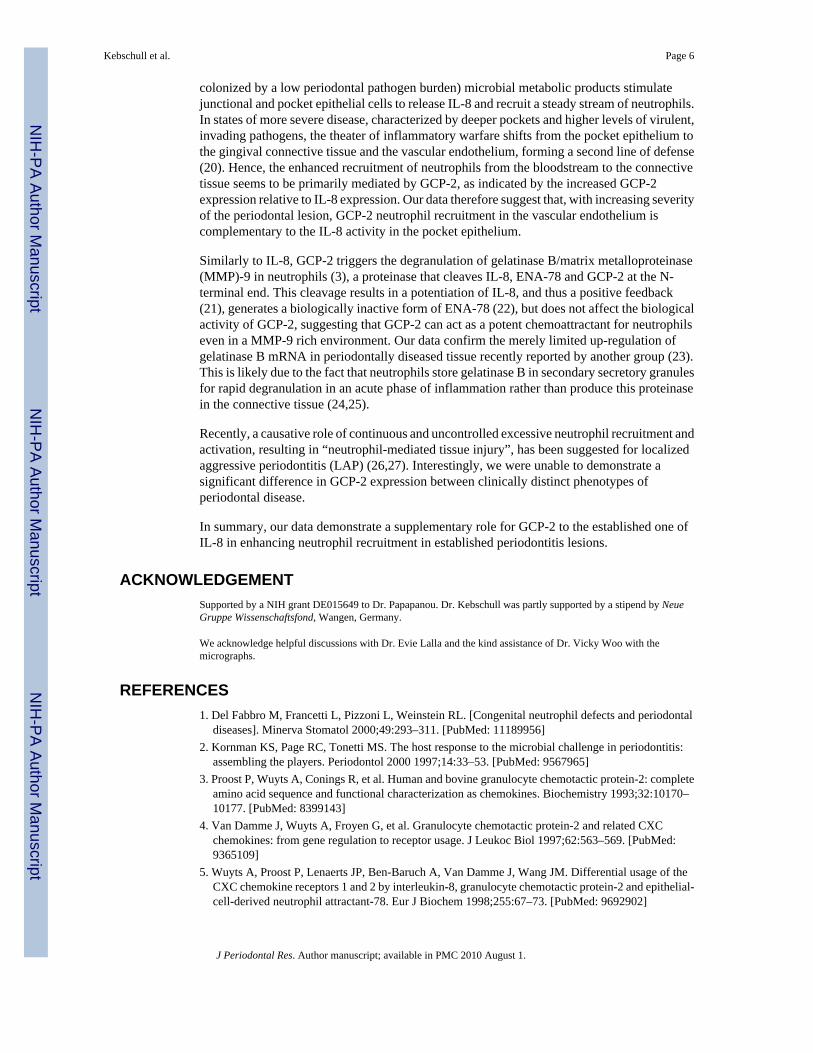
colonized by a low periodontal pathogen burden) microbial metabolic products stimulatejunctional and pocket epithelial cells to release IL-8 and recruit a steady stream of neutrophils.In states of more severe disease, characterized by deeper pockets and higher levels of virulent,invading pathogens, the theater of inflammatory warfare shifts from the pocket epithelium tothe gingival connective tissue and the vascular endothelium, forming a second line of defense(20). Hence, the enhanced recruitment of neutrophils from the bloodstream to the connectivetissue seems to be primarily mediated by GCP-2, as indicated by the increased GCP-2expression relative to IL-8 expression. Our data therefore suggest that, with increasing severityof the periodontal lesion, GCP-2 neutrophil recruitment in the vascular endothelium iscomplementary to the IL-8 activity in the pocket epithelium.
Similarly to IL-8, GCP-2 triggers the degranulation of gelatinase B/matrix metalloproteinase(MMP)-9 in neutrophils (3), a proteinase that cleaves IL-8, ENA-78 and GCP-2 at the N-terminal end. This cleavage results in a potentiation of IL-8, and thus a positive feedback(21), generates a biologically inactive form of ENA-78 (22), but does not affect the biologicalactivity of GCP-2, suggesting that GCP-2 can act as a potent chemoattractant for neutrophilseven in a MMP-9 rich environment. Our data confirm the merely limited up-regulation ofgelatinase B mRNA in periodontally diseased tissue recently reported by another group (23).This is likely due to the fact that neutrophils store gelatinase B in secondary secretory granulesfor rapid degranulation in an acute phase of inflammation rather than produce this proteinasein the connective tissue (24,25).
Recently, a causative role of continuous and uncontrolled excessive neutrophil recruitment andactivation, resulting in “neutrophil-mediated tissue injury”, has been suggested for localizedaggressive periodontitis (LAP) (26,27). Interestingly, we were unable to demonstrate asignificant difference in GCP-2 expression between clinically distinct phenotypes ofperiodontal disease.
In summary, our data demonstrate a supplementary role for GCP-2 to the established one ofIL-8 in enhancing neutrophil recruitment in established periodontitis lesions.
ACKNOWLEDGEMENTSupported by a NIH grant DE015649 to Dr. Papapanou. Dr. Kebschull was partly supported by a stipend by NeueGruppe Wissenschaftsfond, Wangen, Germany.
We acknowledge helpful discussions with Dr. Evie Lalla and the kind assistance of Dr. Vicky Woo with themicrographs.
REFERENCES1. Del Fabbro M, Francetti L, Pizzoni L, Weinstein RL. [Congenital neutrophil defects and periodontal
diseases]. Minerva Stomatol 2000;49:293–311. [PubMed: 11189956]2. Kornman KS, Page RC, Tonetti MS. The host response to the microbial challenge in periodontitis:
assembling the players. Periodontol 2000 1997;14:33–53. [PubMed: 9567965]3. Proost P, Wuyts A, Conings R, et al. Human and bovine granulocyte chemotactic protein-2: complete
amino acid sequence and functional characterization as chemokines. Biochemistry 1993;32:10170–10177. [PubMed: 8399143]
4. Van Damme J, Wuyts A, Froyen G, et al. Granulocyte chemotactic protein-2 and related CXCchemokines: from gene regulation to receptor usage. J Leukoc Biol 1997;62:563–569. [PubMed:9365109]
5. Wuyts A, Proost P, Lenaerts JP, Ben-Baruch A, Van Damme J, Wang JM. Differential usage of theCXC chemokine receptors 1 and 2 by interleukin-8, granulocyte chemotactic protein-2 and epithelial-cell-derived neutrophil attractant-78. Eur J Biochem 1998;255:67–73. [PubMed: 9692902]
Kebschull et al. Page 6
J Periodontal Res. Author manuscript; available in PMC 2010 August 1.
NIH
-PA Author Manuscript
NIH
-PA Author Manuscript
NIH
-PA Author Manuscript

6. Tonetti MS, Freiburghaus K, Lang NP, Bickel M. Detection of interleukin-8 and matrixmetalloproteinases transcripts in healthy and diseased gingival biopsies by RNA/PCR. J PeriodontalRes 1993;28:511–513. [PubMed: 8263721]
7. Tonetti MS, Imboden MA, Lang NP. Neutrophil migration into the gingival sulcus is associated withtransepithelial gradients of interleukin-8 and ICAM-1. J Periodontol 1998;69:1139–1147. [PubMed:9802714]
8. Mathur A, Michalowicz B, Castillo M, Aeppli D. Interleukin-1 alpha, interleukin-8 and interferon-alpha levels in gingival crevicular fluid. J Periodontal Res 1996;31:489–495. [PubMed: 8915952]
9. Fitzgerald JE, Kreutzer DL. Localization of interleukin-8 in human gingival tissues. Oral MicrobiolImmunol 1995;10:297–303. [PubMed: 8596673]
10. Silva TA, Garlet GP, Fukada SY, Silva JS, Cunha FQ. Chemokines in oral inflammatory diseases:apical periodontitis and periodontal disease. J Dent Res 2007;86:306–319. [PubMed: 17384024]
11. Gijsbers K, Van Assche G, Joossens S, et al. CXCR1-binding chemokines in inflammatory boweldiseases: down-regulated IL-8/CXCL8 production by leukocytes in Crohn's disease and selectiveGCP-2/CXCL6 expression in inflamed intestinal tissue. Eur J Immunol 2004;34:1992–2000.[PubMed: 15214047]
12. Gijsbers K, Gouwy M, Struyf S, et al. GCP-2/CXCL6 synergizes with other endothelial cell-derivedchemokines in neutrophil mobilization and is associated with angiogenesis in gastrointestinal tumors.Exp Cell Res 2005;303:331–342. [PubMed: 15652347]
13. Rudack C, Maune S, Eble J, Schroeder JM. The primary role in biologic activity of the neutrophilchemokines IL-8 and GRO-alpha in cultured nasal epithelial cells. J Interferon Cytokine Res2003;23:113–123. [PubMed: 12744776]
14. Rudack C, Hermann W, Eble J, Schroeder JM. Neutrophil chemokines in cultured nasal fibroblasts.Allergy 2002;57:1159–1164. [PubMed: 12464044]
15. Demmer R, Behle JH, Wolf DL, et al. Transcriptomes in Healthy and Diseased Gingival Tissues. JPeriodontol. 2008in press
16. Papapanou PN, Neiderud AM, Sandros J, Dahlen G. Checkerboard assessments of serum antibodiesto oral microbiota as surrogate markers of clinical periodontal status. J Clin Periodontol 2001;28:103–106. [PubMed: 11142661]
17. Socransky SS, Smith C, Martin L, Paster BJ, Dewhirst FE, Levin AE. “Checkerboard” DNA-DNAhybridization. Biotechniques 1994;17:788–792. [PubMed: 7833043]
18. Irizarry RA, Hobbs B, Collin F, et al. Exploration, normalization, and summaries of high densityoligonucleotide array probe level data. Biostatistics 2003;4:249–264. [PubMed: 12925520]
19. Liu RK, Cao CF, Meng HX, Gao Y. Polymorphonuclear neutrophils and their mediators in gingivaltissues from generalized aggressive periodontitis. J Periodontol 2001;72:1545–1553. [PubMed:11759866]
20. Heidemann J, Domschke W, Kucharzik T, Maaser C. Intestinal microvascular endothelium and innateimmunity in inflammatory bowel disease: a second line of defense? Infect Immun 2006;74:5425–5432. [PubMed: 16988217]
21. Van den Steen PE, Proost P, Wuyts A, Van Damme J, Opdenakker G. Neutrophil gelatinase Bpotentiates interleukin-8 tenfold by aminoterminal processing, whereas it degrades CTAP-III, PF-4,and GRO-alpha and leaves RANTES and MCP-2 intact. Blood 2000;96:2673–2681. [PubMed:11023497]
22. Van Den Steen PE, Wuyts A, Husson SJ, Proost P, Van Damme J, Opdenakker G. Gelatinase B/MMP-9 and neutrophil collagenase/MMP-8 process the chemokines human GCP-2/CXCL6,ENA-78/CXCL5 and mouse GCP-2/LIX and modulate their physiological activities. Eur J Biochem2003;270:3739–3749. [PubMed: 12950257]
23. Kubota T, Itagaki M, Hoshino C, et al. Altered gene expression levels of matrix metalloproteinasesand their inhibitors in periodontitis-affected gingival tissue. J Periodontol 2008;79:166–173.[PubMed: 18166107]
24. Bainton DF, Ullyot JL, Farquhar MG. The development of neutrophilic polymorphonuclearleukocytes in human bone marrow. J Exp Med 1971;134:907–934. [PubMed: 4106490]
25. Birkedal-Hansen H. Role of matrix metalloproteinases in human periodontal diseases. J Periodontol1993;64:474–484. [PubMed: 8315570]
Kebschull et al. Page 7
J Periodontal Res. Author manuscript; available in PMC 2010 August 1.
NIH
-PA Author Manuscript
NIH
-PA Author Manuscript
NIH
-PA Author Manuscript

26. Kantarci A, Oyaizu K, Van Dyke TE. Neutrophil-mediated tissue injury in periodontal diseasepathogenesis: findings from localized aggressive periodontitis. J Periodontol 2003;74:66–75.[PubMed: 12593599]
27. Kantarci A, Van Dyke TE. Lipoxin signaling in neutrophils and their role in periodontal disease.Prostaglandins Leukot Essent Fatty Acids 2005;73:289–299. [PubMed: 15979867]
Kebschull et al. Page 8
J Periodontal Res. Author manuscript; available in PMC 2010 August 1.
NIH
-PA Author Manuscript
NIH
-PA Author Manuscript
NIH
-PA Author Manuscript
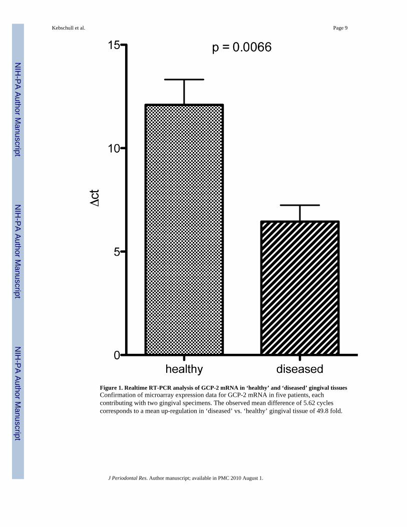
Figure 1. Realtime RT-PCR analysis of GCP-2 mRNA in ‘healthy’ and ‘diseased’ gingival tissuesConfirmation of microarray expression data for GCP-2 mRNA in five patients, eachcontributing with two gingival specimens. The observed mean difference of 5.62 cyclescorresponds to a mean up-regulation in ‘diseased’ vs. ‘healthy’ gingival tissue of 49.8 fold.
Kebschull et al. Page 9
J Periodontal Res. Author manuscript; available in PMC 2010 August 1.
NIH
-PA Author Manuscript
NIH
-PA Author Manuscript
NIH
-PA Author Manuscript

Figure 2. Western blot analysis of GCP-2 protein in ‘healthy’ and ‘diseased’ tissue samplesStronger GCP-2 protein expression in ‘diseased’ gingival tissues. The band for GCP-2 wasdetected at approx. 10 kDa while equal loading was demonstrated by β-actin probing.Densitometric analysis revealed a 3.6 fold up-regulation of GCP-2 protein in ‘diseased’ tissue.The analysis was performed in 3 consecutively recruited patients each contributing with one‘diseased’ and one ‘healthy’ gingival tissue specimen.
Kebschull et al. Page 10
J Periodontal Res. Author manuscript; available in PMC 2010 August 1.
NIH
-PA Author Manuscript
NIH
-PA Author Manuscript
NIH
-PA Author Manuscript

Figure 3. ELISA analysis of GCP-2 protein in homogenized in ‘healthy’ and ‘diseased’ tissuesamplesUp-regulation of GCP-2 protein in ‘diseased’ gingival tissue by ELISA (n=5 consecutivelyrecruited patients, each contributing with one ‘diseased’ and one ‘healthy’ gingival tissuespecimen).
Kebschull et al. Page 11
J Periodontal Res. Author manuscript; available in PMC 2010 August 1.
NIH
-PA Author Manuscript
NIH
-PA Author Manuscript
NIH
-PA Author Manuscript

Figure 4. Immunostaining of GCP-2 protein in frozen tissue sectionsGCP-2 expression is enhanced in ‘diseased’ gingival tissues. GCP-2 immunoreactivity ispredominant in endothelial cells of the gingival microvasculature (arrows). Furthermore, adiffuse positive staining of the connective tissue can be detected primarily in ‘diseased’ samplesas a typical sign of chemokine secretion (asterisk).
Kebschull et al. Page 12
J Periodontal Res. Author manuscript; available in PMC 2010 August 1.
NIH
-PA Author Manuscript
NIH
-PA Author Manuscript
NIH
-PA Author Manuscript

NIH
-PA Author Manuscript
NIH
-PA Author Manuscript
NIH
-PA Author Manuscript
Kebschull et al. Page 13
Table 1Microarray analysis of chemokine and chemokine receptor mRNA expression in periodontal disease. Data are shownfor all chemokines, chemokine receptors and selected functionally related genes with a p-value below the Bonferronithreshold of 9.14×10−07.1
Probe Gene Description Fold Change p-value
206336_at CXCL6 chemokine (C-X-C motif) ligand 6 3.85 1.1E-16
217028_at CXCR4 chemokine (C-X-C motif) receptor 4 3.56 1.1E-16
204470_at CXCL1 chemokine (C-X-C motif) ligand 1 3.45 1.1E-16
209201_x_at CXCR4 chemokine (C-X-C motif) receptor 4 3.07 1.1E-16
211919_s_at CXCR4 chemokine (C-X-C motif) receptor 4 2.90 1.1E-16
202859_x_at IL8 interleukin 8 2.57 1.2E-15
205242_at CXCL13 chemokine (C-X-C motif) ligand 13 2.42 7.9E-14
211506_s_at IL8 interleukin 8 2.26 1.9E-12
209924_at CCL18 chemokine (C-C motif) ligand 18 2.18 1.1E-16
214146_s_at PPBP chemokine (C-X-C motif) ligand 7 2.11 7.0E-14
32128_at CCL18 chemokine (C-C motif) ligand 18 2.08 2.4E-15
203666_at CXCL12 chemokine (C-X-C motif) ligand 12 2.05 1.1E-16
209774_x_at CXCL2 chemokine (C-X-C motif) ligand 2 1.90 2.6E-12
203936_s_at MMP9 matrix metallopeptidase 9 1.81 1.8E-13
210072_at CCL19 chemokine (C-C motif) ligand 19 1.78 5.6E-10
209687_at CXCL12 chemokine (C-X-C motif) ligand 12 1.76 1.4E-15
208335_s_at DARC Duffy blood group, chemokine receptor 1.71 1.1E-16
205098_at CCR1 chemokine (C-C motif) receptor 1 1.66 1.1E-16
207850_at CXCL3 chemokine (C-X-C motif) ligand 3 1.61 6.1E-11
214974_x_at CXCL5 chemokine (C-X-C motif) ligand 5 1.60 8.8E-07
1405_i_at CCL5 chemokine (C-C motif) ligand 5 1.59 6.4E-11
206337_at CCR7 chemokine (C-C motif) receptor 7 1.59 9.0E-12
1555759_a_at CCL5 chemokine (C-C motif) ligand 5 1.56 1.3E-12
205099_s_at CCR1 chemokine (C-C motif) receptor 1 1.48 5.2E-14
205114_s_at CCL3 chemokine (C-C motif) ligand 3 1.48 7.2E-09
206390_x_at PF4 chemokine (C-X-C motif) ligand 4 1.45 3.4E-13
206366_x_at XCL1 chemokine (C motif) ligand 1 1.44 1.1E-16
214567_s_at XCL1 chemokine (C motif) ligand 1 1.42 4.4E-13
204655_at CCL5 chemokine (C-C motif) ligand 5 1.41 1.1E-08
205392_s_at CCL14 chemokine (C-C motif) ligand 14 1.40 2.9E-09
204103_at CCL4 chemokine (C-C motif) ligand 4 1.39 1.6E-10
214038_at CCL8 chemokine (C-C motif) ligand 8 1.35 4.1E-07
207794_at CCR2 chemokine (C-C motif) receptor 2 1.31 3.6E-12
220565_at CCR10 chemokine (C-C motif) receptor 10 1.30 1.4E-11
219161_s_at CKLF chemokine-like factor 1.28 2.1E-11
223451_s_at CKLF chemokine-like factor 1.24 5.3E-11
211434_s_at CCRL2 chemokine (C-C motif) receptor-like 2 1.23 2.5E-11
206126_at BLR1 chemokine (C-X-C motif) receptor 5 1.15 3.9E-07
210133_at CCL11 chemokine (C-C motif) ligand 11 1.15 6.8E-07
J Periodontal Res. Author manuscript; available in PMC 2010 August 1.

NIH
-PA Author Manuscript
NIH
-PA Author Manuscript
NIH
-PA Author Manuscript
Kebschull et al. Page 14
Probe Gene Description Fold Change p-value
210548_at CCL23 chemokine (C-C motif) ligand 23 1.13 1.1E-07
224027_at CCL28 chemokine (C-C motif) ligand 28 0.90 8.7E-08
220351_at CCRL1 chemokine (C-C motif) receptor-like 1 0.71 4.4E-16
222484_s_at CXCL14 chemokine (C-X-C motif) ligand 14 0.59 3.7E-14
218002_s_at CXCL14 chemokine (C-X-C motif) ligand 14 0.56 5.1E-15
237038_at CXCL14 chemokine (C-X-C motif) ligand 14 0.55 1.1E-16
1Multiple Affymetrix probes may map to the same gene. Fold change is defined as the ratio of gene expression in disease over expression in health. Thus,
a fold change < 1.0 indicates a down-regulation in disease vs. health.
J Periodontal Res. Author manuscript; available in PMC 2010 August 1.