Discovery and structure–activity relationship of a novel spirocarbamate series of NPY Y5 antagonists

IntroductionObesity, which is an important risk factor for seriouschronic illnesses such as insulin resistance, type 2 dia-betes, and heart disease is caused by an imbalancebetween energy intake and energy expenditure. Thereare numerous factors influencing whole-body energybalance, including neural and hormonal signals, which,when altered could result in obesity. It is becomingincreasingly evident that the central nervous system,particularly the hypothalamus, plays a critical role inintegrating these signals to regulate energy balance andthat neuropeptide Y (NPY) is an important neuro-modulator in this system (1, 2).
NPY is widely distributed throughout the brain (3).However, most of the metabolic actions of NPYappear to be mediated through defined hypothalam-ic nuclei, including the arcuate nucleus (Arc) wherenumerous NPY-containing neurons originate, theparaventricular nucleus (PVN), where many NPY-containing arcuate neuron terminals and NPY-bind-ing sites are located, and the ventromedial hypothal-amic (VMH) nucleus, which has been referred to as“the satiety center” (3, 4). NPY is the most potentorexigenic agent known (5, 6) and can also causechanges in circulating hormone levels (7). Chronicintracerebroventricular (icv) administration of NPY
results in hyperphagia, hyperinsulinemia, insulinresistance, and obesity (8–10). Lesions of the VMH inrodents also cause multiple changes in metabolic sta-tus, including hyperphagia, hyperglycemia, andhyperinsulinemia (11). Interestingly, injection ofNPY directly into the VMH significantly increasesfood intake (12), and NPY-induced feeding isenhanced in VMH-lesioned rats (13). These data sug-gest the VMH may also be a site of action for NPY inthe development of obesity; however, the mecha-nisms by which NPY is involved in each aspect of cen-tral energy regulation remain to be defined.
Much research has focused on which of the 5 NPY-receptor subtypes cloned so far (Y1, Y2, Y4, Y5, and they6) (14) might mediate the potent NPY-inducedfeeding response. Several lines of evidence point tothe Y1- and/or Y5-receptor subtypes being the mostlikely candidates for such an action (15–19). In addi-tion, our previous studies investigating the mRNAexpression of all known Y receptors in the rat brainalso show clearly that both Y1- and Y5-receptor sub-type mRNAs are expressed in areas pivotal in regu-lating energy balance (20).
Glucocorticoid hormones play a critical role inenergy balance and also appear to mediate at leastsome of their actions through the central NPY axis
The Journal of Clinical Investigation | May 2000 | Volume 105 | Number 9 1253
Adrenalectomy reduces neuropeptide Y–induced insulin release and NPY receptor expression in the rat ventromedial hypothalamus
Todd Wisialowski, Rachel Parker, Elaine Preston, Amanda Sainsbury, Edward Kraegen,Herbert Herzog, and Gregory Cooney
Garvan Institute of Medical Research, 384 Victoria Street, Sydney, Australia
Address correspondence to: Greg Cooney, Garvan Institute of Medical Research, 384 Victoria Street, Darlinghurst, Sydney, NSW 2010, Australia. Phone: 61-2-9295-8209; Fax: 61-2-9295-8201; E-mail: [email protected].
Todd Wisialowski and Rachel Parker contributed equally to this work.
Received for publication October 15, 1999, and accepted in revised form March 8, 2000.
Chronic central administration of neuropeptide Y (NPY) causes hyperphagia, hyperinsulinemia,and obesity, a response that is prevented by prior adrenalectomy (ADX) in rats. The basis ofNPY’s effect and how the acute responses to this peptide are affected by ADX remain unknown.This study investigates the role of glucocorticoids in acute NPY-stimulated food intake, acuteNPY-induced insulin release, and hypothalamic NPY-receptor mRNA expression levels. NPY-induced food intake was similar in ADX and control rats after acute intracerebroventricularinjection of NPY. Injection of NPY caused a significant increase in plasma insulin in control rats,but this effect was completely absent in ADX rats in which basal plasma insulin levels were alsolower than controls. In addition, ADX significantly reduced the number of neurons expressingNPY receptor Y1 and Y5 mRNAs in the ventromedial hypothalamus (VMH), without affecting Y1-or Y5-mRNA expression in the paraventricular hypothalamus or the arcuate nucleus. These dataindicate that glucocorticoids are necessary for acute NPY-mediated insulin release and suggestthat the mechanisms involve glucocorticoid regulation of Y1 and Y5 receptors specifically with-in the VMH nucleus.
J. Clin. Invest. 105:1253–1259 (2000).

(21). In rats, excessive corticosterone promotes bodyfat gain (22) and hyperinsulinemia (23) and alsoincreases NPY synthesis and Y1-receptor mRNAexpression, at least within the Arc (24, 25). Converse-ly, removal of glucocorticoids by adrenalectomy(ADX) reduces hyperphagia and body weight of obese(fa/fa) rats (26), abolishes obesity induced by VMHlesions (27), and prevents obesity induced by chron-ic central NPY infusion in normal rats (28). However,it has been reported that ADX does not alter Y1-receptor mRNA expression in the Arc (24). Glucocor-ticoid-receptor immunoreactivity is found within therat central nervous system, including the Arc, VMH,and PVN (29). Many of these receptors are expressedat the nucleus of NPY-containing, endocrine-relatedneurons and coexist in regions containing high NPY-receptor density (30).
Taken together, these observations indicate thatglucocorticoids have a regulatory role in long-termcentral NPY signaling. However, it is not known whatregulatory role, if any, glucocorticoid hormones havein the effects of acute, central administration of NPYand how these actions relate to NPY-receptor expres-sion. Therefore, we have examined the effects of ADXon acute icv NPY-stimulated food intake and insulinrelease in rats. Furthermore, we have analysed theeffects of ADX on Y1- and Y5-receptor mRNA expres-sion within specific brain regions reported to regu-late food intake and energy balance (Arc, PVN, andVMH), using subtype-specific riboprobes andemploying a uniform technique of in situ hybridiza-tion histochemistry. The results show that ADXreduces basal plasma insulin levels, abolishes NPY-induced insulin release, and significantly decreasesY1- and Y5-receptor mRNA expression selectively
within the VMH. These data suggest that glucocorti-coids regulate NPY-induced insulin release and NPYsignaling within the VMH of the hypothalamus.
Methods
Animals and surgical procedures
All procedures were approved by the Animal Experi-mentation Ethics Committee (Garvan Institute and St.Vincent’s Hospital, Sydney, Australia) and were inaccordance with the National Health and MedicalResearch Council of Australia (NHMRC) guidelines onanimal experimentation.
Male Wistar rats (275–350 g) were housed understandard lighting conditions (6:00 am to 6:00 pm)with chow and water available ad libitum. Under keta-mine (60 mg/kg; Parke Davis, Caringbah, Australia)and xylazine (10 mg/kg; Bayer AG, Leverkusen,Switzerland) anesthesia, a 22-gauge guide cannula(Plastics One, Roanoke, Virginia, USA) was positionedand fixed in the right lateral ventricle (icv) using astereotaxic surgical table and coordinates obtainedfrom a rat brain atlas (31). A cannula for blood sam-pling was inserted into the jugular vein and routed outthrough a small incision on the back between theshoulders. It was plugged with polyvinylpyrrolidone(PVP) in heparin saline to prevent clotting and leaking.The rats were allowed to recover for at least 7 days.After this recovery period, rats were tested for correctcannula placement by a 4-hour feeding study after
1254 The Journal of Clinical Investigation | May 2000 | Volume 105 | Number 9
Figure 1The effect of adrenalectomy on NPY-induced food intake in rats.Rats received an acute injection of 1 µg NPY (icv), and cumulativefood intake was monitored over the next 4 hours. In a second set ofexperiments NPY (5 µg) was injected icv and blood sampled for 2hours without food being available. After 2 hours, food was pre-sented to the rats and food intake monitored over the subsequenthour. Results are expressed as the mean ± SE. Black bars representsham-operated NPY-injected rats (n = 12), and gray bars representADX NPY-injected rats (n = 7).
Figure 2Plasma corticosterone concentration after acute NPY injection innormal, atropine-treated, and ADX rats. Plasma corticosterone wasmeasured after icv injection of vehicle or NPY (5 µg) in normal,atropine-treated (1 mg/kg intravenously) or ADX rats as describedin Methods. Results are shown as the mean ± SE for 5–7 animalsper group. Filled squares represent vehicle-injected rats, filled dia-monds represent NPY-injected rats, open triangles representatropine-treated NPY-injected rats, and open circles represent NPY-injected ADX rats. NPY-injected normal rats and NPY-injectedatropine-treated rats had significantly higher corticosterone release(P < 0.0001) than vehicle-injected normal rats or NPY-injected ADXrats as assessed by repeated measures ANOVA.
acute icv NPY injection (5 µg). A subgroup of the ratswith significant food consumption after icv NPY werereanesthetized, and both adrenal glands were surgi-cally removed (ADX). Rats were given 0.9% saline intheir drinking water to maintain electrolyte balance.Rats were handled and monitored daily to minimizeany stress during the experiments, which were per-formed 8–10 days after the second surgical procedure.ADX rats maintained normal food intake and bodyweight gain after the surgical procedures and werehealthy in appearance and behavior.
Injection of compounds
NPY (Auspep Pty. Ltd., Melbourne, Australia) wasdissolved in 0.9% saline and injected as 5-µg and 1-µg doses in a volume of 5 µL. Icv injections were per-formed using a 28-gauge injection cannula (PlasticsOne) connected to a Hamilton-1801RN 10-µLsyringe through Silastic tubing. Atropine methylnitrate (Sigma Chemical Co., St. Louis, Missouri,USA) was given as a bolus (1 mg/kg) through thejugular catheter 20 minutes before the icv adminis-tration of NPY.
Experimental design
Measurement of food intake. On the morning of the exper-iment, rats were placed in individual cages with accessto drinking water and allowed to acclimatize for about1 hour. ADX and control rats were given an icv injec-tion of NPY (1 µg) and returned to their cages with adlibitum access to food and water. Food intake wasmeasured at 1, 2, and 4 hours after injection. Othergroups of sham and ADX rats were treated with vehicle(icv 5 µL 0.9% saline) for comparison.
Blood sampling procedure. Rats were placed in indi-vidual cages with access to water, and Silastic tubingwas connected to the in-dwelling jugular cannula forblood sampling. After a 60- to 90-minute acclimati-zation period, two basal blood samples were takenapproximately 15 minutes apart. Rats were removedfrom the cages and injected with NPY (5 µg/5 µLover 30 seconds) and returned to their cages forblood sampling. Control experiments were per-
formed with icv injection of vehicle (5 µL 0.9%saline). Two additional groups of animals were givena bolus injection of a muscarinic receptor blocker (1mg/kg atropine methyl nitrate ) 20 minutes beforeicv NPY or saline injection to assess the role of theparasympathetic nervous system in any hormonalresponses to the administration of NPY. Blood sam-ples (300 µL) were collected at regular intervals for 2hours after icv injection. The plasma was separatedby centrifugation, placed on ice, and plasma glucosedetermined before storing at –20°C for furtherassays. The red cells were resuspended in heparinsaline and returned to each rat. Food was presented2 hours after icv injection of NPY or vehicle, andintake was measured over the next hour to confirmefficacy of the NPY injection.
Plasma measurements. Plasma glucose was measuredusing a YSI 2300 STAT Plus Glucose & L-LactateAnalyzer (Yellow Springs Instrument Co. Inc., YellowSprings, Colorado, USA). Plasma insulin and leptinwere measured using radioimmunoassay kits sup-plied by Linco Research, Inc. (St. Charles, Missouri,USA). Plasma corticosterone was measured using aImmuChem Double Antibody Corticosterone RIAKit for rats and mice (ICN Biomedicals Inc., CostaMesa, California, USA).
In situ hybridization histochemistry
In situ hybridization was essentially carried out asdescribed previously, at a hybridization temperatureof 50°C, using 200 ng/mL each of 11digoxigenin-UTP–labeled riboprobe (20).
Data analysis. Regions containing positivehybridization (dark brown/indigo color precipitatedeposit showing a halo pattern in the cytoplasm sur-rounding a clearly visible nucleus) were identifiedand mapped with the aid of a rat brain atlas (31).The numbers of positively expressing cell bodieswere counted within the right and left PVN and Arcand over a standard area for each VMH (n = 8 brainsections each from 3 ADX and 3 control rats). Nodetailed distinctions were made between subdivi-sions within these nuclei. The levels of receptor-sub-
The Journal of Clinical Investigation | May 2000 | Volume 105 | Number 9 1255
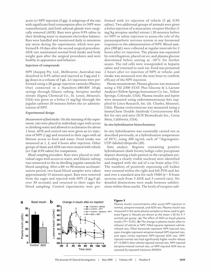
Figure 3Plasma insulin concentration after acute NPY injection innormal, atropine-treated, and ADX rats. Plasma insulin wasmeasured in the same plasma samples as those used to gen-erate Figure 2. Results are shown as the mean ± SE for 5–7animals per group. (a) The effect of ADX on basal plasmainsulin (AP < 0.05). (b) The change in plasma insulin after icvinfusion of vehicle or NPY. Filled squares represent vehicle-infused rats, filled diamonds represent NPY-injected rats,open triangles represent atropine-treated NPY-injected rats,and open circles represent NPY-injected ADX rats. NPY-injected normal rats had significantly higher insulin release(P < 0.0001) than vehicle-injected normal rats, NPY-injectedatropine-treated normal rats, or NPY-injected ADX rats asassessed by repeated measures ANOVA.

type mRNA signal detected within these specifichypothalamic areas were compared in control andADX rat brain sections obtained under the sameassay conditions, and high-power photomicrographswere taken. Adjacent sections were counterstainedwith hematoxylin/eosin to confirm morphologicalidentification of these hypothalamic areas.
Statistical analysis. Data are expressed as mean ± SE,either as raw data or change from baseline conditions.Repeated measures ANOVA with relevant post-hocanalysis was used to determine significant differencesbetween treatment groups, and unpaired t tests wereused to determine the significant difference for the insitu hybridization results. A probability less than 0.05was considered to be statistically different.
ResultsEffects of ADX on NPY-induced feeding. An acute icv injec-tion of NPY (1 µg) stimulated food intake to a similardegree in both ADX and control rats at 1, 2, and 4hours after NPY injection (Figure 1). When food waspresented to animals 2 hours after administration of5 µg NPY and blood sampling, the food intake of con-trol and ADX rats was also not significantly different(Figure 1). Both sham and ADX animals injected withsaline vehicle icv consumed less than 1 g of food overthe 4-hour period, and there was no effect of ADX onthe spontaneous overnight food intake of the rats(data not shown).
Effect of ADX on NPY-induced changes in plasma hor-mones in the absence of feeding. In control rats withoutaccess to food during the experiment, acute icv injec-tion of vehicle (5 µL 0.9% saline) caused a significantrise in plasma corticosterone, which returned to basallevels after 60 minutes (Figure 2). This response wassimilar if the rats were handled without any icv injec-tion (data not shown) and occurred despite the fact
that all rats were handled on a daily basis to reducestress. Injection of 5 µg NPY (icv) produced a furtherincrease in plasma corticosterone levels comparedwith vehicle injection (P < 0.05), and corticosteroneremained elevated for at least 2 hours after NPY injec-tion (Figure 2). Blockade of muscarinic receptors withatropine had no effect on the NPY-induced increasein corticosterone levels, indicating that the effect oficv NPY administration on corticosterone release wasnot mediated through the parasympathetic nervoussystem and the vagus nerve (Figure 2). As expected, inADX rats there was no increase in corticosterone lev-els after acute icv injection of NPY.
Acute icv injection of NPY (5 µg) also caused a sig-nificant increase in plasma insulin levels comparedwith vehicle (P < 0.05), and insulin remained elevat-ed for 90 minutes (Figure 3). Treatment of normalrats with an intravenous bolus of a muscarinic recep-tor blocker (atropine) 20 minutes before icv NPYadministration reduced the effect of NPY injectionon insulin release to a level that was not significant-ly different from that of vehicle administration.Atropine had no significant effect in vehicle-injectedrats. This indicates that the effect of central NPYadministration on insulin release is mediated by anincrease in parasympathetic nervous system activitythrough vagal innervation of the pancreas. ADX ratshad significantly reduced basal plasma insulin levelscompared with controls (22.3 ± 4.7 and 41.3 ± 4.7µU/mL, respectively; P < 0.05, see Figure 3a), andthere was no significant increase above basal levelsafter acute NPY injection in ADX rats (Figure 3). Thedifferences in insulin were not due to differences inplasma glucose levels, which were similar in the con-trol and ADX rats and did not change after acuteNPY injection (P < 0.42; data not shown). Circulatingleptin was significantly decreased in ADX rats com-
1256 The Journal of Clinical Investigation | May 2000 | Volume 105 | Number 9
Figure 4Photomicrograph of expression of Y1-receptor mRNA within hypothalamic nuclei of normal and ADX rats. Specific mRNA was detected byin situ hybridization of serial sections of sham-operated and ADX rats as described in Methods. (a, c, and e) PVN, VMH, and Arc of normalbrains. (b, d, and f) PVN, VMH, and Arc of ADX rats. (g and h) Sense controls. The inset shows the schematic of the regions photographed.Positive signals are defined as a color precipitate diffused around the nucleus (arrows). Scale bar = 100 µm.

pared with controls (1.6 ± 0.2 and 3.6 ± 0.4 ng/mL,respectively) but there was no change in these levelswith acute injection of NPY (data not shown).
Y1- and Y5-receptor mRNA expression. Nonradioactivein situ hybridization employing rat Y1- and Y5-specif-ic riboprobes was used to determine possible changesin Y-receptor expression after ADX. No cross-reactiv-ity occurred when each receptor-specific riboprobewas hybridized under conditions identical to thoseused for in situ hybridization against vector con-structs for each of the other known Y-receptor sub-types. This result was not surprising given the verylow sequence identity of the different Y-receptor sub-types (14). Furthermore, neither of the correspon-ding sense riboprobes used under the same assayconditions showed specific hybridization (Figures 4,g and h, and Figure 5, g and h).
In the PVN, VMH, and Arc of control rats there wasapproximately twice the number of neurons express-ing Y1-receptor mRNA as those expressing Y5-receptormRNA (Figures 4, 5, and 6). These values confirmthose reported previously by our group using similarhybridization techniques (20) and are comparable
with other published data, at least for Y1 mRNAexpression levels within normal Arc using a differenttechnique of in situ hybridization (32). Bilateral ADXcaused a significant reduction in the number of neu-rons expressing Y1- (by up to 80%; P < 0.01) and Y5- (by50%; P < 0.01) receptor mRNA within the VMH (Fig-ures 4d, 5d, and 6). However, ADX did not signifi-cantly change the numbers of neurons expressing Y1-and/or Y5-receptor mRNA within the PVN or the Arc.
DiscussionChronic icv infusion of NPY induces hyperphagia,hyperinsulinemia, and insulin resistance in rats, andthese effects are blocked by previous ADX (28). In thecurrent study we demonstrate that ADX also abolish-es the insulin release caused by an acute icv injectionof NPY and that this is associated with significantlyreduced Y1- and Y5-receptor mRNA expression specif-ically within the VMH. These experiments imply thatglucocorticoids are necessary for icv NPY to stimulateinsulin release and suggest that glucocorticoids man-ifest this regulatory role through alterations in Y1- andY5-receptor expression in the VMH.
The Journal of Clinical Investigation | May 2000 | Volume 105 | Number 9 1257
Figure 5Photomicrograph of expression of Y5-receptor mRNA within hypothalamic nuclei of normal and ADX rats. Specific mRNA was detected byin situ hybridization of serial sections of sham-operated and ADX rats as described in Methods. (a, c, and e) PVN, VMH, and Arc of normalbrains. (b, d, and f) PVN, VMH, and Arc of ADX rats. (g and h) Sense controls. The inset shows the schematic of the regions photographed.Positive signals are defined as a color precipitate diffused around the nucleus (arrows). Scale bar = 100 µm.
Figure 6Quantification of expression of Y1- and Y5-receptormRNA within hypothalamic nuclei. The number ofcell bodies staining for Y1- and Y5-receptor mRNAwas quantified as described in Methods. (a) Thenumber of cells showing hybridization for Y1-recep-tor mRNA in the PVN, the VMH, and the Arc. (b)The number of cells showing hybridization for Y5
mRNA in the same regions of the hypothalamus.Plotted data represent the mean ± SE for determi-nations from 3 separate control and ADX animals.AP < 0.001, unpaired t-test.

Interestingly, in this study ADX rats showed no sig-nificant alteration in the feeding response to acute NPYinjection. Therefore, it was not surprising that theseADX rats also showed no change in expression of Y1-and Y5-receptor mRNA (the proposed NPY feedingreceptors) within the PVN. There are contradictoryreports in the literature regarding the effects of ADXon NPY-induced food intake. Some studies show thatfood intake measured 1 hour after direct NPY admin-istration into the PVN was partially attenuated by ADXand was restored by corticosterone treatment (33, 34).However, in agreement with our results, others reportthat ADX had no significant effect on food intake inrats during 2 hours of NPY infusion (35) or 2 hoursafter acute icv NPY administration (36).
The fact that we found no change in the expressionof Y1- and Y5-receptor mRNA levels in the PVN and Arcindicates that glucocorticoids are not essential for basalregulation of Y1- and Y5-receptor mRNAs within theseareas. Our findings are in agreement with and furtherextend previous studies, showing that glucocorticoidsare not essential for the regulation of Y1-receptormRNA in the Arc (24), and that ADX does not alter 125I-PYY–specific binding in the Arc or PVN (25).
The current study, we believe, is the first to demonstratethat ADX alters Y1- and Y5-receptor mRNA expression inthe VMH. A role for the VMH in insulin secretion andadrenocortical regulation has been proposed previously(37), but the mechanisms whereby glucocorticoids maymediate the effects we observe on Y1- and Y5-receptormRNAs in the VMH are unknown. These effects could bea direct action of glucocorticoids as glucocorticoid-recep-tor immunoreactivity is widely distributed throughoutthe rat forebrain, including the VMH (29). In addition,the Y1-receptor gene is known to contain a putative glu-cocorticoid response element (38), and it has been report-ed that supplemental corticosterone treatment to ADXrats increases NPY receptor–binding sites and Y1-recep-tor mRNA levels within discrete hypothalamic nuclei (24,25). It should be noted that other response elements mayalso be involved in tissue-specific Y-receptor regulation(38) and that changes in mRNA levels do not necessarilyequate to changes in the amount of receptor protein (39).However, these findings do link ADX-induced effects onY-receptor expression in the VMH with alterations in theinsulinemic response to exogenous NPY, indicating thatthese mRNA changes may indeed be of physiological sig-nificance. Studies involving replacement of glucocorti-coids and restoration of Y1-and Y5-receptor expressionand NPY-induced insulin release would be necessary toestablish more directly the role of corticosterone in theobserved effects of ADX. However, such experiments canprovide differing results, depending on the type androute of glucocorticoid replacement (24, 40). A recentstudy has shown that icv infusion of dexamethasone for3 days increases insulin levels in normal rats, which pro-vides some evidence for a central effect of glucocorticoidsin controlling insulin release possibly through alteringNPY-regulated pathways (40).
It is also possible that glucocorticoids act by regulatinglevels of corticotropin-releasing hormone (CRH), whichhas been reported to significantly decrease food intake(41), insulinemia (42), and inhibit the synthesis andrelease of NPY (43). Although changes in CRH actionhave not been linked to changes in Y1- and Y5-receptormRNA, the reduction in NPY action following ADX maybe a result of increased CRH activity as well as decreasedcorticosterone levels. Recently, it has been shown thatADX results in a similar, although not so marked, reduc-tion in type 2 CRH-receptor (CRHR2) mRNA levels with-in the VMH, without any change in levels within the PVN(44). It is noteworthy that the specific reduction in VMHCRHR2 mRNA in these rats was also associated with adecreased body weight and decreased basal plasmainsulin and leptin levels. However, the exact roles of thesereceptors within the VMH are far from understood.
Enhanced NPY expression in the VMH is associatedwith the obesity syndrome (45). Furthermore, NPY hasbeen shown to directly inhibit over one fifth of spon-taneously active rat VMH neurons, and this inhibitionis potentiated by overfeeding (46). Therefore, the mech-anism by which acute icv NPY stimulates insulinrelease in the absence of feeding may be by inhibitingthe spontaneous activity of the VMH through Y1 andY5 receptors. A reduction of these receptors with ADXwould then reduce the ability of NPY to inhibit VMHneurons. In agreement with others, we found that acuteicv NPY administration had no affect on plasma glu-cose levels, indicating that NPY-induced insulin releaseis not simply a secondary response to changes inperipheral glucose (47). By associating the decreasedbasal insulin levels and lack of insulin release inresponse to NPY injection in ADX rats with downreg-ulation of Y1-and Y5-receptor mRNA in the VMH, ourstudy highlights a role for Y1-and Y5-receptors in theVMH in the etiology of NPY-induced hyperinsuline-mia, insulin resistance, and obesity.
In summary, acute NPY-stimulated insulin releasewas blocked by ADX, an effect that coincided withselective reduction in Y1- and Y5-receptor mRNA-expressing neurons in the VMH. These data suggestthat glucocorticoids are necessary for acute NPY-stim-ulated insulin release and indicates that the mecha-nisms of action for this effect of NPY involves the Y1
and Y5 receptors in the VMH.
AcknowledgmentsThis study was made possible by Research Grants fromthe National Health and Medical Research Council ofAustralia. The authors gratefully acknowledge theexpert technical assistance of Donna Wilks and all thestaff members of the Biological Testing Facility at theGarvan Institute of Medical Research.
1. Kalra, S.P., et al. 1999. Interacting appetite-regulating pathways in thehypothalamic regulation of body weight. Endocr. Rev. 20:68–100.
2. Inui, A. 1999. Feeding and body-weight regulation by hypothalamic neu-ropeptides-mediation of the actions of leptin. Trends Neurosci. 22:62–67.
3. De Quidt, M.E., and Emson, P.C. 1986. Distribution of neuropeptide Y-like immunoreactivity in the rat central nervous system II. Immunohis-
1258 The Journal of Clinical Investigation | May 2000 | Volume 105 | Number 9

tological analysis. Neuroscience. 18:545–618.4. Dumont, Y., Fournier, A., St-Pierre, S., and Quirion, R. 1993. Compara-
tive characterization and autoradiographic distribution of neuropeptideY receptor subtypes in the rat brain. J. Neurosci. 13:73–86.
5. Clark, J.T., Kalra, P.S., Crowley, W.R., and Kalra, S.P. 1984. NeuropeptideY and human pancreatic polypeptide stimulate feeding behaviour inrats. Endocrinology. 115:427–429.
6. Stanley, B.G., and Leibowitz, S.F. 1985. Neuropeptide Y injected in theparaventricular hypothalamus: a powerful stimulant of feeding behav-ior. Proc. Natl. Acad. Sci. USA. 82:3940–3943.
7. Marks, J.L., and Waite, K. 1996. Some acute effects of intracerebroven-tricular neuropeptide Y on insulin secretion and glucose metabolism inthe rat. J. Neuroendocrinol. 8:507–513.
8. Stanley, B.G., Kyrkouli, S.E., Lampert, S., and Leibowitz, S.F. 1986. Neu-ropeptide Y chronically injected into the hypothalamus: a powerful neu-rochemical inducer of hyperphagia and obesity. Peptides. 7:1189–1192.
9. Vettor, R., Zarjevski, N., Cusin, I., Rohner-Jeanrenaud, F., and Jeanre-naud, B. 1994. Induction and reversibility of an obesity syndrome byintracerebroventricular neuropeptide Y administration to normal rats.Diabetologia. 37:1202–1208.
10. Zarjevski, N., Cusin, I., Vettor, R., Rohner-Jeanrenaud, F., and Jeanre-naud, B. 1993. Chronic intracerebroventricular neuropeptide-Y admin-istration to normal rats mimics hormonal and metabolic changes ofobesity. Endocrinology. 133:1753–1758.
11. Penicaud, L., et al. 1989. Development of VMH obesity: in vivo insulinsecretion and tissue insulin sensitivity. Am. J. Physiol. 257:E255–E260.
12. Stanley, B.G., Chin, A.S., and Leibowitz, S.F. 1985. Feeding and drinkingelicited by central injection of neuropeptide Y: evidence for a hypothal-amic site(s) of action. Brain Res. Bull. 14:521–524.
13. Kalra, P.S., Dube, M.G., Xu, B., and Kalra S.P. 1997. Increased receptorsensitivity to neuropeptide Y in the hypothalamus may underlie tran-sient hyperphagia and body weight gain. Regul. Pept. 72:121–130.
14. Blomqvist, A.G., and Herzog, H. 1997. Y-receptor subtypes — how manymore? Trends. Neurosci. 20:294–298.
15. Gerald, C., et al. 1996. A receptor subtype involved in neuropeptide-Y-induced food intake. Nature. 382:168–171.
16. Lopez-Valpuesta, F.J., Nyce, J.W., and Myers, R.D. 1996. NPY-Y1 receptorantisense injected centrally in rats causes hyperthermia and feeding. Neu-roreport. 7:2781–2784.
17. O’Shea, D., et al. 1997. Neuropeptide Y induced feeding in the rat ismediated by a novel receptor. Endocrinology. 138:196–202.
18. Marsh, D.J., Hollopeter, G., Kafer, K.E., and Palmiter, R.D. 1998. Role of theY5 neuropeptide Y receptor in feeding and obesity. Nat. Med. 4:718–721.
19. Pedrazzini, T., et al. 1998. Cardiovascular response, feeding behaviourand locomotor activity in mice lacking the NPY Y1 receptor. Nat. Med.4:722–726.
20. Parker, R.M.C., and Herzog, H. 1999. Regional distribution of Y-recep-tor subtype mRNAs in rat brain. Eur. J. Neurosci. 11:1431–1448.
21. King, B.M. 1988. Glucocorticoids and hypothalamic obesity. Neurosci.Biobehav. Rev. 12:29–37.
22. Rebuffe-Scrive, M., Walsh, U.A., McEwen, B., and Rodin, J. 1992. Effectsof chronic stress and exogenous glucocorticoids on regional fat distri-bution and metabolism. Physiol. Behav. 52: 583–590.
23. Guillaume-Gentil, C., Assimacopoulos-Jeannet, F., and Jeanrenaud, B.1993. Involvement of non-esterified fatty acid oxidation in glucocorti-coid-induced peripheral insulin resistance in vivo in rats. Diabetologia.36:899–906.
24. Larsen, P.J., Jessop, D.S., Chowdrey, H.S., Lightman, S.L., and Mikkelsen,J.D. 1994. Chronic administration of glucocorticoids directly upregu-lates prepro-neuropeptide Y and Y1-receptor mRNA levels in the arcuatenucleus of the rat. J. Neurendocrinol. 6:153–159.
25. Akabayashi, A., et al. 1994. Hypothalamic neuropeptide Y, its geneexpression and receptor activity: relation to circulating corticosterone inadrenalectomized rats. Brain Res. 665:201–212.
26. Freedman, M.R., Horwitz, B.A., and Stern, J.S. 1986. Effect of adrenalec-tomy and glucocorticoid replacement on development of obesity. Am. J.
Physiol. 250:R595–R607.27. King, B.M., Banta, A.R., Tharel, G.N., Bruce, B.K., and Frohman, L.A.
1983. Hypothalamic hyperinsulinaemia and obesity: role of adrenal glu-cocorticoids. Am. J. Physiol. 245:E194–E199.
28. Sainsbury, A., Cusin, I., Rohner-Jeanrenaud, F., and Jeanrenaud, B. 1997.Adrenalectomy prevents the obesity syndrome produced by chronic cen-tral neuropeptide Y infusion in normal rats. Diabetes. 46:209–214.
29. Fuxe, K., et al. 1985. Mapping of glucocorticoid receptor immunoreactiveneurons in the rat tel- and diencephalon using a monoclonal antibodyagainst rat liver glucocorticoid receptor. Endocrinology. 117:1803–1812.
30. Harfstrand, A., et al. 1989 Regional differences in glucocorticoid recep-tor immunoreactivity among neuropeptide Y immunoreactive neuronsof the rat brain. Acta Physiol. Scand. 135:3–9.
31. Paxinos, G. and Watson, C. 1986. The rat brain in stereotaxic coordinates. 2ndedition. Academic Press Inc. San Diego, California, USA.
32. Cheng, X., et al. 1998. Regulation of expression of neuropeptide Y, Y1and Y2 receptors in the arcuate nucleus of fasted rats. Brain Res.792:89–96.
33. Stanley, B.G., Lanthier, D., Chin, A.S., and Leibowitz, S.F. 1989. Sup-pression of neuropeptide Y-elicited eating by adrenalectomy or hypophy-sectomy: reversal with corticosterone. Brain Res. 501:32–36.
34. Tempel, D.L., and Leibowitz, S.F. 1993. Glucocorticoid receptors in PVN:interactions with NE, NPY, and Gal in relation to feeding. Am. J. Physiol.265:E794–E800.
35. Kalra, S.P., Dube, M.G., and Kalra, P.S. 1988. Continuous intraventricu-lar infusion of neuropeptide Y evokes episodic food intake in satiatedfemale rats: effects of adrenalectomy and cholecystokinin. Peptides.9:723–728.
36. Morley, J.E., Levine, A.S., Gosnell, B.A., Kneip, J., and Grace, M. 1987.Effect of neuropeptide Y on ingestive behaviors in the rat. Am. J. Physiol.252:R599–609.
37. Dallman, M.F. 1984. Viewing the ventromedial hypothalamus from theadrenal gland. Am. J. Physiol. 246:R1–R12.
38. Ball, H.J., Shine, J., and Herzog, H. 1995. Multiple promoters regulate tis-sue-specific expression of the human NPY Y1 receptor gene. J. Biol. Chem.270:27272–27276.
39. Statnick, M.A., et al. 1998. Characterization of the neuropeptide Y5receptor in the human hypothalamus: a lack of correlation between Y5mRNA levels and binding sites. Brain Res. 810:16–26.
40. Zakrzewska, K.E., et al. 1999. Induction of obesity and hyperleptinemiaby central glucocorticoid infusion in the rat. Diabetes. 48:365–370.
41. Arase, K., Shargill, N.S., and Bray, G. 1989. Effects of intracerebroven-tricular infusion of corticotrophin-releasing factor on VMH-lesionedobese rats. Am. J. Physiol. 256:R751–R756.
42. Rohner-Jeanrenaud, F., Walker, C.D., Greco-Perotto, R., and Jeanrenaud,B. 1989. Central corticotropin releasing factor administration preventsthe excessive body weight gain of genetically obese (fa/fa) rats. Endocrinol-ogy. 124:733–739.
43. Bchini-Hooft van Huijsduijnen, O.B., Rohner-Jeanrenaud, F., and Jean-renaud, B. 1993. Hypothalamic neuropeptide Y messenger ribonucleicacid levels in pre-obese and genetically obese (fa/fa) rats; potential regu-lation thereof by corticotrophin-releasing factor. J. Neuroendocrinol.5:381–386.
44. Makino, S., Nishiyama, M., Asaba, K., Gold, P.W., and Hashimoto, K.1998. Altered expression of type 2 CRH receptor mRNA in the VMH byglucocorticoids and starvation. Am. J. Physiol. 275:R1138–R1145.
45. Guan, X.M., et al. 1998. Induction of neuropeptide Y expression in dor-somedial hypothalamus of diet-induced obese mice. Neuroreport.9:3415–3419.
46. Heidel, E., Plagemann, A., and Davidowa, H. 1999. Increased response toNPY of hypothalamic VMH neurons in postnatally overfed juvenile rats.Neuroreport. 10:1827–1831.
47. Leibowitz, S.F., Sladek, C., Spencer, L., and Tempel, D. 1988. Neuropep-tide Y, epinephrine and norepinephrine in the paraventricular nucleus:stimulation of feeding and the release of corticosterone, vasopressin andglucose. Brain Res. Bull. 21:905–912.
The Journal of Clinical Investigation | May 2000 | Volume 105 | Number 9 1259
Copyright © 2022 FDOKUMEN