Hippocampal NPY gene transfer attenuates seizures without affecting epilepsy-induced impairment of...
12
Hippocampal NPY gene transfer attenuates seizures without affecting epilepsy-induced impairment of LTP Andreas T. Sørensen a , Litsa Nikitidou a , Marco Ledri a , En-Ju D. Lin b , Matthew J. During c , Irene Kanter-Schlifke a , and Merab Kokaia a,* a Experimental Epilepsy Group, Wallenberg Neuroscience Center, BMC A-11, Lund University Hospital, Lund, Sweden b Department of Molecular Medicine and Pathology, The University of Auckland, Auckland, New Zealand c Department of Neurological Surgery, Weill Medical College of Cornell University, New York, NY, USA Abstract Recently, hippocampal neuropeptide Y (NPY) gene therapy has been shown to effectively suppress both acute and chronic seizures in animal model of epilepsy, thus representing a promising novel antiepileptic treatment strategy, particularly for patients with intractable mesial temporal lobe epilepsy (TLE). However, our previous studies show that recombinant adeno-associated viral (rAAV)-NPY treatment in naive rats attenuates long-term potentiation (LTP) and transiently impairs hippocampal learning process, indicating that negative effect on memory function could be a potential side effect of NPY gene therapy. Here we report how rAAV vector-mediated overexpression of NPY in the hippocampus affects rapid kindling, and subsequently explore how synaptic plasticity and transmission is affected by kindling and NPY overexpression by field recordings in CA1 stratum radiatum of brain slices. In animals injected with rAAV-NPY, we show that rapid kindling-induced hippocampal seizures in vivo are effectively suppressed as compared to rAAV-empty injected (control) rats. Six to nine weeks later, basal synaptic transmission and short-term synaptic plasticity are unchanged after rapid kindling, while LTP is significantly attenuated in vitro. Importantly, transgene NPY overexpression has no effect on short-term synaptic plasticity, and does not further compromise LTP in kindled animals. These data suggest that epileptic seizure-induced impairment of memory function in the hippocampus may not be further affected by rAAV-NPY treatment, and may be considered less critical for clinical application in epilepsy patients already experiencing memory disturbances. Keywords NPY; Gene therapy; Epilepsy; Rapid kindling; Synaptic transmission; Synaptic plasticity; LTP; Hippocampus Introduction Neuropeptide Y (NPY) is an abundantly expressed peptide in the brain involved in diverse functions such as food intake, anxiety, blood pressure and memory (Pedrazzini et al., 2003; Lin et al., 2004). Strong antiepileptic effects of NPY have also been reported in numerous © 2008 Elsevier Inc. All rights reserved. * Corresponding author. Fax: +46 46 2220560. [email protected] (M. Kokaia). NIH Public Access Author Manuscript Exp Neurol. Author manuscript; available in PMC 2010 July 4. Published in final edited form as: Exp Neurol. 2009 February ; 215(2): 328–333. doi:10.1016/j.expneurol.2008.10.015. NIH-PA Author Manuscript NIH-PA Author Manuscript NIH-PA Author Manuscript
Transcript of Hippocampal NPY gene transfer attenuates seizures without affecting epilepsy-induced impairment of...
Hippocampal NPY gene transfer attenuates seizures withoutaffecting epilepsy-induced impairment of LTP
Andreas T. Sørensena, Litsa Nikitidoua, Marco Ledria, En-Ju D. Linb, Matthew J. Duringc,Irene Kanter-Schlifkea, and Merab Kokaiaa,*
aExperimental Epilepsy Group, Wallenberg Neuroscience Center, BMC A-11, Lund UniversityHospital, Lund, Sweden bDepartment of Molecular Medicine and Pathology, The University ofAuckland, Auckland, New Zealand cDepartment of Neurological Surgery, Weill Medical College ofCornell University, New York, NY, USA
AbstractRecently, hippocampal neuropeptide Y (NPY) gene therapy has been shown to effectively suppressboth acute and chronic seizures in animal model of epilepsy, thus representing a promising novelantiepileptic treatment strategy, particularly for patients with intractable mesial temporal lobeepilepsy (TLE). However, our previous studies show that recombinant adeno-associated viral(rAAV)-NPY treatment in naive rats attenuates long-term potentiation (LTP) and transiently impairshippocampal learning process, indicating that negative effect on memory function could be a potentialside effect of NPY gene therapy.
Here we report how rAAV vector-mediated overexpression of NPY in the hippocampus affects rapidkindling, and subsequently explore how synaptic plasticity and transmission is affected by kindlingand NPY overexpression by field recordings in CA1 stratum radiatum of brain slices. In animalsinjected with rAAV-NPY, we show that rapid kindling-induced hippocampal seizures in vivo areeffectively suppressed as compared to rAAV-empty injected (control) rats. Six to nine weeks later,basal synaptic transmission and short-term synaptic plasticity are unchanged after rapid kindling,while LTP is significantly attenuated in vitro. Importantly, transgene NPY overexpression has noeffect on short-term synaptic plasticity, and does not further compromise LTP in kindled animals.These data suggest that epileptic seizure-induced impairment of memory function in the hippocampusmay not be further affected by rAAV-NPY treatment, and may be considered less critical for clinicalapplication in epilepsy patients already experiencing memory disturbances.
KeywordsNPY; Gene therapy; Epilepsy; Rapid kindling; Synaptic transmission; Synaptic plasticity; LTP;Hippocampus
IntroductionNeuropeptide Y (NPY) is an abundantly expressed peptide in the brain involved in diversefunctions such as food intake, anxiety, blood pressure and memory (Pedrazzini et al., 2003;Lin et al., 2004). Strong antiepileptic effects of NPY have also been reported in numerous
© 2008 Elsevier Inc. All rights reserved.*Corresponding author. Fax: +46 46 2220560. [email protected] (M. Kokaia).
NIH Public AccessAuthor ManuscriptExp Neurol. Author manuscript; available in PMC 2010 July 4.
Published in final edited form as:Exp Neurol. 2009 February ; 215(2): 328–333. doi:10.1016/j.expneurol.2008.10.015.
NIH
-PA Author Manuscript
NIH
-PA Author Manuscript
NIH
-PA Author Manuscript

studies, proposing a critical role of endogenous NPY in seizure regulation by controllingneuronal excitability (Vezzani et al., 1999; DePrato Primeaux et al., 2000). Mice lacking theNPY gene are more disposed to seizures (Erickson et al., 1996; Baraban et al., 1997), whereasrats overexpressing NPY show decreased seizure susceptibility and epileptogenesis (Vezzaniet al., 2002). At a cellular level, NPY is normally expressed in a subset of GABAergicinterneurons and is preferentially released at high frequency neuronal firing (Thureson-Kleinet al., 1986; Kits and Mansvelder, 2000). When released, NPY can modulate inhibitory GABA-mediated (Sun et al., 2003) and excitatory glutamate-mediated (Haas et al., 1987) synaptictransmission. In the hippocampus, NPY has profound inhibitory effect on presynapticglutamate release and can reduce the magnitude of evoked excitatory responses (Colmers etal., 1985), probably by reducing Ca2+-influx in axonal terminals of principal glutamatergicneurons (Colmers et al., 1988). This mechanism probably also underlies the inhibitory effectof NPY on the generation of long-term potentiation (LTP) in hippocampus (Whittaker et al.,1999), which is a form of long-lasting and activity-dependent synaptic plasticity probablyunderlying learning and memory (Lynch, 2004).
Recently, a novel gene therapy strategy based on the recombinant adeno-associated viral(rAAV) vector carrying the NPY gene has been developed to treat epilepsy, in particularintractable temporal lobe epilepsy (TLE) (Noè et al., 2007). Strong seizure-suppressant effectshave been demonstrated in both acute and chronic epilepsy models in animals with rAAV-mediated hippocampal NPY overexpression (Richichi et al., 2004; Lin et al., 2006; Noè et al.,2008), and these data have provided the framework of a protocol for NPY gene transfer inepilepsy patients, which is currently under evaluation by the FDA (Vezzani, 2007). One mainconcern of using this treatment in clinical applications is a potential side effect that mayexacerbate cognitive function, which is usually already compromised in epilepsy patients.Previously, we have shown that naive rats with rAAV-mediated transgene NPY overexpressionin the hippocampus have a transient delay of hippocampal-based learning which is paralleledby an attenuation of LTP in CA1 area (Sørensen et al., 2008b). However, until now it isunknown to what extent LTP is affected by transgene NPY in animals that have alreadyexperienced epileptogenesis. Therefore, in the present study we determined alterations insynaptic transmission and plasticity in slices from animals injected with rAAV-NPY andsubjected to 40 rapid kindling stimulation-induced seizures, which trigger a process ofepileptogenesis and leads to permanent hyperexcitability in the hippocampus (Elmér et al.,1996).
Methods and materialsAnimals
A total of 34 adult male Sprague Dawley rats (250 g; Charles River, Germany) were used.Animals were housed in individual cages at 22 °C under a 12-hour light/dark cycle with freeaccess to food and water. Experimental procedures were approved by the local EthicalCommittee for Experimental Animals, and followed guidelines in accordance of EuropeanCommunity Council Directive for the Care and Use of Laboratory Animals.
Viral vector injectionThe rAAV vectors were produced as described elsewhere (During et al., 2003; Richichi et al.,2004). Briefly, a plasmid containing the human prepro-NPY cDNA was subcloned into anexpression cassette made of the rat neuron-specific enolase (NSE) promoter, woodchuck post-translational regulatory element (WPRE) and a bovine growth hormone polyA (BGHpA)signal, flanked by AAV2 inverted terminal repeats (pAM/NSE-NPY-WPRE-BGHpA). Thiscassette was cloned into the backbone of the chimeric AAV vector (mix of AAV serotype 1and 2 capsid helper plasmid) and purified. An empty control vector carrying no transgene
Sørensen et al. Page 2
Exp Neurol. Author manuscript; available in PMC 2010 July 4.
NIH
-PA Author Manuscript
NIH
-PA Author Manuscript
NIH
-PA Author Manuscript

(pAM/NSE-empty-WPRE-BGHpA) was produced as above. For injection of viral vectors,animals were anesthetized by intra-peritoneal injection of ketamine (80 mg/kg) and xylazine(15 mg/kg) mixture, and placed into a KOPF stereotaxic frame (David Kopf Instruments,Tujunga, CA). Through drill holes made in the skull, viral vector solutions were injectedbilaterally at one site in the dorsal (AP −3.3, ML ±1.8, V −2.6) and at two sites in the ventral(AP −4.8, ML ±5.2, V −6.4 and −3.8) hippocampus (in mm). Reference points for allcoordinates were bregma, midline and dura, tooth bar at −3.3 mm (Paxinos and Watson,1996). At each site, 1 μl vector suspension (with genomic titer of 1×10¹³ particles per ml forrAAV-NPY and rAAV-empty vector) was injected during 5 min (0.2 μl per min) and the pipettewas left in place for additional 3 min to minimize backflow through the injection track whenretracting the pipette.
Electrode implantationTwo weeks following viral vector injection, animals were anesthetized as described above. Abipolar stainless-steel stimulating/recording electrode (PlasticsOne, Roanoke, VA) wasimplanted stereotaxically into the left ventral hippocampus at the following coordinates (inmm): AP −4.8, ML −5.2, V −6.3 and tooth bar at −3.3. This electrode and a reference electrode(inserted into the cheek muscle) were fixed in a pedestale and onto the skull with dental cement.Animals were allowed to recover for one week before undergoing electrical stimulation.
Electrical rapid kindling stimulationsThe threshold for inducing focal epileptiform activity of more than 5 s duration was determinedin each animal by applying stepwise stimulations (10 μA steps, 1 s, 100 Hz) at increasingcurrent intensity, starting at 10 μA. Focal epileptiform activity (afterdischarge, AD) wasdetected by electroencephalographic (EEG) recording. During rapid kindling stimulation,consisting of 40 suprathreshold stimulation trains (10 s, 1 ms square wave pulses at 50 Hz, 400μA intensity) separated by 5 min interval between stimulations, EEG activity was continuouslyrecorded on a MacLab system (ADInstruments, Bella Vista, Australia) for 200 min exceptduring stimulations. Behavioral seizures were scored according to the scale of Racine(1972): stage 0, no behavioral changes; stage 1, facial twitches; stage 2, chewing and headnodding; stage 3, unilateral forelimb clonus; stage 4, rearing, body jerks, bilateral forelimbclonus; stage 5, imbalance. Rats injected with rAAV-NPY or rAAV-empty vector, and usedfor rapid kindling (RK) were designated as RK-rAAV-NPY (n=12) and RK-rAAV-empty(n=9) injected animals, respectively.
ElectrophysiologyFor electrophysiology, rats used for rapid kindling (RK-rAAV-NPY and RK-rAAV-emptyinjected animals) and 13 time-matched control rats not exposed to rapid kindling (8 rats injectedwith rAAV-empty vector and with no electrode implantation, and 5 naive rats) were brieflysedated with isoflurane before decapitation. Their brains were quickly placed into ice-coldsucrose solution (in mM; 195 sucrose, 2.5 KCl, 0.5 CaCl2, 7.0 MgCl2, 28 NaHCO3, 1.25NaH2PO4, 7.0 glucose, 1.0 ascorbate and 3.0 pyruvate; adjusted to pH 7.4; osmolarity 300mOsm; oxygenated with 95% O2/5% CO2) and 350 μm transverse slices were prepared fromthe right hippocampus (i.e. contra-lateral to the stimulation/EEG electrode) using a vibratome(Vibratome 3000, Ted Pella, Inc., Redding, CA) containing the same solution. Slices weremaintained for >1 h in artificial cerebrospinal fluid (aCSF) (mM; 119 NaCl, 2.5 KCl, 1.3MgSO4, 2.5 CaCl2, 26 NaHCO3, 1.0 NaH2PO4, and 11 glucose; pH 7.4; 296 mOsm;oxygenated, room temperature) before being transferred to the recording chamber, which wasconstantly perfused (2 ml per min) with oxygenated aCSF. In stratum radiatum of CA1, abipolar stimulation electrode and a pipette filled with aCSF (tip resistance of 0.8–1.2 MΩ) wereplaced, separated by approximately 500 μm. Field excitatory postsynaptic potentials (fEPSPs)
Sørensen et al. Page 3
Exp Neurol. Author manuscript; available in PMC 2010 July 4.
NIH
-PA Author Manuscript
NIH
-PA Author Manuscript
NIH
-PA Author Manuscript
at increasing stimulation intensities were recorded and used for input-output analysis byplotting the presynaptic fiber volley (PSFV; mV) against the slope (mV/ms) of thecorresponding fEPSP. Slices generating fEPSPs with PSFV/EPSP ratio of more than 1:3, and/or those with maximal fEPSP amplitudes of less than 1 mV were excluded from the analysis.Stable submaximal baseline fEPSPs (30–40% of maximal fEPSP) were continuouslymonitored for 5–10 min (at 0.05 Hz) before paired-pulse stimulations were delivered atinterstimulus intervals (ISI) of 25, 50, 100 and 200 ms. For LTP, a 15 min fEPSP baseline wasrecorded (at 0.067 Hz and with 15% acceptable variability) before high-frequency stimulation(HFS, 100 Hz, 1 s) was applied. Field EPSPs were recorded for another 60 min, and analyzedby measuring the initial slope (1 ms), normalized to average baseline values and plotted againsttime (average of four fEPSPs per min). Data were acquired at 10 kHz and filtered at 2.9 kHzusing HEKA amplifier and software (EPC 10, PATCHMASTER, HEKA Elektronik,Lambrecht, Germany) and off-line analysis was performed using FITMASTER (HEKAElektronik) and Igor Pro (Wavemetrics, Lake Oswego, OR) software.
ImmunohistochemistryAll brain slices were processed for visualization of NPY immunoreactivity. Followingrecording, slices were post-fixed overnight in 4% paraformaldehyde (4 °C), rinsed in KPBS,quenched (3% H2O2,10% MeOH in KPBS) and incubated in a 1:5000 dilution of rabbitantiserum to rat NPY (Sigma-Aldrich, Sweden) in 5% normal goat serum in KPBS for fourdays at 4 °C. Finally slices were incubated in biotinylated secondary antibody (BA1000; 1:200;Vector Laboratories, Burlingame, CA). The reaction was amplified (Vectastain ABC KIT,Vector Laboratories) and visualized by 3–3′-diaminobenzidine (DAB). For improved imageillustrations, some microtome slices (30 μm) were prepared from the slices (350 μm) cut onthe Vibratome. These thin slices were processed as above, but incubated in primary antibodyin a 1:1000 dilution for 24 h. All images were digitally acquired and no damage caused byeither virus or surgery was observed in any slices.
Statistical analysisThe level of significance was p<0.05 and data are presented as mean±SEM. Kindling data wereanalyzed using two-tailed Student’s t-test. Input-output and paired-pulse facilitation (PPF;calculated as the mean of five consecutive fEPSPs) data were analyzed by ANOVA followedby Bonferroni-Dunn post-hoc test. For LTP analysis, repeated-measures ANOVA and Mann-Whitney test was used. Differences in rate of LTP induction between groups were evaluatedby χ2-test followed by Fisher’s exact test. LTP was determined in individual slices andrecognized as an increase by >15% from baseline values, calculated 20–24 min after HFS.Investigators conducting behavioral grading of seizures, EEG analysis, andelectrophysiological recordings were blind to group identities of experimental animals and pre-treatment conditions. For electrophysiology, animals were randomly selected for experimentson a day-to-day basis. Since no differences in input-output, PPF and LTP were detected inslices prepared from naive rats and non-stimulated rats injected with rAAV-empty vector, theseanimals were pooled in one group and are together referred as control slices.
ResultsEndogenous and transgene NPY expression
Expression of endogenous and transgene NPY was determined in all slices byimmunohistochemistry. Consistent with previous observations (Richichi et al., 2004; Lin etal., 2006; Noè et al., 2008; Sørensen et al., 2008b,a), injection of rAAV-NPY vector into thehippocampus gave rise to long-lasting and strong expression of transgene NPY throughout thehippocampus (Figs. 1A, B, C). RK-rAAV-NPY treated animals had a uniform expressionpattern of transgene NPY immunoreactivity as revealed in hippocampal slices used for
Sørensen et al. Page 4
Exp Neurol. Author manuscript; available in PMC 2010 July 4.
NIH
-PA Author Manuscript
NIH
-PA Author Manuscript
NIH
-PA Author Manuscript
electrophysiology 7–9 weeks post viral vector injection. Transgene NPY was observed withinthe cell layers of CA1 and CA3, including stratum pyramidale, moleculare, radiatum andoriens, as well in the granule cell layer, molecular layer and hilus of the dentate gyrus (Figs.1A, B, C) in agreement with previous results (for details see Richichi et al., 2004 and Sørensenet al., 2008a). Transgene NPY was predominantly confined to neuronal cell bodies and fibersthroughout the hippocampus. In slices from RK-rAAV-empty treated animals, scattered NPYimmunoreactivity was observed throughout hippocampus, particularly within the hilus of thedentate gyrus, most likely representing NPY-positive interneurons (Figs. 1D, E, F). In slicesfrom naive control animals, we found similar expression pattern and intensity of NPYimmunoreactivity (Figs. 1G, H, I) as described for RK-rAAV-empty treated slices (see above),suggesting that rapid kindling, at least 4–6 weeks post kindling, did not alter endogenous NPYexpression.
Transgene NPY provides anticonvulsive effects during rapid kindlingApplying stepwise stimulations at increasing intensities until reaching focal epileptiformactivity of more than 5 s duration did not reveal any difference in seizure threshold betweenRK-rAAV-NPY (n=12) and RK-rAAV-empty (n=9) treated animals (Fig. 2A). Similarly,during the 40 supra-threshold stimulations with 5 min intervals during 3 h and 20 min, thenumber of stimuli needed to reach seizure stage 1–5 was similar for both groups (Fig. 2B).However, the mean AD duration recorded using stimuli at threshold intensity strength wassignificantly shorter (p<0.01, t-test) in RK-rAAV-NPY (25.1±6.0 s) as compared to RK-rAAV-empty (72.3±12.8 s) treated animals (Fig. 2C). Also, during the rapid kindlingprocedure, the mean AD duration at seizure stages 1–4 was significantly shorter in RK-rAAV-NPY as compared to RK-rAAV-empty treated animals (stage 1: 29.4±1.5 s vs. 62.3±3.8 s,p<0.001; stage 2: 39.5±3.1 s vs. 91.7±9.6 s, p<0.001; stage 3: 37.3±8.3 s vs. 84.9±10.8 s,p<0.01; stage 4: 37.7±3.0 s vs. 53.2±5.4 s, p<0.05; Fig. 2C). Finally, the average duration oftotal ADs in each animal during kindling was reduced by ~50% in RK-rAAV-NPY (23±2 min)as compared to RK-rAAV-empty (44±6 min) treated animals (p<0.001, t-test).
Basal synaptic transmission and short-term synaptic plasticityFour to six weeks after animals were subjected to rapid kindling, vibratome slices from thecontralateral hippocampus (site with no stimulation/EEG electrode) were prepared. Input-output, paired-pulse and HFS-induced fEPSPs in CA1 were analyzed to determine possiblealterations in synaptic transmission and plasticity. Serving as a control, recordings in slicesfrom time-matched animals not subjected to rapid kindling were studied.
Evoking fEPSPs using stimulations with stepwise increasing intensities, thereby establishingan input-output relationship between the amount of activated afferent axons (estimated byamplitude of PSFV) and the corresponding magnitude of postsynaptic response (estimated bysteepness of initial slope of the fEPSP), revealed that basal synaptic transmission was unalteredbetween RK-rAAV-NPY (n=25 slices, 9 animals), RK-rAAV-empty (n=23 slices, 11 animals)and control (n=22 slices, 8 animals) slices (Fig. 3A). Paired stimulations at different ISIsinduced fEPSPs with pronounced PPF (Fig. 3B). No differences in PPF were detected at ISIsof 25, 50 and 100 ms between the groups (RK-rAAV-NPY, n=20 slices, 7 animals; RK-rAAV-empty, n=21 slices, 9 animals; control, n=22 slices, 8 animals). However, at ISI of 200 ms,PPF was significantly higher in RK-rAAV-NPY treated slices (138±3%) as compared tocontrol slices (126±4%) (p<0.05, ANOVA followed by Bonferroni-Dunn post hoc test), butsimilar to RK-rAAV-empty treated slices. No differences were detected between RK-rAAV-empty and control slices (Fig. 3B).
Sørensen et al. Page 5
Exp Neurol. Author manuscript; available in PMC 2010 July 4.
NIH
-PA Author Manuscript
NIH
-PA Author Manuscript
NIH
-PA Author Manuscript
Long-term potentiationIn control slices, stable HFS-induced LTP was recorded in Schaffer collateral-CA1 synapses,with a 60±4% increase of fEPSP initial slope (calculated 20–24 min post HFS) as comparedto baseline values (n=10 slices, 8 animals), lasting for at least 60 min (Fig. 3C). At a similartime point, the fEPSP initial slope in RK-rAAV-empty treated slices (n=11 slices, 9 animals)was only increased by 43±3%, which was significantly lower (p<0.05, repeated measuresANOVA; p<0.001, Mann-Whitney test) as compared to control slices. Likewise, in RK-rAAV-NPY treated slices, the magnitude of LTP (39±3%, n=11 slices, 7 animals) was significantlylower (p<0.05, repeated measures ANOVA; p<0.001, Mann-Whitney test) as compared tocontrol slices, but similar to RK-rAAV-empty treated slices (Fig. 3C). At the same time, theinduction rate of LTP (i.e. at least 15% increase from baseline values) was similar betweengroups (73% in RK-rAAV-NPY, 68% in RK-rAAV-empty, 71% in control slices; χ2-testfollowed by Fisher’s exact test).
DiscussionThe present study demonstrates that hippocampal rAAV vector-mediated NPY overexpressionsignificantly reduces seizure duration during rapid kindling stimulations. We also demonstratefor the first time that LTP in the hippocampus is attenuated after rapid kindling, and thattransgene NPY has no further detrimental effect on LTP. This is in contrast with our previousstudies in naive animals, where LTP was compromised by transgene NPY.
Transgene NPY expression and seizure suppressionIn slices transduced with rAAV-NPY, immunohistochemistry revealed increased NPY levelsmainly restricted to neurons and fibers throughout the hippocampus. This pattern of transgeneNPY expression is in line with previous observations showing that injection of rAAV-NPYvector causes long-lasting and widespread expression of transgene NPY in the hippocampus(Richichi et al., 2004; Noè et al., 2008). At the same time, no alterations are observed in theexpression of NPY Y2 receptor subtype (Noè et al., 2008), which appears to be centrallyinvolved in mediating the seizure-suppression effect of transgene NPY (Lin et al., 2006). Inrats with hippocampal overexpression of transgene NPY, acute intrahippocampal andintraventricular kainic acid-induced seizures were significantly attenuated revealed by delayedonset and time spent in EEG seizures (Richichi et al., 2004), and after electrical-induced statusepilepticus, the progression and frequency of subsequent spontaneous seizures were decreasedin a rat model of chronic epilepsy (Noè et al., 2008), indicating that transgene NPY has stronganticonvulsive and antiepileptic effects. In addition, in the rapid kindling model of epilepsy,Richichi et al. (2004) observed a significant delay in kindling acquisition at stage 3–5 and asignificant increase in AD threshold, suggesting that transgene NPY may also effectivelysuppress epileptogenesis. In the present study, the threshold for inducing focal epileptiformactivity and kindling development (at any seizure stage) was similar between RK-rAAV-NPYand RK-rAAV-empty treated animals. However, the duration of ADs was dramaticallydecreased by transgene NPY at threshold stimulation intensity and during seizure stage 1–4(by almost 50%). Since the experimental conditions in our study were different from Richichiet al. (2004) (i.e. time gap between viral vector injection and rapid kindling was 3 weeks and8 weeks, respectively), it appears that the seizure parameters affected by transgene NPY coulddepend on the experimental conditions used. For example, Lin et al. (2006) observed unalteredonset to first motor seizures induced by kainic acid in rAAV-NPY treated mice, while the timespent in seizures was significantly reduced in rAAV-NPY treated animals as compared tocontrols.
Sørensen et al. Page 6
Exp Neurol. Author manuscript; available in PMC 2010 July 4.
NIH
-PA Author Manuscript
NIH
-PA Author Manuscript
NIH
-PA Author Manuscript

Influence on synaptic transmission and short-term synaptic plasticityIn vivo recordings reveal that stimulation-induced EPSPs in CA1 of kindled animals can beenhanced as long as up till 28 days after the last kindling stimulation (Leung and Shen,1991), but remain unchanged when assessed by in vitro recordings in brain slices. In vitro,basal synaptic transmission of kindled animals remains unaltered both shortly after the lastkindling stimulation (<24 h) (Leung and Wu, 2003) and at later time points (>3 weeks) (Zhaoand Leung, 1991; Leung et al., 1994). Similarly, our input-output analysis did not reveal anychanges in basal synaptic transmission neither in slices from RK-rAAV-empty injectedanimals, nor in those injected by rAAV-NPY vector. PPF was also unchanged between thegroups, except at ISI of 200 ms, where the rate of facilitation was slightly, but significantly,higher in RK-rAAV-NPY as compared to control slices. Thus, it appears that short-termsynaptic plasticity is mostly unaffected by rapid kindling and transgene NPY. In other modelsof epilepsy, e.g., after traditional electrical kindling (Zhao and Leung, 1991; Leung and Wu,2003) and during the latent period following status epilepticus (El-Hassar et al., 2007), anincrease in PPF has been observed in vitro. Interestingly, even after less severe seizures duringconventional kindling, such as after partial kindling, i.e., when animals do not experience anygeneralized seizures, PPF was persistently increased 6–8 weeks after the last stimulation(Leung et al., 1994). Moreover, partial kindling induced similar synaptic changes in thecontralateral to the stimulation hippocampal CA1 site (Leung et al., 1994). Taken together,these data indicate that these two models of epilepsy differ in how excitatory synaptictransmission and short-term synaptic plasticity are affected, and therefore may differ inmechanistic aspects of epileptogenesis and development of increased excitability in thehippocampus. The fact that transgene NPY had no effect on PPF indicates that probably verylimited amount of transgene NPY is released during low frequency stimulations. This is in linewith our previous studies, where we showed that transgene NPY is released predominantlyduring HFS (Sørensen et al., 2008b,a).
Attenuation of long-term potentiationThe ability to express LTP is markedly reduced in the surgically resected human hippocampalspecimens from TLE patients (Beck et al., 2000) and often these patients have complains forimpaired memory function (Helmstaedter et al., 2003; Elger et al., 2004), supporting the ideathat LTP may be a synaptic correlate of memory (Lynch, 2004). Similarly, in animals, isolatedCA1 slice preparation exposed to repeated seizure-like activity can totally loose the ability togenerate LTP (Hu et al., 2005), and several reports have demonstrated that both electrical andchemical kindling can severely attenuate LTP in vitro (Leung and Wu, 2003; Schubert et al.,2005) and induce spatial memory deficits (Leung and Shen, 1991; Leung et al., 1994; Mortazaviet al., 2005). In the present study, we now also show for the first time that rapid kindlingsignificantly reduces LTP in the hippocampal formation. In addition, our data show, also forthe first time, that in slices from RK-rAAV-NPY injected animals, LTP is impaired to a similarmagnitude as slices from RK-rAAV-empty injected animals, both compared to control slicesfrom non-kindled animals. This is an important finding bearing in mind that hippocampalrAAV-NPY gene therapy is being considered for clinical application in epilepsy.
Concluding remarksIn contrast to our previous findings, where LTP was strongly attenuated in CA1 of hippocampalslices in naive rats (Sørensen et al., 2008b), our present data suggest that rAAV-NPY therapyin the epileptic brain does not seem to exacerbate the degree of memory deficit already existingin epileptic patients. The detailed mechanisms of why transgene NPY limit seizure durationwithout affecting LTP need to be further investigated. Nevertheless, it is clear that there maybe multiple different mechanisms for how NPY interferes with these two processes in thenormal versus epileptic hippocampus. For example, apart from inhibiting glutamate release
Sørensen et al. Page 7
Exp Neurol. Author manuscript; available in PMC 2010 July 4.
NIH
-PA Author Manuscript
NIH
-PA Author Manuscript
NIH
-PA Author Manuscript

from excitatory synapses in the hippocampal formation (Haas et al., 1987; Colmers et al.,1988), NPY has an effect on excitatory and inhibitory transmission onto a subpopulation ofinhibitory interneurons in the dentate gyrus, which appear to be highly vulnerable to epilepticinsults (our unpublished data).
AcknowledgmentsThis work was supported by grants from the European Commission—EPICURE (LSH-CT-2006-037315), the SwedishResearch Council, the Royal Physiographic Society in Lund, Segerfalk Foundation, Crafoordska Foundation, KockFoundation and Lars Hiertas Memorial Foundation.
ReferencesBaraban SC, Hollopeter G, Erickson JC, Schwartzkroin PA, Palmiter RD. Knock-out mice reveal a critical
antiepileptic role for neuropeptide Y. J. Neurosci 1997;17:8927–8936. [PubMed: 9364040]Beck H, Goussakov IV, Lie A, Helmstaedter C, Elger CE. Synaptic plasticity in the human dentate gyrus.
J. Neurosci 2000;20:7080–7086. [PubMed: 10995855]Colmers WF, Lukowiak K, Pittman QJ. Neuropeptide Y reduces orthodromically evoked population
spike in rat hippocampal CA1 by a possibly presynaptic mechanism. Brain Res 1985;346:404–408.[PubMed: 2996711]
Colmers WF, Lukowiak K, Pittman QJ. Neuropeptide Y action in the rat hippocampal slice: site andmechanism of presynaptic inhibition. J. Neurosci 1988;8:3827–3837. [PubMed: 2848110]
DePrato Primeaux S, Holmes PV, Martin RJ, Dean RG, Edwards GL. Experimentally induced attenuationof neuropeptide-Y gene expression in transgenic mice increases mortality rate following seizures.Neurosci. Lett 2000;287:61–64. [PubMed: 10841991]
During MJ, Young D, Baer K, Lawlor P, Klugmann M. Development and optimization of adeno-associated virus vector transfer into the central nervous system. Methods Mol. Med 2003;76:221–236.[PubMed: 12526166]
El-Hassar L, Esclapez M, Bernard C. Hyperexcitability of the CA1 hippocampal region duringepileptogenesis. Epilepsia 2007;48(Suppl 5):131–139. [PubMed: 17910593]
Elger CE, Helmstaedter C, Kurthen M. Chronic epilepsy and cognition. Lancet Neurol 2004;3:663–672.[PubMed: 15488459]
Elmér E, Kokaia M, Kokaia Z, Ferencz I, Lindvall O. Delayed kindling development after rapidlyrecurring seizures: relation to mossy fiber sprouting and neurotrophin, GAP-43 and dynorphin geneexpression. Brain Res 1996;712:19–34. [PubMed: 8705303]
Erickson JC, Clegg KE, Palmiter RD. Sensitivity to leptin and susceptibility to seizures of mice lackingneuropeptide Y. Nature 1996;381:415–421. [PubMed: 8632796]
Haas HL, Hermann A, Greene RW, Chan-Palay V. Action and location of neuropeptide tyrosine (Y) onhippocampal neurons of the rat in slice preparations. J. Comp. Neurol 1987;257:208–215. [PubMed:3033029]
Helmstaedter C, Kurthen M, Lux S, Reuber M, Elger CE. Chronic epilepsy and cognition: a longitudinalstudy in temporal lobe epilepsy. Ann. Neurol 2003;54:425–432. [PubMed: 14520652]
Hu B, Karnup S, Zhou L, Stelzer A. Reversal of hippocampal LTP by spontaneous seizure-like activity:role of group I mGluR and cell depolarization. J. Neurophysiol 2005;93:316–336. [PubMed:15282258]
Kits KS, Mansvelder HD. Regulation of exocytosis in neuroendocrine cells: spatial organization ofchannels and vesicles, stimulus-secretion coupling, calcium buffers and modulation. Brain Res. BrainRes. Rev 2000;33:78–94. [PubMed: 10967354]
Leung LS, Shen B. Hippocampal CA1 evoked response and radial 8-arm maze performance afterhippocampal kindling. Brain Res 1991;555:353–357. [PubMed: 1933343]
Leung LS, Wu C. Kindling suppresses primed-burst-induced long-term potentiation in hippocampal CA1.NeuroReport 2003;14:211–214. [PubMed: 12598731]
Leung LS, Zhao D, Shen B. Long-lasting effects of partial hippocampal kindling on hippocampalphysiology and function. Hippocampus 1994;4:696–704. [PubMed: 7704112]
Sørensen et al. Page 8
Exp Neurol. Author manuscript; available in PMC 2010 July 4.
NIH
-PA Author Manuscript
NIH
-PA Author Manuscript
NIH
-PA Author Manuscript

Lin S, Boey D, Herzog H. NPY and Y receptors: lessons from transgenic and knockout models.Neuropeptides 2004;38:189–200. [PubMed: 15337371]
Lin EJ, Young D, Baer K, Herzog H, During MJ. Differential actions of NPY on seizure modulation viaY1 and Y2 receptors: evidence from receptor knockout mice. Epilepsia 2006;47:773–780. [PubMed:16650144]
Lynch MA. Long-term potentiation and memory. Physiol. Rev 2004;84:87–136. [PubMed: 14715912]Mortazavi F, Ericson M, Story D, Hulce VD, Dunbar GL. Spatial learning deficits and emotional
impairments in pentylenetetrazole-kindled rats. Epilepsy Behav 2005;7:629–638. [PubMed:16246633]
Noè F, Nissinen J, Pitkänen A, Gobbi M, Sperk G, During M, Vezzani A. Gene therapy in epilepsy: thefocus on NPY. Peptides 2007;28:377–383. [PubMed: 17196301]
Noè F, Pool AH, Nissinen J, Gobbi M, Bland R, Rizzi M, Balducci C, Ferraguti F, Sperk G, During MJ,Pitkänen A, Vezzani A. Neuropeptide Y gene therapy decreases chronic spontaneous seizures in arat model of temporal lobe epilepsy. Brain 2008;131:1506–1515. [PubMed: 18477594]
Paxinos, G.; Watson, C. The Rat Brain in Stereotaxic Coordinates. Academic Press; New York: 1996.Pedrazzini T, Pralong F, Grouzmann E. Neuropeptide Y: the universal soldier. Cell. Mol. Life Sci
2003;60:350–377. [PubMed: 12678499]Racine RJ. Modification of seizure activity by electrical stimulation. II. Motor seizure.
Electroencephalogr. Clin. Neurophysiol 1972;32:281–294. [PubMed: 4110397]Richichi C, Lin EJ, Stefanin D, Colella D, Ravizza T, Grignaschi G, Veglianese P, Sperk G, During MJ,
Vezzani A. Anticonvulsant and antiepileptogenic effects mediated by adeno-associated virus vectorneuropeptide Y expression in the rat hippocampus. J. Neurosci 2004;24:3051–3059. [PubMed:15044544]
Schubert M, Siegmund H, Pape HC, Albrecht D. Kindling-induced changes in plasticity of the ratamygdala and hippocampus. Learn. Mem 2005;12:520–526. [PubMed: 16204204]
Sørensen AT, Kanter-Schlifke I, En-Ju DL, During MJ, Kokaia M. Activity-dependent volumetransmission by transgene NPY attenuates glutamate release and LTP in the subiculum. Mol. Cell.Neurosci 2008a;39:229–237.
Sørensen AT, Kanter-Schlifke I, Carli M, Balducci C, Noe F, During MJ, Vezzani A, Kokaia M. NPYgene transfer in the hippocampus attenuates synaptic plasticity and learning. Hippocampus 2008b;18:564–574.
Sun QQ, Baraban SC, Prince DA, Huguenard JR. Target-specific neuropeptide Y-ergic synapticinhibition and its network consequences within the mammalian thalamus. J. Neurosci 2003;23:9639–9649. [PubMed: 14573544]
Thureson-Klein A, Klein RL, Zhu PC. Exocytosis from large dense cored vesicles as a mechanism forneuropeptide release in the peripheral and central nervous system. Scan. Electron. Microsc 1986:179–187. [PubMed: 3755544]
Vezzani A. The promise of gene therapy for the treatment of epilepsy. Expert Rev. Neurotherapeutics2007;7:1685–1692.
Vezzani A, Sperk G, Colmers WF. Neuropeptide Y: emerging evidence for a functional role in seizuremodulation. Trends Neurosci 1999;22:25–30. [PubMed: 10088996]
Vezzani A, Michalkiewicz M, Michalkiewicz T, Moneta D, Ravizza T, Richichi C, Aliprandi M, MuléF, Pirona L, Gobbi M, Schwarzer C, Sperk G. Seizure susceptibility and epileptogenesis are decreasedin transgenic rats overexpressing neuropeptide Y. Neuroscience 2002;110:237–243. [PubMed:11958866]
Whittaker E, Vereker E, Lynch MA. Neuropeptide Y inhibits glutamate release and long-termpotentiation in rat dentate gyrus. Brain Res 1999;827:229–233. [PubMed: 10320715]
Zhao D, Leung LS. Effects of hippocampal kindling on paired-pulse response in CA1 in vitro. Brain Res1991;564:220–229. [PubMed: 1810623]
Sørensen et al. Page 9
Exp Neurol. Author manuscript; available in PMC 2010 July 4.
NIH
-PA Author Manuscript
NIH
-PA Author Manuscript
NIH
-PA Author Manuscript
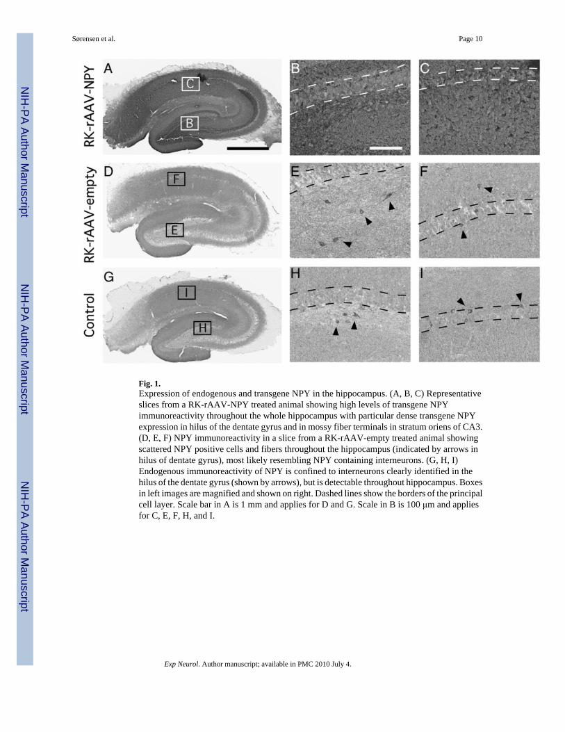
Fig. 1.Expression of endogenous and transgene NPY in the hippocampus. (A, B, C) Representativeslices from a RK-rAAV-NPY treated animal showing high levels of transgene NPYimmunoreactivity throughout the whole hippocampus with particular dense transgene NPYexpression in hilus of the dentate gyrus and in mossy fiber terminals in stratum oriens of CA3.(D, E, F) NPY immunoreactivity in a slice from a RK-rAAV-empty treated animal showingscattered NPY positive cells and fibers throughout the hippocampus (indicated by arrows inhilus of dentate gyrus), most likely resembling NPY containing interneurons. (G, H, I)Endogenous immunoreactivity of NPY is confined to interneurons clearly identified in thehilus of the dentate gyrus (shown by arrows), but is detectable throughout hippocampus. Boxesin left images are magnified and shown on right. Dashed lines show the borders of the principalcell layer. Scale bar in A is 1 mm and applies for D and G. Scale in B is 100 μm and appliesfor C, E, F, H, and I.
Sørensen et al. Page 10
Exp Neurol. Author manuscript; available in PMC 2010 July 4.
NIH
-PA Author Manuscript
NIH
-PA Author Manuscript
NIH
-PA Author Manuscript

Fig. 2.Transgene NPY decreases seizure duration in rapid kindling. (A) Mean afterdischarge (AD)threshold (μA) inducing focal epileptiform activity of more than 5 s duration. (B) Mean numberof suprathreshold stimuli needed to reach seizure stage 1–5 during 40 rapid kindlingstimulations. (C) Mean AD duration at threshold (th) stimuli intensity, and during seizure stage1–5 at suprathreshold stimulation intensity. In all figures, black bars represent RK-rAAV-NPY(n=12) and white bars represent RK-rAAV-empty (n=9) treated animals, respectively. Valuesare mean±SEM, *p<0.05, **p<0.01, ***p<0.001, t-test.
Sørensen et al. Page 11
Exp Neurol. Author manuscript; available in PMC 2010 July 4.
NIH
-PA Author Manuscript
NIH
-PA Author Manuscript
NIH
-PA Author Manuscript
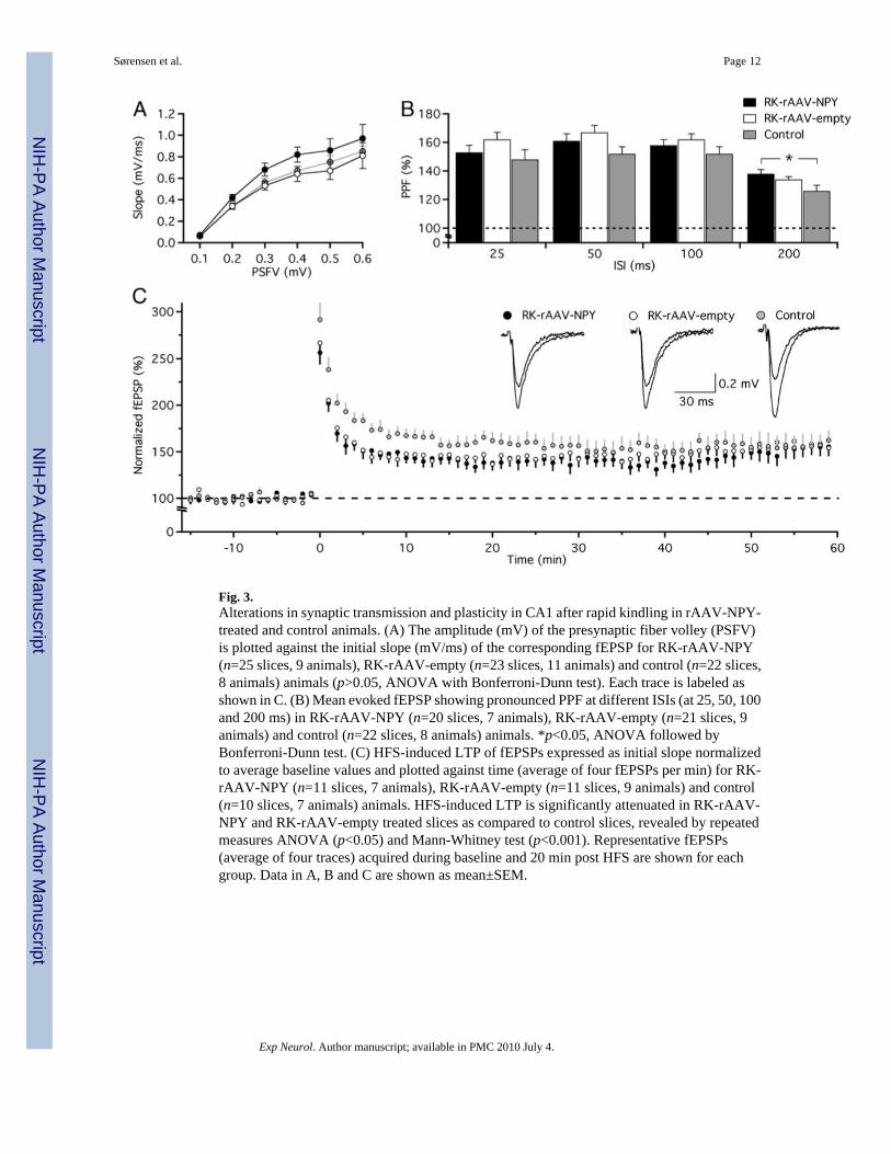
Fig. 3.Alterations in synaptic transmission and plasticity in CA1 after rapid kindling in rAAV-NPY-treated and control animals. (A) The amplitude (mV) of the presynaptic fiber volley (PSFV)is plotted against the initial slope (mV/ms) of the corresponding fEPSP for RK-rAAV-NPY(n=25 slices, 9 animals), RK-rAAV-empty (n=23 slices, 11 animals) and control (n=22 slices,8 animals) animals (p>0.05, ANOVA with Bonferroni-Dunn test). Each trace is labeled asshown in C. (B) Mean evoked fEPSP showing pronounced PPF at different ISIs (at 25, 50, 100and 200 ms) in RK-rAAV-NPY (n=20 slices, 7 animals), RK-rAAV-empty (n=21 slices, 9animals) and control (n=22 slices, 8 animals) animals. *p<0.05, ANOVA followed byBonferroni-Dunn test. (C) HFS-induced LTP of fEPSPs expressed as initial slope normalizedto average baseline values and plotted against time (average of four fEPSPs per min) for RK-rAAV-NPY (n=11 slices, 7 animals), RK-rAAV-empty (n=11 slices, 9 animals) and control(n=10 slices, 7 animals) animals. HFS-induced LTP is significantly attenuated in RK-rAAV-NPY and RK-rAAV-empty treated slices as compared to control slices, revealed by repeatedmeasures ANOVA (p<0.05) and Mann-Whitney test (p<0.001). Representative fEPSPs(average of four traces) acquired during baseline and 20 min post HFS are shown for eachgroup. Data in A, B and C are shown as mean±SEM.
Sørensen et al. Page 12
Exp Neurol. Author manuscript; available in PMC 2010 July 4.
NIH
-PA Author Manuscript
NIH
-PA Author Manuscript
NIH
-PA Author Manuscript