Neuropeptide Y and Memory Processing
549
DOTIC Q : .JAA 0 41991 AD 4" GRANT NO: DAMD17-90-Z-0027 TITLE: CENTRAL & PEREPHERAL SIGNIFICANCE OF NEUROPEPTIDE Y 00 PRINCIPAL INVESTIGATOR: James I. Koenig, Ph.D. 0 M CONTRACTING ORGANIZATION: New York Academy of Sciences N 2 East 63rd Street New York, New York 10021 REPORT DATE: November 15, 1990 TYPE OF REPORT: Final Proceedings PREPARED FOR: U.S. ARMY MEDICAL RESEARCH AND DEVELOPMENT COMMAND Fort Detrick, Frederick, Maryland 21702-5012 DISTRIBUTION STATEMENT: Approved for public release; distribution unlimited The findings in this report are not to be construed as an official Department of the Army position unless so designated by other authorized documents.
Transcript of Neuropeptide Y and Memory Processing

DOTICQ : .JAA 0 41991
AD
4"
GRANT NO: DAMD17-90-Z-0027
TITLE: CENTRAL & PEREPHERAL SIGNIFICANCE OF NEUROPEPTIDE Y
00PRINCIPAL INVESTIGATOR: James I. Koenig, Ph.D.
0M CONTRACTING ORGANIZATION: New York Academy of SciencesN
2 East 63rd StreetNew York, New York 10021
REPORT DATE: November 15, 1990
TYPE OF REPORT: Final Proceedings
PREPARED FOR: U.S. ARMY MEDICAL RESEARCH AND DEVELOPMENT COMMANDFort Detrick, Frederick, Maryland 21702-5012
DISTRIBUTION STATEMENT: Approved for public release;distribution unlimited
The findings in this report are not to be construed as anofficial Department of the Army position unless so designated byother authorized documents.

SECURITY CLASSIFICATION OF THIS PAGE
REPORT DOCUMENTATION PAGE Form Approved
la. REPORT SECURITY CLASSIFICATION lb RESTRICTIVE MARKINGSO
Unclassified
2a. SECURITY CLASSIFICATIOt AUTHORITY 3 DISTRIBUTION / AVAILABILITY OF REPORT
Approved for public release;2b. DECLASSIFICATION I DOWNGRADING SCHEDULE distribution unlimited
4. PERFORMING ORGANIZATION REPORT NUMBER(S) 5. MONITORING ORGANIZATION REPORT NUMBER(S)
6a. NAME OF PERFORMING ORGANIZATION 6b. OFFICE SYMBOL 7a. NAME OF MONITORING ORGANIZATIONNew York Academy of Science (if applicable)
6c. ADDRESS (City, Statt, and ZIP Code) 7b. ADDRESS (City, State, and ZIP Code)
2 East 63rd StreetNew York, New York 10021
Ba. NAME OF FUNDING/SPONSORING 8b. OFFICE SYMBOL 9. PROCUREMENT INSTRUMENT IDENTIFICATION NUMBERORGANIZATION U.S. Army Medical (If applicable)
Research & Development Command SGRD-RMI-S Contract No. DAMDl7-90-Z-0027
8c. ADDRESS (City, State, and ZIP Code) 10. SOURCE OF FUNDING NUMBERSPROGRAM PROJECT TASK WORK UNITFort Detrick ELEMENT NO. NO. NO. ACCESSION NO.
Frederick, Maryland 21702-5012 ......
11. TITLE (Include Security Classification)
Central & Perepheral Significance of Neuropeptide Y
12. PERSONAL AUTHOR(S)James I. Koenig, Janet M. Allen
13a. TYPE OF REPORT 13b. TIME COVERED 14. DATE OF REPORT (Year, Month, Day) IS. PAGE COUNTFinal Proceedings FROM TO 15 November 1990
16. SUPPLEMENTARY NOTATION
Conference held April 2-4, 1990
17. COSATI CODES 1 18. SUBJECT TERMS (Continue on reverse if necessary and identify by block number)FIELD GROUP SUB-GROUP Conference, Neuropeptides, RA I
19. ABSTRACT (Continue on reverse if necessary and identify by block number)In this paper, we report for the first time, the synthesis of NPY receptor antagonists
using a novel strategy based on screening of peptide analog mixtures. In the synthesis ofthese receptor antagonists, a series of NPY analog mixtures was first synthesized using asolid-phase synthetic technique. The antagonist-containing mixture was then identified byscreening these analog mixtures using a bioassay, and two potent NPY receptor antagonists,designated PYX-I and PYX-2, were isolated from this mixture. This analog mixture-screeningstrategy may be used as a general method in developing receptor antagonists for many otherpeptides.
20D DISTRIBUTION rAVAILA13oLITY OF ABSTRArT 21. ABSTRACT CECURITY. AS A OG0 -UNCLASSIFIED/UNLIMITED 0] SAME AS RPT 0] DTIC USERS Unclassified
22a. NAME OF RESPONSIBLE INDIVIDUAL 22b TELEPHONE (include Area Code) I22c. OFFICE SYMBOL
Mrs, Virginia M. Miller 301 663-7325 SG R-SDOForm 1473, JUN 86 Previous editions are obsolete. SECURITY CLASS;IFICATION OF THIS PAGE

R:77 7 7,7770 c e s i 7 7 7 7 7 7 7 7 7 7 7 7 7 7 7 ..............
P
entr I "andPet"i h eralignif ica, hic-fte of'.
orqp
,,,.,--,.,..And s.. Helatw tidb
s N
X.
Wltor',.-.-JAhet Wl Allen
JaMestKoeni9

ANNALS OFTHE NEW YORK ACADEMY
OF SCIENCES
Volume 611
EDITORIAL STAFFExecutive EditorBILL BOLANDManaging EditorJUSTINE CULLINAN The New York Academy of SciencesAssociate Editor 2 East 63rd StreetCOOK KIMBALL New York. New Y'ork iOJ21
THE NEW YORK ACADEMY OF SCIENCES(Founded in 1817)
BOARD OF GOVERNORS, 1990
LEWIS THOMAS, Chairman of the BoardCHARLES A. SANDERS, PresidentDENNIS D. KELLY, President-Elect
Honorary Life GovernorIRVING J. SELIKOFF
Vice-Presidents
DAVID A. HAMBURG CYRIL M. HARRISPETER D. LAX CHARLES G. NICHOLSON
HENRY A. LICHSTEIN, Secretary-Treasurer
Elected Governors-at-LargeJOSEPH L. BIRMAN FLORENCE L. DENMARK LAWRENCE R. KLEINGERALD D. LAUBACH LLOYD N. MORRISETI GERARD PIEL
WILLIAM T. GOLDEN, Past Chairman HELENE L. KAPLAN. General Counsel
OAKES AMES, Executive Director

CENTRAL AND PERIPHERALSIGNIFICANCE OF
NEUROPEPTIDE Y ANDITS RELATED PEPTIDES
Acccioo
For
By
UtDi I
fool

I--w
ANNALS OF THE NEW YORK ACADEMY OF SCIENCES
Volume o I 1
CENTRAL AND PERIPHERALSIGNIFICANCE OF
NEUROPEPTIDE Y ANDITS RELATED PEPTIDES
Edited by Janet M. Allen and James 1. Koenig
010
The New York Academy of SciencesNew York, New York
1990

CopyriRht c 1990 by the New York AcademY of 'Sciences. All rights reserved. Under the provisions ofthe UnitedStates Copyrighti At of 1976. individual readers qf the Annals are permitted to make fair use of the material in themfor teaching and rese'arch. Permission isgranted to) quote jiom the Annals provided that thy customary' atcknowl.,'dgmvrt is made of the source. Mafterial in the Annals mnas be republished only by permission of the Academy.A4ddress inquiries to the Er', wir e Editor ait the New York Ac ademts of Scienccy.
Copying fees: f or each opy of tin artic le made beyond the free copying permitted under Section 107 or 108 ofthe 1976 Copyright Act. ii fee should be pa~id through the Copyright Cleoramnce Center. Inc., 21 Congress St..Salem. MA 0/970. For articles of more than 3 pages the copying fee is $1.75.
G) The paper used itt this publit ation meets the ,ninimum requirements of Amnerican National Standard forInfo~rmation Scienc es- Permanence of Paperf(or Prinited Library Materials. ANSI Z39.48-1984.
Cover (paper edition): Three-dimenisional modi'l or neuropeptide Y.
Library of Congress Cataloging-in-Publication Data
Central and peripheral significance of) neuropeptide Y and its relatedpeptidles / edited by Janet M. Allen and James 1. Koenig,
p. cm. - (Annals of the New York Academy of Sciences. ISSN0077-8923 ;sv. 611)
Based on a conference held on Apr. 2-4, 1990 in Baltimore. Md..sponsored by the New York Academy of Sciences.
Includes index and bibliographical references.ISBN 0-89766-653-4 (cloth : ilk. paper). - ISBN 0-89766-654-2
(pbk. : alk paper)1Nearopeptidle Y-Congresses. I. Allen. Janet M. If. Koenig.
James Irvin, 1954- . Ill. New York Academy of Sciencos.IV. Series,
JDNLM: 1. Central Nervous System-pysiology---congresses.2. Neuropeptide Y-metabol ism- congresses.. Peripheral Nerves-physiology- con greqsses. WI AN626YL s. 6111/ WL 1f44 C397 19901Q If.N5 vol. 6111QP552.N381500 s-dc2O1599. .018 81DNLM/DLCfor Library of Congress 90-13397
CIP
B-BPrinted in the United States of America
ISBN 0-89766-653-4 (cloth)ISBN 0-89766-654-2 (paper)
ISSN 0077-8923

ANNALS OF THE NEW YORK ACADEMY OF SCIENCES
Volume 611November 15, 1990
CENTRAL AND PERIPHERAL SIGNIFICANCEOF NEUROPEPTIDE Y AND ITS
RELATED PEPTIDESa
Editors and Conference ChairsJANET M. ALLEN AND JAMES I. KOENIG
CONTENTS
Preface. By the Editors ..................................................... xiii
Part I. Neuropeptide Y: Structure and FunctionNeuropeptide Y and Its Receptor Antagonists: Use of an Analog
Mixture-Screening Strategy. By KAZUHIKO TATEMOTO ................... I
Neuropeptide Y Receptor Subtypes, Y I and Y2.. By CLAES WAHLESTEDT,LARS GRUNDEMAR, ROLF HAKANSON, MARKUS HEILIG, GREGORY H.SHEN. ZOFIA ZUKOWSKA-GROJEC, and DONALD J. REIS .................. 7
Biologically Active Neuropeptide Y Analog, By JAROSLAV H. BOUBLIK,MARK A. SPICER, NEAL A. SCOTT, MARVIN R. BROWN,
and JEAN E . RIVIER ................................................... 27
Signal Epitopes in the Three-Dimensional Structure of Neuropeptide Y:Interaction with Y,, Y2, and Pancreatic Polypeptide Receptors. ByTHUE W. SCHWARTZ, JANNIE FUHLENDORFF, LISE L. KIEMS, METTE S.
KRISTENSEN, MEES VERVELDE, MAIREAD O'HARE, JOHN L.KRSTENANSKY, and BERITH BJORNHOLM ................................ 35
Comparison of the Neuropeptide Y Receptor in the Rat Brain and Intestine. ByIAN L. TAYLOR, PETER J. MANNON, GREGORY G. HEINTZ,LYNN M. KAISER, and TOAN D. NGUYEN ............................... 48
Neuropeptide Y Receptors: Autoradiographic Distribution in the Brain andStructure-Activity Relationships. By RfMI QUIRION, JEAN-CLAUDEMARTEL, YVAN DUMONT, ALAIN CADIEUX, FRANCOIS JOLICOEUR,
SERGE ST-PIERRE, and ALAIN FOURNIER ................................ 58
Pancreatic Polypeptide and Peptide YY Gene Expression. By S. D. KRASINSKI,M. B. WHEELER, A. S. KOPIN, and A. B. LEITER ....................... 73
Molecular Structure of Neuropeptide Y. By JANET M. ALLEN ................. 86
"The papers in this volume were presented at a conference entitled Central and Peripheral Signif-icance of Neuropeptide Y and Its Related Peptides, which was held by the New York Academy ofSciences in Baltimore, Maryland on April 2-4. 1990.

Regulation of the Human Neuropeptide Y Gene. By CAROLYN A. MINTH andJACK E. DIXON...........................................................99
Part HI. Role of Neuropeptide Y in the Regulation of theCirculatory System
Neuropeptide Y and Central Cardiovascular Regulation: Focus on Its Role as aCotransmitter in Cardiovascular Adrenergic Neurons. B 'y K. FUXE. J. A.AGUIRRE, L. F. AGNATI. G. VON EuI.ER. P. HEDLUND. R. COVE$&AS.M. ZOLI. B. BJELKE. and P. ENEROTH....................................Ill
Neuropeptide Tyrosine in the Cardiovascular System. By JOHN WHARTON andJULIA M. POLAK ......................................................... 133
In Vitro Effects of Neuropeptide Y at the Vascular Neuroeffector Junction. B 'THOMAS C. WESTFALL, XIAOLI CHEN, ANITA CIARLEG~io. KRISTINAHENDERSON, KATHERINE DEL VALLE, MELISSA CURFMAN-FAI.VEY, andLINDA NAES ............................................................. 145
Importance of Neuropeptide Y in the Regulation of Kidney Function. ByPONTus B. PERSSON. GERALD GIMPL, and RUDOLF E. LANG................156
Neuropeptide Y and Sympathetic Neurotransmission. B "N JAN M. LUNDBERG.ANDERS FRANCO-CERECEDA. JEAN-SILVAIN LACROIX. andJOHN PERNOW................................................... 166
Regulation of Neuropeptide Y Gene Expression in Rat Brain. B '% NILSLLNDEFORS, STEFAN BRENt. MARiO HERRERA -MARSCHITZ, and1-AKAN PERSSON.........................................................175
Part III. Neuropeptide Y and the Central Nervous System
Neuropeptide Y in Cortex and Striatum: Ultrastructural Distribution andCoexistence with Classical Neurotransmitters and Neuropeptides.By CHIYE AOKI and VIRGINIA M. PICKEL .................................. 186
Modulation of Synaptic Transmission in Hippocampus by Neuropeptide Y:Presynaptic Actions. By WILLIAM F. COLMERS.............................206
Neuropeptide Y-Cholinergic Interactions in Neocortical Tissue. By JENNIFER J.POULAKOS, EDWIN M. MEYER. CORINNE R. PPUYSERS. andWILLIAM J. MILLARD .................................................... 219
Neuropeptide Y and Memory Processing. By' JOHN E. MORLEY andJAMES F. FLOOD .. . . . . . . . . . . . . . . . . . . . . . . . . . . 226
Part IV. Functions of Neuropeptide Y in the Hypothalamus
Ultrastructural Localization of Neuropeptide Y in the Hypothalamus. B%-GEORGES PELLETIER ..................................................... 232
Neuropeptide Y in the Circadiar Timing System. B ' ROBERT Y. MOORE
and J. PATRICK CARD .................................................... 247
Role of Neuropeptide Y in Reproductive Function. By JOHN K. MCDONALD . .. 258

Hypothalamic Neuropeptide Y: a Circuit in the Regulation of GonadotropinSecretion and Feeding Behavior. By S. P. KALRA, A. SAHU, P. S. KALRA,and W. R. CROWLEY .................................................... 273
Hypothalamic Neuropeptide Y in Relation to Energy Balance. By SARAH FRYER
LEIBOWITZ .............................................................. 284
Regulation of NISH Secretion by Neuropeptide Y in Amphibians. By J. M.DANGER, M. C. TONON, L. CAZIN, B. G. JENKS, A. FASOLO,G. PELLETIER, and H. VAUDRY ........................................... 302
Regulation of the Hypothalamo-Pituitary Axis by Neuropeptide Y.
By JAMES I. KOENIG ..................................................... 317
Neuropeptide Y and the Anterior Pituitary. By D. J. O'HALLORAN,32
'H-NM andComputer Simulation. By R. D. FEINSTEIN, M. SPICER,J.BoBLKJ.P. TUAEJ.RVEMBOWadM. GOODMAN .. 336
Chaactriztio ofNeuropeptide Y Immunoreactivity in Dog Plasma by HighPerormnceLiquid Chromatography and Radioimmunoassay. ByP. ENAAYAEM. R. WARNER, M. N. LEVY, D. W. JACOBSEN, and
C . M FE RAR O .... ... ... ... .... ... ... ... ... ... ... ... ... 340
PpieYY/Neuropeptide Y Receptors in Small Intestine: Characterization,Signal Transduction, and Expression during Cell Differentiation. By
T. VOISIN, C. RoUYER-FESSARD, and M. LABURTHE ....................... 343
Structural Characterization of Y, and Y, Receptors for Neuropeptide Y andPeptide YY. By S. P. SHEIKH and J. A. WILLIAMS ........................ 347
Peptide YY Receptors in Mammalian and Avian Brains. By AIO INuI,MINORU OKITA, MASAKI MIURA, YOSHIAKI HIROSUE, MASAHARUNAKAJIMA, and SHIGEAKI BABA...........................................350
Neuropeptide Y Inhibiti Phosphorylation of 87-kDa Protein in Rat VasDeferens. By GABRIEL FRIED and PAUL GREENGARD ....................... 353
Cardiac Properties and Conformational Studies of Neuropeptide Y (NPY) andNPY(17-36). By A. BALASUBRAMANIAM, S. SHERIFF, V.RENUGOPALAKRISHNAN. S. G. HUANG, M. PRABHAKARAN,W. T. CHANCE, and J. E. FISCHER ........................................ 355
Structure-Activity Relationships of Biologically Active Analogs and Fragmentsof Neuropeptide Y. By M. A. SPICER. J. H. BoUBLIK. R. D. FEINSTEIN,
M. GOODMAN, M. BROWN, and J. RiVIER.................................359
Muscular Neuropeptidy Y Receptors Involved in the Potentiation of theNoradrenaline- Induced Vasoconstriction in Isolated Coronary Arteries. ByJ. PABLO HUIDOBRO-ToRo, Luis EBEL, PILAR MACHO, RAIOL DoMENECH,
ALAIN FOURNIER, and SERGE ST-PIERRE ................................... 362
Structural Study of the N-Terminal Segment of Neuropeptide-Tyrosine. ByMYLtNE FOREST, JEAN-CI A.UDE MARTEL, SERGE ST-PIERRE,
R IMi QUIRION, and ALAIN FOURNIER ................................... 366
Investigations of the Structure-Activity Relationship of Neuropeptide Y. ByA. CADIEUX, M. T-BENCHEKKOUN, A. FOURNIER, and S. ST PIERRE ..... 369
Pharmacological Actions of Neuropeptide Y and Peptide YY in Rat Colon. ByA. CADIEUX, M. T-BENCHEKROUN, A. FOURNIER, and S. S' -PIERRE ..... 372
Regulation of Neuropeptide Y Transcription and mRNA Levels by NerveGrowth Factor, Glucocorticoids, Cyclic AMP, and Phorbol Ester in PCI2Cells. BY STEVEN L. SABOL and HIROSHI HIGUCHI ...................... 376
Strong Evolutionary Conservation of "europeptide Y between Mammals,Chicken, Goldfish and Horned Shark. By ANDERS BLOMQVIST, INGRIDLUNDELL, and P \N LARHAMMAR ..................................... 378
Cell-Type Specific Modification of Peptide ,.,ttranslational Processing inPituitary Cel:s. BY IAN M. DICKERSON and RICHARD E. MAINS ........... 379
Novel Brain Specific Transcription Factors Form DNA-Protein Complexes withSPI and TATA Regions of the Rat Neuropeptide Y Gene Promoter:Enhancement of Complex Formation by Nonneuronal Factors. By Y. M.OO AND HAKAN PERSSON ............................................. 382
Defect of Binding of Nuclear Factors from Embryonic and Newborn BrainNuclear Extracts from Autoimmune NZB Mice with the Rat NeuropeptideY Gene Promoter. By YUET M . Oo .................................... 388
He 90481: A Competitive Nonpeptidergic Antagonist at Neuropeptide YReceptors. By M. C. MICHEL and H. 1. MOTULSKY ...................... 392
Neuropeptide Y and Peptide YY Interact with Rat Brain Sigma and PCPBinding Sites. By F. J. ROMAN, X. PASCAUD, 0. DUFFY, D. VAUCHF, B.M ARTIN. and J. L. JUNIEN ............................................. 395
Characterization and Use of Four Anti-NPY Monoclonal Antibodies to StudyNPY-Receptor Interaction. By E. GROUZMANN, P. WALKER, C. BOHUON,M. BURNIER. E. COMOY, H. R. BRUNNER. and B. WAEBER .............. 397
Neuropcptide Y and Its C Terminal Fragments Attenuate Anion Secretion in RatJejunum Mucosa. By HELEN M. Cox and ALAN W. CUTHBERT .......... 399
Neuropeptide Y Gene Expression Is Dependent upon a SPI-Like DNA-BindingProtein. By CAROLYN A. MINTH and JACK E. DIXON .................... 403
Sympathetic Stimulation-Evoked Overflow of Neuropeptide Y andNorepinephrine from the Heart. By MARGARET R. WARNER. PREENIEDESILVA SENANAYAKE. CARIOS M. FERRARIO, and MATTHEW N. LEVY .. 404
Effect of Neuropeptide Y on the Biphasic Response of the Rabbit Ear Artery toNorepinephrine and Sympathetic Field Stimulation. By JOHN G. DUESLER,JR., ROBERT N. DALY, and J. PAUl. HIEBLE ........................... .405
Platelets as a Source and Site of Action for Neuropeptide Y. By A. K. MYERS.M. Y. FARHAT, G. H. SHEN, W. DEBINSKI. C. WAHI FSTEDT, and Z.ZUKOW SKA-G ROJFC .............. .................................... 408

Sources and Vasopressor Efficacy of Circulating Neuropeptide Y during Acuttand Chronic Stress in Rats. B V ZOFIA ZUKOWSKA-GROIEC. GREGORY HSHEN, MARIA KONARSKA, and KICHARD MCCARTY ........................ 412
High Doses of Neuropeptide Y Reduce Blood Pressurc in Anesthetized Rats. BYLARS GRUNDEMAR, CLAES WAHLESTEDT. GREUORY H. SHEN, ZOFIA
ZUKOWSKA-GROJEC, and ROL- HAKANSON.................................415
Neuropeptide Y (NPY), NPY-lmmune Complex. anu NPY(18-36) Effect', onCardiovascular Functian. By N. SCOTT, J. BOUBLIK, J RIVIER. and M.BROWN ................................................................. 418
Neurc'peptide Y and Anglerfish Pe'qtide Amide Inhibit Secretin-StimulatedPancreatic Exocrine Secretion. BV MAREK RuDNICKI, DAVID W.MCFADDEN, AMBIKAIPAKAN BALASUBRAMANIAM, MICHAEL S. NUSSBAUM,
and JOSEF E. FISCHER ............................... ................ 423
Neuropeptide Y Does Not Mediate lschemnic Spinal Cord Injury followingAortic Cross-Clamping in Rabbits. By BORIS W. KuVSHINOFF, TomNOGUEIRA, DAVID W. MCFADDEN. and RICHARD J. FOWL................ 426
lmmunocytochemical Localization of Neuropeptide Y and Its C-TermninalFlanking Peptide in the Human Heart. By S. GULBENKIAN. N. COSTAANDRADE, J. WHARTON, J. M. POLAK, J. QUEIROZ E MELO. and J. F.DA-VID-FERREIPA ......................................................... 429
Neuropeptide Y and Nonadrcnergic Mechanisms in the Sympathetic VascularControl of the Nasal Mucosa. By J. S. LACROIX, W. LEHMANN. andJ. M. LUNDBERG ........................................................ 432
Neuropeptide Y Causes Nonadrenergic Vasoconstriction in the Eye. Brain, andSome Other Tissues in Rabbits. By Siv F. E. NILSSON .................... 435
Effects of Neuropeptide Y and Noradrenaline on the Uterine Artery. B-iGABRIEL FRIED, MARIANNE THORESEN, and ULF SAML'ELSSON.............439
Reduced Inhibitory tActions of Sympathetic Stimulation and ExogenousNeuropeptide Y on Cardiac Vag;,7 Action in the Presence of Peptide YY.By ERICA K. POTTER and D. I. MCCLOSKEY ............................ 442
Pre- and Postjunctional Actions of Neuropeptide Y and Related Peptides. B -ERICA K. POTTER, L. MITCHELL, M. J. D. MCCLOSKEY. A. TSENG.M. PERSICO, AMANDA E. GOODMAN, MARISSA G. DE PIETRA, MARIONLOUGHMAN, J. SHINE, and D. I. MCCLOSKEY ........................... 444
Neuropeptide Y-A Marker for Noradrenaline Exocytosis. By ALBERT SCHOMIGand MARKUS IIAASS................................................. 447
Common Features of Neuropeptide Y and Noradrenaline Release in Guinea PigHeart. By MARKUJS HAASS. GERT RICHARDT, RUDOLF E. LANG. andALBERT SCHOMIG................................................... 450
Effect of Neuropeptide Y on Stimulated Renin Secretion. B ' JEAN-FRANI OISAuBERT, BERNARD WAEBER, JUJRG N' 'SSBERGER, andHANS R. BRUNNER............ ..................................... 453
Expression of Neuropeptide Y-Like Immunioreactivity Begins after Initiation ofNicotirc Synapse Formation in Developing Sympathetic Ganglia of theBullfrog Tadpole. By J. P. HORN and W. D. STOFER..................... 455

Neuropeptide Y Presynaptic Effects in H-ippocampal Slice In Vitro Are Y,Receptor Mediated but Not via Inhibition of Adenylate Cyclase. By G. JKLAPSTEIN. K. A. TREHERNE, and W. F. COLMERS ........................ 457
Effects of Haloperidol and Cortical Lesions on the Level of Neuiopeptide YmRNA in the Striatumn of the Rat: A Study by In Situ HybridizationHistochemistry. By P. SALIN and M. F. CHESSELET ........................ 459
Regulation of Neuropel tiets Y in Neural and Cardiac Culture. By K. L. MAREKand R. E. MAINS ........................................................ 463
Clinical Evaluation of Neuropeptide Y as a Plasma Marker of Tumors Derivedfrom Neural Crest. By MASAHIKO SONE, TORAICHI MOURt. KAZUHIROTAKAHASHI, KEIICHI ITOL. YUTAKA HAYASHI, MAKOTO OHNEDA, OSAMUMURAKAMI, and KAORU YOSHINAGA......................................465
Neuropeptide Y Regulation of LHRH Release in Ewe Median Eminence:Immunocytochemistry, Tissue Content, and In Vivo Analysis. By J. P.ADvis. C. D. CONOVER. J. K. McDONALD, and R. 0. KUIJIS ........... 468
A Role for Neuropeptide Y in the Mediation of Glucocorticoid- Induced Changesin Hypothalamic Function. By- ROGER CORDER. FRANI;OIS P. PRALONG.DORA TURNILL. and ROLF C. GAILLARD .................................. 471
Comparison of Hypotension-Induced Neuropeptide Y Release in Rats Subjectedto Hemorrhage, Endotoxemnia, and Infusions of Vasodepressor Agents. By'ROGER CORDER, FRAN4;OIS P. PRALONG. and ROLF C. GAILLARD............474
Food Deprivation Modulates Neuropeptide Y Gene Expression in the MurineHypothalamus. By STREAMsON C. CHUA, JR ............................... 477
Steroid Hormone Regulation of Brain Neuropeptide Y mRNA Levels. Bx'P. CAMP and J. D. WHITE ................................................ 478
Hypothalamic Neuropeptide Y Expression in Normal and Aberrant MetabolicHomeostasis. ByV JEFFREY D. WHITE, M4,RYANN KERSHAW, GERARDSANACORA, DAVID OLCHOVSKY, JUDITH A. FINKELSTEIN, and MICHAELBERELOWITZ ............................................................ 480
Central Actions of Neuropeptide Y and Its Related Peptides in the Dog, withSpecial Reference to Their Effects on the Hypothalamic -Pituitary -AdrenalAxis, Feeding Behavior, and Thermoregulation. B 'y MINORU OKITA. AKIOINUI, MASAKI MIURA. YOSHIAKI HIROSUE, MASAHARu NAKAJIMA, andSHIGEAKi BABA .......................................................... 483
Effects of Neuropeptide Y and Its Related Peptides on Feeding and LearningBehaviors in the Mouse. By MASAHARu NAKAJIMA. AKIO INUL MINORUOKITA, YOSHIAKI HIROSUE, MASAKI MfURA, NORIO UIMORI. and SHIGEAKIBABA...................................................................486
Evidence That Neuropeptide Y Elicits Eating by Acting in the CaudolateralParaventri'-ular/Perifornical Hypothalamus. By B. GLENN STANLEY,WILLIAM 1AGDALIN, and SARAH F. LEIBOWITZ ........................... 489
Chronic and Continuous ICV Infusion of Neuropeptide Y Disrupts theNycihemeral Feeding Patterns in Rats. B ' B. BECK, A.STRICKER-KRONGRAD, J. P. NIOLS and C. BURLET......................491
Neuropeptide Y in Pediatric Neural Crest Tumors: Correlation with Malignancy,Metastases, and Clinical Outcome. By P. KOGNER, E. THEODORSS0N, and0. B16RK ...................................................... 495
Reduction of Neuropeptide Y-Induced Feeding in Tumor-Bearing Rats. ByW. T. CHANCE, J. RAMO, S. SHERIFF, F. ZHANG, J. E. FISCHER, andA. BALASUBRAMANIAM.............................................. 497
Changes in Hypothalamic Neuropeptide Y during Immunization in the Rat. ByE. Comoy, N. PAGES, P. PROTAIS, and C. BoHUON...................... 500
Localization. Characterization, and Neuroendocrine Action of Neuropeptide Y inthe Trout Brain. By J. M. DANGER, B. BRETON, M. VALLARINO, S.ST-PIERRE, 0. PELLETIER, and H. VAUDRY ............................. 504
Anatomical Distribution and Biochemical Characterization of NeuropeptideY-Like Immunoreactivity in the Cat Brain and Pituitary. By L. LEGER,J. M. DANGER, Y. CHARNAY, G. PELLETIER, H. VAUDRY, andM. JOUVET............... ...................................... 508
Localization and Characterization of Neuropeptide Y-Like ImmunoreactivePeptides in the Nervous System and Midgut of Locusta migratoria and inthe Brain of Sarcophaga bullata. By LILIANE SCHOOFS, JEAN-MICHELDANGER, SYLVIE itGou, ARNOLD DE LooF, and HUBERT VAUDRY ......... 513
Electrophysiological Action of Neuropeptide Y on Cultured Frog PituitaryMelanotrophs. By C. BASILLE, E. LoulSET, J. M. DANGER, A. FOURNIER,H. VAUDRY, and L. CAZIN ........................................... 516
Interactions between Norepinephnine and Neuropeptide Y in RegulatingPancreatic Islet Hormone Secretion. By S. L. MILGRAM, J. K. MCDoNALD,and B. D. NOE .................................................. 518
Preproneuropeptide Y roRNA Expression in Glial Cell Cultures of the Neonatebut Not 21-Day-Old Rat Brain. By B. A. MASTERS, C. R. PRUYSERS,W. J. MILLARD, E. M. MEYER, and J. J. POULAKOS .................... 522
Neuropeptide Y in Relation to Metabolism and Nutritional State. By M.JHANWAR-UNIYAL, J. A. MENENDEZ, D. M. ATRENS, B. BECK,A. BURLET, C. BURLET, and S. F. LEIBOWITZ .......................... 525
Neuropeptide Y: Comparisons with Norepinephrine in Relation to FeedingBehavior. By S. E. KYRKOULI, G. B. STANLEY, and S. F. LEIBOWITZ .... 527
Neuropeptide Y Effects on Feeding Behavior: Relationship to Norepinephrineand the Circadian System. By DONNA L. TEMPEL and SARtAH F.LEIBOWITZ.................................. ...................... 529
Index of Contributors .......................... .......................... 531

Financial assistance was received from:
Supporter
* U.S. ARMY MEDICAL RESEARCH AND DEVELOPMENT COMMAND
Contributors
" BURROUGHS WELLCOME CO." ICI PHARMACEUTICALS GROUPb THE R.W. JOHNSON PHARMACEUTICAL RESEARCH INSTITUTE" LiLLY RESEARCH LABORATORIES" MERCK SHARP & DOHME RESEARCH LABORATORIES* MERRELL DOW RESEARCH INSTITUTE* NATIONAL INSTITUTE OF MENTAL HEALTH/NATIONAL
INSTITUTES OF HEALTH* NATIONAL SCIENCE FOUNDATION" PFIZER CENTRAL RESEARCH, UK• PFIZER CENTRAL RESEARCH, USA" SERONO USA" SMITHKLINE BEECHAM PHARMACEUTICALS
The New York Academy of Sciences believes it has a responsibility to provide an openforum for discussion of scientific questions. The positions taken by the participants inthe reported conferences are their own and not necessarily those of the Academy. TheAcademy has no intent to influence legislation by providing such forums.

PrefaceIn 1982. Drs. K. Tatemoto and V. Mutt first published the isolation and characterizationof neuropeptide Y (NPY). They could not have anticipated the enthusiastic interest inNPY that has evolved over the last eight years or realized how their discovery wouldinfluence research workers in diverse fields of neuroscience, basic chemistry, cardiovas-cular physiology and gastroenterology. NPY consists of 36 amino acids being character-ized by an amino terminal tyrosine residue and a carboxy terminal tyrosine amide moiety.As tyrosine is represented by Y in the single letter nomenclature for amino acids, it isthese tyrosines that are denoted by Y in the name. In their original description of NPY,Tatemoto and Mutt drew attention to the homology of this newly discovered peptide to thepancreatic polypeptide family of peptides, namely pancreatic polypeptide and peptide YY(PYY). Subsequently, the cDNA and gene encoding NPY were isolated and studies of theregulation of NPY gene expression as well as the physiology of NPY progressed. As anindication of the importance of this family of peptides in biological systems, well over1000 papers have been published on NPY and its related peptides in terms of the chem-istry, physiology, pharmacology and anatomy. A
Although a meetin-g on NPY took place two years ago, the current meeting sponsoredby the New York Academy of Sciences represents the firsit open meeting on the NPYfamily of peptides.-Open communications by young investigators were encouraged andpresented in parallel with the more traditional review talks. However, due to the ubiq-uitous nature of NPY thoughout the body, the scope of the meeting had to be curtailed tokeep it within a reasonable time span. We attempted to highlight several areas of NPYresearch, including an in-depth discussion of the structure of the native NPY molecule,which we hope will serve as a guide to aid in the development of analogues to assess thecritical feature of the molecule essential for receptor binding and signal transduction.Emphasis was also placed on the molecular mechanisms regulating the expression of thegenes encoding the peptides. Since NPY is concentrated in tissues affecting cardiovas-cular performance, attention was directed to the significance of NPY in regulating cardiacand renal function. Finally, much of our present knowledge of NPY has been derivedfrom studies of NPY in neuronal systems. Presentations on the anatomy and physiologyof NPY in higher brain centers are followed by an in-depth review of the function of NPYin the hypothalamus, concentrating on feeding, reproduction and pituitary function. Theoverall goal of the conference was to integrate classical neuropeptide studies with modernapproaches to the study of these substances, so that the most exciting areas of interestcould be identified and explored.
We would like to extend our gratitude to the New York Academy of Sciences, andspecifically to Ms. Renee Wilkerson and Ms Geraldine Busacco, for their efforts inorganizing this conference. We also would like to acknowledge the contributions of all theparticipants, both invited and voluntary, for without excellent presentations and someoneto listen to them, there would not have been a conference. f-inally, we express ourappreciation to the government agencies and companies for their support of this meeting-hopefully, it was a worthwhile investment.
Janet M. AllenJames I. Koenig
xiii

PART I. NEUROPEPTIDE Y: STRUCTURE AND FUNCTION
Neuropeptide Y and Its ReceptorAntagonists
Use of an Analog Mixture-Screening Strategy
KAZUHIKO TATEMOTOPeptide Research Laboratory
Department of Psychiatry and Behavioral SciencesStanford University School of Medicine
Stanford, California 94305
Many of the advances in our knowledge of neuropeptide Y (NPY) and its related peptideshave resulted from the application of new research strategies. For example. a novelstrategy of searching for unknown peptides that possess the C-terminal amide structure ledto the discovery of both NPY' and its related peptide. PYY2, a decade ago. Subsequentstudies by a number of groups employing a variety of new biological, anatomical, andbiochemical approaches have elucidated many important regulatory functions of thesepeptides. -7
In this paper, we report for the first time, the synthesis of NPY receptor antagonistsusing a novel strategy based on screening of peptide analog mixtures. In the synthesis ofthese receptor antagonists, a series of NPY analog mixtures was first synthesized using asolid-phase synthetic technique. The antagonist-containing mixture was then identified byscreening these analog mixtures using a bioassay. and two potent NPY receptor antago-nists, designated PYX-l and PYX-2, were isolated from this mixture. This analog mix-ture-screening strategy may be used as a general method in developing receptor antago-nists for many other peptides.
Neuropeptide Y: Discovery
With the accumulation of knowledge about the structures of many peptide hormonesand neuropeptides, it has become evident that these peptides are produced from theirprecursor proteins by posttranslational processing.' The processing of peptides frequentlyincludes unique modifications of the peptide molecules such as phosphorylation, sul-fation, acetylation, pyroglutamation and C-terminal amidation. C-terminal amidation isknown only to occur in the structures of neuropeptides and peptide hormones in mam-malian tissues. This modification can therefore be exploited as a chemical marker todetect neuropeptides and peptide hormones."
In 1973, we initiated a project to develop a chemical assay for secretin and chole-cystokinin. Our original intention in developing such a chemical assay was to replacelaborious bioassays used during the purification of secretin and cholecystokinin from theintestine. Since these peptide hormones contain the C-terminal amide structure, we pro-posed a novel chemical assay for these hormones. "' This chemical method detected theC-terminal amino acid amide which was cleaved off enzymatically and converted to thefluorescent dansyl derivative, then selectively separated and identified. When porcineintestinal extracts were subjected to this assay, it was found unexpectedly, that theextracts contained several previously unknown peptides with the C-terminal amidestructure. ") This finding led to the isolation of a series of novel intestinal peptide'

2 ANNALS NEW YORK ACADEMY OF SCIENCES
including PHI-27''. 2 and pyy. 2-,' Although these peptides were isolated without anyprior knowledge of their biological activities, each of the isolated peptide amides was laterfound to be biologically active with important neural and/or hormonal properties.9 Basedupon these findings, it was proposed that the isolation of unknown peptides with theC-terminal amide structure would result in the finding of new neuroactive and hormonallyactive peptides. "
The search for new peptides was then extended to the brain where a novel neuropep-tide having a C-terminal tyrosine amide was isolated using this chemical assay.' The 36amino acid peptide was named neuropeptide Y (Y = tryosine), or in short, NPY. Sub-sequent studies have shown that this neuropeptide is a major regulatory peptide in both thecentral and peripheral nervous systems. 3 - 7
Neuropeptide Y: Structure and Function
The primary structure of NPY isolated from porcine brain 3 is shown in Figu-re 1.NPY was also isolated from other species including human, 14 rat, ' 5 guinea-pig, rabbit,' 6
cow, 7 and sheep. '7 These studies suggest that the NPY structure has been well conservedduring evolution. NPY has structural similarities to both PYY and pancreaticpolypeptide. ," The tertiary structures of these peptides also have been shown to besimilar.'...' 9 The structures of cDNAs encoding NPY mRNAs' '20 and clones encodingNPY genes 2' 22 have been elucidated and the primary structures of the precursors ofhuman and rat NPY have been predicted from these studies.
The localization of NPY has been studied using histochemical and immunoassaytechniques. NPY is widely distributed throughout the central and peripheral nervoussystems. NPY is distributed in nerve fibers of the brain, 3 spinal cord,24 blood vessels,2 -
heart, 26 intestine, stomach, pancreas, genital tract,2 5 lung,27 spleen,28 adrenal medulla,29
and other organs. 25 NPY has been shown to coexist with catecholamines in someneurons. -25
.30 Receptor binding studies have indicated the presence of NPY binding sites
in the brain,31 2 spleen 33 kidney,34 and vas deferens. 35
Biological functions of NPY have been extensively studied using various in vivo andin vitro models. NPY induces strong vasoconstrictor actions, 36 inhibits the release ofnoradrenaline at the presynaptic level3 7 and potentiates noradrenaline-evoked vasocon-striction at the postsynaptic level.-" Therefore, NPY may play important roles in sym-pathetic vascular control. 39 NPY strongly inhibits intestinal motility ° and also modulates
1 5 10
Tyr-Pro-Ser-Lys-Pro-Asp-Asn-Pro-Gly-Glu-Asp-Ala-Pro-
15 20 25Ala-Glu-Asp-Leu-Ala-Arg-Tyr-Tyr-Ser-Ala-Leu-Arg-His-
30 35
Tyr-Ile-Asn-Leu-Ile-Thr-Arg-Gln-Arg-Tyr-NH2
FIGURE 1. Primary structure of neuropeptide Y (porcine).

TATEMOTO: ANALOG MIXTURE-SCREENING STRATEGY 3
epithelial ion transport in the intestine, 4' suggesting physiological roles of NPY in gutfunctions. NPY also inhibits glucose-stimulated insulin secretion 42 and exhibits natri-uretic properties. 43 Central administration of NPY induces hypotension and bradypnea.This peptide may therefore be involved in the central control of cardiovascular andrespiratory functions. A dramatic increase in food intake upon central NPY administrationhas been reported, 45- 47 suggesting that NPY may play an important role in feedingbehavior. NPY injection changes the plasma concentrations of leuteinizing hormone,4 "49
growth hormone, prolactin, thyrotropin, 4 gonadotropin-releasing hormone,50 ACTH, s 'and vasopressin. 5' .52 NPY may therefore play significant roles in the secretion of thesehormones. NPY injection into the suprachiasmatic region of the hypothalamus induces ashift in the circadian rhythm. 53 NPY may also play a role in sexual behavior sinceintraventricular injection of NPY suppresses copulatory behaviors. 54
A General Procedure for Synthesis of Receptor Antagonists
The search for receptor antagonists of peptide hormones and neuropeptides is impor-tant toward understanding the mechanisms of actions, elucidating structure-function re-lationships, and introducing new pharmacological drugs. However, the design and syn-thesis of receptor antagonists has proven a difficult and time-consuming task. Despiteattempts at applying structural analysis and computer imaging technology, no efficientgeneral method for designing receptor antagonists has been described.
In the present paper, a general strategy for searching peptide receptor antagonists isproposed (FIG. 2). Using this strategy, the synthesis of peptide receptor antagonists canbe achieved in a short time. This new strategy begins with the design and solid-phasesynthesis of peptide analog mixtures containing hundreds of antagonist candidates. Thereare many ways of generating such peptide analog mixtures. For example, an analogmixture with various amino acid substitutions can be synthesized by dividing the solid-phase resin into several portions at desired positions during the synthesis, coupling adifferent amino acid to each portion, and then recombining the portions for furthercoupling steps. An analog mixture with various chain lengths can similarly be synthesizedby withdrawing portions of the resin at desired positions, and recombining the portions forcleavage from the resin.
After the synthesis of a series of analog mixtures. bioassays of competitive receptorassays are used to screen the mixtures for antagonist activity. The individual receptor
Synthesis of peptide analog mixtures
i screening
Selected analog mixture with antagonist activity
V screening and isolation FIGURE 2. Analog mixture-screening strat-Isolated receptor antagonist egy for developing receptor antagonists.
structural analysis
Re-synthesis of receptor antagonist
characterization

4 ANNALS NEW YORK ACADEMY OF SCIENCES
antagonists are isolated from the mixtures exhibiting antagonist activities using HPLC andother separation techniques, in a manner similar to the isolation of natural peptides fromtissue extracts. The primary structures of the isolated antagonists are then determined, andpeptides identical to the isolated antagonists are synthesized for further study.
This approach allows large numbers of peptide analogs to be synthesized and screenedin a simple and efficient manner. Moreover, studies using this strategy may reveal thestructural requirements for receptor antagonists which could then be used to design andsynthesize more potent antagonists. The analog mixture-screening strategy may have wideapplications in the development of receptor agonists and antagonists, and might also beapplied to the screening of pharmaceutical drugs, as well.
Synthesis of NPY Receptor Antagonists
The novel analog mixture-screening method was successfully applied for the synthesisof potent NPY receptor antagonists. The study began with the synthesis of a series of theNPY analogs each containing one D-amino acid substitution at the C-terminal region ofthe NPY molecule, since the C-terminal region is known to be essential for receptorbinding. 55 The NPY agonist activity of these analogs was then examined by measuring theincrease of intracellular calcium in human erythroleukemia cells 56 and the analogs havinglittle or no agonist activity were selected from these analogs. The selected analogs werefurther subjected to a NPY antagonist assay, which measured inhibition of the NPY-stimulated increase in intracellular calcium in human erythroleukemia cells. Althoughnone of the analogs was found to exhibit antagonist activity, a series of analog mixtureswere synthesized based upon the structures of these selected analogs. After the screeningof these analog mixtures, a (D-Thr 321 NPY analog mixture was found to have antagonistactivity. Two NPY receptor antagonists, designated PYX- I and PYX-2, were isolatedfrom this mixture after three consecutive HPLC steps. Structural analysis revealed thatPYX-I is a decapeptide amide with a modified amino acid at the N-terminus and PYX-2is a decapeptide amide with modified amino acids at both N- and C-termini (FIG. 3).PYX-I corresponded to Ac-[3-(2-6-dichlorobenzyl)-Tyr 2 7. D-Thr32I NPY 27-36, andPYX-2, to Ac-[3-(2-6-dichlorobenzyl)-Tyr 2 7 "36 , D-Thr3 2 I NPY 27-36 (in preparation). Itseems that a D-substitution of threonine at position 32 and a modification of tyrosine atposition 27 converts the C-terminal decapeptide amide of NPY to a receptor antagonist.PYX-I inhibited the NPY action in releasing intracellular calcium in human erythroleu-
1 5 10
3-(CI 2-Bzl)
PYX-1: Ac-Tyr-Ile-Asn-Leu-Ile-D-Thr-Arg-Gln-Arg-Tyr-NH2
3-(CI 2 -Bzl) 3-(CI 2 -Bzl)
PYX- 2: Ac-Tyr-Ile-Asn-Leu-Ile-D-Thr-Arg-Gin-Arg-Tyr-NH2
FIGURE 3. The structures of NPY receptor antagonists. 3-(CI,-Bzl): 3-(2-6-dichlorobenzyl).-NH 2: amide. Ac-: acetyl-.

TATEMOTO: ANALOG MIXTURE-SCREENING STRATEGY 5
kemia cells at a range of 10' to 10-7 M, while PYX-2 at 10 __ to 10-" M, indicatingthat PYX-2 is a more potent antagonist than PYX-l1. Therefore, an additional modificationat the C-terminus seems to potentiate antagonist activity by affecting the binding to NPYreceptors. The results of binding studies indicated that these analogs specifically inhibitedthe bindings of 3 H-labeled NPY to its receptors.
The potential effects of such NPY receptor antagonists on food intake, blood pressure,hormone secretion, pancreatic secretion, gastrointestinal motility, and circadian rhyth-micity might have important pharmacological and clinical applications. Further studies ofNPY antagonists might lead to the identification of drugs for treatment of obesity, hy-pertension, sexual dysfunction and sleep disorders.
REFERENCES
I . TATEMOTO, K.. M. CARLQUIST & V. MUTT. 1982. Nature (London) 296: 659-660.2. TATEMOTO, K. 1982. Proc. Natf. Acad. Sci. USA 79: 2514-2518.3. ALIEN, J.M., S.R. BLOOM, G.J. DOCRAY. C. MACCARRONE & B. JARROTT. 1986. Neuro-
chem. tnt. 8: 1-22.4. GRAY. T.S. & J.E. MORLEY. 1986. Life Sci. 38: 389-401.5. WAHLESTEDT, C. 1987. In Neuropeptidle Y: Actions and Interactions in Neurotransni..sion
(thesis). Department of Pharmacology. University of Lund. Lurnd, Sweden.6. POTTER. E.K. 1988. Pharmacol. & Ther. 37: 251 273.7. TATEMOTO, K. 1989. In Neuropeptide Y. V. Mutt, K. Fuxe. T. Hokfelt & J. Lundberg. Eds.
13-21. Raven P.-cbs. New York.8 ANLIREWS, P.C., K. BRAYTON & i.E. DIXON. 1987. Experientia 43: 784-790.9. TATEMOTO. K. 1982. In Systematic Role of Regulatory Peptides. Symposia Medica Hoechst
18. 507-535. F.K. Schattauer Verlag.10. TATEMOTO. K. & V. MUTT. 1978. Proc. NatI. Acad. Sci. USA 75: 4115-4119.11. TATEMOTO. K. & V. MUTT. 1980. Nature 285: 417-418.12. TATEMOTO, K. & V. MUTT. 1981. Proc. Natl. Acad. Sci. USA 78: 6603-6607.13. TATEMOTO, K. 1982. Proc. Natl. Acad. Sci. USA 79: 5485 -5489.14. CORDER, R., P.C. EMSON & P.1. LOWRY. 1984. Biocheni. J. 219: 699-706.15. CORDER, R., R.C. GAILLARD & P. B6HLEN. 1988. Regul. Peptides 21: 253-261.16. OHARE. M.M., S. TENMOKU. L. AAKERLUND. L. HILSTED. A. JOHNSON & T.W. SCHWARTZ,
1988. Regul. Peplidles, 20: 293-304.17. SILLARD. R., B. AOERBERTH, V. MUTT & H. J6RNVALL. 1989. FEBS Lett. 258: 263-265.18. ALLEN. 3.M., L. NOVOTNY, J1. MARTIN & G. HEINRICH. 1987. Proc. Nati. Acad. Sci. USA
84: 2532-2536.19. GLOVER, I.D.. D.J. BARLOW. J.E. PITTS, S.P. WOOD, 1.J. TICKLE. T.L. BLUNDELL,. K.
TATEMOTO. J.R. KIMMEL. A. WOI.LMER. W. STRASSBURGER & Y-S. ZHANG. 1984. Eur. J.Biochem. 142: 379-385.
20. MINTH, C.D., S.R. BLOOM. 3.M. POLAK & J.E. DIXON. 1984. Proc. Natl. Acad. Sci. USA81: 4577-4581.
21. MINTH, C.D., P.C. ANDREWS & E. DIXON. 1986. J1. Biol. Chem. 261: 11974-1 1979.22. LARHAMMER. D., A. ERICSSON & H. PERssoN. 1987. Proc. Natl. Acad. Sci. USA 84: 2068-
2072.23. ALLEN. Y.S.. T.E. ADRIAN. .M. AiLEN. K. TATEMOTO, TIJ. CROW, S.R. BLOOM & J.M.
POLAK. 1983. Science 221: 877-879.24. SASEK, C.A. & R.P. ELDE. 1985. J. Neurosci. 5: 1729-1739.25. LUNDBERG. 3.M., L. TERENius. T. H6KEIT. C.R. MARTLING, K. TATEMOTO. V. MUTT, J.
POT.AK. S. BLOOM & M. GOLDSTEIN. 1982. Acta Physiol. Scand. 116: 477-480.26. GU,., J.. 3M. POLAK, .TE. ADRIAN, .3M. ALLEN. K. TATrEMOTO & S.R. BLOOM. 1984. Lancet
1: 1008-1010.27. SHEPPARD. M.N.. N.D. CHRISTOFIDES. I.M. ALLEN. K. TATI-MOTO. S.R. BLOOM & J.M.
POLAK. 1983. J. Pathol. 141: 522-533.28. LUNDBERG, J.M., A. ANGG RD. J. PERNOW & T. HOKEELT. 1985. Cell Tissue Res.. 239:9-18.

6 ANNALS NEW YORK ACADEMY OF SCIENCES
29. VARNDELL. I.M.. J.M. POLAK, J.M. ALLEN, G. TERENGHI & S.R. BLOOM. 1984. Endocri-nology 114: 1460-1462.
30. HOKFELT, T., L.M. LUNDBERG, K. TATEMOTO, V. MUTT, L. TERENIUS. J.M. POLAK, S.R.BLOOM, C. SASEK, R. ELDE & M. GOLDSTEIN. 1983. Acta Physiol. Scand. 117: 315-318.
31. UND9N, A.. K. TATEMOTro, V. MUTT & T. BARTFAI. 1984. Eur. J. Biochem. 145: 525-530.32. MARTEL, i-C, S. ST-PIERRE & R. QUIRION. 1986. Peptidus 7: 55-60.33. HARFSTRAND, A.. J.M. LUNDBERG & K. FUXE. 1987. Acta Physiol. Scand. 131: 32 1-322.34. SCHACTER, M., C.M. MILES, C.K. LEYS & P.S. SEVER. 1987. J. Cardiovasc. Pharmacol.
10(Suppl. 12): 157-162.35. CHANG, R.S.L.. V.L. LO)TTI & T-B. CHEN. 1988. Biochem. Biophys. Res. Commun. 151:
1213-12 19.36. LUNDBERG, i.M. & K. TATEMOTO. 1982. Acta Physiol. Scand. 116: 393-402.37. LUNDBERG, I.M. & L. STARNE. 1984. Acta Physiol. Scand. 120: 477-479.38. WAHLESTEDT, C.. L. EDVINSSON, E. EKBLAD & R. HAKANSON. 1985. J. Pharrnacol. Exp.
Ther. 234: 735 -74 1.39. LUNDBERG, I.M., L. TORSSELL. A. SOLLEVI, J. PERNOW. E. THEODORSSON-NORHEIM, A.
ANGOARD & B. HAMBERGER. 1985. Regul. Peptides 13: 41-52.40. HELLSTROM, P.M.. 0. OLERUP & K. TATEMOTO. 1984. In Gastrointestinal Motility. C. Ro-
man, Ed. 433-440. MTP Press. Lancaster, England.41. FRIED, D.D., R.J. MILLER & M.W. WALKER. 1986. Br. J. Pharmacol. 88: 425-431.42. MOLTZ, J.H. & J.K. MCDONALD. 1985. Peptides 6: 1155-1159.43. ALLEN. J.M.. A.E.G. RAINE, J.G.G. LEDINGHAM & S.R. BLOOM. 1985. Clin. Sci. 68:
373-377.44. FUXE, K., L.F. AGNATI, A. HARFSTRAND, 1. ZINt, K. TATEMOTO, E.M. PicHt. T. HOKFELT.
V. MUrr & L. TERENIUS. 1983. Acta Physiol. Scand. 118: 189-192.45. CLARK, J.T.. P.S. KARLA. W.R. CROWLEY & S.P. KARLA. 1984. Endocrinology 115: 427-
429.46. LEVINE, A.S. & J.E. MORLEY. 1984. Peptides 5: 1025-1029.47. STANLEY, B.G. & S.F. LEIBOWITZ. 1984. Life Sci. 35: 2635-2642.48. KARLRA, S.P. & W.R. CROWLEY. 1984. Life Sci. 35: 1173-1176.49. MCDONALD, J.K.. M.D. LUMPKIN, W.K. SAMSON & S.M. MCCANN. 1985. Proc. Nat!. Acad.
Sci. USA 82: 561-564.50. KHORRAM, 0.. K-V-F. PAU & H.G. SPIES. 1987. Neuroendocrinology 45: 290-297.51. HAiRPSTRAND, A.. K. FUXE, L.F. AGNATI, P. ENEROTH, 1. ZmNt. V. MUTTr & M. GOLDSTEIN.
1986. Neurochem. Int. 8: 355-376.52. WILLOUGHBY, J.O. & W.W. BLESSING. 1987. Neurosci. Lett. 75: 17-22.53. ALBERS, H.E. & C.F. FERRIS. 1984. Neurosci. Lett. 50: 163-168.54. KARLA, S.P., L.G. ALLEN, J.T. CLARK. W.R. CROWLEY & P.S. KARLA. 1986. In Neural and
Endocrine Peptides and Receptors. T.W. Moody. Ed. 353-366. Plenum. New York.55. WAHLESTEDT, C., N. YANAICARA & R. HiKANSON. 1986. Regul. Peptides 11- 307-318.56. MOTULSKY. Hi. & M.C. MICHEL. 1988. Am. J. Physiol. 88: E880-E885.

Neuropeptide Y Receptor Subtypes,Y1 and Y2
CLAES WAHLESTEDT." LARS GRUNDEMAR, 6
ROLF HAKANSON, 6 MARKUS HEILIG,' GREGORY H. SHEN,d
ZOFIA ZUKOWSKA-GROJEC," AND DONALD J. REIS"
"Division of NeurobiologyDepartment of Neurology and Neuroscience
Cornell University Mcdical College411 East 69th Street
New York, New York 10021
bDepartments of Pharmacology and 'Psychiatry and NeurochemistryUniversity of Lund
S61vegatan 10S-22362 Lund, Sweden
dDepartment of Physiology and Biophysics
Georgetown University School of Medicine3900 Reservoir Road NW
Washington, DC 20007
INTRODUCTION
Neuropeptide Y (NPY) is an amidated 36 amino acid peptide' with a wide distributionin the central and peripheral nervous system. 2
-5 Accordingly, its effects are many and
varied. NPY forms a family of peptides together with peptide YY (PYY: approximately70% homology) and pancreatic polypeptide (PP; approximately 50% homology). PYYhas been found to occur in endocrine cells of the distal intestine6 and neurons of thebrainstem, albeit in much lower concentrations than NPY. 78 PP, not being as wellconserved between species as NPY and PYY (e.g., REF. 5 and 11), is produced by aspecial type of endocrine cell of the pancreas. 9 NPY and PYY are both extremely bio-active (cf. TABLE I and 2), whereas PP is generally much less active. In our experimentalsystems, the activity of NPY and PYY is principally always the same; however, theirrelative potency may vary slightly, PYY often being more active. Therefore, when var-ious effects of NPY are discussed below, it should be kept in mind that exogenous PYYis presumably active as well. This apparent interchangeability of NPY and PYY is also themain reason why we chose to introduce the nomenclature Y I and Y2 as defined below.Nevertheless, certain concentrations of iodinated NPY and PYY may selectively labeldifferent sites in rat brain.'o
Very much like "classical" neurotransmitters. NPY may exert a wide range of effectson peripheral (TABLE 1) and central (TABLE 2) targets as reviewed previously. " '' Someof these effects may be exerted by NPY per se, whereas others occur as a result ofmodulatory interactions with other agents (cf. FiG. I and below). In any event, it is likelythat NPY (and PYY) act at specific receptors. Radioreceptor studies with iodinated ortritiated NPY have detected specific NPY binding sites in brain (e.g., REF. 12-19),vasculature, 20 '2 ' heart. 22 spleen 23 and uvea. 24 Binding sites for '-251-PYY have beendescribed in brain' 0' 6 ,2 -5 and intestine. 2 12 Autoradiographic studies of brain using I'2'-
7

8 ANNALS NEW YORK ACADEMY OF SCIENCES
TABLE I. Peripheral Neuropeptide Y Target Tissues"
I. Blood vessels (vasoconstriction) 9. Islet 13 cells (insulin)2. Heart (chronotropy; inotropy) 10. Thyroid gland3. Airways II. Sympathetic nerves4. Large intestine (motility) 12. Parasympathetic nerves5. Small intestine (secretion & motility) 13. Sensory nerves6. Kidney (collecting tubule) 14. Endothelial cells7. Juxtaglomerular cells (renin) 15. Platelets8. Atrial cardiocytes (ANF) 16. Mast cells
"For references, see REFS. 5, 11, this paper and other papers of this volume.
NPY O.28- 32 or 1251-pyy[0 . 6 have established similar receptor kinetics as in brain ho-mogenates, whereas a few discrepancies in terms of distribution (homogenates V's auto-radiography) have been noted (e.g.. REF. II).
NPY and the Sympathetic Nervous System
NPY (or PYY) given exogenously has been found to affect both pre- and postjunc-tional (-"synaptic") mechanisms, presumably by activating receptors on nerve terminals(usually associated with inhibition of neurotransmitter release) and on effector cells (suchas nerve and smooth muscle cells), respectively. Many studies have been concerned withthe actions of NPY at sympathetic neuroeffector junctions. Obviously, these studies werestimulated by observations of the prevalence of the peptide in sympathetic, notablyperivascular, nerves and its coexistence with norepinephrine (NE) in these nerves (e.g.,REF. 3). Briefly. we and others have previously demonstrated three effects of NPY atsympathetic neuroeffector junctions: I) a direct postjunctional response, e.g., vasocon-striction manifested in certain vascular beds; 2) a postjunctional potentiating effect onNE-evoked vasoconstriction; and 3) a prejunctional suppression of stimulated NE release(cf. REFS. 5, 33-35 and FIG. I). The two latter phenomena are probably reciprocal; thusNE may affect NPY mechanisms similarly.5" Recently, we have argued that the vascular
TABLE 2. Effects Associated with Centrally Applied Neuropeptide Y"
I. Pituitary hormone release I!. Behavioral
I. Growth hormone I. Eating2. Thyrotropin 2. Drinking3. Luteinizing hormone 3. Circadian rhythms4. Prolactin 4. Sedation/anxiolysis5. Vasopressin 5. EEG6. Adrenocorticotropin 6. Sexual activity
7. Memory
Ill. "Autonomic" IV. "Neurochemical'"
I. Blood pressure I. Norepinephrine turnover2. Heart rate 2. Dopamine turnover3. Respiration 3. et,-adrenoceptors4. Thermoregulation 4. Melatonin synthesis5. Gastric acid 5. Norepinephrine release6. Circulating insulin 6. Glutamate release7. Urinary bladder
"For references, see REFS. 4, 5. II, this paper and other papers of this volume.

WAHLESTEDT et a.: SUBTYPES YI & Y2 9
cooperativitiy of NP Y and NE in vivo as well as in vitro in large part is accounted for bytheshold synergism phenomena rather than receptor-receptor interactions and that. whenpresent, a-adrenoceptor reserve prevents the lowering of the NE response threshold byNPY. 2' Hence, it is likely that that NPY receptors responsible for potentiating NE-evokedvasoconstriction in principle could be identical to the NPY receptors associated with directvasoconstriction. Preji'nctionally. it is conceivable that NE and NPY release is "'cross-regulated" by feedback stimulation of 'autoreceptors" for both messengers and that theyactivate similar, if not identical, mechanisms in the terminals leading to inhibition ofstimulated NE as well as NPY release. The three actions of NPY at sympathetic neuro-effector junctions are illustrated schematically in FIG. 1. The top right panel of FIG. Iillustrates al a that responses to NPY, when compared to NE, generally are slower inonset and more long-lasting; this is exemplified by recordings of pressor responses to NEor NPY in conscious rats. Obviously, however, blood pressure measurements followingsystemically applied NPY w',l1 reflect actions of the peptide on pre- as well as postjunc-tional receptors in numerous vascular beds: for this reason we and others have studied theactions of NPY on various in vitro preparations (see below), and the latter were also usedto obtain preliminary evidence for heterogeneity among NPY receptors (see REFS. 5.33-35 and below). The complexity of the in vivo setting was further emphasized by our recentfindings that high doses of NPY a, well as certain C-terminal NPY fragments have thecapacity to reduce blood pressure (REFs. 36 and 37 and below). Finally, it should be notedthat NPY (and PYY) far from always can be demonstrated to exert pre- and postjunctionaleffects in a given sympathetic target.
We have suggested that the vasoconstrictor activity of NPY becomes more importantduring situations of high sympathetic nerve activity. 5 2' During resting conditions, littleNPY is released'X 4 " and sympathetic nerve activity is la',-ely reflected by adrenoceptoractivation. Increased sympathetic nerve activity is accompanied by both adrenergic de-sensitization and increased NPY release. 2 4(" This may result in a dual effect of NPY.First. NPY may restore the lost responsiveness to NE and, second. NPY per Se maybecome a more efficacious vasoconstrictor agent.'
NPY Receptor Heterogeneity at Sympathetic Nearoeffector Junctions
A few years ago. in order to compare the effects of NPY with those of chemic,"'.related peptides, we characterized some NPY-responsive smooth muscle preparat i'sdesigned to represent each of the three principally different effects of NPY at sympathetictargets (FIG. I and REFS. 5,33-35): 1) direct postjunctional effects of NPY were studiedon isolated blood vessels, for example, guinea pig iliac vein and cat middle cerebralartery; 2) the postjunctional potentiating effect of NPY on NE- (or histamine-) evokedvasoconstriction was studied on other isolated blood vessels, such as rabbit femoral arteryor vein, or rabbit pulmonary artery: and 3) the prejunctional suppressive effect on neu-rotransmitter release was usually studied on rat vas deferens (cf. REFS. 5. 33-35). In allthree types of assay. NPY and PYY were both active while PP displayed much weakeractivity. Additionally, and in agreement with REF. 41. we found that desamido-NPY(NPY free acid) was virtually inactive in every assay. Therefore, we have frequentiv useddesamido-NPY as a negative control in our work, e.g., when microinjecting NPY intobrain.4 2 It has also been found that desamido-NPY is several orders of magnitude lesspotent than NPY in receptor binding studies (RUE. 13 and TABL-. 4).
To our surprise, however, we found that long C-terminal fragments of NPY and PYY.while being essentially inactive in the assays for postjunctional activity (per se-effect aswell as potentiation). retained substantial efficacy prejunctionally (inhibition of transmit-ter release). Some of these data are summarized schematically in TAt+ 3. On the basis

10 ANNALS NEW YORK ACADEMY OF SCIENCES
ME AND NPY ACT INDEPENDENTLY
CROSS- REGULATION CROSS- REGULATIONOF RELEASE OF RELEASE
G
NPY ENHANCES THE ACTION Ofr ME NE ENHANCES THE ACTION OF NPY
FIGURE 1. Schematic illustration of the three principally different types of effect of NPY (or PYY)at sympathetic neuroeffector junctions: 1) NE and NPY act independently, however (right panel)characteristics of response may differ (exemplified by vasopressor effects); 2) Reciprocal regulationof release; 1) Reciprocal enhancement at the level of vascular smoot,. muscle. See text for furtherinformation.
of the selective prejunctional effect of the long C-terminal NPY (or PYY) fragments, itwaF proposed that NPY/PYY receptor subtypes may exist.-'3 - 35 The nomenclature Y Iand Y2 was introduced to denominate the receptor that required the whole NPY (or PYY)molecule for activation (YI), and the other receptor that was selectively stimulated by the'ong C-terminal NPY (or PYY) fragments (Y2), respectively.5 '3 In our neuromuscularpreparatiuns, the postjunctional receptors were thus (predominantly) of the Y I-subtype.while the prejunctional receptors were of the Y2-subtype. However, more recent exper-iments conducted by ourselves and others have indicated that, while the YI-receptor

WAHLESTEDT et a.: SUBTYPES YI & Y2 II
indeed is the major vascular NPY (PYY) receptor, the Y2-receptor can also occur on(postjunctional) vascular smooth muscle (cf. below). There seems to exist an analogy toa 2 -adrenoceptors which were originally thought to be exclusively prejunctional but laterwere found on vasculature as wel!.
NPY Receptor Heterogeneity in Other Experimental Systems; Evidence from Useof NPY and PYY Analogs
The YJ -Receptor
Until the very recent development by Schwartz and collaborators4" of a Y I-selectiveagonist (by a single critical amino acid substitution, of glutamine to the rigid residue.proline, in position 34), the YI-receptor was only defined in a negative manner, i.e.. asa NPY (and/or PYY) receptor with poor affinity for long C-terminal NPY (or PYY)fragments (e.g.. REFS. 5, 33-35. 44). Nevertheless, even in the abs-nce of the extremelyuseful tool, [Pro341NPY. or variants thereof. "negative" data were accumulated in sup-port of several possible functions of the Y I-receptor. For example. it was argued that theYl-receptor represents the major NPY/PYY receptor responsible for the blood pressureincrements evoked by systemic peptide administration. 5 45 This suggestion that theY I-receptor is the predominating vascular receptor was corroborated by the use of I Leu.Pro34 JNPY and (Ile 3
IPro3 4 INPY which were even more potent pressor agents than NPY
itself (43 and unpublished data). However, it should be emphasized that the Y2-receptormay well occur in certain vascular beds (e.g., cardiac) but that this subtype probablycontributes less to systemic blood pressure increments evoked by NPY (see below also).
From studies on the effects of NPY, and natural fragments, evoked by intracerebro-ventricular (ICV) administration, functional evidence for central NPY receptor heteroge-neity has emerged. Being interested in the sedative/anxiolytic properties of NPY. wecompared the effects of NPY 13-36 to those of the parent molecule.4 To our surprise,we found that the fragment, instead of being sedative (suppressing activity) like NPY, infact increased behavioral activity (REF. 46 and FiCi. 2). Therefore. we proposed that thebehavioral/anxiolytic effect of NPY might be Y I-receptor mediated and. consequently.behavioral activation might be associated with Y2-receptors. Since the long C-terminalfragments, such as NPY 13-36, have never (either in brain or periphery) been attributedto antagonist properties, we hold it unlikely that NPY 13-36 in our experiments antag-onized endogenous NPY to increase behavioral activity.
In a separate study, we examined whether NPY 13-36 shares the ability of NPY to
TABLE 3. Effects of NPY. PYY, NPY 13-36, PYY 13-36, and Desamido-NPY (NPYFree Acid) at Sympathetic Neuroeffector Junctions
Post junctional PrejunctionalPotentiation of Suppression of
Direct Postjunctional NE-Evoked TransmitterEffect (Vasoconstriction) Vasoconstriction Release
pNPY + + +pPYY + +pNPY 13-36 0 0 +pPYY 13-36 o o +Desamido-hNPY o o o
+ indicates activity.o indicates no or very low activity.

12 ANNALS NEW YORK ACADEMY OF SCIENCES
4W
300 /FIGURE 2. Opposite effects of intracerebroven-tricularly injected NPY 1-36 and NPY 13-36 onbehavioral activity in the rat. Activity was as-sessed by open field testing and units are horizon-j 200 tal activity (number of line crossings during 15min. total area 100 x 100 cm divided by lines into25 equally sized squares). Error bars representSEM. *Indicates that the group mean differs from
10 -that of controls on Duncan's multiple range test at* NPY13-36 a = 0.05. See text and REFERENCE 46 for furthero NPY 1-36 information.
0
0 0.4 20
Peptide (nmol)
widely affect steady-state brain concentrations of NE and dopamine (DA) as well as theirmajor respective metabolites, MHPG and DOPAC.47 The outcome of this study showedregional differences in the effects of NPY 13-36, possibly indicating that both Y I- andY2-receptors have the capacity to increase NE and DA turnover, and thus probably theirutilization. Of the two catecholamines, DA was relatively more affected, as compared toNE by NPY, and (in some cases) by NPY 13-36.47
Many investigators have been concerned with the potent effects of NPY on ingestive(eating) behavior (cf. REFS. 4,5,11). Several lines of evidence indicate that the Y I-receptor is responsible for this effect of NPY. Thus, in our experiments on behavioralactivity, an en passent finding was that NPY 13-36 did not appear to stimulate feedingto the same extent as NPY 1-36."* However, the possibility that C-terminal NPY frag-ments might increase food intake was studied more thoroughly by using NPY 2-36, NPY9-36, NPY 14-36, NPY 21-36 (REF. 48), and NPY 20-36 and NPY 26-36 (REF. 49).In the two latter studies it was clearly shown that removal of the C-terminal markedlyreduced feeding activity, in agreement with an action of NPY via YI-receptors. Interest-ingly, in this volume evidence is presented that NPY 2-36 is more potent than NPY 1-36in stimulating food intake.
In vitro, primarily by the work of Schwartz and colleagues, several cell lines havebeen shown to possess an exclusive population of YI-. and no Y2- receptors (for viceversa, see below). Thus, several types of human neuroblastoma cells, e.g., SK-N-MC,which was used to generate the data shown in TABLE 4, were found to selectively bindYI-receptor ligands, i.e., NPY, PYY and IPro34JNPY analogs (cf. REFS. 19. 43, and 44.and TABLE 4). The rat pheochromocytoma cell line, PC-12, seems to also harbor theY I-receptor only.44
Y2-Receptors
Consistent with our initial observations that stimulation of the Y2-receptor is oftenassociated with inhibition of transmitter release 4"3 3 " (TABLE 3), several researchershave found that long C-terminal fragments can mimic the eflect of NPY (or PYY) on

WAHLESTEDT et al.: SUBTYPES YI & Y2 13
transmitter release and on parameters that reflect transmitter release. Thus, in the periph-ery. long C-terminal fragments, such as NPY 13-36, seem to inhibit release fromsympathetic, 5
.3 - 3
1 parasympathetic, 4 5 and sensory nerves.: '.51 Moreover, it appears that
hippocampal NPY receptors are associated with inhibition of (glutamate) release5 2 and incombination with binding data"" ' it is reasonable to assume that presynaptic hippo-campal NPY receptors are of the Y2-subtype. This assumption is supported further by theobservation that NPY 20-36 like NPY 1-36 enhanced memory retention, possibly byacting on the Y2-receptors of the rostral portion of the hippocampus.4 9
As mentioned above, there seem to exist central NE and DA terminals equipped with47 46Y2-receptors, possibly related to behavioral activation.
Another example of a probable Y2-receptor is the rat small intestinal receptor referredto as "PYY-preferring" by Servin et al.2 7 These authors found that fragments as short asPYY 22-36 exhibited substantial activity in inhibiting stimulated cAMP production andcompeting with 1
25 I-PYY binding in these intestinal membranes. 27 As with YI-receptors,there are several human neuroblastoma cell lines that harbor Y2-receptors only. 19.43.,4 Anexample of such a cell line is SK-N-BE2 (kindly provided by Dr. R. Ross, FordhamUniversity) and binding data (intact cells) consistent with the presence of the Y2-receptorsare shown in TABLE 4.
The past year has seen two attempts to design centrally truncated NPY analogs withretained receptor activity. 53
.5 4 These analogs, having preserved C-termini, can be as-
sumed to bind well to Y2-receptors, and indeed they potently displace 2 5 1-NPY bindingto brain membranes; 5 3.54 one analog, [D-CysTAocS- 7 ,Cys2E°pNPY, developed byKrstenansky et al. , 3 is particularly impressive in this respect. Interestingly, NPY I-4-Aca-25-36, developed by Beck et al., retained substantial activity in the bloodpressure test, possibly indicating that this analog has affinity to Y I-receptors as well as toY2-receptors.
Other Possible Criteria for Subclassifying YI- and Y2-Receptors
Although the use of fragments and analogs has proven a useful and reliable means ofdistinguishing between Y I- and Y2-receptors, it is still possible that additional knowledge
TABLE 4. Competition Binding to Human Neuroblastoma Cells"
YI Y2(SK-N-MC) (SK-N-BE2)
pNPY 2.1 - 0.18 0.87 t 0.15pPYY 0.86 0.12 0.49 - 0.11p[lle',Pro'4 lNPY 1.7 - 0.21 430 - 170pNPYI3-36 880 - 260 5.4 - 0.40pNPY22-36 > 1000 9.7 ±t 0.33Desamido-hNPY > 1000 > 1000hPP > 1000 > 1000
"Values are IC,- ± SEM (nM), i.e.. peptide concentrations required for the displacement of 50%of '251-PYY (New England Nuclear). Binding studies were performed on intact SK-N-MC or SK-N-BE2 cells in 6-well culture plates with approximately 1(6 cells per well. Approximately 10- " Mradioligand was incubated with or without competing peptides for 45 min at 37°C. Monoiodinated' 2 1-PYY was chosen as radioligand because it has very low nonspecific binding and labels both high-and low-affinity sites. 10.16 The present data were analyzed assuming a one-site model fit. Experimentswere performed in triplicate (n = 3-6). It is concluded that SK-N-MC represents a YI- receptor-containing cell line while SK-N-BE2 has a population of Y2-receptors; see text for further information.

14 ANNALS NEW YORK ACADEMY OF SCIENCES
about NPY (and PYY) receptors might arise as a result of attempts to subclassify phe-nomena that could be selectively associated with Y I - but not Y2-receptors or vice versa.A few possibilities will be entertained in this article: a) Are different second messengersassociated with the different receptors'? b) Are the receptor subtypes differentially sensi-tive to pertussis toxin? c) Do the relative potencies of NPY and PYY for YI- andY2-receptors differ'? d) Are (some of) the Y I-receptors functionally coupled to a 2-adreno-ceptors?
a) Are different second messengers associated with the different receptors? Thispossibility was raised by us a few years ago. 5' 35 It is well accepted that most, if not all,NPY receptors may couple to inhibition of adenylate cyclase and hence decreased levelsof cAMP (reviewed, e.g.. in REFS. 5 and 11). As shown in TABLE 5. NPY decreaseforskolin-stimulated cAMP levels in SK-N-MC (YI-receptor) as well as in SK-N-BE2cells (Y2-receptor). In agreement with the binding studies shown in TABLE 4.[Ile31,Pro34]NPY and NI'YI3-36 selectively affected cAMP accumulation in SK-N-MCand SK-N-BE2, respectively (TABLE 5). On the other hand, PC- 12 cells, which in bindingstudies appear to have Y1- receptors (e.g., REF. 44) may not couple to inhibition ofadenylate cyclase.55 Hence, measurement of cAMP will probably not be a means todistinguish Yl- from Y2- receptors.
TABLE 5. Inhibition of Forskolin-Stimulated cAMP Concentrations inNeuroblastoma Cells"
YI Y2(SK-N-MC) (SK-N-BE2)
pNPY 22 ± 8.1 7.8 _ 3.5pllle 3 ,Pro314 NPY 9.6 ± 4.0 > 1000pNPY13-36 > 1000 36 _ I1"Values are IC5,, t SEM (nM). Neuroblastoma cells (approximately 10' per well) were grown and
preincubated like cells used for binding studies on intact cells (TABLE 4). The peptides were added15 min before 3 ltM forskolin, which was used to elevate cAMP concentrations. Cells were thenincubated for 10 min and cAMP was subsequently measured by radioimmunoassay. The Em_ ofall three peptides were similar and corresponded to a reduction of cAMP concentrations from150-220 to 70-105 pmol (SK-N-MC) and 80-110 to 35-60 pmol ISK-N-BE2) per 10 cells, Theexperiment was performed in triplicate; n = 3-4.
In some cell types, dorsal root ganglion cells,"' human erythroleukemia cells. 5 ' andSK-N-MC cells. 7 NPY has been shown to mobilize intracellular calcium ions. -5 1 56 57
and, in addition, NPY had rather weak direct or potentiating effects on phosphatidylinositide (PI) turnover in brain, 5" 3e ' vas deferens, ' and vasculature. 2' At the presenttime, it cannot be excluded that NPY's effect on PI turnover is secondary to the morepowerful effect on intracellular calcium ion mobilization which in turn may get to stim-ulate phospholipase C resulting in an apparent PI response. -' Interestingly. it has beensuggested that NPY (acting on Y I-receptors) may be targeting a discrete. thapsigargin-sensitive pool in the endoplasmic reticulum that does not take part in the signalling ofcalcium ion entry. 7 In any event, it appears clear that the YI-receptor is capable ofmobilizing intracellular calcium ions in some cells, 57 but it remains to be establishedwhether the Y2-receptor has the same capacity. The dorsal root ganglion cells studied byMiller's group clearly have an NPY-calcium ion coupling, and these cells could indeed beequipped with Y2-receptors since NPY 13-36 did evoke PI hydrolysis, however lesspronounced than NPYI-36.5 '
In conclusion, we feel that measurement of second messengers, in the belief that they

WAHLESTEDT et at.: SUBTYPES Y & Y2 15
may be differentially associated with Yl- and Y2-receptors, does not provide a basis forsubclassification. The differences that have been reported are more likely to be due todifferences between cell types than between NPY (PYY) receptor coupling. Nevertheless,ir,;,sureiiient of scLond nic~sngers may be vcry useful when screening of heterologouslyexpressed NPY receptors (see section below).
b) Are the receptor subtypes differentially sensitive to pertussis toxin? A number ofreports on various cell types have claimed that pertussis toxin abolishes or greatly atten-uates NPY responsiveness. 5
.56.60
-67 However, a few other studies have resulted in
limited' or no effects at all68 '69 of the toxin. Thus, in hippocampus 6 and sympatheticnerve terminals in heart, 69 which are probably Y2-receptor-containing tissues (cf. above),pertussis toxin was without effect. On the other hand, effects that are probably Y l-receptor-mediated seem highly sensitive to the toxin. 56
.6063- 67 The thorough studies by
Miller and colleagues on cultures of rat sensory neurons clearly showed that pertussistoxin abolishes the ability of NPY (and other neurotransmitters) to inhibit calcium ioncurrents and that the effects of the toxin can be overcome by introduction of the a subunitof the G-protein, Go. into the neurons. 5 '6162 It appears that the proposed concept ofselective pertussis toxin sensitivity of the Yl-receptor would infer that the latter authorsindeed were studying this subtype. However, since they found NPY13-36 to be active,although less so, in their system, 5 it is possible that their data imply that the Y2-receptorcan also be affected by pertussis toxin.
In conclusion, many effects of NPY can be blocked by pretreatment with pertussistoxin. At the present time, it cannot be excluded that the Y2-receptor is less sensitive tothe toxin as compared to the Y I -receptor.
c) Do the relative potencies of NPY and PYY for Y I- and Y2-receptors differ'? Thispossibility is raised as a consequence of results obtained by many research groups,indicating that the relative potencies of NPY and PYY differ between various experimen-tal systems. In one instance, the effect of NPY was even found to be opposite that ofpyy. 69 In our experience, the Y2-receptor is significantly more sensitive to PYY thanNPY (e.g., REFS. 5,33 and TABLE 4). On the other hand, the peptides are almost equi-potent at the Yl-receptor (REFs. 5, 33 and TABLE 4). Going through the literature, thisconcept (NPY - PYY at Y I- and NPY < PYY at Y2-receptors is essentially supported,however several inconsistencies occur. Obviously, these inconsistencies may be attributedto use of different batches of peptides from many different suppliers. Therefore. it maynot be completely relevant to compare data from various laboratories, though if data areconsistent in the work of a given laboratory, it may be possible to subclassify receptorsby use of NPY and PYY only. This approach was successfully employed by Lynch etal.,' who were able to selectively label different classes of receptors by using 1251
labelled NPY and PYY. Among areas these authors found to be preferentially labeled by1251-PYY is hippocampus, and this finding appears consistent with the fragment data
indicating that hippocampal NPY receptors are primarily of the Y2-subtype (see above).An alternative interpretation of their data could be that 12 I-PYY labelled both high andlow affinity sites while 1
25 -NPY labelled low affinity sites only. ")In conclusion, it cannot be excluded that use of NPY and PYY may enable differen-
tiation between YI- and Y2-receptors. However, the use of fragments and analogs inaccordance with previously mentioned guidelines will clearly offer a more advantageousroute.
d) Are (some of) the YI-receptors functionally coupled to a,-adrenoceptors? Manystudies have resulted in clear-cut interactions between NPY-receptor- and ot-adrenoceptormechanisms.16 "6 2
,6 6
.7
1- 7 5 More specifically, it is likely that, at least in brain, the a,-
adrenoceptor is related to NPY-ergic transmission and possibly the YI-receptor. Thus,in brain a 2-adrenergic antagonism can attenuate NPY-evoked blood pressure re-sponses. 63 " sedation/anxiolysis, 46 inhibition of NE release,' feeding, 7 and potas-

16 ANNALS NEW YORK ACADEMY OF SCIENCES
sium conductance in cells of locus coeruleus.7 2 In pancreatic islets, phentolamine (anonspecific a-antagonist) reversed glucose-induced insulin secretion 7 while in kidneycortical collecting tubule either a,-adrenereic receptor Hock-idc (yohnbhnc) ur occu-pancy (cionidine) prevented NPY-induced inhibition of arginine vasopressin action."Fuxe and coworkers have investigated the possibility that a,-adrenergic drugs and NPYinteract reciprocally at their respective binding sites.74 7 5 Several of their studies haveindicated intricate receptor-receptor interactions (e.g., REFS. 74,75). However, it is dif-ficult to evaluate if the degree of interactions in these binding studies matches thatobserved in functional studies. In our experience, in studies of cell lines with exclusivepopulations of Y I- or Y2-receptors (cf. TABLE 4) or brain homogenates, adrenergic drugshave no or very limited effects on binding characteristics of ' 251-NPY or 25I-PYY.Therefore, we hold it unlikely that NPY/PYY and adrenergic drugs share the samebinding site. Rather, binding interactions would seem to be secondary to effects on acommon signal-transduction system not shared by, e.g., opioid .L-recepturs. 72 (In thiscontext, it may be noted that it is highly unlikely that vasoconstrictor synergism of NPYand a,-adrenergic agonists reflects an interaction at their respective recognition site, REF.21.) Finally, the mechanism by which NPY and a 2-adrenergic drugs interact is probablysensitive to pertussis toxin. 63 -6 Hence, it is possible that, at least in some cell types. theYl-receptor (as defined previously) may be mediating the a-blockade sensitive actions ofNPY.
Short NPY Fragments (and NPY Itselj) Can Induce Hypotension
A few years ago we noted that C-terminal NPY fragments in principle have thecapacity to lower systemic blood pressure, i.e.. to induce the opposite effect of the parentmolecule. 5 This phenomenon has also been studied in detail by Brown and colleagues.who have concentrated their efforts on NPY 18-36, which, in high doses, howeverdose-dependently, reduces blood pressure. 76
17 It was argued that the latter fragment does
not act by antagonizing endogenous NPY.7 6
Recently, we attempted to further characterize this phenomenon. 3 We then foundNPY itself to cause a biphasic blood pressure response in conscious 37 and anesthetizedintact3" and areflexive pithed36 "3 7 rats. The threshold dose of NPY given i.v. as a bolusfor the biphasic response, is higher than the dose required for evoking the well-knownvasopressor response only.36 37 In addition, we have noted that equally high doses of NPY18-36, NPY 20-36 and NPY 22-36 (10-300 nmol/kg) are able to elicit a biphasicresponse in both conscious and pithed rats, 7 however the pressor component being muchless potently elicited by the fragments. In contrast to the protracted "parabola-like"pressor effect of NPY, the fragments generally elicited a very short-lasting "spike-like"rise in pressure (Fia. 3). However, the fragments were relatively more effective than NPYwith regard to inducing a vasodepressor response lasting several minutes (FiG. 3). Theorder of vasodepressor potency among the three fragments was NPY 18-36 > NPY22-36 > NPY 20-36 (NPY 26-36 was much less active). 7 We have usually found thisdrop in blood pressure to be accompanied by a rise in heart rate. Because this is seen alsoin the pithed rats, which are areflexive. it is probably not entirely due to reflextachycardia. 37
Thus, both NPY and C-terminal NPY fragments are associated with a decrease insystemic blood pressure. Since the (short) fragments largely lack vasopressor activity,they appear to be more selective depressors. This "shift" from largely pressor to largelydepressor is gradual and the most promising (natural) fragments are NPY )8-36 7
.7
6'.7 7
and NPY 22-36.37 However, it must be noted that the potency of these fragments (as wellas of NPY) in eliciting depressor responses is low, which obviously raises the question if

WAHLESTEDT et al.: SUBTYPES Y1 & Y2 17
IIt
IL az zz
C- - 0C
E .2
Fi Ii; c
'CozE
A. Z .C
= ,
ow eowsed p"s s Camd~ ~ ~ ~ easi 2I

18 ANNALS NEW YORK ACADEMY OF SCIENCES
the effect is physiologically significant. Nevertheless, these experiments '6 ' 17
.76,
' soughtto investigate systemic effects and it is still possible that the phenomenon may be ofimportance in selected local blood flow.
In these experiments (FIG. 3 and REFS. 36 and 37) showing slowly emerging andlong-lasting vasodepressor activity of rather large doses of NPY, it cannot be excludedthat shorter fragments were generated upon NPY hydrolysis. If so, such (C-terminal)fragments might be Y2-selective agonists capable of inhibiting sympathetic outflow.
We have possibly gained some insight into the mechanism(s) by which the fragmentslower blood pressure. As expected based on studies in vitro they did not antagonize thevasopressor effects of NPY (or NE).3 7 76 While it is conceivable that they, as opposed toNPY 1-36, selectively activate Y2-receptors to inhibit stimulated (or tonic) sympathetictransmitter release (as described in some detail above), and thereby contribute to thelowering of blood pressure, we would also like to propose an additional intermediary link.We found that the histamine-H I-antagonist, mepyramine, or the histamine liberator com-
30r[ PY:3e--NPY 1-36
o-ONPy 22-36
a.-NPY 26 36
820oFIGURE 4. Histamine release from peritoneal
mast cells evoked by pNPY 1-36. NPY 22-36and NPY 26-36. Mast cells were prepared and
i/ studied as described previously," and histaminewas measured by a fluorometric assay. ' Values
10 /are means of 3-4 separate determinations and
V error bars represent SEM.
-7 - 6 .5
Peplide (log M)
pound, 48/80, abolished or markedly attenuated depressor responses to NPY as well as tothe fragments.36.37 These findings suggested that mast cells might release histamine whenstimulated by NPY or NPY frgments. Indeed, the peptides in vitro released histaminefrom peritoneal mast cells (REF. 37 and FIG. 4). Interestingly, NPY 22-36 was at leastas potent as NPY 1-36. Since, in addition, NPY 26-36 retained some activity on mastcells, while being inactive in Y2-receptor assays (e.g., the electrically paced vasdeferens7T), the mast cells thus appear to possess atypical NPY receptors. distinct frompreviously defined YI- and Y2-receptors.
Can Yet Other Messengers Serve as Mediators of NPY's Effects?
Thus, mast cells and their histamine are probably mediators of some vascular actionsof NPY. There is also evidence of other indirect NPY effects. For instance, protanoids

WAHLESTEDT et al.: SUBTYPES YI & Y2 19
have been implicated (e.g., REFS. 79-81). In rat small intestine, where NPY and PYY areantisecretory, endogenous prostanoid production appears crucial since the effects cannotbe dcmonibtrated in the presence of the cyclooxygenase inhibitor, piroxicam., 0 Moreover,some vasoconstrictor actions of NPY may depend on prostanoids. Hence, the powerfulcoronary conbtriction is attenuated by either cyclooxygenase blockade or thromboxanereceptor blockade and, in addition, intracoronary NPY releases thromboxane. 8,
Another possible mediator of vascular NPY actions is the endothelial cell, howeverprobably only in selected vascular beds. Thus, the integrity of the endothelium was crucialfor maintaining the potentiating effect of NPY on NE-evoked vasoconstriction in rabbitear artery82 and rat superior mesenteric artery" but not in rabbit pulmonary artery. 2'
Are NPY/PYY Fragments Generated In Vivo?
A frequently arising question addressed to investigators utilizing NPY/PYY fragments(to delineate structure-activity relationships and to selectively affect putative receptorsubtypes) is whether the fragments occur endogenously. In other words, are the frag-ments, which undoubtedly constitute useful pharmacological tools, also mimicking phys-iologically relevant events? There is unfortunately very little data to answer this question.Nevertheless, PYY 3-36 seems to exist in both human84 and canine8" colonic mucosa; itcan be assumed that this naturally-occuring fragment has greater affirity to Y2- than toYl-receptors (cf. text above). Moreover, NPY 1-30 has been detected in both bovineadrenal glands and brain, but it is not clear if this implies the existence of NPY 31-36.86
Finally, when analyzing human cerebrospinal fluid by HPLC, we have observedseveral NPY-immunoreactive peaks perhaps corresponding to hydrolyzed fragments.8 7
Since, in contrast, brain tissue largely contained authentic NPY, it is possible that NPYfragments are generated upon release to CSF.8 7
Does NPY (PYY) Interfere with Sigma and Phencyclidine Binding?
Very recently, it was reported that NPY and PYY interact with rat brain sigma andphencyclidine (PCP) binding sites.88 While being essentially equipotent at sigma sites,PYY was more potent than NPY at PCP sites. 88 This possibility is intriguing in view ofproposals of the existence of endogenous peptide ligands for these sites.K9 9
0
Heterologous Expression of NPYIPYY Receptors in Xenopus Oocytes
The Xenopus laevis oocyte has been extensively used in biological research both fordevelopmental studies and for efficient mRNA translation and posttranslational process-ing of receptors and ion channels (e.g., REFS. 91-94). Of particular interest in the presentcontext is the potential of the oocyte to serve as an expression cloning system for receptorsas pioneered by Nakanishi and colleagues. 92 In various attempts to expression cloneNPY/PYY receptor(s), we have pursued detection of de novo expressed NPY/PYY re-ceptor(s) by the following methods: (I) electrophysiology; (2) intracellular calcium ionimaging, and (3) cAMP measurement.
Electrophysiology
In the oocyte, stimulation of a number of endogenous (e.g.. muscarinic) and de novoexpressed receptors results in PI hydrolysis and inositol triphosphate-evoked intracellular

20 ANNALS NEW YORK ACADEMY OF SCIENCES
release of calcium ions. The calcium ions in turn activate chloride currents which areeasily detected in a voltage clamped oocyte. 9 ' 4 These powerful depolarizing currentshave allowed the expression cloning of the substance K92 and serotonin lC 9 3 receptor. Inthese cases mRNAs were initially extracted from stomach and choroid plexus, respec-tively, and when currents were detected in the oocytes, expression libraries were con-structed, and following fractionation single receptor clones were eventually obtained.92- 3
However, based on our experience it seems unlikely that either the YI- or the Y2-receptor couples well to PI hydrolysis when expressed in the oocytes. For instance, whilewe have found various brain mRNA preparations to be faithfully translated into functionalreceptors for other neurotransmitters/neuropeptides (e.g., glutamate, substance P, chole-cystokinin, thyrotropin-releasing hormone, and neurotensin), application of NPY has
SK NPY SKFIGURE 5. Tracings of electrophysiological recording of a Xenopus oocyte, voltage clamped at-60 mV. Three days prior to this experiment the oocyte had been microinjected with 30 ng mRNAextracted by standard guanidinium isothiocyanate/oligo(dT)cellulose methods from bovine lung andI ng in vitro transcribed mRNA corresponding to the cloned bovine substance K-receptor (SKR). 9'2-The latter de novo expressed receptor renders the oocyte sensitive to substance K in a concentration-dependent manner. 92 4 At a threshold concentration of substance K. approximately 5 nM, minor(chloride) currents are observed (left). However, when pNPY (100 nM) was applied prior to sub-stance K. the latter peptide evoked much larger currents (right). This phenomenon has been observedin over 20 oocytes. In oocytes injected with substance K receptor mRNA only. threshold enhance-ment phenomena associated with NPY pretreatment have never been observed (not shown). It isconcluded that lung mRNA is expressed to confer to the oocyte NPY's ability to enhance the responseto substance K.
generally resulted in no or minor currents. Therefore, we hold it unlikely that electro-physiological detection alone will prove useful when screening oocytes for expression ofNPY/PYY receptors represented in cDNA libraries.
Interestingly, however, we have evidence that NPY, by stimulating heterologousNPY/PYY receptors, has the capacity to help P1-coupled agonists overcome responsethresholds for evoking chloride currents, i.e.. NPY can potentiate responses to (P1-coupled) agonists; this situation is analogous to potentiation of vasoconstriction (seeabove and REF. 21). The phenomenon was observed after coexpression in oocytes ofmRNA from lung (a tissue which probably contains both Y I- and Y2- receptors based onphysiological studies referred to above) and in vitro transcribed mRNA for the clonedsubstance K receptor. 2 - 4 When stimulating the latter de novo expressed receptor withthreshold concentrations of the agonist, substance K (cf. REF. 94). we observed that

WAHLESTEDT et al.: SUBTYPES Y1 & Y2 21
preexposure to NPY (100 nM) enhanced substance K-evoked chloride currents (FIG. 5).Thus, we conclude that lung mRNA was expressed in the oocytes and thereby conferredthe ability of NPY to potentiate the response to substance K.
Intracellular Calcium Ion Imaging
As pointed out, NPY has been associated with intracellular calcium ion mobilizationin several cell types.5
56.57 Therefore, we have addressed the question as to whether denovo expressed NPY/PYY receptors in Xenopus oocytes couple to tme release of calciumions, perhaps independent of PI hydrolysis. By imaging oocytes with a digital imagingfluorescence microscopy system using the calcium sensitive dyes fura-2 and fluo-3 wehave recently argued that several endogenous and de nova expressed neurotransmitter/hormone receptors are linked to mobilization of intracellular calcium ions.94 Preliminaryevidence using fluo-3 has indicated that expression of lung mRNA confers NPY respon-siveness (six of eight oocytes; no response in any of ten vehicle-injected oocytes). Thelatter finding appears compatible with a view that stimulation of NPY/PYY receptors, byraising intracellular calcium ion concentration (with kinetics different from that associatedwith PI hydrolysis), may facilitate P1 coupled events such as contraction of smoothmuscle2' and chloride currents in oocytes (see previous paragraph and FIG. 5).
cAMP Measurement
Individual Xenopus oocytes can be assayed for cAMP, however the basal adenylatecyclase activity is low. 9 5' 96 Therefore, when screening oocytes for expression of a re-ceptor that, like the Y l- as well as the Y2-receptor, can be assumed to be associated withreductions in adenylate cyclase activity and cAMP concentrations (cf. TABLE 5), one hasto find a means for raising the latter. However, in our hands and others. "6 forskolin,which activates adenylate cyclase in a receptor-independent fashion in a variety of sys-tems (e.g., the neuroblastoma cells of TABLE 5), causes only a moderate increase incAMP levels. Thus, other methods are necessary, such as coexpression of receptors thatstimulate cAMP levels. Recently, this was successfully accomplished by using in vitrotranscribed 032-adrenergic receptor mRNA;96 this receptor is not endogenous to the oocyteproper." - Accordingly, in our laboratory we have coexpressed the P32-adrenergic receptor(5-6 ng mRNA oocyte; the cDNA was generously provided by Dr. Catharine D. Strader.MSD, Rahway, MJ) and mRNA isolated from the Yl-receptor-containing SK-N-MCneuroblastoma cells. As shown in FIGURE 6, NPY (up to I l±M) was inactive per se butinhibited isoprotenenol-induced elevation of cAMP concentrations. Thus, it is likely thatthe YI-receptor, when expressed in Xenopus oocytes, like in its native environment, hasthe capacity to inhibit adenylate cyclase activity.
In conclusion, many receptors have proven much easier to detect upon de novoexpression in Xenopus oocytes than NPY/PYY receptors. This is primarily due to theapparent inability of the latter to couple well to Pi hydrolysis. Nevertheless, we havedelineated a few strategies to detect functional expression (coexpression of other receptorsfollowed by electrophysiology or cAMP measurement, and possibly by calcium ionimaging). As a complement to eucaryotic expression, the Xenopus oocyte may be a usefulheterologous expression cloning system for NPY/PYY receptors. Our data indicate thatthese heterologous receptors have similar characteristics as in their native environment.
SUMMARY
Heterogeneity among NPY (and PYY) receptors was first proposed on the basis ofstudies on sympathetic neuroeffector junctions, where NPY (and PYY) can exert three

22 ANNALS NEW YORK ACADEMY OF SCIENCES
types of action: I) a direct (e.g., vasoconstrictor) response; 2) a postjunctional potenti-ating effect on NE-evoked vasoconstriction; and 3) a prejunctional suppression of stim-ulated NE release; the two latter phenomena are probably .eciprocal, since NE affect NPYmechanisms similarly. It was found that amidated C-terminal NPY kor PYY) fragments,e.g., NPY 13-36, could stimulate selectively prejunctional NPY/PYY receptors, whichwere termed Y2-receptors. Consequently, the postjunctional receptors which were acti-vated poorly by NPY/PYY fragments, were termed Y l-receptors. Later work has indi-cated that the Y2-receptor may occur postjunctionally in selected sympathetic effectorsystems. The central nervous system appears to contain a mixture of Y I -and Y2-receptorsas indicated by functional as well as binding studies. For instance, NPY and NPY 13-36produced diametrically opposite effects on behavioral activity, indicating the action of theparent peptide on two distinct receptors.
Cell lines, most importantly neuroblastomas, with exclusive populations of Yl- orY2-receptors, have been characterized by binding and second messenger studies. In thiswork, selective agonists for the two receptor subtypes were used.
Work of many investigators has formed the basis for subclassifying NPY/PYY effectsbeing mediated by either YI- or Y2-receptors. A preliminary subclassification based oneffects of NPY. PYY, fragments and/or analogs is provided in TABLE 6. It is, however,to be expected that further receptor heterogeneity will be revealed in the future.
It is argued that mast cells possess atypical NPY/PYY receptors. The histaminerelease associated with stimulation of the latter receptors may, at least in part. underlie thecapacity of NPY as well as of short C-terminal fragments to reduce blood pressure.Fragments, such as NPY 22-36. appear to be relatively selective vasodepressor agentsbecause of their weak vasopressor properties.
Finally, as a result of attempts to expression clone NPY/PYY receptor(s), we have, byuse of Xenopus oocytes, designed a few strategies to detect tie novo NPY/PYY receptorexpression. Thus, it is suggested that mRNA derived from lung and neuroblastoma celllines may confer NPY responsiveness to the oocytes as assessed by electrophysiologicaland second messenger studies.
*- FIGURE 6. Functional expression of NPY re-ceptors in Xenopus oocytes assessed by the abilityof NPY to reduce cAMP concentrations. Threedays prior to cAMP measurement of individualoocytes, these had been microinjected with vehi-cle (control) or a combination of 30 ng mRNAfrom Y i-receptor-containing SK-N-MC cells (seetext) extracted by standard guanidinium isothio-cyanate/oligo(dT)cellulose methods and 5 ng ofin vitro transcribed 032-adrenoceptor mRNA. The
4 co-injected oocytes were incubated for 45 min in4 the presence of either pNPY (100 nM), isopro-terenol (100 p.M) or both compounds. It was
T found that isoproterenol. but not pNPY. stimu-lated the accumulation of cAMP. measured hy
2radioimmunoassay following acetylation 7 How-ever, the data indicate functional expression ofNPY receptors since the peptide significantly re-duced (p <0.05) isoproterenol-induced LAMPaccumulation. Results are means ± SEM of de-terminations on 8-It0 oocytes from the same toad
Con" NPY tswowo.t ,Vawsn donor. See text for further information., NPY

WAHLESTEDT el al.: SUBTYPES V1 & V2 23
* TABLE 6. Effects Possibly Associated with:"
Reference Comment
Y I -receptorsVascular smooth muscle contraction 5.33-35,43.45 major vascular receptorSedation/anxioi. sis 46Centrally evoked hypotension Fuxe et al., this volumeFood intake 48.49: several pap,-s. NPY 2-36 very potent
tlhis volumeIncreased trans;mitter turnover 47 less so than Y2Reduced cAMP accumulation 57; this paper like Y2Calcium ion mohilization 57: this paper also when de novo expressedPotentiation of P1-coupled agonists 5.33-35; this paper also when de novo expressed
Y2-receptorsInhibition of transmitter release 5.33-35.45.50-52 varicty of nervesVascular smooth muscle contraction 5.41.45; this volume selected vasculatureIncreased transmitter turnover 47Behavioral activation 46Centrally evoked hy hypertension Fuxe et al.. this volumeEnhanced memory retention 49Intestinal secretion 27: Cox et al.. this volumePlatelet aggregation Myers e- it., this "olumeReduced cAMP accumulation this paper
"Please see text for further explanation.
ACKNOWLEDGMENTS
C. W. wishes to express his gratitude to Drs. S. Nakanishi (Kyoto Universitv.) and G.Brooker (Georgetown University) for their extensive scientific support. Drs. A. Fournierand T. W. Schwartz have generously donated peptides used in this work.
REFERENCES
I . TATEMOTO. K.. M_. CARLQUIST & V. MUTT. 1982. Nature 296: 659-660-2. DE QUIDT. MI. E. & P. C. EMSON. 1986. Neuroscience 18: 545-618.3. SUNDLER. F.. R. HaiKANSON. E. EBLALD. R. UDDMAN & C. WAHt.ESrED1o. 1986. Ann. Rev.
Cytol. 102: 234-269.4. WAHLESTFDT. C.. R. EKMAN & E. WIDERLOV. 1989. Prog. Neuro-Psvchopharmaco!. & Biol.
Psychiatr. 13: 31-54.5- WAHLETED17s~n. C. 1987. Neuropeptide Y: Acziojis and ILiteractions in Neurotransmission. The-
sis. 1-226. University of Lund, Sweden.6. LUNDBERG. J. NI.. K. TA EEMOTO, L. TERENtUS, P. NI. HEL[ STR6M. V. MUrt1. T. HOKEElI
& B. HAMBERGER. 1982. Proc Natl. Acad. Sci. 1JA 79: 4471-4475.7. LUNDBERG, . MN.. L. TERENius. T. HOKFEI.T AND K. TArEMo1-o. 1984. J. Neurosci. 4:
2376 -2386.8. EKMAN. R.. C. WAHI.ESTEDT. G. B(5UTCI-ILR. R_ HAKANSON. F. SUNDI ER & P. PANULA.
1996. Regul. Pept. 16: 157-168.9. LARSSON. L.. F. SUNDIER & R. HiAKANSON. 1976. Diahetologica 12: 211-226.
10. LYNCH. D. R., NI. W WALKER. R. J. MILtLtER & S. H-. SNYDER. 1999. J. Neurosci. 9:2607-2619.
11. McDoNALD J. . K. 1988. Crit. Rev. Neurohiol. 4: 97-135.12. UNDEN. A.. K. TATEM010. V. NIUTT & T. BART-AI. 1984. Eur. J. BiochLm. 145: 525-535.

24 ANNALS NEW YORK ACADEMY OF SCIENCES
13. CHANG. R. S., V. J. LOTTI. T,-B. CHEN, D. J. CERINO & P. J. KLING. 1985. Life Sci.37: 2111-2122.
14. SARIA, A.. E. THEODORSSON-NORHEIM & J. M. LUNDBERG. 198.5. Eur. J. Pharmacol. 107:150-107.
15. MARTEL. J.-C.. S. ST-PIERRE. P. J. BEDARD AND R. QUIRION. 1987. Brain Res. 419: 403-416.
1 6. WALKER. M. W. AND R. J. MILLER. 1988. Mol. Pharmacol. 34: 779-792.17. MANNON. P. J., J. L. TAY'OR. L. M. KAISER & T. D. NGUYEN. 1989. Am. J. Physiol.
256: G637-G643.18. BUSCH-SORENSEN. M., S. P. SHEIKH, M. O'HARE. 0. TORTORA, T. W. SCHWARTZ & S.
GAMMEL TOFT. 1989 J. Neurochem. 52: 1545-1552.19. SHEIKH, S. P., M. O'HARE. 0. TORTORA & T. W. SCHWARrZ. 1989. I. Biol. Chem. 264:
6648-6654.20. CHANG, R. S. L., V. J. LOTTI & T.-B. CI-EN. 1988. Biochem. Biophys. Res. Common.
151: 1213-1219.21. \VAHLESTEDT, C.. R. HAKANSON. C. A I/AZ & Z. ZUKOWSKA-GROJEC. 1990. Am. 1. Physiol.
258: R736-R742.22. SHERIFF, S., E. F. RIGEL, J. E. FISCHER 8& A. BALASUBRAMANIAM. 1990. Neuropeptides. In
Press.
23. LUNDBERG, J. M.. A. HEMSEN. A. RUDEHILL. A. HARFSTRAND. 0. LARSSON, A. SOLLEVI. A.SARIN, T. HOKFELT. K. FUXE, & B. B. FREDHOLM. 1988. Neuroscience 24: 659-672.
24. WAHIESTEDI. C_. E. COSTA, M. W. WALKER. L. GRUNDEMAR & R. HiKANSON. 1988.Neuroscience, abstract. A10.13.
25. INUI. A., M. OKITA. T. INOUE, N. SAKATANI. M. OYA, H. MORIOKA, K. Stil, Y. YOKONE,N. MIZUNE & S. BABA. 1989, Endocrinology 124: 402-409.
26. LABURTHE. M., B. CHENUT, C. RouyER-FESSARD. K. TATEMOTO, A. COUVINEAU. A.SFRVIN AND B. AmIRANOFF. 1986. Endocrinology 118: l10-1918.
27. SERVIN, A. L.. C. ROWYER-FFSSARD. A. BALASUBRAMANIAN, S. ST.-PIERRE & '4. LA-BURTI1E. 1989. Endocrinology 124: 693-700.
28. MARTEL. J.-C.. S. ST.-PIERRE & R. QUIRION. 1986. Peptides 7: 55-60.29. HARFSTRAND. A., K. FUXE. L. F. AGNATI. F. BENFENATI & M. GOLDSTEIN. 1986. Acta
Physiol. Scand. 128: 195-200.30. NAKAJIMA. T.. Y. YASHIMA & K. NAKAMURA. 1986. Brain Res. 380: 144-148.31. KOHL.ER. C., M. SCHULTZLIERG & A.-C. RADESATER. 1987. Neurosci. Lett. 75: 141-146.32. GIARDINO. L., L. CAICA. M. ZANNI. P. PARCFH, N. BATTISTINI & P. MARRAMA. 1989.
Neuropeptides 13: 23-28.33. WAIILEsTEDT, C.. N. YANAILIARA & R. HiKANSON. 1986. Regul. Pept. 13: 317-328.34. EDVINSSON. L., R. HAKANSON. C. WAHLESTEDT & R. UDDMAN. 1987. Trends Pharmacol.
Sci. 8: 231-234.35. WAHI.ESTEDT. C.. L. EDVINSSON. E. EKBLAD & R. HiKANSON. 1987. In Neuronal Messengers
in Vascular Function. A. Nobin & Ch. Owman. Eds. Fernstrom Foundation Series 10:231-244. Elsevier. Amsterdam. The Netherlands.
36. GRUNDEMAR. L., C. WAHL.ESTEDT. G. H. SHEN. Z. ZUKOWSKA-GROJEC & R. H5KANSON.1990. Eur. J. Pharmacol. 179: 83-87.
37. SHEN. G. H..K Z. ZUKOWSKA-GROJEC. L. GRUNDEMAR. R. 1-1KANSON & C. WAHLESTEDT.1990. Manuscript submitted for publication.
38. LUNDBERG, J. M. & T. HOKIFI T. 1986. In1 Progress in Brain Research. T_ H~kfelt. K. Fuxe& 13. Pernow, Ed%. Vol. 68: 241-262. Elsevier. Amsterdam. The Netherlands.
39. LUNDBERG. J. M.. A. RUDEHII I A. SOILuviYI. THEODORSSON-NORHEIM & B. HAMBERGER.
1986. Neurosci. Lett. 63: 96-1(X).40. ZUKOWSKA-GROJEC. Z. & A. C. VAZ. 1988. Synapse 2: 293-298.
4,WAHIL.E..EDT. C., G. SKA(&FRBFRG. R. EKMAN, M. HHi.iG. F. SUNIDLER & R. HiKANSON.
18.Brain Res. 417: 33-38.E3 UH NDORFF. J..- U. GFTHER. L. AAKERL UND. N. LANGELAND-JOHANSEN. H. THOGERSEN,
S. G MFHERG U.BAN-01 U.N 0.THASTRLJP & T. W. SCHWAR, -. 1990. Proc. Nati.Aa.Sci. USA 87: 182-186.

WAHLESTEI)T et al.: SUBTYPES YVI & Y2 25
44, SHE-IKH. S. P. R. HAKANSON & T. W. SCHWARTZ. 1989. FEBS Lett. 245: 209-214.45. POTTER. E. K.. L. MIICHELL. M. J. D. MCCLOSKEV. A. TSFNG, A. E. GOODMAN, J. SHINE
& D. 1. MCLOSKEiS 1989. Regul. Pept. 25: 167-177.46. H.i~iIG. M.. C. W.SHLESTEDT & E. WiDERLOV. 1988. Eur. J. Pharmacol. 157: 205-213.47. Ht-3 tG. M., L. VVCsfA. C- WHIu FSTEDI CHt. AILING & E. WIDERI.OV. 19%0. J Neural
Transmission 79: 193-208.48. MCLAUGHLIN. C. L., J. S. TON. G. J. ROGAN, C. A. BAILE & B. D. VINEYARD. 1990.
FASEB Abstract 3917.49. FLOOD. J. F. & J. E. MORLEY. 1989. Peptides 10: 963-966.50. GRUNDEMAR. L., N. GRUNDSTROM, G. M. JOHANSSON. T. G. G. ANDERSSON & R. HiKAN-
SON. 1990. Br. J. Pharmacol. 99: 473-476.51. PERNEY. T. M. & R. J. MILLER. 1989. J. Biol. Chem. 264: 7417-7327.52. KtAPSTFIN. 6. J.. K. A. TREHERNE & W. F. COLMERS. 1990. This volume.53. KRSTENANSKY. J. L.. T. J. OWEN. S. P. BUCK, K. A. HAGAMAN & L. R. MCLEAN. 1989.
Proc. NaI. Acad. Sci. USA 85: 4377-4381.54. BECK. A.. G. JUNG. W. GAIDA. H. K6PPEN, R. LANG & G. SCHNORRENBERG. 1989. FEBS
Lett. 244: 119-122.55. DiMpAooIO. D. A.. J. M. FARAH & T. C. WESTFAL I. 1990. FASEB. Abstract 3578.56. MOTULSKY. J. H. & M. C. MICHEL.. 1988. Am. J. Physiol. 255: E880-E885.57. AAKERLLND, L., U. GETHER. J. FuHLENDORFF. T. W. SCHWARTZ & 0. THASTRLJP. 1990.
FEBS Lett. 260: 73-78.58. HINSON. J., C. RAUH & J. COLPFT. 1988. Brain Res. 446: 379-382.59. HAGOBIAD. J. & B. B. FREDHOI.M. 1987. Neurosci. Lett. 82: 211-216.60. HACKENTHAL. E., K. AKTORIES. K. H. JAKOBS & R. E. LANG. 1987. Am. J. Physiol.
252: F543-F55O.61. WALKER. M. W., D. A. EWALD. T. M. PERNEY & R. J. MILLER. 1988. J. Neurosci. 8:
2438-2446.62. EWALD, 0. A.. P. C. STERNWEIS & R. J. MILLER. 19S9. Proc. Natl. Acad. Sci. USA
85: 3633-3637.63. DILLINGHAM, M. A. & R. J. ANDERSON. 1989. Am. J. Physiol. 256: F408-F413.64. FUXE. K.. G. VON EULER. 1. VAN DER PLOEG. B. B. FREDHOLM & L. 1. AGNAVI. 1989.
Neurosci. Lett. 101: 337-341.65. CHANrF W. T.. S. SHERIFF. T. FOLEY-NELSON. 1. E. FISC HER & A. BAt A.SUBRAMANIAN.
1989. Peptides 10: 1283-1286.66. TsLDA. K.. H. YOKOO & M. GOlDSTEIN. 1989. Hypertension 14: 81-86.67. ANDRIANTSITOHAINA. R.. P. ANDRE & J. C. SrOCKI.ET. 1990. FASEB. Abstract 3492.68. CoLMERS. W. F. & Q. J. PITTMAN. 1989. Brain Res. 498: 99-104.69. FoUCARI. S. & H. MAJFWSKI. 1989. Naunyn-Schmiedeberg's Arch. Pharmacol. 340: 658-
665.70. BARANOWSKA. B.. J. GUTKOWSKA. A. LEMIRE. M. CANTIN & J. GI-NEI 1987. Biochem.
Biophys. Res. Comm. 145: 6801-685.71. CLARK. J. T.. R. S. GIST, S. P. KAI RA & P. S. KAI RA. 1988. Phystol. Behav. 43: 417-422.72. ILt~ts. P. & J. T. REGI-NoLDo 1990. Nature 344: 62-63.73. AlWMARK. A. & B. AHRfN. 1987- Fur. J. Pharmacol. 135: 307-310.74 HARFSTRAND. A. 1987. Acta Physlol. Scand. 131(Suppl. 565).75. FuXE. K.. A. HARFSrRAND. L. F. ANATi. G. VAN EUlER. T. SVENSSON & B. B. FREDHOI.M.
1989. In Neuropepttde Y. V. Mutt. K. Fuxe. T. Hokfelt & J. M. Lundberg, Eds. 201-214.kaven Press. New York. NY.
76 BROWN. M. R.. N. A. SCiOTT. J. BOUBLIK. R. S. ALIEN. R. EHLERS, M. L.ANDON, R. CRUM,D. WARD. 0. BRONSTHER. A. MAISEL. & J. RIVIER. 1989. In Neuropeptide Y. V. Mutt. K.Fuxe, T. Hoktelt & j. M. Lundberg. Eds. 201-214. Raven Press. New York. NY.
77. BoURLIK, J.. N. SCOTT. .1. TAUIANE. M. GOODMAN, M. BROWN & J. RtvIi.R. 1989. Int. J.Peptide Protein Res. 33: 11-15.
78. GRUNDEMAR. L. & R. HAKANSON. 19901. Br. J. Pharmacol. In press.79 SARIA, A. & E. BI-uBRI F 1985. Eur. J. Pharmacol. 119: 47-53.80. (Cox. H. M- .A W. CUIHBFRI. R. HjiKANSON & C. WAIT[IESEDt. 1988. J. Phy1tol 39":
65-80.

26 ANNALS NEW YORK ACADEMY OF SCIENCES
81. MARTIN, S. E., 1. T. KUVIN. S. OFFENECKER & R. E. PATTERSON. 1990. FASEB. Abstract4666.
82. HiEBLE, J. P., R. R. RUFFALO, JR.& R. N. DALY. 19)88. J. HYPERTEN~SION 6: S239-S242.83. BOCKMAN, C. S. & P. W. ABEL. 1990. FASEB. Abstract 395.84. EBERLEIN, G. A., V. E. EYSSELEIN, M. SCHAEFFER, P. LAYER, H. BOEBELL, W. NIEBEL. M.
DAVIS, T. D. LEE, J. E. SHIVELY & J. R. REEVE. 1989. Peptides 10:797-863.85. EYSSELEIN, V. E., G. E. EBERLEIN, D. GRANDT, M. SAIAEFFER, B. Z~iiRES, U. BEHN, D.
SCHNAEFER, H. GoEBELL. M. DAVIS, T. D. LEE, J. C. SHIVELY. H. E. MEYER & J. R.REEVE. 1990. Peptides 11: H11-1 16.
86. SHIMODA, K. & H.-Y.T. YANG. 190. FASEB, ABSTRACT 3576.87. WIDERLOV, E., M. HEILIG, R. IDMAN & C. WAHLESTEDT. 1989. In Neuropeptide Y. V. Mutt.
K. Fuxe. T. Hokfelt & J. M. LUNDBERG, EDs.. 33 1-342. RAVEN PRESS. NEW YORK. NY.88. ROMAN, F. J., X. PASCAND. 0. DUFFY. D. VANCHE, B3. MARTIN & J. L. JUNION. 1989. Eur.
J. Pharmacol. 174: 30 1-302.89. Su. T. P., A. D. WEISSMAN & S. Y. YEll. 1986. Life Sci. 38: 2199-22 10.90. DiMAGGIO, D. A., P. C. CONTRERAS & T. L., O*DONOHUE. 1988. In Sigma and PCP-like
Compounds in Molecular Probes in Biology. NPP Books. 157-171. Ann Arbor Press. Mi.91. MILEDI. R., 1. PARKER & U. SUMIKAWA. 1989. In Fidia Research Foundation Neuroscience
Award Lectures. Vol. 3: 57-90. Raven Press. New York. NY.92. MASU. Y., K. NAKAYAMA. H. TAMAKI. Y. HARADA, M. KUNO & S. NAKANISIII. 1987. Nature
329: 836-838.93. JULIUS, D., A. B. MAcDERMOTT, R. AXEL & T. M. JESSELL. 1988. Science 241: 558-561.94. BROOKER, G., T. SEKI. D. CROLL & C. WAHLESTEDT. 1990. Proc. Natl. Acad. Sci.. USA
87: 2813-2817.95. SMITH, A. A., T. BROOKER & G. BROOKER. 1987. FASEB J. 1: 380-387.96. WHITE, M. M. & T. REIStNE. 1990. Proc. Nati. Acad. Sci. USA 87: 133-136.97. HARPER, J. F. & G. BROOKER. 1975. J. Cyclic Nucleotide Res. 1: 207-2 18.98. HfiKANSON. R., S. LEANDER, R. G. G. ANDERSSON & J. HORIG. 1983. Acta Physiol. Scand.
117: 319-320.99. HAKANSON, R. & A. L. R6NNBERG. 1974. Anal. Biochem. 60: 560-567.
Biologically Active Neuropeptide YAnalogsa
JAROSLAV H. BOUBLIK, bd MARK A. SPICER, b
NEAL A. SCOTT', MARVIN R. BROWN,- ANDJEAN E. RIVIERb
bThe Clayton Foundation Laboratories for Peptide Biology
The Salk InstituteP.O. Box 85800
San Diego, California 92138-9216and
'Departments of Medicine and SurgeryUniversit of California, San Diego Medical Center
225 Dickinson StreetSan Diego, California 92103
INTRODUCTION
The synthesis and biological characterization of neuropeptide Y (NPY) analogs withthe aim of understanding relationships between its structure and activity has been ongoingin our laboratories since 1987. Upon definition of a bioactivity that would serve as a basisfor structure-activity studies we followed a general iterative approach based on design,synthesis and testing of analogs. The bioactivity we chose to use in the present studies wasthe measurement of cardiovascular parameters in conscious rats upon intraarterial (i.a.)administration of the analogs. In particular we were interested in elucidating the mech-anism by which NPY elicits a short-lived hypertension upon i.a. administration. We thensought to define the minimum structure analog for that bioactivity by systematic deletionsof either terminus of the native structure. The contribution of single residues to thebioactivity was probed by the synthesis of analogs with systematic D-substitutions. It wasthen possible to integrate these findings and resolve the structure of the NPY moleculeinto putative pharmacophores. The design-synthesis-testing cycle was repeated followingthe observation that the NPY fragment NPY, 8 -3 6 elicited hypotension upon i.a. admin-istration. We report here the results of these studies in which we have synthesized andtested in the conscious rat bioassay NPY analogs of the following classes: 1. NPYl-36analogs with N- and C-terminal modifications and D-substitutions in the N-and C-terminalregions; 2. Fragments of NPY,_3 6 truncated at either or both the C- and N-terminii; and3. Fragments and analogs of NPY,1 _36 .
METHODS
Peptide Synthesis
NPY and NPY,8 _36 analogs were synthesized by methods detailed elsewhere.'Briefly, peptides were manually synthesized using a-butyloxycarbonyl (oi-BOC) amino
"This work was supported by National Institutes of Health Grant HL 41910. JHB was an AustralianNational Health and Medical Research Council Neil Hamilton Fairley Postdoctoral Fellow.
dPresent address: Baker Medical Research Institute, P.O. Box 348, Prahran, Victoria 3181, Aus-tralia.
'Present address: Emory University Hospital, 1364 Clifton Road. Atlanta, GA 30329.27

28 ANNALS NEW YORK ACADEMY OF SCIENCES
protection on methylbenzhydrylamine (MBHA) resins prepared in house by the method ofRivier et al.2 or protected amino acid-chloromethyl (CM) resins prepared by the methodof Horiki et al. 3 a-BOC-amino acids (Bachem, Torrance, CA) were coupled via dicy-clohexvlcarbodiimide in CH 2CI, and/or DMF. Asn and Gin were coupled in the presenceof a 2-fold excess of l-hydroxybenzotriazole. Sidechain protection of a-BOC amino acidswas as follows: Arg(Tos), Asp(OcHx), Glu(OcHx), His(Tos), Lys(2CIZ), Ser(Bzl), Thr-(Bzl), and Tyr(2BrZ). Deblocking was accomplished with 50% trifuloracetic acid (TFA)in CH2CI2 in the presence of I% ethanedithiol for 25 min. The protected peptide-resin wascleaved in liquid hydrogen fluoride in the presence of 3% anisole at 0C for 45 min forCM resins and 60 min for MBHA resins. The crude peptides were precipitated withanhydrous diethyl ether and separated from ether-soluble nonpeptide material by filtra-tion. The peptides were extracted from the resin with water, and the aqueous solutionswere then lyophilized.
Peptide Purification
Crude peptides were purified by preparative reversed phase-high performance liquidchromatography (RP-HPLC) usually in two steps. This methodology has been describedin detail previously.' 4 5 Reagent grade TFA and triethylamine (TEA) were distilled toconstant boiling point for use in the preparation of chromatographic buffers. Briefly,gradient conditions were established by analytical HPLC. Gradient conditions for pre-parative HPLC were inferred from the analytical results. Vydac C, 8 columns (0.46 x 25cm, 5 R±m particle size, 30 nm pore size, Separations Group, Hesperia, CA) were usedand detection was at 210 nm. The preparative HPLC system used was a modified Waters500A Prep LC. The cartridges used were hand-packed, in house, using Waters polyeth-ylene sleeves and frits and Vydac bulk C,8 material, 15-20 iLm particle size, 30 nm poresize. The crude lyophilized peptides (0.5-1.5 g) were dissolved in water, loaded onto theC, 8 cartridges and eluted with linear triethylammonium phosphate (TEAP) pH 2.25/acetonitrile (MeCN) gradients. 50-100-ml fractions were collected and monitored byisocratic analytical HPLC. Appropriately enriched fractions (>95% pure) were pooled,diluted an reloaded onto the preparative cartridge. The peptide was eluted with a lineargradient of 0. 1% TFA/MeCN. Fractions were again collected and analyzed, and thosecontaining the purified peptide were pooled and lyophilized.
Peptide Characterization
Purified peptides were subjected to HPLC analysis in two mobile phase systems: 0.1%TFA/MeCN and TEAP pH 2.25/MeCN on a C, stationary phase and were all >92%pure. Amino acid analysis of the peptides was performed following hydrolysis in 4Nmethanesulfonic acid at I 10C for 24 hr to confirm amino acid composition.
Conscious Rat Blood Pressure Assay
Detailed descriptions of the methods used for the biological testing of these com-pounds appear elsewhere. .6 Male Sprague-Dawley rats (240-280 g, Bantin-KingmanLaboratories, Fremont, CA) were used. All procedures performed on these animals werein accordance with the guidelines of the University of California, San Diego Committeeon Investigations Involving Animal Subjects. All animals, when used in experiments wereconscious and freely moving. Catheters were inserted into the femoral artery, and were

BOUBLIK et aL: NPY ANALOGS 29
routed subcutaneously and exteriorized between the scapulae. Catheter placement wasperformed on the day of the experiment under pentobarbital anesthesia (40 mg/kg). Therewas at least a 2-hr recovery period between the catheter placement and the start of theexperiment. Saline solutions of the peptides were administered intraarteially in a 100-RI1bolus. Zero points were determined prior to administration of peptides by injection ofsaline vehicle alone. Arterial pressure and heart rate (HR) were measured with Gould-Statham P23Db pressure transducers and monitored with a Gould physiograph. A CyborgA-D converter linked to an IBM-XT compatible computer was used for data collection.Mean arterial pressure (MAP) was calculated as:
[(systolic - diastolic / 3) + diastolic].
Cardiac output (CO) was measured with a Cardiotherm 500 computer linked to aCardiomax-Il-R interface. 100 ixl of 0.9% NaCI was injected at room temperature, andduplicate measurements made at each time interval. Systemic vascular resistance (SVR)was calculated from MAP and CO as:
SVR (resistance units) = MAP (mm Hg) / CO (I/min)
Stroke volume (SV) was calculated as:
SV (p2l/beat) = CO (I/min) / HR (beats/min)
Data Analysis
In all cases data points were calculated as A's by subtraction of pretreatment zerovalues from posttreatment values for each animal at each dose and time. These data werethen grouped for each dose and time and means t SEM were calculated. For NPY,_36analogs where short-lived hypertension was the parameter under consideration full doseresponse curves (0.4-40 pg/kg) were fitted to the 1-min data by computer iteration (Bolt,Beranek and Newman Research Systems RS/ I Biocomputing Software, Chicago, IL) andED50's (the effective dose required to elicit 50% of the maximal response) were calcu-lated. Relative potencies were then calculated as ED, 0 (compound)/ED 0 (NPY) for thosecompounds for which the experimentally determined maximum (max) was within 20% ofthat determined for NPY. For NPY 1 -36 analogs where the longer time course of actionprecluded extensive dose studies, data is expressed as AMAP at 0.4 mg/kg, a dose whichresults in a submaximal response with the parent compound NPY, 8 _3 6.
RESULTS AND DISCUSSION
In general we found the syntheses and purification of NPY and analogs to be straight-forward, provided that coupling efficiencies were monitored at each step. Several "dif-ficult" couplings were identified, notably in the sequence Arg25-His 26-Tyr-7. This cau-tioned the use of automated synthesis without monitoring as poor coupling efficiencies atthese points in the synthesis may generate bioactive fragments.
NPYI-J 6
We showed previously' that NPY elicits a rapid-onset. short-lived hypertension uponintraarterial administration in the conscious rat bioassay of doses up to 40 l±g/kg. The

30 ANNALS NEW YORK ACADEMY OF SCIENCES
60 -
40-
A MAP FIGURE 1. Dose response curve for NPY ef-(mm Hg) fect on MAP. Measurements made I min follow-
20- ing injection of peptide. Points represent means-t SEM, n = 13.
0-I 1 10 100
NPY Dose (ug/kg)
ED5 o for this response was 3.04 ± 0.88 pJg/kg (mean ± SEM, n = 13) (FIG. 1), and amaximal dose (40 lg/kg) elicited an increase in MAP from 107.0 t 2.6 to 157.0 ± 5.5mm Hg (mean ± SEM, n = 13). Readministration after 10 min gave a repeated andequivalent response with no evidence of tachyphylaxis. The maximal dose slowed HR butnot to a statistically significant degree. We have since explored further 6 the mechanism ofthis response and found that NPY elevated SVR to a maximal level within 1 min ofadministration. SVR remained elevated for more than 30 min. CO decreased more slowlyand reached a nadir after 5 min. CO then remained depressed for more than 30 min.Accordingly, MAP rose only transiently and returned to baseline values within 5 min.
NPY,.. 6 Analogs
To define the contribution of single residues to the integrity of the hypertensive actionof NPY, single D-substitutions were made throughout the molecule. FIGURE 2 gives therelative hypertensive potencies of NPY 1 _3 6 analogs with D-substitutions in the N-andC-terminal regions of the molecule and several N-terminally modified analogs. It isevident that D-substitutions may be made to the N-terminal region of NPY up to position
Relative Potency
0.0 1.0 2,0NPY
[Ac-Tyrl][Me-Tyrl]I
des-amino-Tyrt I[DTyr1)[Ac-OTyrl I
1DPro2][OSer3l[DLys4)[DPro5l FIGURE 2. Relative potencies of NPY 1, ana-(DAs p6l Jogs caiculated as ED., anaiog/ED50 NPY. ED5so'(DProl) determined from computer fitting of dose-response
[AlaSl
IDAlag 1 curves to data (n -3) for measurements made I[DGIulO min following injection of peptide.[DAspl I1I(DAlal 21(OPro f 31
[Met 17](DArg33]1DGIn34][DArg35}IDyr36]
NPY 1 35NPY 1-35 OHNPY 1-36 OH

BOUBLIK et at.: NPY ANALOGS 31
13 without substantially affecting hypertensive potency. D-substitution of the prolineresidue at position 5 gave the analog with the lowest relative potency of this group perhapsindicative of the importance of this residue in the stabilization of the tertiary structure ofNPY through hydrophobic interactions between residues in the polyproline helix and theantiparallel a-helix structure that has been proposed for NPY by Allen et al. 7 based on thecrystal structure for avian pancreatic polypeptide as solved by Glover et al.' SeveralN-terminal D-substitutions, D Tyr', D Pro2 and D Ser 3 resulted in compounds withextended duration of action on MAP. This may be due to stabilization of a conformationwhich elicits the changes in SVR without the concomitant change in CO. D-substitutionsin the C-terminal region of NPY with the exception of Tyr36 give analogs with nomeasurable hypertensive potency at the dose tested. The potency of [D-Tyr36l NPY is,however, very low. The C-terminal carboxamide and the terminal tyrosine residue arerequired for activity in this assay. The observation that the deletion of the C-terminalamide results in an inactive analog was shown previously for NPY in other assaysystems9"' and appears to be a common feature of C-terminally amidated bioactivepeptides.
I.MAP FIGURE 3. Plot of AMAP after injection of 0.4 mg/(% .1 NPY) 4kg NPY and fragments with N-terminal deletions ex-
pressed as a percentage of the AMAP observed after00 injection of 0.4 mg/kg NPY (n -- 3).
NPY F reont 3 2
NPY Fragments
To define the minimum structure analog of ,Yv for. card'ovascular activity we synthesized fragments of NPY with successive deletions of the N-terminus. C-terminallydeleted fragments were not synthesized as the requirement of a C-terminally amidatedTyr36 residue had already been established. FIGURE 3 shows the results for NPY frag-ments ranging from 2-36 to 26-36. It is evident that even deletion of the N-terminaltyrosine residue diminishes biopotency as the potency of NPY2 _36 from comparisons ofEDo's is 0.4 relative to NPY. At 0.4 mg/kg, a dose which elicits a maximal responsewith NPY, the change in MAP seen at I min with NPY_. 6 is only 80% that seen withNPY suggesting that efficacy is also diminished. For all other fragments efficacy issubstantially reduced precluding estimates of relative potency. In order to ascertain thepoint at which no cardiovascular activity was still measurable we studied NPY 6-. 17-.
18-. 1- and 20-36 at 1.4 mg/kg, a supermaximal dose for the hypertensive activity ofNPY. The results of these studies, published elsewhere,' suggest that NPY,,s_. 6 and toa lesser extent NPY,,_ 36 elicit a hypotension which is slower in onset and longer livedthan the hypertension seen with NPY and many of its 36-residue analogs.
NPY18-36
To redefine this bioactivity in more detail prior to embarking on structure-activitystudies for NPYs-, we made measurements of other cardiovascular parameters. Thesefindings have been published and will only be summarized here. The hypotensive action

32 ANNALS NEW YORK ACADEMY OF SCIENCES
of NPYs_36 is dose related (FIG. 4) and results from the ability of NPY,,_ 36 to decreaseCO in a manner similar to that seen with NPY without the immediate rise in SVR seenwith NPY. Only later, when MAP was reduced was an elevation in SVR observed withNPY, 8s 36 . This could be associated with increased plasma levels of epinephrine, nor-epinephrine, angiotensin II, arginine vasopressin and NPY itself measured 10 minutesfollowing a 1.2 mg/kg dose of NPY"_"36 .6 This is likely to represent a reflex compen-sation for the decreased CO following NPY18 _3 6 and may be the source of the lateelevation in SVR. These findings suggest that the pharmacophores responsible for theeffect on CO and SVR seen with NPY may be defined by the entire structure of NPY andthe 18-36 fragment respectively. These differentiated pharmacophores appear to parallelthe y and y 2 receptor definitions of Schwartz et al.
12 which they have suggested aredefined by NPY and NPY 13 _36. Similarly Wahlestedt et al. 13 suggested the pre- andpostsynaptic differentiation of NPY receptors defined also by NPY and NPY, 3 _36 .
NPY18.3 6 Fragments
To define the minimum structure analog for the hypotensive action of NPYI 8 _3 6 wesynthesized and tested fragments with N-and C-terminal deletions. The results are sum-
0
-20FIGURE 4. Dose-response curvr for NPY18_
S MAP 36 effect on MAP. Measurements made 5 min(mrm Hg) .40- following injection of peptide. Points represent
means _ SEM, n II.-60 ] . . . . . . . . . ..i . . . .
.00i 0i. .i I 1
NPY IS. 3 6 Dose (mg/kg)
marized in FIGURE 5 and show that at the 0.4-mg/kg dose tested, measurable hypotensiveactivity is only seen with NPY, 8 36 itself. Deletion of the C-terminal carboxamide resultsin an inactive compound suggesting a requirement for this moeity, a finding in keepingwith that for NPY. The 18-36 fragment appears to the minimum linear structure analogfor hypotensive activity.
NPYJS. 6 Analogs
NPYts 3 6 analogs with D-substitutions throughout the molecule and several withN-terminal modifications were synthesized to probe the structural requirements for hy-potensive action at single residues and in the N-terminus. FIGURE 6 shows the restits asthe change in MAP at 0.4 mg/kg (means ± SEM, n 3). The analogs fall into twogroups: those with no measurable hypotensive activity at this dose and those with activ-ities comparable to that of NPY 18-36. The first group comprise all analogs with m-ldifi-cations at the N-terminus; N-acetylation, N-methylation and desamination. D-substitu-tions at positions 18,19,20,24 and 25 also do not elict hypotension. This indicates thatthere is a requirement for a native type conformation at the N-terminus, a finding thatcontrasts that seen with NPY analogs where modifications to the N-terminus were well

BOUBLIK et al.: NPY ANALOGS 33
A MAP at 5 min, 0.4 mg/kg
-40 -30 -20 -10 0 10 20
NPY(18-36)
NPY(18-36)-OH
NPY(18-24) FIGURE 5. AMAP for NPY 5 36 and NPYI,_ 36fragments. Measurements made 5 min following in-NPY(18-25) jection of a 0.4 mg/kg dose of peptide. Bars repre-
sent means - SEM, n _-3.NPY(18-27)
NPY(18-32)
NPY(20-36)
NPY(26-36)
tolerated. NPY 1s_ 36 analogs which fell into the second group comprised all the D-substituted analogs not listed with the first group. These included NPY,1 _36 analogs withD-substitutions in the C-terminal half of the molecule. This finding also contrasts thatseen with NPY analogs where C-terminal D-substitutions (positions 33-36) resulted ininactive compounds. There appears then, to be substantial differences in the structuralrequirements for the pharmacophore responsible for the hypotension elicited by NPY 1s_36and certain analogs therof and that for the hypertension elicited by NPYI_ 36 and itsanalogs.
These findings support the concept of a multiplicity of NPY receptor subclasses whichmay correlate with the distinct structure-activity relationships for NPY and for NPY1s_ 36.
A MAP at 5 min, 0.4 mg/kg
-70 40 -10 20
NPY (18-36)[Ac-Alal81
IN-Me Ala18][des-amino Ala18]
[DAlal8 [DArg191 1(DTyr20][DTyr211 FIGURE 6. AMAP for NPY,,-36, N-terminally[DSer22] modified and D-substituted NPY 8 36 analogs.(OAla231 Measurements made 5 min following injection of[DLeu241 - a 0.4 mg/kg dose of peptide. Bars represent[DArg25] means - SEM, n >3.IDHis261(DTyr27](DIle28]
IDAsn29][DLeu30][Olle11
[DThr32][DArg331{DGIn341
[DArg351[DTyr36I
34 ANNALS NEW YORK ACADEMY OF SCIENCES
ACKNOWLEDGMENTS
We acknowledge the excellent technical assistance provided by Karen Carver, DuanePantoja and Charleen Miller.
REFERENCES
t . BOUBLIK. J.. N. SCOTT, M. BROWN & J. RIVIER. 1989. J Med. Chem. 32: 597-601.2. RiVIER. J., W. VALE, R. BURGUS, N. LING, M. Amoss, R. BLACKWELL & R. GUILLEMIN.
1973. J. Med. Chem. 16: 545-549.3. HORItu, K.. K. IGANO & K. INOUYE. 1978. Chem. Lett. 165-168.4. RIVIER. J., R. MCCLINTOCK, R. GALYEAN & H. ANDERSON. 1984. J. Chromatogr. 288:
303-328.5. HOEGER, C.. R. GALVEAN, J. BOUBiLIK, R. MCCLINTOCK & J. RIVIER. 1987. Biochromatog-
raphy 2: 134-142.6. SCOTT, N., M. MICHEL, J. BOUIILIK. J. RivIER. S. MOTOMURA. R. CRUM, M. LANDON & M.
BROWN. 1990. Am. J. Physiol. In press.7. ALLEN. J., 1. NOVOT~NY, J. MARTIN & G. H-EINRICH. 1987. Proc. Nati. Acad. Sci. USA
84: 2532-2536.8. GLOVER. 1.. 1. HANEEF. J. PITTS, S. WOOD. D. Moss, 1. TICKLE & T. BLUNDELL. 1983.
Biopolymers 22: 293-304.9. ALLEN, J., J. HUGHES & S. BLOOM. 1987. Dig. Dis. Sci. 32: 506-512.
10. CHANG. R., V. LOTTI. T-B. CHEN, D. CERINO & P. KLING. 1985. Life Sci. 37: 2' '2-2122.11. BOUBLIK. J.. N. SCOTT, J. TAULANE, M. GOODMAN, M. BROWN & J. RivIER. 1989. ' ':. J.
Peptide Protein Res. 33. 11-15.12. SCHWARTZ, T., J. FUHLENDORFF. N. LANGELAND, H. THOGERSEN, J. JORGENSEN & S.
SHEIKH. 1989. In Neuropeptide If. V. Mutt et at.. Eds. 143-151. Raven Press. New York.13. WAHLESTEDT. C., N. YANAIHARA & R. HAKANSON. 1986. Regul. Pept. 13: 307-318.
Signal Epitopes in theThree-Dimensional Structure of
Neuropeptide YInteraction with Y,, Y2, and Pancreatic
Polypeptide Receptors
THUE W. SCHWARTZ,' JANNIE FUHLENDORFF,
LISE L. KJEMS, METE S. KRISTENSEN, MEES VERVELDE,
MAIREAD O'HARE, JOHN L. KRSTENANSKYb ANDBERITH BJORNHOLM
Laboratory for Molecular EndocrinologyUniversity Department of Clinical Chemistry
Rigshospitalet 6321DK-2100 Copenhagen, Denmark
bMerrell Dow Research Institute
Merrell Dow Pharmaceuticals Inc.2110 East Galbraith Road
Cincinnati, Ohio 45215
cLaboratory of Organic ChemistryRoyal School of Pharmacy
Universitetsparken 2DK-2100 Copenhagen, Denmark
INTRODUCTION
It is generally appreciated that regulatory peptides are recognized as three-dimensionalstructures by the receptors on their target cells. However, our real knowledge of therelationship between the structure and the function of biologically active peptides to alarge degree is limited to some information on the importance of certain parts of theirprimary structure. Although crystal structures are available for a few peptides, for exam-ple glucagon, the relevance of these structures for the biologically active conformation ofthe peptide is unclear because most peptides do not hold a well-defined structure inaqueous solution. Nevertheless, some of these peptides have been shown by nuclearmagnetic resonance (NMR) to fold into ordered conformations when the water activity isdecreased by organic solvents or in the presence of lipid micelles. Whether these 'arti-ficial' solution structures are of relevance for the receptor-binding conformation of thepeptides is an interesting possibility but is still only speculation.
Neuropeptide Y (NPY), peptide YY (PYY), and pancreatic polypeptide (PP) aremembers of the so-called PP-fold family of peptides, which differ from most other small
'Correspondence to: Thue W. Schwartz, Laboratory for Molecular Endocrinology, Rigshospitalet6321, DK-2100, Copenhagen, Denmark.
35

36 ANNALS NEW YORK ACADEMY OF SCIENCES
and medium size peptides in having a surprisingly rigid conformation in aqueous solution.Furthermore, one member of the family, avian PP, has been characterized in great detailby X-ray crystallography. For the last couple of years, we have studied whether the X-raystructure of PP could be used as a model for the solution structure of NPY and especiallywhether this folded structure was important for the receptor binding of the peptide.Finally, we are using this structure as a basis for the characterization of the receptorbinding epitopes of NPY and PP and for the design of useful analogs in these systems.
helix breaking residues NIjperturb both the structureand the receptor binding 36fAmphipathic alpha-helix I
pboth ends oflie the molecule
31 are needed for0receptor binding
Beta- 1turn Poly-proline helix
the whole PP-fold can be exchanged among PP and NPYand replaced by arteficial constructs
FIGURE 1. Diagram of the structure of NPY indicating the structural elements which have beenprobed by construction of synthetic analogs in structure-function studies as discussed in the text. Thestructure has been modeled over the X-ray structure of avian PP.R Residues conserved among NPY,PYY, and PP are indicated in white on black. Note that these are mainly found in the interdigitatinghydrophobic residues in the core of the folded molecule and in the C-terminal pentapeptide. exceptfor the 'discriminating' residue number 3425
The Three-Dimensional Structure of NPY
The Folded X-Ray Structure of PP
In the crystal structure of the 36 amino acid. amidated avian PP, the characteristicPP-fold motf consists of a polyproline-li~e helix, residues 1-8, and an amphiphilicalpha-helix, residues 15-30, which lie antiparallel with an angle of 1520 between the helixaxes.. 2 The two helices are joined by a type I beta turn, residues 9-12. and are held inthe folded configuration through hydrophobic interactions between side-chains of thealpha-helix which are interdigitating with the proline residues in the N-terminal section(FiG. I). Thus, PP-fold peptides appear like small models of large globular proteins inhaving a well-defined folded configuration with a hydrophobic core and a hydrophilic
,4 surface. In the crystal structure, the C-terminal hexapeptide projects away from the

SCHWARTZ et al.: 3-D STRUCTURE OF NPY 37
PP-fold at an angle of approximately 90 degrees (see FIG. 1). There is no evident inter-action between the main fold of the molecule and the C-terminal segment, which isbelieved to be important in the receptor interaction (see below). This part of the moleculeis also the one which is most flexible in the crystal structure. 3 Thus, very little is knownabout the conformation of the part of the molecule which interacts with the receptor,despite the fact that the crystal structure is so well characterized.
The overall homology among members of the PP-fold family within a given speciesvaries between 45 and 70%. More importantly, however, as shown in FIG. 1, the identicalresidues are mainly the core residues which are important for the stabilization of thePP-fold as such, and the residues in the C-terminal end.4 5 Among the over 25 differentPP-fold peptides characterized today from different species, there is also a strong con-servation of the core residues, whereas the surface residues vary rather freely amonghydrophilic residues. 5 Thus, NPY, PYY, and PP appear to be peptides with a commonfolded structure but in which the exposed surface differs in detail, although remaininghydrophilic in nature.
PP-Fold Peptides Have Ordered Structure in Solution
Circular dichroism (CD) studies of different PP-fold peptides indicate that they allhave a considerable amount of secondary structure in aqueous solution. 6 ' This is eventrue for the monomeric form of NPY.9 Initially, the lack of a suitable reference spectrumfor the polyproline-like helix prevented a detailed computational analysis of the spectra.9
However, the calculated difference spectrum between the experimental spectrum of NPYand that of an 8-36 fragment of NPY, lacking the N-terminal polyproline part of thestructure, was similar both in shape and intensity to the CD spectrum of collagen.' 0 Thisobservation indicates that in aqueous solution the N-terminal part of NPY adopts apolyproline-like structure equivalent to the helical structure of collagen and similar to thatfound in the crystal structure of avian PP. By using this difference spectrum as a referencespectrum for the polyproline-like helical structure, computational analysis of the CDspectrum for NPY gave results which closely resembled those of the PP crystalstructure. 'o Thus the CD analysis supports the idea that in aqueous solution NPY containssecondary structural elements similar to those found in the tertiary structure of the PP-fold.
Two-dimensional NMR studies have been performed on NPY in aqueous solution. 12
In these studies, performed at pH 3. 1, a long alpha-helical segment from residue IIthrough to the C-terminus was clearly assigned. The N-terminal segment, which is in thepolyproline-like helix in the crystal structure of avian PP, did not assume any regularstructure which was indicated by the observation that the prolines were in both ci andtrans conformations. 12 Unfortunately these studies could no! be performed at physiolog-ical pH due to solubility problems. However, by CD analysis an increase in the orderedstructure was observed when the pH was increased to 7.4, but this was not interpreted tobe a result of the formation of a polyproline-like helix, but rather as a further extensionof the alpha-helical segment. ' 2 Thus, although the 2-D NMR data indicate that NPY doeshave a relatively large amount of ordered structure in aqueous solution, they do notsupport the notion that NPY has a folded structure similar tc the X-ray structure of avianPP.
Proteolytic stability of the PP-fold. Evidence in favor of the presence of a rigid, foldedstructure of these peptides in solution comes from studies on their stability towardsproteolytic degradation. In earlier studies it was noticed that the hormone PP was sur-prisingly stabile in blood samples as compared to other biologically active peptides, "3 Asshown in FIG. 2, it takes more than 20 hours to cleave intact PP with an enzyme, for

38 ANNALS NEW YORK ACADEMY OF SCIENCES
example, protease Asp-N, which cleaves a sequence found in the presumed bta-turn ofthe molecule. However, under identical conditions this sequence is cleaved within half anhour in a long fragment of PP, which lacks part of the sequence which stabilizes thePP-fold in the crystal structure. 13 These results, together with those obtained with en-doprotease Glu-C and with NPY and full-length, un-folded NPY analogs, indicate that insolution a large fraction of the molecules of PP-fold peptides must be found in a config-uration which is not accessible for the enzymes.
In conclusion, all studies to date show that NPY and the other PP-fold peptides havea surprisingly high degree of secondary structure in aqueous solution as compared to mostother peptides of similar size. However, it is not quite clear whether under physiologicalconditions the solution structure of these peptides is equivalent to the well-characterizedcrystal structure of avian PP; although there is some evidence that this is in fact the case.
PP(1-36)
APLEPEYPGATP8M4QYAAELRRY I NLTFPRY'o
I I NH 200-- 10 16 20 25 30 35
intact - PP(1-26)peptide APLEPEYPG&ATPEMV4QYAAELRR
so- "l l I I I I I
- 10 15 20 25
0T-0 5 10 15 20
Time, hours
FIGURE 2. Proteolytic stability of intact, folded PP compared to the unfolded, long N-terminalfragment PP.' 26 The digestion of PP' 36 and PP'- 6 -"ith endoproteinase Asp-N was performed in0.01 M Tris/HCI buffer, pH 7.5, at room temperature. The products were separated by high per-formance liquid chromatography and identified I-,, amino acid analysis. The identical cleavage sitein both peptides is indicated by an arrow, in the crstal structure this site is situated in a beta-turn(see FtG. II. Similar experiments have been performed with NPY and PP-fold perturbed NPYanalogs (O'Hare and Schwartz, to be published). (The figure is redrawn from REF. 13.)
Probing the Importance of the 3D-Structure for the Receptor Binding of NPY
There Are Two Types of NPY Receptors
Receptors for PP-fold peptides have been characterized in neuronal tissues,"' onneuronal cell lines. ..... on epithelial cells in the intestine and kidney,"'. 20 and on Nascularsmooth muscles. 21 - 23 Apparently, NPY and PYY share common binding sites, whichhave been subdivided into two classes. Y and Y2. This subdivision was originally hasedon bioassays24 and ligand binding studies using long C-terminal fragments of NPY orPYY which selectively reacted with the Y2 type of receptors. Recently we have designedan analog, [Leu1 ,Pro34JNPY. which selectively reacts with the Y, type of receptor.2 5 '26

SCHWARTZ et al.: 3-D STRUCTURE OF NPY 39
Thus today. the two types of NPY/PYY receptors can be defined through opposite bindingprofiles for two or more a~ialogs, in analogy with the opioid receptors. Cross-linkingexperiments have demonstrated that Y, and Y2 receptors are not only pharmacologicallydifferent entities but also structurally distinct glycoproteins. 27 Although there are certaintissue and species differences, a simplified picture of the physiological importance of thetwo different types of NPY/PYY receptors is that the Y, receptors are mainly postjunc-tional receptors involved in vasoconstriction and that Y2 receptors are mainly prejunc-tional receptors in the central and peripheral nervous system. Y, receptors also functionas postjunctional receptors in epithelial cells in the intestine and the kidney, as well as insome vascular systems. Receptors which selectively react with PP and not with NPY/PYYhave been identified on a neuronal cell line,1 8 on intestinal epithelial cells, 8 and in certainspecific brain areas such as the area postrema.29
As presented above, both spectroscopical data and classical biochemical data indicatethat NPY and the other PP-fold peptides probably hold structures in solution which aresimilar to each other and conceivably analogous to the crystal structure of avian PP. Thequestion which ..e have addressed is whether this structure is important for the biologicalfunction of the peptides. According to our current model, the PP-fold mainly functions asa structural element which brings the C- and N-termini of the molecule in close contactand presents the combined moiety to the receptor.5 ' 0
.30 This model has been tested
through a series of different approaches with synthetic peptide analogs in ligand bindingassays. In these studies we have mainly used human Y, and Y2 receptor-assays based onthe convenient, selective expression of these receptors on human neuroblastoma cell linesor porcine hippocampal membranes with Y2 receptors.5 ' 6
Fragments of NPY Indicate that Both Y, and Y'2 Receptors Need Both Ends of NPYfor Binding
Although the C-terminal, amileed ends of both NPY and PP are essential for bindingand function of the peptides, 24
.3' C-terminal fragments of NPY have to be at least 16
residues long, just to give approximately 10 percent of the activity of intact NPY on theY, type of receptor. "o Furthermore, the binding affinity is not increased above this pointuntil the residues in the far N-terminal end of the molecule are added to the long C-terminal fragments. On the Y, receptors, no significant binding is obtained at all beforethe far N-terminal part of the molecule is included in the analogs. "' In other words, onboth types of NPY receptors, both ends of the molecule are needed for full bindingactivity. In the crystal structure of avian PP, these two ends are brought in close proximityby the PP-fold structure.
Perturbation of the PP-Fold Perturbs Receptor Binding
In order to 'disprove' our hypothesis, we tried to prevent the NPY molecule fromfolding into the hypothetical conformation using only a single substitution. Due to thespecial rigid bonds in the amino acid proline, this residue is known to be helix-breakingand never to occur in alpha-helices, except occasionally among the first three residues. 32
Thus. we decided to try to disrupt the PP-fold in NPY by introducing a single prolineresidue at position number 20. in the middle of the supposed alpha-helical region. Thissubstitution had the desired effect on the solution structure, as almost no helical signal wasobserved in the circular dichroic spectrum of the analog as opposed to the clear helicalsignal observed with authentic NPY. ") As shown in FIG. 4A, this single substitutionshifted the binding curve in Y2 receptors to a position corresponding to that of a long

40 ANNALS NEW YORK ACADEMY OF SCIENCES
1o FIGURE 3. Probing the importance of the PP-foldnon "for NPY receptor binding. Lower panel: the effect
P251 ,.py, of unfolding the PP-fold in NPY on Y, receptors.% of max: P The PP-fold of NPY was disturbed by introducing
a single helix-breaking proline residue. The dis-50- 'N ' placement of monoiodinated NPY from porcine
hippocampal membranes by NPY, [Pro 2°INPY,and NPY(19-36)peptide is shown. The inset to the
NPY 3i-3 -) right indicates that [Pro20lNPY is found in almosthybr,d - random coil in contrast to normal NPY according to
0 . circular dichroism studies (Fuhlendorf et al.,-11 _o -9 -8 -'7 1990b).") The analog does not bind at all to Y,
100- -receptors. Upper panel: the lack of effect of swap-
soUnd1 3a ping the PP-fold motif from PP to NPY on Y,125 1-yreceptors. By placing the C-terminal hexapeptide
m ax, from NPY on the PP-fold of PP an analog is createdwhich reacts equally well as NPY on porcine hip-
5o pocampal Y, receptors. The arrows indicate thea 2 change in activity gained by combining the two
__ building blocks, NPY(31-36)peptide and PP,which by themselves are devoid of activity in thissystem. In the inset to the right the many differ-
o ences in the structure of the hybrid analog corn-log peptid coe M pared to NPY are shown in black.
C-terminal fragment. Thus, the intact N-terminal part of the analog was not recognized bythe receptor. On the Y, receptors the analog did not bind at all. ' These observationssupport the hypothesis that the secondary structure is important for the receptor bindingof NPY.
The PP-Fold Motif Can Be Exchanged Freely among Different PP-Fold Peptides
Kaiser and K~zdy have made an impressive case for the importance of secondarystructural elements in the function of medium size peptides. They have designed anexperimental approach for such studies in which they replace a segment of a peptide witha nonhomologous segment which preserves the secondary structure. . In this way.evidence has been provided for the functional importance of segments of amphiphilicsecondary structure in the binding and action of, e.g., endorphine and calcitonin. Thiswas achived by minimizing the sequence homology of the analogs, even including non-peptide elements, and, for example, exchanging a suspected amphiphilic alpha helix withan amphiphilic helix composed of D-amino acids.3 . 3 When this approach was appliedto NPY, the peptide analogs did have the desired physicochemical properties; however,in biological terms the analogs reacted in the best case with less than one percent of thepotency of NPY.8 It should be emphasized that PP-fold peptides appear to differ fromendorphin and calcitonin in holding a globular, tertiary structure in aqueous solution: astructure, in which the amphiphilic helix is stabilized through intramolecular, hydropho-bic interactions (FIG. I). Therefore, we decided to apply the methodological approach ofKaiser and K zdy in a modified form. The structural motif to be probed, the PP-fold, wasswapped between NPY and PP. In other words, we used a motif of secondary structure,which is vastly different in sequence, but which has been structurally refined through theevolutionary process." The hybrid analog and authentic NPY bound equally well to Y,receptors, despite the fact that the two components of the hybrid, PP and the C-terminal

SCHWARTZ et al.: 3-D STRUCTURE OF NPY 41
segment of NPY, by themselves did not bind significantly (FIG. 3B). 'o Thus, the PP-foldmotif can apparently be relatively freely exchanged between different members of thefamily, which strongly indicates that this part of the molecule has a purely structural role.
The PP-Fold Can Be Substituted with a Nonpeptide, Bridging Construct
If the hypothesis is correct, that the PP-fold mainly functions to bring the C- andN-terminal segments of the molecule closely together, then it should be possible toexchange the fold, not only with a nonhomologous peptide-fold but also with a nonpeptidetype of chemical construct. Such NPY analogs, in which the PP-fold was replaced withchemical bridges at different positions, with and without stabilizing disulfide bridges,have been created through computer-aided molecular design. 30
.39 Krstenansky and co-
workers produced centrally truncated NPY analogs where, e.g., residues 8-17 wereexchanged with an 8-aminooctanoic acid residue and a stabilizing disulfide bridge wasplaced between residues 7 and 20. These analogs reacted equally well as NPY with NPYreceptors in mouse brain membranes. 30 Beck and collaborators went even further andreplaced residues 5-24 with a single epsilon-aminocaproic acid spacer. This analog hadan affinity of one third of NPY on brain NPY receptors and had a potency of one tenthof NPY in the vas deferens. 39 This surprisingly high potency of the analog on the Y2 typeof receptors was not found in the presynaptic, blood pressure assay. 39 We have recentlyconfirmed this with a similarly truncated, but disulfide-stabilized NPY analog (FIG. 4),which reacts equally well as NPY on human Y2 receptors, but very poorly with Y,receptors (Vervelde. Krstenansky, and Schwartz, to be published). This short analog alsostimulates chloride absorption in intestinal epithelial cells with a potency not much lessthan NPY (Cox and Krstenansky, to be published). Thus, in respect of reactivity on theY2 receptor the PP-fold can be exchanged with different nonpeptide constructs, providedthat the close spatial orientation of the N- and C-terminal ends of the molecule areretained. This is not the case for the Y, receptor.
Defining the Differential Specificity of PP-Fold Receptors Based on the 3-DStructure of NPY/PP
Residues Which Determine that Receptors Recognize NPY and PP Differently Are
Found in the Discontinuous Epitope Presented by the PP-Fold
The studies described above and especially the fact that full binding activity can beobtained on the Y, receptor with an analog in which residues 5-24 have been replaced by
36
C-2 NPY 3
RFIGURE 4. Simplified diagram of a centrally
N R truncated and disulfide stabilized NPY analog,N C-2 NPY, which reacts equally well as NPY/
H 1 L T PYY on Y, receptors (Vervelde, Krstenansky.Aoc spacer and Schwartz. to be published).
Octmdacaw' 31C
K S "'

42 ANNALS NEW YORK ACADEMY OF SCIENCES
a simple, nonpeptide bridge, demonstrate that the epitope which binds to the receptor iscomposed of residues found in the combined C- and N-termini of the molecule.
As shown in FIG. 5, nine residues are common among all three known PP-foldpeptides in man within this discontinuous epitope. Systematic substitutions with D-aminoacids along the NPY sequence have shown that residues 32-34 in particular are crucial forbinding to receptors on brain membranes, conceivably Y 2 receptors (Boublik, this vol-ume). Binding studies with a series of hexapeptides corresponding to the C-terminal endof NPY in some avian PP/NPY receptors in chicken cerebellum identified the guanidinomoiety of the arginine residue in position 35 as a major receptor-binding element. 4 ' Thus,the side chains of some of the common residues of NPY/PYY and PP, indicated by greysymbols in FIG. 5. are involved in receptor-ligand interactions. However, one or more ofthe side chains of the residues which differ between NPY/PYY and PP, shown in white-on-black in FIG. 5, must determine the very different affinities with which these homol-ogous peptides are recognized by the different receptors for PP-fold peptides. In otherwords, among these residues we can find the key to the different receptors. Some of thesubstitutions are rather conservative whereas others are more chemically significant. In
FIGURE 5. Differences between NPY/PYY andPP in the hypothetical receptor binding epitope ofthe ligand molecules. A very simplified 'model' of areceptor is shown to the right. The grey symbols
Mt 4 represent the residues which are identical betweenNPY, PYY, and PP, whereas the residues found inNPY/PYY are indicated in white on black. The cor-responding residues found in PP are shown aroundthe model of NPY/PYY. The structural basis for thedifferential recognition of NPY/PYY and PP byNPY/PYY and PP receptors should be found inthese different residues. Residue number 34 is im-
4 portant for the differential recognition of NPY/PYYL recepto and PP by the Y2 and the PP receptor but not the Y,
receptor. 25.4 1 Residue number 4 is important for thedifferential recognition by the Y, and the PP recep-tor (to be published).
order to delineate the structural background for the differential specificity of Y,, Y2, andPP receptors, we have systematically exchanged the residues shown in black in FtG. 5,i.e., gradually made the NPY molecule PP-like and in a parallel series of analogs grad-ually made the PP molecule more NPY-like.
Residue 34 Is the Key to the Differential Recognition of NPY/PYY and PP by Y, andPP receptors but Not by Y, Receptors
Since residue number 34 is only one which differs among the last 5 residues inNPY/PYY and PP, and since the difference is major, a glutamine residues in NPY/PYYand a proline in PP, this site was chosen as the initial target. In the first analogs of thisseries, residue number 31, lie in NPY and Leu in PP, was substituted along with residuenumber 34. As shown in FIG. 6, NPY Y 2 receptors, which normally do not bind PP.recognize this hormone equally well as NPY if only residues 31 and 34 are exchangedwith those of NPY. Conversely, [Leu3 ,Pro 34 ]NPY is recognized equally poorly as PP by

SCHWARTZ et al.: 3-D STRUCTURE OF NPY 43
the Y, receptors. In fact, the binding of NPY to Y, receptors is upset just by the singlesubstitution of Gin to Pro in position 34 (42). Thus, residue number 34 is the key to thedifferential binding of NPY/PYY and PP to Y, receptors. If this residue is a glutamine thepeptide binds and activates the receptor, if it !s a proline it does not. This is not at all thecase for Y, receptors. Exchange of residue 34 (and 3 1) between NPY and PP has no effecton the binding of the peptides to Y, receptors (FIG. 6).25 Thus, the far C-terminal segmentof the molecules is not involved when a Y, receptor discriminates between NPY/PYY andPP. Nevertheless, the Y, receptor does not welcome any type of side chain in position 34of the PP-fold molecule. For example, the imidazole ring of histidine and the largehydrophobic aliphatic side chain of isoleucine are not allowed (Kjems, Kristensen,Krstenansky, and Schwartz, to be published).
On the PP receptors, the substitutions in the C-terminal hexapaptide had major impacton the binding affinity. The introduction of proline in position 34 and leucine in position31 increased the binding affinity of NPY by several orders of magnitude (FIG. 6). Sim-ilarly, but in the opposite direction, substitutions in the PP molecule in positions 31 and34 with the NPY residues decreased the binding affinity by more than two orders ofmagnitude (FIG. 6).
Residue 4 Is Important for the Differential Recognition of NPY/PYY and PP by Y, andPP Receptors
Among the other residues which differ between NPY/PYY and PP, we have foundthat substitution of lysine in position 4 of NPY with glutamic acid (as in PP) in an analogalso carrying a proline residue in position 34 increases the binding of the molecule to PPreceptors. In addition, this analog, [Glu 4,Pro34]NPY, binds with less affinity to Y,receptors as compared to NPY and [Pro34JNPY (Fuhlendorff, Kristensen. Kjems, andSchwartz, to be published). These preliminary data, indicate that residue number 4 in theligands is involved in the determination of the differential specificity of Y2 and PPreceptors.
General Importance of the PP-Fold Motif
The PP-fold is important in bringing the C- and N-terminal parts of the ligand togetherand presenting the discontinuous epitope to the receptors. However, certain physicochem-ical characteristics of the PP-fold indicate that this moiety may be more "actively'involved in the ligand-receptor interaction process.
The PP-Fold Motif Is Amphiphilic across the Axis
The alpha-helical segment of all PP-fold molecules is strongly amphiphilic. Althougha major part of the hydrophobic face of the helix is buried in the core of the molecule (seeabove), the PP-fold as such presents a longitudinal, hydrophobic patch along the axis ofthe fold (FIG. 7). This hydrophobic patch is involved in diminerization of the moleculesas characterized in avian PP crystals. As discussed above, in several medium-size pep-tides an amphiphilic segment of the structure is involved in the binding of the ligand toits receptor in a purely structural way. 3 3 3 4 We are currently investigating whether thehydrophobic patch along the axis of the PP-fold of NPY is important for binding of thepeptide to Y, and Y2 receptors.

100 ....PpBound A
126 l-NPY 31 34
Max.WPY
(Lou3 1. Pro 3
60 W NY
0-1 -10 -9 -1 -
100-Bound P1951...NPY% atmelah4max. P
50 Y
(~) Pro 34i
0-11 -10 -9 -8 -7 -6
100- WY
Bound
% ofmax. (Lo UU
1.prO3
4)
50 % Wpy
a* CI 3
0 A.i- . --11 -10 -9 -8 -7 -6
log peptido concentratimn M
FIGURE 6. Delineation of (he structural basis for the differential specificity of receptors for PP-fold
peptides. Binding of NPY, PP and corresponding hybrid analogs to NPY Y1, Y2 and PP receptors.Note that it is possible to totally change the binding of PP and NPY to Y2 receptors just by
substituting residues 31 and 34. of which residue number 34 is the important key. and that these
ubtttoshave no effect on binding to Y, receptors (see text). In this case, the Y, receptors and
PP receptors are on PC 12 cells and Y2 receptors on porcine hippocampal membranes (redrawn from
REF. 41).

SCHWARTZ et al.: 3-D STRUCTURE OF NPY 45
PP-Fold Peptides Have a Strong Electrostatic Dipolar Moment along the Axis of thePP-Fold
In PP-fold peptides there is a clear spatial segregation of positively charged groups inthe combined C- and N-terminal epitope and negatively charged amino acid residues inthe turn region of the PP-fold (see FIG. 1). This electrostatic dipolar moment has been wellconserved during evolution although the type and exact location of the negatively chargedresidues in particular has varied (Bjornholm and Schwartz, to be published). The dipolarmoment could be instrumental both in the stabilization of the peptide structure and moredirectly in receptor binding of the receptor. Capping of alpha-helices with acidic residuesat the N-terminus and basic residues at the C-terminal end is important for the helicalstability in general43 and could therefore be important in stabilizing the PP-fold. Recentlyis was noted that the free acid form of NPY, which is devoid of any biologicaly activity,is unstable in solution with respect to secondary structure. " In this molecule, the dea-midation in the positively charged pole of the molecule has an effect on receptor bindingwhich is energetically much more dramatic than can be explained by the disruption of aninteraction between the amide group and the receptor alone. Destabilization of the alpha-helix and thereby destabilization of the whole PP-fold structure could better explain thestriking impotency of the free acid form of NPY and of other PP-fold peptides. We are
FIGURE 7. Model of NPY showing only thebackbone structure. The strong electrostatic dipolar
36 moment and the direction of the amphiphilic mo-g.- roakc ment are indicated by arrows.
currently investigating the effect on structural stability and biological effect of changes inthe size and direction of the dipolar moment in NPY through substitutions in the acidicpart of the PP-fold, distant from the proposed receptor binding epitope of the Y, receptor,at least.
CONCLUSION
NPY has in aqueous solution a stable structure characterized by a high degree ofsecondary structure which could be equivalent to the PP-fold characterized in detail incrystals of the homologous avian PP. Studies with synthetic peptide analogs have shownthat the tertiary structure of NPY is very important for the receptor binding of the peptideto both Y, and Y2 receptors. The PP-fold appears to function mainly to bring the C- andN-terminal segments of the molecule in close proximity and present the combined moietyto the receptors. Thus, the PP-fold motif can freely be exchanged among PP-fold peptidesand can even be exchanged by a nonpeptide, bridging construct. In the receptor-bindingepitope of PP-fold peptides, residue number 34 defines the differential specificity of Y,receptors. By changing just this single residue in PP from Pro to GIn as in NPY. thePP-analog is recognized equally well as NPY; conversely, by changing Gin 34 in NPY to

46 ANNALS NEW YORK ACADEMY OF SCIENCES
Pro, the NPY analog looses all binding activity in Y2 receptors. In NPY as in all otherPP-fold peptides, there is a strong electrostatic dipolar moment along the axis of the motifas well as an amphiphilic moment across the motif. It is suggested that these physico-chemical characteristics are important both for the structure and function of the peptides.
REFERENCES
1. WOOD, S. P., J. E. PITrS, T. BLUNDELL, 1. J. TICKLE & J. A. JENKINS. 1977. B~ur. J.Biochem. 78: 119-126.
2. BLUNDELL, T.. J. E. PITTS, 1.1J. TICKLE. S. P WOOD & C_-W. WU. 1981. Proc. Nati. Acad.Sci. USA 78: 4175-4179.
3. GLOVER, 1., 1. HANEEF. J. E. PITTS, S. P. WOOD. D. Moss, 1. J. TICKLE & T. BLUNDELL.1983. Biopolymers 22: 293-304.
4. GLOVER, L., D. J. BARLOW, J. E. PITTS, S. P. WOOD, 1. J. TICKLE, T. BLUNDELL, K.TATEMOTO, J. R. KIMMEL, A. WOLLMER, W. STRASSBURGER & Y. S. ZHANG. 1985. Eur. J.Biol. Chem. 142: 379-85.
5. SCHWARTZ, T. W., J. FUHLENDORFF. N. LANGELAND, H. THOGERSEN, J. CHi. JORGENSEN &S. P. SHEIKH. 1989. In Neuropeptide Y, XIV Nobelsymposium, V. Mutt, T. Hokfelt, K.Fuxe & J. Lundberg, Eds. 143 -15 1. Raven Press. New York.
6. NOELKEN, M. E., P. J. CHANG & J. R. KIMMEL. 1980. Biochemistry 19: 1838-1843.7. STRASSBURGER. W., U. GLATTER, A. WOLLMER, J. FLEISCHHAUER, D. A. MERCOLA, T. L.
BLUNDELL, 1. GLOVER. J. E. PITTS, 1. J. TICKLE & S. P. WOOD. 1982. FEBS Lett.139: 295-299.
8. KRSTENANSKY, J. L. & S. H. BUCK. 1987. Neuropeptides 10: 77-85.9. MINAKATA. H., J. W. TAYLOR, M. W. WALKER, R. J. MILLER & E. T. KAISER. 1989. J. Biol.
Chem. 264: 7907-7913.10. FUHLENDORFF, J., N. L. JOHANSEN, S. G. MELBERG, H. TH0GERSEN & T. W. SCHWARTZ.
1980. J. Biol. Chem. 265: 11706-1 1712.11. TONAN, K., Y. KAWATA & H. HAMAGUCHI. I990. Biochemistry 29: 4424-4429.12. SAIJDEK, V. & 1. T. PELTON. 1990. Biochemistry 29: 4509-4515.13. O'HARE, M. M. T. & T. W. SCHWARTZ. 1990. In Degradation of Bioactive Substances:
Physiology and Pathophysiology. J. H. Henriksen, Ed. CRC Press. Boca Raton, FL. In press.14. UNDEN, A.. K. TATEMOTO. V. MUTT & T. BARTFAI. 1984. Eur. J. Biochem. 145: 525-530.I5. CHANG, R. S. L., V. C. Loni, T.-B. CHEN, D. J. CERINO & P. J. KLING. 1985. Life Sci.
37:.211-212.16. SHEIK, S. P., M. M. T. O'HARE. 0. TORTORA & T. W. SCHWARTZ. 1989. J. Biol. Chem.
264: 6648-6654.17. WALKER, M. V. & R. J. MILLER. 1989. J. Mol. Phannacol. 34: 779-792.18. Q-1AT T W.. S. P SHEIKI' ^ M. M1 T. O'HARE. 1987. FEBS Lett. 225: 209-2i4.19. LABURTHjE, M., B. CHENUT. C. RouYER-FESSARD, K. TATEMOTO, A. COUVINEAU. A.
SERVIN & B. AMIRANOFF. 1986. Endocrinology 118: 1910-1917.20. SHEIKH, S. P.. M. 1. SHEIKH & T. W. SCHWARTZ. 1989. Am. J. Physiol. 257: F978-F984.21. CHANG. R. S. L., V. C. Lonij & T.-B. CHEN. 1988. Biochem. Biophys. Res. Commun.
151: 1213-1219.22. LUNDBERG, i. M., A. HEMSEN. A. RUDEHILL, A. HARFSTRAND, 0. LARSSON, A. SOLLEVIE.
T. HOKFELT & K. FuxE. 1988. Neuroscience 24: 659-672.23. SHEIKH, S. P., J. FUHLENDORFIF & J. WILLIAMS. 1990. Submitted for publication.24. WAHLESTEDT, C., N. YANAIHARA & R. HAKANSON. 1986. Regul. Pept. 13: 307-318.25. FUHLENDORFF, J., U. GETHER, L. AAKERLUND, N. L. JOHANSEN. H. TH0GERSEN, S. MEL-
BERG. U. B. OLSEN, 0. THASTRUP & T. W. SCHWARTZ. 1990. Proc. Natl. Acad. Sci. USA87: 182-186.
26. AAKERLUND, L., U. GETHER, J. FUHLENDORFF, T. W. SCHWARTZ & 0. THASTRUP. 1990.FEBS Lett. 760: 73-78.
27. SHEIKH, S. P. & J. A. WILLIAMS. 1990. J. Biol. Chem. 265: 8304-8310.28. GILBERT, W. R., B. H. FRANK, J1. R. GAVIN III & R. L. GINGERICH. 1988. Proc. Natl. Acad.
Sci. USA 85: 4745-4749.

SCHWARTZ et al.: 3D1 STRUCTURE OF NPY 47
29. WHITECOMB, D. C., 1. L. TAYLOR & S. R. VIGNA. 1990. Am. J. Physiol. Submitted forpublication.
30. KRSTENANSKY, J. L., T. J. OWEN, S. H. BUCK, K. A. HAGAMAN & L. R. MCLEAN. 1989.Proc. Nazi. Acad. Sci. USA 86: 4377-4381.
31. CHANCE. R. E., M. CIESZKOWSKI, J. JAWOREK, S. J. KONTUREK & J. TASLER. 1981. J.Physiol. 314: 1-9.
32. VON HEUNE, G. 1987. Sequence Analysis in Molecular Biology, Treasure Trove or TrivialPursuit. 83-93. Academic Press. New York.
33. KAISER, E. T. & F. J. K~zDY. 1983. Proc. Nai. Acad. Sci. USA 80: 1137-1 143.34. KAISER, E. T. & F. J. K9DZY. 1987. Annu. Rev. Biophys. Biophys. Chem. 16: 561-581.35. DEGRADo, W. F., F. J. K9DzY & E. T. KAISER. 1981. J. Am. Chem. Soc. 103: 679-681.36. BLANC, J. P., J. W. TAYLOR, R. J. MILLER & E. T. KAISER. 1983. J. Biol. Chem. 258:
8277-8284.37. MOE, G. R. & E. T. KAISER. 1985. Biochemistry 24: 1971-1976.38. RAJASHEKHAR, B. & E. T. KAISER. 1986. J. Biol. Chem. 261: 13617-13623.39. BECK, A., G. JUNG, G. WOLFGANG, H. KOPPEN, R. LANG & G. SCHNORRENBERG. 1989.
FEBS Lett. 244: 119-122.40. PERLMAN, M. 0., J. M. PERLMAN, M. L. ADAMO, R. HAZELWOOD & D. F. DYcas. 1987.
Int. J. Pept. Protein Res. 30: 153-162.41. JORGENSEN, J. CH., J. FUHLENDORFF & T. W. SCHWARTZ. 1990. Eur. J. Pharmacol. In press.42. KRSTENANSKY, J. L., T. J. OWEN, M. H. PAYEN, S. A. SHATZER & S. H. BUCK. 1990.
Neuropeptides. In press.43. SHOEMAKER, K. R., P. S. Kim, E. J. YORK, J. M. STEWART & R. L. BALDWIN. 1987. Nature
326: 563-567.

Comparison of the NeuropeptideY Receptor in the RatBrain and Intestine",b
IAN L. TAYLOR, PETER J. MANNON, GREGORY G. HEINTZ,LYNN M. KAISER, AND TOAN D. NGUYEN
Duke University Medical CenterP.O. Box 3913
Durham, North Carolina 27710
INTRODUCTION
Neuropeptide Y (NPY), together with pancreatic polypeptide (PP) and peptide YY(PYY), constitute a family of structurally related peptides all of which contain 36 aminoacids and have a similar tertiary structure. '-4 NPY has neurotransmitter and neuromod-ulator functions in the central, peripheral and enteric nervous systems. 5 7 Central admin-istration of NPY increases food intake, 5 produces hypotension, bradypnea and EEGsynchronization, 6 and shifts circadian rhythms. 7 NPY applied to intestinal mucosamounted in Ussing chambers inhibits ion transport with an EC,, of 10-30 nM; s '- itinhibits transmural electrical potential and short circuit current, increases mucosal-to-serosal Na and Cl- fluxes, and reduces erosal-to-mucosal CI- fluxes. 8- This inhib-itory action is only demonstrable when NPY is added to the serosal (but not luminal) sideof the intestinal epithelium. These observations correlate with the localization of NPY tointrinsic nerves within the enteric nervous system that end in close proximity to thelaterobasal region of the intestinal epithelial cell.
We have used a recently described method that allowed isolation of intestinal serosallatero-basal membranes (LBM) free from contamination with luminal brush border mem-branes (BBM), and intracellular endoplasmic reticulum (ER) and Golgi vesicles12 todemonstrate that NPY binds preferentially to the serosal LBM of the enterocyte. Inaddition we have cross-linked radiolabelled NPY to its intestinal receptor, and comparedthe resulting complexes observed on SDS-polyacrylamide gel electrophoresis with thepattern observed after cross-linking the brain receptor.
'This project was supported by funds from the Department of Veterans Affairs. Dr. Toan Nguyenis the recipient of a FIRST award (DK 40506) and Dr. Ian Taylor of a ROI Grant (DK 38216) fromthe National Institutes of Health.
bAbbreviations used: NPY: neuropeptide Y; PYY: peptide YY; PP: pancreatic ,n! -- ptide; HPLC:high performance liquid chromatography; ER: endoplasmic reticulum; LBM: later, -Jl membranes;BBM: brush border membranes; HEPES: N-2-hydroxyethylpiperazine-N'-2-ethanesulfonic acid;BSA: bovine serum albumin; DTT: dithiothreitol; SDS: sodium dodecyl sulfate; PMSF: phenylme-thylsulfonyl fluoride; DSS: disuccinimido suberate; EDTA: ethylenediaminetetraacetic acid; PAGE:polyacrylamide gel electrophoresis; lacto-NPY : lactoperoxidase labelled NPY contained in the firstradioactive peak isolated on HPLC; IODO-GEN-NPY 2: IODO-GEN labelled NPY contained in thesecond radioactive peak isolated on HPLC; B-H-NPY: Bolton-Hunter labelled NPY.
48

TAYLOR et at.: NPY RECEPTOR 49
METHODS
Chemicals and Reagents
Synthetic NPY, PYY and PP were purchased from Peninsula Laboratories (Belmont,CA). Bolton-Hunter labelled [125 1-Lys 4 I NPY (BH-NPY) was purchased from New En-gland Nuclear (specific activity 2200 Ci/mmol). For some experiments one or more of thefive tyrosines in NPY was radioiodinated using lactoperoxidase' 3 or IODO-GEN,' 4 and
the resulting radiolabelled species purified on high performance chromatography(HPLC). 1
4 Lactoperoxidase was purchased from Calbiochem (Los Angeles, CA), Na 251from Amersham Corp. (Arlington Heights, IL), polyacrylamide gel electrophoresis re-agents from Bio-Rad (Richmond, CA) and molecular weight standards for gel electro-phoresis from Pharmacia (Pistcataway, NJ). Triton-X-100 was from J.T. Baker (Phil-lipsburg, NJ), and all the protease inhibitors (antipain, chymostatin, pepstatin, leupeptin,phenylmethylsufonyl fluoride (PMSF), ovomucoid trypsin inhibitor, bacitracin, aproti-nin) were from Sigma. The crosslinking agent disuccinimido suberate (DSS) was pur-chased from Pierce (Rockford, IL). X-ray film (XAR-2) was from Eastman Kodak (Roch-ester, NY). All other chemicals used were of reagent grade.
Intestinal Cell Membrane Fractionation
Membranes were prepared from intestinal epithelial cells dissociated from the jejunumand ileum of fasted male Sprague Dawley rats (-300 gin) using previously describedmethods. 12 Protease inhibitors were added during cell dissociation (2.5 jig/ml each ofantipain, chymostatin, pepstatin, leupeptin, ovomucoid trypsin inhibitor, and I mMPMSF) and during the first homogenization (25 Lg/ml each of antipain, chymostatin,pepstatin, leupeptin, ovomucoid trypsin inhibitor, and I mM PMSF). Differential cen-trifugation was used to yield a pellet (P3) that contained mostly ER-Golgi and LBM;precipitation of ER-Golgi with 8 mM CaCl 2 combined with differential centrifugationyielded a pellet (PB) that contained mostly LBM and BBM. The membrane populationscontained in P3 and P, were subsequently separated by centrifugation (85,000 x g for14-16 hr) in linear sorbitol gradients (25-60% w/v). Fractions (2 ml) were collected fromthe top of the gradient and assayed for protein (Coomassie blue dye binding using reagentsobtained from Bio-Rad), marker enzymes (aryl esterase for ER-Golgi; K-stimulated phos-phatase for LBM; sucrase for BBM), and for NPY binding. In other experiments, all themembranes contained in the whole homogenate were subjected to calcium precipitation toincrease the yield of LBM.
Binding of NPY to Membrane Fractions
Aliquots (200 jil) of the gradient fractions derived from the P, pellet were incubated
for 1 hr at room temperature with trace amounts (10-50 pM) of labelled NPY dissolvedin 800 Rl of 10 mM NaH2 PO4 -K2 HPO4 , 0.2 TIU/ml aprotinin, 0.5% bovine serumalbumin (BSA), 0.05% bacitracin, pH 7.0. When NPY was labelled with lactoperoxidaseor iodogen, Triton X- 100 was added to the incubation buffer at a final concentration of0.006% to minimize the nonspecific binding of labelled peptide to the polypropyleneassay tubes. At the end of the incubation period, the membrane-bound ligand was sep-arated by centrifugation at 27,500 x g for 30 min at 4°C. Total radioactivity at the
beginning of the experiment and in the membrane pellet was determined and specificbinding corrected for nonspecific binding (number of apparently bound counts observed

50 ANNALS NEW YORK ACADEMY OF SCIENCES
in the presence of I p M unlabelled NPY.) Scatchard analysis was performed using theEBDA/LIGAND program (originally written by P. J. Munson and D. Rodbard and mod-ified by G. A. McPherson) obtained commercially from Biosoft/Elsevier (Milltown, NJ).
Cross-Linking of NPY to Intestinal Membranes
Membrane fractions were pooled according to their enzyme markers, washed twice,resuspended to a concentration of I mg/ml of membrane protein in 1 mM MgC 2, 50 mMNaC1, 0.5 mM EDTA, 5 mM histidine-imidazole, pH 7.4. The membrane pools werefrozen in liquid nitrogen, and stored at -70'C. Binding studies were performed as in theprior section except that 100 l~g of membrane protein was used for each assay. Themembrane pellet obtained after allowing radiolabelled NPY to bind was washed (centrif-ugation at 27,500 x g for 30 min) and finely resuspended with a 25 gauge needle in I mlof 60 mM HEPES, pH 7.5. Cross-linking was initiated by the addition of 10 RIl of 20 mMDSS dissolved in dimethyl sulfoxide (final concentration -0.2 mM). and the membraneswere then incubated for 15 min at 4°C on a rotary shaker. The crosslinking was stoppedby the addition of 2 ml of cold 60 mM HEPES, 60 mM ammonium acetate, pH 7.5. Themembranes were centrifuged at 27,500 x g for 30 min and washed with 62.5 mMTris-HCI, pH 6.8, prior to SDS polyacrylamide gel electrophoresis.
Preparation of Brain Membranes, NPY Binding, and Cross-Linking
Male Sprague-Dawley rats (200-250 g) were anesthetized with intraperitoneal pen-tobarbital, decapitated and the brains rapidly removed. The cerebellum and white matterwere dissected away from the cortex, and the cortex then homogenized with a glass-on-glass Dounce homogenizer (6 passes pestle B, 10 passes pestle A) in 10 ml of ice-cold"Buffer A" (137 mM NaCI, 2.68 mM KCI, 2.05 mM MgCI2, 1 mM EDTA, 0.1 mMPMSF, 0.1% bacitracin, 0.1% glucose, 0.2 TIU/ml aprotinin and 20 mM HEPES, pH7.4). The homogenate was brought to a volume of 30 ml with "Buffer A" and centrifugedat 14,000 x g and 4°C for 15 min. The resulting pellet was washed and resuspended in afinal volume of 4.5 ml buffer A. Binding and cross-linking experiments were performedusing the same methods described above for the intestinal membrane except that bindingwas performed in "buffer A" supplemented with 0.005% Triton X-100 and 0.2% BSAand cross-linking was performed with 1 mM DSS.
SDS-Polyacrylamide Gel Electrophoresis and Autoradiography
Cross-linked membrane pellets were resuspended in 150 Ril of a solubilization buffercontaining 2% SDS, 10% w/v glycerol, I mM EDTA, 10 mM DTT, bromphenol blue andpyronin Y as dye indicators, and 0.0625 M Tris-HCI, pH 6.8; DTT was omitted whenusing DSP. Following incubation in a shaking water bath at 37'C for 30 min, the resultingmembrane suspension, or the solubilized material contained in the supernatant aftercentrifugation at 18,000 x g and 22°C for 5 minutes was studied by 10% polyacrylamidegel electrophoresis and autoradiography as outlined by Nguyen et al..-.' In some ex-periments, radioactive bands were cut from the dried gels using the autoradiogram as atemplate and counted directly.

TAYLOR et al.: NPY RECEPTOR 51
RESULTS
Binding of NPY to Pooled Membrane Fractions
Specific binding of Bolton-Hunter labelled NPY to LBM (40 jig of membrane pro-tein) was 5.8% ± 1.4% of the total radioactive NPY added. Binding of NPY to LBMexhibited dependence upon time and the amount of membrane protein added. In contrastthe nonspecific binding to BBM observed in the presence J 10 RM NPY was actuallyhigher than the total binding in the absence of unlabelled NPY. Half-maximal inhibitionof labelled NPY binding to LBM was observed with 20-50 nM unlabelled NPY. Scat-chard analysis demonstrated equally good fits (P >0.05) for a single binding site model(KD 15 nM, Bax 30 pmole/mg of membrane protein) and a two-binding-site model (K,5.3 nM, Bax 4 pmole/mg of membrane protein (Site 11; K[ 29 nM, Bmax 36 pmole/mgof membrane protein [Site 21).
When NPY was radiolabeled with lactoperoxidase or IODO-GEN and purified byHPLC, at least 3 major peaks corresponding to different sites of iodination were observed(NPY has 5 tyrosine residues that can potentially be iodinated). Sheikh et al. 14 previouslydemonstrated that, fo!; wing IODO-GEN radioiodination, NPY contained in the firstradioactive peak (IODO-GEN-NPY , ) on HPLC was monoiodinated at tyrosine residue I;NPY contained in the second radioactive peak (IODO-GEN-NPY.) was monoiodinated attyrosine tesidue 36 and NPY contained in the third major peak or radioactivity (10D0-GEN-NPY 3) was iodinated at both positions I and 36.
We determined whether the preferential binding of radiolabeled NPY to LBM wasdependent on site of iodination of the ligand. IODO-GEN-NPY bound well to LBM(I1% of the initial amount of NPY added specifically bound to 100 pLg of membraneprotein) but poorly to BBM (17 specific binding to 100 ig of membrane protein). Incontrast, IODO-GEN-NPY, and IODO-GEN-NPY 3 both bound well to LBM (20-25%specific ",:nding to 100 jig of membrane protein), but also BBM (8-10% of the initialamount of NPY bound specifically to 100 jig of membrane protein). The lactoperoxidase-labeled NPY obtained from the first peak of radioactivity observed on HPLC (lacto-NPY,) was assessed in the same manner. The specific binding of NPY, to LBM and BBM(75 lg of membrane protein) was respectively 14.8% ± 1.3% and 1.3% ± 0.1% of theinitial amount of radioactive NPY, added. Since IODO-GEN-NPY, and IODO-GEN-NPY, are both radioiodinated at position 36, it is possible that this is the common featurethat allows demonstration of ligand binding to BBM.
Assessment of Label Degradation
As mentioned above, NPY radioiodinated either at the fourth lysine residue using theBolton-Hunter agent (B-H-NPY4 ) or at the first tyrosine residue (NPY1 ) using eitherIODO-GEN or lactoperoxidase binds specifically to LBvi but not to BBM. On the otherhand, NPY radioiodinated at the carboxyl terminal tyrosine residue in position 36(NPY, 6 ) while still exhibiting preferential binding to LBM also exhibited specific bindingto BBM. This observation raises the possibility that there are receptors on the BBM whichare only readily demonstrable with NPY labelled at the carboxyl terminus and not withamino-terminal labelled NPY (NPY,). When the putative NPY receptors were identifiedby cross-linking to radiolabelled NPY no receptor species unique to NPY. 6 wasidentified. 6 It is possible that the binding reflects contamination of BBM with LBM.However, the degree of binding observed with the carboxyl terminal labelled tracersmakes this unlikely. Since BBM contain proteases, preferential degradation of NPY, and

52 ANNALS NEW YORK ACADEMY OF SCIENCES
100
80 FIGURE 1. Time and temperature depen-
70 dence of Bolton-Hunter-NPY degradation by-BBM. B-H-NPY was incubated with BBM (20
0 g of membrane protein) for the times noted on50. the abscissa. After the incubation was com-40 - pleted, the suspension was centrifuged atv- 30 .r 2C 27.500 x g for 30 min. The degree of degrada-
*0 20 tion of the unbound radiolabeled NPY con-l0 tained in the supernatant was assessed by 10%
0 TCA precipitation (30 min at 4C).0 10 20 30 40 50 60
Time (min)
BH-NPY relative to NPY36 could account for the apparent differential binding of thedifferent radioligands to BBM.
To address this question, NPY label degradation was assessed by monitoring either thefraction of radiolabelled NPY that did not precipitate with 10% TCA or the fraction whichcould not be rebound to LBM. In the latter experiments, Bolton-Hunter labelled NPY wasincubated for 1 hr at 22°C, either in the absence of membranes or in the presence of 70R±g of either LBM or BBM. At the cd of the incubation period, the radiolabelled NPYthat was not bound to the membranes was recovered after centrifugation (27,500 x g for30 min) and allowed to rebind to LBM, in the presence or absence of I RiM unlabelledNPY. The specific binding of BH-NPY to LBM was determined as the difference in theamount of radiolabelled NPY bound in the absence or presence of excess unlabelled NPY.Compared to the control preincubation without membrane, preincubation with LBM didnot alter specific binding to LBM in the second incubation (11.5% after prelllubationwith LBM or without membranes). In contrast, after I hr preincubation of BH-NPY withBBM, the subsequent specific rebinding to LBM was only 27% of control (specificbinding of 3%). In a similar fashion, after an incubation with BBM, the unbeund BH-NPY showed a 60% decrease in TCA precipitability (from 87% to 3o%). Degradation ofBH-NPY by BBM was time and temperature dependent (FiG. 1).
We next determined whether there was differential degradation of NPY, and NPY ,by BBM by examining degradation of different forms of IODO-GEN radiolabelled NPY(FIG. 2A). After 30 min incubations of label peaks at 22°C with 40 ±g BBM. thepercentages of unbound radiolabelled NPY that could be TCA-precipitated was 51% forpeak I (NPY 1), 92% for peak 2 (NPY36), and 90% for peak 3 (NPY, , ,). By compar-ison, the corresponding percentages for LBM were 89%, 96% and 94% respectively. Thegreater degradation of NPY, by BBM was little altered by the addition of an excessamount (I jiM) of unlabelled NPY. Similar results were obtained when the differentforms of lactoperoxidase-labelled NPY (FIG. 2B) were studied. Thus, after incubationwith 50 ig membranes, the fractions of lactoperoxidase-radiolabelled NPY that could beTCA-precipitated was 87% (peak I) and 95% (peak 2) after a preincubation with LBM,and 40% and 82% after a preincubation with BBM. This finding correlates well with the50% loss in the expected rebinding ability of NPY, to LBM after preincubation withBBM.
In the next series of experiments, we attempted to minimize enzymatic degradation ofNPY. Even with shorter incubations at 4°C in the presence of a combination of proteaseinhibitors (I mM of phenylmethylsulfonylfluoride and 0.0125 mg/ml each of pepstatin.chymostatin, antipain, leupeptin. trypsin inhibitor), degradation of NPY, was not fullyinhibited. When degradation was partially inhibited, small increases in specific binding of
pi

TAYLOR et al.: NPY RECEPTOR 53
NPY, to BBM could be demonstrated, suggesting that degradation accounts at least inpart for the lack of NPY, binding to BBM. However, since the conditions used to limitdegradation (shorter incubations at 4°C) resulted in decreased specific binding of alllabels, including NPY3 6, we cannot assess with certainty whether the lack of NPY,binding to BBM was due entirely to degradation. In any case, regardless of the radiolabelused, NPY bound less well to BBM than to LBM, suggesting that degradation will notentirely account for the preferential binding of NPY to LBM. Since only the radioactivityof the radiolabeled NPY is monitored, we cannot determine if regions of the moleculeother than those containing the labelled tyrosine residue undergo degradation. However,these results provide evidence that there is preferential degradation of the amino terminusof NPY by BBM.
Cross-Linking the NPY Receptor in Intestine and Brain
Intestine
Lacto-NPY, was cross-linked to its LBM receptor using DSS and the resulting NPY-receptor complexes analyzed on SDS-PAGE followed by radioautography (FIG. 3). TheNPY-receptor complexes, which were cross-linked in the absence of the reducing agentDTT (FiG. 3: lane 3), migrated as a major radioactive band with an Mr of 52.000-59.000and as a minor band with an Mr of 42,000-44,000 (the ranges reflect the apparentmolecular weights estimated using the top and bottom of each band, and are derived fromeight separate experiments). Radioactivity at the top of the lane represents aggregatedmaterial larger than 220 kDa (the size of the largest molecular weight marker, ferritin); the
100"
80
60
S40-F -m-4 M Sam FIGURE 2. Differential degradation of radio-labeled NPY by intestinal LBM and BBM:ae 20 NPY was radiolabeled with either IODO-GEN
or lactoperoxidase and the different peaks ob-0 tained from HPLC purification incubated with
Peak 1 Peak 2 Peak 3taefrmHLpuicaonnubedwh
lODO-GEN radiolabeled NPY LBM or BBM at 22°C. When the incubationwas completed, the suspension was centrifugedat 27,500 x g for 30 min. The degree of deg-
100. radation of the unbound radiolabeled NPY con-tained in the supernatant was assessed by 10%
80- TCA precipitation (30 min at 4°C. (A) IODO-1 ,- GEN radiolabelled NPY, incubation for 30 min. 0- with 40 Rg of membrane protein: (B) lactoper-
oxidase radiolabeled NPY, incubation for 604 (0 , , min with 50 ig of membrane protein.
6' 20
Peak I Peak 2 Peak 3
Lactoperoxidase radlolabeled NPY

54 ANNALS NEW YORK ACADEMY OF SCIENCES
activity at the front of the gel is thought to reflect radiolabelled NPY that was bound, butnot cross-linked to the membranes, which then became dissociated during SDS treatment.Cross-linking was blocked by the addition of 10 I.LM cold NPY prior to the incubationwith DSS, lanes 2 and 4). Incubation of the cross-linked membranes in 10 mM DTT priorto SDS-P.-,GE (lane 1) minimally affected the migration of the 52-59-kDA band but
Ferritin220 kDa
Phos. B94 kDa ,
BSA67 kDa ,
Lact. debydr.-
36 kDa m
Carb. anhydr.30 kDa =
Tissue Intestinal laterobasal membrane Brain
Unleled NPY 0 10_vl 0 16 'M 0 0
FIGURE 3. Size difference between intestinal and brain NPY-receptor complexes. Intestinal lat-erobasal membranes (37.5 jig of membrane protein) or crude brain membranes (370 Rig membraneprotein) were incubated with trace amounts of lactoperoxidase radioiodinated NPY in the presence orabsence of unlabeled 10 ILM NPY. The NPY-receptor complexes were next cross-linked using 0.2mM (intestinal) or 1 mM (brain) of DSS and analyzed subsequently by SDS 10% polyacrylamide gelelectrophoresis in the presence or absence of the reducing agent DTT. The resulting radioautographis shown. On the left of the radioautograph. the positions of the molecular weight standards and thebands corresponding to the intestinal (OP-) or brain (-- NPY receptors are shown.
shifted the position of the 42-44 kDa species to Mr 37- 39 kDa. This later change mayreflect intramolecular disulfide bonding within the 42-44 kDa species or disulfide bond-ing between a 37-39 kDa binding subunit and a small 5 kDa subunit. Additional faintbands at Mr -75,(XX)-90,(XX) and - 110,000-120,(XX) were also revealed, these addi-

TAYLOR et al.: NPY RECEPTOR 55
tional bands reflect specific binding of NPY in that they were inhibited in the presence ofI ;.M unlabelled NPY.
Brain
When radioactive NPY was cross-linked to the brain receptor with DSS, two radio-active bands were seen (FIG. 3: lane 6) which migrated at approximately 62 kDa (range60-64) and 39 kDa (range 36-39). In contrast to the intestinal receptor the addition of 10mM DTr did not appreciably alter the migration pattern of either band (lane 5).
DISCUSSION
NPY is localized to the intrinsic nerves of the small intestine, and may be releasedfrom nerve endings to directly inhibit intestinal secretion. 8 1 Interaction between NPYand specific receptors located on the serosal side of the intestinal epithelial cell is anecessary prerequisite for this biologic action. Using a membrane preparation techniquethat allows fractionation of LBM free of ER-Golgi, we have demonstrated that NPY.radiolabelled with the Bolton-Hunter agent or at tyrosine 1 (with IODO-GEN and lac-toperoxidase) binds exclusively to intestinal LBM. :' This finding is consistent with theobservation that NPY will only inhibit secretion when applied to the serosal surface ofmucosal strips mounted in Ussing chambers. In competitive inhibition studies, unlabelledNPY inhibited B-H NPY binding with an IC5,, of 20-50 nM. Thus the measured affinityof the intestinal receptor was not as high as the values previously described for the brainand kidney NPY receptors, 14
'17 19 or for a PYY-prefering receptor described on crude
intestinal membranes. 20 This 4- to 5-fold lower affinity may be partially due to the smalldegree of ligand degradation by laterobasal membranes. Alternatively, the low affinitycould reflect greater receptor degradation during the 2-day-long intestinal membranefractionation, which might occur despite the addition of protease inhibitors at the begin-ning of the procedure. However, it should be noted that this lower K, is still within therange of the EC,0 (10-30 nM) estimated for the biologic effects of NPY on theintestine."'
We have identified two tentative candidates for the LBM NPY receptor after cross-linking the receptor followed by analysis on SDS-PAGE under reducing conditions: amajor 52-59 kDA band and a minor 37-39 kDa band. In the absence of DY', the 37-39kDa band migrates as a 42-44 kDa species. This finding suggests the presence of di-sulfide bonding within the receptor itself or between a 37-39 kDa binding subunit and aseparate -5-kDa subunit. IODO-GEN-NPY 2, IODO-GEN-NPY 3, and lacto-NPY 2 cross-linking to LBM with DSS revealed additional faint bands at 75-90 kDa and 110-120kDa. 16 These faint bands were also seen with DSP cross-linking of lacto-NPY , and assuch they do not appear to be specific for IODO-GEN-NPY, or lacto-NPY2 . Since thesebands were faint and inconsistent, we have had difficulty in further analyzing theirrelationships to the 52-59 kDa- and 37-39/42-44 kDa species. Since both the 52-59 and37-39 kDa species have the same affinity for NPY, we speculate that the smaller productis derived as a result of degradation from the larger species.
While lnui et al. 2 ' reported the molecular weight for t!he NPY/PYY receptor in thebrain to be of 50 kDa, we demonstrated two candidate species for the brain NPY receptor.with Mr's of 62,000 and 39,000.23 The slight difference in size between the intestinal52-59 kDa species and the brain 62 kDa species was confirmed when the cross-linkedNPY-receptor complexes from these two different tissues were analyzed together on the

56 ANNALS NEW YORK ACADEMY OF SCIENCES
same gel (FIG. 3). Another difference between intestine and brain is that the smallerspecies in the intestine [37-39/42-44 kDa] is sensitive to reducing agents while the brain39 kDa species is not. Alternate methodologies (e.g., immunologic mapping, receptorpurification and amino acid analysis or receptor cloning) will be required to determinewhether these slight differences reflect distinct structures, or artifactual modifications ofthe receptor secondary to enzymatic degradation of the larger species (75-90 kDa or115-120 kDa).
IODO-GEN-NPY, and IODO-GEN-NPY 2 bound equally well to the brain NPYreceptor, 4 while IODOGEN-NPY2 bound better than IODO-GEN-NPY, to LBM andBBM derived from enterocytes. The binding of IODO-GEN-NPY 2 and IODO-GEN-NPY3 coupled with the lack of binding of IODO-GEN-NPY, to brush border membranesis intriguing. IODO-GEN-NPY, is iodinated at tyrosine residue I, IODO-GEN-NPY 2 isiodinated at tyrosine residue 36, and IODO-GEN-NPY 3 is iodinated at both tyrosineresidues I and 36. The differential binding of the different labels to LBM and BBM couldreflect two classes of NPY receptors: a class localized to LBM which can bind B-H-NPYand NPY radioiodinated at the tyrosine residue 1, and an additional class localized toBBM (and possibly LBM) which only recognizes NPY radioiodinated at position 36.However, in our cross-linking experiments, we have been unable to provide structuralevidence for distinct NPY receptors which can only bind NPY radiolabelled at position36. It is conceivable that binding of NPY to BBM reflects contamination with LBM.However, the magnitude of binding of NPY36 labels to BBM makes this unlikely as BBMwould have to be heavily contaminated. Our studies in differential degradation of differentlabel species by LBM and BBM suggest the presence in BBM of an enzyme(s) thatcleaves the amino terminus of NPY thereby removing the label in the Tyr, position andthe Bolton-Hunter label.
In summary, we have localized receptors for NPY to the serosal laterobasal membraneof the intestinal epithelial cell. By covalently cross-linking NPY to its receptor, we havedemonstrated two main molecular species of the receptor with molecular sizes of 48-55kDa (52-59 kDa minus molecular weight of NPY) and 33 -37 kDa. Whilc NPY iodinatedat Tyrosine, is extensively degraded by BBM we have observed that NPY iodinated atTyrosine3 6 will bind to BBM.
SUMMARY
Neuropeptide Y (NPY) is widely distributed in the central and peripheral nervoussystems where it serves neuromodulator and neurotransmitter functions. NPY is containedwithin intrinsic nerves of the small intestine and can be demonstrated to inhibit intestinalsecretion when added to the serosal side of intestine mucosa mounted in Ussing chambers.When injected centrally it has potent effects on food intake, blood pressure, sexualactivity and circadian rhythms. Using NPY radiolabeled with iodogen, lactoperoxidase,or the Bolton-Hunter reagent, we have localized high-affinity NPY receptors on brainmembranes and on the serosal laterobasal membranes of the rat intestinal epithelial cell.We have demonstrated that enzymatic degradation may limit the ability to demonstrateNPY binding to brush border membranes. In other experiments NPY was cross-linked toits receptors in brain and intestine using disuccinimido suberate and the resulting com-plexes analyzed on SDS polyacrylamide gel electrophoresis followed by radioautography.We identified two main NPY receptor species in the intestine with molecular sizes of52-59 kDa and 37-39 kDa. The 37-39 kDa species may possess a disulfide bond whichgives the receptor a fixed conformation, or it may be composed of two subunits (37-39kDa and -5 kDa subunits). This conclusion is based on the different migration of the

TAYLOR et al.: NPY RECEPTOR 57
smaller band in the presence of the reducing agent, dithiothreitol. The intestinal NPYreceptor exhibits differences from the rat brain receptor previously characterized by ususing similar techniques. The brain receptor has a molecular weight of approximately 58kDa with a smaller species of about 35 kDa which shows no differences in migration afterexposure to dithiothreitol. The localization of NPY receptors on laterobasal membranesand brain membranes is consistent with previous anatomic and physiologic findings. Thedifferent characteristics of each receptor type provides physical evidence of receptorheterogeneity. However, it is possible that the greater enzymatic degradation observed inintestinal membranes might explain the differences in receptor sizes in the two organs.
ACKNOWLEDGMENT
The authors are indebted to Dr. Steven R. Vigna for his help with the radioiodinationof NPY.
REFERENCES
1. TATEMOTO, K- 1982. Proc. Natil. Acad. Sci. USA 79: 25!4-2518.2. TATEMOTO, K., M. CARLQUIST & V. MUTT. 1982. Nature 296: 659-660.3. TATEMOTO, K. 1983. Biomedical Res. 4(Suppl.): 1-6.4. KIMMEL, J. R., L. J. HAYDEN & H. G. POLLACK. 1975. J. Biol. Chem. 250: 9369-9376.5. GRAY, T. S. & 1. E. MORLEY. 1986. Life Sci. 38: 389-401.6. WANG, Y.-N., J. K. MCDONALD & R. J. WYATT. 1987. Peptides 8: 145-151.7. EKBLAD, E., R. HAKANSON & F. SUNDLER. 1984. Regul. Peptides 10: 47-55.8. FRIEL, D. D., R. J. MILLER & M. W. WALKER. 1986. Br. J. Pharmacol. 88: 425-431.9. HUBEL, K. A. & K. S. RENQUIST. 1986. J. Pharmacol. Exp. Ther. 238: :67-169.
10. Cox, H. M. & A. W. CUTHBERT. 1988. Pflugers Arch. 413: 38-42.I1. Cox, H. M., A. W. CUTHBERT, R. HAKANSON & C. WAHLESTEDT. 1988. J. Physiol. 398:
65-80.12. NGUYEN, T. D., J. P. BROYART, K. NGU. A. ILLESCAS, A. MIRCHIEFF & G. M. GRAY. 1987.
J. Membr. Biol. 98: 197-205.13. GILBERT, W. R., J. L. KRAMER, B. H. FRANK & R. L. GINGERICH. 1986. Endocrinology
118: 2495-2499.14. SHEIKH, S. P., M. M. T. O'HARE, 0. TORTORA & T. W. SCHWARTZ. 1989. J. Biol. Chem.
264: 6648-6654.15. NGUYEN, T. D., J. A. WILLIAMS & G. M. GRAY. 1986. Biochemistry 25: 361-368.16. NGUYEN, T. D., G. HEINTZ, L. M. KAISER. C. A. STANLEY & 1. L. TAYLOR. 1990. J. Biol.
Chem. 265: 6416-22.17. WALKER, M. W. & R. J. MILLER. 1988. Mol. Pharmacol. 34: 779-792.18. MANNON, P. J., 1. L. TAYLOR, L. M. KAISER & T. D. NGUYEN. 1989. Am. J. Physiol.
256: G637-643.19. SCHACHTER, M., C. M. M. MILES, K. LEYS & P. S. SEVER. 1987. J. Cardiovasc. Pharmacol.
10(Suppl. 12) S157-S162.20. LABURTHE, M., B. CHENUT, C. ROUYER-FESSARD, K. TATEMOTO, A. COUVINEAU, A.
SERVIN & B. AMIRANOFF. 1986. Endocrinology 118: 1910-1917.21. INUI, A., M. OKITA, T. INOUE, N. SAKATANI, M. OYA, H. MORIOKA. K. SHII, K. YOKONO,
N. MIZUNO & S. BABA. 1989. Endocrinology 124: 402-409.
Neuropeptide Y Receptors:Autoradiographic Distribution in the
Brain and Structure-ActivityRelationships"
RtMI QUIRION,b JEAN-CLAUDE MARTEL,b YVAN DUMONT,b
ALAIN CADIEUX,C FRANCOIS JOLICOEUR,-SERGE ST-PIERRE,d AND ALAIN FOURNIERd
bDepartments of Psychiatry, and Pharmacology and Therapeutics
andDouglas Hospital Research Centre
McGill University6875 Boulevard LaSalle
Verdun, Quebec, Canada H4H 1R3
"Department of Psychiatry and PharmacologyFaculty of Medicine
University of SherbrookeSherbrooke, Quebec, Canada JlK 5N4
dINRS-Santi
Universite du QuebecPointe Claire, Quebec, Canada H3R IG6
INTRODUCTION
Following the isolation of neuropeptide Y (NPY) and related peptides (such as PYY)by Tatemoto and colleagues," -3 it rapidly became evident that these substances possessmultiple central and peripheral effects (see REF. 4 for recent review) including the stim-ulation of food intake, 5-8 the modulation of various cardiovascular parameters9 and theinhibition of the electrically stimulated rat vas deferens. 10-12
In the CNS, NPY-like peptide immunoreactivity is broadly distributed being espe-cially concentrated in hypothalamic and cortical areas. 325 In fact, it appears that NPYis one of the most abundant peptides in the brain. On the other hand, presence of genuinePYY and/or pancreatic polypeptide (PP)-immunoreactive materials in the brain has beenmore controversial, although recent data suggest the existence of PYY- and PP-likesubstances in the brain-stem. 26- 29 It is also of interest to note that NPY-like material isoften colocalized with noradrenaline in caudal brain regions 20 while being present insomatostatin and/or GABA neurons in forebrain areas such as the cortex and the
"This research has been supported by the Medical Research Council of Canada (Program GrantPG-38). R. Quirion, A. Fournier and A. Cadieux are holders of "Chercheurs-Boursiers," and J.-C.Martel holds a studentship from the "Fonds de ]a Recherche en Santd du Quebec."
58

QUIRION et a.: CNS & PNS NPY RECEPTORS 59
striatum. 4 -2 t 2 5 This has been used as means to address the possible significance ofneurotransmitters' colocalization.
30
NPY and related peptides most likely induce their various biological effects by acti-vating specific membrane-bound receptor sites. 3 1 4 9 Early on, we sought to characterize
these putative receptor sites using membrane binding assays,3 5 bioassays5° '1 and recep-tor autoradiography. 35 Following our original report on the qualitative autoradiographicdistribution of [ 2 5I]NPY binding sites in rat brain, it became evident that importantmismatches existed between the discrete localization of NPY-binding sites and NPY-immunoreactive fibers and terminals.3 ° ' 2 Consequently, we explored various hypotheseswhich could explain these apparent discrepancies. They are briefly reviewed here. Inaddition, they clearly reveal that major issues remain to be resolved before we can fullyaddress the extent of the physiological role(s) of NPY and homologues in brain andperipheral tissues.
NPY Binding Sites in Rat Brain: Autoradiographic Distribution
Following our original report on the autoradiographic distribution of 1125IJNPY bind-
ing sites in the rat brain,3 5 various other groups have reported similar results3 6 37.44
,5 3
and we have recently published an extensive study on the quantitative localization of[' 2 51]NPY sites in this tissue.
54
As shown in FIGURES 1 and 2, [1251]BH-NPY binding sites are discretely distributedin the rat brain. High densities of specific labelling are especially seen in various layersand subfields of the hippocampus (oriens layer and stratum radiatum), superficial layersof the cortex, certain thalamic nuclei, lateral septum, anterior olfactory nuclei, mammil-lary bodies and inferior colliculus. Low to moderate levels of [12 51]BH-NPY binding arefound in the striatum and most hypothalamic and brainstem nuclei.5 4
The presence of high quantities of [2-51]BH-NPY binding sites in cortical and hip-pocampal area correlates well with the reported behavioral and electrophysiologicaleffects of NPY and homologues which are most likely mediated by neuronal cells lo-cated in these regions. For example, it has been shown that NPY can inhibit populationspike in hippocampal slices 5 "5 and facilitate learning and memory in mice.5 7 However,the presence of low levels of (121]BH-NPY binding in hypothalamic and brainstemareas" is rather puzzling since it is well known that PP-related peptides induce multipleeffects (e.g., food intake, 5 - 8 body temperature,4 modulation of cardiovascularparameters9 ) generally believed to be mediated by cells located in these two brain regions.It also contrasts with the presence of high amounts of NPY-like immunoreactive materials(cell bodies, fibers and terminals) found in many hypothalamic and brainstem nuclei whilecomparatively sparse NPY-like fibers and terminals are seen in the hippocampal for-mation. 13-25 These apparent mismatches have been discussed in detail elsewhere. 30
.5 2
Possible explanations include the existence of major species differences in the localizationof NPY-like immunoreactive materials and binding sites, the rat being an exception. aswell as the presence of multiple classes of NPY receptors in the brain, only a givensubtype being recognized under incubation conditions used in most binding experiments.
NPY Binding Sites in the Brain of Various Mammalian Species
Globally, NPY binding sites appear to be similarly distributed in rat, hamster, guineapig, pig, monkey and human brain. 58 ') In all species studied thus far, high quantitiesof specific labelling are found in the hippocampal formation and various corticallaminae. -8"" However, some differences in labelling intensities have been observed in

60 ANNALS NEW YORK ACADEMY OF SCIENCES
Hi Par
B
to .,90.
FIGURE 1. Horizontal sections ol the rat brain at aipproximately Bregma -7 mm depicting theoverall distnibution of [ IJBH-NPY receptor binding stes The total binding is shown in (A) whilethe nonspecific labelling remaining in presence of L0 (IiM NPY ishown in (B). Note the highdensities of stes present in cortical and hippocampal areas Abbrevi ations AOL. lateral part of theanterior olfactory nucleus CL. claustrum; CPU, audate-putan en Hi hippocanlpus: Par. parietalcortex and Th, thalamus (Modlified from Marteletn.1)
the striatumn septum and thalamus." For example, while the rat caudatL 'litamen con-tains relatively low amounts of binding, this samec area is enriched in [' 25IIBH-NPY
binding sites in the guinea pig. brain. Thus. possible spcies differences in the localization

QUIRION et al.: CNS & PNS NPY RECEPTORS 61
of NPY-like materials and receptor sites can not be excluded and care must be taken whengeneralizing from data obtained in a given species. On the other hand, it is very unlikelythat mismatches between the distribution of NPY and NPY receptors can be fully ex-plained on the basis of phylogenic differences.
Comparative Distribution of NPY and PYY Receptor Sites in the Rat Brain
Apparent recep tor mismatches could also be related to the :-.stence of multiplereceptor subtypes and/or states. Early on, Wahlestedt et al.61 proposed the existence of atleast two classes of NPY receptors (Y, and Y2) on the basis of ,7e differential pre- andpostjunctional effects of certain C-terminal fragments in in vitro bioassays. Other groupshave recently provided additional evidence for the existence of these two classes of NPYreceptors using other assay systems, 4 n'4 4 6 4 8 "5 3 6 2 although it now appears that thisnomenclature is not fully inclusive, additional receptoi subtypes being most certainlypresent in certain preparations (e.g., PP receptors, N-terminal sensitive sites,etc.).39.40.43.53.62-65
We5 4 and others4 have investigated the comparative autoradiographic distribution ofsites labelled by NPY and PYY radioreceptor probes in order to determine if these ligandscan differentiate between receptor subtypes and/or states. This is especially relevant sinceit has been shown that both peptides are equally effective in inducing certain biologicaleffects while in other tissues PYY and NPY differ in their relative potencies (see REF. 4for details). Overall, it appears that [1251]BH-NPY and [1251]PYY bind to a similar classof sites in the rat brain since the autoradiographic distribution of specific labelling issimilar with both probes. 4 54 For example, the labelling pattern is almost identical inareas such as the hippocampal formation, the cortex and various thalamic nuclei (FIG. 3).Autoradiograms are generally of better quality with [' 251]PYY, most likely because of thehigher affinity and greater specificity of this radioligand for the labelled sites. Conse-quently, if both probes are used at identical concentrations, resulting autoradiograms maygive the impression that [1251]PYY differentially labels specific sites in these areas.
However, labelling profiles are most likely different in various hypothalamic andbrainstem nuclei, and in the cerebellum (FIGs. 3,4). 5
4 While low amounts of specificlabelling are observed in these regions with a NPY probe, [125 IjPYY apparently binds tomoderate to high densities of sites in certain hypothalamic and brainstem nuclei as wellas in the cerebellum (FIGs. 3,4). For example, high amounts of [ 25
1JPYY binding areseen in the lateral hypothalamus (FIG. 3), nucleus of the solitary tract (FIG. 4) and thehypoglossal nucleus (FIG. 4). This clearly demonstrates that [' 25I]PYY can detect thepresence of important quantities of NPY/PYY receptor sites in these regions that areappareptly not as well recognized using NPY probes. However, it st'l remains to beestablished if these sites represent a genuine receptor subtype or merely a different stateof a single class of NPY receptors. It is also likely that PP-related ligands may recog-nize yet another class of sites that would be almost exclusively located to the areapostrema, nucleus of the solitary tract and interpeduncular nucleus. 3
NPY Ligand Selectivity Pattern and Structure-Activity Relationships in CNS andPeripheral Tissues
Better characterization of the possible existence of NPY/PYY/PP receptor subtypeswill require the development of highly selective receptor probes acting as either agonistsor antagonists. We.6 3 6t as well as other groups, 12.34.3..41 49#,53.6I .fi2.67. " have begun an

62 ANNALS NEW YORK ACADEMY OF SCIENCES
extensive investigation of the structure-activity relationships of NPY receptors in variousassay systems.
First, we focus on C- and N-terminal fragments since it had been reported that longC-terminal fragments such as NPY, 13 -.3, were comparatively more potent on Y. than Y,receptor sites.6 As shown in TABLE 1, it appears that C-terminal fragments pNPY,, 3 , .pNPY,, , and NPY, ,,-,,, are able to compete for ['HINPY binding sites in rat brainmembrane preparations and to mimic, with full intrinsic activity, the inhibitory effect ofNPY in the electrically stimulated rat vas deferens. However, their relative potencies inthese two preparations were much lower than that of NPY itself. Globally, this profile issimilar, but not identical, to that of the Y, receptor.' Differences are mostly related tothe lower than expected relative potency of pNPY,1 3 _361, especially in the rat brainbinding assay (TABLE I).
Other fragments such as NPY,15 _36 ), and N-terminal fragments NPY, , andpNPYI--,4-Nn2, are basically inactive in both preparations at up to a 1.0 l.±M concen-tration. This suggests that amino acid residues responsible for triggering receptor activa-tion (intrinsic activity) are most likely located in the C-terminal half of the NPY molecule.On the other hand, amino acid residues critical for maintenance of appropriate receptorrecognition (affinity) appear to be present in both the C- and N-terminal portions of themolecule as exemplified by the losses of potency/affinity observed with the removal of thetyrosine residue in position I, and between fragments NPY, ,,-,,, and NPY,,5.3., (TABLEI). This correlates well with the purported tertiary structure of PP-like molecules in whichboth N- and C-terminal residues appear to be in close proximity.'
We also obtained rather intriguing results which tend to suggest that the YiY,receptor classification may not be fully inclusive. For example, we observed that rela-tively high concentrations (R±molar range) of the N-terminal fragment pNPY, I24CoNH2)can potentiate the contraction of the electrically stimulated vas deferens, an effect oppo-site to that of NPY itself.6 In addition, at a lower concentration which did not induce astimulatory action, this N-terminal fragment apparently slightly antagonized the inhibitoryeffect of NPY. 6 3 This could be a useful starting point toward the development of NPYreceptor antagonists. It also suggests that the N-terminal portion of the NPY moleculecould in fact possess some biological properties, as reported for a variety of other peptidesincluding opioids73 and neurokinins.74 Moreover. it would certainly be of interest todetermine if any of these fragments can be generated in vivo.
A few puzzling results were also obtained with C-terminal fragments. 6 5 As shownin FukE 5, while the profile of activity of various C-terminal fragments in the rat coloncorrelates rather well with a Y, selectivity pattern (pNPY - pNPY. 3 ..
- pNPY,,_3,>- NPYis_ j,), the potency and activity of pNPY(, 3 -, in the guinea pig main bronchusis rather unique. In this preparation, this C-terminal fragment induces a much greater
FIGURE 2. Sagittal sec!ions of the rat brain at approximately lateral -0. 1 mm (A), lateral 0.4 mm(B) and lateral 1.4 mm (C) showing the overall distribution of [ '25l1BH-NPY receptor binding sites.Abbreviations used: Acb. nucleus accumbens: AD. anterodorsal thalamic nucleus: AH. anteriorhypothalamic area; AM. anteromedial thalamic nucleus: AOl). anterior olfactory nucleus, dorsalpart' AOM. anterior olfactory nucleus, medial pan: AOV. anterior olfactory nucleus, ventral pan:AV, anteroventral thalamic nucleus: cc, corpus callosum: Ce. cerebellum; CL. centro-lateral thalamicnucleus; CPu. caudate putamen Cx. cortex; Hi. hippocampus: IC. inferior colliculus: 10. inferiorolive: LP. lateral posterior thalamic nucleus; LS. lateral septum: LSD, lateral septum, dorsal part:LSI. lateral septum, intermediate part LSV. lateral septum. ventral part: MD. mediodorsal thalamicnucleus: MM, medial mammillary nucleus: py. pyramidal tract: SuG. superficial gray layer of thesuperior colliculus; 'TT. tenia teca: Tu. olfactory tubercule VM. ventromedial thalamic nucleus.(Modified from Martel et al."S
)

QUIRION et al.: CNS & PNS NPY RECEPTORS 63
TT
TUAM VMMD M P 10
AOD T MV
AO
Ac uAD AM AM

64 ANNALS NEW YORK ACADEMY OF SCIENCES
maximal contraction than pNPY itself, although the threshold concentration required toproduce a minimal contraction is lower for the native peptide (Fiu. 5).' This is certainlydifferent from the usual Y, receptor profile and deserves further investigation.
We also observed that pNPY 2 -3 61 is more potent than pNPY itself to increase foodintake following direct injection in the CNS (TABLE 2).6'- This is rather unique since thissame fragment is generally less potent than the full peptide in a variety of binding andbioassay preparations (for example, see TABLES I and 2). Similar results have recentlybeen reported by Magdalin and colleagues7 and Kaira et al. 7 6 Thus, it would appear that
CAI1-I I-UlU 3. CAICA2 CA3
DG IV
Hb H aIst
R e Re -IH
PMCo" Pir " C Pit
PYY NPY
FIGURE 3. Coronal rat brain sections at approximately Bregma =-3.6 mm showing the com-
parative distribution of I'251 PYY (left) and -'25 11BH-NPY (right) receptor binding sites. While bothpopulations of sites are similarly distributed in cortical and hippocampal areas, there is an apparentabundance of l'2 5 11PYY sites in the hypothalamus. The top pantels show total binding while thebottom panels reveal nonspecific labelling remaining in the presence of 1.0 i1M PYY or NPY,respectively. Abbreviations: I-VI. cortical laminae I to Vt: CA,. CA. CA,. subfields of the Ani-mon's horn of the hippocampus DG. dentate gynis: Hb. habenula: LH. lateral hypothalamic area:Pir. piriform cortex: PM Co. posteromedial conical amygdaloid nucleus: Re. reuniens thalamicnucleus: and st. stria terminalis. (Modified from Manel et al. )
the tyrosine residue in position I is not critical for the activation of the NPY receptorsubtype mediating the effect of this peptide family on food intake behavior.
Finally, on the basis of the loss of receptor activation (intrinsic activity) seen betweenfragments NPY,,X ,, and NPY,,, ,,, in the vas deferens preparation, we decided toevaluate the effects of various modifications of the tvrosine doublets in positions 20 and21." As shown in TABIE I. certain analogues scer to possess preferential affinity foreither brain or vas deferens NPY receptor site,. Substitutions in position 2) (fD-Tyr 2" oiD-Trp 20IhNPY) produced analogues dcmonst'ating 4-fold higher relative potencies in the

QUIRION et al.: CNS & PNS NPY RECEPTORS 65
ACe
12-
-AP
Sol
' -- MdD
12 1 Sp5
PY
B
.,am , Ce
Sol
CU " MdD
12 pyx
PYY 1
FIGURE 4. Coronal rat brain sections at approximately Bregma = -137 mm (A) and - 14.3 mm(B) depicting the dist,ibution of [' 25IIPYY receptor binding sites, At these levels, labelling with['25IIBH-NPY is rather low and mostly nonspecific while [ 2SIIPYY binding is highly specific bothin the cerebellum and the brainstem Abbreviations used: AP, area postrema, Ce. cerebellum: C I/Al,Cl adrenaline cells/Al noradrenaline cells: MdD. medullary reticular nucleus, dorsal part; py,pyramidal tract; pyx. pyramidal decussation Sol, nucleus of the solitary tract; Sp5, spinal trigeminaltract; Sp5C. spinal trigeminal nucleus, caudal part; and 12. hypoglossal nucleus. (Modified fromMartel el al."4)
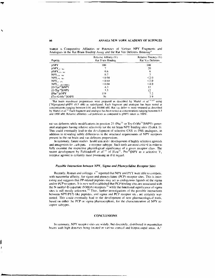
66 ANNALS NEW YORK ACADEMY OF SCIENCES
TABLE i. Comparative Affinities or Potencies of Various NPY Fragments andAnalogues in the Rat Brain Binding Assay and the Rat Vas Deferens Bioassay"
Relayive Affinity (%/) Relative Potency ('7)Peptide Rat 13-ain Binding Rat Vas Deferens
pNPY I(X) 100pNPY, ,, 7 20pNPY,1 0.6 6NPY,18 36, 0.7 7NPY,, , <0 04 <2.0NPY,1 l, <0.(4 <2.0NPY: 24(oni2, <0.04 <4.0ID-Tyr2 t IhNPY 4.3 17[D-Trp2(lhNPY 3.3 12IPhe-']hNPY 119 7.2ITyr-O-Me 2IjhNPY 56 3.9
"Rat brain membrane preparations were prepared as described by Martel et al.'" usingl'Hlpropionyl-pNPY (0.5 nM) as radioligand. Each fragment and analogue has been tested atconcentrations ranging between 0.01 and t0.000 nM. Rat "%as defer, ns were )repared as describedby Martel et 11.13 Each fragment and analogue has been tested at concentrations ranging between 0.5and 1000 nM. Relative affinities :nid potencies as compared to pNP'Y taken as 100%.
rat vas deferens while modifications in position 21 (Phe 2' or Try-O-Me2, jhNPY) gener-
ated analogues having relative selectivity tor the rat brain NPY binding sites (TABLE I).This could eventually lead to the development of selective CNS vs PNS analogues, inaddition to revealing subtle differences in the structural requirements of NPY receptorspresent in the rat brain and vas deferens preparations.
In summary, future studies hould aim at tk' development of highly selective agonistsand antagonists for ,'ach put. e receptor subtype. Such tools are most criticAl in order tofully examine the respective physiological significance of a given receptor class. Therecent development by Fuhlendorff eta (. 7 of ILeu" , Pro34 INPY as a selective Y,receptor agonist is certainly most promising in that regard.
Possible Interaction between NPY, Sigma and Phencyclidine Receptor Sites
Recently. Roman and colleagu s7 7 reported that NPY and PYY were able to compete.with nanomolar affinity, for sigma and phencyclidine (PCP) receptor sites. This is inter-esting and suggests that PP-related peptides may act as endogenous ligands of the sigmaand/or PCP receptors. It is nov well e,:ablished that PCP binding sites are associated withthe N-methyt-D-aspartate (NMDA) receptors75 while the functional significance of sigmasites is still mostly unknown .7 Thus, further investigations of the possible interactionsbetween NPY/PYY-like peptides. and sigma and PCP receptor sit, , are certainly war-ranted. This could eventually lead to the dee!opment of new pharmacological tools,based on either !he PCP or sigma pharmacophore, for the characterization of NPY re-ceptor subtypes.
CONCLUSIONS
In summary. NPY receptor sites arc widely, but discretely. distributed in mammalianbrains with high densities being located in various cortical and hippocmpal areas. A,
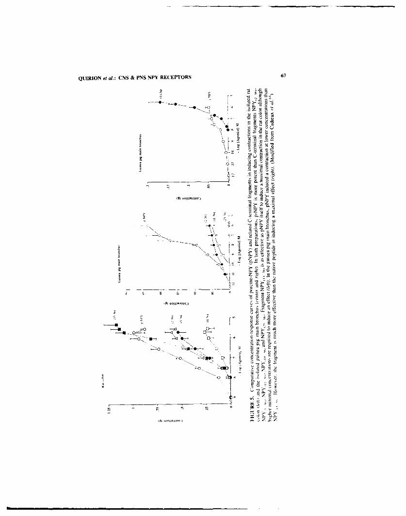
QUIRION et at.: CNS & PNS NPY RECEPTORS 67
-- o1
-o - E
~ .2
E 2
E
(9) uO!I3vWj -
C- - ,
> D - .7HU -6
N 'N
-D '-4 ~ > CNr
13)~ N-- -JUO
\\ U
'oC
z E3
---- ------- L C U
A, "olmoC Z

68 ANNALS NEW YORK ACADEMY OF SCIENCES
T'ABLE 2. Relative Affinities of Various NPY Fragments in Inducing CertainBehavioral Effects of NPY Following ICV Injections.
Decreased Motor Increased FoodActivity Intake Hypothermia
Peptide (17) (1h (')
pNPY 10 fXO I W IpNPY, 1,, 30 130 20)pNPY, 1' 3 6' 20 4 <0.tINPY,,' iv <0.1I <0.1I <0.1I
-NPY, I 4Cki15A2, <0.1I <0.1I <0. I
'For experimental details, see REFERE.NCE 65. Retative potencies as compared to pNPY taken as100%v.
ditionally. NPY/PYY receptor subtypes most likely exist in CNS and peripheral tissues.This hypothesis is based on the differential labelling of hypothalamic and brainstem nucleiby NPY and PYY receptor probes, and on the respective lipand selectivity profile and thedifferential order of potency kit agonists found in various bioassay systems. However, thedevelopment of potent and selective receptor antagonists is awaited in order to providemore definite evidence for the existence of NPY receptor subtypes.
ACKNOWLEDGMENT
The expert secretarial assistance of Mrs. J. Currie is acknowledged.
REFERENCES
1 . Tvv.stoto. K., M. CsttcQutSI & V. MU 1 1, 1982. Neuropeptide Y-a novel brain peptide withstructural simtilarities to peptidle YY and pancreatic polypeptide. Nature 296: 659-6f0).
2 TAtE.Moio. K. 1982. Isolation and characterization of peptide YY (PYYt. a candidate guthormone that inhibtts pancreatic exocrine secretion. Proc. Natil. Acad. Sci. USA 79: 2514-2518.
3. TA riMOiti, K. 1982. Nenropeptidle Y: contplete amnino acid sequence oft the brain peptide.Proc. Nat]. Acad. Sci. USA 79: 5485-5489.
4. McDOycst-. J. K. 1988. NPY and related substances. Crit. Rev. Neurobiol. 4: 97-1355. S1st.-EY. B. G. & S. F. LpltuowiTt. 1984. Neuropeptide Y: stimulation (it feeding and
drinking by injection into the parasentricutar nucleus. Lite Sct. 35: 2635-2642.6, STANt.Ev. B. G.. 13. R. DANtIt.. A. S. CHIN & S. F. L~iBotiiz. 1985. Para venitricutlar
nucleus injections of peptide YY and neurtipeptide Y preferentiatlly enhance carbohydrateingestion. Peptides 6: 12015-1211.
7. SIANIEY. B. G. & S. F. Livtuowii/. 1985. Neuropeptide N' injected tn the paraventrtculathypothalamus: a powerful stintulant of feeding behavitir. Protc Nai], Acid. Sci. USA 82:39401)3943.
9. CI ARK. I. T.- P. S. KN.A W. K. Uiowi is & S. P, KAItkA 19X4. Neuropeptide )f andhunman pancreatic pittspeptide stittulates feeding behavior in rats. EndOCrTimloen 115: 42-429.
9 HARI5TRANt. A.. K. Ftx vi. 1-. 1 Aw-%ii. 1) , N. I- Ni-Rotti. K. TNvtFmoio. &\Nit it r 1984. Studies tin necurtipeptide Y -catechittamite interactions in central card its ascautarregulation in thle alpha-cbtiiralivc anaesthetiei rat Evnidence li a a Possible nea Xav otico rating the alpha 2 adrenergic iransinisi, in line (.1 n Lxp H.%periens P'art AX theoryPract A6t: 1947 !9501
Mt Nki '.1 I Si I L. V11o\1-K. KIvIWiMstto .it MmV,. J lit ots )& S R Hinso 1')S2

QUIRION e! ai.: CNS & PNS NPY RECEPT"ORS 69
Two novel related peptides. neuropeptide Y INPY) and peptide YY (PYY) inhibit the con-traction of the electrically stimulated mouse vas deferens. Neuropeptides 3: 7 1-77.
11. LUNDBERG, J. M. & L. SIJARNE. 1984. Neuropeptide Y INPY) depresses the secretion of'H-noradrenaline and the contractile response evoked by field stimulation, in rat vas deferens.Acta Physiol. Scand. 120:477-479.
12. DoNoSo. V.. M. SIL~VA, S. ST-PIERRE & P. HUIDOBRo-TORO. 1988. Neuropeptidle Y (NPY).an endogenous presynaptic modulator of adrenergic neurotransmission in the rat vas deferens:structural and functional studies. Peptides 9: 545-553.
13. ADRIAN. T. E_, J. M. ALIEN. S. R. Bt.oom. M. A. GHATEI. M. N. RossoR. G. W. ROBERTS.T. J. CROW. K. TATEMOVo & J. M. POLAK. 1983. Neuropeptide Y distribution in the humanbrain. Nature 306: 584-586.
14. CHRONWAIL., B. M.. T. N. CHASE & T. L. O'DoNOIIUE. 1984. Coexistence of neuropept.*deY and somatostatin in rat and human cortical and rat hypothalamic neurons. Neurosci. Lett.52:213 -2 17.
15. CHRONWALL.. B. M., D. A. DiMAGGO, V. J. MASSAR. V. M. PICKEt.. D. A. RUGGIERO &T. L. O'DoNOtlUE. 1985. The anatomy of neuropeptide Y containing neurons in rat brain.Neuroscience 15: 1159-1181.
16. CHAN-PAI.AY, V.. Y. S. ALIEN. W. LANG. U. HAESI.ER & J. M. POLAK. 1985. 1. Cytologyand distribution in nonnal human cerebral cortex of' neurons imimunoreactive with antiseraagainst neuropeptide Y. J1. Comp. Neurol. 238: 382-389.
17. DAWBARN. D.. S. P. HUN[ & P. C. EvisoN. 1984. Neuropeptide Y: regional distribution.chromatographic characterisation and iniunohistochemical demonstration in post-mortemhuman brain. Brain Res. 296: 168-173.
18. DEQUIDT. M. E. & P. C. EMISON. 1986. Distribution of neuropeptidle Y-like immunoreactivitvin the rat central nervous system-I. Radioimmunoassay and chronmatographic localization.Neuroscience 18: 527-543.
19. DEQUIDI. M. E. & P. C. EMSON. 1986. Distribution of neuropeptide Y-Iike immunoreactivitNin the rat central nervous systern-ll. Imimunobistochemical analysis. Neuroscience 18: 545-618.
20. EVERI-T. B. J., T. HOIKEEtIt. L_ TERLNIus. K. TA IEMo1o. V. MUTT & M. GOL.DSTEIN . 1984.Differential co-existence of neuropeptide Y INPYI-like immunoreactivitv with catechol-amines in the central nervous system of' the rat. Neuroscience 11: 443-462.'
21. HENDRY,.S. H. C.. E. G. JONES& P. C. EmsoN. 1984. Mtorphology, distribution. and synapticrelations of somatostatin- and neuropeplide Y-immuntoreactive neurons in rat and monkeyneocortex. J. Neurosci. 4: 2497-2517.
22. H5KFEI. T., J1. M. LUNDBERG. H. LAsorRURANTZ, K. TA.TEMOIO. V. .Mu'r. J. LINDBERG.L. TERE.NiU~s. B. J. EVERII , K. Fuxt:. L. AGNAtt & Mi. GOLDSTEIN. 1983. Occurrence ofneuropeptide Y (NPY)-like itumunoreactivity in catecholatnine neurons in the human medullaoblongata. Neurosci. Lett. 29: 217-222.
23. KOHLIER. C.. L. ERIKSSON. 2 . DAVIES & V. CHAN-PXI.A. 1986. Neuropeptide Y innervationof the hippocampal region in the rat and monkey brain. J. Comp. Neurol. 244: 384-4(X).
24. PELtETIER. G.. L. D~sY. L. KE RKERIAN & J. C'01 F . 1984. lmmiunocstochemical localizationof neuropeptide Y INPY) in the human hypothalamus. Cell Tissue Res. 238: 2013-205.
25. Sviiii. Y. & A. PARENT. 1986. Neuropeptide Y-iinunmoreactive necurons in the striatumn of cat& monkey: morphologtcal characteristics, intrinsic organization atid co-localization w.ith so-matostatin. Brain Res. 372: 241-252.
26. INum. A.. N. MIZUNtO. MI. OOYA. K. Sol:NAGA. H. MORIOKAs. T. Oo6sWA. N1. ISHIDA & S.BABAs. 1985. Cross-reactivities of ncuropeptide Y and] pcptidc YY wkith pancreatic polypeptideantiserai evidence for the existence of pancreatic polypeptide in the brain. Brain Res. 330:386-399.
27. BROOME. M.. T. HOKI iI I & L. TI REItUS. 1985. Peptidle YY (PYN') itttltunoreactise neuronsin the lower brainslemn and spinal cord of rat. Acta Physiol. Scand. 125: 349-3152.
28. NIIYA( III, Y. . W. I tStjtS1lI. A. MivoStit. S. F1JI I A. A. MI/(Isot & K. TA I I N401. 1986. Thedistribution of polypepltde YY-like inunoreactlstlv in rat brain tissues. Fndocrmolog118: 2161-2167
29. FKIAN . R., . -li f.I S I5 EIt G 1)1 I(iI R, FK St Ni i R. R . H sE s\Nsos. & 1)'sut.

70 ANNALS NEW YORK ACADEMY OF SCIENCES
1986. Peptide YY-like immunoreactivity in the central nervous system of the rat. Regul. Pep!.16: 157-168.
30. SCHULE-ZIEROj. M. & T. H6KFFIt1. 19,46. The mismatch problem in receptor autoradiographyand the coexistence of multiple messengers. Trends Neurosci. 9: 109-I110,
31. UNDEN. A. & T. BARrEAt. 1984. Regulation of' neuropeptide Y (NPY) binding by guaninenucleotides in the rat cerebral cortex. FEBS Lett. 177: 125-128.
32. UNDEN. A.. K. TATEmoro. V. MUTT & T. BARTEAI. 1984. Neuropeptide Y receptors in therat brain. Eur. J. Biochem. 145: 525-530.
33. SARIA. A.. E. THEODORSSON-NORHEIM & J. M. LUNDBERG. 1984. Evidence for specificneuropeptide Y-binding sites in rat brain synaptosomes. Eur. J. Pharmacol. 107: 105-107.
34. CH-ANG, R. S_ L., V. J. Lot iT. B. CHEN, D. 1. CERINO & P. J. KLING. 1985. NeuropeptideY INPY) binding sites in rat brain labeled with '251-Bolton-Hunter NPY: comparative poten-cies of' various polypeptides on brain NPY binding and biological responses in the rat vasdeferens. Lif'e Sci. 37: 2111-2122.
35. MARTEL. I.C.. S. ST-PIERRE & R. QUtIRtoN. 1986. Neuropeptide Y receptors in rat brain:autoradiographie localization. Peptides 7: 55-60.
36. H4.RFSTRAND. A.. K. FuXE. L. F. AGNATI. F. BFNFNAII & M. Got or)SEIN. 1986. Receptorautoradiographical evidence for high densities of 1251 neuropeptide Y binding sites in thenucleus tractus solitarius of the normal male rat. Acta Physiol. Scand. 128: 195-2(X).
37. NAKAJIMA. T.. Y_ YASIIIMA & K. NAKAMURA. 1986. Quantitative autoradiographic localiza-tion of neuropeptide Y receptors in the rat lower brainstem. Brain Res. 380: 144-151.
38. CHANG. R. S. L.. V. J. Lotlii & T. B. CHIEN. 1988- Specific I 'Hipropionyl-neuropeptide YINPY) in rabbit aortic membranes: comparisons with binding in rat brain and biologicalresponses in rat vas deferens. Biochem. Biophys. Res. Comm. 151: 1213-1219.
39. IN~UI. A.. M. OYA. M. OKIT A. T'. INOUEI, N. SAKATANI. H. MORIOKA. K. Simt. K. YOKONO.N. N4IZUNO & S. BABA. 1988. Peptide YY receptors in the brain. Biochem. Biophvs. Res.Comm. 150: 25-32.
40. INut, A.. M. OKiTA. T. INOUE. N. SAKATANI. M. OYA. H. MORIOKA, K. Suit. K. YOKo.,o.N. MIZUNO & S. BABA. 1989. Characterization of peptide YY receptors in the brain. En-docrinology 124: 402-409.
41. WALKER. M. W. & R. J. MILLER. 1988. ' 25 1-neuropeptide Y and '21-peptide YY bind tomultiple receptor sites in rat brain. Mol. Pharmacol. 34: 779-792.
42. LUNDBERG. J. M.. A. HL.MSEN. 0. LARSSON. A. RUDEILLi. A. SARIA~ & B. FREDOLMt~. 1988.Neuropeptide Y receptor in pig spleen: binding characteristics, reduction of' cyclic AMPformation and calcium antagonist inhibition of vasoconstriction. Eur. J. Pharmacol. 145:2 1-29.
43. MAcKERELL. A. D.. A. HEMSIEN. J. S. LA(ROtX & J. M. LUNDBERG. 1989. Analysis of-structure-function relationships of neuropeptide Y using molecular dynamics simulations andpharmacological activity and binding measurement. Regul. Pept. 25: 295-313.
44. LYNCH. D. R.. M. W. WALKER. R. J. MILtER & S. H. SNYDER. 1989. Neuropeptide Yreceptor binding sites in rat brain: differential autoradiographic localizations A~ ith ['2511 pep-tide YY and 11'11j neuropeptide Y imply receptor heterogeneity. J. Neurosci. 9: 2N)7-2619.
45. KRSTENANSKY. J. L.. T. J. OWEN. S. H. BUCK. K. A. HAGAMAN & L. McLFXN. 1989.Centrally truncated and stabilized porcine neuropeptide Y analogs: design. synthesis andmoluse brain receptor binding. Proc. Nall. Acad. Sci. USA 86: 4377-4381.
46. SHEIKII. S. P.. R. HAKANSON & T. W. SCilWARtZ. 1989. Y, and Y. receptors foir neuropep-tide Y. FEBS Lett. 245: 2019-2 14.
47. FUHiENDO)RE. I., U.. GLt. IFN. L. AAKERI.1Ni. N. LAN6F.I ANi)-JOilAN.S[N. H. T10X;ERS1N,S. G. MELBRG O U. B. Ol.sEN. 0. TitASTRUr & T. W. SuiwARrz. 19w. [Let]".Pro'4 lneuropeptide Y: a specific Y, receptoir agonist. Proc. Natil. A\cad. Sci. USA 87: 182-186.
48. StiElKIL. S. P.. M. m. T. O*HARL. 0. [uiRIURA & T. W. 5(11w \Ri/. 1989. Bindine ofmorn1iodinated neuropeptide Y to hippocanipal membranes and human neuroblastomia celllines. J. Biol. Chem. 264: 6648-6654.
49. BE( Ks. A.. 6i. JUNG. W. (lAItA. H. KoiPEs. R. Lso& 6 S( i1NiRRI N10 RG 1989.) Hihl>potient and small neuropeptide Y agonist obtained b% li nking NP) via spacer to o-helicalNPY, , FEBS L.ett. 244: 119)-122

QUIRION et al.: CNS & PNS NPY RECEPTORS 71
50. Rioux, F., H. BACHEIARD. .1. -C. MARIELt & S. S I-PIERRE. 1986. The vascoconstrictor effectof neuropeptide Y and related peptides in the guinea pig isolated heart. Peptides 7: 27-3 1.
51. DANGER, J1. -M., F. LEBOULEFNGER. J. Guy. M. -C. ToNON. M. BEN YAMIN A. J. -C. MA R IEL,S. SAINT-PIERRE, PELLE.IIER. G. & H. VAUDRY. 1986. Neuropeptide Y in the intermediatelobe of the frog pituitary acts as an a-MSH-release inhibiting factor. Life Sci. 39: 1183-1192.
52. MARTEL.. J. C.. S. SrF-PIERRE & R. QuiRIoN. 1988. Comparative distribution of neuropeptideY immunoireactivity and receptor autoradiography in rat forebrain. Peptides 9 (Suppl. 1):15-20.
53. TAYtLOR, 1. L., P. MANNON. S. VIGNA. D). WHITCOMB & T. NG~UYEN. 1990. Characterizationof neuropeptide Y (NPY), peptide YY (PYY) and pancreatic polypeptide (PP) receptors. Ann.N.Y. Acad. Sci. This volume.
54. MARTEL, J. C.. A. FOURNIER, S. ST-PIERRE & R. QUIRION. 1990. Quantitative auloradio-graphic distribution of 1
2'11iBolton-Hunter neuropeptide Y receptor binding sites in rat brain.
Comparison with ['25l1peptide YY receptor sites. Neurosciences. In press.55. COLMERS. W. F.. K. LUKOWIAK & Q. J. PITTMAN. 1985. Neuropeptide Y reduces orthodro-
mically evoked population spike in rat hippocampal CA, by a possibly presynaptic mecha-nism. Brain Res. 346: 404-408.
56. COLMERS, W. F.. K. LUKOWIAK & Q. PI IMAN. 1987. Presynaptic action of neuropeptide Yin Area CA, of the rat hippocampal slice. J. Physiol. 383: 285-299.
57. Ft.OOo, .IF . E. N. HERNANDEZ & J. E. MORtEY. 1987. Modulation of memorv processingby neuropeptide Y. Brain Res. 421: 280-290.
58. MARTEL,. J. C.. S. SiF-PIERRE. P. BEDARD & R. QUIRION. 1989. Comparison of [' 21 ]Bolton-
Hunter neuropeptide Y binding sites in the forebrain of various mammalian species. BrainRes. 419: 403-407.
59. l~ut. A.. M. OKtrA, Y. HIROSUE. M. ML71RA & M. NAKAJIMA. 1990. Peptide YY receptorsin mammalian and avian brains. Ann. N.Y. Acad. Sci. This volume.
60. MARTEL,. J. C., R. AI.AGAR. Y. RoBtrAtI.t.E & R. QUtRION. 1990. Neuropeptide Y receptorbinding sites in human brain. Possible alteration in Alzheimer's disease. Brain Res. In press.
61. WAH-I.ES1ED1. C., N. YANAIFIARA & R. HAKANSON. 1986. Evidence for different pre- andpost-junctional receptors for neuropeptide Y and related peptides Regul. Pept. 13: 31)7-3 18.
62. POUrER, E. K.. L. MITHELL, M. J. D). MCIOSKEY. A. TsENGs. A. E. GOODMAN. J. SHINE& D). 1. MCCI.OSKEY. 1989. Pre- and post-junctional actions of neuropeptide Y and relatedpeptides. Regul. Pep!. 25: 167-177.
63. MARTEL. J. C.. A. FOURNIER. S. St-PERRE. i'. DtjvOsNt. M. FOREST & R. QUtRtoN. 1990.Comparative structural requirement of neuropeptide Y brain binding sites and vas deferensneurtopeptide Y receptors. Mol. Pharmacol. In press.
64. CADIEUX. A.. M. T. BENCIIEKROUN, A. FOURNIER & S. St-PIE-RRE_. 1990. Investigations of
the structure-activity relationship of neuropeptide Y. Ann. N.Y. Acad. Sci. This volume.65. DRUMHEI.I.ER. A., R, RIVESr. J. N. MtcttsUD. S. S1-PERRI & IF. B. JOICOEUR. 1988. The
profile of neurobehavioral effects produced by neuropeptide Y. Soc. Neurosci. Abst. 14: 287.66. FORE.., M., J. C. MARIE! . St-PIERRE. R. QUtRtoN & A. FOURNIER. 1990. Structural
study of thc N-terminal segment of neuropeptide tyrosine. J. Med. Chemn. 33: 1615-1619.67. BouBtLtK. J. H., N. A. Sot I, M. R. BROWN & J. F. RtvtER. 1989. Synthesis and hy per-
tensive activity of neuropepllde Y fragments and analogues %%ith modified N- or C-termint or1)-substitut ions. J. Med. Chem. 32: 597-601.
68 . SUE'RVIN. A. L., C. RoUYER-FEssARD, A. BALASUBRAMANIAM. S. SI-PE RRE & NI. LABURIHI-.1989. Peptide-YY and neuropeptidc-Y inhibit vasoactive intestinal peptide-stimulated aden-osine 3' .5'-ntonophosphale production in rat snmall intestine: structural requirements of pep-tides for interacting wtth peptide-YY-prcering receptors,. E-ndocrinologs 124: 692-7(10.
69, BtuNI It . Tf.. . . E. Pit IS. 1. J. THiKt I . S. P WOLt) & C' -\N. Wt 1981. X-ra\ analssis(1.4 A resol ut ion) of asvian pancreatic poI peptide: small g lobular pept ide hormone. ProcNall. Acad Sci. USA 78: 4175-4179
70) (it 0%1R, 1.. 1. H~x'sI I . J, Pit is. W Wot)o. 1) MoSS. 1,. Iit KitI & T. Bt t Nmt ItI. 1981(ontorniation flexibtlit\ tit a stmall globular hormtonte: X-rasN analisis of as ian paitcreat cI~oN pptde at 0 99-A resolution. Bitopoli nics 22: 29; ;0)4
71. At tIN. 1 1.11. J. No\sutNN. V M\RtI'. & G; HtiNitt t 19S7 Iolcular structurer t

72 ANNALS NEW YORK ACADEMY OF SCIENCES
mammalian neuropeptide Y: analysis by molecular cloning and computer-aided comparisonwith crystal structure of avian homologue. Proc. Nati. Acad. Sci. USA 84: 2532-2536.
72. MINAKATA. H., J. W. TAYLOR, M. W. WALKER, R. J, MILLER & E. T. KAIrsr;-R. 1989.Characterization of amphiphilic secondary structures in neuropeptide Y through the design.synthesis, and study of model peptides. J. Biol. Chem. 264: 7907-7913.
73. AKIL, H.. S. J. WArFSON, E. YOUNG, M. E. LEwts. H. KHA HA IURIAN & J. M. WALKER.1984. Endogenous opioids: biology and function. Annu. Rev. Neurosci. 7: 223-255.
74. QUIRIoN'. R. & T. V. DAM. 1988. Multiple neurokinin receptors. Recent developments. Regul.Pept. 22: 18-25.
75. MAGDAIIN, W., B. G. STANLEY, A. FOURNIER & S. F. L~ieowt rz. 1989. A structure-activityanalysis of neuropeptide Y-induced eating behavior. Soc. Neurosci. Abst. 15: 895.
76. KALRA. S. P.. A. SAHU & P. S. KALRA. 1990. Hypothalamic NPY;- a circuit in regulation ofneuroendocrine and behavioral responses. Ann. N.Y. Acad. Sci. This volume.
77. ROMAN. F. J., X. PASCAND. 0. DUJFFY. D. VAUCHE. B3. MARTIN & I. L. JUNIEN. 1989.Neuropeptide Y and peptide YY interact with rat brain (T and PCP binding sites. Eur. J.Pharmacol. 174: 301-302.
78. QUIRION. R., R. CHICHEPORTICHE. P. C. CONTRERAS. K. M. JoiNsoN, D. LODGE, W. S.TAM. J. H. WOODS & S. R. ZUKIN. 1987. Classification and nomenclature of phencyclidineand sigma receptor sites. Trends Neurosci. 10: 444-446.

Pancreatic Polypeptide and Peptide YYGene Expressiona
S. D. KRASINSKI, M. B. WHEELER, A. S. KOPIN, ANDA. B. LEITERb
Department of Medicine, Division of GastroenterologyNew England Medical Center Hospital
Tufts University School of Medicine750 Washington Street
Boston, Massachusetts 02111
INTRODUCTION
Pancreatic polypeptide, peptide YY. and neuropeptide Y are members of a family ofregulatory polypeptides. Pancreatic polypeptide is one of the four major hormones syn-thesized in pancreatic islets. The primary biological effect of pancreatic polypeptide isinhibition of pancreatic exocrine secretion. Pancreatic polypeptide secretion is stimulatedby a variety of nutritive and/or neurogenic factors, most of which appear to be mediatedby cholinergic pathways. Peptide YY has been localized primarily to ileum and colon. '--Peptide YY immunoreactivity has also been identified in pancreas, jejunum, duodenum,and gastric antrum. 2 5 The physiological actions of peptide YY include inhibition ofgastric emptying, intestinal motility, pancreatic exocrine secretion, and secretions of acidand pepsin from the stomach.' 2- 6 7 Peptide YY is released in response to the presence offat in the intestine."-9 In addition to the direct effect of fat on peptide YY release,' °
postprandial secretion of peptide YY appears to be stimulated by signals originating in theforegut.'' Present evidence suggests that cholecystokinin is a candidate foregut factorwhich stimulates peptide YY release. '
We previously isolated cDNAs encoding the precursors of human pancreaticpolypeptide12 and rat peptide YY. '3 The amino acid sequences of the precursor polypep-tides predicted from cDNAs reveal a conserved precursor structure shared by all threemembers of this peptide family. 12- '5 Each precursor consists of a signal peptide 28 or 29amino acids in length, a 36-amino-acid mature hormone followed by a Gly-Lys-Argsequence, and a C-terminal peptide consisting of approximately 30 residues (FIG. 1). Aunique tyrosine amide is present at the C-terminus of the mature polypeptides. Themarked similarities in precursor structures for pancreatic polypeptide. peptidc YY, andneuropeptide Y, are consistent with the hypothesis that each of these peptides arose byduplication of a common ancestral gene.
To study the structure and expression of the genes encoding pancreatic polypeptideand peptide YY, we isolated the genes encoding rat peptide YY and both human and ratpancreatic polypeptide. Here we show that the structure of the rat peptide YY gene shares
"Supported by National Institutes of Health Grant R29-DK39209."Address correspondence to Andrew B. Leiter. M.D.. Ph.D., Department of Medicine. Division
of Gastroenterology. New England Medical Center Hospital, 750 Washington Street. Boston. Mas-sachusetts 02111.
73

74 ANNALS NEW YORK ACADEMY OF SCIENCES
E
CIA!
) C
cca0a~CL
0 o0
0.->
0 CL.

KRASINSKI et al.: PANCREATIC POLYPEPTIDE 75
a common structural organization with the genes encoding neuropeptide Y and pancreaticpolypeptide. We also show that the peptide YY gene is developmentally regulated and thatthe expression of this gene is regulated by protein kinase-dependent intracellular signal-ling pathways.
MATERIALS AND METHODS
Isolation of the Gene Encoding Rat Peptide YY
The gene encoding rat peptide YY was identified from a rat genomic library con-structed in bacteriophage X using an oligonucleotide probe corresponding to the cDNAsequence. Positive clones were plaque-purified by sequential low density plating, andbacteriophage DNA was isolated by the plate-lysate method. A single 4.5 kb Kpn Irestriction fragment was identified by the peptide YY probe in Southern blot hybridiza-tions, and subcloned into a plasmid vector. DNA sequencing was carried out by the chaintermination method using T7 DNA polymerase. " Computer-assisted nucleotide sequenceanalysis was performed as described by Devereux. ,7
Determination of Peptide YY Gene Expression in the Developing Rat
Peptide YY gene expression in ileum and colon of developing rats was investigated byNorthern blot hybridization assays. Immediately following removal, tissues were frozenin liquid nitrogen and stored at -70'C. Total cellular RNA was prepared using RNAzol T"(Cinna/Biotex Inc., Friendswood. TX) according to the manufacturer's instructions. Toassure that equal amounts of RNA were loaded onto gels for Northern blotting experiments.RNA was quantitated by the absorbance at 260 nm. and by the UV fluorescence intensityof the ribosomal RNA subunits on nondenaturing agarose gels stained with ethidiumbromide. Following electrophoresis on denaturing agarose gels, RNA was transferred toNytran membranes (Schleicher and Schuell, Keene, NH) by capillary blotting. Hybrid-izations were carried out under conditions of high stringency (65'C, 50% formamide) usingantisense RNA probes for either rat peptide yy13 or rat glucagon. 8 To assure that equalamounts of mRNA were loaded onto each lane, the blots were reprobed for 3-actin.
Promoter Activity of the Peptide YY Gene in Transient Expression Assays
To study the transcriptional regulation of the rat peptide YY gene, we introducedreporter gene constructions into HeLa cells for transient expression assays. For theseexperiments, fusion genes were constructed by inserting 280, 242. 183 or 127 bp ofpeptide YY 5' flanking sequence and 33 bp of exon I upstream from the reporter gene forthe bacterial enzyme, chloramphenicol acetyl transferase (CAT). The DNA fragmentscontaining 280. 242, or 183 bp of 5' flanking sequence were isolated by restrictionendonuclease digestion of the genomic subclone. The DNA fragment containing 127 bpof 5' flanking sequence was amplified from the peptide YY genomic subclone withThermus aquatis (Taq) DNA polymerase I (Cetus-Perkin Elmer. Norwalk. CT) using thepolymerase chain reaction.
HeLa TK-cells were plated at a density of 106 cells per 100 mm plates in DMEMsupplemented with I0%4 fetal bovine serum. ('ells were transfected with the plasmid DNAby calcium phosphate precipitation. ' After a 24 h transfection period, the medium was

76 ANNALS NEW YORK ACADEMY OF SCIENCES
replaced with medium containing 0.5% fetal bovine serum. Thirty-six h later, cells weretreated with combinations of forskolin and 3-isobutyl-l-methyl xanthine (IBMX) to in-crease intracellular cyclic AMP, and the phorbol ester 12-0-tetradecanoyl-phorbol- 13-acetate (TPA) to activate protein kinase C. Cells were harvested 16 h later, and extractswere prepared by freeze-thawing and sonication. Extracts containing either 50 or 100 l.,gof protein were assayed for CAT activity as described by Gorman, 20 and the results werenormalized to 100 Vig of protein.
RESULTS AND DISCUSSION
Structure of the Rat Peptide YY Gene
The transcription unit of the rat peptide YY gene spans approximately 1.2 kb andconsists of four exons separated by three introns. Each exon of the rat peptide YY genedefines a functional domain of its mRNA, as do the corresponding exons of the pancreaticpolypeptide and neuropeptide Y genes2 1-25 (FIG 2). Exon 1 of the rat peptide YY genecontains the 5' untranslated region of the mRNA. The second exon of the rat peptide YYgene begins at the translational initiation site and encodes the signal peptide and all but theC-terminal tyrosine of the mature polypeptide. Exon 3 encodes the C-terminal tyrosine ofpeptide YY, a Gly-Lys-Arg amidation-cleavage sequence, and most of the C-terminalextension peptide (FIG. 2). Like the third exons of the rat and human neuropeptide Y andhuman pancreatic polypeptide genes,2 -2 3 the splice-donor site for the third intron of therat peptide YY gene occurs at an arginine residue in the C-terminal peptide. Although thisarginine residue has been suggested to serve as a cleavage site in the pancreatic polypep-tide precursor of several species,26 it is not known whether the peptide YY precursor iscleaved at this site. The final exon of the rat peptide YY gene encodes the remainder ofthe C-terminal peptide and the 3' untranslated region of the mRNA.
The amino acid sequence homology among neuropeptide Y, pancreatic polypeptide,and peptide YY as well -s the conserved structural organization of the respective poly-peptide precursors and genes, suggests that each member of this family arose from anancient gene duplication event. The amino acid sequences of peptide YY and neuropep-tide Y are more homologous to each other than to pancreatic polypeptide, suggesting thatthe former two peptides are more closely related to each other than to pancreatic poly-peptide. Analogous to the amino acid sequences, the nucleotide sequences of the exons ofpeptide YY are more homologous to the respective exons of neuropeptide Y, than to thoseof pancreatic polypeptide. This observation supports the hypothesis that peptide YY hasdiverged more recently from neuropeptide Y than from pancreatic polypeptide.
The human neuropeptide Y and pancreatic polypeptide genes have been localized tochromosomes 7 and 17, respectively, suggesting that this gene family originated from atandem duplication of an ancient linkage group followed by chromosomal translocation.2 7
Several other gene families including erb proto-oncogenes, type I collagen, actin, band3 membrane protein, and protein kinases, segregate similarly to pancreatic polypeptideand neuropeptide Y. Genes encoding erbA, erbB-B2, Hox I. alpha I chain type I col-lagen, erythroid band 3, and protein kinase C localize on chromosome 17 along withpancreatic polypeptide. The erbB, Hox2, alpha 2 chain type I collagen, 13-actin, non-erythroid band 3, and protein kinase A localize to chromosome 7 with neuropeptide Y.The protein kinase A and 03-actin genes are themselves duplicated on chromosome 7leading to the speculation that the peptide YY gene may localize to chromosome 7 as well.
Divergence of the C-terminal Peptide of the Rat Pancreatic Polypeptide Gene
The structural organization of the rat pancreatic polypeptide gene differs somewhatfrom the genes encoding other members of the pancreatic polypeptide gene family. The

KRASINSKI et al.: PANCREATIC POLYPEPTIDE 77
Peptide YY
5' UT Signal PYY 24 AA 7 AA 3' UT
- ,- )
i-
Pancreaticpolypeptide
5' UT Signal PP 29 AA 3' UT
U)
T-I
i-
Neuropeptide Y
5' UT Signal NPY 23 AA 7 AA 3' UT
I-
FIGURE 2. Structural organization ot the rat genes in the pancreatic p)lypeptide family. Exons areindicated as boxes. Protein coding regions are shaded. Flanking and intron sequences are shown assolid line,%. Abbreviations are: TATA. Goldberg-Hogness promotor: 5' LIT and 3' UT. 5' and 3'untranslated regions of the mRNA: signal, signal peptide: AA. amino acid.
third exon of the rat pancreatic polypeptide gene encodes the entire C-terminal peptide anda portion of the 3' untranslated region of the mRNA, 2 ' 2 5 whereas cxon 3 of the other
members of this gene family. including human pancreatic polypeptide, encode only aportion of the C-terminal peptide and none of the 3' untranslated region.2
2 ' Examinationof the amino acid sequences encoded by exon 3 of the rat pancreatic polypeptide gene

78 ANNALS NEW YORK ACADEMY OF SCIENCES
reveals little similarity to the corresponding sequences encoded by the human gene. Toexamine the molecular basis for the divergence of the C-terminal peptide of the ratpancreatic polypeptide gene, we and others -4 25 have compared the amino acid andnucleotide sequence of this gene with that of the human pancreatic polypeptide gene.
Examination of the nucleotide sequence of the rat and human pancreatic polypeptidegenes reveal two mechanisms which may explain the divergence of the C-terminal peptideof propancreatic polypeptide. The first mechanism is the use of an alternative splice-donorsite at the 3' end of rat exon 3. As a result of recruitment of an alternate downstreamsplice-donor site. the third exon of the rat gene is 42 bases longer than its humancounterpart (Fit. 3A). These additional 42 bases, which encode I I amino acids and atranslational stop codon, share identity with the corresponding sequences of human intron3. An unused splice-donor site is present in rat exon 3 at a position corresponding to thehuman splice-donor site (Fiti. 3A). A second mechanism contributing to the C-terminalpeptide divergence is a single insertion/deletion at the 5' end of exon 3 which producesa translational frameshift (FiG. 3A). The divergence of the C-terminal peptide sequence,therefore, appears to result from a translational frameshift mutation and the utilization ofan alternate downstream splice-donor site in rat exon 3.
The nucleotide sequences at the 5' end of exon 4 of the rat and human genes are highlyconserved, despite the observation that only the human sequence is translated (FIG. 3B).Exon 4 in the rat gene encodes only the 3' untranslated region of the pancreatic poly-peptide mRNA while exon 4 of the human gene encodes 7 amino acids of the C-terminalpeptide, and the 3' untranslated region. The nucleotide sequence encoding the heptapep-tide in the human gene share 19 of 25 bases with the corresponding nucleotide sequencein the rat gene. This observation suggests that prior to the existence of the frameshiftmutation and the utilization of an alternate splice-donor site in rat exon 3. the 5' regionof rat exon 4 was at one time translated. The differences in the C-terminal domains of theid Wdi human pancreatic polypeptide precursors reflect less strict evolutionary constraintsthan those imposed upon the N-terminal domains of the precursors suggesting that theC-terminil peptides either have different functions or represent a region of the precursorwhich is unnecessary for biological activity. Despite the striking differences in the C-terminal peptide sequences, the similarities in nucleotide sequence of rat and human exonssuggest that these exons arose from common ancestral exons rather than by substitutionof an unrelated exon.
Ontogeny of Rat Peptide YY Gene Expression
Peptide YY immunoreactivity has been co-localized with enteroglucagon-like immu-noreactivity in endocrine cells of ileum and colon in developing and adult animals. 3 4 Todetermine whether peptide YY and glucagon gene expression are regulated similarlyduring development, we compared the time-course of peptide YY gene expression to thatof glucagon in the intestinal tract of fetal and postnatal rats by Northern blot hybridizationassays. In the ileum. both peptide YY and glucagon mRNA were first detected on day 19of gestation and rose to adult levels one day later (FIG. 4). In colon. peptide YY andglucagon mRNA were first detected two days earlier than in the ileum (fetal day 17), andsimilarly rose to adult levels by day 20 in ulero (not shown). Peptide YY immunoreac-tivity in rats has previously been identified in intestinal endocrine cells at day 19 ofgestation 25 whereas enteroglucagon-like immunoreactivity has been detected in intestineas early as fetal day 16 . 2
4 Thus, the time-courses of peptide YY and glucagon geneexpression are similar in developing intestine suggesting that these two genes are devel-opmentally regulated by a common mechanism.
We have shown that the peptide YY gene is expressed at adult levels in intestine
KRASINSKI et al.: PANCREATIC POLYPEPTIDE 79
E-4~ 00- t~-A
0~ -M
.. 6 :. : ,L) E4 "
Ci)-4
0 04~~ 4-:~I Iu u
ld Wu~C W 4-- -7
>1 b)
..4 P EU U ~UC70
U"U$4
-t~l >1
vn .. ~*U p
l1 U) 1) to
U C - u>- >1~ 0
S c 4-)

80 ANNALS NEW YORK ACADEMY OF SCIENCES
Day 18 15 90 21 3 7 10 14 21 U A
FIGURE 4. Northern blot analysis of peptide YY -nd glucagon mRNA in rat ileum during neonataldevelopment. Approximately 15 p.g of tota, RNA were loaled onto each lane. The autoradiogramsrepresent a Northern blot hybridized firot with an antisense RNA probe for peptide Y Y, and then withan antisense RNA probe for gluc.."e', at high stringency (65°C. 50% formamide). The numbersabove each lane irlicate the day (,€ feta! or postnatal development. A, adult.
before birth antedating the put.-tive nutritional and endocrine stimuli for peptide YYrelease in adult ani: . ... increased secretion of hoth peptide YY and glucagon have beenassociated with experimental models of intestanal epithelial cell proliferation.' 0 3' Theexpression of the peptide YY and glucagon genes in fetal animals suggests a potentialtrophic role for these polypeptidcs during fetal development.
Regulation of Rat Peptide YY Gene Expression
Examination of the 5' flanking sequence of the rai peptide YY gene reveals severalsequences similar to cis-acting elements which are known to re nond to specific intra-cellular signalling pathway,. A potential AP-1 binding site was I,,calized 144 bp upstreamfrom the transcriptional initiation site, and several potential AP- ' hinding scquences wereidentified within 130 bp of the transcriptional initiation sit: (FIG. 5). -.. determinewhether these potential regulatory elements are functionally a -tive in the rat pepti le YYgenc, we have carried out transient expression assays with fusion genes containag be-tween 280 and 127 bp of 5' flanking sequence fused to the structural gene for the bacterialenzyme CAT. The fusion genes -vere introduced into HeLa cells by calcium phosphateprecipitation. For all fusion genes, CAT activity was induced 3- to 5-fold followingtreatment with the phor.. tcr TPA (Fic,. 5), suggesting that the sequences necessary fo,this induction are localized to within 127 bp of ine transcriptional initiation site.
To further characterize the regulatory properties of th' peptide YY 5' flanking se-quence. we introduced the chirmeric construction containing '27 bp of 5' flanking se-quence into HeLa cells. and treated the cells from forskolin or IBVX to raise intracellular_AMP levels (Fie. 6)_ Treatment with forskolin or IBMX alone re ,alt d in an approxim te3-fold induction in CAT activity, an indtction similar to that o" TPA alone. Tretmentwith TPA and forskolin produced a 6-told induction in CA'i a( "iity, whereas treatmentwith TPA and IBMX resulted in a 7-fold induction in C, r ,:'.'ity T-eatment with TPA,forskolin. and IBMX resulted in a 21-fold inductiop in CAT aci, ity. exceeding even the
l4 m m l m m m t =.,

KRASINSKI et al.: PANCREATIC POLYPEPTIDE 81
response of the cAMP responsive element-containing 5' flanking sequence of the vaso-active intestinal peptide gene to forskolin and IBMX32 (FIG. 6). These data indicate thatagents which raise intracellular cAMP act in association with phorbol esters to inducepeptide YY gene transcription. Furthermore, the sequences necessary for this inductionare also localized to within 127 bp of the transcriptional initiation site.
Our findings indicate that the potential AP-l site at - 144 is not necessary for TPA-mediated induction of peptide YY gene expression. TPA is a biologically stable analog ofthe second messenger diacylglycerol which activates protein kinase C. Binding of theAP-1 transactivator complex to the sequence, TGAGTCAG, is one established mecha-nism for transcriptional activation by phorbol esters. The potential AP- I binding site inthe peptide YY gene (AAAGTCAG) differs by two bases with the previously reported8-base consensus sequence.' " ' These two bases may therefore be necessary for an AP- Isite to be functionally active.
Four C-rich domains containing sequences which closely resemble transcription factorAP-2 binding sites35 "36 have been identified within 130 bp of the transcription initiationsite (FIG. 5). AP-2 represents an inducible transcription factor that, like AP- 1. is involvedin the process of signal transduction. AP-2 may mediate transcriptional activation byphorbol esters and cAMP,1 3
' and may therefore alter the pattern of gene expressionwithin a cell in response to two distinct signal transduction pathways. The pattern of
-300 -200 -100
AP-2 Fold Stimulation in
AP-1 z 7 "'NTATAF- CAT Activity afterS I Treatment with
Phorbol Estersxx
mean ± SECAT
-280 I I I I I I 3.2±1.1
-242 " I i ' 3.4±1.3
-183 I i I I 2.4±0.4
-127 I I 4.7 ± 0.9
POCAT 1.2 ± 0.1
TRECAT 8.7 ± 1.9
FIGURE 5. Localization of the phorbol ester responsive sequences in the rat peptide YY gene. Thesolid lines represent 5' flanking sequence. Potential regulatory elements and exon I are indicated inboxes. Restriction sites used in the construction of chimeric peptide YY-chloramphenicol acetyltransferase (CAT) fusion genes are indicated. HeLa cells transfected with plasmid constructionscontaining from 280 to 127 bp of peptide YY 5' flanking sequence were treated with TPA at aconcentration of 50 nM for 16 h. A promoterless CAT construction (POCAT) and the plasmicTRECAT, which contains an active AP- I site upstream from the enhancerless RSV promoter. wereused as negative and positive controls, respectively. Cell extracts containing 50 to 100 p.g proteinwere assayed for CAT activity. Results are expressed as the fold increase in CAT activity overuntreated cells. The data are represented as mean + SE of at least three independent transfectionexperiments. Abbreviations are: AP, activator protein TATA. Goldberg-Hogness promoter.

82 ANNALS NEW YORK ACADEMY OF SCIENCES
induction of CAT activity in Hela cells transfected with fusion gene containing 127 bp ofpeptide YY 5' flanking sequence following treatment with TPA, forskolin, and IBMX areconsistent with transactivation by AP-2.
DNA binding sites for AP-2 in enhancer regions of many genes are often found inclose proximity to binding sites for other enhancer elements, suggesting that AP-2 reg-ulates gene transcription in association with other regulatory elements. For example, inthe proenkephalin gene, an AP-2 element acts synergistically with two adjacent cAMP
30
0 PYY(.127)CAT
0 POCAT
I TRECAT
0 ' 20 N VIPCAT
" 10
0 ~
4. . S. " v"_ ''"' "
o o.9 5. Treatmen00
A
TreatmentFIGURE 6. Transcriptional activation of peptide YY-CAT fusion genes by cAMP and phorbolesters. Hela cells were transfected with a plasmic construction containing 127 bp of peptide YY 5'flanking sequence [PYY(- 127)CAT]. Cells were then treated with one or combinations of thefollowing for 16 h: TPA (50 nM), forskolin (10 p.M), and IBMX (0.5 mM). POCAT and TRECATwere used as negative and positive controls as described in FIGURE 5. A CAT construction containingthe vasoactive intestinal peptide cAMP responsive element (VIPCAT) was also included in theseexperiments for comparison. The results were determined as described in FIGURE 5. The data arerepresented as mean + SE of at least five independent transfection experiments.
responsive elements to produce maximal responsiveness to cAMP, although the AP-2element alone is not sufficient for transcriptional activation (38). In the first 127 bp ofpeptide YY 5' flanking sequence, no other known cis-acting DNA regulatory elementswere identified, suggesting that for peptide YY, AP-2 may act alone in activating genetranscription. However, it is possible that as yet unknown enhancer elements act alone orin association with AP-2 to induce peptide YY gene transcription.
Cholecystokinin has been proposed as a candidate endocrine signal from the foregut

KRASINSKI et al.: PANCREATIC POLYPEPTIDE 83
which stimulates the release of peptide YY. " Intravenous administration of cholecysto-kinin octapeptide (CCK-8) in dogs stimulates release of peptide YY in a dose-dependentmanner."' Cholecystokinin may also play a role in the regulation of peptide YY genetranscription. Cholecystokinin appears to exert its effects on target tissues by increasinglevels of the intracellular messengers, IP3 and 1,2-diacylglycerol, leading to mobilizationof intracellular calcium and activation of protein kinase C. 39' 4
0 In pancreatic acinar cellpreparations, CCK-8 has been shown to stimulate the activity of adenylate cyclase inaddition to its effects on protein kinase C. 39
,4 1 The presence of potential cis-acting
regulatory elements in the peptide YY gene which respond to elevated cAMP and acti-vation of protein kinase C, such as AP-2 like elements, is consistent with a potential rolefor cholecystokinin in the transciiptional regulation of the peptide YY gene.
REFERENCES
1. TATEMOTO, K. 1982. Isolation and characterization of peptide YY (PYY), a candidate guthormone that inhibits pancreatic exocrine secretion. Proc. Natl. Acad. Sci. USA 79: 2514-2518,
2. LUNDBERG, J. M., K. TATEMOTO, L. TERENIUS. P. M. Ii. tLSTROM, V. MUTT, T. HOKFELT& B. HAMBERGER. 1982. Localization of peptide YY in gastrointestinal endocrine cells andeffects on intestinal blood flow and motility. Proc. Natl. Acad. Sci. USA 79: 4471-4475.
3. ALI-RACHEDI, A., I. M. VARNDELL, T. E. ADRIAN. D. A. GAPP, S. V. NOORDEEN & J. M.POLAK. 1984. Peptide YY (PYY) immunoreactivity is co-stored with glucagon-related im-munoreactants in endocrine cells of the gut and pancreas. Histochemistry 80: 487-491.
4. BOTTCHER, G., K. SKOLUND, E. EKLAD, R. HAKANSON, T. W. SCHWARTZ & F. SUNDLER.1984. Coexistence of peptide YY and glicentin immunoreactivity in endocrine cells of the gut.Regul. Pept. 8: 261-266
5. MIYACHI, Y., J. WAKA, A. MIYosHI, S. FUJITA, A. MIZUCHI & K. TATEMOTO. 1986. Thedistribution of polypeptide YY-like immunoreactivity in rat tissues. Endocrinology 118:2163-2168.
6. PAPPAS, T. N., H. T. DEBAS, Y, GOTO & 1. L. TAYLOR. 1985. Peptide YY inhibits meal-stimulated pancreatic and gastric secretion. Am. I. Physiol. 248: G118--G 123.
7. PAPPAS, T. N., H. T. DEBAS & I. L. TAYLOR. 1986. Enterogastrone-like effect of peptide YYis vagally mediated in the dog. J. Clin. Invest. 77: 49-53.
8. ADRIAN, T. E., G. -L. FERRI, A. J. BACARESE-HAMILTON, H. S. FUESSL, J. M. POLAK & S.R. BLOOM. 1985. Human distribution and release of a putative new gut hormone, peptide YY.Gastroenterology 89: 1070-1077.
9. APONTE, G. W., A. S. FINK, J. H. MEYER, K. TATEMOTO & I. L. TAYLOR. 1985. Regionaldistribution and release of peptide YY with fatty acids of different chain length. Am. J.Physiol. 249: G745-G750.
10. APONTE. G. W., 1. L. TAYLOR & A. H. SOLL. 1988. Primary culture of PYY cells from caninecolon. Am .J. Physiol. 254: G829-G836.
II. GREELEY, G. H., Y. -J. JENG, G. GOMEZ. T. HASHIMOTO, F. L. C. HILL, K. KERN, T.KUROSKY, H. -F. CHUO & J. C. THOMPSON. 1989. Evidence for regulation of peptide YYrelease by the proximal gut. Endocrinology 124: 1438-1443.
12. LEITER, A. B., H. T. KEUTMANN & R. H. GOODMAN. 1984. Structure of a precursor to humanpancreatic polypeptide. J. Biol. Chem. 259: 14702-14705.
13. LETTER, A. B., A. TODER, H. WOLFE, I. L. TAYLOR, S. COOPERMAN. G. MANDEL & R. H.GOODMAN. 1987. Peptide YY: structure of the precursor and expression in exocrine pancreas.J. Biol. Chem. 262: 12984-12988.
14. MINTH, C. D., S. R. BLOOM, J. M. POLAK & J. E. DIXON. 1984. Cloning, characterization,and DNA sequence of a human cDNA encoding neuropeptide tyrosine. Proc. NatI. Acad. Sci.USA 81: 4577-4581.
15. YAMAMOTO, H., K. NATA & H. OKAMOTO. 1986. Mosaic evolution of prepropancreaticpolypeptide. J. Biol. Chem. 261; 6156-6159.
16. TABOR, S. & C. RICHARDSON. 1987. DNA sequence analysis with a modified bacteriophage T7DNA polymerase. Proc. Natl. Acad. Sci. USA 84: 4767-4771.

84 ANNALS NEW YORK ACADEMY OF SCIENCES
17. DEVEREUX, J., P. HAEBERI. & 0. SMITHI-S. 1984. A comprehensive set of sequence analysisprograms for the VAX. Nucleic Acids Res. 12: 387-395.
18. HENRtCH. G., P. GROS, P. K. LUND, R. C. BENTI.EY & J, F. HABENER. 1984. Preproglucagonmessenger ribonucleic acid: nucleotide and encoded amino acid sequences of the rat pancre-atic complementary deoxyribonucleic acid. Endocrinology 115: 2176-2181,
19, CHEN, C. & H. OKAYAMA. 1987. High-efficiency transformation of mwwrimalian cells byplasmid DNA. Mol. Cell. Biol. 7: 2745-2752.
20. GORMAN, C. M., L. MOFFAT & B. HOWARD. 1982. Recombinant genomes which expresschloramphenicol acetyl transferase in mammalian cells. Mol. Cell. Biol. 2: 1044-105!.
21. LETTER. A. B., M. R. MONTMINV P J, "':,, & R. H. GOODMAN. (985. Exons of thehunwn pancreatic polypeptide gene define functional domains of the precursor. J. Biol.Chem. 260: 13013-13017.
22. MINIH. C. D., P. C. ANDREWS & J. E. DIXON. 1986, Characterization, sequence. andexpression of the cloned human neuropeptide Y gene. J. Biol. Chem. 261: 11974-11979.
23. LARHAMMER, D., A. ERICSSON & H. PERSSON. 1987. Structure and expression of the ratneuropeptide Y gene. Proc. Natl. Acad. Sci. USA 84: 2068-2072.
24. YONEKURA, H., K. NATA. T. WATANABE. Y. KURASHINA, H. YAMAMOTO & H. OKAMOTO.1988. Mosaic evolution of prepropancreatic polypeptide. I1. Structural conservation anddivergence in pancreatic polypeptide gene. J. Biol. Chem. 263: 2990-2997.
25. KOPIN, A. S., A. E. TODER & A. B. LETTER. 1988. Different splice site utilization generatesdiversity between the rat and human pancreatic polypeptide precursors. Arch. Biochem.Biophys. 267: 742-748.
26. SCHWARTZ, T. W. 1987. Cellular peptide processing after a single arginyl residue. Studies onthe common precursor for pancreatic polypeptide and pancreatic icosapeptide. J. Biol. Chem.262: 5093-5099.
27. TAKEUCHI, T., D. Gumucitio. T. YAMADA. M. H. MEISLER. C. D. MINTr, J. E. DIXON, R.E. EDDY & T, B. SHows. 1986. Genes encoding pancreatic polypeptide and neuropeptide Yare on human chromosomes 17 and 7. J. Clin. Invest. 77: 1038-1041.
28. EL-SALHY, M., E. WILANDER, L. JUNTTI-BERGGREN & L. GRIMELIUS. 1983. The distributionand ontogeny of polypeptide Y (PPY)- and pancreatic polypeptide (PP)-immunoreactive cellsin the gastrointestinal tract of rat. Histochemistry 78: 53-60.
29. BRURAKER, P. L. 1987. Ontogeny of glucagon-like immunoreactive peptides in rat intestine.Regul. Pept. 17: 319-326.
30. JOHNSON, L. R. 1988. Regulation of gastrointestinal mucosal growth. Physiol. Rev. 68:456-502.
31. GOODLAD, R. A., M. A. GHATEI, J. DOMIN. S. R. BLOOM, H. GREGORY & N. A. WRIGHT.1989. Plasma enteroglucagon, peptide YY and gastrin in rats deprived of luminal nutrition.and after urogastrone-EGF administration. A proliferative role for PYY in the intestinalepithelium'? Experientia 45: 168-169.
32. FINK, J. S., M. VERHAVE, S. KASPER. T. TSUKADA, G. MANDEL & R. H. GOODMAN. 1988.The CGTCA sequence motif is essential for biological activity of the vasoactive intestinalpeptide gene cAMP-regulated enhancer. Proc. Nati. Acad. Sci. USA 85: 6662-6666.
33. LEE. W. P., P. MITCHEt.L & R. THAN. 1987. Purified transcription factor AP-I interacts withTPA-inducible enhancer elements. Cell 49: 741-752.
34. ANGEI.. P.. M. IMAGAWA, R. CHIU, B. STEIN, R. J. IMBRA. H. J. RAHMSDORF. C. JONAT, P.HERRI.ICH & M. KARIN. 1987. Phorbol ester-inducible genes contain a common cis elementrecognized by a TPA-modulated trans-acting factor. Cell 49: 729-739.
35. IMAGAWA, M.. R. CHIU & M. KARIN. 1987. Transcription factor AP-2 mediates induction bytwo different signal-transduction pathways: protein kinase C and cAMP. Cell 51: 251-260.
36. MITCHE.L, P. J.. C. WANG & R. TIJAN. 1987. Positive and negative regulation of transcriptionin viro: enhancer-binding protein AP-2 is inhibited by SV40 T antigen. Cell 50: 847-861.
37. COMB, M., N. MERMOD. S, E. HYMAN, J. PEARL.BERG. M. E. Ross & H. M. GOODMAN. 1988.Proteins bound at adjacent DNA elements act synergistically to regulate human proenkephalincAMP inducible transcription, EMBO J. 7: 3793-3805.
38. HYMAN. S. E.. M. COMB, J. PEARLBERG & H. M. GOODMAN. 1989. An AP-2 element actssynergistically with the cyclic AMP- and phorbol ester-inducible enhancer of the hum .iproenkephalin gene. Mol. Cell. Biol. 9: 321-324.

KRASINSKI et al.: PANCREATIC POLYPEPTIDE 85
39. WILLEMS, P. H. G. M., R. H. J. TILLY & J. J1. H. H. M. DE PONT. 1987. Pertussis toxinstimulates cholecystokinin-induced cyclic AMP formation but is without effect on secreta-gogue-induced calcium mobilization in exocrine pancreas. Biochim. Biophys. Acta 928:179-185.
40. HAMMOND, C., D. PAUPARDIN-TRITSCH. A. C. NAIRN, P. GREENGARD & H. M. GERSCHEN-FELD. 1987. Cholecystokinin induces a decrease in Ca 2 current in snail neurons that appearsto be mediated by protein kinase C. Nature 325: 809-811.
41. SIMIZU, K.. E. LEBENTHAL & P-C. LEE. 1988. Immature stimulus-secretion coupling in thedeveloping exocrine pancreas and ontogenic changes of protein kinase C. Biochim. Biophys.Acta 968: 186-191.

Molecular Structure of Neuropeptide ya
JANET M. ALLEN
Physiologicai LaboratoryUniversit of Cambridge
Downing StreetCambridge CB2 3EG, United Kingdom
INTRODUCTION
The actions of any neuropeptide will depend or a number of factors but will ultimatelydepend on the ability of the peptide released from the neuronal storage compartment tointeract with its receptor. Molecular structure at all levels is likely to influence the activityof peptidergic systems. Thus, synaptic stored concentrations will be determined by theregulatory elements in nucleotide sequences flanking those of the exons that encode thepeptide, the stability of the mRNA and the rate of processing of the primary translatedproduct to the active form of the neuropeptide. Finally, the peptide must be presented inits appropriate tertiary itructure to its receptors. To gain this information for neuropeptideY (NPY) a combined approach of molecular cloning of the gene and cONA that encoderat NPY, radioimmunoassay of the translated products and computer-aiaed modelling ofthe peptide has been used to determine the key factors involved in the regulation of thepresentation of this neuropeptide to its receptors.
Structure of the Gene and cDNA That Encodes Rat NPY
A double stranded 32P labelled oligonucleotide comprising 60 basepairs was used toscreen a rat genomic library (from Dr. James Bonner) that had been constncted inbacteriophage Charon 4A as a partial Hae II digest. A total of 200,000 plaques werescreened by hybridisation and the four positives obtained were suocloned into the cloningvector, Gemini. Subsequent analysis revealed that two clones were incomplete represen-tations of the gene whereas two contained all the regions of the cDNA. The largest insertwas 14 kb in size and the cDNA is represented in this clone by 4 exons, Exon I encodesthe 5' untranslated region and the first ATG, exon 2 encodes the signal peptide and themajority of NPY, exon 3 encodes the terminal tyrosine of NPY, the three amino acidsrequired for post-translational processing and the first 23 amino acids of the flankingpeptide (C flanking peptide of NPY:CPON) and exon 4 encodes the last 7 amino acids ofCPON and the 3' untranslated region. The overall structure is shown in FIGURE 1 and theprecise sites of the introns are indicated by arrows on the cDNA sequence on FIGURE 2.
The same oligonucleotide probe was used to screen a rat hypothalamic cDNA libraryconstructed in bacteriophage Xgtl I (R.H. Goodman, Tufts University School of Medi-cine, Boston). A total number of 100,000 plaques were screened by hybridisation and atleast 8 independent recombinant bacteriophages hybridised to the probe.' Sequence anal-ysis of one of these inserts revealed that it comprised 559 nucleotides (FIG. 2) comprising5' untranslated region (nucleotides 1-68); open reading frame (nucleotides 69-365) and3' untranslated sequence (nucleotides 366-end) and terminated in 20 adenosyl residuespreceded by a typical polyadenylation motif (AATAAA). At the start of the open reading
'Supported by the Wellcome Trust.86
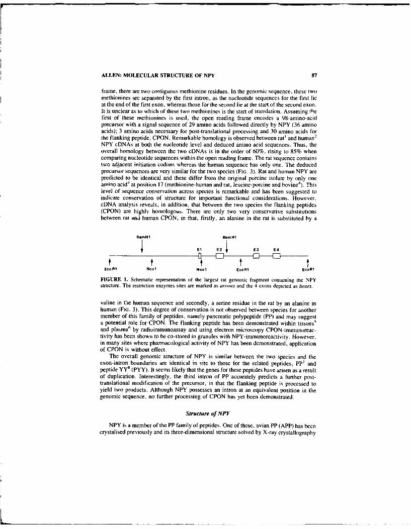
ALLEN: MOLECULAR STRUCTURE OF NPY 87
frame, there are two contiguous methionine residues. In the genomic sequence, these twomethionines are separated by the first intron, as the nucleotide sequences for the first lieat the end of the first exon, whereas those for the second lie at the start of the second exon.It is unclear as to which of these two methionines is the start of translation. Assuming thefirst of these methionines is used, the open reading frame encodes a 98-amino-acidprecursor with a signal sequence of 29 amino acids followed directly by NPY (36 aminoacids); 3 amino acids necessary for post-translational processing and 30 amino acids forthe flanking peptide, CPON. Remarkable homology is observed between rat' and human2
NPY cDNAs at both the nucleotide level and deduced amino acid sequences. Thus, theoverall homology between the two cDNAs is in the order of 60%, rising to 85% whencomparing nucleotide sequences within the open reading frame. The rat sequence containstwo adjacent initiation codons whereas the human sequence has only one. The deducedprecursor sequences are very similar for the two species (FIG. 3). Rat and human NPY arepredicted to be identical and these differ from the original porcine isolate by only oneamino acid 3 at position 17 (methionine-human and rat, leucine-porcine and bovine4). Thislevel of sequence conservation across species is remarkable and has been suggested toindicate conservation of structure for important functional considerations. However,cDNA analysis reveals, in addition, that between the two species the flanking peptides(CPON) are highly homologous. There are only two very conservative substitutionsbetween rat and human CPON, in that, firstly, an alanine in the rat is substituted by a
BamH1 BamHl
El E2 E3 E4
f tEco R1 Ncol NCO EcoR1 ECOR1
FIGURE 1. Schematic representation of the largest rat genomic fragment containing the NPYstructure. The restriction enzymes sites are marked as arrows and the 4 exons depicted as boxes.
valine in the human sequence and secondly, a serine residue in the rat by an alanine inhuman (FIG. 3). This degree of conservation is not observed between species for anothermember of this family of peptides, namely pancreatic polypeptide (PP) and may suggesta potential role for CPON. The flanking peptide has been demonstrated within tissues 5
and plasma6 by radioimmunoassay and using electron microscopy CPON-immunoreac-tivity has been shown to be co-stored in granules with NPY-immunoreactivity. However,in many sites where pharmacological activity of NPY has been demonstrated, applicationof CPON is without effect.
The overall genomic structure of NPY is similar between the two species and theexon-intron boundaries are identical in site to those for the related peptides, PP 7 andpeptide YY 8 (PYY). It seems likely that the genes for these peptides have arisen as a resultof duplication. Interestingly, the third intron of PP accurately predicts a further post-translational modification of the precursor, in that the flanking peptide is processed toyield two products. Although NPY possesses an intron at an equivalent position in thegenomic sequence, no further processing of CPON has yet been demonstrated.
Structure of NPY
NPY is a member of the PP family of peptides. One of these, avian PP (APP) has beencrystalised previously and its three-dimensional structure solved by X-ray crystallography

88 ANNALS NEW YORK ACADEMY OF SCIENCES
'0 U
U H) 0 04 F- -. 40 00 u 4 ~
co uO <'1 E O) co u4C.4.r4U .- 0 <E w < 0 z 00 to
00 u 0WU u . u
0a E-4(vH. ) 0 0< 00 << EE- a)'u -4u0 -t x 44 .)H4z
40 - HOJ- <'- 04o HU '4-4 4 4 0 cc ti
w0 U %4 H~ U40 <0 440 < 0) F 44 '4 U 4
00 0) F c- 0 u. HQ ) HO) s
cU U0 4- E0 a)- j $
>'~ ~ <0 ta.~ )' -4 4U In~ U HU U 4
HO3 ) E-4 (U - U u 0 cm Uou0- ) Hm < -I 0<1 00 w- 4 m . a
F. Hc Hm 0 O <. 0 4 U 4U 0'- U> <01 <( 0 40 4 o (a UU
U 40 00 <0 _. U
a) Hu H4 -l u- =- -u Uo 00m 72
HO) H) 0-cc <> HO HO E.4 w a:5< 00 00 Hu -~ 0 C. a 4 4
4.4 < u E ) u '4 m U mU U' u~ 004 u4 .I 00 u- aU <'u "
F4 < u0 0- 1. <H < 0 U a 40U0 U- U4 UU uU. C
U U) HO <0 w~ < 4 U 40 00 m
004 u~ 044 <- 0. U U u u
Uz w- 00 U4 u 0 Pr
00 HO <414 ) 0 0)4 U4 w0 40 u
U0 00 0 0u w -u < U) Uo
m 00 04 Q 44 u - 004 < > '4 o u
U >.) 0L) <c4 044 u 4 '4 U w0 "Ui
60 <~' u' w)- LD U ' 4
U <H F-O HH4 << < 4 '4c
40 00 '4 6
00 0 00 )4 01 40 '4 0
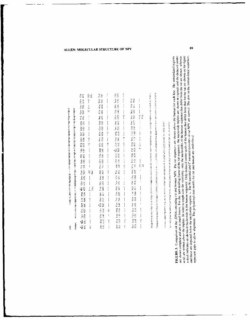
ALLEN: MOLECULAR STRUCTURE OF NPY 89
t~ 41A'
~ 0 c A~- ~jA 'CA C -
-~ ~s -E ?AA.4- ~~A
-, A- ~c A -
~ -~ -~ 0.
.c -~ -zA
OH 00 OUII(I'' --- U "4 I -
-< A< OH I -< I IS A -= -
>.A H %-U)AI '0< I-o I (4< -
AU F-I-- A - At
CjH (414 I .0 -t(4 -4H A <'0 I H
CI)-. (4 CU II-A I -IA I -ASH I <A
OH I CH I
00 I CA IOH o< I
C A -
AU I <A I
'A I AU I AC -~
OH I -< I -~ = SAU I AU I
cZ(414 I 0< A AC IC~U "A I<H <A I
o< A 0< I '0 A"'
01- I ~-A 'A '
AU' O~' I - I A - ~ AC
ICH A IA '0
CA' <AI 1 A A
AU I CA I '-H I-~ AUOH I I C(4A< I C"-
AU I <A I n-I-CA
CC CC nn>,- crz
IC IC 'A I JA I AC I CC I .4 ~
CC .4 CA I -< IIC IC HO I AU I
C
AC CC AU I I-U I 'IC
IC IC C)H I '-A I ~-c 'C A
- (4 AU I AA I ~ <~u-~IC IC
CC Ao< 0AIj 'A I
* (4 i-C
CC CO Al- OH 'A
(4 >'A AC) ~ I .- HC ..,
CC ASH 'LA '0<CC CC AU I C.A
II OH
IC IC AU I 'AlA I < ~S'-' A,
IC IC I-A I AU I -c i ACCC A OA I S-. -i-
CC CC AU I <A I
.4 '-A A 'AU I l-- I .A~t A A
'A~ <~ I '
.2(4 CC< I -'2 I I -
IC IC I~A I CIA(4 CA I <H
A 1$ AE~IC ~-
II 01.4 '0 I 'CI- 5~ A
2 AU I - 2: A
OH I A >0
(4-H I (4A I AC< I - A
IC A I -A U(4 AAI AU I
AU I A A
(4 OH I -< I z ~ -IC AU I AA I I A A A
.4.4 AC .4 0- -n.
"-A I ICH U OH 0 0'~ A IC A 'I CC
'q-OH I UI~UIA< I IC 'C - .4 5~ 0~< I <241<01 'AS I IA IC 2 A-A A A
IC AC CC '1 'C ncr
6 '-A 4 0211 I-U $ < I Al 10 2 2 ~
IC A +C)H 14 OH I 02 I-Al AC C)
~ A< IC AU <H I 1(41"' 'C A
-- C c-.-.- C ~ A .~0. IC ~ A
0. A~ -
StAC
CI~Ac~
K A~.Is -C 'IC *C -C

90 ANNALS NEW YORK ACADEMY OF SCIENCES
at high resolution." Overall. only about halt the amino acids are conserved between APPand NPY but closer comparison reveals that residues t! ertain koy si'v , arc retailcdbetween the two peptides. Based on these. it was possible to use the known crystalstructure of APP to construct. by computer. a model for the structure of NPY. The modelthat emerged from this study indicates that the molecule has a compact and extensivetertiary structure (FiG. 4). The residues of the N terminus form a left handed poflyproline-ll-like helix and are brought. by a tight P turn, into spatial proximity to an o helixstructure formed by residues 14 to 32. There are extensive hydrophobic interactionsbetween the residues on the inner surfaces of these helices that stabilise the structure viaintramolecular contracts. 'he residues of APP most important for maintaining thesehelix-helix contacts are either conserved in NPY or replaced by chemically homologousside chains (Fi;. 5). The net result is that the modelled structure of NPY is ver, stableand. by this mechanism, the N and C termini of NPY arc brought into spatial proxmilThe model begins to explain some of the structure-function data obtained from pharma-cological preparations. At least, two receptor ,ub!,,pes have been proposed for NPY basedon fragment activity. ' Thus. the Y I receptor requires the presence of intact N and Ctermini, whereas the Y2 receptor recognises a truncated version of NPY. 13-36 amidc(i.e., the C terminal polar arm and a helix). The Y I receptor appears predominantI atpo)stsynaptic sites in the sympathetic nervous system, whereas the Y2 receptor appears atpresynaptic sites.
The requirement of the Y I type of receptor for the whole NPY molecule for activit,is best explained by the model, as the N and C terminal tyrosines are brought into spatialproximity and its seems likely that it is this face that is presented to this receptor. Perhapsthe best test of the model is the maintenance of biological activity in molecules con-structed on the basis of the predicted structure. Thus. a synthetic ,cislon of NPY wherethe truncated N (residues 1-4) and C (residues 25-36) portions were chemically linkedwas found to possess pressor activity.''
Regulation of Expression of VPY
NPY demonstrates remarkable specificity in its expression within tissue types. Apartfrom rodent megakaryocytes.' 2 NPY-immunoreactivity is confined to cells derived fromthe neural crest. Indeed all three members of this family of peptides demonstrate remark-able t-ssue specificity, in that, just as NP is confined to neurdl tisNue. PP is expressedonly in the pancreatic islets and PYY is present in the exocrine pancreas and in endocrinecells of the lower gastrointestinal tract.
Studies using the NPY cRNA probe to c.,,mie the distribution of NPY mRNA. ingeneral. have demonstrated similar results to those obtained for the NPY peptide asmeasured by radioimmunoassay. However, in at least two instances mRNAs hybridisinespecifically to the labelled NPY cRNA have been found in unexpected sites, namely, ratspleen' 3 and heart. 1.14 In addition, various pharmacological manipulations have showndiscordant effects on the mRNA levels compared to peptide concentrations. NPY-immu-noreactivity in the rat spleen is present in postganglionic sympathetic nerve terminals.Thus, the finding of high concentrations NPY mRNA in RNA extracted from these tissueswas surprising and the mRNA levels were unaffected by chemical sympathetic ablation(unpublished data). Similarly. NPY-immunoreactivitv within the heart is considered pre-dominantly to be present in sympathetic nerve terminals, although small numbers ofweakly positive NPY-immunoreactive ganglion cells have been demonstrated in theatria.".," Specific mRNA hybridising to labelled NPY cRNA have been demonstrated inRNA exvracrtd from all reoions of the rodent heart' (FIG. 6) and again sympatheticablation had no effect on the concentrations of this mRNA species despite loss of all

ALLEN: MOLECULAR STRUCTURE OF NPY 91
Wer
FIGURE 4. Computer-constructed NPY model. (A) Polypeptide chain backbone showing the poly-proline-Il-like helix and C terminal helix connected by a tight turn. (B) Same orientation as (A) butspace filling representation of the NPY model. (From Allen et al.' Reprinted by permission from theNational Academy of Sciences.)
measurable immunoreactivity from the same heart tissue. 14 It is difficult to explain thepresence of NPY mRNA in these sites and its apparant dissociation from the peptidemeasurements, but this data suggests that there are situations where the mRNA is nottranslated to yield NPY.

92 ANNALS NEW YORK ACADEMY OF SCIENCES
A useful model system to investigate regulation of NPY gene expression in the contextof neuronal differentiation is the PC 12 cell line. 17 This cell line was cloned from a ratadrenal phaeochromocytoma and the cells resemble immature chromaffin cells. Thesecells have been used as a model of differentiation. Dexamethasonc causes the cells tobecome chromaffin-like, whereas nerve growth factor (NGF) halts mitosis and causes thecells to extend neurites. features of differentiation into sympathetic neurone-like cells.
PC 12 cell, contain an niRNA species reactive to the NPY cRNA probe that isidentical in electrophoretic size and hybridisation to the NPY mRNA in rat brain,'although immense variation in the amount of NPY mRNA has been observed between PC
FIGURE S. Stereoscopic view of the APP crystallographic structure (light lines) and the NPYmodel (heavv lines). (From Allen et al.' Reprinted by pe-mission from the National Academy ofSciences.)
12 cells obtained from different laboratories (unpublished data). The mRNA in these cellsis translated as NPY-immunoreactivity has been demorstraled within PC 12 cells." ' Bygel chromatography, the precursor appears to be processed very inefficiently. The ma-jority (approx. 70%) of immunoreactivity elutes from size exclusion columns as a largemolecular weight precursor, whereas only 30% elutes in the position of NPY itself. NPYmRNA and peptide concentrations are increased by exposure of the cells to NGF in theculture medium. An increase in NPY mRNA was observed within 12 hours (data notshown) and the response appeared to be maximal at 3 days" tFRi. 7). Withdrawal of NGFfrom the culture medium resulted in a slow return of NPY mRNA towards the controllevels.
Ii

ALLEN: MOLECULAR STRUCTURE OF NPY 93
A
Iv sp rv a
FIGURE 6. Northern blot analysis of RNA from rat heart, probed with NPY cRNA 32p labelledprobe under high stringency. (A) RNA extracted from rat brain olfactory bulb (OB) and whole heartfrom two animals, HI and H2. (B) RNA extracted from regional dissection of heart in atria (a), leftventricle (v), right ventricle (rv) and interventricular septum (sp).
Although the level of transcription is an important feature in the regulation of storedconcentrations of NPY. it appears that in some tissues a limiting factor may be the rate ofprocessing of the precursor to the mature peptide. The predicted pre-ursor for NPYconsists of 98 amino acids. Cleavage of the signal peptide should yield "pro-NPY"consisting of 69 amino acids (NPY: 36 amino acids, the three amino acids necessary forpost-translational processing and CPON: 30 amino acids). A number of antisera have beenraised to NPY and CPON. Of these YN22 recognises the N terminal part of NPY, YN 10is directed to the extreme C terminal part of NPY (it requires the C terminal amide residuefor full recognition) and JN8 was raised to CPON (FIG. 8). Precursor sequences shouldnot be recognised by YNI0 but are predicted to be recognised by YN22 and JN8. Gel
day C C 1 ccc
T
NPY
PC 12FIGURE 7. Northern blot analysis of RNA from PC 12 cells using NPY cRNA "2P labelled probeand an end labelled oligonucleotide to rat tubulin (T). RNA was extracted from control cells (c) andcells treated with NGF for I day (I), 3 days (3) and 6 days (6).

94 ANNALS NEW YORK ACADEMY OF SCIENCES
,4----- 28 aminoacid --. 36 aminoacid.--. - 30 aminoacid
SIGNAL NEUROPEPTIDE Y CPON
glycine argilnine
lysine
AN E U R O P E P T ID E Y CPON
YN22 JN8
NEUROPEPTIDE Y amide CPON
YN22 YN10 JN8
FIGURE 8. Schematic of the precursor of human NPY with an indication of the recognition sitesof the three antisera YN22, YNI0 & JN8. The translated product consists of a 28-amino-acid signalpeptide, NPY (36 amino acids). Cleav age <f th signal sequence would yield a precursor (pro NPY)that is recognised by the antisera YN22 and JN8. Processing of the precursor will generate theamidated version of NPY that is recognised by the antiserum YNI0.
Vo cc Std Vt
200 J 4JI 50-
Acid extract00-i FIGURE 9. Gel permeation chromatography of
acid and water extracts of rat brain. The extractsL0o were assayed using the YN22 antiserum. All the~inmmnoreactivity eluted from the column in the
0o -position of the NPY standard apart from a minute60 portion (less than 1%) that was measurable eluting
from the column in the expected position of theisoo precursor.
50 Waer extract
1 60Fractions

ALLEN: MOLECULAR STRUCTURE OF NPY 95
fmol /fraction11.0 Vo Std Vt
120
1 00
80
so
60
40
20
140
FIGURE 10. Gel permeation chromatography of ex-120 tract of rat adrenal. The upper panel shows the profile
100 obtained using the YN 10 antiserum, where all the im-munoreactivity is in the form of the mature peptide.
s0 The middle panel demonstrates the profile obtained byusing the YN22 antiserum and the lower panel that for
60 - the JN8 antiserum. About 50% of the immunoreactiv-ity elutes from the column in the position of the NPY
40 and CPON standards respectively. However, bothNPY and CPON immunoreactivity were measured in
20 fractions that eluted earlier from the column as as-sessed using the two antisera YN22 & JN8 and rep-
o " resent the precursor form of NPY. (Modified from160 REFERENCE 20.)
120
80
40
0 1 f I
0 15 30 45 6
fraction
permeation chromatography followed by assay of the fractions with the various tlueeantisera revealed that in most tissues, the only products recognised in extracts were thoseof the mature peptides, NPY and CPON and no large molecular weight or small fragmentswere detected. This was surprisingly the case for extracts of human phaeochromocytomatissues. 6 However, in extracts of rat superior cervical ganglia and rat adrenal tissue,significant quantities of immunoreactivity eluted from gel permeation columns in the

96 ANNALS NEW YORK ACADEMY OF SCIENCES
position of a large molecular weight form. 20 The immunoreactivity in these fractions wasdetectable by the two unrelated antisera, YN22 and JN8, whereas the antiserum YNIOfailed to recognise any immunoreactivity in these fractions. About 25% of total immu-noreactivity as assessed by both YN22 and JN8 of the rat superior cervical ganglionextracts was of the precursor form compared to mature peptide. In the rat adrenal, about50% of the total immunoreactivity was in the form of "pro-NPY." Thus, the efficiencyof precursor processing varies considerably among tissues. The pattern observed is verysimilar to that noted previously for the enkephalins, where the majority of enkephalin
fmol /fractionVo Std vt
120
100
80
60
110FIGURE 11. Gel permeation chromatogra-
20 phy of the extract of adrenals collected fromrats 4 hours after reserpine treatment. The up-
0 per panel shows the profile of immunoreactiv-ity for the YN22 antiserum whereas the bottompanel is that for the JN8 antiserum. In contrast
240 to FIGURE 10, immunoreactivity is only detect-able as the mature peptide and no distinct largemolecule weight precursor was observed.
180
120
60
0 , i I I
0 15 30 I15 60
fraction
immunoreactivity in the brain is in the form of met-enkephalin whereas large molecularweight precursors predominate in adrenal extracts. The proportion of large molecularweight precursor NPY compared to mature NPY in the rat adrenal could be altered bypharmacological manipulation. Thus, acute stress following reserpine treatment resultedin rapid depletion of any measurable large molecular weight precursor, thus suggestingthat in some tissues the rate of conversion of the precursor to free peptides may be thelimiting factor.

ALLEN: MOLECULAR STRUCTURE OF NPY 97
SUMMARY
The original description of NPY following its isolation3 commented on its homologyto the pancreatic polypeptide family of peptides. This homology is extended to the mRNAsequences and the genomic structure, suggesting that this family has arisen as a result ofgene duplication. However, each member demonstrates remarkable specificity in its ex-pression within tissue types. The expression of the NPY gene is thus controlled by, as yetundefined, factors regulating to neural cells. The level of expression within cells isdependent on nerve growth factor. However, other factors, such as the rate of processingof the precursor, may be involved in regulation of the stored concentrations of the peptideproduct.
REFERENCES
1. ALLEN, J. M., 1. NOVOTNY, J. B. MARTIN & G. HEINRICH. 1987. Molecular structure ofmammalia,, neuropeptide Y: analysis by molecular cloning and computer aided comparisonwith crystal structure of avian homologue. Proc. Natl. Acad. Sci. USA 84: 2532-2536.
2. MINTH, C. D., S. R. BLOOM, J. M. POLAK & J. E. DIXON. 1984. Cloning, characterization& DNA sequence of a human cDNA encoding neuropeptide tyrosine. Proc. Nat). Acad. Sci.USA 81: 4577-4581.
3. TATEMOTO, K. 1982. Neuropeptide Y: complete amino acid sequence of the brain peptide.Proc. Natil. Acad. Sci. USA. 79: 5485-5489.
4. TATEMOTO. K. 1989. Neuropeptide Y: isolation, structure and function. In Neuropeptide Y. V.Mutt, K. Fuxe, T. Hokfelt & J. M. Lundberg, Eds. 13-21. Raven Press. New York, NY.
5. ALLEN. J. M., J. M. POLAK & S. R. BLOOM. 1985. Presence of the predicted C-flankingpeptide of neuropeptide Y (CPON) in tissue extracts. Neuropeptides 6: 95-100.
6. ALLEN, J. M., J. C. YEATS, R. CAUSON, M. J. BROWN & S. R. BLOOM. 1987. NeuropeptideY and its flanking peptide in human endocrine tumors and plasma. J. Clin. Endocrinol.Metab. 64: 1199-1204.
7. LEITER, A. B., M. R. MONTMINY, E. JAMIESON & R. M. GOODMAN. 1985. Exons of thehuman pancreatic polypeptide gene define functional domains of the precursor. 1. Biol.Chem. 260: 13013-13017.
8. LEITER, A. B.. TODER, H. J. WOLFE. 1. L. TAYLOR, S. COOPERMAN, G. MANDEL & R. H.GOODMAN. 1987. Peptide YY. Structure of the precursor & expression in exocrine pancreas.J. Biol. Chem. 262: 12984-12988.
9. GLOVER, 1., i. MANEEF, J. PITTS. S. WOOD, D. Moss, 1. TICKLE & T. BLUNDELL. 1983.Biopolymers 22: 293-304.
10. WAHLESTEDT, C., N. YANAIHARA & R. HAKANSON 1986. Evidence for different pre- &
post-junctional receptors tor neuropeptide Y and related peptides. Regul. Pept. 13:307-318.II. BECK, A., G. JUNG, W. GAIDA, M. KOPPEN. R. LANG & G. SCHNORRENBERG. 1989. Highly
potent and small neuropeptide Y agoidst obtained by linking NPY 1-4 via spacer to X helicalNPY 25-36. FEBS Lett. 244: 119-122.
12. ERICSSON, A., M. SCHALLING, K. R. MCINTYRE, J. M. LUNDBERG. D. LARHAMMAR, K.SCROOGY. T. H6KFELT & H. PERSSON. 1987. Detection of neuropeptide Y and its RNA inmegakaryocytes, Enhanced levels in certain autoimmune mice. Proc. Natl. Acad. Sci. USA84: 5585-5589.
13. LARHAMMAR, D., A. ERICSSON & H. PERSSON. 1987. Structure and expression of the ratneuropeptide Y gene. Proc. Natl. Acad. Sci. USA 84: 2068-2072.
14. ALLEN, J. M., J. B. MARTIN & G. HEINRICH. 1988. Neuropeptide Y is intrinsic to the heart.In Advances in Atrial Peptide Research. B. M. Brenner & J. H. Laragh, Eds. Vol. 2:155-160. Raven Press. New York. NY.
15. Gu. ., J. M. POLAK. J. M. ALLEN, W. M. HUANG, M. N. SHEPPARD, K. TATEMOTO & S.
R. BLOOM. 1984. High concentration of a novel peptide, neuropeptide Y, in the innervationof mouse and rat heart. J. Histochem. Cytochem. 32:467-472.

98 ANNALS NEW YORK ACADEMY OF SCIENCES
16. HASSALL, C. J. S. & G. BURtNSTOCK. 1984. Neuropeptide Y-like immunoreactivity in culturedintrinsic neurones of the heart. Neurosci. Lett. 52:111-115.
17. GREENE, L. A. & A. S. TiSCHLER. 1976. Establishment of a nc~adrenergic clonal line of ratadrenal pheochronocytoma cells which respond to nerve growth factor. Proc. Nati. Acad. Sci.USA 73:2424-2428.
18. ALLEN, J. M., J. B. MARTIN & G. HEINICHd. 1987. Neuropeptide Y gene expression in PC12 cells and its regulation by nerve growth factor: a model for developmental regulation. Mol.Brain Res. 3:39-43.
19. ALLEN, J. M., A. S. TiSCHLER, Y. C. LEE & S. R. BLOOM. 1984. Neuropeptide Y in PC 12cells phaeoclsromocytoma cultures: responses to dexamethasone and nerve growth factor.Neurosci. Lett. 46:291-296.
20. ALLEN, J. M. 1989. Molecular structure of neuropeptide Y and regulation of expression of itsgene. In Neuropeptide Y. V. Mutt, K. Fuxe, T. Hokfelt & J. M. Lundberg, Eds. 33-41.Raven Press. New York, NY.

Regulation of the HumanNeuropeptide Y Gene
CAROLYN A. MINTH AND JACK E. DIXON
Department of Biochemistr 'yWright State University
Dayton, Ohio 45435and
Department of Biochemistr "
Purdue UniversityWest Lafayette, Indiana 47907
Eukaryotic gene promoters contain cis-acting elements that regulate transcription directedby RNA polymerase II. These elements usually interact with sequence-specific DNA-binding proteins which either repress or activate transcription. Typically a number ofDNA-binding proteins are capable of interacting with the 5'-flanking sequences of aspecific gene. The availability and activity of these proteins determine the extent to whichthey participate in transcriptional regulation.
The pattern of specific gene expression which generates the complexity demonstratedby the nervous system is not well understood. Because the neuropeptide Y (NPY) gene isexpressed widely throughout the central and peripheral nervous systems, it provides auseful model for studying neuronal gene expression. -4 NPY is coordinately expressedwith a variety of neurotransmitters and neuropeptides including norepinephrine, 5 vaso-
7active intestinal peptide,6 acetylcholine,6 growth hormone releasing hormone,FMRFamide, 8 somatostatin,9 and cholecystokinin. ' How individual neurons selectivelyexpress various combinations of these neuropeptides is unknown. In order to elucidate themechanisms underlying neuronal gene expression, we initiated a study on the NPY gene.
A variety of agents such as forskolin, TPA, and NGF influence the steady state levelsof rat NPY mRNA, I 1.12 suggesting that the NPY gene promoter may be regulated by acombination of transcriptional elements. Furthermore, the 5'-flanking DNA of the humangene contains partial consensus sequences for DNA-binding proteins such as SPI, API,and CAAT.13 -
17 In combination, several transcriptional elements could generate pro-
moter diversity, culminating in the expression pattern of the NPY gene in the nervoussystem.
Sequence and Structural Analysis of the NPY Promoter
The 5'-flanking DNA of the NPY gene is shown in FIGURE 1. The sequence displaysan unusually distinct partitioning of AT versus GC nucleotides. The distal region (- 1078to -796) contains 67% AT nucleotides while the sequence closer to the start of tran-scription (-246 to + 1) contains only 25% AT nucleotides. A computer program de-signed to calculate regions of DNA secondary structure predicts that the AT-rich regioncentered around -780 could potentially form bent DNA.'" Experiments described in alater section demonstrate that deletion of this AT-rich region increases promoter activity.The ability of the AT-rich sequences to participate in negative regulation of the NPY geneis currently under investigation.
Further analysis of the 5'-flanking sequences identified consensus sequences for sev-99

100 ANNALS NEW YORK ACADEMY OF SCIENCES
4A1078GATCTCAG 1071
TCCACTGCATAAAATCTTGATCCTGTAATAATAGTTTCTGTATCT1'GCATATTCATTCMA 1011
CAGGTTTAACGCGATCAGCAAATTAATGTTCATCGTTTTTAACATGTTTCATCTTAATCA 951
GAACCCACATTCTCAACGTTAATTGAACGTACATAGGACTATACAAGGGTTAGTAAATAA 891
GACAGAAACTGTTGCTCATTTAACCACCGTCACTTTGGACCAAAAAAGAAAAAATATATA 831
tA796TTTTTAAAATTGAGCTTAAAAGAGTCTCTAGAAGCTGGAAGCGTGGCTCTTTTTCAGCAA 771
ACTGGGGAATAGGTTTACCGTGTTCCCCCTCTGGGGAATTTTGCGTCGCCACACTCATG 711
4A701TCTCGACCGACCCTGGCTCGCTGCGTCTGAGCCAGTACTTGAGCAAGGCTGATCTAGAAA 651
SPIAACCAGCTGAGAGAAGGGCACAAGCCCCTGAAACCAC GGCCGG CGTGGCGTGGGGA 593
A5334GCCACGTTTGGGACCCTCTAGCCGGAGACTTCCGACCTGCCTCCGACTTGTTCTAAGTA 533
4A499CAGGAAAAATCTGTGCGCCCAGTTGCCTCACTCCAACAGCGCGCAGTTGTGCCCCGGCGAG 473
4.A428GATGCCGCGCTAGTCGTGGAGATGCCCCACCACAAAGAGGATTCAGGTGCTTCCTACTCC 413
GGCACCCAGTGGGCTGGTAGTCCTGTTGGCAGGACAMGMTCGTCTGGGCTGCTCCTA 353
TCTCTGGCAGGACTAGACGGGGCGTGMCGGAAAGAAGGAAAGAAGGAAAGCAGGGATCGG 293
4Ai246GCACTGCCCGAGGGCAGATACTTGGGCTTTGGTGTTGTCCAGCGCGCTCCGAGTGCGCTG 233
SF1 4A178CCTCGCTCACGCGGTCCCAGGCCCCGCTTCTTCAGGCAGTGCCCTGG GGCGG AGCGTT 175
46143 SF1 A1184GGTGTGCGTCGCTCCCTMGCTCGACACTCGTGCGGCTGCGGTTCCAGC CCCCTCC C 117
sF1 SF1 SF1 4A83 CAAT APiCCCGCC ACTCAG GGCCGG GMGCT COCGTGGGAGTCACFCCA-AGCG TGACTG 65
4A63 SP1 4A51 630t TATAJCCGAGG C-C-CCTCC TGCCGCCGCGAGGAAGCTCC ATAAAA GCCCTGTCGCGACCC 10
4+1GCTCTCTGCCCACGTG7CCCCAACCCCCAACAT CAT
Fig. I

MINTH & DIXON: HUMAN NPY GENE 101
eral previously characterized DNA-binding proteins. There are five predicted SPI GCboxes located at -607, - 181, - IlI, -99, and -88. The region between -86 and -62is an imperfect palindrome which contains partial CAAT and API consensus sequences.In addition, two CCCCTCC sites are present at - 118 and -51. The importance of theseCT regions is addressed in the following section.
Deletion Analysis of the 5'-Flanking DNA
A deletion series of the 5'-flanking DNA of the NPY gene was constructed byExonuclease III digestion of the parent plasmid pCatNPY1078.20 A schematic diagramdepicting the construction of these fusion plasmids is shown in FIGURE 2. These con-structs represent varying amounts of 5'-flanking sequences fused to the bacterial reportergene, chloramphenicol acetyltransferase (CAT). These plasmids were transiently intro-duced into the human Lan-5 cell line by a modified CaPO4 technique. 21 pRSVLacZ wasincluded as a control to minimize variations between individual transfections and exper-iments. The Lan-5 cell line affords an ideal system for studying NPY gene expressionbecause it was established from a human neuroblastoma and expresses authentic NPY.The relative CAT conversion for a selected set of fusion constructs is shown in FIGURE 3.These data demonstrate the complexity of NPY gene expression in this system. Theincrease in expression seen when sequences betwecn - 1078 to -796 and -428 to -246are deleted suggest that these regions may contain negative-acting regulatory elements.
This analysis demonstrated that the sequences necessary for expression of the NPYgene are contained between -246 and -51. A second deletion series spanning this regionwas constructed and tested (FtI. 4A). The consensus binding sites for several previouslycharacterized DNA-binding proteins are shown in FIGURE 4B. Deletion of the sequencebetween -63 and -51 abolishes transcription of this gene. Deletion of the CAAT- andAPI-like binding sites at -73 and-64 and the three SPI consensus binding sites between- 118 and -83 do not result in a measurable change of expression. However, deletion ofthe sequence -143 to -118 which contains another CCCCTCC site results in approxi-mately a 2-fold decrease in CAT activity. This data suggests that the CCCCTCC se-quences present in the 5'-flanking DNA are important for NPY gene expression.
Gel Retardation Analysis of the Proximal 5'-Flanking Sequences
The promoter sequence between -75 to -46 was examined for its ability to formspecific DNA-protein interactions using the gel retardation assay.22.23 The nucleotidesequence of the region between -75 and -46 and that of mutant NPY(I) are depicted inFIGURE 5A. A typical gel retardation analysis using this fragment is shown in FIGURE 5B.Two major DNA/protein complexes labelled N I and N2 are formed in the absence ofcompetitor oligonucleotides (lanes I and 20). The oligonucleotides used in the competi-tion reactions contain sevcral defined DNA/protein consensus binding sites. The soma-tostatin promoter fragment contains a binding site for the cyclic AMP responsive bindingprotein (CREB). 24 The TRH oligonucleotide contains both a CRE-like sequence (-51)
FIGURE 1. Nucleotide sequence of the human NPY promoter. Nucleotides are numbered from theinitiation of transcription and negative values indicate 5'-flanking sequences. Deletion endpoints arenumbered and designated by an ., row and bold print. The TATA consensus sequence is boxed as arethe consensus sequences for the DNA-binding proteins SPI, API, and CAAT.

102 ANNALS NEW YORK ACADEMY OF SCIENCES
pCotNPYAI076
PUCl13 [1 1 hV CAT
-z i bXb I
X x X
Oz jExo M (timed aliquots)
S! Nuclease
J/enow, dNTPs
,J-4
JT4 DNA Ligase
A1076
pUICAT geCA
WCo) NPAN)A2
0645
S ~ ~ jCAT Ien w N~
FIGURE 2. Schematic illustration of the construction of the NPY/CAT fusion constructions. Thestarting plar-mid pCatNPY1076 was digested with Sst and Xbal followed by treatment with Exo-nuclease III for varying times. The ends were blunted by SI nuclease and repaired with Klenow.Sequence analysis of plasmids obtained from various time points confirmed the deletion endpoints.
and a CT-rich sequence (-62).2' The human cardiac a- actin and the HSV thymidinekinase promoters contain SPI consensus binding sites.26.27 The N2 complex is mostefficiently competed when a 500-fold excess of the unlabeled NPY (- 75 to -46) or TRH(-70 to -41) oligonucleotide is included in the reaction mixture. The large amount ofcompetitor oligonucleotides needed to reduce the intensity of N2 suggests that this inter-action is not specific. However, since all fragments are not capable of competition, apotential binding site at the 5' end of this oligonucleotide can not be ruled out. The NIcomplex is efficiently competed by the cardiac a-actin and the HSV thymidine kinaseprobes. Furthermore, the mutated NPY oligonucleotide (I) was no longer able to compete
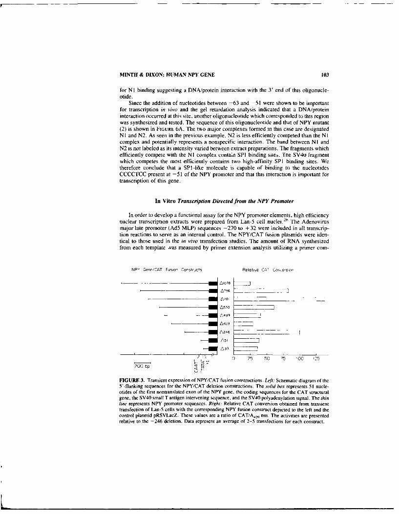
MINTH & DIXON: HUMAN NPY GENE 103
for NI binding suggesting a DNA/protein interaction with the 3' end of this oligonucle-otide.
Since the addition of nucleotides between -63 and -51 were shown to be importantfor transcription in vivo and the gel retardation analysis indicated that a DNA/proteininteraction occurred at this site, another oligonucleotide which corresponded to this regionwas synthesized and tested. The sequence of this oligonucleotide and that of NPY mutant(2) is shown in FIGURE 6A. The two major complexes formed in this case are designatedNI and N2. As seen in the previous example, N2 is less efficiently competed than the NIcomplex and potentially represents a nonspecific interaction. The band between N I andN2 is not labeled as its intensity varied between extract preparations. The fragments whichefficiently compete with the N I complex contain SPI binding sites. Te SV4u fragmentwhich competes the most efficiently contains two high-affinity SPI binding sites. Wetherefore conclude that a SPI-like molecule is capable of binding to the nucleotidesCCCCTCC present at -51 of the NPY promoter and that this interaction is important fortranscription of this gene.
In Vitro Transcription Directed from the NPY Promoter
In order to develop a functional assay for the NPY promoter elements, high efficiencynuclear transcription extracts were prepared from Lan-5 cell nuclei. 28 The Adenovirusmajor late promoter (Ad5 MLP) sequences -270 to + 32 were included in all transcrip-tion reactions to serve as an internal control. The NPY/CAT fusion plasmids were iden-tical to those used in the in vivo transfection studies. The amount of RNA synthesizedfrom each template vas measured by primer extension analysis utilizing a primer com-
NPY Gene/CAT Fusion Constructs Relatlve CAT Conferson
A796 ]
L533
L499
A428
A246
,--1AsI
I\" 0 25 50 75 100 125200 bp
FIGURE 3. Transient expression of NPY/CAT fusion constructions. Left: Schematic diagram of the5'-flanking sequences for the NPY/CAT deletion constructions. The solid box represents 51 nude-otides of the first nontranslated exon of the NPY gene, the coding sequences for the CAT structuralgene, the SV40 small T antigen intervening sequence, and the SV40 polyadenylation signal. The thinline represents NPY promoter sequences. Right: Relative CAT conversion obtained from transienttransfection of Lan-5 cells with the corresponding NPY fusion construct depicted to the left and thecontrol plasmid pRSVLacZ. These values are a ratio of CAT/A420 nm. The activities are presentedrelative to the -246 deletion. Data represent an average of 2-5 transfections for each construct.

104 ANNALS NEW YORK ACADEMY OF SCIENCES
plementary to the CAT gene (FIG. 7A). Correct initiation of transcription results in bandsof 131 and 110 nucleotides in length for the NPY and the MLP transcripts, respectively(FIG. 7B). Correct initiation of RNA synthesis from the various promoter deletions usedin this system correlate with function in the transient transfection analyses. Two notable
A.140
130 -
120 -
CZ 110-
0.r 100
> 90a00 80
I-< 700
50 "
CC 40 I
30
20 -
100I
A246 A178 A143 A118 A83 A63 A51 A30
B. A246 A178 A143 6118 683 A63 A51 A30
SPi SPi SPI SPi SP1 CAAT SPi 1
I10bp
FIGURE 4. Transient expression of proximal NPY/CAT fusion constructions. (A) The open bars
represent the rHative CAT conversion obtained from transient transfections of Lan-5 cells with thedeletions ranging from - 246 to - 30 and the plasmid pRSVLacZ. The values are a ratio of CAT/A4.nm relative to the -246 deletion. (B) Schematic diagram of cis-acting promoter elements between-246 and -30. The positions of the deletions are denoted by vertical lines. The TATA box isrepresented as a blackened box. The CCCCTCC sequences are depicted by blackened circles whilethe GGGCKGG SPI consensus sequences are represented by open circles. The putative API andCAAT consensus sequences are represented by a blackened diamond and a hatched box. respec-tively.
differences are the -30 and -51 constructs. Both of these plasmids were capable ofgenerating significant CAT activity in vivo, but were unable to direct the correct initiationof NPY rmRNA in vitro. In addition, the - i078 construct, whose CAT activity wasconsistently less than that of the -51 and -30 constructs in the in vivo experiments, iscapable of accurate transcription of the NPY promoter in vitro. These experiments dem-

NIINTH & DIXON: HUMIAN NPY' (ENE 105
'IZ z
Ii'
- M
zE =rc- -"
U~ 24- ~

106 ANNALS NEW YORK ACADEMY OF SCIENCES
rE
3tILI
z z X
ZS
za"mo tVo;z 5
u El

MINTH & DIXON: HUMAN NPY GENE 107
Ill -NPY (+I)
A. . 2 ,
-NP Y (+1)
B.
+1
-- lZZJZ f7ZI pCatNPYt6
----E , TT 1 CMAT(-270o) t1t 2ont
FIGURE 7. In vitro transcription analyses. (A) In vitro transcription products directed from theNPY and MCAT promoters. Each reaction contains 3 mg/ml Lan-5 nuclear extract. 0.75 /g of thedesignated NPY-CAT fusion constructs and 0.25 iig of MCAT. The positions of the correctlyinitiated primer extension products for the NPY and MCAT constructs are noted to the right. (BISchematic diagram of the templates utilized in the in vitro transcription analyses. Templates pCat-NPY- and MCAT contain various amounts of the 5'-flanking sequences of the NPY promoter andAd5 MLP, respectively, fused to the bacterial CAT gene. The primer is complementary to thesequences + I' to + 35 of the CAT transcript. The arrow depicts the predicted primer-extensionproducts arising from the in vitro-synthesized RNA. nt, nucleotide.
onstrate the importance of correlating CAT activities with correctly initiated transcriptionfrom the NPY promoter and indicate that vector sequences placed proximal to promotersequences can generate spurious transcripts resulting in exaggerated CAT activities invivo.

108 ANNALS NEW YORK ACADEMY OF SCIENCES
In Vitro Extract Depletion Experiment
The sequence between -63 and -51 is important for NPY gene expression based onboth in vivo transfections and in vitro transcriptions. We demonstrated that a SPI -likemolecule is capable of binding to this region. To test whether this molecule is necessaryto direct expression of the NPY gene, in vitro extract depletion experiments were per-formed. The construct pCatNPY246 is used as the template for the reactions because it isconsistently expressed at a high level in both the in vitro and in vivo experimental systems.In the depletion reactions, 300 ng of competitor oligonucleotide was added prior to theaddition of Lan-5 nuclear extract. Both the NPY promoter sequence -65 to -36 and theSV40 promoter GC boxes IV and V significantly decreased transcription of the NPYpromoter (FIG. 8). The NPY mutant oligonucleotide (2) which no longer competes forprotein binding in the gel retardation experiments was incapable of decreasing transcrip-tion. This competition was specific as addition of excess SPI competitor oligonucleotidesdid not inhibit transcription directed by the Ad5 MLP. The autoradiograph was scannedwith a densitometer and the values for NPY/CAT normalized to those for MCAT. TheSV40 fragment decreased transcription to 15% of the control while the NPY promoterfragment decreased transcription to 20% of the control. These results demonstrate that aSPI-like molecule specifically acts as a transactivator of the NPY gene.
A number of studies have demonstrated that interactions involving the binding ofmultiple proteins to neighboring promoter sequences are required for promoteractivation. 26
.2 7 "29 The sequences responsible for NPY expression contain CT-rich binding
sites which interact with SPI-like proteins. These sites are not representative of the typicalSPI binding sites (TABLE 1). Many other promoters contain SPI binding sites. Forexample, SPI is important for expression of the TK and the immediate early genes ofherpes simplex virus as well as the LTR of the HIV retrovirus. 27 3 ° -3' SPI also activatesthe promoters of the mouse DHFR,32 mouse hypoxanthine phosphoribosyl transferase. 3"mouse adenine phosphoribosyl-transferase- 4 rat type It procollagenj 5 human metal-lothionein IIA,29 hamster hydroxymethyl glutaryl CoA reductase, 6 human c-Ha-ras 1 37
human adenosine deaminase. 3' human urokinase-plasminogen activator, 39 and human
cardiac a-actin genes."6 The CCCCTCC sites in the NPY promoter most closely resemblethe SPI-binding sites in the cardiac a-actin promoter. The role SPI plays in the tran-scriptional activation of such a diverse group of genes has yet to be elucidated.
Tissue specificity may be controlled by subtle differences in transcription factors. SPI
co"itao 0FIGURE 8. In vitro transcription competitionanalyses. The ability of Lan-5 nuclear extract totranscribe the templates pCatNPY246 and MCAT
-Py (.1) was assayed in the presence and absence of 0.3 ligof competitor DNA fragments. The competitorfragments are listed above the lanes in which theyappear. The positions of correctly initiated NPYand MCAT transcripts are noted to the right.
Mca5 (.I
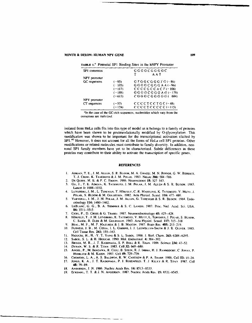
MINTH & DIXON: HUMAN NPY GENE 109
TABLE L" Potential SPI Binding Sites in the hNPY Promoter
SPI consensus GGGGCGGGGCT AAT
NPY promoterGC sequences (-95) G TG GCG GG T G(-86)
(- t05) G G GGCGG G A A(-96)(-117) C CC C GC CA C T(-108)(-188) GG GG C GG GA G(-179)(-613) C GG GC GGG G G(-604)
NPY promoterCT sequences (-57) CC C C TCCT G C(-48)
(-124) C CC C TC C CC C (- 115)
in the ease of the GC-rich sequences. nucleotides which vary from theconsensus are italicized.
isolated from HeLa cells fits into this type of model as it belongs to a family of proteinswhich have been shown to be posttranslationally modifted by 0-glycosylation. Thismodification was shown to be important for the transcriptional activation elicited bySPI.A0 However, it does not account for all the forms of HeLa cell SPI proteins. Othermodifications or related molecules must contribute to family diversity. In addition, neu-ronal SPI family members have yet to be characterized. Subtle differences in theseproteins may contribute to their ability to activate the transcription of specific genes.
REFERENCES
I . ADRIAN, T, E.. 1. M. ALLEN, S. R. BLOOM, M. A. GHATEI, M. N. ROSOR. G. W. ROBERTS.T. J. CROW, K. TATEMoTo & J. M. POLAK. 1983. Nature 306: 584-586.
2. DE QUIDT, M. E. & P. C. EMSON. 1986. Neuroscience 18: 527-543.3. Gu. J., T. E. ADRIAN. K. TATEMOTO, J. M. POLAK. J. M. ALLEN & S. R. BLOOM. 1983.
Lancet 1: 1008-1010.4. LUNDBERG, J. M_, L. TERENius, T. H45KFELT, C. R. MARTLING. K. TATEMOTO. V. MUTT. J.
POLAK, S. BLOOM & M. GOLDSTEIN. 1982. Acta Physiol. Scand. 116: 477-480.5. VARNVELL, 1. M., J. M. POLAK, J. M. ALLEN, G. TERENGHI & S. R. BLOOM. 1984. Endo-
crinology 114: 1460-1462.6. LEBLANC, 0. G., B. A. TRIMMER & S. C. LANDIS. 1987. Proc. Nail. Acad. Sci. USA.
84: 3511-3515.7. Cioii, P., D. CROIX & Gi. TRAMU. 1987. Neuroendocrinology 45: 425-428.8. HdKFELT, T., J. M. LUNDBERG, K. TATEMOTO, V. MUTT, L. TERENIUS. J1. POLAK, S. BLOOM.
C. SASEK, R. ELDE & M. GOLDSTEIN. 1983. Adta Physiol. Scand. 117: 315-318.9. BEAL, M. F., M. F. MAZURER & J. B. MARTIN. 1987. Brain Res. 405: 213-219.
10. FURNESS, J. B., M. COSTA, 1. L. GiBBINs. 1. J. LLEWELLYN-SMITH- & J. R. OILIVER. 1985.Cell Tissue Res. 241: 155-163.
It. HIGUCsst, H., H. -Y. T. YANG & S. L. SABOL. 1988. J. Biol. Chem. 263-. 6288-6295.12. SABOL, S. L. & K. HIGUCHI. 1990. Mol. Endocrinol. 4: 384-392.13. BRIGGS, M. R., J. T. KADONAGA. S. P. BELL & R. ThIAN. 1986. Science 234: 47-52.14. DYNAN. W. S. & R. TjhAN. 1983. Cell 32: 669-68015. ANGEL. P., M. IMAGAWA, R. Crnu. B. STEIN. R. J. IMBRA. H. J1. RAHMSDORF, C. JONAT. P.
HERRLICH & M. KARIN. 1987. Cell 49: 729-739.16. CHODOSII. L. A., A. S. BALDWIN, R. W. CARTHEW & P. A. SHARP. 1988. Cell 53: 11-2-4.17. JONES, K. A., J. T. KADONAGA. P. J. ROSENFELD, T. J. KELLY & R. TitAN. 1987. Cell
48: 79-89.18. ANDERSON, J. N. 1986. Nucleic Acids Res. 14: 85 13-8533.19. ECKDAHL. T. T. & J. N. ANDERSON. 1987. Nucleic Acids Res. IS- 8531-8545.

110 ANNALS NEW YORK ACADEMY OF SCIENCES
20. M1NTH, C. D., P. C. ANDREWS & J. E. DIXON. 1986. J. Biol. Chem. 261: 11974-11979.21. CHEN, C. & H. OKAYAMA. 1987. Mol. Cell. Biol. 7: 2745-2752.22. CARTHEW, R. W., L. A. CHODOSH & P. A. SHARP. 1985. Cell 43: 439-448.23. FRIED, M. & D. M. CROTHERS 1981. Nucleic Acids Res. 9: 6505-6525.24. ANDRISANI, 0. M., D. A. POT, Z. ZHU & J. E. DIXON. 1988. Mol. Cell. Biol. 8: 1947-1956.25. LEE. S. L., K. STEWARD & R. H. GOODMAN. 1988. J. Biol. Chem. 263: 16604-16609.26. GUJSTAFSON, T. A. & L. KEDES. 1989. MoL Cell. Biol. 9:3269-3283.27. JONES, K. A.. K. R. YAMAMOTO & R. TJIAN. 1985. Cell 42: 559-572.28. SHAPIRO, D. J., P. A. SHARP. W. W. WAHLI & M. J. KELLER. 1988. DNA 7: 47-55.29. LEE. W., A. HASLINGER, M. KARIN & P. THIAN. 1987. Nature 325: 368-372.30. LANG. J. C.. D. A. SPANDIDOS & N. M. WILKIE. 1984. EMBO J. 3: 389-395.31. JONES, K. A.. J. T. KADONAGA, P. A. Luciw & R. TjtAN. 1986. Science 232: 755-759.32. DYNAN, W. S., S. SAZER. R. TjtAN & R. T. SCHIMKE. 1986. Nature 319: 246-248.33. MELTON, D. W., D. S. KONECKI, J. BRENNAND & C. T. CASKEY. 1984. Proc. Natl. Acad.
Sci. USA 81: 2147-215 1.34. DuSH. M. K.. J. M. SIKELA. S. A. KHAN, J. A. TiSCHFIELD & P. J. STAMBROOK. 1985. Proc.
Nati. Acad. Sci. USA 82: 273 1-2735.35. KOHNO, K.. M. SULLIVAN & Y. YAMADA. 1985. J. Biol. Chem. 260: 4.441-4447.36. REYNOLDS, G. A., S. K. BASU, T. F. OSBORNE, D. J. CHIN, G. GIL. M. S. BROWN, J. L.
GOLDSTEIN & K. L. LuSKEY. 1984. Cell 38: 275-285.37. ISHII. S.. J. T. KADONAGA. R. TjtAN. J. N. BRADY, G. T. MERLINO & 1. PASTAN. 1986.
Science 232: 1410-1413.38. VALERIO, D., M. G. C. DuYVESTEYN. B. M. M. DEKKER, G. WEEDA, T. M. BERKVENS. L.
VAN DER VOORN, H. VAN ORMONDT & A. J. VAN DER En. 1985. EMBO J. 4: 437-443.39. Riccio, A.. G. GRIMALDI, P. VERDE, G. SEBASTIO. S. BOAST & F. BLAST. 1985. Nucleic
Acids Res. 13: 2759-2771.40. JACKSON, S. P. & R. TIIAN . 1988. Cell 125: 125-133.

PART II. ROLE OF NEUROPEPTIDE Y IN THE REGULATION OF THECIRCULATORY SYSTEM
Neuropeptide Y and CentralCardiovascular RegulationFocus on Its Role as a Cotransmitter in
Cardiovascular Adrenergic Neuronsa
K. FUXE,b J. A. AGUIRREb L. F. AGNATI,' G. von EULER,"P. HEDLUND,b R. COVElASd M. ZOLI,' B. BJELKE,b
AND P. ENEROTHbbDepartment of Histology and Neurobiology
Karolinska InstitutetBox 60400
S-104 01 Stockholm, Sweden
'Department of Human PhysiologyUniversity of Modena
Via Campi 287
411 O0 Modena, Italy
dDepartment of Cellular Biology and Pathology
University of Salamanca37 007 Salamanca, Spain
INTRODUCTION
Especially during the eighties the concept has been established that communicationwithin the central nervous system involves not only the classical synaptic transmission(wiring transmission, WT) but also a transmission which involves the diffusion of re-leased transmitters in the extracellular fluid pathways to reach distant high affinity re-ceptors (volume transmission, VT). 1,
2 Several observations have led to the developmentof this concept such as the demonstration of lack of synaptic specializations in manymonoamine nerve terminals of the brain,3 nonsynaptic release, 4 demonstrations of extra-neuronal catecholamines (CA) and 5-hydroxytryptamine (5-HT). and their rapid diffusionwithin the extracellular fluid. 5-" The demonstration of marked receptor-transmitter mis-matches in the CNS has also been interpreted to reflect the existence of VT in thebrain. 9 " The neuropeptides appear to be highly suitable to participate in VT. since theyare not taken up again by the nerve terminals once released and since active fragmentsmay be formed as the actions of peptidases in the extracellular fluid. The active fragmentscan, via their respective receptors, exert positive or negative feedback actions on theoriginal biological responses induced by the parent peptide as well as produce comple-
"We are very grateful for the support (04X-715) from the Swedish Medical Research Council. J.A. Aguirre has been supported by the Ministerio de Educacion y Ciencia (Spain).
'II!
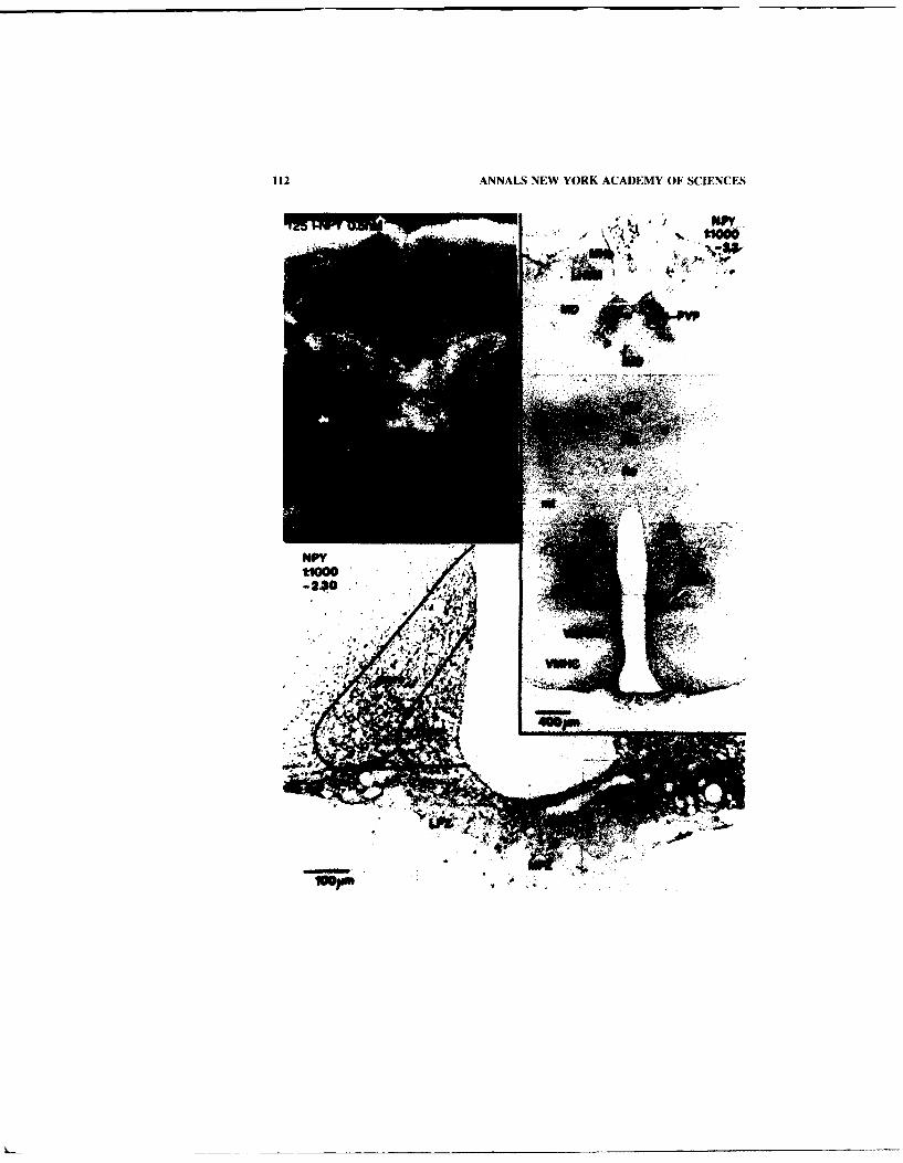
112 ANNALS NEW YORK ACADEMY OF SCIENCES
m44"

FUXE et al.: CENTRAL CARDIOVASCULAR REGULATION 113
mentary responses. Thus, in VT, chemical networks may be formed, at least as far asneuropeptides are involved, leading to the development of syndromic responses. 1 m1
Central neuropeptide Y (NPY) neurons appear to an important extent to communicatewith other cells of the brain via VT in view of the topological and functional transmitterreceptor mismatches (FIG. I) demonstrated in this system. 12.161- 9 Furthermore, we havebeen able to obtain evidence that the high affinity NPY receptors in mismatch areasrepresent biologically active NPY receptors. 1920 In the present article we will discusscommunication in the cardiovascular NPY/adrenaline costoring neurons, 21 :22 especiallyin relation to WT and VT. We will also present evidence that the NPY fragment porcineNPY 13-36 (pNPY 13-36) upon central administration can produce vasopressor actionsmediated by activation of NPY receptors of the Y2 type. 23 It is postulated that suchfragments can be formed in vivo to counteract the vasodepressor response of the pNPY1-3624 which is mediated through activation of Y I receptors. 4.25.26 In addition, we willdiscuss NPY/adrenaline cotransmission in central cardiovascular regulation where the twotransmission lines may interact via NPY/ot2 receptor interactions in the plasma mem-brane, both at the pre- and postsynaptic level. These receptor-receptor interactions mayhelp to facilitate a switching between VT and WT. We will also summarize recentevidence that angiotensin peptides may serve as cotransmitters in NPY/adrenaline and inNPY/noradrenaline (NA) systems of cardiovascular regions.27 Finally, the evidence willbe discussed that adrenalectomy leads to differential actions on adrenaline and NPYcotransmission in the medulla oblongata. 2
H
Anatomy of NPYlAdrenaline Costoring Cardiovascular Neurons
The central adrenaline neurons were discovered by H6kfelt, Fuxe, Johansson andGoldstein in 1973 .29 NPY-like immunoreactivity (IR) was originally demonstrated inthese neurons by H6kfelt and colleagues in 1983.30.3 ' The NPY/adrenaline costoring cellbodies are mainly located in the rostral medulla, especially in the ventrolateral area(group Cl), 29 and these adrenergic neurons project to cardiovascular areas of the brain,including the nucleus tractus solitarius (nTS) and the sympathetic lateral column of thespinal cord.32 -34 Subsequently, a small local NPY/adrenaline costoring system wasdiscovered within the cardiovascular region of the nTS (dorsal strip). 32 3 3 By means ofnewly developed microdensitometrical and morphometrical methods 3'
-35 it has become
possible to quantitatively evaluate the degree of coexistence of phenylethanolamine-
FIGURE 1. Autoradiogram of 1251-NPY (0.5 nM) labelling with corresponding NPY IR in the areaof thalamus and hypothalamus at Bregma level -2.3 mm. A strong NPY binding is present in PVP.MD, IMD. CM and Re. while a moderate NPY binding is present in MHb. LHbM, VMHDM.VMHC and MPZ. A dense plexa of NPY IR is seen in PVP, DA, DMC and DMD, whereas moderatedensity of plexa is present in LHbM. VMHDM. marc, Parc. LPZ, MPZ and very few scattered ifhardly any NPY IR terminals are present in MHb, MD. CM, Rh. Re and VMHC. Matches corre-spond predominantly to PVP, IMD and MPZ where both high density of binding and IR terminalsare seen. In the area of MHb, LHbM, VMHDM matches are also present with low to moderatedensity of both terminals and binding. Mismatches with high-density binding and very few if anyNPY IR terminals are seen in MD. CM, Rh, Re and VMHC. Abbreviations: CM. central medialthalamic nucleus; DA. dorsal hypothalamic area: DMC, dorsal hypothalamic nucleus, compact part:DMD. dorsal hypothalamic nucleus, diffuse part: IMD, intermediodorsal thalamic area: LHbM,lateral habenular nucleus, medial part; LPZ, lateral palisade zone: MD. mediodorsal thalamic nu-cleus; MHb. medial hebenular nucleus: PVP, paraventricular thalamic nucleus, posterior part; Re,reuniens thalamic nucleus: Rh. rhomboid thalamic nucleus: VMHC. ventromedial hypothalamicnucleus, central part: VMHDM. ventromedial hypothalamic nucleus, dorsomedial part.

114 ANNALS NEW YORK ACADEMY OF SCIENCES
N-methyltransferase (PNMT) and NPY in adrenaline cell bodies and nerve terminals.37
The studies reveal a high degree of coexistence of NPY and PNMT IR within nerveterminals in cardiovascular regions such as the nTS and dorsal motor nucleus of thevagus. 12 In a morphometrical analysis the NPY/adrenaline costoring neurons inter aliashow a preferential disappearance of NPY IR during aging in certain cardiovascularadrenaline neurons, especially those of the CI area.3 9 Thus, in aging the neurons may notsimply degenerate but may show a progressive impairment in their functional properties;in the case of group CI a reduction of the cotransmitter NPY. Taken together, theseresults are compatible with the existence of NPY/adrenaline cotransmission in cardio-vascular regions of the brain such as the dorsal medulla and the sympathetic lateralcolumn of the spinal cord.
On the Existence of NPY Receptors in Central Cardiovascular Areas
Large numbers of high-affinity 1251-NPY 1-36 binding sites have been mapped outwithin the CNS of the rat brain. 1225.40"41 We have recently demonstrated marked trans-mitter-receptor mismatches within the various central NPY neuronal systems, including
~.100"S0 "
FIGURE 2. Effects of the pNPY 13-36 frag-40 ment on '251-NPY (0.4 nM) binding in the nu-40 cleus centro medialis of the thalamus using20 quantitative receptor autoradiography (see leg-0
end in FIG. 12). Data represent % of controlU 0 IIIII[IIIIIIfIlIIIIIf[IIIIIIIIII IIIIIIIIIIIIIIIIIIIII value ± SEM . Fisher's PLSD-test: *p <0.05.( -2o ***p <0.001.
-40
NPY 13-36 (log nM)
the nTS 16.42 Within the dorsomedial medulla the highest density of high-affinity 1251-
NPY 1-36 binding sites is found within the dorsal strip region, while the highest densityof NPY IR nerve terminals is demonstrated in the ventral parts of nTS and within thedorsal motor nucleus of the vagus. The restits amplify a role of NPY receptors incardiovascular regulation, since the baroreceptor afferents terminate in the dorsal stripregion. Thus, the NPY receptors can modulate transmission at the primary relay stationfor the baroreceptor afferents.
Recently, two types of NPY receptors have been demonstrated within the CNS, thatis NPY receptors of the YI type, which can only recognize the full parent peptide, pNPY1-36, and NPY receptors of the Y2 type, which also can recognize C-terminal fragmentsof pNPY, especially pNPY 13-36 .3 "4 '4. It should be pointed out, however that inreceptor autoradiograms 125I-NPY 1-36 predominantly labels the Y I receptors, since theC-terminal pNPY fragment 13-36 only can displace 125 -pNPY 1-36 from its bindingsites ivi very high concentrations (over I jM) (FIG. 2) (unpublished data). Therefore, itwill be of substantial interest to map out the 12 5 -NPY 13-36 binding sites in the CNS.Taken together, the available data indicate that the high-affinity NPY receptors of the Y Itype to a large extent may be reached by pNPY 1-36 diffusing in the extracellular fluidand, thus participate in VT.

FUXE et al.: CENTRAL CARDIOVASCULAR REGULATION 115
Neuropeptide Ykv2 Receptor-Receptor Interactions
In both biochemical binding experiments and in receptor autoradiography it has beendemonstrated that pNPY, in the nanomolar range, produces a dose-related increase of theK, value of (x2 agonist binding sites within the dorsal medulla using the radioligand3H-paraminoclonidine. 45 47 Thus, NPY receptors appear to be capable to reduce theaffinity of a2 adrenoceptors via intramembrane interactions. This switch is agonist spe-cific, since it was not observed when the a2 adrenoceptor antagonist 3H-idazoxan wasused. 46 Furthermore, the GTP-induced shift in the IC5, value of clonidine for displace-ment of 3H-idazoxan from its binding sites was not modulated by pNPY 1-36 (10 nM). 4
1
These results were compatible with the involvement of the G-proteins in these intramem-brane receptor-receptor interactions. In line with this idea was also the demonstration thatboth NPY and a2 receptors are coupled in an inhibitory way to the adenylate cyclase viaa G i protein. 4' Evidence for this view was recently obtained by the demonstration thatpertussin-toxin treatment counteracts intramembrane interactions between NPY receptorsand ai2 adrenoceptors. 4 7 Thus, pertussin-toxin treatment counteracted this interactionwithin the plasma membrane without reducing the binding of NPY to its own receptor.
The interaction between NPY receptors, probably of the Y I type in the dorsal me-dulla, and a2 adrenoceptors appears to be bidirectional, since both in vitro and in vivoclonidine, an a2 agonist, can reduce the binding of ' 251-NPY 1--36 within the nTS atvarious rostrocaudal levels.46 These results indicate the existence of antagonistic in-tramembrane receptor-receptor interactions between the NPY receptors of the Y I type andof the a2 receptors. Thus, it seems possible that in NPY/adrenaline cotransmission in thenTS the two transmission lines can modulate the activity of one another via intramem-brane receptor-receptor interactions, which appear to take place both at the presynapticand the postsynaptic level.4 8 However, I nM of pNPY has been found to significantlyenhance the ability of clonidine to inhibit the potassium-induced release of 3H-noradren-aline (3H-NA) in synaptosomal preparations from the dorsal medulla. Thus, in this casethe receptor-receptor interactions appears to lead to an enhanced sensitivity of the a2autoreceptor. Taken together, these findings are compatible with the idea that the NPYtransmission line, can downregulate adrenaline transmission via pre- and postsynapticmodulation of the a2 adrenoceptor. It should also be mentioned that this interaction is nolonger present in the spontaneously hypertensive rat (SHR), neither at the pre- nor at thepostsynaptic level.48 4 9 In the same way, the adrenaline transmission line may downreg-ulate the NPY transmission line in NPY/adrenaline cotransmission.
Central Cardiovascular Actions of NPY and Adrenaline
It was early demonstrated that adrenaline and pNPY when given intraventricularly(i.v.t.) or intracisternally (i.c.) in the awake or the anaesthetized rat can produce alowering of mean arterial blood pressure (MAP), of heart rate (HR) and of respirationrate. 2' .24.5o.51 The major difference between adrenaline and pNPY is the long duration ofaction of NPY versus adrenaline as shown in FiG. 3. 14.52 When pNPY and adrenaline aregiven together in the cisterna magna they antagonize the cardiovascular actions of oneanother. -These results are in line with the biochemical and receptor autoradiographicalstudies indicating that in NPY/adrenaline cotransmission the two transmission lines canexert an inhibitory influence on each other.
In the SHR the ED,, value of pNPY 1-36 is substantially increased with regard tolowering of MAP as compared to the normotensive rat.5 4 These results may reflect analtered sensitivity of the NPY receptor of the Y I type in the cardiovascular regions of thebrain. in line with the postulate that there is an altered sensitivity of these types of NPYreceptors in SHR is the demonstration that pNPY 1-36 can no longer modulate thepresynaptic or the postsynaptic a2 adrenoceptors in SHR.48 49

shami* - sham+corl
Neuropeptide Y 0.25 nmol adx~cr
01 -- ------- --- - ---- x~ot
0 t0 20 30 40 so 60time (mmd)
> 10,Adrenaline 2.00 nmol
-to
..- 20
O time (m~in)
U
0 10 20 30 40 so 60time (min)
FIGURE 3. Comparisons of the time-course of changes of mean arterial blood pressure after centraladministration (i.v.t.) of porcine neuropeptide Y 1-36 (pNPY. 0.25 nmol). adrenaline (base) (A.2.00 nmol) and mock CSF (10 til/min X 3 min) in awake freely moving male rats with variousalterations in pituitary-adrenal activity (sham operated vehicle treated rats = sham +veb: adrena-lectomnized vehicle treated rat =adx + veh; sham operated corticosterone treated rat =sham + coiland adrenalectomized corticosterone treated rat =adx + cort). T'Ie basal values for the mean arterialblood pressure (MAP) (mm Hg): sham +veh (n =20) =103 ± 1; sham + cort (n = 22) =102 ±I ; adx + veh (n =2 1) = 100 t 1; adx +cort (n 2 1) =102 t 1. Data represent means expressedin percent of the basal value: SEM varied from 1-20%. For statistics in relation with peak effects,see FIGURE 13.

FUXE et al.: CENTRAL CARDIOVASCULAR REGULATION 117
C 15 500A B
* 400-
-- 300.
E p< 0.002 200. p < 0.01
0-oU a;,o 1; 00 ..
25 75 77 570 150 300
Doses of i..0 pNPY 13-36 (pmoles/rat)
FIGURE 4. Effects on mean arterial blood pressure (MAP, mm Hg) by different doses of the pNPY13-36 fragment, i.v.t. administered in the awake freely moving male rat. Peak actions (maximal
restonses) and area values (overall effects) are expressed as means ± SEM. The ioncheree-Terpstratest for ordered alternatives to evaluate dose related changes (p <0.01) and the Treatment versusControl nonparametric test (* = p <0.05) were used. Striped areas represent the SEM limits for thepeak mean increase of MAP by mock CSF alone (10 I.l/min x 3 min). Basal values (mm Hg) foreach group: mock CSF 30 p.1 alone = 106 - 4; pNPY 13-36 25 pmol = 100 + 5: pNPY 13-3675 pmol = 99 ± 3; pNPY 13-36 750 pmol = 109 + 6: pNPY 13-36 1250 pmol = 100 ± 6; andpNPY 13-36 3000 pmol = 100 ± 5.
Cardiovascular Actions of pNPY 13-36 in Normotensive and SpontaneouslyHypertensive Rats
Intraventricular injections of 5 doses of the putative Y2 NPY receptor agonist pNPY13-36 (25-3000 pmoles/rat) in the awake unrestrained freely moving male rat produce asignificant dose-related increase in MAP without effects on HR (FIGs. 4 and 5). Thus, incontrast to pNPY 1-36 which activates Y I receptors, NPY receptor agonists of the Y2type increase MAP when i.v.t. administered. Thus, NPY receptors of both the Y I and Y2type appear to participate in central cardiovascular regulation, where they may oppose thefunctional consequences of one another. Unlike pNPY 1-36 which possesses a potentbradycardic action, the fragment pNPY 13-36 does not influence HR compared withmock CSF alone. Thus, only NPY receptors of the Y I type appear to be involved in theregulation of HR.
The potency of pNPY 13-36 to exert a vasopressor action upon i.v.t. injection (FIG.5) in the awake unrestrained rat was substantially increased in the SHR compared with theWistar-Kyoto control animal (WKY) (FG, 6). Thus, a low dose of pNPY 13-36 (25pmoles/rat) exerted substantial and long-lasting increases of MAP, in SHR but not in theWKY normotensive animal. These results indicate that in the SHR and Y2 receptorsensitivity appears to be increased. Such a disturbance may contribute to the developmentof the spontaneous hypertension. It may be speculated that the degradation-regulatedswitch between isoreceptor transmission lines may be disturbed in the SHR leading to anenhancement of the Y2 transmission line.
Possible YI and Y2 NPY Receptor Interactions
In receptor autoradiographical experiments it has recently been found that pNPY13-36 in concentrations of 3-100 nM can increase the binding of 1
25 -NPY 1-36 (FtG.

118 ANNALS NEW YORK ACADEMY OF SCIENCES
C 15 o pNPY 13-16 750 pmoles/ratCDE --- -- CSF30 ul
"-- 10W
0. o
'@ 5,'-n p < 0.05-Ca
0
a) 0 -----0) "' %. ... . .... . . ..C
-5
-5 0 5 10 15 20 25 30 35 40 4 5 50 55 60Time (min)
FIGURE 5. Time-curve of the cenwrt effects (n mean arterial blod pressure )MAP, mnm llgM b%i.v.t. pNPY 13-36 (750 pmol). The effects of mock CSF alone (10 dl'min x 3 mint are also shown.Each point is represented as means ± SEM of change from the MAP basal value for each groupof treated rats (for basal values see Fto. 4).
2). Displacement is only noted in a high concentration of 1(KXX nM. These results can beexplained on the basis of the existence of interactions between NPY receptors of the Y Iand Y2 type. One possibility is that the activation of the NPY receptor of the Y2 type maylead to an increase in the affinity of the NPY receptors of the Y! !,pe. The mechanisn)underlying such a change in affinity is unknown but may reflect an altered coupltng to theG-proteins. since it has recently been demonstrated that i.v.t. perlussis-toxin treatmentcan lead to the development of an increase in the binding of '251-NPY 1-36 in thedorsomedial medulla. .7 Such an interpretation is also supported by functional studies oninteractions between Y I and Y2 receptors in cardiovascular regulation. Thus. as seen inFIGURE 7, the threshold dose of the pNPY fragment 13-36 can significantly counteractthe vasopressor actions of a close to ED,, dose of pNPY 1-36. In contrast, the thresholddose of pNPY 1-36 does not modify the pressor action of the pNPY fragment 13-36using a close to ED5, dose 55 (FIG. 8). These results give evidence for the existence ofchemical networks in central NPY transmission. 55 Thus, it is mainly the active fragmentwhich has a role in the termination of the biological response. These results indicate thatNPY transmission has a high degree of flexibility and is suitable for the long-termregulation of cardiovascular function inter alia by its potential ability to form activeC-terminal fragments. This strongly supports the view that VT represents an importantcomponent in the chemical communication of central NPY neurons.
Neuroendocrine Actions of pNPY 13-36: The Relationship to Effects onHypothalamic Catecholamine Nerve Terminal Systems
In previous work i.v.t. injections of pNPY 1-36 in doses of 7.5-25 pmoles!rat weredemonstrated to produce inhibitory actions on serum levels of corticosterone (CORTICO)

FUXE el al.: C'ENTRAL. CARD)IOVASCU'LAR REGULLATION 119
and aldlosterone (ALD) and to increase serumn prolactin IPRLI levels in the normal malerat as esaluated within the 15 -60) nfin time interval l.~ "' In contrast. in high dlosesranging front 250-750) pmiol pNPY 1 -36 prdue iniitr cl'ct onl serumi threotri)-pine IISH). PRL and growth hormione (GH) levels. while the serumi levels of C)R[IC().adrenocorticotropin (ACTH) and ALDO( were increased.
It has only been possible to demonstrate low densities for high-affinity >I'-pNPY,1-36 binding sites within the hypothalamus and the preoptic area. These result, mas, herelated ito the possibility (see above) that using receptor autwradiography '>I-NPY 1-36does not label high affinity receptors of the Y2 type. To elucidate this possibility furtherwe have in the present experiments studied the actions of centrally adm~inistered pNPY13-36 on the serumn levels of hypophyscal hormones in the awake unrestrained miale rat. "The results are shown in FimitR: 9. With regard to effects on serumn U(RTIC() levelspNPY 13-36 produces a U-shape dose-response curve with a marked and significantreduction of serumi CORTICO levels in a dose of' 75 pmioles. rat (iI(i. 9A). These inhib-tory effects are no longer observed after inhibition of tyrosine hsdroxslase MH. Therestoration of serum CORTIC() levels with the hiehest dose used of the pNPY fragmentmay be explained on the basis that the pNPY fragmient in the highest dose also activateslow-affinity Y2 receptors. These results indicate that high- and losi -atliimihv NPY rccen-tors of the Y2 ty'pe may play a role in the control of CORTICC) secretion via regulationof corticotropin releasing factor )CRF) released fromn the mnedian eminence.
With recard to PRL secreiion the pNPY fragmient 13- 36 is shown ito reduce se:armPRL lev els at the I h timec interval in the doses of 750 and 1250) pmlol (i~mu. 9B). Theseinhibitory affects are abolished after TH inhibition. In contrast, the TH inhibitor unmasksmarked inhibitory effects of the pNPY fragment in the lowest dose raniee of 7.5 and 25pmnoles,/rat. Thus, both high- and low-affinity Y2 NPY receptors seem to participate in theinhibitor,, control of' PRL secretion. The actions at the hiigh-affinit Y2 receptor is tin-
20 SHRp p<0.002 -a- WKY
-0 p 000
E
o 01C
OS5 10 15 20 30 40 50 Ni0
Time (min)
FIGURE 6. Comparison between the ttmie-curs'e (it per centl chanoe ot mnean a.I riaI blood pressure(M AP) (mm H,!) after an i Vtl. injection of a tow dose oft pN PY 1 3 -36 25 piiiol) in the pontane -
ousl hyertnsie aakefre],, moving male rat (SHR) and in the norniotensise as~akc treelsmoinfg mate Wistar-Kvoto control rats (WKY). The Manti-Whitnes 1' test %%as used to compare thetwo different groups. The peak (/ - 0.(X?)2i and o~crall ciect (p -0 (X)2?,werc stenlicatils dill, rentN -6 in each group. MAP basal salues MIIR 140 * 2 mm H,: WKY 107 *4 min

120 ANNALS NEW YORK ACADEMY OF SCIENCES
20- --- 0-- CSF30ul
E - pNPY 13-36 25 pmol
> - pNPY 1-36 75 pmol®10'
--- OD-- simultaneously
CL#A
4 -
Ot
E
C
040 .20r0 5 10 15 20 30 40 50 60Time (mini
FIGURE 7. Effects on mean arterial blood pressure (MAP) (mm Hg) of i.v.t. pNPY 1-36 at a dosesomewhat higher than the EDs( value for the peak effect (45 pmol: see Hirfstrand. 19872') and oti-v.t. pNPY 13-36 at a threshold dose alone or together in the awake unrestrained male rat. TheDunn's test was used to compare the different groups. The overall effect (the area under the curve)of pNPY 1-36 75 pmol was significantly counteracted by pNPY 13-36 25 pmoles. MAP basalvalues (mm Hg): CSF = 106 ± 4; pNPY 13-36 25 pmol = 100 ± 5: pNPY 1-36 75 pmol = 98± 3: pNPY 1-36 75 pmol + pNPY 13-36 25 pmol = 101 + 2. *** = p <0.005.
masked by removal of CA. It should be pointed out that the time interval chosen is notoptimal for the demonstration of increases in PRL secretion following i.v.t. injection ofpNPY 13-36 in view of the fact that i.v.t. pNPY 1-36 mainly increases PRL secretionat the 15 min interval' (see below). In previous work it has been possible to demonstratethat pNPY 1-36 upon i.v.t. injection in increasing doses produced a biphasic change inCA utilization in the tuberoinfundibular DA neurons and in the NA utilization in varioushypothalamic areas. 1'157 In the low-dose range there is an inhibition of regional hypo-thalamic CA utilization, while in higher doses an increase of regional hypothalamic CAutilization takes place. A similar analysis has now been performed with the pNPY frag-ment 13-36 (See FIG. 10). Regional CA levels within the hypothalamus were found to beunaltered (data now shown). In the low dose of 25 pmoles/rat pNPY 13-36 is seen toproduce a significant reduction of the CA depletion produced by TH inhibition within themedial palisade zone and the lateral palisade zone of the median eminence (MPZ andLPZ) (FiG. 10). Thus, pNPY 13-36 appears capable of reducing DA utilization both inthe MPZ and LPZ. In contrast, in the highest dose used the pNPY fragment produces asignificant increase of DA utilization within the median eminence.
These results suggest that both the high- and low-affinity receptors of the Y2 type areinvolved in the control of the tuberoinfundibular DA neurons and also that the action ofpNPY 1-36 on the tuberoinfundibular DA neurons may be mediated via Y2 NPY recep-tors. It seems likely that at least the increase in DA utilization induced by both pNPY 1-36and pNPY 13-36 wiihin the MPZ eAplains the inhibitory changes in PRL secretiondemonstrated following i.v.t. injections of pNPY 1-36 and pNPY 13-36.'-5 Never-theless, the inhibitory effects observed with pNPY 13-36 in the low doses following THinhibition must involve a nondopaminergic mechanism.
In conclusion, the NPY regulation of the tubetoinfundibular dopamine (DA) neurons.the PRL secretion and the pituitary-adrenal axis appear to involve both VT, via high-
/IlI lll l I ~ ll l

FUXE et al.: CENTRAL CARDIOVASCULAR REGULATION 121
affinity Y2 receptors, and WT, via low-affinity Y2 receptors. Finally, the strong biolog-ical potency of pNPY 13-36 in neurcendocrine control emphasize a role of extracellularlyformed C-terminal fragments in NPY transmission within the hypothalamus and thus adominance of VT.
Angiotensin H-Like Immunoreactivity within Subpopulations of TH ImmunoreactiveNeurons in the Al and Cl Area of the Ventral Medulla of the Male Rat
By using double immunolabelling techniques it has recently been possible to dem-onstrate the coexistence of TH and angiotensin 11 (ANG 11) immunoreactivities within theNA Al cell group and the adrenaline CI cell group of the ventral medulla of the rat.2 7
Within the AI area 20-30% of the TH IR nerve cells were ANG II IR and in the CI area5-40% of the TH IR nerve cells displayed ANG II IR (FIG. 1I). These results open upnew possibilities for ANG II/CA/NPY interactions in cardiovascular and neuroendocrineregulation. Thus, the balance of CA NPY and ANG I1 transmission in these costoringneuronal systems of the ventral medu.l, may have a decisive influence on the regulationof cardiovascular and neuroendocrine functions. In line with these results it has also beendemonstrated that ANG II reduces the affinity of ot2 adrenocertors in membrane prepa-rations from the dorsomedial medulla of the rat.5"
Angiotensin II/NPY Receptor Interaction
In view of the existence of ANG II/NPY cotransmission in cardiovascular areas bindingstudies and functional studies have been perforTned on the potential existence of such
-0- - CSF 30 ul--- pNPY 13-36 750 pmoles,
20,4 pNPY 1-36 25 pmol
E - simultaneously
10-
E.0
-o-0
* -100
.20 10 1 5 20 3 0 46 5 0 60Time (min)
FIGURE 8. Effects of a threshold dose of i.v.t, pNPY 1-36 on the vasopressor effects (MAP: mmHg) of a subr.,aximal dose of i.v.t, pNPY 13-36 (750 pmol). The Dunn's test was used to comparethe different groups: no interaction was found. Thus the pressor effect of the pNPY fragment couldnot be reduc, I l-v . threshold dose of pNPY 1-36. MAP basal values (mm Hg): CSF = 106 - 4;pNPY 13-36 -)0 pmol = 109 ± 6; pNPY 1-36 25 pmol = 100 + 3; pNPY 13-36 750 pmol +pNPY 1-36 25 pmol = 98 ± J.

122 ANNALS NEW YORK ACADEMY OF SCIENCES
A"j 150
E
°100 "1 77~l -2C 50
(H44/68 250 mg/Kg i.p. 1 h)U)
-6 0'7.5 25 75 250 750 1250 7.5 25 75 250 750 1250
Doses of ivt. pNPY 13-36 (pmoles/rat)
B(D
C 150-
CI
I0
7 2J A (H44/68 250 mg/Kg p. lh)
o 11 lillhl pliY 133 (pmoles/rIIIJIaIJI
FIGUE 9.Stuies n thDefes of iest, pNPY 13-36 gieni.m~o erum cstrn
(A) and prolactin (B) levels in the absence or presence of tyrosine hydroxylase inhibition (a-MT) inthe awake unrestrained male rat. The pNPY fragment (13-36) was given i.v.t, immediately after thei.p. administration of cc-MT I h before killing. The striped areas represent the SEM limits for therespective CSF control groups. The values are given in per cent of (he mock CSF alone (CORTICO:100% = 356 ± 45 nmol/I1; PRL: 100% = 4.7 ± I ng/ml) and the CSF + a -MT (CORTICO: 100%= 1122 ± 68 nmol/ I PRL: 100% = 66.8 -+8 ng/ml) treated group mean value. The Treatments
versus Control nonparametric test was used (*=p<0.05); n = 6 rats.

175 175
SEL CM150 I50
25 25205 75
20-p .05 MPZ] 7 PV1175 150 1
-M 150 125
S 125 100.
a
so .25
C 200 1750o LPZ PAFP0 75 P 0 150
50 125
25, 100
o 0o, 75
75 50
50 ~ p < 0.01 2
15PV11 17 PAFM
50. 10
100- 100
75 75
50 50
25- 257 5 25 75 250 750 1250 5 25 75 250 750 1250
Doses i v t. pNPY 13-36 (pmol) Doses v t pNPY 13-36 (pmol)
FIGURE 10. Effects of i.v.t. injections of six doses of pNPY 13-36 on CA disappearance in thediscrete CA nerve terminal systems of the hypothalamus after TH inhibition (a-MT 250 mg/kg. i.p.I h). pNPY 13-36 was given immediately after a-MT treatment. Means ± SEM are shown: n = 6in each group. The Treatments versus Control nonparametric test was used. The CA levels in thegroup treated with solvent alone (shaded area) were as follows: SEL 100%: 94 ± 4 nmol/g: MPZ100%: 102 ± 8 nmol/g; LPZ 100%: 277 ± 18 nmollg, PVii 100%: 40 ± 5 nmol/g: DM 100%: 196± 17 nmol/g; PVI 100%: 83 ± 11 nmol/g: PAFP 100%: 228 ± 20 nmolg; PAFM 100%: 132 t IInmol/g. Abbreviations: SEL, subependymal layer of the median eminence: MPZ, medial palisadezone of the median eminence: LPZ. lateral palisade zone of the median eminence: PVII, posteriorperiventricular hypothalamic nucleus: DM, dorsomedial hypothalamic nucleus: PVI, anteriorperiventricular hypothalamic region: PAFP. parvocellular part of the paraventricular hypothalamicnucleus; PAFM, magnocellular part of the paraventricular hypothalamic nucleus.

124 ANNALS NEW YORK ACADEMY OF SCIENCES
FIGURE 11. Fluorescence microphotographs of tyrosine-hydroxylase (TH) and angiotensin 11(ANG 11) immunofluorescent (IR) perikarya are shown in the same coronal section in the Al area(9,b; Bregma - 14.6 mm) and in the ClI area (c,d; Bregma -13.3 mm) of the ventral medulla of themale rat using double immunolabelling procedures. ANG 111TH IR nerve cell bodies are marked byarrows and the TH IR nerve cell bodies lacking ANG If immunofluorescence are indicated byarrowheads. To facilitate the comparisons of the sections one capillary is marked with an asterisk.D, dorsal; M. medial; ANG, ANG 11 immunofluoreseence: TH, tyrosine hydroxylase immunoflu-orescence. The magnification bars are indicated.

FUXE el al.: CENTRAL CARDIOVASCULAR REGULATION 125
40 3 125 I-pNPY 1-36
30 [ 0 1251l-pNPY 1-36+ANG1110 nM
:2 0 ! "1E
0 CLEE 10-
mnTS ds AP mnTSBregma -13.8 Bregma -13.3
(obex -0.3) (obex +0.2)
FIGURE 12. Microdensitometric determination of 2'z1-NPY 1-36 binding in the various regions ofthe dorsomedial medulla oblongata in the absence or presence of angiotensin 11 (ANG 11 10 nM) inthe male rat at two rostrocaudal levels. For details see Hrfstrand 1986.41 Means t SEM are shown:n = 4 rats: 0.5 nM of 125I-NPY 1-36 was used. Size of areas measured: nucleus medial subnucleusof tractus solitarius (mnTS) = 0.03 ± 0.007 mm2; dorsal strip (ds) = 0.03 + 0.002 mm2: areaprostrema (AP) = 0.09 ± 0.008 mm2. The Student t test was used (** p <0.01 t = trend).
interactions. In FIGURE 12, it is shown in receptor autoradiographical experiments that invitro incubation with ANG 11 (10 nM) leads to a significant increase 'n the 1251-NPY 1-36binding within the dorsal strip (dorsal cardiovascular region of nTS) and a trend for anincrease of the 1251-NPY 1-36 binding in the area postrema. The concentration of '251-NPY1-36 was 0.5 nM. In FIGURE 13 it is demonstrated in cardiovascular experiments that athreshold dose of ANG 11 given i.e. (3 nmoles/rat) not only counteracts the vasodepressoraction of ED5o dose of pNPY 1-36, but also leads to the development of a highly significantvasopressor actions of ANG 11 compared with the mock CSF group (FIG. 13). Takentogether, the results from the two experiments can be explained on the basis that ANG 11receptor activation leads to an uncoupling of the NPY Y I receptors to its G,-protein. leadingto an increase in the 1
251-NPY 1-36 binding (YI receptor binding)47 and thus to acounteraction of the vasodepressor response to pNPY 1-36. The vasopressor action ofpNPY 1-36 is then explained by the selective activation at NPY receptors of the Y2 type.which induce vasopressor actions.-- The ANG 11 receptor activation may contribute to thispressor action also by enhancing sensitivity at Y2 receptors.
On the Adrenocortical Hormone Regulation of CAINPY Costoring Neurons
It has recently been possible to demonstrate the existence of strong nuclear glucocor-ticoid receptor (GR) IR within all the NA/NPY and adrenaline/NPY neurons of the brainstem of the rat.60 62 It is of substantial interest that these cardiovascular neurons containhigh amounts of GR in view of the participation of GR in the regulation of bloodpressure." ' Furthermore, adrenaline and NPY synthesis as well as adrenaline/NPY co-storage appear to be controlled by GR. 4
(', suggesting one mechanism for the ability ofGR to influence central cardiovascular control. As seen in FIGURES 3 and 14. adrenalec-

126 ANNALS NEW YORK ACADEMY OF SCIENCES
tomy appears to differentially regulate the vasodepressor responses induced by pNPY andadrenaline in the awake unrestrained male rat 28 (FIG. 3). Thus, adrenalectomy alonesignificantly reduced the vasodepressor responses to i.v.t. adrenaline but not to i.v.t.pNPY. Thus, after adrenalectomy the central vasodepressor responses of pNPY dominate,since the adrenergic vasodepressor responses are selectively reduced. Furthermore, cor-ticosterone treatment abolished the centrally evoked vasodepressor responses to close tomaximal doses of adrenaline and pNPY, which may contribute to the hypertensive prop-erties in man. Thus, it seems possible that when the pituitary-adrenal activity is substan-tially reduced the NPY transmission dominates over adrenaline transmission within thecardiovascular CA/NPY neurons. In line with these cardiovascular results it has recentlybeen shown that adrenalectomy leads to an increase in 125I-NPY 1-36 binding within thearea postrema and the nTS, an increase which is abolished by corticosterone treatment. 65
CONCLUSION
The present findings give further evidence for an important role of NPY in centralcardiovascular regulation and also emphasizes the importance of the C-terminal pNPYfragment 13-36 in cardiovascular regulation, by selectively acting on NPY receptors ofthe Y2 type. In contrast to pNPY 1-36 which produces marked vasodepressor actions byactivation of NPY receptors of the Y! type, the pNPY fragment 13-36 produces vaso-
40-
to
E 30
0 20- CSFNPY 75 pmoi
- - ANG I3 nmol00- NPY+ANGII
Eo 0. ........... .
C
-10
-20 - . . i.
10 15 20 30 40 50 s0
Time (min)
FIGURE 13. Effects on mean arterial blood pressure (MAPI (mm Hg) by i.v. pNPY 1-36 at adose (75 pmol) somewhat higher than the ED value for the peak effect (45 pmol; see Harfstrand.198723) and interactions with a threshold dose of angiotensin I1 (ANG il) in the st-chloralose (100mg/kg) anaesthetized male rat. The Dunn's test was used to compare different groups (* p <0.05for the peak effect between the pNPY 1-36 alone group and the other three groups; *** = p <0.005for the overall effect, between vasodepressor area for mock CSF and vasodepressor area for pNPY75 pmol). MAP basal values (mm Hg) for each group (n = 4): CSF = 106 5; pNPY 75 pmol110 ± 6; ANG 113 nmol = 122 ± 2; pNPY 75 pmol + ANG 113 nmol = 100 ± 5.

FUXE et aL.: CENTRAL CARDIOVASCULAR REGULATION 127
W SHAM+CORT[ADXM ADX+CORT
Neuropeptide Y 0.25 nmol Adrenaline 2.00 nmol
E
2 5
U L *-25 .25 1 L_____J
FIGURE 14. Effects of changes in pituitary-adrenal activity on the overall vasodepressor effects(area values) induced by porcine neuropeptide Y (pNPY) 0.25 nmol and adrenaline (A) 2.00 nmolgiven intraventricularly in the same rats as reported in FIGURE 1. The effects of mock CSF(sham+veh = 5.3% ± 0.6; sham +cort = 5.7% ± 3; adx+veh = 8% ± 2; adx+cort = 8.5%± 1) have been taken away from the cardiovascular values obtained with pNPY and A. Data aregiven as means ± SEM. Area values are expressed in arbitrary units. Dunn's test for multiplecomparisons: * = p <0.05; ** = p <0.01; *** = p <0.005.
pressor responses via selective activation of the high-affinity NPY receptors of the Y2type. Thus, chemical networks appear to exist within NPY transmission involved incardiovascular regulation, leading to the production of syndromic responses and feedbackactions on the YI transmission line. The studies on the pNPY fragment 13-36 in com-bination with the demonstration of morphological and functional transmitter-receptormismatches in the central cardiovascular NPY neurons suggest a role of VT in the NPYtransmission in cardiovascular regions. Available data also underline a potential role ofpeptidases and peptidase inhibitors in NPY transmission in cardiovascular control. Thisregulation may be altered in SHR in view of the enhanced pressor action by pNPY 13-36in this strain of rats. Finally, a nerve impulse-regulated switch between transmission linesin the adrenaline/NPY neurons appear to underlie the duration of the biological action' 4 -26
and the degradation-regulated switch between isoreceptor transmission lines may underliethe direction of the cardiovascular action of pNPY 1-36. These switching phenomenamay be facilitated by intramembrane receptor-receptor interactions such as those betweenNPY YI and at2 receptors, NPY YI and ANG 11 receptors, and NPY isoreceptors.
REFERENCES
I. AGNATI, L. F., K. FuXE, M. ZOLI, I. ZINI, G. TOFFANO & F. FERRAGUTI. 1986. A correlationanalysis of the regional distribution of central enkephalin and 13-endorphin immunoreactiveterminals and of opiate receptors in adult and old male rats. Evidence for the existence of twomain types of communication in the central nervous system: the volume transmission and thewiring transmission. Acta Physiol. Scand. 128: 201-207.
2. FuXE, K. & L. F. AGNATI. 1990. On the dichotomy of electrochemical communication in the

128 ANNALS NEW YORK ACADEMY OF SCIENCES
brain. Introductory remark. In Advances in Neurosciences. K. Fuxe & L. F. Agnati, Eds.Vol. 1. Raven Press. New York. NY. In press.
3. DESCARRIERES, L., P. SEGUELA & K. C. WARKINS. 1990. Non junctional relationships ofmonoamine axon terminals in the cerebral cortex of adult rat. In Advances in Neurosciences.K. Fuxe & L. F. Agnati, Eds. Vol. 1. Raven Press. New York. NY. In press.
4. Vizi, E. S. 1984. Non-synaptic interactions between neurons: Modulation of neurochemicaltransmission. John Wiley. New York, NY.
5. FuXE, K. & U. UNGERSTEOT. 1970. Histochemical, biochemical and functional studies oncentral monoamine neurons after acute and chronic amphetamine administration. In Amphet-amines and Related Compounds. E. Costa & Garattini S, Eds. Vol. 1: 257-288. Proc. MarioNegri Inst. Pharmacol. Res. Milano.
6. ROUTTENBERG, A., J. SALDEK & W. BONDAREFF. 1968. Histochemical fluorescence afterapplication of neurochemical to caudate nucleus and septa area in vivo. Science. 161: 272-274.
7. UNGERSTEDT, U., L. L. BUTCHER, S. G. BUTCHER. N-E. ANDtN & K. FUXE. 1969. Directchemical stimulation of dopaminergic mechanisms in the neostriatum of the rat. Brain Res.14: 461-471.
8. NICHOLSON, C. 1979. Brain cell microenvironment as a communication channel. In TheNeurosciences: Fourth Study Program. F. 0. Schmitt & F. G. Worden, Eds. 457-476. TheMIT Press. Cambridge, MA.
9. AGNATI. L. F., K. FUXE, E. MERLO PiCH, F. BENFENATI, I. ZINI & M. GOLDSTEIN. 1986.Aspects on the information handling by the central nervous system: focus on cotransmissionin the aged rat brain. In Progress in Brain Research. T. H6kfelt, K. Fuxe & B. Pernow, Eds.Vol. 68: 291-301. Elsevier Sci. Pub]. B.V. Amsterdam.
10. HERKENHAM, M. 1987. Mismatches between neurotransmitter and receptor localization inbrain: observations and implications. Neuroscience 23: 1-38.
11. AGNATI, L. F., K. ANDERSSON, K. FUXE, V. LOCATELLI, V. MUTT, T. H6KFELT. 1980.Differential actions of CCK-39, CCK-8 and CCK-7 on pre- and postsynaptic dopaminemechanisms in various parts of the forebrain of the rat. Neurosci. Lett. Suppl. 5: S204.
12. FUXE, K., L. F. AGNATI, A. H.RFSTRAND, A. M. JANSON, A. NEUMEYER, K. ANDERSSON.M. RUGGERI, M. ZOLI & M. GOLDSTEIN. 1986. Morphofunctional studies on the neuropep-tide Y/adrenaline co-storing nerve terminal systems in the dorsal cardiovascular region of themedulla oblongata. Focus on receptor-receptor interactions in cotransmission. In Progress inBrain Research. T. H6kfelt, K. Fuxe & B. Pernow, Eds. Vol. 68: 303-320. Elsevier Sci.Publ. B.V. Amsterdam.
13. FUXE, K.. B. BUNNEMANN, M. ARONSSON, B. TINNER, A. CINTRA. G. VON EULER, L. F.AGNATI, S. NAKANISHI, H. OKHUBO & D. GANTEN. 1988. Pre- and postsynaptic features ofthe central angiotensin systems. Indications for a role of angiotensin peptides in volumetransmission and for interactions with central monoamine neurons. Clin. Exp. Hypertens. PartA Theory Pract. AI0 Suppl. 1: 143-168.
14. FUXE, K., L. F. AGNATI, M. ZOLI, B. BJELKE & I. ZINI. 1989. Some aspects of the com-municational and computational organization of the brain. Acta Physiol. Scand. 135: 203-216.
15. DEWIED, D. & J. JOLLES. 1982, Neuropeptides derived from proopiocortin: behavioural,physiological and neurochemical aspects. Physiol. Rev. 62: 976-1059.
16. AGNATI. L. F., M. ZOLI. E. MERt.O PicH, F. BENFENATI, R. GRtMALDI, 1. ZINI, G. TOFFANO& K. FUXE. 1989. NPY receptors and their interactions with other transmitter systems. InNeuropeptide Y. V. Mutt, K. Fuxe, T. H6kfelt, J. M. Lundberg, Eds. 103-114. Raven Press.New York, NY.
17. ZOLI, M., L. F. AGNATI, K. FUXE, F. FERRAGUTI, G. BIAGINI, A. CINTRA & J. A. GUSTAFS-
SON. 1990. Long-lasting reduction of glucocorticoid receptor immunoreactivity in the hip-pocampal field CA I but not in the dentate gyms after neonatal treatment with corticosteronein the rat. Acta Physiol. Scand. 138: 577-579.
18. FUXE, K.. L. F. AGNATI, A. HARFSTRAND, M. ZOLI, G. VON EULER, R. GRIMALDI, E. MERLOPICH, B. BJELKE, P. ENEROTH, F. BENFENATI, A. CINTRA, 1. ZINI & M. MARTIRE. 1990.
On the role of neuropeptide Y in information handling in the central nervous system in normal

FUXE et al.: CENTRAL CARDIOVASCULAR REGULATION 129
and physiopathological states: Focus on volume transmission and neuropeptide Yla2 receptorinteractions. Ann. N.Y. Acad. Sci. 579: 28-67.
19. FUXE, K., L, F. AGNATI, J. A. AGUIRRE, B. BJELKE, B. TINNER, E. MERLO PICH & P.ENEROTH. 1990. On the existence of volume transmission in the central neuropeptide Yneuronal systems. Studies on transmitter receptor mismatches and on biological effects ofneuropeptide Y fragments. In Advances in Neurosciences. K. Fuxe & L. F. Agnati, Eds. Vol.I. Raven Press, New York, NY. In press.
20. MERLO PICH. E., M. ZOLI, 1. ZINI, F. FERRAGUTI, V. SOLFRINI, M. TIENGO, K. FUXE & L.F. AGNATI. 1990. Effects of central administration of neuropeptide Y on vigilance and painthreshold in spontaneously hypertensive rats. In Advances in Pain Research and Therapy.S. Lipton, E. Tunks & M. Zoppi, Eds. Vol. 13: 55-63. Raven Press. New York.NY.
21. FUXE K, A. H.RFSTRAND. L. F. AGNATI, M. KALIA, A. NEUMEYER. T. G. SVENSSON, G. VONEULER, L. TERENIUS, P. BERNARDI & GOLDSTEIN M. 1988. Physiological and biochemicalcharacteristics of epinephrine/neuropeptide Y-costoring neurons in the medulla oblongata ofthe rat, with particular reference to vasodepressor functions within the nucleus tractus soli-tarii. In Epinephrine in the Central Nervous System. J. M. Stolk, D. C. U'Prichard & K.Fuxe, Eds. 346-365. Oxford University Press. New York & Oxford.
22. HOKFELT. T., G. A. FOSTER, 0. JOHANSSON. M. SCHULTZBERG, V. HOLETS. G. Ju, G.SKAGERBERG, M. PALKOVITS, L- SKIRBOLL, J. M. STOLK. D. C. U'PRICHARD & M, GOLD-STEIN, 1988. Central phenylethanolamine N-methyltransferase-immunoreactive neurons: dis-tribution, projections, fine structure, ontogeny, and coexisting peptides. In Epinephrine in theCentral Nervous System. J. M. Stolk. D. C. U'Prichard & K. Fuxe, Eds. 10-32. OxfordUniversity Press New York & Oxford.
23. SHEIKH, S., R. HaDKANSON & T. W. SCHWARTZ. 1989. Y I and Y2 receptors for neuropeptideY. FEBS. 245 (1,2): 209-214.
24. FUXE, K., L, F. AGNATI, A. HARFSTRAND, 1. ZINI, K. TATEMOTO, E. MERLO PiCH. T.
HOKFELT, V. MUTT & L. TERENJUS. 1983. Central administration of neuropeptide Y induceshypotension, bradypnea and EEG synchronization in the rat. Acta Physiol. Scand.118: 189-192.
25. KRFSTRAND. A. 1987. Brain neuropeptide Y mechanisms. Basic aspects and involvement incardiovascular and neuroendocrine regulation. Acta Physiol. Scand. 131(Suppl.): 565.
26. FuxE. K.. A. HARFSTRAND, L. F. AGNATI, G. vON EULER, T. SVENSSON & B. FREDHOLM.1989. On the role of NPY in central cardiovascular regulation. In Neuropeptide Y. V. Mutt.K. Fuxe, T. H6kfelt. & J. M. Lundberg, Eds. 201-214. Raven Press. New York. NY.
27. COVENAS. R.. K. FUXE, A. CINTRA. J. A. AGUIRRE, M. GOLDSTEIN & D GANTEN. 1990.Evidence for the existence of angiotensin ll-like immunoreactivity in subpopulations of ty-rosine hydroxylase immunorea,-tive neurons in the Al and CI area of the ventral medulla ofthe male rat. Neurosci. Lett. In Iress.
28. AGUIRRE. J. A.. K. FUXE. B. ANDBJER, P. ENEROTH & L. F. AGNATI. 1990. Changes inpituitary-adrenal activity differentially regulate the central vasodepressor responses inducedby neuropeptide Y and adrenaline in the awake unrestrained male rat. Acta Physiol. Scand,Submitted.
29. HOKFELT, T., K. FUXE, M. GOLDSTEIN & 0. JOHANSSON. 1974. Immunohistochemical evi-dence for the existence of adrenaline neurons in the rat brain. Brain Res. 66: 235-25 1.
30. HOKFELT, T., J, M. LUNDBERG. K. TATEMOTO, V. MUTT, L. TERENIUS, J. POLAK. S. BLOOM,
C. SASEK, R. ELDE & M. GOLDSTEIN. 1983. Neuropeptide Y (NPY)- and FMRF amideneuropeptide-like immunoreactivities in catecholamine neurons of the rat medulla oblongata.Acta Physiol. Scand. 117: 315-318.
31. HOKFELT, T., J. M. LUNDBERG, H. LAGERCRANTZ, K. TATEMOTO, V. MUTT, J. LINDBERG.L. TERENIUS, B. J. EvERIT, K. FUXE, L. F. AGANTI & M. GOLDSTEIN. 1983. Occurrence
of neuropeptide Y (NPY)-like immunoreactivity in catecholamine neurons in the humanmedulla oblongata. Neurosci. Lett. 36: 217-222.
32. KALIA. M.. J. J. VIOLA. M. E. HUDSON, K. FUXE. A. HARFSTRAND & M. GOLDSTEIN. 1988.Connectivity and ultrastructure of noradrenergic and adrenergic neurons in the medulla ob-longata. In Epinephrine in the central nervous system. J. M. Stolk. D. C. U'Prichard & K.Fuxe. Eds. 59-77. Oxford University Press. New York, NY.

lid ANNALS NEW YORK ACADEMY OF SCIENCES
33. HOKFELT, T.. G. A. FOSTER, 0. JOHANSSON, M. SCHULTZBERG, V. Hot ETS, G. Ju. G.SKAGERBERG, M. PALKOVITS, L. SKIRBOLL, J. M. STOLK, D. C. U'PRICHARD & M. GOLD-STEIN. 19, Central phenylethanolamine N-methyltransferase-immunoreactive neurons: dis-tribution, projectiow. fine structure, ontogeny, and coexisting peptides. In Epinephrine in theCentral Nervous System. J. M. Stolk, D. C. U'Prichard & K. Fuxe, Eds. 10-32. OxfordUniversity Press. New York, NY.
34. RUGGIERO, D. A., D. J. REIS. 1988. Neurons containing phenylethanolamine N-methyltrans-ferase: a component of the baroreceptor reflex'? In Epinephrine in the Cent"I Ner.'ous Sys-tem. J. M. Stolk, D. C. U'Prichard & K. Fuxe, Eds. 291-307. Oxford University Press. NewYork, NY.
35. KALIA, M.. K. FUXE & M. GOLDSTEIN. 1985. Rat medulla oblongata: Ill. Adrenergic (CI andC2) neurons, nerve fibres, and presumptive terminal processes. J. Comp. Neurol. 233:333-349.
36. AGNATI, L. F., K. FUXE, I. ZINI, L. CALZA, F. BENFENATI. M. ZOLI T. HOKFELT & M.GOLDSTEIN. 1982. A new approach to quantitate the density and antigen contents of highdensities of transmitter-idct iI i terminals, immunocytochemical studies on different typesof tyrosine hydroxylase immunoreactive nerve terminals in nuc!Lus caudatus putamen of therat. Neurosci. Lett. 32: 253-258.
37. FUXE, K., L. F. AGNATI. A. HARFSTRAND, M. ZOLI & A. M. JANSON. 1985. Image analysisand the determination of codistribution and coexistence of neuroactive substances in nerveterminal populations. Acta Stereol. 4/2: 181-186.
38. ZOLI, M., K. FUXE, L. F. AGNATI, A. HARFSTRAND, L. TERENIUS, R. TONI & M. GOLDSTEIN.1986. Computer-assisted morphometry of transmitter-identified neurons: new opening for theunderstanding of peptide-monoamine interactions in the mediobasal hypothalamus. In Neu-rohistochemistry: Modern Methods and Applications. P. Panula, H. Paivarinta & S. Soinila,Eds. 137-172. Alan R. Liss, Inc. New York, NY.
39. AGNATI, L. F., K. FUXE. M. ZOLI. 1. ZINI. A. HARFSTRAND. G. TOFFANO & M. GOLDSTEIN.1988. Morphometrical and microdensitometrical studies on phenylethanolamine-N-methyl-transferase and neuropeptide Y-immunoreactive neurons in the rostral medulla oblongata ofthe adult and old male rat. Neuroscience 26: 461-478.
40. FUXE, K., A. HARFSTRAND, L. F. AGNATI. M. KALIA, B. FREDHOLM, T. SVENSSON, 1. A.GUSTAFSSON, R. LANG & D. GANTEN. 1987. Central catecholamine-neuropeptide Y inter-actions at the pre- and postsynaptic level in cardiovascular centres. J. Cardiovasc. Pharmacol.10 (12): 1 13.
41. H.ARFSTRAND, A.. K. FUXE. L. F. AGNATI, F. BENFENATI & M. GOLDSTEIN. 1986. Receptorautoradiographical evidence for high densities of 2.1-neuropeptide Y binding sites in thenucleus tractus solitarius of the normal male rat. Acta Physiol. Scand. 128: 195-200.
42. ZOLI, M., L. F. AGNATI, K. FUXE & B. BJELKE. 1989. Demonstration of NPY transmitterreceptor mismatches in the central nervous system of the male rat. Acta Physiol. Scand.135: 201-202.
43. WAHLESTEDT. C.. N. YANAIHARA & R. HaDKANSON. 1986. Evidence for different pre- andpost-juntional receptors for neuropeptide Y and related peptides. Regul. Pept. 13- 307-318.
44. WAHLESTEDT. C.. L. EDVINSSON, E. EKBLAD & R. HiKANSON. 1987. Effects of neuropeptideY at sympathetic neuroefector juntions: existence of Y I and Y2 receptors. In NeuronalMessengers in Vascular Function. A. Nobin. Ch. Owman & B. Arreklo-Nobin. Eds. 231-242. Fernstr6m Synup. No 10. Elsevier. Amsterdam.
45. AGNATI. L. F., K. FUXE. F. BENFENATI, N. BATTISTINI. A. HARFSTRAND, K. TATEMOTO, T.HOKFELT & V. MUTT. 1983. Neuropeplide Y in vitro selectively increases the number ofct2-adrenergic binding sites in membranes of the medulla oblongata of the rat, Acta Physiol.Scand. 118: 293-295.
46. HRFSTRAND. A.. K. FUXE. L. F. AGNATI & B. B. FREDHOLM. 1989. Reciprocal interactionsbetween a2-adrenoceptor agonist and neuropeptide Y binding sites in the nucleus solitarius ofthe rat. A biochemical and autoradiographic analysis. I. Neural Transm. 75: 83-99.
47. VON EULER, G.. K. rUXE, I. VAN DER PLOEG. B. B. FREDHOIM & L. F. AGNATI. 1989,Pertussis toxin treatment counteracts intramembrane interactions between neuropeptideY receptors and ot 2-adrenoceptors. Eur. J. Pharmacol. (Molecular pharmacology section).172: 435-441.

FUXE el al.: CENTRAL CARDIOVASCULAR REGULATION 131
48. MARTIRE.. M.. K. FLXE. G. PISTRjrro. P. P~RFZIOSI & L. F. AGNATI. 1989. Reduced inhib-itory effc -ts of clonidine and neuropeptide Y on 3H-noradrenaline release from syflaptosomesof the medulla oblongata of the spontaneous hypertensive rat. J. Neural. Transm. 76: 181.-189.
49. AoGN.T L. F., K. FuXE. F. Bt-NF[E'NAii. N. BAFTtSTINI. A. HARFSTRANO., T. I-IfKFIt, L.CAVICCHIOI.I. K. TAT EMOTO & V. MUTIT. 1983. Failure of neuropeptide Y in vilro to increasethe number of a2-adrenergic binding sites in membranes of medulla oblongata of the spon-taneous hypertensive rat. Acta Physiol. Scand. 119: 309-312.
50. BORKOWSKI. K. R. & L. FINCH. 1990~ The hypotensive actions of centrally administeredadrenaline. In Central Adrenaline Neurons: Basic Aspects of Their Role in CardiovascularDiseases. K. Fuxe. M. Goldstein, B. HWkfelt & T. Hi1kfelt. Eds. Vol. 33: 225-234. Perga-mon Press. Oxford.
51. Fux. K.. P. BOLMO. L.F. AONATi. JONSSON, K. ANDERSSON. C. KfHLER&T. HoIU+LT.1980. On the role of central adrenaline neurons in central cardiovascular regulation. In CentralAdrenaline Neurons: Basic Aspects and Their Role in Cardiovascular Disease. K. Fuxe. M.Goldstein. B. Hokfelt & T. Hokfelt. Eds. Vol. 33: 161-182. Pergamon Press. Oxford.
52. HARFSTRAND. A.. K. FuxE & L. F. AGiNATI. 1987. On the role of receptor-receptor interactionin central cardiovascular regulation: functional studies on the interactions between a-adier-ergic and neuropeptide Y receptors in the rat medulla oblongata. In Receptor-Receptor In-teractions. A New Intramembrane Integrative Mechanism. K. Fuxe & L. F. Agnati. Eds.'I9o -530. MacMillan Press. London.
53. HARFSTRANO. A. k& K. FuXE. 1()R7. Simultaneous central administration of adrenaline and..e.uropeptide Y leads to antagonistic interactions in vasodepressor responses in awake malerats. Acta Physiol. Scand. 130: 529-53l.
54. H~iRFSTRAND, A.. K. FuXE. L. F. AGNAI. D. GANTI N. P. Er't-.i.orH. K. TATEMOTO & V.MurIr. 1984. Studies on neuropeptide Y/catecholaniine interactions in central cardiovasc ularregulation in the ot-chk<.:alose anaesthetized rat. Evidence for a possible new way ofactivating the ot. adrenergic transmission line. Clin. Exp. Hypertens. Part A. Theory Pract.A6 (1O&I11): 1947-1950.
55. AouiRRE, J. A.. K. FuXE. L. F. AGNATI & G. voN EULER. 1990. Centrally in'lected neuro-peptide Y 13-36 produces vasopressor effects and antagonizes the vasodepressor action ofneuropeptide Y 1-36. Neurosci. Lett. In press.
56. HARESTRAND. A.. K. FUXE. L. F. AoiNArt. P. ENEROIH. 1. ZINt. M. Zoi.i. K. ANDERSSON.G. VON EULER. L. TERENIUS, V. MUTT & M. GotoDS]EIN. 1986. Studies on neuropeptideY-catecholamine interactions in the hypothalamus and in the forebrain of the male rat. Re-lationship to neurocndocrinc function. Neurochem. Int. 8/3: 355-376.
57. HARFST'RAND. A.. P. ENERoTH. L. F. AGiNATI & K. FuxI±. 1987. Furt her studies on the effectsof central administration of neuropeptide Y on neuroendocrine function in the male rat:relationship to hypothalamic catecholamines. Regul. Pept. 17: 167-179.
58. FUXE. K.. L. F. AGNATI. A. HARFSTRAND. P. ENEROTH. A. CINTRA. B. TINNER. E. MERLOPict. M. ARONSSON. B. BUNNEMANN. B. LANG & D. GANI-EN. 1989. Studies on theneurochemnical niechaitisms underlying the neuroendocrine actions of neuropeptide Y. InNeuropeptide Y. V. Mutt. K. Fuxe. T'. Hijkfelt & L. MI. Lundberg. Eds. 115-135. RavenPress. New York. NY.
59. FUXE. K.. G. VON EuLEIR & L. F. Aorxit. 1988. Angiotensin 11 reduces the affinity of(UHpara-aminoclonidlite binding sites in menmbrane preparations from the rat dorsomedialmedulla oblongata. Adta Physiol. Scand. 134: 317-318.
60. AGNAI. L. F.. 1. ZINI. M. Zoi-i. K. FuxE. E. MERto0 Picit. R. GRIMAiLDI. G. TOFEANO & M.GOLDSTEIN. 1988. Regeneration in the central nervous system: concepts and facts. fIn Ad-vances and Technical Standards in Neurosurgery. Vol. 16. L. Symion. Ed. 3-50. Springer-Verlag. Vienna. New York.
61. FUXE. K., A. CINTIRA, A. HARESTRAND, L. F. AGNATI. M. KAIA. M. ZoitI. A.-C. WIK-STROM. S. OREI. M. ARONSSON & 1.-A. GusIAFSSON. 1987. Central glucocorticoid recep-tor immunoreactive neurotns: new insights into the enidocrine regulation of the brain. Ann.N.Y. Acad. Sci. 512: 362-393.
62. HARESTRANiO. A.. K. FUXE., A. CINTIRA. L. F. AG;NA 11. I. ZINt. A.-C. WIKSTR6M. S. ORT.Z -Y. Yu. M. GOl.DStiF.N. H. STiiNtiusctt. A. VFRIIOISIAst & J. A. GUSIAESSON. 1986.

132 ANNALS NEW YORK ACADEMY OF SCIENCES
Glucocorticoid receptor imunoreactivity in monoaminergic neurons of rat brain. Proc. Nat],Acad. Sci. USA 83: 9779-9783,
63. GRLJNFELD. J. P. & L. Et-oy. 1987. Role des glucocorticoides dans la regulation de la pressionarterielle. Presse Med. 16 (28): 1365-1367.
64. FuxE. K.. L. F. AGNATI. M. Zot-i, A. HARFS[RAND. R- GRIMALDI. P. BFRNARDI. M. CA-MUiRRI & M. GOLDStEIN. 19S5. Development of quantitative methods for the evaluation ofthe entity of' coexistence of neuroactive substances in nerve terminal populations in discreteareas of the central nervous system: evidence for hormonal regulation of cotransmission. InQuantitative Neuroanatoniy ir Transmnitter Research. L. F. Agnati & K. Fuxe. Eds. Vol.1~ 42: 157-174. MacMillan. London.
65. FuxE, K.. A. CiN rRA. S. T.5NGANEULiI. G. VON EutLtR. U. B. FINNMAN, K. LUNDGREN. J.A. AGLtIRRE. B. Bjiri.iK. M ZoLI. P. ENEROIH & L. F. AGNATi. 1989. Integration of bumoi!and neuronal signals in neuroendocrine regulation. Focus on interaction of glucocorticoids withcentral neuropeptide and monoamine neurotransmission. In Recent Advances in Basic andClinical Neuroendocrinology. F. F. Casanueva & C. Dieguez. Eds. Vol. 1: 109-126. ElsevierSci. PubI. B. V. Amsterdam.
66. HARFS-TRAND. A.. A. CINTRA. K. Ftjxr.. M. ARONSSON. A. C. WiKST116M. S. OKREA. J. AGUSTAFSSON & L. F. AGNA Hi. 1989. Regional differences in glucocorticoid receptor immu-noreactivity among neuropeptide Y immunoreactive neurons of the rat brain. Acta Physiol.
Scand. 135: 3-9.

Neuropeptide Tyrosine in theCardiovascular System"JOHN WHARTON AND JULIA M. POLAK
Department of HistochemistrvRoyal Postgraduate Medical School
Du Cane RoadLondon W12 ONN, United Kingdom
INTRODUCTION
Neuropeptide tyrosine (NPY) is an amidated 36-amino acid sequence, produced byposttranslational processing from a 97-amino acid precursor molecule (preproNPY), andis one of the most abundant neuropeptides identified in the mammalian nervous system.Removal of the 28-amino acid N-terminal signal peptide gives rise to proNPY which,!,dergoes proteolytic processing and amidation to generate both NPY, 3 6, and a 30-aminoacid C-terminal flanking sequence (proNPY40-,,1 or C-PON).'- 3 NPY was originallyisolated from the pig brain, but has since been identified in tissues from several othermammals (man, rat. rabbit, guinea pig. ox and sheep) and found to be a highly conervedsequence,4 which is widely distributed in the peripheral nervous system.
Immunohistochemistry
We have used specific antisera raised against both synthetic human NPY and C-PONsequences to investigate the distribution of NPY-containing nerves in the mammaliancardiovascular system. C-PON immunoreactivity occurs naturally in mammalian tissues 5
and has an identical distribution pattern to that of NPY immunoreactivity,' both sequencesbeing co-localised at the light and ultrastructural level in human cardiac nerve terminals. 7- 8
Optimum immunostaining of NPY- and C-PON-containing nerves has been obtained usingtissue fixed by immersion, for up to 48 hours at 4'C, in a modified Bouin's solution"containing 85 ml of 2% (w/v) paraformaldehyde in 0. 1 M phosphate buffer (pH 7.2-7.4)and 15 ml of saturated picric acid per 100 ml of fixative. After fixation, tissues are rinsedfor 24-48 hours in several changes of phosphate buffer, containing 15% (w/v) sucrose and0. 1% sodium azide, and processed immediately as either whole mount preparations orcryostat sections.75 t4" Prior to immunostaining, the tissues are incubated in phosphatebuffered saline containing 0.2% Triton X-100 for 30-120 minutes and counterstained withPontamine Sky Blue, to aid antibody penetration and reduce background autofluorescencerespectively."
Peripheral nerves are readily visualised using indirect immunofluorescence techniquesand the general distribution pattern of cardiovascular innervation may be demonstratedusing antisera to neural marker proteins such as protein gene product 9.5 (PGP 9.5),synaptophysin and the Schwann cell marker S-100. 7"x.10 ' 2 PGP 9.5 is a human neuron-specific cytoplasmic protein which has recently been found to correspond to a ubiquitin
" This work was supported by the British Heart Foundation and Council for Tobacco Research.USA.
133

134 ANNALS NEW YORK ACADEMY OF SCIENCES
carboxyl-terminal hydrolase,. and synaptophysin is an integral membrane protein ofsecretory vesicles in nerve terminals which contain the classical neurotransmitters ace-tylcholine and noradrenaline. 1
4 Polyclonal antiserum to PGP 9.5 has been found toimmunostain autonomic and sensory nerves, from the ceil body to distal varicose nerveterminals, in peripheral tissues of man as well as other species (e.g., guinea pig). Syn-aptophysin immunoreactivity on the other hand is localised specifically to varicose nerveterminals and appears to exhibit a more extensive distribution in the peripheral nervoussystem of certain animals (e.g., rat and cow) than does PGP 9.5. Subpopulations ofcardiac nerves may be identified according to their neuropeptide and enzyme content,relative distributiuii, AOn ultrastructure and sensitivity to selective neurotoxins.
NPY-Immunoreactive Cardiac Nerves
Nerve fibres displaying NPY and C-PON immunoreactivity are widely distributed inthe mammalian heart where they are concentrated in the myoc-rdium, endocardium,intracardiac ganglia and in association with the coronary vasculature. While relatively fewimmunoreactive fibres occur around epicardial blood vessels in the human heart a denseperivascular network supplies small intramyocardial arteries and arterioles (Fig. 1), form-ing a plexus of varicose fibres and fascicles at the adventitial-medial border. The myo-cardium receives fewer nerves than intramyocardial vessels, but is also well supplied withvaricose fibres containing NPY and C-PON. Immunoreactive nerve fibres occur through-out the walls of all the heart chambers, however, a density gradient exists between theatria and ventricles. NPY-immunoreactive nerves are also found in the cardiac conductionsystem and in the cow, for example, they occur distributed in sinus and atrioventricularnodes (Fig. 2) as well as in ventricular bundle branches. '15 The density of the sinus andatrioventricular node innervation as a whole is significantly greater than that observedelsewhere in the mammalian heart, whereas the density of NPY-immunoreactive nervesis generally comparable to that found in the adjacent myocardium. In the conductionsystem of the guinea pig (Fig. 3) and human heart, for examplc, NPY-inmiunoreactivenerves represent a relatively minor subpopulation compared to the numerous nerve fibresdisplaying PGP 9.5 and synaptophyin immunoreactivity, as well as acetylcholinesteraseactivity. Intracardiac ganglia in the atrial subendocardium, atrioventricular groove and inthe region of the sinus node contain NPY/C-PON-immunoreactive nerve fibres, but in-trinsic neuronal cell bodies appear to lack immunoreactivity in the human heart. ") In thehuman fetal heart, NPY/C-PON-immunoreactive nerves are detected after 10 weeks ofgestation and occur first in the region of the sinus and atrioventricular nodes and atrialepicardium. The ventricular innervation develops later and accompanies the coronaryvasculature, with prominent networks of fine varicose fibres distributed around epicardialvessels and extending together with arterial branches into the myocardium (Fig. 4).Although still comparatively few in number, the general distribution of NPY-immuno-reactive cardiac nerves at 21-24 weeks of gestation appears to be similar to that observedafter birth, and as in the adult, these nerves represent the predominant peptide-containingnerve population detected in the fetal heart.
Posttranslational processing and amidation of NPY are essential for NPY receptorbinding and activity,'6 and recent immunohistochemical findings indicate that NPYpresent in human cardiac nerve fibres occurs in an amidated form. 1 7 Immunohistochem-ical studies have revealed that NPY-immunoreactive nerves generally exhibit a similardistribution pattern to that of nerves containing immunoreactivity for two enzymes in-volved in the catecholamine synthetic pathway, tyrosine hydroxylase and dopamine beta-hydroxylase (Fig. 5), suggesting that NPY-immunoreactive cardiovascular nerves are ofsympathetic origin.
I4,l ~ll~ l I m Ill I I -lll I I Il III I ll Illlll Il Il ll

WHARTON & POLAK: CARDIOVASCULAR SYSTEM 135
5' -B
>~ a
E

136 ANNALS NEW YORK ACADEMY OF SCIENCES
FIGURE 2. Immunofluorescence micrographs showing synaptophysin-immunoreactive nerve fi-bres concentrated around conduction cells in the atrioventricular node of the bovine heart (A). Asubpopulation of fine varicose nerve fibres display NPY immunoreactivity (B). Bar = 50 Lm.
Origins of NPY-Immunoreactive Cardiac Nerves
The presence of NPY immunoreactivity i, sympathetic cardiovascular nerves hasbeen further supported by the results of both surgical and chemical denervation studies.During human heart and heart-lung transplantation the extrinsic efferent and afferentnerve supply to the donor heart is sectioned and the available functional and anatomicalevidence suggests that there is no significant reinnervation of the allograft. "' Nerve fibres.fascicles and ganglia are present in human cardiac allografts, at least up to 7-8 years aftertransplantation, and display immunoreactivity for PGP 9.5, synaptophysin and S-100 aswell as acetylcholinesterase activity. Compared with innervated donor and explantedrecipient or postmortem hearts there is, however, a lack of NPY/C-PON- and tyrosinehydroxylase-immunoreactive nerves in allograft tissues, suggesting that they are of ex-trinsic origin. These observations are consistent with the results of surgical and chemicalsympathetic denervation studies in experimental animals which result in a 50-90%h re-duction in NPY and noradrenaline levels. 81-23 Most NPY-containing mammalian cardiacnerves are therefore likely to represent postganglionic sympathetic nerves originating inthe stellate and other paravertebral ganglia. 18.19.24 The extent to which sympathectomydepletes cardiac NPY levels varies between species and may reflect differences in therelative contribution of intrinsic neurons to the NPY-immunoreactive innervation of theheart. As in other studies, 24 we have been unable to demonstrate the presence of NPY- orC-PON-immunoreactive neurons in the human heart, but immunohistochemical investi-gations have identified subpopulations of immunoreactive cardiac neurons in tissue sec-

WHARTON & POLAK: CARDIOVASCULAR SYSTEM 137
:L
lu
ch
a
E-
2 E-
oE
cz,
,

138 ANNALS NEW YORK ACADEMY OF SCIENCES
FIGURE 4. Section of human fetal heart, at 17 weeks of gestation, displaying a dense plexus ofNPY immunofluorescent nerve fibres associated with an epicardial coronary artery (CA) and scat-tered in the underlying ventricular myocardium. Bar = 50 tm.
tions and cell cultures from other mammals. 15.21.25 -2X In contrast to the human heart.intrinsic cardiac neurons appear to make a significant contribution to the NPY-immuno-reactive innervation of the rat heart. 23 in which no significant reduction of NPY-immu-nostained nerves was found following heterologous cardiac transplantation into the ab-dominal cavity. 2' NPY mRNA has also been detected in extracts of rodent atria andventricles, but the neural origin of this expression is uncertain," 3 ' and it is known thatnon-neuronal cells in the rat also contain NPY mRNA.' 2 In addition to NPY, somemammalian cardiac neurons have been reported to contain other neuropeptides, 24 -3 sug-gesting that the intrinsic cardiac innervation is more complex than was previously thoughtand does not simply represent a local postganglionic parasympathetic nervous system.
Ultrasir,,c-ura" Localisation of NPY Immunoreactivity
Electron microscopic examination of human cardiac tissue has demonstrated the pres-ence of NPY and C-PON immunoreactivity in axon profiles displaying the ultrastructuralfeatures of presumptive sympathetic nerve terminals, immunoreactivity being localised tolarge granular vesicles (70-100nm diameter) rather than the numerous small vesicles(40-60nm diameter) which occur in immunostained axonal varicosities. 7 - The results ofsubcellular fractionation studies also indicate that while noradrenaline is present in boththe small and large vesicle populations. NPY immunoreactivity is concentrated in thelatter. 14 " Electrical stimulation of cardiac sympathetic nerves is associated with an

WHARTON & POLAK: CARDIOVASCULAR SYSTEM 139
overflow of NPY from the pig heart in vivo, 3 6 and sympathetic activation in man isaccompanied by an increase in the plasma concentrations of noradrenaline and NPY,suggesting a co-release of both substances from stimulated sympathetic nerveterminals. 37- 35 Other studies have demonstrated the exocytotic release of both NPY andnoradrenaline in the pig spleen 39 and guinea pig heart,4" and the distinct subcellularlocalisation of NPY may facilitate the differential release of NPY and noradrenaline, highfrequency stimulation resulting in the preferential release of NPY. 39
Pulmonary NPY-Immunoreactive Nerves
As in the heart and systemic blood vessels, NPY- and C-PON-immunoreactive nervesare the predominant peptide-containing nerve population identified in the pulmonaryvasculature4" and airways42 of human infants and children. In the lungs of patients
FIGURE 5. Immunofluorescence micrographs of serial sections of the right coronary artery (RCA)in the guinea pig heart showing the similar distribution pattern of NPY- (A) and tyrosine hydroxylase-immunoreactive (B) nerve fibres and fascicles. Bar = 50 i.m.

140 ANNALS NEW YORK ACADEMY OF SCIENCES
without cardiopulmonary disease, aged I month to 24 years, NPY-immunoreactive nervefibres and fascicles accompany intrapulmonary arteries and veins to the level of therespiratory unit, but were not observed associated with vessels in the alveolar wall.Proximal elastic intrapulmonary arteries are accompanied by nerve trunks and scatteredfibres, the perivascular plexus beir.g denser around smaller muscular averies where nervefibres and fascicles run along and around the vessels in the adventitial-medial border.Unlike developing porcine pulmonary arteries, 43 NPY-immunoreactive r.erves were notseen in the media of human muscular arteries and only rarely penetrated into the outermostlamellar unit of elastic arteries. NPY-containing nerves were prominent in the myocardialsheath surrounding extrapulmonary veins and in contrast to the arterial innervation, nervefibres penetrate the muscle coat of intrapulmonary veins. 4 Age-related changes appear tobe confined to the respiratory unit in the human lung, with the density of alveolar ductartery innervation increasing from 1 month to 2.5 years. Differences in patients withpulmonary hypertension also appear to be confined to the respiratory unit rather than inproximal regions, pulmonary hypcrtension being associated with a premature innervationof arteries in the respiratory unit by NPY-immunoreactive nerves (Fig. 6). NPY nerves arealso the most common peptide-containing subpopulation identified in developing humanairways, supplying bronchial and bronchiolar smooth muscle, submucosal glands, carti-lage and submucosa.42 Within the respiratory unit, airway innervation is less dense thanthat in accompanying pulmonary arteries and while NPY/C-PON-immunoreactive nervesextend into the respiratory bronchiole they were not seen in the alveolar duct region.
Functional Significance
It is uncertain to what extent the relative density of NPY-immunoreactive innervationin the heart vasculature and lung relates to the reactivity of these tissues to NPY. Thepresence of a dense perivascular nerve plexus around human intramyocardial coronaryarteries is, however, consistent with the results of functional studies which indicate apredominant vasomotor role for NPY in the cardiovascular system, causing vasoconstric-tion of small resistance vessels rather than large epicardial arteries in the human heart."The effects of NPY vary in different vascular beds, but it is now recognised that as wellas exerting a direct vasoconstrictor effect NPY can also influence blood flow by pre- andpostjunctional effects, reducing the release of noradrenaline and potentiating the responsesinduced by other vasoconstrictor agents .4 5 - 4 7 The functional significance of the numerousNPY-immunoreactive nerve fibers present in the myocardium and subendocardium has stillto be established since NPY seems to have no direct effect on human myocardial con-tractility. 48-49 NPY may, however, modulate autonomic activity in the heart and attenuatevagal effects on the atrial myocardium and conduction system, acting presynaptically toinhibit the release of acetylcholine from autonomic nerve terminals.5 ° Whether age-relateddifferences in human pulmonary and airway innervation are of functional significance isnot known, but the predominant innervation of small, abnormally thick-walled precapillaryvessels by nerves containing vasoactive NPY may contribute to the susceptibility of infantsto pulmonary hypertensive crises. 4'
In conclusion, NPY-immunoreactive nerves are widely distributed in the cardiovas-cular system of man and other mammals and are the predominant peptide-containingnerve population supplying the human heart and vasculature. There is now substantialevidence indicating that cardiovascular NPY-immunoreactive innervation is mainly ofsympathetic origin, although in some species intrinsic cardiac neurons may also representa further significant source of NPY-containing cardiac nerves. Functional studies haveimplicated NPY in the regulation of vascular function, but its significance in the heart andlung is still not fully understood.

WHARTON & POLAK: CARDIOVASCULAR SYSTEM 141
Ai
FIGURE 6. Immunofluorescence micrographs showing NPY-immunoreactive nerve fibres associ-ated with an abnormally thick-walled intra-acinar artery in a 2-month old child with pulmonaryhypertension (A) and in the bronchial smooth muscle (SM) of a child at t0 years of age (B). Bar =
50 tm.
ACKNOWLEDGMENTS
This work was done in collaboration with Professors K. M. Taylor, M. H. Yacoub.R. H. Anderson and S. G. Haworth.
REFERENCES
I. MINIJI, C. D., S. R. BlooM, 1. M. PoI AK & J. E. DIxoN. 1984. Cloning. characterisation,and DNA sequence of a human cDNA encoding neuropeptide tyrosine. Proc. Natl. Acad. Sci.USA 81: 4577-4581.
2. ALL.N. J. M., J. Nov01 NY, J. MARIIN & G. HLINRIIL. 1987. Molecular structure ol mam-malian neur,,peptide Y: analysis by molecular cloning and computer-aided comparison withcrystal structure of avian homologue. Proc. Natl. Acad. Sci. USA 84: 2532-2536.
3. LARIIAMMAR, I).. A. ERICSSON & H. P-RSSON. 1987. Structure and expression of the ratneuropeptide Y gene. Proc. Natl. Acad. Sci. USA 84: 206-2072.

142 ANNALS NEW YORK ACADEMY OF SCIENCES
4. SILLARD, R., B. AGERBERTH, V. MUTT & H. JORNVALL. 1989. Sheep neuropetide Y: a thirdstructural type of a highly conserved peptide. FEBS Lett. 258: 263-265.
5. ALLEN, J. M.. J. M. POLAK & S. R. BLOOM. 1985. Presence of the predicted C-flankingpeptide of neuropeptide Y (C-PONt in tissue extracts. Neuropeptides 6: 95-100.
6. GULBENKIAN, S., J. WHARTON, G. W. HACKER, I. M. VARNDELL, S. R. BLOOM & J. M.POLAK. 1985. Co-localisation of neuropeptide tyrosine (NPY) and its C-flanking peptide(C-PON). Peptides 6: 1237-1243.
7. WHARTON, J. & S. GULBENKIAN. 1987. Peptides in the mammalian cardiovascular system.Experientia 43: 821-832.
8. WHARTON. J., S. GULBENKIAN, A. MFRIGHI, D. M. KUHN, R. JAHN. K. M. TAYLOR &S. R. POLAK. 1988. immunohistochemical and ultrastructural localization of peptide-con-taining nerves and myocardial cells in the human atrial appendage. Cell Tissue Res. 254:155-166.
9. STEFFANI, M., C. DE MARTINO & L. ZAMBONI. 1967. Fixation of ejaculation spermatozoa forelectron microscopy. Nature 216: 173-174.
10. WHARTON. J., J. M. POLAK. L. GORDON, N. R. BANNER. D. R. SPRINGALL, M. ROSE, A.KHAGANI, J. WALLWORK & M. H. YACOUB. 1990. Immunohistochemical demonstration ofhuman cardiac innervation before and after transplantation. Circ. Res. 66: 900-912.
11. COWEN, T., A. J. HAVEN & G. BURNSTOCK. 1985. Pontamine sky blue: a counterstain forbackground autotluorescence in fluorescence and inin nu'fluorescence histochemistry. His-tochemistry 82: 205-208.
12. GULBENKIAN, S.. J. WHARTON & J. M. POLAK. 1987. The visualisation of cardiovascularinnervation in the guinea pig using an antiserum to protein gene product 9.5 (PGP 9.5). J.Auton. Nerv. Syst. 18: 235-247.
13. WILKINSON, K. D., K. LEE, S. DESHPANDE, P. DUERKSEN-HUGHES, J. M. Boss & J. POHL.1989. The neuron-specific protein PGP 9.5 is a ubiquitin carboxyl-terminal hydrolase. Sci-ence 246: 670-673.
14. NAVONE. F.. R. JAHN, G. Di GIOIA, H. SIKUNBROK, P. GREENBERG & P. DE CAMILLI. 1986.Protein P38: an integral membrane protein specific for small vesicles of neurones and neuro-endocrine cells. J. Cell Biol. 103: 2511-2527.
15. FORSGREN, S. Neuropeptide Y-like immunoreactivity in relation to the distribution of sympa-thetic nerve fibres in the heart conduction system. J. Mol. Cell. Cardiol. 21: 279-290.
16. SCHWARTZ, T. W., J. FUHLENDORFF, N. LANGELAND, H. THOGERSEN, J. JORGENSEN & S. P.SHEIKH. 1989. Y, and Y2 receptors for NPY-The evolution of PP-fold peptides and theirreceptors. In Neuropeptide Y. V. Mutt, K. Fuxe. T. H6kfelt & J. M. Lundberg, Eds.143-151 Raven Press. New York.
17. MIKKELSEN, J. D.. A-V. LAENKHOLM, B. BECK, J. H. SVENDSEN & P. P. CLAUSEN. 1990.Neuropeptide Y is found in nerve fibres in the human myocardium as an amidated molecule.Acta Physiol. Scand. 139: 583-584.
18. LUNDBERG. 1. M., L. TERENIUS, T. HOKFELT, C. R. MARTLING. K. TATEMOTO. V. MUTT. J.M. POLAK, S. R. BLOOM & M. GOLDSTEIN. 1982. Neuropeptide Y (NPY)-like immunore-activity in peripheral noradrenergic neurons and effects of NPY on sympathetic function. ActaPhysiol. Scand. 116: 477-480.
19. LUNDBERG, J. M., A. FRANCO-CERECEDA, T. H6KFELT. L. TERENIUS & M. GotDSTEIN.1985. Differential effects of reserpine and 6-hydroxydopamine on neuropeptide Y (NPY) andnoradrenaline in peripheral neurones. Naunyn-Schiedeberg's Arch. Pharmacol. 328: 331-340.
20. ALLEN, J. M., J. M. POLAK. J. RODRIGO, K. DARCY & BLooM S. R. 1985. Localisation ofneuropeptide Y (NPY) in nerves of the rat cardiovascular system and effect of 6-hydroxy-dopamine. Cardiovasc. Res. 19: 570-577.
21. DALSGAARD, C-J., A. FRANCO-CERECEDA. A. SARIA, J. M. LUNDBERG. E. THEODORSSON-NORHEIM & T. HOKELT. 1986. Distribution and origin of substance P- and neuropeptideY-immunoreactive nerves in the guinea-pig heart. Cell Tissue Res. 243: 477-485.
22. MORRIS, J. L.. R. MURPHY, J. B. FURNESS & M. COSTA. 1986. Partial depletion of neuro-peptide Y from noradrenergic perivascular and cardiac axons by 6-hydroxydopamine andreserpine. Regul Pept 13: 147-162,
23. MACCARRONE. C. & B. JARROI r. 1987. Differential effects of surgical sympathectomy on rat
L

WHARTON & POLAK: CARDIOVASCULAR SYSTEM 143
heart concentrations of neuropeptide Y-immunoreactivity and nOradrenaline. J. Auton. Nerv.Syst. 21: 101-107.
24. FRANCO-CERECEDA. A., J. M. LUNDBERG & T. HLOKFELT. 1986. Somatostatin: An inhibitoryparasympathetic transmitter in the human heart? Eur. J. Pharmacol. 132: 101-102.
25. GU, J., J. M. POLAK, J. M. ALLEN, W. M. HUANG, M. N. SHEPPARD. K. TATEMOTO & S.R, BLoom. 1984. High concentrations of a novel peptide, neuropeptide Y. in the innervationof mouse and rat heart. J. Histochem. Cytochem. 32: 467-472.
26. WEIHE. E. 1987. Peripheral innervation of the heart. In Silent lschaemnia. T. von Armin & A.Maseri. Eds. 7-18. Steinkopff Verlag. Darmstadt,
27. HASSALL. C. J. S. & G. BURNSTOCK. 1984. Neuropeptide Y-like immunoreactivity in culturedintrinsic nteurones of the heart. Neurosci. Lett. 52: 111-115.
28. HASSALL. C. J. S. & G. BURNSTOCK. 1987. lrnmunocytochemnical localisation of neuropeptideY and 5-hydroxytryptamine in a subpopulation of amine-handling intracardiac neurones thatdo not contain dopamine beta-hydroxylase in tissue culture. Brain Ret. 422: 74-82.
29. FORSMANN. W. G3., J. TRIEPEL. C. DAFFNER. CH. HEYM, P. CUEVAS, M. 1. M. NOBLE &N. YANAIHARA. 1988, Vasoactive intestinal peptide in the heart. Ann N.Y. Acad. Sci.527: 405-420.
30. LARHAMMAR, D.. A. ERICSSON & H. PERSSON. 1987. Structure and expression of the ratneuropeptide Y gene. Proc. NatI. Acad. Sci. USA 84: 2068-2072.
31. ALLEN. J. M., J. B. MARTIN & G3. HEINRICH. 1988. Neuropeptide Y is intrinsic to the heart.In Advances in Atrial Peptide Research. B. M. Brenner & 1. H. Laragh. Eds. 155-160.Raven Press. New York.
32. PERSSON. H., A. ERICSSON. A. HEMSEN, T. HOKFELT. D. LARHAMMAR. J. M. LUNDBERG, K.R. MCINTYRE & M. SCHALLING. 1989. Expression of neuropeptide tyrosine (NPY) messen-ger RNA and peptide in non-neuronal cells. In Neuropeptide Y. V. Mutt. K. Fuxe. T. Hijkfelt& J, %I Lt-idberLg. Eds. 43-SO. Ra--n Press. New York.
33. BALUK. P. & G. t.JABELLA. 1989. Some intrinsic neurons of the guinea-pig heart containsubstance P. Neurosci. Lett. 104: 269-273.
34. FRIED, G3.. L. TERENIUS. T. HOEKFELT & M. GOLDSTEIN. 1985. Evidence for differentiallocalisation of noradrenaline and nieuropeptide Y in neuronal storage vesicles isolated from ratvat deferent. J. Neurosci. 5: 450-458.
35. FRIED. G., J. M. LUNDBERG & E. THEODORSSON-NORHEIM. 1985. Subcellular storage andaxonal transport of neuropeptide Y (NPY) in relation to catecholamines in the cat. ActaPhysiol. Scand. 125: 145-152.
36. RUDEHILL, A., A. SOLLEVI, A. FRANCO-CERECEDA & J. M. LUNDBERG. 1986. NeuropeptideY (NPY) and the pig heart: release and coronary vasoconstrictor effects. Peptides 7: 821-826.
37. LUNDBERG, J. M., A. MARTINSSON, A. HEMSEN. E. THEODORSSON-NORHEIM & J. PERNOW.1985. Co-release of neuropeptide Y and catecholamines during physical exercise in man.Biochem. Biophys. Res. Common. 133: 33-36.
38. MORRIS. M. J., A. E. RUSSELL. V. KooPEtt. M. D. CAIN, J. M. ELLIOT. M. J. WEST. L. M.H. WING & J. P. CHARMERS. 1986. Increases in plasma neuropeptide Y concentrations duringsympathetic exercise in man. J. Auton. Nerv. Syst. 17: 143-149.
39. LUNDBERG, 3. M., A. RUDEHILL. A. SOLLEVI. G3. FRIED & G. WALLIN. 1989. Co-release ofneuropeptide Y and noradrenaline from pig spleen in v'ivo: importance of subcellular storage.nerve impulse frequency and pattern feedback regulation and resupply by axonal transport.Neuroscience 28: 475-486.
40. HAASS. M., B. CHENG. G3. RICHARDI, R. E. LANG & A. ScHOMIG. 1989. Ch:-racterisation andpresynaptic modulation of stimulation-evoked exocytotic co-release of noradrenaline andneuropeptide Y in guinea pig heart. Naunyn Schmiedeberg's Arch Pharrnacol. 339: 71-78.
41. ALL.EN, K. M., J. WHARTON, J. M. POLAK & S. G3. HAWORTH. 1989. A study of nervescontaining peptides in the pulmonary vasculature of healthy infants and children and those
with pulmonary hypertension. Br. Heart J. 62: 353-360.42. HISLOP. A. A.. J. WHARTON, K. M. ALL.EN, J. M. POt.&j & S. G3. HAWOwRT1. 1990.
Immunohistochenlical localisation of peptide-containing nerves in the airways of normalyoung children. Am. J1. Resp. Cell Mol. Biol. In press.
43. WHARTON, J., S. G HAWORTH & J. M. POtLAK. 1988. Postnatal development of the inner-vation and paraganglia in the porcine pulmonary arterial bed. J. Pathol. 154: 19-27.

144 ANNALS NEW YORK ACADEMY OF SCIENCES
44. CLARKE, J. G.. G. J. DAxVws. R. KERWIN, 1). HACKEI 1, S. LARKIN, D. 1)AWBARN. Y. LEE.S. R. Btom. M. YACOUH & A. MASERI. 1987. Coronary artery infusion of neuropeptide Yin patients with angina pectoris. The Lancet 1: 1057-1059.
45. LUNDBERG. J. M., L. TORSSELLi. A. Sot LEVt, J. PERNOW. E. THEORORSSON-NORFIEim. A.ANGGARD & B. HAMBER6ER. 1985. Neuropeptidle Y and sympathetic vascular control in man.Regul. Pept. 13: 41-52.
46. EDvINSSON. L., R. HAKANSSON. C. WAHIESTEDr & R. UDI)MAN. 1987. Effects of neuropep-tide Y on the cardiovascular system. Trends Pharmacol. Sci. 8: 23 1-235.
47. PERNOW. J.. 1'. SVENBERG & J. M. LUNDBERG. M97. Actions of calcium antagonists on pre-and postjunctional effects of neuropeptide Y on human peripheral blood vessels in vitro. Eur..1. Pharniacol. 136: 207-218.
48. FRANCO-CERECEDA. A.. L. BENGISSON & J_ M. LUNDBERG. 1987. Inotropic effects of cal-citonin. gene-related peptide. vasoactive intestinal polypepfide and somatostatin on the humanright atrium in vitro. Eur. J. Pharmacol. 134: 69-76.
49. MICHEL, M. C.. S. C. WIRtLI H-R. ZERKOWSKI. A. S. MAISEL & H. J. MOTULSKY. 1989.Lack of inotropic effects of neuropeptide Y in human nmyocardium. J. Cardiovasc. Pharmacol.14: 919-922.
50. WARNER. M.R. & N. L. LEVY. 1989. Inhibition of cardiac vagal effects by neurally releasedand exogenous neuropeptide Y. Circ. Res. 65:1536-1546.

In Vitro Effects of Neuropeptide Y at theVascular Neuroeffector Junctiona
THOMAS C. WESTFALL, XIAOLI CHEN,
ANITA CIARLEGLIO, KRISTINA HENDERSON,
KATHERINE DEL VALLE, MELISSA CURFMAN-FALVEY, AND
LINDA NAES
Department of PharmacologySt. Louis University School of Medicine
1402 South Grand BoulevardSt. Louis. Missouri 63104
Neuropeptide Y is a 36 amino acid peptide that is one of the most abundant neuropeptideswithin the central and peripheral nervous systems. In the periphery, NPY is present inmany noradrenergic sympathetic neurons and is particularly dense in blood vessels. 4 6
The finding of NPY in noradrenergic neurons in blood vessels obviously adds NPY to thelist of peptides exerting important actions on the cardiovascular system. The purpose ofthis paper is to summarize the results of recent studies from our laboratory where we haveexamined the effect of NPY on a model vascular neuroeffector junction-the perfusedmesenteric arterial bed of the rat.
METHODS
Experiments were carried out in various strains of rats including spontaneously hy-pertensive (SHR), Wistar Kyoto (WKY) (Taconic Farms, Germantown, NY) and SpragueDawley (SD) (Sasco, St. Louis, MO). Animals were maintained under standard labora-tory conditions with food and water ad libitum. Blood pressure was monitored in con-scious rats by tail cuff plethysmography one or two days prior to experimentation.
The perfused mesenteric arterial bed was utilized as described by us previously.Briefly, the mesenteric vascular bed was dissected from the rat, perfused (5 ml/minute)and superfused (0.5 ml/minute) with modified Krebs bicarbonate buffer bubbled with95% 0, and 5% CO2 at 37°C. A pair of platinum electrodes were placed around themesenteric artery for periarterial nerve stimulation (various frequencies, 2 ms durationsupramaximal voltage). The preparation was allowed to equilibrate for 45-60 minutesprior to experimentation. Perfusion pressure was continuously monitored by pressuretransducers coupled to chart recorders. In some experiments perfusate effluents werecollected in I-minute fractions into tubes containing 0. 1 N perchloric acid, concentratedby alumina chromatography, separated by high performance liquid chromatography(HPLC) and quantified by electrochemical detection.
To examine the concept that there are subtypes of NPY receptors, a series of C-terminal fragments were generously provided by Dr, Jean Rivier of the Salk Institute. Thefragments were examined for pre- and posijunctional actions in the perfused mesentery aswell as their ability to produce depressor effects following intrathecal administration andto inhibit the K -induced release of norepinephrine from brain slices. Fragments studied
"These studies have been supported by U.S. Public Health Service Grants HL 26319. HL 35202,and NIDA 02668.
145

146 ANNALS NEW YORK ACADEMY OF SCIENCES
included NPY 11-36, 14-36, 16-36, 17-36 and 18-36. Additional peptides studiedincluded NPY 1-36, PYY 1-36 and PPY 1-36.
The effect of NPY and NPY fragments on the potassium (K +) induced release of NEfrom slices of the posterior hypothalamus (PH) or anterior hypothalamus (AH) wasinvestigated as described by us previously.' K ' (56 mM) was used during two stimula-tion periods (S, and S,) to evoke the release of NE measured by HPLC-EC detection orby liquid scintillation spectrometry from slices of the hypothalamus obtained from SD,SHR, or WKY. Drugs were added prior to S. Other experiments examined the effect ofdrugs on the release of NPY-like immunoreactivity (NPY-LI) from brain slices andmeasures by a specific radioimmunoassay.9
For intrathecal injections (Int), animals were prepared according to the method ofYaksh & Rudy" and described by us in detail previously.' ' Briefly the atlantooccipitalmembrane was exposed by midline incision through the trapezoid and rhomboid muscles.Keeping the head flexed at 35'. one polyethylene catheter (PE- 10) was inserted down thelower thoracic spinal subarachnoid space (T,0) through a puncture of the atlantooccipitalmembrane. Drugs were dissolved in saline and slowly injected (over 20-30 sec) at avolume of 5 Ll injection of vehicle to wash the catheter.
0.
FA-------- ------
FIGURE 1. The effect of NPY on the periarterial------------- - ner%,x stimulation-induced release of endogengeous
6- NE from the perfused mesenteric artery of the rat.
Data 3re plotted as the fractional release of endog-enous NE verses the concentration of NPY. Each
L 2point is the mean ± SEM of at least 6 observations.NPY produced a concentration induced inhibitionof the evoked release of NE.
0 1O 10' 1 ' to'NPY (M)
RESULTS
Pre- and Postjunctional Effects of NPY in the Mesenteric Arterial Bed
NPY produced both pre- and postjunctional effects on noradrenergic transmission inthe perfused mesenteric artery of the rat.' '2 The peptide exerted a concentration-depen-dent decrease in the periarterial nerve stimulation-induced release of endogenous NE (FIG.I) The threshold concentration of NPY to exert this inhibitory effect was approximately10 '" M. At concentrations of 10 -' and 10 " M the inhibitory effect of NPY on NErelease resulted in a small attenuation of the increase in perfusion pressure resulting fromperiarterial nerve stimulation.5 However starting at 10 '" M and higher. NPY produced apotentiation of the increase in perfusion p;essure. A representative tracing of the NPY-induced potentiation of nerve stimulation induced increase in perfusion pressure is de-picted in FIGURE 2. The potentiation of the increase in perfusion pressure to periarterialnerve stimulation also seen when perfusion pressure was increased by other vasoactiveagents. FIGURE 3 depicts the effects of NPY on the increase in perfusion pressure pro-duced by arginine vasopressm. Similar potentiation was seen when perfusion pressuic wasincreased by angiotcnsin.' as well as phenylephrine.

WESTFALL et al.: VASCULAR NEUROEFFECTOR JUNCTION 147
FIGURE 2. The periarteriatial nerve stimulative(16 Hz. supramaximal voltage) induced increase
g in perfusion pressure (mmHg) alone (first tracing)* or in the presence of 10 7 M NPY (second trac-
i ng). NPY produced a potentiation of the periar-~terial nerve stimulation increase in perfusion pres-sure.
NPY (10-7M)
The prejunctional inhibitory effect of NPY was not altered by oi, or a, adrenoceptorantagonists.' The postjunctional enhancement of the increase in perfusion pressure waslikewise not blocked by an u. adrenoceptor antagonist but was reduced by the (X, adreno-ceptor antagonist. prazosin, which reduced the initial increase in perfusion pressure pro-duced by the released NE.
Pre- and Postjunctional Effects in the Spontaneously Hypertensive Rat (SHR)
The pre- and postjunctional effects of NPY in the perfused mesenteric artery obtainedfrom 8-10-week-old SHR and Wistar-Kyoto (WKY) controls were examined. It wasobserved that the prejunctional inhibitory effect was attenuated while the postjunctionalpotentiation of the response to both nerve stimulation and exogenously administered NEwas enhanced." 1
'
Pre- and Postjunctional Effects of C-Terminal Fragments
The effect of NPY, PYY as well as various C-terminal NPY fragments on the peri-arerial nerve stimulation induced release of NE is summarized ii, FIc;iuRE 4. With theexception of NPY 17-36, all of the peptides produced a concentration-dependent decreasein the evoked release of NE. NPY 17-36 produced a biphasic response, increasing releaseat low and inhibiting it at high concentrations of peptide. The effect of the peptides on thepostjunctional response. namely the increase in perfusion pressure. is depicted in FIGURE5. All of the C-terminal fragments resulted in an attenuation of the 1. ,iase in perfusionpressure while NPY and PYY produced biphasic effects: slight reduction at low concen-trations but marked enhancement at high concentrations. FIGURE 6 depicts a representa-tive record of the perfusion pressure response to nerve stimulation in the absence orpresence of varying concentrations of NPY 14-36.
A comparison of the maximal pre- and postjunctional responses exerted by NPY. PYYand the C-terminal fragments is summarized in FI(tRE 7. At a concentration of 10 7 M
C'-- -MPY (10 "M)100 ,NPY{0 M)
80 -P 05 FIGURE 3. The increase in perfusion pressureE to arginine vasopressin in the absence or pres-,_ ence of NPY (It) M). Data are plotted as the
SE increase in perfusion pressure (mmHg) verses31 40 •increasing concentrations of vasopressin. Each
bar is the mean I SEM of 6-8 observations.0 "NPY produced a potentiation of the increase in
perfusion pressure to %asopressin.10 3. 10 '
Arginine Vasopressin (M)
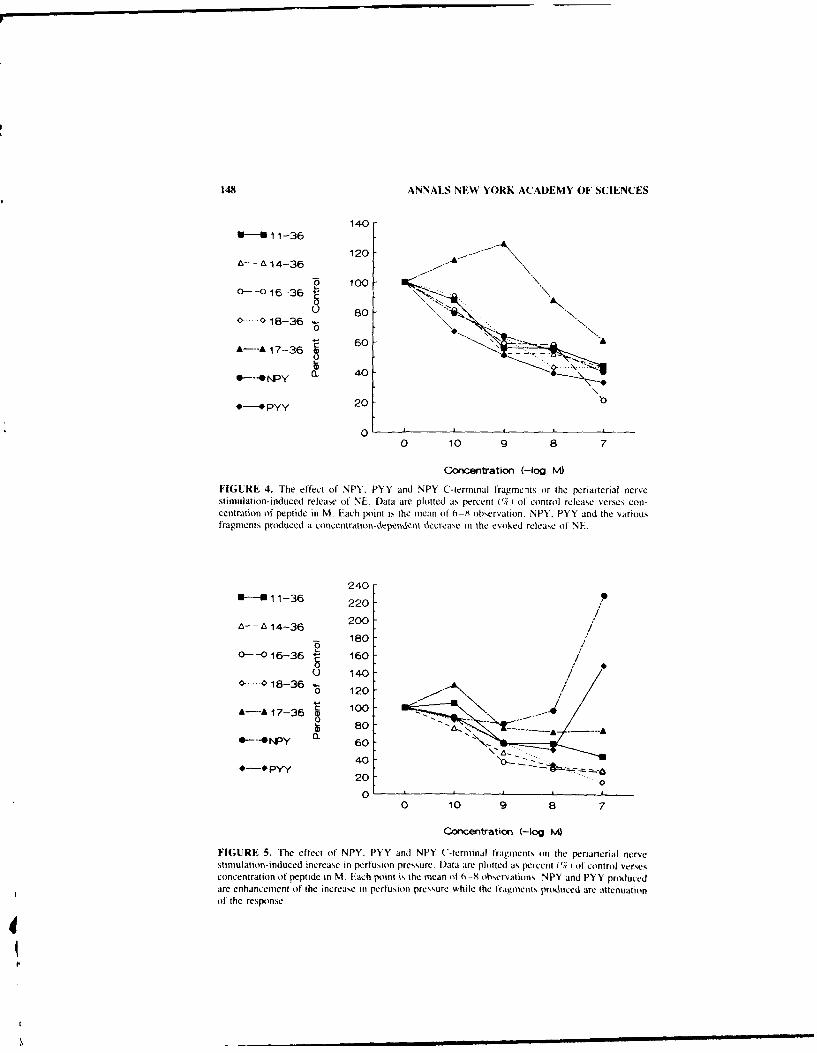
148 ANNALS NEW YORK ACADEMY OF SCIENCES
140-
- .14-36 12
* ~ 100-
U 80-0--018-36 -
00A---A 17-36 60
Q
0--ONY 0. 40-
P 20 0
00 10 9 a 7
Concentration (-og ")
FIGURE 4. The effect of' NPY, PYY and NPY C-terminal fragecits or the periarterial nervestimulation-induced release of NE. Data are plotted as percent (O/H of control release verses con-centration of peptide in M. Each point is the mean of 6-8 observation. NPY. PYY and the variousfragments produced a concentration-dependentl decrease in the evoked release of NE.
240-i- 11-36 20
A --14-36 20/76 180
0-016-36 160/
0 1400- -018-36
A--A 17-36 'o 100
0---. rNPY a 60
*-Pyy 40 \0-
20 ...01
0 10 9 8 7
Concentration (-og M
FIGURE 5. The efflect of NPY. PYY and NPY ('-terminal fragents oin the periarterial nervestimul ation -induced increase in perfusion pressure. Data are plotted as percent (7)of control versesconcentration of peptide in M. Each poit is the mean of 0_8X observations. NPY and PYY producedare enhancement of the increase in perfusion pressure while the tragments Produced are attenuationof' the response.
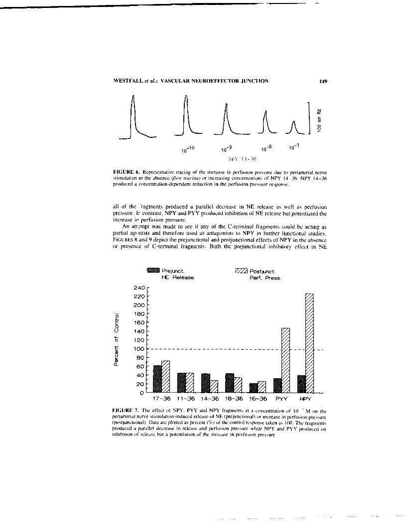
WESTFALL et al.: VASCULAR NEUROEFFECTOR JUNCTION 149
10- 0 10
-9 10-8 10
-7
NPY 154-3,6
FIGURE 6. Representative tracing of the increase in perfusion pressure due to periarterial nervestimulation in the absence (first tracing) or increasing concentrations of NPY 14-36. NPY 14-36produced a concentration-dependent reduction in the perfusion pressure response.
all of the .ragments produced a parallel decrease in NE release as well as perfusionpressure. Ir contrast, NPY and PYY produced inhibition of NE release but potentiated theincrease ir perfusion pressure.
An attempt was made to see if any of the C-terminal fragments could be acting aspartial agonists and therefore used as antagonists to NPY in further functional studies.FIGURES h and 9 depict the prejunctional and postjunctional effects of NPY in the absenceor presence of C-terminal fragments. Both the prejunctional inhibitory effect in NE
-MM Prejumt. Postonct.
NE Release Perf. Press.
240
220-
200
/
-6 180 -
S 1600() 140
o 120
c 100-------- - --- --- -- - - - - - - - ---- -(1
8060
40 E EU 720
17-36 11-36 14-36 18-36 16-36 PYY NPY
FIGURE 7. The effect (f NPY, PYY and NPY fragments at a concentration of 10) 7 M on theperiarterial nerve stimulation-induced release of NE (prejunctional) or increase in perfusion pressure(postjunctional). Data arc plotted as percent (%) of the control response taken as 100. The fragmentsproduced a parallel decrease in release and perfusion pressure while NPY and PYY produced oninhibition of release but a potentiation of the increase tn perfusion pressure

151) ANNALS NEW YORK ACAD)EMY OF SCIENCES
A-ANsPY 0--011-36 0-0 18-36 W .. 019-36+ NPY + NPY + NPY
120
100-
80- A .
6 0-
40-0
20-
0 ,I10 9 8 7
NPY concenfration (-og M
FIGURE 8. The effecl of N'Y alone or NPN plus, virious C-termninal fraigments on the periarierialnerve stimulation induced release of INV l),ita plotted ats it percent ( )of the response !o thelrinnients alone. Thec orniinant) In Pl l-YplLI, Iraymnents resulted in asi niiar inhibition of releaseas Awith NP1Y alone or Iraymnit alonie
release (Fin. 8) as well as potentiation ol the postiinctional response I Fis. 9 and 10)produced by NPY "~ere similar in the absence Or presence of the C-terminal franents.
('-Terminal Fragments in the Spinal Cord and Hypothalamic Slices
The i ntrat hecal admniin istratioin of N PY i nto the thiorac ic spinal cord produces at de-pressor effect as well a,, hemodynamic changecs due to a dccrcase in sym11pathetic nerveactivity.' Similar effects were observed followitng the intrathecal administration of C-termintal fragments although the Iragmients were less, potent &1(. I I.
The potassium- induced releasc of NE fromt slice,, i anterior and posterior hypothal-antus obtained frot vatrious strains of rat was reduced hs NPY t Fi;. 12). This inhibitoryeffct was also iobserved when various C-tcrtttittal fracinents such ats 16 -36 were exam-
I FGURE 9. Mhe etlect of NIPY alone or NPYplis %~arlous ( -teriiiial tragnients oin the periarte-rial nerve stiiiiulation-induejed increase in perfL]-
12. su'n pressure. D~ata are plotted as the increase inpertlisilin p~ressure 111111t1g0 SEt.l The first harreprceents NPY alone (It) kl) Tlie normal pa)-
tentiation of the increase in perfusion pressure pro-duced hN NP1Y A. is not altered M~ ien admiinistercdalon! wit a (-terminal tragmiient,

WESTFALL et al.: VASCULAR NEUROEFFECTOR JUNCTION 151
NPY 14-36 (10-7 M) NPY 1l-36 (10-7M)+Npy (10-7 M)
FIGURE 10. Representative tracings of the increase in perfusion pressure to periarterial nervestimulation in the presence of NPY 14-36 or NPY 14-36 plus NPY. The typical potentiation of theresponse produced by NPY was not altered when NPY was administered together in the NPY 14-36.
ined (FIG. 13). There was no evidence obtained that the C-terminal fragments couldreduce the inhibitory effect of NPY on the potassium-evoked release of NE (Ftu. 14).
DISCUSSION
These studies show that NPY produces marked effects at the vascular neuroeffectorjunction. Using the perfused mesenteric arterial bed as a model of the noradrenergicneuroeffector junction, NPY exerted both prejunctional and postjunctional effects.Prejunctionally, NPY decreased the periarterial nerve stimulation-induced release of NEwhile postjunctionally it potentiated the nerve stimulation-induced increase in perfusionpressure. Moreover, NPY potentiated the increase in perfusion pressure produced by avariety of vasoactive agents including angiotensin, vasopressin, as well as ct-adrenoceptoragonists like phenylephrine or norepinephrine. These results suggest that NPY is acting atthe level of transduction mechanisms common to this diverse group of vasoactive sub-stances. Our results also suggest that the postjunctional effect of NPY is independent ofendothelial cells.
There is growing evidence for the existence of multiple subtypes of NPY recep-tors.1 3 - 15 Studies by Wahstedt and colleagues suggest that the postjunctional responserequires the entire 1-36 peptide and have called this receptor subtype Y,. -'7 In contrast
WY fragments or the percent i%) change in arte-
0 -10- rial blood pressure of the anesthetized rat fol-143 -10lowing intrathecal administrative. Data are
-15 plotted as the percent (%) change in arterial5pressure verses dose of NPY or NPY frag-113-20 rument. NPY and the various fragments all pro-
-2 duced a decrease in arterial pressure.-25 . .
0 0.01 0.1 1.0DOSE (rrnol)

152 ANNALS NEW YORK ACADEMY OF SCIENCES
C~CNTF 12= Wy o FIGURE 12. The potassium (56 mM) in-
o.00 duced release of 'H-NE from slices of the an-terior hypothalamus of Sprague Dawley (SD),
O~AO Wistar Kyoto (WKY) and spontaneously hy-- __ pertensive (SHRI rats in the absence (control)
or pressure of NPY (10 ' M). Each bar is theS0.", mean -t SEM of 6-8 observations. Data are
plotted at the S2 (pressure of NPY) /S, (ab-020 sence of NPY) ratio. NPY produced a signif-
icant decrease in the potassium-induced re-o O WKlease of 3H-NE.
the prejunctional response could be produced by shorter C-terminal fragments (e.g., PPY13-36 or NPY 13-36) and has been designated the Y, receptor.'- 17 Ligand bindingstudies support the concept of at least two types of NPY receptors that can also bedistinguished on the basis of the entire NPY sequence or shorter NPY fragments. 18-19 Yreceptors bind long C-terminal fragments while Y, receptors bind the entire amino acidsequence. In addition, there are also NPY analogs that are specific for the Y, receptor.20
Further support for the Y, and Y2 concept is data suggesting they are coupled to differenttransduction mechanisms.21 For instance, in the human neuroblastoma cell line SK-N-MCNPY and the specific Y, receptor legend (Leu 31, Pro34)-NPY caused a rapid and transientincrease in the concentration of free calcium in the cytoplasm as well as inhibited theforskolin-stimulated cAMP production. Y2 specific ligands had no effect on either intra-cellular second messenger 9 Our results utilizing NPY, PYY. and a series of NPY C-terminal fragments are consistent with the Y,-Y 2 concept. The strength of our study isthat pre- and postjunctional responses were examined simultaneously in a model nor-adrenergic vascular neuroeffector junction. Our results are consistent with the postjunc-tional response (Y,) requiring both the N- and C-terminal while the prejunctional responserequires only the C-terminal end of the peptide. Although it may be premature or evensimplistic to think that there is a pre-postjunctional distribution of Y, and Y, receptors,our results do suggest that the predominant postjunctional receptor is the Y, subtype whilethe predominant form of the prejunctional receptors is of the Y, subtype. Because of theknown colocalization of NPY and NE in noradrenergic neurons innervating the mesentericarterial bed, our results reinforce the idea that NPY acts as an important cotransmitter/modulator at this vascular neuroeffector junction.
The biphasic effect of NPY 17-36 deserves further comment. It is of interest that NPYand the COOH terminal fragments 13-36. 16-36, 19-36, 20-36 and 25-36 all inhibitedboth basal and isoproterenol-stimulated adenylate cyclase activity in rat cardiac ventric-ular membranes22 In contrast NPY 17-36 stimulated basal and enhanced the isoprotere-nol-sttmulated adenylate cyclase activity. In the present studies NPY 17-36 stimulated
obo - FIGURE 13. The effect of NPY (10 ' M) orNPY 16-36 (10-" M) or the potassium-induced
o- release of endogenous NE from hypothalamicslices of Sprague Dawley rats. Data are plotted as
0,4 an S,/S, ratio similar to FIGURE II. The dottedline represents release of NE to potassium alone
020 while the bars depict release in the pressure ofNPY or NPY 16-36. All the fragments studied
produced a significant increase in the potassium-Hy MPY 10-16l induced release of NE similar to NPY itself.

WESTFALL et al.: VASCULAR NEUROEFFECTOR JUNCTION 153
release of NE except at high concentrations while NPY, PYY and all the other fragmentsproduced only inhibition of NE release. Since there are data suggesting that increases incAMP enhance transmitter release, it is suggestive of cAMP being involved in theprejunctional actions of NPY. Further studies are, of course, needed to further probe thisidea.
Our results also implicate NPY in the pathophysiology of hypertension developmentand/or maintenance in the SHR. At the vascular neuroeffector junction of the mesentericarterial bed, the postjunctional potentiation of contraction to vasoactive agents and nervestimulation by NPY was enhanced. In contrast, the prejunctional effect of inhibition ofNE release was attenuated. This combination of responses would result in a greaterincrease in blood pressure. The mechanism of these changes is not understood. However,since there is evidence for subtypes of NPY receptors, it is possible that there might bedifferential up and down regulation of Y, and Y2 receptors. It is also possible that theremay be differential effects on the intracellular transduction mechanisms. To date NPY hasbeen shown to inhibit the stimulation of cAMP induced by various agents, 23 24 to increasephosphoinositide turnover.--2- to produce an increase in intracellular calcium21 '
2 or toinhibit intracellular Ca2 transient and depolarization induced transmembrane Ca2 I
currents. 2 5- ' Alterations in one or more of these transduction mechanisms may occur inblood vessels or neuronal tissue of the SHR.
GAO
FIGURE 14, The effect of NPY alone (10 '
M) a NPY (10- M) plus NPY 16-36 (10C0M ton the potassium-induced release of endog-
enous NE from hypothalamic slices. Data areplotted in a similar fashion to FIGURF 12. The
020- pressure of the NPY fragments did not alter theinhibition of release produced by NPY alone.
01MIPY Y 16-386.vY
The results of studies carried out with C-terminal fragments in the spinal cord andhypothalamus suggest that the depressor effect following intrathecal administration andthe inhibition of NE release from hypothalamic slices may be due to activation of Y,receptors.
CONCLUSION
In conclusion, our studies have demonstrated that NPY produces prejunctional inhib-itory and postjunctional excitatory effects at the noradrenergic neuroeffector junction ofthe mesenteric arterial bed. The postjunctional effect is nonspecific in that NPY poten-tiated the increase in perfusion pressure to a number of diverse substances suggesting aneffect on a common intracellular transduction mechanism. The postjunctional effect isindependent of endothelial cells and appears due to activation of Y. receptors while theprejunctional effect is due to activating the Y, receptor subtype. The prejunctional effectof NPY was attenuated while the postiunctional effect was enhanced in beds obtainedfrom the SHR. Our results therefore implicate an alteration in the NPY system in thedevelopment or maintenance of hypertension in the SHIR.

154 ANNALS NEW YORK ACADEMY OF SCIENCES
REFERENCES
I.LUNDBERG, J. M. & T. HOKFELT. 1983. Coexistence of peptides and classical neurotransmit-ter-s. Trends Neurosci. 6(8): 325-333.
2. EVERITT. B. V. T. H(3KELT, L. TERENIUS. V. TATEMOTro. V. MUIT & M. GOLDSTEIN. 1984.Differential co-existence of neuropeptide Y (NPY)-like immunoreactivity with catechol-amines in the central nervous system of the rat. Neuroscience 11: 443-462.
3. GIBSON, J., K. M. POLAK. T. E. ADRIAN. J. S. KELLY & S. R. BLOOM. 1984. The distributionand origin of a novel brain peptide. neuropeptide Y in the spinal cord of several mammals.J. Comp. Neurol. 227: 78-91.
4. LUNDBERG. J. M. & L. STJARNE. 1984. Neuropeptidle Y INPY) depresses the secretion of3H--noradrenaline and the contractile response evoked by field stimulation, in rat vas deferens.Adta. Physiol. Scand. 120(3): 477-490.
5. LuN"P~--. J M . I TERENius. T. 1-OKFELT1 & M. GOLDSTEIN, 1983. High levels of neu-ropeptide Y in peripheral noradrenergic neurons in various mammals including man. Neuro-sci. Lett. 42(2): 167-172.
6. COSTA. M., J. B. FURNESS & 1. L. GIIIBINS. 1986. Chemical coding of enteric neurons. Prog.Brain Res. 68: 217-239.
7. WESTFALI.. T. C., S. CARPENIIER, X. L. CHEN. M. C. BEINFELD. L. NAES & M. J. MEL-DRUM. 1987. Prejunctional effects of neuropeptide Y at the noradrenergic neuroeffectorjunction of the perfused mesenteric: arterial bed of the rat. J. Cardiovasc. Pharmacol.10: 7 16-722.
8. CIARLEGLIO. A. E., M. C. BEINFEI.D & T. C. WESTFAI.I. 1987. Neuropeptide Y modulationof noradrenergic transmission in the posterior hypothalamus of the rat. Pharmacologist29: 128.
9. CIARLEGLIO. A. E.. M. C. BEINFEt.D & T. C. WESTFAII.. 1988. Release of neuropeptide Yimmunoreactivity from the rat hypothalamus. Soc. Neurosci. Abstr. 14: 843.
10. YAKSH, T. L. & T, A. RUDY, 1976. Chronic catherization of the spinal cord subarachnoidspace. Physiol. Behav. 17: 1031-1036.
11. CHEN, X.. K. HENDERSON. M. C. BEINFELD & T. C. WESTFAI.I. 1988. Alterations in bloodpressure of normotensive and hypertensive rats following intrathecal injections of neuropep-tide Y. J. Cardiovasc. Pharmacol. 12: 473-478.
12. WESTFAtL., T. C.. J. MARTIN, X. CHIEN. A. CIARtEutlo0. S. CARPENTIER. K. HENDERSON. M.KNUEPFER. M. C. BEINEID & L. NAES. 1988. Cardiovascular effects and modulation ofnoradrenergic neurotransmission following central and peripheral administration of neuropep-tide Y. Synapse 2: 299-307.
13. WESTFALL. T. C.. S. P. HAN, X. CHIEN. K. DEL VALLIE, M. CLTRFMAN. A. CIARI-EGtLto & L.NAES. 1990. Presynaptic peptide receptors and hypertension. Ann. N.Y. Acad. Sci. In press.
14. WESTFALL, T. C., S. P. HAN. M. KNUEPEER, J. MARTIN. X. CHE-N, K. Dt:li VALLE. A.CIARLI.OI & L, NAES. 1990. Neuropeptides in hypertension: Role of neuropeptile Y andcalcitonin gene related peptide. Brit. J. Clin. Pharmacol. 30. In press.
I5. WAHLESTEDT, C.. N. YANAIHARE & R. HAKANSON. 1986. Evidence for different pre- andposijunctional receptors for neuropeptides Y and related pel-ides. Regul. Pept. 13: 307-3 18.
16. WAHIEsrEOT. C. & R. HXKANSON. 1989. Y, and Y, receptors for neuropeptide Y FEBS Lett.245(1-2): 2(09-214.
17. WAHLESTEOT. C., R. EKMAN & R. HAKANSON. 1987. Studies on interaction between neuro-peptide Y (NPY) and noradrenaline in rat cerebral cortex. Soc. Neurosci. Abstr. 13: 1271.
18. SHEIKH. S. P.. R. HX KANSON & T. W. S(HWARTZ. 1989. Y, and Y, receptors for neuropep-tide Y. FEBS Lett. 245(1-2): 209-214.
19. SCHWARTZ. T. W., J. FAHI.ENDORFF & J. LANGELAND. 1989. Y, and Y, receptors for NPY-:.The evolution of PP-fold peptides and their receptors. Nobel Symposium XIV: NeuropeptideY. Raven Press. New York. NY.
20. FUHLENDORIFF. IL. U. GETHERt L. AAKI-.RI.UND. N. LANGEI AND-JOHSNSEN. H. THOGiERSEN.
S. G. MEt BERG. U. B. 0LSE-N. 0. THASIRtIP & T. W. SCHWARTIZ. 19%0. ILeu". Pro14 1Neuropeptide Y: A specific Y, receptor agonist- PMC. Proc. Naill Acad. Sci. USA87: 182-186.
21. AAKI.RIAtND. L., U. Gvl tER. J. Ft;HLFNNOOREI-. T. S.IHWARIZ & 0. IIIASTRUP. 19%(1 Y,

WESTFALL et al.: VASCULAR NEUROEFFECTOR JUNCTION 155
receptors for neuropeptide Y are coupled to mobilization of intracellular calcium and inhibi-tion of adenylate cyclase. FEBS Lett 260: 73-78.
22. BALASUBARAMANIAM, A., S. SHERIFF, R. RENUYOPALAKRISHNAU, S. G. HUAEG, M. PRAB-HAKARAN, W. T. CHANCE & J. E. FISCHER. 1990. Cardiac properties and conformationalstudies of neuropeptide Y and NPY 17-36. Ann. N.Y. Acad. Sci. In press.
23. LUNDBERG. J. M., A. EMSON, 0. LARRSON. A. RUDEHILL, A. SARIA & B. FREDHOLM. 1988.Neuropeptide Y receptor in pig spleen: binding characteristics, reduction of cyclic AMPformation and calcium antagonist inhibition of vasoconstriction. Eur. J. Pharmacol.145: 21-29.
24. H.RFSTRAND, A., B. FREDHOLM & K. FUXE. 1987. Inhibitory effects of cyclic AMP accu-mulation in slices of the nucleus tractus solitarius. Neurosci. Lett. 76: 185-190.
25. HINSON, J., C. RANH & J. COUPET. 1988. Neuropeptide Y stimulates inositol phospholipidhydrolysis in rat brain minipressure. Brain Res. 446: 379-382.
26. PERNEY, T. M. & R. J. MILLER. 1989. Two different G-proteins mediate neuropeptide Y andbradykinin stimulated phospholipid breakdown in cultured rat sensory neurons. J. Biol.Chem. 264: 7317-7327.
27. MOTINLSKY, H. J. & M. C. MICHEL. 1988. Neuropeptide Y mobilized Ca 2 and inhibitsadenylate cyclase in human erythroleukemia cells. Am. J. Physiol. 255: E880-E885.
28. SCHAFIELD, G. G. & R. S. IKEDA. 1988. Neuropeptide Y blocks a calcium current in C-cellsof bullfrog sympathetic ganglia. Eur. J. Pharmacol. 151: 131-134.
29. EWALD, D. A., P. C. STERNWEISS & R. J. MILLER. 1988. Guanine nucleotide binding proteinco-induced coupling of neuropeptide Y receptors to Ca 2 ' channels in sensory neurons. Proc.Natl. Acad. Sci. USA 85: 3633-3t)37.
30. WALKER, M. W., D. A. EWALD, T. M. PERNEY & R. J. Miller. 1988. Neuropeptide Ymodulates neurotransmitter release and Ca 2
1 currents in rat sensory neurons. J. Neurosci.8: 2438-2446.
31. COLMERS, W. F., K. LUKOWIAK & 0. J. PITTMAN. 1988. Neuropeptide Y action in the rathippocampal slice: site and mechanism of presynaptic inhibition. J. Neurosci. 8: 3827-3837.

Importance of Neuropeptide Y in theRegulation of Kidney Function'
PONTUS B. PERSSON,6 GERALDU GIMPL,'
AND RUDOLF E. LANG'hl. Physiologisches Institut'Pharmakologisches InstitutIm Neuenheimer Feld 326
D-6900 Heidelberg, Federal Republic of Germany
INTRODUCTION
Neuropeptide Y (NPY) is a 36-aminoacid peptide structurally related to the pancreaticpeptide family. It has previously been shown to exist in sympathetic nerves and in centralnervous tissue. " Within the sympathetic nerve terminals NPY is stored in large dense-cored vesicles. 5.6 Not only is NPY co-localized with norepinephrine, it is also co-releasedduring nerve stimulation7"- and interacts with norepinephrine on the pre- and postsynapticlevel. , ,9. o
The physiological effects of NPY are similar to noradrenergic stimulation, but sys-temic administration of NPY will produce hypertension, which is not blocked by adren-ergic antagonists. " NPY does not only increase vascular resistance by this adrenoceptor-independent mechanism, it also potentiates vasoconstrictor responses to norepinephrineand sympathetic nerve stimulation. 12 The kidney possesses a particularly high concen-tration of NPY, as do the coronary and mesenteric arteries. '" Furthermore, the kidneyreveals a particularly high density of high-affinity binding sites for this peptide. 1
4 Hence,NPY may play an important role in modulating a variety of renal processes. In fact.studies in the rat have provided evidence for an influence on renal sodium reabsorption'iand renin release. .7 These observations are strengthened by the fact that NPY has beenlocalized in the sympathetic nerve fibres at the juxtaglomerular apparatus. 8 the site ofrenin secreting cells.
In addition to these direct effects, NPY might also modulate renal function via itsinteraction with the adrenergic transmitters. It is well documented, that sympatheticnerves have a profound impact on sodium excretion. ."21 Pressure-dependent renin re-lease is also modulated by sympathetic activation. 2
1'2
1 and even the range of renalautoregulation is dependent on the prevailing sympathetic tone to the kidney. 22
Characterization of Renal NPY Receptors
Binding sites for NPY have been reported in a variety of tissues including the brain,21
spleen. 24 and aorta.2 5 A vast amount of high-affinity binding sites has also been found inthe rabbit kidney. '4 Studies employing autoradiographic techniques in the kidney havedemonstrated that NPY binding is not restricted to the vascular tissue. The proximal
"This study was generously supported by Boehringer (Ingelheim) the German Research Founda-tion (Forschergruppe Niere, Heidelberg), and the "Bundesministerium fur Forschung und Technol-ogie" (project: 0318957 B).
156

PERSSON et at.: NPY & KIDNEY FUNCTION 157
convoluted tubules reveal binding as well. 26 This seems to fit nicely to the proposedeffects of NPY on sodium excretion as will be referred to below.
A recent study27 describes a technique for solubilizing NPY receptors from the rabbitkidney using the zwitterionic detergent 3-[(3-cholamidopropyl)dimetylammoniol-l-pro-panesulfonic acid (CHAPS). The receptor characteristics were not altered by this proce-dure as indicated by saturation studies revealing very similar k. values (0.10 - 0.02 nMfor membranes vs. 0.12 ± 0.03 nM for soluble extract , A Scatchard analysis of theequilibrium binding data demonstrated a single class of hi;,ding sites (FIG. 1, A 100-kDapolypeptide represents the covalently labeled NPY rece, .or. This was shown by affinitylabelling experiments using two different categories or cross-linking agents. The speci-ficity of the labelling was apparent by the ability of unlabeled NPY to displace thebinding.
The nonhydrolizable GTP analog (GTP-yS) specifically inhibited NPY binding.Within 5 min of incubation the specific 1251I-NPY binding decreased to 15% of control(FIG. 2) for the solubilized fraction. This suggests that these NPY receptors are function-ally coupled to GTP-binding regulatory proteins. This agrees with some indirect evidenceobtained in other tissues, that at least one type of NPY receptors belongs to the class ofG-protein coupled receptors.
24.28.29
Considerable evidence derived by investigations on the rat brain and cell lines o1neuronal origin speaks in favor of two NPY receptor subtypes 30;- Uwil further exper-iments utilizing these ligands have been performed in the kidney. the question about thepresence of both subtypes in the kidney remains unanswered.
An interesting observation of rabbit kidney NPY receptors, however, is the compet-itive action between NPY and the humoral peptide YY.14. 27 Perhaps circulating peptideYY also acts as a physiological ligand at the kidney NPY receptor.
NPY Localization and Distribution throughout the Kidney
With the use of several region-specific antisera and the peroxidase-antiperoxidasetechnique, Reinecke and Forssmann3" found a wide distribution of NPY in all segmentsof the renal vasculature. In comparison to the large number of polypeptides investigatedin renal nerves. NPY was the most prominent. The nerve fibres containing NPY werelocated homogeneously around large renal arteries continuing into a dense plexus of thearcuate and interlobular arteries. An equivalent density of NPY nerves occurred aroundthe afferent and efferent arterioles.
The juxtaglomerular apparatus is a neuroendocrine unit located at the vascular pole ofthe glomerulus and is known to have a rich supply of monoaminergic nerve fibres. In thisdense plexus of fibres of several species including man, NPY has also been located bymeans of immunocytochemistry. " This finding is of particular importance since thejuxtaglomerular apparatus is a crucial component for renin secretion. In the mentionedstudy, NPY-LI immunoreactivity was also markedly depleted after chemical sympathec-tomy by 6-hydroxydopamine.
In yet another study. 26 in vitro labelling of NPY binding sites with ' 251-NPY wasperformed in rabbit kidney sections. Autoradiographic and histochemical techniquesidentified binding sites not only in the vascular smooth muscle, but in the proximaltubules as well. Intriguingly, however, no binding was found in the rat, guinea pig andhuman kidney. Hence, for the time being, an unequivocal interpretation of these partlyconflicting results is not possible, but the preferential localization of NPY to certainsegments of the kidney may provide insight into its physiological and pathophysiologicalimportance.

158 ANNALS NEW YORK ACADEMY OF SCIENCES
700 A
C 600
0C_CL 500
o0
400450
S.300 0000
200 10
ri 100 0 200 400 600 80o
0.0 0.5 1.0 1.5 2.0 2.5CONCENTRATION OF '25I-NPY [nM
1600
B
0 15000
L- 1200CL
CCDN.a00 0 600 1200 1800
03
z
- 0.0 0.5 1.0 1.5CONCENTRATION OF 12 1-NPY (nMl
FIGURE 1. Saturation curves and Scatchard analysis of NPY binding. (A) Membrane preparation;(B) CHAPS solubilized fractions. Open circles refer to unspecific bindirg;filled circles represent thespecific binding estimate (unspecific-total ligand bound). The insets show the corresponding Scat-chard plots (B: specifically bound NPY. F: free NPY in nM).

PERSSON el al.: NPY & KIDNEY FUNCTION 159
E .2
E
C
E cL
punaq AdNISZ~ 0!1padc tu-
-Z uE -
cv
E0
-

160 ANNALS NEW YORK ACADEMY OF SCIENCES
NPY and Renin Release
iuxtaglomerular renin secretion is modulated by numerous local and systemic influ-ences. It is remarkable that all effectors of renin secretion have one feature in common:vasoconstricting agents generally inhibit, and vasodilators augment renin secretion. "There can be no doubt that NPY is a potent vasoconstrictor thus, according to this generalrule, one should expect NPY to act in an inhibitory fashion on renin secretion. Indeed, aninhibition of renin secretion by NPY was described by Hackenthal and colleagues1 6 in theisolated rat kidney. Since perfusion pressure is known to be one of the most importantfactors influencing renin release, 34 the kidney was perfused at a constant pressure ( 105mrititgi by means of a servocontrol mechanism. In this preparation NPY produced a dosed.penndent inhibition of renin release, which corresponded to the degree of vasoconstric-ti,,n. However, the baseline renin secretion increased steadily throughout the experimentalprotocol. This seemingly paradox behavior is not necessarily due to a specific NPY effect,since it was also demonstrated by other vasoconstricting agents, such as angiotensin II.
To exclude a simple flow-dependent or "washout" effect on rcin release, theseauthors repeated their study under constant flow conditions; furthermore, tissue sampleswere studied taken from the hydronephrotic rat kidney. All approaches yielded the sameresults: NPY had a strong inhibitory effect on renin release. Pertussis toxin clearly atten-uated the response to NPY; thus an adenylate cyclase coupled mechanism was suggested.
The inhibition of renin release by NPY is supported by a recent study in anesthetizedcats." Pressor and nonpressor intravenous (iv) NPY doses decreased plasma renin ac-tivity. Intriguingly, the same "rebound effect" of renin release as seen by Hackenthal etal. " was observed. The baseline level of plasma renin activity increased substantially assoon as the infusion ended.
The harmonious results of these two studies is challenged by the work of Echtenkampand Dandridge. 36 They studied the effects of NPY administered either intravenously ordirectly into the renal artery of anesthetized primates. Increasing iv NPY doses from 20to 1000 ng/kg/min, as well as intrarenal NPY administration between 20-400 ng/kg/minNPY did not have any significant effects on plasma renin activity or renin release, in spiteof renal vasoconstriction at the higher NPY levels. This is substantiated by a recent studyshowing that iv infusions of NPY in the rat did not influence plasma renin activity. 7 Alack of NPY inhibition on renin release has also been found during iv NPY infusions innormal rats when only modest increases in arterial blood pressure occurred.' 7 However.in the same study NPY normalized renin release in adrenalectomized rats without con-comitant changes in blood pressure. Accordingly, NPY might exert an indirect influenceon renin release via its interaction with adrenergic transmitters, of- and P-adrenergiceffects on renin release are well documented.2 0. 2' and NPY inhibits nerve stimulationevoked norepinephrine release in several tissues including the kidney. 3. 3
" To test thishypothesis, an experimental protocol was developed in the conscious dog in which areflex sympathetic stimulus could be elicited and renal perfusion pressure was controlled(Fiu. 3). Dose response curves were obtained by infusing NPY into the renal artery andmeasuring the reductions in renal blood flow (RBF). A dose was chosen for the exper-iments which was just below the dose necessary to reduce RBF (I Rig/min). This infusionincreased renal venous NPY concentrations from roughly 40 to 300 pg/ml. The prelim-inary data depicted in FIGURE 3 show that renin release was not decreased by thisprocedure. if at all; there might even be a slight increase during NPY infusions. The nextstep was to determine whether these effects were the same at a reduced level ol "enalartery pressure. This is important since there is a threshold pressure for renin release(roughly 95 mmHg) 4 Above this limit the effects on renin release may be negligible orat least difficult to detect. Renal artery pressure was servocontrolled by an electropncu-matic control system 4 at 80 - 0.5 mmHg. The surprising finding was that during the low

PERSSON et al.: NPY & KIDNEY FUNCTION 161
NPY (1,g/rnin)
.40. .......... Servo control
120
100
so
60.
150-
oo0
00
FIGURE 3. Effects of intrarenal
NPY-infusions (I p.g/min, right) com-
pared to no infusions (left). Commoncarotid occlusion (CCO) was used as asympathetic stimulus. Renal artery
180o pressure could be servocontroled at 801mmHg (PS0). The data are means from
1501' 6 cxperiments in 3 dogs.
120.
80
z~60itZ
2000
O 10001
n_ 5001
Pao P80 Pao POO
CONTROL CCO NPY NPY&CCO

162 ANNALS NEW YORK ACADEMY OF SCIENCES
dose NPY infusion renin release increased much more than under control conditions. Itshould be mentioned, that 80 mmHg is well above the lower limit for RBF auto-regulation; 22 hence, the NPY infusion did not alter total renal vascular resistance (FIG. 3).
In order to quantify the interaction between sympathetic activation and simultaneousNPY infusion, we repeated this experimental protocol during common carotid occlusion(CCO). The results indicate, that there is no apparent difference between the protocolswith or without the reflex sympathetic activation. This, on the other hand, agrees withfindings from Hackenthal and colleagues' 6 made in the isolated rat kidney. They foundthat the a,-adrenoceptor agonist prazosin (2 1jM) only slightly modified the renin releaseresponse due to NPY.
Taken together, there is disagreement towards several important features concerningthe effects of NPY on renin release. It has, however, been shown, that there is a reboundphenomenon of renin release after NPY infusions. This phenomenon cannot be explainedat present. but it does not seem to be confined to NPY, since similar findings were madefor other vasoconstrictors. Furthermore, it does not seem likely that NPY and adrenergictransmitters specifically interact in the regulation of renin release. Nevertheless, at thispoint, it is not possible to clarify the crucial question if NPY mediates its effects on reninrelease via a direct action on the juxtaglomerular apparatus, or if these effects may besecondary to its vasoconstrictor actions.
Renal Vascular Resistance
More insight might be obtained by the data concerning renal vascular resistance, andespecially by the electrophysiological recordings from epithelioid cells.
In a number of in vitro and in vivo preparations, exogenous NPY exerts a directpostsynaptic vasoconstriction and potentiates the vasoconstrictor action of otheragents. 15.40A41 Although, NPY induced vasoconstriction has been described in most vas-cular beds, the vasoconstricting potency varies considerably between different organs.Pernow and Lundberg"' found a slight reduction in RBF by exogenous NPY in very smalldoses, which resulted to an arterial plasma NPY-LI concentration of 164 pmol/l. Thisdemonstrates a remarkable sensitivity of the renal vasculature to NPY since other bedsrespond with vasoconstriction at nanomolar concentrations.39 In the same study, theeffects of renal nerve stimulation on RBF were investigated before and after adrenergicblockade. During high frequency stimulation ( 10 Hz) there were still mentionable reduc-tions of RBF inspite adrenergic blockade. It is likely, that this effect is due to NPYrelease, as indicated by a high correlation between NPY-LI overflow and the c7 reductionof RBF.
By injecting considerable amounts of NPY (10 I-g/kg as a bolus) in the consciousrabbit, Minson and colleagues4 detected a marked increase in renal vascular resistance.a moderate rise in mesenteric vascular resistance, and a transient fall in hindlimb vascularresistance. Taken together. there is data indicating (i) a high renal vascular sensitivity toNPY, (ii) a reduction of RBF duing high frequency renal nerve stimulation in thepresence of adrenergic blockers, and (iii) differential effects of high dose NPY adminis-tration on regional vascular resistance. Thus, the following hypothesis can be made: Insituations with a high sympathetic outflow, NPY may redistribute blood flow in f;ivor ofthe skeletal muscle. This would suggest a participation of NPY in the "fight or flight"reflex.
Intriguingly. preliminary data suggests, that the effects of NPY are also not evenlydistributed throughout the kidney. Biihrle and colleagues4 2 investigated the membranepotential of renin containing epithelioid and va:;cular smooth muscle cells of the afferentarteriole in the hydronephrotic mouse kidney. Several agents, such as angiotensin II. and
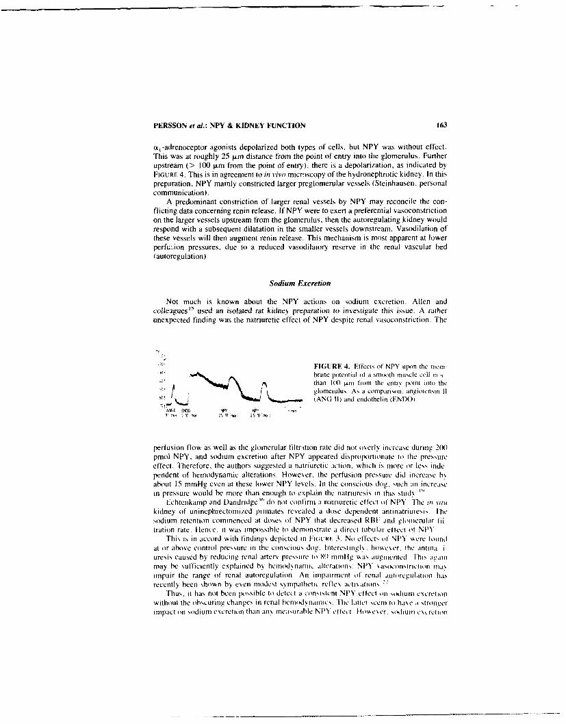
PERSSON et al.: NPY & KIDNEY FUNCTION 163
a,-adrenoceptor agonists depolarized both types of cells, but NPY was without effect.This was at roughly 25 I.±m distance from the point of entry into the glomerulus. Furtherupstream (> 100 tm from the point of entry), there is a depolarization, as indicated byFIGURE 4. This is in agreement to in vivo microscopy of the hydronephrotic kidney. In thispreparation. NPY mainly constricted larger preglomerular vessels (Steinhausen, personalcommunication).
A predominant constriction of larger renal vessels by NPY may reconcile the con-flicting data concerning renin release. If NPY were to exert a preferential vasoconstrictionon the larger vessels upstream from the glomerulus, then the autoregulating kidney wouldrespond with a subsequent dilatation in the smaller vessels downstream. Vasodilation ofthese vessels will then augment renin release. This mechanism is most apparent at lowerperfu:;ion pressures, due to a reduced vasodilatory reserve in the renal vascular bed(autoregulation).
Sodium Excretion
Not much is known about the NPY actions on sodium excretion. Allen andcolleagues"' used an isolated rat kidney preparation to investigate this issue. A ratherunexpected finding was the natriuretic effect of NPY despite renal vasoconstriction. The
FIGURE 4. Effects of NPY upon the mern-brane potentiat (it a smooth muscle cell Il mthan IMX (.Lm fromi the entrs point into theglomerulus. As a comparison: anglotensmn 1tANG 11) and endothelin (ENDO)
tC m : to le - 'I ZS ID' 25 ID"r
pertusion flow as well as the glomerular filtrition rate did not overly increase during 2(X)pmol NPY, and sodium excretion after NPY appeared disproportionate to the pressureeffect. Therefore, the authors suggested a natriuretic iction. which is more or less inde-pendent of hemodynamic alterations. However, the perfusion pressure did increase b,,about 15 mmHg even at these lower NPY levels. In the conscious dog. such :n increasetn pressure would be more than enough to explain the natriurcsis in this stud.y."'
Echtenkamp and Dandridge " (to not confirn a natriuretic effect of NPY. The it Nitkidney of uninephrectomi/ed primates revealed a dose dependent aninatriuresis. Thesodium retention commenced at doses of NPY that decreased RBF and eloincrular ii-tration rate. Hence, it was impossible to demonstrate a direct tubular elfect of NPY
This is in accord with findings depicted in H:OVRt 3. No eftects of NPY %ere foundat or above control pressure in the conscious dog. Interestingl . hoe,,er, the antina I-
uresis caused by reducing renal arterv pressure to 8I iinlig vas augmeniced lhis againmay be sufficiently explained by hcmod nanii, alterations: NPY %asoconstriction inaximpair the range of renal autoregulation An impairment of renal aulorcgulaloi hasrecently been shown by even modest sNnipathetic reflex actix ations. -I
Thus, it has not been possible to detect a con,,istent NPY elfec.t oti sollum excretionwithout the obscuring changes in renal hemodynamic,, [he latter seem to h,,c a strongerimpact (in sodium excretion than any tncasurable NPY ellect lthwcser, s(huni c\. rctiov

164 ANNALS NEW YORK ACADEMY OF SCIENCES
of the rabbit may be different from these other species, since NPY binding has beendetected in the rabbit proximal convoluted tubules.
ACKNOWLEDGMENTS
We thank Dr. Kirchheim and Dr. Ehmke, I. Physiologisches Institut, Heidelberg, fortheir valuable collaboration in the dog experiments. We are grateful to Dr. Hackenthal,Pharmakologisches Institut, Heidelberg, for the plasma renin activity determinations. Weare further indebted to Mrs. Barbara Collins. Mrs. Inge Keller, and Miss Lucie Mahi fortheir expert technical assistance.
REFERENCES
I . ALLEN, J. M. & S. R. BLOOM. 1986. Neurochem. Int. 8: 1-8.2. GRAY. T. S. & J. E. MORLEY. 1986. Life Sci. 38: 389-401.3, TATEMOTO. K.. M. CARLQUIST & V. MUTT. 1982. Nature 296: 659-660.4. LUNDBERG, J. M.. L. TERENius. T. HOKFELT & K. TATEMOTO. 1984. J. Neurosci. 4: 2376-
2386.5. FRIED. G., L. TERENIUS. T1. HOKFEI.T & M. GOLDSTEIN. 1985. J. Neurosci. 5: 450-456.6. JARVI. R.. P. HELEN. M. PELTO-HUIKKO & A. HERVONEN. 1986. Neurosci. Lett. 67: 223-
227.7. ALLEN, J. M., P. M. M. BIRCHAM. S. R. BL~OOM & A. W. EDWARDS. 1984. J. Physiol.
(London) 357: 401-406.8. THEODORSSON-NoRHEIM. E., A. HEMSEN & J. M. LUNDBERG. 1985. Scand. .1. Lab. Invest.
45: 355-365.9. WAHLESTEDT. C.. N. YANAIHARA & R. HAKANSON. 1986. Regul. Pept. 13: 307-31IX
10. LUNDBERG, J. M. & L. STIA RNE. 1984. Acta Physiol. Scand. 120: 477-479.11. ZUKOWSKA-GROJEC. Z., M. HAASS & M. BAYORH. 1986. Regul. Pept. 15: 99-110.12. LUNDBERG, J. M.. L. TORSELL, A. SolizEVI. J. PERNOW. E. THEODORSSON-NORHEim. A.
ANGGARD & B. HAMBERGER. 1985. Regul. Pept. 13: 41-51.13. UDDMAN. R., E. EKBLAD. L. EDVINSSON. R. HiKANSON & F. SUNDLER. Regul. Pepi. 10:
243-257.14. SCHACHTER, M.. C. M. M. MILEs. K. LEYS & P. S. SEVER. 1987- . CardjoVaSL- Pharmacol
10(Suppl 12): 157-162.15. ALLEN. J. M., A. E. G. RAINF. J. G. G. LEDINGHAM & S, R. BLOM. 1985. Clin. Sci.
68: 373-377.16. HACKENTEIAi., E., K. AKtORIES. K. H. JAKOBS & R. E. LANG. 1987. Am. J. Phvsil. 252
(Renal Fluid Electrolyte Physiol. 21 ): F543-F550.17, PFISTER. A.. B. VWAEBER. J. NLSSBERGER & H. R. BRLINNFR. 1986. Life Sci. 39: 2161-2167.ig. BALLEiSIA. J., J. M. POL.AK. . M. At i EN & S. R. Bi m. 1984, Histixheniistrn SO: 483-
485.19. PERSSON. P. B., H. EHMK1-. U. K66LiER & H. R. KIRCHIII-IM. 1989. Am. J. PhNysiol. (Renal
Fluid Electrolyte Physiol 25) F485-1-48920. DiBONA. G. F, 1982. Rev. Phx'.iol. Biochem. Pharmacol. 94: 70-181.21. EHMKf. H , P. PERSSON. S. Fiscmi. E. HACKNTHAI & H. KIREIHHEM. 1989 PflugerN
Arch. 413: 261-264622 PERS~ON, P. B.. H. FHMKE. B NAt., & H. R. KIimmiM 199) Am J. Phssiol 258 (Renal
Fluid Electrolyte Physioli: FW6-F371.23. UsNO(,N. A . K TAIEMONo. V. MUIii & T BARTI-AI 19X4. Fur I. Biochem. 145: 555)24. L~ioit,. I MI.. A. HEmsfEN. 0 [ARSSON. A. Ri. it-Mii i . A SARIAs & B B. FRID14 1.1
1998 Eur 1. Pharmacol 145: 21-2925 (*MAN(,. R S. L. . V tL t.(iTi. T CI-%. 1) (i RINI & P J Ki iN(, N'X5 Life Sci
37: 2111- 212226. Livs. K. NIM Si IihiA( 1-14 & P SESE,1R 1997 Fur I Phamacol 1,%4: 233 -2317

PERSSON et a).: NPY & KIDNEY FUNCTION 165
27. GIMPL, G., R. GERSTBERGER, K.-N. KLOTZ & R. E LANG. 1990. Submitted.29. UND#N, A. & T. BARTFAI. 1984. FEBS Lett. 177:. 125-128.29. PERNEY. T. M. & R. J. MILLER. 1989. J. Biol. Chem. 264: 7317-7327.30. LYNCH, D. R., M. W. WALKER, R. J. MILLER & S. H. SNYDER. 1989. J. Neurosci. 9:
2607-2619.31. SHEIKH, S. P., M. M. T. O'HARE. 0. TOTORA & T. W. SCHWARTZ. 1989. J. Biol. Chem.
264: 6648-6654.32. REINECKE. M. & W. G. FORSSMANN. 1988. Histochermstry 89: 1-9.33. 1-ACKENTHAL. E., U. SCHWERTSCHLAG & R. TAUGNER. 1983. Clin. Exp. Hypertenr. 5:
975-993.34. KIRCHHEIM, H. R., R. FINKE. E. HACKENTHAL, W. L6WE & P. PERSSON. 1985. Pflujgers
Arch. 405: 127-135.35. CORDER. R., M. B. VALLOTTON, P. J. LOWRY & A. G. RAMAGE. 1989. Neuropeptides
14: 111-114.36. ECHTENKAMP. S. F. & P. F. DANDRIDGE. 0989. Am. J. Physiol. (Renal Fluid Electrolyte
Physiol. 25): F524-F531.37. AUBERT. J. F., M. BRUNIER. B. WAEBER, 1. NUSSBERGER, D. J. DiPETTE. J. F. BURRIS & H.
R. BRUNNER. 1988. J. Phannacol. Exp. Ther. 244: 1109-1115.38. PERNOW. J. & J. M. LUNDBERG. 1989. Acta Physiol. Scand. 136: 507-517.39. PERNOW. J., T. KAHAN, P. HJEMDAHL & M. J. M. LUNDBERG. 1988. Acta Physiol. Scand.
132: 43-50.40. MINSON. R.. R. MCRITCHIE & J. CHALMERS. 1989. J. Auton. Nerv. Syst. 27: 139-146.41. PERNOW, J. 1988. Acta Physiol. Scand. 133 (Suppl. 568): 1-56.42. BUHRLE, C. P.. H. SCHOLZ, E. HACKENTHAL. R. NoBILING & R. TAUGNER. 1986. Mol. Cell.
Endocrinol. 45:37-47.

Neuropeptide Y and SympatheticNeurotransmissiona
JAN M. LUNDBERG, ANDERS FRANCO-CERECEDA,
JEAN-SILVAIN LACROIX, AND JOHN PERNOWDepartment of Pharmacology
Karolinska InstitutetS-104 O Stockholm, Sweden
INTRODUCTION
Neuropeptide Y (NPY) is a peptide with 36-amino acid residues which was oiginallyisolated from the porcine brain' and probably represents the earlier described neuronalpancreatic polypeptide-like immunoreactivity. 2 A major portion of the NPY-immunore-active nerves in peripheral organs represents classical sympathetic fibres where NPYco-exists with noradrenaline (NA). NPY is thus present in perivascular sympatheticfibres as well as in the noradrenergic nerves to the muscle of the heart, spleen. and vasdeferens.
Vasoconstrictor Actions of NPY
Exogenous NPY causes potent, long-lasting reduction in local blood flow in manyorgans of experimental animals5 6 (FiG. 1) as well as in the human forearm. 7 In vitro,NPY contracts small isolated blood vessels such as cerebral,8 splenic,' renal and skeletalmuscle arteries"' with threshold effects in the nM concentration range. The NPY effect ismediated via activation of specific nonadrenergic receptor mechanisms and the vasocon-striction is characterized by a slow onset and a long duration. The vasoconstrictor effectof NPY is independent of the vascular endothelium suggesting an action directly onvascular smooth muscle cells.' In the human forearm local i.a. infusion of NPY causesboth reduction in blood flow and increase in venous tone suggesting constriction of bothresistance and capacitance vessels 7 in analogy with data from pig nasal mucosa.''
NPY Receptors and Intracellular Messengers
Calcium antagonists like nifedipinc inhibit the vasoconstrictor response to NPY onrenal and skeletal muscle arteries but not on mesenteric veins."' The NPY effect waslargely uninfluenced by changes in extracellular Ca2 ' concentrations suggesting thatNPY rather acts via changes in intracellular Ca2 ' (see RuI.. lo. NPY does not stimulateinositol phosphate turnover in blood vessels per se' 2 while the forskolin-stimulated cyclicAMP formation in vascular smooth muscle' 2 and heart muscle'" is inhibited. High af-finity-binding sites for NPY with receptor characteristics have been demonstrated in both
"rhe present paper sunmnmari/es data obtained with suppwt (rm the S\,cdih Medical RcscarchCouncil 14X-6554). the Swedish lobacco Company. the American Council for tobacco Resear.hand the Heart & t.ung Foundation.
166
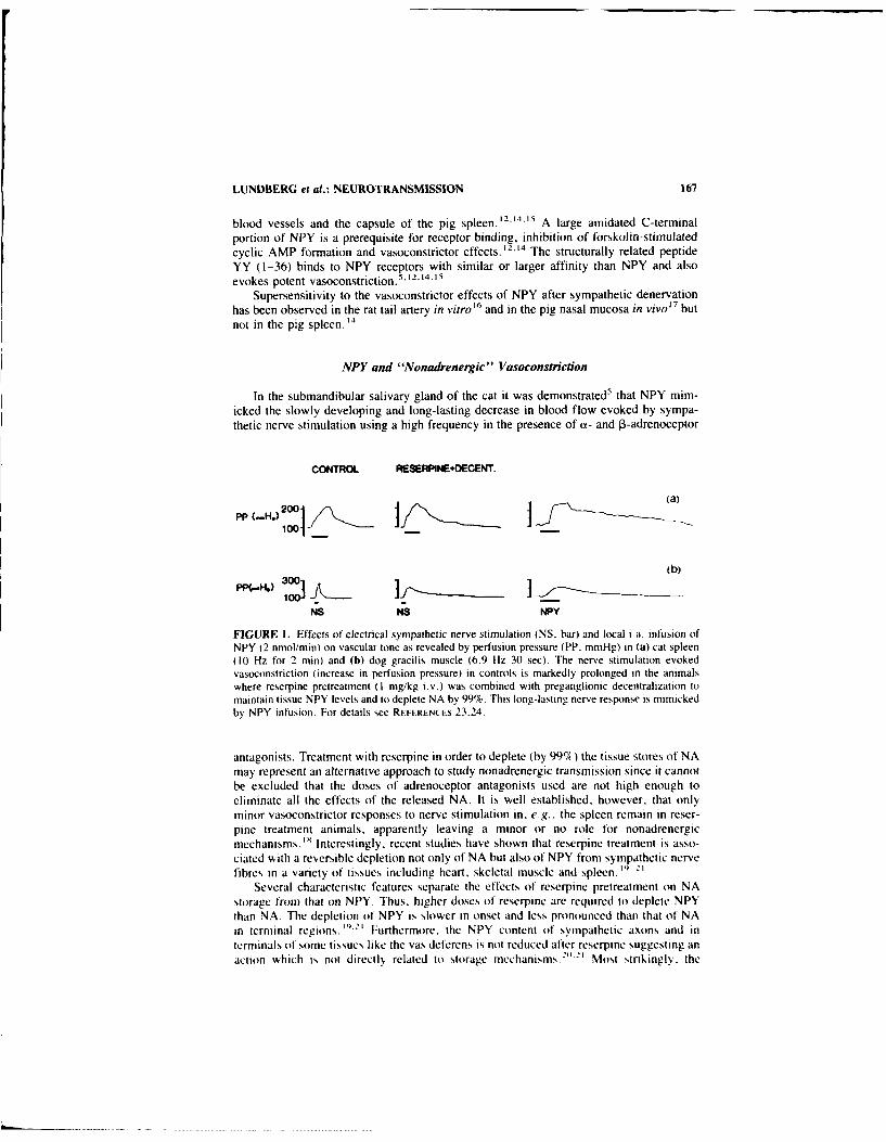
LUNDBERG et al.: NEUROTRANSMISSION 167
blood vessels and the capsule of the pig spleen. 12.14." ' A large amidated C-terminal
portion of NPY is a prerequisite for receptor binding, inhibition of forskolin-stimulatedcyclic AMP formation and vasoconstrictor effects. 12.14 The structurally related peptideYY (1-36) binds to NPY receptors with similar or larger affinity than NPY and alsoevokes potent vasoconstriction.
5' 12.14-15
Supersensitivity to the vasoconstrictor effects of NPY after sympathetic denervationhas been observed in the rat tail artery in vitro16 and in the pig nasal mucosa in vivo' 7 butnot in the pig spleen.'
4
NPY and "Nonadrenergic" Vasoconstriction
In the submandibular salivary gland of the cat it was demonstrated 5 that NPY mim-
icked the slowly developing and long-lasting decrease in blood flow evoked by sympa-thetic nerve stimulation using a high frequency in the presence of a- and P-adrenoceptor
CONTROL RESERPINE.DECENT.
PP -H. 101/._ jf __
tb)
NS NS NPY
FIGURE 1. Effects of electrical sympathetic nerve stimulation (NS. bar) and local ia. infusion ofNPY (2 nmol/min) on vascular tone as revealed by perfusion pressure (PP, mmHg) in (a) cat spleen(10 Hz for 2 min) and (b) dog gracilis muscle (6.9 Hz 30 sec). The nerve stimulation evokedvasoconstriction (increase in perfusion pressure) in controls is markedly prolonged in the animalswhere reserpine pretreatment (I mg/kg i.v.) was combined with preganglionic decentralization tomaintain tissue NPY levels and to deplete NA by 99%. This long-lasting nerve response is mimickedby NPY infusion. For details see REFERENCFS 23,24.
antagonists. Treatment with reserpine in order to deplete (by 99%) the tissue stores of NA
may represent an alternative approach to study nonadrenergic transmission since it cannotbe excluded that the doses of adrenoceptor antagonists used are not high enough toeliminate all the effects of the released NA. It is well established, however, that onlyminor vasoconstrictor responses to nerve stimulation in, e.g.. the spleen remain in reser-pine treatment animals, apparently leaving a minor or no role for nonadrenergicmechanisms. ' Interestingly, recent studies have shown that reserpine treatment is asso-ciated with a reversible depletion not only of NA but also of NPY from sympathetic nerve
fibres in a variety of tissues including heart, skeletal muscle and spleen. "' 2'Several characteristic features separate the effects of reserpine pretreatment on NA
storage from that on NPY. Thus. higher doses of reserpine are required to deplete NPY
than NA. The depletion of NPY is slower in onset and less pronounced than that of NAin terminal regions. " '
) Furthermore. the NPY content of sympathetic axons and interminals of some tissues like the vas deferens is not reduced after reserpine suggesting anaction which is not directly related to storage mechanisms. Most strikingly. the

168 ANNALS NEW YORK ACADEMY OF SCIENCES
reserpine-induced reduction of tissue NPY levels is, in contrast to the effect on NA,entirely dependent on an intact nerve activity since either pretreatment with a nicotinicreceptor blocking agent such as chlorisondamine, surgical transection at the pre- orpostganglionic levels or clonidine, which reduces sympathetic discharge, impairs thereserpine effect on NPY but not on NA levels. 21-29 The reserpine-induced depletion ofNPY is therefore likely to be caused by an increased release of the peptide in excess ofsynthesis and resupply capacity by axonal transport (see REF. 25). It is known thatreserpine pretreatment is associated with a rapid increase in firing rate of postganglionicsympathetic nerves .26 Treatment with reserpine can therefore be combined with pharma-cological agents or surgical procedures in order to prevent the increased neuronal activityto the organ studied which after 24 h results in a situation where NA is depleted by98-99%, while the tissue content of NPY is preserved. Stimulation of the sympatheticnerves in the spleen with high frequency then evoked a marked long-lasting vasocon-strictor response which was mimicked by the action of exogenous NPY 23 (FIG. 1). Thisexperimental approach has now been tested in a variety of preparations in vivo in additionto the cat spleen, 23 i.e., the pig spleen, 2' dog skeletal muscle,24 pig nasal mucosa" andpig kidney29 with similar results. The high correlation (r = 0.79-0.91) between thevasoconstrictor effects and the detectable overflow of NPY into the local venous effluent,the characteristic time course of the response being slowly developing and long-lastingand fatigue of the response upon repeated stimulation in these reserpinized preparationsfurther favour a peptide like NPY as the mediator. Although release of NPY seems to befacilitated by high frequency stimulation, NPY outflow is detected both from spleen 28 andkidney29 already at stimulation with a low frequency like 0.5 Hz. Furthermore, in reser-pinized pigs, nasal vasoconstriction is observed even in response to single impulses''suggesting that exocytosis of the NPY content from large dense cored vesicles can occuralso under such conditions although to a lesser extent than at high frequency stimulation.
Following reserpine treatment the levels of NPY in sympathetic ganglia are elevatedand an increased accumulation of peptide occurs above an axonal ligation suggesting bothenhanced synthesis and subsequent anterograde axonal transport. 20 Acccrdingly, recentdata have shown that expression of specific NPY messenger RNA in sympathetic ganglioncells is increased following reserpine administration. 30 The observation that subchronicpretreatment with chlc-'ondamine reduces the NPY content in sympathetic ganglioncells 22 further supports the concept of a nicotinic receptor-stimulated regulation of NPYsynthesis.
Co-Release of NPY and NA upon Sympathetic Activation
Release of NPY, as revealed by either overflow into the local venous effluent fromorgans of experimental animals 28 '29.31*32 or human heart33 as well as increases in sys-temic plasma levels in man 34.35.36 occurs upon sympathetic activation. The ratio betweenthe overflow of NPY and that of NA increases with the frequency of stimulation 2" and thecorrelation between the NPY and NA overflow represents a sigmoid curve, 37 suggestingthat NPY release is preferentially facilitated by high stimulation frequencies2-8 .37 (FIG. 2).The local plasma concentration of endogenous NPY-LI in the pig splenic or renal venouseffluent upon high frequency stimulation in reserpinized animals is in the nM range, i.e.,similar to where exogenous NPY evokes vasoconstriction. 6 .21,21 Since further character-ization by high performance liquid chromatography suggests that the NPY-LI detected bythe antiserum (NI) used represents NPY (1-36);2 this suggests that endogenous NPYconcentrations which are likely to be much higher close to release sites are more thansufficient to activate receptors on vascular smooth muscle cells and to contribute to thefunctional response. Upon prolonged stimulation (evoked by either increased sympathetic
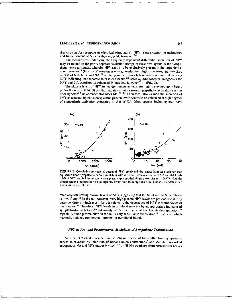
LUNDBERG et al.: NEUROTRANSMISSION Mr9
discharge as for reserpine or electrical stimulation), NPY release cannot be maintainedand tissue content of NPY is then reduced, however. 28
The mechanisms underlying the frequency-dependent differential secretion of NPYmay be related to the partly separate vesicular storage of these two agents in the sympa-thetic nerve terminals, whereby NPY seems to be exclusively present in the large dense-cored vesicles2 8 (FIG. 3). Pretreatment with guanethidine inhibits the stimulation-evokedrelease of both NPY and NA,3 ' while tyramine evokes NA secretion without influencingNPY indicating that separate release can occur.3 2 After a 2 -adrenoceptor antagonists theNPY and NA overflow is enhanced in parallel, however2 5 3 ' (FIG. 3).
The plasma levels of NPY in healthy human subjects are mainly elevated upon heavyphysical exercise (FIG. 3) or other situations with a strong sympathetic activation such asafter hypoxia3 3 or adrenoceptor blockade. 34 3 6 Therefore, also in man the secretion ofNPY as detected by elevated systemic plasma levels seems to be enhanced at high degreesof sympathetic activation compared to that of NA. Most species including man have
(a) (b)
200
.30- r=0.96 0 r=0.87 0
150
20 0 C.
S100-z10, 500=
0 00 OR
0000__ _ _ _ _ _
0 1oo 2000 3000 0 10 20 30 40NA (pmol) NA (nM)
FIGURE 2. Correlation between (a) output of NPY (pmol) and NA (pmol) from the blood perfusedpig spleen upon sympathetic nerve stimulation with different frequencies (r = 0.96) and (b) levels(pM) of NPY and NA in human venous plasma upon graded physical exercise (r = 0.87). Note thesimilar relative increase in NPY at high NA levels both from pig spleen and humans. For details seeREFERENCES 28, 34, 36.
relatively low resting plasma levels of NPY suggesting that the basal rate ot NPY releaseis low, if any. 2 5 In the rat, however, very high plasma NPY levels are present also duringbasal conditions which most likely is related to the occurrence of NPY in trombocytes ofthis species. 3
" Therefore, NPY levels in rat blood may not b! an appropriate indicator ofsympathoadrenal activity 2
' but mainly reflect the degree of trombocyte degranulation, 9
especially since plasma NPY in the rat is very sensitive to vinblastine40 treatment, whichmarkedly reduces trombocyte numbers in peripheral blood.
NPY as Pre- and Postjunctional Modulator of Sympathetic Transmission
NPY or PYY exerts prejunctional actions on release of transmitter from sympatheticnerves as revealed by inhibition of nerve-evoked contractions' and stimulation-evokedendogenous NA and NPY output in Vivo
4' .42 or 3H-NA overflow from perivascular nerves

170 ANNALS NEW YORK ACADEMY OF SCIENCES
of rat and man in vitro1°'43 (FIG. 3). Experimental data studying contractile responses oranalysis of transmitter overflow suggest that NPY also inhibits the release of both ace-tylcholine and NA in the heart. 4 4 6
In the mouse vas deferens NPY induced a potentiation of the contractile effects of NAand adenosine-5-triphosphate (ATP) .47 Furthermore, NPY reduced the stimulation-
+ Release
2
NicotineI ~ NPY2
ATII 42NA NPY
Beta 1 NPY1
FIGURE 3. Schematical illustration of a varicosity uf a sympathetic nerve containing both NA andNPY in large dense-cored vesiclcs and only NA in the small vesicles. At low frequency stimulationmainly small vesicles secrete their content of NA acting on postjunctional an-adrenoceptors to evokevasocunstriction or 0,-adrenoceptors to increase cardiac contractility. NA also activates prejunctionalo,-adrenoceptors inhibiting release of itself and of NPY from the large vesicles. NPY is secretedupon stronger stimulation and induces vasoconstriction as well as inhibits release of NA and itself.There is some evidence that the vasoconstriction and the prejunctional effect of NPY is mediated viaseparate receptor mechanisms (designed NPY- J and NPY-2, respectively). Nicotine increases basalNA and NPY secretion while angiotensin II (AT I) facilitates nerve stimulation-evoked release ofNA and NPY. Due to the complex interplay between NA and NPY both at the pre- and postjunctionallevels a variety of drugs which interfere with NA mechanisms will also change especially NPYrelease.
evoked contraction and secretion of 3H-NA and selectively depressed the stih,ulus evokedbut not the spontaneously occurring excitatory junction potentials in smooth muscle cells.Possibly this action of NPY, similarly to o,-agonists is dependent on an inhibitory effectin part via a target upstream of the varicosities.
NPY not only evokes vasoconstriction per se but also enhances the contractions of

LUNDBERG et al.: NEUROTRANSMISSION 171
blood vessels to a variety of agents including NA in vitro. 4 43 The enhancing effect ofNPY is not unique but shared by other vasoconstrictors like serotonin and characterizedby being more pronounced on larger vessels where NPY exerts minor or no contractionsper se.
NPY Release in Human Cardiovascular Disease
The re!ease of NiY upon sympathetic activation in healthy volunteers as revealed byincrease in systemic plasma levels
3 4 "35 or elevated levels in the coronary sinus uponhypoxia 33 combined with the potent, long-lasting vasoconstrictor effects of NPY in man7
raise the possibility that NPY may be involved in the pathophysiology of human cardio-vascular disorders. NPY is a potent constrictor of small human coronary vessels in vitro,49
and elevated plasma levels of NPY are present in patients with heart disease such as acutemyocardial infarction, angina pectoris and especially severe left heart failure. 5" Thepossible functional role of NPY under physiological conditions with intact noradrenergicmechanisms, as well as involvement in human vascular disorders, remains to be estab-lished with the use of specific NPY antagonists at the pre- and postjunctional levels. Itseems clear, however, that many drugs commonly used in experimental studies on car-diovascular control or in treatment of hypertensive or vasopastic disorders in humans alsoinfluence NPY mechanisms.
SUMMARY
The coexistence of neuropeptide Y (NPY) with noradrenaline (NA) in perivascularnerves as well as in sympathetic nerves to muscle in the heart, spleen and vas deferenssuggests a role for NPY in autonomic transmission. Sympathetic nerve stimulation orreflexogenic activation in experimental animals or man are associated with NPY releaseas revealed by overflow mainly upon strong activation. This difference between NPY andNA secretion may be related to the partly separate subcellular storage whereby NPYseems to be exclusively present in the large dense-cored vesicles. The NPY secretion islikely to be regulated by the local biophase cuncentrations of NA acting on prejunctionalalpha-2-adrenoceptors since alpha-2 agonists inhibit and antagonists enhance NPY over-flow, respectively. Furthermore. alter NA has been depleted by reserpine. the nervestimulation-evoked release of NPY is enhanced leading to a progressive depletion oftissue content of NPY. Exogenous NPY binds to both pre- and postjunctional receptors,inhibits NA and NPY release, enhances NA-evoked vasoconstriction and induces vaso-constriction per se. The prejunctional action of NPY which is especially noticeable in thevas deferens may serve to reduce transmitter secretion upon excessive stimulation. Thelong-lasting vasoconstriction evoked by sympathetic stimulation in several tissues includ-ing skeletal muscle, nasal mucosa and spleen, which remains in animals pretreated withreserpine (to deplete NA) combined with preganglionic denervation (tt prevent the con-comitant excessive NPY release and depletion), is mimicked by NPY and highly corre-lated to NPY release. Under these circumstances the NPY content in the local venouseffluent reaches levels at which exogenous NPY evokes vasoconstriction.
REFERENCES
I. TA rrMoro. K. 1982. Neuropeptide Y: the complete amino-acid sequence of the brain peptide.Proc. Nail. Acad. Sci. USA 79: 5485-5489.

172 ANNALS NEW YORK ACADEMY OF SCIENCES
2. LUNDBERG. J. M.. L. TERENIus, T. HOKFELT & K. TATEMOTO. 1984. Comparative immu-nohistochemical and biochemical analysis of pancreatic polypeptide-like peptides with specialreference to presence of' neuropeptide Y in central and peripheral neurons. J. Neurosci.45: 2376-2386.
3. LUNDBERG, J. M., L. TERENUS.T1. HOKFEI-T, C-R. MARTL.ING, K. TATEMOTO, V. MUTT, J.POLAK. S. R. BLOOM & M. GOLDSTEIN. 1982. Neuropeptide Y (NPY)-like imrounoreactivityin peripheral noradrenergic neurons and cffects of NPY on sympathetic function. Acta Phys-lol. Scand. 116: 477-480
4. EKBLAD, E., L. EDvINSSON. C. WAHLESTEDT. R. UDDMAN. R. HkKANSSON & F. SUNDLER.1984. Neuropeptidle Y co-exists and co-operates with noradrenaline in perivascular nervefibres. Regul. Pept. 8: 225-235.
5. LUNDBERG. J. M. & K. TAKEMOTO. 1981. Pancreatic polypeptidle family (APP, BPP. NPY andPYY) in relation to alpha-adreno~ceptor-resistant sympathetic vasoconstriction. Acta Physiol.Scand. 116: 393-402.
6. RUDEHILIJ. A., M. OL-Cf.N. A. SOLLEVI. B. H-AMBERGER & J. M. LUNDBERG. 1987. Releaseof neuropeptide Y upon hemorrhagic hypovolemia in relation to vasoconstrictor effects in thepig. Acta Physiol. Scand. 131: 5 17-523.
7. PERNOW. J.. J. M . LUNDBERG & L. KAKJSER. 1987. Vasoconstfictor effects in vivo and plasmadisappearance rate of neuropeptide Y in man. Life Sci. 40: 47-54.
8. EDviNSSON. L.. P.C. EmSON. J. MCCULt.OCti K. TATEMOTO & R. UDDMAN. 1983. Neuro-peptide Y. cerebrovascular innervation and vasomotor effects in the cat. Neurosci. Lett.43: 79-84.
9. PERNOW. J. & J, M. LUNDBRGR. 1988- Neuropeptide Y induces potent contraction of arterialvascular smooth muscle via an endothelium-independent mechanism. Acta. Physiol, Scand.134: 157-158.
10. PERNOW. V. T. SVENBERG & J1. M. LUNDBERG. 1987. Actions of calcium antagonists on pre-and postjunctional effects of neuropeptide Y on human peripheral blood vessels in vitro. Eur.J. Pharmacol. 136: 207-218.
11, LACROIX. J. S., P. STJARNE. A. ANGoARD & 1. M. LUNDBERG. 1988. Sympathetic vascularcontrol of the pig nasal mucosa. (11: reserpine-resistant. non-adrenergic nervous responses inrelation to neuropeptide Y and ATP, Acta Physiol. Scand. 133: 183-197.
12. LUNDBERG. J. M.. A. HFMSf.N. 0. LARSSON. A. RUDEHILL. A. SARIA & B. B. FREDHOI.M.1988. Neuropeptide Y receptor in pig spleen: binding characteristics, reduction of cyclicAMP formation and calcium antagonist inhibition of vasoconstriction. Eur. J. Pharmacol.145: 21-29.
13. MILLAR. B. C., H. MI. PIPER & B. J. McDERMO I . 1988. The antiadrenergic effect ofneuropeptide Y on the ventricular cardiomyocyte. Naunyn-Schniiedeberg's Arch. Pharmacol.338: 426-429.
14. LUNDBERG. J. M.. A. HEMSf.N. A. RUDEHIt I-, A. HARFSTRAND. 0. LARSSON. A. SOLL-Evi. A.SARIA. T. HOKEELT. K. FUXE & B. B. FREDHOI.M. 1988. Neuropeptide Y- and alpha-adrenergic receptors in pig spleen: localization, binding characteristics, cyclic AMP effectsand functional response in control and denervated animals. Neuroscience 24: 659-672.
15. MAcKERREI. A. D. JR., A. HEMSfN. J. LAxCROIX & J. M. LUNDBERG. 1989. Analysis ofstructure-function relationships of neuropeptide Y using molecular dynamics simulations andpharmacological activity and binding measurements. Regul. Pept. 25: 295-318.
16. NEILD, T. 0. 1987. Actious of neuropeptide Y (in innervated and denervated rat tail arteries.J. Physiol. 3%6: 19-30.
17. LACROIX, J. S. & k. M. LUNDBERG. 1989. Adrenergic and neuropeptide Y supersensitivity indenervated nasal mucosa vasculature of the pig. Europ. J. Pharmacol. 169: 125-136,
18. CUBEDDI . L. V. E. BARNES. S. Z. LANGIR & N. WFtNFR. 1974. Release of norepinephrineand dopamine-p3-hydroxylase by nerve stimulation. 1. Role of neuronal and extraneuronaluptakc and of alpha-presynaptic receptors. J. Pharmacol. Exp. Ther. 190: 431-450.
19. 111NDBER(;. J. M., A. SARtA. A. ANG6O IRD, 1'. H(WFEtLr & L. TERENIUS. 1984. NeuropeptideY and noradrenalinc interact1ion in peripheral cardiovascular control. Clin. Exp. Theory Pract.A6-. 1901-1972,
20. L(tNDBI.RG. J. M., A. SAsRIA. A. I2RANCO-CERIUE DA, T. HOKtti.l1. L. TERI NIU.S & MI. GOi D-StIEIN 1995. Differential effects of reserpine and 6-hydroxvodopansine on neuropeptide Y and
LUNDBERG et al.: NEUROTRANSMISSION 173
noradrenaline in peripheral neurons. Naunyn-Schmiedeberg's Arch. Pharmacol. 328: 331-340.
21. NAGATA. M., A. FRANCO-CERECEDA. A. SARIA. R. AMMAN & J. M. LuNDBERG. 1987.Reserpine-induced depletion of neuropeptide Y in the guinea-pig: tissue-specific effects andmechanism of action. J. Aut. Nerv. Syst. 20: 257-263.
22. LUNDBERG. J. M., A. SARIA, A. FRANCO-CERECEDA & E. THEODORSSON-NORIIEIM. 1985.Treatment with sympatholytic drugs changes tissue content of neuropeptide Y in cardiovas-cular nerves and adrenal gland. Acta Physiol. Scand. 124: 603-611.
23. LUNDBERG. J. M., G. FRIED, J. PERNOW. E. THEODORSSON-NORHEIM & A. ANG~aRD. 1986.NPY-a mediator of reserpine-resistant. non-adrenergic vasoconstriction in cat spleen afterpreganglicnic denervation? Acta Physiol. Scand. 126: 151-152.
24. PERNOW. J., T. K,,.!IN & J. M. LUNDBERG. 1988. Neuropeptide Y and reserpine-resistantvasoconstriction evoked by sympathetic ne've stimulation in dog skeletal muscle. Br. J.Pharmacol. 94: 952-960.
25. LUNDBERG. J. M.. J. PERNOW. A. FRANcO-CZFECEDA & A. RUDEHILL. 1987. Effects ofantihypertensive drugs on sympathetic vascular control in relation to neuropeptide Y. J.Cardiovasc. Pharmacol. 12 (Suppl. 10) 551-56h.
26. PERNOW, J., P. THOR N, B-I. MIt.[BERG & J. M. LUNDBERG. 1988. Renal sympathetic nerveactivation in relation to reserpi ne- induced depletion (if neuropeptide Y in the kidney of the rat.Acta Physiol. Scand. 134: 53-59-
27. FRANCO-CERECEDA. A.. M. NAGATA, T. H. SVENSSON & J. M. LUNDBERG,. 1987. Differentialeffects of clonidine and reserpine treatment on neuropeptide Y content in some sympatheti-cally innervated tissues of' the guinea-pig. Eur. J. Pharmacol. 142: 267-273.
28. LUNDBERG. J. M., A. RUDEHILI., A. Sot.tEvt. G. FRIED & G. WALItiN. 1989. Co-release ofneuropeptide Y and noradrenaline from pig spleen in vivo: importance of subcellular storage.nerve impulse frequency and pattern, feedback regulation and resupply by axonal transport.Neuroscience 28: 475-486.
29. PERNOW. J. & J. M. LUNDBERG. 1989. Release and vasoconstrictor effects of neuropeptide Yin relation to not--adrenergic sympathetic control of renal blood flow in the pig. Acta Physiol.Scand. 136: 507-5 17.
30. SCHAILING. M.. A. FRANCO-CERFCEDA. T. HOKFEFT H. PERSSON & J. M. LUINDBERG;. 1988.Increased neuropeptide Y messenger RNA and peptide in sympathetic ganglia after reserpinepretreatment. Eur. J. Phar-nacol, 156: 419-420.
1' LUNDBERG. J. M., A. ANG(iaRD. E. THEODORSSON-NORHEIM & J. PERNOW'. 1984. Guanethi-dine-sensitive release of NPY-like iii-;,nunoreactivity by sympathetic nerve stimulation. Neu-rose,. Leti. 52: 175-180.
32. LUNDBERG, J. M.. A. RijoEtlit., A. Soi~iEvI & B. HAMBERGER. 1989. Evidence for cotrans-mitter role of neuropeptide Y in pig spleen. Br. J. Pharmacol. 96: 675-687.
33. KAIJSER. L.. - . PERNOW. B. BERGIUND & J. M. LUNDBERG. 1990. Neuropeptide Y is releasedtogether with noradrenaline from the human heart during exercise and hypoxia. Clin. Physiol.10: 179-188.
34. LUNDBERG. J. M4., A. MARTINSSON. A. HEmsf.N, E. THEoUXoRSSON-NORHEIM, J. .S% DENHAG.E. EKBt.OM & P. HJEMDAHI.. 1985. Co-release of neuropeptide Y and catecholamines duringPhysical exercise in man. Giochem. Biophys. Res. Commun. 133: 30-36.
35. PERNOW. J., J. M. LUNDBERG. L. KAIJSER. P. HJEMDAHI.. E. THEOD'wSSON-NORHIM. E.MARI INSSON & B. PERNOW. 1986. Plasma neuropeptide Y-like inimunoreactivity and cate-cholamines during various degrees of'sympathetic activation in man. Clii. PhN &oL. 6: 561.-578.
36. PFRNOW. V. J. M. LLNDBER6 & L. KAIJSER. 1988. a-Adrenocepior influence oi plasmalevels of neuropeptide Y-like immunoreactivity and catecholamines during rest and Synli-pathoadrenal activation in humans. J. Cardiovasc. Pharniacol. 12; 593-599.
37 LUNDBERG, J. M.. J. PvERNow & J. S. LA( ROIX. 1989. Neuropeptide Y: s~Nipathetic cotrans-miller and modulator? NIPS 4: 13-17.
38. FRKSSON. A.. M. So HAI i~tNG. K. M( IN IYRtE. J. M. LuNDBEIRG. 1). LARHAMMAR. K. SiRo-(NiY. K. HOKEEI T & H. PE-RSSON. 1987. Detection of neuropeptide Y aind its, mRNA inmeg.- 'arvocytes: enhanced levels in certain autoirumune mice. Proc. Nail Acad. Sci. UiSA84. 5585-559(X)

174 ANNALS NEW YORK ACADEMY OF SCIENCES
39. MYExs, K. M., M. Y. FARKAT, C. A. VAZ, I. R. KEISE. & Z. ZUKOWSKA-GROuC. 1988.Release of immunoreactive neuropeptide Y by rat platelets. Biochem. Biophys. Res. Comm.155: 118-122.
40. HEMStN, A., M. STENSDOTTER, C., SODERBLOM & J. M. LUNDBERG. 1989. Effects of vin-blastine, reserpine and capsaicin on neuropeptide Y in the sympathoadrenal system andtrombocytes of the rat. XXXI IUPS. Helsinki. P4297.
41. PERNOW, J. & J. M. LUNDBERG. 1989. Modulation of noradrenaline and neuropeptide Y(NPY) release in the pig kidney in vivo: involvement of alpha2, NPY and angiotensin Itreceptors. Naunyn-Schmiedeberg's Arch. Pharmacol. 340: 379-385.
42. LUNDBERG, J. M., A. RUDEHILL & A. SOLLEVI. 1989. Pharmacological characterization ofneuropeptide Y and noradrenaline mechanisms in sympathetic control of pig spleen. Eur. J.Pharmacol. 163: 103-113.
43. PERNOW, J., A. SARIA & J. M. LUNDBERG. 1986. Mechanisms underlying pre- and postjunc-tional effects of neuropeptide Y in sympathetic vascular control. Acta Physiol. Scand.126: 239-249.
44. FRANCO-CERECEDA, A., J. M. LUNDBERG & C. DAHLOF. 1985. Neuropeptide Y and sympa-thetic contiol of heart contractility and coronary vascular tone. Acta Physiol. Scand.129: 361-370.
45. LUNDBERG, J. M., X-Y. HUA & A. FRANCO-CERECEDA. 1984. Effects of neuropeptide Y(NPY) on mechanical activity and neurotransmission in the heart, vas deferens and urinirybladder of the guinea pig. Acta Physiol. Scand. 121: 325-332.
46. POTTER, E. K. 1985. Prolonged non-adrenergic inhibition of cardiac vagal action followingsympathetic stimulation: neuromodulation by neuropeptide Y? Neurosci. Lett. 54: 117-121.
47. STJARNE, L., J. M. LUNDBERG & P, ASTRAND. 1986. Neuropeptide Y-a cotransmitter withnoradrenaline and adenosine-5-triphosphate in the sympathetic nerves of the mouse vas def-erens? A biochemical, physiological and electropharmacological study. Neuroscience18: 151-166.
48. STJARNE, L., E. STJARNE, M. MSHGINA & J. M. LUNDBERG. 1988. Neuropeptide Y mayinhibit sympathetic transmitter secretion by an effect upstream of varicosities. Acta PhysiolScand. 134: 577-578.
49. FRANCO-CERECEDA, A. & J. M. LUNDBERG. 1987. Potent effects of neuropeptide Y andcalcitonin gene-related peptide on human coronary vascular tone in vitro. Acta Physiol.scand. 131: 159-160.
50. HULTING, J., A. SOLLEVI, B. ULLMAN, A. FRANCO-CERECEDA & J. M. LUNDBERG. 1990.Plasma neuropeptide Y on admission to a coronary care unit. Elevated NPY levels in asubgroup of patients with heart failure. Cardiovasc. Res. 24: 102-108.

Regulation of Neuropeptide Y GeneExpression in Rat Braina
NILS LINDEFORS,b STEFAN BRENE,' MARIO
HERRERA-MARSCHITZ,b AND HAKAN PERSSON"
bDepartment of Pharmacology
Karolinska Institutetand
"Department of Medical ChemistryLaborator, of Molecular Neurobiology
P.O. Box 60 400S-104 01 Stockholm, Sweden
INTRODUCTION
Neuropeptide Y (NPY) is found in several rat brain regions including caudate-putamen and cerebral cortex. '. 24 NPY-like immunoreactivity in rat brain appears as onesingle 36-amino acid peptide.' Both complementary DNA (cDNA) and genomic cloneshave been isolated for rat NPY and used to determine the regional distribution of NPYmRNA in the rat brain. 2
.13 '1 9 NPY mRNA containing cellbodies in rat brain has also been
demonstrated using in situ hybridization. 7 2 1
In this report in situ hybridization and RNA blot analysis of NPY mRNA expressiond
in rat brain will be discussed a.id the findings compared with NPY mRNA expression inthe human brain. Transsynaptic regulation of rat NPY mRNA expression by midbraindopamine neurons is described using a unilateral 6-hydroxydopamine-induced dopaminedeafferentation of the ipsilateral forebrain. For comparison, the effects of a dopaminedeafferentation on preprosomatostatin (SOM), preprocholecystokinin (CCK), prepro-tachykinin (PPT) and glutamic acid decarboxylase (GAD a marker of -y-amino buturicacid (GABA) neurons) mRNA expression, have also been studied. The results show thatmidbrain dopamine neurons differentially control expression of NPY as well as SOM,PPT, and GAD mRNA expression in a complex fashion.
METHODOLOGICAL ASPECTS
Male Sprague-Dawley rats (b.w. 150 g) were used, either naive or unilaterally in-jected with 4 lil of 6-hydroxydopamine (2 mg/ml in 0.02% ascorbic acid in saline) orvehicle alone into the left ventral tegmental area containing the axons leaving the midbraindopamine neurons. ' A unilateral lesion of ascending dopamine neurons by 6-hydroxy-dopamine injection in the ventral tegmental area induced dopamine deafferentation of
"This work was supported by grants from the Swedish Medical Research Council (grant no. 8653)and from the Swedish Natural Scier.ce Research Council.
'For simplicity, the level of different mRNAs seen after in situ hybridization histochemistry andRNA blot analysis is referred to as the level of mRNA expression. However, it should be pointed outthat both techniques measure the steady-state level of mRNA, which may not directly reflect the levelof mRNA expression, due to the possible presence of posttranscriptional controls
175

176 ANNALS NEW YORK ACADEMY OF SCIENCES
several ipsilateral brain regions including caudate-putamen' and frontoparietal coLcA. '2The elimination of tyrosine hydroxylase (TH) mRNA hybridization in ventral tegmentalarea and substantia nigra ipsilateral to the 6-hydroxydopamine injection were used ascontrol of successful lesions.
In Situ Hybridization
After cryostat sectioning, the tissues were fixed in 10% formalin in phosphate buff-ered saline for 30 min, rinsed twice for 4 min in PBS, and delipidated in graded series ofethanol, including a 5-min incubation with chloroform. The sections were then air-dried.The hybridization cocktail contained 50% formamide, 4 x SSC (I X SSC is 0.15 MNaCI, 0.015 M sodium citrate pH 7.0), 1 x Denhardts solution, 1% Sarcosyl, 0.02MNaPO 4 (pH 7.0), 10% dextransulphate 0.5 mg/ml yeast tRNA, 0.06M D'TT and 0.1mg/ml sheared salmon sperm DNA. For NPY mRNA hybridization. a 44-mer oligonu-cleotide complementary to rat NPY mRNA encoding amino acid 15 to 29 of the matureNPY peptide was used. '9 For PPT mRNA hybridization, a 48-mer oligonucleotide com-plementary to rat PPT mRNA coding for amino acid 49 to 64, and thus complementaryto a. P and -y-PPT mRNA was used. ' For SOM mRNA hybridization, a 46-mer olig-onucleotide complementary to rat SOM mRNA coding for amino acid 101 to 115 wasused.' For CCK mRNA hybridization, a 44-mer oligonucleotide complementary to ratCCK mRNA coding for amino acid 89 to 103 was used.5 For GAD mRNA hybridizationa 48-mer oligonucleotide complementary to feline GAD mRNA coding for amino acid389 to 405 was used. 14 For TH mRNA a 48 -met oligonucleotide complementary to rat THmRNA encoding amino acid 445 to 492 of the TH polypeptide was used. 9 The oligoiu-cleotides were 3'-end labeled with cx-35S-dATP using terminal deoxyribonucleotidyltransferase (International Biotech. Inc., New Haven, CT) to a specific activity of ap-proximately 5 x 10⁸ cpm/mg. The labelled probes were purified on a Nensorb column(DuPont, Wilmington, DE) prior to use. Following hybridization for 16 h at 42°C, thesections were rinsed 5 times for 15 min in I x SSC at 55°C. Finally, the sections wererinsed in autoclaved water for 5 min, and then dehydrated through a series of gradedalcohol and air-dried. The sections were exposed to X-ray film (Amersham P-max) for7-25 days and to photographic emulsion (Kodak NTB2) for 10-28 days. After havingbeen developed, the slides were stained with cresyl violet followed by microscopic anal-ysis. As a control for the specificity of the hybridization, we performed in situ hybrid-ization with eDNA probes which revealed a similar hybridization pattern as the onedescribed here. In addition, in caudate-putamen and frontoparietal cortex incubation withthe TH oligonucleotide probe served as a negative control.
Neuron Counting and Computerized Image Analysis
The numerical density of hybridization positive neurons was analyzed using a LeitzOrtholux microscope at 313 x magnification. Hybridization positive neurons were de-fined as those expressing more than 10 grains per neuron (well above background densityof grains). Tht number of grains per neuron and the size of NPY. SOM, CCK and PPTmRNA positive neurons were calculated using a Crystal image analysis processor(Quantel Ltd. UK). For this purpose. the microscope was equipped with a Panasonic WV1500 TV camera to provide a video signal for the image ana'ysis. However, over someneurons analyzed the high density of grains made a direct numerical count futile. Instead,the total area of grains, also including all fused gra ,s was divided with a standard grainarea obtained from measurements of unfused grains over neurons. The area of a standard
4

LINLoEFORS et al.: NPY GENE EXPRESSION 177
grain was arbitrarily defined as the mean area of a few small grains measured over ahybridization positive cell.
Northern Blot Analysis
Brains from one group of animals were dissected directly after decapitation and usedto prepare RNA from frontoparietal cortex and caudate-putamen. The tissue was dissectedto fit with that used for in situ hybridization. Dissected brain tissue from fronto-parietalcortex and caudate-putamen were homogenized in 4 M guanidine isothiocyanate, 0.1 M1-mercaptoethanol, 0.025 M sodium citrate, pH 7.0 and analyzed for the level of NPYmRNA by RNA blot analysis. The homogenate was mounted on top of 4 ml cushion of5.7 M CsCI in 0.025 M sodium citrate at pH 5.5 and centrifuged at 15'C in a BeckmanSW41 rotor for 16 to 24 hours. The indicated amounts of total or polyadenylated RNAwere separated on 1% agarose gels containing 0.7% formaldehyde followed by transfer tonitrocellulose filters. The filters were then hybridized to nick-translated cDNA probeslabelled with Ct- 32P-dCTP to a specific activity of approximately 5 X 10⁸ cpm/mg TheNPY cDNA prob used was a 287 bp Xba -Ava I fragment containing the second exon ofthe NPY gene. " The filters were washed at high stringency (0.1 x SSC. 0.1% SDS at54C) and exposed to Kodak XAR-5 films. The same filters as used for NPY hybridiza-tion, were boiled for 5 min in 1% glycerol and rehybridized to a nick-translated 1.5 kbPstl fragment from a mouse a-actin cDNA clone. 23 Appropriate exposures, i.e., expo-sures that allowed measurements of the hybridization signal in a linear interval of allautoradiograms were quantified using a Shimadzu CS-9000 densitometer. The levels ofNPY mRNA were then normalized to the levels of actin mRNA.
RESULTS
NPY mRNA Expression in Control and Dopamine DeafferentatedCaudate-Putamen and Frontoparietal Cortex
NPY mRNA hybridizations2 ' revealed a specific labeling in several discrete brainregions, including septal nuclei, basal forebrain and hypothalamic nuclei (FIG. 1). Mi-croscopic examination of emulsion dipped tissue sections showed dense clusters of grainsover neurons throughout caudate-putamen, nucleus accumbens and cerebral cortex (FIG.2). Less than 5% of neurons were positive in these regions. The labeling was present overall cortical layers, with a concentration over layers V and VI. A two-times higher nu-merical density of NPY mRNA positive neurons was seen in frontoparietal cortex com-pared with caudate-putamen. Furthermore, the number of grains per NPY mRNA positiveneuron in frontoparietal cortex was 47% higher than in caudate-putamen.
In the dopamine deafferentated side the numerical density of NPY mRNA positiveneurons in caudate-putamen increased approximately two-fold compared with the intact,contralateral caudate-putamen. The number of grains per NPY mRNA positive neuron incaudate-putamen was, however, not changed when comparing the deafferentated caudate-putamen with the contralateral caudate-putamen. In the dopamine deafferented frontopa-rietal cortex, the numerical density of NPY mRNA positive neurons was less than half ofthat in the intact side. The number of grains per NPY mRNA positive neuron in fronto-parietal cortex was, as in caudate-putamen. not changed when the two sides were com-pared.
RNA blot analysis showed the presence of a single 0.8 kb NPY inRNA in caudate-

178 ANNALS NEW YORK ACADEMY OF SCIENCES
putamen and frontoparietal cortex (FiG. 3). Furthermore. the RNA blot analysis, origi-nally reported here, showed an increase with 62% in the level of NPY mRNA in thedopamine deafferented caudate-putamen and a decrease with 79% in the deafferentedfrontoparietal cortex (FIG. 3) compared with corresponding regions in sham-operatedanimals. The results from the RNA blots are in agreement with the in situ hybridizationdata,
FIGURE !. Upper left panel shows a low magnification dark-field photomicrograph of NPY mRNAin situ hybridization histochemistry in caudate-putamen and frontoparietal cortex, after a unilateral6-hydroxydopamine-induced lesion of midbrain dopamine neurons. The tissue was hybridized to aoligonucleotide specific for rat NPY mRNA and exposed to a photographic film. PPT, GAD andCCK mRNA hybridizations from adjacent tissue sections are shown for comparison. Abbreviations:fpc = frontoparietal cortex, c-p = caudate-putamen. CO = contralateral side, IP = ipsilateral side.(Modified from REERENC .S 20 and 21.)
SOM, CCK, PPT and GAD mRNA Expression in Control and DopamineDeafferentated Caudate-Putamen and Frontoparietal Cortex
SOM mRNA hybridizations?' showed a specific labeling with a distribution verysimilar to that of NPY. However, in contrast to NPY the number of grains per SOMmRNA positive neuron in frontoparietal cortex was similar to that in caudate-putamen.The regulation of SOM mRNA expression was in most cases similarly affected by thedopamine deafferentation as in the case of NPY mRNA expression. However, in contrastto NPY, the grain density over SOM mRNA positive neurons decreased (24%) in fron-toparietal cortex, while the numerical density of SOM mRNA positive neurons wasunchanged.

LINDEFORS et al.: NPY GENE EXPRESSION 179
~$ bI 4--
W '0
C-P, fp PC
FIGURE 2. High magnification bright-field emulsion photomicrographs of NPY mRNA positive
neurons in rat frontoparietal cortex and caudate-putamen. The neurons shown are representative, and
selected from the same section as shown in the upper left panel of FIGURE I. Note that the cortical
neuron is larger and more intensely labeled. Abbreviations as in FIGURE I.
C-P fpc FIGURE 3. Expression of NPY mRNA incaudate-putamen and frontoparietal cortex (ip-
C C silateral to injections) in 6-hydroxydopamine-E 0 2 0 injected and sham-injected animals. Total RNA
S "0a " (35 iLg/slot) from a pool of 3 to 5 animals was
@1 - CD a electrophoresed in a formaldehyde containing
agarose gel followed by transfer to nitrocellu-lose filters. The filters were hybridized to a
. -2 P-labelled 287 bp Xba l-Av I cDNA probe
for rat NPY mRNA. The filters were washed athigh stringency followed by autoradiography.
0.8kb- _i _ Note the increased level of NPY mRNA in- , ipsilateral caudate-putamen and the decreased
level in ipsilateral frontoparietal cortex of le-
i sioned animals. All values determined from thefilms were normalized to the level of actin.Abbreviations as in FIGURE 1.

180 ANNALS NEW YORK ACADEMY OF SCIENCES
CCK mRNA hybridizations21 (FIG. 1) showed an intense two-layer labeling in thecerebral cortex with much higher numerical density of labeled neurons than after hybrid-ization to NPY or SOM probes. However, no hybridization signal was detected in eitherthe lesioned or the intact caudate-putamen. The numerical density of CCK mRNA pos-itive neurons in frontoparietal cortex, as well as the number of grains over CCK mRNApositive neurons, were not changed after the dopamine deafferentation.
PPT mRNA hybridizations 2' (Fi,. 1) showed an apparent homogenous labeling incaudate-putamen and nucleus accumbens with a few intense dots. Microscopic examina-tion of the sections showed that the apparent homogeneous signal consisted of manyrelatively weakly labeled neurons, while the intense dots represented sparsely distributed.intensively labeled neurons. 40 to 60% of all neurons were PPT mRNA positive incaudate-putamen. In the cerebral cortex the PPT probe showed labeling only over veryfew cells. The numerical density of PPT mRNA positive neurons in the dopamine deaf-ferented caudate-putamen decreased to one fifth (fivefold) compared with the intact cau-date-putamen. Furthermore, the number of grains over PPT mRNA positive neurons inthe deafferented caudate-putamcn decreased by one half compared with the intact caudate-putamen.
GAD mRNA hybridizations2 ' (Fto. I) revealed two populations of GAD mRNApositive neurons in caudate-putamen. The majority of neurons in this region were GADmRNA positive and of the hybridization positive neurons the majority showed a weaklabeling, while only less than 10% of GAD mRNA positive neurons showed intenselabeling. The numerical density of GAD mRNA hybridization positive neurons was lowerin frontoparietal cortex but the intensity of hybridization signal (grain density) was higherover individual cortical neurons compared to that in caudate-putamen. The dopaminedeafferentation of caudate-putamen induced an increase in the number of intensivelylabelled neurons with 46% as compared with the unlesioned side. However, the totalnumber of positive neurons was similar in both sides. The number of grains per neuronover the intensely labelled neurons was similar in both sides. In frontoparietal cortex, thedopamine deafferentation reduced the number of neurons expressing high levels of GADmRNA to less than half the number seen in the intact side. The remaining highly ex-pressing neurons showed, however, similar amounts of grains per neuron as the intenselylabelled neurons in the intact side. Thus, GAD mRNA expression was changed in similardirections as NPY mRNA expression, by the dopamine deafferentation, both in caudate-putamen and in frontoparietal cortex. Furthermore. similar to NPY mRNA, GAD mRNAexpression seemed to be affected in a subpopulation of hybridization positive neurons inboth caudate-putamen and frontoparietal cortex.
Neuronal Dimensions
Mean cross-section areas of the NPY mRNA positise neurons were also studied usinga computerized image analysis system. In caudate-putamen, the mean cross-section areaof the NPY mRNA positive neurons was 234 Vm2 (SEM = 17; n = 20) in the intact sideand 200 pm2 (SEM = 16; n = 20) in the lesioned side. In frontoparietal. cortex NPYmRNA positive neurons were 274 pLm2 (SEM = 20: n = 20) and 274 .tm2 (SEM = 17:n = 18) in the intact and lesioned side, respectively.
DISCUSSION
In situ hybridization and RNA blot analysis were used to study c.,pression of NPYmk,, ^ ;rat brn. Eo rc;:;,r " ,,.,, " ,"C'T and GAD 1IiRNA expression was

LINDEFORS et al.: NPY GENE EXPRESSION 181
studied for comparison. 2 '2 ' To evaluate transsynaptic regulation of mRNA expression,rats were unilaterally dopamine deafferented by an injection of 6-hydroxydopamine intothe mesencephalon and mRNA levels were compared with levels in sham-injected con-trols and in naive animals. Specific labeling was found in several brain regions. NPY andSOM mRNA showed a similar distribution in the rostral part of telencephalon, withscattered intensely labeled neurons in cerebral cortex and caudate-putamen, as well as inadjacent brain regions. The hybridization patterns of PPT and CCK rnRNA were clearlydifferent from those of NPY and SOM mRNA. CCK mRNA hybridization in the rostraltelencephalon was restricted to the cerebral cortex and showed labeling over two distinctlayers. At low magnification the overall PPT mRNA labeling was intense in caudate-putamen, nucleus accumbens and olfactory tubercles, while in the cerebral cortex onlyvery yew cells are positive. In contrast to both NPY and SOM mRNA positive neurons,most individual PPT mRNA positive neurons in caudate-putamen were weakly labeled,although a few scattered intensely labeled neurons were also found. A similar normaldistribution of NPY, SOM, CCK, PPT and GAD mRNA expression in rat brain also hasbeen reported by others (for REFS. see 20,21). We have also investigated the distributionof NPY mRNA expression in human brain,4 and NPY mRNA positive neurons werefound with a similar distribution as in rat brain. However, in human brain the numericaldensity of NPY mRNA cxpressing neurons was much lower in cortex cerebri comparedto in caudate nucleus and putamen, while in rat brain the numerical density of NPYmRNA positive neurons was higher in cerebral cortex compared to in caudate-putamen. 2
1
The expression of NPY and SOM mRNAs appears to be similarly, but not identicallyregulated by dopamine. In frontoparietal cortex ipsilateral to the dopamine lesioned side.the numerical density of NPY mRNA positive neurons decreased by more than one half(FiG. 4), whereas the number of SOM mRNA positive neurons was not changed. Thegrain density over labeled neurons in cerebral cortex, on the other hand, was not changedover NPY expressing neurons (FiG. 4). but decreased over SOM expressing neurons.Thus, midbrain dopamine neurons appear to control neuronal -xpression of NPY andSOM mRNA by a tonic upregulation in frontoparietal (somatosensory) cerebral cortex.NPY mRNA expression in cortex appears to be regulated by dopamine at the level of thenumber of neurons expressing NPY mRNA. Alternatively, dopamine may drasticallychange the level of NPY mRNA expression, and bring NPY mRNA above detection limit,in a subpopulation of cortical neurons, that without dopamine, produce very low NPYmRNA levels.
In caudate-putamen, the dopamine deafferentation increased the number of NPYmRNA (FIG. 4) expressing neurons whereas the grain density over neurons was notchanged in either case and the same response was seen for SOM mRNA expression. Thissuggests that dopamine exerts a tonic downregulation of NPY and SOM mRNA expres-sion in the intact caudate-putamen. Interestingly, the image analysis revealed that themean size of NPY mRNA positive neurons was decreased in the ipsilateral caudate-putamen. No such change was seen in the SOM mRNA positive neurons. Since thenumerical density of NPY mRNA positive neurons was doubled in the ipsilateral caudate-putamen compared with the contralateral side, it is possible that these new NPY mRNAexpressing neurons represent a population of neurons different from those in the intactcaudate-putamen. GAD mRNA is expressed in different neuronal populations in caudate-putamen, and the dopamine deafferentation increased expression of GAD mRNA in onesubpopulation of medium size neurons 2' (mean surface area of 163 Rtm
2 ). A simplecalculation (mean area of NPY mRNA positive neurons in ipsilateral caudate-putamen is200 iLm2 and in contralateral caudate-putamen 234 Lm2; give (234 FLm
2 + X)/2 = 200ijm7- X = 166 jam 2) indicates that the new NPY mRNA positive neurons have a meansurface area of around 166 jam 2. i.e., similar to the size of the GAD mRNA expressingneurons that apnear in the '1,p-',ine 4cafferented caudate-pu-imen. Mcieo.r CAD alli

182 ANNALS NEW YORK ACADEMY OF SCIENCES
NPY have been shown to coexist in brain neurons. lo It is therefore possible that ascendingdopamine neurons have a tonic inhibitory effect on exnression of both NPY and GADmRNA in a population of caudatc-putamcn neurons which use both GABA and NPY asneurotransmitters/neuromodulators.
The possibility of dopamine regulation of NPY expression in neurons of caudate-putamen is also strengthened by the findings of synaptic dopaminergic input on NPY.immunoreactive neurons in this region. 17 Furthermore. a unilateral lesion of ascendingdopamine neurons with 6-hydroxydopamine increases the number of NPY-immunoreac-tive neurons in the ipsilateral caudate-putamen. '5 The possibility that dopamine receptors
A B10 240
2 8-180
> 6-0 0
L.L
FIGURE 4. (A) Numerical density (neuron/Rrmn ) of NPY mRNA positive neurons in caudate-
putamen and frontoparietal cortex contra- and ipsilateral to a lesion of ascending mesencephaicdopamine neurons. Values counted from coronal sections at the rostro-caudal level of Bregma + 1.0to 1.6. The positive neurons counted for each animal (over I am2 in total. in two-tissue sections)corresponded to those around the tips of the arrows as indicated in FiGURL- . Values are mean _±SEM (n = 5). * indicates difference between contra- and ipsilateral side (p < 0.05, Studenttwo-tailed 1 test for paired samples). (B) Density of silver grains over single representative neuronsin caudate-putamen and frontoparietal cortex contra- and ipsilateral to a lesion of ascending mesen-cephalic dopamine neurons. Values were calculated using a computerized image analysis system andthe values represent number of grains per neuron in mean t SEM (n = 5). Sections with appropriate
exposure time were used for each probe. Abbreviations as in FH(tURE 1.
regulate peptide levels differentially has been studied in pharmacological experiments.Dopamine D, receptor antagonists have been shown to decrease the number of NPY-immunoreacti ve neurons in caudate-putamen, whereas a D, receptor antagonist causes anincrease in the number of NPY-immunoreactive neurons. '- " It is therefore possible thatthe increased level of NPY and SOM mRNA that we have observed in caudate-putamenafter a dopamine deafferentation is caused by a decreased stimulation of dopamine D,re-eptors.
In contrast to NPY mRNA regulation, the dopamine deafferention of caudate-putamenclearly decreased PPT mRNA express ion, suggesting a tonic stimulatory effect by striatal

LINDEFORS et al.: NPY GENE EXPRESSION 183
dopamine on PPT expression in caudate-putamen. These data are in agreement withearlier findings where a unilateral dopamine deafferentation have been shown to inhibitPPT mRNA expression in caudate-putamen. 2'
No evidence was found for a change in cortical CCK mRNA expression by thedopamine deafferentation. This finding is in agreement with recent data from Savasta andcollaborators, 2 who also did not find an effect on CCK mRNA expression in the soma-tosensory cortex after a unilateral dopamine lesion with 6-hydroxydopamine. The lack ofa response by cortical CCK mRNA expression to the dopamine deafferentation, is incontrast to the change in SOM, NPY and GAD mRNA expression. This is probably dueto the fact that cortical CCK and cortical NPY/SOM/GAD are located in different neuronpopulations. 0.26
Alterations in neuropeptide levels in the cerebral cortex have also been studied usingother lesions or treatments. Ibotenic acid induced lesion of rat nucleus basalis, containingcholinergic afferents to the cerebral cortex, results in an increased density of NPY- andSOM-immunoreactive fibers in the parietal cortex,6 indicating a regulation opposite tothat after a dopamine deafferentation. Furthermore, a systemic injection of kainic acid inrat that decrease seizure threshold, change GAD enzyme activity in similar direction aschanges in tissue levels of NPY and SOM. 22 This finding suggests that co-existingGABA/SOM/NPY in cortical neurons,") may be co-regulated. A similar correlation canbe suggested when comparing the results from NPY mRNA hybridizations in frontopa-rietal cortex of dopamine deafferented rats with GAD mRNA hybridizations in the sameregion. The numerical density of NPY and GAD mRNA hybridizatioit positive neuronsboth decrease with 55-57%/,2 12' indicating dopaminergic regulation of a subpopulationof cortical neurons that possibly use both NPY and GABA as neuromodulators/neuro-transmitters.
SUMMARY AND CONCLUSIONS
NPY mRNA expression was studied in rat brain using in situ hybridization and RNAblot analysis. Transsynaptic regulation of NPY gene expression was specifically studiedin caudate-putamen and frontoparietal (somatosensory) cortex of rats with unilateral le-sion of midbrain dopamine neurons and in sham-injected animals. NPY mRNA expres-sion in these two brain regions and the regulation by midbrain dopamine neurons werecompared with that of SOM, PPT, CCK and GAD mRNA expression. Neurons express-ing NPY and SOM mRNA showed a similar distribution and the expression of both NPYand SOM appears to be regulated by dopamine in a similar fashion. Following a unilateraldopamine deafferentation, the numerical density of both NPY and SOM mRNA express-ing neurons almost doubled in the lesioned rat caudate-putaincn with no change in theaverage grain density over positive neurons. Hence, in the intact caudate-putamen dopa-mine appears to normally suppress expression of these two neuropeptide genes. Anactivation of both NPY and SOM mRNA expression in many non- or low-expressingneurons is seen when the level of dopamine is decreased. In the frontoparictal cortex, onthe other hand, dopamine appears to stimulate NPY and SOM gene expression. RNA blotanalysis shows clear-cut changes of NPY mRNA levels in both caudate-putamen andfrontoparietal cortex consistent with the changes observed using in situ hybridization. Noevidence was found for a change in CCK mRNA expression by the dopamine deaffer-entation, while PPT mRNA expression decreased in the deafferented caudate-putamen.Consequently, dopamine exerts dissimilar effects on the expression of different neuro-peptide genes, that in turn do not respond in the same way in different brain regions.Indirect evidence is also presented indicating that dopamine regulates NPY mRNA ex-

184 ANNALS NEW YORK ACADEMY OF SCIENCES
pression in a subpopulation of neurons that possibly also express GAD mRNA, both incaudate-putamen and in frontoparietal cortex.
REFERENCES
1. ALLEN, Y. S., T. E. ADRIAN. J. M. ALLEN. K. TATEMOTO. T. J. CROW. S. R. BLOOM & J.M. POLAK. 1983. Neuropeptide Y distribution in the rat brain. Science 221: 877-879.
2. ALLEN. J.. J1. NOVOTNY. J. MARTIN & G. HEINRICHI. 1987. Molecular structure of mamnmalianneuropeptide Y: analysis by molecular cloning and computer-aided comparison with crystalstructure of avian homologue. Proc. Nail. Acad. Sci. USA 84: 2532-2536.
3. RVAL. M. F. & J. B. MARTIN. 1984. Effects of neuroleptic drugs on brain somatostatin-likeimmunoreactivity. Neurosci. Lett. 47: 125-130.
4. BRENf.. S.. N. LINDEFORS. J. Kopp, G. SEDVALL & H. PERSSON- 1989- Regional distributionof neuropeptide Y mRNA in postmortem human brain. Mot. Brain Res- 6: 241-249.
5. DESCHENES. R. J., L. J. LORENZ, R. S. HAUN, B. A. Roos. K. J. COLLIER & J. E. DIXON.1984. Cloning and sequence analysis of a cDNA encoding rat preprocholecystokinin. Proc.Natil. Acad. Sci. USA 81: 726-730.
6. GAYKEMA. R. P. A.. J. C. COMPAAN. C. NYAKAS. E. HORVATH & P. G. M. LuITEN. 1989.Long-term effects of cholinergic basal forebrain lesions on neuropeptide Y and somatostalinimmunoreactivity in rat neocortex. Brain Res. 489: 392-396.
7. GEHLERT. D. R.. B. M. CHRON WALL, M. P. SCHAFFER & T. L. O'DoNoHUL. 1987. Local-ization of neuropeptide Y messenger ribonucleic acid in rat and mouse brain by ill situhybridization. Sy-'apse 1: 25-31.
S. GOODMAN. R. H.. D. C. ARON & B. A. Roos. 1983. Rat pre-prosomatostatin. J. Biol. Chem.258: 5570-557 3.
9. GRIMA. B.. A. LAMAOUROLJX. F. BLANOT. N. F. BIQIJET & J. MALLET. 1985. Completecoding sequence of rat tyosine hydvo~vtase mRNA. Proc. Natl. Acad. Sci. USA 82: 617-621.
10. HENDRY, S. H. C., E. G. JONES, J. DEFELIPE, D. SCHMECHEL., C. BRANDON & P. C. EmsoN.1984b. Neuropeptide-containing neurons of the cerebral cortex are also GABAregic. Proc.Nail. Acad. Sci. USA 81: 6526-6530.
11. HERRERA-MARSCHITZ. M. & U. UNGERSTEDT. 1984. Evidence that striatal efferents relate todifferent dopamine receptors. Brain Res. 323: 269-278.
12. HERRERA-MARSCHITZ. M.. M. GOINY. H. UTSUMI & U. UNGERSTFDT. 1989. Mesencephalicdopamine innervation of frontoparietal (sensorimotor) cortex of the rat: a microdialysis study.Neurosci. Lett. 97: 266-270.
13. HiGUCHt, H.. H. Y. YANG & S. L. SABOL. 1988. Rat neuropeptide Y precursor gene expres-sion. J. Biol. Chem. 263: 6288-6295.
14. KAUFMAN. D. L., J. F. MCGINNIS, N. R. KRIEGER & A. J. TOBIN. 1986. Brain glutamatedecarboxylase cloned in lgt-l IL fusion protein produces g-aminobutyric actd. Science 232:1138-1140.
15. KERKERIAN. L., 0. BOSLER. G. PELLETIER & A. NtEOULLION. 1986. Striatal neuropeptide Yneurons are under the influence of the nigrostriatal dopaminergic pathway intmunohisto-chemical evidence. Neurosci. Lett. 66: 106-112.
16. KERKERIAN. L.. P. SALIN & A. NiEOULLON. 1988. Pharmacological characterization ofIdopaminergic influence on expression of neuropeptide Y immuno-reactivity by rat striatalneurons. Neuroscience 26: 809-8 17.
17. KUBOTA. Y.. S. INAGAKI. S. KiTo. S. SI4IMADA. T. OKAYAMA, H. HATANAKA, G. PELLt.TIER.H. TAKAGI & M. TOHYAMA. 1988. Neuropeptide Y-immunoreactive neurons receive synapticinputs from dopaminergic terminals in the rat neostfiatum. Brain Res. 458: 389-393.
18. KRAUrSE. J. E., J. M. CHIRGWIN, M. S. CARTER. Z. S. Xu & A. D. HERSHEY. 1987. Threerat preprotachykinin mRNAs encode the neuropeptide substance P and neurokinin A. Proc.Natil. Acad. Sci. USA 84: 881-885.
19. LARHAMMAR, D.. A. ERICSSON & H. PERSSON. 1987. Structure and expression of the ratneuropeptide Y gene. Proc. Nail. Acad. Sci. USA 84: 2068-2072.
20. LINDEFORS. N.. S. BRENf., M. HERRERA-MARSCHITZ & H. PERSSON. 1989. Region specific

LINDEFORS, et al.: NPY GENE EXPRESSION 185
regulation of glutamic acid decarboxylase mRNA expression by dopamine neurons in ratbrain. Exp. Brain Res. 77: 61 1-620.
21. LINDEFORS, N., S. BPENt, M. HERRERA-MARSCHITZ & H. PERSSON. 1990. Neuropeptide geneexpression in brain is differentially regulated by midbrain doparnine neurons. Exp. Brain Res.SO- 489-500.
22. MARKSTEINER. J. & G. SPERK. 1988. Concomitant increase of somatostatin, neuropeptide Yand glutamate decarboxylase in the frontal cortex of rats with decreased seizure threshold.Neuroscience 26: 379-385.
23. MINTY, A. J., M. CARAVATTI, R. BENOIT, A. COHEN. P. DAUBAS. A. WEYDF :T. F. Gitos &M. E. BUCKINGH-AMI. 198). Mouse actin messenger RNAs. J. Biol. Chem. 256: 1008-1041.
24. NAKAGAWA, Y., S. SHIOSAKA. P. C. EMSON & M. TOHYAMA. 1985. Distribution of neuro-peptide Y in the forebrain and diencephalon: an immunohistochemical analysis. Brain Res.361: 52-60.
25. SAVASTA, M., E. RuBERTE. J. M. PALACtOS & G. MENGOD. 1989. The colocalization ofcholecystokinin and tyrosine hydroxylase mRNAs in mesencephalic dopaminergic neurons inthe rat brain examined by in situ hybridization. Neuroscience 29: 363-369.
26. Sosioct. P.. A. J. HODGSON, A. D. SMITH, M. G. NUNZI. A. GoRto & J-Y WU. 1984.Different populations of GABAergic neurons in the visual cortex and hippocampus of catcontain somnatostatin- or cholecystokinin-immunoreactive material. J. Neurosci. 4: 2590-2603.
27. YOUNG. W. S.. T. 1. BONNER & M. R. BRANN. 1980. Mesencephalic dopamine neuronsregulate the expression of neuropeptide mRNAs in the rat forebrain. Proc. Natl. Acad. Sci.USA 83: 9827-9831.

PART Il. NEUROPEPTIDE Y AND THE CENTRAL NERVOUS SYSTEM
Neuropeptide Y in Cortex andStriatum
Ultrastructural Distribution and Coexistence withClassical Neurotransmitters and Neuropeptides"
CH'YE AOKI AND VIRGINIA M. PICKFL
livision '? NeurohioloiyvDepartment ot Neurologv and Neuroscience
Cornell University hMedicul College41/ Fast 69th Street
New York. New York 10021
INTRODUCTION
Neuropeptide Y (NPY) is a 36-amino acid polypeptide that is structurally similar tothe pancreatic polypeptide (PP) and the intestinal peptide YY (PYY).' Radioimmunoas-say and immunocytochemical techniques have rewt aled a wide distribution of NPY in thecentral, peripheral and enteric nervous systems in species ranging fromh insects tohumans. 2 Consistent with its distribution, many physiological functions are modulated bsNPY. These include regulations of the cardiovasculature, kidney and neuroendocrinefunction, sympathetic neurotransmission, and mcinoty nrocessing (reviewed elsewhere inthis volume). NPY may modulate basal ganglia function, since it can increase the turnoverof dopamine in the striatum. ' Recent studies suggest that NPY plays important roles in theperinatal development of cerebral cortex. This idea is based on observations that neuronscontaining this peptide occur transiently in time and place where the thalamic afferentswait before beginning to innervate the developing cortex.- "
The diversity of NPY action may depend, in part, on the intracellular mechanismsthrough which NPY interact with various transmitters. Thus, this paper will briefly reviewearlier studies describing *he distribution and coexistence of NPY with other transmittersand peptides in the striatum and cortex. The anatomical circuitry involving NPY-neurons.i.e.. the types of input, targets and the intrinsic versus efferent connectivities, also areexpected to dictate NPY function. The focus of this updated review is to describe thecellular and ultrastructural basis for intra- and intercellular relationship among neuronscontaining NPY. GABA and catecholamines in the two regions. Portions of the review arebased on our recent studies in the cerebral cortex and striatum of rat and cat brains. h 7
Anatomical Methods Used to Identify NPY-Neurons
In 1983, Vincent et al. used antisera directed against the avian pancreatic polypeptide(anti-aPP) to show that striatal neurons in rat brains exhibiting high levels of NADPH-
"This research was supported by grants from the National Institutes of Health (NSt)7782.HL19974), the National Eye Institute (,Yt)8955 i, the Nat ional Institute of Mental Health IMH(XX)79and MH403421. and the National Institute on Drug Abuse (DA046(MIv
186

AOKI & PICKEL: CORTEX & STRIATLIM 187
diaphorase activit? ,:ntained both somatostatin- and aPP-like peptides. ' " Succeedin%studies confirmed that these neurons actually contained NI'Y, since aPP-immunoreactivitvcould be abolished by preadsorption of the antiserum with NPY " and because the neuronscould be stained dually for aPP and NPY within single sections.'' These results indicatethat all PP-immunoreactive neurons probably contain NPY. However, a possibility re-mains that tme anti PP may fail to recognize all NPY-neurons.
NPY probably is synthesized by the neurons containing NPY, since in situ hybrid-ization studies show close resemblance in the regional distribution of neurons containinghigh levels of NPY-mRNA and aPP-immunorcactivity., 2 '3 However. the identificationof NPY-mRNA and NPY-immunoreactivitv within single neurons would be required toconfirm this p ,int.
We used a goat antiserum directed against the carboxv-termiral hexapeptide of aPP tolabel the NPY-neurons oy the peroxidase-antiperoxidase (PAP) immunocytochemicalmethod 14 within free-floating. aldehyde-fixed Vibratome sections of rat forebrait. 5 andcat visu ((:.-ex. The antiserum was generously provided to us by Dr. O'Donohue'slaboratory at the National lnstititp of Health. Bethesda. MD and has been shown tospecifically recognize NPY within brain tissue. '" 2' " ' After the PAP-labeling forNPY.the rat brain sections were labeled for GABA using a rat an:iserum directed againsta protein-conjugated form of GABA. This antiserum was generously provided to us byDr. Andrew C. Towle. Cornell University Medical College and has been characterized forits specificity. '7 The antigenic sites for anti-GAL;A then were recognized by the immu-neautoradiographic technique. "' Alternatively, the rat brain sections labeled for NPY tythe PAP method were labeled for catecholamines by using a rabbit antiserum directedagainst the catecholamine-synthesizing enzyme. tyrosine hydr'xylase (TH) and applyingthe immLinoautoradiographic technique. These sections were analyzed for the distributionof immunoreactivc neurons and processes under light and electron microscopy. Furtherdetails of methods have been published.' 7
The Morphological Characteristics and Distribution of NPY-Neurons
Neurons immunolabeled by the anti-aPP antiserum show features that are in closeaccord with previous descriptions.8 1 ''' ) Within the striatum. the anti-aPP antiserumlabels cell bodies that are sparsely distributed, oblong, rectangular or triangular in shapeand 15 to 25 f.tm in diameter. The density of NPY-neurons is higher ventrally thandorsally. Two to three processes. presumably dendrites, emanate from these cell bodies.They are approxima'ely I iim wide near the cell body. taper distally and are varicose butnot spiny. Within the cerebral cortex, neurons with similar mc-phology tFit. I A.B) occurin laminae Il/ll and V/VI, and rarely within laminae I or IV. In addition, NPY-immu-noreactive neurons also occur near and within the white matter. They usually are fusifonnin the tangential orientation and show dendrites that course longitudinally along theorientation of fibers in the white matter.
The ultrastructural features of neurons containing NPY are similar in the dorsal andventral str!tum and in the cerebral cortex. Immunoperoxidase reaction product reflectingthe presence of NPY is diffusely distributed throughout the neuronal perikarya. Perikaryacontain large. indented nuclei and prominent stacks of rough endoplasmic reticulum (Fi(i.IC) and Golgi apparatus. These morphological features frequently are associated withcells undergoing rapid turnover of peptides.2" The plasma mem'aranes have irregularcontours, sparse distributions of axo-somatic contacts and are largely surrounded byprocesses and cell bodies of glia.
Fibers immunoreactive for NPY appear varicose by light microscopy tFi(;. 2). Theygive rise to fine short collaterals at regular intervals, and have main axes 'hat extend long

~ r
A A
AD~
44
I. A.
FIGURE ~ ~ ~ ~ ~ ~ , 1.Terlto fuYnuost lo eses ih irgah n()ad()so
NP -im noect -L., bodie an rcse arwedy roal edie)i h acortex~~ ~ ~ ~ ~ thtaci ls rxmt oP)dvses nesl m uoeciefbr salat
are~~ ~ ~ ~ vaics an cours ovrbodvses h lcrnmco~n n()sosteprxds
localization,~ ~ ~ ~ ~ ~ relctn NP -ieimnratvtwti ernlprkro NYno u
cleu) i th dosal tritum of'a ra brin.Thenuclus f' he abeld cll N identd. romnen
sta ~ ~ kso'og nolsi eiuu r )in na h lsn e baeta sjxaKsdta ~ ~ ~ ~ ~ ~ ~ ~ ~ ~ ~ * gla el(Coe ulu) hci un s daeti lo esl ulu fa
enotelalcel.Asersk i AC boo v~s; ~mns (~i..ICmoifedfrm i . A

AOKI & PICKEL: CORTEX & STRIATUM 189
distances. For example, fibers greater than 100 Vim within the cortex and striatum of ratbrains and greater than I mm in the monkey cortex h:,.'L been reported. "'-"' These fibersgenerally course tangentially within the superficial and deeper laminae, and radially in thelaminae in between. During perinatal development, some of the NPY neurons in thedeeper layers of cat visual cortex are reported to possess very long axons that courseradially from the white matter to layer I (gieater than 3 mm).4 Others in the superficiallayers send axons that ',ce-.d towards the white matter. These transiently occurringneurons possess axons that form loops, and are thus called -axonal loop cells."
Electron microscopy reveals that NPY-immunoreactivity is much more abundantwithin axons than in cell bodies or dendrites (FIG. 3). NPY-immunoreactive axons can befollowed to traverse in and out of the ultrathin section and make numerous contacts withina small region. These contacts resemble symmetric (presumably inhibitory21 ) synapticjunctions. These contacts are very narrow and thus do not persist beyond one or two serialultrathin sections (ca. 50 nm thick). Most portions of NPY-axons appear terminal-like inthat they are packed with small clear vesicles that are intensely rimmed by the immu-noperoxidase reaction product. Occasionally, these small vesicles are found to coexistwith a few larger vesicles that are immunoreactive within the core (not shown in Fig. 3).Within the ventral striatum, we have also observed terminals containing numerous largevesicles that are immunoreactive only within cores. Small clear vesicles that are nonim-munoreactive coexist wiIh these immunoreactive dense-core vesicles. By using dual im-munocytochemical techniques, we have shown that at least some of these terminals arecatecholaminergic (FIG. 3). Thus, although all NPY neurons may be intrinsic,2 2 not allNPY fibers originate from these intrinsic neurons. These terminals may be noradrenergicor adrenergic rather than dopaminergic. since terminals with this type of intracellulardistribution for NPY-immunoreactivity do not occur in the dorsal striatal area recognizedto receive heavy dopaminergic innervation, but have been observed in the sensor'neocortex"' and nucleus of the solitary tract2 3 which contain more of the nondopaminergiccatecholaminergic fibers. The affe-ents are likely to arise in the brainstem areas that arcknown to contain both NPY and norepinephrine or epinephrine. 2
1 27
The Relation of NPY-Neurons to Blood Vessels
Along the pial surface, previous light microscopic studies have described intimaterelationships between blood vessels and catecholaminergic fibers arising from the superiorcervical ganglion and containing NPY 2X (also reviewed in this volume). This relationshipis interesting, because NPY has been shown to exert potent vasoconstricting effects thatare calcium-dependent. 28 We have examined the possible relationship between NPY-neurons and blood vessels within the cortical grey matter by light and electron micros-copy. For this purpose. we postfixed the sections that were immunocytochemically la-beled for NPY with 2% osmium tetroxide for I hour to allow better visualization of bloodvessels by light microscopy. Sections prepared by this method show many NPY-immu-noreactive cell bodies and dendritic processes that are in close proximity to capillaries andlarger blood vessels (FIG. I A,B). However. ultrastructural examination of sections im-munolabeled for NPY shows that the NPY-neurons rarely, if ever, form direct contactwith the basement membrane of pericytes or endothelial cells of the blood vessel wall.Instead, NF -immunoreactive processes are freouently in direct contact with glial cellbodies. Glial cells, in turn, frequently contact the basement membrane of blood vesselwalls (FIG. IC). Also notable are NPY-immunoreactive varicose processes (probablyaxons) that appear to loop around or course along small and larger blood vessels withinlight microscopic preparations (FI(;. 2AB). Ultrastructural examination of NPY-immu-noreactive terminals show that direct contact between these fibers and blood vessels is

190 ANNALS NEW YORK ACADEMY OF SCIENCES
• h6vl
" ua 0.5trn

AOKI & PICKEL: CORTEX & STRIATUM 191
also rare. Under close scrutiny, fine glial processes interleave the space between NPY-terminals and the basement membrane of blood vessels (FIG. 2C). Glial cells have beenshown to possess NPY-binding sites.2 9 Thus glial processes may be involved in thetransduction of NPY-mediated signals to and from blood vessel wall or circulation. Inaddition, brief interruptions in the glial interposition do occur (Fio. 2C). These may besites for direct interactions between NPY-neurons and blood vessels.
Chemical Heterogeneity among NPY-Neurons
The studies of Vincent et at.' were the first to indicate chemical heterogeneity amongNPY-neurons. They showed that all neurons in the striatum exhibiting NADPH-diapho-rase activity contained immunoreactivities for both NPY-like substances and somatosta-tin. In contrast, some of the NPY-neurons in the cerebral cortex were NADPH-diapho-rase-negative and contained lower levels of NPY-immunoreactivity.
In the following years, Hendry et al. used dual immunocytochemical techniques toshow that the NPY-neurons in the cerebral cortex of the rat and monkey co-localize notonly with somatostatin but also with glutamate decarboxylase, the GABA-synthesizingenzyme. """ In fact, NPY is one of many peptides and transmitter-like substances that arenow recognized to coexist with GABA.3 1 More recent dual immunocytochemical studiesindicate that a small population of NPY-neurons are not GABAergic but coexist withtachvkinins.
32
The NPY-neurons in the striatum have not been considered to be GABAergic. Thisnotion was based on the knowledge that NPY-neurons are aspiny8' and intrinsic, 22 whilemost of the GABAergic neurons in the striatum are spiny and make efferentprojections. 3 .. Moreover, postmortem analysis of striatum afflicted with Huntington'sdisease showed drastic loss in GABAergic neurons, and at the same time. sparing ofNPY-immunoreactive neurons. 36 This suggested that the NPY-neurons in the striatumwere distinct from the GABAergic population of neurons. On the other hand, Bolam etal. 37 had reported on the existence of a population of aspiny neurons in the striatum thataccumulated GABA. Thus, a possibility remained that a smaller, aspiny population ofneurons in the striatum also were GABAergic, or, instead, had capacities for uptake butnot the release of GABA. To clarify this point, we examined the distribution of GABAin relation to NPY at pre- and postsynaptic sites within neurons of the striatum, thencompared with the distribution of GABA and NPY in the cerebral cortex.
Light microscopic examination of the sections labeled dually for GABA and NPYshowed that the majority of NPY-immunoreactive cell bodies in the anterior cingulate andsomatosensory areas of cerebral cortex also contained GABA-immunoreactivity. whilemost of the NPY-neurons in the caudate-putamen lacked GABA-immunoreactivity. How-
FIGURE 2. The relation of blood vessels to fibers with NPY-like immunoreactivity. The lightmicrograph in (A) shows NPY-immunoreactive varicose fibers in a section from the cat visual cortexthat was postfixed with 2% osmium tetroxide. Some of the fibers can be seen to course along bloodvessels (double and triple small arrows) or show varicosities near the wall of blood vessels Isinglesmall arrows). Large arrow points to lumen of a portion of a small diameter vessel, possibly acapillar' The electron micrograph in (B) shows an axon containing NPY-like immunoreactivity neara blood vessel (bvl = blood vessel lumen) of the rat somatosensory cortex. The wall of the bloodvessel shows basement membranes (arrowhead pairs) that are ensheathed by a fine glial process(asterisks within cytoplasm). The NPY-terminal (NPY-t) courses adjacent to the glial process, andat one point, is immediately adjacent to the basement membrane (double arrows). ua = unlabeledunmyelinated axons. Fim. 2B modified from REF.. 6.)

192 ANNALS NEW YORK ACADEMY OF SCIENCES
40 5
FIGURE 3. The dual ultrastructural localization of NPY and tyrosine hydroxylase. The electronmicrograph. taken from the ventral striatum of a rat, shows (he ultrastructural distribution of NPY-like immunoreactivity within a singly labeled dendfite and in a terminal containing only NPY or NPYand TH. The immunoreactivity is more intense within terminal (NPY-t) than in the dendrite (NPY-d).Although most NPY-ternsinals show a diffuse distribution of immunoreactivity that excludes lumensof small vesicles. a select population of axons contain a large number of nonimmunoreacnive vesiclesadmixed with large vesicles that are immunoreactive only within cores (dcv). Immunoautoradi,-graphic labeling for tyrosine hydroxylase (silver grains) indicates that these are catecholaminergicaxons (CA-t). (Modified from RtF.F. 6.)

AOKI & PICKEL: CORTEX & STRIATUM 193
FIGURE 4. Ultrastructural localization of GABA- and NPY-immunoreactivity in a perikaryonwithin the infragranu~ar layer of rat somaiosensory cortex. The electron micrograph shows a diffusedistribution of peroxidase reaction product, reflecting NPY-immunoreactivity. within the cytoplasmthat contains a well-developed Golgi apparatus (Ga) and dense-core vesicles (dcv). Numemus silvergrains (%g), indicating GABA-immunoreactivity. overlie the cytoplasm as well as the nucleoplasm(NPY-n). The siomatic plasma membrane is in contact with a glial cell (GC). Arrowheads Point to thecellular boundary of the dually labeled neuron. (Modified from Rtiiv. 7-1

194 ANNALS NEW YORK ACADEMY OF SCIENCES
Z~.4
T; -A-:
-,r 't.~
=27,~
kdPst t,U~/w
'- ta-F~p,2
E1
.:~ ~E
/4
IkeQ

AOKI & PICKEL: CORTEX & STRIATUM 195
A,
FIGURE 6. NPY-immunoreactive terminal contacting a GABAergic perikarya in the supragranularlamina of rat somatosensory cortex. The electron micrograph shows silver grains (sg) over aperikaryon indicating GABA-immunoreactivity (GABA-n over nucleus). This perikaryon is in directcontact (arrowheads) with an NPY-terminal (NPY-t) containing numerous small vesicles. No syn-aptic specialization is evident at the point of contact. Instead. saccules of the Golgi apparatus (Ga)are clustered in the vicinity.
ever, a few NPY-immunoreactive cell bodies without detectable levels of GABA-immu-noreactivity were found in the cortex. In addition, dually labeled neurons were detectablein the striatum. In both regions, many more of the cell bodies in the vicinity of NPY-immunoreactive neurons were immunoreactive only for GABA.
In the cerebral cortex, the presence of three population of neurons. i.e., GABA +/NPY +. GABA +/NPY- and GABA-/NPY +, was confirmed by electron microscopic

196 ANNALS NEW YORK ACADEMY OF SCIENCES
identifications of singly and dually labeled somata, dendrites and axon terminals. FIGURE4 shows an example of a neuron exhibiting immunoreactivity for both GABA and NPYwithin the cell body. The somata of NPY-neurons were frequently in direct contact withthe cell body of glial cells.
In the striatum, most of the somata, dendrites and axons contained either NPY orGABA. However, somata and dendrites containing immunoreactivity for NPY andGABA also were detected (FIG. 5). In contrast, dually labeled axons were not found, eventhough they were prevalent in the cerebral cortex in the same Vibratome sections. We alsonoted that the dually labeled neurons in the striatum differed markedly from most otherGABAergic neurons in the vicinity in that the density of silver grains reflecting GABA-immunoreactivity was lower and that the frequency of synaptic inputs along proximaldendritic shafts also was lower. The dually immunoreactive neurons often were synapti-cally associated with GABAergic terminals. Thus, one possible explanation for the duallyimmunoreactive neurons in the striatum is that they contain GABA by uptake mecha-nisms, but do not synthesize or release GABA as a transmitter.
The Synaptic Relation between NPY- and GABA-Containing Neurons
In both cerebral cortex and striatum, NPY-terminals frequently form direct contactssimultaneously with dendrites and axons that, in turn, are associated with each other
FIGURE 7. Schematic representation of cellular associations among GABAergic and NPY-con-taining neurons, glial cells and blood vessels in the striatum and cerebral cortex. NPY-immunore-active neurons (shaded grey) innervate somata, dendrites and spines of GABAergic neurons. TheseNPY-receptive GABAergic neurons (GABA +, lower left) receive few inputs from non-NPY ter-minals. In contrast, many of the GABAergic neurons without apparent input from NPY-neurons(GABA +, upper left) show numerous contacts with non-NPY terminals (triangles with extension).The NPY neurons, whether or not coexisting with GABA. are frequently adjacent to glial cells ortheir processes (asterisks within cytoplasm) or to blood vessels. BVL = blood vessel lumen.

AOKI & PICKEL: CORTEX & STRIATUM 197
TABLE i. Cellular Association between NPY-ir Axon Terminals and GABA-ir Neuronsin Cerebral Cortex and Caudate-Putamen Nuclei (in Percent)
Cerebral Cortex Caudate Putamen
Synaptic contacts n = 179 n = 87with somata
GABA-ir 4 4unlabeled I I 0
with dendritesGABA-ir 4 4unlabeled 57 51
with spinesGABA-ir 0 4unlabeled 24 36
Juxtaposition with axons n = 87 n = 14en passant 86 93junctional 14 7
through symmetric (putatively inhibitory2 l) synaptic junctions. Dual labeling for GABAand NPY has confirmed that some of the symmetric synapses contacted by NPY terminalsare GABAergic.
Previous studies have also shown that NPY-neurons in the ventral striatum receivedirect input from GABAergic axon terminals. '9 Conversely, we have also observed directcontacts between NPY-terminals and GABAergic somata or dendrites in the dorsal stri-atum and cerebral cortex (FIG. 6). The contacts between GABAergic somata and NPY-terminals rarely exhibit identifiable synaptic specializations. Instead, the two membranesmerely face each other without interposition by glial processes. This, together withColmer's earlier report that NPY failed to exert electrophysiological effects at cellbodies , suggests that some of the mechanism mediating intercellular interaction withNPY may be biochemical rather than electrochemical. For example, the organized stack-ing of the Golgi apparatus near the site of contact may reflect NPY's modulation ofpeptide processing.
Although the proportion of GABAergic dendrites receiving synaptic inputs fromNPY-terminals is low (8% in both the dorsal striatum and cortex), few other terminalsinnervate the NPY-receptive GABAergic dendrites (FIG. 7). Other GABAergic neurons inthe vicinity that are without detectable NPY-input usually show more numerous (2.4:1)axo-dendritic inputs. Thus, NPY may modulate a select population of GABAergic neu-rons that are characterized by their relatively sparse innervation. Since fewer other syn-apses compete with NPY's input, NPY may exert potent influence on a specific class ofGABAergic neurons.
The Role of NPY in Excitatory Transmission
Pharmacological studies using in vitro hippocampal slice preparation indicate thatNPY may inhibit the release of excitatory transmitters by blocking Ca' ' influx at theterminal 38 (also reviewed in the next paper). In support of this, Hendry et al. ") haveshown that NPY-terminals in the monkey cerebral cortex can occur directly across asym-metric types of syInaptic clefts that are considered to be the sites for excitatoryneurotransmission. ' NPY-terminals at these sites contact both the presynaptic terminalsand postsynaptic dendritic spines, but do not exhibit any synaptic specializations.

198 ANNALS NEW YORK ACADEMY OF SCIENCES
.~
V -P it
FIGURE 8. Axo-axonic relationship between NPY-GABA terminals and unlabeled terminals form-ing asymmetric junctions in the rat somatosensory cortex. The electron micrographs, (A) and (B),taken from serially collected ultrathin sections, show silver grains (sg) and peroxidase reactionproduct indicating the coexistence of GABA with NPY (NPY/GABA-t). This terminal makes sym-metric synaptic junctions (open arrow) with two nonimmunoreactive dendrites (udi and ud2). Anunlabeled terminal (ut) forms an asymmetric synaptic junction to udl in one (closed arrow in A) butnot the adjacent section (arrowhead in B). NPY/GABA-t and ut show parallel alignments of theirplasma membranes at points of contact (arrowhead pairs). Synaptic vesicles are clustered near theregion of axo-axonic contact. However membrane specializati,,ns usually associated with synapsesare not evident. (Modified from RtF.. 7.
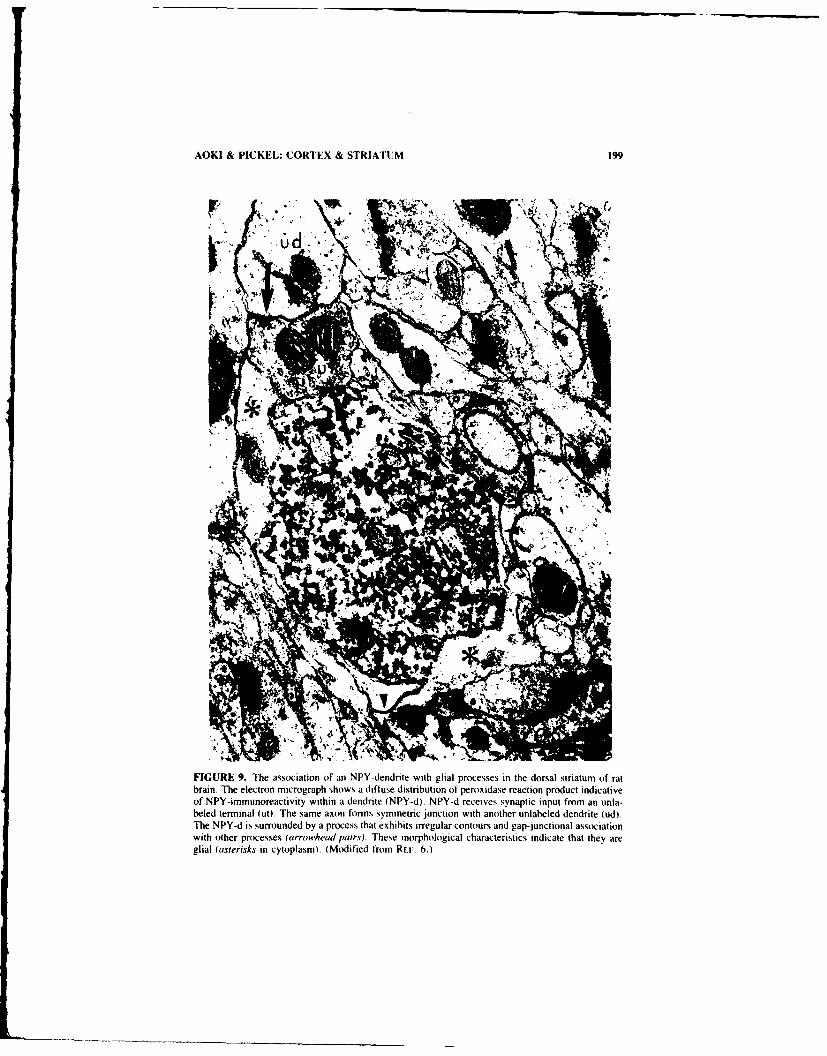
AOKI & PICKEL: CORTEX & STRIATUM 199
FIGURE 9. The association of an NPY-dendrite with glial processes in the dorsal striatum of ratbrain. The electron micrograph shows a difluse distribution of peroxidase reaction product indicativeof NPY-immunoreactivity within a dendrite (NPY-d). NPY-d receives synaptic input from an unla-beled terminal (ut). The same axon forms symmetric junction with another unlabeled dendrite (ud).The NPY-d is surrounded by a process that exhibits irregular contours and gap-junctional associationwith other processes (arroewhead pairs). These morphological characteristics indicate that they areglial (asterisks in cytoplasm). (Modified from R-F. 6.)

200 ANNALS NEW YORK ACADEMY OF SCIENCES
We have compared the relative frequency of NPY-terminals forming direct contactswith synaptic junctions in the rat striatum and cortex. The results indicate that approxi-mately half of all NPY-immunoreactive terminals in the cortex are juxtaposed to otheraxons (TABLE I). Of them, 14% occur directly across synaptic junctions formed by otherterminals. FiG. 8 shows an example of one such arrangement between pairs of terminalsand dendrites. Axo-axonic interaction is suggested by the parallel alignment of the plasmamembrane of the two terminals and the clustering of vesicles near both of the alignedmembranes. Within favorable planes of section. some of the NPY-immunoreactive axonsat synaptic junctions also form symmetric synaptic junctions onto a dendrite receivinginput from the neighboring axon terminal. The synaptic junction formed between theneighboring unlabeled terminal and the common dendrite is asymmetric, suggesting thatit is an excitatory synapse. 21
In striatum. the relative frequency of juxtaposition between NPY-terminals and otheraxons was approximately 15% (TABLE I). Of these, only 7% of the terminals formedparallel alignments of the plasma membrane simultaneously with that of the presynapticterminal and postsynaptic dendrite. These observations suggest that more of the NPY-terminals in the cerebral cortex may be involved in the modulation of synaptic transmis-sion. The NPY-immunoreactive terminals occurring at synaptic junctions not only forman organized association with the neighboring presynaptic terminal, but also the commondendrite. Thus, NPY may modulate non-NPY synaptic transmission by altering the re-lease of other transmitters and also through postsynaptic mechanisms involving secondmessengers. "9
NPY-Neurons and Degeneration
The NADPH-diaphorase-positive NPY neurons have been noted by some groups to beresistant to degeneration caused by NMDA receptor-specific excitotoxicity and diseasessuch as Huntington's chorea both in cortex and in the striatum. " .4 " The possibility thatNADPH-diaphorase itself may play a role in protecting the neurons from excitotoxicityhas been suggested." Our ultrastructural studies indicate that two additional features ofNPY-neurons may contribute towards their excitotoxicity-resistant phenotype. NPY so-mata and dendrites, whether in the cortex or striatum and whether or not coexisting withGABA show sparse distributions of synapses along the plasma membrane (see Fics. ). 4,5, 7 and 9). Thus, in comparison to the frequency of synaptic inputs observed withnon-NPY GABAergic neurons in the vicinity, the density of asymmetric synaptic junc-tions reflecting excitatory input is less than half. Therefore, the NPY-neurons may alsohave lower densities of glutamate receptors. In addition, dendrites and somata of NPYneurons are ensheathed by glial processes that form gap junctions with one another tFict.9). Thus, the high-capacity uptake sites for glutamate within glial processes4" may serveto protect NPY-neurons from excitotoxicity caused by the release of the excitatory trans-mitters.
The Role of NPY in Catecholaraine Turnover
One striking feature of the striatum is its rich innervation by catecholaminergic ter-minals. These terminals are mostly dopaminergic within the dorsal caudate-putamennucleus. Ventrally (in n. accumbens), more of the catecholaminergic fibers containnorepinephrine.i' Previous pharmacological studies have suggested that the release ofdopamine may be modulated by NPY, since NPY can stimulate the turnover ofdopamine. 3 Conversely, other studies have suggested that the NPY-containing neuronal

AOKI & PICKEL: CORTEX & STRIATUMI 211
FI(;URE I. The r aionship btween NPYneurons ad catechlnrgcansith ntl
stitu f a ran TeelcrN mcorPhtaetrn ralsclcedutth tin.hoanNYtrinl(P-t otiignueossal la c-itsadlonn vmercsn
FleRE 10sc.e Thoe einsipcore el tds nern and catechol- minergic axot snce sir grain ecr
over the cytoplasmi in serial sections. The plasmia membrane (it the CA-( is in direct contact with thatof NPY-d (rig/u arrovwhead in B) and the NPY-i. (Modilied front Ri 61

202 ANNALS NEW YORK ACADEMY OF SCIENCES
system may be undcr the tonic influence of the nigrostriatal aopamine system, since6-hydroxydopamine lesion of the nigrostriatal dopaminergic neurons leads to a markedincrease in the number of NPY-immunoreactive perikarya.4 7 However, since the juxta-position of catecholaminergic fibers and NPY-neurons had not yet been demonstrateddirectly in situ, a possibility remained that the function of the two transmitters wasmediated through the interpose( ieurons or glia.
Dual electron microscopic immunolabeling further .-t ucidated the cellular relationshipbetween nc-uronal cell bodies and processes that contain NPY or are catecholaminergic.No axo-somatic synapses were detected between catecholaminergic terminals and NPY-contoining cell bodies in our analysis of striatum or cortex. However, another group usingthe immunoautoradiographic metho4 to label NPY-cell bodies and catecholaminergicfibers by the immunoperoxidase method 'ave shown direct axo-somatic synapses betweenthe two."' This difference in results is likel' to be attributed to the differences in sensi-tivity for labeling the catecholaminergic fibers.
TABLE 2. Targets of NPY-ir Axons in Striatum (in Percent)
Caudate Putamen N. Accumbens
Synaptic contacts n = 190 n - 133with somata
NPY-ir 0 Iunlabeled 2 5%s/CA input" 0 0
with dendritesNPY-ir 0 1unlabeled 26 25w/C, input" 4 2
with spaiesNPY-ir 0 0unlabeled 15 24w/CA input" 0 0
unidentifiable 58 47
"w/CA input = processes also receiving catecholaminergic input within plane of section.
We additionally analyzed the relationship between catecholaminergic fibers and distalparts of NPY-neurons. The results indicate that catecholaminergic fibers rarely innervateNPY-dendrites. Instead, fine glial processes course between the two (FIG. 3). Occasion-ally (4% in dorsal striatum, 1% in ventral striatum), catecholaminergic axons convergewith NPY-axons onto common dendrites. More frequently, the two types of fibers coursepast each other towards presumably divergent destinations. In a single case, a putativelyconvergent association of NPY- and catecholaminergic axon terminals onto a commondendrite was found accompanied by direct contacts between the two terminals (FIG. 10).It is intriguing to consider that these may be sites for the mutual modulation of release bythe two transmitter systems, as has been suggested by pharmacologists.
SUMMARY
NPY-iieurons in the ;triatum and cortex have many morphological and chemicalfeatures in common. They ire intrinsic, medium sized, aspiny and exhibit ultrastructural
n 'i4il i m lI li i Il n

AOKI & PICKEL: CORTEX & STRIATUM 203
characteristics typical of neurons undergoing active synthesis and release of peptides.Most of the NPY-neurons in the two regions coexist with somatostatin. exhibit high levelsof NADPH-diaphorase and are resistant to degeneration associated with Huntington'sdisease. Ultrastructural analysis suggests that the ensheathment by glia and sparsity ofasymmetric (putatively excitatory) inputs may render NPY neurons resistant to excito-toxicity. Although NPY-neurons receive few inputs, they make numerous contacts withdendrites within a small region of the neuropil. Among their targets are GABAergicneurons. These NPY-receptive GABA neurons differ from other GABAergic neurons inthe vicinity in that they receive few other inputs along their somata and proximal den-drites. This suggests that NPY may exert more influence on a specific class of GABA-ergic neurons. Many more of the NPY-terminals are found at sites that would be strategicfor the simultaneous modulation of the release of transmitters and postsynaptic responses.
The differences among NPY-neurons in the striatum versus cerebral cortex are mainlychemical. Most notably, the NPY-neurons are GABAergic in the cortex and not GABAer-gic in the striatum. In addition, some of the NPY-axons in the ventral portions of striatumand cerebral cortex may be catecholaminergic. and thus originate in brainstem areasrecognized to contain NPY and epinephrine or norepinephrine. NPY- and catecholamin-ergic fibers converge onto same dendrites. Thus, the two transmitters may interactthrough intercellular biochemical pathways postsynaptically. Finally, the sites where thetwo fibers directly contact each other may be where NPY stimulates the turnover ofdopamine.
REFERENCES
I. TATEMOTO, K., M. CARLQUIST & V. MUTT. 1982. Neuropeptide Y-a novel brain peptidewith structural similarities to peptide YY and pancreatic polypeptide. Nature 296: 659-660.
2. McDONALD, J. K. 1988. NPY and related substances. Crit. Rev. Neurobiol. 4: 97-135.3. BEAL, M. F., R. C. FRANK, D. W. ELLISON & J. B. MARTIN. 1986. The effect of neuropeptide
Y in striatal catecholamines. Neurosci. Lett. 71: 118-125.4. WAHLE, P. & G. MEYER. 1987. Morphology and quantitative changes of transient NPY-ir
neuronal populations during early postnatal development of the cat visual cortex. J. Comp.Neurol. 261: 165-192.
5. NAEGELE, J. R., C. J. BARNSTABLE & P. WAHLE. Transient expression of a subplate-specificantigen in cat visual cortex. Soc. Neurosci. Abstr. 15: I.
6. AOKI, C. & V. M. PICKEL. 1988. Neuropeptide Y-containing neurons in the rat striatum:ultrdstructure and cellular associations with tyrosine hydroxylase-containing terminals andwith astrocytes. Brain Res. 459: 205-225.
7. AOKI, C. & V. PtCKEI.. 1989. Neuropeptide Y in the cerebral cortex and the caudate-putamennuclei: Ultrastructural basis for interactions with GABAergic and non-GABAergic neurons. J.Neurosci. 9: 4333-4354.
8. VINCENT, S. R., 0. JOHANSSON, T. HOKFELT, L. SKIRBOLE, R. P. ELDE, L. TERENIUS, J.KIMMEL & M. GOLDSTEIN. 1983. NADPH-diaphorase: a selective histochemical marker forstriatal neurons containing both somatostatin- and avian pancreatic polypeptide-like immu-noreactivities. J. Comp. Neurol. 217: 252-263.
9. VINCENT, S. R. & 0. JOHANSSON. 1983. Striatal neurons containing both somatostatin andavian pancreatic polypeptide (APP)-like immunoreactivities and NADPH-diaphorase activity:a light and electron microscopy study. J. Comp. Neurol. 217: 264-270.
0. HENDRY. S. H. C., E. G. JONES & P. C. EMSON. 1984. Morphology, distribution and synapticrelations of somalostatin- and neuropeptide Y-immunoreactive neurons in rat and monkeyneocortex. J. Neurosci. 4: 2407-2517.
II. DIMAGGIO. D. A., B. M. CHRONWALL, K. BUCHANAN & T. L. O'DONoHUE. 1984. Pancre-atic polypeptide immunoreactivity in rat brain is actually neuropeptide Y. Neuroscience15: 1149-1157.
12. GEHLERT, D. R.. B. M. CHRONWAIL, M. P. SCHAFER & T. L. 0'[o)NOtHUE. 1987. LAocal-

204 ANNALS NEW YORK ACADEMY OF SCIENCES
ization of neuropeptide Y messenger ribonucleic acid in rat and mouse brain by in situhybridization. Synapse 1: 25-31.
13. MoRis, B. J. 1989. Neuronal localization of neuropeptide Y gene expression in rat brain. J.Comp. Neurol. 290: 358-368.
14. STERNBERGER, L. A. 1979. Immunocytochemistry. Wiley, New York.15. PICKEL, V. M. 1981. Immunocytochemical methods. In Neuroanatomical Tract-Tracing Meth-
ods. L. Heimer & M. J. Robards, Eds. 483-509. Plenum. New York.16. CHRONWALL, B. M., D. A. DIMAGGIO, V. J. MASSARI, V. M. PICKEL, D. A. RUGGIERO &
T. L. O'DONOHUE. 1985. The anatomy of neuropeptide Y-containing neurons in rat brain.Neurosci. 15: 1159-1181.
17. LAUDER, J. M., V. K. M. HAN, P. HENDERSON, T. VERDOORN & A. C. TOWLE. 1986.Prenatal ontogeny of the GABAergic system in the rat brain: an immunocytochemical study.Neuroscience 19: 465-493.
18. PICKEL, V. M., J. CHAN & T. A. MILNER. 1986. Autoradiographic detection of '21 -secondaryantiserum: a sensitive light and electron microscopic labeling method compatible with per-oxidase immunocytochemistry for dual localization of neuronal antigens. J. Histochem. Cy-tochem. 34: 707-771.
19. MASSARI, V. J., J. CHAN. B. M. CHRONWALL, T. L. O'DONOHUE, W. H. OERTEL & V. M.PICKEL. 1988. Neuropeptide Y in the rat nucleus accumbens: ultrastructural localization inaspiny neurons receiving synaptic input from GABAergic terminals. J. Neurosci. Res.19: 171-186.
20. PICKEL. V. M. 1985. General morphological features of peptidergic neurons. In Handbook ofChemical Neuroanatomy. A. Bjorklund & T. H6kfelt. Eds. Vol. 1: 72-92. Elsevier. Am-sterdam.
21. GRAY, E. G. 1959. Axosomatic and axo-dendritic synapses of the cerebral cortex. An electronmicroscope study. J. Anat. 93: 420-433.
22. SMITH. Y. & A. PARENT. 1986. Neuropeptide Y-immunoreactive neurons in the striatum of catand monkey: morphological characteristics, intrinsic organization and co-localization withsomatostatin. Brain Res. 241: 241-252.
23. PICKEL. V. M.. J. CHAN & V. J. MASSARI. 1989. Neuropeptide Y-like immunoreactivity inneurons of the solitary tract nuclei: vesicular localization and synaptic input from GABAergicterminals. Brain Res. 476: 265-278.
24. EVERITT, B. I., T. HWKFELT, L. TERENIUS, K. TATEMOTO, V. MUTT & M. GOLDSTEIN. 1984.Differential co-existence of neuropeptide Y (NPY)-like immunoreactivity with catechola-mines in the central nervous system of the rat. Neuroscience 11: 443-462.
25. HOCKELT, T., J. M. LUNDBERG, K. TATEMOTO, V. MUTT, L. TERENIUS. J. POLAK. S.BLOOM, C. SASEK, R. ELDE & M. GOLDSTEIN. 1983. Neuropeptide Y (NPY)- and FMRF-amide-like immunoreactivities in catecholamine neurons in the rat medulla. Acta Physiol.Scand. 117: 315-318.
26. SAWCHENKO. P. E.. L. W. SWANSON, R. GRZANNA, P. R. C. HOWE. S. R. BLOOM & J. M.POLAK. 1985. Co-localization of neuropeptide Y immunoreactivity in brainstem catechola-minergic neurons that project to the paraventricular nucleus of the hypothalamus. J. Comp.Neurol. 241: 138-153.
27. DAGERLIND, A. & M. SCHALLING. 1988. Localization of dopamine P3-hydroxylase (DBH) andneuropeptide tyrosine (NPY) mRNA in the grey monkey locus coeruleus and adrenal medulla.Acta Physiol. Scand. 134: 563-564.
28. EDVINSSON, L., J. R. COPELAND. P. C. EMSON. J. MCCULLOCH & R. UDDMAN. 1987. Nervefibers containing neuropeptide Y in the cerebrovascular bed: immunocytochemistry. radio-immunoassay and vasomotor effects. J. Cereb. Blood Flow Metab. 7: 45-57.
29. FEINSTEIN, D. Personal communication.30. HENDRY, S. H. C., E. G. JONES. J. DEFELIPE, D. SCHMECHEI. C. BRANDON & P. C. EMSON.
1984. Neuropeptide-containing neurons of the cerebral cortex are also GABAergic. Proc.Nat]. Acad. Sci. USA 81: 6526-6530.
31. JONES, E. G., S. H. C. HENDRY & J. DEFELIPE. 1987. GABA-peptide neurons of the primatecerebral cortex: A limited cell class. In Cerebral Cortex. Vol. 6. Further Aspects of CorticalFunction. Including Hippocampus. E. G. Jones & A. Peters. Eds. 237-266. Plenum. NewYork.

AOKI & PICKEL: CORTEX & STRIATUM 205
32. JONES, E. G.. J. DEFELIPE, S. H. C. HENDRY & J. E. MAGGIO. 1988. A study of tachykinin-immunoreactive neurons in monkey cerebral cortex. J. Neurosci. 8: 1206-1224.
33. Sostooyi, P. & A. D. SMITH. 1979. Projection of neostriatal spiny neurons to the substantianigra: application of a combined Golgi-staining and peroxidase transport technique at bothlight and electron microscopic levels. Brain Res. 178: 3-15.
34. KITr'I. S. T. 1981. Anatomy and phygiology of the neostriatumn. In GABA and the BaisalGanglia. G. DiChiara & G. L. Gessa, Eds. 1-21. Raven. New York.
35. TAKAGI, H.. P. Somoovi. J. Sosmoo~i & A. D. SMITH. 1983. Fine structural studies on a typeof somatostatin-immunoreactive neurone and its synaptic connections in the rat neostriatum:a correlated light and electron microscopical study. J. Comp. Neurol. 214: 1-16.
36. FERRANTE, R. I.- N. W. KOWALL, M. F. BEAL, E. P. RicHARDSON. JR.. E. D. BIRD & J. B.MARTIN. 1985. Selective sparing of a class of striatal neurons in Huntington's disease.Science 230: 561-563.
37. BOLAM, J. P., D. J. CLARKE, A. D. SMITH & P. SOMOOVI. 1983. A type of aspiny neuron inthe rat neostriatumn accumbens accumulates 13H]gamma-aminobutyric acid: combination ofGolgi-staining, autoradiography and electron microscopy. J. Comp. Neurol. 213: 121-134.
38. COLMI'RS, W. F.. K. LUDOW)AK & Q, J. PITTMAN. 1988. Neuropeptide Y action in the rathippocampal slice: site and mechanism of presynaptic inhibition. J. Neurosci. 8: 3827-3837.
39. WALKER. M. W., D. A. EWALD. T. M. PENNEY & R. J. MILLER. 1988. Neuropeptide Ymodulates neurotransmitter release and calcium currents in rat sensory neurons. J. Neurosci.8: 2438-2446.
40. KOH, J. & D. W. CHtOI. Cultured striatal neurons containing HADPH-diaphorase or acetyl-cholinesterase are selectively resistant to injury by NMDA receptor agonists. Brain Res.446: 374-378.
41. DAVIES, S. W. & P. 1. ROBARTS. 1987. No evidence for preservation of somatostatin-con-tamning neurons after intrastriatal injections of quinolinic acid. Nature 327: 326-329.
42. BOEGMAN, R. J., Y. SMITH & A. PARENT. 1987. Quinolinic acid does not spare striatalneuropeptide Y-immunoreactive neurons. Brain Res. 415: 178-182.
43. CHOt. D. W. & S. M. RoTrHMAN. 1990. The role of glutamate neurotoxicity in hypoxic-isehemic neuronal death. Annu. Rev. Neurosci. 13: 171-182.
44. MOHANDApS et al. 1984. Res. Commun. Chem. Pathol. Pharmacol. 43: 463.45. SCHOUSBOE, A., 0. M. LARSSON, J. DREJER. P. KROGSGAARD-LERSEN & L. HERTZ. 1983.
Uptake and release processes for glutamnine. glutamate and GABA in cultured neurons andastrocytes. In Glutamine. Glutamnate and GABA in the CNS. L. Hertz. E. Kvamme, E. G.McGeer & A. Schousbhoe. Eds. 297-31I5. Liss. New York.
46. MOORE, R. Y. & J. P. CARD. 1984. Noradrenaline-containing neuron systems. In Handbookof Chemical Ncuroanatomy. Vol. 2. A. Bjorklund & T. Hokfelt. Eds. 123-156. Elsevier.Amsterdam.
47. KERKERIAN, L., 0. BOSLER, G. PELLETIER & A. NIEOULLON. 1986. Striatal neuropeptide Yneurons are under the influence of the nigrostriatal dopamninergic pathway: immunohisto-chemical evidence. Neurosci. Lett, 66: 106-112.
48. VUILLET. J., L. KERKERIAN, P. KACHIDIAN, 0. BOSLER & A. NIEOULtLON. 1989. Ultrastruc-tural correlates of functional relationships between nigral dopaminergic or cortical afferentfibers and neuropeptide Y-containing neurons in the rat striatumn. Neurosci Lett. 100: 99-104.

Modulation of Synaptic Tranmissionin Hippocampus by Neuropeptide Y:
Presynaptic Actionsa
WILLIAM F. COLMERS
Department of Pharmacology9-70 Medical Sciences Building
Universiy" of AlbertaEdmonton Alberta, Canada T6G 2H7
INTRODUCTION
The hippocampal formation contains a significant amount of Neuropeptide Y (NPY),expressed by neurons and contained in their fibers in the dentate gyrus and hippocampusproper.' The stratra radiatum and oriens of hippocampus, which represent the apical andbasal dendritic fields, respectively, of the pyramidal neurons of areas CAI and CA3, andwhich contain the presynaptic fibers and terminals impinging on the pyramidal celldendrites, 2 demonstrate some of the highest concentrations of NPY binding sites any-where in the brain, although there are few or no binding sites in the cell body layers ofeither CAI or CA3. 3 While there is no precise correspondence of NPY-containing fibersand binding sites, there is also no gross mismatch, suggesting that locally-released peptidecould act at nearby receptors.
NPY has been demonstrated to have both pre- and postsynaptic actions in peripheraltissues such as vas deferens and vascular smooth muscle (Wahlestedt " and this volume).Because of the well characterized synpatic responses in hippocampus, and the presence ofthis peptide and its receptors in the region, I tested the hypothesis that NPY affectssynaptic transmission in the rat hippocampal since preparation, in vitro.
METHODS
Details of the preparation and recording apparatus have been published elsewhere. 5"Briefly, transverse slices (400-450 tim thick) were prepared from hippocampi taken frommale Sprague-Dawley rats (100-200 g). and perfused at 1.5-2 ml min-' with artificialcerebrospinal fluid (ACSF), while held submerged on a nylon mesh in a recordingchamber (300 RI volume). The ACSF was saturated with 95% 02, 5% CO 2, and held atconstant temperatures (t 0.5°C) between 32 and 35°C by a water jacket. A valve per-mitted the rapid switching between source reservoirs for peptide and drug application viathe perfusate. The experimental preparation is illustrated in FIGURE 1. Extracellular fieldpotentials were recorded with 2 M NaCl-filled glass microelectrodes (2-10 Mil) placedin the cell body layer of CAI, while intracellular recordings were made with 2 M K +
"Support for these studies came from the Medical Research Council of Canada. The author is aMedical Scholar of the Alberta Heritage Foundation for Medical Research.
206

COLMERS: SYNAPTIC TRANSMISSION 207
acetate-filled microelectrodes (90-180 Mfl). A bipolar stimulating electrode was placedin stratum radiatum between areas CA2 and CA l, or on other pathways as described.Square-wave monophasic pulses (50-200 Rs, 2-25 V) were delivered to this electrodefrom a stimulus isolation unit. Stimuli were generally delivered at 0. 1 Hz.
Orthodromic stimulation of stratum radiatum elicited a field potential in the cell bodylayer recorded by the extracellular microelectrode. The population spike (PS) componentof the field potential was measured from the peak of the negative wave to the peak of thefollowing positive one5 (FIG. 1, inset left). Once the population response had been stablefor at least 20 minutes, in many experiments a potassium acetate-filled microelectrode
Rat Hippocampal Slice
Fextracellular electrode (NaCl) electr e EPSP
FIGURE 1. Schematic diagram of transverse rat hippocampal slice, showing synaptic relationshipsbetween principal neurons and the placement of recording and stimulating electrodes. Inset at leftillustrates a typical extracellular field potential recording, while inset at right illustrates an intra-
cellular recording of an excitatory postsynaptic potential (EPSP) evoked in a CA I pyramidal cell bystimulation of stratum radiatum during a hyperpolarization induced by a pulse of current applied viathe electrode. Abbreviations: CAl-area CAl of hippocampus proper; CA3-area CA3 of hippo-campus proper; D.G.- dentate gyros.
was lowered into the cell body layer nearby and a CA!I pyramidal neuron was impaled andidentified by standard criteria such as resting membrane potential, action potential wave-form, and a response to orthodromic stimuli. 6"7 Square-wave, constant-current hyperpo-larizing pulses were passed via the bridge circuit of the amplifier to prevent the neuronfrom reaching action potential threshold when stratum radiatum was stimulated 40 msecafter the start of the pulse6 7 (FIG. I, inset right). Orthodromic stimuli were varied inintensity in regular steps from near threshold for the EPSP to just below action potential
threshold. Once resting membrane properties and synaptic responses had stabilized, usu-

208 ANNALS NEW YORK ACADEMY OF SCIENCES
Population Spike Intracellular EPSP
Control
1 y M N PY
1 h Wash -
2.5 mV L 25 mV
25 msec 50 msec
FIGURE 2. Effects of I iiM NPY on orthodromic synaptic potentials in CA I. Simultaneousextracellular (left) and intracellular (right) recordings were made from the cell body layer and apyramidal cell. respectively. Stratum radiatum was stimulated 40 msec after the start of a hyperpo-larizing current pulse applied to intracellular electrode. NPY inhibited population spike (PS) ampli-tude by about 90%, and inhibited the EPSP by about 40% in this experiment. Synaptic responsesrecovered to near control values after about I hour washout with control ACSF.
ally after 20 minutes or longer, control responses were established and experiments wereinitiated.
Data were averaged and stored on floppy diskettes and analyzed on-line and from tapeusing a Nicolct 4094 digital oscilloscope. D.C. coupled chart recordings were made ofresting membrane potential and injected current. Critical parts of experiments were re-corded either on FM tape or digitized and recorded on videocassette. Neurons served astheir own controls for statistical purposes. Student's paired t test was used unless other-wise noted. All data are from preparations exhibiting substantial recovery from anypeptide or drug effects. Unless otherwise noted, amidated porcine sequence NPY 1-36was used in these experiments.

COLMERS: SYNAPTIC TRANSMISSION 209
RESULTS AND DISCUSSION
Action of NPY in CAI
Orthodromic stimuli elicited stable postsynaptic responses in ,area CA I (FIG. 2). Bathapplication of NPY at I p.M caused an inhibition of the extracellularly recorded PS byabout 90% of control (FIG. 2, right). The effect of NPY was reversible upon prolonged(-- I h) washout. The inhibition of the PS by NPY was dependent on the concentration ofNPY applied; threshold for the effect was around 10 nM and the EC,, was about 300 nM. 5
To test whether the ability of CAI pyramidal cells to generate action potentials wasaffected by NPY, their axons were stimulated antidromically by a bipolar electrode placedin alveus, and the resulting field potential was recorded. Application of 1 I±M NPY didnot alter the amplitude of the antidromically-evoked field potential. 5
Because the effect of NPY in CAI was consistent with an inhibition of synaptictransmission at the stratum radiatum-CAI synapse, an additional microelectrode wasplaced in the cell body layer and used to impale a CAI pyramidal neuron (FIG. 1).
EPSP Glutamate Pulse
Control
Low Ca++, High Mg f/
25 mV 6.25 mV
+ 1 M NPY 50 msec ( 1 sec
FIGURE 3. NPY does not affect glutamate responses in CAI neurons. Digitally averaged (n = 8)responses of CAI pyramidal neuron to stratum radiatum stimulation (left) or to a brief (250 msec)pulse of glutamate (right) applied ionophoretically from an electrode placed near the neuron's apicaldendrites (upper traces). Note different time and voltage scales for the two responses. The twostimuli were presented alternately to the neuron at 0.05 Hz. Synaptic transmission was interrupted byapplication of a bathing medium of containing 0.5 mM Ca . 3.5 mM Mg * . with the otherconstituents being as in control ACSF, to preclude an indirect, synaptically mediated effect ofglutamate on the neuron (middle traces). Application of I l.M NPY (lower traces) did not inhibitresponse of neuron to glutamate. (From Colmers et al.' Reprinted by permission from the Physio-logical Society.)

210 ANNALS NEW YORK ACADEMY OF SCIENCES
Stimulation of stratum radiatum while passing a hyperpolarizing current pulse into the cellelicited a fast EPSP. Bath application of I I.M NPY resulted in an inhibition of the EPSPamplitude by about 40% of control, which reversed upon prolonged washout (FiG. 2,left). The time course of NPY-mediated inhibition and recovery subsequent to washoutwas the same for the EPSP and the PS. 6 "7
Extensive experiments were performed to determine whether NPY affected any activeor passive membrane properties of the postsynaptic CAI neurons. 6 I p.M NPY had noeffect on the resting membrane potential of CA 1 neurons. The input resistance, estimatedfrom the voltage response of the neuron to families of hyperpolarizing and depolarizingcurrent pulses, was also not affected by NPY. NPY also had no effects on the amplitude,duration, threshold, or number of action potentials evoked by a depolarizing current pulsein CAI cells. 6 Therefore, there was no direct action of NPY on the postsynaptic propertiesof CAI cells measured at the cell soma.
There remained three possible explanations for the effects of NPY on synaptic trans-mission. 1) NPY could cause a very localized reduction in the input resistance of thedendrites, which, because of the great electronic length of the apical dendrites, might notbe observed while recording in the soma, but could have a profound effect on theamplitude of the EPSP. 2) NPY could be acting as an antagonist at the postsynapticreceptors for the excitatory transmitter, glutamate. 3) NPY could inhibit synaptic trans-mission by a presynaptic action. To test these hypotheses, pulses of glutamate wereapplied iontophoretically from a pipette placed next to the apical dendrites of impaledCAl neurons. If NPY were ct3ing via either mechanisms 1) or 2), the response toglutamate should be inhibited by NPY application to a similar degree as the EPSP. Thiswas tested in the absence of synaptic transmission, using a low Ca' +, high Mg'solution to preclude indirect excitation of other presynaptic elements by glutamateapplication. 6 However, application of 1 p.M NPY caused no change in the amplitude ofthe response of these neurons to glutamate (FiG. 3).
Site and Mechanism of Action in CA)
The above series of experiments led to the conclusion that NPY was inhibiting syn-aptic transmission by an action at a presynaptic site, as there was no evidence for any,even subtle, effects of the peptide on the postsynaptic neuron. The next series of exper-iments were designed to identify the presynaptic site of action of NPY. and to attempt toelucidate the mechanism by which it acts.
The hippocampal circuitry is shown in schematic form in FIGURE 4. As indicated, themajority of synaptic input to CAl arises from the Shaffer collaterals, the ipsilateralbranches of CA3 pyramidal cell axons. 2 Experiments showed that NPY had no effects onthe active or passive membrane propertie. of CA3 pyramidal neurons. 7 Because thepeptide could affect the excitability of the Shaffer collateral axons or their terminals,' astimulating electrode was placed in stratum radiatum of CAl (stimulus site 2, FIG. 4), andantidromically-evoked field potentials were recorded with extracellular microelectrodes inthe cell body layer of area CA3. However, NPY had no effect on the amplitude of theantidromic field potentials in CA3, indicating that there was no effect on the excitabilityof the axons or terminals in CAI.7 In other experiments, the amplitude of the afferentvolley (a component of the field potential recorded in stratum radiatum of CAI in re-sponse to orthodromic stimuli), which represents the number of presynaptic axons dis-charging in response to the stimulus,Id was not affected by NPY. Thus, two lines ofevidence were consistent with there not being any effect of the peptide on the excitabilityof the presynaptic axons.
The possibility remained that NPY was acting at the presynaptic terminals themselves

COLMERS: SYNAPTIC TRANSMISSION 211
to inhibit release of neurotransmitter, by affecting conductances which are not active atrest, and would therefore not greatly affect the excitability of the terminals and axons astested above. Two possible mechanisms for this are: 1) the activation of a voltage-dependent potassium conductance in the terminals, hyperpolarizing it and making depo-larization less likely upon stimulation," and 2) the inhibition of voltage-dependent cal-cium influx, upon which transmitter release depends.1 2 It is known that presynapticterminals in rat CNS possess potassium channels which are very sensitive to the action of4-aminopyridine (4-AP). ' 3 To test possibility 1), NPY's effects on synaptic transmissionwere examined in the presence of low concentrations of 4-AP.
Application of 10 l±M 4-AP, a concentration known to markedly inhibit potassiumchannels in presynaptic terminals, 13 but which has little effect on the transient, voltage-dependent "A" potassium current in the soma, 14 caused a marked increase in transmitter
4 2
I Stratum Oriens
6 ibraC1 PyamdalCell, BaktCel]
(Schaffer Collateral) Stratum radiatumn FedF rad nenuo
CA3 Pyramidal Cell 5 Dentate Granule Cell
Mossy n Fibers A Perforant Path8Commissural Fibers
FIGURE 4. Schematic diagram of synaptic circuitry in hippocampus, illustrating stimulating elec-trode placement for experiments. See text for details.
release to a given stimulus (FIG. 5). Application of I p.M NPY in the presence of 10 K.M4-AP had no effect on synaptic transmission (FIG. 5). Thus, it at first appeared that NPY'seffect was via an activation of a 4-AP sensitive K + conductance.
However, there was another interpretation for the results. By blocking voltage-de-pendent K ' channels in the presynaptic terminal, the repolarization of the membrane atthe terminal was delayed. If NPY were inhibiting the activation of only a portion of thevoltage-dependent Ca + ' channels in the presynaptic terminal, then the delayed repolar-ization induced by 4-AP could allow enough calcium to enter the terminal to causetransmitter release, overwhelming the normal inhibition caused by NPY (FIG. 6). Theextracellular Ca' + was therefore reduced, in the presence of 4-AP, to a concentrationwhich restored the pre-4AP levels of synaptic transmission. This presumably meant thatthe amount of Ca + ' entering the presynaptic terminals was about the same as before

212 ANNALS NEW YORK ACADEMY OF SCIENCES
Control
1O0MM 4-AP
1 FIGURE 5. Effect of 4-aminopyridine (4-AP)on NPY inhibition of stratum radiatum EPSP
+ 1 gM NPY recorded intracellularly in a CAI pyramidal neu-ron. 4-AP increased synaptic transmission in re-sponse to identical stimulus as control. Applica-tion of 1 pM NPY in the presence of 4-AP didnot inhibit EPSP.
1 h Wash
J 25 mV
50 msec
4-AP was applied. The concentration of extracellular Ca' needed to restore the pre-4-AP levels of transmitter release was found empirically to be 0.70 mM. Under theseconditions, application of I [LM NPY once again inhibited synaptic transmission7(FIG. 7).
Further experiments were conducted using a higher concentration of,- AP, 50 1,M. Asin the earlier experiments. NPY was ineffective in the presence of the higher concentra-tion of 4-AP. When the Ca ' concentration was lowered to 0.70 mM, NPY was stillineffective. However, lowering extracellular Ca to 0.50 mM restored NPY's ability toinhibit synaptic transmission. 7
The results are therefore not consistent with the presynaptic inhibition by NPY re-sulting from its activation of a 4-AP-sensitive K conductance. It is furthermore unlikelythat NPY activates another, 4-AP insensitive, K + conductance. This is because the inputresistance of the presynaptic terminal would be increased due to the blockade of a K 'conductance by 4-AP. If NPY were to activate a 4-AP insensitive K + conductance, thehyperpolarization, and therefore presumably the action of NPY thus caused, would begreater in the presence of 4-AP than in control.
However, the data are consistent with the NPY's presynaptic action resulting from theinhibition of a portion of the Ca' + influx into the presynaptic terminal. In support ofthis, experiments on rat dorsal root ganglion (DRG) neurons in culture'5 have demon-strated that NPY has a very potent and prolonged inhibitory effect on calcium currentsrecorded in isolation.
Coupling of the NPY Receptor to Effectors
Experiments on DRG cells in culture indicated that NPY receptors inhibited theopening of voltage-dependent calcium channels via the activation of a GTP-bindingprotein which was sensitive to the action of the bacterial exotoxin produced by Bordetellapertussis, pertussis toxin (PTX). This toxin acts by entering the cell and irreversiblyplacing an ADP-ribose molecule onto the GTP binding site of some GTP binding proteins(G-protein), which irreversibly inactivates them, thus decoupling the receptor from its

COLMERS: SYNAPTIC TRANSMISSION 213
effector by eliminating the intermediary. 15.16 If NPY acts at the presynaptic terminal bydecreasing calcium influx, then this action might be sensitive to the action of PTX. Thehypothesis was therefore tested whether the action of NPY on synaptic transmission wassensitive to the actions of PTX. Rats were injected with 1.5 Lg PTX into the lateralcerebral ventricle, and allowed to survive for 3 days, for the toxin to act. 817 Slices wereprepared from the hippocampi of these animals in an otherwise identical manner asbefore.
Physiological properties of slices prepared from PTX pretreated animals did not differsignificantly from those prepared from control animals. Intracellular recordings of CA Ineurons in slices from PTX pretreated animals demonstrated no significant differencefrom those of untreated animals in their resting membrane properties or action potentialparameters. Stimulation of stratum radiatum in these slices evoked an intracellularlyrecorded EPSP and an extracellularly recorded PS. Application of I LM NPY to slicesfrom PTX pretreated animals inhibited excitatory synaptic transmission: The EPSP was
0
0
0
FIGURE 6. Cartoon illustrating possible interpretations for the 4-AP mediated blockade of NPYinhibition of transmitter release at a presynaptic terminal. The NPY receptor (NPYR) could becoupled so as to activate a 4-AP sensitive potassium channel (gK - + NPY) or to inhibit activationof voltage-dependent calcium channels (gCa ' ' + V, -NPY) in the presynaptic terminal. However,if NPY inhibits only a portion of the calcium channels in the presynaptic terminal, and other,NPY-insensitive voltage-dependent calcium channels (gCa + + +V), are present, the prolongeddepolarization caused by 4-AP blockade of potassium channels could permit enough calcium to enterthe terminal through the NPY-insensitive channels to permit transmitter release, thus seeming tooverwhelm NPY's inhibition of the other calcium channels.

214 ANNALS NEW YORK ACADEMY OF SCIENCES
CA1 Population Spike Pyramidal Cell EPSP
CONTROL(1.5 mM Ca)
-10 5 M 4-AP(0.70 mM Ca)A
+ 1 6 6M NPY
60' Wash
2.5 nv 25 mV
25ms 50 .s
FIGURE 7. Reducing extracellular Ca concentration in the presence of 4-AP restores NPY'sinhibition of synaptic transmission. Simultaneous extracellular (left traces) and intracellular (righttraces) recordings in CA I in control (upper traces), and after bathing medium had been changed toone containing 10 IAM 4-AP and having only 0.7 mM Ca ' ' (lower three traces). Application of IliM NPY under these conditions inhibited synaptic responses, which recovered upon prolongedwashout.
inhibited by about 40%, and the PS was inhibited by about 80%. in slices both from PTXpretreated and control animals. No significant difference between the actions of NPY wasobserved in the PTX pretreated groupX.
Because these results raised the possibility that PTX pretreatment had been ineffectivein ADP-ribosylating the G-proteins in the hippocampus, we applied either 5-HT or ba-clofen, both of which elicit a hyperpolarizing response brought about by an increase inpotassium conductance, mediated by a PTX-sensitive G-protein. "x However, neither 5-HT nor baclofen had any effects on the membrane potential of the CAI pyramidal cells.
I

COLMERS: SYNAPTIC TRANSMISSION 215
even at very high concentrations, indicating that the PTX had been effective ai -liminatingG-proteins in the cell bodies of hippocampal neurons." Assuming that the toxin has equalaccess to the presynaptic terminals, this indicates that the action of NPY at the terminalis mediated by a PTX-insensitive G-protein. Interestingly, in this study it was also ob-served that thec, presynaptic inhibitory action of baclofen on excitatory synaptic transmis-sion in hippocampus vas not affected by PTX pretreatment, although its postsynapticeffects "-'ere eliminated. 8 There may thus be a similar mechanism coupling both presyn-aptic NPY and GABA, receptors to the inhibition of trai smitter release in hippo-campus.8'17
Other Sites of NPY Action in Hippocampus
Excitatorvy Synaptic Transmission
Similar experiments have demonstrated that I l, M NPY potently and reversibly in-hibits synaptic transmission at other sites in the hippocampus. The EPSP evoked bystimulation of stratum oriens in CAI (Site 4. FIG. 4) is inhibited by NPY by about 45%of control. In CA3 pyramidal neurons, the EPSP evoked by stimulation of the mossy fiberinput pathway (Site 5, FIG. 4) is inhibited by NPY by about 45% of control. Th" EPSPevoked in CA3 cells by stratum radiatum (Site 1, FIG. 4) which activates t!,e CA3-C, .3excitatory interconnections, '9 was also reduced by NPY to about 50% of control. Stimulation of the fimbria in area CA3 (Site 6, FIG. 4) evoked an EPSP which was inhibitedby about 45% by NPY. By contrast, the EPSP's evoked in dentate granule cells bystimulation of either perforant path input (Site 7, FIG. 4) or commissural input (Site 8.FIG. 4) was not affected by NPY, although other known agents having presynaptic action.such as baclofen, muscarine and adenosine all inhibited the intracellularly recorded EPSPand extracellularly recorded PS in granule cells. 2" This correlates with the binding data.which indicate that: 1) there are no NPY binding sites in the outer two-thirds of themolecular layer, which is where the perforant path fibers terminate on the dendrites of thegranule cells, 3 and 2) that the binding site density is much lower in dentate than in thehippocampus proper.3 Because the greatest density of NPY innervation in the hippocam-pal formation is in the outer two-thirds of the molecular layer.' 2 it raises the intriguingquestion of what actions the local release of NPY might have in this apparently insensitiveregion.
Inhibitory Synaptic Transmission
The action of NPY was studied on inhibitory synaptic potentials evoked in area CA I.
Stimulation of stratum radiatum elicits a compound svnaptic potential in CAI pyramidal
4 neurons, of which the EPSP is the earliest component. Following the EPSP, a fast.GABAA receptor-mediated, chloride-dependent inhibitory postsynaptic potential (IPSP)occurs. 2' This potential originates with the basket cells, inhibitory interneurons which aredriven by axon collaterals of the pyramidal cells of CAI (FIGURt- 4), and which innervatethe soma and initial segment of the CAI neuron. This IPSP is generally referred to as.'recurrent." since it feeds back onto the pyramidal cells, and can be evoked in isolationby stimulation of the CAI cell axons in alveus 2' (FIG. 4). A later, slower inhibitorypotential is also seen. especially at higher stimulus intensities. 2' This is mediated by aGABAR receptor, which activates a potassium conductance via a GTP binding protein.and is largely localized at the dendrites of the pyramidal cells. "8.21 This IPSP cannot beactivated by alvear stimulation, and is therefore referred to as a "'feed-forward" IPSP.2'

216 ANNALS NEW YORK ACADEMY OF SCIENCES
Both feed-forward and recurrent IPSP's were elicited in single CA I pyramidal neu-rons by alternate stimulation of stratum radiatum and alveus. Application of I Ip.M NP,reduced the amplitude of the ISPS's evoked by stimulation of stratum radiatum to about60% of control, the same amount as the EPSP was reduced in these neurons. However,the recurrent IPSP evoked by stimulation of the alveus was not affected by NPY at thesame time as the feed-forward ISPS's were inhibited. 22
The simplest explanation of these results is that NPY inhibits the feed-forward exci-tatory drive from stratum radiatum fibers, both to the CAI pyramidal neurons, thusindirectly reducing the drive to the inhibitory basket cells, and directly reducing thesynaptic excitation of the feed-forward inhibitory interneurons. However, NPY does notappear to affect the synaptic excitation of the basket cells directly, -is the ISPS evokedfrom the alveus is not affected by NPY.
,7,A DGA1--
FIGURE 8. Schematic diagram of synaptic connections in hippocampus. illustrating possible actionof NPY in modulating synap!ic transmission in hippocampus. Arrows symbolize the amplitude of anexcitatory input to -;npocampus. As excitatory input passes along the trisynaptic pathway. NPYaction would attenuate it, at inputs to both area CA3 and area CAl. Asterisks (*) indicate excitatorysynapses inhibited by NPY. Abbreviations: ALV = alveus; COMM-commissural inputs to dentategranule cells or CA3 pyramidal cells (different pathways); GC-dentate granule cells; MF-mossyfibers; PP-perforant path; SO--stratum oriens; SR-stratum radiatum.
SUMMARY
NPY is a potent inhibitor of excitatory synaptic transmission in the rat hippocampusin vitro. I I 'Y has no demonstrable actions on the active or passive properties of dendrites,cell bodies and axons of principal neurons in this area of the brain. It does appear to inhibitthe release of the excitatory neurotransmitter. which is probably glutamate, from presyn-aptic nerve terminals. Although the mechanism of its action at the presynaptic nerveterminal is not absolutely certain, results from the 4-AP experimerts are consistent withNPY inhibiting a portion of the voltage-dependent calcium channels in the terminal. This

COLMERS: SYNAPTIC TRANSMISSION 217
would also be in agreement with the results obtained by others from mammalian neuronsin culture, in which NPY is a potent inhibitor of voltage-dependent Ca + ' channels. ' Itappears that, although the NPY receptor is most likely a G-protein linked receptor, 23 andcan couple to both G, and G,,2' it does not do so at the presynaptic nerve terminal inhippocampus. The NPY receptor in hippocampus has a pharmacological profile similar tothe peripheral Y, subtype (Klapstein et al., this volume). Although the Y2 receptor hasbeen shown to inhibit adenylate cyclase, this does not appear to be the mechanism of itsaction in presynaptic terminals, since the elevation of cAMP with membrane solubleanalogues did not interfere with NPY's inhibitory effects (Klapstein el al., this volume).However, because there is evidence: 1) that inhibition of calcium influx can occur throughPTX-insensitive G-proteins 24 evidently activated by the same receptor which also cou-ples to a PTX-sensitive G-protein,' 7 and 2) that secretory processes can be regulateddirectly via a G-protein dependent process. independent of the actions on Ca + ' influx,25
there are several further mechanisms by which NPY might potentially regulate the releaseof neurotransmitter at the nerve terminal.
The picture emerges from these data that the release of NPY in hippocampus wouldbe accompanied by a reduction in the amplitude of an excitatory signal which enters thehippocampus proper. NPY effectively reduces the input amplitude of both the feed-forward excitatory and inhibitory synaptic components, without changing the propertiesof the postsynaptic circuitry, such as recurrent inhibition. NPY thus does not affect theinput-output properties of the postsynaptic circuitry, unike other substances known to actat inhibitory presynaptic inhibitory receptors in hippocampus, all of which have postsyn-aptic actions as well. Because of this, NPY's actions in hippocampus can be likened to thevolume control on a good hi-fi amplifier, attenuating the input without affecting thefidelity of the amplification. The attenuation of a signal originating from entorhinal cortexwould presumably increase at every stage where NPY release is elicited (FiG. 8). Theactions of NPY could however be construed as being consistent with the role of an"endogenous anticonvulsant." Given the recent demonstration of a rat model of epilepsyin which the NPY/somatostatin containing interneurons of the dentate gyrus are selec-tively ablated. 26 and the reported selective loss of these interneurons in dentate gyrus ofhuman patients suffering from cryptogenic temporal lobe epilepsy, 27 it is certainly con-ceivable that NPY plays a role in the prevention of hyperexcitability in the hippocampus.
Because there is as yet no reliable antagonist for NPY receptors, the actual physio-logical function ik subserves in hippocampus is still a matter of speculation. However, thedramatic and potent action of NPY in the pharmacological experiments outlined above,and the very high concentration of NPY receptors in hippocampus indicate a role for thispeptide in the normal physiology of the rat hippocampus. It is known that NPY/somato-statin-containing interneurons are innervated by axon collaterals of principal neurons inhippocampus, and can therefore presumably release the peptide in response to appropriatelevels of stimulation. In the absence of a specific NPY antagonist, it may be possible todemonstrate appropriate release by displacement of radiolabelled NPY, as has recentlybeen done for dynorphin release from mossy fibers. 29
Much further work is needed on NPY's action in the hippocampus, from elucidatingthe exact mechanism of action of NPY at the presynaptic nerve terminal to determiningunder which physiologically appropriate circumstances the peptide is released. Ulti-mately, of course, the question to be answered is that of the physiological role of NPY inthe brain of an intact animal.
ACKNOWLEDGMENTS
I gratefully acknowledge the support, collaboration and encouragement of Dr. Quen-tin Pittman in my early investigations on NPY. and his continued advice. I am indebted

218 ANNA.LS NEW YORK ACADEMY OF SCIENCES
to Dr. Ken Lukowiak for his collaboration and support in the early studies. It is a pleasureto acknowledge the work of my more recent collaborators, especially that of Ms. GloriaKlapstein, and of Ms. Kerni Treherne and Mr. Ken Makus.
REFERENCES
I . KOHLER, C.. L. ERIKSSON. S. DAVIES & V. CHAN-PALAY. 1986. J. Comp. Neurol. 244:569-584.
2. SWANSON, L. W., I. M. WYSS & W. M. COWAN. 1978. J. Comp. Neurol. 181: 681-716.3. MARTEL, J.-C., A. FOURNIER, S. ST-PIERRE & R. QUIRION. 1990. Neuroscience. In press.4. WAHLESTEDT. C. 1987. Neuropeptide Y (NPY): Actions and Interactions in Neurotransmis-
Sion. Thesis. University of Lund. Sweden.5. COLMERS, W. F.. K. LUKOWIAK & Q. J. PITTMAN. 1985. Brain Res. 346: 404-408.6. COLMERS, W. F.. K. LUKOWIAK & Q. J. PITTMAN. 1987. J. Physiol. (London) 383: 285-299.7. COLMERS, W. F., K. LuBOWIAK & Q. J. PITTMAN. 1988. J. Neurosci. 8: 3827-3837.8. COLMERS, W. F. & Q. J. PITTMAN. 1989. Brain Res. 498: 99-104.
9. WALL, P. D. 1958. J. Physiol. (London) 142: 1-21.10. ANDERSEN. P.. H. SILFVENIUS, S. H. SUNDBERG. 0. SVEEN & H. WIGSTROM. 1978. Brain
ResKTZ . 111. MLD.16.J hs.(odn 0:6971
12. KATZ. B. & R. MILEDI. 1967. J. Physiol. (London) 192: 407-436.13. BARTSCHAT. D.K. & M.P. BLAUSTEIN. 1985. J. Physiol. (London) 361: 419-440.14. GUSTAFSSON. B.. M. GALVAN, P. GRAFE & H. WIGSTROM. 1982. Nature 299: 252-254.15. WALKER, M. W., D. A. EWALD, T. M. PERNEY & R. J. MILLER. 1988. J1. Neurosci. 8:
2438-2446.16. HOLZ, G. G., S. RANE & K. DUNLAP. 1986. Nature (London) 319: 670-672.17. COLMERS, W. F. & 1. T. WILLIAMS. 1988. Neurosci. Lett. 93: 300-306.18. ANDRADE, R., R. C. MALENKA & R. A. NICOLL. 1986. Science 234: 1261-1265.19. MACVICAR. B. A. & F. E. DUDEK. 1980. Brain Res. 184: 220-223.20. COLMERS, W. F. & G. J. KLAPSTEIN. 1989. Soc. Neurosci. Abstr. 15: 218.21. ALGER. B. E. & R. A. NICOLL. 1982. J. Physiol. (London) 328: 125-141.22. COLMERS. W. F. 1989. Can. J1. Physiol. Pharmacol. 67: Avii.23. EWALD, D. A.. P. C. STERNwEIS & R. J. MILLER. 1988. Proc. Nail. Acad. Sci. USA
85: 3633-3637.24. PENINGTON, N. J. 1990. Neuron. In press.25. LuINI, A. & M. A. DE MATTEIS. 1990. J. Neurochemn. 54: 30-38.26. SLOVITER. R. S. 1989. In The Hippocampus-New Vistas. C. Kohler & V. Chan-Palay. Eds.
443-461. Alan R. Liss. New York, NY.27. DE LANEROLI.E. N. C.. J. H. Kim, R. J. ROBBINS & D. D. SPENCER. 1989. Brain Res.
495: 387-395.28. NEUMAIER. J. F. & C. CHAVKIN. 1989. Brain Res. 493: 292-302.

Neuropeptide Y-Cholinergic Interactionsin Neocortical Tissue
JENNIFER J. POULAKOS,- EDWIN M. MEYER,- CORINNE R.PRUYSERS," AND WILLIAM J. MILLARD b
University of Florida"College of Medicine
Department of Pharmacology and Therapeuticsand
bCollege of PharmacyDepartment of Pharmacodynamics
Gainesville, Florida 32610
INTRODUCTION
Neuropeptide Y (NPY) is a 36-amino acid peptide isolated from porcine brain byTatemoto et al. in 1982.' It is widely distributed throughout the central and peripheralnervous systems where it appears to be a neuromodulator and transmitter. 2-4 The signif-icance of NPY in regulating a variety of brain-functions is well documented, since itsdiscovery only 8 years ago. Intracerebroventricular administration of NPY to rats resultsin hypotension, bradycardia and bradyapnea.' Injections of NPY into the hypothalamusevoke a 10-fold increase in food intake, 6 stimulate drinking 7 and inhibit LH secretion.' Ofparticular interest recently has been the potential influence of NPY-transmission in mem-ory and cognition, which appears to be mediated in part by NPY in the cerebral cortex andhippocampus. NPY administration into the hippocampus was shown to improve learningand memory processes in the rat,9 indicating a potential role for NPY in these highercognitive processes. Further, Alzheimer's disease can but does not always decrease thelevels of this peptide in the cerebral cortex (Ref. #10 vs 11,12).
The inconsistent effects of Alzheimer's disease on neocortical NPY levels have beenparticularly interesting to us, since NPY is usually co-localized with somatostatin, thelevels of which are more reproducibly decreased by the disease. It therefore appears thatthis disease may be capable of selectively reducing the levels of one peptide withoutaffecting the other co-localized peptide. This differential sensitivity to Alzheimer's dis-ease may be important in understanding how it renders the somatostatin-system andpotentially others hypofunctional. While considerable work has been performed on theregulation of somatostatin gene expression and turnover, less is known about the corre-sponding regulation of the NPY gene, especially in the neocortex. We therefore began toinvestigate the factors that may regulate the expression and turnover of NPY and itsencoding mRNA in the cerebral cortex.
Neocortical NPY containing neurons are local circuit, intrinsic neurons.' 3 They are ofsmall to intermediate size (5-15 llm in diameter) and are characterized by their multi-polar, bipolar or stellate shapes. NPY containing neuronal cell bodies are found in alllayers of the cerebral cortex but appear to concentrate in layers I1, I1l, V and Vi. NPY isextensively co-localized with somatostatin within the cerebral cortex. " It is estimated that80% of the NPY containing neurons also contain somatostatin and together account forapproximately 2% of cortical neurons. Many NPY containing neurons also contain GABAas well as the NADPH diaphorase reaction.' 5 Within the cortex, NPY axons terminate onsmall peripheral dendrites of pyramidal neurons (glutamatergic or aspartatergic) which in
219

220 ANNALS NEW YORK ACADEMY OF SCIENCES
turn project to deeper structures such as the striatum, thalamus, brain stem and othercortical regions. 16 Neuropeptide Y fibers in laminae Il-VI mainly run vertically whereasfibers in lamina I generally run parallel to the pial surface. '7 In the neocortex, neuropep-tide Y fibers are visualized in close proximity to blood vessels, which may constitute amorphological basis for its vasoconstrictive actions.
While many of these synaptic outputs of neocortical NPY containing neurons havebeen characterized, very little is known about the afferents synapsing on these NPYneurons. One likely candidate for doing so is the cholinergic system, based on severalfunctional studies. Long-term neocortical cholinergic hypofunction in the cerebral cortexfollowing nucleus basalis lesions elevates NPY levels by over 100% in the parietal cortexby 10 months postlesioning. ' Even short-term changes in cholinergic transmission, suchas those induced by peripheral oxotremorine injections, can significantly alter the levelsof mRNA encoding for NPY in the rat parietal cortex. 9 However, while these resultssuggest that changes in cholinergic transmission can modulate the synthesis and levels ofNPY in the cortex, they do not demonstrate direct interactions between these transmittersystems.
Cholinergic innervation in the rat neocortex consists of at least two systems, oneintrinsic and one extrinsic. Intrinsic cholinergic neurons are local circuit interneurons thatare found throughout the cerebral cortex. Extrinsic cholinergic input into the cerebralcortex originates from neuronal cell bodies located within the nucleus basalis magnocel-lularis (nBM) that project to the cerebral cortex. It is these extrinsic neurons which areamong the most consistently rendered dysfunctional during the early stages of Alzhei-mer's disease2 '" and are implicated in the memory and cognitive disorders associatedwith the disease. 2 2 2 3 However, it is conceivable that both types of cholinergic neuronsmodulate NPY neurons involved in memory and cognition.
In order to study the effects of cholinergic transmission on NPY expression moredirectly, we developed a primary neuronal cell co-culture system containing both neocor-tical NPY neurons and basal forebrain cholinergic ncurons. Previous work from ourlaboratory showed that there was little or no cholinergic activity in primary ciltures of the
neocortex alone under these conditions, perhaps because the intrinsic cholinergic neuronsdid not survive the plating procedure (E. Meyer, unpublished observation). Therefore,these co-cultures should be useful in dissociating the effects of basal forebrain cholinergicneurons from the combined effects of both types of neurons (intrinsic and extrinsic) thatare observed in vivo. This report describes our study of whether these co-cultures syn-thesized, stored and released NPY in a manner sensitive to changes in muscarinic receptoractivity.
METHODS
Neuronal Cell Co-cultures
Primary neuron-enriched cell cultures were prepared from the cerebral cortex andbasal forebrain of I-day-old rats, essentially as described elsewhere. 2" Briefly, the basalforebrain and neocortex were isolated, digested with trypsin and mechanically dissoci-ated. 15 x 106 cells were plated onto poly-l-lysine coated culture plates (100 mm) andmaintained on Dulbecco's Modified Eagle's Medium (DMEM) containing 10% plasmaderived horse serum. Three days later, cells were treated with media containing theantimitotic agent, cytosine arabinoside, which prevents the rapidly growing glial cellsfrom proliferating. This treatment destroys approximately 90% of the glia. After a two-day period, this medium was replaced with fresh medium and the cultures were allowed

POULAKOS et al.: NPY-CHOLINERGIC INTERACTIONS 221
to mature for 10 to 15 days before use. The neuronal cultures contained greater than 85%neurons.
Experimental Paradigm
NPY release: All experiments were performed on 15-day-old neuronal co-cultures.Neuronal cells were washed twice with Krebs Ringer buffer, 37'C, pH 7.4, to remove themedia and 5 mls of Krebs Ringer buffer. 37'C, pH 7.4. was immediately added to eachplate. The various drug treatments were added and incubated with the cells frr 10 min. Atthe end of the 10-min incubation period, NPY was measured in the Krebs Ringer bufferand cells by radioimmunoassay as described previously.2 5 The incubation medium wasadded to ice-cold glacial acetic acid to form a final concentration of 2.0 N glacial aceticacid. The cells were scraped from each plate with 2.0 N glacial acetic acid. Total NPYlevels (cellular content + KRB content) were expressed as pg/plate.
Pre-pro NPY mRNA: All experiments were performed on 15-day-old neuronal co-cultures. The various treatments were added to the medium for a time interval of 4 hrs.At the end of the incubation period, the medium was discarded and the plates rinsed withice-cold phosphate buffered saline. The cells were then scrapped from the plates with 4.0M guanidinium isothiocyanate buffer pH 7.0 and processed for total RNA extraction.
Pre-Pro NPY mRNA Assay
Total cellular RNA was extracted from neuronal co-cultures by the acid guanidinium-phenol-chloroform method as described by Chomczynski and Sacchi (1987).26 Approx-imately 25-40 , g RNA was isolated from each 100-mm plate. The concentration of RNAisolated was quantitated spectrophotometrically by absorption at 260 nm and the purityassessed by A,6:A2 8o ratios. Only ratios greater that 1.9 were used for further analysis.
Pre-pro NPY mRNA was measured by dot blot analysis utilizing a rat pre-pro NPYcDNA probe that was generously provided by Dr. Janet Allen.2 7 Three concentrations oftotal RNA from each sample were filtered under slight vacuum through Nitroplus 2000nitrocellulose membrane (MSI, Inc., Westboro, MA) held in a Hybri-Dot manifold (Be-thesda Research Laboratories). The membrane was baked in a vacuum oven for 2 hrs at80'C. Blots were prehybridized for at least 3 hrs at 42°C and then hybridized for 16-24hrs at 42°C using the same hybridization conditions as those used previously for Northernblots. '9 The rat pre-pro NPY cDNA was labeled by nick translation and the hybridizationbuffer contained 106 cpm/ml of [alpha 32p]dCTP labeled pre-pro NPY cDNA. Dot blotswere quantitated by computer image analysis with the Betascope Blot Analyzer, Model603 (Betagen Corp., Waltham, MA). The data were calculated as the counts of 32PdCTP-NPY/R±g RNA that were collected over a 16-hr time period.
RESULTS
Atropine (I liM) caused an approximately 2-fold elevation in pre-pro NPY mRNAlevels in primary neuronal co-cultures after a 4-hr incubation (FIG. I). Carbachol had noeffect of pre-pro NPY mRNA in whole brain neuronal cell cultures after a 4-hr incubationperiod (data not shown). Total NPY levels representing the amount of NPY within thecells in addition to the amount of NPY released into the KRB (in 10 min) did not differfrom control after a 4-hr incubation with atropine (I RM) (FIG. I). Cells were then

222 ANNALS NEW YORK ACADEMY OF SCIENCES
incubated with atropine (I 1 ±M) for 4 hrs, fed with fresh media for 2 hrs and then assayedfor total NPY levels, to determine if this time interval postatropine treatment would allowincreased synthesis of NPY. Total NPY levels after this treatment also did not differ fromcontrols (FIG. I). These data indicate that although atropine causes a significant increasein pre-pro NPY mRNA, this increase does not appear to increase the amount of peptidewithin the cell.
Cholinergic muscarinic receptors are known to mediate their actions through adeny-late cyclase or phosphtidylinositol metabolism. Furthermore, it was demonstrated that theNPY gene can be regulated through the cAMP second messenger system in PCI2 cells. 2XTherefore, we studied the effects of atropine and forskolin, an activator of adenylatecyclase on primary neuronal co-cultures. Atropine (I jM) and forskolin (10 jM) signif-icantly elevated messenger RNA levels encoding NPY after a 4-hr incubation period byapproximately 55% and 42%, respectively (FIG. 2). Combined treatment of these co-cultures with atropine plus forskolin elevated NPY levels by approximately 40% (FIG. 2).
150
U 32PdCTP-NPYS 100-o 3 Total NPY (4 hr)
o U Total NPY (6 hr)
50
ATROPINE *(P < 0.05)1 gM compared to control
FIGURE 1. The effects of atropine on pre-pro NPY mRNA levels and total NPY levels (cellular +Krebs Ringer Buffer medium) in primary neuronal cell co-cultures (neocortex and basal forebrain)(Student t test).
This suggests that both of these agents may be acting through the same mechanism, i.e..increasing cAMP levels.
There was no increase in NPY release over basal levels after a 10-min incubationperiod with the cholinergic agonist, carbachol or the muscarinic antagonist. atropine(FIG. 3). In order to determine that NPY could be released from co-cultures, cells weretreated with 50 mM potassium chloride in the presence and absence of calcium. Theresults indicated that NPY was released from neuronal co-cultures upon potassium de-polarization in a calcium dependent manner (FIG. 3).
DISCUSSION
The primary co-cultures utilized in these experiments were prepared from selectivebrain regions (neocortex and basal forebrain) and were designed to address neocortical

POULAKOS et al.: NPY-CHOLINERGIC INTERACTIONS 223
150
0 oo0 0
- 50
0-ATR FORSK F + A (p < 0.05)1 gM 10 gM compared to control
FIGURE 2. The effects of atropine (ATR and A) and forskolin (FORSK and F) on pre-pro NPYmRNA levels after a 4-hr incubation period with primary neuronal cell co-cultures (neocortex andbasal forebrain) (Student t test).
NPY-cholinergic interactions more directly. These data indicate that this primary neuronalco-culture system can serve as a model for studying how cholinergic and other receptorsregulate neuronal NPY expression in the cerebral cortex.
NPY was synthesized and released by depolarization from primary neuronal co-
cultures derived from the basal forebrain and neocortex of 1-day-old rats. These data areconsistent with the putative neurotransmitter function of NPY in the nervous system 2- 4
and establishes that the neurons of this co-culture system contain and release NPY. Whilethese cultures are only 85-90% neuronal, it appears unlikely that any of the NPY wasglial since pure primary glial preparations from these brains synthesize none of the peptide
500 o
- .Ca
400 U-Ca
U) .,I
Wl 300
0 200T
Z 100
0
KCI ATR CARB "(p < 0.05)50 mM 1 ptM 10 g.M compared to control
FIGURE 3. The effects of K + depolarization and cholinergic treatments on the release of NPYfrom 15-day-old primary neuronal co-cultures (neocortex and basal forebrain) (Student t test). KCI= potassium chloride, ATR atropine and CARB = carbachol.

224 ANNALS NEW YORK ACADEMY OF SCIENCES
(not shown). At present, hcwever, we cannot ascribe the NPY to neocortical neuronssince it is possible that some basal forebrain neurons may also have synthesized some ofthe peptide as well under these conditions.
Perhaps the most interesting result of this study is the ability of atropine to increasepre-pro NPY mRNA in neuronal co-cultures, suggesting that muscarinic transmissionexerts a tonic inhibition on pre-pro NPY mRNA expression. It is not surprising that themuscarinic agonist carbachol had no effect under these conditions, since for atropine to beeffective there must already be substantial muscarinic receptor stimulation.
The ability of forskolin to increase pre-pro NPY mRNA in primary neuronal co-cultures suggests that NPY gene transcription in these cells is stimulated by cyclic AMPaccumulation. Elevation of cyclic AMP stimulates the transcription of several neuropep-tide genes including NPY (28-31). NPY gene expression was shown to be positivelyregulated by forskolin in PCI2 rat pheochromocytoma and NI8TG-2 mouse neuroblas-toma cell lines.28 Therefore, our data are in agreement with others and show that NPYgene expression in primary neocortical and basal forebrain cultured neurons is also reg-ulated by cyclic AMP.
That the tonic inhibition of mRNA synthesis by basal cholinergic transmission may bedue to an inhibitory action on cyclic AMP synthesis is also suggested by the forskolinstudy, since if atropine were acting by some other mechanism from cyclic AMP synthesis,one would expect some additive effects when forskolin and atropine were added. Thishypothesis is supported by the observation that the M2 cholinergic receptor subtype caninhibit adenylate cyclase, while none has been found to activate adenylate cyclase. How-ever, additional work will be necessary to characterize the muscarinic receptor subtypesin more detail.
The functional significance of the atropine-induced elevation in NPY mRNA levels inthis co-culture system is unclear, since there was no corresponding elevation in NPYlevels or release. In contrast, we observe that depolarization, which increases release ina calcium dependent manner, also increases mRNA synthesis and total NPY levels in acalcium dependent manner. One interesting possibility is that the regulation of NPY geneexpression occurs without changes in membrane potential (suggested by the inability ofatropine or carbachol to trigger NPY release), so that mRNA is synthesized for use at alater time when the NPY neuron is depolarized. This type of precursor synthesis in the cellbody for subsequent use later when the nerve is activated may be a novel type of neu-romodulation if the mRNA ever becomes translated. Even more interesting is the possi-bility that this may provide a mechanism for changes in synaptic input (muscarinic) toalter the amount of transmitter released much later from the NPY neuron. Such a long-term change in transmission in a pathway following earlier activation of the pathway maybe a substrate for a working "memory."
The atropine-induced increase in NPY encoding mRNA appears to be consistent withthe results of bilaterally nBM-lesioned rats. NPY levels are elevated in the parietalcorticies of bilaterally nBM lesioned rats at 10, 14 and 20 months postlesioning comparedto sham operated controls, but not at 2 months postlesioning. I' However, it is verydifficult to compare these in vitro data with the effects of cholinergic treatments or nucleusbasalis lesions in vivo, of course, due to the possibilities that: 1) the cultures may havedifferent synaptic connections than the brain does; 2) the cultured NPY containing neu-rons may express a different ratio of muscarinic receptor subtypes than the brain NPYneurons do; and 3) the NPY neurons themselves may respond differentially to the sametreatments than brain neurons do because of differential receptor-transductio, processes orgene regulatory processes (e.g., some de-differentiation occurs). Nonetheless, it appearsthat at least one type of muscarinic receptor can modulate the activity of NPY encodinggenes. Whether this is a direct action on the NPY neuron requires additional study, e.g.,measuring the effects of atropine or carbachol in the presence of tetrodotoxin, which

POULAKOS et al.: NPY-CHOLINERGIC INTERACTIONS 225
blocks impulse generation and the intermediary actions of neurons between cholinergicterminals and NPY neurons.
REFERENCES
I . 'IATEMOTO, K., M. CARLQUIST & V. MUTT. 1982. Nature 296: 659-660.2. O'DONoHUE. T. L., B. M. CHRON WALL, R. M. PRuss. E. MESEY. J. Z. Kiss. L. E. EIDEN,
J. MAss~ARI R. E. TESSEL, V. PICKEL, D. DIMAGGIO, A. J. HOTCHKISS, W. R. CROWLEY &Z. ZUKOWSKA-GROJEC. 1985. Peptidles. 6: 755-768.
3. ALLEN. J. M. & S. R. BLOOM. 1986. Neurochem. tnt. 8: 1-8.4. POTTER. E. K. 1988. Pharmacol. Ther. 37: 25 1-273.5. FuXE, K.. L. R. AGNATI. A. HXRFSTRAND, 1. ZINI, K. TATEMOTO. E. M. PicH. T. HOKFELT,
V. MUTT & L. TERENIUS. 1983. Acta Physic!. Scand. 118: 189-192.6. LEVINE, A. S. & J. E. MORLEY. 1984. Peptides 5: 1025-1029.7. STANLEY. B. G. & S. F. LEIBOWITZ. 1984. Li~e Sci. 35: 2635-2642.8. MCDONALD. J. K., M. D. LUMPKIN. W. K. SAMSON & S. M. MCCANN. 1985. Proc. Natl.
Acad. Sci. USA 82: 561-564.9. FLOOD. J. F.. E. HERNANDEZ & J. E. MORLEY. 1987. Brain Res. 421: 280-290.
10. DAWBARN. D.. M. ROSSOR. C. MOUNTJOY, M. ROTH & P. EMSON. 1986. Neurosci. Lett.70: 154-159.
11. DAVIES, P.. R. KATZMAN & R. TERRY. 1980. Nature 288: 279-280.12. CHAN-PALAY, V.. Y. S. ALLEN, W. LANG, U. HAWSLER & J. M. POLAK. 1985. J. Comp.
Neurol. 238: 391.13. BEAL, M. F.. M. F. MAZUREK & J. B. MARTIN. 1987. Brain Res. 405: 213.14. JONES, E. G. & S. H. C. HENDRY. 1986. In Implications of Neuropeptides in N'~urological and
Psychiatric Diseases. J. B. Martin & J. Barchus. Eds. Raven Press. New York. NY.I5. KOWALL. N. W., R. J. FERRANTE, M. F. BEAL, E. P. RICHARDSON. JR.. M. V. SOFRONIEW.
A. C. CUELLO & J. B. MARTIN. 1987. Neuroscience 20: 817-828.16. MARTIN. J. B.. M. F. BEAL, N. KOWALL & M. MAZUREK. 1989. In Neuropeptide Y. V. Mutt,
T. Hbkfelt. K. Fuxe & J. M. Lundberg. Eds. 3 13-320. Raven Press. New York. NY.17. CHRONWALL, B. M. 1989. In Neuropeptide Y. V. Mutt, T. Hbkfelt, K. Fuxe & J. M.
Lundberg. Eds. 5 1-59. Raven Press, New York, NY.18. ARENDASH. G. W., W. J. MILLARD, A. J. DUNN & E. M. MEYER. 1987. Science 238:
952-956.19. POULAKOS, J. J., G. W. ARENDASH. W. J. MILLARD. N. SJAK-SHIE & E. M. MEYER. 1990.
In Novel Approaches for the Treatment of Alzheimer's Disease E. M. Meyer. J. Simpkins& J. Yamamoto, Eds. 171-181. Plenum Press. New York, NY.
20. COYLE, J. T., D. PRICE & M. R. DELONG. 1983. Science 219: 1184-1 190.21. WHITEHOUSE, P. J. D. L. PRICE. R. G. STRUBLE. A. W. CLARK, J. T. COYLE & M. R.
DELONG. 1982. Science 215: 1237-1239.22. MURRAY, C. L. & H. C. FIBIGER. 1985. Neuroscience 14: 1025-1032.23. BARTUS. R. T., C. FLICKER. R. L. DEAN. M. PONTECORVO, J. C. FIGUEIREDO & S. K.
FISHER. 1985. Pharmacol. Biochem. Behav. 23: 125-135.24. SHEMER, J., M. RAIZADA. B. A. MASTERS, A. OTA & D. LERoITH. 1987. J. Biol. Sci.
262: 7693.25. BEAL. M. F.. M. F. MAZUREK. L. J. LORENZ, G. K. CHATTHA & J. B. MARTIN. 1986. Ann.
Neurol. 20: 282.26. CHOMCZYNSKI. P. & N. SACCHI. 1987. Anal. Biochem. 162: 156-159.27. ALLEN. J., J. NOVOTNY, J. B. MARTIN & G. HEINRICH. 1987. Proc. Nati. Acad. Sci. USA
84: 2532-2536.28. HIGUCHI, H., H.-Y. T. YANG & S. L. SABOL. 1988. J. Biol. Chem. 263: 6288-6295.29. COMB. M., N. C. BIRNBERG. A. SEASHOLTZ. E. HERBERT & H. GOODMAN. 1986. Nature
32.3:353-356.30. MONTMINY. M. R., K. A. SEVARINO, J. A. WAGNER. G. MANDEL & R. H. GOODMAN. 1986.
Proc. Nail. Acad. Sci. USA 83: 6682-6686.31. TsUKADA. T., J. S.FINK. G. MANDEL & R. H. GOODMAN. 1987. J. Biol. Chem. 262:
8743 -8747.

Neuropeptide Y and Memory Processing
JOHN E. MORLEY AND JAMES F. FLOOD
Geriatric Research Education andClinical Center
St. Louis Veterans Administration Medical CenterSt. Louis, Missouri 63125
andDivision of Geriatric Medicine
St. Louis University Medical School1402 S. Grand BoulevardSt. Louis, Missouri 63104
Numerous neurotransmitters have been demonstrated to modulate memory (TAitLE 1).
Neuropeptide Y (NPY) has been demonstrated to modulate a variety of behaviors.' NPYis highly concentrated in the hippocampus and amygdala. 2 The highest concentrations ofNPY binding sites are in the hippocampus. 3 As both the amygdala and hippocampus areknown to be important areas involved in memory processing, we postulated that NPY mayplay a role in memory modulation.
Memory Processing and Central Injections of NPY
Posttraining intracerebroventricular (ICV) administration of NPY to mice, which wereundertrained, on a step-down passive avoidance task, resulted in improved rciention whenthe mice were re-tested 7 days later.4 A maximum improvement in memory was seenbetween 0.25 and 0.5 micrograms of NPY and the dose reponse demonstrated a typicalinverted-U shaped curve.
Next, the effects of NPY on memory retention administered ICV immediately aftertraining in a T-maze active avoidance task were tested. NPY improved retention in thisactive avoidance task with maximum improvement occurring at a dose of 5 micrograms.NPY free acid was ineffective at improving memory in this paradigm, suggesting aspecific effect of the active form of NPY. Peripheral administration of NPY had no effecton memory processing, showing that NPY was exerting its effect within the centralnervous system and not by diffusing o it of the brain into the periphery. The effect of NPYon memory retention was time dependent with the effect dissipating when NPY wasadministered 90 minutes after training indicating that the effect of NPY on retentiontesting was not due to a proactive effect of the peptide. NPY had no effect on acquisitionof T-maze footshock avoidance confirming a specific effect of NPY on memory retention.
Previous studies have demonstrated that approximately one-tenth of the optimal doseof a memory retention enhancing agent will improve recall when administered just beforeretention testing.' 6 One week after T-maze footshock avoidance training NPY was ad-ministered ICV immediately prior to retention testing. NPY enhanced retention with theoptimal dose being 0.25 micrograms of NPY.4 As NPY did not alter acquisition, thisenhanced recall most probably reflects e inanced retrieval of previously stored memories.
Amnesia produced by inhibitors of ?rotein synthesis can be reversed by numerousneurotransmitter agonists and hormones. Similarly, NPY was shown to reverse amnesiaproduced by the protein synthesis inhibitor, anisomycin (20 mg/kg) .4 Scopolamine is anacetylocholine receptor antagonist. Scopolamine induces amnesia. Scopolamine-induced
226

MORLEY & FLOOD: MEMORY PROCESSING 227
amnesia is reversed by a variety of neurotransmitters. 7 Mice receiving saline had an 8014recall score; scopolamine treated mice had only a 13% recall score demonstrating theamnestic effect of scopolamine. Mice receiving both scopolamine and NPY had an 80%/recall score, demonstrating the ability of NPY to reverse the scopolamine-inducedamnesia.4
Recently food ingestion has been demonstrated to enhance memory retention for aT-maze aversive test.' NPY is a potent anorexigenic agent. ' This suggested that thememory enhancement produced by NPY could be secondary to its ability to increase foodintake. To test this possibility, we tested the ability of NPY to enhance memory when foodwas not available for 3 hours after training. NPY was equally effective at enhancingmemory retention when food was not available.4 Therefore, the NPY effect on memoryretention is a primary one and not secondary to its ability to enhance food intake.
Administration of optimal memory retention enhancing doses of cholinergic agonistssuch as arecoline and tacrine impair retention when mice are well (over) trained ratherthan under (poorly) trained. Other neurotransmitters that enhance memory retention willimpair memory retention in well trained mice. NPY is no exception to this rule. Whenmice were given 5 trials instead of 4, footshock was increased from 0.30 to 0.35 mA andthe buzzer was louder (65dB compared to 55dB), 73% of controls remembered the task
TABLE I. Neurotransmitters Known to Modulate Memory
Memory Enhancers Amnestic Agents
Acetylcholine Gama-amino butyne acidAlpha-noradrenergic Beta-endorphinSerotonin Vasoactive intestinal peptideDopamineSomatostatinCholecystokininVasopressinACTHNeuropeptide YNeuropeptide K
compared to 20% or less in the undertrained condition. NPY administered ICV at a doseof 5 micrograms impaired retention, with a recall score of only 13%.4 Thus NPY followsthe general rule for memory retention enhancing drugs, i.e., that these agents are amnesticin overtrained animals.
To summarize, NPY when administered ICV, is a potent memory enhancing agent.NPY demonstrates many parallels to the memory enhancement seen with the cholinergicagent, arecholine (TABLE 2).
Site of Action
To examine the forebrain sites at which NPY produced its effect on memory retentionwe injected 0 to I microgram NPY or saline in a volume of 0.5 microliters into the caudaland rostral portion of hippocampus, the septum, the amygdala. the caudate and thethalamus. ' NPY, at an optimum dose of 0.5 l.g. enhanced memory for the T-mazefootshock active avoidance task when injected into the rostral hippocampus and theseptum. NPY was amnestic when injected into the amygdala and caudal hippocampus.Injections of NPY into the caudate and thalamus were without effect. Injection of NPYinto cortical sites just above the rostral hippocampus and septum also failed to produce an

228 ANNALS NEW YORK ACADEMY OF SCIENCES
TABLE 2. Comparison between the Effects of NeuropeptidceY and Arecoline (NPY) on Memory
NPY Arecoline
Acquisition no effect no effectMemory retention enhanced enhancedReverses anisomycin amnesia yes yesReverses scopolamine amnesia yes yesMemory recall enhanced enhancedEnhanced memory in old mice yes yesAmnesia in overtrained mice yes yes
effect on memory retention. This confirmed the specificity of the sites of action at whichNPY modulates memory retention.
The physiologic role of NPY on memory retention was tested by passively adminis-tering NPY antibodies into these structures that had been shown to be NPY sensitive.NPY antibodies were amnestic when injected into the rostral hippocampus and septumand enhanced memory retention when injected into the caudal hippocampus and theamygdala. " These effects of NPY antibody administration were opposite to those ob-tained with NPY administration.
To further examine the specificity of the effect of NPY. we examined the effect oflocalized injections of a similar size neuropeptide. Neuropeptide K. NPK is a 36-aminoacid peptide which contains the sequence of the substance P presursor. neurokinin A. asamino acids 27 to 36 of its C-terminus. Neuropeptide K is present in high concentrationsin the hippocampus, " and neurokinin A receptors are present in the hippocampus and theamygdala. 12 Both NPK and neurokinin A h, ve been shown to enhance memory for theaversive T-maze paradigm when administered intracerebroverntricularly immediately af-ter training. '" TABt.E 3 compares the effects of localized injections of neuropeptide K tothose of NPY. Like NPY. neuropeptide K enhanced memory when injected into tie rostralhippocampus, However, neuropeptide K also enhanced memory in two areas where NPYwas amnestic, viz. the caudal hippocampus and amygdala. Neuropeptide K produced noeffects in the septum, an area in which NPY enhanced memory retention. These studiesshow that NPY and NPK have specific sites within the central nervous system at whichthey modulate memory and that the memory modulating effects of 36-amino-acid peptidesare not due to nonspecific effects of peptides of this size.
Overall this series of studies has demonstrated that NPY is a physiological modulatorof memory. In addition, the effects of NPY on memory are dependent on the anatomic siteat which it is released. This anatomic specificity supports the concept that NPY is acting
TABLE 3. Comparison of the Effects of LocalizedInjections of Neuropeptide Y (NPY) and Neuropeptide K(NPK) on Memory Retention
Effect on MemoryAnatomic Site NPY NPK
Hyppocampus-rostral-caudal enhanced enhancedamnestic enhanced
Amygdala amnestic enhancedSeptum enhanced no effectThalamus no effect no effect

MORLEY & FLOOD: MEMORY PROCESSING 229
as a neuromodulator, perhaps through modulating the release or receptor function o aco-released classical neurotransmitter.
NPY Receptors Memory and Feeding
NPY is the most potent orexigenic agent yet to be discovered. ,4 It produces its effectson feeding predominantly in the ventromedial hypothalamus and in the paraventricularnucleus of the hypothalamus. ' as well as in structures associated with the fourt,,ventricle. 14
Recently it has been demonstrated that there are two classes of NPY receptors; apostsynaptic (Y,) receptor and a presynaptic (Y,) receptor. Shorter NPY fragments bindonly to the Y, receptor. Y. receptors are present in neuronal membranes of thehippocampus. '- Our studies showed that the shorter NPY segments were capable ofproducing memory enhancement. 6 On the other hand, only the intact fragment wascapable of producing food ingestion. Thus the memory effects of NPY appear to involve
A B
NBasket Cells P Basket Cells
Pyramidal Cell Pyramidal C:ells
FIGURE 1. (A) Neuropeptide Y (NPY) could be co-released with gamma amino butyric acid(GABA) and NPY could feedback presynaptically on the basket cell to inhibit further release ofGABA. Thus GABA would no longer inhibit the firing of the pyramidal cell containing glutamate.Glutamate enhances memory retention. (B) A similar scenario is postulated for NPY inhibiting therelease of vasoactive intestinal peptide (VIP). Alternatively VIP could act on a separate set ofpyramidal cells.
the presynaptic (Y,) receptors, while the feeding effects utilize postsynaptic (Y , ) recep-tors.
Mechanism by Which NF' Enhances Memory
The activity of classical neurotransmitter activity resulting from an action potentialcan be modified by coexisting classical neurotransmitters. NPY is found in basket-typecells in the hippocampus that synapse on perikarya and dendrites of pyramidal cells in thehippocampus.' 7 In general. the major neurotransmitter in basket cells is gamma aminobutyric acid (GABA). S Application of NPY -o the hippocampus reduces spike amplitudewhen this is recorded either extracellularly or from pyramidal cells. t- 20 thus suggestingits effects are mediated by an interneuron. NPY fails to alter the excitatory effect ofglutamate applied iontophoretically to the hippocampus. "' As NPY acts predominantly onthe presynaptic (Y2 ) receptor to enhance memory, we suggest that NPY produces itsmemory enhancing effect by inhibiting release of GABA from basket cells, and thusfacilitating firing of glutamate containing pyramidal cells (FIG. I A).

230 ANNALS NEW YORK ACADEMY OF SCIENCES
Vasoactive intestinal peptide (VIP) is a potent amnestic agent when injected directlyinto the rostral portion of the hippocampus (unpublished observations). This effect of VIPis not reversed by co-administration of NPY, but is reversed by the cholinergic analog,arecholine. FIGUFE 1B gives a scheme by which NPY would be co-released with GABA.and VIP would b released from a separate basket cell. Alternatively, all three neuro-transmitters could be co ieleased.
These studies allow the beginnings of an understanding of how classical neurotrans-mitters and neuropeptides might interact to modulate memory.
Alzheimer's Disease and Neuropeptide Y
Hippocampal neurons and axons containing immunoreactive NPY are involved inplaque formation in Alzheimer's Disease. 21 22 NPY is present in the same cell bodies assomatostatin and these cell bodies ar involved in the pathological processes of Alzhei-mer's disease. 23 Measurements of immunoreactive NPY ha. suggested a reduced levelin cerebral cortical cells of patients with Alzheimer's disease, 24 but other studies havefailed to show changes in cortical cells.2 -5 26 Increased levels of immunoreactive NPYhave been found in the substantia innominata of patients with Al/heimer's disease. 27
Overall it appears that changes in immunoreactive NPY occur in Alzheimer's disease oronly in some forms of Aizheimer's disease. Cerebrospinal fluid levels of immunoreactiveNPY were reduced by 18% in patients with dementia associated with extrapyramidal signsbut not in those with dementia not associated with extrapyramidal signs.2 ' These studiessuggest a possible role for NPY in the pathogenesis of Alzheimer's disease.
CONCLUSION
Memory retention appears to be under the regulation of numerous neurotransmitters.Many of these seem to play modulatory role in allowing the development of the memorytrace. NPY appears to be a potent physiological modulator of memory, The effects ofNPY are anatomically specific. Within the rostral hippocampus NPY ma) exert its effectsby being co-released with GABA from basket cells in the hippocampus. NPY would thenfeedback on presynaptic receptors (Y2) to inhibit the release of GABA. Lack of release ofGABA would allow the unimpeded release of the memory enhancing neurotransmitter.glutamate. Conflicting results exist concerning the effect of Alzheimer's disease on NPYlevels measured by radioimmunoassay. It is possible that an imbalance of NPY togetherwith other neurotransmitters may play a role in the pathogenesis of Alzheimer's disease.
REFERENCES
I. GRAY, T.S. & J. E. MORLEY. 1986. Life Sci. 38: 389-4)1.2. ALLEN, Y. S.. T. E.. ADRIAN, I. M. ALLEN. K. TArEMOTO. T. J. CROw. S. R. BlooM & J.
M. POLL AK. 1983. Science 221: 877-879.3. CHANG, R. S. L., V. J. LoTTL, T-B. CHEN. D. J. CFRIRO &' P. J. Ki.,-,,. 1985. Life Sci.
37: 2111-2112.4. FLooD, J. F., E. N. HERNANDEZ & J. E. MORlEY. 1987. Brain Res. 421: 28G-290.5. FLOOD, J. F., G. E. SMITH & A. CHERKIN. 1986. J. Pharmacol. 16: 39-49.6. FLOOD. J. F. & A. C. CHERKIN. 1987. Psychopharmacology 93: 36-43.7. FLOOD. J. F.. A. C. CHFRKiN & J. E. MORLEY. 1987. Brain k.Zc 422: 218-234.
- 8. FiOD. J. F.. G. E. SMirli & J. E. MORLEY. 1987. Science 236: 832-i14.
p
MORLEY & FLOOD: MEMORY PROCESSING 231
9. MORLEY. J. E., A. S. LEVINE. B. A. GOSNE~LL, J. KNEIW & M. GRACE. 1987, Ain J. Phy'sioI.252: R599-R609.
10. FL.OOD, J. F., M. L. BAKER, E. N. HERNANDEZ & J. E. MORLEY. 1989. Brain Res. 503:73-82.
11. ARAI, H. & P. C. EMSON. 1986. Brain Res. 399: 240-249.12. SAFFROY. M., J. C. BEAUJOIJAN. Y. ToRRENS. J. BESSEYRE, L. BERGSTROM & J. GILOWINSKI.
1988. Peptides 9: 227-241.13. FLOOD. J. F.. M. L. BAKER. E. N. HERNANDEZ & J. E. MORLEY. 1990. B~ain Res. In press.14. MORLEY, J. E. 1987. Endocr. Rev. 8: 256-287.15. SCHWARTz. T_ W.. J. FLFHLENDORFF. N. LANGELAND, H. TrHOGERSEN. .1. C. JORG;ENSEN &
S. P. SHECK. 1989. In Nobel Syrnpc.~ium XIV: Neuropeptide Y. V. Mutt. T. Hokfelt & K.Fuxe, Eds. Raven Press. New York.
16. FLOOD, J. F. & J. E. MORLEY. 1989. Peptides 10: 963-966.17. KOHLER, C., L. ERIKSSON, S. DAVIES & V. CHAN-PALAY. 1985. J. Comp. Neurol. 239:
420-430.18. SERON. L & C. E. RIBAK. 1984. J. Neurocytol. 13: 215-225.19. COLMERS. W. F.. K. LuBOWIAK & Q. J. PIUMAN. 1987. J. Physiol. 383: 285-299.20. HAAS, M. L.. A. HERMANN. R. W. GREENE & V. CHAN-PALAY. 1987. J. Comp. Neurol.
257: 208-215.21. CHAN-PALAY. V., W. LANG & U. HAESLER. 1986. J. Comp. Neurol. 248: 376-384.22. DAWBAM. D. & P. C. EMSON. 1985. Biochem. Biophys. Res. Commun. 126: 289-294.23. NAKAMURA, S. & S. R. VINCENT. 1986. Brain Res. 370: 11-20.24. BEAL, M. F., M. F. MAZUREK. G. K. CHALTA & J. B. MASTER. 1984. J. Neurol. Sci.
64: 325-331.25. DAWBAM. D., M. N. ROSSOR & C. Q. MOUNTJOY. 1984. Neurosci. Lett. 52: 213-217.26. FOSTER. N. L., C. A. TAMMINGA & T. L. O'DONOHUE. 1986. Neurosci. Lett. 63: 71-75.27. ALIEN, J. M., 1. N. FERRIER & G. W. ROBERTS. 1984. .1. Neurol. Sci. 64: 325-331.28. ATACK. J. R., M. F. BEAL.. C. MAY. J. A. KAYE, M. F. MAZUREK. A. D. KAY & S. I.
RAPPORT. 1986. Arch. Neurol. 45: 269-274.
PART IV. FUNCTIONS OF NEUROPEPTIDE Y IN THE HYPOTHALAMUS
Ultrastructural Localizationof Neuropeptide Y in
the HypothalamusGEORGES PELLETIER
MRC Group in Molecular EndocrinologyLaval University Medical Center
2705 Laurier BoulevardQuebec, Canada GI V 4G2
INTRODUCTION
Neuropeptide Y (NPY) is a 36-amino acid C-terminally amidated peptide of thepancreatic polypeptide family. 1.2 Both the gene" 4 and mRNA 5 - 7 sequences for rat andhuman NPY have been determined and predict a precursor consisting of a signal peptidefollowed by NPY and a C-terminal peptide. NPY is found widely distributed and in highconcentrations in the mammalian brain, particularly within hypothalamus, hippocampusand cortex.X'- The distribution of NPY immunoreactivity in the hypothalamus of differentspecies has been extensively studied by means of immunocytochemistry mostly per-formed at the light microscopic level. 1 3 In all the species studied, the highest concen-tration of NPY-containing cell bodies has been found in the arcuate nucleus. In the rat,immunoreactive cell bodies have also been found in smaller amounts in the paraventric-ular nucleus, suprachiasmatic nucleus and preoptic area. NPY fibers are most denselypacked in the paraventricular nucleus. suprachiasmatic nucleus and preoptic area. Lowerdensities of NPY-containing fibers are observed in the anterior hypothalamic area,periventricular nucleus. arcuate nucleus, ventromedial and dorsomedial nuclei, and theinner zone of the median eminence.
Evidence is accumulating to indicate that NPY is implicated in neuroendocrinefunctions" as well as other physiological activities, including food intake ' and cardio-vascular function. ' In order to better understand the role as well as the regulation of thehypothalamic NPY system, it is required to determine the connections of this system withthe other monoaminergic and peptidergic hypothalamic systems. These studies requireprecise localization of the different systems at the ultrastructural level. In the presentpaper, we will present data obtained by our group on the ultrastructural localization ofNPY as well the anatomical connections between NPY and monoaminergic neurons.
METHODS
Immunocytochemical Localization of NPY
In all the experiments, adult male rats weighing 250-350 g were used. For lightmicroscopic studies, a few animals received an injection of 30 ±g of colchicine into theleft lateral ventricle 48 hours before fixation.
For preembedding staining, they were perfused with 200 ml of a mixture of 4%paraformaldehyde and 0.2% glutaraldehyde in 0. I M phosphate buffer (pH 7.4). Consec-utive coronal sections (30-40 l~m) were cut with the Vibratome (Oxford). The stainingwas performed on the Vibratome sections using the peroxidase-antiperoxidasc (PAP)
232

PELLETIER: ULTRASTRUCTURAL LOCALIZATION 233
complex as described by Pickell et al. "7 Some sections were mounted on glass slides forlight microscopic observations whereas others were postfixed in osmium tetroxide, de-hydrated in ethanol and flat-embedded in Araldite.
For postembedding staining, the animals were fixed by perfusion with 200 ml of 2.5%glutaraldehyde in 0. 1 M phosphate buffer (pH 7.4). Several hypothalamic areas werecarefully dissected, postfixed in 2% osmium tetroxide, dehydrated in ethanol and em-bedded in Araldite. Ultrathin sections were mounted on nickel grids and were immuno-stained with the immunogold staining method involving use of colloid gold coated withprotein A."8
In these studies, the antiserum (no 820) to NPY was used at a dilution ranging from1:500 (for preembedding staining) to 1:800 (for postembedding staining). This antiserumshowed no significant cross-reactivity with either PYY or APP. ' 2 Specificity of theimmunostaining was further determined by absorption of the antiserum (diluted at 1:500and 1:800) with the following antigens at a final concentration of 10- ' M: NPY, PYY,avian PP (APP), somatostatin, LHRH and P-endorphin. Only immunoabsorption with NPYcould prevent staining, all the other peptides being totally ineffective in blocking reaction.
Simultaneous Localization of NPY and Tyrosine Hydroxylase
The animals were fixed by perfusion with 200 ml of 4% paraformaldehyde at pH 6.5in sodium acetate buffer and then with 4% paraformaldehyde at pH 10.5 in carbonate-bicarbonate buffer as described by Berod et al. 9 The brains were dissected and postfixedat 4'C in the same alcaline fixative. Using Vibratome, serial sections (50 Rm) wereobtained. The immunocytochemical staining was performed with the anti-NPY antiscrumand an anti-tyrosine hydroxylase (TH) antiserum (Institut Jacques Boy S.A.. France). Thedouble-labelling procedure consisted firstly in the incubation of sections with the primaryantiserum diluted 1/1000 in 0.05 M Tris Buffer (TB), pH 7.6, overnight at room tem-perature followed by an I-hour incubation with goat anti-rabbit--y-globulins diluted 1/50in TB and then incubation in the peroxidase-antiperoxidase (PAP) complex diluted 1/50in TB for 30 min. The reaction was revealed with diaminobenzidine (DAB) and HO, for5 min. After washing in water and TB, sections were then incubated in the second primaryantiserum overnight at room temperature followed by 1.5 hour incubation with goatanti-rabbit--y-globulins. The intensification of the first DAB reaction was done at this step.Sections were washed in a solution of 2% sodium acetate for 15 min. They were thenincubated in a solution of 10% thyoglycolic acid for 2 hours and washed again in 2%sodium acetate and developed during 10-20 min. The developer was prepared as de-scribed by Gallyas et al.2 ' After intensification, the second primary antiserum was re-vealed by incubation overnight in PAP complex at room temperature. After the DABreaction, the sections were postfixed in 2.5% glutaraldehyde for 15 min, washed inphosphate buffer 0.1 M, pH 7.4, containing 5% sucrose, fixed again in 1% osmiumtetroxide for 1 hour, and then dehydrated and embedded in Araldite. Semithin and ul-trathin sections were respectively mounted on glass slides or grids. The ultrathin sectionswere contrasted with uranyl acetate and lead citrate. As control, we used in alternate theanti-TH or anti-NPY antiserum in the first sequence of staining. Whatever the sequenceof staining, the results were identical.
RESULTS AND DISCUSSION
Localization of NPY in the Rat Hypothalamus
Observation of the Vibratome sections revealed that in colchicine-treated animals ahigh concentration of immunostained cell bodies could be detected in the arcuate nucleus

234 ANNALS NEW YORK ACADEMY OF SCIENCES
VV
I X a-%1
1 -
At
FIGURE 1. Coronal section through the mediobasal hypothalamus of a colchicine-treated rat.Neuronal cell bodies (-) staining for NPY are abundant in the arcuate nucleus. Positive fibers canalso be observed. V: third ventricle. X18O.
(Fic. I). Less numerous stained neurons were also observed in the periventricular andparaventricular nuclei. In nontreated rats. the stained fibers were more numerous than incolchicine-treated animals. They were found in high concentrations in the arcuate.paraventricular, periventricular and suprachiasmatic nuclei (FIG. 2) and in moderate

PELLETIER: ULTRASTRUCTURAL LOCALIZATION 235
amounts in the dorsomedial and ventromedial nuclei, and in the inner zone of the medianeminence (FIG. 3).
At the electron microscopic level, with the preembedding technique involving use ofPAP, immunostaining was observed in nerve fibers in the periventricular nucleus,paraventricular nucleus, suprachiasmatic nucleus and arcuate nucleus. In all these areas,the staining pattern was very similar. NPY-immunoreactivity was observed in both den-drites and axons. In the dendrites, the staining was rather diffuse and not associated withany specific organelles. In the axon profiles and endings, the staining was mostly detectedin the large dense core vesicles (70-100 nm in diameter) and also more diffusely through-out the axoplasm (FIG. 4). The small clear vesicles seemed devoid of reaction productwhich formed a ring of labelling around their external membranes. So far, immunostain-ing has always been observed in unmyelinated but not in myelinated processes.
In areas reported to contain NPY-immunoreactive cell bodies, such as the paraven-tricular, periventricular and arcuate nuclei, immunostained cell bodies could be detected.The staining was localized primarily in dense core vesicles (70-100 nm in diameter) butalso distributed more diffusely throughout the cytoplasm (FIG. 5).
With the immunogold technique, NPY immunostained neuronal cell bodies and axonswere also observed in the periventricular, paraventricular and arcuate nuclei of the hy-pothalamus. No stained dendrites could be detected with this technique. However, in theimmunoreactive cell bodies and axons, the immunogold labeling was restricted to the
. , . . .
;z .... " .
V• . . . -VV
A*.
/--
FIGURE 2. Section through the anterior hypothalamus. Numerous fibers staining for NPY arepresent in the ventral portion of the suprachiasmatic nucleus (-). V: third ventricle. X200.

4 236 ANNALS NEW YORK ACADEMY OF SCIENCES
FIGURE 3. Rat median eminence. Immunostained fibers are observed in the inner zone (IZ). V:third ventricle. X450.
large dense core vesicles (70-100 nm in diameter) (FiGs. 6 and 7). As observed with thepreembedding technique, the small clear vesicles (30-50 nm in diameter) were neverfound to be stained.
Both techniques provided excellent ultrastructural preservation, thus allowing theidentification of synaptic contacts and the evaluation of NPY-immunoreactive terminalsforming typical synaptic junctions. On 74 labelled terminals found in the periventricularnucleus, arcuate nucleus and suprachiasmatic nucleus, about 20% of them were observedforming asymmetrical or symmetrical synaptic contacts with nerve processes, especiallydendrites. Most of the other terminals appeared as free endings without typical synapticcontact.
Anatomical Connections between NPY and Serotonin (5-HT) Systems
Suprachiasmatic Nucleus
In the suprachiasmatic nucleus, a large number of both 5-HT and NPY afferent fibersoverlap in the ventral part of the nucleus. 2 This localization suggests interactions be-tween the two systems which might be involved in the regulation of circadian rhythms. Inorder to investigate the possible anatomical interactions between these two systems.experiments combining i'H15-HT autoradiography and NPY immunocytochemistry wereconducted at the ultrastructural level.22
It was demonstrated that NPY and 5-HT were not co-localized in the same structures.

PELLETIER: ULTRASTRUCTURAL LOCALIZATION 237
Both NPY and 5-HT terminals were shown to be in synaptic contacts, sometimes on thesame neuronal target. Some cases of direct axoaxonic appositions between the two typesof terminals were also observed. These appositions did not involve the typical membranespecialization which characterizes the true synaptic contacts. These morphological ob-servations suggest a possible physiological interaction between these two systems whichremains to be elucidated.
Arcuate Nucleus
The arcuate nucleus which contains the highest concentration of NPY neurons re-ceived an important 5-HT innvervation 2
1 suggesting the possibility of interactions be-tween the two systems. In fact, connections between 5-HT fibers and dopaminergic andproopiomelanocortin (POMC) neurons have already been demonstrated in the arcuatenucleus. 2 3 Using a combination of autoradiography (5-HT localization) and immunocy-tochemistry (NPY localization), Guy el al. 24 have studied the 5-HT/NPY interactions.Direct nonsynaptic appositions between (
3 HI5-HT labeled terminals and immunoreactivecell bodies or dendrites, also receiving unlabelled afferents, could be observed. Occa-sionally, axoaxonic appositions between 1
3 H]5-HT labelled and NPY immunoreactiveprofiles were detected. Considering that NPY neurons are projecting to the paraventricularand dorsomedial nuclei of the hypothalamus, " 25 it can be hypothesized that arcuate5-HT/NPY interactions could be involved in the 5-HT and/or NPY-mediated regulation offeeding behavior- 5 or neuroendocrine functions. 4
Anatomical Connections between NPY and Catecholaminergic Systems
FIGURE 4. Preembedding NPY staining in the arcuate nucleus. A positive ending (-) is insynaptic contact with an unlabelled dendrite (D). X35,000.

238 ANNALS NEW YORK ACADEMY OF SCIENCES
FIGURE 5. Preembedding staining for NPY in the arcuate nucleus. A neuronal cell body is im-munostained. Note the presence of strongly immunoreactive dense core vesicles (-). RER: roughendoplasmic reticulum: N: nucleus. X26.000.
shown 27 a marked increase in NPY immunoreactivity in the arcuate nucleus in rats whichhad been treated with a-methylparatyrosine (an inhibitor of catecholamine synthesis) orhaloperidol (a dopaminergic antagonist) (FtG. 8). Vallejo et al. 2 8 have demonstrated that

PELLETIER: ULTRASTRUCTURAL LOCALIZATION 239
intracerebroventricular administration of NPY decreased noradrenaline turnover in thehypothalamus as well as in the brainstem and in the striatum. Hairfstrand el al. 26 haveshown, by immunocytochemistry, a substantial overlap between the distribution of ty-rosine hydroxylase (TH) immunoreactive nerve terminals and the NPY immunoreactivenerve terminals within the ventrolateral part of the arcuate nucleus. In order to provideultrastructural evidence for such NPY/CA interactions in the arcuate nucleus, we haveused a double immunostaining procedure at both light and electron microscope levels (seeMethods).29
,!7g*
IvI
FIGURE 6. Immunogold labelling in the arcuate nucleus. In a po sitive cell bodiy, the dense corevesicles are decorated with gold particles (-). N: nucleus. M: mitochondria. X38,0WX.

240 ANNALS NEW YORK ACADEMY OF SCIENCES
FIGURE 7. Immunogold labelling in a median eminence section. Two positive free endings can beobserved in the inner zone. The gold particles are overlying the dense core vesicles (-). x 32,000.
In the double-stained preparation observed at the light microscopic level, the firstantiserum was intensified by silver-gold which produces a black staining and the secondwas stained by the DAB chromogen only giving the classical brown coloration. In semi-thin sections, we observe TH-immunoreactive terminals in proximity to NPY imm'ino-stained cell bodies and also NPY-immunoreactive terminals very close to TH-immuno-reactive cell bodies (FIG. 9). In addition, NPY cell bodies were seen in close proximityto other NPY-immunopositive structures.
At the electron microscopic level, two types of immunostained structures were easilydetectable. The intensifed reaction was characterized by aggregates of gold particles andthe second immunohistochemical staining appears as a diffuse electron density as ob-served in the preembedding staining. All types of elements including cell bodies, axons,and dendrites were immunostained either for NPY or TH. In this study, we have seen justa few direct appositions between TH- and NPY-immunoreactive structures. These appo-sitions were of the axodendritic or axosomatic types such as . "PY-immunopositiveterminal on a TH-immunoreactive dendritic cell (FIG. 10) or a •i ,mmunoreactive ter-minal on a NPY-immunoreactive cell body (FIG. II). A few axoaxonic appositions werealso seen. As it was suggested by light microscopic observations, wc saw some directappositions between two NPY-immunoreactive structures (FIG. I1). At all these axoso-matic, axodendritic and axoaxonic contacts, no synaptic membrane differentiation couldbe observed, whereas morphologically defined synapses could be routinely observed inadjacent areas in the same preparation. Since we have used a TH antiserum to labelcathecolamines, it is then not possible to determine whether TH immunopositive fiberscontain dopamine, noradrenaline or adrenaline since the arcuate nucleus is known tocontain the three types of catecholaminergic innervations.:(' By contrast, TH immuno-positive cell bodies and dendrites, within the arcuate nucleus are exclusively dopa-minergic' and belong to the A-12 dopaminergic tuberoinfundibular neurons. "
Our results showed direct appositions between TH and NPY immunoreactive struc-

PELLETIER: ULTRASTRUCTURAL LOCALIZATION 241
tures and also between two NPY immunoreactive structures. The fact that we had nosynaptic membrane differentiation could not be attributed to an inadequate fixation sincesuch densities could be observed in adjacent areas. The low sensitivity of this combined
.1
0 CONTROL
' 4U. t .
Oa-M PT
W HA LOPERIDOL
FIGURES8. Sections through the arcuate nucleus. (A) Control rat; (B) rat treated with a-MPT; (C)rat treated with haloperiodol. NPY neuronal cell bodies (-) are absent in control rat and numerousin haloperidol and ct-MPT-treated animals. X 140.

242 ANNALS NEW YORK ACADEMY OF SCIENCES
0 -
'.
FIGURE 9. Micrograph of a semithin plastic section obtained after double staining. TH-immuno-positive fibers which have been silver-gold intensified and appear black (--) are in very closeproximity to a NPY-immunoreactive cell body. Note the presence (of some other immunostainedstructures intensified or not. X2000.
staining could, however, decrease the possibility of seeing some of these membranespecializations between TH and NPY neurons. This could also explain why we have notobserved synaptic contacts between two TH immunoreactive structures. Moreover, theseTH synaptic contacts were not shown by Piotte et al.'2 who described the presence ofunlabeled terminals synapsing on TH labeling dendrite. However, in the present study,the lack of synaptic contact could be explained by the presence of gold particles onimmunoreactive structures which might have a masking effect on synaptic membranes. Areconstruction study could help to determine the presence or the absence of synapticjunctions. In the literature, there are other examples of nonsynaptic cellular interaction inthe arcuate nucleus. Such cases include proopiomelanocortin POMC/POMC," 5-HT/dopamine, 23 5-HT/POMC ,21 DA/endorphin 4 and recently 5-HT/NPY 22 -2 4 interactions.
In conclusion, these morphological data showing direct appositions between TH im-munoreactive structures and NPY containing neurons in addition to the pharmacological

PELLETIER: ULTRASTRUCTURAL LOCALIZATION 243
data obtained by our2 7 and other groups,. - 2- ' - strongly suggest that there is a directinteraction between these two systems in the arcuate nucleus.
SUMMARY AND CONCLUSIONS
Immunoreactive NPY neurons are widely distributed in the hypothalamus of severalmammalian species. In the rat, dense NPY fiber networks are found in the paraventric-ular, suprachiasmatic and arcuate nuclei. NPY-containing cell bodies are mostly found inthe arcuate nucleus. Studies performed at the electron microscope level clearly indicatethat NPY is concentrated in dense core vesicles in the cytoplasm of cell bodies as well asin terminals. Only a small percentage (about 20%) of the NPY endings are making
FIGURE 10. Electron micrograph showing a NPY-immunoreactive terminal (-+ which had beensilver-gotd intensified in direct apposition to a TI--immunc~positive dendritic element. Note theasymmetrical synaps+e of an unlabelled structurc (*1. X45.<)00.

'.4
2A14 ANNALS NEW YORK ACADEMY OF SCIENCES
-4
FIGURE 11. Electron micrograph showing a TH-immunoreactive profile (gold particles) which isin direct appoiion to an NPY-immunoreactive (NPY) cell body. A NPY-immunopositive terminal(*) is directly apposed to the same NPY cell body. The nucleus (Nu) is completely negative.X33tMXXI)
synaptic contacts with nerve processes, especially dendrites. These ultrastructural datasuggest that NPY might play a ncurotransmitter/neuromodulatur role.
NPY has been shown, when injected into hypothalamic areas, to exert a variety ofeffects, including modifications in food intake, energy balance and pituitary secretion. Inan attempt to dlefinle the exact role of NPY in hypothalamic functic.1s. we have designed
4

PELLETIER- IJLTRASTRUCTURAL LOCALIZATION 245
experiments to study the interactions of" NPY with other neurotransmitter systems. In thesuprachiasmatic nucleus, both NPY and 5-HT terminals have been shown to establishsynaptic junctions sometimes with the same neurons. Occasionally. axoaxonic junctionsbetween these two types of endings have been observed. These results suggested that both5-HT and NPY might be involved in the complex regulation of circadian rythms. In thearcuate nucleus. nonsynaptic appositions between 5-HT nerve endings and NPY-con-taining' neurons were demonstrated. In this nucleus, direct appositions between TH- andNPY-containing neurons were also detected. These appositions were of* axosomatic.axodendritic or axoaxonic types. Since it has been demonstrated that arcuate NPY neuronsare projected to other hyriothalamnic areas, such as the paraventricular and dorsomedialnuclei, it might be speculated that arcuate 5-HT/NPY and catecholamines/NPY interac-tions might be involved in regulation of behavior and neuroendocrnne functions.
REFERENCES
1 . T..XTFMoTo. K. 1982- Neuropeptide Y: comptete anino acid sequence of the brain peptide.Proc. Natil. AcaJ. Sci. USA 79: 5485-54W8
2. TAIEMoro, K.. M. CARt.QUSts & V. MUTt. 1982. Neuropeptidle Y: a novel brain peptide %kithstructurat similarities to peptide YY and pancreatic polypeptide. Nature 296: 659-660.
3. LARHAMMAR. D.. A. EtssoN & N. PERSSON. t987. Structure and expression o1 the ratneuropeptide Y gene. Proc. Natt. Acad. Sci. USA 84: 2068-2072.
4. MINFH. C. D., P. C. ANDREWS & J. E. DiXON. 1986. Characterization. sequence and ex-pression of the cloned human neuropeplide Y gene. 3. Biol. Chem. 261: t1t974-t 1979.
5. ALLE~N, J. M.. J. NoVoFNY. J. B. MARTIN & G. HEINRICV1. 1987. Neuropeptide Y distributionin the rat brain. Proc. Natil. Acad. Sci. USA 84: 2532-2536.
6. Hioucmi. H.. H-Y .T. HANGr & S. L. SABOr.. 1988. Rat neuropepiide Y precursor geneexpression. J. Biol. Chem. 263: 6288-6293.
7. MINTH. C. A., S. R. Bi-ooM. J. M. Piti .xK & J, E. DIXON. 1984. Clonine. characterizationand DNA sequence of a human eDNA encoding neuropeptide tyrosine. Proc. Natl. Acad. Sci.USA 81: 4577-4581.
8. At LN, Y. S., T. E. ADRIAN. J. M. Ai.i LN. K. TA IIM0iIO. T, J. ('Row. S. R. BitooM4 & J.M. Pot AK. 1983. Neuropeptide Y distribution in the rat brain. Science 221: 877-879.
9 Di-. Quit. M. E. & P. C. ErvsoN. 1986. Distribution of neuropeptide Y-like immunoreaetis itNin the rat central nervous systemn-Il. Immunohistochernicat anats,.sis. Neuroscience 18: 545-618,
10l. PEi.i+ER. G_ J. 6uy. Y. S. Ati~EN & L NM. Pot AK. 1984. Electron microscope itniuno-cytochemnical localiz.ation of neuropeptide Y INPY) in the rat brain. Neuropeptides 4: 319-324.
H1. Ki RK-Ri AN, L . & GJ. Pt+L i 1 R ti-. 1 986. Eff1ects of mionosod urn L-gl utarate administration oinneuroipeptide Y-containing neurons in the rat hypothalamus. Brain Res. 369: 388-390.
12. PFi.V HERH. G., L . DI~sN- L. Kt RKERIAN & J. Cot ( 1984. ittunocytochemmncal Iocalivationof tieuropeptidc Y (NPYt in the human hypothalamus. Cel Tissue Res. 238: 203-205.
13. Swmi it, Y.. A. PARE-N 1 . L. KmvRKI1RIAN & G. PIt 1- t ER. 19185. tDistributiotn of neuropeptideY-irnmunoreactivity in the basal forebrain and upper brainsttn. of the q~uiruct monkey:'(Suimiri sciureusu. 3. Comp. Neurol. 236: 71-89.
14, KEHKEIiAN. L., J. GUY. G. t..tivR1 & G. PELIF HER. 1986 Effects oft neurmpeptide Y (NI'Y)in the release of anterior pituitary hormones, in the rat. Peptides 6: 12(1 12014.
15. CItARK. 3. 1.. P. S. KAsI RA. W R. (HitwilY & S, P KAI HA. 1984. Neuropeptide Y andhunian pancreatic polypeptide stimtulate feeding behavior in rats. Endocrinoloi!s 115: 427-434
16 Eowt~ssoN. [.. .R WtKA NSON, U. WAHtiI !Sti)t & R. ttit 11Q87 E 'ffects , rieuropep.tide Y on the cardiovascular systems. Tfrends Phantiacol. Set. 8: 231-2.3 '.
17 Pi( Ki I. V M_ T 1 H, Jovi. I). 3 Rtis & S. .' ,1- 1MAN 1979 I'leetron mnicrinseopic lovil-i/at in o f subhstance P) and enkephal in in axson terminals related to dendlrites of c atet bolitoierg!iL neurons. Brain Res. 160: W -400)

246 ANNALS NEW YORK ACADEMY OF SCIENCES
18. ROTH,.J.. M. BENDAYAN & L. ORCI. 1978. Ultrastructural localization of intracellular antigensby the use of protein A-gold complexes. J. Histochem. Cytochem. 26: 1074-1079.
19. BEROD. A.. B. K. HARTMAN & J. F. Pujot.. 1981. Importance of fixation in immunohisto-chemistry: use of formaldehyde solutions at variable pH for the localization of tyrosinehydroxylase. J. Histochem. Cytochem. 29: 844-850.
20. GAILYA5. F.. T. GO)RCS & 1. MERCHENTALF.R. 1982. High-grade intensification of the end-product of the diaminobenzidline reaction for peroxidase histochemnistry. J. Histochem. Cy-tochem. 30: 183-184.
21. STEINBUSCtt. H. W. M. 1981. Distribution of serotonin-like iromunoreactivity in the centralnervous system of the rat. Cell bodies and axon terminals. Neuroscience 6: 557-618.
22. Guy. L.. 0. BOSLER. G. DuSTIICJER. G. PELLETIER & A. CALAS. 1987. Morphological cor-relates of serotonin-neuropeptide Y interactions in the rat suprachiasmatic nucleus: combinedradioautographic and immunocytocheinical data. Cell Tissue Res. 250: 657-662.
23. BOSLER. 0. & A. BEAUDET. 1985. Relations ultrastructurales entre syst~mes monoamin-ergiques et peptidergiques dlans I'hypothalamus. approche radioautographique et immunocy-tochimique coupl~e dans le noyau arqu et le noyau suprachiasmatique du rat. Ann. Endlo-crinol. 46: 19-26.
24. Guy, J.. G. PEILETIER & 0. BosLEFR. 1988. Serotonin in neration of neuropeptide Y neuronsin the rat arcuate nucleus. Neurosci. Lett. 85: 9-13.
25. BAt. F. L., M. YAMANO. Y. SHtOTANI, P. C. EmSON. A. D. SMITH. J. F. POWELL & M.TOHYAMA. 1985. An arcuate paraventricular and dorsomedial hypothalamic neuropeptidleY-containing system which lacks noradrenaline in the rat. Brain Res. 331: 172-175.
26. HXRESTRAND. A., K. FuXE. L. F. AGNATI. P. ENEROTIt. 1. ZtNI. M. ZOLI. K. ANDERSSON.G. V. EULER, L. TERENIUS. V. MUTT & M. GOLDSTEIN. 1986. Studies on neuropeptideY-catecholamine interactions in the hypothalamus and in the forebrain of the male rat. Re-lationship to neuroendocrine function. Neurochem. Int. 8: 355-376.
27. LI. S. & G. PELL.ETIER. 1986. The role of dlopamnine in the control of neuropeptide Y neuronsin the rat arcuate nucleus. Neurosci. Lett. 69: 74-77.
28. VALLEJO, M., D. A. CARrER, S. BISWAS & S. L. LiGHTMAN. 1987. Neuropeptide Y altersmonoamIne turnover in the rat brain. Ncurosc.. Lett. 73: 155-160.
29. Guy, J. & G. PELL.ETIER. 1988. Neuronal interactions between neuropeptide Y (NPY) andcatecholaminergic systems in the rat arcuate nucleus as shown by dual immunocytochemistry.Peptides 9: 567-570.
30. SWANSON. L. W. & B. K. HARTMAN. 1975. The central adrenergic system. An immunoflu-orescence study of the location of cell bodies and their efferent connections in the rat utili'ringdlopainine 0-hydroxylase as a marker. J. Comp. Neurol. 163: 467-506.
31. H6KEEIT. T.. R. MARTENSSON. A. BJORLUND. S. KLEINEAU & M. GOLDSTEIN. 1984. Dis-tributional maps of tyrosine-hydroxylase-immiungoreactive neurons in the rat brain. In Hand-book of Chemical Neuroanatomy. E.A. Bjdrklund & T. Hdkfelt, Eds. 277-380. ElsevierScience Publishers. Amsterdam.
32.N PIYT. M., A. BEALDE 1, T. H. Jiu & J. R. BRAWI-. 1985. The fine structural organizationof tyrosine hydroxylase immunoreactive neurons in rat arcuate nucleus. J. Comp. Neurol.239: 44-53.
33. CHIEN, Y. Y. & G. PELLETIER. 1983. Demonstration of contacts between proopiomnelanocortinneurons in the rat hypothalamus. Neurosci. Lett. 43: 27 1-276.
34. MOREI.. 6. & G. PELLETIER. 1986. Endorphinic neurons are contacting the tuberoinfundibulardopaininergic neurons in the ral brain. Peptides 7: 1197-1199.
35. VALLIEJO. M., D, A. CARTER, J. F. DII-z-GUERRA, P. C. EMSON and S. L. LIGHTMAN. 1987.Neonatal administration of a specific neuropeptide Y antiserum alters the vasopressin re-
sponse to haemorrhage and the hypothalamic content of noradrenaline in rats. Neuroendo-crinology 45: 507-509).

Neuropeptide Y in the CircadianTiming Systema
ROBERT Y. MOORE"' AND J. PATRICK CARD d
'Departments of Neurology and NeurobiologyState University of New YorkStony Brook, New York 11794
anddVirology Disease Group
Central Research and Development DepartmentE. 1. DuPont de Nemours and Co.
Wilmington, Delaware 19898
The Circadian Timing System
Circadian rhythms are adaptations to the solar cycle of light and dark. In mammals,circadian rhythms express a temporal organization of physiological processes and behav-ior that results in optimal animal adaptation. The essential features of circadian rhythmsare that they are generated by endogenous pacemakers and continuously reset, a processtermed "entrainment." by the light-dark cycle. This generation and entrainment of cir-cadian rhythms is accomplished by a set of central neural structures, the circadian timingsystem (CTS). The principal components of the CTS are visual pathways mediatingentrainment, pacemakers and efferent projections from the pacemakers to effector systemsthat exhibit circadian function (FIG. I).
Over the last twenty years there has been a rapid accumulation of information aboutthe organization and function of the CTS (cf. Rusak and Zucker' and Meijer and Rietveld 2
for reviews that reflect this progress). The essential features of the CTS, as we nowunderstand it, are as follows. The lateral eyes are necessary for entrainment. Lightactivates retinal photoreceptors which are coupled through the usual retinal circuitry to aspecific set of ganglion cells, probably a subgroup of W cells, 3 responding predominantlyto changes in luminous flux. These cells appear to project in both the lateral geniculatecomplex and to the suprachiasmatic nuclei (SCN) of the hypothalamus. 4 The projectionto the SCN has been designated the retinohypothalamic tract (RHT). Early studies usingthe autoradiographic tracing method" - found only projections to the SCN but more recentstudies, using sensitive anterograde lectin transport methods, have shown projections tothe anterior hypothalamus, lateral hypothalamus and retrochiasmatic area as well as to theSCN.' The function of the projections beyond the SCN is unclear at this time but there issubstantial evidence to establish that the RI-T to the SCN is sufficient to maintain en-trainment in the absence of other visual projections. '
."
The SCN is a circadian pacemaker (FIG. 2). Three lines of evidence support thatconclusion. First, ablation of the SCN results in a loss of circadian rhythms. .12.2 Thereare individual studies from which it is concluded that one or another rhythm, usually the
"The research rexrted in this review was supported by grants from the National Institutes ofHealth. NS-16304. and from the Air Force Office of Scientific Research.
'Address for correspondence: Robert Y. Moore. Dept. of Neurology. HSC T12-020. SUNY atStony Brook. Stony Brook. NY 11 794-8121.
247

248 ANNALS NEW YORK ACADEMY OF SCIENCES
CIRCADIAN EFFECTOR
PACEMAKER SYSTEMSEYE FIGURE 1. Diagram showing the necessary compo-
LIGHT G netsof the circadian timing system. See text for de-T € ,,s,,.Am-- scription.
temperature rhythm, is maintained after SCN ablation but other studies report the samerhythm lost. It seems likely that these failures to demonstrate loss of a rhythm reflectincomplete lesions rather than the presence of non-SCN circadian pacemakers. Never-theless, these studies have been used to support the view that the CTS is a multioscillatorsystem containing circadian pacemakers other than the SCN. 12.1" Two other sets of datahave been similarly interpreted. One is the phenomenon of internal desynchronization.This is exemplified by the "splitting" of the circadian rhythm in locomotor activity thatis observed in hamsters after prolonged exposure to continuous light. 14.15 Recent evi-dence indicates that this reflects a desynchronization of groups of neurons within the SCNrather than the dissociation of the SCN from another oscillator. 16 Two other situationsprovide more compelling evidence for a non-SCN circadian pacemaker. These are theinduction of a circadian activity rhythm in arrhythmic animals with SCN ablation, eitherby chronic administration of methamphetamine1 7 or by imposition of a very restrictedaccess to food. " In the latter case, in particular, the evidence that function of a non-SCNcircadian pacemaker induced by the environmental stimulus is quite strong. However, ineach case the pacemaker function does not persist after removal of the inducing stimulusindicating that it is a weak pacemaker and raising the question that it might not functionunder normal conditions. Thus, all of the ablation data are consistent with the view thatthe SCN is the principal circadian pacemaker.
v* -*
,: 111 4. i;;.,P to
.
.
FIGURE 2. Photomicrograph of a coronal section through the rat hypothalamus showing the su-prachiasmatic nuclei as two dense cell accumulations above the optic chiasm and adjacent to the thirdventricle

MOORE & CARD: CIRCADIAN RHYTHM 249
The second major line of evidence that the SCN is a circadian pacemaker comes fromelectrophysiological studies and studies of glucose utilization. In a series of elegantstudies, lnouye and Kawamura' -2" demonstrated persistence of a rhythm in multiunitactivity recorded from the SCN. isolated from the remainder of the brain by knife cuts,in animals that were rendered arrhythmic by the lesion. This work was extended shortlythereafter by several investigators who demonstrated a rhythm in single unit activity inSCN neurons in vitro in hypothalamic slices. 2
2 3 Like the rhythm in multiunit activity,that in single unit activity is high in the daytime and low at night, a pattern that conformsto the pattern of SCN glucose utilization both in vivo 24 and in vitro. 25.26
The third line of evidence is derived from studies of the effects of transplants oncircadian function in animals rendered arrhythmic by SCN ablation. In these studies, thecircadian locomotor activity rhythm is restored by transplants of fetal anterior hypothal-amus containing the SCN 2 7
29 but not by transplants of other areas of fetal brain.29
Subsequent studies using a mutant hamster with alterations of circadian period.. havedemonstrated that the restoration of rhythmicity is a function of the transplanted tissue,not an induction of remaining circadian function from the host." That is, the period of therestored rhythmicity reflects the fetal donor rather than the host period. These lines ofevidence clearly establish that the SCN is a circadian pacemaker in the mammalian brain.
The projections of the SCN were originally described using the autoradiographictracing method,3 2 34 and those data have been confirmed and extended using lectintransport. 35 Surprisingly, given the wide range of functions under circadian control, theprojections of the SCN are very limited. The major projections are hypothalamic andlargely confined to anterior hypothalamic structures in the immediately vicinity of theSCN. There also are limited projections to the tuberal hypothalamus, basal forebrain,midline thalamus and perlaqueductal gray. The very restricted distribution of SCN pro-jections raises the issue of how pacemaker function is coupled to effector systems thatexhibit circadian function, but that is beyond the scope of this review.
NPY in the Circadian Timing System
Organization of the Intergeniculate Leaflet
The lateral geniculate complex is the principal thalamic nucleus receiving retinalifferents.36 Until recently, the complex was believed to have two major components, adorsal lateral geniculate nucleus (DLG) derived from dorsal thalamus and projecting to thevisual cortex and a ventral lateral geniculate nucleus (VLG) derived from ventral thalamusand projecting to a series of subcortical structures including the SCN. '"" This organi-zational framework was altered by Hickey and Spear,3" who demonstrated bilateral retinalprojections to a zone lying between the DLG and VLG in a pattern quite distinct from theretinal projections to either DLG or VLG. On this basis, and because of the location andshape of this new retinorecipient zone, Hickey and Spear" ' designated this region theintergeniculate leaflet (IGL). The functional significance of the ZGL was not appreciateduntil Card and Moore'" demonstrated that it contained NPY neurons projecting to ieSCN. The presence of NPY neurons in the IGL was confirmed by Mantyh and Kemp, 4
who also described a separate population of enkephalin iENK)-containing neurons.The most rostral component of the geniculate complex to appear on the lateral, dorsal
border of the thalamus is the DLG. As the DLG becomes prominent, NPY neurons of theIGL appear on its ventral surface and, more caudally, the VLG forms beneath the IGL.At middle levels of the geniculate complex. the IGL is a thin lamina of cells intercalatedbetween the DLG and VLG (Fii. 3). More caudally, as the medial geniculate forms, theIGL extends medially and ventrally between the VLG and the medial geniculate to

250 ANNALS NEW YORK ACADEMY OF SCIENCES
Retinal
OT dLG N
vLGN t "
'I
jBl GL
a b C
NPY
IM I A
d e
FIGURE 3. Diagrams showing the distribution of retinal afferents (a--) and NPY-immunoreactivecells and axons (d-f) in the intergeniculate leaflet (IGL). a-c and d-f are, respectively, diagramsfrom rostra], mid- and caudal pars of the IGL. Abbreviations: dLGN = dorsal lateral geniculate:vLGN ventral lateral geniculate: OT = optic tract MGB = media] geniculate body.
become contiguous with the lateral zona incerta. At its most caudal levels, the IGL is athin lamina of cells ventral to the medial geniculate. The distribution of retinal afferentsand NPY neurons in the IGL is shown in FIGURE 3. In addition to the NPY neurons in theIGL, there is an extensive plexus of NPY- immunoreactive axons (FIG. 4). Since there arevery few commissural NPY projections,42 it seems likely that this plexus is composed ofrecurrent collaterals of NPY neurons projecting to the SCN.
Connections of the IGL
The principal afferent input to the IGL is from the retina. There have not been directstudies of afferents from other sources but it does appear that there are norepinephrine-containing projections from the locus coeruleus," serotonin-containing projections fromthe midbrain raphe nuclei, 4' a substance P-containing projection from an unidentifiedsource, perhaps the raphe nuclei,-" a GABA-containing projection from an unidentifiedsource.4 4 a projection from the SCN and adiacent hypothalamus. 11.42 recurrent collat-

MOORE & CARD: CIRCADIAN RHYTHM 251
erals from local NPY neurons (FIG. 4) and an ENK-containing projection from thecontralateral IGL.42
There appear to be only two efferent projections of the IGL, projections to the SCNand commissural projections to the contralateral IGL.42 In their initial paper, Card andMoore' described NPY neurons in the IGL using an antiserum to avian pancreaticpolypeptide which subsequently was shown to recognize the carboxy terminus of NPY. 46
They also demonstrated that bilateral lesions of the IGL produced a complete loss of NPYimmunoreactivity in the SCN. 4 ° Unilateral lesions of the IGL produced a partial loss ofNPY immunoreactivity in the SCN both ipsilateral and contralateral to the lesion, con-firming the conclusion of Swanson et al. 37 that the geniculate-SCN projection is bilateral.These observations have been confirmed subsequently in the hamster.47 5 o Indeed, anNPY plexus has been shown in the SCN in a number of mammals indicating that thisprojection from the IGL to the SCN, the geniculohypothalamic tract (GHT), is a standardfeature of the mammalian brain.5
152.42 In the rat SCN, NPY-immunoreactive fibers arepresent in the ventral and lateral part of the nucleus from its rostral border to the caudalend of the nucleus extending to the retrochiasmatic area. The fibers in the SCN are smallerthan those arising from NPY neurons in the adjacent hypothala..ws and produce a verydense plexus, with numerous varicosities, largely overlapping the RHT distribution.4Thus, the RHT and GHT appear likely to innervate the same population of SCN neurons.
In a recent study, Card and Moore 42 conducted an extensive analysis of lateral gen-iculate hypothalamic connections in the rat. Using a combination of retrograde transport
FIGURE 4. Photomicrograph showing the distribution of NPY-immunoreactive cells and fiber% inIGL (a) (marker bar = 25 I.m) and fibers in the SCN (b) (marker bar = 75 I±m). The plexus in theventral SCN is very dense. In addition, the SCN is surrounded by a dense plexus of NPY-immu-noreactive fibers that are not of IGL origin. (From Card and Moore. ' Reprinted by permission fromthe Journal of Comparative Neurology.)

252 ANNALS NEW YORK ACADEMY OF SCIENCES
a
GHTg1 I
* %
FIGURE 5. Diagrams showing the distribution of retinal afferents (a-c) and NPY-immunoreactiveaxons (d-f) in the suprachiasmatic nucleus (SCN). a-c and d-f are, respectively, diagrams fromrostral, mid- and caudal parts of the SCN. Abbreviation: OC = optic chiasm. The retinal and NPYplexuses largely overlap but the retinal plexus is somewhat more extensive and goes beyond theborders of the nucleus.
and immunocytochemistry, they demonstrated that the NPY neurons of the IGL projectnearly exclusively to the SCN. The pattern of this projection is essentially to overlap thezone of RHT afferents (FIG. 5) in the ventrolateral SCN. In ultrastructural analysis, theNPY-containing axon terminals have a lucent cytoplasm with pleomorphic vesicles in-cluding large, dense core vesicles." They appear to terminate predominantly on smalldendritic profiles. In addition to the NPY neurons, there is a second population of IGLneurons, one without an identified transmitter, that also projects to the SCN.4 2 This groupis at least as large as the NPY group. It is not known at present whether the group that doesnot contain NPY is functionally distinct. Lastly, there is a third IGL neuron group,ENK-containing neurons, that projects exclusively in the rat to the contralateral IGL.4 2 Inthe hamster, these neurons project both to the SCN and the contralateral IGL. 54 Theorganization of RHT and GHT projections in the rat is shown schematically in FIGURE 6.
NPY in the Human CTS
There is relatively little information available on the organization of the human CTS.The SCN is a distinct feature of the anterior hypothalanmuF in the human brain. 5 whichfirst appears in very rostral hypothalamus above the rostral chiasm and extends caudallyabove the chiasm in a periventricular location. It ends at the caudal border of the chiasm.
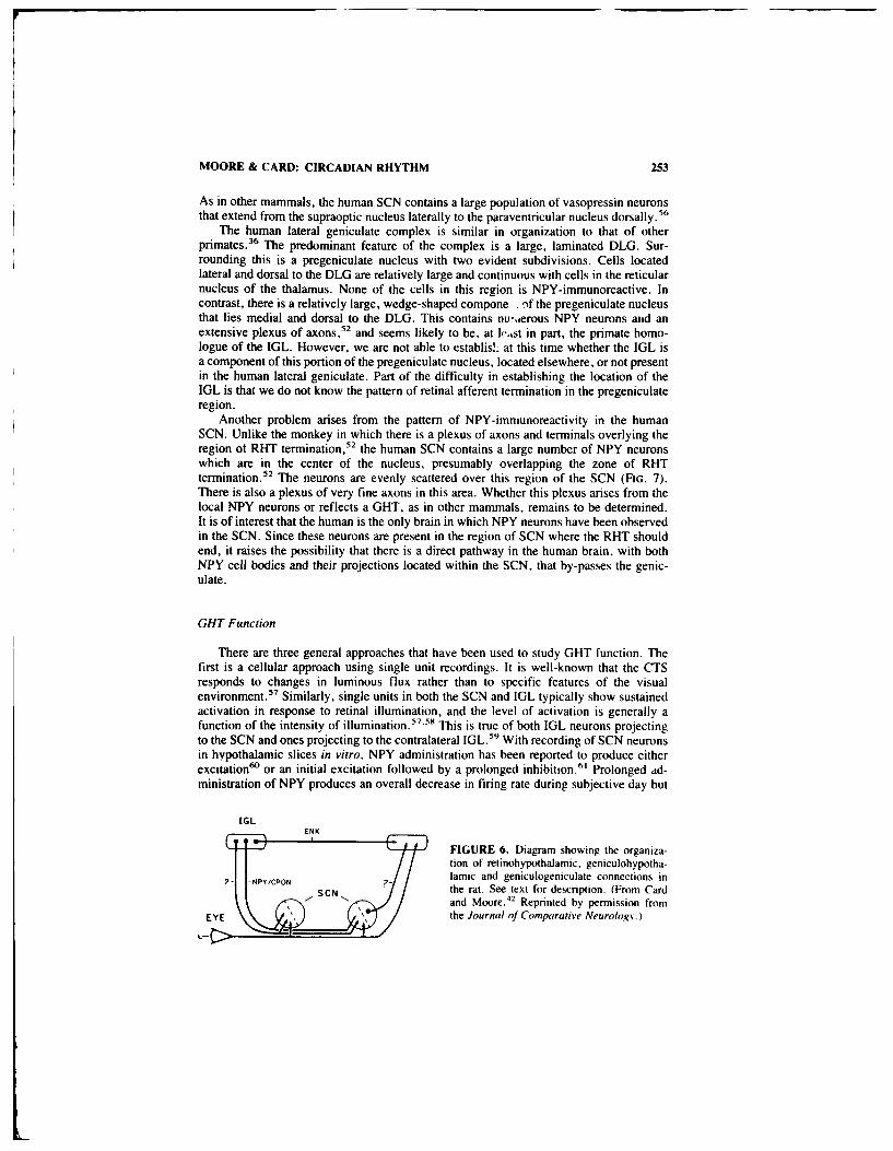
MOORE & CARD: CIRCADIAN RHYTHM 253
As in other mammals, the human SCN contains a large population of vasopressin neuronsthat extend from the supraoptic nucleus laterally to the paraventricular nucleus dorsally.56
The human lateral geniculate complex is similar in organization to that of otherprimates. 36 The predominant feature of the complex is a large, laminated DLG. Sur-rounding this is a pregeniculate nucleus with two evident subdivisions. Cells locatedlateral and dorsal to the DLG are relatively large and continuous with cells in the reticularnucleus of the thalamus. None of the cells in this region is NPY-immunoreactive. Incontrast, there is a relatively large, wedge-shaped compone -'f the pregeniculate nucleusthat lies medial and dorsal to the DLG. This contains nu-aerous NPY neurons and anextensive plexus of axons,52 and seems likely to be, at le.,st in part, the primate homo-logue of the IGL. However, we are not able to establis!: at this time whether the IGL isa component of this portion of the pregeniculate nucleus, located elsewhere, or not presentin the human lateral geniculate. Part of the difficulty in establishing the location of theIGL is that we do not know the pattern of retinal afferent termination in the pregeniculateregion.
Another problem arises from the pattern of NPY-immunoreactivity in the humanSCN. Unlike the monkey in which there is a plexus of axons and terminals overlying theregion ot RHT termination,52 the human SCN contains a large number of NPY neuronswhich are in the center of the nucleus, presumably overlapping the zone of RHTtermination. 5 2 The neurons are evenly scattered over this region of the SCN (FIG. 7).There is also a plexus of very fine axons in this area. Whether this plexus arises from thelocal NPY neurons or reflects a GHT, as in other mammals, remains to be determined.It is of interest that the human is the only brain in which NPY neurons have been observedin the SCN. Since these neurons are present in the region of SCN where the RHT shouldend, it raises the possibility that there is a direct pathway in the human brain, with bothNPY cell bodies and their projections located within the SCN, that by-passes the genic-ulate.
GHT Function
There are three general approaches that have been used to study GHT function. Thefirst is a cellular approach using single unit recordings. It is well-known that the CTSresponds to changes in luminous flux rather than to specific features of the visualenvironment.5 Similarly, single units in both the SCN and IGL typically show sustainedactivation in response to retinal illumination, and the level of activation is generally afunction of the intensity of illumination. 57' 58 This is true of both IGL neurons projectingto the SCN and ones projecting to the contralateral IGL. 59 With recording of SCN neuronsin hypothalamic slices in vitro, NPY administration has been reported to produce eitherexcitation' or an initial excitation followed by a prolonged inhibition. 6' Prolonged ad-ministration of NPY produces an overall decrease in firing rate during subjective day but
IGLENK
FIGURE 6. Diagram showing the organiza-tion of retinohypothalamic, geniculohypotha-lamic and geniculogeniculate connections in
- -'0the rat. See text for description. (From Cardand Moore. 42 Reprinted by permission from
EYE the Journal of Comparative Neurology.)

254 ANNALS NEW YORK ACADEMY OF SCIENCES
has no effect during subjective night." These observations are consistent with the viewthat the GHT would function in entrainment.
The second approach to analysis of GHT function has been to study the effects ofablation of the IGL on circadian function. It is clear that ablation of the lateral geniculatecomplex, including the IGL, does not disrupt the stable entrainment of circadianrhythms. 62 'o An early study with lesions of the primary optic tracts, and presumably theGHT, reported some alteration of responses to a change in phase and a lengthenedcircadian period.6 3 Subsequent studies, using either electrolytic or neurotoxic lesions ofthe IGL, have failed to find an alteration of circadian period but have routinely demon-strated changes in re-entrainment. 49 645" Two studies5o"5 have shown an alteration of thephase angle of entrainment in stable, entrained activity rhythms. Finally, Harrington andRusak66 studied the effects of IGL lesions on free-running rhythms in constant light andobserved a shortened free-running period. a decrease in the likelihood of splitting and anelimination of splitting in some animals. These studies, taken together, provide furthersupport for the view that the GHT participates in the entrainment.
The third approach has been to analyze the effects of NPY administration to the SCNon circadian function both in vivo and in vitro. The initial study of this type used avianpancreatic polypeptide," 7 or NPY,"8 superfused into the SCN region in intact, behavinganimals in constant conditions. Both peptides produced dramatic, and virtually identical,changes in the phase of the circadian activity rhythm with a phase response curve (PRC)that is quite distinctive and most closely resembles the PRC obtained from administeringdark pulses to animals in constant light 69
.7
) This PRC is characterized by phase delaysin early subjective day followed by phase advances in mid- and late subjective day witha transition to phase delays in subjective night (FiG. 8). Although the designation iscertainly tentative, we shall refer to this as a D, or dark, type PRC to contrast it with theusual PRC obtained from exposing animals in constant darkness to light pulses. That PRCis typically characterized by little alteration of phase during subjective day, phase delaysduring early subjective night and phase advances during late subjective night . 2
Very similar changes in phase can be obtained from acute stimulation of the IGL invivo. In our study examining the effects of IGL ablation on circadian function,"' we used
I PVN
I~SC
FIGURE 7. The human suprachiasmatic nucleus. (A) Diagram showing the location of the SCN inthe chiasmal hypothalamus. Abbreviations: OC = optic chiasm: PVN = paraventricular nucleusSCN = suprachiasmatic nucleus: SON supraoptic nucleus: 3V = third ventricle. (B) Photomi-crograph of NPY-immunoreactive neurons and axons in the SCN (marker bar = 100 tLm).

MOORE & CARD: CIRCADIAN RHYTHM 255
zD-
',. FIGURE 8. Diagram showing simulated phaseS--- -response curves (PRC). See text for description.
zL refers to a light-type PRC and D to a dark-, orASE NPY-type PRC.
o 12 18 24
CIRCAOIAN TIME
the excitatory neurotoxin. N-methylaspartic acid, to produce the lesions. Like otherexcitatory agents, this produces prolonged and excessive neuronal excitation resulting incell death. In so doing, it will produce neuronal firing and release of transmitter from theterminals of affected neurons. The lesions in our study were made at different times of dayand the animals were returned to their running wheels soon after surgery. The activityrhythms in operated animals exhibited phase changes that were dependent on the time the!esions were made and consistent with those observed by Albers and his colleagues67" 68
with NPY infusion.Similarly, we examined acute phase changes in firing rates of single SCN neurons
recorded in hypothalamic slices in vitro. 7' Stimulation of the optic nerve, or chiasm,produces changes in firing rate with a PRC very similar to that for light. In contrast, NPYadministration produces changes in firing rate with a PRC very similar to that obtainedwith NPY administration in vivo.6 8 It is of interest that the changes in firing rate occurimmediately. This suggests that the change in phase of pacemaker function may also beimmediate. These changes in firing rate have been maintained for three days in similarcircumstances7 2 indicating that they are likely to reflect a permanent change in pacemakerphase.
A series of recent studies have suggested that these two types of PRCs reflect twoforms of SCN response to environmental stimulation. In the intact animal, systemicprotein synthesis inhibition, injection of triazolam,74 social interaction, 75 or local in-jection of a protein synthesis inhibitor76 or glutamate 77 into the SCN area all producephase changes that resemble NPY, or dark. Similarly, administration of a protein syn-thesis inhibitor, calmodulin inhibitors78 and cAMP72 to hypothalamic slices in vitroproduces changes in the firing rate rhythm that are similar to those produced by NPY. Incontrast, pentobarbital, which produces electrical silence during administration, does notalter the firing rate rhythm but depolarization of the slice with potassium produceschanges that are similar to optic chiasm stimulation. 78 These observations argue both thatthe regulation of circadian phase is complex and that there appear to be two principalintracellular pathways for affecting pacemaker mechanisms to change phase. One is thepathway activated by light and the other is the pathway activated by NPY. The mecha-nisms by which these interact to control pacemaker function in the intact animal will bea fascinating subject for future investigation.
ACKNOWLEDGMENTS
We are deeply indebted to Join Speh for her superb technical assistance.

256 ANNALS NEW YORK ACADEMY OF SCIENCES
REFERENCES
I R USAK, B. & I. ZUCKER. 1979. Physiol. Rev. 59: 449-526.2. M-IJER, J. H. & W. J. RIErvEi). 1989. Physiol. Res. 69: 671-707.3. MURAKAmI. D. M.. J. D. MILLER & C. A. FullER. 1989. Brain Res. 482: 283-296.4. PICKARD, G. E. 1985. Neurosci. Lett. 55: 211-217.5. Mit.HOUSE. 0. E. 1977. Brain Res. 137: 351-355.6. MOORE, R. Y. & N. J. LENN. 1972. J. Comp. Neurol. 146: 1-14.7. MooRE, R. Y. 1973. Brain Res. 49: 403-409.8. HENDRICKSON, A. E., N. WAGNER & W. M. COWAN. 1972. Z. Zellforsch. 135: 1-26.9. JOIHNSON. R. F.- L. P. MoRIN & R. Y. MOORE. 1988. Brain Res. 462: 311-312.
10. KLEIN. D. C. & R. Y. MoORE. 1979. Brain Res. 174: 245-262.11. JOHNSON, R. F.. R. Y. MOORE & L. P. MoRIN. 1988. Brain Res, 460: 297-313.12. ROSENWASSER A. N. & N. T. ADLER. 1986. Neurosci. Behav. Rev. 10: 431-448.13. MOORE-EDE, M. C.. F. M. SULZMAN & C. A. FuLLER. 1982. The Clocks That Time Us.
Harvard University Press. Cambridge, MA,14. PITTENDRIGH, C. S. 1974. In The Neurosciences-Third Study Program. F. 0. Schmitt & F.
G. Worden, Eds. 437-358. MIT Press. Cambridge. MA.15. BoUOS. Z. & M. TERMAN. 1979. J. Comp. Physiol. 134: 75-83.16. MooRE. R. Y. L. P. MORIN & S. SHIBATA 1990. J. Biol. Rhythms. Submitted.17. HONMA, K. IL. S. HONMA & T. HIRoStIIGE. 1987. Physiol. Behav. 40: 767-774.18. STEPHAN, F. K. 1983. Behav. Brain Res. 7: 81-92.19- INOUNE, S.-I. & H. KAWAMURA. 1979. Proc. Natil. Acad. Sci. USA 76: 5962-5966.20. INOLyE. S.-I. & H. KAWAMURA. 1982. J. Comp. Physiol. 146: 153-166.21. GREEN. D. J. & R. GiLElIrE. 1982. Brain Res. 245: 198-2(X).22. GROoS. G. & J. HENDRICKS. 1982. Neurosci. Lett. 34: 283-288.23. SHtBAr,. S,, Y. OMUR,A. H. Kit., & K. HArioRI. 1982. Brain Res. 247: 154-158.24. SCHWARZ, W. J.. L. DAV ISEN & C. SMITH. 1980. J. Comp. Neurol. 189: 157-167.25. NEWMAN, G. C. & F. HospoD. 1986. Brain Res. 381: 345-35(0.26. Sotswi.k S. & R. Y. MOORE. 198. Brain Res. 43%: 374-37'A.27. DRUCKER-COLIN. R., R. AGUIt AR-ROBI.ERO. F. GARCIA-HERNANDE , F. FERNANDEZ-
CANCINOA & F. B. BArIONI 1984. Brain Res. 311: 353-357.28. SAWAKI, Y.. I. NiiIONMAISu & H. KAWAMURA. 1984. Neurosci. Res. 1: 67-72.29. LEHMAN. M. N., D. SILVER. W. R. GLADSIONE. R. M. KAHN. M. GIBSON & E. L. BIVMAN.
1987. J. Neurosci. 7: 1626-1638.31. RALPIH. M. R. & M. MENAKER. 1988. Science 241: 1225-1227.31. RALPH. M. R.. R. G. Fosif.R. F. C. DAVIS & M. MFNAKER. 1990. Science 247: 975-978.32. SWANSON. L. W. & W. M. COWAN. 1975. J. Comp. Neurol. 160: 1-12.33. SiEptAN, F. K.. K. M. BERKE EY & R. Moss. 1981. Neuroscience 6: 2625-2641.34. BERK. M. L. & J. A. FINKI ESLEIN. 1981. Brain Res. 226: 1-13.35. WArTS, A. G.. L. W. SWANSON & G. SANCHEZ-WATTS. 1987. J. Comp. Neurol. 258:
204-229.36. JONEs, E. G. 1985. The Thalamus. 724-733. Plenum Press. New York.37. SWANSON, L. W.. W. M. COWAN & E. G. JONES. 1974. J. Comp. Neurol. 156: 143-163.38. RIBAK, C. E. & A. PEITERS. 1975. Brain Res. 92: 261-294.39. HCKEY. T. I. & P. 1). SPEAR. 1976. Exp. Brain Res. 24: 523-529.40. CARD. 1. P. & R. Y. MOORE. 1982. J. Comp. Neurol. 206: 39( -396.41. MANTYi, P. W. & J. A. Kisi'. 1983. Brain Res. 288: 344-348.42. CARD. J. P. & R. Y. MOORE. 1989. J. Comp. Neurol. 284: 135-147.43. KRomER. I. F. & R Y. MOORE. 1980. Neuroscience 5: 155-271.44. MUGNAINI, P. & W. H. OIIFI . 1985. In GABA and Neuropeptides in the CNS. A. Bjork-
lund and T. Hklelh. eds. 436-622. Elsevier. Amsterdam.45 WAt rs, A. G & L. W. SWANSON. 1987. 1. Comp. Neurol 258: 230-252.46. MOORE. R. Y., E. . (USI ,FSON & J. P. CARD. 1985. Cell Tissue Res. 2.6: 41-46.47. HARRIN1otN. M. E., 1), M. NANCI & B. RuSAK. 1985. Brain Res. Bull. 15: 465-472.48. HARRIN6LON. M. E., 1). M. NAN(I- & B. RusAK. 1987. Brain Res. 410: 275-282.49. HARrINGION. M. E. & B RtSAK. 1986 J Biol. Rhythms I: 3(9-325.50 JOHNSON. R. F., R. Y MoORE & L. P. MoRIN. 1989. Brain Res. Bull. 22: 41-442.
lb'1
MOORE & CARD: CIRCADIAN RHYTHM 257
51. CASSONE. V. M.. J. C. SPEH, J. P. CARD& R. Y. MoORE. 1988. J. Biol. Rhythms 3: 71-91.52. MOORE, R. Y. 1989. Brain Res. 486: 190-194.53. CARD, J. P. & R. Y. MOORE. 1988. Neurosci. Lett. 88: 241-246.54. MORINb, L. P., J. BLANCHARD & R. Y. MOORE. 1990. Vis. Neurosci. Submitted.55. BRAAK, H. & E. BRAAK. 1987. Anat. Embryol. 176: 315-330.56. SWAAB, D. F., E. FLIERS & T. S. PARTIMAN. 1985. Brain Res. 342: 37-44.57. MEIJER, J. H., G. A. GROOS & B. RUSAK. 1986. Brain Res. 382: 104-118.58. HARRINGTON, M. E. & B. RUSAK. 1989. Vis. Neurosci. 2: 367-375.59. ZHANG. D. X. & B. RUSAK. 1989. Brain Res. 504: 161-164.60. MASON, R., M. E. HARRINGTON & B. RUSAK. 1987. Neurosci. Lett. 80: 175-179.61. SHIBATA, S. & R. Y. MOORE. 1988. J. Biol. Rhythms 3: 265-276.62. DARK. J. G. & D. ASDOURIAN. Physiol. Behav. 15: 295-301.63. RUSAK, B. 1977. J. Comp. Physiol. 118: 341-368.64. PICKARD, G. E., M. R. RALPH & M. MENAKER. 1987. J. Biol. Rhythms 2: 35-56.65. PICKARD, G. E. 1989. Brain Res. 494: 151-154.66. HARRINGTON, M.E. & B. RUSAK. 1988. Physiol. Behav. 42: 183-It).67. ALBERS, H. E., C. F. FERRIS. S. E. LEEMAN & B. D. GOLDMAN. 1984. Science 223: 833-
835.68. ALBERS, H. E. & C. F. FERRIS. 1985. Neurosci. Lett. 50: 163-168.69. BOULOS, Z. & B. RUSAK. 1982. J. Comp. Physiol. 146: 411-417.70. ALBERS, H. E. 1986. Am. J. Physiol. 250: R708-71 1.71. SHIBATA, S. & R. Y. MOORE. 1989. Soc. Neurosci. Abstr. 15: 24.72. PROSSER. R. A. & M. U. GILLETTE. 1989. J. Neurosci. 9: 1073-1081.73. TAKAHASHI, J. S. & F. W. TUREK. 1987. Brain Res. 405: 199-203.74. TUREK. F. W. & S. LOSEE-OLSON. 1986. Nature 321: 167-168.75. MROSOVSKY. N. 1988. J. Comp. Physiol. A162: 35-4676. INOUYE. S.-L., J. S. TAKAHASHI, F. WOLLNIK & F. W. TUREK. 1988. Am. J. Physiol.
255: R1055-1058.77. MEIJER, J. H., E. A. VAN DER ZEE & M. DIErz. 1988. Neurosci. Lett. 86: 177-183.78. SHIBATA. S. & R. Y. MOORE. 1990. 1. Biol. Rhythms. Submitted.

Role of Neuropeptide Y inReproductive Functiona
JOhN K. McDONALD
Department of Anatomy and Cell BiologyEmor Universi. School of Medicine
Atlanta, Georgia 30322
INTRODUCTION
Since its discovery in 1983, many investigators have examined the distribution andactivity of neuropeptide Y (NPY) in the reproductive neuroendocrine axis (see REFs. 1-3for reviews). NPY is present throughout the hypothaldmus, 4 pituitary,' and ovary,' andis found in high concentrations in the hypothalamo-hypophysial portal plasma. o- 2 Con-siderable evidence accumulated in recent years indicates that NPY imdulates the secretionof luteinizing hormone-releasing hormone (LHR-) from tiie hypothalamus and also af-fects secretion of reproductive hormones from the anterior pituitary gland.' 3 -21 Thesemodulatory effects of NPY may have physiological significance in the control of theneuroendocrine events preceding ovulation. This paper will present results of studies bythe author and his collaborators during the last several years elucidating the distributionand fuiction of NPY in the rct roductive neuroendocrine axis. These data are discussed inrelationship to other research in the 'e'i.
Distribution of NPY
NPY immunoreactivity has been found throughout the reproductive axis of severalspecies using both immunonistochemical and radioimmunoassay techniques (seereviewst 3 for detailed account). Standard immunohistochemical techniques have beenemployed to investigate the distribution of NPY. These techniques have been described indetail and the reader is referred to the references cited for additional information. Thehypothalamus displays high densities of NPY-immunoreactivity (IMR) in several nucleiwhich have been implicated in the control of reproductive hormone secretion. 3-5 7 Themedial preoptic nucleus and horizontal limb of the diagonal band contain numerousNPY-IMR fibers in the rat, some of which surround LHRH-IMR perikarya therebyprovid;ig an anatomical basis for some of the observed functional interactions betweenthese peptide systems. 22- 24 High concenrations of NPY containing fibers are also fouidin the arcuate, paraventricular, dorsomedial, and periventricular nuclei, in the lateralhypoth,lamic area and in the median eminence. The arcuate nucleus of the rhesus monkeycontains numerous NPY-IMR perikarya which appear to project axons to the infundibularstalk (FiG. I ) Numerous NPY-IMR axons in the superior aspect of the infundibular stalksurround vessels of the hypothalamo-hypophysial portal system while others continueinfenorly to the neural lobe (FIG. 2). Thus, NPY is probably secreted into the portal
"The support of the N-:iona! Institutes of Health (HD 19731. RCDA HD (K727 and 3RSGS07-RR-05364) and the March of Dimes Birth Defects Foundation iBasil O'Connor Scholar Re-search Award 5-525 and Basic Research Grant I-II 11 is gratelully acknowledged.
258
4

McDONALD: REPRODUCTIVE FUNCTION 259
V,,
I01
FIGURE 1. Distribution of NPY-immunoreactivity in the infundibular nucleus (IN) and upperinfundibular stem (UIS) of the rhesus monkey. High concentration of NPY-labeled penkarva in theventral IN. Note the stained fibers and perikarya in the UIS (arrows). some near blood vcssels. Bar= f00 im x 106. (McDonald, J- K., J. Tigges. M. Tigges & C. Reich. unpublished observations.)
vasculature of rhesus monkeys and other nonhuman primates and bathes the anteriorpituitary gland. In the rat, NPY-IMR fibers surround capillaries in the median eminencealthough the density is lower than in the rhesus monkey (Fi;. 3). " Ultrastructural im-munohistochemical analysis of the rat median eminence shows NPY-IMR axons adjacentto the perivascular space near fenestrated capillaries (Calka & McDonald, unpublishedobservations). High levels of NPY have been measured in the portal plasma of male andfemale rats (discussed below). In view of the direct effects of NPY on pituitary hormonesecretion, these anatomical observatons in the rhesus suggest that NPY may directlyinfluence anterior pituitary function in primates.
The ovary is richly innervated with NPY-containing fibers which are distributed
primarily around the vasculatire although other fibers course through the interstitial tissueand some are closely associated with follicles Fin. 4' 1' A ,asomoto (unction ot NPY insome perivascular ovarian nerves is likely although the function of interstitial and peri-follicular NPY-containing fibers remains unknown. Preliminary studies suggest that NPYalone or together with follicle-slinulating hormone IlISII) is a poor stimulus for csradiolor progesterone secretion from granulosa cell cullt.rcs. A potential endocrinc role in theovary, perhaps to affect secretion ot sonic nonstcr dal suhstance renialns to he detcemined

260 ANNALS NEW YORK ACADEMY OF SCIENCES
4P4.
FGR 2.Relationship of NPY-imrnunoreactive fibers to portal vessels within thL middle (M) ISand adjacent it) the anterior pituitary i AP) of the rhesus monkey. (A) Dense accumulation of NPY-positive varicosities surrounding several small and large vessels in the MIS. The labeled vessel (%)
4 distributes within the AP. Bar -510 p~n. x 169. (B) Mans varicose NPY-containing fibers surroundvessels (v) leading directl), intol the dorsal AP. Other fibers continue down the IS liar -5(0 pLrm), 100. Abbreviations as in legend to Fi;im 1, (Fn. 2A lromi Mc[)onald t al.' Reprinted hb.
permission from 0,11 and lihou' RcemarAu
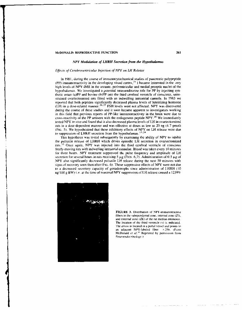
McDONALD: REPRODUCTIVE FUNCTION 261
NPY Modulation of LHRH Secretion from the Hypothalamus
EfJects of Cerebroventricular Injection of NPY on LH Release
In 198 1. during the course of immunocytochemical studies of pancreatic polypeptide(PP) immunoreactivity in the developing visual cortex, 25 I became interested in the veryhigh levels of NPY-IMR in the arcuate, periventricular and medial preoptic nuclei of thehypothalamus. We investigated a potential neuroendocrine role for PP by injecting syn-thetic avian (a)PP and bovine (b)PP into the third cerebral ventricle of conscious, unre-strained ovariectomized rats fitted with an indwelling intraatrial cannula. In 1983 wereported that both peptides significantly decreased plasma levels of luteinizing hormone(LH) in a dose-related manner.26 27 FSH levels were not affected. NPY was discoveredduring the course of these studies and it soon became apparent to investigators workingin this field that previous reports of PP-like immunoreactivity in the brain were due tocross-reactivity of the PP antisera with the endogenous peptide NPY. 2
" We immediatelytested NPY in vivo and found that it also decreased plasma levels of LH in ovariectomizedrats in a dose-dependent manner and was effective at doses as low as 20 ng (4.7 pmol)(FIG. 5). We hypothesized that these inhibitory effects of NPY on LH release were dueto suppression of LHRKH secretion from the hypothalamus. 13.29
This hypothesis was tested subsequently by examining the ability of NPY to inhibitthe pulsatile release of LHRH which drives episodic LH secretion in ovariectomizedrats. " Once again, NPY was injected into the third cerebral ventricle of consciousfreely-moving rats with indwelling intraatrial cannulae. Blood was taken every 10 minutesfor three hours. NPY treatment suppressed the pulse frequency and amplitude of LHsecretion for several hours in rats receiving 5 .g (FIGs. 6,7). Administration of 0.5 [Lg ofNPY also significantly decreased pulsatile LH release during the next 50 minutes withsigns of recovery soon thereafter (FIG. 6). These suppressive effects of NPY were not dueto a decreased secretory capacity of gonadotrophs since administration of LHRH (10ng/ 100 g BW) i.v. at the time of maximal NPY suppression of LH release caused a 1239%I
FIGURE 3. Distribution of NPY-immunoreactivefibers in the subependymal zone, internal zone (ZI),and external zone (ZE) of the rat median eminence.The location of the third ventricle (v) is indicated.The arrow is located in a portal vessel and points toan adjacent NPY-labeled fiber. x 256. tFromMcDonald et al, Reprinted by pemfission fromNeuroendoc'rinolog v.

262 ANNALS NEW YORK ACADEMY OF SCIENCES
FIGURE 4. NPY- immunofluorescent fibers in the rat ovary which surround this vessel (V) inlongitudinal section may give rise to fibers which appear intimately associated with a follicle Fi.X 256. 1From McDonald et a/.' Reprinted by permission from Endocrinologv.)
increase in plasma LH levels compared to a 124%4 increase in control animals injectedwith saline into the third ventricle (Fio. 8). These data support the notion that NPY mayhe a component of the LHRH pulse generating system. Perhaps the NPY-containingneurons of the arcuate nucleus exert a modulator' influence on the pulse generator in thebasal hypothalamus. ' Other investigators have observed that push-pull perfusion of NPYinto the hypothalamus of ovariectomizd rabbits decreases pulsatile LHRH release. "9
Estrogen changes the direction of the effect of NPY on LH and LHRH secretion.I.c.v. injection of NPY in ovariectoniied rats treated with estrogen and progesteroneproduces a transient stimulation of I.tt release. " Gonadally intact rabbits also displayNPY-induced LHRH secretion during push-pull perfusion of the hypothalamus. ', Inves-tigations carried out in vitro suggest that the stimulatory effects of NPY on LHRHsecretion in steroid-treated ovariectotnii.ed rats may occur in the medial basal hypo-thalamus. ' perhaps specifically at the median eminence. "' In contrast. NPY suppressionof IfRI-I secretion in oariectomi/ed rats seems to require additional hypothalamicnuclei. We initiated an extensic series of studies to explore the steroid dependent, ofNPY modulation f I.IR I release Ir nm the median eminence in 0tro.
N,'P ,$ldilllat!onl oP tl ,.'(wtolntl In Vitro
Role of E'strgecn. Rals vcrc bilatcrall ovaricctioied and 2 -4 weeks laler receivCdOne or s.'\cril Sila.tic tubes s.t Lonlailfl! estradiol henioate in oil or oil \ehicic Thrce

McDONALD: REPRODUCTIVE FUNCTION 263
days later, animals were sacrificed, trunk blood collected and the median eminenceremoved as described in detail and placed individually in test tubes (see Ri:. 16 forexperimental details). After several rinses with modified Krebs-Ringer bicarbonate me-dium containing glucose, a 30-minute basal sample was taken followed by administrationof NPY (0. 1-10 tLM) during a 30-minute test period. Medium was assayed for LHRH.NPY had no effect on LHRH secretion from median eminences obtained from ovariec-tomized rats and did not significantly modify potassium-induced LHRH release. Thus,NPY suppression of LH release in vivo may be mediated by other sites than the medianeminence. The estrogen treatments produced physiological plasma levels of estradiolsimilar to those measured during the estrous cycle. 6 Median eminences were harvestedit, the morning between 0900-1000 h when plasma levels of LH were low, significantlybefore the afternoon LH surge. However. the median eminence content of LHRH and thebasal release of LHRH were significantly increased by these treatments. NPY levels weresignificantly increased only following treatment with the highest dose of estrogen. There-fore all data concerning LHRH release during the test period were expressed in terms ofthe basal release of LHRH in the preceding period. When plasma estradiol levels were low(17 pg/ml), as in diestrous, NPY produced a dose-related increase in LHRH release.Treatment with two or four times this dose of estrogcn produced higher physiologicallevels of circulating estradiol (30 and 73 pg/ml) comparable to levels on proestrous. Thesehigher doses of estrogen also caused a dose-related increase in the sensitivity of themedian eminence to NPY. NPY-stimulated LHRH secretion increased significantly inanimals with higher plasma levels of estradiol (FIG. 9. Thus it seems that the median
24o..0 Saline (N=7)
A- A O021.g NPY (N=5)20 0-0 20,qg NPY(N 6)
20 r1 0 -o---------------.............. .
6 -- o ---
12
E *oA
8
4 *
0 5 15 30 60 120Minutes
FIGURE 5. [fect of the third cerebrisentrICItar inection 0\ ( (I we saline and NM' on plasnalesel ol IH in consLc1o1 . unrestraincd. ivariectoni/ed rats In thi's ligr., poml/ and %cr0iu l i frrepresent inearl valle . SlNI n . / t dl i't h p int reprccit (ihe Ie. el ot inlictacnce ',khen,ompared to ,altne-in'ceted control.. p , Ii -O1s 0 i . 1) i NP' doses skcre 2 (0
and ) (12 p.gI |orn Mcl)onald t a/. Reprinted h\ permni-io from the Naional ,adem\ ofsience I

264 ANNALS NEW YORK ACADEMY OF SCIENCES
SALINE SALINE
3000 A
A L 5000EZ 2000- -40001--
-3000-J "I
1000- 2000 -'
-1000400 0004000- .5 ug NPY .5 ug NPY 5000
-40003000-
A A 300-305 2000-
-J A 2000
1000- -1000
0' " I 0
4000- 5 ug NPY 5 ug NPY -5000
.~ 30004000E 3000- ~-3000
2000-
- 20001000- I s% -1000
01*
0 20 40 60 80 100 120 0 20 40 60 80 100 120
TIME (min) TIME (min)
FIGURE 6. Individual profiles of LH secretion (nanograms per ml) in OVX rats receiving a3V-injection of NPY (5.0 or 0.5 I±g/2 W) or saline (2 WA) at 0 min. Symhol indicates a pulse of LHsecretion. (From McDonald et al." Reprinted by permission from Endocrinolg v)
eminence is sufficient to demonstrate NPY stimulation of LIIRH release in estrogen-treated ovariectomized rats.
Effects of Progesterone in Estrogen-Treated Ovariectomized Rats: In view of theability of physiological plasma levels of estrogen to change the direction of the effect ofNPY from suppression in ovariectomized rats to stimulation, and the reported enhance-ment by progesterone of NPY-stimulated LHRH release from medial basal hypothalamicfragments in vitro, ,7 we performed experiments to investigate the hypothesis that pro-gesterone increases NPY-stimulated LHRH secretion from the median eminence invitro. ' Rats were bilaterally ovariectomized and implanted s.c. with Silastic capsulescontaining estradiol benzoate in oil (235 ;,g/ml) to produce physiological plasma levels ofestradiol. as described above. Three days later, animals were injected between 08(X)-0830 hours with progesterone (1, 2, or 19 mg s.c. per animal) or oil vehicle. Animalswere sacrificed three hours after injection and trunk blood was collected. Median emi-nences were removed and either sonicated in 0. IN HCI to determine levels of NPY andLHRH. or placed in test tubes containing culture medium to examine the effects of NPYon LHRH secretion. The I- and 2-mg doses of progesterone produced physiologicalplasma levels of progesterone (30 and 64 ng/ml, respectively) while injection of 19 tug

McDONALD: REPRODUCTIVE FUNCTION 265
resulted in pharmacological levels (534 ng/ml) that were 10 times greater than during theestrous cycle. In no case did progesterone treatment enhance NPY-stimulated LHRHsecretion from the median eminence in vitro. Injection of I mg had no effect on themedian eminence content of LHRH. while 2 mg and 19 mg of progesterone significantlyincreased LHRH levels. In spite of these changes, NPY stimulation of LHRH release wasslightly less effective in rats treated with the 2-mg dose, and was significantly reduced inanimals receiving 19 mg of progesterone, although these animals displayed significantlyelevated basal release of LHRH .3" These results indicate that progesterone administrationto ovariectomized estrogen-treated rats does not enhance NPY-stimulated LHRH releasefrom the median eminence when examined three hours later. LHRH levels are increasingat this time, well before the afternoon LH surge, yet NPY is apparently less effective instimulating the release of available LHRH. It is interesting to speculate that progesterone
2500"
-J C IZ 1500-
500-<
2000-
I
0...ea 1000
cFIGURE 7. This four-panel histogram summa-
0L L rizes the effects of NPY (5.0 or 0.5 ig) on thetu mean level, trough level, pulse amplitude, anda 2000- pulse frequency of LH secretion over the 2-h sam-
piing period (mean - SEM). *. p <0.05: **. pJE IT1 <0.01 (vs saline (SAL)-injected controls). (From
McDonald et al. Reprinted by permission from1" 0 Enkwcrinologx. !
0-J
4-0 0 00
.U W
-L J
0- 0
SAL .5 ug 5 ugNPY

266 ANNALS NEW YORK ACADEMY OF SCIENCES
2000
OY 5I' u g
USALINE
1500 FIGURE 8. Effect of NPY on the pituitary LH re-
sponse to iv LHRH. Plasma levels of LH (mean ±SEM) in OVX rats receiving a 3V injection of NPY (5Rg: circle) or saline (square). At 60 min, when LH
WOO - levels were significantly reduced in NPY-treated rats,LHRH (10 ng/100 g BW) was injected iv (arrow).Saline injected animals showed a 124% increase inplasma LH 10 min after LHRH administration. Incontrast. NPY-treated animals responded with a
500 1239% increase in LH release (p <0.01 vs controls).(From McDonald et al."' Reprinted by permissionfrom Endocrinology.).
0*
0 40 80 120 160 200
TIME in-,)
exerts an inhibitory influence during and after the afternoon LH surge to limit NPYstimulation of LI-RH secretion . 30 Additional research employing both in vitro and in vivo
methods is needed to fufly explore the effects of progesterone on NPY-induced LHRHsecretion throughout the afternoon and evening of the LH surge. Taken together, these
375
300
W~ 225*W I-
ZU. 150
6 5NPY (-log M)
FIGURE 9. Effects ol estrogen 0 E I Ill-mm implant: 2E = 2 10-mm implants; 4E = 2 20-mmimplants of estradiol benzoate 235 iRgiitil or vehicle treatment on NPY (10 " and 10' M)-stimu-lated LHRH release from ME fragments ot OVX rats. ****, p <0 W1I. 4E vs OIL for 10' M NPY:*** p <0.005. 4E vs I E for 10 M NPY: ** p <0.0i, 4E vs 2E for 10 1M NPY; *, p <0.05,2E and I E vs OIL for 10 ' M NPY. and 4E vs 0IL tor 1) ' MI NPY. (From Sahatino et a/."'Reprinted by permission front Endotrmologv.)
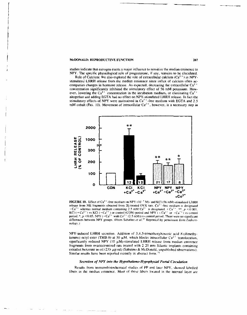
McDONALD: REPRODUCTIVE FUNCTION 267
studies indicate that estrogen exerts a major influence to sensitize the median eminence toNPY. The specific physiological role of progesterone, if any, remains to be elucidated.
Role of Calcium: We also explored the role of extracellular calcium (Ca2 ) in NPY-stimulated LHRH release from the median eminence since influx of calcium often ac-companies changes in hormone release. As expected, decreasing the extracellular Ca2
concentration significantly inhibited the stimulatory effect of 56 mM potassium. How-ever, lowering the Ca 2 concentration in the incubation medium, or eliminating Ca2
,
altogether and adding EGTA had no effect on NPY-stimulated LHRH release. In fact thestimulatory effects of NPY were maintained in Ca2 '-free medium with EGTA and 2.5mM cobalt (FIG. 10). Movement of intracellular Ca 2
, however, is a necessary step in
2000
- 1000lWlI.- i
. Z 300
cc O 200
100
0 1 12 21 1CON KCI KCI NPY NPY NPY
+Ca2
+ -Ca2 + +Ca
2 -Ca2 -Ca 2
+
+Co2*
FIGURE 10. Effect of Ca2 ' -free medium on NPY (10 5 M)- and KCI (56 mM)-stimulated LHRHrelease from ME fragments obtained from 2E-treated OVX rats. Ca2 '-free medium is designated-Ca 2 ' whereas normal medium containing 2.5 mM Ca2 ' is designated + Ca2 ' . **. p <0.001,KCI (+ Ca2 ') vs KCI (-Ca2' ) or control (CON) period and NPY ( + Ca2 ' or -Ca2 + ) vs controlperiod; *, p <0.05, NPY (-Ca2
2 with C02' (2.5 mM)O vs control period. There were no significantdifferences between NPY groups. (From Sabatino et at. " Reprinted by permission from Endocri-nology.)
NPY-induced LHRH secretion. Addition of 3,4.5-trimethoxybcnzoic acid 8-(dimethy-lamino) octyl ester (TMB-8) at 50 ltM, which blocks intracellular Ca2' translocation,significantly reduced NPY (10 1±M)-stimulated LHRH release from median eminencefragments from ovariectomized rats treated with 2 20 mm Silastic implants containingestradiol benzoate in oil (23b ptg/ml) (Sabatino & McDonald. unpublished observations).Similar results have been reported recently in abstract form. "
Secretion of NPY into the Hypothalamo-Hypophysial Portal Circulation
Results from immunohistochemical studies of PP and latcr NPY, showed labelledfibers in the median eminence. Most of these fibers located in the internal layer are

268 ANNALS NEW YORK ACADEMY OF SCIENCES
destined for the neural lobe and respond to osmotic stimuli. 32 Other stained fibers arelocated in the external layer and lateral aspect of the median eminence in the vicinity ofcapillaries of the hypothalamo-hypophysial portal system (FIG. 3). These observationscombined with the direct stimulatory effect of NPY on hormone secretion from anteriorpituitary cells lead us to hypothesize that NPY is released from fibers in the medianeminence into the perivascular space surrounding these fenestrated capillaries. This hy-pothesis was investigated by collecting portal blood from the transected pituitary stalk ofmale rats and comparing the levels of NPY-IMR with those in a sample of peripheralblood taken simultaneously. ") Increasing volumes of plasma produced parallel displace-ment of labelled NPY in the RIA. In addition, chromatographic characterization of thesesamples with successive HPLC runs to obtain high resolution of NPY-IMR showed thatthis IMR eluted as a single peak which displayed a slightly shorter retention time than theporcine NPY standard. This difference was likely due to the methionine for leucinesubstitution which occurs in position 17 in rat NPY versus porcine NPY. Human and ratNPY elute slightly earlier than porcine NPY in these HPLC solvent and gradient condi-tions. Portal plasma levels of NPY were 3-4 times greater than in the peripheral circu-lation. These results support the hypothesis that NPY is released into the portal circulationand acquire additional significance in view of NPY stimulation of anterior pituitaryhormone secretion. Other investigators have provided compelling evidence that NPYlevels in the portal plasma are elevated in concert with LHRH before the LH surge inestrogen- and progesterone-treated ovariectomized rats, and also before the first LH surgeat pubertal onset.'' '2 These results combined with those in the next section stronglysuggest that NPY exerts a direct regulatory influence on anterior pituitary function.
NPY Modulation of Reproductive Hormone Secretion from the AnteriorPituitary Gland
In 1984, we reported that application of NPY stimulated the release of LH, FSH andgrowth hormone from dispersed anterior pituitary cells which were obtained from ova-riectomized rats, mixed with Biogel P2, and perifused in syringe columns (FIG. 11)13-29These original findings of direct effects of NPY on anterior pituitary hormone releasewere later supported by other investigators.2' However the test system apparently has agreat influence on the ability to detect NPY activity. 18
,3
1,34 Use of hemipituitary frag-
ments or long-term cultures of dispersed cells did not reveal any effect of NPY onhormone release. 11.3' Khorram et al.2) placed quartered rabbit anterior pituitaries in aperifusion system and demonstrated transient NPY-stimulated LH and FSH secretion frompituitaries of ovariectomized animals and a sustained increase in LH and FSH release fromglands of intact animals. This indicates that ovarian steroids modulate the sensitivity ofthe anterior pituitary gland to NPY. Other investigators have reported that NPY exerts aslight but significant stimulation of LH release aad also increases the LH secretoryresponse of gonadotrophs to LHRH using hemipituitary fragments." When NPY wasapplied to three-day cultures of dispersed anterior pituitary cells there was no effect on LHrelease but the response to LHRH was potentiated. " Peripheral injection of NPY antise-rum blocked the LH surge in ovariectomized rats treated with estrogen and progesterone.These investigators suggested that NPY serves in a modulatory role to prime gonado-trophs to LHRH-.
SUMMARY
NPY acts both at the hypothalamus and the anterior pituitary gland to modulatereproductive hormone secretion (FiR;. 12). Within the hypothalamus, NPY stimulates

McDONALI): REPRODUCTIVE FUNCTION 269
NPY(10 'M) NPY 110 M)
125-
* 100-
~75C 50AA
25-
FI(;URE 11. Li. FSit. and (A released frrnt
200 a peritused Icll column BitoGel P-2. it 4li 1s~te vah7 lt ipr datro
15 tttar cells io ariectani/ed tettiale rat disnorsiFitse-tlltnuie traction, ssere collected and NPN
X 100(i" anid 10 Mi Skas included Tin the inediutim
U-50- 1J ''"ssI/sss Est ~tanl is
prinited h\ pcrttii' ion I ra te NTIIII at IoIIIalIA1 Ad -
x0 -- 0
elmis oit Scien:C, I
x
oJ1 0 1's 20 35 40 4*5
Fraction
LHRH secretion in the presence at' physiological level,, of estrogen and suppresses pul-satile LHRH release t'ollovtine ovariectomv. Intracerebroventricular injectlion of' NPYantiserum blocks or delays the L H surgec in sterisid-primied m.variectomi/ed rat,,,." therebyadding support for a physiological role of NPY in the neuroendocri events, precedingovulation. Blockade at alpha 2 adrenergic receptors decreases NPY'-stimiulated[Li- releasein steroid-primed rats impising a potential noradrenergic mediation of NPY actis its.Physiological levels of' progesterone do not aug-ment. and may actually suppress NPY-induced LHRH secretion it) T-iro tramn median eminences obtained trom estrogenr-pritiedas ariectomniizd rats. The physiological role of progesteronie. it arN~ . in miodulating NP'teffects on LHRH release remains to he determined. Little. it atNsthing. is, kno)%n ab~out the
is a ~si ~ FIGU'RE 12. SunitimarN dAwratts 'hikkuti thedual ste IT action III NPY inI modulating repros-
ductise litnntic release trolt the fix pathalamtu,and ainterior piuit ar% gland See Sutamars in
11511test 101r aliisnal dIisCLsIsMII. I represetits thepertusissis efect atI esiroiets ott NtVY-sttulatedlitRH release I nt thec h~ psthalatisu theIkis ioteial roles ot prittrs site it) atnd nore -piliephlritli I NL) Inl tIssidLIalaIng this inf luenice sll
Pi il tsI A" ' retta o iie deterinted. Yhe Isswg s'/s/isui
sirris1 represer[tit li i\psthalisis-h paph\ sPilalil i Pontal \ ee IsAsh tch trasnxpo ri hi sh NPIY arid
I tRI to tlie goniadlsirsph I as I %k here these',1 fICpIAlt'. appear Its act Itepetsdent111 aud als
e Ititerici ItI the itissdillaiiisist.,t lit ecllonst

270 ANNALS NEW YORK ACADEMY OF SCIENCES
NPY receptor in the median eminence or the intracellular mechanisms which transduccthe NPY signal into activation of LHRH release in estrogen- treated ovariectomized ratsalthough translocation of intracefllular calcium is required. Equally puzzling is the mech-anism of desensitization of the LHRH-releasing mechanisms of the median eminence ofovariectomnized rats or the specific site of NPY suppression of pulsatile LHIRH secretion.
INPY is released into the hypothalamo-hypophysial portal circulation and this appearscorrelated with LHRH secretion before the LH surge. NPY affects LH and FSH releasefrom anterior pituitary cells in vitro and enhances LHRH-induced LH secretion.
Taken together. the studies described above suggest an important physiological rolefor NPY as a modulator of neuroendocrine activity which culminates in the preovulatorysurge of LH.
ACKNOWLEDGMENTS
4 1 would like to sincerely thank all of my collaborators who are referenced in theinvestigations described above.
REFERENCES
1. McDONALD. J. K. 1988. NPY and related substances. In CRC Critical Reviews in Neurobi-ology. J. Nelson. Ed. Vol. 4: 97-135. CRC Press. Boca Raton, FL.
2.MCCANN., S.V RFTTORI. L. MILENKOVIC M. RiFoFi.. C. AcuILlA & J. K. M(Do.ALD.1989. The role of neuropeptide Y in the control of anterior pituitary hormone relc~sc in therat. In Neuropeptide Y. Karolinksa Institute Nobel Conference Series. V. Mutt. K. Fuse. T.H~ikfelt & J. M. Lundberg. Eds. 215-228. Raven Press. New York. NY.
3. McDONALD. J. K. 1990. Reproductive neuroendocrine effects of neuropeptide Y and relatedpeptides. In Brain-Gut Peptides and Reproductive Function. Advances in Physiolog). C.Johnston & C. D. Barnes. Eds. Telford Press. Caldwell, NJ. In press.
4. CHRONWALL. B. M.. D. A. DiMAGGIO. V. J. MASSARI. V. M. PICKE~T, D. A. RucCotERO &T. L. O'DONOHtJE. 1985. The anatomy of neuropeptide-Y-containing neurons in rat brain.Neuroscience 15: 1159-1181.
5. DEQUIDT. M. E. & P. C. EMSON. 1986. Distribution of neuropeptide Y-like immunoreactivityin the rat central nervous system--[. Radioimmunoassay and chromatographic characterisa-tion. Neuroscience 18: 527-543.
6. DEQUIDT, M. E. & P. C. EMSON. 1986. Distribution of neuropeptide V-like immunoreactivit%in the rat c,:ntral nervous system-It. Immunohistochemnical analysis. Neuroscience 18: 545 -618.
7. SABATtNO. F. D., J. M. MURNANE. R. A. HOFFMAN & J. K. MUDONALo. 19487. The distri-bution of neuropeptide Y-like immunoreactivity in the hypothalamus of' the adult egldenhamster. J. Comp. Neurol. 257: 93-104
8. McDONAt.D. J. K.. J. Ttooeus. M. TiiGFs & C. R~icHt. 1988. Developmental study ofI neu-ropeptide V-like immunoreactivity in the neurohypophysis and intermediate lobe of the rhesus.monkey (Macaca mulatta). Cell Tissue Res. 254: 499-5094.
9. McDONAtD. J. K.. W. L. DE us, C. E. AHMED, B. D. Not: & S. R. OJFt),. 1987. Biochemicaland immunocytochemical characterization of neuropeptide Y in the Immature rat osary.Endocrinology 120: 1703-1710.
10. McDONALD. J. K.. 1. I. KOENto. D. M. GIBBtS. P. Cot t.INS% & H. 1). Not . 1987. Highconcentrations of neuropeptide V in pituitary portal blood of rats. Neuroendocritmlog,, 46:538-541.
If. SUTTON. S. W.. N. Mirsuot. P. Nf. P1.015KY & D. K. SARKAR. 19,S8. Neuropeptide Y (NPY).a possible role in the initiation of puberty. Endocrinology 123: 2152-2154
12. SUTTON S. W.. T. T. TOYAMA, S. OTTO & P. M. Pt OISKN. 1988. Evidence that neuropeptide

McDONALD: REPRODUCTIVE FUNCTION 271
Y (NPY) released into the hypophysial-portal circulation participates in priming gonadotrophsto the effects of gonadotropin releasing hormone (GnRH). Endocrinology 123: 1208-1210.
13. MCDONALD, J. K., M. D. LUMPKIN. W. K. SAMSON & S. M. MCCANN. 1985. NeuropeptideY affects secretion of luteinizing hormone and growth hormone in ovariectomized rats. Proc.Natl. Acad. Sci. USA 82: 561-564.
14. KALRA, S. P. & W. R. CROWLEY. 1984. Norepinephrine-like effects of neuropeptide Y on LHrelease in the rat. Life Sciences 35: 1173-1176.
15. MCDONALD, J. K., M. D. LUMPKIN & L. V. DEPAOLO. 1989. Neuropeptide Y suppressespulsatile secretion of luteinizing hormone in ovariectomized rats: possible site of action.Endocrinology 125: 186-191.
16. SABATiNO, F. D.. P. COLLINS & J. K. MCDONALD. 1989. Neuropeptide Y stimulation ofluteinizing tiormone-releasing hormone secretion from the median eminence in vitro by es-trogen dependent and Ca2 ' independent mechanisms. Endocrinology 124: 2089-2098.
17. CROWLEY, W. R. & S. P. KALRA. 1987. Neuropeptide Y stimulates the release of luteinizinghormone-releasing hormone from medial basal hypothalamus in vitro: modulation by ovariannormones. Neairoendocrinology 46: 97-103.
18. CROWLEY, W. R.. A. HASSID & S. P. KALRA. 1987. Neuropeptide Y enhances the release ofluteinizing hormone tLH) induced by LH-releasing hormone. Endocrinology 120: 941-945.
19. KHORRAM, 0.. K. Y. F. PAU & H. G. SPIES. 1987. Bimodal effects of neuropeptide Y onhypothalamic release of gopadotropin releasing hormone in conscious rabbits. Neuroendo-crilology 45: 290-297.
20. KHORRASI 0., K. Y. F. PAU & H. G. SPIES. 1988. Release of hypothalamic neuropeptide Yand effects of exogenous NPY on the release of hypothalamic GnRH and pituitary gonadot-ropins in intact and ovariectomized does in vitro. Peptides 9: 411-417.
21. HRFSTRAND, A., K. FUXE, L. F. AGNATI, P. ENEROTH. 1. ZINI, NM. Zot I, K. ANDERSSON.G. VON EULER. L. TERENIUS, V. MUTT & M. GOLDSTEIN. 1986. Studies on neuropeptideY-catecholamine interactions in the hypothalamus and in the forebrain of the male rat. Re-lationship to neuroendocrine function. Neurochem. Int. 8: 355-376.
22. CALKA. J. & J. K. MCDONALD. 1989. Neuropeptide-Y and LHRH in the rat hypothalamus:anatomical relationships. Soc. Neurosci. Abstract. p. 187.
23. MCDONALD. J. K., F. D. SABATINO & J. CALKA. 1988. Neuropeptide Y and GnRH in the rathypothalamus: physiological and anatomical relationships, hi Proceedings of the InternationalSymposium on Frontiers in Reproduction Research. 102. Beijing, PRC.
24. GUY, J., S. Li & G. PELLETIER. 1988. Studies on the physiological role and mechanism ofaction of neuropeptide Y in the regulation of luteinizing hormone secretion in the rat. Regul.Pept. 23: 209-216.
25. MCDONALD. J. K., J. G. PARNAVEiLs. A. N. KARAMANLIDIS & N. BRECHA. 1982. Themorphology and distribution of peptide containing neurons in the adult and developing visualcortex of the rat. IV. Avian pancreatic polypeptide. J. Neurocytol. 11: 985-995.
26. MCDONALD. J. K. & M. D. LUMPKIN. 1983. Third ventricular injections of pancreatic poly-peptides decrease LH and growth hormone secretion in ovariectomized rats. 65th AnnualMeeting of The Endocrine Society, 152.
27. MCDONALD, J. K., M. D. LUMPKIN, W. K. SAMSON & S. M. MCCANN. 1985. Pancreaticpolypeptides affect luteinizing and growth hormone secretion in rats. Peptides 6: 79-84.
28. DiMAGGOtO. D. A.. B. M. CHRONWALL. K. BUCHANAN & T. L. O'DONOHUE. 1985. Pancre-atic polypeptide immunoreactivity in rat brain is actually neuropeptide Y. Neuroscience15: 1149-1157.
29. McDONALD, J. K, M. D. LUMPKIN, W. K. SAMSON & S. M. MCCANN. 1984. Third ven-tricular injections of neuropeptide Y decrease LH and growth hormone secretion in ovariec-tomized rats. Soc. Neurosci. Abstract, p. 1214.
30. SABATINO, F. D., P. CoLt INS & J. K. MCDONALD. 1989. Effects of prog--terone on neiro-peptide Y stimulated LHRH release from rat median eminence. 71st Annual Meeting of TheEndocrine Society. 76.
31. CROWLEY, W. R.. G. V. SHAH. D. KENNEDY. M. E. INWI.R & S. P. KAI RA. 1989.Involvement of the calcium messenger system in the stimulation of luteinizing hormonesecretion by neuropeptide Y in rats. Soc. Neurosci. Abstract. p. 628.
32. Hoot. S. C, G. S. Ri(!iARDSON, 1. K. MCDoNAtD. J. M AiII. N, J. B. MARTIN & J. I.

272 ANNALS NEW YORK ACADEMY OF SCIENCES
KOENIG. 1989. Neuropeptide Y (NPY) and vasopressin (AVP) in the hypothialamo-neurohy-pophysial axis of salt-loaded and/or Brattleboro rats. Brain Res. 486: 214-220.
33. '(ERKERIAN. L., J. Guy,.G. LEFEVRE &G. PELLETIER. 1985. Effects of neuropeptide Y (NPY)on the release of antcrior pituitary hormones in the rat. Peptides 6: 1201-1204.
34. ROD)RIGUEZ-SIERRA. 1. F.. D. M. JACOBOWI rz & C, A. BLAKE. 1987. Effects of neuropeptideY on LH. FSH. and TSH release in male rats. Peptides 8: 539-542.
35. WEIIRENBERG. W. B.. R. CORDER & R. C. GAILLARD. 1989. A physiological role for neu-ropeptide Y in regulating the estro ertlprogesterv-.- induced luteinizing hormone surge iovariectomized rat,.. Neuroendocrino.,)gy 49: 68ti-6h?2
36. ALLEN, L. G._ W. R. CROWL.EY & S. P. KALRA. 1987. Iiweractions beiween neuropeptide Yand adrenergic systems in the stimulation of luteinizing horn,.'ne release in steroid-primedovariectomized rats. Endocrinology 1LI: 1953-1959.

Hypothalamic Neuropeptide Y: a Circuitin the Regulation of GonadotropinSecretion and Feeding Behavior"
S. P. KALRA, A. SAHU, P. S. KALRA, AND W. R. CROWLEY"
Department of Obstetrics and GynecologyUniversity of Florida College of Medicine
Gainesville. Florida 32610aild
hDepartment of PharmacologyUniversity of Tennessee College of Medicine
Memphis. T mnessee 38163
INTRODUCTION
Neuropeptide Y (NPY) is an important messenger molecule in the neuroendocrinecontrol of reproduction and appetitive behavior. The early history of the development ofour idea that members of the pancreatic polypeptide family. including NPY, may bcinvolved in the hypothalamic control of pituitary luteinizing hormone (LH) secretion, andof feeding and sexual behaviors is described elsewhere.' In this article the mechanism ofaction of NPY at cellular and subcellular levels in stimulation of LH release and feedingbehavior is described. In particular, attention is focused on the site and specificity of NPYaction, the functional connectivity of NPY with other neuropeptidergic and monoamin-ergic systems in the hypothalamus and brain stem (BS), and whether NPY is a physio-logical neurochemical signal in the induction of ovulation and stimulation of appetite.
ROLE OF NPY IN THE CONTROL OF HYPOTHALAMIC LHRH-PITUITARYLH SECRETION
Depending upon the existing status of the gonadal steroidal milieu. ,.ential adminis-tration of NPY either inhibits or stimulates the release of LH from the anterior pituitary. -2However, the excitatory nature of NPY is physiologically relevant because NPY stimu-lates LH release in gonad-intact rats and rabbits and in ovariectomized (ovx) rats pre-treated with ovarian steroids. 2 ' Further, this excitation of LH release was found to be dueto NPY action at two sites, one in the hypothalamus wherein it acted to stimulate LHreleasing hormone (LHRH) release, and the other at the level of the pituitary, where itpotentiated the release of LH induced by LHRH. 7
"
Mode of NPY Action in Stimulation of Hypothalamic LHRH Release
Our studies showed that an optimal level of communication between hypothalamicNPY and LHRH neurons required priming with gonadal steroids.X" " In general. NPY
"Supported by National Institutes of Health Grants HD 08634 (SPK). HD 11362 (PSK). DK 37273(PSK and SPK) and HD 13703 (WRC).
273

274 ANNALS NEW YORK ACADEMY OF SCIENCES
failed to alter the in vitro basal LHRH release from the medial basal hypothalamus (MBH)of steroid-unprimed ovx rats, but priming with either estradiol 17P (E2) alone or incombination with progesterone (P) resulted in stimulation of LHRH release by NPY. Infact, NPY ,-adily stimulated LHRH release from the MBH of rats primed with physio-logical levels of E, alone. Furthermore, NPY potentiated the release of LHRH in responseto K' -induced depolarization from the MBH of ovx rats. Therefore, it seemed that NPYcan not only stimulate LHRH on its own. but may also potentiate the response of LHRHneurons to depolarization produced by those synaptic inputs, such as adrenergic neuro-transmitters, which stimulate LHRH and LH release.
The anatomical evidence clearly shows that NPY exists in a number of neuronalpopulations in the brain stem (BS) which produce norepinephrine (NE) and epinephrine(EPI). " - " To what extent do these cells, showing coexistence of NPY and adrenergictransmitters, project into the hypothalamic sites innervated by LHRH neurons? What isthe nature of interaction between adrenergic transmitters and NPY in stimulation of theLHRH-LH axis'? Bilateral neural transection (NT) at the level of the dorsal tegmentum inthe mesencephalon not only drastically decreased NE and EPI levels in the POA andMBH, but also reduced the levels of NPY in selected sites in the preoptic-tuberalpathway. " Severing these BS projections to the hypothalamus reduced NPY concentra-tions in the medial preoptic area (MPOA) and median eminence (ME). It appeared that theafferents of the two populations of NPY-produc:.-g cells, the remote BS population whichalso contain NE or EPI, and the local arcuate nucleus (ARC) population project into areasinnervated by LHRH neurons. Based on the known topography of the LHRH network inthe diencephalon. it is reasonable to suggest that stimulation of LH release by NPYoccurred via axo-dendritic or axo-somatic synapses in the MPOA, and via axo-axonicsynapses in the ME region.
Allen et al 05 showed that such links between NPY and LHRH at the morphologicallevel are functionally operational in the ovarian steroid-primed ovx rats. Doses of NPYand NE, that on their own were minimally effective in stimulating LH release, whenadministered together induced a LH response greater than the sum of the individualresponses. On the other hand, combined doses of NPY and NE, which were eitherineffective or maximally effective, failed to induce an additive response. This interplaybetween NPY and adrenergic transmitters is uniquely complex. We were unable to blockthe NPY-induced LH release either by selectively blocking oi- or P-adrenoreceptors ordopamine receptors, or by stimulating opiate receptors in the hypothalamus. Intriguingly,however, stimulation of LH release by NPY was significantly suppressed by prior block-ade of a2-adrenoreceptors. Therefore, it appeared that either ct2-adrenoreceptors locatedat a postsynaptic site, mediated th- excitatory LHRH response, or that it was necessary tosustain optimal postsynaptic ot2 -adrenoreceptor activity by endogenous adrenergic trans-mitters, to fully express the action of NPY. However, currently we cannot rule out thepossibility that NPY may directly modulate hypothalamic a2-adrenoreceptor membranebinding sites that are coupled to the phosphatidylinositol second messenger system.
With respect to the receptor specificity of the NPY action, it is likely that the Y,receptor subtype mediates the excitatory effects of NPY on LHRH release. We observedthat a specific Y, receptor agonist, JLeu 3 ,Pro 4]-NPY,"6 readily stimulated LHRH.whereas the Y2 receptor agonist, NPY, 1,,17.18 was completely ineffective in stimulat-
ing LHRH release from the ME of ovarian steroid-primed ovx rats. ' Furthermore,stimulation of LHRH release by Y, receptor activation does not require extracellularCa2 * because LHRH release by NPY persisted in Ca2 ' -free/EGTA medium. In contrast,we observed that two antagonists of intracellular Ca2 ' mobilization. TMB-8 and ryan-odine, blocked the NPY-induced LHRH release. 19 This line of evidence favors the ideathat activation of the Y, receptor subtype subsequent to mobilization of intracellular Ca2
results in stimulation of LHRH release.t,

KALRA et al.: REPRODUCTION & APPETITE 275
Mode of NPY Action at the Level of the Pituitary
The morphological and experimental evidence favors a neurohumoral role of NPY instimulation of LH release directly from the pituitary gland.'' 22" NPY was found tostimulate LH release in vitro from either hemipituitaries or dispersed pituitLry cells.Further analysis revealed that the direct stimulatory effect of NPY on LH release was veryweak as compared to that of the physiological releaser LHRH, and demonstration of eventhis marginal response was dependent upon a number of factors such as NPY pulseduration and the previous hormonal status of the donor. 7 9 "1
On the other hand, NPY interacted with LHRH in a unique fashion. We found thatNPY potentiated LH release in response to LHRH. 7 " When added to dispersed pituitarycell incubations, NPY augmented the release of LH by LHRH in a dose-related fashion.Further, although we have not yet observed direct binding of NPY to pituitary mem-branes, NPY enhanced LIRH binding to its receptors on pituitary membranes, an actionlikely to contribute to augmented LH release by LHRH. 2 ' Furthermore, unlike thepostsynaptic LHRH stimulation response in the hyp.thalamus. 7" we observed that thedihydropyridine Ca2
2 channel antagonist, nitrendipin,, blocked the effects of NPY onLHRH-induced LH release. 10.22,23 Further, NPY enhanced the LHRH-induced increasein cytosolic Ca 2 as ,ell as the number of pituitary cells displaying increased intracel-lular Ca 2" in responst. to LHRH. 22 23
Consequently. we envision that NPY participates in the neuroendocrine control ofLHRH-LH axis in a novel way. At the two disparate sites-hypothalamus and pituitary-NPY displays a unique cooperativity with classial adrenergic transmitters and the LHRHpeptidergic system by dilferent intracellular modes of action, the optimal manifestation ofwhich is apparently dependent upon the gonadal steroid milieu.
Physiological Significance
Participation of NPY in the Induction of Ovulation
Because it readily stimulates both LHRH and LH release, we hypothesized that theNPY network may be an important component of the hypothalamic neural circuitryresponsible for the preovulatory discharge of LH. 2' We observed parallel changes in NPYand LHRH levels in the ME in association with the afternoon LHRH and LH hyperse-cretion either induced by ovarian steroids in ovx rats or that occurring spontaneously onthe afternoon of proestrus.2 .26 On proestrus, serum LH levels were basal between 1000and 1300 h, rose significantly at 1500 h and plateaued between 1600 and 1800 h. Of thefive hypothalamic sites examined, NPY levels displayed marked fluctuations only in theME in close association with the LH surge. 26 NPY concentrations were low between 1000and 1300 h, and rose abruptly at 1400 h preceding the onset of LH rise at 1500 h. Theseelevated levels were maintained until 1600 h during which time serum LH rose to aplateau and then fell at 1800 h to the low range seen between 1000 h and 1300 h. The factthat these dynamic changes in the ME NPY levels occur in parallel with LHRH levels onproestrus, but do not occur on diestrus ii, is in line with our hypothesis that a subset ofNPY neurons terminating in the ME may be a component of the excitatory input that,either independently or in co-action with the adrenergic system, is responsible for theinduction of preovulatory LH release. 24 Indeed, the observation, that NPY stimulatedLHRH release,"' passive immunization with NPY antisera blocked the LH surge.27 andhigh levels of NPY were detected in the hypophyseal portal plasma in the afternoon ofproestrusz2 favor the notion that elevated ME NPY levels are associated with an increasein NPY dischargc for a two-pronged involvement in the preovulatory discharge of LII.

276 ANNALS NEW YORK ACADEMY OF SCIENCES
one to increase hypothalamic LHRH release and the other to augment the pituitary re-sponse to LHRH.
The observations of parallel increments of NPY and LHRH levels in the ME posedtwo additional questions. What is the source of the increments in ME NPY levels? Is therea common neural mechanism that activates the diverse hypothalamic neurochemical sig-nals implicated in the initiation of the preovulatory LH surge? Based on our studies, 24 wesuspect that the abrupt increments in the ME NPY levels are pimarily due to increasedprocessing and elaboration of NPY in the ME nerve terminals originating primarily fromthe ARC, and in response to a heavy local functional demand to sustain a high rate of NPVefflux required f,,r the LH surgtL. Wia.h icjct to the question of the neural factors thattrigger these dynamic changes on proestrus, the emerging evidence has assigned a crucialrole to the endogenous opioid tone (EOP) in the preoptic tuberal pathway.2 9
.3 Extending
these findings further, we observed that a decrease in opioid tone with infusion of theopiate receptor antagonist, naloxone, in estrogen-primed ovx rats, advanced the onset andamplified the magnitude of the LH surge in the afternoon. 3' In association with thisaugmentation, naloxone infusion significantly increased NPY concentrations selectivelyin the ME, MPOA and ARC, sites previously implicated in induction of the preovulatoryLH surge. Further, we observed that naloxone stimulated the in vitro efflux of both NPYand LHRH from the MBH. 3' Since previous studies showed that a transient decrease inopioid tone induced by the neural clock (NC) on proestrus and in steroid-treated ovx ratsinitiates and sustains the preovulatory LH surge.24.29.30 the findings that NAL infusionincreased NPY levels and release, while it concurrently promoted LHRH levels andrelease 3' as well as adrenergic turnover and release 24 imply that the EOP-NPY connec-tion is important in the induction of preovulatory discharge of LH. Consequently. weproposed that the NC gradually decreases the tonic inhibitory opioid tone in the hypo-thalamus sometime before the critical period on proestrus. In response to this decrease inopioid influence, there is increased activation of NPYergic and adrenergic systems which.in turn, on a temporal basis, stimulate the episodic discharge of LHRH. A sustainedstimulation of the pituitary by LHRH in cooperation with NPY results in a massivedischarge of LH in the afternoon of proestrus.
NPY and Aging
Gonadotropin and testosterone (T) secretion is markedly suppressed during reproduc-tive aging in male rats. 32
.33 We observed that the KCI-induced NPY release in vitro from
the MBH of I l-month-old rats was decreased and there was a widespread suppression ofNPY levels in 7 hypothalamic nuclei, including the ARC, ME, and MPOA in 15-month-old rats.-3 Since NPYergic systems are excitatory to LHRH, these studies support theview that the age-dependent decrease in the NPY excitatory input in the hypothalamusleads to depressed secretion of LHRH and LH which, in turn, are responsible for reducedgonadal function.
32
Regulation of NPY Secretion by Gonadal Steroids
Several lines of evidence suggest that gonadal steroids modulate NPY secretion inmale and female rats. We observed that two weeks after castration, hypothalamic NPYrelease in vitro in response to K ' decreased and of the 6 microdissected hypothalamicsites, NPY levels were reduced selectively in the ME. ARC, and VMH. 4 Testosterone(T) replacement in the physiological range to castrated rats restored the NPY releaseresponse as well as levels in these three sites. " It appears that steroid-concentrating cells
4I
L

KALRA et al.: REPRODUCTION & APPETITE 277
in the BS and hypothalamus may participate in these site-specific stimulatory actions ofT.36 When the BS projections to the hypothalamus were interrupted, T replacement wasstill effective in raising NPY levels in the ARC and VMH, but not in the ME. Apparently,the site of T action is different in modulating NPY levels in these three hypothalamic sites.To augment NPY levels in the ME, T may require an intact BS-hypothalmic link, whereasin the VMH and ARC, the site of T action may be resident locally in the hypothalamus. 36
Our more recent studies show a sexually dimorphic response in the ME NPY levelsand release. 37 Unlike in male rats, ovariectomy failed to change either the basal orK -induced MBH NPY release in vitro and decreased NPY concentration in the ARC andVMH, but not in the ME. E2 replacement restored NPY levels in th, VMH and ARC, butin the ME the levels were slightly decreased.37 Cumulatively, it is clear that gonadalsteroids modulate NPY secretion and despite a common source of hypothalamic NPY inmale and female rats, there is a sexually dimorphic NPY response in the ME. Unlike inmale rats, NPY levels do not decrease in the ME of female rats in response to gonadec-tomy and ovarian steroids evoke marked fluctuations in association with the LH surge inovx rats.
Where is the cellular site of gonadal steroid action? We suspect that a population ofneurons that possess steroids receptors, namely in the MPOA, VMH and ARC and thosein the BS 3
8*39 are likely to modulate NPY neurosecretion. In collaboration with Dr. M.
Sar (University of North Carolina), we have recently observed co-localization of 3H-estradiol- 1713 and NPY immunoreactivity in a certain population of cells in the ARC. Theproportion of cells showing co-localization of E2 and NPY varied (5-25%) from theanterior to posterior segment of the ARC. Consequently, direct genomic activation inNPY neurons in the ARC may, in part, be responsible for facilitation of NPY neurose-cretion by gonadal steroids in male and female rats.
NEUROPEPTIDE Y AND FEEDING BEHAVIOR
During the course of our studies to investigate the effects of human pancreatic poly-peptides and NPY on pituitary LH release in 1982-83, 1.2.4o.41 robust feeding activity wasobserved in rats after the intraventricular administration of these peptides.42 "43 We haveextended these unexpected findings, affirmed by Stanley et al.44 and Levine andMorley,45 to test the hypothesis that NPY may be one of the key physiological signals thatencodes acquisition of daily food intake in rats.
Effects of NPY on Feeding Behavior
In general, extremely low NPY concentrations that are well within the physiologicalrange, evoke a dose-related feeding response in satiated male and female rats.42-43.46.47NPY is equally effective during the daytime when normally rats do not eat, and during thenighttime when they feed voraciously. The feeding induced by NPY is generally of adiscontinuous nature, and the dose-related increase in feeding is largely due to an increasein the duration of feeding and the local eating rate (g/min). Continuous intraventricularinfusion of NPY in satiated female rats also stimulated a dose-related feeding responseduring the 4-h infusion period, and these rats continued to eat albeit at reduced levels,during the 2-h postinfusion period. 4 -5 ° Seemingly, an extremely low level of NPYreceptor activation can sustain the motivation to acquire food for a considerable period oftime in satiated rats, and the rapid fall in the rate of food consumption after cessation ofNPY infusion, can be attributed to a decrease in NPY receptor activation rather than

278 ANNALS NEW YORK ACADEMY OF SCIENCES
satiety. Also, we observed that continuous NPY infusion modified the microstructure offeeding behavior. Feeding occurred in discrete episodes, and time spent eating wasincreased by NPY infusion. Since the episodic stimulation of feeding by NPY bears astrong resemblance to the normal intermittent nocturnal feeding pattern in the rat," .
5 2 itis reasonable to suggest that -an increase in NPY secretion may normally be responsiblefor the sustained intermittent nocturnal feeding pattern, and that the onset of the darkphase may, itself, herald activation of NPY release in relevant brain sites."a4 5() Thefollowing studies concur with this hypothesis.
Effects of Food Deprivation (FD) on Hypothalamic NPY Output
If NPY is a neurochemical signal that stimulates "hunger" or "appetite" for food,then one would expect an increase in NPY activity in relevant target brain sites in responseto FD. Indeed, our investigation of the time course of changes in NPY levels in the PVNin response to FD followed by food ad libitum, attests to this view. 3 We observed thatof the 7 hypothalamic sites, the PVN was the only site where NPY levels fluctuated in apredictable reciprocal manner in response to FD and food intake. FD produced a steadyaccumulation of NPY in th- PVN reaching a plateau by day 3 and thereafter, in responseto ad libitum supply of rat coow for one day, which apparently induced a satiety state, thePVN NPY concentrations returned to the contro! range. In addition- in thc ARC whereNPY perikarya are localized, NPY levels increased steadily during FD and remainedelevated even after one day of ad libitum food supply. That the PVN NPY response isspecifically related to ingestive behavior is indicated by the finding that NPY levels wereunaffected by FD in the ME, the other terminal bed of NPY perikarya located in theARC.
53
Although the estimation of NPY levels in the PVN reflects a balance of release,synthesis and metabolism of the neuropeptide, these observations are in accord with theproposal that FD may increase the supply of NPY to the PVN, as a result of an enhancedrate of NPY synthesis. The observation of increased Pre-Pro NPY mRNA in response toFD54 are in line with this proposal and further reveals that the experimentally-induced".appetite" selectively activates the ARC-PVN NPY line of communication. In addition,we observed that the PVN was the only site where NPY levels returned to the normalrange after presentation of ad libitum food. This selective specific response suggested thatNPY receptive elements concerned primarily with elicitation of ingestive behavior arelocated in the PVN. 53.
55
Effects of Experimental Diabetes on NPY Output
If one assumes that NPY, the most potent orexigenic peptide known, is an endogenoussignal to normally evoke nocturnal feeding, then it is reasonable to suspect that abnormalNPY neurosecretion may be the underlying cause of eating disorders due to disease andstress, and the hyperphagia and obesity produced by genetic factors. Recent evidenceshows that NPY concentrations in many hypothalamic sites increase after experimentally-induced diabetes. 6 -- NPY concentrations increased in the PVN and other neighboringsites for up to 6 months after streptozotocin injection (STZ) and these effects werereversed with insulin substitution therapy. In addition, the in vitro release of NPY inresponse to K ' depolarization from the hypothalami of STZ-diabetic rats was signifi-cantly increased.17 In these rats, White et al. -" found evidence of increased NPY geneexpression in the ARC. Additional studies suggested that insulin per se may modulateNPY neurosecretion in the ARC-PVN axis.57 Presumably then, irregularities in insulin

KALRA et al.: REPRODUCTION & APPETITE 279
secretion may influence NPY output in the PVN and the increased NPY secretion may bethe underlying cause of hyperphagia in STZ-treated diabetic rats.
Effects of Experimentally Altered Hypothalamic NPY Secretion on Feeding Behavior
An altered pattern of eating behavior or reduced appetite is a common symptom ofcentral nervous system diseases, drugs of abuse and neuroactive agents. We speculatedthat if NPY is an endogenous orexigenic agent then altered NPY secretion may be theunderlying cause of changes in eating behavior patterns under these conditions. To meetthis objective, we assessed the effects of selectively decreasing the BS NPY input into thehypothalamus on eating behavior in a series of experiments in the rat. Adult male ratsunderwent bilateral neural transection at the level of the dorsal tegmental area of themesencephalon.1 4 This surgical transection drastically reduced hypothalamic adrencrgiitransmitters and reduced NPY levels of BS origin by more than 50% selectively in thePVN, MPOA, ME and dorsal medial nucleus. As a consequence of this decreased NPYand adrenergic input, rats were rendered hyperresponsive to NPY. 59 In general, thelatency to onset of feeding was sigaificantly reduced, these rats ate sooner than controlsin response to NPY. Food consumption was augmented to the extent that low doses ofNPY evoked maximal food intake thereby the dose-related consumption seen in normalrats was abolished in transected rats. We propose that this hyperresponsiveness of twoparameters of feeding in these neurally transected rats reflects denervation-induced su-persensitivity caused by reduced NPY release in the PVN. Furthermore, it is likely thatthis hyperresponsiveness to NPY is specifically mediated by NPY receptors becausedespite concurrent reduction in adrenergic transmitters, a similar hyperresponsiveness infeeding parameters was not evident after NE injection. These observations are consistentwith the possibility that altered function of brain NPY may mediate the pathophysiologyof eating disorders associated with stress, central nervous system diseases and drugs ofabuse.
Structure-Function Relationship
Several members of the pancreatic polypeptide family-human pancreatic peptide. ratpancreatic peptide and PYY-are effective in stimulating feeding in the rat. Each of thesemembers of the PP family of peptides elicited a reliable dose-related increase in foodintake, but the cumulative response and the parameters of eating were differentlyaltered.42 '4 6 Nevertheless, their effectiveness after central administration raise the pos-sibility that these pancreatic polypeptides may bind to NPY receptors in the brain to evokefeeding and that each of these peptides may contain a sequence of amino acids readilyrecognized by NPY receptors in the rat brain. In our quest to identify the sequence of theamino acid in the parent NPY molecule that stimulates feeding, we observed that the firstamino acid at the N-terminal of NPY is not essential for stimulation of feeding (unpub-lished). In fact, NPY. , was more effective than the parent NPY,-_, in stimulatingcumulative food intake. Additional deletions of amino acids at the N-terminal end ren-dered the peptide fragment completely ineffective in stimulating feeding behavior. FurtherILeu 3
IPro34 ] NPY, a specific Y, receptor agonist"6 and not NPY, 3,, ,, ,-receptor
agonist' 7 stimulated food intake in satiated male rats. Presumably, increased food intakein response to endogenous NPY and, possibly, other members of the pancreatic polypep-tide family, is mediated postsynaptically by Y, NPY receptors.

280 ANNALS NEW YORK ACADEMY OF SCIENCES
CONCLUDING REMARKS
We have simultaneously studied the involvement of NPY messenger molecule in thecontrol of a ieuroendocrine and a behavioral response in the rat. Our findings show thatNPY is a key component of the hypothalamic neural circuitry that controls reproductionand appetitive behavior. Despite the common origin of NPY, it acts at specific anddiscrete hypothalamic sites to modulate these two diverse responses. A primary source ofNPY for these responses is the population of NPY producing cells in the ARC of thehypothalamus. Projections from these NPY cells to the preoptic-tuberal pathway regulatethe LHRH-LH axis, and those to the PVN stimulate ingestive behavior. Apparently, boththe neuroendocrine and behavioral responses are evoked by activation of postsynaptic Y,NPY receptor subtypes. We propose that additional regulatory systems may play a keyrole in regulation of NPY output locally in the target sites and that disease states, e.g.,diabetes, and environmental factors, e.g., stress, that affect biosynthesis, processing andtransport of this messenger molecule from cell bodies in the ARC may cause multipleneuroendocrine and behavioral abnormalities.
ACKNOWLEDGMENTS
Thanks are due to Ms. Sally McDonell for secretarial assistance.
REFERENCES
1. KALRA, S. P., J. T. CLARK. A. SAHU, P. S. KALRA & W. R. CROWLEY. 1989. HypothalamicNPY: A local Circuit in the control of reproductive and behavior. In Neuropeptide Y. V. Mutt.K. Fiixe. T. H6kfelt & J. Lundberg, Eds. 229-241. Raven Press. New York. NY.
2. KALRA S. P. & W. R. CROWLEY. 1984. Norepinephrine-like effects of neuropeptide Y on LHrelease in the rat. Life Sci. 35: 1173-1176.
3. ALLEN, L. G., P. S. KALRA, W. R. CROWLEY & S. P. KALRA. 1985. Comparison of theeffects of neuropeptide Y and adrenergic transmitters on LH release and food intake in malerats. Life Sci. 37: 617-623.
4. RODRIGUEZ-SIERRA, 1., 0. JACOBOWITZ & C. A. BLAKE. 1987. Effects of neuropeptide Y onLH, FSH and TSH in male rats. Peptides 8: 539-542.
5. KHORRAM, 0., H-Y. F. PAU & I-I. G. SPIES. 1987. Biomodal effects of neuropeptide Y onhypothlamic release of gonadotropin-releasing hormone in conscious rabbits. Neuroendo-crinology 45: 290-297.
6. KHORRAM, 0., K-Y. F. PAU & H. G. SPIES. 1988. Release of hypothalamic neuropeptide Yand effects of exogenous NPY on the release of hypothalamic GnRH and pituitary gonado-tropins in intact and ovariectomized does in vitro. Peptides 9: 411-417.
7. CROWLEY, W. R., A. HASSID & S. P. KALRA. 1986. Evidence for dual sites of action forneuropeptide Y in stimulation of luteinizing hormone release. tAbst.) Ann. Mtg. Soc. Neu-rosci. Washington, DC. Nov. 9-14
8. CROWLEY, W. R, & S. P. KALRA. 1987. Neuropeptide Y stimulates the release of luteinizinghormone-releasing hormone from medial basal hypothalamus in vitro: Modulation of ovarianhormones. Neuroendocrinology 46: 97-103.
9. CROWLEY, W. R., A. HASSIO & S. P. KALRA. 1987. Neuropeptide Y enhances the release ofluteinizing hormone induced by luteinizing hormone-releasing hormone. Endocrinology 120:941-945.
10. CROWLEY. W. R. & S. P. KALRA. 1988. Regulation of luteinizing hormone secretion byneuropeptide Y in rats: Hypothalamic and pituitary actions. Synapse 2: 276-281
1I. HOKFELI. T., 0. JOHANSSON & M. GOLDSTEIN. 1984. Chemical anatomy of the brain. Science225: 1326-1334.

KALRA et al.: REPRODUCTION & APPETITE 2f1
12. EVERITT, B. J. & T. H6KFELT. 1989. The coexistence of neuropeptide Y with other peptidesand amines in the central nervous system. In Neuropeptide Y. V. Mutt. K. Fiixe, T. Hokfelt.& J. Lundberg, Eds. 61-72. Raven Press. New York. NY.
13. SAWCHENKO, P. E., L. W. SWANSON, R. GRZANNA. P. R. C. HOWE, S. R. BLooM & I. M.POLAK. 1985. Colocalization of neuropeptide Y immunoreactivity in brain stem catechola-minergic neurons that project to the paraventricular nucleus of the hypothalamus. J. Comp.Neurol. 241: 138-153.
14. SAHU, A., S. P. KALRA, W. R. CROWLEY & P. S. KAIRA. 1988. Evidence that NPY-containing neurons in the brain stem project into selected hypothalamic nuclei: Implication infeeding behavior. Brain Res. 457: 376-378.
15. ALLEN, L. G.. W. R. CROWLEY & S P. KALRA. 1987. Interactions between neuropeptide Yand adrenergic systems in stimulation of LH release in steroid-primed ovariectomized rats.Endocrinology 121: 1953-1959.
16. FUHLENDORFF, J., U. GETHER, L. AAKERLUND, N. L. JOHANSEN, H. THOGERSEN. S. G.MELBERG, U. B. OLSEN, 0. THASTRUP & T. W. SCHWARTZ. 1990. ILeu3 1 .Pro"l neuro-peptide Y-a specific Y, receptor agonist. Proc. NatI. Acad. Sci. USA 87: 182-186.
17. HIXRESTRAND, A., K. FOXE. L. AGNArI & B. FREDHOLM. 1989. Reciprocal interactionsbetween a2-adrenoceptor agonist and neuropeptide Y binding sites in the nucleus tractussolitarious of the rat. A biochemical and autoradiographic analysis. J. Neural Transm. 75:83-99
18. SHEIKH, S. P.. R. HAKANSON & T. W. SCHWARTZ. 1989. V, and Y, receptors for neuropep-tide Y. FEBS Lett. 245: 209-214.
19. CROWLEY, W. R., B. L. CARROLL & S. P. KALRA. 1990. Stimulatory effects of neuropeptideY on LHRH release from rat median eminence: Evidence for the involvement of the Y,receptor subtype coupled to mobilization of intracellular Ca2 '. Endocrinology. In press.
20. MCDONALD, J. K., J. I. KOENIG, D. M. GIBBS, P. CoiLINS & B. D. Not.- 1987. High
concentrations of neuropeptide Y in pituitary portal blood of rats. Neuroendocrinology 46:538-541.
21. PARKER. S. L., B. L. CARROL, S. P. KALRA & W. R. CROWL EY. 1990. Neuropeptide Yenhances the binding of a GnRH analog to anterior pituitary GnRH receptor sites. (At,,t.)72nd Ann. Mtg. Endocrine Soc.. June 20-23. Atlanta, GA.
22. CROWLEY, W. R., G. U. SHAH. D. KENNEDY. M. F D'k'-T R & S P K,'.,:,. 1989.J,, , ,,aicium messenger system in the stimulation of luteinizing hormone
secretion by neuropeptide Y in rats. 1901 Ann. Mtg. Soc. Neurosci.. Oct. 29-Nov. 3.Phoenix. AL, IS: 628.
23. CROWLEY, W. R. G. U. SHAH, B. L. CARROLL. D. KENNEDY. M. E. DOCKTER & S. P.KALRA. 1990. Neuropeptide Y enhances LHRH-induced LH release and elevations in cyto-solic Ca 2 in rat anterior pituitary cells: Evidence for involvement of extracellular Cainflux through voltage-sensitive channels. Endocrinology. In press.
24. KALRA, S. P. 1986. Neural circuitry involved in the control o1 LHRH secretion: A model forpreovulatory LH release. In Frontiers in Neuroendocrinology, L. Martini & W. F. Ganong.Eds. 31-75. Raven Press. New York, NY.
25. CROWLEY, W. R., R. E. TESSEL, T. L. O'DONOHUE, B. A. ADLER & S. P. KAIRA. 1985.
Effects of ovarian hormones on the concentrations of immunoreactive neuropeptide Y indiscrete brain regions of the female rat: Correlation with serum LH and median eminenceLHRH. Endocrinology 117: 1151-1155.
26. SAHU, A.. W. JACOBSON. W. R. CROWLEY & S. P. KAtRA. 1989. Dynamic changes inneuropeptide Y concentrations in the median eminence in association with preovulatoryluteinizing hormone (LH) release in the rat. J. Neuroendocrinol. I: 83-87.
27. WHERENBERG, W. B.. A. CORDOR & R. C. GAILLARD 1989. A physiological role for Neu-ropeptide Y in regulating the estrogen/progesterone-induced luteinizing hormone surge inovariectomized rats. Neuroendocrinology 46: 680-682.
28. SUTTON, S., T. TovAA. S. Oro & P. PtorsKY. 1988. Evidence that neuropeptide Y (NPY)released into the hypophyseal-portal circulation participates in priming gonadotrops to theeffects of gonadotropin releasing hormone (GnRH). Endocrinology 123: 1208-1210.
29. AtLEN. L. G. & S. P. KALRA. 1986. Evidence that a decrease in opioid tone may evokepreovulatory luteinizing hormone release in the rat. Endocrinology 118: 2375-2381.

282 ANNALS NEW YORK ACADEMY OF SCIENCES
30, AL LEN, L. G., E. HAHN. D. CAION & S. P. KAI HA, 1998. Evidence that a decrease in opioiidtone on proestrous changes in episodic pattern of luteinizing hormone l-H) secretion: Inipi-cations in the preovutatoTy LH hvpersecretion. Endocrinology 122: ](014- 1013.
31. S.xt.ii' A.. W. R. CRoWi v,, & S. P. KAI RA. 199A0. An opioid-neuropeptide Y transmission lineto luteiiing hormone (LH) releasing hormone neurons: A role in the induction of LH surgeEndocrinology 126. 976-883.
32. GRAY. G. D). 1978. Changes in levels oif luteiniting hormione and testosteriine in circulatn 'I,aging male rats. J. Endiicrinol. 76: 551-552.
33. SAFIU. A .P S. KALRA. W. R. CRWIw EY & S. P. KALRA. 1988. Evidence that hypothalanmicneuroipeptide Y secretion decreases in aged male rats: Imiplication for reproductive agingEndocrnnology 122: 2199 -22013.
34. SAHU. A.. S. P. KAtRA. W. R. CROWt.EY. T. L. O'Dtotiu & P. S. KAI RA. 1997Neuropeptide Y levels in ttiicrodissected regions of the hypoithalamus and in vitro, release inresponse Iio KCI and prostaglandin E~i Effects oif castration. Endocrinol-gy' 120: 1831-1836,
35. SAHU, A., S. P. KALtRA, W. R. CROWLEY~ & P. S. KAI RA. 1989. Testosterone raises, neurii-peptide Y concentrations in selected hypo~thalamic sites and in tare, release from the miedialbasal hypothalamus of castrated male rats. Endocrinology 124: 410-414.
36. SAHL1. A.. P. S. KALRA. W. R. CROWLEY & S. P. K-.,RA. 1989. Brain stem projections. arerequired for the testosterone iT)-induced augmentation of mnedian eminence neuropeptide Y(NPYI. (Abst.t 14th Ann. Mtg. Soc. Neurosci.. Oct 29-Nov.. 3. Phoenix. AZ.
4 37. SAm.'. A.. W. R. CROwt.FY & S. P. KAIRA. 19%(. A selective sexuall), dimiorphic response4 in the median eminence neuropepiide Y INPY). (Abst I 72nd Ann Nlti!. Endocrine Soc..June 20-23. Atlanta. GA.
39. SAR. M. & W. E. Si umpt. 1975. Distribution of androgen -concentrating neurons in rat brain.In Anatomical Neuroendocrinologs. W. E. Stumnpf'& L. D. Grant. Eds. 10(- 133. S KargerAG. Basel.
39. PF-AFF. D. W. & M. KUtNMR 1973. Atlas of esiradio_!-o.,mncentrating cell', in the central nervoussystem iif the femnale rat. J. Comp. Neurol. [51: 121-141.
40. KAi RA. S. P. & W. R. CROWLEY. 1984. Differential effects of pancreatic polypeptide onluteinizing hormone release in female rats. Neuroendocrinolog 38-: 511t-513.
41. KAt RA. S. P. & W. R. CRowitF.Y. 1984. Norepinephrine-like effects of ncuropepti,,c Ia tNP)and human pancreatic polypeptide. (Abst.) 14th Ann. Mtg. Soc. Neurosci.. Oct 8-16 Ana-heim. CA.
42. CiARK. 1. T., P. S. KAiRA. W. R. CROWI FY & S. P. KAI RA. 1984. Neuropeptide Y and-.wran pancreatic polypeptide stimulates feeding behavior in rats. Endocrinology 115: 427-
429.43. CLARK, J. T., P. S. KAI RA & S. P. KALRA. 1985. Neuropeptide Y stimiulates feeding but
inhibits sexual behavior in male rats. Endocrinology 117: 2435-2442.44. STFANI.EY. B. G. & S. F. L-FtRowtrz. 1984. Neuropeptide V: Stimulation of feeding and
driiki-tg by iiijectioniito the paraventneular nucleus. Lite Set. 35: 26;35-2t),42.45. LFviNE. A. S. & J. E. MORtEY. 1984. Neuropeptide Y: A potent inducer of consummator"
behavior in rats. Peptides 5: 1025-1029.46. CLARK. J. T.. A. SAHU. P. S. KAIRA. A. BALASIRRAMANIAM & S. P. KAI RA. 19K7. Neu-
ropeptidle Y INPYI-induced feeding behavior in female rats:~ Comipaiison with beman NPY(Methionine-i"-NPY). NPY analog (Norleucine'-NPY) and peptide YY. Regul. Peptides17: 31-39.
47. KAtRA. S. P.. L. G. AtLLEN. J. T. CLARK. W. R. Ckow +v & P. S. KAItRA. 1986. Neuro-peptide Y-an integrator of reproductive and appetitive functiions. )In Neural and EndocrinePeptides and Receptors. T. W. Moody. Ed. 353-366. Plenum Press. New York. NY.
48. KAiRA. S. P.. M. G. DunF. & P. S. KALRA. 1988. Continuous intraventricular infusion ofneuropeptide Y evokes episodic food intake in satiated female rats: Effects of adrenalectoms,and eholecystokinin. Peptides 9: 723-728.
4 49. KAt RA. S. P., J. T. CiLARK. A. SAHU. M. G. DuBE & P. S. KAt RA. 1988. Contriil vf feedingand sexual behaviors by neuropeptide Y: Physiological implications. Synapse 2: 254-257
50. KALRA. S. P. & P. S. KAt.RA. 1990. Neuropeptide Y--A novel peptidergic signal for thecontrol of feeding behavior. In Current Topics in Neuroiendocrinology. D). W. Pfaff & ..
Ganten. Eds. Vol. 101. Springer- Verlag. Berlin. In press.

KALRA el ~al.: ]IwPRODI (A ION & APIETIrF; 283
51) Sit ii .P1 S 1961 Footd intake in the rat in relation to the light-dark cice J. (oiitp Ph-ol
)./i io ii. 1)71 Light dark rh,,th non'in rat eating and drinking~ hela% tor. Ph%'oil Bcha%6: 11Is 126
51 S %io -\ N 1 S Kit k N & S P1 K Nt H \ 191AS Fonod depriiation anti ineetitin indJuce rVLcip-riical changes in neCUropeptideC NV cincentratiiiti in the parawentricular nucleus Paeptide, 9:
54 kit mi J 1) & M K i itii \%% 9X9. Increased hypoithalamtic PrePrit-Neuropepttdc YV itiRNAContient l lint i nc! too d depr \at ion. iA bst. I 9th Anni N51 Soc Neuri usc, Oc .e 29 -Ni% Ai.P1hocni A/. IS: 1273
55 St 1ii B G .A. S (,iii\ N_ S 1: Limiiii 1985. Feeding and drinkiniz elicited h%cntral injection ot neuropeptide YV Eidence for hy~pothalamtic si')! a~tion brain Re,
Bull 14: A-'[ s2415t 6 i iii ist \m. J S Gi i i Y C ~i t . 14B CI (siooo. B. E- Ok~tiR i & S. R. BiioI 19X9
Incre"ASed riCUroiutde YV coAcentraticrns in speciic h~ pothalamic regiiins A the sirep1010-iucin-induCed diabetic Fail Ijiahetes 38: 321I-327.
57, S x i ii. , . S,.i % si . P. Kit i Rs & S. P. K t its R 991) Neuropeptid n'V eincetird I iiiiiii iticroioscied h piiihalairi region, and ini iio ielease t the niedial hasal h~ pithal-
aiii-ritcarea it strciii/ioiti-diahtinc rats \koih andi " ithii itisulin suhfitition (her-
ap\ Endoinoni i i\ 126: N198, 955 Wotti,, u.N I N) I)i itt i tii.sk f R NI IKis x& NI. BiIn I I. 91) H\xpoihalari1k
,intenit oi Pre Pro- Neuropepi de 't meeit cr rihi nucleic acid in si rep)oti i ici-diabet iL rat,iAhsi. ) 7, 1t Attn Nie E-ndoicrine SiL . June 2 1-4. Seattle. WA. p 17
59 S \im . A.\ N1 G 0~I Mi . S P Kt \it R 11 S K \i it% 1 9) Bilateral neural tratisections althe le el oite nclephalin it tc reasc fooid intake amid reduce latencx, (k onset (it I eedtng ifl
respitise~~~ toNaietd 'Pcides 9: 1 2w) - 12-;7

Hypothalamic Neuropeptide Y inRelation to Energy Balance"
SARAH FRYER LEIBOWITZ
T/tc Rock/ 'lher Univeritv1230 York Avenuet(
New tork, New )ork 10021
A novel peptide, neuropeptide Y NPY. with a C-terminal tyrosine amide. swas isolatedfrom the brain in 1982.' This peptide, named neurpeptide Y INPY). has 36 amino acids.contains five tyrosine residues in its primary structure, and has structural similarities topeptide YY (PYY) and pancreatic polypeptidc. Following the isolation of NPY. a varietsof studies has delnonstrated that NPY is widely distributed throughout the central andperipheral nervous sv slems and has a multiplicity of physiological functions. including theregulation of neuroendocrine secretion and circadian rhythmicit,. modulation of consum-matory and reproductive behavior, and sympathetic cardiovascular control. The structureof NPY has been \xcll conserved during evolution, and studies of the structure-function ofNPY indicate that the C-terminal amide structure of NPY is critical for its biologicalacti, ity. Moreover. NPY is found to act at both the pre- and postjunctional levels, ahd thewhole structure of NPY appears to be required for its postiunctional effects, while aC-terminal portion of the molecule is sufficient for exerting the prcjunctional effects.
This review\ will concentrate on the effects of NPY on consummatory beha% ior andassociated endocrine and metabolic systems. Neuropeptide Y is known to have particu-larly high concentrations within the hypothalamus, a s:ructure which is most criticallyinvolved in energy homeostasis and neuroen, ,,:rineautonomic sstenis. ]'his peptidc isalso known to coexist, in certain hypothalamic neurons, with the classical aniner-icneurotransmitters. namely. norepinephrine NEL. epinephrine tEPb and serotonin (5-HT).Recent studies. building on extensive evidence showingz NE and 5-HIT to have potent andwell-defined effects within the hypothalamus.' - suggest that these h.,pothalanic neuro-circuits may provide excellent model systems for examining, in locali/ed brain areas., theprecise nature of the peptide-amine interaction,, in the brain and their physiological func-tion in controlling ingestive behavior and Npecific endocrine and metabolic processes.
A wide variety of evidence now% indicates that most neurons utilize more than onetransmitter to perform their functions. This neuronal coexistence of transmitters greatlyincreases the number of messages that reach the postsynaptic cell. to affect its metabolicand electrical state. Through immunohistochernical technique,,, certain peptides havebeen identified and found to coexist with the monoamines in neurons and neurosecretorycells of the periphery and brain. ' These neuropeptides and the classical neurotransmittersmay be colocalized in the same or different subellular organelles \xithin the ner\eterminal, and investigators have attempted to deter-ine tle nature of the interactionbetween the ncurotransnlittcrs and their receptors. The peptides may act directl \ via incirown peptidcrgic receptors and independently of the rmonoaniuies they may also act in adependent fashion, either presynaptically through modulating the release of the nionoam-ines or postsynapticall through modulation of the monoaminergic efflector response or
'This reearch Aas suppoiired h, National Ihistuic lit Menial Ilealth (iran, tN1- 2287) and Ni H41422,
284

LEIBOWITZ: NPY & ENERGY BALANCE 285
through a direct rcceptor-receptor interaction, There is evidence for each of these possi-bilities, and the specific role, relative importance, and mode of interaction of the dilfcrentcoexisting messengers in the overall eflector response appears to var with the geometryof innervation and the receptor characteristics of the sperific tissue under investigation.
The effects of NE and 5-HT in the hypothalamus have been extensively studied, andthL evidence obtained has established specific functions for these endogenou monoam-ines in the physiological control of energy balance and neuroendtocrinc processes. Toprovide an appropriate background for understanding the actions of NPY, an overview ofthis evidence, in which the monoamine effects are related to natural patterns of eatingbehavior, will be presented first. The findings obtained with NPY will then be describedand related to the actions of the monoamines, i -n a'ompt to d'fine the nature of theirinteraction in the physiological control of energy nalance.
Characterization of Natural Patterns of Eating Behavior
To define the precise physiological function of NPY, as well as the amines. it isessential that the natural patterns of feeding and related responses first be characterized asfully as possible, both temporally and qualitatively in terms of the specific nutrients beingconsumed. Since the feeding response under one condition is likely to be different fromthe response under another condition, the underlying neurotransmitter substrates are alsolikely to differ. With precise definition of the relevant conditions and of the nature of theresponse expressed under these conditions, there is some hope that the physiologicalfunction of the various transmitters and hormones in controlling normal behavior can beidentified. With this information available to us, our ultimate goal. that of determining thedisturbances in brain neurotransmitters associated with abnormal eating patterns in hu-mans. becomes more obtainable.
Most species exhibit a strong circadian pattern of feeding behavior. The rat. a noc-turnal animal, consumes most of its food in the dark cycle, exhibiting periods of intensefeeding activity particularly at the beginning and the end of the dark. The feeding thatoccurs during these early and late dark periods differs in sevcral respects, suggesting thatthe responses at these two times may be differently regulated.5 "' For example, ratsmaintained on pure macronutrient diets display a strong preference for carbohydrate at theonset of the dark period, in contrast to a preference for protein toward the end of the dark.Moreover. they exhibit temporal differences in their responsiveness to experimental con-ditions, with stronger compensatory feeding to deprivation occurring at the beginning ofthe feeding cycle and a decline in the satiating value of food apparent towards the end ofthe feeding cycle.
These temporal differences in feeding behavior may reflect a dependency of earlydark feeding on immediate energy requirements, as opposed to an anticipatory quality oflate dark feeding which must provide energy resources for later utilization during the lightperiod. Since carbohydrate has the most direct and rapid effects on blood glucose."' therats' preference for this nutrient during their first meal of the active cycle may serve torestore body energy levels reduced during the inactive period. Moreover, the strongersatiating value of protein' may explain the rats' preference for this nutrient toward the endof the active cycle.
More detailed analyses of meal patterns, using computer-assisted techniques. haverevealed in freely feeding rats a gradual decline in consumption of carbohydrate across the12 hours of the nocturnal cycle, in association with a gradual rise in preference for proteinand to a lesser extent fat." "' The fir: meal in most rats is carbohydrate predominant.occurring within 10 minutes after the lights go out; this contrasts with meals later in the
286 ANNALS NEW YORK ACADEMY OF SCIENCES
nocturnal cycle, when preference for carbohydrate decreases and protein or fat intakepredominates.
Superimposed on this gradual shift across the 12-hour nocturnal cycle, there alsoappears to be a natural, meal-to-meal shift in nutrient preference.' j The first carbohydrate-rich meal, which lasts 10-15 min. is followed by an intermeal interval of 30-60 min andthen a second meal that shows an increase in protein content along with a decrease inrelative concentration of carbohydrate. This meal-to-meal shift at dark onset is apparentthroughout the entire night, with a meal predominant in a specific nutrient preceded andfollowed by a meal that is low in that nutrient. This natural meal pattern suggests thatdifferent neurochemical or humoral mechanisms exist to mediate the different mealsconsumed over the course of the active cycle and possibly to induce nutrient-specificsatiety.
Norepinephrine
Effects on Eating ,,e;,uviu
Hypothalamic stimulation with NE or EPI has long been known to elicit feeding insatiated animals. 12-13 Studies in brain-cannulated rats demonstrate that this phenomenonhas a rapid onset (<I min) and a short duration (< 15 min); it is anatomically localized.to the hypothalamic paraventricular nucleus (PVN), in addition to the periventricularregion and ventromedial hypothalamus (VMH), and it is mediated via a,-, rather thanc(,-, type receptors located on the postsynaptic membrane. 4 '' Destruction of the PVN,as opposed to other brain sites, disturbs the animal's ability to respond to NE. '"' as wellas to regulate normal circadian patterns of carbohydrate and protein intake. " Moreover,biochemical studies, employing the push-pull. microdialysis and micropunch techniquesto measure endogenous NE in the PVN or VMH." ' 25 have revealed an enhanced releaseor turnover of NE in association with a spontaneous or deprivation-induced feedingresponse. The opposite pattern, a decline in NE efflux, has been observed after the mealoccurs or in response to an intragastric nutrient load.
The primary effect of NE. or the selective a,-receptor agonist clonidine. is to causea preferential increase in the animal's appetite for carbohydrate, generally at the expenseof protein intake, and to enhance the size of the carbohydrate meal ingested, rather thanthe frequency of meals taken. 7.26 2X A chronic state of overeating and weight gain, witha strong preference for carbohydrate and a reduction in protein intake, can be induced bycontinuous or phasic infusion of NE or clonidine directly into the PVN or VMH. '( -2x ;2In contrast, a deficit in food intake, particularly carbohydrate ingestion, and a decrease inbody weight occurs in response to local or peripheral infusion of agents which block a,receptors, inhibit NE synthesis or destroy noradrenergic innervation. 17 2 16
Diurnal RhYthm
The activity of this a,-noradrenergic system in the PVN is closely linked to thelight/dark cycle. The available evidence suggests that NE in the PVN may be most activein stimulating eating. in particular carbohydrate intake, specifically at the onset of theactive feeding period. At this time, the feeding responses induced by NE or clonidine arefound to be considerably larger than at other times later in the dark cycle or during thelight period.a1 ' Moreover, a microdialysis study of endogenous NE in the PVN hasrevealed a single, circadian peak of extracellular NE levels at dark onset"' (FiG. I), whilereceptor binding studies have shown a dramatic rise in medial hypothalamic or PVN o,

LEIBOWITZ: NPY & ENERGY BALANCE 287
receptor sites at dark onset. 41 -42 This increase in a,-noradrenergic activity coincides withthe strong carbohydrate preference exhibited naturally by rats in the early dark period.X "'
It is proposed that endogenous NE has an active role in mediating the first carbohydrate-predominant meal which occurs within 10 min after dark onset.' 7
Hormones and Metabolism
Evidence is now accumulating to suggest that the activity of PVN NE in stimulatingcarbohyirate ingestion is closely associated with circulating hormones, in particular cor-ticosterone (CORT), insulin and vasopressin, all of which are known to control the level
200-
175 T
S125z
a. p
100o,z
75 LIGHT PHASE DARK PHASE
012 14 16 18 20 22 24 2 4 6 8 10 12
TIME OF DAY (HRS X 100)
FIGURE 1. Pattern of Extracellular norepinephrine (mean t SEM) in I I freely feeding rats as afunction of time of day. The NE levels at 22:tNO hrs are significantly greater (p <0.05) than at anyother time. (From Stanley et al.2 4 Reprinted by permission from Life Sciences.)
and metabolism of circulating and stored carbohydrates.' 1213 17,43 Medial hypothalamicor PVN injection of NE or EPI increases circulating levels of each of these hormones, inaddition to blood levels of glucose. 44
.4-5 Moreover, these hormones and nutrient are
additionally found to feedback positively on the PVN a, receptors, to enhance the actionof NE in stimulating feeding and in preferentially increasing appetite for carbohydrate. 7
Similar to the temporal pattern described above for natural and NE-induced carbohydratefeeding, the endogenous release pattern and feedback action of these hormones is closelylinked to the light/dark cycle, peaking at the onset of active cycle. This diurnal rhythmof neuroendocrine function may. in part, be attributed to the unimodal peak of endo-genous NE and a,-receptor activity observed in the PVN at onset of the feedingcycle. 10.17.40.41.46

288 ANNALS NEW YORK ACADEMY OF SCIENCES
In addition to this association between PVN NE and circulating hormones in coordi-nating natural feeding patterns, there is evidence that NE may also have direct modulatoryeffects on metabolism. Norepinephrine in the PVN is found to cause a reduction in energyexpenditure in freely moving as well as lightly sedated rats.4 7 This effect occurs rapidlyand in the absence of any change in locomotor activity. Together with the above findingsrevealing a stimulatory effect of NE on energy intake and the release of glucoregulatoryhormones, this evidence on energy expenditure supports a role for PVN and possiblyVMH noradrenergic neurons in coordinating various processes geared towards energyconservation, most particularly at the beginning of the active feeding period when energystores are most severely depleted.
Physiological Function of Medial Hvpotialanic NE
As described above. NE injected into the PVN has a variety of behavioral, endocrineand physiological effects relating to energy balance. Moreover, the endogenous NE andU_-noradrenergic receptors that mediate this effect are themselves modulated by metabolicand endocrine substances which fluctuate depending upon the physiological state of theanimal and upon the diurnal rhythm. Taken together, the evidence suggests that thesephenomena revealed experimentally reflect the normal functioning of this a,-noradren-ergic system in the PVN.
The PVN, through its anatomical and neuroendocrine processes.4X is known to receiveand integrate multiple inputs from metabolic, hormonal and neural factors regarding thenutritional status of the animal, and then translate this information into appropriate signalsfor maintaining internal homeostasis. The knowledge we have obtained, from studies ofnatural daily rhythms. of short-term changes induced by deprivation, hormones andnutrients, and of the effects caused by central injections of NE. has led us to formulate thefollowing hypothesis regarding C12-noradrenergic function in the PVN.
It is proposed that NE, through the activation of glucocorticoid- and glucose-sensitiveot-receptor sites in the PVN (and possibly the VMH), is physiologically active in energyhomeostasis, particularly at the onset of the animal's active cycle and specifically duringthe first meal of this period. This neurotransmitter in the PVN, derived from projectionsascending from the locus coeruleus and medullary cell bodies, appears to evoke a state ofenergy conservation. 7 This state involves adjustments in carbohydrate ingestion as wellas metabolism that allow animals to maintain energy reserves by anticipating or respond-ing to a depletion. These adjustments in endogenous NE turnover and ct,-receptor activitymay occur, in conjunction with fluctuations in CORT, glucose and vasopressin, to de-termine the precise nature and time course of meals consumed.
Serotonin
Eff'cts on Eating Behavior
In contrast to NE or EPI. hypothalamic administration of serotonin (5-HT) has asuppressive effect on food intake and body weight in freely feeding or food-deprivedanimals . 4' 5' This effect occurs with both peripheral and central administration of sero-tonergic drugs which are believed to act through the release of endogenous 5-HT. Cannulamapping and lesion studies indicate that this phenomenon is anatomically localized to themedial hypothalamus, specifically the PVN and VMH. in addition to the suprachiasmaticnucleus (SCN). Pharmacological tests suggest that it is mediated by postsynaptic 5-HT,,

LEIBOWITZ: NPY & ENERGY BALANCE 289
receptor sites, which are found to be particularly dense in the medial hypothalamicnuclei 54 where endogenous 5-HT is most active in controlling food intake.
The serotonergic system, similar to the ct,-noradrenergic system, has a selective effecton macronutrient intake. 7
.4'9 -5 2 Serotonin in the PVN, as well as the VMH and SCN,
dose-dependently suppresses carbohydrate intake, while sometime:; enhancing appetitefor protein, and it reduces the size rather than the frequency of the carbohydrate mealsconsumed. This evidence is coisistent with other findings obtained with lesions in thesethree nuclei, demonstrating the essential role of these hypothalamic sites in controllingsatiety for carbohydrate, the ratio of carbohydrate/protein in the diet, as well as theanimals' responsiveness to peripherally-injected serotonergic drugs. 1' " 5
055 It also
agrees with the results of studies with the serotonergic agent, d-norfenfluramine, whichacts through the release of endogenous 5-HT and mimics the feeding-inhibitory action ofexogenous 5--T in these medial hypothalamic areas. , 5-.57 As revealed via the mi-crodialysis technique, microinjection of d-norfenfluramine directly into the PVN causesa significant enhancement of extracellular levels of endogenous 5-HT. 57
Diurnal Rhythm
As indicated for NE, the activity of the PVN serotonergic system shifts across thelight/dark cycle, exhibiting a peak at the onset of the active feeding period. This rhythmis reflected in pharmacological experiments, showing hypothalamic injections of 5-HT orthe serotonergic drugs to be most effective and selective in suppressing carbohydrateintake at the start of the natural feeding cycle.49 ' 2 It has also been demonstrated inbiochemical studies of 5-HT synaptic content, synthesis, metabolism or uptake in thePVN, SCN or VMH. as well as in radioligand binding assays of hypothalamic 5-HT,,receptors, which are found to peak in density either a few hours prior to (PVN and SCN)or precisely at (VMH) the onset of the dark cycle. 5 6 4
As described above, carbohydrate-rich meals consumed under natural feeding condi-tions predominate during the early hours of the feeding cycle.'"') Thus, it is proposed that5-HT in the medial hypothalamus plays a specific role in terminating these initial carbo-hydrate meals, possibly by stimulating PVN or VMH "satiety" neurons known to controlintake of this macronutrient. 1,
5 as well as SCN neurons that determine the circadianrhythms of physiological systems." -64
Physiological Function of Medial Hypothalamic 5-HT in Relation to NE
Based on the above evidence, showing NE and 5-HT to have opposite effects on foodintake, nutrient appetite, meal patterns, and consequently body weight gain, it has beenproposed 13
.49 that 5-HT in the medial hypothalamus interacts antagonistically with NE to
modulate the circadian pattern of feeding and, in particular, to control the sequence ofcarbohydrate and protein ingestion specifically at the beginning of the animal's activecycle. The neural substrates for this 5-HT modulation originate from the midbrain dorsalraphe nuclei and include: I) serotonergic projections to the PVN and VMH, where thereexist neurons that control satiety in relation to the body's energy stores, and 2) seroton-ergic projections to the SCN, a primary circadian pacemaker that controls temporalpatterns of feeding, possibly independently of energy requirements.
The possibility that 5-HT and NE interact directly, on the same medial hypothalamicneurons controlling satiety for carbohydrate, is supported by studies demonstrating areversal of the NE-elicited eating response with PVN injection of 5-HT at low doses orof drugs which release endogenous 5-HT.49 53 Further evidence indicates that the 5-HT

290 ANNALS NEW YORK ACADEMY OF SCIENCES
precursor, 5-hydroxytryptophan. operating through the synthesis of endogenous 5-HT, issimilarly effective in antagonizing NE's action in the PVN."4 This interaction betweenserotonergic and a,-noradrenergic receptor stimulatio i in the PVN is consistent with theresults of electrophysiological studies, which have shown 5-HT to have a predominantlyexcitatory effect on PVN neuronal activity in contrast to the inhibitory action of NE,' andalso the results of biochemical studies, that have revealed a direct antagonism betweenthese neurochemical systems in the rat hypothalamus. "
Thus, rather than tonically inhibiting feeding behavior, endogenous 5-HT is believedto act phasically, according to a distinct daily rhythm with a peak at the beginning of theactive feeding cycle. 5" '67 At this time, rats under freely-feeding conditions are found toconsume a large. initial meal that is rich in carbohydrate and frequently absent of protein.This meal is then followed by a second, smaller mea! that is rich in protein. It has beenproposed'7 that the first carbohydrate meal is stimulated, in part, by activation of theao,-noradrenergic system in the PVN. While relatively inactive at the beginning of thismeal, 5-HT in the medial hypothalamus may increase its activity gradually over the courseof this carbohydrate meal, due to enhanced tryptophan uptake into the brain, and then,through the stimulation of 5-HT, , receptors and antagonism of a,-noradrenergic activity.5-HT evokes a state of satiety that is specific to carbohydrate. In view of its tendency tohave opposite effects on protein intake. ' 7 5-HT may additionally play a role in switchingthe animal's preference towards protein, thereby balancing the ratio of the two macro-nutrients in the diet. Protein is naturally preferred in the second meal of the feeding cycle,and its ingestion has sometimes been shown to be enhanced by 5-HT while reduced by NEor clonidine.
Neuropeptide Y
Effects on Eating Behavior
In addition to containing NPY receptor binding sites,"' the hypothalamus, and inparticular the PVN, is known to receive a rich innervation of NPY-immunoreactive nerveendings, either from EPI-containing neurons in the medulla (CI and C2), NE-containingneurons in the medulla (Al) and dorsal pons (A6), or noncatecholaminergic neurons in thearcuate nucleus. 6 9 When injected directly into the PVN, a primary site of action for NEand EPI,' 5 the eating response induced by NPY, as well as by the related substancepeptide YY (PYY), is found to be dose dependent, within the range of 0.02 to 1.0nmole. 7" The maximal feeding response induced by an optimal dose of NPY or PYY(0.25 nmol) ;,t sat;ated rats is 15 g in I hour, which is approximately 507 of the rat'snormal daily intake (FIG. 2).
With measurements taken during the first hour after injection, the maximal feedingresponses elicited by NPY and by NE or EPI in the PVN are comparable inmagnitude.1" 7' However, the latency of these two responses differs considerably, withthe amines acting within <1 min"' and NPY after 10 min.72 Moreover, their responseduration also differs, <30 min for NE and >4 hrs for NPY, which allows NPY to yielda more potent, long-term effect. 72 This difference in duration of action has similarly beendetected in studies of the inhibitory effect of PVN NE and NPY on gastric acidsecretion. 73 as well as in studies of peri.)heral autonomic function.74 75
Similarities between the Efft s of NPY and NE
The behavioral and endocrine responses elicited by NPY and NE in the PVN aresimilar in a number of respects. In addition to produci'g a small increase in drinking
iu 6 I'mIii illll l IB BH

LEIBOWITZ: NPY & ENERGY BALANCE 291
behavior, both substances stimulate food intake by potentiating the rate of eating, as wellas the duration of the eating response. 7
1, 7' They also increase preferentially the rat'sintake of carbohydrate, while having little or no effect on protein or fat ingestion 21.70,79
(Fic. 3).Moreover, both substances appear to be most active at the onset of the nocturnal
(active) cycle. They produce their largest feeding response at this time, as compared to arelatively weak response at the end of the nocturnal feeding period.2. 3X As with NE. thispeak behavioral responsiveness to PVN injection of NPY occurs simultaneously with aunimodal rise in endogenous NPY content or synthesis in the PVN.
The NPY-elicited feeding response also appears to be closely linked to circulatinglevels of the adrenal hormone, CORT, which rise towards the onset of activity. Similarto NE and EPI, NPY in the PVN stimulates the release of CORT.44 8 as well as ofvasopressin 44 (FiG. 4). In addition. the feeding response elicited by NPY, as with NE,1 7
is dependent upon circulating CORT levels which naturally peak at dark onset."'This evidence, revealing considerable similarities between the behavioral and endo-
'5
PEPTIDE YY FIGURE 2. Food intake (mean ± SEM) 60 min4postinjection, by rats given PVN injections of PYY
(closed circles, n = 7) or NPY (open circles, n =
10. (From Stanley et all.70 Reprinted by permissiono0 from Peptides. )
5 NEUROPEPTIDE Y
2'4 7.8 24 78 235 783
DOSE (pmoles)
crine effects of NPY and NE (or EPI) in the medial hypothalamus, suggests that thispeptide and amine function, at least to some extent, via common neural mechanisms tocontrol energy balance, in particular, carbohydrate feeding and possibly metabolism.-2
However, a variety of pharmacological, anatomical and biochemical evidence, describedbelow, indicates that NPY can act largely independently of NE and that its action mayactually be potentiated in response to depleted stores of endogenous NE.
Relationship and Interaction between Neuropeptide Y and Norepinephrine
A number of studies indicate that the actions of NPY and NE in the hypothalamus canbe dissociated.
Pharmacological Studies
Pharmacological evidence, obtained in a variety of peripheral and central systems, hasgenerally indicated that NPY, while mimicking the action of NE or EPI, acts via its own

292 ANNALS NEW YORK ACADEMY OF SCIENCES
30 T
NOREPINFPHRINE NEUROPEPTCDE Y
U 20
w
*Of
10
T P C F T P C F
T=Total P=Protein C=CcrOohdrate F=Fot
FIGURE 3. Macronutrient intake (Kcah 60 min postinjection in rats given PVN injection of NE (40nmool) or NPY (78 pniol). :***p <0.001 relative to baseline.
peptide receptor and independently of the postsynaptic a-noradrenergic receptorsite. 475.-2M This also appears to be the case with the feeding-stimulatory effect of NPYin the PVN. This eating response is unaffected by local administration of general a-noradrenergic or selective at-noradrenergic receptor antagonists.,2 - 4
1, which abolisheating elicited by NE' 4 (Fi(;. 5), It may, in fact, be potentiated by peripheral adminis-tration of an at-receptor antagonist , as well as by PVN injection of the monoamine5-HT (Kyrkouli et al.. unpublished data) which is found to inhibit the feeding responseelicited by NE. 5 '
In addition to their independent action on the postsynaptic membrane, other evidenceindicates that NPY elicits feeding independently of presynaptic stores of NE, consistentwith findings obtained in other systems.4 "7 4 "T5 . x 2 Specifically, the feeding stimulatoryeffect of NPY remains intact, and may in fact be significantly potentiated, after local PVNadministration of catecholamine synthesis inhibitors.- as well as after brainstemknife cuts which reduce hypothalamic NE and EPI levels."" These manipulations, whichdamage adrenergic afferents to the PVN, are both effective in antagonizing the behavioraleffects of catecholamine-releasing drugs in this nucleus. 2.
16(.x Thus, NPY, in modulat-
ing food intake, does not appear to require the functional release of NE from presynapticnerve endings in the PVN.
Neuroanatomical Studies
Additional evidence dissociating the action of NPY and NE derives from mappingexperiments that tested the sensitivity of multiple brain sites to NPY injection. These

LEIBOWITZ: NPY & ENERGY BALANCE 293
studies have demonstrated that, while NE acts in a localized fashion within the area of thePVN to stimulate food intake, 5 the eating response produced by NPY administiation canbe observed throughout multiple hypothalamic sites, although generally not in extrahy-pothalamic areas. 7 9s
However, the most recent evidence suggests that the PVN, in particular its caudola-teral portion. is at least one of this peptide's main sites of action.i ' o In this extensivecannuta mapping study, NPY was injected into the hypothalamus in a particularly smallvolume of 10 nl. With this method, the hypothalamic sites most responsive to NPYinjection were found to be located at the level of the PVN, and by far the largest responsewas obtained in the caudolateral part of the PVN, as well as in the medial perifornicalregion just lateral to the PVN. At this site, the animals consumed over 12 grams of foodin I hour and 20 grams in 4 hours, which is >60% of the rats' normal 24-hour intake.Injections of NPY into sites bracketing this region, less than I mm from the critical area,yielded significantly reduced responsiveness. This degree of anatomical definition isconsistent with additional analyses showing the distribution of 1
25 1-NPY, after hypotha-lamic injection, to be essentially restricted to a 0.5-mm radius. This confirms the impor-tance of the caudolateral part of the PVN in the mediation of NPY's action. This is in
Effects of Parovontrlcul or Nucieus
S-
Effects of Paroventricular NucleusInjections on Serum Vasopressin
E - FIGURE 4. Serum levels of corticosterone(jig%). vasopressin (pg/mi). and glucose (mg/dl)
~0 measured 15 minute-, after PVN infusion of salinevehicle. norepinephrine (NE). epinephrine (EPI)or neuropeptide Y (NPY). ***p <0.001 relative
to saline baseline. (From Leibowiuz et al."~ Re-L fIL printed by permission from Brain Research Bulle-
SALINE NE EPI NPY un.)
EFFECTS OF PARAVENTRICULAR NUCLEUSINJECTIONS ON SERUM GLUCOSE
200
d
1o0
SALINE NE EPI NPY

294 ANNALS NEW YORK ACADEMY OF SCIENCES
IMPACT OFct2-BLOCKER IMPACT OF a2-BLOCKER
ON NPY-INDUCED EATING ON NE-INDUCED EATING
4 jaw o.oe,,, M 4 UI aw o.m M,
=.. ..C C
o 0
C 2
*5
o 0
+ 4i4.4. ..0 0
01 ~ ~ ~ ~ ~ ~ P VIC~ A M 1VhVhR PNCNPY WY IW NE NE NE
FIGURE 5. Impact of PVN injections of the ca-receptor blocker rauwolscine (RAU) on the feedingresponse induced by local administration of neuropeptide Y (NPY <78 pmol) or norepinephrine (NE.63 nmolj in satiated rats. **p <0.01.
distinct contrast to the medial, periventricular portion of this same nucleus which isbelieved to be most critical in the mediation of the eating response associated with NE 15
The PVN receives a particularly dense innervation of NPY-containing neurons. bothfrom adrenergic and noradrenergic cells in the lower brainstem and from noncatechola-minergic cells in the arcuate nucleus." ' The particular importance of the PVN and theseprojections in feeding i, reflected in the finding that electrolytic lesions of the PVNsignificantly attenuate the feeding response elicited by intraventricular NPY injection,-similar to their effect on NE's action. "7 Moreover, the PVN is likely to be critical for thestimulatory effect of NPY on CORT release," as well as for the feedback action of CORT
on local ct2 and possibly NPY receptor sites. - 17," ' As described below, the PVN, inaddition to the arcuate nucleus, is distinguished by its changes in NPY content or NPY-positive innervation in response to food deprivation and subsequent refeeding. ' 5
Biochemical Sudies
Results of biochemical studies further support the proposal that NPY. while mimick-ing the action of exogenous NE. acts independently of presynaptic release of endogenousNE. Generally. in the periphery as well as the brain. NPY fails to enhance the turnoverof NE and may, in fact, depress its release. 74 At low doses, NPY injection into theventricles reduces NE utilization in the hypothalamus and specifically in the PVNI.' + 5" "Moreover. in freely moving animals with a microdialysis probe in the PVN, local iniec-lion of NPY fails to increase, and may tend to reduce. extracellular concentration of PVNNE. "
Alterations in hypothalamic NPY content, demonstrated in food-deprived. refed orgenetically obese rats, also appear to differ from changes observed in levels or turnover

LEIBOWITZ: NPY & ENERGY BALANCE 295
of NE in these animals. Food deprivation increases NPY content'3'' or NPY-positiveinnervation 14 in the PVN, while it decreases the content and stimulates the turnover of NEin this nucleus. ". Moreover, Zucker obese rats exhibit higher levels of PVN NPYcontent,' 0 1 versus lower content of PVN NE. t12 Since only peptide levels were measuredin these studies, the results with NPY are difficult to interpret, as the rise in PVN NPYcontent may actually reflect either an increase in transport from the arcuate nucleus or adecrease in NPY release and catabolism within the PVN.
Metabolic Studies
Investigations examining the effect of NPY on energy metabolism have further dif-ferentiated the effects of this peptide from those of NE. When injected into the PVN, NEhas been shown to increase circulating glucose levels, whereas NPY appears to have noeffect 44"' 3 (FIG. 4). Moreover, PVN NE also reduces energy expenditure, independent ofits effect on locomotor activity,47 whereas NPY has no effect on energy expenditure oractivity level."" Instead, this peptide has a potent stimulatory influence on respiratoryquotient, which increases in latency with increase in dose. This increase in respiratoryquotient suggests that NPY potentiates fat synthesis preferentially from carbohydrate.
This effect on substrate utilization, a diversion of metabolism toward carbohydrateutilization and lipogenesis, may be related to the finding that acute NPY injection has apreferential stimulatory effect on carbohydrate ingestion". 5 and that chronic NPY injcc-
.--. NEUROPEPTIDE Y! NO INJECTION
[ >-oSALINE[ k/
400I
0350.
ii
3oo0
START INJECTIONS
0 -4 -2 0 2 4 5 5 0 FIGURE 6. Measurements of body weight,DAYS carcass fat and carcass weight (mean - SEM) in
40o I 400o rats given chronic injections of PVN NPY orsaline vehicle. **p <0.01. ***p <0.001. (FromStanley er al.")" Reprinted by permission from
Zor 300 Peptides.)
S20' 20451
,o- : oo00.
,

296 ANNALS NEW YORK ACADEMY OF SCIENCES
tion has a potent effect on fat deposition and body weight and an increasing tendency topotentiate fat consumption 106 17 (FI(G. 6). This pattern of effects observed with NPY maybe distinguished from that of NE: that is, when chronically administered into the PVN.this amine selectively stimulates carbohydrate intake and has relatively little effect on fat
ingestion. fat deposition and body weight gain. ",-"
Physiological Function of Hypothalamic NPY in Relation to NE and 5-HT
As described above, there is strong evidence implicating the amines, NE and 5-HT.in the physiological control of food intake and appetite for carbohydrate, particularly atthe onset of the active feeding cycle. The question is, does hypothalamic NPY alsoparticipate in this process. and. if so, to what extent does it interact with or depend uponthe monoamines?
Evidence indicates that the relative importance of NPY as a cotransmitter increaseswith the frequency of the nerve impulses. That is, NPY coexists with NE primarily inlarge dense-cored vesicles, and intense, high frequency stimulation favors exocytosis ofNPY from these large vesicles.7 '4 75x- This suggests that NPY may exert its effect on foodintake specifically under conditions that evoke high frequency stimulation. such as whenenergy stores are most depleted by an extended period of little eating.
Diurnal RhYthm of NPY and NE
It is proposed that NPY. in conjunction with NE or EPI. is activated specifically at theonset of the active feeding cycle to potentiate carbohydrate intake. The strongest evidencein support of this idea2 ' is the finding that this particular time period is associated with:1) a peak responsiveness to PVN injection of NPY: 2) a uniiodal peak in PVN contentof NPY; 3) a rise in the density of PVN a, receptors: and 4) an increase in the rat's naturalpreference for carbohydrate. 7
."''" . This is consistent with the report that NPY-clicitedfeeding is stronger in the dark cycle, as compared to the light cycle. t Furthermore. NPYinjected into the PVN causes the release of CORT.4" which is normally known to risetoward the onset of the dark cycle, 1 7 . and the stimulatory action of this peptide onfeeding is dependent upon high levels of circulating CORT. xI
A state of energy depletion, and a consequent NPY release in response to intenseneural stimulation. may similarly occur after environmentally imposed periods of depri-vation. It is known that food deprivation stimulates carbohydrate intake as well as fatingestion."' Deprivation. in addition to potentiating NE turnover and decreasing a2-receptor sites specifically in the PVN. " -1 1 1 has been shown to cause an increase in PVNNPY-positive innervation9 4 as well as a rise in NPY content.9", 5 A particular importanceof NPY, relative to NE. in potentiating energy intake under such conditions may besuggested by its greater potency and longer duration of action. 7 -
Temporal Characteristics and PotencY of NPY and NE's Action
As described above, the latency, duration and potency of NPY's action are quitedifferent from those exhibited by NE. The latency of these two responses differs consid-erably, with the amines acting within <I minX and NPY after 10 min.72 Moreover, theirresponse duration also differs considerably, <30 rin for NE ind _-4 hr for NPY. Thisdifference in duration of action, which has similarly been detected in studies of theinhibitory effect of PVN NE and NPY on gastric acid secretion. 7 as well as in studies of

LEIBOWITZ: NPY & ENERGY BALANCE 297
peripheral autonomic function,17 allows NPY to yield a more potent eflect. 'hislong-term action and greater potency is particularly evident under conditions of (t ronitNPY stimulation of the PVN. which causes a marked increase in carbohydrate as well asfat ingestion, in association with a substantial rise in tat deposition and body weight2 2
12
(Fi(;. 6). This is in marked contrast to the effects of' chronic NE infusion, which has amore moderate eftect on carbohydrate intake and produces little change in fat intake andbody weighi gain.
D{Ifrcnial Metabolic Efflcr rk ol ,PY anl Nk
As described above. PVN in*iection of NE and NPY are found to have differentialeflects on energy metabolism, with NE reducing energy expenditure1 and NPY increas-ing respiratory quotient and thus altering substrate utilization. "'" This difference betweenthe metabolic, as well as bod. weight. effects of NPY and NE appears to qualitativelydistinguish their mode of action and may also reflect quantitative differences in thelatency and duration of their action.
Working Hypotu'..
Based on this evidence, it is proposed that NPY has an important direct function as anauxiliar,, mediator of carbohydrate appetite at a time of greatest cnergy depletion. Theworking hypothesis is that NPY and NE. both exertine anabolic effects, function coop-eratively and sequentially at the onset of the active ccle whcn carbohydrate plays apredominant role in restorine energ balance. Whereas NE acts rapidly to restore inme-diale energy requirements. NPY acts subsequently to produce lipogencsis and restorepositive energy balance, particularly under conditions of greatcr food deprivation. energ.delicits and sympathetic actiation. With lower depriation levels and energ, require-ments. NE at (lark onset is initially activated, in association %% ith ('ORT, to stinulate acarbohydrate meal, raise blood glucose and reduce energy expenditure. With greaterenerg\ deficits. sympathetic activation and consequent NE depletion, NPY. with its morepotent and longer latencyduration of action, subsequently produces a stronger increase incarbohydrate feedine, releases insulin, increase, respiratory quotient. and metaboli/escarbohydrate to promote Iipogenesis.
In addition to its interaction with NE. NPY ma\ also interact with the monoamine5-4T. which is believed to act specifically at the onset of the dark cyclc. tirst. to inhibitcarbohydrate intake and, then, to switch the animal's preference tosards protein."" Neu-ropeptide Y is co-localized with 5-HT in dorsal raphe neurons known to innerkate thehypothalamus and, in particular, the PVN. " " '" Moreover. NPY is found to reduce5-HT metabholism in the brain." 1 '1 -1 possiblk \ia impact on presynaptic 5-HT.,,receptors, i"l and an antagonistic interaction between s erotonergic stimulation and NPY-elicited eeding has been demonstrated. 122 Thus. NPY may potentiate ingestion of car-bohydrate. in part. by controlling 5 H [T c.ld,,e picsvnapticaiy and thereby disinhibitingthe eating response.
A nlaoni. l ic Inlerm lion etween NPY and Nh
While NPY and NE may initiate feeding via separate rc,:cpiors on the postsynapticmembrane, there is additional evidence for a reciprocal antagonistic inter;ction betwseenNPY and the v, receptors at the presynaptic terminal. This is reflected in the finding that

298 ANNALS NEW YORK ACADEM1Y OF SCIENCES
NPY via its own receptors acts similar to u,. receptors in controlling the release ot NEfrom noradrenergoic nerve terminals (see above' In particular. NPY redues NE utilizationin the hy'pothalamus"' and PVN .'' and it h.;s been shown to potentiate the presynapticaction of the os-receptor agonist clonidine in the brainstem. 11' A reciprocal release-modulating mechanism between the cotransmnitters may also be operative. where by NEv ia its presynaptic ot, reccptors controls the release of NPY. as well as the density of itsreceptors. This has been demonstrated by the finding that vsx-receptor antagonists. as wellas NE or EPI synthesis inhibitors. enhance the release of NPY and potentiate its receptoraction in the periphery and brain 7 4 2 . 41
Such an antagonishic interaction between NPY and NE specifically in the PVN mayexplain the finding that NPY is more effective in eliciting feeding when (X, receptors areblocked and when endogenous NE stores are reduced or depleted. This result has beendetected after PVN administration of NE synthesis inhibitors. 2 .
7 peripheral injection ofCL2-receptor antagonists." and brainslem knife cuts thal damage NE or EPI fibers pro-jecting to the PVN."
In light of these diverse actions of NPY. it has been proposed that this peptide and NEmnay bave a biphasic mode of interaction .7 With specific reference to their physiologicalfunction in the PVN. it is suggested that NPY and NE kiv! an initial cooperative.postsynaptic role in reinforcing the signal for carbohydrate ingestion at dark onset: sub-sequently. however. these cotransmitters interact presynaptically to antagonize the secre-tory mechanism of hyperactive nerve terminals and thus help to terminate the carbohy-drate meal and thereby allow for ingestion of other macronutrients. such as protein. Ashift of this nature, between carbohydrate and protein intake, has been found to occurnaturally in freely feeding rats."lom
REFERENCES
t. T.xirmsoro. K.. M. CARiLQUIST & 'V. MUii1. 1982. Nature 296: 659 -662.2. tLuimowiit. S. F. 1988, Ini Progress in Catecholanine Research. Purt B: Central Aspects. M.
Sandler. V. Dahtstromn & R. H. Betmaker. Eds. 245-250. Alan R. Liss. New York.3. Liinmvown,.S. F. & M. J11ANWAR-Usivsi.. 1990. Neurosci. Absir. 16. In press.4. Htiicijir. T_. V. R. Hot~rs. W. STAIN .5. B. MEISTER. T. MEANDUR. M. SCH-AI.t IN(;. M.
Scm I izRiFiwi. J. FREEDMAN. H. BloilKLi). L. 0OlSON. B. LiNOH-. L-G. EIDvis. J. M.LUNOR .RlO. J. A. Lisliau-s. B. SAMIIHiSSON. B. PERNO%. L. TFRUNitrs. C. Post. B.EvKii & M. Got Osii IN 986. Prog. Brain Res. 68: 33-70-
5. Aic1SIioN6. S.. J. CLARKE & G. COLEMAN. 1978. Physiol. Behav. 21: 785-788.6, LI MAGslN.N J. 191. Behav. Brain Sci. 4: 561-607.7, SiioR-POSsi.. G.. C. ]AN. C. GuFvRRA & S. F. LUiilowirz. 1987. Soc. Nea-')sci. Abstr.
13: 464.8. BRLNNAN. (G., G. SIOR-POSNFR. C. IAN & S. F. Lu~lno\lr/.. 1988. Soc. Neurosci. Abstr.
14: 531.9. SHORiiPOSNER, G.. G. BRLNNAN. R. JsAiIIS. B. L .itowiiz & S. F. LlviiowrrZ. Soc.
Neurosci. Abstr. 14: 760.10). 1EI[ ). L., (G. Stloil-PosNEuR. 1). DwNI-R & S. F. LUitiowiiz. 199). Ani. J. Phvsiol.
256: R541-548,I I Sii-ii+N.s. A. B. 1969. Physio. Behav. 4: 929-831,2. tioiiz. S. F. 1990). In Handbook of the Hypothalamus. P. J. Morgane & J. Panksepp.
Eds. Vol. 3i 299-437. Marcel D~ekker. New York.3, Liiiiowo I.I. S. F. 1986. Fed. Proc. 45: 1396 -14013.14I. 601 DMAN. C. K.. L. MARINO & S. F. lHiiiowii,. 1985. Eur. J1. Pharnnacol. 115: 1 1-19.15. Liiiowirz. S. F. 1978. Phannacol. Biochcrn. Behav. 8: 163-175.16. Lvliowii/. S. F. & [L. L.. BROWN. 19801. Brain Res. 201: 289 -114.17. Lumowiiu. S. F. 1988. Neurosci. Biobeha%. Rev. 12: 11)1- 109.

LEIBOWITZ: NPY & ENERGY BALANCE 299
18. SHOR-POSNER. G., A- P. AZAR. S. INSINGA & S. F. LuEiowliz. 1985. Physiol. Behav.35: 883-890.
19. JHANWAR-UNIYAI., M.. M. D.5RISH-. B. E. LEVIN & S. F. I-JBowiTz. 1987. Pharmacol.Biochem. Behav. 26: 271-27i.
20. KRUISSINK, N., J. VAN DER Gt-; I- N & J1. L. SIANGLN. 1985. Pharmacol. Biochem. Behav.24: 575-579.
21. MARRIAGE. B. J. & J. L. JOHNSTON. 1989. Phar-nacol. Biochem. Behav. 32: 101-i110.22. MCCALFB. M. L. R D. MYERS. G. SlINGER & G. WiLis. 1979. Am. J. Physiol. 236:
R312-R321.23. MYERS, R. D. & M. L. MCALER. 1980. Science 209: 1037-1053.24. STANLEY, B. G-. D. H. S.HWAR-1L. L. HERNANDEZ, B. G. HOEBEL & S. F. LEIBOWITZ.
1989. Life Sci. 45: 275 -282.25. VAN DER GUGTEN, J. & J. L. SLANGFN. 1977. Pharmacol. Biochem. Behav. 7: 211-219.26. LEIBOWITZ, S. F . G. F. WEISS. F. YEE & J. B. TRErTER. 1985. Brain Res- Bull. 14:
56 1-567.27. SHOR-POSNER. G.. J. A. GRINKER. C. MARINI-SCU & S. F. LEIBOWirz- 1985. Physiol.
Behav. 35: 209-214.28. SHOR-POSNER. G.. A. P. AZAR, M. VOLP'E, J. A. GRINKER & S. F. LEIBOWITZ. 1988.
Pharmacol. Biochem. Behav. 30: 925-932.29. LEIBOWITZ, S. F.. 0. BROWN. J. R. TRETTER & A. KIRCHGESSNER. 1985. Pharmacol.
Biochem. Behav. 14: 521-524.30. LICHTENSTEIN. S. S.. C. MARINESCU & S. F. LEtBOWtIZ. 1984. Brain Res. Bull. 13:
59 1-595.31. SHIMAZU. T.. M. NOMA & M. SAtro. 1986. Brain Res. 369: 215-?23.32. YEE, F. C. MAcLOW. 1. N. CHAN & S. F. LEIBIOWITZ. 1987. Appetite 9: 127-138.33. CALLAHAN. M. F. M. BEAt ES & G. A. GITMANS. 1984. Pharmacol. Biochem. Behav.
20: 59 1-599.34. CHEUNG. W. K.. C. B. DIETZ, J. ALEXANDER. J. ADE. G. BRENNAN & S. F. LEIBoW1rZ.
1990. Neurosci. Abstr. 16. In press.35. Rossi, 1. 1ll. A. J. ZOLOVICK. R. F. DAV.IES & J. PANKSEPP. 1982. Neurosci. Biobehav. Rev.
6: 195-204.36. SHOR-POSNER. G., A. P. AZAR. M. IHANWAR-UNIYAL. R. Fit ARI & S. F. LEIBOWITZ. 1986.
Pharmacol. Biochem. Behav. 25: 381-392.37. BHAKTHAVATSAt.AM. P. & S. F. LEIBOWITZ. 1986. Am. J. Physiol. 50: R83-R88.38. TEMPEL. D., L. NICHOL.AS & S. F. L-EiRowirz. 1988. Proc. Eastern Psychol. Assoc -59: 55.39. TEMPEL, D., L, ToMKOW & S. F. L-EtBovvoz. 1987. Soc. Neurosci. Abstr. 13: 16.40. STANLEY, B. G.. D. H. SCHWARTZ. L. HERNANDEZ. B. G. HoEREL & S. F. LEIBOWITZ.
1989. Life Set. 45: 275-282.41. JHANWAR-UNIYAL. M., C. R. Rol-AND & S. F. -Ei~oswirz. 1986. Life Sci. 38: 473-482.42. KRAUCHI, K.. A. WIRZ-JL'SIICE. T. MORIMASA. R. WItIENGER & H. FEER. 1984. BRAIN
RES. 321: 83-90.43. ARAVICH, P. & C. D. SLAD-K. 1986. Brain Res. 385: 245-252.44. LuEOWITZ, S. F.. C. StADEK. L. SPENCER & 1). Tvmsivt.. 1988. Brain Res. Bull. 21:
905-912.45. STEFFENS, A. B. . DAMSMA & J. VAN DER GLUtN. 1984. Am. J. Physiol. 247: £765-
E77 1.46. TEMPEL., D. L. & S. F LEIBOWITZ. 1989. Brain Res. Bull. 23: 553-560.47. Siviy. S. M., A. KRIKOS, D. M. A IRENS & A. SHEPHERD. 1989. Brain Res. 487: 79-88.48. SWANSON. L. W. & P. E. SAWCHENCKO. 1983. Annu. Rev. Neuroscil 6: 275-325.49. LEIBOWITZ. S. F.. G. F WEISS & G. SIIOR-POSNER. 1988. Clin. Neuropharmacol. I 1(Suppl.
1): S5I-S71.50. LEtBOWITZ, S. F.. G. SHiOR-PSNVR & G. F. WEISS. 1990. Int Serotonin; from Cell Biology
to Pharmacology and Therapeutics. R. Paoletti. P. M. Vanhoutte. N. Brunello & F. M.Maggi, Eds. 203-211. Kluwer Academic Publishers. The Netherlands.
51. SHOR-POSNER. 0.. J. A. GRINKFR. C. MARINESCL. 0. BROWN & S, F. 1.Eiwowlit. 1980.Brain Res. Bull. 17: 663-671.

30 ANNALS NEW YORK ACAI)EMY OF SCIENCES
52. Lvmow-i' . S. F.. G. Wiss. L. A. WAI SHi & 1). VIsxn.x i II. Brain Res. 503: 132-140.
53. Wiiss., G. F., P. PAPADAKOS, K. KNuis.-IN & S. F. Li-iiowi I/. 1986. Pharmacol. Biochem.Behav. 25: 1223-1230.
54. LuIBOWIIZ. S. F. & M. JH,\NWAM R-UNI\AI . 19S9. Neurosci. Absir. 15: 655.55. SCI AFANI. A. & P, F. ARAVICII. 1983. Am. J. l'hxsiol. 244: R696-R694.56. WEiss, G. F.. N. Ro;ACKi. A. FuF-G. 1). BiuNIV- & S, F. iFutiowiiz. 1990. Brain Res.
Bull. In press.57. ROGACKI, N., G. F. Wriss. A. Ft-t(;. J. S. Stui. S. P.i . B. G. S I AN[ IN,. D. T. WON(; &
S. F. I-miowiiz. 1989. Ann. N.Y. Acad. Sci. 575: 619-621.58. SIANIIY. B. G., D. H. SCHArtz. L. HIRNANDF.Z, S. F. Lumowiiz & B. G. HoFu|i..
1999. Pharmacol. Biochem. Behav. 33: 257-260.59. LFIBowitz, S. F. & M. JIIANWAR-UNIY..SI. 1990. Neurosci. Abstr. 16. In press.60. FARADJI, H., R. DEsPui.io & M. Jouvi.i. 1983. Brain Res. 279: Il-I 19.61. HFRY. M.. M. FAt.DJON. G. DusiictF.KR & F. Htuvt'. 1982. i. Endocrinol. 94: 157-166.62. MARIEN, K. F. & C. A. MARSDEN, 1985. i Circadian Rhythms in the Central Nervous
System. P. H. Redfern. I. C. Campbell. J. A. Davies & K. F. Martin, Eds. 81-92. TheMacmillan Press. London.
63. MASON. R. 1986. J. Physiol. 377: 1-13.64. MEYR. D. C. & W. B. QU.\N. 1976. Endocrinology 98: 1160-1165.65. Kow, L. & D. W. PFAFF, 1989. Physiol. Behav. 46: 265-271.66. G.A.ZIN, A. M., C. MORr I & S. Z. LANGFR. 1984. J. Pharmacol. Exp. Ther. 228: 725 -732.67. LrIitOwIIz, S. F.. G. F. Wtiss. U. A. WALSH & 1). ViSw,\N.\|ii. 1989. Brain Res. 503:
132-140.68. MARTEL.. J-C., S. St. PIRRrE & R. QUIRION. 1986. Peptides 7: 55-61).69. McDoNAID. J. K. 1988. Crit. Rev. Neu ohiol. 4: 97-135.70. StANiEN,'. B. G. ). R. DANmit.. A. S. CHIN & S. F. Li-itowitz. 1985. Peptide- 6: 1205-
1211.71. TEMPEt.. D. L., K. J. L|,rmowjiz & S. F. LFIBOsvirZ. 1988. Peptides 9: 309-314.72. SIAN.-Y. B. G. & S. F. LtiBowiiz. 1985. Proc. Nail. Acad. Sci. USA 82: 3940-3943.73. HUMPHREYS. G. A.. J. S. DAvIsoN & W. L. Vi-Ati-. 1988. Brain Res. 456: 241-248.74. PFRNOW, J. 1988. Acta Physiol. Scand. Suppl. 568. 133: 1-56.75. SUJARNF. L.. J. M. LUNDHERG & P. ASIRND. 1986. Neuroscience 18: 151-166.76. CLARK. J. T.. P. S. KALRA & S. P. KALRA. 1985. Endocrinolov. 117: 2435-2441.77. SIIOR-POSNER. G.- J. A. GRINK-R. C. MARINESCkU & S. F. L-Bowirz. 1985. Phvsiol.
Behav. 35: 209-214.78. STAN.|-.v, B. G. & S. F. Ltiiowiiz. 1984. ite Sci. 35: 2635-2642.79. MoRt.Fy. J. E., A. S. L-VINE. B. A. GOSN-LL.. J. KNEP & M. GRV.. 19,7. Am. 3.
Physiol. 252: R599-R609.80. WAHIIESIEDF. C., G. SKAGERBER(i. R. EDMAN, M. Hi Io. F. St'NI)I.-R & R. HAK.S'SoN.
1987. Brain Res. 417:33-38.81. SiA,.NYr. B. G.. 1). LAN|OJER. A. S. CHIN & S. F. Liowiii. 1989. Brain Res. 501:
32-36.82. FuxF. K.. L. A(Nl,\ A. HARFSIRAND. A. M. JANSON. A. NiLUMriER. K. ANIMRssON, NI.
RUGOFRI & M. GOL DISIHIN. 1986. Prog. Brain Res. 68: 303-320.83. LUNDBERG. J. M. & T. HiKH-t.t.. 1986. Prog. Brain Res. 68: 241-262.84. KAIRA. S- P. J. T. CI ARK, A. SAVI., M. G. Dutt- & P. S. KAi RA. 1988. Synapse
2: 254-257.85. KYRKOUI.i. S. E-, B. G. SIANI I;Y. R. HuIC(IIINSON, R. D. SU+It & S. F LieIBO&IZ.
1990. Brain Res. 521: 185-191.86. LuVINF. A, J. & J. E. MoRi.y. 1984. Peplides 5: 1025-10301.87. HUI-INSON. R.. S. E. K'RKOULi.I. B. G. SIxNI i-N & S. F. LirioWiiz. 1987. Proc. Eastern
Psychol. Assoc. 58: 13.88- Liu.IiOwiiz. S. F.. A. ARCOMANO & N. J. HAMMIR. 1978. Prog. Neuropsychopharmacol.
2: 349-358.89. SiANE Y. B. G., A. S. CliN & S. F. I.Fiiowii,.. 1995. Brain Res. Bull. 14: 521-524.90. SIANlIY. B. G.- W. MAtioAi IN & S. F. LiI-FttoxIIz. 1999. Neurosci. Absir. 15: 894

LEIBOWITZ: NPY & ENERGY BALANCE 301
91. O'DONOHUE. T. L., B. M. CHRONWALL. R. M. PRuss. E. MEzEY. J. Z. Kiss. L. E. EIDEN,V. J. MASSARI. R. E. TESSEL. V. M. PICKEI.. D. A. DIMAGio, A. J. HOT IIKISS. W. R.CROWLE.Y & Z. ZUKOWSKA-GROJEC. 1985. Peptides 6: 755-768.
92. KYRKOULI, S. E., R. D. SEIRAFI. B. G. STANL.EY & S. F. LuEOWITZ. 1989. Proc. Eastern
Psychol. Assoc. 60: 24.93. SAHU. A.. P. S. KALRA & S. P. KALRA. 1988. Peptides 9: 83-86.94. BATTISTINI. N., L. GIARDINO. V. MONTANINI, M. TIENGO & L. CALZA. 1987. Neurosci.
22(Suppl.): S140.95. BECK, B.. M. JHANWAR-UNiYAI.. A. BURLET. M. CHAPLEUR-CHATEAU. S. F. -EiBlowiz &
C. BURLET. 1990. Brain Res. In press.96. WESTFALL. T. C., J. MARTIN. X. CHEN. A. CIARLEGLIG. S. CARPENTIER. K. HENDERSON.
M. KNUEPFER. M. BEINFELD & L. NAES. 1988. Synapse 2: 299-3071.97. HkRFSTRAND. A., K. FIXE. L. AGNATI. P. ENEROTH. 1. ZINI. M. ZoL., K. ANDERSSON. G.
VON EUL.ER. L. TERENIUS, V. MUTT & M. GOLO~sTEIN. 1986. Neurochem Int. 8: 355-376.
98. VALLEJO, M., D. A. CARTER, S. BISWAS & S. L. LIGHTMAN. 1987. Neurosci. Lett, 73:155-160.
99. KYRKOULI. S. E.. B. G. STANLEY & S. F. LEIBOWITZ. 1988. Soc. Neurosci. Abstr. 14: 614.100. IHANWAR-UNIYAL. M., M. DARISH. B. E. LEVIN & S. F. LEIBOWITZ. 1987. Pharmacol.
Biochem. Behav. 26: 271-275.101. BECK. B.. A. BURLET. J. P. NICOLS & C. BURLET. 1990. Physiol. Behav. 47: 449-453.102. CRucE. 5. A. F., N. B. TIIDA & D. M. 3ACOBOWITZ. 1976, Brain Res. 101: 165-170.103. CHAFETZ. M. D.. K. PARKO. S. DIAZ & S. F. LEIBOWITZ. 1986. Brain Res. 384: 404-408.104. MENENDEZ, J. A.. 1. S. MCGREGOR. P. A. HEALEY, D. M. ATRENS & S. F. LEIBOWITZ.
1990. Brain Res. 516: 8-14.105. STANLEY, B. G., D. R. DANIEL. A. S. CHIN & S. F. LEIBOWITZ. 1985. Peptides 6: 1205-
1211,106. STANLEY. B. G., S. E. KYRKOULI. S. LAMPERT & S. F. LEIBOWITZ. 1986. Peptides 7:
1189-1192.107. STANLEY, B3. G.., K. C. ANDERSON. M. H. GRAYSON & S. F. LEIBOWITZ. 1989. Physiol.
Behav. 46: 173-177.108. SHOR-POSNFR. G., C. IAN. C. GUERRA & S. F. LEIBOWITZ. 1987. Soc. Neurosci. Ahstr,
13: 464.109. JI-ANWAR-UN]YAL.. M., C. R. Rol-AND & S. F. LEIBOWITZ. 1986. Life Sci. 28: 473-482.110. JHANWAR-UNIYAL, M., M. DARISHI B. E. LEVIN & S. F. LEIBtOWITZ. 1987. Pharmacol.
Biochem. Behav. 26: 271-275.1l1. JHANWAR-UN[YAL, M. & S. F. LEIBOWITZ. 1986. Brain Res. Bull. 17: 889-896.112. STANLEY, B. G., K. C. ANDERSON. M. H. GRAYSON & S. F. LEIBOWITZ. 1989. Physiol.
Behav. 46: 173-177.113. MARTIRE, M., K. FUXE. G. PISTRITTO. P. PREZIOSI & L. F. AGNATI. 1986. J. Neural.
Transm. 67: 113-124.114. GOLDSTEIN. M., N. KUSANO. C. ADLER & E. MEI.IER. 1986. Prog. Brain Res. 68: 331-335.115. SCHOUPs. A. A., V. K. SAXENA. K. TomBEUR & W. P. DE POT-TER. 1988. Life Sci.
42:.517-523.116. MELANDER, T., T. HOKFELT. A. RoKAEUS. A. C. CUELLO. W. H. OERTEI.. A. VERHOESTAD
& M. GOLDSTEIN. 1986. J. Neurosci. 6: 3640-3654.117. BLESSING. W. W., P. R. C. HOWE. T. H. JoH, J. R. OLIVER & J. 0. WiIOUGHBY. 1986.
J. Comp. Neurol. 248: 285-300.118. SAWCHENKO. P. E.. L. W. SWANSON, H. W. M. STEINIIUSCH & A. A. J. VERHOESTED.
1983. Brain Res. 277: 355-360.119. FuXE, K.. 5.-0. OGREN, A. iANSSON. A. CINTRA. A. HARFSTRAND & L. F. AGNATI. 1988.
Acta Physiol. Scand. 133: 579-581.120. SUNDSTROM. E. & T. MELANDER. 1988. Eur. J. Phartnacol. 146: 327-329.121. FUXE. K., G. VON EULER. L. F. AGNATI & S.-O. OGREN. 1988. Neurosci. Lett. 85: 153-
167.122. BENDOTTI. C.. S. GARATTINI & R. SAMANIN. 1987. 1. Pharm. Pharnsacol. 39: 9"1-903.

Regulation of MSH Secretion byNeuropeptide Y in Amphibians'
J. M. DANGERb M. C. TONON," L. CAZIN,b B. G. JENKS,'A. FASOLO,S G. PELLETIER,' AND H. VAUDRY" '
bLaboratory of Molecular Endocrinology
URA CNRS 650Uniti Affilige INSERM
University of Rouen76134 Mont-Saint-Aignan, France
"Laboratory of Animal PhysiologyFaculty of Sciences
University of NijmegenToernooiveld
6525 ED Nijmegen, The Netherlands
dDipartimento di Biologia Animale
Lab. Anatomia ComparataVia G. Giolitti, 3410123 Torino, Italy
eMRC Group in Molecular EndocrinologyC.H.U. Laval, Quebec GIV4G2, Canada
Of all neuropeptides recently discovered, neuropeptide Y (NPY) has perhaps aroused thegreatest interest. The isolation and characterization of the peptide from porcine brainextracts' was followed by a number of immunohistochemical studies to localize NPY-containing neurons in the central and peripheral nervous system of various mammalianspecies.2-9 These studies have established the widespread distribution of NPY in thebrain 2 and in sympathetic neurons of mammals. 6 -9 The ubiquitous localization of NPYin central and peripheral nervous systems suggests that the peptide may exert pleiotropicactivities. Consistent with this hypothesis, pharmacological studies indicate that, in mam-mals, NPY exhibits a number of potential functions including stimulation of feedingbehaviour,'0 inhibition of sexual behaviour,'" control of cardiovascular physiology , 2- ,
4
and modulation of neuroendocrine secretion. 1.5-17
Now several studies have been conducted to examine the localization and character-ization of NPY in various nonmammalian species including amphibians," 2 ' fish,. 2 3
insects24 and crustaceans.25 26 In addition, neuroendocrine functions of NPY have alsobeen investigated in these animals. In particular, NPY has been shown to modulate
"'his research was supported by research grants from INSERM (86-4016). France-Qu6bec ex-change program (SB7), the European Community (STI-084-JC and ST2J-0468-C). NATO (86/332)and the Conseil Rdgional de Haute-Normandie. J.M. Danger is a recipient of a post-doctoi ,1 fel-lowship from La Commission des Communaut~s Europ6ennes and the Conseil R6gional de Haute-Normandie.
fFrom whom reprints should be requested.302

DANGER et al.: NPY INHIBITS aMSH SECRETION 303
gonadotropin release in fish27 and to inhibit a-MSH release20 ' 21'
29 in the pars intermediaof anuran amphibians. This report summarizes data that describe the localization, char-acterization and neuroendocrine action of NPY in the brain and pituitary of amphibians.Part of these studies have been conducted in our laboratory and several contributions fromother laboratories have also been incorporated into this report.
Localization of NPY in the Brain and Pituitary
The distribution of NPY-immunoreactive (IR) neurons has been described in detail inanuran and urodele amphibians, e.g., the frogs Rana ridibunda'8 (FIG. 1) and Ranacatesbeiana2" and in the newt Triturus carnifex.19 As in mammals, NPY-IR neuronsappear to be widely distributed in the brain of these amphibian species.
Telencephalon." In amphibians, the dorsal and medial portions of the pallium shownumerous perikarya and varicose fibers containing NPY." 19, 2 ' A moderate number ofNPY-IR cells is located in the pars medialis of the amygdala and in the posterior entope-duncular nucleus; scarce cells are visible in the nucleus of the diagonal band ofbroca. 18.19.21 The localization of NPY neurons in amphibians thus recalls the widespreaddistribution of NPY-IR fibers and perikarya in the cortex of mammals,30'3 ' in particularin the entire hippocampal region of rat, monkey32 and human. 33 The presence of NPY-neurons in the striatum and amygdala of the frog and newt is also consistent with thelocalization of NPY in the amygdaloid complex of rats5 and in the human striatum. 34
Diencephalon: NPY-IR cells are located in the posterocentral nucleus of the thalamusand in the ventromedial area of the thalamus. 18,.9,2 The greatest accumulation of NPY-IR cell bodies and fibers in amphibians is observed in the medial basal hypothalamus. "The cell bodies are located in three distinct regions of the frog and newt hypothala-mus:1 8.19 i) the ventral infundibular nucleus, ii) the dorsal infundibular nucleus and iii) thesuprachiasmatic and the posterior parts of the preoptic nucleus. The presence of a highdensity of NPY-containing neurons in the medial basal hypothalamus appears to bea common feature to mammals, 4'6 3 5 amphibians,' 8' 9' 2 ' and fish.22'23 However, somedifferences among species can be pointed out, such as the weak innervation of thesuprachiasmatic nucleus observed in the cat brain35 and the fact that, in the toad Xenopuslaevis, NPY cell bodies are located only in the preoptic region of the hypothalamus. 20 Inthe frog28 and toad brains, 20 a prominent bundle of NPY-containing fibers courses alongthe infundibulum and projects through the median eminence towards the pars intermediaof the pituitary. Thus, the pars intermedia of anurans receives a dense innervation (FIG.2) which probably originates from the NPY cell bodies located in the medial basalhypothalamus. Similarly, in fish, the pituitary gland is directly innervated by NPY neu-rosecretory fibers.22'2 In contrast, in the newt36 and in mammals, 6 the pars intermedia isdevoid of NPY-IR elements.
Mesencephalon: The highest density of NPY-IR perikarya in the mesencephalon ofamphibians is located in the optic tectum.1'8 ,
9'2 1 Here, the perikarya are found in theperiventricular area, cent~al subdivisions and in the superficial grey layer (FIG. 3). Incontrast, in the goldfish,23 the NPY-IR cell bodies are restricted to the periventricularregions of the optic tectum. The superficial layers in both the fish22'23 andamphibians'' 2' display a dense network of NPY-IR fibers. In the frog brain. NPY-IR

PLd
Pt OTAWL
ST.,
S
NAS
a0" ePMSA
-~NF6OT
EF Nft
AC NEC
Wi MEIIFIGURE~~~~~~~~~~~~~~~~G 1.C Sceai rnaSetostrug h ri fRnrdhnadpitnthdirb-
tinofNYimuoratvepriay (ranl)an eroa poese aseiks.Th esiyofibe isman o beM prprinlfoSeint ein heshm ftesgta eto niAtS
the dfeetpaeoftefoalscin.A:aerocmmsueA:Amgaapa SlACasAm mgalpr mdai; AA araSetai neirtaai :Ae etoAPai
thalmi;CGL Copus eniolaus aceale;E: pipysi; GC Grseu cetrae rhmbecepali HCHabeularcommssur; LF: Laeralforerainbunde; M: Meian minece;MAD: uluanteodoralistegmntimesecephli; AS:Nuclus acumbns epti N~y NuceusantVnra1k cgmeni meencehali NCR: Ncleu cerbell; ND: Ncleu diaonalbandof rocINBC
bed~ ~ ~ ~~~~0 nulu ftepolal o misr; N L : Ncesdroaeasaneor ha mi ND A
Nucleus~ ~~~ doNmeilaneirtaaiNP:Ncesntpunuai;N:Nlusfth jmNHD Nuleu haenlars drsais NH: Ncles abeulais enrals; ID:Nuleu inundbu
lars orals;NI: uclusinuniblaisvetrais NS:Nules atrals epi;N:PCuceu
IGUaE eti ScheStatum rialsecinstrough thci bGra um gfRaarieu p ei tingar tecditribu-S
traam gu Coseiclt aeale; Eet; : triu.ps dais; S: Srsu etraum rs entrpali.HC
iaeua o msue F:Ltrlfrban ude E eineiec:N D ulu

DANGER ,t al.: NPY INHIBITS aMSH SECRETION 305
cells and fibers are also located in the dorsal and ventral parts of the ventral tegmentumand in the region just posterior to the torus semicircularis. "8 The interpeduncular nucleusof amphibians receives an NPY-innervation, 1 "9'21 similar to the situation reported inmammals4 and fish. 22 2 3
Merencephalon: The cerebellum of amphibians is devoid of NPY-IR neurons.' 9 1The lack of NPY-innervation of the cerebellum is a constant feature within all the ver-tebrates species studied so far, 3' This is remarkable in view of the wide distribution ofNPY in other brain areas.
FIGURE 2. Immunohistochemical localization of NPY-containing fibers in the intermediate lobe ofthe frog pituitary. (a) General view of a sagittal section through the pituitary. (b) Control sectionadjacent to that shown in (a). Staining is completely prevented by prea-absorption of antiserum with10
-6 M synthetic NPY. (c) Higher magnification of a sagittal section of the intermediate lobe stained
with anti-NPY serum. White bar represents 25 l~m. DL: Distal lobe; IL: Intermediate lobe; NL:Neural lobe.

306 ANNALS NEW YORK ACADEMY OF SCIENCES
S
FIGURE 3. (a) lmmunofluorescence photograph through the telencephalon showing typical mo-nopolar NPY-positive perikaryon. (x× 620). (h) The nerve fibers (arrow heads) sometimes dividedinto two branches. (X× 820). (C) Frontal section through the optic tectum illustrating the zonation ofimmunoreactive fibers in the tectum. NPY-containing terminals are found in the superficial whitematter (SAS). Two other layers of fibers are observed in the central white matter (SAC) andperiventricular white matter (SAP). Perikarya (arrows) are visualized in the superficial gray matter(SGS). € × 140). TV: Tectum ventricle (d) Frontal section through the suprachiasmatic region of thepreoptic nucleus (NPO). Groups of perikarya are found rn the ventral periventricular region. Scat-tered cell bodies are also observed more dorsally throughout the suprachiasmatic nucleus. (X× 145).OC: Optic chiasma; Ill: Third ventricle. fe) Sagittat section of the median eminence (ME). lmmu-noreactive fibers issuing from the infundibulum (INF) are mainly distributed in the external zone.( × 210. (I) Frontal section at the level of the habenular region showing symmetric groups of fiberssurrounding the habenular nucleus. A positive tract of discrete fibers is observed in the habenularcommissure (HC). The telencephalon (T) contains also numerous NPY-IR fibers. (X×71).
Rhombencephalon: In the frog brain, two dense tracts of NPY-IR fibers are runningventrally and dorsally in the medulla oblongata. However, no cell bodies are present inthis brain region. 1
Several =mmunocytochemical studies have been conducted at the electron microscopiclevel to determine the subeellular localization of NPY in lower vertebratc species. .2 hiparticular, staining of ultrathin sections with NPY antiserum has made it possible toidentify positive neurons in the infundibulum'5 and the pars intermedia of the frog.2x TheNPY-IR material apezrs to be concentrated in dense core vesicles of about 100 nm indiameter. In the infundibulum. about 10% of the NPY-containing fibcrs display synaptic

DANGER et al.: NPY INHIBITS eMSH SECRETION 307
contacts with other nerve fibers. " In the intermediate lobe of the frog pituitary, 28 NPY-positive nerve fibers are found coursing among the endocrine cells of the intermediatelobe (FiG. 4).
Co-Localization of NPY and the C-Terminal Flanking Peptide of Neuropeptide Y
Using molecular biology approaches, the structures of the rat3 7 and human3" precursormolecules to NPY have been established. NPY is synthesized as a 97-amino-acid pre-pro-peptide. Proteolytic cleavage of the precursor protein gives rise to NPY, and a30-amino-acid cryptic peptide which has been designated as "the c-terminal flankingpeptide of neuropeptide Y (C-PON). "39 The distribution of C-PON-IR neurons has beenstudied in the frog2 using an antibody raised against the human C-PON sequence. 39 NPYand C-PON exhibit a remarkable co-distribution in Rana ridibunda4° and Ranacatesbeiana.2 The two peptides are co-located within the same cell bodies throughout thebrain (FIo. 5) and sequential double immunohistochemical staining methods4' indicatethat NPY and C-PON are also co-localized within the same nerve processes. ° In theintermediate lobe of the pituitary, numerous fibers containing both NPY- and C-PON-iRhave been observed 4' (FiG. 6). These data suggest that the amphibian C-PON peptide issimilar to the deduced mammalian sequences and therefore show that the structure of thepropeptide has been highly preserved during evolution. The fact that C-PON is alsopresent in the pars intermedia of the frog pituitary indicates that the peptide is transporteddistally in nerve terminals and is likely co-released with NPY.
,* .1
FIGURE 4. Immunoelectron microscopic locilization of NPY in the intermediate lobe. Immuno-staining is restricted within a nerve fiber coursing between parenchymal cells. The gold particles arelocated in dense core vesicles. ( x 40,000).

308 ANNALS NEW YORK ACADEMY OF SCIENCES
Biochemical Characterization
NPY-IR material in amphibian species has also been studied using a highly specificradioimmunoassay method." Serial dilutions of frog brain and neurointermediate lobeextracts give binding curves which parallel that of synthetic porcine NPY. s'
1.28 Thisindicates that the amphibian peptide and porcine NPY have a close structural relationship.The highest concentration of NPY is present in the neurointermediate lobe of the pituitary(TABLE 1). The infundibulum and the preoptic region of the hypothalamus of the frog"and the newt 9 also contain large quantities of NPY-like material. Gel permeation chro-
. - • . . .1:
.; -
FIGURE 5. Serial frontal sections through the dorsal pailium. Using the PAP technique, the sameperikarya stain for NPY (a) and CoPON (b). (×X 215).
matography resolves a major peak of NPY-IR material which corresponds to a compoundhaving an apparent molecular weight similar to that of synthetic NPY. "8 SDS-polyacryl-amide gel electrophoresis, followed by immunoblotting detection of NPY-1R indicatesthat the newt peptide co-migrates with mouse NPY-IR and with synthetic porcine NPY.It exhibits an apparent molecular weight of 4,000 daltons. "9 A minor peak correspondingto a high molecular weight immunoreactive form of NPY is also resolved by gel perme-ation of frog and newt brain extracts.1, 'Iis likely reflects the presence of unprocessedprecursor in the frog brain. Analysis by reverse phase high performance liquid chroma-tography (HPLC) of neurointermediate lobe extracts clearly indicates that the retention

DANGER et al.: NPY INHIBITS ctMSH SECRETION 309
.3tt
, 4
'Ap
FIGURE 6. Serial frontal sections through the pituitary. Using the PAP technique antisera to NPY(a) and C-PON (b) stained a network of fibers in the pars intermedia (Pt). ( x 188). PD: Pars distalis;PN: Pars nervosa.
time of frog NPY-IR peptide is shorter than that of synthetic porcine NPY. 5 8" thusindicating that the former peptide is less hydrophobic (FIG. 7). The newt NPY-like peptideappears to be. as well, less hydrophobic than porcine NPY; '9 in fact, human42 and bovineNPY 43 also exhibit a shorter retention time than porcine NPY. Comparison of the struc-ture of the human peptide with the porcine molecule shows that leucine 17 in the latterpeptide has been substituted by a methionine in the former peptide. This substitutionaccounts for the difference in the chromatographic behaviour of these peptides. Minorchanges in the primary structure of the amphibian molecules relative to that of porcineNPY will probably explain the shorter retention times of these peptides.
TABLE 1. Immunoreactive NPY Contents and Concentrations inthe Frog Brain and Pituitary"
NPY ConcentrationBrain Region (pmole/mg Protein)
Neurointermediate lobe (n = 8) 693 ± 88Infundibulum (n = 8) 233 ± 16Preoptic region (n = 8) 151 ± 12Telencephalon tn 8) 163 - 8Mesencephalon and thalamus (n = 81 60 ± 13Medulla oblongata (n = 8) 48 _ 15Cerebellum (n = 8) ND
"Values are given as mean - SEM; the number of animals in each groupis shown in parentheses. ND, non detectable.

310 ANNALS NEW YORK ACADEMY OF SCIENCES
%acetonitrile
.100
E 500.
--------- ~ 50
----------
0.
0 0
0 20 40 60
Elution volume (ml)FIGURE 7. Reverse phase HPLC analysis of frog neurointermediate lobe extracts. The gradientused is shown on the figure. 0-0: neurointermediate lobe extracts. 0-0: synthetic NPY.
Control of Alpha-MSH Secretion
On the basis of anatomical data indicating the existence of a dense innervation of thepars intermedia of anuran amphibians by NPY-containing fibers, a possible role of NPYin the control of pars intermedia was investigated in these species. The first demonstrationthat NPY is actually involved in the regulation of melanotropin secretion was provided inthe frog Rana ridibunda.2X This study was conducted by means of the in vitro perifusiontechnique." Through the use of this dynamic incubation system, coupled to a highlyspecific radioimmunoassay,4" to monitor the release of a-MSH from the pars intermedia.the kinetics of secretory response of melanotropes to various exogenous secretagogues canbe analyzed. Using this model, it has been demonstrated that administration of gradedconcentrations of synthetic porcine NPY induces a dose-dependent inhibition of a-MSHfrom neurointermediate lobes of Rana ridibunda (FIG. 8). Half-maximum inhibition isobserved with a dose of 5 x 10 " M; no desensitiiation occurs during prolonged infusionof NPY." 6 The inhibitory action of NPY on o(-MSH release has subsequently beenconfirmed in two other amphibian species, the toad Xenopus laevis ') and the frog Ranapipiens. 41 By means of pulse-chase experiments it has also been demonstrated, in thetoad, that NPY inhibits coordinately the release of all proopiomelanocortin (POMC)-derived peptides. 2
0 In the sorl term, at least, NPY is without effect on the biosynthesis orprocessing of the precursor POMC.2' In vivo experiments showed that administration ofNPY to freely moving Xenopus causes a dramatic aggregation of melanophores in blackbackground-adapted animals (unpublished observation), suggesting that NPY plays aphysiological role in skin color adaptation. In the newt Triturus crisatus, which lacksNPY innervation of the pars intermedia. NPY has no effect on n-MSH secretion. " A

DANGER et al.: NPY INHIBITS aMSH SECRETION 311
comparative study has shown that while NPY inhibits n-MSH secretion from the parsintermedia of anurans, it has no effect on o-MSH release in the rat. 47 This is consistentwith the lack of NPY innervation ' and binding sites 4 s in the pars intermedia of therodenis. Symmetrically, in the goldfish, the intermediate lobe is richly innervated by NPYfibers" and in this species. NPY modulates c-MSH secretion. 4
9
The structure-activity relationship of NPY. with respect to its inhibitory effect ona-MSH secretion from the frog pars intermedia, has been investigated.2 In this study, therelative potencies of various natural and synthetic peptides chemically related to NPYwere compared (FIG. 9). The most potent peptide in inhibiting o1-MSH release is NPY.The synthetic analogue NPY [2-361 also induces a dose-related inhibition of s-MSHsecretion and the dose-response curves for both NPY and NPY 12-391 are parallel. Thefragments NPY [16-361 and NPY [25-361 are much less active and provoke a slightinhibition only at high concentrations. The fragment NPY 11-15) has no effect on c-MSHsecretion. The order of inhibitory potency of the various NI-Y analogues is: NPY > NPY(2-361 > NPY [16-361 > NPY 125-36) > NPY [1-151. These observations are in goodagreement with the results of the structure-activity studies on NPY analogues conductedwith various mammalian tissues.' - The fact that the fragments NPY 116-361 and NPY[25-361, despite a considerable loss of activity, still exhibit N-MSH release inhibitoryactivity, suggests that the biological determinant of NPY is contained in the C-terminalregion of the molecule.
Two pancreatic peptides have been tested for inhibitory activity on the amphibian parsintermedia, namely peptide YY (PYY) and avian pancreatic polypeptide (APP). Of these.PYY appears to be the most active but the dose-responsL cuive is not parallel to thatobtained with NPY. 2 This result is consistent with recent receptor binding experimentswhich show that PYY actually binds the NPY receptor in the brain of rodents. " 'However, whether PYY can be considered to be physiologically involved in the regulationof a-MSH secretion is doubtful since this peptide is not located in the central nervoussystem5" and consequently not contained in nerve fibers of the pars intermedia. The othermember of the pancreatic polypeptide family, avian pancreatic polypeptide (APP), alsoinduces an inhibition of ot-MSH release.2 ' However, the effect is weak and is observed
80
10-8U 3.16x 0-aM iO-7M 3 16xlO-rM 10-6
M
J 60.-I
0 1 2 012 I 2 2
TIME (Hour)
FIGURE 8. hx-MSH secretion by isolated perifused frog neurointermediate lobes in response tograded doses of synthetic NPY. The reference level (1(X)% basal release) was calculated as the meana-MSH secretion rate during 30 mm (4 consecutive fractions: -K just preceding the infusion ofNPY,
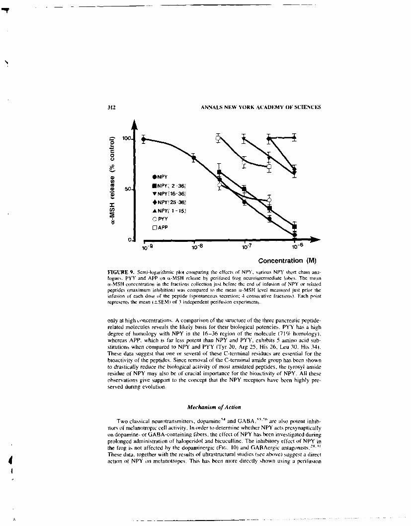
312 ANNALS NEW YORK ACADEMY OF SCIENCES
S100.
0U
04) NPY
cc o ENPYr12-361
h.0) VNPY[16-36J
00y
110-8 117 10-6
Concentration (M)
FIGURE 9. Semi-logarithmic plot comparing the effects of NPY. various NPY short chain ana-logues. PYY and APP on aY-MSH release by perifused frog neurointermediate lobes. The meanax-MSH concentration in the fractions collection just before the end of infusion of NPY or relatedpeptides (maximum inhibition) was compared to the mean a-MSH level measured just prior theinfusion of each dose of the peptide (spontaneous secretion: 4 consecutive fractions). Each lpintrepresents the mean (±SEM) of 3 independent perifusion experiments.
only at high concentrations. A comparison of the structure of the three pancreatic peptide-related molecules reveals the likely basis for their biological potencies. PYY has a highdegree of homology with NPY in the 16-36 region of the molecule (71% homology),whereas APP, which is far less potent than NPY and PYY, exhibits 5 amino acid sub-stitutions when compared to NPY and PYY (Tyr 20, Arg 25, His 26. Leu 30, His 34).These data suggest that one or several of these C-terminal residues are essential for thebioactivity of the peptides. Since removal of the C-terminal amide group has been shownto drastically reduce the biological activity of most amidated peptides, the tyrosyl-amideresidue of NPY may also be of crucial importance for the bioactivity of NPY. All theseobservations give support to the concept that the NPY receptors have been highly pre-served during evolution.
Mechanism of Action
Two classical neurotransmitters, dopamine 54 and GABA,55" 6 are also potent inhib-itors of melanotropic cell activity. In order to determine whether NPY acts presynapticallyon dopamine- or GABA-containing fibers, the effect of NPY has been investigated duringprolonged administration of haloperidol and bicuculline. The inhibitory effect of NPY inthe frog is not affected by the dopaminergic (FIG. 10) and GABAergic antagonists.2' -"These data. together with the results of ultrastructural studies (see above) suggest a directaction of NPY on melanotropes. This has been more directly shown using a perifusion

DANGER et al.: NPY INHIBITS otMSH SECRETION 313
model with dispersed frog pars intermedia cells, a preparation in which all afferent fibersare destroyed. It was found that NPY induces inhibition of a-MSH release in frog. 46
Thus, in this animal, NPY exerts a direct action on melanotropes. This observation is alsogiven support by results of recent electrophysiological experiments, carried out on aprimary culture of frog pars intermedia cells, using the patch-clamp technique. NPYinduces hyperpolarization in these cells and inhibits spontaneous action potentials. 57 Withrespect to the mechanism of action of NI-Y, some species differences may be expected.In contrast to the frog, in the pars intermedia of the toad Xenopus laevis, the melanotropesmay not constitute the main target for NPY, since it has no effect on dispersed pars cellsin this animal (B.G.J., unpublished data).
The pars intermedia of the frog pituitary is directly innervated by thyrotropin-releasing
NPY 10- 7 M
100 N.,
80.
60.
- 20.
0 , , FIGURE 1O. Differential effects of haloperi-0 dol upon the inhibitory actions of NPY (l07
D. 10-8M M) and dopamine (10-' M) on cs-MSH secre-tion. Frog neurointermediate lobes were contin-uously perifused in the absence (0) or in thepresence (0) of haloperidol (10--' M).
I 100.~(,o
80.
60.
40
20
0 T
TIME (Hour)

314 ANNALS NEW YORK ACADEMY OF SCIENCES
hormone (TRH)-IR fibers-5 and we have previously shown that TRH is a potent stimulatorof ot-MISH secretion in anuran amphibians. 9 6 Since the intermediate lobe of the pitu-itary of anurans, unlike urodeles,33
6 is under the antagonistic control of TRH and NPY,possible interactions between these two neuropeptides on ci-MSH secretion have beenexamined in vitro. During prolonged administration of NPY. the stimulatory effect ofTRH is inhibited in a dose-dependent manner.' Half-maximum inhibitions of OL-MSHrelease (both basal and TRH-induced secretion) are observed at a NPY concentration of3 x 10-8 M. These data suggest that NPY and TRH- act through a common intracellularpathway to regulate at-MSH secretion in the frog. Since it has been shown recently thatTRH produces an increase of phosphoinositol breakdown in the frog pars intermedia,6 ' apossible effect of NPY on this second messenger system has been examined: no effect ofNPY on the level of inositols could be demonstrated (unpublished results). A possibleaction of NPY on adenylate cyclase synthesis is currently being examined. Primarily.results indicate that NPY induces a dose-dependent inhibition of the synthesis of cyclic-adenosyl monophosphate (c-AMP).
In conclusion, an authentic NPY is present in the brain of amphibians and the peptidelocated in the pars intermedia of the pituitary of anurans can be considered has a phys-iologica! melanotropin-inhibiting factor. Since the pars intermedia is composed of ahomuogeneous population of endocrine cells, this tissue appears well-suited for investi-gating the intra-cellular mechanisms which mediate the action of NPY.
REFERENCES
1. TATEMOTO. K., M. CARLOUIST, & V. MUTT. [982. Nature 296: 659-660.2. SMITH. Y., A. PARENT. L. KERKERIAN & G. PELL.ETIER. 1985. J. Comp. Neurol. 236: 71-89.3. ALLEN, Y, S.. T, E. ADRIAN, J. 1.LEN, K. TATEMOTO. T. 1. CROW, S. R. BLOOM & J.
M. POLAK. [983. Science 221: 877-879.4. CHRONWALL. B. M_. D. A. DiMAGGIO. V. J1. MASSARL. V. M. PICKEL, D. A. RUGIERRO & T.
L. O'DONOHLJE. [985. Neuroscience 15: [159-1181.5. DE QUIOT. M. E., & P. C. EmsoN. 1986. Neuroscience 18: 545-618.6. PELLETIER. G.. L. DESY. L. KERKERIAN & J. COTE. 1984. Cell Tissue Res. 238: 203-205.7. LUNDBERG, J. M.. L. TERENIus. T. H6KFELT. C. R. MARTFLING, K. TATEMOTO. V. MUTT. J.
M. POLAK, S. BLOOM & M. GOLDSTEIN. 1982. Adta Physiot. Scand. 116- 477-490.8. JARvI. R., P. HELEN. M. PELTO-HUJKKO & H. H-ERVONEN. 1986. Neurosci. Lett. 67: 223-
227.9. FISHER-COBRIE. R., J. DIEz-GUERRA. P. C. EMsON & H. WINKLER. 1986. Neuroscience
18: 167-174.10. SAH-U, A.. P. S. KALRA & S. P. KALRA. 1988. Peptides 9: 83-86.11. KALRA. S. P.. P. S. KALRA. A. SAHU & W. R. CROWLEY. 1987. Steroid Biochem. 27:
677-681.12. ZUKOWSKA-GROJFC, Z., E. S. MARKS & M. HAAS. 1987. Am J. Physiol. 253: 1234-1239.13. GIBBN~S, 1. L., & i. L. MORRIS. 1988. Brain Res. 444: 402-406.[4. MARTIN. J. R., M. C. BEINFELD & T. C. WESTFALL. 1988. Am. J. Physiol. 254: 879-888.15. McDONALD. J. K.. M. D. LuMPKIN, W. K. SAMSON & S. M. MCCANN. 1985. Peptides
6: 71-84.[6. KERKERIAN. L., J. Guy. G. LEF~vRE & G. PELLETIER. 1985. Peptides 6: 1201-1204.17. WAHLESTEDT. C.. G. SKAGERBERG. R. EKMAN, M. HEIIIG. F. SUNDIER & R. HAKCANSON,
[987. Brain Res. 417: 33-38.18. DANGER. J. M.. J. Guy. M. BENYAMINA, S. JEGOU. F. LEBOULENGER. J. COTE, M. C.
ToNON. G. PELLETIER & H. VAUDRY. 1985. Peptides 6: 1125-1236.[9. PERROTEAU. 1., J. M, DANGER, G. PELL.ETIER, H. VAUDRY & A. FASOLO. 1988. J. Comp.
Neurol. 275: 309-325.20. VERBURG-VAN KEMENADE, B. M. L., B. G. JENKS. J. M. DANGER. H. VAUDRY. G. PELLE-
TIER & S. SAINT-PIERRE. 1987, Peptides 8: 1093-1100.

DANGER et al.: NPY INHIBITS ciMSH SECRETION 315
21. CAILLIEZ. D., J. M. DANGER. A. C. ANDERSEN. .1. M. POLAK. G. PELL.ETIER. K. KAWA-
MURA, S. KIKUYAMA & H. VAUDRY. 1997. Zool. Sci. 4- 123-134.221. VALLARINO. M.. J. M. DANGER, A. FASOLO. G. PLILETIER. S. SAINT-PIERRE. & H. VAUDRY.
1988. Brain Res. 448: 67-76.23. PONTE1. A.. J. M. DANGER, P. DuBOURG. G. PELLETIER, H. VAIJDRY & 0. KAH. 1989. Cell
Tissue Res. 255: 529-538.24. SCHOOFS. L., J1. M. DANGER. S. JEGOU. G. PELLETIER. R. HUYBRECHT. H. VAUDRY & A. DE
LooF. 1989. Peptides 9: 1027-1036.25. CIIARMENTIER-DAURES. M.. J. M. DANGER. P. NETCI-ITAII.0. G. PELLETIER & H. VAUDRY.
1987. C. R. Acad. Sci. 305: 479-483.26. REMY. C.. J. Guy, G. PELLETIER & H. BOER. Cell Tissue Res. In press.27. BRETON, B.. T. MIKOLAJCZYK. J. M. DANGER. F. GONNET. S. SAINT-PIERRE & H. VAUD)RY.
1989. Fish. Physiol. Biachem. 7: 77-83.28. DANGER. J. M., F. LEBOLENGER. J. Guy. Ni. C. TONON. M. BENYAMINA. J. C. MARTI.-
S. SAINT-PIERRE. G. PELLETIER & H. VAUDRY. 1986. Life Sci. 39: 1183-1192.29. DANGER. J. Mi., M. C. TONON. M. LAMACZ. J. C. MARTEL, S. SAINT-PIERRE. G. PELLETIER
& H. VAUDRY. 1987. Life Sci. 40: 1875-1880.30. BONS. N.. N. MESTRE, A. PETTER, J. M. DANCER. G. PELLETIER & H. V.XUDRY. J CoMp.
Neural. In press.31. DANGER, J. Mi.. M. C. ToNON. B3. G. JENKS, S. SAINT-PhERRE. J. C. MARTEIL. A. FASOLG.
B. BRETON, R. QUIRION. G. PELLIETIER & H. VAUDRY. 1990. Fundam. Clin. Pharmacal.4: 307-340.
32. KOHLER, C.. L. ERIKSON, S. DAVIES & V. CHAN-PALAY. 1986. J. Comp. Neural. 244:384-400.
33. CHAN-PAI.AY. V. & G. YASARGIL. 1986. Anat. Embryol. 174: 27-33.34. DE QUIDT. M. E. & EmSGN, P.C. 1986. Neuroscience 18: 527-543.35. LEGER. L., Y. CHARNAY. 1. Ni. DANGER. H. VAUDRY. P. M. DUBaIS & Mi. JOUVET. 1987.
J. Camp. Neural. 255: 283-292.36. DANGER. J. M.. 1. PERROTEAL!. M. F. FRANONI, S. SAINI-PiERRE. A. l-ASOL() & H.
VAUDRY. 1989. Neurendacrinalagy 50: 543-549.37. MINTH. C.. S. R. BIoaM. J. Ni. POLAK & J. E. DIXON. 1984. Prac. Natl. Acad. Sci. USA
81: 4577-4581.38. ALLEN. J. M.. J. NOVOTNY, 3. MARTIN & G. HEINRICH. 1987. Prac. Natl. Acad. Sci. USA
84: 2532-2536.39. GULBEKIAN. S., J. WARTON. G. W. HACKER. 1. M. VNRNOELL_. S. R. BLooMt & J. M. POLAK.
1985. Peptides 6: 1237-1243.40. CAlL UEZ. D.. J. M. DANGER. J. M. POLAK. G. PELLETIER. A. C. ANDERSEN & H. VAUDRY.
1987. Neurasci. Lett. 74: 163-168.41. TRAMU. G.. A. PiLLEz & J. LEONARDEI.II. 1978. . Histachem. Cytachem. 26: 322-324.42. ALLIEN. J. Mi., T. E. ADRIAN. J. M. POL.AK~ & S. R BLOOM. 1983. J. Autonarn. Nerx. S-vNI.
9: 559-563.43. MAJANE. J. C.. H. AnLo, Y. KATAOKA. C. H. LEE & H. Y. T. YANG. 1985. Endocrinology
117: 1162-1168.44. TON.N M. C.. F. LEBauLENGER. C. DELARUE. S. JE*OU. J. FRESEI.. P. LFROux & H.
VAUDRY. 1980. In Biochemical Endocrinology. M. ]uticl. & K. McKerns. Eds. 731. PlenumPress. New York. NY.
45. VAUDRY. H.. M. C. TONON. C. DELARUE. R. VAILI AN I & J. KRAICLR. 1978. Neuroendo-crinalagy 27: 9-24.
46. DANGER. J. Mi.. M. LAMAC/, F. MALVIARD. S. SAINT-PIFRRE. B. G. JENKS, M. C. TONO)N& H. VAUDRY. 1990. Gen. Comp. Endocrinol. 77:. 143-14().
47. KRAICER. J., B. LussiER. S. KorANI N & B. C. MOOR. 1988. Can J. Physial. Pharmacal.66: 318-320.
48. MARTEL., J1. C. .SAINT-PII-RRI & R. QuIRION. 1986. Peplides 7: 55-60.49. FRYER. J. N. 1989. Fish Physial. Biachem. In press.5(0. LUNDBERG. 3. M. & K. TATEMOTO. 1982. Acta Physial. Scand. 116: 393-40251. ALIEN. . Mi., T. E. ADRIAN. K. TATFMO 10, J. Ni. POl AK. J. HUGUiES & S. R. HI oom.
Neuropeptldes 3: 7 1-77.

316 ANNALS NEW YORK ACADEMY OF SCIENCES
52. Rioux. F.. H. BACHELARD, J. C. MARTEL & S. SAINT-PIERRE. 1986. Peptides 7: 27-31.53. MASSARI, V. J., J. CHAN. B. M. CHRONWALL, T. L. O'D0NOHtJE & V. M. PICKEL. 1984.
Soc. Neurosci. Abstr. 537.54. TONON. M. C.. P. LEROUX, M. E. STOECKEL. S. iEGOU, G. PELLETIER & H. VAUDRY. 1983.
Endocrinology 112: 133-141.55. ADJEROUD, S., M. C. TONON. M. LAMACZ, E. LENEVEU, M. E. STOECKEL, M. L. TAPPAz,
L. CAZIN. J. M. DANGER. C. BERNARD & H. VAUDRY. 1986. Brain Res. 17: 717-724.56. VERBURG-VAN KEMENADE, B. M. L., M. TAPPAz. L. PAUT & B. G. JENKS. 1986. Endocri-
nology 118: 260-267.57. BASILLE, C., E. LouIsET. J. M. DANGER, A. FOURNIER, H. VAUDRY & L. CAZIN. 1990. N.Y.
Acad. Sci. In press.58. SEKI, T., Y. NAKAI. S. SHIODA. T. MITSUMA & S. KIKUYAMA. 1983. Cell Tissue Res.
233: 507-516.59. TONON, M. C.. P. LEROUX, F. LEBOULENGER, C. DELARUE. S. JEGOU, J. FRESEL & H.
VAUDRY. 1980. Life Sci. 26: 869-875.60. TONON. M. C., J. M. DANGER, A. FASOLO, G. PELLETIER, S. SAINT-PIERRE & H. VAUDRY.
1988. In The Melanotropic Peptides. M. E. Hadley, Ed. CRC Press. New York, NY.61. DESRUES, L., M. C. TONON & H. VAUDRY. J. Mol. Endocrinol. In press.

Regulation of the Hypothalamo-PituitaryAxis by Neuropeptide Y
JAMES I. KOENIG
Department of Physiology and BiophysicsGeorgetown University School of Medicine
3900 Reservoir Road NWWashington, DC 20007
Immunohistochemical studies have demonstrated neuropeptide-Y (NPY) immunoreactiv-ity in neuron cell bodies in the arcuate, perinl-icular, anterior, supraoptic and paraven-tricular (PVN) nuclei of the hypothalamus. '-3 In addition, in situ hybridization his-tochemistry has similarly shown the presence of NPY mRNA in many of these areas.'Dense NPY fiber networks have been found throughout the hypothalamus, with partic-ularly dense concentrations of fibers in the arcuate, PVN, periventricular and preopticnuclei. 1-3 Furthermore, it appears that the NPY terminals in the PVN may be derivedfrom several sources. The hypothalamic arcuate nucleus 5"6 and brainstem NPY-con-taining cell bodies project to the PVN. 7- 9 The innervation of the PVN by brainstem NPYfibers may also contain epinephrine or norepinephrine. 7 4 However, lesioning the cate-cholaminergic neurons with 6-hydroxydopamine has resulted in the sparing of hypotha-lamic NPY neurons in spite of dramatic depletion in hypothalamic norepinephrineconcentrations, 10,11 raising doubts about the origin of NPY in the PVN. Although, Bai etal. have suggested a greater contribution by the arcuate NPY cell bodies to the PVN. 5 TheNPY projections to the PVN also appear to be directed toward the parvocellular portionof the nucleus and directly synapse on neurons containing corticotropin-releasing hor-mone (CRH). 12 In addition, projections also are directed at magnocellular neurons of thesupraoptic nucleus containing vasopressin.13
The physiological function of hypothalamic NPY has yet to be determined althoughdata from the laboratories of McDonald et al. and Kalra et al. suggest that NPY may actto enhance reproductive function in the female rat and may be involved in coordinating therelease of luteinizing hormone during the estrous cycle. 14.15 In addition, convincing datafrom the laboratories of Leibowitz et al. and Kalra et al. suggest that NPY, especially inthe PVN, is responsible for stimulating food intake in the rat. 16 '17 NPY may also beinvolved in regulating fluid homeostasis in the rat.' 8
Several groups have convincingly demonstrated that NPY secretion is linked to reg-ulation of metabolic hormone secretion, in particular, the regulation of ACTH secretion.Hirfstrand et al. " initially reported the ability of NPY to augment the secretion of ACTHand corticosterone in the rat following infusion into the ventricular system of the ratbrain. " Subsequently. Wahlestedt et al.21' and Haas and George2 ' confirmed this finding.In addition, Haas and George showed that NPY administration increased the concentra-tion of corticotrophin-releasing hormone in the hypothalamus, suggesting the induction ofCRH synthesis by NPY. NPY-induced activation is thought to be mediated by the en-hanced secretion of CRH by the hypothalamus. 22 NPY also has been found in the hy-pophysial portal circulation23 and in the anterior pituitary.2 4 As a consequence. NP maymediate its actions at multiple sites in the hypothalamic-pituitary adrenal axis.
These pharmacological and anatomical studies suggest that NPY is potentially capableof modulating ACTH secretion physiologically, especially during responses to stresswhen multiple factors are activated to insure a robust ACTH secretory response. The
317

318 ANNALS NEW YORK ACADEMY OF SCIENCES
present studies were designed to further characterize the role of hypothalamic NPY inmediating ACTH responses to physiological challenges in the rat.
EXPERIMENTAL METHODS
Adult male Sprague-Dawley rats (175-200 gms) were purchased from Charles RiverInc. (Kingston, NY). The animals were housed in a temperature-controlled animal roomon a fixed light cycle (lights on 0700-1900 hrs). The animals had free access to rat chowand water at all times.
Hypophysial Portal Blood Collection
Hypophysial portal blood was collected according to the protocol of Porter andSmith. Briefly, the rats were anesthetized with urethane (1.2 gm/kg). The ventralsurface of the skull was exposed by the parapharyngeal approach and the bone over thepituitary gland was carefully removed. The dura in the floor of the sella was cut and thepituitary gland was reflected forward to expose the pituitary stalk. The pituitary stalk wastransected and the gland removed. Heparinized saline (0.5 ml) was administered throughan intravenous cannula. A polyethylene cannula was placed over the stump of the cut stalkand blood was collected by gravity into a chilled tube containing aprotinin (Sigma).Collections were of 30-60 min duration. Following centrifugation, the plasma was storedat - 800C.
Dispersed Pituitary Cell Cultures
Male rats weighing 175-200 g were sacrificed by decapitation. The anterior lobe ofthe pituitary gland was isolated and dispersed monolayer cell cultures were prepared aspreviously described 26 with several modifications. Anterior lobes of the pituitary weremechanically disrupted and incubated with collagenase, DNase and pancreatin as de-scribed by Vale et al.27 The cells were suspended in Dulbecco's modified Eagle's medium(DMEM) supplemented with fetal calf serum (2%), T3 (60 pm), insulin (5 Rg/ml),transferrin (5 l.g/ml) and FGF (I ng/ml) and plated in plastic multiwell plates. Thecultures were incubated in a humid atmosphere containing 5% CO 2 for 3 days at whichtime the cells were washed 3 times with DMEM and were incubated for an additional 24hours in serum-free test medium which consists of DMEM and a serum substitute (Uni-versity of California, San Francisco, Cell Culture Facility). The following day the cellswere washed 4 times with DMEM and groups of 6 wells were incubated for 3 hours withthe peptides to be tested. At the end of the incubation, the medium was removed from thecells with a polypropylene pipet and transferred directly into chilled polypropylene assaytubes. Multiple aliquots were prepared to prevent freezing and thawing artifacts. Mediumconcentrations of ACTH were determined by radioimmunoassay (see below).
Tissue Processing
The brain and pituitary were rapidly removed from the skull. The neurointermediatelobe of the pituitary (NIL) was separated from the anterior lobe and placed in 200 1I of2 N acetic acid. The brain was placed on wet ice and the median eminence (ME) was

KOF NIG: III POTHAIAMO PITUIT, RY AXIS 319
removed with the aid of a dissecting microscope and fine iris scissors. After removing theME, the brain was frozen on dry ice for later dissection. The ME was placed in 200 Iof 2 N acetic acid. Both the ME and NIL were placed in a boiling water bath for 20 min.After boiling, the extracts were aliquoted and lyophilized. The frozen brain was dissectedinto an anterior hypothalamic fragment (POA) containing the preoptic area, paraventric-ular, suprachiasmatic and supraoptic nuclei and a mediobasal hypothalamic (MBH) frag-ment containing the arcuate, ventromedial and dorsomedial nuclei. These fragments wereplaced in 1 ml of 2 N acetic acid and heated in a boiling bath for 20 min. The tissue debriswas pelleted by centrifugation and the extracts were aliquoted and lyophilized. The tissuepellets were dissolved in IN NaOH for protein determinations.
Radioimmunoassays
Plasma concentrations of ACTH were determined using standard procedures. 21 The
ACTH antiserum was purchased from IgG Corp. (Nashville, TN) and radioiodinatedACTH was purchased from Radioassay Systems Laboratory (Carson, CA). PorcineACTH purchased from IgG Corp. (Nashville, TN) was used as standard. The assaysensitivity was 1 pg/ml. The intra- and interassay coefficients of variation were 6.4% and1 9.8%, respectively.
Tissue concentrations of NPY were determined by radioimmunoassay as described byAllen et al.29 Plasma samples were processed as previously described. 23 Briefly, antiserato NPY were raised in New Zealand white rabbits against porcine NPY conjugated tobovine serum albumin with carbodiimide. 1251I-NPY was purchased from New EnglandNuclear (Boston, MA). The assay was run under nonequilibrium conditions; antibody wasincubated with the samples for 72 h in the absence of tracer. Following a further 72-hincubation in the presence of tracer, free ' 25I-NPY was separated from bound by additionof goat-anti-rabbit serum. Assay sensitivity was 2 pg/tube and inter- and intraassay co-efficients of variation were 8.8% and 6.6%, respectively.
Protein Determinations and Statistical Analysis
The concentration of protein in the tissue extracts was determined using the fluoro-metric method as described by Bohlen et al.30
Statistical differences were determined by analysis of variance and Student t testaccording to procedures outlined by Zar. 3' Values of p less than 0.05 were consideredsignificant.
RESULTS
Hypophysial Portal Blood Concentrations of NPY
NPY immunoreactive material was readily detectable in the hypophysial portal bloodof the urethane-anesthetized male rat. Portal plasma concentrations of NPY in the first 30min collection period were 52.0 -t 4.0 ng/ml. Continuing the collection for an additional30 min did not significantly alter NPY levels. Furthermore, a significant portal-systemicgradient existed for NPY as systemic plasma concentrations of NPY were 16.0 ± 4.5ng/ml (p <0.004). These levels were not significantly altered during a second 30-minperiod.

320 ANNALS NEW YORK ACADEMY OF SCIENCES
Z 5.000
0
X 4,000
I-
zO'3,000
Z E0 -OOZl +S2,000
<1.000
0
- Vt -t1o -p -a -7 -4CONTROL 10 10 10 10 10 10
U NPY 1 NPY + CRH (lnM)FIGURE 1. The effect of NPY on ACTH secretion from dispersed anterior pituitary cells in theabsence or presence of I nM CRH. Anterior pituitary cells were enzymatically dispersed and platedat a density of 259,000 cells per well. The cells were incubated with NPY (Richelieu Biotechnol-ogles), CR (I aM; Peninsula Laboratories) or the combination of NPY and CRH for 3 hours. Themedium was collected in chilled plastic tubes and frozen immediately. Shown in the figure is thegroup mean from 6-12 wells per point and the standard error of the mean. *-indicates statisticalsignificance at the 0.05 level compared to the appropriate control.
Actions of NP¥ on ACTH Secretion from Dispersed Anterior Pituitary Cells
Previous studies from several laboratories demonstrated the dose-dependent stimula-tory action of NPY on the secretion of LH, FSH and GH from penifused dispersed anteriorpituitary cells. 32"33 However, an effect of NPY on ACTH secretion was not observed, 33
FIGURE I depicts the effect of NPY on ACTH secretion from dispersed anterior pituitarycells derived from male rats.
Anterior pituitary cells were plated at a density of 250,000 cells per well. The additionof NPY (10 -" M to 10- 6 M) was without effect on the release of ACTH into the mediaduring a 3-hr incubation period. Whereas, the addition of I nM CRH to the wellssignificantly stimulated ACTH secretion (p<0.001) under basal conditions. The coincu-bation of CRH (I nM) and NPY (10- 1 to 10- 6 M) resulted in a dose-related augmen-tation of the stimulatory effect of CRH (p <0.005). Maximal secretion of ACTH occurredwhen I nM CRH and 100 nM NPY were coincubated. This combination resulted in a100% increase in ACTH secretion compared to CRH alone.
Interaction between NPY and ACTH Regulation during Stress
Stress of various types has been found to increase the actiity of the hypothalamo-pituitary-adrenal axis as reflected in plasma concentrations of ACTH or corticosterone.Hypovolemic stress has been associated with an activation of CRH secretion into the

KOENIG: HYPOTHALAMO-PITUITARY AXIS 321
hypophysial portal circulation, 3- whereas insulin hypoglycemia-induced activation ofACTH secretion is apparently mediated by activation of vasopressin secretion. 35 Al-though CRH mechanisms may also be important in this response. 1,.37 Recent studies alsohave identified CRH as the mediator of interleukin-induced ACTH secretion.1', 3 How-ever, the location of NPY in the long projection neurons derived from brainstem NEsources implicate this neuropeptide as a potential mediator of these responses.
Activation of immune mechanisms have been associated with a concomitant increasein ACTH or corticosterone secretion. " In the present study, FIGURE 2 depicts the timecourse of the endotoxin-induced release of ACTH. Plasma concentrations of ACTH peak3-4 hrs after the injection of bacterial lipopolysacchride (25 p g/kg, i.p.). At the timewhen ACTH levels are at their peak, concentrations of NPY in the anterior hypothalamus/POA were increased 33% (p <0.001; FIG. 3). However, concentrations of NPY in otherbrain regions and the pituitary gland were unchanged.
In contrast to the minimal changes observed in NPY concentrations during circadianACTH secretion or LPS-induced stress, insulin hypoglycemia induced a much greater andmore rapid increase in plasma ACTH concentrations (FiG. 4). ACTH concentrationsincreased approximately 25-fold 30 min after the injection of human recombinant insulin(5 IU/kg, i.p.). The sharp decrease in plasma glucose concentrations resulted in a 60%depletion in POA NPY concentrations and a 40% reduction in the basal hypothalamus(MBH) (FIG. 5). In contrast to these regions, a highly significant increase in NPY contentwas observed in the neurointermediate and anterior lobes of the pituitary gland (FIG. 6).Ninety minutes after insulin infusion. ACTH levels were still greater than 400 pg/mI butwere decreasing. The depletion of NPY in the POA fragments observed at 30 min was stillevident at this time: however, NPY concentrations in the basal hypothalamus were in-creased 50% over basal levels and 200% over the 30-min time point. In addition, theconcentration of NPY in the ME increased 100% over basal levels. NPY concentrationsin both lobes of the pituitary returned to pretreatment levels.
DISCUSSION
Previous studies have indicated that the infusion of pharmacological amounts of NPYactivated the hypothalamo-pituitary-adrenal axis of the rat. ' -2 Furthermore, these stud-
FIGURE 2. The effect of endotoxin on plasmaconcentrations of ACTH. Male Sprague-Dawleyrats were treated with bacterial lipopolysacchride(25 .g/kg. i.p.: serotype 05:55, Sigma). The ani-mals were sacrificed by decapitation I hr after ve-
___ _ _hicle treatment or I. 2, 4, 6. and 24 hours afterinjection of endotoxin. Brains were rapidly re-
E400 moved, the median eminence was dissected from the300 fresh brain and peptides were extracted in boiling
T ~acetic acid. The remainder (if the brain was frozen200 -for later dissection. The frozen brain was dissected
into POA and MBH fragments, and peptides were'0 ,extracted in boiling acetic acid. Blood samples were
0 collected in ice-cold tubes containing EDTA at the0 - 2 time of sacrifice. Plasma was separated by centrif-
TIME (hours) ugation and aliquoted for the ACTH assay. Each barrepresents the mean ACTH response and the stan-ddrd crio, of the mean in groups of 8 animals. *-indicates significant differences at the 0.05 level vthe control animals treated with vehicle I hr before.

322 ANNALS NEW YORK ACADEMY OF SCIENCES
400 i 5
3W FIGURE 3. The effect of endotoxin on NPY con-3- o centrations in several brain regions 4 and 24 hours
2E after treatment. NPY concentrations foi the MEg are depicted on the left axis whereas other regions
loo should be compared to the right axis. See FIGUREz 2 legend for further details.
o D 4 2
TIME (hours)N ME E NIL [ POA H MBH
ies implicated a central nervous site of action for NPY. In the present studies, we haveidcitificd the presence of NPY in the hypothalamo-pituitary portal circulation. The con-centration of NPY in the portal blood was 3-5 times greater than the amount in thesystemic circulation suggesting active secretion of NPY from nerve terminals in themedian eminence.23 Recent studies have indicated that NPY also may be secreted by theanterior pituitary.24 However, the pituitary gland was removed from the animals inadvance of the collection of the portal blood, so that the source of NPY was restricted tosystemic sources or the median eminence.
Our studies also have demonstrated the ability of NPY to potentiate CRH-inducedACTH secretion under in vitro conditions. NPY alone was unable to act as an ACTHsecretogogue in doses up to 10 - 6 M, similar to the results obtained by Kerkerian et al.
33
The CRH potentiating activity of NPY was dose-related with maximal actions at 10- s to10 - 7 M. These concentrations of NPY are similar to the concentration of the peptidedetected in the hypophysial-portal blood studies suggesting that NPY may function as anendocrine or paracnne factor modulating ACTH secretion in the pituitary. In addition, thedata of Bloom et al.24 enhance the likelihood of a paracrine function for NPY in thepituitary.
In the present study, evidence supporting a central nervous system site of NPY actionon ACTH secretion has been presented. Administration of pharmacological amounts of
FIGURE 4. ACTH responses to insulin-inducedhypo. lycemia. Adult male rats were treated at 0900
z 000 hrs with 5 IU/kg recombinant human insulin (Hu-- **minulin, Lilly), and were sacrificed 30 and 90 min
<a after injection. The brains were rapidly removed. ofrom the skull, the ME was dissected from the fresh
U - brain under microscopic visualization and peptide°Z _E 6W was extracted with boiling acetic acid. The remain-
der of the brain was frozen for later dissection.POA and MBH fragments were dissected from the
< 0frozen brain and were placed in boiling acetic acid.NPY concentrations were determined in these ex-
0 200 Itracts b' RIA. Blood was collected at the time of
-5 sacrifice in chilled plastic tubes containing EDTA.a- Plasma was separated by centrifugation and ali-
CoNTmaL 30 MIN 90 u1 quoted for ACTH assay. Each point represents theTIME mean plasma concentration of ACTH and the stan-
dard error of the mean in 8 animals. **-indicates
significant differences at the 0.001 level vs the con-trol (saline-treated) animals.
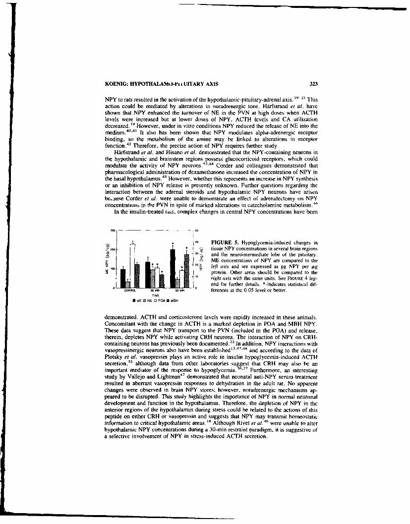
KOENIG: HYPOTHALAMO-Pt UITARY AXIS 323
NPY to rats resulted in the activation of the hypothalamic-pituitary-adrenal axis. I, -2I Thisaction could be mediated by alterations in noradrenergic tone. Hirfstrand et al. haveshown that NPY enhanced the turnover of NE in the PVN at high doses when ACTHlevels were increased but at lower doses of NPY, ACTH levels and CA utilizationdecreased. "9 However, under in vitro conditions NPY reduced the release of NE into themedium."' It also has been shown that NPY modulates alpha-adrenergic receptorbinding, so the metabolism of the amine may be linked to alterations in receptorfunction.4 2 Therefore, the precise action of NPY requires further study.
Hdrfstrand et al. and Hisano et al. demonstrated that the NPY-containing neurons inthe hypothalamic and brainstem regions possess glucocorticoid receptors, which couldmodulate the activity of NPY neurons. 43
.44 Corder and colleagues demonstrated that
pharmacological administration of dexamethasone increased the concentration of NPY inthe basal hypothalamus. 45 However, whether this represents an increase in NPY synthesisor an inhibition of NPY release is presently unknown. Further questions regarding theinteraction between the adrenal steroids and hypothalamic NPY neurons have arisenbe,.aue Corder et al. were unable to demonstrate an effect of adrenalectomy on NPYconcentrations in the PVN in spite of marked alterations in catecholamine metabolism. 4 6
In the insulin-treated rats, complex changes in central NPY concentrations have been
3W 50
4 0 FIGURE 5. Hypoglycemia-induced changes in20* - tissue NPY concentrations in several brain regions
a - and the neurointermediate lobe of the pituitary.ME concentrations of NPY are compared to the
*120 a_ left axis and are expressed as pg NPY per -gz protein. Other areas should be compared to theright axis with the same units. See FIGURE 4 leg-
end for further details. *-indicates statistical dif-coOta 30 uN so u ferences at the 0.05 level or better.
TIME
0 ME 0 NIL 0 POA S MSH
demonstrated. ACTH and corticosterone levels were rapidly increased in these animals.Concomitant with the change in ACTH is a marked depletion in POA and MBH NPY.These data suggest that NPY transport to the PVN (included in the POA) and release,therein, depletes NPY while activating CRH neurons. The interaction of NPY on CRH-containing neurons has previously been documented. 22 In addition, NPY interactions withvasopressinergic neurons also have been established' 3.47.40 and according to the data ofPlotsky et al. vasopressin plays an active role in insulin hypoglycemia-induced ACTHsecretion,3 5 although data from other laboratories suggest that CRH may also be animportant mediator of the response to hypoglycemia. 36 37 Furthermore, an interestingstudy by Vallejo and Lightman"7 demonstrated that neonatal anti-NPY serum treatmentresulted in aberrant vasopressin responses to dehydration in the adult rat. No apparentchanges were observed in brain NPY stores; however, noradrenergic mechanisms ap-peared to be disrupted. This study highlights the importance of NPY in normal neuronaldevelopment and function in the hypothalamus. Therefore, the depletion of NPY in theanterior regions of the hypothalamus during stress could be related to the actions of thispeptide on either CRH or vasopressin and suggests that NPY may transmit homeostaticinformation to critical hypothalamic areas. " Although Rivet et al.46 were unable to alterhypothalamic NPY concentrations during a 30-min restraint paradigm, it is suggestive ofa selective involvement of NPY in stress-induced ACTH secretion.

324 ANNALS NEW YORK ACADEMY OF SCIENCES
0.6
FIGURE 6. Hypoglycemia-induced changes in>0., anterior pituitary NPY concentrations. See leg-z t Lends to FiGURES 4 and 5 for further details.
0.2
CON"M. 30 MIN 90 MAIN
TIME
0 AP
Increases in NPY concentrations in the neurointermediate lobe and median eminenceare consistent with an increase in transport of NPY into nerve terminals in these areasduring hypoglycemic stress, but whether release of this peptide is stimulated is currentlyunknown. The source of this peptide also is unknown although studies of Kerkerian et al.suggest that median eminence and neurointermediate lobe NPY may be derived fromarcuate nucleus sources. 6
Stress has been found to increase plasma concentrations of NPY in rats.49"5 Hypo-glycemia induced by insu!in increased plasma concentrations of NPY in man as well. 5
Whether the pituitary gland or median eminence contribu,.- to the rising systemic plasmaconcentrations of NPY is currently unknown but seems highly unlikely, because plasmaconcentrations of this peptide are very high (especially in rodents) and the tissue concen-trations of the peptide are relatively low, If, however, the secretory rate was very high.then a contribution might be expected.
Endotoxin also altered anterior hypothalamic sources of NPY. However, the stressinduced by treating rats with LPS was small as reflected by the small increase in ACTHconcentrations. The time course of the response also is more prolonged than hypoglyce-mia-induced ACTH secretion, indicating that if earlier time points had been studied, apattern of NPY changes more similar to hypoglycemia might have been observed.
Endotoxin-induced activation of the hypothalamo-pituitary-adrenal axis has recentlybeen reported. -
52 The increase in plasma concentrations of ACTH appear to be mediated
by activation of hypothalamic CRH neurons.- 2 .11 Howcver, endotoxin administration alsomodifies the activity of somatostatin-, thyrotropin-releasing hormone-, beta-endorphin-and vasopressin-containing neurons. 54- 5 7 Activation of the immune response has alsobeen reported to enhance the turnover of norepinephrine in the brain. 8 In the presentstudy, it was found that NPY is depleted from POA stores 4 hrs after bacterial Ii-popolysacchride treatment but returns to control levels 24 hrs after injection. Significantchanges in NPY concentrations in other brain regions or the pituitary were not observed.Since NPY is colocalized in brainstem noradrenergic neurons projecting to the PVN. 7 "8 itwould appear that NPY may be involved in transmitting homeostatic information to thehypothalamus. However, the significance of the NPY and noradrenergic colocalization inthis response is currently unknown. It is possible that brainstem NPY fibers are notinvolved and the concomitant NPY changes are due to alterations in the responses ofarcuate NPY-ergic neurons. Corder et al. also demonstrated that dexamethasone influ-ences hypothalamic concentrations of NPY. 4V 5 Therefore, the glucocorticoid increase as-sociated with the administration of endotoxin may provoke the change in NPY. Furtherwork in this area is required before adequate conclusions can be drawn.
KOENIG: HYPOTHALAMO-PITUITARY AXIS 325
NPY and its associated mRNA have been demonstrated in the cells of the immunesystem. 5" LPS administration is known to enhance the production of the cytokine, inter-leukin- 1. Whether an activation of neuropeptide Y synthesis or secretion is associated withactivation of the immune system is currently unknown. The possibility that peripherallyproduced interleukin- I is responsible for the alteration in NPY also should be considered.
In conclusion, it is clear that NPY may act on neurons of the central nervous systemto augment the activity of the hypothalamo-pituitary-adrenal axis. NPY-containing neu-rons in the brain are also influenced by the changes in the homeostatic environment of theanimal and with norepinephrine may represent a pathway by which sensory inputs mod-ulate neuroendocrine function. However, the current study also opens the possibility of apituitary site of action for NPY and hence, peripheral sources ot the peptide may playsignificant roles in changing pituitary hormone secretion.
REFERENCES
I. CHRONWALL, B. M.. D. A. DIMAGGIO, V. J. MASSARI. V. M. PICKEL, D. A. RUGGIERO &T. L. O'DONOHUE. 1985. The anatomy of neuropeptide Y-containing neurons in rat brain.Neuroscience 15: 1159-1181.
2. DEQUIDT, M. E & P. C. EMSON. 1986. Distribution of neuropeptide Y-like immunoreactivityin the rat central nervous system. II. Immunohistochemical analysis. Neuroscience 18: 545-618.
3. CARD, J. P. & R. Y. MOORE. 1988. Neuropeptide Y localization in the rat suprachiasmaticnucleus and periventricular hypothalamus. Neurosci. Lett. 88: 214-246.
4. MORRIS. B. M. 1989. Neuronal localization of neuropeptide Y gene expression in rat brain. J.Comp. Neurol. 290: 358-368.
5. BAt, F. L., M. YAMANO, Y. SHIOTANI, P. C. EMSON, A. D. SMtTH, J. F. POWELL & M.TOHYAMA. 1985. An arcuato-paraventricular and -dorsomedial hypothalamic neuropeptideY-containing system which lacks noradrenaline in the rat. Brain Res. 331: 172-175.
6. KERKERIAN, L. & G. PELLETIF.R. 1986. Effects of monosodium I-glutamate administration onneuropeptide Y-containing neurons in the rat hypothalamus. Brain Res. 369: 388-390.
7. EvERITT, B. J., T. HOKFELT, L. TERENIUS, K. TATEMOTO, V. MUTT & M. GOLOSTEiN. 1984.Differential co-existence of neuropeptide Y (NPY)-like immunoreactivity with catechola-mines in the central nervous system of the rat. Neuroscicnce 11: 443-462.
8. SAWCHENKO, P. E., L. W. SWANSON, R. GRZANNA, P. R. C. HOWE. S. R. BLOOM & J. M.POLAK 1985. Colocalization of neuropeptide Y immunoreactivity in brainstem catechola-minergic neurons that project to the paraventricular nucleus of the hypothalamus. J. Comp.Neurol. 241: 138-153.
9. HA.RSTRAND, A., K. FUXE, L. TERENIUS & M. KALIA. 1987. Neuropeptide Y-immunoreac-tive perikarya and nerve terminals in the rat medulla oblongata: relationship to cytoarchitec-ture and catecholaminergic cell gtoups. J. Comp. Neurol. 260: 20-35.
10. SCHON, F., J. M. ALLEN, J. C. YEATS, A. KENT, J. S. KELLY & S. R. BLOOM. 1986. Theeffect of 6-hydroxydopamine, reserpine and cold stress on neuropeptide Y content of 'he ratcentral nervous system. Neuroscience 19: 1247-1250.
II. GUSTAFSON, E. L. & R. Y. MOORE. 1987. Noradrenaline and neuropeptide Y innervation ofthe rat hypothalamus are differentially affected by 6-hydroxydopamine. Neurosci. Lett. 83:53-58.
12. LiposITs, Zs, L. SIEVERS & W. K. PAULL. 1988. Neuropeptide-Y and ACTH-immunoreactiveinnervation of corticotropin releasing factor (CRF)-synthesizing neurons in the hypothalamusof the rat. An immunocytochemical analysis at the light and electron microscopic levels.Histochemistry 88: 227-234.
13. BEROUKAS. D.. J. 0. Wi.LOUGHBY & W. W. BLESSING. 1989. Neuropeptide Y-like immu-noreactivity is present in boutons synapsing on vasopressin-containing neurons in rabbitsupraoptic nucleus. Neuroendocrinology 50: 222-228.
14. MCDONALD. J. K. 1988. NPY and related substances. Crit. Rev. Neurobiol. 4: 97-135.15. KALRA, S. P. & W. R. CROWi.EY. 1984. Norepinephrine-like effects of neuropeptide Y on LH
release in the rat. Life Sci. 35: 1173-1176.

326 ANNALS NEW YORK ACADEMY OF SCIENCES
16. STANLEY, B. G. D. LANTHIER, A. S. CHIN & S. F. LEIBOWITZ. 1989. Suppression ofneuropeptide Y-elicited eating by adrenalectomy or hypophysectomy: reversal with corticos-tero.e. Brain Res. 501: 32-36.
17. SAHU, A., P. S. KALRA & S. P. KAIRA. 1988. Food deprivation and ingestion induce recip-rocal changes in neuropeptide Y concentrations in the paraventricular nucleus. Peptides 9:83-86.
18. HooL, S. C., G. S. RICHARDSON, J. K. McDONALD, J. M. ALLEN. J. B. MARTIN & J. 1.KOENIG. 1989. Neuropeptide Y and vasopressin in the hypothalamo-neurohypophysial axis ofsalt-loaded or Brattleboro rats. Brain Res. 486: 214-220.
19. HARFSTRAND, A., P. ENEROTH. L. AGNATI & K. FUXE. 1987. Further studies on the effectsof central administration of neuropeptide Y on neuroendocrine function in the male rat:relationship to hypothalamic catecholamines. Regul. Pept. 17: 167-179.
20. WAHLESTEDT. C., G. SKAGERBERG, R. EKMAN, M. HEILIG, F. SUNDLER & R. HAKANSON.
1987. Neuropeptide Y (NPY) in the area of the hypothalamic paraventricular nucleus activatesthe pituitary-adrenocortical axis in the rat. Brain Res. 417: 33-38.
21. HAAS. D. A. & S. R. GEORGE. 1987. Neuropeptide Y administration acutely increases hypo-thalamic corticotropin-releasing factor immunoreactivity: lack of effect in other rat brainrcgions. Life Sci. 41: 2725-2731.
22. TSAGARAKIS, S., L. H. REES. G. M. BESSER & A. GROSSMAN. 1989. Neuropeptide-Y stimu-lates CFR-41 release from rat hypothalami in vitro. Brain Res. r%01: 167-170.
23. MCDONAL.D. J. K.. J. I. KOENIG, D. M. GIBBS, P. COLINS & B. D. NOE. 1987. Highconcentrations of neuropeptide Y in pituitary portal blood of rats. Neuroendocrinology 46:538-541.
24. JONES. P. M.. M. A. GHATEL, J. STEEL. D. O'HAI.ORAN. G. GON, S. LEGON. J. M. BURRIN.U. LEONHAROT. J. M. POILAK & S. R. BLOOM. 1989. Evidence for neuropeptide Y synthesisin the rat anterior pituitary and the influence of thyroid hormone status: comparison withvasoactive intestinal peptide. substance P. and neurotensin. Endocrinology 125: 334-341.
25. PORTER. J. C. & K. R. SMITH. 1967. Collection of hypophysial stalk blood in rats. Endocri-nology 81: 1182-I 187.
26. FUKATA, J. & J. B. MARTIN. 1986. Influence of sex steroid hormones on rat growth hormone-releasing hormone and somatostatin in dispersed pituitary cells. Endocrinology 119: 2256-2261.
27. VAtE. W., G. GRANi. M. AMoss. R. BL.ACKWEI.L. & R. GULtLEMIN. 1972. Culture ofenzymatically dispersed anterior pituitary cells: functional validation of a method. Endocri-nology 91: 562-570.
28. NICHOLSON. W. E.. D. R. DAvis. B .. SHERREL & D. N. ORrH. 1984. Rapid radioimmunoassay for corticotropin in unextracted human plasma. Clin. Chem. 30: 259-2675.
29. ALLEN. J. M. J. C. YEATS. T. E. ADRIAN & S. R. BLOOM, 1984. Radioimmunoassay ofneuropeptide Y. Regul. Pept. 8: 61-70.
30. BOHLEN. P., S. STEIN, W. DAIRMAN & S. UNDENFRIEND. 1973. Fluorometric assay ofproteins in the nanogram range. Arch. Biochem. Biophys. 155: 213-220.
31. ZAR. J. H. 1984. Biostatistical Analysis. Prentice-Hall. Englewood Cliffs, NJ.32. MCDONALD. J. K. M. D. LUMPKIN. W. K. SAMSON & S. M. MCCANN. 1985. Neuropeptide
Y affects secretion of luteinizing hormone and growth hormone in ovariectomized rats. Proc.NatI. Acad Sci. USA 82: 561-564.
33. KERKERIAN. L.. J. Guy. G. LEFERRE & G. PELLETIER. 1985. Effects of neuropeptide YINPY) on the release of anterior pituitary hormones in the rat. Peptides 6: 1201-1204.
34. PLOrSKY. P. M., T. 0. BRUHN & W. VALE. 1985. Evidence for multifactor regulation of theadrenocorticotropin secretory response to hemodynamic stimuli. Endocrinology 116: 633-640.
35. PLOtSKY. P. M.. T. 0. BRUHN & W. VAIE. 1985. Hypophysiotropic regulation of adreno-corticotropin secretion in response to insulin-induced hypoglycemia. Endocrinology 117:323-329.
36. SUDA, T., F. TOZAWA, M. YAMADA. T. USHIVAMA. N. ToMORI. T. SUMITOMO, Y. NAKA-GAMI. H. DEMURA & K. Sm-t;zME. 1988. Insulin-induced hypoglycemia increases corticotro-pin-releasing factor messenger ribionucleic acid levels in rat hypothalamus. Endocrinology123: 1371-1375
l4 l / I•Hl

KOENIG: HYPOTHALAMO-PITUIT ARY AXIS 327
37. I3ERKENBOSCH. F., D. C. E. DE GoEtI & F. J. H. TILDERS. 1989. Hypoglycemia enhancesI rnover of corticotropin-releasing factor and of "asopressin in the zona externa of the ratindian eminence. Endocrinology 125: 28-34.
38. SAPLSKY, R. M., C. RiVIER. G. YAMAMOTO. P. PLOTSKY & W. VALE. 1987. Interleukin-lIstimulates the secretion of hypothalamic corticotropin-releasing factor. Science 238: 522-524.
39. BERKLNBOSCH. !-. J. VAN OERS. A. DEL REY, F. TiLDERS & H. BESEDOVSKY. 1987. Corti-cotropmni~zeasing factor-producing neurons in the rat activated by interleukin- I. Science238: 524-526.
40. YOKOO, H., D. H. SCHL ESINGER & M. GOLDSTEIN. 1987. The effect of neuropeptide Y (NPY)on stimulation-evoked ulease of 1'H1 norepinephrine (NE) from rat hypothalamic and cere-bral cortical slices. tiur. J1. Pharmacol. 143: 283-286.
4 1. SHIMIZU, H. & G. A. BRAY. 1989. Effects of neuropeptide Y on norepinephrine and serotoninmetabolism in rat hypothalamus in vivo. Brain Res. Bull. 22: 945-950.
42. AGNATI. L., K. FuXE, F. BENFENATI. N. BATTISTINI. A. HARFSTRAND. K. TATEMOTO, T.H6KFELT & '-' MUIr. 1983. Neuropeptide Y in vitro selectively increases the number ofalpha-2 adrenergic binding sites in membranes of the medulla oblongata of the rat. ActaPhysiol. Scand. 118: 293-295,
43. HARFSTRANO. A.. A. CINTRA, K. FUXE. M. ARONSSON. A.-L. WIKSTROM, S. OKRET. 1.-A.GUSTAF'SSON & L. F. AGNATI. 1989. Regional differences in glucocorticoid receptor immu-noreactivity among neuropeptide Y immunoreactive neurons of the rat brain. Acta Physiol.Scand. 135: 3-9.
44. HISANO. S.. Y. KAGOTANI. Y. TsURUO. S. DAIKOKU. K. CHIHARA & M. H. WHITNAI.L.1988. Localization of glucocorticoid receptor in neuropeptide Y-containing neurons in !hearcuate nucleus of the rat hypothalamus. Neurosci. Lett. 95: 13-18.
45. CORDER, R., F. PRALONG, D. TURNHILL. P. SAUDAN, A. F. MULLER & R. C. GAILLARD.1988. Dexamethasonc treatment increases neurop'ditide Y levels in rat hypothalamic neurons.Life Sci. 43: 1879-1886.
46. RIVET, J.-M.. V. CASTAGNE. R. CORDER & P. MORMEDE. 1989. Study of the influence ofstress and adrenalectomy on central and peripheral neuropeptide Y levels. Comparison withcatecholamines. Neuroendocrinoic 50 S: 4 13-420.
47. VALLEJO. M., D. A. CARTER. F. JAVIER DIEz-GUERRA, P. C. EMSON & S. L. LIGHTMAN.1987. Neonatal administration of a specific neuropeptide Y antiserum alters the va~opressinresponse to haemorrhage and the hypothalamic c3ntent of noradrenaline in rats. Neuroendo-crinology 45: 507-509.
48. WILLOUGHBY. J. 0. & W. W. BLESSING. 1987. Neuropeptide Y injected into the supraopticnucleus causes secretion of vasopressin in the unanesthetized rat. Neurosci. Lett. 75: 17-22.
49. CASTAGNF, V.. R. CORDER. R. GAILLARD & P. MORMEDE. 1987. Stress-induced changes ofcirculating neuropeptide Y in the rat: comparison with catecholamines. Regul. Pept. 19:55-63.
50. ZUKOWSKA-GROJEC, Z., M. KONARSKA & R. MCCARTY. 1988. Differential plasma catechol-amine and neuropeptide Y responses to acute stress in rats. Life Sci. 42: 1615-1624-
51. TAKAHASHI, K.. T. MOURI. 0. MURAKAMI. K. ITO], M. SONE. M. OHNEDA, M. NOZL'it &K. YOSHINAGA. 1988. Increases of neuropeptide Y-like immunoreactivity in plasma duringinsulin-induced hypoglycemnia in man, Peptidles 9: 433-435.
52. BIATEMAN, A.. A. SINGH, T. KRAL & S. SOLOMON. 1989. The immune-hypothalamic-pitu-itary-adrenal axis. Endocr. Rev. 10: 92-112.
53. SUDA, T., F. ToZAWA. T. USHIYAMA, T. SUMITOMO. M. YAMADA & H. DEMURA. 1990).Interleukin-l stimulates corticotropin-releasing factor gene expression in rat hypothalamus.Endocrinology 126: 1223-1228.
54. SCARBOROUGHt, D. E., S. L. LEE, C. A. DINARELI.O & S. REtCHIN. 1989. Interleukin-lI betastimulates somatostatin biosynthesis in primary cultures of fetal rat brain. Endocrinology124: 549-551,
55. LIN. M. T., P.S. WANG, J. CHUANG. L. J. FAN &S J. WON. 1989. Cold stress or apyrogenicsubstance elevates thyrotropin-releasing hormone levels in the rat hypothalamus and inducesthermogenic reactrns. Nemroendocrinology 50: 177-18 1.
56. CARR, D. B.. R. Bt-R(;[AND. A. HAMILTON. H. BIUMt. N. KASrIN(,, M. ARNOI o. J, B.

328 ANNALS NEW YORK ACADEMY OF SCIENCES
MARTIN & M. ROSENBLATT. 1982. Endotoxin-stimulated opioid peptide secretion: two secre-tory pools and feedback control in vivo. Science 217: 845-848.57. KASTING, N.W. & i. B. MARTIN, 1983. Changes in immunoreactive vasopressin concentra-tions in brain regions of the rat in response to endotoxin, Brain Res. 258: 127-132.58. DUNN, A. J. 1988. Systemic interleukin-I administration stimulates hypothalamic norepineph-rine metabolism paralleling the increased plasma corticosterone. Life Sci. 43: 429-435,59. ERICSSON, A., M. SCHALItNG, K. R. MCINTYRE. J. M. LUNDBERG, D. LARHAMMER. K,SEROOGY, T. HOKFELT & H. PERRSON. 1987. Detection of neuropeptide Y and its mRNA inmegakaryocytes: enhanced levels in certain autoimmune mice. Proc. Natil, Acad. Sci. USA84: 5585-5589.

Neuropeptide Y and theAnterior Pituitary
D. J. O'HALLORAN, P. M. JONES, J. H. STEEL, M. A. GHATEI,J. M. POLAK, AND S. R. BLOOM-
Department of MedicineRoyal Postgraduate Medical School
Hammersmith HospitalDu Cane Road
London W12 ONN, United Kingdom
BACKGROUND
Neuropeptide Y (NPY) is a 36-amino-acid member of the pancreatic polypeptidefamily which was originally isolated by Tatemoto from porcine brain.' It shows a widedistribution throughout the mammalian nervous system, including the hypothalamus, andnumerous endocrine systems and has been shown to affect the release of anterior pituitaryhormones .2
Central administration of NPY in rat blocks the release of growth hormone (GH), 3
thyrotropin (TSH),4 '5 prolactin (PRL)4 5 and, under some circumstances, luteinizing hor-mone s ecretion (LH). 3
.5
.6 NPY has also been reported to stimulate LH secretion in
estrogen-primed female rats,6 '7 suggesting that this hormonal response to NPY dependscritically on the animal's sex steroid background. When anterior pituitary cells fromovariectomised rats were tested in a perifusion system with various concentrations of NPYa significant dose-response stimulation of LH, GH and follicle stimulating hormone(FSH) release was observed. 3 Crowley noted a dose-dependent potentiation by NPY ofLH stimulation by LH releasing hormone from anterior pituitary cell cultures obtainedfrom ovariectomised rats.'
Recent recognition that the anterior pituitary is a source of bioactive compounds,including many neuropeptides that may have a local functional role, prompted this study.The aim of this investigation was to determine whether NPY is present in the anteriorpituitary, and if so to investigate its cellular localisation and regulation by endocrinefactors. As a peptide may be present in a tissue either as a result of local synthesis or fromtransport of exogenously produced peptide, we have assayed tissues for both peptide andmessenger RNA (mRNA) content. The precise cellular location of NPY was de-minedby immunocytochemistry.
ANIMAL MODELS USED
Adult control and surgically manipulated Wistar rats were obtained from InterfaunaLtd. (Huntingdon, Cambridgeshire, UK) and experiments were timed to commence fivedays after surgery. They were caged in groups of five with a 13:11 h light-dark cycle
'Address correspondence to: Professor S. R. Bloom, Department of Medicine, 2nd Floor-FrancisFraser Laboratory, Royal Postgraduate Medical School, Du Cane Road. London W12 ONN, UK.
329

330 ANNALS NEW YORK ACADEMY OF SCIENCES
(lights off at 1700 h) and an ambient temperature of 220C. At the end of each experiment9 animals from each group were alloted for radioimmunoassay and 6 for analysis ofmRNA levels. For histochemical studies, 5 control and 5 thyroidectomised animals wereexamined. Control animals in all groups had sham surgery and daily injection of thevehicle as appropriate. All experiments were terminated after 28 days treatment except inthe case of thyroid manipulation which ceased after 14 days. Estimation of relevantcirculating hormone concentrations were in each case undertaken to confirm the efficacyof the endocrine treatments (data not shown).
The following treatment groups were studied.
I. Thyroid manipulations. Male animals were surgically thyroidectomised A non-operated group received T4 15 l.g/100 g daily s.c.
2. Adrenal manipulation. Male rats were surgically adrenalectomised alone, or incombination with castration, and maintained on oral 0.9% NaCI. A non-operatedgroup received dexamethasone I mg per kg daily s.c. Animals were sacrificedbetween 9.00 and 10.00 h daily.
3. Estrogen manipulation. Female rats were surgically ovariectomised and one halfof the group received replacement 17 beta-estradiol 5 p.g in 100 Wl safflower seedoil daily s.c. A hyperestrogenised group received 17 beta-estradiol 2 mg s.c. onday I and day 21.
TABLE i. Details of Conjugate, Antisera and Label Used in the RIA
Antigen porcine NPY/BSAConjugation glutaraldehydeAntibody specificity COOH-terminal2-1-label porcine NPY
RIA antibody dilution I: 120,000Sensitivity (fmol/lube)" 0.6
"95% confidence limit from zero.
TECHNIQUES
Tissue Extraction and Analysis
Radioimmunoassay
The anterior pituitary lobe in each animal was separated from the neurointermediatelobe and extracted in 0.5 M acetic acid held at 100 "C for 10 min. Extracts were frozenand stored at -20'C until aliquots were assayed in duplicate as previously described.'Briefly, all assays were carried out in a total volume of 0.8 ml of 0.06 mol/litre phosphatebuffer, pH 7.4, containing 10 mmol/litre EDTA, 45 Rmol/litre BSA, and 20 kallikreininhibiting units/ml aprotinin and were incubated at 4"C for 5 days. Antibody-bound labelwas separated from free label by adding to each tube 250 iW1 of a suspension contain.',g8 mg charcoal coated with clinical grade dextran (1:10 g charcoal). The tubes werecentrifuged at 1600 X g for 20 min at 4°C, followed by immediate separation of thesupernatant. Details of the assay are summarised in TABLE 1.
RNA Preparation and Northern Blot Analysis
Anterior pituitaries for RNA purification were pooled and snap frozen in liquid ni-trogen and stored at -70'C prior to extraction using the acid guanidium thiocyanate-

O'HALLORAN et al.: ANTERIOR PITUITARY 331
phenol-chloroform method.' ° Total RNA was denatured and run on a MOPS 3-(N-morpholino)propane-sulphonic acid-formaldehyde-l% agarose-denaturing gel" beforebeing transferred to Hybond-N membrane (Amersham, Amersham, UK). Followingtransfer, RNA was fixed by baking at 80'C for two hours before being probed with aradioactively labelled NPY cDNA probe. The rat specific complementary DNA (cDNA)probe was prepared by random hexanucleotide labelling' 2 and denatured by boiling forfive minutes before addition to the hybridisation solution. Filters were prehybridised at42°C for two hours in a solution containing 50% formamide, 5 X sodium saline citrate(SSC),' 3 50 mmol sodium phosphate pH 6.8, 1 X Denhart's solution 13 and 100 pLg/mldenatured sonicated salmon sperm DNA. Hybridisation was carried out at the sametemperature for sixteen hours in the same solution to which had been added a 32P labelledcDNA probe and dextran sulphate to a final concentration of 2 ng/ml and 10% (w/v)respectively. Following hybridisation, filters were washed twice at room temperature ina solution of 2 X SSC plus 0.2% sodium dodecyl sulphate (SDS) for five minutes beforebeing washed twice at 60'C in a solution of 0.1 X SSC plus 0.2% SDS for thirty minutes.After washing, all filters wer se"-ed into plastic bags and exposed to Kodak XAR-5 filmwith intensification screens at -70'C for one to ten days. Northern blots were analysedusing a Joyce-Loebl Chromoscan 3 scanning densitometer (Joyce-Loebl, Gateshead,UK). The rat NPY clone was a kind gift from J. Dixon (Perdue University, Lafayette,IN).
Immunocytochemistry
Surgically thyroidectomised and control animals were perfused with 0.01 M phos-phate buffered 0.15 M saline (PBS) pH 7.4, followed by Bouin's fluid. Pituitaries wereremoved .. -J fixation continued for I hour before tissues were washed repeatedly in PBScontaining 15% sucrose and 0.01% sodium azide to remove picric acid. After overnightwashing the pituitaries were processed to paraffin wax. Paraffin sections (2 Rm) of controland thyroidectomised rat pituitaries were immunostained usir, antiserum to syntheticporcine NPY, characterised previously' 4 using the avidin bio' eroxidase method. Be-fore immunostaining, endogenous peroxidase activity was blocKed by incubation of sec-tions in methanol containing 0.3% hydrogen peroxide for 30 min. For characterisation ofthe anterior pituitary cell type(s) immunoreactive for NPY, pairs of 2-Rm serial paraffinsections were immunostained using anti-NPY (1:1000 dilution) and antisera to the clas-sical anterior pituitary hormones (TSH, LH, GH, FSH, ACTH and PRL at dilutions of1:4800, 1:3200, 1:3200 1:8000, 1:2000 and 1:800 respectively) obtained from the Na-tional Institute of Arthritis, Diabetes and Digestive and Kidney Diseases (Bethesda, MD)as described previously. '
- Specificity of the hormone antisera was deternined by using asa control antibodies to all rat pituitary hormones pre-incubated with 0.0001-1 nmol/ml ofthe respective antigens before being applied to the sections. Positively immunostainedcells in each pair were compared by light microscope to identify coexistence, and theresults were confirmed by comparison of high power photomicrographs.
Statistical Analysis
All results for RIA are presented as mean - SEM. Comparison between groups wasinitially assessed by one-way analysis of variance and, when indicated, by a non-pairedStudent t test.

332 ANNALS NEW YORK ACADEMY OF SCIENCES
TABLE 2. Densitometry Results of Anterior Pituitary Hormone and NPY mRNAAnalysed by Northern Blots following Endocrine Manipulation"
Classical Hormone mRNA NPY mRNA
C 100% 100%T. bTSH undetectable 128Tx bTSH >725 21,760Ax POMC 260 88AC POMC 220 118Dx POMC 21 121Ox bLH >I 000 230OR bLH 14 104E, bLH 2 112
'Control values were arbitrarily assigned a value of 100 optical density units and all other groupsa percentage of this. C = control. T, = hyperthyroid, Tx = thyroidectomised. Ax = adrenalec-tomised, AC = combined Ax + castration. Dx = dexamethasone treated, Ox = ovarixctomised.OR = Ox + estrogen replacement, E2 = hyper-estrogenised.
OBSERVED CHANGES IN NPY
Anterior Pituitary Hormone mRNA Expression
There were marked differences in the expression of anterior pituitary target cellhormone mRNA following each endocrine manipulation, as shown in TABLE 2. Eachhormone mRNA showed evidence of classical negative feedback regulation as expectedand thus confirmed the efficacy of the manipulation.
Anterior Pituitary NPY Peptide and NPY mRNA Expression
The results of endocrine manipulation on NPY mRNA and peptide content are pre-sented in TABLES 2 and 3 respectively. In the male rat the most interesting effects wereobserved following thyroidectomy when a dramatic increase in NPY content was noted(335 ± 58 fmol/gland vs control, 15 ± 4, p <0.001). This was accompanied by a
TABLE 3. Anterior Pituitary NPY Content following Endocrine Manipulation '
Group NPY-IR
C 15 t 4Ti"4 12 ±3Tx **335 ± 58C 17 ±3Ax 16 ± 3AC **7 ± 3Dx 18 ± 3C 20 ±2Ox *35 + 4OR 21 ± 3E2 16 2
'Values are fmol/gland (mean ± SEM, n = 9). Abbreviations as in TABI-E 2.*versus control. p <0.01.**versus control. p <0.001.

O'HALLORAN et at.: ANTERIOR PITUITARY 333
>200-fold increase in NPY mRNA expression relative to control pituitaries (FIG. 1).Chronic high-dose T 4 treatment failed to affect either NPY content or mRNA expression.Similarly, pituitary NPY content was unchanged in the adrenalectomised and dexametha-sone treated groups. However, NPY content was decreased following combined adrena-lectomy and castration (7 ± 3 vs 17 ± 3 fmol/gland, p <0.001). This was accompaniedby a 20% increase in NPY mRNA levels. In the female rat, ovariectomy resulted in areversible doubling in both NPY peptide and mRNA expression. However, estrogentreatment in pharmacological doses failed to alter pituitary NPY expression.
Immunocytochemistry
The results of immunocytochemistry are shown in FIGURE 2. Large, polygonal cellswere sparsely distributed in control rat anterior pituitaries. There was no NPY-immuno-reactivity visible in the neurointermediate lobe. Following thyroidectomy the stainingintensity, size and frequency of NPY-positive cells increased, and they retained their
FIGURE 1. Northern blot analysis of NPYmRNA from anterior pituitaries of control andthyroid manipulated animals. All lanes ,ontained contained 40 vIg of RNA except for thelane marked 5. which contained 5 jig of totalRNA. Abbreviations as in TABLE 2. Upper andlower dashes on the left indicate the positions of28S and 18S ribosomal RNAs, respectively.
polygonal shape. In both control and thyroidectomised rats these NPY-immunoreactivecells were consistent in appearance with thyrotropes. Colocalisation studies revealed thata proportion of TSH-immunoreactive cells in both control and hypothyroid pituitarieswere positive for NPY. This suggests that the NPY immunoreactivity is found in a subsetof thyrotropes. No other anterior pituitary cell types were positive for NPY immunore-activity.
CONCLUSIONS
These results demonstrate the presence of NPY and its prohormone encoding mRNAin the adult rat anterior pituitary and their coordinate regulation by endocrine status,particularly thyroid hormone levels. These findings suggest that NPY regulation in thepituitary occurs predominantly at the gene transcriptional level. In addition, NPY immu-noreactivity has been localised to a population of thyrotropes in both the normal andhypothyroid pituitary.
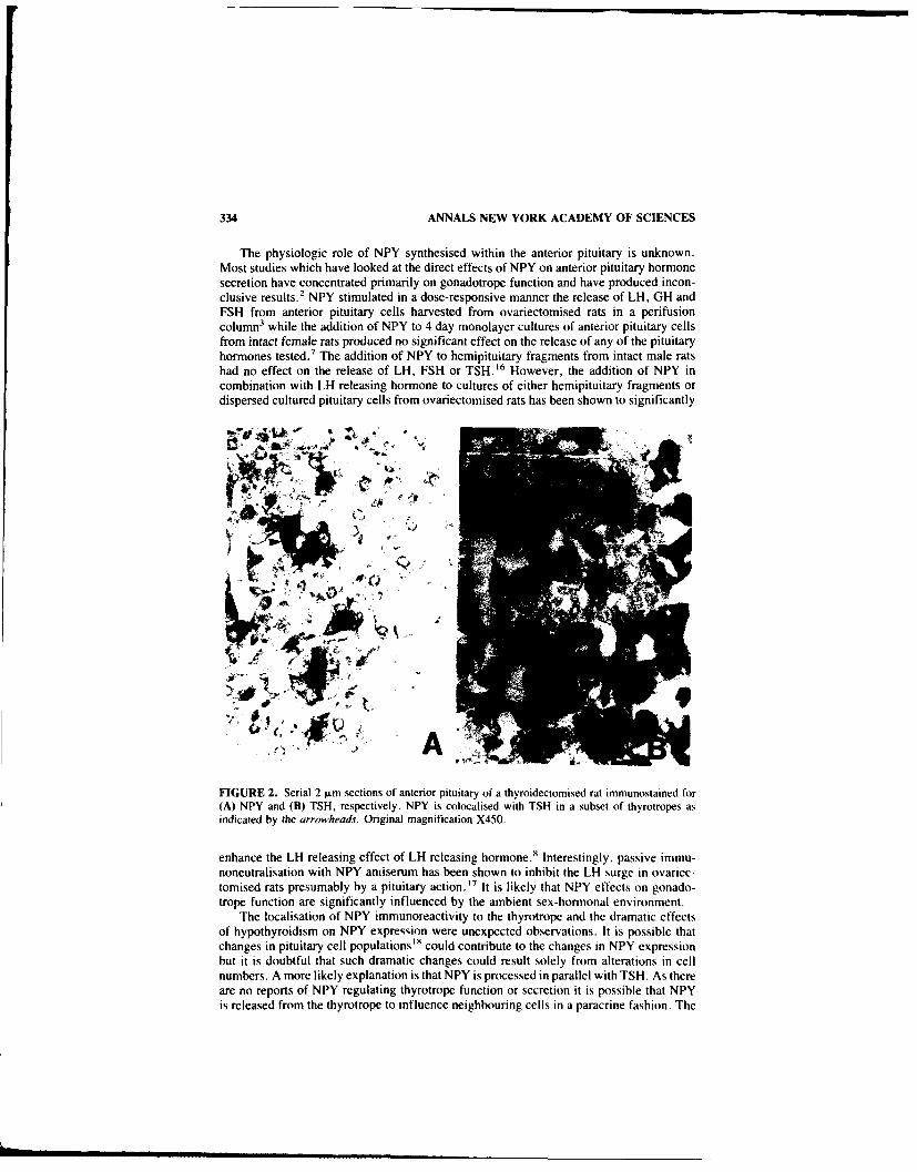
334 ANNALS NEW YORK ACADEMY OF SCIENCES
The physiologic role of NPY synthesised within the anterior pituitary is unknown.Most studies which have looked at the direct effects of NPY on anterior pituitary hormonesecretion have concentrated primarily on gonadotrope function and have produced incon-clusive results.2 NPY stimulated in a dose-responsive manner the release of LH, GH andFSH from anterior pituitary cells harvested from ovariectomised rats in a perifusioncolumn3 while the addition of NPY to 4 day monolayer cultures of anterior pituitary cellsfrom intact female rats produced no significant effect on the release of any of the pituitaryhormones tested.7 The addition of NPY to hemipituitary fragments from intact male ratshad no effect on the release of LH, FSH or TSH. 16 However, the addition of NPY incombination with LH releasing hormone to cultures of either hemipituitary fragments ordispersed cultured pituitary cells from ovariectomised rats has been shown to significantly
'
II
FIGURE 2. Serial 2 lm sections of anterior pituitary of a thyroidectomised rat immunostained for(A) NPY and (B) TSH, respectively. NPY is colocalised with TSH in a subset of thyrotropes asindicated by the arrowheads. Original magnification X450.
enhance the LH releasing effect of LH releasing hormone.8 Interestingly, passive immu-noneutralisation with NPY antiserum has been shown to inhibit the LH surge in ovariec-tomised rats presumably by a pituitary action.' 7 It is likely that NPY effects on gonado-trope function are significantly influenced by the ambient sex-hormonal environment.
The localisation of NPY immunoreactivity to the thyrotrope and the dramatic effectsof hypothyroidism on NPY expression were unexpected observations. It is possible thatchanges in pituitary cell populations"' could contribute to the changes in NPY expressionbut it is doubtful that such dramatic changes could result solely from alterations in cellnumbers. A more likely explanation is that NPY is processed in parallel with TSH. As thereare no reports of NPY regulating thyrotrope function or secretion it is possible that NPYis released from the thyrotrope to influence neighbouring cells in a paracrine fashion. The

O'HALLORAN et al.: ANTERIOR PITUITARY 335
doubling of anterior pituitary NPY following ovariectomy coupled with its unresponsive-ness to high-dose estrogen treatment and male castration (data not shown) may furtherreflect the intrinsic differences in the gonadotrope response to NPY which appears to bedependent on the sex-steroid status. Similarly, the result obtained in the combined adre-nalectomised and castrated group might reflect a similar divergence in function.
These results strongly suggest that NPY is synthesised within the rat anterior pituitaryand that this synthesis is dramatically increased in the hypothyroid state The applicationof in-situ hybridisation techniques and receptor mapping studies should further enhanceour understanding of the intrapituitary role that NPY plays.
REFERENCES
1. TATEMOTO, K.. M. CARLQUIST & V. MUTT. 1982. Neuropeptide Y-a novel brain peptidewith structural similarities to peptide YY and pancreatic polypeptide. Nature (London) 296:659-660.
2. McDONALD, J. K. 1988. NPY and related substances. Cn'. Rev. Neurobiol. 4: 97-135.3. MCDONALD. J. K., M. D. LUMPKIN. W. K. SAMSON & S. M. MCCANN. 1985. Neuropeptide
Y affects secretion of luteinizing hormone and growth hormone in ovariectomised rats. Proc.Natil. Acad. Sci. USA 82: 561-564.
4. HARFSTRAND. A., P. ENROTH. L. AGNATI & K. FUXE. 1987. Further studies on the centraladministration of neuropeptide Y on neuroendocrine function in the male rat: relationship tohypothalamic catechn!rnines. Regul. Pept. 17: 167-179.
5. HARFSTRAND, A., K. 1"UXE, L. !- AGNATI, P. ENEROTH, I. ZINI. M. ZOLI, K. ANDERSON, G.VON EULER. L. TERENIUS, V. MUTT & M. GOLDSTEIN. 1986. Studies on neuropeptide-catecholamine interactions in the hypothalamus and in the forebrain of the male rat. Rela-tionship to neuroendocrine function. Neurochem. Int. 8: 355-376.
6. KALRA, P. S. & S. P. KALRA. 1986. Steroidal modulation of the regulatory neuropeptides:luteinizing hormone releasing hormone, neuropeptide Y and endogenous opioid peptides. J.Steroid Biochem. 25: 733-740.
7. KERKERIAN, L.. J. GuY, G. LEFEVRE & G. PELLETIER. 1985. Effects of' neuropeptide Y (NPY)on the release of anterior pituitary hormones in the rat. Peptides 6: 1201-1207.
8. CROWLEY, W. R.. A. HASSID & S. P. KALRA. 1987.Neuropeptide Y enhances the release ofluteinising hormone (LH) induced by LH releasing hormone. Endocrinology 120: 941-945.
9. ALLEN, J. M.. J. C. YEATS, T. E. ADRIAN & S. R. BLOOM. 1984.Radioimmunoassay ofneuropeptide Y. Regul. Pept. 8: 61-70.
10. CHOMCZYNSKI, P. & N. SACCHI. 1987. Single method of RNA isolation by acid guanidiumthiocyanate- phenol- chloroform extraction. Anal. Biochem. 162: 156-159.
I1. MANIATIS. T., E. F. FRISCH & J. SAMBROOK. 1982. Extraction, purification, and analysis ofmRNA from eukaryotic cells. In Molecular Cloning: A Laboratory Manual. Chap. 6: 187-210. Cold Spring Harbor Laboratories. Cold Spring Harbor, NY.
12. FEINBERG, A. P. & B. VOGELSTEIN. 1983. A technique for radiolabelling DNA restrictionendonuclease fragments to high specific activity. Anal. Biochem. 132: 6-13.
13. MANIATIS, T.. E. F. FRITSCH & J. SAMBROOK. 1982. Molecular cloning: a laboratory manual.Appendix A. pages 435-478 Cold Spring Harbor Laboratories, Cold Spring Harbor. NY.
14. ALLEN, Y. S., T. E. ADRIAN, J. M. ALLEN, K. TATEMOTO, T. J. CROW. S. R. BLOOM &J. M. POLAK. 1983. Neuropeptide Y distribution in the rat brain. Science 221: 877-879.
15. VAN NOORDEN S., J. M. POLAK, M. ROBINSON, A. E. G. PEARSE & P. J. MARANGOS. 1983.Neurone-specific enolase in the pituitary gland. Neuroendocrinology 38: 309-316.
16. RODRIGUEZ-SERRA, J. F., D. M. JACOBOWITZ & C. A. BLAKE. 1987. Effects of neuropeptideY on LH, FSH and TSH release in male rats. Peptides 8: 539-542.
17. SUTTON, S. W., T. T. TOYAMA, S. OTTO & P. M. PLOTSKY. 1988. Evidence that neuropeptideY (NPY) released into the hypophyscal-portal circulation participates in priming gonadotropesto the effects of gonadotrophin-releasing hormone (GnRH). Endocrinology 123: 1208-1210.
18. DEF-s, C. R., H. S. ASTIER & M. I. SURKS. 1979. Kinetics of thyrotrophs and somatotrophesduring development of hypothyroidism and l-triiodothyronine treatment of hypothyroid rats.Endocrinology 104: 1172-1180.

POSTER PAPERS
Conformational Analysis of theNeuropeptide Y Fragment
NPY(18-36) by 'H-NMR andComputer Simulationa
R. D. FEINSTEINb M. SPICERd J. BOUBLIKd J. P. TAULANE b
J. RIVIER d M. BROWN.' AND M. GOODMAN b
bDepartment of Chemistry, B-043
University of California, San DiegoLa Jolla, California 92093
cDepartments of Medicine and SurgeryUniversity of California, San Diego Medical Center
225 Dickinson StreetSan Diego, California 92103
dThe Clayton Foundation Laboratories for Peptide BiologyThe Salk InstituteP.O. Box 85800
San Diego, California 92138-9216
INTRODUCTION
In the ongoing effort to elucidate the structural requirements of NPY necessary forboth receptor selectivity and efficacy, the conformational preferences of the Y2-receptorselective ligand NPY(18-36) have been investigated using 500 MHz 'H-NMR and com-puter simulations. Towards the development of conformationally constrained receptorspecific agonists and antagonists, elucidation of the conformational preferences ofNPY( 18-36) and related analogs is essential. We report here the results of the investi-gation of NPY(18-36).
METHODS
NPY( 18-36) was synthesized by solid phase methodology and purified by RPHPLC.The conformational characterization was carried out using 'H-NMR and NOE-restrainek'energy minimizations as well as high and low temperature dynamics simulations. Theproton resonances were assigned using 2D COSY, HOHAHA and ROESY techniques. 2 4
All NOE experiments used for determination of the preferred conformations of NPY( 18-36) were carried out in dimethylformamide-d7 (DMF-d7 ).
Computer simulations were carried out on a SGI Iris 4-D using the CHARMm andQUANTA simulation programs. Fifty random conformations (random 4), ti and x,angles), a fully extended conformation and a right-handed a-helical conformation were
1This work was supported in part by National Institutes of Health Grants RR03342-01 andGM 18694 and by National Science Foundation Grant BBS86-12359.
336
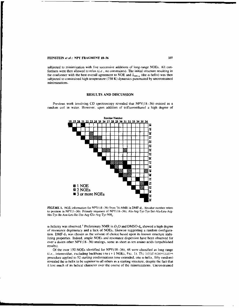
FEINSTEIN et al.: NPY FRAGMENT 18-36 337
subjected to minimization with five successive additions of long-range NOEs. All con-formers were then allowed to relax (i.e., no constraints). The initial structure resulting inthe conformer with the best overall agreement to NOE and JNH-, (the a-helix) was thensubjected to constrained high temperature (750 K) dynamics punctuated by unconstrainedminimizations.
RESULTS AND DISCUSSION
Previous work involving CD spectroscopy revealed that NPY(18-36) existed as arandom coil in water. However, upon addition of trifluoroethanol a high degree of
Residue Number18 19 20 21 22 23 24 25 26 27 29 29 30 31 32 33 34 35 36
* 1 NOE19 2 NOEs* 3 or more NOEs
FIGURE I. NOE information for NPY(18-36) from 'H-NMR in DMF-d.. Residue number refersto position in NPY(I-36). Primary sequence of NPY(18-36); Ala-Arg-Tyr-Tyr-Ser-Ala-Leu-Arg-His-Tyr-Ile-Asn-Leu-Ile-Thr-Arg-Gln-Arg-Tyr-NH 2 .
a-helicity was observed.' Preliminary NMR in D20 and DMSO-d, showed a high degreeof resonance degeneracy and a lack of NOEs, likewise suggesting a random configura-tion. DMF-d7 was chosen as the solvent of choice based upon its known structure stabi-lizing properties. Indeed, ample NOEs and resonance dispersion have been observed forover a dozen other NPY(18-36) analogs, some as short as ten amino acids (unpublishedresults).
Of the over 150 NOEs identified for NPY(18-36), 48 were classified as long range(i.e., interresidue, excluding backbone i to i + I NOEs, FiG. I). T!: m.i± ! ,r.i:tic!.procedure applied to 52 starting conformations (one extended, one a-helix, fifty random)revealed the a-helix to be superior to all others as a starting structure, despite the fact thatit lost much of its helical character over the course of the minimizations. Unconstrained
• m m mmm i mm m rm•mmm

338 ANNALS NEW YORK ACADEMY OF SCIENCES
minimizations of 100 structures resulting from 10 ps of constrained high temperaturedynamics of the "ot-helix" revealed six major families of conformers. The lowest energymembers of each of the six families all showed good agreement with the NOE results andJNH-, coupling constants (FIG. 2).-
For all six low energy structures certain overall conformational properties remainedconserved. Of particular interest was the hydrophobic core present in all, made up of theresidues Ala, 23 Leu, 24 lie,28 Leu3° and Ile. 3
1 In the case of NPY(18-36) (as opposed toresidues 18 to 36 within NPY(1-36)) the formation of a hydrophobic core was reasonable,in that it most efficiently shielded the nonpolar side chains of Ala, Leu and lie.
FIGURE 2. Minimum energy conformer of NPY(18-36) derived from constrained high tempera-ture dynamics. N-terminus is at extreme left, C-terminus is at upper right. Hydrophobic cleft denotedby filled atom centers of side chains from lie and Leu residues (middle top to middle bottom).
The detailed conformational analysis of the many NPY(18-36) analogs is currentlyunder way. It is anticipated that important structure-activity relationships will becomeclear.
REFERENCES
I. BouBLIK, J., N. SCOTT, J. TAULANE, M. GOODMAN, M. BROWN & J. RIVIER. 1989. Int. J.Peptide Protein Res. 33: 11-15.

339
FEINSTEIN et al.: NPY FRAGMENT 18-36
2. BAx, A, & D.G. DAVIS. 1995. J. Magn, Reson. 65: 355-360.
3. BAX, A. & D.G. DAViS. 1985. J. Magn. Reson. 63: 207-213.
4. LEVITT, M. 1983. J. Mol. Biol. 168: 595-620.
5. BystRokv, V.F. 1976. Progr. NMR Spectrosc. 10: 41-81.

Characterization of Neuropeptide YImmunoreactivity in Dog Plasma by
High Performance LiquidChromatography andRadioimmunoassay
P. SENANAYAKE," M. R. WARNER,b M. N. LEVYC
D. W. JACOBSEN,- AND C. M. FERRARIO"
"Cleveland Clinic FDNDepartment of Brain and Vascular Research, FF2
9500 Euclid AvenueCleveland, Ohio 44195-5070
bKrannert Institute of CardiologyIndienapolis, Indiana
'Case Western Reserve UniversityCleveland, Ohio
INTRODUCTION
Fragments of neuropeptide Y (NPY) with varying degrees of physiological and im-munological activity may circulate in the blood. NPY in plasma extracts has been rou-tinely quantified by radioimmunoassay (RIA) using polyclonal antibodies. While thesedeterminations are sensitive and provide useful information, the levels of NPY mayrepresent a mixture of NPY and peptide fragments of varying biological and immuno-logical potency. The aim of this study was to develop a method for separation andquantitation of NPY immunoreactivity (NPY-ir) in dog plasma as a first step towardsunderstanding the processing of the peptide and characterizing the circulating levels of thepeptide and its fragments.
MATERIAL AND METHODS
Venous blood (10 ml) was collected into ice chilled tubes containing heparin (20IU/ml blood) and aprotinin (500 KIU/ml blood) from 20 normal conscious male mongreldogs weighing between 18 and 25 kg. The effects of either a short or a long-lastingbarbiturate on venous levels of NPY were evaluated by a comparison of the effects ofanesthesia induced by either sodium pentobarbital (20 mg/kg IV) (n = 5) or sodiumthiamyal (surital, 20 mg/kg IV) (n = 6). Samples simultaneously collected from a veinand an indwelling arterial catheter of a conscious dog were used for high performanceliquid chromatography (HPLC) analysis. Blood samples were centrifuged at 2100 x g for20 minutes at 4°C and the plasma separated and stored at -20°C. Plasma samples werethawed at 4°C. NPY was extracted from plasma proteins using a Waters octadecasilanereverse phase cartridge, which was activated by sequentially washing with 5 ml of meth-
340

SENANAYAKE et al.; DOG PLASMA 341
anol, tetrahydrofuran. hexane, methanol and distilled water. Plasma was applied to theSep-Pak, washed with distilled water (10 ml) and eluted with 60% acetonitrile. Fractionswere evaporated to dryness and either quantified by RIA or stored at -20'C for HPLCfollowed by RIA. 2 5 I-NPY (1000 cpm) and synthetic porcine NPY (range 100-1000pg/mb were added to plasma samples before extraction to determine radiolabel andimmunoreactive recoveries. The RIA was carried out according to the method of Allen etal. and the characteristics of the assay are summarized in TABLE 1.
NPY peptides and fragments were separated on a LKB 2150 gradient system equippedwith a Vydac CI, column (4.6 x 150 mm, Hesperia, CA) and a 3.2 x 15 mm NewguardC, guard column (Brown Lee Labs. Santa Clara, CA). The column was equilibrated with60/c mobile phase A 10.13% heptafluorobutyric acid (HFBA)J and 40% mobile phase B
TABLE i. Neuropeptide '' Radioimmunoassay Characteristics Antiserum-Specificity
Peptide % Cross-Reactivity
Porcine neuropeptide Y(1-36) 100Porcine neuropeptide Y(16-36) 33%Human neuropeptide Y(-36) 100%Human neuropeptide Y(20-36) 66%Human neuropeptid- Y(22-36) 100%Human neuropeptide Y(26-36) 100 %Human pancreatic polypeptide 0.02%hAvian pancreatic polypeptide 0.007%Peptide YY 0.003%cVasoactive intestinal peptide 0.(X) ICRat corticotropin releasing factor 0.00117Human growth hormone releasing factor 0.(X)I%Glucagon 0%ZSecretin 0%
SensitivityED(20 = 225 pg/tubeED(O = i 16 pg/tubeED80) 58 pg/tube
ReproducibilityIntraassay variation = 10%Interassay variation = 14%
Recover,Synthetic peptide 76 ± 8c/Radiolabeled peptide = 80 - 5o/
(80%4 acetonitrile/0. 131/ HFBA) at a flow rate of I ml/min. Plasma extracts were dis-solved in 40% mobile phase B and applied to the column. NPY and peptide fragmentswere eluted with a 24 min gradient of 40-47% mobile phase B. Fractions (0.2 ml) werecollected at 12-set intervals and evaporated to dryness. The dried fractions were recon-stituted in the phosphate buffer (0.06 M, pH 7.4) and quantified by RIA.
RESULTS AND SUMMARY
In conscious dogs, plasma levels of NPY-ir in venous blood averaged 126 - II pg/mIn- 201. The circulating levels of NPY were not influenced by anesthesia induced by

342 ANNALS NEW YORK ACADEMY OF SCIENCES
3Z 00 -"~~ tm
20.
S0
40 . FIGURE 1. High pressure liquid chromatc
graphic separation of NPY and its immunoreac-tive fragments. In panel (A) the retention timesa.
+. for standards [NPY(26-36), NPY(22-36),NPY(20-36), human NPY(I-36), porcine
10 NPY(16-36) and porcine NPY(I-36)j areshown. HPLC analysis of normal conscious ar-
Z 0 terial (panel B) and venous (panel C) plasmashows a fragment with retention times similar tohuman NPY and a number of unknown peaks.
S30 .
20
0 3 10 15 20 25
RETENTION TIME. MIN
either sodium pentobarbital (92 ± 17 vs 138 ± 42 pg/ml) or sodium thiamyal (109 ± 23vs 113 ± 22 pg/ml). Canine plasma contained a NPY fragment with properties similar tothe human, but not porcine form FIG. 1 .) This fragment. probably canine NPY(1-36), wasfound in both arterial and venous samples. It accounted for approximately 15% of the totalNPY-ir, and thus is not a major circulating component in dog plasma. Three other NPYfragments amounting to 42, 14, and 32% of the total NPY-ir had retention times thatdiffered from known standards. In summary, this is the first demonstration of NPY andits carboxy-terminal immunoreactive components in plasma.
REFERENCE
I. ALLEN, et al. 1984. Regul. Pept. 8: 61.

Peptide YY/Neuropeptide Y Receptors inSmall Intestine
Characterization, Signal Transduction, andExpression during Cell Differentiation
T. VOISIN, C. ROUYER-FESSARD, AND M. LABURTHE
Unit de Recherches sur la Diffirenciation et la Neuroendocrinologiede Cellules Digestives (INSERM U178)
16 avenue Paul Vaillant Couturier94807 Villejuif Cedex, France
Peptide YY (PYY) and neuropeptide Y (NPY) are present in high concentrations in theintestines. PYY is localized in endocrine cells of the mucosa (colon, rectum, ileum)whereas NPY is strictly a neuropeptide. NPY-containing nerve fibers form a dense net-work in the lamina propria which underlies the small intestinal epithelium (see REF. I forreview). In 1986, we reported the properties of a new receptor for PYY and NPY in ratintestinal epithelium.2 This receptor has been characterized by using 251-labeled PYY asa tracer and is PYY-preferring since it binds PYY (Kd = 0.1 nM) with a 5-times-higheraffinity than NPY.2 Concomitantly, PYY and NPY were shown to be potent inhibitors offluid and electrolytes secretion in small intestine (see REF. I for review), providing afunctional significance for intestinal epithelial PYY-NPY receptors. 2 We have furthershown recently that PYY and NPY inhibit VIP- or prostaglandins-stimulated cAMPproduction in rat small intestinal epithelial cells. 3 The receptor mediating this inhibition 3
is also PYY-preferring as assessed pharmacologically with PYY, NPY and several frag-ments of the peptides3 including PYY 22-36 which appears to be a potent agonist. 3
In small intestinal epithelium, cells originate in the crypts and migrate onto the villi(FIG. 1). Crypt cells proliferate rapidly, but as they migrate they stop to divide and theydifferentiate into a major population of columnar absorptive cells. The villus is the site ofions and nutriments absorption whereas the crypt is believed to be the site of Cl- secre-tion. We have recently determined the distribution of PYY-NPY receptors along thevillus-crypt axis (FIG. I). By using monoiodinated PYY (from NEN) as a tracer, we haveshown that the binding capacity of receptors decreases markedly from crypt to villus (REF.4 and FIG. 1). Scatchard plots are linear in both cell populations (FIG. I): BMAx are 166and 21 fmol/mg protein and Kd are 0.1 and 0.05 nM in membranes prepared from cryptand villus cells, respectively (REF. 4 and FIG. I). The receptor is PYY-preferring in bothcompartments. 4 PYY and NPY inhibit VIP-stimulated cAMP production in crypt cells butnot in villus cells (REF. 4 and FIG. 1). Therefore, PYY-prefering receptors, negativelycoupled to cAMP production system, are preferentially expressed in crypt cells wherethey mediate the potent anti-secretory actions of PYY and NPY on water and electrolytes(see REI I for review).
As shown in nervous tissues, NPY/PYY binding sites occur in two subtypes. YI andY2, which differ in specificity but accept NPY and PYY equally well. The YI receptordoes not bind NPY 13-36 whereas the Y2 receptor binds NPY 13-36 with a good affinity.'The PYY-preferring receptor characterized in small intestine 4 resembles the Y2 receptorin that it binds PYY 22-36 with a good affinity3 but differs from it since it discriminatesbetween PYY and NPY.'- 4 In order to further explore this issue. monoiodinated radio-ligands of PYY and NPY have been prepared by reverse phase HPLC as described6 with
343

344 ANNALS NEW YORK ACADEMY OF SCIENCES
modifications (FIG. 2). Since PYY 22-36 binds rather well to intestinal PYY receptors, 3
we have used 1125 1-Tyr'] monoiodo PYY to further characterize PYY receptors in rat
small intestine (FIG. 2). The receptor labeled by this radioligand discriminates betweenPYY, NPY, and pancreatic polypeptide as did radiolabeled PYY tracers usedpreviously." 4 This further confirms the expression of a PYY-preferring receptor in rat
NON PROLIFERATIVE CELLS
1A VILLUS
iis'e 60-0
t I
VILLUS T (ILL
BOUND (fmai I z% proii
I *MVPaoeo n h rsneo . PYY Se .. o thrdti VI, P
{1251-yra6] PY bining i inhibted l)oyUNP (witha hig pso (16nM)I n )
04
II
so in IS
PRLFRTV CELLSBON goA me "OW
FIGU RE P Exrsso o Y-rfrigrcposdrneptlaleldfeetiaina.th
ratsmllinesin -ilu-cyp ais Poliertie ryt ell wresearte fom atrevil26 cllas escibe.' onoodiate Y N a sdt hrceiercposi rd ebaefrom ~ ~ ~ ~ i crp el 0 n ilscls 0.cM a esrdi slae el fe nuainwt
I IL VI lnAri.h rsec fIR Y.SeRE.4frohrdtis
FaGUR 1. isqiepfression o ha obspr egreedr din epiheia celyrl idifrnton Y lndethe
smal 'itestine ['2 -binding imnoiioedo1b NPY wih wa used to,, chrateiz YI an 2b

VOISIN et al.: SMALL INTESTINE 345
E~Z-or
.0
u E
z U0 E
"S,
r4
I.- CL
E a--u E
0. 0
U,CL-
c~~d >9 CN - E U
:7
- 'i
E
U EC

346 ANNALS NEW YORK ACADEMY OF SCIENCES
unlabeled peptides with the following order of potency NPY>PYY>PP (FIG. 2). Thesedata suggest that a low affinity NPY-prefemrng binding site may coexist with high affinityPYY-prefemrng receptors in rat small intestinal epithelium. The biological significance, ifany, of the low affinity NPY-preferring binding site and its distribution along the crypt-villus axis remains to be determined.
REFERENCES
I. LABURTHE, M. 1990. Trends Endocrinol. Metab. 1: 168-174.2. LABURTHE, M.. B. CHENUT. C. ROUYER-FESSARD, K. TATEMOTO, A. COUVINEAU, A.
SERVIN & B. AM]RANOFF. 1986. Endocrinology 118: 1910-1917.3. SERVIN, A.. C. RouYER-FESSARD, A. BALASUBRAMANIAM, S. SAINT PIERRE & M. LABURTHE.
1989. Endocrinology 124: 692-700.4. VOISIN,. T., C. ROUYER-FESSARD & M. LABURTHE. 1990. Am. J. Physiol. 258 (Gastrointest.
Liver Physiol.). 21: G753-G759.5. SHEIKH, S. P., R. HAKANSON & T. W. SCHWARTZ. 3989. FEBS Lett. 245: 209-214.6. SHEIKH, S. P., M. M. T. O'HARE, 0. TORTORA & T. W. SCHWARTZ. 1989. J1. Biol. Chem.
264: 6648-6654.

Structural Characterization of Y, andY2 Receptors for Neuropeptide Y and
Peptide YYS. P. SHEIKH AND J. A. WILLIAMS
Department of Physiology and Internal MedicineUniversity of Michigan
Ann Arbor, Michigan 48109and
Laboratory of Molecular EndocrinologyUniversity of Copenhagen
RigshospitaletCopenhagen, Denmark
Two receptor subtypes for peptide YY (PYY) and neuropeptide Y (NPY) named Y,and Y2 have been proposed to exist based on pharmacological studies. 1.2 In order toidentify and structurally characterize Y, and Y, receptors we covalently cross-linked[12 I-Tyr6]PYY to its receptors. Affinity labeling of the Y,-receptor protein was ana-lyzed in membranes from the MC-IXC human neuroblastoma cell line. Analysis bySDS-polyacrylamide gel electrophoresis and autoradiography resulted in labeling of amajor band centered at Mr = 70,000, and a smaller band at 45,000. The labeled bandswere unaffected by reducing agents (FIc. 1). The labeling of the M, = 45,000 band wasgreater in the absence of protease inhibitors, suggesting that it is a degradation product ofthe larger band. For characterization of Y2 receptors we utilized two different tissues; rathippocampal membranes and rabbit kidney tubule membranes. In both tissues affinitylabeling of PYY binding proteins resulted in labeling of a major protein centered at M, =50,000, which was unaffected by reducing agents (FIG. 2). For both the Y, and Y,receptors the binding to the receptors and the intensity of the labeled bands were inhibitedin a parallel dose-dependent manner by increasing concentrations of unlabeled pyy. 3
Efficient labeling of Y2-receptor proteins was obtained using a number of different homo-and heterobifunctional cross-linking reagents, whereas labeling of Y,-receptor proteinswas obtained only when N-5-azido-2-nitrobenzoyloxysuccinimide (ANBNOS) was em-ployed for cross-linking followed by exposure to UV light. To determine whether thereceptors were glycoproteins the detergent solubilized receptor-hormone complexes wereexposed to different agarose-coupled lectins. The cross-linked Y, and Y2 receptors werealmost completely absorbed by wheat germ agglutinin agarose. The Y, receptor waspartially absorbed by icin communis 1i, and the Y2 showed partial absorption to con-canavalin A. These absorptions were in all cases blocked by the appropriate hapten sugar.These results indicate that the Y, and Y, subtypes of NPY and PYY receptors, previouslycharacterized pharmacologically, are structurally distinct glycoproteins, not disulfide-linked to other subunits.
347

348 ANNALS NEW YORK ACADEMY OF SCIENCES
A~NN6 EMS
200116
97 -FIGUKE 1. Affinity labeling of the Y, re-ceptor in a human neuroblastoma cell line,
66 MC-IXC, with radiolabeled PYY. Membranes(200 pLg) were incubated with 250 pM radio-labeled PYY, washed and cross-linked with
45 0. 1 mM ANBNOS or 0.5 mM EGS. Reducingagent (50 mM DTT) and unlabeled PYY (IpM) were added as indicated. (From Sheikhand Williams. 3 Reprinted by permission from31 the Journal of Biological Chemistry.)
[ " + + +
EGS
200-
116-
66- FIGURE 2. Affinity labeling of the Y2 re-ceptor in rat hippocampus with radiolabeledPYY. Membranes (200 pjg/ml) were incubatedwith 200 pM radiolabeled PYY, washed free* of unbound ligand, and cross-linked with 0.5mM EGS. Unlabeled PYY (I jiM) and dithio-45 - threitol (50 mM) were added as indicated.(From Sheikh and Williams. Reprinted by per-mission from the Journal of Biological Chem-isto.)
31-
gf " . ++ +
OT +U + nlJim_
SHEIKH & WILLIAMS: Y, & Y7 RECEPTORS 349
REFERENCES
I. WAHLESTEDT, C., N. YANAIHARA & R. HAKANSON. 1986. Evidence for different pre- andpost-junctional receptors for neuropeptide Y and related peptides. Regul. Pept. 13: 307-318.
2. SHEIKH, S. P., M. M. T. O'HARE, 0. TORTORA & T. W. SCHWARTZ. 1989. Binding ofmonoiodinated neuropeptide Y to hippocampal membranes and human neuroblastoma celllines. J. Biol. Chem. 264: 6648-6654.
3. SHEIKH, S. P. & J. A. WILLIAMS. Structural characterization of Y, a i Y2 receptors forneuropeptide Y and peptide YY by affinity cross-linking. J. Biol. Chen. 265: 8304-8310.

Peptide YY Receptors in Mammalianand Avian Brains
AKIO INUI, MINORU OKITA, MASAKI MIURA,
YOSHIAKI HIROSUE, MASAHARU NAKAJIMA,
AND SHIGEAKI BABA"
Second Department of Internal MedicineKobe Universiy School of Medicine
Kusunoki-cho. Chuo-kuKobe 650, Japan
"Hyogo Institute for Research in Adult DiseasesAkashi, Japan
The ligand binding technique developed during the past two decades has identified a greatmultiplicity of neurotransmitter receptors, including those for NPY and PYY. I-" Whilemost of the CNS immunoreactivity for this PP family of peptides is NPY, PYY-containingneurons are also present in the brain. This provides an anatomical basis for the effects ofNPY and PYY on brain functions. In the present study, we summarize the PYY receptorsin mammalian and avian brains.
Direct binding of radioactive peptides to their target cells identifies receptors by theiraffinity and specificity. Two classes of j1 2 5
1)PYY binding sites were observed in porcinebrain membrane preparations. 6 "7 The calculated binding affinity of 0.139 nM for the highaffinity component is similar to the value reported for PYY binding to membranes pre-pared from rat intestinal epithelium (kd = 0.4 nM), which is consistent with the venousblood level of PYY observed after a meal. Brain PYY receptors also exhibit high spec-ificity. However, PYY and NPY receptors do not distinguish between PYY and NPY(FiG. 1), suggesting that PYY and NPY regulate brain function through interaction atcommon receptor sites, a notion also suggested from biological studies. PYY receptorbinding requires not only the C-terminal portion of the molecule but also the N-terminalportion, and is linked to a second messenger system including adenylate cyclase inhibitionvia G proteins.
TABLE I shows the distribution of PYY receptors in the brain, which are abundant inthe hippocampus, amygdala, hypothalamus, and the pituitary gland. In dogs and rodentshigh binding activity was also observed in the thalamus and cerebral cortex, suggestingspecies differences. Autoradiographic studies with radiolabeled NPY and PYY demon-strated that PYY and NPY receptors are quite similarly distributed throughout the brain,although somewhat differential localizations were observed, e.g., in the mamillary body,thalamus, or the cerebellum.'o- 2
The PYY receptors in brain and intestinal epithelial membranes were further charac-terized after covalent labeling with chloramine T-iodinated PYY using homobifunctionalcross-linker disuccinimidyl suberate. 5
.7 Analysis by sodium dodecyl sulfate-polyacryla-
mide gel electrophoresis followed by autoradiography demonstrated that PYY receptorsare protein of mol wt 50 kDa with no disulfide-linked subunits (the same as that of NPYreceptors), and are found in various regions of the brain and intestinal epithelium acrossthe species. In the chicken brain, however, PYY receptors are found as a 67-kDa proteinband which does not discriminate either PYY, NPY or avian PP well." The APP and NPYreceptors are also the same 67-kDa proteins, with PYY, NPY and APP acting at either ofthese receptors. These results indicate that chicken brain also has receptors specific for
350

INUI t; al.: MAMMALIAN & AVIAN PYY 351
380 -
NYY
EX,,5 24-36
S60NPY6O
17-3617-36
C3 40
20
011 10 9 8 7 6
Peptide, -Log (M)
FIGURE 1. Inhibition of [12 51JPYY binding to porcine hippocampal membranes by PYY analogs.
Membranes were incubated with 40 pM [ t25 IIPYY for 5 h at 25°C. Varying concentrations of analogswere added as shown. Nonspecific binding was subtracted from total binding and specific bindingwas expressed as a percentage of maximal specific binding.
PYY and NPY, and that PYY, NPY, and APP act in the brain through interaction atmultiple receptor sites which are quite similar to. and shared by, other members of the PPfamily.
We have recently found that porcine brain has binding sites specific for APP (TABLEI). The APP binding sites are more specific for APP than those in avian brain, since
TABLE i. Regional Distribution of PYY, NPY, and APP Receptors in the BrainSpecific PYY Binding Specific NPY Binding Specific APP Binding
(fmol/mg Protein) (fmol/mg Protein) (fmol/mg Protein)
Brain Area Pig Dog Guinea Pig Pig Pig
Cerebral cortex 2.5 ± 0.9 5.6 ± 1.4 11.5 ± 2.8 0.11 ± 0.05 9.9 _ 1.9Olfactory bulb 4.2 ± t.2 3.0 ± 0.8 4.8 ± 1.8 0.41 ± 0.20 8.5 ± 1.7Caudate nucleus 17.7 ± 4.9 15.1 ± 2.2 12.4 ± 3.1 () 43 ± 0.15 9.4 ± 21Hippocampus 84.5 ± 13.6 22.6 ± 7.1 14.6 ±L 3.5 3.05 ± 0.80 11.2 ± 1.7Amygdala 28.6 ± 5.8 12.4 ± 2.0 14.0 ± 4.5 0.64 0,13 10.0 ± 1.7Thalamus 4.0 ± 0.9 18.3 ± 3.2 6.9 ± 13 0.18 ± 0.10 9.5 ± 1.6Hypothalamus 34.3 ± 9.1 13.2 ± 3.4 10.1 ± 2.8 088 ± 0.14 9.7 ± 1.8Pituitary gland 58.9 ± 13.6 12.4 ± 3.6 10.6 ± 0.7 2.85 ± 076 16.9 ± 2.7Midbrain 11.3 ± 2.5 12.2 ± 2.6 11.9 ± 29 0.26 ± 013 91 ± I.APons 9.4 ± 2.4 5.6 ± 2.(1 8.1 1 8 0.40 - 0,10 9.4 ± 1.4Medulla 16.3 ± 3.0 79 ± 0.8 6.9 1 1.5 1.11 - 0.27 7.5 ± 1.8oblongataCerebellum 3.0 ± 1.5 0.4 ± 0.2 8.3 - 2.2 031 ±0.10 96 _ 1.0

352 ANNALS NEW YORK ACADEMY OF SCIENCES
porcine tissue APP binding cannot be inhibited by NPY, PYY or PPP, whereas in chickentissue all peptides of this family inhibit APP binding. Although the relatively low affinityof the porcine APP binding site does not allow consideration of its functional significance,it is possible that some endogenous peptide related to APP may exist and serve a specificfunction in porcine brain.
The identification of all of the PP family peptides and theii receptors, especially theirstructures form a basis for the rational de ign of new, potent and selective drugs for eachreceptor subtype, an important advancement in neuroscience.
REFERENCES
I. i. K. & L. F. AGNATI. 1985. Receptor-receptor interactions in the central nervous system.A new integrative mechanism in synapses. Med. Res. Rev. 5: 441-482.
2. UNDEN, A., L-L. PETFRSON & Y. BARTFAI. 1985. Somatostatin, substanc- P, vasoactiveintestinal polypeptide, and neuropeptide Y receptors: critical assessment of biochemical meth-odology and results. Int. Rev. Neurobiol. 27: 141-177.
3. McDONALD, J. K. 1988. NPY and related substances. In CRC Critical Reviews in Neurobi-ology. J. Nelson, Ed. Vol. 4: 97-135. CRC Press. Boca Raton, FL.
4. MUTT, V., T. HOKFELT, K. FUXE & J. M. LUNDBERG. 1989. Neuropeptide Y. KarolinskaInstitute Nobel Conference Series. Raven Press. New York, NY.
5. INUI. A. & S. BABA. 1990. Gastrointestinal peptide binding and function in the brain: emphasison peptide YY. In Neuroendocrine Perspectives. E. E. Miuller & R. M. MacLeod. Eds. Vol.8. Springer-Verlag. New York, NY, In press.
6. INUI, A., M. OYA, M. OKITA, T. INOUE. N. SAKATANI, H. MORIOKA. K. S-tu, K. YOKONO,
N. MtzuNo & S. BABA. 1988, Peptide YY receptors in the brain. Biochem. Biophys. Res.Commun. 150: 25-32.
7. INUI, A., M. OKITA. T. INOUE, N. SAKATANI. M. OYA, H. MORIOKA, K. Smi, K. YoKONO.
N. MizuNo & S. BABA. 1989. Characterization of peptide YY receptors in the brain. En-docrinology 124: 402-409.
8. INUI, A.. M. OKITA, M. MIURA, Y. HIROSUE, M. NAKAJIMA, T. INOUE. M. OYA & S. BABA.1990. Characterization of the receptors for peptide YY and avian pancreaic polypeptide in thechicken and pig brains. Endocrinology 127: 934-941.
9. WALKER. M. W. & R. J. MILLER. 1988. '2 l-neuropeptide Y and '251-peptide YY bind tomultiple receptor sites in rat brain. Mol. Pharmacol. 34: 779-792.
10. NIWA, M., T. OKKUBO, K. YAMASHITA. T. MAEDA. Y. KATAOKA, K. SHIGUEMATSU & M.OZAKI. 1988. Localization and characterization of neuropeptide Y (NPY) receptors in ratbrain. Ther. Res. 9: 1219-1236.
I1. LESLIE, R. A., T. J. MCDONALD & H. A. ROBERTSON. 1988. Autoradiographic localization ofpeptide YY and neuropeptide Y binding sites in the medulla oblongata. Peptides 9: 1071-1076.
12. LYNCH, D. R., M. W. WALKER. R. J. MILLER & S. H. SNYDER. 1989. Neuropeptide Yreceptor binding sites in rat brain: differential autoradiographic localizations with ' : -peptideYY and " 5 1-neuropeptide Y imply receptor heterogeneity. J. Neurosci. 9: 2607-2619.

Neuropeptide Y Inhibits Phosphorylationof 87-kDa Protein in Rat Vas Deferens"
GABRIEL FRIED AND PAUL GREENGARD
Department of Physiology IKarolinska Institute
S-104 01 Stockholm, Swedenand
Department of Molecular and Cellular NeuroscienceRockefeller Universiy
1230 York AvenueNew York, New York 10021-6399
INTRODUCTION
A major substrate for protein kinase C phosphorylation, the 87-kDa protein, has beenidentified in brain, spleen, lung and vas deferens of several species.' In vas deferens.NPY-containing sympathetic nerves are abundant in the smooth muscle wall. 2 We haveinvestigated the effect of NPY on the phosphorylation of the 87-kDa protein.
METHODS
Vas deferens were divided into three segments; proximal, middle and distal, andprelabelled in aerated Krebs buffer containing 32 -P ( mCi/ml) for 60 min NPY (500 nM)was added for 1-3 min, and the tissue was frozen on liquid N, and sonicated in boiling1% SDS. The 87-kDa protein was immunoprecipitated with 87-kDa aitibody and SACcells. The immunoprecipitate was subjected to SDS-PAGE and autoradiography to quan-titate the amount of incorporated 32_p.
RESULTS
In the proximal segment of vas deferens, NPY inhibited 87-kDa phosphorylation. Inthe middle and distal segments, the effect was variable. The degree of inhibition in theproximal segment was 46 -t 7 % of control (FIG. I). In preliminary experiments, theinhibition could be abolished by addition of pertussis toxin (100 ng/ml).
CONCLUSION
The results indicate that NPY-receptor activation in the proximal vas deferens leads toinhibition of phosphorylation of 87-kDa protein. The 87-kDa protein has shown to bephosphorylated and translocated from membranes to soluble phase during depolarization-induced Ca 2" influx in synaptosomes.' NPY was found to presynaptically inhibit nor-
'Supported by the Swedish Medical Research Council (14X-7164) and United States Public HealthService Grant MH-39327.
353

354 ANNALS NEW YORK ACADEMY OF SCIENCES
20
FIGURE 1. Effect of NPY (500 nM) on phos-~~horylation of87-kDa protein in proximal, mid-
dle and distal part of rat vas deferens expressed as50 per cent change (± SEM, n = 4) from control.
adrenaline release in vas Jet erens. an effect later shown to be confined only to theproximal half.i NPY was also found to inhibit cAMP accumulation in vas deferens' andto inhibit cardiac adenylate cyclase through a pertussis toxin sensitive G-protein."7 It maybe speculated that the observed effects of NPY on 87-kDa phosphorylation could involvecrosstalk between an adenylate cyclase and a protein kinase C in the vas deferens, and thatit also may be related to presynaptic inhibition of noradrenatine release.
FIGURE 2- Effect of pertussis toxin on NPY-in-1 2 3 4 5 6 7 8 9 duced inhibition of 87-kDa protein in rat vas defe-
rens. Autoradiograph shows 32-P-labelled 87-kDa8 7 kO 0 protein that was immunoprecipitated using 87-kDa
antibody, and separated on SDS-PAGE. Lanes 1-3:control, lanes 4-6: NPY (500 nM). hines 7-9: NPY(500 nM) +i pertussis toxin (100 ng/ml). Lanes
D M P 0) M P D M P 1,4,7 are from distal (= epididymal), lanes 2.5.8Control NPY NPY +PT from middle. and lanes 3.6.9 from proximal 1
prostatic) vas deferens.
REFERENCES
1. ALBFRr. K. A.. S. 1. WALAAS. J1. K. T. WAN6 & P. GREENGARD. 1986. Widespread occur-rence of "87kDa". a major specific substrate for protein kinase C. Proc. NatI. Acad- Sci.USA 83: 2822-282&.
2. FRIED. G.. L. TERENius. T. HOKrtlIr & M. GoLDSTEIN. 1985. Evidence for differentiallocalization of noradrenaline and neuropeptide Y neuronal storage vesicles isolated from ratvas deferens. 1. Neurosci. 5: 450-458.
3. WANG. J. K. T.. S. 1. WAI.AAS. T. S. SiIIJRA. A. ADEREM & P. GREENGARD. 1989. Phos-phorylation and associated translocation of the 87-kDa protein, a major protein kinase Csubstrate, in isolated nerve terminals. Proc. Natil. Acad. Sci. USA 86: 2253-2256.
4. Ls;DBr6w~t. J. M. AND L. S rl.(RNF. 1984. Neuropeptide Y (NPY(I depresses the secretion of3H--noradrenaline and the contractile response evoked by field stimulation, in rat vas deferens.Acla Physiol. Scand. 120: 477-479.
5. IDONOSO. V.. M. SItLVA. S. Sr-PIERRE & J. P. HUtDitROTORO5. 1910;. Neuropeptide Y (NPY).an endogenous presynaptic modulator of adrenergic neurotransmission in the rat vas deferens:stnuctural and functional -studies. Peptides 9: 545-553.
6, H MiGHi AD. J. & B. B. FREDHI-IM. 1987. Adenosine and neuropeptide Y enhance otI -adreno-ceptor-induced accumulation of inositol phosphates and attenuate forskol ins-induced accumu-lation of cyclic AMP in rat vas deferens. Neurosci. Lett. 82: 211-21lb.
7. Kvsss. S.. M. OLASMAA, L. Ti'RENIurs & P. H. FISHMsAN. 197. Neuropeptide Y inhibitscardiac adenylate cyclase through a pertussis-sensitive G protein. i. Biol. Chem, 262: 342.9-3411.

Cardiac Properties and ConformationalStudies of Neuropeptide Y (NPY) and
NPY(17-36)a
A. BALASUBRAMANIAM,b S. SHERIFFb
V. RENUGOPALAKRISHNAN," S. G. HUANG,'
M. PRABHAKARAN, W. T. CHANCE," AND 1. E. FISCHER"bDepartment of Surgery
University of Cincinnati Medical CenterVeterans Administration Medical Center
ML 558231 Bethesda Avenue
Cincinnati, Ohio 45267and
'Harvard Medical SchoolBoston, Massachusetts
The presence of neuropeptide Y (NPY) in high concentrations in nerve fibers surroundingblood vessels and heart has led to numerous investigations of the cardiovascular effects ofthis hormone. These studies have shown that NPY inhibits the contractile force' 2 and theadenylate cyclase activity3 'f cardiac muscles and myocytes, respectively. Furthermore.specific receptors of NPY in the rat cardiac ventricular membranes have been character-ized recently in our laboratory. 4 In the present study we investigated the effects of NPYand NPY(17-36) on 1
25 I-NPY binding and adenylate cyclase activity of rat cardiacventricular membranes. We also compared the solution structures of these peptides by 2DNMR.
NPY and NPY(17-36) were synthesized by solid phase method and purified byreversed phase chromatography. Radioreceptor assay investigations revealed thatNPY(17-36) (IC50 = 2 nM) exhibits nearly equal potency as NPY (IC5, = 0.5 nM) indisplacing the 1
25 I-NPY bound to ventricular membranes. Furthermore. as previouslyreported, 3 NPY inhibited both basal and isoproterenol-stimulated cardiac adenylate cy-clase activity in a dose-dependent manner. In contrast NPY( 17-36) stimulated the basaland enhanced the isoproterenol-stimulated cardiac adenylate cyclase activity in a dose-dependent way (FIG. 1). However, other COOH-tLrminal fragments, 13-36, 16-36.19-36, 20-36 and 25-36. inhibited isoproterenol-stimulated adenylate cyclase activity inthe same way as NPY. Pertussis toxin treatment blocked the inhibitory effect of NPY. buthad no effect on the stimulatory adcnylate cyclase activity of NPY( 17-36). Moreover.NPY( 17-36) (I .LM) shifted the inhibitory dose-response curve of NPY on cardiac ade-nylate cyclase activity (IC,, = 4 nM) to the right (ICo = 158 nM).
Solution conformations of NPY and NPY(17-361 were studied in DMSO by 2DNMR." NPY manifested a rich NOE spectrum. The prominent features of this spectrumare a network of cross-peaks between NH and N. 3H protons of Ala1 x-TyrA, regionwihich are clearly indicative of c-helical structures in the COOH-terminal region. A
"Supported in part by United States Public Health Service Grants GM 386) I A B ) and CA 48057(W T.C.)
355

356 ANNALS NEW YORK ACADEMY OF SCIENCES
400A p =0.001
*.S300
0.
E 200 T.. C
'i p =0.01
, 1004-0C
0-CONTROL NPY NPY.(1 7-36)
1500 -
B p =0.0002
-cT
E .1000
.CL
p =0.0002
0 -4
00
CONTROL NPY NPY.(l 7-36)
FIGU1RE 1. (omrldi~ono ti hec ticct~ PP i Idnid Ni'\i' lil tin Thc haj'. Atand improterenmi-simiulated (Bla dcn~ latc c',i.,sc jtikt of 0 rat cardijk %,ntriculai memrarine,

BALASUBRAMANIAM et al.: NPY(11-36) 357
FIGURE 2. Minimized structure of NPY satisfying NOE constraints.
similar network of cross-peaks was also present in the NOE spectrum of NPY(17-36). Inaddition, the existence of short proton-proton contacts in the NH,-terminal region, indic-ative of 1-turns. were also evident in the NOE spectrum of NPY. These observationscorroborate the evidence previously obtained from CD and Raman studies ' and have
formed the basis for mo!ecular mechanics-dynamics studies (FIG. 2).The results presented in this paper suggest that NPY( 17-36) interacts with NPY
receptor with high affinity, but fails to transduce the signal to the G, protein. However.it stimulates adenylate cyclase activity probably by interacting with another receptor
coupled to a G, protein. These two factors may, in concert, antagonize the inhibitoryadenylate cyclase activity of NPY. Furthermore, these observations suggest that COOH-terminal ce-helical region of NPY is crucial for binding to the cardiac receptors.

358 ANNALS NEW YORK ACADEMY OF SCIENCES
REFERENCES
I. BALASUBRAMANitM, A.. 1. GRUPP, M. A. MATLIB, R. BENZA, R. L. 1ACKSON, J. E. FISCHER
& G. GRUPP. 1988. Regul. Pept. 21: 289-299.2. RIGEL. D. F., I. GRUPP, A. BALASUBRAMANIAM & G. GRUPP. 1989. Am. J. Physiol.
257: H1082-H 1087.3. KASIS, S., M. OLASMAA, L. TERENIUS & P. H. FISHMAN. 1987. J. Biol. Chem. 262: 3429-
3431,4. BALASUBRAMANIAM, A., S. SHERIFF, D. F. RIGEL & J. E. FISCHER. 1990. Peptides i1:
545-550.5. RENUGOPALAKRISNAN, V., S. G. HUANG, M. PRABHAKARAN & A. BALASUBRAMANIAM.
1989. 2D NMR studies of neuropeptide Y (NPY) in dimethyl sulfoxide-d,. In Peptides
Chemistry, Structure & Biology: Procedings of the Eleventh American Peptide Symposium.J. Rivier & G. R. Marshall, Eds. 314-316. Escom. Leiden, Netherlands.
6. BALASUBRAMANIAM, A., V. RENUGOPALAKRISHNAN, D. F. RIGEL, D. F. NUSSBAUM. M. S.RAPAKA. J. C. DoBBs. L. A. CARREIRA & J. E. FISCHER. 1989. Biochim. Biophys. Acta
997: 176-181.

Structure-Activity Relationships ofBiologically Active Analogs andFragments of Neuropeptide ya
M. A. SPICER,b J. H. BOUBLIKe R. D. FEINSTEIN,'
M. GOODMAN,d M. BROWN," AND J. RIVIERbbThe Clayton Foundation Laboratories for Peptide Biology
The Salk InstituteLa Jolla, California 92138
"University of California, San Diego Medical CenterSan Diego. California 92103
dDepartment of ChemistryUniversity of California, San Diego
La Jolla, California 92093
'Prince Henry's Institute for Medical ResearchSouth Melbourne, 3205 Victoria, Australia
INTRODUCTION
The aims of our groups have included the elucidation of structure-activity relation-ships (SAR) for neuropeptide Y (NPY), its analogs and fragments. To this end we havesynthesized many analogs and fragments of NPY in an effort to determine which con-formational motifs may be required for receptor selectivity and efficacy.' 2 Herein wereport a tentative correlation between biological activities derived from biological assaydata3 and circular dichroic spectra of analogs reported for the first time in an accompa-nying communication. 4 Methods are to be found in REFERENCES I and 2.
RESULTS AND DISCUSSION
Of the many analogs and fragments of NPYs 36 that were synthesized, N-terminallymodified analogs, analogs in which D-substitutions were made in the central region of themolecule, and C-terminally modified analogs were studied.
Modifications to the N-terminal region of NPY, 83-,. i.e., N-acetylation. N-methy-lation, desamination of Ala'" or D-substitution of either Ala'" or Arg"' resulted in pep-tides with no measurable hypotensive activity, while analogs in which D-substitutionswere made in more central positions showed significant hypotensive activity. C-termi-
"This .ork was supported by National Institutes ot Health Grant H, 41910, JHB was a NeilHanilton [airie, Postdoctoral Fellow supported by the Australian National Health and MedicalResearch Council
'Address correspondence to Dr. Jean Rivier. The Clayton Foundation Laboratories for PeptideBIologl,. The Salk Institute. P 0. Box 959ME. San Diego. CA 92138-9216.
359

360 ANNALS NEW YORK ACADEMY OF SCIENCES
nally modified analogs similarly retained potencies of the same order as that of NPYJ_ 3 6with the notable exception of NPY , 8, 36 OH which was inactive. Also inactive in this assaywere the C-terminally deleted analogs NPY18 _24, NPY 18 _25 , NPY 1 8 - 27 and NPY1 5 _2. 4
The most potent analog tested was [DTyr21]NPY1s8 36 (TABLE l).
These findings indicated that the presence of diminished positive charge at theN-terminus (e.g., N-acetyl-NPY 18 _36) resulted in compounds which were inactive aswere analogs in which substantial negative charge resided at the C-terminus (e.g.,NPY, 8-36OH). Based upon the macrodipole stabilization of a-helices proposed by Shoe-maker et al. 5 and Fairman et al.6 we hypothesized that the presence of an a-helical regionat the N-terminus of these analogs was detrimental to biological activity while the pres-ence of an a-helical domain at the C-terminus was required for activity.
In order to test this hypothesis we examined the CD spectra of the more significantanalogs, i.e., NPY18 _36, NPY 1s8 36 OH and [DTyr2 ']NPY, 1 _36 (TABLE 1). NPY 1 s_ 36 wasnot found to be a-helical in 100% phosphate buffer but readily developed an a-helicaldomain in 15% TFE-phosphate buffer. NPY 18 - 36 0H however did not assume an a-helicalconformation even in high concentrations of TFE (80%) consistent with our hypothesis.It was also expected that [DTyr 2']NPY, 8 -36 would exhibit a-helicity either in purephosphate buffer or in relatively low concentrations of TFE. It was found, however, thata-helicity could not be induced in this compound in pH 8 phosphate buffer and differentconcentrations of TFE, suggesting that a-helicity as determined by CD is not a strictrequirement for biological activity.
TABLE t. Biological and CD Data for Selected NPY, 8,- 6 Analogs"
AMAP (mmHg) Molar Ellipticityat 5 Mins x 10-' deg. cm2
Compound (0.4 mg/kg) dmole-'
NPY18 36 -28 ± 5 -1.9 and -1.8bNPY 18 _ 6OH -5 ± 5 -0.8"IDTyr 2 1]NPY. 8 _3 6 -52 ± 5 -I.4'
'Concentration of samples was I mg ml-'.'At 208 and 222 nm.'At 197 nm.
CONCLUSIONS
From the above data it is clear that while the presence of a random coil domain at theN-terminus of the compounds studied is compatible with biological activity, so is thepresence of an a-helical region in the C-terminus. In fact, the substantially diminishedpotcncies observed for analogs in which D-substitutions have been made in the C-ter-minus suggest that a well defined secondary structural motif is required in this region ofNPY,,_ 36 . In order to more fully explore this assertion, further conformational analysisof NPY,. 3 6 has been undertaken employing 500 MHz proton nuclear magnetic reso-nance spectroscopy (H-NMR) and computer simulations. From these studies a compactstructure has been proposed.7
ACKNOWLEDGMENTS
We acknowledge the excellent technical assistance provided by Karen Carver. DuanePantoja, and Charleen Miller
I I III ii i n J m n4

SPICER et al.: SAR FOR NPY 361
REFERENCES
1. BOLIBLIK. J. H., N. A. SCOTT. M. R. BROWN & J. E. RiVIER. 1989. Synthesis and hyper-tensive activity of neuropeptide Y fragments and analogues with modified N- or C- termnini orD-substitutions. J. Med. Chem. 32: 597.
2. BoIJBLIK, J. H.. N. SCOTT & J. TAULANE. 1989. Neuropeptide Y and neuropeptide Y,,-16:structural and biological characterization. Int. J. Pept. Protein Res. 33: 11-15.
3. SCOTT. N. A., M. C. MICHEL, J. H. BOUJEUK, J. E. RivIER. S. MOTOMURA. R. L. CRUJM. M.LANDON & M. R. BROWN. 1990. Distinction of NPY receptor subtypes in vitro and in vivo.
11. Differential hemodynamnic effects of NPY and NPYIX<36,. Am. J. Physiol. (Heart andCirculatory Physiology). 28: H174-l4180.
4. BOUBLIK. J. H.. M. A. SPICER, N. A. SCOTT, M. R. BROWN & J. E. RiVIER. 1990. Biolog-ically active neuropeptide Y analogs. This volume. 27-34.
5. SHOEMAKER. K. R., P. S. Kim, E. J. YORK. J. M. STEWART & R. L. BALDWIN. 1987. Testsof the helix dipole model for stabilization of a-helices. Nature 326(6113): 563 -567.
6. FAIRMAN. R., K. R. SHOEMAKER. E. J. YORK. J. M. STEWART & R. L. BALDWIN. 1989.Further studies of the helix dipole model: effects of a free a-NH., + or a-COO- group on helixstability. Proteins Struct. Funct. Genet. 5: 1-7.
7. FEINSTEIN, R. D., M. SPICER, J. BouRLIK. J1. TAULANE. J. RIVIER. M. BROWN & M. GOOD-MAN. 1990. Conformational analysis of the neuropeptide Y fragment NPY,~~ by 1l-1-NMRand computer simulation. This volume. 336-339.

Muscular Neuropeptide Y ReceptorsInvolved in the Potentiation of the
Noradrenaline-Induced Vasoconstrictionin Isolated Coronary Arteries'
J. PABLO HUIDOBRO-TORO, LUIS EBEL, PILAR MACrIO,b
RAUL DOMENECH,b ALAIN FOURNIER,' ANDSERGE ST-PIERRE'
Neurohumoral Regulation UnitDepartment of Physiology
Faculty of Biological SciencesCatholic University of Chile
P.O. Box 114-DSantiago 1, Chile
bDepartment of Pathophysioiogy
East AreaFaculty of MedicineUniversity of ChileSantiago 1, Chile
'Universiti du QuebecInstitut National de la Recherche Scientfique-Santi
Pointe-Claire, Quibec, Canada H9R IG6
The finding that NPY is co-stored with noradrenaline (NA) in nerves surrounding bloodvessels and that these substances are co-released from sympathetic nerves, poses theattactive hypothesis that NPY and NA play a concerted role in the regulation of thevascular wall. Following NPY administration, the vascular system becomes supersensi-tive to subsequent applications of catecholamines.' In vitro experiments show that pre-incubation with NPY potentiates the contractile action of catecholamines and other pres-sor agents in a variety of blood vessels.2
.3 To further understand the physiology of NPY
and NA in the coronary vasomotor tone, we designed in vitro experiments where NPY andNA are co-administered to superfused coronary arteries mimicking the action of sympa-thetic stimulation. Experiments were aimed at clarifying whether the NPY-induced po-tentiation of the NA contractions is receptor mediated and to evaluate whether the vascularendothelium is involvea in the NPY-induced potentiation.
Helical strips of circumflex or descendent dog coronary arteries (15-20 mm) .vereprepared to record isometric muscular tension; tissues were superfused with Krebs-Ringer buffer gassed with 95% 0,15% CO 2. Noncumulative concentration-response
"Funded in part by FONDECYT Grants 0305-88, 0442-88 and 0767-90, and grants from theMcdical Research Council of Canada and the Quebec Heart Foundation.
362

HUIDOBRO-TORO et al.: NA-INDUCED VASOCONSTRICTION 363
curves were performed adding NA to the superfusion buffer in the absence or followinga 5 min tissue preincubation with 0.2 nM NPY, peptide fragments or PYY. The NPY-induced potentiation experiments were performed in intact or denuded arteries.
0.2nM NPY
J0.i g
SIo.ig0.2 jJM NA 0.2 jiM NA
150 A -- CONTROL B 150-0- + 0.2 nM NPY
-~100 -100 E
< 50 - 50I.- u-IZ -J8P <0.05 P <0.05 "
0 I I I I I 0
8 7 6 5 8 7 6 5-Log C NORADRENALINE ],M.
FIGURE 1. Effect of 0.2 nM NPY on the vasomotor actions of noradrenaline (NA) in helicalcoronary strips incubated in the absence of propranolol. Pohvgraphic tracings: Recording of repre-sentative cases obtained collowing the application of 0.2 p.,M NA prior to and after a 5 mai tissueincubation with 0.2 nM NPY. Note that in one case NPY reduced the magnitude of the NA-inducedcoronary relaxation while in the other strip NPY reverted the NA-induced relaxation to a contractileresponse. Lower panel." (A) Synergism of the contractile action of NA by preincubation with 0.2 nMNPY (n =8 separate strips, p <0.05. analysis of variance): (B) Reduction of the NA-inducedrelaxation by preexposure to 0.2 nM NPY (n = 7. p <0.01. analysis of variancel. Symbols refer tomean values; bars to SE
NA (0.02-2 p.M) caused either a relaxation (27 out of 50 separate strips) or acontraction (23/50) which in only 8/50 cases was sustained. In the rest of the strips. thecontraction was followed by relaxation. The NA-induced contractions are concentrationdependent and statistically potentiated by 0.2 nM NPY. concentration that did not sig-nificantly modify per Se the tone (FIG. I ). When NA relaxed thc vessels. NPY reduced

364 ANNALS NEW YORK ACADEMY OF SCIENCES
the magnitude of the relaxation displacing the NA concentration-response curve; in someoccasions following 0.2 nM NPY, NA caused a transient contraction (FIG. I). Uponaddition of I M propranolol to the incubation media, NA contracted the coronaries in asustained fashion. That the potentiating action of NPY on the NA-induced contractileresponse is likely related to NPY receptor activation is supported by the fact that the NPYfragment 5-36 but not 1-36 mimicked NPY (FIG. 2). In addition, tissue incubation withPYY, significantly displaced the NA concentration-response curve to the left in about thesame magnitude as NPY. When the synergism was examined at a fixed concentration of70 nM NA (inset of FIG. 2), it is eviuent that the rank order of potency is NPY 2- PYYt NPY5 .36 > NPY,.....
-o- CONTROL - CONTROL -4- CONTROL -e- CONTROL
2S0 -o- 0 2nM NPY -- *02nM NPYs-36 - 0.2nM NPYii-36 0.O.ZnM PYYn-8,P<0.05 n=B,P <0,05 n.8, n.4, P<0.05
200 o- 8
04 4 0-
kA10 NPY NPYS-36 NPYII-36 PSY
500/
-LL
8 7 6 8 7 6 a 6 8 7 6-Log[NA],M. -Log[NAiM. _Log[NA],M. -Log[tA] ,M.
FIGURE 2. Noradrenaline (NA) contraction-response curves in tissues pretreated with I ,Mpropranolol prior to and following incubation with NPY and related peptides. Separate helicalcoronary strips were used to record noncumulative NA responses prior to and following a 5-minincubation with either 0.2 nM NPY (8 strips), NPY,_36 (8 strips), NPY,. 36 (8 strips) or PYY (4strips). Symbols indicate mean values; bars SE NPY, NPY-_ 6 and PYY but not NPY 1 .36 caused asignificant leftward displacement of the NA concentration-response curve (p <0.05, analysis ofvariance). Inset shows the % increase in the muscular concentration caused by the application of 70nM NA prior to and following a 5 min incubation with 0.2 nM NPY, peptide fragments or PYY.
Denuded arteries were supersensitive to the action of NA as compared to pairedsegments with intact endothelium. The absence of the endothelial layer did not interferewith the NPY-induced NA synergism; NPY caused in both cases a leftward displacementof the NA-response curve of about equal magnitude. The concentration of NA required tocause an increase of tension of 50 mg prior to NPY was 200 nM. This value decreased to70 nM in the presence of NPY (p <0.05) in denuded arteries. In the case of the intactarteries, these values were 300 and 90 nM NA (p <0.05) respectively,
The present results can be interpreted as true synergism. the mechanism of which isas yet not clear. Our data supports the participation of NPY receptors likely localized onthe vascular smooth muscle membrane. The synergism may be related to an increase inNPY receptor number or affinity, or to the facilitation of membrane transduction mech-anisms. The implications of the concerted action of NPY and NA in the physiology of thecoronary circulation in health and disease warrants further experimentation.

HUIDOBRO-TORO et al.: NA-INDUCED VASOCONSTRICTION 365
REFERENCES
I. LOPEZ, L. F., A. PEREZ, S. ST-PIERRE & J. P. HUIDOBRO-TORO. 1989. Neuropeptide tyrosine-induced potentiation of the pressor activity of catecholamines in conscious rats. Peptides10: 551-558.
2. WAHLESTEDT, C., L. EDVINSSON, E. EKBLAD & R. HAKANSON. 1985. Neuropeptide Y po-tentiates noradrenaline-evoked vasoconstriction: mode of action. J. Pharmacol. Exp. Ther.234: 735-741
3. MACHO. P.. R. PEREZ, J. P. HUIDOBRO-TORO & R. DOMENEC-. 1989. Neuropeptide Y: acoronary vascoconstrictor and potentiator of catecholamine-induced coronary constriction.Eur. J. Pharmacol. 167: 67-74.

Structural Study of the N-TerminalSegment of Neuropeptide-Tyrosine
MYLENE FOREST,' JEAN-CLAUDE MARTEL,b
SERGE ST-PIERRE,0 REMI QUIRION,b AND
ALAIN FOURNIER '
0lnstitut National de la Recherche Scientifique-SanteUniversitg du Quebec
245, Boulevard HymusPointe-Claire, Quebec, Canada H9R 1G6
andbCentre de Recherches de 'H6pital Douglas
Departement de Pharmacologie et TherapeutiquesDepartement de Psychiatrie
Faculti de MedicineUniversiti McGill
6875, Boulevard LasalleVerdun, Quebec, Canada H4H IR3
In order to better understand the contribution of the N-terminal residues of NPY to thereceptor affinity and the expression of the biological activity, we synthesized a series ofN-terminal analogues by solid-phase peptide synthesis, in which each of the first 10 aminoacid residues was successively substituted by an L-alanine. The biological activities ofthese analogues were evaluated by using the rat vas deferens bioassay, and their affinityto CNS receptors was estimated by using the rat brain membrane binding preparation.
RESULTS AND DISCUSSION
NPY and analogues were synthesized using the BOP reagent,' according to syntheticmethods recently described by Fournier et al.2 "3 All couplings, including those ofBoc-asparagire and Boc-histidine(tosyl) were carried out with BOP. The qualit of thematerial showed that a complete synthesis of NPY can be performed using only BOP asa coupling reagent, instead of the troublesome dicyclohexylcarbodiimide. as used byother groups. 7
The rat vas deferens was chosen to evaluate the potency of each analogue. TABLE Ishows the relative potencies of inhibition of the series [Ala'] to [Ala'"] hNPY in the ratvas deferens. From these relative potencies, it clearly appears that positions 5 and 8.which are occupied by proline residues in the native molecule and are believed to con-tribute to the stability of the conformation by intramolecular hydrophobic interactions, areextremely sensitive to a substitution by L-Ala. This observation strongly suggests theimportance of the integrity of the polyproline type II helix for the full expression of thebiological activity in the vas deferens preparation.
TABLE I also gives the relative potencies of each analogue in inhibiting ['HI-NPY
'Corresponding author.366
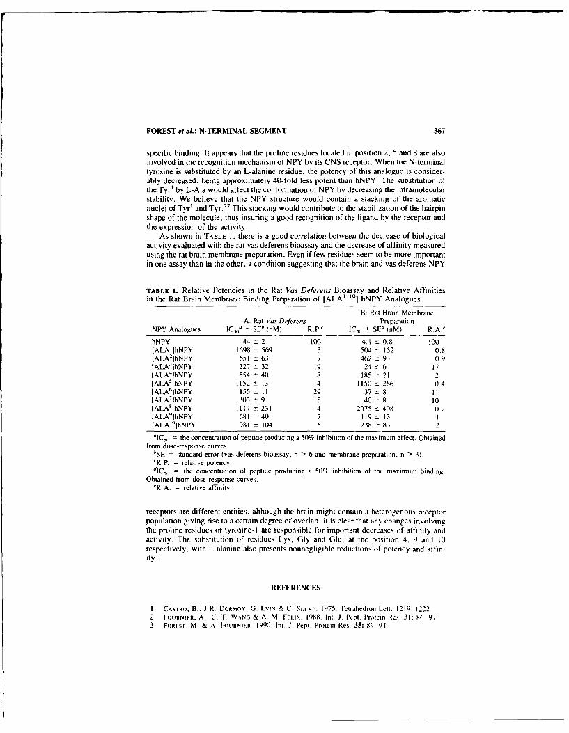
FOREST et al.: N-TERMINAL SEGMENT 367
specific binding. It appears that the proline residues located in position 2, 5 and 8 are alsoinvolved in the recognition mechanism of NPY by its CNS receptor. When the N-terminaltyrosine is substituted by an L-alanine residue, the potency of this analogue is consider-ably decreased, being approximately 40-fold less potent than hNPY. The substitution ofthe Tyr' by L-Ala would affect the conformation of NPY by decreasing the intramolecularstability. We believe that the NPY structure would contain a stacking of the aromaticnuclei of Tyr' and Tyr.2 7 This stacking would contribute to the stabilization of the hairpinshape of the molecule, thus insuring a good recognition of the ligand by the receptor andthe expression of the activity.
As shown in TABLE 1, there is a good correlation between the decrease of biologicalactivity evaluated with the rat vas deferens bioassay and the decrease of affinity measuredusing the rat brain membrane preparation. Even if few residues seem to be more importantin one assay than in the other, a condition suggesting that the brain and vas deferens NPY
TABLE I. Relative Potencies in the Rat Vas Deferens Bioassay and Relative Affinitiesin the Rat Brain Membrane Binding Preparation of IALA'"o] hNPY Analogues
B: Rat Brain MembraneA: Rat Vas Deferens Preparation
NPY Analogues iC9 o 0 - SE" (nM) R.P.' IC,, ± SEd (nM) R.A-'
hNPY 44 -2 100 4.1 ± 0.8 100[ALA']hNPY 1698 - 569 3 504 ± 152 0.8[ALA 2IhNPY 651 - 63 7 462 ± 93 0.9[ALA'IhNPY 227 ± 32 19 24 t 6 17IALA 4IhNPY 554 ± 40 8 185 ± 21 2[ALA']hNPY 1152 - 13 4 1150 ± 266 0.4IALA6]hNPY 155 I1 29 37 ± 8 II[ALA']hNPY 303 - 9 15 40 ± 8 10[ALA 8JhNPY 1114 231 4 2075 ± 408 0.2[ALA9IhNPY 681 ± 40 7 119 : 13 4[ALA"'IhNPY 981 ± 104 5 238 ± 83 2
"IC5o = the concentration of peptide producing a 50% inhibition of the maximum effect. Obtainedfrom dose-response curves.
bSE = standard error (vas deferens bioassay, n -> 6 and membrane preparation, n - 3)."R.P. = relative potency.dIC5, = the concentration of peptide producing a 50% inhibition of the maximum binding.
Obtained from dose-response curves.'R.A. = relative affinity.
receptors are different entities, although the brain might contain a heterogenous receptorpopulation giving rise to a certain degree of overlap. it is clear that any changes involvingthe proline residues or tyrosine-I are responsible for important decreases of affinity andactivity. The substitution of residues Lys, Gly and Glu, at the position 4, 9 and 10respectively, with L-alanine also presents nonnegligible reductions of potency and affin-ity.
REFERENCES
I. CASTRO. B.. J.R. DORMOY, G. EviN & C. St-ei-. 1975 Tetrahedron Lett. 1219-12222, Fo.'RNIF.R. A.. C. T. WNNG & A. M. FElix. 1988. Int. J. Pept. Protein Res. 31: 86-97.3 ForEsT, M. & A FourNIFR. 1990. tnt. i. Pept. Protein Res. 35: 89-94.

368 ANNALS NEW YORK ACADEMY OF SCIENCES
4. BALASUBRAMANIAM, A., I. GRUPp, L SRIVASTA, K. TATEMOTO, R. F. MURPHY, S. N. JOFFE& J. E. FISCHER. 1987. Int. J. Pept. Protein Res. 29: 78-83.
5. KRSTENANSKY, J.L. & S. H. BucK. 1987. Neuropeptides 10: 77-85.6. DANHO, W., J. TRISCARI, G. VINCENT, T. NAKAJIMA, J. TAYLOR & E. T. KAISER. 1988. Int.
J. Pept. Protein Res. 32: 496-505.7. BOUBLIK, J. H., N. A. SCOTT, M. R. BROWN & J. E. RIVIER. 1989. J. Med. Chem. 32:
597-601.

Inveitigations of the Structure-ActivityRelationship of Neuropeptide y"
A. CADIEUX," M. T-BENCHEKROUN." A. FOURNIER.'
AND S. ST-PIERRE'hDepartment of Pharmacology
Faculn,' of MedicineUniversity of Sherbrooke
Sherbrooke, Quebec, Canada JIH 5N4and
'Institui National de la Recherche Scientifique-SanttPointe-Claire, Quebec, Canada H9R IG6
Neuropeptide Y (NPY), a 36 amino acid peptide isolated from the porcine brain.' is a wellrecognized neurotransmitter/neuromodulator both in the central and peripheral nervoussystem. We have demonstrated recently that NPY contracts in a dose-related fashionguinea pig airways2 and rat colon by stimulating post- and prejunctionals receptors re-spectively. Using various fragments and analogues of NPY, the aims of this study wereto characterize the NPY receptors responsible for these contractile effects and to deter-mine if the two differently located biological receptors might correspond to the two typesof NPY receptors described previously.'
METHODS
A series of C-terminal fragments of p-NPY and analogues of h-NPY with modifica-tions of tyrosine residues in position 20 and 21 were synthesized by solid-phase peptidesynthesis and purified by preparative HPLC. The homogeneity, purity and proper com-position of the intended structures were confirmed by analytical HPLC and amino acidanalysis. All synthetic peptides were tested for their hiological activity in isolated guineapig bronchus and rat colon by generating cumulative or noncumulative dose-responsecurves. Their relative potencies were evaluated using p- or h-NPY as standard.
RESULTS AND DISCUSSION
Comparison of results obtained with NPY fragments in both assay systems revealedthat NPY receptors were differently sensitive to small changes introduced in the nativemolecule (FIG. I). Deletion of only one amino acid such as Tyr'. resulted in an almostinactive peptide in guinea pig bronchus (FIG. IA) whereas it caused only a 5-fold decreaseof NPY potency in rat colon (FIG. IC). In this latter bioassay, the concentration-response(C/R) curve to NPY(2-36) exhibited parallelism to that of NPY but the maximal responseaveraged only 60% that of the entire molecule. Shorter NPY fragments up to (25-36),
This work was supported by Grant PG38 from the Medical Research Council of Canada.369

370 ANNALS NEW YORK ACADEMY OF SCIENCES
were proportionnally less potent and less active, exhibiting specially with fragments(16-36) to (25-36) nonparallel C/R curves. This differs from other studies in which NPYfragments (2-36) to (19-36) were reported as active as NPY, albeit less potent, 4 '5 sug-gsting a difference in structural requirements for NPY receptors in various peripheraltissues. Another unexpected but interesting observation was the peculiar agonist propertyoffered by NPY fragment (13-36). This fragment behaved similarly to NPY in rat colon(FIG. IC) but appeared much more active (although less potent) than the mother molecule
A f
05- pNPY 12
S(336)
l~ .o,/1/o/
I.c 1.25.
.00
II ~ ~ 8 0II 7 6 1 I1
-~~~~~~ Lol fAgonistl M a AIol
E 1 si'1 a r cppad
S o 6aon.5
FIUR 1 Cncntaionrso cuvsfr P n NYfamntsiP uieYi boc
(AB) and rat colon (C) .75on n a ersn h ea n Ema fdt rm3t
tissues. esults ar .rsne 5sgas fcnrcin

CADIEUX et al.: SAR OF NPY 371
TABLE 1. EC, 0 Values, Relative Potencies (R.P.) and Developed MaximalContractions (M.C.) for h-NPY Analogues in Isolated Rat Colon and Guinea PigBronchusa
EC,) M.C.Tissue NPY Analogue (nM) R.P. (g)
Rat colon h-NPY 32.3 ± 11.3 100 0.73 ± 0.1[D-Tyr2° l 233 ± 33.3 13.8 0.60 ± 0.15ID-Tyr2 '1 425 ± 25 7.6 0.54 - 0.08
[Trp 2"] 125 ± 25 25.9 0.62 - 0.08[Trp 2tl] 346 ± 57.6 9.6 0.41 ± 0.13
(DTrp20
j 96.8 ± 35.4 33.3 0.61 - 0.02[DTrp 21j 500 ± 100 6.4 0.6 ± 0.05[Phe 20l 225 ± 25 12.6 0.73 ± 0.11[Phe2" ] 95.5 t 19.1 33.8 0.98 ± 0.08[Cha2°] 233 ± 66 13.8 0.17 ± 0.06[Cha2 l] 72.3 ± 14 44.6 0.67 ± 0.01
[Tyr-O-Me 0 l 85.7 ± 7.2 37.6 0.46 - 0.1[Tyr-O-Me2"l 157 ± 71.7 20.5 0.8 ± 0.1
Guinea pig bronchus h-NPY 2210 ± 1250 100 0.!6 ± 0.02{Phe 2'] >10,000 - 0.12 ± 0.04lPhe21 ] >10,000 - 0.23 ± 0.03
'EC5ovalues are expressed as means ± SE mean of 3 to 7 determinations. Potency of h-NPY isarbitrarily fixed to 100%. In guinea pig bronchus, maximal contractions of h-NPY and its analogueswere evaluated at a concentration of 10-6 M.
in guinea pig bronchus (FIG. IB). Furthermore both peptides activated different mecha-nisms. The response to NPY was abolished by indomethacin (10 - 6 M) while that ofNPY(13-36) was not. Such phenomenon did not occur in rat colon where contractileresponses to both NPY and NPY(13-36) were totally inhibited in presence of atropine10 - 6 M. Further evidence for the presence of different NPY receptor subtypes wasprovided with NPY analogues. As can be seen in TABLE 1, substitutions of tyrosine 20 or21 were not detrimental for NPY receptor activation in rat colon. In agreement withprevious in vitro binding and pharmacological studies,6 despite showing small loss ofpotency, each analogue tested exhibited a relatively high level of biological activity.However, except for [Phe20 - and [Phe2t ]-NPY, none of the analogues studied here wasable to induce significant contractile activity (up to 20% that of h-NPY) in guinea pigairways, even when tested in concentrations as high as 10 - 6 M. Taken together, theseresults support previous proposals of NPY receptors heterogeneity.3.6
REFERENCES
I. TATEMOTO. K., M. CARLQUIST & M. MUTT. 1982. Nature 296: 659-660.2. CADIEUX, A., M. T-BENCHEKROUN, S. ST-PERRE & A. FOURNIER. 1989. Neuropeptides
13: 215-219.3. WAHLESTEDT, C., M. YANAIIIARA & R HkKkwjSON. 1986 Regul. Pept. 13: 307-318.4. Rioux. F., H. BACHELARD, IC. MARTEL & S. ST-PIERRE. 1986. Peptides 7: 27-31.5. DONOSO, V., M. SILVA, S. ST-PIERRE & J.P. HUIDOBRo-ToRo. 1988. Peptides 9: 545-553.6. MARTEL, J.C., A. FouRtVIER, S. ST-PIERRE, Y. DUMONT & R. QUIRION. 1990. 1. Pharmacol.
Exp. Ther. In press.

Pharmacological Actions ofNeuropeptide Y and Peptide YY in
Rat Colona
A. CADIEUXb M. T-BENCHEKROUN,/ A. FOURNIER.' ANDS. ST-PIERRE'
'Department of PharmacologyFaculty of Medicine
University of SherbrookeSherbrooke, Quebec, Canada Jill 5N4
and'institut National de la Recherche Scientifiqur-Santt
Pointe-Claire, Quebec. Canada H9R IG6
Neuropeptide Y (NPY) and peptide YY (PYY) are two related 36 amino acid polypeptidesoriginally isolated from porcine brain and duodenum respectively. 1.2 Both peptides arepresent in the gastrointestinal tract, each having its own distinctive cel! type anddistribution. 3'4 Evidence is accumulating that these YY peptides are involved in theregulation of various gastrointestinal functions including neuronal control of motility. Ithas been proposed that NPY and PYY may interact with a common receptor to inducetheir actions . 5 The aims of this study were therefore 1) to examine and compare the invitro effects of NPY and PYY on rat distal colon and 2) to establish whether the twopeptides activate similar mechanisms.
METHODS
Segments of Sprague Dawley rat distal colon were set up for isometric recording(Grass Instruments) in a 5 ml organ bath (Tyrode solution) under a resting tension of I g.NPY or PYY was added to the baths in increasing concentrations, left in contact with thetissues until a steady level of tone was achieved, and then washed out. A minimum periodof 20 min was allowed between doses challenge. When antagonists were used, they wereadded at least 15 min before the addition of the peptides. Noncumulative concentration-response curves to 5-HT and PgE 2 were also generated for comparison purposes.
RESULTS
NPY or PYY from 10- to 3 x 10 -6 M produced a distinct concentration-dependantincrease in the basal tone of the rat distal colon. The responses induced by NPY werecharacterized by an initial maximal peak which was followed by a progressively decliningresponse and increased spontaiwous activity (Fic I A). In contrast. PYY caused contrac-tions which were short lasting; the initial peak being replaced by increased phasic activity
"Thi work was suptl,,ed by , r d3r f--.... c;. :.i Research Council of Canada.
372

CADIEUX et al.: RAT DISTAL COLON 373
TensionA /w Ww (g)
' 0
A A Ap-NPY p-NPY p-NPY
10-8M 10- 7M 10-6M
B
A A 0
PYY PYY PYY
10_8 M 10_7 M 10- 6 M 15 m n
C IApNPY
p-NPY 10- 7 M
10-7 M Atropine 10- 6 M
D
AApyy Pyy
10 7 M 10-7 M
Atropine 10- 6 M
FIGURE 1. Contractile responses to NPY and PYY in ra, distal colon. The original recordingspresented here are from different tissue preparations. Panels (A) and (B) represent the effect ofincreasing concentration of NPY (A) and PYY (B) on the basal tone of rat colon. Panels (C) and (D)show tissue responses to single dose of NPY (C) and PYY (D) in absence and in presence of atropine(106- M).

374 ANNALS NEW YORK ACADEMY OF SCIENCES
1.8. o-o pNPY0-e PYY
0-o 5-HT1.6- - PgE 2 T
1.4-
O 1.2C
o T0 1.0- //~ j
0.8 10.6-
0.4- /
9 8 3
- Log (Agonist) M
FIGURE 2. Concentration-response relationships for the contractile effects of NPY, PYY. 5-HTand PgE, on rat distal colon. Each point and bar represents the mean ± SEM of 6 to 10 determi-nations. Results are presented as grams of contraction.
after few minutes (FIG. I B). The tonic responses to NPY were unaffected by methyser-gide, indomethacin. diphenhydramin or et and P blocking agents but were inhibited byatropine (10 - 6 M). In contrast, contractions elicited by PYY were resistant to the cho-linergic antagonist (FIG. IC.D). However, both NPY and PYY effects were totally abol-ished in the presence of tetrodotoxin (10 ' M). ECSo values for NPY and PYY were ofthe same order of magnitude, e.g., 8.6 and 4-4 x 10 " M respectively. On a molar basisPYY was equipotent to PgE, (ECS = 5.9 X 10 " M) but about 60 times more potentthan 5-HT (ECso = 2.6 X 10 ' M) in producing half maximal contraction. Furthermore,the maximal tension induced by PYY was found to be higher than those of other agonists(FiG. 2).

CADIEUX et al.: RAT DISTAL COLON 375
DISCUSSION
The present study showed that NPY and PYY have motor actions similar to those oflocal smooth muscle contracting factors present in the rat large intestine. This observationis consistent with the reported pattern of distribution of NPY-immunoreactive nerve fibers
in muscle layers of the gut and with the localization of PYY in endocrine-like cells indistal colon. 3
.4 Pharmacological analysis revealed that both NPY and PYY effects were
neuronally mediated but via different neurotransmitters. On the basis of the present data,it seems likely that contraction of rat colon to NPY can be explained by the release ofacetylcholine. The response to PYY however seems to involve a nonadrenergic andnoncholinergic component; the nature of which remains to be determined. In conclusion,these findings support the concept of a role for NPY and PYY in the rat colon and suggestthat these two related peptides exert their action at different operational receptor sites.
REFERENCES
I. TATEMOTO, K. 1982 Proc. Nat]. Acad. Sci. USA 79: 5485-5489.2. TATEMOTO, K. 1982. Proc. Natl. Acad. Sci. USA 79: 2514-2518.3. LUNDBERG, J. M., K. TATEMOTO, L. TERENIUS, P. M. HELLSTROM, V. MUTT, T. H6KFELT
& B. HAMBERGER. 1982. Proc. Natil. Acad. Sci. USA 79: 4471-4475.4. LUNDBERG, J. M.. L. TERENiUS, T. HOKFELT & M. GOLDSTEIN. 1983. Neurosci. Let.
42: 167-172.5. HELLSTROM, P. M., J. M. LUNDBERG, T. HOKFELT & M. GOLDSTEIN. 1989. Scand. J.
Gastroenterol. 24: 231-243.
Regulation of Neuropeptide YTranscription and mRNA Levels by
Nerve Growth Factor, Glucocorticoids,Cyclic AMP, and Phorbol Ester in
PC12 Cells
STEVEN L. SABOL AND HIROSHI HIGUCHI
Laboratory of Biochemical GeneticsNational Heart, Lung, and Blood Institute
National Institutes of HealthBuilding 36, Room IC06
Bethesda, MD 20892
We are irivestigating the regulation of the preproneuropeptide Y gene (NPY gene) by
hormones, second messenger systems, and factors that control cellular differentiation. Werecently described' aspects of the regulation of the NPY mRNA level in PCI2 ratpheochromocytoma cells. The abundance of NPY rnRNA in these cells, which is very lowunder basal conditions, was markedly elevated by the synergistic actions of cAMP ele-vation and phorbol esters, which activate protein kinases A and C, respectively. In the
present study we have examined by Northern blot analyses of mature NPY mRNA(700-800 bases) and by nuclear run-on transcription analyses the interactive effects if
nerve growth factor (NGF) and glucocorticoids on NPY gene expression in PCI2 cells
highly sensitive to NGF.Treatment of cells with 2.5 S NGF for 1-6 d increased the abundance of mature NPY
mRNA (700-800 bases) 30-100-fold. The half-maximal and maximai effects of NGFwere obtained with 16 and 65 ng/ml, respectively. Glucocorticoids such as dexametha-sone (Dex), which alone generally elevated the NPY mRNA level slightly. potentiated byup to 3-fold the stimulation by NGF at early times (!-7 h) but strongly suppressed it at
later times ( -25 h). The response to NGF (4-8 h) required ongoing protein synthesis.suggesting an initial induction by NGF of factor(s) acting on the NPY gene. II nuclearrun-on analyses, treatment of cells for 1-24 h with NGF stimulated NPY gene transcrip-tion 4-8-fold, a response potentiated or inhibited by Dex during 2-h or 24-h treatments,
respectively. Treatment with forskolin, which markedly elevates cAMP levels, or phor-bol-12-myristate-13-acetate (PMA), which activates protein kinase C. elevated NPYmRNA levels 4-10-fold or :54-fold, respectively, while forskolin + PMA synergisticallyelevated NPY mRNA levels 30-70-fold and NPY gene transcription 6-25-fold. NGF +forskclin + PMA elevated NPY mRNA levels approximately 200-fold and NPY tran-scription 15-40-fold.
W_ conclude that the NPY gene is transcriptionally activated by NGF and by inter-active actions of protein kinases A and C. The marked discrepancy between mRNA and
apparent transcription rate elevations elicited by NGF suggests that NGF may elevateNPY mRNA by mechanism(s) in addition to transcriptional activation, sucn as mRNA
processing and stabilization.The striking NGF/glucocorticoid antagonism (L,24 h of treatment) affecting NPY
gene regulation has been observed for other delayed inductions by NGF and may reflect
376

SABOL & HIGUCHI: PCI2 CELLS 377
a mechanism for phenotypic choice during neural crest differentiation. 2 A possible mo-
lecular exvlanation is downregulation of NGF receptors by prolonged glucocorticoidtreatment.
This work is described in a full paper.4
REFERENCES
I. HIGUCHI, H., H. Y. T. YANG & S. L. SABOL. 1988. J. Biol. Chem. 263: 6288-6295.
2. DOUPE, A. J., P. H. PATTERSON & S. C. LANDIS. 1985. J. Neurosci. 5: 2119-2142.
3. Tocco, M. D., M. L. CONTRERAS, S, KOIzUMI, G. DICKENS & G. GUROFF 1988. J. Neu-
rosci. Res. 20: 411-419.4. SABOL, S. L. & H. HIGUCHI. 1990. Mol. Endocrinol. 4: 384-392.

Strong Evolutionary Conservation ofNeuropeptide Y between Mammals,
Chicken, Goldfish, and Horned Shark
ANDERS BLOMQVIST, INGRID LUNDELL, AND
DAN LARHAMMAR"Department of Medical Genetics
Uppsala UniversityBox 589
S-751 23 Uppsala, Sweden
The NPY family of peptides includes, in addition to NPY, also peptide YY (PYY) andpancreatic polypeptide (PP) as well as the fish pancreatic peptide PY. To investigate thedegree of conservation of NPY at the structural level during evolution, as well as itsrelationship to the other members of this peptide family, we have isolated DNA clonesencoding NPY from chicken, goldfish (Carassius auratus) and horned shark (Hetero-dontusfrancisci). We have previously reported that the chick and goldfish NPY sequencesreveal a remarkable degree of homology, as they display only 1 and 5 differences.respectively, to human NPY.
Using a probe containing the entire coding region of a chick NPY cDNA clone, wehave now screened a shark genomic library (kindly provided by Dr. G. Litman, TampaBay Research Institute, St. Petersburg, FL) under conditions of !ow stringency. Severalphage clones were isolated and the cross-hybridizing region of one of these clones wassubcloned in a plasmid vector and subjected to nucleotide sequence determination. Theclone was found to contain exon two of the shark NPY gene.
Exon two of the NPY gene encodes the signal peptide and 34 of the 36 amino acidsof mature NPY. The shark signal peptide differs considerably from the signal peptides ofthe other species, but the general hydrophobic character is maintained. In contrast, matureshark NPY displays only three differences to the human sequence. Two of these differ-ences are conservative amino acid r, placements; leucine instead of methionine and lysineinstead of arginine.
Mature shark NPY has two differences to goldfish NPY. The shark sequence displaysno unique amino acid residues as compared to the other known NPY sequences. Thissuggests that shark NPY may be identical to NPY of the common ancestor of cartilaginousfishes, bony fishes, and mammals.
In order to investigate the structural conservation of CPON, we are currently trying tolocalize exons 3 and 4 of the shark NPY gene by using low-stringency hybridizations.
The impressive similarity of shark NPY to human NPY makes this peptide one of themost highly conserved known (92% identity at the amino acid level), clearly more con-served than insulin. The strong sequence conservation of NPY indicates that this peptidesubserves evolutionarily old and important functions.
"Corresponding author.378

Cell-Type Specific Modification ofPeptide Posttranslational Processing in
Pituitary Cells"
IAN M. DICKERSON" AND RICHARD E. MAINS
Department of NeuroscienceThe Johns Hopkins University School of Medicine
725 North Wolfe StreetBaltimore, Mar'la - 21205
One approach to examine tie peptide processing capabilities of a cell involves transfrctionof the cDNA for a foreign propeptide precursor into th,: cell and then detailed examinationc-f the peptides produceJ by that cell. We have used that approach to examine the cell-typespecificity of peptide processing in two widely used pituitary cell lines, mouse cortico-tropes (AtT-20) and rat somatomammotropes (GH,). We have transfected the cell lineswith vectors employing the mouse metallothionein-I promoter to c:ontrol expression ofwildtype and mutant forms of human proNeuropeptide Y (proNPY) cDNA. The fourtypes of proNPY cDNA, kindly provided by Dr. Jack Dixon (Purdue University). differonly in the pair of basic amino acids present at the single site of cleavage in the molecule.
When the four forms (f proNPY eDNA were expressed in AtT-20 cells, a markedhierarchy of 2leavage site preference was seen.' The wildtype -Lys-Arg-(KR) and themutant -Arg-Arg- (RR) sequences were cleaved nearly to completion within a couplc othours. By comparison, the mutant RK-proNPY was cleaved much more sl, ,vly and lesscompletely, and the mutant KK-proNPY was cleaved extremely poorly in the AtT-20cells. When the corresponding four cell lines were created using the GH, cells as the hostline, a very different result was obtained. The extent of cleavage of all four forms ofproNPY proceeded approximately to the same extent, with the same kinetics. 2 After a16-hour labeling in medium containing I3 Hlleucine. medium samples were immunopre-cipitated using an affinity-purified NPY antiserum and analyzed by SDS polyacrylamidegel electrophoresis (Ftc. 1). A significant amount ot cleavage of proNPY to NPY wasseen with all four forms of proNPY as precursor. Peptide analyses demonstrated that theNPY-size immunoprecipitable molecules from both GH-NPY and AtT-NPY cell lineswere authentic NPY.' 2
While the GH-NPY lines all cleaved proNPY to a certain extent (Fic. I), the cleavagewas not usually as complete as seen with the AtT-NPY cells. Since methods had beenestablished to dramatically increase the number of secretory granules in the GH cells,using a treatment of e!.tradiol, insulin, and epidermal growth factor in the medium, 3
_4 we
asked what effect such a treatment might have on the storage and processing of theexogenoua proNPY in the t-ansfected GH-NPY cells. As expected. the amount of growthhormone produced by the cells was reduced to one-third during the hormone treatment(FiG. 2A). while the level of the much more abundant prolactin rose four-fold (FIG.. 2B).Similar hormone treatments had no effect on the ACTH production by transfected AtT-20
Supported by National Institutes of Health Grants DK-32948. DA-(XX97. and DK-08121.b Current address: Department of Physiology & Biophysics, University of Miami School of
Medicine. Miami, FL.
379

384) ANNAL.S NEW N ORK At' AIENtY OP' SCIENCES
E proNPY
NPY
dfR ~FIGUIE 1. 51)5-PA6GE ' nalxsi', of irninluno-
AtT-KRprecipitated NPY-relatetd peptides triin, piluitdrNcdl lines. Cell ines: Atr-KR. Aff-2() cellsshich express -IA s-AIr2-conainimg prol'Y:
GH-KR -(--- H-KR, GH, cells AhIch express -L. s-Are-containliie proNPY, GH-RR. iHt cells Ahichexpress -Arg-Arg,-containing priiNPY, (d--RK.
GH-RR -Gil, cells wkhich express -Ar-Lvs' %-conainingproNPY:, GH-KK. iH; cells , hich exprcss-lVs-ES s-co ntaining proNTY.
GH-RK
GH-m(K-A0 0 10 15 20 25
Slice Number
cells (FtG. 2C). During, these changes in response to) the hormonal treatment, the residencetime of the proNPY-derived peptides increased strikingly in the Gli cells. .%ith no chanein the extent of' cleavage of- the 1l.roNPY. No .4fects %kere seen oin proNPY storaue orprocessing in the AtT-NPY cells.
Thus. not only ..s the ability to process propeptide precursors cell-type specific, hutalso the cha,-iges in propeptide procce ng in response to external stimuli are also cell-typespecific.
A. OH-KR Cells B. OH-KR Cells C. AtT-KR CellsOH in Medium TCA of Medium ACTH in Medium
OH 22 kDo ABI gCH
~-Plin*-Hormone
S00 5000- 5000-1 ACTH
00 01L.0 20 40 0 20 4 0 20 40
Slice Number
FIGUIRE 2. Effect of hornsinc treat nent on eiidogenous pept ide product ion in 611H cellIs andAtT-20Icells. Petdsaavc ee(r~h1Imrni1onc IA) and Prolactin (B) Ironi Gi-KR cells. an"ACTH it') fromn AtF-k cells

DICKERSON & MAINS: PITUITARY CELLS 38J
REFERENCES
1. DICKERSON. 1. M., J. E. DIXON. & R. E. MAINS. 1990. Biosynthesis and posttra.nslationalprocessing of site-directed endoproteolytic cleavage mutants of proNPY in mouse pituitarycells. J. Biol. Chem. 265: 2462-2469.
2. DICKERSON. 1. M. & R. E. MAINS. 1990. Cell-type specific post-translational processing ofpeptides by different pituitary cell li-- En~locrinology 127: 133-140.
3. SCAMMELL. J. G., T. G. BURRAGE & I S. DANNIES. 1986. Hormonal induction of secretorygranules in a pituitary tumor cell line. Endocrinology 119: 1543-1548.
4. TOUGARD. C.. L. E. NASCIUMTI R. PICART, A. TiXIER-VIDAL & W. B. HUTTNER. 1989.Subcellular distribution of secretogranins in rat tumoral prolactin cells as revealed by electronmicroscopic immunocytochemistry. J. Histochem. Cytochem. 37: 1329-1336.

Novel Brain Specific TranscriptionFactors Form DNA-Protein Complexeswith SPI and TATA Regions of the Rat
Neuropeptide Y Gene PromotorEnhancement of Complex Formation by
Nonneuronal iactors
Y. M. 001 AND HAKAN PERSSON
Department of Physiological Chemistr I
andDepartment of Medical Chemistry I
Karolinska InstitutetP.O. Box 604 00
S-104 01 Stockholm, Sweden
INTRODUCTION
Neuropeptide Y (NPY) is a 36-amino acid peptide widely distributed throughout themammalian central nervous system. 1 Its diverse functions are well documented. 3 Its roleas a neurotransmitter or neuromodulator is well established. It is a vasoconstictor andcoexpresses with adrenalin and certain neuropeptides in the brain. The access to genomicclones of human and rat NPY has enabled the elucidation of peptide structure and genesequence and localized its mRNA distribution in neuronal and nonneuronal tissues.' 6However, the nature of binding sites for brain nuclear factors that form transcriptionalcomplexes with the NPY gene is not known. The present study is conducted to gain newinsights into transcriptional mechanisms of the rat NPY gene in brain and nonneuronaltissues. By a gel shift assay using 1
32P -end-labeled oligonucleotides annealed to thecomplementary strand, or Klenow-labeled enzyme restriction fragments. DNA sequencesspanning -16 to -144 upstream of the transcriptional start site of the rat NPY promoterwere analysed for putative transcriptional complexes formed by DNA-protein bindingwith nuclear extracts. Nuclear extracts were prepared from brain, spleen, liver and bonemarrow obtained from adult Sprague Dawley rats. 7 DNA motifs were determined bymethylation protection.'
RESULTS AND DISCUSSION
1 Ising an oligonucleotide spanning - 16 to -74 (oligo I). of the rat NPY promoter with"TATA-like" element located at -25,5 nuclear extract from rat brain was found to formthree major DNA-protein complexes, YI, Y2 and Y3 showing binding affinitiesY2>Y3>YI (Fi(i. IA. panels I and 2). By comparison with complexes Y2 and Y3,lower affinity complexes Y4 and Y5 were detected with DNA sequences of an Xho I-SmaI restriction fragment spanning -78 to -144 (FIG. IA. panel 3). or its related oligonucle-otide. -75 to -133 (oligo 11). No complexes were formed with sequences -142 to -201
382

001 & PERSSON: TRANSCRIPTIONAL MECHANISMS 383
(oligo 111) (FIG. IA, panel 4). Effective competition was achieved with unlabeled oligoI and oligo I1. but not with oligo III or unrelated DNA (FIG. I B). indicating spe-cific binding with promoter. Methylation protection identified three distinct bindingsites on the promoter for brain nuclear factors: a unique 18-mer sequence 5'-CTCCATAAAAGCCCGTTG, which includes "TATA-like element" ATAAAA at -25,and a novel 9-mer motif 5'-GCCCCTCCT located at -50, as well as its complementary
DNA probe: oligoT XhoI- Sma . oligo iii-16 to- 74 -78 to -144 -142 to-201
abc d a bcd abcd
-Y1
I 2 3 4
FIGURE IA. Gel shift assay for DNA-protein complexes using nuclear extract incubated with[32Pi !abeled duuble stranded oligonucleotides (oligo I or 111) or restriction fragment Xho i-Sma Iindicated. Binding to promoter as a function of concentration of brain nuclear extract protein isshown: (a) free probe without nuclear extract. (b) 8 R±g; (c) 16 t.g; and (e) 32 R±g of brain nuclearprotein (panels I to 4). The different concentrations of brain nuclear extract were incubated withapproximately 0.5 to I ng of the indicated labeled probes in a binding assay at 30 degrees centigrade,in presence of 4 Rg poly(di-dC). The resulting complexes were separated from unbound inputfragment by electrophoresis on 6-8% polyacrylamide gel. Panels I and 2 show results of autora-diography of same experiment; panel I. after 12-hr exposure to Kodak X-ray film with intensifyyingscreen; panels 2-4, after 30 hours exposure.
sequence (FiG. IC. 1). Altogether, the findings show recruitment of three distinct bindingsites to assemble brain DNA-protein complexes.
Interestingly, the anti-sense 9-mer sequence resembles the consensus G/TGGCGGGGC/AAT, site for the general transcription factor Spl. Additional experi-ments showed the ability of unlabeled 5'-GCCCCTCCT sequences to compete off brainspecific DNA-protein complexes from labeled oligo I with 100% efficiency in contrast to

384 ANNALS NEW YORK ACADEMY OF SCIENCES
compemo. ongoI olgol xhoI- S I
ab 1140 8011040 Soho 40 01
Y3-
FIGURE lB. Competition to identify promoter specificity by the addition of unlabeled oligonu-cleotide to displace [32pl-labeled oligo Ill bound in DNA-protein complexes in similar gel shiftassays. Just before addition of labeled probe, competitor DNAs were added in the ng quantitiesindicated. Formation of DNA-protein complexes were monitored by gel shift assays as in FIGURE IA.(a) No extract added; (b) 10 lg of brain nuclear extract in absence of any competitor.
50% competition effected by its complementary strand (FIG. IC, 2). The findings stronglysuggest a key role for the 9-mer motif identifying it as a major sequence that assemblesspecific DNA-protein complexes with brain nuclear factors; cooperation with TATA-likeand Spi sites on the promoter bind brain nuclear factors, forming transcriptional com-plexes to govern NPY gene expression in brain. Addition of a spleen, liver or bonemarrow nuclear extract to a limited amount of brain nuclear extract enhanced formationof brain complexes (FIG. 2A), whereas each nonneuronal extract formed its individualprofile of DNA-protein complexes with electrophoretic characteristics different frombrain complexes (FIG. 2B). The findings suggest recruitment of tissue specific mecha-nisms by the NPY promoter and synergism between neuronal and nonneuronal factors.

001 & PERSSON: TRANSCRIPTIONAL MECHANISMS 385
FIGURE IC. Panel. Methylation protection of oligonucleotide I using total brain nuclear extract.The DMS protection was done by incubation of a total brain extract with (' 2P1-Iabeled coding strandof oligonucleotide 1. The guanine reaction of the free probe (oligonucleotide I) is indicated(unbound),' the bound (DNA-protein complex) guanine reacted probe is shown. Panel 2. Compe-tition analyses using a 9-mer oligonucleotide. 5' -GCCCCTCCT. Competitive binding was done asin FIGURtE l B. using [3 2PI-labeled oligonucleotide I and brain nuclear extract. The quantities in ngare shown of a 9-mer oligonucleotide, 5'-GCCCCTCCT as competitor. The competitor oligonucle-otide is located at -50 of the coding strand. DNA-protein complexes were assayed as described inFIGURE l B. (L, No extract added. (b) 10 lhg of brain nuclear extract in the absence of competitor.

386 ANNALS NEW YORK ACADEMY OF SCIENCES
A B
MW l__t" mwow brin kjwm
a b, d &ila 2.4 ai' 1i .h. .
Ta--
FIGURE 2. (A) ..nhancement of brain DNA-protein complexes by nonneuronal factors. Mixingexperiments were do, by adding increasing amounts of a nonneuronal extract to a limiting amount(5 pig) of a brain nuclea. extract and the mixture was incubated with [32 Pl -labeled oligonucleotide 1.Complexes formed were asayed by 3-12% gel gradient. (a) Probe alone; (b) brain, 5 pig; (c) spleen,60 pIg; (d) liver, 60 jig; and (e) bone marrow; each nonneuronal extract was added to 5 p.g of a brainextract at (I) 30 pig, (2) 60 Rg and (3) 120 Rg of the indicated nonneuronal extract. (B) Enhancementof brain DNA-protein complexes using nonneuronal extract mixed with one another and bound withlabeled oligo I. The individual electrophoretic mobilities of complexes formed with each nonneuronalextract was observed by gel gradients, with features different from brain specific DNA-proteincomplexes.
CONCLUSIONS
In summary, the rat NPY promoter forms two high affinity brain specific DNA-protein complexes Y2 and Y3, with brain nuclear factors. Being the major high affinitycomplexes with distinct features, they most likely constitute brain specific transcriptioncomplexes. Brain DNA-protein complex formation is enhanced by nonneuronal factorsindicating synergistic cooperation. The study has identified a novel 9-mer core motif, 5'-GCCCCTCCT at -50, which binds brain nuclear factors and by cooperation with SpI (5'-AGGAGGGGC) and TATA (5' -CTCCATAAAAGCCCGTTG) regions on the promoterf-,rm specific transcriptional complexes to govern NPY gene expression in brain.

001 & PERSSON: TRANSCRIPTIONAL MECHANISMS 387
REFERENCES
I . TATEMOT O, K. & V. MUTT. 1980. Nature 285: 417-418.2. ALLEN, Y. S., T. E. ADRIAN, J. M. ALLEN. K. TATEMOTo, T. J. CROW, S. R. BLOOM & J.
M. POLAK. 1983. Nature 306: 584-586.3. MUTT, V., Ed. 1989. Neuropeptide Y. Karolinska Institute Nobel Conference Series. 99-
0694929-2. Raven Press, New York.4. MINTH, C. D., S. R. BLOOM. J. M. POLAK & J. E. DIXON. 1984. Proc. Nati. Acad. Sci. USA
81: 4577-4581.5. LARHAMMAR, D., A. ERICSSON & H. PERSSON. 1987. Proc. Natl. Acad. Sci. USA 84: 2068-
2072.6. ALLEN. J., J. NOVOTHY, J. MARTIN & G. HEINRICH-. 1988. J. Biol. Chem. 263: 6288-6295.7. DIGNAM. J. D., R. M. LEBOVITZ & R. G. ROEDER. 1983a. Nucleic Acids Res. 11: 1475-
1489.8. ]ONES. K. A., J. T. KADONAGA, P. J. ROSENFELD, T. J. KELLY & R. TIJIAN. 1987. Cell
48: 79-89.

Defect of Binding of Nuclear Factorsfrom Embryonic and Newborn BrainNuclear Extracts from Autoimmune
NZB Mice with the Rat Neuropeptide YGene Promoter
YUET M. 001
Department of Physiological Chemistry IP.O. Box 60400
Karolinska InstituteS-104 01 Stockholm, Sweden
INTRODUCTION
Neuropeptide Y (NPY) is a 36-amino acid peptide with neurotransmitter, neuromod-ulator and vasoconstrictor functions. It is distributed throughout the mammalian centralnervous system. - 3 Its diverse functions are well established.' The access to genomicclones of human and rat NPY has enabled the elucidation of gene sequence and peptidestructure4 6 and has localized its mRNA distribution in neuronal and nonneuronal tissuesconfirming the broad functions of NPY.7 Studies using complementary DNA (cDNA) ofthe rat NPY gene have shown increased levels of NPY mRNA in megakaryocytes ofcertain mouse strains like the New Zealand Black (NZB) mouse, that develop a sponta-neous form of autoimmune disease resembling human systemic lupus erythematosus. 3
The mechanisms of increased expression are not known. The present study is conductedto define transcriptional mechanisms of the NPY gene in the autoimmune NZB mousewhich manifests serological disease by 2 weeks of age. By a gel shift assay using[32p]-end-labeled oligonucleotides annealed to the complementary strand, DNA se-quences spanning -16 to -133 upstream of the transcriptional start site of the rat NPYpromoter were analyzed for putative transcriptional complexes formed by DNA-proteinbinding with nuclear extracts.7 Nuclear extracts were obtained from NZB mice andcompared with normal C57B1/61 mouse strains in different developmental stages: (a)embryos at 8 days gestation, (b) brain from newborn mice at I day of age (PI) and (c) at6 weeks.
RESULTS AND DISCUSSION
Using an oligonucleotide spanning -16 to -74 (oligo 1) of the rat NPY promoter witha "TATA-like" element located at -25, nuclear extract from embryos of C57Bl/6J miceat 8 days gestation was found to form a major complex YM (FIG. I A: b,c). Interestingly.two major complexes YMI and YM2 were formed with nuclear extract from brain ofnewborn C57BI/6J, at I day after birth (FIG. IB: a,b). The same profile of complexesYM I and YM2 showing higher binding affinity was obtained using brain nuclear extractfrom 6-week-old C57B1/6J mice (FIG. IC: a). Interestingly, nuclear extract from 8th dayembryos of NZB mice did not form complex YM; instead a different complex YZ was
388

001: AUTOIMMUNE NZB MOUSE 389
formed (FIG. IA: d,e). Moreover, brain nuclear extract from newborn NZB mice alsoshowed a defect in formation of complexes YM I and YM2 detected in brain of C57BI/6Jnewborn mice. However, brain nuclear extract from 6-week-old NZB mice formed com-plexes YMI and YM2 with binding affinity and features comparable to those obtainedwith extract from 6-week-old C57B1/61 mice (FIG. IC: b). The distinct profiles of DNA-protein complex formation in development in the mouse suggest a strong influence of
DNA probe: oligo 1 - 16 to - 74
ABnuclear extract: [ j b]
mouse strain:
"Im
a bcde abo d a b
FIGURE 1. Gel shift assay for DNA-protein complexes using nuclear extract (n.e.) incubated with
[-2P-labeled double stranded oligonucleotides (oligo I and 11). Nuclear extracts (n.e.) were obtainedfrom two mouse strains and compared for formation of putative transcriptional complexes in ontog-eny. Oligo I -16 to -74, was used at approximately 0.5 to 1.0 ng used in nuclear extract binding asdescribed. Complexes formed were separated from remaining unbound input of labeled probe byelectrophoresis on 7% polyacrylamide gel electrophoresis. Panel A.- (a) free probe without extractn.e.; (b) n.e. from C57B1/6J embryo of 8 days gestation incubated with oligo I at (b) 32 Ig. (cI 48;.g; and from NZB embryo (d) 16 t.g, (e) 48 gig. Panel B: n.e. from newborn C57BI/6J brain, (a)16 gig. (b) 48 gig. Comparison with NZB newborn brain n.e. at higher concentration (c). 32 and (d)48 gig. Panel C: brain n.e. from 6 week C57B/6J mice (a) 16 gg and (b) from 6 week NZB mouse,16 gig.
developmental processes on mechanisms of DNA-protein binding in DNA-protein com-plex formation and thus govern gene expression. With an annealed I 2P]-labeled oligo-nucleotide -75 to -133 upstream of the promoter, similar nuclear extract from embryos ofthe C57B1/61 mouse formed a different profile of complexes YM3 and YM4 (FIG. 2: b,c),whereas with brain extract from newborn C5781/6J mouse, a different complex YM5 was
formed in addition to formation YM3 (Fi,. 2: de). Complex YM3 and YM4 showed high
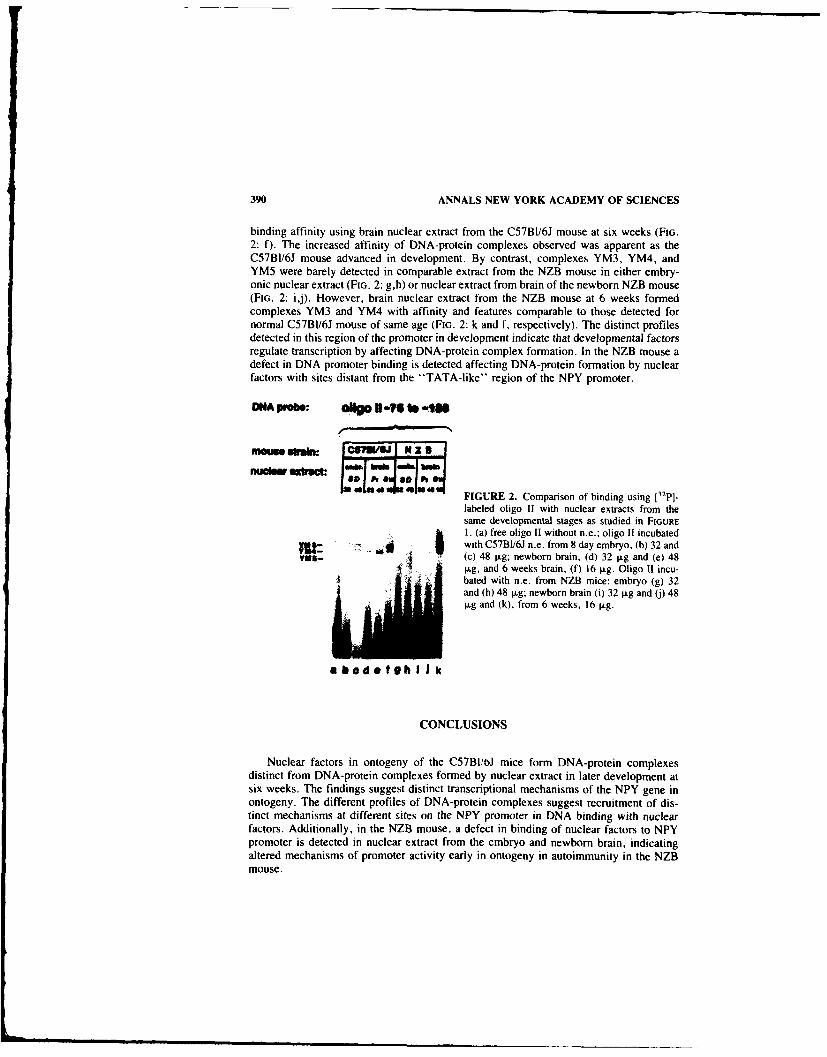
390 ANNALS NEW YORK ACADEMY OF SCIENCES
binding affinity using brain nuclear extract from the C57BI/61 mouse at six weeks (FIG.2: f). The increased affinity of DNA-protein complexes observed was apparent as theC57B1/6J mouse advanced in development. By contrast, complexes YM3, YM4, andYM5 were barely detected in comparable extract from the NZB mouse in either embry-onic nuclear extract (FIG. 2: g,h) or nuclear extract from brain of the newborn NZB mouse(FIG. 2: ij). However, brain nuclear extract from the NZB mouse at 6 weeks formedcomplexes YM3 and YM4 with affinity and features comparable to those detected fornormal C57BI/6J mouse of same age (FIG. 2: k and f, respectively). The distinct profilesdetected in this region of the promoter in development indicate that developmental factorsregulate transcription by affecting DNA-protein complex formation. In the NZB mouse adefect in DNA promoter binding is detected affecting DNA-protein formation by nuclearfactors with sites distant from the "TATA-like" region of the NPY promoter.
DNA probe: OiO Ui-VS lb -in
nuhur .xnsc: I'PI" "1Jk!"
FIGURE 2. Comparison of binding using [32p].labeled oligo 11 with nuclear extracts from thesame developmental stages as studied in FIGURE1 t. (a) free oligo II without n.e.; oligo 11 incubated
a r , with C57BI/6J n.e. from 8 day embryo, (b) 32 and'M11- j (c) 48 jig; newborn brain, (d) 32 jig and (e) 48
pg, and 6 weeks brain, (f) 16 pg. Oligo I1 incu-bated with n.e. from NZB mice: embryo (g) 32and (h) 48 ig; newborn brain (i) 32 ig and (j) 48pg and (k), from 6 weeks, 16 ig.
abodofghI Jk
CONCLUSIONS
Nuclear factors in ontogeny of the C57BI!bJ mice form DNA-protein complexesdistinct from DNA-protein complexes formed by nuclear extract in later development atsix weeks. The findings suggest distinct transcriptional mechanisms of the NPY gene inontogeny. The different profiles of DNA-protein complexes suggest recruitment of dis-tinct mechanisms at different sites on the NPY promoter in DNA binding with nuclearfactors. Additionally, in the NZB mouse, a defect in binding of nuclear factors to NPYpromoter is detected in nuclear extract from the embryo and newborn brain, indicatingaltered mechanisms of promoter activity early in ontogeny in autoimmunity in the NZBmouse.

001: AUTOIMMUNE NZB MOUSE 391
REFERENCES
I. TATEMOTO, K. & V. MUTT. 1980. Nature 285: 417-418.2. ALLEN. Y. S.. T. E ADRIAN, J. M. ALLEN. K. TATEMOTO, T. J. CROW, S. R. BLOOM & J.
M. POLAK. 1983. Nature 306: 584-586.3. MUTT. V. ED. 1989. Neuropeptide Y. Karolinska Institute Nobel Conference Series, 99-
0694929. Raven Press. New York.4. MINTH, C. D., S. R. BLOOM, J. M. POLAK. & J. E. DIXON. 1984. Proc. Nat]. Acad. Sci. USA
81: 4577-4581.5. LARHAMMAR, D.. A. ERICSSON. & H. PERSSON. 1987. Proc. Natl. Acad. Sci. USA
84: 2068-2072.6. ALLEN. J.. J. NOVOTHY. J. MARTIN & G. HEINRICH. 1988. J. Biol. Chem. 263: 6288-6295.7. JONES, K. A.. J. T. KADONAGA. P. J. ROSENFELD, T. J. KELI.Y & R. TIJIAN. 1987. Cell
48: 79-69.

He 90481: A Competitive NonpeptidergicAntagonist at Neuropeptide Y Receptors
M. C. MICHEL" h ' AND H. J. MOTULSKY"
"Biochemical Research LaboratoriesDepartment of Medicine
Universit , of EssenEssen. Federal Republic of Germanyi
andbDepartment of Pharmacology M-036University of California, San Diego
San Diego, California 92093
INTRODUCTION
Neuropeptide Y (NPY) has a widespread localization in many neurons of the centraland peripheral nervous system and exogenous NPY elicits numerous physiologicaleffects.' The ultimate assessment of the physiological relevance of NPY. however, willrequire the use of specific antagonists. Moreover, such antagonists may also be useful inthe pharmacological characterization of possible subtypes of NPY receptors which havebeen proposed but not unequivocally defined. The present study used NPY-stimulatedmobilization of intracellular Ca ' in human erythrolcukemia (HEL) cells 2-" as a modelsystem to determine the functional effects of He 90481 on NPY receptors.
MATERIALS
He 90481 (FIG. I) and porcine NPY were kindly provided by Heumann Pharma(Ndrnberg, Fed. Rep. Germany) and by Drs. J. E. Rivier and J. H. Boublik (SalkInstitute, La Jolla, CA), respectively. Fura-2 was purchased from Molecular Probes(Eugene, OR).
RESULTS AND DISCUSSION
NPY increased the free intracellular Ca concentration of HEL cells with an EC,of - 5 nM (FIG. 2), whereas He 90481 (in concentrations up to 100 lLM) had no directeffect (data not shown). However, He 90481 shifted the concentration-response curves forNPY to the right (towards higher concentrations) without altering the maximal NPY-stimulated Ca' ' increase (FIG. 2). Schild plots were linear with a slope which was notsignificantly different from unity (1.01 ± 0.25; n = 3). A pA, value of 4.43 (affinity37.5 1LM) was calculated from these data.
These data demonstrate that He 90481 is a competitive antagonist at NPY receptors.
'Address for correspondence: Medizinische Klinik, Hufelandstrasse 55. D-43(N) Essen I, FederalRepublic of Germany.
392

MICHEL & MOTULSKY: He 90481 393
F F
NH
N ~FIGURE 1. Chemical structure of He 90481.
H
x 3 HCI H
The usefulness of He 90481 as a NPY antagonist, however, is limited by two factors: i)Its affinity at NPY receptors is quite low, and ii) it is not specific for NPY receptors. Inthe concentration range where He 90481 blocks NPY receptors, it also competes fora.-adrenergic receptor binding (unpublished observations). Moreover, He 90481 is anagonist at H, histamine receptors of guinea pig heart with a potency approximately 1000times greater than its affinity at NPY receptors (Dr. EnglL;. Heumann Pharma. personalcommunication). Further studies with related compounds may yield nonpeptidergic NPYantagonists with higher affinity and greater specificity.
0 controlwx He 90481 10 AM
o 100 * He 90481 30 AMCD* He 90481 100 AM Maximal Ca2*
M U ,Iincrease
E_ 400.T Tc- 4
8 50
o roUC_
C-) E-= 200- =
0 A0I I I1
10-10 10-8 10-6[NPY], M
FIGURE 2. Eftects of various concentrations of He 90481 on NPY-stimulated Ca" mobilizationin HEL cells. HEL cells were grown in RPM! medium and free intracellular Ca* ' concentration ofthese cells was determined with the fluorescent indicator dye Fura-2 as recently described.' Con-centration-response curves for NPY were constructed in the absence and presence of various con-centrations of He 90481. The curves were generated by fitting the experimental data to a sigmoidcurve: in these calculations the bottom of the curve was fixed at 0 and the Hill-slope at I. The datawere then expressed as % of the maximal response obtained in the experiment, and these percentageswere averaged to obtain the mean concentration-response curves shown. The mean absolute increasescaused by NPY in the absence and presence of He 90481 are shown in the inset. Data are mean (inthe inset mean t SEM) from three experiments.

.w94 ANNALS NEW YORK ACADEMY OF SCIENCES
REFERENCES
I. POTTER. E. K. 1988. Pharmacol. Ther 37: 251-273.2. MOTULSKY, H. J. & M. C. MICHEL. 1988. Am. J. Physiol. 255: E880-E885.3. MICHEL, M. C., E. SCHLICKE' K. FINK, J. H. BOUBLIK, M. GOTHERr, R. N. WILLETTE, R.
N. DALY, J. P. HIEBLE, J. E. RIVIER & H. J. MOTULS4Y. 1990. Am. J. Physiol. In press.
I

Neuropeptide Y and Peptide YYInteract with Rat Brain Sigma and PCP
Binding SitesF. J. ROMAN, X. PASCAUD. 0. DUFFY. D. VAUCHE,
B. MARTIN, AND .. L. JUNIENInstitut de Recherche Jouveinal
3 . 9, rue de La Loge94265, Fresnes Cedex, France
The existence of endogenous ligands "'sigmaphins" for sigma opioid receptors was firstdemonstrated by Quirion et al.' in pig extracts. This finding has largely, been assessed byseveral authors reporting the existence of peptidergic fractions from guinea pig, pig andhuman brain exhibiting affinity for sigma or PCP receptors, or both.
The purpose of the present report is to describe the interactions of neuropeptide Y(NPY) and other related neuropeptides with the sigma binding site labeled with (+ )3H-SKFIO,047 ((+)3H-NANM) or 3H-DTG and with the PCP binding site labeled with3H-TCP in rat brain membranes.
METHODS
Brain membranes prepared from male Sprague-Dawley rats weighing 200-225 g(IFFA-CREDO, France) were used for binding studies with (+)3H-NANM (40 Ci/mmole),3H-DTG (52.3 Ci/mmole), or 3H-TCP (46 Ci/mmole). The incubations werecarried out according to Largent et al.2 in a final volume of 250 ILl. All peptides werefrom Peninsula and ligands were from New England Nuclear Corp. except for 3H-TCP(CEA, FRANCE).
RESULTS AND DISCUSSION (TABLE 1)
I. PYY and NPY displayed an IC50 <10 nM for the displacement of (+)3H-NANM. NPY appeared to be specific for the (+ )3H-NANM binding site.
2. PYY was devoid of specificity, and besides its high affinity for the sigma bindingsites it was more active than PCP itself on 3H-TCP binding sites.
3. The existence of 2 domains for sigma ligand binding already hypothetized byothers3 is confirmed by our results showing the differentiation between a(+)benzomorphan domain binding the (+)benzomorphans such as (+)NANMand pentozocine and a nonbenzomorphan domain binding nonbenzomorphansigma ligands such as (+ )3PPP and DTG.
CONCLUSIONS
These results suggest that NPY and to a lesser extent PYY could be the endogenousligand interacting with the ( + )benzomorphan binding site. On the other hand, PYY andto a lesser extent NPY, could be the endogenous ligand interacting with the PCP bindingsite.
395

396 ANNALS NEW YORK ACADEMY OF SCIENCES
TABLE i. Binding Affinities of NPY and Related Peptides for the Rat Brain SigmaBinding Sites Labeled with Various 3H--Ligands and for PCP Binding Sites Labeledwith 3H-TCP'"
Compounds (+ )3H-NANM 3H-DTG 3H-TCP
Haloperidol 24.6 -- 6.7 (6) 52 :t 13 (5) > 10000 (3)(+)NANM 98 ± 17 (4) 4543 ± 1741 (4) 343 ± 57 (4)DTG 253 ± 57 (3) 108 :t 15 (3) 9024 ± 1604 (3)R(+)3PPP 789 ± 376 (7) 106 -- 21 (4) >l10000 (3)(±)Pentazocine 38 ± 5.6 (3) 254 :t 53 (4) 2879 ± 197 (3)Rimcazole 2649 ± 636 (4) 2503 ±1033 (3) > 10000 (3)BMY14802 3216 :t 1122 (3) 271 46 (4) > 10000 (3)PCP 601 ±371 (4) 5485± 1778 (4) 61 ±7.7 (3)NPY 10 ± 5 (4) - 18%b (3) - 7%b (3)PYY 4.9 ± 0.8 (4) - 39%b (3) 32 ± 4 (2)PP -42%b (4) -.. 37 %b (3) - 14%b (3)Neurotensin 0 %b (4) 0 %b (3) 0%b (3)Bombesin - 17V" (3) -11%11 (3) - 16%" (4)Somatostatin -23%" (3) - 12%b (4) -20%b (3)Substance P - 25 %b (4) -.5 %b (3) - 14%h (3)VIP - 4 7 %b (3) - 10%h (3) - 9 %b (3)TRH -8 %b (3) 0%" (3) 0%b (3)
'Values are the mean IC50 (nM) ± SEM from the number of independent experiments indicatedin brackets.
"in the case of peptides, when 50% inhibition was not reached at 100 nM. the activity was givenin terms of percent inhibition at that concentration.
REFERENCES
1. QUIRIoN, R., D. A. DIMAGGIO, E. D. FRENCH, P. C. CONTRERAS, J. SHILOACH, C. B. PERT,J. EVERIST. A. PERT & T. L. O'DoNOHUE. 1984. Evidence for an endogenous peptide ligandfor the phencyclidine receptor. Peptides 5: 967-973.
2. LARGENT, B. L., A. L. GUNDLACH & S. H. SNYDER. 1986. Pharmacological and autoradio-graphic discrimination of sigma and phencyclidine receptor binding sites in brain with ( + )-[1-11 SKF 10,047, (+)-13H1-3-13-hydroxyphenyll-N-) (1-propyl) piperidine and (31--I-12-thienyl)cyclohexyl] piperidine. J. Phannacol. Exp. Ther. 238: 739-748.
3. BOWEN, B. L., B. N. KIRSHNER. A. H. NEWMAN & K. C. RICE. 1988. cr Receptors negativelymodulate agonist stimulated phosphoinositide metabolism in rat brain. Eur. J. Pharmacol.149: 399-400.

Characterization and Use of FourAnti-NPY Monoclonal Antibodies to
Study NPY-Receptor Interaction
E. GROUZMANN," P. WALKER, C. BOHUON, M. BURNIER,
E. COMOY, H. R. BRUNNER, AND B. WAEBER
Division of HypertensionCHUV
1011 Lausanne, Switzerlandand
I.G.R.Ddpartement de Biologie Clinique
Villejuif, France
Neuropeptide Y (NPY) is a 36-amino acid peptide present abundantly in sympatheticperivascular nerves and different areas of the brain. Several lines of evidence suggest thatNPY is involved in cardiovascular regulation. 1.2 We describe the production and char-acterization of four anti-NPY monoclonal antibodies (NPY02, NPY03, NPY04 andNPY05) and their use to study the NPY-receptor interaction.
Total NPY and NPY 27-36 were conjugated to keyhole limpet hemocyanin (KLH,Sigma) with glutaraldehyde as the coupling reagent.
Immunizations, cell fusion, hybridoma selection, cloning and ascite production wereperformed according to standard procedures. 3 The epitopes of these antibodies weremapped using 25 different fragments and related peptides of NPY. The characteristics ofthe antibodies are given in TABLE I.
The interactions between NPY and the different antibodies were investigated using apreparation of rat brain membranes known to contain a lot of binding sites of NPY usingstandard methods.'
The radioreceptor assay was performed by incubating 100 ttg of these m,-mbraneswith iodinated NPY (total binding). Nonspecific binding was obtained in the presence ofI jiM of cold NPY. Specific binding was about 70% of the total binding.
The 4 monoclonal antibodies were preincubated with iodinated NPY for 2 hours atroom temperature. The mixture was then added to the rat brain membrane preparation for90 minutes and the binding of the monoclonal antibody-NPY complex to the receptormeasured. FIGURE I shows the influence of the different antibodies on the binding ofradiolabeled NPY on the membranes. NPY02 or NPY05 abolished binding of NPY to itsreceptor. FAb of NPY05 were obtained by papain digestion. These fragments were stillable to bind to NPY. NPY03 had no effect on NPY-receptor interaction but, unexpect-edly, NPY04 produced a consistent enhancement of NPY-receptor binding. lodinatedNPY04 was found not to bind to the membranes in the absence of NPY (data not shown),suggesting that the binding of the complex 251 NPY-NPY04 is not due to a Fc receptor.
"Address for correspondence: Dr. E. Grouzmann, CHUV, Division of Hypertension, 1011 Lau-sanne, Switzerland.
397

398 ANNALS NEW YORK ACADEMY OF SCIENCES
TABLE 1. Characteristics of NPY Antibodies
Antibody Epitope K. lsotype
NPY02 NPY 11-24 5.5 x 10... IgGINPY03 NPY 27-33 6.7 x 19IgG2NPY04 NPY 01-12 3.8 x 10⁸ lgG3NPY05 NPY 32-36 2.5 x 10... IFG2
Moreover, the binding of iodinated NPY04 to the mer.,branes was dependent on theconcentration of cold NPY added to the medium.
Thus, the availability of monoclonal aitibodies directed against precisely definedepitopes of NPY represents a useful tool not only for developing sensitive and specifictwo-site immunoradiometric assays of NPY,5 but also for the study of structure-functionrelationships and, possiibly, the cloning of the NPY receptor gene.
40000
0. 3000 UTotal bindingzU Non specific binding
z K NPY02
0 200000NPOm 1] NPY04
UNPY050aU FAb NPY05
~ 1000 0 Irrelevant Ab
0
FIGURE 1. Effect of 4 anti-NPY monoclonal antibodies on binding of NPY to rat brain membranes.The binding of iodinated NPY is expressed iis CPM.
REFERENCES
I. O'DONOHtUET. L., B. M. CHRONWALL, R. M. PRUSS, E. MEZEY, J. Z. Kiss. L. E. EiDEN,V. J. MASSARI, R. E. TESSEL. V. M. PICKEL, D. A. Di MAGGIO, A. J1. HOTCHKcISS. W. R.CROWLEY & Z. ZUKOWSKA-GROJEK. 1986. Peptides 6: 755-768.
2. WAEBER, B., J. F. AUBERT. R. CORDER, D. EVEQUOZ. J. NUSSBERGER. R. C. GAILLARD &H. R. BRUNNER. 1988. Am. J. Hypertens. 1: 193-199.
3. BIDART, J1. M.. M. OZTURK, D. H. BELLET & C. B. BoHuoN. 1985. J. Immunol.134: 457-464.
4. UNDEN, A., K. TATEMOTO, V. MUTT & T. BARTFAI. 1984. Eur. J. Biochem. 145: 525-530.5. GROUZMANN, E., E. Comoy & C. BoHUON. 1989. J. Clin. Endocrinol. Metab. 64: 808-813.

Neuropeptide Y and Its C TerminalFragments Attenuate Anion Secretion in
Rat Jejunum Mucosaa
HELEN M. COX AND ALAN W. CUTHBERT
Department of PharmacologyUniversit y of Cambridge
Tennis Court RoadCambridge CB2 IQJ, United Kingdom
Neuropeptide Y (NPY) is present within intrinsic nerves innervating gastrointestinalsmooth muscle and mucosa in the rat.' Submucous neurones predominantly innervate themucosal area and those that contain NPY-like immunoreactivity form particularly densenetworks of fibres in the villus area of the rat small intestine. This dense innervation isindicative of a functional role for NPY and indeed this peptide is a potent antisecretoryagent,2 as is Peptide YY (PYY), reducing basal2 as well as secretagogue-evoked chloridesecretion 3 in rat jejunum. These effects are mediated by direct activation of basolateralepithelial receptors specific for NPY/PYY. 4 The aim of this study was to establish thetype of NPY receptor (Y, or Y,-1ike) involved in these antisecretory actions by comparingthe effects of a range of C terminal fragments of NPY with those of the full lengthneuropeptide.
Overlying smooth muscle was removed by blunt dissection and the remaining mucosalsheets were placed between two halves of an Ussing chamber and voltage clamped asdescribed previously.2 Tissues were bathed in Krebs-Henseleit buffer and gassed with95% 02/5% CO. All preparations were allowed to attain a steady basal short circuitcurrent (scc) before initiating investigations. In certain studies where electric field stim-ulation (EFS) was performed, a pair of Ag sheet electrodes was placed either side of thetissue in order to deliver rectangular pulses (of 0.6 msec duration and 5 Hz frequency, forI second). These trains of stimuli were delivered at 7-min intervals while continuouslyrecording changes in scc. These stimulation parameters elicit rapid transient increases inscc that are repeatable and remain unchanged in the absence of any blockers. Whencomparing the effects of NPY and fragments upon secretory responses generated by EFS.values were quoted as % of controls in the presence of hexamethonium and atropine.
Full length NPY reduced basal scc with an ECo of 10 nM (FIG. 1). The C terminalfragments (11-36), (12-36), (13-36) and (14-36) NPY appeared to be full agoniststhough much less potent than NPY (EC,, of approximately I gM). Shorter fragments alsoattenuated basal scc but these were less potent still. A similar order of potency wasobtained when assessing the attenuation of EFS-generated secretion by NPY and frag-ments. NPY attenuated EFS secretory response in a concentration-dependent manner(ECo of 3 nM, data not shown), 30 nM NPY virtually abolishing EFS responses 14-28min after application (FIG. 2). This was compared with the lesser effects of 300 nMconcentrations of each fragment. As the peptides became shorter their inhibition was lessmarked and of shorter duration. (26-36) NPY, desamido NPY and the C terminal flank-
'Supported by a project grant from the Medical Research Council, UK.399

400 ANNALS NEW YORK ACADEMY OF SCIENCES
Log10 NPY fragment
-9 -8 -7 -6 -50
-2
-4
CY
EQ -6o 12-36
0-8 - NPY
- 11-36
0) 13-36c 14-36
CD
C 9 -0 1-36.D
0
0 70
-216-36
-~20-36
-4 15-36
22-36
-6FIGURE I. Antisecretory effects of NPY and fragments upon baseline scc. All peptide additionswere made to the basolateral surface in a cumulative fashion and reductions in scc were recorded as,A.0.6cm- 2 . Each point represents the mean with I SEM (some errors are omitted for reasons of
clarity). The number of observations for each fragment are as follows: NPY, 3-11: NPY (I i-36)3-9; NPY (12-36) 2-9; NPY (13-36) 2-11 NPY (14-36) 2-9: NPY (15-36) 2-7; NPY (16-36)2-7: NPY (20-36) 3-7: NPY (22-36) 3-7.

A 26-3613-36
10014-36
/ 12-36
80
60
40
200(a 11-36z(.LU
-i0
z 22-36
o 16-3615-36
s0 20-36
60
40
20 1-36
0f0 7 14 21 28 35 42 49 56
min
FIGURE 2. The effect of a range of NPY fragments upon hexamethonium/atropine-resistant EFSresponses. Control secretory responses were obtained in each tissue in the presence of hexametho-nium and atropine before application of NPY fragments (at 300 nM throughout) or 30 nM NPY(1-36). The figure is split into A and B for reasons of clarity. Control EFS responses were denotedas 100% and all subsequent increases in scc after EFS were calculated as a percentage of each tissuecontrol. Control EFS responses as 1LA.0.6cm - 2 were as follows with n values in parenthesis: 6.1 ±0.5 (4) for NPY (1-36); 2.9 + 0.4 (4) for NPY (11-36); 1.7 + 0.2 (3) for NPY (12-36); 1.8 t 0.2(4) for NPY (13-36); 3.4 ± 0.8 (4) for NPY (14-36); 5.5 + 0.8 (4) for NPY (15-36); 6.5 ± 0.5(4) for NPY (16-36); 4.7 ± 0.3 (4) for NPY (20-36); 6.9 1.7 (4) for NPY (22-36) and 6.6 ±0.8 (4) for NPY (26-36). All points and values are quoted as means and the errors are not shown inthe figure for reasons of clarity, but were not greater than 10%.

402 ANNALS NEW YORK ACADEMY OF SCIENCES
ing peptide of NPY (CPON) were inactive, both at reducing basal scc and EFS secretion,and they had no effect upon NPY responses per se.
In conclusion the antisecretory effects observed with NPY and its C terminal frag-ments indicate that the receptors mediating these responses in the rat jejunum are Y 2-1ike.
REFERENCES
1. EKBLAD, E., C. WINTHER, R. EKMAN, R. HaKANSON & F. SUNDLER. 1987. Projections ofpeptide-containing neurones in rat small intestine. Neuroscience 20: 169-188.
2. Cox, H. M., A. W. CUTHBERT, R HiKANSON & C. WAHLESTEDT. 1988. The effect of NPYand PYY on electrogenic ion transport in rat intestinal epithelia. J. Physiol. 398: 65-80.
3. Cox, H. M. & A. W. CUTHBERT. 1988. NPY antagonises secretagogue evoked chloridetransport in rat jejunal epithelium. Pfluigers Arch. 413: 38-42.
4. SERVIN, A. L., C. ROUYER-FESSARD, A. BALASUBRAMANIAM, S. SAINT PIERRE & M. LA-
BURTHE. 1989. Peptide YY and neuropeptide Y inhibit VIP-stimulated adenosine 3' ,5' mono-phosphate production in rat small intestine: structural requirements of peptides for interactingwith PYY-preferring receptors. Endocrinology 124: 692-700.
Neuropeptide Y Gene Expression IsDependent upon a SP1-Like
DNA-Binding Protein
CAROLYN A. MINTH AND JACK E. DIXON
Department of BiochemistrySchool of Medicine
Wright State UniversityDayton, Ohio 45435
andDepartment of Biochemistry
Purdue UniversityWest Lafayette, Indiana 47907
Neuropeptide Y (NPY) is the most abundant and widely distributed neuropeptide in themammalian nervous system. Elucidating the molecular mechanisms responsible for itspattern of expression will provide insight into how the diversity of gene expression in thenervous system is manifested.
A series of expression plasmids were constructed which contain varying amounts ofthe NPY gene's 5'-flanking sequences fused to the coding sequences of the bacterialenzyme, chloramphenicol acetyltransferase (CAT). These deletion constructs were tran-siently introduced into the human Lan-5 cell line, which produces authentic NPY. Theseexperiments demonstrated that the sequences between -63 and -51 were necessary forexpression of this gene. Gel retardation analyses of this region indicated that a SPI-likemolecule bound to these sequences. A Southwestern analysis was performed in whichLan-5 cell and HeLa cell nuclear extracts were probed with the NPY promoter sequences(-63 to -51) or the SV40 promoter (GC boxes IV and V). In each case, a band ofapproximately 97 Kd was apparent. This was in good agreement with purified SPI.
An in vitro transcription system was created from Lan-5 cell nuclei which accuratelytranscribed the NPY/CAT fusion constructs. The data obtained from these experimentsalso demonstrated that the sequences between -63 and -51 were necessary for transcrip-tion. Competition experiments in which excess oligonucleotides containing SPI consen-sus sequences were added to the transcription reactions reduced transcription from theNPY promoter by 80%.
Maximal expression of the NPY gene in transient transfection and in vitro transcrip-tion experiments was obtained when sequences up to -246 were included in the promoterconstruct. This region of the NPY promoter contains several consensus sequences forubiquitous DNA-binding proteins such as SPI, API, and CAAT. DNAse I protectionanalysis of the region between -118 and + I indicated that extensive DNA/protein inter-actions were possible. The functional significance of these interactions is currently un-known.
Addition of sequences up to -1078 resulted in decreased expression of the NPY geneboth in the in vivo transfection and in vitro transcription experiments. In contrast to theproximal 5'-flanking sequences, analysis of the region between -1078 and -796 showedthat it was 67% AT nucleotides and contained the potential for forming bent DNA. Therole these sequences play in NPY gene expression is currently under investigation.
403
Sympathetic Stimulation-EvokedOverflow of Neuropeptide Y andNorepinephrine from the Heart
MARGARET R. WARNER, PREENIE DeSILVA SENANAYAKE,CARLOS M. FERRARIO, AND MATTHEW N. LEVY
The Mount Sinai Medical CenterOne Mt. Sinai Drive
Cleveland, Ohio 44106and
The Cleveland Clinic Foundation9500 Euclid Avenue
Cleveland, Ohio 44195 -5070
Neuropeptide Y (NPY) and norepinephrine are stored in and released from cardiac sym-pathetic nerve terminals. Although previous studies have shown that norepinephrine (NE)overflow returns rapidly to control after the cessation of sympathetic stimulation, muchless is known about the washout of neuropeptide Y from the heart. In 9 anesthetized dogswe measured the overflow of neuropeptide Y and norepinephrine evoked by 3-minutetrains of sympathetic stimulation applied at frequencies of 5 Hz and 10 Hz. We ct.llectedblood samples from the coronary sinus and descending aorta before, during, and aftersympathetic stimulation. The plasma content of neuropeptide Y was determined by ra-dioimmunoassay and that of norepinephrine by HPLC with electrochemical detection. Tocalculate the overflow we multiplied the venous-arterial concentration difference by thecoronary sinus blood flow. Sympathetic stimulation increased the overflow of norepi-nephrine and neuropeptide Y from the heart in a frequency-dependent manner (p <0.001)(TABLE I).
After the cessation of the 5 and 10 Hz trains of sympathetic stimulation, the norepi-nephrine overflow returned to basal levels within 5 minutes; however, the neuropeptideY overflow returned to basal levels only after 20 and 60 minutes, respectively. Althoughthe much slower decay rate of neuropeptide Y overflow may be explained by differencesin the removal mechanisms (e.g., diffusion) for these two molecules from the cardiacneuroeffector junctions, the substantial disparity in the decay rates indicates that differ-ences in other neuroeffector processes (e.g., agonist-receptor interactions) may also playa role.
TABLE I. Overflow of NE and NPY from the Heart during Sympathetic Stimulation
5 Hz 10 Hz
NE (ng/min) 928.4 ± 135.7 5538.6 ± 1171.3NPY (ng/min) 9.4 ± 3.1 47.7 ± 17.2
404

V_
Effect of Neuropeptide Y on the BiphasicResponse of the Rabbit Ear Artery to
Norepinephrine and SympatheticField Stimulation
JOHN G. DUESLER. JR.,' ROBERT N. DALY,-'b ANDJ. PAUL HIEBLEh
"Biomedical Engineering and Science InstituteDrexel University
32nd and Chestnut StreetsPhiladelphia, Pennsylvania 19101
andbSmithkline Beecham Pharmaceuticals
Research and Development709 Swedeland Road
King of Prussia, Pennsylvania 19406-2799
In addition to its ability to constrict blood vessels, neuropeptide Y (NPY) can potentiatethe response to other constrictor agents and to sympathetic field stimulation. We showedpreviously that NPY will potentiate the response of the isolated perfused rabbit ear arteryto brief trains of field stimulation (300-500 msec). 1.2 To further characterize the facili-tatory effect of NPY on sympathetic neurotransmission, we studied its effect on extendedstimulation of the perfused ear artery by either sympathetic field stimulation or exogenousnorepinephrine (NE). Extended stimulation of this preparation produces a constrictorresponse which is often separable into two distinct components, an initial phasic response,followed by a tonic response which is maintained for the duration of stimulation. 3
NPY produced a concentration-related potentiation of both phases of the constrictorresponse of the ear artery to field stimulation (FIG. I). In contrast to field stimulation, theinitial phasic response could often not be identified in the response to exogenous NE.When the NE-induced constrictor response was expressed as the peak tonic contractionobserved during the 2-min drug perfusion. a small and highly variable potentiation byNPY was observed (20 ± 10%, n = 16). However, as observed in the rat caudal artery, 4
the rate of onset of the NE-induced contraction appeared to be enhanced. This resulted ina selective potentiation of the phasic response in those arteries where the two phases couldbe clearly discerned.
To quantitate this effect, an additional series of experiments was performed, using adifferentiator to calculate the maximum slope of the contractile response. In this manner,NPY was shown to produce a statistically significant potentiation of the peak rate of theNE-induced contraction. However, in contrast to the contractions induced by field stim-ulation, NPY did not produce a significant potentiation of the tonic response to exogenousNE (FIG. 2), although there were individual experiments where NPY produced an equiv-alent potentiation of both phases of the response.
These results show a difference in the effect of NPY on the response of the perfusedrabbit ear artery to exogenous vis-A-vis endogenous NE. A selective potentiation of thephasic response was observed only in the case of exogenous NE. The phasic and tonic
405

406 ANNALS NEW YORK ACADEMY OF SCIENCES
4 21'6C_ FIGURE 1. Effect of intraluminal administra-
tion of NPY at 3, 30 and 300 nM on the phasic9 and tonic vasoconstrictor responses elicited byT field stimulation of the isolated perfused rabbit" ear artery (2 Hz, 80 V, 0.7 msec pulse duration)
applied for 2 min at 10-min intervals. Each barrepresents the mean of 5 experiments - SEM.
lo Since the response to field stimulation wasclearly biphasic in all cases, the magnitude ofthe phasic and tonic responses could be mea-
- sured directly from the polygraph tracing.3 nM 30 nM 300 nM
Phasic Response
- Tonic Response
responses in the ear artery are thought to be mediated by intracellular calcium release andextracellular calcium translocation, respectively. 3 Calcium translocation is apparentlyinvolved in the potentiation by NPY of the response to field stimulation in the ear artery.based on attenuation of the NPY effect by nifedipine. z Hence the differential action on theresponses exogenous and endogenous NE may suggest differences in the inolvement ofthe two calcium pools. Alternatively, the differences may relate to the anatomical site ofaction, with exogenous NE directed toward the intimal smooth muscle but the endogenousneurotransmitter released at the medial-adverntitial junction.
A
'05 0"5
go ,FIGURE 2. Effect of intraluminal administra-tion of NPY on the vasoconstrictor response
7 5 ,25 elicited by intraluminal norepinephrine infusionto the isolated perfused rabbit ear artery ( 100nM for 2 min at 10-min intervals). Peak rate of60 +0
contraction (A) was determined by electronicdifferentiation of the pressure response. The
4 5 1tonic response (B) was measured as the maxi-mum perfusion pressure attained during thenorepinephrine infusion. This summary data in-
4 cludes some experiments where only the tonicresponse was measured.
w s m smSr,,eroSEM, ("'oan ± SEMI

DUESLER et al.: RABBIT EAR ARTERY 407
REFERENCES
I. HIEBLE, J. P., J. G, DuEt ±R, JR. & R. N. DALY. 1989. i. Pharmacol. Exp. Ther.250: 523-528.
2. DUESLER, J. 6., R. N DALY & J. P. HIEBLE. 199(. Am. J. Hypertens. In press.3. STEINSLAND, 0. S., C. E. JOHNSON & A. SCRIABINL. 1985. J. Card'ovasc. Pharmacol.
7: 990-995.4. Vv, H. Q., D. BUDA] & S. P. DUCKLES. 1989. J. Pharmacol. Ep. Ther. 251: 852-857.
!
'4

Platelets as a Source and Site of Actionfor Neuropeptide ya
A. K. MYERS, M. Y. FARHAT, G. H. SHEN, W. DEBINSKI, b
C. WAHLESTEDT,' AND Z. ZUKOWSKA-GROJEC
Department of Physiology and BiophysicsGeorgetown University
School of Medicine3900 Reservoir Road NWWashington, DC 20007
'Laboratorv of Molecular Biology, D.C.B.D.National Cancer Institute
Bethesda, Warvland 20892'Department of NeurologyDivision of Neurobiology
Cornell Universirv Medical CollegeNew York, New York 10021
Despite the conventional view that circulating neuropeptide Y (NPY) reflects neuronalrelease, platelets are the largest source of circulating immunoreactive-NPY (i-NPY) in therat. Our discovery of i-NPY in rat platelets' was based on the observation of NP'1 mRNAin megakaryocytes of rats and some strains of mice. -2 We observed tenfold greater i-NPYin platelet-rich plasma (PRP) compared to platelet-poor plasma (PPP); i-NPY content wassimilarly high in platelet pellets prepared from platelet-rich plasma.' This i-NPY isreleased during the secondary, irreversible stage of platelet aggregation and the associatedrelease reaction, but not during the primary, reversible stage. Thus, we speculated thati-NPY from platelets is an important component of the platelet-vascular interaction, inwhich substances released by platelets and vascular endothelium participate in the com-plex co-regulation of vascular tone and platelet function.Studies were undertaken todetermine whether I) i-NPY from platelets is the same as neuronal NPY. 2) rat plateletscontain binding sites for NPY. and 3) NPY affects platelet function.
To examine the chemical character of i-NPY, PRP was prepared from anticoagulatedrat blood and was, *imulated maximally with collagen to induce ,econdary aggregation.After aggregation and rapid centrifugation. the supernatant was extracted with ethanol.filtered, and subjected to reverse phase HPLC using a linear gradient of acetonitrile in0.1% trifluoroacetic acid. Fractions were collected and assayed for i-NPY. Porcine NPYstandards and PPP were also run under identical conditions. The elution profile of porcineNPY standard consisted of a single immunoreactive peak (Ftci. la). whereas a complexprofile %as obtained for PPP cFui. I b). as generally reported for rat plasma. Of the twomajor peaks, the second had a retention time slightly less than porcine NPY, as expectedfor rat NPY. Supernatant from stimulated PRP also had two major peaks, one in theposition expected for rat NPY. Notably. this peak was Il -fold higher in PRP comparedto PPP tht arlier peak was elevated 4-fold. These data suggest that rat platelet i-NPY is
"This work was supported in pail by Grants-In-Aid from the Washingtoi., I)C Affiliate of theAmerican Heart Association (to AKM and ZZ) and by United States Public Health Service GrantHL43 100
408

MYERS et al.: PLATELETS 409
100a. StndardPorcine NPY
a.standard
0.1
-50
o.000 10 20 30 40
E 0.04- 100ii b. Flat Platelet-Poor Plasma
0E 0.02-
0 0so H 0z 0 10 20 30 40
0.200.2 adtesc. Rat Platelet-Rich Plasma a
-i-NPY
-11- % Acetonitrile0.10 - 50
0.00 0
0 10 20 30 40
Fraction Number
FIGURE 1. Reverse phase HPLC profile of i-NPY in rat plasma. Porcine NPY standard yielded oneimmunoreactive peak (a), whereas two major peaks, of i-NPY were observed in platelet-poo'r plasma(b) and the supernatant from platelet-rich plasma maximally aggregated by addition of collagen (c).
The second major peak in both cases had a retention time slightly less than the porcine NPY standard,suggestive of its identity as authentic rat NPY. This peak was Il -fold higher in the supernatant ofstimulated platelet-rich plasma than in platelet-poor plasma, demonstrating its release during aggre-gation.
authentic NPY. Ogawa et al.' recently reported that several different HPLC methods yieldsimilar results, with coelution of rat platelet-derived i-NPY and authentic rat NPY. In-terestingly, Ogawa et al. observed substantially more platelet i-NPY in spontaneouslyhypertensive rats compared to normotensive rats.

410 ANNALS NEW YORK ACADEMY OF SCIENCES
To investigate binding of NPY to rat platelets, platelets isolated from rat PRP werehomogenized, and membranes were prepared by ultracentrifugation and washing with 5mM Tris-HCI, with the final resuspension of membranes in 25 mM Tris-HCI (pH 7.4).Specific and nonspecific binding of '251-NPY was measured by standard methods after 60min incubation at 22C. Scatchard analysis revealed specific, saturable binding sites forNPY in rat platelets, with a Kd of approximately 0.8 nM (similar to affinity in brain andiris); the Bmax was approximately 470 fmol/mg protein. Preliminary experiments dem-onstrate displacement of binding by NPY 13-36, suggesting a Y2-type binding site.
We previously reported that NPY has no direct stimulatory or inhibitory effects onaggregation of rat platelets, but that under some conditions, there is a tendency toward
a a• Neuronal NPY,
"," C. b"
Platele ,Circulating NPY
FIGURE 2. Proposed role of NPY in the platelet-vascular interaction. Circulating i-NPY in the ratrepresents both neuronal (a) and platelet (b) derived NPY. NPY from both sources contributes tovasoconstriction during cardiovascular states involving both sympathetic nervous system and plateletactivation (c and d), for example during severe stress, or shock. During thrombosis, platelet-derivedNPY (d), along with other vasoactive platelet products, promotes vasoconstriction. Both neuronaland platelet NPY bind to and might also affect function of platelets (e and f).
potentiation of aggregation by other agonists.' Recently, it was reported that NPY inhibitsepinephrine-induced aggregation of human platelets.4 We have repeated those experi-ments and are unable to confirm this effect under our laboratory conditions in our pop-ulation of subjects. Briefly, PRP was prepared from anticoagulated blood from volun-teers, and aggregation was measured in a transmittance aggregometer, using sufficientepinephrine to stimulate secondary aggregation. Only aspirin-free volunteers displayingboth primary and secondary aggregation in response to epinephrine were included in thestudy. Aggregation was tested in the presence of human or porcine NPY, 10 - ' to l0 -
M, or vehicle. No inhibition of aggregation by NPY was observed in human PRP, withthe same results in gel-filtered human platelets. In contrast, in rat platelets, we continue

MYERS et al.: PLATELETS 411
to observe a tendency toward potentiation of aggregation by threshold doses of plateletagonists such as collagen; this is an area of active investigation in this laboratory.
Based on our studies, we propn-e that NPY is an important component of the platelet-vascular interaction in species in which platelets contain and release NPY (FIG. 2). Sucha role has been defined for other vasoactive substances released by platelets, includingserotonin, thromboxane, platelet activating factor, and platelet-derived growth factor. Inaddition, many platelet release products stimulate or potentiate aggregation. Clearly,circulating i-NPY in some species reflects both platelet-derived i-NPY and NPY fromneuronal sources. Release of i-NPY by platelets during pathophysiological states involv-ing platelet activation (e.g., severe stress, thrombosis and shock) should contribute tovasoconstriction, along with NPY released from sympathetic nerve endings. Furthermore,because platelets in the rat, and possibly other species, contain binding sites for NPY,platelets represent a potential site of action for NPY released through either sympatheticor platelet activation. The nature of this action is under investigation.
Along with the effect of NPY on platelet function, several important questions remain:1) Do platelets from other species contain and release i-NPY? 2) Are binding sites forNPY universal in mammalian platelets? 3) Are platelet levels of i-NPY altered in diseasestates such as hypertension? The answers to such questions should yield important insightinto the role of NPY in the platelet-vascular interaction and in broader areas of cardio-vascular pathophysiology.
REFERENCES
1. MYERS, A. K., M. Y. FARHAT, C. A. VAZ, H. R. KEISER & Z. ZUKOWSKA-GROJEC. 1988.Release of immunoreactive NPY by rat platelets. Biochem. Biophys. Res. Comm.155: 118-122.
2. ERICSSON, A., M. SCHALLING, K. R. MCINTYRE, J. M. LUNDBERG, D. LARHAMMAR, K.SEROOGY, T. HOKFELT & H, PERSSON. 1987. Detection of neuropeptide Y and its mRNA inmegakaryocytes: Enhanced levels in certain autoimmune mice. Proc. Natl. Acad. Sci. USA84: 5585-5589.
3. OGAWA, T., K. KITAMURA, M. KAWAMOTO, T. ETO & K. TANAKA. 1989. Increased immu-noreactive neuropeptide Y in platelets of spontaneous hypertensive rats. Biochem. Biophys.Res. Comm. 165: 1399-1405.
4. DEWAR, H. A.. M. A. ZAR & A. OX[EY. 1989. Neuropeptide Y and platelet aggregation byadrenaline. Life Sci. 45: 367-370.

Sources and Vasopressor Efficacy ofCirculating Neuropeptide Y duringAcute and Chronic Stress in Ratsa
ZOFIA ZUKOWSKA-GROJEC,b GREGORY H. SHEN,bMARIA KONARSKA,C AND RICHARD McCARTYC
bDepartment of Physiology and Biophysics
Georgetown University Medical Center3900 Reservoir Road NWWashington, DC 20007
andcDepartment of Psychology
University of VirginiaCharlottesville, Virginia 22903
Previously we reported that rat platelets contain large amounts of neuropeptide Y immu-noreactivity (NPY-ir), releasable during aggregation.' Thus, in rats, platelets as well asthe sympatho-adrenomedullary system are potential sources of circulating NPY. Con-ceivably, both sources may contribute to the increased plasma NPY-ir levels observed inrats during certain types of stress.2 The vasoactivity of sympatho-adrenomedullary and/orplatelet-derived NPY in stress is unknown. However, NPY is a vasoconstrictor whoseefficacy is markedly enhanced by norepinephrine;3 and since stress elevates circulatingcatecholamines, 4 we hypothesize that the role of NPY in stress-induced vasoconstrictionmay also increase. In the present study we evaluated which sources, neuronal, extraneu-ronal, or both, contribute to circulating NPY levels at rest, and during acute and chronicstress, using sympathetic ganglionic blockade or heparin (at the dose which inhibitsplatelet aggregation).
The studies were performed on male albino rats (300-400 g), cannulated for bloodpressure measurement, blood sampling (femoral artery), and drug injections (femoralvein) 48 hours before the experiments. Two stress paradigms were used. In an acute stressof cold water exposure (COLD), rats were placed for 10 min in plastic cages with I cmdeep ice-cold (4°C) water. In the footshock stress (FS), rats were placed in shock cham-bers where electrical footshock was delivered through the floor grid at 1.5 mA, 0.5 msecduration for 5 min. In chronically stressed rats, FS was delivered daily for 30 days. Bloodsamples for NPY-ir were collected before, immediately after, and 20 min after stress.Plasma NPY-ir levels were measured in acid-ethanol extracted plasmas by radioimmu-noassay using NPY antiserum (Peninsula Lab.), 125I-NPY (Amersham), and porcine NPYas standard, as previously described.4
Circulating NPY-ir levels increased with both high intensity stressors.4 Acutely, aganglionic blocker, chlorisondamine (3 mg/kg, i.p.) prevented COLD- (FIG. 1) inducedincrements in plasma NPY-ir, as previously shown for norepinephrine, 5 indicating asympatho-neural origin of these responses. However, COLD-evoked plasma NPY-ir in-creases were also prevented by an antiaggregatory dose of heparin (1000 units/kg, i.v.).
"This work was supported in part by Adele Melbourne Holmes Award from the American HeartAssociation, Nation's Capital Affiliate, to Z. Zukowska-Grojec.
412

ZUKOWSKA-GROJEC et al.: VASOPRESSOR EFFICACY 413
Heperin 110V0 uikg)
.. Chlowisorndarini1.3 -"i
jFIGURE 1. Plasma NPY-ir responses to anacute stress of cold (4C) water exposure in
E 0.9- vehicle-. heparin (1000 units/kg, i.v.)-, andchlorisondamine (3 mg/kg, i.v.)-treated rats (n= 4 each group). *p <0.05 as compared to
0.5- ................. .. basal, by paired Student I test.M ....... ....................
0 10 30
Time (min)
This suggests that platelet release of NPY may occur secondarily to adrenergic activation(which facilitates platelet aggregation), and platelet-derived NPY normally contributes tocirculating NPY levels. Chronic chlorisondamine treatment (3 mg/kg. i.p. for 30 days)increased resting plasma NPY-ir levels but prevented the stress-induced increase of NPY(FIG. 2). This may indicate that, chronically, under ganglionic blockade, resting circu-lating NPY-ir levels increase as a result of the enhanced release from other sources suchas platelets. Thus, sympatho-adrenomedullary blockade acutely prevents, but chronicallystimulates, the release of NPY from extraneuronal sources.
Chronic FS stress also elevated resting plasma NPY-ir levels but mostly from thesympatho-adrenomedullary system (blocked by chlorisondamine) (FIG. 2). This sym-patho-neural pool of NPY, however, was insensitive to stress, since chlorisondamine(given acutely before the last stress) did not prevent increases of plasma NPY-ir in
:Chronic blockade U Sal +acute FS
E U Chi. + acute FS *
0 .2 Acute blockade U Sal + chronic FSE Chia + chronic FS *
# #0.8-0.
z
Em 0.4
0.0basal post-FS
FIGURE 2. Plasma NPY-ir responses to acute or chronic footshock (1.5 mA, 10 min) in ratspretreated acutely or chronically with ganglionic blocker, chlorisondamine (Chlo), or saline (Sal) (n= 5-8). *p <0.05 as compared (o basal, and # p <0.05 as compared to chronic Sal + acute FS.by ANOVA and Dunnett test. Chronic chlorisondamine treatment increased resting NPY-ir levelsbut prevented plasma NPY-ir response to stress. Chlorisondamine, given acutely. normalized plasmaNPY-ir levels elevated by chronic stress.
P
414 ANNALS NEW YORK ACADEMY OF SCIENCES
chronically stressed rats (FIG. 2). Thus, conditions of chronic stress raise resting circu-lating levels of NPY-ir by stimulating the sympatho-adrenomedullary system, but alsosensitize the extraneuronal NPY pool to respond to stress. In contrast to plasma catechol-amine responses in chronically stressed stress which become attenuated with repeatedstress (a homotypic habituation), 5 plasma NPY-ir levels do not appear to habituate, butrather increase in response to chronic stress. We postulate that this sensitization of NPYresponses to chronic stress may be due to multiple sources of circulating NPY whichinteract with each other in a positive fashion.
The vasopressor efficacy of neuronal and platelet-derived NPY may increase in stressconsistently with the hyperresponsiveness to NPY previously found in vivo and in vitrounder exposure to high catecholamine concentrations. In support of this notion, NPYcaused markedly greater increases in mean arterial pressure in COLD-stressed rats (61.2+ 4.2 mm Hg, n = 6) than in nonstressed rats (30.7 + 3.7 mm Hg, n = 6, p <0.01,at an ED3 o dose of NPY). Thus, during acute and chronic stress NPY appears to bereleased into the circulation from both neuronal and platelet sources, becomes a moreefficacious vasopressor agent, and may become a predominant mediator of chronic re-sponses to stress when the adrenergic system undergoes habituation (desensitization).
REFERENCES
1. MYERS, A. K., M. Y. FARHAT. C. A. VAZ, H. R. KEISER & Z. ZUKOWSKA-GROJEC. 1988.Biochem. Biophys. Res. Comm. 155: 118-122.
2. ZUKOWSKA-GROJEC, Z. & C. A. VAZ. 1988. Synapse 2: 293-298.3. ZUKOWSKA-GROJEC, Z., M. KONARSKA & R. MCCARTY. 1987. Life Sci. 42: 1615-1624.4. WAHLESTEDT, C., R. HAKANSON, C. A. VAZ, & Z. KUKOWSKA-GROJEC. 1990. Am. 1.
Physiol. In press.5. KONARSKA, M., R. E. STEWART & R. MCCARTY. 1989. Physiol. Behav. 45: 255-261.

High Doses of Neuropeptide Y ReduceBlood Pressure in Anesthetized Rats
LARS GRUNDEMAR," CLAES WAHLESTEDT, b
GREGORY H. SHEN,' ZOFIA ZUKOWSKA-GROJEC,'
AND ROLF HAKANSON
aDepartment of PharmacologyUniversity of Lund
Solvegatan 10S-22362 Lund, Sweden
bDepartment of Neurology and Neuroscience
Cornell University Medical College411 East 69th Street
New York, New York 10021'Department of Physiology and Biophysics
Georgetown UniversitySchool of Medicine
3900 Reservoir Road NWWashington, DC 20007
Neuropeptide Y (NPY) is a 36-amino acid peptide that is widely distributed in theperipheral nervous system and it is found in many sympathetic fibres.' It has beenattributed with a variety of actions at the sympathetic neuroeffector junction. NPY causesvasoconstriction and enhances noradrenline-evoked vasoconstriction, thus accounting forthe observation that i.v. injection of NPY causes systemic hypertension. 2 Our aim was tofurther examine the effects of NPY on blood pressure and heart rate (HR) in anesthetizedrats.
Male Sprague-Dawley rats were anesthetized with i.p. administration of ketamine andxylazine. The jugular vein and the femoral artery were cannulated with catheters for i.v.administration of the agents and for continuous recording of systemic blood pressure viaa pressure transducer connected to a polygraph. In a series of experiments, the histamineH,-receptor antagonist, mepyramine (10 mg/kg) or the histamine liberator, compound48/80 (0.12 mg/kg) was given 10 min before i.v. injection of NPY (10 nmol/kg). Thepreparation for pithing rats is described elsewhere. 3
NPY raised the mean arterial blood pressure (MAP) dose-dependently (FIG. 1). Thepressor response was maximal 20 s after the injection, lasted for about 30 s and wasfollowed by a dose-dependent fall in blood pressue at doses of 3.0 nmol/kg and higher.Maximum hypotension was observed I min after injection of 10 nmol/kg NPY. At thesame time the HR was 300 ± 32.2 compared to a starting value of 260 ± 15.2, p >0.05(n = 5). After repeated administration of NPY (10 nmol/kg with 10-min intervals), theamplitude to the pressor response was not affected but the depressor response disap-peared. Pretreatment with mepyramine or compound 48/80 did not change the amplitudeof the pressor response to NPY; the depressor response, however, disappeared (FIG. 2).The blood pressure remained elevated several min after the NPY injection. Pithed ratswere given 30 nmol/kg NPY i.v. NPY initially evoked a pressor response: the MAPincreased by 134 ± 5% from a baseline of 64 ± 2 mmHg, with a maximum approximatelyI min after the injection. There was a slight increase in HR I min after injection of NPY,+4.5 ± 0.8 %, p <0.01 (n = 4). The pressor response was followed by a depressor
415

416 ANNALS NEW YORK ACADEMY OF SCIENCES
A)4s0 mmH
40 j
0.3 10 30 nmol/kg
B)MAP %Changec0e FIGURE 1. (A) Recordings showing arterial blood
pressure responses to increasing doses of NPY (each40 dose was given to a naive rat). Injection of NPY is
30 s after nlecti on indicated. (B) Dose-response curves show the %change of the mean blood pressure (MAP) in re-
20 sponse to NPY after 30 s (0) and after 60 s (0);
means t SEM, n = 4-8.0 -- - 7
-20
60 s after inection
0.3 to 3.0 10 " 30
NPY rnofkg
response which reached a maximum 7-12 min after the NPY injection. The depressorresponse was 20 ± 3% decrease with respect to the starting value.
In this study we show that, after injection of high doses of NPY, the pressor responsewas followed by a dose-dependent fall in blood pressure in intact as well as in pithed rats.When high doses were given repeatedly, the amplitude of the pressor response was notaffected whereas the depressor response disappeared. The depressor response was abol-ished by pretreatment with mepyramine or compound 48/80, suggesting that NPY lowersblood pressure by releasing histamine from a mast cell pool,
MAPchange
60 control mepyramine compound 48/80
4 . FIGURE 2. Bars showing pressor response in
% of starting level of mean arterial blood pres-20 V sure (MAP) to NPY (10 nmollkg) after 20-30 s
(white) and depressor (or lack of depressor) re-- " sponse after 60 s (shaded). Means + SEM n =
0 4-5, p <0.001.

GRUNDEMAR et al.: BLOOD PRESSURE 417
REFERENCES
1. F. SUNOLER er al. 1986. Ann. Rev. Cytol. 102: 234.2. L. EDVINSSON er al. 1987. Trends Pharmacol. Sci. 8: 231.3. Z. ZUKOWSKA-GROJEc et al. 1986. Regul. Pept. 15: 99.

Neuropeptide Y (NPY), NPY-ImmuneComplex, and NPY(18-36) Effects on
Cardiovascular FunctionaN. SCOTr,b J. BOUBLIK,' J. RIVIER,' AND M. BROWNb- d
bUniversity of California, San Diego Medical Center
San Diego, California 92103and
'Clayton Foundation Laboratories for Peptide BiologyThe Salk Institute
La Jolla, California 92138-9216
Neuropeptide Y (NPY) at low doses (0.2-0.6 nmoles) given intra-arterially (ia) increasedsystemic vascular resistance (SVR), mean arterial pressure (MAP), and decreased heartrate (HR) maximally at I minute but did not alter cardiac output (CO).' At higher doses(2.2-6.5 nmoles) NPY elicited a similar increase of SVR and MAP but produced adecrease of CO and MAP maximally at 5-20 minutes. To study the role of end- genousNPY in regulating cardiovascular function, NPY antiserum was administered ia to aw,,kerats. A middle-directed polyclonal antiserum raised in rabbits injected with a glutaralde-hyde-conjugated NPY human alpha-globulin complex administered to rats attenuatedNPY-induced increase of MAP.2 Unexpectedly, this NPY-NPY antiserum treatment re-sulted in a dramatic decrease of MAP and CO and an increase of HR and SVR. Thesechanges of cardiovascular function were accompanied by an increase in plasma concen-tration of catecholamines and vasopressin. Treatment of animals with other hyperimmunesera did not produce this response. See FIGURES 1, 2, and 2.2.
The C-terminal analog of NPY, NPY(18-36), given ia did not produce any increaseof MAP but did produce changes of MAP. CO, HR, and SVR in a temporal-qualitativepattern similar to that observed following treatment with NPY + NPY antiserum. . 3
Animals treated with NPY(18-36) exhibited a neurohumoral response, i.e., increasedplasma concentrations of catecholamines and vasopressin, similar to that observed fol-lowing NPY + NPY antiserum.
Based on these data, the following conclusions are made:
1. NPY is capable of producing two types of cardiovascular responses. Type I re-sponse follows administration of low doses of NPY, occurs rapidly, and is asso-ciated with an increase of SVR and MAP. Type II response occurs maximally 5-15min after administration of higher doses of NPY and is associated with a decreaseof CO and MAP.
2. NPY antiserum attenuated the Type I response but augmented the Type II re-sponse, resulting in a dramatic decrease of CO and MAP followed by reflextachycardia and neurohumorally mediated increase of SVR.
'This work was supported by National Institutes of Health Grant HL 43154.dAddress correspondence to: Dr. Marvir jirown, Dept. of Medicine and Surgery (T-017). UCSD
Medical Center, 225 Dickinson Street. San Diego. CA 92103.418

SCOTT etal.: NPY CARDIOVASCULAR FUNCTION 419
40 40
T. 1.0,0A
20 00 T Ca" NAP
0 *MWY Ah
340 ,.OuqFg 120 30,uq AFY
T r
00
0 40
4
200
' ' I I .,-40 i I
212
szo
0 Ca"~ Ab*20 6 w Aa 10Ouq APY
1 5 10 5 i S I1 IS
Ti( T E Ori)
FIGURE 1. Effects of different doses of NPY on mean arterial pressure and heart rate in animalstreated with NPY antiserum (500 0I) or hyperimmune serum (bombesin antiserum) given intra-arterially 10 min prior to peptide administration.
3. NPY(18-36) produced only a Type II response, resulting in decreases of CO andMAP with reflex tachycardia and neurohumorally mediated increase of SVR.Based on the postulated Y1 and Y2 receptor subtypes, it is possible that the TypeI cardiovascular response is mediated by a YI receptor, and the Type I! cardio-vascular response is mediated by a Y2 receptor.

420 ANNALS NEW YORK ACADEMY OF SCIENCES
Control" .- ,. NPY
NPY onliserum + NPY60.
00V)Ln 30.
Ix 20.
tO
E-10
_720.:X -30,-
z-40.- 5 0 1 1 -1 5 - 4 I ! ,
-15 -i0 -5 0 5 10 15 20 25
1 -0
50
IM -(-20
6 -0
1-00
-15 -to -5 0 5 tO 15 20 25
TIME (chiP)
FIGURE 2. Effects of NPY and NPY antiserum + NPY on mean arterial pressure, cardiac output.heart rate, systemic vascular resistance, and strock volume. NPY antiserum (500 IA) was given at T
-0. NPY 2.2 nmoles was administered at T 0.
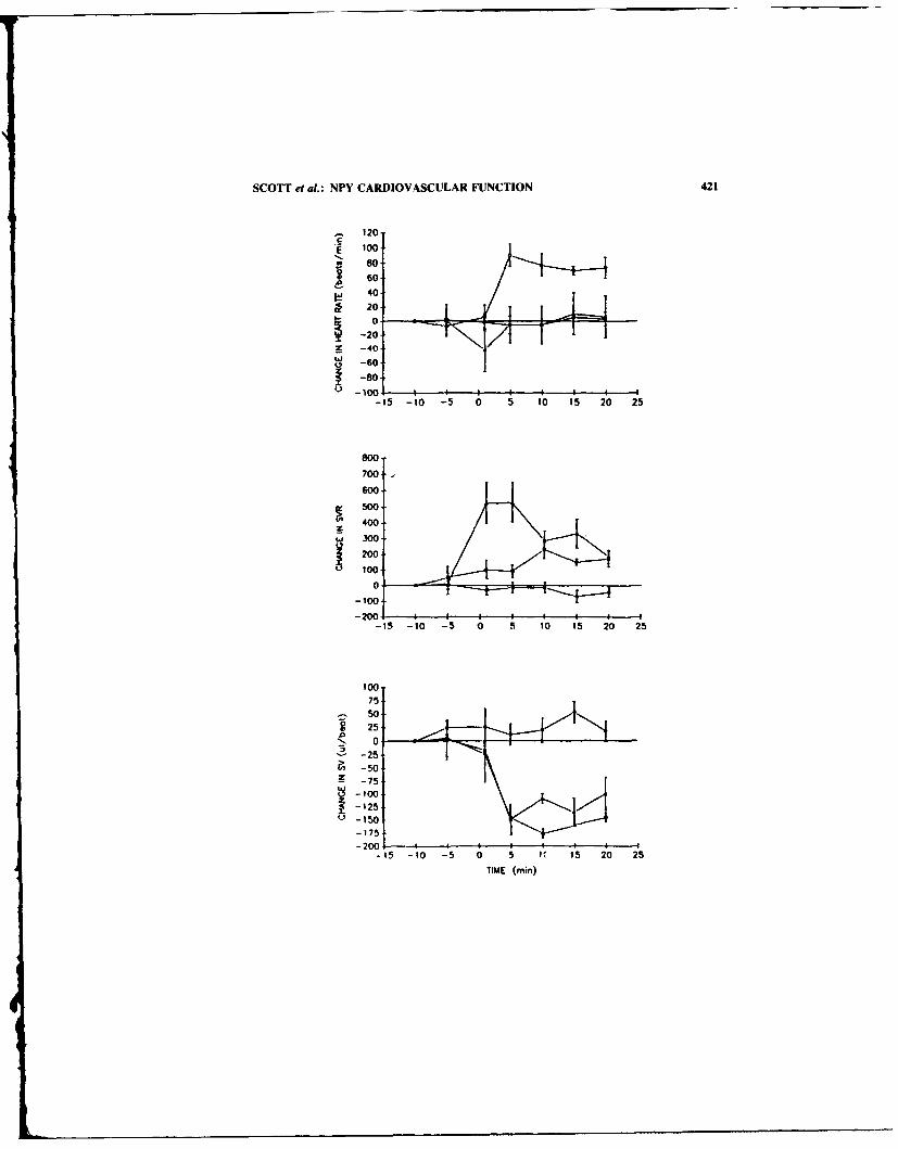
SCOTT et at.: NPY CARDIOVASCULAR FUNCTION 421
.- 120c
S100
9'80
6040
2010
S-20 / -
40
700600
S500
400
w 300
S200
100
00
-10
1-00
TIE5.ie

422 ANNALS NEW YORK ACADEMY OF SCIENCES
REFERENCES
I. SCOTT. N.A.. M.C. MICHEL, J.H. BOUBUK, J.E. RjVIER et al. 1990. Distinction of NPYreceptors in vitro and in vivo. II. Differential effects of NPY and NPY'" 3'. Am. J. Physiol(Heart Circ. Physiol). 259: H174-H180.
2. BROWN, M.R., N.A. SCOTT. J. BOUBLIK, R.S. ALLEN et al. 1989. Neuropeptide Y: biologicaland clinical studies. In Neuropeptide Y. V. Mutt et al.. Eds. 321-329. Raven Press. NewYork.
3. BouBLIK, J., N. SCOTT, J. TAULANE et al. 1989. Neuropeptide Y and neuropeptide Y'8-36 :
structural and biological characterization. int. J. Peptide Protein. Res. 33: 11-15.
4
I.

Neuropeptide Y and Anglerfish PeptideAmide Inhibit Secretin-Stimulated
Pancreatic Exocrine Secretion
MIAREK RUDNICKI, DAVID W. McFADDEN,"
AMBIKAIPAKAN BALASUBRAMANIAM,MICHAEL S. NUSSBAUM, AND JOSEF E. FISCHER
Department of SurgeryUniversity of Cincinnati College of Medicine
Cincinnati Department of Veterans Affairs Medical Center231 Bethesda Avenue (ML 558)
Cincinnati, Ohio 45267
Nevropeptide Y (NPY) and anglerfish peptide amide (APY-NH 2) share substantial aminoacid sequence homology with the pancreatic inhibitory peptides pancreatic polypeptide(PP) and peptide YY (PYY).' Immunoreactive NPY has been localized in pancreaticnerve fibers. 2 This rich NPY supply to the pancreas may indicate a role for NPY in theregulation of pancreatic secretion. This study was designed to evaluate the influence ofexogenous NPY and APY-NH 2 on secretin-stimulati pancreatic exocrine secretion.
Four 25-kg dogs had Herrera pancreatic cannulae surgically implanted under generalanesthesia. The dogs were allowed to recover for two weeks postoperatively and main-tained their weight on standard dog chow. The animals were conditioned to stand inPavlov harnesses. Studies were perfom d in randomized order at weekly intervals afteran overnight fast.
At the beginning of each study the animals were given an intravenous infusion ofsecretin (0.5 p/kg bolus followed by 0.5 lL/kg/hr) to attain maximal pancreatic stimula-tion. After plateau secretion was verified, one 30-minute basal period was measured. Aseparate infusion of NPY at 50, 100, or 100pMol/kg/hrorAPY-NH2 at 100, 500. or 1000pMol/kg/hr was performed for two 30-minute periods. One 30-minute recovery periodconcluded the study.
Pancreatic secretion was collected volumetrically in 30-minute aliquots. Pancreaticbicarbonate (HCO 3-) was measured using an autoanalyzer (Radiometer, Copenhagen,Denmark). Protein was measured by the Lowry method. 3 Blood sampling was performedat 15-minute intervals for NPY and PYY radioimmunoassay.
RESULTS
Statistical analysis was performed using ANOVA with p <0.05 accepted for signif-icance. Results are expressed as mean ± SEM as percentage of basal secretion. NPYplasma levels averaged 420 ± 30 pg/ml basally. Significant elevations in circulating NPYwere found in all three NPY doses averaging 670 ± 24 pg/ml, 1175 ± 93 pg/ml, and 3968
"Corresponding author.423

424 ANNALS NEW YORK ACADEMY OF SCIENCES
TABLE i. Effects of NPY on Pancreatic Exocrine Secretion
Secretin Secretin + NPY Secretin
NPY Dose Basal 30' 60' 90'
50 pMol/kg/hr Volume 100% 100 _ 21 117 ± 4 85 ± 12HCO, 100% 87 10 91 ± 8 91± 7Protein 100%, 102 ± 4 110 ± 21 III 12
100 pMol/kg/hr Volume 100% 70 - 13 63 ± 14 93 ± 14H1-CO 100% 67 ± 6* 65 ± 12* 71 9Protein 100% 95 ± 8 83 ± 6 105 ± 7
1000 pMol/kg/hr Volume 100% 69 ± 6* 53 ± 1* 94 t 21HCO, 100% 82 6 64 +9* 84 ± 20Protein 100% 74 ± 18 83 19 116 ± 27
*p <0.0)5 vs basal by ANOVA.
± 294 pg/ml, respectively. Basal PYY levels averaged 238 - 20 pg/ml and were unaf-fected by NPY or APY-NH, infusion.
NPY significantly inhibited secretin-stimulated HCO 3 secretion at the 100 and 1000pMol doses. NPY inhibited volume secretion at only the 1000 pMol dose. No effect onprotein secretion was demonstrated (TABLE 1). APY-NH, significantly inhibited pancre-atic volume, HCO, , and protein secretion at both 500 and 1000 pMol/kg/hr, an effectthat extended into the recovery period (TABLE 2).
DISCUSSION
Our results demonstrate an inhibition of secretin-stimulated pancreatic exocrine se-cretion in a conscious animal model by both NPY and APY-NH,. [he evidence heresupports a role for NPY in the physiologic control of pancreatic secretion. The markedeffect of APY-NH, on pancreatic secretion suggests a possible clinical role for its use inacute pancreatitis.
TABLE 2. Effects of APi'-NH, on Pancreatic Exocrine Secretion
Secretin Secretin + APY-NH, Secretin
APY-NH, Dose Basal 30' 60' 90'
IN) pMol/kg!hr Volume 100% 15 ± 14 64 ± 5 93 - 9HCO, 100% 69 ±24 62 ±23 78 ± 20Protein I1(K) % 89 ± 13 97 ± 15 112 ± 17
5(0 pMol/kg/hr Volume 100% 49 ± 7* 39 3* 58 1 6*HCO I 0% 37 3* 36 4* 39 ±8*
Protein 100% 48 ± 6* 41) 4* 69 + 7I(M) pMol/kg/hr Volume I(X/)% 42 ± 1* 49 9 9* 74 4
HC) 1007/ 27 ± 7* 18 ± 4* 38 ± 8*Protein I(X) c 41 ± 12* 34 _ 7* 66 ± 2
*p <0.05 vs basal by ANOVA.

RUDNICKI et at.: PANCREATIC EXOCRINE SECRETION 425
REFERENCES
I.- BALASUBRAMANIAM, A., P. C., ANDREWS, V. RENUG0PALAKRISHNAN & D. F. RIGEL. 1989.Peptides 10: 581-585.
2. SUNDLER, F., E. M0GHIMZADEH, R. HiKANSON, M. EKELUND & P. EmSON. 1983. CellTissue Res. 230: 487-493.
3. LOWRY, 0. H., N. J. ROSEBROUGH, R. L. FARR & R. J1. RANDALL. 1951. J. Biol. Chem.1931265-274.

Neuropeptide Y Does Not MediateIschemic Spinal Cord Injury following
Aortic Cross-Clamping in RabbitsBORIS W. KUVSHINOFF, TOM NOGUEIRA,
DAVID W. McFADDEN," AND RICHARD J. FOWL
University of Cincinnati College of MedicineCincinnati Department of Veterans Affairs Medical Center
231 Bethesda Avenue (ML 558)Cincinnati, Ohio 45267
Paraplegia resulting from spinal cord ischemia during repair of thoracoabdominal aorticaneurysms is a significant clinical problem.' One postulated mechanism of neurologicdamage includes posttraumatic ischemia secondary to persistent vasospasm. NeuropeptideY (NPY), found in neural tissue, pia mater and cerebrospinal fluid (CSF), has beenimplicated in pathologic cerebral vasospasm.2 This study examined the release of NPYinto the CSF and blood in response to temporary occlusion of the suprarenal or infrarenalabdominal aorta in rabbits.
METHODS
New Zealand rabbits were randomized into three groups. In the first group the aortawas not clamped (n = 6); in the other two troups either the infrarenal aorta (n = 9) orthe suprarenal aorta (n = 6) was clamped for 25 minutes. Animals were anesthetized withintravenous pentobarbital and the abdominal aorta exposed through a retroperitonealapproach. Blood and CSF (cisternal puncture) were collected before, during, and aftercross-clamping the abdominal aorta. NPY was measured by a sensitive and specificradioimmunoassay developed in our laboratory. 3
RESULTS
Plasma levels of NPY were not affected by temporary occlusion of either the supra-renal or infrarenal aorta (FIG. 1). NPY concentrations in the CSF were also unchangedexcept for a small decrease during aortic occlusion in the suprarenal group (FIG. 2). Lowerextremity paralysis occurred in 70% of the infrarenal group, 83% of the suprarenal group,and 0% of the control group.
DISCUSSION
Little is known concerning the pathophysiology and treatment of spinal cord injuryfollowing nontraumatic ischemia. In models of cerebrospinal trauma, persistent vaso-
"Corresponding author.426
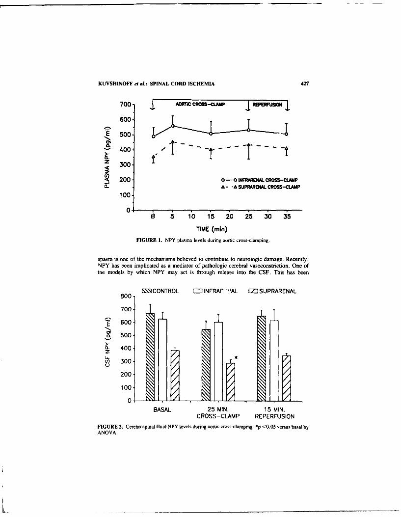
KUVSHINOFF et at.: SPINAL CORD ISCHEMIA 427
700- DOflC CROSS-4CLAMP MM*V"UO
600-
500.-
400 tz 300
S 200- 0-0 INFRARENAL CROSS-CLAMPa- A- -A SUPRARENAL CROSS-CLAMP
100-
0 B 5 10 1'5 20 25 30 35
TIME (min)FIGURE 1. NPY plasma levels during aortic cross-clamping.
spasm is one of the mechanisms believed to contribute to neurologic damage. Recently,NPY has been implicated as a mediator of pathologic cerebral vasoconstriction. One oftne models by which NPY may act is through release into the CSF. This has been
MCNTR0L =INFRAF *'AL IZJSUPRARENAL
800-
700-
E 600-
a-500-
a- 400.z(1' 300-
200-
100
0IG RE, 1-7~ 7
BASAL 25 MIN. 15 MIN.CROSS-CLAMP REPERFUSION
FGR2.Cerebrospinal fluid NPY levels during aortic cross-clamping. *p <0.05 versus basal byANOVA.

428 ANNALS NEW YORK ACADEMY OF SCIENCES
previously described in association with centrally-stimulated hypertension, and after ex-perimental subarachnoid hemorrhage. "
S
The present study demonstrates that NPY is not released into either the CSF orcirculation as a result of cross-clamping the suprarenal or infrarenal abdominal aorta.Therefore, we are unable to suggest a role foi CSF or plasma NPY in ischemic spinal cordinjury following temporary aortic occlusion in rabbits. Our findings do not exclude thatNPY may have a local effect on spinal cord blood flow independent of CSF release orcirculating blood levels.
REFERENCES
1, CRAWFORD, E. C., J. L. CRAWFORD, H. J. SAFI, J. S. COSELLI, K. R. HESS, B. BROOKS, H.J. NORTON & D. H, GLAESER. 1986. J. Vasc. Surg. 3: 389-404.
2. MCDONALD, J. K., C. HAN, B. D. NOE & P. W. ABEL. 1988. Brain Res. 463: 259-267.3. BALASUBRAMANIAM, A., D. W. MCFADDEN, M. RUDNICKI, M. S. NUSSBAUM, R. DAYAL, L.
S. SRIVASTAVA & J. E. FIScHER. 1989. Neuropeptides 14: 209-212.4. P1LOWSK, P. M., M. J. MORRIS, J. B. MINSON, M. J. WES.T, J. P. CHALMERS, J. 0.
WILLOUGHBY & W. W. BLESSING. 1987. Brain Res. 420: 380-384.5. ABEL, P. W., C. HAN, B. D. NOE & J. K. MCDONALD. 1988. Brain Res. 463: 250-258.

Immunocytochemical Localizationof Neuropeptide Y and
Its C-Terminal Flanking Peptidein the Human Heart
S. GULBENKIAN, ad N. COSTA ANDRADEb j. WHARTON,c
J. M. POLAK,C J. QUEIROZ E MELO,b
AND J. F. DAVID-FERREIRA"
'Gulbenkian Institute of ScienceOeiras, PortugalbHeart Institute
Carnaxide, PortugalcRoyal Postgraduate Medical School
London, United Kingdom
NPY is one of the most abundant and widespread peptides in the mammalian cardiovas-cular system.' Recently it was demonstrated that the predicted C-terminal flanking pep-tide of NPY (C-PON) is also widely distributed in both the central and peripheral nervoussystems of several mammalian species including man, and has an identical distribution toNPY.2 In the present study we investigated the localization and the possible coexistenceof NPY and C-PON in the human heart using immunocytochemical techniques at both thelight and electron microscopical levels. Immunofluorescence staining of cryostat sectionsand whole mount preparations revealed that the human heart is supplied by numerousnerve fibres containing NPY and C-PON immunoreactivity. NPY-immunoreactive nervefibres were localized throughout the atrial and ventricular walls and exhibited a similardistribution pattern to nerves displaying C-PON immunoreactivity. NPY/C-PON-immu-noreactive nerve fibres were more abundant in the atria than in the ventricles. In all fourchambers NPY/C-PON-immunoreactive nerves were distributed throughout the myocar-dium, concentrated around small arteries and arterioles at the adventitial-medial borderand formed an endocardial plexus (FIGs. la and Ib), which extended from the suben-docardium to just beneath the endothelial lining. Intrinsic neuronal cell bodies in thehuman heart appeared to lack NPY/C-PON immunoreactivity. It was also observed thatthe number and distribution pattern of NPY/C-PON- and tyrosine hydroxylase-immuno-reactive nerve fibres was very similar.
At the electron microscopical level the use of double immunogold staining techniquesrevealed that NPY and C-PON immunoreactivities were found consistently in largc gran-ular vesicles (70-100 nm in diameter), and both peptide immunoreactivities were colo-calized to the same vesicles (FIG. 2) in axonal varicosities which usually also containednumerous small secretory vesicles (40-60 nm in diameter).
The findings demonstrate that NPY/C-PON-immunoreactive nerves are extensivelydistributed throughout the human heart and appear to largely represent postganglionicsympathetic neurones. At the ultrastructural level, the demonstration that NPY/C-PON
'Correspondence: Sirgio Gulbenkian, PhD, Laborat6rio de Biologia Celular, Instituto Gulbenkiande Ciencia, Apartado 14, 2781 Oeiras Codex, Portugal.
429

430 ANNALS NEW YORK ACADEMY OF SCIENCES
FIGURE 1. Immunofluorescence micrographs of a right atrial appendage cryostat section (a) andwhole mount preparation of endocardium (b) immunostained for NPY (a) and C-PON (b). Densenetwork of NPY- and C-PON- immunoreactive nerve fibres and fascicles are present in the myo-cardium. around small arteries and arterioles (a) and in the endocardium (b). A: artery. Scale bar50 l.±m.

G~ULBENKIAN etal.: NYP & C PON 431
FIGURE 2. Axon varicosity in the human right atnial appendage myocardiun. Colocalization ofNPY and C-PON immunoreactivities to the same large granular secretory vesicles as demonstratedby a double immunogoid staining procedure. NPY. 10 nm gold particles earrowcsi. C-PON. 15 nmgold particles (orrowvheadsi. M: myocyte. Scale bar = 2MK nm.
immunoreactivity was localized to large granular vesicles in presumed sympathetic nerveterminals supports the view that the storage of NPY and noradrenaline in two distinctsubcellular compartments could facilitate the differential release of noradrenaline or NPY.the latter being preferentially released at high stimulation frequencies.
REFERENCES
1. WHARTON. J. & S. GutBENKIAN. 1987. Experientia 43: 821-832.2. Gut-BFNKIAN. S., J. WHARTON. G. W. HACKER, 1. M. VARNIt.L S. R. Bt oost & I. M.
POLAK. 1985. Peptides 6: 1237-1243.3. LuNDBERG. J. M.. A. RUDEmiut.. A. Sot-i.vt. G. FRttID & G. WA1.tIN. 1989. Neuroscience
28: 475-486.

Neuropeptide Y and NonadrenergicMechanisms in the Sympathetic Vascular
Control of the Nasal Mucosa
J. S. LACROIX,- W. LEHMANN,- AND J. M. LUNDBERGb
'"Clinic of Oto-Rhino-LaryngologyUniversity Hospital
CH-1211 Geneva. Switzerlanda,,d
bDepartment of PharmacologyKarolinska Institutet
S-104 01 Stockholm, Sweden
INTRODUCTION
Coexistence of noradrenaline (NA) and neuropeptide Y (NPY)-like immunoreactivity(LI) was observed in the dense sympathetic innervation of resistance and capacitancevessels in the nasal mucosa of most species including man.
A new in vivo model was developed in pentobabital anaesthetized pig which allowedparallel recordings of arterial blood flow (BF), volume of the nasal cavity (reflectingcapacitance function V), nasal venous outflow (VOF) and superficial movement of bloodcells (by mean of laser Doppler flowmetry (LDF) signal) of the nasal mucosa uponsympathetic nerve stimulation (SNS) and/or local intra-arterial injection of various phar-macological agents. 2
AIMS OF THE STUDY
To evaluate the pig nasal mucosa content of NA and NPY-LI in control conditions,after reserpine pretreatment (± preganglionic denervation) and 2 weeks after sympath-ectomy.
To study the characteristics for NA and NPY release as revealed by overflow into thenasal mucosal venous effluent upon SNS in vivo.
To investigate the possible development of supersensitivity to vascular NA and NPYeffects after sympathetic denervation of the pig nasal mucosa in vivo.
RESULTS
Reserpine pretreatment induced a marked depletion of both NA and NPY-Li in the pignasal mucosa. The reduction of NA was not influenced by preganglionic denervationwhile the depletion of NPY then was prevented (TABLE I). Two weeks after sympath-ectomy, the remaining NPY-LI seems to be of parasympathetic origin.'
SNS induced simultaneous reduction of BF, V, VOF and the LDF signal. After locali.a. pretreatment with the a-adrenoceptor antagonist phenoxybenzamine (I mg kg- ') thevascular responses to SNS were significantly attenuated but clear-cut reduction of BF. V,
432

LACROIX et al.: NA & NPY-LI 433
€ .2 .o0
o I 'r- cE 0-=
~._
-4- CC
o
rrirE
I~
-Ec 2E-L ->
44 .. C .
E
p vI
'174
rE

434 ANNALS NEW YORK ACADEMY OF SCIENCES
VOF and the LDF signal remained even to single nerve impulses while the effects ofexogenous NA were abolished.
A frequency-dependent increase of NA overflow was observed in the nasal venouseffluent upon SNS in control pigs, whereas detectable release of NPY-LI occurred onlyat high frequency.
Slowly developing, frequency-dependent and long-lasting vascular responses re-mained (up to 80% of control) upon SNS in reserpinized and decentralized animals. Inthese animals, the NA overflow was abolished while release of NPY-LI was enhanced.Exogenous NPY mimicked the reserpine-resistant SNS-evoked vascular responses (FIG.1).
Two weeks after sympathectomy, an increased sensitivity to the vasoconstrictor ef-fects of NA and NPY was observed (not shown). The development of in vivo supersen-sitivity to NPY further suggests a physiological role of this peptide as a nonadrenergicmediator in the sympathetic vascular control of the nasal mucosa. Although NA is likelyto mediate most of the sympathetic vascular responses to low frequency SNS, somenonadrenergic effects remain even to single impulse stimulation. The characteristic"NPY-like" vascular responses which remained following SNS in spite of adrenoceptorblockade or reserpine-induced NA depletion and the parallel NPY-LI overflow, suggestthat NPY could mediate the nonadrenergic component of sympathetic nasal vasoconstric-tion.
TABLE 1. Pig Nasal Mucosal Content of NA and NPY-LIa
NA (nmol/g) NPY-LI (pmol/g)
Non- Reduction Non- Reductionstimulated in % SNS stimulated in % SNS
Control 14.00 9.7 11.4 9.1± 1.60 - 1.0 t 1.2 ± 1.9
After reserpine 0.09 99** - 6.3 44* -± 0.01 ± 0.5
After reserpine and 0.08 99** 0.06 14.5 7.8preganglionic denervaton ± 0.02 ± 0.02 - 1.5 ± 2.0*
2 weeks after 0.10 99** - 3.1 74**sympathectomy ± 0.03 ± O. t0*Data (means ± SEM, n = 8) from control animals are compared with biopsies obtained from
animals pretreated 24 h before with reserpine (I mg kg- 1 . i.v.), with or without preganglionicdenervation and finally 2 weeks after surgical sympathectomy. For comparison, the tissue contentafter a course of sympathetic nerve stimulations (SNS) are also shown. *p <0.05; **p <0.01; ***p<0.001.
REFERENCES:
I. LACROIX, J. S., A. ANGGItRD, T. HOKFELT, M. M. T. O'HARE, 1. FAHRENKRUG & J. M.
LUNDBERG. 1990. Cell Tissue Res. 259: 119-128.2. LACROIX, J. S, P. STJIARNE, A. ANGGIRD & J. M. LUNDBERG. 1988. Acta Physiol. Scand.
132: 83-90.

Neuropeptide Y Causes NonadrenergicVasoconstriction in the Eye, Brain, and
Some Other Tissues in Rabbits
SIV F E. NILSSON
Department of Physiology and Medical BiophysicsBox 572
University of UppsalaS-75123 Uppsala, Sweden
The eye has a rich innervation of sympathetic nerve fibers from the superior cervicalganglion and like sympathetic nerve fibers to many other tissues, they are immunoreactiveto neuropeptide Y (NPY). Although the density of the NPY-fibers differs between spe-cies, the distribution pattern seems to be about the same in most species. The iris dilatatormuscle and the blood vessels of the uvea (choroid, iris and ciliary body) have a richinnervation, but there are NPY-fibers present also in the ciliary processes and outflowapparatus.' The innervation pattern suggests that NPY may have effects on the pupil size,ocular blood flow and aqueous humor dynamics. In the present study, the effects of NPYon regional blood flows in the eye and other tissues were studied in the rabbit.
The experiments were made on albino rabbits under urethane anesthesia (7 ml/kg b.w.of a 25% solution). All animals were tracheotomized and artificially ventilated. In eightrabbits, uveal blood flow (UBF) was determined by direct measurement of the flow froma cannulated vortex vein simultaneously with registration of the mean arterial bloodpressure (MABP) and the intraocular pressure (lOP). The uveal vascular resistance (UVR)was calculated as (MABP-IOP)/UBF. 2 Regional blood flows were measured with radio-actively labelled microspheres3 (15 jim) in twenty-five rabbits. Two different experimen-tal series were made; one with and one without c-adrenoceptor blockade (phenoxyben-zamine 50 mg/kg b.w.). Blood flow determinations were made before and at 2 and 10 minafter the start of i.v. infusion of NPY, 120 pmol/kg x min. The cervical sympathetic nervewas sectioned on one side in all experiments. Blood flows during the NPY-infusion werecompared with blood flows under control conditions by Student t test. Mean values andSE are given.
Intravenous infusion of increasing doses of NPY (7.5-120 pmol/kg x min) caused adose-dependent increase in the UVR. Near maximal effect (70% increase) was achievedwith 120 pmol/kg x min. In the microsphere experiments without o-adrenoceptor block-ade, the NPY-infusion had no significant effect on the MABP, but cardiac output wassignificantly decreased at 10 min. After 2 min of NPY-infusion, local blood flows weremarkedly decreased in the kidneys, spleen and adrenal glands (FIG. I A). There were alsosignificant blood flow reductions (20-60%) in different parts of the gastro-intestinal tract.In these tissues there was no further decrease in blood flow at 10 min (FIG. IA) and theeffects of NPY were not affected by o-adrenoceptor blockade (FiG. IB). In the eye theeffect of NPY was moderate at 2 min, but at 10 min there were marked blood flowreductions in all parts of the uvea. The blood flow reductions on intact and sectioned siderespectively were for the choroid 55 - 5 and 56 - 4% (p -50.001). ciliary body 53 - 5and 52 - 6% (p f_0.001) and iris 33 ± 7 and 28 - 8% (p <-0.01). The salivary glands,tongue and masseter muscle responded in a similar way as the eye; moderate effect at 2
435

436 ANNALS NEW YORK ACADEMY OF SCIENCES
6
A5
-4-
'-3
0 Kidney
0 spleen
0
0 2 10 Ttme (min)77±4 79±4 73±5 MABP (mmHg)
415±29 392±25 347±27" CO (g/nmln)
312±8 308±8 300±9 HR
6
-4-
-3
Spleen
0 **,Kidney
Adrenal gland0
0 2 10 Tlime (min)44±2 50±2' 48±2... MABP (mmHg)
673±32 666±27 618±33 CO (g/naln
331±5 332±5 330±5 HRt
FIGURE 1. Mean arterial blcod pressure (MABP), cardiac output (CO). heart rate (HR) and localblood flows in the kidney, spleen and adrenal gland before and at 2 and 10 min after the start of i.'.infusion of NPY. 120 pmol/kg x min. (A) Experiments without ci-adrenoceptor blockade (n = 12).(B) Experiments with ci-adrenoceptor blockade (n = 13). *p !5O.05; **p !5001l and ***p -.0.00O.
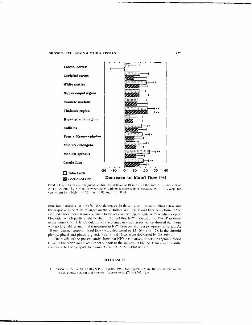
NILSSON: EYE, BRAIN & OTHER TISSUES 437
Frontal cortex
Occipital cortex
White matter
Hippocampal region
Caudate nucleus
Thalamiic region *
Hypothalamic region
Collicdes
Pons + Mesencephalon
Medulla oblongata
Medulla spinalis
Cerebellum
inat ie-20 -10 0 10 20 30 40
~jSectioned side Decrease in blood flow M%)
FIGURE 2. Decrease, in renonaf cerehral hMood flo~ s at 10 ft m after the start it i. initusion ofN -Y . 1-1 2))riol k, x ini i in e pern men rib ithiout ii -adrenocepti r blo ckade in 1). c scet lorcerehelinn tor "xIch n -2), 'p -0.05 and -1) - 0.01,
rnin. hut miarked at 1 0 ruin 1 30 -70'/( decrease). In these t issuies. the in itia jliood t mios andthe response to NPY were larger onl the sectioned side. The blood low% redIuctions' In theeve and other facial tissues sceined to he less in the experimnrts with nt-adrettoccptorhlockade, wshich partly, could he due to the tact tat NPY increased the NIABP in thesecx peri itents Fio It B.1). 'ale il atiott of the change in vascular resistance showekd that therewAas no large difference in the response to NPY between the two expierinlental series. At10 ntlin rcuional cerebral blood flowvs were decreased b 15-~2011; (:1;. 2). Irt the choroidPlexus. Pineal and pituitary gland, local blood flows were decreased b\ 30-601/'(
The results of the present study' show that NPY has mnarked effects onl reiottal bloodflow's in the rabbit and gisecs further support to the sugest ion that NVlY mna\, sigiiicant lNcontribute to thle symipathetic vasoconstriction tn the rabbit us ca.
REF ERENC ES
I S iii'i . R, A . A Mi Lx I itS & P' U, t-ASmsmN. IYXOi. NCnrojctmmf N~ and tfie oc mir iniicr\aitiotiof r..i. Liunica pmi. cal and] ininkex Neuriiscienkcc 17(41: 1207 12lot

438 ANNALS NEW YORK ACADEMY OF SCIENCES
2. NILSSON, S. F. E. & A. BILL. 1984. Vasoactive intestinal polypeptide (VIP): Eff'ects in the eyeand on rer'ional blood flows. Acta Physiol. Scand. 121: 385-392.
3. ALM. A. A A. BItt.. 1972. The oxygen supply to the retina, 11. Effects of high intraocularpressure i id of increased arterial carbon dioxide tension on uveal and retinal blood flow incats. Acta Physiol. Scand. 84: 306-319.
4. GRANSTAM. E. & S. F. E. NILSSON. 1990. Non-adrenergic vasoconstriction in the eye andsome other facial tissues in the rabbit. Eur. J. Pharniacol. 175: 175-186.

Effects of Neuropeptide Y andNoradrenaline on the Uterine Artery
GABRIEL FRIED." MARIANNE THORESEN," AND
ULF SAMUELSSONd
Departments of 6Physiology, 'Obstetrics and Gynecology and"Plastic Surgery
Karolinska InstitutetS-104 01 Stockholm, Sweden
INTRODUCTION
Many peripheral sympathetic nerves, including those of the female reproductive tract,contain both noradrenaline (NA) and neuropeptide Y (NPY).' Both substances are vaso-active, and are released upon nerve stimulation. 2 During pregnancy, there is a decrease ofuterine sympathetic nerves. 3 We have studied the effects of NPY and NA in vitro onisolated human nonpregnant uterine arteries, and also in a situation where the uterinesympathetic nerves have degenerated, in the uterine circulation of the pregnant guinea pigin ViVo.
4
METHODS
Segments of human uterine arteries and veins were obtained during elective surgery,and mounted in oxygenated Krebs-Ringer buffer in organ baths connected to a GrassFT03C transducer and model 7 polygraph. In vivo, local arterial blood pressure wasmeasured in term pregnant guinea pigs through a cannulated side branch of the uterineartery. Blood flow was measured by ultrasound Doppler velocimetry. 5
RESULTS
In the isolated human uterine artery, NPY had no effects per se. However. NPY inhigher concentrations (10-' M) often, but not always, enhanced the contractile effect ofNA (FIG. I). In the human uterine vein, NPY increased basal tone and induced sponta-neous contractions. The contractile effect of nerve stimulation was also enhanced byNPY. In the pregnant guinea pig uterus, NPY increased blood pressure, but had nosignificant effect on blood flow, i.e., the net effect was an increased vascular resistance.NPY (10 7 M) was more potent than NA (10-- 7 M) in increasing blood pressure (FiG. 2).The effects of NA were reduced or abolished by phentolamine, whereas the effects ofNPY were not affected by phentolamine.
"Supported by the Swedish Medical Research Council {14X-7164).439

440 ANNALS NEW YORK ACADEMY OF SCIENCES
10 mN L
1 min WW W
t t ,10-eM 10-6M 10-7M 10-6M
NA NA NPY NA
10 mN L
1 mlin
NPY 10- 9M 10-8M 10- 7M
FIGURE 1. Upper trace: Effects of NPY and NA on human uterine artery in vitro. NPY (10- M)has no intrinsic effect, but appears to enhance the contraction caused by NA (10- 6 M). w = wash.Lower trace: Effect of NPY on human uterine vein in vitro. There is an increased tone at 10- M,spontaneous contractions appear at 10- M, and an increase in amplitude appears at 10- M, anddisappears after wash (w).
100
NPY ::75 E NA
50 **w ** K
< 25BLOOD FLOW
0
-'25 BLOOD PRESSURE
-25
10- 7M 10- 6M 10- 5M 10-7M 10- 6M 10- 5M
FIGURE 2. Effects of NPY and NA on blood pressure (left panel) and blood flow velocity (rightpane[) in the pregnant guinea pig artery. On the abscissa, concentrations of NPY and NA are given(10-'-10 - ' M). On the ordinate, percentage changes in blood pressure and blood flow velocity ascompared to control values (averages obtained 2 min before the infusion). Open bars = NPY. solidbars = NA. * p <0.001;** = p <0.05;* = p <0.05.

FRIED et al.: UTERINE ARTERY 441
DISCUSSION
The results show that NPY has no vasoconstrictor effects per se in the nonpregnanthuman uterine artery, and moderate vasoconstrictor effects in the human uterine vein. Inthe pregnant guinea pig, local infusion of NPY gave an increased blood pressure but noeffect on blood flow velocity. Taken together, the results indicate that NPY cooperateswith NA to cause an increased vascular resistance. The effect of NPY appears to bece-adrenoceptor resistant. Both NPY and NA may thus be involved in the control of theuterine circulation, both before and during pregnancy.
REFERENCES
1. LUNDBERG. J. M., L. TERENIUS, T. HOKEELT & M. GOLDSTEIN. 1983. High levels of neu-ropeptide Y (NPY) in peripheral noradrenergic neurons in various mammals including man.Neurosci. Lett. 42: 167-172.
2. LUNDBERG, J. M., A. RUDEHILL. A. SOLLEVI. G. FRIED & G. WALLIN. 1989. Co-release ofneuropeptide Y and noradrenaline from pig spleen in vivo: Importance of subcellular storage,nerve impulse frequency and pattern, feedback regulation and resupply by axonal transport,Neuroscience 28: 475-486.
3. FRIED, G., T. HOKFELT, J. M. LUNDBERG, L. TERENIUS & L. HAMBERGER. 1986. Neuropep-tide Y and noradrenaline in human uterus and myometrium during normal and pre-eclampticpregnancy, Hum Reprod. 1: 359-364.
4. FRIED, G., T. HOKFELT, L. TERENIUS & M. GOLDSTEIN. 1985. Neuropeptide Y (NPY)-likeimmunoreactivity in guinea pig uterus is reduced during pregnancy in parallel with noradren-ergic nerves, Histochemistry 83: 437-442.
5. THORESEN, M. & J. WESCHE. 1988. Doppler measurements of changes in human mammaryand uterine blood flow during pregnancy and lactation. Acta Obstet. Gynecol. Scand. 67:741-745.

Reduced Inhibitory Actions ofSympathetic Stimulation and Exogenous
Neuropeptide Y on Cardiac VagalAction in the Presence of Peptide YY
ERICA K. POTTER AND D. I. McCLOSKEY
School of Physiology and PharmacologyUniversity of New South Wales
P.O. Box IKensingion, Sydney
New South Wales 2033, Australia
It has been proposed that neuropeptide Y (NPY) is released by stimulation of the cardiacsympathetic nerves in the anaesthetized dog and is responsible for a subsequent prolongedinhibition of cardiac vagal action by inhibiting acetylcholine release.' Testing this pro-posal has been hindered by lack of a specific antagonist to NPY.
Another approach, however, is to use a peptide which has similar but more potentNPY-like action as a 'superagonist.' Peptide YY (PYY), a related peptide also inhibitscardiac vagal action but is more powerful than NPY and has a longer duration of action.2
It is postulated that when PYY is occupying receptors on vagal nerve terminals, nerve-released NPY will be either unable to produce an effect, because it cannot gain access tothe receptors, or it will displace PYY from at least some receptors and, being lesspowerful than PYY, will lessen the preexisting vagal attenuation.
"le following ratio was measured in anaesthetised dogs before and after PYY ad-minP mitration:
API after sympathetic stimulation or NPYAPI before sympathetic stimulation or NPY
API is the increment in pulse-interval (the period between successive beats of the heart)evoked in response to a standard vagal simulus. R becomes <I wher sympathetic nervestimulation or exogenous NPY inhibit the cardiac slowing effect of the vagus. R wasmeasured before and after PYY administration.
R was measured for sympathetic stimulation in nine dogs. In all of these there was lessinhibition of vagal action on pulse interval (i.e., R was greater) in the presence of PYYthan in its absence. Indeed, in 5 of the dogs, R, which was always <1 in controlcircumstances, became > I in the presence of PYY. This indicates that in the presence ofPYY sympathetic stimulation was associated with enhancement rather than inhibition ofvagal action on pulse interval (FIG. 1). Such an effect could occur if the less potenttransmitter NPY were released from sympathetic nerves, displacing PYY from receptorson vagal nerve terminals, and so relieving the vagal inhibition established in the presenceof PYY.
R was measured for NPY administration in 3 dogs, in the absence then presence ofPYY. In all of these R was increased in the presence of PYY, and in 2 of them R wasequal to I, indicating a failure of NPY to inhibit cardiac vagal action in the presence of
442
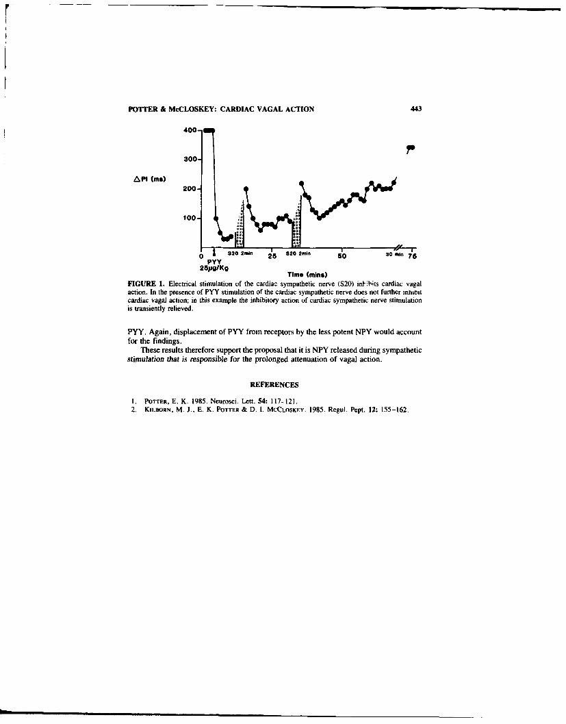
POTTER & McCLOSKEY: CARDIAC VAGAL ACTION 443
400-
300-
API (me)200-
820 o , 25 820 m 50 30 min,
PYYTime (min)
FIGURE 1. Electrical stimulation of the cardiac sympathetic nerve (S20) int-ibits cardiac vagalaction. In the presence of PYY stimulation of the cardiac sympathetic nerve does not further Itnnincardiac vagal action; in this example the inhibitory action of cardiac sympathetic nerve stimulationis transiently relieved.
PYY. Again, displacement of PYY from receptors by the less potent NPY would accountfor the findings.
These results therefore support the proposal that it is NPY released during sympatheticstimulation that is responsible for the prolonged attenuation of vagal action.
REFERENCES
I. POTTER, E. K. 1985. Neurosci. Lett. 54: 117-121.2. KILBORN, M. J., E. K. POTTER & D. I. MCCLOSKEY. 1985. Regul. Pept. 12: 155-162.

Pre- and Postjunctional Actionsof Neuropeptide Y and
Related Peptides
ERICA K. POTTER," L. MITCHELL," M. J. D. McCLOSKEY."
A. TSENG." M. PERSICO," AMANDA E2. GOODMAN.'
MARISSA G. DE PIETRA,6 MARION LOUGHNAN,6 J. SHINE,"AND D. 1. McCLOSKEY"
"School of Physiology and PharmacologyUniversity of New South Wales
P.O. Kensington, Sydney 2033 Australiaand
bPacific BiotechnologyP.O. Box 172
Kings Cross, Sydney, 2011 Australia
The effects of neuropeptide Y (NPY) and related peptide fragments on arterial bloodpressure (BP) and cardiac vagal action were studied in vagotomised anaesthetised rats. Anincrease in BP was used as a measure of postjunctional action of peptide (direct vaso-constrictor and/or constrictor-potentiating), and a change in cardiac vagal action wastaken as a measure of prejunctional activity. Three analogs of NPY were studied in detailin two series of experiments.
In the first series, the effect of equimolar doses of NPY 13-36 and a 'stabilised" NPY13-36 (ANA-NPY') were compared with whole NPY. The results are listed in TABLE I.It can be seen that both 13-36 fragments have significant pre- and postjunctional activity.ANA-NPY, the stabilised fragment, has similar prejunctional activity to whole NPY, butis less potent postjunctionally. The findings are consistent with there being more than oneNPY receptor type, but do not support the basis of distinction of the prejunctional onebeing itF failure to be activated by the 13-16 fragment?2
In the second series of experiments the activity of the C-terminal fragment NPY 18-36was compared with whole NPY in 18 rats. NPY 18-36 has both pre- and postjunctionalactivity but is significantly less active than NPY 1-36 at both sites. In intact anaesthetisedrats in contrast with the findings in vagotomised rats, however, NPY 18-36 was found tohave a prominent depressor action. This was especially notable at higher doses of peptide.and was typically preceded by a small, transient pressor response. This depressor actionwas greatly attenuated, or absent, after vagotomy (Fic. 1). The results help explain thefindings of depressor effects of NPY 18-36 reported by Boublik and associates' in ratswith intact vagi. The dependence of the effects on the vagus suggest that NPY 18-36 actsby activating sensory nerves that travel in the vagus and produce their depressor actionsreflexly.
TABLE I. Pre- and Postjunctional Actions of NPY and Related Peptides
NPY 1-36 NPY 13-36 ANA-NPY
BP (mm Hg): postjunctional 71 ± 4 40 - I 30 3% Vagal inhibition: prejunctional 91 ± 3 52 _+ 9 86 7
444

POTTER ef at, PRE- & POSTJUNCTIONAL ACTIONS 445
z ,
10-0 zC
400
CL
0 -
a. 0. 0. 0-
CL r
CID
43, c'.
g. E)
ji! K OE. E.'
t >0

446 ANNALS NEW YORK ACADEMY OF SCIENCES
REFERENCES
I . POTTER, E. K., L. MITCHELL. M. J. D. MCCLOSKEY. A. TSENG, A. E. GOODMAN, J. SHINE
& D. 1. MCCLOSKEY. 1989. Regul. Pept. 25: 167-177.2. WAHLESTEDT, C.. N. YANAIHARA & R. HAKANSON. 1986. Regul. Pept 13: 307-318.3. BoUBLIK. J. H.. N. A. SCOTT, J. TAULONE. M. GOODMAN. M. BROWN & J. RIVIER. 1989.
Int. J. Peptide Protein Res. 33: 11-15.

Neuropeptide Y-A Marker forNoradrenaline Exocytosis
ALBERT SCHOMIG" AND MARKUS HAASS
Department of CardiologyUniversity of HeidelbergBergheimer Strasse 58
D-6900 Heidelberg, Germany
In ischemia adrenergic excess stimulation of the myocardium is accompanied by delete-rious consequences such as the occurrence of fatal arrhythmias and the accleleration ofcell damage.' Local release of noradrenaline in the energy-depleted heart can be mediatedby two distinct release mechanisms, either by exocytosis or by a nonexocytotic releasemechanism. We now provide evidence that neuropeptide Y (NPY), which is colocalizedwith noradrenaline in cardiac postganglionic sympathetic nerve fibres, can be utilized asa marker for exocytotic noradrenaline release and therefore differentiates between bothrelease mechanisms. 2
METHODS
All experiments were performed in guinea pig perfused hearts, which were preparedaccording to a modified Langendorff technique and perfused with Krebs-Henseleit bufferat a constant flow rate of 7 ml/min/g heart weight. 3 Two models of energy depletion wereemployed: I. anoxia (pO2 less than 5 mmHg); 2
.4 "- 2. cyanide intoxication plus glucose-
free perfusion. 4 -5 In the coronary venous effluent noradrenaline and NPY were deter-mined by HPLC 4 5 and RIA, 3 respectively.
RESULTS AND DISCUSSION
Central sympathetic stimulation is accompanied by a concomitant overflow of nor-adrenaline and NPY from the heart. ' The corelease of both transmitters induced byelectrical stimulation of postganglionic sympathetic nerve fibres, veratridine or nicotinerequires the presence of extracellular calcium and has therefore been attributed toexocytosis. '6 In contrast, noradrenaline efflux evoked by the indirectly acting sympatho-mimetic tyramine is independent from the extracellular calcium concentration, the acti-vation of N-type calcium channels and the stimulation of protein kinase C. and is notaccompanied by any significant overflow of NPY (FIG. I). The tyramine-induced effluxof noradrenaline can be described by a nonexocytotic release mechanism, consisting of atwo step process: In a first step noradrenaline is lost from storage vesicles, resulting inincreased axoplasmic noradrenaline concentrations. In a second, rate-limiting step nor-adrenaline is transported across the axolemm into the synaptic cleft using the neuronalcatecholamine re-uptake (uptake,) in reverse of its normal transport direction. The latter
"Corresponding author.447

44~ ANNALS NEW YORK ACADEMY OF SCIENCES
mechanism also accounts for the local metabolic release of noradrenaline occurring in theenergy-depleted heart.' The local metabolic release of noradrenaline during anoxta,2cyanide intoxication (unpublished observations), and ischemia' is not accompanied byoverflow of NPY and is quite distinct from exocytosis. as summarized in TABL.E 1:
Uptake -blockade (e.g.. by desipramine) increases exocytotic and suppresses nonexocy-totic noradrenaline overflow from the heart. Whereas exocytotic overflow of noradren-aline and NPY is modulated by presynaptic receptors, ~ nonexocytotic noradrenalinerelease occurs independent from presynaptic regulation.'
cumulativeoverflow ofnoradrenal.-ne lelectrical stimulatio tyramine
600.
Pmollg.
400.
200.
NPY
0.6. control=~ w- conotoxin lO0nM
pmol/g m TPA 300nM
0.4-i f-j polymyxin B 100pM
0.2
0jFIGURE J. Effect of btockade of'N-type calcium channels (by id-conotoxin). and of activation (byphorhol1-t2-myristate-13-acetate (TPA)) or inhibition (hy potvrnxin B) of protein kinase C on theoverflow of noradrenaline and neuropeptide Y (NPY) from the guinea pig perfused heart evoked byelectrical stimulation (1t2 Hz: I min) of cardiac sympathetic nerve fibres (lft pooch) or by lU-mmninfusion of' tyramine (right po~iel. Alt hearts were perfused with a catcim-contains Krebss-Henseleif buffer:.2 n =4-6 in each group. mean ± SEM.

SCHOMIG & HAASS: NA EXOCYTOSIS 449
TABLE i. Characteristics of Nonexocytotic Noradrenaline Release in the Guinea PigPerfused Heart during Energy Depletion or Tyramine Administration: Comparison withExocytotic Noradrenaline Release
Exocytotic NonexocytoticRelease Release
Central sympathetic stimulation + -Concomitant overflow of NPY + -Modulation by presynaptic receptors + -Dependence from extracellular calcium + -Inhibition by uptake, -blockade +
During early stages of ischemia, both release mechanisms play a role in the adrenergicoverstimulation of the heart.' Hence, simultaneous measurement of noradrenaline andNPY allows to assess the relative contribution of exocytotic and nonexocytotic transmitterrelease and may help to determine therapeutic strategies to suppress the local catechol-amine excess.
REFERENCES
I. SCHOMIG, A. & G. RICHARDT. 1990. J. Cardiovasc. Pharmacol. In Press.2. HAASS, M., M. HOCK, G. RICHARDT & A. SCHOMIG. 1989. Naunyn-Schmiedeberg's Arch.
Pharmacol. 340: 509-515.3. HAASS, M., B. CHENG, G. RJCHARDT, R. E. & A. SCOOMIG. 1989. Naunyn-Schmiedeberg's
Arch. Pharmacol. 339: 71-78.4. SCH6MIG, A.. S. FiSCHER, T. KURZ. G. RICHARDT & E. SCHOMIG. 1987. Circ. Res. 60:
194-205.5. SCiOMIG, A., T. KURZ, G. RICHARDT & A. SCH6MIG. 1988. Circ. Res. 63: 214-226.6. HAASS, M., G. RICHARDT, R. E. LANG & A. SCHOMIG. 1990. N.Y. Acad. Sci. This volume,
450-452.7. FRANCO-CERECEDA, A., A. SARIA & J. M. LUNDBERG. 1989. Acta Physiol. Scand. 135:
173-187.

Common Features of Neuropeptide Yand Noradrenaline Release in
Guinea Pig HeartMARKUS HAASS." GERT RICHARDT. RUDOLF E. LANG." AND
ALBERT SCHOMIG
Departments qf Cardiology and "PharmacologyUniversity of HeidelbergBergheiner Strasse 58
D-6900 Heidelberg. Germany,
Neuropeptide Y (NPY) is colocalized with catecholamines in - ,stganglionic sympatheticnerve fibres and the adrenal medulla. Furthermore, ,-ympatnetic stimulation has beendemonstrated to be accompanied by a corelease of NPY and noradrenaline in animalmodels' and in man. 2 This short review will focus on our most recent studies character-izing the common featurcs of the exocytotic release of NPY and noradrenaline fromguinea pig in situ perfused hearts wsith intact sympathetic innervation.
METHODS
For in situ perfusion (constant flow of 7 ml/min/g heart weight: Krebs-Henseleitbuffer) the guinea pig hearts were piepared according to a modified Langendorff tech-nique. In addition, thc !cft stellate ganglion "as carefully dissected free for electricalstimulati'n (12 Hz. I min) of c.rdiac sympathetic nerve fibres. ' In the coronary venouseffluent NPY and noradrenaline were determined by RIA' and HPLC, 4 respectively.
RESULTS AND DISCUSSION
The common features of stimulation-evoked release of NPY and noradrcnaline aresummarized in TABLE I. The corelease of both transmitters evoked by electrical ,.imu-lation of the left stellate ganglion :s positively related to the stimulation frequency' and tothe extracellular calcium concentration. and is regulated by presynaptic receptors, suchas alpha,-adrenoceptors. adenosine A, -receptors. and angiotensin Il-receptors, in a par-allel manner. By means of a negative feedback mechanism both noradrenaline and pos-sibly NPY reduce their corelease by interference with presynaptic alpha.- and NPY-receptors. respectively. As observed in guinea pig heart' and pig spleen. blockade ofneuronal amine re-uptake (uptake,) by dsipraminc or nisoxetine differentially affects thetransmitter overi!ow. Uptake,-blockade increases noradrenaline and reduces NPY over-
flow. This phenomenon can be explained by an increased stimulation of inhibitory pr2-synaptic alpha,-adrenoceptors induced by the elevated local concentrations of
noradrenahne.
"CVorresponding author.450

HAASS et al.: NPY & NORADRENALINE kELEASE 451
TABLE 1. Common Features of the Stimulation-Evoked Exocytotic Corelease ofNeuropeptide Y (NPY) and Noradrenaline (NA) in the Guinea Pig Perfused Heart"
NPY NA
General featuresFrequency-dependence + +Calcium-dependence +1-
Presynaptic receptorsAlpha, -adrenoceptor prazosin (1)-Alpha,-adrenoceptor yohimbin (1)
B-HT 9)20 (S)NPY-receptor NPY (S) 4Adrenosine A,-receptor cyclohexyladenosine (S)Angiotensin Il-receptor angiotensin 11l(S)
Amine uptakeUptake, desipramine (1)
nisoxetine (1)Calcium channels
N-type wj-conotoxin (I14cadmium chloride (1)44
L-type felodipine (llSecond messengers
Protein kinase C TPA (S)polymnyxin B (1)4
"(1) =inhibitory; (4=stitnulatory: + change; no change: decreas,.:increase.
neuron
7 ___ NPYprotein kinase C
Nhi-type NA utkcalciumn channel] NA taE
calcium . LI-
presttnaptic1 NPYinh ibitoryreceptors NA NA
eax oc ytosi.]NA/NPY
postlynaptic
efIfe c ts
FIGURE 1. Schenic of the regulation of 0tiinulation-e~ oked cl'rclca~c of ncijropcptidc Y INPN I andnoradrcnitic (NA) fromt cardiac symnpathetic nerscecndmLn Itrict l clcctriwal 'timutitation of rxotgangl,m 'nt npathbetic ners e tihre e\'- a c orclecasc of both I ran~ 111tter, that 1, jii ,du Ic tjedl'prc snaptlc reccptor and refurr, tc r'nwik of exftacclilr aiwnttt. the inflti\ -I -kiiul
through N-tvv pe kIU tunhantiCk .111d IJ 1ic 11 tj. on )I totem (onae

452 ANNALS NEW YORK ACADEMY OF SCIENCES
According to an exocytotic release mechanism, the overflow of NPY from the heartevoked by various stimuli, such as nicotine.6 veratridine.7 or electrical stimulation '5 '7
critically depends on the presence of extracellular calcium. Using various ligands forvoltage-dependent calcium channels, it can be concluded, that the entry of calcium intothe sympathetic nerve ending requires the activation of voltage-dependent N-type but notof L-type calcium channels.5 Furthermore, activation of protein kinase C by the phorbolester TPA markedly enhanced, inhibition of the enzyme by polymyxin B completelyprevented NPY and noradrenaline overflow. With the exception of studies involvinguptake -blockade, there was a molar ratio of NPY and noradrenaline overflow of approx-imately 1 : 600.
The regulation of NPY and noradrenaline overflow at the sympathetic nerve ending ofthe heart is illustrated in FIGURE 1. Whether the regulation of the transmitter release bypresynaptic receptors is mediated by modulation of N-type calcium channel gating8 or byinterference with the protein kinase C activity cannot be ,oncluded from our results.
In conclusion, the stimulation-evoked exocytotic corelease of NPY and noradrenalineis characterized by parallel presynaptic regulation, dependence on the presence ofextracellular calcium, stimulation of N-type calcium channels, and activation of proteinkinase C.
REFERENCES
I. LUNDBERG, J. M., A. RUDEHIL, A. SOLLEVI & B. HAMBERGER. 1989. Br. J. Pharmacol.96: 675-687.
2. MORRIS, M. ., A. E. RUSSELL. V. KAPOOR. M. D. CAIN. J. M. ELoLIOT, M, J. WEST, L. M.H. Witc; & J. P. CHALMERS. 1986. J. Auton. Nerv. Syst. 17: 143-149.
3. HAASS, M., B. CHENG, G. RICHAROT. R. E. LANG & A. SCHOMIG. 1989. Naunyn-Schmiede-berg's Arch. Pharmacol. 339: 71-78.
4. SCHOMIG. A., S. FISCHER. T. KURZ, G. RtCHARDr & E. ScHjMosG. 1987. Circ. Res. 60:194-205.
5. HAASS. M., C. FORSTER, G. RICHARDT. R. E. LANG & A. SCHOMIc. 1989. Circulation80(Suppl. I1): 11-446 (abstract).
6. RICHARDT. (., M. HAASS, S. NEEB, M. HOCK, R. E. LANG & A. SciiHMio. 1988. Kin.Wochenschr. 66(Suppl. XI): 21-27.
"i. HASS, M., M. HOCK, G. RiCHARDT & A. S HOMIG. 1989. Naunyn-Schmiedeberg's Arch.Pharmacol. 340: 509-515.
8. LIPSCOMBE, D., S. KONGSAMUF & R, W. TsIEN. 1989. Nature 340: 639-642.

Effect of Neuropeptide Y on StimulatedRenin Secretiona
JEAN-FRAN4;OIS AUBERT, BERNARD WAEBER,/
JORG NUSSBERGER, AND HANS R. BRUNNERDivision of Hypertension
University HospitalLausanne, Switzerland
INTRODUCTION
Neuropeptide Y (NPY) is a 36 amino acid peptide with vasoconstrictor properties.This peptide is present in the brain, the adrenal medulla as well as in sympathetic nerveendings. In the kidney, NPY has been found in the fibers innervating the juxtaglomerularapparatus, i.e., in the vascular pole of the glomerulus where renin-secreting cells arelocated.' In the isolated rat kidney, NPY has been shown to inhibit renin release. 2 Asuppressing effect of NPY on renin release has also been observed in intact rats with arenin-angiotensin system activated. 3-5 The present study was undertaken to assess inconscious normotensive rats whether exogenous NPY affects renin secretion induced byvarious stimuli.
METHODS
Male normotensive Wistar rats weighing between 240 and 300 g were used. Theanimals were cannulated under ether anesthesia with one arterial and two venous cathe-ters. Blood pressure and heart rate were then continuously monitored using a computer-ized data acquisition system. All experiments were started 90-120 min after the end ofsurgery, at a time a steady state was reached. A blood sample was obtained at the end ofthe experiment through the arterial ine for measurement of plasma renin activity.
Rats were infused for 45 min with either a nonpressor dose of NPY (0. 1 g/min i.v.,Bio Mega, Laval, Canada) or its vehicle. Starting 15 min later, a second infusion wasinitiated. It consisted either of the beta-adrenoceptor stimulant isoproterenol (10 ng/mini.v.; Winthrop, Basel, Switzerland), or the prostacyclin agonist iloprost (50 ng/min i.v.;Schering, Berlin, Germany).
Results are reported as means ± SEM. Statistical analysis of the results was doneusing a one-way analysis of variance, followed by a least significant difference test, whereappropriate.
RESULTS
NPY had no significant blood pressure effect. Blood pressure remained also un-changed when isoproterenol and iloprost were infused together with NPY. TABLE I showsthe levels of mean blood pressure measured at the beginning and the end of the experimentin the different groups of rats.
'This work was supported by a grant from the Swiss National Foundation."Correspondence to: Bernard Waeher, M.D.. Division of Hypertension. CHUV. t011 Lausanne.
Switzerland.453

454 ANNALS NEW YORK ACADEMY OF SCIENCES
Plasma renin activity was not altered by NPY infused alone. However, the peptidesignificantly attenuated the increase in plasma renin activity caused by both isoproterenoland iloprost (TABLE 1).
DISCUSSION
The first evidence indicating a role for NPY in the regulation of renin release isderived from experiments in which rats with glucocorticoid- induced hypertension weretreated with a nonpressor dose of the peptide .3 Some rats had been subjected to a bilateraladrenalectomy and exhibited very high plasma renin levels. In these rats. NPY allowedplasma renin activity to decrease to levels similar to those measured in control animalswith intact adrenals. NPY was also shown to blunt the renin response to ACE inhibitionand renal artery clipping .4 5 The inhibitory effect of NPY on renin secretion was con-firmed in vitro using kidney slices. 2
TABLE I. Mean Blood Pressure (MBP, mmHg) and Plasma Renin Activity (PRA.ng/ml/hr) in the Different Study Groups"
MBPGroups (n) NPY lsoproterenol Iloprost Before Afteu PRA
1 6 --- 119 2 120 2 3-4 0.811 7 + -- 121 3 126 ±4 2.5 ±0.3
III 6+ - 120 2 119±1 16.4 3IV 7 + + - 120 ±3 120 ±3 7.21 1 .5**V 8 -+ 118 2 116 2 35.0±7
VI 8 + + i 120 2 120±2 9.5 2.9*
-**p <0.01 vs vehicle of NPY: + test substance infusedt - no active treatment.
In the present study. two well established stimuli of renin secretion were infused toconscious rats at doses devoid of systemic blood pressure effect. Both agents markedlystimulated renin secretion. NPY infused at a nonpressor dose had no effect on plasmarenin activity in baseline conditions, but markedly suppressed the renin release induced bythe two test stimuli.
REFERENCES
1. BALLESIA. V. J. M. POtAK, J. M. At~tr.N & S. R, Bioom 1984. The nerves of the juxta-glomerular apparatus of man and other mammal.% contain the potent peptide NI'Y Histochem-
tstry 80: 483-485.2. HACKF.NIt1At.. E.. K. A]Kiorrts. K. H. JAKOBS & R. E. LAN(, 1987. Neuropeptide Y inhibits
renin release oi- a pertussis toxin-sensitive mechanism. Am. J. Physiol. 552: F543-F550V. PFrIER. A.. B. WAsEBrlR. J. Nussiritr& & H-. R. BikvN*,ti 1496. Neuropeptide Y nonnal-
ieN renin secretion in adrenalectomi/ed rats without changing blood pressure tile Sci39: 2161-2167.
4. AiruRr. J, F., M. B1 RNILRr. B. WAI-trrr. J. N'ssBRffoitR. 1) J, Dit Prt tt-. J. F EllRRINH. R. BRI 'NN.&. 1 9X Efftects o1 non-pressor dose of neuropeptide Y on cardiac: output.regionalI blood flow. distribution andi plasma renin. %asiipresin and catecholamine les elsPharmacol E-xp Ther. 244:I 11 N -I1115
5 W~ut-R]. B . 1) [s fvtio/. I E At HtR 1. .1 P1 1:t Lt (,I R ( 1 1 1 t R N t, 1 Nt ssrtt R~t R &H R Bttt NN R 1991 Presention of renal hspcrtensiun in the rat bs lieuropeptide N
HsypertenN 8: 21-25

Expression of Neuropeptide Y-LikeImmunoreactivity Begins after Initiation
of Nicotinic Synapse Formation inDeveloping Sympathetic Ganglia of the
Bullfrog Tadpolea
J.P. HORN AND W.D. STOFER
Department of PhysiologyUniversity of Pittsburgh School of Medicine
Pittsburgh, Pennsylvania 15621
Neuropeptide Y (NPY) in bullfrog sympathetic ganglia is selectively expressed by C cells.a subclass of postganglionic neurons that are vasomotor in function. In addition, bullfrogganglia contain fast B and slow B neurons that project to nonvascular targets in the skin.These experiments analyze the developmental differentiation of B and C neurons inlumbar paravertebral ganglia 9 and 10 during tadpole stages Ill to XXV and in juvenilebullfrogs. The process of ganglionic synaptogenesis and the development of axonal con-duction velocity were studied by recording postsynaptic compound action potentials fromthe sciatic plexus. The expression of NPY and of tyrosine hydroxylase (TH). an adren-ergic marker, was studied in 10-R, sections of ganglia 6, 9. and 10 using indirect immu-nofluorescence.
Small postganglionic potentials (3-12 l.V in amplitude) corresponding to separatesympathetic B and C systems were present at stage Ill. The B response was elicited bypreganglionic stimulation of the sympathetic chain between ganglia 6 and 7. and the Cresponse was elicited by preganglionic stimulation of spinal nerve 8 central to its ramuscommunicantes. Synaptic mediation of the postganglionic action potentials was demon-strated by reversible blockade in low calcium, high magnesium Ringer and in nicotine.The combined pre- and postganglionic conduction velocity of the B wave was 0.22 m/s(n = 2) and that of the C wave was 0.13 rn/s (n = 2). Between stages Ill and XXV, themagnitudes of the responses grew exponentially and reached amplitudes of 200-400 iV.Conduction velocity also increased during development, but at a slower rate. Duringmetamorphic stages (XVII-XXV), the B wave conducted at 0.45 - 0.07 m/s (mean -SD, n = 6) and the C wave conducted at 0.25 - 0.04 m/s (n 6). These findingsindicate that synapse formation in the B and C systems is selective from the outset and thatthe number of innervated ganglion cells increases during this period of development.Differentiation of the B wave into fast B and slow B components was first observed injuvenile bullfrogs that had completed metamorphosis between II days and 15 weeksearlier. This suggests that the selective myelination of preganglionic B fibers and post-ganglionic fast B fibers occurs after metamorphosis.
NPY-likc immunorcactivity was first observed in a small population (less than I %) of
This %kork waN supportcd h% a (rant-in-Aid from the Pcnn's0lania Affiliate of the American
Heart Association. h, National Instiutes of lcalth (NIH (;rant NS21065. and hN NIH RcscarchCareer t)Deelopment Askard NS01427
455
456 ANNALS NEW YORK ACADEMY OF SCIENCES
neurons in ganglia 9 and 10 at stage XI. This period marks the beginning of the 'foot'stages of development and coincides with the onset of reflexive movements in the hind-limb. The percentage of cells containing NPY then increases gradually, reaches adultproportions (-55%) by stage XX, and persists there through metamorphosis. In order todetermine whether the onset of NPY expression follows a rostro-caudal gradient, countsof NPY positive and negative neurons were made in ganglion 6 and ganglion 9 at stagesIX, XV, and XVII. At each stage, the levels of NPY expression were the same in bothganglia. This indicates that the timing of NPY expression is not linked to segmentalposition. The onset and pattern of NPY expression differed from that of TH. At stage IIIand all later stages, virtually all ganglion cells were positive for TH. During late meta-morphic stages and in juvenile frogs the intensity of TH staining became bimodal; largeganglion cells were intensely positive, and small ganglion cells were weakly positive.Double labeling of sections for TH and NPY revealed that the NPY positive neuronscorresponded to the cells that were weakly positive for TH. In six-inch adult bullfrogs, thedistinct bimodal pattern of TH staining disappeared.
Together, the results of this study demonstrate that the onset of NPY expression is nottightly linked to the electrophysiological differentiation of B and C cells, ganglionicsynaptogenesis, or the adrenergic differentiation of sympathetic neurons. The transientalterations in the intensity of TH staining that occur after the onset of NPY expressionindicate that the adrenergic and peptidergic properties of C neurons can be independentlyregulated. The synchronized onset of NPY expression in different segmental gangliasuggests that NPY expression is not linked to a rostro-caudal gradient of developmentalevents, but rather to factors that act globally upon developing sympathetic neurons.

Neuropeptide Y Presynaptic Effects inHippocampal Slice In Vitro Are Y2
Receptor Mediated but Not viaInhibition of Adenylate Cyclasea
G. J. KLAPSTEIN, K. A. TREHERNE, AND W. F. COLMERSb
Department of Pharmacology9-70 Medical Sciences Building
University of AlbertaEdmonton, Alberta, Canada T6G 2H7
Neuropeptide Y (NPY) is found in abundance in the central nervous system of the rat (seethis volume). Binding sites for NPY are concentrated in the strata radiatum and oriens ofrat hippocampus.' NPY potently and reversibly inhibits excitatory synaptic transmissionfrom stratum radiatum to CA I pyramidal neurons in the in vitro hippocampal slicepreparation, by directly inhibiting calcium influx into the presynaptic nerve terminal. 2
,'3
There is evidence for two subtypes of NPY receptor; the receptor mediating presynapticinhibition in the vas deferens is of the Y2 subtype. 4'5 Here, we have characterized theNPY receptor mediating presynaptic inhibition in rat hippocampal slice.
Electrophysiological recordings were made from continuously perfused, submergedtransverse slices (400-450 lim) of rat hippocampus as previously described. 23 Thepopulation spike (PS) component of the extracellular field potential evoked by stinlulationof stratum radiatum was recorded from the stratum pyramidale of area CAI. Stimulusintensity was adjusted to elicit a PS response of between 50 and 75% of maximum. NPYfragments (generous gifts of Dr. S. St-Pierre and A. Fournier; porcine sequence, C-terminally amidated unless otherwise noted) and drugs were applied via the bath. Com-parisons of potency were made at a peptide concentration of I p.M.
Application of I K.M NPY caused a potent (89.9 ± 2.8%) inhibition of PS amplitude,which recovered upon prolonged (-> lh) washout. The analogues, PYY and human NPY(hNPY) were also very effective at inhibiting synaptic transmission (98.8 - 1.0% and91.2 ± 4.9% inhibition, respectively). However. hNPY-COOH, which lacks the C-terminal amide, was without significant effect.
C-terminal fragments of NPY were also effective at inhibiting the PS (FIG. I). Asignificant reduction in potency occurred between the 2-36 and 5-36 fragments, betweenthe 11-36 and 13-36 fragments and between the 13-36 and 16-36 fragments. WhilepNPY 16-36 still inhibited the PS significantly, 25-36 was without effect.
The data indicated a similar structure-activity profile for the hippocampal NPY re-ceptor as that which mediates presynaptic inhibition in vas defercns, the Y, receptor. 45
Because activation of this receptor nhibits the enzyme adenylate cyclase, we elevatedcAMP levels by applying a high concentration of the membrane permeant analogue8(4-chlorophenylthio) cAMP (CPTcAMP; 100 p.lM. applied in the presence of the antag-
"Supported by the Medical Research Council of Canada. WFC is a Medical Scholar (i the AlbertaHeritage Foundation for Medical Research
'Corresponding author.
457

458 ANNALS NEW YORK ACADEMY OF SCIENCES
onist, 8-p-sulfophenyl theophylline, to prevent cAMP activation of the inhibitory presyn-aptic adenosine receptor6 ), then tested NPY actions. Under these circumstances, CPT-cAMP elevated synaptic transmission by 206.7 ± 28.8% of control. In the presence ofCPTcAMP, NPY inhibited PS amplitudes by 51.8 ± 9.9% of control.
The results show that the presynaptic action of NPY in hippocampus is mediated bya receptor resembling the peripheral Y 2 receptor, but that its action is independent of itseffect on adenylate cyclase.
150
* pNPY Fragments
z
40
I I
* a a a a0 !
NPY Analogue or Fragment
FIGURE 1. Structure-activity relationship of analogues and c-terminal fragments of NPY on theinhibition of excitatory synaptic transmission in rat hippocampal slice, in vitro. Comparisons aremade at I ItM peptide concentration. The effect of analogues and fragments are normalized to theeffect of porcine NPYI-36-NH 2. Abbreviations: Porcine sequence peptide YY (PYY); human se-quence NPYI-36NH2 (hNPY). human sequence NPYI-36-COOI fhNPYa). All NPY c-terminalfragments tested are porcine sequence. ***p <0.001 compared with pNPYI-36NH2. + p <0.005.+ + p <0.001 compared with previous (longer) fragment.
REFERENCES
I. MART.., J.-C., A. FOURNIER, S. ST-PIERRE & R. QUIRION. 1990. Quantitative autoradio-graphic distribution of 11
25 lbolton-hunter neuropeptide Y receptor binding sites in rat brain.Comparison with [1
2511peptide YY receptor sites. Neuroscience. In Press.
2. COLMERS, W. F., K. LUKOWIAK & Q. J. PITTMAN. 1987. Presynaptic action of neuropeptideY in area CAI of the rat hippocampal slice. J. Physiol. (London) 383: 285-299.
3. COiMF s. W. F., K. LUKOWIAK & Q. J. PITTMAN. 1988. Neuropeptide Y action in the rathippocampal slice: site and mechanism of presynaptic inhibition. J. Neurosci. 8: 3827-3837.
4. WAHu.ESTFDr, C., N. YAN11tARA & R. HiKANSON. 1986. Evidence for different pre- andpost-junctional receptors for neuropeptide Y and related peptides. Regul. Pept. 13: 307-3 18.
5. WAHt.ESTEDT. C. 1987. Neuropeptide Y (NPY): 'ictions and interactions in neurotransmission.Thesis. Lund University.
6. DtrNwIornF. T. V. & B. J. HoH--R. 1980. Adenine neucleotides and synaptic transmission inthe in vitro rat hippocampus. Br. J. Pharmacol 69: 59-68.

Effects of Haloperidol and CorticalLesions on the Level of Neuropeptide Y
mRNA in the Striatum of the RatA Study by In Situ Hybridization Histochemistry
P. SALIN AND M.F. CHESSELET"
Department of PharmacologyUniverity ql Pennsyl- 'ania
Philadelphia. Pennsylvania 19104
High concentrations of neuropeptide Y (NPY) are present in the caudate-putamen (stri-atum), a structure involved primarily in the control of movement. ' In this area. NPY iscolocalized with somatostatin in a population of medium-size interneurons (classified astype IV) which are known to receive direct contacts from the dopaminergic nigro-striatalpathway and from the cerebral cortex.-S Previous immunohistochemical experimentsindicate that striatal NPY neurons are regulated by these two main projections to thestriatum. In particular, lesions of the dopaminergic nigrostriatal neurons and of the fronto-parietal cortex increase NPY levels in the ventro-medial and dorso-lateral part of thestriatum, respectively.6 In view of the critical role played by nigro-striatal dopaminergicneurons in the extrapyramidal control of movement and the reciprocal influence of NPYand dopamine in the striatum, 7 it was of interest to determine whether dopaminergicantagonists differing in their ability to induce motor effects differentially affected striatalNPY neurons. In the present study, we have measured by in situ hybridization histochem-istry the level of messenger RNA (mRNA) encoding NPY in the rat striatun. nucleusaccumbens and frontal (motor) cortex after treatment with either vehicle (0.5 ml Tween).haloperidol (1 mg/kg, 28 days) an antipsychotic which produces motor side effects, orclozapine (20 mg/kg, 28 days) which does not. We have also examined NPY mRNAexpression in the striatum 5 and 2 1 days after extensive fronto-parietal lesions by ther-mocoagulation. The brains were frozen and frontal sections (10 Itm) were cut on acryostat between A 9.2 and A 1(.6 according to the atlas of Paxinos and Watson (1986).The NPY mRNA was detected with an antisense 35S-labeled cRNA probe which wastranscribed from a eDNA clone provided by Dr. S. Sabol (NIH). Topographical maps ofthe labeled cells were drawn using dark-field microscopy. The number of labeled cellswas measured in each structure considered. To measure the intensity of labeling per cell.the digitized image of each cell, identified on the topographical map. was placed in themiddle of the video screen within a circle of fixed surface area. and the area covered bysilver grains was measured with a Morphon image analysis system. The validity of thesemeasurements, expressed as the number of pixels per cell, was confirmed by comparingthe values obtained by visual grain counting and computer-assisted image analysis in asample of cells from each structure examined and from each experimental group (corre-lation coefficient ,-0.95). l)ifferences in the number of labeled cells and in the number ofpixels/cell among structures of expe imental groups were assessed using the Mann-
"Send c rre'opondence Io M U, (hc,,clcl. I)epi ol 1harmoacolo,, e. t ii er,% ol Pennsmi'.an a.Joihn .Morgan Buildin.. t)h Strcet and Hamiltom \alk. Philadelphia. PA 19104
459

460 ANNALS NEW YORK ACADEMY OF SCIENCES
Number of cell Number of pixels /cell
-58 -27.3%-
-3%-7" -3 IS
+10% --57%
-22.5%386
50 ROSTRAL FRONTAL CORTEX 50 CAUDAL FRONTAL CORTEX
40
30 30
20 20 -
10 I0o
0 0
control heloperidol clozopine control heloperidol clozepine
FIGURE 1. (A) Topographical analysis of the intensity of labeling (pixels) and number of labeledcells in the rat striatum after chronic haloperidol treatment (1mg/kg; 28 days). The mean ± SEM ofthe average number of pixels/cell and labeled cells in 5 animals were different from control in areaslabeled with *(p <0.01 with Mann Whitney U test). Data show per cent changes calculated incomparison with values of corresponding areas in control rats. (B) Effects of chronic treatment withhaloperidol and clozapine on the level of neuropeptide Y mRNA in the rostral and caudal frontalcortex. Data are the mean ± SEM (n = 5) of the average labeling measured in at least 3 sections peranimal and per level. Light bars: number of cells and dark bar: number of pixels/cell after halo-peridol (I mg/kg; 28 days) and clozapine (20 mg/kg; 28days) treatment. *p <6.005 as compared tocontrol values, with the Mann-Whitney U statistic.
Whitney statistic. In addition, because no differences were found within each group(Dunnett test), values from all animals of each group were pooled and the frequencydistributions of the number of pixels/cell were plotted for each region and each experi-mental group, and compared using the two-tailed Kolmogorov-Smirnov sample test.'
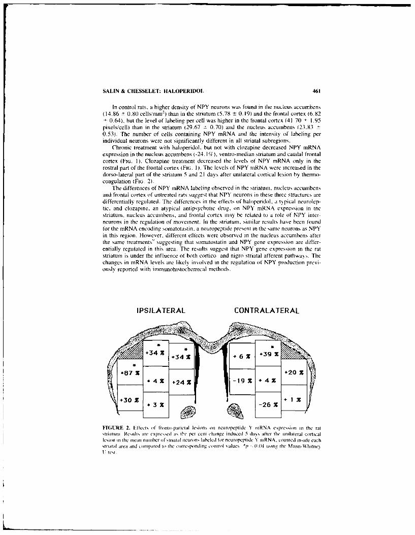
SALIN & CHESSELET: HALOPERIDOL 461
In control rats, a higher density of NPY neurons was found in the nucleus accumbens(14.86 - 0.80 cells/mm2) than in the striatum (5.78 ± 0.19) and the frontal cortex (6.82± 0.64), but the level of labeling per cell was higher in the frontal cortex (41.70 - 1.95pixels/cell) than in the striatum (29.67 - 0.70) and the nucleus accumbens (23.83 -0.53). The number of cells containing NPY mRNA and the intensity of labeling perindividual neurons were not significantly different in all striatal subregions.
Chronic treatment with haloperidol. but not with clozapine decreased NPY mRNAexpression in the nucleus accumbens (-24.1%), ventro-median striatum and caudal frontalcortex (FIG. 1). Clozapine treatment decreased the levels of NPY mRNA only in therostral part of the frontal cortex (FI(. 1). The levels of NPY mRNA were increased in thedorso-lateral part of the striatum 5 and 21 days after unilateral cortical lesion by thermo-coagulation (FIG. 2).
The differences of NPY mRNA labeling observed in the striatum, nucleus accumbensand frontal cortex of untreated rats suggest that NPY neurons in these three structures aredifferentially regulated. The differences in the effects of haloperidol. a typical neurolep-tic, and clozapine. an atypical antipsychotic drug. on NPY mRNA expression in tnestriatum, nucleus accumbens. and frontal cortex mav be related to a role of NPY inter-neurons in the regulation of movement. In the striaum. similar results have been foundfor the mRNA encoding somatotastin, a neuropeptide present in the same neurons as NPYin this region. However, different effects were observed in the nucleus accumbens afterthe same treatments9 suggesting that somatostatin and NPY gene expression are differ-entially regulated in this area. The results suggest that NPY gene expression in the ratstriatum is under the influence of both cortico- and nigro-striatal afferent pathways. Thechanges in mRNA levels are likely in,,olved in the regulation of NPY production previ-ously reported with immunohistochemical methods.
IPSILATERAL CONTRALATERAL
+07 X +20X
FIGURE 2. Effects of tronto-parietal lesions on neuropcptide N niRNA expression in the ratstriaturn. Results are expressed as the per cent change induced 5 days after the unilateral corticallesion in the mean number of striatal neurons laheled for neuropeptide Y inRNA. counted inside eachstriatal area and compared to the corresponding control v alues. *p -- 0.(1 using the Mann-WhinceU. teNt

462 ANNALS NEW YORK ACADEMY OF SCIENCES
REFERENCES
I. ADRIAN, T. E., J. M. ALLEN, S. R. BLOOM, M. A. GHATEI, M. N. ROSSER, G. W. ROBERTS.T. J. CROW, K. TATEMOTO & J. M. POLAK. 1983. Neuropeptide Y distribution in humanbrain. Nature 306: 584-586.
2. ALLEN, Y. S., T. E. ADRIAN, J. M. ALLEN, K. TATEMOTO, T. J. CROW, S. R. BLOOM & J.M. POLAK. 1983. Neuropeptide Y distribution in the rat brain. Science 221: 877-879.
3. SMITH, Y & A. PARENT. 1986. Neuropeptide Y-immunoreactive neurons in the striatum of catand monkey: morphological characteristics, intrinsic organization and co-localization withsomatostatin. Brain Res. 372: 241-252.
4. VUILLET, J., L. KERKERIAN. P. SALIN & A. NIEOULLON. 1989. Ultrastructural features ofNPY-containing neurons in the rat striatum. Brain Res. 477: 241-251.
5. VUILLET, J., L. KERKERIAN, P. KACHIDIAN, 0. BOSLER & A. NIEOULLON. 1989. Ultrastruc-tural correlates of functional relationships between nigral dopaminergic or cortical afferentfibers and neuropeptide Y-containing neurons in the rat striatum Neurosci. Lett. 100: 99-104.
6. KERKERIAN, L., P. SALIN & A. NIEOULLON. 1990. Cortical regulation of striatal neuropeptideY (NPY)-containing neurons in the rat. Eur. J. Neurosci. 2(3): 181-189.
7. KERKERIAN, L., P. SALIN & A. NIEOULLON. 1988. Pharmacological characterization ofdopaminergic influence on neuropeptide Y immunoreactivity by rat striatal neurons. 26:809-817.
8. HEILIG. M.. L. VECSEI, C. WAHLESTEDT, CH. ALLING & E. WIDERLOV. 1990 Effects ofcentrally administered neuropeptide Y (NPY) and NPY 3.3 , on the brain monoaminergicsystems of the rat. J. Neural. Transm. 79: 193-208.
9. SALIN, P., M. MERCUGLIANO & M. F. CHESSELET. 1990. Differential effects of chronictreatment with haloperidol and clozapine on the level of preprosomatostatin mRNA in thestriatum, nucleus accumbens and frontal cortex of the rat. Cell. Molec. Neurobiol. 10:127-144.

Regulation of Neuropeptide Y in Neuraland Cardiac CultureK.L. MAREK" AND R. E. MAINS b
"Department of Neurology333 Cedar Street LCI
Yale University School of MedicineNew Haven, Connecticut 06510
and'Department of Neuroscience
Johns Hopkins School of Medicine725 North Wolfe Street
Baltimore, Maryland 21205
We have examined the regulation of Neuropeptide Y (NPY) mRNA levels, peptidesynthetic rate and peptide content in primary cell cultures from rat superior cervicalganglion (SCG)' 2 and atrium. 3 SCG and atrial cultures were established from 1-2-day-old rats and cultures were maintained in serum-free medium for 2-3 weeks. NPY wasstable in spent medium for at least 48 hours. Since NPY is colocalized with norepineph-fine (NE) in SCG,1 we have compared the regulation of NPY and NE synthesis' and NPYand tyrosine hydroxyJase (TH) mRNA 4 in these neurons after treatment of cultures withseveral adrenergic agents. These data show that several important regulatory effects canbe seen in the primary neuronal cultures (TABLE 1). First, despite the marked changes inNE synthesis and accumulation, the cultures never show any change in NPY synthesis andaccumulation in response to a variety of adrenergic drugs. Second, the acute and chroniceffects of these drugs on NE synthesis clearly differed. In particular the chronic effects ofdesipramine and clonidine were significant, but no acute effects on NE synthesis wereseen.
Factors which regulate signal transduction pathways frequently after peptide synthesisand secretion. As shown in TABLE 2, treatment with cAMP increased NPY and THmRNA levels without changing NPY or NE synthesis and accumulation. PMA reducedNPY and NE synthesis and accumulation, but had little effect on NPY or TH mRNAlevels. In general chronic treatment of SCG cultures with cAMP, PMA and dexametha-sone regulated NPY and NE synthetic rates and NPY and TH mRNA levels in parallel.Since the changes in synthetic rate (NE and NPY) and mRNA levels (TH and NPY) werenot always in agreement, the mechanism of peptide and transmitter regulation probablyinvolves interesting posttranscriptional regulatory events.
There is mounting evidence that NPY plays an important role in the mammaliancardiovascular system. We investigated the effect of cAMP. PMA and Dex on NPYsynthesis in primary atrial cultures. Treatment with cAMP and dexamethasone mnarkedlyreduced NPY content in spent medium and reduced NPY mRNA levels in cell extracts.In contrast treatment with PMA increased NPY content in spent medium and increasedNPY rnRNA in cell extracts. These data demonstrate that the regulation of NPY inneuronal and atrial culture is tissue-specific.
463

464 ANNALS NEW YORK ACADEMY OF SCIENCES
TABLE 1. Jffect oI Adrenergic Drugs on Neurotransmitter Synthetic Rates"
Norepinephrine
Acute Chronic NPY Action on Catecholamines
Reserpine down down nc block ',otake into granulesTyramine down down nc fill gransb!ps with octopamnineDesipramine nIC down nc block presyi.lntic uptake of NECionidine nc down nc ot. agonistYohimbine nc nc nc of, antagonist
'Inc =no chan,
TABLE 2 SCG Cultures: Parallel Effects on NE and NPY"
Synthesis mRNATreatment NPY,NE NPY.TH
cAM.P nc 166%PMA 40% ncDexamethasone 1339c nc
"nc = no change.
REFERENCES
1. MAxiK K. L. & R. E. MAINS. 1981). Biosynthesis. development, and regulation of neuropep-tide Y in superior cerv ical ganglion cu!,ur,;. J. Neujochem. 52: 1 M- 18 16.
2. DICKERsoN. 1. M., 3. E. iIxoN & R. E. MAINS. 1987. Transfected human neuropeptide YLDNA expression in mouse pitr ells. J. Biol. Chem. 28: 3646-13653.
3. Ttiw i.r.. E. A.. K. L. MAREK & B. A. - PPER. i 969. Tissue ,.-'ific regulation of peptidvl-glvcine a-aniidating monooxygenase e> pic:,son. Endocrinology 125: 2279-2288&
4. TANK. A. W., P. CutzFt.i,.A & L. HAM. 1096. Induction of mRNA for tyrosine hydroxylase )vcyclic AMP and g!- cocorticoids in a rat pheochromocytomna cell line. Mol. PharmacL.+30: 497-503.
5. WAEHI-R. B. J. AUGERr. R. CORDER, D. EvFQuoz. .1. Nt. shZ 'F.R, R. GAILLARD & H4.BRLNNI.R. 19XX. An'. .i H-ypertens. 1: 193 -199.

Clinical Evaluation of Neuropeptide Yas a Plasma Marker of Tumors
Derived from Neural Crest
MASAHIKO SONE, TORAICHI MOURI,
KAZUHIRO TAKAHASHI, KEIICHI ITOI, YUTAKA HAYASHI,"
MAKOTO OHNEDA, OSAMU MURAKAMI,
AND KAORU YOSHINAGASecond Department of Internal Medicine
"Department of Pediatric SurgeryTohoku Universit, School of Medicine
Sendai 980. Japan
Neuropeptide Y (NPY) is a 36 amino acid peptide, which was originally isolated from theporcine brain.' NPY is known to exist in the central nervous system, sympathetic ganglia,peripheral sympathetic nerves and adrenal medulla in high concentrations. 4 High con-centrations of NPY in plasma and tumor tissues of pheochromocytoma (Pheo) and gan-glioneuroblastoma patients had already been reported. 5
.6
To evaluate NPY as a plasma marker of tumors, we measured plasma and tissueimmunoreactive (IR)-NPY levels by radioimmunoassay (RIA) as described elsewhere. 7 '
The serial dilution curves of the extracts of both the normal mixed plasma and the plasmaof a Pheo and a neuroblastoma were parallel to the standard dilution curve (FiG. I).Plasma IR-NPY levels in 17 of 21 Pheo were higher than 290 pg/ml, which is the upperlimit of the plasma IR-NPY levels in normal adult subjects. Plasma IR-NPY levels in 19of 21 ganglioneuroblastomas or neuroblastomas (NB-group) were higher than 350 pg/ml,which is the upper limit of the plasma IR-NPY levels in normal children. Plasma IR-NPYlevels in 2 of 4 cases of rhabdomyosarcoma were very high (1611 and 3632 pg/ml)(TABLE I). High levels of IR-NPY were also found in tumor tissues of Pheo (0.025-95.3R±g/g wet tissue, n = 11) and NB-group (0.07-5.33 'g'g wet tissue, n = 5). An analysisusing reverse phase high performance liquid chromatography revealed that IR-NPY inplasma extracts and tumor tissue extracts of Pheo and NB-group was eluted in an identicalposition to synthetic human NPY in most cases (data not shown). Notably. plasmaIR-NPY levels in patients with chronic renal failure were also high suggesting that therenal clearance of NPY in these patients is decreased.
These findings indicate that, in case of normal renal function, NPY is a useful plasmamarker of tumors originating from the neural crest. Further investigation is required as tothe mechanism of the NPY-gene expression in these tumors.
465

466 ANNALS NEW YORK ACADEMY OF SCIENCES
100 Peptid VY 20.7%Pancreatic polypoptide
Avian <0.001%Human <0.001%
Porcie VP <0.001%Porcine Secretin <0.001%Human glu.aaon <0.001%Substance P <0.001%
B/Bo Antensin I <0.001%
(M) Somatostati, <0.001%
V50
NPY
S I I I I
0 t t l t I3.9 7.5 15.6 31.2 62.5 125 250 500 1000
NPY pq/tube
FIGURE 1. A standard curve of NPY (0), serial two-fold dilution curves (0) of a normal mixedplasma extract, a pheochromocytoma plasma extract, a neuroblastoma plasma extract and crossreactivities.
TABLE t. Plasma IR-NPY Concentrationsa in Various Diseases
N IR-NPY (pg/ml)
Normal subjects 21 151 ± 29Essential hypertension 35 175 ± 48Pheochromocytoma 21 1636 ± 3902Neuroblastoma group 21 1748 ± 1827Rhabdomyosarcoma 4 1611 ± 3632Endocrine diseases 39 138 ± 31Brain tumors 4 145 ± 30Carcinomas 1! 199 ± 60Chronic renal failure
without hemodialysis 8 330 ± 63with hemodialysis 10 374 ± 80
"Values are represented as the means ± SD.
REFERENCES
1. TATEMOTO, K., M. CARLQUIST & V. MUTT. 1982. Nature 296: 659-660.2. ADRIAN, T. E., J. M. ALLEN, S. R. BLOOM, M. A. GHATEI, M. N. POSSER. G. W. ROBERT,
T. J. CROW, K. TATEMOTO & . M. POLAK. 1983. Nature 306: 584-586.

SONE et aL: PLASMA MARKER OF TUMORS 467
3. LUNDBERG, J. M., L. TERENIUS, T. HOKFELT, C.-M. MARTLING, K. TATEMOTO, V. MUTT,J. POLAK, S. R. BLOOM & M. GOLDSTEIN. 1982. Acta. Physiol. Scand. 116: 477-480.
4. ALLEN J. M., T. ADRIAN, J. POLAK & S. R. BLOOM. 1983. J. Auton. Nerv. Syst. 9: 559 -563.5. ADRIAN, T. E.. J. M. ALLEN, G. TERENGHI, A. J. BACARESE-HAMILTON, M. J. BROWN.
J. M. POLAK & S. R. BLOOM. 1983. Lancet 2: 540-542.
6. CORDER, R., P. C. EMSON & P. J. LOWRY. 1984. Biochem. J. 219: 699-706.
7. TAKAHASHI, K., T. MOURI, K. ITO), M. SONE. M. OHNEDA, 0. MURAKAMI, M. NOZUKI.Y. TACHIBANA & Y. YOSHINAGA. 1987. J. Hypertens. 5: 749-753.
8. TAKAHASHI. K.. T. MOURI, K. Iroi, M. SONE, Y. HAYASHI, 0. MURAKAMI, M. OHNEDA.
R. OHI & K. YOSHINAGA. 1989. Endocrinol. Japon. 36(5): 627-633.

Neuropeptide Y Regulation of LHRHRelease in Ewe Median EminenceImmunocytochemistry, Tissue Content, and
In Vivo Analysis"
J. P. ADVIS." C. D. CONOVER," J. K. McDONALD,'
AND R. 0. KULJIS"
"Department of Animal SciencesRutgers University
New Brunswick, New JerseN 08903'Department of Anatomv and Cell Biology
EmorY UniversityAtlanta, Georgia 30322
"Department of NeurologyUniversitv of Iowa
Iowa City., iowa 52242
Luteinizing hormone tLH)-releasing hormone (LHRH) neurons are the main element ofthe hypothalamic hypophysiotropic reproductive network. While synchronization withinthis network might be achieved through synapses among LHRH neurons, modulation ofits activity preceding the preovulatory surge of LHRH is probably related to its neuro-transmitter and/or peptidergic synaptic inputs. Stimulatory and inhibitory inputs exist atthe level of LHRH cell bodies in the medial preoptic nucleus and medial basal hypothal-amus (MBH), and at the level of their terminals in the median eminence (ME). Thepreovulatory surge of LHRH is probably related to a temporal predominance of facilita-tory over inhibitory inputs to this LHRH neuronal network.
In rats and hamsters, NPY-containing perikarya are located in the MBH and NPY-containing axons converge toward the ME. 1.2 Furthermore, both a stimulatory and aninhibitory role of NPY on LHRH release has been reported in rats and rabbits. 3 - 7 Forexample, the high circulating levels of serum LH observed after castration are dramati-cally decreased by NPY administered in the third ventricle of male and female rats.3-6NPY administration also decreases pulsatile LH secretion in ovariectomized rats.' Thirdventricular administration of NPY also induces a prolonged inhibition of LH in intact malerats, 6 as does NPY perifusion of the MBH in ovariectomized females and intact malerabbits. 7 In contrast, the latter procedure increases in vivo L-R-1 release in intact femalerabbits. 7
In the ewe, in vivo MF-NPY release, assessed by push-pull cannula (PPC) sampling.is higher in estrous than during the luteal or anestrous period (FIG. I). Perfusion of NPY(5 LgI200 p.l perfusate) through the PPC in the posterior-lateral ME increased in vivoLHRH release in estrous but not in luteal or anestrous ewes. No effect was observed whenNPY was perfused in the anterior-lateral ME. On the other hand, our immunocytochem-
"Supported by New Jersey Agricultural Experimental Station-Hatch Grant 06108 and U.S De-partment of Agriculture Grant 89-37240-4587 to J. P. Advis.
468

ADVIS et al.: LHRH RELEASE IN ME 469
ical studies indicate that NPY-containing perikarya are located in the arcuate (arn) area,while NPY-containing axons converge toward the ME and descend into the infundibulum(inf) of the ewe (results not shown). Finally, discrete microdissection of these areasfollowed by specific RIA indicate that NPY content is higher in the ME (stalk and inf/arn)than in the lateral peri-arn (105 + 20 vs 30 ± 6 pg/Lg protein, mean - SEM, n = 25).Similarly, NPY content in the ME (stalk and inf/arn) is highest in late luteal and lowestin mid-luteal ewes (132 ± 35 vs 82 ± 8).
A stimulatory and possibly synaptic NPY-LHRH interaction, restricted to the poste-rior-lateral ME and probably modulated by estradiol, might trigger the natural preovula-tory surge of LHRH in the ewe. This suggestion is based on our immunocytochemical andin vivo NPY and LHRH data, specifically: a) the similar projection areas of NPY- and
BASAL NPY-STIMULATEDME-NPY ME-LHRH
I RELEASE RELEASE
(4)250_ 250
P value Vs (3)preceding groups
* PO.05** P<O.O1
(3) () of ewes
((5)125- ] control period 125
4 (2)
0 - 0A L E A L E
REPRODUCTIVE PHASE
A= anestrous ; L= luteal ; E= estrous
FIGURE 1. Median eminence (ME) in vivo release assessed by push-pull cannula (PPC) sampling(pg/200 W.1 PPC perfusate/20 min during 120 min).
LHRH-containing axons toward the ME provide opportunities for potential synaptic in-teractions; b) NPY increases in vivo ME-LHRH release only when serum estradiol (E2)
is high and serum progesterone (P4) is low, but is ineffective when E2 is low and P4 ishigh, or when serum levels of both steroids are low (anestrus); c) in vivo basal ME-NPYrelease is higher in estrous than in luteal ewes; and d) ME content of NPY is highest atthe end of the luteal period of cycling ewes.
That E2 might be involved in the LHRH response to NPY is also supported by resultsfrom the rat."' For example, NPY-induced release of LHRH from MBH-ME fragmentsis greatly enhanced by E2 but not by P4.8 - 'n Pharmacological doses of P4 actuallydecrease the E2-facilitated NPY-induced release of ME-LHRH.9 In addition, the effectsof NPY on LHRH have also been associated with alterations in LHRH content in various

470 ANNALS NEW YORK ACADEMY OF SCIENCES
hypothalamic areas in the rat. For example, E2 decreases NPY content, while P4 inducedonly a transient decrease and a subsequent rise of NPY content in the interstitial nucleusof the stria terminalis, the arcuate, and the preoptic area. 12
Thus, a stimulatory and possibiy synaptic NPY-LHRH interaction, restricted to theposterior-iateral ME and probably modulated by estradiol. might trigger the natural pre-ovulatory surge of LHRH in the ewe.
REFERENCES
I1. SABATINo. F. D_. J. M. MURNANE, R. A. HOFFMAN & J. K. McDONALD. 1987. J. Comp.Neurol. 257: 93-104.
2. CALKA J., & J. K. McDONALD. 1988. Soc. Neurosci. Abstr. 187.3. MCDONALD. J. K., M. D. LuMPKIN. W. K. SAMSON & S. M. MCCANN. 1988. 82: 561-5644. MCDONALD, J. K., MI. D. LuMPKIN & L. V. DEPAOLO. 1989. Endocrinology 125: 186-191.5. KALRA. S. P. & W. R. CROWLEY. 1984. Life Sci. 35: 1 173-1 176.6. KERKERIAN. L. J. Guy, G. LEFEjVRE & G. PELLETIER. 1985. Peptides 6: 1201-1204.7. KHORRI M, () . K. Y. F. PAU & H. G. SPIES. 198/ Neuroendocrinology 45: 290-B'4.8. CROWLEY, W. R. & S. P. KALkA. 0987. Neuroendocrinology 46: 97-99.9. SABATINO. F. D., P. COLLINS & J1. K. MCDONALD. 1989. The Endocrin~e Society. p. 76.
10. KI-ORRAM. 0.. F. K. Y. PAUl & H. G. SPIES. 1988. Peptides 9: 411-417.11. SABATINO. F. D., P. COLLINS & J. K. MCDONALD. 1989. Endocrinology 124: 2089-2098.12. CROWLEY, W. R. R. E. TESSEL. T. L. O'DONO-UE. B. A. ADLER & S. P. KALRA. 1985.
Endocrinology 117: 1151-1 156.

A Role for Neuropeptide Y in theMediation of Glucocorticoid-InducedChanges in Hypothalamic Function"
ROGER CORDER, FRAN4OIS P. PRALONG, DORA TURNILL,
AND ROLF C. GAILLARD
Neuroendocrine UnitDepartment of Medicine
University Hospital1211 Geneva 4, Switzerland
Immunocytochemical studies have shown abundant NPY nerve terminals in a number ofhypothalamic areas and nuclei. : The two main sources of this NPY innervation areprojections from brainstem catecholaminergic cell groups' and a dense population ofneuronal cell bodies located in the arcuate nucleus. 2 In both the brainstem and thehypothalamus, the majority of NPY perikarya contain a high density of nuclear gluco-corticoid receptors (GR) indicating a direct sensitivity to changes in adrenocortical'unction3. This may be of particular relevance to the regulation of the hypothalamo-pituitary-adrenal axis as corticotropin releasing hormone neurones in the paraventricularnucleus (PVN) receive a dense NPY innervation from both the brainstem and arcuatenucleus (ARC). '2 To evaluate the influence of glucocorticoids on the function of NPYneuronal systems in the hypothalamus, we have performed a number of differentinvestigations. 4 5 whose findings are briefly summarised here.
Primary cultures of hypothalamic neurones prepared from neonatal rats were main-tained for one week in serum free medium containing different concentrations of dexa-methasone. Intracellular levels of NPY demonstrated concentration-dependent increases.with the content being 2-3-fold higher than control after treatment with dexamethasoneat 10- and 10- 7 (FiG. I).' A positive effect of glucocorticoids was also observed whenfemale Wistar rats (200 g) were administered 100 pLg dexamethasone i.p. per day for 5days, tissue levels of NPY being increased by 117% in the medial basal hypothalamus(p <0.002).
4
However, a study in male Wistar rats designed to further clarify the effect of gluco-corticoid excess produced a less marked response. Microdissection of the ARC from ratssacrificed 10 days after receiving a depot preparation of betamethasone (BM, 1.5 mg) didnot show significant increases in NPY (BM 55. I t 1.9 fmol/p g protein, control 49.8 ±4.6). But, in the same study, adrenalectomy (ADX) performed 12 days before sacrificewas found to significantly reduce levels (37.8 - 2.7, p <0.02). A similar pattern of NPYvalues was observed in the PVN of these animals (control 37.4 ± 3. 1. BM 36.8 ± 1.8.ADX 28.8 ± 3.3 p <0.05 v. control). In contrast. norepinephrine (NE) levels in the PVNwere not significantly altered by ADX but showed a significant depression after BMadministration (NE fmol/iLg protein; control 753 ± 48, BM 568 ± 58 p <0.05, ADX 642+ 66).
"This work was supported by the Swiss National Research Foundation (3.091.087).
471

472 ANNALS NEW YORK ACADEMY OF SCIENCES
Microdissection of II nuclei and areas from 19 human hypothalami collected atpostmortem showed a wide range of NPY values in the infundibular nucleus as well as inother hypothalamic nuclei. 5 Retrospective analysis of glucocorticoid administrationshowed that this was not the cause of the differences. However, analysis of the variouscauses of death revealed that NPY levels were significantly higher in the infundibular.ventromedial (VM) and paraventricular nuclei of subjects suffering from chronic respi-ratory failure (> i0 days). a condition associated with sustained distress (Fio. 2). More-over, NPY values in the infundibular nucleus demonstrated a good correlation with valuesfor the VM (r = 0.89) and PVN (r = 0.84). Whether, as in the rat.2 this results fromneuronal projections from the infundibular awaits further investigation.
[romi dtese different studies, it may be concluded thq! the function of hypothalamicNPY neurones is not simply related to circulating glucocorticoid levels but is also de-pendent on the simultaneous activation of other interconnecting neuronal pathways oc-curring during situations of chronic stress. For example, Sahu and colleagues have re-ported increases in the NPY levels of the ARC and PVN when rats have been deprived offood for 3 to 4 days.6 Although it was concluded that this was the result of altered feedingbehaviour,6 it cannot be excluded that a stress of this duration would not also produce asimilar change in NPY levels.
Microinjections of NPY into the third ventricle or directly into specific hypothalamicareas have been reported to produce a number of effects ascribed to specific functionswithin the hypothalamus. 7 These include phase shifting of circadian rhythms, stimulationof ACTH secretion, increasing food intake, alterations of blood pressure, and both inhi-bition and stimulation of LH secretion depending on the hormonal background at the timeof NPY administration. Therefore, the interaction of glucocorticoids with hypothalamicGR-containing NPY neurones could directly modulate the neural circuitry responsible forthe physiological regulation of these different neuroendocrine responses.
400
300
200
100
0
control 1' 11 1010 10*9 10 "8 10 7
Dexamethasone [M]FIGURE 1. Effect of dili-t! ' -!'mhasone concentrations on the NPY content of cultured rathypothalamic neurones maintained for one week in serum free medium (*p <0.05. ***p <(U). v
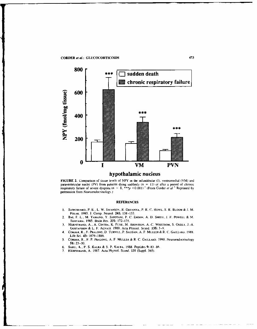
CORDER et al.: GLUCOCORTICOIDS 473
800W- sudden death
Uchronic respiratory failure
~'600
~400
Z 200
0 VM PVN
hypothalamic nucieusFIGURE 2. Comparison of tissue levels of NPY in the infundibular (1). ventromedial (VM) andparaventricular nuclei (PV) from patients dying suddenly (n = 1) or after a period of chronicrespiratory failure of severe dyspnea In =8. *** <0.001).5 (From Corder et al. Reprinted bypermission from Neuroendocrinology.)
REFERENCES
I . SAWCHENKO, P. E., L. W. SWANSON. R. GRZANNA, P. R. C. HOWE, S. R. BLOOM & 1. M.POLAK. 1985. J. Comp. Neurol. 241: 138-153.
2. BAt, F. L., M. YAMANO. Y. SEHOTANI, P. C. EMSON. A. D. SMITH. J. F. POWEtLL & M.TOHYAMA. 1985. Brain Res. 331: 172-175.
3. HARFSTRAND, A., A. CINTRA, K. FUXE, M. ARONSSON. A.-C. WIKSTROM. S. OKRET. 1.-A.GUSTAFSSON & L. F. AGNATI. 1989. Acta Physiol. Scand. 135: 3-9.
4. CORDER. R., F. PRALONG, D. TURtNILL. P. SAUDAN. A. F. MULLER & R. C. GAILLARD. 1988.Life Sci. 43: 1879-1886.
5. CORDER. R., F. P. PRALONG, A. F. MULLER & R. C. GAILLARD. 1990. Neuroendocrinology51: 23-30.
6. SAHU. A.. P. S. KALRA & S. P. KALRA. 1988. Peptides 9: 83-86.7. HARFSTRAND, A. 1987. Acta Physiol. Scand. 131 (Suppl. 565).

Comparison of Hypotension-InducedNeuropeptide Y Release in Rats
Subjected to Hemorrhage, Endotoxemia,and Infusions of Vasodepressor Agents"
ROGER CORDER. FRAN(;OIS P. PRALONG. AND
ROLF C. GAILLARD
Neuroendocrine UnitDepartment (# Medicine
University Hospital1211 Geneva 4. Switzerland
Consistent with its potent vasoconstrictor properties and its ability to enhance pressorresponses of adrenergic agonists,' early investigations of the peripheral distribution ofNPY identified perivascular sympathetic neurones as one of the major sources ofimmunoreactivity. 2 In addition, NPY was localised to a subpopulation of adrenal med-ullary cells and to intrinsic nonadrenergic neurones of the heart and gastrointestinal tract 2Subsequent studies have attempted to definc the physiological stimuli evoking the releaseof NPY-like immunoreactivity into the circulation from either the adrenal medulla orneuronal sources.' Because of the vasoconstrictor action of NPY. it is of particularphysiological interest when release occurs during activation of the sympathetic nervoussystem with simultaneous vasoconstriction of peripheral vascular beds. such as duringhemorrhagic hypotension. 4" In contrast, no change in plasma levels of NPY was ob-served in a canine model of endotoxin-induced shock despite large increases in circulatingcatecholamine concentrations. 6 Experimental models of septic shock as well as the clin-ical condition are characterised by a reduced responsiveness to pressor amines. 7 If NPYrelease from perivascular nerves regulates the response of vascular smooth muscle tonorepinephrine, inadequate or decreased release of NPY could in part explain the reducedfficacy of vasoconstrictor agcw., during ondoto:emi The sttvdie summarised helo,
have been performed to further evaluate the relationship between hypotension and NPYrelease.
Earlier observations demonstrated the important influence of psychological stress onplasma levels of NPY in the rat.' Hence to avoid the cftect of psychogenic ;timuli, thedifferent manipulations leading to hypotension were carried out in anesthetised rats. Thefirst of these studies compared circulating levels of NPY and catecholamine.; following 30and 60 min of either hemorrhage or endotoxin-induced hypotension (FlG. I ). In agreementwith the results of other groups suggesting a generalised activation of perivascular sym-pathetic neurones, 4 "5 hemorrhage produced marked increases in plasma NPY levels ,2.5-and 3.5-fold increases at 30 and 60 min respectively). In contrast, NPY values duringendotoxemia were not significantly different from control values despile comparabledecreases in MABP to the hemorrhage group. Plasma catecholamine levels were in-
"This work was supported by the Swiss National Research Foundation (3.091 .087). the Geig-Jubilaiums Foundation. and the Sandoz Foundation.
474

ANNALS NEW YORK ACADEMY OF SCIENCES 475
creased by both types of shock (Fi(;. 1). The very high levels of E particularly afterendotoxin administration suggest that a large component of the increase in NE is also ofadrenal medullary rather than of neuronal origin.
Previously it was found that NPY infusion can restore the pressor response to nor-epinephrine in nonhypotensive endotoxemia.7 The very high circulating concentrations ofcatecholamines but apparent absence of increased NPY release after endotoxin adminis-tration, even with marked hypotension, led us to evaluate whether infusing NPY reversedthe fall in MABP during endotoxin shock. Endotoxin was administered to anesthetisedrats as a bolus (2.5 mg/I(X) g) at 0 min, and 10 min later a I h i.v. infusion of NPY (10pmol/100 g/min) or vehicle was started. Although administration of this dose of NPY waswithout effect on MABP in control rats, it produced a partial reversal of the fall in MABP
in endotoxin-treated rats to - 13 - 4 and - 14 ± 6 mmHg after 30 and 60 min infusion(p <0.01), compared to -34 ± 5 and -35 t 6 in rats receiving vehicle. Increasing thedose of NPY to 30 pmol/100 gimin had no greater effect. Infusion of norepinephrine (30ng/l00 g/mir), instead of NPY, had no effect on the time course of changes in MABPduring endotoxin shock even though it raised MABP by 30-40 mmHg in control animals.
To investigate the apparent suppressed NPY release during endotoxemia, relative tothe fall in blood pressure. we compared the effect of infusing different depressor agentsover a 30-min period to anesthetised rats (FIG. 2). In this study, levels of NPY were notsignificantly different from control values after CGRP or nitroprusside infusions. Incontrast, infusions of salbutamol and VIP significantly increased plasma concentrations ofNPY (p <0.05 and -p0.01).
In summary,. endotoxin-induced hypotension in the rat differs from hemorrhagicshock in that NPY release is not markedly increased. However. reduced responsivenessto endogenous and exogenous7 catecholamines during endotoxemia can be partially over-come by NPY infusion. The results obtained after infusion of different vasodilators
4.0..
2 endotoxin1 2.0 - hemorrhage
0.FIGURE I. Comparison of changes in plasma
levels of NPY, norepinephnne (NE) and epineph-eL 0.4z .rine (E) after endotoxin (n = 6, bolus at 0 min of
0.2 I mg:Il(X) g E. 'oli lipopolysacchande Ot 11:B410.0 Ii/co bibhoratories. followed by infusion of 10
|g/lX g/nain) or 25% hemorrhage (n = 5). corn-s.0 pared with control rats In = 5). Male Wistar rats
(290-320 g) were anesthetised by i.p. injection of2.0 4 mg/ltX) g pentobarbitone and 5 mg/lOO g ke-
tamne. Mean arterial bhxd pressure (MABP.z 1.0 mmHg) at 30 and N0 rain was decreased by 30I t
0.5.______ mand 33 ± 6 during endotoxin-induced shock: and... by 43 - 5 and 45 ± 6 during hemorrhagic hy-
20.0 pnol n. NPY was measured by direct radioim-= !0.... * nunoassay Statistical analysis was performed by4.0 ANOVA *p -0 05, ***p <0.001 v. correspond-E 2.0 Ing control sample; + p 0.05. + + +p <'0.(OOI
1.0.- v corresponding endotoxin sample.
s4
basal + 30 min + 60 min

476 CORDER el al.: HEMORRHAGIC HYPOTENSION
FIGURE 2. Effect of hypotension induced by dif-ferent vasodilatoir agents on plasma levels of NPYmeasured by radioinirnunoassa . Male Wistar rat,,(280-320 g) were anesthetised by ip. injection of
** 10 mg/ 1(X) g ketarnine and 0.5 mg/l(X3 g midato-1.0 - 'am, and infused an i. . maintenance mixture of 4.89
mg/l(X) gi'h ketarme and 0.2 mg/l(X) g/h midaio-lam, Vehicle (Ve. l71 human serum albumin in
E 0.5 -0.91/, s~line. nt 10. calcitonin gene-related pep-tide (CGRP. 25 pmol/l00) g/mm., n = 10). sodium
E nitroprusside (NP. I5 f(ig;I00( g.'min. n l 10. sal-T b~utamol (SB. (1.2 f.ig/l0() g/fin. n =8) and vaso-0.2 active intestinal peptide (VIP, 25 pmol/l(X) g/min.
z n 10) were infused for 3(1 min. Highly significantfalls in MABP (p <0.(X)!) of 34 -2. 40 _- 5, 22
0.!4 and 48 :t4 mml-lg for CGRP. NP. SB and VIPVe CGRP NP SB VIP respectively were observed after 5 min infusion. The
depressor effects were sustained throughout the in-fusion period with corresponding reductions of 44t2. 50 _-3. 37 -_7 and 45 ± 5 mnmHg being recordedat + 30 min (*p < 0.0(5, Ip <0.0 1).
demonstrate the complexity of the regulation of NPY release in rats subjected to differentmanipulations leading to hypotension.
REFERENCES
I . DAHLOF. C.. P. DAHLOF & J. M. LUNDBERG. 1985. Eur. J. Pharmiacol. 109: 289-292.2. SUNDIER. F.. R. HAKANsosI. E. EKBt.AD. R. UDDoMAN & C WAst EDIt~. 1986. m. Rev.
Cytol. 102: 243 -20)3. CASTAGNE-I V.. R. 'ORDER. R. GAILLtARD & P. MORME-DE. 1987. Regul. Pep!. 19: 55-03.4. MORRIS. M.. V. KAPOOR & J. CHAL MERS. 1987. J. Cardiovasc. Pharmacol. 9: 541-545.5. RUDEHtt t, A., M. OLCN. A. Sot LEVt. B. H-AMBIERGER & J. M. LuNDBERG. 1987. Acta
Physiol .Scand. 131: 5 17-523.6. WAtSON, J. D.. M. R. J. SURY. R. CORDER. R. CARSON. P. MI. BOLt~oI X, P. J. LoWrRN. G.
M. BiESSER & C. J. HINDS. 1988. J. Endocrinol. 116: 41-1-426.7. EvuQuoz. D., B WAEBER. R. CORDER. J. NLIURLRR R. GAItLARD & H. R. BRLNNER.
1987 Life Set. 41: 2573-2580.
Food Deprivation ModulatesNeuropeptide Y Gene Expression in the
Murine HypothalamusSTREAMSON C. CHUA, JR"
Department of Human Behavior and MetabolismRockefeller Unive, sitv
New York, New York 10021
The behavioral effects of central administration of Neuropeptide Y (NPY) suggest that itparticipates in the control of food intake. Increased amounts of immunoreactive NPY havebeen found in the hypothalamus of two models of caloric deficit: the food-deprived rat'and the rat with streptozotocin-induced diabetes. 2 Since NPY is synthesized in the arcuatenucleus of the hypothalamus, 3 as well as in noradrenergic neurons of the brainstem whichproject to the hypothalamus, 4 the source of the elevated NPY is uncertain. We reasonedthat detection of fasting-induced changes in NPY messenger RNA would identify the siteof increased peptide synthesis. Quantitative Northern blot analysis of the hypothalami andbrainstems of ad libitum-fed and 24-hour-fasted CFI mice indicates that only the hypo-thalamus exhibits fasting-induced increases in NPY messenger RNA levels.
Our results show that NPY neurons in the hypothalamus of mice are sensitive to fooddeprivation. Other studies have shown that NPY can increase food intake' as well asdecrease energy expenditure. 6 Thus, hypothalamic NPY neurons may mediate someaspects of the physiological and behavioral changes associated with fasting.
REFERENCES
I. SAHU, A., P. S. KALRA, & S. P. KALRA. 1988. Peptides 9: 83-86.2. WILLIAMS, G., J. H. STEEL, H. CARDOSO, M. A. GHATEI, Y. C. LEE, J. S. GILL, J. M.
BURRI, J. M. POLAK & S. R. BLoOM. 1988 Diabetes 37: 763-772.3. BAI, F. L., M. YAMANO, Y. SHIOTANI, P. C. EMSON, A. D. SMITH, J. F. POWELL & M.
TOHYAMA. 1985. Brain Res. 331: 172-175.4. SAWCHENKO, P. E., L. W. SWANSON, R. GRZANNA. P. R. C. HOWE, J. POL.AK & S. R.
BLOOM. 1985. J. Comp. Neurol. 241: 138-153.5. STANLEY, B. G., & S. F. LEIBOWITZ. 1984. Life Sci. 35: 2635-2642.6. HARFSTRAND, A. 1986. Acta Physiol. Scand. 128: 121-123.
"Address for correspondence: Streamson Chua, Jr., Hospital Box 26, 1230 York Avenue, NewYork, NY 10021.
477

Steroid Hormone Regulation of BrainNeuropeptide Y mRNA Levels
P. CAMP AND J. D. WHITE
Division of EndocrinologyState University of New YorkStony Brook, New York 11794
Gonadal steroid ri-gulation of the central nervous system (CNS) and pituitary function isone of the most thoroughly described endocrine feedback systems. Luteinizing hormone(LH), released from the anterior pituitary, acts directly on the ovaries to cause ovulation.The preovulatory LH surge is regulated by complex interactions between several neuro-endocrine factors and the proper steroid hormone environment, i.e.. estradiol (E2 ) andprogesterone (P 4 ). Possible sites of action for E2 and P4 in regulating serum LH levelsinclude the anterior pituitary (the source of LH) and the preoptic area-hypothalamus (thesource of LHRH). For example, there is substantial evidence that E, and P4 affect theLHRH system; however, there are no steroid receptors within LHRH-containing neurons.Thus, steroid hormones are likely to act via a system, or systems, impinging upon LHRHneurons, e.g., NPY.' High concentrations of NPY-rontaining cell bodies are found in thearcuate nucleus of the hypothalamus and the Al region of medulla,2 two brain areas thatare known to project to the preoptic area, to modify LHRH release, and to contain steroidreceptors. NPY does alter serum LH levels3"4 and in vitro release of LHRH' with thespecific effect dependent upon the steroid hormone environment. Therefore. NPY neu-ronal pathways are likely candidates for gonadal steroid hormone regulation of CNS geneexpression.
Initial nuclease protection experiments examined the effect of ovariectomy (OVX) onhypothalamic preproNPY mRNA levels (TABLE 1) indicating that there is no difference inpreproNPY mRNA levels with time postOVX from 7 to 28 days. A second set ofexperiments examined the effect of steroid hormones on hypothalamic preproNPY mRNA(TABLE 1) showing that E2 treatment significantly decreased preproNPY mRNA relativeto the OVX group (p <0.05) and that 6 h of P4 treatment are required to significantlyincrease preproNPY mRNA levels relative to the E,-treated group (p <0.05). The cellularlocalization of preproNPY mRNA was assessed in the arcuate nucleus of E,- and E,/P4-treated OVX rats (TABLE 2) confirming P4 stimulation of hypothalamic preproNPYexpression.
These result:' demonstrate that hypothalamic preproNPY mRNA is regulated by phys-iological steroid levels within a time frame when these steroids are inducing both LHRHrelease and an LH surge. Thus, the multiple steroid effects on the LHRH system may bemediated by steroid effects on a CNS NPY system. Future experiments will examinewhether NPY-containing neurons are capable of responding directly to E. and P,.
478

CAMP & WHITE: STEROID HORMONE REGULATION 479
TABLE i. Nuclease Protection Analysis of Hypothalamic PreproNPY mRNA Levelsfrom OVX and Steroid-Treated OVX Rats"
Experimental System Experimental Group PreproNPY mRNA
Ovariectomy 7 day 1.20 -t 0.281 -;y 0.94 ± 0.21
13 day 0.99 ± 0.2416 day 0.81 ± 0.1619 day 0.99 ± 0.2222 day 1.00 ± 0.2125 day 1.15 ± 0.2328 day 0.90 ± 0.24
Steroid treatment OVX 1.00 ± 0.19E2 0.50 - 0.17EP 4 -3h 0.61 - 0.18E2P 4-6h 1.10 ± 0.06
"PreproNPY mRNA levels were measured using 3 Lg total RNA isolated from individual rats(6/group) that had been OVX 7-28 days or that had been: a) OVX for the duration of the experiment(9 days; OVX), b) OVX 7 days, then E.-treated (3 x 150 jig/mt capsules/rat) 2 days iE 2 . or c OVX7 days, E2-treated 2 days, then P.-treated (4 X 50 mg/ml capsules/rat) for 3 or 6 hours (E.P-3 hand EYP4-6h, respectively). Data is expressed as mean ± SEM of the ratio of preproNPY mRNAto cyclophilin mRNA (cyclophilin mRNA hybridization is used as an internal control). For the OVXexperiment, means then were normalized such that the average of all means equaled 1.0. For thesteroid hormone experiment, data were normalized such that the mean of the OVX group equaled1.0. Data were analyzed by one-way ANOVA and Student-Newman-Keuls' multiple range test.
TABLE 2. In Situ Hybridization for Arcuate Nucleus PreproNPY mRNA"
Steroid Treatment Density Area Density* Area
E, 2.40 2.20 ± 0.2 5.33 ± 0.5E2P4 2.50 2.96 ± 0.4 6.86 ± 0.6
"E2 treatment (3 x 150 Lg/ml capsules) was for 2 days beginning 7 days post-OVX and P4treatment (4 X 50 mg/ml capsules) was for 6 hours on day 2 of E2 treatment. 35-_labeled preproNPYcRNA probe was allowed to hybridize with paraformaldehyde fixed, free-floating 30-gim sectionsthrough hypothalamus. Sites of hybridization were determined by film and emulsion autoradiogra-phy. Film autoradiographs were analyzed by an MCID image analysis system to determine thedensity of hybridization and area of hybridization of cRNA probe over the arcuate nucleus. Valuesfor the E2/P4 animal are different from the E2 animal for every category at p <0.05 using a one-tailedt test.
REFERENCES
1. COEN, C. W., M. C. Rutz DE ELVIRA, K. MACLACHLAN, C. MONTAGNESE, M. J. D. AN-DREWS, K. T. O'BYRNE, i. J. CLARKE & Z. LiPOSITS. 1988. Functional and morphologicalstudies on putative interactions between NPY and LHRH in rats and monkeys. 18th AnnualMeeting of Society for Neuroscience, Nov. 13-18, 1988, Toronto, Ontario, Canada. Abst.#471.5.
2. DE QUJOT, M. E. & P. C. EMSON. 1986. Distribution of neuropeptide Y-like immunoreactivityin rat CNS. 11. lmmunohistochemical analysis. Neuroscience 18: 545-618.
3. CROWLEY, W. R., A. HASSID & S. P. KALRA. 1987. Neuropeptide Y enhances the release ofluteinizing hormone (LH) induced by LH-releasing hormone. Endocrinology 120: 941-945
4. McDONALD, J. K., M. D. LUMPKIN, W. K. SAMSON & S. M. MCCANN. 1985. NeuropeptideY affects secretion of LH and growth hormone in ovariectomized rats. Proc. Nat]. Acad. Sci.USA 82: 561.
5. CROWLEY, W. R. & S. P. KALRA. 1987. Neuropeptide Y stimulates release of LHRH frommedial basal hypothalamus in vitro: modulation by ovarian hormones. Neuroendocrinology46: 97-103.

Hypothalamic Neuropeptide YExpression in Normal and Aberrant
Metabolic Homeostasis
JEFFREY D. WHITE, MARYANN KERSHAW,
GERARD SANACORA, DAVID OLCHOVSKY,
JUDITH A. FINKELSTEIN,- AND MICHAEL BERELOWITZ
Division of EndocrinologyState University of New York
Stony Brook, New York 11794and
"Department of AnatomyNortheastern Ohio Universities College of Medicine
Rootstown, Ohio 44272
The peripheral and central mechanisms that coordinate and control feeding remain poorlydefined. It is now widely believed that neuropeptide Y (NPY) plays an integral role in thenormal control of food intake.' Thus, if NPY neurons are among the central nervoussystem (CNS) systems that initiate feeding, then peripheral metabolic status may directlyinfluence the expression and secretion of NPY. We have investigated the regulation ofhypothalamic NPY expression, as assessed by measuring preproNPY mRNA levels, inthree models of aberrant metabolic homeostasis.
Initially, hypothalamic preproNPY mRNA levels were assessed in animals subjectedto food deprivation for up to 72 hours. 2 These studies demonstrated a time-dependentincrease in hypothalamic preproNPY mRNA levels in food-deprived animals whichreached 2.6-fold above levels found in fed animals (TABLE 1). Using the technique of insitu hybridization, we verified that this change in NPY expression was localized to thearcuate nucleus. Next, hypothalamic preproNPY mRNA levels were examined in a modelof experimental diabetes mellitus, the streptozotocin-treated rat.3 Again, hypothalamicpreproNPY mRNA levels were increased in diabetic animals compared to controls andthis increase could be prevented with insulin replacement (TABLE 1). This increasedexpression was again localized to the arcuate nucleus. Finally, NPY expression wasexamined in genetically obese Zucker "fatty" rats.4 As predicted, preproNPY mRNAcontent was increased specifically in the hypothalamus of obese male and female ratswhen compared to their lean littermates (TABLE 1). Interestingly, when the effects of fooddeprivation on hypothalamic preproNPY mRNA levels were examined in lean and obeseanimals, only lean animals displayed increased NPY expression (TABLE i).
Each of these experimental systems share similar changes in peripheral metabolicstatus, particularly with respect to signals reflecting carbohydrate status. In food-deprivedanimals, plasma insulin and glucose are depressed while glucocorticoids are elevated. Indiabetic animals, plasma insulin is absent, glucose is high, but insulin-dependent glucoseutilization is absent and glucocorticoids are elevated. Similarly, obese "fatty" rats arehyperinsulinemic but euglycemic with elevated serum glucocorticoids. When coupledwith observations of glucocorticoid regulation of NPY in other systems,5 these datasuggest that glucocorticoids stimulate while insulin inhibits NPY expression, as is seen forthe regulation of phosphoenolpyruvate carboxykinase.6 Similarly, glucose may modulateNPY secretion from hypothalamic neurons in the situation analogous to the regulation of
480

WHITE et al.: HYPOTHALAMIC NPY EXPRESSION 481
TABLE i. Hypothalamic PreproNPY mRNA Content in Aberrant Metabolic Homeo-statis"
Experimental System Experimental Group PreproNPY mRNA
Food deprivation control 1.0 i 0.08O/N fast 1.4 0.2348 hr deprived 2.5 - 0.41*72 hr deprived 2.6 0.68*
Experimental diabetes control 1.0 0.45s'Z 4.1 1.5*STZ + insulin 1.0 0.18
Zucker fatty rats lean-male 1.0 - 0.44obese-male 3.2 t 0.55*lean-female 1.0 t 0.22obese-female 2.0 t 0. 19*lean-male, fed 1.0 : 0.23lean-male, FD 2.1 t 0.3*obese-male, fed 2.0 ± 0.24obese-male, FD 2.6 -- 0.26
'Data shown above were taken from REFERENCES 2-4 and other unpublished work. For eachexperimental system, data are expressed as mean ± SEM relative to the mean value for controlanimals; for Zucker fatty rats, lean fed animals of the same sex are taken as control.STZ = streptozotocin-treated animals; FD = 72 hr food-deprived animals. For food-deprived normalrats, values for 48 hr and 72 hr food-deprived animals are different from control at p <0.05: fordiabetic animals, STZ-treated animals are different from controls at p <0.05; obese males andfemales are different from lean males and females respectively at p = 0.015 and p = 0.005;food-deprived lean animals are different from fed lean animals at p = 0.003, but food-deprived andfed obese animals show ao significant differences. Values reported here for food-deprived and fedobese animals were extrapolated from data in REFERENCE 4 and other unpublished data.
Gl; ucose / Insu lin l S g othsis and c Se rt io
C lucocorticoilds Syte i n erto
F ee in F e ,Z ,,1Hypohalaic NH luotttalam j n ,
thsi an SecretionrtdSereio
tesls and c? Glucocorticolds
FIGURE 1. Model for regulation of NPY expression by peripheral metabolic status.

482 ANNALS NEW YORK ACADEMY OF SCIENCES
somatostatin secretion. 7 Therefore, we can propose a model for the regulation of NPYexpression/secretion by peripheral metabolic status (FIG. 1) in which insulin, glucose, andglucocorticoids interact to modulate NPY expression and secretion. This model does notpreclude that different signals influence NPY expression vs secretion, nor is it meant toimply that other signals, e.g., other neural systems, are ;ot involved in NPY regulation.The model does provide a framework through which peripheral signals could directlymodulate brain function. Experiments are currently in progress to test the separate influ-ences of insulin, glucose, and glucocorticoids on NPY gene expression and secretion inthe hypothalamus.
REFERENCES
1. MORLEY, J. E. 1987. Neuropeptide regulation of appetite and weight. Endocr. Rev. 8: 256-287.
2. WHITE, J. D. & M. KERSHAW. 1990. Increased hypothalamic neuropeptide Y expressionfollowing food deprivation. Mol. Cell. Neurosci. 1. In press.
3. WHITE, J. D., D. OLCHOVSKY, M, KERSHAW & M. BERELOWITZ. 1990. Increased hypotha-lamic content of preproneuropeptide Y messenger ribonucleic acid in streptozotocin-diabeticrats. Endocrinology 126: 765-772.
4. SANACORA, G., M. KERSHAW, J. A. FINKELSTEIN & J. D. WHITE. 1990. Increased hypotha-lamic content of preproneuropeptide Y mRNA and its modulation by food deprivation ingeneti'ally obese Zucker rats. Endocrinology. In press.
5. HioucmI, H., H-Y. T. YANG & S. L. SABOL. 1988. Rat neuropeptide Y precursor geneexpression. J. Biol. Chem. 263: 6288-6295.
6. MAGNUSON, M. A., P. G. QUINN & D. K. GRANNER. 1987. Multihormonal regulation ofphosphoenolpyruvate caxboxykinase-chloramphenicol acetyltransferase fusion genes. Insu-lin's effects oppose those of cAMP and dexame:hasone. J. Biol. Chem. 262: 14917-14920.
7. BERELOWITZ, M., D. DUDLAK & L. A. FROHMAN. 1982. Release of somatostatin-like immu-noreactivity from incubated rat hypothalamus and cerebral cortex. Effects of glucose andglucoregulatory hormones. J. Clin. Invest. 69: 1293-1301.

Central Actions of Neuropeptide Y andIts Related Peptides in the Dog, withSpecial Reference to Their Effects onthe Hypothalamic-Pituitary-Adrenal
Axis, Feeding Behavior,and Thermoregulation
MINORU OKITA, AKIO INUI," MASAKI MIURA,
YOSHIAKI HIROSUE, MASAHARU NAKAJIMA,AND SHIGEAKI BABAb
Second Department of Internal MedicineKobe University School of Medicine
Kobe, Japanand
bH1yogo Institute for Research in Adult DiseasesAkashi, Japan
In view of its widespread distribution, very high concentrations, and coexistence withcatecholamines in the central nervous system, neuropeptide Y (NPY) has received muchattention in the field of neuroscience. '-- The role of this peptide in brain function,including neurohormonal or behavioral functions has been studied mainly in laboratoryrats. Recent reports have demonstrated that PYY and PP, discovered as gastrointestinalhormones, also occur in the brain, although their physiological roles remain unclear. Inthis paper, we have summarized the effects of NPY and its related peptides on brainfunction after intracerebroventricular administration in conscious dogs. We have foundthat the PP family of peptides have a variety of central actions (TABLE 1), some of whichreflect species differences.
Hypothalamic-Pituitary-Adrenal Axis
There is increasing evidence that NPY and its related peptides influence the release ofpituitary hormones, including ACTH.4 Central injections of NPY increase hypothalamicCRF immunoreactivity and plasma ACTH and cortisol concentrations in the rat. In thecanine brain, NPY and PYY evoke as strong an ACTH and cortisol secretion as CRF,whereas a C-terminal fragment of NPY, NPYI9-36, has much less potency. NPY po-tentiates CRF-induced ACTH release, and the ACTH response to NPY is attenuated by aCRF antagonist. Pretreatment with anti-NPY "y-globulin reduces the ACTH and cortisolresponses after hypoglycemic stress. These results, together with strong densities of NPYand its receptors in the hypothalamus and with high concentrations of NPY in the pituitary
"Address for correspondence: Dr. Akio Inui, Second Department of Internal Medicine, KobeUniversity School of Medicine, Kusunoki-cho, Chuo-ku, Kobe 650, Japan.
483

484 ANNALS NEW YORK ACADEMY OF SCIENCES
portal blood, indicate that NPY may modulate ACTH secretion in concert with CRF. TheACTHl-releasing activity of NPY is also shared by PP. Since NPY and PYY receptorsdiscern this peptide, PP possibly exerts the effect via its putative receptors in the brain.In the dog, stimulation of a central catecholaminergic system has been reported to inhibitACTH secretion.
Feeding Behavior
NPY is the most potent naturally occurring orexigenic agent yet described in variousspecies, including rats, mice, ground squirrels, pigs, sheep, and chickens. PYY is alsoeffective in stimulating food intake. However, neither centrally administered NPY, PYY,nor norepinephrine initiates feeding in satiated dogs, suggesting species differences incentral appetite regulation. 5 In contrast to the absence of an orexigenic effect of NPY andPYY, porcine (canine) PP stimulates feeding in a dose-dependent manner in this species.Human PP also acts in the same way, but its C-terminal fragments, i'18-36 and PP23-36, do not, suggesting that the whole PP molecule is required for its full effect on appetitestimulation. PP also shows weak, but significant orexigenic activity when administeredinto the rat brain.
TABLE 1. Central Actions of PP Family Peptides in the Dog
Central Actions NPY PYY PP Order of Magnitude
I. Effect on hypothalamic-pituitary-adrenal axisStimulation of ACTH and cortisolsecretion + + + PYY - NPY - PP
2. Effect on appetite regulationStimulation of feeding _ - + b PP >> NPY = PYY
3. Effect on thermoregulationDecrease of rectal temperature + + - PYY -> NPY >> PPAttenuation of PGE 2-inducedhyperthermia -- NPY >> PP
"NPY19-36: --.bPP18-36: t.
Thermoregulation
NPY induces hypothermia in a normal state and PYY has an even more potent andprolonged effect. 6 In addition to this, NPY attenuates the fever response produced bycentral administration of prostaglandin E2 . However, PP does not alter the rectal tem-perature in either situation. Therefore, the hypothermic effects of NPY and PYY are likelyto be mediated by their receptors, and not to be primarily concerned with ACTH secre-tion, because there is no effect of PP on thermoregulation. There is evidence that catechol-amines act to accelerate heat loss or block heat production in the preoptic anterior hypo-thalamus. It is thus possible that NPY works, at least in part, with catecholamines in thebrain. In rats, NPY was reported to have no or minimal effect on body temperature.
OKITA et al.: DOG BRAIN FUNCTION 485
To summarize, not only NPY but its structurally related peptides, PYY and PP, havemultifunctions in the canine brain, including activation of the hypothalamic-pituitary-adrenal axis, stimulation of feeding behavior, and production of hypothermia. The PPfamily of peptides could act via their own receptors, both in concert with, and independentfrom, catecholamines. It is important that the species differences are taken into accountwhen studying the neuroendocrine or behavioral effects of NPY and its related peptides.
REFERENCES
I. McDONALD, J. K. 1988. NPY and related substances. In CRC Critical Reviews in Neurobi-ology. J. Nelson, Ed. Vol. 4: 97-135. CRC Press. Boca Raton, FL.
2. TAYLOR, I. L. 1989. Pajk.reatic polypeptide family: pancteatic polypeptide. neuropeptide Y,and peptide YY. In Handbook of Physiology. B. B. Rauner, S. G. Schultz & G. M. Makh-louf. Eds. Sec.6. Vol.2: 475-543. American Physiological Society. Bethesda, MD.
3. INUI. A. & S. BABA. 1990. Gastrointestinal peptides binding and function in the brain: Em-phasis on peptide i'Y. In Neuroendocrine Perspectives. E. E. Mdller & R. M. MacLeod, Eds.Vol. 8: 133-173. Springer-Verlag. New York, NY.
4. imNqUE, T.. A. INUI. M. OKITA. N. SAKATANI, M. OYA, H. MORIOKA. N. MIZUNO. M.OIMOMI & S. BABA. 1989. Effect of neuropeptide Y on the hypothalamic-pituitary-adrenalaxis in the dog. Life Sci. 44: 1043-1051.
5. INUI, A., M. OKITA. M. NAKAJIMA, T. INOUE, N SAKATANI. M. OYA. H. MORIOKA. Y.OKIMURA, K. CHIHARA & S. BABA. 1990. Neuropeptide regulation of feeding in the dog.Am. J. Physiol. In press.
6. INUI, A., H. MORIOKA, M. OKITA, T. INOUE, N. SAKATANI, M- OYA. H. HATANAKA. N.MIZUNO, M. OIMOMI & S. BABA. 1989. Physiological antagonism between prostaglandin E.and neuropeptide Y on thermoregulation in the dog. Peptides 10: 869-871.
Effects of Neuropeptide Y and ItsRelated Peptides on Feeding andLearning Behaviors in the MouseMASAHARU NAKAJIMA, AKIO INUI," MINORU OKITA,
YOSHIAKI HIROSUE, MASAKI MIURA, NORIO HIMORI,b
SHIGEAKI BABA'
Second Department of Internal MedicineKobe University School of Medicine
Kobe, JapanbDepartment of Pharmacology
Nippon Roche Research Center"Iyogo Institute for Research in Adult Diseases
Akashi, Japan
It was previously reported that neuropeptide Y (NPY), a major neuropeptide in the brain,increases feeding in many species and improves learning in mice. I.2 Its related peptides,pyy 3 and pp,4 have also been shown to exist in the brain and have orexigenic effects. 5- 7
PYY and PP are indeed the most potent orexigenic substances in rats5 and dogs, 7 respec-tively. However, it is not known whether PYY and PP have any effects on learningbehavior. In this study, we compared the effects of NPY, PYY and PP on the feeding andlearning behaviors of the mouse in order to study structure-activity relationships.
A simple new technique which allows easy injection into tie cerebroventricle at anydesired moment and frequency, was developed and used. Male mice of the ddY strainwere implanted with cannula at 9-10 weeks of age, and subjected to the followinglearning and feeding tests I week later.
Learning Study
Learning behavior was estimated in step-down-type passive avoidance in which an ap-paratus delivered a scramble-type electric shock of 0.25 mA for I sec 3 times each 5 sec.Mice were intracerebroventricularly (icv) given NPY, PYY and PP (0, 0.01, 0.03, 0. 1,0.3, 1.0, 3.0 and 10 Ig/brain) dissolved in 4 I of artificial cerebrospinal fluid (ACSF)immediately after training. Step-down-latency (SDL) was tested twice at a 30-60-secinterval 24 hr later, and the longer SDL was recorded. As shown in FIGURE 1, NPY, PYYand PP significantly improved memory retention at a dose of 0.03, 0.3 and 3.0 pgg,respectively, when compared with the ACSF group.
Feeding Study
Food intake was examined during 4 successive periods of 0-20 min, 20-60 min. 1-2hr and 2-4 hr after icy treatment which began between 10 a.m. and noon. NPY, PYY and
'Address for correspondence: Dr. Akio lnui. Second Department of Internal Medicine, KobeUniversity School of Medicine, Kusunoki-cho, Chuo-ku, Kobe 650. Japan.
486

NAKAJIMA et al.: EFFECTS IN THE MOUSE 487
300
C.
z 200LI
1000
I-U/)
0
NPY PYY IP
El 0 (A%/brain) D 0. 1 ( g/brain) 3.0 (p.g/brain)
00.01 ( ) 0.3 ( -) E1050.03 () 1.0 (
FIGURE 1. The effects of NPY. PYY and PP on memory consolidation in the mouse. Each columnindicates the median step-down-latency (SDL) of 14-15 mice. Significance from 0 kLg/brain dosewas determined by Mann-Whitney's U-test. *: p <0.05; **: p <0.01.
PP significantly increased food intake 60 min after administration at doses of 3.0 (0.3-10). 1.0 (0.1-3.0) and 1.0-10 p.g, respectively (FiG. 2). Mice which received higherdoses of NPY and PYY appeared sedated during the first session (0-20 min).
NPY shares a high sequence homology with PP (50%) and even more so with PYY
1.0 X
E 0.8
W 0.6 X
0.4 X
08 0.2L.
0 NPY PYY Pp
E0 0 (pg/brain) 0 0.1 (pg/brain) 12 3.0 (pg/brain)] 0.01() 00.3(") 10(')
.o3(,) ( 1.0( )
FIGURE 2. The effects of NPY. PYY and PP on food intake in the mouse (0-1 hr). Each columnindicates the mean food intake of 15 mice. Significance from 0 ptgtbrain was determined by Dun-nett's test. *: p <0.05; **: p <0.01.

488 ANNALS NEW YORK ACADEMY OF SCIENCES
(70%). PYY generally mimics the actions of NPY, but to a greater extent. In addition tothis, the similarities in the pharmacological and structural characteristics of NPY and PYYreceptors suggested that they regulate brain function through interaction at commonreceptor sites.8 Utilizing a bioassay, two distinct subtypes of NPY receptor are pos-tulated; 9 the YI-type (postsynaptic) the effects of which could be seen by the completeNPY molecule, and the Y2-type (presynaptic) the effects of which could be identified byeven long C-terminal fragments. The difference in the effects of NPY and its C-terminalfragments between feeding and learning behaviors suggested that these behaviors areproduced through the YI and Y2 receptors, respectively. ' In the present study, we foundthat among the PP family peptides, the ranking of potency in improving memory retentionwas NPY > PYY > PP, whereas PYY > NPY > PP in stimulating feeding in mice.These results suggest that the NPY/PYY receptors which mediate learning and feedingbehaviors are different. It may thus be possible to selectively enhance memory processesor increase food intake through the NPY (Y2) or PYY receptors, respectively. Theheterogeneity of brain PYY receptors needs to be clarified.
REFERENCES
1. MORLEY, J.E., E. N. HERNANDES & ). F. FLOOD. 1987. Neuropeptide Y increases food intakein mice. Am. J. Physiol. 253: 516-522.
2. FLOOD, J. F., E. N. HERNANDEZ & J. E. MORLEY. 1987. Modulation of memory processingby neuropeptide Y. Brain Res. 421: 280-290.
3. EKMAN, R., C. WAHLESTEDT. G. BOTTCHER, F. SUNDLER. R. H5KANSON & P. PANUIA.1986. Peptide YY-like immunoreactivity in the central nervous system of the rat. Regul. Pept.16: 157-168.
4. INUI, A., N. MIZUNO, M. OYA, K. SUENAGA. H. MORIOKA, T. OGAWA, M. ISHIDA & S.BABA. 1985. Cross-reactivities of neuropeptide Y and peptide YY with pancreatic polypeptideantisera: evidence for the existence of pancreatic polypeptide in the brain. Brain Res. 330:386-389.
5. MORLEY, J. E., A. S. LEVINE, M. GRACE & J. KNEIP. 1985. Peptide YY (PYY). a potentorexigenic agent. Brain Res. 341: 200-203.
6. CLARK, J. T.. P. S. KALRA, W. R. CROWLEY & S. P. KAI.RA. 1984. Neuropeptide Y andhuman pancreatic polypeptide stimulate feeding behavior in rats. Endocrinology 115: 427-429.
7. INUI, A., M. OKITA, M. NAKAJIMA, T. INouE. N. SAKATANI. M. OYA. H. MORIOKA, Y.OKIMURA. K. CHIHARA & S. BABA. 1990. Neuropeptide regulation of feeding in the dog.Am. J. Physiol. In press.
8. INs, A.. M. OKITA, T. INoUE, N. SAKATANI, M. OYA. H. MORIOKA.. K. SHII. K. YOKONO,N. MIZUNO & S. BABA. 1989. Characterization of peptide YY receptor in the brain. Endo-
crinology 124: 402-409.9. WA-LES-F.DT, C.. N. YANAIHARA & R. HAKANSON. 1986. Evidence for different pre- and
post-junctional receptors for neuropeptide Y and related peptides. Regul. Pept. 13: 307-318.10. FLOOD, J. F. & J. B. MORLEY. 1989. Dissociation of the effects of neuropeptide Y on feeding
and memory: Evidence for pre- and postsynaptic mediation. Peptides t0: %3-966.

Evidence That Neuropeptide Y ElicitsEating by Acting in the Caudolateral
Paraventricular/PerifornicalHypothalamus"
B. GLENN STANLEY.' WILLIAM MAGDALIN.' AND
SARAH F. LEIBOWITZ'
"Department o Psv. 'hologyLUniversit' (v! Cafiirrnia
Riverside, Calfifornia 92521and
'The Rockefeller University1230 York Avenue
New York. New York I(X)21
Previous work has shown that neuropeptide Y (NPY) is a powerful stimulant of eatingbehavior.' with acute central injections producing a dramatic eating response and chronicinjections producing hyperphagia and obesity.2- These clftts appear to be due, at leastin parl. to NPY's actions within the hypothalamus, since injections of NPY directly intothis structure are highly effective, whereas injections bracketing this structure are inet-fective.4 To date. however, there is no clear indication that any particular hpothalaInicstructure is most sensitive to the eating-stimulatory effects of NPY, with injections intorelatively widespread hypothalamic areas producing essentially equivalent responses.'The goal of the present study was to identify the hypothalamic site)s) most sensitive toNPY.
To accomplish this. NPY (78 pmole) sas injected through indwelling guide cannulasinto an array of 47 different hypothalamic sites (n 2 to I I rats, brain site) and the foodintake of satiated adult male rats was measured I and 4 hours postinjection. To minimizethe diffusion of NPY away from the injection site. it was injected in an extremely smallvolume ( 10 nil of artificial CSF. Each subject received a minimum of five tests. 4 withNPY and I with vehicle. An additional group of 20 rats was used to investigate the extentof NPY diffusion away from the injection site. To accomplish this., these subjects weregiven hypothalamic injections of ' 5I-NPY dissolved in 10 nil of artificial CSF. Subse-quently they were sacrificed, their brains were frozen and cut in I()-pgm-thick coronalsections. and the radioactivity of each section was determined in a gamma counter.
The behavioral results demonstrate that NPY's stimulatory effect on eating behavioris site specific, with by far the largest responses obtained in a small brain area centeredon the medial perifornical hypothalamus at the level of and encompassing the caudolateraltip of the paraventricular nucleus. The average increase in fo.,,, intake of the I I animalswith injections within this region was 12.5 ± 1.5 grams in I hour. and 20.0 - 2.7 gramsin 4 hours. Injections of NPY into sites bracketing this region. less than I mm from thecritical area, yielded reduced responsiveness, from 25%,4 to 8 4c/ lower in magnitude, witheven smaller responses at greater distances. Analysis of the distribution of the injected
"Supported by National Institutes of Health Gian! NS 2.1268 to B.G.S
489

490 ANNALS NEW YORK ACADEMY OF SCIENCES
12 "l-NPY revealed that the diffusion was extremely restricted, with 100% of the NPYlocalized within 1.0 mm of the injection and 95% found within 0.5 mm of the injection
k site.These findings suggest that the receptors activated by NPY to induce eating are
centered in the medial perifornical iegion of the hyp-ithalamnus at the level of and includ-ing the caudolateral tip of the paraventricular nucleus. It may be noteworthy that eventhough this region encompasses the caudolateral tip of the paraventricular nucleus, it isclearly distinct from tht. more anteromedial portion of the paraventricular nucleus, the sizewhere norepinephrine, another feeding stimulatory neurotransmitter, is most effective.'
REFERENCES
I- CLARK. J. T_ R. S. KALRA, W. R. CROWLEY & S. P. KALRA. 1984. Neuropeptide Y andhuman pancreatic polypeptide stimulate feeding behavior in rats. Endocrinology 115: 427-429.
2. STANLEY. B. G. & S. F. LEIBOWITZ. 1985. Neuropeptide Y injected in the paraventmeularhypothalamus: a powerful stimulant of feeding behavior. Proc. Nail. Acad. Sci. USA 82:3940-3943.
3. STANLEY, B.G., S. E. KYRlKOULI, S. LAMPERT & S. F. Laiaowirz. 1986. Neuropeptide Ychronically injected into the hypothalamus: a powerful neurochemical inducer of hyperphagiaand obesity. Peptides 7: 1189-1192.
4. STANLEY, B.G., A. S. CHIN, & S. F. LEIBOWITZ. 1985. Feeding and drinking elicited bycentral injection of neuropeptide Y: evidence for a hypothalamic site(s) of action. Brain Res.Bull. 14: 521-524.
5. L-Eiaowinz, S. F. 1978. Paraventricular nucleus: a primary site mediating adrenergic stimu-lation of feeding and drinking. Pharmacol. Biochem. Behav. 8: 163-175.

Chronic and Continuous ICV Infusion ofNeuropeptide Y Disrupts the
Nycthemeral Feeding Patterns in Rats
B. BECK, A. STRICKER-KRONGRAD, J. P. NICOLAS, AND
C. BURLET
INSERM U. 308Micanismes de Regulation du Comportement Alimentaire
38 rue Lionnois54000 Nancy, France
INTRODUCTION
Neuropeptide Y (NPY) has a potent stimulator' effect on food intake'2 and partic-ularly on carbohydrate intake. 3 We previously showed that the obese hyperphagic Zuckerrat is characterized by significant increases in NPY content in several brain nuclei andparticularly in hypothalamic nuclei involved in the regulation of food intake.' This wasobserved in the paraventricular nuclei, one of the main sites of action of NPY5 as well asin the suprachiasmatic nuclei which is actually considered to be the principal circadianoscillator of the rodent brain 6 and is largely involved in the regulation of the feedingperiodicity. 7 We therefore hypothesized that hyperphagia and disturbed nycthemeral mealpattern of the obese Zucker rat might be related to the high hypothalamic NPY levels. Totest this hypothesis, we tried to reproduce this physiological situation and to simulatethese high central levels of NPY by continuously infusing NPY in the brain ventricle ofLong-Evans rats through osmotic minipumps.
MATERIALS AND METHODS
Male Long-Evans rats (Centre d'Elevage R. Janvier. Le Genest-Saint Isle, France)weighing 336.7 -± 6.3 g were housed in individual wire cages in an air-conditioned roomwith an automatic light-dark cycle (dark between 9.30 and 20.00). The animals were fedon a high fat (HF) diet (68% of energy from fat) and a high carbohydrate (HC) diet (69%of energy from carbohydrates) which were given simultaneously in two separate cups.Diets and tap water were available ad libitum. After 7 days of habituation to theseconditions, the rats were implanted with a stainless steel guide cannula (27 gauge; TerumoCorporation, Leuven, Belgium) aimed in the right lateral ventricle. After 7 days ofrecovery, the rats were implanted with an osmotic minipump (Alzet Model 2002; PaloAlto, USA) under light ether anesthesia. The pumps were placed subcutaneously on therat's back between the shoulders. Prior to implantation, the pumps were filled with eithersterile artificial cerebrospinal fluid (CSF; passed through 0.22 micron Millipore filters; n= 11) or NPY dissolved in CSF (n = 1i). They were incubated at 37°C during 4 hoursto avoid a premature excessive release induced by the difference in temperature existingbetween surgery room and rat body. NPY concentration in the pump was I ig/ zl and thepump delivered 0.44 g/hr during 14 days. The pumps were removed after 2 weeks underlight ether anesthesia. Rats were killed by decapitaticn one week later.
491

492 ANNALS NEW YORK ACADEMY OF SCIENCES
Food intake was recorded twice daily at the end of light and dark periods from the firstday with food choice (7 days before cannulation) until sacrifice. Body weight was re-corded daily during the same period.
RESULTS AND DISCUSSION
Body weight and food intake are shown in FIGURE 1. Rats infused with NPY weresignificantly heavier than those infused with CSF. This was partly due to the increase ofthe fat mass since at autopsy, it appears that abdominal fat mass was very developed inthe NPY rats. These results confirm a previous experiment using repeated acute NPYinjections.8 Total food intake was also significantly increased by NPY infusion (21.0 -1.6 vs 11.5 ± 0.5 g/day; p <0.01). The effect of NPY infusion was more marked on theHC intake (+ 191%) than on the HF intake (+63%). This increase in quantity wasassociated with a modified distribution of food intake between the light (L) and dark (D)periods (FIG. 2). The D/L ratio of HC intake was 4.3 ± 0.4 during the preinfusion periodand fell to 1.6 ± 0.1 (p <0.001) during NPY infusion. This effect lasted during the entireinfusion period with ratios reaching nearly 1 during the first days. A parallel fall of theD/L ratios of HF intake was observed during the two first days of infusion. The end ofinfusion was immediately followed by an increase of both ratios.
These results not only showed that NPY has a stimulatory effect on food intake witha preferential effect on carbohydrate intake but also that it can modify the feeding patternduring all the time where its central levels are high. They agree with our hypothesis of avery important role of NPY in the development of hyperphagia and obesity in someanimal models like the Zucker rat.
BODY WEIGHT (G)
FOOD INTAKE (GIDAY)450-
210
300 30
START END HC . HF
FIGURE 1. Body weight at the start and end of infusion and total food intake (high carbohydrate(HC) diet + high fat (HF) diet) of rats infused with either neuropeptide Y (open bars: n = I1) orvehicle (cerebrospinal fluid; stripped bars; n = II) during 14 days through an osmotic minipump.*: p <0.05; **: p <0.01.

BECK et at.: NYCTIIEMERAL FEEDING PATTERNS 493
D/L RATIO6-
PUMP PUMP WITHDRAWAL
4 I3-
2--
07 14 21 28 35
DAYSD/L RATIO
5-
PUMP WITHDRAWAL
4- PUMP
3-
2-
07 14 21 28 35
DAYS
FIGURE 2. Distribution between the dark (D) and light (L) periods of diet intake for the highcarbohydrate (HC) diet (top panel) and high fat (HF) diet (bottom) in rats infused with neuropeptideY (0.44 I.gfhr during 2 weeks).
REFERENCES
1. STANLEY, B. G. & S. F. LEIBOWITZ. 1984. Neuropeptide Y: stimulation of feeding anddrinking by injection into the paraventricular nucleus. Life Sci. 35: 2635-2642.

494 ANNALS NEW YORK ACADEMY OF SCIENCES
2. LEVINE. A. S. & J. E. MORLEY. 1984. Neuropeptide Y: a potent inducer of consummatorybehavior in rats. Peptides 5: 1025-1029.
3. STANLEY, B. G., D. R. DANIEL. A. S. CHIN & S. F. LEIBOWITZ. 1985. Paraventricularnucleus injections of peptide YY and neuropeptide Y preferentially enhance carbohydrateingestion. Peptides 6: 1205-1211.
4. BECK, B., A. BURLET, J. P. NICOLAS & C. BURLET. 1990. Hypothalamic neuropeptide Y(NPY) in obese Zucker rats: implications in feeding and sexual behaviors. Physiol. Behav.47: 449-453.
5. MORLEY. J. E., A. S. LEVINE, B. A. GOSNELL, J. KNEIP & M. GRACE. 1987. Effect o1neuropeptide Y on ingestive behaviors in the rat. Am. J. Physiol. 252: R599-R609.
6. MOORE, R. Y. 1983. Organization and function of a central nervous system circadian oscil-lator: the suprachiasmatic nucleus. Fed. Proc. 42: 2783-2789.
7. STOYNEV, A. G. & 0. C. [KONOMOV. 1987. Circadian regulation of feeding in rats: supra-chiasmatic versus ventromedial hypothalamic nuclei. Appetite 9: 217-229.
8. STANLEY. B. G.. S. E. KYRKOULI, S. LAMPERT & S. F. LEIBOWITZ. 1986. Neuropeptide Ychronically injected into the hypothalamus: a powerful neurochemical inducer of hyperphagiaand obesity. Peptides 7: 1189-1192.

Neuropeptide Y in Pediatric NeuralCrest Tumors
Correlation with Malignancy, Metastases, andClinical Outcome
P. KOGNER,'" E. THEODORSSON," AND 0. BJORK"
Departments of "Pediatrics and "'Clinical ChemistrvKarolinska Hospital
Box 60500S-104 01 Stockholn, Sweden
Neuropeptide Y (NPY) is a 36-amino acid regulatory peptide in the human peripheralsympathetic and central nervous systems. Elevated concentrations of NPY-like immu-noreactivity (NPY-LI) have been found in plasma and tumor tissue ot patients with tumorsof the neural crest.' We have previously reported high plasma concentrations of NPY-LIin children with neuroblastoma. 2-'
Neuroblastoma (NBL) is a malignant pediatric tumor of the sympathetic nervoussystem with poor prognosis if metastatic disease is lound over one year of age. Infantswith NBL and those with localized disease have better prognosis. Children with benignneural crest tumors, ganglioneuroma (GN). and potentially malignant. ganglioneuroblas-toma (GNB). most often do well.
We have studied the relation of NPY to tumor malignancy, metastatic disease, andclinical outcome in 17 pediatric patients with neural crest tumors, 10 children with NBL.4 GNB. and 3 GN. NPY-LI was analyzed in extracted plasma and tumor tissue using acompetitive radioimmunoassay.'
At diagnosis plasma NPY-LI was increased (p <0.001) in NBL children (270: 110-640 pmol/L, median, interquartile range) when compared to 17 matched healthy controls(34: 27-52 pmol/L) and 34 pediatric tumor controls (27;20-37 pmol/L) (FIG. 1). Thehighest concentrations were found in four children with metastatic disease (Evans stageIV) (360.640.760, and 850 pmol/L resp).
Children with GN and GNB had lower levels (66: 44-70 pmol/L) but still higher thanthe controls (p <0.05 and p <0.001 resp) (Fi(i. I). In all children plasma NPY-LInormalized during treatment. Seven children relapsed, all had increasing plasma NPY-LI.In three children the increase in plasma NPY-LI preceeded all other signs of relapse. Thefive children who died had higher (p <0.01) concentrations of NPY-LI at diagnosis thanthe survivors (FItj. 1).
Higher concentrations of NPY-LI were found in NBL tissue (220 and 144 pmol/g)than in GN tissue (0.6 pmol/g) and healthy adrenal (5.5 pmol/g) (FIG. 2). In NBLmetastasis NPY-LI was considerably increased compared to primary tumor in the samechild (3091 vs 220 pmol/g) (FiGi. 2).
During surgical tumor manipulation plasma NPY-LI increased 2-4 times in NBLpatients while during surgery of GN plasma NPY-LI remained normal.
Gel-permeation chromatography revealed a majority of NPY-LI corresponding tointact NPY in NBL primary tumor while metastasis and plasma in the same patient mainlycontained smaller :mmunoreactive fragments.
We conclude that plasma NPY-L is increased at diagnosis in children with neuralcrest tumors. The highest levels are found in those with malignant tumors and advanced
495

496 ANNALS NEW YORK ACADEMY OF SCIENCES
1000
500e0 FIGURE 1. NPY-LI in plasma at diagnosis in
6 20C 00 children with neural crest tumors and matchedE0 healthy controls and children with other malignant
503, tumors. NBL = neuroblastoma, GNB = ganglio-.v. 50 8 0 neuroblastoma Isquares), GN = ganglioneuroma
a. (triangles). Solid sy:nbol = patient deceased.Z 20
NBL GNB & GN HEALTHY TUMORCTRL CTRL
metastasizing disease. Plasma NPY-LI may be an important marker for diagnostic andstaging purposes. High plasma NPY-LI at diagnosis indicate bad prognosis. Furthermore,plasma NPY-LI may be a sensitive marker for monitoring disease and detecting earlyrelapse.
Tumor concentrations of NPY-LI are higher in malignant tumors with a further in-crease in metastatic tissue indicating a close relation of NPY with aggressive tumorbehaviour. The presence of smaller fragments of NPY in plasma and metastasis implicatea disturbed production or metabolism of NPY in rapidly growing tumor cells. Since smallC-terminal fragments of NPY may modulate the vasoconstrictive effect of intact NPY5
these fiagments could be an explanation of the absence of hypertension, even duringsurgery, in most children with NBL.
3091
000S1441. 220
O 100 FIGURE 2. NPY-LI in tumor tissues and controlE tissues in pmol/g wet weight. NBL = neuroblas-
10 5.5 toma. GN = ganglioneuroma, MET = metasta-S1 sis, ADR = healthy adrenal. LYMPH = lym-)- 1.4
E.1 0.6 phoma. WILMS' = Wilms' tumor.z
0.20.12 IIIII=
GN NBL NBL NBL ADR LYMPH WILMS'MET
REFERENCES
I. ADRIAN, T. E., J. M. ALLEN, G. TERENGHI et al. 1983. Lancet ii: 540-542.2. KOGNER, P.. 0. BJORK & E. THEODORSSON. 1988, J. Tumor Marker Oncol. 3: 298.3. KOGNER, P., 0. BJORK & E. THEODORSSON. 1990. Pediatr. Pathol. 10: 207-216.4. THEODORSSON-NORHEIM. E., A. HEMSEN & J. M. LUNDBERG. 1985. Scand. J. Clin. Lab.
Invest. 45: 355-365.5. WAHLESTEDT, C., L. EDVINSSON, E. EKBLAD & R. HaKANSSON. 1987. In Neuronal Messen-
gers in Vascular Function. A. Nobin, C. Owman & B. Arneklo-Nobin, Eds. 231-242.Elsevier. Amsterdam.

Reduction of Neuropeptide Y-InducedFeeding in Tumor-Bearing Rats"
W. T. CHANCE, h"' J. RAMO," S. SHERIFF," F. ZHANG,"
J. E. FISCHER, b AND A. BALASUBRAMANIAM b
bDepartment of SurgeryUniversity of Cincinnati Medical Center
and"Veterans Affairs Medical Center231 Bethesda Avenue, ML 558
Cincinnati, Ohio 45267
The potency of exogenously-administered neuropeptide Y (NPY) in stimulating feedingsuggests an involvement of endogenous NPY in the normal control of food intake.Therefore, alterations in the feeding response to NPY or in endogenous levels of NPYmight be associated with various feeding pathologies. Specifically, we hypothesized thatanorectic tumor-bearing (TB) rats would exhibit reduced feeding following the intrahy-pothalamic injection of NPY. To test this hypothesis, 24 ga stainless steel cannulae wereimplanted into the paraventricular hypothalamic area of 27 anesthetized (pentobarbital 45mg/kg) male Fischer 344 rats (250-300 g) as reported previously.' Two weeks later 15of these rats were inoculated (sc) with 50 mg of fresh methylcholanthrene-induced sar-coma, while the remaining rats received sham inoculations. Since this tumor line typicallycauses significant anorexia during the third week of tumor growth, 2 tests of NPY-inducedfeeding were scheduled prior to (days 14 and 1g), at the onset of (days 21 and 25) andduring (days 28 and 35) tumor-induced anorexia. One group of TB (n = 8) and control(n = 7) rats received 2 lig of NPY contained in I 1il of artificial cerebrospinal fluid (CSF)at these times. The remaining TB (n = 7) and control (n = 5) rats received I il injectionsof CSF only. Rat chow and water intake were assessed for 4 hr following these injections.Body weight was monitored daily as was the ad libitum intake of rat chow and water. Atthe conclusion of the experiment the rats were decapitated and the tumors were weighed(83 ± 4 g). Plasma was retained for the determination of NPY concentrations byradioimmunoassay. 3 Statistical significance was determined by analysis of variance tech-niques.
As illustrated in FIGURE 1, consistent significant (P <0.01) anorexia was first ob-served 25 days after tumor inoculation. Although there was no difference in 4-hr feedingresponse between TB and control rats following the injection of NPY on day 14 (FIG. 2),tests conducted on day 18 and subsequently revealed significant (p <0.05) reductions inNPY-induced feeding in TB rats. NPY stimulated feeding was also reduced in TB rats by39% and 48% during the I and 2 hr measurements, respectively. Thus, even prior to theonset of anorexia, the feeding response to NPY was reduced in TB rats. NPY-inducedfeeding became even more attenuated in TB rats as the anorexia worsened, with day 34values being reduced by 73%. Plasma concentration of NPY was reduced significantly (p<0.05) by 30% in TB rats (12.3 ± 1.2 vs 17.7 ± .I ng/ml), suggesting either reducedsynthesis or increased degradation of this peptide peripherally in these rats.
"Supported by U.S. Public Health Service Grants CA 48057 (WTC) and GM 38601 (AB) and bya Veterans Affairs grant to WTC.
497

498 ANNALS NEW YORK ACADEMY OF SCIENCES
m
I
1-0
0Q-C-CSF (n=5)o 0 -. 0 C-NPY (n=7)0 L ~ A -A TB-CSF (n=7)
S 27 A-A TB-NPY (n8l)
z
12 14 16 18 20 22 24 26 28 30 32 34 36 35
DAYS AFTER TUMOR INOCULATIONFIGURE 1. Mean (± SEM) daily ad libitum intake of rat chow by tumor-bearing (TB) and control(C) rats treated with intrahypothalamic injection of artificial cerebrospinal fluid (CSF) or neuropep-tide Y (NPY).
S15 O-O0C-CSF (n=5)0 - C-NPY (n=7)A-ATB-CSF (n=7)
o 12- ATB-NPY (n=1)
0I-9-
La
00
z
14 16 18 20 22 24 26 25 30 32 34 36
DAYS AFTER TUMOR INOCULATIONFIGURE 2. Mean (± SEM) intake of rat chow by tumor-brearing (TB) and control (C) rats duringthe four hours following the intrahypothalamic injection of artificial cerebrospinal fluid (CSF) or 2lgw of neuropeptide Y (NPY).

CHANCE et at.: TUMOR-BEARING RATS 499
These results indicate that the mechanisms of NPY-induced feeding are altered by thepresence of a tumor. The reduction of NPY-induced feeding prior to the onset of anorexiasuggests that the alteration of these feeding mechanisms may be involved in the etiologyof cancer anorexia. The decrease in plasma NPY concentration in TB rats supports thishypothesis, suggesting that cancer may deplete NPY concentration to less than thatnecessary to control feeding normally. Alternatively, other factors2 that may cause canceranorexia may also reduce the feeding response to NPY as well as endogenous NPY levelsin TB organisms.
REFERENCES
1. CHANCE, W. T., S. SHERIFF, T. FOLEY-NELSON, J. E. FISCHER & A. BALASUBRAMANIAM.
1989. Peptides 10: 1283-1286.2. CHANCE, W. T., L. CAO, T. FOLEY-NELSON, J. L. NELSON & J. E. FISCHER. 1989. Brain Res.
486: 316-324.3. SHERIFF, S., D. F. RIGEL, J. E. FISCHER & A. BALASUBRAMANIAM. 1990. Neuropeptides
15: 157-160.
Changes in Hypothalamic NeuropeptideY during Immunization in the RatE. COMOY," N. PAGES," P. PROTAIS," AND C. BOHUON"
"Faculte de Mdecine-PharmacieB.P. 97
76803 Rouen Cedex, Franceand
"'Facuht de Pharmacie92 Chatenay-Malabrq, France
INTRODUCTION
Several studies suggest that both the sympathetic nervous system and the brain areinvolved in the immune response: (i) signals from the activated lymphoid cells produce animportant increase in electrical activity in the ventromedial nucleus of the rat hypothal-amus (HY).' and (ii) the decrease of norepinephrine in the rat spleen during immuneresponse is considered as a sympathetic signal regulating this response. 2 Moreover,changes of some circulating hormones, such as an ;ncrease in the plasma levels ofcorticosterone, are observed after antigenic stimulation of rats. 2 A hypothetical schema ofthe neuro-hormonal regulation during activation of the immune system is proposed inFiGURE I. Neuropeptide Y (NPY) and calecholamines (CA) were measured in the centralnervous system (HY and cortex) and in the plasma of the rat to study their involvementin this schema.
MATERIALS AND METHODS
Twenty-eight rats were injected i.p. with 5. 10' human red blood cells in I ml 0.9%NaCI, as antigen: 14 rats were injected i.p. with I ml 0.9% NaCI (controls). Fourimmunized rats and 2 controls were sacrified from the 2nd day to the 14th day afterimmunization at two-day intervals. CA (norepinephrine = NE. epinephrine = E, dopam-ine = DA) were differentially measured by a HPLC.EC method after purification usingalumina. NPY was measured by an IRMA using two monoclonal antibodies. 3 Student'st test was used for the statistical analysis
RESULTS (TABLE I)
In the plasma, i) up to the 6th day after immunization, a slight decrease was observedin all the parameters without statistical significance, (ii) NPY was significantly increasedfrom the 8th day (p <0.01) with a maximal level on the 10th day: the concentrationreturned to the basal range on the 12th day, and (iii) plasma NE was significantlyincreased on the 8th day (p <0.05) and returned to the normal on the I10h day. Nosignificant variation of other CA was observed.
In the cerebral tissues, (i HY content of NPY was significantly increased from the 6thday (p <0.05) and reached a maximum on the 10th day (p 40O M1 ): the basal level was
500

COMOY et al.: IMMUNE RESPONSE 501
recovered on the 12th day. (ii) within the first 8 days after immunization, HY NE, E andDA contents were decreased by 20c, an increase in the 3 CA was observed on the I0thday simultaneously with the increase in NPY. with a normalization on the 12th day, and(iii) no variation was found in NPY and CA contents on the cerebral cortex.
DISCUSSION
These results allow to conclude that (i) the NPY content of HY is increased from the6th day to the 12th day after immunization. In comparison, the increase in CA content ofthis tissue starts with a delay of 4 days relatively to NPY, but both responses are nor-malized at the same time. (ii) In plasma. the increase in NPY occurs two days after thatobserved in HY. but both values return to basal concentration at the same time. As in HY,
MACROPHAGES
S LYMPHOID CELLS]
INTERLEUKIN 1/ INTERLEUKIN 2
- NEUROPEPTIDE V
GLUCOCORTCOIDS ACTH
ADRENAL GLAND *-S(
FIGURE I. Hypothetical place of neuropeptide Y in the modulation of the immune response.
NE levels are more briefly increased. It is noteworthy that the maximal increases of NPYin blood and HY are simultaneously observed at day 10. That indicates a possible ubiq-uitary involvement of sympathetic and central NPY in the course of immunization. Thesimultaneous changes in NE suggest that the NPY variations are due to its neuromodu-lating function; that does not exclude the possible role of NPY as a specific agent in theregulatory processes of immunization. Indeed, the presence of glucocorticoid receptors inHY and the increased corticosterone plasma levels observed in the rat during immuniza-tion are well known, so that it can be thought that the NPY syntheses can be stimulatedby the glucocortoids produced by immunization according to the sequential way corn-prizing interleukins, CRF and ACTH. Further studies remain to be done in order todetermine if plasma NPY can be considered as a marker of immune response and todelineate its role precisely in immunoregulation.

502 ANNALS NEW YORK ACADEMY OF SCIENCES
+j +j
+~- 1. +1
rl
+1 +
EE c +!
Cl

COMOY el al.: IMMUNE RESPONSE 503
REFERENCES
1. BESEDOVSKY. H.. E. SORKIN. D. FELIX & H. HAAS. 1977. Hypothalamic changes during theimmune response. Eur. J. Immunol. 7: 323-325.
2. DEL. REY, A.. H. 0. BESEDOVSKY. E. SORIN. M. DA PRADA & G. P. BONDIOLOTTI. 1982.Sympathetic immunoregulation: difference between high and low animals. Am. J. Physiol.242: R30-R33.
3. GROUZMANN. E., E. COMOY & C. BOHUON. 1989.4. Neuropeptide Y concentrations in patients with neuroendocnine tumors. J. Clin. Endocrinol.
Metab. 68: 808-813.
Localization, Characterization, andNeuroendocrine Action of Neuropeptide Y
in the Trout Brain
J. M. DANGER." B. BRETON," M. VALLARINO,'
S. ST-PIERRE.dG. PELLETIER," AND H. VAUDRY"
"Laboratory ol Molecular Endocrinologv
CNRS URA 650Unm l Af,ili e t)l I'INSERM
University (4 Rouen76134 Mont-Saint-Aignan, France
"INRA, Campus de Beaulieu35042 Rennes, France
'Institute o4 Comparative AnatomUniversity of Gcnova16132 Genova, Italy
"lNRS-SantPointe-Claire H9R IG6, Canada
MRC Group in Molecular Endocrinolog.CHU Laval. Qutbec GIV 4G2 Canada
Neuropeptide Y (NPY) was first isolated from the porcine brain.' Immunocvtocherncalstudies have demonstrated that NPY is widely distributed in the brain of mammals2 ' andnonmammalian vertebrates.i However, very few studies ha,,e been conducted to inves-tigate the occurrence and the neuroendocrine functions of NPY in fish. This reportdescribes the distribution and biochemical characterization of NPY in the trout brain, aswell as the action of NPY on the secretion of gonadotropins in vivo and in vitro.
Mapping and Characterization of NPY in the Trout Brain
Using a specific antiserum against synthetic porcine NPY,' we examined the distri-bution of NPY-like immunoreactivity in the brain and pituitary of the trout Sahmigaird-neri with indirect immunofluorescence and peroxidase-antiperoxidase techniques. Thehighest density of NPY-immunoreactive elements was found in the basal telencephalonand in the hvothalarnus. In particular, NPY-immunoreactive neurons were located in thenucleus entopeduncularis (FIG;. [a) and in the preoptic nucleus (Fi(;. lb). NPY-immu-noreactive fibers were observed throughout the trout brain. The preoptic nucleus, thesuprachiasmatic nucleus, and the nucleus entopeduncularis . la) were denselx inner-vated. In addition, NPY-positive fibers were detected in the nucleus lateralis tuberis andthe distal and intermediate lobes of the pituitary. The distribution of NPY-containingneurons and fibers in the brain and pituitary of the trout is in good agreement with thatrecenty reported in the goldfish 6 and the dogfish.' The NPY-likc peptide present in thetrout brain was characterized by combining high performance liquid chromatograph(HPLC) analysis and radioimmunological detection. Serial dilutions of trout hypothala-mus and pituitary extracts produced displacement curves which were parallel to thestandard curve. HPI.C analysis resolved a major peak which was slightly less hydrophobic
504
nd Il lIIIlIlllllllll l

DANGER el at.: TROUT BRAIN 505
than porcine NPY. These results are consistent with those obtained in frog, goldfish,6
and sharks, 7 indicating that, in cold-blooded vertebrates, NPY exhibits a shorter retentiontime than porcine NPY.
Neuroendocrine Action of NPY
An in vitro perifusion technique was used to investigate the effect of NPY on gonad-otropin (GTH) release from the trout pituitary. When vitellogenic females were used astissue donors, exposure of the pituitary gland to synthetic porcine NPY (10 - M) causeda slight but significant inhibition of GTH secretion.8 It has also been reported that, infrogs and toads, NPY causes inhibition of a-MSH release from pituitary melano-trophs.'' 0 In contrast, when NPY was administered to pituitary glands from ovulated
FIGURE 1. Localization of NPY-immunoreactive neurons in the trout brain. (a) Perikarya andfibers in the nucleus entopeduncularis. Transverse section. x 320. (b) Sagittal section through theanterior part of the preoptic nucleus showing typical NPY-immunofluorescent perikarya. x 420.
females, a robust stimulation of GTH secretion was observed. The greatest stimulationoccurred with pituitaries taken on the day of ovulation, in these conditions, the stimulatoryeffect of NPY was simil.,r to that observed with gonadotropin-releasing hormone (GnRH)(FIG. 2). Vitellogenic females were treated with 1-4-6 androstadien 3-7 dione (PD), aninhibitor of aromatase activity; pituitaries taken from these animals exhibited the sametype of response to NPY as ovulated females, i.e., stimulation of GTH release. Whenpituitaries from ovulated females were exposed to prolonged infusion of V,, GnRH an-tagonist D-Phe 2 6 -Pro 3-GnRH (10 6 M; 30 min), the effect of NPY (10- 7 M; 15 min)was totally abolished.
Altogether, these data suggest that NPY. contained in nerve fibers terminating directlyin the pitiittry eland. causes presynaptic modulation of GnRH release in fish. The effectof NPY (ini,,mtory in vitellogenic and simulatory in ovulated animals) appears to dependon the in vivo steroidogenic environment of the animals.
506 ANNALS NEW YORK ACADEMY OF SCIENCES
NPY 10-7 M GnRH 10-8 M
450
4) 4004)
350-
.0 300-
150-
0 200-
z 50-
0 20 40 60 80 100 120 140 160 180
TIME (min)FIGURE 2. Effect of GnRH (1(0-7 M) and NPY (1()- M) on GTH release from perifused troutpituitaries. The pituitary pars distalis were taken within 24 h following ovulation. Synthetic GnRHand NPY were administered for 20 min (bracketed area). The profile represents the mean ± SEMof 4 independent experiments.
REFERENCES
1. TATEMOTO, K., M. CARLQUIST & V. MUTTr. 1982. Neuropeptide Y-a novel brain peptidewith structural similarities to peptide YY and pancreatic polypeptide. Nature 296: 659-660.
2. ALLEN, Y. S., T. E. ADRIAN, J. M. ALLEN, K. TATEMOTO, T. J. CROW, S. R. BLOOM & J. M.POLAK. 1983. Neuropeptide Y distribution in the rat brain. Science 221: 877-879.
3. PELLEIERt, G., L. DESY, L. KERKERIAN & J. COTE. 1984. lmmnunocytochemica localizationof neuropeptide Y (NPY) in the human hypothalamus. Cell Tissue Res. 233: 203-205.
4. CHRONWALL, B. M., D. A. Dt MAGGIO. V. J. MASSARI, V. M. PICKEL. D. A. RUGItERO &T. L. O'DONOHUE. 1985. The anatomy of neuropeptide Y-containing neurons in rat brain.Neuroscience 15: 1159-1 181.
5. DANGER. J. M., J. GUY, M. IIENYAMINA, S. JEGou, F. LEBOLYLENGER, J. COTE, M. C.TONON, G. PELLETIER & H. VAUDRY. 1985. Localization and identification of neuropeptideY (NPY)-like immunoreactivity in the frog brain. Peptide 6: 1225-1236.
6. PONTET, A., J. M. DANGER, P. DUBOURG, G. PELLETIER, H. VAUDRtY & 0. KAH. 1989.Distribution and characterization of neuropeptide Y-like immunoreactivity in the brain andpituitary of the goldfish. Cell Tissue Res. 255: 529-538.
7. VALLARINO. M., J. M. DANGER. A. FASOLO, G. PELLETIER, S. SAINT-PIERRE & H. VAUDRY.1988. Distribution and characterization of neuropeptide Y in the brain of an elasmobranchfish. Brain Res. 448: 67-76.
8. BRETON, B., T. MIKOLAICZYK. J. M. DANGER, F. GoNNET, S. SAINT-PIERRE & H. VAUDRY.1989. Neuropeptide Y (NPY) modulates in vitro gonadotropin release from rainbow pituitarygland. Fish Physiol. Biochem. 7: 77-83.

DANGER etaat.: TROUT BRAIN 507
9. DANGER, J. M., F. LEBOULENGER, J. Guy. M. C. TONON, M. BFNYAMINA, J. C. MARTEL.S. SAINT-PIERRE, G. PELLETIER & H. VAUDRY. 1986. Neuropeptide Y in the intermediatelobe of the frog pituitary acts as an ot-MSH-release inhibiting factor. Life Sci. 39: 1183-1192.
10. VERBURG VAN KEMENADE, B. M. L., B. G. JENKS, J. M. DANGER, H. VAUDRY, G. PELLE-TIER & S. SAINT-PIERRE. 1987. A NPY-like peptide may function as MSH-release inhibitingfactor in Xenopus laevis. Peptides 8: 61-67.

Anatomical Distribution and BiochemicalCharacterization of Neuropeptide
Y-Like Immunoreactivity in the CatBrain and Pituitarya
L. LEGER,b J. M. DANGER," Y. CHARNAY,d G. PELLETIER,'
H. VAUDRY," AND M. JOUVET6
6INSERM U.52
CNRS URA 11958 avenue Rockefeller69373 Lyon, France
'Laboratoire d'Endocrinologie MolculaireCNRS URA 650
Uniti Affilide INSERMUniversiti de Rouen
76134 Mont-Saint-Aignan, France"Institutions Universitaires Psvchiatriques de Gendve
1225 Chene-Bourg, Switzerland"MRC Group in Molecular EndocrinoloyCHU Laval, Quibec GI V 4G2, Canada
The various physiological effects induced by the administration of neuropeptide Y (NPY)in the central nervous system (for review see REFS. i, 2) find an anatomical basis in thewide distribution of this peptide in the brain and spinal cord. In mammals3 -7 the tele- anddiencephalon are richly supplied with NPY-immunoreactive (NPY-IR) fibers and cellbodies, whereas the brainstem shows a more contrasted distribution of the immunoreac-tivity. These data are confirmed by RIA measurements of the NPY concentrations indifferent brain regions.8 9
In this work we studied the anatomical distribution of NPY-immunoreactivity in thecat brain and hypophysis. In addition, we characterized the immunoreactive (IR) productsby HPLC analysis and measured the NPY concentrations in a few selected areas. Younganimals (5 weeks to 6 months) were chosen because of their high IR content. Five weeksis a time when the majority of the NPY systems should have reached an adult pattern, ifwe refer to the study by Wahle and Meyer 6 on the visual cortex. Detailed descriptions ofthe distribution of the NPY neurons in specific areas of the adult cat brain can be foundin recent studies by other workers. "-4
The immunofluorescence reactions were performed on frontal frozen sections frombrains perfused with paraformaldehyde-lysine-periodate fixative, in the absence or after acolchicine injection in the lateral or third ventricle. The anti-NPY antiserum No. 4603-2'was used at a concentration of 1:800 over I h. This antiserum was shown not to recognizeBPP, PYY and FMRF-amide. 5- 6
The tele- and diencephalon were by far the most abundantly supplied with NPY-IR
"This work was supported by INSERM U.52, CNRS URA 650 and 1195. and France-Quebecexchange program.
508

LEG ER et al.: CAT BRAIN & PITUITARY 509
A I
conjutivum Cd L t~AU
(bokdoso helf)an ies boklie nth ih) nteca r Abe t n AFGU EI chmtcrersnato fth itibto f h P mnnrdc ecllbde
amygdala.T- nulu bBai:A neirCisueAO pePtcaaBCbchucauat nulu:Cl=castu:C crba pdnl ettgyro: DV dosalmotonuceus f te vaus:G7 =gen of he th nrveGP Alou
paC1~ i ipcmu:Hp hppyi: cl ifro ILl G ltrlgnc
uae nces R aea eiua ulu:LN=ltrlvsiua ulu M mdaNamSaynces 7=nceso h t ere I ulu h t ev S e
nuceu ofteisatriai:O ofcoybl:O pi ratPG praud a ry
5SP~ : spina nuluPf3h ithnre

510 ANNALS NEW YORK ACADEMY OF SCIENCES
FIGURE 2. Illustrations of the NPY-imnunoreactive neurons as seen with the immunolluorescencetechnique in the cat brain. 4a) Immunoreactiye axons through layers I to V in the cerebral cortex.Arrows point to labeled cell bodi G C x 120, (b) Paraventricular nucleus of the thalamus; (3 x 120.(0) Dorsal horn of the spinal cord. 1d) and (e) In munoreactiye cell bodies in the cerebral cortex, atthe border between white matter I Hve rpari of Ike] ires and layer% Vt. Obliquely cut axons canbe seen in the white matter: Gi x 120, (7) Caudate fulus. G x 120, g) Mediobasal hypothalamus;Gi x 30. V3: third ventricle.

LEGER etal.: CAT BRAIN & PITUITARY 511
neurons (FIG. 1). In the cortex, IR cell bodies were found throughout the white matter, inthe deepest layers (Fic,. 2a, d, e) lnd occasionally i!, the superticial layers. The inner-vation was in the form of long IR fibers running in the white matter and vertically acrossall layers in the gray matter (FIG. 2a) with denser plexuses in layer VI and under the pialsurface. In the neostriatum, the caudate nucleus contained a large number of IR cellbodies lying among densely interwoven fibers (FIG. 2f). whereas the putamen displayedIR cells among fibers with mostly a terminal-like appearance. The amygdala received adense NPY-IR fiber supply, especially the basal nucleus. In the hippocampus, IR cellswere present in the hilus of the dentate gyrus and in or adjacent to the pyramidal layer.A dense plexus of fibers was observed in the stratum lacunosum moleculare.
In the hypothalamus, as noted previously, 5 a large population of IR cell bodies wasvisible in the mediobasal area after colchicine injection (FIG. 2g). These cell bodies weresurrounded by a rich plexus of IR fibers, which were in continuity with abundant fibersin the periventricular, dorsal and ventrolateral areas. In the hypophysis numerous IRfibers passed along the pituitary stalk and ended in the neural lobe. The thalamus waspoorly supplied with NPY-IR axons, except for the paraventricular nucleus (FiG. 2b). NoIR cell bodies could be observed in this area of the brain.
In the brainstem, IR cell bodies were few compared to the rostral brain. Some werevisible at the periphery of the inferior colliculus, in the pontine gray and the ventrolateralmedulla. Moderately dense networks of IR fibers were seen in the periaqueductal andperiventricular gray, the superior colliculus and periphery of the inferior colliculus, thelateral reticular formation, the dorsal pontine tegmentum and ventrolateral medulla. In thedorsal medulla, the nucleus of the tenth nerve and the dorsomedial part of the solitary tractnucleus contained dense aggregations of IR fibers. Patches of very dense IR plexuses inthe spinal trigeminal root were continuous with abundant fibers in the superficial layers ofthe dorsal horn in the spinal cord (FIG. 2c).
The concentration of NPY in different areas of the rostral brain were measured by adouble-antibody radioimmunoassay technique ...... The displacement curves obtainedwith serial dilutions of acetic acid extracts of various brain regions were parallel to thatof synthetic porcine NPY. The highest concentrations of NPY-like material were found inthe dorsolateral hypothalamus, lateral geniculate and mediobasal hypothalamus (16.2 t3.0, 9.1 t 3.6 and 6.6 ± 5.2 ng/mg protein, respectively). Reverse-phase high perfor-mance liquid chromatography (HPLC) analysis revealed that the NPYJR material elutedas two major peaks which exhibited retention times shorter than synthetic porcine NPY.
Altogether, these data suggest that NPY located in the brain of the cat plays a varietyof physiological actions. Since, most of the potential functions of NPY have been inves-tigated in the rat, it will be of real interest to examine the behavioral and neuroendocrineeffects of NPY in the cat.
REFERENCES
1. ALLEN, J. M. & S. R. BLOOM. 1986. Neurochem. Int. 8: 1-8.2. GRAY, T. S. & J, E. MORLEY. 1986. Life Sci. 38: 389-401.3. PELLETIER, G., L. DESY, L. KERKERIAN & J. COTE. 1984. Cell Tissue Res. 238: 203-205.4. CHRONWALL, B. M., D. A. DIMAGGIO. V. J. MASSARI, V. M. PICKEL, D. A. RUGIERO & T. L.
O'DoNOHuE. 1985. Neuroscience 15: 1159-1181.5. SMiTH, Y., A. PARENT, L. KERKERIAN & G. PELLETIER. 1985. J. Comp. Neurol. 236: 71-89.6. WAHLE, P. & G. MEYER. 1987. J. Comp. Neurol. 261: 165-192,7. BONS. N.. N. MESTRE, A. PETTER. J. M. DANGER, G. PELLETIER & H. VAUDRY, 1990.
J. Comp. Neurol. In press.8. ALLEN, Y. S., T. . ADRIAN, J. M. A.LLEN, K. TATEMOTO. T. J. CROW, S. R. BLOOM & J. M.
POLAK. 1983. Science 221: 877-879.

ANN4LS NEW YORK WCADEMY (IF SCIENCES
9. DE QUIDT, M. E. & P. C. EmSON. 1986. Neuiroscience 3: 527-543.10. SMITH. Y. & A. PARENT. 1986. Brain Res. 372: 241-252.11. WAHLE. P.. G. MEYER & K. ALBUS. 1986. Exp. Brain Res. 61: 364-374.12. Hu, H.. J. K. RAO. C. PRASAD & A. JAYARAMAN. 1987. Peptides 8: 569-573.13. AGUIRRE. J. A., R. COVENAS, M. S. DAWtD-MILNER, J. R. ALONSO, G. GARCIA-MERDUGO
& S. GONZALEZ-BARON. 1989. Brain Res. Bull. 23: 201-207.14. COVENAS, R.. J. A. AGUIRRE, J. R. ALONSO. M. Dios. J. LARA & J. AuJoN. 1990. Peptides
11: 45-50.15. LEGER. L., Y. CHARNAY. J. M. DANGER. H. VAUDRY, G. PELLETIER, P-NI. DuBois & M.
lOUVET. 1987. J. Comp. Neurol. 255: 283-292.16. DANGER, J. M., J. Guy. M. BENYAMINA. S. JEGOU. F. LEBOULENGER. J. COTE, M. C.
TONON. G. PELLETIER & H. VAUDRY. 1985. Peptides 6: 1225-1236.17. VAUDRY, H.. M. C. TONON. C. DELARUE. R. VAILLANT & J. KRAICER. 1978. Neuroendo-
crinology 27: 9-24.

Localization and Characterization ofNeuropeptide Y-Like ImmunoreactivePeptides in the Nervous System and
Midgut of Locusta migratoria and in theBrain of Sarcophaga bullata
LILIANE SCHOOFS," JEAN-MICHEL DANGER,b SYLVIEJEGOU,b ARNOLD DE LOOF," AND HUBERT VAUDRYb
'Zoological Institute of the UniversityNaamsestraat 59
3000 Leuven, Belgiumand
bGroupe de Recherche en Endocrinologie MoliculaireURA CNRS 650
Unite Affilige d 'INSERMUniversiti de Rouen
76134 Mont-Saint-Aignan, France
During the last few years an increasing number of investigations into insect neurohor-mones have been performed and it now appears that neuropeptides are involved in mostinsect regulatory processes. Several insect neuropeptides such as bombyxin, locustasul-fakinin and leucosulfakinin, the locust diuretic hormone and the locustatachykinins ex-hibit sequence homologies with vertebrate hormones, insulin, gastrin/cholecystokinin,arginine-vasotocin and tachykinins respectively.' We have demonstrated the presence ofsubstances related to the vertebrate 36 amino acid peptide NPY (neuropeptide tyrosine) intwo insect species, Locusta migratoria and Sarcophaga bullata. NPY has a widespreaddistribution and is present in extremely high concentrations in the brain of vertebrates.The distribution of NPY-like substances in the nervous system and the midgut of themigratory locust, and in the brain of the fleshfly was determined by immunocytochem-istry using an antiserum directed against synthetic porcine NPY. By means of the per-oxidase-antiperoxidase method, NPY-immunoreactive perikarya were detected in thebrain (FIG. 1), optic lobes, corpora cardiaca, suboesophageal ganglion and ventral nervecord of the locust and in the brain, optic lobes and suboesophageal ganglion of thefleshfly.
The gross anatomic distribution of the NPY-immunopositive neurons and nerve fibersindicates that the NPY-like material can be regarded as a potential neurotransmitter orneuromodulator. The NPY-immunoreactive innervation of the locust gut musculature, inthe same way as has been reported for the NPY-immunopositive nerve endings in the gutmusculature of the guinea pig, 2 also points out a possible role of the NPY-like substanceas an important neurochemical mediator in insects, e.g., in the control of gut motility.NPY is known to have an inhibitory effect on the guinea pig ileal motility. Recently weisolated 12 neuropeptides from the locust brain which suppress the contractions of theinsect gut.3 These peptides will be sequenced in the near future. In the midgut many basalgranulated cells which extend an apical process were intensively stained. Therefore, theseNPY-immunoreactive cells, which are probably open to the gut lumen, might release their
513

514 ANNALS NEW YORK ACADEMY OF SCIENCES
secretions into the gut lumen. The presence of NPY-like material in the corpora cardiacasuggests that it also might be released into the haemolymph, serving as a neurohormone.
The concentrations of NPY-immunoreactive material in acetic acid extracts of locustbrain, optic lobes, thoracic ganglia, ovaries and midguts were measured using a specificradioimmunoassay technique. The dilution curves of the crude tissue extracts were quiteparallel to the NPY-standard curve. The highest amount of NPY immunoreactivity wasfound in the ovary (18 pg/mg tissue) and the midgut (4.6 pg/mg tissue). These findingsconfirmed and extended our immunohistochemical results and indicate that the nervoussystem and midgut of the locust contain a peptide related to NPY. This does not mean thatboth gut and nervous system contain the same immunoreactive material. In fact, reversephase high performance liquid chromatography (RP-HPLC) in combination with theNPY-radioimmunoassay of a locust brain extract revealed the presence of three imnu-noreactive peaks (FIG. 2). The major peak (elution time 89 min) was less hydrophobicthan the porcine NPY standard (elution time 100 min). The presence of two minor peaks(elution times 81 and 125 min) which were resolved under the same HPLC conditions mayindicate the natural occurrence of two other distinct locust brain peptides which share
amino acid homologies with NPY. Alternatively, they may result from chemical modi-fications of a single peptide during the extraction procedure. Such an heterogeneity ofextracted NPY-like material has also been reported in different vertebrate species. 4 Whencompared to HPLC profiles of brain extracts, HPLC analysis of a gut extract revealed thepresence of an additional major NPY-immunoreactive peak. This specific NPY-relatedgut peptide could represent another insect member of the pancreatic polypeptide family.
The widespread distribution of NPY-related peptides in the nervous tissue and in thegut suggests that they may have important functions in insects.
-. . tw• " ..................................
i P+ .:.,4 "" .4..
. 7.,l( t .e. ; .4
FI(;URE 1. NPY-like ilinntunoreucti\e ¢cll K Jies and ner~e l ihcr,, in the brain tit Loc't ~.ra -
Iorli#.
k~

SCHOOFS eal.: TWO INSECT SPECIES 515
14W STAU U
| |00./ ... 0em
400
2W0
50 U0 70 o 100 I110 120 130 140 150 RU 170FRACTIONS 0411
La MAINS FIGURE 2. RP-HPLC and RIA determination of
NPY-like immunoreactive peptides in the locust8 0 brain and midgut. Synthetic NPY and acid extracts
4W of 100 locust brains and 50 midguts were applied to3the Lichrosorb Cl 8 column at a flow rate of I
m/min. The mobile phase consisted of 0. 1% tri-W fluoracetic acid/water (solvent A) and a gradient of
1W0 and acetonitrile/methanol (80/20) mixture (solvent0 / .B). All one-minute fractions were assayed for
50 7 10 U 0 110 120 130 140 150 150 70 NPY-like immunoreactivity.FRACTIIOE (MIN)
250
W0 70 o go 10 120 130 140 150 180 170FRAVTIO1 (MIN)
REFERENCES
I. DE LooF. A. & L. SCHOOFS. 1990, Comp. Biochem. Physiol. 95B: 459-468.2. FURNESS. J. B.. M. COSTA. P. C. EMSON. R. HAKANSON. E. MOGHIMZADEK. F. SUNDI.ER. L.
TAYLOR & R. E. CHANCE. 1983. Cell Tissue Res. 234: 71-92.3. SCHOOFS. L.. G. M. HOIMAN. T. HAYES. & A. Di L(x)F. 199. It Chromatography and
Isolation of Insect Hormones and Pheromones. A. NcCallery & I. Wilson. Eds. 231-241.Plenum Press. New York and London.
4. DANGEIR. J. M.. J. Guv. M. BI NYs misi . S JM(;ot'. F. Lil otioi.NlR. J. C6I f. M. CToNoN. G. Pt111l11i 1R & H. VI'DR). 19X5. Peptides 6: 1225-1236.

Electrophysiological Action ofNeuropeptide Y on Cultured Frog
Pituitary Melanotrophsa
C. BASILLE, E. LOUISET, J. M. DANGER, A. FOURNIERb H.
VAUDRY, AND L. CAZIN
Laboratoire d'Endocrinologie MoliculaireCNRS URA 650
Unite Affilie d l'INSERMUniversite de Rouen
4 76134 Mont-Saint-Aignan, Franceand
'lnstitut National de la Recherche ScientifuqueBoulevard Hymus
Pointe-Claire, Quebec, Canada
Phylogenic studies indicate that the structure of neuropeptide Y has been highly preservedduring evolution.' in particular, in amphibians, biochemical and immunological ap-proaches reveal the existence of an NPY-'"ke peptide closely related to porcine or humanNPY. 6- 8 In the frog hypothalamus, NPY-producing neurons are located in the preopticnucleus as well as in the dorsal and ventral infundibular nuclei. 6 A dense bundle ofNPY-containing fibers innervates the intermediate lobe of the pituitary, 9 suggesting thatNPY may be involved in the regulation of ot-melanocyte-stimulating hormone (ct-MSH)release from pars intermedia cells. Actually. synthetic porcine NPY causes a dose-relatedinhibition of a-MSH secretion from perifused neurointermediate lobes of frogs 9 ' andtoads. In order to understand the ionic events underlying NPY-evoked inhibition ofhormonal release, the effects of NPY on membrane potential and major ionic currentswere studied in cultured frog pituitary melanotrophs using the patch-clamp technique inthe whole-cell configuration.
The recordings were conducted on 3- to 12-day old cells superfused with standardRinger's solution (112 mM NaCI, 2 mM KCI, 2 mM CaCI2, 15 mM HEPES, 11 mMglucose; pH adjusted to 7.4 with NaOH) as previously described. 12.,3 The pipettes werefilled with the intracellular solution (100 mM KCI, 2 mM MgCl2,. 1 mM CaCI2, 10 mMEGTA, 10 mM HEPES, pH adjusted to 7.4 with KOH). NPY (10-6 M) was administeredfor 3 to 10 s via pressure ejection from a glass pipette. In normal saline, among 22 cellstested at the resting potential, 18 responded to NPY application by a hyperpolarizationvarying in magnitude from 5 mV to 20 mV and persisting for 20-30 s. In spontaneouslyactive cells the hyperpolarization was accompanied by an inhibition of the action poten-tials. When repeated hyperpolarizing pulses were applied, an increase of the membraneconductance was observed throughout the hyperpolarization induced by NPY. To furtherelucidate the ionic mechanisms which are responsible to the inhibitory action of NPY inmelanotroph cells we have analyzed the effects of this neuropeptide on potassium andcalcium currents. Voltage-dependent outward potassium current (IKv), monitored by
"Supported by INSERM (CRE 89-4015), CNRS (URA 650) and the Conseil Regional de Haute-Normandie.
516

BASILLE et al.: FROG a-MSH SECRETION 517
voltage steps from -60 mV to + 10 mV, repeatedly applied at 10-s intervals, wasmeasured in conditions where sodium and calcium channels were blocked by tetrodotoxin(TTX, 10-6 M) and replacement of CaCI2 with 2 MM CoCd2 . The intensity of lKv evokedby constant depolarizing pulses dramatically increased when the cells were exposed toNPY (10-6 M) and returned to its initial value during rinsing with normal saline.
The effects of NPY on voltage-activated Ca" channels were also investigated usingwhole-cell current recordings, in conditions where calcium ions were the only carriers ofinward currents. The electrodes were filled with a standard cesium-tetraethylammonium(TEA) solution containing 100 mM CsCI, 20 miM TEA, 1 mM CaCI2, 2 mM MgCI2, 10mTM EGTA and 10 mM HEPES, pH 7.4. The external solution contained TTX (10 6 M)and TEA (20 mM) with a high calcium concentration (10 mM). Two distinct voltage-dependent calcium currents, namely a nifedipine-sensitive sustained current (I-aS) and anifedipine-unsensitive inactivating current (Ica,), previously identified in frog melano-trophs " were evoked by constant depolarizing pulses. Voltage steps from - 80 mV to +20 mV induced both 1Ia and Ica,. The global current was reversibly reduced by NPYapplication. In the presence of nifedipinie (10-5 M), the remaining current, l'aN, com-pletely disappeared when the cell was exposed to NPY, and the initial response wasrecovered during the washout. When the membrane potential was set at -401 mV, depo-larizing steps to + 20 mV elicited Ila, alone. In all cells tested, this current was severelydepressed by NPY application.
Taken together these data indicate that NPY-induced inhibition of oL-MSH secretion infrog pituitary melanotrophs is associated with dramatic changes in the electrical activityof these cells. NPY causes hyperpolarization and inhibition of the spontaneous firing ofcultured melanozrophs. The electrophysiological effects of NPY result from activation ofvoltage-gated potassium channels and blockage of voltage-operated S and N calciumchannels.
REFERENCES
1. ALLEN. J. M., J. NOVOTNY, J. MARTIN & G. HEINRICH. 1987. Proc. NatI. Acad. Sci. USA84: 2532-2536.
2. CORDER. R., P. C. EMSON & P. LOWRY. 1984. Biochemn. J. 219: 699-706.3. LARHAMNIAR, D., A. ERICSON & H. PERSSON. 1987. Proc. Nail. Acad. Sci. USA 84: 2068-
2072.4. MINTm, C. D., S. R. BLOOM, J. M. POLAK & J. E. DixoN. 1984. Proc. Natil. Acad. Sci. USA
81: 4577-4581.5. O'HARE. M. M. T., S. TENMOKU. L. AAKERLUND. L. HILSTED. A. JOHNSEN & T. W.
SCHWARTZ. 1988. Regul. Pept. 20: 293-304.DANGER. J. M., J. Guy, M. BENYANIINA. S. JEGou. F. LEBOULENGER, J). COTE. M. C.
TONON. G. PELLETIER & H. VAUDRY. 1985. Peptides 6: 1225-1236.7. CAILLEZ, D., J. M. DANGER. A. C. ANDERSEN. J1. M. POLAK, G. PELLETIER. K. KAWAMURA,
S. KIKUYAMA & H. VAUDRY. 1987. Zool. Sci. 4: 123-134.8. CAILLEZ. D.. J. M. DANCER. J. M. POL.AK. G. PELLETIER & A. C. ANDERSEN. 1987.
Neurosci. Lett. 74: 163-168.9. DANGER. J. M.. F. LFBoLI.ENGE.R,. IGUY. M. C. ToNON. M. BtENYAMINA. J. C. MARTEI.
S. SAINT-PIERRE. G. PFL IUR & H. VAUDRY. 1986. Life Sci. 39: 1183-1192.10. KRAICER. V. B. LUSSII. S. KoiSN!N & B. C. MooR 19X8. Can. J. Physiol. Pharnmacol.
66: 318-320.11, VIRG.6VAN-KFMU.NADI . B. M, L.. B G. JVNKS. J. M. DvAc~i i. H. VAtofty. G. PH' I I.-
IIVR & S, S.I%-PI-oioo 1987 Peptides 8: 61-672. Louvis. E . L, ( %/I%. %I L. vst M . C. ToNoN & H. V %( DRN 19XK Eindmnrnoogy'
48: 50)7- 515.13 Louist i. EL . EL. ('%/i%. NI M I i NI. C. oN. & H, v.%t )RN 1989. J Mol. Endocrinol
3: 207-21IX

Interactions between Norepinephrineand Neuropeptide Y in Regulating
Pancreatic Islet Hormone Secretiona
S. L. MILGRAM, J. K. McDONALD, AND B. D. NOE
Department of Anatomy and Cell BiologyEmory University School of Medicine
Atlanta, Georgia 30322and
Marine Biological LaboratoryWoods Hole, Massachusetts 02543
Pancreatic islet hormone secretion is regulated through the combined actioii of nutrient,hormonal and neural secretagogues. Nerve fibers in pancreatic islets of a number ofspecies contain both neurotransmitters and neuropeptides. We have recently demonstratedthat peptides with HPLC and immunoreactive (IR) chracteristics identical to neuropeptideY (NPY) are found in nerve fibers in anglerfish (AF) pancreatic islets.' In some species,NPY has been shown to affect basal or glucose-stimulated islet hormone release. 2- - Sincedopamine-13-hydroxylase-IR nerve fibers are found in AF islets, and NPY is often colo-calized with NE in nerve terminals, we have investigated the possibility that NPY and NEinteract in regulating basal or glucose-stimulated hormone secretion from AF islet cells.
Islet cells were dispersed using Dispase and cultured for a minimum of 48 hr in RPMI1640 containing 2 mM glucose. Cells (3-5 x 10⁶) were suspended and perifused in anEndotronics APS 10 perifusion system linked to an Acusyst-S cell culture system and amultitube fraction collector. Basal incubation medium (IM) consisted of modified Krebs/Ringer buffer containing 2 mM glucose, 0.10% BSA and 15 mM Hepes. After 100minutes of perifusion in basal IM, cells were perifused with IM containing KCI, glucose,NE and/or NPY. Challenges lasted 10 or 15 minutes depending on the experiment and upto four pulses of test substances were applied in a single experiment. Five-minute frac-tions were collected and aliquots taken for insulin, glucagon, and somatostatin RIA.Hormone release in each fraction was expressed as the % change t SEM from baseline.Baseline was defined as the mean hormone release in the two fractions immediatelypreceding each pulse. The significance of changes in hormone release were determinedusing paired t tests to compare levels of hormone release in the fraction preceding pulseonset to levels of hormone release in the fraction following pulse onset.
Glucose stimulated both insulin and SS secretion in a dose-related manner withmaximal increases of 3- to 4-fold occurring at I I and 16.7 mM glucose, respectively.While 5.5 mM glucose stimulated glucagon secretion, higher concentrations were inhib-itory. Application of NE (I nM) stimulated insulin release 35.2 ± 9.3% (p <0.05) while10 nM NE had no effect. In contrast higher concentrations of NE (100 nM) inhibitedinsulin secretion (-27.1 ± 3.5%, p <0.005). NE stimulated somatostatin secretion in adose-related manner. Glucagon release was also stimulated by NE in a dose-relatedfashion; however in contrast to insulin and somatostatin the response pattern was biphasic.
"Supported by National Science Foundation Grant DCB-8700843. National Institutes of HealthGant HD-00727, Emory University Grant 2-50022 and a Sigma Xi Grant-in-Aid of Research.
518

MILGRAM et al.: PANCREATIC ISLET HORMONE SECRETION 519
10 nM 100 nM 1 U 11 mM300 glucose
S200
0~
300 *
200z 0
100 .
300
~200
100,,,
0- 100 ..00
45 75 105 135 165 195 225
TIME (MINUTES)
FIGURE 1. NPY, perifused in basal glucose conditions, modulates insulin, glucagon, and soma-tostatin secretion. Low concentrations of NPY (10 and 100 nM) had no effect on insulin secretionduring the NPY pulse; however, beth induced a signifi%.ant postpulse inhibition of insulin secretion.The highest concentration of NPY (I I.M) stimulated insulin release 53 ± 14% and no postpulseinhibition was noted. NPY, at 10 nM, stimulated glucagon secretion 222.8 ± 48.5%, Higherconcentrations of NPY induced a transient stimulation of glucagon release followed by a decrease tobasal levels. NPY also induced a postpulse stimulation of glucagon secretion. In contrast to the NEresponse, this postpulse effect was not concentration dependent. As the NPY concentration in-creased. total glucagon secretion decreased in a dose-dependent manner. NPY ( 10 nM) stimulatedsomatostatin secretion 78.8 ± 10.7%. Higher NPY concentrations also increased somatostatin re-lease, however, statistically significant differences in the mag'itude of this effect were not observed.•p <0.05. ** p <0.005: paired t test with Bonferroni correction for multiple comparisons: n : 5.

520 ANNALS NEW YORK ACADEMY OF SCIENCES
NPY (1 I.M) increased insulin release 53 ± 14% (p <0.05). NPY stimulated SS secretionthroughout the duration of the pulse whereas stimulatory effects on glucagon secretionwere bimodal (FIG. I). At 10 nM, NPY stimulated glucagon secretion 222.8 ± 48.5%/(p <0.05). Higher concentrations of NPY induced a transient stimulation of glucagonrelease followed by a decrease to basal levels. Furthermore, I R±M NPY caused an initialinhibition of glucagon secretion followed by a transient stimulation. At both 100 nM andI p.M NPY glucagon release was again stimulated after termination of the NPY pulse.When 10 nM NE and NPY were perifused together, NPY blocked the NE-inducedstimulation of glucagon secretion in a dose dependent manner. Peak glucagon releasedecreased from 117 ± 34% (p <0.05) when NE was applied alone, to 30.4 - 18.4%when NE and 10 nM NPY were perifused together (p <0.05) (FIG. 2). Interactionsbetween NE and NPY in regulating insulin and SS secretion are currently being investi-gated.
NPY NPY NPYZ 10 nM lOOnM 1.Lim
200NE NE NE NE
100 , *
0
-100 ...... I ...45 75 105 135 165 195 225
TIME (MINUTES)
FIGURE 2. NPY interacts with NE to regulate glucagon secretion in basal glucose. NE (10 nM) orNE (10 nM) + NPY were applied at the times and concentrations indicated. NE stimulated glucagonrelease 120.4 -- 32%. Application of 10 nM NE + 10 nM NPY decreased peak glucagon release4-fold to 30.4 :t 18.4% and increasing concentrations of NPY administered together with 10 nM NEdecreased glucagon secretion below basal levels. Postpulse glucagon secretion in response to NE +10 nM NPY did not differ from the postpulse response to NE alone. The postpulse response to higherNPY concentrations + 10 nM NE. differed from the postpulse response to NE alone in a statisticallysignificant manner. (ANOVA, p <0.05). * p <0.05, **p <0.005; paired t test with Bonferronicorrection for multiple comparisons; n = 5.
REFERENCES
I. NoE. B. D.. S. L. MUGRAM. A. BAASUBRAMANIAM, P. C. ANDREWS. J. CALKA & J. K.McDoNALD. 1989. Localization and characterization of NPY-like peptides in the brain andislet organ of the anglerfish (Lophius Americanus). Cell Tissue Res. 257: 303-311.
2. Mot rz. J. H. & J. K. McDONALD. 1985. Neuropeptide Y: Direct and indirect action on insulinsecretion in the rat. Peptides 6: 1155-1159.
• mm ilmlum lnn m tm m m Imml~lltra nnlmmn mllmm mm4

MILGRAM et al.: PANCREATIC ISLET HORMONE SECRETION 521
3. PETTERSSON, M., B. AHRENS, I. LUNDQUIST, G. BOTTCHER & F. SUNDLER. 1987. Neuro-
peptide Y: Intrapancreatic neuronal localization and effects on insulin secretion in the mouse.Cell Tissues Res. 248: 43-48.
4. DUNNING, B. E., B. AHRENS, G. BOTTCHER. F. SUNDLER & G. 1. TABORSICY, JR. 1987. Thepresence and actions of NPY in the canine endocrine pancreas. Regul. Pept. 18: 253-265.
5. ETrTERSSON, M., 1. LUNDQUIST & B. AHRENS. 1987. Neuropeptide Y and calcitonin gene-related peptide: Effects on glucagon and insulin secretion in the mouse. Endocr. Res. 13:407-417.

Preproneuropeptide Y mRNAExpression in Glial Cell Cultures of theNeonate but Not 21-Day-Old Rat Brain
B. A. MASTERS,.d C. R. PRUYSERSb W. J. MILLARD,'
E. M. MEYER, h AND J. J. POULAKOSb
Departments of "Physiology, bPharmacology, and cPharmacodynamicsUniversity of Florida
Gainesville, Florida 32610
INTRODUCTION
Neuropeptide Y (NPY) has been associated with the central and peripheral nervoussystems since its isolation from porcine brain by Tatemoto in 1982.' Its function as atransmitter and modulator in the nervous system is supported by a large body of evidence.The expression of NPY in nonneuronal cells, however, has recently been reported.2 Forexample, preproNPY mRNA has been identified in heart, spleen, adrenal gland, ovary,thymus, peripheral blood cells, bone marrow and lung. 3 With respect to these observa-tions, it was of interest to determine if specific nonneuronal cell types present in the brainhad the ability to express NPY. Therefore, preproNPY mRNA expression was examinedin relatively pure cultures of astrocytes and oligodendrocytes prepared from rat brain atdifferent stages of development.
METHODS
Neuron-enriched primary cultures from 1-day-old rat brains, astrocyte glial culturesfrom 1 and 21-day-old rat brains and oligodendrocyte cultures from I-day-old rat brainswere prepared as described previously,' 5 These cultures contained more than 85% neu-rons, 98% astrocytes and 90% 02A-oligodendroctyes.
Total RNA was isolated from the various cell cultures by guanidinium thiocyanateextraction6 and separated on agarose/formaldehyde gels by electrophoresis. PreproNPYmRNA was examined by Northern blot analysis utilizing a "2P-labeled cDNA probe forNPY (generously provided by Dr. Janet Allen). 7 Hybridization conditions were: 50%formamide, 5 x SSC, 5 x Denhardts, 0.1% SDS, 42°C for 16-20 hr after a 3-hrprehybridization. Total 32P-dCTP counts were quantitated by the Betascope Model 603Blot Analyzer computer imaging system (Betagen Corp., Waltham, MA). Data are ex-pressed as 32P-dCTP counts minus background 32P-dCTP counts. The Northern blot wasexposed to Kodak XAR 5 X-ray film for 5 days after it was counted 18 hr on theBetascope Blot Analyzer, to produce an autoradiograph.
dPresent address: The School of Veterinary Medicine, Laboratory of Reproductive Physiology,University of Pennsylvania, 3800 Spruce Street, Room 100E, Philadelphia. PA 19104.
522

MASTERS et at.: PREPRONPY mRNA 523
RESULTS
Northern blot analysis detected a 0.8 kb preproNPY mRNA in neurons and astrocytesprepared from I-day-old rats but not in astrocytes prepared from 21 -day-old rats. Oligo-dendrocytes showed faint 0.8 kb bands in the 25 and 50 I±g lanes which may be due tothe presence of astrocytes (10% contamination) in these cultures (FIG. 1). Quantitation ofthis blot by computer image analysis indicated that the 25 and 50 l.g neuronal RNA laneshad 563 and 1662 counts of 32 P-dCTP-NPY cDNA, respectively. Astrocytes preparedfrom 1-day-old rat brains contained 332 and 298 counts of 3 2P-dCTP-NPY cDNA for the25 and 50 jig RNA lanes, respectively (TABLE 1). The 25 and 50 Rg lanes containingRNA from oligodendrocytes had 130 and 60 counts above background, respectively. The
N AI A 21 0
1 23 12831 2 3 283
0.6kb-
FIGURE 1. PreproNPY mRNA: autoradiograph of Northern blot of total RNA isolated from thevarious cell cultures and hybridized with 12P-dCTP-labeled NPY eDNA. N = neuron enrichedprimary cells prepared from i-day-old rat brains; A I = astrocytes prepared from I-day-old ratbrains; A 21 = astrocytes prepared from 21-day-old rat brains; and 0 = oligodendrocytes preparedfrom I-day-old rat brains. I = 10 j±g RNA, 2 = 25 jig RNA and 3 = 50 R±g RNA.
neuronal cell cultures (28 counts/i±g RNA) contained approximately 3 times more pre-proNPY mRNA than the astrocyte cell cultures (10 counts/R±g RNA) prepared fromI-day-old rat brains. Furthermore, NPY was not detectable by RIA in pooled samples ofoligodendrocytes, indicating that the preproNPY mRNA present in these samples was nottranslated appreciably.
DISCUSSION
These results indicate that preproNPY mRNA was present in astrocytes prepared fromneonates but not 2 1-day-old rat brains. This age-related difference in expression suggests

524 ANNALS NEW YORK ACADEMY OF SCIENCES
TABLE I. Preproneuropeptide Y mRNA32P-dCTP-NPY cDNA Counts'
Total RNA
Cell Culture 10 jIg 25 pjg 50 jigNeurons (1-day-old rats) 24 563 1662Astrocytes (1-day-old-rats) 0 332 298Astrocytes (21-day-old rats) 0 0 0Oligodendrocytes (1-day-old rats) 0 130 60"Values represent total number of 32P-dCTP counts collected by a Betascope Blot Analyzer over
an 18-hr period minus background. Average background (526 counts) was obtained from severalarbitrary areas of the blot.
that preproNPY mRNA may be involved in brain development. Others have shown thepresence of peptide mRNAs in cultured astrocytes in both a gene-and brain-region-specific manner.8 For example, somatostatin mRNA was demonstrated in cerebellar butnot neocortical or striatal astrocytes, while methionine enkephalin mRNA was present inastrocyte cultures prepared from all three brain regions.8 Studies are underway to deter-mine if the preproNPY mRNA present in neonate astrocytes is translated as well.
REFERENCES
I. TATEMOTO, K., M. CARLQUIST & V. MUTT. 1982. Nature (London) 296: 659.2. ERICSSON, A., M. SCHALLING, K. R. MCINTYRE, J. M. LUNDBERG, D. LARHAMMAR, K.
SEROOGY, T. HOKFELT & H. PERSSON. 1987. Proc. Natil. Acad. Sci. USA 84: 5585.3. DALSGAARD, C.-J., A. FRANCO-CERECEDA, A. SARIA, J. M. LUNDBERG, E. THEODORSSON-
NORDHEIM & T. H6KFELT. 1986. Cell Tissue Res. 243: 477.4. SHEMER, J., M., RAIZADA, B. A. MASTERS, A. OTA & D. LEROITH. 1987. J. Biol. Sci.
262: 7693.5. MCCARTHY, K. D. & J. DEVELLIS. 1980. J. Biol. Chem. 85: 890.6. CHOMCZYNSKI, P. & N. SACCHI. 1987. Anal. Biochem. 162: 156.7. ALLEN, J., J. NOVOTNY, J. MARTIN & G. HEINRICH. 1987. Proc. Nail. Acad. Sci. USA
84: 2532.8. SHINODA, H., A. M. MARINI, C. Cost & J. P. SCHWARTZ. 1989. Science 245: 415.

Neuropeptide Y in Relation toMetabolism and Nutritional State
M. JHANWAR-UNIYAL, a J. A. MENENDEZb D. M. ATRENS,bB. BECK, A. BURLET,' C. BURLET,' AND S. F. LEIBOWITZ'
"Rockefeller University1230 York Avenue
New York, New York 10021-6399bUniversity of Sydney
Sydney, Australia'INSERM U. 308
54000 Nancy, France
INTRODUCTION
Neuropeptide Y (NPY) is widely distributed throughout the brain with particularlyhigh concentrations in the hypothalamus' and, in particular, the paraventricular nucleus(PVN). NPY is one of the most potent stimulants of food intake, in particular carbohy-drate intake, when injected into several hypothalamic sites, including the PVN. 2 Inaddition, PVN NPY injection has been shown to alter the release of CORT and insulin,both of which are known to be important in natural feeding processes.' Furthermore,content of NPY in the PVN has been shown to vary with the nutritional state of animals. 3'4
Additional evidence suggests the possibility that NPY may be involved in metabolicregulation.5 In light of this evidence indicating that NPY may be involved in metabolicregulation, we have investigated the role of this peptide on such metabolic parameters asenergy expenditure (EE) and respiratory quotient (RQ), as well as alterations in hypo-thalamic NPY levels as a function of nutritional state. This paper reviews these studies.
METHODS AND MATERIALS
In experiment 1, male Wistar rats were used for the determination of RQ and EE, afterthe administration of NPY directly into the PVN.
In experiment 2, three groups of male Sprague-Dawley rats were used. Group 1:satiated; Group 2: 48 hr food deprived; Group 3: 48 hr food deprived followed by 6 hrrefeeding. The rats were sacrificed at the start of light period. The NPY content wasmeasured by radioimmunoassay in 7 hypothalamic sites, namely PVN, supraoptic (SON),suprachiasmatic (SCN), dorsomedial (DMN), ventromedial (VMN) nuclei, perifornicallateral hypothalamus (LH) and arcuate nucleus-median eminence (ARC-ME).
RESULTS
Experiment I
PVN NPY (39-156 pmol) significantly increased RQ, with the higher doses leadingto longer latencies. Energy expenditure (EE) and activity remained unaffected by anydose.
525

526 ANNALS NEW YORK ACADEMY OF SCIENCES
Experiment 2
Forty-eight hr of food deprivation caused a 5-fold increase in NPY content in the PVNand a 10-fold increase in the ARC-ME. NPY levels were unchanged in all other hypo-thalamic sites tested. Six hr of refeeding was effective in restoring NPY levels in theARC-ME but not in the PVN.
CONCLUSIONS
These results show that PVN NPY increases RQ, while having no effect on EE oractivity levels. The NPY-induced increase in RQ suggests that NPY increases the catab-olism of carbohydrate in favor of fat synthesis. This anabolic state, in which carbohydrateplays an important role, may explain the findings that acute PVN NPY injection selec-tively potentiates appetite for carbohydrates, specifically at the onset of the active cycle;and that chronic PVN NPY infusion has a potent effect on fat deposition and body weightand an increasing tendency to potentiate fat consumption. 2
In addition to these effects on RQ, the results of the second experiment demonstratethat NPY levels in both the ARC-ME and PVN are sensitive to the nutritional status of theanimal. The increase in PVN NPY content may reflect an increase in transport from theARC-ME; alternatively, it may indicate a decrease in NPY release and catabolism withinthe PVN.
Taken together, these results suggest that PVN NPY may modulate both energy intakeand energy substrate utilization. Since PVN NPY increases RQ, carbohydrate intake, andfat synthesis, all of which naturally occur at the onset of the active cycle, it is proposedthat NPY in the PVN may play an important role in natural feeding specifically at thattime of the diurnal cycle.
REFERENCES
1. LEIBOWITZ. S. F. 1989. Hypothalamic neuropeptide Y and galanin: functional studies ofcoexistence with monoamines. In Neuropeptide Y. V. Mutt et al., Eds. 267-281. RavenPress. New York.
2. STANLEY, B. G., K. C. ANDERSON, M. H. GRAYSON & S. F. LEIBOWITZ. 1989. NeuropeptideY chronically injected into the paraventricular nucleus of female rats induces obesity byenhancing carbohydrate and fat consumption. Physiol. Behav. 46: 173-177.
3. BECK, B., M. JHFANWAR-UNIYAL. A. BURLET, M. CHAPLEUR. S. LEIBOWITZ & C. BURLET.
Rapid and localized alterations of neuropeptide Y (NPY) in discrete hypothalamic nuclei withthe feeding status. Brain Res. In press.
4. SAHU, A., P. S. KALRA & S. P. KALRA. 1988. Food deprivation and ingestion inducedreciprocal changes in neuropeptide Y concentration in the paraventricular nucleus. Peptides9: 83-86.
5. MENENDEZ, J. A., i. S. MCGREGOR, P. A. HEALEY. D. M. ATRENS & S. F. LEIBOWITZ.1990. Metabolic effects of neuropeptide Y injections into the paraventricular nucleus of the
hypothalamus. Brain Res. 516: 8-14.

Neuropeptide Y: Comparisons withNorepinephrine in Relation to
Feeding Behavior
S. E. KYRKOULI, B. G. STANLEY,a AND S. F. LEIBOWITZ
Rockefeller University1230 York A-venue
New York, New York 10021and
"University of CaliforniaRiverside, California 92521
Neuropeptide Y (NPY) is widely distributed in the brain with particularly high concen-trations in the hypothalamus,' a structure considered to be critically involved in energyhomeostasis and neuroendocrine/autonomic systems. This peptide coexists in certain hy-pothalamic neurons with the classical aminergic neurotransmitters, namely, norepineph-rine (NE), epinephrine and serotonin (5-HT). Neuropeptide Y and NE are colocalized inthe hypothalamic paraventricular nucleus (PVN) where administration of these substancesstimulates fc,,,,, in rarri,'ular, carbohydrate intake in satiated rats. 2 The PVN is par-ticulary rich in NPY-containing fibers as well as binding sites for this peptide. Whileseveral similarities between the effects of NPY and NE in the PVN suggest that thispeptide and amine act through common neural substrates to stimulate feeding, otherevidence indicates that their actions may be dissociable.
This report reviews the results of studies which used pharmacological and microdi-alysis/HPLC procedures to define the nature of the interaction between NPY and NE. Thedrugs used were the peptide, NPY, the monoamines, NE and 5-HT, the general a-receptorblocker phentolamine (PHT), the specific a 2-receptor blocker, rauwolscine (RAU) andthe catecholamine (CA)-synthesis inhibitors, a-methyl-p-tyrosine (a-MpT) and Fla-63.
METHODS AND PROCEDURES
Subjects were adult male Sprague-Dawley rats with indwelling cannulae aimed at thePVN. The drugs used were administered directly into the PVN at a volume of 0.3 ±l. Ratswere given fresh food I hr prior to tests which were initiated by injecting one of theantagonists or vehicle followed by NPY, NE or their vehicles. Food intake was measuredI hr postinjection. Animals in the microdialysis experiment had a 0.2 mm rer.,ovableprobe aimed at the PVN, and HPLC analysis of extracellular NE were conducted afterlocal injection of NPY or vehicle.
RESULTS
The general a- and specific a.-noradrenergic receptor blocker, PHT and RAU,respectively, failed to affect the feeding response induced by NPY. In contrast, RAU atthe same doses significantly reduced NE-stimulated feeding (p <0.00t) causing a max-imal suppression of approximately 85%.
527
528 ANNALS NEW YORK ACADEMY OF SCIENCES
The CA-synthesis inhibitors a-MpT and Fla-63 significantly enhanced, by over100%, the eating responses elicited by PVN NPY administration (p <0.05 for eitherca-MpT or Fla-63). a-Methyl-p-tyrosine did not affect the feeding response produced byexogenously administered NE.
The amine 5-HT failed to reduce NPY-induced feeding and it actually significantlyenhanced this response (p <0.01). In contrast, 5-HT attenuated NE-stimulated feeding (p<0.01).
As measured by microdialysis, NPY tended to reduce extracellular levels of PVN NE,although this effect did not reach statistical significance. This is in contrast to the effectof galanin (GAL), another feeding stimulatory peptide that also coexists with NE in thePVN, which was found to cause a significant enhancement of NE levels.
CONCLUSIONS
The results of the present study show that the feeding response induced by NPY canbe pharmacologically and biochemically dissociated from that induced by NE. In accor-dance with other studies, they suggest that NPY may act directly on its own peptidereceptors, independently of postsynaptic tt2 -receptors and presynaptic NE stores. In par-ticular, our finding that reduced synthesis of NE results in a potentiation of NPY's feedingstimulatory action, in light of other evidence, suggests the existence of an antagonisticinteraction between NPY and NE in the PVN.
Evidence indicates that the relative importance of NPY as a cotransmitter increaseswith the frequency of the nerve impulses.3 That is, NPY coexists with NE primarily inlarge dense-cored vesicles, and intense, high frequency stimulation favors exocytosis ofNPY from these large vesicles. This indicates that NPY exerts its effects on food intakespecifically under conditions that evoke high frequency stimulation, such as when energystores are most depleted by an extended period of little eating.
REFERENCES
1. McDONALD, J. K. 1988. Crit. Rev. Neurobiol. 4: 97-135.2. LEIBOWITZ, S. F. 1989. Hypothalamic neuropeptide Y and galanin: Functional studies of
coexistence with monoamines. In Neuropeptide Y. V. Mutt et al., Eds. 267-281. RavenPress. New York.
3. LUNDBERG, J. M. & T. HOKFELT. 1986. Prog. Brain Res. 68: 241-262.

Neuropeptide Y Effects on FeedingBehavior: Relationship toNorepinephrine and the
Circadian SystemDONNA L. TEMPEL AND SARAH F. LEIBOWITZ
Rockefeller University1230 York Avenue
New York, New York 10021
INTRODUCTION
Neuropeptide Y (NPY) is a potent stimulant of food intake when injected into thehypothalamic paraventricular nucleus (PVN). 1 In addition, NPY has been shown to affectmany parameters important in natural feeding processes. These include: stimulatory ef-fects on corticosterone (CORT) and insulin release, 2 3 both of which are known to in-crease food intake when injected systemicaily; suppressive effects on serotonin release, 4
which normally inhibits feeding; alterations in energy substrate utilization; 5 and effects onbody weight regulation.' Furthermore, NPY levels in the PVN have been shown tofluctuate depending on the nutritional state of the animal. 6
Macronutrient intake in the rat displays a circadian rhythm with carbohydrate intakebeginning at the onset of the dark phase of the diurnal cycle, followed later in the dark bya decline in carbohydrate feeding with a corresponding increase in fat and protein intake. 7
The natural carbohydrate feeding at dark onset has been shown to require the presence ofcirculating CORT,8 and the release of this hormone is affected by both NE and NPY. 2
This paper will review recent studies involving the effects of adrenalectomy (ADX) andCORT replacement on NE as well as NPY-induced feeding in relation to the circadianrhythm of macronutrient intake.
METHODS
Adult male Sprague-Dawley rats (n = 45) were stereotaxically implanted withchronic stainless steel guide cannulae aimed at the PVN. Following 3-5 days of recovery,25 of the rats were randomly selected for either adrenalectomy (n = 15) or sham (n =10) surgery. All rats were maintained on three pure macronutrient diets of protein (Casein,National Casein Co.; 3.7 Kcal/G), carbohydrate (a mixture of 30% dextrin, 30% cornstarch, 40% sugar, ICN pharmaceuticals; 3.7 Kcal/G) and fat (lard, Armor; 7.7 Kcal/g).All diets were fortified with vitamins and minerals on a per-kilocalorie basis.
Injections of NPY (78 pmol), NE (40 nmol) or saline were given in counterbalancedorder at different times of the light-dark cycle. Food intake was recorded I hr afterinjection. In CORT-replacement tests, CORT (Sigma; 0.5 mg/kg, in propylene glycolvehicle injected subcutaneously) was given V12 hr prior to PVN injection.
RESULTS AND DISCUSSION
After saline injection at the start of the dark period, rats disolaved a preference for thecarbohydrate diet (60% of total intake). In contrast, at the end of the dark carbohydrate
529

530 ANNALS NEW YORK ACADEMY OF SCIENCES
intake was minimal and rats displayed a preference for the protein (35%) and fat (43%)diets.
PVN NPY and NE injection elicited a strong feeding response at both the start and endof the dark period; however, the stimulatory effect on carbohydrate intake was stronger atthe start as compared to the end of the night. The feeding response to PVN NE and NPY,regardless of injection time, was lost in ADX rats and restored after CORT replacement.
These results indicate that after injection into the PVN, both NE and NPY have theirstrongest stimulatory effects on carbohydrate intake at the start of the dark phase, whennatural preference for this diet is at its peak. In addition, PVN NPY-induced feeding, likeNE-induced feeding9 and like natural carbohydrate feeding at dark onset,' is dependent onthe presence of circulating CORT, which also reaches peak blood levels at this time of thecycle.
Recent studies, 5" ' indicate that both NE and NPY are anabolic in their actions, buthave diverse effects on metabolism. While NE serves a conservative function, decreasingenergy expenditure, while increasing food intake,' 0 NPY enhances metabolic activity byincreasing carbohydrate utilization and lipogenesis. 5 In addition, other results indicatethat the feeding stimulatory and anabolic effects of NPY have a longer latency andduration that those of NE. ' Based on these results, it is proposed that this peptide andamine may interact sequentially, specifically at the start of the dark phase in the processof initiating and maintaining the natural carbohydrate feeding chracteristic of the earlydark period. At this time of the cycle, increases in circulating CORT, and PVN NE, maylead to the initiation of the initial carbohydrate meal. Subsequently, the utilization of NE,as well as increased CORT levels, may enhance the release or activity of NPY," whichthrough its inhibitory actions on serotonin release4 and its excitatory actions on insulin3
and CORT release, 2 may serve to prolong carbohydrate feeding at dark onset. NE andNPY may act cooperatively, albeit through different mechanisms, in their control ofcarbohydrate feeding and the restoration of energy balance particularly at the onset of thedark cycle.
REFERENCES
I. STANLEY, B. G., D. R. DANIEL, A. S. CHIN & S. F. LEIBOWITZ. 1985. Peptides 6: 1205-
1211.2. LEIBOWITZ, S. F., C. SLADEK, L. SPENCER & D. L. TEMPEL 1988. Brain Res. Bull 21:
905-912.3. MOLTZ, J. H. & J. K. McDONALD. 1985. Peptides 6: 1155-1159.
4. SHIMIZU, H. & G. A. BRAY. 1989. Brain Res. Bull. 22: 945-950.5. MENENDEZ, J. A., 1. S. MCGREGOR, P. A. HEALY, D. M. ATRENS & S F. LEIBOWITZ. 1990.
In press.6. SAHU, A., P. S. KALRA & S. P. KALRA. 1988. Peptides 9: 83-86.7. TEMPEL, D. L., G. SHOR-POSNER, D. DWYER & S. F. LEIBOWITZ. 1989. Am. J. Physiol.
256: R541-R548.8. TEMPEL, D. L. & S. F. LEIBOWITZ. 1989. Brain Res. Bull. 23: 553-560.9. ROLAND, C. R., P. BHAKTHAVATSALAM & S. F. LEIBOWITZ. 1986. Neuroendocrinology
42: 296-305.1O. SlylY, S. M., A. KRITIKOS, D. M. ATRENS & A. SHEPHERD. 1989. Brain Res. 487: 79-88.II. LEIBOWITZ, S. F. 1989. In: Neuropeptide Y. V Mutt et al.. Eds. 267-281. Raven Press. New
York.

Index of Contributors
Advis. J. P., 468-470 Curfman-Falvey, M., 145-155Agnati, L. F., 111-132 Cuthbert, A. W., 399-402Aguirre, J. A., 111-132Allen, J. M., xiii. 86-98 Daly, R. N., 405-407Aoki. C., 186-205 lar)ner, J. M., 302-316, 504-507. 508-512,Atrens. D. M., 5z:-526 513-515. 516-517Aubert, J.-F., 453-454 David-Ferreira, J. F., 429-431
De Pietra, M. G., 444-446D De Loof, A., 513-515Baba, S., 350-352, 483-485. 486-488 Debinski, W., 408-411Balasubramaniam, A., 355-358. 423-425, Del Valle, K., 145-155
497--99 Dickerson, 1. M., 379-381Basille. C.. 516-517 Dixon, J. E., 99-' !0. 403Beck, B., 491-494, 525-526 Domenech, R., 362-365Berelowitz, M., 480-,4b2 Duesler, J. G., Jr., 405-407Bjelke, B., I 11-132 Duffy. 0., 395-396Bj6rk, 0.. 495-496 rumont, Y., 58-72Bjornholm, B.. 35-47Blomqvist, A., 378 EI L.. 362-365Bloom , S. R._ 329-335 bn rl, .,362 -365Bohoun. C., 397-398, 500-503 Eneroth. P.. 11-132Boublik, J. H., 27-34, 336-339, 359-361, F a
418-422 arhai, M. ., 408-411Bren6, S., 175-185 Fasolo, A.. 302-316Breton, B., 504-507 Feinstein, R. D., 336-339, 359-361Brown, M. R., 27-34. 336-339. 359-361, Ferrario, C. M., 340-342. 404
4!8 422 Finkel!ein, J. A.. 480-482Brunner, H. R., 397-398. 453-454 Fischer. J. E._ 355-358, 423-425, 497-499Burlet. A., 525-526 Flood, J. F., 226-231Burlet. C., 491-494, 525-526 Forest. M., 366-368Burnier, M., 397-398 Fournier. A., 58-72, 362-365, 366-368,369-371, 372-375, 516-517C~ Fowl, R. J., 426-428Cadieux, A.. 58-72. 369-371, 372-375 Franco-Cereceda, A., 166-174Camp, P., 478-479 Fried, G., 353-354. 439-441Card, J. P., 247-257 Fuhlendorff. J., 35-47Cazin, L., 302-316, 516-517 Fuxe, K.. 111-132Chance, W. T., 355-358, 497-499Charnay, Y., 508-512 Gaillard, R. C.. 471-473, 474-476Chen, X., 145-155 Ghatei, M. A., 329-335Chesselet, M. F.. 459-462 Gimpl, G., 156-165Chua, S. C., Jr,. 477 Goodman. A. E., 444-446Ciarleglin A.. 145-155 Goodman. M., 336-339. 359-361Colmers, W, F., 206-218, 457-458 Greengard, P., 353-354Comoy, E.. 397-398. 500-503 Grouzmann. E., 397-398Conover, C. D., 468-470 Grundemar. L., 1-26. 415-417Corder, R.. 471-473. 474-476 Gulbenkian, S., 429-431Costa Anrade. N.. 429-431 TCovefias, R., II 1-132 H aass. M._ 447-449, 450-452Cox, H. M.. 399-402 Hhkanson, R., 1-26. 415-417Crowley. W. R., 273-283 Hayashi, Y., 465-467
531

532 ANNALS NEW YORK ACADEMY OF SCIENCES
Hedlund, P., 111-132 M acho. P., 362-365Heilig, M., 1-26 Magdalin, W., 489-490Heintz. G. G., 48-57 Mains, R. E., 379-381, 463-464Henderson. K., 145-155 Mannon, P. 1., 48-57Herrera-Marschitz, M., 175-185 Marek, K. L., 463-464Hieble, J. P., 405-407 Martel. J.-C., 58-72, 366-368Higuchi, H., 376-377 Martin, B., 395-396Himori. M., 486-488 Masters, B. A., 522-524Hirosue, Y., 350-352, 483-485. 486-488 McCarty, R., 412-414Horn, J. P., 455-456 McCloskey, D. 1., 442-443, 444-446Huang, S. G., 355-358 McCloskey, M. J. D., 444-446Huidobro-Toro, J. P., 362-365 McDonald, J. K., 258-272, 468-470,
518-521Inui, A., 350-352, 483-485, 486-488 McFadden, D. W., 423-425, 426-428
Itoi. K., 465-467 Menendez, J. A., 525-526Meyer, E. M., 219-225, 522-524
Jacobsen, D. W., 340-342 Michel, M. C., 392-394
J6gou, S., 513-515 Milgram, S. L., 518-521
Jenks, B. G., 302-316 Millard, W. J., 219-225, 522-524
Jhanwar-Uniyal, M., 525-526 Minth, C. A., 99-110, 403
Jolicoeur, F.. 58-72 Mitchell, L., 444-446
Jones, P. M., 329-335 Miura, M., 350-352, 483-485, 486-488
Jouvet, M.. 508-512 Moore, R. Y., 247-257
Junien, J. L., 395-396 Morley, J. E., 226-231Motulsky, H. J., 392-394k. Mouri, T., 465-467
K aiser, L. M., 48-57 Murakami, ., 465-467
Kalra, P. S., 273-283 Myrs, 0., 465-467
Kalra. S. P., 273-283 Myers, A. K., 408-411
Kershaw, M., 480-482Kjems, L. L., 35-47 NLaes, L., 145-155
Klaps'ein, G. J., 457-458 Nakajima, M., 350-352, 483-485, 486-488
Koenig, J. i., xiii, 317-328 Nguyen, T. D., 48-57
Kogner, P., 495-496 Nicolas, J. P., 491-494Konarska, M., 412-414 Nilsson, S. F. E., 435-438Kopin, A. S., 73-85 Noe, B. D., 518-521
Krasinski, S. D., 73-85 Nogueira, T., 426-428
Kristensen, M. S., 35-47 Nussbaum, M. S., 423-425
Krstenansky. J. L., 35-47 Nussberger. J., 453-454
Kuljis, R. 0., 468-470Kuvshinoff, B. W., 426-428 O 'Halloran, D. J., 329-335Kyrkouli. S. E., 527-528 O'Hare, M.. 35-47
Ohneda, M., 465-467
Laburthe, M., 343-346 Okita. M., 350-352, 483-485, 486-488
Lacroix, J. S.. 166-174, 432-434 Olchovsky, D., 480-482
Lang, R. E.. 156-165, 450-452 Ooi, Y. M.. 382-387, 388-391
Larhammar, D., 378Leger, L.. 508-512 Pages, N., 500-503Lehmann, W., 432-434 Pascaud, X., 395-396Leibowitz, S. F., 284-301, 489-490. Pelletier. G., 232-246, 302-316, 504-507,
525-526, 527-528, 529-530 508-512Leiter. A. B.. 73-85 Pernow, J., 166-174Levy, M. N., 340-342, 404 Persico, M., 444-446Lindefors, N., 175-185 Persson, H., 175-185, 382-387Loughnan, M., 444-446 Persson. P. B., 156-165Louiset, E,, 516-517 Pickel, V. M., 186-205Lundberg. J. M., 166-174, 432-434 Polak, J. M.. 133-144, 329-335, 429-431Lundell. I., 378 Potter, E. K., 442-443, 444-446

INDEX OF CWNTR!UTORS 533
Poulakos. J. J., 219-225. 522-524 Stofer, W. D.. 455-456Prabhakaran. M.. 355-358 Stricker-Krongrad. A.. 491-494Pralong. F. P.. 471-473. 474-476Protais. P., 5(X)-503 T-Benchekroun. M.. 369-371L 372-375Pruvsers. C. R.. 219-225, 522 -524 Takahashi, K.. 465-467
uatemoto. K.. 1-6
Q ueiroz e Melo. J.. 429-431 Taulane. J. P.. 336-339
Quirion, R.. 58-72 366-368 Taylor. 1. L.. 48-57Tempel, 1). L., 529-530
Ramo, J.. 497-499 Theodorsson. E., 495-496
Reis. D. J., 1-26 Thoreen. M.. 439-441
Renugopalakrishnan, V.. 355-358 Tonon. M. C.. 312-316
Richardt. G.. 450-452 Treherne. K. A.. 457-458
Rivier. J.. 27-34. 336-339. 359-361. Tseng. A.. 444-446
418-422 Turnill, D.. 471-473
Roman. F. J., 395-396 VaRouver-Fessard, C.. 343-346 Valarino. M.. 504-507
Rudnicki, M., 423-425 Vauche. D.. 395-396Vaudry. H.. 302-316. 504-507. 508-512.
Sabol. S. L., 376-377 .513-515.516-517SA..7-23 Vervelde. M.. 35-47Salin. P., 459-462 Voisin. T.. 343-346
Samuelsson, U.. 439-441 von Euler, G., 111-132
Sanacora, G.. 480-482 9-
Sch61nig. A., 447-449, 45(-452 eber. B.. 397-398. 451-454
Schoofs. L.. 513-515 Wahlestedt, C.. 7-26. 408-41 I, 415-417
Schwartz. T. W.. 35-47 Walker. P.. 397-398
Scott. N. A.. 27-34, 418-422 Warner. M. R.. 340-342, 4(4
Senanayake. P. D.. 340-342. 404 Westfall. T. C.. 145-155
Sheikh. S. P.. 347-349 Wharton. J.. 133-144. 429-431
Shen. . H., 1-26. 408-411. 412-414, Wheeler. M. B.. 73-85
415-417 White. J. D., 478-479. 480-482
Sheriff. S.- 355-358. 497-499 Williams. J. A.. 347-349
Shine. J.. 444-446Sone. M.. 465-467 loshinaga. K.. 465-467Spicer. M. A., 27-34. 336-339. 359-361St-Pierre. S.. 58-72, 362-365, 366-368. Zhang. [.. 497-499
369-371. 372-375. 504-5(17 Zoli. N.. I I 1-132Stanley. B. G.. 489-490. 527-528 Zukow ska-Grojec. Z.. 1-26. 4(18-411.Steel. J. H. 329-335 412-414, 415- 417
Hi H I ib Im I I -1