Neuropeptide S receptor gene — converging evidence for a role in panic disorder
44
1 NEUROPEPTIDE S RECEPTOR (NPSR) GENE - CONVERGING EVIDENCE FOR A ROLE IN PANIC DISORDER Katharina Domschke 1§* , Andreas Reif 2§ , Heike Weber 2 , Jan Richter 3 , Christa Hohoff 1 , Patricia Ohrmann 1 , Anya Pedersen 1, 4 , Jochen Bauer 1 , Thomas Suslow 1 , Harald Kugel 5 , Walter Heindel 5 , Christian Baumann 2 , Benedikt Klauke 1 , Christian Jacob 2 , Wolfgang Maier 6 , Jürgen Fritze 7 , Borwin Bandelow 8 , Petra Krakowitzky 9 , Matthias Rothermundt 1 , Angelika Erhardt 10 , Elisabeth Binder 10 , Florian Holsboer 10 , Alexander L. Gerlach 4, 11 , Tilo Kircher 12 , Thomas Lang 13, 14 , Georg W. Alpers 15 , Andreas Ströhle 16 , Lydia Fehm 17 , Andrew T. Gloster 18 , Hans-Ulrich Wittchen 18 , Volker Arolt 1 , Paul Pauli 15 , Alfons Hamm 3 , Jürgen Deckert 2 1 Department of Psychiatry, University of Muenster, 2 Department of Psychiatry, University of Wuerzburg, 3 Institute of Psychology, University of Greifswald, 4 Institute of Psychology, University of Muenster, 5 Department of Radiology, University of Muenster, 6 Department of Psychiatry, University of Bonn, 7 Department of Psychiatry, University of Frankfurt, 8 Department of Psychiatry, University of Goettingen, 9 Institute of Transfusion Medicine and Transplantation Immunology, University of Muenster, 10 Max-Planck-Institute of Psychiatry, Munich, 11 Institute of Clinical Psychology and Psychotherapy, University of Koeln, 12 Department of Psychiatry, University of Marburg, 13 Christoph-Dornier-Foundation for Clinical Psychology, Bremen, 14 Zentrum für Klinische Psychologie und Rehabilitation, University of Bremen, 15 Department of Psychology, University of Wuerzburg, 16 Department of Psychiatry, Charité, Berlin, 17 Institute of Psychology, University of Berlin, 18 Institute of Clinical Psychology and Psychotherapy, Technical University of Dresden, Germany peer-00552527, version 1 - 6 Jan 2011 Author manuscript, published in "Molecular Psychiatry (2010)" DOI : 10.1038/mp.2010.81
-
Upload
independent -
Category
Documents
-
view
0 -
download
0
Transcript of Neuropeptide S receptor gene — converging evidence for a role in panic disorder

1
NEUROPEPTIDE S RECEPTOR (NPSR) GENE -
CONVERGING EVIDENCE FOR A ROLE IN PANIC DISORDER
Katharina Domschke 1§*, Andreas Reif 2§, Heike Weber 2, Jan Richter 3,
Christa Hohoff 1, Patricia Ohrmann 1, Anya Pedersen 1, 4, Jochen Bauer 1,
Thomas Suslow 1, Harald Kugel 5, Walter Heindel 5, Christian Baumann 2,
Benedikt Klauke 1, Christian Jacob 2, Wolfgang Maier 6, Jürgen Fritze 7,
Borwin Bandelow 8, Petra Krakowitzky 9, Matthias Rothermundt 1,
Angelika Erhardt 10, Elisabeth Binder 10, Florian Holsboer 10, Alexander L. Gerlach 4, 11,
Tilo Kircher 12, Thomas Lang 13, 14, Georg W. Alpers 15, Andreas Ströhle 16,
Lydia Fehm 17, Andrew T. Gloster 18, Hans-Ulrich Wittchen 18,
Volker Arolt 1, Paul Pauli 15, Alfons Hamm 3, Jürgen Deckert 2
1 Department of Psychiatry, University of Muenster, 2 Department of Psychiatry, University of
Wuerzburg, 3 Institute of Psychology, University of Greifswald, 4 Institute of Psychology,
University of Muenster, 5 Department of Radiology, University of Muenster, 6 Department of
Psychiatry, University of Bonn, 7 Department of Psychiatry, University of Frankfurt,
8 Department of Psychiatry, University of Goettingen, 9 Institute of Transfusion Medicine and
Transplantation Immunology, University of Muenster, 10 Max-Planck-Institute of Psychiatry,
Munich, 11 Institute of Clinical Psychology and Psychotherapy, University of Koeln,
12 Department of Psychiatry, University of Marburg, 13 Christoph-Dornier-Foundation for
Clinical Psychology, Bremen, 14 Zentrum für Klinische Psychologie und Rehabilitation,
University of Bremen, 15 Department of Psychology, University of Wuerzburg, 16 Department
of Psychiatry, Charité, Berlin, 17 Institute of Psychology, University of Berlin, 18 Institute of
Clinical Psychology and Psychotherapy, Technical University of Dresden, Germany
peer
-005
5252
7, v
ersi
on 1
- 6
Jan
2011
Author manuscript, published in "Molecular Psychiatry (2010)" DOI : 10.1038/mp.2010.81
2
§ This is to indicate that Katharina Domschke and Andreas Reif contributed equally to this
work and therefore should both be considered first authors.
* Corresponding author
Katharina Domschke, MA, MD, PhD, Department of Psychiatry, University of Muenster,
Albert-Schweitzer-Strasse 11, D-48143 Muenster, Germany, Tel: ++49-251-8356601, Fax:
++49-251-8356612, Email: [email protected]
Running title
Neuropeptide S receptor and panic disorder
peer
-005
5252
7, v
ersi
on 1
- 6
Jan
2011

3
ABSTRACT
Animal studies have suggested neuropeptide S (NPS) and its receptor (NPSR) to be
involved in the pathogenesis of anxiety-related behavior. In the present study, a multilevel
approach was applied to further elucidate the role of NPS in the etiology of human anxiety.
The functional NPSR A/T (Asn107Ile) variant (rs324981) was investigated for
association with (1) panic disorder with and without agoraphobia in two large, independent
case-control studies, (2) dimensional anxiety traits, (3) autonomic arousal level during a
behavioral avoidance test and (4) brain activation correlates of anxiety-related emotional
processing in panic disorder.
The more active NPSR rs324981 T allele was found to be associated with panic
disorder in the female subgroup of patients in both samples as well as in a meta-analytic
approach. The T risk allele was further related to elevated anxiety sensitivity, increased heart
rate and higher symptom reports during a behavioral avoidance test as well as decreased
activity in the dorsolateral prefrontal, lateral orbitofrontal and anterior cingulate cortex during
processing of fearful faces in patients with panic disorder.
The present results provide converging evidence for a female-dominant role of NPSR
gene variation in panic disorder potentially via heightened autonomic arousal and distorted
processing of anxiety-relevant emotional stimuli.
KEYWORDS
neuropeptide S – NPS – anxiety – panic – autonomic arousal – emotional processing
peer
-005
5252
7, v
ersi
on 1
- 6
Jan
2011

4
INTRODUCTION
Neuropeptides have been suggested to play a pivotal role in the pathogenesis of stress,
arousal and anxiety. In particular, there is converging evidence from animal studies and
molecular genetic studies in humans for neuropeptide S (NPS) and its cognate receptor
(NPSR) to be involved in the mediation of anxiety-related behavior and anxiety disorders.
NPS, a 20 amino acid peptide, acts as an agonist at the G-protein coupled NPSR increasing
free intracellular calcium and cyclic adenosine monophosphate accumulation.1-3
In rodent models, NPS or NPSR agonists have been observed to elicit a robustly
increased arousal as indicated by hyperlocomotion, righting reflex and wakefulness.3-5
Interestingly, these arousal promoting effects seem to be paralleled by an anxiolytic-like
profile of NPS since centrally administered NPS has been shown to increase the time mice
spent exploring less protected or brighter areas of their environment (e.g., open field, four-
plate test, elevated plus maze, elevated zero maze, light-dark box).3-5 An overall anxiolytic
effect of NPS has furthermore been demonstrated with NPS significantly reducing the time
mice spent burying unfamiliar objects (defensive marble-burying test).3,6 NPS also seems to
reduce the physiological response to stress as demonstrated by attenuation of stress-induced
hyperthermia (SIH) in mice.4,5 Consistently, NPSR knock-out mice were found to exhibit
increased anxiety-like behavior, and accordingly reduced exploratory activity.7 On an
anatomical level, NPS has been shown to be expressed in the amygdaloid complex8 and exert
a modulatory effect on both afferent and intrinsic transmission in amygdala networks in
rodent models.9-11 On a cellular level, NPS increases glutamatergic transmission to
intercalated GABAergic neurons in the amygdala conferring an inhibitory influence on the
central amygdaloid nucleus and thereby attenuating anxiety-like responses.9 Given the crucial
role of the amygdala in the elicitation and regulation of anxiety-related responses12-14 and
peer
-005
5252
7, v
ersi
on 1
- 6
Jan
2011
5
increased amygdala activity in patients with anxiety disorders,15-18 this finding underlines the
importance of NPS in the processing of acute fear. Additionally, acute administration of
caffeine, which is a potent anxiogenic and arousal-increasing substance in many
participants,19 has been observed to induce a marked decrease in mRNA levels of NPS itself,
but to up-regulate NPSR expression levels in the brainstem.20 Finally, NPS can selectively
inhibit the release of serotonin and norepinephrine in the frontal cortex,21 which might
constitute another mechanism through which NPS modulates anxiety- and arousal-related
behavior.
The human NPSR gene is located on chromosome 7p14.3 and comprises at least nine
exons. The chromosomal region 7p14-15 has previously been found to be linked to panic
disorder22-24 rendering the NPSR gene a promising functional and positional candidate gene.
An A/T single nucleotide polymorphism (SNP) (rs324981) at position 107 of the NPSR gene
leading to an amino acid exchange from Asn to Ile (N107I) is of functional relevance with the
T allele (Ile107) increasing NPSR expression and NPS efficacy at NPSR about tenfold.25,26
Homozygosity for the less active A allele (Asn107) was found to be underrepresented in the
male (n=51) subgroup of 140 Japanese panic disorder patients suggesting the higher active T
(Ile107) allele to constitute a risk factor for panic disorder.27
In the current study, a multilevel approach was applied to further elucidate the role of
NPS in the pathogenesis of human anxiety. In detail, it was investigated whether the
functional NPSR A/T (Asn107Ile) variant (rs324981) was associated (1) with the categorical
phenotype of panic disorder with and without agoraphobia in two independent, large and
well-controlled case-control studies, (2) participants’ scores on dimensional anxiety traits
such as anxiety sensitivity, (3) with autonomic arousal level as reflected by heart rate and
symptom ratings during a behavioral avoidance test and (4) with brain activation correlates of
peer
-005
5252
7, v
ersi
on 1
- 6
Jan
2011

6
anxiety-related emotional processing in patients with panic disorder with and without
agoraphobia.
MATERIALS/SUBJECTS AND METHODS
Association studies with panic disorder and dimensional anxiety traits
Samples
The discovery sample (ascertained in Münster, Munich, Würzburg, Bonn and
Göttingen and hereafter termed MMWBG sample) comprised 499 unrelated German patients
with panic disorder (female=312, male=187; mean age 38.14+11.61; panic disorder with
agoraphobia: 75.7%). The diagnosis of panic disorder was ascertained by experienced
psychiatrists on the basis of medical records and structured clinical interviews (SADS-LA,
CIDI, SKID-I) according to the criteria of DSM-III-R or DSM-IV, respectively.28-30 Patients
with bipolar disorder, schizophrenia, mental retardation, neurological or neurodegenerative
disorders impairing psychiatric evaluation were not included in this analysis. Data regarding
comorbid depression was available for 497 of these patients, of whom 15% suffered from a
comorbid major depressive episode. The control group consisted of 499 unrelated anonymous
blood donors of German descent (female=312, male=187; mean age 40.01+12.55), who due
to anonymity requirements could not be controlled for the presence of mental diseases. The
study was approved of by the respective local ethical committees and informed consent was
obtained from all participating subjects.
The replication sample is a subsample of patients from the Mechanisms of Action in
CBT (MAC) Study in patients with panic disorder with agoraphobia31 obtained from the
BMBF "Panic Network" consisting of 277 of the total 369 included patients. In the patient
sample (n=277, female=209, male=68; mean age 36.40±10.96), the diagnosis of panic
peer
-005
5252
7, v
ersi
on 1
- 6
Jan
2011

7
disorder with agoraphobia was established by a structured clinical interview and verified by
clinical interview (Composite International Diagnostic Interview, CIDI) according to DSM-
IV criteria.30 Patients with bipolar disorder, schizophrenia, mental retardation, neurological
and neurodegenerative disorders were not included in this study, 35% of the patients suffered
from a 12-month comorbid diagnosis of depression.31 All patients were free of psychotropic
medication. For categorical association studies, control subjects matched 1:1 by sex (n=277,
female=209, male=68; mean age 28.90±7.67) were drawn from a larger number of screened
healthy controls (n=519, female=361, male=158; mean age 25.61±6.72). The complete
control sample (n=519) was used to test for association of NPSR rs324981 with dimensional
anxiety traits. Absence of mental axis 1 disorders was established by experienced
psychologists on the basis of a structured clinical interview (Mini International
Neuropsychiatric Interview, MINI) according to the criteria of DSM-IV.30 For both patient
and control groups, panic fear and anxiety sensitivity were evaluated by German versions of
the Agoraphobic Cognitions Questionnaire (ACQ)32,33 and Anxiety Sensitivity Index
(ASI).34,35 Cases as well as controls were of Caucasian origin. The study was approved of by
the ethic committee of Würzburg and written informed consent was obtained from all
participating subjects.
Genotyping
To determine the functional NPSR rs324981 A/T (Asn107Ile) polymorphism, DNA
isolated from venous blood samples was amplified by PCR using the primers F: 5'-
GAAGGAAAAAAATTAAAAATGAACCTCCCCAGGATTTCAT and R: 5'-
TTCTACCCAGGAGAAAGCGGGCAGTTTGATGCA resulting in an amplicon size of 353
bp. Standard PCR was carried out in a 20 µl volume containing 45-60 ng of genomic DNA,
10 pmol of each primer, 200 µM dNTPs, 0.4 U TaqTM DNA Polymerase (Eppendorf AG,
Hamburg, Germany), 50 mM KCl, 1.5 mM MgCl2 and 10 mM TRIS-HCl (pH 8.4). After an 5
peer
-005
5252
7, v
ersi
on 1
- 6
Jan
2011
8
min denaturation, 35 cycles were carried out consisting of 30 s at 94°C, 30 s at 66°C and 60 s
at 72°C, followed by a final extension time of 10 min at 72°C. Amplicons were digested with
TasI (Fermentas) (1U), separated for 2 h on a 15% polyacrylamide gel and visualized by
silver-staining. Hardy-Weinberg criteria, as calculated by the online program DeFinetti
(http://ihg.gsf.de/cgi-bin/hw/hwa1.pl; Wienker TF and Strom TM), were fulfilled for
genotype distributions of NPSR rs324981 in all tested samples (all p values > 0.05). There
were genotyping failures in ten patients resulting in a total n of 489 patients (female=305,
male=184) in the discovery sample available for further analysis.
Statistical analysis
Statistical analyses of allele and genotype distributions were performed by means of
Chi-Square test as implemented in Haploview version 3.2 and Armitage’s trend test as
provided by DeFinetti. Additionally, grouped genotype (AA vs. AT/TT) distribution was
statistically analyzed based on our results pointing towards a dominant model for the T allele
and in analogy to the initial study by Okamura et al. explicitly reporting an under-
representation of the homozygous AA genotype in patients with panic disorder.27 Both
association studies have also been analyzed jointly using the same procedure; furthermore,
meta-analytic treatment of the studies was performed: Odds ratios (ORs) were calculated as a
measure for effect size; thereafter, the Q-statistic36,37 was applied to assess heterogeneity.
Inconsistency across studies was quantified with I^2 metric (I^2=Q-df/Q).38 In absence of
heterogeneity, ORs were combined using fixed-effects models;39 if significant heterogeneity
was detected, joint ORs were derived from random-effects models as denoted in figures 1 a,
b, c.40 Calculations were performed using R version 2.1041 along with the package metafor
version 0.5-7.42
Psychophysiological assessment in a behavioral avoidance test
peer
-005
5252
7, v
ersi
on 1
- 6
Jan
2011

9
264 German patients suffering from panic disorder and agoraphobia, who were part of
the replication sample (MAC sample), were investigated in a behavioral avoidance test
(BAT). Thirty patients were excluded from the analysis because they did not enter the small,
lightproof chamber, and 29 patients had to be excluded due to measurement failures. From the
remaining sample of 205 patients, 55 patients were homozygous NPSR rs324981 A allele
carriers and were compared to the remaining 150 T allele carriers. The groups did not differ in
gender, chi2=1.58, p=0.209 (AA: 37 female and 18 male patients; AT/TT: 114 female and 36
male patients), and age, F(1, 203)<1, p=0.80 (AA: 35.18 years ± 1.57 SE ; AT/TT: 35.62
years ± 0.87 SE). After arriving at the laboratory, patients were informed about the procedure
before the sensors for recording of electrocardiogram (ECG) were attached. During
anticipation, patients were sitting for 10 minutes in a front of a small (75 cm wide, 120 cm
long, 190 cm high), dark chamber with the door open. During exposure, patients were locked
in the chamber and told to stay as long as possible for a maximum of 10 minutes. During
recovery, patients again sat in front of the opened chamber for 8 minutes. Patients could
terminate the BAT at any time during the procedure. Patients were instructed to rate intensity
of 13 panic symptoms and subjective indices of anxiety on a scale from 1 to 10 after each
period.
The ECG was measured using an Einthoven lead II setup with two standard,
electrolyte-filled Ag/AgCl electrodes (Marquette Hellige). The raw signal was filtered (8-13
Hz bandpass) and amplified using a Coulbourn V75-04 bioamplifier and continuously
digitized with a sampling rate of 100 Hz. Heart rate was derived from ECG signal using
software provided by ANSLAB.43 Inter-beat intervals were checked and corrected whenever
misplaced R-wave triggers had occurred (due to increased T-waves or movement artifacts)
and converted to heart rate (beats per minute, bpm) in half-second bins. Mean heart rate was
calculated during anticipation, exposure and recovery. This part of the study was approved by
peer
-005
5252
7, v
ersi
on 1
- 6
Jan
2011
10
the ethics committee of the Technical University of Dresden and informed consent was
obtained from all participating subjects.
To test effects of genotype on symptom reports and autonomic arousal, a mixed-model
analysis of variance (ANOVA) was applied including genotype (AA vs. AT/TT) as a
between-subjects factor and block (anticipation vs. exposure vs. recovery) as a within-
subjects factor. All statistical tests used a significance level of p<0.05.
Functional imaging during presentation of emotional stimuli
20 patients with panic disorder were investigated using an imaging genetic approach
(female=12, male=8; mean age 36.75+9.39). Panic disorder was diagnosed by experienced
psychiatrists on the basis of medical records and a structured clinical interview (SKID-I)
according to the criteria of DSM-IV.30 Only patients with primary panic disorder were
included, secondary life-time diagnoses were social phobia in 10 and major depression in 5
patients. 10 patients were treated with a selective serotonin re-uptake inhibitor, the other 10
patients were free of medication. This part of the study was approved by the local ethical
committee in Muenster and informed consent was obtained from all participating subjects.
Technical details of fMRI data acquisition and processing have been reported in detail
previously.44,45 Briefly, subjects viewed 30 sec blocks of alternating emotional (fearful, angry,
happy, neutral) faces46 or a no-face control stimulus (gray rectangle). Emotional stimuli were
presented twice per second in a random sequence for 500 ms. T2* functional data were
acquired at a 3 Tesla scanner (Gyroscan Intera 3.0 T, Philips Medical Systems, Best, NL;
matrix 128*128, resolution 1.75mm*1.75m*3.5mm; TR=3s, TE=30 ms, FA=90°). Functional
imaging data were preprocessed (motion corrected, normalized to standard MNI space, and
smoothed) with a published protocol using SPM5 (Wellcome Department of Cognitive
Neurology, London, UK). Statistical analysis was performed by modeling the different
conditions (fearful, angry, happy, neutral, no face) as variables within the context of the
peer
-005
5252
7, v
ersi
on 1
- 6
Jan
2011

11
general linear model (modeled with a standard hemodynamic response function). Subject-
specific contrasts, reflecting emotion-related activation (F, A, H, and N vs. no face), were
entered into a random-effects analysis. Voxel values of 5 x 2 predefined regions of interest
(ROI)47 were extracted on the basis of a-priori hypotheses regarding potential involvement of
these regions in emotional stimuli processing (amygdala, orbitofrontal cortex, ventromedial
prefrontal cortex, dorsolateral prefrontal cortex, anterior cingulate cortex), summarized by
mean and tested among the different conditions using the MarsBaR toolbox.48 Contrast values
were used for further analyses. To depict activations on anatomical MRI slices, the Wake
Forest University PickAtlas was applied for the same regions. The statistical threshold was set
at p<0.001 (uncorrected) with clusters defined by at least 10 contiguous voxels of significant
response. To test effects of genotype on activation in brain regions of interest, a multivariate
analysis of variance (MANOVA) was applied including genotype (AA vs. AT/TT) as a
between-subjects factor and four contrast values (fearful, angry, happy, neutral vs. no faces)
as a within-subjects factor. There were no statistically detectable differences in medication
with SSRIs (p=0.639), sex (p=0.085) and comorbidity with major depression (p=0.787) or
social phobia (p=0.639) across NPSR genotype groups as calculated by Chi-Square test.
However, since genotype groups differed for age (T=2.456, p=0.024), age was included as a
covariate in all further tests. All statistical tests used a significance level of p<0.05.
RESULTS
Association studies with panic disorder
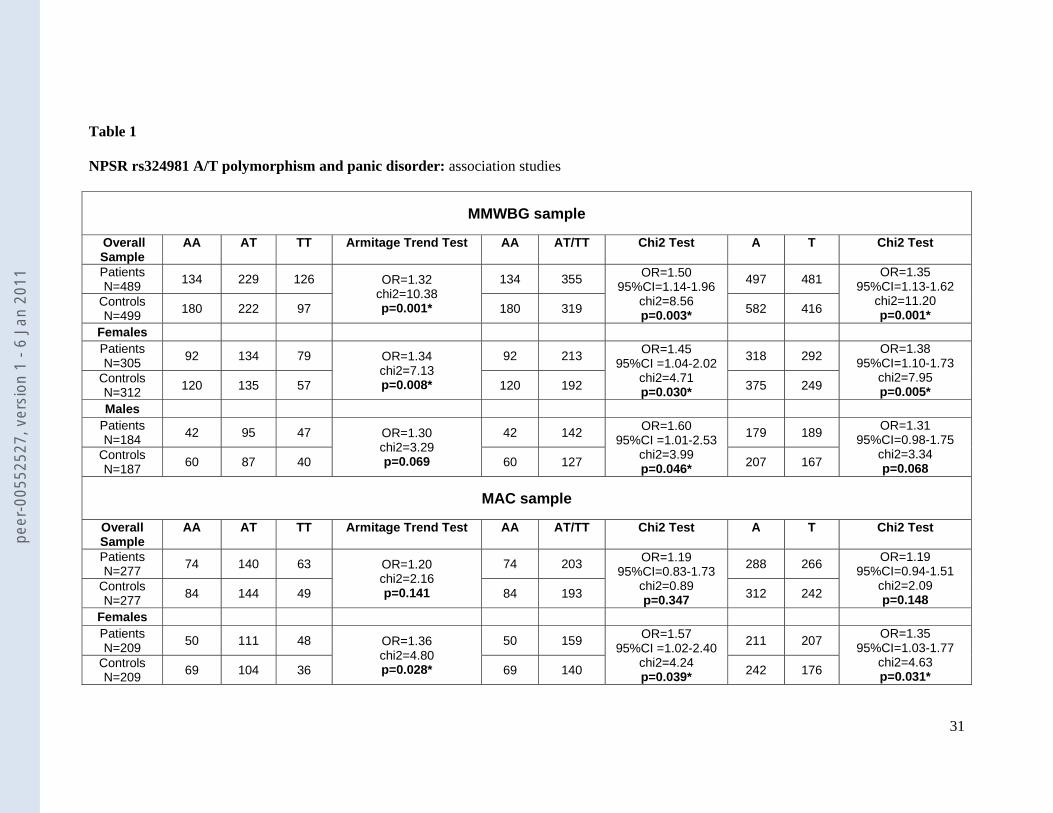
Allele and genotype frequencies of NPSR rs324981 in patient and control samples are
given in table 1. In the discovery MMWBG sample, the NPSR rs324981 AA genotype as
compared to carriers of at least one T allele was underrepresented in patients with panic
peer
-005
5252
7, v
ersi
on 1
- 6
Jan
2011
12
disorder (p=0.003). Stratification for gender revealed that this effect held true for both
genders, however, was more pronounced in the female subgroup of patients (female: p=0.03;
male: p=0.05) (see table 1).
In an attempt to replicate the association of NPSR rs324981 with panic disorder, we
interrogated a second, independent case-control sample of 277 patients suffering from panic
disorder with agoraphobia (MAC sample), along with matched controls (table 1). Genotyping
procedures and statistical analyses were identical to the discovery sample. Again, the NPSR
rs324981 AA genotype was significantly under-represented in female patients with panic
disorder, while there was no significant effect in the male or the total sample (see table 1).
[INSERT TABLE 1 ABOUT HERE]
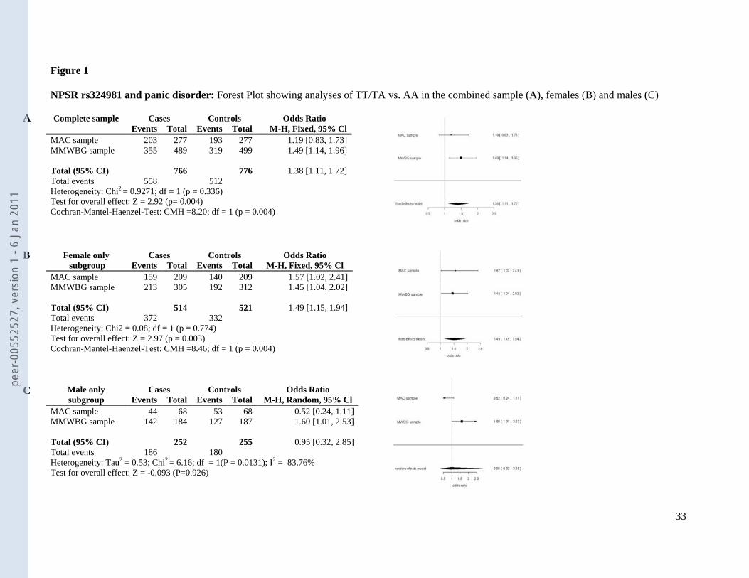
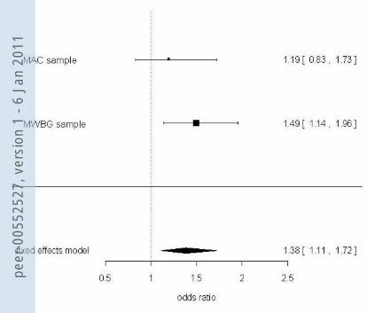
When both samples were analyzed jointly in a meta-association approach leading to a
combined sample size of 766 patients and 776 controls, there was a highly significant
association of the NPSR rs324981 risk genotypes containing at least one T allele with disease
in the total sample (Armitage’s trend test OR 1.28, chi2=12.11, p=0.0005) as well as in the
female (Armitage’s trend test OR 1.35, chi2=11.95, p=0.00055), but not the male sub-sample
(Armitage’s trend test OR 1.15, chi2=1.25, p=0.263). As such a meta-association approach
might bear several problematic issues, such as ethnic background admixture (although this is
unlikely as both samples were ascertained in Germany), we also treated the data in a meta-
analytic manner (see figure 1). Association of the NPSR rs324981 T risk allele with panic
disorder was confirmed in this meta-analysis, yielding an OR of 1.38 (p=0.004), which held
true in the female (OR 1.49, p=0.004), but not the male sub-sample (OR 0.95, p>0.1).
[INSERT FIGURE 1 ABOUT HERE]
Association study with dimensional anxiety traits
peer
-005
5252
7, v
ersi
on 1
- 6
Jan
2011

13
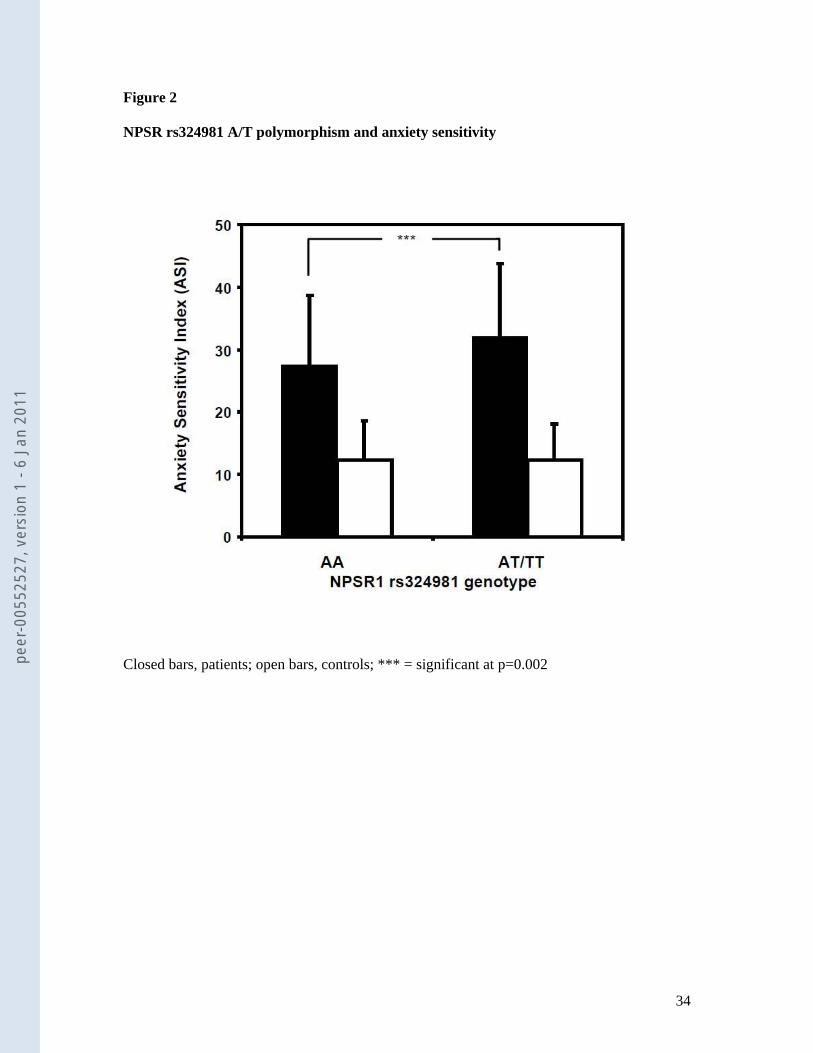
Next, we examined whether the NPSR rs324981 T allele also influences dimensional
constructs associated with panic disorder and agoraphobia. To do so, we tested whether the
Agoraphobic Cognitions Questionnaire (ACQ) and Anxiety Sensitivity Index (ASI) were
associated with NPSR rs324981 in patients from the MAC study as well as 519 healthy
controls. Although there was not a significant association in control subjects (ACQ, p=0.41;
ASI, p=0.99), the ASI (but not the ACQ; best p=0.31) score was significantly associated with
genotype in the expected direction in patients: while NPSR rs324981 AA carriers had an ASI
score of 27.5±11.1, T allele carriers displayed a score of 32.1±11.7 (p=0.002) (see figure 2).
Again, the difference was due to the female sub-sample (27.0±11.7 vs. 32.6±11.8, p=0.001)
and not present in the male sub-sample (28.6±11.8 vs. 30.2±11.2, p=0.29).
[INSERT FIGURE 2 ABOUT HERE]
Genetic modulation of heart rate and symptom reports: Behavioral Avoidance Test
(BAT)
Genotype groups (AA=55, AT=102, TT=48) did not differ significantly in duration of
exposure, Group F(1, 203)<1, p=0.33 (AA: 539.98 sec ± 22.74 SE; AT/TT: 514.00 sec ±
13.77 SE). As expected, mean heart rate and symptom reports significantly increased from
anticipation to exposure and decreased again during recovery (Block F(2, 406) = 28.69,
p<0.001; Block F(2, 406) = 90.42, p< 0.001 for heart rate and symptom reports, respectively)
demonstrating that the BAT reliably induced intense fear in this group of patients.
NPSR rs324981 genotype systematically modulated these response patterns.
Homozygous A allele carriers showed significantly lower overall heart rate than T allele
carriers, Group F(1, 203) = 6.39, p<0.05, which was not modulated by conditions (figure 3),
thus depicting an overall larger arousal response during all three phases of the BAT.
Symptom reports were also significantly modulated but showed a different pattern. While A
peer
-005
5252
7, v
ersi
on 1
- 6
Jan
2011

14
and T allele carriers did not differ in their symptom reports during anticipation and recovery,
T allele carriers reported significantly more intense symptoms during acute exposure, Block ×
Group F(2, 406) = 4.26, p<0.05.
[INSERT FIGURE 3 ABOUT HERE]
Genetic modulation of fear circuit activation
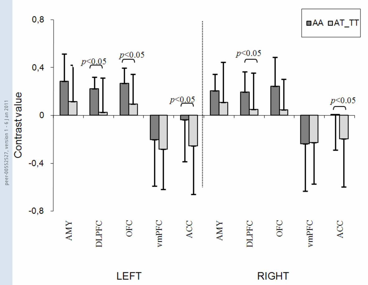
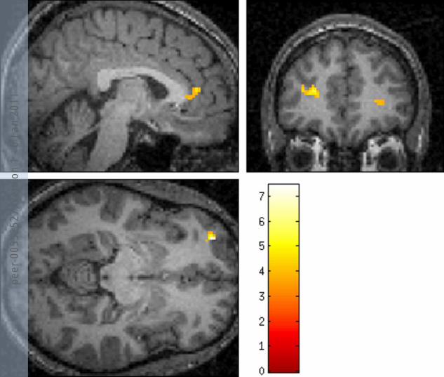
We observed association of NPSR rs324981 with brain activation responses to fearful
faces in contrast to the no face condition (figure 4): the protective AA genotype was found to
be associated with an increased activation in the dorsolateral prefrontal cortex (left: AA:
mean=0.221, SD=0.097; AT/TT: mean=0.025, SD=0.285; F=3.857, p=0.042; right: AA:
mean=0.193, SD=0.170; AT/TT: mean=0.048, SD=0.306; F=4.182, p=0.033) and lateral
orbitofrontal cortex (left: AA: mean=0.267, SD=0.124; AT/TT: mean=0.092, SD=0.251;
F=2.621, p=0.102; right: AA: mean=0.243, SD=0.243; AT/TT: mean=0.044, SD=0.259;
F=3.674, p=0.047), while carriers of at least one T risk allele exhibited a significantly
decreased activity in the anterior cingulate cortex (left: AA: mean=-0.037, SD=0.352; AT/TT:
mean=-0.255, SD=0.408; F=4.880, p=0.021; right: AA: mean=0.009, SD=0.301; AT/TT:
mean=-0.197, SD=0.402; F=5.962, p=0.011).
No other regions of interest including the amygdala were found to be associated with
NPSR rs324981 at the corrected threshold in response to fearful faces. There was no
significant impact of NPSR rs324981 on responsiveness in any of the regions of interest to
happy, angry or neutral faces.
[INSERT FIGURE 4 ABOUT HERE]
peer
-005
5252
7, v
ersi
on 1
- 6
Jan
2011

15
DISCUSSION
In the present study, association of the more active NPSR rs324981 T allele with the
categorical diagnosis of panic disorder with and without agoraphobia was observed in two
independent, large case-control studies as well as in a meta-analytic approach. This finding
strengthens recent evidence arising from animal models for a pivotal role of neuropeptide S in
the expression of anxiety3-7,9-11,49 and for a risk locus on chromosome 7p14 containing the
NPSR gene in panic disorder.22-24,27
Interestingly, in both samples comprising a total of 766 patients association of NPSR
gene variation with panic disorder strongly originated from or was even restricted to the
female sub-group of patients (n=514). This finding is in line with previous molecular genetic
reports of female-specific association findings in panic disorder possibly via hormonal
interactions or female-specific transcriptional patterns.e.g.,50-52 From an evolutionary
perspective, one might speculate that the evolutionarily “newer” T allele variant might convey
a beneficial effect by optimizing the fight-or-flight reaction via increased arousal levels,
which might be positive in more male-dominant predatory environments such as hunting, but
not in comparatively safe agricultural or household environments. The present female-
dominant finding is contrary to the observation by Okamura et al. who reported an association
of NPSR rs324981 with panic disorder in the male sub-group of patients only.27 However, as
their male subsample of patients was very small (n=51) and NPSR rs324981 genotype
distribution in male control subjects did not conform to Hardy-Weinberg equilibrium, this
observation is finally inconclusive. Nevertheless, population-specific differences across
Caucasian and Asian samples, as already observed for other polymorphisms, may explain this
discrepancy.e.g.,53
peer
-005
5252
7, v
ersi
on 1
- 6
Jan
2011

16
Association of the gain-of-function NPSR rs324981 T allele with panic disorder in
humans seems inconsistent with findings in rodent models, where NPS and agonists at NPSR
have been shown to rather exert a dose-dependent anxiolytic effect in the open field, the light-
dark box, the elevated plus maze and marble-burying paradigms3 (although it should be noted
that pharmacological interventions during adulthood do not readily mimic genetically driven
alterations during ontogeny). A possible explanation of this paradox might be that panic
disorder is to a great extent conferred via an increased level of arousal,e.g.,54-56 which in animal
models has been found to be driven by increased NPS activity with a potentially differential
dose-dependent effect on arousal and anxiety, respectively.1,3-5 Increased arousal mediated by
the more active T allele would be in line with Smith et al. reporting NPS to cause a significant
stimulation of the hypothalamo-pituitary-adrenal (HPA) axis with an increase in plasma
ACTH and corticosterone along with increased arousal-like behavior in rats.57 This hypothesis
of increased arousal possibly mediating the anxiety-increasing effect of NPSR gain of
function gene variation is substantiated by several translational findings at different levels in
the present study:
1) The NPSR rs324981 T risk allele was observed to be associated with increased
anxiety sensitivity as measured by the Anxiety Sensitivity Index. Anxiety sensitivity is a
dimensional construct measuring the extent to which individuals believe that symptoms of
arousal and anxiety can have harmful somatic, social or psychological consequences.35
Several studies provide evidence for anxiety sensitivity predicting fear responses to bodily
sensations increasing physiological arousal,58 e.g., following a stair-stepping task59 or
hyperventilation challenge,60 during detection of changes in pulse transit time61 or in response
to caffeine administration,62 and ASI seems to be a predictor for the development of panic
disorder after periods of stress.63 So, the observed association of the NPSR rs324981 T risk
allele with increased anxiety sensitivity might in part reflect an increased level of arousal.
peer
-005
5252
7, v
ersi
on 1
- 6
Jan
2011
17
Additionally, individual anxiety sensitivity profiles seem to discriminatively characterize
specific panic disorder subtypes since in patients with panic disorder high scores on the ASI
have been reported to be associated with the respiratory subtype of panic disorder64 and to
predict symptomatological reaction to CO265 as well as increased subjective fear during
hyperventilation challenge.66 Given that patients with the respiratory subtype of panic
disorder are characterized by a particularly high familial loading of panic disorder and thereby
a potentially higher genetic vulnerability,67 NPSR gene variation associated with high anxiety
sensitivity in patients with panic disorder might in part reflect vulnerability towards this
particular subtype of patients with panic disorder, which, however, remains to be elucidated in
future studies. Finally, women with panic disorder score higher than men on the AS physical
concerns subscale,68 and anxiety sensitivity has been shown to be heritable particularly in
women.69 So, the present finding of NPSR gene variation to be associated with the categorical
diagnosis of panic disorder particularly in female patients might in part be explained by its
association with the dimensional phenotype of anxiety sensitivity restricted to female patients.
2) The NPSR rs324981 T risk allele was related to overall increased heart rate during
exposure but also during anticipation and even recovery during a behavioral avoidance test.
These data are consistent with the notion that patients who carry the T risk allele have an
enhanced sympathomimetic activation and consequently arousal level. In challenging
situations in which the arousal response further increases over an already elevated baseline
level, these patients are more prone to experience panic symptoms. These findings are in line
with modern learning theories of panic disorder55 postulating that internal cues of elevated
arousal increase the chance of experiencing another panic attack once they have been
associated with aversive responses.e.g.,70 The current data suggest that not only conditioned
anxious apprehension but also genetic risk factors contribute to the increased arousal level
peer
-005
5252
7, v
ersi
on 1
- 6
Jan
2011
18
and might therefore uncover an important missing link in the understanding of the
mechanisms involved in the development of a panic disorder.
3) We observed an NPSR rs324981 genotype-dependent brain activation pattern in
response to anxiety-relevant emotional stimuli. The NPSR rs324981 T risk allele was found to
be associated with significantly decreased activity in the anterior cingulate cortex. This result
is in line with previous studies reporting anterior cingulate cortex volume reduction in
patients with panic disorder71,72 and diminished cingulate activity in patients with panic
disorder during identification of fearful facial affect compared to healthy controls.73
Decreased cingulate cortex activity has been hypothesized to be due to a chronic hyperarousal
in patients with panic disorder,74 since decreased cingulate cortex activity appears to be
associated with engagement of arousal networks particularly during processing of threat-
related stimuli also in other anxiety disorders such as simple phobias and posttraumatic stress
disorder.75-78 Given that NPS has been found to dose-dependently elicit increased arousal
response as detailed above, the NPSR rs324981 T allele increasing NPSR expression and NPS
efficacy at NPSR might confer a heightened risk of panic disorder via an elevated level of
arousal as reflected by decreased cingulate cortex activity. Additionally, homozygosity for the
A allele was associated with significantly increased activity in the dorsolateral prefrontal and
orbitofrontal cortex during the processing of fearful faces compared to activation in T risk
allele carriers. Given the well-established role of the prefrontal and orbitofrontal cortex to
attenuate an emotional response to fear/threat-related stimuli in anxiety disorders by
inhibition of amygdala activation via a top-down mechanism,e.g.,13-15,78 increased activity in
those regions might contribute to the observed protective effect of the NPSR rs324981 AA
genotype with respect to panic disorder.
Several limitations of the present study have to be considered: Patients and controls in
the discovery and replication samples were not completely matched for age in that some of
peer
-005
5252
7, v
ersi
on 1
- 6
Jan
2011

19
the control subjects still are at risk to develop panic disorder. This, however would bias
against an association finding and not favor a false positive finding. Indeed, this may even
explain the somewhat weaker association in the replication sample as here the age difference
was larger. Also, the discovery and replication sample differed with respect to the ratio of
panic disorder patients with and without agoraphobia with a smaller agoraphobia ratio in the
discovery sample possibly. Furthermore, comorbid depression might constitute a confounding
factor, particularly, since the discovery and the replication samples differed in the rate of
comorbid depression. A further limitation of the present study is that no non-Caucasian
populations were investigated rendering comparison to the study by Okamura et al.27 difficult.
However, although in all samples Caucasian ancestry was ascertained, ethnic stratification
even within the German population cannot be excluded,e.g.,79 particularly since no genomic
control has been performed in the presently investigated samples. The effect of genotype on
the ASI is only found in patients, but not in controls. One possible explanation may be the
small variation of anxiety sensitivity in our supra-normal control sample. Finally, caution and
restraint is necessary in the interpretation of the imaging genetic part of the study, given a
relatively small number of patients and an explorative statistical approach not corrected for
multiple testing. Apart from significantly decreased activity in the anterior cingulate cortex in
response to fearful faces, p-values would not withstand Bonferroni correction for the four
MANOVAs that were performed for the four different contrasts (fearful, angry, happy, neutral
vs. no faces) over all ROIs resulting in a corrected p-value of 0.0125 (two-sided) or 0.025
(one-sided), respectively, which might reflect a possible type I error. However, as largely
debated, Bonferroni correction might be too conservative for strictly hypothesis-driven
studies as the present one and actually increase the probability of a type II error. A further
limitation is that the small sample size did not allow for meaningful statistical analyses in
subsamples stratified for gender, which, given a female-dominant result in the categorical
peer
-005
5252
7, v
ersi
on 1
- 6
Jan
2011

20
association studies, would have been of interest also in the imaging genetic part of this study.
Furthermore, although no biased distribution of gender, medication with SSRIs and
comorbidity with major depression or social phobia across NPSR genotype groups could be
discerned, these factors still could constitute possible confounding factors in the evaluation of
emotional processing. For instance, either single dose or subchronic (7 to 21 days) medication
with SSRIs has been shown to attenuate amygdala, hippocampus and medial prefrontal cortex
response to aversive facial expression.cf. 80-82 Thus, besides a potential ceiling effect due to
generally increased baseline amygdala activity in patients with panic disorder, this may partly
explain why no amygdala activation could be observed as with previous studies in this
sample.cf.44 Additionally, patients with social phobia tend to evaluate neutral faces as
potentially threatening and more aversive than healthy controls or possibly also patients with
pure panic disorder,83 which might have influenced the present results. Future studies will
have to elucidate the mechanisms by which genetic variation of NPSR modulates the
sympathomimetic system, cingulate and orbito-/prefrontal cortex activity and their possible
functional interactions. One such mechanism may be mediated by the startle reflex which
therefore is currently under investigation in our group.
In summary, the present body of research provides converging lines of evidence for a
role of NPSR gene variation in the pathogenesis of panic disorder. The more active NPSR
rs324981 T allele (Ile107) has been found to be associated with the categorical phenotype of
panic disorder in a female-dominant manner, possibly mediated via increased arousal levels
as reflected by elevated anxiety sensitivity, increased autonomic arousal as well as distorted
processing of fear-related emotional stimuli. In synopsis with previous evidence from animal
models, these findings might nourish future studies exploring a potentially beneficial use of
therapeutic agents targeting the NPS system in anxiety disorders.
peer
-005
5252
7, v
ersi
on 1
- 6
Jan
2011
21
ACKNOWLEDGEMENTS
This study was supported by the Deutsche Forschungsgemeinschaft (DFG; SFB-TRR-58
projects C2 and Z2 to KD, AR, PP and JD; Grant KFO 125 to AR; DE357/4-1 to AR, AH,
and JD; RE1632/5 to AR) and the BMBF (Panic-Net, to ALG, TK, AS, HUW, VA, AH, and
JD; details see webpage http://www.paniknetz.de/netzwerk.html). The recruitment of the Max
Planck panic sample in Munich was supported by the Max Planck Excellence Foundation. We
gratefully acknowledge the skilful technical support by Anna Baffa.
CONFLICT OF INTEREST
The authors declare no conflict of interest related to this work.
peer
-005
5252
7, v
ersi
on 1
- 6
Jan
2011

22
REFERENCES
1. Reinscheid RK, Xu YL. Neuropeptide S as a novel arousal promoting peptide
transmitter. FEBS J 2005; 272: 5689-5693.
2. Sato S, Shintani Y, Miyajima N, Yoshimura K. Novel G-protein-coupled receptor
protein and DNA thereof. World Patent Application 2002, WO02/31145 A1.
3. Xu YL, Reinscheid RK, Huitron-Resendiz S, Clark SD, Wang Z, Lin SH et al.
Neuropeptide S: a neuropeptide promoting arousal and anxiolytic-like effects. Neuron
2004; 43: 487-497.
4. Leonard SK, Dwyer JM, Sukoff Rizzo SJ, Platt B, Logue SF, Neal SJ et al.
Pharmacology of neuropeptide S in mice: therapeutic relevance to anxiety disorders.
Psychopharmacology (Berl) 2008; 197: 601-611.
5. Rizzi A, Vergura R, Marzola G, Ruzza C, Guerrini R, Salvadori S et al. Neuropeptide S
is a stimulatory anxiolytic agent: a behavioural study in mice. Br J Pharmacol 2008;
154: 471-479.
6. Vitale G, Filaferro M, Ruggieri V, Pennella S, Frigeri C, Rizzi A et al. Anxiolytic-like
effect of neuropeptide S in the rat defensive burying. Peptides 2008; 29: 2286-2291.
7. Duangdao DM, Clark SD, Okamura N, Reinscheid RK. Behavioral phenotyping of
Neuropeptide S receptor knockout mice. Behav Brain Res (in press).
8. Xu YL, Gall CM, Jackson VR, Civelli O, Reinscheid RK. Distribution of neuropeptide
S receptor mRNA and neurochemical characteristics of neuropeptide S-expression
neurons in the rat brain. J Comp Neurology 2007; 500: 84-102.
9. Jüngling K, Seidenbecher T, Sosulina L, Lesting J, Sangha S, Clark SD et al.
Neuropeptide S-mediated control of fear expression and extinction: role of intercalated
GABAergic neurons in the amygdala. Neuron 2008; 59: 298-310.
peer
-005
5252
7, v
ersi
on 1
- 6
Jan
2011
23
10. Meis S, Bergado-Acosta JR, Yanagawa Y, Obata K, Stork O, Munsch T. Identification
of a neuropeptide S responsive circuitry shaping amygdala activity via the endopiriform
nucleus. PLoS One 2008; 3: e2695.
11. Pape HC, Jüngling K, Seidenbecher T, Lesting J, Reinscheid RK. Neuropeptide S: A
transmitter system in the brain regulating fear and anxiety. Neuropharmacology. (in
press).
12. LeDoux JE. Emotion circuits in the brain. Annu Rev Neurosci 2000; 23: 155-184.
13. Phillips ML, Drevets WC, Rauch SL, Lane R. Neurobiology of emotion perception I:
The neural basis of normal emotion perception. Biol Psychiatry 2003; 54: 504-514.
14. Phillips ML, Drevets WC, Rauch SL, Lane R. Neurobiology of emotion perception II:
Implications for major psychiatric disorders. Biol Psychiatry 2003; 54: 515-528.
15. Etkin A, Klemenhagen KC, Dudman JT, Rogan MT, Hen R, Kandel ER et al. Individual
differences in trait anxiety predict the response of the basolateral amygdala to
unconsciously processed fearful faces. Neuron 2004; 44: 1043-1055.
16. Javanmard M, Shlik J, Kennedy SH, Vaccarino FJ, Houle S, Bradwejn J.
Neuroanatomic correlates of CCK-4-induced panic attacks in healthy humans: a
comparison of two time points. Biol Psychiatry 1999; 45: 872-882.
17. Pfleiderer B, Zinkirciran S, Arolt V, Heindel W, Deckert J, Domschke K. fMRI
amygdala activation during a spontaneous panic attack in a patient with panic disorder.
World J Biol Psychiatry 2007; 8: 269-272.
18. Sakai Y, Kumano H, Nishikawa M, Sakano Y, Kaiya H, Imabayashi E et al. Cerebral
glucose metabolism associated with a fear network in panic disorder. Neuroreport 2005;
16: 927-931.
19. Charney DS, Heninger GR, Jatlow PI. Increased anxiogenic effects of caffeine in panic
disorders. Arch Gen Psychiatry 1985; 42: 233-243.
peer
-005
5252
7, v
ersi
on 1
- 6
Jan
2011

24
20. Lage R, Diéguez C, López M. Caffeine treatment regulates neuropeptide S system
expression in the rat brain. Neurosci Lett 2006; 410: 47-51.
21. Raiteri L, Luccini E, Romei C, Salvadori S, Calò G. Neuropeptide S selectively inhibits
the release of 5-HT and noradrenaline from mouse frontal cortex nerve endings. Br J
Pharmacol 2009; 157: 474-481.
22. Crowe RR, Goedken R, Samuelson S, Wilson R, Nelson J, Noyes R Jr. Genomewide
survey of panic disorder. Am J Med Genet 2001; 105: 105-109.
23. Knowles JA, Fyer AJ, Vieland VJ, Weissman MM, Hodge SE, Heiman GA et al.
Results of a genome-wide genetic screen for panic disorder. Am J Med Genet 1998; 81:
139-147.
24. Logue MW, Vieland VJ, Goedken RJ, Crowe RR. Bayesian analysis of a previously
published genome screen for panic disorder reveals new and compelling evidence for
linkage to chromosome 7. Am J Med Genet B Neuropsychiatr Genet 2003; 121B: 95-99.
25. Bernier V, Stocco R, Bogusky MJ, Joyce JG, Parachoniak C, Grenier K et al. Structure-
function relationships in the neuropeptide S receptor: molecular consequences of the
asthma-associated mutation N107I. J Biol Chem 2006; 281: 24704-24712.
26. Reinscheid RK, Xu YL, Okamura N, Zeng J, Chung S, Pai R et al. Pharmacological
characterization of human and murine neuropeptide s receptor variants. J Pharmacol
Exp Ther 2005; 315: 1338-1345.
27. Okamura N, Hashimoto K, Iyo M, Shimizu E, Dempfle A, Friedel S et al. Gender-
specific association of a functional coding polymorphism in the Neuropeptide S receptor
gene with panic disorder but not with schizophrenia or attention-deficit/hyperactivity
disorder. Prog Neuropsychopharmacol Biol Psychiatry 2007; 31: 1444-1448.
28. Mannuzza S, Fyer AJ, Klein DF, Endicott J. Schedule for affective disorders and
schizophrenia–Lifetime version modified for the study of anxiety disorders (SADS-LA):
peer
-005
5252
7, v
ersi
on 1
- 6
Jan
2011
25
Rationale and conceptual development. J Psychiatr Res 1986; 20: 317-325.
29. Robins LN, Wing J, Wittchen HU, Helzer JE, Babor TF, Burke J et al. The Composite
International Diagnostic Interview. An epidemiologic instrument suitable for use in
conjunction with different diagnostic systems and in different cultures. Arch Gen
Psychiatry 1988; 45: 1069-1077.
30. Wittchen HU. SKID-I: strukturiertes klinisches Interview für DSM-IV, Achse I:
Psychische Störungen. Goettingen: Hogrefe. 1997.
31. Gloster AT, Wittchen HU, Einsle F, Hofler M, Lang T, Helbig-Lang S et al. Mechanism
of action in CBT (MAC): methods of a multi-center randomized controlled trial in 369
patients with panic disorder and agoraphobia. Eur Arch Psychiatry Clin Neurosci 2009;
259: 155-166.
32. Chambless DL, Caputo GC, Bright P, Gallagher R. Assessment of fear in agoraphobics:
the body sensations questionnaire and the agoraphobic cognitions questionnaire. J
Consult Clin Psychol 1984; 52: 1090-1097.
33. Ehlers, A., & Margraf, J. (1993). Fragebogen zu körperbezogenen Ängsten, Kognitionen
und Vermeidung (AKV). Weinheim: Beltz Test.
34. Alpers GW, Pauli P. Angstsensitivitäts-Index. Würzburg: Julius-Maximilians-
Universität. 2001.
35. Reiss S, Peterson RA, Gursky DM, McNally RJ. Anxiety sensitivity, anxiety frequency
and the prediction of fearfulness. Behav Res Ther 1986; 24: 1-8.
36. Fleiss J. Statistical Methods for Rates and Proportions. Wiley: New York, 1981.
37. Lau J, Ioannidis JP, Schmid CH. Quantitative synthesis in systematic reviews. Ann
Intern Med 1997; 127: 820-826.
38. Zintzaras E, Hadjigeorgiou GM. Association of paraoxonase 1 gene polymorphisms
with risk of Parkinson's disease: a meta-analysis. J Hum Genet 2004; 49: 474-481.
peer
-005
5252
7, v
ersi
on 1
- 6
Jan
2011

26
39. Mantel N, Haenszel W. Statistical aspects of the analysis of data from retrospective
studies of disease. J Natl Cancer Inst 1959; 22: 719-748.
40. DerSimonian R, Laird N. Meta-analysis in clinical trials. Control Clin Trials 1986; 7:
177-188.
41. R-Development-Core-Team. R: A Language and Environment for Statistical
Computing. 2.10.0 ed. R Foundation for Statistical Computing: Vienna, Austria, 2009.
42. Viechtbauer W. metafor: Meta-Analysis Package for R. 0.5-7, 2009.
43. Wilhelm FH, Peyk P. ANSLAB 4.0: Autonomic nervous system laboratory.
http://www.sprweb.org, 2005.
44. Domschke K, Braun M, Ohrmann P, Suslow T, Kugel H, Bauer J et al. Association of
the functional -1019C/G 5-HT1A polymorphism with prefrontal and amygdala
activation measured with 3T fMRI in panic disorder. Int J Neuropsychopharmacol
2006; 9: 349-355.
45. Domschke K, Ohrmann P, Braun M, Suslow T, Bauer J, Hohoff C et al. Influence of the
catechol-O-methyltransferase val158met genotype on amygdala and orbitofrontal cortex
emotional processing in panic disorder. Psychiatry Res – Neuroimaging 2008; 163: 13-
20.
46. Ekman P, Friesen WV. Pictures of facial affect. Consulting Psychologists Press, Palo
Alto, CA, USA. 1976.
47. Tzourio-Mazoyer N, Landeau B, Papathanassiou D, Crivello F, Etard O, Delcroix N et
al. Automated anatomical labeling of activations in SPM using a macroscopic
anatomical parcellation of the MNI MRI single-subject brain. NeuroImage 2002; 15:
273-289.
48. Brett M, Anton JL, Valabregue R, Poline JB. Region of interest analysis using an SPM
toolbox. NeuroImage 2002; 16: 497.
peer
-005
5252
7, v
ersi
on 1
- 6
Jan
2011

27
49. Okamura N, Reinscheid RK. Neuropeptide S: a novel modulator of stress and arousal.
Stress 2007; 10: 221-226
50. Deckert J, Catalano M, Syagailo YV, Bosi M, Okladnova O, Di Bella D, et al. Excess of
high activity monoamine oxidase A gene promoter alleles in female patients with panic
disorder. Hum Mol Genet 1999; 8: 621-624.
51. Domschke K, Freitag CM, Kuhlenbaumer G, Schirmacher A, Sand P, Nyhuis P et al.
Association of the functional V158M catechol-O-methyl-transferase polymorphism with
panic disorder in women. Int J Neuropsychopharmacol 2004; 7: 183-188.
52. Domschke K, Hohoff C, Jacob C, Maier W, Fritze J, Bandelow B et al. Chromosome
4q31-34 panic disorder risk locus: association of neuropeptide Y Y5 receptor variants.
Am J Med Genet B Neuropsychiatr Genet 2008; 147B: 510-516.
53. Domschke K, Deckert J, O’Donovan M, Glatt SJ. Meta-analysis of COMT val158met in
panic disorder – ethnic heterogeneity and gender specificity. Am J Med Genet
(Neuropsychiatr Genet) 2007; 144: 667-673.
54. Blechert J, Michael T, Grossman P, Lajtman M, Wilhelm FH. Autonomic and
respiratory characteristics of posttraumatic stress disorder and panic disorder.
Psychosom Med 2007; 69: 935-943.
55. Bouton ME, Mineka S, Barlow DH. A modern learning theory perspective on the
etiology of panic disorder. Psychol Rev 2001; 108: 4-32.
56. Clark DM. A cognitive approach to panic. Behav Res Ther 1986: 24: 461-470.
57. Smith KL, Patterson M, Dhillo WS, Patel SR, Semjonous NM, Gardiner JV et al.
Neuropeptide S stimulates the hypothalamo-pituitary-adrenal axis and inhibits food
intake. Endocrinology 2006; 147: 3510-3518.
58. Joiner TE Jr, Steer RA, Beck AT, Schmidt NB, Rudd MD, Catanzaro SJ. Physiological
hyperarousal: construct validity of a central aspect of the tripartite model of depression
peer
-005
5252
7, v
ersi
on 1
- 6
Jan
2011

28
and anxiety. J Abnorm Psychol 1999; 108: 290-298.
59. Rabian B, Embry L, MacIntyre D. Behavioral validation of the Childhood Anxiety
Sensitivity Index in children. J Clin Child Psychol 1999; 28: 105-112.
60. Sturges LV, Goetsch VL, Ridley J, Whittal M. Anxiety sensitivity and response to
hyperventilation challenge: physiologic arousal, interoceptive acuity, and subjective
distress. J Anxiety Disord 1998; 12: 103-115.
61. Richards JC, Bertram S. Anxiety sensitivity, state and trait anxiety, and perception of
change in sympathetic nervous system arousal. J Anxiety Disord 2000; 14: 413-427.
62. Telch MJ, Silverman A, Schmidt NB. The relationship between anxiety sensitivity and
perceived control in a caffeine challenge. J Anx Disord 1996; 10: 21-35.
63. Schmidt NB, Lerew DR, Jackson RJ. Prospective evaluation of anxiety sensitivity in the
pathogenesis of panic: Replication and extension. J Abnorm Psychol 1999; 108: 532-
537.
64. Onur E, Alkin T, Tural U. Panic disorder subtypes: further clinical differences. Depress
Anxiety 2007; 24: 479-486.
65. Perna G, Romano P, Caldirola D, Cucchi M, Bellodi L. Anxiety sensitivity and 35%
CO2 reactivity in patients with panic disorder. J Psychosom Res 2003; 54: 573-577.
66. Brown M, Smits JA, Powers MB, Telch MJ. Differential sensitivity of the three ASI
factors in predicting panic disorder patients' subjective and behavioral response to
hyperventilation challenge. J Anxiety Disord 2003; 17: 583-591.
67. Nardi AE, Lopes FL, Valença AM, Nascimento I, Mezzasalma MA, Zin WA.
Psychopathological description of hyperventilation-induced panic attacks: a comparison
with spontaneous panic attacks. Psychopathology 2004; 37: 29-35.
68. Foot M, Koszycki D. Gender differences in anxiety-related traits in patients with panic
disorder. Depress Anxiety 2004; 20: 123-130.
peer
-005
5252
7, v
ersi
on 1
- 6
Jan
2011
29
69. Jang KL, Stein MB, Taylor S, Livesley WJ. Gender differences in the etiology of
anxiety sensitivity: a twin study. J Gend Specif Med 1999; 2: 39-44.
70. Pauli P, Marquardt C, Hartl L, Nutzinger DO, Hölzl R, Strian F. Anxiety induced by
cardiac perceptions in patients with panic attacks: A field study. Behav Res Ther 1991;
29: 137-145.
71. Asami T, Hayano F, Nakamura M, Yamasue H, Uehara K, Otsuka T et al. Anterior
cingulate cortex volume reduction in patients with panic disorder. Psychiatry Clin
Neurosci 2008; 62: 322-330.
72. Uchida RR, Del-Ben CM, Busatto GF, Duran FL, Guimarães FS, Crippa JA, et al.
Regional gray matter abnormalities in panic disorder: a voxel-based morphometry study.
Psychiatry Res 2008; 163: 21-29.
73. Pillay SS, Gruber SA, Rogowska J, Simpson N, Yurgelun-Todd DA. fMRI of fearful
facial affect recognition in panic disorder: the cingulate gyrus-amygdala connection. J
Affect Disord 2006; 94: 173-181.
74. Bremner JD, Staib LH, Kaloupek D, Southwick SM, Soufer R, Charney DS. Neural
correlates of exposure to traumatic pictures and sound in vietnam combat veterans with
and without posttraumatic stress disorder: A positron emission tomography study. Biol
Psychiatry 1999; 45: 806–816.
75. Felmingham KL, Williams LM, Kemp AH, Rennie C, Gordon E, Bryant RA. Anterior
cingulate activity to salient stimuli is modulated by autonomic arousal in posttraumatic
stress disorder. Psychiatry Res 2009 173: 59-62.
76. Fredrikson M, Wik G, Annas P, Ericson K, Stone-Elander S. Functional neuroanatomy
of visually elicited simple phobic fear: additional data and theoretical analysis.
Psychophysiology 1995 32: 43-48.
77. Shin LM, Whalen PJ, Pitman RK, Bush G, Macklin ML, Lasko NB et al. An fMRI
peer
-005
5252
7, v
ersi
on 1
- 6
Jan
2011
30
study of anterior cingulate function in posttraumatic stress disorder. Biol Psychiatry
2001; 50: 932-942.
78. Davidson RJ. Anxiety and affective style: Role of the prefrontal cortex and amygdala.
Biol Psychiatry 2002; 51: 68-80.
79. Steffens M, Lamina C, Illig T, Bettecken T, Vogler R, Entz P et al. SNP-based analysis
of genetic substructure in the German population. Hum Hered 2006; 62: 20-29.
80. Murphy SE, Norbury R, O'Sullivan U, Cowen PJ, Harmer CJ. Effect of a single dose of
citalopram on amygdala response to emotional faces. Br J Psychiatry 2000; 194: 535-
540.
81. Harmer CJ, Mackay CE, Reid CB, Cowen PJ, Goodwin GM. Antidepressant drug
treatment modifies the neural processing of nonconscious threat cues. Biol Psychiatry
2006; 59: 816-820.
82. Arce E, Simmons AN, Lovero KL, Stein MB, Paulus MP. Escitalopram effects on
insula and amygdala BOLD activation during emotional processing.
Psychopharmacology (Berl) 2008; 196: 661-672.
83. Yoon KL, Zinbarg RE. Threat is in the eye of the beholder: social anxiety and the
interpretation of ambiguous facial expressions. Behav Res Ther 2007; 45: 839-847.
peer
-005
5252
7, v
ersi
on 1
- 6
Jan
2011
31
Table 1
NPSR rs324981 A/T polymorphism and panic disorder: association studies
MMWBG sample
Overall Sample
AA AT TT Armitage Trend Test AA AT/TT Chi2 Test A T Chi2 Test
Patients N=489 134 229 126 OR=1.32
chi2=10.38 p=0.001*
134 355 OR=1.50 95%CI=1.14-1.96
chi2=8.56 p=0.003*
497 481 OR=1.35 95%CI=1.13-1.62
chi2=11.20 p=0.001*
Controls N=499 180 222 97 180 319 582 416
Females Patients N=305 92 134 79 OR=1.34
chi2=7.13 p=0.008*
92 213 OR=1.45 95%CI =1.04-2.02
chi2=4.71 p=0.030*
318 292 OR=1.38 95%CI=1.10-1.73
chi2=7.95 p=0.005*
Controls N=312 120 135 57 120 192 375 249
Males Patients N=184 42 95 47 OR=1.30
chi2=3.29 p=0.069
42 142 OR=1.60 95%CI =1.01-2.53
chi2=3.99 p=0.046*
179 189 OR=1.31 95%CI=0.98-1.75
chi2=3.34 p=0.068
Controls N=187 60 87 40 60 127 207 167
MAC sample
Overall Sample
AA AT TT Armitage Trend Test AA AT/TT Chi2 Test A T Chi2 Test
Patients N=277 74 140 63 OR=1.20
chi2=2.16 p=0.141
74 203 OR=1.19 95%CI=0.83-1.73
chi2=0.89 p=0.347
288 266 OR=1.19 95%CI=0.94-1.51
chi2=2.09 p=0.148
Controls N=277 84 144 49 84 193 312 242
Females Patients N=209 50 111 48 OR=1.36
chi2=4.80 p=0.028*
50 159 OR=1.57 95%CI =1.02-2.40
chi2=4.24 p=0.039*
211 207 OR=1.35 95%CI=1.03-1.77
chi2=4.63 p=0.031*
Controls N=209 69 104 36 69 140 242 176
peer
-005
5252
7, v
ersi
on 1
- 6
Jan
2011

32
Males Patients
N=68 24 29 15 OR=0.83 chi2=0.74 p=0.389
24 44 OR=0.51 95%CI =0.24-1.11
chi2=2.91 p=0.088
77 59 OR=0.81 95%CI=0.50-1.31
chi2=0.73 p=0.394
Controls N=68 15 40 13 15 53 70 66
* significant p-value at significance level of <0.05
peer
-005
5252
7, v
ersi
on 1
- 6
Jan
2011
33
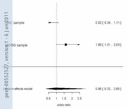
Figure 1
NPSR rs324981 and panic disorder: Forest Plot showing analyses of TT/TA vs. AA in the combined sample (A), females (B) and males (C)
Heterogeneity: Chi2 = 0.08; df = 1 (p = 0.774) Test for overall effect: Z = 2.97 (p = 0.003) Cochran-Mantel-Haenzel-Test: CMH =8.46; df = 1 (p = 0.004)
Complete sample Cases Controls Odds Ratio Events Total Events Total M-H, Fixed, 95% Cl MAC sample 203 277 193 277 1.19 [0.83, 1.73] MMWBG sample 355 489 319 499 1.49 [1.14, 1.96] Total (95% CI) 766 776 1.38 [1.11, 1.72] Total events 558 512 Heterogeneity: Chi2 = 0.9271; df = 1 (p = 0.336) Test for overall effect: Z = 2.92 (p= 0.004) Cochran-Mantel-Haenzel-Test: CMH =8.20; df = 1 (p = 0.004)
Female only Cases Controls Odds Ratio subgroup Events Total Events Total M-H, Fixed, 95% Cl
MAC sample 159 209 140 209 1.57 [1.02, 2.41] MMWBG sample 213 305 192 312 1.45 [1.04, 2.02] Total (95% CI) 514 521 1.49 [1.15, 1.94] Total events 372 332
Male only Cases Controls Odds Ratio subgroup Events Total Events Total M-H, Random, 95% Cl
MAC sample 44 68 53 68 0.52 [0.24, 1.11] MMWBG sample 142 184 127 187 1.60 [1.01, 2.53] Total (95% CI) 252 255 0.95 [0.32, 2.85] Total events 186 180 Heterogeneity: Tau2 = 0.53; Chi2 = 6.16; df = 1(P = 0.0131); I2 = 83.76% Test for overall effect: Z = -0.093 (P=0.926)
A B
C
peer
-005
5252
7, v
ersi
on 1
- 6
Jan
2011
34
Figure 2
NPSR rs324981 A/T polymorphism and anxiety sensitivity
Closed bars, patients; open bars, controls; *** = significant at p=0.002
peer
-005
5252
7, v
ersi
on 1
- 6
Jan
2011

35
Figure 3
Mean heart rate (upper panel; A) and mean symptom intensity (lower panel; B) during
anticipation, exposure and recovery in homozygous A allele carriers and T allele
carriers, respectively
(A)
(B)
peer
-005
5252
7, v
ersi
on 1
- 6
Jan
2011

36
Figure 4 NPSR rs324981 A/T (Asn107Ile) effects on brain activation during processing of fearful faces (vs. no face)
(A) Contrast values analyzed by MarsBaR; (B) ROI analyses of the same regions using the Wake Forest University Pick Atlas (patients
homozygous for the A allele vs. patients with AT and TT genotypes at p<0.001), AMY: amygdala, DLPFC: dorsolateral prefrontal cortex, OFC:
orbitofrontal cortex, vmPFC: ventromedial prefrontal cortex, ACC: anterior cingulate cortex
(A) (B)
peer
-005
5252
7, v
ersi
on 1
- 6
Jan
2011
peer
-005
5252
7, v
ersi
on 1
- 6
Jan
2011

peer
-005
5252
7, v
ersi
on 1
- 6
Jan
2011
peer
-005
5252
7, v
ersi
on 1
- 6
Jan
2011

peer
-005
5252
7, v
ersi
on 1
- 6
Jan
2011

peer
-005
5252
7, v
ersi
on 1
- 6
Jan
2011

peer
-005
5252
7, v
ersi
on 1
- 6
Jan
2011
peer
-005
5252
7, v
ersi
on 1
- 6
Jan
2011
peer
-005
5252
7, v
ersi
on 1
- 6
Jan
2011