Neuropeptide Y inhibits cholangiocarcinoma cell growth and invasion
Upload
independentCategory
view
3download
0
Neuropeptide Y Overexpression Using RecombinantAdenoassociated Viral Vectors
Francesco Noé,* Angelisa Frasca,* Claudia Balducci,* Mirjana Carli,* Gunther Sperk,†
Francesco Ferraguti,† Asla Pitkänen,‡� Ross Bland,§¶ Helen Fitzsimons,§¶
Matthew During,¶ and Annamaria Vezzani*
*Department of Neuroscience, Mario Negri Institute for Pharmacological Research, Milano, 20156 Italy, †Department ofPharmacology, Medical University, A-6020 Innsbruck, Austria, ‡Epilepsy Research Lab, A.I. Virtanen Institute for Molecular Science,University of Kuopio, �Department of Neurology, Kuopio University Hospital, Kuopio, FI-70211 Finland, §Neurologix, Inc., Fort Lee,
New Jersey 07024, ¶Human Cancer Genetics Programme, The Ohio State University, Columbus, Ohio 43210
Summary: Gene therapy may represent a promising alternativetreatment of epileptic patients who are resistant to conventionalanti-epileptic drugs. Among the various approaches for theapplication of gene therapy in the treatment of CNS disorders,recombinant adeno-associated viral (AAV) vectors have beenmost widely used. Preclinical studies using a selection of “ther-apeutic” genes injected into the rodent brain to correct thecompromised balance between inhibitory and excitatory trans-mission in epilepsy, showed significant reduction of seizuresand inhibition of epileptogenesis. In particular, transduction ofneuropeptide genes, such as galanin and neuropeptide Y (NPY)in specific brain areas in experimental models of seizures re-
sulted in significant anticonvulsant effects. Recent findingsshowed a long-lasting NPY over-expression in the rat hip-pocampus by local application of recombinant AAV vectorsassociated with reduced generalization of seizures, delayedkindling epileptogenesis, and strong reduction of chronicspontaneous seizures. These results establish a proof-of-principle evidence of the efficacy of gene therapy as anti-convulsant treatment. Additional investigations are requiredto address safety concerns and possible side effects in moredetail. Key Words: Adeno-associated viral vectors, anticon-vulsant, gene therapy in epilepsy, neuropeptides, temporal lobeepilepsy.
INTRODUCTION
Epilepsy, the third most common neurological disease, isa chronic CNS disorder characterized by recurrent sponta-neous seizures. Current antiepileptic drugs (AEDs) mainlytarget transmitter receptors and ion channels,1 and are ef-fective in only 60 to 70% of individuals. Less than 10% ofpatients refractory to two or more AEDs are suitable forsurgical resection of the epileptic focus as a final thera-peutic option.2,3 Gene therapy techniques provide a re-alistic alternative to resective surgery by offering thepossibility to express a “therapeutic” gene in the epilep-tic focus by avoiding tissue ablation.4 Neurotropic ad-eno-associated viral (AAV) vectors represent the tool ofchoice for gene delivery in experimental models of epi-
lepsy. These vectors present many advantages sincethey can efficiently express single or multiple trans-genes from a wide range of regulatory elements inneurons; they can be engineered at the capsid andpromoter level to preferentially target specific popu-lations of neurons in a controllable manner; very im-portantly they are nonpathogenic and appear not toaffect normal brain physiology.5
There is a wide range of potential therapeutic genesthat could inhibit neuronal hyperexcitability and thechoice of the appropriate gene is one crucial aspect for asuccessful gene therapy approach. Among the transgenesexpressed in CNS so far using AAV vectors there are: theconstitutive NMDA receptor subunit (NR1), which wasdownregulated by AAV expression of a specific anti-sense transcript to reduce the number of functionalNMDA receptors, thus impairing the glutamate-mediatedexcitatory transmission;6 the GABAA receptor alpha 1subunit that was increased in granule cells using a sei-zure-activated promoter, and the neuropeptides, such as
Address correspondence and reprint requests to: Annamaria Vezzani,PhD, Department of Neuroscience, Mario Negri Institute for Pharma-cological Research, Via G. La Masa 19, Milano, 20156 Italy. E-mail:[email protected].
Neurotherapeutics: The Journal of the American Society for Experimental NeuroTherapeutics
Vol. 6, 300–306, April 2009 © The American Society for Experimental NeuroTherapeutics, Inc.300

galanin and NPY.7–11 Using pharmacological approachesand transgenic rodents, galanin and NPY were previ-ously shown to be endowed of powerful anticonvulsantactivities in various experimental models of seizures andepileptogenesis.8
NPY AS AN ENDOGENOUSANTICONVULSANT PEPTIDE
NPY overexpression in the hippocampus and temporalcortex has been described after experimentally induced sei-zures and in human epileptogenic tissue from temporal lobeepileptic patients.9,12,13 Changes in NPY expression areconcomitant to modifications in its receptor subtypes, thussuggesting that NPY-mediated neurotransmission is alteredin the epileptic tissue. A high density of NPY Y2 receptorsis observed in the terminal field of mossy fibers and Y1receptors are down-regulated in the molecular layer of thedentate gyrus in chronic epileptic specimens.14
Pharmacological and functional studies showed an-ticonvulsant actions of NPY in experimental models ofseizures (see as follows), as well as in the epileptichuman hippocampus.15
In experimental models of seizures, administration ofNPY or Y2/Y5 receptor agonists results in strong anti-convulsant effects.9,13 Accordingly, mice with a deletionof the NPY gene or its receptor subtypes were moresusceptible to seizures.16,17 The NPY-mediated inhibi-tion of excitatory glutamatergic neurotransmission in thehippocampus is responsible for the NPY anticonvulsanteffects.18,19 These findings indicate that increased NPYtransmission in the hippocampus results in prominentanticonvulsant effects, therefore suggesting that AAV-mediated peptide overexpression may represent an effec-tive antiepileptic strategy.
THE NPY GENE AS A THERAPEUTICTARGET: STUDIES IN EXPERIMENTAL
MODELS
As a first approach to deliver and express the NPYgene in the rat brain, we used AAV vectors with differentserotypes and targeted the hippocampus, the area whereseizures originate in our experimental models. Weshowed that the efficiency of AAV vector to transducehippocampal neurons varies, depending on the capsid
FIG. 1. Neuropeptide Y (NPY) overexpression in the hippocampus of rats injected with recombinent (r) adeno-associated viral(AAV)1/2 vector. Representative micrographs of the immunohistochemical section from injected dorsal rat hippocampus (4x), andmagnification of CA3, hilus, and CA1 areas (10x) in Sham (rats injected with saline, A-D), AAV1/2-empty (rats injected with thevector lacking the transgene, E-H), and AAV1/2-NPY (rats injected with the vector expressing NPY under the control of the CBApromoter, I-N). Naive rats were injected with the vectors20 and killed 4 weeks thereafter. NPY staining was similarly present ininterneurons in Sham and AAV1/2-CBA-NPY rats, in AAV1/2-NPY increase staining was prominently observed in the terminal fieldof mossy fibers (arrow in L), in the outer molecular layer (oml) of the dentate gyrus (DG, arrow in M), and in CA1 representing NPYin fibers (arrow in N) (arrow in the outer molecular layer in M depicts the region where hilar interneurons project). h � hilus; sl �strata orient; so � lacunosum.
NEUROPEPTIDE Y OVEREXPRESSION 301
Neurotherapeutics, Vol. 6, No. 2, 2009

genes. Thus, serotype 2 AAV vector increased NPYexpression in hilar interneurons only,20 a population ofcells that normally express NPY in physiological condi-tions, whereas the chimeric serotype 1/2 vector causedmore widespread expression, including ectopic expres-sion in mossy fibers and in pyramidal cells and the sub-iculum.20 Figure 1 shows the pattern of AAV1/2-medi-ated NPY overexpression in the hippocampus driven bythe cytomegalovirus-chicken-�-actin (CBA) promoter.This pattern is very similar to the one observed using theneuron-specific-enolase promoter.20 Our unpublisheddata show that AAV-mediated NPY overexpression inthe hippocampus lasts for at least 6 months in rats. His-tological evaluation of brain tissue close to the AAVvector injection site did not show neuronal cell loss(FIG. 2), or microglia or astroglia activation any differentfrom the changes related to the mechanical damage in-
duced by the injection needle. Notably, inflammationwas absent in vector-injected tissue as assessed by mea-suring IL-1� immunostaining (FIG. 2).
Radioimmunoassay measurements demonstrated anaverage 10-fold increase in NPY levels induced byAAV1/2-CBA vector in the hippocampus.21 HPLC anal-ysis showed that NPY encoded by the transfected neu-ronal cells is eluted with synthetic rat NPY, which indi-cates that transduced and endogenous NPY do not differ.AAV1/2 mediated NPY overexpression results in en-hanced KCl-induced release from hippocampal slices ascompared to slices obtained from empty-vector-injectedrats (control rats receiving a vector with a cassette lack-ing the transgene). Importantly, levels of NPY Y2, mea-sured by ligand binding, in the hippocampus of AAV1/2-CBA-NPY injected rats were not affected by NPYoverexpression, whereas Y1 receptors were reduced21
FIG. 2. Neuronal and interleukin (IL)-1 staining in vector injected hippocampus. Photographs show high magnification (20x) ofhippocampal sections adjacent to the injection site in representative rats 4 weeks after injection with adeno-associated viral(AAV)1/2-empty (A, C) or AAV1/2-CBA-neuropeptide Y (NPY) (B, D) vector: NeuN staining shows neuron preservation and IL-1staining depicts lack of inflammation in sections close to the injection needle. CA1 � pyramidal cells; GC � granule cells;h � hilus.
NOÉ ET AL.302
Neurotherapeutics, Vol. 6, No. 2, 2009
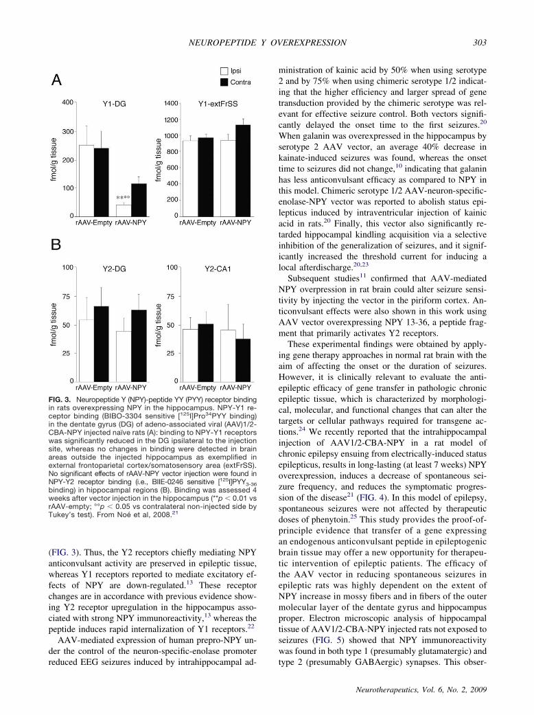
(FIG. 3). Thus, the Y2 receptors chiefly mediating NPYanticonvulsant activity are preserved in epileptic tissue,whereas Y1 receptors reported to mediate excitatory ef-fects of NPY are down-regulated.13 These receptorchanges are in accordance with previous evidence show-ing Y2 receptor upregulation in the hippocampus asso-ciated with strong NPY immunoreactivity,13 whereas thepeptide induces rapid internalization of Y1 receptors.22
AAV-mediated expression of human prepro-NPY un-der the control of the neuron-specific-enolase promoterreduced EEG seizures induced by intrahippocampal ad-
ministration of kainic acid by 50% when using serotype2 and by 75% when using chimeric serotype 1/2 indicat-ing that the higher efficiency and larger spread of genetransduction provided by the chimeric serotype was rel-evant for effective seizure control. Both vectors signifi-cantly delayed the onset time to the first seizures.20
When galanin was overexpressed in the hippocampus byserotype 2 AAV vector, an average 40% decrease inkainate-induced seizures was found, whereas the onsettime to seizures did not change,10 indicating that galaninhas less anticonvulsant efficacy as compared to NPY inthis model. Chimeric serotype 1/2 AAV-neuron-specific-enolase-NPY vector was reported to abolish status epi-lepticus induced by intraventricular injection of kainicacid in rats.20 Finally, this vector also significantly re-tarded hippocampal kindling acquisition via a selectiveinhibition of the generalization of seizures, and it signif-icantly increased the threshold current for inducing alocal afterdischarge.20,23
Subsequent studies11 confirmed that AAV-mediatedNPY overpression in rat brain could alter seizure sensi-tivity by injecting the vector in the piriform cortex. An-ticonvulsant effects were also shown in this work usingAAV vector overexpressing NPY 13-36, a peptide frag-ment that primarily activates Y2 receptors.These experimental findings were obtained by apply-
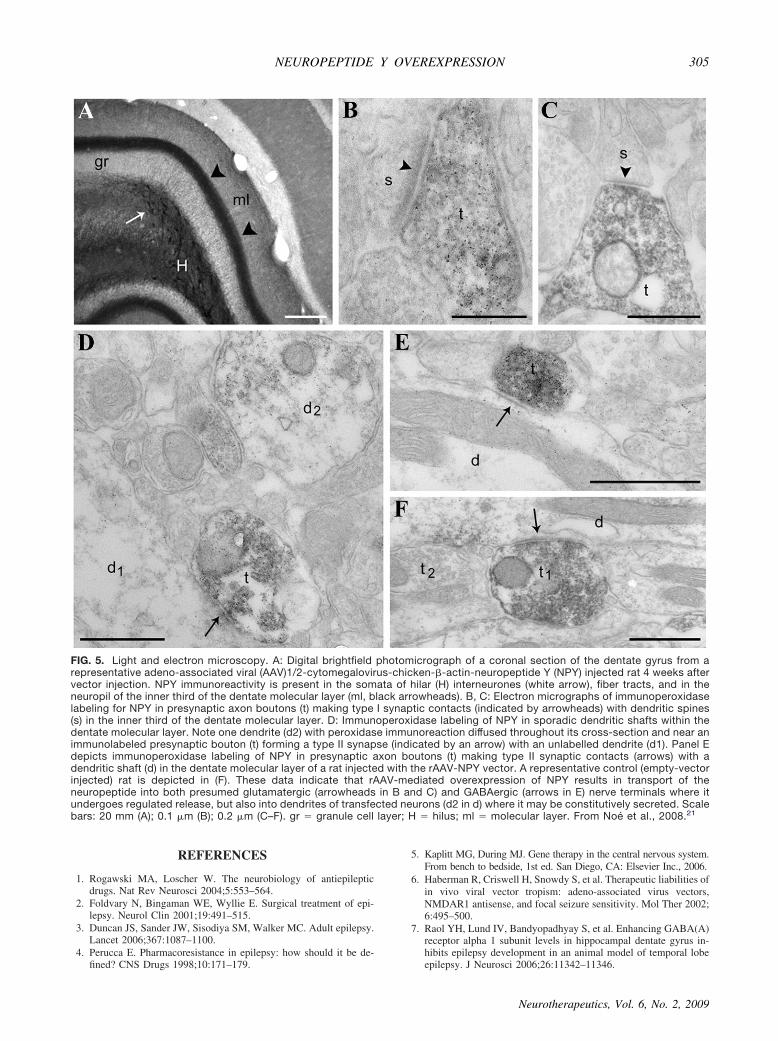
ing gene therapy approaches in normal rat brain with theaim of affecting the onset or the duration of seizures.However, it is clinically relevant to evaluate the anti-epileptic efficacy of gene transfer in pathologic chronicepileptic tissue, which is characterized by morphologi-cal, molecular, and functional changes that can alter thetargets or cellular pathways required for transgene ac-tions.24 We recently reported that the intrahippocampalinjection of AAV1/2-CBA-NPY in a rat model ofchronic epilepsy ensuing from electrically-induced statusepilepticus, results in long-lasting (at least 7 weeks) NPYoverexpression, induces a decrease of spontaneous sei-zure frequency, and reduces the symptomatic progres-sion of the disease21 (FIG. 4). In this model of epilepsy,spontaneous seizures were not affected by therapeuticdoses of phenytoin.25 This study provides the proof-of-principle evidence that transfer of a gene expressingan endogenous anticonvulsant peptide in epileptogenicbrain tissue may offer a new opportunity for therapeu-tic intervention of epileptic patients. The efficacy ofthe AAV vector in reducing spontaneous seizures inepileptic rats was highly dependent on the extent ofNPY increase in mossy fibers and in fibers of the outermolecular layer of the dentate gyrus and hippocampusproper. Electron microscopic analysis of hippocampaltissue of AAV1/2-CBA-NPY injected rats not exposed toseizures (FIG. 5) showed that NPY immunoreactivitywas found in both type 1 (presumably glutamatergic) andtype 2 (presumably GABAergic) synapses. This obser-
FIG. 3. Neuropeptide Y (NPY)-peptide YY (PYY) receptor bindingin rats overexpressing NPY in the hippocampus. NPY-Y1 re-ceptor binding (BIBO-3304 sensitive [125I]Pro34PYY binding)in the dentate gyrus (DG) of adeno-associated viral (AAV)1/2-CBA-NPY injected naïve rats (A): binding to NPY-Y1 receptorswas significantly reduced in the DG ipsilateral to the injectionsite, whereas no changes in binding were detected in brainareas outside the injected hippocampus as exemplified inexternal frontoparietal cortex/somatosensory area (extFrSS).No significant effects of rAAV-NPY vector injection were found inNPY-Y2 receptor binding (i.e., BIIE-0246 sensitive [125I]PYY3-36binding) in hippocampal regions (B). Binding was assessed 4weeks after vector injection in the hippocampus (**p � 0.01 vsrAAV-empty; °°p � 0.05 vs contralateral non-injected side byTukey’s test). From Noé et al, 2008.21
NEUROPEPTIDE Y OVEREXPRESSION 303
Neurotherapeutics, Vol. 6, No. 2, 2009

vation is consistent with the immunohistochemical local-ization of the peptide in granule cell mossy fibers and inhilar interneurons (FIG. 1).
Initial investigations of cognitive functions in naïverats showed no significant alterations in learning andshort-term memory performance after injection ofAAV1/2-CBA-NPY,21 suggesting that this physiolog-ical function is not compromised by the peptide over-expression. Transgene NPY overexpression, providessignificant anticonvulsant effects in the kindling mo-del,20,23 but does not further affect long-term potenti-ation that is significantly attenuated in kindled rats.23
CONCLUSIONS
Our studies show that intrahippocampal injection ofAAV vectors mediating NPY overexpression in rat mod-els of acute seizures, epileptogenesis as well as chronicepilepsy results in long-lasting peptide overexpression inneurons and concomitant powerful anticonvulsant ef-fects. NPY is involved in various functions, includingthe regulation of blood pressure, circadian rhythms, feed-ing behavior, anxiety, memory processing, and cogni-tion,26,27 raising the possibility of side effects conse-quent to peptide overexpression. However, many ofthese functions are mediated by extrahippocampal ar-eas. The highly localized AAV-vector mediated over-expression of NPY makes it unlikely that physiologi-cal processes mediated by extrahippocampal brainregions are affected.20,21
These findings provide first proof-of-principle evi-dence that transfer of a gene expressing an endogenousanticonvulsant peptide in chronic epileptic tissue mayoffer a novel therapeutic intervention, alternative to sur-gery, in patients with drug-refractory epilepsy.Further development in gene therapy strategies is now
focused particularly in the area of carrier design and itsin vivo delivery, cell-type specificity of transgene ex-pression, and transgene expression regulation. Moreover,before treatments can be brought to the clinic, not onlythe efficacy but also the safety of the treatment, such asthe immunogenicity of the transgene product or of thevector serotypes, for example, must be carefully ad-dressed and evaluated using clinically relevant experi-mental models.
DisclosureM.J.D. is a founder and consultant to Neurologix, Inc.,
a Delaware-based company that has an interest in thecommercialization of recombinant AAV vectors for thetreatment of neurological disorders.
Acknowledgments: This study was supported by TelethonOnlus Foundation and Fondazione Monzino (AV) and TheAcademy of Finland and The Juselius Foundation (AP), andNeurologix Inc. We thank Mrs. Merja Lukkari for excellenttechnical assistance in histology. The authors thank Drs.Valentina Vaghi, Walter Kaufmann, Massimo Rizzi, andMs. Anna Wieselthaler-Holzl for their contributions to thisstudy.
FIG. 4. Effect of rAAV-neuropeptide Y (NPY) on spontaneous seizures in epileptic rats. Proportion of rats with progressive increasein seizure frequency (�1.5-fold increase as compared to the baseline). In the AAV-NPY group, seizure frequency increased�1.5-fold only in 20% of rats as compared to 64% in the rAAV-empty group (p � 0.05, by �2 test). In the remaining 80% of ratsin the rAAV-NPY group seizure frequency increased less than 1.5-fold as compared to the baseline (nonprogressive). Moreover,half of these rats had a net 50% decrease in seizure frequency as compared to their respective baseline. Adapted from Noé et al.,2008.21
NOÉ ET AL.304
Neurotherapeutics, Vol. 6, No. 2, 2009
REFERENCES
1. Rogawski MA, Loscher W. The neurobiology of antiepilepticdrugs. Nat Rev Neurosci 2004;5:553–564.
2. Foldvary N, Bingaman WE, Wyllie E. Surgical treatment of epi-lepsy. Neurol Clin 2001;19:491–515.
3. Duncan JS, Sander JW, Sisodiya SM, Walker MC. Adult epilepsy.Lancet 2006;367:1087–1100.
4. Perucca E. Pharmacoresistance in epilepsy: how should it be de-fined? CNS Drugs 1998;10:171–179.
5. Kaplitt MG, During MJ. Gene therapy in the central nervous system.From bench to bedside, 1st ed. San Diego, CA: Elsevier Inc., 2006.
6. Haberman R, Criswell H, Snowdy S, et al. Therapeutic liabilities ofin vivo viral vector tropism: adeno-associated virus vectors,NMDAR1 antisense, and focal seizure sensitivity. Mol Ther 2002;6:495–500.
7. Raol YH, Lund IV, Bandyopadhyay S, et al. Enhancing GABA(A)receptor alpha 1 subunit levels in hippocampal dentate gyrus in-hibits epilepsy development in an animal model of temporal lobeepilepsy. J Neurosci 2006;26:11342–11346.
FIG. 5. Light and electron microscopy. A: Digital brightfield photomicrograph of a coronal section of the dentate gyrus from arepresentative adeno-associated viral (AAV)1/2-cytomegalovirus-chicken--actin-neuropeptide Y (NPY) injected rat 4 weeks aftervector injection. NPY immunoreactivity is present in the somata of hilar (H) interneurones (white arrow), fiber tracts, and in theneuropil of the inner third of the dentate molecular layer (ml, black arrowheads). B, C: Electron micrographs of immunoperoxidaselabeling for NPY in presynaptic axon boutons (t) making type I synaptic contacts (indicated by arrowheads) with dendritic spines(s) in the inner third of the dentate molecular layer. D: Immunoperoxidase labeling of NPY in sporadic dendritic shafts within thedentate molecular layer. Note one dendrite (d2) with peroxidase immunoreaction diffused throughout its cross-section and near animmunolabeled presynaptic bouton (t) forming a type II synapse (indicated by an arrow) with an unlabelled dendrite (d1). Panel Edepicts immunoperoxidase labeling of NPY in presynaptic axon boutons (t) making type II synaptic contacts (arrows) with adendritic shaft (d) in the dentate molecular layer of a rat injected with the rAAV-NPY vector. A representative control (empty-vectorinjected) rat is depicted in (F). These data indicate that rAAV-mediated overexpression of NPY results in transport of theneuropeptide into both presumed glutamatergic (arrowheads in B and C) and GABAergic (arrows in E) nerve terminals where itundergoes regulated release, but also into dendrites of transfected neurons (d2 in d) where it may be constitutively secreted. Scalebars: 20 mm (A); 0.1 �m (B); 0.2 �m (C–F). gr � granule cell layer; H � hilus; ml � molecular layer. From Noé et al., 2008.21
NEUROPEPTIDE Y OVEREXPRESSION 305
Neurotherapeutics, Vol. 6, No. 2, 2009

8. Mazarati AM. Galanin and galanin receptors in epilepsy. Neu-ropeptides 2004;38:331–343.
9. Noé F, Nissinen J, Pitkanen A, et al. Gene therapy in epilepsy: thefocus on NPY. Peptides 2007;28:377–383.
10. Lin EJ, Richichi C, Young D, Baer K, Vezzani A, During MJ.Recombinant AAV-mediated expression of galanin in rat hip-pocampus suppresses seizure development. Eur J Neurosci 2003;18:2087–2092.
11. Foti S, Haberman RP, Samulski RJ, McCown TJ. Adeno-associ-ated virus-mediated expression and constitutive secretion of NPYor NPY13-36 suppresses seizure activity in vivo. Gene Ther 2007;14:1534–1536.
12. de Lanerolle NC, Kim JH, Robbins RJ, Spencer DD. Hippocampalinterneuron loss and plasticity in human temporal lobe epilepsy.Brain Res 1989;495:387–395.
13. Vezzani A, Sperk G, Colmers WF. Neuropeptide Y: emergingevidence for a functional role in seizure modulation. Trends Neu-rosci 1999;22:25–30.
14. Furtinger S, Pirker S, Czech T, Baumgartner C, Ransmayr G,Sperk G. Plasticity of Y1 and Y2 receptors and neuropeptide Yfibers in patients with temporal lobe epilepsy. J Neurosci 2001;21:5804–5812.
15. Patrylo PR, van den Pol AN, Spencer DD, Williamson A. NPYinhibits glutamatergic excitation in the epileptic human dentategyrus. J Neurophysiol 1999;82:478–483.
16. Baraban SC, Hollopeter G, Erickson JC, Schwartzkroin PA,Palmiter RD. Knock-out mice reveal a critical antiepileptic role forneuropeptide Y. J Neurosci 1997;17:8927–8936.
17. El Bahh B, Balosso S, Hamilton T, et al. The anti-epileptic actionsof neuropeptide Y in the hippocampus are mediated by Y2 and notY5 receptors. Eur J Neurosci 2005;22:1417–1430.
18. Colmers WF, Lukowiak K, Pittman QJ. Neuropeptide Y action inthe rat hippocampal slice: site and mechanism of presynaptic in-hibition. J Neurosci 1988;8:3827–3837.
19. Greber S, Schwarzer C, Sperk G. Neuropeptide Y inhibits potas-sium-stimulated glutamate release through Y2 receptors in rat hip-pocampal slices in vitro. Br J Pharmacol 1994;113:737–740.
20. Richichi C, Lin EJ, Stefanin D, et al. Anticonvulsant and antiepi-leptogenic effects mediated by adeno-associated virus vector neu-ropeptide Y expression in the rat hippocampus. J Neurosci 2004;24:3051–3059.
21. Noé F, Pool AH, Nissinen J, et al. Neuropeptide Y gene therapydecreases chronic spontaneous seizures in a rat model of temporallobe epilepsy. Brain 2008;131:1506–1515.
22. Pheng LH, Dumont Y, Fournier A, Chabot JG, Beaudet A, QuirionR. Agonist- and antagonist-induced sequestration/internalization ofneuropeptide Y Y1 receptors in HEK293 cells. Br J Pharmacol2003;139:695–704.
23. Sorensen AT, Nikitidou L, Ledri M, et al. Hippocampal NPY genetransfer attenuates seizures without affecting epilepsy-induced im-pairment of LTP. Exp Neurol 2009;215:328–333.
24. Holmes GL, Ben-Ari Y. Seizing hold of seizures (News andViews). Nat Med 2003;9:994–996.
25. van Vliet EA, van Schaik R, Edelbroek PM, et al. Inhibition of themultidrug transporter P-glycoprotein improves seizure control in phe-nytoin-treated chronic epileptic rats. Epilepsia 2006;47:672–680.
26. Danger JM, Tonon MC, Jenks BG, et al. Neuropeptide Y: local-ization in the central nervous system and neuroendocrine func-tions. Fundam Clin Pharmacol 1990;4:307–340.
27. Hokfelt T, Broberger C, Zhang X, et al. Neuropeptide Y: someviewpoints on a multifaceted peptide in the normal and diseasednervous system. Brain Res Brain Res Rev 1998;26:154–166.
NOÉ ET AL.306
Neurotherapeutics, Vol. 6, No. 2, 2009
Copyright © 2022 FDOKUMEN