
Lentiviral fluorescent protein expression vectors for biotinylation proteomics
15
Lentiviral Fluorescent Protein Expression Vectors for Biotinylation Proteomics Irene Riz 1 , Teresa S. Hawley 2 , and Robert G. Hawley 1 1 Department of Anatomy and Regenerative Biology, The George Washington University, Washington, DC 20037, USA 2 Flow Cytometry Core Facility, The George Washington University, Washington, DC 20037, USA Abstract In vivo biotinylation tagging, based on a method in which a protein of interest is tagged with a peptide that is biotinylated in vivo by coexpression of E.coli BirA biotin ligase, has been successfully used for isolation of protein-protein and protein-DNA complexes in mammalian cells. We describe a modification of this methodology in which cells stably expressing the tagged gene of interest and the BirA gene can be selected by fluorescence activated cell sorting (FACS). We recently implemented this approach to isolate and characterize proteins associated with TLX1, a homeodomain transcription factor with leukemogenic function. The modified technique utilizes two components: a lentiviral vector coexpressing the gene of interest containing a biotinylation tag on a bicistronic transcript together with a downstream yellow fluorescent protein gene; and a second lentiviral vector encoding a fusion protein composed of bacterial BirA linked to the green fluorescent protein. This FACS-based binary in vivo biotinylation tagging system allows precise control over the levels of BirA-mediated biotinylation as well as the expression of the gene of interest, which is especially important if high-level expression negatively impacts cell growth or viability. Keywords In vivo biotinylation tagging; E.coli BirA biotin ligase; simian immunodeficiency virus-based lentiviral vectors; fluorescent protein reporters; TLX1 homeodomain transcription factor 1. Introduction A variety of biochemical methods exist for studying protein-protein interactions in mammalian cells (1,2), including those based on fluorescence activated cell sorting (FACS) (3) (see also the chapter by Szöllõsi et al., this volume). Strouboulis and colleagues developed a method in which a recombinant protein is tagged with a peptide that is biotinylated in vivo by the coexpressed E.coli BirA biotin ligase (4). A strength of this approach is the very high affinity interaction between biotinylated substrates and streptavidin (K d = 10 −15 ), which allows high stringencies to be employed during purification (5). The biotinylation tagging method described here has been successfully used for the single-step purification (6) of a number of transcription factor complexes in nuclear extracts of mammalian cells (4,7–12). Correspondence: R.G. Hawley, Department of Anatomy and Regenerative Biology, The George Washington University, 2300 I Street NW, Washington, DC 20037, USA; Phone: 202-994-3511; Fax: 202-994-8885; [email protected]. NIH Public Access Author Manuscript Methods Mol Biol. Author manuscript; available in PMC 2012 February 14. Published in final edited form as: Methods Mol Biol. 2011 ; 699: 431–447. doi:10.1007/978-1-61737-950-5_21. NIH-PA Author Manuscript NIH-PA Author Manuscript NIH-PA Author Manuscript
Transcript of Lentiviral fluorescent protein expression vectors for biotinylation proteomics

Lentiviral Fluorescent Protein Expression Vectors forBiotinylation Proteomics
Irene Riz1, Teresa S. Hawley2, and Robert G. Hawley1
1Department of Anatomy and Regenerative Biology, The George Washington University,Washington, DC 20037, USA2Flow Cytometry Core Facility, The George Washington University, Washington, DC 20037, USA
AbstractIn vivo biotinylation tagging, based on a method in which a protein of interest is tagged with apeptide that is biotinylated in vivo by coexpression of E.coli BirA biotin ligase, has beensuccessfully used for isolation of protein-protein and protein-DNA complexes in mammalian cells.We describe a modification of this methodology in which cells stably expressing the tagged geneof interest and the BirA gene can be selected by fluorescence activated cell sorting (FACS). Werecently implemented this approach to isolate and characterize proteins associated with TLX1, ahomeodomain transcription factor with leukemogenic function. The modified technique utilizestwo components: a lentiviral vector coexpressing the gene of interest containing a biotinylation tagon a bicistronic transcript together with a downstream yellow fluorescent protein gene; and asecond lentiviral vector encoding a fusion protein composed of bacterial BirA linked to the greenfluorescent protein. This FACS-based binary in vivo biotinylation tagging system allows precisecontrol over the levels of BirA-mediated biotinylation as well as the expression of the gene ofinterest, which is especially important if high-level expression negatively impacts cell growth orviability.
KeywordsIn vivo biotinylation tagging; E.coli BirA biotin ligase; simian immunodeficiency virus-basedlentiviral vectors; fluorescent protein reporters; TLX1 homeodomain transcription factor
1. IntroductionA variety of biochemical methods exist for studying protein-protein interactions inmammalian cells (1,2), including those based on fluorescence activated cell sorting (FACS)(3) (see also the chapter by Szöllõsi et al., this volume). Strouboulis and colleaguesdeveloped a method in which a recombinant protein is tagged with a peptide that isbiotinylated in vivo by the coexpressed E.coli BirA biotin ligase (4). A strength of thisapproach is the very high affinity interaction between biotinylated substrates andstreptavidin (Kd = 10−15), which allows high stringencies to be employed during purification(5). The biotinylation tagging method described here has been successfully used for thesingle-step purification (6) of a number of transcription factor complexes in nuclear extractsof mammalian cells (4,7–12).
Correspondence: R.G. Hawley, Department of Anatomy and Regenerative Biology, The George Washington University, 2300 I StreetNW, Washington, DC 20037, USA; Phone: 202-994-3511; Fax: 202-994-8885; [email protected].
NIH Public AccessAuthor ManuscriptMethods Mol Biol. Author manuscript; available in PMC 2012 February 14.
Published in final edited form as:Methods Mol Biol. 2011 ; 699: 431–447. doi:10.1007/978-1-61737-950-5_21.
NIH
-PA Author Manuscript
NIH
-PA Author Manuscript
NIH
-PA Author Manuscript

In the initial version of the method (4), expression cassettes encoding the tagged protein ofinterest and BirA were subcloned into separate plasmids that also carried a selectable drugresistance gene, the neomycin phosphotransferase gene and the puromycin N-acetyltransferase gene, respectively. Typically, target cells would be transfected with theBirA plasmid by physical methods such as electroporation, stable clones obtained byselection for puromycin resistance, and then screened for BirA expression by Northern orWestern blot analysis. An appropriate BirA-expressing clone would then be transfected withthe vector encoding the tagged protein of interest and stable cells expressing the taggedprotein biotinylated by BirA obtained by selection for cells that were resistant to theneomycin analog geneticin as well as to puromycin. A drawback of this experimental designis that the optimal concentration of drugs used for selection depends on the cell line andneeds to be determined a priori. Moreover, selection for drug resistance of the transfectedplasmids does not necessarily ensure coexpression of BirA or the tagged gene of interest.
Hoang and colleagues reported a modification of this methodology in which the tagged geneof interest was coexpressed from a lentiviral vector on a bicistronic transcript that alsocontained the BirA gene (9). In addition, the BirA gene was expressed as a fusion proteinwith the green fluorescent protein (BirA-GFP), allowing stably transduced cellscoexpressing the tagged gene of interest and BirA to be isolated by FACS. As describedbelow, we have further modified the FACS-based strategy of Hoang and colleagues. Theexpression system we have developed consists of two components: a lentiviral vectorcoexpressing the gene of interest containing a biotinylation tag together with a downstreamyellow fluorescent protein (YFP) gene via an encephalomyocarditis virus internal ribosomeentry site (IRES); and a second lentiviral vector expressing GFP-BirA (12).
Our work is focused on TLX1 (T-cell leukemia homeobox 1, previously known as HOX11 orTCL3), an evolutionarily conserved member of the dispersed NKL (NK-Like or NK-Linked)subclass of homeobox genes (13,14). The murine ortholog of human TLX1 is essential forsplenogenesis and required for the development of certain neurons (15,16). Although TLX1is not expressed in the hematopoietic system, its inappropriate activation is a recurrent eventin human T cell acute lymphoblastic leukemia (T-ALL) (17). The manner in whichderegulated TLX1 expression induces neoplastic conversion remains to be fully elucidated(18,19). Several lines of evidence indicate that TLX1 functions as a transcriptional regulatorthat can either activate or repress gene expression via direct or indirect modes of action (20–28). In this regard, TLX1 has been reported to form protein-protein interactions with othertranscription factors as well as with a number of transcriptional coregulators and chromatin-modifying enzymes. Among the molecules that have been identified are: CTF1, a ubiquitoustranscription factor that associates with TFIIB and the basal transcription machinery (29);MEIS and PBX members of the TALE (three amino acid loop extension) superclass ofhomeodomain proteins (30,31); the acetyltransferase coactivator CREB-binding protein(26); the serine/threonine phosphatases PP1 and PP2A (25,32); and the eukaryotic initiationfactor 4E (eIF4E) (33).
We recently implemented the in vivo biotinylation tagging approach to isolate andcharacterize the various TLX1 protein complexes in T-ALL cells (12). Our initial attemptsto coexpress biotinylation-tagged TLX1 and GFP-BirA from a bicistronic lentiviral vectorwere unsuccessful because the cells did not tolerate high levels of TLX1. Since BirA wasexpressed on the same transcript, it was selected against. As a result, much higherexpression levels of BirA were achieved with the empty GFP-BirA vector, making itdifficult to obtain similar levels of endogenous biotinylated proteins for comparisonsbetween the experimental and control samples. To circumvent these problems, we designeda two-component expression system using the simian immunodeficiency virus (SIV)lentiviral vector backbone pCL20cSLFR MSCV-GFP (34). In pCL20cSLFR MSCV-GFP,
Riz et al. Page 2
Methods Mol Biol. Author manuscript; available in PMC 2012 February 14.
NIH
-PA Author Manuscript
NIH
-PA Author Manuscript
NIH
-PA Author Manuscript

which was constructed from the nonpathogenic SIVmac1A11 isolate, the GFP gene isexpressed from an internal promoter derived from the long terminal repeat (LTR) of themurine stem cell virus (MSCV), which is highly active in most mammalian cell types (35).A biotinylation tagging vector expressing a COOH-terminal tagged TLX1 (TLX1bio) underthe control of the MSCV LTR was created by inserting the TLX1 coding region in frameupstream of the coding sequences for the BirA target peptide linked to an IRES-YFPcassette, generating pCL20cSLFR MSCV-TLX1bio-IRES-YFP (component 1). A BirAexpression vector was similarly constructed by replacing the GFP gene of pCL20cSLFRMSCV-GFP with GFP-BirA, generating pCL20cSLFR MSCV-GFP-BirA (component 2)(Fig. 1A).
In pilot studies, SupT1 cells, which are negative for TLX1 expression but which are arrestedat the same stage of T-cell differentiation as TLX1+ T-ALL cells, were transduced withrecombinant CL20cSLFR MSCV-TLX1bio-IRES-YFP lentiviral vector particles and/orrecombinant CL20cSLFR MSCV-GFP-BirA lentiviral vector particles and stable cell linesobtained by sorting for YFP and/or GFP fluorescence (Fig. 1B). SupT1 cells expressingGFP-BirA alone served as control for binding of any biotinylated endogenous proteins to thestreptavidin beads. Using single-step affinity capture on streptavidin beads, followed bymatrix-assisted laser desorption ionization time-of-flight (MALDI-TOF) mass spectrometry,we identified the Groucho/transducin-like Enhancer of split (Gro/TLE) family memberTLE1 as an in vivo binding partner of TLX1 (12) (Fig. 2A). Gro/TLE proteins are regulatedby multiple signaling cascades and serve as corepressors for different families oftranscription factors (36,37). The transcription factors that interact with Gro/TLEcorepressors contain short peptide sequences related to either WRPW or to FXIXXIL(where X is any amino acid), the latter referred to as the Engrailed homology 1 (Eh1) motif,a repression domain first identified in the Drosophila Engrailed homeodomain protein (38).We demonstrated that TLX1 interacts with TLE1 in vitro and in vivo through a 7 amino acidsequence encompassing amino acids 19 to 25 (FGIDQIL) that exhibits similarity to an Eh1motif (Fig. 2B). Moreover, we found that this motif was required for optimal regulation ofexpression of two TLX1 target genes, Aldh1a1 and Fhl1 (21,22,27). As shown in Fig. 2A, apreviously reported in vitro TLX1 interacting protein, eIF4E (33), is also efficientlyprecipitated by streptavidin affinity capture of in vivo TLX1bio protein complexes. Thus, webelieve that our adaptation of the FACS-based in vivo biotinylation tagging system willprovide a powerful tool for characterization of transcription factor and other protein-proteincomplexes in mammalian cells.
2. Materials2.1. Biotinylation Tagging Vectors
1. Starting material plasmids: the SIVmac1A11-based pCL20cSLFR MSCV-GFPlentiviral vector was provided by Arthur Nienhuis (St. Jude Children’s ResearchHospital, Memphis, TN) (34); coding sequences for a 23-amino acid biotinylationpeptide tag (see Note 1) and the GFP-BirA fusion gene were obtained from thepTRIP/BPcter/IRES/EGFPBirA plasmid (provided by John Strouboulis, ErasmusMedical Center, Rotterdam, The Netherlands) (9).
2. SIV lentiviral vectors: pCL20cSLFR MSCV-TLX1bio-IRES-YFP expressing theTLX1 homeodomain transcription factor containing a COOH-terminal biotinylation
1More recently, we have constructed an NH2-terminal biotin-tagged TLX1 (bioTLX1) modeled after the bioNanog homeodomainprotein and other biotin-tagged transcription factors described by Orkin and colleagues (46,47), which is based on the 15 amino acidAviTag™ sequence (GLNDIFEAQKIEWHE) that is very efficiently biotinylated in vivo by BirA (Avidity, LLC, Aurora, CO;www.avidity.com).
Riz et al. Page 3
Methods Mol Biol. Author manuscript; available in PMC 2012 February 14.
NIH
-PA Author Manuscript
NIH
-PA Author Manuscript
NIH
-PA Author Manuscript

peptide tag linked to a downstream yellow fluorescent protein (YFP) reporter on abicistronic transcript (12); pCL20cSLFR MSCV-GFP-BirA expressing a fusionprotein consisting of a green fluorescent protein (GFP) reporter and the bacterialBirA biotin ligase (GFP-BirA) (12). The pCL20cSLFR MSCV-IRES-YFP andpCL20cSLFR MSCV-GFP-BirA plasmids can be obtained from the authors uponrequest.
3. Restriction and modifying enzymes for PCR amplification and subcloning of genesof interest.
4. Plasmid DNA purification kits: QIAquick Gel Extraction Kit, QIAGEN PlasmidMaxi Kit (Qiagen, Valencia, CA).
2.2. Production and Titration of Vector Particles2.2.1. Transient Transfection
1. 293T/17 (293T) human embryonic kidney cell line (CRL-11268; American TypeCulture Collection (ATCC), Manassas, VA) (see Note 2).
2. 293T cell culture medium: Dulbecco’s Modified Eagle’s Medium (DMEM)supplemented with 4.5 g/L glucose, 2 mM L-glutamine, 50 IU/mL penicillin, 50μg/mL streptomycin, 10% heat-inactivated fetal bovine serum (FBS). Store at 4°Cand warm up to 37°C before use.
3. Phosphate-buffered saline without Ca2+ and Mg2+ (PBS): 137 mM NaCl, 2.68 mMKCl, 1.47 mM KH2PO4, 8.1 mM Na2HPO4 (adjust pH to 7.4 if necessary, using 1N HCl or 1 N NaOH).
4. Solution of trypsin and ethylenediamine tetraacetic acid (trypsin-EDTA): 0.05%trypsin, 0.53 mM EDTA in PBS.
5. pCL20cSLFR MSCV-IRES-YFP biotinylation tagging SIV lentiviral vectorplasmid DNA containing the gene of interest; pCL20cSLFR MSCV-GFP-BirA SIVlentiviral vector plasmid DNA.
6. SIV lentiviral packaging plasmid DNAs (e.g., pCAG-SIVgprre and pCAG4-RTR-SIV; provided Arthur Nienhuis) (34).
7. Vesicular stomatitis virus G (VSV-G) glycoprotein envelope plasmid DNA (e.g.,pMD.G; (39,40)).
8. 2.5 M CaCl2: Dissolve 183.7 g CaCl2 dihydrate (tissue culture grade) in deionizedwater. Bring the volume up to 500 mL and filter-sterilize using a 0.22-μmnitrocellulose filter. Store at −20°C.
9. 2X N-(2-hydroxyethyl)piperazine-N′-(2-ethanesulfonic acid) (HEPES)-bufferedsaline (2X HBS): 50 mM HEPES (H4034; Sigma-Aldrich Corp., St. Louis, MO),280 mM NaCl, 1.5 mM Na2HPO4. Titrate to pH 7.05 with 5 N NaOH. Filtersterilize using a 0.22-μm nitrocellulose filter. Store as single use aliquots at −20°C.
2The 293T/17 cell line is a clone of the 293T (293tsA1609neo) human embryonic kidney cell line (48), which was selected for hightransfectability and capability of producing high-titer vector stocks. The 293T cell line is a derivative of 293 cells into which thesimian virus 40 (SV40) large T antigen gene was inserted. 293 cells express the adenovirus serotype 5 E1A 12S and 13S geneproducts, which strongly transactivate transcription from expression vectors containing the human CMV enhancer-promoter (49).Expression of the SV40 large T antigen by 293T cells may stimulate extrachromosomal replication of plasmids containing the SV40origin of replication during transient transfection.
Riz et al. Page 4
Methods Mol Biol. Author manuscript; available in PMC 2012 February 14.
NIH
-PA Author Manuscript
NIH
-PA Author Manuscript
NIH
-PA Author Manuscript

2.2.2. Collection and Concentration of Vector Particles1. 0.45-μm pore-size filter units: Stericup 150-mL unit, low protein binding Durapore
(PVDF) unit (Millipore Corp., Bedford, MA).
2. Ultracentrifuge, polycarbonate 70-mL ultracentrifuge bottles and caps.
2.2.3. Titration of Vector Particles1. HT1080 human fibrosarcoma cells (CCL-121; ATCC).
2. HT1080 cell culture medium: same as 293T cell culture medium (see Subheading2.2.1.).
3. Polybrene (hexadimethrine bromide, H9268; Sigma-Aldrich Corp.) stock solution(1000X): 8 mg/mL in sterile, deionized water. Aliquot and store at −20°C.
4. Analytical flow cytometer.
2.3. Generation of Cells Stably Expressing Biotin-Tagged Proteins1. Replication-defective SIV lentiviral vector particles (e.g., CL20cSLFR MSCV-
TLX1bio-IRES-YFP and CL20cSLFR MSCV-GFP-BirA).
2. Target cells (e.g., SupT1 human T-ALL cell line, CRL-1942; ATCC).
3. Suspension cell culture medium: Iscove’s modified Dulbecco’s medium (IMDM)supplemented with 2 mM L-glutamine, 50 IU/mL penicillin, 50 μg/mLstreptomycin, 10% heat-inactivated FBS.
4. Polybrene stock solution (see Subheading 2.2.3.).
5. Fluorescence-activated cell sorter (see Note 3).
2.4. Cell Fractionation1. Phenylmethylsulphonyl fluoride (PMSF): 0.5 M in dimethylsulfoxide. Store at
−20°C.
2. Dithiothreitol (DTT): 0.5 M in deionized water. Store at −20°C.
3. Protease inhibitors (Protease inhibitor cocktail tablets, 11836153001; RocheDiagnostics Corp., Indianapolis, IN).
4. Phosphatase inhibitors (PhosSTOP phosphatase inhibitor cocktail tablets,04906837001; Roche).
5. Hypotonic wash buffer: 10 mM HEPES, pH 7.9, 1.5 mM MgCl2, 10 mM KCl, 0.5mM PMSF, 0.5 mM DTT.
6. NP40 nuclear extraction (NP40 NE) buffer: 20 mM HEPES, pH 7.9, 25% glycerol,1.5 mM MgCl2, 20 mM KCl, 0.25 M NaCl, 0.1% NP40, 5 mM EDTA, 1 mMPMSF, 0.5 mM DTT, protease inhibitors (1 tablet for 5 mL), phosphatase inhibitors(1 tablet for 5 mL).
7. Other solutions: PBS; 0.4% trypan blue in normal saline.
8. (Optional) Sonifier (e.g., Branson Sonifier 250 with microtip).
3Standard detection filters supplied with most commercial flow cytometers are unable to distinguish between GFP and YFP signals.Suppliers of custom optical filters include Omega Optical Inc. (Brattleboro, VT), Chroma Technology Corp. (Rockingham, VT), andSemrock (Rochester, NY).
Riz et al. Page 5
Methods Mol Biol. Author manuscript; available in PMC 2012 February 14.
NIH
-PA Author Manuscript
NIH
-PA Author Manuscript
NIH
-PA Author Manuscript

2.5. Streptavidin Affinity Precipitation and Mass Spectrometry Analysis1. Streptavidin sepharose beads (17-5113-01; GE Healthcare, Piscataway, NJ).
2. Chicken albumin: 20 mg/mL in deionized water. Store at −20°C.
3. HENG buffer: 20 mM HEPES, pH 7.9, 25% glycerol, 1.5 mM MgCl2, 0.25 mMEDTA.
4. NP40 T buffer: 70 mM Tris-HCl, pH 8.0, 25% glycerol, 1.5 mM MgCl2, 20 mMKCl, 0.25 M NaCl, 0.1% NP40, 5 mM EDTA, 1 mM PMSF, 0.5 mM DTT.
5. Tris-glycine SDS polyacrylamide gel electrophoresis (SDS-PAGE) gels, andassociated running and loading buffers.
6. SDS-PAGE staining reagents (Colloidal blue staining kit, LC6025; Invitrogen,Carlsbad, CA).
7. In-gel tryptic digestion reagents (Trypsin profile IGD kit, PP0100; Sigma AldrichCorp.).
8. ZipTip (0.6 μL C18 resin, ZTC18S096; Millipore Corp.).
9. Elution buffer: 50% acetonitrile (ACN), 0.1% trifluoroacetic acid (TFA) in Milli-Q® grade water.
10. α-cyano-4-hydroxycinnamic acid (α-cyano-CHCA, C8982; Sigma-Aldrich Corp.):10 mg/mL in 50% ACN, 0.05% TFA.
11. Mass spectrometer.
3. Methods3.1. Biotinylation Tagging Vectors
1. Subclone the gene of interest linked to a biotinylation peptide tag upstream of theIRES-YFP cassette in the pCL20cSLFR MSCV-IRES-YFP SIV lentiviral vector(see Notes 1 and 4).
2. Extract plasmid DNA using plasmid DNA purification kits.
3.2. Production and Titration of Vector Particles293T (293 human embryonic kidney cells expressing simian virus 40 [SV40] large tumor[T] antigen) cells are highly transfectable such that transient cotransfection with the self-inactivating SIV lentiviral vectors, packaging plasmids and envelope plasmids yields high-titer, replication-defective vector particles (41). Lentiviral vectors pseudotyped with theVSV-G glycoprotein have a broad host-cell range and can be utilized to transduce all celltypes (39,40).
3.2.1. Transient Transfection1. Propagate 293T cells in 293T cell culture medium at 37°C in a humidified
atmosphere with 5% CO2.
2. Passage the cells every 3 to 4 days by splitting them 1/4 to 1/8. Remove themedium and rinse the cells with PBS. Remove the PBS and add enough trypsin-
4Common recombinant DNA techniques are used for plasmid DNA preparations, restriction enzyme digestions, PCR amplificationsand subclonings. Detailed protocols for each technique can be obtained from commercial sources, as well as from various standardmolecular biology manuals. Accordingly, these techniques have not been described here.
Riz et al. Page 6
Methods Mol Biol. Author manuscript; available in PMC 2012 February 14.
NIH
-PA Author Manuscript
NIH
-PA Author Manuscript
NIH
-PA Author Manuscript

EDTA to cover the cells. Incubate the plate at room temperature until the cellsround up and detach. Add an equal volume of cell culture medium to inactivate thetrypsin. Collect the cells and centrifuge for 5 min at 375g. Resuspend the cells infresh cell culture medium for plating.
3. Transfect 293T cells with plasmid DNA using the calcium phosphate precipitationmethod (42). On the day before transfection, plate 293T cells in 7 mL of cellculture medium at a density of 5 × 10⁶ cells per 100-mm tissue culture dish.
4. Prepare a mixture of 20 μg of plasmid DNA composed as follows: 10 μg ofpCL20cSLFR MSCV-IRES-YFP biotinylation tagging SIV lentiviral vectorcontaining the gene of interest (or pCL20cSLFR MSCV-GFP-BirA SIV lentiviralvector), 6 μg of pCAG-SIVgprre plus 2 μg of pCAG4-RTR-SIV packagingplasmids, and 2 μg of the pMD.G VSV-G glycoprotein envelope plasmid (34) (seeNote 5). Bring the volume up to 400 μL with sterile, deionized water. Add 100 μLof 2.5 M CaCl2 and mix. Add the DNA/CaCl2 solution dropwise to 500 μL of 2XHBS in a 15-mL conical tube. Use a second pipettor and a 2-mL pipet to bubble the2X HBS as the DNA/CaCl2 solution is added. Vortex immediately for 5 sec andincubate at room temperature for 20 min. Add the 1 mL of DNA/calcium phosphatemixture directly to each 100-mm dish while swirling (see Note 6). Incubate thecells at 37°C overnight (16 h). Change the medium and culture for 24–48 h (thismethod usually results in transfection of 50–80% of the cells).
3.2.2. Collection and Concentration of Vector Particles—Collect the culturemedium containing vector particles 24–48 h after medium change. Centrifuge at 2000g for10 min to remove cellular debris and filter through a 0.45 μm pore-size filter (depending onthe volume, use a 150-mL filter unit or a small filter unit attached to a 5-mL syringe). Usedirectly for transductions or aliquot and store at −80°C (see Note 7).
Several procedures have been developed to concentrate lentiviral vector particles (43). Thechoice of concentration protocol depends on the envelope selected to pseudotype the particleand the quantities of particles to be produced. The stability of the VSV-G envelope proteinallows generation of high-titer lentiviral vector particles by ultracentrifugation, as describedhere.
1. Ultracentrifuge the vector particles at 50,000g and 4°C for 90 min.
5The pCL20cSLFR MSCV-GFP SIV-based vector system is modeled after ‘third generation’ lentiviral vector systems that haveincorporated features to enhance safety and improve the efficiency of vector particle production (50). Specifically, several viralaccessory proteins have been eliminated from the pCAG-SIVgprre gag-pol packaging construct, and the LTRs have been modified tocontain the human CMV enhancer-promoter and to eliminate Tat dependence during vector production (5′ LTR) and to be self-inactivating (3′ LTR) upon integration. Because both the expression of the pCL20cSLFR MSCV-GFP SIV lentiviral vector backboneas well as the gag and pol genes encoded by the pCAG-SIVgprre packaging construct are dependent on trans complementation by theRev protein, use of this system requires cotransfection with a rev expression construct (e.g., pCAG4-RTR-SIV).6Vector titer depends on the vector backbone design, the size and nature of inserted sequences as well as the efficiency of transfection.To achieve optimal transfection efficiency, use an exponentially growing culture (50–70% confluent) of 293T cells for transfectionand make sure that the cells form a uniform monolayer upon plating. Transfection efficiency is also affected by the quality of theplasmid DNA used. Commercially available plasmid DNA purification kits usually yield highly purified endotoxin-free supercoiledplasmid DNA (traditionally obtained by purifying on two separate cesium chloride gradients). Sterilize the plasmid DNA by ethanolprecipitation, resuspend the air-dried pellet in sterile, deionized water, and determine its concentration and quality byspectrophotometric analysis and gel electrophoresis. Pay special attention to the pH of the 2X HBS solution, which should be between7.05 and 7.12. Upon addition of the transfection mixture to the cells, a fine precipitate should develop within a few minutes.7pH and temperature fluctuations as well as freeze-and-thaw frequency could have an impact on the stability of vector particles, andhence, vector titer (51). Minimize pH changes by adding HEPES at a final concentration of 10 mM to buffer the cell culture medium.Once collected, vector particles should be kept on ice at all times. For future use, they should be stored in aliquots at −80°C. Avoidrepeated freezing and thawing.
Riz et al. Page 7
Methods Mol Biol. Author manuscript; available in PMC 2012 February 14.
NIH
-PA Author Manuscript
NIH
-PA Author Manuscript
NIH
-PA Author Manuscript

2. Discard supernatant. Using gentle pipetting, resuspend the pellet in ~100 μL ofmedium appropriate for the downstream application. To facilitate completeresuspension, vortex gently overnight at 4°C (see Note 8).
3. To remove cellular debris, centrifuge concentrated vector particles at 10,000g and4°C for 5 min in a microcentrifuge. Collect the supernatant containing the vectorparticles, aliquot and freeze at −80°C (see Note 9).
3.2.3. Titration of Vector Particles—The HT1080 human fibrosarcoma cell line can beused to determine the titer of the lentiviral vector particles.
1. Propagate HT1080 cells in HT1080 cell culture medium at 37°C in a humidifiedatmosphere with 5% CO2.
2. 4–6 h prior to titrating the vector particles, plate 2.5 × 10⁵ HT1080 cells into eachwell of a 6-well tissue culture dish.
3. Prepare serial dilutions of each vector preparation (e.g., 100, 10−1, and 10−2 forunconcentrated vector particles; 10−2, 10−3, and 10−4 for concentrated vectorparticles) using cell culture medium, in a final volume of 1 mL per dilution. Add 1μL of polybrene stock solution to each dilution. Remove the medium from the cellsand add 1 mL of each dilution to each well. Incubate at 37°C for 4 h.
4. Remove the vector particles after the 4-h transduction and replace with 2 mL offresh cell culture medium. Return to the CO2 incubator and incubate at 37°C.
5. After 48 h, determine the relative end-point vector titer (in transducing units permL [TU/mL]) by flow cytometric analysis. Evaluate GFP or YFP expression on ananalytical flow cytometer equipped with 488 nm excitation wavelength and a530/30-nm bandpass (BP) filter (44).
6. To determine vector titer, use the following equation: Vector titer = number ofHT1080 cells × % fluorescent protein-positive cells × dilution factor (see Note 10).
3.3. Generation of Cells Stably Expressing Biotin-Tagged ProteinsThe following protocol is used to transduce suspension cell lines with the recombinantbiotinylation tagging lentiviral vectors. For adherent cells, the protocol described inSubheading 3.2.3. for titration of vector particles is used.
1. Propagate cells (e.g., SupT1) in suspension cell culture medium at 37°C in ahumidified atmosphere with 5% CO2.
2. Prepare an appropriate dilution of vector particles in 1 mL of cell culture mediumcontaining 1 μL of polybrene stock solution. Resuspend 1–2 × 10⁵ cells in thediluted vector particles in a 14-mL conical polystyrene centrifuge tube.
3. Centrifuge the mixture of cells and vector particles at 600g and 15°C for 2 h.Resuspend the cell pellet in 0.5 mL of fresh cell culture medium and incubate
8Small (~2 to 5-mm-diameter) pellets should be visible after concentration by centrifugation. Expect a 50–75% recovery followingvector concentration.9The vector particle preparation can be further purified if necessary by centrifuging through a sucrose cushion (52).10When reporter proteins are evaluated by flow cytometry, it is advantageous to wait at least 5 days before analyzing the transducedcells. This will minimize the contribution of false positive signals due to pseudotransduction, which is the direct transfer of reporterprotein (adhered to vector particles or incorporated into vector particles) to the target cells; this is particularly problematic for VSV-Gglycoprotein-pseudotyped vectors (53,54). Note that it has also been shown that transgenes can be efficiently transiently expressedfrom unintegrated lentiviral vectors during this timeframe (10–14 days) (55).
Riz et al. Page 8
Methods Mol Biol. Author manuscript; available in PMC 2012 February 14.
NIH
-PA Author Manuscript
NIH
-PA Author Manuscript
NIH
-PA Author Manuscript
overnight. Repeat the above centrifugation-enhanced transduction once a day for 2days.
4. Culture for 10 days. Isolate target cells stably expressing the appropriatefluorescent protein(s) on a fluorescence-activated cell sorter equipped with 488 nmexcitation wavelength (see Note 11). Evaluate GFP and YFP fluorescence with a510/20-nm BP filter and a 550/30-nm BP filter, respectively, separated by a 525-nm shortpass dichroic mirror. To determine correct settings for fluorescencecompensation, use cells expressing GFP or YFP alone as single color controls (44).
3.4. Cell FractionationThe protocol described below is a variation of the Dignam procedure for preparation ofnuclear extracts for human tissue culture cells (45) that has been modified for T-ALL celllines (e.g., SupT1). The standard procedure is based on a starting cell number of 2 × 10⁸
cells and can be adjusted to accommodate 2 × 10⁹ cells by increasing the buffer volumes atsteps 2 and 3 by a factor of 4. All solutions are kept on ice; prior to cell harvesting, freshlythawed PMSF and DTT, and freshly prepared protease and phosphatase inhibitors are addedto the prechilled buffers. Centrifugations are performed at 4°C.
1. Harvest the cells by centrifuging at 600g for 10 min.
2. Wash once in 10 mL of PBS and centrifuge at 600g for 10 min.
3. Wash once in 10 mL of hypotonic wash buffer and centrifuge at 600g for 5 min.
4. Remove supernatant immediately and discard; resuspend the pellet in 3 mL ofhypotonic wash buffer.
5. Subject the cells to hypotonic shock by incubating for 30 min.
6. Check the efficiency of cell lysis under a microscope by staining an aliquot withtrypan blue.
7. Centrifuge homogenate at 3,300g for 15 min.
8. Remove supernatant (mostly composed of cytosolic and loosely-associated nuclearproteins) and adjust salt concentration by adding an equal volume of NP40 NEbuffer. Freeze sample at −80°C or keep on ice until streptavidin affinityprecipitation (see Subheading 3.5.).
9. Resuspend the pellet in 3 mL of NP40 NE buffer.
10. Incubate for 30 min on ice to extract nuclear proteins; during the incubation period,shear DNA either by mild sonication (e.g., for 20 sec using a Branson sonifier 250set at constant duty and microtip output control limited to 3) or by passing thematerial through a 25-gauge needle.
11. Freeze sample at −80°C or keep on ice until streptavidin affinity precipitation (seeSubheading 3.5.).
3.5. Streptavidin Affinity Precipitation and Mass Spectrometry AnalysisStreptavidin affinity precipitation is performed essentially as described (6) (see Note 12).
11When the target cells already express the protein of interest, sort YFP/GFP-positive cells that express a subendogenous level of thebiotin-tagged protein so as to avoid perturbing any existing protein-protein network interactions. To further minimize competitionbetween endogenous and biotin-tagged proteins under these circumstances, we have recently adapted the shRNA-expressing pLKO.1-puro lentiviral vector (56), by substituting the puromycin N-acetyltransferase gene with the cyan fluorescent protein (CFP) gene, sothat simultaneous knockdown of endogenous transcripts can be achieved by sorting for CFP-positive cells.
Riz et al. Page 9
Methods Mol Biol. Author manuscript; available in PMC 2012 February 14.
NIH
-PA Author Manuscript
NIH
-PA Author Manuscript
NIH
-PA Author Manuscript

1. Wash 120μL of streptavidin sepharose beads (for 2 samples) with 1 mL of HENGbuffer. Centrifuge at 10,000g for 20 sec. Resuspend the beads in 500 μL of HENGbuffer containing 200 μg/ml of chicken albumin and incubate on a rotating platformfor 1 h at room temperature. Centrifuge and resuspend the beads in 65 μL of HENGbuffer.
2. Centrifuge the samples at 25,000g for 30 min. To decrease nonspecific binding,transfer supernatants to new tubes even if there is no visible insoluble material.
3. Add 30μL of streptavidin bead suspension per sample and incubate on a rotatingplatform for 1 h at 4°C.
4. Wash the sample twice by resuspending in 1 mL of NP40 T buffer and centrifugingat 10,000g for 20 sec.
5. Wash the sample three times by resuspending in 1 mL of NP40 T buffer, incubatingon a rotating platform at room temperature for 5 min, and centrifuging at 10,000gfor 20 sec.
6. Resuspend the material in 2X loading buffer and fractionate by SDS-PAGE.
7. Stain gel with colloidal blue. Excise discretely stained bands that are evident onlyin the presence of biotinylated protein of interest.
8. Subject each band to in-gel tryptic digestion. Desalt and concentrate resultantpeptides through a ZipTip, and elute in 5 μL of elution buffer.
9. Load 1 μL of peptides and 1 μL of α-cyano-CHCA matrix onto a target plate.
10. Conduct peptide mass fingerprinting using MALDI-TOF mass spectrometry.Perform protein database searches using Mascot software(www.matrixscience.com).
11. Confirm the identity of interacting proteins revealed by mass spectrometry usingWestern blot analysis.
AcknowledgmentsWe are grateful to Ali Ramezani for sharing his expertise on lentiviral vector protocols and Sara Karandish fortechnical assistance. This work was supported in part by National Institutes of Health grants R01HL65519 andR01HL66305, and by an Elaine H. Snyder Cancer Research Award and a King Fahd Endowed Professorship (toR.G.H.) from The George Washington University.
References1. Kocher T, Superti-Furga G. Mass spectrometry-based functional proteomics: from molecular
machines to protein networks. Nat Methods. 2007; 4:807–15. [PubMed: 17901870]
12While single-step purification to generate protein complexes sufficiently clean to be analyzed by mass spectrometry is a strength ofthe in vivo biotinylation approach (6), it might be advantageous to perform tandem-affinity purifications in some experiments. Elegantstudies carried out by Orkin and colleagues in murine embryonic stem cells to elaborate the transcriptional interaction networkinvolving nine transcription factors, including the Nanog homeoprotein, have validated and demonstrated the utility of in vivobiotinylation of tagged proteins and streptavidin affinity capture to identify downstream targets on a global scale by ChIP-on-chip(46,47). In those studies, both single-step capture on streptavidin beads as well as tandem purification with anti-Flagimmunoprecipitation followed by capture with streptavidin were carried out. For example, candidate Nanog-interacting proteins fellinto three groups: the first group included proteins present in at least two of three independent one-step purifications and tandempurification; the second group included proteins that may be part of unstable or transient complexes that dissociate during tandempurification; and the third group contained proteins that were ‘masked’ in one-step purifications but were recovered in the tandemprocedure (46). For this reason, we have also included NH2-terminal Flag tags on our recombinant proteins (12,23,26), which can beused for this purpose.
Riz et al. Page 10
Methods Mol Biol. Author manuscript; available in PMC 2012 February 14.
NIH
-PA Author Manuscript
NIH
-PA Author Manuscript
NIH
-PA Author Manuscript

2. Suter B, Kittanakom S, Stagljar I. Two-hybrid technologies in proteomics research. Curr OpinBiotechnol. 2008; 19:316–23. [PubMed: 18619540]
3. Lievens S, Van der Heyden J, Vertenten E, Plum J, Vandekerckhove J, Tavernier J. Design of afluorescence-activated cell sorting-based Mammalian protein-protein interaction trap. Methods MolBiol. 2004; 263:293–310. [PubMed: 14976373]
4. de Boer E, Rodriguez P, Bonte E, Krijgsveld J, Katsantoni E, Heck A, et al. Efficient biotinylationand single-step purification of tagged transcription factors in mammalian cells and transgenic mice.Proc Natl Acad Sci USA. 2003; 100:7480–5. [PubMed: 12802011]
5. Savage, MD.; Mattson, G.; Desai, S.; Nielander, GW.; Morgensen, S.; Conklin, EJ. Avidin-BiotinChemistry: A Handbook. Pierce Chemical Co; Rockford, IL: 1992.
6. Rodriguez P, Braun H, Kolodziej KE, de BE, Campbell J, Bonte E, et al. Isolation of transcriptionfactor complexes by in vivo biotinylation tagging and direct binding to streptavidin beads. MethodsMol Biol. 2006; 338:305–23. [PubMed: 16888367]
7. Rodriguez P, Bonte E, Krijgsveld J, Kolodziej KE, Guyot B, Heck AJ, et al. GATA-1 forms distinctactivating and repressive complexes in erythroid cells. EMBO J. 2005; 24:2354–66. [PubMed:15920471]
8. Schuh AH, Tipping AJ, Clark AJ, Hamlett I, Guyot B, Iborra FJ, et al. ETO-2 associates with SCLin erythroid cells and megakaryocytes and provides repressor functions in erythropoiesis. Mol CellBiol. 2005; 25:10235–50. [PubMed: 16287841]
9. Goardon N, Lambert JA, Rodriguez P, Nissaire P, Herblot S, Thibault P, et al. ETO2 coordinatescellular proliferation and differentiation during erythropoiesis. EMBO J. 2006; 25:357–66.[PubMed: 16407974]
10. Meier N, Krpic S, Rodriguez P, Strouboulis J, Monti M, Krijgsveld J, et al. Novel binding partnersof Ldb1 are required for haematopoietic development. Development. 2006; 133:4913–23.[PubMed: 17108004]
11. Sanchez C, Sanchez I, Demmers JA, Rodriguez P, Strouboulis J, Vidal M. Proteomic analysis ofRing1B/Rnf2 interactors identifies a novel complex with the Fbxl10/Jmjd1B histone demethylaseand the BcoR corepressor. Mol Cell Proteomics. 2007; 6:820–34. [PubMed: 17296600]
12. Riz I, Lee HJ, Baxter KK, Behnam R, Hawley TS, Hawley RG. Transcriptional activation byTLX1/HOX11 involves Gro/TLE corepressors. Biochem Biophys Res Commun. 2009; 380:361–5.[PubMed: 19250647]
13. Owens BM, Hawley RG. HOX and non-HOX homeobox genes in leukemic hematopoiesis. StemCells. 2002; 20:364–79. [PubMed: 12351808]
14. Holland PW, Booth HA, Bruford EA. Classification and nomenclature of all human homeoboxgenes. BMC Biol. 2007; 5:47. [PubMed: 17963489]
15. Roberts CW, Shutter JR, Korsmeyer SJ. Hox11 controls the genesis of the spleen. Nature. 1994;368:747–9. [PubMed: 7908720]
16. Qian Y, Shirasawa S, Chen CL, Cheng L, Ma Q. Proper development of relay somatic sensoryneurons and D2/D4 interneurons requires homeobox genes Rnx/Tlx-3 and Tlx-1. Genes Dev.2002; 16:1220–33. [PubMed: 12023301]
17. Aifantis I, Raetz E, Buonamici S. Molecular pathogenesis of T-cell leukaemia and lymphoma. NatRev Immunol. 2008; 8:380–90. [PubMed: 18421304]
18. Hawley RG, Fong AZC, Lu M, Hawley TS. The HOX11 homeobox-containing gene of humanleukemia immortalizes murine hematopoietic precursors. Oncogene. 1994; 9:1–12. [PubMed:7905617]
19. Hawley RG, Fong AZC, Reis MD, Zhang N, Lu M, Hawley TS. Transforming function of theHOX11/TCL3 homeobox gene. Cancer Res. 1997; 57:337–45. [PubMed: 9000579]
20. Dear TN, Sanchez-Garcia I, Rabbitts TH. The HOX11 gene encodes a DNA-binding nucleartranscription factor belonging to a distinct family of homeobox genes. Proc Natl Acad Sci USA.1993; 90:4431–35. [PubMed: 8099440]
21. Masson N, Greene WK, Rabbitts TH. Optimal activation of an endogenous gene by HOX11requires the NH2-terminal 50 amino acids. Molecular and Cellular Biology. 1998; 18:3502–8.[PubMed: 9584190]
Riz et al. Page 11
Methods Mol Biol. Author manuscript; available in PMC 2012 February 14.
NIH
-PA Author Manuscript
NIH
-PA Author Manuscript
NIH
-PA Author Manuscript

22. Greene WK, Bahn S, Masson N, Rabbitts TH. The T-cell oncogenic protein HOX11 activatesAldh1 expression in NIH 3T3 cells but represses its expression in mouse spleen development. MolCell Biol. 1998; 18:7030–7. [PubMed: 9819390]
23. Owens BM, Zhu YX, Suen TC, Wang PX, Greenblatt JF, Goss PE, et al. Specific homeodomain-DNA interactions are required for HOX11-mediated transformation. Blood. 2003; 101:4966–74.[PubMed: 12586625]
24. Hoffmann K, Dixon DN, Greene WK, Ford J, Taplin R, Kees UR. A microarray model systemidentifies potential new target genes of the proto-oncogene HOX11. Genes Chromosomes Cancer.2004; 41:309–20. [PubMed: 15384172]
25. Riz I, Hawley RG. G1/S transcriptional networks modulated by the HOX11/TLX1 oncogene of T-cell acute lymphoblastic leukemia. Oncogene. 2005; 24:5561–75. [PubMed: 15897879]
26. Riz I, Akimov SS, Eaker SS, Baxter KK, Lee HJ, Marino-Ramirez L, et al. TLX1/HOX11-inducedhematopoietic differentiation blockade. Oncogene. 2007; 26:4115–23. [PubMed: 17213805]
27. Rice KL, Kees UR, Greene WK. Transcriptional regulation of FHL1 by TLX1/HOX11 is dosage,cell-type and promoter context-dependent. Biochem Biophys Res Commun. 2008; 367:707–13.[PubMed: 18073142]
28. Riz I, Hawley TS, Johnston H, Hawley RG. Role of TLX1 in T-cell acute lymphoblastic leukaemiapathogenesis. Br J Haematol. 2009; 145:140–3. [PubMed: 19133982]
29. Zhang N, Shen W, Hawley RG, Lu M. HOX11 interacts with CTF1 and mediates hematopoieticprecursor cell immortalization. Oncogene. 1999; 18:2273–9. [PubMed: 10327073]
30. Allen TD, Zhu YX, Hawley TS, Hawley RG. TALE homeoproteins as HOX11-interacting partnersin T-cell leukemia. Leuk Lymphoma. 2000; 39:241–56. [PubMed: 11342305]
31. Milech N, Gottardo NG, Ford J, D’Souza D, Greene WK, Kees UR, et al. MEIS proteins aspartners of the TLX1/HOX11 oncoprotein. Leuk Res. 2010; 34:358–63. [PubMed: 19559479]
32. Kawabe T, Muslin AJ, Korsmeyer SJ. HOX11 interacts with protein phosphatases PP2A and PP1and disrupts a G2/M cell-cycle checkpoint. Nature. 1997; 385:454–8. [PubMed: 9009195]
33. Topisirovic I, Culjkovic B, Cohen N, Perez JM, Skrabanek L, Borden KL. The proline-richhomeodomain protein, PRH, is a tissue-specific inhibitor of eIF4E-dependent cyclin D1 mRNAtransport and growth. EMBO J. 2003; 22:689–703. [PubMed: 12554669]
34. Hanawa H, Hematti P, Keyvanfar K, Metzger ME, Krouse A, Donahue RE, et al. Efficient genetransfer into rhesus repopulating hematopoietic stem cells using a simian immunodeficiency virus-based lentiviral vector system. Blood. 2004; 103:4062–9. [PubMed: 14976042]
35. Hawley RG, Lieu FHL, Fong AZC, Hawley TS. Versatile retroviral vectors for potential use ingene therapy. Gene Ther. 1994; 1:136–8. [PubMed: 7584069]
36. Buscarlet M, Stifani S. The ‘Marx’ of Groucho on development and disease. Trends Cell Biol.2007; 17:353–61. [PubMed: 17643306]
37. Cinnamon E, Paroush Z. Context-dependent regulation of Groucho/TLE-mediated repression. CurrOpin Genet Dev. 2008; 18:435–40. [PubMed: 18721877]
38. Smith ST, Jaynes JB. A conserved region of engrailed, shared among all en-, gsc-, Nk1-, Nk2- andmsh-class homeoproteins, mediates active transcriptional repression in vivo. Development. 1996;122:3141–50. [PubMed: 8898227]
39. Burns JC, Friedmann T, Driever W, Burrascano M, Yee JK. Vesicular stomatitis virus Gglycoprotein pseudotyped retroviral vectors: concentration to very high titer and efficient genetransfer into mammalian and nonmammalian cells. Proc Natl Acad Sci USA. 1993; 90:8033–7.[PubMed: 8396259]
40. Naldini L, Blomer U, Gallay P, Ory D, Mulligan R, Gage FH, et al. In vivo gene delivery andstable transduction of nondividing cells by a lentiviral vector. Science. 1996; 272:263–7.[PubMed: 8602510]
41. Pear WS, Nolan GP, Scott ML, Baltimore D. Production of high-titer helper-free retroviruses bytransient transfection. Proc Natl Acad Sci U S A. 1993; 90:8392–6. [PubMed: 7690960]
42. Hawley TS, Sabourin LA, Hawley RG. Comparative analysis of retroviral vector expression inmouse embryonal carcinoma cells. Plasmid. 1989; 22:120–31. [PubMed: 2560217]
43. Ramezani A, Hawley RG. Generation of HIV-1-based lentiviral vector particles. Curr Protoc MolBiol. 2002; 16.22:1–15.
Riz et al. Page 12
Methods Mol Biol. Author manuscript; available in PMC 2012 February 14.
NIH
-PA Author Manuscript
NIH
-PA Author Manuscript
NIH
-PA Author Manuscript

44. Hawley TS, Herbert DJ, Eaker SS, Hawley RG. Multiparameter flow cytometry of fluorescentprotein reporters. Methods Mol Biol. 2004; 263:219–38. [PubMed: 14976369]
45. Dignam JD, Lebovitz RM, Roeder RG. Accurate transcription initiation by RNA polymerase II ina soluble extract from isolated mammalian nuclei. Nucleic Acids Res. 1983; 11:1475–89.[PubMed: 6828386]
46. Wang J, Rao S, Chu J, Shen X, Levasseur DN, Theunissen TW, et al. A protein interactionnetwork for pluripotency of embryonic stem cells. Nature. 2006; 444:364–8. [PubMed: 17093407]
47. Kim J, Chu J, Shen X, Wang J, Orkin SH. An extended transcriptional network for pluripotency ofembryonic stem cells. Cell. 2008; 132:1049–61. [PubMed: 18358816]
48. DuBridge RB, Tang P, Hsia HC, Leong PM, Miller JH, Calos MP. Analysis of mutation in humancells by using an Epstein-Barr virus shuttle system. Mol Cell Biol. 1987; 7:379–87. [PubMed:3031469]
49. Gorman CM, Gies D, McCray G, Huang M. The human cytomegalovirus major immediate earlypromoter can be trans-activated by adenovirus early proteins. Virology. 1989; 171:377–85.[PubMed: 2548325]
50. Ramezani A, Hawley RG. Overview of the HIV-1 lentiviral vector system. Curr Protoc Mol Biol.2002; 16.21:1–15.
51. Higashikawa F, Chang L. Kinetic analyses of stability of simple and complex retroviral vectors.Virology. 2001; 280:124–31. [PubMed: 11162826]
52. Tiscornia G, Singer O, Verma IM. Production and purification of lentiviral vectors. Nat Protoc.2006; 1:241–5. [PubMed: 17406239]
53. Liu ML, Winther BL, Kay MA. Pseudotransduction of hepatocytes by using concentratedpseudotyped vesicular stomatitis virus G glycoprotein (VSV-G)-Moloney murine leukemia virus-derived retrovirus vectors: comparison of VSV-G and amphotropic vectors for hepatic genetransfer. J Virol. 1996; 70:2497–2502. [PubMed: 8642678]
54. Gallardo HF, Tan C, Ory D, Sadelain M. Recombinant retroviruses pseudotyped with the vesicularstomatitis virus G glycoprotein mediate both stable gene transfer and pseudotransduction in humanperipheral blood lymphocytes. Blood. 1997; 90:952–7. [PubMed: 9242523]
55. Nightingale SJ, Hollis RP, Pepper KA, Petersen D, Yu XJ, Yang C, et al. Transient geneexpression by nonintegrating lentiviral vectors. Mol Ther. 2006; 13:1121–32. [PubMed:16556511]
56. Moffat J, Grueneberg DA, Yang X, Kim SY, Kloepfer AM, Hinkle G, et al. A lentiviral RNAilibrary for human and mouse genes applied to an arrayed viral high-content screen. Cell. 2006;124:1283–98. [PubMed: 16564017]
Riz et al. Page 13
Methods Mol Biol. Author manuscript; available in PMC 2012 February 14.
NIH
-PA Author Manuscript
NIH
-PA Author Manuscript
NIH
-PA Author Manuscript

Fig. 1.FACS-based binary in vivo biotinylation tagging system. (A) Schematic representation ofthe self-inactivating SIV lentiviral vectors used to produce biotinylated TLX1. Top,pCL20cSLFR MSCV-TLX1bio-IRES-YFP biotinylation tagging vector. Bottom,pCL20cSLFR MSCV-GFP-BirA vector encoding a GFP-BirA fusion protein.Abbreviations: CMV, cytomegalovirus immediate early enhancer-promoter; R, direct repeat;U5, unique 5′ region of long terminal repeat; MSCV, murine stem cell virus promoter;TLX1bio, TLX1 homeodomain transcription factor with COOH-terminal biotinylationpeptide tag (the version indicated is a homeodomain deletion mutant); IRES, internalribosome entry site; YFP, yellow fluorescent protein; GFP-BirA, green fluorescent protein-BirA biotin ligase fusion protein; ΔU3, deleted unique 3′ region of long terminal repeat;polyA, polyadenylation signal. (B) Bivariate histograms depicting SupT1 cells stablyexpressing TLX1bio-IRES-YFP and/or GFP-BirA. Cell sorting and data analysis wereperformed using a FACSAria instrument equipped with FACSDiva software (BDBiosciences, San Jose, CA) as previously described (44).
Riz et al. Page 14
Methods Mol Biol. Author manuscript; available in PMC 2012 February 14.
NIH
-PA Author Manuscript
NIH
-PA Author Manuscript
NIH
-PA Author Manuscript
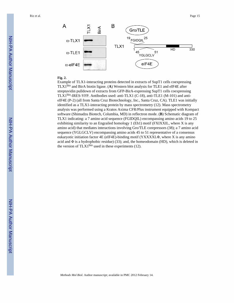
Fig. 2.Example of TLX1-interacting proteins detected in extracts of SupT1 cells coexpressingTLX1bio and BirA biotin ligase. (A) Western blot analysis for TLE1 and eIF4E afterstreptavidin pulldown of extracts from GFP-BirA-expressing SupT1 cells coexpressingTLX1bio-IRES-YFP. Antibodies used: anti-TLX1 (C-18), anti-TLE1 (M-101) and anti-eIF4E (P-2) (all from Santa Cruz Biotechnology, Inc., Santa Cruz, CA). TLE1 was initiallyidentified as a TLX1-interacting protein by mass spectrometry (12). Mass spectrometryanalysis was performed using a Kratos Axima CFR/Plus instrument equipped with Kompactsoftware (Shimadzu Biotech, Columbia, MD) in reflectron mode. (B) Schematic diagram ofTLX1 indicating: a 7 amino acid sequence (FGIDQIL) encompassing amino acids 19 to 25exhibiting similarity to an Engrailed homology 1 (Eh1) motif (FXIXXIL, where X is anyamino acid) that mediates interactions involving Gro/TLE corepressors (38); a 7 amino acidsequence (YGLGCLV) encompassing amino acids 45 to 51 representative of a consensuseukaryotic initiation factor 4E (eIF4E)-binding motif (YXXXXLΦ, where X is any aminoacid and Φ is a hydrophobic residue) (33); and, the homeodomain (HD), which is deleted inthe version of TLX1bio used in these experiments (12).
Riz et al. Page 15
Methods Mol Biol. Author manuscript; available in PMC 2012 February 14.
NIH
-PA Author Manuscript
NIH
-PA Author Manuscript
NIH
-PA Author Manuscript