Proliferative activity in the frog brain: A PCNA-immunohistochemistry analysis

Comparative Distribution ofNeuropeptide Y Y1 and Y5 Receptors
in the Rat Brain by UsingImmunohistochemistry
MICHAEL L. WOLAK,1 M. REGINA DEJOSEPH,1 ALLISON D. CATOR,1
ASHWINI S. MOKASHI,1 MARK S. BROWNFIELD,2AND JANICE H. URBAN1*
1Department of Physiology and Biophysics, Finch University of Health Sciences/ChicagoMedical School, North Chicago, Illinois 60064
2Department of Comparative Biosciences and Neuroscience Training Program, Universityof Wisconsin School of Veterinary Medicine, Madison, Wisconsin 53706
ABSTRACTNeuropeptide Y (NPY) Y1 and Y5 receptor subtypes mediate many of NPY’s diverse
actions in the central nervous system. The present studies use polyclonal antibodies directedagainst the Y1 and Y5 receptors to map and compare the relative distribution of these NPYreceptor subtypes within the rat brain. Antibody specificity was assessed by using Westernanalysis, preadsorption of the antibody with peptide, and preimmune serum controls. Im-munostaining for the Y1 and Y5 receptor subtypes was present throughout the rostral–caudal aspect of the brain with many regions expressing both subtypes: cerebral cortex,hippocampus, hypothalamus, thalamus, amygdala, and brainstem. Further studies usingdouble-label immunocytochemistry indicate that Y1R immunoreactivity (-ir) and Y5R-ir arecolocalized in the cerebral cortex and caudate putamen. Y1 receptor ir was evident in thecentral amygdala, whereas both Y1- and Y5-immunoreactive cells and fibers were present inthe basolateral amygdala. Corresponding with the physiology of NPY in the hypothalamus,both Y1R- and Y5R-ir was present within the paraventricular (PVN), supraoptic, arcuatenuclei, and lateral hypothalamus. In the PVN, Y5R-ir and Y1R-ir were detected in cells andfibers of the parvo- and magnocellular divisions. Intense immunostaining for these receptorswas observed within the locus coeruleus, A1–5 and C1–3 nuclei, subnuclei of the trigeminalnerve and nucleus tractus solitarius. These data provide a detailed and comparative mappingof Y1 and Y5 receptor subtypes within cell bodies and nerve fibers in the brain which,together with physiological and electrophysiological studies, provide a better understandingof NPY neural circuitries. J. Comp. Neurol. 464:285–311, 2003. © 2003 Wiley-Liss, Inc.
Indexing terms: NPY; hypothalamus; hippocampus; amygdala
Neuropeptide Y (NPY), peptide YY (PYY), and pancre-atic polypeptide (PP) comprise the pancreatic polypeptidefamily of neuropeptides. NPY is widely distributed in thecentral (Chronwall et al., 1985; de Quidt and Emson,1986; Chronwall, 1989) and peripheral (Lundberg et al.,1982) nervous systems and is implicated in the regulationof several physiological processes, including but not lim-ited to food intake (Stanley et al., 1986), blood pressure(Carter et al., 1985), anxiety (Wahlestedt et al., 1993;Kask et al., 1998a), hormone secretions (Wahlestedt et al.,1987; Leibowitz et al., 1988; Bauer-Dantoin et al., 1991),epilepsy (Baraban et al., 1997), circadian rhythms (Biello,1995), and neuromodulation (Kapoor and Sladek, 2001).Currently, there are at least six receptor subtypes identi-
fied for NPY and its family of related ligands: Y1, Y2, Y3,Y4, Y5, Y6. Of these G protein-coupled receptors (GPCR),
Grant sponsor: Grant number: R29 MH53663; Grant sponsor: SchweppeFoundation (J.H.U.); Grant sponsor: Sigma Xi Grant-in-Aid (M.L.W.).
Dr. Michael L. Wolak’s current address is Section of Neurosurgery,Dartmouth-Hitchcock Medical Center, Lebanon, NH 03756.
*Correspondence to: Janice H. Urban, Department of Physiology andBiophysics, FUHS/Chicago Medical School, 3333 Green Bay Road, NorthChicago, IL 60064. E-mail: [email protected]
Received 26 August 2002; Revised 3 April 2003; Accepted 9 April 2003DOI 10.1002/cne.10823Published online the week of August 4, 2003 in Wiley InterScience
(www.interscience.wiley.com).
THE JOURNAL OF COMPARATIVE NEUROLOGY 464:285–311 (2003)
© 2003 WILEY-LISS, INC.

the Y1, Y2, Y4, and Y5 receptors have been cloned in therat and human and are associated with Gi proteins, whichnegatively couple to adenylate cyclase (Eva et al., 1990;Gerald et al., 1995, 1996; Bard et al., 1995). Compared
with many of the other GPCR superfamilies, the NPYreceptor family has a high degree of sequence conserva-tion among species (with over 93% conservation betweenrat and human receptor sequences) and, in contrast, has a
Abbreviations
ac anterior commissureAD anterodorsal nucleus, thalamusAH anterior hypothalamusAHP anterior nucleus, posterior hypothalamusAM anteromedial nucleus, thalamusAmb ambiguus n.AP area postremaArcN arcuate nucleusArcL arcuate nucleus, lateralArcM arcuate nucleus, medialAStr amygdalostriatal transition areaB basal nucleus of MeynertBLA basolateral nucleus, anterior, amygdalaBLP basolateral nucleus, posterior, amygdalaBLV basolateral nucleus, ventral, amygdalaBM basomedial nucleus, amygdalaBST bed nucleus of the stria terminalisBSTL bed nucleus of the stria terminalis, lateralBSTM bed nucleus of the stria terminalis, medialCA cornu Ammon’sCb cerebellumcc corpus callosumCC central canalCeAm central amygdalaCeL central amygdala, lateralCeM central amygdala, medialcir circular n.CL centrolateral thalamic n.Cl claustrumCPu caudate putamencst commissural stria terminalisctx cerebral cortexDA dorsal hypothalamic areaDBB diagonal band of BrocaDEn dorsal endopiriformDG dentate gyrusDLG dorsal lateral geniculateDMPAG dorsomedial periaqueductal grayDR dorsal rapheECIC external cortex of the inferior colliculusECu external cuneate n.f fornixfmj forceps major of the corpus callosumGi gigantocellular reticular nucleusGrDG granular layer, dentate gyrusHDB horizontal limb of the diagonal bandhf hippocampal fissureHip hippocampusic internal capsuleICj island of CallejaIO inferior olive nucleusIP interpeduncular n.LaV lateral nucleus, ventral, amygdalaLC locus coeruleusLD laterodorsal nucleus, thalamusLG lateral geniculateLHA lateral hypothalamic areaLM lateral mammillary nucleusLPB lateral parabrachial n.LPGi lateral paragigantocellular n.LPLC lateral posterior thalamic n.LRt lateral reticular nucleusLSD lateral septal nucleus, dorsalLSI lateral septal nucleus, intermediateLSO lateral superior oliveMCPO magnocellular preoptic nucleusMCPC magnocellular n. of the posterior commissureMD mediodorsal nucleus, thalamusME median eminence
MeAm medial amygdalaMe5 mesencephalic trigeminal nucleusMG medial geniculateMHb medial habenulaMM medial mammillary nucleus, medialMolDG molecular layer, dentate gyrusMPA medial preoptic areaMR medial raphe n.MS medial septal nucleusMVe medial vestibular nucleusNAc nucleus accumbensnts nucleus tractus solitariusOp optic nerve layer of the superior colliculusor oriens layer of the hippocampusox optic chiasmPaDC paraventricular n., dorsal capPaLM paraventricular n., lateral magnocellularPaMP paraventricular n., medial parvocellularPaV paraventricular n., ventralPar2 parietal cortex 2pc posterior commissurePDTg posterior tegmental nucleusPeV periventricular nucleusPeF perifornical nucleusPir piriform cortexPnC pontin reticular n., caudal partPnO pontin reticular n., oral partPo posterior thalamic nuclear groupPoDG polymorphic layer, dentate gyrusPR prerubral fieldPr prepositus n.Pr5 principal sensory trigeminal n.PT parataenial thalamic n.PVA paraventricular n., anteriorPVN paraventricular nucleusPu Purkinje layer, cerebellumPy pyramidal layer, hippocampusRad radiatum layer, hippocampusRCh retrochiasmatic n.RE retrosplenial cortexRe reuniens nucleus, thalamusRt reticular nucleusS subiculumSCh suprachiasmatic nucleusSfi septofimbrial nucleussm stria medullarisSN substantia nigraSNC substantia nigra, pars compactaSNR substantia nigra, pars reticulataSol nucleus of the solitary tractSolC nucleus of the solitary tract, commonSolM nucleus of the solitary tract, medialSolI nucleus of the solitary tract, interstitialSON supraoptic nucleusSpVe spinal vestibular n.Sp5 spinal trigeminal nucleusSubC subcoeruleusTz trapezoid bodyVA ventral anterior nucleus, thalamusVDB ventral limb of the diagonal bandVL ventrolateral nucleus, thalamusVMH ventromedial nucleus, hypothalamusVM ventromedial nucleus, thalamusVMHVL ventromedial hypothalamic n, ventrolateral partVMHVM ventromedial hypothalamic n, ventromedial partVP ventral pallidumVPL ventral posterolateral thalamic n.VPM ventral posteromedial thalamic n.
286 M.L. WOLAK ET AL.

relatively low sequence homology among the different re-ceptor subtypes (31% homology between and Y1 and Y2receptors and 42% homology exists between the Y1 and Y4receptors [Larhammar, 1996]). Radioligand studies dem-onstrate binding sites for NPY and related ligands in thebrain, peripheral nervous system, vasculature, and nu-merous peripheral organs, including heart, adrenal,ovary/testis, and gastrointestinal tract (Allen et al., 1993;Dumont et al., 1993; Ghilardi et al., 1994). Both radioli-gand and in situ hybridization analyses reveal distinctpatterns of distribution for each of these receptors provid-ing anatomic correlates for the physiological role of NPYreceptor activation.
Of all the NPY receptors, the Y1 and Y5 receptor (Y1R,Y5R) subtypes have received significant attention basedon their ability to increase food intake (Schaffhauser et al.,1997; Duhault et al., 2000), modulate anxiety (Wahlestedtet al., 1993; Heilig et al., 1993), and regulate hormonesecretions (Leupen et al., 1997; Hastings et al., 2001). TheY1 receptor was the first receptor to be cloned and isperhaps the best characterized receptor for NPY (Eva etal., 1990, 1992). In situ hybridization reveals the distribu-tion of Y1R mRNA in the brain of several mammalianspecies, with the highest levels of expression consistentlyseen in forebrain regions, including the cerebral cortex,the hippocampal formation, and several amygdaloid, tha-lamic, and hypothalamic nuclei (Mikkelsen and Larsen,1992; Parker and Herzog, 1999). In addition, high levelexpression of Y1R mRNA has been detected in numerousbrainstem nuclei. Within these brain regions, the Y1 re-ceptor is generally considered to be postsynaptic. How-ever, immunohistochemical studies by Pickel et al. (1998)identified the presence of Y1 receptors in NPY-immunoreactive terminals, which suggests that the Y1Rmay also be a presynaptic receptor.
The Y5 receptor has been cloned more recently (Geraldet al., 1996). Binding sites as well as mRNA for the Y5Rhas been found within cerebral cortex, hippocampus, hy-pothalamus, amygdala, and brainstem nuclei (Dumont etal., 1998a; Durkin et al., 2000). The Y5 receptor has sim-ilar actions to the Y1 receptor in that it regulates ormodulates food intake (Marsh et al., 1998), anxiety (Sa-jdyk et al., 2002), and �-aminobutyric acid (GABA) release(Pronchuk et al., 2002). Y1 and Y5 receptor mRNA havevery similar patterns of distribution and, because thesereceptors have overlapping gene structures, it has beenpostulated that there is coordinated expression of Y1 andY5 receptors (Herzog et al., 1997). Whereas data demon-strates overlap of the distribution of the Y1 and Y5 recep-tors, it remains to be determined whether these receptorsare expressed in the same cell. Further examination of thedistribution and coexpression of these receptors in indi-vidual cells will be instrumental in determining the de-gree of participation of each of these receptors in physio-logical processes.
The present studies were designed to examine the local-ization and distribution of the Y1 and Y5 receptors in therat brain by using single- and double-label immunohisto-chemistry. In several sites within the brain, NPY exertspotent physiological effects (i.e., feeding effects in thePVN), yet there is minimal ligand binding in these areasas detected by means of receptor autoradiography. Usingimmunocytochemistry affords higher sensitivity to detectindividual cells and/or fibers with low levels of receptorexpression. Comparing the distribution and cellular local-
ization of these receptors provides more information re-garding the role of these receptors in regulating physiol-ogy and an additional, and complementary, way ofexamining NPY receptor localization.
MATERIALS AND METHODS
Animals
Adult male Sprague-Dawley rats (Charles River; 250–350 g) were maintained under standardized lighting con-ditions (14:10 light:dark cycle) in a temperature-controlled environment (23°C) with free access to rat chowand tap water. All procedures were approved by the insti-tutional animal care and use committee (IACUC).
For tissue fixation, animals were anesthetized with anintraperitoneal (i.p.) injection of sodium pentobarbital(Anpro Pharmaceutical; 50 mg/kg) and transcardially per-fused with 25 ml of phosphate-buffered saline (PBS: 10mM Na2HPO4, 150 mM NaCl, pH 7.5) containing 0.1%procaine and 100 U/ml heparin at 37°C followed by 50 mlof fixative solution (4% paraformaldehyde in PBS [pH 8.0])at �4°C. Brains were rapidly dissected out, post-fixed for18–24 hours in fixative solution at �4°C, and stored inPBS (pH 7.5) at �4°C. Coronal brain sections were cut at30–40 �m thicknesses on a Vibratome, and free-floatingsections were collected in ice-cold PBS (pH 7.5) and pro-cessed for immunohistochemical staining.
Y1 and Y5 Antisera
Polyclonal Y1R and Y5R antisera were generated inrabbits. The selection of the targeted peptide sequenceswas based on computer analysis of the full-length Y1R andY5R sequences listed in the Swiss Protein Database usingGeneRunner (Hastings Software, Hastings on Hudson,NY) and Peptide Structure Genetics Computer Group(GCG, Madison, WI) software. The peptides were selectedbased on high antigenicity, surface probability, and sur-face topology indices. A C-terminal peptide consisting ofamino acids 363-382 of the NPY Y1 receptor was synthe-sized (Quality Control Biochemicals; LKQASPVAFKKIS-MNDNEKI), conjugated to keyhole limpet hemocyanin(KLH), and injected into rabbits (initially with 500 �g andsubsequently with 50-�g boosts once per month for 18months). Similarly, a peptide corresponding to extracellu-lar loop 3 and parts of transmembrane domains VI/VII ofthe NPY Y5 receptor was synthesized (VTDFNDNLIS-NRHFKLVYCIC; representing amino acids 400-420), con-jugated to KLH and injected into rabbits. Rabbits werebled 10 days after each immunogen injection, plasma wascollected and affinity purified. Purified antibody was pre-pared by affinity chromatography against immobilized ratY1R(363-382) or Y5R(400-420) medium prepared by usinga Pierce SulfoLink kit (Rockford, IL). Antibodies wereeluted with 0.2 M glycine (pH 2.5) and collected into 1.0 MTris (pH 8.0). Antisera were extensively dialyzed againstPBS and made up in 1% bovine serum albumin (BSA) and0.01% sodium azide. No similarity between the syntheticpeptides and the sequence of other currently cloned NPYreceptor subtypes or other transmembrane receptors hasbeen found in searches against updated databanks (Non-redundant GenBank CNS). The specificity of each anti-serum was assessed with Western analysis, immunohis-tochemistry using preimmune sera and preadsorption ofthe antibody with the synthetic peptides used to generate
287Y1/Y5 RECEPTOR IMMUNOREACTIVITY IN RAT BRAIN

the respective antisera in rabbit at a concentration ratio of1:10 (antiserum:peptide).
Immunocytochemistry
Free-floating sections were washed in immunocyto-chemistry (ICC) buffer consisting of 0.2% gelatin, 0.01%thimerosal, 0.002% neomycin in PBS (pH 7.5), blockedwith antibody dilution buffer (ICC buffer containing 2%normal goat serum [NGS; Vector Laboratories]) for 30minutes at �4°C, and then incubated for 24–48 hours at�4°C with either the rabbit anti-Y1 receptor antiserum(1:2,000) or rabbit anti-Y5 receptor antiserum (1:20) di-luted in ICC buffer with 2% NGS. Sections were washed inICC buffer and then incubated for 45 minutes at roomtemperature with biotinylated, affinity purified goat anti-rabbit IgG (H�L; Vector Laboratories; 1:400), followed bya wash in ICC buffer and incubation for 45 minutes atroom temperature with an avidin-biotin-horseradish per-oxidase (ABC) complex (VECTASTAIN Elite ABC, VectorLaboratories; 1:300). Sections were washed in Tris-buffered saline (TBS: 100 mM Tris base, 150 mM NaCl,pH 7.5), and signal was developed by incubating sectionsin a chromogenic substrate solution consisting of 0.03%3,3�-diaminobenzidine (DAB), 0.01% H2O2, and 0.03%NiCl2 in TBS (pH 7.5). Color development was carefullymonitored and proceeded at room temperature for 10–15minutes, after which time the sections were washed inTBS and mounted on gelatin-coated slides. Mounted sec-tions were dehydrated in a series of ascending ethanolwashes (50, 70, 80, 90, 100, 100%) and cleared in a xylenesubstitute (Hemo-De; Fisher). Coverslips were appliedwith Permount.
Brain sections were atlas matched according to the ste-reotaxic rat atlas of Paxinos and Watson (1997), and im-munoreactive staining was visualized by light microscopy.The distribution of Y1R immunoreactivity (-ir) and Y5R-irpresented in the table was determined in sections from 10animals. Specific signal, in part, was assessed in alternatesections incubated with antibody that was preincubatedwith immunizing peptide. The distribution and degree ofimmunostaining for each antibody in each brain regionwas assessed by two different investigators. By using apredetermined scale, the cell density and relative inten-sity (Fig. 1) of signal was rated. Images were captured byusing a SPOT 2 digital camera and MetaMorph software;the contrast and brightness of the photomicrographs wereadjusted by using MetaMorph software or Adobe Photo-shop. Montages were assembled by using Adobe Photo-shop.
Double-label immunocytochemistry
To assess colocalization of Y1 and Y5 receptors in thesame cell, we used a standard secondary antibody immu-nofluorescence protocol combined with biotinylated tyra-mide amplification immunofluorescence based on that de-scribed by Adams (1992). As both the Y1 and Y5 antiserawere generated in rabbits, there were technologic con-cerns regarding the generation of false positives in thedouble-label immunocytochemical assays. Using biotinyl-ated tyramide to enhance the detection of one antibodyallowed us to use one antiserum at dilutions below thelimit of detectability using standard secondary antibodyimmunofluorescence methods. Briefly, tissues were incu-bated with one antibody (Y1 or Y5 receptor), visualized byusing the biotinylated tyramide enhancement and then
processed for the other NPY receptor subtype by usingstandard secondary antibody immunofluorescence histo-chemistry. Brains (n � 5) were processed as mentionedabove and free-floating sections were washed in PBS fol-lowed by incubation in 1% H2O2 for 15 minutes and 3%normal donkey serum in antibody dilution buffer for 30minutes at room temperature. The sections were incu-bated with primary antibody (Y1, 1:3,000; Y5, 1:100) for36–48 hours at �4°C. The sections were washed in ICCbuffer (5 � 5 minutes) and incubated in biotinylated don-key anti-rabbit (1:2,500) for 1 hour at room temperature.After rinses in ICC buffer, the sections were incubated inABC complex (VECTASTAIN Elite ABC; 1.5 �l/ml) for 30minutes, rinsed with PBS (5 � 5 minutes), and incubatedwith biotinylated tyramide (1.5 �l/ml) for 10 minutes atroom temperature. Tissues were washed in PBS (5 � 5minutes) and incubated with either Cy2- or Cy3-conjugated strepavidin (1:250) for 3 hours. After PBSwashes (5 � 5 minutes), the sections were incubated withthe second primary antiserum (Y1:1:800; Y5: 1:20) for36–48 hours at �4°C. After another series of PBS washes(5 � 5 minutes) the sections were incubated with Cy2- orCy3-conjugated donkey anti-rabbit antisera (1:250). Thetissues were rinsed in PBS (5 � 5 minutes), mounted ongelatin-coated slides, and allowed to air-dry. Coverslipswere applied by using 2.5% PVA-DABCO (polyvinyl alco-hol; 1,4-diazabicyclo[2.2.2]octane) antifade mounting me-dium.
Immunocytochemical staining was visualized by usingan Olympus Fluoview Scanning Laser confocal micro-
Fig. 1. Representative cells for the different labeling intensityrankings (�, ��, ���) indicated in Table 1. A: Y1R-labeled cells inthe ventral pallidum. B: Y1 receptor (Y1R) -labeled cells in the dorsalhypothalamus. C: Y5R-labeled cells in the locus coeruleus. Scalebars � 50 �m in C (applies to A–C).
288 M.L. WOLAK ET AL.

scope. Images were obtained by using the Fluoview soft-ware, and contrast was optimized by using Adobe Photo-shop. The specificity of the signal was assessed byomission of either one or the other antibody through thedouble-label protocol and peptide preadsorption of eachantibody. Both antibodies were titrated by using bothimmunofluorescence procedures to characterize the stain-ing patterns and ensure that no nonspecific staining wasgenerated in the double-label procedure. Additionally, theoverall distribution of Y1R-ir and Y5R-ir was not differentthan that presented in Table 1, although the signals weremore intense.
Cell culture
The human neuroblastoma cell line SK-N-MC, whichexpresses the Y1 receptor subtype, was obtained from theAmerican Type Culture Collection (ATCC; Rockville, MD).Cells were grown in Eagle’s Minimum Essential Medium(EMEM; Gibco-BRL) supplemented with 10% fetal bovineserum, 1.0 mM sodium pyruvate, 50 U/ml penicillin, and25 �g/ml streptomycin. Cells were maintained in a water-saturated atmosphere of 95%O2/5% CO2 at 37°C andgrown to 70% confluence. At the third to fifth passage,cells were rinsed in TBS supplemented with 1 mM EDTA(pH 8.0) and protease inhibitors (Boehringer Completeprotease inhibitor tablets), harvested, pelleted by briefcentrifugation, and stored at �80°C.
Sodium dodecyl sulfate-polyacrylamide gelelectrophoresis and Western analysis of rat
brain homogenate and SK-N-MC cells
Brains were rapidly dissected out and homogenized(Polytron homogenizer; Brinkmann Instruments) in Trisbuffer supplemented with 1 mM EDTA (pH 8.0) and pro-tease inhibitors. The homogenates were centrifuged(15,000 � g for 20 minutes), and the resulting supernatantwas stored at �80°C. For preparation of membrane- andcytosol-enriched fractions, tissue was homogenized andcentrifuged (1,000 � g for 10 minutes). The supernatantcontaining solubilized receptor was further centrifuged at100,000 � g for 1 hour at 4°C. The supernatant containingthe cytosol-enriched fractions (S100) was collected, andthe pellet containing the membrane-enriched fraction(P100) was resuspended in sodium dodecyl sulfate-polyacrylamide gel electrophoresis (SDS-PAGE) bufferand subsequently stored at �80°C.
Homogenates of brain and SK-N-MC cells were dilutedin SDS-PAGE sample buffer (60 mM Tris-HCl [pH 6.8],10% glycerol, 2% SDS, 0.0025% bromophenol blue). Sam-ples containing 50–100 �g of total protein were heated to90°C for 5 minutes, loaded on 10% Tris-glycine gels inTowbin buffer (24 mM Tris base, 192 mM glycine, pH 8.3)with 0.1% SDS, and separated by electrophoresis (2 hoursat 110 V [constant]). Separated proteins were blotted ontopolyvinylidene difluoride membranes by electrophoresis(90 minutes at 25 V [constant]) in 0.5� Towbin buffer with20% methanol and 0.025% SDS. SDS-PAGE and proteintransfer were carried out by using the Novex XCell IIMini-Cell apparatus and Blot Module. Immunodetectionwas performed by using the Novex WesternBreeze chemi-luminescent detection system according to manufacturer’sinstruction, with primary antibody dilutions of 1:2,000(anti-Y1R antiserum) and 1:30 (anti-Y5R antiserum). Im-munoreactive bands were detected by exposure to KodakX-OMAT AR film at room temperature for 5–15 minutes,
and molecular weights were estimated by using Bio-RadKaleidoscope and SeaBlue prestained standards. Specific-ity controls were performed by preincubation of the anti-Y1R and anti-Y5R antisera with the peptides used togenerate the respective antisera.
RESULTS
The distribution of Y1R-ir and Y5R-ir extends through-out the rostral–caudal extent of the brain with Y1R over-all being more widely distributed than Y5R. Both recep-tors were present with relatively high concentrations ofcell bodies and fibers within the cortex, hippocampus,hypothalamus, amygdala, and brainstem correspondingwith some of the major actions of NPY. Figure 2 is adiagrammatic representation of the distribution of cellbody (dots/triangles) and fiber (stippled areas) density forY1R-ir (left side of diagram) and Y5R-ir (right side ofdiagram). The information presented in Table 1 indicatesthe relative density of Y1R- and Y5R-containing cellswithin a region and the intensity of immunoreactivity inthese different brain regions. The relative labeling inten-sities of cells and fibers were assessed by using the scalerepresented in Figure 1. A (�) was given to those cells andfibers that exhibited a light, uniform immunostaining(Fig. 1A). The presence of a darker immunostaining pat-tern or a variation in labeling intensity was representedas �� (Fig. 1B). In some cases, the staining was veryintense (black); this staining was represented with ���(Fig. 1C). That we observe differences in signal intensitiesamong brain regions suggests that the current stainingcondition has a dynamic range which allows for detectionof differential receptor expression under physiological con-ditions. Immunoreactive staining was absent in those sec-tions incubated with preimmune serum or antisera thatwas preincubated with peptide sequences used to generatethe antibodies (Fig. 3).
Western analysis of SK-N-MC cells (Fig. 4) revealed asingle band migrating at approximately 42 kDa, which isin agreement with the predicted molecular weight of theY1 receptor protein (Eva et al., 1990). In Western blots ofbrain homogenates, two specific immunoreactive bandsfor the Y1 receptor were identified: a light band at 42 kDaand a band at �85 kDa. These bands showed a differentialdistribution when compared in membrane-enriched(P100) vs. cytosol-enriched (S100) protein fractions. Inmembrane-enriched (P100) fractions, two bands werepresent that corresponded to �42 and �85 kDa, whereasthe lower molecular weight species was not present in thecytosol-enriched fraction (S100). For the Y5 receptor,Western blots of rat brain homogenates revealed twobands migrating at �45–50 kDa, corresponding to thepredicted molecular weight of the Y5 receptor protein(Gerald et al., 1996). The presence of these bands wasabolished in preadsorption control assays. No Y5R-immunoreactive band was present in homogenates fromthe SK-N-MC cells.
Telencephalon
Within the neocortex, the strongest labeling of Y1R-irperikarya was localized to a cluster of neurons present inlayers IV/V of parietal cortex 2 (Par2; Fig. 5A,C). Cellbodies comprising this neuronal population typically dis-played long, tapering processes of either a bipolar or mul-tipolar arrangement and were distributed with a low to
289Y1/Y5 RECEPTOR IMMUNOREACTIVITY IN RAT BRAIN

TABLE 1. Regional Distribution of Y1 and Y5 Immunoreactivity Throughout the Rostrocaudal Extent of the Brain1
Region
Y1 receptor Y5 receptor
Celldensity
Stainingintensity
Celldensity
Stainingintensity
TelencephalonNeocortex �/�� �/�� �� ��/���Periallo- and proisocortex �/�� �/�� �� ��/���Dorsal endopiriform cortex �� �� � �Hippocampal formation
CA1 ��� � ��� ��CA2 ��� �/�� ��� ��CA3 ��� ��/��� ��� ��Dentate gyrus
Granule layer (GrDG) nd nd ��� �/��Molecular layer (MolDG) fibers �� fibers ��Polymorphic layer (PoDG) �� ��/��� � ��
Subiculum (S) �� �� �� ��Basal nuclei
Caudate-putamen (CPu) � �� �� ��Nucleus Accumbens (Acb)-shell �/�� � �� �Islands of Calleja (LCJ) ��/��� �� �� ��Pallidal complex
Globus pallidus (GP) � � nd ndEntopenduncular nucleus (EP) � �� � �Ventral pallidum (VP) �/�� �� nd ndClaustrum (Cl) ��� ��� � �
Basal nucleus of Meynert (B) � ��� nd ndSeptum
Medial septum-diagonal band (MSDB) �/�� �/�� � �Lateral septum (LS) � � �/�� �Septofimbrial nucleus (Sfi) �� ��� � ���
Amygdaloid complexNucleus of the lateral olfactory tract (LOT) ��/��� �� nd ndBed n. accessory olfactory tract (BAOT) �/�� � nd ndBasolateral complex
Lateral nucleus (La) �/�� �/�� �� �Basolateral nucleus (BL) ��/��� �� �� �
Extended amygdalaBed n. stria terminalis-medial (BSTM) �� �� nd nd-lateral (BSTL) �� �/�� nd nd
Medial nucleus (Me) � � nd ndCentral nucleus (Ce) �/�� �� � �Interstitial n. post. limb ant. commissure (IPAC) � �/�� nd ndIntercalated cell masses (I) �� �� nd ndBed N. of the anterior commissure (BAC) �/�� � nd nd
DiencephalonThalamus
Anterior nuclear groupAnterodorsal nucleus (AD) �� �� � �Anteromedial n. ventral (AMV) ��/��� ��/��� �� ��
Mediodorsal nucleus (MD) �/�� �/�� �� ��Ventral nuclear group
Ventromedial n. (VM) �� �� � ��Ventrolateral n. (VL) �� �� ��� ��Ventroanterior n. (VA) �� �� �� ���Ventroposterior nuclear complex (VPL/VPM) � �/�� �� �/��Submedius n.-ventral (SubV) � � �� ��
Lateral nuclear groupLateroposterior n. (LP) �/�� �� �� �Laterodorsal n. (LD)-ventrolateral (LDVL) �� �/�� � �
-dorsomedial (LDDM) � � � �Posterior nuclear group (Po) �� � �� ��Intralaminar nuclear group
Paracentral n. (PC) �/�� � nd ndParafascicular n. (PF) �� �� nd fibers
Dorsal midline nuclear groupParatenial n. (PT) ��/��� ��/��� �� ���Interanteromedial n. (IAM) �/�� �/�� � �
Reuniens n (Re) ��/��� ��/��� nd ndSubparafasicular n. (SPF) �� � nd ndReticular n. (Rt) �� �/�� �� ��/���
MetathalamusLateral geniculate (LG)-dorsal (DLG) �/�� � �� �Medial geniculate (MG) � �/�� �� �/��
EpithalamusMedial habenula (Mhb) ��� � ��� ��Medial–lateral junction �� ��/��� nd nd
HypothalamusPreoptic region
Anteroventral periventricular n. (AVPe) �� �� �� ��Magnocellular preoptic n. (MCPO) �� �� nd ndMedial preoptic area (MPA) � �/�� � �Medial preoptic n. (MPO) �/�� �/�� � �Median preoptic n. (MnPO) �/�� �/�� nd ndMedial preoptic n. (MPO)Periventricular preoptic n. (PePO) �/�� ��/��� nd ndMedial preoptic area (MPA)Ventromedial preoptic n. (VMPO) �� �� nd nd
290 M.L. WOLAK ET AL.

moderate density of immunopositive fibers. Y1R-labeledperikarya were also observed throughout several neocor-tical fields, most consistently within layers II, IV/V, andVIb, as well as in various transition layers of peri- andproisoallocortical fields.
Immunoreactive staining for the Y5 receptor was alsoobserved throughout several cortical fields. The highestcell densities were seen in the cingulate, retrosplenial,frontal, and parietal cortices and were localized primarilyto isocortical layers IV/V and corresponding transitionlayers of the periallo- and proisocortices (Fig. 5B,D). Inthese regions, medium, oval- to pyramidal-shaped cellbodies, often exhibiting prominent apical processes, wereinterspersed among a moderate density of immunoposi-tive fibers that extended throughout all the cortical layers.To further extend these studies, double-label immunohis-tochemistry demonstrated colocalization of Y1 and Y5 re-
ceptor immunoreactivities in the retrosplenial cortex (Fig.6) as well as in parietal cortex. In the retrosplenial/cingulate cortex, there was considerable colocalization ofY1R-ir and Y5R-ir (Fig. 6C, arrows), and single-labeledY1R but not Y5R cells were also observed (Fig. 6C, arrow-heads). Also note the high degree of Y1R and Y5R fiberstaining that appears in a continuum along the apicalprocesses.
Several populations of Y1R-ir perikarya were observedthroughout the major divisions of the hippocampal forma-tion (Fig. 7A). Within the dentate gyrus, small to mediumimmunoreactive cell bodies having short processes with apredominantly bipolar arrangement were localized to thehilus, or polymorphic layer (Fig. 7C), whereas the granulecells had little detectable Y1R-ir. The largest and mostdensely distributed population of Y1R-labeled perikaryain the hippocampal formation was observed within the
TABLE 1. (continued)
Region
Y1 receptor Y5 receptor
Celldensity
Stainingintensity
Celldensity
Stainingintensity
Anterior regionAnterior hypothalamic n.-posterior (AHP) �� �/�� nd ndAnterior periventricular n. (PeA) �/�� ��/��� nd ndLateral hypothalamic area (LH) �/�� �� �/�� ��Lateroanterior hypothalamic n. (LA) �/�� �/�� nd ndParaventricular n. (Pa)
Lateral and medial magnocellular ��� �� �� ��Parvocellular (medial, anterior, dorsal cap) �� ��/��� �� �/��
Accessory magnocellular neurons ��� �� �� �/��Retrochaismatic n. (Rch) �� ��� � �Stigmoid n. �� �/�� nd ndSuprachiasmatic n. (SCh) �� �/�� �� �/��Supraoptic n. (SON) ��� �� �� ��Ventrolateral n. (VLH) �� � nd nd
Tuberal regionArcuate n. (ArcN)
Dorsal (ArcD) � ��� � ��Medial and lateral (ArcM; ArcL) �� ��� � ��
Dorsal area (DA) � �� � ��Perifornical area (PeF) ��/��� �� nd ndSubincertal n. (Sl) �/�� �� nd ndVentromedial hypothalamus-anterior (VMHA) �/�� � fibers �
Mammillary regionTuberomammillary n. (TM) ��� � �� ��Posterior periventricular n. (PeP) �/�� ��/��� nd nd
MesencephalonSubstantia nigra (SN)-compacta (SNC) �� �� � �
-reticulata (SNR) � �� nd ndVentral tegmental area (VTA) �� �� nd ndRed n. (R)-magnocellular (RMC) �� �� � ��Medial accessory oculomotor n. (ME3) �� �� nd ndPeriaqueductal gray, dorsomedial (DMPAG) � ��/��� nd ndDorsal raphe n. (DR) �� �� � ��Sphenoid n. (Sph) �/�� �/�� nd ndTrapezoid body (TZ) ��/��� �� ��� ��
Metencephalon/myelencephalonMedian raphe n. (MR) �� �� � ��Locus coeruleus (LC)–subcoeruleus (SubC) ��� ��/��� ��� ���Gigantocellular reticular n. (Gi) �/�� � � �Pontine reticular n. caudal (PnC) �/�� �� � �Lateral reticular n. (Lrt) �� �� � ��Inferior olive complex (IO) �� �/�� � ��Mesencephalic trigeminal n. (Me5) �� ��� �� ��/���Motor trigeminal n. (Mo5) �� ��� �� ��/���Spinal trigeminal n. (Sp5) �� �� � �Dorsal motor n. of the vagus (10) �� �� �� ��Hypoglossal n. (12) �� �� �� ��Nucleus of the solitary tract � �� �� ��Cerebellum (Purkinje layer) �� � �� ��Facial n. nd nd � �Vestibular n. lateral and medial � �� � ��Raphe magnus nd nd � ��N. Ambiguus �� �� �� ���Lateral paragigantocell (LPGi) nd nd � ���Cuneate �� � �� ��
1Cell density and staining intensity was compared for each receptor subtype in representative atlas matched sections and recorded (high, ���; moderate, ��; low, �; and notdetected, nd; Cell density represents the relative number of cells within the region, whereas staining intensity reflects the intensity of the signal per cell (see Fig. 1).
291Y1/Y5 RECEPTOR IMMUNOREACTIVITY IN RAT BRAIN

Fig. 2. Diagrammatic representation and comparison of Y1 recep-tor (Y1R; left) and Y5R (right) immunoreactivity in coronal sectionsthough an adult male rat brain. The coronal sections, with referenceto bregma, correspond to the stereotaxic atlas of Paxinos and Watson(1997). The diagrams illustrate the relative distribution of immunopo-
sitive cell bodies (Y1R, black dots; Y5R, black triangles) and immu-noreactive fibers (represented by stippling). The number of dots/triangles within an area reflects the relative area of distribution andnot quantitation of cell number or density. For abbreviations, see list.
292 M.L. WOLAK ET AL.

Figure 2 (Continued)
293Y1/Y5 RECEPTOR IMMUNOREACTIVITY IN RAT BRAIN

stratum pyramidale of Ammon’s horn (CA2 and CA3; Fig.7A). This population of pyramidal-shaped cell bodies weremoderately labeled and characteristically displayed short,tapering apical processes oriented perpendicular to theadjacent stratum radiatum. The subiculum contained amoderate to high density of predominantly pyramidal-shaped Y1R-immunoreactive cell bodies displaying shortapical processes (Fig. 7F). A low-density of Y1R-immunoreactive fibers was localized predominantly to theoriens, pyramidal, and radiatum strata of Ammon’s horn(CA1, CA2, and CA3). Several of these fibers were ema-nating off of cell bodies in these regions, whereas denseY1R-immunoreactive staining was present on fine fibersin the MolDG. Also prominent within the hippocampalformation were Y1R-ir perikarya randomly distributedthroughout the various strata of Ammon’s horn (CA1,CA2, and CA3; Fig. 7D). These cells were medium to largein size and characteristically displayed a multipolar ar-rangement of arborizing processes. These processes were
predominantly oriented perpendicular to the hippocampalfissure and could often be traced for several micrometers.
Y5R-immunoreactive perikarya were also abundantlyobserved throughout the hippocampal formation. Y5R-irfibers were observed projecting from cells within theCA1–3 regions (Fig. 7B) and extended into the correspond-ing stratum radiatum. In general, both the distributionand morphology of Y5R-labeled neurons closely matchedthe immunoreactive staining patterns observed for theY1R receptor, with the pyramidal cell layer and the poly-morphic dentate gyrus (Fig. 7E,G) showing the highestlevels of immunoreactivity. There was colocalization ofY1R-ir and Y5R-ir in cells of the CA1–3 regions withapproximately 50% of Y1R-immunopositive cells showingcolocalization with the Y5R. This percentage is a conser-vative estimate of cell number as Y1R and Y5R is differ-entially expressed in the stratum pyramidale. In general,Y5R-ir is stronger on fibers and proximal dendrites,whereas Y1R-ir is present mostly on cell bodies. Strong
Fig. 3. Specificity of Y1 receptor (Y1R) and Y5R immunostaining.Photomicrographs of Y1R immunostaining in the ArcN (A) and Y5receptor immunostaining in the PVN (C) from a male rat. B,D: Thelack of a specific signal in corresponding sections after preadsorption
of the antibodies with the peptide used to generated the respectiveantiserum. For abbreviations, see list. Scale bars � 500 �m in D(applies to A–D).
294 M.L. WOLAK ET AL.

Y5R-ir was also evident on cells interspersed throughoutthe stratum pyramidale similar to that identified in Fig-ure 7D, although they were much fewer in number thanthe Y1R-ir cells. No colocalization of Y1R- and Y5R-ir wasfound in these cells.
In septal areas, a prominent cluster of Y1R-immunoreactive perikarya and fibers was observed in therostral portion of the septofimbrial nucleus (Fig. 8E). Thisneuronal population displayed an intermediate density ofcell bodies with extensive processes, the latter of whichcontributed to a dense fiber plexus that could be seenprojecting ventrally within the precommissural fornix.The Y5R also showed strong immunoreactive stainingwithin this region, although there were fewer cell bodiespresent. Both Y1R- and Y5R-immunoreactive perikaryawere present in the medial septum-diagonal band complex(Fig. 8A,B) with generally more cells showing labeling forY1R (Fig. 8C) than Y5R (Fig. 8D).
Y1R- and Y5R-immunoreactive perikarya were identi-fied in several telencephalic components of the basal nu-clei, including the caudate putamen (CPu; Fig. 9A–C),accumbens nucleus (NAc), olfactory tubercle, and pallidalcomplex (Fig. 1A). Within the CPu, there was considerablecolocalization of the two receptor subtypes (Fig. 9C). Anal-ysis of sections from different animals suggests that �98%of Y1R-immunoreactive cells are colocalized with Y5R;there are populations of Y5R-ir cells (approximately 35%)that do not coexpress Y1R (Fig. 9C, arrowheads). Outsidethe corpus striatum, the claustrum had one of the highestdensities of Y1R-ir in the rat brain comprised of a densepopulation of cell bodies and a varicose network of immu-nopositive fibers (Fig. 9D); this area is devoid of Y5R-ir(Fig. 9E). This figure illustrates the specificity of the
double-label ICC where only Y1R-ir (green) is detected inthe claustrum and the signal on the red (Cy3) channel isinsignificant. This finding indicates that there is no bleed-through of the signal nor is there cross-reactivity of thetwo methods of detection.
Within the basal telencephalon, the amygdaloid com-plex displayed immunoreactive staining for Y1R and Y5R.Discrete populations of Y1R-immunoreactive perikaryawere identified in the central and medial divisions of theextended amygdala, as well as in “cortical-like” nucleargroups of the basolateral complex (Fig. 10A,C). Y1R-ir cellbodies in the medial CeAm displayed extensive processesthat were present within a rich fiber plexus (Fig. 10E).The overall intensity and presence of Y1R was stronger inthe medial CeAm, which correlates well with the differen-tial NPYergic innervation of the medial CeAm (CeM). Alsoprominent were populations of Y1R-immunoreactiveperikarya and fibers in the lateral and medial parts of thebed nucleus of the stria terminalis (Fig. 8A,F), as well asin the medial amygdaloid (MeAm) nucleus. Y1R-immunoreactive cells in the basolateral complex were rel-atively homogenous populations of pyramidal-shaped cellbodies with short apical processes, interspersed among alow density of immunoreactive fibers. This pattern of im-munoreactive staining extended throughout the rostral–caudal extent of the lateral and basolateral nuclei.
Immunoreactive staining for the Y5 receptor was ob-served almost exclusively in the basolateral amygdaloidcomplex with few cells observed in the lateral division ofthe central amygdala (Fig. 10B,D). Similar to the Y1R,Y5R-immunoreactive perikarya were homogeneously dis-tributed throughout the lateral and basolateral complex,displaying pyramidal-shaped cell bodies with apical pro-cesses (Fig. 10F).
Diencephalon
The thalamus contained striking amounts of immuno-reactive labeling for both the Y1 (Fig. 11A,C) and Y5 (Fig.11B,D) receptors. Abundant Y1R-immunoreactiveperikarya were distributed throughout most major nu-clear groups, where labeled cell bodies were typically me-dium in size and often displayed processes with a multi-polar arrangement (Fig. 11C). In contrast to theabundance of labeled cell bodies and processes, Y1R-immunopositive fibers were sparsely distributed through-out most of the thalamus with highest concentrationspresent in the reticular (Rt) and ventrolateral (VPL) thal-amus (Fig. 11E). Y1R immunostaining extended postero-laterally into the metathalamus, and was present in thelateral (dorsal nucleus) and medial (dorsal, medial, andventral nuclei) geniculate bodies. Dense fiber staining forthe Y1R was evident in a discrete region corresponding tothe junction of the medial and lateral portions of thehabenula. Immunostaining for Y5R was also very abun-dant in the thalamus with a slightly different pattern ofstaining than that of the Y1R receptor. Notably, Y5Rimmunostaining was less in the midline nuclear groups(Table 1). Within most major divisions of the thalamus,Y5R-ir was present on multipolar cells and within finefibers that were interspersed within nuclear groups (Table1). Strong immunoreactive staining for the Y5 receptorwas present on dispersed cells and fibers in the reticularthalamus (Fig. 11F) and displayed a high degree of colo-calization with the Y1 receptor (data not shown).
Fig. 4. Western blot analysis of (A) Y1 receptor (Y1R) and (B) Y5Rimmunoreactivity in rat brain and SK-N-MC cells. A: Y1R immuno-reactivity in protein homogenates from SK-N-MC cells, brain, andcytosol-enriched (S100) and membrane-enriched (P100) fractions fromrat brain. B: Immunoblot showing Y5R immunoreactivity in wholebrain tissue. Molecular weight markers (kilodaltons) are indicated onthe left side of the diagram and between A and B.
295Y1/Y5 RECEPTOR IMMUNOREACTIVITY IN RAT BRAIN

The hypothalamus is another region where there werenotable differences in the relative concentrations and dis-tribution of Y1R-ir and Y5R-ir (Table 1). Rostrally, themedial preoptic area (MPOA) contained a heterogeneouscell population in terms of size, morphology, and distribu-tion, interspersed amid a diffuse network of fibers. Y1R-immunoreactive labeling extended continuously from thedorsal to the ventral aspects of the medial preoptic area,with the highest density of cell bodies and fibers observedventromedially in regions corresponding to the medial,anteroventral, periventricular, and ventromedial nuclei.No detectable labeling was noted for the Y5 receptor inthis brain region. In the suprachiasmatic nucleus (SCh),Y1R-immunoreactive perikarya and fibers were concen-trated in the ventral portion of the nucleus (Fig. 12A),whereas Y5R-ir cells and fibers also extended to the dorsalaspect of the nucleus (Fig. 12B).
The hypothalamic arcuate nucleus (ArcN) contained oneof the most striking clusters of Y1R-ir perikarya seen in
the rat brain (Fig. 12C). These cells were located predom-inantly in the ventrolateral portion of the arcuate nucleusand extended laterally into the periarcuate region. A clus-ter of morphologically similar perikarya was also observedrostrally in the retrochiasmatic area (RCh), a brain regionclosely associated with the arcuate nucleus. A second pop-ulation of medium, lighter labeled Y1R-immunoreactiveperikarya could be distinguished within the ventromedialArcN (ArcM) nucleus. Y1R-immunoreactive fibers werealso abundant in both the medial and lateral aspects ofthis nucleus. In contrast to Y1R, immunoreactive stainingfor the Y5 receptor was concentrated on cells, not fibers, ofthe ArcN (Fig. 12D). Y5R-immunoreactive cell bodies weremedium in size, characteristically displaying punctate im-munoreactivity, devoid of processes, and were not seen inproximity to any immunopositive fibers. In some assays, itappeared that staining may be present within microglia,although further double-label studies will need to directlyassess this staining. Labeling for both Y1 (Fig. 3A) and Y5
Fig. 5. Distribution of Y1 (A,C) and Y5 (B,D) immunoreactivity inthe cerebral cortex. A,C: Y1-immunoreactive perikarya in layers IV/Vin the parietal cortex. Lighter-labeled cells are evident within othercortical layers (lamina II–IV). B,D: Y5 receptor (Y5R) immunoreac-
tivity in the retrosplenial cortex. Pyramidal and oval cells presentwithin layers II and IV–V exhibit labeling as do their apical processes.For abbreviations, see list. Scale bars � 200 �m in A (applies to A,B),100 �m in C (applies to C,D).
296 M.L. WOLAK ET AL.

(Fig. 12D) receptors was evident in the median eminence(ME).
High densities of cell bodies and processes for Y1R wereobserved in the anterior, dorsomedial, and ventrolateraldivisions of the VMH (Fig. 3A), whereas the central divi-sion of the nucleus was relatively cell-sparse. A rich net-work of Y1R-immunopositive fibers was similarly distrib-uted within the ventromedial nucleus, with the highestdensities localized to cell-rich, e.g., anterior, dorsomedial,and ventrolateral nuclear divisions. Within the VMH, arelatively dense plexus of Y5R-ir, devoid of detectablecells, was present in fibers throughout the three divisions.
Abundant immunoreactive staining for Y1R was seenin the hypothalamic PVN. Y1R-immunoreactive peri-karya were present in both the parvo- and magnocellu-lar nuclear divisions (Fig. 13A,C,D). A population ofsmall to medium cell bodies with extensive processeswas seen interspersed among a high density of fibers inthe parvocellular nuclear division, which could be fur-ther localized to distinct parvocellular subdivisions(Fig. 13A). A second neuronal population, having largeY1R-immunoreactive perikarya, was localized to mag-nocellular parts of the paraventricular nucleus. Magno-cellular perikarya were characteristically devoid of pro-cesses and, with rare exception, were not observedcodistributed with immunoreactive fibers (Fig. 13D). Mor-phologically similar populations of Y1R-immunoreactivemagnocellular perikarya were also observed in the princi-pal and retrochiasmatic portions of the supraoptic nucleus(Fig. 13E) and in accessory magnocellular nuclei (i.e., n.circularis).
Immunoreactive staining for the Y5R was present inmagnocellular parts of the PVN, where medium to largecell bodies extending short processes were interspersedamong a low density of fibers with punctate immunoreac-tivity (Fig. 13B) and within the parvocellular divisions. Ofinterest, the intensity of Y5R immunostaining within themagnocellular division showed a variability between ani-mals. Whether this represents dynamic regulation of NPYY5 receptor levels remains to be determined. As observedfor the Y1R, Y5R-immunoreactive perikarya were seen inthe principal and retrochiasmatic portions of the SON andin accessory magnocellular nuclei (Fig. 13F). A moderateto high density of immunoreactive fibers was observedventral to the SON, corresponding to the glial lamina
(Armstrong et al., 1982). In contrast with Y1R, few Y5R-ircells or fibers were observed in the parvocellular PVN.
A population of Y1R-ir perikarya was observed in thedorsal hypothalamic area (Fig. 1B). Labeled cell bodiesand processes were associated with a rich fiber networkthat displayed a similar, predominantly lateral distribu-tion. A similar pattern of distribution was noted for theY5R in the dorsal hypothalamus as well. Also notable atthe level of the dorsal hypothalamic area were populationsof Y1R-ir perikarya and fibers in the perifornical nucleus(Fig. 13A) and lateral hypothalamic area. The perifornicalnucleus contained immunoreactive cell bodies concentri-cally arranged around the dorsal aspect of the postcom-missural fornix. Y1R- and Y5R-labeled cell bodies in thelateral hypothalamic area shared a similar morphologyand were observed throughout the rostrocaudal and dor-soventral extent of this brain area. These latter popula-tions of Y1R-labeled perikarya displayed relatively few,short processes and were not seen in close proximity toany immunopositive fibers.
Mesencephalon/Metencephalon/Myelencephalon
Immunoreactive staining for the Y1R and Y5R receptorswas seen in several brainstem nuclei that are known to beprincipal sources of monoamine neurotransmitters in thecentral nervous system (CNS). Populations of small tomedium, pyramidal-shaped Y1R- and Y5R-immunoreactivecell bodies were concentrated in the pars compacta of thesubstantia nigra (SNC) and the medially adjacent ventraltegmental area (VTA). Fibers for both receptors were ob-served to be localized to a narrow margin in the ventralaspect of the SNC. The SNC and VTA, collectively referredto as the “SNC–VTA complex,” constitute the principalsources of dopamine in the CNS. Populations of Y1R- andY5R-labeled cell bodies were observed in the midbraindorsal raphe nucleus and, similarly, in the pontine medianraphe nucleus, which constitute the principal sources ofserotonin in the CNS (Steinbusch and Niewenhuys, 1983).One of the highest concentrations of immunoreactivestaining for the Y1 and Y5 receptor was seen in the locuscoeruleus (LC; Fig. 14A,B), a primary source of noradren-ergic neurons in the CNS (Everitt et al., 1984; Holets etal., 1988). This dense population of small, oval to fusiformcell bodies was observed in sagittal views extending con-
Fig. 6. Confocal images of Y1 receptor (Y1R) -immunoreactive (green; A) and Y5R-immunoreactive(red; B) cells and fibers in the retrosplenial cortex (layer IV). C: A merged image showing the degree ofoverlap between Y1R and Y5R immunoreactivities (yellow). Arrows indicate double-labeled cells, andarrowheads indicate single-labeled cells. Scale bar � 50 �m in A (applies to A–C).
297Y1/Y5 RECEPTOR IMMUNOREACTIVITY IN RAT BRAIN

Fig. 7. Photomicrographs of Y1 receptor (Y1R; A) and Y5R (B)immunoreactivity in the hippocampus. C: Y1R immunoreactivity inthe polymorphic and molecular layers of the dentate gyrus. Cells inthe granule layer are sparsely labeled. D: Y1-immunopositive inter-neuron in the CA1 region. E: Demonstration of Y5R immunoreactivityon cells and process in the granule layer and within scattered cells in
the polymorphic layer of the dentate gyrus. F: Y1R immunoreactivityon cells in the subiculum. G: Higher magnification of Y5R immuno-reactivity on cells and fibers in the dentate gyrus. For abbreviations,see list. Scale bars � 500 �m in A (applies to A,B), 200 �m in C(applies to C,F); 50 �m in D; 200 �m in E, 100 �m in G.
298 M.L. WOLAK ET AL.

Fig. 8. Immunoreactive staining for the Y1 receptor (Y1R; A) andY5R (B) in the medial septum-diagonal band complex. Staining forY5R is also present in the islands of Calleja (ICj). Y1R (C) and Y5R (D)-immunoreactive cells are found dispersed in the HDB. E: Y1R im-
munostaining on cells and fibers in the Sfi. F: Y1R immunopositivecell and fibers are present in the lateral BNST (BSTL). For abbrevi-ations, see list. Scale bar � 500 �m in A (applies to A,B), 100 �m inC (applies to C,D), 200 �m in E (applies to E,F).
299Y1/Y5 RECEPTOR IMMUNOREACTIVITY IN RAT BRAIN

tinuously along the rostral–caudal extent of the nucleus,and into the ventrally adjacent subcoeruleus. All of theaforementioned populations of Y1R-ir perikarya displayedshort to moderate processes and were not observed in closeproximity to any immunopositive fibers. Similar to the Y1receptor, Y5R-labeled perikarya extended continuouslyalong the rostral–caudal extent of the locus coeruleus andinto the subcoeruleus. In contrast, Y5R-ir cell bodies dis-played relatively long processes and were embedded in adense network of immunopositive fibers (Figs. 1C, 14B).
Populations of Y1R- and Y5R-ir perikarya were local-ized to several cranial nerve nuclei. The trigeminal sys-tem, including both sensory and motor components, dis-played a particularly rich immunoreactive staining forboth NPY receptors (Fig. 14A,B). The motor trigeminalnucleus (Mo5) contained a population of large Y1R-immunoreactive, fusiform cell bodies with short processesof a predominantly bipolar arrangement. A similarlylarge-sized population of Y1R-immunoreactive perikaryawas seen in the mesencephalic (sensory) trigeminal nu-cleus (Me5, Fig. 14A). In contrast to Mo5, this neuronalpopulation displayed predominantly oval cell bodies withno apparent processes. Neither of the latter populations ofY1R-immunoreactive perikarya were seen in associationwith immunopositive fibers. Small to medium Y1R-labeledcell bodies with long processes were also observed concen-trated in the dorsal pole of the spinal nucleus of thetrigeminal nerve. This population of neurons was embed-ded in a rich network of labeled fibers that coursed dorso-ventrally throughout the superficial lamina of the spinaltrigeminal nucleus.
Y1R- and Y5R-immunoreactive perikarya were also lo-calized to the oculomotor (medial accessory), hypoglossal,
and vagal (dorsal motor) nuclei (Fig. 14A–D). Labeledneurons in the respective cranial nerve nuclei were mor-phologically similar, displaying medium to large, oval cellbodies without processes and were not seen in proximityto immunopositive fibers. A similar staining pattern wasobserved for the Y5 receptor. A low density of Y1R-immunoreactive perikarya was observed in the interstitialsubnucleus of the nucleus tractus solitarius (nts), a majortermination site for vagal (superior laryngeal branch) sen-sory afferents (Kalia and Fuxe, 1985). This neuronal pop-ulation exhibited small, fusiform-shaped cell bodies withlong processes having a bipolar arrangement and wereembedded in a high density of varicose fibers (Fig. 14C).
Immunoreactive staining for both Y1 and Y5 receptorswas observed in several brainstem nuclei involved in cer-ebellar functioning, e.g., precerebellar nuclei (Ruigrok andCella, 1995), including the retrotegmental pontine nu-cleus, lateral reticular nucleus (magnocellular part), andinferior olive nuclear complex. Y1R-immunoreactive neu-rons in these precerebellar nuclei displayed oval to pyra-midal cell bodies, typically without processes, and werenot seen in association with labeled fibers. A similar im-munoreactive staining pattern was associated with Y1R-labeled perikarya in the magnocellular part of the rednucleus, also known to have an important role in cerebel-lar functioning. Within the cerebellum, a population oflarge, oval Y1R- and Y5R-immunoreactive cell bodies wereseen in the Purkinje cell layer (Fig. 14A,B). Strong fiberstaining for the Y5 receptor was seen on processes of thePurkinje cells. The presence of binding sites in this regionhas been documented, although there appears to be littleNPY innervation to the cerebellum.
Fig. 9. Distribution of Y1 receptor (Y1R; green; A) and Y5R (red;B) immunoreactivity in cells and fibers of the caudate putamen.C: Colocalization (yellow) of the Y1 and Y5 receptors (indicated byarrows). Arrowheads point to cells that demonstrate a single label.
D: Y1R immunoreactivity in the claustrum. E: Absence of Y5R im-munoreactivity in the claustrum. Scale bar � 50 �m in A (applies toA–E).
300 M.L. WOLAK ET AL.

Fig. 10. Photomicrographs of Y1 receptor (Y1R; A,C) and Y5R(B,D) immunoreactivity in the amygdaloid complex. Y1R (bregma-2.56 mm) and Y5R (bregma -2.80 mm) immunoreactivity is presenton cells in the lateral and basolateral divisions of the amygdala.
E: Y1R expression on cells and fibers within the lateral (CeL) andmedial (CeM) division of the central amygdala. F: Higher magnifica-tion of Y5R-expressing cells in the BLA. For abbreviations, see list.Scale bar � 200 �m in D (applies to A–D), 100 �m in E, 200 �m in F.
301Y1/Y5 RECEPTOR IMMUNOREACTIVITY IN RAT BRAIN

Fig. 11. Y1 receptor (Y1R; A; bregma -1.40 mm) and Y5R (B;bregma -2.80 mm) immunoreactivity in the thalamus. C: Higherpower magnification of Y1R-immunopositive cells in the parataenialn. (PT), showing medium cells with a predominantly bipolar arrange-ment. D: Y5R-immunopositive cells in the posterior thalamus (Po).
Y1R (E) and Y5R (F) -immunoreactive cells and fibers are prominentin the reticular and ventroposterolateral (VPL) thalamic nuclei. Forabbreviations, see list. Scale bars � 500 �m in A (applies to A,B), 200�m in C (applies to C–F).
302 M.L. WOLAK ET AL.

DISCUSSION
The present study is a comprehensive comparison ofNPY Y1 and Y5 receptor immunoreactivities in the malerat brain by using newly developed polyclonal antisera.The antibodies were generated against the last 20 aminoacids (363-382) of the rat Y1R protein and 20 amino acidsof an internal sequence corresponding to extracellularloop 3 (400-420) of the Y5R protein. The widespread dis-tribution of Y1R-ir was quite striking, being present in cellbodies and fibers throughout the rostral–caudal extent ofthe brain. The Y5R was also fairly widely distributed,although not to the same extent as Y1R (Table 1). Thespecificity of these antibodies was assessed by using pre-immune serum and peptide preadsorption controls, whichprevented detectable signals in immunocytochemical as-says and Western analysis blots. Antibodies generatedagainst different regions of these receptor proteins dem-onstrate similar patterns of immunostaining (Zhang et al.,1994; Caberlotto et al., 1998; Kopp et al., 2002). Overall,our findings are consistent with the presence of Y1 and Y5receptor mRNA and protein levels as assessed by using
immunocytochemistry (Grove et al., 2000; Migita et al.,2001; Campbell et al., 2001; Kopp et al., 2002), in situhybridization (Mikkelsen and Larsen, 1992; Parker andHerzog, 1999; Durkin et al., 2000), receptor autoradiogra-phy (Quirion et al., 1990; Dumont et al., 1998b), electro-physiology (Sun et al., 2001; Pronchuk et al., 2002), andpharmacologic studies (Kask et al., 1998b; Sajdyk et al.,1999; Niimi et al., 2001). With few exceptions, Y1 and/orY5 receptor-ir shows a strong correlation with the knowndistribution of NPY-immunoreactive terminals (Allen etal., 1983; de Quidt and Emson, 1986).
The mapping studies presented describe the extent ofY1 and Y5 receptor-ir using a standard double-antibodystaining protocol with the signal being visualized with aDAB (diaminobenzidine)-Ni� intensification. By usingthis method, we are able to visualize considerable cellbody and fiber staining within regions that are known tobe responsive to NPY and observe signals that are com-parable to those previously described (Zhang et al., 1994;Grove et al., 2000; Campbell et al., 2001). In comparisonwith the recent study by Kopp et al. (2002), we observe
Fig. 12. Immunoreactive staining for Y1 receptor (Y1R; A) andY5R (B) cells and fibers in the SCh, primarily distributed in theventral portion of the nucleus. C: Y1R-immunoreactivity in the ArcN.Tissues were treated with 0.1% Triton to increase the visibility of the
extensive fiber distribution of Y1R. D: Y5R immunoreactivity on cellswithin the ArcN and fibers in the median eminence (ME). For abbre-viations, see list. Scale bar � 500 �m in D (applies to A–D).
303Y1/Y5 RECEPTOR IMMUNOREACTIVITY IN RAT BRAIN
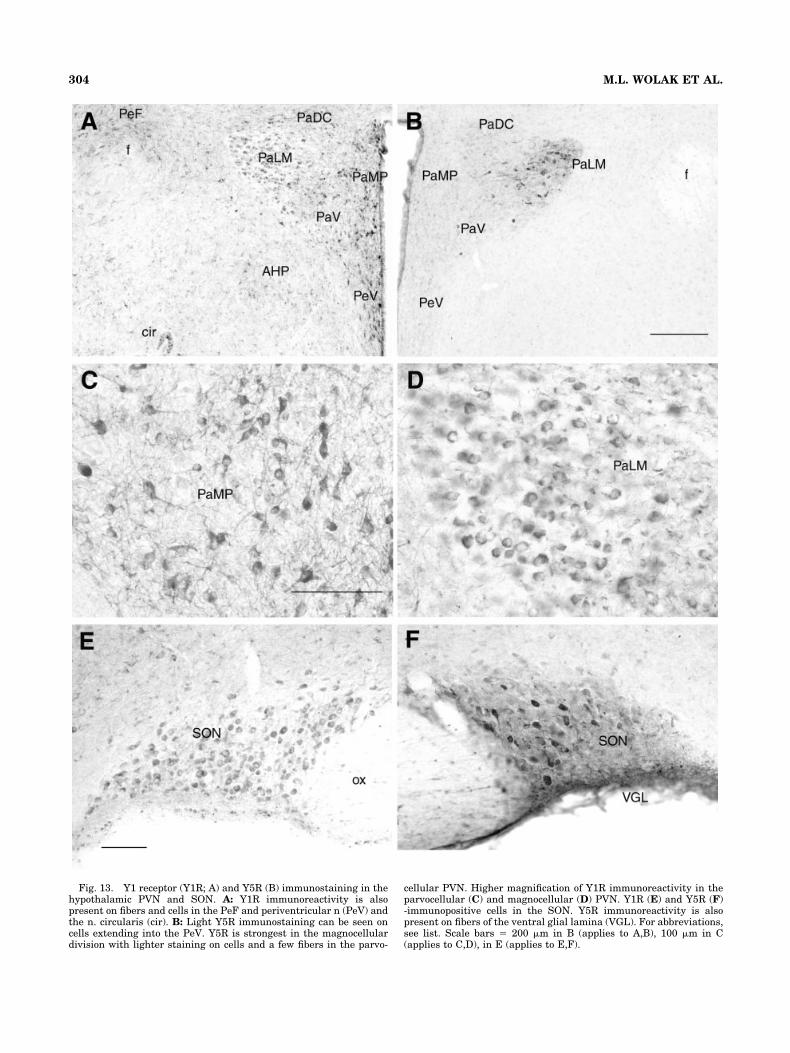
Fig. 13. Y1 receptor (Y1R; A) and Y5R (B) immunostaining in thehypothalamic PVN and SON. A: Y1R immunoreactivity is alsopresent on fibers and cells in the PeF and periventricular n (PeV) andthe n. circularis (cir). B: Light Y5R immunostaining can be seen oncells extending into the PeV. Y5R is strongest in the magnocellulardivision with lighter staining on cells and a few fibers in the parvo-
cellular PVN. Higher magnification of Y1R immunoreactivity in theparvocellular (C) and magnocellular (D) PVN. Y1R (E) and Y5R (F)-immunopositive cells in the SON. Y5R immunoreactivity is alsopresent on fibers of the ventral glial lamina (VGL). For abbreviations,see list. Scale bars � 200 �m in B (applies to A,B), 100 �m in C(applies to C,D), in E (applies to E,F).
304 M.L. WOLAK ET AL.

more Y1R-positive cell bodies in several regions (e.g., thal-amus, magnocellular paraventricular hypothalamus, andbasolateral amygdala). As our antibodies are directed tosimilar regions of the Y1R protein, the few differencesbetween our results may be attributable to the use ofdifferent detection methods or the use of Triton in theimmunostaining protocol. In initial studies, the use ofTriton at concentrations as low as 0.1%, decreased thedetection of Y1R in cell bodies in certain regions, such asthe thalamus, magnocellular PVN, and amygdala. It maybe that the detergent-like actions of Triton disruptsmembrane-associated Y1R protein perhaps either by re-moving it from the membrane or altering its epitope and,hence, its detection by the antibody. This effect of Tritonon receptor immunoreactivity has been demonstrated inother systems (Decavel and Curras, 1997). There was asimilar, yet less striking, effect of Triton on Y5R-immunoreactive staining.
Both antibodies show characteristic staining on cellmembranes as well as in cell bodies, fibers, and cytoplasm.
This distribution of Y1R immunostaining in these com-partments is supported by Western analysis of hypotha-lamic tissue where immunoreactive bands are present inboth the membrane- and cytosol-enriched fractions. Byusing Western analysis, two bands were observed for theY1R in the membrane-enriched protein preparations fromhypothalamic tissue: a low (�42 kDa) and a high (85–90kDa) molecular weight band for Y1R. The cytosol-enrichedfraction from hypothalamus demonstrated only the highermolecular weight band. The presence of the �42-kDaband in the membrane-enriched fraction likely representsa monomeric form of the membrane-bound receptor,whereas the higher molecular weight band could repre-sent a dimerized form of the receptor. The presence ofmultiple bands for the Y1R has been reported previously(Migita et al., 2001) and suggested to be a product ofposttranslational processing or glycosylation. There areseveral N-linked glycosylation sites in the N-terminus forthe Y1 receptor, which may account for the different mo-lecular weight bands. It is equally likely that this higher
Fig. 14. Coronal sections showing Y1 receptor (Y1R; A) and Y5R(B) immunoreactivity in the locus coeruleus (LC) and subcoeruleus(subC). The Me5 and cerebellar Purkinje cells and fibers demonstratelabeling for both receptors. Comparative distribution of Y1R (C) and
Y5R (D) immunoreactivity in the caudal medulla. Both receptors arepresent on cells within the SolM, X, and XII. For abbreviations, seelist. Scale bars � 500 �m in A (applies to A,B), in D (applies to C,D).
305Y1/Y5 RECEPTOR IMMUNOREACTIVITY IN RAT BRAIN

molecular weight protein could reflect synthesis of a pro-receptor protein before insertion into the membrane. Thepresence of a 85- to 90-kD molecular weight band in thecytosol-enriched fraction is more open to interpretation.As this is a relatively crude preparation, there may besome membrane proteins in this fraction or the receptorcould be bound to other cytosolic compounds, such as Gproteins. Western analysis showed a double band for theY5 receptor in hypothalamic tissue and no Y5-immunoreactive bands were detected in Western blots ofSK-N-MC homogenates, which convincingly demon-strated that the Y5R antiserum does not cross-react withthe Y1 receptor.
Overall, we observe good correlation between the pres-ence of Y1 and Y5 receptor protein in regions that are richwith NPYergic fibers (Allen et al., 1983; de Quidt andEmson, 1986). There are a few regions where receptorprotein is detected without strong NPY immunostaining.The islands of Calleja contain few NPY fibers, whereasboth Y1 and Y5 receptor-ir were prevalent. Additionally,the thalamus, which contains striking immunostainingfor both receptor subtypes, is not endowed with many NPYfibers. However, application of NPY to thalamic neuronselicits changes in neuronal firing, indicating that func-tional receptors are present (Sun et al., 2001). In themidbrain and brainstem regions, Y1R and Y5R are alsoseen in the geniculate (DLG), medial mammillary nucleus,superior colliculus, vestibular nuclei, and spinal trigemi-nal nuclei. These regions contain few NPYergic fibers. Thecerebellum contains Y1R- and Y5R-ir on Purkinje cellsand fibers, which has been noted by others (Parker andHerzog, 1999; Kopp et al., 2002). NPY receptors are alsofound within components of the olivary complex (LSO),which also do not receive strong NPY innervation. Thesemismatches of ligand and receptor have been discussed inthe literature, and it is possible that NPY reaches thesereceptors by volume transmission (Fuxe et al., 1991) orthat there are additional members of the NPY ligandfamily that have yet to be identified (Herzog et al., 1995).
Whereas the Y1 and Y5 receptors have long been pre-sumed to be postsynaptic receptors, with the Y2R beingthe primary presynaptic receptor, several histochemicaland physiological studies indicate that Y1R and Y5R mayalso have presynaptic actions. The data presented hereand in other studies demonstrate the presence of Y1R- andY5R-ir on fibers (Campbell et al., 2001; Kopp et al., 2002;Glass et al., 2002), suggesting that activation of thesereceptors could modulate presynaptic neurotransmitterrelease. Studies using electron microscopy have shownthat Y1R-ir is present within nerve terminal regions thatalso contain NPY, thereby providing anatomic evidencefor a presynaptic role for NPY, perhaps in regulating itsown release (Pickel et al., 1998; Glass et al., 2002). Thedistribution of the Y5R has not yet been examined byusing ultrastructural analysis.
A major point addressed by this work is the comparisonof the distribution of Y1R-ir and Y5R-ir in the rat brain.There is considerable overlap in the distribution of Y1R-irand Y5R-ir with the Y1R being more widely dispersed andpresent to a larger degree on fibers. Parker and Herzog(1999) presented a mapping of Y1R and Y5R mRNA byusing in situ hybridization and demonstrated that theexpression of Y1R and Y5R was distributed throughoutthe brain, with Y5R mRNA expression always coincidingwith that of Y1R mRNA. The issue that has not yet been
addressed is whether these two receptors are colocalized.The Y1R and Y5R genes, unlike the other NPY receptorsubtypes, are in close proximity to each other and havelikely evolved from a gene duplication event (Herzog et al.,1997). More interestingly, there is overlap between thesequence of the Y1R (exon 1C) and the Y5R (third intra-cellular loop), which includes the internal intron sequencefrom the Y1R, in opposite orientation, in the promoterregion of the Y5R gene. It has been suggested that thisnovel overlapping sequence may result in the coordinatedexpression of Y1 and Y5 receptors within the same cell.Whether this results in one or the other receptor beingsolely expressed or coordinately regulated in the same cellis not known. Whereas in situ hybridization ascertains thebiosynthetic activity of the cell with high sensitivity, im-munocytochemistry has the added advantage of being ableto visualize the distribution of the actual protein withincells and fibers of given cells, which not only refines thedistribution of these receptors but also adds to possiblemechanisms of action (i.e., pre- vs. postsynaptic) for NPYin these regions. A specific double-label immunofluores-cence protocol has been used to begin to address the coex-pression of Y1 and Y5 receptors in the same cell. Withinthe scope of this report, Y1 and Y5 receptor colocalizationhas been demonstrated in cells of the cerebral cortex (pre-dominantly in layer IV/V) and the caudate putamen. Ad-ditional analysis indicates that these receptors are alsocolocalized in the CA1–3 regions of the hippocampus andthe reticular thalamus. Of interest, there are regionaldifferences in the relative number of cells that coexpressthe two receptors: in the cerebral cortex, nearly all Y5R-immunoreactive cells coexpress the Y1R while there arepopulations of single-labeled Y1R-immunoreactive cells;in the caudate putamen, populations of Y5R- and notY1R-immunoreactive cells exhibit single label. These dataare important in that they demonstrate colocalization ofY1 and Y5 receptor-ir in the same cell and that differentbrain regions exhibit varying degrees of colocalization.This is an important finding as the difference in the de-gree of colocalization may reflect an overall difference inY1 versus Y5 receptor tone in a given area.
Within the telencephalon, immunostaining was ob-served for both the Y1 and Y5 receptors within the cere-bral cortex, hippocampal formation, and amygdala com-plex. In the cerebral cortex, immunoreactive fibers forboth Y1 and Y5 receptors were observed traversingthrough the different cortical layers (I–VI), although theoverall staining pattern for Y5R was stronger and moreprevalent in the cingulate/retrosplenial and piriform cor-tices. Similar patterns of distribution for these receptorswithin these regions have been noted using other antibod-ies (Caberlotto et al., 1998; Grove et al., 2000; Migita et al.,2001), binding, and in situ hybridization studies (Gehlertand Gackenheimer, 1997; Dumont et al., 1998b; Naveil-han et al., 1998; Parker and Herzog, 1999). That the Y1and Y5 receptors overlap in this region has been sup-ported by double-label immunofluorescence showing colo-calization of these receptors within the same cells in thecingulate cortex (Fig. 6) as well as somatosensory cortex(data not shown). Within the cingulate cortex, some of theY5-positive cells are colocalized with GABA and cholineacetyl transferase (Caberlotto et al., 1998; Grove et al.,2000), providing a downstream circuit whereby NPY caninfluence the activity of cortical neurons.
306 M.L. WOLAK ET AL.

Both Y1 and Y5 receptors are present within the hip-pocampal formation where they play an important role inmodulating neural excitability and seizure thresholds(Marsh et al., 1999; Ho et al., 2000; Guo et al., 2002). Theoverall distribution of Y1R and Y5R in the hippocampus issimilar, although there are differences in the distributionof the proteins. Y1R immunostaining is stronger on theCA2–3 regions with intense immunostaining occurring oninterneurons within CA1–3. In the dentate gyrus, themajority of immunostaining for the Y1R is present on cellswithin the polymorphic layer, with no detectable stainingon cells in the granule layer yet there is considerable Y1Rfiber staining in MolDG, which has also been observed inother studies (Kopp et al., 2002). The Y5R antibody lightlylabeled cell bodies within CA1–3 while staining proximaldendrites that extend into the stratum radiatum; thislabeling pattern was also observed in the dentate gyrus,where cells in the granule layer also expressed Y5R. Cur-rent studies indicate that, at least within subpopulationsof cells in the stratum pyramidale, there is coexpression ofboth Y1R and Y5R, suggesting that both of these receptorscontribute to the functioning of these cells. Additionalstudies identified that a large portion of Y5-immunoreactive cells in the hilar region of the hippocam-pus contained GABA-ir (Grove et al., 2000). Preliminarystudies in our laboratory indicate that a large proportionof Y1R-immunopositive interneurons in the CA1–3 regionare GABAergic (Teppen and Urban, unpublished observa-tions). These studies suggest that NPY, acting through theY1 and Y5 receptors, may influence hippocampal activitythrough modulation of GABA neurotransmission.
Another prominent role for Y1 and Y5 receptors is in theamygdala, where NPY has anxiolytic actions (Wahlestedtet al., 1993; Heilig et al., 1993; Sajdyk et al., 1999; Kask etal., 2002). The presence of NPY receptors in this region isalso important with respect to the generation of autonomicand behavioral responses to stress and anxiety (Gray etal., 1989; Gray, 1991). Y5R immunostaining is primarilyrestricted to the BLA, whereas that of the Y1 receptor ismore widely distributed, extending to the CeAm andMeAm. Recently, it has been demonstrated that the anxi-olytic actions of NPY are specific to the BLA throughactivation of Y1 and Y5 receptors (Sajdyk et al., 1999,2002). Whereas the actions of NPY on Y1 and Y5 receptorshas been demonstrated pharmacologically, there is little,if any, significant expression of receptor protein in theseareas by autoradiography (Ohkubo et al., 1990; Dumont etal., 1993). The current demonstration of Y1R- and Y5R-irin the BLA supports these pharmacologic findings andprovides more information as to how NPY might alteranxiety-related behaviors. Given the low incidence of NPYreceptor immunostaining on fibers in the BLA, it is likelythat activation of Y1 and Y5 receptors influences the ac-tivity of cells postsynaptically. As the morphology of thesecells is similar, it would not be surprising to find colocal-ization of Y1R and Y5R. Additional studies are needed tofurther examine the neurotransmitter content and projec-tions of these Y1R- and Y5R-immunoreactive cells as theyrelate to the role of NPY in modulating anxiety and re-lated behaviors. These future studies are potentially im-portant, as some clinical treatments of anxiety have sig-nificant actions in the amygdala (Pesold and Treit, 1995;Sajdyk and Shekhar, 1997) and may influence or modulatethe activity of NPY and its receptors in this region (Ehlerset al., 1997). The distribution of Y1R-ir in the CeAm
closely reflects the staining for NPY, in that more Y1R-iris found in the medial division of the CeAm (fibers andcells) with less immunostaining in the lateral aspect ofthis nucleus. NPY in this region may participate in theanxiolytic actions of the peptide (Heilig et al., 1993) or itmay influence hormonal and cardiovascular rather thanbehavioral responses to a stressor (Gray et al., 1989; Grayand Bingaman, 1996; Roozendaal et al., 1997).
Immunoreactivity for both the Y1 and Y5 receptors ispresent throughout the hypothalamus and anatomicallydefines sites of action for these subtypes in regulatinghormone secretion (Wahlestedt et al., 1987; Pelletier et al.,1994; Urban et al., 1996), feeding (Stanley et al., 1985,1993), and circadian rhythms (Biello, 1995). Y1R-ir in thediagonal band of Broca (DBB) and median eminence cor-responds with the roles of NPY and the Y1R in stimulat-ing the production (Pelletier et al., 1994) and release ofgonadotropin-releasing hormone (GnRH) from the hypo-thalamus (Kalra et al., 1992; Besecke and Levine, 1994;Besecke et al., 1994; Urban et al., 1996). Recently, it hasbeen postulated that activation of Y5R contributes to theinhibitory actions of NPY on reproductive hormone secre-tion (Raposinho et al., 1999). Y5R-ir is present in the DBBand preoptic hypothalamus and has been shown to becolocalized in GnRH cell bodies (Campbell et al., 2001).
Within the PVN, intense immunostaining for Y1R waspresent on parvo- and magnocellular neurons as well as onnumerous fibers within the parvocellular division. Thisfinding is different than previous reports, indicating thelack of Y1R-ir in the magnocellular PVN of the mouse(Broberger et al., 1999). These discrepant results could bethe result of species differences or the use of colchicine inthe previous study. Y5R-ir is displayed on cells in themagnocellular PVN and on scattered cells and fiberswithin the parvocellular division. At least within the mag-nocellular PVN and SON, it is conceivable that thesereceptors are colocalized within the same cell. The pres-ence of Y1R- and Y5R-ir on magnocellular PVN and SONsuggests that activation of these receptors may influencethe synthesis and/or release of oxytocin (OT) and vaso-pressin (VP). Both in vivo and in vitro studies demon-strate that NPY dose dependently stimulates vasopressin(Leibowitz et al., 1988; Liu et al., 1994; Larsen et al., 1994)and oxytocin synthesis and secretion (Kapoor and Sladek,2001). Administration of NPY and the Y1 and Y5 receptorpreferring ligand [Leu31Pro34]NPY excites vasopressincells, stimulates release in vitro (Khanna et al., 1993;Kapoor and Sladek, 2001), and enhances both VP and OTrelease by phenylephrine, an 1 receptor agonist. ThatNPY actively participates in the osmotic regulation ofhormone secretion is suggested by the increase in NPYmRNA and protein levels observed in the PVN and SONafter chronic osmotic stimulation (Larsen et al., 1993).The presence of Y1R- and Y5R-ir in the parvocellulardivision further supports a role for these receptors in theregulation of corticotropin releasing hormone (Hastings etal., 2001) and thyrotrophin releasing hormone secretion(Michalkiewicz and Suzuki, 1994). As both Y1 and Y5receptor-ir is present within cell bodies of the PVN andmedian eminence, it is likely that activation of either orboth of these receptors directly influences the synthesisand release of hypothalamic releasing factors.
One major, and clinically relevant, effect of NPY is itsrole in the induction of feeding at a number of sites,
307Y1/Y5 RECEPTOR IMMUNOREACTIVITY IN RAT BRAIN

including the PVN, lateral, dorsomedial, and perifornicalhypothalamic areas (Stanley et al., 1985, 1993). The NPYreceptor subtype mediating these actions has been open todebate, although there is overwhelming evidence to sup-port a role for both the Y1 and Y5 receptors in this process.The distribution of Y1R- and Y5R-ir provides evidencethat both receptors are present within these “feeding ar-eas” of the hypothalamus. That these receptors in thePVN participate in feeding has been documented (Stanleyet al., 1986; Gerald et al., 1996; Kask et al., 1998b; Yoko-suka et al., 1999), although the actual mechanism under-lying these actions of NPY has not been elucidated. Thepresence of both Y1R and Y5R protein in this region indi-cates that both may participate in the production of feed-ing responses. The regulation of these receptors underdifferent conditions is important. Food restriction in-creases the expression of Y1R mRNA (Xu et al., 1998a)and alternately decreases Y5R expression (Widdowson,1997). This finding may reflect coordinate regulation ofthe Y1 and Y5 receptors as discussed by Herzog et al.(1997). The perifornical area and LHA contain a strikingdensity of Y1-immunoreactive fibers and scattered Y1-and Y5-immunoreactive cell bodies and fibers. This regionof the brain receives inputs from the hypothalamic arcu-ate NPYergic pathway and contains a population ofmelanin-concentrating hormone (MCH)- and orexin-containing cells (Elias et al., 1998), which are devoid ofY1R-ir (Campbell et al., 2003). Whether the NPY recep-tors are present on MCH-containing cells is not known. Inaddition to these hypothalamic sites of action for NPY,there are additional, nonhypothalamic, sites that are alsoimportant in initiating feeding behavior (Broberger andHokfelt, 2001) such as NTS, PAG (periaqueductal gray),and thalamus, which receive projections from the ArcNand contain significant densities of Y1R and Y5R protein.
The locus coeruleus and subcoeruleus are cell groupsthat contain intensely labeled cells for both Y1 and Y5receptors and numerous processes that contain Y5 immu-nostaining. Because the majority of cells in the LC arenoradrenergic, it is likely that NPY receptors are presenton catecholaminergic cells. The noradrenergic neurons inthe LC colocalize with galanin and NPY (Holets et al.,1988; Moore and Gustafson, 1989; Xu et al., 1998b) andproject to several structures, including the forebrain, hip-pocampus, thalamus, preoptic area, and hindbrain (re-viewed in Aston-Jones et al., 1995). The activity of thesecells, therefore, is of possible importance to the regulationof several processes, including hormone secretion(Anselmo-Franci et al., 1997), arousal (Aston-Jones et al.,2001), blood pressure (Miyawaki et al., 1991), and anxiety,although these anxiolytic actions are likely due to theactivation of Y2 receptors (Kask et al., 1998a). Studies byYang et al. (1996) showed that Y1R-ir is present withinthe majority of tyrosine hydroxylase-positive cells of themedullary A1, A2, and C1–3 cell groups, and it is possible,therefore, that noradrenergic cells in the LC may alsocoexpress NPY receptors, thereby regulating the ability ofthese neurons to produce and release norepinephrine andNPY.
SUMMARY
The goal of the present studies was to compare thedistribution of Y1 and Y5 receptor-ir in the brain with theintent to provide further insight to the cellular distribu-
tion of these proteins and regions where they may overlap,perhaps on the same neural substrates. There are severalbrain regions where the cellular distribution of Y1R- andY5R-ir are very similar, those being the DBB (Fig. 8),reticular thalamus (Fig. 11), hippocampus (CA2–3, Fig. 7),BLA (Fig. 10), PVN/SON (Fig. 13), locus coeruleus (Fig.14), Mo5 and Me5 (Fig. 14), and cerebellum. In many otherareas, both receptors are present yet their localizationwithin brain nuclei is different. Colocalization of Y1R- andY5R-ir has been identified in several brain regions, includ-ing the cerebral cortex, hippocampus, thalamus, and cau-date putamen. As activation of both Y1 and Y5 receptorscontribute to the function of several physiological systems,understanding the distribution, function, and regulationof these receptors is an integral part of understanding therole of NPY and its receptors in physiological processes.Using well characterized antibodies can further elucidatethe cellular distribution of Y1 and Y5 receptors as well asidentify projection sites of NPY receptive neurons.
ACKNOWLEDGMENTS
We thank Joanna Romanelli for her expert technicalassistance. Analysis of tissues by using confocal micros-copy was made possible through the use of the Microscopeand Imaging Facility at Finch University of HealthSciences/The Chicago Medical School.
LITERATURE CITED
Adams JC. 1992. Biotin amplification of biotin and horseradish peroxidasesignals in histochemical stains. J Histochem Cytochem 40:1457–1463.
Allen YS, Adrian TE, Allen JM, Tatemoto K, Crow TJ, Bloom SR, PolakJM. 1983. Neuropeptide Y distribution in the rat brain. Science 221:877–879.
Allen CJ, Ghilardi JR, Vigna SR, Mannon PJ, Taylor IL, McVey DC,Maggio JE, Mantyh PW. 1993. Neuropeptide Y/peptide YY receptorbinding sites in the heart: localization and pharmacological character-ization. Neuroscience 53:889–898.
Anselmo-Franci JA, Franci CR, Krulich L, Antunes-Rodrigues J, McCannSM. 1997. Locus coeruleus lesions decrease norepinephrine input intothe medial preoptic area and medial basal hypothalamus and block theLH, FSH and prolactin preovulatory surge. Brain Res 767:289–296.
Armstrong WE, Scholer J, McNeill TH. 1982. Immunocytochemical, Golgiand electron microscopic characterization of putative dendrites in theventral glial lamina of the rat supraoptic nucleus. Neuroscience 7:679–694.
Aston-Jones G, Shipley MT, Grzanna R. 1995. The locus coeruleus, A5 andA7 noradrenergic cell groups. In: Paxinos G, editor. The rat nervoussystem. New York: Academic Press. p 183–206.
Aston-Jones G, Chen S, Zhu Y, Oshinsky ML. 2001. A neural circuit forcircadian regulation of arousal. Nat Neurosci 4:732–738.
Baraban SC, Hollopeter G, Erickson JC, Schwartzkroin PA, Palmiter RD.1997. Knock-out mice reveal a critical antiepileptic role for neuropep-tide Y. J Neurosci 17:8927–8936.
Bard JA, Walker MW, Branchek TA, Weinshank RL. 1995. Cloning andfunctional expression of a human Y4 subtype receptor for pancreaticpolypeptide, neuropeptide Y, and peptide YY. J Biol Chem 270:26762–26765.
Bauer-Dantoin AC, McDonald JK, Levine JE. 1991. Neuropeptide Y po-tentiates luteinizing hormone (LH)-releasing hormone-stimulated LHsurges in pentobarbital-blocked proestrous rats. Endocrinology 129:402–408.
Besecke LM, Levine JE. 1994. Acute increase in responsiveness of lutein-izing hormone (LH)- releasing hormone nerve terminals toneuropeptide-Y stimulation before the preovulatory LH surge. Endo-crinology 135:63–66.
Besecke LM, Wolfe AM, Pierce ME, Takahashi JS, Levine JE. 1994. Neu-ropeptide Y stimulates luteinizing hormone-releasing hormone release
308 M.L. WOLAK ET AL.

from superfused hypothalamic GT1-7 cells. Endocrinology 135:1621–1627.
Biello SM. 1995. Enhanced photic phase shifting after treatment withantiserum to neuropeptide Y. Brain Res 673:25–29.
Broberger C, Hokfelt T. 2001. Hypothalamic and vagal neuropeptide cir-cuitries regulating food intake. Physiol Behav 74:669–682.
Broberger C, Visser TJ, Kuhar MJ, Hokfelt T. 1999. Neuropeptide Yinnervation and neuropeptide Y Y1-receptors expressing neurons inthe paraventricular hypothalamic nucleus of the mouse. Neuroendocri-nology 70:295–305.
Caberlotto L, Tinner B, Bunnemann B, Agnati L, Fuxe K. 1998. On therelationship of neuropeptide Y Y1 receptor-immunoreactive neuronalstructures to the neuropeptide Y-immunoreactive nerve terminal net-works. A double immunolabelling analysis in the rat brain. Neuro-science 86:827–845.
Campbell RE, ffrench-Mullen JMH, Cowley MA, Smith MS, Grove KL.2001. Hypothalamic circuitry of neuropeptide Y regulation of neuroen-docrine function and food intake via the Y5 receptor subtype. Neuroen-docrinology 74:106–119.
Campbell RE, Smith MS, Allen SE, Grayson BE, ffrench-Mullen JMH,Grove KL. 2003. Orexin neurons express a functional pancreaticpolypeptide Y4 receptor. J Neurosci 23:1487–1497.
Carter DA, Vallejo M, Lightman SL. 1985. Cardiovascular effects of neu-ropeptide Y in the nucleus tractus solitarius of rats: relationship withnoradrenaline and vasopressin. Peptides 6:421–425.
Chronwall BM. 1989. Anatomical distribution of NPY and NPY messengerRNA in rat brain. In: Mutt V, Hokfelt T, Fuxe K, Lundberg JM, editors.New York: Raven Press. p 51–59.
Chronwall BM, DiMaggio DA, Massari VJ, Pickel VM, Ruggiero DA,O’Donohue TL. 1985. The anatomy of neuropeptide-Y-containing neu-rons in the rat brain. Neuroscience 15:1159–1181.
de Quidt ME, Emson PC. 1986. Distribution of neuropeptide Y-like immu-noreactivity in the rat central nervous system. II. Immunohistochem-ical analysis. Neuroscience 18:545–618.
Decavel C, Curras MC. 1997. Increased expression of the N-methyl-D-aspartate receptor subunit, NR1, in immunohistochemically identifiedmagnocellular hypothalamic neurons during dehydration. Neuro-science 78:191–202.
Duhault J, Boulanger M, Chamorro S, Boutin JA, Zuana OD, Douillet E,Fauchere JL, Feletou M, Germain M, Husson B, Vega AM, Renard P,Tisserand F. 2000. Food intake regulation in rodents: Y5 or Y1 NPYreceptors or both. Can J Physiol Pharmacol 78:173–185.
Dumont Y, Fournier A, St. Pierre S, Quirion R. 1993. Comparative char-acterization and autoradiographic distribution of neuropeptide Y re-ceptor sites in the rat brain. J Neurosci 13:73–86.
Dumont Y, Fournier A, Quirion R. 1998a. Expression and characterizationof the neuropeptide Y Y5 receptor subtype in the rat brain. J Neurosci18:5565–5574.
Dumont Y, Jacques D, Bouchard P, Quirion R. 1998b. Species differencesin the expression and distribution of the neuropeptide Y Y1, Y2, Y4 andY5 receptors in rodents, guinea pig and primates brains. J CompNeurol 402:372–384.
Durkin MM, Walker MW, Smith KE, Gustafson EL, Gerald C, BranchekTA. 2000. Expression of a novel neuropeptide Y receptor subtype in-volved in food intake: an in situ hybridization study of Y5 mRNAdistribution in rat brain. Exp Neurol 165:90–100.
Ehlers CL, Somes C, Lopez A, Kirby D, Rivier JE. 1997. Electrophysiolog-ical actions of neuropeptide Y and its analogs: new measures for anxi-olytic therapy? Neuropsychopharmacology 17:34–43.
Elias CF, Saper CB, Maratos-Flier E, Tritos NA, Lee C, Kelly J, Tatro JB,Hoffman GE, Ollmann MM, Barsh GS, Sakurai T, Yanagisawa M,Elmquist JK. 1998. Chemically defined projections linking the medio-basal hypothalamus and the lateral hypothalamic area. J Comp Neurol402:442–459.
Eva C, Keinanen K, Monyer H, Seeburg P, Sprengel R. 1990. Molecularcloning of a novel G protein-coupled receptor that may belong to theneuropeptide receptor family. FEBS Lett 271:81–84.
Eva C, Oberto A, Sprengel R, Genazzani E. 1992. The murine NPY-1receptor gene. Structure and delineation of tissue-specific expression.FEBS Lett 314:285–288.
Everitt BJ, Hokfelt T, Terenius L, Tatemoto K, Mutt V, Goldstein M. 1984.Differential co-existence of neuropeptide Y (NPY)-like immunoreactiv-ity with catecholamines in the central nervous system of the rat.Neuroscience 11:443–462.
Fuxe K, Agnati LF, Aguirre JA, Bjelke B, Tinner B, Merlo Pich E, Eneroth
P. 1991. On the existence of volume transmission in the central neu-ropeptide Y neuronal systems. Studies on transmitter receptor mis-matches and on biological effects of neuropeptide Y fragments. In: FuxeK, Agnati L, editors. Volume transmission in the brain: novel mecha-nisms for neural transmission. New York: Raven Press, Ltd. p 105–130.
Gehlert DR, Gackenheimer SL. 1997. Differential distribution of neuropep-tide Y Y1and Y2 receptors in rat and guinea pig brains. Neuroscience76:215–224.
Gerald C, Walker MW, Pierre J-J, He C, Branchek TA, Weinshank RL.1995. Expression cloning and pharmacological characterization of ahuman hippocampal neuropeptide Y/peptide YY Y2 receptor subtype.J Biol Chem 270:26758–26761.
Gerald C, Walker MW, Criscione L, Gustafson EL, Batzi-Hartmann C,Branchek TA, Weinshank RL. 1996. A receptor subtype involved inneuropeptide-Y-induced food intake. Nature 382:168–171.
Ghilardi JR, Allen CJ, Vigna SR, McVey DC, Mantyh PW. 1994. Cholecys-tokinin and neuropeptide Y receptors on single rabbit vagal afferentganglion neurons: site of prejunctional modulation of visceral sensoryneurons. Brain Res 633:33–40.
Glass MJ, Chan J, Pickel VM. 2002. Ultrastructural localization of neu-ropeptide Y Y1 receptors in the rat medial nucleus tractus solitarius:relationships with neuropeptide Y or catecholamine neurons. J Neuro-sci Res 67:753–765.
Gray TS. 1991. Limbic pathways and neurotransmitters as mediators ofautonomic and neuroendocrine responses to stress. In: Brown MR,Koob GF, Rivier C, editors. Stress: neurobiology and neuroendocrinol-ogy. New York: Marcel Dekker, Inc. p 73–89.
Gray TS, Bingaman EW. 1996. The amygdala: corticotropin-releasing fac-tor, steroids, and stress. Crit Rev Neurobiol 10:155–168.
Gray TS, Carney MF, Magnuson DJ. 1989. Direct projections from thecentral amygdaloid nucleus to the hypothalamic paraventricular nu-cleus: possible role in stress-induced adrenocorticotropin release. Neu-roendocrinology 50:433–446.
Grove KL, Campbell RE, ffrench-Mullen JMH, Cowley MA, Smith MS.2000. Neuropeptide Y Y5 receptor protein in the cortical/limbic systemand brainstem of the rat: expression on �-aminobutyric acid andcorticotropin-releasing hormone neurons. Neuroscience 100:731–740.
Guo H, Castro PA, Palmiter RD, Baraban SC. 2002. Y5 receptors mediateneuropeptide Y actions at excitatory synapses in area CA3 of the mousehippocampus. J Neurophysiol 87:558–566.
Hastings JA, McClure-Sharp JM, Morris MJ. 2001. NPY Y1 receptors exertopposite effects on corticotropin releasing factor and noradrenalineoverflow from the rat hypothalamus in vitro. Brain Res 890:32–37.
Heilig M, McLoed S, Brot M, Heinrichs SC, Menzaghi F, Koob GF, BrittonKT. 1993. Anxiolytic-like actions of neuropeptide Y: mediation by Y1receptors in amygdala, and dissociation from food intake effects. Neu-ropsychopharmacology 8:357–363.
Herzog H, Hort Y, Schneider R, Shine J. 1995. Seminalplasmin: recentevolution of another member of the neuropeptide Y gene family. ProcNatl Acad Sci U S A 92:594–598.
Herzog H, Darby K, Ball H, Hort Y, Beck-Sickinger AG, Shine J. 1997.Overlapping gene structure of the human neuropeptide Y receptorsubtypes Y1 and Y5 suggests coordinate transcriptional regulation.Genomics 41:315–319.
Ho MW, Beck-Sickinger AG, Colmers WF. 2000. Neuropeptide Y(5) recep-tors reduce synaptic excitation in proximal subiculum, but not epilep-tiform activity in rat hippocampal slices. J Neurophysiol 83:723–734.
Holets VR, Hokfelt T, Rokaeus A, Terenius L, Goldstein M. 1988. Locuscoeruleus neurons in the rat containing neuropeptide Y, tyrosine hy-droxylase or galanin and their efferent projections to the spinal cord,cerebral cortex and hypothalamus. Neuroscience 24:893–906.
Kalia M, Fuxe K. 1985. Rat medulla oblongata. I. Cytoarchitectonic con-siderations. J Comp Neurol 233:285–307.
Kalra SP, Fuentes M, Fournier A, Parker SL, Crowley WR. 1992. Involve-ment of the Y-1 receptor subtype in the regulation of luteinizing hor-mone secretion by neuropeptide Y in rats. Endocrinology 130:3323–3330.
Kapoor JR, Sladek CD. 2001. Substance P and NPY differentially poten-tiate ATP and adrenergic stimulated vasopressin and oxytocin release.Am J Physiol 280:R69–R78.
Kask A, Rago L, Harro J. 1998a. Anxiolytic-like effect of neuropeptide Y(NPY) and NPY13-36 microinjected into vicinity of locus coeruleus inrats. Brain Res 788:345–348.
Kask A, Rago L, Harro J. 1998b. Evidence for involvement of neuropeptide
309Y1/Y5 RECEPTOR IMMUNOREACTIVITY IN RAT BRAIN

Y receptors in the regulation of food intake: studies with Y1-selectiveantagonist BIBP3226. Br J Pharmacol 124:1507–1515.
Kask A, Harro J, Von Horsten S, Redrobe JP, Dumont Y, Quirion R. 2002.The neurocircuitry and receptor subtypes mediating anxiolytic-likeeffects of neuropeptide Y. Neurosci Biobehav Rev 26:259–283.
Khanna S, Sibbald JR, Day TA. 1993. Neuropeptide Y modulation of A1noradrenergic neuron input to supraoptic vasopressin cells. NeurosciLett 161:60–64.
Kopp J, Xu ZQ, Zhang X, Pedrazzini T, Herzog H, Kresse A, Wong H,Walsh JH, Hokfelt T. 2002. Expression of the neuropeptide Y Y1receptor in the CNS of rat and of wild-type and Y1 receptor knock-outmice. Focus on immunohistochemical localization. Neuroscience 111:443–532.
Larhammar D. 1996. Structural diversity of receptors for neuropeptide Y,peptide YY and pancreatic polypeptide. Regul Pept 65:165–174.
Larsen PJ, Mikkelsen JD, Jessop DS, Lightman SL, Chowdrey HS. 1993.Neuropeptide Y mRNA and immunoreactivity in hypothalamus neu-roendocrine neurons: effect of adrenalectomy and chronic osmotic stim-ulation. J Neurosci 13:1138–1147.
Larsen PJ, Jukes KE, Chowdrey HS, Lightman SL, Jessop DS. 1994.Neuropeptide-Y potentiates the secretion of vasopressin from the neu-rointermediate lobe of the rat pituitary gland. Endocrinology 134:1635–1639.
Leibowitz SF, Sladek CD, Spencer L, Tempel D. 1988. Neuropeptide Y,epinephrine, norepinephrine in the paraventricular nucleus: stimula-tion of feeding and the release of corticosterone, vasopressin and glu-cose. Brain Res Bull 21:905–912.
Leupen SM, Besecke LM, Levine JE. 1997. Neuropeptide Y Y1-receptorstimulation is required for physiological amplification of preovulatoryluteinizing hormone surges. Endocrinology 138:2735–2739.
Li C, Chen P, Smith MS. 1999. Morphological evidence for direct interac-tion between arcuate nucleus neuropeptide Y (NPY) neurons andgonadotropin-releasing hormone neurons and the possible involvementof NPY Y1 receptors. Endocrinology 140:5382–5390.
Liu JP, Clarke IJ, Funder JW, Engler D. 1994. Studies of the secretion ofcorticotropin-releasing factor and arginine vasopressin into thehypophysial-portal circulation of the conscious sheep. II. The centralnoradrenergic and neuropeptide Y pathways cause immediate andprolonged hypothalamic-pituitary-adrenal activation. Potential in-volvement in the pseudo-Cushing’s syndrome of endogenous depressionand anorexia nervosa. J Clin Invest 93:1439–1450.
Lundberg JM, Terenius L, Hokfelt T, Martling CR, Tatemoto K, Mutt V,Polak J, Bloom S, Goldstein M. 1982. Neuropeptide Y (NPY)-like im-munoreactivity in peripheral noradrenergic neurons and effects of NPYon sympathetic function. Acta Physiol Scand 116:477–480.
Marsh DJ, Hollopeter G, Kafer KE, Palmiter RD. 1998. Role of the Y5neuropeptide Y receptor in feeding and obesity. Nat Med 4:718–721.
Marsh DJ, Baraban SC, Hollopeter G, Palmiter RD. 1999. Role of the Y5neuropeptide Y receptor in limbic seizures. Proc Natl Acad Sci U S A96:13518–13523.
Michalkiewicz M, Suzuki M. 1994. Adenohypophyseal vasoactive intestinalpeptide and neuropeptide Y responses to hypothyroidism are abolishedafter anterolateral deafferentation of the hypothalamus. Neuroendo-crinology 59:85–91.
Migita K, Loewy AD, Ramabhadran TV, Krause JE, Waters SM. 2001.Immunohistochemical localization of the neuropeptide Y Y1 receptor inrat central nervous system. Brain Res 889:23–37.
Mikkelsen JD, Larsen PJ. 1992. A high concentration of NPY(Y1)-receptormRNA-expressing cells in the rat arcuate nucleus. Neurosci Lett 148:195–198.
Miyawaki T, Kawamura H, Komatsu K, Yasugi T. 1991. Chemical stimu-lation of the locus coeruleus: inhibitory effects on hemodynamics andrenal sympathetic nerve activity. Brain Res 568:101–108.
Moore RY, Gustafson EL. 1989. The distribution of dopamine--hydroxylase, neuropeptide Y and galanin in locus coeruleus neurons.J Chem Neuroanat 2:95–106.
Naveilhan P, Neveu I, Arenas E, Ernfors P. 1998. Complementary andoverlapping expression of Y1, Y2 and Y5 receptors in the developingand adult mouse nervous system. Neuroscience 87:289–302.
Niimi M, Sato M, Taminato T. 2001. Neuropeptide Y in central control offeeding and interactions with orexin and leptin. Endocrine 14:269–273.
Ohkubo T, Niwa M, Yamashita K, Kataoka Y, Shigematsu K. 1990. Neu-
ropeptide Y (NPY) and peptide YY (PYY) receptors in rat brain. CellMol Neurobiol 10:539–552.
Parker RMC, Herzog H. 1999. Regional distribution of Y-receptor subtypemRNAs in rat brain. Eur J Neurosci 11:1431–1448.
Paxinos G, Watson C. 1997. The rat brain in stereotaxic coordinates.Orlando, FL: Academic Press, Inc.
Pelletier G, Li S, Hong M, Fournier A, St. Pierre S. 1994. Role of neuropep-tide Y in the regulation of gonadotropin releasing hormone (GnRH)gene expression in the rat preoptic area. Mol Brain Res 26:696–673.
Pesold C, Treit D. 1995. The central and basolateral amygdala differen-tially mediate the anxiolytic effects of benzodiazepines. Brain Res671:213–221.
Pickel VM, Beck-Sickinger AG, Chan RKW, Wieland HA. 1998. Y1 recep-tors in the nucleus accumbens: ultrastructural localization and associ-ation with neuropeptide Y. J Neurosci Res 52:54–68.
Pronchuk N, Beck-Sickinger AG, Colmers WF. 2002. Multiple NPY recep-tors inhibit GABA(A) synaptic responses of rat medial parvocellulareffector neurons in the hypothalamic paraventricular nucleus. Endo-crinology 143:535–543.
Quirion R, Martel J-C, Dumont Y, Cadiuex A, Jolicoeur F, St. Pierre S,Fournier A. 1990. Neuropeptide Y receptors: autoradiographic distri-bution in the brain and structure-activity relationships. Ann N Y AcadSci 611:58–72.
Raposinho PD, Broqua P, Pierroz DD, Hayward A, Dumont Y, Quirion R,Junien J-L, Aubert ML. 1999. Evidence that the inhibition of luteiniz-ing hormone secretion exerted by central administration of neuropep-tide Y (NPY) in the rat is predominantly mediated by the NPY-Y5receptor subtype. Endocrinology 140:4046–4066.
Roozendaal B, Koolhaas JM, Bohus B. 1997. The role of the central amyg-dala in stress and adaption. Acta Physiol Scand 161:51–54.
Ruigrok TJH, Cella F. 1995. Precerebellar nuclei and red nucleus. In:Paxinos G, editor. The rat nervous system. San Diego CA: AcademicPress. p 277–308.
Sajdyk TJ, Shekhar A. 1997. Excitatory amino acid receptor antagonistsblock the cardiovascular and anxiety responses elicited by gamma-aminobutyric acid A receptor blockade in the basolateral amygdala ofrats. J Pharmacol Exp Ther 283:969–977.
Sajdyk TJ, Schober DA, Gehlert DR. 2002. Neuropeptide Y receptor sub-types in the basolateral nucleus of the amygdala modulate anxiogenicresponses in rats. Neuropharmacol 43:1165–1172.
Sajdyk TJ, Vandergriff MG, Gehlert DR. 1999. Amygdalar neuropeptide YY1 receptors mediate the anxiolytic-like actions of neuropeptide Y inthe social interaction test. Eur J Pharmacol 368:143–147.
Schaffhauser AO, Stricker-Krongrad A, Brunner L, Cumin F, Gerald C,Whitebread S, Criscione L, Hofbauer KG. 1997. Inhibition of foodintake by neuropeptide Y Y5 receptor antisense oligodeoxynucleotides.Diabetes 46:1792–1798.
Stanley BG, Chin AS, Leibowitz SF. 1985. Feeding and drinking elicited bycentral injection of neuropeptide Y: evidence for a hypothalamic site(s)of action. Brain Res Bull 14:521–524.
Stanley BG, Kyrkouli SE, Lampert S, Leibowitz SF. 1986. Neuropeptide Ychronically injected into the hypothalamus: a powerful neurochemicalinducer of hyperphagia and obesity. Peptides 7:1189–1192.
Stanley BG, Magdalin W, Seirafi A, Thomas WJ, Leibowitz SF. 1993. Theperifornical area: the major focus of (a) patchily distributed hypotha-lamic neuropeptide Y-sensitive feeding system(s). Brain Res 604:304–317.
Steinbusch HWM, Niewenhuys R. 1983. The raphe nuclei of the rat brain-stem: a cytoarchitectonic and immunohistochemical study. In: EmsonP, editor. Chemical neuroanatomy. New York: Raven Press. p 131–207.
Sun QQ, Akk G, Huguenard JR, Prince DA. 2001. Differential regulation ofGABA release and neuronal excitability mediated by neuropeptide Y1and Y2 receptor in rat thalamic neurons. J Physiol 531:81–94.
Urban JH, Das I, Levine JE. 1996. Steroid modulation of neuropeptideY-induced luteinizing hormone releasing hormone release from medianeminence fragments from male rats. Neuroendocrinology 63:112–119.
Wahlestedt C, Pich EM, Koob GF, Yee F, Heilig M. 1993. Modulation ofanxiety and neuropeptide Y-Y1 receptors by antisense oligodeoxynucle-otides. Science 259:528–531.
Wahlestedt C, Skagerberg G, Ekman R, Heilig M, Sundler F, Hakanson R.1987. Neuropeptide Y (NPY) in the area of the hypothalamic paraven-tricular nucleus activates the pituitary-adrenocortical axis in the rat.Brain Res 417:33–38.
Widdowson PS. 1997. Regionally selective down-regulation of NPY recep-
310 M.L. WOLAK ET AL.

tor subtypes in the obese Zucker rat. Relationship to the Y5 “feeding”receptor. Brain Res 758:17–25.
Xu B, Kalra PS, Moldawer LL, Kalra SP. 1998a. Increased appetite aug-ments hypothalamic NPY Y1 receptor gene expression: effects of an-orexigenic ciliary neurotropic factor. Regul Pept 75–76:391–395.
Xu ZQ, Shi TJ, Hokfelt T. 1998b. Galanin/GMAP- and NPY-like immuno-reactivities in locus coeruleus and noradrenergic nerve terminals in thehippocampal formation and cortex with notes on the galanin-R1 and-R2 receptors. J Comp Neurol 392:227–251.
Yang S-N, Bunnemann B, Cintra A, Fuxe K. 1996. Localization of neu-
ropeptide Y Y1 receptor-like immunoreactivity in catecholaminergicneurons of the rat medulla oblongata. Neuroscience 73:519–530.
Yokosuka M, Kalra PS, Kalra SP. 1999. Inhibition of neuropeptide Y(NPY)-induced feeding and cfos response in magnocellular paraven-tricular nucleus by a NPY receptor antagonist: a site of NPY action.Endocrinology 140:4494–4500.
Zhang X, Bao L, Xu ZQ, Kopp J, Arvidsson U, Elde R, Hokfelt T. 1994.Localization of neuropeptide Y Y1 receptors in the rat nervous systemwith special reference to somatic receptors on small dorsal root gan-glion neurons. Proc Natl Acad Sci U S A 91:11738–11742.
311Y1/Y5 RECEPTOR IMMUNOREACTIVITY IN RAT BRAIN
Copyright © 2022 FDOKUMEN