Application of CD56, P63 and CK19 immunohistochemistry in the diagnosis of papillary carcinoma of...
12
BioMed Central Page 1 of 12 (page number not for citation purposes) Diagnostic Pathology Open Access Research Application of CD56, P63 and CK19 immunohistochemistry in the diagnosis of papillary carcinoma of the thyroid Dina El Demellawy* 1 , Ahmed Nasr 2 and Salem Alowami 3 Address: 1 University of Northern Ontario School of Medicine, Thunder Bay Regional Health Sciences Centre, Department of Pathology and Laboratory Medicine, Thunder Bay, Ontario, Canada, 2 University of Toronto, Department of Surgery, Toronto, Ontario, Canada and 3 McMaster University, Department of Pathology and Molecular Medicine, Hamilton, Ontario, Canada Email: Dina El Demellawy* - [email protected]; Ahmed Nasr - [email protected]; Salem Alowami - [email protected] * Corresponding author Abstract Papillary carcinoma of the thyroid (PTC) is the commonest thyroid cancer. In the recent decades an obvious increase in the incidence of PTC has occurred. The pathological diagnosis of PTC is usually an easy diagnosis in the majority of cases. However since the introduction of follicular variant of PTC and the wide threshold range in interpretation of the clearly set pathological criteria for diagnosis of PTC, between pathologists including experts, the diagnosis in some cases became quite difficult. Unfortunately some cases are unjustifiably over-called as follicular variant of PTC as a result of the wide inter observable variability between pathologists, including thyroid pathologists. Ancillary studies such as immmunohistochemistry may be helpful, but till now there is no 100% consistent marker(s), that distinct between PTC and other follicular thyroid lesions and tumors. We assessed expression of antibodies against CD56, CK19, P63 and E-Cadherin in PTC and other follicular thyroid lesions and neoplasms. A total of 175 cases were studied. The neoplastic cases included 75 carcinomas (72 papillary, 2 follicular, 1 Hurthle cell) and 35 adenomas (32 follicular and 3 Hurthle cell). The non-neoplastic thyroids included 65 cases, (25 nodular hyperplasia, 5 thyrotoxic hyperplasia (Grave's disease), 19 lymphocytic thyroiditis and 6 Hashimoto's thyroiditis). All cases were evaluated by immunohistochemistry for the expression of the above mentioned markers. The markers' patterns and intensities of staining were scored. Positive expression of the markers equal or >10% of the follicular epithelium within the tumor or lesional cells was considered positive. An expression of <10% was considered to be negative. Our results showed CD56 positive in all the lesions and tumors except for PTC in all cases (100%). CD56 was negative in all PTC cases (100%). CK 19 showed positive expression in PTC accounting for 85% of cases and in 26% of non PTC lesions/tumors. P63 showed selective focal positivity in PTC cases, in contrast to other non PTC lesions/tumors. P63 expression was in 70% of cases of PTC and was consistently absent in all the non PTC cases. E-Cadherin showed consistent non discriminatory expression in all cases included in the study. We concluded that a panel consisted of CD56, CK19 and P63 is of value in distinction of PTC from other thyroid follicular lesion. P63 is a specific but less sensitive marker for PTC than CK19. CD56 is more specific and sensitive marker than CK19, however it is a negative rather than a positive marker for PTC. E-Cadherin is of no value in the diagnosis of thyroid follicular lesions/tumors. We recommend application of a panel composed of CK19, P63 and CD56 by a group of expert thyroid pathologists on a large series of follicular malignant thyroid neoplasms of uncertain malignant. Published: 6 February 2008 Diagnostic Pathology 2008, 3:5 doi:10.1186/1746-1596-3-5 Received: 25 June 2007 Accepted: 6 February 2008 This article is available from: http://www.diagnosticpathology.org/content/3/1/5 © 2008 El Demellawy et al; licensee BioMed Central Ltd. This is an Open Access article distributed under the terms of the Creative Commons Attribution License (http://creativecommons.org/licenses/by/2.0 ), which permits unrestricted use, distribution, and reproduction in any medium, provided the original work is properly cited.
-
Upload
independent -
Category
Documents
-
view
3 -
download
0
Transcript of Application of CD56, P63 and CK19 immunohistochemistry in the diagnosis of papillary carcinoma of...

BioMed CentralDiagnostic Pathology
ss
Open AcceResearchApplication of CD56, P63 and CK19 immunohistochemistry in the diagnosis of papillary carcinoma of the thyroidDina El Demellawy*1, Ahmed Nasr2 and Salem Alowami3Address: 1University of Northern Ontario School of Medicine, Thunder Bay Regional Health Sciences Centre, Department of Pathology and Laboratory Medicine, Thunder Bay, Ontario, Canada, 2University of Toronto, Department of Surgery, Toronto, Ontario, Canada and 3McMaster University, Department of Pathology and Molecular Medicine, Hamilton, Ontario, Canada
Email: Dina El Demellawy* - [email protected]; Ahmed Nasr - [email protected]; Salem Alowami - [email protected]
* Corresponding author
AbstractPapillary carcinoma of the thyroid (PTC) is the commonest thyroid cancer. In the recent decades an obviousincrease in the incidence of PTC has occurred. The pathological diagnosis of PTC is usually an easy diagnosis inthe majority of cases. However since the introduction of follicular variant of PTC and the wide threshold rangein interpretation of the clearly set pathological criteria for diagnosis of PTC, between pathologists includingexperts, the diagnosis in some cases became quite difficult. Unfortunately some cases are unjustifiably over-calledas follicular variant of PTC as a result of the wide inter observable variability between pathologists, includingthyroid pathologists.
Ancillary studies such as immmunohistochemistry may be helpful, but till now there is no 100% consistentmarker(s), that distinct between PTC and other follicular thyroid lesions and tumors.
We assessed expression of antibodies against CD56, CK19, P63 and E-Cadherin in PTC and other follicularthyroid lesions and neoplasms. A total of 175 cases were studied. The neoplastic cases included 75 carcinomas(72 papillary, 2 follicular, 1 Hurthle cell) and 35 adenomas (32 follicular and 3 Hurthle cell). The non-neoplasticthyroids included 65 cases, (25 nodular hyperplasia, 5 thyrotoxic hyperplasia (Grave's disease), 19 lymphocyticthyroiditis and 6 Hashimoto's thyroiditis). All cases were evaluated by immunohistochemistry for the expressionof the above mentioned markers. The markers' patterns and intensities of staining were scored. Positiveexpression of the markers equal or >10% of the follicular epithelium within the tumor or lesional cells wasconsidered positive. An expression of <10% was considered to be negative.
Our results showed CD56 positive in all the lesions and tumors except for PTC in all cases (100%). CD56 wasnegative in all PTC cases (100%). CK 19 showed positive expression in PTC accounting for 85% of cases and in26% of non PTC lesions/tumors. P63 showed selective focal positivity in PTC cases, in contrast to other non PTClesions/tumors. P63 expression was in 70% of cases of PTC and was consistently absent in all the non PTC cases.E-Cadherin showed consistent non discriminatory expression in all cases included in the study.
We concluded that a panel consisted of CD56, CK19 and P63 is of value in distinction of PTC from other thyroidfollicular lesion. P63 is a specific but less sensitive marker for PTC than CK19. CD56 is more specific and sensitivemarker than CK19, however it is a negative rather than a positive marker for PTC. E-Cadherin is of no value inthe diagnosis of thyroid follicular lesions/tumors. We recommend application of a panel composed of CK19, P63and CD56 by a group of expert thyroid pathologists on a large series of follicular malignant thyroid neoplasms ofuncertain malignant.
Published: 6 February 2008
Diagnostic Pathology 2008, 3:5 doi:10.1186/1746-1596-3-5
Received: 25 June 2007Accepted: 6 February 2008
This article is available from: http://www.diagnosticpathology.org/content/3/1/5
© 2008 El Demellawy et al; licensee BioMed Central Ltd. This is an Open Access article distributed under the terms of the Creative Commons Attribution License (http://creativecommons.org/licenses/by/2.0), which permits unrestricted use, distribution, and reproduction in any medium, provided the original work is properly cited.
Page 1 of 12(page number not for citation purposes)

Diagnostic Pathology 2008, 3:5 http://www.diagnosticpathology.org/content/3/1/5
IntroductionPapillary thyroid carcinoma (PTC) is the commonest thy-roid cancer and through the recent decades a markedincrease in its incidence has occurred. Such increasereflects true increase in incidence of PTC with a minorcomponent of over diagnosis of PTC. It is clear that somecases do raise controversy as being PTC or non PTC. Forexample follicular adenoma and follicular variant of PTC,when some of the nuclear diagnostic criteria for PTC areoccasionally present. Unfortunately such controversyexists between expert thyroid pathologists. Inter observerdisagreements among pathologists are welldocumented[1]. Eight American and Japanese pathologists had only a62% diagnosticagreement of 21 thyroid nodules [2]. Itshould be noted that these cases represent a minority ofcases and commonly represented by follicular variant ofPTC (as mentioned) or PTC arising in a setting of Hashi-moto's thyroiditis. Although these cases are minority ofcases, labeling patients with cancer and their over man-agement is unacceptable, even though it protects thepathologists from facing an under diagnosed PTC withfuture metastasis. Despite that the diagnostic criteria forPTC have been established for more than 50 years, [3,4] itseems that its application, especially as regards quantiza-tion is still not fully established.
Up till today the gold standard for diagnosis of follicularthyroid lesions particularly PTC is histology. Some of theancillary studies as immunohistochemistry and moleculartechniques may be helpful, but none of them is conclu-sive. Hence the diagnosis of PTC in some cases still subjec-tive with Inter observer variation between expert thyroidpathologists that varies between benignity and malig-nancy on the same case.
CD56 is a neural cell adhesion molecule; hence its expres-sion may affect the migratory capability of tumor cells.Hence it is not surprising that loss of CD56 correlates withmetastatic potentials and poor prognostic outcome insome malignancies [5,6].
P63, a p53-homologue nuclear transcription factor that islocated on 3q27 and encodes six different isoforms, whichharbor either trans-activating or negative dominant effectson p53 reporter genes. P63 tumor suppressing propertiesare disputable and mutations in this gene are rather rarein human malignancies [7,8]. It is consistently expressedin basal, squamous and myoepithelial cells such as inbasal cells of the prostate acini and ducts, myoepithelialcells of the breast and squamous cell carcinoma [7-11].
Cytokeratin polypeptide 19 (CK19) is a type I intermedi-ate filament protein an is the smallest known keratin andis remarkable in that, contrary to all other keratins, it doesnot have a designated partner for the formation of fila-
ments, implying that regulation of its expression is differ-ent from other keratin-encoding genes [12]. Cytokeratin19 concentrate at sarcomeres of striated muscle and copu-rify with the dystrophinglycoprotein complex, perhapsthrough the interaction of the cytokeratin with the actin-binding domain of dystrophin. In vitro studies showedthat dystrophin binds directly and specifically to CK19[13]. CK19 is synthesized in simple and stratified epithe-lia. E-cadherin is a molecule involved in adhesionbetween epithelial cells that seems to have a protectiverole in cancer, since its loss is associated with tumor pro-gression and metastases formation in a series of differentcancers [14].
We evaluated the diagnostic value of proteins expressionsusing antibodies against CD56, P63, CK19 and E-Cad-herin in normal follicular thyroid epithelium, follicularthyroid lesions, and follicular thyroid neoplasms. Ouraim was to study the applicability of difference in thesemarkers' expressions that distinguish PTC, including thefollicular variant from other follicular thyroid lesions andneoplasms, provided that the right morphological andclinical features are fulfilled.
MethodsThyroid gland lesions and tumors from 1999 to 2007were searched through the Anatomic Pathology databaseand the electronic patients' charts at Thunder BayRegional Health Sciences Center and Hamilton HealthSciences Center.
Demographic information, clinical data, tumor stage,treatment, and follow up were reviewed. Cases werereviewed by two pathologists (SA and DD). For the diag-nosis of PTC, we followed the same histological criteriaapplied to the diagnosis of PTC as those proposed byChan JKC [15], which are divided into major and minorfeatures. The major features include: 1. Nuclei are ovoidrather than round; 2. Nuclei are crowded, often manifest-ing as lack of polarization in the cells that line a follicle; 3.Nuclei show a clear or pale chromatin pattern; 4. Psam-moma bodies are found. If one of the 4 features is lacking,4 or more of the following subsidiary features may occur:1. Presence of abortive papillae; 2. Predominantly elon-gated or irregularly shaped follicles; 3. Dark-staining col-loid; 4. Presence of rare nuclear pseudoinclusions; or 5.Multinucleated histiocytes in the lumens of follicles.
The cases diagnosed as Hürthle cell tumors/carcinomaswere composed of greater than 75% oncocytic cells show-ing moderate to abundant eosinophilic granular cyto-plasm as proposed in an earlier study [16]. Adenomaswere defined as completely encapsulated follicular orHürthle cell tumors with homogeneous architecture and
Page 2 of 12(page number not for citation purposes)

Diagnostic Pathology 2008, 3:5 http://www.diagnosticpathology.org/content/3/1/5
morphology, lacking nuclear features of PTC and withoutcapsular and vascular invasion [16].
As per the local surgical pathology protocols, submittedrepresentative tissue sections from resections specimenswere routinely processed and embedded in paraffin. Incases which show a grossly distinct nodule, the nodule issubmitted in toto. Sections 2–3 μ were stained for immu-nohistochemistry with a standard avidin-biotin complexmethod using antibodies against CD56, P63, CK19 and E-Cadherin (table 1). Results of immunostains wereassessed by two pathologists (DD and SA), and a consen-sus regarding controversial cases was reached at the dou-ble-headed microscope. Evaluation of theimmunohistochemical staining was performed by lightmicroscopy using a 10× objective lens with the selectiveuse of a 20–40× objective lens for confirmation. A positivemembranous expression with or without cytoplasmictaining in 10% or more of neoplastic cells qualified thecase as "positive (+)" for CD56, E-Cadherin and CK19.Any nuclear P63 staining is accounted as positive expres-sion of P63. Negative cases for P63 are that shows zeronuclear staining for P63 in the lesional/neoplastic cells.Immunohistochemistry was performed on 4 m thick sec-tions using the labeled streptavidin-biotin peroxidasecomplex system (LSAB2) in a Dako Autostainer (Dako,Carpinteria, CA, USA).
ResultsOut of the 203 cases of thyroid resections retrieved, a totalof 185 cases were included in the study and all werestained for antibodies against CD56, P63, CK19 andECadherin. A total of 18 cases were excluded as these rep-resented medullary (10 cases) and anaplastic (4 cases)thyroid carcinomas, those classified as follicular neo-plasm of uncertain malignant potential (3 cases) orcaused controversy during review regarding diagnosis (1case). The 185 cases included 175 cases of follicularlesions and neoplasms and 10 cases of normal thyroidsfrom radical laryngectomies for laryngeal squamous cellcarcinomas. The 175 follicular lesions and neoplasmsincluded 75 carcinomas (72 papillary, 2 follicular, 1
Hurthle cell) and 35 adenomas (32 follicular and 3Hurthle cell).
The PTC included 15 classic, 23 follicular, 27 micropapil-lary (24 follicular micropapillary and 3 classic micropap-illary), 2 Hurthle cell (oxyphilic), one columnar cell, 2encapsulated and 2 tall cell variants. None of the cases ofPTC was diffuse sclerosing, cribriform, trabecular, muco-epidermoid, PTC with nodular fasciitis-like stroma ordedifferentiated PTC variant. Nine cases of PTC (5 fromfollicular variant, 1 from columnar variant and 3 fromclassic variant) showed metastatic cervical lymph nodesduring presentation or follow up. In both instances metas-tasis was documented through pathological examination.Cases of PTC did not show another site of metastasis otherthan cervical lymph nodes.
The non-neoplastic thyroids included 65 cases, (25 nodu-lar hyperplasia, 5 thyrotoxic hyperplasia (Grave's disease),19 lymphocytic thyroiditis and 6 Hashimoto's thyroidi-tis). Immunohistochemical results.
The expression of CD56 proteins was diffuse and strongwithin all thyrocytes except for all cases of PTC (Figure 1,Figure 2, Figure 3, Figure 4, Figure 5 and Figure 6), includ-ing the encapsulated, follicular and microcarcinoma vari-ants.
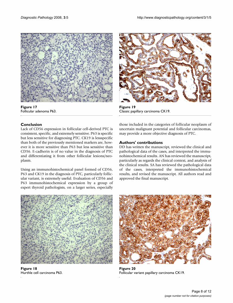
Out of the 175 cases included in the study, only the casesof PTC (including the encapsulated, follicular (Figure 3,Figure 4), and microcarcinoma variants and those occur-ring in the setting of Hashimoto's thyroiditis (Figure 5,Figure 6) showed absent CD56 expression. Such findingwas consistent in all the cases and showed no differencewith different tumor morphological variant or tumorstage. The metastatic deposits of PTC within the lymphnodes showed absent CD56 expression, as seen in theirthyroid primaries. The surrounding non-lesional thyro-cytes showed identical CD56 staining properties.
Within individual benign group/normal thyrocytes andnon-PTC lesions/tumors, CD56 expression showed con-
Table 1: Properties of immunhistochemical antibodies used in the study
Primary Antibody Marker/Clone Supplier Mono (M) Poly (P) Dilution Antigen Retrieval
CD56 Clone: 123C3 ZymedInvitrogen Corp. (USA)
M 1/100 pH 9.0
E-Cadherin Clone: NCH-38 DakoUSA
M 1/100 pH 9.0
Cytokeratin 19 Clone RCK108 DakoUSA
M 1/50 EnzymeProteinase K
P63 Clone: 4A4 DakoUSA
M 1/100 pH 9.0
Page 3 of 12(page number not for citation purposes)

Diagnostic Pathology 2008, 3:5 http://www.diagnosticpathology.org/content/3/1/5
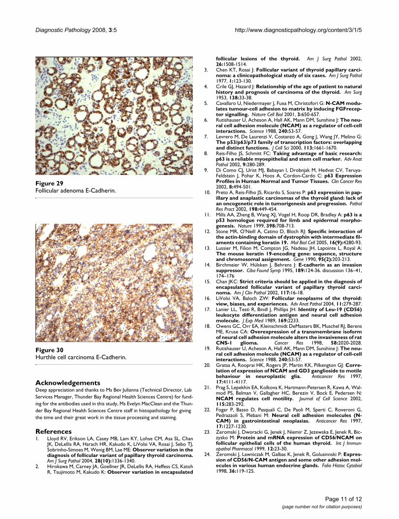
sistently strong diffuse expression with no differencebetween different subgroups, including hyperplasia, thy-roiditis (Figure 7, Figure 8), adenomas (Figure 9, Figure10) and non PTC carcinomas (Figure 11, Figure 12). In allcases of the non-PTC group, the CD56 expression wasmembranous, crisp and diffuses involving 100% of thethyrocytes, in the lesion.
Within the PTC group, occasional CD56 positive cellswere identified. In all cases, these cells were located at thetumor non-tumor interface i.e. at the periphery of thePTC. In all the PTC cases the tumors centers were com-pletely devoid of any CD56 staining (0%). For all thecases of PTC, CD56 expression in individual cells did notexceed 5% of cells all at the periphery. To us these cells
perhaps may represent the infiltrative tumor nontumorinterface and may merely be residual non-tumor cells. Wepointed out a <10% cut of for recording negative CD56expression, to avoid the future dilemma of lack of quanti-zation. It worth mentioning that, this occasional CD56positive cells located at the tumor periphery (interface)were present in 6/15 of the classic, 20/23 of the follicular,15/27 of the micro-papillary and of the 2/2 tall PTC vari-ants. In the remaining PTC variants included in the study,CD56 expression was absent in all individual cells, cen-trally and peripherally (0%).
P63 showed selective focal positivity in PTC cases (Figure13, Figure 14, Figure 15), in contrast to other non-PTClesions/tumors (Figure 16, Figures 17, Figure 18). P63
Follicular variant papillary carcinoma CD56Figure 4Follicular variant papillary carcinoma CD56.
Classic papillary carcinoma CD56Figure 2Classic papillary carcinoma CD56.
Classic papillary carcinoma H&EFigure 1Classic papillary carcinoma H&E.
Follicular variant papillary carcinoma H&EFigure 3Follicular variant papillary carcinoma H&E.
Page 4 of 12(page number not for citation purposes)
Diagnostic Pathology 2008, 3:5 http://www.diagnosticpathology.org/content/3/1/5
expression was in 70% of cases of PTC and was consist-ently absent in all the non-PTC cases.
CK 19 showed positive expression in PTC (Figure 19, Fig-ure 20, Figure 21) and absent expression in non PTClesions (Figure 22, Figure 23, Figure 24). However, CK19expression accounted for 85% of cases of PTC and in 26%of cases of non-PTC lesions/tumors.
E-cadherin showed consistent non-discriminatory expres-sion in all cases included in the study (Figure 25, Figure26, Figure 27, Figure 28, Figure 29, and Figure 30).
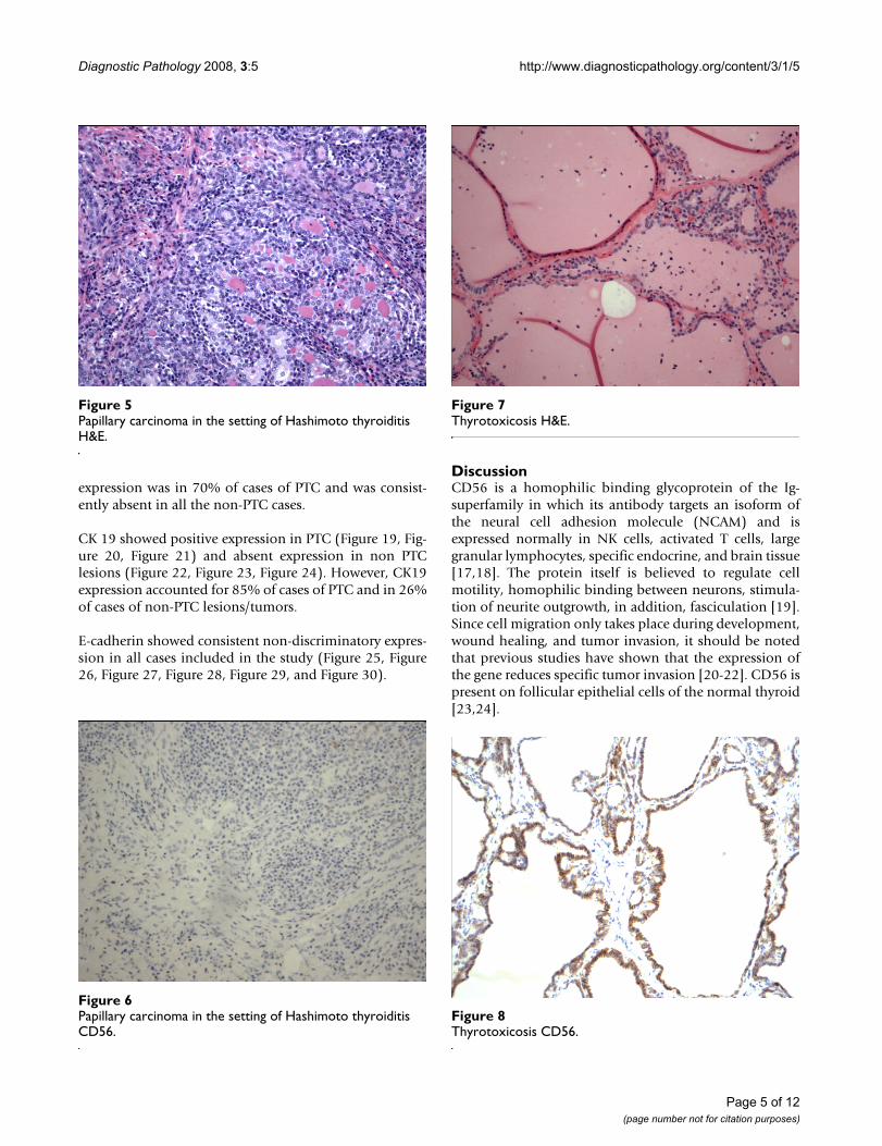
DiscussionCD56 is a homophilic binding glycoprotein of the Ig-superfamily in which its antibody targets an isoform ofthe neural cell adhesion molecule (NCAM) and isexpressed normally in NK cells, activated T cells, largegranular lymphocytes, specific endocrine, and brain tissue[17,18]. The protein itself is believed to regulate cellmotility, homophilic binding between neurons, stimula-tion of neurite outgrowth, in addition, fasciculation [19].Since cell migration only takes place during development,wound healing, and tumor invasion, it should be notedthat previous studies have shown that the expression ofthe gene reduces specific tumor invasion [20-22]. CD56 ispresent on follicular epithelial cells of the normal thyroid[23,24].
Thyrotoxicosis CD56Figure 8Thyrotoxicosis CD56.
Papillary carcinoma in the setting of Hashimoto thyroiditis CD56Figure 6Papillary carcinoma in the setting of Hashimoto thyroiditis CD56.
Papillary carcinoma in the setting of Hashimoto thyroiditis H&EFigure 5Papillary carcinoma in the setting of Hashimoto thyroiditis H&E.
Thyrotoxicosis H&EFigure 7Thyrotoxicosis H&E.
Page 5 of 12(page number not for citation purposes)

Diagnostic Pathology 2008, 3:5 http://www.diagnosticpathology.org/content/3/1/5
One of the most frequent difficulties in thyroid pathologyis differentiating follicular variant of PTC from follicularadenoma. The inter observable variability in this area iswell recognized. The differentiation is critical for the treat-ment and long-term management of the tumors. Despitethe universal recognition of this variant, it is clearly lack-ing the minimal histological definition of the follicularvariant of PTC. Instead of setting an objective minimalhistological definition, in terms of field size or number ofnuclei with PTC features, the literature resorts to an atypi-cal category and introduces the term "well-differentiatedtumor of uncertain malignant potential [25]." This termencompasses encapsulated tumors that have only "minornuclear changes of the type seen in typical PTC. The prob-lem with this term is that it really did not solve the inter
observable variability between pathologists concerningthe diagnosis of the follicular variant of PTC. Instead, itallowed safe expression of differences in opinion but cer-tainly increased the chances of over-call and over-treat-ment of the follicular variant of PTC. Unfortunately, theimmunoprofile of follicular derived lesions and neo-plasms show some overlap and to the best of our knowl-edge, no single marker or even panel is 100% sensitiveand 100% specific for PTC. Hence, the use of immunohis-tochemistry in the diagnosis of the follicular variant ofPTC has to be used with extreme caution. A ecent studyhas investigated the expression of NCAM (CD56) in tissuesections of 61 cases of papillary carcinoma and in 14lymph node metastases using immunohistochemistry[26]. In this study, low or absent expression of CD56 was
Hurthle cell carcinoma CD56Figure 12Hurthle cell carcinoma CD56.
Follicular adenoma CD56Figure 10Follicular adenoma CD56.
Follicular adenoma H&EFigure 9Follicular adenoma H&E.
Hurthle cell carcinoma H&EFigure 11Hurthle cell carcinoma H&E.
Page 6 of 12(page number not for citation purposes)

Diagnostic Pathology 2008, 3:5 http://www.diagnosticpathology.org/content/3/1/5
noted in PTC using immunohistochemistry and PCR [26].In the current study, CD56 was extremely useful in the dis-tinction between PTC, including the follicular variant andother follicular lesions/neoplasms, as well as PTC mim-ickers' such as Hashimoto and lymphocytic thyroiditisand follicular adenoma.
We point out that we excluded those cases (4 cases), thatwe disagreed upon and those were diagnosed as well-dif-ferentiated follicular neoplasm of uncertain malignantpotential. The rational behind their exclusion is that thosecases are rare and highly controversial, and perhaps a largeseries of such cases should be evaluated by a group ofexperts in regards to the diagnosis and applicability ofCD56, P63, E-cadherin and CK19. Our study shows that
within the thyroid gland, the use of an immunohisto-chemical panel formed of CD56, E-cadherin, and P63 isextremely helpful in selecting cases of PTC from other fol-licular cell-derived thyroid lesions/tumors, with 100%sensitivity and 100% specificity. However, this is certainlynot applicable outside of the thyroid gland, i.e. withinmetastatic sites.
Hence, we suggest that immunohistochemistry using thispanel is helpful in the diagnosis of PTC including the fol-licular variant of PTC. E-Cadherin immunhistochemistryis noncontributory.
Thyrotoxicosis P63Figure 16Thyrotoxicosis P63.
Follicular variant papillary carcinoma P63Figure 14Follicular variant papillary carcinoma P63.
Classic papillary carcinoma P63Figure 13Classic papillary carcinoma P63.
Papillary carcinoma in the setting of Hashimoto thyroiditis P63Figure 15Papillary carcinoma in the setting of Hashimoto thyroiditis P63.
Page 7 of 12(page number not for citation purposes)
Diagnostic Pathology 2008, 3:5 http://www.diagnosticpathology.org/content/3/1/5
ConclusionLack of CD56 expression in follicular cell-derived PTC isconsistent, specific, and extremely sensitive. P63 is specificbut less sensitive for diagnosing PTC. CK19 is lessspecificthan both of the previously mentioned markers are, how-ever it is more sensitive than P63 but less sensitive thanCD56. E-cadherin is of no value in the diagnosis of PTCand differentiating it from other follicular lesions/neo-plasm.
Using an immunohistochemical panel formed of CD56,P63 and CK19 in the diagnosis of PTC, particularly follic-ular variant, is extremely useful. Evaluation of CD56 andP63 immunohistochemical expression by a group ofexpert thyroid pathologists, on a larger series, especially
those included in the categories of follicular neoplasm ofuncertain malignant potential and follicular carcinomas,may provide a more objective diagnosis of PTC.
Authors' contributionsDD has written the manuscript, reviewed the clinical andpathological data of the cases, and interpreted the immu-nohistochemical results. AN has reviewed the manuscript,particularly as regards the clinical context, and analysis ofthe clinical results. SA has reviewed the pathological dataof the cases, interpreted the immunohistochemicalresults, and revised the manuscript. All authors read andapproved the final manuscript.
Follicular variant papillary carcinoma CK19Figure 20Follicular variant papillary carcinoma CK19.
Hurthle cell carcinoma P63Figure 18Hurthle cell carcinoma P63.
Follicular adenoma P63Figure 17Follicular adenoma P63.
Classic papillary carcinoma CK19Figure 19Classic papillary carcinoma CK19.
Page 8 of 12(page number not for citation purposes)

Diagnostic Pathology 2008, 3:5 http://www.diagnosticpathology.org/content/3/1/5
Page 9 of 12(page number not for citation purposes)
Hurthle cell carcinoma CK19Figure 24Hurthle cell carcinoma CK19.
Thyrotoxicosis CK19Figure 22Thyrotoxicosis CK19.
Papillary carcinoma in the setting of Hashimoto thyroiditis CK19Figure 21Papillary carcinoma in the setting of Hashimoto thyroiditis CK19.
Follicular adenoma CK19Figure 23Follicular adenoma CK19.

Diagnostic Pathology 2008, 3:5 http://www.diagnosticpathology.org/content/3/1/5
Page 10 of 12(page number not for citation purposes)
Thyrotoxicosis E-CadherinFigure 28Thyrotoxicosis E-Cadherin.
Classic papillary carcinoma E-CadherinFigure 25Classic papillary carcinoma E-Cadherin.
Follicular variant papillary carcinoma E-CadherinFigure 26Follicular variant papillary carcinoma E-Cadherin.
Papillary carcinoma in the setting of Hashimoto thyroiditis E-CadherinFigure 27Papillary carcinoma in the setting of Hashimoto thyroiditis E-Cadherin.
Diagnostic Pathology 2008, 3:5 http://www.diagnosticpathology.org/content/3/1/5
AcknowledgementsDeep appreciation and thanks to Ms Bev Julianna (Technical Director, Lab Services Manager, Thunder Bay Regional Health Sciences Centre) for fund-ing for the antibodies used in this study, Ms Evelyn MacClean and the Thun-der Bay Regional Health Sciences Centre staff in histopathology for giving the time and their great work in the tissue processing and staining.
References1. Lloyd RV, Erikson LA, Casey MB, Lam KY, Lohse CM, Asa SL, Chan
JK, DeLellis RA, Harach HR, Kakudo K, LiVolsi VA, Rosai J, Sebo TJ,Sobrinho-Simoes M, Wenig BM, Lae ME: Observer variation in thediagnosis of follicular variant of papillary thyroid carcinoma.Am J Surg Pathol 2004, 28(10):1336-1340.
2. Hirokawa M, Carney JA, Goellner JR, DeLellis RA, Heffess CS, KatohR, Tsujimoto M, Kakudo K: Observer variation in encapsulated
follicular lesions of the thyroid. Am J Surg Pathol 2002,26:1508-1514.
3. Chen KT, Rosai J: Follicular variant of thyroid papillary carci-noma: a clinicopathological study of six cases. Am J Surg Pathol1977, 1:123-130.
4. Crile GJ, Hazard J: Relationship of the age of patient to naturalhistory and prognosis of carcinoma of the thyroid. Am Surg1953, 138:33-38.
5. Cavallaro U, Niedermayer J, Fuxa M, Christofori G: N-CAM modu-lates tumour-cell adhesion to matrix by inducing FGFrecep-tor signalling. Nature Cell Biol 2001, 3:650-657.
6. Rutishauser U, Acheson A, Hall AK, Mann DM, Sunshine J: The neu-ral cell adhesion molecule (NCAM) as a regulator of cell-cellinteractions. Science 1988, 240:53-57.
7. Levrero M, De Laurenzi V, Costanzo A, Gong J, Wang JY, Melino G:The p53/p63/p73 family of transcription factors: overlappingand distinct functions. J Cell Sci 2000, 113:1661-1670.
8. Reis-Filho JS, Schmitt FC: Taking advantage of basic research:p63 is a reliable myoepithelial and stem cell marker. Adv AnatPathol 2002, 9:280-289.
9. Di Como CJ, Urist MJ, Babayan I, Drobnjak M, Hedvat CV, Teruya-Feldstein J, Pohar K, Hoos A, Cordon-Cardo C: p63 ExpressionProfiles in Human Normal and Tumor Tissues. Clin Cancer Res2002, 8:494-501.
10. Preto A, Reis-Filho JS, Ricardo S, Soares P: p63 expression in pap-illary and anaplastic carcinomas of the thyroid gland: lack ofan oncogenetic role in tumorigenesis and progression. PatholRes Pract 2002, 198:449-454.
11. Mills AA, Zheng B, Wang XJ, Vogel H, Roop DR, Bradley A: p63 is ap53 homologue required for limb and epidermal morpho-genesis. Nature 1999, 398:708-713.
12. Stone MR, O'Neill A, Catino D, Bloch RJ: Specific interaction ofthe actin-binding domain of dystrophin with intermediate fil-aments containing keratin 19. Mol Biol Cell 2005, 16(9):4280-93.
13. Lussier M, Filion M, Compton JG, Nadeau JH, Lapointe L, Royal A:The mouse keratin 19-encoding gene: sequence, structureand chromosomal assignment. Gene 1990, 95(2):203-213.
14. Birchmeier W, Hülsken J, Behrens J: E-cadherin as an invasionsuppressor. Ciba Found Symp 1995, 189:124-36. discussion 136–41,174–176
15. Chan JKC: Strict criteria should be applied in the diagnosis ofencapsulated follicular variant of papillary thyroid carci-noma. Am J Clin Pathol 2002, 117:16-18.
16. LiVolsi VA, Baloch ZW: Follicular neoplasms of the thyroid:view, biases, and experiences. Adv Anat Pathol 2004, 11:279-287.
17. Lanier LL, Testi R, Bindl J, Phillips JH: Identity of Leu-19 (CD56)leukocyte differentiation antigen and neural cell adhesionmolecule. J Exp Med 1989, 169:2233.
18. Owens GC, Orr EA, Kleinschmidt DeMasters BK, Muschel RJ, BerensME, Kruse CA: Overexpression of a transmembrane isoformof neural cell adhesion molecule alters the invasiveness of ratCNS-1 glioma. Cancer Res 1998, 58:2020-2028.
19. Rutishauser U, Acheson A, Hall AK, Mann DM, Sunshine J: The neu-ral cell adhesion molecule (NCAM) as a regulator of cell-cellinteractions. Science 1988, 240:53-57.
20. Gratsa A, Rooprai HK, Rogers JP, Martin KK, Pilkengton GJ: Corre-lation of expression of NCAM and GD3 ganglioside to motilebehaviour in neuroplastic glia. Anticancer Res 1997,17:4111-4117.
21. Prag S, Lepekhin EA, Kolkova K, Hartmann-Petersen R, Kawa A, Wal-mod PS, Belman V, Gallagher HC, Berezin V, Bock E, Pedersen N:NCAM regulates cell motility. Journal of Cell Science 2002,115:283-292.
22. Fogar P, Basso D, Pasquali C, De Paoli M, Sperti C, Roveroni G,Pedrazzoli S, Plebani M: Neural cell adhesion molecules (N-CAM) in gastrointestinal neoplasias. Anticancer Res 1997,17:1227-1230.
23. Zeromski J, Dworacki G, Jenek J, Niemir Z, Jezewska E, Jenek R, Bic-zysko M: Protein and mRNA expression of CD56/NCAM onfollicular epithelial cells of the human thyroid. Int J Immun-opathol Pharmacol 1999, 12:23-30.
24. Zeromski J, Lawniczak M, Galbas K, Jenek R, Golusinnski P: Expres-sion of CD56/N-CAM antigen and some other adhesion mol-ecules in various human endocrine glands. Folia Histoc Cytobiol1998, 36:119-125.
Follicular adenoma E-CadherinFigure 29Follicular adenoma E-Cadherin.
Hurthle cell carcinoma E-CadherinFigure 30Hurthle cell carcinoma E-Cadherin.
Page 11 of 12(page number not for citation purposes)
http://www.ncbi.nlm.nih.gov/entrez/query.fcgi?cmd=Retrieve&db=PubMed&dopt=Abstract&list_uids=3281256
http://www.ncbi.nlm.nih.gov/entrez/query.fcgi?cmd=Retrieve&db=PubMed&dopt=Abstract&list_uids=3281256
http://www.ncbi.nlm.nih.gov/entrez/query.fcgi?cmd=Retrieve&db=PubMed&dopt=Abstract&list_uids=3281256
http://www.ncbi.nlm.nih.gov/entrez/query.fcgi?cmd=Retrieve&db=PubMed&dopt=Abstract&list_uids=1701153
http://www.ncbi.nlm.nih.gov/entrez/query.fcgi?cmd=Retrieve&db=PubMed&dopt=Abstract&list_uids=1701153
http://www.ncbi.nlm.nih.gov/entrez/query.fcgi?cmd=Retrieve&db=PubMed&dopt=Abstract&list_uids=1701153
http://www.ncbi.nlm.nih.gov/entrez/query.fcgi?cmd=Retrieve&db=PubMed&dopt=Abstract&list_uids=7587628
http://www.ncbi.nlm.nih.gov/entrez/query.fcgi?cmd=Retrieve&db=PubMed&dopt=Abstract&list_uids=7587628
http://www.ncbi.nlm.nih.gov/entrez/query.fcgi?cmd=Retrieve&db=PubMed&dopt=Abstract&list_uids=2471777
http://www.ncbi.nlm.nih.gov/entrez/query.fcgi?cmd=Retrieve&db=PubMed&dopt=Abstract&list_uids=2471777
http://www.ncbi.nlm.nih.gov/entrez/query.fcgi?cmd=Retrieve&db=PubMed&dopt=Abstract&list_uids=2471777
http://www.ncbi.nlm.nih.gov/entrez/query.fcgi?cmd=Retrieve&db=PubMed&dopt=Abstract&list_uids=9581848
http://www.ncbi.nlm.nih.gov/entrez/query.fcgi?cmd=Retrieve&db=PubMed&dopt=Abstract&list_uids=9581848
http://www.ncbi.nlm.nih.gov/entrez/query.fcgi?cmd=Retrieve&db=PubMed&dopt=Abstract&list_uids=9581848
http://www.ncbi.nlm.nih.gov/entrez/query.fcgi?cmd=Retrieve&db=PubMed&dopt=Abstract&list_uids=3281256
http://www.ncbi.nlm.nih.gov/entrez/query.fcgi?cmd=Retrieve&db=PubMed&dopt=Abstract&list_uids=3281256
http://www.ncbi.nlm.nih.gov/entrez/query.fcgi?cmd=Retrieve&db=PubMed&dopt=Abstract&list_uids=3281256
http://www.ncbi.nlm.nih.gov/entrez/query.fcgi?cmd=Retrieve&db=PubMed&dopt=Abstract&list_uids=9428343
http://www.ncbi.nlm.nih.gov/entrez/query.fcgi?cmd=Retrieve&db=PubMed&dopt=Abstract&list_uids=9428343
http://www.ncbi.nlm.nih.gov/entrez/query.fcgi?cmd=Retrieve&db=PubMed&dopt=Abstract&list_uids=9428343
http://www.ncbi.nlm.nih.gov/entrez/query.fcgi?cmd=Retrieve&db=PubMed&dopt=Abstract&list_uids=9137477

Diagnostic Pathology 2008, 3:5 http://www.diagnosticpathology.org/content/3/1/5
Publish with BioMed Central and every scientist can read your work free of charge
"BioMed Central will be the most significant development for disseminating the results of biomedical research in our lifetime."
Sir Paul Nurse, Cancer Research UK
Your research papers will be:
available free of charge to the entire biomedical community
peer reviewed and published immediately upon acceptance
cited in PubMed and archived on PubMed Central
yours — you keep the copyright
Submit your manuscript here:http://www.biomedcentral.com/info/publishing_adv.asp
BioMedcentral
25. Xu XC, El-Naggar AK, Lotan R: Differential xxpression of galec-tin-1 and galectin-3 in thyroid tumors: potential diagnosticimplications. Am J Pathol 1995, 147:815-822.
26. Scarpino S, Di Napoli A, Melotti F, Talerico C, Cancrini A, Ruco L:Papillary carcinoma of the thyroid: low expression of NCAM(CD56) is associated with down regulation of VEGF-D pro-duction by tumor cells. J Pathol 2007, 212(4):411-419.
Page 12 of 12(page number not for citation purposes)
http://www.ncbi.nlm.nih.gov/entrez/query.fcgi?cmd=Retrieve&db=PubMed&dopt=Abstract&list_uids=7677193
http://www.ncbi.nlm.nih.gov/entrez/query.fcgi?cmd=Retrieve&db=PubMed&dopt=Abstract&list_uids=7677193