Pathologically and Biologically Distinct Types of Epithelium in Intraductal Papillary Mucinous...
10
ORIGINAL ARTICLE Pathologically and Biologically Distinct Types of Epithelium in Intraductal Papillary Mucinous Neoplasms Delineation of an “Intestinal” Pathway of Carcinogenesis in the Pancreas N. Volkan Adsay, MD,* Kambiz Merati, MD,* Olca Basturk, MD,* Christine Iacobuzio-Donahue, MD,‡ Edi Levi, MD,* Jeanette D. Cheng, MD,* Fazlul H. Sarkar, PhD,* Ralph H. Hruban, MD,‡ and David S. Klimstra, MD† Abstract: Although general characteristics of intraductal papillary mucinous neoplasms (IPMNs) and their delineation from other pan- creatic tumors have been well established, several issues regarding their biology and management remain unresolved. It has been noted briefly by us and other authors that there are different types of papillae in IPMNs; however, their frequency, biologic significance, and clini- cal relevance are unknown. In this study, the association of different papillary patterns with clinical, pathologic, and biologic parameters was studied in 74 IPMNs, and the expression profile of CDX2 (a spe- cific marker and one of the key determinants of intestinal “program- ming,” and a tumor suppressor) was determined immunohistochem- ically in addition to MUC1 (a marker of an “aggressive” phenotype in pancreatic neoplasia) and MUC2 (“intestinal type mucin,” a marker of the “indolent” phenotype, and a tumor suppressor). The patterns of papillae identified and their association with these parameters were as follows: 1) The intestinal-type (Yonezawa’s dark-cell type), similar to villous adenomas, was seen in 26 of 74 (35%) cases. The majority harbored carcinoma in situ (85%) or borderline atypia (15%). They tended to be large (mean, 5.5 cm). Most expressed CDX2 (95%) and MUC2 (92%) but not MUC1 (8%). This type was more commonly associated with colloid-type invasion (14 of 16 invasive carcinomas were of colloid type). 2) The pancreatobiliary type, characterized by arborizing papillae lined by cuboidal cells resembling papillary neo- plasms of the biliary tract, was present in 22% of the cases. These were mostly graded as carcinoma in situ (94%); they rarely expressed CDX2 (6%) or MUC2 (19%) but often showed MUC1 labeling (44%). This pattern was more commonly associated with the tubular type of invasive carcinoma and had a slight tendency for a more ag- gressive clinical course. 3) The null type was characterized by abun- dant apical mucin and basally located nuclei, similar to the gastric foveolar epithelium. Thirty-one percent of IPMNs had this type of papillae, but this pattern was also present in the background of other IPMNs and in the cystic components of most cases as well. Most pure null-type IPMNs were devoid of complexity and consequently clas- sified as adenoma (48%). They tended to be small (mean, 2.6 cm), were often negative for CDX2, MUC1, and MUC2, and were rarely associated with invasive carcinoma. 4) Some IPMNs (12%) exhibited features that were difficult to classify, and 2 cases had a mixture of pancreatobiliary and intestinal types of papillae. In conclusion, IPMNs include pathologically and biologically distinct epithelial pat- terns. CDX2 and MUC2 expression is relatively specific for the in- testinal type papillae, confirming that these IPMNs indeed exhibit intestinal differentiation. Their close association with colloid carci- noma, which also shows consistent MUC2 and CDX2 expression, supports the existence of an intestinal pathway of carcinogenesis. This “metaplastic” pathway may reflect different genetic events in the development of these IPMNs, and the presence of intestinal differen- tiation may potentially be used in prognostication and stratification of patients into appropriate treatment categories. Key Words: intraductal papillary mucinous neoplasms, pancreas, in- testinal, differentiation, CDX2, MUC1, MUC2 (Am J Surg Pathol 2004;28:839–848) I ntraductal papillary mucinous neoplasm (IPMN) is now a well-recognized entity in the pancreas, 1,3,22,23 unifying tu- mors that are characterized by intraductal proliferation of neo- plastic mucinous cells, which usually form papillae and lead to cystic dilation of the pancreatic ducts, forming clinically and macroscopically detectable masses. 10,13,16,24,27,38,40,45,46,54,56 Whereas the general characteristics of this group and its delineation from other pancreatic neoplasms have been well established, several issues remain unresolved. 1,42 Foremost is the delineation of pathologically, prognostically, and thera- peutically relevant subtypes of IPMNs. It is largely accepted that IPMNs have a spectrum of dysplasia ranging from adeno- ma to borderline to carcinoma in situ (CIS), and in approxi- mately one third of the cases, IPMNs are associated with inva- sive carcinoma of either tubular or colloid (mucinous noncys- tic) types. 4 Currently, a wide spectrum of therapeutic approaches, ranging from chemoprevention (with agents like COX-2 inhibitors) to total pancreatectomy, is being consid- From the *Departments of Pathology, Karmanos Cancer Institute and Wayne State University, Detroit, MI; †Memorial Sloan-Kettering Cancer Center, New York, NY; and ‡Johns Hopkins University Hospitals, Baltimore, MD. Supported in part by the National Cancer Institute Specialized Program in Research Excellence (P50-CA62924). Reprints: N. Volkan Adsay, MD, Harper Hospital and Wayne State Univer- sity, 3990 John R. Street, Detroit, MI 48201 (e-mail: adsayv@med. wayne.edu). Copyright © 2004 by Lippincott Williams & Wilkins Am J Surg Pathol • Volume 28, Number 7, July 2004 839
Transcript of Pathologically and Biologically Distinct Types of Epithelium in Intraductal Papillary Mucinous...

ORIGINAL ARTICLE
Pathologically and Biologically Distinct Types of Epithelium inIntraductal Papillary Mucinous Neoplasms
Delineation of an “Intestinal” Pathway of Carcinogenesis in the Pancreas
N. Volkan Adsay, MD,* Kambiz Merati, MD,* Olca Basturk, MD,* Christine Iacobuzio-Donahue, MD,‡Edi Levi, MD,* Jeanette D. Cheng, MD,* Fazlul H. Sarkar, PhD,* Ralph H. Hruban, MD,‡
and David S. Klimstra, MD†
Abstract: Although general characteristics of intraductal papillarymucinous neoplasms (IPMNs) and their delineation from other pan-creatic tumors have been well established, several issues regardingtheir biology and management remain unresolved. It has been notedbriefly by us and other authors that there are different types of papillaein IPMNs; however, their frequency, biologic significance, and clini-cal relevance are unknown. In this study, the association of differentpapillary patterns with clinical, pathologic, and biologic parameterswas studied in 74 IPMNs, and the expression profile of CDX2 (a spe-cific marker and one of the key determinants of intestinal “program-ming,” and a tumor suppressor) was determined immunohistochem-ically in addition to MUC1 (a marker of an “aggressive” phenotype inpancreatic neoplasia) and MUC2 (“intestinal type mucin,” a markerof the “indolent” phenotype, and a tumor suppressor). The patterns ofpapillae identified and their association with these parameters were asfollows: 1) The intestinal-type (Yonezawa’s dark-cell type), similarto villous adenomas, was seen in 26 of 74 (35%) cases. The majorityharbored carcinoma in situ (85%) or borderline atypia (15%). Theytended to be large (mean, 5.5 cm). Most expressed CDX2 (95%) andMUC2 (92%) but not MUC1 (8%). This type was more commonlyassociated with colloid-type invasion (14 of 16 invasive carcinomaswere of colloid type). 2) The pancreatobiliary type, characterized byarborizing papillae lined by cuboidal cells resembling papillary neo-plasms of the biliary tract, was present in 22% of the cases. Thesewere mostly graded as carcinoma in situ (94%); they rarely expressedCDX2 (6%) or MUC2 (19%) but often showed MUC1 labeling(44%). This pattern was more commonly associated with the tubulartype of invasive carcinoma and had a slight tendency for a more ag-gressive clinical course. 3) The null type was characterized by abun-dant apical mucin and basally located nuclei, similar to the gastricfoveolar epithelium. Thirty-one percent of IPMNs had this type of
papillae, but this pattern was also present in the background of otherIPMNs and in the cystic components of most cases as well. Most purenull-type IPMNs were devoid of complexity and consequently clas-sified as adenoma (48%). They tended to be small (mean, 2.6 cm),were often negative for CDX2, MUC1, and MUC2, and were rarelyassociated with invasive carcinoma. 4) Some IPMNs (12%) exhibitedfeatures that were difficult to classify, and 2 cases had a mixture ofpancreatobiliary and intestinal types of papillae. In conclusion,IPMNs include pathologically and biologically distinct epithelial pat-terns. CDX2 and MUC2 expression is relatively specific for the in-testinal type papillae, confirming that these IPMNs indeed exhibitintestinal differentiation. Their close association with colloid carci-noma, which also shows consistent MUC2 and CDX2 expression,supports the existence of an intestinal pathway of carcinogenesis.This “metaplastic” pathway may reflect different genetic events in thedevelopment of these IPMNs, and the presence of intestinal differen-tiation may potentially be used in prognostication and stratification ofpatients into appropriate treatment categories.
Key Words: intraductal papillary mucinous neoplasms, pancreas, in-testinal, differentiation, CDX2, MUC1, MUC2
(Am J Surg Pathol 2004;28:839–848)
Intraductal papillary mucinous neoplasm (IPMN) is now awell-recognized entity in the pancreas,1,3,22,23 unifying tu-
mors that are characterized by intraductal proliferation of neo-plastic mucinous cells, which usually form papillae and lead tocystic dilation of the pancreatic ducts, forming clinically andmacroscopically detectable masses.10,13,16,24,27,38,40,45,46,54,56
Whereas the general characteristics of this group and itsdelineation from other pancreatic neoplasms have been wellestablished, several issues remain unresolved.1,42 Foremost isthe delineation of pathologically, prognostically, and thera-peutically relevant subtypes of IPMNs. It is largely acceptedthat IPMNs have a spectrum of dysplasia ranging from adeno-ma to borderline to carcinoma in situ (CIS), and in approxi-mately one third of the cases, IPMNs are associated with inva-sive carcinoma of either tubular or colloid (mucinous noncys-tic) types.4 Currently, a wide spectrum of therapeuticapproaches, ranging from chemoprevention (with agents likeCOX-2 inhibitors) to total pancreatectomy, is being consid-
From the *Departments of Pathology, Karmanos Cancer Institute and WayneState University, Detroit, MI; †Memorial Sloan-Kettering Cancer Center,New York, NY; and ‡Johns Hopkins University Hospitals, Baltimore,MD.
Supported in part by the National Cancer Institute Specialized Program inResearch Excellence (P50-CA62924).
Reprints: N. Volkan Adsay, MD, Harper Hospital and Wayne State Univer-sity, 3990 John R. Street, Detroit, MI 48201 (e-mail: [email protected]).
Copyright © 2004 by Lippincott Williams & Wilkins
Am J Surg Pathol • Volume 28, Number 7, July 2004 839

ered in the management of IPMNs.1,15,25,42,50 Before these canbe initiated, the prognostic categorization of these tumorsought to be better understood.
It has been noted recently by us3 and other authors (Yon-ezawa et al,39,57–59 and Lüttges et al31–33) that IPMNs exhibithistologically different patterns of papillae and that these pat-terns have different patterns of mucin protein MUC expres-sion. Some IPMNs have long intestinal-type papillae identicalto those of intestinal villous adenomas (referred to as villous-dark cell type by Yonezawa et al39,57–59), and in others thereare more complex papillae lined by cuboidal cells, reminiscentof papillary neoplasms of the biliary tract, which we refer to aspancreatobiliary type.3 While some authors regarded thesepatterns as a mere reflection of different grades of dysplasia,others consider them as morphologically distinct subtypes ofIPMNs. The clinical and biologic significance of these epithe-lial subtypes is largely unknown.
Establishing the molecular profiles of these subtypes, es-pecially the expression pattern of markers of aggressivenessand differentiation, may help determine their biologic signifi-cance. There is emerging evidence that these different sub-types of IPMNs differ in their molecular alterations, includingMUC expression profiles.5,6,31–33,39,57–59
Recently, CDX2, also a tumor suppressor, has beenimplicated as one of the specific and key molecules of intes-tinal differentiation, working in close association withMUC2.9,11,14,18,29,35–37,41,44,47,48,52,55,60 The expression pro-file of these markers may help further define the subtypes ofIPMNs and determine their line of differentiation as well aspotential biologic nature.
MATERIALS AND METHODS
CasesHistologic sections of 74 cases of IPMNs retrieved from
the files of John Hopkins University Hospital, MemorialSloan-Kettering Cancer Center, and Karmanos Cancer Insti-tute, Wayne State University were reviewed. The noninvasivecomponents of the neoplasms were classified as adenoma, bor-derline tumor, and CIS using the WHO classification17 and,where present, the invasive carcinomas were classified as tu-bular or colloid (mucinous noncystic) type. The size and thelocation of the neoplasm were obtained from the pathologyreports.
Classification of Papillary PatternsIPMNs were classified into four groups:
1. Those that were composed of long finger-like projections(without complex branching) and lined by columnar cellswith cigar-shaped nuclei were classified as intestinal type(Fig. 1A). These were morphologically indistinguishablefrom colonic villous adenomas. The cells contained vari-able amounts of mucin in the apical cytoplasm. Nuclei were
FIGURE 1. IPMNs consist of three morphologically distincttypes of papillae. A, Intestinal type: Similar to colonic villousadenomas, there are tall, finger-like projections with only mini-mal branching. The cells are columnar and pseudostratifiedwith cigar-shaped nuclei. There is a variable amount of mucinin the cytoplasm. B, Pancreatobiliary type: More complexbranching papillae lined by relatively cuboidal cells, some withprominent nucleoli, similar to the papillary neoplasms of thebiliary tract. C, Null type: Tall columnar cells with basally lo-cated nuclei and abundant apical mucin (with variable chro-mophilia; acidophilic in this example) resemble gastric foveo-lar epithelium or PanIN-1 lesions.
Adsay et al Am J Surg Pathol • Volume 28, Number 7, July 2004
840 © 2004 Lippincott Williams & Wilkins

pseudostratified with varying degrees of atypia. This pat-tern corresponds to Yonezawa’s villous-dark cell type.57,58
2. IPMNs composed of complex arborizing papillae lined bycuboidal cells, often with round nuclei containing a singleprominent eccentric nucleolus, were classified as pancre-atobiliary type (Fig. 1B). This classification is based onsimilarities to a subgroup of papillary neoplasms of the bil-iary tree. Some examples were similar to intraductal onco-cytic papillary neoplasms but lacked both the oncocyticchange and the intraepithelial lumen formation characteris-tic of the latter.
3. IPMNs lined by tall columnar cells with abundant pale su-pranuclear mucin, some with acidophilia, creating a patternreminiscent of gastric foveolar cells or the mucinous cellsseen in low-grade pancreatic intraepithelial neoplasia(PanIN-1A) were classified as null type (Fig. 1C).
4. IPMNs that could not be categorized specifically into oneof the aforementioned types, or those that had separate ar-eas resembling intestinal and pancreatobiliary [many of theintestinal and pancreatobiliary types had null type compo-nents, but these are not included here] patterns, were seg-regated as unclassifiable.
Immunohistochemical Labeling for CDX2,MUC1, and MUC2
Immunohistochemical stains were performed using theavidin-biotin peroxidase complex method. Primary and sec-ondary antibodies and the detection kit were purchased fromcommercial laboratories: CDX2 from Biogenex (San Ramon,CA), and MUC1 (clone Ma695) and MUC2 (clone Ccp58)from Vector Laboratories (Burlingame, CA). After deparaf-finization and blocking of endogenous peroxidase, tissue sec-tions were steamed in 10 mM, pH 6.0, citrate buffer for 20minutes and allowed to stand in the hot buffer for an additional20 minutes. Antibodies were incubated with the tissue sectionsfor 60 minutes and 90 minutes, respectively. Biotinylated anti-mouse and avidin-biotin complex were applied for 10 minuteseach. After color development with 3-amino-9-ethylcarbazole,sections were counterstained with hematoxylin. Normal andneoplastic colon tissue were used as controls for CDX2 andMUC2 antibodies, and normal breast tissue for MUC1.
The labeling was scored both for the extent and the in-tensity of labeling. The extent was recorded semiquantitativelyas the percentage of the cells that showed labeling: 0 for <10%,focal for 10% to 50%, and diffuse for >50%. For all three an-tibodies, immunolabeling in more than 10% of the cells wasconsidered as “expression.” For CDX2, only the nuclear label-ing was regarded as expression, as has been advocated.36
Correlation of the Papillary Patterns WithClinical and Pathologic Parameters
The four morphologic types of papillae were correlatedwith size of the tumor, histologic grade (based on cytoarchi-
tectural atypia), presence and type of invasive carcinoma (ifpresent), and immunoexpression of CDX2, MUC1, andMUC2.
Statistical AnalysisOverall differences between types of IPMN were as-
sessed using �2 tests with 3 df for differences in proportions.Bonferonni’s method was used to adjust for multiple compari-sons. In general, we reported a result, as statistically significantif the attained significance value was 0.008 or less. If the over-all test was significant, pairwise comparisons were made using1 df �2 tests. �2 test was also used for comparison of the dif-ferent patterns with respect to their grade, presence and type ofinvasion, and anatomic location. The Kruskal-Wallis test wasused to evaluate the difference of the tumor size betweengroups. For studying the relationship between the expressionsof the markers and grading, Fisher exact test and Spearmanregression analysis were chosen. The Cox Proportional Hazardmodel was used for comparison of the survival between thedifferent types of papillae.
RESULTS
Frequency of Different Types of PapillaeOf the total of 74 cases, 26 (35%) had papillae of intes-
tinal type, 16 (22%) were pancreatobiliary, 23 (31%) werenull, and 9 (12%) were unclassifiable, including 2 cases thatwere intestinal with focal pancreatobiliary pattern. Null-typeepithelium, however, could be seen in the background of otherpapillary patterns, both in the small branch ducts (reminiscentof PanIN-1A) as well as in the more cystically dilated ducts ofmost of the tumors.
Different Papillary Types and GradeThe tumor grade was compared across the four groups.
Of the 23 null cases, almost half (48%) were adenomas (Table1). Of the 26 intestinal cases, 22 (85%) were CIS and 4 werecategorized as borderline. Of the 16 pancreatobiliary cases, 15(94%) were CIS, and the remaining one was borderline. Thedifference in grade between the null type and the two othersubtypes was statistically significant (P < 0.0005).
TABLE 1. Papillary Patterns and Grade
Subtype(n = 74)
Adenoma[no. (%)]
Borderline[no. (%)]
Carcinomain situ
[no. (%)]
Null (n = 23) 11 (48) 6 (26) 6 (26)Intestinal (n = 26) 0 (0) 4 (15) 22 (85)Pancreatobiliary (n = 16) 0 (0) 1 (6) 15 (94)Unclassifiable (n = 9) 0 (0) 4 (44) 5 (66)
Am J Surg Pathol • Volume 28, Number 7, July 2004 Papillary Types and Intestinal Differentiation in IPMNs
© 2004 Lippincott Williams & Wilkins 841
Different Papillary Types and Presence ofInvasive Carcinoma
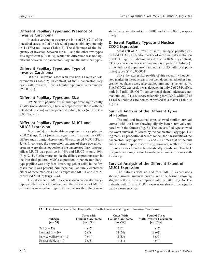
Invasive carcinoma was present in 16 of 26 (62%) of theintestinal cases, in 9 of 16 (56%) of pancreatobiliary, but onlyin 4 (17%) null cases (Table 2). The difference of the fre-quency of invasion between the null and the other two typeswas significant (P < 0.05), while this difference was not sig-nificant between the pancreatobiliary and the intestinal types.
Different Papillary Types and Type ofInvasive Carcinoma
Of the 16 intestinal cases with invasion, 14 were colloidcarcinomas (Table 2). In contrast, of the 9 pancreatobiliarycases with invasion, 7 had a tubular type invasive carcinoma(P = 0.001).
Different Papillary Types and SizeIPMNs with papillae of the null type were significantly
smaller (mean diameter, 2.6 cm) compared with those with theintestinal (5.5 cm) and the pancreatobiliary types (4.0 cm; P =0.05; Table 3).
Different Papillary Types and MUC1 andMUC2 Expression
Most (96%) of intestinal-type papillae had cytoplasmicMUC2 (Figs. 2, 3) (intestinal-type mucin) expression (88%diffuse and strong), whereas only 8% expressed MUC1 (Figs.3, 4). In contrast, the expression patterns of these two glyco-proteins were almost opposite in the pancreatobiliary-type pa-pillae: MUC1 was positive in 44% and MUC2 in only 19%(Figs. 2–4). Furthermore, unlike the diffuse expression seen inthe intestinal pattern, MUC2 expression in pancreatobiliary-type papillae was only focal (marking goblet cells) in the fewcases that it was present. Null-type papillae rarely expressedeither of these markers (1 of 23 expressed MUC1 and 2 of 23expressed MUC2) (Figs. 2–4).
The difference of MUC1 expression in pancreatobiliary-type papillae versus the others, and the difference of MUC2expression in intestinal type papillae versus the others were
statistically significant (P = 0.005 and P = 0.0001, respec-tively).
Different Papillary Types and NuclearCDX2 Expression
Most (20 of 21, 95%) of intestinal-type papillae ex-pressed CDX2, a specific marker of intestinal differentiation(Table 4; Fig. 3). Labeling was diffuse in 84%. By contrast,CDX2 expression was very uncommon in pancreatobiliary (1of 16 with focal expression) and null (1 of 23 with focal posi-tivity) types (P < 0.000001).
Since the expression profile of this recently character-ized marker in the pancreas is not well documented, other pan-creatic neoplasms were also studied immunohistochemically.Focal CDX2 expression was detected in only 2 of 25 PanINs,both in PanIN-1B. Of 74 conventional ductal adenocarcino-mas studied, 12 (16%) showed labeling for CDX2, while 12 of14 (86%) colloid carcinomas expressed this maker (Table 4;Fig. 3).
Survival Analysis of the Different Typesof Papillae
The null and intestinal types showed similar survivalcurves, with the latter showing slightly better survival com-pared with the former (Fig. 5). The unclassified type showedthe worst survival, followed by the pancreatobiliary type. Us-ing the COX proportional hazard model, the hazard ratio of thepancreatobiliary type was 1.37 and 2.13 times that of the nulland intestinal types, respectively; however, neither of thesedifferences was found to be statistically significant. This lackof significance may be due to inadequate number of cases withfollow-up.
Survival Analysis of the Different Extent ofMUC1 Expression
The patients with no and focal MUC1 expressionsshowed similar survival curves, with the former showingslightly better survival compared with the latter (Fig. 6). Thepatients with diffuse MUC1 expression showed the signifi-cantly worse survival.
TABLE 2. Association of Papillary Patterns With Invasion and Type of Invasive Carcinoma
Subtype(n = 74)
Cases withTubular Carcinoma
[no. (%)]
Cases WithColloid Carcinoma
[no. (%)]
Total of CasesWith Invasive Carcinoma
[no. (%)]
Null (n = 23) 4 (17) 0 (0) 4 (17)Intestinal (n = 26) 2 (8) 14 (54) 16 (62)Pancreatobiliary (n = 16) 7 (44) 2 (13) 9 (57)Unclassifiable (n = 9) 3 (33) 1 (11) 4 (44)
Adsay et al Am J Surg Pathol • Volume 28, Number 7, July 2004
842 © 2004 Lippincott Williams & Wilkins

DISCUSSIONThe findings in this study have two sets of implications:
1) they provide further evidence for the existence of a distinctpathway of carcinogenesis in the pancreas, the intestinal path-way, and 2) they show that the morphologically distinct sub-sets of IPMNs have different biologic characteristics and maytherefore be important in the often problematic management ofthese neoplasms.
Intestinal Pathway of Carcinogenesis in thePancreas
IPMNs are a precursor to invasive carcinoma, and theinvasive carcinomas associated with IPMNs are usually of oneof two types: conventional ductal (tubular) adenocarcinoma orcolloid (mucinous noncystic) carcinoma.1,3,21,28,30 The latter is
rarely seen without an IPMN component, and it is associatedwith an indolent clinical behavior, with a 5-year survival sig-nificantly better than that of ductal carcinoma.7
We and other authors have noted that there aremorphologically distinctive patterns of papillae seen inIPMNs.3,31–33,39,57–59 Some are morphologically similar to co-lonic villous adenomas, which we refer to as intestinal type,3
and Yonezawa et al designate as villous-dark cell type.39,57–59
Other papillae resemble the papillary neoplasms of the biliarytract or are architecturally similar to intraductal oncocytic pap-illary neoplasms2,4 but lack the oncocytic cells and intraepi-thelial lumina characteristic of the latter (possibly correspond-ing to Yonezawa’s compact cell type).39,57–59 In other cases (orareas), the papillae are lined by tall columnar cells that havebasally located nuclei and abundant apical mucin with various
FIGURE 2. MUC2 and papillary patterns.
TABLE 3. The Relationship of the Tumor Size With the Different Histologic Subtypes
Subtype Null Intestinal Pancreatobiliary Unclassifiable
Mean tumor diameter 2.6 5.5 4.0 3.9Standard error 0.5 0.8 0.8 1.0
Am J Surg Pathol • Volume 28, Number 7, July 2004 Papillary Types and Intestinal Differentiation in IPMNs
© 2004 Lippincott Williams & Wilkins 843

Adsay et al Am J Surg Pathol • Volume 28, Number 7, July 2004
844 © 2004 Lippincott Williams & Wilkins

degrees of chromophilia, resembling gastric foveolar epithe-lium or PanIN-1 lesions. This latter pattern also typically com-poses the cystic regions of most IPMNs as well as lining thebranch ducts away from the main lesion in many cases. Whilethese different papillary patterns have been noted by us andother authors, their existence and more importantly their bio-logic significance, has been an issue of controversy.
Recently, analysis of MUC expression profilesin IPMNs has brought some light to these papillary pat-terns.31–33,39,57–59 MUCs are a heterogeneous family of glyco-proteins, some of which are located in the cell membrane, andothers prepared as secretory products and excreted. MUC1 is amembrane glycoprotein that is referred to as mammary-typemucin because it is expressed in the apical membrane of mam-mary epithelial cells (as well as epithelia of many other organsincluding the pancreas) and is considered to be responsible forthe maintenance of lumen formation.20,26,34,43,53 In neoplasia,
MUC1 is thought to have an inhibitory role in cell-cell andcell-stroma interaction as well as in immunoresistance.43 Italso acts as a signal transducer, interacting with and promotingthe activities of EGFR, MAP kinase, and Wnt signaling path-ways.26 In pancreatic neoplasia, MUC1 has been found to be amarker of an aggressive phenotype, expressed in some higher-grade PanINs, and more importantly, present uniformly in in-filtrating conventional ductal adenocarcinoma. MUC2, on theother hand, is a secretory type mucin that is normally producedalmost exclusively in goblet cells. MUC2 functions as a pro-tective barrier in the intestinal epithelium.8,12,19,49,51 MUC2knock-out mice develop gastrointestinal neoplasms includingadenomas and carcinomas, indicating the tumor suppressorrole of this molecule.51 In the pancreas, MUC2 appears to be amarker of an indolent phenotype in the neoplasms of this or-gan; it is not expressed in the normal pancreas, PanINs or duc-tal adenocarcinoma, but it is often detected in IPMNs and is
FIGURE 3. Nuclear expression of CDX2 is highly specific for colloid carcinoma (A) and the intestinal pattern of papillae (B). MUC1expression is significantly more common in pancreatobiliary-type papillae (E) than in intestinal (C) and null (G) types. In contrast,most of intestinal type papillae have cytoplasmic MUC2 expression (D). MUC2 expression in pancreatobiliary (F) and null (H) typesis very rare, and in some cases, highlights the presence of goblet cells (H).
FIGURE 4. MUC1 and papillary patterns.
Am J Surg Pathol • Volume 28, Number 7, July 2004 Papillary Types and Intestinal Differentiation in IPMNs
© 2004 Lippincott Williams & Wilkins 845

uniformly present in colloid carcinomas.5 Indeed, in colloidcarcinoma, it may have a role in the distinctive morphologyand indolent behavior of this tumor type, with its well-documented “gel-forming” properties, engulfing the neoplas-tic cells and slackening their spread.6
This study confirms that MUC1 expression is infrequentin IPMNs (12%), and when present, it is seen predominantly inpancreatobiliary-type papillae (44% of pancreatobiliary typeexpress MUC1) and is very uncommon in the intestinal type(8%). In contrast, MUC2 is expressed uniformly and diffuselyin intestinal-type papillae (92%) but rarely and focally in thepancreatobiliary type (19%). This mirror image MUC expres-sion profile is in accordance with the findings of Yon-ezawa39,57–59 and Lüttges et al.31–33
CDX2 is a transcription factor recently found to be oneof the specific and main determinants of intestinal differentia-tion.9,11,14,18,29,35–37,41,44,47,48,52,55,60 Its expression has beenfound to be highly specific not only for normal intestinal epi-thelium but also for intestinal-type neoplasms. CDX2 is alsoexpressed in intestinal metaplasia induced by injury, includingBarrett’s esophagus, Helicobacter pylori gastritis, and gastricatrophy, suggesting that it plays a significant role in “intestinalprogramming.”9,14,35,37,41,44 Moreover, CDX2 was also re-cently found to have tumor suppressor activity11 similar to thatof MUC2.51
In this study, nuclear CDX2 expression was diffuse andconsistent in intestinal-type IPMNs and colloid carcinomas(95% and 86%, respectively), whereas its expression was ex-ceedingly uncommon in other IPMN papillary patterns (4% innull type and 6% in pancreatobiliary type), PanINs (8%), orductal (tubular) adenocarcinomas (16%). Together with theclose association of the villous pattern with colloid carcinomadocumented in this study, this pattern of CDX2 expressionprovides further evidence that intestinal-type IPMNs and col-loid carcinomas do represent a distinct pathway of carcinogen-esis with intestinal differentiation.
Biologic Correlates of Different PapillaryPatterns in IPMNs and Their Potential Value inthe Diagnosis and Management ofThese Tumors
This study confirms that IPMNs contain three pathologi-cally and biologically distinct epithelial subtypes: intestinal(35%), pancreatobiliary (22%), and null (31%). The latter isoften present in the background of the other two types. In theremainder (12%), the epithelium displays features that may notbe easily classifiable into one of these categories.
These morphologically defined patterns also differ intheir association with invasive carcinomas and with the patternof CDX2, MUC1, and MUC2 expression. Some of these asso-
TABLE 4. CDX2 in Pancreatic Neoplasia
Null (n = 23)[no. (%)]
Intestinal(n = 21)
[no. (%)]
Pancreatobiliary(n = 16)
[no. (%)]
PancreaticIntraepithelial
Neoplasia(n = 25)
[no. (%)]
Ductal Carcinoma(n = 74)
[no. (%)]
Colloid Carcinoma(n = 14)
[no. (%)]
Cases with CDX2 expression 1 (4) 20 (95) 1 (6) 2 (8) 12 (16) 12 (86)
FIGURE 5. Papillary patterns and survival (Kaplan-Meiercurves).
FIGURE 6. MUC1 expression and survival (Kaplan-Meiercurves).
Adsay et al Am J Surg Pathol • Volume 28, Number 7, July 2004
846 © 2004 Lippincott Williams & Wilkins

ciations may be partly attributable to their association withgrade. The defining morphologic phenotypes of these patternsbias them toward a certain grade. The pancreatobiliary type iscytoarchitecturally complex, and most are classified as CIS.The intestinal type is usually graded as borderline or CIS, andthe null type is usually cytoarchitecturally simple, graded asadenoma. The findings in this study, however, disclose that theintestinal and pancreatobiliary patterns are not mere reflec-tions of grade in a single progression scheme, but rather rep-resent distinct subtypes of IPMNs, as demonstrated by theirdistinct MUC profiles, and the specific CDX2 expression inthe intestinal type. These findings are equally valid even ifonly the examples of the intestinal and pancreatobiliary typesgraded as CIS are compared. The null pattern, on the otherhand, may be the yet uncommitted type with the capacity toprogress toward the other two distinct categories. The commonfinding of areas with null-type papillae in both intestinal andpancreatobiliary-type IPMNs further supports this proposal.
The associations of the intestinal-type papillae withCDX2/MUC2 and the pancreatobiliary-type with MUC1 arelikely to be of biologic significance. In normal mammary andpancreatic tissue, MUC1 is responsible for maintaining lumenformation. In carcinogenesis, however, MUC1 has been foundto have an inhibitory role in cell-stroma and cell-cell interac-tion as well as in resistance of neoplastic cells to cytotoxic Tcells, and has been implicated in progression and dissemina-tion of carcinoma cells. In contrast, CDX2 and MUC2 werefound to have tumor-suppressor activity. These correlationssuggest that the pancreatobiliary type represents the aggressiveand intestinal type the indolent subgroups of IPMNs; however,whether or not this translates to the clinical behavior needs tobe further investigated. In this study, although there was atrend for pancreatobiliary-type papillae to be associated withshorter survival (2.13 and 1.37 times less than that of null andintestinal types, respectively), the difference was not statisti-cally significant.
The types of invasive carcinomas associated with the in-testinal and pancreatobiliary types of IPMN are also often dif-ferent: in this study, 14 of 16 invasive carcinomas developingfrom the intestinal type were colloid carcinomas, whereas 7 of9 of those from pancreatobiliary type were tubular. The intes-tinal-type IPMNs also tend to be relatively large (5.5 cm). Thenull type, on the other hand, tends to be a lower-grade IPMNand smaller in size. Invasive carcinoma is much less commonin this type, but surprisingly, if present, it is of the tubular type.This may be taken as further evidence that tubular-type inva-sion is more likely to develop in the absence of CDX2 andMUC2 expression. This may also explain the apparently worseprognosis of null type than the intestinal type.
In summary, IPMNs can be subclassified on a morpho-logic basis. This subclassification appears to have immuno-phenotypic, biologic, and clinical significance. The patternthat is similar to that of villous adenomas does indeed represent
intestinal differentiation. Colloid carcinoma, which oftenarises from this subset of IPMNs, is also a neoplasm with in-testinal differentiation. These two tumors (intestinal-typeIPMN and colloid carcinoma) appear to be part of a biologi-cally indolent pathway of pancreatic carcinogenesis with in-testinal lineage. CDX2 and MUC2, considered to be importantmolecules of “intestinal programming,” may be not only themarkers, but also the determinants and regulators of this meta-plastic pathway.
ACKNOWLEDGMENTSThe authors thank Dr. Judith Abrams for providing her
expertise in statistical analysis, Cheryl Lubinski for her assis-tance in the preparation of this manuscript, Tierra Munn fororganization of the data, and Pam Tabaczka, Glen Kotcher,and John Frank for the performance of the immunohistochem-ical labeling.
REFERENCES1. Adsay NV. The “new kid on the block”: intraductal papillary mucinous
neoplasms of the pancreas: current concepts and controversies. Surgery.2003;133:459–463.
2. Adsay NV, Adair CF, Heffess CS, et al. Intraductal oncocytic papillaryneoplasms of the pancreas. Am J Surg Pathol. 1996;20:980–994.
3. Adsay NV, Conlon KC, Zee SY, et al. Intraductal papillary-mucinousneoplasms of the pancreas: an analysis of in situ and invasive carcinomasin 28 patients. Cancer. 2002;94:62–77.
4. Adsay NV, Longnecker DS, Klimstra DS. Pancreatic tumors with cysticdilatation of the ducts: intraductal papillary mucinous neoplasms and in-traductal oncocytic papillary neoplasms. Semin Diagn Pathol. 2000;17:16–30.
5. Adsay NV, Merati K, Andea A, et al. The dichotomy in the preinvasiveneoplasia to invasive carcinoma sequence in the pancreas: differentialMUC1 and MUC2 expression supports the existence of two separate path-ways of carcinogenesis. Mod Pathol. 2002;15:1087–1095.
6. Adsay NV, Merati K, Nassar H, et al. Pathogenesis of colloid (pure mu-cinous) carcinoma of exocrine organs: coupling of gel-forming mucin(MUC2) production with altered cell polarity and abnormal cell-stromainteraction may be the key factor in the morphogenesis and indolent be-havior of colloid carcinoma in the breast and pancreas. Am J Surg Pathol.2003;27:571–578.
7. Adsay NV, Pierson C, Sarkar F, et al. Colloid (mucinous noncystic) car-cinoma of the pancreas. Am J Surg Pathol. 2001;25:26–42.
8. Allen A, Hutton DA, Pearson JP. The MUC2 gene product: a human in-testinal mucin. Int J Biochem Cell Biol. 1998;30:797–801.
9. Almeida R, Silva E, Santos-Silva F, et al. Expression of intestine-specifictranscription factors, CDX1 and CDX2, in intestinal metaplasia and gas-tric carcinomas. J Pathol. 2003;199:36–40.
10. Barbe L, Ponsot P, Vilgrain V, et al. Intraductal papillary mucinous tu-mors of the pancreas: clinical and morphological aspects in 30 patients.Gastroenterol Clin Biol. 1997;21:278–286.
11. Bonhomme C, Duluc I, Martin E, et al. The Cdx2 homeobox gene has atumour suppressor function in the distal colon in addition to a homeoticrole during gut development. Gut. 2003;52:1465–1471.
12. Byrd JC, Ho JJ, Lamport DT, et al. Relationship of pancreatic cancerapomucin to mammary and intestinal apomucins. Cancer Res. 1991;51:1026–1033.
13. Cellier C, Cuillerier E, Palazzo L, et al. Intraductal papillary and mucin-ous tumors of the pancreas: accuracy of preoperative computed tomogra-phy, endoscopic retrograde pancreatography and endoscopic ultrasonog-raphy, and long-term outcome in a large surgical series. Gastrointest En-dosc. 1998;47:42–49.
14. Eda A, Osawa H, Satoh K, et al. Aberrant expression of CDX2 in Barrett’s
Am J Surg Pathol • Volume 28, Number 7, July 2004 Papillary Types and Intestinal Differentiation in IPMNs
© 2004 Lippincott Williams & Wilkins 847

epithelium and inflammatory esophageal mucosa. J Gastroenterol. 2003;38:14–22.
15. Fernandez-del Castillo C. Surgical treatment of intraductal papillary mu-cinous neoplasms of the pancreas: the conservative approach. J Gastro-intest Surg. 2002;6:660–661.
16. Fukishima N, Mukai K, Kanai Y, et al. Intraductal papillary tumors andmucinous cystic tumors of the pancreas: clinicopathologic study of 38cases. Hum Pathol. 1997;28:1010–1017.
17. Hamilton AL Sr. Tumours of the exocrine pancreas. In: Hamilton AL Sr,ed. Pathology & Genetics Tumours of the Digestive System. Lyon, France:IARC Press, 2000:219–251.
18. Hinoi T, Loda M, Fearon ER. Silencing of CDX2 expression in coloncancer via a dominant repression pathway. J Biol Chem. 2003;278:44608–44616.
19. Ho JJ, Han SW, Pan PL, et al. Methylation status of promoters and ex-pression of MUC2 and MUC5AC mucins in pancreatic cancer cells. Int JOncol. 2003;22:273–279.
20. Hollingsworth MA, Strawhecker JM, Caffrey TC, et al. Expression ofMUC1, MUC2, MUC3 and MUC4 mucin mRNAs in human pancreaticand intestinal tumor cell lines. Int J Cancer. 1994;57:198–203.
21. Kloppel G. Clinicopathologic view of intraductal papillary-mucinous tu-mor of the pancreas. Hepatogastroenterology. 1998;45:1981–1985.
22. Kloppel G, Hruban RH, Longnecker DS, et al. Pathology and genetics oftumours of the digestive system. In: Kleihues P, Sobin LH, Hamilton SR,et al., eds. World Health Organization Classification of Tumours. Lyon,France: IARC Press, 2000.
23. Kloppel G, Lüttges J. WHO classification 2000: exocrine pancreatic tu-mors. Verh Dtsch Ges Pathol. 2001;85:219–228.
24. Kobari M, Egawa S, Shibuya K, et al. Intraductal papillary mucinous tu-mors of the pancreas comprise 2 clinical subtypes: differences in clinicalcharacteristics and surgical management. Arch Surg. 1999;134:1131–1136.
25. Kokawa A, Kondo H, Gotoda T, et al. Increased expression of cyclooxy-genase-2 in human pancreatic neoplasms and potential for chemopreven-tion by cyclooxygenase inhibitors. Cancer. 2001;91:333–338.
26. Li Y, Ren J, Yu W, et al. The epidermal growth factor receptor regulatesinteraction of the human DF3/MUC1 carcinoma antigen with c-Src andbeta-catenin. J Biol Chem. 2001;276:35239–35242.
27. Loftus EV Jr, Olivares-Pakzad BA, Batts KP, et al. Intraductal papillary-mucinous tumors of the pancreas: clinicopathologic features, outcome,and nomenclature: members of the Pancreas Clinic, and Pancreatic Sur-geons of Mayo Clinic. Gastroenterology. 1996;110:1909–1918.
28. Longnecker DS. Observations on the etiology and pathogenesis of intra-ductal papillary-mucinous neoplasms of the pancreas. Hepatogastroen-terology. 1998;45:1973–1980.
29. Lorentz O, Duluc I, Arcangelis AD, et al. Key role of the Cdx2 homeoboxgene in extracellular matrix-mediated intestinal cell differentiation. J CellBiol. 1997;139:1553–1565.
30. Lüttges J, Beyser K, Pust S, et al. Pancreatic mucinous noncystic (colloid)carcinomas and intraductal papillary mucinous carcinomas are usuallymicrosatellite stable. Mod Pathol. 2003;16:537–542.
31. Lüttges J, Brocker V, Kremer B, et al. Immunohistochemical mucin ex-pression and DPC4 status in intraductal papillary mucinous tumors(IPMTs) of the pancreas [Abstract]. Pancreas. 2000;21:459.
32. Lüttges J, Feyerabend B, Buchelt T, et al. The mucin profile of noninva-sive and invasive mucinous cystic neoplasms of the pancreas. Am J SurgPathol. 2002;26:466–471.
33. Lüttges J, Zamboni G, Longnecker D, et al. The immunohistochemicalmucin expression pattern distinguishes different types of intraductal pap-illary mucinous neoplasms of the pancreas and determines their relation-ship to mucinous noncystic carcinoma and ductal adenocarcinoma. Am JSurg Pathol. 2001;25:942–948.
34. Makiguchi Y, Hinoda Y, Imai K. Effect of MUC1 mucin, an anti-adhesionmolecule, on tumor cell growth. Jpn J Cancer Res. 1996;87:505–511.
35. Mizoshita T, Tsukamoto T, Nakanishi H, et al. Expression of Cdx2 andthe phenotype of advanced gastric cancers: relationship with prognosis.J Cancer Res Clin Oncol. 2003;130:29–36.
36. Moskaluk CA, Zhang H, Powell SM, et al. Cdx2 protein expression innormal and malignant human tissues: an immunohistochemical surveyusing tissue microarrays. Mod Pathol. 2003;16:913–919.
37. Mutoh H, Hakamata Y, Sato K, et al. Conversion of gastric mucosa to
intestinal metaplasia in Cdx2-expressing transgenic mice. Biochem Bio-phys Res Commun. 2002;294:470–479.
38. Nagai E, Ueki T, Chijiiwa K, et al. Intraductal papillary mucinous neo-plasms of the pancreas associated with so-called “mucinous ductal ecta-sia”: histochemical and immunohistochemical analysis of 29 cases. Am JSurg Pathol. 1995;19:576–589.
39. Nakamura A, Horinouchi M, Goto M, et al. New classification of pancre-atic intraductal papillary-mucinous tumour by mucin expression: its rela-tionship with potential for malignancy. J Pathol. 2002;197:201–210.
40. Paal E, Thompson LD, Przygodzki RM, et al. A clinicopathologic andimmunohistochemical study of 22 intraductal papillary mucinous neo-plasms of the pancreas, with a review of the literature. Mod Pathol. 1999;12:518–528.
41. Phillips RW, Frierson HF Jr, Moskaluk CA. Cdx2 as a marker of epithelialintestinal differentiation in the esophagus. Am J Surg Pathol. 2003;27:1442–1447.
42. Sarr MG, Murr M, Smyrk TC, et al. Primary cystic neoplasms of the pan-creas: neoplastic disorders of emerging importance-current state-of-the-art and unanswered questions. J Gastrointest Surg. 2003;7:417–428.
43. Satoh S, Hinoda Y, Hayashi T, et al. Enhancement of metastatic propertiesof pancreatic cancer cells by MUC1 gene encoding an anti-adhesion mol-ecule. Int J Cancer. 2000;88:507–518.
44. Seno H, Oshima M, Taniguchi MA, et al. CDX2 expression in the stom-ach with intestinal metaplasia and intestinal-type cancer: prognostic im-plications. Int J Oncol. 2002;21:769–774.
45. Sessa F, Solcia E, Capella C, et al. Intraductal papillary-mucinous tu-mours represent a distinct group of pancreatic neoplasms: an investigationof tumour cell differentiation and K-ras, p53 and c-erbB-2 abnormalitiesin 26 patients. Virchows Arch. 1994;425:357–367.
46. Siech M, Tripp K, Schmidt-Rohlfing B, et al. Intraductal papillary mu-cinous tumor of the pancreas. Am J Surg. 1999;177:117–120.
47. Silberg DG, Sullivan J, Kang E, et al. Cdx2 ectopic expression inducesgastric intestinal metaplasia in transgenic mice. Gastroenterology. 2002;122:689–696.
48. Silberg DG, Swain GP, Suh ER, et al. Cdx1 and cdx2 expression duringintestinal development. Gastroenterology. 2000;119:961–971.
49. Toribara NW, Gum JR Jr, Culhane PJ, et al. MUC2 human small intestinalmucin gene structure: repeated arrays and polymorphism. J Clin Invest.1991;88:1005–1013.
50. Traverso LW. Surgical treatment of intraductal papillary mucinous neo-plasms of the pancreas: the aggressive approach. J Gastrointest Surg.2002;6:662–663.
51. Velcich A, Yang W, Heyer J, et al. Colorectal cancer in mice geneticallydeficient in the mucin Muc2. Science. 2002;295:1726–1729.
52. Werling RW, Yaziji H, Bacchi CE, et al. CDX2, a highly sensitive andspecific marker of adenocarcinomas of intestinal origin: an immunohis-tochemical survey of 476 primary and metastatic carcinomas. Am J SurgPathol. 2003;27:303–310.
53. Wesseling J, van der Valk SW, Vos HL, et al. Episialin (MUC1) overex-pression inhibits integrin-mediated cell adhesion to extracellular matrixcomponents. J Cell Biol. 1995;129:255–265.
54. Yamada M, Kozuka S, Yamao K, et al. Mucin-producing tumor of thepancreas. Cancer. 1991;68:159–168.
55. Yamamoto H, Bai YQ, Yuasa Y. Homeodomain protein CDX2 regulatesgoblet-specific MUC2 gene expression. Biochem Biophys Res Commun.2003;300:813–818.
56. Yanagisawa A, Ohashi K, Hori M, et al. Ductectatic-type mucinous cyst-adenoma and cystadenocarcinoma of the human pancreas: a novel clini-copathological entity. Jpn J Cancer Res. 1993;84:474–479.
57. Yonezawa S, Horinouchi M, Osako M, et al. Gene expression of gastrictype mucin (MUC5AC) in pancreatic tumors: its relationship with thebiological behavior of the tumor. Pathol Int. 1999;49:45–54.
58. Yonezawa S, Nakamura A, Horinouchi M, et al. The expression of severaltypes of mucin is related to the biological behavior of pancreatic neo-plasms. J Hepatobiliary Pancreat Surg. 2002;9:328–341.
59. Yonezawa S, Taira M, Osako M, et al. MUC1 mucin expression in inva-sive areas of intraductal papillary mucinous tumors of the pancreas.Pathol Int. 1998;48:319–322.
60. Yuasa Y. Control of gut differentiation and intestinal-type gastric carci-nogenesis. Natl Rev Cancer. 2003;3:592–600.
Adsay et al Am J Surg Pathol • Volume 28, Number 7, July 2004
848 © 2004 Lippincott Williams & Wilkins