
AMP-activated protein kinase signaling is upregulated in papillary thyroid cancer
8
AUTHOR COPY ONLY CLINICAL STUDY AMP-activated protein kinase signaling is upregulated in papillary thyroid cancer Ana Paula Vidal*, Bruno M Andrade 1, *, Fernanda Vaisman 2,3 , Juliana Cazarin 1 , Luis Felipe Ribeiro Pinto 3 , Marisa Maria Dreyer Breitenbach 3 , Rossana Corbo 2,3 , Adriana Caroli-Bottino, Fernando Soares 4 , Mario Vaisman 2 and Denise P Carvalho 1 Servic ¸o de Anatomia Patolo ´gica do Hospital Universita ´rio Clementino Fraga Filho, Universidade Federal do Rio de Janeiro, Avenida Rodolpho Paulo Rocco 255, Ilha do Funda ˜o, Rio de Janeiro, Brazil, 1 Laborato ´rio de Fisiologia Endo ´crina, Instituto de Biofı ´sica Carlos Chagas Filho, Universidade Federal do Rio de Janeiro, Avenida Carlos Chagas Filho, 373 - CCS - Bloco G - G1060, Ilha do Funda ˜o, Rio de Janeiro, Brazil, 2 Servic ¸o de Endocrinologia do Hospital Universita ´rio Clementino Fraga Filho, Universidade Federal do Rio de Janeiro, Avenida Rodolpho Paulo Rocco 255, Ilha do Funda ˜o, Rio de Janeiro, Brazil, 3 Instituto Nacional de Ca ˆncer, Prac ¸a da Cruz Vermelha 23, Centro, Rio de Janeiro, Brazil and 4 Servic ¸o de Patolo ´gica, Hospital A.C. Camargo, Rua Professor Antonio Prudente 211, Liberdade, Sa ˜o Paulo, Brazil (Correspondence should be addressed to D P Carvalho; Email: [email protected]) *(A P Vidal and B M Andrade contributed equally to this work) Abstract AMP-activated protein kinase (AMPK) is activated by the depletion in cellular energy levels and allows adaptive changesin cell metabolism and cell survival. Recently, our group described that AMPK plays an important role in the regulation of iodide and glucose uptake in thyroid cells. However, AMPK signaling pathway in human thyroid carcinomas has not been investigated so far. Objective: To evaluate the expression and activity of AMPK in papillary thyroid carcinomas. Methods: We examined total and phosphorylated AMPK (tAMPK and pAMPK) and phosphorylated acetyl-CoA-carboxylase (pACC) expressions through imunohistochemistry, using a tissue microarray block composed of 73 papillary thyroid carcinomas (PAP CA) or microcarcinomas (PAP MCA) and six adenoma (AD) samples from patients followed at the Federal University Hospital. The expression levels were compared with the non-neoplastic tissues from the same patient. Two different pathologists analyzed the samples and attributed scores of staining intensity and the proportion of stained cells. A total index was obtained by multiplying the values of intensity and the proportion of stained cells (INTxPROP). Results: tAMPK, pAMPK, and pACC showed a predominant cytoplasmic staining in papillary carcinomas, adenomas, and non-neoplastic thyroid tissues. However, the intensity and the proportion of stained cells were higher in carcinomas, so that a significant increase was found in the INTxPROP score both in PAP CA and PAP MCA, when compared with their respective controls. Conclusion: Our results show unequivocally that AMPK pathway is highly activated in papillary thyroid carcinomas; however, more studies are necessary to understand the pathophysiological significance of AMPK activation in thyroid carcinogenesis. European Journal of Endocrinology 169 521–528 Introduction AMP-activated protein kinase (AMPK) is a metabolic stress-sensing cytoplasmic enzyme composed of an a-catalytic and two regulatory subunits (b and g) (1). Stresses that deplete energy and increase the intra- cellular AMP-to-ATP ratio induce allosteric activation of AMPK (1, 2), promoting conformational changes that make the enzyme a better substrate for upstream kinases, which phosphorylate its Thr-172 residue and further activate AMPK. In its activated state, AMPK shuts down processes that consume energy and upregulates energy-producing pathways in an attempt to restore intracellular ATP levels (1, 2, 3). One of the well-described effects of AMPK is the inhibition of acetyl- CoA-carboxylase (ACC), an enzyme responsible for the conversion of acetyl-CoA to malonyl-CoA in de novo fatty acid biosynthesis. Malonyl-CoA is a potent inhibitor of carnitine palmitoyl transferase-1, responsible for the transport of long-chain fatty acid to mitochondrial matrix. The reduction of malonyl-CoA induced by AMPK activation thus favors fatty acid translocation into the mitochondria to be oxidized, in an attempt to restore intracellular ATP levels (1, 2, 3, 4). Activation of AMPK can be evaluated by the use of a specific antibody that recognizes the catalytic subunits of AMPK when phosphorylated in Thr-172 residue (4). Also, a direct inference of AMPK activity is obtained through the European Journal of Endocrinology (2013) 169 521–528 ISSN 0804-4643 q 2013 European Society of Endocrinology DOI: 10.1530/EJE-13-0284 Online version via www.eje-online.org
Transcript of AMP-activated protein kinase signaling is upregulated in papillary thyroid cancer

AUTHOR COPY ONLYEuropean Journal of Endocrinology (2013) 169 521–528 ISSN 0804-4643
CLINICAL STUDY
AMP-activated protein kinase signaling is upregulated inpapillary thyroid cancerAna Paula Vidal*, Bruno M Andrade1,*, Fernanda Vaisman2,3, Juliana Cazarin1, Luis Felipe Ribeiro Pinto3,Marisa Maria Dreyer Breitenbach3, Rossana Corbo2,3, Adriana Caroli-Bottino, Fernando Soares4,Mario Vaisman2 and Denise P Carvalho1
Servico de Anatomia Patologica do Hospital Universitario Clementino Fraga Filho, Universidade Federal do Rio de Janeiro, Avenida Rodolpho Paulo Rocco255, Ilha do Fundao, Rio de Janeiro, Brazil, 1Laboratorio de Fisiologia Endocrina, Instituto de Biofısica Carlos Chagas Filho, Universidade Federal do Rio deJaneiro, Avenida Carlos Chagas Filho, 373 - CCS - Bloco G - G1060, Ilha do Fundao, Rio de Janeiro, Brazil, 2Servico de Endocrinologia do HospitalUniversitario Clementino Fraga Filho, Universidade Federal do Rio de Janeiro, Avenida Rodolpho Paulo Rocco 255, Ilha do Fundao, Rio de Janeiro, Brazil,3Instituto Nacional de Cancer, Praca da Cruz Vermelha 23, Centro, Rio de Janeiro, Brazil and 4Servico de Patologica, Hospital A.C. Camargo, Rua ProfessorAntonio Prudente 211, Liberdade, Sao Paulo, Brazil
(Correspondence should be addressed to D P Carvalho; Email: [email protected])
*(A P Vidal and B M Andrade contributed equally to this work)
q 2013 European Society of E
Abstract
AMP-activated protein kinase (AMPK) is activated by the depletion in cellular energy levels and allowsadaptive changes in cell metabolism and cell survival. Recently, our group described that AMPK playsan important role in the regulation of iodide and glucose uptake in thyroid cells. However, AMPKsignaling pathway in human thyroid carcinomas has not been investigated so far.Objective: To evaluate the expression and activity of AMPK in papillary thyroid carcinomas.Methods: We examined total and phosphorylated AMPK (tAMPK and pAMPK) and phosphorylatedacetyl-CoA-carboxylase (pACC) expressions through imunohistochemistry, using a tissue microarrayblock composed of 73 papillary thyroid carcinomas (PAP CA) or microcarcinomas (PAP MCA) and sixadenoma (AD) samples from patients followed at the Federal University Hospital. The expression levelswere compared with the non-neoplastic tissues from the same patient. Two different pathologistsanalyzed the samples and attributed scores of staining intensity and the proportion of stained cells.A total index was obtained by multiplying the values of intensity and the proportion of stained cells(INTxPROP).Results: tAMPK, pAMPK, and pACC showed a predominant cytoplasmic staining in papillarycarcinomas, adenomas, and non-neoplastic thyroid tissues. However, the intensity and the proportionof stained cells were higher in carcinomas, so that a significant increase was found in the INTxPROPscore both in PAP CA and PAP MCA, when compared with their respective controls.Conclusion: Our results show unequivocally that AMPK pathway is highly activated in papillary thyroidcarcinomas; however, more studies are necessary to understand the pathophysiological significance ofAMPK activation in thyroid carcinogenesis.
European Journal of Endocrinology 169 521–528
Introduction
AMP-activated protein kinase (AMPK) is a metabolicstress-sensing cytoplasmic enzyme composed of ana-catalytic and two regulatory subunits (b and g) (1).Stresses that deplete energy and increase the intra-cellular AMP-to-ATP ratio induce allosteric activation ofAMPK (1, 2), promoting conformational changes thatmake the enzyme a better substrate for upstreamkinases, which phosphorylate its Thr-172 residue andfurther activate AMPK. In its activated state, AMPKshuts down processes that consume energy andupregulates energy-producing pathways in an attemptto restore intracellular ATP levels (1, 2, 3). One of the
ndocrinology
well-described effects of AMPK is the inhibition of acetyl-CoA-carboxylase (ACC), an enzyme responsible for theconversion of acetyl-CoA to malonyl-CoA in de novo fattyacid biosynthesis. Malonyl-CoA is a potent inhibitorof carnitine palmitoyl transferase-1, responsible for thetransport of long-chain fatty acid to mitochondrialmatrix. The reduction of malonyl-CoA induced byAMPK activation thus favors fatty acid translocationinto the mitochondria to be oxidized, in an attempt torestore intracellular ATP levels (1, 2, 3, 4). Activation ofAMPK can be evaluated by the use of a specific antibodythat recognizes the catalytic subunits of AMPK whenphosphorylated in Thr-172 residue (4). Also, a directinference of AMPK activity is obtained through the
DOI: 10.1530/EJE-13-0284
Online version via www.eje-online.org

AUTHOR COPY ONLY522 A P Vidal, B M Andrade and others EUROPEAN JOURNAL OF ENDOCRINOLOGY (2013) 169
analyses of the phosphorylation in Ser-79 of its down-stream target, ACC (4). In its activated state, AMPK allowscellular adaptive changes in order to maintain growth,differentiation, and metabolism under conditions of lowintracellular energy availability (1, 2, 3, 4, 5).
Recently, we described the involvement of AMPK sig-naling in the regulation of iodide and glucose uptake in ratthyrocytes, both in vitro and in vivo (3, 6). We showed thatthe AMPK activator, 5-aminoimidazole-4-carboxamide-ribonucleoside (AICAR), decreases iodide uptake(in vitro and in vivo) and NIS protein and mRNA contentin thyroid cells (4). Also, using the same methodo-logical approach, we have recently demonstrated thatAICAR produces a concentration-dependent increasein glucose uptake by thyroid PCCL3 cells (6). It isimportant to notice/emphasize that tumor cells aresubmitted to metabolically stressful conditions and ashift toward glucose metabolism, known as the‘Warburg Effect’ occurs. Therefore, it is tempting tospeculate whether AMPK activation could be implicatedin this phenomenon.
Although the role of AMPK signaling in cancer cellsis not completely understood, some evidence suggeststhat AMPK activation leads to anti-proliferative effects,with G1-S phase cell cycle arrest (7, 8, 9, 10). Furtherevidence for AMPK anti-proliferative effects on tumorcells relies on the fact that the major kinase that phos-phorylates and activates AMPK has been identified tobe the tumor suppressor kinase LKB1 (9). The previousfindings showing that the oral anti-diabetic drug, met-formin, inhibits proliferation of epithelial cells derivedfrom breast, prostate, and ovarian cancers, effects thatrequire both LKB1 and AMPK, are consistent withthe concept that AMPK pathway might be implicatedin tumor cell biology (1, 11, 12, 13). Also, severalepidemiological studies demonstrated that the chronicuse of metformin is associated with a lower incidence ofcancer (1, 13). On the one hand, activated AMPK showsanti-proliferative effects on tumor cells; on the otherhand, it leads to tumor cell survival, as cell adaptionto adverse conditions, such as glucose deprivation andhypoxia, might require AMPK activation (1, 14, 15).
Differentiated thyroid carcinomas (DTC) are slow-growing and usually curable forms of thyroid cancer(16). The adequate intervention for this type of cancerincludes the combined effects of surgery, radioiodineablation, and thyroid-stimulating hormone suppressivetherapy (17, 18, 19). However, tumor recurrence canoccur in about 20–30% of patients with DTC, whichreinforces the importance of unraveling novel targets forthyroid cancer diagnosis and treatment (20, 21, 22).Previous reports show that thyroid tumor progression isaccompanied by increased glucose uptake detected by18F-fluorodeoxiglucose positron emission tomography(FDG-PET) and decreased radioiodide uptake ability (23).Thus, based on our previous findings in normal thyroidcells, we hypothesized that the expression and activationof AMPK could be modulated in thyroid tumors.
www.eje-online.org
Hence, our objective in this study was to evaluatethe expression of total AMPK (tAMPK), phosphorylatedAMPK (pAMPK), and phosphorylated ACC in papillarythyroid carcinomas and microcarcinomas, in relationto adenomas and the non-neoplastic tissue (NNT) ofthe same patient. We herein describe for the first timethat the AMPK pathway is significantly upregulated inpapillary thyroid carcinomas, while normally expressedin benign hyperplastic lesions of the thyroid.
Subjects and methods
Patients
This retrospective study used paraffin-embedded tissueblocks from 79 patients, accompanied at the Clem-entino Fraga Filho University Hospital from the FederalUniversity of Rio de Janeiro, who underwent resectionof papillary thyroid carcinomas (nZ73) or thyroidfollicular adenomas (nZ6) between 1998 and 2008.After obtaining approval from the institutional reviewboard, we retrospectively reviewed the electronicmedical records of the patients. Each slide case wasreviewed by the same two pathologists and classifiedaccording to the classification of the World HealthOrganization (22). The patients consisted of 12 men and61 women, and the mean patient age at the time ofsurgery was 45.2G15.3 years old (range: 14–87 yearsold). All available clinical, pathological, and follow-updata were collected from our database, reviewed andupdated for all patients.
Tissue microarray
Thyroid tissue specimens were fixed in 10% bufferedformaldehyde solution and embedded in paraffin. Wereviewed the available slides and selected the paraffin-embedded tissue blocks. The diagnosis of carcinomaswas confirmed using hematoxylin- and eosin (HE)-stained sections, following standard criteria accordingto the classification of the World Health Organization(22). Using a manual tissue microarray (TMA)instrument (Beecher Instruments, Sun Prairie, WI,USA), two blocks of high-density TMA were designedto include at least two samples of 57 papillary thyroidcarcinomas (O1 cm, CAPAP) distributed as 50 classicaland seven follicular subtypes, 16 papillary microcarci-nomas (!1 cm, MCAPAP), and six follicular adenomas.All the samples were analyzed in relation to theircorresponding NNT. A total of 236 spots were analyzed.
Immunohistochemistry
AMPKa, phospho-AMPKa (Thr-172)-(40H9), andphospho-acetyl-CoA carboxylase (Ser-79) primaryantibodies were purchased from Cell Signaling Tech-nology (Berverly, MA, USA). The antibodies were

AUTHOR COPY ONLYTable 1 Difference between paired samples of NNT andCAPAP/MCAPAP in intensity score calculated by Wilcoxon testfor total AMPK, phospho-AMPK, and phospho-ACC.
DifferencebetweenNTT and
CAPAP n (%) P
DifferencebetweenNTT and
MCAPAP n (%) P
Total AMPKReduced 17 (30) 6 (37)Maintained 33 (58) 0.04 8 (50) NSIncreased 7 (12) 2 (13)
Phospho-AMPKReduced 8 (14) 3 (18)Maintained 26 (46) 0.01 7 (44) NSIncreased 23 (40) 6 (38)
Phospho-ACCReduced 7 (12) 1 (6)Maintained 27 (48) 0.001 5 (31) 0.007Increased 23 (40) 10 (63)
NS, non-significant.
AMPK signaling in papillary thyroid cancer 523EUROPEAN JOURNAL OF ENDOCRINOLOGY (2013) 169
previously tested for immunohistochemistry analysis inparaffin-embedded tissue, as mentioned by the manu-facturer. Optimal staining conditions such as epitopeunmasking, antibody titer, and incubation and visual-ization methods were validated in our service usingconventional whole tissue sections and TMA fragments.For the immunostaining reaction, DAKO autostainerusing Vectastatin ABC kits (Vector Labs, Peterborough,UK) were used according to the manufacturer’sprotocol. The antigen retrieval was carried out incitrate buffer (pH 6.0) vaporized at 95–98 8C for20 min. Tissues were subject to blocking of endogenousperoxidase activity with 3% hydrogen peroxide. Sectionswere then incubated with the primary antibody over-night, at 4 8C, in the dilution of 1:50. Ductal breastcarcinoma was used as positive and negative controlsof the reactions. Omitting the primary antibody wascarried out to provide negative controls. Following theincubation with the specific primary antibodies,sections were incubated with either biotinylated anti-rabbit, anti-sheep, or anti-mouse antibodies for 30 min,followed by Vectastain Elite ABC reagent for another30 min. Liquid diaminobenzidine (Dako, Glostrup,Denmark) was used as a chromogenic agent for 5 minand sections were counterstained with Mayer’shematoxylin. After each immunostaining step, theslides were briefly washed in PBS buffer, pH 7.6.
Two independent pathologists (V A P and C B A)analyzed the immunostaining reactions. The observerswere unaware of the clinical history and the follow-up ofthe patients. Each staining was assessed using a scoringsystem based on the Quick Score Method (23).Immunoreactivity was scored semiquantitatively forboth the staining intensity and the proportion ofcytoplasmic cell staining. Intensity evaluation usedscores that ranged from 0 to 3 (0Z negative; 1Z light;2Z moderate; 3Zstrong) and the proportion scoresranged from 1 to 6 (1Z0–4%; 2Z5–20%; 3Z21–40%;4Z41–60%; 5Z61–80%; 6Z81–100%). The twoscores were then multiplied to obtain a total index ofstaining. Average score was taken as the final scorebetween duplicate spots of the same patient. Thestaining analyses were scored at the magnification of40! to estimate the proportion of positive cells. Thetwo pathologists, using a double-headed microscope,reevaluated all cases with discrepant scores and aconsensus was reached.
Statistical analysis
The statistical analysis was done using GraphPad Prism(La Jolla, CA, USA) 5.0 software. For the histologicalquantification of intensity and proportion, we analyzeddata using the nonparametric Wilcoxon matched pairstest for the distribution frequency of the scores. Datashown in Table 1 correspond to the differences in ranksin two conditions (NNT and CAPAP; NNT and MCAPAP).Data are expressed in number and percentiles of those
conditions in the ranks of the test that were denominatedas reduced, maintained, and increased. For the analysisof the total index obtained in tumor and NNT, data wereexpressed as meanGS.E.M. and analyzed using theWilcoxon matched pairs test. P values !0.05 wereconsidered statistically significant.
Results
Immunohistochemistry analysis of tAMPK,pAMPK, and pACC in CAPAP of thyroid
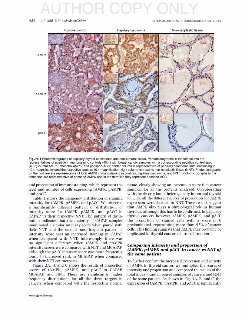
tAMPK, pAMPK, and phospho-ACC (pACC) showed acytoplasmic staining in papillary carcinomas and non-neoplastic thyroid tissues (Fig. 1, center and rightcolumns). Apart from cytoplasmic staining, membraneand nuclear stainings were also present. Ductal breastcarcinoma was used as positive and negative controls ofthe reactions (Fig. 1, left column). Strong and diffusestaining was observed for both tAMPK and pAMPK(Fig. 1, first and second lines) in the majority of papillarycarcinomas. Most spots of non-neoplastic thyroid tissueshowed a focal positivity for either tAMPK or pAMPK(Fig. 1, first and second lines). Diffuse staining wasobserved for pACC in papillary carcinoma, while a focalpattern was observed in NNT (Fig. 1, third line).Increased AMPK phosphorylation signal was seen inthe vast majority of papillary thyroid cancer specimens,as a diffuse and strong staining in the cytoplasmcompared with the focal expression in normalepithelium with a significant positive association.
Comparison of tAMPK, pAMPK, and pACCexpression between CAPAP and MCAPAP ofthyroid
In order to quantify the expression pattern of AMPKpathway in thyroid cancer, we analyzed the intensity
www.eje-online.org
AUTHOR COPY ONLY
tAMPK
Positive control Papillary carcinoma Non-neoplastic tissue
pAMPK
pACC
20 µm
20 µm
20 µm20 µm
20 µm
20 µm20 µm
20 µm
20 µm
Figure 1 Photomicrographs of papillary thyroid carcinomas and non-tumoral tissue. Photomicrographs in the left column arerepresentatives of positive immunostaining controls (40!) with breast cancer samples with a corresponding negative control spot(40!) in total AMPK, phospho-AMPK, and phospho-ACC; center column is representative of papillary carcinoma immunostaining in40! magnification and the respective spots at 10! magnification; right column represents non-neoplastic tissue (NNT). Photomicrographson the first line are representative of total AMPK immunostaining in controls, papillary carcinoma, and NNT; photomicrographs in thecenterline are representative of phospho-AMPK and in the third line they represent phospho-ACC.
524 A P Vidal, B M Andrade and others EUROPEAN JOURNAL OF ENDOCRINOLOGY (2013) 169
and proportion of immunostaining, which represent thelevel and number of cells expressing tAMPK, pAMPK,and pACC.
Table 1 shows the frequency distribution of stainingintensity for tAMPK, pAMPK, and pACC. We observeda significantly different pattern of distribution ofintensity score for tAMPK, pAMPK, and pACC inCAPAP vs their respective NNT. The pattern of distri-bution indicates that the majority of CAPAP samplesmaintained a similar intensity score when paired withtheir NNT, and the second most frequent pattern ofintensity score was an increased staining in CAPAPwhen compared with NNT. Interestingly, there wasno significant difference when tAMPK and pAMPKintensity scores were compared with NTT and MCAPAP,although the pACC intensity score was more frequentlyfound to increased rank in MCAPAP when comparedwith their NTT counterparts.
Figure 2A, B, and C shows the results of proportionscores of tAMPK, pAMPK, and pACC in CAPAP,MCAPAP, and NNT. There are significantly higherfrequency distributions for all the three targets incancers when compared with the respective normal
www.eje-online.org
tissue, clearly showing an increase in score 6 in cancersamples, for all the proteins analyzed. Corroboratingwith the description of heterogeneity in normal thyroidfollicles, all the different scores of proportion for AMPKexpression were detected in NNT. These results suggestthat AMPK also plays a physiological role in humanthyroids, although this has to be confirmed. In papillarythyroid cancers however, tAMPK, pAMPK, and pACCthe proportion of stained cells with a score of 6predominated, representing more than 95% of cancercells. This finding suggests that AMPK may probably beimplicated in thyroid cancer cell transformation.
Comparing intensity and proportion oftAMPK, pAMPK and pACC in cancer vs NNT ofthe same patient
To further confirm the increased expression and activityof AMPK in thyroid cancer, we multiplied the scores ofintensity and proportion and compared the values of thetotal index found in paired samples of cancers and NNTof the same patient. As shown in Fig. 3A, B, and C, theexpression of tAMPK, pAMPK, and pACC is significantly

AUTHOR COPY ONLY
100A
B
C
* *
* *
* *
80
60
40
20
0NNT CAPAP
Per
cent
of s
core
100
80
60
40
20
0NNT MCA PAP
Per
cent
of s
core
100
80
60
40
20
0NNT CAPAP
Per
cent
of s
core
100
80
60
40
20
0NNT MCA PAP
Per
cent
of s
core
100
80
60
40
20
0NNT CAPAP
Per
cent
of s
core
100
80
60
40
20
0NNT MCA PAP
Per
cent
of s
core
Score 1 Score 2 Score 3 Score 4 Score 5 Score 6
Figure 2 Distribution pattern of the proportion of total AMPK,phospho-AMPK, and phospho-ACC in papillary (nZ57) and micro-papillary (nZ16) carcinomas and their respective non-neoplasticthyroid tissue samples. The staining proportion was scored using arange from 1 to 6 regarding the number of cells stained in each TMAspot. (A) Total AMPK; (B) phospho-AMPK; (C) phospho-ACC.Results are shown as percent of the total number of cases in eachscore. *P!0.0001 vs control (c2 test).
AMPK signaling in papillary thyroid cancer 525EUROPEAN JOURNAL OF ENDOCRINOLOGY (2013) 169
increased in both CAPAP and MCAPAP compared withtheir respective NNT samples. These results initiallysuggest that AMPK is not only more expressed but alsohighly active in thyroid cancer cells than in NNT. Inorder to correlate the increase in expression and activityof AMPK with carcinogenesis, we analyzed six samplesof thyroid adenomas. Interestingly, there was nosignificant difference in the expression of tAMPK,pAMPK, and pACC in adenomas when compared withthe NNT of the same patients (Fig. 3C).
Indirect analysis of AMPKactivity through theratio between pACC and the relationshipbetween pAMPK and tAMPK expression incancers
In order to evaluate the activity of AMPK in humanpapillary thyroid cancer, we calculated the ratio
between pACC and AMPK phosphorylation rate(pAMPK/tAMPK). This analysis is necessary in orderto exclude the possibility that the observed increase inpAMPK could only be related to the increase in tAMPKexpression and not to a greater activity of this kinase.Indeed, Fig. 4A and B showed and confirmed thatpapillary and micro-papillary cancer samples haveincreased AMPK activity compared with their respectiveNNT and adenomas (Fig. 4C).
Discussion
In this study, we describe for the first time that themetabolic sensor AMPK is expressed in normal humanthyroid gland and that its expression and activity aresignificantly higher in papillary thyroid carcinomas. Asshown herein, a strong and diffuse cytoplasmic stainingis observed for tAMPK and pAMPK in the majority ofpapillary carcinomas in contrast with the focalpositivity found in non-tumor thyroid tissue cells. It isinteresting to notice that in the previous studiesregarding the molecular signature of PAPCA throughmicroarray, AMPK mRNA has not been identified asdifferentially expressed. The differences between thosestudies and our present results could be related to thedifferent sensitivity of the methods used and/or due toincreased translation of AMPK mRNA, with nodifferences in its expression levels (24).
A significantly higher expression and activity ofAMPK were found in thyroid cancer cells in relation toa higher total index (intensity!proportion) of immu-nostaining. Furthermore, there was a significantdifference in the proportion of cells stained for thethree proteins: tAMPK, pAMPK, and pACC, suggestingthat when we compare normal vs papillary thyroidcarcinomas, a strong stimulus for AMPK expression andactivation is observed.
Although the cytoplasmic staining in both NNT andpapillary carcinoma thyroid tissues were predominant,we also observed membrane and nuclear staining(Fig. 1, center and right columns). The relevance ofthese findings, however, is not known. It has beendemonstrated that AMPKa2 is localized in the nuclei ofmany cells and might be involved in the regulation ofgene expression (25). The physiological significance ofthe increase in AMPK expression and activity still hasto be elucidated. Recently, Faustino et al. (2012) (25)published a study describing the pattern of expressionand activity of mTOR in thyroid cancer lesions. Theauthors showed that the PI3K/AKT/mTOR pathway isactivated and that there is a correlation between thispathway activation and BRAFV600E mutations inthyroid carcinomas (26). The interrelationship betweenAMPK and mTOR is well described for tissues other thanthe thyroid (27). Our group has previously describedthat mTOR plays an important role in the regulation ofiodide uptake in rat thyrocytes (28, 29). Although not
www.eje-online.org

AUTHOR COPY ONLY
20
A
B
C
15
10
INT
×P
RO
PIN
T×
PR
OP
INT
×P
RO
P5
0NNT CAPAP
Total AMPK
*Phospho-AMPK
*Phospho-ACC
*
* * *
& NS NS
20
15
10
5
0NNT CAPAP
20
15
10
5
0NNT CAPAP
20
15
10
5
0NNT MCAPAP
20
15
10
5
0NNT MCAPAP
20
15
10
5
0NNT MCAPAP
20
15
10
5
0NNT Adenoma
20
15
10
5
0NNT Adenoma
20
15
10
5
0NNT Adenoma
Figure 3 Expression pattern of total AMPK, phospho-AMPK, and phospho-ACC in papillary (nZ57) and micro-papillary (nZ16)carcinomas, adenomas (nZ6), and non-neoplastic thyroid paired samples. The antigen expression is represented by a score thatcorresponds to the multiplication of the value attributed to the staining intensity (from 0 to 3) by the value attributed to the staining proportion(0–6) (maximum value is therefore 3!6Z18), comparing carcinoma vs the non-neoplastic tissue of the same patient. (A) Papillarycarcinoma; (B) micro-papillary carcinoma; (C) adenoma. Results are shown by meanGS.E.M. *P!0.001 vs control; &P!0.05 vs control(Wilcoxon paired test).
526 A P Vidal, B M Andrade and others EUROPEAN JOURNAL OF ENDOCRINOLOGY (2013) 169
yet demonstrated for the thyroid tissue, under normalphysiological conditions, AMPK activation inhibitsmTOR through different mechanisms (27). Neverthe-less, as our study clearly demonstrates increasedexpression and activity of AMPK in papillary thyroidcarcinomas, and taking into consideration the previousreport of Faustino et al. (2012) (25), one mightspeculate that at least in papillary thyroid carcinomasboth mTOR and AMPK pathways are stimulated. Theapparent controversy might be explained by the findingof Esteve-Puig et al. (2009) (27), who showed thatBRAFV600E mutation in melanoma cells leads touncoupling in LKB1–AMPK pathway, also changingits relationship with the mTOR signaling pathway (30).However, the relationship between AMPK and mTORpathways in normal thyroid cells and in thyroidcancers needs to be evaluated in future studies,preferably using fresh tumor samples and a morequantitative analysis.
Previous studies from our group have described thatAMPK activation in normal thyroid gland and PCCL3rat thyroid cell lineage leads to decreased iodide uptake
www.eje-online.org
and increased glucose uptake (2, 5). This phenomenon(so called ‘flip-flop’) is commonly observed in thyroidoncology and seems to be well correlated with gain intumor aggressiveness (20, 21, 31). Therefore, it istempting to speculate whether changes in AMPKexpression could play a role in thyroid tumor pro-gression, although this has to be further evaluated.Buzzai et al. (2005) (14) demonstrated that AMPK isnecessary for the survival of LN-229 cells duringglucose deprivation protocol, indicating that thispathway might be involved in the control of tumorcell survival under stressful conditions. No matter thesignificance of AMPK activation for cancer prognosis,we can conclude that the higher expression and activityof AMPK in well-differentiated thyroid cancer could berelated to the lower iodide uptake ability of thyroidcancer cells. As some clinical data point to thepossibility that metformin – an AMPK agonist – mightreduce cancer incidence, it is important to betterunderstand the role of this kinase in tumorigenesis,cancer progression, and the reduced iodide uptake thatimpairs radioiodine therapeutically approach.

AUTHOR COPY ONLY20
A
15
10
*
5Rat
io (
AC
C/A
MP
K)
0
NNT CAPAP
20B
C
15
10
#
5Rat
io (
AC
C/A
MP
K)
0NNT
NS
MCAPAP
20
15
10
5Rat
io (
AC
C/A
MP
K)
0
NNT Adenoma
Figure 4 Ratio of expression of phospho-ACC in relation to (totalAMPK/phospho-AMPK) in papillary (nZ57) and micro-papillary(nZ16) carcinomas, adenomas (nZ6), and non-neoplastic thyroidpaired samples. The ratio of expression was calculated throughthe division of the value attributed to phospho-ACC and the ratio ofphospho-AMPK and total AMPK pattern of expression, comparingcarcinoma vs normal tissue of the same patient. (A) Papillarycarcinoma; (B) micro-papillary carcinoma; (C) adenoma. Resultsare shown by meanGS.E.M.. *P!0.0001 vs control; #P!0.001 vscontrol; NS, not significant (Wilcoxon paired test).
AMPK signaling in papillary thyroid cancer 527EUROPEAN JOURNAL OF ENDOCRINOLOGY (2013) 169
Inconclusion, our datashow thatAMPKexpression andphosphorylation are increased in papillary thyroid cancerspecimens when compared with the non-neoplastic
counterpart tissues and benign lesions. This findingsuggests that AMPK may probably be implicated in thyroidcancer cell transformation. More data are now required togive us a comprehensive understanding about the role ofAMPK pathway in thyroid carcinoma.
Declaration of interest
The authors declare that there is no conflict of interest that could beperceived as prejudicing the impartiality of the research reported.
Funding
This work was supported by grants from Fundacao Carlos ChagasFilho de Amparo a Pesquisa no Estado do Rio de Janeiro (FAPERJ),Coordenacao de Aperfeicoamento de Pessoal de Nıvel Superior(CAPES), Conselho Nacional para o Desenvolvimento Cientıfico eTecnologico (CNPq), Instituto Nacional de Ciencia e Tecnologia paraPesquisa Translacional em Saude e Ambiente (INPeTAm).
Acknowledgements
The authors thank Heliomar Pereira Marcos for the contribution inimmunostaining experiments.
References
1 Dandapani M & Hardie DG. AMPK: opposing the metabolicchanges in both tumour cells and inflammatory cells? BiochemicalSociety Transactions 2013 41 687–693. (doi:10.1042/BST20120351)
2 Hardie DG. AMP-activated protein kinase: an energy sensor thatregulates all aspects of cell function. Genes and Development 201125 1895–1908. (doi:10.1101/gad.17420111)
3 Andrade BM, Araujo RL, Perry RL, Souza EC, Cazarin JM,Carvalho DP & Ceddia RB. A novel role for AMP-kinase in theregulation of the NaC/IK-symporter and iodide uptake in the ratthyroid gland. American Journal of Physiology. Cell Physiology 2011300 C1291–C1297. (doi:10.1152/ajpcell.00136.2010)
4 Gaidhu MP & Ceddia RB. Remodeling glucose and lipid metabolismthrough AMPK activation: relevance for treating obesity and type2 diabetes. Clinical Lipidology 2009 4 465–477. (doi:10.2217/clp.09.30)
5 Kahn BB, Alquier T, Carling D & Hardie DG. AMP-activatedprotein kinase: ancient energy gauge provides clues to modernunderstanding of metabolism. Cell Metabolism 2005 1 15–25.(doi:10.1016/j.cmet.2004.12.003)
6 Andrade BM, Cazarin J, Zancan P & Carvalho DP. AMP-activatedprotein kinase upregulates glucose uptake in thyroid PCCL3cells independent of thyrotropin. Thyroid 2012 22 1063–1068.(doi:10.1089/thy.2012.0041)
7 Rattan R, Giri S, Singh AK & Singh I. 5-Aminoimidazole-4-carboxamide-1-b-D-ribofuranoside inhibits cancer cell proliferationin vitro and in vivo via AMP-activated protein kinase. Journal ofBiological Chemistry 2005 280 39582–39593. (doi:10.1074/jbc.M507443200)
8 Luo Z, Zang M & Guo W. AMPK as a metabolic tumor suppressor:control of metabolism and cell growth. Future Oncology 2010 6457–470. (doi:10.2217/fon.09.174)
9 Shackelford DB & Shaw RJ. The LKB1–AMPK pathway: meta-bolism and growth control in tumor suppression. Nature Reviews.Cancer 2009 9 563–575. (doi:10.1038/nrc2676)
10 Jones RG, Plas DR, Kubek S, Buzzai M, Mu J, Xu Y, Birnbaum MJ &Thompson CB. AMP-activated protein kinase induces a p53-dependent metabolic checkpoint. Molecular Cell 2005 18 283–293.(doi:10.1016/j.molcel.2005.03.027)
www.eje-online.org

AUTHOR COPY ONLY528 A P Vidal, B M Andrade and others EUROPEAN JOURNAL OF ENDOCRINOLOGY (2013) 169
11 Grisouard J, Dembinski K, Mayer D, Keller U, Muller B & Christ-Crain M. Targeting AMP-activated protein kinase in adipocytesto modulate obesity-related adipokine production associated withinsulin resistance and breast cancer cell proliferation. Diabetology& Metabolic Syndrome 2011 20 3–16. (doi:10.1186/1758-5996-3-16)
12 Hadad SM, Baker L, Quinlan PR, Robertson KE, Bray SE, Thomson G,Kellock D, Jordan LB, Purdie CA, Hardie DG et al. Histologicalevaluation of AMPK signalling in primary breast cancer. BMCCancer 2009 9 307. (doi:10.1186/1471-2407-9-307)
13 Gallagher EJ & LeRoith D. Diabetes, cancer, and metformin:connections of metabolism and cell proliferation. Annals of theNew York Academy of Sciences 2011 1243 54–68. (doi:10.1111/j.1749-6632.2011.06285.x)
14 Buzzai M, Bauer DE, Jones RG, Deberardinis RJ, Hatzivassiliou G,Elstrom RL & Thompson CB. The glucose dependence ofAkt-transformed cells can be reversed by pharmacologic acti-vation of fatty acid b-oxidation. Oncogene 2005 24 4165–4173.(doi:10.1038/sj.onc.1208622)
15 Hayashi T, Hirshman MF, Fujii N, Habinowski SA, Witters LA &Goodyear LJ. Metabolic stress and altered glucose transport:activation of AMP-activated protein kinase as a unifying couplingmechanism. Diabetes 2000 49 527–531. (doi:10.2337/diabetes.49.4.527)
16 Schlumberger M. Papillary and follicular thyroid carcinoma. NewEngland Journal of Medicine 2000 338 297–306. (doi:10.1056/NEJM199801293380506)
17 Mazzaferri EL & Massoll N. Management of papillary and follicular(differentiated) thyroid cancer: new paradigms using recombinanthuman thyrotropin. Endocrine-Related Cancer 2002 9 227–247.(doi:10.1677/erc.0.0090227)
18 Cooper DS, Doherty GM, Haugen BR, Kloos RT, Lee SL, Mandel SJ,Mazzaferri EL, McIver B, Pacini F, Schlumberger M et al.Revised American Thyroid Association management guidelinesfor patients with thyroid nodules and differentiated thyroid cancer.Thyroid 2009 19 1167–1214. (doi:10.1089/thy.2009.0110)
19 Goretzki PE, Simon D, Frilling A, Witte J, Reiners C, Grussendorf M,Horster FA & Roher HD. Surgical reintervention for differentiatedthyroid carcinoma. British Journal of Surgery 1994801009–1012.(doi:10.1002/bjs.1800800826)
20 Haugen BR. Management of the patient with progressive radio-iodine non-responsive disease. Seminars in Surgical Oncology 19991634–41. (doi:10.1002/(SICI)1098-2388(199901/02)16:1!34::AID-SSU7O3.0.CO;2-2)
21 Coelho SM, Corbo R, Buescu A, Carvalho DP & Vaisman M.Retinoic acid in patients with radioiodine non-responsivethyroid carcinoma. Journal of Endocrinological Investigation 200427 334–339.
www.eje-online.org
22 De Lellis AR, Lloyd RV, Heitz PU & Eng C. Thyroid and parathyroidtumors. In Pathology and Genetics of Tumors of Endocrine Organs.World Health Organization Classification of Tumors, 1st edn, pp49–133. Eds AR De Lellis & ED Williams. Lyon: WHO, IARC, 2004.
23 Leake R, Barnes D, Pinder S, Ellis I, Anderson L, Anderson T,Adamson R, Rhodes T, Miller K & Walker R. Immunohistochem-ical detection of steroid receptors in breast cancer: a workingprotocol. UK Receptor Group, UK NEQAS, The Scottish BreastCancer Pathology Group, and The Receptor and BiomarkerStudy Group of the EORTC. Journal of Clinical Pathology 2000 53634–635. (doi:10.1136/jcp.53.8.634)
24 Wang W, Larson SM, Tuttle RM, Kalaigian H, Kolbert K,Sonenberg M & Robbins RJ. Resistance of [18F]-fluorodeoxyglu-cose-avid metastatic thyroid cancer lesions to treatment withhigh-dose radioactive iodine. Thyroid 2001 12 1169–1175.(doi:10.1089/10507250152741028)
25 Faustino A, Couto JP, Populo H, Rocha AS, Pardal F, Cameselle-Teijeiro JM, Lopes JM, Sobrinho-Simoes M & Soares P. mTORpathway overactivation in BRAF mutated papillary thyroidcarcinoma. Journal of Clinical Endocrinology and Metabolism 201297 E1139–E1149. (doi:10.1210/jc.2011-2748)
26 Inoki K, Kim J & Guan KL. AMPK and mTOR in cellular energyhomeostasis and drug targets. Annual Review of Pharmacology andToxicology 2012 10 381–400. (doi:10.1146/annurev-pharmtox-010611-134537)
27 Esteve-Puig R, Canals F, Colome N, Merlino G & Recio JA.Uncoupling of the LKB1–AMPKa energy sensor pathway bygrowth factors and oncogenic BRAF. PLoS ONE 2009 4 e4771.(doi:10.1371/journal.pone.0004771)
28 de Souza EC, Padron AS, Braga WM, de Andrade BM, Vaisman M,Nasciutti LE, Ferreira AC & de Carvalho DP. MTOR downregulatesiodide uptake in thyrocytes. Journal of Endocrinology 2010 206113–120. (doi:10.1677/JOE-09-0436)
29 Souza EC, Ferreira AC & Carvalho DP. The mTOR protein as atarget in thyroid cancer. Expert Opinion on Therapeutic Targets 201115 1099–10112. (doi:10.1517/14728222.2011.594044)
30 Schmid KW & Farid NR. How to define follicular thyroidcarcinoma? Virchows Archiv 2006 448 385–393. (doi:10.1007/s00428-006-0162-0)
31 Salt I, Celler JW, Hawley SA, Prescott A, Woods A, Carling D &Hardie DG. AMP-activated protein kinase: greater AMP dependence,and preferential nuclear localization, of complexes containing thealpha2 isoform. Biochemical Journal 1998 334 177–187.
Received 4 April 2013
Revised version received 27 June 2013
Accepted 29 July 2013