C 8 + 9 SURGICAL PATHOLOGY OF PANCREAS
133
1 SURGICAL PATHOLOGY OF PANCREAS 4 hours
-
Upload
independent -
Category
Documents
-
view
1 -
download
0
Transcript of C 8 + 9 SURGICAL PATHOLOGY OF PANCREAS

1
SURGICAL PATHOLOGY OF PANCREAS
4 hours

SURGICAL PATHOLOGY OF PANCREAS – 4 hours
• C 8 – Acute pancteatitis: etiology, phisiopathology, clinical findings, evaluation, diagnosis, differential diagnosis, complications(pancreatic pseudocyst), treatment.
– Chronic pancteatitis: etiology, phisiopathology, clinical findings, evaluation, diagnosis, differential diagnosis, complications, treatment.
• C 9 – Malignant tumors of the pancreas: etiology, classification, pathology, clinical findings, evaluation, diagnosis, complications, treatment.
– Tumors of the Vater's ampulla: etiology, classification, pathology, clinical findings, evaluation, diagnosis, complications, treatment;
– Tumors of the endocrine pancreas.2

3
SURGICAL PATHOLOGY OF PANCREAS
Penetrating Injuries • Penetrating injuries to the pancreas are usually diagnosed in the operating room. • The pancreas is located in the retro peritoneum, surrounded by other viscera and
major vascular structures. • An isolated injury to the pancreas is unusual, major vascular injuries are seen in
40% to 50% of patients with penetrating pancreatic injuries,
PANCREATIC TRAUMA Penetrating Injuries of Pancreas
Pancreatic injury involving the ductal system
Retroperitoneal location of pancreas
4
SURGICAL PATHOLOGY OF PANCREAS
• Patients with penetrating pancreatic trauma have obvious indications for abdominal exploration.
• Preoperative serum amylase concentrations are not helpful; (elevated in 30% of patients with penetrating pancreatic injuries).
Abdominal exploration, • Signs of pancreatic injury include:
– knife or a projectile path that passes in proximity to the pancreas, – a central hematoma in the upper abdomen, and – injuries to the duodenum, vena cava, suprarenal aorta, or mesenteric
vessels. • In all these instances, the pancreas should be thoroughly explored: • The anterior surface of the pancreas is visualized by entry into the lesser sac of the peritoneal
cavity by division of the gastrocolic ligament in a relatively avascular area to the left of the midline.
• The tail of the pancreas can be more fully visualized, especially in its posterior aspect, by mobilization of the spleen and the tail of the pancreas as a unit.
• The posterior aspect of the body of the pancreas is visualized by development of the avascular area at the inferior margin of the body and tail of the pancreas with a combination of sharp and blunt dissection.
• The posterior aspect of the head of the pancreas can be exposed by an extensive Kocher maneuver. In combination with entry into the lesser sac, this also allows for bimanual palpation of the pancreatic head, with one hand placed on the anterior surface of the pancreas through the hole in the lesser sac and the other hand placed behind the pancreas in the plane developed by the Kocher maneuver.
PANCREATIC TRAUMA Penetrating Injuries of Pancreas

5
SURGICAL PATHOLOGY OF PANCREAS
• In the evaluation of penetrating pancreatic injuries, the key to operative management is the determination of whether a ductal injury is present. – Transduodenal intraoperative pancreatography has been recommended,
but the use of this technique in the identification of ductal injuries is controversial or dangerous.
– In certain circumstances, the use of intraoperative ERCP (endoscopic retrograde cholangiopancreatography) eliminates this problem.
• Penetrating pancreatic injuries can be classified according to both location and severity.
• With respect to location, injuries can be subdivided into those of the – head, – body, and – tail of the pancreas.
• With respect to severity, classification systems must be satisfactory to research applications but also in the determination of the best treatment.
Classification– Class I injuries are simple contusions of the pancreas; – Class II injuries are lacerations of the parenchyma in the body or tail of the
pancreas; – Class III injuries are those with severe disruption of the head or body; – Class IV injuries are those in which there is an associated injury to the
duodenum.
PANCREATIC TRAUMA Penetrating Injuries of Pancreas

6
SURGICAL PATHOLOGY OF PANCREAS
• Class I injuries should be usually observed or simply drained externally. The type of drain used after pancreatic injury is probably not important as long as adequate drainage is effected. This can be accomplished with passive, sump, or closed suction systems.
• If drains are used, they should be left in place for at least 5 to 7 days to ensure that a drain tract develops. The timing of drain removal should be based on both the amount and character of the pancreatic drainage:
– Drain outputs in excess of 150 to 200 mL/day are suggestive of pancreatic fistula.
• The morbidity rate for patients with undrained pancreatic secretions is much greater than that for those with drained pancreatic secretions.
• Class II injuries The treatment depends on the presence or absence of a ductal injury, a determination that can be difficult to make.
• The argument for intraoperative pancreatography is made for class II injuries. • The presence of a ductal injury in the head of the pancreas does not
usually make a difference with respect to treatment, because most of these injuries should be drained regardless.
• If a ductal injury is present in the body or tail of the pancreas, the appropriate treatment is resection of the distal pancreas, whereas if no ductal injury is present, simple drainage is adequate.
• the difficulty lies in determining whether injuries to the body or tail of the pancreas do in fact include ductal injury.
• Intraoperative pancreatography is helpful, regardless of the technique employed.
• Class III injuries of the body or tail of the pancreas, should be treated with a distal pancreatectomy. Distal resection can include up to 80% of the gland if necessary.
PANCREATIC TRAUMA Penetrating Injuries of Pancreas

7
SURGICAL PATHOLOGY OF PANCREAS
• Class III injuries of the head of the pancreas should be drained. Resection of these injuries requires internal drainage, near-total pancreatectomy, or pancreaticoduodenectomy. If the patient develops a pancreatic fistula, the fistula can be controlled by the drains. If the fistula does not resolve with time, the pancreas can be drained internally at a later date.
• Class IV injuries of the pancreas involve injuries to the duodenum as well as the pancreas. – If the injuries to the duodenum and pancreas are simple, the duodenum can be repaired
primarily, and the pancreas can be drained, or a distal resection can be carried out if the pancreatic injury is in the body or tail.
– For more complicated, combined injuries, pyloric exclusion can be done to minimize pancreatic stimulation and protect the duodenal repair (see Duodenum).
– For massive injuries to the duodenum and head of the pancreas, pancreaticoduodenectomy with reconstruction should be reserved for cases in which débridement of devitalized tissue results in a de facto removal of the duodenum and head of the pancreas.
– Penetrating injuries to the ampulla of Vater may also require formal pancreaticoduodenectomy.
• Internal drainage of the pancreas has been suggested as a means of treating ductal injuries without the need for resection of viable and functional pancreatic tissue.
• Distal pancreatectomy for traumatic injuries should be performed only after the pancreas has been thoroughly mobilized and exposed.
• It is possible to perform a distal pancreatectomy without a concomitant splenectomy. This adds to operative time and can increase the risk of bleeding, particularly if there is an associated injury to the spleen treated with splenorrhaphy.
• Splenic salvage should be attempted, therefore, only in hemodynamically stable patients with minimal or no associated intraabdominal or extraabdominal injuries.
PANCREATIC TRAUMA Penetrating Injuries of Pancreas

8
SURGICAL PATHOLOGY OF PANCREAS
PANCREATIC TRAUMA Penetrating Injuries of Pancreas
Internal drainage of a pancreatic injury involving the ductal system with the creation of a Roux-en-Y jejunoileal limb and construction of a pancreaticojejunal anastomosis.

9
SURGICAL PATHOLOGY OF PANCREAS
Complications of pancreatic injuries: • Pancreatic fistulas, after trauma are characterized by persistent drainage of pancreatic
enzymes and secretions from the pancreatic injury for a number of weeks after injury. Most of these fistulas close spontaneously, especially if there is no proximal obstruction of the pancreatic ductal system. In the rare instances in which fistulas fail to close spontaneously, surgical intervention may be required.
• Pseudocysts, that develop after pancreatic trauma often resolve on their own or with percutaneous aspiration. If, after 4 to 6 weeks of observation with serial ultrasound or CT scanning, the pseudocyst does not show signs of resolution, it should be drained internally with a defunctionalized limb of jejunum.
• Bleeding in the area of the pancreatic bed, is usually an early complication that is caused by inadequate drainage with resultant autodigestion of the pancreas and surrounding tissue. Bleeding can be avoided by identifying all injuries at the time of exploration and ensuring adequate drainage. If massive bleeding does occur, it should be dealt with through operative intervention.
• Pancreatitis, another complication of pancreatic injury, is also related to inadequate drainage of pancreatic secretions. Treatment consists of provision of adequate drainage and supportive care. If the pancreatitis is localized to the distal pancreas and a trial of conservative management fails, distal pancreatectomy should be considered.
PANCREATIC TRAUMA Penetrating Injuries of Pancreas

10
SURGICAL PATHOLOGY OF PANCREAS
Blunt Injuries • The major difference between penetrating and blunt injuries of the pancreas concerns the diagnosis. • Penetrating injuries are usually discovered on abdominal exploration for associated injuries, but
blunt injuries may occur in isolation, and the preoperative diagnosis can be difficult. • Blunt pancreatic injuries are relatively rare, which increases the difficulty of diagnosis. So delays in
diagnosis of blunt injuries ranged up to several days, which induce an increased morbidity.• The body of the pancreas lies directly anterior to the vertebral column and is vulnerable to crush
injuries when the anterior abdominal wall is forceably compressed, as can occur from a seat belt or a sharp blow to the epigastrium. In such instances, the pancreas may be the only intraabdominal organ injured.
• A number of different means are available to make the diagnosis of blunt pancreatic injury:– Physical examination of the abdomen is useful, but because of the
retroperitoneal location of the pancreas, the results can be misleadingly benign until a number of hours after injury.
– This emphasizes the importance of serial examinations. – In most cases, the abdomen becomes progressively more tender to palpation during the
first 24 to 48 hours after injury, and the need for abdominal exploration becomes more obvious.
– The physical examination of the abdomen is much less reliable in young children and in patients with head injuries.
• The serum amylase concentration is elevated on admission in about 70% of patients with blunt pancreatic injury.
• However, elevated serum amylase has a poor positive predictive value and also occurs in many patients without pancreatic injury. The amylase concentration can be elevated because of trauma to other organs, including the salivary glands and the ovaries.
• DPL(diagnostic peritoneal lavage) is of little help in the early diagnosis of pancreatic injury unless there have been associated intraperitoneal injuries. The retroperitoneal location of the pancreas results in minimal findings in the lavage fluid, and obtaining amylase concentrations in the lavage fluid is not helpful.
PANCREATIC TRAUMA Blunt Injuries of Pancreas

11
SURGICAL PATHOLOGY OF PANCREAS
• CT scan of the abdomen allows for visualization of the retroperitoneum, including the pancreas. In the case of isolated injury to the pancreas, the sensitivity of the CT scan is at its lowest shortly after injury. Although it may be a good test for the diagnosis of pancreatic injury after a number of hours have passed, immediate CT of the abdomen will miss some pancreatic injuries, particularly if expert interpretation is not available.
• Finally, ERCP is a means of diagnosing pancreatic injury. ERCP is an attractive diagnostic method because it is less invasive than abdominal exploration and also provides information about the status of the ductal system, but there are several practical disadvantages of the technique. – Most of the studies that have reported successful use of ERCP have
involved stable patients studied hours to days after injury and sent to a referral center specifically because of suspicion of a pancreatic injury.
– These patients are a selected group, quite different from patients who are freshly injured.
– ERCP is not universally available and, even in large centers, is often unavailable at the odd hours necessary for early diagnosis in acutely injured patients.
– Many endoscopists are fearful of inducing an exacerbation of pancreatitis in patients with mild pancreatic injuries lacking ductal involvement.
• To summarize,• the early diagnosis of blunt pancreatic injuries, particularly if they occur in isolation, can be
extremely difficult, and • no single test allows for an easy and reliable diagnosis. • A combination of serial abdominal examinations and serum amylase determinations, CT scan,
and ERCP in selected patients is the best diagnostic strategy available. • These studies should be combined, with a low threshold for operative
intervention if a pancreatic injury is suspected.
PANCREATIC TRAUMA Blunt Injuries of Pancreas

12
SURGICAL PATHOLOGY OF PANCREAS
• Basic principles of exposure and operative management of blunt injuries of the pancreas are the same as for penetrating injuries.
• In many instances of severe injury, the pancreas has already been transected by the trauma, making the pancreatic resection somewhat simpler to carry out.
• Isolated injuries of the pancreas from blunt trauma also lend themselves to distal pancreatectomy with splenic preservation.
• As in penetrating injury, splenic salvage should be attempted only in stable patients without associated splenic rupture or severe associated intraabdominal or extraabdominal injuries.
• Complications of pancreatic injury are similar to those outlined for penetrating injuries.
PANCREATIC TRAUMA Blunt Injuries of Pancreas

13
SURGICAL PATHOLOGY OF PANCREAS
DEFINITION• Acute pancreatitis is a complex disorder of the exocrine pancreas characterized by acute acinar
cell injury and both regional and systemic inflammatory responses. • It is a common disease with a broad spectrum of clinical and pathologic findings that
contribute to considerable morbidity and mortality. • Because pathogenic mechanisms are unclear, specific treatment is not available.
Empiric supportive care remains standard, and clinical outcomes have improved only to the extent that critical care has evolved in recent years.Most patients with acute pancreatitis have simple edematous pancreatitis, a self-limited and reversible process. In a small number of patients, fulminant or progressive disease develops, with pancreatic necrosis that can lead to multiorgan system failure or death.
ACUTE PANCREATITIS
The problems related to acute pancreatitis pose a formidable challenge to both
the clinical surgeon and the basic scientist.

14
SURGICAL PATHOLOGY OF PANCREAS
PATHOLOGY
ACUTE PANCREATITIS
Normal acinar cell ultrastructure. Cytoplasmic processing of the proenzymes is depicted, with apical discharge into the acinar ductule by means of zymogen granule exocytosis.

15
SURGICAL PATHOLOGY OF PANCREAS
PATHOLOGY• Acute pancreatitis is characterized by alterations in acinar cell structure and function as well as by
the development of acute regional and systemic inflammatory responses. • The fundamental pathologic event is injury to the acinar cell. • Acute pancreatitis is characterized by:
– Interstitial edema formation. – Grossly, the gland becomes enlarged and edematous, with small areas of focal
necrosis involving either the pancreas or areas of adjacent retroperitoneal fat. – Microscopically, edema is both interlobular and intralobular, and the acinar units appear
dispersed within the relatively sparse fibrous matrix.
ACUTE PANCREATITIS
The histologic features of acute edematous pancreatitis.There is extensive edema formation, acinar cell cytoplasm vacuolization, and polymorphonuclear cell (PMN) infiltration.

16
SURGICAL PATHOLOGY OF PANCREAS
• Acute inflammation occurs rapidly, experimentally, the process begins within minutes.
• The initial cellular response involves the infiltration of polymorphonuclear leukocytes into the perivascular regions of the pancreas.
• Within hours, mononuclear cells, including macrophages and lymphocytes, accumulate.
• Experimental evidence suggests that phagocyte-derived oxygen radicals and possibly other phagocytic products are involved in a primary injury to pancreatic capillary endothelial cells.
• The resulting increase in microvascular permeability facilitates access to the acinar cell microenvironment for circulating formed elements (additional neutrophils, monocytes, platelets) and humoral factors, such as complement products and cytokines. – The relative importance of this inflammatory injury to the pancreatic
microvasculature is unclear, but it provides one explanation for the process of local edema formation as well as for the systemic microvascular sequelae of acute pancreatitis.
• Although clinical – Edematous pancreatitis usually is a reversible disease characterized by acinar
cell injury and edema formation, – frank pancreatic necrosis develops in 5% to 10% of patients, which can lead to irreversible
regional injury or multiorgan system failure. Predictably, this group of patients is the primary source of morbidity and mortality associated with acute pancreatitis.
• Histologic characteristics of advanced disease include – extensive acinar cell necrosis, – interstitial microabscess formation, – extensive peripancreatic fat necrosis, – microvascular thrombosis, and – local hemorrhage.
• Pathologically, all these features appear to represent progression of processes already established with acute edematous pancreatitis.
ACUTE PANCREATITIS

17
SURGICAL PATHOLOGY OF PANCREAS
PATHOPHISIOLOGY• The cellular events that lead to acute pancreatitis may be initiated by a variety of different stimuli,
and the process has been considered a final common pathway. Current data offer considerably more insight into the relevant pathogenic events than the simple historic concept that pancreatic autolysis occurs.
• Acinar cell proteases (such as trypsin, chymotrypsin, carboxypeptidase, and elastase) and phospholipases are normally synthesized in an inactive zymogen form.
• Peptide synthesis is accomplished and the proenzymes are packaged into cytoplasmic zymogen granules.
• After apical exocytosis into the acinar ductal lumen, these precursors are transported with water and bicarbonate through the pancreatic duct into the duodenum, where they are converted enzymatically into active forms by enterokinase, a brush-border enzyme.
• A variety of endogenous protease inhibitors (α1-antitrypsin, β 2-macroglobulin, and pancreatic secretory trypsin inhibitor) are normally found in pancreatic tissue and pancreatic secretions, and in plasma in quantities sufficient to protect against premature or inappropriate activation of these digestive enzymes.
• The specific mechanisms that initiate human disease are not known, the normal orderly secretory sequence appears to be disrupted in acute pancreatitis.
• Inappropriate protease activation overcomes endogenous antiprotease defenses.
• In experimental acute pancreatitis, zymogen granules become localized with lysosomes and fuse to form autophagic cytoplasmic vacuoles (zymogen lakes). These vacuoles move preferentially to the basolateral acinar cell cytoplasm, rather than to the luminal apex.
ACUTE PANCREATITIS

18
SURGICAL PATHOLOGY OF PANCREAS
• Disordered discharge of the acinar cell contents through the basolateral cell membrane occurs.
• The mechanisms by which this cytoplasmic colocalization of zymogen granules and lysosomes occurs are not known,
• Although evidence suggests that cytoskeletal alterations are associated with the loss of normal acinar cell polarity and loss of the ability to achieve apical exocytosis.
• Trypsinogen is normally a major constituent of the zymogen granules, whereas cathepsin B is usually abundant within the lysosomal fraction. Cathepsin B–induced trypsinogen cleavage is known to generate activated trypsin, which, in turn, is capable of further cytoplasmic proenzyme conversion.
• The cytoplasmic liberation of activated proteases causes cell membrane injury, followed by the disordered discharge of acinar cell contents through the basolateral cell membranes. For example, intracellular trypsinogen cleavage has been demonstrated after either hypoxia- or acidosis-induced acinar cell injury. Collectively, these findings suggest that the outcome of acinar cell injury involves intracellular activation of endogenous proteases, leading to further injury and the local extracellular discharge of acinar cell contents.
ACUTE PANCREATITIS
Diagram illustrating the loss of acinar cell polarity and the process of cytoplasmic fusion of lysosomes and zymogen granules. Disordered basolateral discharge of activated proteases from the acinar cell follows.

19
SURGICAL PATHOLOGY OF PANCREAS
• Once initiated, this process of protease release perpetuates acinar cell injury and initiates a regional acute inflammatory response, generating additional injury.
• The endogenous inflammatory system participates early in the development of acute pancreatitis.• Both humoral and cellular factors / elements appear to be involved:
– Complement activation, – Histamine release, and – Bradykinin generation are demonstrable. Studies have suggested a role for – Cytokines in the acute inflammatory process of pancreatitis. – Serum levels of tumor necrosis factor (TNF) α, interleukin-6 (IL-6), and IL-1 are
elevated in animals with experimental pancreatitis and in patients with systemic complications of acute pancreatitis. Administration of an IL-1 receptor antagonist, even after the onset of acute pancreatitis, limited the degree of pancreatic inflammation. The administration of anti – TNF antibody has both beneficial and deleterious effects on the development of local inflammation and may reflect the different experimental models used.
– Neutrophil-mediated pancreatic capillary endothelial injury appears to occur early, when the process is still reversible.
– NADPH oxidase–dependent oxygen radicals are directly implicated, whereas other phagocyte products, such as elastase, collagenase, cathepsin G and D, phospholipases A2 and C, DNAase, RNAase, glycosidases and other lysosomal hydrolases, platelet-activating factor, and myeloperoxidase, are all potential participants in both the acinar and endothelial cell injury processes.
– Chronic inflammatory cells, particularly macrophages, are recruited to the pancreas within hours and share many of the proinflammatory products noted earlier. The microvascular injury appears to amplify the inflammatory process
ACUTE PANCREATITIS

20
SURGICAL PATHOLOGY OF PANCREAS
ACUTE PANCREATITIS
Schematic diagram illustrating the inflammatory response in acute pancreatitis. Inflammatory effector cells and plasma and tissue mediators are depicted. Increased microvascular permeability results from capillary endothelial cell injury.

21
SURGICAL PATHOLOGY OF PANCREAS
• In addition to this localized pancreatic inflammation, evidence of a systemic response exists. • Considerable data implicate the inflammatory process in the pathogenesis of
pancreatitis-induced multiorgan system failure. • The systemic distribution of activated neutrophils, mononuclear cells and
macrophages, complement activation products, and other factors is clearly linked to remote organ dysfunction.
• A common event appears to be microvascular endothelial cell injury in diverse target organs.
• Pancreatitis-induced, polymorphonuclear leukocyte–dependent microvascular lung injury is one such example This pathologic process is a likely explanation for the frequent pulmonary symptoms and the occasional adult respiratory distress syndrome in patients with acute pancreatitis.
• Other target organs at risk for acute pancreatitis-induced injury are the liver, kidneys, and heart, although the mechanisms involved are less clear.
• Complications of acute pancreatitis : Early
– Shock – Multiorgan failure – Encephalopathy – Coagulopathy – Sepsis – Hypocalcemia
Late – Pseudocyst – Diabetes – Abscess
ACUTE PANCREATITIS
One consequence of the systemic inflammatory response associated with acute pancreatitis is neutrophil-dependent, oxygen radical–mediated alveolar capillary endothelial cell injury.

22
SURGICAL PATHOLOGY OF PANCREAS
• Common clinical associations with acute pancreatitis include: • Ethanol ingestion acute pancreatitis
– Acetaldehyde, the hepatic product of ethanol metabolism, is known to induce acinar cell cytoskeletal changes (microtubule disruption) as well as to increase acinar cell membrane permeability.
– Ethanol-induced elevations in plasma triglycerides provide a potential source of cytotoxic free fatty
– In addition, endogenous trypsin-inhibiting capacity in both the pancreas and in pancreatic duct fluid is diminished after ethanol exposure acids.
– It appears that regional pancreatic blood flow is diminished after ethanol exposure, potentially adding ischemia to other acinar cell insults
– The cytoskeletal disruption induced by acetaldehyde is consistent with the concept that the loss of acinar cell polarity and associated proteinprocessing abnormalities lead directly to ethanol-induced acute pancreatitis.
– Potentially important indirect ethanol effects include reduction in the level of protection afforded by the endogenous antiprotease system and hypoxic stress induced by diminished pancreatic microvascular blood flow.
• Pancreatic ductal obstruction• Obstruction of the ampulla of Vater associated with pancreatic duct hypertension or increased
permeability may also lead to extravasation of acinar cell contents into the pancreatic interstitium. – In this circumstance, the initial source of proenzyme cleavage is unknown because luminal small
bowel contents have no access to this compartment. – Neutrophil and macrophage products do provide a potential source of proteases for proenzyme cleavage
and therefore represent a possible triggering mechanism. – Experimentally, acute pancreatitis is reliably induced by bile duct or
duodenal occlusion or by retrograde injection of substances such as bile, duodenal aspirate, drugs (including ethanol), and other substances into the pancreatic duct under pressure.
– Given the common clinical association of acute pancreatitis and gallstones or other occlusive lesions of the pancreatic duct, it is likely that these are relevant experimental observations.
ACUTE PANCREATITIS

23
SURGICAL PATHOLOGY OF PANCREAS
• The role of the microcirculation in the development of acute pancreatitis is poorly understood. – In addition to increased permeability, microvascular thrombosis or obstruction by leukoaggregates
may lead to local or regional tissue hypoxia. – In nonacinar cell populations, it is well known that hypoxia-induced adenosine
triphosphate depletion is associated with cytoskeletal (microtubule and microfilament) disruption. – A similar finding in acinar cells would be consistent with the pathogenic
scheme summarized earlier. Diminished microvascular blood flow occasionally initiates the process of acute pancreatitis.
– Clinically, hypoxic acinar cell injury is thought to be associated with events such as cardiopulmonary bypass, thromboembolic disease, and myocardial infarction.
ACUTE PANCREATITIS
Ethanol- and acetaldehyde-induced changes in the pancreas include acinar cell microtubule disruption, increased cell membrane permeability, diminished trypsin-inhibiting capacity, and reduced regional blood flow.
Pancreatic duct hypertension or reflux through the ampulla of Vater may lead to increased permeability of the pancreatic duct with leakage of activated proteases into the pancreatic interstitium. The result is self-perpetuating acinar cell injury.

24
SURGICAL PATHOLOGY OF PANCREAS
Incidence• The precise incidence of AP is difficult to determine.• Variations among populations are highly dependent on social factors such as
ethanol use and on environmental and hereditary determinants such as the incidence of gallstones.Age
• AP can occur at any age but is most common in adults between 30 and 70 years of age.
• Patients with gallstone-induced pancreatitis are older (age 40 to 60 years), whereas those with alcohol-associated pancreatitis are younger (age 30 to 40 years).Sex ratio
• The sex distribution of acute pancreatitis depends on the clinical cause of the disease:
– women represents 68% of patients with gallstone-associated pancreatitis– when alcohol is the primary association, most patients are men.
Mortality• The mortality rates associated with AP range from 6% to 20.5%. • Acute hemorrhagic or necrotizing pancreatitis is associated with mortality
rates of 50% or more. Necrotizing pancreatitis occurs in 5% to 10% of patients in most series of acute pancreatitis.
• More recent studies have reported lower mortality rates in AP, reflecting advances in critical care, nutritional support, and antibiotic therapy.
ACUTE PANCREATITIS CLINICAL FEATURES

25
SURGICAL PATHOLOGY OF PANCREAS
ACUTE PANCREATITIS CLINICAL FEATURES
CLINICAL ASSOCIATIONS WITH ACUTE PANCREATITIS BILIARY TRACT STONE DISEASE
ETHANOL
OTHER:
• Trauma• Postprocedural• Postoperative• Post-ERCP• Direct• Mechanical (nongallstone) obstruction• Tumors of the pancreas, duodenum, or bile duct• Duodenal obstruction• Pancreas divisum• Infection• Hyperlipidemia• Hyperparathyroidism• Drugs• Steroids• Estrogen• Glucocorticoids• Diuretics• Furosemide
• Thiazides• Ethacrynic acid• Diazoxide• Calcium• Coumadin• Cimetidine• Quinidine• Phenformin• Azothioprine• Mercuric chloride• Paracetamol• Sulfonamides• Tetracyclines• L-Asparaginase• Methyldopa• Clonidine• Pregnancy• Idiopathic

26
SURGICAL PATHOLOGY OF PANCREAS
Etiology and Clinical Associations • Clinical associations with AP can be divided into three broad categories:
– biliary stones, – ethanol, and – others
• Biliary tract stone disease and ethanol-induced pancreatitis account for most cases of acute pancreatitis reported worldwide.
• The high incidence of idiopathic and other causes in India and Hong Kong may be related to endemic infestation with Opisthorchis sinensis in these geographic areas. Biliary Tract Stone Disease
• Because the intramural portion of the distal common bile duct is shared with the pancreatic duct pancreatic ductal hypertension
may result when a gallstone obstructs the ampulla of Vater during passage into the duodenum, the classic common channelconcept.
• If transient obstruction results in edema formation and ampullary dysfunction, reflux of bile or duodenal contents into the pancreatic duct may then initiate acute pancreatitis. If gallstone passage is arrested within the ampulla, pancreatic ductal hypertension may additionally contribute.
• Choledocholithiasis is not always demonstrable in cases of suspected biliary pancreatitis.
ACUTE PANCREATITIS CLINICAL FEATURES
The common channel concept
27
SURGICAL PATHOLOGY OF PANCREAS
• In one review of 1450 patients with gallstone-associated pancreatitis shows the following aspects:
– gallstone impaction at the ampulla of Vater was identified in only 2%. – Simple cholelithiasis was found in 72%, – choledocholithiasis in 20%, and – cholecystitis without apparent gallstones in 8%.
• Presumably, passage of a gallstone before diagnostic evaluation is routine. When stools are carefully screened in patients with suspected biliary pancreatitis, gallstones can be demonstrated in 85% to 94% of the patients within 10 days of the onset of disease. Ethanol
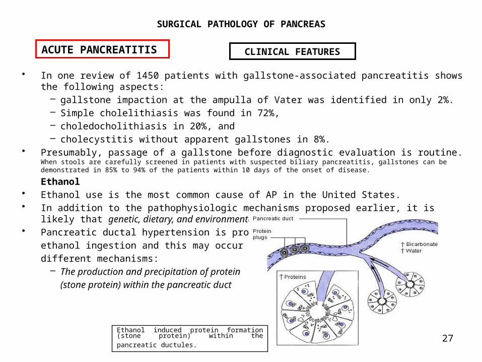
• Ethanol use is the most common cause of AP in the United States. • In addition to the pathophysiologic mechanisms proposed earlier, it is
likely that genetic, dietary, and environmental factors contribute.• Pancreatic ductal hypertension is probable after
ethanol ingestion and this may occur by several different mechanisms:
– The production and precipitation of protein (stone protein) within the pancreatic duct
ACUTE PANCREATITIS CLINICAL FEATURES
Ethanol induced protein formation (stone protein) within the pancreatic ductules.

28
SURGICAL PATHOLOGY OF PANCREAS
– Ethanol-induced increase in ampullary tension and therefore resistance
– Ethanol-induced gastric acid secretion increases pancreatic secretion indirectly through acid mediated secretin release.
• The combination of increased ampullary resistance and increased ductal flow results in pancreatic ductal hypertension and appears to enhance protease entry into the pancreatic interstitium. Others
• A wide variety of other clinical conditions may be associated with APPostprocedural Pancreatitis
• Many surgical procedures in the upper abdomen are associated with postoperative AP.
– After gastric resection the incidence of AP is between 0.6 -1.23%. – After biliary tract surgery, particularly after common bile duct exploration, AP occurs with an incidence of 0.5% to 3%.
– Direct manipulation or retraction of the pancreas or pancreatic duct appears to be the most common cause.
– After endoscopic retrograde cholangiopancreatography (ERCP), AP develops in about 1% of patients This is a predictable event, and the risk can be minimized by limiting the pressure used for contrast injection of the pancreatic duct.
– After coronary artery bypass surgery AP also occurs Although pancreatitis in this circumstance is thought to result from ischemia.
Abdominal Trauma • AP occurs in about 1% to 2% of patients with abdominal trauma, whether
blunt or penetrating. Contusion of the pancreatic parenchyma and pancreatic ductal injury both lead to extravasation of activated enzymes to initiate the process.
ACUTE PANCREATITIS CLINICAL FEATURES
Ethanol-induced increases in ampullary
resistance
29
SURGICAL PATHOLOGY OF PANCREAS
Hyperlipoproteinemia • Rare causes of AP are the hyperlipoproteinemias, types I and V. The initial
acinar cell injury is thought to result from the liberation of free fatty acids from circulating triglycerides by the local action of lipases within the pancreatic microcirculation. Physical disruption of the microvascular endothelium by cholesterol crystals may also occur. Hyperparathyroidism
• AP is reported in 1% to 19% of patients with hyperparathyroidism. It is unclear whether increased circulating plasma levels of calcium or parathyroid hormone are primarily responsible. Hypercalcemia may be associated with the precipitation of calcium phosphate within the pancreatic duct as well as with pancreatic hypersecretion. Parathyroid hormone elevated levels may have a direct cytotoxic effect on acinar cells. Drugs
• Drug-induced acute pancreatitis is relatively common and has been reported in association with many pharmacologic agents. The mechanisms involved are largely unknown. Infections
• The development of acute pancreatitis has been reported after a variety of bacterial, fungal, parasitic, and viral infections. Postulated mechanisms include direct cytotoxic effects, coexisting immunosuppression, and alterations in pancreatic blood flow. Vascular Disease
• Impaired pancreatic blood flow arising from either anatomic lesions or functional events can induce acute pancreatitis. IE: embolization of the pancreaticoduodenal artery after translumbar aortography, celiac artery stenosis, ruptured abdominal aortic aneurysm, and myocardial infarction. Immunologic Factors
• AP has been associated with systemic lupus erythematosus, rheumatoid arthritis, polyarteritis nodosa, and Be87het syndrome. Systemic vasculitis, the associated glucocorticoid use, and the presence of circulating antibodies to acinar cells are potentially related findings.
ACUTE PANCREATITIS CLINICAL FEATURES

30
SURGICAL PATHOLOGY OF PANCREAS
Obstruction of the Duodenum or the Pancreatic Duct • Intrapancreatic or duodenal tumors, parasitic obstruction of the pancreatic
duct, other obstructions of the duodenum, and pancreas divisum are also associated with the development of pancreatic duct hypertension and AP. Pregnancy
• Acute pancreatitis has been linked to pregnancy with an incidence of 0.01% to 0.1%. Because most reports include patients with gallstones, ethanol use, or other risk factors, it is unclear whether pregnancy is an independent risk factor.
Presentation • The cardinal clinical symptom for acute pancreatitis is epigastric pain that is
visceral in character and radiates into the back. • The degree of discomfort is variable. The pain may be similar to that of acute
peritonitis and may mimic a perforated viscus. At the other extreme, pain may be minimal. Other common signs and symptoms are nonspecific.
ACUTE PANCREATITIS CLINICAL FEATURES
COMMON SIGNS AND SYMPTOMS OF UNCOMPLICATED ACUTE EDEMATOUS PANCREATITIS Sign or Symptom Frequency (%)Abdominal pain 85–100Nausea and vomiting 54–92Anorexia 83Fever 12–80Abdominal mass 6–20Ileus 50–80

31
SURGICAL PATHOLOGY OF PANCREAS
• Clinical signs of complex disease or necrotizing pancreatitis include – jaundice – hypotension – retroperitoneal hemorrhage associated with pancreatic necrosis may become apparent as blood dissects into the subcutaneous tissues, producing blue discoloration of the flanks (Grey Turner sign), the umbilicus (Cullen sign), or the inguinal ligament (Fox sign). None of these signs is common.
• The clinical course of patients with acute pancreatitis is exceedingly variable.
– The spectrum includes patients with minimal symptoms, few signs, and only mild hyperamylasemia who require little or no treatment and have few, if any, complications.
– The opposite end of the spectrum, without apparent differentiating risk factors, is fulminant pancreatic necrosis with hemorrhage and progressive, inexorable multisystem organ failure and death.
• Several grading systems have been developed, to estimate risks and outcomes based on the presenting clinical features, and the Ranson criteria are widely adopted.
• A patient with three or more of these findings has a mortality rate of about 30%. • Pancreatitis-associated morbidity and mortality are directly correlated
with the number of these criteria present. • The mortality rate is 28% if three criteria are met, 40% for five or six, and 100% for seven or eight. • The APACHE II (Acute Physiology Score and Chronic Health Evaluation)
scoring system for critically ill patients has also been applied. • Although not specific for acute pancreatitis, the APACHE score has some
utility by predicting mortality rates in critically ill patients.
ACUTE PANCREATITIS CLINICAL FEATURES

32
SURGICAL PATHOLOGY OF PANCREAS
On Admission • Age above 55 years • White blood cell count above 16,000/E6L • Glucose level above 200 mg/dL • Lactase dehydrogenase level above 350 IU/L • Serum glutamic-oxaloacetic transaminase value
above 250 IU/L
After 48 Hours • Hematocrit decrease of 10% • Blood urea nitrogen level increase of 5 mg/dL • Ca2+ level below 8 mg/dL • PaO2 level below 60 mmHg • Base deficit value above 4 mEq/L • Fluid sequestration greater than 6 L
ACUTE PANCREATITIS CLINICAL FEATURES
RANSON GRAVE PROGNOSTIC SIGNS ASSOCIATED WITH ACUTE PANCREATITIS

33
SURGICAL PATHOLOGY OF PANCREAS
DIAGNOSIS • The diagnosis of AP ultimately depends on a clinical judgment, based on the
finding of epigastric abdominal pain and tenderness with the laboratory finding of hyperamylasemia.
• No single laboratory or physical finding is pathognomonic. Imaging
• Plain abdominal roentgenograms may show :– A diffuse ileus and – A solitary left upper abdominal sentinel loop are classic and are often seen on,
– The psoas muscle margins may be obscured by retroperitoneal edema; – Pancreatic ascites may be apparent; – Pancreatic calcifications imply preexisting chronic disease.
• Chest radiographs abnormalities are present at one third of patients with AP at the time of diagnosis.
– Segmental atelectasis, – An elevated hemidiaphragm, – Pleural effusions, or the presence of – Early pulmonary parenchymal infiltrates.
• Barium studies of the gastrointestinal tract An upper gastrointestinal contrast study often demonstrates
– narrowing or spasm of the duodenum, with – widening of the C loop secondary to pancreatic inflammation and edema formation.
• Ultrasound scanning provides a readily available, rapid, and noninvasive imaging of the pancreas.
– In the case of simple acute pancreatitis, an enlarged, echolucent gland is typically seen.
– Ultrasound examination yields information regarding cholelithiasis, choledocholithiasis, and the status of the intrahepatic and extrahepatic biliary ducts.
– The technique is also valuable for assessing and sequentially evaluating peripancreatic fluid collections or pancreatic pseudocysts.
ACUTE PANCREATITIS CLINICAL FEATURES

34
SURGICAL PATHOLOGY OF PANCREAS
• Computed tomography (CT) has similar capabilities to ultrasound, although sensitivity for detecting cholelithiasis is lower. Intraluminal contrast in the duodenum and small bowel is necessary to optimize CT imaging. Dynamic CT scanning with simultaneous intravenous contrast enhancement can give valuable information in evaluating regional pancreatic perfusion and may provide an estimate of the extent of pancreatic necrosis. CT may show gas bubbles in the pancreatic necrosis or peri pancreatic fluid collections assessing the infection though the indication for surgical approach
• Needle aspiration either CT- or ultrasound-guided, allows access to peri pancreatic or pancreatic fluid collections for diagnostic sampling and/or therapeutic drainage. Cultures may be obtained by this route. Contrast injections can assess the relation of a pseudo cyst or abscess cavity to the pancreatic duct. This latter information is important for any preoperative planning. Placement of therapeutic drainage catheters is routinely combined with CT- and ultrasound-directed imaging procedures.
• ERCP The role in the diagnostic evaluation of the patient with AP is severely limited because of the high risk of exacerbating existing inflammation. ERCP is generally reserved for the evaluation of a patient with a suspected obstructive lesion and is timed to follow resolution of the acute phase of the illness. In addition, it is appropriate to obtain ERCP in a patient with idiopathic acute pancreatitis after a first recurrence of the disease. This strategy is designed to identify promptly anatomically correctable causes of acute pancreatitis. ERCP is also useful to delineate the pancreatic duct after injury, pseudo cyst drainage, or the development of pancreatic ascites. Lastly, ERCP combined with urgent therapeutic sphincterotomy and gallstone extraction has been used for patients with impacted ampullary gallstones.
ACUTE PANCREATITIS CLINICAL FEATURES

35
SURGICAL PATHOLOGY OF PANCREAS
Biochemical Markers • Amylase. Amylase is released from the acinar cell into the pancreatic
microcirculation in conjunction with the pathophysiologic events described earlier.
• The laboratory finding of hyperamylasemia in a patient with clinical signs and symptoms of AP is the usual means of confirming the diagnosis of AP.
• Efforts to correlate the degree of hyperamylasemia with disease severity or prognosis have been consistently unsuccessful, and Ranson criteria are notable for the absence of serum amylase levels. An important reason for this relates to:
– the relatively rapid clearance of amylase from plasma, – the half-life being about 130 minutes. – Pancreatitis resulting from a discrete event such as transient pancreatic duct obstruction with gallstone passage is characterized by a single serum amylase peak with a rapid rise and prompt clearance, both measured in terms of hours.
• A normal or minimally elevated serum amylase level may also be found in a patient with necrotizing pancreatitis or with CP; in these instances, complete or nearly complete destruction of the acinar cell population may have occurred, reducing the plasma amylase level.
• Additionally, a number of nonpancreatic sources of amylase exist, so that hyperamylasemia may result from other pathology:
– Salivary glands, – fallopian tubes, and – the small bowel are important alternative amylase sources.
ACUTE PANCREATITIS CLINICAL FEATURES

36
SURGICAL PATHOLOGY OF PANCREAS
Clinical conditions associated with hyperamylasemia include the following:
ACUTE PANCREATITIS CLINICAL FEATURES
•Salivary gland injury
•Burns •Cerebral trauma •Multiple trauma •Diabetic
ketoacidosis •Macroamylasemia •Renal
transplantation •Renal dysfunction •Pneumonia •Pregnancy •Fallopian tube
pathology
• Drugs • Afferent loop syndrome • Acute appendicitis • Dissecting aortic aneurysm
• Small bowel injury • Perforated ulcer • Small bowel obstruction • Mesenteric infarction

37
SURGICAL PATHOLOGY OF PANCREAS
• In the case of salivary gland disease, plasma amylase isoenzyme determinations differentiate the source. An accurate and more rapid amylase assay using a monoclonal antibody specific to salivary isoamylase has been described.
• Gastrointestinal tract pathology other than pancreatitis may lead to increased amylase absorption through the intestine or peritoneum and relatively mild elevations of pancreatic amylase in the circulation.
• Lipase. Lipase is derived primarily from pancreatic acinar cells, and its elevation is also taken as evidence of acinar cell injury in acute pancreatitis. Lipase is also nonspecific and has not proved more useful than serum amylase determinations in clinical use.
• Other Serum Enzymes. Other acinar cell products, such as immunoreactive trypsin, chymotrypsin, elastase, ribonuclease, and phospholipase A2, may be detectable in plasma after the onset of acute pancreatitis. Measurement techniques are not in wide clinical use, but institutional enthusiasm for one particular assay or another has led to an abundant literature.
• Ribonuclease and phospholipase A2 plasma elevations may correlate with more complex disease.
• Methemalbumin. Methemalbumin results from the proteolytic conversion of hemoglobin into oxidized hematin that is conjugated with plasma albumin. Acinar cell protease release into the circulation increases red blood cell exposure to proteases and accelerates this process, so that methemalbumin plasma levels may be elevated with acute pancreatitis. A correlation between levels of methemalbumin and severity of pancreatic disease is proposed but unproved.
• Other Serum Abnormalities. Other characteristic but nonspecific biochemical features commonly associated with acute pancreatitis are summarized in the following Table These may have both therapeutic and diagnostic relevance.
ACUTE PANCREATITIS CLINICAL FEATURES

38
SURGICAL PATHOLOGY OF PANCREAS
Other characteristic but nonspecific biochemical features commonly associated with acute pancreatitis
ACUTE PANCREATITIS CLINICAL FEATURES
INCREASEDHematocrit, hemoglobin (hemoconcentration)White blood cell countBlood urea nitrogenCreatinineBilirubinLipid, triglyceride levelsGlucoseAlkaline phosphataseSGOT, SGPT DECREASEDHematocrit, hemoglobin (hemorrhage)CalciumMagnesiumPao2 OTHERRespiratory alkalosis (early)Metabolic alkalosis (early)Consumptive coagulopathyMetabolic acidosis (late)Respiratory acidosis (late)

39
SURGICAL PATHOLOGY OF PANCREAS
DEFINITION• Acute pancreatitis is a complex disorder of the exocrine pancreas characterized by acute acinar
cell injury and both regional and systemic inflammatory responses. • It is a common disease with a broad spectrum of clinical and pathologic findings that
contribute to considerable morbidity and mortality. • Because pathogenic mechanisms are unclear, specific treatment is not available.
Empiric supportive care remains standard, and clinical outcomes have improved only to the extent that critical care has evolved in recent years.Most patients with acute pancreatitis have simple edematous pancreatitis, a self-limited and reversible process. In a small number of patients, fulminant or progressive disease develops, with pancreatic necrosis that can lead to multiorgan system failure or death.
ACUTE PANCREATITIS
The problems related to acute pancreatitis pose a formidable challenge to both
the clinical surgeon and the basic scientist.

40
SURGICAL PATHOLOGY OF PANCREAS
MANAGEMENT • Neither medical nor surgical treatment strategies provide specific therapy for the acinar cell injury
characteristic of acute pancreatitis. • Modern approaches provide general supportive care in the form of:
– appropriate resuscitation, – nutrition, and – ventilation
in the expectation that the cellular pathophysiologic processes will be self-limited. Medical Treatment
• Conventional medical therapy for AP consists fundamentally of: – intravenous fluid resuscitation, – nasogastric decompression, and – monitoring of hematocrit, electrolytes, and blood gases.
• All are empiric and are not known to shorten or favorably alter the course of the disease. • Regional retroperitoneal inflammation and the systemic microvascular injury contribute to the loss
of intravascular plasma volume. • Hypovolemia may be mild or profound to the point of shock. • A relation exists between the magnitude of extravascular fluid
sequestration and the severity of the pancreatitis. • This is recognized by inclusion of an estimated plasma volume deficit exceeding 6 L
in the Ranson scoring system (see earlier list) as a grave prognostic sign.• More than half of patients with acute pancreatitis have clinical evidence
of inadequate end-organ perfusion. • Resuscitation requires the intravenous administration of large volumes of isotonic crystalloid
solution and the aggressive use of invasive hemodynamic monitoring devices.
ACUTE PANCREATITIS MANAGEMENT

41
SURGICAL PATHOLOGY OF PANCREAS
Pulmonary dysfunction occurs in about two thirds of patients with AP. • More than 70% of patients have evidence of transient hypoxemia, defined as
a single PaO2 of less than 70 mmHg. • Progression to acute respiratory failure requiring endotracheal intubation and
mechanical ventilation carries a mortality rate of up to 75% but is a relatively uncommon event (about 5% of all cases).
• Monitoring of pulmonary gas exchange with periodic arterial blood gases is standard in all patients with AP.
Renal dysfunction is demonstrable in 40% to 80% of patients with acute pancreatitis. Therefore, renal function must be sequentially evaluated biochemically, and the urine output must be carefully monitored during the acute resuscitative phase of the illness.
• Early recognition of oliguria or azotemia allows correction. • Hypovolemia is almost invariably the cause of the renal dysfunction and is
therefore simply treated if recognized. • The reported incidence of acute renal failure secondary to acute
pancreatitis ranges from 2% to 20%. If required, dialysis, hemofiltration, and similar supportive measures are all associated with substantially increased mortality rates (up to 80%).
Patients with AP share many hemodynamic and metabolic characteristics with SEPTIC patients. – Glucose production is exaggerated, – Insulin resistance increased – Pancreatic endocrine insufficiency may develop, – Protein catabolism is marked.
ACUTE PANCREATITIS MANAGEMENT
42
SURGICAL PATHOLOGY OF PANCREAS
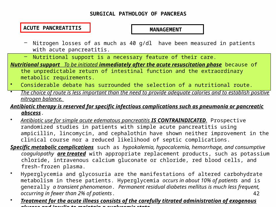
– Nitrogen losses of as much as 40 g/dl have been measured in patients with acute pancreatitis.
– Nutritional support is a necessary feature of their care. Nutritional support To be initiated immediately after the acute resuscitation phase because of
the unpredictable return of intestinal function and the extraordinary metabolic requirements.
• Considerable debate has surrounded the selection of a nutritional route. • The choice of route is less important than the need to provide adequate calories and to establish positive
nitrogen balance. Antibiotic therapy is reserved for specific infectious complications such as pneumonia or pancreatic
abscess. • Antibiotic use for simple acute edematous pancreatitis IS CONTRAINDICATED. Prospective
randomized studies in patients with simple acute pancreatitis using ampicillin, lincomycin, and cephalothin have shown neither improvement in the clinical course nor a reduced likelihood of septic complications.
Specific metabolic complications such as hypokalemia, hypocalcemia, hemorrhage, and consumptive coagulopathy are treated with appropriate replacement products, such as potassium chloride, intravenous calcium gluconate or chloride, red blood cells, and fresh-frozen plasma.
• Hyperglycemia and glycosuria are the manifestations of altered carbohydrate metabolism in these patients. Hyperglycemia occurs in about 10% of patients and is generally a transient phenomenon. Permanent residual diabetes mellitus is much less frequent, occurring in fewer than 2% of patients.
• Treatment for the acute illness consists of the carefully titrated administration of exogenous glucose and insulin to maintain a euglycemic state.
ACUTE PANCREATITIS MANAGEMENT

43
SURGICAL PATHOLOGY OF PANCREAS
Encephalopathy Clinical evidence of encephalopathy is discernible in 4% to 20% of patients with AP. • Symptoms may include:
– disorientation, – confusion, – delirium, – delusions, or – hallucinations. – transient acute psychosis is also reported.
Because ethanol ingestion also produce all these symptoms, it is often difficult to distinguish the underlying cause.
• Cerebral edema, hemorrhage, and focal necrosis, presumably secondary to microvascular blood flow alterations, have all been associated with AP.
• Treatment is nonspecific and supportive. Disorders of the coagulation system, particularly microvascular thrombosis and disseminated
intravascular coagulopathy, are common in acute pancreatitis. • At later times (6 or 7 days), clinical studies have shown hyperfibrinogenemia
and increased platelet counts, suggesting enhanced thrombotic potential. • Heparin(low-molecular-weight), dextran, and fibrinolytic therapy have been shown to
prevent microvascular thrombosis in experimental acute pancreatitis in dogs. In this favorable and controlled setting, these agents prevent the development of acute hemorrhagic pancreatitis.
Hypocalcemia is relatively common (3% to 30%) in AP. • The magnitude of the hypocalcemia correlates with the degree of illness. • It appears that large quantities of calcium are bound in the tissues during the process of
peripancreatic fat saponification. Other factors, such as changes in plasma levels of parathyroid hormone, glucagon, and calcitonin, may also contribute. Unattended, the hypocalcemia may progress to tetany. Periodic monitoring of serum calcium is standard practice during the acute illness. Treatment consists of intravenous replacement to maintain the serum calcium level within a normal range.
ACUTE PANCREATITIS MANAGEMENT

44
SURGICAL PATHOLOGY OF PANCREAS
A variety of pharmacologic agents that directly or indirectly reduce acinar cell enzyme release or ductal secretion have undergone clinical evaluation for the treatment of acute pancreatitis, generally with unimpressive results.
• Among the first were anticholinergic drugs, pancreatic anti enzymes, a somatostatin analogue.
• Despite the theoretic appeal, it has not been possible to demonstrate that pancreatic anti enzymes and somatostatin alters the natural history or prognosis of simple acute pancreatitis, although it diminishes pancreatic secretion.
Surgical Therapy Surgical therapy for AP is reserved for specific complications and for those situations in which a correctable anatomic cause can be identified.
Endoscopic sphincterotomy is usually the initial therapeutic procedure for relief of biliary ductal obstruction when acute pancreatitis occurs in association with choledocholithiasis.
• The procedure reliably decompresses the ampulla of Vater, with overall clinical success for gallstone disimpaction or passage in 90% of patients.
• ERCP has greatly reduced the need for operative procedures designed to either divert or open and explore the common bile duct.
• In the more usual circumstance, a patient who has simple cholelithiasis and an episode of acute pancreatitis is treated nonoperatively(ERCP) with resolution of the AP but gallbladder litiasis must be operated. Cholecystectomy is often performed after the resolution of acute pancreatitis but before hospital discharge.
ACUTE PANCREATITIS MANAGEMENT
45
SURGICAL PATHOLOGY OF PANCREAS
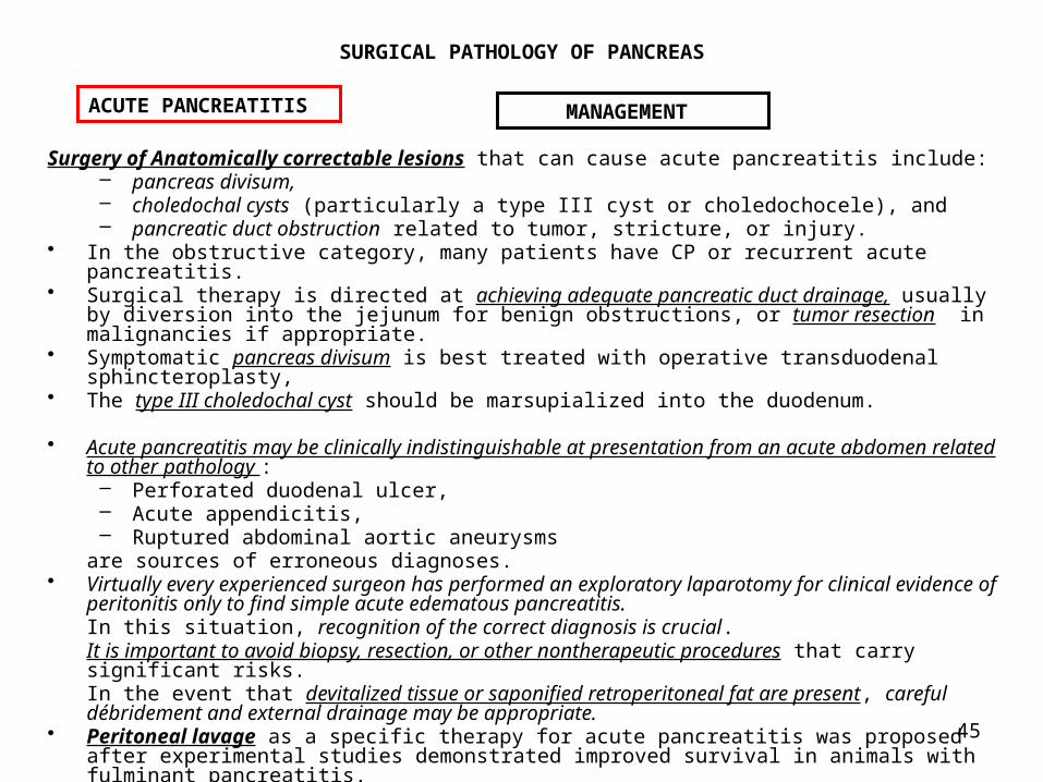
Surgery of Anatomically correctable lesions that can cause acute pancreatitis include: – pancreas divisum, – choledochal cysts (particularly a type III cyst or choledochocele), and – pancreatic duct obstruction related to tumor, stricture, or injury.
• In the obstructive category, many patients have CP or recurrent acute pancreatitis.
• Surgical therapy is directed at achieving adequate pancreatic duct drainage, usually by diversion into the jejunum for benign obstructions, or tumor resection in malignancies if appropriate.
• Symptomatic pancreas divisum is best treated with operative transduodenal sphincteroplasty,
• The type III choledochal cyst should be marsupialized into the duodenum.
• Acute pancreatitis may be clinically indistinguishable at presentation from an acute abdomen related to other pathology :– Perforated duodenal ulcer, – Acute appendicitis, – Ruptured abdominal aortic aneurysms
are sources of erroneous diagnoses. • Virtually every experienced surgeon has performed an exploratory laparotomy for clinical evidence of
peritonitis only to find simple acute edematous pancreatitis. In this situation, recognition of the correct diagnosis is crucial.It is important to avoid biopsy, resection, or other nontherapeutic procedures that carry significant risks. In the event that devitalized tissue or saponified retroperitoneal fat are present, careful débridement and external drainage may be appropriate.
• Peritoneal lavage as a specific therapy for acute pancreatitis was proposed after experimental studies demonstrated improved survival in animals with fulminant pancreatitis.
ACUTE PANCREATITIS MANAGEMENT

46
SURGICAL PATHOLOGY OF PANCREAS
• The concept was appealing in that activated proteases and other vasoactive substances identifiable in peritoneal aspirates from patients with pancreatitis would be removed, rather than systemically absorbed.
• Unfortunately, clinical trials using this approach have produced disappointing results, and the eventual overall mortality rate appears unchanged.
• Surgical approach for patients with necrotizing pancreatitis. • When pancreatitis does not resolve spontaneously, diagnostic efforts are directed
at distinguishing infected and noninfected areas of pancreatic necrosis. • The presence of an infected sequestrum mandates operative exploration to
débride devitalized tissue and to provide external drainage.• Specific antibiotic coverage is essential and should be dictated by intraoperative
cultures or aspirates from the necrotic tissue. • Débridement is often required on multiple occasions, usually at 24- to 48-hour intervals, until
the necrotic tissue is replaced by a granulating wound. Many strategies related to multiple operations with open and closed peritoneal drainage systems have been devised.
• This is often a difficult judgment and is necessarily individualized for each patient.
ACUTE PANCREATITIS MANAGEMENT

47
SURGICAL PATHOLOGY OF PANCREAS
Pancreatic Pseudocysts • A pancreatic pseudocyst is a fluid-filled cystic structure without a true epithelial lining that is
associated with the pancreas or pancreatic duct
CT scan showing pancreatic pseudocyst.
ACUTE PANCREATITIS Complications of AP Pancreatic Pseudocysts

48
SURGICAL PATHOLOGY OF PANCREAS
• True cysts of the pancreas (epithelium-lined) are rare, whereas pseudocysts are relatively common.
• Pancreatic pseudocysts account for 2% to 10% of patients with pancreatic disease.
• The most common cause of pancreatic pseudocyst in the United States is ethanol-related CP. Biliary and posttraumatic pancreatitis follow in frequency with regard to cause.
• The pseudocyst wall is composed of displaced adjacent viscera (often stomach, small bowel, or colon) and a fibrous capsule that has evidence of both acute and chronic inflammation.
• The thickness of this fibrous capsule is variable, depending on how long the pseudocyst has been present.
• The presence of a fibrous capsule becomes an important consideration in the timing and selection of drainage procedures.
• The fluid within the cyst cavity is usually serous in character and contains pancreatic secretions, including amylase and proteases, as well as albumin and inflammatory cells. The amylase content may be high; it is not unusual to see a level of several thousand international units in pseudocyst aspirates. If recent hemorrhage has occurred, bile pigment may also be present, and bacteria are cultured in about 35% of pseudocysts.The clinical presentation of a pancreatic pseudocyst is usually that of: – Persistent visceral pain – Ileus after an episode of acute pancreatitis. – Fever, – Leukocytosis, – Palpable epigastric mass are common, – Nausea and vomiting – Jaundice suggests common bile duct obstruction from either intrinsic
stones or extrinsic distortion.
ACUTE PANCREATITIS Complications of AP Pancreatic Pseudocysts
49
SURGICAL PATHOLOGY OF PANCREAS
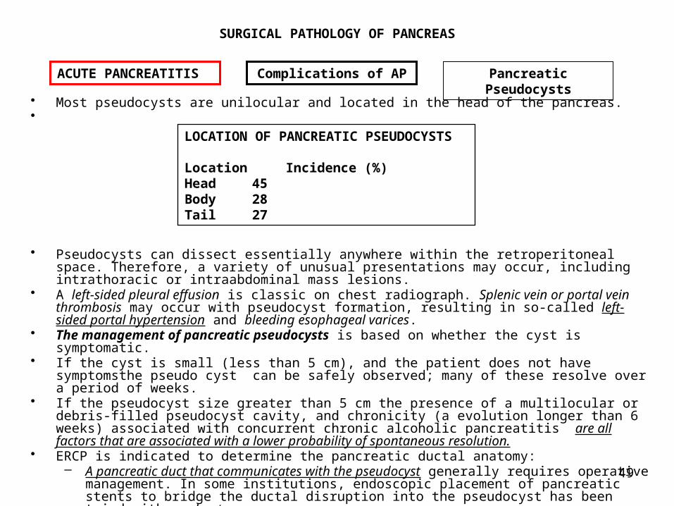
• Most pseudocysts are unilocular and located in the head of the pancreas.•
• Pseudocysts can dissect essentially anywhere within the retroperitoneal space. Therefore, a variety of unusual presentations may occur, including intrathoracic or intraabdominal mass lesions.
• A left-sided pleural effusion is classic on chest radiograph. Splenic vein or portal vein thrombosis may occur with pseudocyst formation, resulting in so-called left-sided portal hypertension and bleeding esophageal varices.
• The management of pancreatic pseudocysts is based on whether the cyst is symptomatic.
• If the cyst is small (less than 5 cm), and the patient does not have symptomsthe pseudo cyst can be safely observed; many of these resolve over a period of weeks.
• If the pseudocyst size greater than 5 cm the presence of a multilocular or debris-filled pseudocyst cavity, and chronicity (a evolution longer than 6 weeks) associated with concurrent chronic alcoholic pancreatitis are all factors that are associated with a lower probability of spontaneous resolution.
• ERCP is indicated to determine the pancreatic ductal anatomy: – A pancreatic duct that communicates with the pseudocyst generally requires operative
management. In some institutions, endoscopic placement of pancreatic stents to bridge the ductal disruption into the pseudocyst has been tried with moderate success.
LOCATION OF PANCREATIC PSEUDOCYSTS Location Incidence (%)Head 45Body 28Tail 27
ACUTE PANCREATITIS Complications of AP Pancreatic Pseudocysts
50
SURGICAL PATHOLOGY OF PANCREAS
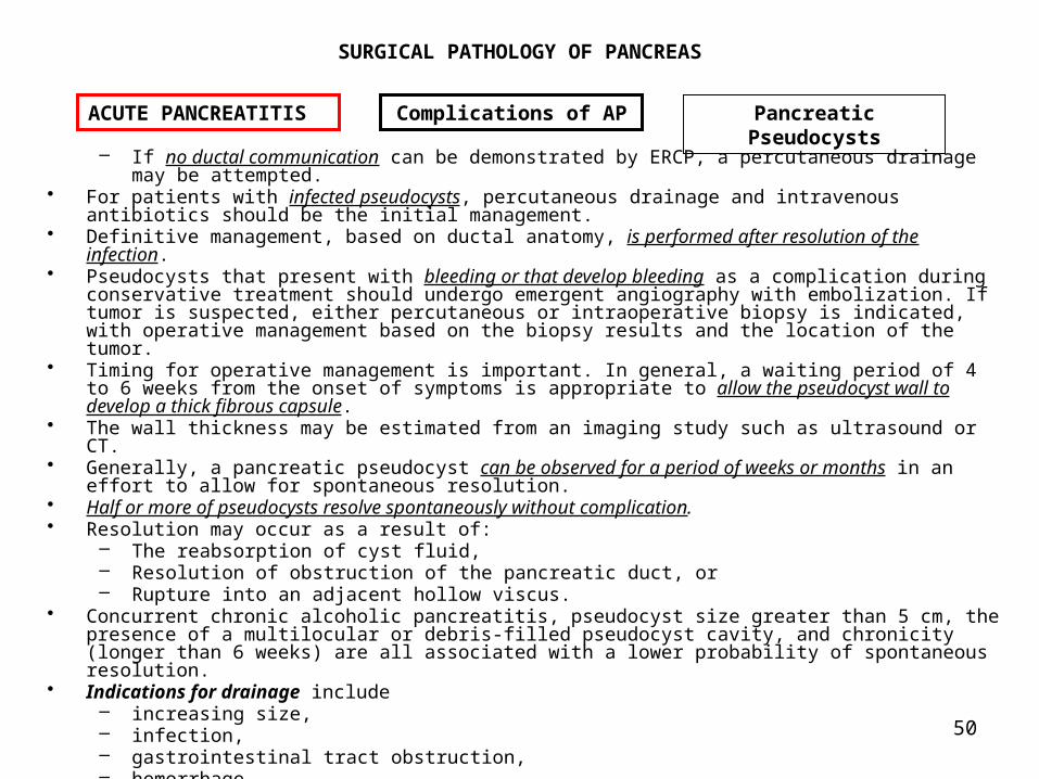
– If no ductal communication can be demonstrated by ERCP, a percutaneous drainage may be attempted.
• For patients with infected pseudocysts, percutaneous drainage and intravenous antibiotics should be the initial management.
• Definitive management, based on ductal anatomy, is performed after resolution of the infection.
• Pseudocysts that present with bleeding or that develop bleeding as a complication during conservative treatment should undergo emergent angiography with embolization. If tumor is suspected, either percutaneous or intraoperative biopsy is indicated, with operative management based on the biopsy results and the location of the tumor.
• Timing for operative management is important. In general, a waiting period of 4 to 6 weeks from the onset of symptoms is appropriate to allow the pseudocyst wall to develop a thick fibrous capsule.
• The wall thickness may be estimated from an imaging study such as ultrasound or CT.
• Generally, a pancreatic pseudocyst can be observed for a period of weeks or months in an effort to allow for spontaneous resolution.
• Half or more of pseudocysts resolve spontaneously without complication. • Resolution may occur as a result of:
– The reabsorption of cyst fluid, – Resolution of obstruction of the pancreatic duct, or – Rupture into an adjacent hollow viscus.
• Concurrent chronic alcoholic pancreatitis, pseudocyst size greater than 5 cm, the presence of a multilocular or debris-filled pseudocyst cavity, and chronicity (longer than 6 weeks) are all associated with a lower probability of spontaneous resolution.
• Indications for drainage include – increasing size, – infection, – gastrointestinal tract obstruction, – hemorrhage, – spontaneous rupture, and – failure to resolve.
ACUTE PANCREATITIS Complications of AP Pancreatic Pseudocysts

51
SURGICAL PATHOLOGY OF PANCREAS
• Percutaneous ultrasound or CT-directed aspiration or drainage catheter placement is an initial treatment option for these lesions, but in the following conditions:– If the initial aspirate is sterile Simple aspiration is performed;– If the aspirate is infected, a drainage-catheter or a open drainage
procedure is appropriate.• Determination of pancreatic ductal anatomy is important whenever possible.
Therefore, contrast injection into the pseudocyst at the time of aspiration should be considered to assess the possibility of pancreatic ductal communication and obstruction, or multiple cysts.
• The pseudocyst recurrence rate after simple aspiration is about 20% to 25%. Open surgical approaches: Drainages or Pancreatic ResectionsOpen surgical drainage is usually reached when a complication develops or if the pseudocyst recurre after repeated aspirations.
• The decision of an open surgical drainage depends on: – Size of the pseudocyst, – Location of the pseudocyst,, – The underlying cause of the cyst– Pseudocyst wall. Generally, a waiting period of at least 6 weeks is needed for the pseudocyst to
resolve or for the wall to develop a thick fibrous capsule. If it is impossible to judge the date of pseudocyst formation, 6 weeks from the time of initial diagnosis is generally chosen; the wall thickness may be estimated from imaging studies such as ultrasound or CT.
– The status of the pancreatic duct which should be assessed preoperatively, preferably by ERCP. Knowledge of the anatomy of the pancreatic duct allows the design of an appropriate drainage plan.
• Operative drainage can be either external or internal. – External drainage is chosen in the presence of infection or an immature
capsule. The disadvantages of external drainage include the risk of pancreatic fistula formation and a pseudocyst recurrence. External drainage has been associated with a higher mortality rate, probably because it is used in patients at higher risk, especially those with sepsis, pancreatic abscesses, or ruptured pseudocysts.
ACUTE PANCREATITIS Complications of AP Pancreatic Pseudocysts

52
SURGICAL PATHOLOGY OF PANCREAS
• The internal drainage procedure selected depends on the location of the pseudocyst and whether there is associated pancreatic ductal pathology.
• Cystogastrostomy is the simplest and safest alternative if the pseudocyst is appropriately adjacent to the posterior wall of the stomach.
• Cystoduodenostomy, using a transduodenal approach• Cystojejunostomy using a Roux-en-Y or loop jejunostomy may also be
appropriate, depending on the location and specific anatomy of the pseudocyst. Pancreatic resection is associated with the lowest recurrence rate (3%), but is limited to pseudocysts occurring in the tail of the pancreas.
ACUTE PANCREATITIS Complications of AP Pancreatic Pseudocysts
Cystojejunostomy Roux en Y
Roux en Y technique for multiple pancreatic
pseudocysts.

53
SURGICAL PATHOLOGY OF PANCREAS
Pancreatic Abscess • The common causes of pancreatic abscess are:
– an infected pancreatic pseudocyst and – necrotizing pancreatitis.
• The diagnosis is suggested by – Persistent fever, – Leukocytosis, – A palpable abdominal mass. – Bacteremia and systemic toxicity are late clinical features. – On imaging, Ultrasounds, and/or CT scans: debris within a cyst is more suggestive of
an abscess; – Ultrasound-guided needle aspiration of suspicious peripancreatic fluid collections.
– Percutaneous aspiration with positive cultures is the definitive preoperative test.
• The treatment of choice is wide surgical debridement with removal of all infected and devitalized tissues. Generous drainage is mandatory.
• Whether to leave the abdomen open and the choice of drainage systems remain controversial. – Advocates of closed drainage (ie, placement of large, dependent drains and abdominal closure) report mortality rates of about 30%.
– Use of open drainage or marsupialization with frequent dressing changes has a reported mortality rate of 10% to 15%.
• The important principles are: – to employ aggressive (often sequential) débridement, – appropriate antibiotics, and – effective external drainage.
ACUTE PANCREATITIS Complications of AP Pancreatic Abscess

54
SURGICAL PATHOLOGY OF PANCREAS
• If the pancreatic abscess is correlated with the Ranson criteria: – if three risk factors are present the mortality rate is 14%, – if five are present 100% mortality rate.
ACUTE PANCREATITIS Complications of AP Pancreatic Abscesses

55
SURGICAL PATHOLOGY OF PANCREAS
DefinitionCP is a disease characterized by progressive and permanent destruction of the pancreatic exocrine parenchyma associated with fibrosis of the gland.
• In acute pancreatitis the lesions such as edema, hemorrhage, and fat necrosis, may regress completely when the underlying cause is eliminated.
• Although both acute and CP may be categorized into relapsing and nonrelapsing forms depending on their clinical presentation,
• Progressive morphologic and functional derangement is demonstrated only by CP. • In CP, fibrotic destruction of the exocrine gland is often also accompanied by endocrine
dysfunction.
Classification• The classification of pancreatitis is usually reduced to include only acute
and chronic disease. • Within CP, calcifying CP and obstructive CP may be distinguished, with important
functional and therapeutic implications.
Incidence• The incidence and prevalence of CP, is not known with precision. • In the United States and Western Europe, the incidence of new cases
approximates 5 to 10 per 100,000 population per year, with a prevalence of about 25 cases per 100,000 inhabitants.
• Because alcohol consumption is the most important risk factor for the development of CP, countries with low alcohol consumption rates generally have lower incidence rates of CP. Correlation of alcohol intake with incidence of CP within various populations has often been discrepant, however, suggesting that environmental or hereditary factors may also influence susceptibility to the disease.
Cronic Pancreatitis General Aspects

56
SURGICAL PATHOLOGY OF PANCREAS
Alcohol Consumption • Alcohol consumption is the major cause of CP, with about 70% of cases
attributable to this factor. • Most patients have consumed large volumes of alcohol for long periods of time. • The average daily intake of alcohol was 150 to 175 g, and the risk for
development of CP increased with increasing alcohol intake. • The mean duration of alcoholism before recognition of CP was 18 years for men and
11 years for women. • Because only 10% of alcoholics develop CP, however, factors other than long-
term alcohol exposure may influence susceptibility. • Diet may be important in this regard, the risk of alcohol-induced CP is increased by
high-protein, high-fat diets. Heredity
• The hereditary form of CP is transmitted as an autosomal dominant trait of incomplete penetrance.
• Affected patients usually become symptomatic in childhood at an average age of 10 to 12 years.
• The clinical and histologic features of hereditary pancreatitis differ little from nonhereditary forms of the disease.
• The diagnosis may be made if several members of a family develop CP in the absence of alcohol consumption or other known causes. Hyperparathyroidism
• Calcifying CP may occur in the presence of long-standing untreated hyperparathyroidism.
• Hyperparathyroidism is detected and treated at an early stage in Western countries,so the incidence of associated CP is decreasing, currently accounting for not more than 1% to 2% of cases.
• The pathogenesis of CP in hyperparathyroidism is presumed to be due to injury caused by:
– The acute elevation of serum calcium which is a potent secretagogue for human pancreatic enzymes.
– Intraductal precipitation of calcium in pancreatic secretions
Cronic Pancreatitis Etiology

57
SURGICAL PATHOLOGY OF PANCREAS
Tropical Pancreatitis • Tropical CP is a nutritional disease of importance in tropical Africa and
Southeast Asia. The disease develops among juveniles and young adults in the setting of chronic malnutrition. Protein-calorie malnutrition and deficiencies of copper, zinc, and selenium have been associated with the disease. The precise cause remains elusive. Duct Obstruction
• Obstruction of the main pancreatic duct can cause a distinctive form of chronic pancreatic disease known as obstructive pancreatitis. Occlusion may be caused by: – tumors, – congenital anomalies, – scars from prior injury or – inflammatory disease, or fibrosis of the ampulla of Vater.
• Although the gross alterations of the pancreas observed in obstructive pancreatitis are similar to those of other forms of CP, microscopic changes are different : – Obstruction causes diffuse atrophy of the exocrine tissue, whereas patchy
atrophy is more common in early forms of nonobstructive CP. – Alternating areas of ductal stenosis and dilation are not seen; instead, the
ductal system behind the obstruction is uniformly dilated. – The ductal epithelium is preserved, and protein plugs and calcifications are
unusual. – The relief of obstruction can be followed by reversal of parenchymal fibrosis
and atrophy. A restitution of pancreatic structure and function is not observed in other forms of CP.
• Obstructive CP is unusual, accounting for not more than 5% of cases. Idiopathic Causes
• The most common form of nonalcoholic calcifying pancreatitis is idiopathic, a designation given to those cases with an unrecognized cause. Idiopathic pancreatitis accounts for about 15% of cases.
• Idiopathic pancreatitis has two peaks in incidence, suggesting that differing underlying causes may exist: – The first peak occurs in young adulthood; – The second type, termed senile pancreatitis, has a peak occurrence at 60 years of
age.
Cronic Pancreatitis General Aspects

58
SURGICAL PATHOLOGY OF PANCREAS
• The mechanisms by which alcohol consumption causes CP are unknown. • Investigations have focused on:
– direct toxic effects of alcohol on pancreatic exocrine cells, – the effects of chronic alcohol intake on pancreatic protein secretion, and
– the role of calcium ions secreted into pancreatic juice. • Most investigators believe that the initial lesion in CP is acinar cell
injury. • A characteristic feature of early alcoholic CP is: a patchy distribution of normal
acinar tissues in the midst of abnormal lobules. • Microscopic examination shows:
– irregular dilation of ductules, – loss of ductal epithelium and acinar tissue, – localized obstruction, and formation of small intraparenchymal cysts. – Typically, precipitates of proteinaceous material are observed in intercalated and canalicular ducts. As the disease
progresses, these protein plugs cause increasing ductal obstruction. – Infiltration of the interstitium by inflammatory cells is followed by the deposition of fibrous tissue within and
between lobules. – In advanced stages, exocrine tissue is replaced by fibrosis. – Endocrine islets survive in isolated nests in the midst of broad areas of scar tissue.
• Increasing evidence indicates that abnormalities in pancreatic protein and calcium secretion may influence the development of CP.
• In humans, chronic alcohol consumption is associated with a marked increase in total protein concentration in pancreatic secretions. The increased concentration of secretory enzymes is probably due to increased biosynthesis within acinar cells.
• In humans with CP, abnormal proteins are detected in pancreatic secretions. Lactoferrin, not normally found in pancreatic secretions, may constitute up to 0.3% of total protein. This anionic molecule strongly associates with acidic molecules to form intraductal protein precipitates, postulated to be the basis for formation of pancreatic concretions.
Cronic Pancreatitis Pathogenesis

59
SURGICAL PATHOLOGY OF PANCREAS
• A novel protein has been identified in the pancreatic secretions of normal controls and patients with CP.
• The association of this protein with pancreatic calculi led to its original designation as pancreatic stone protein.
• Pancreatic stone protein is now referred to as lithostathine. This phosphoglycoprotein has a molecular weight of 14,000. It has been localized immunohistochemically to zymogen granules in acinar cells, suggesting an exocrine secretory pathway paralleling digestive enzymes.
• Lithostathine is hydrolyzed by trypsin and cathepsin to lithostathine H1 and H2.
• Lithostathine H1 acts to inhibit pancreatic stone formation. Lithostathine has the unique property of suppressing nucleation of calcium carbonate. Pancreatic secretions normally contain calcium at supersaturated concentrations, and the function of lithostathine is presumed to be inhibition of calcium carbonate crystal formation. Low levels of this protein could thus have a major influence on the development of calcific CP.
• Some patients with CP have an elevated pancreatic ductal pressure. Sphincter of Oddi manometry is usually normal in such patients, suggesting that sphincteric dysfunction is not the cause of ductal hypertension.
Cronic Pancreatitis Pathogenesis
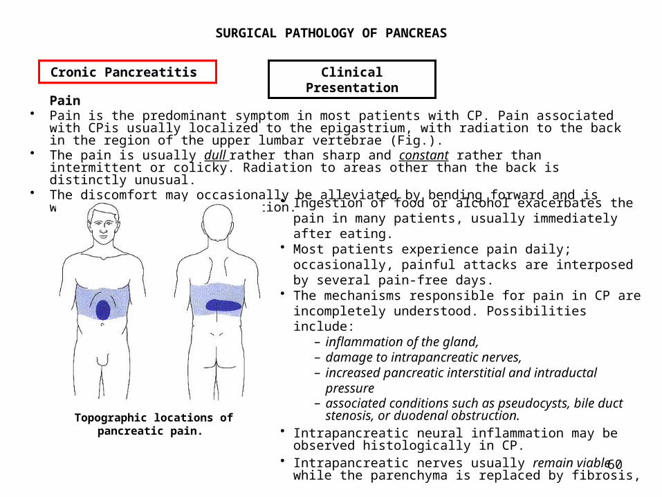
60
SURGICAL PATHOLOGY OF PANCREAS
Pain • Pain is the predominant symptom in most patients with CP. Pain associated
with CPis usually localized to the epigastrium, with radiation to the back in the region of the upper lumbar vertebrae (Fig.).
• The pain is usually dull rather than sharp and constant rather than intermittent or colicky. Radiation to areas other than the back is distinctly unusual.
• The discomfort may occasionally be alleviated by bending forward and is worsened by the supine position.
Cronic Pancreatitis Clinical Presentation
Topographic locations of pancreatic pain.
• Ingestion of food or alcohol exacerbates the pain in many patients, usually immediately after eating.
• Most patients experience pain daily; occasionally, painful attacks are interposed by several pain-free days.
• The mechanisms responsible for pain in CP are incompletely understood. Possibilities include:
– inflammation of the gland, – damage to intrapancreatic nerves, – increased pancreatic interstitial and intraductal
pressure– associated conditions such as pseudocysts, bile duct
stenosis, or duodenal obstruction. • Intrapancreatic neural inflammation may be observed histologically in CP.
• Intrapancreatic nerves usually remain viable while the parenchyma is replaced by fibrosis,

61
SURGICAL PATHOLOGY OF PANCREAS
• The nerves are retained in an abnormal condition: – The nerves are larger and more numerous, – The organization of intraneural organelles is disturbed. – The perineural sheath is disrupted so that it no longer forms a barrier to noxious substances.
– Foci of inflammatory cells are associated with nerves and ganglia so it has been postulated that degranulation of inflammatory cells in contact with nerves generates pain.
– Nerve bundles are edematous. – The amount of neurotransmitters, such as substance P, in afferent neurons is increased, and it has been postulated that altered afferent nerves generate sustained painful signals.
• Clinical studies suggest that continued pancreatic secretion in the presence of ductal obstruction by stricture or intraductal stone can result in elevated intrapancreatic pressure and thus pain.
• Cessation of pancreatic pain occurs eventually in most patients with CP. Pain relief correlates closely with development of pancreatic insufficiency in these patients.
• These observations indicate that loss of exocrine function relieves pancreatic pain by reducing secretory ductal pressures.
• Direct measurement of ductal pressures, made in limited numbers of patients with CP and dilated pancreatic ducts, supports this concept:
– In normal individuals,pancreatic intraductal pressures average 7 to 10 mmHg.
– In patients with CP, intraoperatively measured pancreatic tissue pressures averaged 17 mmHg.
– In patients with dilated pancreatic ducts, direct-puncture pressure measurements made at surgery ranged from 18 to 48 mmHg.
• Surgical decompression of dilated pancreatic ducts normalizes intrapancreatic pressure and is associated with immediate (but not necessarily permanent) pain relief in most patients.
• Peripancreatic inflammation involving contiguous organs can cause painful symptoms localized to the affected region. The most commonly affected organs are the common bile duct and the duodenum. No evidence suggests that pancreatic ductal stones are a cause of pain in CP.
Cronic Pancreatitis Clinical Presentation
62
SURGICAL PATHOLOGY OF PANCREAS
Malabsorption and Weight Loss • Malabsorption occurs when loss of functioning exocrine tissue is advanced, usually greater
than 90%. • Because pancreatic lipase secretion usually decreases before proteolytic
enzymes, steatorrhea is often clinically apparent before azotorrhea. • Steatorrhea occurs when lipase secretion falls below 5% to 10% of normal.
– Bulky, oily bowel movements and abdominal bloating are common complaints.
– Weight loss is nearly always observed. – Deficiencies of fat-soluble vitamins are occasionally of clinical
importance in patients with pancreatic insufficiency. • Syndromes associated with fat-soluble vitamins deficiencies include:
– coagulopathy (vitamin K), – osteomalacia (vitamin D), – neuropathy (vitamin E), – night blindness (vitamin A), and – dermatitis (essential fatty acids).
• In most patients with CP, deficiencies of fat-soluble vitamins are subclinical. • Coincident with reduction of enzyme production, pancreatic bicarbonate secretion is
diminished. • As a result, postprandial duodenal pH may be decreased for prolonged periods. If duodenal
pH is less than 4, acid denaturation of pancreatic enzymes may exacerbate malabsorption. • Postprandial abdominal pain is common in patients with CP, and the fear of this
pain can lead to a further reduction in food intake. Endocrine Insufficiency
• Although the pathogenesis of CP centers around the progressive destruction of exocrine tissues.
• The endocrine portion of the gland is inevitably affected as well. • Exocrine insufficiency always precedes endocrine deficits in CP.
Cronic Pancreatitis Clinical Presentation

63
SURGICAL PATHOLOGY OF PANCREAS
• In fact, subclinical endocrine defects are common in the early stages of CP: – Altered insulin secretion has been consistently observed in these patients.
– Abnormal glucose tolerance can be demonstrated in 50% to 70% of patients with CP;
– overt diabetes is present in 32% to 40%. • Deficits are progressive; if individual patients are repetitively tested,
progressive deterioration is observed.
Cronic Pancreatitis Clinical Presentation

64
SURGICAL PATHOLOGY OF PANCREAS
Routine Laboratory Tests • Routine tests of blood or serum are not helpful in making a diagnosis of CP:
– Anemia is common but nonspecific. – Leukocytosis is not observed unless acute disease is superimposed on
CP. – Deficiencies of fat-soluble vitamins occur but are unpredictable after
steatorrhea develops. – Elevation of alkaline phosphatase may be observed if chronic fibrosis
or compression by pseudocyst causes intrapancreatic biliary ductal obstruction.
– Serum amylase levels may be normal, elevated, or subnormal in CP. Low values have been attributed to advanced loss of exocrine tissue, although total serum amylase is usually maintained near normal as a result of salivary amylase secretion. Persistently elevated amylase values in patients with CPshould suggest a superimposed attack of acute pancreatitis or the development of a complication, such as pseudocyst.
• Determination of urine tests, urinary amylase secretion does not increase sensitivity, nor does assay for other pancreatic enzymes, such as lipase, elastase, isoamylase, or trypsin. Tests of Pancreatic Exocrine Function
• Tests of exocrine function may be divided into: – Tests that directly measure pancreatic secretion of enzymes or
bicarbonate and– Tests that indirectly measure the effects of secreted enzymes by
assaying compounds that require pancreatic digestion before absorption. • Direct measurement of exocrine function requires collection of pancreatic secretions.
This may be accomplished by direct cannulation of the pancreatic duct using ERCP or by using double-balloon tubes that isolate portions of the duodenum. Collection of pancreatic secretion is thus relatively invasive and uncomfortable. Appropriate corrections must be made for the effects of incomplete collection or dilution by gastric or intestinal secretions. Because basal pancreatic output in humans is variable, examination of secretion stimulated by intravenous cholecystokinin or secretin is necessary.
Cronic Pancreatitis Diagnosis

65
SURGICAL PATHOLOGY OF PANCREAS
• Demonstration of subnormal pancreatic enzyme or bicarbonate secretion is diagnostic of CP if pancreatic cancer has been excluded.
• Because of the inability to detect early disease reliably and because of the attendant patient discomfort, direct measurements of exocrine function are not commonly performed.
• Indirect tests of pancreatic function measure the absorption of some nutrient that first requires pancreatic digestion.
• Because clinically detectable malabsorption does not occur until 90% of exocrine function is lost, it is apparent that indirect tests of pancreatic function do not detect early stages of CP.
• For all such tests, sensitivity is 90% to 100% for advanced disease but less than 50% for early CP.• False-positive tests may occur in other states that cause malabsorption
(Crohn’s disease, sprue, postgastrectomy states) or in association with diabetes mellitus, cirrhosis, or renal disease. In the bentiromide test, N-benzoyl-L-tyrosyl-p-aminobenzoic acid is digested by trypsin to release paraaminobenzoic acid. Free paraaminobenzoic acid, absorbed by the small intestine, is secreted by the kidney and assayed in urine.
• Fat malabsorption can be tested by feeding (14C)olein and measuring exhaled 14CO2. Pancreatic Imaging Studies
• Radiologic studies to define pancreatic structure have largely supplanted pancreatic functional tests in confirming the clinical diagnosis of CP.
• The simplest confirmatory test for CP is a plain abdominal film demonstrating calcification of the pancreas. This finding, present in 32% of patients, is pathognomonic of CP.
Cronic Pancreatitis Diagnosis

66
SURGICAL PATHOLOGY OF PANCREAS
Ultrasound is useful in initial evaluation of patients with suspected CP. Supportive findings include: – atrophy of the gland, – reduced echogenicity, – dilation of the pancreatic duct to greater than 4 mm, and – associated cystic lesions.
• Ultrasound examination of the pancreas may sometimes be compromised by overlying intestinal gas.
• Ultrasound has a reported sensitivity rate of about 60% for CP and a specificity rate of 80% to 90%. Computed tomographic (CT) examination of the pancreas is more sensitive than ultrasound in CP, although CT examination involves ionizing radiation and is more expensive.
• CT findings consistent with this diagnosis include glandular atrophy, irregularity of the pancreatic outline, calcification, and ductal dilation (Fig. 32-3 and Fig. 32-4). Small cystic lesions are well demonstrated by CT. Sensitivity for CT approaches 75% to 90%, with a specificity of 85%.
Cronic Pancreatitis Diagnosis
Abdominal CT scan in a patient with chronic pancreatitis,
illustrating dilation of main pancreatic duct
Abdominal CT scan demonstrating pancreatic calcifications (arrow in A) and associated cystic lesions (arrows in B)

67
SURGICAL PATHOLOGY OF PANCREAS
ERCP has become widely recognized as the most sensitive and reliable method for diagnosing CP.
• The sensitivity of ERCP approaches 90%, with equal specificity: – In earliest CP, ductal changes are limited to secondary and tertiary ducts that show irregular dilation.
– In moderate disease, the main pancreatic duct may be dilated with alternating areas of stenosis.
– In advanced CP, marked ductal changes may form a chain-of-lakes appearance.
• ERCP is also useful in demonstrating associated anatomic abnormalities, such as common bile duct stenosis or pancreatic pseudocyst. Most studies comparing the sensitivity of ERCP and pancreatic secretory tests have found good correlation in advanced disease. In earlier stages of CP, correlation of morphologic changes and pancreatic function is often poor.
Cronic Pancreatitis Diagnosis
ERCP illustrating moderate dilation of the main pancreatic duct and ectasia of
secondary ducts associated with moderately advanced chronic pancreatitis. Arrows
indicate intraductal pancreatic stones.
ERCP illustrating early changes of chronic pancreatitis with ductal ectasia confined to the
pancreatic tail (arrows).

68
SURGICAL PATHOLOGY OF PANCREAS
PainAbstinence
• Management of pain in patients with CPshould begin with abstinence. With elimination of alcohol, 50% to 75% of patients have some decrease in pain, although most do not become pain free. Because alcohol is a secretagogue for pancreatic enzymes, pain relief is more likely in patients who retain some exocrine function. Enzyme Replacement
• Exogenous enzyme administration as a treatment for pain has been proposed, based on the concept of negative-feedback inhibition of pancreatic secretion.
• In humans, the intraduodenal administration of trypsin or chymotrypsin inhibits pancreatic secretion, and diversion of pancreatic secretion from the duodenum stimulates secretion of digestive enzymes.
• It has been postulated that patients with CP may have continuous stimulation by hormonal or neural pathways because of diminished secretion of digestive enzymes.
Cronic Pancreatitis Treatment of Complications
Pain
Schematic diagram of stimulatory and inhibitory influences on pancreatic exocrine secretion. CCK, cholecystokinin; PP, pancreatic polypeptide.

69
SURGICAL PATHOLOGY OF PANCREAS
• The increased stimulatory signals are presumed to cause or exacerbate pain. • If this contention is correct, effective delivery of pancreatic enzymes to
the duodenum will reduce chronic stimulation, decrease ductal pressure, and relieve pain. Although initial controlled trials suggested that improvement in pain can occur as a result of enzyme replacement, especially in patients with idiopathic pancreatitis, disappointing results have also been reported.
• Enzyme replacement trials are difficult to interpret due to a high placebo effect rate (30%) in patients with CP. Further studies are needed to evaluate this novel therapy.
• Cholecystokinin receptor antagonist and the somatostatin analogue octreotide have also been proposed for treatment of pain in CP, but their efficacy is still unproved. Endoscopic Therapy
• The use of endoprostheses or stents placed into the pancreatic duct endoscopically has been proposed as a treatment for CPcomplicated by ductal stenosis.
• About 30% to 75% of patients treated in this way had symptomatic improvement when observed for 14 to 36 months.
• The use of pancreatic duct stents, however, is associated with a risk of pancreatic ductal injury and fibrosis. Because of this concern, many investigators recommend short-term use of stents to identify patients most likely to benefit from surgical drainage.
• The role of endoscopic removal of pancreatic ductal stones is unsettled.• Fragmented by extracorporeal shock wave lithotripsy, stones can be extracted
after sphincterotomy of the pancreatic duct. When stones are cleared, half of patients report long-term improvement in pancreatic pain. Analgesics
• Analgesics remain the mainstay for nonoperative treatment of pain in CP. • Nonnarcotic analgesics should be used initially. If pain is progressive,
increases in dose or frequency of these agents should be attempted before narcotics are prescribed.
Cronic Pancreatitis Treatment of Complications
Pain

70
SURGICAL PATHOLOGY OF PANCREAS
• Eventually, most patients with chronic pancreatitis require narcotic pain relief; addiction is common and makes evaluation of treatments aimed at pain relief difficult. Percutaneous, radiologically guided injection of the celiac ganglia with neural ablative agents has been used in patients with CP, based on the success of this approach in patients with pancreatic cancer. The procedure is not usually effective long-term in CP, with pain relief lasting 6 months in fewer than half of treated patients. Repeated injection is not usually successful. Surgical Treatment
• Intractable pain is the most frequent indication for operation in patients with CP.
• Operation may be considered when pain is severe enough: – to interfere substantially with quality of life, – to interrupt employment or normal family life, – to affect general health by interfering with nutrition, or – to cause narcotic addiction.
• All patients being considered for operative treatment should undergo CT examination of the pancreas to exclude pancreatic carcinoma and ERCP to evaluate pancreatic ductal anatomy.
• Operative treatment for chronic pain associated with CPcan be broadly divided into: – ductal drainage procedures and – resection procedures.
Ductal drainage procedures• Pancreaticojejunostomy. When patients with chronic pancreatitis have
pancreatic ducts dilated to more than 8 mm, ductal decompression using pancreaticojejunostomy (Puestow procedure) may be employed for relief of pain.
Cronic Pancreatitis Treatment of Complications
Pain

71
SURGICAL PATHOLOGY OF PANCREAS
• Pancreatic Resection. Pancreatic drainage is not feasible when pancreatic ducts are small or normal in diameter. Pancreatic resection may be considered when patients have small ducts, when the disease process involves primarily one portion of the gland and the remainder of the gland is nearly normal, or after a failed pancreaticojejunostomy. The rationale for pancreatic resection is that pain and risk of complications are reduced by removing the diseased portion of the gland.
• Distal pancreatectomy may be performed when pathologic changes are confined to the tail or body of the pancreas.
• Resection of the pancreatic head by pancreaticoduodenectomy may be appropriate in selected patients with disease confined predominantly to the head of the gland.
• Indications for pancreaticoduodenectomy include: (1) a chronic inflammatory mass involving primarily the head of the gland
and the uncinate process, (2) a chronic inflammatory mass in the head of the pancreas associated
with duodenal stenosis, (3) multiple pseudocysts confined to the head of the pancreas, and failure
of pancreaticojejunostomy secondary to inadequate drainage of the uncinate process.
Cronic Pancreatitis Treatment of Complications
Pain

72
SURGICAL PATHOLOGY OF PANCREAS
Cronic Pancreatitis Treatment of Complications
Pain
Pancreatico-jejunostomy

73
SURGICAL PATHOLOGY OF PANCREAS
Cronic Pancreatitis Treatment of Complications
Pain
Points of parenchymal transection for 50% and 85% distal pancreatectomies.
Reconstruction after standard pancreaticoduodenectomy.

74
SURGICAL PATHOLOGY OF PANCREAS
Malabsorption • Treatment of malabsorption secondary to pancreatic insufficiency requires
delivery of exogenous pancreatic enzymes in active form to the duodenum. • This goal is often difficult because of inadequate amounts of pancreatic
enzymes in commercially available oral preparations and acid-peptic destruction of ingested enzymes. Because steatorrhea is most troublesome clinically, the delivery of lipase to the duodenum is the critical variable.
• Fat malabsorption usually does not occur if 25,000 IU of lipase activity can be provided during a 4-hour postprandial period.
• The major impediment to delivery of oral pancreatic enzymes to the postprandial small intestine is gastric acidity.
• Pancreatic enzymes are active at an alkaline pH and inactive at a pH of less than 5. • Pancreatic lipase is irreversibly denatured at a pH of less than 4. • Several approaches have been used to circumvent gastric effects:
– Large amounts of pancreatic enzymes have been given with meals in the hope that enough enzyme would survive gastric passage to digest the fat contained in a typical diet.
– Suppression of gastric acid production using histamine-2 (H2) receptor antagonists or antacids are attempts to protect enzyme activity.
– Enteric-coated formulations of pancreatic enzymes have been prepared that resist acid but dissolve (and release their contents) on contact with the alkaline pH found in the small intestine.
– Finally, acid-stable forms of lipase with a wider pH range of activity, derived from Aspergillus niger and Rhizopus arrhizus, have been developed for human testing.
• A rational approach to treatment of pancreatic insufficiency begins with the administration of sufficient enzyme tablets to abolish azotorrhea and to reduce steatorrhea to tolerable levels. This often means ingesting several tablets with each meal. If symptoms persist, the number of tablets should be increased, or the fat content of meals may be decreased. H2 receptor antagonists may be added for patients resistant to these measures. If steatorrhea persists, a search for other contributing causes (bacterial overgrowth, ileal disease) should be performed.
Cronic Pancreatitis Treatment of Complications
Malabsorption
75
SURGICAL PATHOLOGY OF PANCREAS
Biliary Complications • Biliary complications involving the common bile duct can occur in CP because of
the intimate association of that structure with the head of the pancreas. In two thirds of the population, the common bile duct traverses the pancreatic parenchyma; in about 25%, the common bile duct lies in a groove along the posterior surface of the pancreas; and in only 10%, the duct is extrapancreatic, always posterior to the gland.
• Thus, fibrosis associated with CPcan encase and compress the common bile duct.
• Common bile duct stenosis is a relatively common complication of CP, occurring in about 10% of cases observed long-term.
• Alkaline phosphatase elevation is the most sensitive screening method for detection of biliary stenosis, and a larger proportion of patients demonstrate increases in alkaline phosphatase than develop jaundice or symptoms of biliary obstruction. Cholangiographic investigation of patients with common bile duct disease can be accomplished either by transhepatic or retrograde endoscopic routes. Because CPpatients with common bile duct disease frequently require treatment of concurrent pancreatic disease, examination of both systems is best accomplished by ERCP. Bile duct fibrosis typically results in long, gradually tapering strictures conforming to the intrapancreatic duct.
• Malignant strictures usually result in abrupt termination of the biliary duct. The proximal suprapancreatic portion is variably dilated.
• The most serious sequelae of unrelieved biliary obstruction are cholangitis and biliary cirrhosis. Collected series of CPpatients with common bile duct stenosis suggest that each of these complications develops in 7% to 10% of patients with radiographic abnormalities. The degree of biliary obstruction as shown by cholangiography does not correlate with severity of pancreatic disease, liver histology, or biochemical abnormalities, and therapeutic decisions should be based on clinical factors rather than radiologic criteria. Persistent elevation of serum alkaline phosphatase (three to five times normal), although imperfect, is probably the best predictor of progressive biliary stenosis.
Cronic Pancreatitis Treatment of Complications
Biliary Complications

76
SURGICAL PATHOLOGY OF PANCREAS
• Operation in patients with stricture of the common bile duct associated with CP is justified to treat symptoms or to prevent the development of biliary cirrhosis. Operative indications include the following: • Persistent jaundice • Cholangitis • Liver biopsy evidence of biliary cirrhosis • Inability to exclude pancreatic cancer • Progressive stricture supported by radiologically progressive dilation
of extrahepatic and intrahepatic biliary ducts • Persistent elevation of alkaline phosphatase at greater than three
times normal • Both choledochoduodenostomy and choledochojejunostomy are excellent
operative choices for patients with intrapancreatic strictures of the common bile duct. A number of other complications can occur in CP, including pancreatic pseudocyst, pancreatic ascites, and splenic vein thrombosis.
Prognosis• Patients with CP have decreased long-term survival rates compared with the
general population, with an excess mortality rate of 36% over 20 years. • Surprisingly, less than 20% of deaths are directly attributable to
pancreatitis or its complications. • Excessive mortality is related to the extrapancreatic complications of alcoholism and smoking.
Cancers of the aerodigestive system, complications of diabetes, and complications of cirrhosis are the most frequent causes of death.
• Pancreatic cancer has been reported to occur in 4% of patients with CPobserved for 20 years.
Cronic Pancreatitis Treatment of Complications
Biliary Complications

77
SURGICAL PATHOLOGY OF PANCREAS
• The management of neoplasms of the exocrine pancreas presents a major challenge to the surgeon. • The most common of these tumors, ductal adenocarcinoma of the pancreas, has
become the fifth most common cause of cancer death in the United States and continues for the most part to be recalcitrant to treatment.
• Other less common neoplasms of the pancreas can present formidable difficulties in diagnosis and treatment. – The generally vague early symptoms of pancreatic disease, – The inaccessibility of the organ to examination, – The aggressiveness of most pancreatic tumors, and – The technical difficulties associated with pancreatic surgery
make pancreatic exocrine neoplasms among the most daunting diseases treated by surgeons. Nevertheless, progress has been made in our ability to manage pancreatic neoplasms.
Neoplasms of the exocrine pancreas

78
SURGICAL PATHOLOGY OF PANCREAS
Epidemiology and Etiology • The cause of pancreatic cancer is unknown. • The most striking epidemiologic fact about pancreatic cancer is its steady increase in incidence
during the 20th century, in contrast to declining or level rates for other gastrointestinal malignancies.
• The current annual mortality rate in the United States of about 10 cases per 100,000 population is almost three times the rate in 1930.
• This rise in incidence parallels changes in the prevalence of cigarette smoking in the United States, and epidemiologic studies suggest that much of the increase in pancreatic cancer in recent decades is attributable to increased tobacco use.
• Adenocarcinoma of the pancreas most commonly develops in the seventh decade of life and is rare before the age of 40 years.
• Is more common in men than in women by a ratio of 3:2. Possible Etiologic Environmental Factors
• The most consistently observed risk factor for pancreatic cancer is cigarette smoking: most studies estimate that smoking doubles or triples a person’s risk of developing pancreatic cancer.
• The mechanism is unknown, but carcinogens in cigarette smoke have been shown to produce pancreatic tumors in laboratory animals.
• Alcohol consumption has been implicated in some case-control studies of pancreatic cancer, but the overall evidence is inconsistent, and alcohol is not likely to be a major factor in the development of the disease.
• Although considerable public interest was focused on coffee consumption as a risk factor for pancreatic cancer, several case-control and cohort studies fail to support an association.
• The possible importance of diet in the development of pancreatic cancer. • High dietary fat consumption is in general associated with an increased
risk of pancreatic cancer, and Diets high in fruits and vegetables are associated with a lower incidence of pancreatic cancer.
Neoplasms of the exocrine pancreas Ductal adenocarcinoma of the pancreas

79
SURGICAL PATHOLOGY OF PANCREAS
Possible Etiologic Host Factors • Most cases of pancreatic cancer have no obvious predisposing host factors. • Abnormal glucose tolerance is present in about 80% of patients with
pancreatic cancer, if carefully sought. • Diabetes and pancreatic cancer occur together far more frequently than
would be expected by chance. • In most cases, diabetes manifests at about the same time as the pancreatic
neoplasm; the onset of diabetes can be a early sign of pancreatic cancer. • A diabetogenic substance may elaborated by pancreatic carcinomas; islet
amyloid polypeptide has been proposed as a candidate peptide mediating glucose intolerance in patients with pancreatic tumors.
• Chronic pancreatitis is a significant risk factor for the development of pancreatic cancer. It appears that all forms of chronic pancreatitis are associated with an increased risk of pancreatic cancer, suggesting that it is the pancreatitis and not the injuring agent (eg, alcohol) that is responsible for the augmented cancer risk.
• Patients who have previously undergone gastric resection are three to seven times more likely to develop pancreatic cancer as a control population.
• Women who have undergone cholecystectomy are at increased risk, although this observation has not been consistent.
• Several reports have been made of familial clustering of pancreatic cancer. Epidemiologic studies suggest that about 7% of pancreatic cancer patients have a positive family history of the disease.
• Pancreatic cancer is also more common in a few rare autosomal dominant syndromes such as hereditary pancreatitis, multiple endocrine adenomatosis type I, and Gardner syndrome.
Neoplasms of the exocrine pancreasNeoplasms of the exocrine pancreas Ductal adenocarcinoma of the pancreas

80
SURGICAL PATHOLOGY OF PANCREAS
• The exocrine pancreas contains two major types of epithelium: acinar and ductal. • The acinar cells of the pancreas are primarily concerned with the elaboration
of digestive enzymes, whereas the ductal epithelium is responsible for the secretion of fluid and electrolytes and the conveyance of pancreatic juice to the duodenum.
• The ductal type of epithelium begins with the so-called centroacinar cells, which provide an interface to the acinar cells that cluster around them in a grapelike fashion.
• The smallest pancreatic ducts are lined by low cuboidal epithelium. • As these ducts coalesce to form progressively larger conduits, the height of
the lining cells increases; • The epithelium becomes columnar in the main pancreatic duct. • The character of the epithelium also changes in the larger ducts, with an increasing percentage of
mucin-producing cells as the ducts approach the duodenum. • Despite the fact that ductal epithelial cells make up less than 5% of the pancreatic mass, they appear
to be the cells of origin of most pancreatic carcinomas. Although evidence suggests that acinar epithelium may dedifferentiate to a ductlike form, the most straightforward interpretation of human pancreatic carcinogenesis is that the tumors for the most part arise from preexisting ductal cells.
• The microscopic appearance of a typical ductal pancreatic cancer consists of large and small glands lined by cuboidal or columnar epithelium producing variable amounts of mucin. The glands are embedded in a dense fibrous matrix, which is responsible for the scirrhous consistency of the tumors.
• The degree of differentiation of ductal carcinoma varies; poorly differentiated tumors demonstrate less gland formation and mucus production and more epithelial anaplasia.
• Most patients with pancreatic cancer have an associated chronic obstructive pancreatitis, with duct dilation, atrophy and fibrosis of the acinar parenchyma, and varying degrees of chronic lymphocytic infiltration. About 10% of patients show histologic evidence of superimposed acute pancreatitis with a polymorphonuclear cell infiltrate; pseudocyst formation can occur in this group but is rare.
Ductal adenocarcinoma of the pancreas
Pathology

81
SURGICAL PATHOLOGY OF PANCREAS
HISTOLOGIC CLASSIFICATION OF 645 CASES OF PRIMARY, NONENDOCRINE CANCER OF THE PANCREAS
CLASSIFICATION NumberDUCT (DUCTULAR) CELL ORIGIN 572 (89%)
Duct cell adenocarcinoma 494Giant cell carcinoma 27Giant cell carcinoma (osteoid) 1Adenosquamous carcinoma 20Microadenocarcinoma 16Mucinous (colloid) carcinoma 9Cystadenocarcinoma (mucinous) 5
ACINAR CELL ORIGIN 8 (1%)
Acinar cell carcinoma 7Cystadenocarcinoma (acinar cell) 1
UNCERTAIN HISTOGENESIS 61 (9%)
Pancreaticoblastoma 1Papillary and cystic neoplasm 1Mixed typeduct and islet cells 1Unclassified 58
CONNECTIVE TISSUE ORIGIN 4 (1%)TOTAL 645 (100%)
Ductal adenocarcinoma of the pancreas
Pathology

82
SURGICAL PATHOLOGY OF PANCREAS
Precursor and Precancerous Lesions • Ductal carcinoma of the pancreas probably arises as a progressive process beginning
with ductal hyperplasia, followed by the development of atypical hyperplasia, carcinoma in situ, and finally invasive carcinoma, as is the case for many neoplasms.
• Unfortunately, the study of preneoplastic lesions in the pancreas is confounded by the fact that the lesions are in general both asymptomatic and inaccessible. Site of Ductal Adenocarcinoma
– 60 to 70% of pancreatic ductal adenocarcinomas occur in the head of the gland.
– About 15% reside in the body of the gland, another – 10% are in the tail, and – The remaining 5% to 15% are diffuse.
• The predilection of pancreatic cancer to develop in the head of the gland is unexplained. • The practical consequence of this pattern is that:
– Tumors in the head are diagnosed earlier because they cause obstructive jaundice, whereas
– Tumors in the body and tail tend to be more advanced at the time of symptomatic presentation.
– Tumors in the body and tail are typically larger at the time of diagnosis (average, 7 to 8 cm) than in the head (average, 4 to 5 cm).
Ductal adenocarcinoma of the pancreas
Pathology

83
SURGICAL PATHOLOGY OF PANCREAS
Sites and Frequency of Local Extension • Extension beyond the confines of the pancreas is the rule rather than the exception in ductal
carcinoma of the pancreas: – The bile duct is invaded early in the course of the disease, and about 80% of
patients with tumors in the head of the pancreas present with jaundice.
– Invasion of the first or second portion of the duodenum also occurs early in the course of the disease and is present in about 25% of cases.
– In most pancreatic cancers, there is early invasion of the retroperitoneum, either directly or along the course of autonomic nerves of the celiac plexus.
– Some degree of perineural invasion is present in 90% of cases. – In about half of cases, the walls of the portal or superior mesenteric veins also are
invaded, and complete transmural invasion can ultimately lead to thrombosis. – Carcinoma of the body and tail can invade the splenic vein, with
resultant thrombosis and development of gastric varices. – Other sites of local invasion, which tends to occur later, include the
superior mesenteric and splenic arteries, – transverse mesocolon, – stomach, – kidneys, and left adrenal gland. – A few florid cases of extension of pancreatic cancer along the
pancreatic duct into the tail of the gland have been described, occurring early in the course of the disease.
Ductal adenocarcinoma of the pancreas
Pathology

84
SURGICAL PATHOLOGY OF PANCREAS
Sites and Frequency of Metastatic Disease •The most common sites of metastatic spread from carcinoma of the pancreas are regional and juxtaregional lymph nodes and liver.
Ductal adenocarcinoma of the pancreas
Pathology
SITES OF METASTASIS IN PANCREATIC CANCER Site of Primary Carcinoma
BodyHead, Body,
Head and Tail and TailSite of Metastasis (n = 106) (n = 34) (n = 24)
Regional lymph nodes 85 34 24Juxtaregional lymph nodes 52 25 18Liver 80 28 21Stomach 15 7 13Peritoneum 23 17 11Lungs 28 8 7Pleura 29 13 6Pericardium 3 2 1Colon 3 4 4Spleen 6 11 4Adrenal glands 15 9 5Bones 13 5 3Kidneys 9 7 6Skin 1 1 2No metastases 15 1 0

85
SURGICAL PATHOLOGY OF PANCREAS
• Lymphatic spread usually precedes hematogenous spread, • Among patients who appear to have no gross evidence of distant metastasis, the prevalence of lymph
node metastasis on examination of the resected specimen is about 60%. • Knowledge of the route of lymph node metastasis of pancreatic carcinoma is
important to the surgeon. • The demonstration of lymph node involvement is not a contraindication to resection, the involved
nodes must be included in the planned resection. • The most common sites of lymph node involvement in pancreatic cancer are the
posterior pancreaticoduodenal nodes and the nodes along the superior margin of the pancreatic head.
Ductal adenocarcinoma of the pancreas
Pathology
Lymph node involvement in duct cell carcinoma of the head of the pancreas

86
SURGICAL PATHOLOGY OF PANCREAS
Staging of Pancreatic Cancer • Accurate pathologic staging of pancreatic cancer is important for for the
following reasons: – First and foremost, it has prognostic importance for the patient. – Second, careful pathologic staging allows retrospective examination of
the accuracy of diagnostic modalities such as computed tomography (CT). – Finally, the results of therapeutic trials cannot be accurately judged
or compared without detailed knowledge of the pathologic stage of the treated tumors.
• Using this system, it was demonstrated in a total of 924 patients that the survival rate in stage I disease (33% at 1 year) was significantly higher than in stages II and III (combined 13% at 1 year). Survival rates were similar for cases of advanced direct extension (stage II, 15% at 1 year) and cases of lymph node metastases (stage III, 11% at 1 year). The survival rate for stage IV patients with distant metastases was significantly worse than in all other groups (5% at 1 year).
• With improvements in diagnostic imaging, it has been possible to assess the natural history of small carcinomas of the pancreas (2 cm or less).
• It is discouraging to find that these small tumors have already metastasized to lymph nodes in 30% to 40% of cases and that extrapancreatic invasion has occurred in about 30%.
• Less than half of patients with tumors 2 cm or smaller have stage I disease. The overall 5-year survival rate of patients with tumors of this size is about 30%.
Ductal adenocarcinoma of the pancreas
Pathology

87
SURGICAL PATHOLOGY OF PANCREAS
Ductal adenocarcinoma of the pancreas
Pathology
TNM CLASSIFICATION FOR STAGING OF CANCER OF THE PANCREAS
TNM DEFINITIONS
Primary TumorT1 No direct extension of the primary tumor beyond the pancreasT2 Limited direct extension (to duodenum, bile ducts, or stomach), still possibly permitting tumor resectionT3 Further direct extension, incompatible with surgical resectionTX Direct extension not assessed or not recorded Regional Lymph Node InvolvementN0 Regional nodes not involvedN1 Regional nodes involvedNX Regional node involvement not assessed or not recorded Distant MetastasisM0 No distant metastasisM1 Distant metastatic involvementMX Distant metastatic involvement not assessed or not recorded STAGE GROUPINGStage I T1, T2, N0, M0—No or limited direct extension to adjacent viscera, with no regional node extension and absence of distant metastases. Limited direct extension defined as involvement of organs adjacent to the pancreas that could be removed en bloc with thepancreas if a curative resection were attempted.Stage II T3, N0, M0—Further direct extension of tumor into adjacent viscera, with no lymph node involvement and no distant metastases, which precluded surgical resection.Stage III T1-3, N1, M0—Regional node metastases without clinical evidence of distant metastases.Stage IV T1-3, N0-1, M1—Distant metastatic disease in liver or other sites.

88
SURGICAL PATHOLOGY OF PANCREAS
• Many of the difficulties in the treatment of pancreatic cancer can be traced to our inability to diagnose the disease in its early stages.
• The vague early symptoms of pancreatic cancer are often minimized by both patient and physician, leading to a delay of months in making the diagnosis.
• It is ordinarily not until the patient develops jaundice or extreme weight loss that the diagnosis is made, and by this time the pancreatic tumor is typically large and has grown beyond the confines of the pancreas. Clinical Symptoms and Signs
• The most common presenting symptoms of pancreatic cancer is weight loss, which is usually substantial, averaging 10 kg. The weight loss can initially occur as an isolated symptom in the face of a seemingly normal appetite. Later, it is usually associated with anorexia. Unexplained documented weight loss should prompt a search for occult malignancy; in older adults, it is appropriate to perform a CT scan of the abdomen for this indication alone. Other digestive symptoms are common in pancreatic cancer and include nausea, vomiting, and change in bowel habits.
• Most patients with pancreatic cancer come to physicians because of jaundice. In people older than 60 years of age, the combination of jaundice and weight loss usually means carcinoma of the pancreas or periampullary region.
• The diagnosis is often evident, requiring only appropriate imaging studies for confirmation.
• The jaundice is progressive. It is associated with dark urine and light stools. Pruritus is present in about one fourth of jaundiced patients.
• Although it is often taught that carcinoma of the pancreas presents with painless jaundice (to help distinguish it from choledocholithiasis), this aphorism is not accurate. Most patients do experience pain as part of the symptom complex of pancreatic cancer.
• Pain is usually perceived in the epigastrium but can occur in any part of the abdomen and can radiate to the back. Early on, it is often mild and vague, that explain the delay in diagnosis.
Ductal adenocarcinoma of the pancreas
Diagnosis

89
SURGICAL PATHOLOGY OF PANCREAS
Ductal adenocarcinoma of the pancreas
Diagnosis
SYMPTOMS OF PANCREATIC CANCER
Symptom Patients (%)HEAD Weight loss 92Jaundice 82Pain 72Anorexia 64Dark urine 63Light stools 62Nausea 45Vomiting 37Weakness 35Pruritus 24Diarrhea 18Melena 12Constipation 11Fever 11Hematemesis 8 BODY AND TAIL Weight loss 100Pain 87Weakness 43Nausea 43Vomiting 37Anorexia 33Constipation 27Hematemesis 17Melena 17Jaundice 7Fever 7Diarrhea 3

90
SURGICAL PATHOLOGY OF PANCREAS
• 50% to 70% of patients with pancreatic cancer experience pain that they describe as moderate or severe; the prevalence is particularly high with tumors of the body and tail, probably because of invasion of the celiac plexus.
• The most common presenting signs of pancreatic cancer are jaundice and hepatomegaly.
• When bilirubin levels are only minimally elevated, icterus may be confined to the sclerae, but most patients have cutaneous changes by the time of presentation. Hepatomegaly usually reflects congestion associated with biliary obstruction and does not imply the presence of metastatic disease unless the liver is nodular or hard.
• The obstructed gallbladder is palpable in about 25% of patients with pancreatic cancer(Courvoisier-Terrier sign). In most patients, the tumor itself is not palpable.
• Ascites is present in about 15% of patients with pancreatic cancer. With large tumors, there can be gross or occult blood in the stool from invasion of the duodenum, stomach, or colon.
Laboratory Investigations • In carcinoma of the head of the pancreas, liver function tests typically reveal :
– elevations in bilirubin (particularly the conjugated fraction) and – alkaline phosphatase characteristic of extrahepatic biliary
obstruction. – The transaminases can also be elevated, but usually not to the extent
of the alkaline phosphatase. If jaundice has been long-standing, – the prothrombin time can be abnormally prolonged. – Mild elevations of the serum amylase in the range of 300 U/L occur,
but marked elevations of serum amylase are rare. • Routine laboratory determinations add little to the diagnosis of pancreatic cancer other than
reinforcing the suspicion of extrahepatic biliary obstruction. • Considerable effort has been expended to find serum markers of pancreatic
cancer. Mucins are large glycosylated glycoproteins whose function appears to be protection and lubrication of epithelial cells. Mucin molecules are produced by most moderately well-differentiated pancreatic carcinomas.
Ductal adenocarcinoma of the pancreas
Diagnosis

91
SURGICAL PATHOLOGY OF PANCREAS
• In the past few years, mucin-associated antigens have been isolated and purified. CA19-9 is an example of a mucin-associated carbohydrate antigen that can be detected in the serum of patients with pancreatic cancer.
• The CA19-9 antigen has been identified as sialosyl-fucosyl-lactotetrose, which corresponds to the sialylated Lewisa (Lea) blood group substance found on erythrocytes. About 5% of the Western population lacks the Lewis gene and therefore cannot make CA19-9. The CA19-9 antigen resides in cell membrane glycolipid and in mucin glycoprotein. CA19-9 can be detected in pancreatic juice, in serum, and in pancreatic tissue by immunohistologic techniques.
• Serum levels of CA19-9 are elevated (above 37 U/mL) in about 75% of patients with pancreatic cancer. Unfortunately, CA19-9 is also elevated in about 10% of patients with benign diseases of the pancreas, liver, and bile ducts.
• The availability of CA19-9 also appears not to have led to earlier diagnosis of pancreatic cancer because most patients are symptomatic at presentation and CA19-9 is often not elevated in very small cancers.
• On the other hand, the use of CA19-9 with imaging studies improves overall diagnostic accuracy and can simplify the evaluation of patients with suspected pancreatic cancer.
• Of potential diagnostic significance is the fact that about 90% of human pancreatic cancers contain the mutated c-K-ras oncogene.
• The c-K-ras gene is ordinarily present in human cells and encodes a membrane-bound protein that possesses high affinity for guanosine triphosphate and guanosine diphosphate and appears to be important for signal transduction across the cell membrane. The ras protein is active when bound to guanosine triphosphate but is inactive when bound to guanosine diphosphate. Certain mutations in the gene result in its transformation to an active oncogene. In the case of c-K-ras, a single point mutation at codon 12 is sufficient. As a result of the mutation, the level of activated ras protein rises, and high levels of active protein can contribute to unrestrained cellular growth by facilitating the transmission of growth factor signals across the cell membrane.
Ductal adenocarcinoma of the pancreas
Diagnosis

92
SURGICAL PATHOLOGY OF PANCREAS
Radiologic Investigations • Imaging of the pancreas has dramatically improved with the development of ultrasonography,
CT scanning, and ERCP MRI(MRCP). With appropriate use of these studies, it should be possible to arrive at a radiologic diagnosis of pancreatic cancer in over 90% of patients presenting with the disease.
• Standard transcutaneous ultrasonography is an appropriate first test in the evaluation of the patient with jaundice because the presence of a dilated common bile duct or intrahepatic bile ducts is essentially diagnostic of extrahepatic biliary obstruction. This finding directs the physician to a search for the cause of the obstruction. If the bile ducts are not dilated, mechanical obstruction is unlikely, and the diagnostic thrust should move toward hepatocellular disease. Ultrasonography is also the best test to determine whether gallstones are present; this is extremely important because choledocholithiasis is one of the conditions most likely to cause jaundice in the elderly population. If an ultrasound examination shows gallstones and no evidence of a pancreatic mass, the appropriate next step is ordinarily an ERCP (see later) to document common duct stones. On the other hand, if no gallbladder stones are present by ultrasonography, choledocholithiasis is unlikely, and a pancreatic or periampullary tumor or chronic pancreatitis becomes a more likely cause of the obstruction. Ordinarily, a CT scan is the most appropriate next test.
• Ultrasonography reveals a pancreatic mass in 60% to 70% of patients with pancreatic cancer, but the sensitivity of ultrasound is slightly lower than that of CT, though the absence of a mass on ultrasound scan cannot be accepted as firm evidence against pancreatic cancer.
• In most cases of pancreatic cancer, CT is the single most useful test. • CT scanning not only usually detects the presence of the tumor mass but also provides
important information about the extent of the tumor. CT scans may miss tumors smaller than 2 cm. In such cases, the CT findings may be limited to pancreatic or bile duct dilation. Such findings are highly suspicious for pancreatic malignancy and should be further evaluated, ordinarily with ERCP. Dynamic CT scanning, in which high-speed scans are obtained during rapid intravenous administration of iodinated contrast material, and new techniques such as three-dimensional reconstruction from spiral scans provide excellent information about vascular invasion.
• CT scans provide the best available radiologic information to determine whether or not a pancreatic neoplasm is resectable, but they cannot be considered absolutely definitive in this regard. Only about half of pancreatic tumors that appear to be confined to the pancreas on CT scan are found to be resectable in the operating room.
Ductal adenocarcinoma of the pancreas
Diagnosis

93
SURGICAL PATHOLOGY OF PANCREAS
• CT scanning is more accurate in the diagnosis of unresectability. CT findings that indicate that the tumor is unlikely to be surgically curable include vascular invasion, enlarged lymph nodes outside the boundaries of resection, ascites, distant metastases (eg, liver), and distant organ invasion (eg, colon).
• When a CT scan shows distant metastases or extensive local invasion, the positive predictive value of the technique is high; some 90% of such patients have unresectable disease at laparotomy. Resectability is a relative term, the definition of which depends on how extensive a surgical resection is contemplated. For example, portal venous compression is typically considered a sign of unresectability, yet many surgical groups would consider performing a segmental resection of the portal vein if the tumor otherwise appeared removable.
• Likewise, some groups advocate an extensive upper abdominal lymph node dissection, which encompasses nodes well away from the primary lesion.
• Fine-needle aspiration biopsy of the pancreas under CT or ultrasound guidance is an important advance in the diagnosis of pancreatic cancer. A 22-gauge needle is passed directly into the pancreatic mass, and a cytologic examination is performed on aspirated cells. The experience with this technique is extensive, and most centers report 70% to 80% sensitivity and 100% specificity. The technique is particularly useful in distinguishing chronic pancreatitis from pancreatic cancer and in providing a tissue diagnosis in patients with advanced disease who are not considered candidates for palliative or curative surgery.
• ERCP is an excellent diagnostic test for pancreatic cancer, with sensitivities in the range of 90%. ERCP does not provide any evidence about spread of disease beyond the pancreas. ERCP is indicated to resolve special problems in the diagnosis of pancreatic cancer. These include primarily cases with CT evidence of bile duct or pancreatic duct obstruction without a mass, cases in which the differentiation between chronic pancreatitis and pancreatic cancer is difficult, and cases of cholelithiasis and bile duct obstruction without a pancreatic mass on ultrasound.
Ductal adenocarcinoma of the pancreas
Diagnosis

94
SURGICAL PATHOLOGY OF PANCREAS
• Findings on ERCP that suggest pancreatic cancer include irregular pancreatic duct narrowing, displacement of the main pancreatic duct, destruction or displacement of side branches of the duct, and pooling of contrast material in necrotic areas of tumor. In most cases, the bile duct portion of the study shows an irregular stenosis with proximal dilation. In cases in which the differential diagnosis lies between chronic pancreatitis and pancreatic cancer, the distinction can usually be made because chronic pancreatitis is characterized by multiple or long stenoses of the pancreatic duct. In contrast, pancreatic cancer typically causes an abrupt focal interruption of the duct.
• Upper gastrointestinal endoscopy is a useful tool in the diagnosis of pancreatic cancer. Endoscopy can be valuable in finding tumors of the ampulla of Vater or duodenum, which have a considerably better prognosis than pancreatic cancers. It may be possible to obtain a tissue diagnosis of pancreatic cancer if there is invasion of the duodenum. Finally, it is possible to estimate the degree of duodenal obstruction in pancreatic cancer, which can have implications in choosing therapy.
• An imaging technique that shows promise in the diagnosis and staging of pancreatic cancer is endoscopic ultrasonography. Rotating ultrasound probes at the tip of an upper gastrointestinal endoscope produce a 360-degree image. Pancreatic carcinomas appear as hypoechoic areas in the pancreatic substance.
• Endoscopic ultrasonography appears to be more sensitive than transcutaneous ultrasound or CT in detecting tumors smaller than 2.5 cm. It is also sensitive tool for evaluating vascular invasion. It will probably assume a larger role in the diagnosis and staging of pancreatic cancer as experience increases.
• The generally grim prognosis of pancreatic cancer has led to pessimism on the part of physicians evaluating elderly patients with jaundice. Because some jaundiced patients have nonmalignant diseases such as choledocholithiasis or fibrotic strictures of the bile duct due to chronic pancreatitis, and because some have tumors that carry a better prognosis than ductal carcinoma of the pancreas (such as carcinoma of the bile duct), it is inappropriate to provide only palliation of jaundice (eg, by endoscopic stent placement) without a tissue diagnosis. The tools are available to achieve a firm diagnosis in nearly all jaundiced patients and should be used.
Ductal adenocarcinoma of the pancreas
Diagnosis

95
SURGICAL PATHOLOGY OF PANCREAS
Curative treatmentSurgery
• Surgical resection is the only potentially curative therapy for pancreatic cancer. • Pancreaticoduodenectomy. Most resectable carcinomas of the pancreas are located
in the head of the gland, probably because the onset of jaundice results in earlier diagnosis than in tumors involving the body and tail of the gland.
• Whipple and colleagues first described the operation of removal of the head of the pancreas and duodenum in 1935.
•
Ductal adenocarcinoma of the pancreas
Treatment

96
SURGICAL PATHOLOGY OF PANCREAS
• Total Pancreatectomy. In response to dissatisfaction with the results of standard pancreaticoduodenectomy for pancreatic cancer, several groups performed total pancreatectomy. The potential advantages of total removal of the pancreas are eradication of microscopic carcinoma extending beyond the standard pancreaticoduodenectomy margin and avoidance of the dangerous pancreaticojejunal anastomosis. Additionally, potentially involved lymph node groups along the body of the pancreas can be removed. The primary disadvantages of the operation are the resultant insulin-dependent diabetic state and complete pancreatic exocrine insufficiency, necessitating the oral administration of pancreatic enzymes.
• The results of total pancreatectomy for pancreatic cancer have not proved to be superior to those for standard pancreaticoduodenectomy.
• Extended Radical Pancreatectomy. Because of the high incidence of direct retroperitoneal invasion and regional lymph node metastasis at the time of surgery, it has been argued that the scope of resection for pancreatic cancer should be enlarged to include a radical regional lymphadenectomy and resection of areas of retroperitoneal invasion.
• The operations generally include the following features: – Extension of the pancreatic resection from the neck to the middle body
of the pancreas – Segmental resection and reanastomosis of the portal vein if necessary
to achieve tumor-free margins – Extensive lymphadenectomy to include the peripancreatic and celiac
nodes – Resection of retroperitoneal tissue, particularly in the right
perinephric area and the region of the celiac plexus• Results with extended radical pancreatectomy have been inconsistent. In one
series in which patients were treated with regional pancreatectomy for ductal carcinoma of the pancreas, median survival (12 months) did not appear to be improved compared with the historical experience with pancreaticoduodenectomy.
• High operative morbidity and mortality rates have been reported• Surgery With Postoperative or Preoperative Adjuvant Chemoradiotherapy • The use of adjuvant radiation and chemotherapy after apparently curative resection of pancreatic
Ductal adenocarcinoma of the pancreas
Ductal adenocarcinoma of the pancreas
Treatment

97
SURGICAL PATHOLOGY OF PANCREAS
• carcinoma has been carefully evaluated in controlled trials performed by the Gastrointestinal Tumor Study Group.13 This group initially reported that the use of 5-fluorouracil (5-FU) and external-beam radiotherapy after resection resulted in a therapeutic benefit.
Palliative Treatment of Symptoms • Jaundice. Jaundice is present in about 80% of patients with pancreatic cancer.
If mild and asymptomatic, it may not require any therapy. The natural progression in most cases is to deepening jaundice with hepatocellular failure and coagulation abnormalities.
• Vomiting and Duodenal Obstruction. Duodenal invasion of varying degree is present at the time of diagnosis in about one fourth o
• Pain. Most patients with pancreatic cancer suffer from abdominal pain. It can be mild and manageable with oral medication, but 50% to 70% of patients ultimately develop moderate to severe pain that likely is due to invasion of retroperitoneal nerve trunks.
Ductal adenocarcinoma of the pancreas
Ductal adenocarcinoma of the pancreas
Treatment

98
SURGICAL PATHOLOGY OF PANCREAS
Palliative Antineoplastic Therapy • 5-Fluorouracil and External-Beam Radiotherapy. In a large controlled clinical
trial,17 the Gastrointestinal Tumor Study Group demonstrated that the combination of 5-FU and external-beam radiotherapy resulted in prolonged survival of patients with unresectable pancreatic cancer. Patients were randomly assigned to one of three treatment groups after exploration and surgical bypass: external-beam radiotherapy alone (6000 cGy), 5-FU plus 4000-cGy external-beam radiotherapy, or 5-FU plus 6000-cGy external-beam radiotherapy. The radiotherapy was administered in split courses of 2000 cGy each over 2 weeks, with 2-week rest periods between courses. The 5-FU was administered in a dose of 500 mg/m2 on each of the first 3 days of each radiation cycle and then weekly after the completion of radiotherapy. The study demonstrated that patients who received combination therapy survived longer than patients who received radiotherapy alone, with median survival rates of 42 weeks for 5-FU with 4000 cGy, 40 weeks for 5-FU with 6000 cGy, and 23 weeks for 6000 cGy alone. External-beam radiotherapy and 5-FU should, therefore, be considered in all patients with unresectable disease. Although the toxicity of the regimen is mild, about 5% of patients develop severe thrombocytopenia or leukopenia. Nausea and vomiting are common. Because of toxic side effects, it may be inappropriate to recommend therapy for patients with widespread and extensive metastatic disease who have poor performance status.
• Intraoperative Radiotherapy. It was hoped that IORT would be superior to external-beam radiotherapy for palliation of local disease in patients found to have unresectable pancreatic cancer. The potential advantage of IORT is the ability to deliver therapeutic doses of radiation to a narrow treatment port directly visualized at surgery and thus to avoid the morbidity of external-beam radiotherapy, which must be directed over a broader field. Early reports suggested that survival might be prolonged in comparison to historical controls, but a controlled trial failed to demonstrate any advantage of IORT over external-beam radiotherapy either in terms of survival or local disease control.18 Other reports failed to demonstrate any survival advantage of IORT compared with concurrent nonrandomized patients receiving external-beam radiotherapy. Most groups evaluating IORT believe it has been effective in the amelioration of pain, but the evidence suggests that IORT has little advantage over external-beam therapy in terms of tumor control.
Ductal adenocarcinoma of the pancreas
Ductal adenocarcinoma of the pancreas
Treatment

99
SURGICAL PATHOLOGY OF PANCREAS
Solid and Cystic Tumor of pancreas • Solid and cystic tumor of the pancreas is also referred to as papillary–
cystic neoplasm of the pancreas or papillary and solid neoplasm. About 100 cases of this tumor have been reported, with a favorable outlook in comparison to many other pancreatic tumors.
• Solid and cystic tumor is primarily a disease of adolescent and young women, with peak incidence between the ages of 10 and 35 years.
• Grossly, the tumors are rounded, soft, and light brown in color. The center of the tumor contains old blood and cystic spaces filled with necrotic debris. The rim of the tumor is fibrous and can contain calcifications.
• Histologically, the tumor consists of sheets of uniform cells with features of both endocrine and exocrine lineage, often arranged in pseudorosettes around fibrous stalks.
• The histogenetic origin of the tumor is uncertain; it can arise from a primordial pancreatic stem cell. Reports indicate that some of the tumors have estrogen and progesterone receptors.
• The tumors are separated from surrounding normal pancreas by a fibrous capsule, but the capsule can be invaded by tumor.
• Solid and cystic tumors are usually large at the time of presentation, averaging 10 cm in diameter. Patients ordinarily present with an upper abdominal mass or abdominal pain.
• CT scanning is the most effective method for diagnosis, although findings are not specific. Scanning demonstrates a mass consisting of varying degrees of solid and liquefied material. Some tumors contain peripheral calcification. Despite the nonspecific appearance, a pancreatic mass of this sort in a young woman is most likely a solid and cystic tumor.
• Treatment of solid and cystic tumor of the pancreas is surgical resection of the involved area of the pancreas.
• The tumors are most common in the tail of the gland, where they can be managed by distal pancreatectomy.
• The outlook after resection is good, and about 90% of patients appear to be cured by removal of the tumor.
Other exocrine pancreatic tumours
Solid and Cystic Tumor of pancreas

100
SURGICAL PATHOLOGY OF PANCREAS
Serous Cystadenoma• Serous cystadenomas are usually large, well-circumscribed (average, 10 cm) tumors that occur most frequently in the body and tail of the pancreas. The tumors sometimes develop in the head of the pancreas and can present with obstructive jaundice owing to compression of the bile duct. They are slightly more common in women, and most occur in patients older than 50 years of age.
• On cut section, the serous cystadenoma is multiloculated and consists of many small cysts ranging in diameter from 1 mm to 2 cm, arranged in a honeycomb pattern. The fluid in the cysts is clear and serous, and no mucin is present. There is typically a stellate central core of fibrous tissue. Histologically, the tumor consists of multiple cystic spaces lined by bland cuboidal epithelium. The cells characteristically contain glycogen.
• Serous cystadenoma usually presents as an abdominal mass or abdominal pain. The tumor also can be discovered as an incidental finding.
• CT scanning and ultrasonography are the most useful diagnostic tests and usually demonstrate a pancreatic mass. About 30% contain central calcifications. The cysts are often too small to appreciate radiologically.
• Serous cystadenoma is a benign neoplasm. • Resection is usually indicated to differentiate the tumor from other, more dangerous pancreatic pathology, to eliminate discomfort, or occasionally to relieve biliary or intestinal obstruction. If removed with a small margin of surrounding normal pancreas, the tumor should not recur.
Other exocrine pancreatic tumours
Serous Cystadenoma
CT scan of large serous cystadenoma of the pancreas (arrow), demonstrating central stellate scar and tumor calcifications.
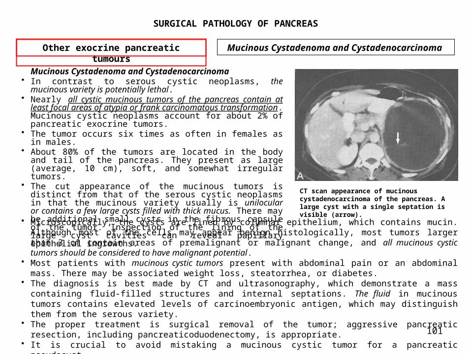
101
SURGICAL PATHOLOGY OF PANCREAS
Mucinous Cystadenoma and Cystadenocarcinoma • In contrast to serous cystic neoplasms, the
mucinous variety is potentially lethal. • Nearly all cystic mucinous tumors of the pancreas contain at
least focal areas of atypia or frank carcinomatous transformation. Mucinous cystic neoplasms account for about 2% of pancreatic exocrine tumors.
• The tumor occurs six times as often in females as in males.
• About 80% of the tumors are located in the body and tail of the pancreas. They present as large (average, 10 cm), soft, and somewhat irregular tumors.
• The cut appearance of the mucinous tumors is distinct from that of the serous cystic neoplasms in that the mucinous variety usually is unilocular or contains a few large cysts filled with thick mucus. There may be additional small cysts in the fibrous capsule of the tumor. Inspection of the lining of the large cyst cavities can reveal papillary epithelial ingrowths.
Other exocrine pancreatic tumours
Mucinous Cystadenoma and Cystadenocarcinoma
CT scan appearance of mucinous cystadenocarcinoma of the pancreas. A large cyst with a single septation is visible (arrow).
• Microscopically, the cysts are lined by columnar epithelium, which contains mucin. Although most of the cells may appear benign histologically, most tumors larger than 3 cm contain areas of premalignant or malignant change, and all mucinous cystic tumors should be considered to have malignant potential.
• Most patients with mucinous cystic tumors present with abdominal pain or an abdominal mass. There may be associated weight loss, steatorrhea, or diabetes.
• The diagnosis is best made by CT and ultrasonography, which demonstrate a mass containing fluid-filled structures and internal septations. The fluid in mucinous tumors contains elevated levels of carcinoembryonic antigen, which may distinguish them from the serous variety.
• The proper treatment is surgical removal of the tumor; aggressive pancreatic resection, including pancreaticoduodenectomy, is appropriate.
• It is crucial to avoid mistaking a mucinous cystic tumor for a pancreatic pseudocyst.

102
SURGICAL PATHOLOGY OF PANCREAS
• Internal drainage of a malignant mucinous cystic tumor results in catastrophic tumor dissemination and should never be performed. With appropriate treatment, all patients with histologically benign tumors should be cured; for tumors demonstrating malignant change, the 5-year survival rate after surgery is about 60%.
Pancreatic lymphoma• Although lymphoma is not a pancreatic exocrine tumor in the true sense, it can arise in the
pancreas and can present as a pancreatic mass lesion. • About one third of all non-Hodgkin lymphomas include at least microscopic involvement of the
pancreas, • The term pancreatic lymphoma is reserved for those cases in which the lymphoma appears to have
arisen in the pancreas and in which the bulk of the tumor burden is in the pancreas and peripancreatic tissues.
• Defined in this way, pancreatic lymphoma accounts for 1% to 2% of pancreatic neoplasms and about 1% of non-Hodgkin lymphomas.
• Pancreatic lymphoma usually has a rapid onset, and the tumors attain large size quickly. • At the time of presentation, pancreatic lymphomas are typically 6 to 10 cm. • The clinical presentation is similar to that of ductal pancreatic cancer: weight loss
and jaundice are the most common symptoms. • Pancreatic lymphoma may be difficult to distinguish from pancreatic carcinoma radiographically, except that the finding of a large tumor without vascular invasion and with extensive lymphadenopathy should raise the level of suspicion for lymphoma.
• When the diagnosis is suspected, percutaneous needle biopsy of the mass should be performed.
Other exocrine pancreatic tumours
Pancreatic lymphoma

103
SURGICAL PATHOLOGY OF PANCREAS
• If the diagnosis of lymphoma can be made unequivocally by percutaneous biopsy, and if there is sufficient tissue for cellular typing, laparotomy can be avoided because only rarely is it possible to resect the tumor, and jaundice usually resolves with chemotherapy.
• Most patients can be staged adequately with a CT scan and bone marrow biopsy.
• Surgical exploration is indicated if the diagnosis cannot be firmly established by needle biopsy, if additional tissue is believed necessary for adequate typing, or in the rare circumstance in which a small tumor might be completely resectable and the patient appears to have no disease outside the pancreas.
• Exploration for the purpose of biliary decompression alone is not ordinarily indicated. If the jaundiced patient is explored for another reason, cholecystojejunostomy is appropriate because the improvement in hepatic function allows the
Other exocrine pancreatic tumours
Pancreatic lymphoma
Pancreatic lymphoma. CT scan demonstrates a large mass virtually replacing the pancreas (arrow) and extensive peripancreatic lymphadenopathy.
more rapid institution of multidrug chemotherapy. • About half of patients undergo an initial complete remission with chemotherapy alone; long-term follow-up suggests, however, that most patients ultimately succumb to recurrent disease.

104
SURGICAL PATHOLOGY OF PANCREAS
• The management of tumors of the exocrine pancreas remains a formidable problem. • The last few years have seen improvement, particularly:
– in the ability to detect pancreatic neoplasms radiologically but also – in the safety of extirpative pancreatic surgery – in the firm demonstration that chemoradiotherapy is of value in the treatment of pancreatic cancer.
• The generally grim prognosis of pancreatic carcinoma relates primarily to the advanced stage of the tumors at the time of symptomatic presentation.
• The major challenges involve developing methods for earlier detection of pancreatic tumors, determining the optimal extent of surgery for pancreatic cancer, and developing adjunctive strategies to increase resectability and prevent tumor recurrence after surgery.
• Progress in meeting these goals has been slow, but some ground has been gained.
• In face of the increasing incidence of these tumors, it is important to continue to explore new possibilities in the hopes of further progress.
Exocrine pancreas malignancies CONCLUSIONS

105
SURGICAL PATHOLOGY OF PANCREAS
Incidence• Neoplasms of the endocrine pancreas(NEP) are rare, with an annual clinically
recognized incidence in the United States of about 5 cases per million persons and per year. • In unselected autopsy material, however, the prevalence of these tumors
approximates 1 per 100 person-years and are typically noted as incidental findings.
• Cells of the pancreatic islets are presumed to originate from neural crest cells. • Cells of this origin are called amine precursor uptake and decarboxylation - APUD
cells, indicating they have a high amine content of amine, are capable of amine precursor uptake, and contain an amino acid decarboxylase.
• A generalized derangement of the APUD system can cause abnormalities of multiple endocrine cells as is observed in multiple endocrine neoplasia (MEN) syndromes.
• Evidence suggests that some APUD cells may not originate from neural crest cells but rather have an endodermal origin.
• Neoplasms of the endocrine pancreas can be divided into: – functional and – nonfunctional varieties.
• Most pancreatic endocrine neoplasms discovered clinically are functional, indicating that they elaborate one or more hormonal products into the blood, leading to a recognizable clinical syndrome.
• Functional tumors are named according to their predominate clinical syndrome and hormonal product. • Patients with endocrine tumors of the pancreas with no recognizable clinical syndrome and normal
serum hormone levels (excluding pancreatic polypeptide) are considered to have nonfunctional pancreatic endocrine tumors.
Neoplasms of the endocrine pancreas
Generalities

106
SURGICAL PATHOLOGY OF PANCREAS
• All neoplasms of the endocrine pancreas have a similar light microscopic appearance. Routine histologic examination does not predict the biologic behavior or the endocrine manifestations of these neoplasms. Immunofluorescence techniques and the peroxidase-antiperoxidase procedure allow the demonstration of specific hormones within neoplastic cells. Malignancy is typically determined by the presence of local invasion that has spread to regional lymph nodes or by the existence of hepatic or distant metastases.
• Recent observations in the fields of classic and molecular genetics have added to our knowledge of pancreatic endocrine neoplasms. For example, malignant pancreatic endocrine tumors have been found to have clonal chromosomal abnormalities in up to half of cases, whereas ras oncogene mutations are absent in most of these tumors. Gastrinoma has been shown to be associated with amplification of the HER-2/neu protooncogene, and insulinoma has been shown to highly express mRNA for the a subunit of Gs protein. Further, sporadic pancreatic endocrine tumors and tumors arising as a manifestation of MEN I have both been shown to have mutations leading to genetic loss on chromosome 11, thereby inactivating a putative tumor suppressor gene on that chromosome.
• Three general principles apply to the treatment of patients with suspected functional neoplasms of the endocrine pancreas. First is the recognition of the abnormal physiology or characteristic syndrome. Characteristic clinical syndromes are well described for insulinoma, gastrinoma, VIPoma, and glucagonoma. The somatostatinoma syndrome is nonspecific, much more difficult to recognize, and exceedingly rare. Second is the detection of hormone elevations in serum by radioimmunoassay. Radioimmunoassays are widely available for measuring insulin, gastrin, vasoactive intestinal peptide (VIP), and glucagon. Assays for somatostatin, pancreatic polypeptide, prostaglandins, and other hormonal markers are not widely available but can be obtained from certain laboratories and investigators. The third step in patient evaluation involves localizing and staging the tumor in preparation for possible operative intervention.
Neoplasms of the endocrine pancreas
Generalities

107
SURGICAL PATHOLOGY OF PANCREAS
CT - The initial imaging technique recommended for localizing a pancreatic endocrine neoplasm is a dynamic abdominal computed tomographic (CT) scan with intravenous and oral contrast.
• The accuracy of CT in detecting the primary islet cell tumor varies from about 35% to 85% and depends largely on the scanning technique and the size and location of the primary tumor (Fig. 1).
• The accuracy of the CT scan in tumor localization is improved by using both oral and intravenous contrast as well as focused dynamic scanning through the pancreas at 5-mm intervals.
• The CT scan is also used to assess for peripancreatic lymph node enlargement and the presence of hepatic metastases.
Visceral angiography Should the CT scan fail to detect the primary tumor, the next step in radiograpic assessment is visceral angiography, focusing on the selective visualization of the arterial supply to the pancreas and peripancreatic regions.
• The accuracy of angiography in detecting the primary islet cell tumor varies from 45% to 85%, depending on radiographic technique and expertise, the selectivity of the contrast injection, and the size and neovascularity of the primary tumor (Fig. 2).
Neoplasms of the endocrine pancreas
Localization and staging
Fig.1 CT scan with oral and intravenous contrast in a patient with biochemical evidence of insulinoma. The neoplasm (arrow) is seen as a contrast-enhancing structure, 3 cm in diameter in the tail of the pancreas, posterior to the stomach
Fig. 2 Selective celiac angiogram in a patient with gastrinoma.

108
SURGICAL PATHOLOGY OF PANCREAS
• A newer technique which improves the preoperative localization of NEP is endoscopic ultrasonography.
Endoscopic ultrasonography was more sensitive than the combination of CT and visceral angiography. Somatostatin receptor imagingWhole body radionuclide scanning (WBRS) with iodine-labeled tyrosine3-octreotide is Another
technique that holds promise for the imaging of NEP reported with favorable results. Additional studies have been reported using indium-labeled pentetreotide, an octreotide analogue.
• These techniques rely on the presence of somatostatin receptors on many islet cell tumors and have the potential to identify primary tumors as well as hepatic and extrahepatic metastases.
• A modification of the WBRS technique has also been evaluated, using a hand-held gamma-detecting probe intraoperatively along with intravenous injection of 125I-tyrosine3-octreotide or lanreotide.
• In addition to somatostatin-receptor imaging, the use of VIP-receptor imaging has been recently reported for patients with pancreatic endocrine neoplasms.
• In a minority of patients with NEP, the primary tumor is not be localized with imaging studies such as CT, visceral angiography, or endoscopic ultrasound. This is most common in patients with insulinoma or gastrinoma.
Selective transhepatic portal venous hormone sampling may help to assist in localizing the occult neoplasm.
• This invasive technique is designed to demonstrate an increase in hormone concentration at the site where the tumor drains its hormonal product into the portal venous system.
• The overall accuracy of this test ranges from 70% to higher than 95% Selective arterial secretin stimulation test. • Is a newer technique which has been described for localizing occult gastrinomas. It
involves selective visceral arterial secretin injection with concurrent hepatic venous sampling for gastrin.
Neoplasms of the endocrine pancreas
Neoplasms of the endocrine pancreas
Localization and staging

109
SURGICAL PATHOLOGY OF PANCREAS
• This technique takes advantage of the unique biology of gastrinoma, in that gastrinoma cells are known to respond both in vitro and in vivo to secretin with the release of gastrin.
• Secretin is serially injected through an arterial catheter into at least three sites—the splenic, gastroduodenal, and inferior pancreaticoduodenal arteries. Samples are drawn from an hepatic vein catheter before and immediately after these three or more arterial secretin injections. The arterial supply to the occult gastrinoma can be determined based on which selective secretin injection is followed by a large increment in hepatic vein gastrin concentration.
• The selective arterial secretin stimulation test was more sensitive than portal venous sampling in localizing gastrinomas, particularly small gastrinomas arising in the duodenum.
Neoplasms of the endocrine pancreas
Localization and staging
Endoscopic ultrasound image in a patient with an insulinoma (arrows) in the body of the pancreas. SV, splenic vein.
Schematic depiction of data from transhepatic portal venous insulin sampling in a patient with an insulinoma.

110
SURGICAL PATHOLOGY OF PANCREAS
Graphic depiction of the results of a selective arterial secretin stimulation test
in a patient with gastrinoma.
Neoplasms of the endocrine pancreas
Localization and staging

111
SURGICAL PATHOLOGY OF PANCREAS
• Surgical exploration for NEP, permits to perform a complete evaluation of the pancreas and peripancreatic regions: – The body and tail of the pancreas are exposed by dividing the gastrocolic ligament. This portion of the pancreas can be partially elevated out of the retroperitoneum by dividing the inferior retroperitoneal attachments to the gland.
– After elevating the second portion of the duodenum out of the retroperitoneum using the Kocher maneuver, the pancreatic head and uncinate process are palpated bimanually.
– The liver is carefully assessed for evidence of metastatic disease. – Potential extrapancreatic sites of tumor are evaluated in all cases, with particular attention paid to the duodenum, splenic hilum, small intestine and its mesentery, peripancreatic lymph nodes and the reproductive tract in women.
• One technique that provides additional information in the intraoperative ultrasonography, which can assist in tumor identification.
• The goals of surgical therapy for pancreatic endocrine neoplasms include: – controlling symptoms from hormone excess, – safely resecting maximal tumor mass, and preserving maximal pancreatic parenchyma.
– Management strategies, including preoperative, intraoperative, and postoperative considerations, vary for the different types of endocrine neoplasms of the pancreas.
Neoplasms of the endocrine pancreas
Surgical exploration

112
SURGICAL PATHOLOGY OF PANCREAS
• Insulinoma is the most common neoplasm of the endocrine pancreas. • The insulinoma syndrome is associated with the Whipple triad:
(1) symptoms of hypoglycemia during fasting; (2) documentation of hypoglycemia with serum glucose less than 50 mg/dL;
and (3) relief of hypoglycemic symptoms following administration of exogenous
glucose. • Autonomous insulin secretion from insulinomas leads to spontaneous hypoglycemia, with
symptoms that can be characterized into two groups: – Neuroglycopenic symptoms include:
• confusion, • seizure, • obtundation, • personality change, and • coma.
– Hypoglycemia-induced catecholamine-surge symptoms include: • palpitations, • trembling, • diaphoresis, and • tachycardia.
• In most cases, patients consume carbohydrate-rich meals and snacks to relieve or prevent these symptoms.
• The Whipple triad is not specific for insulinoma. The differential diagnosis of adult hypoglycemia is extensive and includes the following: – Reactive hypoglycemia – Functional hypoglycemia associated with gastrectomy or
gastroenterostomy – Nonpancreatic tumors – Pleural mesothelioma – Sarcoma
Neoplasms of the endocrine pancreas
Neoplasms of the endocrine pancreas
Insulinoma

113
SURGICAL PATHOLOGY OF PANCREAS
– Adrenal carcinoma – Hepatocellular carcinoma – Carcinoid – Hypopituitarism – Chronic adrenal insufficiency – Extensive hepatic insufficiency – Surreptitious administration of insulin or ingestion of oral
hypoglycemic agents• A common mistake made in evaluating a patient with suspected insulinoma is
to begin with an oral glucose tolerance test. • Insulinoma is most reliably diagnosed using a monitored fast. During a
monitored fast, blood is sampled every 4 to 6 hours for glucose and insulin determinations and also at the time of symptom occurrence.
• Hypoglycemic symptoms typically occur when glucose levels are below 50 mg/dL, with concurrent serum insulin levels often exceeding 25 mU/mL.
• Additional support for the diagnosis of insulinoma comes from the calculation of the insulin/glucose ratio at different points during the monitored fast.
• Normal persons have insulin/glucose ratios less than 0.3.• Patients with insulinoma typically demonstrate insulin/glucose ratios greater
than 0.4 after a prolonged fast. • Other measurable b-cell products synthesized in excess in patients with insulinoma
include C peptide and proinsulin. Elevated levels of both are typically found in the peripheral blood of patients with insulinoma.
• The possibility of surreptitious insulin or oral hypoglycemic agent administration should be considered in all patients with suspected insulinoma.
• C-peptide and proinsulin levels are not elevated in patients who self-administer insulin. • Additionally, patients self-administering either bovine or porcine insulin
may demonstrate anti-insulin antibodies in circulating blood.
Neoplasms of the endocrine pancreas
Insulinoma

114
SURGICAL PATHOLOGY OF PANCREAS
• The presence of oral hypoglycemic agents such as sulfonylureas can be assessed using standard toxicologic screening.
• After confirming the diagnosis of insulinoma by biochemical analyses, the appropriate localization and staging studies described earlier are performed.
• For insulinoma, the standard imaging studies include: – abdominal CT, – endoscopic ultrasound, and – visceral angiography.
• The treatment of insulinoma is surgical in nearly all cases. • Insulinomas are found evenly distributed in the pancreas, with one third
found in the head and uncinate process, one third in the body, and one third in the tail of the gland.
• Of patients diagnosed with insulinoma, 90% are found to have benign solitary adenomas amenable to surgical cure.
• Less than 10% of patients with insulinoma have some form of the MEN I syndrome. In patients with MEN I, the possibility of multiple insulinomas must be suspected, and the recurrence rate is higher than in sporadic cases.
• In about 10% of all cases, insulinoma is metastatic to peripancreatic lymph nodes or to the liver, justifying a diagnosis of malignant insulinoma.
• Small benign insulinomas not close to the main pancreatic duct can be removed by enucleation,39 independent of their location in the gland
• Large insulinomas deep in the head or uncinate process of the pancreas may not be amenable to local excision and may require pancreaticoduodenectomy.
• In rare instances, patients undergo exploration for insulinoma without definite preoperative tumor localization.
• At surgery, no tumor can be identified intraoperatively by visualization, palpation, and real-time ultrasonography.
Neoplasms of the endocrine pancreas
Insulinoma

115
SURGICAL PATHOLOGY OF PANCREAS
• In these circumstances, a management dilemma exists. • Some authors have recommended a “blind” distal (left-sided) pancreatic
resection to the level of the superior mesenteric vein (60% to 70% pancreatectomy) in hopes of excising a previously unidentified insulinoma residing in the body or tail of the pancreas.
• Others have suggested that a blind pancreaticoduodenectomy would be more appropriate because the thickness of the head and uncinate process render this region of the pancreas most likely to harbor an unidentifiable insulinoma.
• The favored option in the situation of an occult insulinoma is to defer any form of blind resection and to perform postoperative selective transhepatic portal venous insulin sampling to allow for specific tumor localization and directed surgical excision at a second operation.
• About 10% of insulinomas are malignant, typically with evidence of lymph node or hepatic metastases.
• Under these circumstances, cautious and safe resection of the primary tumor and accessible metastases should be considered.
• Such tumor debulking can be helpful in reducing hypoglycemic symptoms that can threaten long-term survival.
• The average patient survives several years after diagnosis and treatment of malignant islet cell tumors, indicating that the natural history of these malignant tumors typically follows an indolent course.
• In patients with unresectable insulinoma, dietary manipulations to include judicious spacing of carbohydrate-rich meals and night-time snacks can be helpful to minimize dangerous hypoglycemic episodes. Medications such as diazoxide and octreotide can be used to inhibit insulin release, raise serum glucose, and further minimize hypoglycemia.
• Chemotherapeutic agents with some efficacy against malignant insulinoma include streptozocin, dacarbazine, doxorubicin and 5-fluorouracil. The highest response rates to chemotherapy have been observed using combination therapy.
Neoplasms of the endocrine pancreas
Insulinoma

116
SURGICAL PATHOLOGY OF PANCREAS
Neoplasms of the endocrine pancreas
Insulinoma
The technique used for enucleation of a benign pancreatic endocrine neoplasm,
117
SURGICAL PATHOLOGY OF PANCREAS
• In 1955, Zollinger and Ellison described two patients with severe peptic ulcer disease and pancreatic endocrine tumors, postulating that an ulcerogenic agent originated from the pancreatic tumor.
• It is currently estimated that 1 in 1000 patients with primary duodenal ulcer disease and 2 in 100 patients with recurrent ulcer after ulcer surgery harbor gastrinomas.
• 75% of gastrinomas occur sporadically, and 25% are associated with the MEN I syndrome. • In the past, most gastrinomas were found to be malignant, based on the
findings of metastatic disease at the time of workup or exploration. More recently, with increased awareness and earlier screening for hypergastrinemia, the diagnosis of gastrinoma is made earlier, leading to the discovery of a higher percentage of benign, curable neoplasms.
• The clinical symptoms of patients with gastrinoma are a direct result of the circulating hypergastrinemia:
– Abdominal pain and – Peptic ulceration of the upper gastrointestinal tract are seen in up
to 90% of patients. – Diarrhea in 50% of patients, and about 10% have diarrhea as the
solitary symptom. – Esophageal symptoms or endoscopic abnormalities from gastroesophageal
reflux are seen in over 50% of patients, with esophagitis typically occurring in association with peptic ulcer disease or diarrhea.
• The diagnosis of gastrinoma should be suspected in several clinical settings, and the liberal use of serum gastrin measurement for screening is encouraged. The indications for measuring gastrin include the following:
– Peptic ulcer disease – Initial diagnosis – Recurrent ulcer – Failure of medical therapy – Postoperative ulcer – Postbulbar ulcer – Family history of ulcer disease – Ulcer with diarrhea
Neoplasms of the endocrine pancreas
Gastrinoma

118
SURGICAL PATHOLOGY OF PANCREAS
– Prolonged undiagnosed diarrhea – MEN I kindred – Nongastrinoma pancreatic endocrine tumor (because of the high
association of secondary hormone elevations)– Prominent gastric rugal folds on upper gastrointestinal series
(reflecting the trophic effect of gastrin on the gastric fundus)• In most patients with gastrinomas, the fasting serum gastrin level is
elevated to at least 200 pg/mL. Gastrin values over 1000 pg/mL are virtually diagnostic of gastrinoma, particularly when they are accompanied by hyperchlorhydria or well-established ulcer disease.
• Fasting hypergastrinemia alone, however, is not sufficient for the diagnosis of gastrinoma. This is because hypergastrinemia can exist in other pathophysiologic states, since gastrin is the normal secretory product of antral G cells.
• Gastric acid analysis is an important test in evaluating patients with suspected gastrinoma, because it can differentiate between ulcerogenic (high gastric acid) causes of hypergastrinemia and nonulcerogenic (low gastric acid) causes of hypergastrinemia. To obtain an accurate gastric acid analysis, patients must abstain from antisecretory medications, such as H2 antagonists or omeprazole.
• The diagnosis of gastrinoma is supported by a basal acid output higher than 15 mEq/h in nonoperated patients, a basal acid output that exceeds 5 mEq/h in patients with previous vagotomy or antiulcer operations, or a ratio of basal to maximal acid output that exceeds 0.6.
• Once it is documented that hypergastrinemia is associated with excessive acid secretion, provocative testing using secretin should be performed to differentiate between gastrinoma, antral G-cell hyperplasia or hyperfunction, and the other causes of ulcerogenic hypergastrinemia.
• The secretin stimulation test is done in the fasting state by obtaining peripheral serum samples for gastrin in the basal period, administering secretin, 2 U/kg, as an intravenous bolus, and obtaining serum samples for gastrin at 5-minute intervals for 30 minutes. An increase in the gastrin level of more than 200 pg/mL above the basal level supports the diagnosis of gastrinoma (Fig. 34-7).
Neoplasms of the endocrine pancreas
Gastrinoma

119
SURGICAL PATHOLOGY OF PANCREAS
DISEASE STATES ASSOCIATED WITH HYPERGASTRINEMIA
NONULCEROGENIC CAUSES (NORMAL TO LOW ACIDSECRETION)• Atrophic gastritis• Pernicious anemia• Previous vagotomy• Renal failure• Short-gut syndrome ULCEROGENIC CAUSES (EXCESS ACID SECRETION)• Antral G-cell hyperplasia or hyperfunction• Gastric outlet obstruction• Retained excluded antrum• Zollinger- Ellison syndrome
Neoplasms of the endocrine pancreas
Gastrinoma
• This conventional secretin stimulation test done on peripheral blood differs markedly from the doubly invasive selective arterial secretin stimulation test that is designed to localize the site of the primary tumor in patients with gastrinomas that have already been proved by biochemical testing.
• After the biochemical confirmation of the diagnosis of gastrinoma, two steps are important in patient treatment.
• First, gastric acid hypersecretion is pharmacologically controlled. Omeprazole, 20 to 200 mg/d, is now considered the drug of choice for antisecretory therapy in patients with gastrinoma. The dose is adjusted to achieve a nonacidic gastric pH during the hour immediately before the next dose of the drug.
• Second, after the initiation of omeprazole therapy, all patients with gastrinomas should undergo imaging studies to localize the primary tumor and to assess for metastatic disease.
• The modalities appropriate for localization and staging of gastrinoma patients have already been discussed and include dynamic abdominal CT scanning with intravenous and oral contrast, selective visceral angiography, endoscopic ultrasonography, somatostatin receptor imaging, percutaneous transhepatic portal venous sampling for gastrin, and the selective arterial secretin stimulation test.

120
SURGICAL PATHOLOGY OF PANCREAS
• Patients whose localization and staging studies indicate unresectable hepatic metastases should undergo percutaneous or laparoscopically directed liver biopsy for absolute histologic verification. If unresectable gastrinoma is confirmed, open surgical exploration is not performed, and the patient is maintained on long-term omeprazole therapy. Virtually all patients can be rendered achlorhydric with an appropriate dose of omeprazole. Noncompliant patients who refuse to take appropriate doses of omeprazole and who experience complications related to their ulcer diathesis may require total gastrectomy. Total gastrectomy removes the end organ (parietal cell mass) and was once the procedure of choice for gastrinoma. Today, its use in patients with gastrinomas has markedly declined.
• In most patients, unresectable disease is not identified by staging studies, and patients should be offered surgical exploration with curative intent. At the time of exploration, the entire abdomen is carefully assessed for areas of extrapancreatic and extraduodenal gastrinoma. Most gastrinomas are found to the right of the superior mesenteric vessels, in the head of the pancreas or the duodenum. This area is called the gastrinoma triangle.
• Intraoperative ultrasonography should be available to assist in tumor localization. In addition, intraoperative upper endoscopy may help by allowing transillumination of the duodenal wall and identification of small duodenal gastrinomas. At exploration, any suspicious peripancreatic lymph nodes are excised and submitted for frozen section. Primary tumors located in the substance of the pancreas that are small (less than 2 cm) and well-encapsulated can be carefully enucleated. Pancreatic tumors without defined capsules or that are situated deep in the pancreatic parenchyma may require partial pancreatic resection by either distal pancreatectomy or pancreaticoduodenectomy.
• In the absence of an identifiable pancreatic or duodenal tumor, a longitudinal duodenotomy can be performed at the level of the second portion of the duodenum to allow for eversion of the duodenum in a search for duodenal microgastrinomas. Primary gastrinomas identified in the duodenal wall are resected locally with primary closure of the duodenal defect. In a small percentage of patients, gastrinoma is found only in peripancreatic lymph nodes, with these lymph nodes harboring the primary tumor. Resection of these apparent lymph node primary gastrinomas has been associated with long-term eugastrinemia and biochemical cure in up to half of cases.
• Occasionally, preoperative localization studies, such as portal venous gastrin sampling or the selective arterial secretin stimulation test, localize the tumor in the gastrinoma triangle; however, no tumor may be demonstrable at laparotomy.
• In the face of such a negative exploration, several surgical options are available.• First, parietal cell vagotomy has been proposed as a way to reduce antisecretory drug
dose requirements in patients on high-dose antisecretory drug therapy but without prior life-threatening complications.
• Total gastrectomy is the second surgical option for patients with negative results on exploration. Although total gastrectomy was the most reliable way to control ulcer diathesis in the past, the introduction and availability of omeprazole has drastically reduced the need for total gastrectomy. There may still be a limited role for total gastrectomy in patients whose tumors cannot be localized if they cannot or will not take adequate doses of omeprazole.
Neoplasms of the endocrine pancreas
Gastrinoma

121
SURGICAL PATHOLOGY OF PANCREAS
• Unfortunately, like parietal cell vagotomy, total gastrectomy leaves the primary tumor behind with the potential for subsequent tumor growth, metastases, and patient death from tumor burden.
• A third surgical option in patients with clear-cut biochemical documentation of hypergastrinemia, hyperchlorhydria, and tumor localization in the gastrinoma triangle involves blind pancreaticoduodenectomy. In a small number of patients, these blind resections have yielded pathologically verified primary gastrinomas in the duodenal wall or the head of the pancreas which were not apparent at laparotomy. Blind resections should be performed as classic pancreaticoduodenectomies,(without pylorus preserving technique) including a distal gastric resection, because duodenal gastrinomas may arise close to the pylorus and be inadvertently left behind with pylorus-sparing pancreaticoduodenectomy.
• In a limited number of cases reported, patients have been rendered eugastrinemic by blind resection, and most continue to be eugastrinemic at postoperative follow-up.
Neoplasms of the endocrine pancreas
Gastrinoma
The gastrinoma triangle: operative implications.

122
SURGICAL PATHOLOGY OF PANCREAS
• Most patients with incurable metastatic gastrinoma succumb to eventual tumor growth and dissemination.
• Multiple modalities have been used to treat patients with such metastatic gastrinoma. The overall objective response rate to chemotherapy appears to be less than 50%.
• A prospective study of monthly cycles of streptozocin, 5-fluorouracil, and doxorubicin in 10 patients with metastatic gastrinoma showed a partial response rate of 40%, with 60% having no response. Chemotherapy did not improve the length of survival.
• Hormonal therapy with octreotide has been reported to improve symptoms, reduce hypergastrinemia, and diminish hyperchlorhydria in patients with metastatic gastrinoma.
• Patients with gastrinoma associated with MEN I present difficult treatment issues. Omeprazole should be used to control gastric acid hypersecretion. Surgical treatment of hypercalcemia caused by parathyroid hyperplasia should preceed any surgical treatment of hypergastrinemia. MEN I gastrinoma typically involves multiple pancreatic or duodenal neoplasms, and careful preoperative and intraoperative localization techniques are needed to guide resection. In limited numbers of patients, hypergastrinemia associated with MEN I gastrinoma has been corrected by surgical resection at short term follow-up. The overall cure rates with MEN I gastrinoma appear to be lower than those with sporadic gastrinoma.
Neoplasms of the endocrine pancreas
Gastrinoma

123
SURGICAL PATHOLOGY OF PANCREAS
• Verner and Morrison are credited with the definition of this secretory-type diarrhea syndrome following their report of two cases in 1958.
• Synonyms for this syndrome include the WDHA syndrome (watery diarrhea, hypokalemia, and either achlorhydria or hypochlorhydria) and the pancreatic cholera syndrome.
• Patients characteristically present with – intermittent severe diarrhea, typically of a watery nature, averaging
5 L/d (Table 34-5). – Malabsorption and steatorrhea are not common.– Hypokalemia results from the fecal loss of large amounts of potassium
(up to 400 mEq/d), and low serum potassium levels are associated with – muscular weakness, – lethargy, and – nausea. – Half of the patients have some degree of hyperglycemia and
hypercalcemia, and cutaneous flushing can be observed in a minority. • The diagnosis of VIPoma is typically made after excluding other more common
causes of diarrhea. The active agent in the VIPoma syndrome is usually VIP, with a minority of patients having elevations of other candidate mediators such as peptide histidine-isoleucine or prostaglandins. Because VIP secretion can be episodic in patients with VIPomas, several fasting VIP levels should be measured because a single low VIP level does not rule out the syndrome.
• After biochemical documentation of elevated VIP levels, tumor localization and staging begins with dynamic abdominal CT scan with intravenous and oral contrast. In addition, because 10% of patients with VIPomas may have extrapancreatic tumors located in the retroperitoneum or thorax, a thoracic CT scan is indicated if the abdominal scan fails to identify a tumor.
• In most reported cases, the abdominal CT scan identified the tumor, and further imaging studies, such as visceral angiography or portal venous hormone sampling, were unnecessary.
Neoplasms of the endocrine pancreas
VIPoma

124
SURGICAL PATHOLOGY OF PANCREAS
• In preparing patients with VIPomas for surgical exploration, fluid and electrolyte balances must be corrected by vigorous intravenous fluid administration and appropriate electrolyte replacement.
• Therapy with parenterally administered octreotide can be an important adjunct in the preoperative setting because octreotide leads to a reduction in circulating VIP levels with a resultant decrease in the volume of diarrhea. Before octreotide was available, corticosteroids and indomethacin were used preoperatively to control diarrhea and associated fluid and electrolyte losses.
• Surgical excision of the VIPoma is appropriate in all patients with the Verner-Morrison syndrome. Most VIPomas have been located in the distal pancreas, where they are amenable to resection by distal pancreatectomy. If no tumor is found in the pancreas, a careful exploration of the retroperitoneum including both adrenals should be performed.
• Metastatic disease to the lymph nodes and the liver have been reported in half of all cases. In the presence of metastatic disease, safe palliative debulking of the metastatic tumor is indicated.
• In patients with recurrent or unresectable VIPoma, octreotide therapy is used to reduce circulating VIP levels and control diarrhea.
• Chemotherapy specific for VIPoma patients has not been studied prospectively, although small numbers of patients have appeared to partially respond to streptozocin, combination chemotherapy or interferon.
Neoplasms of the endocrine pancreas
VIPoma

125
SURGICAL PATHOLOGY OF PANCREAS
VIPoma Parameter Description
Symptoms• Watery diarrhea• Weakness• Lethargy• Nausea
Diagnostic tests• Hypokalemia• Achlorhydria• Serum vasoactive intestinal
peptide levelsAnatomic localization
• Most in body or tail of pancreas
Neoplasms of the endocrine pancreas
VIPoma
Verner-Morrison Syndrome: Differential Diagnosis
Entity Workup
Villous adenoma Lower GI endoscopyLaxative abuse Stool examination for phenolphthaleinCeliac disease Fecal fat measurement D-Xylose tolerance test Small bowel biopsyParasitic and infectious Stool culturediseases Ovum and parasite analysis Clostridium difficile toxin assayInflammatory bowel Lower GI endoscopydisease Upper GI and small bowel seriesCarcinoid syndrome Urinary 5˘-HIAA Upper GI and small bowel series Abdominal CT scan Serum serotonin measurementGastrinoma Serum gastrin measurement Gastric acid analysis Secretin stimulation test

126
SURGICAL PATHOLOGY OF PANCREAS
• glucagonoma • somatostatinoma • calcitoninoma, • parathyrinoma, • GRFoma, • ACTHoma and • neurotensinoma.
Neoplasms of the endocrine pancreas
Neoplasms of the endocrine pancreas
Rare

127
SURGICAL PATHOLOGY OF PANCREAS
• The most common findings in the glucagonoma syndrome are: – severe dermatitis, – mild diabetes, – stomatitis, – anemia, and– weight loss
• The dermatitis manifests as a characteristic skin rash termed necrolytic migratory erythema. This rash exhibits cyclic migrations with erythematous patches that spread serpiginously and central healing points of resolution. It has been theorized that the hypoaminoacidemia seen in patients with glucagonoma is responsible for the dermatitis. Because glucagon is a catabolic hormone, glucagonoma patients typically demonstrate malnutrition and hypoproteinemia.
• The diagnosis of glucagonoma may be suggested by: – the clinical presentation and – biopsy of the skin lesions but is secured by the documentation of – elevated levels of fasting serum glucagon.
Normal fasting levels of glucagon peak at 150 pg/mL. The diagnosis of glucagonoma can be confirmed by demonstration of hypoaminoacidemia, however, such testing is expensive and is not necessary in most cases.
• Patients should undergo radiographic localization and staging with dynamic contrast-enhanced abdominal CT scan. These tumors are usually large and solitary, the CT scan localizes the tumor in most patients.
• Before exploration, attention should be given to managing the malnutrition. Total parenteral nutrition has been used to improve the catabolic state created by hyperglucagonemia, reverse the malnutrition, and improve the dermatitis.
• Octreotide has been used to reduce the circulating glucagon levels and to allow improved response to total parenteral nutrition.
Neoplasms of the endocrine pancreas
GlucagonomaNeoplasms of the endocrine pancreas

128
SURGICAL PATHOLOGY OF PANCREAS
• Most glucagonomas have been located in the body and tail of the pancreas. These tumors are typically large and bulky, and surgical resection has required distal pancreatectomy. Metastases have been found in most patients, and safe debulking of these metastatic lesions should be considered.
• Glucagonoma patients with incurable or recurrent disease appear to have low response rates to standard chemotherapeutic agents such as streptozocin and dacarbazine. Octreotide can be successful in reducing elevated glucagon levels and in controlling the hyperglycemia and dermatitis associated with incurable glucagonoma.
Neoplasms of the endocrine pancreas
GlucagonomaNeoplasms of the endocrine pancreas
GLUCAGONOMA Parameter Description
Symptoms Dermatitis manifested as necrolytic migrator erythema Stomatitis Weight lossDiagnostic tests Hyperglycemia Hypoproteinemia Serum glucagon measurement Serum amino acid profileAnatomic localization Most in body or tail of pancreas

129
SURGICAL PATHOLOGY OF PANCREAS
• The somatostatinoma syndrome is the least common of the five generally accepted functional pancreatic endocrine neoplasia syndromes, with an estimated annual incidence of less than 1 in 40 million people.
• The clinical features of the somatostatinoma syndrome are nonspecific and include: – steatorrhea,– diabetes, – hypochlorhydria, and – cholelithiasis (Table 34-8).
• A fasting plasma somatostatin level can be used to confirm the diagnosis of a somatostatinoma. • While the normal plasma level is below 100 pg/mL, patients with
somatostatinoma have been found to have high levels of circulating somatostatin, often measurable in nanograms per milliliter.
• Most somatostatinomas have been located in the head of the pancreas and the periampullary region. The most useful test for localization and staging is the abdominal CT scan, which has been used to identify and stage these typically large tumors.
• Preoperative treatment of patients with somatostatinoma involves treatment of hyperglycemia and malnutrition.
• Surgery resection for cure has been uncommon because of the presence of metastatic disease in most cases. Safe resection of the primary tumor and careful debulking of hepatic metastases appear to be indicated.
• At the time of exploration, cholecystectomy is indicated even in the absence of documented gallstones because of the concern about the development of cholelithiasis with persistently elevated somatostatin levels.
Neoplasms of the endocrine pancreas
Neoplasms of the endocrine pancreas
Somatostatinoma
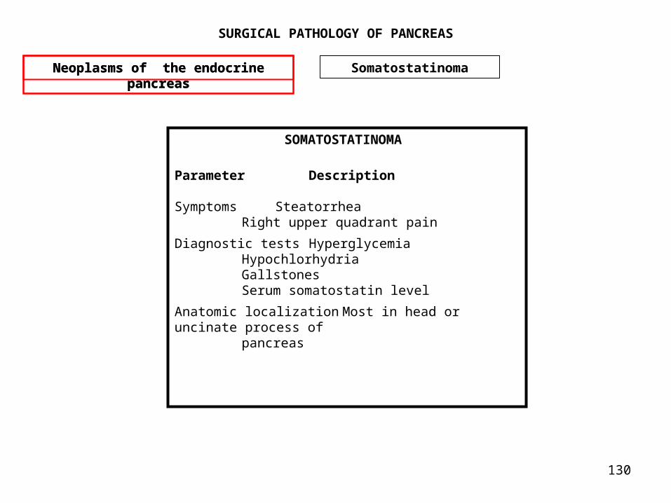
130
SURGICAL PATHOLOGY OF PANCREAS
Neoplasms of the endocrine pancreas
Neoplasms of the endocrine pancreas
Somatostatinoma
SOMATOSTATINOMA Parameter Description
Symptoms Steatorrhea Right upper quadrant painDiagnostic tests Hyperglycemia Hypochlorhydria Gallstones Serum somatostatin levelAnatomic localization Most in head or uncinate process of pancreas

131
SURGICAL PATHOLOGY OF PANCREAS
• There are several extremely rare clinical syndromes that have been proposed as candidate functional endocrine syndromes associated with pancreatic neoplasms (Table 34-9). These include calcitoninoma,89,90 parathyrinoma,91 GRFoma, ACTHoma and neurotensinoma.92 Calcitonin-secreting pancreatic endocrine neoplasms are associated with watery diarrhea, whereas parathyrinomas are accompanied by elevations in PTH-related protein with clinical features of hypercalcemia. GRFoma is marked by elevations of serum growth hormone–releasing factor (GRF), with clinical features of acromegaly. ACTHoma has features of Cushing syndrome, with elevated serum adenocorticotropic hormone (ACTH). Neurotensinoma appears to be characterized by tachycardia, hypotension, and malabsorption, with elevation of serum neurotensin. As further cases are reported and clinical experience broadens, these rare and unusual functional pancreatic exocrine neoplasms and others may someday be recognized along with the classic five syndromes of insulinoma, gastrinoma, VIPoma, glucagonoma, and somatostatinoma.
•
Neoplasms of the endocrine pancreas
Neoplasms of the endocrine pancreas
RARE

132
SURGICAL PATHOLOGY OF PANCREAS
• In about a third of patients with neoplasms of the endocrine pancreas, there is no defined clinical syndrome and no lack of elevated serum insulin, gastrin, VIP, glucagon and somatostatin levels. These patients are considered to have nonfunctional endocrine neoplasms. The one hormone that may be elevated in the serum in these nonfunctional tumors is pancreatic polypeptide. It appears to be a marker for some pancreatic endocrine tumors without being the mediator of any specific pancreatic polypeptide–related clinical syndrome.93 These nonfunctional endocrine neoplasms present with clinical manifestations such as abdominal pain, weight loss and jaundice resulting from space-occupying lesions in the pancreas.94,95 These clinical manifestations are similar to those found in patients with ductal adenocarcinoma of the pancreas. Nonfunctional tumors are most commonly located in the head, neck, or uncinate process of the pancreas.96 The malignancy rate for these tumors ranges from 50% to 90%. However, in contrast to the poor prognosis associated with ductal adenocarcinoma of the pancreas, these nonfunctional tumors tend to grow in a more indolent fashion and to be associated with a longer survival.
• Localization and staging studies are performed in a similar fashion to those performed for patients with the more common diagnosis of ductal adenocarcinoma of the exocrine pancreas. The abdominal CT scan is used to evaluate the primary tumor and to assess for hepatic metastases. Preoperative cholangiography may be indicated in the setting of jaundice with the potential for imaging by endoscopic or percutaneous transhepatic routes. At surgery most of these nonfunctional neoplasms are larger than 2 cm and are not safely excised by local techniques. Tumors in the head, neck, or uncinate process of the pancreas typically require pancreaticoduodenectomy for safe resection, whereas tumors arising in the body or tail of the pancreas are treated by distal pancreatectomy. Patients with unresectable tumors in the head of the pancreas are candidates for surgical palliation of obstructive jaundice and gastric outlet obstruction by biliary-enteric and gastroenteric bypass, respectively. The overall 5-year survival rate in all patients with resected nonfunctional pancreatic neoplasms approaches 50%.97
• In patients with unresectable disease, partial responses to combination chemotherapy have been reported. In a multicenter trial reported on by Moertel and associates,46 105 patients with advanced islet cell carcinoma, half of whom had nonfunctional tumors, were randomly assigned to one of three treatment regimens. The lowest response rate (30%) was seen in the group receiving chlorozotocin alone; an intermediate response rate of 45% was seen in patients receiving the combination of streptozocin plus 5-fluorouracil; and the highest response rate of 69% was seen in patients receiving streptozocin plus doxorubicin. The streptozocin-plus-doxorubicin therapy was associated with a significant survival advantage when compared to the other two treatments. The most common toxic reactions to the chemotherapy were nausea and vomiting, leukopenia, and mild renal insufficiency.
•
Neoplasms of the endocrine pancreas
Neoplasms of the endocrine pancreas
Nonfunctional

133
SURGICAL PATHOLOGY OF PANCREAS
Cronic Pancreatitis Diagnosis
All neoplasms of the endocrine pancreas have a similar light microscopic appearance. Routine histologic examination does not predict the biologic behavior or the endocrine manifestations of these neoplasms. Immunofluorescence techniques and the peroxidase-antiperoxidase procedure allow the demonstration of specific hormones within neoplastic cells. Malignancy is typically determined by the presence of local invasion that has spread to regional lymph nodes or by the existence of hepatic or distant metastases.
Recent observations in the fields of classic and molecular genetics have added to our knowledge of pancreatic endocrine neoplasms. For example, malignant pancreatic endocrine tumors have been found to have clonal chromosomal abnormalities in up to half of cases,2 whereas ras oncogene mutations are absent in most of these tumors.3 Gastrinoma has been shown to be associated with amplification of the HER-2/neu protooncogene,4 and insulinoma has been shown to highly express mRNA for the a subunit of Gs protein.5 Further, sporadic pancreatic endocrine tumors and tumors arising as a manifestation of MEN I have both been shown to have mutations leading to genetic loss on chromosome 11, thereby inactivating a putative tumor suppressor gene on that chromosome.6
Three general principles apply to the treatment of patients with suspected functional neoplasms of the endocrine pancreas. First is the recognition of the abnormal physiology or characteristic syndrome. Characteristic clinical syndromes are well described for insulinoma, gastrinoma, VIPoma, and glucagonoma. The somatostatinoma syndrome is nonspecific, much more difficult to recognize, and exceedingly rare. Second is the detection of hormone elevations in serum by radioimmunoassay. Radioimmunoassays are widely available for measuring insulin, gastrin, vasoactive intestinal peptide (VIP), and glucagon. Assays for somatostatin, pancreatic polypeptide, prostaglandins, and other hormonal markers are not widely available but can be obtained from certain laboratories and investigators. The third step in patient evaluation involves localizing and staging the tumor in preparation for possible operative intervention.