Effect of rosiglitazone on the pathology of experimentally ...
Upload
khangminh22Category
view
0download
0
Pathology of the uvea
Carol Naranjo, LV, DACVP, DECVP, PhDIDEXX Laboratories

Uvea
• Anterior:– Iris– Ciliary body
• Posterior:– Choroid
Maggs, Miller, Ofri. Slatter’s fundamentals of Veterinary Ophthalmology, 2013

Normal Histology – Anterior Uvea

Normal histology - Choroid
Tapetal Non-tapetal
Feline tapetum

Uvea
• Congenital disease
• Degenerative, hyperplastic, age related changes
• Uveitis and inflammation w/in the globe
– Non-infectious
– Infectious

CONGENITAL DISEASE
Uvea

Congenital disease
• Iris hypoplasia
• Aniridia
• PPM
Maggs, Miller, Ofri. Slatter’s fundamentals of Veterinary Ophthalmology, 2013


DEGENERATIVE, HYPERPLASTIC,
AGE-RELATED CHANGES
Uvea

Degenerative, hyperplastic, age-related
• Iris atrophy: age-related, trauma, chronic glaucoma
• Cysts of iridociliary epithelium:
– Pars plana cysts: aged cats and horses: NP CB epit
– Sporadic iridal cysts: non-significant
• Except in some breeds (GR, Great Dane, American Bulldog)

Pars plana cysts

IRIS CYSTS

Thin-walled iridociliary cysts in GR
• Pigmentary and cystic glaucoma of GR
– GR uveitis, pigmentary uveitis, cystic uveal dz
• Bilateral, not symmetrical
• No goniodysgenesis
• Clinical and histological differences noted

Thin-walled iridociliary cysts in GR
• Gross:
– Difficult to see cysts
– Pigment:
• Back of the cornea
• Lens capsule

Courtesy of Dr. Dubielzig (COPLOW)

Thin-walled iridociliary cysts in GR
• Histo:
– Sparse to no inflammation
– Cysts in the posterior chamber
• Lens equator or anterior vitreal face
• Single layer of cuboidal to flattened cells
• Pigmented or not
– Free pigment in the ICA
– Heavily pigmented eyes


Thin-walled iridociliary cysts in GR
• Supporting the cysts:– PAS-positive membrane – Collagenous matrix (Masson’s trichrome)
Esson et al., 2009

Thin-walled iridociliary cysts in GR
• IHC:– Vimentin +– NSE +– S100 +– Occ. cytokeratin +
Iridociliary epithelium
Esson et al., 2009

Thin-walled iridociliary cysts in GR
• Glaucoma?
– Physical obstruction by iris
– Pigment dispersion
– PIFMs/PAS
– Posterior synechia/iris bombé

Cysts in other breeds• Great Dane
– Poorly pigmented cysts

Cysts in other breeds• American Bulldog
– Concurrent goniodysgenesis
– Significant inflammation
– PIFMs and pigment dispersion
– Cysts + pigment chronic inflammation and
goniodysgenesis glaucoma

Pumphrey et al., 2013

Canine ocular melanosis
• Pigmentary glaucoma
• Cairn terriers and other breeds
• Bilateral, not symmetrical
• Progressive pigmentation glaucoma

Canine ocular melanosis - Gross
• Thickening and darkening of the uvea
• Pigmented:
– Equatorial and limbal sclera
– Cornea

Cairn terrierYorkshire terrier


Canine ocular melanosis – Histo
• Round, heavily pigmented cells
• Distortion of uveal profile
• Invasion of ICA, sclera, cornea
• Sclera around ON, meninges, choroid, retina
• LP anterior uveitis possible


Canine ocular melanosis - IHC
• Round cells: melanocytes
– HMB45 +
– MITF +
– Melan A –
• Melanophages admixed (CD18 +)
Dawson-Balien et al., 2018

Canine ocular melanosis - EM
Petersen-Jones et al., 2008

Canine ocular melanosis - DDx
• Melanocytoma:
– Double population (round and spindle cells)
– Typically a discrete mass lesion (can be diffuse)
• Melanocytomas or malignant melanomas may
arise w/in ocular melanosis

Melanocytoma


Canine ocular melanosis – other breeds
• Labrador, Boxer, Dachshund, etc.
• Typically unilateral, occ. bilateral
• Similar morphology
• Possibly melanophages? (van de Sandt et al., 2003)

Canine ocular melanosis - prognosis
• Good for general health, bad for eyeball
• Intraorbital spread? (Dees et al., 2013)

UVEITIS
Uvea
Uveitis / Intraocular inflammation
• Difference b/w clinical and histological Dx:
– Aqueous flare
– Hyphema
– Histo:
• Presence of inflammatory cells
• Pathogenesis
– Effect of previous treatment?
• Severity

Systemic hypertension

Intraocular inflammation - terminology
• Uveitis: inflammation of the uvea
• Anterior uveitis/iridocyclitis: iris and CB
• Posterior uveitis/choroiditis: choroid
• Endophthalmitis: uvea and chambers/vitreous
• Panophthalmitis: all the tunics

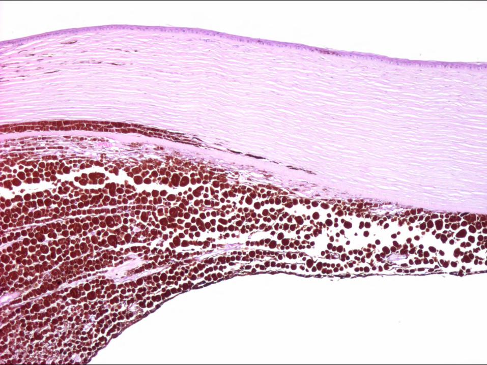
Uveitis
Hypopyon

Uveitis
Hyphema

Uveitis – Fibrovascular membranes
PIFM

Fibrovascular membranes
PIFM w/ ectropion uvea
PIFM w/ entropion uvea
Post-iridal fibrovascular membrane

Uveitis – Fibrovascular membranes
PIFM w/ PAS Anterior synechia

Uveitis – Fibrovascular membranes
Posterior synechia

Uveitis – Fibrovascular membranes
Cyclitic membranes

Uveitis – Fibrovascular membranes
Retrocorneal membrane


Pathogenesis
ISCHEMIA
PRO-ANGIOGENIC FACTORS
INFLAMMATION
FIBROVASCULAR MEMBRANES
Hemorrhage Glaucoma
NEOPLASIA
GLAUCOMA

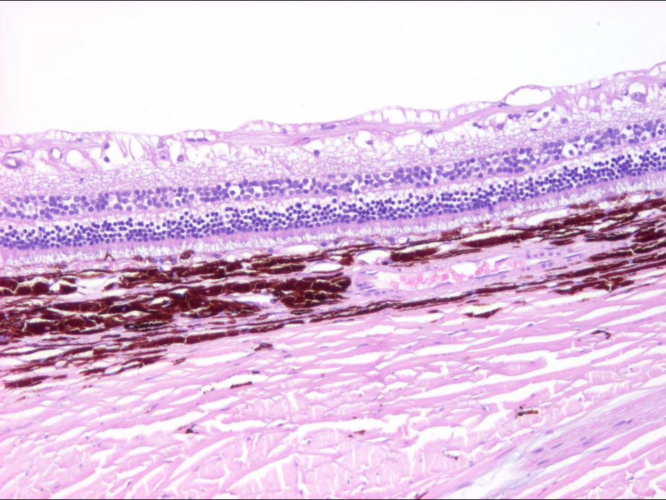
Feline lymphoplasmacytic uveitis
• Very common histo diagnosis
– Histologic pattern
• Cause unknown:
– End-stage?
• Toxoplasma spp., Bartonella spp., FIV, FeLV
– Immune-mediated

Feline LPU – Gross
• Lens luxation/subluxation
• Vitreous liquefied
• Opacity of anterior vitreous


Feline LPU – Histo
• Lymphocytes / Plasma cells
– Anterior uvea
• CB non-pigmented epithelium
– Retina
• Follicle-like structures
• Condensed anterior vitreous
• Hypereosinophilic granular protein

Lymph node


Feline LPU – Glaucoma
• Inflammation in ICA
• PIFM
• Lens lux
• Anterior vitreous prolapse

Equine recurrent uveitis (ERU)
• Typically received after the eye is blind / painful
• Not all uveitis in horses are ERU!
– Important to distinguish the features
– Bilateral (not all cases)

ERU – Gross
• Non-specific:
– Phthisis
– Cataract
– Synechiae
– Pale floccular material over CB processes


ERU – Histo 1. LP in the uvea +/- follicle-like structures
2. Eosinophilic linear inclusions (crystalline protein)
– Non-pigmented CB epithelium
– ONH
3. Eosinophilic homogeneous mb over inner CB
– Red Congo + (like amyloid)
– Green birefringence


Courtesy of Dr. Dubielzig (COPLOW)
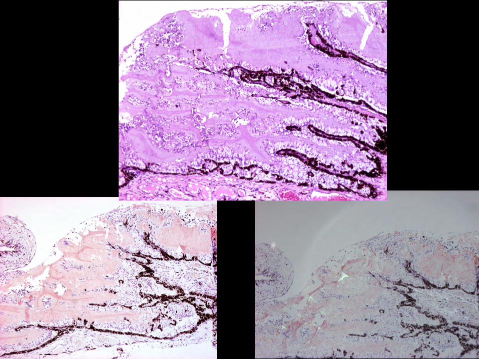

IHC, amyloid AA

ERU – Histo (nonspecific)
• PIFM / PAS / retrocorneal membranes
• Cataract
• Retinal detachment
• Inflammation in ON
• Glaucoma
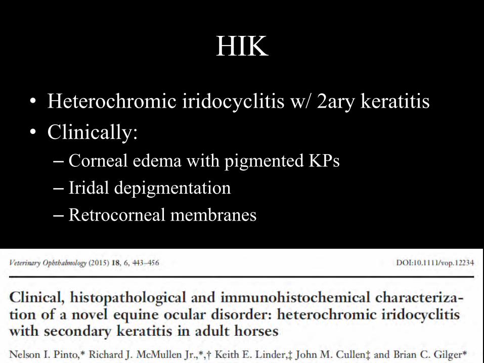
HIK
• Heterochromic iridocyclitis w/ 2ary keratitis• Clinically:
– Corneal edema with pigmented KPs– Iridal depigmentation – Retrocorneal membranes

HIK - Pathology
• LP w/ fewer pigment-laden MO
– MO and MNGC, anterior iris surface
• Iris pigment loss
• Retrocorneal fibrous membranes
– Pigment-laden MO and lymphocytes
• Keratitis

Courtesy of Dr. Dubielzig (COPLOW)

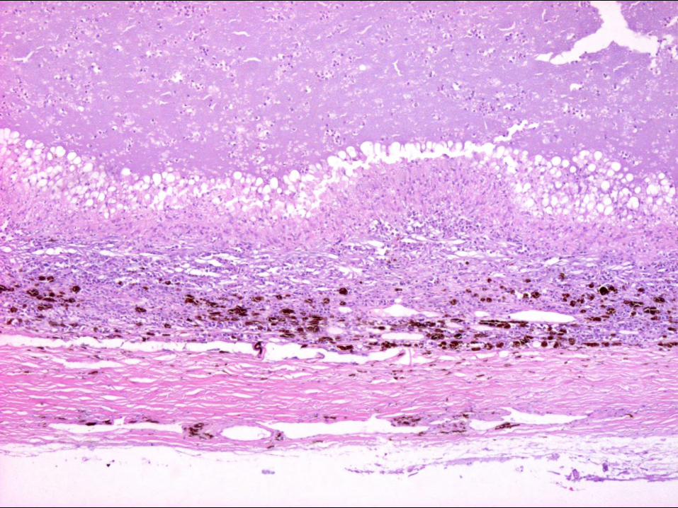
Uveodermatologic syndrome
• VKH-like
• Akitas, Northern breeds, other
• Bilateral and symmetrical
• Dermal dz may not be present
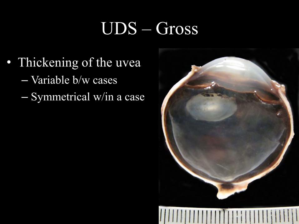
UDS – Gross
• Thickening of the uvea – Variable b/w cases– Symmetrical w/in a case


UDS – Histo
• Granulomatous inflammation
– MO >>> lymphocytes, plasma cells
– MO contain intracytoplasmic melanin granules
• Typically the rest of the globe is quiet
• RD
• Glaucoma


Depigmenting or “quiet” VKH-like
Courtesy Dr. Dubielzig (COPLOW)

Lens-associated uveitis
• Phacolytic uveitis (LIU)
• Phacoclastic uveitis
– Sterile/bland phacophagocytic inflammation
– Asymmetric uveitis:
• Diabetic
• Non-diabetic
• Septic implantation syndrome

Phacolytic uveitis - LIU
• Chronic leakage of lens prot through intact capsule• Gross:
– Mature, hypermature, intumescent cataract • Histologically:
– LP infiltrate in anterior uvea (nonspecific!) – Cataract!!! – Other: glaucoma, PIFM, synechiae, iris bombé,…
• Rule out other diseases!


Lens-associated uveitis
• Phacolytic uveitis (LIU)
• Phacoclastic uveitis
– Sterile/bland phacophagocytic inflammation
– Asymmetric uveitis:
• Diabetic
• Non-diabetic
• Septic implantation syndrome

Sterile phacoclastic uveitis
• Lens capsule rupture w/o globe penetration
• Grossly:
– Hazy media around lens
• Histo:
– Lens capsule rupture
– Macrophages and MNGC surrounding lens fibers


Asymmetric uveitis – Non-diabetic
• Small breeds (Poodles!)
• Females >> males
• Second eye at risk!
– Immediately to 4 years
• Gross:
– Haziness/opacity of ocular media (endophthalmitis)
– White lining to inner surfaces of the globe


Asymmetric uveitis – Non-diabetic
• Histo:
– Pyogranulomatous inflammation
– Macrophage-rich carpet on inner surfaces
– Penetrating injury
• Corneal/scleral rupture
• Lens capsule rupture
• Sepsis
• FB


Asymmetric uveitis – Diabetic
• Similar pathology to non-diabetic variant
• Miniature Schnauzers overrepresented
• Symmetrical (both eyes at the same time)
Courtesy of Dr. Dubielzig (COPLOW)

Lens-associated uveitis
• Phacolytic uveitis (LIU)
• Phacoclastic uveitis
– Sterile/bland phacophagocytic inflammation
– Asymmetric uveitis:
• Diabetic
• Non-diabetic
• Septic implantation syndrome

Septic implantation syndrome
• Phacocentric suppurative endophthalmitis
• Response to infectious agents, NOT lens protein
• Male >> female
• Trauma (cat scratch):– 39% of dogs
– 20% of cats
• Duration of clinical signs: 1 week to 1 year prior
Bell et al., 2012

SIS – Gross
• Whitish to tan phacocentric exudate
• Broad posterior synechiae
• Posterior segment uninvolved


SIS – Histo
• Lens capsule rupture
• Perilenticular and intralenticular inflammation – Suppurative
– Phacitis / abscess
– Cataract
– Bacteria (65% of dogs, 70% of cats), away from inflam• Gram + > Gram – (occ. fungi)
– Corneal / scleral rupture, rarely


Courtesy of Dr. Dubielzig (COPLOW)


Phacoclastic uveitis in rabbits
• Encephalitozoon cuniculi
• Dwarf rabbits
• Vertical transmission
• Cataract and lens capsule rupture

E. cuniculi in rabbits – Gross

E. cuniculi in rabbits – Histo
• Granulomatous to pyogranulomatous inflam
– Anterior and posterior chamber, lens
• Lens capsule rupture
• Intralenticular inflammation
• Organisms rarely seen
– Gram +, Giemsa, Ziehl-Neelsen


Ziehl Neelsen
Gram’s

Infectious intraocular inflammation
• Not all dz have specific pathological features
– Rickettsiae and Ehrlichiae
– Borrelia spp.
– Bartonella spp.
– Brucella spp.
– Leptospira spp.
• Underdiagnosed

Suppurative endo/panophthalmitis
• Most common consequence of penetrating trauma
• Brachycephalic and large breed
• Gross:
– Tan to yellow exudates throughout globe
– Displaced or ruptured lens

Endophthalmitis


Endophthalmitis + keratitis?

Suppurative endo/panophthalmitis
• Histo:
– PMNNs in all structures
– Lens capsule rupture
– Retinal necrosis
– Bacteria (vitreous)
– FB (plant material)


Suppurative endo/panophthalmitis
• Site of penetration:
– Cornea: impossible to discern from primary corneal dz
– Sclera:
• Superior: attack, plant
• Inferior:
– Plant or FB from oral cavity
–Dental episode?


Extension into the orbit

Feline infectious peritonitis
• Mutated FeCV/FIPV
• Typically younger than 3 years
• Commonly seen with the “dry” form
– Pyogranulomatous inflammation
• Occ. seen as an neuro-ocular form

FIP – Gross
• Translucent to opaque ocular media
• Solidified aqueous and/or vitreous
– High-protein exudate

FIP
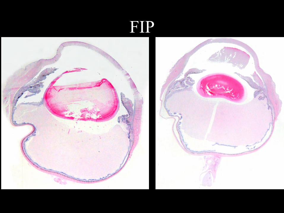
FIP

FIP – Histo • Mixed inflammation
– Uvea, retina, ON and its meninges
• Regional predominance of:
– Plasma cell-rich inflammation
– Macrophages: atypical (cleaved nuclei, prominent nucleoli)
– Neutrophils
• Vasculitis not always found


Mycotic uveitis
• Geographical restrictions
• Pyogranulomatous chorioretinitis
• PAS or silver stains (GMS, Grocott)

Blastomycosis
• Blastomyces dermatitidis
• Dogs > cats
• Mississippi, Ohio and Missouri River valleys
• Inhalation or inoculation of spores
• Lung > eyes, skin, LN, bone

Blastomycosis – Gross • Tan exudate in the vitreous, retina, choroid
Courtesy of Dr. Dubielzig, COPLOW

Blastomycosis – Histo
• Pyogranulomatous, nodular to diffuse
• Posterior segment
• 10-15 µm diameter, refractile wall
• Broad-based budding
• Splendore-Hoeppli

Blastomycosis – Histo
Courtesy of Dr. Dubielzig, COPLOW
Splendore Hoeppli
GMS
PAS

Cryptococcosis
• Cryptococcus neoformans
• Cats >>> dogs
• Worldwide
• Inhalation of spores (pigeon droppings)
• Lungs, CNS, LN, skin eyes

Cryptococcosis – Gross • Pyogranulomatous chorioretinitis/endophthalmitis• Gelatinous

Cryptococcosis – Histo
• (Pyo)granulomatous chorioretinitis
• “Soap-bubble”
• Inverse relationship b/w inflam and organisms
• 5-8 µm w/ 1-30 µm-thick capsule – Alcian blue, mucicarmine
• Narrow-based budding
• Pseudohyphae

Cryptococcosis – Histo

Alcian Blue stain

Cryptococcosis –Histo
Narrow-based budding
Pseudohyphae (rare)Courtesy of Dr. Dubielzig, COPLOW
Mucicarmine, Courtesy of Dr. Reilly

Histoplasmosis
• Histoplasma capsulatum
• Cats > dogs (eye)
• Worldwide (Mississippi and Ohio river valleys)
• Spores in soil (bird / bat droppings)
• Lung, intestine, LN, skeleton, skin, eye

Histoplasmosis
Courtesy of Dr. Dubielzig, COPLOW

Histoplasmosis – Histo • Pyogranulomatous chorioretinitis • 3-5 µm, intrahistiocytic (sparse) • No budding
Courtesy of Dr. Dubielzig, COPLOW

Histoplasmosis – Histo
Courtesy of Dr. Dubielzig, COPLOW
GMS

Histoplasmosis
• Presumed Ocular Histoplasmosis (POH)
• Few or no organisms
• Spindle cell membrane internal to the choroid
• Proliferation of RPE

Coccidioidomycosis
• Coccidioides immitis
• Dogs > cats
• SW USA, Central and South America
• Inhalation of spores
• Immunosuppressed and outdor animals
• Lung, bones, eyes

Coccidioidomycosis – Gross
Courtesy of Dr. Dubielzig, COPLOW

Coccidioidomycossis – Histo
• Pyogranulomatous chorioretinitis / endophthalmitis• Low numbers of organisms• 25-85 µm, thick refractile walls
Courtesy of Dr. Dubielzig, COPLOW

Aspergillosis
• Aspergillus spp.
• Systemic dz, although eye may be noted first
• German shepherd
• Aberrant immune response

Aspergillosis – Gross
• Pyogranulomatous vitreitis/endophthalmitis

Aspergillosis – Histo
• Pyogranulomatous endophthalmitis
• Centered on vitreous / inner retina
• Intralesional hyphae
– Vitreous
– Lens capsule (ruptured)

Aspergillosis – Histo
Courtesy of Dr. Dubielzig, COPLOW

Aspergillosis – Histo
Courtesy of Dr. Dubielzig, COPLOW

Protothecosis• Prototheca zopfii > Prototheca wickerhamii
• Dogs
• World-wide distribution
• GI dz, systemic spread (6/14 systemic signs)
• Ocular involvement: 2/3 of cases

Protothecosis – Gross
• Complete RD, thick tan exudate around retina
Courtesy of Dr. Dubielzig, COPLOW
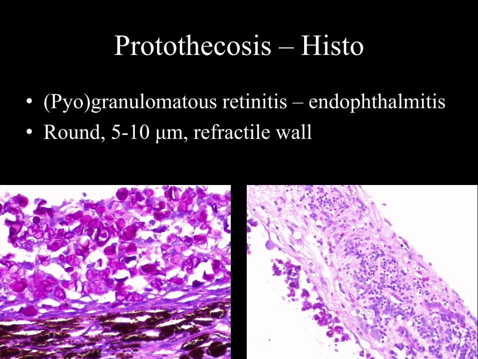
Protothecosis – Histo
• (Pyo)granulomatous retinitis – endophthalmitis• Round, 5-10 μm, refractile wall
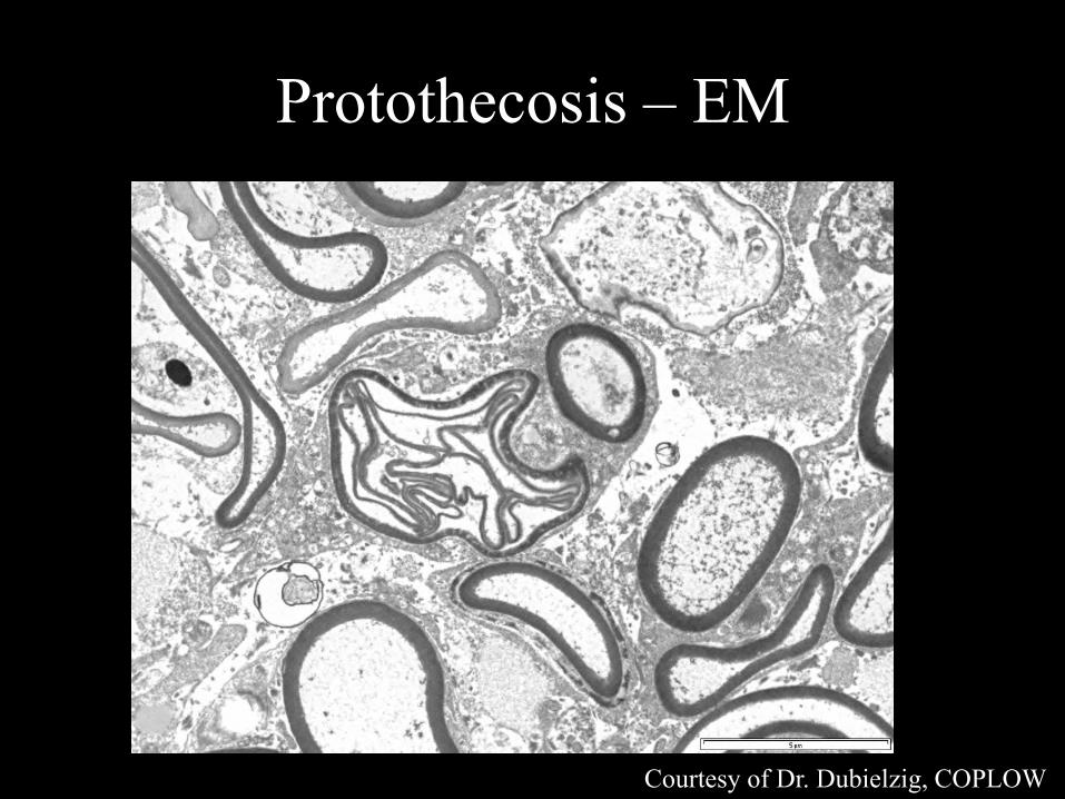
Protothecosis – EM
Courtesy of Dr. Dubielzig, COPLOW

Protozoa - Leishmaniosis
• Leishmania spp.
• Dogs >>> cats (immunocompromised)
• Mediterranean, Central and South America
• Spread to other northern areas
• 25-30% of dogs: ocular disease
• Anterior uvea >>>> choroid


Leishmaniosis – Histo
• Granulomatous uveitis, diffuse or nodular
• Intrahistiocytic amastigotes, 1-2 µm
– IHC
• Can see blepharitis, conjunctivitis,
scleritis/episcleritis, keratitis, lacrimal adenitis


Uveal damage after endolaser
Courtesy of Dr. Dubielzig, COPLOW

Trans-scleral LaserPhotocoagulation
Scleral collagen heat damage
Burn spots
Courtesy of Dr. Dubielzig, COPLOW

Trans-scleral Laser Photocoagulation
Nerve and Vessel Necrosis
Courtesy of Dr. Dubielzig, COPLOW
Copyright © 2022 FDOKUMEN