Effect of rosiglitazone on the pathology of experimentally ...
226
\\ EFFECT OF ROSIGLITAZONE ON THE PATHOLOGY OF EXPERIMENTALLY INDUCED ENDOMETRIOSIS IN BABOONS A THESIS SUBMITTED IN PARTIAL FULFILMENT FOR THE DEGREE OF MASTER OF SCIENCE IN VETERINARY PATHOLOGY AND DIAGNOSTICS, THE UNIVERSITY OF NAIROBI BY: Dr. DANIEL CHAI CHIVATSI, B.V.M., (UON) DEPARTMENT OF VETERINARY PATHOLOGY, MICROBIOLOGY AND PARASITOLOGY / UNIVERSITY OF NAIROBI University ol NAIROBI Library 'mini 0416754 0 May 2009
-
Upload
khangminh22 -
Category
Documents
-
view
2 -
download
0
Transcript of Effect of rosiglitazone on the pathology of experimentally ...

\ \EFFECT O F ROSIGLITAZONE ON THE PATHOLOGY OF EXPERIMENTALLY
INDUCED ENDOMETRIOSIS
IN BABOONS
A THESIS SUBMITTED
IN PARTIAL FULFILMENT FOR THE
DEGREE OF MASTER OF SCIENCE IN VETERINARY PATHOLOGY AND
DIAGNOSTICS, THE UNIVERSITY OF NAIROBI
BY:
Dr. DANIEL CHAI CHIVATSI, B.V.M., (UON)
DEPARTMENT OF VETERINARY PATHOLOGY, MICROBIOLOGY AND
PARASITOLOGY / UNIVERSITY OF NAIROBI
University ol N AIR O B I Library'mini0416754 0
May 2009

d e c l a r a t io n .
1 his is my original work and it has not been presented for a degree in any other
University’
Dr. Daniel Chai Chivatsi, B.V.M.
Signed: .......
SUPERVISORS DECLARATION
‘ I his thesis has been submitted for examination with our approval as University
supervisors’.
Prof. Tarcisius A. I^gatia, B.V.M., MSc, Dip PVM. PhD.
s ig n .^ ? ? ^ ^ ^ ? rrrr . ojsDepartment of Veterinary Pathology, Microbiology and Parasitology,
University of Nairobi.
Prof. Peter Karuri Gathumbi, B.V.M., MSc, PhD.
...Sign:........
Department of Veterinary Pathology, Microbiology and Parasitology,
University of Nairobi.
Dr. Jason M. Mwepda, BSc.Jvfphil, PhD.
V..\Sign:t f l X c 1
Institute ol Primate Research, National Museums of Kenya.
Prof. Thomas M. DTIooghe, MD, PhD.__ / .

DEDICATION
I would like to dedicate this work first to Our Lord Jesus Christ whose blessings 1 have seen.
Secondly to my family; my wife Gladys Tatu, daughters Pendo, Rehema and son Chivatsi for
standing by me through the study.

ACKNOWLEDGEMENT
I would like to acknowledge, Dr. Dan Lcbovic for funding the project through a National
Institute Grant No. 5K23HD043952, as part of collaboration work between the Department
of Female Reproduction, Institute o f Primate Research, Nairobi Kenya, and the University of
Michigan Department of Obstetrics and Gynaecology. His original hypothesis and work
created this thesis as an off shoot. He entrusted the IPR team with the project and graciously
permitted use of his study data (Lebovic et al., 2007), o f which I am a co-author. Am
grateful to Prof. T.A. Ngatia and Prof. Gathumbi, my University supervisors for their
availability for consultation, especially on histopathology, and for taking keen interest in my
project. I am also grateful to Prof. Thomas M. D’Hooghe and Dr. Jason M. Mwenda for
providing professional guidance and support during the conduct of this Msc project.
I would also like to thank the staff of the Department of Animal Resources, Institute for
Primate Research, for their generous contributions to the study. Dr. Peter Mwcthera for
providing the serum kits used in this project. Dr. Jason Mwenda for financial support and
providing all consumables for histopathological work. Atunga Nyachico, Nicholas Kiulia,
Habib Saibulu, Mary Galo and Eric Omollo for their assistance with the surgeries and
documentation. John Muongi at Department of Veterinary Pathology, Microbiology and
Parasitology, University of Nairobi for processing the histological sections. Lastly, we
respect and honour the 12 baboons sacrificed in this study in order to benefit humanity.

TABLE OF CONTENTS
DECLARATION_______________________________________________________ II
SUPERVISORS DECLARATION________________________________________ II
DEDICATION_________________________________________________________ III
ACKNOWLEDGMENT_________________________________________________ IV
TABLE OF CONTENTS________________________________________________ V
LIST OF TABLES______________________________________________________ X
LIST OF FIGURES_____________________________________________________ XII
LIST OF PLATES______________________________________________________ XIV
LIST OF APPENDICES_________________________________________________ XVII
LIST OF ABBREVIATIONS & ACRONYMS______________________________ XVIII
ABSTRACT_____
CHAPTER ONE:
L I.
1.2.
1.2. 1.
1.2.2.
CHAPTER TWO:
2.1.
2.2.
2 3 .
INTRODUCTION__________________
INTRODUCTION___________________
OBJECTIVES OF THE STUDY______
Specific objectives...................................
Rationale and study justification.............
LITERATURE REVIEW__________
PATHOGENESIS OF ENDOMETRIOSIS
PREVALENCE OF ENDOMETRIOSIS___
ENDOMETRIOSIS IN ANIMAL MODELS
XXI
1
1
2
2
3
5
5
8
9

2.4. GROSS AND MICROSCOPIC PATHOLOGY OF
ENDOMETRIOSIS________________________________ 10
2.5. DIAGNOSIS OF ENDOMETRIOSIS_________________ 13
2.6. EFFECTS OF ENDOMETRIOSIS___________________ 16
2.7. ADVANTAGES OF THE BABOON MODEL FOR
REPRODUCTIVE RESEARCH_____________________ 17
2.7.1. Advantage number 1: Reproductive anatomy, endocrinology
and physiology.......................................................................... 17
2.7.2. Advantage number 2: Non-invasive cycle monitoring based
on perineal changes................................................................... 18
2.7.3. Advantage number 3: Continuous breeding............................. 18
2.7.4. Advantage number 4: Baboon size and strength...................... 19
2.7.5. Advantage number 5: Spontaneous peritoneal fluid................ 19
2.7.6. Advantage number 6: Cross-reactivity between baboons and
human......................................................................................... 19
2.7.7. Advantage number 7: Vaginal transcervical uterine access..... 19
2.8. DEVELOPMENT OF BABOON MODEL FOR
RESEARCH IN ENDOMETRIOSIS AT THE IPR------- 20
2.9. TREATMENT OF ENDOMETRIOSIS____________ 21
2.9.1. Expectant management.....................—........— ................... 23
2.9.2. Empirical treatment of pain symptoms without a

2.93 . Treatm ent of endometriosis-associated pain in confirmed
2.9.4.
2.9.5.
2.10.
2.10.1.
2.10.2.
2.103.
CHAPTER THREE:
3.1.
3.2.
3.2.1.
3.2.1.1.
3.2.1.2.
3.2.13.
3.2.1.4.
3.2.1.5.
3.2.1.6.
3.2.2.
3.2.2.1.
3.2.2.2.
disease.—.............. ........................ .................... ........
Treatment of endometriosis associated infertility,
Side effects of treatment..........................................
THIAZOLIDENEDIONES----------------------------
Mode of action of Thiazolidenediones---------- -—
Toxicology of Rosiglitazone.M..«mM~..«..~.........M.
Use of TZDs in treatment of endometriosis-------
MATERIALS AND METHODS---------------------------------
MA I ERIALS............................ ...............................................
METHODS_________________________________
Clinical phase procedures....................................— ....— ..
Monitoring of Menstrual cycle-----------------------—------ ...
Induction Laparoscopy......................... ..............--------------
Staging pre-treatment laparoscopy.....................................
Drug treatments------------------------------------ -— ...............
Post-treatment staging laparoscopy------------- -—.............
Post mortem procedure......- .......................... ...... ...----- ......
Serum chemistry procedures-------------------------------------
Total proteins determination.................... ........... .................
Bilirubin determination....—.............................. ..................
30
32
34
35
36
36
39
39
41
41
41
41
43
43
46
46
48
48
49
23

3.2.23.
3.2.2.6.
33.2.7.
3.23.
3.2.4.
3.2.5.
33.
CHAPTER FOUR:
4.1.
Aspartate Aminotransferase (AST/SGOT) determination
Albumin determination---------- — -------------------- .....—
Urea determination----------------------------------————~.
Haematology..----- — ----------------------------------- ———.
Ilistopathology procedures------------------- -------------------
Scoring of endometriosis lesions......—— ------- .......-------
Statistical analysis...-------------------------------------- ---- -----
RESULTS___ ____________________________________
ENDOMETRIOSIS LESIONS---------------------------------
54
55
56
57
57
59
61
63
63
4.1.1.
4.1.2.
4.13.
4.1.4.
4.1.5.
4.1.6.
4.1.7.
4.1.8.
Gross appearance of endometriosis lesions assessed
during laparoscopy.—— --------—....----- ---------- —— —
The distribution of the types of lesions---------------..-------
Surface area of endometriotic lesions--------------------------
Gross appearance and histopathology of endometriosis
lesions----------- ------------------ ---------------- ---- -------- ------ -
Scoring of the histologic changes in the endometriotic
lesions............... - ..............— ..... - .............. ..—.......-------- —
Frequency of histological changes or features among the
treatment groups.....-------------------- —------------------—
Presence of tissue eosinophils in endometriotic lesions......
Presence of stromal endometriosis-------------------------------
63
76
81
85
102
128
130
130

4.2.1. Haemoglobin---------------------------------------------------------- • 33
4.2.2 Total plasma protein............................................— ............. 133
4.2.3. Eosinophils......................................................................... ... 136
4.2.4. Plasma fibrinogen............................................................. ~~~ 136
43. CLINICAL CHEMISTRY---------------- -------------- 139
4.4. HISTOLOGY OF ORGANS OTHER THAN THOSE
WITH ENDOMETRIOTIC LESIONS------------------------ 139
CHAPTER FIVE: DISCUSSION, CONCLUSION AND
RECOMMENDATIONS------------------------------------------ 147
5.1. DISCUSSION_____________________________________ I47
5.2. CONCLUSIONS----------------------------------------------------- 158
53. RECOMMENDATIONS------------------------------------------ 159
CHAPTER SIX: REFERENCES____________________________________ 160
CHAPTER SEVEN: APPENDICES...._..__....—...........— .............— ....— 186

LIST OF TABLES
Table 1: Treatment groups, drugs and routes of administration of rosiglitazonc
and (Ganirelix(R) ) in cndometriotic baboons............................................. 45
Table 2: Data from lesions collected during the pre-and post treatment
laparoscopy of cndometriotic baboons........................................................ 77
Table 3: Quantitative changes of the different types of endometriosis lesions for
the various treatment groups....................................................................... 78
Table 4: Summary of the histological scores for glands and interstitial tissue in
all treated cndometriotic baboons............................................................... 106
Table 5: Summary of the histological scores for ESCs and peritoneum tissue in
all treated endomctriotic baboons............................................................... 107
Table 6: Summary of comparison o f scores among the treatment groups of
cndometriotic baboons................................................................................. 108
Table 7: Endometriosis lesions at necropsy............................................................... 118
Table 8: Summary of the histological scores for glands in all types of
endomctriotic lesions.................................................................................... 119
Table 9: Summary of the histological scores for interstitial tissue and peritoneum
in all types of endometriotic lesions............................................................ 120
Table 10: Comparison o f histological changes among the types of lesions in
endometriotic baboons................................................................................ 121

Table II:
Table 12:
Summary of frequency o f occurrence and comparison of histological
changes among the treatment groups using CHI Square fisher s hxact
Test of endometriotic baboons....................................................................
Summary and analysis o f histological findings in each treatment groups
in endometriotic baboons............................................................................

LIST OF FIGURES
cndomctriotic lesions for the treatment groups.......................................... 79
Figure 2. A box plot illustrating change in total number of red lesions in
cndomctriotic baboons................................................................................ 80
Figure 3. A box plot illustrating changes in surface area o f lesions (without
adhesions) before & after treatment of cndomctriotic baboons................ 83
Figure 4. A box plot illustrating relative change in surface area of lcsion(with
adhesions) in cndomctriotic baboons in the treatment groups................ 84
Figure 5. A box plot illustrating comparison of glandular degenerative changes
among the 3 groups of endometriotic baboons.......................................... 109
Figure 6. A box plot illustrating comparison of degenerative changes in
cndomctriotic stromal cells (ESCs) among the 3 types of cndomctriotic
baboons......................................................................................................... 110
Figure 7. A box plot illustrating comparison of peritoneum lymphocytes among
the 3 treatment groups of cndomctriotic baboons...................................... 111
Figure 8. A box plot illustrating comparison of glandular luminal mucus among
the 3 treatment groups of endometriotic baboons...................................... 112
Figure 9. A box plot illustrating comparison of interstitial degenerative slit scores
among the 3 treatment groups of endometriotic baboons......................... 113
Figure 10. A box plot illustrating comparison of acute inflammatory cells within
Figure 1. A box plot illustrating changes in specific types and number of

Figure 11. A box plot illustrating comparison of glandular luminal haemorrhage
scores between the different types of lesions in cndomctriotic baboons... 122
Figure 12. A box plot illustrating comparison of interstitial degenerative scores
between the different types of lesions in cndomctriotic baboons............. 123
Figure 13. A box plot illustrating comparison o f interstitial lymphocyte scores
between the different types of lesions in cndomctriotic baboons............. 124
Figure 14. A box plot illustrating comparison of interstitial smooth muscle scores
between the different types of lesions in cndomctriotic baboons............. 125
Figure 15. A box plot illustrating comparison of degenerative scores of endometrial
stromal cells between the different types of lesions in cndomctriotic
baboons................................................- ....................................................... 126
Figure 16. A box plot illustrating comparison of haemorrhage scores in the
peritoneum between the different types of lesions in cndomctriotic
baboons......................................................................................................... 127
Figure 17. A box plot illustrating haemoglobin changes before and after treatment
o f endometriotic baboons............................................................................ 134
Figure 18. A box plot illustrating inter-group changes in total plasma in
endometriotic baboons................................................................................. 135
Figure 19. A box plot illustrating eosinophils counts in endometriotic baboons at
different treatments...................................................................................... 137
Figure 20. A box plot illustrating plasma fibrinogen levels in cndomctriotic
67
68
69
70
71
72
73
74
75
90
LIST OF PLATES
Gross appearance ofendomctriotic lesion (Nil) from a placebo treated
baboon (PAN 2985)....................................................................................
Gross appearance of cndometriotic lesion (N5) from a placebo treated
baboon (PAN 2912)............................ ........................................................
Gross appearance of endomctriotic lesion (N2) from a placebo treated
baboon (PAN 2991).....................................................................................
Gross appearance of endometriotic lesion (N5) from a GnRH treated
baboon lesion (PAN 2871)..........................................................................
Gross appearance of endometriotic lesion (N7) from a GnRH treated
baboon (PAN 2871)....................................................................................
Gross appearance of endomctriotic lesion (Nil) from a GnRH treated
baboon (PAN 2993)....................................................................................
Gross appearance of endometriotic lesion (N4) from a rosiglitazone
treated baboon (PAN 3032).........................................................................
Gross appearance of endometriotic lesion (N8) from a rosiglitazone
treated baboon (PAN 3030).........................................................................
Gross appearance of endometriotic lesion (N9) from a rosiglitazone
treated baboon (PAN 3030).........................................................................
Photograph of endomctriotic lesion (N1) from a placebo treated baboon
at laparoscopy III (PAN 2912)....................................................................

92
93
94
95
96
97
98
99
100
101
at Laparoscopy III (PAN 2991)..................................................................
Photomicrograph of cndomctriotic lesion (N3) from a placebo treated
baboon at laparoscopy III (PAN 2991)......................................................
Photograph of cndomctriotic lesion (N3) from a GnRH treated baboon
at laparoscopy III (PAN 2993)................................................................
Photomicrograph of cndometriotic lesion (N3) from a GnRH treated
baboon at laparoscopy III (PAN2993).......................................................
Photograph of cndomctriotic lesion (N9) from a GnRH treated baboon
at laparoscopy III (PA2993)....................................................................
Photomicrograph of cndometriotic lesion (N9) from a GnRH treated
baboon at laparoscopy III (PAN 2993)....................................................
Photograph of cndomctriotic lesion (N5) from a rosiglitazonc treated
baboon at laparoscopy III (PAN 2998)......................................................
Photomicrograph of cndometriotic lesion (N5) from a rosiglitazonc
treated baboon at laparoscopy III (PAN 2998)........................................
Photograph of cndomctriotic lesion (N13) from a rosiglitazonc treated
baboon at laparoscopy III (PAN 3030)....................................................
Photomicrograph of cndometriotic lesion (N13) from a rosiglitazonc
treated animal at laparoscopy III (PAN3030)............................................
An cndometriotic lesion from a baboon showing invasivencss property
Photograph of cndomctriotic lesion (N3) from a placebo treated baboon

Plate 25. Liver section from a rosiglitazone treated animal (PAN 2998).................. 144
Plate 26. Section of gall bladder wall from a rosiglitazone treated animal (PAN
2998)............................................................................................................. 145
Plate 27. A photomicrograph showing a myocardial infarct in a rosiglitazone
treated animal (PAN 2998).........................................................................
Plate 24. Liver section from a rosiglitazone treated baboon (PAN 2998).................. 143

LIST OF APPKNDICES
Appendix 1.
Appendix 2.
Appendix 3.
Appendix 4.
Appendix 5.
Appendix 6.
Appendix 7.
Appendix 8.
Appendix 9.
Appendix 10.
Appendix 11.
Sampling Form........................................................................................ 186
Staging laparoscopy form....................................................................... 187
Pelvic anatomic map............................................................................... 188
Checklist for scoring o f the histological features in glandular
epithelium features................................................................................... 189
Checklist of scoring o f the histologic features in interstitial tissue...... 192
Checklist of scoring for the histologic features in peritoneum
histologic features...................................................................................... 195
Data from lesions collected during the pre-and post-treatment
laparoscopy of individual cndometriotic baboons (Mean ± standard
deviation)................................................................................................. 197
Haematology data of individual cndometriotic baboon
(Mean ± standard deviation)................................................................... 198
Histopathology of lungs in cndometriotic baboons............................... 199
Histopathology of liver in endometriotic baboons................................ 200
Summary o f Scrum chemistry values at pre-induction and post
induction in individual endometriotic baboons (Mean ± standard
deviation 201

LIST OF ABBREVIATIONS AND ACRONYMS.
A,
Hi
ANOVA
ART
CAM
CA19
CA-125
COCs
DIE
EMB
ESCs
FDA
FSH
GA
g/dl
GOT
GnRH
H & E
Spectrophotometer absorbance reading 1
microliter
Univariate analysis of variance
assisted reproductive techniques
Cell adhesion molecules
Cancer antigen 19
Cancer antigen 125
Combination estrogen-progestin oral contraceptives
Deep infiltrating endometriosis
Endometrial biopsy
endometriotic stromal cells
Food and Drug Administration of United States of America.
Follicle stimulating hormone
GnRH group after treatment,
grams/deciliter
Glutamate Oxaloacetic acid, also called Aspartate transaminase (AST) /serum
glutamic oxaloacetic transaminase (SGOT) / aspartate aminotransferase
(AAT)
Gonadotrophin releasing hormone
Haematoxylin and Eosin stain

IL-8 Interleukin -8
IPR Institute of Primate Research
IVF In-vitro fertilization
LDH Lactate dehydrogenase
LDL Low density lipoproteins
LH Leutenising hormone
LSD Least Significant Difference
MDH Malate dehydrogenase
mg/dl milligrams per deciliter.
nm nanometer
NADH Nicotinamide adenine dinucleotide, as reducing agent form
NK- k B transcription factor nuclear factor- kB
PA Placebo group after treatment.
PAI-I plasminogen activator inhibitor-1
PB Placebo group before treatment.
PF Peritoneal fluid
RA Rosiglitazone group after treatment.
RB Rosiglitazonc group before treatment.
ROS reactive oxygen species
Rosiglit Rosiglitazone.
SIV Simian Immunodeficiency virus

TNF- a Tumour Necrosis Factor - alpha
VEGF Vascular endothelial growth factor

ABSTRACT
The objective o f this study was to determine the effect of rosiglitazonc, on the pathology o f
endometriosis in a baboon model. It was a prospective, randomised, placebo-controlled
study, conducted at the Institute of Primate Research in Nairobi, Kenya. Endometriosis was
induced using intrapci vie seeding of eutopic menstrual endometrium in 12 female baboons
that had at least one menstrual cycle while in captivity.
Laparoscopy was performed on each o f the 12 baboons. Menstrual endometrial tissue was
extracted from each baboon by curettage, and one gram of endometrium was then seeded
onto several peritoneal sites to induce endometriosis. About 34 — 68 days after the induction
of endometriosis, a pre-treatment laparoscopy was performed in the baboons to assess the
extent of endometriosis . Thereafter, the 12 baboons were randomised into three groups that
were treated daily for 30 days as follows; placebo group (n = 4) received phosphate -
buffered saline (PBS) tablets once a day orally, for 30 days. The positive control group (n =
4) received a gonadotrophin releasing hormone antagonist, (Ganirelix acetate*), at a daily
dose o f 125 pg/day subcutaneous for 30 days. Finally, the experimental group (n= 4)
received 2 mg rosiglitazone, (Avandia*) by mouth daily for 30 days. A third and final
laparoscopy was performed 30 days after the start of treatment to record the extent of
endometriosis.
Endometriosis lesions were examined at laparoscopy and classified morphologically as
A.___"____1 LI____L I _____1. ___~ ...A*, wl OM/Xln w r i A i / a o i ^ n lo r rrvl V w m m rrh 'i (»l/» m f \

adhesion. During the procedure, a video recording was performed to document the number
and surface area of the endometriosis lesions. Blood for haematology and serum for clinical
pathology was collected during the time of pre-treatment laparoscopy and at the final
laparoscopy. The baboons were then cuthaniscd by administrating 60mg/kg body weight of
Sodium Pentobarbitone, (Euthatal*), intravenously and immediately thereafter a complete
post mortem was performed.
Lesion samples were obtained and fixed in 10% neutral buffered formalin to confirm the
histological features of endometriosis. The endometriosis lesions were scored on a scale of 0
to +4 in order to monitor the effects o f the drug. The scoring scale was designed on the basis
of a trial run that identified all the possible histological features that enabled an allocation of
scores of 0 - 4. Intensity, severity and quantitative parameters within the lesions formed a
subjective basis of scoring. This was not a blinded scoring.
Biochemical tests were carried out in serum to assess the adverse effects of the drug on the
liver function. The liver function assays included analyses for total protein content, alkaline
phosphatase (ALK), aspartate aminotransferase (AST/SGOT/AAT) and bilirubin. Kidney
function tests included creatinine and urea tests. The endometriosis lesions were assessed
and scored for histopathologic changes and features.
It was noted that the surface area o f endometriotic lesions were significantly lower in
1 • * _______ a____ a. __ I A__ I________ ____L _______ ____ ___ w l . . . r i L o r / \ i i n D o k o n n c f n * * i t < » i l

(mm2) of peritoneal cndometriotic lesions than controls. The extent of epithelial degenerative
changes, including nuclear pyknosis and focal epithelial erosions were increased in treated
baboons.
Rosiglitazonc effectively reduced the burden of endometriosis disease in a baboon model. In
one rosiglitazonc treated animal, the level of degeneration in the endometriosis lesion was
severe enough to cause epithelial necrosis. There were also significant degenerative changes
of cndometriotic stromal cells (ESCs) in the treated group. The experimental induction also
produced some Stromal endometriosis (i.e. endometriosis without glands).
The systemic effects of rosiglitazonc on baboons were analysed by comparing the frequency
of appearance o f histological features among the groups. Briefly, the following significant
observations were recorded in rosiglitazonc treated group compared with the placebo and
GnRH treated group; pulmonary edema & hemorrhages; hepatic bile cananaculi surrounded
by inflammatory cells and bile duct hyperplasia. Haematology also showed significant
changes when a pre and post-treatment data was compared; there was an increase in
haemoglobin values in the placebo and Rosiglitazonc treated group ; a significant increase of
total plasma protein in GnRH group; an increase in eosinophil count in the rosiglitazonc
treated group and an increase in plasma fibrinogen in the GnRH group. Scrum chemistry
showed no significant changes.

observation that gross and histopathology changes showed a significant reduction in surface
area of lesions and an increase in degenerative and epithelial desquamation of endometriosis
in rosiglitazone and GnRH treated baboons. The baboon model holds promise that a
thiazolidinedione drug may be helpful in women with endometriosis.
Key Words: Baboon, endometriosis, rosiglitazone, thiazolidinedione, GnRH-antagonist,
systemic pathology.

CHAPTER ONE
1.1. INTRODUCTION
Endometriosis is a benign gynaccologic disease of man that is defined and histologically
diagnosed as presence of both endometrial glands and stromal cells outside the uterine cavity.
Endometriosis is not a malignancy but it is associated with pelvic pain, subfertility, abnormal
menstrual bleeding and disrupted gastrointestinal tract due to partial obstruction. It may also
contribute to periodic cyclic abdominal pain from irritation o f pain fibres on peritoneal
surface. In the uterus, endometrial cells have exquisite sensitivity to sex hormones,
depending on the cell type and location (McClellan et al., 1990; Slayden and Brenner 2004;
Brenner and Slayden 2005). Endometriosis is an oestrogen dependent disease, and
endometriotic lesions can produce their own oestrogen after 6 months of experimental
induction (Atar and Bulun 2006). In women endometriosis is the second leading reason, after
uterine fibroids, for hysterectomies (http://womwnhealth.gov 1 July 2006). The prevalence
of endometriosis in women is reported to be as high as 22% in asymptomatic cases, 4 0 - 6 0
% in dysmenorrhoeal cases and 20 - 30 % in subfertile women (Farquhar 2000).
Grossly, endometriosis lesions may appear as small reddish peritoneal petechiae, white cystic
peritoneal vesicles, brownish diffuse peritoneal lesions, or classic blue-black folded lesions
on the peritoneum (Martin et al., 1989). Endometriosis may also affect the ovary, resulting in
laraf* rvst-like lesions that contain chocolate-fluid called endometriomas (Olive and

Because it is not possible to study mechanisms of endometriosis in affected women in vivo, a
baboon model has been developed in which intrapelvic injection of menstrual endometrium
results in the induction of endometriotic lesions (D'Hooghc et al., 1994a; Fazleabas et al.,
2002). The disease has also been reported to occur naturally in other non-human primates
(Schenken et al., 1984; D’Hooghc et al., 1991; Comillie et al., 1992; Dick et al., 2003;
Story and Kennedy 2004).
The clinical data of this study, represented here in Section 4.1.1; Section 4.1.2 and Section
4.1.3 have been published before (Lebovic et al., 2007). The author of this thesis was a co
author of this publication (Lebovic et al., 2007). He performed all the surgeries,
documentation, photography, video recording used in this publication.
This thesis represents a report (sections 4.1.4 to 4.4) on the clinical pathology and pathology
based on blood samples collected at Laparoscopy II, Laparoscopy III and tissues at necropsy,
and can be considered as a spin-off o f the published study (Lebovic et al., 2007) initiated by
Dr Dan Lebovic.
1.2. OBJECTIVES OF THE STUDY
The major objective of the study was to determine the effects o f nosiglitazone on pathology of
experimental endometriosis in a baboon model.

in endometriosis lesions in rosiglitazonc treated baboons
1.2.1.2. To determine the adverse effects o f rosiglitazone using serum chemistry
parameters in baboons.
1.2.1.3. To establish the target organs of rosiglitazonc toxicity in baboons based
on histopathology.
These objectives will be studied comparing the data in the study group (rosiglitazonc) with a
negative control group (placebo) and a positive control group (GnRH antagonist)
1.2.2. Rationale and study justification
Preclinical studies in the rodent model o f endometriosis (Lebovic et al., 2004; Demirturk et
al., 2006; Aytan et al., 2007) have shown that using a peroxisome proliferator-activated
receptors gamma (PPAR-y) agonist may have merit to treat endometriosis lesions since it
reduces the size of experimentally induced endometriosis. These promising results observed
in the rat formed a basis of this investigation using nonhuman primates (Lebovic et al., 2007).
Two common medical treatment options available for endometriosis are gonadotrophin
releasing hormone (GnRH) antagonist, and a combination of oestrogen and progestin oral
contraceptives (COC). This combination, referred to as the "pseudopregnancy" regimen, has
been used for several decades, and many physicians use it as first-line therapy for
endometriosis (Moore et al., 2000). These hormonal drugs were first used for the treatment
of endometriosis more than 40 years ago but interfere with fertility (Moore et al., 2000).

eliminate endomctriotic lesions, prevent recurrence and not impede ovulation or cause
infertility. Rosiglitazone (a PPAR-y) is a possible new candidate for treatment of
endometriosis. However, controlled studies of endometriosis in women arc restricted due to
ethical reasons o f performing repeated invasive surgical procedures. Experimentally induced
endometriosis in the baboon provides a non-human primate model for endometriosis,
allowing the evaluation of new drugs in the prevention or treatment of endometriosis
(D’Hooghe et al., 1995). Baboon endomctriotic implants show histological transformations
similar to those seen in human endomctriotic lesions. In the current study, the baboon model
of endometriosis will be utilized to assess if rosiglitazone can impede the growth of induced
ectopic endometriosis tissue.

CHAPTER TWO
2.0. LITERATURE REVIEW
2.1. PATHOGENESIS OF ENDOMETRIOSIS
Endometriosis is the presence of functional endometrial glands and/or stroma in ectopic
locations outside the uterine cavity.
Pathogenesis is poorly understood and remains controversial. Three concepts can be
discerned. The oldest concept that of in-situ development is that endometriosis develops on
the spot where it is found (Ridley 1968). The mechanism of in situ development of lesions
could be as a result o f local induction, development from Mullerian remnants or alternatively
from metaplasia of the peritoneal or ovarian tissue into endometrium (Ridley 1968; Lauchlan
1972). This can explain a reported case of endometriosis in a man (Pinkert et al., 1978).
While the disease is associated with significant morbidity, it is uncommon for endometriosis
to cause death in humans.
A second concept, the induction or metaplasia theory, is that endometriosis results from
differentiation o f mesenchymal cells, activated (induced) by substances released by
degenerating endometrium that arrives in the abdominal cavity (Levandcr and Norman 1955;
Merrill 1966; Matsuura etal., 1999).

theory in pathogenesis of endometriosis, one study observed that in patients with obstructed
menstrual outflow, endometriosis occurred in 77% of patients with a functional endometrium
and patent tubes and in up to 89% of those with hematocolpos (a condition in which the
vagina fills with menstrual blood) or hematometra (a collection or retention of blood in the
uterine cavity) (Olive and Henderson 1987). Extrapelvic endometriosis can be explained by
the transplantation of menstrual cellular components or stem cells with potential to undergo
changes into endometriosis to these sites via hacmatogenous or lymphatic system.
Retrograde menstruation is a proposed and widely accepted mechanism that explains mostly
the presence o f endometrial cells in ectopic sites (Sampson 1927). Although widely
accepted, this theory does not wholly explain the disease progression and the reason why it
occurs only in some women yet; all women have some menstrual reflux. Several other
theories of the aetiology and pathophysiology of endometriosis have been proposed. These
include inability of immune-system to clear retrograde menstruation and vascular
dissemination o f endometrial cells during menses (Akoum et al., 2002; Giudice and Kao
2004).
The disease may also involve the interaction and combined effects of several pathways:
hormonal (Hastings and Fazleabas 2006), cell adhesion, angiogenic, hypersensitivity and
immunological mechanisms (Lebovic et al., 2001; Kyama et al., 2003). Aberrant immune
response (Mori et al., 1992; Lebovic et al., 2001; Akoum et al.. 2002), inflammatory reaction

Wieser et al., 2002) and estrogens dependency (Noble et al., 1997) are responsible for
separate pathophysiologies that support progression of endomctriotic lesions. This
combination of processes is accompanied by synthesis of prostaglandins endoperoxidase
synthetase - 2 leading to greater levels of prostaglandins E2, which, in turn, is a potent
stimulator of the aromatase II promoter in endometriotic stromal cells (Noble et al., 1997).
It has been postulated that endometriosis has an autoimmune aetiology, but the nature o f this
disorder has not been properly characterized. Patients with endometriosis have a higher
incidence of autoantibodies o f both IgM and IgG isotypes directed against phospholipids,
histones, or DNA (Gleicher et al., 1987). Endometriosis lesions are characterized by the
presence of abundant plasma cells, many of which produce IgM, and macrophages that
produce a TNF-a implicated in other autoimmune diseases (Hever et al., 2007). To support
this theory, epidemiological studies have documented a higher incidence of other autoimmune
diseases among endometriosis patients. Studies have shown that hypothyroidism,
fibromyalgia, chronic fatigue syndrome, autoimmune diseases, allergies and asthma are all
significantly more common in women with endometriosis than in women without
endometriosis (Sinaii et al., 2002). Other characteristics of endometriosis, like its recurring
nature, are suggestive of autoimmunity.
Inflammation and endometriotic tissue growth are the two pathological processes which are
responsible for chronic pelvic pain and infertility in endometriosis. Estrogens, growth factors
___ 1 __n n K 'i t v M , o n / l n m o h - i A h r t i c c i i p ornu/th anrl in vasion o f

cndometriotic tissues, where as prostaglandins and cytokines mediate pain, inflammation and
infertility (Bulun 2009).
2.2. PREVALENCE OF ENDOMETRIOSIS
Endometriosis affects 6 - 10% of women in the reproductive -age group (Eskcnazi and
Warner 1997). Endometriosis is the most common cause o f pelvic pain in women and
occurs in 10 - 70% of women with pelvic pain (Lapp 2000), 30-40% of women with
infertility (Lapp 2000; Giudice and Kao 2004) and representing the third leading cause of
gynaecological hospitalisation in the USA. Some clinical investigations show that the
prevalence may be lower in black Africans than the Caucasian population. Some authors
have reported no difference (Kyama et al., 2007b) possibly due to under reporting of
prevalence of endometriosis in Indigenous African women and African -Americans, thus the
erroneous perceived low prevalence in this group.
In general endometriosis is commonly increased in infertile Caucasian or African -American
women more than Indigenous - African women. The proposed possible low risk factors of
endometriosis in Indigenous - African women are those that limit the cumulative number of
menstrual cycles with retrograde menstruation a woman undergoes. These factors include
giving birth at low age, increased number o f children, tubal infertility (that causes 57% of
female infertility) (Kyama et al., 2007b). Also the low prevalence of endometriosis in
Indigenous - African women could be due to low awareness, poor diagnostic and therapeutic

African women often show cervical and umbilical endometriosis and manifest more cases of
endometriosis- related ascites and pulmonary/thoracic endometriosis than infertile Caucasian
or African-American women (Kyama et a i, 2004).
2 3 . ENDOMETRIOSIS IN ANIMAL MODELS
Current understanding of endometriosis is limited due to the difficulty in studying the
disease in humans. Non-human primates develop spontaneous endometriosis that is
morphologically identical to its human counterpart (Schenkcn et a i, 1984) and lesions arc
found at similar sites (D'Hooghe et a i, 1991). Most published studies describe the disease in
rhesus macaques (Macaco mulatto) and baboons (Papio anubis anubis) (Dick et a i, 2003),
but it has also been reported in cynomolgus monkeys (Macaco fascicularis) (Fanton and
Hubbard 1983).
Baboons are widely found in Africa and are considered as pests in many places. They are
also not endangered. They have a menstrual cycle of 33 days, close to that of a women.
Baboons are also an established model for cardiovascular, endoscopic, endocrinology,
teratology and toxicological studies. Various methods o f experimental inductions of
endometriosis in monkey have been tried but only the intraperitoneal seeding of menstrual
flow contents results in more profound lesions (D’Hooghe et a i, 1994a).
Endometriosis has also been experimentally induced in rats and rabbits and used to study the
t__• i___ j :_______ __l!___ t l : .a u i f o n ^ n m p t r in m o n thf*
an advantage due to their low cost relative to the monkey, but they arc disadvantaged by lack
of a menstrual cycle and absence of spontaneous endometriosis in rodents. The lesions in
rodents are not physiologic, do not damage the uterus, or cause adhesions that interfere with
fertility as in human disease (Lebovic et al.. 2004). Thus the ectopic autologous
transplantation o f uterine tissue in these models may not sufficiently replicate human
endometriosis. The baboon endometriosis model was therefore established as a better model
of the disease by intrapelvic seeding o f menstrual eutopic endometrium on top of the pelvic
organs (D'Hooghe etal., 1995).
2.4. GROSS AND MICROSCOPIC PATHOLOGY OF ENDOMETRIOSIS
Grossly, typical endometriosis lesions occur as white plaques with pigmented spots or blue-
black cysts in the peritoneum. Subtle lesions are red and occur either as red-orange vesicles,
red polyps, hemorrhagic zones, or as petechiae or as white plaques with or without vesicles.
Subtle lesions are considered as indicative lesions for endometriosis. Generally, active
lesions are red and inactive ones are blue-black or white and have been reported to have
increased estrogen receptors E2R (ER - a) expression compared with inactive (bluish- black)
(Matsuzaki et al., 2001) thus making them more responsive to estrogen. Endometrial and
stromal glands that occur in endometriosis show an overt endometrioid appearance that can
be inactive or proliferative, secretory or hyperplastic nature (Clement 2007).
Endometriosis is a progressive disease and remodelling of endomctriotic lesions from one

lesions was found (Thomas and Cooke 1987). This was confirmed in baboons by that the
incidence of typical lesions, which arc considered to be older and/or burnt out endometriosis,
decrease with age (D’Hooghc etal., 1996a).
Endometriosis lesions are graded according to the classification system of the American
Society for Reproductive Medicine (ASRM) (American Society for Reproductive Medicine
1997). In this system, scores are determined from lesion size, depth of penetration as
determined during the excision, status o f the posterior cul-de-sac, presence and size of
ovarian endometriomas, and presence and nature of tubo-ovarian adhesions. Adhesions
involving ovary, Fallopian tube and cul-de sac of the uterus are the greatest risk factors for
infertility to occur in endometriosis and a given higher scores. The ovarian endometriomas
that occur as large, cyst-like structures containing chocolate-fluid, represent a more severe
form of the disease and are given a higher ASRM score. The ASRM classification system
has been adapted for use in the baboon (D’Hooghc et al., 2006; Falconer et al., 2006;
Lebovic etal., 2007).
Deeply infiltrating endometriosis (DIE) is another form of endometriosis that penetrates
more than 5 mm under the peritoneal surface (Koninckx and Martin 1994; Chapron et al..
2003). These lesions are often not visible during laparoscopy. These lesions are very active
and they are strongly associated with pelvic pain (Koninckx et al., 1991).
r_j____ nn/t cin n o o f ounli/< Iviom nrrhaot1 hilt nr*t nil

complicated by alterations or by the absence of glandular or stromal components in the
lesion. The appearance of endometriosis may also be altered by hormonal and mctaplastic
changes, as well as cytologic atypia and hyperplasia (Clement 2007).
Although endometriosis is usually confined to the pelvis, extrapelvic sites of the lesions have
been reported in nearly all organs of the abdominal cavity. The most common locations o f
intrapclvic endometriosis include abdominal sites including the ovaries, fallopian tubes,
ligaments that support the uterus, the area between the vagina and the rectum (rectovaginal
septum), the outer surface o f the uterus, and the lining of the pelvic cavity. Extrapelvic
endometriosis can be observed sometimes in abdominal surgery scars, on the intestines or in
the rectum, appendix, on the urinary bladder, vagina, diaphragm, cervix, and vulva. Deep
endometriotic lesions in the recto-vaginal septum are usually extensions from the cul-de-sac
of the uterus. Extrapelvic endometriosis occurs less commonly outside the abdomen, in the
lung skin, muscles, peripheral nerves, brain, arm, thigh, spinal column, genital tract, urinary
tract, and lymph nodes (Robboy et al., 1994). In extremely rare cases, endometriosis can
occur in men. Scattered case reports in men exist of lesions that are histologically
indistinguishable from endometriosis. These have occurred all in men with cancer of the
prostate who were undergoing high-dose oestrogen therapy. In these men, endometriosis was
found in the prostate (Pinkert et al., 1978).
Infertility is associated with endometriosis. Several studies aimed at explaining the
mechanisms of infertility in endometriotic women; have reported ultrastructural differences

normal and diseased baboons. Ultrastructurally, diseased endometrial glands showed
abnormalities in secretory vacuoles and an intracellular accumulation of glycogen; in later
stages o f the disease, glands resembled those of the late secretory phase endometrium. The
abnormalities within the endometrial o f the endomctriotic women were seen as changes in
glycan expression. In early disease, there was an increased binding of lectin from Dolichos
biflorus agglutinin (DBA) to fucosylated N-acetylglucosamine residues, whereas in later
stages, this binding generally decreased in association with the appearance of a late secretory
phenotype. They concluded that endometriosis is accompanied by progressive changes in the
gland architecture and biochemistry resulting in dyssynchrony within the window of uterine
receptivity, which may result in the reduced fertility associated with this disease (Jones et al.,
2006). This glandular variation was also reported in two ultrastructural studies of human
endometriosis (Schweppe and Wynn 1984: and Schweppe et al., 1984). Other studies in
baboons have shown that gene expression changes occur in eutopic endometrium of
experimentally induced endometriosis (Hastings and Fazleabas 2006). The significance of
these changes is not yet known.
2.5. DIAGNOSIS OF ENDOMETRIOSIS
Surgical laparoscopy, with histology, is the only accepted mode of diagnosis of endometriosis
(Dmowski 1984; Pittaway 1992). Diagnosis of endometriosis requires surgery because
imaging techniques, such as ultrasound and Magnetic Resonance Imaging, are not reliable in
the diagnosis or grading of the disease.

A true diagnosis o f endometriosis is achieved only when endometrial glands and/or typical
stroma are found on histological examination o f the lesion. There is no correlation between
histological and visual findings at laparoscopy (Jansen and Russell 1986; Walter et al.,
2001).
Wide variations in histological features that support the presumptive diagnosis of
endometriosis have been described. Sometimes glands and stroma are absent in lesions such
as chocolate cysts of the ovary or in the brownish-pigmented implants on serosal surfaces.
Hemosiderin-laden macrophages and fibrous connective tissue containing inflammatory cells
are often present in these lesions.
In his article focusing on diagnostic problems and unusual morphologic features of pelvic
endometriosis, Clement (2007) documented some causes that can explain the under-diagnosis
or misdiagnosis of endometriosis. He classified these into problems that include alterations
of the stromal component, glandular component, tumour-like findings, unusual inflammatory
and reactive changes, site related and other rare associated lesions. In some cases it may
require special histological stains for diagnosis of endometriosis. CD 10 positivity is present
in normal and neoplastic stromal cells. It is helpful when dealing with limited materials and
when glandular tissue is absent in the lesion. It is also helpful in distinguishing endometriotic
stromal cells from ovarian cells (which are CD10 negative). Although McCluggage et al.,
(2003), found that normal endocervical stromal cells are immunorcactive for CD10, those

endocervical stromal cells were predominantly CD347CD10', whereas endometrial stromal
cells were predominantly CD347CD10*. Additionally, Orlandi et al., (2004) found that
cellular Retinol-Binding-Protein-1, is more specific for endomctriotic stromal cells than
CD 10 and that it stains endocervical stromal cells.
No clinical laboratory methods have been proven to be specific in confirmatory diagnosis of
endometriosis. Current evidence suggests that endometriosis induces local and systemic
inflammatory processes. Consequently, numerous studies have focused on markers of
inflammation in diagnosis of endometriosis (Barberi et al., 1986; Pittaway and Fayez 1986;
Mol et al., 1998; Bedaiwy et al., 2002; Somigliana et al., 2004; Cho et al., 2007; Seeber et
al., 2008). Several cytokines such as interleukin-6 (IL-6), IL-8, and tumour necrosis factor-
alpha (TNF-a) have been shown to differ between women with endometriosis compared with
those without (Bedaiwy et al., 2002). Tumour markers that are useful in the diagnosis of
cancer [Cancer antigen 19 (CA19), Cancer antigen 125 (CA-125) and Cancer antigen 15-3
(CA15-3)] have also been tested in women with endometriosis (Mol et al., 1998; Somigliana
et al., 2004). The most extensively studied marker is CA-125. Elevated CA-125 levels have
been observed in serum, menstrual effluent, and the peritoneal fluid (PF) o f women with
endometriosis (Barberi et al., 1986; Pittaway and Fayez 1986; Koninckx et al., 1992).
Although CA-125 is often elevated in advanced endometriosis, the low sensitivity o f this
assay limits its usefulness for detecting minimal and mild disease. A meta-analysis based on
23 articles showed limited diagnostic performance of serum CA-125 for detecting
advanced cases o f endometriosis but arc rarely elevated in mild-to-mod crate disease. The test
lacks adequate sensitivity or specificity to be of clinical value.
Recent studies have made use of white blood cell (WBC) subtypes and the neutrophil-to-
lymphocyte ratio (NLR) as simple indices o f systemic inflammatory response in critically ill
patients and as prognostic indicators for various diseases. Evaluation of differential WBC
counts and NLR in patients with endometriosis and in conjunction with CA-125, has been
proposed as diagnostic markers for endometriosis (Cho etal., 2008).
The same can be said of proangiogenctic factors, such as vascular endothelial growth factor
(VEGF), and C - reactive protein found in elevated quantity in inflammatory processes
(Xavier et al., 2006). An additional problem is that the serum concentration of molecules
such as C-reactive protein and CA 125 varies with menstrual cycle (Xavier et al., 2006).
Some studies have proposed the use o f a panel of markers which include IL-6, TNF-a, MIF,
MCP-1, IFN-y, leptin, and CA-125 (Seebcr etal., 2008).
2.6. EFFECTS OF ENDOMETRIOSIS
The main complication of endometriosis is that it causes pain and impaired fertility. It is
second to pelvic inflammatory disease as the leading cause o f infertility in females that
ovulate normally. Endometriosis can produce adhesions that can trap the egg near the ovary.
It may inhibit the mobility o f the fallopian tube and impair its ability to pick up the egg. In

peritoneal fluid and disorders of fertilization and unmunorcgulatory function explain the
infertility that occurs in endometriosis (D’Hooghc et al., 2003; Hastings and Fazleabas
2006). Other suggested causes of infertility range from impaired folliculogcnesis, ovulatory
dysfunction, hyperprolactinaemia, luteal phase defect, accelerated ovum transport, sperm
phagocytosis, impaired fertilization, cmbryotoxicity, defective implantation (Garrido et al.,
2000), diminished ovarian reserve and dysregulation of activation of complement system
(Kabut etal., 2007).
Other complications include: internal scarring, adhesions, pelvic cysts, chocolate cysts,
ruptured cyst, bowel and urethral obstruction resulting from pelvic adhesions. Rarely,
endometriosis can be extraperitoneal in the lungs and central nervous system.
2.7. ADVANTAGES OF THE BABOON MODEL FOR REPRODUCTIVE
RESEARCH
The baboon is a unique preclinical model for research in human reproduction for various
reasons (D’Hooghe 1997; Kyama et al., 2007b; D’Hooghe et al., 2008).
2.7.1. Advantage number 1: Reproductive anatomy, endocrinology and physiology
The baboon is comparable to women with respect to the cycle length (33 +/- 2 days); and a
duration of menstruation (3 +/- 1 days); the time interval between Lutcnising hormone peak
and menstruation (17 +/- 1 days); maximum serum estradiol level attained per cycle (245 +/-
30 pg/mL) and maximum serum progesterone level attained per cycle (11.5 +/- 2 ng/ml)

baboon is in some ways comparable to, and in other ways different from placentation in
women and in rhesus monkeys (Pijnenborg ei al., 1996).
2.7.2. Advantage number 2. Non-invasive cycle monitoring based on perineal changes
In baboons, but not in rhesus monkeys or in cynomolgus monkeys, it is possible to perform
noninvasive perineal skin monitoring to determine the phase o f the menstrual cycle in
baboons. Perineal inflation and deflation correspond with follicular and luteal phase,
respectively, whereas ovulation occurs about 2 days before perineal deflation (Stevens 1997).
A code has been developed for this perineal swelling and monitoring is performed by trained
animal attendants at the IPR on a daily basis, and this allows detailed follow-up of individual
baboons over a long period o f time.
2.73. Advantage number 3. Continuous breeding
Baboons have continuous breeding in captivity (Birrell et al., 1996), in contrast with
seasonal breeding observed in rhesus monkeys (Zondervan et al., 2002). This advantage
allows investigators to carry out fertility follow up studies throughout the year and saves
costs (D ’Hooghe et al., 2008).
2.7.4. Advantage number 4. Baboon’s size and strength
Adult female baboons are stronger and larger (8-15 kg) than adult female rhesus monkeys (4-

large cervix, which allows taking of endometrial biopsies and other intrauterine procedures
with ease.
2.7.5. Advantage number 5. Spontaneous peritoneal fluid (PF)
Baboons, but not rhesus monkeys or cynomolgus monkeys have spontaneous presence o f PF
in sufficient amounts (about 2 ml after ovulation) that can be harvested and used for research.
This is important since the peritoneal cavity and PF are key players in the pathogenesis of
endometriosis (D’Hooghe et al., 1991)
2.7.6. Advantage number 6. Cross-reactivity of biological markers between baboons and
humans
Due to a closely shared genetic composition and a close phylogenetic relation (evolutionary
relatedness among various species) between baboons and humans, cross-reactive human
steroid assays, antibodies or PCR primers can be used in baboons in the context of research in
endometriosis or other reproductive disorders (D’Hooghe et al., 1996b; D’Hooghe et al.,
2001a-b; Overbergh et al., 2005; Kyama et al., 2007a; D’Hooghe et aL, 2008). They
have 42 chromosomes compared to human genome, which is composed of 23 pairs of
chromosomes (46 in total).
2.7.7. Advantage number 7. Vaginal transcervical uterine access
In baboons, but not in rhesus monkeys or in cynomolgus monkeys, it is possible to have
_i_ __!_1 rihi nllmi/inn on^nmotnqI hinnev

1996c; D’Hooghe et al.. 2004; Nyachieo et al., 2007; Chai et al., 2007). Alternative
techniques have been developed where routine uterine access via a vaginal speculum proves
difficult. Combined abdominal-cervical manipulation “the Chai technique” (Chai et al.,
2007; D’Hooghc et al., 2008) allows transvaginal uterine access in nearly all difficult cases.
Alternatively, endometrial biopsy is also possible by transabdominal insertion of a Novak
curette through the uterine fundus under direct laparoscopic control (Nyachieo et al., 2007;
D’Hooghe etal., 2008).
2.8 DEVELOPMENT OF BABOON MODEL FOR RESEARCH IN
ENDOMETRIOSIS AT IPR
Baboons have spontaneous retrograde menstruation (D’Hooghe et al, 1996d), display
human-like minimal to severe spontaneous endometriosis (D’Hooghc et al., 1991; Comillie
et al., 1992; Dick et al., 2003), offer an in vivo culture model for endometrial-peritoneal
interaction and develop induced endometriosis within 25 days after intrapelvic injection of
menstrual endometrium (D’Hooghe et al., 1995). The baboon model also allows the study of
evolution of both spontaneous and induced endometriosis by serial laparoscopies with
detailed and repeated quantitative pelvic assessment o f this condition.
The baboon model has been used to test new drugs for treatment or prevention of
endometriosis at the IPR (D’Hooghe et al., 2006; Falconer et al., 2006; Lebovic et al.,
2007) and the baboon is important for testing general and reproductive safety of new anti-
i . • • i / r \ * _ I A . O A A / i • C o l ^ / \ n n r o f n / O f W I A *

The baboon model has also been developed for standardized and controlled fertility studies o f
endometriosis through laparoscopy assessment; ovulation by monitoring of the perineal cycle
or for sexual activity through observation of timed intercourse with male baboon and
postcoital test (D’Hooghe et al., 1994b; D’Hooghe et al., 1996e) and sperm analysis
(Amboka and Mwethera 2003). Endometriosis-associated subfertility has been observed in
baboons with mild, moderate or severe endometriosis that is either spontaneous or induced.
In baboons, it is possibly related to an increased incidence and recurrence of the Luteinized
Ruptured Follicle Syndrome, in the absence o f ovarian endometriotic cysts (D’ Hooghc 1997;
D’Hooghe et al., 1996c).
2.9. TREATMENT OF ENDOMETRIOSIS
Professional guidelines for the clinical management of endometriosis, like the ESHRE
Guidelines (Kennedy et al., 2005) and the Practice o f Guidelines for the American Society
for Reproductive Medicine (Practice Committee of the American Society of Reproductive
Medicine 2006), provide a reference information.
Treatment of endometriosis, the impact o f the disease and the effect on quality o f life must be
individualized. Pain symptoms of endometriosis may persist despite the extent of medical
and/or surgical treatment. In humans, such cases require a multidisciplinary approach
involving a pain clinic and counseling in the treatment plan.

Currently, there is no known cure for endometriosis in humans, though in some patients
menopause abates the process. A hysterectomy and/or removal of the ovaries does not
guarantee that the endometriosis lesions/or the symptoms of endometriosis will not recur.
Adhesions can affect other organs besides those of reproductive system and even on the
abdominal walls. It is suggested but unproven that pregnancy and childbirth can cease the
progression of endometriosis. There is no guarantee that the endometriosis will not reccur
after pregnancy (Stormert 2005).
Several strategies employed in the treatment o f endometriosis and these can be classified into
four major categories: 1) expectant management; 2) medical hormonal therapies; and 3)
surgery therapy and 4) Assisted reproductive technologies (ART).
Medical therapy and conservative surgical therapy (uterus and ovaries retained) are only
suppressive, not curative. Much of the concern pertains to appropriate treatments of minimal
and mild disease. Severe endometriosis is usually treated surgically and is often associated
with or followed by IVF (in-vitro fertilization) treatment for the infertility associated with
endometriosis. In the past, numerous trials that address the question of optimal therapy for
endometriosis have been completed (Stormert 2005). It is only now that well-designed
prospective, randomised and controlled studies arc been introduced. The presence of a
control group that includes patients who do not receive any therapy for endometriosis is
essential to demonstrate conclusively that treatment of endometriosis is better than expectant

2.9.1. Expectant management
This method consists of a period o f observation with symptomatic treatment using
antiprostaglandin medications to relieve pain. This method can be used in women with mild
or minimal endometriosis who wish to become pregnant.
The document “ESHRE Guidelines for the diagnosis and treatment of endometriosis” several
recommendations are given (Kennedy et al., 2005). The guide categorises the forms of
disease management as shown below. Among other things, the guide also recommends
patient self-help groups as a source of invaluable counselling, support and advice (Kennedy
etal., 2005).
2.9.2. Empirical treatment of pain symptoms without a definitive diagnosis.
Empirical treatment of pain symptoms of presumed endometriosis includes counselling,
adequate analgesia, nutritional therapy, progestagens or the combined oestrogen-progestin
contraceptives (COCs). Aspirin and other nonsteroidal anti-inflammatory drugs (NSAIDs)
are typically used to relieve mild-to-moderate pain associated with dysmenorrhea (Allen et
al., 2005). NSAIDs not only reduce pain but also reduce menstrual flow. They are
commonly used in conjunction with other therapy. For more severe cases narcotic
prescription drugs may be used.
2.9 J Treatment of endometriosis-associated pain in confirmed disease.
Treatment may involve Non-steroidal anti-inflammatory drugs, Hormonal treatment and
2.93.1 Hormonal treatments
A fundamental strategy for treating endometriosis employs agents that decrease estrogen
levels or increase androgen or progestin action. COCs, gonadotropin-releasing hormone
(GnRH) agonist analogues, (Danazol*) and progestins are all effective in relieving pelvic
pain caused by endometriosis. GnRH agonist analogues are the most effective; COCs arc the
least expensive (Barbcri 2000). Hormonal therapy is not a cure for endometriosis since the
disease recurs once treatment is stopped.
Progestins: Are synthetic progestagens (also known as, progestogens, progesterone or
gestagens) that produce similar effects to those of progesterone. They cause (1) suppression
of LH and FSH secretion, which suppresses estradiol production; (2) direct antiestrogenic
effects that cause atrophy of endometriosis lesions and (3) induction of
pseudodecidualization and endometrial atrophy.
Medroxyprogesterone acetate (Depo-Provera*), a long-acting depot injection is a convenient
and a low-cost treatment for those patients unwilling or unable to tolerate (Danazol (R)) or
GnRH agonist therapy.
Androgenic agents: Danazol is a weak synthetic oral androgenic agent that induces
amenorrhoea through suppression of the hypothalamic-pituitary-ovarian axis, accompanied
by increased serum androgen concentrations and low serum estrogen levels (Valle and Sciarra
2003). The improvement (atrophy) o f endometriosis may also be mediated by the effects of
^ • .. ___ ._____ c _.__ ___ i:__________ i o ___

COCs cause inhibition of ovulation, decreased gonadotropin levels, reduced menstrual flow
and decidualization of endomctriotic implants (Rice 2002); they reduce the swelling,
bleeding, and inflammation of endomctriotic lesions. COCs are progestin-dominant and they
have been shown to down-rcgulate cell proliferation and increase apoptosis in the eutopic
endometrium o f women with endometriosis (Meresman et al., 2005). COCs can be taken
indefinitely, are cost-effective and cause relatively mild adverse effects.
GnRH analogues / analogs are synthetic peptide drugs modeled after the human
hypothalamic gonadotrophin releasing hormone (GnRH). They interact with the GnRH
receptor and modify the release of pituitary gonadotropins, FSH and LH, for therapeutic
purposes (Wieser et al., 2007). Two types of analogues exist: GnRH agonist and GnRH
antagonist.
GnRH agonist act by causing constant stimulation o f the pituitary GnRH receptors and
initially causes stimulation (flare), but thereafter decreases pituitary secretion
(downregulation) of LH and FSH. Examples of GnRH agonist are leuprolide acetate,
goserelin acetate, and nafarelin. Some experiments have shown that GnRH agonists may
have direct action on ovarian steroidogenesis independent of their action on the pituitary and
direct effect on endometriosis growth. Laboratory data have confirmed direct action of
GnRH agonists on ectopic endometrial cells (Meresman et al., 2003; Meresman et al., 2005;
Wang et al., 2006). Endometriosis cells exposed to GnRH agonist (leuprolide acetate)
- I ________J ___ ________ I _____ n f n r n m i » n ( K » n i / ' r u t n l r i n P C C lir*V t 9 C

the growth of endometriosis. These effects were reversed by the addition of antide, a GnRH
antagonist. Vascular endothelial growth factor may be involved in maintenance of
endometriosis (Donnez et al., 1998; Me Larcn 2000). Furthermore, GnRH receptors have
been identified in ectopic endometrium (Lebovic et al., 2000) suggesting that GnRH may be
a direct regulator of endometriosis growth. These actions of GnRH would explain in part the
regression of endometrial lesions seen following GnRH agonist therapy (Mercsman et al.,
2003) as related to more than just the induced hypoestrogenic state. GnRH agonist therapy
also influences eutopic endometrium function in patients with endometriosis either as a
consequence of the induced hypoestrogenic state (Ishihara et al., 2003) or by direct action
such as demonstrated by Wang et al., (2006). The studies imply an autocrine - paracrine
action on local GnRH receptors within endometrium or ectopic endometrial tissue.
GnRH antagonists act by competitively blocking the GnRH receptor resulting in an
immediate drop in gonadotropins (FSH, LH) secretion (Mahutte and Anci 2003; Shalev
2003). An example is (Ganirelix®) (used in this study as the positive control). Their action is
immediate, time related and reversible. There is no initial flare of gonadotropins either
before or after the onset of action. But unlike GnRH agonists, gonadotrophins are not
depleted though the similar end effect o f a hypoestrogen state is achieved (Rice 2002).
Suppression of the ovarian function for 6 months reduces endometriosis-associated pain. The
hormonal drugs so far investigated are COCs, (Danazol(R)), gestrinone, medroxyprogesterone
effective at reducing endometriosis- associated pain, but there is insufficient evidence to
make recommendations (Kennedy et al., 2005). Duration of GnRH agonists treatment is for
3 months and may be effective for 6 months in terms of pain relief. Treatment for up to 2
years with combined estrogen progestagen “add back” appears to be effective and safe in
terms of pain relief and bone density protection.
Compared with medical therapy, expectant management is less costly, and avoids treatment-
induced anovulation and medication-related side effects. The disadvantages of expectant
management are that it does not specifically treat the endometriotic implants, and in the
majority of patients who fail to conceive with expectant management, progression o f the
disease may occur (Stomert 2005).
Though they are most expensive, GnRH antagonists, have added advantage of acting on the
endometriotic lesions (Batzer 2006).
2.9J.2. Surgery
Depending upon the severity of the disease, the ideal practice is to diagnose and remove
endometriosis surgically, provided that pre-operative adequate consent has been obtained.
Conservative surgery during laparoscopy is attempted to remove all areas of visible
endometriosis through excision, figuration (tissue destruction using an electrical current), or
laser vaporization, without damaging normal tissue. Surgical treatment of severe disease has
1_______1_________ n n /1 K A ^ r - i t n 1 Q Q 7 \ CllOOOCC o f Cl 1 rO O fV 1C

endometriosis has been documented to result in 60% and 35% pregnancy rates, respectively
(Olive and Lee 1986). Pregnancy rates are improved when minimal and mild disease is
treated, but not as much as with more advanced disease (Marcoux et al.. 1997). Guzick and
Rock (1983) demonstrated a rapid rise in the cumulative pregnancy rate with endoscopic
laser therapy.
Removing the entire lesions in severe and deeply infiltrating disease can reduce
endometriosis-associated pain. If a hysterectomy is performed, bilateral salpingo-
oophorectomy should be considered, provided that all visible endometriotic tissue is removed
at the same time (Kennedy et al., 2005). In summary laparoscopic removal of minimal to
mild endometriosis appears to have some therapeutic benefit. Treatment for severe
endometriosis with either laparoscopic surgery or assisted reproductive techniques (ART)
significantly improves pregnancy rates (Kodama et al., 1996). In women with severe
endometriosis, the only treatment of the disease may be a total hysterectomy with bilateral
oophorectomy (Barberi 2000).
Ablation of endometriotic lesions plus laparoscopic uterine nerve ablation (LUNA) in
minimal - moderate disease reduces endometriosis-associated pain at 6 months compared to
diagnostic laparoscopy; the smallest effect is seen in patients with minimal disease (Jacobson
et al., 2004). However, there is no evidence that LUNA by itself is a necessary component,
as it has no effect on dysmenorrhoea associated with endometriosis (Kennedy et al., 2005).
f __ 1 L n n k / w iM i i n /%/1 n r o n f t r n o t m o n t l n n n i ‘ H \J n t n i

2.933. Post-operative treatment
Combined Medical-Surgical Therapy may offer several advantages in advanced
endometriosis. It is effective for pain relief; although it is not effective in improving fertility.
Surgery alone or in combination with medical therapy, remains a common treatment method
for all stages of endometriosis. Treatment with danazol or a GnRH agonist for 6 months after
surgery reduces endometriosis-associated pain and delays recurrence at 12 and 24 months
compared with placebo and expectant management. However, postoperative treatment with a
COC is not effective (Kennedy et al., 2005). At the same time there is no data to justify
hormonal treatment prior to surgery to improve the success of surgery (Kennedy et al., 2005).
2.93.4. Hormone replacement therapy (HRT)
HRT is recommended after bilateral oophorectomy in young women but the ideal regimen is
unclear (Kennedy et al., 2005). Adding progestrerone after hysterectomy is unnecessary but
should protect against the unopposed action of estrogens on any residual disease. This
theoretical benefit must be balanced against the small risk of recurrent disease and the
increase in breast cancer risk reported to be associated with both tibolone and combined
estrogen and progestagen HRT (Kennedy et al., 2005)
H o w ev er, there is n o d a ta su p p o rtin g th e u se o f u terine su sp en sio n b u t, in ce rta in cases, th e re
m ay b e a role fo r p re -sac ra l n eu rec to m y (K e n n e d y et al., 2005).

2.93 3 . Post-operative treatment
Combined Medical-Surgical Therapy may offer several advantages in advanced
endometriosis. It is effective for pain relief; although it is not effective in improving fertility.
Surgery alone or in combination with medical therapy, remains a common treatment method
for all stages of endometriosis. Treatment with danazol or a GnRH agonist for 6 months after
surgery reduces endometriosis-associated pain and delays recurrence at 12 and 24 months
compared with placebo and expectant management. However, postoperative treatment with a
COC is not effective (Kennedy et al., 2005). At the same time there is no data to justify
hormonal treatment prior to surgery to improve the success of surgery (Kennedy et al., 2005).
2.9J.4. Hormone replacement therapy (HRT)
HRT is recommended after bilateral oophorectomy in young women but the ideal regimen is
unclear (Kennedy et al., 2005). Adding progestrerone after hysterectomy is unnecessary but
should protect against the unopposed action of estrogens on any residual disease. This
theoretical benefit must be balanced against the small risk of recurrent disease and the
increase in breast cancer risk reported to be associated with both tibolone and combined
estrogen and progestagen HRT (Kennedy et al., 2005)
H o w ev er, there is no d a ta su p p o rtin g th e u se o f u terine su sp en sio n b u t, in ce rta in cases, th e re
m ay b e a role fo r p rc -sac ra l n e u rec to m y (K e n n e d y et al., 2005).

2.9.4. Treatment of endometriosis associated infertility
For patients with endometriosis-associated infertility, treatment of infertility may be
necessary. Although the lack of prospective, controlled trials and inconsistency in staging
contribute to the uncertainty about endometriosis as a cause o f reduced fertility. The
cumulative pregnancy rate after 5 years without treatment is 90% in women with minimal
disease and is only slightly more in women without endometriosis (Stormcrt 2005). Based
on these findings, it is recommended that a period of expectant management prior to any
other therapy be allowed. This means additional infertility factors such as ovulatory
disorders should be diagnosed and treated. The use of hormonal treatment to improve
fertility is not effective and should not be offered for this indication alone (Kennedy et al.,
2005).
2.9.4.I. Surgical methods to improve fertility
Ablation of endometriotic lesions plus adhesiolysis to improve fertility in minimal -mild
endometriosis is effective (Kennedy et al., 2005). It is not known whether surgical excision
of moderate to severe endometriosis enhances pregnancy rates. There seems to be a negative
correlation between the stage of endometriosis and the spontaneous cumulative pregnancy
rate after surgical removal of endometriosis. Laparoscopic cystectomy for ovarian
endometriomas more than 4 centimeters diameter improves fertility compared to drainage
and coagulation. Coagulation or laser vaporization of endometriomas without excision o f the
pseudocapsule is associated with a significantly increased risk o f cyst recurrence (Kennedy et

Post operative treatment with danazol or a GnRH agonist after surgery does not improve
fertility compared with expectant management.
2.9.4.2. Assisted reproduction in endometriosis
2.9.4.2.1. Intra uterine insemination (IUI)
Treatment of mild endometriosis with IUI combined with ovarian stimulation improves
fertility, but the role of IUI alone is uncertain (Kennedy et al., 2005).
2.9.4.2.2. Use of Clomide
In patients with mild endometriosis, treatment with fertility medication clomiphenc may lead
to success. This drug stimulates ovulation. Flushing with Lipiodol, an iodinated radio-
opaque contrast poppyseed oil, may increase fecundity (Johnson 2005). There have been a
few studies that suggest that flushing the lipiodol media through the tubes gives a short-term
rise in fecundity in patients with unexplained infertility. A systematic review has suggested a
significant increase in fertility, especially in those women who have endometriosis after using
lipiodol flushing (Johnson 2005).
2.9.4.23 . In vitro fertilization (IVF)
IVF procedures are effective in improving fertility in many women with endometriosis. IVF
makes it possible to combine sperm and eggs in a laboratory and then place the resulting
embryos into the woman's uterus. IVF is one type of assisted reproductive technology that
____C ______ pnrlnm ptrinQ i«

compromised, if there is also male factor infertility, and/or when other treatments have failed.
IVF pregnancy rates are lower in patients with endometriosis than in those with tubal
infertility. Also prolonged treatment with GnRH agonist before IVF in moderate-severe
endometriosis should be considered and discussed with patients because improved pregnancy
rates have been reported (Kennedy et al„ 2005).
2.9.5. Side effects of treatment
Analgesics: The use of analgesics has adverse effects of gastrointestinal nature and include
nausea, abdominal pain, anorexia, constipation, and gastrointestinal bleeding. Some aspirin
based NSAIDs have an anti-ovulatory effect when taken at mid-cycle (Kennedy et al., 2005).
When NSAIDs are ineffective, a narcotic pain reliever may be necessary.
Progestogens may cause androgenic effects, antiandrogenic, estrogenic, glucocorticoid, or
antimineralocorticoid. Many women using progestogens gain weight or experience
symptoms typical of the premenstrual period such as mood changes and bloating. Other
common adverse effects of progestogens include nausea, breast tenderness, irregular
bleeding, amenorrhea, and reduced libido. Progestogens are not an appropriate choice for
women who wish to conceive quickly.
Androgenic agents: With Danazol, menstruation and ovulation usually cease and then
resume after treatment is discontinued. Although Danazol is effective, its extended use may
cause androgenic and metabolic side effects. These include acne, weight gain, mild

usually cease when danazol is discontinued, androgenic effects, such as deepening voice or
increased hair growth may be irreversible. Danazol is also a known teratogen and causes side
effects including migraine headaches, impaired liver function, seizure disorders and it may
increase thromboembolic disease. Danazol is also important as a cancer risk factor for
women with endometriosis (Modugno et al., 2004).
COCs: Common adverse effects associated with COCs include nausea, breakthrough
bleeding (or ‘spotting’ is the occasional, irregular bleeding/spotting experienced by women
while using a hormonal method of birth control), breast tenderness, headache, and weight
gain.
GnRH agonists and GnRH antagonists: The main effect o f GnRH agonists and GnRH
antagonists is hypoestrogenism and it is associated with amenorrhea, hot flushes, dry vagina
and loss of libido (Story and Kennedy 2004). Other side effects of GnRH agonists are
breakthrough bleeding, mild breast swelling or tenderness, mild depression, and headaches.
These drugs may also cause disordered eating, stress fractures, osteoporosis, and a potential
increase in the risk of premature cardiovascular disease. All GnRH analogues are
contraindicated in pregnancy.
Disadvantages of Surgery
The disadvantage with laparoscopy is that not all endometriosis can be treated this way.
Hysterectomy and removal of endometriosis also have their own risks which arc greater than
those o f laparoscopy. Hysterectomy removes fertility and some women grieve for uterus
loss. If a total hysterectomy with bilateral oophorectomy is performed, it results in
hypocstrogenism and associated symptoms may need Hormonal Replacement Therapy
(HRT). Hysterectomy may at times not cure pain.
Furthermore, the emphasis on targeting the cndometriotic lesions, by surgical removal or
hypo-estrogen inactivation, does not necessarily correct the aberrant underlying molecular
mechanism(s) o f pathogenesis o f endometriosis.
2.10. THIAZOLIDENEDIONES
Thiazolidenediones (TZDs) are synthetic compounds that are currently used for treatment of
Diabetes type II. The drug is currently not used to treat endometriosis. TZDs act by binding
to peroxisome proliferator-activated receptors (PPARs), a group of receptor molecules inside
the cell nucleus, specifically PPAR-y (gamma). The normal endogenous ligands for these
receptors are free fatty acids (FFAs) and eicosanoids. TZDs have important and sustained
effects on glycemic control.
PPAR-y arc expressed in the following organs; adipose tissue, liver, spleen, colon, adrenal
gland, muscle tissue, macrophages and endometrial epithelial and stromal cells. Previous
work has shown that the cndometriotic lesions do express PPAR-y. Since endometrial
__ '.I / : i i : ________ i_____— A c o A n fo in P P A Q - v ^ P r i t t c o l n I 7 0 0 7 "

rosiglitazone, on a proof-of- principle study using the baboon model of endometriosis
(Lebovic etal., 2007).
2.10.1. Mode of action of Thiazolidenediones
PPAR-y is a pleiotropic nuclear hormone receptor that binds to specific DNA response
elements and may regulate the expression indirectly, negatively, or positively, through
competition with other transcription factors. When activated, the receptor (PPAR) migrates
to the DNA, activating transcription of a number of specific genes. TZDs agonists regulate
adipocyte differentiation and glucose homeostasis among other actions through the PPAR
(Homung et al.. 2003). TZDs thus act as nuclear immune modulators.
Considerable evidence suggests that PPAR-y ligands, such as TZDs, (1) are potent cell
growth inhibitors (Yee et al.. 1999; Houston et al.. 2003); (2) arc inducers of apoptosis
(Yee et al., 1999; Heaney et al., 2003); (3) have anti-angiogenic effects mediated by
diminished vascular endothelial growth factor production and (4) act through PPAR-
gamma. TZDs inhibit proinflammatory cytokines (Pritts etal., 2002; Wanichkul et al.. 2003)
as well as NK-kB (Ricote et al.. 1998), an important nuclear transcription factor for the
production of many cytokines (Lebovic et al., 2001).
NK- kB appears to be activated in endometriotic cells and thus may play a central role in the
pathophysiology of endometriosis (Guo 2006). Nuclear Factor-kappa beta (NK-kB) levels

immunomodulatory drug such as rosiglitazone to ameliorate the immunological dysfunction
seen with endometriosis represents an alternate novel treatment. Several in vitro studies with
thiazolidiones (TZDs) and cndometriotic cells (Pritts et al., 2002; Pceters et al., 2005) led to
their use in endometriosis animal models. There is evidence in the rat model of
endometriosis that TZDs results in decreased induction of endometriosis lesions (Dcmirturk
et al., 2006) and in regression of established induced disease (Lebovic et al., 2004; Aytan et
al, 2007).
2.10.2 Toxicology of rosiglitazone
Troglitazone (Rezulinw)), the first TZD to be approved by Food, Drug Administration (FDA)
in the US, proved to be hepatotoxic and was withdrawn from the market (Scheen 2001).
Rosiglitazone (Avandia*) and pioglitazonc (Actos*) are the other two TZDs still in the
market. Although there are reports of clinical data that seem to exonerate rosiglitazone from
the increased hepatotoxicity risk (Lebovitz et al., 2002), cases o f acute hepatotoxicity have
been reported at pathology (Al-Salman et al., 2000; Forman et al., 2002).
The main side effect of all TZDs is fluid retention leading to edema, weight gain, and these
potentially aggravate heart failure. (Avandia®) has been ordered in USA to have a strong
warning on its label, advising users of an increased risk of heart failure (www. wikipedia. 12,h
November 2007).

al., 2004; Demirturk et al., 2006; Aytan et al.. 2007) and baboons (Lebovic el al., 2007). In
first experiment by Lebovic et al., (2004), ciglitazone (a TZD) was first used in the rat model
of endometriosis to evaluate a new approach to lmmunomodulation of endometriosis.
Histological assessment showed maintenance of folliculogcncsis and normal eutopic
endometrial architecture in all animals. There was a significant decrease in the volume of
ectopic uterine tissues and the mean explant wet weight, at a 52% reduction o f volume and
70% lower weight, respectively. The ciglitazone -treated group showed marked epithelial
regression and diminution of persisting epithelium score compared with the control group.
In the second experiment, by Demirturk et al., (2006) it was shown that rosiglitazone
interfered with the development of an endometriosis model in rats. In this experiment
surgical induction of endometriosis was done and 0.2 mg/kg/d rosiglitazone was administered
to the study group orally. Scoring systems were used to evaluate preservation of epithelia.
The results showed that there was a significant difference in post-treatment spherical volumes
and explant weights between control and rosiglitazone-trcated groups. The epithelia were
found to be preserved significantly better in the control group when compared with the
rosiglitazone-treated group. Rosiglitazone was found to affect the induction of endometriosis
negatively in the experimental rat model and it seemed to interfere with the growth and
maintenance of the uterine explants.
In the third experiment by Aytan et al., (2007), endometriosis was surgically induced in rats
Kv troncnlontlnn on oiitnlnomic frairm^nt nf ^rvlnmetriaI tissue onto the inner surface of the

vehicle treatment was given to the control group. There was a significant difference in post-
treatment length, width, height, and spherical volumes between control and rosiglitazone-
treated groups. The epithelia were found to be significantly better preserved in the control
group when compared with the rosiglitazone-treated group (Aytan et al., 2007).
And finally this study was designed to further investigate and replicate these effects in a
nonhuman primate model, which is phylogenetically closer to the humans and studies in this
model are a requirement by law in most countries before potential human trials can be done.

CHAPTER THREE
3.0. MATERIALS AND METHODS
3.1. MATERIALS
3.1.1. Animals
A total of 12 adult female baboons (Papio anubis anubis) weighing 9-15 kgs were used in
this study. The animals were part of a large troop trapped in the wild and were kept in
captivity at Institute of Primate Research, Nairobi, Kenya. A detailed capture technique has
been described elsewhere (Muchemi and Yole 2008).
The animals were kept in quarantine for 3 months and were confined in single cages. During
this period the animals were screened for TB (five times), de-wormed and any medical
condition taken care of. They were bled and the blood stored in the scrum bank, an aliquot
was used to screen for SIV and STLV. From the quarantine, the animals were kept in group
cages with males to enhance their cyclicity. They were kept for 9 months to 1 year, long
enough for them to adapt to captivity and start cycling and menstruating normally. Capture
stress tends to make the animals to stop cycling for some time. The animals were moved
from the group cages to single cages for the duration o f the study.
The choice of only 12 adult females baboons was determined on the basis of the following
three methods for determining sample size; i) Previous experience or custom ii) Power
analysis and iii) The "Resource equation" method. It takes huge samples to detect tiny

is a huge difference as seen in the previous rat experiments (Lebovic et al., 2004). A
replication of 4 times (animals) in a group should be able to gives us a scientifically
significant effect that is also statistically significant
3.1.2. Laboratory Equipment
The equipments used in this project were partly available at the Institute of Primate Research
(IPR) and partly at the Department of Veterinary Pathology, Microbiology and Parasitology,
University of Nairobi. Equipments include that for video laparoscopic surgery,
histopathology processing, serum chemistry and hematology.
The Video laparoscopy equipment used was manufactured by Karl-Storz, Germany and is
available at IPR.
For the laparoscopy viewing system, a Xenon light source, (STORZ KARL STORZ -
ENDOSKOPE Model 201315 20, Tuttlingen, Germany) attached to an endoscope with a
video camera (KARL STORZ -ENDOSKOPE Telecam SL Pal Camera 202120 20,
Tuttlingen, Germany) was attached to a television monitor (SONY Trinitron Monitor, Japan).
Each examination was video documented in a Video Cassette recorder, (SONY, Multisystem
VCR, Model SLV- GA45AV, Japan). Once identified, the individual lesions were given an
identification number, measured (width, length and depth) and photographed and the photos
printed using a color printer (SONY Color Video Printer Model UP-2300P, Japan).

3.1 J . Drugs
The PPAR - y agonist, Avandia* rosiglitazonc, used in this study is manufactured by
GlaxoSmithKline (Avandia, Research Triangle Park, North Carolina). The GnRH-
antagonist, (Ganirelix* ) Organon, Roscland, New Jersey), was used as an active comparator
/ positive control. The placebo tablets made of PBS (Phosphate Buffered Saline), were
provided by Dr. Dan Lebovic, the University of Michigan Investigational Drug Services.
3.2. METHODS
3.2.1. CLINICAL PHASE PROCEDURES
3.2.1.1. Monitoring of Menstrual cycle
For each baboon, changes in the pattern of the menstrual and perineum skin cycle were
carefully monitored during the study. Daily perineal inspection of each baboon allowed
determination o f the onset of perineal inflation (start of the perineal cycle, corresponding to
the initiation of the follicular phase) and perineal deflation during the total duration of the
study period. Perineal inflation is achieved when oestrogen levels in the body cause the soft
tissue around the perineum to swell. As the levels of estrogens diminish, the tissue flattens in
about 2 days. The flat perineum stays for about 7 to 10 days before the animal menses; after
a further two days, the perineum tissue slowly starts to swell. The swelling increases slowly
until it peaks at around day 19 to 21 o f the cycle.
3.2.1.2 Induction Laparoscopy
[ anarn'smnv was conducted under a closed gaseous anaesthesia with the baboon in a dorsal
and to create a working space. For this reason, a Verres needle was used to make a puncture
hole on abdominal skin near the umbilicus through which CO2 gas under pressure was forced
inside the abdomen. The abdominal pressure was maintained at an equivalent o f 12 cm H2O.
The small puncture hole created by the Verres needle was enlarged and a 10mm trocar and
cannula was pushed into the abdominal cavity. The purpose of the trocar and cannula was to
create a 10 mm diameter opening for inserting the video camera which was attached on the
endoscope. To achieve this, the cannula was removed and the endoscope with light source
and camera was inserted through the trocar. Two other 5 mm secondary cannula and trocar
were introduced laterally to the linea alba, a few centimetres caudal to the first opening, to
create ports used for manipulations. The internal abdominal organs were then inspected and
examined for any spontaneous endometriosis.
On the first or second day after onset o f menses, endometrial tissue was extracted from each
baboon by uterine curettage and suction using Novaks Curettes attached to a 10 ml syringe.
The curettes were inserted from the perineum end, through the vagina, the external cervical
opening into the uterus. The collected menstrual flow was fragmented through an 18-gauge
needle. Menstrual flow used for induction of endometriosis was collected and any amount
over 1 gram was seeded into the pelvic cavity using a standardized method (D’Hooghe et al.,
1994a). The menstrual paste was autologously seeded onto various peritoneal sites namely;
uterosacral ligaments, uterovesical fold, pouch of Douglas, ovaries, and ovarian fossae
(D’Hooghe et al., 1995). The seeding sites were carefully recorded on individual pelvic map
__ 1 j _____________i t 1 — , . . . • 1 'it in n K n tm x o n tK x Q m n n n tc

1 gram may lead to induction of no lesions due to clearance by the animal's immune system
(D’Hooghe etal., 1994a).
3.2.1J Staging pre-treatment laparoscopy
A second video laparoscopy was performed 52.3 ± 12.3 days (mean ± SD; range, 34 - 68
days) after the endometriosis induction laparoscopy. Staging laparoscopy was done during
luteal phase, 5 - 6 days post perineal skin deflation. The number and surface area of the
endometriotic lesions were documented and the presence, localization, and extent of adhesion
were determined during the staging laparoscopy.
In the process each lesion was photographed, identified with a number (e.g. 2998N1;
2998N2; 2998N3 etc), their sizes noted, video recorded and indicated on the pelvic map
(Appendix 2). The surface area (mm2) of endometriotic lesion and or presence of an
endometriotic-related adhesion was determined. The type of endometriotic lesion was also
recorded (See nomenclature in Appendix 2).
3.2.1.4. Drug treatments
The 12 baboons were randomly divided into three groups, the placebo, positive control and
rosiglitazone treatment group (Table 1). The treatment schedule was as follows: group I (n
= 4) were given a placebo tablet orally (per os) daily using a banana as a vehicle; group II
(n=4) were injected a GnRH antagonist, (Gamrelix®) 125 pg subcutaneous (sc) daily, and

The mean baboon weight at the time of treatment was 13.3 kg. An equivalent dose of
rosiglitazone in a 65 kg woman would be 9.92 mg daily of rosiglitazonc. The current Food
and Drug Administration (FDA) approved dose of rosiglitazone in women is a maximum o f 8
mg daily. Because of uncertain absorption after oral administration of rosiglitazonc hidden in
a banana, in this study, the dose of 2mg daily is within the FDA — approved range of 4- 8 mg
daily in women (since the baboons weighed 13.3 kgs).
In Group 3, one half the recommended human dose of Ganirelix® was used despite the
baboon weighing roughly one-fifth of the weight of an average woman (the absorption of
drugs given subcutaneously in baboons can be variable). All subcutaneous injections were
given using an animal squeeze cage-mechanism.

Table 1: The treatment groups, drugs and routes of administration of rosiglitazone and Ganirelix<K> in endometriotic baboons.
TreatmentGroup
Animal Number Treatment Dose-Frequency
PlaceboI
PAN 2991 PAN 2985 PAN 2631 PAN 2912
Phosphate Buffered Saline (PBS) tablets (Negative control)
Daily per os in banana
GnRHII
PAN 3014 PAN 3036 PAN 2871 PAN 2993
Ganirclix* G nR Flan tagonist (Positive control)
125 meg sc daily
RosiglitazoneIII
PAN 3030 PAN 2998 PAN 3032 PAN 2788
AvandiaRosiglitazone
2 mg per os in banana

3.2.1.5. Post-treatment staging laparoscopy.
The third (and last) laparoscopy was done on or about day 25 after the start of treatment
(following the second laparoscopy). Pelvic inspection and documentation was done as
described for the second laparoscopy. The main task was to identify and document the lesion
as in the second laparoscopy. The type o f lesion was recorded and categorised as described
by (D’Hooghe et al., 1995). The number, size and surface area (mnT) of endomctriotic
lesions was recorded on the pelvic map. Video recording was done and photographs were
taken as described in Section 3.2.1.3. The changes between the second pre-treatment
laparoscopy and the third post-treatment laparoscopy were then analysed and compared.
3.2.1.6 Post mortem procedure
After the third laparoscopy, all animals were euthanised and subjected to a full post-mortem
examination. Euthanasia was achieved by administration o f Sodium pentobarbitone
(Euthanaze*, CENTAUR LABS, Johannesburg, South Africa) intravenously at a dose o f 60
mg/kg body weight.
The carcass was placed in a supine (dorsal) position and a midline skin-deep incision was
made from the ventral region of the mandible down the midlinc of the neck, thorax and
abdominal through the linea alba to the pelvic symphysis. Another skin-deep incision was
made from the midline of the carcass, at the level of the clavicle bone down the fore limbs to
the wrists for both arms. The same lines were done for the hind limbs from the pelvic
-- -------1------ - - i L a 1 a m a a T t k n L i n / I l i m k o 1a f h o n n v i i o l t o r c a l m i n t c T h e v:k i n w n

carcass. The neck arteries and femoral arteries were cut so as to bleed the animal as much as
possible to minimise the interference of blood with the visualization of endometriosis lesions
in the abdominal cavity once the animal was opened.
At post-mortem, the pelvic maps and photographs that were taken during the final
laparoscopy were placed in an orderly manner on a table next to the post mortem table.
Before removing the endometriosis lesions, the maps and photographs were carefully studied
to get their proper anatomic positions before the abdominal cavity was opened. The lesions
were then collected according to their identification numbers in an orderly manner starting
with lesion number 1 to the last one. Once a lesion was identified it was placed in a (Nunc*)
vial containing 10% neutral buffered formalin that was then labelled with animal number,
lesion number and date. New lesions that were identified in other parts of the abdominal
cavity especially on other sites like descending colon, omentum were given new numbers and
documented as new lesions. After all lesions were harvested into formalin, a complete
necropsy was done as briefly described below:
The omentum was carefully removed by incising its attachment to the mesenteric side of the
large and small intestines. The small intestine were double ligated at the pyloric end and cut
at between the ligatures. The rectum also was double ligated and cut between the ligatures.
Then the small and large intestines were removed by cutting through their dorsal attachments.
The adrenals were identified and removed. The liver was then removed by cutting through
art/1 ♦ n p o r o A i l I t / c o n q r o tn H fYv'tm tK p H i q h f irs te rm n n t i n i v a n v (TTOQQ

examined for adhesion of capsule to the kidneys. The stomach was ligated twice at the
oesophageal end and cut at the middle of the ligatures.
The tongue was removed by cutting the inside of the medial mandible bones and pulled out
Two parallel incisions were done on both sides of the tongue and oesophagus up to the
thoracic inlet. Then the thorax was opened by cutting through the intercostals muscles first,
to expose the ribs, which were then cut with a (Stryker*) electric saw. The tongue, trachea,
oesophagus and lungs were pulled out and carefully separated using scissors and placed on a
dissecting board for examination. All organs were then examined carefully for any lesions.
Representative organ and lesion samples were fixed in neutral buffered 10 % formalin for
histopathology.
3.2.2 Serum chemistry procedures
A toxicological investigation of the side effects of the drug was done through analysis o f
blood collected for haematology and serum for biochemical tests (mainly for liver functions
tests). The liver function tests included assays of total proteins, alkaline phosphatase (ALK),
Aspartate aminotransferase (AST/SGOT) and Bilirubin. Kidney function was tested using
Creatinine and Urea assays. All the tests were done using the wet serum chemistry method.
A Spectrophotometer, CECIL Elegant Technology- CE 6606 Superscan Graphic Plotter 6000
Series Multimode Computing UV was used at IPR.
3.2.2.1 Total protein determination

Method (Human Laboratories Ltd, Wiesbaden, Germany). The test was based on the
principle that cupric ions form a purple complex after reacting with protein in an alkaline
solution. The absorbance of this complex was then measured using a spectrophotometer. In
this test, the absorbance of the purple complex is proportional to the amount of protein.
A blank was prepared by using 1000 microliter o f a mixture of Sodium hydroxide
200mmol/L, Potassium sodium tartrate 32 mmol/L, Copper sulfate 12 mmol/L and Potassium
iodide 30 mmol/L. This was used to get the blank reading on the spectrophotometer set at a
wavelength of 540 nm.
Thereafter the pipetting scheme involved taking 20 microlitre o f sample or standard into 1000
microlitre of the blank. Each test tube was labelled appropriately. These were mixed,
incubated for 10 minutes at room temperature. Then the absorbance of the samples and
Standard (which contained protein 8 g/dl and Sodium azide 0.095%) were measured within
30 minutes.
The calculation o f the protein concentration was done using the following formula:
Concentration o f Total Protein = 8 x (Absorbance of sample / Absorbance of Standard).
Where 8 is a multiplication factor given by the manufacturer and correspond to the
concentration o f the protein in the standard.
3.2.2.2. Bilirubin determination
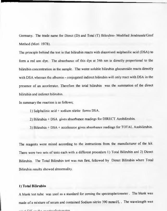
Germany. The trade name for Direct (D) and Total (T) Bilirubin- Modified Jcndrassik/Grof
Method (Mori 1978).
The principle behind the test is that bilirubin reacts with diazotised sulphanilic acid (DSA) to
form a red azo dye. The absorbance o f this dye at 546 nm is directly proportional to the
bilirubin concentration in the sample. The water-soluble bilirubin glucuronidc reacts directly
with DSA whereas the albumin - conjugated indirect bilirubin will only react with DSA in the
presence of an accelerator. Therefore the total bilirubin was the summation of the direct
bilirubin and indirect bilirubin.
In summary the reaction is as follows;
1) Sulphalinic acid + sodium nitrite forms DSA.
2) Bilirubin + DSA gives absorbance readings for DIRECT Azobilirubin.
3) Bilirubin + DSA + accelerator gives absoibance readings for TOTAL Azobilirubin.
The reagents were mixed according to the instructions from the manufacturer of the kit.
There were two sets of tests each with a different procedure 1) Total Bilirubin and 2) Direct
Bilirubin. The Total Bilirubin test was run first, followed by Direct Bilirubin where Total
Bilirubin results showed abnormality.
1) Total Bilirubin
A blank test tube was used as a standard for zeroing the spectrophotometer . The blank was
made of a mixture of serum and contained Sodium nitrite 390 mmol/L . The wavelength was

One ml of the mixture which contained Sulphanilic acid 14 mmol/L, Hydrochloric acid
300mmol/L, Caffeine (accelerator) 200 mmol/L and Sodium benzoate 420 mmol/L) was put
in each o f the differently labelled test tube. A drop (40 pi) of Sodium nitrite 390 mmol/L was
then added to the above test tubes and mixed thoroughly with a mixer (Super-Mixer, LAB
LINE Instrument, Inc, Melrose Park, Illinois). The test tubes were incubated for 5 minutes at
room temperature, and then 100 pi of each of the samples were added into the respective test
tubes. These were mixed and incubated at room temperature for 10 to 30 minutes. The
absorbency for each test tube was then measured against the blank.
2) Direct Bilirubin
The determination of Direct Bilirubin was done to the samples which were found to have
Total bilirubin. The procedure was as follows: One ml of a mixture of Sulphanilic acid 14
mmol/L and Hydrochloric acid 300 mmol/L was put into each labeled test tube. One-drop
(40 pi) of Sodium nitrite 25 mmol/L) was added to the above test tubes and mixed thoroughly
and 100 pi of the sample added within 2 minutes. This was again mixed and incubated at
room temperature for exactly 5 minutes. The absorbance was then measured against the
sample blank.
The calculation of the concentration o f total and direct bilirubin was obtained using the
formula:
Bilirubin concentration [mg/dl] = [Absorbance reading] x 13.0

The determination of alkaline phosphatase in serum was based on a commercial diagnostics
kit called "ALKALINE PHOSPHATASE liquicolor” DBA Buffer, DGKC. The kit was
manufactured by Human Laboratories Ltd, Wiesbaden, Germany. It is both a colorimetric
kit, (i.e. based on determination of colour change) and kinetic kit.
The Reaction Principle for the Test is that, p-Nitrophenylphosphatc +H2O o phosphate + p-
nitrophenol (yellow coloured). The enzyme involved in this reaction is Alkaline
Phosphatase.
The working reagent was made by pipetting 2 ml of substrate (containing
p-Nitrophenyl phosphate 55 mmol/1) into one bottle containing Diethanolamine buffer (pH
10.35 ± 0.2) 1.25 mo 1/1 and Magnesium chloride 0.625 mmol/1) and mixed thoroughly.
These were dispensed at 1 ml into test tubes, and then 100 pi of the sample was added.
This was mixed, and the absorbance read at wavelength of 410 nm after 1 minute and at the
same time the stopwatch was started. Four readings were taken after 1, 2, 3 and 4 minutes.
The mixture turned from clear to yellow with time since it was a kinetic kit.
From the readings the mean absorbance change per minute (AA/min.) was calculated. The
calculation of the alkaline phosphatase activity in the sample was done using the following
3.2.23. Alkaline phosphatase determ ination.
factor:

3.2.2.4 Creatinine determination
The determination of creatinine was done using a commercial diagnostics kit called
“CREATININE liquicolor, JafTe Reaction”. The kit used was acquired from Human
Laboratories Ltd, Wiesbaden, Germany. It is a photometric colorimetric test for kinetic
measurements of the reaction principle as Creatinine + Picric acid -► Crcatininc-picratc
complex. According to the manufacturer’s instruction, the method is done without
deproteinisation.
Creatinine forms an orange-red coloured complex with picric acid in alkaline solution.
The absorbance o f this complex is proportional to the creatinine concentration in the
sample.
The Sodium Hydrochloride (1.6 mol/1) in the kit was diluted with distilled water in the
ratio 1+4 (i.e . 4 mis plus 16 mis o f d istilled w ater) and stored in a plastic bottle.
The diluted Sodium Hydrochloride was mixed with the Picric Acid (26 mmol/1) to produce a
working reagent in the ratio 1 to 1. The standard was made o f Creatinine 2 mg/dl or 176.8
umol. The wavelength on the spectrophotometer was set at 490 nm.
A volume of 1000 ul of the Standard working reagent was pipetted into cuvettes and used to
get the blank reading. Thereafter, 100 ul of samples were mixed with similar volumes of
Standard working reagent. After 30 sec. the absorbance Ai was read. Then the second
absorbance A2 was read exactly after 2 min. The difference between the two readings, A2 -
Ai = change of absorbance of sample .
The calculation o f serum creatinine was done using the following formula:

(Difference between 1* and 2nd absorbance of Sample + Difference between 1“ and 2nd
absorbance of Standard) X 2.0 = Creatinine [mg/dl]
Where 2.0 is a multiplication factor given by manufacturer.
3.2.2.S. Aspartate Aminotransferase (AST/SGOT) determination
Aspartate transaminase (AST) also called serum glutamic oxaloacetic transaminase (SCOT)
or aspartate aminotransferase (ASAT/AAT). The test was done based on a commercial
diagnostics kit called “GOT (AST) IFCC modified liquiUV rest” Aspartate
Aminotransferase (EC 2.6.1.1). The kit used was manufactured by Human Laboratories Ltd,
Wiesbaden, Germany.
The reaction is as follows:
2-Oxoglutarate+L-aspartate o (Aspartate transaminase) o L-glutamate+ oxaloacetatc
Oxaloacetate + NADH + H* o (Malate dehydrogenase) o L-malate + NAD*
The test is a kinetic method for determination of ASAT activity.
The entire contents o f one of the bottles o f the substrate, which contained 2-oxoglutarate 60
mmol/1 and NADH 0.9 mmol/1, were poured into one of the other bottle provided containing
the enzyme for the reaction, which was made of TRIS buffer (pH 7.8) 100 mmol/1, L-
aspartate 300 mmol/1, LDH >0.9 kU/1 and Malate dehydrogenase > 0.6 kU/1, and mixed
thoroughly.

The wavelength was set at 340 nm. 200 pi of the sample and 1000 pi of working reagent
were pipetted into cuvettes. This was mixed and the absorbance read after 1 minute, further
reading of the absorbance was done again exactly after 1, 2 and 3 minutes.
Concentration of AST was determined by multiplying the change o f absorbance per
minute by 1151 = U/l.
3.2.2.6. Albumin determination
The test was based on a commercial diagnostics kit called “Albumin liquicolor Photometric
Colorimetric Test for Albumin BCG- Method”. The kit used was acquired from Human
Laboratories Ltd, Wiesbaden, Germany. The test is based on the fact that Bromocrcsol
green forms a coloured complex with albumin in citrate buffer. The absorbance of this
complex is proportional to the albumin concentration in the sample.
A standard first reading was acquired and recorded. The reagent blank consisted of 1000 pi
of a mixture of Citrate buffer (ph 4.2) 30 mmol/L and Bromocrcsol green 200 pmol/1). 10
pi of sample or working standard made of Albumin 4 gm/dl or 40 g/1 and Sodium azide
0.095 %) was added to 1000 pi of the mixture of Citrate buffer (ph 4.2) 30 mmol/L and
Bromocresol green 200 pmol/1. This was mixed; incubate for 5 minutes at room temperature
20 to 25°C. The absorbance o f the sample and the standard was measured against the reagent
blank within 30 minutes. The spectrophotometer was set at a 578.0 wavelength.
Calculation of the albumin concentration was determined by the following formula:

3.2.2.7. Urea determination
The test was based on a commercial diagnostics kit called “UREA liquicolor, Enzymatic
Colorimetric Test for Urea”. The kit used was acquired from Human Laboratories Ltd,
Wiesbaden, Germany.
Urea hydrolysed in the presence of water and urease to produce ammonia and carbon dioxide.
In a modified Bcrthelot reaction the ammonium ions react with hypochlorite and salicylate to
form a green dye. The absorbance increase at 578 nm is proportional to the urea
concentration in the sample.
The “Enzyme reagent la “ was prepared by mixing the contents of the bottle labelled [ ENZ]
(w h ich was made of Urease > 500 K.U/1) with bo t t l e l a b e l l e d [ R G T l ] ( w h i c h
was made of Phosphate buffer (pH 7.0) 120 mmol/L, Sodium salicylate 60 mmol/1. Sodium
nitroprusside 5 mmol/1 and EDTA 1 mmol/1). This was mixed at a ratio ofe.g. 1 ml of [ENZ]
with 100 ml [RTG1]. This mixture was named “Enzyme reagent la”.
The other reagents, [RGT2] (which was made of Phosphate buffer (pH < 13) 120 mmol/1,
Hypochlorite 0.6 g/1) and [STD] (which was made of Urea 80 mg/dl or 13.3 mmol/1,
equivalent to BUN 37.28 mg/dl or 6.2 mmol/1, and Sodium azide 0.095%) were ready for
use and did not need preparation.
The reagent used as the blank was made o f 1000 pi of “Enzyme reagent la which was
prepared as described above. The wavelength was set at 580 nm. A 10 pi of sample or

to 25°C. This turned to a greenish colour. The absorbance was read for the sample and the
standard against the reagent blank within 60 minutes.
Concentration of blood Urea = Absorbance of sample x 80 mg/dl
Absorbance of Standard
3.23. Haematology
Complete haemograms were done on the EDTA blood samples. The parameters taken were
white blood cells (WBC), Red blood cells (RBC), Haemoglobin (HB), Packed cell volume
(PCV), Mean Corpuscular volume (MCV), Mean cell haemoglobin concentration (MCHC),
Mean cell haemoglobin (MCH), Total plasma, plasma fibrinogen. Band neutrophils,
segmented neutrophils, lymphocytes, monocytes and eosinophils. The hematology data
collected was summarized in Appendix 8.
3.2.4. Histopathology procedures
Many tissues were collected for histopathology. These included adrenal glands, colon, heart,
ileum, jejunum, kidneys, liver, ovaries, pancreas, spleen, uterus (Fundus, horn and cervix)
and all gross lesions. Only small sections (1 cm3) was randomly cut from the larger organs
like the liver, lungs and spleen; for the small organs like the heart, adrenal glands, kidneys,
ovaries, and uterus, they were collected whole. The tissues were immersed and stored in
10% neutral buffered formalin in separate specimen jars for each animal and labelled.
Tissues from endometriosis lesions (Table 7) were fixed in a smaller (Nunck) 2 ml tubes.

sections other the organs were held in neutral buffered formalin for 48 36 hours after
fixation. They were then transferred into 4 % neutral buffered formalin until they were
processed for histopathology. The tissues were then processed through routine
histopathology procedure (Humason 1962; Luna 1968). Sectioning was done at 5 pm
thickness. Serial sections across the endometriosis lesions were done and about five sections
mounted per slide. The approach was to completely section the lesion until there was no
tissue left on the paraffin block. Depending on the size of the block, a tissue section was
selected for staining after about 10 to 20 cuts to produce about 5 slides per cndometriotic
lesion. Representative tissues were sectioned from the other organs with or without lesions.
The cut tissue sections were spread on slides which were earlier prc-coated with egg albumin.
Following section mounting, the slides were placed on a vertical rack and allowed to drain for
at least one minute. The slides were then dried in a 60°C dust free oven for 30 -60 minutes or
overnight at 37°C. For staining purposes, the slides were then placed for 5 minutes in xylene
to dewax/deparaffinize. To prepare the slides for staining, they were first re-hydrated by
rinsing for 5 minutes respectively in 100% isopropanol, 96% industrial spirit and water.
After rehydration, the slides were then transferred into haematoxylin for 5-8-10 minutes. For
purposes of bluing, they were put in water for 1 minute then dipped twice in 5% Acid alcohol
while closely monitoring the diminishing of the blue colour. A water wash for 5 minutes
followed. Eosin counterstaining was done for 4 minutes. The slides were then dipped into
water while checking the contrast between hacmaoxylin and cosin. The slides were then
___________ I C _______ . : U . . r . i n G A O / i n ^ n c t r i o l n l r n K n l S h i r H i n n i n o t

100% isopropanol. The slides were ready for mounting, and DPX mounting media was used
to secure the cover-slip to slide.
3.2.5. Scoring of endometriosis lesions
The lesions were scored microscopically in order to measure and monitor the effects of the
drug on endometriosis. A Scoring method was developed after trial runs through the
endometriosis slides. The trial run identified all the histological lesions. A subjective
scoring of 0 to 4 was created. This designated the scores according to the intensity, severity
and quantitative findings. In this effort a subjective and semi-quantitative basis of scoring
was developed but there was no blinding to the type o f treatment the baboon received had
received. The scores were converted to numerical mean values which were used in statistical
analysis using Kruskal Wallis test. The frequency of appearance o f the histological changes
were analysed using the CHI Square Fisher’s Exact test.
3.2.5.I. Examination of the glandular epithelium features
The presence o f haemorrhages in the lumen of glands or periglandular haemorrhages was
recorded. The presence of hemosiderin in the lumen of glands or periglandular area and the
presence of mucus in the lumen of glands was also noted.
The number o f cross sections of glands was also scored, and any degenerative or necrotic
changes on the glandular epithelium recorded. Erosions of the glandular epithelium were
noted. The presence of acute inflammatory cells like neutrophils, eosinophils in the lumen of

inflammatory cells, such as lymphocytes in these areas were also noted. The checklist
designed and used for scoring of lesion in glandular epithelium is attached in Appendix 4.
3.2.5.2. Examination of the interstitial tissue
The interstitial tissue is defined as all the tissue apart from the glands, endometrial stromal
cells and peritoneum that is seen in each lesion. Usually the interstitium contains supporting
structures such as connective tissue, blood vessels, smooth muscles etc.
The histological changes that were monitored in interstitial tissue included haemorrhages,
haematomas and hemosiderin and other deposits. The presence o f unique degenerative slits
was noted and scored. These degenerative slits were defined as the presence of gaps or
empty space, mainly narrow, long and spindle shaped following the grain of the connective
tissue in interstitial tissues. These were associated with shrinking of interstitium after
degeneration of stromal cells. Degeneration was more visible on the edges of the slits. Thus,
presence of degeneration of interstitial endometriotic stromal cells (ESCs) was noted and
scored. The presence of sworls, formation of arcs in the connective tissue was noted and
scored as were the presence o f acute inflammatory cells.
Presence of a diffuse ground substance (myxoid) in the connective tissue was noted and
scored. Smooth muscle, the numbers o f small or large blood vessel were also noted and
scored. Other features like fat, oedema, amount of periglandular cuffing with endometriotic
stromal cells (ESCs) were examined. The scatter and spread of ESCs was also scored as was
I__,1/wmnnr'itivl P V ('hnpUist HrKioivH 51 r»H 1 1 f o r seorinp o f lesions

3.2.5.3. Examination of the peritoneal surface of lesions
In the biopsies where the peritoneum surface was visible, the same changes as seen in the
glands and interstitial connective tissue were examined. Thus, the presence of haemorrhages,
hemosiderin, lymphocytes, acute inflammatory cells, fibrin on and under the peritoneal
surface were recorded. The above information was used to compare the features within the
types of lesions and between the types of treatments. The checklist designed and used for
scoring of peritoneal surface lesions is attached in Appendix 6.
3 3 . STATISTICAL ANALYSIS
The data was managed through collection of the raw data in tabular format followed by
systematic analysis and checking for any significant changes. The tests used for significance
tests were the Paired Student t-tests within each group (to test the difference before and after
treatment); the Univariate analysis of variance (ANOVA) followed by post hoc analyses like
Tukey’s or Least Significant Difference (LSD) (to test how they differ between the groups),
and finally, the CHI Squared, Fisher’s Exact test for frequencies o f occurrence of changes in
tissues.
The following parameters were subjected to the statistical tests;
1) Changes in surface area of lesions within animals and between groups in respect to
changes before and after the treatments.
2) Changes in the types of lesions (red, typical, white fibrotic plaques or nodules, etc).

and then compared among the three treatment groups. This was done using the Kruskal
Wallis One - way ANOVA test by ranks.
3) Haematological changes before and after treatment.
4) Clinical chemistry changes before and after treatment.
5) Scoring of histopathology findings o f cndomctriotic lesions.
6) Histology changes in other body organs.
A relative change value was calculated for the surface area of each lesion using the following
equation:
Surface area after treatment (laparoscopy 3) minus Surface area before (laparoscopy 2)
divided by Surface area before (laparoscopy 2).
A computer package (INSTAT 3.42, Reading, England) was used to analyse the data. At first
the One-way ANOVA was used to compare the means. The tabulated F- tabulated at a =
0.05 at degrees o f freedom VI = 2 and V2 = 9 was 2.785. The Chi Squared, Fisher’s Exact
test was performed using GraphPad InStat version 3.00 for Windows 95, GraphPad Software,
San Diego California USA, www.graphpad.com. Copyright 1992-1998 GraphPad Software
Inc. (www.graphpad.com).

CHAPTER FOUR
4.0. RESULTS
4.1. ENDOMETRIOSIS LESIONS
4.1.1. Gross appearance of endometriosis lesions assessed during laparoscopy
The presence of endometriosis was confirmed in all babooas. Representative in vivo
laparoscopic images showing the macroscopic changes in the placebo, GnRH antagonist and
rosiglitazone treated lesions are shown in Plates 1 to 9. Out of the several lesions in each
animal and each treatment group, only a few were selected. In the placebo group, 3 out of 11
lesions are represented in Plate 1, 2 and 3 (PAN 2985, PAN 2912 and PAN 2991). In the
GnRH group, 3 out of the 19 lesions are represented in Plate 4, 5 to 6 (two from PAN 2871
and one from PAN 2993). Finally from rosiglitazone group, 3 out of 18 lesions arc
represented in Plate 7, 8 to 9 (one from PAN 3032 and two PAN 3030). The selected lesions
represent the trend of changes in lesions from pre-treatment to post-treatment for each
treatment group.
4.1.1.1 Placebo group.
In the placebo group, some of the lesions progressed to more active lesions and some
remained in the same status after the 25 days of treatment. A few also turned to the typical
bluish-black lesions which is called typical because in women it is the commonly seen type at

(PAN 2985), which was initially a red hemorrhagic (Plate 1 A) and remained the same after
placebo treatment (Plate 1B). While at the same time, some lesions appeared typical bluish-
black lesions in the placebo group at laparoscopy 11 and remained as such in laparoscopy III.
Plate 2 shows endometriotic lesion N5 from a placebo animal (PAN 2912), which typical
bluish black (Plate 2A), and did not change after placebo treatment (Plate 2B). Also the same
was true in Plate 3, where the red lesion maintained the status quo. The endometriotic lesion
N2 from PAN 2991, which was initially a red hemorrhagic vesicular one (Plate 3A), did not
change its morphology after placebo treatment (Plate 3B).
Table 2, 3 and Figure 1 shows the increase in number of both typical and red lesions when
compared between the pre and post treatment stage within the placebo group. Though the
change was not significant, the trend was apparent where more active lesions appeared.
4.1.1.2 GnRH group
After 25 days o f treatment, most lesions in the GnRH antagonist treated group appeared to
change towards less active lesions i.e. typical bluish black or white lesions in a majority of
cases. Plate 4 shows an endometriotic lesion (N5) from PAN 2871 which was initially a
bluish-black lesion (Plate 4A) that changed to a white nodule after GnRH antagonist
treatment (Plate 4B). Some lesions turned from red (active) to white nodules (inactive). This
is exemplified by endometriotic lesion (N7), from PAN 2871, which was initially a
gelatinous red hemorrhagic lesions (Plate 5A), which turned to a white nodule with
niemented snots after GnRH antagonist treatment (Plate 5B). And finally, Plate 6 shows

lesion (Nil) , from PAN 2993 which was initially a red nodule (Plate 6A) and changed to a
pale orange yellow nodule after GnRH antagonist treatment (Plate 6B).
Table 2, 3 and Figure 1 show the increase in the number of typical lesions within the GnRH
antagonist treated group when compared between the pre-treatment and post-treatment stage.
Though the changes were not significant, the trend was clear where less active lesions
appeared to increase at post treatment.
4.1.1.3 Rosiglitazone group.
After 25 days of treatment, most lesions in the rosiglitazone group (like in GnRH antagonist
group) appeared to change towards less active lesions i.e. typical bluish black or white lesions
in a majority of cases. Examples of this transition are shown in Plate 7. The Plate shows an
endometriotic lesion (N4) from PAN 3032 that was a red-white nodule (Plate 7A) that
changed to a white plaque after rosiglitazone treatment (Plate7B). And also in Plate 8, it
shows an endometriotic lesion (N8) from PAN 3030, which was red white hemorrhagic
nodule (Plate 8A) that turned to a typical bluish black lesion after rosiglitazone treatment
(Plate 8B). And finally Plate 9 shows an endometriotic lesion (N9) from PAN 3030 which
was a red haemorrhagic nodule (Plate 9A) that turned to a white nodule after rosiglitazone
treatment (Plate 9B).
From the results, the following statements can be made. The rosiglitazone and GnRH
_l____ 1 1_ I n r i n m ' Q C K \/

8 and 9). In these cases, the lesions mostly turned from red (active) to white nodule or typical
types which are considered less active.
An analysis of the distribution of types o f endometriosis lesions among the 3 treatment
groups showed a general increase in the number of typical bluish black (less active) lesions in
all the treatment groups (Table 2, 3 and Figurel). However, this increase in the less active
lesion was more significant in rosiglitazone group than in both GnRH and placebo group.

Plate 1. G ross ap p ea ran ce o f endom etrio tic lesion ( N i l ) from a placebo trea ted baboon (PA N 2985)
Key: ArrowsPAN 2985 Nil LII L3
Indicate location of lesion.Animal identification number.Identification number of endometriosis lesion Photo taken during second laparoscopy documentation Photo taken during third laparoscopy documentation
- 6 7 -
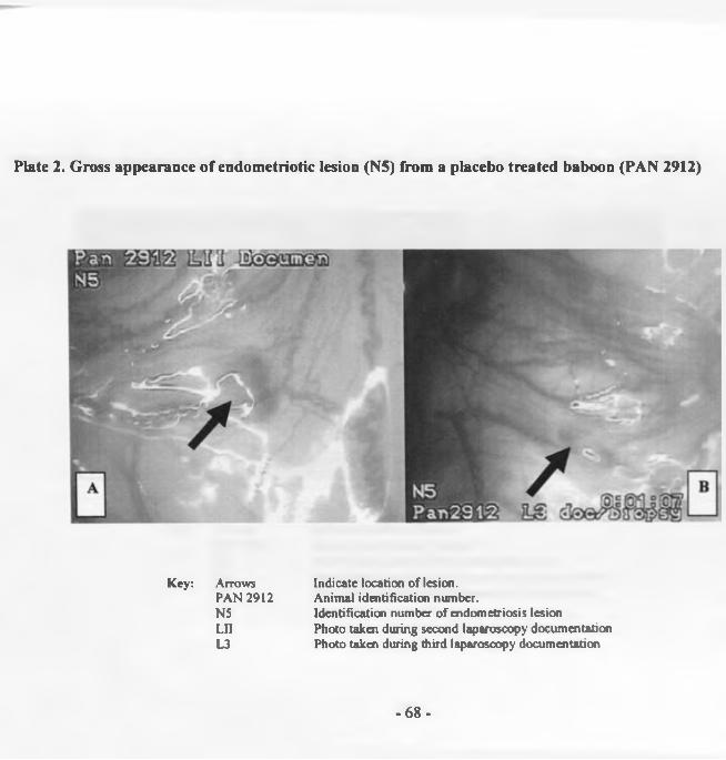
P late 2. G ross ap p ea ran ce o f endom etrio tic lesion (N5) from a placebo tre a te d baboon (PA N 2912)
Key: ArrowsPAN 2912N5LnL3
Indicate location of lesion.Animal identification number.Identification number of endometriosis lesion Photo taken during second laparoscopy documentation Photo taken during third laparoscopy documentation
- 6 8 -

P late 3: G ross ap p earan ce o f en d om etrio tic lesion (N2) from a p lacebo trea ted baboon (PAN 2991)
7*50 H3 S D3SH35 0u
Key: ArrowsPAN 2991 N2LnL3
Indicate location of lesion.Animal identification number.Identification number of endometriosis lesion Photo taken during second laparoscopy documentation Photo taken during third laparoscopy documentation
- 6 9 -

Plate 4: G ross a p p ea ran ce o f en d o m etrio tic lesion (N5) from a G n R H tre a te d bab o o n lesion (P A N !
Key: ArrowsPAN 2871 N5 LII L3
Indicate location of lesion.Animal identification number.Identification number of endometriosis lesion Photo taken during second laparoscopy documentation Photo taken during third laparoscopy documentation
- 7 0 -

P late 5: G ross a p p ea ran ce o f en d o m etrio tic lesion (N7) from a G n R H tre a te d bab o o n (PA N 2871)
Key: ArrowsPAN 2871 N7 LII L3
Indicate location of lesion.Animal identification number.Identification number of endometriosis lesion Photo taken during second laparoscopy documentation Photo taken during third laparoscopy documentation
-71 -

P late 6: G ross ap p earan ce o f endom etrio tic lesion ( N i l ) from a G nR H trea ted baboon (PAN 2993)
Key: ArrowsPAN 2993NULnL3
Indicate location of lesion.Animal identification number.Identification number of endometriosis lesion Photo taken during second laparoscopy documentation Photo taken during third laparoscopy documentation
- 7 2 -

Plate 7: G ross ap p ea ran ce o f endom etrio tic lesion (N4) from a rosig litazone trea ted baboon (PAN 3032)
Key: ArrowsPAN 3032 Nil LII L3
Indicate location of lesion.Animal identification number.Identification number of endometriosis lesion Photo taken during second laparoscopy documentation Photo taken during third laparoscopy documentation
- 7 3 -

P late 8: G ross a p p ea ran ce o f en d om etrio tic lesion (N8) from a rosig litazone trea ted baboon (PAN 3030)
Key: ArrowsPAN 3032NilLnL3
Indicate location of lesion.Animal identification number.Identification number of endometriosis lesion Photo taken during second laparoscopy documentation Photo taken during third laparoscopy documentation
-74 -

Plate 9: G ross a p p ea ran ce o f en d om etrio tic lesion (N9) from a rosig litazone trea ted baboon (PAN 3030)
Key: ArrowsPAN 3030 Nil Ln L3
Indicate location of lesion.Animal identification number.Identification number of endometriosis lesion Photo taken during second laparoscopy documentation Photo taken during third laparoscopy documentation
- 75 -

4.1.2.1. Observations on the Typical and Red lesions
Briefly, there was a change in the distribution and frequency o f two types o f lesions
when the pre-treatment and post-treatment results were compared. These lesions were
the typical bluish-black and the red lesions as summarised in Table 2, 3 and Fig. 1).
Both Tables show the quantitative changes of the different types of endometriosis
lesions (typical and red lesions) for the various treatment groups before and after the
treatment month. Appendix 7 has the raw data of this results.
The plot graph (Figure 1) is showing the total number o f lesions for each the two types
o f lesion. A Student t-paired test analysis in Placebo, GnRH-antagonist, and
rosiglitazone groups revealed 0.41, 0.84 and 0.04, respectively. Although not
significant, the typical lesions increased in all the groups. While the red number of
lesions increased only for the placebo group, they were reduced in the GnRH and
rosiglitazone though not significantly (p value = 0.38 and 0.08, respectively).
4.1.2.2. Observations on the total number of Red lesions
Briefly, the total number o f red lesions was increased in the placebo group and was
reduced in both the GnRH and rosiglitazone treated group though none were significant
(Table 2 and 3; Figure 1 and 2). The paired t-tests were 0.31, 0.38 and 0.08,
respectively.
4.1.2. T h e d istribu tion of the types o f lesions

T ab le 2: D ata from lesions collected d u rin g th e p re -an d p o s t-trea tm en t laparoscopy o f endom etrio tic baboons
O bservation Placebo G nRH - antagonist Rosiglitazone
Pre Post Pre Post Pre PostMean surface Area (mm2) with adhesions
180.8 ± 113.9 164.6 ±80.6 315 ±150.1 $
177.9 ± 141.2 $
240.3 ± 86.8 t
123.2 ±78.3t
Mean surface Area (mm^) without adhesions
121.3 ±77.2 89.6 ±71.8 109.8 ±17.7 *
55.9 ±7 .3 *
106.3 ± 69.2 @
(42.2 ± 10.8 @
Mean number o f lesions 9.5 ± 2 .6 9.3 ±3.3 10.3 ±3 .2 10.8 ±2 .4 13.3 ± 4 .6 12.5 ±4 .5
Mean number o f Red lesions 1.8 ± 2.1 3 ± 2 .4 4.3 ±4 .7 2.5 ± 1 .7 4.3 ± 2 .9 #
0.8 ± 1 .3 #
Mean area o f red lesions (mm2).
21± 24.5 40.3 ± 49.6 8.5 ±43.7 8.7 ±5.3 62.9 ±72.5 0
4 ± 6 .7 ©
Mean number o f adhesions 2.8 ± 1 .9 2.5 ± 1 .9 2.8 ±0 .5 3.3 ±0 .5 3.5 ± 2 .9 3.3 ±2 .5
KEY: $ GnRH relative surface area reduction was significant with Dunnett t-test post hoc statistic (p = 0.044)t Rosiglitazone group had significant reduction (F value = 5.7, Tukey p value = 0.033).* GnRH surface area without adhesion, the reduction was significant (Student’s test, t value = 4.89, p value = 0.0164)
@ Rosiglitazone group though reduced was not significant (Student t value = 2.14, p value = 0.1214).# Rosiglitazone group total number of red lesions was reduced though not significantly (p value = 0.08).© Rosiglitazone group area of red lesions was reduced though not significantly (p value = 0.201)
- 7 7 -

T ab le 3: Q u an tita tiv e changes o f th e d iffe ren t types of endom etriosis lesions fo r th e v ario u s tre a tm e n t g roups
ObservationPlacebo GnRH - antagonist Rosigli tazone
Pre Post Pre Post Pre Post
Typical blue black
lesions
1 (0-5) 2.5 (0-7) 1.5 (0-6) 2.5 (1-4) 0.5 (0-5) 4(1-11)
Red lesions 1.5 (0-4) 3 (0-6) 2.5 (0-1) 2(1-5) 5.5 (0-6) 0.5 (0-2)
Typical and red lesions 4(0-9) 5(1-13) 5.5 (2-3) 6(2-6) 6(0-11) 4.5(3-11)
Note: All data are presented as median (range).
PrePost
= Pre-treatments counts. = Post treatment counts.
- 7 8 -

Num
ber
of l
esio
ns
F ig u re 1: C hanges in specific types an d n u m b er of en d o m etrio tic lesions fo r th e tre a tm e n t g roups
GB GATreatment groups
K E Y .PB Placebo Before GB GnRH Before RB Rosiglitazone Before
PA Placebo After GA GnRH After RA Rosiglitazone After
LEGENDBoth types o f lesions Red type o f lesions Tvoical blue black lesions
- 7 9 -

F ig u re 2: C h ange in to ta l n u m b er of red lesions in end o m etrio tic baboons
10
1Z
PB PA GB GA RB RATreatment groups
KEY.PB Placebo Before PA Placebo AfterGB GnRH Before GA GnRH AfterRB Rosiglitazone Before RA Rosiglitazone
- 8 0 -

4 .1 3 S u rface area o f endom etrio tic lesions
The sizes o f the endometriosis lesions (with adhesions) or (without adhesions) were compared
separately. An analysis o f the changes in surface area o f the endometriosis lesions in the three
treatment groups showed the following changes;
4.13.1. Changes of surface area of lesions (without adhesions)
The mean surface area of endometriotic lesions (without adhesions) was significantly reduced in
the GnRH group only. In this group, the pre-treatment surface area (109.8 ± 17.7) was compared
with the post-treatment surface area (55.9 ± 7.3), (Student’s test, t value = 4.89, p value =
0.0164). Figure 3 and Table 2 summarises these findings. Similarly, the surface area o f the
lesions o f rosiglitazone group was also reduced though not significant (Student t value = 2.14, p
value = 0.1214). Appendix 7 shows the raw data on the surface are measurements, number and
types o f lesions.
4.13.2. Changes of surface area of lesions (with adhesions)
There was a relative reduction in the surface area o f the rosiglitazone-treated group (which was
statistically significant when compared to the placebo group) (p value = 0.033) (Figure 4). In the
GnRH group, the relative reduction in surface area was only statistically significant with the
Dunnett t-test post hoc statistic (p = 0.044) when compared with the placebo group.
A comparison o f the relative lesion surface area using ANOVA o f the means between the three

4.1 J 3 Changes in surface area of Red lesions
The reduction of the mean surface areas of red endometriotic lesions from pre-treatment to post
treatment was reduced though not significant within the rosiglitazone group i.e. from 5.5 (0-6) to
0.5 (0-2), p-value = 0.201, where the data is represented as median (range). See Table 2.

Figure 3: Changes in surface area of lesions (w ithout adhesions) before & after trea tm en t of endom etriotic Baboons
KEY.PB Placebo Before PA Placebo After NS Difference not significantGB GnRH Before GA GnRH After S Difference SignificantRB Rosiglitazone Before RA Rosiglitazone After
- 8 3 -

Rel
ativ
e ch
ange
in s
urfa
ce a
rea
(no
units
)
Figure 4: Relative change in surface area of lesions (with adhesions) in endometriotic baboons in the treatment groups
Placebo GnRH RosiglitazoneTreatment Groups
- 8 4 -
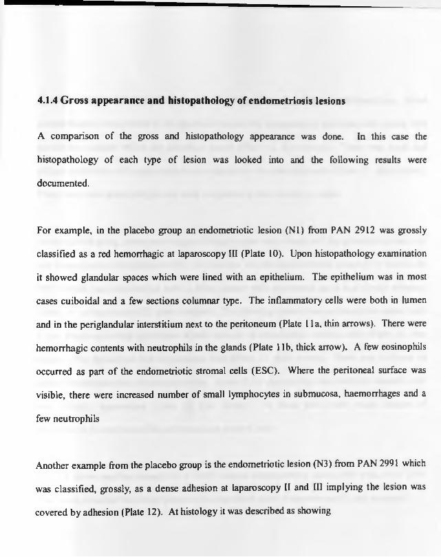
A comparison o f the gross and histopathology appearance was done. In this case the
histopathology o f each type o f lesion was looked into and the following results were
documented.
For example, in the placebo group an endometriotic lesion (N l) from PAN 2912 was grossly
classified as a red hemorrhagic at laparoscopy III (Plate 10). Upon histopathology examination
it showed glandular spaces which were lined with an epithelium. The epithelium was in most
cases cuiboidal and a few sections columnar type. The inflammatory cells were both in lumen
and in the periglandular interstitium next to the peritoneum (Plate 11 a, thin arrows). There were
hemorrhagic contents with neutrophils in the glands (Plate 1 lb, thick arrow). A few eosinophils
occurred as part o f the endometriotic stromal cells (ESC). Where the peritoneal surface was
visible, there were increased number of small lymphocytes in submucosa, haemorrhages and a
few neutrophils
Another example from the placebo group is the endometriotic lesion (N3) from PAN 2991 which
was classified, grossly, as a dense adhesion at laparoscopy II and III implying the lesion was
covered by adhesion (Plate 12). At histology it was described as showing
4.1.4 Gross appearance and histopathology of endometriosis lesions

glandular structures lined by both simple cuiboidal and simple columnar epithelium type. Some
haemorrhages were evident in the glandular lumen with desquamated epithelial cells mixed with
proteinous exudate within the glandular lumen (Plate 13, thin arrow). There was focal and
diffuse infiltration o f lymphocytes & macrophages in the interstitial area (Plate 13, thick arrow).
There were also haemorrhages and mild congestion in the connective tissue.
In the GnRH group, there were signs of degeneration and atrophy o f the glandular epithelium
and desquamation o f epithelial cells. An example was the endometriotic lesion (N3) from PAN
2993 which was classified as both a white plaque with pigmented spots and flimsy adhesion
(Plate 14) at laparoscopy III, after treatment. The histology showed an endometriotic lesion with
a low simple cuiboidal epithelium a mild amount o f endometrial stromal cells (Plate 15, thin
arrow). The epithelium had degenerated cells (Plate 15, thick arrow). There was evidence of
extensive desquamation of epithelial cells. Some o f the surrounding endometriotic stromal cells
were slightly degenerated (Plate 15, thin arrow). At times there were small amount of
hemosiderin present around the endometriotic stromal cells.
Plate 16 shows another example o f a GnRH treated endometriotic lesion (N9) from PAN 2993
which was classified as a white plaque with pigmented spots at laparoscopy II, and remained

the same at laparoscopy III (Plate 16, arrow). And at histology, the description o f the
endometriotic lesion showed simple cuiboidal and low pseudostratified epithelia. There was
evidence o f degenerated and desquamation o f epithelium (Plate 17, arrow). The surrounding
periglandular tissue had extensive connective tissue and small amounts o f smooth muscles.
There were mild haemorrhages and deposits o f hemosiderin
Another endometriotic lesion (N5) from PAN 2998 (Plate 18), a rosiglitazone treated baboon,
was classified as a white plaque at laparoscopy III. At histology, it showed necrosis (nuclei are
pyknotic and karyorhexis) o f glandular epithelium nuclei and desquamation o f the epithelium
into the lumen of the glandular structures (Plate 19, arrow). The rest o f the section showed
excessive haemorrhage and a haematoma covered with extensive fibrin with moderate
lymphocytic infiltrating.
The endometriotic lesion (N13) from PAN 3030, a rosiglitazone treated animal. The lesion was
classified as a white plaque at laparoscopy III (Plate 20). At histology, it showed degeneration,
atrophy and desquamated areas on the glandular epithelium. Degenerated epitheliums was visible
(Plate 21, thick arrow). Interstitial area had mainly connective tissue and smooth muscles.
Peritoneal surface (not in photomicrograph) had fibrin deposits that could explain a white plaque
type o f lesion. There was mild infiltration o f lymphocytes around glands (Plate 21, thin arrow).

Below are a series o f representative photomicrographs o f histopathology of some endometriosis
lesions from various treatment groups (See Plate 10 to 23). In all the plates, the photographs of
the endometriotic lesions are followed by a photomicrograph of its histopathology. The plates
were paired in the following manner; Plate 10 with plate 11; Plate 12 with plate 13; Plate 14
with plate 15; Plate 16 with plate 17; Plate 18 with plate 19 and Plate 20 with plate 21. The
lesions were taken at laparoscopy III i.e. these were the lesions that were observed after
treatment. In summary, rosiglitazone treated group showed more necrosis and degeneration
compared to GnRh and Placebo animals. This also correlated with the reduction in size and
changes in morphology seen in the groups. The placebo had fewer changes while GnRH had
more changes and rosiglitazone most, in that order.
In this study there were 25 out o f 34 cases (74%) of glandular lumen haemorrhages; 27 out o f 34
cases (79 %) o f periglandular haemorrhages; 35 out o f 38 cases (92 %) o f interstitial
haemorrhages; 32 out of 37 cases (87%) o f peritoneal haemorrhages across all the treatment
groups.
In conclusion, a general description of the endometriosis lesions that was consistent in all 3
treatment groups is that: the epithelia o f endometriotic glands at times appeared less uniform
with varying heights in the same gland often showing varying degree o f cytologic
polymorphism. A loss o f polarity o f the epithelial cells was at times seen and cellular

times a mantle o f fibrosis, which, to varying degrees replaced the specialised endometriosis
stromal cells, frequently surrounded the endometriotic lesions. In some instances, the
endometriotic tissue appeared to develop the menstrual bleeding phase pattern.

Plate 10: P ho tograph o f endom etrio tic lesion (N l) from a placebo treated baboon at
laparoscopy III (PAN 2912)

P late I I : P ho tom icrog raph o f endom etrio tic lesion (N l) a t laparoscopy III from
placebo treated baboon (PAN 2912)
(Magnification x 100, H & E).

P la te 12: P h o to g rap h o f endom etrio tic lesion (N3) from a placebo treated baboon a t
laparoscopy III (PAN 2991)
Key: ArrowsPAN 2991N3i i
Indicate location of lesion.Animal identification number.Identification number of endometriosis lesion Photo taken during third laparoscopy documentation

P la te 13: P ho tom icrog raph o f endom etrio tic lesion (N3) from a placebo trea ted
baboon a t laparoscopy III (PAN 2991)
(Magnification x 100, H & E).

P la te 14: P h o to g rap h o f endom etrio tic lesion (N3) from a GnKH treated baboon a t
laparoscopy III (PAN 2993)
__ n»rt/\f I / ' IfhClAn

P la te 15: P h o to m icrog raph o f endom etrio tic lesion (N3) from a G n R Il trea ted
baboon a t laparoscopy III (PAN2993)

P la te 16: P h o to g rap h o f endom etrio tic lesion (N9) from a (in R H trea ted baboon at
laparoscopy III (PAN 2993)
Kpv: Arrow shows position of endometriotic lesion.

» L>
P la te 17: P ho to m icro g rap h o f endom etrio tic lesion (N9) from a G nR H treated
baboon at laparoscopy III (PAN 2993)
(M a g n if ic a tio n x 2 5 0 , H & E ) .
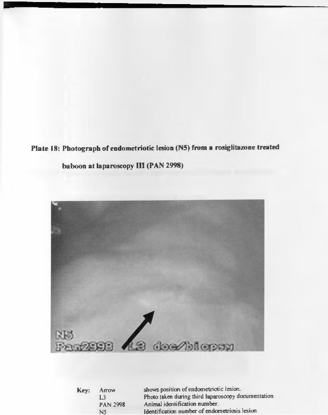
P la te 18: P h o to g rap h o f endom etrio tic lesion (N5) from a rosiglitazone trea ted
baboon a t laparoscopy III (PAN 2998)
Key: ArrowL3PAN 2998 N5
shows position of endometriotic lesion.Photo taken during third laparoscopy documentation Animal identification number.Identification number of endometriosis lesion

P la te 19: P h o to m icro g rap h of endom etrio tic lesion (N5) from a rosiglitazone
tre a te d baboon a t laparoscopy HI (PAN 2998)
(Magnification x 2 5 0 , H & E ).
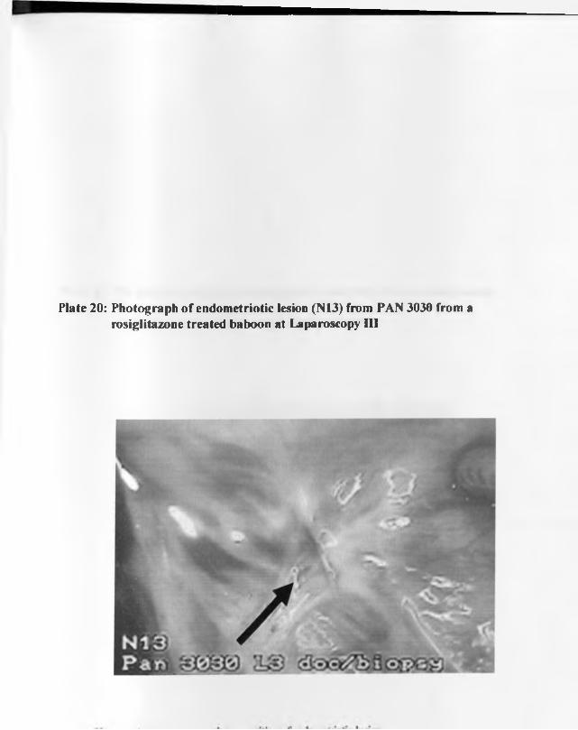
Plate 20: Photograph of endometriotic lesion (N13) from PAN 3030 from a rosiglitazone treated baboon at Laparoscopy III

Plate 21: P h o to m icro g rap h o f endom etrio tic lesion (N13) from a rosiglitazone
treated animal at laparoscopy III (PAN3030)
(Magnification x 100, H & E).

The histological scoring was done by checking sections o f endometriotic tissue. While guided
by and using the Appendices 4 to 6, the approximate range or nearest description fitting the
microscope reading was recorded. A raw data chart was used to enter the scores collected. Each
endometriotic lesion had a maximum o f 5 slides, which after reading an average o f the slides was
retrieved and use for statistical analysis.
4.1.5.1 Scores among the types of treatment
Scores o f the various histopathological changes were compared between the 3 treatment groups.
The scoring on the slides was subjective and relied on the Appendix 4, 5 and 6. The design o f the
key was based on the fact that there were varying degrees o f changes in all the 33 features
identified. The lack of appearance o f the feature was labelled zero (0) and the lowest intensity
was 1 and highest 4. The number o f slides made from a lesion were scored and an average given
taken. The number o f slides per lesion ranged from 1 to 5. The mean o f all the scores from each
lesion were used for statistical comparison o f the treatment groups or type o f lesions.
Tables 4 to 5 are the details of the synthesized scores for all the treatment groups. Table 6 shows
a summary o f the comparison o f scores among the treatment groups o f endometriotic baboons.
Figures 5 to 10 are a graphical description of these changes among the 3 treatment groups.
4.1.5 S c o r in g o f th e h isto log ica l c h a n g e s in th e e n d o m e tr io tic lesions

data o f the scores inserted in the tables are the average scores o f all the slides examined from
each lesion.
A comparison o f the histopathological scores of the endometriosis lesions from the 3 treatment
groups, showed significant differences in degeneration o f glandular epithelium; and that of
endometrial stromal cells, along with presence of peritoneum lymphocytes (Figures 5 to 7).
The rosiglitazone treated group had the highest amount of glandular degenerative changes among
the 3 groups o f endometriotic baboons, followed by GnRH - antagonist treated and the least the
placebo treated group. The placebo group had an average score o f 1, GnRH was 2.48 and
rosiglitazone was 2.82. (See Table 4). The scoring was based on the level of degenerative or
necrotic changes on the glandular epithelium. The variation in the scores glandular degenerative
scores when compared between the 3 groups is graphically represented in Figure 5. (Wallis One
- way ANOVA test by ranks, p value = 0.0043).
The ESCs degenerative scores when compared between the 3 groups showed both rosiglitazone
and GnRH- antagonist groups had the highest scores and placebo group the least scores. The
placebo group had an average score o f 0.82, GnRH was 2.45 and rosiglitazone was 2.63 (See
Table 5). A graphical representation o f the variation is shown in Figure 6. (Wallis One - way
ANOVA test bv ranks, d value = 0.0002).

A graphical representation o f the variation in the presence o f peritoneum lymphocytes scores
when compared between the 3 groups is shown if Figure 7. The rosiglitazone had the highest
score followed by the placebo and the last was the GnRH- antagonist treated group. The placebo
group had an average score o f 1.54, GnRH was 1.33 and rosiglitazone was 2.05 (Wallis One -
way ANOVA test by ranks, p value = 0.0224) (see Table 5). The presence o f lymphocytes in the
peritoneum adjacent to the endometriosis was significantly different among the treatment groups.
Figure 8 shows a graphical representation o f the variation in the glandular luminal mucus scores
when compared between the 3 groups. The GnRH-antagonist treated group had highest score
followed by rosiglitazone and finally the placebo group. Although varied, the scores were not
significant enough (Wallis One - way ANOVA test by ranks, p value = 0.0609).
Occurrence o f degenerative slits was a unique feature in this study. This can be described as the
presence o f narrow spindle shaped gaps within the interstitial tissue and appearance of
degenerated shrunk cells at their edges. These lesions could be linked with degeneration and
possibly shrinking in the surrounding ESCs. Figure 9 shows a graphical representation o f the
variation in the interstitial degenerative scores when compared between the 3 groups. Though
there were no significant differences between the three treatment groups, the rosiglitazone had
the highest score followed by GnRH-antagonist treated group and the least was the placebo
group. Although varied, the scores were not significant enough (Wallis One - way ANOVA test

Figure 10 shows a graphical representation o f the variation in the presence o f acute inflammai
cells scores when compared between the 3 groups. Though there were no significant differer
between the three treatment groups, the rosiglitazone group had the highest scores followed
both GnRH- antagonist group and placebo group. Although varied, the variation among tl
groups was not significant (Wallis One - way ANOVA test by ranks, p value = 0.0636).
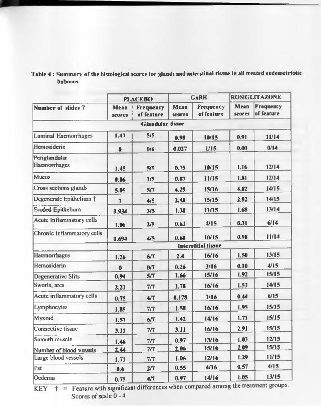
Table 4 : Summary of the histological scores for glands and interstitial tissue in all treated endometriotic baboons
PLACEBO GbRH ROSIGLITAZONENumber of slides 7 Mean
scoresFrequency of feature
Meanscores
Frequency of feature
Meanscores
Frequency of feature
Glandular tissue
Luminal Haemorrhages 1.47 5/5 0.98 10/15 0.91 11/14Hemosiderin 0 0/6 0.027 1/15 0.00 0/14PeriglandularHaemorrhages 1.45 5/5 0.75 10/15 1.16 12/14Mucus 0.06 1/5 0.87 11/15 1.81 12/14Cross sections glands 5.05 5/7 4.29 15/16 4.82 14/15Degenerate Epithelium f 1 4/5 2.48 15/15 2.82 14/15Eroded Epithelium 0.934 3/5 1.38 11/15 1.68 13/14Acute Inflammatory cells
1.06 2/5 0.63 4/15 0J1 6/14Chronic Inflammatory cells
0.694 4/5 0.68 10/15 0.98 11/14Interstitial tissue
Haemorrhages 1.26 6n 2.4 16/16 1.50 13/15Hemosiderin 0 0/7 0.26 3/16 0.10 4/15Degenerative Slits 0.94 5/7 1.66 15/16 1.92 15/15Sworls, arcs 2.21 i n 1.78 16/16 1.53 14/15Acute inflammatory cells 0.75 4/7 0.178 3/16 0.44 6/15Lymphocytes 1.85 i n 1.58 16/16 1.95 15/15Myxoid 1.57 6/7 1.42 14/16 1.71 15/15Connective tissue 3.11 i n 3.11 16/16 2.91 15/15Smooth muscle 1.46 i n 0.97 13/16 1.03 12/15Number ofhlnoH vessels 2.44 i n 2.06 15/16 2.09 15/15Large blood vessels 1.71 i n 1.06 12/16 1.29 11/15Fat 0.6 i n 0.55 4/16 0.57 4/15
Oedema 0.75 A n 0.97 14/16 1.05 13/15
KEY t = Feature with significant Scores of scale 0 -4
differences when compared among the treatment groups.

Table 5 : Summary of the histological scores for ESCs and peritoneum tissue in all treated endometriotic baboons
PLACEBOGnRH ROSIGLITAZONE
Mean Frequency Mean Frequency Mean Frequency
Number of slides 7scores of feature scores of feature scores of feature
ESCsPeriglandular cuff 0.75 4/6 1.17 15/16 1.38 13/15Hemosiderin 0 0/7 0 0/16 0.00 0/15Degenerate stromat 0.82 5/7 2.45
16/162.63 14/15
Scatter 2.47 7/7 1.66 16/16 1.92 14/15Peritoneum
Haemorrhages 1.52 6/7 1.77 14/16 1.52 13/15Hemosiderin 0 0/7 0.103 2/16 0.01 1/15Lymphocytes j 1.54 6/7 133 16/16 2.05 15/15Acute inflammatory cell 0.38 2/7 0.15 0.77 9/15Fibrin 0.68 5/7 1.19 15/16 1.20 12/15Myxoid 134 6/7 1.6 16/16 1.85 15/15
KEY: t = Feature with significant differences when compared among theT reatment groups.
Scores o f scale 0 - 4

T ab le 6: S u m m ary o f com parison o f scores am ong th e tre a tm e n t g roups o f en d o m etrio tic baboons
Figurenumber
Histological changes P value Statistical decision
1 Degenerated glandular epithelium
0.0043 Significant
2 Degenerate ESC 0.0002 Significant
3 Peritoneum Lymphocytes 0.0224 Significant
4 Glandular Luminal mucus 0.0609 Not Significant
5 Interstitial Degenerative slits 0.0701 Not Significant
6 Peritoneum Acute inflammatory cells
0.0636 Not Significant
- 108 -

Scor
es o
f gla
ndul
ar d
egen
erat
ive
chan
ges
F ig u re 5: C o m p ariso n o f g lan d u la r d eg en era tiv e changes am ong the 3 g ro u p s o f en d o m etrio tic b aboons
Placebo GnRH RosiglitazoneTreatment Group
Key: GnRH = GnRH- antagonist treated group.Score of 0 * Normal epithelium1 = Traces of degeneration in some sections of the gland2 = Mild degeneration with pyknotic nuclei with epithelial cell having more dark nuclei3 = Moderate degeneration nucleus completely pyknotic nucleus. Epithelia cells with intact shape and organisation.4 = Severe degeneration and karyorhexis and disintegration of the nucleus and epithelial cells i.e. epithelium necrotised.
- 109 -

F ig u re 6: C o m p ariso n o f d eg enera tive changes in endom etrio tiendom etrio tic baboons
<u60-cCJ<D>20)c60U•o
UC/3-
H8
C/5
3.5
3
2.5
2
1.5
1
0 5
0 _ L* '
Placebo GnRH Treatment G
Key: GnRH GnRH- antagonist treated group.Score of 0 = No evidence of ESCs scatter or spread within interstitial
tissue.1 - Small to traces of amount of ESCs scatter or spread within interst2 = Mild amount of ESCs scatter or spread within interstitial tissue.3 * Moderate of ESCs scatter or spread within interstitial tissue.4 = Wide spread amount of ESCs scatter or spread within interstitial I
- 110-

Figure 7: Comparison of peritoneum lymphocytes among the 3 treatment groups of endometriotic baboons
2= Mild amount lymphocytes within the surface and subepithelia tissue of peritoneum.3= Moderate amount of lymphocytes within the surface and subepithelia tissue of peritoneum. 4= Severe infiltration of lymphocytes within the surface and subepithelia tissue of peritoneum.
- I l l -

F ig u re 8: C o m p ariso n o f g lan d u la r lum inal m ucus am ong th e 3 tre a tm e n t g ro u p s o f en d o m etrio ticbaboons
C/33y3E
Placebo GnRH- RosigHtazoneTreatment Group
Key:GnRH - GnRH- antagonist treated group.Score of 0 = No mucus or proteinous exudate in lumen
1 = Small traces of amount of mucus or proteinous exudate in glandular lumen covering less than Vi of lumen2 = Glandular lumen % to Vi half filled with mucus or proteinous exudate3 = About Vi to V* glandular lumen full of mucus or proteinous exudate4 = Whole Glandular lumen or cavity filled with mucus or proteinous exudate
Legend* Outlier lesion 2991N3, the only not having a 0
scores i.e 0.3• Outlier lesion 2871N10, highest score of 4<>
Outlier lesion lesion 2788N16, highest score
- 112-

Scor
es f
or in
ters
titia
l de
gene
rativ
e sl
its
Figure 9: Comparison of interstitial degenerative slit scores among the 3 treatment groups of endometriotic baboons
Placebo GnRHTreatment Group
Rosiglitazone
Key: GnRH = GnRH- antagonist treated group.Score of 0 * No degenerative slits
1 = Traces of degenerative slits2 * Mild presence of degenerative slits3 = Moderate amount of degenerative slits4 = Severe or extensive presence of degenerative slits
- 113 -

Figure 10: Comparison of acute inflammatory cells within the baboons.
Treat
Key: GnRH = GnRH- antagonist treated group.Score of 0 = No acute inflammatory cells present.
1 = Traces amount of acute inflammatory cells.2 = Mild amount of acute inflammatory' cells.3 = Moderate amount of acute inflammatory cells.4 = Severe infiltration of acute inflammatory cells.
-114 -

4.1.5.2. Scores among the types of lesions
Histopathology on transformation of the lesions with treatment was comprehensively done in this
study. The synthesized scoring o f data collected from lesions was recorded as a mean of
endometriosis from the slides examined and these are tabulated in Table 8 and 9. Table 10 is a
summary o f the statistical analysis showing the comparison o f histological changes among the
types o f lesions in endometriotic baboons.
There were over 33 histological features or changes in different grades that were compared.
Histopathological features o f the different lesions were tested for association with type o f lesion.
A test on the null hypothesis that all the types’ o f lesions have an equal histological score was
rejected for some histological changes. These were; 1) Glandular haemorrhages; 2) Interstitial
tissue degenerative slits; 3) Interstitial tissue lymphocytes; 4) presence o f Smooth muscles
Interstitial tissue; 5) Endometrial stromal cell degeneration and 6) Peritoneal haemorrhages.
This implied that there was cross-association between some types o f endometriotic gross lesions
with specific histological features.
A graphic description of these results are summarised in Figure 11 to 16. In figure 11, the figure
shows the glandular luminal haemorrhages had a significant variation in the scores/ amount
between the types o f lesions given (p value = 0.0249). For example it shows Type 1 lesion had
thp hiahpst amnnnt olanrlnlnr luminal haemorrhages than for examnle tvoe 6 and 7. There was

classification o f lesions (by color o f lesion), where Type 1 was a Typical blue-black due to
deoxygenated blood in the lumen.
In Figure 12, the findings are represented graphically and shows that the interstitial degenerative
scores were significantly different among the types o f lesions given Table 4 (p value = 0.025).
For example its shows Type 2 had the highest amount o f interstitial degeneration than for Type
7.
In Figure 13 the findings are represented graphically and show that there is a significant
difference in the amount o f interstitial lymphocytes among the types o f lesions (p value =
0.0242). Types 1 and 2 appear to have a higher amount than type 6 and 7.
In Figure 14, the significant findings on the amount o f smooth muscles are represented
graphically. The figure shows the smooth muscles amount varied significantly between the
types o f lesions given Table 10 (p value = 0.0325). Type 7 had the highest amount o f smooth
muscles than type 6.
Briefly, the significant findings on ESCs degenerative changes are represented graphically in
Figure 15. It shows that there was a significant difference in the scores among the different types
_TnUi^ i a — n mo'i'v 9 hoH thr* hiohf»ct rhancws together with tvne 6 and

Figure 16 shows the peritoneal haemorrhages had a significant variation in the scores/ amount
between the types o f lesions (p value = 0.0224). Type 1 and 2 had the highest amount ol
peritoneum haemorrhages than both type 6 and 7. This can be expected since the morphological
classification o f type o f lesion depends on colour o f lesions.

Table 7: Endometriosis lesions at necropsy.
Code for type
ofLesion
Morphological Description of endometriotic lesion Number of lesions
Placebo GnRH Rosiglitazone1 Typical blue-black 4 5 22 White plaque with pigmented
spots 4 23 Red vesicular4 Red haemorrhagic 15 Red polyp6 White plaques 2 67 White nodules 1 6 18 White clear vesicle9 Red white plaque10 Peritoneal ulcer11 Flimsy adhesion 312 Dense adhesion 2 1 2
Post-mortem findings 3 1Not seen at Laparoscopy III but seen at Post Mortem 1 1Not found at 3rd laparoscopy 1TOTAL 11 19 18

T ab le 8: S u m m ary o f th e histological scores fo r gl
1 2
Histological feature
Mean
scores
Frequency
Mean
scores
1 Luminal Haemorrhages t1.8S 6/9 1.18
2 Hemosiderin 0 0 0
3PeriglandularHaemorrhages
1.26 5/9 1.18
4 Luminal Mucus 0.39 4/9 0.245 Cross sections o f glands 5.36 7/9 5.436 Degenerate Epithelium 2.14 7/9 2.737 Eroded Epithelium 7/9 1.688 Acute Inflammatory cells 0.93 3/9 0.29 Chronic Inflammatory cells 0.61 4/9 0.94
KEY t = Feature with significant differences when Type of lesion (1= Typical blue-black; 2 = Whin
4 = Red haemorrhagic; 6 = Whit< Scores of scale 0 - 4
- 119-

T able 9: S u m m ary o f th e histological scores for In terstitia l tissue and Peritoneumin all types of endometriotic lesions
Histological feature
Type of lesion1 2 4 6 7
Mean scores
Frequency
Mean scores
Frequency
Mean scores
Frequency
Mean scores
Frequency
Mean scores
Frequency
Interstitial tissue
10 Haemorrhages 1.49 9/9 1.63 6/6 0 - 1.2 2/2 0.64 6/8
11 Hemosiderin 0.14 2/9 0.33 4/6 0 - 0 0 0 0/8
12 Process Slits 1.68 9/9 2.25 6/6 2.4 - 2.4 2/2 1.52 8/8
13 Degenerative Slits t 1.17 9/9 232 6/6 1 - 23 2/2 0.98 6/8
14 Sworls, arcs 1.86 9/9 1.76 6/6 2.8 - 1.1 2/2 1.62 7/8
15 Acute inflammatory cells 0.43 3/9 0.34 2/6 0 - 0.6 1/2 0 0/8
16 Lymphocytes t 1.87 9/9 2.14 6/6 1.6 - 1.9 2/2 1.28 8/8
17 Myxoid 13 9/9 1.8 5/6 2 - 1.7 2/2 1.65 8/818 Connective tissue 3.09 9/9 2.83 6/6 2.8 - 3 2/2 333 8/819 Smooth muscles f 1 9/9 1.05 5/6 1.2 - 0 0 1.6 8/820 Number of blood vessels 2.21 8/9 2.27 6/6 2 - 1.8 2/2 236 8/821 Large blood vessels 1.5 8/9 1.43 5/6 1 - 0.5 1/2 1.38 7/822 Fat 0.8 4/9 0.23 2/6 0 - 0.7 1/2 0.43 2/823 Oedema 0.97 7/9 0.68 4/6 1 - 1.6 2/2 0.6 5/8
24 Periglandular cuffing ESCs 1.49 7/8 1.21 6/6 1.8 - 0.4 2/3 1.26 7/8
25 ESC Hemosiderin t 0 0/9 0 0/6 0 - 0 0 0 0/8
26 Degenerated ESC 2.01 8/9 2.77 6/6 1 - 2.4 3/3 2.25 7/8
27 Amount of Scatter of ESC 1.87 9/9 1.41 6/6 2.6 - 1.8 3/3 1.95 8/8
Peritoneum28 Haemorrhages f 1.25 8/9 1.63 6/6 2.6 - 1 1/2 0.25 3/829 Hemosiderin 0.14 1/9 0.08 2/6 0 - 0 0 0 0/830 Lymphocytes 133 8/9 2.14 6/6 3 - 1.3 2/2 0.83 7/8
31 Acute inflammatory cells 0.07 1/9 0 0/6 2.2 - 0.3 1/2 0.1 3/8
32 Fibrin 0.87 8/9 0.5 3/6 1 - 1 2/2 0.75 7/8
33 Myxoid 1.38 8/9 2 6/6 2.4 - 1.9 2/2 1.6 7/8KEY t = Feature with significant differences when compared among the types of lesions
'T'___ _ r 1_; ______ r ___: __I K l .w t- 0 = U / h itp n la n n p U /lth D l P m e n t c d SDOtSl 4 = R e d
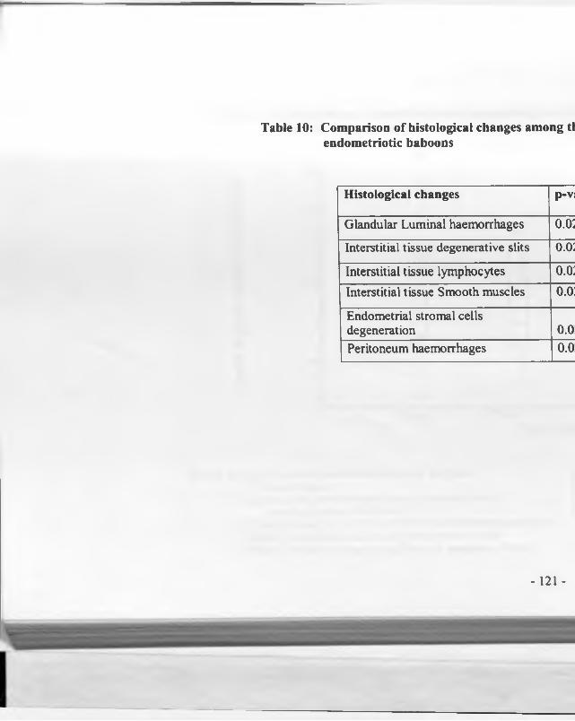
Table 10: Comparison of histological changes among ttendometriotic baboons
Histological changes p-vj
Glandular Luminal haemorrhages o.o;
Interstitial tissue degenerative slits o.o:
Interstitial tissue lymphocytes o.o:
Interstitial tissue Smooth muscles o.o:
Endometrial stromal cells degeneration 0.0Peritoneum haemorrhages 0.0
- 121 -

Figure 11: Comparison of glandular lumen haemorrhage scoriendometriotic baboons.
Type o f L
Key for scoring of haemorrhages within the lumen of the glands.0 = No blood in lumen1 = Small to traces of extravasated blood in glandular lumen
covering less than V* of lumen2 = Glandular lumen or Cavity V* to V4 half filled with extravasated blood.3 = About 'A to V* glandular lumen full of extravasated blood4 * Whole Glandular lumen or cavity filled with extravasated blood.
- 122-

Figure 12: Comparison of interstitial degenerative slits scores betweebaboons
Type o f Lesion
Key for scoring of degenerative slits within interstitial tissues.0 - No degenerative slits1 - Traces of degenerative slits2 = Mild presence of degenerative slits3 = Moderate amount of degenerative slits4 = Severe or extensive presence of degenerative slits
- 123-

Scor
es o
f int
erst
itial
lym
phoc
ytes
Figure 13: Comparison of interstitial lymphocyte scores betwcbaboons
Key for scoring of interstitial lymphocytes within interstitial tissues.0 * No lymphocytes within interstitial tissues.1 = Traces of lymphocytes within interstitial tissues.2 * Mild presence of lymphocytes within interstitial tissues.3 * Moderate amount of lymphocytes within interstitial tissues.4 = Severe or extensive presence of lymphocytes within interstitial tissues.
- 124-

Figure 14: Comparison of interstitial smooth muscle scores between tBaboons
3E■S8E
w
—c
QC/5
Key for scoring of presence of smooth muscle in the interstitial tissu<
0 = No smooth muscles seen1 = Traces of smooth muscle seen.2 = Mild presence of smooth muscle seen.3 = Moderate amount of smooth muscle seen.4 = Large amount of smooth muscle seen.
- 125-

F igure 15: C o m p ariso n o f degenera tive scores o f en d o m etria l s tro m aend o m etrio tic baboons
UCOUJw>w
2us$
eouCOType
Key for scoring amount periglandular cuffing of glands with endometriotic stromal cells (ESCs)0 = No ESCs surrounding glands.1 = Small amount of ESCs surrounding glands.2 = Mild amount of ESCs surrounding glands.3 = Moderate amount of ESCs surrounding glands.4 = Large amount of ESCs surrounding glands.
LEGI
- 126-

F ig u re 16: C o m p ariso n o f h aem o rrh ag e scores in th e p erito n eu m b e ten d om etrio tic baboons
L E G E N D
OuKey for scoring the presence of haemorrhages in the peritoneal tissue
0 = No haemorrhages within the surface and subepithelia tissue of peritoneurr1 = Small to traces of haemorrhages within the surface and subepithelia tissue2 = Mild haemorrhages within the surface and subepithelia tissue of peritoneu3 = Moderate haemorrhages within the surface and subepithelia tissue of periti4 = Severe haemorrhages within the surface and subepithelia tissue of peritom
- 127-

4.1.6. Frequency o f histological changes or features among the treatment groups
Briefly, Table 11 below summarises the results of frequency of occurrence o f each
histopathological change as compared among the treatment groups. This was done by using CHI
Square Fisher’s Exact test and only the significant changes are shown.
The frequency o f histological feature was calculated from the entries within each group, as
summarised in Table 4 and 5. To calculate the frequency the positive scores were divided by
total number o f lesions in the group.
Hemosiderin
In our experiment, there were a few cases that showed hemosiderin in the endometriotic tissues.
There were two cases in both the rosiglitazone and GnRH groups and none in the placebo.
However, the differences in the frequencies o f occurrence o f hemosiderin were not significant.

Table 11: Summary of frequency o f occurrence and comparison of histologicalchanges among the treatment groups using CHI Square Fisher’s Exact Test o f endometriotic baboons
Histologicalfeatures
Frequency/ proportion in each group with changes Results
Placebo GnRH
Rosiglitazone
GlandularLumenmucus
1/5 11/15 12/14 Rosiglitazone and GnRH show significantly more intense lesion than placebo (p =0.0173).
Degenerative slits in Interstitial tissue
5/7 15/16 15/15 Rosiglitazone and GnRH, though high in frequency do not show quite significant lesions than placebo (p =0.0909).

4.1.7. Presence o f tissue eosinophils in endometriosis
One feature noted in endometriosis lesions was the presence o f eosinophils in some o f th
lesions. The frequency o f tissue eosinophils in the endometriosis lesions was as follows; in th<
rosiglitazone treated group the frequency was (4/15); the placebo group had a frequency o f (1/7
and finally, the GnRH had a frequency o f (1/15). There was no significant difference betweei
the groups tested using CHI square Fisher’s Exact test.
4.1.8. Presence o f stromal endometriosis
There was evidence o f stromal endometriosis in some lesions. Stromal endometriosis ij
described as being characterised histological by small microscopic nodules or plaques o!
endometrioid-type stroma, sometimes with a whorled pattern and prominent vascularity and
erythrocyte extravasation. These were found in 4 out o f the 48 endometrial biopsies (8.3 %). In
the placebo group there were 2 lesions out o f 7 lesions (28.6%), and both lesions were from one
animal (i.e. lesions 2912N5 and 2912N6). The GnRH group had 1 case out of 16 lesions (6.25
%), and Rosiglitazone group had also 1 case out o f 16 lesions (6.25%).
4.1.9. Invasiveness of endometriosis.
Briefly, endometriosis has a property which can be termed as invasiveness. Plate 22 shows an
area with this property indicated by an arrow. The photomicrograph shows an endometriotic

were immature, large with pleiomorphic nucleus and these do not rest on a clear basemen
membrane. This is a characteristic that is associated with the invasiveness properties o f th
lesions. The gross lesion was an adhesion scar found at necropsy.

Plate 22: An en d om etrio tic lesion from a baboon showing invasiveness
property (PAN 3036).
Magnification x 250
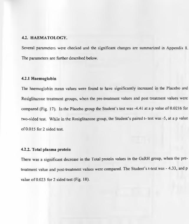
4.2. HAEMATOLOGY.
Several parameters were checked and the significant changes are summarized in Appendix 8.
The parameters are further described below.
4.2.1 Haemoglobin
The haemoglobin mean values were found to have significantly increased in the Placebo and
Rosiglitazone treatment groups, when the pre-treatment values and post treatment values were
compared (Fig. 17). In the Placebo group the Student’s test was -4.41 at a p value o f 0.0216 for
two-sided test. While in the Rosiglitazone group, the Student’s paired t- test was -5, at a p value
of0.015 for 2 sided test.
4.2.2. Total plasma protein
There was a significant decrease in the Total protein values in the GnRH group, when the pre
treatment value and post-treatment values were compared. The Student’s t-test was - 4.33, and p
value o f 0.023 for 2 sided test (Fig. 18).

F igure 17: H aem oglobin changes before an d a fte r tre a tm e n t o f endoi
15
15.5
15
14.5
14
13.5
13
12.5
12
11.5
PB PA GBTreatment groi
KEY:PB Placebo before GB GnRH beforePA Placebo after GA GnRH after-------- A___________________________
- 134-

Tota
l pla
sma
prot
ein
gm/d
l
F ig u re 18: In te r g ro u p changes in to ta l p lasm a in endom etrio ti
7.6
U
7.2
7
6,8
6.6
6.2-
6.2
6
5,8
PB PA GB
Treatn
KEY:PB Placebo before PA Placebo after
GB GnRH be I GA GnRH aft

4.23 . Eosinophils
Eosinophils values were significantly increased in the Rosiglitazone group when the pre-
treatment and post treatment values were compared (Student’s pair t-test = - 2.53 (Fig. 19)
4.2.4. Plasma fibrinogen
The plasma fibrinogen value was found to be significantly increased, within the GnRH group,
when the pre- treatment values (0.075 ± 0.05) and the post treatment values (0.15 ± 0.06) were
compared using Student’s paired t- test = 3.0, p value 0.0577 (Fig. 20).

Abs
olut
e di
ffer
entia
l val
ues
of E
osin
ophi
ls p
er p
i
F ig u re 19: E osinophils coun ts in en d om etrio tic baboons
KEY:PB Placebo before PA Placebo after
GB GnRH before GA GnRH after

Plas
ma
fibrin
ogen
gra
ms
per d
ecili
tre
Figure 20: Plasma fibrinogen levels in endometriotic ba
0,3
0.25
02
0.15
0.1
0.35
0PB PA GB
Treati
K E Y :PB Placebo before GB GnRH beforePA Placebo after GA GnRH after
138

4.3. CLINICAL CHEMISTRY
Analysis using Student t tests and ANOVA showed no significant results to report.
See Appendix 11.
4.4. HISTOLOGY OF ORGANS OTHER THAN THOSE WITH
ENDOMETRIOTIC LESIONS
All the individual slides examined and the organs with significant change are given in Appendix
9 to 10. A further analysis and comparison is summarised in Table 12 below. The table shows a
summary o f the significant changes (after statistical analysis) o f the histopathology o f the various
other organs.
The systemic effects o f treatment were analysed by comparing the frequency o f histological
changes in the various organs among the treatment groups. The frequency o f histological
changes in rosiglitazone treated group was found to be significantly higher than in the placebo
and GnRH treated groups in the following organs:
i) Lung: Pulmonary oedema was moderate to severe and it occurred together with mild
hemorrhages. The pulmonary oedema was characterised by mild to moderate accumulation of
fluid in alveoli (Plate 23, arrow) in the rosiglitazone treated baboon. There was also presence of

ii) Liver: Bile canaliculi were surrounded or infiltration of mild amount o f lymphocytes (Plate
24, arrow) around the bile duct wall and bile duct hypertrophy in the rosiglitazone treated
animal.
Plates 23 to 26, shows some o f the changes in the lungs and the liver. Pulmonary oedema was
mild to moderate (Plate 23) in the rosiglitazone group.
The changes in bile canaliculi changes are shown in plate 24 to 25. One section o f the gall
bladder showed heavy infiltration o f lymphocytes in the wall, in a rosiglitazone treated baboon
(Plate 25, arrow).
In our study, two animals in the rosiglitazone group showed fatty degeneration and myocardial
infarcts in the heart although the frequency of the cases were not significantly high (CHI square
Fisher’s Exact test, p value = 0.4286). Plate 27 shows an area in the myocardium which was
necrotic, infiltrated mononuclear cells, with collagen fibres, diffuse interstitial oedema, loss of
myocytes and decreased striations (arrow).

Table 12: Summary and analysis of histologic findings in each
Number of positive animals group with changes
Organ Histological changes Placebo GnRH RosiglLungs Moderate to severe edema &
hemorrhages. Emphysema with corresponding mild atelectasis
0/4 1/4 4
Liver Bile ducts surrounded by lymphocytes.
0/4 1/4 4
- 141 -

Plate 23: L ung section from a rosiglitazone treated animal (PAN 2788)
Magnification x 100, H & E Stain

Plate 24: L iver section from a rosiglitazone treated baboon (PAN 2998)
M ag n ifica tio n , x 100, H & E Stain
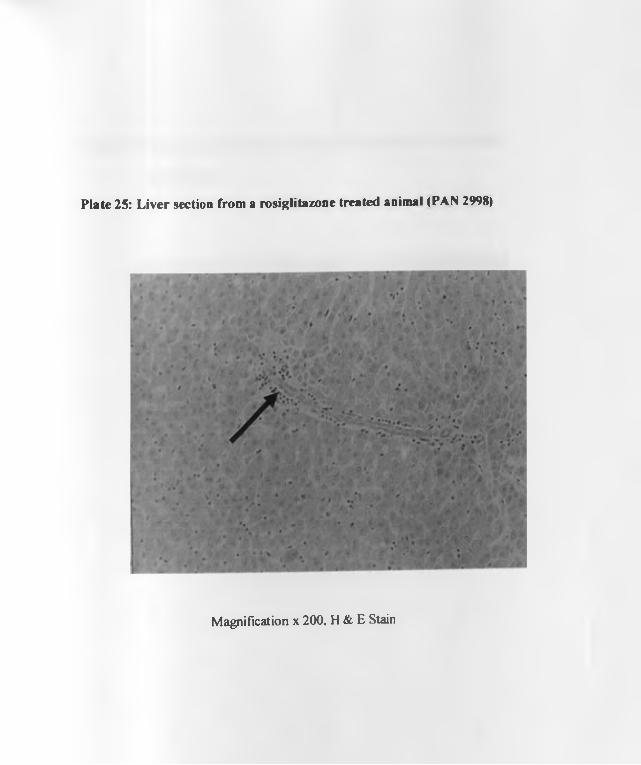
Plate 25: L iver section from a rosiglitazone treated animal (PAN 2998)
M ag n ific a tio n x 200 , H & E Stain

Plate 26: Section o f gall bladder wall from a rosiglitazone treated animal
(PAN 2998)
M ag n ifica tio n x 100, H & E S ta in

Plate 27: A photom icrograph showing a myocardial infarct in a rosiglitazone
treated animal (PAN 2998)
M ag n ifica tio n x 250 . H & E Stain

CHAPTER FIVE
5.0 DISCUSSION, CONCLUSION AND RECOMMENDATIONS
5.1. DISCUSSION
The broad objective of this study was to determine the effects of rosiglitazonc on clinical
feature and pathology o f experimental endometriosis in a baboon model. The specific
objectives were first, to determine the nature and extent of gross and histopathology changes
in endometriosis lesions in baboons treated with rosiglitazonc. Secondly, to document any
pathological indicators of the side effects of the drug using serum chemistry, haematology
and histology. And finally, to establish the target organs of rosiglitazonc toxicity based on
histopathology of different organs.
In summary, there was clinical evidence of therapeutic effects of rosiglitazone on the
endometriosis lesions. The transformation of active lesions (red) to less active lesions
(typical bluish-black or white lesions) was significantly more in rosiglitazone treatment when
compared with placebo tablet treated group or (Ganirelix®) injection treated group. In this
study, the placebo group appeared to worsen with time by progressing to the more active
form that had typical bluish appearance; and the red lesion increased in number. This change
in the placebo group is a natural progression and also occurs in untreated humans (Koninckx
et al., 1991) and baboons (D’Hooghe etal., 1996a).

provided more information on this tissue degeneration process and corroborated the gross
findings. In the present study, degenerated cells appeared with condensed pyknotic nuclei
and dark staining. In all the rodent studies, changes in the ESCs were not reported (Lcbovic
et a l, 2004; Demirturk etal., 2006; Aytan etal., 2007).
In the present study, there was evidence of increased peritoneum lymphocytes in the
rosiglitazone group. This is in contrast to Homung et al., (2003), who showed that TZDs
inhibits peritoneal inflammation although comparison may not be fair, since Homung et al.,
2003, used peritoneal fluid and in this study we used perineum tissue to assess the
inflammatory response. In women, a preliminary report (Debrock et al., 2000) described that
the peritoneal fluid concentration of leukocytes and erythrocytes increased during
menstruation in women with endometriosis.
In our study, it appears that the gross endometriotic lesions are associated with specific
histopathological features, only in some cases. This is contrary to what some authors have
documented that there is no correlation between histologic and visual findings at laparoscopy
with histology (Dmowski 1984; Jansen and Russell 1986; Walter etal., 2001).
To understand the effects of rosiglitazone on the endometriosis lesions, first we need to look
at the pathophysiology of the disease. Inflammation, is a major feature of endometriotic
I n c i . ifU f Vtn ' nf nmctaalQnHinc /T arll P t f i l

cytokines such as interleukin-1 p, interleukin-6 (Tseng et al., 19%), and tumor necrosis factor
among others (Osteen et al., 1996; Giudice and Kao 2004; Bulun, 2009). Monocyte
chemoattractant protein 1, interleukin-8, and RANTES attract the granulocytes, natural killer
cells, and macrophages that are typical of endometriosis. The transcription nuclear factor- kB
(NK- kB) appears to be activated (Guo 2006) and this activation is important in production of
many cytokines (Lebovic et al., 2001). Autoregulatory positive-feedback loops ensure
further accumulation of these immune cells, cytokines, and chcmokincs in established lesions
(Lebovic et al., 2001). In patients with endometriosis, inflammatory and immune responses,
angiogenesis, and apoptosis are altered in favour of the survival and replenishment of
endometriotic tissue. These basic pathologic processes depend in part on oestrogen or
progesterone (Bulun 2009). Both rosiglitazone (Yee et al., 1999; Pritts et al., 2002;
Houston et al., 2003; Wanichkul et al., 2003) and GnRH-antagonists (Meresman et al.,
2003) have anti-inflammatory properties. And as observed in our study on endometriosis, it
appears this anti-inflammatory aspect of the drug reduces lesion size and progression. Both
TZD receptors have been identified in endometriotic lesions (Pritts et al., 2002; Wanichkul
et al., 2003), and their role in the reduction of the sizes o f the lesions though unknown, may
be important. Earlier work on rats has documented that TZDs do cause epithelial changes in
the endometriosis of rats (Lebovic et al., 2004; Demirturk et al., 2006; Aytan et al., 2007).
The effect of GnRH-antagonist on the endometriotic glands is probably due to down
regulation o f LH and FSH in treated baboons (Rice 2002; Shalev 2003; Mahuttc and Arici

(Donnez et al., 1998; Me Laren, 2000; Meresman et al.. 2003; Mcrcsman eta!., 2005).
VEGF may be involved in maintenance of endometriosis. GnRH receptors have been
identified in ectopic endometrium (endometriosis) (Lebovic et al., 2000). Oestrogens are
considered more of pro-inflammatory factors, rather than anti-inflammatory (Joscfsson et al.,
1992; Vegeto et al., 2002). Different biological mechanisms underlying estrogen anti
inflammatory activity have been proposed (Vegeto et al., 2002).
In our study, there was incidental induction of stromal or micronodular endometriosis.
Stromal endometriosis has been described by Clement (2007), as a specific type of
endometriosis with no glands. There are reports on the incidence of stromal endometriosis in
humans and baboons. They range from 6.6% of human biopsies (Boyle and McCLuggage
2009), and 8% in baboons (Comillie et al., 1992). Stromal endometriosis tends to take a
superficial position as a typical location on the peritoneum, intimately associated with surface
mesothelium (Comillie et al., 1992; Boyle and McCLuggage 2009). After studying many
stromal endometriosis tissues, Boyle and McCLuggage (2009) concluded that stromal
endometriosis is induced when the menstrual tissue comes into contact with the peritoneum
via retrograde menstruation. The superficial production of stromal endometriosis in this
study, supports the theory that coeleomic metaplasia may be involved in pathogenesis of
endometriosis. There are some suggestions that endometriosis originates or grows from the
endometriotic stromal cells (Mai et al., 1998). To support this theory, we have seen several
incidences of histological evidence of invasivencss or continuity of glandular epithelium into

It is observed elsewhere that the endometriotic tissue epithelium may exhibit different phases
in the same woman (Bergqvist et al., 1984; Metzger etal., 1988). Bcrgqvist et al. (1984),
reported that 25 to 49 % of endometrial implants displayed some form of local haemorrhages
irrespective o f the timing of menstrual cycle. The presence of local haemorrhages, (either
hemosiderin-laden macrophage or extravasetcd red blood cells (RBCs) may indicate
menstrual activity in the endometriotic glands (Bergqvist etal., 1984; Metzger etal., 1988).
Since the baboons used in our experiment were not in the menstruation phase, these results
may support this observation.
In his article, Clement (2007) discussed and emphasised on the problems faced by
pathologists in diagnosis of endometriosis. Many diagnostic problems can arise as a result of
alterations or absence of glandular or stromal components in the lesion. The diagnostic
difficulty can be compounded by the limited tissue in a small biopsy specimen. Also
histological diagnosis of endometriosis can also be challenging when the gross lesion occurs
in an unusual or unexpected site. Clement (2007) mentions five such sites as on or near
ovarian surface, superficial cervical endometriosis, vaginal endometriosis, tubal
endometriosis, and intestinal endometriosis. Finally, endometriotic foci can occasionally be
intimately admixed with another process, such as peritoneal leimyomatosis or gliomatosis,
resulting in a potentially confusing histologic appearance (Clement 2007).

of a number of pathological conditions e.g. parasitic infections, local allergic reactions (such
as asthma), rhinitis and eczema, and hypcreosinophilic syndrome (Glcich et al., 1993). In
addition, eosinophils produce cytokines such as interleukin (IL-3, IL-5) and granulocyte-
macrophage colony stimulating factor (GM-CSF) that contribute to their pro-inflammatory
functions. The cationic proteins, major basic protein (MBP), eosinophil peroxidase (EPO)
and eosinophil cationic protein (ECP) are toxic to airway epithelium and may be deposited
over large areas of the bronchial epithelial lining in patients with asthma (Glcich el al., 1993;
Rabe et al., 1994). The eosinophil MBP also causes the release of histamine from mast cells
and basophils, activates neutrophils and alveolar macrophages, and is directly implicated in
epithelial cell damage, exfoliation and bronchospasm in asthma.
The presence of eosinophils in endometriosis tissue might be associated with a physiological
repair role (Birkland et al., 1994), or a possible pathophysiology role in endometriosis
(Nagata 2005). At sites of inflammation, multiple inflammatory cells including eosinophils,
neutrophils and macrophages are capable of generating reactive oxygen species (ROS) which
can contribute to development o f various diseases, including endometriosis (Nagata 2005).
Morphological and functional properties of endothelial cells may be altered by ROS,
including permeability and adhesion molecule expression (Nagata 2005). In endometriosis,
oxidative stress and antioxidant biomarkers are present in both serum and peritoneal fluid.
Products of this process are exported to serum/plasma where the oxidised metabolites are
incorporated into carriers, such as ox-LDL (Murphy et al., 1998). Studies have

In this present experiment, haemoglobin levels increased within the placebo treated group
and in the rosiglitazone treated baboons compared to the GnRH group. It is suggested that
heme may be involved in the pathogenesis and/or development of endometriosis and that the
immune system, might be insufficient to detoxify heme in women with endometriosis (Van
Langendonckt et al., 2002a; Van Langendonckt et al., 2002b). Iron metabolism by
macrophages seems to be enhanced in case of endometriosis. Also higher levels of iron,
which is probably released after lysis of erythrocytes, have been found in the peritoneal fluid
o f patients with endometriosis (Arumugam 1994), and the concentration of iron is related to
the severity of disease (Arumugam and Yip 1995). This is supported by the fact that
siderophages, iron-storing macrophages that are heavily laden with hemosiderin, are
considered to be indicators of endometriosis (Gaulier et al., 1983; Stowell et al., 1997).
The presence o f hemosiderin in the endometriotic lesions cannot though be extrapolated to
the increase in peripheral haemoglobin levels in our experiment.
In rosiglitazone treated patients, haemoglobin has been reported to go down and in most
cases fluid retention has been implicated through hemodilution (Actos 2000; Avandia
2000). It is reported that TZDs can cause weight gain and fluid retention in some patients.
These changes in weight gain and blood profile are usually observed during the first weeks of
therapy and plateau thereafter. In a review of several studies, Hollenberg (2003), showed that
the decreases in haemoglobin and in hematocrit were highly correlated, providing evidence
favouring hemodilution as the responsible mechanism. Plasma protein concentrations are
In one study, the frequency of decrease in plasma protein concentration was about half that
observed for haemoglobin and hematocrit and did not correlate with either of these
parameters (Hollenberg 2003). This observation is not in agreement with simple
hemodilution, suggesting that additional studies are needed to explain these findings.
Toxicity studies by Rothwell et al., (2001) with troglitazonc in nonhuman primates
documented a reduction of erythrocyte counts, haemoglobin, and hematocrit of 8% to 16% in
haemoglobin values of males at all doses used in their experiment. This was quite opposite
with what we found in this study, where there was an increase in haemoglobin values from
pre treatment to post treatment period in all groups.
The results on total plasma protein showed that a significant decrease occurred from the pre-
treatment to post-treatment period in the GnRH treated group. From the literature it appears
that there is a positive relationship between oestrogen and plasma protein (Nowaczynski et
al., 1978; Korsgaard, 1990). This could explain the decrease in total plasma protein, since
oestrogen was decreased in GnRH antagonist treated group.
Although haematology in this study indicated changes only in eosinophils, total plasma and
haemoglobin in the peripheral circulation, there are publications that show that endometriosis
as a disease may have other subclinical systemic manifestations (Barbcri et al.. 1986;
Pittaway and Fayez 1986; D’Hooghe et al., 1996c; Mol et al., 1998; Bedaiwy et al..
2002; Somigliana et al., 2004; Agic et al., 2006; Cho et al., 2007; Scebcr et al., 2008).

Factor a (TNF-a) (Dmowski 1995), Monocyte Chemotactic Factor 1 (MCP-1) (Akoum et
al., 2002), Interleukin -6 (IL-6) (Tseng et al., 1996), Interleukin- 8 (IL-8) (Bcdaiwy et al.,
2002) and Chemokine (C-C motif) receptor 1 (CCR1) (Zhang et al., 1999), in the peripheral
blood of patients with endometriosis (Agic et al., 2006). CD44+ and CD 14+ monocytes arc
significantly increased, while CD3+ T lymphocytes and CD2(H B lymphocytes show modest,
but significant decrease in peripheral blood of women with endometriosis. Some previous
work at the IPR, has shown that the percentage of CD4+ and IL2R subsets of white blood
cells, was increased in baboons with the stage II-IV spontaneous endometriosis (D’Hooghe
et al., 1996c).
The histology on the various tissues other than the cndometriotic lesions showed varying
changes in some organs. The lungs had mild, moderate to severe pulmonary oedema which
was significantly more in rosiglitazone treated group than in the placebo group. Pulmonary
oedema has been reported by clinicians as an important side effect of rosiglitazone in
treatment o f diabetes (Idris et al., 2003; et > 2006). From the literature, the use
of TZDs is associated with a triad of fluid retention, oedema, and weight gain (Niemcyer and
Janncy 2002; Kermani and Garg 2003; Nesto etal., 2003; Idris etal., 2003; Mudaliar et
al., 2003). The incidence of oedema ranges from 2% to 5% in diabetic patients under
rosiglitazone therapy (Kermani and Garg 2003). If combined with insulin, this ratio
increases to 14.7% ((^ekmen et al., 2006). Fluid retention generally is considered mild and

weight gains were not significantly different between pre-treatment and post-treatment
period within group; or between the 3 treatment groups.
Rosiglitazone may have the risks of congestive heart failure (CHF) and Myocardial infarcts
(MI) (Wooltorton 2002; Nissen and Wolski 2007; http://www.nytimcs.com. 22 June
2007). The theory behind the CHF is a cumulative effect o f fluid overload described above.
Recently, warnings about possible myocardial ischemia and MI have been issued by Food,
Drugs Administration in the USA (http://www.fda.gov 21st May 2007; http://www.fda.gov
19th November 2007). The interim report on Rosiglitazone Evaluated for Cardiac Outcomes
and Regulation of glycemia in Diabetes (RECORD) trial has also raised substantial
uncertainty about the complete cardiovascular safety of the use of this drug, at least in the
diabetic population (Home et al., 2007). Therefore, TZDs should be used cautiously in
patients with heart failure symptoms.
Toxicity studies by Rothwell et al., (2001) with troglitazone documented an increase in
absolute and relative liver weights at all doses in cynomolgus monkeys of both sexes. The
doses used ranged from approximately 3, 6 to 10 times those given to humans. Humans are
given 400 mg daily. Minimal or mild bile duct hypertrophy was present at all doses and both
incidence and severity increased with increasing doses (Rothwell et al., 2001). This finding
confirms with reports on changes on gall bladder and bile ducts.

The hepatotoxicity of troglitazone does not seem to translate across the class of TZDs
(Scheen 2001; Van Gaal and Scheen 2002). Reversible liver injury and hepatic failure have
occurred in a few patients with both drugs (Ravinuthala and Non 2000; May et al., 2002)
but cause and effect have not been established (Lebovitz et al., 2002). With either drug, the
FDA recommends monitoring of liver function every 2 months for the first year and less
frequently thereafter. In our study, the biochemical tests did not reveal any significant
changes among the 3 treatment groups. The absence of any change many exonerated
rosiglitazone from hepatotoxicity.
The other known effects of TZDs are as follows; they may slightly increase LDL cholesterol
levels, but they also change the small, dense cholesterol particles, thought to increase risky
effects to more the buoyant ones, thought to be safer. These drugs increase HDL by about 10
-20%, decrease triglycerides, decrease PAI-I activity and improve endothelial function
(Nesto et al., 2003). They also are thought to increase subcutaneous but not visceral
adiposity (Parulkar et al., 2001). Pioglitazone may have a greater favourable effect than
rosiglitazone on serum lipids (Khan et al., 2002). There are “TZDS class effects” and
“TZD-specific effects” that has differentiated the use of rosiglitazone vis a vis troglitazone.
For this reason there is need to study other TZDs so as to know more on the other “TZD —
specific effects” which may be different from Rosiglitazone. Class effects include fluid

In conclusion, this study presents the first subhuman primate evidence that treatment with a
TZD can reduce the surface area (~50% decrease in relative change compared with placebo)
o f peritoneal endometriosis in baboons with established disease. This work shows lesions
such as extensive glandular epithelium necrosis and degeneration of glandular epithelium of
endometriotic lesions. Degenerative changes of endometriotic stromal cells (ESCs) were also
a significant finding. Histopathology has also confirmed that the gross laparoscopy
appearance of endometriosis conforms to some histological features. Total plasma protein
concentration increased in GnRH treatment group. The eosinophil count increased in
rosiglitazone treatment group, and plasma fibrinogen also increased in GnRH treatment
group. There was also an incidental induction o f stromal or micronodular endometriosis.
The most consistent changes were moderate to severe pulmonary oedema & hemorrhages;
and bile ducts that were surrounded by inflammatory cells. Presence of pulmonary oedema
and bile duct changes was significantly more prominent in rosiglitazone group.
This study shows that rosiglitazone as a thiazolidinedione is a helpful anti-inflammatory and
growth inhibitor agent in the treatment of the disease. It reduced the extent of endometriosis
in the baboon model.
5.2. CONCLUSIONS
reduction o f volume and surface area of the endometriosis lesions is concerned. It also
provides more detailed explanation o f the necrosis in the epithelial glands.
2) The baboon model can be used to test new drugs for treatment or prevention of
endometriosis and it is therefore important in testing general and reproductive safety of new
anti-endometriosis drugs.
3) An ideal therapy option for treating endometriosis should possess minimal side
effects, low cost burden, and proven efficacy at diminishing pelvic pain. It should spare
fertility potential during treatment. Rosiglitazone is therefore a potential candidate for this
therapy.
5 3 . RECOMMENDATIONS
1) Clinical trials would be helpful in determining the utility of this novel class of compounds
for women with endometriosis.
2) More of the TZDs class of drugs should be investigated to determine other “TZD -
specific effects” which may be different from those of rosiglitazone.
3) Currently no available drugs resolve pain due to endometriosis. The baboon can be
developed as an animal model for endometriosis related pain-alleviating drugs, since pain is a
major problem in endometriosis.
4) Research on biomarkers for diagnosis of endometriosis is an important area of future
research. Currently there are no non-invasive tests for diagnosis of endometriosis. The

6.0 REFERENCES
Abrao MS., Podgaec S., Filho BM., Ramos LO., Pinotti JA. and de Olivera RM. (1997).
The use of biomarkers in the diagnosis of pelvic endometriosis. Journal of Human
Reproduction 12: 2523 - 2527.
Actos (pioglitazone hydrochloride) (2000). [package insert]. Takcda Pharmaceuticals
America, Inc. Lincolnshire, Illinois. United States of America.
Agic A., Xu H., Finas D., Banz C., Diedrich K. and Homung D. (2006). Is Endometriosis
Associated with Systemic Subclinical Inflammation? Journal of Gynaecological
Obstetrics Investigations 62: 139-147.
Akoum A., Kong J., Metz C. and Beaumont MC. (2002). Spontaneous and stimulated
secretion of monocyte chcmotactic protein-1 and macrophage migration inhibitory
factor by peritoneal macrophages in women with and without endometriosis. Journal
o f Fertility and Sterility 77: 989-994.
Allen C., Hopewell S., Prentice A. and Allen C. (2005). Non-steroidal anti-inflammatory
drugs for pain in women with endometriosis. Cochrane Database of Systematic
Reviews.website. October 2008.
Al-Salman J., Aijomand H., Kemp DG. and Mittal M. (2000). Hepatocellular injury in a
patient receiving rosiglitazone: A case report. Annals 132: 121-124.
Amboka JN. and Mwethera PG (2003). Characterization o f semen from olive baboons.

American Society for Reproductive Medicine. (1997). Revised American Society for
Reproductive Medicine classification of endometriosis. Journal of Fertility and
Sterility 67 :815-82 .
Arumugam K. (1994). Endometriosis and infertility: raised iron concentration in the
peritoneal fluid and its effect on the acrosome reaction. Journal of Human
Reproduction 9: 1153-1157.
Arumugam K. and Yip YC. (1995). De novo formation of adhesions in endometriosis. The
role of iron and free radical reactions. Journal of Fertility and Sterility 64: 62-64.
Atar E. and Bulun SE. (2006). Aromatase and other steroidogenic genes in endometriosis:
translational aspects. Human Reproduction Update 12: 47-56.
Avandia (rosiglitazone maleate) (2000). [package insert]. GlaxoSmithKline
Pharmaceuticals. Philadelphia, Pa., United States of America.
Aytan H., Caliskan AC., Demirturk F., Aytan P. and Koseoglu. DR. (2007). Peroxisome
proliferator-activated receptor-gamma agonist rosiglitazone reduces the size of
experimental endometriosis in the rat model. Australia and New Zealand Journal of
Obstetrical Gynaecology 47: 321-5.
Barberi RL., Niloff JM., Bast Jr. RC., Scaetzl E., Kistner RW. and Knapp RC. (1986).
Elevated serum concentrations of CA-125 in patients with advanced endometriosis.
Journal of Fertility and Sterility 45: 630-634.
Barberi RL. (2000). 1 6 W o m e n s Health, X Endometriosis, ACP Medicine Online, Dale
DC; Federman DD, Eds. WebMD Inc., New York, 2000.

Barrier BF., Bates GW., Leland MM., Leach DA., Robinson RD. and Propst AM. (2004).
Efficacy of anti-tumor necrosis factor therapy in the treatment of spontaneous
endometriosis in baboons. Journal of Fertility and Sterility 81: 775-9.
Barroeta JE ., Pasha TL. and Acs G. (2007). Immunoprofile of cndocervical and
endometrial stromal cells and its potential application in localization of tumour
involvement. International Journal of Gynaecologic Pathology 26: 76-82.
Batzer FR. (2006). GnRH agonists and antagonist: options for endometriosis pain
treatment. Department of Obstetrics and Gynaecology Faculty papers. Thomas
Jefferson University. Journal of Minimally Invasive Gynaecology 13: 539 - 545.
Bedaiwy MA., Falcone T., Sharma RK., Goldberg JM., Attaran M. and Nelson DR. (2002).
Prediction of endometriosis with serum and peritoneal fluid markers: a prospective
controlled trial. Journal of Human Reproduction 17: 426-431.
Bergqvist A., Ljungberg O. and Myhre E. (1984). Human endometrium and endometriotic
tissue obtained simultaneously: a comparative histological study. International
Journal of Gynaecological Pathology 3: 135 - 145.
Birkland TP., Cheavens MD. and Pincus SH. (1994). Human eosinophils stimulate DNA
synthesis and matrix production in dermal fibroblasts. Archives of Dermatological
Research 286: 312-318.
Birrell AM., Hennessy A., Gillin A., Horvath J. and Tiller D. (19%). Reproductive and
neonatal outcomes in captive bred baboons (Papio hamadryas). Journal of Medical
Primatology 25: 287-93.

Boyie D. and McCluggage WG. (2009). Peritoneal stromal endometriosis: A detailed
morphological analysis of a large series of cases of a common and under-recognised
form of endometriosis. Journal Clinical Pathology. Published Online First: 20
January 2009. doi: 10.1136/jcp.2008.064261
Brenner RM and Slayden OD. (2005). Progesterone receptor antagonists and the
endometrial antiproliferative effect. Seminars in Reproductive Medicine 23:74 -81.
Bulun SE. (2009). Endometriosis. The New England Journal of Medicine 360: 268-279
Carli C., Blanchet J., Fontaine J., Villa J., Bourcier N. and Akoum A. (2009). Marked
expression of prostaglandin E synthases in endometriosis. Journal of Fertility and
Sterility (90): S147-S147
Cekmen N., Cesur M., (^etinbas R., Bedel P. and Erdemli O. (2006). Acute pulmonary
oedema due to rosiglitazonc use in a patient with Diabetes mellitus. The Journal of
Intensive Care Medicine 21:47-51.
Chai DC., Cuneo S., Falconer H., Mwenda JM. and D’Hooghe TM. (2007). Baboon (Papio
anubis anubis) as an appropriate model for intrauterine research. Journal of Medical
Primatology 36: 365-9
Chapron Fauconnier A., Vieira M., Barakat H., Dousset B., Pansini V., Vacher-Lavenu
MC. and Dubuisson J.B. (2003). Anatomic distribution of deeply infiltrating
endometriosis: Surgical implications and proposition for a classification. Journal of
Human Reproduction 18: 157-161.
Cho SH., Oh YJ., Nam A., Kim HY., Park JH. and Kim JH. (2007). Evaluation of serum and

Cho S„ Cho R , Nam A., Kim HY., Choi YS., Park KR, Cho DJ and Lee BS. (2008).
Neutrophil-to-lymphocyte ratio as an adjunct to CA-125 for the diagnosis of
endometriosis. Journal of Fertility and Sterility 90: 2073-9.
Clement PB. (2007). The pathology of endometriosis. A survey of the many faces of a
common disease emphasizing diagnostic pitfalls and unusual and newly appreciated
aspects. Journal of Advances in Anatomic Pathology 14: 241-260.
Cornillie FJ., D'Hooghe TM., Bambra CS., Lauweryns JM., Isahakia M. and Koninckx PR.
(1992). Morphological characteristics of spontaneous endometriosis in the baboon
(Papio anubis, Papio Cynocephalus). Journal of Gynaecological Obstetrics
Investigations 34,225-228.
Debrock S., Drijkonigen R., Goossens W., Meuleman C., Hill JA. and D’Hooghe TM.
(2000). Quantity and Quality of retrograde menstruation: red blood cells,
inflammation and peritoneal cells. Abstract presented at the Annual Meeting of the
American Society of Reproductive Medicine, Toronto, Canada, October 14th , 2000.
Journal of Fertility and Sterility 74: 3S.
Demirturk F., Aytan H., Caliskan A., Aytan P. and Koseoglu DR. (2006). Effect of
peroxisome proliferator-activated receptor-gamma agonist rosiglitazonc on the
induction of endometriosis in an experimental rat model. Journal of Society of
Gynecologic Investigation 13: 58-62.
D’Hooghe TM., B a m b r a CS., Comille FJ., Isahakia M. and Koninckx PR. (1991). The
prevalence and laparoscopic appearance of spontaneous endometriosis in the baboon

Development of a model of retrograde menstruation in baboons (Papio anubis).
Journal of Fertility and Sterility 62: 635 - 638.
D'Hooghe TM., Bambra CS. and Koninckx PR. (1994b). Cycle fecundity in baboons of
proven fertility with minimal endometriosis. Journal of Gynecological Obstetrics
Investigations 37:63-65.
D'Hooghe TM., Bambra CS., Raeymaekers BM., De Jonge I., Lauweryns JM. and
Koninckx PR. (1995). Intrapelvic injection o f menstrual endometrium causes
endometriosis in baboons (Papio cynacephalus and Papio anubis). American Journal
o f Obstetrical Gynaecology 173: 809-813.
D'Hooghe TM., Bambra CS., Raeymaekers BM. and Koninckx PR. (1996a). Serial
laparoscopies over 30 months show that endometriosis in captive baboons (Papio
anubis, Papio cynocephalus) is a progressive disease. Journal of Fertility and Sterility
65: 645-649.
D'Hooghe TM., Bambra CS., Hill JA. and Koninckx PR. (1996b). Effect on endometriosis
and the menstrual cycle on white blood cell subpopulations in the peripheral blood
and peritoneal of baboons. Journal of Human Reproduction 11: 1736-1740.
D'Hooghe TM., Bambra CS., Raeymaekers BM. and Koninckx PR. (1996c). Increased
incidence and recurrence of recent corpus luteum without ovulation stigma
(Luteinized Unruptured Follicle-Syndrome?) in baboons (Papio anubis, Papio
cynocephalus) with endometriosis. Journal of the Society for Gynaecologic
D’Hooghe TM ., Bam bra CS., Dunselman GA., Evers, HL. and Koninckx PR. (1994a).

prevalence and recurrence of retrograde menstruation in baboons with spontaneous
endometriosis. Journal of Human Reproduction 11: 2022-2025.
D'Hooghe TM., Bambra CS., Raeymaekers BM., Riday AM., Suieman M A. and Koninckx
PR. (1996e). The cycle pregnancy rate is normal in baboons with stage I
endometriosis but decreased in primates with stage II and stage Ill-IV disease. Journal
o f Fertility and Sterility 66: 809-813.
D'Hooghe TM. (1997). Clinical relevance of the baboon as a model for the study of
endometriosis. Journal of Fertility and Sterility 68: 613-25.
D'Hooghe TM., Pudney J., Alves L., Peixe K. and Hill JA. (2001a). Immunobiology of the
Female reproductive tract in the baboon. American Journal of Primatology 53:47-54.
D'Hooghe TM., Ling Xiao., Koninckx PR. and Hill JA. (2001b). Cytokine profile in
peripheral blood and peritoneal fluid of women with deep and superficial
endometriosis. Archives of Gynaecology and Obstetrics 265: 40-44.
D'Hooghe TM., Debrock S., Hill JA. and Meuleman C. (2003). Endometriosis and
subfertility: Is the relationship resolved? Seminars in Reproductive Medicine 21:
243-254
D'Hooghe TM., Spiessens C., Chai DC., Mwethera PG., Makokha AO. and Mwenda JM.
(2004). Ovarian stimulation, egg aspiration, in vitro fertilization and embryo transfer
in the baboon (Papio anubis): a pilot project at the Institute of Primate Research,
Nairobi, Kenya. Journal o f Gynaecological Obstetrics Investigations 57: 23-6.
D'Hooghe TM ., Bambra CS., Raeymaekers BM. and Koninckx PR. (1996d). Increased

Mihalyi A. and Mwenda JM. (2006). Recombinant human TNFRSF1A (r-hTBPl)
inhibits the development of endometriosis in baboons: a prospective, randomized,
placebo- and drug-controlled study. Biology of Reproduction 74: 131-6.
D’Hooghe TM., Kyama CK., Mihalyi AM., Chai D., Falconer H. and Mwenda JM. (2008).
The baboon model for translational research in endometriosis. Reproductive
Sciences 16: 152-61.
Dick EJ., Hubbard GB., Martin LJ. and Leland MM. (2003). Record review of baboons with
histologically confirmed endometriosis in a large established colony. Journal of
Medical Primatology 32: 39-47.
Dmowski WP. (1984). Pitfalls in clinical, laparoscopic and histologic diagnosis of
endometriosis. Acta Obstetricia Et Gynecologica Scandinavica 123: 61-66.
Dmowski WP. (1995). Immunological aspects of endometriosis. The International Journal
o f Gynaecological Obstetrics 50: 3-10
Donnez J., Smoes P. and Gillerot S. (1998). Vascular endothelial growth factor (VEGF) in
endometriosis. Journal of Human Reproduction 13: 1686-90.
Eskenazi B. and Warner ML. (1997). Epidemiology of endometriosis. Journal of
Obstetrical Gynaecology Clinics of North America 24: 235 - 238.
Falconer H., Mwenda JM., Chai DC., Wagner C., Song XY., Mihalyi A., Simsa P., Kyama
C., Comillie FJ., Bergqvist A., Fried G. and D'Hooghe TM. (2006). Treatment with
anti-TNF monoclonal antibody (c5N) reduces the extent of induced endometriosis in
D'Hooghe TM., Nugent NP., Cuneo S., Chai DC., Deer F., Debrock S., Kyama CM.,

Farquhar CM. (2000). Endometriosis. Brirtish Medical Journal 320: 1449 52.
Fazleabas AT., Brudney A., Gurates B., Chai D. and Bulun S. (2002). A modified baboon
model for endometriosis. Annals of the New York Academy of Sciences of USA 55:
308-317.
Forman LM., Simmons DA. and Diamond RH. (2002). Hepatic failure in a patient taking
rosiglitazone. Annals 132: 118-121.
Garrido N., Navarro J., Remohi J., Simon C. and Pellicer A. (2000). Follicular hormonal
environment and embryo quality in women with endometriosis. Journal of Human
Reproduction Update 6: 67 - 74.
Gaulier A., Jouret-Mourin A. and Marsan C. (1983). Peritoneal endometriosis. Report of a
case with cytologic, cytochemical and histopathologic study. Acta Cytologica 27:
446-449.
Giudice LC. and Kao LC. (2004). Endometriosis. Lancet 364:1789-1799.
Gleicher N., el-Roeiy A., Confino E. and Friberg J. (1987). Is endometriosis an autoimmune
disease? Journal of Obstetrics and Gynaecology 70: 115-122.
Gleich GJ., AdolphsonCR. and Leiferman KM. (1993). The Biology of the Eosinophilic
Leukocyte. Annual Review of Medicine 44: 85-101.
Guo SW. (2006). Nuclear factor-kappafl (NF-kappaB): an unsuspected major culprit in the
pathogenesis of endometriosis that is still at large? Journal of Gynaecological and
Obstetrics Investigations 63: 71-97.
Guzick DS. and Rock JA. (1983). A comparison of danazol and conservative surgery for

Hastings JM and Fazleabas AT. (2006). A baboon model for endometriosis: implications
for fertility. Reproductive Biology and Endocrinology 4; S7 1 -20.
Heaney AP., Femado M. and Memed S. (2003). PPAR-gamma receptor ligands: novel
therapy for pituitary adenomas. Journal of Clinical Investigations 111: 1381-8.
Hever A., Roth RB., Hevezi P., Marin ME., Acosta JA., Acosta H., Rojas J., Herrera R.,
Grigoriadis D., White E., Conlon PJ., Maki RA. and Zlotnik A. (2007). Human
endometriosis is associated with plasma cells and over expression of B lymphocyte
stimulator. Proceedings of the National Academy o f Sciences of the United States of
America. 104: 12451-12456.
Hollenberg NK. (2003). Considerations for management of fluid dynamic issues associated
with thiazolidinediones. The American Journal of Medicine 115: 111-115.
Home PD., Pocock SJ., Beck-Nielsen H., Gomis R., Hanefeld M., Jones NP., Komajda M.,
McMurray JJV. (2007). Rosiglitazone evaluated for cardiovascular outcomes - An
interim analysis. New England Journal of Medicine 357: 1-11.
Hornung D., Ryan IP., Chao VA., Vigne JL., Schriock ED. and Taylor RN. (1997).
Immunolocalization and regulation of the chemokine RANTES in human endometrial
and endometriosis tissues and cells. Journal o f Clinical Endocrinology and
Metabolism 82: 1621-1628.
Hornung D., Chao VA, Wallwiener D. and Taylor RN. (2003). Thiazolidinedione
inhibition of peritoneal inflammation. Gynaecological and Obstetric Investigations

Houston KD., Copland JA., Broaddus RR., Gottardis MM., Fischer SM. Walker CL . (2003).
Inhibition of proliferation and estrogen receptor signaling by perixisome prolifcrator-
activated receptor gamma ligands in uterine leiomyoma. Journal of Cancer Research
63: 1221-7.
http://womwnhealth.gov/faq/hysterectomy. (1/7/2006). Hysterectomy.
http://www.fda.gov/Drugs/DrugSafety/PostmarketDrugSafety
InformationforPatientsandProviders/ucm 143349.htm. (21/5/2007): Food Drugs
Administration Safety Alerts: FDA ALERT. Rosiglitazone maleate (marketed as
Avandia, Avandamet, and Avandaryl) Information.
http://www.fda.gov/Drugs/DnigSafety/PostmarketDrugSafety
InformationforPatientsandProviders/ucm 143349.htm. (19/11/2007). Food Drugs
Administration Safety Alerts. Rosiglitazone maleate (marketed as Avandia,
Avandamet, and Avandaryl) Information.
http://www.nytimes.com/2007/05/22/business/22drug.htmL (22/6/2007). Saul S. Heart
attack risk seen in drug for diabetes [article online], 22 May 2007. New York Times.
Humason GL. (1962). Animal tissue techniques. 2nd edition. W. H. Freeman and C o., San
Francisco.
Hunnell NA., Rockcastle NJ., McCormick KN., Sinko LK., Sullivan EL. and Cameron JL.
(2007). Physical activity of adult female rhesus monkeys (Macaca mulatto) across
the menstrual cycle. American Journal of Physiological and Endocrinology

Idris L, Gray S. and Donnelly R. (2003). Rosiglitazonc and pulmonary oedema: an acute
dose-dependent effect on human endothelial cell permeability. Diabctologia 46:
288 - 290.
Ishihara H., Kitawaki JO. and Kado N. (2003). Gonadotropin-releasing hormone agonist
and Danazol normalize aromatase cytochrome P450 expression in eutopic
endometrium from women with endometriosis, adenomyosis, or leiyomas. Journal of
Fertility and Sterility 79: 735-42.
Jacobson TZM Barlow DH., Garry R. and Koninckx P. (2004). Laparoscopic surgery for
pelvic pain associated with endometriosis (Cochrane Review). In The Cochrane
Library, Issue 3. John Wiley and Sons Ltd. Chichester, UK.
Jansen RP. and Russell P. (1986). Non-pigmented endometriosis: Clinical, laparoscopic,
and pathologic definition. American Journal of Obstetrical Gynaecology 155: 1154-
9.
Johnson NP. (2005). A review of the use of lipiodol flushing for unexplained infertility.
Treatments in Endocrinology 4: 233-43.
Jones CJP., Denton J. and Fazleabas AT. (2006). Morphological and glycosylation
changes associated with the endometrium and ectopic lesions in a baboon model of
endometriosis. Journal o f Human Reproduction 21: 3068-3080.
Josefsson E., Tarkowski A. and Carlsten H. (1992). Anti-inflammatory properties of
estrogen. I. In vivo suppression of leukocyte production in bone marrow and
redistribution o f peripheral blood neutrophils. Cell-Immunology 142: 67-78

Kennedy S., Bergqvist A., Chapron C., D’Hooghe T., Dunselman G., Greb R., Hummelshoj
L., Prentice A. and Saridogan (2005). ESHRE Special Group for Endometriosis and
Endometrium Guideline Development Group : ESHRE Guideline for the diagnosis
and treatment of endometriosis. Journal of Human Reproduction 20: 2698 704.
Kermani A. and Garg A. (2003). Thiazolidinedione-associatcd congestive heart failure and
pulmonary edema. Journal of Mayo Clinical Proceedings 78:1088-1091.
Khan MA., Peter JV. and Xue JL. (2002). A prospective, randomized comparison of the
metabolic effects of pioglitazonc or rosiglitazone in patients with Type 2 diabetes who
were previously treated with troglitazone. Diabetes Care 25: 708-711.
Kitawaki J., Obayashi H., Ishihara H., Koshiba H., Kusuki I., Kado N., Tsukamoto K.,
Hasegawa G., Wakamura N. and Honjo H. (2001). Oestrogen receptor-a gene
polymorphosim is associated withendometriosis, adenomyosis and leimyomata.
Journal of Human Reproduction 16: 51 - 55.
Kodama H., Fakuda J., Karube H., Matsui T., Shimizu Y. and Tanaka T. (19%). Benefit of
in-vitro fertilization for endometriosis associated infertility. Journal of Fertility and
Sterility 66: 974-979.
Koninckx PR., Meuleman C., Demeyere S., Lesaffre E. and Comillie fJ. (1991). Suggestive
evidence that pelvic endometriosis is a progressive disease, whereas deeply
infiltrating endometriosis is associated with pelvic pain. Journal of fertility and
Sterility 55: 759-765.
Koninckx PR., Riittinen L., Seppala M. and Comillie FJ. (1992). CA-125 and placental

Koninckx PR. and Martin D. (1994). Treatment of deeply infiltrating endometriosis. Current
Opinions on Obstetrics and Gynaecology 6: 231-241.
Korsgaard B. (1990). Estrogen treatment and its influence on protein synthesis and amino
acid metabolism in Zoarces viviparus (L) males. Journal offish Physiology 8 : 560 -
565.
Kyama CM., DebrockS., Mwenda JM., D'Hooghe TM. (2003). Potential involvement of
the immune system in the development of endometriosis. Reproductive biology and
endocrinology 1: 123 - 126.
Kyama CM., D’Hooghe TM., Debrock S., Machoki J., Chai D., Mwenda JM. (2004). The
prevalence of endometriosis among African-American and African-Indigenous
women. Journal of Gynaecologic and Obstetrical Investigations 57: 40-42.
Kyama CM., Overbergh L., Mihalyi A., Cuneo S., Chai D., Debrock S., Mwenda JM.,
Mathieu C., Nugent NP. and D’Hooghe TM. (2007a). Effect of recombinant human
Tumour Necrosis Factor Binding Protein -1 (r-hTBP-1) and GnRH antagonist
on mRNA expression o f inflammatory cytokines, adhesion and growth factors in
endometrium and endometriosis tissues from baboons. Journal of Fertility and
Sterility 89: 1306-1313.
Kyama CM., Mwenda JM., Machoki J., Mihalyi A., Simsa, P., Chai D. and D’Hooghe TM.
(2007b). Endometriosis among African women. Women Health 3: 637 -646.
Lapp T. (2000) ACOG issues recommendations for the management of endometriosis.
American Family Physician 62: 1431-1434.

endometriotic stromal cell cultures by interleukin-1 A. MoL Journal of Human
Reproduction 6: 269-75
Lebovic DI., Mueller MD. and Taylor RN. (2001). Immunobiology of endometriosis.
Journal of Fertility and Sterility 75: 1-10.
Lebovic DI., Kir M. and Casey CL. (2004). Perixisome proliferator-activatcd receptor-
gamma induces regression of endometrial explants in a rat model of endometriosis.
Journal of Fertility and Sterility 82:1008-13.
Lebovic DIM Mwenda JM., Chai DC., Mueller MD., Santi A., Fisseha S., and D’Hooghe T.
(2007). PPAR-gamma receptor ligand induces regression of endometrial explants in
baboons: A prospective, randomised, placebo- and drug-controlled study. Journal of
Fertility and Sterility 88:1108-19.
Lebovitz HE., Kreider M. and Freed MI. (2002). Evaluation of liver function in type 2
diabetic patients during clinical trials; evidence that rosiglitazone does not cause
hepatic dysfuncion. Journal of Diabetes Care 25: 815-21.
Lee KS., Park SJ., Hwang PH., Yi HK., Song CH„ Chai OK., Kim JS., Lee MK. and Lee
YC. (2005). PPAR-gamma modulates allergic inflammation through up-regulation of
PTEN. The Federation of American Societies for Experimental Biology Journal. 19:
1033-1035.
Lemberger T., Desvergne B. and Wahli, W. (1996). Perixisome proliferator-activatcd
receptors: A nuclear receptor signalling pathway in lipid physiology. Annual
Lebovic DI„ B en tz ien F. and C h ao V A . (2000). Induction o f an ang iogenic pheno type in

Luna LG. (1968) Manual of Histologic Staining Methods of the Armed Forces Institute of
Pathology, 3rd edition. MeGraw. Hill Book Co., New York.
Mahutte NG. and Arid A. (2003). Medical management of endometriosis-associated pain.
Journal of Obstetrical Gynaecology Clinics of North America 30: 133-50.
Mai KT., Yazdi HM., Perkins DG. and Parks W. (1998). Pathogenetic role of the stromal
cells in endometriosis and adenomyosis. Journal of Histopathology 32, 379-387.
Marcoux S., Maheux R., and Berube S. (1997). Canadian Collaborative Group on
endometriosis. Laparoscopic surgery in infertile women with minimal or mild
endometriosis. New England Journal of Medicine 337: 217-222.
Martin DC., Hurbert GD., Vander Zwang R. and el-Zeky FA. (1989). Laparoscopic
appearances of peritoneal endometriosis. Journal of Fertility and Sterility 51: 63-67.
Matsuura 1C, Ohtake H., Katabuchi H. and Okamura H. (1999). Coelomic metaplasia
theory of endometriosis. Journal of Gynaecological Obstetrics and Investigations 47:
18-20.
Matsuzaki S., Murakami T., Uchara S., Cania M., Sasano H. and Okamura K. (2001).
Expression of oestrogen receptor a and B in peritoneal and ovarian endometriosis.
Journal of Fertility and Sterility 75: 1198 -1205.
May LD., Lefkowitch JH., Kram MT., and David E. Rubin DE. (2002). Mixed
Hepatocellular-Cholestatic Liver Injury after Pioglitazone Therapy. Annals of
Internal Medicine 136: 449-452
McClellan MC., Rankin S., West NB., and Brenner RM. (1990). Estrogen receptors.

endocervical glandular lesions, endocervical stroma and endometrioid
adenocarcinomas of the uterine corpus: CD 10 positivity is characteristic of, but not
specific for, mesonephric lesions and is not specific for endometrial stroma.
Histopathology 43: 144-150.
Me Laren J. (2000). Vascular endothelial growth factor and endometriotic angiogenesis.
Journal of Human Reproduction Update 6:45-55.
Meresman GF., Buquet RA. and Bilotas M. (2003). Gonadotropin-releasing hormone
agonist (GnRH-a) induces apoptosis and reduces cell proliferation in eutopic
endometrial cultures from women with endometriosis. Journal of Fertility and
Sterility 80: 702-7
Meresman GF., Bilotas MA., Lombardi E. and Tesone M. (2005). Effect of GnRH
analogues on apoptosis and release of interleukin-la and vascular endothelial growth
factor in endometrial cell cultures from patients with endometriosis. Journal of
Human Reproduction 18: 1767-71.
Merrill JA . (1966). Endometrial induction of endometriosis across Millipore filters.
American Journal of Obstetrical Gynaecology 94: 780 - 790.
Metzger DA., Olive DL. and Haney AD. (1988). Limited hormonal responsiveness of
ectopic endometrium: histologic correlation with intrauterine endometrium. Journal of
Human Pathology 19: 1417-1424.
Modugno F., Ness RB., Allen GO., Shildkraut JM., Davis FG. and Goodman MG. (2004).
M c C lu g g a g e W G ., O liv a E. and H erring ton CS. (2003). C D 10 and calretin in stain ing o f

(1998). The performance o f CA-125 measurement in the detection of endometriosis:
a meta-analysis. Journal o f Fertility and Sterility 70: 1101-1108.
Moore J ., Kennedy S., and Prentice A. (2000). Modem combined oral contraceptives for
pain associated with endometriosis. Cochrane Database System Reviews 2:
CD001019.
Mori H., Sawairi M., Nakagawa M., Itoh N., Wada K. and Tamaya T. (1992). Expression of
interleukin-1 (IL-1) beta messenger ribonucleic acid (mRNA) and IL-1 receptor
antagonist mRNA in peritoneal macrophages from patients with endometriosis.
Journal of Fertility and Sterility 57: 535-542.
Mori L. (1978). M o d i f i e d Jendrassik-Grof Method for Bilirubins adapted to the Abbott
Bichromatic analyzer. Journal of Clinical Chemistry 24: 1841 -1845.
Muchemi GM. and Yole D. (2008). Humane methods of capture and care of nonhuman
primates for conservation and biomedical research. In Proceedings of the Sixth
Biennial Scientific Conference and Exhibition, 2008. Faculty of Veterinary
Medicine, University of Nairobi, Kenya. Page 20.
M udaliar S., Chang AR. and Henry RR. (2003). Thiazolidinediones, peripheral oedema, and
type 2 diabetes: incidence, pathophysiology, and clinical implications. Journal of
Endocrinology Practise 9: 406-416.
Murphy A., Palinski W., RankinS., Morales A. and ParthasarathyS. (1998). Evidence for
oxidatively modified lipid-protein complexes in endometrium and endometriosis.
M o l B W ., B ayram N ., L ijm er JG ., W iegerinck M A ., B ongcrs M Y . and van d e r Veen F .

Nesto RW^ Bell D., and Bonow RO. (2003). Thiazolidincdione use, fluid retention, and
congestive heart failure : a consensus statement from the American Heart Association
and American Diabetes Association. Circulation 108: 2941-2948.
Niemeyer NV. and Janney LM. (2002). Thiazolidincdione-induccd oedema.
Pharmacotheraphy 22: 924 - 929.
Nissen SE and Wolski K. (2007). Effect of rosiglitazone on the risk of myocardial infarction
and death from cardiovascular causes. The New England Journal of Medicine 14:
2457-71.
Noble LS., Takayama K., Zeitoun KM., Putman JM., Johns DA. and Hinshclwood M.
(1997). Prostaglandin E2 stimulates aromatase expression in endometriosis-derived
stromal cells. Journal of Clinical Endocrinology Metabolism 82: 600-606.
Nowaczynski W., Murakami T., Richardson K. and Genest R. (1978). Increased aldosterone
plasma protein binding in women on combined oral contraceptives throughout the
menstrual cycle. Journal of Clinical Endocrinology and Metabolism 47: 193-199.
Nyachieo A., Chai DC., Deprest J., Mwenda JM. and D'Hooghe TM. (2007). The baboon as
a research model for the study of endometrial biology, uterine receptivity and embryo
implantation. Journal of Gynaecological Obstetrics Investigations 64: 149-55.
Olive DL. and Lee KL. (1986). Analysis of sequential treatment protocols for endometriosis-
associated infertility. American Journal of Obstetrics and Gynaecology 154: 613 —
619.
Olive DL. and Henderson DY. (1987). Endometriosis and mullerian anomalies. Journal of

Olive DL. and Martin DC. (1987). Treatment of endometriosis-associated infertility with
CO2 laser laparoscopy: the use of one- and two-parameter exponential models.
Journal of Fertility and Sterility 48:18-23.
Orlandi A., Ferlosio A., Ciucci A., Sesti F., Lifschitz-Mercer B., Gabbiani G., Spagnoli LG.
and Czemobilsky B. (2004). Cellular retinol-binding protein-1 expression in
endometrial stromal cells: physiopathological and diagnostic implications.
Histopathology 45:511-517.
Osteen KG., Bruner KL. and Sharpe-Timms KL. (1996). Steroid and growth factor
regulation of matrix metalloproteinase expression and endometriosis. Seminars in
Reproductive Endocrinology 14:247-255.
Overbergh L., Kyama CM., and Valckx D. (2005). Validation of real time RT-PCR assays
for mRNA quantification in baboons. Cytokine 31: 454 - 458.
Parulkar AA., Pendergrass ML., Granda-Ayala R., Lee TR. and Fonseca VA. (2001).
Nonhypoglycemic Effects of Thiazolidinediones. Annals of Internal Medicine 134:
61 .
Peeters LL., Vigne JL., Tee MK., Zhao D., Waite LL. and Taylor RN. (2005). PPARgamma
represses VEGF expression in human endometrial cells: implications for uterine
angiogenesis. Angiogenesis 8: 373-379.
Pijnenborg R., D'Hooghe T., Vercruysse L. and Bambra C. (1996). Evaluation of
Trophoblast invasion in placental bed biopsies of the baboon, with
immnnnhistnchemical localization of cytokeratin, fibronectin and laminin Journal of

Pinkert TC., Catlow CE. and Straus R. (1978). Endometriosis of the urinary bladder in a
man with prostatic carcinoma. Journal of Cancer 43: 1562-1567.
Pitta way DE. and Fayez JA. (1986). The use of CA-125 in the diagnosis and management
o f endometriosis. Journal of Fertility and Sterility 46: 790-795.
Pitttaway DE. (1992). Diagnosis of endometriosis. Infertility and Reproductive Medical
Clinic o f North America 3: 619.
Practice Committe of the American Society of Reproductive Medicine (2006). Treatment
o f pelvic pain associated with endometriosis. Journal of Fertility and Sterility 86: SI 8
- S27.
Pritts EA., Zhao D., Ricke E., Waite L. and Taylor RN. (2002). PPAR-gamma decreases
endometrial stromal cell transcription and translation of RANTES in vitro. Journal of
Clinical Endocrinology and Metabolism 87: 1841-4.
Rabe KF, Munoz NM, Vita AJ, Morton BE, Magnussen H and Leflf AR. (1994).
Contraction of human bronchial smooth muscle caused by activated human
eosinophils. American Journal of Physiology - Lung Cellular and Molecular
Physiology 267: L326-L334
Ravinuthaia RS. and Nori U. (2000). Rosiglitazone Toxicity. Annals of Internal Medicine
S133: 658.
Red wine DB. (1987). The distribution of endometriosis in the pelvis by age groups and
fertility. Journal of Fertility and Sterility 47: 173-175.
Rice VM. (2002). Conventional medical therapies for endometriosis. Annals of New York

Ricote Mm Li AC., Willson TM., Kelly CJ. and Glass CK. (1998). The pcnxisome
proliferator-activated receptor-gamma is a negative regulator of macrophage
activation. Nature 391: 79-82.
Ridley JH . (1968). The histogenesis of endometriosis. A review of facts and fancies.
Journal of Obstetrical Survey 23: 1 - 23.
Robboy SJ., Guggan MA., Kurman RJ. (1994). The Female Reproductive System. Rubin E
Farber JL (eds). Pathology: 936-938, Lippincott, Philadelphia.
Rothwell C., McGuire EJ. and Altrogue DM. (2001). Chronic toxicity in monkeys with the
thiazolidinedione antidiabetic agent troglitazone. The Journal of Toxicological
Sciences 27: 35 -47.
Sampson JA. (1927). Peritoneal endometriosis due to menstrual dissemination of
endometrial tissue into the peritoneal cavity. American Journal of Obstetrics and
Gynecology 14: 422-469.
Scheen AJ. (2001). Hepatotoxicity with Thiazolidinediones. Is it a class effect?. Journal of
Drug Safety 12: 873-88.
Schenken RS., Asch RH., Williams RF. and Hodgen GD. (1984). Etiology of infertility in
monkeys with endometriosis: luteinized unruptured follicles, luteal phase defects,
pelvic adhesions, and spontaneous abortions. Journal of Fertility and Sterility 41:
122-130.
Schweppe KW. and Wynn RM. (1984). Endocrine dependency of endometriosis: an
ultrastructural study. European Journal of Obstetrics & Gynecology and

endometriotic implants and eutopic endometrium. American Journal of Obstetrics
and Gynecology 148:1024-1039.
Seeber B., Sammel MD., Fan X., Gerton GL., Shaunik A., Chittams J. and Barnhart KT.
(2008). Panel of markers can accurately predict endometriosis in a subset of patients.
Journal of Fertility and Sterility 89: 1073-1081.
Sharpe-Timms KL. and Cox KE. (2002). Paracrine regulation of matrix metalloproteinase
expression in endometriosis. Annals of the New York Academy of Science 955: 147-
156.
Shalev E. (2003). Gonadotropin-releasing hormone and reproductive medicine. Journal
Obtretrics Gynaecological Canada 25: 98-113.
Sinaii N., Cleary SD., Ballweg ML., Nieman LK. and Stratton P. (2002). High rates of
autoimmune and endocrine disorders, fibromyalgia, chronic fatigue syndrome and
atopic diseases among women with endometriosis: a survey analysis. Journal of
Human Reproduction 17: 2715-2724.
Slayden OD and Brenner RM. (2004). Hormonal regulation and localization of eostrogen,
progestin and androgen receptors in the endometrium of nonhuman primates effects
o f progesterone receptors antagonists . Archives o f Histological Cytology 67: 393 -
409.
Somigliana E., Vigano P. and Tirelli AS. (2004). Use of the concomitant serum dosage of
CA 125, CA 19-9 and interleukin-6 to detect the presence of endometriosis. Results
S c h w e p p e KW., and W ynn R M . an d Be Her FK. (1984). U ltrastructu ra l com parison o f

Stevens VC. (1997). Some reproductive studies in the baboon. Journal of Human
Reproduction. Update 3:533-40.
S t o r m e r t JM. (2005). Endometriosis. In ‘Glass’ Office Gynaecology'’ Editors; Glass RH..
Curtis MG., Overholt S.t Hopkins MP. 6th Edition, Lippincott Williams and Wilkins,
Philadelphia, USA.
Story L. and Kennedy S. (2004). Animal Studies in Endometriosis: A Review. Journal of
International Laboratory Animal Research 45:132-8.
Stowell SB., Wiley CM., Perez-Reyes N. and Powers CN. (1997). Cytologic diagnosis of
peritoneal fluids. Applicability to the laparoscopic diagnosis of endometriosis. Acta
Cytologica41: 817-822.
Thomas EJ., and Cooke ID. (1987). Successful treatment of asymptomatic endometriosis:
does it benefit infertile women? The British Medical Journal 294: 1117-1119.
Tseng JF ., Ryan IP. and Milam TD. (1996). Interleukin-6 secretion in vitro is up-regulated
in ectopic and eutopic endometrial stromal cells from women with endometriosis.
Journal of Clinical Endocrinology and Metabolism 81:1118-1122.
Valle RF. and Sciarra JJ. (2003). Endometriosis: treatment strategies. Annals of New York
Academy of Sciences 997: 229-239.
Van Gaal L. and Scheen AJ. (2002). Are all glitazones the same?. Diabetes/Metabolism
Research and Reviews 18: S1-S4.
Van Langendonckt A., Casanas-Roux F. and Donnez J. (2002a). Iron overload in the
peritoneal cavity of women with pelvic endometriosis. Journal of Fertility and

Van Langendonckt A., Casanas-Roux F., Dolmans MM. and Donnez J. (2002b). Potential
involvement of haemoglobin and heme in the pathogenesis of peritoneal
endometriosis. Journal of Fertility and Sterility 77: 561-70.
Varma R., Rollason T., Gupta JK. and Maher ER. (2004). Endometriosis and the neoplastic
process. A Review. Journal of Reproduction 127: 293-304.
Vegeto E., Ciana P. and Maggi A. (2002). Estrogen and inflammation: hormone generous
action spreads to the brain. Molecular psychiatry 7: 236- 238.
Vernon MW and Wilson EA. (1985). Studies on the surgical induction of endometriosis in
the ra t Journal of Fertility and Sterility 44: 684-694.
W alter AJ., Hentz JG., Magtibay PM., Cornelia JL. and Magrina JF. (2001). Endometriosis:
Correlation between histologic and visual findings at laparoscopy. Journal of
Obstetrical Gynaecology 184: 1407 - 13.
Wang JH m Zhou FZ. and Dong MY. (2006). Prolonged gonadotropin-releasing hormone
agonist therapy reduced expression of nitric oxide synthase in the endometrium of
women with endometriosis and infertility. Journal o f Fertility and Sterility 85: 1037-
44.
W anichkul T„ Han S„ Huang RP. and Sidell N. (2003). Cytokine regulation by perixisome
proliferator-activated receptor-gamma in human endometrial cells. Journal of fertility
and Sterility 79: 763-9.
Weiser F., Schnceberger C., Tong D., Tcnpler C., Huber JC., Wcnzl R. (2002). PROGINS
receptor polymorphism is associated with endometriosis. Journal of fertility and

(2007). Evolution of medical treatment for endometriosis: back to the roots? Journal
o f Human Reproduction Update 13:487-499.
Wooltorton, E. (2002). Rosiglitazone (Avandia) and pioglitazonc (Actos) and heart failure.
Journal of Canadian Mcdiacal Association 166:219.
www.wikipedia. (12th November 2007). HealthDay News. Avandia to carry stronger heart
failure warning.
Xavier P., Belo L., Rebelo I., Martinez-de-Oliveira J., Lunet N. and Barros H. (2006). Serum
levels of VEGF and TNF- alpha and their association with C-rcactive protein in
patients with endometriosis. Archives of Gynaecology and Obstetrics 273: 227-31.
Yee LD., Sabourin CL., Liu L., Li HM., Smith PJ., See Waldt V. and Kniss DA. (1999).
Perixisome proliferator-activated receptor gamma activation in human breast cancer.
International Journal of Oncology 15: 967-73.
Yoshimoto T, Naruse M, and Nishikawa M. (1997). Anti-hypertensive and vasculo- and
reno-protective effects of pioglitazonc in genetically obese diabetic rats. American
Journal of Physiology 272: E989-E996.
Zhang S., Youn BS., Gao JL., Muiphy P., Kwon BS. (1999). Differential effects of
leukotactin- 1 and macrophage inflammatory protein-1 a on neutrophils mediated by
CCR1. The Journal of Immunology 162: 4938 - 4942
Zondervan K~, Cardon L. and Desrosiers R. (2002). The genetic epidemiology of
spontaneous endometriosis in the rhesus monkey. Annals of New York Academy of
W ie s e r F ., C o h e n M ., G aeddert A ., Y u J., B urks-W icks C ., B e rg aS L . and T ay lo r RN.

7.0 APPENDICES
Appendix 1: Sampling Form
PRE TREATMENT/ POST TREATMENT LAPAROSCOPY SAMPLING FORM
Animal No: Pan................ Date:___.............
Weight At Surgery: ........... kgs.
Description of Surgery: Classical laparascopy.Remarks:
Peritoneal Fluid: Present Yes/No.
If Yes: Volume:
Color: Red/Brown/Pink/Yellow/Clear.
Endometrial Biopsy.Technique: Transcervical Laparascopic control:
Chai Technique:Verress needle technique.
Weight (mg):
Volume (ml):
Documentation:Photo:
Video:

Appendix 2: Staging laparoscopy formRosiglitazone projectAnimal Number; Pan................................................ Date: . . . J .......J ,No Type ol
lesionLocation of lesion
Size of lesion Comments
KEY for type of lesion1 X X mm 1 Typical biuc-black2 X X mm 2 White plaque with pigmented spots3 X X mm 3 Red vesicular4 X X mm 4 Red hemorrhagic5 X X mm 5 Red polyploid6 X X mm 6 White plaques7 X X mm 7 White nodules8 X X mm 8 White clear vesicles9 X X mm 9 Red-white plaque10 X X mm 10 Peritoneal ulcer11 X X mm 11 Filmy adhesion12 X X mm 12 Dense adhesion13 X X mm Key for location o f lesion.
14 X X mm 1 Anterior fundus15 X X mm 2 Posterior fuidus16 X X mm 3 Uterovesical fold17 X X mm 4 Anterior broad ligament18 X X mm 5 Posterior broad ligament19 X X mm 6 Bladder20 X X mm 7 Round ligament21 X X mm 8 Fallopian tube22 X X mm 9 Pouch of Douglas23 X X mm 10 Pelvic wall24 X X mm 11 Posterior cervix25 X X mm 12 Anterior cervix26 X X mm 13 Guttural pouch27 X X mm 14 Ovarian bursa28 X X mm 15 Rectum
16 Anterior peritoneal wallSide o f location 17 Sacrouterine ligament wallILeft

Appendix 3. Pelvic anatomic map
Induction L aparo sc opy (Lap l)/Po»t-tieatnient *aging Laparoscopy (Lap 2).
Amm al Number: Pan.......... Data ...............
Cycle Stage/ Day,.........................
Localization of lesions and Adheaons
a

Appendix 4. Checklist for scoring of the histological features in glandular epithelium features
i) Checklist for scoring for haemorrhages within the lumen of the glands.0 = No blood in lumen1 = Small traces of extravasated blood in glandular lumen covering less than '/«
of lumen2 = Glandular lumen or Cavity f t to ft half filled with extravasated blood.3 *= About ft to ft glandular lumen full of extravasated blood4 = Whole Glandular lumen or cavity filled with extravasated blood.
ii) Checklist for scoring o f the Periglandular haemorrhages.Scoring for presence and amount haemorrhages around the immediate vicinity of glands within the interstitial tissue.0 = No haemorrhage1 = Traces of haemorrhage found on less the 1/4 of the peripheral o f the
perimeter of the glandular circumference2 = About a ft o f perimeter of glandular epithelium surrounded by
haemorrhages3 = About a 'A to % of perimeter of glandular epithelium surrounded by
haemorrhages4 = Whole peripheral of the perimeter of the glandular surrounded by
haemorrhagesiii) Checklist for scoring of the presence of hemosiderin in the lumen of glands or
periglandular area.0 = No hemosiderin in lumen1 = Small traces of hemosiderin in glandular lumen covering less than % of
lumen2 = Glandular lumen or Cavity ft to ft half filled with hemosiderin3 = About '/a to ft glandular lumen full of hemosiderin4 = Whole Glandular lumen or cavity filled with hemosiderin
iv) Checklist for scoring for the presence of mucus in the lumen of glands.0 = No mucus or proteinous exudate in lumen1 = Small traces of mucus or proteinous exudate in glandular lumen covering
less than % of lumen2 = Glandular lumen Vi to ft half filled with mucus or proteinous exudate3 = About f t to ft glandular lumen full of mucus or proteinous exudate4 = Whole Glandular lumen or cavity filled with mucus or proteinous exudate
iv) Checklist for scoring of the number of cross sections of glands.The numbers of cross sections o f glands were counted and the average noted

v) Checklist for scoring o f the degenerative or necrotic changes on the glandular epithelium.0 = Normal epithelium1 = Traces of degeneration in some sections of the gland as characterised by a
compact nuclei and cytoplasm with a deep staining.2 = Mild degeneration with pykynotic nuclei with epithelial cell having more
dark stain within the whole glandular epithelium.3 = Moderate degeneration nucleus completely pykynotic nucleus epithclia cells
with intact shape and organisation.4 = Severe degeneration and karyorhexis and disintegration of the nucleus and
epithelial cells i.e. epithelium necrotised.
vi) Checklist for scoring of the erosion of the glandular epithelium.0 = Normal epithelium.1 = Trace or early signs of areas with epithelial separation/absent from basement
membrane. Cells also with degeneration as characterised by a compact nuclei and cytoplasm with a deep staining nuclei.
2 = About % of epithelium desquamated/ absent with remaining cells characterised by a compact nuclei and cytoplasm with a deep staining nuclei.
3 = Moderate about Vi to % of epithelium in glands separated/absent with remaining cells characterised by a compact nuclei and cytoplasm with a deep staining nuclei.
4 = Whole gland has no epithelium
vii) Checklist for scoring of the presence of acute inflammatory cells like neutrophils in the lumen of glands.
0 = No acute inflammatory cells in lumen1 = Small number of inflammatory cells in glandular lumen covering less than %
of lumen2 = Glandular lumen % to Yi filled with acute inflammatory cells3 = About Vi to 3/< glandular lumen filled with acute inflammatory cells.4 = Whole Glandular lumen filled with acute inflammatory cells.

viii) Checklist for scoring of the chronic inflammatory cells, Le. mononuclear cell, lymphocytes in the glands.
0 = No chronic inflammatory cells in lumen1 = Small number of chronic inflammatory cells in glandular lumen covering
less than V* of lumen2 = Glandular lumen % to Vi filled with chronic inflammatory cells3 = About l/t to V* glandular lumen filled with chronic inflammatory cells.4 = Whole Glandular lumen filled with chronic inflammatory cells.

Appendix 5. Checklist of scoring of the histologic features in interstitialtissue.
i) Checklist for scoring of haemorrhages,0 = No haemorrhages within interstitial tissue.1 = Traces of haemorrhages within interstitial tissue.2 = Mild haemorrhages within interstitial tissue.3 = Moderate haemorrhages within interstitial tissue.4 = Severe haemorrhages within interstitial tissue.
ii) Checklist for scoring of hemosiderin deposits in the interstitial tissues.0 = No hemosiderin1 = Traces of hemosiderin2 = Mild amount of hemosiderin3 = Moderate amount of hemosiderin4 = Severe hemosideriosis
iii) Checklist for scoring the degenerative slits within interstitial tissues.0 = No degenerative slits1 = Traces o f degenerative slits2 = Mild presence of degenerative slits3 = Moderate amount of degenerative slits4 = Severe or extensive presence of degenerative slits
iv) Checklist for the amount of degeneration of endomctriotic stromal cells (ESCs) within the interstitial
0 = No degeneration o f ESCs1 = Traces of degeneration in some ESCs as characterised by a compact nuclei
and cytoplasm with a deep staining nuclei.2 = Mild number of ESCs with degeneration.3 = Moderate number of ESCs with degeneration.4 = Most of ESCs with degeneration.
v) Checklist for scoring for the presence of sworls, arcs formation of the connective tissue.0 = No presence of sworls, arcs formation of the connective tissue.1 = Traces of sworls, arcs formation of the connective tissue.2 = Mild presence o f sworls, arcs formation of the connective tissue.3 = Moderate presence of sworls, arcs formation of the connective tissue.4 = Extensive presence of sworls, arcs formation of the connective tissue.
vi) Checklist for scoring of the presence of acute inflammatory cells in the interstitial tissue.0 = No acute inflammatory cells in interstitial tissue1 = Small number of inflammatory cells in interstitial tissue2 = Mild presence o f acute inflammatory cells in interstitial tissue.3 = Moderate presence of acute inflammatory cells in interstitial tissue4 = Severe presence of acute inflammatory cells in interstitial tissue.
vi) Checklist for scoring for the presence of a diffuse ground substance, myxoid, in the interstitial tissue.
0 = No homogenous ground substance in interstitial tissue1 = Traces or scant amount of homogenous ground substance in interstitial
tissue.2 = Mild presence homogenous ground substance in interstitial tissue3 = Moderate presence homogenous ground substance in interstitial tissue4 = Severe presence homogenous ground substance in interstitial tissue
vii) Checklist for scoring of presence of Smooth muscle in the interstitial tissue.0 = No smooth muscles seen1 = Traces of smooth muscle seen.2 = Mild presence of smooth muscle seen.3 = Moderate amount of smooth muscle seen.4 = Large amount of smooth muscle seea
viii) Checklist for scoring for the numbers of blood vessel i.e. both small or large blood vessels.
0 = No arterioles or arteries present.1 = Small number of arterioles and arteries present2 = Mild number of arterioles and arteries present3 = Moderate number of arterioles and arteries present4 = Arterioles and arteries spread evenly all over the interstitial
space.vix) Checklist for scoring amount o f fat.
0 = No fat1 = Traces o f fat seen2 = Mild amount of smooth muscle seen.3 = Moderate amount of smooth muscle seen.4 = Large amount of smooth muscle seea
x) Checklist for scoring for interstitial oedema as evidence by spatial separation of interstitial structures by empty spaces.
0 = No evidence of oedema seen.1 = Traces of oedema seen.2 = Mild evidence of oedema seen.3 = Moderate evidence of oedema seen.4 = Large separation o f structures with empty spaces.
xi) Checklist for scoring amount periglandular cuffing of glands with endomctriotic stromal cells (ESCs) were checked.
0 = No ESCs surrounding glands1 = Small amount of ESCs surrounding glands2 = Mild amount of ESCs surrounding glands3 = Moderate amount of ESCs surrounding glands.4 = Large amount o f ESCs surrounding glands

r
xii) Checklist for scoring the scatter and spread of ESCs within the interstitial tissue.0 = No evidence of ESCs scatter or spread within interstitial tissue.1 = Small to traces of amount of ESCs scatter or spread within interstitial tissue.2 = Mild amount of ESCs scatter or spread within interstitial tissue.3 = Moderate of ESCs scatter or spread within interstitial tissue.4 = Wide spread amount of ESCs scatter or spread within interstitial tissue.
xiii) Checklist for scoring the amount of hemosiderin in the interstitial tissue.0 = No amount of hemosiderin in the interstitial tissue.1 = Small to traces of amount of hemosiderin in the interstitial tissue.2 = Mild amount of hemosiderin in the interstitial tissue.3 = Moderate amount of hemosiderin in the interstitial tissue.4 = Severe hemosiderosis.
ix) Key for scoring of interstitial lymphocytes within interstitial tissues.0 = No lymphocytes within interstitial tissues.1 = Traces of lymphocytes within interstitial tissues.2 = Mild presence of lymphocytes within interstitial tissues.3 = Moderate amount of lymphocytes within interstitial tissues.4 = Severe or extensive presence of lymphocytes within interstitial
tissues.
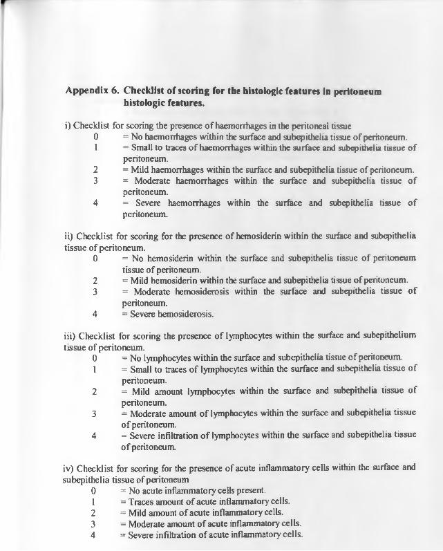
Appendix 6. Checklist of scoring for the histologic features in peritoneum histologic features.
for scoring the presence of haemorrhages in the peritoneal tissue= No haemorrhages within the surface and subcpithelia tissue of peritoneum.= Small to traces of haemorrhages within the surface and subcpithelia tissue of peritoneum.= Mild haemorrhages within the surface and subcpithelia tissue of peritoneum. = Moderate haemorrhages within the surface and subcpithelia tissue of peritoneum.= Severe haemorrhages within the surface and subcpithelia tissue of peritoneum.
ii) Checklist for scoring for the presence of hemosiderin within the surface and subcpithelia tissue of peritoneum.
0 = No hemosiderin within the surface and subepithelia tissue of peritoneumtissue of peritoneum.
2 = Mild hemosiderin within the surface and subepithelia tissue of peritoneum.3 = Moderate hemosiderosis within the surface and subepithelia tissue of
peritoneum.4 = Severe hemosiderosis.
iii) Checklist for scoring the presence of lymphocytes within the surface and subepithelium tissue of peritoneum.
0 = No lymphocytes within the surface and subepithelia tissue of peritoneum.1 = Small to traces of lymphocytes within the surface and subepithelia tissue of
peritoneum.2 = Mild amount lymphocytes within the surface and subepithelia tissue of
peritoneum.3 = Moderate amount of lymphocytes within the surface and subepithelia tissue
of peritoneum.4 = Severe infiltration o f lymphocytes within the surface and subcpithelia tissue
o f peritoneum.
iv) Checklist for scoring for the presence of acute inflammatory cells within the surface and subepithelia tissue of peritoneum
0 = No acute inflammatory cells present.1 = Traces amount of acute inflammatory cells.2 = Mild amount of acute inflammatory cells.3 = Moderate amount of acute inflammatory cells.4 = Severe infiltration of acute inflammatory cells.
i) Checklist 0 1
23
4

v) Checklist for scoring for the presence of fibrin on and under the peritonealsurface if present.0 = No evidence of fibrin deposits on surface of peritoneum1 = Trace evidence of fibrin deposits on surface of peritoneum.2 = Mild fibrin deposits on surface of peritoneum.3 = Moderate fibrin deposits on surface of peritoneum.4 = Large fibrin deposits on surface of peritoneum.

r
Appendix 7: Data from lesions collected during the pre-and post-treatment laparoscopy of individual endometriotic baboons (Mean ± standard deviation)
Placebo GnRH - antagonist Rosiglitazone
Pre Post Pre Post Pre PostMean surface area (mm2)with Adhesions
145,219,314,45. (180.8 ± 113.9)
162,209,235,53.(164 .6180 .6)
191,459,430,180 (3 1 5 1 150.1)
$
109,118,389,96. (177 .91141.2)
$
209,328,134,291) (240.3 1 86.8)
t
55,235,87,116(123 .2178 .3 )
tMean surface area (mm2) without Adhesions
65, 195, 180,45. (121.3 ±77.2)
32, 193, 74, 53. (89 .6171 .8 )
133, 94, 114, 98.(109 .81 17.7)
*
50, 64, 60, 50.(55.9 1 7.3)
*
209, 83, 59, 75 (106 .3169 .2)
55,45, 29, 40. (42 .21 10.8)
, J g ______Mean number o f lesions
8,13,7,10 (9.5 ± 2.6)
7,13,6,11(9 .3 1 3 .3 )
9,9,8,15(10 .313 .2 )
9,9,11,14(10 .812 .4 )
15,18,7,13(13 .314 .6 )
15, 16, 6, 13 12 .514 .5 )
Mean number o f Red lesions
4, 3, 0, 0 (1.8 ± 2.1)
3, 6, 0, 3. (3 1 2.4)
4 ,1 ,1 ,1 1 .(4 .3 1 4 .7 )
1,2, 2, 5. (2 .5 1 1 .7 )
6, 6, 5,0. (4.3 1 2.9)
1.0, 2, 0. (0 .8 1 1.3)
Mean area o f red lesions (mm2).
38,46, 0, 0. (21± 24.5)
17, 112, 0, 32. (40 .3149 .6)
69, 1, 1, 83. (8 .5143 .7 )
9,3,7,16.(8 .7 1 5 .3 )
166,29,57,0 (62 .91 72.5)
2,0,14,0(4 1 6 .7 )
Mean number o f adhesions
3,4,4,0. (2.8 ± 1.9)
4,2,4,0. (2 .5 1 1 .9 )
2,3,3,3. (2.8 10 .5 )
3,4,3,3. (3.3 10 .5 )
0,4,7,3. (3.5 12 .9 )
0.3,6,4. (3.3 12 .5 )
- 197-

1
Appendix 8: Haematology data of individual endometriotic baboons (Mean ± standard deviation)P la c e b o G n R H - a n ta g o n is t R o s ig l i ta z o n e
P r e P o s t P r e P o s t P r e P o s tWhite Blood Cells xidVpi
9.4,7.9,6.9,9.8 (8.5±1.344)
8,6.6,12.2, 11.4 (9.5512.68)
21.9,10.8, 11.6, 6.5 (12.716.53)
21.2, 11.8,9.7,9.8 (13.315.47)
16.6, 6, 10.7, 6.7 (1014.86)
16.4, 7,12.4,9 (11.214.12)
Red Blood Cells xl06/pl
4.01,4.12,4.83,4.41(4.3410.37)
4.16, 4.5,4.6, 4.66 (4.4810.22)
4.61,4.84,4.39, 4.02 (4.4710.35)
4.88, 4.71,5.16,4.22 (4.7410.40)
5.31,4.2, 5.11,4.36 (4.7510.55)
4.78, 4.8,5.25,4.18 (4.7510.44)
Haemoglobin x gm/dl
11.5,11.5,13.9,12.5(12.35±1.14)
*
13.1,12.7,14.6,13.1(13.3810.84)
*
12.4, 12, 12.4, 11.5 (12.0110.43)
13.2, 13.5, 15.7, 12.2 (13.6511.48)
12.7,123,14.4,12.2 (12.911.02)
$
13.7, 12.8, 15.9, 13.4 (13.951135)
$Total Plasma Protein gm/dl
6.7, 5.9, 7.5, 6.7 (6.710.65)
6.1, 5.9, 7.3, 6.7 (6.510.63)
7.7,7,6.7, 7.4 (7.210.44)
t
6.5, 5.8,6 4,6.2 (6.2310.31)
5.9, 6.4, 6.4, 7 (6.4310.45)
6.3, 6.2, 6.7, 7.2 (6.610.46)
Plasma fibrinogen gm/dl
0.2, 0.2, 0.2, 0.1 (0.17510.05)
0.1, 0.1, 0.2, 0.1 (0.12510.05)
0.1,0,0.1,0.1 (0.07510.05)
t
0.2,0.1,0.2, 0.1 (0.1510.06)
T
0.2, 0.1,0.2,0 2 (0.1810.05)
0.2, 0.1,0.2,03 (0.210.08)
Band Neutrophils per pi
94, 79,0,0 (43.25150.32)
0,0, 244,0 (611122)
219, 108, 116,0 (110.75189.48)
424, 118,0, 98 (1601183.4)
0,0.0,67 (16.75133.5)
492.0.0.90(145.51234.86)
Segmented Neutrophils per pi
2726,4898, 3243, 5292
(4039.7511247.06)
3360, 2772, 7564, 4322
(4504.512137.39)
11388,5508, 8468, 2945
(7077.2513653.98)
15624,6962, 4656, 4998
(80601 5144)
11620, 2460, 5350, 2479
(5477.314314.4)
9020,3360, 6200, 3780
(5590126068)Lymphocytes per pi 6486, 2686, 3381,
3999(413811654.67)
4160, 3498,3782, 6840
(457011537.44)
9636, 5076, 2900, 3445
(5264.2511177.11)
5088,4248, 4753, 4312
(4600.31395.14)
4814, 3480,5136, 4020
(4362.51752.39)
6532, 3430, 5580, 4590
(503311330.7)Monocytes per pi 94,237, 207, 279
(204.25179.21)240, 330, 366, 228
(291167.62)219, 108, 116, 65
(127165.29)212,472, 291,29
(2511183.71)166, 60,214, 134 (143.5164.65)
164, 140, 496, 270 (267.51162.47)
Eosinophils per pi 0, 79, 69,0 (37142.92)
240, 0, 244, 0 (1211139.73)
438,0,0, 65 (125.751210.4)
212,0,0,98(77.51100.87
0.0.0.0(0±0)
•
4 9 2 , 70 , 124, 27 0 (2 3 9 1 1 8 8 .6 4 )
•$ Haemoglobin changed significantly in Rosiglitazone group. t Plasmafibnnogcn changed significantly in GnRH group • Eosinophils changed significantly in Rosiglitazone group ̂ Total Plasma Protein reduced
- 198-

A ppendix 9: H istopathology o f lungs in endom etriotic baboons
Findings Animal identification Number in Placebo group
Frequency of occurrence in placebo group
Animal identification Number in GnRH
Frequencyofoccurrence in GnRH group
Animal identification Number in Rosiglitazone
Frequencyofoccurrence in Rosiglitazone group
Normal 2631,29122991,2985
4/4 2993, 3036 3014
3/4 1/4
Edema moderate to severe & hemorrhages. Emphysema with corresponding atelectasis mild
0/4 2871 1/4 3032, 2788 2998, 3030
4/4
Focal emphysema andcorrespondingatelectasis
0/4 0/4 2998, 2788 2/4
Congestion & mild hemosiderin
0/4 2871 2/4 0/4
- 199 -

A ppendix 10: Histopathology o f liver in endom etriotic baboons
Findings Animal identification
Number in Placebo
group
Frequencyof
occurrence in placebo
group
Animal identification
Number in GnRH
Frequencyof
occurrence in GnRH
group
Animal identification
Number in Rosiglitazone
Frequencyof
occurrence in Rosiglitazone
groupNormal 2912 1/4 0 0Bile ducts surrounded by inflammatory cells.
3036 1/4 2998, 3032 3030, 2788
4/4
Focal infiltration o f lymphocytes 29122631
2/4 3014,3036,2993
3/4 0
Both focal Focal infiltration o f lymphocytes and bile duct surrounded lymphocytes
2991 1/4 3014, 2871 2993
3/4 2788 1/4
Bile duct hyperplasia and surrounded by lymphocytes
3023 1/4
- 200 -
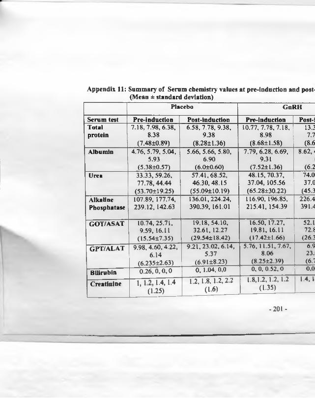
Appendix 11: Summary of Serum chemistry values at pre-induction and post- (Mean ± standard deviation)
Placebo GnRH
Serum test Pre-induction Post-induction Pre-induction Post-iTotal 7.18, 7.98, 6.38, 6.58, 7.78, 9.38, 10.77, 7.78, 7.18, 13.3protein 8.38 9.38 8.98 7.7
(7.48±0.89) (8.2811.36) (8.6811.58) (8.6Albumin 4.76, 5.79, 5.04, 5.66, 5.66, 5.80, 7.79, 6.28, 6.69, 8.62,'
5.93 6.90 9.31(5.3810.57) (6.010.60) (7.5211.36) (6.2
Urea 33.33, 59.26, 57.41,68.52, 48.15, 70.37, 74.077.78,44.44 46.30,48.15 37.04, 105.56 37.0
(53.70119.25) (55.09110.19) (65.28130.22) (45.3Alkaline 107.89, 177.74, 136.01,224.24, 116.90, 196.85, 226.4Phosphatase 239.12, 142.63 390.39, 161.01 215.41, 154.39 391.4
GOT/ASAT 10.74, 25.71, 19.18, 54.10, 16.50, 17.27, 52.19.59, 16.11 32.61, 12.27 19.81, 16.11 72.8
(15.5417.35) (29.54118.42) (17.4211.66) (26.3GPT/ALAT 9.98, 4.60, 4.22, 9.21,23.02, 6.14, 5.76, 11.51,7.67, 6.9
6.14 5.37 8.06 23.(6.23512.63) (6.9118.23) (8.2512.39) (6.1
Bilirubin 0 .26 ,0 ,0 ,0 0, 1.04, 0,0 0, 0, 0.52, 0 0.0
Creatinine 1, 12, 1.4,1.4 (1.25)
1.2,1.8, 1.2,2.2 (16)
1.8,1.2,1.2, 1.2 (1-35)
1.4, 1
- 201 -