CLINICAL LABORATORY & ANATOMIC PATHOLOGY MANUAL
140
CLINICAL LABORATORY & ANATOMIC PATHOLOGY MANUAL 2018 – 2019 Pathology & Laboratory Medicine VA Medical Center Erie, PA 16504 Attachment E
-
Upload
khangminh22 -
Category
Documents
-
view
0 -
download
0
Transcript of CLINICAL LABORATORY & ANATOMIC PATHOLOGY MANUAL

CLINICAL LABORATORY &
ANATOMIC PATHOLOGY
MANUAL
2018 – 2019
Pathology & Laboratory Medicine VA Medical Center Erie, PA 16504
Attachment E
VETERANS AFFAIRS MEDICAL CENTER Erie, Pennsylvania
CLINICAL LABORATORY MANUAL
Pathology & Laboratory Medicine
This manual has been prepared by the staff of Pathology & Laboratory Medicine in order to apprise you of general policies and procedures for procurement of specimens and services provided by the Laboratory. No part of this manual may be altered without the consent of the Medical Director, Pathology & Laboratory Medicine. _____________________________________________ BERNADETTE HALL, MT(ASCP) Supervisor, Pathology & Laboratory Medicine _____________________________________________ RITU KHERA, MD Medical Director, Pathology & Laboratory Medicine
Attachment E

TABLE OF CONTENTS
Pathology & Laboratory Medicine
PATHOLOGY & LABORATORY MEDICINE DIRECTORY I. General Information and Services
A. Location B. People Who Care C. Services Provided D. Laboratory Hours of Operation and Staffing E. Keeping You Informed F. Accreditation G. Quality Assurance (QA) and Quality Control (QC) H. Dedication to Excellence
Clinical Laboratory II. GENERAL INFORMATION - Clinical Laboratory
A. Ordering of Laboratory Tests 1. General Information/Ordering of Laboratory Tests 2. Laboratory Test Ordering Urgency 3. Laboratory Test Ordering During Computer Down
B. Specimen Collection and Handling 1. Patient Identification Procedure 2. Specimen Collection
a. Phlebotomy General Information b. Blood Sample That Cannot be Collected by Phlebotomist c. Vacutainer Tubes d. Collection of Other Samples (Urine, Sputum, Stool, Swabs) e. Specimen Labeling
3. Patient Identification Procedure for Phlebotomy
4. Vacutainer Chart and Other Test Collection Information C. Criteria for Handling/Rejecting Unacceptable Specimens D. Turnaround Time of Test Results E. Reporting of Test Results – General Information F. Specimen Retention Guidelines G. Reference Laboratory Testing
1. Selection of Reference Laboratory 2. Reference Laboratories Currently Used
Attachment E
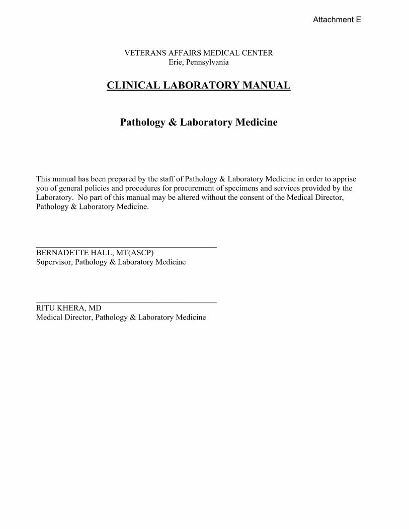
III. DEPARTMENT INFORMATION – Clinical Laboratory A. Chemistry
1. General Information 2. Test Information 3. Specimen Collection and Storage 4. Reference Ranges 5. Additional Information Attachments:
Instructions for the Chemistry Special Tests 24-Hour Urine Testing: Preservative Quick Reference Chart Instructions for 24-hour Urine Collection Glucose Tolerance Test Lactose Tolerance Test Acetone, Iron, TIBC and Alcohol
B. Special Chemistry 1. General Information 2. Test Information 3. Specimen Collection and Storage 4. Reference Ranges
C. Toxicology 1. General Information 2. Test Information 3. Specimen Collection and Handling 4. Reference Ranges 5. Additional Information
D. Hematology 1. General Information 2. Test Information 3. Specimen Collection 4. Reference Ranges
E. Coagulation 1. General Information 2. Test Information 3. Specimen Collection 4. Reference Ranges
F. Microbiology 1. General Information 2. Test Information 3. Specimen Collection and Storage 4. Additional Information
A) Rejection of Microbiology Specimens B) Quantitation of Organisms and Cells C) Sensitivity Testing
D) Turnaround Time
Attachment E

Attachments: Microbiology Test List
Microbiology Test Chart
G. Urinalysis 1. General Information 2. Test Information 3. Specimen Collection and Handling 4. Reference Ranges
H. Serology 1. General Information 2. Test Information 3. Specimen Collection and Handling 4. Reference Ranges
I. Arterial Blood Gas 1. General Information 2. Test Information 3. Specimen Collection and Storage 4. Reference Ranges
J. Blood Bank 1. General Information 2. Test Information 3. Specimen Collection 4. Reference Laboratories
5. Additional Blood Bank MCMs K. Ancillary Testing – Point of Care Testing (Waived Testing) 1. General Information 2. Approved Ancillary Tests 3. Home Testing 4. Patient Self Testing 5. Additional Ancillary Testing MCMs IV. TESTS AND REFERENCE RANGES
Reference Range Chart
V. GENERAL INFORMATION - Anatomic Pathology VI. SURGICAL PATHOLOGY
Specimen Submission and Handling/Procurement and Transport Criteria for Acceptance/Rejection of Tissue Specimens Instructions for Specimen Procurement: Routine Specimens Specimens Requiring Special Handling
Breast Tissue for Estrogen and Progesterone Receptor Assay Muscle Biopsy Open Lung Biopsy Procedure Lymph Node Biopsy Testicular Biopsy for Infertility
Attachment E

Bone Marrow Aspiration and Biopsy Cytogenetics Studies Immunophenotyping/Flow Cytometry Studies
VII. CYTOPATHOLOGY
General Information Specimen Procurement Policy Specimen Collection and Preservation - General Information Instructions for Collection/Preparation of Specimens Instructions for Collection/Preparation of Specimens from Various Body
Sites
Gynecologic Specimens (Female Genital Tract) Non-Gynecologic Specimens
Respiratory Tract Gastrointestinal Tract Urinary Tract Body Fluids Cerebrospinal Fluid Cytology Scrapings Nipple Discharge Cytology Cyst Fluid Cytology Other Special Studies
Aspiration Cytology Specimens Criteria for Unacceptable/Unsatisfactory Cytology Specimens
VIII. AUTOPSY SERVICE Autopsy Service Performance of Autopsy Examination Post-Mortem Reports
Provisional Anatomic Diagnosis for Post-Mortem Examination Final Post-Mortem Examination Report Turnaround Time of Post-Mortem Reports
STAFF DIRECTORY
Attachment E

NAME TITLE/SECTION EXTENSION Ritu Khera, MD Medical Director x2177 Bernadette Hall, MT(ASCP) Supervisor x2178 Carol Kendra Program Support Assistant x2176 Erin Skelly, MT(ASCP) Ancillary Testing Coord./BB x2190 Lisa Miller, MT(ASCP) Lab Information Manager x2955
Clinical Laboratory
NAME DEPARTMENT EXTENSION Destiny Belle Phlebotomy x2776 Beverly Freeman, MT(ASCP) General Laboratory x2184 Sarah Hatcher, MT(ASCP) General Laboratory x2184 Stephanie Hess Phlebotomy x2274 Joseph Kisiel, MT(ASCP) Chemistry x2182 Jared Leonardi, MT(ASCP) Intermittent Med. Technologist x2176 Filicia Menendez, MT(ASCP) Microbiology x2187 Laura Merritt, MT(ASCP) Intermittent Med. Technologist x2176 Suzanne Parker, MT(ASCP) Intermittent Med. Technologist 460-4039 Gail Patterson, MT(ASCP) Histology/Cytology x2189 Donald Vicary, MT(ASCP) 3rd shift – General Laboratory 460-4039 Mark Wagner, MT(ASCP) Hematology/Coagulation x2181 Beth Wasielewski, MT(AMT) Urinalysis/ABG x2180 Kayla Watkins Phlebotomy x2191 Kelci Yousett General Laboratory x2184
Attachment E

I. General Information and Services
Pathology & Laboratory Medicine
A. Location B. People Who Care C. Services Provided D. Laboratory Hours of Operation and Staffing E. Keeping You Informed F. Accreditation G. Quality Assurance (QA) and Quality Control (QC) H. Dedication to Excellence
General Information - Clinical Laboratory Index II
Attachment E

I. General Information and Services
The Pathology & Laboratory Medicine performs a comprehensive range of Laboratory tests with our goal being to provide accurate and timely Laboratory test results in a cost effective manner. A. LOCATION Pathology & Laboratory Medicine is located on the first floor, east wing, E1-1A. B. PEOPLE WHO CARE The Laboratory is directed by a Pathologist and staffed by qualified Medical Technologists and support staff who are committed to provide the highest level of quality and service. The staff has broad based knowledge and technical expertise to assist with information regarding significance of test results, unusual cases, and technical matters. C. SERVICES PROVIDED Pathology & Laboratory Medicine provides Clinical and Anatomic Services. The Clinical laboratory offers a wide variety of testing being performed with sufficient rapidity
to support emergency, inpatient acute care, outpatient services, outreach clinics and long-term health care monitoring. The departments in the Clinical Laboratory include Chemistry, Special Chemistry, Therapeutic Drug Monitoring, Hematology, Coagulation, Blood Bank, Microbiology, Serology/Immunology, Urinalysis, and Blood Gases. Each department provides a comprehensive menu of testing.
Anatomic Pathology includes surgical pathology, intra-operative consultation, cytopathology, and post-mortem examination.
Ancillary Testing Program Services includes establishing policies, standards, operation, quality management and medical appropriateness for all bedside laboratory testing within the Medical Center and its outreach functions.
D. LABORATORY HOURS OF OPERATION AND STAFFING Laboratory technical coverage is available 24 hours per day, assuring adequate coverage to meet the demands of the inpatient and outpatient services provided. The hours of operation are as follows:
1. Clinical Laboratory Laboratory Testing………………………….Technologist on-site 24 hours every day Outpatient Phlebotomy……………………...Mon - Fri 6:30am – 5pm, CLOSED Holidays
Attachment E

2. Anatomic Pathology Weekdays (Monday - Friday) Surgical Pathology, Cytopathology, and Autopsy Pathology On weekends and holidays, autopsy services are also available, if necessary.
E. KEEPING YOU INFORMED The Laboratory will provide you with up-to-date information regarding current developments, new in-house tests, and any changes in test procedures. F. ACCREDITATION Pathology & Laboratory Medicine is accredited by Joint Commission and the Food and Drug Administration (FDA). G. QUALITY ASSURANCE (QA) AND QUALITY CONTROL (QC) Quality Assurance (QA) and Quality Control (QC) are an integral part of daily operation. The internal and external proficiencies are utilized to monitor the accuracy and precision of each patient run for every assay performed. Laboratory Standard Operating Procedures (SOP’s) have been written to include when to repeat testing to assure that reported test values are accurate and precise. H. DEDICATION TO EXCELLENCE The Laboratory has state of the art, automated and computerized equipment and an efficient and motivated workforce to provide services that are of high quality and customer focused.
Attachment E

II. General Information - Clinical Laboratory
A. Ordering of Laboratory Tests 1. General Information 2. Laboratory Test Ordering Urgency 3. Laboratory Test Ordering During Computer Down
B. Specimen Collection and Handling 1. Patient Identification
2. Specimen Collection - Phlebotomy General Information - Blood Sample that Cannot be Obtained by
Phlebotomist - Vacutainer Tubes - Collection of Other Samples (Urine, Sputum, Stool
and Swabs) 3. Patient Identification Procedure During Phlebotomy
4. Vacutainer Chart & Other Test Collection Information
C. Criteria for Handling/Rejecting Unacceptable Specimens
D. Turnaround Time of Test Results
E. Reporting of Test Results
F. Specimen Retention Guidelines
G. Reference Lab Testing 1. Selection of Reference Laboratory 2. Reference Laboratories Currently Used
Attachment E

CLINICAL LABORATORY
Laboratory tests contribute vital information about a patient’s health. Correct diagnostic and therapeutic decisions rely, in part, on accuracy of test results. Adequate patient preparation, specimen collection, and specimen handling are essential prerequisites for accurate testing. The accuracy of test results depends on integrity of specimens. This section provides information on the following:
A. Ordering of Laboratory Test(s) B. Specimen Collection and Handling C. Criteria for Handling/Rejecting Unacceptable Specimen(s) D. Turnaround Time of Test Results E. Reporting of Test Results F. Specimen Retention G. Reference Laboratory Testing
Attachment E

A. ORDERING OF LABORATORY TESTS
1. ORDERS - GENERAL INFORMATION
a. “Electronic” test(s) requests:
All requests for Clinical Laboratory tests are made electronically into the CPRS computer system by authorized healthcare providers. The Laboratory personnel are not authorized to place orders for Laboratory testing except under situations such as:
- Lab test orders that are cancelled and are required to be recollected. - Reflex testing as designated per policy. - During direct consultation with ordering provider who will electronically sign - During emergency situations (e.g., Cardiac Alert or Surgery)
The following requests for testing should be phoned to the Laboratory in addition to
electronically ordered: - STAT test requests - “Immediate Collect” orders - Blood culture requests
Blood Bank testing – the physician CPRS order designating desired testing, type and quantity
of product(s).
Special Procedure(s) requests: The following tests should be scheduled with the Laboratory. - Glucose and Lactose tolerance tests – Chemistry (x2182) - Bone marrow procedures – Histology (x2184) - Therapeutic phlebotomies – Blood Bank (x2190 or x2184)
HIV testing is considered routine testing and no longer will require a written consent
(documented verbal patient consent only). Laboratory service staff will not request, require or verify consent per VHA Directive 1113 dated May 5, 2015.
b. Verbal/telephone test requests: Telephoned or verbal orders for laboratory tests are not authorized except in emergency
situations. Providers are required to enter the order into CPRS and then contact the lab to have the lab test collected or completed using previously drawn specimens.
In the event of an emergency request, the P&LM staff taking the verbal or telephone order for laboratory tests should write down the patient’s full name/identification and complete order or enter it into the computer as a “verbal” or “telephoned” order to prompt physician signature. The order should be read back to the provider to ensure accuracy. The P&LM will document in VistA under comment section for patient test accession number that “read back was performed for the test order.”
c. The Laboratory test order must be accurate and include the following information:
Patient’s full name and full social security number Test(s) required
Attachment E
Type of material to be analyzed (e.g., blood, urine, cerebrospinal fluid, etc.) Ordering provider Date, and when relevant, time of specimen collection Any special handling required.
NOTE: The correct information is of paramount importance. Incorrect or incomplete information causes unnecessary delays, as all discrepancies must be resolved prior to processing the specimens.
Attachment E

2. LABORATORY TEST ORDERING URGENCY
An urgency status is defined as the priority given to each laboratory test that will indicate how quickly Laboratory personnel must perform that test.
The urgencies for the Laboratory tests are as follows:
Test Ordering Urgency
Definition Turnaround Time (TAT)
STAT The “STAT” as applied to Laboratory test requests means a request for information that has immediate implications for patient care. The key elements of the definition are: * Patient’s condition requires immediate action * Results of the test are required before a decision about the type of action to be taken can be made The category should be confined to life threatening situations.
One (1) hour for most tests. List of approved tests on STAT list is attached.
IMMEDIATE COLLECT
“Immediate collect” applies to test requests that need to be collected at a specific time or right away, other than normal inpatient rounds. * The lab needs to be called/ notified and an order needs completed. * Immediate collect may also be used for outpatients; however, it would not be the recommended choice. This ordering urgency should be used for special circumstances to include timed specimen collection for urgent care patients, drug/stimulation testing or serial cardiac monitoring.
Up to two (2) hours.
ROUTINE The “routine” category includes test request(s) that are required for routine treatment/follow-up of patients. NOTE: If no urgency is given, the order will be entered as “routine”.
Dependent on test complexity
PRE-OP The “pre-op” status applies to pre-operative laboratory work-up for an elective surgery. NOTE: Any delay in writing the order will not be considered enough justification to place it in the “STAT” category.
One (1) to two (2) hours
PATIENT WAITING
The “patient waiting” category applies to outpatients who are waiting for the test results. The tests will be performed ASAP.
Two (2) hours for routine tests
Attachment E
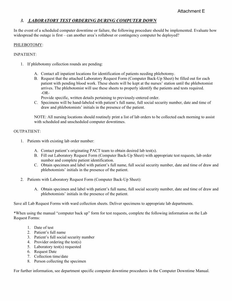
3. LABORATORY TEST ORDERING DURING COMPUTER DOWN
In the event of a scheduled computer downtime or failure, the following procedure should be implemented. Evaluate how widespread the outage is first – can another area’s rollabout or contingency computer be deployed? PHLEBOTOMY: INPATIENT:
1. If phlebotomy collection rounds are pending:
A. Contact all inpatient locations for identification of patients needing phlebotomy. B. Request that the attached Laboratory Request Form (Computer Back-Up Sheet) be filled out for each
patient with pending blood work. These sheets will be kept at the nurses’ station until the phlebotomist arrives. The phlebotomist will use these sheets to properly identify the patients and tests required. -OR- Provide specific, written details pertaining to previously-entered order.
C. Specimens will be hand-labeled with patient’s full name, full social security number, date and time of draw and phlebotomists’ initials in the presence of the patient.
NOTE: All nursing locations should routinely print a list of lab orders to be collected each morning to assist with scheduled and unscheduled computer downtimes.
OUTPATIENT:
1. Patients with existing lab order number:
A. Contact patient’s originating PACT team to obtain desired lab test(s). B. Fill out Laboratory Request Form (Computer Back-Up Sheet) with appropriate test requests, lab order
number and complete patient identification. C. Obtain specimen and label with patient’s full name, full social security number, date and time of draw and
phlebotomists’ initials in the presence of the patient.
2. Patients with Laboratory Request Form (Computer Back-Up Sheet):
A. Obtain specimen and label with patient’s full name, full social security number, date and time of draw and phlebotomists’ initials in the presence of the patient.
Save all Lab Request Forms with ward collection sheets. Deliver specimens to appropriate lab departments. *When using the manual “computer back up” form for test requests, complete the following information on the Lab Request Forms:
1. Date of test 2. Patient’s full name 3. Patient’s full social security number 4. Provider ordering the test(s) 5. Laboratory test(s) requested 6. Request Date 7. Collection time/date 8. Person collecting the specimen
For further information, see department specific computer downtime procedures in the Computer Downtime Manual.
Attachment E

LABORATORY REQUEST FORM
(Computer Back-up Sheet)
TEST DATE: ________________ ROUTINE TESTING UNLESS INDICATED Laboratory Tests:
HEMATOLOGY CHEMISTRY _____ CBC & DIFF □ STAT _____ Glucose □ STAT _____ HGB & HCT □ STAT _____ Lipid Profile _____ ESR (Westergren/Sed Rate) _____ Liver Profile □ STAT _____ Retic Count _____ Chem 8 □ Fasting □ Nonfasting □ STAT _____ Other _____ Chem 14 □ Fasting □ Nonfasting □ STAT COAGULATION _____ Cardiac Profile □ STAT _____ PTT □ STAT (CPK/CK-MB, Troponin) _____ ProTime □ STAT _____ Troponin-I □ STAT _____ Other _______________________ _____ Magnesium □ STAT _____ Lipase □ STAT _____ Amylase □ STAT SEROLOGY _____ Calcium □ STAT _____ RPR _____ Other ________________________ _____ RA _____ Other _______________________ THERAPEUTIC DRUGS _____ Digoxin (monitor/random) □ STAT URINALYSIS _____ Theophylline (monitor/random) □ STAT _____ Routine □ STAT _____ Dilantin □ STAT _____ Phenobarbital □ STAT MICROBIOLOGY _____ Valproic Acid □ STAT _____ Culture & Sensitivity _____ Lithium (sent out) □ STAT Site: ___________________________ _____ Other _______________________ _____Other_______________________ _____ Vancomycin PEAK time ____________
_____ Vancomycin TROUGH time _________ BLOOD BANK _____ Gentamycin PEAK time _____________ _____Specific Testing_______________ _____ Gentamycin TROUGH time __________ _____Product/Quantity_______________ _____ Tobramycin PEAK time _____________ MISCELLANEOUS _____ Tobramycin TROUGH time __________ _____ Test: ________________________ _____ Other__________PEAK time _________ _____ Test: ________________________ _____ Other __________ TROUGH time_____ _____ Dose to be given at _________ over UNIT: ___________________________ __________ hours. Patient ID Stamp: DOCTOR: ________________________________
REQUEST DATE: _________________________
COLLECTION TIME: ______________________
COLLECTION DATE: ______________________ COLLECTED BY: _________________________
Revised 11/30/17
Attachment E

B. SPECIMEN COLLECTION AND HANDLING An adequate specimen properly timed, collected, identified, preserved and stored is critical in achieving an accurate test result.
1. Patient Identification
Accurate patient identification, specimen labeling and maintaining the integrity of identification from specimen collection to accessioning within the Laboratory is essential. Each section of the Laboratory will maintain the identity of each specimen throughout all phases of testing and reporting of test results. For complete identification procedure, refer to “Patient Identification for Phlebotomy.” 2. Specimen Collection
All specimens will be collected using proper collection technique and appropriate specimen requirements for the test(s) requested.
PHLEBOTOMY SERVICES - GENERAL INFORMATION
The Laboratory provides phlebotomy service for all inpatients and outpatients. The phlebotomist will routinely perform venipunctures on the hand and arm. Phlebotomies in the foot can be performed with provider permission. Nursing personnel or physicians may draw blood specimens from in-line catheters. Arterial blood draws are performed by Respiratory Therapy staff or physicians only. a. Service Hours
Outpatients: Phlebotomy service will be provided to outpatients in the Laboratory phlebotomy area from 6:30 a.m. to 5:00 p.m. Monday through Friday. Laboratory personnel are available for phlebotomy on the weekends and holidays for STAT and critical timed tests and for patients seen during extended clinic hours. Inpatients: An electronic order must be placed by the provider, or other authorized personnel, prior to scheduled phlebotomy rounds. Pathology & Laboratory Medicine will routinely perform inpatient phlebotomy rounds at the following times:
Timing for Phlebotomy Rounds 6:00 AM
10:30 AM 3:00 PM 6:00 PM 8:30 PM
STAT, timed and Immediate Collect specimens will be collected as ordered. STAT requests should be limited to life threatening situations.
Attachment E

b. Blood Sample That Cannot Be Obtained by Phlebotomist Pathology & Laboratory Medicine will make two (2) attempts to draw a blood specimen on a patient. The phlebotomist will not attempt more than two (2) venipunctures on the same patient. After two attempts, an alternate phlebotomist will be requested to draw the patient. If the alternate phlebotomist is unable to obtain the specimen, the attending physician will be contacted for further direction. c. Vacutainer Tubes All Laboratory tests require different Vacutainer tubes containing anticoagulants and preservatives. For information, refer to “Vacutainer Chart & Other Test Collection Information.” For any Laboratory test(s) not listed or requiring special collection procedures, and/or special transportation, call the specific Laboratory section. d. Other Specimens (Urine, Sputum, Stool and Swabs)
Prior to each collection, review the laboratory’s specimen requirements, including proper specimen
to be collected, the amount, the procedure, collection materials, and the handling requirements.
Preparing the patient: Provide the patient in advance with appropriate collection and instructions when necessary. NOTE: Some 24-hour urine specimen containers may contain hazardous chemicals such as hydrochloric or boric acid. Collection containers containing these preservatives will be labeled accordingly in order to inform the patient of its content.
The specimens should be delivered to the Laboratory as soon as possible after collection.
e. Specimen Labeling All specimens collected by Laboratory and hospital personnel must be labeled in the presence of the patient in accordance with hospital policy and contain the following information:
Patient’s full name and full social security number Date and time (if applicable) collected Initials of person collecting the specimen
“AVOID COMMON ERRORS DURING SPECIMEN COLLECTION”
Careful attention to routine procedures can eliminate most of the errors. The complete blood collection system and other collection materials provided by the Laboratory can maintain the integrity of the specimens only when they are used in strict accordance with the instructions provided. Types of errors include:
Failure to label a specimen correctly and to provide all pertinent information Insufficient quantity of specimen to run test or QNS (quantity not sufficient) Failure to use the correct container or appropriate specimen preservation Inaccurate and incomplete patient instructions prior to collection Failure to tighten specimen container lids resulting in leakage and/or contamination of container
NOTE: Refer to the Specimen Rejection section of the General Lab Manual for detailed criteria for acceptance/rejection of Laboratory specimens
Attachment E

3. Patient Identification Procedure for Phlebotomy Patient Safety Goal: Improve the accuracy of patient identification. NOTE: Properly identifying the patient is the most critical aspect of specimen collection.
Phlebotomy Recommendations Approved Patient Identifiers
Outpatient/ Inpatient Phlebotomy
Require two (2) approved patient identifiers (neither to be the patient’s location).
Patient(s) who can communicate adequately. Active participation with patient stating two of the verbal identifiers (full name and SSN, DOB [optional]). The other identifiers are VA Identification Card, Driver’s License or their Patient Identification Band as necessary.
Patient(s) who cannot communicate. Identifiers would include: VA Identification Card, Driver’s License or Patient Identification Band. The healthcare worker, family member or attendant could participate in the identification process.
Patient(s) in Emergency Room who are unresponsive. The attendant, family member or healthcare worker could participate in the identification process: NOTE: In case a temporary name and medical record number are assigned, these identifiers could be used to identify the patient and match against specimen labels.
NOTE: The patient’s identity should be re-established if the Phlebotomist leaves the patient’s location prior to initiating the phlebotomy.
Approved Patient Identifiers: Patient’s Full Name Patient’s entire Social
Security Number Patient’s Date of
Birth Patient’s VA
Identification Card or Driver’s License
Patient’s Identification Band
Specimen Labeling
DO NOT pre-label tubes because this may cause patient specimen mix up. Use preprinted computer-generated label or pen/marker for labeling tubes. Use two (2) patient identifiers to label blood and other specimen collection tubes/containers in the
presence of the patient.
Blood Bank Phlebotomy Specimens
In addition to the above guidelines for Blood Bank specimens:
Venipuncture: For Blood Bank specimens, phlebotomy will be performed only on one patient at a time to minimize the risk of error.
Specimen Labeling: Label the sample tube at patient’s bedside immediately after blood sample has been taken. The sample tube must be labeled with the minimum patient information of: Patient’s Full Name (Last, First, Middle) Patient’s Social Security Number Date/time of collection Signature or initials of the person drawing the sample (or employee ID, code) Signature of the healthcare provider witnessing the patient identification and blood sample collection on
VBECS order.
Attachment E

(NOTE: Healthcare providers include nursing staff, CRNA, nurse practitioners, physicians and second
Laboratory medical technologist.)
Attachment E

4. Vacutainer Chart and Other Test Collection Information for Common Tests
NOTE: Minimum sample volumes are provided to minimize unnecessarily large blood draw volumes. Questions or concerns about minimum blood volumes or turnaround times may be answered by contacting the appropriate Laboratory section or reference laboratory.
IN-HOUSE TESTS Tube Turnaround Time (*offered
as STAT with TAT ≤ 1 hour) Test Minimum
Sample Volume
Gold top with gel separator OR Red top/Silicone coated/no additive 10 ml/7 ml
* Daily Acetone 2 ml Daily Albumin
* Daily Alcohol (no alcohol cleanser on arm)
Daily Alk. Phos. Isoenzymes Daily Alkaline Phosphatase Daily ALT (SGTP)
* Daily Amylase Daily AST (SGOT)
* Daily Beta hCG Daily B12
* Daily Bicarbonate (CO2) Daily Bilirubin, Direct Daily Bilirubin, Total
* Daily BUN * Daily Calcium Daily Chem 14
* Daily Chem 8 * Daily Chloride Daily Cholesterol
* Daily CPK * Daily Creatinine Daily Ferritin Daily Folate Daily Free T4 Daily GGT
* Daily Glucose Daily HDL Daily Iron
* Daily LDH Daily LDL Cholesterol (calculated)
* Daily Lipase Daily Lipid Profile
* Daily Liver Profile * Daily Magnesium Daily Osmolality, calculated Daily Phosphorus
* Daily Potassium * Daily Pregnancy Daily PSA
* Daily Sodium Daily TIBC Daily Total Protein Daily Triglycerides Daily TSH Daily Uric Acid Daily Vitamin D
Attachment E

Tube Turnaround Time
(*offered as STAT with TAT ≤ 1 hour)
Test Minimum Sample Volume
Pink Top K2 EDTA * Daily Blood Bank Specimens 4 ml 2 hrs Direct Coombs
Red top Silicone coated/no additive
* Daily Digoxin 2 ml
* Daily Vancomycin
* Daily Phenobarbital * Daily Theophylline * Daily Phenytoin (Dilantin)
Lavender 5 ml EDTA
* Daily CBC/Diff 2-4 hrs Eluate Daily HBA1C (Glycated HGB)
* Daily Platelet Count Daily Reticulocyte Daily Sed Rate 1.5 ml
Blue 3.2% Sodium Citrate 4.5 ml
* Daily PT/INR Tube filled to line
* Daily PTT
Dark Green Top Lithium Heparin 4 ml
Light Green Top PST gel Li Hep 4 ml
* Daily CK-MB Tube 2/3 Full
* Daily Troponin
NOTE: *** Cardiac Profile: (1) Light Green Top and (1) Gold Top Vacutainer Tubes to be used with Urine Collection System Cup:
Tube Turnaround Time (*offered as STAT with
TAT ≤ 1 hour)
Test Minimum Sample Volume
Red/Yellow Speckled Top Conical 8 mL UA preservative
* Daily Urinalysis 4 ml * Daily Urinalysis with Reflex * Daily Diabetic Urinalysis
Gray Top 4 mL Urine C&S preservative
2 days Urinalysis with Reflex Urine C&S
2 ml
Clear Top 6 ml No Additive
* Daily Diabetic Urinalysis Microalbumin/Creatinine Ratio
6 ml
Yellow Top 10 ml UA No Additive
* Daily Urine Drug screen 10 ml
NOTE: ***Urinalysis with Reflex: collect Red/Yellow speckled & gray top Diabetic Urinalysis: collect Red/Yellow speckled and clear top
Attachment E

OTHER IN-HOUSE TESTS
Test Turnaround Time (*offered as STAT with
TAT ≤ 1 hr)
Collection Device Specimen Volume
Manual Differential (Peripheral Blood Smear)
2-4 hrs Microscope Slide 1 drop of capillary
blood from
EDTA tube
Blood Gas * 15 min Heparinized syringe – drawn by RTs or physician only
1 ml arterial blood
Fecal Immunochemical Testing
3 days Special FIT test device
Minute amount of stool
Culture and Sensitivity OR Other Microbiology Tests
2 days Sterile culture swab OR Other sterile collecting device and/or media
Refer to Microbiology Dept. section
in General Lab Manual
MRSA
* Daily Red top culturette Refer to Microbiology Dept. section
in General Lab Manual
Blood Culture
5 days 1 bottle Bactec Plus Aerobic/F, 1 bottle Bactec Lytic/10 Anaerobic/F (8-10 mL whole blood per bottle)
Refer to Microbiology Dept. section
in General Lab Manual
INFLUENZA A and B Antigen, PCR
* Daily Nasopharyngeal Collection Device
Refer to Microbiology Dept. section
in General Lab Manual
Surgical Pathology/Cytology Tests
Daily/Weekly Refer to appropriate section in General Lab Manual
Refer to appropriate section in
General Lab Manual
Attachment E

C. SPECIMEN REJECTION CRITERIA AND SUBOPTIMAL TEST REQUESTS The criteria for accepting or rejecting laboratory specimens for analysis to limit the number of pre-analytical variables in testing and ultimately to ensure an uncompromised reliable sample for testing. The following are the criteria for acceptance/rejection of specimens for Laboratory testing;
1. Incorrect/Inadequate Specimen Identification: The Laboratory personnel will not assume the responsibility for identification of unlabeled specimens. All specimens not properly labeled with full patient name and full patient social security number will not be processed until positive patient identification is made by the collector. If identification cannot be secured, the specimen will be rejected. Specimen cups must have the identification label affixed to the side of the specimen container, not the lid. Each individual specimen container (tube, cup, swab, etc.) must be properly labeled. 2. Inadequate Volume (QNS): Certain minimum volumes are required for test analysis. Also, the amount of additive placed into a tube is intended for a defined volume of blood. If less blood is drawn, the excess additive has the potential to adversely affect the Laboratory results. The following criteria will apply for the rejection of "short draw" samples when the optimum volume is difficult to obtain:
Blood Bank specimens not containing at least 5 ml of blood will be rejected. Coagulation specimens (drawn in blue sodium citrate tubes) must show 90% or greater
expected fill or they will be rejected. FSP (Fibrinogen Split Products ) tubes must contain 1-2 ml of blood. This tube cannot be overfilled and is obtained from the Coagulation depart-ment.
Hematology specimens must contain at least 1 ml of blood.
Urine specimens for routine Urinalysis must contain at least 1.5 ml of urine or they will not
be accepted for analysis. Please refer to specific test procedures for additional sample requirement information.
3. Hemolyzed Specimens:
Hemolysis can result from a difficult venipuncture or from improper handling of specimens. Hemolysis can also occur within the patient as a result of intravascular erythrocyte destruction caused by a disease state. Certain tests will be inaccurate when the specimen is "moderately" hemolyzed in vitro. The presence of a visible red pigmentation also interferes with spectrophotometric analysis and interpretation of blood banking procedures. Therefore:
Blood Bank specimens that are hemolyzed will be rejected.
Attachment E

Chemistry determination (especially LDH, K+, AST, ALT, and Iron) showing moderate to gross hemolysis will be rejected.
Specimens exhibiting slight hemolysis will be accepted for analysis. Results will be entered
with a comment reflecting the presence of slight hemolysis. 4. Clotted Specimens: EDTA (Lavender) and sodium citrate (Blue) tubes for Hematology and Coagulation that arrive in a clotted condition will be rejected. 5. Delays or Error in Transport: Specimens must be transported in as short a time as possible. Unopened specimens for coagulation studies can remain uncentrifuged for up to 24 hours. Tubes of blood are to be kept stoppered at all times and be transported in a vertical position when possible. Specimens requiring special transport include but are not limited to:
Ammonia-- Placed on wet ice and transported to lab immediately. ACTH-- Draw into chilled lavender top (EDTA). Transport on wet ice, separate and freeze
plasma. Alcohol-- Arm must not be prepped with alcohol. Blood cultures--Require special preparation to ensure a sterile venipuncture. Fibrinogen Degradation (Split) Products: Use special tubes kept in Send Out refrigerator. Carotene-- Draw serum; protect from light.
Further instructions can be obtained from the appropriate Laboratory department. All samples arriving in the Laboratory not meeting specific sample transport requirements will be rejected. 6. Improper Collection:
Wrong collection tube: specific specimen requirements must be considered as indicated in the test chart section of the General Laboratory manual. In particular, tubes with additives cannot be used indiscriminately. An additive can interfere with the analyte to be determined.
EXAMPLE: (1) Sodium Fluoride interferes in BUN testing (urease method) (2) Plasma collected when serum is needed (Amylase)
Specimens arriving in the wrong collection tube will be rejected. Notification of specimen rejection will be made to the collecting location.
Attachment E

24 hour Urine: Improper collection of 24 hour urine specimens, i.e., wrong preservative,
wrong timing, wrong pH, and lack of patient information regarding volume, height, weight, will be rejected. Date and time period of collection must be noted.
7. Gross Contamination/Unsafe Submission of Specimens:
Specimens arriving in the Laboratory in an unsanitary condition will be discarded and a second specimen requested. Laboratory staff will notify appropriate collecting location when recollection is necessary.
EXAMPLE: (1) Gross contamination of the outside of the container with the specimen. (2) Leakage of specimen from the container.
Specimens that arrive with needles attached will be returned to the collecting location for proper submission or recollection.
2. PROCEDURES FOR HANDLING SUBOPTIMAL TEST REQUESTS AND SPECIMENS
For detailed procedures for Handling of Suboptimal (Unsatisfactory) Test Requests and Specimens, refer to Pathology & Laboratory Medicine Operations Policy HANDLING OF SUBOPTIMAL (UNSATISFACTORY) TEST REQUESTS AND SPECIMENS.
Attachment E
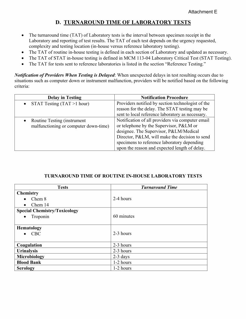
D. TURNAROUND TIME OF LABORATORY TESTS
The turnaround time (TAT) of Laboratory tests is the interval between specimen receipt in the Laboratory and reporting of test results. The TAT of each test depends on the urgency requested, complexity and testing location (in-house versus reference laboratory testing).
The TAT of routine in-house testing is defined in each section of Laboratory and updated as necessary. The TAT of STAT in-house testing is defined in MCM 113-04 Laboratory Critical Test (STAT Testing). The TAT for tests sent to reference laboratories is listed in the section “Reference Testing.”
Notification of Providers When Testing is Delayed: When unexpected delays in test resulting occurs due to situations such as computer down or instrument malfunction, providers will be notified based on the following criteria:
Delay in Testing Notification Procedure STAT Testing (TAT >1 hour)
Providers notified by section technologist of the reason for the delay. The STAT testing may be sent to local reference laboratory as necessary.
Routine Testing (instrument malfunctioning or computer down-time)
Notification of all providers via computer email or telephone by the Supervisor, P&LM or designee. The Supervisor, P&LM/Medical Director, P&LM, will make the decision to send specimens to reference laboratory depending upon the reason and expected length of delay.
TURNAROUND TIME OF ROUTINE IN-HOUSE LABORATORY TESTS
Tests Turnaround Time Chemistry
Chem 8 Chem 14
2-4 hours
Special Chemistry/Toxicology Troponin
60 minutes
Hematology CBC
2-3 hours
Coagulation 2-3 hours Urinalysis 2-3 hours Microbiology 2-3 days Blood Bank 1-2 hours Serology 1-2 hours
Attachment E

E. REPORTING OF TEST RESULTS
1. GENERAL INFORMATION The Clinical Laboratory test results are entered into the patient’s electronic record via the VistA computer system following verification. STAT test results will be reported as specified in MCM 113-04 Laboratory Critical Test (STAT Testing). Critical values will be called to patient’s healthcare provider and documented as specified in MCM 113-05 Communication of Critical Values for Laboratory Tests. 2. REPORTS: All Laboratory results will have adequate pertinent information and are available via the VistA/CPRS computer system (electronic record), which can be accessed by all appropriately privileged providers.
a. Interim Reports: The interim reports are printed immediately at all inpatient and outpatient ordering locations after verification of the test results. The providers may also access all interim reports in the patient’s VistA and CPRS chart. The interim reports should not be charted. b. Cumulative Reports: The cumulative summary reports are no longer printed and distributed. Cumulative reports are found in CPRS under the Laboratory Section, which can be accessed by all privileged providers. c. Anatomic Pathology Reports The tissue and cytology reports are entered into the patient’s electronic record d. Reference Lab Reports The copies of all reference laboratory test results are retained in the Laboratory. All reference laboratory test results are entered into VistA upon receipt without alterations that would affect clinical interpretation. Exceptions to this would be complex test results, such as electrophoresis. These more complex reports are scanned into VistA Imaging. These results have a comment attached stating “SEE SEPARATE REPORT IN VISTA IMAGING” to indicate original test results appear on the patient chart.
e. Pending Lab Results
All inquiries concerning pending in-house Laboratory test results should be directed to the specific sections of the Laboratory.
3. CORRECTION OF ERRONEOUS RESULTS:
The Laboratory technical staff discovering the error is responsible for communicating the error to the Supervisor, P&LM or designee.
The error should be IMMEDIATELY communicated to the healthcare provider responsible for the patient.
The error should be corrected in the patient’s report as “corrected result.” All errors, including major and minor errors, will be documented in P&LM as a part of Performance
Improvement activity with investigation of results and corrective action taken.
Attachment E

F. SPECIMEN RETENTION
SPECIMEN RETENTION CHART Specimens are retained for a clinically acceptable period as defined below:
Specimen Type Retention Time Storage Requirements Chemistry 48 hours Chemistry refrigerator Special Chemistry Toxicology Serology
48 hours Chemistry refrigerator
Hematology CBC’s Peripheral Blood
Smears/Body Fluid Smears
48 hours 2 weeks
Hematology refrigerator Room temp
Coagulation 48 hours Room temperature Urinalysis 24 hours Room temperature Body Fluids 48 hours Room temp/refrigerate Hepatitis C (purple pearl) 1 month Chemistry freezer Urine Drug Screens 1 month Chemistry refrigerator Blood Bank 2 weeks Blood bank refrigerator Blood Gas 48 hours Room temperature Microbiology Culture plates Permanently Stained
Microbiology Slides (e.g., gram, trichrome, etc.)
+ Blood Culture
1 week 1 month
1 month
Room temperature in Microbiology section
Histology Gross Specimens
Paraffin Blocks Microscopic Slides
2 weeks after reporting
10 years 25 years
Room temp in
preservative Room temp in morgue Room temp in morgue
Cytology CSF/Body Fluids Urine Cell Blocks Microscopic slides
48 hours 24 hours 10 years 25 years
Refrigerate Room temperature Room temperature Room temperature
Reference: National Enforcement Office Operations Manual, Record Retention Guidelines, v.2.2, 2015
Attachment E

G. REFERENCE TESTING – CLINICAL LABORATORY
The tests that cannot be performed in-house are sent to fully accredited (CLIA 88 certified) reference laboratories.
1. SELECTION OF REFERENCE LABORATORY:
The reference laboratory(ies) are selected for VISN 4 based on evaluation criteria which focuses on quality, turnaround time and customer satisfaction.
The selected reference laboratories are approved by the Medical Executive Council.
2. REFERENCE LABORATORIES CURRENTLY BEING USED
Non-VA/VA Reference Laboratories
Reference Laboratories Type of Tests Sent
Turnaround Time
Non-VA Reference Laboratories
Laboratory Corporation of America, Columbus, Ohio
Used for esoteric send out tests that cannot be performed in-house or by another VAMC
One (1) to two (2) days for routine specimens
One (1) to five (5) days for special routine tests, e.g. 5HIAA, ACTH.
ACL/Quest Erie, PA
STAT tests not done in-house
CSF/Body fluids Back up for
instrument malfunction
2-3 hours for STATs
VA Reference Laboratories
VAMC Pittsburgh Routine tests with TATs >24 hours.
HIV HCV RNA-PCR HIV RNA, viral load CD4-CD8 Subset
Pap Smears
2-3 days, usually 2-3 weeks
3-10 days
VAMC Kentucky Mycoplasma Epstein Barr Farmers lung panel Toxoplasma Herpes Simplex Ag
Titer HCV Genotyping
1-2 weeks
VAMC Philadelphia, PA Vitamin D Testing
2-3 days
VAMC Baltimore, MD Depleted Uranium Toxic Embedded
Fragments
90 days
Attachment E

III. Department Information CLINICAL LABORATORY
A. Chemistry B. Special Chemistry C. Toxicology D. Hematology E. Coagulation F. Microbiology G. Urinalysis H. Serology I. Arterial Blood Gas J. Blood Bank K. Ancillary Testing (Point of Care Testing – Waived Testing)
Attachment E

A. CHEMISTRY
1. General Information 2. Test Information 3. Specimen Collection and Storage 4. Reference Ranges 5. Additional Information Attachments: Instructions for the Chemistry Special Tests
Attachment E

A. CHEMISTRY
1. GENERAL INFORMATION
a. In-house Automated Tests: All automated chemistry procedures are performed during normal duty hours. b. Reference Laboratories: Tests not performed in-house may be sent to Laboratory Corporation of America, Associated Clinical Laboratories or another VAMC for testing if approved by the Chief, Pathology & Laboratory Medicine Service. It may take a week or more to receive results for these tests. Contact the Laboratory at x2184 for turnaround times of specimens sent to a Reference Lab.
c. All body fluid testing in chemistry is sent to a local reference lab for testing.
2. TEST INFORMATION
a. STAT TESTS: The following tests are available on a STAT basis (24 hours a day, 7 days a week). The turnaround time is approximately one (1) hour: Chem 8
Chem 14 Electrolyte Panel Liver Profile
CPK, LDH Amylase Magnesium
Lipase Cardiac Profile Alcohol (Ethanol) Urine Drug Screen
b. ROUTINE TESTS:
TEST PROFILES: (NOTE: Any test may be ordered as a single analyte). Liver Profile (Hepatic Function Panel) Chem 8 (fasting/nonfasting) Albumin (Basic Metabolic Profile) Total bilirubin Sodium Direct bilirubin Potassium Alk Phosphatase Chloride Total Protein CO2 ALT Glucose AST BUN Creatinine Lipid Panel Calcium Cholesterol, total eGFR HDL cholesterol Triglycerides Cardiac Profile LDL cholesterol, calculated CPK CPK-MB Troponin I
Attachment E
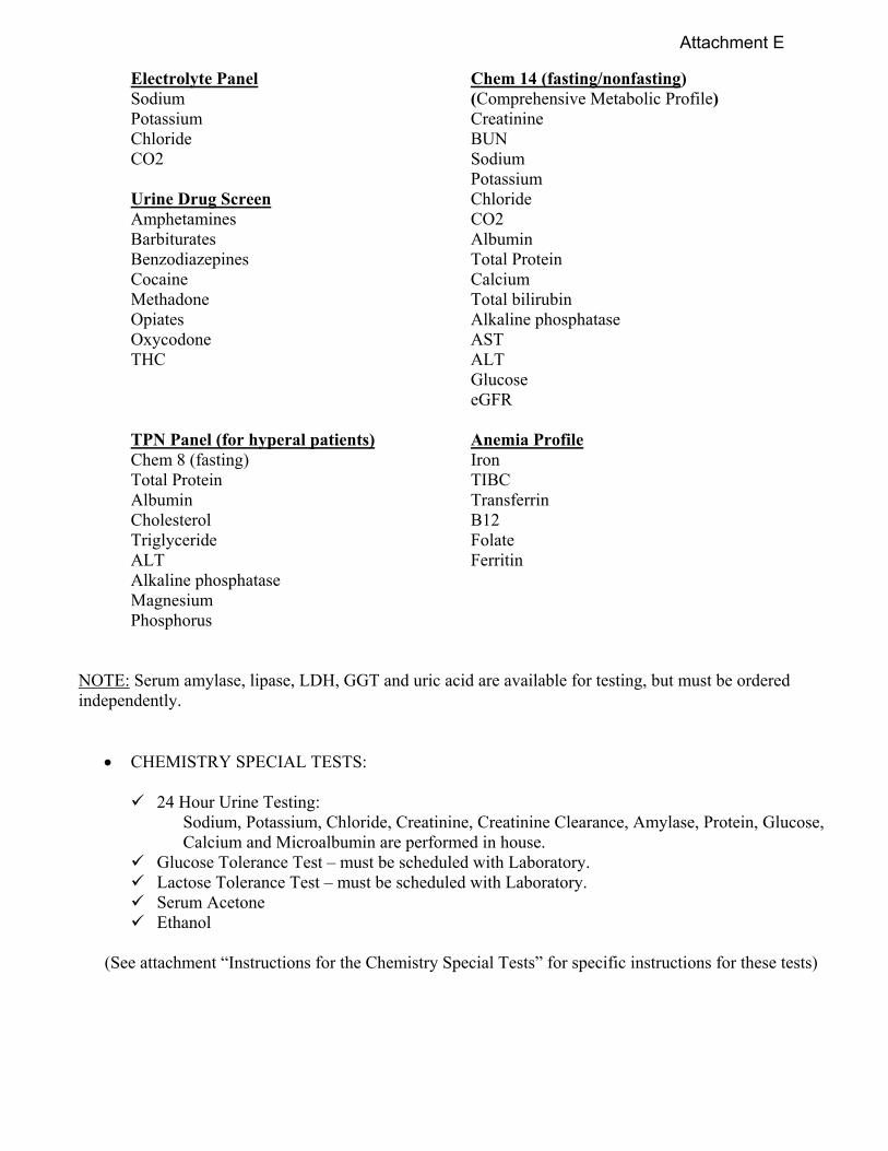
Electrolyte Panel Chem 14 (fasting/nonfasting) Sodium (Comprehensive Metabolic Profile) Potassium Creatinine Chloride BUN CO2 Sodium Potassium Urine Drug Screen Chloride Amphetamines CO2 Barbiturates Albumin Benzodiazepines Total Protein Cocaine Calcium Methadone Total bilirubin Opiates Alkaline phosphatase Oxycodone AST THC ALT Glucose eGFR TPN Panel (for hyperal patients) Anemia Profile Chem 8 (fasting) Iron Total Protein TIBC Albumin Transferrin Cholesterol B12 Triglyceride Folate ALT Ferritin Alkaline phosphatase Magnesium Phosphorus
NOTE: Serum amylase, lipase, LDH, GGT and uric acid are available for testing, but must be ordered independently.
CHEMISTRY SPECIAL TESTS: 24 Hour Urine Testing:
Sodium, Potassium, Chloride, Creatinine, Creatinine Clearance, Amylase, Protein, Glucose, Calcium and Microalbumin are performed in house.
Glucose Tolerance Test – must be scheduled with Laboratory. Lactose Tolerance Test – must be scheduled with Laboratory. Serum Acetone Ethanol
(See attachment “Instructions for the Chemistry Special Tests” for specific instructions for these tests)
Attachment E

3. SPECIMEN COLLECTION AND STORAGE
a. Unless specified by lab order, fasting is not required for specimen collection.
b. Serum or plasma should be free of hemolysis and separated from the clot ASAP or within 2 hours of collection. If there is marked hemolysis, lipemia or icterus, the specimen should be noted as such on the final report. If gross hemolysis is present, a new specimen may be requested at the physician's discretion. c. 24-hour urine should be collected with the appropriate preservative and preferably refrigerated during the collection period. See attachment “Instructions for the Chemistry Special Tests” regarding list of tests requiring preservatives. d. Cerebrospinal fluid and other body fluids should be analyzed promptly or refrigerated at 2-8C. All body fluid testing is sent to a local reference lab for testing. Normal ranges are not available for most body fluid testing.
4. REFERENCE RANGES
Chemistry Routine, Glucose Tolerance Tests and Special Tests: Refer to "Test and Reference Range Chart"(Section IV).
5. ADDITIONAL INFORMATION
Drug Effect on Blood Chemistry: It is important for the physician and laboratory technologist alike to be aware of the possible effect of drugs on laboratory test results. Communication between those concerned with patient care is essential since many of the drug interferences have been discovered through such cooperation. Drugs may have physiological or pharmacological actions in addition to those for which they are prescribed; they may cause idiosyncrasies or hypersensitivities, which could lead to unusual test values; they may have toxic effects particularly on the liver and kidneys; and finally they may interfere in test procedures. In most cases, the association between the drug and unexpected test results may be readily identified. Results become abnormal after the drug is given and return to normal after it is discontinued. Please contact the Laboratory with any specific questions regarding drug interferences.
Attachment E

INSTRUCTIONS FOR CHEMISTRY SPECIAL TESTS A. 24 HOUR URINE COLLECTION
Obtain the one gallon plastic urine collection container from the Laboratory. DO NOT remove any tablets or other preservatives that may be in the bottle. Note any warnings or instructions posted on the outside of the urine collection container.
***CAUTION: Do Not Urinate Directly into the 24 Hour Container if there are any warning labels or any additives to the container: A small urine cup or urinal will be provided to urinate into, and transfer the contents into the large container. ***
1. In the morning (for example 7:00 a.m.), completely empty the bladder and discard the urine specimen.
2. All urine which is voided subsequently, including the final specimen voided at the end of the 24 hour collection period (i.e., 7:00 a.m. the following day) is collected and added to the plastic container.
NOTE: If one gallon jug is not sufficient to hold all of the urine for a 24 hour period, a clean milk jug or other suitable container may be used to hold the additional urine. It is essential for accurate test results that all urine is collected for the 24 hour period.
3. Keep the 24 hour urine collection bottle in a cool place, preferably in the refrigerator, or in a bucket with ice around the container.
4. The container must be labeled with the patient's name, social security number, and date/time collection started and finished.
5. Return the 24 hour urine collection bottle as soon as possible to the Laboratory
6. CBOC only- record the total volume in mL on 24 hour urine container and wrap the screw-top lid with para-film before sending to main laboratory.
7. LABORATORY- verify the correct preservative was used for the test ordered. Record the total volume in mL onto a properly labeled aliquot cup. Shake 24 hour urine jug to mix the specimen and pour off approximately 100mL into aliquot cup.
NOTE: It is essential that this procedure be followed without any deviation, since the results provided to the physician depend upon the accuracy of timing and the collection of the entire 24 hour specimen. ** Creatinine Clearance and Urea Clearance tests MUST have a GOLD top tube drawn on the day the 24 hour sample is DELIVERED to the Lab ** Any deviation to these directions will invalidate the results and test must be repeated.
Attachment E

24-HOUR URINE TESTING: PRESERVATIVE QUICK REFERENCE CHART
Collection is normally taken between 7:00 AM to 7:00 AM the following day. Analyte Collection
Time Preservative Storage Preservative
Also Acceptable
Comments
Aldosterone 24 hours None Refrigerate Aminolevulinic acid (ALA)
24 hours or random
Acetic acid Refrig/Freeze Protect from light; pH must be <6.0
Amylase 24 hours None Refrigerate Arsenic 24 hours or
random None Room temp.
Calcium 24 hours 6N HCl Refrigerate pH must be <4.0
Catecholamine, fractionated
24 hours 6N HCl Refrigerate No boric/acetic acid *adjust pH to <3*
Chloride 24 hours or random
None Refrigerate
Citrate 24 hours 6N HCl Refrigerate Final pH must be 1-3
Copper 24 hours or random
None Room temp.
Cortisol, free 24 hours Boric acid, 1 g Refrigerate 6N HCl Creatine 24 hours None Refrig/Freeze Creatinine 24 hours or
random None Refrigerate
Creatinine Clearance
24 hours None Refrigerate Need height and weight of patient. +1 GOLD top serum
Cystine 24 hours or random
6N HCl Room temp
Free K and Light Chains
24 hours or random
None Room temp.
Glucose 24 hours or random
Boric acid Refrigerate
Heavy metals 24 hours or random
None Room temp.
Homovanillic acid 24 hours or random
6N HCl optional
Refrigerate
Hydroxy-corticosteroids
24 hours
Boric acid, 1 g
Refrigerate
6N HCl
Hydroxyindo-leacetic acid 5HIAA
24 hours None Refrigerate
Attachment E

Immuno-electrophoresis
24 hours or random
None Refrigerate
Kidney Stone Risk Evaluation (calculus saturation)
24 hours None Refrigerate (2) Two 100ml aliquots frozen
Lead 24 hours or random
None Room temp.
Magnesium 24 hours 6N HCl Refrigerate Mercury 24 hours or
random None Room temp.
Metanephrines 24 hours or random
6N HCl Refrigerate
Microalbumin 24 hours or random
None Refrigerate
Myoglobin Random None Refrigerate Osmolality 24 hours or
random None Refrigerate
Oxalate 24 hours 6N HCl Refrigerate
Phosphorus 24 hours or random
6N HCl Refrigerate
Porphobilinogen 24 hours or random
Acetic Acid Refrig/Freeze Protect from light.
Porphyrins 24 hours Sodium carbonate
Refrigerate None Protect from light
Potassium 24 hours None Refrigerate Do not perform on urine to which acid has been added
Protein 24 hours None Refrigerate Do not perform on urine to which acid has been added
Protein electrophoresis
24 hours or random
None Refrigerate
Sodium Timed or 24 hours
None Refrigerate Do not perform on urine to which acid has been added
Urea nitrogen clearance
24 hours None Refrigerate +1 GOLD top serum
Uric acid 24 hours None Refrigerate Vanillyl-mandelic acid (VMA)
24 hours 6N HCl Refrigerate
Zinc 24 hours None Room temp. Revised 12/7/18
Attachment E

PATIENT INSTRUCTIONS FOR 24-HOUR URINE COLLECTION
Obtain the one-gallon plastic urine collection container from the Laboratory. DO NOT remove any tablets or other preservatives that may be in the bottle. Note any warnings or instructions posted on the outside of the urine collection container. For 24-hour urine collections with ANY type of warning labels or ANY additions to the container, DO NOT URINATE DIRECTLY INTO THE CONTAINER. A small urine cup will be provided to urinate into and then transfer the contents into the large container. For 24-hour urine collections without additives or warning labels, you may urinate directly into the container.
URINE COLLECTION 1. In the morning (for example 7AM), completely empty the bladder and discard the urine specimen. 2. All urine that is voided subsequently, including the final specimen voided at the end of the 24-hour
collection period (i.e., 7AM the following day) is collected and added to the plastic container. 3. Keep the 24-hour urine collection bottle in a cool place, preferably in the refrigerator. 4. The container must be labeled with the patient’s name, social security number and date/time collection
started and finished. 5. Return the 24-hour urine collection bottle as soon as possible to the Laboratory. NOTE: It is essential that this procedure be followed without any deviation, since the results provided to the physician depend upon the accuracy of timing and the collection of the entire 24-hour specimen. Pathology & Laboratory Medicine VA Medical Center, Erie, Pennsylvania
Attachment E

B. GLUCOSE TOLERANCE TEST
I. PRINCIPLE:
The glucose tolerance test measures the body’s ability to metabolize glucose. Glucose is the sugar that the body uses for energy. Patients with untreated diabetes have high blood glucose levels. Glucose tolerance tests are one of the tools used to make the diagnosis of diabetes. The oral GTT is also used to screen pregnant women for gestational diabetes.
II. SPECIMEN:
A. Patient prep: An 8 hour fast is recommended; no beverages containing caffeine or smoking just prior to or during the test. All non-essential medications should be discontinued. A diet of average intake should precede the test for at least three days.
B. Test is scheduled with the Lab Monday through Friday to start at 7:00 AM.
C. Serum or plasma.
D. Urine.
III. REAGENTS:
EasydeX Glucola, Orange 50g, 75g or 100g
IV. PROCEDURE: A. Collect a fasting specimen of blood and urine. B. The recommended usual dose of the glucose tolerance beverage is 7.5 oz for nonpregnant adults; 10 oz. for pregnant women. Administer the recommended dose. Start timing when the patient begins to drink the EasydeX Glucola 100 (drink in about 5 minutes). C. Collect subsequent blood and urine specimens at the following times: before glucose administration, 1/2 hour, 1 hour, 2 hours and 3 hours (or the maximum specific time period of the request) after glucose administration. NOTE: It is strongly recommended that a fasting and post prandial glucose be tested prior to any glucose tolerance testing and that the fasting glucose is less than 160 mg/dl before proceeding with the tolerance test.
V. REFERENCE RANGES: A. For a 75-gram oral glucose tolerance test used to check for Type II diabetes, normal (nondiabetic) blood values are:
Fasting: 60-110 mg/dl 1 hour: less than 200 mg/dl 2 hours: less than 140 mg/dl. Between 140-200 mg/dl is considered impaired glucose tolerance or pre-
diabetes. This group is at increased risk for developing diabetes. Greater than 200 mg/dl is diagnostic of diabetes mellitus.
Attachment E

B. For a 50-gram oral glucose tolerance test used to screen for gestational diabetes, normal blood values at 1 hour are less than 140 mg/dl. C. For a 100-gram oral glucose tolerance test used to screen for gestational diabetes, normal blood values are:
Fasting: less than 95 mg/dl 1 hour: less than 180 mg/dl 2 hours: less than 155 mg/dl 3 hours: less than 140 mg/dl
VII. LIMITATIONS: Interfering Factors:
Acute stress (for example, from surgery or an infection) Vigorous exercise
Several drugs may cause glucose intolerance, including the following:
Thiazide diuretics (e.g., hydrochlorothiazide) Beta-blockers (e.g., propranolol) Oral contraceptives Corticosteroids (e.g., prednisone) Some psychiatric medications
Before having the test, let your health care provider know if you are taking any of these medications. VIII. REFERENCES:
A. Beckman Synchron LX Clinical Systems Chemistry Information Manual
B. Aniket R. Sidhaye, M.D., Division of Endocrinology and Metabolism, Johns Hopkins University School of Medicine, Baltimore, MD. Review provided by VeriMed Healthcare Network, 8/12/2004.
Attachment E

C. LACTOSE TOLERANCE TEST
I. PRINCIPLE:
Lactose is the type of sugar found in dairy products. The Lactose tolerance test measures the ability of the patient to metabolically breakdown a lactose load using the body’s enzyme lactase, found in the intestines, into glucose and galactose. Glucose levels are performed to quantify the progression. Lactose intolerance is used to detect abnormalities in carbohydrate metabolism which may result in a number of disease states that cause mucosal damage or dysfunction, such as celiac disease, tropical sprue, and acute gastroenteritis. The most common carbohydrate malabsorption disorder is lactose deficiency.
II. SPECIMEN:
A. Patient prep: An 8 hour fast is recommended; no beverages containing caffeine or smoking just prior to or during the test. All non-essential medications should be discontinued. A diet of average intake should precede the test for at least three days.
B. Test is scheduled with the Lab Monday through Friday to start at 7:00 AM.
C. Serum or plasma.
III. REAGENTS: 50 grams (2x25 gram packets) of oral lactose dissolved in 200 mLs of water
IV. PROCEDURE: Special Instructions: 1. Collect a fasting blood specimen. 2. Label the tube as a fasting specimen and with the appropriate patient identification. Immediately take the specimen to Chemistry for testing and verify the result is in the acceptable range before proceeding to step 3. **BEFORE PROCEEDING WITH THE TEST, THE FASTING GLUCOSE RESULT MUST BE <160 mg/dL, if >160 CONTACT PATIENT’S PROVIDER BEFORE PROCEEDING WITH THE TEST** 3. Instruct the patient to consume 50 grams of oral lactose that was dissolved in 200mLs of water and mixed thoroughly. Lactose packets are located in the send out department. 4. Note the time the patient finishes drinking the oral lactose solution. Remind the patient that he/she must not leave the lab waiting area, eat, drink anything (other than water), chew gum, smoke or be more than minimally physically active during the test. 5. Obtain a blood specimen(s) at the prescribed time interval(s) after consumption of the lactose solution. The number of specimens and time intervals are determined by the physician’s order. 6. Label each tube with the time interval, time collected and the appropriate patient identification.
Attachment E

V. REFERENCE RANGES: The patient is monitored for onset of symptoms, gastrointestinal bloating, cramps, and diarrhea. Glucose values are reported to the nearest whole number.
Glucose levels will be increased in the blood stream if lactose has been successfully cleaved and its components absorbed. The galactose metabolite of the lactose is converted quickly into glucose by the liver.
An increase of 30 mg/dL of glucose above the baseline is considered normal
A rise of 20-30 mg/dL above the baseline is inconclusive
An increase of less than 20 mg/dL with bloating, cramps, and/or diarrhea is evidence for a deficiency of the intestinal disaccharide
VI. LIMITATIONS:
Diabetic patients may have abnormal lactose tolerance curves due to lactose intolerance. Since 25% of normal individuals have flat glucose tolerance tests, it has been suggested that patients with flat lactose tolerance tests should also have a glucose tolerance test. Ethanol can prevent conversion of galactose to glucose by the liver; thus, blood or urine galactose can be measured.
VIII. REFERENCES: Fundamentals of Clinical Chemistry, Norbert W. Tietz, WB Saunders Company, 5th edition 2001. Clinical Chemistry, Michael Bishop, Edward Fody and Larry Schoeff, Seventh edition, 2001. Wu A. Teitz Guide to Laboratory Tests. 4th Ed. Saunders/Elsevier; 2006.
D. ACETONE, IRON, TIBC and ALCOHOL:
1. No special preparation for acetone.
2. Iron and TIBC should be drawn fasting in the morning and prior to administration of therapeutic iron or blood transfusion. Iron determinations in patients who have had blood transfusions should be delayed at least four (4) days.
3. Alcohol should be collected after cleansing the venipuncture site with a non-alcohol based cleanser.
Attachment E

B. SPECIAL CHEMISTRY
1. General Information 2. Test Information 3. Specimen Collection and Storage 4. Reference Ranges
Attachment E
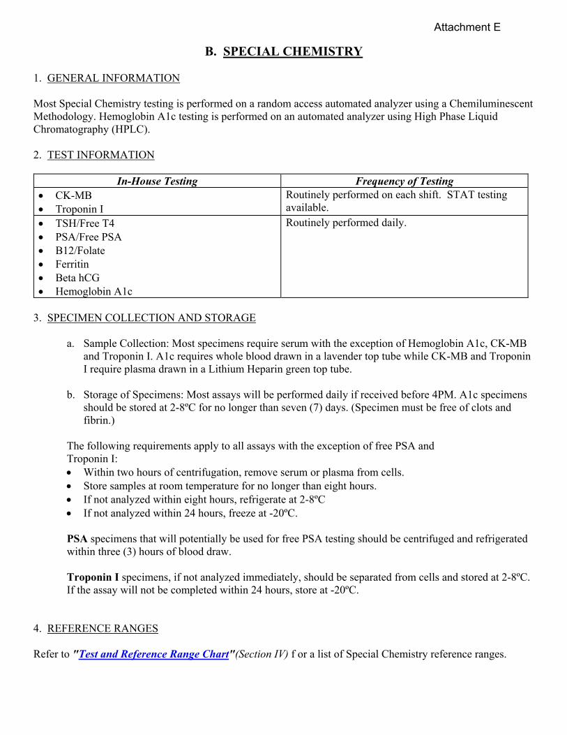
B. SPECIAL CHEMISTRY 1. GENERAL INFORMATION Most Special Chemistry testing is performed on a random access automated analyzer using a Chemiluminescent Methodology. Hemoglobin A1c testing is performed on an automated analyzer using High Phase Liquid Chromatography (HPLC). 2. TEST INFORMATION
In-House Testing Frequency of Testing CK-MB Troponin I
Routinely performed on each shift. STAT testing available.
TSH/Free T4 PSA/Free PSA B12/Folate Ferritin Beta hCG Hemoglobin A1c
Routinely performed daily.
3. SPECIMEN COLLECTION AND STORAGE
a. Sample Collection: Most specimens require serum with the exception of Hemoglobin A1c, CK-MB and Troponin I. A1c requires whole blood drawn in a lavender top tube while CK-MB and Troponin I require plasma drawn in a Lithium Heparin green top tube.
b. Storage of Specimens: Most assays will be performed daily if received before 4PM. A1c specimens
should be stored at 2-8ºC for no longer than seven (7) days. (Specimen must be free of clots and fibrin.)
The following requirements apply to all assays with the exception of free PSA and Troponin I: Within two hours of centrifugation, remove serum or plasma from cells. Store samples at room temperature for no longer than eight hours. If not analyzed within eight hours, refrigerate at 2-8ºC If not analyzed within 24 hours, freeze at -20ºC.
PSA specimens that will potentially be used for free PSA testing should be centrifuged and refrigerated within three (3) hours of blood draw. Troponin I specimens, if not analyzed immediately, should be separated from cells and stored at 2-8ºC. If the assay will not be completed within 24 hours, store at -20ºC.
4. REFERENCE RANGES Refer to "Test and Reference Range Chart"(Section IV) f or a list of Special Chemistry reference ranges.
Attachment E
C. TOXICOLOGY
1. General Information 2. Test Information 3. Specimen Collection and Storage 4. Reference Ranges 5. Additional Information
Attachment E

C. TOXICOLOGY 1. GENERAL INFORMATION The Toxicology testing is performed on a random access automated analyzer. Therapeutic drugs as well as Drugs of Abuse are analyzed in-house. Testing is performed on a STAT or timed basis, depending on the urgency determined by the ordering provider. All critical results are immediately phoned to the ordering physician. 2. TEST INFORMATION a. In-House Testing:
In-House Testing Frequency of Testing Digoxin Theophylline Phenytoin (Dilantin) Urine Drug Screen (see panel in Chemistry Section for tests)
Performed daily as ordered. STAT testing is available during regular and off duty hours, and result turnaround time is approximately one (1) hour.
Phenobarbital Vancomycin, Random Vancomycin, Peak Vancomycin, Trough
Performed daily as ordered.
NOTE: Routine therapeutic drug levels not performed in-house are sent to Pittsburgh VAMC for testing. STAT therapeutic drug levels not performed in-house are sent to a local reference laboratory for testing. b. Definitions TROUGH: The trough is the lowest concentration of drug observed during the dosing interval and is usually measured just prior to the next dose. For most therapeutic drug monitoring requests, a trough concentration, drawn just prior to the next dose, is recommended to ensure that a therapeutic concentration of drug is maintained throughout the dosage interval. PEAK: During the drug-dosing interval, the peak is the highest concentration obtained and may occur immediately after an intravenous dose or may require 0.5 to 2 hours or longer after oral dosage. NOTE: Measurement of both peak and trough concentrations may be indicated for some drugs (e.g., aminoglycoside antibiotics and Vancomycin); however, peak concentrations may be useful only for intravenous therapy due to the variable rates of absorption following oral dosages. c. Indications for TDM: Although the need for therapeutic drug monitoring varies, the primary indications are summarized as follows: ●Drugs with a relatively low margin of safety (i.e., serum concentrations required for therapeutic effect are in the same range as the toxic effects). ●Drugs with large differences in pharmacokinetics between patients. ●Drugs administered concomitantly (especially if one is known to induce or inhibit drug metabolism.
Attachment E

●In presence of disease or clinical conditions, which may alter drug kinetics (e.g., renal, hepatic, cardiovascular disease; prepubescence; pregnancy. ●For nonresponders to drug therapy (to identify patient non-compliance or alter the drug dosage regimen). ●As part of the differential diagnosis if symptoms consistent with drug toxicity are observed.
1. PROCEDURE FOR ORDERING AND COLLECTION OF TIMED DRUG DRAWS The timing of blood sampling in relation to dosage is critical for correct interpretation of the serum concentration results. Collection time should be based on the pharmacokinetic properties of the drug, the dosage form (i.e., IV, IM, oral, etc.), the dosing protocol and the clinical reason for assaying the sample. For this process to work, it is necessary to have close communication between pharmacy, nursing and the laboratory.
a. For Urine Drug Screens: Use the test name ‘Urine Drug Screen’. The test may also be ordered Stat. b. For Therapeutic Drug Monitoring: Medication order entered in CPRS.
1. Clinical Pharmacist/Provider orders peak and trough lab draws by “Immediate Collect” option. 2. Immediate Collect order prints out on designated lab printer and to nursing printer on units. 3. The person ordering sends email to lab staff to notify us of this order. Lab pulls the immediate collect order off printer and files in the correct day of week bin. 4. Lab draws blood at designated time and informs nursing staff prior to leaving unit. 5. Lab enters timed lab draw into lab through “Accessioning Tests Ordered by Ward Order Entry” option in VistA, entering the exact time the specimen was drawn. 6. Nursing staff is to call and inform lab staff of any discontinued orders and/or time of draw changes due to any problems. 7. Lab processes the test and enters timed lab result into the computer.
4. SPECIMEN COLLECTION:
a. Urine Drug Screens require a minimum of 25 mls of random urine in a cup. This volume will support confirmatory testing if needed. 1. Once collected, the patient will hand the specimen to lab staff who will immediately record the temperature using an IR thermometer. 2. Questionable specimens (diluted, temperature varies greatly from normal body temperature) are not rejected. Lab staff will note the discrepancy on the lab report. Exception: Specimen will not be analyzed if suspected adulteration may damage analytical instruments i.e., chemical odor.
b. Blood specimens should be collected in a plain red top tube. DO NOT USE serum separator tubes. If the specimen is a timed draw (i.e., peak or trough), the exact time of draw must be entered into the computer. See chart for recommended drug draw times.
TEST DRAW TIME Theophylline ½ - 1 hr before dose Digoxin ½ - 1 hr before dose Phenytoin ½ - 1 hr before dose Phenobarbital ½ - 1 hr before dose Gentamicin, trough ½ - 1 hr before dose Gentamicin, peak (IV) ½ hr after dose infused Vancomycin, trough ½ - 1 hr before dose Vancomycin, peak (IV) 1 hr after dose infused
Attachment E

Carbamazepine (Tegretol) ½ - 1 hr before dose NOTE: To facilitate interpretation of peak concentrations, the recommended sample time is usually 0.5 to 2.0 hours after the end of an intravenous infusion. This allows for the expected differences in distribution and is less sensitive to small errors in sampling time. In setting the recommended time for blood collection, the following should be considered:
●Length of time patient has received the drug (at the same dose) ●Expected half-life of the drug ●Time of last dose ●Time of next dose ●Whether peak and/or trough concentration is required.
5. REFERENCE RANGES Refer to “Test and Reference Range Chart” (Section IV) for a list of Therapeutic Drug reference ranges. 6. ADDITIONAL INFORMATION REPORTING OF RESULTS: Each report generated will contain the following:
●Type of request (i.e., random, peak/trough) ●Time of collection specific to test results ●Test results and related units ●Expected ranges for type of request ●Notation in “comments” of critical values – to whom and time phoned.
Urine Drug Screens are reported qualitatively as “Negative” or “Positive” based on each drug’s cut-off limits. The cut-off for each drug is summarized as follows:
TEST
CUTOFF LIMIT ng/ml
AMPHETAMINES 1000 BARBITUATES 200 BENZODIAZEPINES 200 COCAINE METABOLITE 300 METHADONE 300 OPIATES 300 OXYCODONE 300 THC 50
Confirmations using GC/MS, gas chromatography/mass spectrometry will be sent to the Philadelphia VAMC laboratory. Turnaround time is approximately 2 weeks. Confirmations must be requested by the provider and phoned to the laboratory for ordering. The urine samples will be saved in the VAMC Erie Laboratory for 30 days.
Attachment E

D. HEMATOLOGY
1. General Information 2. Test Information 3. Specimen Collection 4. Reference Laboratories 5. Reference Ranges
Attachment E
D. HEMATOLOGY
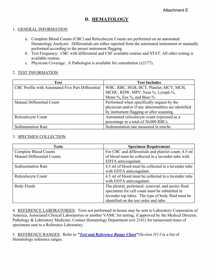
1. GENERAL INFORMATION
a. Complete Blood Counts (CBC) and Reticulocyte Counts are performed on an automated Hematology Analyzer. Differentials are either reported from the automated instrument or manually performed according to the preset instrument flagging.
b. Test Frequency: CBC with differential and CSF available routine and STAT. All other testing is available routine.
c. Physician Coverage: A Pathologist is available for consultation (x2177). 2. TEST INFORMATION
Test Test Includes CBC Profile with Automated Five Part Differential WBC, RBC, HGB, HCT, Platelet, MCV, MCH,
MCHC, RDW, MPV, Neut %, Lymph %, Mono %, Eos %, and Baso %
Manual Differential Count Performed when specifically request by the physician and/or if any abnormalities are identified by instrument flagging or after scanning.
Reticulocyte Count Automated reticulocyte count expressed as a percentage in a total of 30,000 RBCs.
Sedimentation Rate Sedimentation rate measured in mm/hr. 3. SPECIMEN COLLECTION
Tests Specimen Requirement Complete Blood Counts Manual Differential Counts
For CBC and differentials and platelet count, 4.5 ml of blood must be collected in a lavender tube with EDTA anticoagulant.
Sedimentation Rate 4.5 ml of blood must be collected in a lavender tube with EDTA anticoagulant.
Reticulocyte Count 4.5 ml of blood must be collected in a lavender tube with EDTA anticoagulant.
Body Fluids The pleural, peritoneal, synovial, and ascitic fluid specimens for cell count must be submitted in lavender top tubes. The type of body fluid must be identified on the test order and tube.
4. REFERENCE LABORATORIES: Tests not performed in-house may be sent to Laboratory Corporation of America, Associated Clinical Laboratories or another VAMC for testing, if approved by the Medical Director, Pathology & Laboratory Medicine. Contact Hematology Department (ext 2181) for turnaround times of specimens sent to a Reference Laboratory. 5. REFERENCE RANGES: Refer to "Test and Reference Range Chart"(Section IV) f or a list of Hematology reference ranges.
Attachment E

E. COAGULATION
1. General Information 2. Test Information 3. Specimen Collection 4. Reference Laboratories 5. Reference Ranges
Attachment E
E. COAGULATION
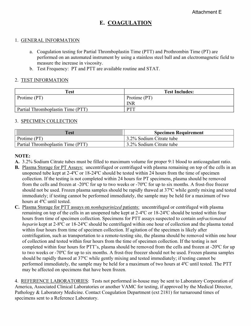
1. GENERAL INFORMATION
a. Coagulation testing for Partial Thromboplastin Time (PTT) and Prothrombin Time (PT) are performed on an automated instrument by using a stainless steel ball and an electromagnetic field to measure the increase in viscosity.
b. Test Frequency: PT and PTT are available routine and STAT. 2. TEST INFORMATION
Test Test Includes: Protime (PT) Protime (PT)
INR Partial Thromboplastin Time (PTT) PTT
3. SPECIMEN COLLECTION
Test Specimen Requirement Protime (PT) 3.2% Sodium Citrate tube Partial Thromboplastin Time (PTT) 3.2% Sodium Citrate tube
NOTE: A. 3.2% Sodium Citrate tubes must be filled to maximum volume for proper 9:1 blood to anticoagulant ratio. B. Plasma Storage for PT Assays: uncentrifuged or centrifuged with plasma remaining on top of the cells in an
unopened tube kept at 2-4ºC or 18-24ºC should be tested within 24 hours from the time of specimen collection. If the testing is not completed within 24 hours for PT specimens, plasma should be removed from the cells and frozen at -20ºC for up to two weeks or -70ºC for up to six months. A frost-free freezer should not be used. Frozen plasma samples should be rapidly thawed at 37ºC while gently mixing and tested immediately; if testing cannot be performed immediately, the sample may be held for a maximum of two hours at 4ºC until tested.
C. Plasma Storage for PTT assays on nonheparinized patients: uncentrifuged or centrifuged with plasma remaining on top of the cells in an unopened tube kept at 2-4ºC or 18-24ºC should be tested within four hours from time of specimen collection. Specimens for PTT assays suspected to contain unfractionated heparin kept at 2-8ºC or 18-24ºC should be centrifuged within one hour of collection and the plasma tested within four hours from time of specimen collection. If agitation of the specimen is likely after centrifugation, such as transportation to a remote-testing site, the plasma should be removed within one hour of collection and tested within four hours from the time of specimen collection. If the testing is not completed within four hours for PTT’s, plasma should be removed from the cells and frozen at -20ºC for up to two weeks or -70ºC for up to six months. A frost-free freezer should not be used. Frozen plasma samples should be rapidly thawed at 37ºC while gently mixing and tested immediately; if testing cannot be performed immediately, the sample may be held for a maximum of two hours at 4ºC until tested. The PTT may be affected on specimens that have been frozen.
4. REFERENCE LABORATORIES: Tests not performed in-house may be sent to Laboratory Corporation of America, Associated Clinical Laboratories or another VAMC for testing, if approved by the Medical Director, Pathology & Laboratory Medicine. Contact Coagulation Department (ext 2181) for turnaround times of specimens sent to a Reference Laboratory.
Attachment E

5. REFERENCE RANGES Refer to "Test and Reference Range Chart"(Section IV) f or a list of Coagulation reference ranges.
Attachment E

F. MICROBIOLOGY
1. General Information 2. Test Information 3. Specimen Collection and Storage 4. Additional Information Attachments: Microbiology Test List Microbiology Test Chart
Attachment E

F. MICROBIOLOGY
1. GENERAL INFORMATION Quality results in Microbiology depend on both the client and the Laboratory. The many factors contributing to the successful isolation of potential pathogens range from specimen selection and collection to proper transport and timely delivery to the Laboratory. Specific plating media and procedures depend on proper specimen handling for pathogen isolation. It is extremely important to refer to specimen collection and submission instructions to ensure that we receive the most suitable or appropriate specimens for Laboratory analysis.
a. HOURS OF OPERATION: Daily: 8:00 AM – 4:30 PM Extension 2187 Complete specimen processing is available during the above hours. A technologist may be reached outside of these hours by calling extension 2181 or 2182. It is suggested that after 5:00 PM Monday through Friday and on weekends, work be limited to specimen collecting which will have an immediate influence on the care of the patient due to reduction of personnel at these times. Cultures will be set daily until 6:00 PM. Reports on specimens arriving after that may be delayed 24 hours. Microbiology reports will generally be available in the computer before 1PM. Please check the computer before calling the Laboratory for results. b. CRITICAL VALUES: The following will be called to the nurse/physician immediately and put into the computer:
Positive Blood Cultures Positive AFB Smear/Culture Positive CSF Gram Stain/Culture/Cryptococcal Antigen (Salmonella/Shigella/Campylobacter/Yersinia/E. coli 0157/Shiga toxin Positive Malaria Smear Positive Gram Stain Sterile Body Fluids Positive MRSA, VRE, C. diff Bacterial isolates positive for ESBL, KPC/CRE Positive Herpes, Chlamydia, N. gonorrhoeae Positive Influenza Other diseases reportable to the Health Department
The microbiology technologist will notify the infectious control coordinator. c. Only gram stains and collection of blood cultures may be ordered on a true STAT basis. Microbiology cultures must be incubated at least 15-18 hours before initial reading can be done; therefore, a STAT culture has merit only in its collection and alert notification.
2. TEST INFORMATION
a. IN-HOUSE TESTING SERVICES: Bacteriology (includes cultures from different sources):
Urines Respiratory sources (sputum, throat, and bronchial washings)
Attachment E

Fluids (CSF, pleural, peritoneal, thoracentesis, etc.) Genital cultures Blood cultures Anaerobes N. gonorrhoeae Feces Wounds (including eye, ear, etc.)
Occult Blood (FIT and Guaiac) Fecal Lactoferrin Clostridium Difficile Parasite Profile Influenza CT/NG
b. TESTS SENT TO REFERENCE LABORATORY
Malaria Mycology KOH - scabies KOH – fungus Mycology culture Mycobacteriology (AFB) Viral Studies Qualitative fecal fat Herpes culture Legionella cultures
c. REFERENCE LABORATORIES
Laboratory Corporation of America, 6370 Wilcox Road, Dublin, OH 43016-1296 VAMC, Fungal & Viral Serology, Lexington, KY, 40507 Associated Clinical Laboratories, 1526 Peach Street, Erie, PA, 16501 VAMC Pittsburgh Healthcare System, University Drive Division, University Drive C,
Pittsburgh, PA 15240
d. MICROBIOLOGY TEST LIST See Microbiology Test List after Additional Information section
3. SPECIMEN COLLECTION AND STORAGE
a. REQUEST FORMS
One of the following should accompany all accessioned and properly labeled specimens to the Microbiology Laboratory:
VistaA CPRS order (*work copy*) and/or specimen accession label Required information if unable to accession:
Patient name
Attachment E
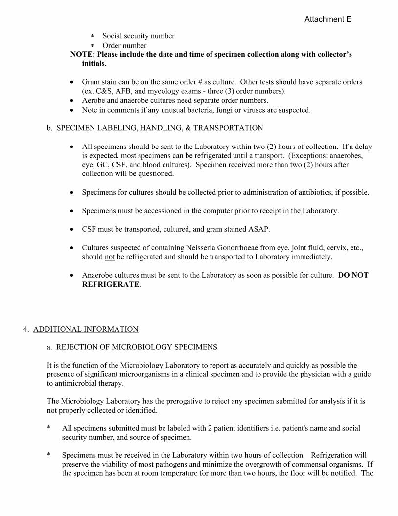
Social security number Order number
NOTE: Please include the date and time of specimen collection along with collector’s initials.
Gram stain can be on the same order # as culture. Other tests should have separate orders
(ex. C&S, AFB, and mycology exams - three (3) order numbers). Aerobe and anaerobe cultures need separate order numbers. Note in comments if any unusual bacteria, fungi or viruses are suspected.
b. SPECIMEN LABELING, HANDLING, & TRANSPORTATION
All specimens should be sent to the Laboratory within two (2) hours of collection. If a delay
is expected, most specimens can be refrigerated until a transport. (Exceptions: anaerobes, eye, GC, CSF, and blood cultures). Specimen received more than two (2) hours after collection will be questioned.
Specimens for cultures should be collected prior to administration of antibiotics, if possible. Specimens must be accessioned in the computer prior to receipt in the Laboratory. CSF must be transported, cultured, and gram stained ASAP. Cultures suspected of containing Neisseria Gonorrhoeae from eye, joint fluid, cervix, etc.,
should not be refrigerated and should be transported to Laboratory immediately.
Anaerobe cultures must be sent to the Laboratory as soon as possible for culture. DO NOT REFRIGERATE.
4. ADDITIONAL INFORMATION
a. REJECTION OF MICROBIOLOGY SPECIMENS It is the function of the Microbiology Laboratory to report as accurately and quickly as possible the presence of significant microorganisms in a clinical specimen and to provide the physician with a guide to antimicrobial therapy. The Microbiology Laboratory has the prerogative to reject any specimen submitted for analysis if it is not properly collected or identified.
* All specimens submitted must be labeled with 2 patient identifiers i.e. patient's name and social
security number, and source of specimen.
* Specimens must be received in the Laboratory within two hours of collection. Refrigeration will preserve the viability of most pathogens and minimize the overgrowth of commensal organisms. If the specimen has been at room temperature for more than two hours, the floor will be notified. The
Attachment E

specimen will be recollected if possible or if not, it will be noted that the results may not be accurate. Do not refrigerate CSF, eye, GC, anaerobes or blood cultures.
* Swab specimens will be rejected if the swabs are completely dry or if the material is insufficient.
* Sputum specimens will be rejected if they are not in a sterile container, if the quantity submitted is
insufficient, or if the exterior of the container is contaminated. All expectorated sputums will be evaluated by gram stain before culturing to check for oropharyngeal contamination. They may be rejected on this basis.
* Urine specimens will be rejected if they are not in a sterile container or if the quantity is insufficient
or if the exterior of the container is contaminated.
* Fecal specimens will be rejected if the amount submitted is insufficient, if they are contaminated with urine, or if the exterior of the container is contaminated.
* Most duplicate specimens received on the same date should not be processed; exceptions include
blood cultures, CSF, tissue, and sterile body fluids excluding urine.
* Foley catheter tips are not accepted for culturing. (Studies show no correlation between cultures from catheter tips and urinary tract infections). Request urine specimens.
* Anaerobe cultures must be sent to the Laboratory as soon as possible in anaerobe culturettes or
syringe (body fluids), otherwise they are unacceptable. They should not be refrigerated.
* CSF specimens should be brought to the Laboratory immediately since fastidious organisms may not survive storage or variations in temperature.
b. QUANTITATION OF ORGANISMS AND CELLS
Growth on Agar Plates Rare - 1+ scanty growth
2+ few colonies 3+ moderate growth 4+ profuse growth Bacteria and Cells on Gram Stain/Oil Immersion Field
0-1 rare 1-5 1+ 5-25 2+ > 25 3+ WBC + epithelial cells in sputum are quantitated/low power field (greater than or less than
25)
c. SENSITIVITY TESTING It is not necessary to request sensitivity testing. Sensitivities are performed routinely on most clinically significant isolates. Testing is dependent upon specimen source and type and quantity of organisms
Attachment E

isolated. Results are reported as S (sensitive), I (intermediate) or R (resistant), as dictated by CLSI guidelines. Some organisms have predictable sensitivity patterns and will not routinely receive sensitivity testing unless isolated from blood or CSF (Beta-hemolytic strep) Antibiograms are available annually to all physicians and nurse practitioners and upon request to other personnel. d. TURN-AROUND TIME * In-house procedures:
The time until release of final results is dependent upon several factors including the number of organisms present, the nature and complexity of their identification, organism purity, and viability. Preliminary reports will be entered into the computer on aerobic cultures requiring more than 24 hours and daily thereafter until completion as more information becomes available. Final reports are available in the computer for all tests upon completion. APPROXIMATE COMPLETION TIME Occult Blood-FIT 3 days Occult Blood-Guaiac 2 hours Gram Stain 24 hours Urine Culture 48 hours Respiratory Culture 48 hours Wound Culture 4 days Feces Culture 48 hours Anaerobic Culture 4 days Blood Culture 5 days Mycology Culture 6 weeks AFB Smear 24 hours AFB Culture 6-8 weeks C. difficile 24 hours MRSA 4 hours – 8 hours Sterile Body Fluids 4 days – 7 days
Tests Sent to Reference Laboratory:
The turnaround time of tests sent to the Reference Laboratories is dependent on several factors including the transport time, test schedule at each individual Lab, and the time for the return of the report. Because it is sometimes difficult to predict, please call the Microbiology Lab (x2187) if there are individual questions.
Attachment E
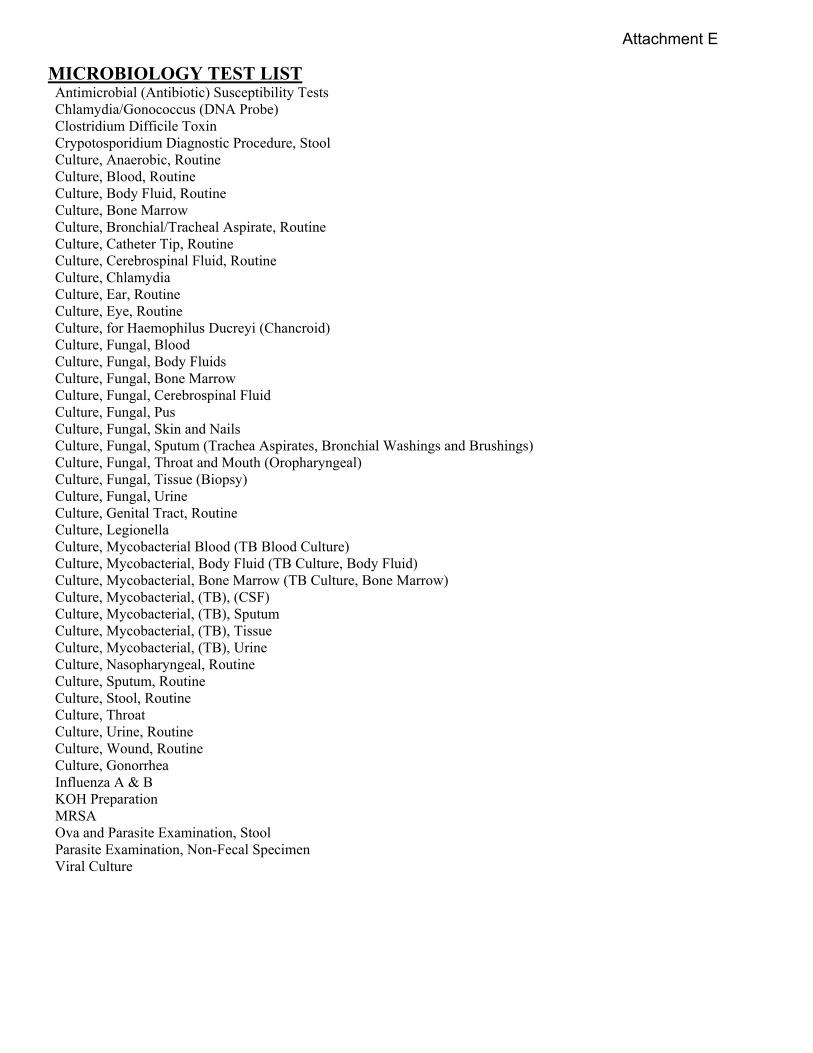
MICROBIOLOGY TEST LIST Antimicrobial (Antibiotic) Susceptibility Tests Chlamydia/Gonococcus (DNA Probe) Clostridium Difficile Toxin Crypotosporidium Diagnostic Procedure, Stool Culture, Anaerobic, Routine Culture, Blood, Routine Culture, Body Fluid, Routine Culture, Bone Marrow Culture, Bronchial/Tracheal Aspirate, Routine Culture, Catheter Tip, Routine Culture, Cerebrospinal Fluid, Routine Culture, Chlamydia Culture, Ear, Routine Culture, Eye, Routine Culture, for Haemophilus Ducreyi (Chancroid) Culture, Fungal, Blood Culture, Fungal, Body Fluids Culture, Fungal, Bone Marrow Culture, Fungal, Cerebrospinal Fluid Culture, Fungal, Pus Culture, Fungal, Skin and Nails Culture, Fungal, Sputum (Trachea Aspirates, Bronchial Washings and Brushings) Culture, Fungal, Throat and Mouth (Oropharyngeal) Culture, Fungal, Tissue (Biopsy) Culture, Fungal, Urine Culture, Genital Tract, Routine Culture, Legionella Culture, Mycobacterial Blood (TB Blood Culture) Culture, Mycobacterial, Body Fluid (TB Culture, Body Fluid) Culture, Mycobacterial, Bone Marrow (TB Culture, Bone Marrow) Culture, Mycobacterial, (TB), (CSF) Culture, Mycobacterial, (TB), Sputum Culture, Mycobacterial, (TB), Tissue Culture, Mycobacterial, (TB), Urine Culture, Nasopharyngeal, Routine Culture, Sputum, Routine Culture, Stool, Routine Culture, Throat Culture, Urine, Routine Culture, Wound, Routine Culture, Gonorrhea Influenza A & B KOH Preparation MRSA Ova and Parasite Examination, Stool Parasite Examination, Non-Fecal Specimen Viral Culture
Attachment E

MICROBIOLOGY TESTS CHART
Specimen Collection Special Instructions
1. ANTIMICROBIAL SUCEPTIBILITY TESTING
This includes panels of antibiotics tested against potential aerobic pathogenic bacteria. It will be done automatically when indicated. Limitations are for organisms which fail to grow on subculture or are too fastidious to grow for susceptibility testing. For anaerobic bacteria, susceptibility tests are not done routinely. Selected isolates are held for one week in case susceptibility is indicated later. The positive blood culture isolates are held for one month. Fastidious organisms that fail to grow in susceptibility testing will be reported out with an appropriate comment “unable to perform sensitivity”.
2. CHLAMYDIA/GONOCOCCUS BY DNA PROBE
Tests include Chlamydia trachomatis and/or Neisseria gonorrhoeae NOTE: One or both may be done from one XPERT Urine or Vaginal/Endocervical specimen collection kit
SPECIMEN: Vaginal/Endocervical, or urine CONTAINER: Gene Xpert collection kit.
SPECIAL INSTRUCTIONS: Chlamydia/GC test is valid for female Vaginal/Endocervical and male/female urine specimens.
3. CULTURE, ANAEROBIC ROUTINE
Test includes gram stain, isolation and identification of anaerobic bacteria. No sensitivity testing is performed.
a. WOUND: Specimens include pus tissue or other material properly obtained from an abscess, aspirate, exudate, lesion, or wound.
COLLECTION: Overlying and adjacent areas must be carefully disinfected to eliminate contamination with indigenous flora. Ideally, pus or other fluid obtained by needle aspiration through intact skin or mucous membrane which has been cleaned with antiseptic is optimal. Sampling of open lesions is enhanced by deep aspiration. Biopsies of base of open lesion is optimal. If irrigation is necessary, non-bacteriostatic sterile normal saline may be used.
b. BODY FLUIDS: Specimens include aspirated pleural, pericardial, peritoneal, synovial, cerebrospinal fluid, etc.
COLLECTION: Specimens are to be aspirated from a prepared site using sterile technique. Contamination with normal flora from skin must be avoided.
c. TISSUE: Specimens include surgical tissue and biopsy material.
COLLECTION: Specimens should be selected from a prepared site using sterile technique.
d. URINE SUPRAPUBIC: Specimen includes suprapubic urine aspirate only. Catheterized or clean catch urines are not acceptable for anaerobic culture.
COLLECTION: Specimen should be collected from prepared site using sterile technique.
Attachment E
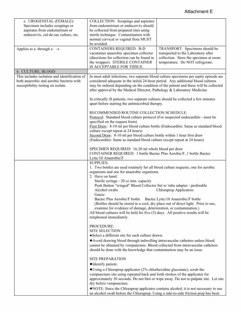
e. UROGENITAL (FEMALE): Specimen includes scrapings or aspirates from endometrium or endocervix, cul-de-sac culture, etc.
COLLECTION: Scrapings and aspirates from endometrium or endocervix should be collected from prepared sites using sterile technique. Contamination with normal cervical or vaginal flora MUST be avoided.
Applies to a. through e. CONTAINERS REQUIRED: B-D vacutainer anaerobic specimen collector (directions for collection can be found in the wrapper). STERILE CONTAINER IS ACCEPTABLE FOR TISSUE.
TRANSPORT: Specimens should be transported to the Laboratory after collection. Store the specimen at room temperature. Do NOT refrigerate.
4. CULTURE, BLOOD This includes isolation and identification of both anaerobic and aerobic bacteria with susceptibility testing on isolate.
In most adult infections, two separate blood culture specimens per septic episode are considered adequate in the initial 24-hour period. Any additional blood cultures may be ordered depending on the condition of the patient and these will be collected after approval by the Medical Director, Pathology & Laboratory Medicine. In critically ill patients, two separate cultures should be collected a few minutes apart before starting the antimicrobial therapy. RECOMMENDED ROUTINE COLLECTION SCHEDULE: Protocol: Standard blood culture protocol (For suspected endocarditis - must be specified on the request form) First Draw: 8-10 ml per blood culture bottle (Endocarditis: Same as standard blood culture except repeat at 24 hours) Second Draw: 8-10 ml per blood culture bottle within 1 hour first draw (Endocarditis: Same as standard blood culture except repeat at 24 hours) SPECIMEN REQUIRED: 16-20 ml whole blood per draw CONTAINER REQUIRED: 1 bottle Bactec Plus Aerobic/F, 1 bottle Bactec Lytic/10 Anaerobic/F
SUPPLIES: 1. Two bottles are used routinely for all blood culture requests; one for aerobic organisms and one for anaerobic organisms. 2. Have on hand:
Sterile syringe - 20 cc min. capacity Push Button “winged” Blood Collector Set w/ tube adapter - preferable Alcohol swabs Chloraprep Applicators Gauze Bactec Plus Aerobic/F bottle Bactec Lytic/10 Anaerobic/F bottle (Bottles should be stored in a cool, dry place out of direct light. Prior to use, examine for evidence of damage, deterioration, or contamination.)
All blood cultures will be held for five (5) days. All positive results will be telephoned immediately. PROCEDURE: SITE SELECTION ●Select a different site for each culture drawn. ●Avoid drawing blood through indwelling intravascular catheters unless blood cannot be obtained by venipuncture. Blood collected from intravascular catheters should be done with the knowledge that contamination may be an issue. SITE PREPARATION ●Identify patient. ●Using a Chloraprep applicator (2% chlorhexidine gluconate), scrub the venipuncture site using repeated back and forth strokes of the applicator for approximately 30 seconds. Do not blot or wipe away. Do not re-palpate site. Let site dry before venipuncture. ●NOTE: Since the Chloraprep applicator contains alcohol; it is not necessary to use an alcohol swab before the Chloraprep. Using a side-to-side friction prep has been
Attachment E

found to be more effective than the circumferential, inside-out technique of skin preparation. DISINFECTING BLOOD CULTURE VIALS ●Remove flip-off caps from vials. ●Wipe the tops of the blood culture vials with 70% isopropyl alcohol pad and leave the pad on top of the bottle until the blood is ready to be injected. ●Do not use iodine to disinfect tops of vials. NOTE: Prior to each use, each vial should be examined for evidence of contamination such as cloudiness, bulging or depressed stopper or leakage. DO NOT USE any vial showing evidence of contamination. A contaminated vial could contain positive pressure. If a contaminated vial is used for direct draw, gas or contaminated culture media could be refluxed into the patient’s vein. Prior to use, the user should examine the vials for evidence of damage or deterioration. On rare occasions, the glass bottleneck may be cracked and the neck may break during removal of the flip-off cap or in handling. Also, on rare occasions, a vial may not be sealed sufficiently. In both cases, the contents of the vials may leak or spill. If the vial has been inoculated, treat the leak or spill with caution, as pathogenic organisms/agents may be present. SPECIMEN COLLECTION AND VOLUME ●Avoid touching the site of venipuncture. ●When using the Butterfly set, the phlebotomist MUST carefully monitor the volume collected by means of the 5 mL graduation marks on the vial label. If the volume is not monitored, the stated maximum amount collected may be exceeded. This condition may adversely create a “false” positive result, due to high blood background. ●If using a needle and syringe, typically a 20 cc syringe is used for adults. Draw 16-18 cc for one blood culture set (aerobic and anaerobic). Aseptically inject: 8-10 mL of blood into the aerobic vial first, then 8-10 mL of blood into the anaerobic vial. The volume of blood cultured is critical because the concentration of organisms in most cases of bacteremia is low, especially if the patient is on antimicrobial therapy. The use of lower or higher volumes may adversely affect recovery and/or detection times. ●The inoculated BACTEC vials should be transported ASAP to the laboratory for incubation.
5. CULTURE, BODY FLUIDS Tests include gram stain, isolation and identification of rapidly growing aerobic and facultative aerobic bacteria. Susceptibility tests done, if appropriate.
SPECIMEN: Joint fluid, pericardial, peritoneal, pleural, and ascitic fluid. VOLUME OF SPECIMEN: 10 ml. Minimum volume 0.5 ml. Container required: sterile container.
6. CULTURE, BONE MARROW Specimen collection instructions are given
under headings dealing with cultures for specific organisms (e.g., “culture fungal, bone marrow”, “mycobacterial cultures, bone marrow”). Consult those sections for proper procedures. The clinical diagnosis and suspected agents must be clearly stated on the requisition as special culture techniques may be required.
7. CULTURE, BRONCHIAL/ TRACHEAL ASPIRATE, ROUTINE
Attachment E

Tests include isolation and identification of rapidly growing aerobic and facultative aerobic bacteria and yeasts. Susceptibility tests done, if appropriate.
SPECIMEN REQUIRED: Bronchial washings, brushings, transbronchial biopsies, bronchial sections, tracheal or tracheostomy aspirates. VOLUME OF SPECIMEN: 10-50 ml. Minimum 1 ml. CONTAINER REQUIRED: Sterile, leak proof, tightly sealed container.
SPECIAL INSTRUCTIONS: See “Sputum culture, routine”. Cultures for mycobacterial and fungi must be requested separately.
8. CULTURE, CATHETER TIP, ROUTINE
Tests include culture for rapidly growing aerobic and facultative aerobic bacteria and yeasts. Only organisms recovered in numbers indicating probable significance (see below) will be fully identified. Susceptibility tests done, if appropriate. Applies to arterial catheters, central venous pressure lines, umbilical, intravenous, Swan Ganz, Hickman catheters (not Foley catheter tip).
SPECIMEN: 1” to 2” segment of catheter COLLECTION: Aseptically prepare insertion site. Remove line without contact with adjacent skin. Send only the intra-arterial segment in a sterile, dry, screw cap container. After removal of catheter, culture any exudate from catheter site and submit to Microbiology.
9. CULTURE, CEREBROSPINAL FLUID, ROUTINE
Tests include gram stain and culture for rapidly growing aerobic or facultative aerobic organism type. Growth of potentially significant organisms will be reported to physician by phone immediately. Positive gram stains will be called to physician immediately.
SPECIMEN: Cerebrospinal fluid VOLUME OF SPECIMEN: 1-5 ml for bacterial culture: 10 ml when fungus or mycobacteria was suspected. Minimum volume 1 ml. Organisms may be present in low numbers; therefore the probability of detecting the organisms decreases significantly if inadequate amounts of specimen are submitted. CONTAINER: Sterile, leak proof tubes
SPECIAL INSTRUCTIONS: Include the following information on the requisition: prior antibiotic treatment (prior therapy requires that cultures be held longer), suspected etiology, presence of shunt, etc. Transport to the Laboratory ASAP. DO NOT REFRIGERATE.
10. CULTURE, CHLAMYDIA Tests include cell culture for detection of Chlamydia trachomatis.
SPECIMEN: Chlamydia is an intracellular pathogen. Obtain swab specimens containing epithelial cells of conjunctiva, cervix, posterior nasopharynx, throat, rectum or urethra. CONTAINER: Specific Chlamydia transport media is required for specimen collection. Call Microbiology laboratory for media (x2187) TRANSPORT: Specimen should be transported to the Laboratory immediately.
11. CULTURE, EAR, ROUTINE Applies to outer ear and middle ear culture - includes gram stain culture for aerobic bacteria. Identification and Susceptibility testing of suspected pathogens.
SPECIMEN: Tympanocentesis fluid. Swab specimen from external ear canal. Aspirated pus. VOLUME OF SPECIMEN: 0.1-0.5 ml or more, if possible. OUTER EAR: Sterile culturette or container. INNER EAR: Anaerobic culturette.
SPECIAL INSTRUCTIONS: Please note on requisition clinical information, suspected organism, history of swimming, previous surgery, drainage tubes, malignant otitis externa, diabetes, etc.
Attachment E

12. CULTURE, EYE, ROUTINE Tests include gram stain, culture for rapidly growing aerobic and facultative aerobic organisms and sensitivity of suspected pathogens.
SPECIMEN REQUIRED: Conjunctival scrapings, pus fluid, corneal scrapings, exudate. VOLUME OF SPECIMEN: The amount of specimen obtainable is often limited. As much as possible should be provided. CONTAINER REQUIRED: Sterile culturette.
DO NOT REFRIGERATE.
13. CULTURE FOR HAEMOPHILUS DUCREYI (CHANCROID)
Tests include gram stain, isolation and identification of H. ducreyi.
SPECIMEN: Clean lesion with gauze moistened with sterile saline. Collect material for gram stain and culture from the undermined edge with a culturette and send to Laboratory promptly. Alternatively, cleanse ulcer base with sterile saline. Allow exudate to collect and aspirate with syringe from undermined edge. CONTAINER: Sterile culturette.
SPECIAL INSTRUCTIONS: Note “Check for H. ducreyi” on request.
14. CULTURE, FUNGAL BLOOD Tests includes isolation and identification of yeasts and fungi. Growth will be telephoned to physician.
SPECIMEN: Call Microbiology Lab (x2187) for Myco F/lytic aerobic blood culture bottle.
15. CULTURE, FUNGAL, BODY FLUIDS
Tests include isolation and identification of yeasts and other fungi. Growth will be telephoned to physician.
SPECIMEN: Synovial, pleural, pericardial, or peritoneal fluid. Multiple specimens may be necessary to exclude fungal disease. VOLUME OF SPECIMEN: As much as possible. Every effort should be made to collect at least 5-10 ml. CONTAINER: Sterile, leak proof, tightly sealed container.
SPECIAL INSTRUCTIONS: Specify species suspected.
16. CULTURE, FUNGAL, BONE MARROW
Tests include isolation and identification of yeasts and other fungi. Any bacteria isolated will also be identified with susceptibility tests. Growth will be telephoned to physician.
SPECIMEN: Bone marrow aspirate: 0.25-0.5 ml. CONTAINER: Call Microbiology Laboratory (x2187) for media.
SPECIAL INSTRUCTIONS: Specify fungal species suspected.
17. CULTURE, FUNGAL, CEREBROSPINAL
Tests include isolation and identification of yeast and other fungi. Any growth will be telephoned to physician.
SPECIMEN REQUIRED: Cerebrospinal fluid. Multiple specimens my be necessary to exclude fungal disease. VOLUME OF SPECIMEN: 5-10 ml. Minimum volume 1 ml. The number of organisms per ml of CSF may be very small (fewer than 1 per ml) in cryptococcal meningitis. Likelihood of detecting the organism may be significantly reduced if inadequate amounts of specimen are submitted. CONTAINER: Sterile, tightly sealed, leak proof tube.
SPECIAL INSTRUCTIONS: State fungal species suspected.
Attachment E

18. CULTURE, FUNGAL, PUS Test include isolation and identification of fungi.
SPECIMEN: Aspirated exudate. Swabs should be used to collect specimens only if exudate cannot otherwise be collected. Swabs are never optimal for collecting specimens for fungi. VOLUME OF SPECIMEN: As much as possible should be submitted. Every effort should be made to collect 3-5 ml. CONTAINER: Sterile, leak proof, tightly sealed container or capped syringe or culturette.
SPECIAL INSTRUCTIONS: State fungal species suspected.
19. CULTURE, FUNGAL, SKIN AND NAILS
SYNONYMS: Cutaneous mycoses, dermatophytes. Identification of any fungus recovered.
SPECIMEN: Skin scrapings, nail clippings, exudate. As much material as can be collected should be submitted. The most frequent error is to collect too little specimen. CONTAINER: Sterile, leak proof, tightly sealed container.
20. CULTURE, FUNGAL, SPUTUM (TRACHEAL ASPIRATES, BRONCHIAL WASHING AND BRUSHINGS)
Tests include isolation and identification of yeasts and other fungi.
SPECIMEN: Expectorated sputum (first morning sputum collected prior to eating is preferred); induced sputum (use saline only for induction, do not sure propylene glycol or ethylene glycol, as these are toxic to mycobacteria and some pathogenic fungi). VOLUME OF SPECIMEN: 15-20 ml is optimal. Minimum volume 5 ml (undiluted). CONTAINER: Sterile, tightly sealed, leak proof container.
21. CULTURE, FUNGAL, THROAT AND MOUTH (OROPHARYNGEAL)
Tests include isolation and identification of yeasts and fungi.
SPECIMEN: Lesion scrapings or exudate. CONTAINER: Culturette. INTERPRETATION: Yeasts may be isolated from the oropharynx in 20-40% of the healthy population; therefore, the isolation of a small amount of Candida from the mouth or throat is not particularly meaningful. These results should be interpreted by correlating with the clinical history.
22. CULTURE, FUNGAL, TISSUE (BIOPSY)
Tests include isolation and identification of yeasts and other fungi.
SPECIMEN: Surgical specimen. As much tissue as possible should be collected and sent to the Lab. Every effort should be made to obtain at least 1 gram. CONTAINER: Sterile, leak proof, tightly-sealed container containing 5.0 ml of sterile, distilled water or non-bacteriostatic saline to prevent drying.
Attachment E
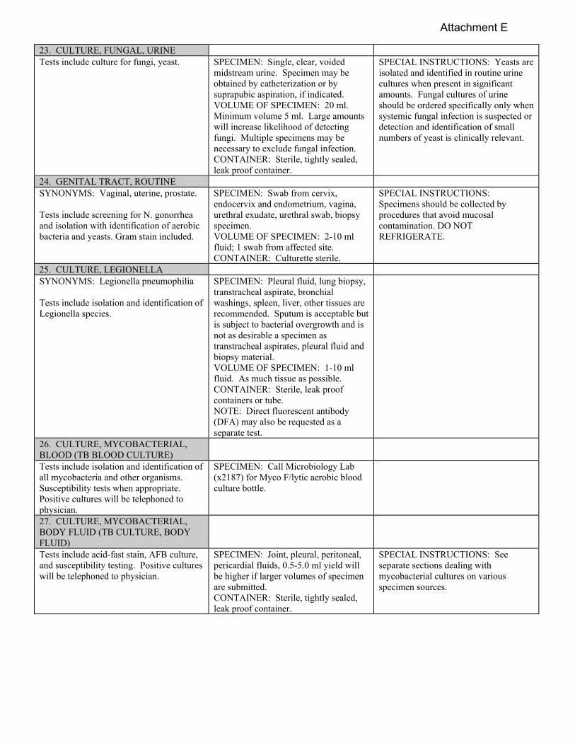
23. CULTURE, FUNGAL, URINE Tests include culture for fungi, yeast. SPECIMEN: Single, clear, voided
midstream urine. Specimen may be obtained by catheterization or by suprapubic aspiration, if indicated. VOLUME OF SPECIMEN: 20 ml. Minimum volume 5 ml. Large amounts will increase likelihood of detecting fungi. Multiple specimens may be necessary to exclude fungal infection. CONTAINER: Sterile, tightly sealed, leak proof container.
SPECIAL INSTRUCTIONS: Yeasts are isolated and identified in routine urine cultures when present in significant amounts. Fungal cultures of urine should be ordered specifically only when systemic fungal infection is suspected or detection and identification of small numbers of yeast is clinically relevant.
24. GENITAL TRACT, ROUTINE SYNONYMS: Vaginal, uterine, prostate. Tests include screening for N. gonorrhea and isolation with identification of aerobic bacteria and yeasts. Gram stain included.
SPECIMEN: Swab from cervix, endocervix and endometrium, vagina, urethral exudate, urethral swab, biopsy specimen. VOLUME OF SPECIMEN: 2-10 ml fluid; 1 swab from affected site. CONTAINER: Culturette sterile.
SPECIAL INSTRUCTIONS: Specimens should be collected by procedures that avoid mucosal contamination. DO NOT REFRIGERATE.
25. CULTURE, LEGIONELLA SYNONYMS: Legionella pneumophilia Tests include isolation and identification of Legionella species.
SPECIMEN: Pleural fluid, lung biopsy, transtracheal aspirate, bronchial washings, spleen, liver, other tissues are recommended. Sputum is acceptable but is subject to bacterial overgrowth and is not as desirable a specimen as transtracheal aspirates, pleural fluid and biopsy material. VOLUME OF SPECIMEN: 1-10 ml fluid. As much tissue as possible. CONTAINER: Sterile, leak proof containers or tube. NOTE: Direct fluorescent antibody (DFA) may also be requested as a separate test.
26. CULTURE, MYCOBACTERIAL, BLOOD (TB BLOOD CULTURE)
Tests include isolation and identification of all mycobacteria and other organisms. Susceptibility tests when appropriate. Positive cultures will be telephoned to physician.
SPECIMEN: Call Microbiology Lab (x2187) for Myco F/lytic aerobic blood culture bottle.
27. CULTURE, MYCOBACTERIAL, BODY FLUID (TB CULTURE, BODY FLUID)
Tests include acid-fast stain, AFB culture, and susceptibility testing. Positive cultures will be telephoned to physician.
SPECIMEN: Joint, pleural, peritoneal, pericardial fluids, 0.5-5.0 ml yield will be higher if larger volumes of specimen are submitted. CONTAINER: Sterile, tightly sealed, leak proof container.
SPECIAL INSTRUCTIONS: See separate sections dealing with mycobacterial cultures on various specimen sources.
Attachment E
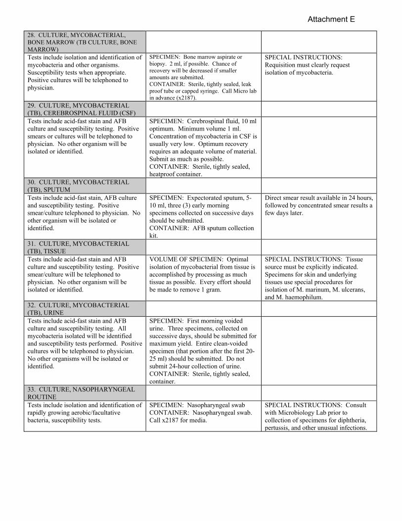
28. CULTURE, MYCOBACTERIAL, BONE MARROW (TB CULTURE, BONE MARROW)
Tests include isolation and identification of mycobacteria and other organisms. Susceptibility tests when appropriate. Positive cultures will be telephoned to physician.
SPECIMEN: Bone marrow aspirate or biopsy. 2 ml, if possible. Chance of recovery will be decreased if smaller amounts are submitted. CONTAINER: Sterile, tightly sealed, leak proof tube or capped syringe. Call Micro lab in advance (x2187).
SPECIAL INSTRUCTIONS: Requisition must clearly request isolation of mycobacteria.
29. CULTURE, MYCOBACTERIAL (TB), CEREBROSPINAL FLUID (CSF)
Tests include acid-fast stain and AFB culture and susceptibility testing. Positive smears or cultures will be telephoned to physician. No other organism will be isolated or identified.
SPECIMEN: Cerebrospinal fluid, 10 ml optimum. Minimum volume 1 ml. Concentration of mycobacteria in CSF is usually very low. Optimum recovery requires an adequate volume of material. Submit as much as possible. CONTAINER: Sterile, tightly sealed, heatproof container.
30. CULTURE, MYCOBACTERIAL (TB), SPUTUM
Tests include acid-fast stain, AFB culture and susceptibility testing. Positive smear/culture telephoned to physician. No other organism will be isolated or identified.
SPECIMEN: Expectorated sputum, 5-10 ml, three (3) early morning specimens collected on successive days should be submitted. CONTAINER: AFB sputum collection kit.
Direct smear result available in 24 hours, followed by concentrated smear results a few days later.
31. CULTURE, MYCOBACTERIAL (TB), TISSUE
Tests include acid-fast stain and AFB culture and susceptibility testing. Positive smear/culture will be telephoned to physician. No other organism will be isolated or identified.
VOLUME OF SPECIMEN: Optimal isolation of mycobacterial from tissue is accomplished by processing as much tissue as possible. Every effort should be made to remove 1 gram.
SPECIAL INSTRUCTIONS: Tissue source must be explicitly indicated. Specimens for skin and underlying tissues use special procedures for isolation of M. marinum, M. ulcerans, and M. haemophilum.
32. CULTURE, MYCOBACTERIAL (TB), URINE
Tests include acid-fast stain and AFB culture and susceptibility testing. All mycobacteria isolated will be identified and susceptibility tests performed. Positive cultures will be telephoned to physician. No other organisms will be isolated or identified.
SPECIMEN: First morning voided urine. Three specimens, collected on successive days, should be submitted for maximum yield. Entire clean-voided specimen (that portion after the first 20-25 ml) should be submitted. Do not submit 24-hour collection of urine. CONTAINER: Sterile, tightly sealed, container.
33. CULTURE, NASOPHARYNGEAL ROUTINE
Tests include isolation and identification of rapidly growing aerobic/facultative bacteria, susceptibility tests.
SPECIMEN: Nasopharyngeal swab CONTAINER: Nasopharyngeal swab. Call x2187 for media.
SPECIAL INSTRUCTIONS: Consult with Microbiology Lab prior to collection of specimens for diphtheria, pertussis, and other unusual infections.
Attachment E

34. CULTURE, SPUTUM, ROUTINE Tests include gram stain, isolation of rapidly growing aerobic and facultative bacteria and yeasts. Only predominant organisms will be completely identified. Susceptibility tests will be performed, if appropriate. Presence/absence of normal upper respiratory flora will be noted.
SPECIMEN: 1-2 ml sputum (not saliva) CONTAINER: Clean, sterile, leak proof container.
SPECIAL INSTRUCTIONS: Single, early morning specimens are recommended. Pooled specimens or 24-hour collections are frequently overgrown with contaminating organisms and will not be accepted. NOTE: Smears are done on all specimens to assess specimen quality (oropharyngeal contamination). If the specimen is saliva and not a deep cough, sputum will be rejected and repeat specimen requested from the ordering location.
35. CULTURE, STOOL, ROUTINE Tests include isolation of Salmonella, Shigella, Campylobacter and E. coli Shiga Toxin. Predominance of Staphylococci, Pseudomonas, Candida or other organisms not usually found will be reported. Presence or absence of normal fecal flora will be noted. NOTE: Other pathogens such as Yersinia and Vibrio must be ordered separately. These are not part of a routine stool culture.
SPECIMEN: 1 gram or 1 ml minimum, solid or liquid fecal material. CONTAINER: Clean, wide-mouth, leak proof container. If liquid stool: Use plastic screw-cap container.
SPECIAL INSTRUCTIONS: Salmonella and Shigella are present in stool in appreciable numbers only during the acute stage (first 3 days of diarrheal disease). Specimen should be collected early in the course of disease and before antimicrobial therapy is begun. Include portions of stool containing pus, blood, or mucous. Hospitalized patients who develop diarrhea when hospitalized and more than 72 hours after admission should be tested for C. difficile.
36. CULTURE, THROAT SYNONYMS: Pharyngitis SPECIMEN: Throat swab, pharyngeal
exudate; 1 swab from affected area. CONTAINER: Sterile culturettes
SPECIAL INSTRUCTIONS: If any of the following are requested, notify the Microbiology Lab: Bordatella pertussis, Corynebacterium diphtheria, Neisseria gonorrhoeae
37. CULTURE, URINE Tests include isolation, quantitation, and susceptibility tests for rapidly growing aerobic/facultative bacteria and yeasts. Only those present in significant numbers will be completely identified. RESULTS: All potential pathogens in significant numbers will be identified to species and will have susceptibility done. The urine cultures with 3 organisms will be reported as such and a sterile repeat collection recommended. No further testing will be done on this culture.
SPECIMEN: Urine, 5-10 ml. SPECIMEN COLLECTION: The patient must be thoroughly instructed for proper collection of “clean catch” specimen. CONTAINER: Sterile container.
SPECIAL INSTRUCTIONS: Must note exact collection method when ordering. The first morning specimen is preferred because the concentration of organisms is higher after overnight incubation in the bladder. Specimens may be obtained by single urethral catheterization only when the patient is unable to void or unable to cooperate in collecting a clean-voided midstream specimen, or when catheterization must be done for diagnostic or therapeutic purposes. Foley catheter tips are not accepted. Indwelling Foley catheters are surrounded by an exudative sheath or urethral secretion that is invariably populated with bacteria. There is no consistent relationship between the organisms found in this exudate and urinary tract infections in patients with indwelling catheters; therefore, these catheters tips are not accepted. Urine for diagnosing urinary tract infections in these patients should be obtained from quantitative culture.
Attachment E

Polymicrobic bacteriuria is not uncommon in patients with chronic indwelling catheters. It is very important that such specimens are sent to the Laboratory with their origin clearly indicated on the requisition form.
38. CULTURE, WOUND, ROUTINE Applies to culture of abscess, decubitus discharge, pus, and ulcers. Tests include gram stain, isolation and identification of rapidly growing, non-fastidious aerobic organisms. Only organisms which predominate will be completely identified. Susceptibility testing, if relevant. Anaerobic cultures must be specifically requested, properly collected, and transported.
CONTAINER: Sterile culturette. SPECIMEN: Pus or other material properly obtained aseptically from the wound site or abscess.
SPECIAL INSTRUCTIONS: Avoid contaminating with skin surface organisms.
39. CULTURE/DIRECT SMEAR FOR GONORRHEA
Tests include isolation and identification of N. gonorrhea only. Isolates tested for production of beta lactamase.
SPECIMEN: Males: Urethral exudate, prostate secretions, rectal swab, throat swab, urine. Females: Urethral exudate, cervical swab, rectal swab, throat swab, drainage from Bartholin’s glands. A separate swab should be submitted from each site sampled. CONTAINER: Sterile culturette.
SPECIAL INSTRUCTIONS: Separate specimen culturettes are required for each body site. Direct smear of exudate on glass slide may be sent for gram stain. DO NOT REFRIGERATE.
40. OVA AND PARASITE EXAMINATION, STOOL
Testing includes an enzyme immunoassay method for the detection of antigens specific for Giardia lamblia, Entamoeba histolytica and Cryptosporidium parvum. Complete ova and parasite examination with trichrome stain will only be performed upon special request.
SPECIMEN: Fresh or freshly frozen un-fixed random stool specimen.. Do not fill the container for liquid stool specimens. Only diarrheal stools will be tested to rule out infection with intestinal parasites. CONTAINER: Submit specimen in clean, air-tight plastic screw cap container.
41. PARASITE EXAMINATION, NON-FECAL SPECIMEN
Applies to aspirates from cyst, liver, lung and lymph nodes, muscle biopsy, rectal biopsy for Schistosomiasis, vagina, urine, etc. Tests include wet mount and stained smear (the latter on fluids or aspirate only)
SPECIMEN: Aspirate, tissue biopsy, body fluid, vaginal aspirate, rectal aspirate, urine. VOLUME: 3-4 ml of body fluids, 1-gram tissue. Sterile screw cap container. INSTRUCTIONS: Deliver specimen to Microbiology immediately. NOTE: Tissue biopsy for Schistosoma ova must be placed in sterile distilled water. Otherwise, tissue should be transported in saline to prevent drying.
Applies to tick identification. SPECIMEN: Tick container – plastic screw cap container
SPECIAL INSTRUCTIONS: Include clinical history such as body site, geographic location, etc.
42. CLOSTRIDIUM DIFFICILE
Attachment E

Includes simultaneous detection of Clostridium difficile antigen and toxins A and B.
SPECIMEN COLLECTION: 25uL fresh or frozen fecal specimens. Swabs are not accepted. Transport to Microbiology as soon as possible. Stool should be diarrheal. CONTAINER: Submit in a clean, plastic screw cap container.
43. CRYPOTOSPORIDIUM DIAGNOSTIC PROCEDURES, STOOL
Tests include examining the stool specimen with modified acid-fast staining for the presence of cryptosporidium.
SPECIMENS AND COLLECTION: A fresh random stool in a stool container with lid and transport the specimen to Microbiology immediately. For liquid specimens, do NOT fill the container.
44. KOH PREPARATION SYNONYM: Fungus smear or scabies. SPECIMEN AND SPECIMEN
COLLECTION: Call Microbiology Lab (x2187)
45. NEISSERIA GONORRHEA SMEAR See Culture/Direct Smear for Gonorrhoeae. SPECIMEN: Smear, air-dried or
culturette of exudate.
46. VIRAL CULTURE Based on specimen sources, viruses to be detected by cytopathic effect and/or antibody staining include adenovirus, CMV, enteroviruses, HSV, influenza, mumps, parainfluenza, RSV and V-Z.
SPECIMEN AND COLLECTION: Transport media for virus culture is available in Microbiology (x2187). State specific virus requested. Blood, CSV, dermal, ocular, genital, mucosal, oral, rectal, respiratory, stool, tissue, urine, biopsy.
47. . INFLUENZA A and B ANTIGEN, PCR
SPECIMEN: Nasopharyngeal Transport media available in Microbiology (x2187).
48. MRSA SPECIMEN: Naris. Do both sides with
one swab. Red top culturette only.
SPECIAL INSTRUCTIONS: Test done on admission and pre-op.
Attachment E

G. URINALYSIS
1. General Information 2. Test Information 3. Specimen Collection and Handling 4. Reference Ranges
Attachment E

G. URINALYSIS
1. GENERAL INFORMATION Urinalysis testing is performed on an automated instrument utilizing fiber optics. 2. TEST INFORMATION a. CHEMISTRIES TESTED BY URINALYSIS DIPSTICK:
Glucose pH Bilirubin Protein Ketone Urobilinogen Specific Gravity Nitrite
Leukocyte Esterase Blood
b. MICROSCOPIC URINE SEDIMENT EXAMINATION:
Microscopic tests on urine sediment will not be performed on urines if dipstick for all urine chemistry tests is negative and specimen is clear.
3. SPECIMEN COLLECTION AND HANDLING During the course of a single day, the composition of urine is constantly changing. Accordingly, various types of urine specimens are used for analysis. a. TYPE OF URINE COLLECTION:
Type of Urine Specimen Collection 1. Random or spot specimen Portion of any voided urine is collected in a clean
container. Majority of urine specimens submitted for urinalysis are random specimens. Composition of random voided specimens may vary widely; however, if rules for handling the specimens are observed, the variance will be minimized.
2. Fasting Specimen Urine is voided four or more hours following ingestion of food and discarded. The next voided specimen is collected.
3. First Morning Specimen Patient voids before retiring and urine is discarded. On arising in the morning, the first voided specimen is collected.
4. Morning Specimen for Routine Urinalysis First morning specimen is discarded to prevent false positive results on urine chemistry and microscopic examination from collection of constituents and formed elements retained in the bladder overnight. Second morning specimen is collected and sent to the Laboratory.
Attachment E

5. Midstream Specimen With a clean collection container at hand, urination is initiated. Without interrupting the process of urination, when approximately half of voiding is completed, a portion of urine is collected in the container. The latter portion of the urine flow is discarded.
6. Clean Catch Specimen For either males or females, the external genitalia are washed using a mild antiseptic solution. Then, midstream urine is collected in a clean sterile container.
7. Random Urine Toxicology Screen Instruct the patient to collect urine in the collection cup and directly hand the specimen to a laboratory employee. The temperature will immediately be taken using an infra-red thermometer by aiming the thermometer at the specimen through the cup and pressing the measurement button. This temperature will be recorded on the urine collection cup. Acceptable range: 32.2 – 37.8oC. The physician will be notified by the laboratory if temperature is outside this range.
b. URINE COLLECTION WITH THE BD VACUTAINER COLLECTION CUP SYSTEM:
The healthcare professional obtains the designated cup for the patient and cautions the patient to NOT remove the cap label to protect against a needle stick from the integrated transfer device.
Urinalysis, C&S and Urinalysis with Reflex: use 8 mLBD Vacutainer with UA Preservative and 4 mL BD Vacutainer with Urine C&S preservative tubes. Transfer the specimen into accompanying evacuated tube by peeling back the cap label to expose the transfer device. Advance the tubes over the puncture device and fill tubes. Once filled, carefully replace the cap label over the transfer device and treat the screw cap as a contaminated sharp. Label all tubes and collection cups appropriately.
Urine Drug Screen: use 10 mL BD Vacutainer with UA No Additive tube. Transfer the specimen into accompanying evacuated tube by peeling back the cap label to expose the transfer device. Advance the tubes over the puncture device and fill tubes. Once filled, carefully replace the cap label over the transfer device and treat the screw cap as a contaminated sharp. Label all tubes and collection cups appropriately.
Diabetic Urinalysis and Microalbumin/Creatinine ratio (MCRT): 8 mLBD Vacutainer with UA Preservative and 6 mL BD Vacutainer with No Additive tubes. Transfer the specimen into accompanying evacuated tube, peel back the cap label to expose the transfer device. Advance the tubes over the puncture device and fill tubes. Once filled, carefully replace the cap label over the transfer device and treat the screw cap as a contaminated sharp. Label all tubes and collection cups appropriately.
c. SPECIMEN AMOUNT: For routine urinalysis, at least 10 mL of urine should be submitted. d. SPECIMEN STORAGE: Specimens for routine urinalysis are stable for 48 hours at room temperature. Specimens for C&S can remain at room temperature for 72 hours.
Attachment E

4. REFERENCE RANGES a. URINALYSIS REFERENCE RANGES (DIPSTICK & MICROSCOPIC): Refer to “Test and Reference Range Chart” (Section IV) for a list of Urinalysis reference ranges.
Attachment E

H. SEROLOGY
1. General Information 2. Test Information 3. Specimen Collection and Handling 4. Reference Ranges
Attachment E
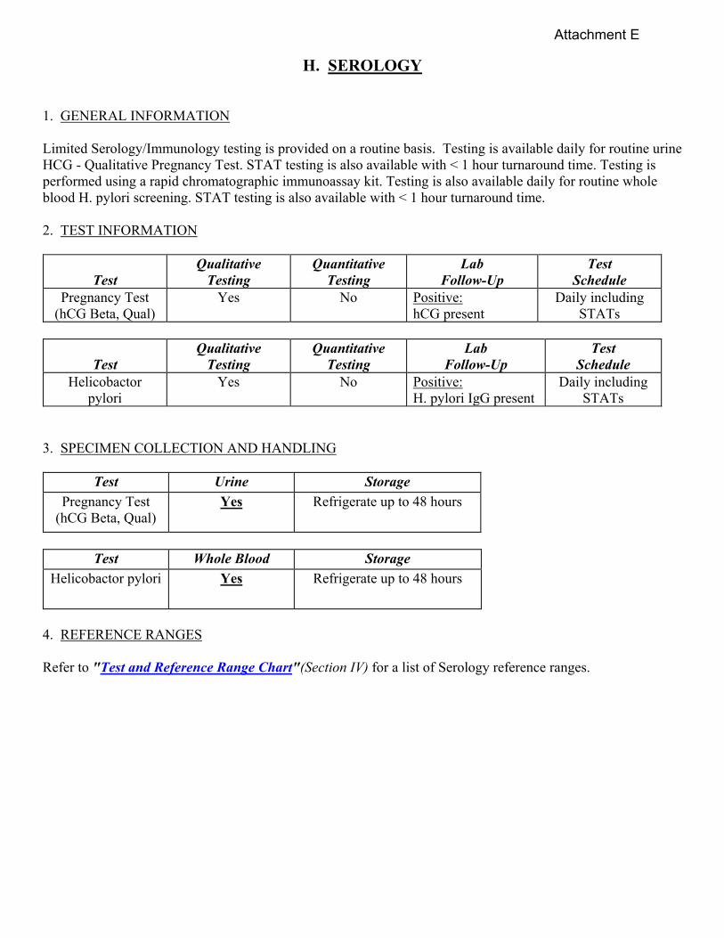
H. SEROLOGY
1. GENERAL INFORMATION Limited Serology/Immunology testing is provided on a routine basis. Testing is available daily for routine urine HCG - Qualitative Pregnancy Test. STAT testing is also available with < 1 hour turnaround time. Testing is performed using a rapid chromatographic immunoassay kit. Testing is also available daily for routine whole blood H. pylori screening. STAT testing is also available with < 1 hour turnaround time. 2. TEST INFORMATION
Test
Qualitative Testing
Quantitative Testing
Lab Follow-Up
Test Schedule
Pregnancy Test (hCG Beta, Qual)
Yes No Positive: hCG present
Daily including STATs
Test Qualitative
Testing Quantitative
Testing Lab
Follow-Up Test
Schedule Helicobactor
pylori Yes No Positive:
H. pylori IgG present Daily including
STATs 3. SPECIMEN COLLECTION AND HANDLING
Test Urine Storage
Pregnancy Test (hCG Beta, Qual)
Yes Refrigerate up to 48 hours
Test Whole Blood Storage
Helicobactor pylori Yes Refrigerate up to 48 hours
4. REFERENCE RANGES
Refer to "Test and Reference Range Chart"(Section IV) for a list of Serology reference ranges.
Attachment E

I. ARTERIAL BLOOD GAS
1. General Information 2. Test Information 3. Specimen Collection and Storage 4. Reference Ranges
Attachment E

I. ARTERIAL BLOOD GAS
1. GENERAL INFORMATION The concentration of pH, pCO2, and pO2 in arterial blood is measured on an automated Blood Gas analyzer located in the Laboratory. The arterial blood specimen is aspirated into the analyzer and passed by a series of sensors. The unknown concentration of PCO2 is obtained by infrared spectroscopy measurement at three wavelengths. The unknown pH is obtained by an optical/color sensitive indicator measurement. A phosphorescence quenching measurement obtains the unknown concentration of PO2. All other results obtained are calculations. The analyzer then prints out these results. 2. TEST INFORMATION
Test Test Schedule pH pCO2 pO2
Available STAT. Seven (7) days a week, 1st shift and 2nd shift. Coverage by 3rd shift as scheduled. Other shifts - Sent out to Hamot Medical Center for testing when coverage not available.
3. SPECIMEN COLLECTION AND STORAGE Arterial Blood Gas specimens are drawn by certified Respiratory Technicians. During Laboratory off duty hours, the AOD draws the Arterial Blood Gas specimens. The Arterial Blood Gas specimens are drawn in lithium heparin syringes. The syringe is sealed and checked to assure no air bubbles are visible. Specimens are brought to the Lab within 30 minutes of collection. Specimens on ice must be tested within two hours of collection. Minimum specimen volume is 0.09 cc. Venous Blood gas specimens may be used as alternative to arterial blood gas when Respiratory Technicians are not in house. Venous Blood Gas specimens are drawn by Medical Technologist. Peripheral venous sample (obtained by venipuncture) using Lithium Heparin tube. *NOTE: pO2 in venous blood is not useful in assessing oxygenation because oxygen has already been extracted by the tissues by the time blood reaches the venous circulation. 4. REFERENCE RANGES Refer to "Test and Reference Range Chart"(Section IV) f or a list of Arterial Blood Gas reference ranges.
Attachment E

J. Blood Bank
1. General Information 2. Test Information 3. Specimen Collection 4. Reference Laboratories 5. Additional Blood Bank MCMs
Attachment E

J. Blood Bank
1. GENERAL INFORMATION The blood bank carries several different blood products that a physician may order depending on the needs of the patient. Primary testing in blood bank includes ABO/Rh typing, antibody screening, antibody identification, direct antiglobulin testing (DAT), and Transfusion Reaction Workup. Testing and blood products can be ordered as Routine, ASAP, or STAT. A detailed description of blood/blood component therapy policies can be found in MCM 113-01 “Blood/Blood Components Transfusion and Management of Suspected Transfusion Reaction”. 2. TEST INFORMATION
Test Test Includes Type To determine a patient’s ABO and Rh type Screen To detect red cell antibodies in addition to anti-A or
anti-B that could cause hemolysis to transfused red cells.
DAT To determine the presence or absence of immunoglobulin on the surface of the patient’s red blood cells.
Transfusion Reaction Workup Ordered by a physician when a suspected adverse reaction has occurred after a patient has received a blood product. Testing to possibly include; Haptoglobin, Urinalysis, DAT, Total and Direct Bilirubin.
3. SPECIMEN COLLECTION
Proper accurate identification of the intended patient, blood sample and blood component(s) are critical to ensure transfusion safety regardless of patient location.
All patients undergoing compatibility testing should be issued a patient identification band that should remain in place throughout the transfusion process.
All blood bank specimens require two approved patient identifiers.
For all blood bank specimens, phlebotomy will be performed only on one patient at a time to minimize the risk of error.
NOTE: A signature of the healthcare provider witnessing the sample collection is only required on the VBECS order printout. Healthcare providers include nursing staff, CRNA, Nurse Practitioners, Physicians and a second Laboratory Medical Technologist.
Attachment E

4. REFERENCE LABORATORIES Tests not performed in-house may be sent to Indiana Blood Center for further workup. The blood bank technologist will phone the appropriate provider to inform them of the delay in testing and when to expect testing to be completed. 5. ADDITIONAL BLOOD BANK MCMS:
MCM 113-07: Blood Warmers MCM 113-08: Therapeutic Phlebotomy MCM 113-16: Reporting of Post Transfusion Diseases MCM 113-28: “Look Back” Program for Blood and Blood Products
Attachment E

K. ANCILLARY TESTING (POINT OF CARE TESTING –
WAIVED TESTING)
1. General Information 2. Approved Ancillary Tests
3 Home Testing 4. Patient Self Testing 5. Additional Ancillary Testing MCMs
Attachment E

ANCILLARY TESTING
(POINT OF CARE TESTING – WAIVED TESTING) 1. GENERAL INFORMATION
The Ancillary Testing (Point of Care Testing, Waived Testing) refers to laboratory testing or services provided within the medical center or its outreach functions (clinics, etc.) but outside the physical facilities of the main clinical laboratory.
The important criteria for Ancillary Testing is that no permanent space is dedicated to the tests
performed. The kits and instruments used are hand-carried or transported to the site of testing. 2. APPROVED ANCILLARY TESTS All Ancillary Testing will be performed in compliance with Joint Commission (JC) standards and Department of Veterans Affairs guidelines. Pathology & Laboratory Medicine (P&LM) has the overall responsibility and oversight of the program. The following tests are being performed at VAMC Erie as Ancillary Testing:
WAIVED TESTS/SITES/INSTRUMENT USED REFERENCE RANGES/CRITICAL VALUES
Ancillary Test Classification Testing Sites
Method Used Reference Ranges
Critical Values
Definitive Tests Bedside Capillary Blood Glucose Testing
Waived *Unit 5 East (unit 6) *Unit 5West (CLC) *Unit 4 *Amb.Surg *UCC *Red Team *White Team *CBOC: -Ashtabula -Crawford -Venango -Warren -McKean
Roche AccuChek Inform II
Meter
70-100 mg/dl non-diabetic
adults
Low <50 mg/dl
High >400 mg/dl
Pregnancy Testing Urine only-Waived
CBOC: -Ashtabula -Crawford -Venango -Warren -McKean
Sure-Vue hCG-STAT
Positive/Negative N/A
Ancillary Test Classification Testing Sites Instrument Used Reference Ranges
Critical Values
Screening Tests Fecal Occult Blood Testing
Waived Test UCC only Occult Blood Slides
Positive/ Negative
N/A
Attachment E
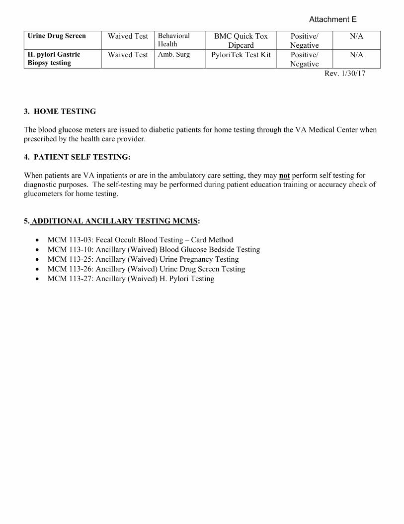
Rev. 1/30/17
3. HOME TESTING
The blood glucose meters are issued to diabetic patients for home testing through the VA Medical Center when prescribed by the health care provider.
4. PATIENT SELF TESTING:
When patients are VA inpatients or are in the ambulatory care setting, they may not perform self testing for diagnostic purposes. The self-testing may be performed during patient education training or accuracy check of glucometers for home testing. 5. ADDITIONAL ANCILLARY TESTING MCMS:
MCM 113-03: Fecal Occult Blood Testing – Card Method MCM 113-10: Ancillary (Waived) Blood Glucose Bedside Testing MCM 113-25: Ancillary (Waived) Urine Pregnancy Testing MCM 113-26: Ancillary (Waived) Urine Drug Screen Testing MCM 113-27: Ancillary (Waived) H. Pylori Testing
Urine Drug Screen Waived Test Behavioral Health
BMC Quick Tox Dipcard
Positive/ Negative
N/A
H. pylori Gastric Biopsy testing
Waived Test Amb. Surg PyloriTek Test Kit Positive/ Negative
N/A
Attachment E

IV. TESTS AND REFERENCE RANGES
Attachment E

TESTS AND REFERENCE RANGES In House Testing: The attached list of Laboratory tests and reference ranges will be updated as necessary
throughout the year. Reference Testing: The reference ranges for Laboratory tests sent to the Reference Laboratory will be
provided with each test.
Attachment E

ERIE VAMC-LABORATORY REFERENCE RANGES September 2018
CHEMISTRY
TEST EXPECTED VALUES Acetone Negative Albumin 3.5 – 5.2 g/dl
Alkaline Phosphatase 40-150 International Units/liter ALT 0-55 International Units/liter
Amylase 25-125 Units/liter Anion Gap 10 – 20 mmol/liter
AST 5-34 International Units/liter Carbon Dioxide (CO2) 22 - 29 mmol/liter
Bilirubin, direct 0.0 – 0.5 mg/dl Bilirubin, total 0.2 – 1.2 mg/dl
BUN Male 19-50years old: 9-20mg/dL Male 51-130years old: 9-25mg/dL Female 19-50years old: 7-18mg/dL
Female 51-130years old: 10-20mg/dL Calcium 8.4-10.2 mg/dl Chloride 98-107 mmol/liter
Cholesterol Low risk: < 200 mg/dl Borderline risk: 201 - 239 mg/dl
High risk: 240 mg/dl CPK Male: 30-200 International Units/liter
Female: 29-168 International Units/liter Creatinine 0.72-1.25 mg/dl (male)
0.57-1.11 mg/dl (female) gamma-GT (GGT) Male: 12-64 International Units/liter
Female: 9-36 International Units/liter Glucose 70-99 mg/dl
Glucose, postprandial ADA Guidelines for Fasting Specimen: 70 – 100 normal
101 – 125 prediabetes > 126 diagnostic for diabetes
HDL cholesterol Low cardiovascular risk > 60 High cardiovascular risk < 40
Iron Male: 65-175 micrograms/dl Female: 50-170 micrograms/dl
LDH 125-220 International Units/liter LDL Cholesterol (calculated) Optimal: Less than 100 mg/dl
Near or Above Optimal: 100 – 129 mg/dl Borderline High: 130-159 mg/dl
High: 160-189 mg/dl Very High: > 190 mg/dl
Attachment E

URINE CHEMISTRY TEST EXPECTED VALUES
Calcium 100 - 300 mg/24 hrs Chloride 110 - 250 mmol/24 hrs
Random urine: No established reference ranges Creatinine 800-2000 mg/24 hrs
Creatinine clearance Age (Years) Males Females 20-30 88-146 81-134
30-40 82-140 75-128 40-50 75-133 69-122 50-60 68-126 64-116 60-70 61-120 58-110 70-80 55-113 51-105
Glucose Random: 1 - 15 mg/dl 24 hr urine: < 0.5 g/24 hr
LDL Cholesterol (direct) Optimal: Less than 100 mg/dl Near or Above Optimal: 100 – 129 mg/dl
Borderline High: 130-159 mg/dl High: 160-189 mg/dl
Very High: > 190 mg/dl
Lipase 8-78 Units/liter Magnesium 1.6-2.6 mg/dl
Osmolality (calculated) 275 – 300 mOsm/kg H2O Phosphorus 2.3 - 4.7 mg/dl Potassium 3.5-5.1 mmol/liter Sodium 136 - 145 mmol/liter
TIBC (calculated) Male: 252 – 461 ug/dl Female: 269 – 535 ug/dl
Total protein 6.4-8.3 g/dl
Alcohol Legal intoxication: 80 mg/dl or greater Flushing, slow reflexes, impaired visual activity: 50-100
mg/dl Depression of CNS: > 100 mg/dl Fatalities reported: > 400 mg/dl
Transferrin Male 14-60 years old: 174-364 mg/dL Male 61-80 years old: 163-344 mg/dL
Female 14-60 years old: 180-382 mg/dL Female 61-80 years old: 173-360 mg/dL
Triglycerides Normal: < 150 mg/dl Borderline high: 150 – 199 mg/dl
High: 200 – 500 mg/dl Very high: > 500 mg/dl
Uric acid 3.5-7.2 mg/dl (male) 2.6-6.0 mg/dl (female)
Attachment E

Microalbumin Random urine: > 1.9 mg/dl 24 hr urine: Normal: less than 30
30-300 = microalbuminuria >300 suggests macroalbuminuria
Microalbumin/creatinine ratio > 30 micrograms/mg creatinine Potassium 25 - 125 mmol/24 hr
Random urine: No established reference ranges Protein (quantitative) Random urine: < 10 mg/dl
24 hr urine: 50 - 100 mg/24 hrs Sodium 40 - 220 mmol/24 hr
Random urine: No established reference ranges SPECIAL CHEMISTRY
TEST EXPECTED VALUES B12 213-816 pg/ml
CPK-MB 0.0 – 6.6 ng/ml
Ferritin Male: 21.81-274.66 ng/ml Female: 4.63-204.00 ng/ml
PSA <4.0 ng/ml
TSH 0.35-4.94 micro-International units/ml
Folate 0-20 ng/mL
%Free PSA 50-64 yrs 65-75 yrs 0.00-10.00% 56% 55% 10.01-15.00% 24% 35% 15.01-20.00% 17% 23% 20.01-25.00% 10% 20% >25.00% 5% 9%
Free T4 0.7 – 1.48 ng/ml
Troponin I 0-0.029 ng/ml Beta hCG Non-Pregnant Individuals: <5 mIU/mL
May be Indicative of Early Preg: 5-25mIU/mL Positive: >25 mIU/mL
Hemoglobin A1c 4.4-5.9%
THERAPEUTIC DRUG MONITORING (TDM)
TEST THERAPEUTIC
RANGE
TOXIC RANGE
DRAW TIME Theophylline 8-20 micrograms/ml > 20 micrograms/ml ½ - 1 hr before dose
Phenytoin 10-20 micrograms/ml > 20 micrograms/ml ½ - 1 hr before dose Phenobarbital 15-40 micrograms/ml > 40 micrograms/ml ½ - 1 hr before dose
Digoxin 0.8-1.5 ng/ml (congestive heart failure)
1.5-2.0 ng/ml (arrhythmias)
> 2.5 ng/ml ½ - 1 hr before dose
Vancomycin 20-40 ug/ml >80 ug/ml ½ - 1 hr before dose
Attachment E

HEMATOLOGY
TEST EXPECTED VALUES WBC 4.2 - 9.1 thou/microliters RBC Males: 4.6 – 6.1 mil/microliters
Females: 3.9 – 5.2 mil/microliters HGB Males: 13.7 – 17.5 g/dl
Females: 11.2 – 15.7 g/dl HCT Males: 40.1 – 51.0%
Females: 34.1 – 44.9% PLT 163 - 337 thou/microliters MCV Males: 79 – 92.2 fl
Females: 79.4 – 94.8 fl MCH 25.6 – 32.2 pg
MCHC 32.3 – 35.5 g/dl RDW 11.6 – 14.4 % MPV 9.4 – 12.4 fl
ESR (Sedimentation Rate) Males Under 50 years: < 15 mm/h
50-85 years: < 20 mm/h Over 85 years: < 30 mm/h
Females Under 50 years: < 20 mm/h
50-85 years: < 30 mm/h Over 85 years: < 42 mm/h
Reticulocyte Count 0.51– 1.81 %
MANUAL DIFFERENTIAL Neutrophils 40-75%
Lymphocytes 20-44% Monocytes 2-10% Eosinophils 0-6% Basophils 0-2%
Bands 2-6% Atypical lymphocytes 0-8%
Metamyelocytes 0 Myelocytes 0
Promyelocytes 0 Blasts 0
NRBCs/100 WBCs 0
COAGULATION TEST EXPECTED VALUES
PT 12.2 – 14.9 sec INR 0.9 – 1.1
INR – oral therapy 2-3.5 PTT 23.1 – 34.2 sec
Heparin Therapeutic Range (PTT) 72– 116 sec
Attachment E

URINALYSIS Color Colorless or yellow
Appearance Clear or hazy Sp. Gravity 1.010 - 1.025
Ph 5 – 8.5 Protein Negative - Trace Glucose Negative Ketone Negative
Bilirubin Negative Blood Negative Nitrite Negative
Urobilinogen Negative Leuk Esterase Negative
Microscopic WBC 0 - 5/HPF RBC 0 - 3/HPF
Epithelial Squamous only Mucous 1+ Bacteria Trace or <1+
Yeast None Casts 0 – 5 hyaline/LPF
Crystals Few oxalates Few amorphous urates or phosphates
SEROLOGY
HCG-B Qualitative Negative
ARTERIAL BLOOD GAS PH 7.350 - 7.450
PCO2 35.0 - 45.0 mmHg PO2 80.0 - 100 mmHg
HCO3a 20.0 - 26.0 mmol/liter BE -3.0 - 3.0mmol/l SO2 95.0 - 100.0 %
TEMP 98.6oF
Attachment E
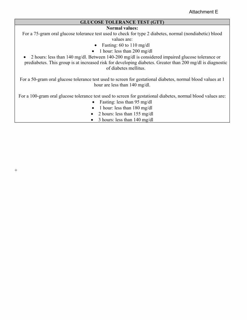
GLUCOSE TOLERANCE TEST (GTT) Normal values:
For a 75-gram oral glucose tolerance test used to check for type 2 diabetes, normal (nondiabetic) blood values are:
Fasting: 60 to 110 mg/dl 1 hour: less than 200 mg/dl
2 hours: less than 140 mg/dl. Between 140-200 mg/dl is considered impaired glucose tolerance or prediabetes. This group is at increased risk for developing diabetes. Greater than 200 mg/dl is diagnostic
of diabetes mellitus.
For a 50-gram oral glucose tolerance test used to screen for gestational diabetes, normal blood values at 1 hour are less than 140 mg/dl.
For a 100-gram oral glucose tolerance test used to screen for gestational diabetes, normal blood values are:
Fasting: less than 95 mg/dl 1 hour: less than 180 mg/dl 2 hours: less than 155 mg/dl 3 hours: less than 140 mg/dl
+
Attachment E

ANATOMIC PATHOLOGY
Ritu Khera, MD Pathologist Ext. 2177 Gail Patterson Histotechnologist/ Lisa Miller Cytotechnologist Ext. 2189 Erin Skelly Carol Kendra Program Assistant Ext. 2176 Anatomic Pathology Reports Ext. 2176
GENERAL INFORMATION
1. The Anatomic Pathology Service includes the following sections: SURGICAL PATHOLOGY CYTOPATHOLOGY AUTOPSY PATHOLOGY 2. The hours of operation are 8:00 AM to 4:30 PM Monday through Friday. The Histotechnologist is available from 3PM to 11PM. Back-up technologists provide histotechnology coverage when the primary Histotechnologist is not available. The Pathologist is available 6:00AM to 10:00AM.
Attachment E

VI. SURGICAL PATHOLOGY
Specimen Submission and Handling
Specimen Procurement and Transport
Criteria for Acceptance/Rejection of Tissue Specimens
Instructions for Specimen Procurement: ♦Routine Specimens ♦Specimens Requiring Special Handling
- Breast Tissue for Estrogen and Progesterone Receptor Assay - Muscle Biopsy - Open Lung Biopsy Procedure - Lymph Node Biopsy - Testicular Biopsy for Infertility - Bone Marrow Aspiration and Biopsy - Cytogenetic Studies for Bone Marrow or Peripheral Blood
- Immunophenotyping/Flow Cytometry
Link to Cytology Index
Attachment E

INSTRUCTIONS FOR SPECIMEN PROCUREMENT AND FIXATION
This includes instructions for:
I. ROUTINE SPECIMENS
II. SPECIMENS REQUIRING SPECIAL HANDLING
Attachment E

INSTRUCTIONS FOR SPECIMEN PROCUREMENT (continued)
I. ROUTINE SPECIMENS
1. The routine small surgical specimens should be submitted in Safe-Fix preservative. The specimens should contain at least 20 times the volume of specimens. 2. The large specimens should be placed in adequate 10% neutral buffered formalin. NOTE: DO NOT PLACE VERY SMALL OR LARGE SPECIMENS IN SALINE as it may distort the histology of the specimen. Also, undue manipulation of the specimen may cause tissue damage and hinder interpretation of the pathologic changes.
SPECIMEN INSTRUCTIONS Small specimens, e.g., teeth, biopsies, appendix, hernial sacs, TUR, etc.
Submit in Safe-Fix preservative 20 times the volume of specimen.
Large specimens, e.g., colon resection, mastectomy, radical prostatectomy, lung resection, etc.
Submit in 10% Neutral Buffered Formalin
II. SPECIMENS REQUIRING SPECIAL HANDLING
The following specimens require special handling: 1. Breast tissue for Estrogen and progesterone assay 2. Muscle biopsy 3. Open lung biopsy 4. Lymph node biopsy 5. Testicular biopsy for infertility 6. Cytogenetics studies 7. Flow Cytometry Notify the pathologist and/or histotechnologist prior to any of the above named surgical procedures.
Attachment E

VA Medical Center, Erie, Pennsylvania Pathology & Laboratory Medicine
SURGICAL PATHOLOGY
SPECIMEN SUBMISSION AND HANDLING ALL tissue specimens and foreign objects removed from patients must be promptly submitted to Anatomic Pathology section of Pathology & Laboratory Medicine (P&LM) for identification and gross/microscopic examination when indicated. The medicolegal significance of properly documenting the nature of the specimen, in addition to the surgeon’s operative findings, in the patient’s record cannot be over-emphasized. MCM 113-23, “Guidelines for Surgical Specimen Submission” outlines the guidelines for specimen submission and categories of surgical specimens that usually require a gross description and diagnosis and are exempted from mandatory microscopic description. SPECIMEN PROCUREMENT AND TRANSPORT The specimen procurement encompasses all activities from surgical removal of the specimen to its acceptance by the Histology section of the Laboratory. It involves close cooperation among the surgeon, operating room nursing staff, clerical/technical staff of the Histology Laboratory and the pathologist. The initial responsibility for specimen handling including preservation and labeling, lies with the submitting physician and the assistant. The procurement of a specimen for histological evaluation consists of the following elements:
A. Correct identification and integrity of identification. B. A complete surgical pathology requisition. C. Fixation or special handling appropriate to the specimen. D. Prompt delivery of the specimen to the Laboratory.
1. CORRECT IDENTIFICATION OF SUBMITTED SPECIMENS: The correct identification and
integrity of identification from specimen removal to accessioning within the Laboratory are essential and each part of the specimen must be properly identified at the time of the procedure. The proper identification MUST be on the container and should include:
- Patient’s name and social security number. - Date obtained - Organ/tissue site.
This identifying information on the container must match the information on the specimen requisition form. In general, the identification should be placed on the body of the container holding the specimen, rather than on the lid, as the latter may be inadvertently transferred. Standard safety precautions should be followed in handling all specimens; however, if a specimen presents a known or suspected biohazard, the container should be so marked.
2. COMPLETED SURGICAL PATHOLOGY REQUISITION: A properly completed specimen
requisition form must accompany all specimens and all identifying information on the requisition form must match that on the specimen container. If the specimen presents a known or suspected biohazard, this information should be included on the requisition form. The patient identification data should be correct and
Attachment E

legible and compared to patient’s wristband, prior to submission to the Laboratory. The requisition form should include the following information:
Full identification of the patient (including name, sex, date of birth and social security number). Name of submitting physician. Type and source of specimen (organ/tissue). Date/time when the specimen was obtained. Pertinent clinical information Pre- and postoperative diagnosis filled in by the submitting physician or designee.
Adequate history is essential for the most accurate pathologic diagnosis and its absence may inconvenience a number of people by necessitating telephone calls in addition to delaying the pathology report.
3. FIXATION OR SPECIAL HANDLING APPROPRIATE TO THE SPECIMEN: For routine specimens appropriate fixative is required for each specimen and will be available to the submitting physicians. The exceptions to routine fixation, e.g. frozen sections, cases with suspected infection or lymphoma or specimens requiring special procedures/handling should be communicated by the operating room to the laboratory staff in advance for specific specimen submission requirements. Refer to “Instructions for Specimen Procurement and Fixation” section for specific information.
4. PROMPT DELIVERY OF THE SPECIMEN TO THE LABORATORY
A. After the specimens are removed from the patient, they should be placed in a clean, leak-proof container with secure closure. Before being transported to the laboratory, the primary container should be placed into a sealable secondary container, i.e., plastic zip-lock specimen transport bags, which are labeled with the biohazard symbol. Specimens too large to be contained in the zip-lock bag will be placed in a red bag with a biohazard label attached. The following precautions should be taken during specimen handling and transportation:
The outside of the primary container and zip-lock specimen transport bag must not be visibly
contaminated. Laboratory requisition slips should be protected from visibly contaminated material and, if
necessary, separated from the primary container. Personnel who transport specimens should be trained in safe handling practices and decontamination
procedures in case of spill. Laboratory personnel should examine all specimens for visible contamination before opening the
specimen containers. The outside of the contaminated containers should be cleaned with hospital approved disinfectant solution before sending to the work areas for testing. Laboratory requisitions, visibly contaminated, should be discarded and replaced.
Laboratory personnel will initial laboratory requisition and record the number of specimens brought to the Lab.
B. All specimens should be properly labeled in the operating room or any other location where the
procedure is performed. The specimens should preferably be delivered to the laboratory as soon as qualified personnel obtain them. The late, weekend and emergency surgical specimens should remain in the surgical suite (operating room) and be delivered to the laboratory the next working day after 8:00AM. The histotechnologist will check specimens received against those expected by reviewing the daily surgery schedule. When anticipated specimens are not received, prompt inquiry will be initiated to avoid lost or misplaced specimens.
Attachment E

CULTURES If there is indication that microorganisms may be responsible for the pathologic changes, appropriate material for smears and cultures of these specimens should be obtained. Such material is best obtained in the Operating Room under sterile conditions rather than in the Laboratory. Specimens for micro-organism/cultures should not be included with the Surgical Pathology specimens, or included on the Surgical Pathology Requisition form. Specimens for Microbiology MUST be submitted to the Laboratory separately accompanied by an appropriate laboratory order. TISSUE IMPRINTS In certain instances, particularly in suspected lymphomas, make touch imprints of the lymph node or tumors to better determine cell type. If imprints from any tissue are desired, please notify the pathologist and/or histotechnologist. These imprints must be properly made, quickly air-dried, labeled, and submitted to the Laboratory for further processing along with the specimen. SPECIAL STAINS ON HISTOLOGIC SLIDES The decision whether special stains or other procedures might be helpful in arriving at a definitive diagnosis rests with the pathologist. In cases where examination of tissues for specimen substances (i.e., amyloid) or specimen disease conditions is desired, contact the pathologist or so indicate on the specimen form. Special stains are time consuming, expensive, and should not be ordered indiscriminately. CALCULUS Calculi sent for analysis will be sent to a reference laboratory. OTHER SPECIAL PROCEDURES The histochemical procedures, immunofluorescent and immunoperoxidase techniques are also
available. These should be limited to selected cases where they are required for definitive diagnosis. It takes approximately 1 to 3 weeks to receive the report.
The special procedures in certain instances require special handling when removed from the patients. In all such instances, the pathologist and/or histotechnologist should be informed well ahead of the planning procedure.
Attachment E

VA Medical Center, Erie, Pennsylvania Pathology & Laboratory Medicine
CRITERIA FOR ACCEPTANCE/REJECTION OF TISSUE SPECIMENS
The specimen procurement and transport require systems of positive identification of specimens, integrity of identification throughout the procurement process, provision of demographic and clinical information, appropriate fixation, and prompt delivery of specimens to the Laboratory. The criteria of acceptance/rejection of tissue specimens is as follows: I. INCORRECT, IMPROPER, INCOMPLETE IDENTIFICATION OR LABELING The technician who accessions tissue specimens will not accept specimens that are inappropriately or incompletely labeled or without a proper accompanying specimen requisition in accordance with requirements listed for specimen submission. When a specimen has incorrect/improper/incomplete identification, the originating site will be notified. The person correcting the labeling error will need to accept responsibility and complete the “Mislabeled Specimen Responsibility” form. For specimens submitted without requisition forms, the originating site will be notified. A timely follow up will be done by Histology Section to assure proper re-submission. NOTE: Due to uniqueness of most surgical pathology specimens, these will NOT be rejected until an exhaustive attempt has been made to correct the deficiency. II. INADEQUATE OR IMPROPER PRESERVATION (FIXATION) Refer to section on "Instructions for Special Procurement". Specimens submitted in improper, inadequate, or without fixative will be documented and the Operating Room/Outpatient Clinic notified. III. LEAKING CONTAINER AND/OR SOILED REQUISITIONS The Operating Room/Outpatient Department will be notified. If the requisition form is soiled, the Operating Room/Outpatient Clinic will be requested to provide a replacement. The rejected requisition will be disposed of with biohazard waste. NOTE: The specimens will not be processed unless any identified deficiencies are corrected.
Attachment E
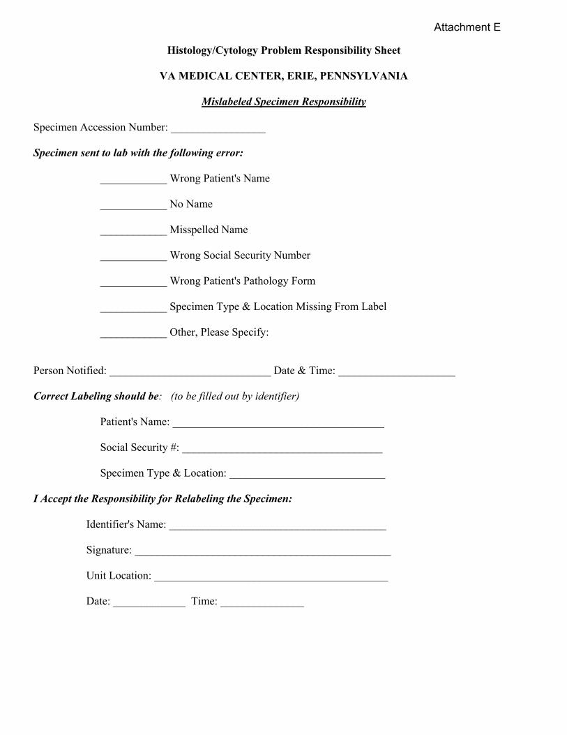
Histology/Cytology Problem Responsibility Sheet
VA MEDICAL CENTER, ERIE, PENNSYLVANIA
Mislabeled Specimen Responsibility Specimen Accession Number: _________________ Specimen sent to lab with the following error: ____________ Wrong Patient's Name ____________ No Name ____________ Misspelled Name ____________ Wrong Social Security Number ____________ Wrong Patient's Pathology Form ____________ Specimen Type & Location Missing From Label ____________ Other, Please Specify: Person Notified: _____________________________ Date & Time: _____________________ Correct Labeling should be: (to be filled out by identifier) Patient's Name: ______________________________________ Social Security #: ____________________________________ Specimen Type & Location: ____________________________ I Accept the Responsibility for Relabeling the Specimen: Identifier's Name: _______________________________________ Signature: ______________________________________________ Unit Location: __________________________________________ Date: _____________ Time: _______________
Attachment E

BREAST TISSUE FOR ESTROGEN AND PROGESTERONE RECEPTOR ASSAY
SPECIMEN REQUIREMENTS AND HANDLING 1. The pathologist or the histotechnologist should be notified at least 24 hours in advance so that arrangements
can be made for handling the specimen (ext. 2176 or 2189). 2. If the breast tissue is determined to be malignant at the time of the frozen section, the specimen should be
fixed in 10% neutral-buffered formalin within 20 minutes of collection. Optimum fixation time is 12-24 hours, not to exceed 48 hours.
3. After fixation, appropriate sections of the tumor will be submitted for paraffin embedding. 4. The formalin-fixed, paraffin-embedded tumor tissue (1 paraffin block) will be sent to the Reference
Laboratory for estrogen/progesterone receptors immunohistochemical assay. METHODOLOGY The ER/PR immunohistochemical staining is a semi quantitative measurement that detects ER/PR positive tumor nuclei within sections from formalin-fixed, paraffin-embedded tissues. Because of the recent advances in the production of monoclonal antibodies and in antigen-retrieval methods, the paraffin materials methodology demonstrate good concordance with biochemical and immunocytochemical assays in frozen tissues. The immunohistochemical detection of ER/PR offers advantages over cytosolic methods.
●Less tumor tissue is required for ER/PR evaluation. ●Exact location of ER/PR positive cells can be determined (i.e., tumor cells versus normal breast tissue).
The presence or absence of positive internal control staining will be indicated on all negative reports to rule out false negative results. CLINICAL SIGNIFICANCE The clinical significance for the measurement of ER/PR receptors is patient prognosis and response to adjuvant endocrine therapy. Studies have shown that >50% of patients with receptor positive tumors will respond to some type of hormonal treatment whereas <10% of patients with receptor-negative tumors will have a response. REFERENCES:
1. Battifora H, et al: Estrogen receptor immunohistochemical assay in paraffin-embedded tissue: A better gold standard. Appl. Immunohistochem 1:39-45, 1993.
2. Aasmundsted TA, et al: Oestrogen receptor analysis: correlation between enzyme immunoassay and immunohistochemical methods. J. Clin Pathol 45:125-129, 1992.
3. DeNegri F. et al: Comparison of monoclonal immunocytochemical and immunoenzymatic methods for steroid receptor evaluation in breast cancer. Am J. Clin Pathol 96:53-58, 1991.
Attachment E

MUSCLE BIOPSY The histological study of skeletal muscle requires great care in obtaining and preparation of the specimen to assure maximum diagnostic information. The pathologist of the Histology section of the Laboratory should be informed at least 24 hours in advance so that arrangements can be made for handling the specimens. INDICATIONS 1. Diagnosis and classification of muscle disease. 2. Distinguish myopathic and neuropathic disorders. 3. Diagnosis of certain systemic diseases. 4. Monitor therapy in some myopathies. 5. Aid in genetic counseling. BIOPSY SITE A muscle believed to be actively involved but not to a severe degree is preferred. In recent myopathy, select the most impaired muscle. In chronic myopathy, select the less impaired muscle. The biceps and vastus lateralis are most often used. AVOID SAMPLING 1. Muscles used for EMG or intramuscular injections. 2. Muscles subjected to regular trauma (deltoid, abdominal, gluteus, gastrocnemius). 3. Site of origin and insertion of the muscle. SPECIMEN Two (2) fresh muscle biopsy specimens at least 2 cm long in the direction of muscle fibers and 1 cm in diameter is adequate. Do NOT moisten the specimen with saline. Place the specimen in a STERILE container and deliver it to the Histology Section of the Laboratory immediately. CLINICAL INFORMATION The specimen must be accompanied by a completely filled out Surgical Pathology request form. The information must be detailed and should include:
1. Anatomic site of muscle biopsy. 2. Clinical history or differential diagnosis.
- Age, sex and family history - Onset and rate of progression - Distribution of weakness - Other systemic disease
3. Serum enzymes – total CPK value. 4. Electromyography and nerve conduction studies.
SPECIMEN HANDLING The histotechnologist will place the sterile container with unfrozen specimen in a second container with ice (NOT DRY ICE). The specimen containers with clinical information will be shipped FedEx to Muscle Histochemistry and Electron Microscopy Laboratory, VAMC Little Rock, Arkansas.
Attachment E

OPEN LUNG BIOPSY PROCEDURE An open lung biopsy is useful in selective cases. The following guidelines should be followed for properly obtaining and handling the specimen:? 1. Notify the Laboratory in advance so that preparation may be made to process the material with appropriate studies. The biopsy should preferably be done early in the day to assure prompt performance of the necessary procedures. 2. Individual Laboratory tags should be made out beforehand for any of the following studies that may be considered appropriate:
a. Surgical Pathology b. Gram stain (routine and for Legionnaire’s disease, if indicated) c. Acid fast stain and culture. d. Routine aerobic and anaerobic culture e. Culture for Legionnaire’s disease. f. Tissue imprints for direct fluorescent antibody test for Legionella and other organisms (specify)
3. The biopsy specimen should be of sufficient size (4 x 3 x 3 cm is recommended). 4. The entire excised specimen must be placed in a DRY STERILE wide-capped container and immediately delivered to the Laboratory. 5. Appropriate examination will be made by the pathologist.
Attachment E

LYMPH NODE BIOPSY In suspected lymphomas, the lymph node biopsies require special handling. The pathologist or the Histology section of the Laboratory should be notified at least 24 hours in advance so arrangements can be made for handling the specimen. SPECIMEN: The lymph node biopsy specimen should be wrapped in wet saline gauze and delivered IMMEDIATELY to the Histology Section of the Laboratory. The specimen must be accompanied by a completely filled out surgical pathology request form including pertinent clinical history and any other information. PROCESSING 1. Make 10 imprints from unfixed, freshly cut section. 2. Place a portion of the lymph node in 10% zinc formalin. 3. Section the rest of the lymph node 3 to 5 mm in thickness and place in 10% zinc formalin fixative.
Attachment E

TESTICULAR BIOPSY FOR INFERTILITY
The testicular biopsy for clinical evaluation of male infertility requires special handling. SPECIMEN: 1. Notify the histotechnologist in advance so that arrangements can be made for handling the specimen. 2. Place the specimen in Bouin's fixative and deliver to the Histology section of the Laboratory. The specimen must be accompanied by a completely filled out Surgical Pathology requisition form including pertinent clinical history and any other information.
Attachment E

BONE MARROW ASPIRATION AND BIOPSY
AVAILABILITY: The histotechnologist/medical technologist will assist the physician during the bone marrow procedure(s). The routine procedures should be scheduled with the Histology Section of the Laboratory, preferably between 8AM to 12Noon, Monday through Friday. The indications for cultures, special orders and any additional studies should be indicated at that time so appropriate arrangements can be made. The request form must be completed including pertinent clinical findings and accompany the specimen. SPECIMEN - Aspirate smears
- Core biopsy - Touch preparations are made if no aspirate - Material for special studies (cultures, etc.) if indicated
The smeared slides are prepared at the bedside of where the procedure is being done. The biopsy and clot is placed in a fixative. STAINING: The aspirate smears are stained with Wright’s stain. The H&E stain is done on the marrow clot and core biopsy. The other special stains are performed if indicated. IRON STAIN: The iron stain is performed on bone marrow smeared slides and aspirate. Note: The bone marrow biopsy is not a preferred specimen for iron stain because when a bone biopsy specimen is decalcified, "leaching” of iron may occur so that the bone biopsy may be negative for iron while aspirate smear is positive. The iron stain is not done if no marrow is obtained (dry tap) or if there are no marrow particles on smears. Interpretation: The iron stain is a semiquantitation of bone marrow iron stores. It is a sensitive test for evaluation of iron reserve and aids in diagnosis of iron deficiency and sideroblastic anemia. BONE MARROW EXAMINATION AND INTERPRETATION: The bone marrow examination is useful in interpreting the bone marrow morphology, hematopoiesis, myeloid/erythroid ratio, megakaryocytes, myelopoiesis, cellularity and marrow iron stores. Marrow histology is helpful in staging malignancies, i.e., lymphoid malignancies and metastatic tumor. The bone marrow examination REPORT includes the description of marrow as follows: 1. Peripheral Blood Smear and Blood Count Results Report:
This should include the following: - Morphologic assessment of red cell series including a statement about measure indices (if
known). - Quantitative and morphologic assessment of white cells - Quantitative and morphologic assessment of platelets - Other features (rouleaux, parasites, etc.)
2. Aspirate Smear Report: This report should include the following:
- Adequacy of specimen - Morphologic assessment of all lines (maturation abnormalities, leukemic cell features, etc.) - Differential count (if necessary) - M:E ratio
Attachment E
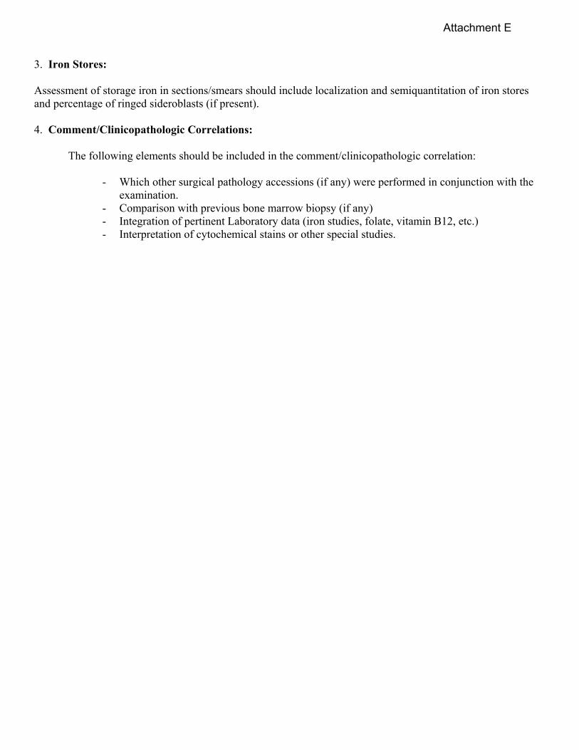
3. Iron Stores: Assessment of storage iron in sections/smears should include localization and semiquantitation of iron stores and percentage of ringed sideroblasts (if present). 4. Comment/Clinicopathologic Correlations:
The following elements should be included in the comment/clinicopathologic correlation:
- Which other surgical pathology accessions (if any) were performed in conjunction with the examination.
- Comparison with previous bone marrow biopsy (if any) - Integration of pertinent Laboratory data (iron studies, folate, vitamin B12, etc.) - Interpretation of cytochemical stains or other special studies.
Attachment E

CYTOGENETICS STUDIES
Availability: The specimen(s) for bone marrow/peripheral blood cytogenetics studies should be preferably collected Monday through Friday and sent to LabCorp Reference Laboratory. If cytogenetics studies are needed on a tissue biopsy, the Histology Section of the P&LM must be contacted in advance so arrangements could be made with LabCorp for supply of Lymph Node Transport Kit. Specimen Collection/Storage
Cytogenetics Specimen Requirements Specimen Storage Instructions
Chromosome Analysis Lymphoma/Leukemia. Hematological disorders such as myelodysplasia, acute and chronic leukemia, lymphoma or myeloproliferative disease.
Bone marrow aspirate – 2 ml Peripheral blood – 3 ml Tissue biopsy – 0.5-1 cm (to be
submitted in Lymph Node Transport Kit) NOTE: If bone marrow aspirate is successful, it is not necessary to send the peripheral blood. Blood for a hematological study should be sent only as a substitute for poor or unobtainable bone marrow aspirate.
Maintain specimen at room temperature.
CML Profile (Chromosomes and DNA)
Bone marrow aspirate – 2 ml OR Peripheral blood – 3 ml
Maintain specimen at room temperature.
Chromosome analysis (karyotyping) For suspected constitutional abnormality, sex chromosome determination, etc.
Peripheral blood – 5-10 ml Maintain specimen at room temperature. Specimens may be refrigerated if delay in transport. Do NOT freeze.
Chromosome analysis For solid tumor or tissue biopsies.
Tissue biopsy/solid tumor – 0.4-1.0 cm specimen (to be submitted in Lymph Node Transport Kit)
Maintain specimen at room temperature.
Special Instructions Include medical findings, tentative diagnosis and treatment on the request form as appropriate. Transport of Specimens to Reference Laboratory The specimens will be picked up by the LabCorp courier and transported to reference laboratory. Clinical Significance Cytogenetic analysis can identify numerical and structural chromosomal abnormalities, which are diagnostic and/or prognostic of some types of leukemias/lymphomas/other tumors, and evaluate other suspected chromosomal disorders.
Attachment E

IMMUNOPHENOTYPING/FLOW CYTOMETRY
Availability The specimen for immunophenotyping/flow cytometry should be preferably collected Monday through Friday and sent to LabCorp reference laboratory. Specimen Collection:
Immunophenotyping/ Flow Cytometry
Specimen Requirements Storage Instructions
Acute Leukemia Profile (myeloid process). Test profile includes the following markers: CD2, CD3, CD7, CD10, CD11b, CD13, CD14, CD15, CD19, CD20, CD33, CD34, CD45, CD71, CD117, HLA-DR. Supplemental markers may be added as determined by pathologist (CD1a, CD4, CD5, CD8, CD61, lycophorin A, TdT).
Fresh bone marrow aspirate – 2 ml collected in sodium heparin tube (green top).
Peripheral blood – 3 ml collected in EDTA tube (lavender top).
CBC
Maintain specimen at room temperature and submit using bone marrow specimen transport kit.
Chronic Lymphocytic Leukemia/Lymphoma Profile (lymphoid process) Test profile includes the following markers: CD2, CD3, CD4, CD5, CD7, CD8, CD10, CD11c, CD19, CD20, CD22, CD23, CD25, CD33, CD38, CD45, CD56, FMC-7, kappa and lambda immunoglobulin light chains. Additional markers may be added as determined by pathologist (CD71, CD138, TCR alpha/beta, TCR gamma/delta).
Fresh bone marrow aspirate – 2 ml collected in sodium heparin tube (green top).
Peripheral blood – 3 ml collected in EDTA tube (lavender tube).
CBC
Maintain specimen at room temperature and submit using bone marrow specimen transport kit.
Transport of Specimens to Reference Laboratory The specimens will be picked up by LabCorp courier and transported to reference laboratory. Clinical Significance The immunophenotyping/flow cytometry is useful in diagnosis and in determining prognosis of hematologic malignancies as well as in evaluating residual/relapse disease in conjunction with cytomorphology and histologic features of each case.
Attachment E

Acute leukemia profile is useful in lineage assessment in acute leukemia for selecting appropriate therapy and assessing prognosis. It is also useful in distinguishing lymphoid from myeloid disorders.
Chronic lymphocytic leukemia/lymphoma profile is useful to identify and characterize the following:
- Reactive lymphocytosis vs. chronic lymphocytic leukemia (CLL) - Prolymphocytic leukemia vs. lymphoblastic leukemia: large granular lymphocyte proliferations,
T-gamma lymphoproliferative disease, natural killer cell proliferations, T-cell CLL, T-cell gamma/delta proliferations.
- Sezary syndrome - Non-Hodgkin’s lymphoma - Adult T-cell leukemia/lymphoma
Attachment E

VII. CYTOPATHOLOGY
Attachment E

VII. CYTOPATHOLOGY
General Information
Specimen Procurement Policy
Specimen Collection and Preservation - General Information
Instructions for Collection/Preparation of Specimens
Instructions for Collection/Preparation of Specimens from Various Body Sites Gynecologic Specimens (Female Genital Tract) Non-Gynecologic Specimens Respiratory Tract Gastrointestinal Tract
Urinary Tract Body Fluids Cerebrospinal Fluid Cytology Scrapings Nipple Discharge Cytology Cyst Fluid Cytology Other Special Studies Aspiration Cytology Specimens
Criteria for Unacceptable/Unsatisfactory Cytology
Specimens
Attachment E

CYTOPATHOLOGY
Ritu Khera, MD Pathologist Ext. 2177 Gail Patterson Histotechnologist Ext. 2189 Lisa Miller Erin Skelly Cytology Reports Program Assistant Ext. 2176
GENERAL INFORMATION
The hours of operation for the Cytology section of the Laboratory are 6:00 AM to 4:30 PM Monday through Friday
Attachment E
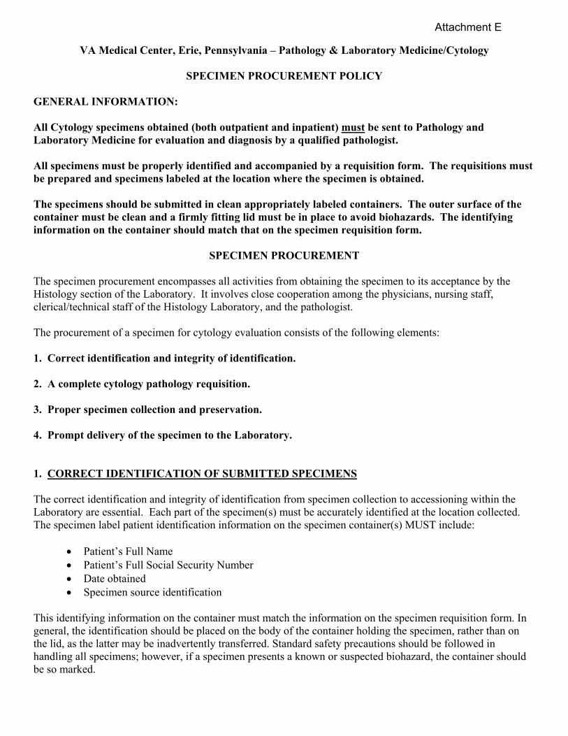
VA Medical Center, Erie, Pennsylvania – Pathology & Laboratory Medicine/Cytology
SPECIMEN PROCUREMENT POLICY
GENERAL INFORMATION: All Cytology specimens obtained (both outpatient and inpatient) must be sent to Pathology and Laboratory Medicine for evaluation and diagnosis by a qualified pathologist. All specimens must be properly identified and accompanied by a requisition form. The requisitions must be prepared and specimens labeled at the location where the specimen is obtained. The specimens should be submitted in clean appropriately labeled containers. The outer surface of the container must be clean and a firmly fitting lid must be in place to avoid biohazards. The identifying information on the container should match that on the specimen requisition form.
SPECIMEN PROCUREMENT
The specimen procurement encompasses all activities from obtaining the specimen to its acceptance by the Histology section of the Laboratory. It involves close cooperation among the physicians, nursing staff, clerical/technical staff of the Histology Laboratory, and the pathologist. The procurement of a specimen for cytology evaluation consists of the following elements: 1. Correct identification and integrity of identification. 2. A complete cytology pathology requisition. 3. Proper specimen collection and preservation. 4. Prompt delivery of the specimen to the Laboratory. 1. CORRECT IDENTIFICATION OF SUBMITTED SPECIMENS The correct identification and integrity of identification from specimen collection to accessioning within the Laboratory are essential. Each part of the specimen(s) must be accurately identified at the location collected. The specimen label patient identification information on the specimen container(s) MUST include:
Patient’s Full Name Patient’s Full Social Security Number Date obtained Specimen source identification
This identifying information on the container must match the information on the specimen requisition form. In general, the identification should be placed on the body of the container holding the specimen, rather than on the lid, as the latter may be inadvertently transferred. Standard safety precautions should be followed in handling all specimens; however, if a specimen presents a known or suspected biohazard, the container should be so marked.
Attachment E

2. COMPLETED CYTOLOGY PATHOLOGY REQUISITION A properly completed order or consult must accompany all specimens and identifying information on the requisition form must match that on the specimen container. If the specimen presents a known or suspected biohazard, this information should be included on the requisition form. The patient identification data should be correct and legible and compared to patient’s wristband, prior to submission to the Laboratory. The requisition form should include the following identification:
Full identification of the patient (including patient’s full name, full social security number and date of birth)
Name of submitting physician Type and source of cytologic material Date/time when the specimen was collected Ordering location Pertinent clinical history such as previous conditions and treatment and prior cytology and/or history
(if known). For routine gynecological specimens, information such as date of the last menstrual period, contraceptive history, hormones and obstetric history should also be provided.
3. PROPER SPECIMEN COLLECTION AND PRESERVATION The specimens should be collected in a clean, leak-proof container with a secure closure. For further information on specimen collection and preservation, refer to "Instructions for Specimen Collection and Preservation" section. 4. PROMPT DELIVERY OF THE SPECIMEN TO THE LABORATORY
Before being transported to the Laboratory, the primary container should be placed into a sealable secondary container, i.e., plastic zip lock specimen transport bags, which are labeled with the biohazard symbol. Specimens too large to be container in the zip lock bag will be placed in red bag with a biohazard label attached. The following precautions should be taken during specimen handling and transportation:
- The outside of the primary container and zip lock specimen transport bag must not be visibly contaminated.
- Laboratory requisition slips should be protected from visibly contaminated material and, if necessary, separated from the primary container.
- Personnel who transport specimens should be trained in safe handling practices and decontamination procedures in case of spill.
- Laboratory personnel should examine all specimens for visible contamination before opening the specimen containers. The outside of the contaminated containers should be cleaned with hospital-approved disinfectant solution before sending to the work areas for testing. Laboratory requisitions, visibly contaminated, should be discarded and replaced.
The specimens should preferably be delivered to the Laboratory as soon as they are obtained by qualified personnel. During non-duty hours, the specimens should be obtained only if necessary. All specimens collected after regular duty hours MUST be refrigerated and deliver the next day after 6:00AM.
5. REFERENCES: 1. Quality Management in Anatomic Pathology, College of American Pathologists, 2005. 2. JCAHO Comprehensive Accreditation Manual for Pathology and Clinical Laboratory Services, current
edition.
Attachment E

VA Medical Center, Erie, Pennsylvania – Pathology & Laboratory Medicine/CYTOLOGY
SPECIMEN COLLECTION AND PRESERVATION Proper collection techniques and specimen preservation are essential for accurate cytologic interpretation. The addition of alcohol or use of refrigeration to retard or stop cell degeneration and bacterial growth are essential for fluid specimens that cannot be processed immediately. The immediate fixation of smears made outside the Laboratory is essential to prevent the air-drying of slides. Cell degeneration and air-drying could compromise the cytologic diagnosis. GENERAL INFORMATION: The material for cytologic examination generally is submitted as follows: 1. Prepared Smears
2. Fluid Specimens 3. Fine Needle Aspirates (FNA) 4. Gynecological specimens
1. PREPARED SMEARS: include Pap smears, brushings, imprints, and scrapes. Rapid fixation of smears is
necessary to preserve cytologic detail of cells spread on a glass slide. If smears are allowed to air dry prior to fixation, marked distortion of cells occurs.
2. FLUID SPECIMENS: Fluid specimens from different body sites such as respiratory tract, gastrointestinal
tract, urinary tract, and effusions must be submitted in a clean, leak-proof container, which is capped tightly and properly labeled. Preservation of cellular morphology until the sample can be processed is essential to accurate cytologic preparation.
FRESH MATERIAL The "fresh sample" is one to which no fixative or preservative has been added. The prompt delivery of the fresh, unfixed material is critical. Leaving the specimen at room temperature will result in rapid deterioration of the cellular material. Such specimens are unsatisfactory for cytopathological evaluation. The length of time between collection and preparation of sample before cellular damages occur depend on pH, protein content, enzymatic activity, and the presence or absence of bacteria. It is not possible to predict these variables even in specimens from the same anatomic site. However, the following guidelines will usually yield acceptable results:
Specimens with a high mucus content, such as sputums, bronchial aspirates, or mucocele fluid may be preserved for 12 to 24 hours if refrigerated. Refrigeration slows the bacterial growth that causes cellular damage and the breakdown of mucus. Mucus apparently coats the cells, protecting them against rapid degeneration. The cells in specimens without thick mucus or specimens diluted with saliva are not as well protected and may deteriorate more rapidly. Specimens with a high protein content, such as pleural, peritoneal, or pericardial fluids may be preserved for 24 to 48 hours with refrigeration. The protein rich fluid in which the cells are bathed acts as a tissue culture medium in preserving cellular morphology. Specimens with low mucus or protein content, such as urine or cerebrospinal fluid will endure only a 1 to 2 hour delay even if refrigerated. The fluid medium in which these cells are bathed
Attachment E

contain enzymatic agents capable of causing cell destruction. Refrigeration may inhibit bacterial growth, but does not protect the cells. Specimens with low pH, such as gastric material must be collected on ice and be prepared within minutes of collection to prevent cellular destruction by hydrochloric acid.
3. FINE NEEDLE ASPIRATE (FNA) SPECIMENS: The specimen(s) are collected directly into 30 ml of Cytolyte solution.
4. GYNECOLOGICAL SPECIMENS: The specimens are collect in Thin Prep vial.
GENERAL DIRECTIONS FOR SPECIMEN COLLECTION/PRESERVATION (SUMMARY) Specimen Instructions
Prepared Smears Brushings Scrapings Nipple discharge Gynecological smears
Fixative should be held 6 inches from slide and sprayed evenly once across and once back across slide, making sure entire slide is covered by fixative.
Fluids Cyst fluid Gastrointestinal Bronchial aspirate and washings Tracheal aspirate Sputum Urine Serous fluids (pleural, peritoneal,
pericardial, ascetic, synovial and CSF)
Prompt delivery of specimens to the Laboratory. Specimens will be fixed then refrigerated IMMEDIATELY upon arrival in the Laboratory.
Fine Needle Aspirate (FNA) – breast, lung, thyroid, pancreas, lymph node, liver, neck or mediastinal mass, etc.
Collect specimen(s) directly into 30 ml of PreservCyt solution and mix. The specimen(s) will be sent to VAMC Pittsburgh for processing and interpretation using ThinPrep technology.
Gynecological specimens Collected in Sure Path vial and sent to Laboratory. The specimen(s) will be sent to VAMC Pittsburgh for processing and interpretation using ThinPrep technology.
Attachment E

VA Medical Center, Erie, Pennsylvania – Pathology & Laboratory Medicine/Cytology
INSTRUCTIONS FOR COLLECTION/PREPARATION OF SPECIMENS
GENERAL INSTRUCTIONS The instructions for collection/preparation of cytology specimens, including prepared smears and fluid specimens are as follows: PREPARED SMEARS These include the following: Pap Smear Cytology Brushings MATERIALS NEEDED: Prior to preparation of smears, it is important to secure the necessary materials and lay them out on a suitable conveniently located surface within the reach of the operator. Instrument(s) used to obtain smears Clean microscopic glass slides with frosted ends Lead pencil Paper clips for separation of slides from each other if liquid fixative is used Fixative Cardboard containers Physician order or consult IDENTIFICATION OF SLIDES Label the frosted end of the glass slide(s) with the patient's last name and last four (4) digits of the social security number using a lead pencil. PREPARATION OF SMEARS For most diagnostic purposes, well-prepared and well-fixed smears are required. Every effort should be made to place as much as possible of the material on the slide and to prepare a thin uniform smear. Thick smears with overlapping cell layers are difficult or impossible to interpret. FIXATION Immediate fixation is ABSOLUTELY ESSENTIAL for cytological smears that are stained by Papanicolaou method. Even the slightest delay in fixation will cause air drying, resulting in poor cellular detail. CHOICE OF FIXATIVE
Attachment E

The choice of fixative is spray fixative, which is available in the Histology section of the Laboratory. As soon as the smear is prepared, spray fix the smear within 2-3 seconds. Hold the can at least 12 inches from the slide. Spraying any closer will freeze the cells with aerosol propellant distorting cellular detail. Once the fixative on the smear is dry, place the slide in a cardboard slide folder. Transport the specimen to the Histology section of the Laboratory. The fluid fixative 95% ethyl alcohol may also be used for brushings. The Coplin jar with the fixative must carry a label with identification of the patient. If more than one glass slide is placed in the same fixative, it is essential that each slide be correctly identified. A paper clip placed on the frosted or marked end of every other slide will prevent adherence of slides to each other. It is preferable to use one Coplin jar for each patient to prevent errors of identification and occurrence of "floaters". FLUID SPECIMENS The fluid specimens should be submitted fresh as outlined in the "Specimen Collection and Preservation - General Information" section.
Attachment E

VA Medical Center, Erie, Pennsylvania – Pathology & Laboratory Medicine/Cytology
INSTRUCTIONS FOR COLLECTION/PREPARATION OF SPECIMENS FROM VARIOUS BODY SITES
These instructions are categorized as follows:
Gynecologic Specimens Non-Gynecologic Specimens, which include:
Respiratory tract Gastrointestinal tract Urinary tract Body fluids Cerebrospinal fluid Cytology scrapings Nipple discharge Cyst fluid cytology
Aspiration Cytology Specimens
Attachment E

GYNECOLOGIC SPECIMENS (FEMALE GENITAL TRACT)
Types of gynecologic specimens:
Uterine Cervix/Vaginal routine cytology evaluation Vaginal smear for Hormonal studies Miscellaneous gynecological specimens including endocervical scrapings and brushings and vaginal
cuff cytology. CYTOLOGY REQUISITION FOR GYNECOLOGIC SPECIMENS: The cytology consult is submitted electronically through the CPRS hospital computer system. The following data MUST be completed:
COMPLETE NAME AND SOCIAL SECURITY NUMBER AGE/DATE OF BIRTH MENSTRUAL HISTORY DATE OF LMP OBSTETRIC HISTORY PREVIOUS CYTOLOGY/HISTOLOGY IUD OR EXOGENOUS HORMONES INCLUDING BIRTH CONTROL PILLS
PROCEDURE: 1. Thin-Prep Cytology: Write the patient’s full name and full Social Security number on the Sure Path vial.
The speculum must be introduced with no lubricant jelly. Insert the Rover’s Cervex-Brush into the endocervical canal. Apply gentle pressure until the bristles form against the cervix. Maintaining gentle pressure, hold the stem between the thumb and forefinger and rotate the brush five times in a
clockwise direction. Remove the Rover’s Cervex-Brush from the patient. Placing your thumb against the back of the brush, disconnect the entire brush from the stem, dropping it into the
Sure Path preservative vial. Place the cap on the vial and tighten. Place the capped vial into a specimen bag and sent to lab.
NOTE: For vaginal smear for hormonal studies, the lateral wall of the vagina should be sampled some distance from the cervix.
2. Smears: Write the patient’s full name and full Social Security number with a lead pencil on the FROSTED
END OF A GLASS SLIDE. The smear recommended for routine cancer detection combines a vaginal pool smear and a cervical smear on one slide. The speculum must be introduced with no lubricant jelly. Obtain mucus from the posterior vaginal pool with a spatula and place on the slide as a thick drop but DO NOT SMEAR. Use the cervical end of the spatula to scrape high up in the endocervical canal, rotating 360˚ around. Mix all this pancervical scraping with the lower portion of the vaginal pool mucus drop. Then smear on the slide and mix the specimen in the liquid and spread the material in a small area. Spray with cytology fixative immediately.
Attachment E

NON-GYNECOLOGIC SPECIMEN
GENERAL DESCRIPTION
1. Cytology Washings
This applies to bronchial washings, tracheal washings, esophageal washings, urethral washings, etc. The test includes the routine cytological examination of the smears and cellblock (when applicable).
PROCEDURE: The washings or aspirations should be collected in a clean, properly labeled container and delivered to the Laboratory immediately.
2. Cytology Brushings
This applies to bronchial brushings, tracheal brushings, esophageal brushings, colonic brushings, urethral or renal pelvis brushings. PROCEDURE: The physician prepares the direct smears from the brushings of the lesion. Label the frosted end of the slides with the patient’s last name and last four (4) digits of the social security number. To prevent air-drying, place a small drop of saline on the frosted end of the slide. Using a circular motion, roll the brush in the saline drop and smear on the slides. Fix the smears immediately with spray fixative or 95% alcohol. Allow spray fixed smears to air-dry for 5-10 minutes. Place the slides in the cardboard slide holder and seal the folder. After the smears are made, place the brush in a test tube containing saline and deliver to the Laboratory immediately. NOTE: Excessive crushing of material must be avoided. The cytological sampling must be performed prior to the biopsy.
3. Lavage
The lavage is limited to the esophagus and stomach. A syringe with 50-100 ml of saline solution is attached to the plastic tube and a jet of fluid directed at a lesion or an area chosen by the endoscopist. The fluid is collected in a container by a second intubation after withdrawal of the endoscope. The specimen must be delivered to the Laboratory immediately.
Attachment E

CYTOLOGIC SAMPLING OF ORGANS
1. RESPIRATORY TRACT
a. Sputum
It is necessary to obtain a “deep cough” specimen, bringing up material from small bronchi and alveoli. If the patient cannot bring up a “deep cough” specimen, the sputum may be obtained by stimulating the cough reflex artificially. COLLECTION: The patient is instructed to expectorate sputum. Morning specimens resulting from overnight accumulation of secreta yield the best diagnostic results. Upon awakening in the morning, the patient rinses the mouth with water and discards the rinse. The patient then coughs deeply and expectorates into a sterile collection cup. Seal the container tightly, label appropriately and send to the Laboratory immediately. Three (3) specimens on three (3) successive days are usually required to ensure a maximum of diagnostic accuracy. NOTE: The patient must be carefully instructed not to spit without a deep cough since saliva is of no diagnostic value. If pulmonary macrophages are not identified, the specimen will be reported as unsatisfactory for adequate evaluation. b. Bronchial Aspirates and Washings
Bronchial aspirates are obtained by suction during bronchoscopic procedures. During the procedure, approximately 10 ml of normal saline is instilled in small portions of 2 to 3 ml at a time. The material is aspirated and submitted to the Laboratory in a clean container. c. Bronchial Brushings
The fiberoptic bronchial brushing specimen is obtained and smears are prepared as outlined in “Cytology Brushings.” d. Tracheal Aspirate The aspirate is obtained by instilling 20 ml of saline solution in 2-ml portions and re-aspirating with a catheter in various positions. The specimen is collected in a clean container and sent to the Laboratory immediately.
2. GASTROINTESTINAL TRACT a. Esophagus Esophageal washings are best obtained using direct endoscopy. The washings should be obtained prior to biopsy. The specimen is collected in an appropriate container and sent directly to the Laboratory immediately. Esophageal brushings obtained during the endoscopic procedure are smeared and fixed as outlined in "Cytology Brushings".
Attachment E

b. Stomach
Lavage - The specimen should be collected in a clean, leak proof container. The container should be kept in ice and delivered directly to the Laboratory. Brushings - Gastric brushings obtained during the endoscopic procedure are smeared and fixed as outlined under "Cytology Brushings". c. Colon Lavage - The lavage specimen should be collected in an appropriate container and sent directly to the Laboratory. Brushings - Brushings of colonic mucosal abnormalities at the time of fiberoptic examination of the colon are smeared and fixed as outlined in "Cytology Brushings". This is usually done in high-risk patients such as patients with multiple polyps, ulcerative colitis of long duration, and past history of colonic carcinoma.
3. URINARY TRACT
a. Urine The specimen should be a freshly voided, clean catch urine. Twenty-four (24) hour specimens are NOT acceptable; catheterized urine is acceptable. The voided urine is preferred over a catheterized sample due to atypical cell changes caused by trauma during catheterization. The history of instrumentation, if any, must be given. The specimen must be sent to the Laboratory immediately. b. Bladder Irrigation (Washings) Washings of the bladder with saline solution obtained at the time of cytoscopy may be collected and sent, preferably in a fixative. This has a limited use confined to symptomatic patients. c. Renal Pelvis and Ureters The specimens obtained by retrograde catheterization or direct brushing for suspected lesions of renal pelvis or ureters must be sent to the Laboratory immediately. Specimens must be labeled as to type (voided or catheterized). For urethral specimens, designate origin (left or right).
4. BODY FLUIDS
Body fluids include pleural, peritoneal, pericardial, ascitic, and synovial fluids. Specimens may be collected in sterile evacuation bottles, tubes, or syringes. COLLECTION: The fresh body fluid should be collected as described above. The minimum volume should be at least 5 ml of fluid as less than that will not yield enough cells for accurate cytological evaluation. NO FIXATIVE, PRESERVATIVE, OR ANTICOAGULANT SHOULD BE ADDED TO THE SPECIMEN. The specimen should be sent immediately to the Laboratory for processing. NOTE: If the specimen is collected on a weekend or if there is a delay in delivery of specimen, it should be refrigerated IMMEDIATELY and delivered the next working day.
Attachment E

5. CEREBROSPINAL FLUID
The minimum volume of cerebrospinal fluid required is 1 ml. The specimen should be sent fresh in a sterile tube from the lumbar puncture tray or a sterile disposable container. DO NOT ADD ANY FIXATIVE, PRESERVATIVE, OR ANTICOAGULANT. Deliver the specimen to the Laboratory immediately.
6. CYTOLOGY SCRAPINGS
Cytology scrapings include skin scrapes, oral scrapes, mucus membrane scrapes, and Herpes cytology. SPECIMEN COLLECTION: A direct smear of the lesion, vesicle, or blister is prepared. a. If the lesion is soft, moist, and clean, swab the lesion with a pre-moistened, non-absorbent, cotton-tipped
applicator. Place a small drop of saline on the slide. Spread the material from the applicator onto the pre-moistened slide. Fix with spray fixative immediately. Allow the slides to dry before placing in the slide folder.
b. If the lesion is dry or has a necrotic, inflammatory surface, gently moisten and remove the necrotic
debris with non-absorbent cotton, which has been moistened in saline. Discard the swab and debris. Using a second non-absorbent cotton swab moistened in saline, gently rub the growing margins of the lesion. Place a small drop of saline on a clean slide. Quickly roll the second swab onto the pre-moistened slide. Fix with spray fixative immediately. Allow the slides to dry before placing them in the slide holder.
7. NIPPLE DISCHARGE CYTOLOGY
SPECIMEN: Soak the nipple with warm saline with cotton or gauze for 10-15 minutes. Place a small drop of saline on the slide. Then place pre-moistened slide on the nipple secretion and slide across quickly. Do not make thick smears. Make as many smears as the amount of the specimen allows. If smears are prepared from both breasts, label slides as "left" and "right". Deliver slides to the Laboratory.
8. CYST FLUID CYTOLOGY
Cyst fluid applies to renal cyst fluid, breast cyst fluid, pancreatic cyst fluid, hydrocele cyst fluid, liver cyst fluid, thyroid cyst fluid, bile duct fluid, and T-tube fluid. SPECIMEN: Fresh fluid should be submitted to the Laboratory in a clean, leak-proof container. DO NOT ADD FIXATIVE, PRESERVATIVE, OR ANTICOAGULANT. Deliver to the Laboratory immediately. After regular Laboratory hours, specimens should be refrigerated until the next working day.
9. OTHER SPECIAL STUDIES
If any special studies are required, contact the Histology/Cytology section of the Laboratory in advance so that proper arrangements can be made.
Attachment E

FINE NEEDLE ASPIRATE CYTOLOGY SPECIMENS (FNA)
This includes fine needle aspirations of breast, lung, thyroid, pancreas, lymph node, liver, neck, or mediastinal mass, etc. The test includes examination of fine needle aspirate specimen(s) using ThinPrep technology. Clinical information is necessary for the pathologist to render a diagnosis. The specific site, clinical diagnosis, whether the lesion is solid or cystic and the gross appearance of the aspirate (if applicable) should be indicated on the request form. SUBMISSION OF SPECIMEN(S) Collect all fine needle aspirate (FNA) specimen(s) directly into 30 ml of Cytolyte Solution and mix. Label
the Cytolyte container with patient’s full name/full social security number and send to the Histology section of the Laboratory.
The FNA specimen(s) will be sent to Cytology Department of VAMC Pittsburgh for processing and
interpretation.
Attachment E

VA Medical Center, Erie, Pennsylvania – Pathology & Laboratory Medicine/Cytology
CRITERIA FOR UNACCEPTABLE/UNSATISFACTORY CYTOLOGY SPECIMENS
SPECIMEN ADEQUACY The general criteria for adequacy of cytology specimens include: Positive identification and labeling of specimens. Proper fixation Provision of demographic and relevant clinical information The specific criteria for adequacy of non-gynecological and gynecological specimens are as follows: Non-gynecological Specimen Adequacy:
For most non-gynecological specimens, certain cell types should be present in sufficient numbers to be considered adequate for evaluation. For example, sputum specimens should contain alveolar macrophages. Washings and lavage of certain body sites (bladder, bronchoalveolar) should contain at least minimal numbers of cells representative of those sites. In contrast, normal cerebrospinal fluids have very few cells, so scantly cellular spinal fluid should not be labeled as unsatisfactory. For fine needle aspiration biopsy (FNA) specimens, it is critical not to label unsatisfactory or non-diagnostic specimens as “negative”. A distinction should be made between specimens rejected prior to processing and those that are rejected after processing and review (unsatisfactory or inadequate for evaluation).
Gynecological Specimen Adequacy:
♦The smears should contain adequate number of well-preserved and visualized squamous cells. ♦In females with cervix at least ten (10) well preserved endocervical or squamous metaplastic cells should be observed to report that a transformation zone component is present. When there is partial or complete atrophy, it may be difficult to determine the presence of a transformation zone component. Parabasal cells should not be counted as transformation zone components, and it should be mentioned in the comments regarding the difficulty in identifying a transformation zone component.
CYTOLOGY SPECIMEN UNACCEPTABLE/UNSATISFACTORY CRITERIA/HANDLING
The cytology specimens will not be processed by Histology Section of the Laboratory unless any identified problems are corrected. The Histology Section of the Laboratory may process suboptimal test requests/specimens after appropriate corrective action has been taken and documented.
The technologist is responsible for identifying all incidents of unsatisfactory/unacceptable test
requests/specimens, taking appropriate corrective action before processing and documenting on “Specimen Rejection Log” worksheet. The Supervisor, P&LM/Pathologist will review incidents of problems identified and appropriateness of corrective action on a monthly basis.
Attachment E

VIII. AUTOPSY
Autopsy Service
Request Form/Autopsy Permit
Performance of Autopsy Examination
Post-Mortem Reports Provisional Anatomic Diagnosis for Post-Mortem Examination Final Post-Mortem Examination Report Turnaround Time of Post-Mortem Reports
Attachment E

AUTOPSY SERVICE
A. LOCATION: MORGUE (E1-14), first floor, behind Clinical Laboratory.
B. REQUEST FORMS:
1. Standard Form 523 2. Autopsy History Form 1-20 (5243)
C. AUTOPSY PERMIT
1. The Member Services/Processing Team aids in the administrative and clinical aspects of the autopsy permission.
2. A legal autopsy permit, Standard Form 523, can be given by the following individuals:
a. By written authorization signed by the deceased during lifetime. b. By written authorization of any party whom the deceased during lifetime designated by
written instruction to take charge of his body for burial and the written consent of decedent’s surviving spouse, if any, after death.
c. By written authorization of the decedent’s surviving spouse. d. If the surviving spouse is incompetent, unavailable or does not claim the body for burial, of if
there is no surviving spouse, by written authorization of the following in order of precedence if the claimant agrees to provide burial (I) adult children, (II) adult grandchildren, (III) parents, (IV) brothers or sisters, (V) nephews or nieces, (VI) grandparents, (VII) uncles or aunts, (VIII) cousins, (IX) stepchildren, (X) relatives or next of kin of previously deceased spouse.
e. If none of the above persons are available to claim the body, by written authorization of any other relative or friend who assumes custody of the body for burial.
f. If there is no known living kin, or no one falling within the established order or precedence for claiming the body, the Director is authorized to sign for an autopsy.
g. Autopsy may be performed on (1) patients who die outside of a VA facility while in a follow-up status after hospitalization, (2) outpatients who had been assigned solely for research purpose or who made visits solely for research purpose.
3. Authorization may be given by telegram or by telephone if the telegram or telephone
authorization is certified by two persons present when it was received. When telephone consent is grant to perform an “autopsy”, this is to be interpreted as permission for a “complete autopsy”, including removal of the brain, permission should be considered “limited” only when expressly stated.
4. In cases where the legality of the permission is in doubt, the Medical Director, Pathology &
Laboratory Medicine, will assist the clinician and Member Services in determining the legality of a permission.
Attachment E

D. INCLUSIVENESS OF PERMIT
1. Permission includes examination of the brain and spinal cord. Specific permission is needed for examination of unusual areas, as dismembering a limb. For exclusion of areas such as the brain, a notation of the limitation must appear on the consent form.
E. PROCEDURE
1. If the next-of-kin is available, the authorization is signed and witnessed on the unit. If not, Member Services will aid in securing a telephonic or telegraphic permission. The Laboratory will not perform an autopsy with a telephonic permission unless the words “autopsy” or “postmortem” are used in the request for the necropsy.
2. When the autopsy permission has been secured, please call:
a. The Laboratory (8AM-3:30PM, Monday through Friday). b. The telephone operator (on Saturday, Sunday and legal holidays).
3. Please send the chart, autopsy permission and completed clinical history form to the Laboratory (8AM-4:30PM, Monday through Friday). At other times, please send these documents to the telephone operator’s area (1st floor).
4. Please consult with the Pathology Department before giving the family an estimate as to when
the body can be released to the undertaker.
5. In cases when the patient’s record and a valid permit reach the Laboratory after 3:30PM and before 8:00AM, the autopsy will be delayed until 10:00AM. Special arrangements can be made with the Medical Director, Pathology & Laboratory Medicine, to have autopsy done at other times.
6. The body will be deposited in the refrigerator of the morgue until its release and the relevant date
will be entered in the workbook of the morgue.
7. The release of the body, after performing the autopsy, for the funeral arrangement occurs through Member Services, filling out Form 523A.
F. CORONER’S CASE
1. In Pennsylvania, the Coroner’s Office shall investigate deaths in cases of (a) sudden, violent, or suspicious death – including those in which alcohol, drugs or other toxic substances may have had a direct bearing on the outcome, and (b) any death wherein no cause of death is properly certified.
2. In Coroner’s cases, the attending physician will call the Coroner’s office. The Laboratory is
available to render service.
3. Keep in mind that the Coroner’s office does not automatically assume jurisdiction. Reportable cases are one category, and those in which jurisdiction is assumed are a second category. In some
Attachment E

cases, the Coroner’s office may grant permission for the hospital to perform the autopsy, assuming (a) the hospital will perform, and (b) the family permits the autopsy.
4. UNDER NO CIRCUMSTANCE SHOULD AN AUTOPSY PERMISSION BE REQUESTED BY A FAMILY IN A CORONER’S CASE. This is to prevent undue and possibly damaging criticism directed toward the hospital and Coroner’s office in the event that the family refused permission and the Coroner’s office subsequently finds it necessary to take the body to perform an autopsy. In such a circumstance, a charge could be leveled that the hospital and Coroner’s office are functioning in an illicit manner in order to have autopsies performed against the wishes of the family. The Laboratory will attempt, whenever feasible, to have the autopsy performed in this hospital.
5. OFFICE REPORT TO CORONER, Erie County. Please remember that the “Report of Contact”,
VAF 119, must be completed by the appropriate Program or Member Services. a. All sudden deaths not caused by readily recognizable disease; or wherein the cause of
death cannot be properly certified by a physician on the basis of prior (recent) medical attendance.
b. All deaths occurring under suspicious circumstances, including those where alcohol, drugs or other toxic substances may have had a direct bearing on the outcome.
c. All deaths occurring as a result of violence or trauma, whether apparently homicidal, suicidal or accidental (including those due to mechanical, thermal, chemical, electrical or radiation injury, drowning, cave-ins and subsidences); and regardless of the time elapsing between the time of injury and time of death.
d. Any fatal death, stillbirth or death of any baby within 24 hours after its birth, where the mother has not been under the care of a physician.
e. All therapeutic and criminal abortions, regardless of the length of the pregnancy and spontaneous abortions beyond 16 weeks gestation.
f. All operative and perioperative deaths in which the death is not readily explainable on the basis of prior disease.
g. Any death wherein the body is unidentified or unclaimed. h. Any death where there is uncertainty as to whether or not it should be reported to the
Coroner’s office. The fact that a death occurs within 24 hours following admission to a hospital is not, of itself, reason for its being considered a reportable death unless, in addition, it falls into one of the specific categories herewith defined.
Attachment E

PERFORMANCE OF AUTOPSY EXAMINATIONS
A. GENERAL DIRECTIONS:
1. Before proceeding with the autopsy, the following items should be checked:
a. Autopsy permit to determine the limitations of the autopsy, proper signing of the permit, Medical Examiner’s case or clearance by that office. When there is a question regarding the legality of autopsy authorization, contact MAA.
b. A complete clinical record and a listing of clinical questions or concerns related to possible autopsy
findings should be furnished to the Pathologist prior to the beginning of the autopsy. The Pathologist will review the clinical record carefully for positive radiological and clinical findings, which should be verified by gross and histological examination. The chart will be carefully searched for a record of administration of radioactive materials for therapeutic purposes. Patients who have received radioactive isotopes should be handled under the supervision of the Safety Specialist or person designated by him/her. The clinical information is discussed with the attending physician before conducting the autopsy.
B. AUTOPSY EXAMINATION: The objective of autopsy examination is full exposition of the patient’s
disease processes, the limits thereof and the patient’s response to therapy.
1. There will be positive identification of the deceased by the Pathologist before the procedure is actually begun. Such identification will consist of checking the name and other identifying data attached to the deceased and comparing these with information recorded on SF 523, “Authorization for Autopsy.” If there is uncertainty regarding identification, a physician or nurse who knew the deceased during life will make the necessary identification.
2. There will be strict adherence to the family’s wishes as recorded on the SF523, “Authorization for
Autopsy.”
3. The Pathologist will notify the attending physician as soon as possible, as to the time of autopsy and will arrange to demonstrate the gross findings.
4. Embalming prior to autopsy, whether by arterial injection or by intracavitary trocar injection, is
prohibited because of the risk of these procedures causing anatomic alterations, making it impossible to determine if these changes preceded embalming. Care must be exercised that there is no undue delay in performing the autopsy, which would inconvenience the family of the decedent.
5. Autopsy examination normally encompasses both gross and microscopic studies and sufficiently
detailed protocol. Photographic documentation is an essential component of the autopsy examination and facilities are readily available.
6. Unless the autopsy is a limited one, the brain, all pelvic organs (including testes) and the abdominal
organs should be removed and examined. Representative tissue samples not greater than 1 cm in thickness are taken for the stock bottles even though no gross abnormality is seen. Portions of normal and pathologic skeletal muscle, peripheral nerve and bone should also be included in the stock bottle. The latter should include portions of iliac crests, rib and vertebral bodies. The spinal cord and tongue should only be removed when there are specific indications.
Attachment E
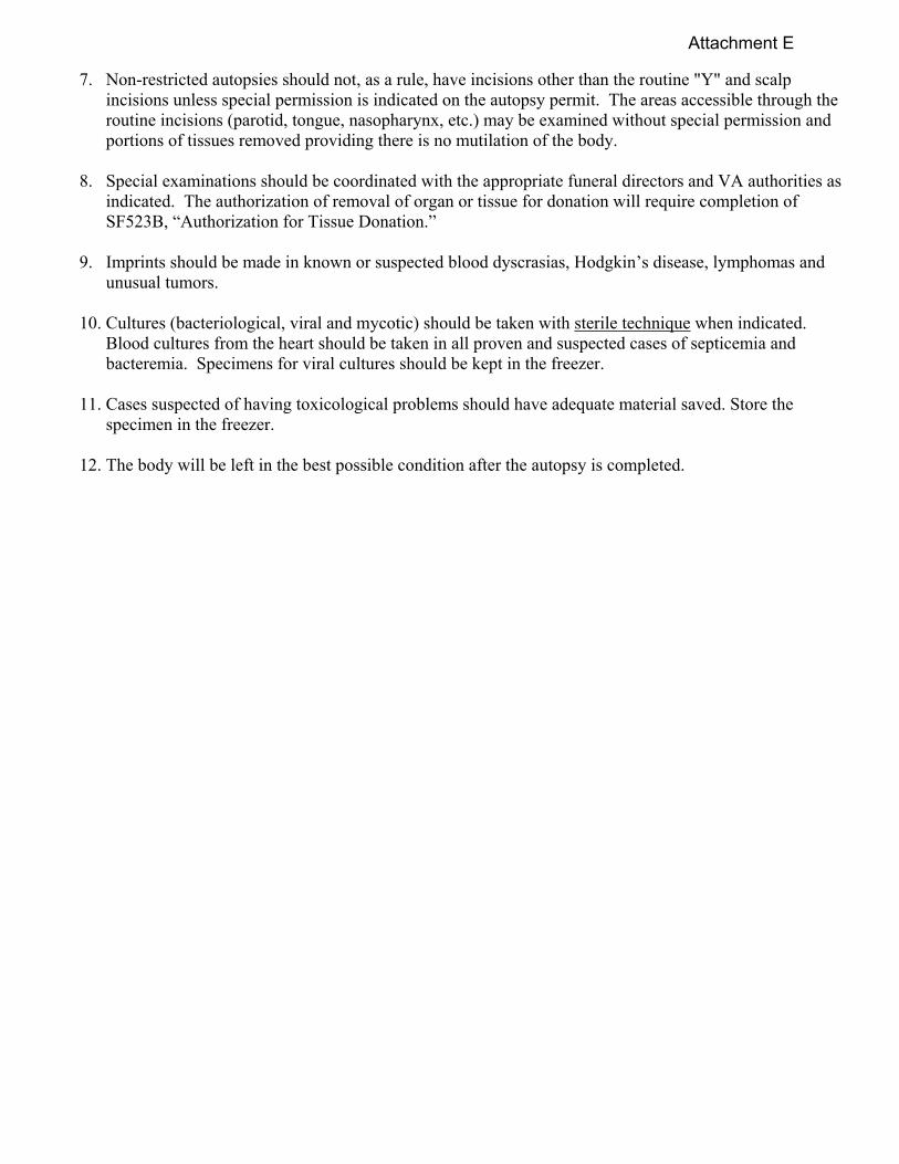
7. Non-restricted autopsies should not, as a rule, have incisions other than the routine "Y" and scalp incisions unless special permission is indicated on the autopsy permit. The areas accessible through the routine incisions (parotid, tongue, nasopharynx, etc.) may be examined without special permission and portions of tissues removed providing there is no mutilation of the body.
8. Special examinations should be coordinated with the appropriate funeral directors and VA authorities as
indicated. The authorization of removal of organ or tissue for donation will require completion of SF523B, “Authorization for Tissue Donation.”
9. Imprints should be made in known or suspected blood dyscrasias, Hodgkin’s disease, lymphomas and
unusual tumors.
10. Cultures (bacteriological, viral and mycotic) should be taken with sterile technique when indicated. Blood cultures from the heart should be taken in all proven and suspected cases of septicemia and bacteremia. Specimens for viral cultures should be kept in the freezer.
11. Cases suspected of having toxicological problems should have adequate material saved. Store the
specimen in the freezer. 12. The body will be left in the best possible condition after the autopsy is completed.
Attachment E

VA Medical Center Erie, Pennsylvania
POST-MORTEM (AUTOPSY) REPORTS
A. PROVISIONAL ANATOMIC DIAGNOSIS FOR POST-MORTEM EXAMINATION
1. The diagnosis is telephoned to the patient's attending physician promptly after completion of post-
mortem examination.
2. A written provisional anatomic diagnosis will be placed in the patient's medical record within 2 working days and a copy will be forwarded to the attending physician.
B. FINAL POST-MORTEM EXAMINATION REPORT
1. Only a qualified licensed Pathologist, board certified in anatomic pathology, will provided a final
written diagnosis for gross and microscopic autopsy findings. 2. The following format is used for the post-mortem protocol:
a. Complete Demographic Data: The demographic data on the face sheet should include the following:
Name of the institution where autopsy was performed Patient's identification (including name, social security number, date of birth, and sex) Date and time of death Extent of autopsy Final autopsy completion date
b. Clinical Diagnosis: This includes in a list form all important clinical states, process, diagnosis, and treatments. c. Gross and Microscopic Findings: The gross findings are recorded as soon as possible after the post-mortem examination is completed. The extent of description of microscopic description varies with each autopsy. The findings generally include a record of the organs and tissues examined histologically. A record of the source of slides in relation to lesions or abnormalities as described grossly is important. Special Studies: The results of any special slides such as microbiologic, toxicologic, or other analysis are included in the protocol.
Attachment E
d. Clinical Summary and Clinico-pathologic Correlation: A brief clinical history generally includes patient's established medical conditions and diagnosis, risk factors, pertinent to the disease process identified, hospitalizations, surgical operations, medications, and pertinent Laboratory data. The Clinic-pathological summary correlates the clinical findings with gross and microscopic findings to describe and elucidate the sequence of events leading to death. The underlying cause or causes and immediate cause of death is discussed. The summary is intended to address clinical problems and questions faced by the attending physicians and to clarify quality improvement matters. e. Final Post-Mortem Diagnosis (including neuropathologic findings if brain was removed): The final diagnosis should include findings in chronological order and importance with respect to the main disease process. Miscellaneous findings need not be given in special order. If chemotherapy or radiation therapy has been given, it should be indicated. In malignant tumors, the distribution of disease should be included. The reports of cultures on other studies should also be included. Final post-mortem examination diagnoses must be coded by employing a recognized system of coding, e.g. Systematized Nomenclature of Medicine (SNOMED) or Systematized Nomenclature of Pathology (SNOP) for retrieving diagnostic information as needed. f. Letter to Next of Kin: Draft of lay letter to next of kin by Medical Administration if copy of autopsy is requested by family.
C. TURNAROUND TIME OF POST-MORTEM (AUTOPSY) REPORTS
1. The post-mortem examination (autopsy) reports will be made a part of the patient's medical record within 30 working days for routine cases. The cases requiring special studies will have a longer turnaround time.
2. A copy of the report will be retained in the Pathology & Laboratory Medicine.
D. AUTOPSIES WITH POSSIBILITY OF A CLAIM
1. The Medical Director, Pathology & Laboratory Medicine will provide the Chief of Staff with a copy of
the post-mortem examination report in any case in which the post-mortem examination findings raise the possibility of a claim against the VA.
2. Post-mortem examination findings may be disclosed in accordance with the limited disclosure
provisions of 38 U.S.C. Section 5705. In any case where there is the slightest indication of potential claim, no action will be taken to release information without first consulting with District Counsel. NOTE: Disclosure is limited by confidentiality statutes.
Attachment E

E. CONFIDENTIAL TREATMENT OF POST-MORTEM EXAMINATION RECORDS
1. If tissues or records are to be sent from VA for examination in non-VA laboratories or be investigators, such persons can be given access to such items only within the restrictions imposed by laws governing the disclosure of information, e.g., the Privacy Act of 1974, 38 U.S.C., Sections 5701, 5705, and 7332.
2. Some of these statutes address the disclosure of information about patients in an individual identifiable
format. If the examiner requires that the slides and records contain veterans' name or other confidential information, there must be a prior written agreement that the:
3. Recipient of the slides and records will not re-disclose any information in an identifiable form without
prior specific VA authorization; 4. Information will be safeguarded from disclosure. NOTE: VHA Records Control Schedule (RCS) 10-1,
provides disposition instructions for agency records. Conversion of temporary paper records to laser disc storage and disposal of the paper record do not require the National Archives and Records Administration's approval; and
5. The slides and records will be returned to the VA when there is no longer a need for the recipient to
retain them in order to accomplish the purpose for which they were originally supplied. 6. To the extent that any of the records discussed in this chapter are medical quality assurance records
subject to 38 U.S.C. 5705 and 38 CFR Sections 17.500 through 17.540, they may be disclosed only in accordance with the statute.
7. When it is necessary to release records or slides in a manner other than that defined, the District Counsel
should be consulted prior to the release.
Attachment E