Cardiovascular Pathology in AIDS
26
Barbaro G (ed): HIV Infection and the Cardiovascular System. Adv Cardiol. Basel, Karger, 2003, vol 40, pp 23–48 Cardiovascular Pathology in AIDS Edward C. Klatt Florida State University, College of Medicine, Tallahassee, Fla., USA The heart is not the most frequent site for opportunistic infectious or neoplastic processes in patients with AIDS (table 1). However, cardiovascular pathologic findings may occur in up to 40% of AIDS patients at autopsy [2–4]. Cardiac lesions are the immediate cause of death in 1–3% of AIDS patients [5]. The most common significant manifestations of cardiac disease, which can occur at any stage of HIV infection and can lead to death, include cardiomy- opathy, dysrhythmias and pericardial effusion with tamponade [6]. Clinical cardiac findings may be present in a fourth to three fourths of adult AIDS patients and may be accompanied by findings that include chest pain, tachycardia, electrocardiographic changes including various arrhythmias, effusions and congestive heart failure. There may be mild cardiomegaly on chest roentgenograms [7]. The prevalence of HIV-related cardiac disease appears to be decreasing with the use of highly active antiretroviral therapy (HAART) [8]. Overall, deaths from cardiovascular diseases in patients dying of AIDS increased slightly from 1987 to 1999, as advances in antiretroviral therapy reduced deaths due to HIV infection after 1995 and the proportion of deaths caused by other conditions increased [9]. Atherosclerosis Many persons with HIV infection are in the third to fifth decades of life when cardiovascular complications from atherosclerosis are not as frequent as in older persons. Atherosclerotic cardiovascular disease leading to ischemia and myocar- dial infarction (fig. 1) can and does occur in HIV-infected patients, particularly as the numbers of HIV-infected persons on HAART rise and as the population of long-term survivors from AIDS increases. In one autopsy study, 39% of men and 21% of women dying of AIDS had significant atherosclerosis [4].
-
Upload
khangminh22 -
Category
Documents
-
view
1 -
download
0
Transcript of Cardiovascular Pathology in AIDS
Barbaro G (ed): HIV Infection and the Cardiovascular System.
Adv Cardiol. Basel, Karger, 2003, vol 40, pp 23–48
Cardiovascular Pathology in AIDS
Edward C. Klatt
Florida State University, College of Medicine, Tallahassee, Fla., USA
The heart is not the most frequent site for opportunistic infectious or
neoplastic processes in patients with AIDS (table 1). However, cardiovascular
pathologic findings may occur in up to 40% of AIDS patients at autopsy [2–4].
Cardiac lesions are the immediate cause of death in 1–3% of AIDS patients [5].
The most common significant manifestations of cardiac disease, which can
occur at any stage of HIV infection and can lead to death, include cardiomy-
opathy, dysrhythmias and pericardial effusion with tamponade [6].
Clinical cardiac findings may be present in a fourth to three fourths of
adult AIDS patients and may be accompanied by findings that include chest
pain, tachycardia, electrocardiographic changes including various arrhythmias,
effusions and congestive heart failure. There may be mild cardiomegaly on
chest roentgenograms [7]. The prevalence of HIV-related cardiac disease
appears to be decreasing with the use of highly active antiretroviral therapy
(HAART) [8]. Overall, deaths from cardiovascular diseases in patients dying of
AIDS increased slightly from 1987 to 1999, as advances in antiretroviral therapy
reduced deaths due to HIV infection after 1995 and the proportion of deaths
caused by other conditions increased [9].
Atherosclerosis
Many persons with HIV infection are in the third to fifth decades of life when
cardiovascular complications from atherosclerosis are not as frequent as in older
persons. Atherosclerotic cardiovascular disease leading to ischemia and myocar-
dial infarction (fig. 1) can and does occur in HIV-infected patients, particularly as
the numbers of HIV-infected persons on HAART rise and as the population of
long-term survivors from AIDS increases. In one autopsy study, 39% of men and
21% of women dying of AIDS had significant atherosclerosis [4].
Klatt 24
Table 1. Opportunistic infections and neoplasms in 565 cases of AIDS
at autopsy [1]
Disease condition Number with Number with
cardiac condition
involvement diagnosed
Pneumocycstis carinii 2 308
Cytomegalovirus 5 286
Candida 13 240
Kaposi’s sarcoma 6 138
Mycobacterium avium complex 3 104
Non-Hodgkin’s lymphoma 13 81
Cryptococcus neoformans 13 78
Mycobacterium tuberculosis 5 76
Toxoplasma gondii 5 51
Histoplasma capsulatum 4 13
Coccidioides immitis 3 10
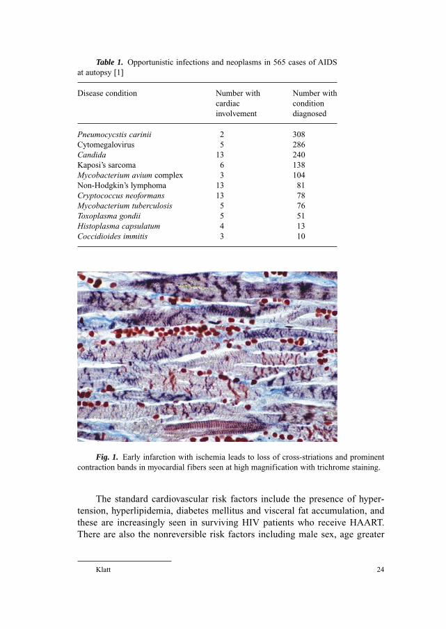
Fig. 1. Early infarction with ischemia leads to loss of cross-striations and prominent
contraction bands in myocardial fibers seen at high magnification with trichrome staining.
The standard cardiovascular risk factors include the presence of hyper-
tension, hyperlipidemia, diabetes mellitus and visceral fat accumulation, and
these are increasingly seen in surviving HIV patients who receive HAART.
There are also the nonreversible risk factors including male sex, age greater
than 40 years and family history of coronary artery disease. Other factors
include smoking and sedentary lifestyle. In older patients and those with other
risk factors, HAART may accentuate these risk factors. It is not clear at this
time whether these factors predispose HIV-infected patients to accelerated
atherosclerosis [10].
Atherosclerosis arises as a consequence of ongoing endothelial dysfunc-
tion and damage that allows the increased uptake of lipids into the intima to
stimulate atheroma formation. Over time, the plaque increases in size, with
smooth muscle proliferation and overlying thrombus formation as the plaque
ruptures. The increasing size of the atheroma narrows the coronary arterial
lumen, leading to myocardial ischemia and possible infarction.
HIV infection can produce metabolic disturbances that increase the risk for
atherosclerosis. During HIV disease progression, there can be serum lipid
abnormalities including decreased LDL cholesterol and increased triglyceride
levels [11]. The HIV-associated proteins gp120 and Tat may produce endo-
thelial cell activation in association with cytokines in response to HIV-induced
mononuclear cell activation [12]. Coinfection with herpes simplex virus and
cytomegalovirus may contribute to vascular endothelial damage [13]. These
contributing factors to endothelial alteration may produce endothelial damage
that promotes atherosclerosis.
Though premature coronary artery disease associated with endothelial
dysfunction, hypercoagulability and hypertriglyceridemia were reported prior
to the widespread use of antiretroviral therapy including protease inhibitors, the
frequency of coronary artery disease and other atherogenic lesions in HIV-
infected persons has increased since the advent of HAART with protease
inhibitors [14]. These risk factors are associated with the syndrome of protease-
inhibitor-associated lipodystrophy (PIAL), and they promote atherogenesis.
In this syndrome, there is hypercholesterolemia and hypertriglyceridemia along
with insulin resistance and glucose intolerance typical of diabetes mellitus [15].
Glucose intolerance is associated with an increased risk for coronary artery
disease. Smoking as an additional risk factor for atherosclerotic heart disease is
seen in many of these patients [16]. Peripheral vascular atherosclerosis, how-
ever, may not be associated with PIAL [17].
The syndrome of PIAL is characterized by fat accumulation within the
abdomen, in the breasts of women and over the cervical vertebrae (‘buffalo
hump’), hyperlipidemia and insulin resistance. In addition, there is lipoatrophy
involving the face, limbs and upper trunk. These findings are observed in
association with protease inhibitors after a median 10 months from initiation
of therapy. Diabetes mellitus type 2 is a less common adverse effect. The
lipodystrophy syndrome may be a result of the inhibition of cytoplasmic
retinoic acid binding protein type 1 and LDL-receptor-related protein involved
Cardiovascular Pathology in AIDS 25
in lipid metabolism that have significant homology to the catalytic site of HIV
protease [18].
Increased cholesterol or triglyceride levels may be seen in 50–74% of
patients receiving protease inhibitor therapy [18, 19]. The rise in LDL choles-
terol is typically small, but triglyceride elevations can be marked, with levels
over 1,000 mg/dl, particularly with the use of ritonavir [20]. HIV-infected
patients receiving therapy including protease inhibitors have decreased fibri-
nolysis and increased coagulability, which may be additional risk factors for
cardiovascular disease [21].
An increase in thickness of the intima and media of the carotid artery, as
measured by B mode ultrasonography, is predictive of an increased risk for
myocardial infarction. In one study comparing protease-inhibitor-treated
patients with HIV-infected patients who had not received protease inhibitors,
and with normal non-HIV-infected controls, half of the patients treated with
protease inhibitors had acquired vascular wall lesions. There were slightly
significant correlations between carotid lesions and age, male sex and hyper-
cholesterolemia. The most significant correlations occurred between smoking,
hypertriglyceridemia and HIV stage. The highest significant correlation
occurred with the use of protease inhibitors [17].
Myocardial infarction has been reported in the setting of PIAL, with
occurrence from 24 to 29 months following initiation of protease inhibitor
therapy [22]. A retrospective study of 4,993 HIV-infected patients treated
with different antiretroviral regimens showed that the incidence of myocardial
infarction increased in persons over the age of 40 after introduction of
HAART [23]. Another retrospective study found a 5-fold increase in the risk
for myocardial infarction in patients treated with protease inhibitors [24].
However, another study of ischemic heart disease and HIV infection showed
no apparent association with protease inhibitor therapy, but instead an associ-
ation with more traditional risk factors such as hypertension and hypercholes-
terolemia [25].
Features of accelerated coronary atherosclerosis have been observed in
young persons with HIV infection. These features are intermediate between the
coronary arteriopathy of cardiac transplants and typical coronary atherosclerosis.
Such features include proximal arterial intimal and medial thickening of an
equal degree that was associated with smooth muscle cell proliferation and
increased elastic fiber production associated with increased tumor necrosis
factor � and interleukin 1�. In two thirds of these cases there was overlying
atheroma formation, and in a third of cases mamillated vegetations with intra-
luminal protrusion were present on the surface of the lesions [26].
However, the chronic debilitated state with cachexia and wasting syndrome
brought on by AIDS may lead to regression of atherosclerotic lesions.
Klatt 26
AIDS Cardiomyopathy
A congestive (dilated) cardiomyopathy may be identified in both adult
and pediatric AIDS patients. By echocardiography, the prevalence of cardiac
muscle disease is 15% in HIV-positive patients. Most of these cases are
idiopathic, for no specific opportunistic infection or neoplasm can be identi-
fied. Patients with symptomatic heart failure from dilated cardiomyopathy,
typically present late in the course of AIDS, have low CD4 counts, have
myocarditis and have a persistent elevation of antiheart antibodies. Echocar-
diographic findings include a fractional shortening of �28% with global left
ventricular hypokinesia [27]. Even asymptomatic HIV-infected patients may
have altered cardiac function. In one study of 61 such patients, all had some
degree of left ventricular mass index reduction and diastolic functional
abnormalities [28].
It is possible that cardiomyopathy and myocarditis are both immunologic
phenomena resulting from HIV-containing lymphocytes in cardiac muscle [7].
Cytokine elaboration by inflammatory cells may contribute as well, since
increased levels of both tumor necrosis factor � and inducible nitric oxide
synthase have been found in patients with HIV-associated cardiomyopathy [29].
Cardiac myocytes have also been shown to be a direct target for HIV
infection, which may result in cardiomyopathy [30]. A proposed autoimmune
mechanism for myocardial damage is based upon the observation that autoan-
tibodies to myosin and B-cell receptor can be detected in HIV-infected patients
with cardiomyopathy. Abnormal anti-�-myosin autoantibody concentrations
have been found to be higher in patients with HIV (19%), particularly those
with heart muscle disease (43%), than in HIV-negative controls (3%) [31]. Such
autoimmune phenomena may occur when HIV alters myocardial cell surface
proteins to elicit an immune reaction. A possible mechanism for an autoimmune
contribution to myocardial damage is hypergammaglobulinemia with immune
complex formation [7].
In addition, experimental studies with transgenic mice have shown that the
HIV protein product of the Tat gene decreases glutathione activity. Glutathione
is an important mitochondrial antioxidant. Thus, HIV may induce mitochondrial
dysfunction that contributes to myocardial damage and cardiomyopathy [32].
HIV interacts with endothelial cells and inflammatory cells in the heart to
upregulate the expression of metalloproteinases [33].
Nutritional status may play a role in the development of HIV-associated
cardiomyopathy. Selenium, required for activity of the enzyme glutathione
peroxidase, has been reported at lower plasma levels in patients with HIV infec-
tion, particularly those with AIDS. Selenium deficiency may be associated with
myopathy, cardiomyopathy and immune dysfunction [34].
Cardiovascular Pathology in AIDS 27
Cardiac manifestations in pediatric AIDS are similar to those in adults. The
most common clinical feature is progressive left ventricular dysfunction, with
a prevalence of 5–6% [35]. Mortality is higher when there is decreased left
ventricular fractional shortening and an increased size of the left ventricle [36].
In malnourished children, the inverse relationship between cardiac muscle mass
and nutritional status suggests that altered metabolic rates with possible
increased sympathetic tone account for left ventricular hypertrophy [37].
The gross appearance of the heart with AIDS cardiomyopathy at autopsy is
that of a dilated cardiomyopathy with 4-chamber dilation that is more pronounced
than hypertrophy, with greater cardiac weight in persons surviving longer.
In cases of cardiomyopathy without other specific pathologic findings, the coro-
nary arteries show minimal to no atherosclerosis. The epicardium appears normal.
On sectioning, the myocardium is pale and flabby, with minimal to no visible
fibrosis. Dilation of the ventricles results in semilunar valvular insufficiency that
can be manifested by endocardial fibrosis. In addition, the enlarged cardiac
chambers increase the risk for mural thrombosis. Microscopically, AIDS
cardiomyopathy resembles other dilated cardiomyopathies, with myocardial fiber
hypertrophy, pronounced nuclear enlargement (so-called ‘boxcar’ nuclei) with
hyperchromatism and diffuse interstitial fibrosis [38, 39].
Myocarditis
AIDS patients with a history of clinical cardiac abnormalities may have a
myocarditis, and some cases of AIDS cardiomyopathy may be the result of
myocarditis. There is typically 4-chamber dilation. Clinical characteristics of
severe symptomatic cardiac dysfunction include a low CD4 count and persist-
ently elevated antiheart antibodies [40]. Involvement of the conduction system
by myocarditis may result in first-degree atrioventricular block, left anterior
hemiblock and left bundle branch block [41].
Myocarditis is defined as an inflammatory cell infiltrate of the myocardium
accompanied by necrosis and/or degeneration of adjacent myocytes not typical
of the ischemic damage that is associated with coronary artery disease (fig. 2)
[42]. A nonspecific myocarditis composed mainly of lymphocytes often appears
in the myocardium of AIDS patients. At autopsy of persons who died from
AIDS, myocarditis may be seen in up to half of the cases [7, 43]. Grossly, the
heart demonstrates dilation of the cardiac chambers, but, unlike AIDS
cardiomyopathy, the heart weight is usually normal and the consistency of the
myocardium is not flabby. Microscopically, lymphocytes, along with fewer
macrophages, are distributed diffusely as single cells or in small clusters. The
lymphocytes are predominantly CD8 cells. Very minimal myocardial fiber
Klatt 28
ischemia or necrosis usually accompanies the myocarditis seen with HIV infec-
tion, but the severity of clinical findings may not correlate with the degree of
myocardial inflammation and damage [38]. Although septicemia, particularly
with bacterial organisms, is not uncommon in patients with AIDS, myocardial
abscess formation is rare [1, 44].
Patients with myocarditis may present with fever and infection of the upper
respiratory tract or flu-like symptoms for hours to days. Signs and symptoms
may occur at rest and include palpitations, atypical chest pain and electrocar-
diographic alterations including S–T segment elevation followed by T wave
inversion in different leads. Laboratory alterations may include elevations of
cardiac troponin I, myoglobin or CK-MB mass. An isolated positivity of cardiac
troponin I suggests minimal myocardial necrosis (micronecrosis) that may be
caused by myocarditis, pericarditis with epicardial extension, autoimmune
mechanisms induced by infections or antiviral drugs. An elevated CK-MB
Cardiovascular Pathology in AIDS 29
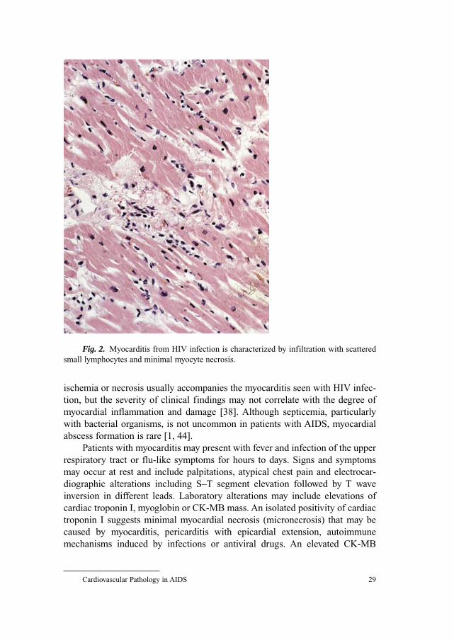
Fig. 2. Myocarditis from HIV infection is characterized by infiltration with scattered
small lymphocytes and minimal myocyte necrosis.
and/or cardiac troponin I level with a nondiagnostic electrocardiogram requires
clinical skill and echocardiography to differentiate acute myocardial infarction.
The frequency of myositis in HIV-infected patients makes myoglobin a less useful
marker. Myocarditis may be masked by concomitant bronchopulmonary disease
and/or wasting syndromes. A definitive diagnosis of myocarditis may require
endomyocardial biopsy [45].
In more than 80% of cases of myocarditis with HIV infection, a specific
etiologic factor, such as an opportunistic infectious agent, cannot be identified
as the cause of the myocarditis [29]. Nonspecific myocarditis can also appear
in persons with a history of intravenous drug use independent of HIV infection,
particularly when cocaine use is documented [46].
Since an infectious agent is not often identified in association with myo-
carditis in AIDS, direct cardiac involvement by HIV may explain some of the
cases. In one autopsy study of 440 patients with AIDS, 82 had cardiac findings,
with myocarditis seen in 30, and in 29 there was evidence of HIV nucleic acid in
myocytes by in situ hybridization. There was a myocarditis present in 25 of those
29 cases, and the inflammatory infiltrate was primarily lymphocytic and com-
posed of CD3 and CD8 cells. HIV may thus cause T lymphocyte activation with
cytokine release that potentiates myocardial damage. In 7 of those cases there was
coinfection with coxsackievirus group B, in 2 coinfection with Epstein-Barr virus
and in 1 cytomegalovirus was found [41]. In HIV-infected children, the two most
commonly detected viruses by PCR are adenovirus and cytomegalovirus [47].
HIV may cause direct damage to myocytes and may produce an autoimmune
process that secondarily involves the myocardium. Alternatively, HIV may act in
concert with other viruses to produce a myocarditis. By in situ hybridization, both
HIV and cytomegalovirus can be detected in myocytes of AIDS patients with
lymphocytic myocarditis and severe left ventricular dysfunction [30, 41].
The detection of antimyosin antibodies in patients with AIDS and cardio-
myopathy suggests an immune process. In addition, the HIV regulatory protein
nef binds to major histocompatibility complex class II receptors on antigen-
presenting cells, stimulating T lymphocytes to release cytokines such as tumor
necrosis factor �, �-interferon and interleukin 2 that mediate the immune
response [29]. Tumor necrosis factor � can produce a negative inotropic effect
by altering intracellular calcium homeostasis, mediated by nitric oxide [48].
Both tumor necrosis factor � and nitric oxide synthase can be detected with
greater intensity of staining in the myocardium of patients with HIV-associated
cardiomyopathy, particularly those with myocardial viral infection [49].
The most common opportunistic infectious agent associated with
myocarditis in AIDS is Toxoplasma gondii, observed as often as 12% in one
autopsy series with deaths from AIDS between 1987 and 1991 (fig. 3). There
may be regional differences in the incidence of T. gondii myocarditis, perhaps
Klatt 30
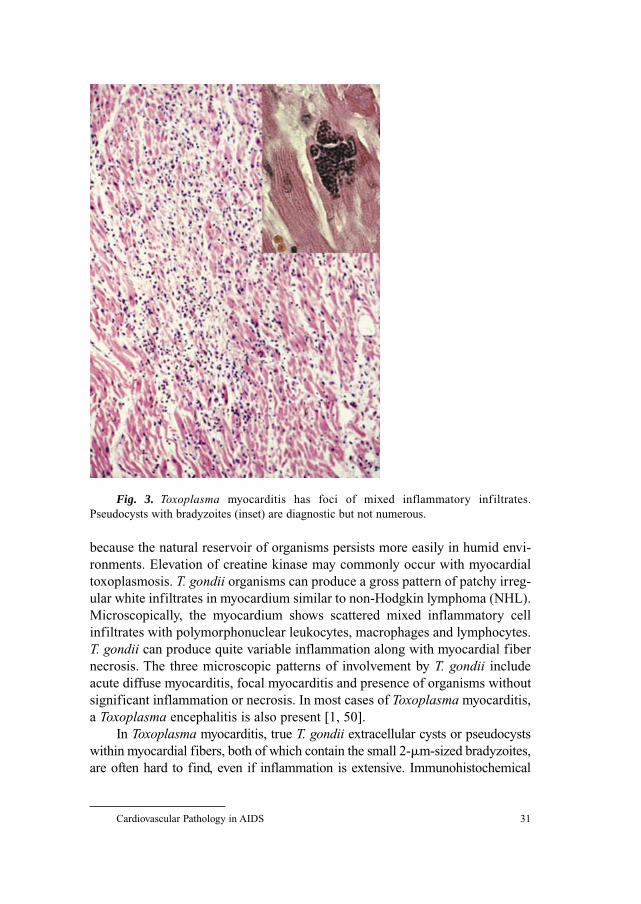
because the natural reservoir of organisms persists more easily in humid envi-
ronments. Elevation of creatine kinase may commonly occur with myocardial
toxoplasmosis. T. gondii organisms can produce a gross pattern of patchy irreg-
ular white infiltrates in myocardium similar to non-Hodgkin lymphoma (NHL).
Microscopically, the myocardium shows scattered mixed inflammatory cell
infiltrates with polymorphonuclear leukocytes, macrophages and lymphocytes.
T. gondii can produce quite variable inflammation along with myocardial fiber
necrosis. The three microscopic patterns of involvement by T. gondii include
acute diffuse myocarditis, focal myocarditis and presence of organisms without
significant inflammation or necrosis. In most cases of Toxoplasma myocarditis,
a Toxoplasma encephalitis is also present [1, 50].
In Toxoplasma myocarditis, true T. gondii extracellular cysts or pseudocysts
within myocardial fibers, both of which contain the small 2-�m-sized bradyzoites,
are often hard to find, even if inflammation is extensive. Immunohistochemical
Cardiovascular Pathology in AIDS 31
Fig. 3. Toxoplasma myocarditis has foci of mixed inflammatory infiltrates.
Pseudocysts with bradyzoites (inset) are diagnostic but not numerous.
staining may reveal free tachyzoites, the organisms that are found outside of cysts.
Otherwise, it is difficult with routine hematoxylin and eosin staining to distinguish
these free tachyzoites from fragments of inflammatory cells or myocytes that have
undergone necrosis within the areas of inflammation [51].
Fungal opportunistic infections of the heart occur infrequently. They are
often incidental findings at autopsy, and cardiac involvement is probably the
result of widespread dissemination, as exemplified by Candida and by the fungi
Cryptoccocus neoformans, Coccidioides immitis or Histoplasma capsulatum.
Fungal lesions are characterized grossly by the appearance of multiple small
rounded white plaques. They may have a hemorrhagic border, particularly
lesions caused by Aspergillus that can be angioinvasive. Microscopically, fungal
lesions have variable inflammatory infiltrates and necrosis, and a specific diag-
nosis is made by identifying yeast forms or hyphae of specific organisms, aided
by standard histologic stains such as Gomori methenamine silver or periodic
acid-Schiff [38]. The near absence of an inflammatory infiltrate accompanying
fungal organisms is a manifestation of immune system failure with progression
of AIDS to a late stage when opportunistic infections are more likely to be
widely disseminated to organs such as the heart.
Viral causes for myocarditis seen with AIDS include coxsackievirus B3,
Epstein-Barr virus and cytomegalovirus [29]. Patients living in endemic areas
for Trypanosoma cruzi, a form causing trypanosomiasis, may rarely develop a
pronounced myocarditis [52, 53]. Mycobacterium avium complex infection can
be widely disseminated and involve the heart with microscopic lesions charac-
terized by clusters of large macrophages filled with numerous acid-fast rod-
shaped organisms. Cardiac opportunistic infectious lesions in pediatric AIDS
cases are not frequent [36].
Pneumocystis carinii can involve the heart in cases with widespread
dissemination of this organism. Grossly, the epicardium and cut surfaces of the
myocardium may have a sandpaper-like quality due to the presence of multiple
pinpoint foci of calcification. Microscopically, this calcification is not accom-
panied by significant inflammatory cell infiltrates, but there may be deposits
of amorphous granular pink exudate similar to that seen in alveoli with
Pneumocystis pneumonia. The cysts may be difficult to recognize, even with
the Gomori methenamine silver stain, and diagnosis is aided by immunohisto-
chemical staining [1, 54].
Endocarditis
Both infectious and noninfectious endocarditis may complicate the course
of HIV infection. The noninfectious form of endocarditis seen with AIDS is
Klatt 32
nonbacterial thrombotic endocarditis (NBTE), also known as ‘marantic’ endo-
carditis. The debilitation of patients with AIDS, particularly in the terminal
course, may predispose to the formation of NBTE. This was once the most
common form of endocarditis with AIDS, seen in about 3–5% of persons dying
of AIDS at autopsy, most of them older than 50 years. Marantic endocarditis
has not been reported in the literature in the era of improved antiretroviral
therapy [45].
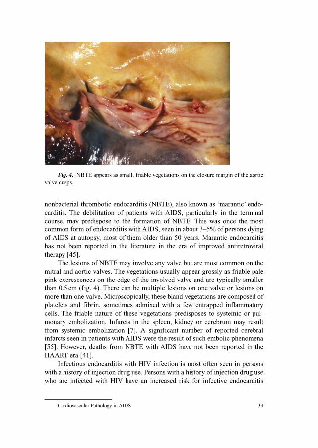
The lesions of NBTE may involve any valve but are most common on the
mitral and aortic valves. The vegetations usually appear grossly as friable pale
pink excrescences on the edge of the involved valve and are typically smaller
than 0.5 cm (fig. 4). There can be multiple lesions on one valve or lesions on
more than one valve. Microscopically, these bland vegetations are composed of
platelets and fibrin, sometimes admixed with a few entrapped inflammatory
cells. The friable nature of these vegetations predisposes to systemic or pul-
monary embolization. Infarcts in the spleen, kidney or cerebrum may result
from systemic embolization [7]. A significant number of reported cerebral
infarcts seen in patients with AIDS were the result of such embolic phenomena
[55]. However, deaths from NBTE with AIDS have not been reported in the
HAART era [41].
Infectious endocarditis with HIV infection is most often seen in persons
with a history of injection drug use. Persons with a history of injection drug use
who are infected with HIV have an increased risk for infective endocarditis
Cardiovascular Pathology in AIDS 33
Fig. 4. NBTE appears as small, friable vegetations on the closure margin of the aortic
valve cusps.
compared to HIV-seronegative injection drug users. Over 90% of cases of
infective endocarditis with HIV infection occur in injection drug users. The
mortality rate among HIV-infected patients is higher in those with CD4 cell
counts below 200/�l [56]. Infectious endocarditis, particularly in intravenous
drug users, can be associated with dilated cardiomyopathy [30, 57]. A case of
infectious endocarditis has been reported in an infant with HIV infection [58].
Right-sided endocardtitis is more common in persons with HIV infection,
because of the preponderance of cases associated with injection drug use, and the
tricuspid valve is the most commonly affected valve, in just over half of the cases.
Left-sided valvular disease occurs in 45% of cases, and multiple valves are
involved in 18%. The presence of left-sided heart involvement is associated with
an increased risk of death, compared with right-sided heart involvement [56].
Echocardiography may demonstrate mobile echodense masses attached to
the inflow side of valvular leaflets or mural endocardium. Transthoracic
echocardiography is useful for detecting relatively large valvular masses.
However, perivalvular abscess, leaflet perforation or rupture of valvular chordae
are better assessed by transesophageal echocardiography [45].
The vegetations of infectious endocarditis are grossly large, soft and friable,
and red to tan in color. The infection often produces valve leaflet destruction,
and there can be extension of infection to the adjacent endocardium or myocar-
dium. Microscopically there are numerous neutrophils admixed with bacterial
colonies, platelets and fibrin in the vegetations. These vegetations, being friable,
often produce septic emboli.
Vegetations that form on the tricuspid valve, or less frequently the pulmonic
valve, may predispose to pulmonary embolism and septic pulmonary infarction.
Such infarcts may appear as multiple opacities on a chest X-ray. Systemic
emboli from left-sided valvular endocarditis often involve the coronary arteries,
spleen, bowel, extremities and central nervous system. Cardiac rhythm alter-
ations such as atrioventricular block may suggest the presence of an abscess in
proximity to the atrioventricular node. Peripheral pulses may be absent with
embolic occlusion, or a pulsating mass may suggest a mycotic aneurysm.
Cerebral arterial mycotic aneurysms may lead to intracranial hemorrhage [45].
The most frequent agents isolated in cases of infectious endocarditis seen
with HIV infection are Staphylococcus aureus, Streptococcus pneumoniaeand Haemophilus influenzae [41]. Other bacterial agents may include Strepto-coccus, viridans group and Salmonella species. Salmonellosis typically occurs
with non-S.-typhi species, is seen with CD4 counts below 100/�l and has a
mortality rate of 50% [59]. S. aureus is by far the most common organism
(60% of cases). The mortality rate is higher with left-sided valvular disease,
multiple valve involvement and with lower CD4 counts. Most patients have a
coexisting pneumonia or meningitis [7, 60]. Increasing frequency of resistant
Klatt 34
bacterial strains, such as S. pneumoniae and H. influenzae, puts HIV-infected
patients at an increased risk for recurrent endocarditis, particularly when vac-
cines for these organisms may not be as effective as in immunocompetent
patients [61, 62].
Fungal endocarditis may occur with HIV infection. Aspergillus species,
Candida albicans, Pseudallescheria boydii, C. neoformans and H. capsulatuminfections have been reported. HIV disease produces immunologic impairment
that may predispose patients to fungal infection, including defects in granulocyte
numbers and function, defective macrophage phagocytosis and dysregulation of
cytokine production. The vegetations of Aspergillus endocarditis tend to be large
and friable, with possible embolization. Aspergillus is difficult to isolate with
blood cultures. Microscopically, the vegetations are composed in large part of
long branching septate hyphae [61, 63, 64].
Pericarditis and Pericardial Effusions
Pericardial effusions, most of which are small and clinically insignificant,
may be seen in up to 41% of persons during the course of HIV infection. The
incidence is 11% per year for those with AIDS [65]. However, in a third of
HIV-infected persons who have such an effusion, it is moderate to severe [66].
In almost half of the cases with a moderate to severe effusion, there is right
atrial diastolic compression, and a third of these have evidence of cardiac tam-
ponade requiring pericardiocentesis [67].
Clinical manifestations of pericarditis may include fever, chest pain with
radiation of dull pain to the left shoulder (aggravated by supine posture and
often decreased by sitting up and leaning forward) and pericardial friction rub
(over the left sternal border, usually accentuated by sitting up and leaning
forward). Pericardial effusion is suggested by the absence or weakness of the
apical impulse with an apparent increase in the area of dullness to percussion
over the left chest and over the hepatocardiac angle, muffled heart sounds, a
diffuse low-voltage electrocardiogram, electrical alternans of QRS complexes
and increased cardiac opacity on chest X-ray [45].
Despite the frequency of effusions, acute pericarditis is uncommon. The
characteristic findings of an elevated jugular venous pulse and pulsus para-
doxus may be masked by dehydration, with ‘low-pressure tamponade’ [67].
Histologically, mononuclear cells may also be seen as a mild epicarditis, which
may account for some pericardial effusions [7]. Tuberculous pericarditis, typi-
cally appearing with disseminated tuberculosis, may produce a granulomatous
inflammatory reaction, and resultant constrictive pericarditis has been reported
with HIV infection [68, 69].
Cardiovascular Pathology in AIDS 35
A specific etiology for a pericardial effusion in HIV-infected patients,
which can include a variety of infectious agents, is found in about a fourth of
cases. When cardiac tamponade is present with AIDS, causes include mycobac-
terial infection in 20% of cases, bacterial infection (most commonly S. aureusor S. pneumoniae) in 19%, lymphoma in 7%, Kaposi’s sarcoma (KS) in 5%,
viral infection in 3% and fungal infection in 1%, while 45% are idiopathic [70].
Persons with AIDS who have a pericardial effusion, regardless of size, tend
to have lower CD4 counts and decreased survival, compared to those without
effusions. Pericardial effusions can be seen in the late stages of AIDS where
such effusions are occasionally the immediate cause of death. Regardless of
etiology, a large pericardial effusion in AIDS carries a high mortality, and treat-
ment with a pericardial window is unlikely to prolong survival significantly [7].
In children with HIV infection, pericardial effusions may be seen in 16–26% of
cases. However, the effusions are typically small and asymptomatic [36].
Pathologic findings within the epicardium or pericardium are subtle.
Grossly, there may be no apparent changes or minimal opacification of the
epicardial or pericardial surfaces. A fibrinous exudate or fibrous adhesions are
uncommon. In one study of 52 hearts at autopsy from persons dying with
AIDS, there were 38 with a lymphocytic pericarditis, 1 with fibrinous peri-
carditis and 1 with pericardial fibrosis [71]. Hemorrhagic pericarditis is uncom-
mon and development of constrictive pericarditis unlikely [7].
Cardiac Neoplasms in AIDS
A high-grade NHL is one of the most common AIDS-diagnostic diseases
seen in the heart, occurring in about one sixth of AIDS cases when lymphoma is
diagnosed at autopsy (table 1). Such NHLs with AIDS tend to be seen late in the
course of HIV infection when CD4 counts are low. NHLs with AIDS are typically
extranodal and widely disseminated. Clinical findings may be nonspecific, and
the diagnosis goes undetected, or findings may be related to heart failure and
include dyspnea, chest pain and arrhythmias. Sudden death and myocardial
rupture have been reported but are rare complications [72]. The serum creatine
kinase is unlikely to be elevated. Diagnosis is typically made by echocardiography,
with histologic confirmation by endomyocardial biopsy [73].
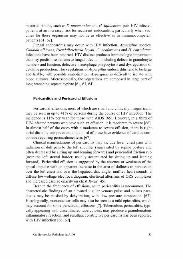
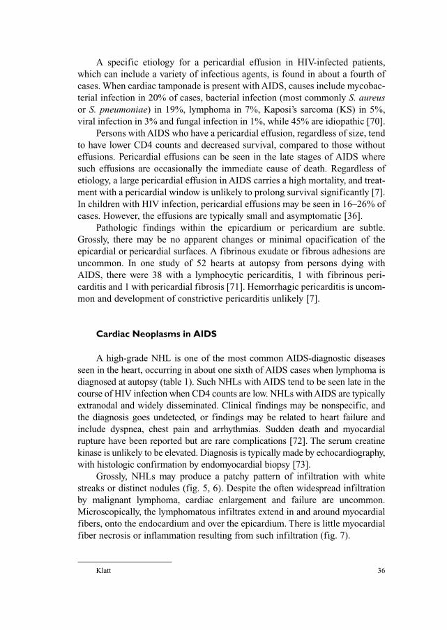
Grossly, NHLs may produce a patchy pattern of infiltration with white
streaks or distinct nodules (fig. 5, 6). Despite the often widespread infiltration
by malignant lymphoma, cardiac enlargement and failure are uncommon.
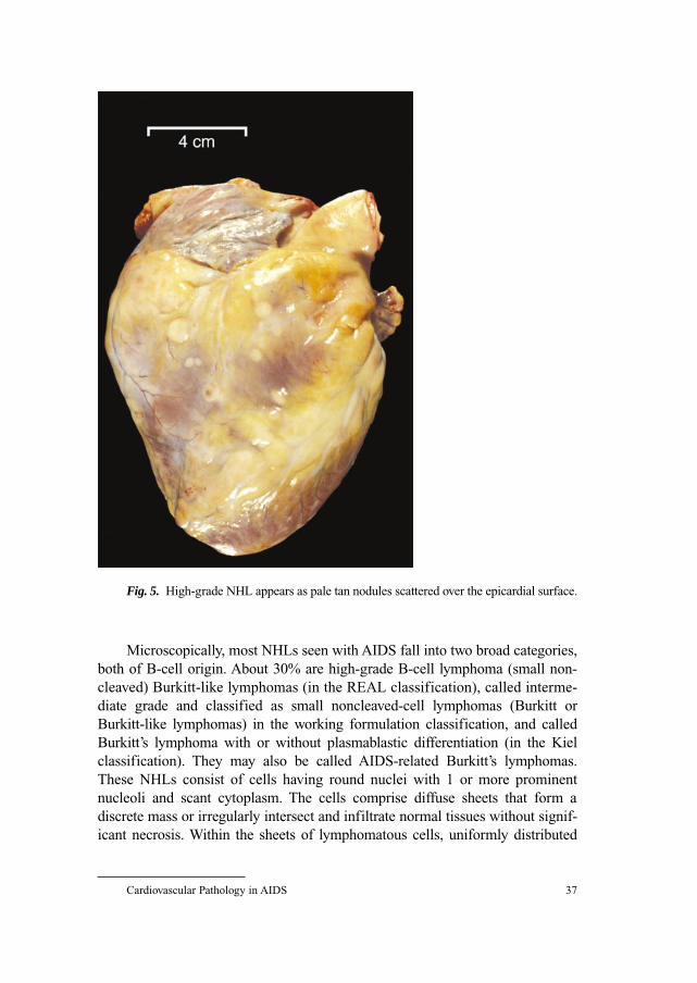
Microscopically, the lymphomatous infiltrates extend in and around myocardial
fibers, onto the endocardium and over the epicardium. There is little myocardial
fiber necrosis or inflammation resulting from such infiltration (fig. 7).
Klatt 36
Microscopically, most NHLs seen with AIDS fall into two broad categories,
both of B-cell origin. About 30% are high-grade B-cell lymphoma (small non-
cleaved) Burkitt-like lymphomas (in the REAL classification), called interme-
diate grade and classified as small noncleaved-cell lymphomas (Burkitt or
Burkitt-like lymphomas) in the working formulation classification, and called
Burkitt’s lymphoma with or without plasmablastic differentiation (in the Kiel
classification). They may also be called AIDS-related Burkitt’s lymphomas.
These NHLs consist of cells having round nuclei with 1 or more prominent
nucleoli and scant cytoplasm. The cells comprise diffuse sheets that form a
discrete mass or irregularly intersect and infiltrate normal tissues without signif-
icant necrosis. Within the sheets of lymphomatous cells, uniformly distributed
Cardiovascular Pathology in AIDS 37
Fig. 5. High-grade NHL appears as pale tan nodules scattered over the epicardial surface.
Klatt 38
Fig. 6. High-grade NHL is seen on the endocardial surface as small pale tan nodules.
Fig. 7. Microscopic finding of high-grade NHL showing lymphomatous infiltrates
extending in and around myocardial fibers along with little myocardial fiber necrosis.
macrophages containing phagocytosed debris are present, and occasional mitoses
are seen. Plasmablastic features including eccentric nuclei and a well-defined
Golgi zone may occur [74].
The second broad category of NHLs with AIDS, comprising virtually all pri-
mary CNS lymphomas seen with AIDS and about 70% of systemic lymphomas
in AIDS, is composed of large cells that are best described as diffuse large B-cell
lymphoma (in the REAL classification), which can be either large-cell immuno-
blastic lymphomas in the working formulation classification (immunoblastic with
or without plasmacytic differentiation in the Kiel classification) or large non-
cleaved-cell lymphomas in the working formulation classification (centroblastic
diffuse in the Kiel classification). The immunoblastic types consist of cells having
moderate to large amounts of cytoplasm with or without plasmacytic features of
eccentric nuclei and basophilic cytoplasm, large round to oval nuclei and prom-
inent single nucleoli. The large-cell types have less cytoplasm and 1 or more
peripheral nucleoli in a nucleus with finely dispersed chromatin. Necrosis is often
a prominent feature, and mitoses are frequent [74].
NHLs of the T-cell phenotype are much less frequent in AIDS but can
involve the heart [75].
Another type of lymphoproliferative disease seen with HIV infection
occurs in association with concomitant human herpesvirus 8 infection and is
manifested as a primary effusion lymphoma involving body cavities, including
the pericardial sac. In such cases, an effusion is present in which the neoplastic
cells can be identified, but a grossly apparent mass lesion is not identified, and
only a fibrinous exudate or fibrous thickening of the cavity surface is noted.
Microscopically, the mesothelial lining is obliterated and the lymphoma cells
have plasmablastic features with medium-sized to large peripherally placed
nuclei, prominent nucleoli, basophilic cytoplasm, numerous mitoses, apoptosis
and more than 1 nucleus per cell. The cells mark with CD45 and CD138, but
are variably positive for CD20. Subserosal lymphatics may contain the neo-
plastic cells [76].
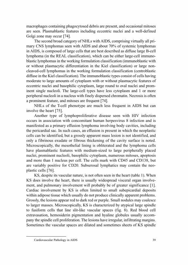
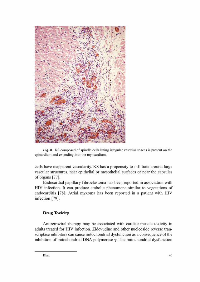
KS, despite its vascular nature, is not often seen in the heart (table 1). When
KS does involve the heart, there is usually widespread visceral organ involve-
ment, and pulmonary involvement will probably be of greater significance [1].
Cardiac involvement by KS is often limited to small subepicardial deposits
within adipose tissue which usually do not produce clinically apparent problems.
Grossly, the lesions appear red to dark red or purple. Small nodules may coalesce
to larger masses. Microscopically, KS is characterized by atypical large spindle
to fusiform cells that line slit-like vascular spaces (fig. 8). Red blood cell
extravasation, hemosiderin pigmentation and hyaline globules usually accom-
pany the spindle cell proliferation. The lesions have irregular, infiltrating margins.
Sometimes the vascular spaces are dilated and sometimes sheets of KS spindle
Cardiovascular Pathology in AIDS 39
cells have inapparent vascularity. KS has a propensity to infiltrate around large
vascular structures, near epithelial or mesothelial surfaces or near the capsules
of organs [77].
Endocardial papillary fibroelastoma has been reported in association with
HIV infection. It can produce embolic phenomena similar to vegetations of
endocarditis [78]. Atrial myxoma has been reported in a patient with HIV
infection [79].
Drug Toxicity
Antiretroviral therapy may be associated with cardiac muscle toxicity in
adults treated for HIV infection. Zidovudine and other nucleoside reverse tran-
scriptase inhibitors can cause mitochondrial dysfunction as a consequence of the
inhibition of mitochondrial DNA polymerase �. The mitochondrial dysfunction
Klatt 40
Fig. 8. KS composed of spindle cells lining irregular vascular spaces is present on the
epicardium and extending into the myocardium.
may lead to cardiomyopathy [80]. However, a similar cardiac disease in children
has not been seen [81]. In addition, the zidovudine-induced mitochondrial dis-
order with massive liver steatosis, myopathy, lactic acidosis and mitochondrial
DNA depletion which affects liver and skeletal muscle does not appear to affect
the myocardium [82].
A number of pharmacologic agents may induce significant cardiac arrhyth-
mias. One of the most common of these arrhythmias is prolongation of the Q–T
interval, which is thought to develop from myocardial ion channel blockage. Such
drugs include macrolide antibiotics such as erythromycin (less frequently clar-
ithromycin), trimethoprim-sulfamethoxazole, fluoroquinolones, amphotericin B,
azole antifungals such as ketoconazole, pentamidine, antihistamines, antipsychotic
medications such as haloperidol and tricyclic antidepressants [19].
Bradycardia is seen in children treated with amphotericin B. Doxorubicin
used in therapies for KS and NHLs has a dose-related effect on cardiomyo-
pathy [83]. �-Interferon administered as part of prolonged antiretroviral
therapy or the antivirals ganciclovir and foscarnet may also lead to a dilated
cardiomyopathy [84]. Since �-interferon is not associated with myocardial
dysfunction in patients without HIV, it may have a synergistic effect with HIV
infection [85].
Although it has not as yet been demonstrated that cocaine use by an HIV-
infected patient increases the risk of cardiac complications, both cocaine and
HIV infection are associated with myocarditis, and cocaine is associated with
contraction band necrosis that may accentuate ischemic heart disease and car-
diac dysfunction. A hyperadrenergic state with catecholamine release produced
by cocaine use may exacerbate myocarditis [86].
Pulmonary Hypertension
HIV-related pulmonary hypertension was first described in 1987 and has
been observed with increased frequency in association with HIV infection and
AIDS [87]. It is a form of pulmonary hypertension in patients for whom no fac-
tor other than HIV infection is present to explain the findings. The incidence is
estimated to be 1/200 in persons with HIV infection, compared with 1/200,000 in
the general population [29]. There is a slight male preponderance and a wide age
range, with a median age of 33 years [88]. There does not appear to be an asso-
ciation with either CD4 lymphocyte counts or with the existence of pulmonary
infections and the onset of pulmonary hypertension [87].
Clinical findings with HIV-related pulmonary hypertension are similar
to primary pulmonary hypertension and include progressive shortness of
breath, pedal edema, nonproductive cough, fatigue, syncope and chest pain.
Cardiovascular Pathology in AIDS 41
Chest X-ray findings include cardiomegaly and pulmonary arterial prominence.
By electrocardiography there is typically right ventricular hypertrophy, right
atrial abnormality and right axis deviation. An echocardiogram is likely to
demonstrate right heart chamber enlargement, tricuspid regurgitation and para-
doxical septal motion [87]. The mean time to diagnosis from onset of symptoms
ranges from 6 to 30 months. The mean time from diagnosis to death is 6 months,
with death occurring from right-sided heart failure, cardiogenic shock and
sudden death. The acute response to epoprostenol therapy is similar to that for
non-HIV-infected patients. The course is slightly more fulminant than in
patients with primary pulmonary hypertension, with half of the patients dying
in a year [88, 89].
There has been no direct evidence for HIV itself in lung tissue as a cause
for pulmonary hypertension, either by electron microscopy or immunohisto-
chemical staining. Inflammation resulting from HIV infection, with release of
inflammatory mediators and growth factors, such as vascular endothelial
growth factor or platelet-derived growth factor, has been postulated as an incit-
ing event. The HIV-1 envelope glycoprotein known as gp120 can stimulate pro-
duction of endothelin 1 and tumor necrosis factor � with an effect on vascular
endothelium [90]. HIV-infected persons who possess the HLA-DR6 and -DR52
alleles have a predisposition to the development of pulmonary hypertension
[89]. Regression of HIV-related pulmonary hypertension has been reported in
association with successful HAART therapy and suppression of HIV-1 in
plasma [91].
Pathologic findings with HIV-related pulmonary hypertension are similar
to primary pulmonary hypertension. Histologically, the patterns of disease
range from plexogenic pulmonary angiopathy (sometimes in association with
lymphoplasmacytic pulmonary infiltrates) to thrombotic pulmonary arteriopathy
and to pulmonary veno-occlusive disease [88, 89]. The most common finding
is a pulmonary plexiform arteriopathy. Other arterial findings may include iso-
lated medial hypertrophy or medial hypertrophy with intimal fibrosis. Less fre-
quently observed is pulmonary veno-occlusive disease [87].
Miscellaneous Findings
Rheumatic inflammatory changes, ranging from rare scattered Anitschkov
myocytes to well-formed Aschoff nodules similar to those seen in rheumatic
heart disease, are rarely reported to occur in AIDS. However, chronic rheumatic
sequelae of fibrosis or valvular disease have not been seen in AIDS [92].
Vasculitis involving small and medium-sized arteries has been infrequently
seen in patients with HIV infection. In about a third of cases, the pattern of
Klatt 42
vasculitis resembles a distinct type of vasculitis such as polyarteritis nodosa,
Henoch-Schönlein purpura or drug-induced hypersensitivity vasculitis. In the
remaining patients, the vasculitis has variable features. Such vasculitides
may result from HIV-induced immunologic abnormalities, infections and drugs
[93, 94].
A vasculopathy involving large arteries including the aorta and its
branches has also been described in young adults with AIDS. The features of
this vasculopathy overlap with Takayasu’s disease, characterized by involve-
ment of the aorta and its branches by a panarteritis that leads to focal stenosis
or occlusive lesions. Grossly there is arterial wall thickening and focal raised
intimal plaques. Microscopically, early lesions have infiltrates of neutrophils,
macrophages, lymphocytes and occasional Langhans giant cells. Late lesions
are marked by fibrosis, intimal thickening and secondary atherosclerotic
changes. With large-artery vasculopathy there is a propensity for the appearance
of single or multiple aneurysms of variable size. There can be angiogenesis with
proliferation of slit-like channels in the adventitia. The appearance of these
lesions in the aorta may also be due to vasculitis of vasa vasora or small adven-
titial arteries in aortic branches. There does not seem to be an association of this
vasculopathy with opportunistic infections [94, 95].
Kawasaki disease, also known as mucocutaneous lymph node syndrome, is
an acute systemic vasculitis seen most commonly in children aged �5 years.
Untreated Kawasaki disease has a mortality rate of 0.8% due to coronary artery
aneurysm formation and occlusion during the early convalescent phase of the
illness. A similar disease has been described in HIV-infected adults manifesting
with fever �5 days’ duration, bulbar conjunctivitis without exudates, swelling
and pain in the hands and feet, a diffuse erythematous rash and either an aseptic
pharyngitis or tender cervical adenopathy [96].
Aortic root dilation, both progressive and nonprogressive, has been
described in HIV-infected children. This dilation has been shown to correlate
with increased plasma HIV-1 RNA levels and decreased CD4 counts, as well as
left ventricular dilation. Markers of stress-modulated growth, including heart
rate, systolic blood pressure, stroke volume or hematocrit, do not correlate with
this dilation [97].
Congenital heart disease has been reported in children with HIV infection.
Rates of congenital heart disease with HIV infection, however, are not different
from those for similarly screened populations. The reported malformations
include atrial septal defect, ventricular septal defect, patent ductus arteriosus,
tricuspid valve prolapse, mitral valve prolapse, valvar pulmonic stenosis,
subaortic stenosis and single coronary artery [98]. By fetal ultrasound, there is
increased right and left ventricular wall thickness in the hearts of fetuses of
HIV-infected women [99].
Cardiovascular Pathology in AIDS 43
References
1 Klatt EC, Nichols L, Noguchi TT: Evolving trends revealed by autopsies of patients with AIDS.
Arch Pathol Lab Med 1994;118:884–890.
2 Lewis W: Cardiac findings from 115 autopsies. Prog Cardiovasc Dis 1989;32:207–215.
3 Yunis NA, Stone VE: Cardiac manifestations of HIV/AIDS: A review of disease spectrum and
clinical management. J Acquir Immune Defic Syndr Hum Retrovirol 1998;18:145–154.
4 Morgello S, Mahboob R, Yakoushina T, Khan S, Hague K: Autopsy findings in a human immuno-
deficiency virus-infected population over 2 decades: Influences of gender, ethnicity, risk factors,
and time. Arch Pathol Lab Med 2002;126:182–190.
5 McKenzie R, Travis WD, Dolan SA, Pittaluga S, Feuerstein IM, Shelhamer J, Yarchoan R, Masur H:
The causes of death in patients with human immunodeficiency virus infection: A clinical and
pathologic study with emphasis on the role of pulmonary diseases. Medicine 1991;70:326–343.
6 Francis CK: Cardiac involvement in AIDS. Curr Probl Cardiol 1990;15:569–639.
7 Rerkpattanapipat P, Wongpraparut N, Jacobs LE, Kotler ME: Cardiac manifestations of acquired
immunodeficiency syndrome. Arch Intern Med 2000;160:602–608.
8 Pugliese A, Isnardi D, Saini A, Scarabelli T, Raddino R, Torre D: Impact of highly active anti-
retroviral therapy in HIV-positive patients with cardiac involvement. J Infect 2000;40:282–284.
9 Selik RM, Byers RH Jr, Dworkin MS: Trends in diseases reported on US death certificates that
mentioned HIV infection, 1987–1999. J Acquir Immune Defic Syndr 2002;29:378–387.
10 Falusi OM, Aberg JA: HIV and cardiovascular risk factors. AIDS Read 2001;11:263–268.
11 Grunfeld C, Pang M, Doerrler W, Shigenaga JK, Jensen P, Feingold KR: Lipids, lipoproteins,
triglyceride clearance, and cytokines in human immunodeficiency virus infection and the acquired
immunodeficiency syndrome. J Clin Endocrinol Metab 1992;74:1045–1052.
12 Chi D, Henry J, Kelley J, Thorpe R, Smith JK, Krishnaswamy G: The effects of HIV infection on
endothelial function. Endothelium 2000;7:223–242.
13 Vallance P, Collier J, Bhagat K: Infection, inflammation, and infarction: Does acute endothelial
dysfunction provide a link? Lancet 1997;349:1391–1392.
14 Passalaris JD, Sepkowitz KA, Glesby MJ: Coronary artery disease and human immunodeficiency
virus infection. Clin Infect Dis 2000;31:787–797.
15 Fantoni M, Del Borgo C, Autore C, Barbaro G: Metabolic disorders and cardiovascular risk in
HIV-infected patients treated with antiretroviral agents. Ital Heart J 2002;3:294–299.
16 Duong M, Buisson M, Cottin Y, Piroth L, Lhuillier I, Grappin M, Chavanet P, Wolff JE, Portier H:
Coronary heart disease associated with the use of human immunodeficiency virus (HIV)-1 protease
inhibitors: Report of four cases and review. Clin Cardiol 2001;24:690–694.
17 Depairon M, Chessex S, Sudre P, Rodondi N, Doser N, Chave JP, Riesen W, Nicod P, Darioli R,
Telenti A, Mooser V, Swiss HIV Cohort Study: Premature atherosclerosis in HIV-infected indi-
viduals – Focus on protease inhibitor therapy. AIDS 2001;15:329–334.
18 Carr A: HIV protease inhibitor-related lipodystrophy syndrome. Clin Infect Dis 2000;30(suppl 2):
S135–S142.
19 Fantoni M, Autore C, Del Borgo C: Drugs and cardiotoxicity in HIV and AIDS. Ann NY Acad Sci
2001;946:179–199.
20 Sullivan AK, Feher MD, Nelson MR, Gazzard BG: Marked hypertriglyceridemia associated with
ritonavir therapy. AIDS 1998;12:1393–1394.
21 Koppel K, Bratt G, Schulman S, Bylund H, Sandstrom E: Hypofibrinolytic state in HIV-1-infected
patients treated with protease inhibitor-containing highly active antiretroviral therapy. J Acquir
Immune Defic Syndr 2002;29:441–449.
22 Flynn TE, Bricker LA: Myocardial infarction in HIV-infected men receiving protease inhibitors.
Ann Intern Med 1999;131:548.
23 Rickerts V, Brodt H, Staszewski S, Stille W: Incidence of myocardial infarctions in HIV-infected
patients between 1983 and 1998: The Frankfurt HIV Cohort Study. Eur J Med Res 2000;5:329–333.
24 Jutte A, Schwenk A, Franzen C, Romer K, Diet F, Diehl V, Fatkenheuer G, Salzberger B:
Increasing morbidity from myocardial infarction during HIV protease inhibitor treatment? AIDS
1999;13:1796–1797.
Klatt 44
25 David MH, Hornung R, Fichtenbaum CJ: Ischemic cardiovascular disease in persons with human
immunodeficiency virus infection. Clin Infect Dis 2002;34:98–102.
26 Tabib A, Leroux C, Mornex JF, Loire R: Accelerated coronary atherosclerosis and arteriosclero-
sis in young human-immunodeficiency-virus-positive patients. Coron Artery Dis 2000;11:
41–46.
27 Currie PF, Jacob AJ, Foreman AR, Elton RA, Brettle RP, Boon NA: Heart muscle disease related
to HIV infection: Prognostic implications. BMJ 1994;309:1605–1607.
28 Martinez-Garcia T, Sobrino JM, Pujol E, Galvez J, Benitez E, Giron-Gonzalez JA: Ventricular
mass and diastolic function in patients infected by the human immunodeficiency virus. Heart
2000;84:620–624.
29 Barbaro G, Lipshultz SE: Pathogenesis of HIV-associated cardiomyopathy. Ann NY Acad Sci
2001;946:57–81.
30 Barbaro G, Di Lorenzo G, Grisorio B, Barbarini G: Incidence of dilated cardiomyopathy and detec-
tion of HIV in myocardial cells of HIV-positive patients. N Engl J Med 1998;339:1093–1099.
31 Currie PF, Goldman JH, Caforio ALP, Jacob AJ, Baig MK, Brettle RP, Haven AJ, et al: Cardiac
autoimmunity in HIV related heart muscle disease. Heart 1998;79:599–604.
32 Raidel SM, Haase C, Jansen NR, Russ RB, Sutliff RL, Velsor LW, Day BJ, et al: Targeted myocardial
transgenic expression of HIV Tat causes cardiomyopathy and mitochondrial damage. Am J Physiol
Heart Circ Physiol 2002;282:H1672–H1678.
33 Sundstrom JB, Mosunjac M, Martinson DE, Bostik P, Donahoe RM, Gravanis MB, Ansari AA:
Effects of norepinephrine, HIV type 1 infection, and leukocyte interactions with endothelial cells
on the expression of matrix metalloproteinases. AIDS Res Hum Retroviruses 2001;17:1605–1614.
34 Dworkin BM: Selenium deficiency in HIV infection and the acquired immunodeficiency syndrome
(AIDS). Chem Biol Interact 1994;91:181–186.
35 Lipshultz SE, Orav EJ, Sanders SP, Hale AR, McIntosh K, Colan SD: Cardiac structure and function
in children with human immunodeficiency virus infection treated with zidovudine. N Engl J Med
1992;327:1260–1265.
36 Keesler MJ, Fisher SD, Lipshultz SE: Cardiac manifestations of HIV infection in infants and children.
Ann NY Acad Sci 2001;946:169–178.
37 Miller TL, Orav EJ, Colan SD, Lipshultz SE: Nutritional status and cardiac mass and function in
children infected with the human immunodeficiency virus. Am J Clin Nutr 1997;66:660–664.
38 Klatt EC: Cardiovascular pathology in AIDS; in Klatt (ed): Pathology of AIDS, version 11.
pp 165–167, http://medstat.med.utah.edu/WebPath/TUTORIAL/AIDS/AIDS.html (February 5,
2002).
39 D’Amati G, Di Giola CRT, Gallo P: Pathological findings of HIV-associated cardiovascular disease.
Ann NY Acad Sci 2001;946:23–45.
40 Herskowitz A, Willoughby SB, Vlahov D, Baughman KL, Ansari AA: Dilated heart muscle disease
associated with HIV infection. Eur Heart J 1995;16(suppl O):50–55.
41 Barbaro G, Di Lorenzo G, Grisorio B, Barbarini G: Cardiac involvement in the acquired immuno-
deficiency syndrome: A multicenter clinical-pathological study. AIDS Res Hum Retroviruses
1998;14:1071–1077.
42 Artez HT: Myocarditis: The Dallas criteria. Hum Pathol 1987;18:619–624.
43 Anderson DW, Virmani R: Emerging patterns of heart disease in human immunodeficiency virus
infection. Hum Pathol 1990;21:253–259.
44 Almond DS, Lea BI, Saltissi S, Neal TJ, Carey PB: Interventricular septal abscess formation in an
HIV-positive man. Int J STD AIDS 1999;10:749–750.
45 Barbaro G, Fisher SD, Giancaspro G, Lipshultz SE: HIV-associated cardiovascular complications:
A new challenge for emergency physicians. Am J Emerg Med 2001;19:566–574.
46 Turnicky RP, Goodin J, Smialek JE, Herskowitz A, Beschorner WE: Incidental myocarditis with
intravenous drug abuse: The pathology, immunopathology, and potential implications for human
immunodeficiency virus-associated myocarditis. Hum Pathol 1992;23:138–143.
47 Bowles NE, Kearney DL, Ni J, Perez-Atayde AR, Kline MW, Bricker JT, Ayres NA, Lipshultz SE,
Shearer WT, Towbin JA: The detection of viral genomes by polymerase chain reaction in the
myocardium of pediatric patients with advanced HIV disease. J Am Coll Cardiol 1999;34:857–865.
Cardiovascular Pathology in AIDS 45

48 Finkel MS, Oddis CV, Jacob TD, Watkins SC, Hattler BG, Simmons RL: Negative inotropic effects
of cytokines on the heart mediated by nitric oxide. Science 1992;257:387–389.
49 Barbaro G, Di Lorenzo G, Soldini M, Giancaspro G, Grisorio B, Pellicelli A, Barbarini G:
The intensity of myocardial expression of inducible nitric oxide synthase influences the clinical
course of human immunodeficiency virus-asssociated cardiomyopathy. Circulation 1999;100:
933–939.
50 Hofman P, Drici MD, Gibelin P, Michiels JF, Thyss A: Prevalence of toxoplasma myocarditis in
patients with the acquired immunodeficiency syndrome. Br Heart J 1993;70:376–381.
51 Tschirhart DL, Klatt EC: Disseminated toxoplasmosis in the acquired immunodeficiency syndrome.
Arch Pathol Lab Med 1988;112:1237–1241.
52 Oddó D, Casanova M, Acuña G, Ballesteros J, Morales B: Acute Chagas’ disease (trypanosomiasis
americana) in acquired immunodeficiency syndrome: Report of two cases. Hum Pathol 1992;23:
41–44.
53 Sartori AM, Lopes MH, Benvenuti LA, Caramelli B, di Pietro A, Nunes EV, Ramirez LP,
Shikanai-Yasuda MA: Reactivation of Chagas’ disease in a human immunodeficiency virus-
infected patient leading to severe heart disease with a late positive direct microscopic examination
of the blood. Am J Trop Med Hyg 1998;59:784–786.
54 Klatt EC: Cardiovascular pathology in AIDS; in Klatt (ed): Pathology of AIDS, version 11.
pp 63–64, http://medstat.med.utah.edu/WebPath/TUTORIAL/AIDS/AIDS.html (February 5, 2002).
55 Pinto AN: AIDS and cerebrovascular disease. Stroke 1996;27:538–543.
56 Cicalini S, Forcina G, De Rosa FG: Infective endocarditis in patients with human immunodefi-
ciency virus infection. J Infect 2001;42:267–271.
57 Ferguson DW, Volpp B: Cardiovascular complications of AIDS. Heart Dis Stroke 1994;3:388–394.
58 Van Doorn CA, Yates R, Tsang VT: Endocarditis as the first presentation of AIDS in infancy. Arch
Dis Child 1998;79:179–180.
59 Fernandez Guerrero ML, Ramos JM, Nunez A, Nunez A, de Gorgolas M: Focal infections due to
non-typhi Salmonella in patients with AIDS: Report of 10 cases and review. Clin Infect Dis 1997;
25:690–697.
60 Cicalini S, Forcina G, De Rosa FG: Infective endocarditis in patients with human immunodeficiency
infection. J Infect 2001;42:267–271.
61 Gonzaga C, Dever LL: Emergence of penicillin resistance in recurrent pneumococcal endocarditis
in an HIV-infected patient. Microb Drug Resist 1998;4:61–63.
62 Munoz P, Miranda ME, Llancaqueo A, Pelaez T, Rodriguez-Creixems M, Bouza E: Haemophilus
species bacteremia in adults: The importance of the human immunodeficiency virus epidemic.
Arch Intern Med 1997;157:1869–1873.
63 Petrosillo N, Pellicelli AM, Cicalini S, Conte A, Goletti D, Palmieri F: Endocarditis caused by
Aspergillus species in injection drug users. Clin Infect Dis 2001;33:97–99.
64 Scapellato PG, Desse J, Negroni R: Acute disseminated histoplasmosis and endocarditis. Rev Inst
Med Trop Sao Paulo 1998;40:19–22.
65 Heidenreich PA, Eisenberg MJ, Kee LL, Somelofski CA, Hollander H, Schiller NB, Cheitlin MD:
Pericardial effusion in AIDS: Incidence and survival. Circulation 1995;92:3229–3234.
66 Hsia J, Ross AM: Pericardial effusion and pericardiocentesis in human immunodeficiency virus
infection. Am J Cardiol 1994;74:94–96.
67 Silva-Cardoso J, Moura B, Martins L, Mota-Miranda A, Rocha-Goncalves F, Lecour H: Pericardial
involvement in human immunodeficiency virus infection. Chest 1999;115:418–422.
68 Trautner BW, Darouiche RO: Tuberculous pericarditis: Optimal diagnosis and management. Clin
Infect Dis 2001;33:954–961.
69 Eckstein FS, Bohlmann MK, Balmer MC, Carrel TP: Constrictive tuberculous pericarditis in an
HIV-positive patient. Eur J Cardiothorac Surg 2001;19:940–942.
70 Chen Y, Brennessel D, Walters J, Johnson M, Rosner F, Raza M: Human immunodeficiency virus-
associated pericardial effusion: Report of 40 cases and review of the literature. Am Heart J 1999;
137:516–521.
71 Lanjewar DN, Katdare GA, Jain PP, Hira SK: Pathology of the heart in acquired immunodeficiency
syndrome. Indian Heart J 1998;50:321–325.
Klatt 46
72 Sanna P, Bertoni F, Zucca E, Roggero E, Passega Sidler E, Fiori G, Pedrinis E, Mombelli G,
Cavalli F: Cardiac involvement in HIV-related non-Hodgkin’s lymphoma: A case report and short
review of the literature. Ann Hematol 1998;77:75–78.
73 Duong M, Dubois C, Buisson M, Eicher JC, Grappin M, Chavanet P, Portier H: Non-Hodgkin’s
lymphoma of the heart in patients infected with human immunodeficiency virus. Clin Cardiol
1997;20:497–502.
74 Raphael M, Gentilhomme O, Tulliez M, Byron PA, Diebold J: Histopathologic features of high-
grade non-Hodgkin’s lymphomas in acquired immunodeficiency syndrome. Arch Pathol Lab Med
1991;115:15–20.
75 Burke AP, Andriko JA, Virmani R: Anaplastic large cell lymphoma (CD30�), T-phenotype, in the
heart of an HIV-positive man. Cardiovasc Pathol 2000;9:49–52.
76 Ascoli V, Signoretti S, Onetti-Muda A, Pescarmona E, Della Rocca C, Nardi F, Mastroianni CM,
Gastaldi R, Pistilli A, Gaidano G, Carbone A, Lo-Coco F: Primary effusion lymphoma in HIV-
infected patients with multicentric Castleman’s disease. J Pathol 2001;193:200–209.
77 Tappero JW, Conant MA, Wolfe SF, Berger TG: Kaposi’s sarcoma: Epidemiology, pathogenesis,
histology, clinical spectrum, staging criteria and therapy. J Am Acad Dermatol 1993;28:371–395.
78 Paraf F, Berrebi A, Chauvaud S, Fornes P, Farge D, Bruneval P: Mitral papillary fibroelastoma in
an HIV-infected patient. Presse Méd 1999;28:962–964.
79 Shaw AJ, Mclean KA: Atrial myxoma and HIV infection. Sex Transm Infect 2000;76:144.
80 White AJ: Mitochondrial toxicity and HIV therapy. Sex Transm Infect 2001;77:158–173.
81 Lipshultz SE, Easley KA, Orav EJ, Kaplan S, Starc TJ, Bricker JT, Lai WW, Moodie DS, Sopko G,
McIntosh K, Colan SD: Absence of cardiac toxicity of zidovudine in infants. N Engl J Med
2000;343:759–766.
82 Chariot P, Drogou I, de Lacroix-Szmania I, Eliezer-Vanerot MC, Chazaud B, Lombes A, Schaeffer A,
Zafrani ES: Zidovudine-induced mitochondrial disorder with massive liver steatosis, myopathy, lactic
acidosis, and mitochondrial DNA depletion. J Hepatol 1999;30:156–160.
83 Sayed-Ahmed MM, Khattab MM, Gad MZ, Osman AM: Increased plasma endothelin-1 and cardiac
nitric oxide during doxorubicin-induced cardiomyopathy. Pharmacol Toxicol 2001;89:140–144.
84 Brown DL, Sather S, Cheitlin MD: Reversible cardiac dysfunction associated with foscarnet therapy
for cytomegalovirus esophagitis in an AIDS patient. Am Heart J 1993;125:1439–1441.
85 Deyton LR, Walker RE, Kovacs JA, Herpin B, Parker M, Masur H, Fauci AS, Lane HC: Reversible
cardiac dysfunction associated with interferon alpha therapy in AIDS patients with Kaposi’s sarcoma.
N Engl J Med 1989;321:1246–1249.
86 Soodini G, Morgan JP: Can cocaine abuse exacerbate the cardiac toxicity of human immunodefi-
ciency virus? Clin Cardiol 2001;24:177–181.
87 Mehta NJ, Khan IA, Mehta RN, Sepkowitz DA: HIV-related pulmonary hypertension: Analytic
review of 131 cases. Chest 2000;118:1133–1141.
88 Seoane L, Shellito J, Welsh D, de Boisblanc BP: Pulmonary hypertension associated with HIV
infection. South Med J 2001;94:635–639.
89 Mesa RA, Edell ES, Dunn WF, Edwards WD: Human immunodeficiency virus infection and pul-
monary hypertension: Two new cases and a review of 86 reported cases. Mayo Clin Proc 1998;73:
37–45.
90 Ehrenreich H, Rieckmann P, Sinowatz F, Weih KA, Arthur LO, Goebel FD, Burd PR, Coligan JE,
Clouse KA: Potent stimulation of monocytic endothelin-1 production by HIV-1 glycoprotein.
J Immunol 1993;150:4601–4609.
91 Speich R, Jenni R, Opravil M, Jaccard R: Regression of HIV-associated pulmonary arterial hyper-
tension and long-term survival during antiretroviral therapy. Swiss Med Wkly 2001;131:663–665.
92 Di Carlo FJ, Anderson DW, Virmani R, Burns W, Macher AM, Rotiguez J, Petitto S: Rheumatic
heart disease in a patient with acquired immunodeficiency syndrome. Hum Pathol 1989;20:
917–920.
93 Gherardi R, Belec L, Mhiri C, Gray F, Lescs MC, Sobel A, Guillevin L, Wechsler J: The spectrum
of vasculitis in human immunodeficiency virus-infected patients: A clinicopathologic evaluation.
Arthritis Rheum 1993;36:1164–1174.
94 Chetty R: Vasculitides associated with HIV infection. J Clin Pathol 2001;54:275–278.
Cardiovascular Pathology in AIDS 47
95 Chetty R, Batitang S, Nair R: Large artery vasculopathy in HIV-positive patients: Another vasculitic
enigma. Hum Pathol 2000;31:374–379.
96 Johnson RM, Little JR, Storch GA: Kawasaki-like syndromes associated with human immuno-
deficiency virus infection. Clin Infect Dis 2001;32:1628–1634.
97 Lai WW, Colan SD, Easley KA, Lipshultz SE, Starc TJ, Bricker JT, Kaplan S: Dilation of the aortic
root in children infected with human immunodeficiency virus type 1: The Prospective P2C2 HIV
Multicenter Study. Am Heart J 2001;141:661–670.
98 Lai WW, Lipshultz SE, Easley KA, Starc TJ, Drant SE, Bricker JT, Colan SD, Moodie DS, Sopko G,
Kaplan S: Prevalence of congenital cardiovascular malformations in children of human immunodefi-
ciency virus infected women. J Am Coll Cardiol 1998;32:1749–1755.
99 Hornberger LK, Lipshultz SE, Easley KA, Colan SD, Schwartz M, Kaplan S, Starc TJ, Ayres NA,
Lai WW, Moodie DS, Kasten-Sportes C, Sanders SP: Cardiac structure and function in fetuses of
mothers infected with HIV: The Prospective P2C2 HIV Multicenter Study. Am Heart J 2000;
140:575–584.
Dr. Edward C. Klatt, MD
Florida State University, College of Medicine
Tallahassee, FL 32312 (USA)
Tel. �1 850 644 9397, Fax �1 850 644 9399, E-Mail [email protected]
Klatt 48