pathology-brs-4thed.pdf - Medical Students
450
-
Upload
khangminh22 -
Category
Documents
-
view
4 -
download
0
Transcript of pathology-brs-4thed.pdf - Medical Students


-
Contents
Preface vii Acknowledgments ix
1.
2.
3.
CELLULAR REACTION TO INJURY
I. Adaptation to Environmental Stress 1
II. Hypoxic Cell Injury 3
III. Free Radical Injury 4
IV. Chemical Cell Injury 4
V. Necrosis 5
VI. Apoptosis 6
VII. Reversible Cellular Changes and Accumulations 8
VIII. Disorders Characterized by Abnormalities of Protein Folding 11
Review Test 12
INFLAMMATION
I. Introduction 17
II. Acute Inflammation 17
III. Chronic Inflammation 24
IV. Tissue Repair 26
Review Test 28
HEMODYNAMIC D YSFUNCTION
I. Hemorrhage 33
II. Hyperemia 33
III. Infarction 34
IV. Thrombosis 34
V. Embolism 38
V\' Edema 40
VII. Shock 41
Review Test 43
1
17
33
xi

xi i Contents
4.
5.
6.
7.
8.
GENETIC DISORDERS
I. Chromosomal Disorders 48
II. Modes of Inheritance of Monogenic Disorders 52
III. Mendelian Disorders 53
IV. Balanced Polymorphism 59
V. Polygenic and Multifactorial Disorders 60
VI. Disorders of Sexual Differentiation 60
Review Test 62
I MMUNE DYSFUNCTION
I. Cells of the Immune System 67
II. Cytokines 68
III. Complement System 68
IV. Human Leukocyte Antigen System 69
V. Innate versus Acquired Immunity 69
VI. Mechanisms of Immune Injury 69
VII. Transplantation Immunology 72
VIII. Immunodeficiency Diseases 73
IX. Autoimmunity 76
X. Connective Tissue (Collagen) Diseases 77
XI. Amyloidosis 80
Review Test 83
NEOPLASIA
I. General Considerations 88
II. Classification and Nomenclature of Tumors 88
III. Properties of Neoplasms 90
IV. Carcinogenesis and Etiology 93
V. Other Neoplastic Disorders with Known DNA Defects 98
VI. Grading and Staging 98
Review Test 99
ENVIRONMENTAL PATHOLOGY
I. Physical Injury 103
II. Chemical Abuse 105
III. Environmental Chemical Injuries 106
IV. Adverse Effects of Therapeutic Drugs 108
Review Test 109
NUTRITIONAL DISORDERS
I. Malnutrition 113
II. Vitamins 113
III. Obesity 117
Review Test 118
48
67
88
103
113

9. VASCULAR SYSTEM
I. Arterial Disorders 122
II. Venous Disorders 126
III. Tumors of Blood Vessels 126
IV. Vasculitis Syndromes (Vasculitides) 127
V. Functional Vascular Disorders 129
VI. Hypertension 129
Review Test 131
10. THE HEART
I. Ischemic Heart Disease (IHD) 135
II. Rheumatic Fever 137
III. Other Forms of Endocarditis 138
IV. Valvular Heart Disease 139
V. Congenital Heart Disease 141
VI. Diseases of the Myocardium 142
VII. Diseases of the Pericardium 144
VIII. Tumors of the Heart 144
IX. Congestive Heart Failure 144
X. Hypertrophy of the Heart 145
Review Test 147
11. ANEMIA
I. General Concepts 152
II. Acute Posthemorrhagic Anemia 152
III. Iron Deficiency Anemia 152
IV. Megaloblastic Anemias 154
V. Anemia of Chronic Disease 156
VI. Aplastic Anemia 156
VII. Myelophthisic Anemia 157
VIII. Hemolytic Anemias 157
Review Test 163
12. NEOPLASTIC AND PROLIFERATIVE DISORDERS OF THE HEMATOPOIETIC AND LYM PHOID SYSTEMS
I . Leukemia 168
II. Myeloproliferative Diseases 17l
III. Non-neoplastic Lymphoid Proliferations 172
IV. Plasma Cell Disorders 173
V. Lymphoid Neoplasms 175
Review Test 180
Contents xi i i
122
135
152
168

xiv Contents
13. HEMORRHAGIC DISORDERS
I. Disorders of Primary Hemostasis 185
II. Disorders of Secondary Hemostasis 187
III. Combined Primary and Secondary Hemostatic Defects 188
Review Test 190
14. RESPIRATORY SYSTEM
I. Disorders of the Upper Respiratory Tract 195
II. Tumors of the Upper Respiratory Tract 195
III. Chronic Obstructive Pulmonary Disease (COPD) 196
IV. Restrictive Pulmonary Disease 199
V. Pulmonary Vascular Disease 203
VI. Pulmonary Infection 204
VII. Miscellaneous Disorders of the Lungs 208
VIII. Cancers of the Lung 208
Review Test 211
15. GASTROINTESTINAL TRACT
I. Diseases of the Mouth and Jaw 217
II. Diseases of the Salivary Glands 218
III. Diseases of the Esophagus 219
IV. Diseases of the Stomach 220
V. Diseases of the Small Intestine 223
VI. Diseases of the Colon 226
VII. Diseases of the Appendix 230
Review Test 231
185
195
217
16. LIVER, GALLBLADDER, AND EXOCRINE PANCREAS 237
I. Diseases of the Liver 237
II. Diseases of the Gallbladder 245
III. Diseases of the Exocrine Pancreas 246
Review Test 248
17. KIDNEY AND URINARY TRACT
I. Congenital Anomalies of the Urinary Tract 254
II. Glomerular Diseases 254
III. Urinary Tract Obstruction 259
IV. Infection of the Urinary Tract and Kidney 260
V. Tubular and Interstitial Disorders of the Kidney 260
VI. Diffuse Cortical Necrosis 262
254

VII. Nephrocalcinosis 262
VIII. Urolithiasis 263
IX. Cystic Diseases of the Kidney 263
X. Renal Failure 264
XI. Nonrenal Causes of Azotemia 264
XII. Tumors of the Kidney, Urinary Tract, and Bladder 265
Review Test 267
18. MALE REPRODUCTIVE SYSTEM
I. Diseases of the Penis 276
II. Diseases of the Testes 277
III. Diseases of the Prostate 281
Review Test 283
19. FEMALE REPRODUCTIVE SYSTEM AND BREAST
I. Vulva and Vagina 287
II. Uterine Cervix 290
III. Uterine Corpus 291
IV. Fallopian Tubes 293
V. Ovaries 293
VI. Disorders of Pregnancy 297
VII. Breast 299
Review Test 302
20. ENDOCRINE SYSTEM
I. Pituitary 309
II. Thyroid Gland 311
III. Parathyroid Glands 316
IV. Adrenal Glands 317
V. Endocrine Pancreas 320
VI. Multiple Endocrine Neoplasia (MEN) Syndromes 322
Review Test 324
21. SKIN
I. Terminology Relating to Skin Diseases 332
II. Inflammatory and Vesicular Lesions 332
III. Disorders of Pigmentation 334
IV. Disorders of Viral Origin 335
V. Miscellaneous Skin Disorders 335
VI. Skin Malignancies 337
Review Test 338
Contents xv
276
287
309
332

....----- --
xvi Contents
22. M USCULOSKELETAL SYSTEM
I. Diseases of Skeletal Muscle 343
II. Diseases of Bone 345
III. Diseases of Joints 350
IV. Soft Tissue Tumors 354
Review Test 355
23. NERVOUS SYSTEM
I. Congenital Disorders 359
II. Cerebrovascular Disease 360
III. Head Injuries 361
IV. Infections 362
V. Demyelinating Diseases 366
VI. Degenerative Diseases 367
VII. Ocular Disorders 370
VIII. Tumors 372
Review Test 375
24. INTERPRETATION OF DIAGNOSTIC TESTS: LABORATORY STATISTICS
I. General Considerations 380
II. Sensitivity and Specificity 380
III. Positive and Negative Predictive Values 381
IV. Variation 382
Review Test 383
Comprehensive Examination 387
Index 423
343
359
380

Cellular Reaction to Injury
I. ADAPTATION TO ENVIRONMENTAL STRESS
A. Hypertrophy jiJ 1. Hypertrophy is an i ncrease in the size of an organ or tissue due to an increase in the size
of cells. 2. Other characteristics include an increase in protein synthesis and an increase in the size
or number of intracellular organelles. jiJ 3. A cellular adaptation to increased workload results in hypertrophy, as exemplified by
the increase in skeletal muscle mass associated with exercise and the enlargement of the left ventricle in hypertensive heart disease.
B. Hyperplasia jiJ 1. Hyperplasia is an increase in the size of an organ or tissue caused by an i ncrease in the
number of cells. 2. It is exemplified by glandular proliferation in the breast during pregnancy.
jiJ 3. In some cases, hyperplasia occurs together with hypertrophy. During pregnancy, uterine enlargement is caused by both hypertrophy and hyperplasia of the smooth muscle cells in the uterus.
C. Aplasia 1. Aplasia is a fai lure of cell production. 2. During fetal development, aplasia results in agenesis, or absence of an organ due to
failure of production. 3. Later in life, it can be caused by permanent loss of precursor cells in proliferative tissues,
such as the bone marrow.
D. Hypoplasia 1. Hypoplasia is a decrease i n cell production that is less extreme than i n aplasia. 2. It is seen in the partial lack of growth and maturation of gonadal structures in Turner
syndrome and Klinefelter syndrome.
E. Atrophy 1. Atrophy is a decrease in the size of an organ or tissue and results from a decrease in the
mass of preexisting cells (Figure 1-1). jiJ 2. Most often, causal factors are disuse, nutritional or oxygen deprivation, diminished
endocrine stimulation, aging, and denervation (lack of nerve stimulation in peripheral muscles caused by injury to motor nerves) .
jiJ 3. Characteristic features often include the presence of autophagic granules, which are intracytoplasmic vacuoles containing debris from degraded organelles.

2 BRS Pathology
FIGURE 1-1 Marked atrophy of frontal cortex of brain. Note the thinning of the gyri and the widening of the sulci. IFrom Rubin R, Strayer 0, et aI., eds.: Rubin's Pathology. Clinicopathologic Foundations of Medicine, 5th ed. Baltimore, Lippincott Williams & Wilkins, 2008, p. 3. Original source: Okazaki H, Scheithauer BW: Atlas of Neuropathology. New York, Gower Medical Publishing, 1988. By permission of the author.)
4. In some instances, atrophy is thought to be mediated in part by the ubiquitin-proteosome pathway of protein degradation. In this pathway, ubiquitin-linked proteins are degraded within the proteosome, a large cytoplasmic protein complex.
F. Metaplasia is the replacement of one differentiated tissue by another (Figure 1 -2). 1. Squamous metaplasia
a. Squamous metaplasia is exemplified by the replacement of columnar epithelium at the squamocolumnar junction of the cervix by squamous epithelium.
b. It can also occur in the respiratory epithelium of the bronchus, in the endometrium, and in the pancreatic ducts.
� c. Associated conditions include chronic irritation (e.g., squamous metaplasia of the bronchi with long-term use of tobacco) and vitamin A deficiency.
d. This process is often reversible. 2. Osseous metaplasia
a. Osseous metaplasia is the formation of new bone at sites of tissue injury. b. Cartilaginous metaplasia may also occur.
3. Myeloid metaplasia (extramedullary hematopoiesis) is proliferation of hematopoietic tissue at sites other than the bone marrow, such as the liver or spleen.
FIGURE 1-2 Squamous metaplasia in the uterine cervix. The columnar epithelium is partially replaced with squamous epithelium. Although this is a benign process, it can become a focus of dysplasia, which can lead to malignant change. IReprinted with permission from Rubin R, Strayer 0, et aI., eds.: Rubin's Pathology. Clinicopathologic Foundations of Medicine, 5th ed. Baltimore, Lippincott Williams & Wilkins, 2008, p. 7.)

-
i!iitft1tdI Cellu lar Reaction to Injury 3
I I . HYPOXIC CEll INJURY
A. Causes. Hypoxic cell injury results from cellular anoxia or hypoxia, which in turn results from various mechanisms, including: 1. Ischemia (obstruction of arterial blood flow) , which is the most common cause 2. Anemia, which is a reduction in the number of oxygen-carrying red blood cells 3. Carbon monoxide poisoning, which results in diminution in the oxygen-carrying capacity
of red blood cells by chemical alteration of hemoglobin 4. Decreased perfusion of tissues by oxygen-carrying blood, which occurs in cardiac failure,
hypotension, and shock 5. Poor oxygenation of blood secondary to pulmonary disease
B. Early stage. Hypoxic cell injury first affects the mitochondria, with resultant decreased oxidative phosphorylation and adenosine triphosphate (ATP) synthesis. Consequences of decreased ATP avai labi l ity include:
� 1 . Fa ilure of the cell membrane pump (ouabain-sensitive Na+ -K+ -ATPase) results in increased intracellular Na+ and water and decreased intracellular K+. This process causes cellular swelling and swelling of organelles. a. Cellular swelling, or hydropic change, is characterized by the presence of large vacuoles
in the cytoplasm. b. Swel l ing of the endoplasmic reticulum is one of the first ultrastructural changes evident
in reversible injury. c. Swell ing of the mitochondria progresses from reversible, low-amplitude swelling to irre
versible, high-amplitude swelling, which is characterized by marked dilation of the inner mitochondrial space.
2. Disaggregation of ribosomes leads to fai lure of protein synthesis. Ribosomal disaggregation is also promoted by membrane damage.
� 3. Stimulation of phosphofructokinase activity results in increased glycolysis, accumulation of lactate, and decreased intracellular pH. Acidification causes reversible clumping of nuclear chromatin.
C. late stage 1. Hypoxic cell injury eventually results in membrane damage to plasma and to lysosomal
and other organelle membranes, with loss of membrane phospholipids. 2. Reversible morphologic signs of damage include the formation of:
a. Myelin figures, whorl-like structures probably originating from damaged membranes b. Cell blebs, a cell surface deformity most likely caused by disorderly function of the
cellular cytoskeleton
D. Cel l death. Finally, cell death is caused by severe or prolonged injury. � 1. The point of no return is marked by irreversible damage to cell membranes, leading to
massive calcium influx, extensive calcification of the mitochondria, and cell death. 2. Intracellular enzymes and various other proteins are released from necrotic cells into the cir
culation as a consequence of the loss of integrity of cell membranes. This phenomenon is the basis of a number of useful laboratory determinations as indicators of necrosis. a. Myocardial enzymes in serum. These are discussed in more depth in Chapter 10.
(1 ) Enzymes that have been useful in the diagnosis of myocardial infarction ("heart attack," see Chapters 3 and 10) include the following: (a) Aspartate aminotransferase (AST. previously known as SGOT) (b) lactate dehydrogenase (lDH) (c) Creatine kinase (CK, also known as CPK)
(2) These markers of myocardial necrosis vary in specificity for heart damage, as well as in the time period after the necrotic event in which elevations in the serum appear and persist. The delineation of isoenzyme forms of LDH and CK has been a useful adjunct in adding specificity to these measures.

'I
4 BRS Pathology
(3) The foregoing enzymes are beginning to be replaced by other myocardial proteins in serum as indicators of myocardial necrosis. Important examples include the troponins (troponin I [TnI] and troponin T [TnT]) and myoglobin.
b. liver enzymes in serum. These enzymes are discussed in more detail in Chapter 1 6. Enzymes of special interest include the transaminases (AST and alanine aminotransferase [ALT]), alkaline phosphatase, and y-glutamyltransferase (GGT).
3. The vulnerability of cells to hypoxic injury varies with the tissue or cell type. Hypoxic injury becomes irreversible after:
jiJ a. 3-5 minutes for neurons. Purkinje cells ofthe cerebellum and neurons of the hippocampus are more susceptible to hypoxic injury than are other neurons.
b. 1-2 hours for myocardial cells and hepatocytes c. Many hours for skeletal muscle cells
III. FREE RADICAllNJURV
A. Free radicals 1. These molecules have a single unpaired electron in the outer orbital. 2. Examples include the activated products of oxygen reduction, such as the superoxide
(02-;) and the hydroxyl (OR) radicals.
B. Mechanisms that generate free radicals 1. Normal metabolism
jiJ 2. Oxygen toxicity, such as in the alveolar damage that can cause adult respiratory distress syndrome or as in retrolental fibroplasia (retinopathy of prematurity) , an ocular disorder of premature infants that leads to blindness
jiJ 3. Ionizing radiation jiJ 4. Ultraviolet light jiJ 5. Drugs and chemicals, many of which promote both proliferation of the smooth endoplas
mic reticulum (SER) and induction of the P-450 system of mixed function oxidases ofthe SER. Proliferation and hypertrophy of the SER of the hepatocyte are classic ultrastructural markers of barbiturate intoxication.
jiJ 6. Reperfusion after ischemic injury
C. Mechanisms that degrade free radicals jiJ 1. Intracellular enzymes, such as glutathione peroxidase, catalase, or superoxide dis mutase jiJ 2. Exogenous and endogenous antioxidants, such as vitamin A, vitamin C, vitamin E, cysteine,
glutathione, selenium, ceruloplasmin, or transferrin 3. Spontaneous decay
IV. CHEMICAL CElllNJURV
jiJ Chemical cell injury is illustrated by the model of liver cell membrane damage induced by carbon tetrachloride (CCI4) .
A. I n this model, CCl4 i s processed b y the P-450 system o f mixed function oxidases within the SER, producing the highly reactive free radical CCI3·.
jiJ B. CCl3 diffuses throughout the cell, initiating lipid peroxidation of intracellular membranes. Widespread injury results, including:
jiJ 1. Disaggregation of ribosomes, resulting in decreased protein synthesis. Failure of the cell to synthesize the apoprotein moiety of lipoproteins causes an accumulation of intracellular lipids ( fatty change).

i!iiIlJllt2iI Cellu lar Reaction to I njury 5
Ii" 2. Plasma membrane damage, caused by products of lipid peroxidation in the smooth endoplasmic reticulum, resulting in cellular swelling and massive influx of calcium, with resultant mitochondrial damage, denaturation of cell proteins, and cell death
v. NECROSIS (TABLE 1-1)
A. General considerations 1. Necrosis is one of two contrasting morphologic patterns of tissue death. The other is
apoptosis (see VI). 2. Necrosis is the sum of the degradative and inflammatory reactions occurring after tissue
death caused by injury (e.g., hypoxia, exposure to toxic chemicals) ; it occurs within living organisms. In pathologic specimens, fixed cells with well-preserved morphology are dead but not necrotic.
3. Autolysis refers to degradative reactions in cells caused by intracellular enzymes indigenous to the cell. Postmortem autolysis occurs after the death of the entire organism and is not necrosis.
4. Heterolysis refers to cellular degradation by enzymes derived from sources extrinsic to the cell (e.g., bacteria, leukocytes) .
B. Types of n ecrosis 1. Coagulative necrosis
Ii" a. Coagulative necrosis results most often from a sudden cutoff of blood supply to an organ (ischemia) , particularly the heart and kidney.
b. General preservation of tissue architecture is characteristic in the early stages. c. Increased cytoplasmic eosinophilia occurs because of protein denaturation and loss of
cytoplasmic RNA.
t a b le 1-1
Type
Coagulative necrosis
Liquefactive necrosis
Caseous necrosis
Gangrenous necrosis
Fibrinoid necrosis
Fat necrosis
Types of Necrosis
Mechanism
Most often results from interruption of blood supply, resulting in denaturation of proteins; best seen in organs supplied by end arteries with limited collateral circulation, such as the heart and kidney
Enzymatic liquefaction of necrotic tissue, most often in the CNS, where it is caused by interruption of blood supply; also occurs in areas of bacterial infection
Shares features of both coagulation and liquefaction necrosis; most commonly seen in tuberculous granulomas
Most often results from interruption of blood supply to a lower extremity or the bowel
Characterized by deposition of fibrin-like proteinaceous material in walls of arteries; often observed as part of immune-mediated vasculitis
Liberation of pancreatic enzymes with autodigestion of pancreatic parenchyma; trauma to fat cells
Pathologic Changes
General architecture well preserved, except for nuclear changes; increased cytoplasmic binding of acidophilic dyes
Necrotic tissue soft and liquefied
Architecture not preserved but tissue not liquefied; gross appearance is soft and cheese-like; histologic appearance is amorphous, with increased affinity for acidophilic dyes
Changes depend on tissue involved and whether gangrene is dry or wet
Smudgy pink appearance in vascular walls; actual necrosis may or may not be present
Necrotic fat cells, acute inflammation, hemorrhage, calcium soap formation, clustering of lipid-laden macrophages (in the pancreas)

6 BRS Pathology
d. Nuclear changes, the morphologic hallmark of irreversible cell injury and necrosis, are characteristic. These include:
Ji' (1) Pyknosis, chromatin clumping and shrinking with increased basophilia Ji' (2) Karyorrhexis, fragmentation of chromatin Ji' (3) Karyolysis, fading of chromatin material Ji' (4) Disappearance of stainable nuclei
2. Liquefactive necrosis Ji' a. Ischemic injury to the central nervous system (CNS) characteristically results in liquefac
tive necrosis. After the death of CNS cells, liquefaction is caused by autolysis. b. Digestion, softening, and liquefaction of tissue are characteristic. c. Suppurative infections characterized by the formation of pus (liquefied tissue debris
and neutrophils) by heterolytic mechanisms involve liquefactive necrosis. 3. Caseous necrosis
Ji' a. This type of necrosis occurs as part of granulomatous inflammation and is a manifestation of partial immunity caused by the interaction of T lymphocytes (CD4+, CD8+, and CD4-CD8-), macrophages, and probably cytokines, such as interferon-)" derived from these cells.
b. Tuberculosis is the leading cause of caseous necrosis. c. Caseous necrosis combines features of both coagulative necrosis and liquefactive
necrosis. d. On gross examination, caseous necrosis has a cheese-like (caseous) consistency. e. On histologic examination, caseous necrosis has an amorphous eosinophilic
appearance. 4. Gangrenous necrosis
a. This type of necrosis most often affects the lower extremities or bowel and is secondalY to vascular occlusion.
b. When complicated by infective heterolysis and consequent liquefactive necrosis, gangrenous necrosis is called wet gangrene.
c. When characterized primarily by coagulative necrosis without liquefaction, gangrenous necrosis is called dry gangrene.
5. Fibrinoid necrosis a. This deposition of fibrin-like proteinaceous material in the arterial walls appears smudgy
and acidophilic. b. Fibrinoid necrosis is often associated with immune-mediated vascular,damage.
6. Fat necrosis occurs in two forms. a. Traumatic fat necrosis, which occurs af ter a severe injury to tissue with high fat content,
such as the breast b. Enzymatic fat necrosis, which is a complication of acute hemorrhagic pancreatitis,
a severe inflammatory disorder of the pancreas (1) Proteolytic and lipolytic pancreatic enzymes diffuse into inflamed tissue and lit
erally digest the parenchyma. (2) Fatty acids liberated by the digestion of fat form calcium salts (saponification, or
soap formation). (3) Vessels are eroded, with resultant hemorrhage.
VI. APOPTOSIS (TABLE 1-2)
A. General considerations 1. Apoptosis is a second morphologic pattern of tissue death. (The other is necrosis; see V)
It is often referred to as programmed cell death, 2. This is an important mechanism for the removal of cells. An example is apoptotic removal
of cells with irreparable DNA damage (from free radicals, viruses, cytotoxic immune mechanisms), protecting against neoplastic transformation.

-
Characteristics
Etiology
Morphologic changes
Biochemical changes
Inflammatory reaction
• I .
Comparison of Necrosis and Apoptosis
Necrosis
Gross irreversible cellular injury
Involves many contiguous cells Increased cytoplasmic eosinophilia
due to denaturation of proteins Progressive nuclear condensation
and fragmentation with eventual disappearance of nuclei
Preservation of tissue architecture in early stages of coagulative necrosis
Passive form of cell death not requiring gene involvement or new protein synthesis
DNA fragmentation is haphazard rather than regular, resulting in an electrophoretic smudge pattern
Marked inflammatory reaction, liberation of lysosomal enzymes, digestion of cell membranes, and disruption of cells
Influx of macrophages due to release of chemotactic factors
Removal of debris by phagocytic macrophages
Cellular Reaction to I njury
Apoptosis
Subtle cellular damage, physiologic programmed cell removal
Involves single cells or small clusters of cells Cytoplasmic shrinking and increased
eosinophilic staining Chromatin condensation and fragmentation Fragmentation into membrane-bound
apoptotic bodies
Active form of cell death requiring gene expression, protein synthesis, and energy consumption
DNA fragmentation is regular at nucleosomal boundaries, resulting in an electrophoretic "Iaddered" pattern
No inflammatory reaction Apoptotic bodies engulfed by neighboring
macrophages and epithelial cells
7
3. In addition, apoptosis is an important mechanism for physiologic cell removal during embryogenesis and in programmed cell cycling (e.g., endometrial cells during menstruation) .
4, This involutional process is similar to the physiologic loss of leaves from a tree; apoptosis is a Greek term for "falling away from."
B. Morphologic features 1. A tendency to involve single isolated cells or small clusters of cells within a tissue 2. Progression through a series of changes marked by a lack of inflammatory response
a. Blebbing of plasma membrane, cytoplasmic shrinkage, chromatin condensation b. Budding of cell and separation of apoptotic bodies (membrane-bound segments) c. Phagocytosis of apoptotic bodies
3. Involution and shrinkage of affected cells and cell fragments, resulting in small round eosinophilic masses often containing chromatin remnants, exemplified by Councilman bodies in viral hepatitis
C. Biochemical events 1. Diverse injurious stimuli (e.g. , free radicals, radiation, toxic substances, withdrawal of
growth factors or hormones) trigger a variety of stimuli, including cell surface receptors such as FAS, mitochondrial response to stress, and cytotoxic T cells.
2. The extrinsic pathway of initiation is mediated by cell surface receptors exemplified by FAS, a member of the tumor necrosis factor receptor family of proteins. This pathway is initiated by signaling by molecules such as the FAS ligand, which in turn signals a series of events that involve activation of caspases. Caspases are aspartate-specific cysteine proteases that have been referred to as "major executioners" or "molecular gUillotines." The death signals are conveyed in a proteolytic cascade, through activation of a chain of caspases and other targets. The initial activating caspases are caspase-8 and caspase-9, and the terminal caspases (executioners) include caspase-3 and caspase-6 (among other proteases) .

8 BRS Pathology
3. The intrinsic, or mitochondrial, pathway, which is initiated by the loss of stimulation by growth factors and other adverse stimuli, results in the inactivation and loss of bcl-2 and other antiapoptotic proteins from the inner mitochondrial membrane. This loss results in increased mitochondrial permeability, the release of cytochrome c, and the stimulation of proapoptotic proteins such as bax and bak. Cytochrome c interacts with Apaf-l causing self-cleavage and activation of caspase-9. Downstream caspases are activated by upstream proteases and act themselves to cleave cellular targets.
4. Cytotoxic T-cell activation is characterized by direct activation of caspases by granzyme B, a cytotoxic T-cell protease that perhaps directly activates the caspase cascade. The entry of granzyme B into target cells is mediated by perforin, a cytotoxic T-cell protein.
5. Degradation of DNA by endonucleases into nucleosomal chromatin fragments that are multiples of 180-200 base pairs results in the typical "Iaddering" appearance of DNA on electrophoresis. This phenomenon is characteristic of, but not entirely specific for, apoptosis.
6. Activation oftransglutaminases crosslinks apoptotic cytoplasmic proteins. 7. The caspases consist of a group of aspartic acid-specific cysteine proteases that are acti
vated during apoptosis. 8. Newer methods such as the TUNEL assay (Terminal Transferase dUTP Nick End Labeling)
are ways to quantitate cleaving of nucleosomes and, thus, apoptosis. Similarly, caspase assays are coming into use as apoptotic markers. Surely more will follow.
D. Regulation of apoptosis is mediated by a number of genes and their products. Important genes include bcl-2 (gene product inhibits apoptosis) , bax (gene product facilitates apoptosis) , and p53 (gene product decreases transcription of bcl-2 and increases transcription of bax, thus facilitating apoptosis) .
E. Additionally, complex signaling pathways involving multiple genes and gene products are the subject of vigorous scientific investigation. Since many pathologic processes are related to either stimulation or inhibition of apoptosis (e.g., many forms of cancer) , this area of inquiry promises to yield major understanding that will surely lead to important therapeutic applications.
VII. REVERSIBLE CELLULAR CHANGES AND ACCUMULATIONS
jiJ A. Fatty change (fatty metamorphosis, steatosis) 1. General considerations
a. Fatty change is characterized by the accumulation of intracellular parenchymal triglycerides and is observed most frequently in the liver, heart, and kidney. For example, in the liver, fatty change may be secondary to alcoholism, diabetes mellitus, malnutrition, obesity, or poisonings.
2. Imbalance among the uptake, utilization, and secretion of fat is the cause of fatty change, and this can result from any of the following mechanisms: a. Increased transport of triglycerides or fatty acids to affected cells b. Decreased mobilization of fat from cells, most often mediated by decreased production
of apoproteins required for fat transport. Fatty change is thus linked to the disaggregation of ribosomes and consequent decreased protein synthesis caused by failure of ATP production in CCl4-injured cells.
c. Decreased use of fat by cells d. Overproduction of fat in cells
B. Hyaline change 1. This term denotes a characteristic (homogeneous, glassy, eosinophilic) appearance in
hematoxylin and eosin sections. 2. It is caused most often by nonspecific accumulations of proteinaceous material.

-
FIGURE 1-3 Anthracotic deposition. Note the accumulation of black carbonaceous pigment in this mediastinal lymph node. (Reprinted with permission from Rubin R, Strayer 0, et aI., eds.: Rubin's Pathology. Clinicopathologic Foundations of Medicine, 5th ed. Baltimore, Lippincott Williams & Wilkins, 2008, p. 19.)
C. Accumulations of exogenous pigments
tiitl1lI!IlI Cel lu lar React ion to I njury
1 . Pulmonary accumulations of carbon (anthracotic pigment), si l ica, and iron dust 2. Plumbism (lead poisoning)
9
3. Argyria (silver poisoning) , which may cause a permanent gray discoloration of the skin and conjunctivae (Figure 1 -3)
D. Accumulations of endogenous pigments 1. Melanin
a. This pigment is formed from tyrosine by the action of tyrosinase, synthesized in melanosomes of melanocytes within the epidermis, and transferred by melanocytes to adjacent clusters of keratinocytes and also to macrophages (melanophores) in the subjacent dermis ..
b. Increased melanin pigmentation is associated with suntanning and with a wide variety of disease conditions.
c. Decreased melanin pigmentation is observed in albinism and vitiligo. 2. Bil irubin
a. This pigment is a catabolic product of the heme moiety of hemoglobin and, to a minor extent, myoglobin.
b. In various pathologic conditions, bilirubin accumulates and stains the blood, sclerae, mucosae, and internal organs, producing a yellowish discoloration called jaundice. (1) Hemolytic jaundice, which is associated with the destruction of red cells, is dis
cussed in more depth in Chapter II. (2) Hepatocellular jaundice, which is associated with parenchymal liver damage, and
obstructive jaundice, which is associated with intra- or extrahepatic obstruction of the biliary tract, are discussed more fully in Chapter 16.
3. Hemosiderin a. This iron-containing pigment consists of aggregates of ferritin. It appears in tissues as
golden brown amorphous aggregates and can be positively identified by its staining reaction (blue color) with Prussian blue dye. It exists normally in small amounts as physiologic iron stores within tissue macrophages of the bone marrow, liver, and spleen.
b. It accumulates pathologically in tissues in excess amounts (sometimes massive) (Table 1 -3). (1 ) Hemosiderosis is defined by accumulation of hemosiderin, primarily within tissue
macrophages, without associated tissue or organ damage. (2) Hemochromatosis is more extensive accumulation of hemosiderin, often within
parenchymal cells, with accompanying tissue damage, scarring, and organ dysfunction. This condition occurs in both hereditary (primary) and secondary forms.

10 BRS Pathology
t a b lei 1-3 Abnormal Deposition of Hemosiderin
Type Pathologic Features Mechanisms
local hemosiderosis Local deposition of hemosiderin Most often results from hemorrhage into tissue; hemosiderin derived from breakdown of hemoglobin
Systemic hemosiderosis Generalized hemosiderin deposition without May result from hemorrhage, multiple blood transfusions, hemolysis, and excessive dietary intake of iron, often accompanied by alcohol consumption
tissue or organ damage
Hemochromatosis Damage to many tissues and organs; More extensive accumulation than hemosiderosis; can result from any of the causes of systemic hemosiderosis; most often a hereditary disorder characterized by increased iron absorption Ihereditary hemochromatosis)
scarring and organ dysfunction manifested as hepatic cirrhosis and fibrosis of pancreas, leading to diabetes mellitus; increased melanin pigmentation in skin
(a) Hereditary hemochromatosis is most often caused by a mutation in the Hfe gene on chromosome 6. ( i ) Hemosiderin deposition and organ damage in the liver, pancreas,
myocardium, and multiple endocrine glands is characteristic, as well as melanin deposition in the skin.
( i i ) This results in the triad of micronodular cirrhosis, diabetes mell itus, and skin pigmentation. This set of findings is referred to as "bronze diabetes," Laboratory abnormalities of note include marked elevation of the serum transferrin saturation because of the combination of increased serum iron and decreased total iron-binding capacity (TIBC).
(b) Secondary hemochromatosis is most often caused by multiple blood transfusions administered to subjects with hereditary hemolytic anemias such as f3-thalassemia major (Figure 1 -4) .
4. Lipofuscin a. This yellowish, fat -soluble pigment is an end product of membrane lipid peroxidation. b. It is sometimes referred to as "wear-and-tear" pigment. c. It commonly accumulates in elderly patients, in whom the pigment is found most
often within hepatocytes and at the p. oles of nuclei of myocardial cells. The combination of lipofuscin accumulation and atrophy of organs is referred to as brown atrophy.
FIGURE 1-4 Hereditary hemochromatosis. Prussian blue staining marks the intraparenchymal deposition of hemosiderin. IReprinted with permission from Rubin R, Strayer D, et aI., eds.: Rubin's Pathology. Clinicopathologic Foundations of Medicine, 5th ed. Baltimore, Lippincott Williams & Wilkins, 2008, p. 19.)
---
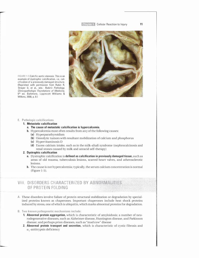
FIGURE 1-5 Calcific aortic stenosis. This is an example of dystrophic calcification, i.e., calcification of a previously damaged structure. (Reprinted with permission from Rubin R, Strayer D , et aI., eds.: Rubin's PathologV. Clinicopathologic Foundations of Medicine, 5th ed. Baltimore, Lippincott Williams & Wilkins, 2008, p. 8.)
E. Pathologic calcifications 1 . Metastatic calcification
I!miftll'iiIi Cellular Rea ction to I njury 11
a. The cause of metastatic calcification is hypercalcemia. b. Hypercalcemia most often results from any of the following causes:
(a) Hyperparathyroidism (b) Osteolytic tumors with resultant mobilization of calcium and phosphorus (c) Hypervitaminosis D (d) Excess calcium intake, such as in the milk-alkali syndrome (nephrocalcinosis and
renal stones caused by milk and antacid self-therapy) 2. Dystrophic calcification
a. Dystrophic calcification is defined as calcification in previously damaged tissue, such as areas of old trauma, tuberculosis lesions, scarred heart valves, and atherosclerotic lesions.
b. The cause is not hypercalcemia; typically, the serum calcium concentration is normal (Figure 1-5) .
VIII. DISORDERS CHARACTERIZED BY ABNORMALITIES OF PROTEIN FOLDING
A. These disorders involve failure of protein structural stabilization or degradation by specialized proteins known as chaperones. Important chaperones include heat shock proteins induced by stress, one of which is ubiquitin, which marks abnormal proteins for degradation.
B. Two known pathogenetic mechanisms include: 1. Abnormal protein aggregation, which is characteristic of amyloidosis; a number of neu
rodegenerative diseases, such as Alzheimer disease, Huntington disease, and Parkinson disease; and perhaps prion diseases, such as "mad cow" disease
2. Abnormal protein transport and secretion, which is characteristic of cystic fibrosis and IX)-antitrypsin deficiency

Review Test
Directions: Each of the numbered items or incomplete statements in this section is followed by answers or by completions of the statement. Select the one lettered answer or completion that is best in each case.
1. The illustration shows a section of the heart from a 45-year-old African-American man with long-standing hypertension who died of a "stroke." Which of the follOwing adaptive changes is exemplified in the illustration?
(Reprinted with permission from Rubin R, Strayer D, et aI., eds.: Rubin's Pathology. Clinicopathologic Foundations of Medicine, 5th ed. Baltimore, Lippincott Williams & Wilkins, 2008, p. 5.)
(A) Aplasia (8) Atrophy (e) Hyperplasia (D) Hypertrophy (E) Hypoplasia
2. A 16-year-old girl undergoes radiologic imaging of her abdomen and is found to have only one kidney. She had been entirely unaware of this problem. Which of the following terms is most descriptive of this finding?
(A) Agenesis (8) Atrophy (e) Hyperplasia (D) Hypoplasia (E) Metaplasia
12
3. An impending myocardial infarction was successfully averted by thrombolytic (clot-dissolving) therapy in a 55-year-old man. Which of the following biochemical events most likely occurred during the period of hypoxia?
(A) Decreased hydrogen ion concentration (8) Increase in oxidative phosphorylation (e) Loss of intracellular Na+ and water (D) Stimulation of ATP synthesis (E) Stimulation of anaerobic glycolysis and
glycogenolysis
4. A 45-year-old man with a long history of alcoholism presents with severe epigastric pain, nausea, vomiting, fever, and an increase in serum amylase. During a previous hospitalization for a similar episode, computed tomography scanning demonstrated calcifications in the pancreas. A diagnosis of acute pancreatitis superimposed on chronic pancreatitis was made. In this condition, which of the following types of necrosis is most characteristic?
(A) Caseous (8) Coagulative (e) Enzymatic (D) Fibrinoid (E) Liquefactive
5. A 29-year-old man hospitalized for acquired immunodeficiency syndrome (AIDS) is found to have pulmonary tuberculosis. Which type of necrosis is found in the granulomatous lesions (clusters of modified macrophages) characteristic of this increasingly frequent complication of AIDS?
(A) Caseous (8) Coagulative (e) Enzymatic (0) Fibrinoid (E) Liquefactive

-
6. A 45-year-old woman is investigated for hypertension and is found to have enlargement of the left kidney. The right kidney is smaller than normal. Contrast studies reveal stenosis of the right renal artery. The size change in the right kidney is an example of which of the following adaptive changes?
(A) Aplasia (8) Atrophy (e) Hyperplasia (0) Hypertrophy (E) Metaplasia
7. A 56-year-old man recovered from a myocardial infarction after his myocardium was entirely "saved" by immediate thrombolytic therapy. If it had been possible to examine microscopic sections of his heart during his ischemic episode, which of the follOwing would be the most likely cellular change to be found?
(A) Karyolysis (8) Karyorrhexis (e) Pyknosis (0) Swelling of the endoplasmic
reticulum
I!!Jllt1I:QII Cellular Reaction to I njury 13
8. A 64-year-old woman presents with fever, chills, headache, neck stiffness, vomiting, and confusion. The Kernig sign (passive knee extension eliciting neck pain) and Brudzinski sign (passive neck flexion eliciting bilateral hip flexion) are both positive. Examination of the cerebrospinal fluid reveals changes consistent with bacterial meningitis, and brain imaging demonstrates a localized abscess. Which of the following types of necrosis is most characteristic of abscess formation?
(A) Caseous (8) Coagulative (e) Enzymatic (0) Fibrinoid (E) Liquefactive
9. A 20-year-old man presents with yellowing of the sclerae, skin, and oral mucosa. Which of the following accumulations underlies these findings?
(A) Bilirubin (8) Hemosiderin (e) Lead (0) Melanin (E) Silver
10. This figure illustrates the microscopic appearance of the heart of a 56-year-old man who died after a 24-hour hospitalization for severe "crushing" chest pain complicated by hypotension and pulmonary edema. The type of necrosis shown is best described as
(Reprinted with permission from Rubin R, Strayer 0, et ai., eds.: Rubin's Pathology. Clinicopathologic Foundations of Medicine, 5th ed. Baltimore, Lippincott Williams & Wilkins, 2008, p. 23.)
(A) caseous. (8) coagulative. (e) fibrinoid. (0) gangrenous. (E) liquefactive.

14 B R S Pathology
1 1 . The illustration is from a liver biopsy of a 34-year-old woman with a long history of alcoholism. Which of the following is the best explanation for the changes shown here?
(A) Accumulation of triglycerides within hepatocytes
(8) Apoptosis with replacement of damaged cells by lipid-laden macro phages
(e) Bilirubin accumulation with mobilization of fat by bile salts
(D) Enzymatic fat necrosis with digestion of liver parenchyma by released enzymes
(E) Irreversible damage to mitochondria
12. A 45-year-old man is referred because of a recent diagnosis of hereditary hemochromatosis. Which of the follOwing is a correct statement about this disorder?
(A) Damage to organs results from abnormal deposition of lead.
(8) It can progress to liver cirrhosis, diabetes mellitus, and skin pigmentation.
(e) Most cases are due to spontaneous mutations.
(D) Skin hyperpigmentation is due to bilirubin accumulation.
( E) The total iron-binding capacity (TIBC) is characteristically increased.
(Reprinted with permission from Rubin R, Strayer D, et a I . , eds.: Rubin's Pathology. Clinicopathologic Foundations of Medicine, 5th ed . B a lt imore, L ipp incott Wil l iams & Wilkins, 2008, p. 646.)
1 3. A 60-year-old woman with breast cancer and widespread bony metastases is found to have calcification of multiple organs. The calcifications are best described as
(A) dystrophic with decreased serum calcium.
(8) dystrophic with increased serum calcium.
(e) metastatic with decreased serum calcium.
(D) metastatic with increased serum calcium.
1 4. A 56-year-old man dies 24 hours after the onset of substernal chest pain radiating down his left arm to the ulnar aspect of his fingertips. Which of the following morphologic myocardial findings is an indicator of irreversible injury?
(A) Cell blebs (8) Depletion of glycogen (e) Mitochondrial swelling (D ) Myelin figures ( E) Pyknotic nuclei

--
Answers and Explanations
1. The answer is D. The illustration shows marked hypertrophy of the left ventricle. Hypertrophy of this extent, often seen in hypertensive heart disease, is caused by increased
.
workload from increased ventricular pressure. This organ enlargement is the result of an increase in size of the individual muscle cells.
2. The answer is A. The patient has renal agenesis, absence of the kidney due to failure of organ development. The congenital lack of one kidney differs from atrophy, in which a decrease in the size of an organ results from a decrease in the mass of pre-existing cells. Unilateral renal agenesis is usually a harmless malformation, and the opposite kidney is often enlarged due to compensatory hypertrophy. Bilateral renal agenesis is incompatible with life and is of special interest since it can lead to the Potter progression (see Chapter 1 7) .
3 . The answer i s E . The sequence o f events i n hypoxic cell damage i s a s follows: Hypoxia results in failure of oxidative phosphorylation, with resultant depletion of ATP and increase in AMP and ADP. Anaerobic glycolysis and glycogenolysis are stimulated (not inhibited) through increased phosphofructokinase and phosphorylase activities, respectively. This results in an accumulation of cell lactate, with a decrease in intracellular pH and depletion of cellular glycogen stores. Decreased availability of ATP also results in failure of the Na +K+ -ATPase pump, which then leads to increased cell Na+ and water and decreased cell K+ .
4 . The answer is C . Pancreatic enzymatic fat necrosis represents autodigestion by proteolytic and lipolytic enzymes released from damaged parenchymal cells of the pancreas. Fatty acids liberated by the digestion of fat form calcium soaps, a process referred to as saponification. The precipitated calcium in the soaps can be visualized by radiologic imaging.
5. The answer is A. Caseous necrosis occurs as part of granulomatous inflammation, typified by the lesions of tuberculosis.
6. The answer is B. The decreased size is due to restriction of the blood supply, one of the causes of atrophy. The increase in size of the opposite kidney is referred to as compensatory hypertrophy. Unilateral renal artery stenosis is a well-known cause of secondary hypertension. In this setting, increased renin excretion and stimulation of the reninangiotensin system results in a form of hypertension that is potentially curable by surgical correction of the underlying vascular abnormality.
7. The answer is D. If infarction is averted by immediate thrombolytic therapy, indicators of necrosis, such as karyorrhexis, pyknosis, and karyolysis, which represent irreversible changes, would not be expected. Swelling of the endoplasmic reticulum from increased cell water, one of the earliest ultrastructural changes observed in injured cells, is reversible and would be expected.
8. The answer is E. Liquefactive necrosis is characteristic of ischemic injury in the central nervous system and suppurative infections that cause abscess formation (see Chapter 2) . The changes in the cerebrospinal spinal fluid characteristic of bacterial meningitis are detailed in Chapter 3.
9. The answer is A. Yellowing of the sclerae, skin, and oral mucosa are all characteristic of jaundice, the accumulation of bilirubin, the catabolic product of the heme moiety of hemoglobin. Jaundice can occur by diverse mechanisms: hemolytic (see Chapter 1 1) , hepatocellular (see Chapter 16) , or obstructive (see Chapter 16) .
1 5

16 BRS Pathology
10. T he answer is B. The figure illustrates general preservation of myocardial architecture with some fragmentation, more intense cytoplasmic staining corresponding to increased cellular eosinophilia, and loss of nuclei, all of which are characteristic of coagulative necrosis.
1 1 . T he answer is A. The figure illustrates fatty change of the liver, which is characterized by the accumulation of intracellular parenchymal triglycerides. It is seen most frequently in the liver, heart, and kidney and commonly is secondary to alcoholism. Fatty change results from an imbalance between the uptake, utilization, and mobilization of fat from liver cells. Alcoholic fatty liver may be reversible with complete abstinence from alcohol.
12. The answer is B. In advanced form, primary (hereditary) hemochromatosis is characterized by the triad of cirrhosis, diabetes, and hyperpigmentation, or so-called "bronze diabetes." The disease is most often caused by a mutation in the HIe gene on chromosome 6 and is characteristically familial rather than sporadic. The manifestations of the disorder are the result of iron overload and deposition of hemosiderin in tissues such as the liver, pancreas, skin, joints, and pituitary. Laboratory abnormalities of note include increased serum iron and decreased total iron-binding capacity (TIBC). The skin hyperpigmentation is due largely to increases in melanin and to lesser accumulations of hemosiderin.
13. T he answer is D. Metastatic calcification, or deposition of calcium in previously normal tissue, is caused by hypercalcemia. In this patient, tumor metastases to bone with increased osteolytic activity caused mobilization of calcium and phosphate, resulting in hypercalcemia. Metastatic calcification should be contrasted with dystrophic calcification, in which the serum calcium concentration is normal and previously damaged tissues are the sites of deposition.
1 4. T he answer is E. Myelin figures, cell blebs, mitochondrial swelling, and glycogen depletion are all signs of reversible injury. Nuclear changes such as pyknosis, karyorrhexis, and karyolysis are signs of cell death and are, of course, irreversible.

-
Inflammation
I. INTRODUCTION
Inflammation is a vascular response to injury.
A. Processes 1. Exudation of fluid from vessels 2. Attraction of leukocytes to the injury. Leukocytes engulf and destroy bacteria, tissue
debris, and other particulate material. 3. Activation of chemical mediators 4. Proteolytic degradation of extracellular debris 5. Restoration of injured tissue to its normal structure and function. This is limited by the
extent of tissue destruction and by the regenerative capacity of the specific tissue.
B. Cardinal signs 1. Rubor (redness caused by dilation of vessels) 2. Dolor (pain due to increased pressure exerted by the accumulation of interstitial fluid
and to mediators such as bradykinin) 3. Calor (heat caused by increased blood flow) 4. Tumor (swelling due to an extravascular accumulation of fluid) 5. Functio laesa (loss of function)
C. Causes 1. Infection 2. Trauma 3. Physical injury from thermal extremes or from ionizing radiation 4. Chemical injury 5. Immunologic injury 6. Tissue death. Inflammatory changes occur in viable tissue adjacent to necrotic areas.
II. ACUTE INFLAMMATION
A. Adhesion molecules 1 . General considerations
a. Adhesion molecules play an important role in acute inflammation. b. They are divided into three families: selectins, immunoglobulin-family adhesion pro
teins, and integrins. 2. Selectins
a. These molecules are induced by the cytokines interleukin- I (IL- I ) and tumor necrosis factor (TNF) .
1 7

1 8 BRS Pathology
b. l-selectins are expressed on neutrophils and bind to endothelial mucin-like molecules such as GlyCam- l .
c . E - and P-selectins are expressed on endothelial cells and bind to oligo saccharides such as sialyl-Lewis X on the surface of leukocytes. P-selectins, stored in endothelial WeibelPalade bodies and platelet alpha granules, relocate to the plasma membrane after stimulation by mediators such as histamine and thrombin.
3. Immunoglobulin-family adhesion proteins a. Intercellular adhesion molecules 1 and 2 (ICAM-I and ICAM-2) are expressed on
endothelial cells and bind to integrin molecules on leukocytes. b. Vascular cell adhesion molecules (VCAMs) similarly are expressed on endothelial cells
and bind to integrin molecules on leukocytes. 4. Integrins. Examples include leukocyte LFA- I , MAC- I , and VLA-4, which bind to endothe
lial immunoglobulin-family adhesion proteins.
B. Vasoactive changes 1. These changes begin with a brief period of vasoconstriction, followed shortly by dilation
of arterioles, capillaries, and postcapillary venules. 2. The resultant marked increase in blood flow to the affected area is clinically manifest by
redness and increased warmth of the affected area.
C. Increased capil lary permeabil ity 1 . This results in leakage of proteinaceous fluid, which causes edema. 2. Causes include endothelial changes that vary from contraction of endothelial cells in
postcapillaryvenules, with widening of inter endothelial gaps, to major endothelial damage involving arterioles, capillaries, and venules.
D. Types of inflammatory cells 1. Neutrophi ls are the most prominent inflammatory cells in foci of acute inflammation
during the first 24 hours. Important causes of neutrophi l ia (increased neutrophils in the peripheral blood) include bacterial infections and other causes of acute inflammation, such as infarction. The early release of neutrophils into the peripheral blood in acute inflammation is from the bone marrow postmitotic reserve pool. There is often an increase in the proportion of less mature cells such as band neutrophi ls (Figure 2 - 1 ) .
� 2 . After 2-3 days, neutrophils are replaced mainly by monocytes-macrophages, which are capable of engulfing larger particles, are longer-lived, and are capable of dividing and proliferating within the inflamed tissue. Important causes of monocytosis (i.e., increased number of monocytes in the peripheral blood) include tuberculosis, brucellosis, typhus, and salmonella infection.
)' 3. lymphocytes are the most prominent inflammatory cells in many viral infections and, along with monocytes-macrophages and plasma cells, are the most prominent cells in chronic inflammation. lymphocytosis (i.e., an increased number of lymphocytes in the peripheral blood) is most often caused by viral infections such as influenza, mumps, rubella, and infectious mononucleosis and certain bacterial infections such as whooping cough and tuberculosis.
4. Eosinophi ls are the predominant inflammatory cells in allergic reactions and parasitic infestations. The most important causes of eosinophi l ia include allergies such as asthma, hay fever, and hives and also parasitic infections. Other causes include polyarteritis nodosa and Hodgkin lymphoma.
5. Mast cells and basophils are sources of histamine. Important causes of basophi l ia include chronic myelogenous leukemia and other myeloproliferative diseases.
E. Cellular response of leukocytes 1 . Emigration is the passage of inflammatory leukocytes between the endothelial cells into
the adjacent interstitial tissue. Before emigration, circulating leukocytes from the central blood flow move toward the endothelial surface. a. Margination occurs as leukocytes localize t9 the outer margin of the blood flow adja
cent to the vascular endothelium.

-
FIGURE 2-1 Neutrophils (polymorphonuclear leukocytes, PMNs) in tissue. P M N infiltration typifies t h e early stages of acute inflammation. (Reprinted with permission from Rubin R, Strayer 0, et aI., eds.: Rubin's Pathology. Clinicopathologic Foundations of Medicine, 5th ed. Baltimore, Lippincott Williams & Wilkins, 2008, p. 39 )
i!1lmi1tlIfI Inflammation 19
b. Pavementing occurs as leukocytes line the endothelial surface. c. Rol l ing (or tumbl ing) is mediated by the action of endothelial selectins loosely binding
to leukocytes, producing a characteristic "rolling" movement of the leukocytes along the endothelial surface.
d. Adhesion occurs as leukocytes adhere to the endothelial surface and is mediated by the interaction of integrins on leukocytes binding to immunoglobulin-family adhesion proteins on endothelium.
e. Transmigration is the movement of leukocytes across the endothelium and is mediated by platelet endothelial cell adhesion molecule- l (PECAM - 1 ) on both leukocytes and endothelium.
2. Chemotaxis � a. This is the process by which leukocytes are attracted to and move toward an injury.
b. Chemotaxis and other forms of cellular migration are measured in an in vitro system (Boyden chamber technique) that assesses the migration of cells from an upper chamber through a microporous membrane to a lower chamber filled with a chemoattractant.
c. This process is mediated by diffusible chemical agents (Table 2-1 ); movement of leukocytes occurs along a chemical gradient.
d. Chemotactic factors for neutrophi ls, produced at the site of injury, include: ( 1 ) Products from bacteria
� (2) Complement components, especially C5a � (3) Arachidonic acid metabolites, especially leukotriene B4 (LTB4) , hydroxyeicosate
traenoic acid (HETE) , and kallikrein 3. Phagocytosis
a. Definition. Phagocytosis is the ingestion of particulate material (e.g., tissue debris, living or dead bacteria, other foreign cells) by phagocytic cells. Neutrophils and monocytesmacrophages are the most important phagocytic cells.
b. Anatomic changes ( 1 ) Phagocytosis is characterized morphologically by internalization of the attached
opsonized particle by pseudopodial extensions from the surface of the leukocyte, which enclose the foreign particle, forming an internalized vesicle, the phagosome.

20 BRS Pathology
t a b I e 2-1 Chemotactic Factors
Factor Description Chemotactic For
Formylated peptides C5a
Bacterial products of Escherichia coli Activated complement component Leukotrienes
Neutrophils Neutrophils Neutrophils Neutrophils
H ETE, LTB4 Kallikrein
Fibrinogen PAF
PDGF
TGF-j3
Fibronectin
Product of factor Xlla-mediated conversion of pre kallikrein
Plasma protein AGEPC; from basophils, mast cells, and
other cells From platelets, monocytes-macrophages,
smooth muscle cells, and endothelial cells From platelets, neutrophils, macrophages,
lymphocytes, and fibroblasts Extracellular matrix protein
Neutrophils Eosinophils
Neutrophils and macrophages
Macrophages and fibroblasts
Fibroblasts and endothelial cells
PAF, platelet-activating factor; PDGF, platelet-derived growth factor; AGEPC, acetyl-glyceryl-ether phosphorycholine; TGF-I3, transforming growth factor-l3·
(2) Phagosomes fuse with cytoplasmic lysosomes and form phagolysosomes. (3) Phagolysosome formation is associated with leukocytic degranulation.
c. Opsonization ( 1 ) This process facilitates phagocytosis. It is the coating of particulate material by
substances referred to as opsonins, which immobilize the particles on the surface of the phagocyte.
� (2) The most important opsonins are immunoglobulin G (lgG) subtypes and C3b, a complement component.
(3) Fragments opsonized by IgG are bound to phagocytic cells by cell-surface receptors for the Fc portion of the IgG molecule.
(4) Fragments opsonized by C3b bind to cellular receptors for C3b. 4. Intracellular microbial ki l l ing is mediated within phagocytic cells by oxygen-dependent
and oxygen-independent mechanisms. � a. Oxygen-dependent microbial ki l l ing is the most important intracellular microbicidal
process. ( 1 ) Phagocytosis initiates activity of the hexose monophosphate shunt, causing an
oxidative burst and supplying electrons to an NADPH oxidase in the phagosomal membrane.
(2) One of the products of the NADPH oxidase reaction is superoxide anion (02-;-) ' which is further converted to hydrogen peroxide (H202) by dismutation. H202 may be further converted to the activated hydroxyl radical (OH·).
(3) In the presence of the leukocyte enzyme myeloperoxidase and a halide ion such as chloride, H202 oxidizes microbial proteins and disrupts cell walls. This entire process is referred to as the myeloperoxidase-halide system of bacterial ki l l ing.
b. Oxygen-independent microbial ki l l ing ( 1 ) This process is much less effective than oxygen-dependent microbial killing. (2) This process is mediated by proteins, such as lysozyme, lactoferrin, major basic
protein of eosinophils, and cationic proteins, such as bactericidal permeabilityincreasing protein and defensins.
F. Exogenous and endogenous mediators of acute inflammation These mediators influence chemotaxis, vasomotor phenomena, vascular permeability, pain, and other aspects of the inflammatory process (Table 2-2).
1. Exogenous mediators are most often of microbial origin (e.g., formylated peptides of Escherichia coli, which are chemotactic for neutrophils) .

-
i!l1tlllttIfJ I nf lammation 21
t a b I e 2-2 Vasoactive Mediators
Activity Mediator
Vasoconstriction TxA2
Vasodilation
LTC4, LTD4, LTE4 PAF
PGI2 PGD2, PGE2, PGF2a Bradykinin PAF Nitric oxide
Increased vascular permeability Histamine Serotonin PGD2, PG E2, PGF2a LTC4, LTD4, LTE4 Bradykinin PAF
LTC4, leukotriene C4; LTD4. leukotriene °4: LTE4• leukotriene E4; TxA2. thromboxane A2; PAF, platel et-activating factor; PGI2. prostacycl in (prostag landin 12); PG02. prostaglandin 02; PGE2. prostaglandin E2; PGF2«. prostag landin F2«
2. Endogenous mediators are of host origin. a. Vasoactive amines
(1 ) Histamine mediates the increase in capillary permeability associated with the contraction of endothelial cells in postcapillary venules that occurs with mild injuries.
� (a) Histamine is liberated from basophi ls, mast cel ls, and platelets. (b) 8asophi ls and mast cel ls. Histamine is liberated by degranulation triggered by
the following stimuli: (i) Binding of specific antigen to basophil and mast cell membrane-bound
IgE (complement is not involved) (ii) Binding of complement fragments C3a and C5a, anaphylatoxins, to spe
cific cell-surface receptors on basophils and mast cells (specific antigen and IgE antibodies are not involved)
(i i i) Physical stimuli such as heat and cold (iv) Cytokine IL- 1 (v) Factors from neutrophils, monocytes, and platelets
(c) Platelets. Histamine is liberated from platelets by platelet aggregation and the release reaction, which can be triggered by endothelial injury and thrombosis or by platelet-activating factor (PAP) . (i) PAP is derived from the granules of basophils and mast cells and from
endothelial cells, macrophages, neutrophils, and eosinophils. PAP is acetyl-glyceryl-ether phosphorylcholine, also known as AGEPC.
(ii) PAP activates and aggregates platelets, with the release of histamine and serotonin; causes vasoactive and bronchospastic effects; and activates arachidonic acid metabolism.
(2) Serotoni n (5-hydroxytryptamine) (a) This substance acts similarly to histamine. (b) It is derived from platelets. It is liberated from platelets, along with histamine,
during the release reaction. b. Arachidonic acid metabol ites. Phospholipase A2 stimulates the release of arachidonic
acid from membrane phospholipids. The metabolism of arachidonic acid proceeds along two pathways: ( 1 ) The cyclooxygenase (cycl ic endoperoxide) pathway is catalyzed by two enzymic
isoforms, referred to as cyclooxygenase- I (COX- I ) and cyclooxygenase-2 (COX-2) . (a) This pathway is inhibited by aspirin and other anti-inflammatory drugs.

22 BRS Pathology
(b) It yields thromboxanes and prostaglandins: thromboxane A2 (TxA2) in platelets, prostacyclin (PGI2) in endothelial cells, and other prostaglandins in other tissues.
� (i) Platelet TxA2 is a powerful vasoconstrictor and platelet aggregant. � ( i i) Endothel ia l PGI2 is a powerful vasodilator and inhibitor of platelet
aggregation. (2) The l ipoxygenase pathway yields hydroperoxyeicosatetraenoic acid (HPETE) and
its derivatives, 12-HPETE in platelets, and 5-HPETE and 1 5-HPETE in leukocytes. (a) 5-HPETE in turn gives rise to HETE, a chemotactic factor for neutrophils. (b) 5-HPETE also gives rise to leukotrienes:
(i) LTB4, a chemotactic factor for neutrophils ( i i) LTC4, LTD4, and LTE4, potent vasoconstrictors, bronchoconstrictors, and
mediators of increased capillary permeability, which are sometimes jointly referred to as the slow-reacting substance of anaphylaxis
(c) 5-HPETE also indirectly gives rise to l ipoxins. LXA4 and LXB4 inhibit polymorphonuclear neutrophils and eosinophils and also activate monocytes and macrophages. It is proposed that these lipoxins are involved in resolving inflammation and are potential anti-inflammatory mediators that may have therapeutic value.
c. Cytokines. These soluble proteins are secreted by several types of cells. They can act as effector molecules that influence the behavior of other cells. (1 ) Cytokines are mediators of immunologic response (e.g., interferon-'Y [produced
by T cells and natural killer cells] activates monocytes) . (2) The cytokines Il-l and TNF are secreted by monocytes-macrophages and other
cells and have several effects on inflammation. (3) IL- 1 and TNF induce acute phase responses, such as � (a) Systemic effects of inflammation, including fever and leukocytosis � (b) Hepatic synthesis of acute phase proteins, such as C-reactive protein, serum
amyloid-associated protein, complement components, fibrinogen, pro thrombin, (Xl-antitrypsin, (Xz-macroglobulin, ferritin, and ceruloplasmin
� (c) Synthesis of adhesion molecules � (d) Neutrophi l degranulation (4) IL- 1 and TNF reduce the thromboresistant properties of endothelium, thus pro
moting thrombosis. � d. Kinin system. The kinin system is initiated by activated Hageman factor (factor XlIa) .
Factor XlIa also activates the intrinsic pathway of coagulation and the plasminogen (fibrinolytic) system. Activation of this system in turn activates the complement cascade. Thus, factor Xl l a l inks the kinin, coagUlation, p lasminogen, and complement systems. ( 1 ) This system converts prekallikrein to kallikrein (a chemotactic factor) .
� (2) It results in the cleavage, by kallikrein, of high-molecular-weight kininogen to bradykinin, which is a peptide that is nine amino acids in length that mediates vascular permeability, arteriolar dilation, and pain.
e. Complement system. The complement system consists of a group of plasma proteins that participate in immune lysis of cells and play a significant role in inflammation.
� (1 ) C3a and C5a (anaphylatoxins) mediate degranulation of basophils and mast cells with the release of histamine. C5a is chemotactic, mediates the release of histamine from platelet-dense granules, induces expression of leukocyte adhesion molecules, and activates the lip oxygenase pathway of arachidonic acid metabolism.
� (2) C3b is an opsonin. (3) C5b-9, the membrane attack complex, is a lytic agent for bacteria and other cells.
f. Nitric oxide (formerly known as endothelium-derived relaxing factor) (1 ) This is produced by endothelial cells. (2) It stimulates relaxation of smooth muscle, thus playing a role in controlling vas
cular tone. (3) It inhibits platelet aggregation, contributing to endothelial thromboresistance.

G. Outcome of acute inflammation
i!3JflJ'tttlij I nflammation 23
1. Resolution of tissue structure and function often occurs if the injurious agent is eliminated. 2. Tissue destruction and persistent acute inflammation
j>l a. Abscess. This is a cavity filled with pus (neutrophils, monocytes, and liquefied cellular debris). ( 1 ) It is often walled off by fibrous tissue and is relatively inaccessible to the circulation. (2) It results from tissue destruction by lysosomal products and other degrada!ive
enzymes. j>l (3) It is usually caused by bacterial infections, often by staphylococci. b. Ulcer
(1) This is the loss of surface epithelium. (2) This can be caused by acute inflammation of epithelial surfaces (e.g., peptic ulcer,
ulcers of the skin) . c. Fistula. This is an abnormal communication between two organs or between an organ
and a surface. d. Scar. This is the final result of tissue destruction, with resultant distortion of structure
and, in some cases, altered function. 3. Conversion to chronic inflammation
a. This change is marked by the replacement of neutrophils and monocytes with lymphocytes, plasma cells, and macro phages.
b. It often includes proliferation of fibroblasts and new vessels, with resultant scarring and distortion of architecture.
H. H e reditary defects that impair the acute inflammatory response 1. Deficiency of complement components
j>l a. This defect manifests clinically as increased susceptibility to infection. b. Notable deficiencies include C2, C3, and C5.
2. Defects in neutrophi ls a. Chronic granulomatous disease of chi ldhood
(1) This disease is most commonly an X-linked disorder characterized by deficient activity of one of the enzymes involved in NADPH oxidase activity and the oxidative burst. Autosomal recessive variants also occur.
(2) The disease is marked by phagocytic cells that ingest but do not kill certain microorganisms.
j>l (3) Cata lase-positive organ isms are ingested but not killed. These organisms (e.g. , Staphylococcus aureus) can destroy H202 generated by bacterial metabolism. Because enzyme-deficient neutrophils cannot produce H202 and bacterial H202 is destroyed by bacterial catalase, H202 is not available as a substrate for myeloperoxidase. Thus, the myeloperoxidase -halide system of bacterial killing fails.
(4) Catalase-negative organisms are ingested and killed. These organisms (e.g. , streptococci) produce sufficient H202 to permit oxygen-dependent microbicidal mechanisms to proceed. In effect, the substrate for myeloperoxidase is produced by the bacteria, and the bacteria in a sense kill themselves.
b. Myeloperoxidase deficiency ( 1 ) This defect is rarely associated with recurrent bacterial infections but often has
little clinical consequence. (2) In some instances, this defect has been associated with a marked increase in sus
ceptibility to infections with Candida albicans. c. Chediak-Higashi syndrome
( 1 ) This autosomal recessive disorder is characterized by neutropenia, albinism, cranial and peripheral neuropathy, and a tendency to develop repeated infections.
(2) It is marked by the presence of abnormal white blood cells, which are characterized as follows:
j>l (a) Functional ly, by abnormal microtubule formation, affecting movement, with impaired chemotaxis and migration

24 BRS Pathology
(b) Morphological ly, by large cytoplasmic granules (representing abnormal lysosomes) in granulocytes, lymphocytes, and monocytes and by large abnormal melanosomes in melanocytes, all caused by impaired membrane fusion of lysosomes
d. Leukocyte adhesion deficiency (LAD) types 1 and 2 ( 1 ) LAD type 1 deficiency is associated with recurrent bacterial infections and is
caused by deficiency of I3z-integrins. (2) LAD type 2 deficiency is also associated with recurrent bacterial infections and
results from mutations in the gene that codes for fucosyltransferase, required for the synthesis of sialyl-Lewis X on neutrophils.
I I I . CHRONIC I NFLAMMATION
A. General considerations 1. Chronic inflammation can occur when the inciting injury is persistent or recurrent or
when the inflammatory reaction is insufficient to completely degrade the agent (e.g. , bacteria, tissue debris, foreign bodies) that incites the inflammatory reaction.
2. It often occurs de novo, without a preceding acute inflammatory reaction. 3. It occurs in two major patterns: chronic nonspecific inflammation and granulomatous
inflammation.
B. Chronic nonspecific inflammation (Figure 2-2) 1. A cellular reaction with a preponderance of mononuclear (round) cells (macrophages,
lymphocytes, and plasma cells), often with a prol iferation of fibroblasts and new vessels. Scarring and distortion of tissue architecture is characteristic.
2. This type of inflammation is mediated by the interaction of monocytes-macrophages with lymphocytes.
3. Monocytes are recruited from the circulation by various chemotactic factors.
FIGURE 2-2 Chronic inflammation. Note the presence of lymphocytes, macrophages, and plasma cells (marked by arrows). (Reprinted with permission from Rubin R, Strayer 0, et aI., eds.: Rubin's Pathology. Clinicopathologic Foundations of Medicine, 5th ed. Baltimore, Lippincott Williams & Wilkins, 2008, p. 39.)

-
FIGURE 2-3 G r a n u lomatous infl a m mation . Note the a bsence of caseation in th is granuloma taken from a lymph node from a patient with sarcoidosis. The lesion consists of focal a c c u m u lat ions of altered m a c ro p h a g es referred to as epithelioid cel is. (Re printed with permission from Rubin R, Strayer 0, et aI . , eds. : Rubin 's Pathology. Clinicopathologic Foundations of Medicine, 5th ed. Baltimore, Lippincott Wil l iams & Wilkins, 2008, p. 68.)
i!11Eful!lfj Inf lammation 25
4. Cytokines derived from monocytes-macrophages activate lymphocytes. The activated lymphocytes, in turn, are the source of additional cytokines that activate monocytesmacro phages.
5. B lymphocyte activation by macrophage-presented antigen results in the formation of antibody-producing plasma cells.
C. Granulomatous inflammation (Figures 2-3 and 2-4) � 1 . This type of inflammation is characterized by granulomas, which are nodular collections
of specialized macrophages referred to as epithel ioid cel ls. Granulomas are usually surrounded by a rim of lymphocytes.
� 2. Activation of macrophages by interactions with T lymphocytes is involved. Poorly digestible antigen is presented by macrophages to CD4 + lymphocytes. Interaction with the antigen-specific T-cell receptor of these cells triggers the release of cytokines (especially interferon-'Y) , which mediate the transformation of monocytes and macrophages to epithelioid cells and giant cells.
� 3. Caseous necrosis is often characteristic (especially in tuberculosis). resulting fro m the killing of mycobacteria-laden macrophages by T lymphocytes and possibly by cytokines or sensitized macro phages. N oncaseating pulmonary g ranulomatous disease is caused most often by sarcoidosis,
� 4. The presence of multinucleated giant cells derived from macro phages is also characteristic. The Langhans giant cel l has nuclei arranged in a horseshoe-shaped pattern about the periphery of the cell and is particularly characteristic of, but not specific for, the granulomatous inflammation of tuberculosis. The foreign body giant cel l has scattered nuclei.
FIGURE 2-4 Giant cel i in granulomatous infl a m m ation . G i a nt c el is, derived from m acrophages, a re a frequent compo nent of granu lomatous infl a m mation. This typical example with the nuc le i arran g e d i n
t h e periphery i n a horseshoe pattern i s referred t o as a La n g h a n s g iant c e l l . (Repr inted with permission from R u b i n R , Strayer 0, e t a I . , eds.: Rubin 's PathologV. Clinicopathologic Foundations of Medicine, 5th ed. B a ltimore, Lippin cott Wil l iams & Wilkins, 2008, p. 68.)

26 BRS Pathology
5. Granulomatous inflammation is the characteristic form of inflammation associated with a number of diverse etiologic agents, including:
.Jii a. Infectious agents ( 1 ) Mycobacterium tuberculosis and M. leprae (2) Blastomyces dermatitidis, Histoplasma capsulatum, Coccidioides immitis, and
many other fungi (3) Treponema paUidum (4) The bacterium of cat-scratch disease
.Jii b. Foreign bodies .Jii c. Unknown etiology, including sarcoidosis
IV. TISSUE REPAIR
A. Restoration of normal structure. This occurs when the connective tissue infrastructure remains relatively intact. It requires that the surviving affected parenchymal cells have the capacity to regenerate. 1. labile cells
a. These cells divide actively throughout life to replace lost cells. b. They are capable of regeneration after injury. c. They include cells of the epidermis and gastrointestinal mucosa, cells lining the sur
face of the genitourinary tract, and hematopoietic cells of the bone marrow. 2. Stable cells .Jii a. Characteristically, these cells undergo few divisions but are capable of division when
activated; that is, they can regenerate from Go cells when needed. b. They are also capable of regeneration following injury .
.Jii c. They include hepatocytes, renal tubular cells, parenchymal cells of many glands, and numerous mesenchymal cells (e.g., smooth muscle, cartilage, connective tissue, endothelium, osteoblasts) .
3. Permanent cells a. These cells have been considered to be incapable of division and regeneration (a view
challenged by recent provocative new evidence involving stem cells) . b. They include neurons and myocardial cells. c. They are replaced by scar tissue (typically fibrosis; gliosis in the central nervous sys
tem) after irreversible injury and cell loss.
B. Cellular proliferation. This process is mediated by an assemblage of growth factors. 1 . Platelet-derived growth factor (PDGF) is a competence factor that promotes the prolifera
tive response of fibroblasts and smooth muscle cells on concurrent stimulation by progression factors (e.g., other growth factors) . Indirectly in this manner, PDGF promotes the synthesis of collagen. a. PDGF is synthesized by platelets and several other cells. b. PDGF promotes the chemotactic migration of fibroblasts and smooth muscle cells .
.Jii c. PDGF is chemotactic for monocytes. d. PDGF reacts with specific cell-surface receptors. Generally, growth factor receptors are
transmembrane proteins that respond to ligand interaction by conformational changes that induce tyrosine kinase activity in their intracellular domains.
2. Epidermal growth factor (EGF) is a progression factor that promotes the growth of endothelial cells and fibroblasts, as well as epithelial cells.
3. Fibroblast growth factors (FGFs) promote the synthesis of extracellular matrix protein (including fibronectin) by fibroblasts, endothelial cells, monocytes, and other cells. Fibronectin is a glycoprotein with the follOwing characteristics:
.Jii a. It is chemotactic for fibroblasts and endothelial cells. b. It promotes angiogenesis (new vessel formation) .

-
l!1lt1'!1ttiI Inflammation 27
c. It links other extracellular matrix components (e.g., collagen, proteoglycans) and macromolecules (e.g., fibrin, heparin) to cell-surface integrins. Integrins mediate interactions between cells and extracellular matrix.
4. Transforming growth factors (TGFs) a. TGF-a functions similarly to EGF. b. TGF-� is a growth inhibitor for many cell types and may aid in modulating the repair
process; it is also a chemotactic factor for macro phages and fibroblasts. 5. Macrophage-derived growth factors ( ll-1 and TNF) promote the proliferation of fibroblasts,
smooth muscle cells, and endothelial cells.
C. The repair process 1 . Removal of debris begins in the early stages of inflammation and is initiated by liquefac
tion and removal of dead cellular material and other debris. 2. Formation of granulation tissue
a. Granulation tissue is highly vascular, newly formed connective tissue consisting of capil laries and fibroblasts; it fills defects created by liquefaction of cellular debris.
b. Granulation tissue is not related to granulomas or granulomatous inflammation. 3. Scarring
a. Col lagen is produced by fibroblasts. As the amount of collagen in granulation tissue progressively increases, the tissue becomes gradually less vascular and less cellular.
b. Progressive contraction of the wound also occurs, often resulting in a deformity of the original structure.
D. Factors that delay or impede repair 1 . Retention of debris 2. Impaired circulation 3. Persistent infection
� 4. Metabolic disorders, such as diabetes mellitus (associated with both susceptibility to infection and impaired circulation)
� 5. Dietary deficiency of ascorbic acid or protein, which are required for collagen formation

-
Review Test
Directions: Each of the numbered items or incomplete statements in this section is followed by answers or by completions of the statement. Select the one lettered answer or completion that is best in each case.
1. A 72-year-old man presents with a 3-day history of progressively worsening productive cough, fever, chills, and signs of toxicity. Prominent physical findings include signs of consolidation and rales over the right lung base. Sputum culture is positive for Streptococcus pneumoniae. An intra-alveolar exudate filling the alveoli of the involved portion of the lung is present. Which of the following types of inflammatory cells is most likely a prominent feature of this exudate?
(A) Basophils (8) Eosinophils (C) Lymphocytes (D) Monocytes-macrophages (E) Neutrophils
2. A routine complete blood count performed on a 22-year-old medical student reveals an abnormality in the differential leukocyte count. She has been complaining of frequent sneezing and "watery" eyes during the past several weeks and reports that she frequently had such episodes in the spring and summer. Which of the following cell types is most likely to be increased?
(A) Basophils (8) Eosinophils (C) Lymphocytes (D) Monocytes (E ) Neutrophils
3. A 1 6-year-old boy presents with a 24-hour history of severe abdominal pain, nausea, vomiting, and low-grade fever. The pain is initially periumbilical in location but has migrated to the right lower quadrant of the abdomen, with maximal tenderness elicited at a site
28
one third of the way between the crest of the ileum and the umbilicus (McBurney point) . The leukocyte count is 1 4,OOO/mm3, with 74% segmented neutrophils and 12% bands. Surgery is performed. Which of the following describes the expected findings at the affected site?
(A) Fistula (abnormal duct or passage) connecting to the abdominal wall
(B) Granulation tissue (new vessels and young fibroblasts) with a prominent infiltrate of eosinophils
(C) Granulomatous inflammation with prominent aggregates of epithelioid cells and multinucleated giant cells
(D) Massive infiltration of lymphocytes and plasma cells
(E ) Prominent areas of edema, congestion, and a purulent reaction with localized areas of abscess formation
4. A 2-year-old boy presents with recurrent infections involving multiple organ systems. Extensive investigation results in a diagnosis of chronic granulomatous disease of childhood. Which of the following most closely characterizes the abnormality in this patient's phagocytic cells?
(A) Decreased killing of microorganisms because of enhanced production of hydrogen peroxide
(8) Deficiency of NADPH oxidase activity (C) Impaired chemotaxis and migration
caused by abnormal microtubule formation
(D) Inability to kill streptococci (E ) Increased myeloperoxidase-halide
mediated killing of catalase-positive organisms as compared to catalase-negative organisms

-
-
I!iiIlJtttII'I I nf lammat ion 29
5. The accompanying figure is representative of the findings in a hilar lymph node from a 54-year-old man who sought medical care for low-grade fever, anorexia, fatigue, night sweats, weight loss, and persistent cough with bouts of hemoptysis. A chest X-ray had revealed a right apical infiltrate with beginning cavitation, and examination of the sputum had revealed acid-fast bacilli. This condition is typified by a form of inflammation that invariably includes which of the following?
(Reprinted with permission from Rubin R, Strayer 0, et aI., eds.: Rubin's Pathology. Clinicopathologic Foundations of Medicine, 5th ed. Baltimore, Lippincott Williams & Wilkins, 2008, p. 25.)
(A) A morphologically identifiable etiologic agent
(8) Caseous necrosis (e) Clusters of epithelioid cells (0) Multinucleated giant cells (E) Prominent granulation tissue
6. A laboratory experiment is performed to evaluate the chemotactic potential of a group of potential mediators. Which of the following substances most likely has the greatest affinity for neutrophils?
(A) C5a (8) Fucosyl transferase (e) !)2-Integrin (0) P-selectin (E) Tumor necrosis factor-a
7. A 26-year-old African-American woman has bilateral hilar adenopathy, and radiography reveals multiple reticular densities in both lung fields. A bronchoscopic biopsy reveals granulomatous inflammation with multiple giant cells of the Langhans type and no evidence of caseous necrosis.
Which of the following is the most likely diagnosis?
(A) Aspergillosis (8) Coccidioidomycosis (e) Histoplasmosis (0) Sarcoidosis (E) Tuberculosis
8. In a laboratory exercise for medical students, an unknown compound is studied. The students are informed that the compound has been isolated from endothelial cells and that its synthesis can be inhibited by aspirin. In the laboratory, the students demonstrate that the compound is a potent vasodilator and platelet antiaggregant. Given these findings, the substance is most likely which of the following mediators?
(A) 5-Hydroperoxyeicosatetraenoic acid (5-HPETEl
(8) Leukotriene (LTC4l (e) Lipoxin A4 (LXA4l (0) Prostacyclin (PGI2l (E) Thromboxane A2 (TxA2l

30 BRS Pathology
9. A 70-year-old man presents with the sudden onset of left-sided weakness, spasticity, and hyperactive and pathologic reflexes. The most serious consequences of this disorder are the result of damage to which of the following cell types?
(A) Labile cells (B) Multipotent adult progenitor cells (C) Permanent cells (0) Stable cells
Answers and
_
E
_
x
_
p
_
la
�
n
�
a
_
t
_
io
_
n
�
s __ J 1 . The answer is E. The patient has bacterial pneumonia due to Streptococcus pneumoniae,
a classic example of severe acute inflammation. In the early stages of acute inflammation, the neutrophil is the most prominent inflammatory cell. It is noteworthy that in many instances, bacterial infections are characterized by neutrophilic infiltrates. It is also noteworthy that S. pneumoniae (also known as the "pneumococcus") is the most common etiologic agent of lobar pneumonia (see Chapter 14) .
2. The answer is B. This type of reaction is primarily mediated by the release of histamine from tissue mast cells, and the associated cellular infiltrate and peripheral blood findings represent mobilization and increased numbers of eosinophils. The symptoms reported are those of seasonal rhinitis, better known as "hay fever," a manifestation of type I hypersensitivity (see Chapter 5) .
3. The answer is E. The clinical findings are typical of acute appendicitis, another example of severe acute inflammation. Because the danger of perforation is great, early appendectomy is the treatment of choice. Suppurative or purulent inflammation is characterized by the prominent areas of edema resulting from increased vascular permeability, congestion, and a purulent (pus-containing) exudate consisting of necrotic cells and large numbers of neutrophils. In addition, other signs of acute inflammation, such as congestion, are prominent. The patient responds with the sensation of pain (induced by increased hydrostatic pressure in tissue and by chemical mediators such as bradykinin) and the acute phase reaction (in this instance, fever and neutrophilic leukocytosis with a "shift to the left") .
4. The answer is B. Chronic granulomatous disease of childhood, a condition characterized by repeated infections and most commonly X-linked inheritance, is marked by failure of the myeloperoxidase-halide system of killing within phagocytic cells. It is caused by deficiency of NADPH oxidase activity. This results in a secondary deficiency of reactive oxygen metabolites, including H202, which, along with halide ions, functions as a substrate for myeloperoxidase. A hallmark of the disorder is the failure of intracellular killing of catalase-positive organisms, exemplified by staphylococci. These organisms are ingested but not killed. The impaired phagocytic cell is incapable of producing H202, and any H202 produced by the microorganism itself is inactivated by endogenous catalase. In contrast, catalase-negative microorganisms, such as streptococci, are ingested and killed. They too produce endogenous H202, which is thus available as one of the substrates for myeloperoxidase. In a sense, the microorganisms assist in their own killing.
5. The answer is C. The clinical description and the figure are both typical of advanced secondary tuberculosis. Although this disorder is now relatively uncommon, its incidence is increasing, especially in association with immunodeficiency. Tuberculosis is a classic cause of granulomatous inflammation, which is characterized by the presence of "granulomas," which by definition consist of clusters of modified macro phages referred to as epithelioid cells. Additional features such as caseous necrosis, giant cell formation, and identifiable etiologic agents may or may not be present and are not invariable features of this form of inflammation. Granulation tissue is a feature of early repair and is totally unrelated to granulomatous inflammation.
6. The answer is A. Several substances have chemotactic potential for neutrophils (see Table 2-1) . C5a is a prominent example.
31

32 BRS Pathology
7. The answer is D. The histologic hallmark of sarcoidosis is the finding of noncaseating granulomatous inflammation. Although this finding is not entirely specific, a non-necrotizing granulomatous response of the lung is rarely seen in patients with tuberculosis or deep-seated fungal infections. These infections usually have a necrotizing component.
8. The answer is D. Prostacyclin (PGlz) is a prostaglandin that is synthesized and expressed primarily in endothelial cells. It is a product of the cyclooxygenase pathway of arachidonic acid metabolism, which is inhibited by aspirin. PGlz is a potent vasodilator and platelet antiaggregant. These properties are often contrasted with those of thromboxane Az (TxAz) , which is primarily synthesized in platelets and is a vasoconstrictor and platelet aggregant. The other compounds are products of the lip oxygenase pathway of arachidonic acid metabolism, which is not inhibited by aspirin.
9. The answer is C. The clinical findings are those of "stroke," or cerebrovascular disease. This group of entities encompasses injury to the brain caused by disorders of the cerebral vasculature, such as thrombosis, embolism, and hemorrhage (see Chapter 3) . The most important consequence is damage to neurons, because neurons are considered to be "permanent" cells, incapable of division and replication (however, this has been recently challenged as the result of provocative stem cell research) . Permanent cells are exemplified by neurons and myocardial cells. Labile cells, such as cells of the epidermis or gastrointestinal mucosa, divide throughout the life of the individual. Stable cells, such as hepatocytes and renal tubular cells, do not divide regularly but have the capacity to divide and regenerate as needed.

I . HEMO RRHAGE
A. Genera l considerations
Hemodynamic Dysfunction
1 . Hemorrhage is the escape of blood from the vasculature into surrounding tissues, a hollow organ or body cavity, or to the outside.
2. Hemorrhage is most often caused by trauma.
B. Hematoma. This localized hemorrhage occurs within a tissue or organ.
C. Hemothorax, hemopericardium, hemoperitoneum. and hemarthrosis. Hemorrhage may occur in the pleural cavity, pericardial sac, peritoneal cavity, or a synovial space, respectively.
D. Petechial hemorrhages. petechiae. or purpura. These small, punctate hemorrhages occur in the skin, mucous membranes, or serosal surfaces.
E. Ecchymosis. This diffuse hemorrhage is usually in skin and subcutaneous tissue.
I I . HYPEREMIA
This is a local ized increase in the volume of blood in capillaries and small vessels.
A. Active hyperemia. The cause is localized arteriolar dilation (e.g., blushing, inflammation) .
B. Passive congestion (passive hyperemia). The cause is obstructed venous return or increased back pressure from congestive heart failure (CHF) . 1 . Acute passive congestion occurs in shock, acute inflammation, or sudden right-sided
heart failure. 2. Chronic passive congestion
a. Chronic passive congestion of the lung is caused most often by left-sided heart failure or mitral stenosis. (1) Congestion and distention of alveolar capil laries leads to capil lary rupture and pas
sage of red cells into the alveoli. (2) Phagocytosis and degradation of red cells result in intra-alveolar hemosiderin
laden macrophages called heart failure cel ls. (3) In long-standing congestion, fibrosis of interstitium and hemosiderin deposition
result in brown induration of the lung.
33

34 BRS Pathology
� b. Chronic passive congestion of the l iver and lower extremities is most often caused by right-sided heart fai lure.
� ( 1 ) Nutmeg l iver, a speckled, nutmeg-like appearance on a cut section, may occur. (2) This condition is produced by a combination of dilated, congested central veins
and the surrounding brownish-yellow, often fatty, liver cells.
III. INFARCTION
A. Definition. Infarction is necrosis resulting from ischemia caused by obstruction of the blood supply; the necrotic tissue is referred to as an infarct.
B. Anemic infarcts 1 . These infarcts are white or pale infarcts. 2. They are usually caused by arterial occlusions in the heart, spleen, and kidney.
C. Hemorrhagic infarcts 1. These infarcts are red infarcts, in which red cells ooze into the necrotic area.
� 2. They occur characteristically in the lung and gastrointestinal tract as the result of arterial occlusion. These sites are loose, well-vascularized tissues with redundant arterial blood supplies (in the lung, from the pulmonary and bronchial systems; in the gastrointestinal tract, from multiple anastomoses between branches of the mesenteric artery) , and a hemorrhage into the infarct occurs from the nonobstructed portion of the vasculature.
� 3. They can also be caused by venous occlusion. This is an important contribution to infarcts associated with volvulus, incarcerated hernias, and postoperative adhesions.
IV. THROMBOSIS
A. General considerations 1. Thrombosis is intravascular coagulation of blood, often causing significant interruption of
blood flow. � 2. It is pathologically predisposed by many conditions, including venous stasis, usually
from immobilization; CHF; polycythemia; sickle cell disease; visceral malignancies; and the use of oral contraceptives, especially in association with cigarette smoking.
B. Thrombogenesis. This process results from the interaction of platelets, damaged endothelial cells, and the coagulation cascade. 1. Platelets
a. Platelet functions ( 1 ) Maintain the physical integrity of the vascular endothelium (2) Participate in endothelial repair through the contribution of platelet-derived
growth factor (PDGF) (3) Form platelet plugs (4) Promote the coagulation cascade through the platelet phospholipid complex
b. Reactions involving platelets ( 1 ) Adhesion
(a) Vessel injury exposes subendothelial collagen, leading to platelet adhesion (adherence to the subendothelial surface) .
(b ) Interaction of specific platelet-surface glycoprotein receptors and subendothelial collagen is mediated by von Willebrand factor.
(2) Release reaction. Soon after adhesion, platelets release adenosine diphosphate (ADP) , histamine, serotonin, PDGF, and other platelet granule constituents.
I!iiff1ttliI Hemodynamic Dysfunction 35
(3) Activation of coagulation cascade. Conformational change in the platelet membrane makes the platelet phospholipid complex available, thus contributing to the activation of the coagulation cascade, leading to the formation of thrombin.
� (4) Arachidonic acid metabolism. Arachidonic acid, provided by activation of the platelet membrane phospholipase, proceeds through the cyclooxygenase pathway to the production ofthromboxane A2 (TxA2). Platelet TxA2 is a potent vasoconstrictor and platelet aggregant. The inhibition of cyclooxygenase by low:dose aspirin is the basis of aspirin therapy for prevention of thrombotic disease.
(5) Platelet aggregation (a) Platelets stick to each other (as contrasted to adhesion, the adherence of
platelets to the underlying subendothelium) . (b) Additional platelets are recruited from the circulation to produce the initial
hemostatic platelet plug. (c) The process is mediated by the glycoprotein lIb-lIla complex on the sur
face of platelets that is required for the linking of platelets by fibrinogen bridges.
� (d) Agonists that promote aggregation include ADP, thrombin, and TxA2, as well as collagen, epinephrine. and platelet-activating factor. derived from the granules of basophils and mast cells.
(6) Stabi l ization of the platelet plug. Fibrinogen bridges bind the aggregated platelets together. The platelet mass is stabilized by fibrin.
� (7) limitation of platelet plug formation. Prostacyclin (PGI2) , another product of the cyclooxygenase pathway, is synthesized by endothelial cells. Endothelial PGI2 is antagonistic to platelet TxA2 and limits further platelet aggregation. Fibrin degradation products are also inhibitors of platelet aggregation.
2. Endothel ial cells a. These cells are resistant to the thrombogenic influence of platelets and coagulation
proteins. Intact endothelial cells act to modulate several aspects of hemostasis and oppose coagulation after injury by thromboresistance.
b. Some functions of endothelial cells include: ( 1 ) Producing heparin- l ike molecules, endothelial proteoglycans that activate
antithrombin I I I . which neutralizes thrombin and other coagulation factors, including factors IXa and Xa
(2) Secreting plasminogen activators, such as tissue plasminogen activator (TPA) (3) Degrading ADP (4) Taking up, inactivating, and clearing thrombin (5) Synthesizing thrombomodulin, a cell-surface protein that binds thrombin and con
verts it to an activator of protei n C, a vitamin K-dependent plasma protein. Activated protein C (APC) cleaves factors Va and VIIIa, thus inhibiting coagulation.
(6) Synthesizing protein S, a cofactor for APC m Synthesizing and releasing PGI2 (8) Synthesizing and releasing nitric oxide, which has actions similar to those of PGlz
3. Coagulation cascade. This has been classically described as following two distinct, but interconnected, pathways (Figure 3-1 ). a. Extrinsic pathway of coagulation is initiated by tissue factor, which activates factor VII
and forms a tissue factor-factor Vila complex. The complex initiates coagulation through the activation of factor X to factor Xa (and additionally factor IX to factor IXa) . Factor Xa converts prothrombin (factor I I ) to thrombin (factor l Ia). Factor Va is a cofactor required in the conversion of prothrombin to thrombin. Thrombin converts fibrinogen to fibrin. (1 ) The prothrombin-mediated cleavage of fibrinogen results in a fibrin monomer,
which is polymerized and stabilized by factor XIII . thus forming the fibrin clot. (2) The action of the tissue factor-factor VIla complex is limited by tissue factor path
way inhibitor. . (3) The extrinsic pathway is clinically evaluated by the prothrombin time (PT). which is
a measure of factors II, V, VII, X, and fibrinogen.

36 BRS Pathology
Intrinsic System
HMWK
Extrinsic System
Kallikrein ( Prekall ikrein V I I
(\ JHMWK
XII Xl la
� XI Xla
� IX IXa
1 Tissue Factor
(Vila + Tissue Factor)
Ca2+PL 1 Villa � V I I I
( IXa + Villa + Ca2+ + PL)
� � X Xa
Ca2+PL 1 Va � V
(Xa + Va + Ca2+ + PL)
(\ Prothrombin Thrombin \
. . (\
. /cA Fibrinogen Fibrin ( �� \
.�2:1 Xilia XI I I
Cross-Linked F ibrin
FIGURE 3-1 Classic representation of the coagulation cascade. This simplified conceptualization is useful for laboratory testing, but the division into extrinsic and intrinsic pathways is somewhat artificial. The role of the contact factors (factor X I I, pre kallikrein, and high-molecular-weight kininogen [HMWK]) in physiologic clotting has been questioned, and there are interactions between the so-called intrinsic and extrinsic pathways, the most important of whic h is the activation of factor IX to factor IXa by the tissue factor-factor Vila complex. Additionally, thrombin is an activator of factor V to factor Va, factor V I I I to factor Vil la, and factor XI to factor X l a. (Modif ied from Kjeldsberg C, Beutler E, et al.: Practical Diagnosis of Hematologic Disorders. Chicago, ASCP Press, 1989, p. 527.)
b. Intrinsic pathway of coagulation involves the activation of all clotting factors with the exception of factors VII and XIII. (1) This pathway may involve contact activation with interactions of the so-called
contact factors: factor XII (Hageman factor), prekall ikrein. and high-molecularweight kininogen. as well as factor XI. Contact activation is important in in vitro clotting in glass containers and in laboratory testing, but its physiologic role has been questioned because a deficiency of the contact factors is not associated with abnormal bleeding.
(2) It is probably initiated by the tissue factor-factor VIla complex (from the extrinsic pathway) , activating factor IX to factor IXa. Factor IXa, in turn, leads to the conversion of factor X to factor Xa, catalyzed byfactor Vil la . It can also be activated by the platelet phospholipid complex. which becomes available through conformational changes in the platelet membrane.

....
i!1itftttlliI Hemodynamic Dysfunction 37
(3) Thrombin production further stimulates the pathway by the activation of factor XI to factor Xla and by the activation of the cofactors factor V to factor Va and factor VIII to factor VIlla.
(4) The intrinsic pathway can be evaluated by the partial thromboplastin time (PIT), which is a measure of factors II, V, VIII, IX, X, Xl, XlI, and fibrinogen.
C. Fibrinolysis (thrombus dissolution). This is concurrent with thrombogenesis and modulates coagulation. It restores blood flow in vessels occluded by a thrombus and facilitates healing after inflammation and injury. 1 . The proenzyme plasminogen is converted by proteolysis to plasmin, the most important
fibrinolytic protease. 2. Plasmin splits fibrin.
� 3. It is a classic teaching that factor XlI to XlIa activation links the fibrinolytic system, coagulation system, complement system, and kinin system.
D. Thrombotic disorders 1. Classification
a. Thrombotic disorders can be antithrombotic (hemorrhagic) , leading to pathologic bleeding states such as hemophilia, Christmas disease, and von Willebrand disease. The hemorrhagic disorders are discussed further in Chapter 13 .
b. They can also be prothrombotic, leading to hypercoagulability with pathologic thrombosis.
2. Hereditary thrombophilia a. General considerations. Hereditary thrombophilia is a prothrombotic familial syn
drome occurring most often in adolescents or young women. (1 ) Characteristic features include recurrent venous thrombosis and thrombo
embolism. � (2) The syndrome can be caused by deficiency of a number of antithrombotic pro
teins, including antithrombin I I I , protein C, and protein S. � b. Factor V leiden
( 1 ) This is the most frequent cause of hereditary thrombophilia. (2) This is an abnormal factor V protein with a specific mutation that alters the cleav
age site targeted by APC. The mutation prevents the cleavage and inactivation of the mutant factor Va by APC, a phenomenon referred to as "hereditary resistance to activated protein c."
c. Prothrombin 20210A transition ( 1 ) This is the second most common cause of hereditary thrombophilia (as of this
writing) . (2) This G-to-A mutation in the 3'-untranslated region of the prothrombin gene is
associated with elevated plasma prothrombin levels and an increase in venous thrombosis.
d. Methylene tetrahydrofolate reductase mutation (MTHFR C677T) ( 1 ) This results in a moderate increase in serum homocysteine, which is associated
with both arterial and venous thrombosis. The increased homocysteine can be reduced by dietary supplementation with folic acid and vitamins B6 (pyridoxine) and BIZ (cobalamin) .
(2) This is also associated with an increased risk of neural tube defects and possibly a number of diverse neoplasms.
e. Increased levels of factor VIII, factor IX, factor XI, or fibrinogen are also associated with increased venous thrombosis.
3. Antiphospholipid antibody syndrome a. This prothrombotic disorder is characterized by autoantibodies directed against a
number of protein antigens complexed to phospholipids. It is further characterized by recurrent venous and arterial thromboembolism, fetal loss, thrombocytopenia, and a variety of neurologic manifestations.

38 BRS Pathology
b. This syndrome is most often diagnosed because of an incidental finding of a prolonged PTT. This is a paradoxical abnormality, because a prolonged PTT is usually considered to be an indicator of antithrombotic or hemorrhagic syndromes, just the reverse of the prothrombotic characteristics of the antiphospholipid antibody syndrome. The prolonged PTT is thought to be an in vitro artifact caused by interaction of the antiphospholipid antibodies with the phospholipid reagent used in the PTT test.
c. Sometimes (but not always) , this syndrome is associated with systemic lupus erythematosus (SLE) . For this reason, an anti phospholipid antibody that prolongs the PTT is sometimes referred to as the lupus anticoagulant. Another antiphospholipid antibody associated with SLE is the anti cardiolipin antibody, which can cause a false-positive serologic test for syphilis.
4. D isseminated intravascular coagulation (DlC) a. DIC is both a prothrombotic and antithrombotic disorder characterized by wide
spread thrombosis and hemorrhage resulting from the consumption of platelets and coagulation factors.
b. DIC is discussed further in Chapter 13. 5. Heparin-induced thrombocytopenia (HIT) syndrome
a. This syndrome is characterized by heparin-induced thrombocytopenia (and thrombosis) and is a consequence of therapy with high-molecular-weight heparin.
b. It is thought to be caused by antibodies to the complex of heparin and platelet factor 4 (PF4) .
E. Morphologic characteristics of thrombi and clots 1. Arterial thrombi
a. These thrombi are formed in areas of active blood flow. b. When mature, they demonstrate alternate dark gray layers of platelets interspersed
with lighter layers of fibrin. This layering results in the l i nes of Zahn. c. Eventually they liquefy and disappear or are organized with fibrous tissue formation.
Recanalization, new blood vessel formation within a thrombus, restores blood flow. 2. Venous thrombi (phlebothrombosis)
a. These thrombi are formed in areas of less active blood flow, most often in the veins of the lower extremities and in the periprostatic or other pelvic veins.
b. They are predisposed by venous stasis, with a high incidence occurring in hospitalized patients on bed rest.
c. They are dark red with a higher concentration of red cells than arterial thrombi. Lines of Zahn are not prominent or are absent.
d. They are often associated with concurrent venous inflammatory changes. Inflammation of veins with thrombus formation is referred to as thrombophlebitis.
3. Postmortem clots a. These clots appear soon after death and are not true thrombi. In contrast to true
thrombi, they are not attached to the vessel waIl. b. Settling of red cells results in a two-layered appearance: currant jelly appearance in the
red cell-rich lower layer and a chicken fat appearance in the cell-poor upper layer.
V. EMBOLISM
This is the passage and eventual trapping within the vasculature of any of a wide variety of mass objects.
A. Thromboembolism. Embolism of fragments of thrombi is the most frequent form of embolism. 1 . Pulmonary emboli (Figure 3-2)
a. These emboli are an important cause of sudden death, usually occurring in immobil ized postoperative patients and in those with CHF.

-
FIGURE 3-2 Pulmonary embolus. This large venous embolus obstructing both main branches of the pulmonary artery is known as a saddle embolus. (From Rubin R, Strayer 0, et aI., eds.: Rubin's Pathology. Clinicopathologic Foundations of Medicine, 5th ed. Baltimore, Lippincott Williams & Wilkins, 2008, p. 236. Courtesy of Dr. Greg J. Davis, Department of Pathology, University of Kentucky College of Medicine.)
I!1illftlllii Hemodynamic Dysfunction 39
� b. Immobilization leads to venous thrombosis in the lower extremities. Portions of the friable thrombus break away, travel through the venous circulation, and lodge in branches of the pulmonary artery.
� c. Pulmonary emboli vary in size from saddle emboli obstructing the bifurcation of the pulmonary artery, which can produce sudden death, to less clinically significant small emboli. Emboli of intermediate size can cause pulmonary hypertension and acute right ventricular failure.
� d. Obstruction of the pUlmonary artery leads to pulmonary infarction, a term often used interchangeably with pulmonary embolism. Hemorrhagic pulmonary infarcts result. These are characteristically wedge-shaped and located just beneath the pleura.
2. Arterial emboli � a. Sites of origin. Arterial emboli usually arise from a mural thrombus, a thrombus that
adheres to one wall of a heart chamber or major artery. � (1) Mural thrombi in the left atrium are associated especially with mitral stenosis with
atrial fibrillation. � (2) Mural thrombi in the left ventricle are caused by myocardial infarction.
(3) Thrombi at the junction of the internal and external carotid arteries are a cause of thrombotic brain infarcts and can also be a site of origin of emboli.
b. Sites of arrest � (1) Branches of the carotid artery, most frequently the middle cerebral artery, leading to
cerebral infarction (2) Branches of the mesenteric artery, leading to hemorrhagic infarction of the intestine (3) Branches of the renal artery, producing characteristic wedge-shaped pale infarcts
of the renal cortex � 3. Paradoxical emboli are left-sided emboli that originate in the venous circulation but gain
access to the arterial circulation through a right-to-Ieft shunt, most often a patent foramen ovale or an atrial septal defect.
Chaz
Placed Image

40 BRS Pathology
B. Other forms of embolism 1 . Fat emboli
jiJ a. These emboli are particles of bone marrow and other fatty intra osseous tissue that enter the circulation as a result of severe (often multiple) fractures.
b. They lodge in the lungs, brain, kidneys, and other organs. jiJ c. They may be asymptomatic or may be manifest clinically by the potentially fatal fat
embolism syndrome, characterized by pulmonary distress, cutaneous petechiae, and various neurologic manifestations.
2. Air emboli a. These emboli result from the introduction of air into the circulation, most often by a pen
etrating chest injury or as a consequence of a clumsily performed criminal abortion. b. They can occur as decompression sickness, observed in deep-sea divers who return to
the surface too rapidly. Bubbles of relatively insoluble nitrogen come out of solution and obstruct the circulation, producing musculoskeletal pain ("the bends") and small infarcts (caisson disease) in the central nervous system, bones, and other tissues. Because nitrogen has an affinity for adipose tissue, obese persons are at increased risk for this disorder.
3. Amniotic fluid emboli a. These emboli are caused by escape of amniotic fluid into the maternal circulation. b. They can activate the coagulation process, leading to DlC. c. They can cause maternal death.
4. Miscellaneous sources of emboli include fragments of atherosclerotic plaques, clumps of inflamed, infected tissue, and tumor fragments.
VI. EDEMA
This is an abnormal accumulation of fluid in interstitial tissue spaces or body cavities.
A. Causes of edema 1. Increased hydrostatic pressure is exemplified by CHE
jiJ a. Right-sided heart failure results in peripheral edema. jiJ b. Left-sided heart failure results in pulmonary edema.
jiJ 2. Increased capil lary permeabil ity occurs in inflammation or with injury to capillary endothelium, as may occur in burn injury.
)iJ 3. Decreased oncotic pressure is from hypoalbuminemia caused by: a. Increased loss of protein, for example, by renal loss in the nephrotic syndrome b. Decreased production of albumin in cirrhosis of the l iver
4. Increased sodium retention can occur as either a primary or secondary phenomenon. a. Primary sodium retention, associated with renal d isorders b. Secondary sodium retention, such as occurs in CHF jiJ (1) Decreased cardiac output results in decreased renal blood flow, which activates
the renin-angiotensin system. jiJ (2) In turn, this activates aldosterone production, with resultant retention of sodium
and water. 5. Blockage of lymphatics results in lymphedema.
B. Types of edema 1 . Anasarca is generalized edema. 2. Hydrothorax is an accumulation of fluid in the pleural cavity. 3. Hydropericardium is an abnormal accumulation of fluid in the pericardial cavity. 4. Hydroperitoneum (ascites) is an abnormal accumulation of fluid in the peritoneal cavity. 5. Transudate
a. This noninflammatory edema fluid results from altered intravascular hydrostatic or osmotic pressure.
b. It has a low protein content and a specific gravity less than 1 .012 .

-
6. Exudate
I!1itf1lmii Hemodynamic Dysfunction 41
a. This edema fluid results from increased vascular permeabil ity caused by inflammation. Ji'l b. It has a high protein content, a specific gravity exceeding 1 .020, and characteristically
contains large numbers of inflammatory leukocytes. Because the metabolically active leukocytes consume glucose, the glucose content is often greatly reduced.
VII. SHOCK
This condition represents circulatory collapse with resultant hypoperfusion and decreased oxygenation of tissues.
A. Causes of shock 1. Decreased cardiac output. as occurs in hemorrhage or severe left ventricular failure 2. Widespread peripheral vasodilation, as occurs in sepsis or severe trauma, with hypoten
sion often being a prominent feature
B. Types of shock 1. Hypovolemic shock is circulatory collapse reSUlting from the acute reduction in circulat
ing blood volume caused by: a. Severe hemorrhage or massive loss of fluid from the skin, from extensive burns, or from
severe trauma b. Loss of fluid from the gastrointestinal tract, through severe vomiting or diarrhea
2. Cardiogenic shock is circulatory collapse resulting from pump failure of the left ventricle, most often caused by massive myocardial infarction.
3. Septic shock is most characteristically associated with gram-negative infections, which cause gram-negative endotoxemia; also occurs with gram-positive and other infections. a. Initially, vasodilation may result in an overall increase in blood flow; however, signifi
cant peripheral pooling of blood from peripheral vasodilation results in relative hypovolemia and impaired perfusion.
b. Lipopolysaccharide (endotoxin from the outer membrane of gram-negative bacteria) and other bacterial products appear to induce a cascade of cytokines (e.g., TNF, IL- I , IL-6, and IL-8) , activate complement components and the kinin system, and cause d irect toxic injury to vessels.
c. Endothelial injury can lead to activation of the coagulation pathways and to me. d. A group of toxic molecules, the so-called superantigens, produces septic shock-like
manifestations. The release of these molecules occurs in the "toxic shock syndrome," which is most often associated with Staphylococcus aureus infection.
e. The systemic inflammatory response syndrome (SIRS) is a generalized immune and inflammatory reaction that may complicate septic shock. This constellation of findings may also be caused by a number of other mechanisms such as infection, ischemia, trauma, and so on.
f. The multiple organ dysfunction syndrome (MODS). a possible outcome of septic shock, is characterized by systemic shut-down of vital processes, requiring major intervention to maintain homeostasis.
4. Neurogenic shock is most often associated with severe trauma and reactive peripheral vasodilation.
C. Stages of shock 1. Nonprogressive (early) stage. Compensatory mechanisms. including increased heart rate
and increased peripheral resistance, maintain perfusion of vital organs. 2. Progressive stage. This stage is characterized by tissue hypoperfusion and the onset of
circulatory and metabolic imbalance, including metabolic acidosis from lactic acidemia. Compensatory mechanisms are no longer adequate.
3. I rreversible stage. Organ damage and metabolic disturbances are so severe that survival is not possible.

42 BRS Pathology
D. Morphologic manifestations )iJ 1. A wide variety of anatomic findings are observed in shock. The most important of these
is acute tubular necrosis of the kidney, which is potentially reversible with appropriate medical management.
2. Other anatomic findings in shock include: a. Areas of necrosis in the brain b. Centrilobular necrosis of the liver c. Fatty change in the heart or liver
)iJ d. Patchy mucosal hemorrhages in the colon )iJ e. Depletion of lipid in the adrenal cortex )iJ f. Pulmonary edema

....
.-----
Review Test j .. __ , ____ .� _______ __'_'_'__ __ "__ __ "___......J_
Directions: Each of the numbered items or incomplete statements in this section is followEOd by answers or by completions of the statement. Select the one lettered answer or completion that is best in each case.
1. A 40-year-old woman dies after a long history of an illness characterized by dyspnea, orthopnea, hepatomegaly, distended neck veins, and peripheral edema. The cut surface of the liver as it appears at autopsy is shown in the first panel. The second panel shows the microscopic appearance of the liver. Which of the following disorders is the most likely cause of these findings?
(Reprinted with permission from Rubin R, Strayer D, et aI., eds.: Rubin's Pathology. Clinicopathologic Foundations of Medicine, 5th ed. Baltimore, Lippincott Williams & Wilkins, 2008, p. 232.)
(A) Chronic alcoholism (8 ) Diabetes mellitus (C) Niemann-Pick disease (D ) Right-sided heart failure (E ) Viral hepatitis
2. A 70-year-old man seeks medical attention because of shortness of breath on minimal exertion. A posteroanterior chest radiograph reveals blunting of the right costophrenic sulcus interpreted as a right-sided pleural effusion. The aspirated fluid is straw-colored and clear. The protein concentration is low, and the specific gravity is LOl l . Microscopic examination reveals an
occasional mesothelial cell. Which of the following is the most likely cause of the effusion?
(A) Decreased oncotic pressure (8) Left ventricular heart failure (C) Mesothelioma (D) Pneumonia (E) Tuberculosis
3. A 26-year-old woman dies after a short illness beginning in the late stages of labor. At autopsy, blood vessels in the lungs contained fetal debris (e.g., squamous cells, vernix, mucin) , as did other vessels of multiple organs. Review of the clinical
43
Chaz
Placed Image
Chaz
Placed Image

44 BRS PathologV
history reveals that she had become acutely ill with dyspnea, hypotension, and seizures, and a chest radiograph had demonstrated evidence of pulmonary edema. This was all followed by prolonged hemorrhage from the vagina and generalized bleeding from multiple other sites. The changes that were found within multiple blood vessels most likely are
(A) bone marrow emboli. (8 ) fat emboli. (e) gas emboli. (0) septic emboli. (E ) widespread thrombosis.
4. Two days following a cholecystectomy, a 32-year-old hospitalized woman has sudden onset of dyspnea, pleural pain, and cough productive of frothy, blood-tinged sputum. Ventilation-perfusion scintigraphy indicates a perfusion defect. If it were possible to examine a portion of the affected lung, which of the following would most likely have been found?
(A) Air embolism (8) Anemic (white or pale) infarct (e) Disseminated intravascular coagulation
(mC) (0) Generalized thrombosis (E ) Hemorrhagic (red) infarct
5. A 50-year-old right-handed man with a long history of rheumatic heart disease with mitral stenosis and atrial fibrillation is brought to the emergency department after collapsing to the floor at home. He is unable to speak or walk and has right hemiplegia with a right extensor plantar response. These findings most likely result from embolism to which of the following arteries?
(A) Anterior cerebral (8 ) Anterior communicating (e) Middle cerebral (0) Posterior communicating (E) Superior cerebellar
6. An 86-year-old man with a history of recurrent urinary tract infection presents with fever, tachypnea, tachycardia, mental obtundation, and reduced blood pressure. Which of the follOwing forms of shock is most likely?
(A) Anaphylactic shock (8 ) Cardiogenic shock (e) Hypovolemic shock (0) Neurogenic shock (E ) Septic shock
7. A 60-year-old man with unstable angina (a form of acute coronary syndrome) is treated with an intravenously administered glycoprotein IIb-IlIa inhibitor. The mechanism of action of this agent is the ability to
(A) dilate coronary arteries. (8 ) inhibit atherogenesis. (e) inhibit platelet adhesion. (0) inhibit platelet aggregation. (E ) lyse thrombi.
8. A 23-year-old man undergoes surgery for fractures of the pelvis and left femur reSUlting from a high-speed motor vehicle accident. The following day he develops dyspnea, speech difficulties, and a petechial skin rash. Which of the following types of embolism is the likely cause of these findings?
(A) Air (8 ) Amniotic fluid (e) Fat (0) Paradoxical (E ) Thrombotic
9. A 56-year-old man is surgically treated by a four-vessel coronary artery bypass graft procedure and placed on prophylactic daily aspirin therapy. Aspirin has been shown to prevent recurrent myocardial infarction through its ability to inhibit the synthesis of
(A) adenosine diphosphate (ADP). (8 ) leukotriene B4 (LTB4). (e) nitric oxide (NO). (0) prostaglandin 12 (PGI2). (E ) thromboxane A2 (TxA2).
10. A bedridden elderly patient experiences the sudden onset of pleuritic pain and hemoptysis. The underlying lesion that led to this complication was most likely located in which of the following sites?
(A) Hepatic veins (8 ) Lower extremity veins (e) Pelvic veins (0) Portal vein (E ) Pulmonary veins

--
1 1 . Fluid is aspirated from the grossly distended abdomen of a 47-year-old chronic alcoholic man. The fluid is straw-colored and clear and is found to have a protein content (largely albumin) of 2.5 gl dL. Which of the following is a major contributor to the fluid accumulation in this patient?
(A) Blockage of lymphatics (8 ) Decreased oncotic pressure (e) Decreased sodium retention (0) Increased capillary permeability (E ) Inflammatory exudation
12. A 36-year-old man dies during cardiac surgery. He had a history of long-standing rheumatic heart disease with mitral stenosis. At autopsy, the pathologist reports findings consistent with mitral stenosis and noted the presence of "heart failure cells." This finding results from
(A) activation of the coagulation cascade. (8 ) chronic passive congestion of the lungs. (e) hypoxic myocardial injury. (0) myocardial hyperemia.
13. During a laboratory exercise on coagulation testing, a 23-year-old medical student is found to have a prolonged bleeding time. She has had a long history of "easy bleeding,"
i!iIlfititlIiI Hemodynamic Dysfunction 45
with frequent bleeding of the gums, epistaxis, cutaneous bleeding, and menorrhagia. Further testing revealed a deficiency of von Willebrand factor. Which of the following thrombogenic processes involving platelets is most directly impaired?
(A) Adhesion . (8 ) Conformational change with activation
of phospholipid surface (e) Formation of fibrinogen bridges (0) Release reaction (E) Stabilization of platelet plug
14. A 28-year-old woman is evaluated for possible thrombophilia since she has had two episodes of deep vein thrombosis, as well as two pregnancies that terminated in spontaneous abortion. The activated partial thromboplastin time is prolonged, and she has a positive VDRL screening test for syphilis. This combination of findings is most suggestive of
(A) anti phospholipid antibody syndrome. (8 ) disseminated intravascular
coagulation. (e) factor V Leiden. (0) methylene tetrahydrofolate reductase
mutation. (E ) prothrombin 20210A transition.
Answers and Explanations
1 . The answer is D. The clinical findings described in the question are typical of right -sided heart failure, as are the illustrations, which reveal the nutmeg-like appearance of hepatic chronic passive congestion. The gross morphologic appearance is caused by congested centrilobular areas alternating with pale portal areas.
2. The answer is B. A clear, straw-colored fluid with low protein and low specific gravity is a transudate, and the term hydrothorax refers to the accumulation of a significant volume of transudate within the pleural cavities (to be detected by chest radiograph, about 200 to 400 mL of pleural fluid must be present). The most common cause of hydrothorax is cardiac failure, which may be either unilateral or bilateral (bilateral is more common). It is incumbent on the clinician to distinguish pleural transudates from exudates, because the causes of each are quite different.
3. The answer is E. The history is typical of amniotic fluid embolism, one of the major obstetric causes of disseminated intravascular coagulation (DIC). Other obstetric complications associated with DIC include retained dead fetus and abruptio placentae (premature separation of the placenta). Nonobstetric causes include neoplasms or tissue damage from infection, immunologic mechanisms, or trauma. Neoplastic causes include tumors of the lung, pancreas, prostate, and stomach, and FAB M3 acute myeloblastic (pro myelocytic hypergranular) leukemia. Tissue damage can result from trauma such as lung surgery, from hemolysis or hemolytic transfusion reactions, and from inflammatory causes, such as gram-negative sepsis and immune complex disease.
4. The answer is E. The history is that of pulmonary embolism and infarction, a danger of immobilization and the postoperative state. The infarct consists of an area of coagulative necrosis with superimposed hemorrhage, a combination referred to as a hemorrhagic, or red, infarct. Red infarcts are typical of tissues with a redundant arterial blood supply. Prominent examples are the lung with its double circulation from the pulmonary and bronchial arteries and the gastrointestinal tract with its mUltiple anastomoses between branches of the mesenteric artery. When a portion of the blood supply is obstructed, other portions remain patent, which can lead to hemorrhage into the infarcted area.
5. The answer is C. The findings are consistent with occlusion of the middle cerebral artery, the most common site of arrest of arterial emboli in branches of the carotid artery. Such emboli usually arise from a mural thrombus in the left atrium or left ventricle. Left atrial mural thrombi are especially associated with mitral stenosis with atrial fibrillation. Mural thrombi in the left ventricle are caused by myocardial infarction. Thrombi at the junction of the internal and external carotid arteries are a cause of thrombotic brain infarcts and can also be a site of origin of emboli.
6. The answer is E. The diagnosis is septic shock, most likely a result of gram-negative sepsis originating from a urinary tract infection. Gram-negative organisms contain lipopolysaccharide in the outer membrane, which triggers the release of cytokines, such as tumor necrosis factor, reSUlting in a cascade of events culminating in increased capillary permeability and redistribution of circulatory volume into the interstitium. Anaphylactic shock is a result of a type I hypersensitivity. Cardiogenic shock often results from myocardial infarction. Hypovolemic shock is a result of blood or fluid loss. Neurogenic shock can result from spinal cord injuries.
7. The answer is D. Glycoprotein lIb-IlIa inhibitors prevent the action of the corresponding platelet surface receptor glycoprotein complex, which is required for formation of fibrinogen bridges between adjacent platelets.
46

.....
-
I!!ltl'ttfltiI Hemodynamic Dysfunction 47
8. The answer is C. The patient has fat embolism syndrome, which is characterized by pulmonary distress, cutaneous petechiae, and various neurologic manifestations. Fat embolism is a well-known complication of fractures of long bones, such as the femur, and other bones with abundant fatty marrow. On fracture, marrow fat can enter the circulation, and small fat droplets can lodge in vessels of the skin, lung, and microvasculature of the brain, resulting in the clinical manifestations of this disorder.
9. The answer is E. Thromboxane Az (TxAz) promotes platelet aggregation, as does ADP. Aspirin irreversibly inhibits the enzymes cyclooxygenase 1 and 2 and thereby the synthesis of TxAz' thus inhibiting platelet aggregation, which is thought to be an important early step in atherogenesis. A negative but apparently unimportant consequence of aspirin prophylaxis is the parallel inhibition of synthesis of the antiaggregant endothelial PGIz' also a product of the cyclooxygenase pathway.
10. The answer is B. The clinical description is characteristic of pulmonary infarction, which, in turn, most often results from thromboembolism originating from thrombosis in the lower extremity veins. Because venous thrombosis is associated with impaired blood flow, this condition is particularly characteristic of immobilization, which is often seen in elderly, debilitated, or chronically bedridden persons.
1 1 . The answer is B. The patient has cirrhosis of the liver secondary to chronic alcoholism. A prominent manifestation of this disorder is decreased hepatic synthesis of albumin, the most significant contributor to plasma oncotic pressure. In addition, ascites is associated with increased sodium and water retention because of stimulation of the renin-angiotensin system. Also, hydrostatic forces (because of intrahepatic scarring and partial obstruction of the portal venous return) result in fluid transudation and increased secretion of hepatic lymph.
12. The answer is B. "Heart failure cells" are intra-alveolar hemosiderin-laden macrophages and are indicative of marked chronic passive congestion of the lung. Red cells leak from congested alveolar capillaries into the alveoli, where they are engulfed and degraded by macro phages.
13. The answer is A. von Willebrand factor is required for platelet adhesion to the sub endothelium of damaged blood vessels. See further discussion of von Wille brand disease in Chapter 13.
14. The answer is A. The combination of a prolonged activated partial thromboplastin time (APTT), a positive VDRL test for syphilis, recurrent thromboses (arterial or venous), and spontaneous abortion is highly suggestive of the anti phospholipid antibody syndrome. As the name implies, antibodies directed at phospholipids are a characteristic finding. Because of the prolonged APTT and frequent association with systemic lupus erythematosus (SLE), the antibody has been referred to as the "lupus anticoagulant," a misleading term because affected subjects have a thrombotic rather than hemorrhagic diathesis and not all subjects have SLE. The term primary antiphospholipid antibody syndrome is used when there is no evident underlying disease. It should be contrasted to secondary antiphospholipid antibody syndrome, in which the patient has a well-defined autoimmune disorder such as SLE.

Genetic Disorders
I. CHROMOSOMAL DISORDERS
A. Changes in chromosome number or structure. Normal cells are diploid, containing 46 chromosomes, 22 pairs of autosomes and 1 pair of sex chromosomes, XX in females or XY in males.
48
1 . Aneuploidy is a chromosome number that is not a multiple of 23, the normal haploid number. It is caused most often by an addition or loss of one or two chromosomes; this change may result from nondisjunction or anaphase lag. a. Nondisjunction
( 1 ) Nondisjunction is the failure of chromosomes to separate during meiosis or mitosis. Meiotic nondisjunction is the most common cause of aneuploidy.
(2) This process is responsible for disorders such as trisomy 21, the most common form of Down syndrome.
b. Anaphase lag ( 1 ) Anaphase lag results in the loss of a chromosome during meiotic or mitotic division. (2) In early embryonic life, this can result in mosaicism, in which an individual devel
ops two lines of cells, one with a normal chromosome complement and another with monosomy, a single residual chromosome, for the affected chromosome pair.
2. Polyploidy is a chromosome number that is a multiple greater than two of the haploid number. a. Triploidy is three times the haploid number; tetraploidy is four times the haploid
number. b. Polyploidy is rarely compatible with life and usually results in spontaneous abortion.
3. Deletion a. Deletion is most often an absence of a portion of a chromosome, although it can be a
loss of an entire chromosome. b. This change is denoted by a minus sign following the number of the chromosome and
the sign for the chromosomal arm involved, p for the short arm and q for the long arm. For example, cri du chat syndrome, characterized by partial loss of the short arm of chromosome 5, is designated as 46)CY,5p- in males or 46,XX,5p- in females.
4. Inversion is a reunion of a chromosome broken at two points, in which the internal fragment is reinserted in an inverted position.
5. Translocation a. This is an exchange of chromosomal segments between nonhomologous chromo
somes. It is denoted by a t followed by the involved chromosomes in numeric order. For example, the translocation form of Down sy.ndrome is designated as t(14q;21q).
b. Reciprocal or balanced translocation is a break in two chromosomes leading to an exchange of chromosomal material. Because no genetic material is lost, balanced translocation is often cl inically silent.
c. Robertsonian translocation is a variant in which the long arms of two acrocentric chromosomes, chromosomes in which the short arm is very short, are joined with a

-
li1i!lltilDJ Genetic D isorders 49
common centromere, and the short arms are lost. One important example leads to a hereditable form of Down syndrome. Chromosome 2 1 is joined to a second acrocentric chromosome, commonly chromosome 14 or 22. The union of a gamete with this translocation with a gamete from an unaffected person can result in trisomy 2 1 ( l in 3 theoretic chance).
6. Isochromosome formation is the result of transverse rather than longitudinal division of a chromosome, forming two new chromosomes, each consisting of either two long arms or two short arms. One of the two isochromosomes, usually the short-arm isochromosome, often is lost.
B. Sex chromosomes: X inactivation and Barr body formation Extreme karyotype deviations in the sex chromosomes are compatible with life; this is believed to be due to X inactivation ( lyon ization) and the relatively scanty genetic information carried by the Y chromosome. 1. Barr bodies. Also known as sex chromatin, these are clumps of chromatin in the inter
phase nuclei of all somatic cells in females. a. According to the Lyon hypothesis, each Barr body represents one inactivated X chromo
some. Thus, normal female cells (XX) have one Barr body; normal male cells (XY) have no Barr bodies; and XXXY cells have two Barr bodies. The number of Barr bodies is always one less than the number of X chromosomes.
b. Assessment of the presence or absence of Barr bodies and their number was once an important diagnostic tool, but it has now been supplanted by more definitive and sophisticated analytic procedures.
2. X inactivation a. This is the process by which all X chromosomes except one are randomly inactivated at
an early stage of embryonic development. It results in all normal females being mosaics, with two distinct cell lines, one with an active maternal X, another with an active paternal X.
b. It can be demonstrated if the female is heterozygous for an X-l inked gene; if individuals demonstrate inheritable differences that distinguish the protein products of one X chromosome from the other, then members of the two cell lines can be identified.
c. The X-inactive-specific transcript (X1ST) is a large untranslated RNA molecule that is associated with "coating" and inactivating of one of the two X chromosomes. All except a single remaining X chromosome are inactivated.
d. The phenotypic differences between XO, XX, and multiple X genotypes are thought to be caused by residual genes on the X chromosome that escape inactivation.
C. Abnormal ities of autosomal chromosomes 1 . Down syndrome is the most frequently occurring chromosomal disorder.
a. Causes of Down syndrome � (1 ) Trisomy 21
(a ) The vast majority of cases (95%) are caused by trisomy 2 1 , and the incidence increases with maternal age.
(b ) Maternal meiotic nondisjunction is the usual cause. When the cause is paternal nondisjunction, there is no relation to paternal age.
� (2) Translocation � (a ) Translocation leads to a famil ia l form of Down syndrome, with significant risk
of the syndrome in subsequent children. (b ) From 3% to 5% of cases result from translocation, and there is no relation to
maternal age. (c) The cause is parental meiotic translocation between chromosome 21 and
another chromosome. The fertilized ovum has three chromosomes bearing the chromosorp.e 2 1 material, the functional equivalent of trisomy 2 l .
b. Characteristics o f Down syndrome � (1) Severe mental retardation is marked.

50 BRS Pathology
(2) Changes in appearance include: � (a) Large forehead, broad nasal bridge, wide-spaced eyes, epicanthal folds, large
protruding tongue, and small low-set ears (b) Brushfield spots, small white spots on the periphery of the iris
� (c) Short, broad hands with curvature of the fifth finger; simian crease, a single palmar crease; and an unusually wide space between the first and second toes
c. Compl ications of Down syndrome � ( 1 ) Congenital heart disease, especially defects of the endocardial cushion, including
atrioventricular valve malformations and atrial and ventricular septal defects � (2) Acute leukemia (20-fold increase), most often lymphoblastic � (3) Increased susceptibi l ity to infection
(4) In patients surviving into middle age, morphologic changes in the bra in similar to those of Alzheimer disease
d. Maternal screening for Down syndrome ( 1 ) a-Fetoprotein-Iow (2) Human chorionic gonadotropin (hCG)-high (3) Unconjugated estriol-low (4) The foregOing assays are referred to as the "triple screen." When assay for elevated
serum inhibin A is also performed, it is referred to as the "quadruple screen." Expansion of this panel to include other substances such as pregnancy associated plasma protein-A (reduced in Down syndrome) and ultrasound studies for physical defects such as increased nuchal fold has shown promise.
2. Cri du chat (5p-, cry of the cat) syndrome a. The cause is the deletion of the short arm of chromosome 5. b. Characteristics are severe mental retardation, microcephaly, and an unusual catlike cry.
Additional manifestations include low birth weight, round face, hypertelorism (wide-set eyes), low-set ears, and epicanthal folds.
3. D iGeorge/velocardiofacial syndrome (microdeletion of 22qll , CATCH 22 syndrome) a . This syndrome leads to a spectrum of clinical abnormalities formerly thought to be at
least two separate and distinct disorders. b. Characteristics include a set of findings summed up in the acronym CATCH 22, which
denotes Cardiac abnormalities, Abnormal facies, T-cell deficit because of thymic hypoplasia, Cleft palate, Hypocalcemia because of hypoparathyroidism, and micro deletion 22ql l. In about 30% of cases, this syndrome is also associated with behavior disorders and psychosis (bipolar disorder and schizophrenia) that develop during adolescence.
4. Edwards syndrome (trisomy 18) a. Most frequently, trisomy 18 results from nondisjunction. b. Characteristics include mental retardation, prominent occiput, micrognathia (small
lower jaw), low-set ears, rocker-bottom feet. flexion deformities of the fingers (index overlapping third and fourth), and congenital heart disease.
5. Patau syndrome (trisomy 13). Characteristics include mental retardation, microcephaly, microphthalmia, brain abnormalities, cleft lip and palate, polydactyly, rocker-bottom feet, and congenital heart disease.
D. Abnormalities of sex chromosomes 1 . Klinefelter syndrome
� a. Klinefelter syndrome occurs when there are at least two X chromosomes and one or more V chromosomes. The most striking clinical changes are male hypogonadism and its secondary effects. Most often, the karyotype 47, XXV is characteristic. Variants include additional X chromosomes (e.g., XXXY) and rare mosaic forms.
� b. The disorder is always manifested by a male phenotype with testes. Affected individuals have atrophic testes; tall stature, because fusion of the epiphyses is delayed; and a eunuchoid appearance with gynecomastia . In addition, they have decreased testosterone production and increased pituitary gonadotropins from loss of feedback inhibition.

-
b1tJlitl1ij Genetic D isorders 51
c. The disorder is a frequent cause of male infertility. d. Usual ly, there is no association with mental retardation. If present, mental retardation is
usually mild, and the extent of retardation increases with increased number of X chromosomes.
e. Most often, the cause is maternal meiotic nondisjunction, and incidence increases with maternal age.
f. Usually, Klinefelter syndrome is undiagnosed before puberty. 2. XVV syndrome
a. XYY syndrome occurs with increased frequency among criminals demonstrating violent behavior; the significance of this association is unknown, because only about 2% of XYY individuals display such behavioral abnormalities.
b. Characteristics include tallness, severe acne, and only rarely mi ld mental retardation. 3. Turner syndrome
a. Turner syndrome is a disorder that occurs when there is complete or partial monosomy of the X chromosome.
� b. An XO karyotype (45, X) is characteristic. c. The most striking clinical changes are female hypogonadism and its secondary effects.
Turner syndrome is also often associated with autoantibody-mediated hypothyroidism. d. Other characteristics of Turner syndrome include the following:
( 1 ) Replacement of the ovaries by fibrous streaks � (2) Decreased estrogen production and increased pituitary gonadotropins from loss
of feedback inhibition (3) Infantile genital ia and poor breast development
� (4) Short stature, webbed neck, shield-like chest with widely spaced nipples, and wide carrying angle of the arms
(5) Lymphedema of the extremities and neck � (6) Coarctation of the aorta and other congenital malformations e. Turner syndrome, the most common cause of primary amenorrhea, is generally consid
ered not to be a cause of mental retardation. 4. XXX syndrome (47,XXX) and other mu lti-X chromosome anomalies. These syndromes are
usually unaccompanied by any clinical abnormalities, although they may be marked by menstrual irregularities. Additional X chromosomes beyond XXX are marked by progressively increasing mental deficiency, depending on the number of additional X chromosomes.
E. Abnormal ities due to increased numbers of trinucleotide repeats 1 . General considerations. Several disorders have been found to be associated with the
expansion of the number of tandem trinucleotide repeats in certain critical genes. a. The number of repeats often increases from generation to generation and is associated
with earlier onset and more severe manifestations in successive generations. This phenomenon is referred to as anticipation.
b . Prominent examples of trinucleotide repeat disorders include fragile X syndrome, Huntington disease, and myotonic dystrophy. Because fragile X syndrome can be detected as a karyotypic abnormality, it is discussed here. Huntington disease is discussed in Chapter 23, and myotonic dystrophy is discussed in Chapter 22.
2. Fragi le X syndrome a . Fragile X syndrome is an important cause of hereditary mental retardation, second in
frequency only to Down syndrome. b. The cause is a cytogenetically demonstrable defect on the long arm of the X chromo
some that leads to chromosome breakage in vitro. Fragile X syndrome is considered to be an X-linked disorder; however, the pattern of inheritance has a number of unusual features. ( 1 ) Both males and females can be asymptomatic carriers. Such carriers have an
increased number �f eGG tandem repeats in the 5' untranslated region of the familial mental retardation (FMR- l ) gene. This increased number of repeats is referred to as a premutation. In carrier females (but not carrier males), premutations can

52 BRS Pathology
expand in the germ line to even greater increases in the number of tandem repeats. These increased numbers are referred to as full mutations.
(2) Carrier males (transmitting males) can transmit premutations through their daughters, who remain clinically unaffected but who can become carriers of X chromosomes with full mutations due to germline expansion of the repeats.
(3) Carrier females can transmit the affected X chromosome to both sons and daughters. (a) Sons with full mutations exhibit mental retardation and often bilateral macro
orchidism (enlarged testes). The genetic defect can be further transmitted to all of their daughters.
(b) Daughters with full mutations may or may not (-50%) exhibit mental retardation. This unexplained phenomenon is possibly due to selective X inactivation, in which affected females have a greater number of somatic cells with full mutations than do unaffected females.
F. Disorders associated with genomic imprinting. In these hereditary disorders, different phenotypes occur depending on whether an abnormal gene is of maternal or paternal origin. 1. It is thought that epigenetic changes occurring during gametogenesis mark at least some
genes as of either maternal or paternal origin and can modify the later expression of these genes when they are passed to the next generation. Such marking is referred to as genomic imprinting. The most likely explanation for this phenomenon is differing levels of DNA methylation in the female and male gonads, making certain genes nonactive (Le., not able to be transcribed).
2. This phenomenon is illustrated by two rare syndromes in which the same cytogenetic deletion, del( l5)(qllq13), results in differing phenotypes in progeny depending on whether the deletions were transmitted by the mother or the father. a. Paterna l transmission results in the Prader-Will i syndrome, characterized by hypogo
nadism, hypotonia, mental retardation, behavior problems, and uncontrolled appetite leading to obesity and diabetes.
b. Maternal transmission results in the Angelman syndrome, sometimes referred to as the "happy puppet" syndrome, which is also characterized by mental retardation but is additionally characterized by ataxia, seizures, and inappropriate laughter. In some instances, this condition is associated with alterations in a gene involved in the ubiquitin -proteosome system.
II. MODES OF INHERITANCE OF MONOGENIC DISORDERS (FIGURE 4-1 )
A. Autosomal dominant inheritance 1 . One heterozygous parent carries a gene associated with phenotypic expression of a dis
order and the other parent is normal, by far the most likely case in nonconsanguineous matings.
2. One half of the children are expected to inherit the gene and are themselves heterozygotes who phenotypically manifest the gene.
3. Distribution of the phenotype is the same in both sexes.
B. Autosomal recessive inheritance /" 1 . Both parents are heterozygotes who do not phenotypically manifest the disorder. /" 2. One in four of their children will be homozygous for the trait and, in the case of disease
states, will phenotypically manifest the disorder. Similarly, one in four of the children will not inherit the trait. Two of the children will be heterozygotes.
3. Distribution of the disordered phenotype is the same in both sexes.

---
Autosomal dominant
X-linked recessive
FIGURE 4-1 Modes of inherita nce .
C. X-linked recessive inheritance
I!il!lltt!lliJ Genetic D isorders 53
Autosomal recessive
Legend
[] Heterozygous male
() Homozygous female
" Phenotypically abnormal
D Normal male
o Normal female
jiJ 1. In the most frequent disease setting, the female parent is a heterozygous carrier, and the male parent is genotypically and phenotypically unaffected.
jiJ 2. The affected X chromosome will be inherited by one in two children; male chi ldren who inherit the affected X chromosome phenotypically manifest the disorder; heterozygous female children are carriers.
3. In a variant setting, the male parent carries the affected gene on the X chromosome and the female parent is unaffected. All female children inherit the paternal X chromosome and become carriers; all male children are genotypically and phenotypically unaffected.
D. Other modes of inheritance 1 . X-l inked dominant inheritance is a rare variant of X-linked inheritance. Heterozygous
females, as well as hemizygous males, phenotypically manifest the disorder. 2. Mitochondrial inheritance is mediated by cytoplasmic (mitochondrial) genes, which are
i nherited exclusively by maternal transmission.
III. MENDELIAN DISORDERS
A. Autosomal dominant disorders 1 . Adult polycystic kidney disease is the most frequently occurring hereditary renal disorder.
a. Characteristics include numerous bilateral cysts that replace and ultimately destroy the renal parenchyma.
b. The disease becomes clinically manifest between 20 and 40 years of age even though the genetic defect is present at birth; death usually occurs at about 50 years of age. (Similarly, Huntington disease, a condition associated with mUltiple trinucleotide repeats, does not become manifest until adult life.)
2. Famil ia l hypercholesterolemia is a genetic defect characterized by anomalies of receptors for low-density lipoprotein (LDL) receptors. a. The disease results in decreased transport of LDL cholesterol into cells, which causes
hypercholesterolemia and a striking increase in incidence and in earlier onset of atherosclerosis and its complications.

54 BRS Pathology
b. Additional manifestations include xanthomas, raised yellow lesions filled with lipidladen macrophages, in the skin and tendons.
3. Hereditary hemorrhagic telangiectasia (Osler-Weber-Rendu syndrome) is a rare disorder seen with increased frequency in certain populations, such as in Mormon families of Utah. Characteristics include localized telangiectases of the skin and mucous membranes and by recurrent hemorrhage from these lesions.
4. Hereditary spherocytosis a. The cause is a variety of inherited defects of erythrocyte membrane-associated skele
tal proteins. b. Characteristics include spheroidal erythrocytes that are sequestered and destroyed in
the spleen, producing hemolytic anemia. 5. Marfan syndrome is a defect of connective tissue characterized by faulty scaffolding
(Figure 4-2) . a. The apparent cause is a deficiency of fibrillin, a glycoprotein constituent of microfibrils. b. Characteristics include defects in skeletal, visual, and cardiovascular structures.
,Ii' (1) Patients are tall and thin with abnormally long legs and arms, spider-like fingers (arachnodactyly), and hyperextensible joints.
,Ii' (2) Dislocation of the ocular lens (ectopia lentis) is frequent. ,Ii' (3) Cystic medial necrosis can lead to aortic dilation with resultant aneurysm of the
proximal aorta, aortic valvular insufficiency, and d issecting aneurysm of the aorta. Loss of connective tissue support may lead to mitral valve prolapse.
6. Neurofibromatosis type I (von Recklinghausen disease) ,Ii' a. Distinguishing features include multiple neurofibromas in skin and other locations,
cafe au lait spots, and pigmented iris hamartomas (Lisch nodules). The benign neurofibromas can become malignant (Figure 4-3).
,Ii' b. Skeletal d isorders, such as scoliosis and bone cysts, and increased incidence of other tumors, especially pheochromocytoma, and malignancies, such as Wilms tumor, rhabdomyosarcoma, and leukemia, also occur.
c. The cause is mutations in the NFl gene, a tumor suppressor gene that normally codes for a GTPase-activating protein (GAP) that facilitates the conversion of active ras-GTP to inactive ras-GDP.
7. Tuberous sclerosis a. Characteristics include the presence of glial nodules and distorted neurons in the cere
bral cortex. Seizures, mental retardation, and adenoma sebaceum (a facial skin lesion consisting of malformed blood vessels and connective tissue) also occur.
b. Associated symptoms include rhabdomyomas of the heart and with renal angiomyol ipomas, lesions consisting of malformed blood vessels, smooth muscle, and fat cells.
FIGURE 4-2 Ara c hnoda ctyly in M a rfa n syndrome. Note the long spider-like th in fingers. (Reprinted with permission from Rubin R, Strayer D, et a I ., eds.: Rubin 's Pathology. Clinicopathologic Foundations of Medicine, 5th ed. Ba ltimore, Lippincott Wil l iams & Wilkins, 2008, p. 199.)
Chaz
Placed Image

jiiiP
-
Iili!iJtttlliI Genetic D isorders 55
FIGURE 4-3 M ultiple neuromas in neurofibromatosis type I. The multiple n e u ra l tumors seen here, along with cafe a u lait spots a n d Lisc h nodules, are prominent features of this disorder. A simi lar disorder characterized by bilatera l acoustic neuromas is referred to as neurofibromatosis type II . ( Reprinted with permission from Rub in R, Strayer 0, et aI., eds.: Rubin's Pathology. Clinicopathologic Foundations of Medicine, 5th ed. B a lt imore, Lippincott Wil l iams & Wilkins, 2008, p. 201 . )
8. von Hippel-lindau disease � a. Characteristics include hemangioblastoma or cavernous hemangioma of the cerebel
lum, brain stem, or retina; adenomas; and cysts of the liver, kidney, pancreas, and other organs.
� b. A remarkably increased incidence of renal cell carcinoma is associated with von HippelLindau disease. The gene for von Hippel-Lindau disease has been localized to the short arm of chromosome 3, deletion of which has been noted in many cases of sporadic renal cell carcinoma.
B. Autosomal recessive disorders. These include most i nborn errors of metabol ism (Table 4 - 1 ) . 1 . lysosomal storage diseases are a group o f disorders characterized by deficiency o f a specific
single lysosomal enzyme, resulting in an accumulation of abnormal metabolic products.
t a b l e 4-1 Some Autosomal Recessive D isorders
Disorder
Tay-Sachs disease
Gaucher disease
Niemann-Pick disease
Hurler syndrome
von Gierke disease (type I glycogenosis)
Pompe disease (type I I glycogenosis)
Cori disease (type III glycogenosis)
McArdle syndrome (type V glycogenosis)
Galactosemia
Phenylketonuria
Alkaptonuria
Enzyme Deficiency
Hexosaminidase A Glucocere brosidase Sphingomyelinase a-L-Iduronidase G lu cose-S-phosphatase a-l A-glucosidase Amylo-l ,S·glucosidase Muscle phosphorylase Galactose-l-phosphate uridyl transferase Phenylalanine hydroxylase
Homogentisic oxidase
Accumulation
GM2 ganglioside Glucocerebroside Sphingomyelin Heparan sulfate, dermatan sulfate Glycogen Glycogen Glycogen Glycogen
Ga lactose-l-phosphate Phenyla lanine and its degradation
products
Homogentisic acid
Chaz
Placed Image

56 BRS Pathology
FIGURE 4-4 Gaucher cel ls . In this i l lustration, typ ical Gaucher c el ls with an e c c entric nuc leus are infi ltratin g the spleen. The appearance of the cytoplasm is often referred to as a c igarette paper-like appearance. (Reprinted with permission from Rubin R, Strayer 0, et aI., eds.: Rubin's Pathology. Clinicopathologic Foundations of Medicine, 5th ed. Baltimore, Lippincott Will iams & Wilkins, 2008, p . 207 )
� a. Tay-Sachs d isease (amaurotic fami l ia l id iocy) is the most common form of gangliosidosis and occurs primarily in those of Ashkenazi (central European origin) Jewish descent. ( 1 ) The cause is a deficiency of hexosaminidase A, with consequent accumulation of
GM2 ganglioside, especially in neurons. (2) Characteristics include central nervous system (CNS) degeneration, severe mental
and motor deterioration, bl indness (amaurosis) , a characteristic cherry-red spot in the macula, and death before 4 years of age.
b. Gaucher disease is a disorder of lipid metabolism caused by a deficiency of glucosylceramidase (glucocerebrosidase), which results in an accumulation ofglucocerebroside in cells of the mononuclear phagocyte system (Figure 4-4) . ( 1 ) This disorder can be identified by the presence of Gaucher cells, enlarged histio
cytes with a distinctive cigarette paper-like cytoplasmic appearance. (2) There are three major variants:
(a) Type I. or adult Gaucher disease, which accounts for about 80% of cases, is characterized by hepatosplenomegaly, erosion of the femoral head and of the long bones, and mild anemia. Gaucher cells are seen in the liver, spleen, lymph nodes, and bone marrow. A normal lifespan is possible.
(b) Type II, or infantile Gaucher disease, is marked by severe CNS involvement and results in death before 1 year of age. There is no detectable glucocerebrosidase in the tissues.
(c) Type II I , or juvenile Gaucher disease, involves both the brain and the viscera but is less severe than type II. Onset is usually in early childhood.
c. Niemann-Pick disease � ( 1 ) Often, the cause is a deficiency of sphingomyelinase, with consequent sphingomyelin
accumulation in phagocytes (types A and B Niemann-Pick disease) . More commonly, the cause is a defect in a gene involved in cholesterol transport with cholesterol accumulation within phagocytes (type C Niemann-Pick disease).
(2) Characteristics include "foamy histiocytes," containing sphingomyelin, which proliferate in the liver, spleen, lymph nodes, and skin, as well as hepatosplenomegaly, anemia, fever, and, in some variants, neurologic deterioration. About half of the patients have a cherry-red spot in the macula similar to that ofTay-Sachs disease. Death occurs by 3 years of age.
d. Hurler syndrome ( 1 ) This mucopolysaccharidosis is caused by deficiency of a-L- iduronidase, with con
sequent accumulations of the mucopolysaccharides heparan sulfate and dermatan sulfate in the heart, brain, liver, and other organs.
(2) Characteristics include progressive deterioration, hepatosplenomegaly, dwarfism, gargoyle-like facies, stubby fingers, corneal clouding, progressive mental retardation, and death by 10 years of age.
Chaz
Placed Image

--
il!iiff'!1m Genetic D isorde rs 57
(3) The syndrome is clinically similar to, but should not be confused with, Hunter syndrome, which is an X-linked recessive disorder.
2. Glycogen storage diseases are a group of disorders caused by defects in the synthesis or degradation of glycogen. a. von Gierke disease is glycogen storage disease type I, or hepatorenal glycogenosis. I'" ( 1 ) The cause is a deficiency of glucose-6-phosphatase, with consequent accumula
tion of glycogen, primarily in the liver and kidney. I'" (2) Characteristics include hepatomegaly and sometimes intractable hypog lycemia. b. Pompe disease is glycogen storage disease type II but can also be classified as a lysoso
mal storage disease. ( 1 ) The cause is a deficiency of a-l ,4-glucosidase (a lysosomal enzyme) , with conse
quent accumulation of glycogen, especially in the liver, heart, and skeletal muscle. (2) Characteristics include cardiomegaly, muscle hypotonia, and splenomegaly;
death occurs from cardiorespiratory failure before 3 years of age. I'" (3) The disease can also be characterized by intractable hypoglycemia. c. Cori disease is glycogen storage disease type III.
( 1 ) The cause is a deficiency of the debranching enzyme, amylo-l ,6-glucosidase, leading to variable accumulation of glycogen in the liver, heart, or skeletal muscle.
I'" (2) Characteristics include stunted growth, hepatomegaly, and hypoglycemia. d. McArdle syndrome is glycogen storage disease type V.
( 1 ) The cause is deficiency of muscle phosphorylase, with consequent glycogen accumulation in skeletal muscle.
I'" (2) This disease produces painful muscle cramps and muscle weakness following exercise.
3. Disorders of carbohydrate metabol ism: galactosemia a. Classic galactosemia
( 1 ) The cause is deficiency of galactose-l -phosphate uridyl transferase, with resultant accumulation of galactose-l-phosphate in many tissues.
(2) Characteristics include fai lure to thrive, infantile cataracts, mental retardation, and progressive hepatic failure leading to cirrhosis and death. Most of these changes can be prevented by early removal of galactose from the diet.
b. Galactokinase-deficiency galactosemia is much less frequent than classic galactosemia. The disorder is often marked only by infantile cataracts.
4. D isorders of amino acid metabolism a. Phenylketonuria (PKU)
( 1 ) In most cases, PKU is caused by mutation of the phenylalanine hydroxylase gene. Phenylalanine hydroxylase deficiency results in failure of conversion of phenylalanine to tyrosine in the liver.
(2) PKU results in high serum concentrations of phenylalanine, which are neurotoxic and cause progressive cerebral demyelination. Minor pathways of phenylalanine catabolism come into play, and metabolites such as phenylpyruvic ac id ("phenylketone") and phenylacetic acid accumulate. These are found in large amounts in the urine of .children with PKU.
I'" (3) Characteristics include progressive mental deterioration, usually pronounced by 1 year of age. Other manifestations include seizures, hyperactivity, and other neurologic abnormalities; decreased pigmentation of hair, eyes, and skin (children are characteristically blond and blue-eyed) ; and mousy or musty body odor from phenylacetic acid in urine and sweat.
I'" (4) Successful treatment is a phenylalanine-free diet. Screening tests for serum phenylalanine or urinary catabolites are usually performed on the third or fourth day of life. Earlier screening may result in false-negative results.
(5) Even though a phenylalanine-free diet is usually abandoned in adult patients, there is special danger of maternal PKU during pregnancy. If PKU is not controlled by reinstitution of a phenylalanine-deficient diet in the affected mother, the infant (who does not have PKU) is at risk of congenital heart disease, growth retardation, microcephaly, and mental retardation.

58 BRS Pathology
� b. Alkaptonuria
FIGURE 4-5 Alkaptonuria. Urine turns dark on standing. (From Rubin R, Strayer D, et aI . , eds.: Rubin's Pathology. Clinicopathologic Foundations of Medicine, 5th ed. Baltimore, Lippincott Wil l iams & Wilkins, 2008, p. 214. Original source: Reprinted from Bu l lough PG, Vigorita VJ: Atlas of Orthopedic Pathology. New York, Gower Medical Publ ishing, 1988, with permission from Elsevier. )
(1 ) The cause is incomplete metabolism of phenylalanine and tyrosine due to deficiency of homogentisic oxidase, leading to accumulation and urinary excretion of homogentisic acid.
(2) Characteristics include urine that turns dark and finally black on standing; ochronosis, dark pigmentation of fibrous tissues and cartilage; and incapacitating ochronotic arthritis. Cardiac valves may also be involved (Figure 4-5) .
� c. Maple syrup urine disease ( 1 ) This rare inborn error of metabolism can be caused by any of a number of defects
in the proteins that make up the branched-chain a-keto acid dehydrogenase (keto acid decarboxylase) complex.
(2) Characteristics include mental and physical retardation, feeding problems, and a maple syrup odor to the urine, as well as high urinary levels of the keto acids of leucine, isoleucine, and valine.
(3) If untreated, maple syrup urine disease results in mental and physical disabilities and often leads to neonatal death. This disease can be detected by newborn screening programs and can be minimized in severity when treated with proteinmodified diets.
5. Cystic fibrosis (mucoviscidosis. fibrocystic disease of the pancreas) is one of the most common lethal genetic diseases among whites. a. The cause is mutations in the cystic fibrosis transmembrane conductance regulator (CFTR)
gene. which has been localized to the midsection of the long arm of chromosome 7. This gene codes for a membrane protein that facilitates the movement of chloride and other ions across membranes. In 70% of cases, the cause involves deletion of the three base pairs that code for phenylalanine at position 508 (�F508 mutation) , an important example of deletion of an entire codon.
b. Characteristics include malfunction of exocrine glands, resulting in increased viscosity of mucus and increased chloride concentration in sweat and tears. Clinical manifestations include:
� (1) Chronic pulmonary disease is caused by retention of viscid mucus, which leads to secondary infection; recurrent bouts of pneumonia. severe chronic bronchitis, bronchiectasis, and lung abscess are common. Infection with Pseudomonas aeruginosa is a common cause of death in cystic fibrosis.
Chaz
Placed Image

-
i!iltJ'ill§iI Genetic D isorders 59
t a b l e'I 4-2 Examples of X-Linked D isorders
Disorder
Hunter syndrome
Fabry disease
G6PD deficiency
Classic hemophilia (hemophilia A)
lesch-Nyhan syndrome
Duchenne muscular dystrophy
Enzyme Deficiency
L- Iduronosulfate sulfatase
a-Galactosidase A G6PD
Factor VI I I HGPRT
Dystrophin
G6PD = glucose-6-phosphate dehydrogenase; HGPRT = hypoxanthine-guanine phospho ribosyl transferase.
Accumulation
Heparan sulfate, dermatan sulfate
Ceramide trihexoside
Uric acid
/'> (2) Pancreatic insufficiency is a deficiency of pancreatic enzymes that leads to malabsorption and steatorrhea.
/'> (3) Meconium i leus is small-bowel obstruction in the newborn caused by thick, viscous meconium.
c. The sweat test is an important diagnostic procedure. Secretion by sweat glands of chloride and sodium is normal, but their reabsorption by sweat ducts is impaired.
C. X-l inked recessive disorders (Table 4-2) 1 . Hunter syndrome is a lysosomal storage disease, a form of mucopolysaccharidosis clini
cally similar to, but less severe than, Hurler syndrome. a. This disorder is caused by deficiency of l-iduronosulfate sulfatase, resulting in accumu
lations of heparan sulfate and dermatan sulfate. b . Characteristics include hepatosplenomegaly, micrognathia, retinal degeneration,
joint stiffness, mild mental retardation, and cardiac lesions. /'> 2. Fabry d isease (angiokeratoma corporis diffusum universale) is a lysosomal storage disease
caused by deficiency of a-galactosidase A, with resultant accumulation of ceramide trihexoside in body tissues. a. Characteristics include skin lesions (angiokeratomas) over the lower trunk, febrile
episodes, severe burning pain in the extremities, and cardiovascular and cerebrovascular involvement.
b. Death results in early adult life from renal fai lure. /'> 3. Classic hemophilia (hemophilia A) is a relatively common X-linked disorder caused by
mutations affecting the factor VIII gene, which has been localized to the tip of the long arm of the X chromosome. The disease is manifest as a deficiency of coagulation factor VIII. a. Symptoms and signs include hemorrhage from minor wounds .and trauma, bleeding
from oral mucosa, hematuria, and hemarthroses. b. Recurrent hemarthroses can lead to progressive crippling deformities.
4. Lesch-Nyhan syndrome a . The cause is a deficiency of hypoxanthine-guanine phosphoribosyltransferase (HGPRT),
with resultant impaired purine metabolism and excess production of uric acid. b. Characteristics include gout, mental retardation, choreoathetosis, spasticity, self
mutilation, and aggressive behavior.
IV. BALANCED POLYMORPHISM
A. General considerations 1. Balanced polymorphism refers to the increased incidence of deleterious (usually in
homozygotes) alleles $lmong certain populations in environments in which the same allele is associated with a potential survival advantage (usually in heterozygotes) .
2. This condition has been observed in both autosomal and X-linked disorders.
60 BRS Pathology
B. Hemoglobin S. Heterozygotes are thought to be relatively resistant to Plasmodium Jalcipal·um malaria, and homozygotes have sickle cell anemia.
C. Glucose-6-phosphate dehydrogenase (G6PD) deficiency. In this X-linked disorder, hemizygotes manifest drug-related (classically primaquine, an antimalarial) or oxidant-related hemolytic anemia and are also resistant to malaria. In this instance, selection working both positively and negatively clearly represents a manifestation of the balance implied by the term balanced polymorphism.
D. Phenylketonuria. Unaffected heterozygotes have a lower incidence of spontaneous abortion. It is thought that modestly increased concentrations of phenylalanine exert a protective effect on pregnancy. This should not be confused with maternal PKU, which is discussed elsewhere in this chapter
E. Tay-Sachs disease, in which there may be a protective effect against tuberculosis.
F. Cystic fibrosis, in which there is an apparent protective effect against cholera. It is thought that the enterotoxin of cholera facilitates the egress of chloride and water from intestinal mucosa by enhanced activity of chloride channels. Both heterozygous carriers and homozygous affected subjects with cystic fibrosis are relatively resistant to this effect, because insufficient chloride channels are available.
V. POLYGE N IC AND M U LTIFACTORIAL DISORDERS
A. These disorders are more common than monogenic disorders.
B. The causes are abnormalities of complex processes that are regulated by the protein products of two or more genes. Environmental factors also play an important role in the modulation of the genetic defects.
C. Common polygenic disorders include ischemic heart disease, diabetes mellitus, hypertension, gout, schizophrenia, bipolar disorder, and neural tube defects.
VI . D ISORDERS OF SEXUAL DIFFERENTIATIO N
These occur when genetic sex, gonadal sex, o r genital sex o f an individual is discordant.
A. Definitions 1. Genetic sex is determined by the presence or absence of a V chromosome. At least one Y
chromosome is necessary for male gender to be manifest. 2. Gonadal sex is determined by the presence of ovaries or testes. The gene responsible for
the development of the testes, the sex-determining region Y gene (SRY gene) is localized to the Y chromosome.
3. Genital sex is based on the appearance of the external genital ia.
B . True hermaphrodite � 1. This rare condition is characterized by both ovarian and testicular tissue, with ambigu
ous external genitalia and both X and Y chromosomes. 2. One possible mechanism is the parthenogenetic division of a haploid ova into two hap
loid ova, followed by double fertilization and then fusion of the two zygotes in early embryonic development.

--
I!11tltifl1iI Genetic D isorders 61
C. Pseudohermaphrodite. This organism has gonads of only one sex, but the appearance of the external genitalia does not correspond to the gonads present. 1. Male pseudohermaphrodite. The gonads are testes, but the external genitalia are not
clearly male. The cause may be tissue resistance to androgens (testicular feminization) , defects in testosterone synthesis, or hormones administered to the mother during pregnancy. The condition has also been linked to chromosomal anomalies. such as 46,XY / 45,X mosaicism.
2. Female pseudohermaphrodite. The gonads are ovaries, but the external genitalia are hot clearly female. The condition is most often caused by increased androgenic hormones from congenital adrenal hyperplasia, an androgen-secreting adrenal or ovarian tumor in the mother, or hormones administered to the mother during pregnancy.

"T" G -- ,. �l
Review Test __ ___.,. ___ J
Directions: Each of the numbered items or incomplete statements in this section is followed by answers or by completions of the statement. Select the one lettered answer or completion that is best in each case.
1 . A 1 9-year-old college sophomore is referred by his ophthalmologist because of the finding of ectopia lentis (dislocation of the lens), which has resulted in visual difficulties that have interfered with his performance on the varsity basketball team. The patient is very tall, with long limbs and long, slender, spiderlike fingers. His chest has a "caved-in" appearance, and he also has a modest degree of scoliosis. A midsystolic "click" is heard, and an echo cardiogram reveals mitral valve prolapse. The most likely diagnosis is
(A) Ehlers-Danlos syndrome. (8) Fabry disease. (e) Hurler syndrome. (D) Marfan syndrome. (E) Pompe disease.
2. A 20-year-old woman has a robertsonian translocation involving chromosome 2 1 and a second acrocentric chromosome. What is the theoretic likelihood of a functional trisomy 2 1 if one of her ova is fertilized by a normal sperm?
(A) 1 in 1 (8) 1 in 2 (e) 1 in 3 (D) 1 in 4 (E) 1 in 1500
3. A l -year-old female infant is hospitalized for pneumonia. Bacterial cultures of the sputum have grown Pseudomonas aeruginasa. She has had two prior hospitalizations for severe respiratory infections. Her mother has noted that when she kisses her child, the child tastes "salty." The child has had weight loss that the mother attributes to frequent vomiting and diarrhea with bulky, foulsmelling fatty stools. The child is small for her age. Which of the following critical proteins is altered in this condition?
62
(A) Cystic fibrosis transmembrane conductance regulator
(8) Dystrophin (e) a- 1 A-Glucosidase (D) a-L-Iduronidase (E) Lysyl hydroxylase
4. A newly described neurologic disorder is found to affect multiple family members in three generations that were available for study. In the first generation, two sisters and one brother were affected. In the second generation, all of the children of the first-generation sisters were affected, but none of the descendants of the first-generation son. In the third generation, all of the children of the affected second-generation women were affected, but none of the descendants of the second-generation men. The mode of inheritance exemplified here is
(A) autosomal dominant. (8) autosomal recessive. (e) mitochondrial. (D) X-linked dominant. (E) X-linked recessive.
5. As part of a fourth-year elective, a medical student rotating through a medical genetics service is assigned to counsel a patient who is concerned about a family history of hypertension. To be properly prepared for the counseling session, the student reviews course notes on modes of inheritance of various disorders. Knowledge of which of the following modes of inheritance is most pertinent to the upcoming discussion with the patient?
(A) Autosomal dominant (8 ) Autosomal recessive (e) Multifactorial (D) X-linked dominant (E) X-linked recessive

6. A 2-year-old child has been followed for mental retardation and slow development, as well as multiple birth defects. The child has a high-pitched catlike cry. On examination, microcephaly, hypertelorism, micrognathia, epicanthal folds, low-set ears, and hypotonia are noted. Karyotypic analysis would be expected to show
(A) 5p-. (S) 22ql l - . (e) 45,XO. (0) 46,XY. (E ) 47,XXY.
7. The parents of a 1 7-year-old boy with Down syndrome seek counseling because they are concerned that their son may develop a life-threatening disorder known to be associated with his chromosomal abnormality. The physician should be prepared to discuss which of the following disorders in terms of its association with Down syndrome?
(A) Berry aneurysm of the circle of Willis (S) Creutzfeldt -Jakob disease (e) Lymphoblastic leukemia (0) Medullary carcinoma of the thyroid (E ) Osteosarcoma
8. A 14-year-old girl with amenorrhea is concerned because of the delayed onset of menses. She has shortened stature and a wide, webbed neck; broad chest; and secondary sexual characteristics consistent with those of a much younger girl. Which of the following chromosomal changes is most consistent with these findings?
(A) 5p-(S) 22ql l (e) 45,XO (0) 46,XY (E ) 47,XXY
9. A 50-year-old woman of Eastern European Jewish ancestry has a history of recurrent fractures and easy bruising and is found to have hepatosplenomegaly and mild anemia. Serum assays reveal elevations of chitotriosidase and angiotensin-converting enzyme. Assay of cultured leukocytes most likely reveals marked deficiency of which of the following enzymes?
I!ilmttt§iI G enetic D isorders 63
(A) Glucocerebrosidase (S) a-l ,4-Glucosidase (e) Hexosaminidase A (0) a-L-Iduronidase (E) Sphingomyelinase
10. During a routine physical examination, a 41 -year-old woman is noted to have blue� black pigmented patches in the sclerae and gray-blue discoloration of the ear cartilages. The extensor tendons of the hands exhibit similar discoloration when she is asked to "make a fist." On questioning, the patient vaguely remembers hearing her mother say that the patient had dark discoloration on her diapers when she was an infant. Her only current complaint is slowly increasing pain and stiffness of the lower back, hips, and knees. A urine sample darkens on standing. These findings are characteristic of a deficiency of which of the follOwing enzymes?
(A) Homogentisic oxidase (S) Hypoxanthine-guanine phosphoribosyl-
transferase (e) L-Iduronosulfate sulfatase (0) Ketoacid decarboxylase ( E) Phenylalanine hydroxylase
11 . A screening test for phenylketonuria (PKU) is performed on umbilical cord blood from a fair-skinned blond, blue-eyed infant born to dark-complexioned parents. The test is reported as negative, and no dietary restrictions are imposed. At 1 year of age, the child is seen again, this time with obvious signs of severe mental retardation, and a diagnosis of PKU is made. The diagnosis was missed at birth because
(A) cord blood is not a good source of fetal blood.
(S) the screening (Guthrie) test has low sensitivity.
(e) the test should have been performed on maternal blood.
(0) the test should have been performed on urine rather than blood.
(E) the test was performed too early.
12. A 56-year-old man dies of a 1 5-year progressive illness characterized by athetoid movements and deterioration leading to

64 BRS Pathology
hypertonicity, fecal and urine incontinence, anorexia and weight loss, and eventually dementia and death. The disease is known to have an autosomal dominant mode of inheritance and to be due to an abnormality in a gene on chromosome 4 that is altered by increased numbers of intra genic trinucleotide repeats. In addition, this disorder has an earlier onset and is more debilitating in successive generations, a phenomenon that might be due to
(A) a shift from trinucleotide repeats to pentanucleotide repeats.
(B) an increase in the number of trinucleotide repeats in successive generations.
(e) defects in membrane receptors and transport systems.
(D ) imprinting variability in successive generations.
(E ) increased medical awareness of the condition.
--
Answers and Explanations
1 . The answer is D. Marfan syndrome, an autosomal dominant disorder caused by muta- . tions of the fibrillin gene on chromosome 15, is a frequent cause of ectopia lentis. Other cardinal features include skeletal and cardiovascular abnormalities. Patients are tall and thin, with notably long limbs and digits. An anterior chest deformity known as pectus excavatum is sometimes seen, and vertebral abnormalities include scoliosis and lordosis. In addition, a highly arched palate and crowding of the teeth may occur. Cardiovascular complications include mitral valve prolapse and mitral regurgitation. Cystic medial necrosis can lead to dilation of the aortic root and aortic regurgitation. Life-threatening complications are aortic aneurysm and aortic dissection.
2. The answer is C. Theoretically, a person who carries a robertsonian translocation with chromosome 2 1 and a second acrocentric chromosome has a 1 in 3 chance of having a child with trisomy 2 1 ; however, the risk of a live birth of a child with Down syndrome is actually much less, presumably because of a high incidence of spontaneous abortion of such fetuses. The important point is that a robertsonian translocation predisposes to a hereditable form of Down syndrome. The risk is not related to maternal age and is much higher than the risk in the general population, which is 1 in 1 500 for women 20 years of age and younger, increasing to 1 in 25 in women older than 45 years of age.
3. The answer is A. The diagnosis is cystic fibrosis, the most common lethal genetic disease in Caucasian populations. The disorder is due to a defect in the cystic fibrosis transmembrane conductance regulator protein, and about 70% of cases have a deletion of phenylalanine in position 508 (LlF508 mutation) . Affected patients often have multiple pulmonary infections and pancreatic insufficiency with steatorrhea and failure to thrive. Death is often due to respiratory failure secondary to repeated pulmonary infections, facilitated by the buildup of thick, tenacious mucus in the airways. Increased concentration of chloride in sweat and tears is characteristic, and the sweat test is an important diagnostic adjunct.
4. The answer is C. In mitochondrial inheritance, inheritance is entirely maternal in transmission. Affected males do not transmit the trait to any of their children, and affected females transmit the trait to all of their children. Abnormalities of mitochondrial inheritance typically involve genes that code for enzymes of oxidative phosphorylation.
5. The answer is C. Multifactorial disorders are among the most common familial abnormalities and are much more common than monogenic disorders. They include a number of common entities, such as ischemic heart disease, diabetes mellitus, hypertension, gout, schizophrenia, bipolar disorder, and neural tube defects.
6. The answer is A. The clinical presentation is that of the cri du chat (or 5p-) syndrome.
7. The answer is C. Patients with Down syndrome are at increased risk of lymphoblastic leukemia. In addition, there is common occurrence of congenital heart disease, especially defects of the endocardial cushion (atrioventricular valve malformations and atrial and ventricular septal defects), and increased susceptibility to infection. Many patients with Down syndrome who are older than 35 years of age show clinical signs, symptoms, and pathologic findings of Alzheimer-type dementia, with an incidence much higher than in the general population.
65
66 BRS Pathology
8. The answer is C. Although most patients with Turner syndrome have a 45,XO karyotype, the syndrome is thought to be caused by the absence of one set of genes from the short arm of one X chromosome, and a variety of chromosome abnormalities may be found. Many patients are mosaics (e.g., 45,XO/46,XX or 45,XO/47,XXX) , and the phenotype is highly variable. A deletion of the SHOX gene can cause an identical phenotype and may be considered to be a variant of Turner syndrome.
9. The answer is A. The clinical findings are those of type I Gaucher disease, which is a manifestation of glucocerebrosidase deficiency. The disorder is most often seen in persons of European (Ashkenazic) Jewish lineage. Prominent findings include bone pain and fractures, easy bruising, hepatosplenomegaly, anemia, and thrombocytopenia. Bone marrow aspiration reveals numerous typical Gaucher cells, but specific enzyme assay is required to confirm the diagnosis. This lysosomal storage disease is relatively mild compared to a number of other such entities, such as Tay-Sachs disease and Niemann-Pick disease, which are rarely seen in adults. The disease is highly variable in its clinical manifestations, and assays of chitotriosidase and angiotensin-converting enzyme, markers of macrophage proliferation, are useful measures of the extent of disease and of its control.
10. The answer is A. The patient has homogentisic oxidase deficiency, a rare inborn error of metabolism (actually the first such disorder described by Garrod in 1902) , clinically manifest by alkaptonuria and ochronosis. The term alkaptonuria refers to urinary excretion of unmetabolized homogentisic acid imparting a dark color to urine on standing. The term ochronosis refers to pigment deposition in multiple tissues, most prominently in cartilage and connective tissue. Most symptoms result from joint involvement, which can lead to disabling arthritis as patients age. Other affected structures include the eyes, larynx and bronchi, heart and vessels, prostate, and sweat glands.
1 1 . The answer is E. The concentration of phenylalanine in affected infants is usually normal at birth and increases rapidly during the first days of life. False-negative results are common immediately after birth but are rare on the second and third days of life. Consequently, the blood sample for phenylketonuria is usually taken from the infant's heel within 2 to 3 days after birth. If the test is performed too early, the diagnosis could be missed.
12. The answer is B. The phenomenon of earlier and more severe manifestations of a disorder in successive generations (anticipation) is a characteristic of many trinucleotide repeat disorders, the best known examples of which are fragile X syndrome (discussed in this chapter) and Huntington disease (described in this clinical scenario and further discussed in Chapter 23) . The degree of expansion is closely related to the gender of the parent with the genetic abnormality. In the fragile X syndrome, expansion occurs during oogenesis. In Huntington disease, expansion occurs during spermatogenesis. Even though trinucleotide repeats almost always involve guanine and cytosine (G and C) , the third nucleotide is different in the two conditions: CGG in fragile X syndrome and CAG in Huntington disease.

...
Immune Dysfunction
I. CELLS OF THE IMMUNE SYSTEM
A. Lymphocytes. These include B cells, T cells, and natural killer (NK) cells. They are identified by cell-surface glycoproteins specific for both cell type and stage of differentiation. 1 . B cells originate from stem cells in the bone marrow. They continue their differentiation
within the bone marrow and peripherally, where they cluster in the germinal centers of lymph nodes and in the lymphoid fol l icles of the spleen. a. The presence of surface immunoglobulin is characteristic. b. Approximately 15% of circulating peripheral blood lymphocytes are B cells.
2. T cells originate from stem cells in the bone marrow and differentiate in the thymus. They populate the paracortical and deep medullary areas of lymph nodes and periarteriolar sheaths of the spleen. a. Approximately 70% of circulating peripheral blood lymphocytes are T cells. b. T cells are subclassified by surface markers as follows:
( 1 ) CD4+ T cells (T4+ T cells) , which account for approximately 60% of circulating T cells
(2) CD8+ T cells (T8 + T cells) , which account for approximately 30% of circulating T cells. The normal 2 : 1 ratio of CD4+ to CD8+ T cells is dramatically altered in some disease states. For example, in AIDS, the ratio is often 0.5: 1 or less.
3. NK cells are also called large granular lymphocytes (LGLs) because of their distinctive large size, pale cytoplasm, and prominent granulation. a. NK cells kill tumor cells, fungi, and cells altered by viral infection. Neither specific sen
sitization nor antibody is involved in this type of cell killing. NK cells can also lyse cells by antibody-dependent cell-mediated cytotoxicity (ADCC).
b . Approximately 15% of circulating lymphocytes are NK cells.
B. Macrophages. These derivatives of peripheral blood monocytes are members of the mononuclear phagocyte system of cells. 1 . Macrophages secrete a variety of cytokines, including interleukin-1 ( lL-1 ), as well as other
products, such as acid hydrolases, neutral proteases, and prostaglandins. In addition, they process and present antigen (along with human leukocyte antigen [HLA] class II antigens) to CD4+ T cells.
2. Macrophages also participate in delayed hypersensitivity reactions. They may be capable of directly killing tumor cells.
C. Dendritic cells of l ymphoid tissue are characterized by dendritic cytoplasmic processes. 1. Dendritic cells express large quantities of cell surface HLA class II antigens. 2. In contrast to macrophages, dendritic cells are poorly phagocytic; however, like
macro phages, dendritic cells are antigen-presenting cells.
67

68 BRS Pathology
t a b l e 5-1 Cytokine Functions
Cytokine
Interleukin-l (ll-l)
Interleukin-2 (ll-2)
Interleukin-3 (ll-3)
Interleukin-4 (ll-4)
Interleukin-5 ( ll-5)
Interleukin-G ( ll-G)
Interferon-a (lFN-a)
Interferon-fl ( lFN-fl)
Interferon-y ( lFN-y)
Tumor necrosis factor-a (TNF-a, cachectin)
Tumor necrosis factor-fl (TNF-fl)
Source
Monocytes, macrophages, and other cells
Macrophages, T cells, and NK cells
T cells
T cells
T cells
T cells, monocytes, and other cells
B cells and macrophages
Fibroblasts
T cells and NK cells
M acrophages, T cells, and NK cells
T cells
HLA = human leukocyte antigen; NK = natural k i l ler.
D. Langerhans cells of the skin
Major Functions
Stimulates T-cel l proliferation and IL-2 production
Stimulates proliferation of T cells, B cells, and NK cells; a ctivates monocytes
Acts as growth factor for tissue mast cells and hematopoietic stem cells
Promotes growth of B and T cells; enhances expression of HLA class I I antigens
Promotes end-stage maturation of B cells into plasma cells
Promotes maturation of B and T cells; inhib its growth of fibroblasts
Has antiviral a ctivity
Has antiviral a ctivity
Has antiviral activity; activates macrophages; enhances expression of HLA class I I antigens
Stimulates T-cel l proliferation and IL-2 production; cytotoxic to some tumor cells
Stimulates T-cell proliferation and IL-2 production; cytotoxic to some tumor cel ls
1. Ultrastructural characteristics include the presence of Birbeck granules, tennis racket-shaped cytoplasmic structures.
2. Like dendritic cells of lymphoid tissue, Langerhans cells of the skin express HLA class II antigens and are antigen-presenting cells.
I I . CYTOKIN ES
These soluble proteins are secreted by lymphocytes (lymphokines) , monocytes-macrophages (monokines) , and NK cells, as well as other cell types. They act as effector molecules influencing the behavior of B cells, T cells, NK cells, monocytes, macro phages, hematopoietic cells, and many other cell types (Table 5-1).
I I I . COMPLEMENT SYSTEM
This system consists of about 20 plasma proteins and their products, which can be activated by way of the classic or alternate pathway to form a final product-the membrane attack complex-that lyses targeted cells.
A. Classic pathway. This pathway is initiated by reaction with antigen-antibody complexes. The final lytic form of activated complement is the result of a series of enzymatic cleavages and recombinations of cleavage products.
� B. Alternate pathway. This pathway is initiated directly by non immunologic stimuli , such as invading microorganisms, and, like the classic pathway, leads to cleavage products that cause cell lysis. It bypasses the initial stages of the classic pathway.

--
i!lOiD\1Qij Immune Dysfunction 69
IV. HUMAN LEUKOCYTE ANTIGEN SYSTEM
The HLA system consists of a group of related proteins referred to as HlA antigens. The genes that code for HLA antigens are called histocompatibility genes and are localized to a region on the short arm of chromosome 6, known as the major histocompatibi l ity complex. The HLA system is important in organ transplantation, where HLA typing and matching of donor and recipient are now widely used to predict tissue compatibility.
A. HlA antigens. The two major classes are separated on the basis of structure and tissue distribution. 1. Class I antigens include the HLA-A, HLA-B, and HLA-C antigens, which are found on
almost all human cells. � a. Class I antigens are the principal antigens involved in tissue graft rejection. Serologic
testing for HLA-A and HLA-B antigens is used to predict the likelihood of long-term graft survival.
b. Standard serologic techniques are used for identification. 2. Class II antigens are chiefly found on immunocompetent cells, including macrophages,
dendritic cells, Langerhans cells, B cells, and some T cells. a. The HLA-Dp, HLA-DQ, and HLA-DR antigens are identifiable by standard serologic
techniques or by mixed lymphocyte reactions. b. The HLA-D antigens are identifiable only by mixed lymphocyte reactions.
B. Association of HlA antigens with d isease. There is a significant association of certain HLA antigens with a number of specific diseases. Many HLA-associated disorders involve immunologic abnormalities, but the mechanisms for these observed associations await full explanation.
� 1. HlA-B27 antigen is associated with almost 90% of cases of ankylosing spondyl itis. 2. Specific HLA antigens are also associated with insulin-dependent diabetes mellitus,
rheumatoid arthritis, uveitis, and Reiter syndrome (urethritis, conjunctivitis, and arthritis) , as well as with many other entities.
V. INNATE VERSUS ACQUIRED IMMUNITY
There are two fundamental immune responses-innate and adaptive immunity. Adaptive immune responses are mediated by B and T lymphocytes and are related to well known reactions with specific antigens. These are not immediate and take time for optimal reactivity. Innate immune responses such as phagocytosis and activation of complement are more rapid and often associated with Toll-like receptors, first identified in Drosophila, but ubiquitous in vertebrates. For example, Toll-like receptors play roles in cellular responses to bacterial lipopolysaccharide (LPS, or endotoxin) .
VI. MECHANISMS OF IMMUNE INJURY
Adverse reactions caused by immune mechanisms are termed hypersensitivity reactions. The classification of Gell and Coombs divides hypersensitivity reactions into four types (Table 5-2) . Types I, II, and III require the active production of antibody by plasma cells (terminally differentiated B cells) . Type IV is mediated by the interaction ofT cells and macrophages.
A. Type I ( immediate or anaphylactic) hypersensitivity 1. Steps in the reaction
� a. Immunoglobulin E (lgE) antibody production by IgE B cells is stimulated by antigen. The IgE antibody is then bound to the Fc receptors of basophils and tissue mast cells.

70 BRS Pathology
t a b 1'"eI 5-2 Mechan isms of Immune I nj u ry (Modif ied Ge l l and Coombs Classifi c ation )
Type o f Hypersensitivity Mechanism Examples
Type I (immediate or anaphylactic)
Type II (antibody-mediated or cytotoxic)
Type I I I (immune complex)
Type IV (cell-mediated)
Antigen reacts with IgE bound to surface of basophils or tissue mast cells, causing degranulation with release of histamine and other substances, many of which are vasoa ctive, smooth muscle spasm-inducing, or chemotactic
Antibodies react with antigens that are intrinsic components of cell membrane or other structures, such as basement membranes, resulting in direct damage, complement-mediated increased susceptibi l ity to phagocytosis, or antibody-dependent cel l-mediated cytotoxicity; also may be c a used by inactivation of cell-surface receptors by anti-receptor antibodies
Insoluble complement-bound aggregates of a ntigen-antibody complexes are deposited in vessel walls or on serosal surfaces or other extravascular sites; neutrophils are chemotactically attracted and release lysosomal enzymes, prostaglandins, kinins, and free radica ls, resulting in tissue damage
D elayed hypersensitivity; proliferation of antigen-specific CD4+ memory T cells, with secretion of I L-2 and other cytokines, wh ich in turn recruit and stimulate phagocytic macrophages; may also involve cytotoxic CD8+ T lymphocyte ki l l ing of specific target cells
Hay fever; allergic asthma; hives; anaphylactic shock
Warm antibody autoimmune hemolytic anemia; hemolytic disease of the newborn; Goodpasture syndrome; Graves disease
Serum sickness; Arthus reaction; polyarteritis nodosa; SLE; immune complex-mediated g lomerular d iseases
Tuberculin reaction; contact dermatitis; tumor cell kil l ing; virally infected cel l ki l l ing
b. On subsequent exposure, antigen (allergen) reacts with bound IgE antibody (complement is not involved), resulting in cytolysis and degranulation of basophils or tissue mast cells. This reaction requires bridging (cross-linking) of adjacent IgE molecules on the mast cell surface.
� c. Degranulation results in histamine release, which increases vascular permeability. Various other substances are produced, many of which are vasoactive or smooth muscle spasm-inducing.
d. Chemotactic substances recruit eosinophils, resulting in tissue and peripheral blood eosinophil ia.
2. Cl inical examples � a. Allergic or atopic reactions, such as seasonal rhinitis (hay fever) , allergic asthma, or
urticaria (hives) � b. Systemic anaphylaxis (anaphylactic shock), which is a potentially fatal reaction. Rapid
onset of urticaria, bronchospasm, laryngeal edema, and shock after exposure to an offending antigen is characteristic.
� c. Angioedema, which is acute edema of cutaneous or mucosal structures, most commonly involving the lips and eyelids. Laryngeal edema can occur and be life threatening. Hereditary angioedema is caused by deficiency of Cl esterase inhibiter and is not a manifestation of type I hypersensitivity. Serum C4 is low and other complement components, such as C3, are consumed.

...
I!!1Ef\1fDiI Immune Dysfunction 71
B. Type I I �antibody-mediated or c ytotoxic) h ypersensitivity
;;- 1. Complement-fixing antibodies react directly with antigens that are integral components of the target cell. The interaction of complement with the cell surface results in cell lysis and destruction. Serum complement is characteristically decreased. a. The antigens involved are usually localized to tissue basement membranes or blood
cell membranes. ;;- b. Clinical examples include warm antibody autoimmune hemolytic anemia, hemolytic
transfusion reactions, and hemolytic disease of the newborn (erythroblastosis fetalis) , in which the antigens are components of red blood cell membranes, and Goodpasture syndrome (antiglomerular basement membrane antibody disease) , in which the pulmonary alveolar and glomerular basement membranes are affected.
2. Antibody-dependent cell-mediated cytotoxicity �ADCC) a. Antibody reacts directly with integral surface antigens of targeted cells. b. The free Fc portion of the antibody molecule reacts with the Fc receptor of a variety
of cytotoxic leukocytes, most importantly NK cells. Other leukocytes, including monocytes, neutrophils, and eosinophils, also bear Fc receptors and can participate in ADCC.
c. The target cells are killed by the Fc receptor-bound cytotoxic leukocytes. Complement is not involved.
3. Reaction of anti-receptor antibodies with cel l-surface receptor protein a. This variant, sometimes classified separately as type V hypersensitivity, is exemplified
by the reaction of thyroid-stimulating immunoglobulin with the thyroid-stimulating hormone (TSH) receptor of thyroid follicular cells in Graves disease.
b. In this disorder, the antigen-antibody reaction mimics the effect ofTSH on the follicular cells and results in glandular hyperplasia and hyperproduction of thyroid hormone with clinical hyperthyroidism.
C. Type I I I ( immune complex) h ypersensitivity ;;- 1. Exogenous antibody produced in response to exposure to antigen combines with anti
gen, resulting in circulating antigen-antibody complexes. In contrast to type II hypersensitivity, the antigen is not an i ntrinsic component of the target cells.
2. Immune complexes are most often removed by cells of the mononuclear phagocyte system without adverse effect. In other cases, insoluble aggregates of immune complex are deposited in vessel walls or on serosal surfaces or other extravascular sites. This involves smaller immune complexes that are less easily removed by the mononuclear phagocyte system.
3. The immune complexes bind complement, which is highly chemotactic for neutrophils. The neutrophils release lysosomal enzymes, resulting in tissue damage, which can also result from other substances released by neutrophils, including prostaglandins, kinins, and free radicals. Serum complement is decreased.
4. Hageman factor �factor XII ) is also activated, with further activation of the intrinsic pathway of coagulation, resulting in thrombosis in nearby small vessels, and activation of the kinin system, resulting in vasodilation and edema.
5. Platelet aggregation causes microthrombus formation and leads to the release ofvasoactive amines from platelet-dense granules.
6. Cl inical examples a. Serum sickness is a systemic deposition of antigen-antibody complexes in multiple
sites, especially the heart, joints, and kidneys. In the past, antibody-containing foreign serum (most often horse serum) was administered therapeutically for passive immunization against microorganisms or their toxic products. Because of the danger of serum sickness, this mode of therapy is no longer employed.
;;- b. Systemic lupus erythematosus is also an example of a multisystem immune complex disease.
;;- c. Arthus reaction is a localized immune complex reaction that occurs when exogenous antigen is introduced, either by injection or by organ transplant, in the presence of an excess of preformed antibodies .

72 BRS Pathology
d. Polyarteritis nodosa is a generalized immune complex disease especially involving small- and medium-sized arteries.
� e. Immune complex-mediated glomerular diseases include poststreptococcal glomerulonephritis, membranous glomerulonephritis, and lupus nephropathy.
D. Type IV (cell-mediated) hypersensitivity 1. Delayed hypersensitivity
� a. The T-cell receptor of CD4+ lymphocytes interacts with the antigen, presented by macrophages, and with HLA class II antigens on macrophages, resulting in stimulation of antigen-specific CD4+ memory T cells. ( 1 ) On subsequent contact with antigen, the CD4+ memory T cells proliferate and
secrete cytokines. (2) IL-2 and other cytokines secreted by the CD4+ T cells recruit and stimulate the
phagocytic activity of macro phages. b. Examples � ( 1 ) The tuberculin reaction is a localized inflammatory reaction initiated by the intra
cutaneous injection of tuberculin and marked by proliferation of lymphocytes, monocytes, and small numbers of neutrophils, with a tendency toward cellular accumulations about small vessels (perivascular cuffing). Induration (hardening) results from fibrin formation.
� (2) Contact dermatitis may result from either delayed hypersensitivity or direct chemical injury to the skin.
2. Cytotoxic T lymphocyte-mediated cytotoxicity is direct CD8+ T cell-mediated kil l ing of target cells (typically tumor cells or virus-infected cells) . a. Specific target cell antigen is recognized by the T-cell receptor of CD8+ lymphocytes. b. Target cell HLA class I antigens recognized as self-antigens are also required. c. Cytokines are not involved.
VII. TRANSPLANTATION IMMUNOLOGY
A. General considerations 1 . For a successful graft, donor and recipient must be matched for ABO blood groups and,
ideally, for as many HLA antigens as possible. 2. Adverse immune responses can be suppressed by immunosuppressant drugs, radiation,
or recipient T-cell depletion. These processes, however, can result in clinically significant immunodeficiency.
B. Types of transplant rejection. Three basic patterns of graft rejection are well illustrated by rejection following kidney transplantation. 1. Hyperacute rejection
� a. Rejection is primarily antibody-mediated and occurs in the presence of preexisting antibody to donor antigens.
� b. Rejection most often occurs within minutes of transplantation. c. Rejection is a local ized Arthus reaction marked by acute inflammation, fibrinoid necro-
sis of small vessels, and extensive thrombosis. 2. Acute rejection � a. Rejection is primarily T cel l-mediated. � b. Rejection generally occurs days to weeks after transplantation.
c. Rejection is characterized by infiltration of lymphocytes and macrophages. When antibody-mediated mechanisms are prominent, it may show evidence of arteritis with thrombosis and cortical necrosis.
3. Chronic rejection � a. Rejection is primarily caused by antibody-mediated vascular damage. � b. Rejection may occur months to years after an otherwise successful transplantation.

i!1lflf'!lOt§ij Immune Dysfunction 73
c. Rejection is characterized histologically by marked vascular fibrointimal proliferation, often resulting in a small, scarred kidney. It is becoming more common with the success of immunosuppression in overcoming acute rejection.
� C. G raft-versus-host disease. This is a significant problem in bone marrow transplantation because immunocompetent cells are transplanted in this procedure. It can also be caused by whole blood transfusion in patients with severe combined immunodeficiency (SCID).
1. The rejection of "foreign" host cells by engrafted T and B cells is characteristic. a. CD8+ T cells from graft directly damage host cells. b. Cytokines from graft CD4+ T cells recruit macrophages, which damage host cells. c. Clinical features include fever, rash, hepatosplenomegaly, and jaundice. d. Principal target organs are liver, skin, and gastrointestinal mucosa.
VI I I . IMMUNODEFICIENCY DISEASES
A. X-l inked agammaglobul inemia of Bruton. This disorder occurs in male infants but usually is not manifest clinically until after 6 months of age because of the persistence of maternal antibodies. 1 . Immune system defects
a. Failure of antibody synthesis caused by a block in maturation of pre-B cells to B cells due to a mutation in the B cell tyrosine kinase (Btk) gene. Cell-mediated immunity is unaffected.
� b. Absence of plasma cells in tissue results in virtual absence of serum immunoglobul ins. � c. Absent or poorly defined germinal centers in lymphoid tissue occur. 2. Effects � a. A propensity for recurrent bacterial infections with organisms, such as pneumococci,
streptococci, staphylococci, and Haemophilus inJluenzae. b. Resistance to viral and fungal infections or phagocytosis and killing of bacteria by neu
trophils is not affected. (However, there some important exceptions; for example, these patients are vulnerable to enteroviruses such as coxsackievirus and echovirus.)
B. Isolated IgA deficiency 1. The most common inherited B cell defect, isolated IgA deficiency, occurs in approxi
mately 1 in 700 persons. 2. It results from the inability of IgA B cells to mature to plasma cells. Other immunoglob
ulins are normal. 3. Most often, the disorder is asymptomatic, but it may be characterized by occasional
anaphylactic reactions to transfused blood. It may also be associated with infections, especially those involving mucosal surfaces, and manifest as recurrent upper respiratory tract infections and frequent episodes of diarrhea.
C. Common variable immunodeficiency, This diverse group of disorders is caused by fai lure of terminal B-cell maturation, resulting in diminution in the number of plasma cells and thus hypogammaglobulinemia. It is manifest clinically by a recurrent bacterial infection.
O. OiGeorge syndrome (thymic hypoplasia) � 1. This congenita l T-cell deficiency results from aberrant embryonic development of the
third and fourth branchial arches, leading to hypoplasia of the thymus and parathyroid glands, as well as abnormalities of the mandible, ear, and aortic arch.
2. Failure of T-cell maturation reSUlting in lymphopenia is characteristic. B cells remain unimpaired.
� 3. Clinical manifestations include recurrent viral and fungal infections and tetany from hypoparathyroidism with hypocalcemia.

74 BRS Pathology
4. DiGeorge syndrome can be summed up by the popular acronym CATCH 22, which denotes Cardiac defects, Abnormal facies, Thymic hypoplasia, Cleft palate, Hypocalcemia, and microdeletion of chromosome 22.
E. Severe combined immunodeficiency d isease (SCID ). This disorder is also known as Swiss-type agammaglobulinemia.
� 1. Marked deficiency of both B and T cells manifests as profound lymphopenia and severe defects in both humoral and cell-mediated immunity.
� 2. SCID, which can be caused by a wide variety of genetic defects, occurs in both autosomal recessive and X-linked forms. Approximately 50% of autosomal recessive cases are caused by adenosine deaminase (ADA) deficiency, which leads to an accumulation of deoxyadenosine and deoxy-ATp, substances that are toxic to lymphocytes.
3. Cl in ical man ifestations � a. Severe infections (bacterial, viral, and fungal)
b. High incidence of malignancy c. Failure to thrive, usually with fatal outcome in infancy
� d. Graft-versus-host disease as a result of blood transfusions 4. Anatomic manifestations
a. Thymic hypoplasia with absent or greatly reduced thymic lymphoid component b. Hypoplasia of lymph nodes, tonsils, and other lymphoid tissues
5. Treatments a. Bone marrow or stem cell transplantation b. ADA gene transplantation (presently on hold)
F. Immunodeficiency with thrombocytopenia and eczema (Wiskott-Aldrich syndrome) � 1. This syndrome is an X-linked disorder. � 2. Characteristics include eczema, thrombocytopenia, recurrent i nfections, and poor anti
body response to polysaccharide antigens. Total immunoglobulins most often are normal.
G. Hyper-lgM syndrome 1. This syndrome occurs in both X-linked and autosomal variants; X-linked are more
common. 2. It is characterized by normal or elevated levels of IgM, but failure of isotype switching to
IgG, 19A, or IgE. 3. This results in increased propensity to pyogenic infections.
H. Acquired immunodefic iency syndrome (AIDS) 1. Cause. AIDS is caused by human immunodeficiency virus (H IV) infection and has become
a worldwide epidemic since the first clinical description in 198 1 . The vast majority of AIDS cases in the U.S. and Europe are caused by infection with the retrovirus HIV-1 .
2 . Mechanisms of HIV infection a. The HIV virion expresses a cell surface protein, gp120, with binding sites for the CD4
molecule on the surface of CD4+ T cel ls. The interaction of viral gp120 with cellular CD4 explains the affinity of HIV for CD4+ T cells. In addition, two recognition sites on gp120 for the coreceptors CCR5 and CXCR4 are involved in the entry of HIV into the cell. Of considerable interest is the finding that individuals homozygous for mutated CCR5 receptor are highly resistant to some strains of HIV.
b. Other CD4+ cel l types that are targets for HIV infection include monocytes, macrophages, dendritic cells, Langerhans cells, and microglial cells of the central nervous system (CNS) . ( 1 ) Monocytes and macrophages may function as reservoirs for HIV and possibly as
vehicles for viral entry into the CNS. (2) HIV may infect neural cells directly by way of CD4 receptors or may compete
(through the gp120 protein) for neural receptor sites for neuroleukin, a neural tissue growth factor.

--
I!iOiIlJttllI:j Immune Dysfunction 75
c. After cellular binding of gp 120 to CD4 and internalization of HN into the cell, proviral DNA is synthesized by reverse transcription from genomic viral RNA.
d. Proviral DNA is integrated into the host genome. ( 1 ) In its proviral form, HIV may remain latent for an extended period until activa
tion, possibly by infection with other viruses, such as cytomegalovirus or EpsteinBarr virus (EBV).
(2) Low-level virion production, with resultant infectivity, occurs even during the latent period.
'
� e. The HN virus is found in blood, semen, vaginal secretions, breast milk, and saliva. � f. Diagnosis by the ELISA test is presumptive; follow-up tests include Western blot and
direct assessment of viral RNA. 3. High-risk populations: AIDS
� a. Homosexual or bisexual men (75% of cases) ( 1 ) The risk is apparently greater with anal receptive intercourse. (2) In Central Africa, the incidence in both sexes is about equal and is no higher in
homosexual or bisexual men than in the general population. � b. Intravenous drug abusers ( 15% of cases) . The virus is spread by sharing needles used by
infected drug users. � c. Heterosexual partners of persons in high-risk groups (4% of cases) . Sexual transmission
from intravenous drug abusers is the major mode of entry of HN into the heterosexual population.
d. Patients receiving multiple blood transfusions (2% of cases) . Risk has been greatly diminished by screening donor blood for anti-HIV antibodies, HIV p24 antigen, and HN- 1 RNA.
e. Hemophil iacs ( 1% of cases) . Most likely, the entire cohort of hemophiliacs who received factor VIII concentrates between 1981 and 1985 became infected with HIY. Since 1985, HN screening and heat inactivation ofHN in factorVIII concentrates have become universal.
f. Infants of high-risk parents. Infection can be transplacental or can occur at the time of delivery.
4. Pathogenesis of AIDS � a. Infection with HN results in the depletion of CD4+ T cel ls. The number of circulating
lymphocytes is greatly decreased, and this decrease is accounted for by a loss of CD4+ T cells. The CD4 + :CD8+ ratio is also greatly reduced, often to less than l .0.
b . The loss of CD4+ (helper) T cells causes failure in humoral and cel l-mediated hypersensitivity reactions.
� c. Despite the inability to produce specific antibodies, patients with AIDS paradoxically demonstrate hypergammaglobulinemia from polyclonal B cell activation.
FIGURE 5-1 Pneumonia in AIDS due to Pneumocystis jiroveci. The foamy exudate f i l l ing the a lveol i c o ntains the c a usative orga nisms, which can be demonstrated by silver sta i n i n g . (Reprinted with permission from Rubin R, Strayer D, et aI., eds.: Rubin's Pathology. Clinicopathologic Foundations of Medicine, 5th ed. Baltimore, Lippincott Will iams & Wilkins, 2008, p. 500.)
Chaz
Placed Image
Chaz
Placed Image

76 BRS Pathology
5. Clinical characteristics of AIDS
FIGURE 5-2 Kaposi sarcoma. T h i s otherwise rare neop lasm is a frequent assoc iation of A IDS . Note the c h a racteristic atypica l spind l e ce l ls and red ce l l containing slit-like vasc u l a r spaces. (Reprinted with permission from Rubin R, Strayer D, et aI., eds. : Rubin's Pathology. Clinicopathologic Foundations of Medicine, 5th e d . B a lt imo re, Lip p i n c ott Wi l l iams & Wilkins, 2008, p. 1 055. )
� a. Severe immunodeficiency manifested by opportunistic infection with organisms such as Pneumocystis jiroveci (Pneumocystis carin ii), cytomegalovirus, Mucor species, and typical and atypical mycobacteria such as Mycobacterium avium-intracellulare; other opportunistic infections often found include Candida, Cryptosporidium, Coccidioides, Cryptococcus, Toxoplasma, Histoplasma, and Giardia infections (Figure 5 - 1 ) .
� b . Increased incidence of malignancy, particularly multifocal Kaposi sarcoma, an otherwise rare lesion that in AIDS is almost entirely confined to the homosexual male population, and B-cell non-Hodgkin lymphoma; an increased incidence of Hodgkin disease and hepatocellular carcinoma also occurs. This lesion is associated with human herpesvirus 8 (HHV-8) , also called Kaposi sarcoma herpesvirus (KSHV) (Figure 5-2) .
c. Central and peripheral nervous system manifestations occur due to opportunistic infections, eNS tumors, or direct neural infection with HN.
6. Stages of H IV infection. HIV disease may be asymptomatic for many years. Before fully developed AIDS occurs, there is acute illness resembling infectious mononucleosis; a long latent phase followed by generalized lymphadenopathy; and a stage marked by chronic fever, weight loss, and diarrhea. a. HIV seropositivity begins soon after initial HIV infection. Antibodies to the proteins
coded by the genes of retroviral gag, env, and pol regions can be demonstrated, especially antibodies to the gp120 and p24 proteins. HIV infection can also be demonstrated by amplification of viral genetic sequences by polymerase chain reaction or by viral culture.
b. The last stage, defined as AIDS, is marked by HIV infection complicated by specified secondary opportunistic infection or malignant neoplasms.
IX. AUTOIMMUN ITY
A. General considerations 1. Autoimmunity results in disease caused by immune reactions directed toward tissues of
the host, with apparent inability to distinguish self from nonself. Examples include a number of autoimmune disorders, including autoimmune hemolytic anemia, Hashimoto thyroiditis, idiopathic adrenal atrophy, and a group of disorders referred to as connective tissue diseases.
Chaz
Placed Image

I!il!ttl1tlD Immune Dysfunction 77
Ii" 2. One or more of the following associations are often characteristic: the presence of autoantibodies (incidence increases with age) ; comorbidity with other autoimmune diseases; morphologic changes such as lymphoid follicle formation, as prominently exemplified by Hashimoto thyroiditis; and association with specific HLA haplotypes.
3. A number of possible mechanisms may mediate autoimmunity.
8. Antigens 1. Host antigens may be recognized as nonself if modified by infection, inflammation, or
complexing with a drug. 2. Antigens usually isolated from the immune system may be exposed by trauma or inflam
mation and become recognized as foreign. Examples include thyroglobulin, lens protein, and spermatozoa.
3. A foreign antigen may share a common structure with a host antigen.
C. Antibodies 1 . Many autoimmune disorders are characterized by the presence of specific autoantibod
ies, antibodies directed against host tissue. 2. The demonstration of autoantibodies is presumptive (but not entirely conclusive) evi
dence of the autoimmune nature of a disorder.
D. D isordered immunoregulation 1. I ncrease in helper T-cell function or decrease in suppressor T-cell function 2 . Nonspecific 8-cell activation by EBV may trigger polyclonal antibody formation 3. Thymic defects or 8-cell defects
E. Genetic factors 1 . Genetic predisposition is suggested because several autoimmune disorders, including
Hashimoto thyroiditis, pernicious anemia, type 1 diabetes mellitus (also known as insulindependent diabetes mellitus, or IDDM) , and Sjogren syndrome, are associated with an increased incidence of other autoimmune disorders.
2. Some HLA antigens are associated with increased incidence of certain autoimmune disorders. For example, incidence of Hashimoto thyroiditis is increased in HLA-DRS- and HLA-BS-positive individuals, and incidence of type 2 diabetes is increased in HLA-DR3-and HLA-DR4-positive individuals.
F. Environmental factors 1 . Infection (particularly viral) or other environmental agents may initiate autoimmune
reactions in genetically susceptible individuals. 2. Some viruses apparently trigger autoimmune islet cell inflammation and resultant type 1
diabetes.
X. CONNECTIVE TISSUE (COLLAGEN) D ISEASES
These encompass a group of loosely related conditions, most of which feature fibrinoid change in connective tissue. They may be of autoimmune origin; antinuclear antibodies (ANAs) and various other autoantibodies are often present.
A. Systemic lupus erythematosus (SLE) 1 . SLE, the prototype connective tissue disease, most often affects women (80% of patients),
usually those of childbearing age. 2. SLE is marked by the presence of a spectrum of ANAs and by extensive immune complex
mediated inflammatory lesions involving multiple organ systems, especially the joints, skin, serous membranes, lungs, and kidneys. The lesions of greatest clinical importance in SLE are those in the kidney.

78 BRS Pathology
3. Cl inical manifestations a. Fever, malaise, lymphadenopathy, and weight loss b. Joint symptoms, including arthralgia and arthritis
� c. Skin rashes, including a characteristic butterfly rash over the base of the nose and malar eminences, often with associated photosensitivity
� d. Raynaud phenomenon, manifested by vasospasm of small vessels, most often of the fingers
� e. Serosal inflammation, especially pericarditis and pleuritis � f. Diffuse interstitial pulmonary fibrosis, manifest as interstitial pneumonitis or diffuse
fibrosing alveolitis � g. Endocarditis of the characteristic atypical nonbacterial verrucous (Libman-Sacks)
form, in which vegetations are seen on both sides of the mitral valve leaflet. The tricuspid valve is less frequently involved.
h. Immune complex vasculitis in vessels of almost any organ. In the spleen, perivascular fibrosis with concentric rings of collagen around splenic arterioles results in a characteristic onion -skin appearance.
� i. Glomerular changes varying from minimal involvement to severe diffuse proliferative disease with marked subendothelial and mesangial immune complex deposition, endothelial proliferation, and thickening of basement membranes; can be indistinguishable from idiopathic membranous glomerulonephritis. ( 1 ) Subendothelial immune complex deposition in the glomeruli has considerable
diagnostic significance. This change results in the wire-loop appearance seen by light microscopy.
(2) Thickening of basement membranes can result in changes indistinguishable from those of membranous glomerulonephritis.
j. Neurologic and psychiatric manifestations k. Eye changes, with yellowish, cotton wool-like fundal lesions (cytoid bodies)
4. Laboratory findings a. LE test is based on the LE phenomenon, which occurs in vitro and is possibly mediated
by an ANA known as antinucleosome-specific autoantibody. ( 1 ) In this procedure, morphologically characteristic LE cells are formed in a mixture of
mechanically damaged neutrophils and autoantibody-containing patient serum. (2) The LE test is positive in only about 70% of cases and has now been largely
replaced by more sensitive determinations. b. A positive test result for ANA is seen in almost all patients with SLE. ANAs are also found
in patients with other connective tissue diseases. � ( 1 ) The ANA test becomes almost specific for SLE when the antinuclear antibodies
react with double-stranded DNA. When this reaction is assessed by microscopic examination of cells using immunofluorescent techniques, a characteristic peripheral nuclear staining, or "rim" pattern, is seen.
� (2) ANAs that react with Sm (Smith) antigen, a ribonucleoprotein, are also highly specific for SLE.
c. Serum complement is often greatly decreased, especially in association with active renal involvement.
d. Immune complexes at dermal-epidermal junction are demonstrable in skin biopsies. e. Biologic false-positive tests for syphil is (due to anticardiolipins, a form of antiphos
pholipid antibody) occur in approximately 15% of patients. This may be the earliest laboratory abnormality in some cases of SLE.
B. Progressive systemic sclerosis (PSS, scleroderma) � 1 . PSS involves widespread fibrosis and degenerative changes that affect the skin, gastroin
testinal tract (especially the esophagus), heart, muscle, and other organs, such as the lung and kidney. The disease occurs most frequently in young women.
2. PSS is marked by the presence of the ANA anti-Scl-70 in one third of patients. ANA with anticentromere activity is characteristic of a PSS variant, the CREST syndrome (Calcinosis, Raynaud phenomenon, Esophageal dysfunction, Sclerodactyly, and Telangiectasia) .

i!ilf1tttDij Immune Dysfunction 79
3. The initial presentation usually includes skin changes, polyarthralgias, and esophageal symptoms.
4. Characteristics include: a. Hypertrophy of collagen fibers of the subcutaneous tissue, leading to tightening of the
facial skin and a characteristic fixed facial appearance b. Sclerodactyly (claw-like appearance of the hand) c. Raynaud phenomenon in approximately 75% of patients d. Visceral organ involvement, especially of the esophagus, gastrointestinal tract, kidneys,
lungs, and heart ( 1 ) The esophagus is frequently affected, and dysphagia is common. (2) Interstitial pulmonary fibrosis is a serious complication. (3) Hypertension often occurs.
C. Sjogren syndrome. This disease most often affects women in their late middle age (Figure 5-3). 1 . Cl inical manifestations
� a. Triad ofxerostomia (dry mouth). keratoconjunctivitis sicca (dry eyes), and one of several connective tissue or other autoimmune diseases, most often rheumatoid arthritis (1 ) Other associated disorders may include SLE, PSS, polymyositis, or Hashimoto
thyroiditis. � (2) Sicca syndrome is a variant characterized by xerostomia and keratoconjunctivitis
alone. b. Involvement of salivary glands, often with bilaterally enlarged parotids diffusely infil
trated by lymphocytes and plasma cells. This cellular infiltration can partly or completely obscure the parenchyma of the parotid gland and can mimic, or in some cases lead to, malignant lymphoma.
c. Involvement of lacrimal glands 2. laboratory findings
a. Significant polyclonal hypergammaglobulinemia (a broad-based elevation of serum gamma globulins demonstrable by electrophoresis)
b. ANAs, including the highly specific anti-SS-8 and somewhat less specific anti-SS-A
D. Polymyositis. This chronic inflammatory process especially involves the proximal muscles of the extremities. When the skin is also involved, with a characteristic reddish-purple rash
FIGURE 5-3 Sjogren syndrome. This minor sa l ivary g land shows a n intense lymphoid infiltrate, as wel l as destruction of the a c in i . The arrows ind icate the sal ivary d u cts. (Reprinted with permission from Rub in R, Strayer D , et ai., eds. : Rubin's Pathology. Clinicopathologic Foundations of Medicine, 5th ed. Ba ltimore, Lipp incott Wil l iams & Wilkins, 2008, p. 1 27. )
Chaz
Placed Image
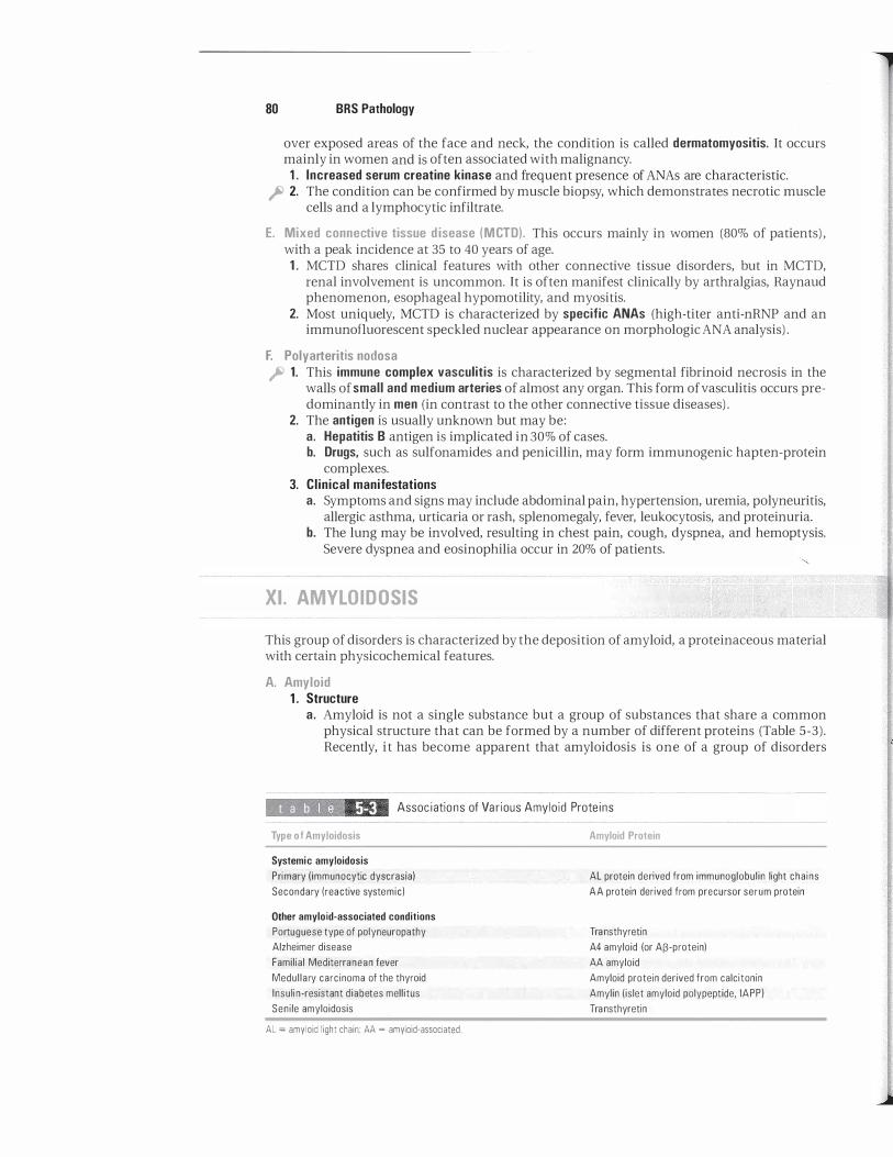
80 BRS Pathology
over exposed areas of the face and neck, the condition is called dermatomyositis. It occurs mainly in women and is often associated with malignancy.
1 . Increased serum creatine kinase and frequent presence of ANAs are characteristic. � 2. The condition can be confirmed by muscle biopsy, which demonstrates necrotic muscle
cells and a lymphocytic infiltrate.
E. Mixed connective tissue d isease (MClD). This occurs mainly in women (80% of patients) , with a peak incidence at 35 to 40 years of age. 1 . MCTD shares clinical features with other connective tissue disorders, but in MCTD,
renal involvement is uncommon. It is often manifest clinically by arthralgias, Raynaud phenomenon, esophageal hypomotility, and myositis.
2. Most uniquely, MCTD is characterized by specific ANAs (high-titer anti-nRNP and an immunofluorescent speckled nuclear appearance on morphologic ANA analysis) .
F. Polyarteritis nodosa � 1. This immune complex vasculitis is characterized by segmental fibrinoid necrosis in the
walls of small and medium arteries of almost any organ. This form of vasculitis occurs predominantly in men (in contrast to the other connective tissue diseases) .
2. The antigen is usually unknown but may be: a. Hepatitis B antigen is implicated in 30% of cases. b. Drugs, such as sulfonamides and penicillin, may form immunogenic hapten-protein
complexes. 3. Clinical manifestations
a. Symptoms and signs may include abdominal pain, hypertension, uremia, polyneuritis, allergic asthma, urticaria or rash, splenomegaly, fever, leukocytosis, and proteinuria.
b. The lung may be involved, resulting in chest pain, cough, dyspnea, and hemoptysis. Severe dyspnea and eosinophilia occur in 20% of patients.
XI. AMYLOIDOSIS
This group of disorders is characterized by the deposition of amyloid, a proteinaceous material with certain physicochemical features.
A. Amyloid 1 . Structure
a. Amyloid is not a single substance but a group of substances that share a common physical structure that can be formed by a number of different proteins (Table 5-3) . Recently, it has become apparent that amyloidosis is one of a group of disorders
t a b lew 5-3 Assoc iations of Va rious Amyloid Prote ins
Type of Amyloidosis
Systemic amyloidosis
Primary ( immunocytic dyscrasia)
Secondary (reactive systemic)
Other amyloid-associated conditions
Portuguese type of polyneuropathy
Alzheimer disease
Familial Mediterranean fever
Medul lary carcinoma of the thyroid
Insuli n-resistant d iabetes mellitus
Senile amyloidosis
AL = amyloid light chain; AA = amyloid-associated
Amyloid Protein
AL protein derived from immunoglobul in light chains
AA protein derived from precursor serum protein
Transthyretin
A4 amyloid (or AI3-protein)
AA amyloid
Amyloid protein derived from calcitonin
Amylin (islet amyloid polypeptide, lAPP)
Transthyretin

.....
I!1itt!1tD:1 Immune Dysfunction 81
characterized by abnormal protein folding. As pointed out in an earlier chapter, other folding defect disorders (not necessarily characterized by amyloid deposition) include a number of neurodegenerative diseases, such as Alzheimer disease, Huntington disease, and Parkinson disease; and perhaps prion diseases, such as "mad cow" disease.
b. It always has a l3-pleated sheet configuration (demonstrable by x-ray diffraction) . 2. Morphologic features
a. Amyloid is characteristically extracellular in distribution, most often appearing as accumulations proximate to basement membranes.
b. It has an amorphous eosinophilic appearance in routine hematoxylin and eosin sections.
c. It is characteristically stained by Congo red dye, demonstrating apple green birefringence when viewed under polarized light and confirming the suspected presence of amyloid. It can also be demonstrated by a variety of other methods, including immunochemical, fluorescent, and metachromatic techniques.
B. Clinical patterns of amyloidosis 1. Primary amyloidosis ( immunocytic dyscrasia amyloidosis)
a. The cause is deposition of amyloid fibrils derived from immunoglobulin l ight chains. referred to as Al (amyloid l ight chain) protein.
b. Amyloid deposition in tissues of mesodermal origin, such as heart. muscle. and tongue is characteristic. It may involve the kidney. with amyloid deposits in the glomerular mesangium as well as in the interstitial tissue between tubules.
� c. Primary amyloidosis is the form frequently associated with plasma cel l disorders. such as multiple myeloma, Waldenstrom macroglobulinemia, and other less defined disorders.
2. Secondary amyloidosis (reactive systemic amyloidosis) a. Deposition of fibrils consisting of the amyloid protein called AA protein. which is
formed from a precursor, serum amyloid-associated protein (SAA) is characteristic. Chronic tissue destruction leads to increased SAA.
b. Usually, involvement of parenchymatous organs, especially the kidney (nephrotic syndrome is very common) , l iver. adrenals. pancreas. lymph nodes. and spleen is seen. Perifollicular involvement in the spleen results in "sago spleen," an appearance reminiscent of tapioca-like granules.
� c. Secondary amyloidosis characteristically is a complication of chronic inflammatory disease. such as rheumatoid arthritis, tuberculosis, osteomyelitis, syphilis, or leprosy. It also may complicate noninflammatory disorders, such as renal cell carcinoma and Hodgkin disease.
3. Other forms of amyloidosis a . Portuguese type of polyneuropathy is associated with an amyloid derived from a protein
known as transthyretin (a serum protein that transports thyroxine and retinol). It is characterized by severe peripheral nerve involvement caused by amyloid deposits.
b. Alzheimer d isease is characterized by deposits of an amyloid protein referred to as A4 amyloid. or amyloid l3-protein. which differs from AL, AA, and transthyretinderived amyloid. The gene that codes for the protein precursors of A4 amyloid has been localized to chromosome 21 .
c. Famil ial Mediterranean fever is an autosomal recessive disorder occurring in persons of Eastern Mediterranean origin. It is characterized by episodic fever and polyserositis. The distribution and type of amyloid are similar to that of secondary amyloidosis (AA amyloid) .
d. Medullary carcinoma of the thyroid is characterized by prominent amyloid deposits within the tumor, apparently derived from calcitonin.
e. Diabetes mell itus in the insulin-resistant adult-onset form (type 2) is characterized by deposits of amyloid in islet cells. This amyloid is thought to be derived from either insulin or glucagon and is referred to as amylin or, alternatively, islet amyloid polypeptide. It is postulated that amylin interferes with insulin sensing by beta cells.

82 BRS Pathology
f. Senile amyloidosis is characterized by minor deposits of amyloid found at autopsy in the very elderly. This condition may involve the heart, brain, and other organs. When senile amyloidosis occurs in the heart, the amyloid protein is derived from transthyretin.
g. Dialysis-associated amyloidosis is characterized by amyloid deposits in the joints of patients who have undergone hemodialysis for several years. The amylOid is derived from l3-microglobulin, a protein not readily filtered by the dialysis membrane.

.....
Review Test 1 Directions: Each of the numbered items or incomplete statements in this section is followed by answers or by completions of the statement. Select the one lettered answer or completion that is best in each case.
1 . A 28-year-old woman is found to have pulmonary sarcoidosis. Flow cytometric analysis ofT cells isolated from the alveoli and lung interstitium reveals the presence of large numbers ofT helper (THl ) cells. These cells are known to secrete which of the following substances?
(A) Complement component C5A (8 ) Elastase and lysyl-hydroxylase (e) Interleukin-2 (IL-2) and interferon-/, (D ) IL-8 and transforming growth factor-[3 (E) Leukotrienes C4 and Cs
2. A pathologist examines a renal biopsy from a 45-year-old man with the nephrotic syndrome and requests a Congo red stain to confirm the nature of an amorphous acidophilic extracellular hyaline substance localized within the mesangial matrix of the glomeruli. A positive test confirms the presence of
(A) aI-antitrypsin. (8 ) amyloid. (e) copper. (D ) glycogen. (E) hemosiderin.
3. Within minutes of a bee sting, a 23-year-old woman develops generalized pruritus and hyperemia of the skin, followed shortly by swelling of the face and eyelids, dyspnea, and laryngeal edema. This reaction is mediated by
(A) antigen-antibody complexes. (8 ) cytotoxic T cells. (e) IgA antibodies. (D ) IgE antibodies. (E ) IgG antibodies.
4. A 25-year-old woman with membranous glomerulonephritis receives a kidney transplant. The donor is her HLA-matched sister. She does well initially, but after several weeks, there is a progressive increase in serum creatinine. Assuming that this represents acute cellular rejection, an infiltrate with which of the follOwing types of inflammatory cells is most likely to be a prominent finding on renal biopsy?
(A) Eosinophils (8 ) Lymphocytes (e) Mast cells (D ) Monocytes-macrophages (E) Neutrophils
5. A l -year-old boy has had repeated pyogenic infections with streptococci, staphylococci, and Haemophilus for the past 6 months. Tests for T-cell function, granulocyte function, and complement activity have all been unaffected. Serum IgG is 50 mg/ dL (normal 500 mg/ dL) . Flow cytometry revealed absent expression of heavy-chain fL on blood lymphocytes. T lymphocytes were slightly increased in number, with a normal CD4 +:CD8+ ratio. Expected findings on examination of a lymph node biopsy from this patient include which of the following?
(A) Absent germinal centers (8 ) Follicular hyperplasia with exuberant
proliferation of immature B cells (e) Massive THI cell infiltration into
lymphoid follicles (D ) Normal lymphoid tissue development (E) Plasma cell hyperplasia
83

84 BRS Pathology
6. A 20-year-old woman presents with malar rash, arthralgias, low-grade fever, and high titer antibodies to double-stranded DNA and to the Sm (Smith) antigen. Which of the following forms of hypersensitivity is the primary mechanism of the abnormalities found in this disorder?
(A) Type I (immediate or anaphylactic) hypersensitivity
(B ) Type II (antibody-mediated or cytotoxic) hypersensitivity
(e) Type III (immune complex-mediated disorders) hypersensitivity
(0) Type IV (cell-mediated) hypersensitivity
7. An HIV-positive intravenous drug user is suspected of having active tuberculosis, and a tuberculin (Mantoux) intradermal skin test is performed. After 48 hours, 10 cm of induration is observed. Which of the following are involved in this form of hypersensitivity reaction?
(A) B cells and antibodies (B) Basophils and IgE (e) Immune complexes and complement (0) Plasma cells and IgM (E ) T cells and macrophages
8. A 1 -year-old girl with an inborn error of metabolism reSUlting in a lysosomal storage disease receives a hematopoietic stem cell transplant intended to replace her macrophage population. The gene of interest has a "marker" small nucleotide polymorphism within a noncoding intron of the affected gene in which an A (patient gene) is substituted for a G (donor gene) . She does quite well for the first 3 weeks. She tests positive for the missing enzyme, her previously abnormally enlarged organs begin to diminish in size, and assay of peripheral blood lymphocytes reveals increasing numbers of cells with the G polymorphism. However, the attending physicians are now concerned because repeated genetic testing reveals a progressive increase in lymphocytes with the A polymorphism. Which of the following is the best explanation for this finding?
(A) Generalized immune complex formation (B) Graft-versus-host disease (e) Immune paralysis (0) Immune tolerance (E) Rejection of the stem cell transplant
9. A 24-year-old woman who had previously been uneventfully transfused receives a blood transfusion during surgery and shortly thereafter develops itching, generalized urticaria, laryngeal edema, and dyspnea with wheezing respiration. She has a past history of recurrent upper respiratory tract infections and frequent episodes of diarrhea. Laboratory studies are most likely to reveal decreased concentrations of which of the following immunoglobulins?
(A) IgA (8 ) IgD (e) IgE (0) IgG (E ) IgM
10. A 22-year-old woman with acute myeloblastic leukemia receives an allogeneic bone marrow transplant with apparent successful engraftment. Three weeks later, early jaundice, as well as a generalized maculopapular rash, is noted. Profuse diarrhea follows. A skin biopsy reveals vacuolar changes, necrotic epidermal cells, and a lymphocytic infiltrate. These findings are most likely caused by
(A) antibody-dependent cellular cytotoxicity. (8 ) attack on host epithelial cells by donor
CD8 + T cells. (e) contamination of the donor transplant
cells with hepatitis C virus. (0) IgE and mast cell-mediated anaphylactic
hypersensitivity. (E ) secretion of interleukin-2 and
interferon -')' by T H 1 cells.
1 1 . A 19-year-old intravenous drug user has regularly sought human immunodeficiency virus (HN) testing, always with negative results. He admits to carelessly sharing needles on multiple occasions with individuals later found to be HIV-positive. He has heard that there is an inherited genetic basis for some people to be relatively "immune" to HN infection. The genetic change that he is referring to is a mutation in a gene coding for which of the follOwing proteins?
(A) CCR5 (8 ) CD4 (e) gp120 (0) gp41 (E) Reverse transcriptase

12. A 2-year-old boy has eczema and thrombocytopenia. There is also a history of recurrent infection. His brother has similar abnormalities, but none of his three sisters are affected. Patients with this disorder are known to have impaired antibody response to which of the following types of antigen?
(A) Deoxyribonucleic acid (B ) Phospholipid (e) Polysaccharide (0) Ribonucleic acid (E ) Steroid
13. A 45-year-old woman is seen because of varied complaints. She has been troubled by small painful lumps under the skin of her fingers, some of which have ruptured and leaked a chalky white substance. She also complains of painful episodes in her fingers and toes, which blanch and turn blue on exposure to cold. In addition, questioning reveals increasing "heartburn" and difficulty swallowing. Examination reveals thickening of the skin of the fingers and toes, resulting in a claw-like appearance. Telangiectatic cl usters of vessels, appearing as small focal
il!il!!l11t§ii Immune Dysfunction 85
red lesions, are observed in the skin of the face, upper trunk, and hands, and on the mucosal surface of the lips. Antibodies to which of the following are most characteristic of the findings presented by this patient?
(A) Centromeric proteins (B ) Histidyl-t-RNA synthetase 00- 1) (e) Histones (D ) Mitochondria (E) Native DNA
14. A 7-month-old boy has had multiple bouts of otitis media, sinusitis, bronchitis, oral candidiasis, and multiple viral infections. Cessation of the recurrent infections follows successful engraftment of a bone marrow transplant. The basis of the clinical improvement is
(A) direct transfusion of antibody-producing B cells.
(B) direct transfusion of donor CD4+ and CD8+ lymphocytes.
(e) donor suppression of recipient cytotoxic T cells.
(D ) infusion of donor-derived cytokines. (E ) maturation of donor lymphoid
progenitor cells.

Answers and Explanations
1 . The answer is C. T helper (TH l ) cells secrete interleukin-2 and interferon-'Y' which in turn facilitate T-cell expansion and macrophage activation.
2. The answer is B. A positive Congo red test confirms the presence of amyloid. Apple green birefringence is observed under polarized light.
3. The answer is D. The clinical description is characteristic of systemic anaphylaxis, an IgE-mediated type I hypersensitivity reaction. In type I hypersensitivity, reaction of antigen with preformed IgE antibodies fixed by Fc receptors to the surface of basophils or tissue mast cells results in cytolysis and degranulation of these cells, with release of histamine and other mediators.
4. The answer is B. Acute cellular rejection is characterized by an infiltrate of both CD4+ and CD8 + lymphocytes. Acute rejection occurs over a variable time period, ordinarily days to weeks to months after transplant.
5. The answer is A. The diagnosis is X-linked agammaglobulinemia of Bruton. Failure of maturation of pre-B cells is associated with absence of mature B lymphocytes and plasma cells; failure of antibody synthesis; marked serum hypogarnmaglobulinemia; and recurrent bacterial infections, especially sinopulmonary infections. Histologic examination of lymphoid tissue reveals marked underdevelopment of germinal centers. T cells are unaffected, as are T-cell functions such as cell-mediated immunity and resistance to most viral infections. The disease is X-linked and is due to mutations in the B cell tyrosine kinase (Btk) gene.
6. The answer is C. The diagnosis is systemic lupus erythematosus, and the most characteristic lesions are mediated by immune complex deposition (type III hypersensitivity) . In this form of hypersensitivity, antibody combines with antigen, resulting in antigenantibody complexes. Insoluble aggregates of immune complex are deposited in vessel walls, serosal surfaces, and other extravascular sites, and complement is bound. The antigen-antibody-complement complexes are highly chemotactic for neutrophils, which release lysosomal enzymes and other mediators of tissue damage (prostaglandins, kinins, and free radicals) .
7. The answer is E. The tuberculin test is a classic example of delayed hypersensitivity, a form of cell-mediated hypersensitivity involving CD4+ T cells and macrophages. Native CD4+ T cells are converted to T H I cells that secrete cytokines, especially interferon-'Y' which is a central mediator of delayed hypersensitivity. Among the many actions of interferon-'Y, the most important is the activation of macrophages.
8. The answer is E. Rejection of the stem cell transplant is occurring, as evidenced by reappearance of the marker for the patient's original gene. Small nucleotide polymorphisms are the most frequent form of DNA variation. As the name implies, they are typically small in size, often a single nucleotide. They may occur in any portion of the gene, even in intergenic regions of the genome, and are of increasing importance as genetic markers, as illustrated by the example presented here.
9. The answer is A. Isolated 19A deficiency is most often asymptomatic but can be characterized by anaphylactic reactions to transfused blood. It also can be associated with frequent episodes of diarrhea and recurrent infections, especially those involving mucosal surfaces. This inherited B-cell defect is due to inability of 19A B cells to mature into 19A-producing plasma cells. Interestingly, the defect leading to systemic anaphylaxis involves both 19A and IgE antibody formation. Patients lacking 19A can develop IgE antibodies against the 19A antibodies present in transfused blood. This sensitization can result in susceptibility to anaphylaxis on subsequent transfusion.
86
p
i!l1ftlit1D Immune Dysfunction 87
10. The answer is B. Graft-versus-host disease is most often manifest by clinical findings related to the three principal target organs: the skin, liver, and gastrointestinal tract. The skin manifestations are often initiated by a pruritic rash. Elevation of bilirubin and liver enzymes signals the hepatic involvement. Effects on the ileum and colon present as diarrhea and abdominal pain. The lesions are caused by donor lymphocytes, with targeting of host epithelial cells by CD8+ T cells.
1 1 . The answer is A. Either of two coreceptors, CCR5 or CXCR4, is involved in the initial binding of the virus to the cell surface molecule gp120 on CD4+ cells. It is of considerable interest that certain mutations in the CCR5 gene are associated with what appears to be total resistance to infection with some common strains of HIV. Homozygotes are totally resistant, and heterozygotes develop a more slowly progressive disease.
12. The answer is C. Wiskott-Aldrich syndrome is an X-linked disorder characterized by eczema, thrombocytopenia, recurrent infections, and poor antibody response to polysaccharide antigens. Bloody diarrhea is also common. Death before 6 years of age occurs frequently and is most often due to bleeding, infection, or malignancy (most often lymphoma) .
13. The answer is A. The patient exhibits the cardinal findings of the CREST syndrome, a less severe variant of systemic sclerosis (scleroderma) characterized by Calcinosis, Raynaud phenomenon, Esophageal dysfunction, Sclerodactyly, and Telangiectasia. Although a number of antinuclear antibodies can be found in this disorder, the most characteristic is directed at centromeric proteins (the antibody is often referred to as anticentromere) .
14. The answer is E. Severe combined immunodeficiency disease is characterized by failure to thrive and increased susceptibility to bacterial, fungal, and viral infections. Laboratory studies reveal decreased numbers of both B cells and T cells and deficiency of immunoglobulins. The treatment of choice is bone marrow (or other sources of hematopoietic stem cells) transplantation and is based on maturation of donor lymphoid progenitor cells.

Neoplasia
I. GENERAL CONSIDERATIONS
Neoplasia is the uncontrolled, disorderly proliferation of cells, resulting in a benign or malignant tumor, or neoplasm.
A. Dysplasia � 1. This reversible change often precedes malignancy.
2. Morphologic manifestations include disorderly maturation and spatial arrangement of cells; marked variability in nuclear size and shape (pleomorphism) ; and increased, often abnormal, mitosis.
3. Examples include dysplasia occurring in squamous epithelium of the cervix. This change is often a precursor of malignancy.
B. Neoplasms 1 . If the resemblance to tissue of origin is close, the neoplasm is termed well d ifferentia ted;
if little resemblance to the tissue of origin is seen, it is poorly differentiated. 2. Neoplasms grow at the expense of function and vitality of normal tissue without benefit
to the host and are largely independent of host control mechanisms.
II. CLASSIFICATION AND NOMENCLATURE OF TUMORS
Neoplasms are classified as either malignant or benign, based on their behavior. They are also described by terms derived from the appearance of the neoplasm, tissue of origin , or degree of differentiation.
A. Malignant tumors (cancer) 1. General considerations
88
� a. Invasion (spread of the neoplasm into adjacent structures) and metastasis (implantation of the neoplasm into noncontiguous sites) are characteristic. Metastasis is the most important defining characteristic of malignancy, although there are some malignant tumors, such as basal cell carcinoma of the skin, that rarely metastasize.
b. Malignant tumors are usually less differentiated than benign tumors. � c. Anaplasia, in which tumor cells are very poorly differentiated and exhibit pleomor
phism, hyperchromatism (dark-staining nuclei), an increased nuclear-cytoplasmic ratio, abnormal mitoses, cellular dyspolarity, and often prominent nucleoli , is a common feature. In general, highly anaplastic tumors are very aggressive, and well-differentiated tumors are less aggressive. Paradoxically, the most aggressive tumors often respond
--
IiIi!tt'11tDi N e 0 p I a s i a 89
well to chemotherapy and radiotherapy, because these modalities are most effective with rapidly dividing cells.
2. Carcinoma is a malignant tumor of epithel ia l origin and can be seen in the following variations: a. Squamous cell carcinoma � (1) Squamous cell carcinoma originates from stratified squamous epithelium of, for
example, the skin, mouth, esophagus, and vagina, as well as from areas of squamous metaplasia, as in the bronchi or the squamocolumnar junction of the uterine cervix.
� (2) It is marked by the production of keratin. b. Transitional cell carcinoma arises from the transitional cell epithelium of the urinary
tract. c. Adenocarcinoma is carcinoma of glandular epithel ium and includes malignant
tumors of the gastrointestinal mucosa, endometrium, and pancreas. It is often associated with desmoplas ia, tumor-induced proliferation of non-neoplastic fibrous connective tissue, particularly in adenocarcinoma of the breast, pancreas, and prostate.
3. Sarcoma is a malignant tumor of mesenchymal origin. It is often used with a prefix that denotes the tissue of origin of the tumor, as in osteosarcoma (bone), rhabdomyosarcoma (skeletal muscle) , leiomyosarcoma (smooth muscle) , and l i posarcoma (fatty tissue) .
4. Eponymically named tumors include Burkitt lymphoma, Hodgkin disease, and Wilms tumor.
5. Teratoma is a neoplasm derived from all three germ cell layers, which may contain structures such as skin, bone, cartilage, teeth, and intestinal epithelium. It may be either malignant or benign and usually arises in the ovaries or testes.
B. Benign tumors 1 . General considerations
a. Benign tumors are usually well differentiated and closely resemble the tissue of origin. b. They grow slowly and do not metastasize. If their growth compresses adjacent tissues,
they can be harmful. For example, benign intracranial tumors can be more lethal than some malignant skin tumors.
c. They tend to become encapsulated. d. They are denoted by the suffix -oma, as in lipoma and fibroma; however, this suffix
is also applied to some malignant neoplasms, such as hepatoma, melanoma, lymphoma, and mesothelioma, as well as several non -neoplastic swellings, including granuloma and hematoma.
2. Papil loma. This benign neoplasm most often arises from surface epithelium, such as squamous epithelium of the skin, larynx, or tongue. It may also develop from transitional epithelium of the urinary bladder, ureter, or renal pelvis. The tumor consists of delicate finger-l ike epithel ial processes overlying a core of connective tissue stroma that contains blood vessels.
3. Adenoma. This benign neoplasm of glandular epithelium occurs in several variants: a. Papil lary cystadenoma is characterized by adenomatous papillary processes that extend
into cystic spaces, as in cystadenoma of the ovary. b . Fibroadenoma is marked by proliferation of connective tissue surrounding glandular
epithelium; for example, fibroadenoma of the breast. In this instance, either the connective tissue or the stroma may be neoplastic.
4. Benign tumors of mesenchymal origin are most often named by the tissue of origin (e.g., leiomyoma, rhabdomyoma, lipoma, fibroma, chondroma) . They include the most common neoplasm of women, the uterine leiomyoma, or fibroid tumor.
5. Choristoma. This is a small non -neoplastic area of normal tissue misplaced within another organ, such as pancreatic tissue within the wall of the stomach.
6. Hamartoma. This is a non-neoplastic, disorganized, tumor-like overgrowth of cell types regularly found within an affected organ; hemangioma, an irregular accumulation of blood vessels, is an example.

90 BRS Pathology
III. PROPERTIES OF NEOPLASMS
A. Monoclonality. This denotes origin from a single precursor cell. � 1. Most neoplasms are monoclonal; in contrast, polyclonal prol iferations are almost always
non-neoplastic. 2. Monoclonality is assessed by a variety of approaches using isoenzyme patterns or other
markers in individuals who are heterozygous for the measured trait. a. Monoclonal tumors express only one of the isoenzymes or other markers of clonality. b. Polyclonal cellular prol iferations exhibit both isoenzymes or markers.
3. Glucose-6-phosphate dehydrogenase (G6PD) isoenzymes and other X-l inked markers a. These are the first described and classic indicators of monoclonality in tumors. b. Because of X inactivation in early embryonic life, tissues of females heterozygous for
G6PD isoenzymes consist of a mosaic of cell types, with random cells expressing one or the other of the two isoenzymes; however, in the case of G6PD, one of the two polymorphic forms of these isoenzymes occurs with significant frequency only in women of African lineage, limiting the application of this technique.
c. Other polymorphic X-linked markers such as iduronate-2-sulfatase and phosphoglycerate kinase can be used in a broader spectrum of patients.
4. Human androgen receptor gene (HUMARA) a. This gene has now become the most common marker used to determine clonality. b. This method involves studying methylation patterns adjacent to high-frequency
polymorphisms in multiple populations. 5. Specific translocations. If present, genes such as the 8 ; 14 translocation can also be used
to assess clonality in certain neoplasms (e.g., in Burkitt lymphoma) . 6. Monoclonal ity of cells of lymphoid origin
a . Indicators of monoclonality in malignancies of B-cell origin ( 1 ) Immunoglobul ins are produced by B-cell malignant tumors and are demonstrable
as cytoplasmic or surface immunoglobulin or, in the case of multiple myeloma, are secreted and are demonstrable in the serum. (a) If monoclonal, the resultant mixture of immunoglobulin molecules exhibits
either kappa or lambda chain specificity, but not both, a characteristic finding in neoplastic B-cell proliferations.
� (b) If B-cell or plasma cell proliferations are polyclonal, they result in the production of heterogeneous immunoglobulin molecules, some of which express kappa specificity and others that express lambda specificity.
(2) Immunoglobulin gene rearrangement is a characteristic of B-cell maturation. The number of possible combinations achieved by rearrangement is almost countless; it can be assumed that each normal B cell is marked by a unique rearrangement pattern. Neoplastic prol iferation results in large numbers of cells, all demonstrating the same pattern of immunoglobulin gene rearrangement denoting their common origin from a single cell. (a) Assessment is by molecular diagnostic techniques. (b) Because immunoglobulin heavy chain rearrangement is limited to B cells,
this approach also demonstrates the B-cell origin of a tumor. b. Indicators of monoclonality in malignancies of T-cell origin
( 1 ) Surface antigens (markers) are demonstrable as T cells mature; they may be characteristic of either the stage of maturation or functional subclass. Cellular prol iferations in which large numbers of T cells share surface markers in common are suggestive of monoclonality. In addition to many others, these markers include the CD4 antigen marking helper T cells and the CD8 antigen marking suppressor T and cytotoxic T cells.
(2) T-cell receptor gene arrangement is analogous to immunoglobulin gene rearrangement and is used in a similar manner to demonstrate both the T-cell origin of a tumor and its monoclonality.

FIGURE 6-1 Metastasis to the liver. The multiple nodules in this liver are characteristic of metastatic carcinoma. (Reprinted with permission from Rubin R, Strayer 0, et aI., eds.: Rubin's Pathology. Clinicopathologic Foundations of Medicine, 5th ed. Baltimore, Lippincott Williams & Wilkins, 2008, p. 666.)
B. Invasion and metastasis (Figures 6- 1 , 6-2, and 6-3)
I!iit1ttOt§ij N e 0 p I a s i a 91
1. Invasion is aggressive infiltration of adjacent tissues by a malignant tumor. � a. It often extends into lymphatics and blood vessels, with the formation of tumor
emboli that may be carried to distal sites. b. Not all tumor emboli result in metastatic tumor implants, and the presence of tumor
cells within blood vessels or lymphatics indicates only the penetration of basement membranes and is not synonymous with metastasis.
2. Metastasis is implantation in distal sites. � a. Multistep process of metastasis
( 1 ) Growth and vascularization of the primary tumor (2) Invasiveness and penetration of basement membranes into lymphatics or blood
vessels (3) Transport and survival of tumor cells in the circulation (4) Arrest of tumor emboli in the target tissue and passage again across basement
membranes
FIGURE 6-2 Metastasis to the lung. These metastatic lesions result in a characteristic cannon ball appearance on X-ray. (Reprinted with permission from Rubin R, Strayer 0, et aI., eds.: Rubin's Pathology. Clinicopathologic Foundations of Medicine, 5th ed. Baltimore, Lippincott Williams & Wilkins, 2008, p. 545.)
Chaz
Placed Image
Chaz
Placed Image
Chaz
Placed Image

92 BRS Pathology
FIGURE 6-3 Metastasis to lymph nodes. This carcinoma has metastasized to periaortic lymph nodes. (Reprinted with permission from Rubin R, Strayer D, et aI., eds.: Rubin's Pathology Clinicopathologic Foundations of Medicine, 5th ed. Baltimore, Lippincott Williams & Wilkins, 2008, p. 146.)
(5) Overcoming of target tissue defense mechanisms (6) Development of successful metastatic implants
b. Preferential routes of metastasis vary with specific neoplasms. ( 1 ) Carcinomas tend to metastasize via lymphatic spread. (2) Sarcomas tend to invade blood vessels early, resulting in widespread blood-borne
(hematogenous) dissemination. (3) Notable exceptions include renal cell and hepatocellular carcinoma, which are
marked by early venous invasion and hematogenous dissemination. c. Target organs are most commonly the liver, lungs, brain, adrenal glands, lymph nodes,
and bone marrow. They rarely include skeletal muscle or the spleen. � d. Tumor progression is characterized by the accumulation of successive cytogenetic or
molecular abnormalities. � (1) It is exemplified by the progression of changes from normal colonic epithelium to
adenoma to carcinoma to metastasis, with parallel changes in the APe, K-ras, Dee, p53, and possibly other genes.
� (2) Individual neoplastic cells within a tumor may have varying metastatic potential.
C. Other clinical manifestations of malignancy. These are mediated by mechanisms other than invasion and metastasis. 1. Cachexia and wasting. The origin is complex; it is characterized by weakness, weight
loss, anorexia, anemia, infection, and hypermetabolism. This process may be mediated in part by cachectin (tumor necrosis factor-a, TNF-a), a product of macrophages that promotes catabolism of fatty tissue.
2. Endocrine abnormalities are caused by tumors of endocrine gland origin, which may actively elaborate hormones, leading to a variety of syndromes. a. Pituitary abnormal ities � (1) Prolactinoma, leading to amenorrhea, infertility, and sometimes galactorrhea
(2) Somatotropic (acidophi l ic ) adenoma, leading to gigantism in children and acromegaly in adults
(3) Corticotropic (most often basophi l ic) adenoma, leading to Cushing disease (adrenal hypercorticism of pituitary origin)
b. Adrenocortical abnormalities include adrenogenital syndrome, Conn syndrome, and Cushing syndrome of adrenal origin, reSUlting from adrenal cortical tumors.
c. Ovarian abnormalities ( 1 ) Granulosa-theca cell tumor, leading to hyperestrinism (2) Sertol i-Leydig cell tumor, leading to excess androgen production
Chaz
Placed Image

jii>
I!1lI!J'l1mj N e 0 p I a s i a 93
d. Trophoblastic tissue abnormal ities include hyperproduction of human chorionic gonadotropin from hydatidiform mole or choriocarcinoma.
3. Para neoplastic syndromes a. Endocrinopathies are caused by ectopic production of hormones or chemically unre
lated substances inducing effects similar to those of a given hormone. They include the following:
/'- ( 1 ) Cushing syndrome is caused by production of ACTH-like substances by small cell (oat cell) carcinoma of the lung.
/'- (2) Inappropriate secretion of antidiuretic hormone may be caused by a variety of tumors, most commonly small cell carcinoma of the lung.
/'- (3) Hypercalcemia is caused by metastatic disease in bone, secretion of a substance similar to parathormone by squamous cell bronchogenic carcinoma, or secretion of a substance similar to osteoclast-activating factor by the malignant plasma cells of multiple myeloma.
(4) Hypoglycemia is caused by secretion of insulin-like substances by hepatocellular carcinomas, mesotheliomas, and some sarcomas.
/'- (5) Polycythemia is caused by elaboration of erythropoietin by renal tumors and other neoplasms.
(6) Hyperthyroidism is caused by production of substances such as thyroid -stimulating hormone by hydatidiform moles, choriocarcinomas, and some lung tumors.
b. Neurologic abnormalities may occur in the absence of metastatic disease. They include degenerative cerebral changes with dementia, cerebellar changes with resultant gait dysfunction, and peripheral neuropathies.
/'- c. Skin lesions may be associated with visceral malignancies. They include acanthosis nigricans and dermatomyositis.
/'- d. Coagulation abnormal ities include migratory thrombophlebitis associated with carcinoma of the pancreas and other visceral malignancies (Trousseau phenomenon), and disseminated intravascular coagulation associated with various neoplasms.
4. Oncofetal antigens a. These are proteins normally expressed only in fetal or embryonic life; their expression
by neoplastic cells is considered a manifestation of dedifferentiation (i.e., the undifferentiated neoplastic cells tend to resemble their embryonic counterparts) .
/'- b. They include carcinoembryonic antigen (CEA), which is associated with colon cancer and other cancers and preneoplastic processes, and a-fetoprotein (AFP), which is associated with hepatocellular carcinoma and many germ cell tumors. APP is also increased in fetal anencephaly and other neural tube defects.
IV. CARCINOGENESIS AND ETIOLOGY
The transformation of normal to neoplastic cells is caused by both endogenous and exogenous factors, including chemical and physical agents, viruses, activation of cancer-promoting genes, and inhibition of cancer-suppressing genes.
A. Chemical carcinogenesis /'- 1 . Association between chemicals and cancer (Table 6- 1 )
2. Types of carcinogens a . Direct-reacting carcinogens do not need to be chemically altered to act. b. Indirect-reacting carcinogens require metabolic conversion from procarcinogens to
active ultimate carcinogens. For example, a mucosal glucuronidase in the urinary bladder converts �-naphthylamine glucuronide to the carcinogen �-naphthylamine.
3. Stages of chemical carcinogenesis a. Initiation is the first critical carcinogenic event, and it is usually a reaction between a
carcinogen and DNA. Two or more agents (e.g., chemicals, viruses, radiation) may act together as cocarcinogens.

94 BRS Pathology
Some E nvironmental Fact ors, D rugs, and Ch emica ls A ssociated w ith H uman Cancer
factor Type of Malignancy
Cigarette smoking Excess sun exposure Alkylating agents Asbestos Smoked foods rich in
nitrosamines Alcohol
Arsenic Low-fiber diet High-fat diet Aniline dyes, aromatic amines,
�-naphthylamine Aflatoxin Bl Benzene Polyvinyl chloride Thorotrast Diethylstil bestrol (DES)
Nickel Chromium Uranium
Carcinoma of the lung; carcinoma of the larynx Squamous cell carcinoma and basal cell carcinoma of skin; melanoma Acute leukemia Carcinoma of the lung; pleural and peritoneal mesothelioma; gastrointestinal tract cancers Adenocarcinoma of stomach
Carcinoma of mouth and esophagus (especially in association with cigarette smoking) Hepatocellular carcinoma (in association with cirrhosis of liver; all forms of cirrhosis
predispose to hepatocellular carcinoma) Squamous cell and basal cell carcinoma of skin Adenocarcinoma of colon Breast carcinoma Transitional cell carcinoma of bladder (caused by action of bladder mucosal glucuronidase
on detoxified glucuronides of these compounds) Hepatocellular carcinoma Acute leukemia Hepatic hemangiosarcoma (angiosarcoma) Hepatic hemangiosarcoma (angiosarcoma) Clear-cell adenocarcinoma of vagina (occurs in daughters of patients who received DES
during pregnancy)
Carcinoma of the lung Carcinoma of the lung Carcinoma of the lung
b. Promotion is induced by a stimulator of cell proliferation and enhances the carcinogenic process. A promoter, not a carcinogenic in itself, enhances other agents' carcinogenicity. For example, phorbol esters react with membrane receptors, stimulating cell replication. This may enhance clonal selection, resulting in cells with increasingly deleterious DNA changes.
B. Radiation carcinogenesis Ji" 1 . Exposure to ultraviolet radiation in the form of sunlight is clearly related to the frequency
of skin cancers, such as squamous cell and basal cell carcinomas and melanomas. Ji" a. The process is thought to act by inducing dimer formation between neighboring
thymine pairs in DNA. In most cases, such dimers are successfully repaired by enzymatically mediated mechanisms.
b. That skin cancer may be induced by such dimer formation is suggested by the greatly increased incidence of skin tumors seen in xeroderma pigmentosum, an autosomal recessive disorder characterized by failure of DNA excision repair mechanisms.
2. Ionizing radiation is a classic cause of cancer, exemplified by the increased incidence of cancers in those exposed to radiation. There are several historic examples:
Ji" a. Skin cancer and myeloid leukemias in radiologists b. Lung cancer in uranium miners
Ji" c. Thyroid cancer in patients who have received head and neck radiation therapy Ji" d. Acute and chronic myeloid (but not lymphoid) leukemias in survivors of atomic
blasts Ji" e. Osteosarcoma in radium watch-dial workers
Ji" C. Viral carcinogenesis (Table 6-2) 1. Virus types
a. DNA viruses (1) These viruses integrate viral DNA into host genomes, perhaps resulting in host
cell expression of viral mRNA coding for specific proteins.

-
I!1illJttttti N e 0 p I a s i a 95
t a b I e 6-2 Vi ruses and Other Infectious Age nts A ssociated w ith H u m a n M a l igna nc y
Infectious Agent Neoplasm Etiologic Role
HTLV-l HPV
Adult T-cell leukemia/lymphoma Genital tract neoplasms, especially premalignant
lesions and cancers of the cervix and vulva; laryngeal papillomas
Definite Almost certain
EBV Nasopharyngeal carcinoma Burkitt lymphoma
Almost certain Stimulates proliferation of B cells; increases
opportunity for translocation and oncogene activation
HBV Hepatocellular carcinoma Kaposi sarcoma
Almost certain HHV-8 Helicobacter pylori Adenocarcinoma and B-ceil lymphomas
IMALTomas) of stomach
Definite High suspect
HTLV-1 = human T Iymphotrophic virus type 1; HPV = human papillomavirus; EBV = Epstein-Barr virus; HBV = hepatitis B virus; HHV-8 = human herpesvirus-8 (also known as Kaposi sarcoma herpesvirus).
Ji'l (2) Examples include human papillomavirus, Epstein-Barr virus, and hepatitis B virus as prominent suspects that play a role in human carcinogenesis_
b_ Retroviruses are marked by transcription of viral genomic RNA sequences into DNA by action of viral reverse transcriptase. In the case of retroviruses that are tumorigenic in experimental animals, retroviruses are frequently characterized by substitutions of genomic sequences known as viral oncogenes.
2. Viral oncogenes (v-ones) are named with a three-letter abbreviation, preceded by vfor viral (Table 6-3) . They exhibit homology for DNA sequences of humans and other eukaryotic species; these eukaryotic DNA sequences are called proto-oncogenes, or cellular oncogenes (c-oncs), and are identified with the same three-letter abbreviations preceded by c for cellular.
D. Oncogenes and cancer, The protein products of proto-oncogenes play essential roles in DNA replication and transcription. 1. Mechan isms of action of oncogene protein products
a. Activation by binding of guanosine triphosphate (GTP). Ras oncogenes code for proteins known as p 2 1 proteins, which are functionally similar to G proteins, membranesignaling proteins activated by GTP binding, which mediate signal transduction from the cell surface. ( 1 ) Characteristics-Ras proteins and G proteins are located at the plasma membrane
and have GTP binding and GTPase activities.
t a b l� 6-3
Oncogene
v-src v-ab/ v-sis v-myc v-H-ras v-K-ras v-erb v-fms v-fos voros v-myb
Ex a mples of Retrovira l Oncogenes
Source
Rous sarcoma virus of chickens Abelson murine leukemia virus Simian sarcoma virus MC29 viral isolate from chickens Harvey rat sarcoma virus Ki rsten rat sarcoma virus Erythroblastosis virus of chickens Feline McDonough sarcoma virus FBJ osteosarcoma virus of mice UR2 avian sarcoma virus Avian myeloblastosis virus

96 BRS Pathology
� (a) GTPase hydrolytically converts active ras-GTP to inactive ras-GDP. These proteins are inactivated by ras-GTPase mediated by GTPase-activating protein (GAP) .
(b) GTP activation of ras can stimulate or depress adenylate cyclase activity, altering intracellular cAMP levels, thus affecting cellular behavior.
(2) Mutation of the ras gene usually occurs at codon 12, resulting in an aberrant p21 protein product with intact GTP binding but with a loss of GTPase activity. (a) Mutant ras proteins can be activated by GTP binding but cannot be inacti
vated by GTPase activity. � (b) The ras gene is mutated in 25%-30% of malignancies.
b. Protein tyrosine kinase activity is exhibited by the following: ( 1 ) The oncogene product of the Rous sarcoma virus (designated as pp60Src) (2) Other oncogene products, usually oncogenic analogs of transmembrane receptor
proteins c. Growth factor or growth factor receptor activity. Alterations in expression or structural
changes in oncogene products may result in inappropriate activation of receptor proteins or their oncogenic analogs, thus mimicking the actions of growth factors. ( 1 ) On stimulation with the appropriate growth factor, receptor proteins often demon
strate tyrosine kinase activity of their cytoplasmic domains. (2) Significant homologies occur between several oncogenes and the genes for cellu
lar growth factors and their receptors. For example: � (a) v-sis and the gene for the � chain of platelet-derived growth factor
(b) v-erb and the gene for the epidermal growth factor (EGF) receptor (c) v-fms and the gene for the colony-stimulating factor- l receptor (d) c-neu and the gene for the EGF receptor
d. Nuclear proteins. Some oncogene products, including the protein products of mye, fos, and myb, are confined to the cell nucleus.
2. Oncogenes and human cancer. Mechanisms by which c-oncs become tumorigenic include the following: a. Promoter insertion ( insertional mutagenesis)
( 1 ) Insertion of retroviral promoter or enhancer sequences into the host genome can lead to increased expression of a nearby oncogene.
(2) This mechanism is similar to the promoter-induced hyperexpression associated with translocations characteristic of several human leukemias and lymphomas.
b. Point mutations. These are exemplified by single nucleotide changes in codon 12 of the ras family of genes that are associated with a number of human tumors.
c. Chromosomal translocation. Frequent association with malignancy seen in these genetic rearrangements has been clarified by demonstrating that important genes are situated at the sites of chromosomal breaks. For example:
� ( 1 ) 8;14 translocation and Burkitt lymphoma. The c-mye proto-oncogene on chromosome 8 is translocated to a site adjacent to the immunoglobulin heavy chain locus on chromosome 14 . Major regulatory sequences within the immunoglobulin gene are thought to increase the expression of c-mye.
� (2) 14;18 translocation and fol l icular lymphoma. The immunoglobulin heavy chain locus on chromosome 1 4 is transposed to a site adjacent to bcl-2, an oncogene on chromosome 18. This results in enhanced expression of bcl-2, thus inhibiting apoptosis.
� (3) 9;22 translocation and chronic myeloid leukemia (CML). The c-abl proto-oncogene on chromosome 9 is transposed to a site adjacent to ber (breakpoint cluster region) , an oncogene on chromosome 22. (a) The union of ber and abl results in a hybrid, or chimeric, bcr-abl fusion gene
that codes for a protein with increased tyrosine kinase activity. (b) The altered chromosome carrying this hybrid gene, the Philadelphia chromo
some, can be demonstrated by cytogenetic techniques in hematopoietic cells of patients with CML.
(4) 15;1 7 translocation and acute promyelocytic leukemia (FAB M3 AML). The translocation involves the PML gene on chromosome 15 and the retinoic acid receptor-a
l!iill1l1tiilI N e 0 p I a s i a 97
(RAR-a) gene on chromosome 1 7. Therapy with the retinoic acid analogue alltrans retinoic acid can result in maturation of these leukemic cells and clinical remission.
d. Gene amplification ( 1 ) This reduplication of the gene, with multiple resultant genomic DNA copies, can
sometimes result in a thousand or more copies of the amplified gene. (2) Extensive amplification can result in small free chromosome-like bodies called
double minute chromosomes or in band-like structures within chromosomes caned homogeneously staining regions, which are both demonstrable cytogenetically.
(3) Several human neoplasms involve gene amplification. (a) Neuroblastoma is an aggressive childhood tumor characterized by marked
N-myc amplification that correlates inversely with the degree of differentiation of the neuroblastoma cells.
(b) Some breast cancers are marked by amplification of the HER-21neu oncogene (c-erbB2). Such amplification is correlated with a poor prognosis.
3. Cancer suppressor genes (anti-oncogenes). In contrast to the preceding mechanisms, cancer suppressor genes promote cellular proliferation when the gene is inactivated (most often by deletion) . A single residual copy of the anti-oncogene suppresses tumor formation, but homozygous inactivation (i.e. , loss of function of both copies) promotes the expression of the neoplastic phenotype.
� a. The prototype is retinoblastoma, an intraocular childhood tumor caused by inactivation of the Rb gene. The "two-hit" hypothesis of Knudson holds that two mutagenic events are required to induce alterations on both chromosomes. ( 1 ) In the familial forms of retinoblastoma, the gene on one chromosome in the
germline is inactivated or deleted, and the gene on the other chromosome is affected by a somatic mutation.
(2) In sporadic nonfamilial cases of retinoblastoma, both deletions occur as somatic mutations.
b. Inactivation of the Rb gene is also a factor in the genesis of other tumors, especially osteosarcoma, which often occurs following successful surgical cure of familial retino blastoma.
c. There are several other important cancer suppressor genes: ( 1 ) p53
(a) This gene, which has been called the "guardian of the genome," is mutated in more than 50% of all malignant tumors.
(b) In the setting of DNA damage, the p53 gene causes cell cycle arrest in G1, providing time for DNA repair. If repair is successful, cells re-enter the cell cycle. If repair is not successful, p53 product causes cell death by apoptosis (events involve transcription of p2 1 , inhibition of the formation of cyclin-CDK complexes, and inhibition of Rb phosphorylation, and also the transcription of bax, an apoptosis-promoting gene) .
(c) Familial loss causes the Li-Fraumeni syndrome, which is characterized by a wide variety of tumors: breast tumors, soft tissue sarcomas, brain tumors, and leukemias.
(2) WT-1 and WT-2. Inactivation or deletion of either of these genes, which are located on chromosome 1 1 , is associated with Wilms tumor, the most common renal neoplasm of children.
(3) A PC. Inactivation is common in familial polyposis coli and adenocarcinoma of the colon as well as a few other tumors (e.g., gastric and esophageal cancers) .
(4) BRCA-1 . Inactivation is associated with familial propensity to breast and ovarian carcinomas. (BRCA-2 inactivation is associated with breast cancer alone.)
E. Epidemiology. Important epidemiologic factors include geographic and racial differences, heredity, age, sex and hormonal differences, dietary factors, environmental toxins, and infection (see Table 6-1 ) .

98 BRS Pathology
V. OTHER NEOPLASTIC DISORDERS WITH KNOWN DNA DEFECTS
A. von Recklinghausen neurofibromatosis type 1 and NF-1 1. Characteristics include multiple benign neurofibromas, cafe au lait spots, iris hamar
tomas, and an increased risk of developing fibrosarcomas (see Chapter 4) . 2. The cause of the disorder is mutations in the NF-l tumor suppressor gene (which func
tions as a GAP protein that inactivates ras) .
B. Multiple endocrine neoplasia type lIa (MEN lIa) and ret (see Chapter 20 for descriptions of MEN I and MEN lIb) 1. MEN is the familial occurrence of the combination of medullary thyroid carcinoma,
bilateral pheochromocytomas, and hyperparathyroidism due to hyperplasia or tumor. � 2. The cause is mutations of the ret proto-oncogene that are transmitted in the germline.
Thus, demonstration of a ret mutation in a patient with medullary thyroid carcinoma would indicate the need for surveillance for the development of pheochromocytoma or hyperparathyroidism.
C. Hereditary nonpolyposis colon cancer (HNPCC, or lynch syndrome) 1. The cause of HNPCC is an inherited mutation in certain DNA repair genes, resulting in
genomic instability. The mutation, referred to as the replication error phenotype, is involved in mismatch repair and is detected as instability in micro satellite repeat sequences. HNPCC predisposes to mutations in other genes more directly related to transformation.
2. Characteristics include familial right-sided colorectal carcinomas and increased risk of other carcinomas.
� D. Xeroderma pigmentosum 1. This autosomal recessive disorder is manifest by an increased incidence of skin can
cers (basal cell carcinoma, squamous cell carcinoma, malignant melanoma) caused by hypersensitivity to ultraviolet light.
2. It involves defects in genes that function in nucleotide excision repair, which is required for repair of ultraviolet-induced pyrimidine (often thymine) dimers (cross-linked pyrimidine residues) .
VI. GRADING AND STAGING
These clinical measures are used for prognostic evaluation and planning of clinical management.
A. Grading. This histopathologic evaluation of the lesion is based on the degree of ce l l u lar d ifferentiation.
B. Staging. This is clinical assessment of the degree of local ization or spread of the tumor. � 1. Staging generally correlates better with prognosis than does histopathologic grading;
however, both approaches are useful. 2. It is exemplified by the generalized TNM system, which evaluates the size and the extent
of the tumor (T), lymph node involvement (N ), and metastasis (M). 3. It is sometimes oriented toward specific tumors, as exemplified by the Dukes system for
colorectal carcinoma and the Ann Arbor system for Hodgkin disease and non-Hodgkin lymphomas.

jii>
Review Test
Directions: Each of the numbered items or incomplete statements in this section is followed by answers or by completions of the statement. Select the one lettered answer or completion that is best in each case.
1. A 54-year-old woman who has been diagnosed with early stage breast cancer undergoes surgery for a lumpectomy to remove a small tumor detected by mammography. The pathology report confirms the early stage of the cancer and further comments on the fact that there is significant desmoplasia in the surrounding tissue. The term desmoplasia refers to
(A) an irregular accumulation of blood vessels.
(8) maturation and spatial arrangement of cells.
(e) metastatic involvement of surrounding tissue.
(D) normal tissue misplaced within another organ.
(E) proliferation of non-neoplastic fibrous connective tissue.
2. A 24-year-old woman with a history of heavy and painful menstrual periods has been having difficulty conceiving despite months of trying to become pregnant. Further workup includes a bimanual pelvic examination and an ultrasound, which demonstrates a mass in the uterus that is presumed to be a leiomyoma. This mass is a
(A) benign tumor of mesenchymal tissue. (8) benign tumor of surface epithelium. (e) malignant tumor of epithelial tissue. (D) malignant tumor of glandular
epithelium. (E) malignant tumor of mesenchymal tissue.
3. A 68-year-old man has a long history of prostate cancer that was metastatic at the time of diagnosis. Over the past 2 months, he has had significant weight loss, loss of appetite, and loss of energy. His current
spectrum of conditions can be attributed to which of the following?
(8) Fibroblast growth factor (e) Interleukin-2 (A) Platelet-derived growth factor (D) Tumor necrosis factor-a (E) Vascular endothelial growth factor
4. A 58-year-old man with a 700-pack-peryear smoking history presents to the emergency department with shortness of breath and hemoptysis. Portable chest radiography demonstrates a large mass centrally located within the left lung field. The serum calcium is 13 .0 mg/dL (normal 8.5 to 1 0.2) . The metabolic abnormality described here is likely due to elaboration of which substance?
(A) Adrenocorticotropic hormone-like substance
(8) Antidiuretic hormone (e) Carcinoembryonic antigen (D) Erythropoietin (E) Parathyroid-related hormone
5. An 8-year-old boy is referred to the dermatologist for numerous "suspicious" pigmented lesions on the face and neck. Further history reveals that the patient has had difficulty seeing out of his right eye; he is referred to the ophthalmologist, who diagnoses an ocular melanoma. Based on the patient's symptoms, the diagnosis of xeroderma pigmentosum is considered. This condition results from
(A) aberrant expression of a receptor tyrosine kinase.
(8) an inborn defect in DNA repair. (e) chemical carcinogenesis. (D) DNA viral infection. (E) retroviral infection.
99

1 00 BRS Pathology
6. A 46-year-old woman with prominent splenomegaly presents with a 3 -month history of malaise, easy fatigability, weakness, weight loss, and anorexia. A complete blood count and differential demonstrates a white blood cell count of 250,OOO/mm3 (normal 3000-10,OOO/mm3) with a predominance of myelocytes, metamyelocytes, band cells, and segmented neutrophils. Cytogenetic analysis is most likely to reveal which of the following translocations?
(A) t(8; 14) (B) t(9;22) (e) t(11 ;22) (0) t(14; 18) (E) t(15; 17)
7. A 63-year-old woman discovers a lump in her right breast. Mammography confirms the presence of a suspicious "lump," and a needle core biopsy is performed to determine whether the mass is malignant. The pathology report confirms that the mass is indeed cancerous and that the tissue demonstrates amplification of the Her-2lneu oncogene. The gene product of Her-2lneu is what kind of protein?
(A) GTPase (B ) GTPase-activating protein (e) Nuclear transcription factor (0) Receptor tyrosine kinase (E) A retinoic acid receptor protein
8. A 27 -year-old woman has recently been diagnosed with a glioma (a malignant brain tumor) . Further family history reveals that her 4-year-old son has been diagnosed with leukemia and has been undergoing chemotherapy. In addition, the patient's mother died at 36 years of age due to metastatic breast cancer. Li-Fraumeni syndrome is suspected, given the familial clustering of this group of malignancies. The
gene mutated in Li-Fraumeni syndrome normally functions in what capacity?
(A) Activates the GTPase activity of the gene product of the Ras oncogene
(B) Excises ultraviolet light-induced thymidine dimers
(e) Functions as a cytoplasmic tyrosine kinase
(0) Functions as a transmembrane tyrosine kinase
(E) Halts the cell cycle if DNA damage is detected
9. An 8-year-old child is evaluated by the pediatrician, who notes what appears to be ten small cafe-au-lait spots on the child's torso. In addition, on close inspection of the eyes, the presence of Lisch nodules is noted. The patient is diagnosed with von Recldinghausen neurofibromatosis type 1 . The protein that is mutated in this disorder normally
(A) activates the GTPase activity of Ras. (B ) cleaves cellular proteins during
apoptosis. (e) functions as a regulator of the cell cycle. (0) promotes angiogenesis in the growing
tumor mass. (E) promotes the cell to undergo apoptosis.
10. A 78-year-old Navy veteran with a 600-pack-per-year history of cigarette smoking presents with cancer. During his military career, he was involved in fireproofing naval combat ships with asbestos insulation. Given his environmental exposure to both tobacco and asbestos, to which cancer do both of these carcinogens contribute?
(A) Bladder cancer (B ) Bronchogenic cancer (e) Cancer of the throat (0) Esophageal cancer (E) Mesothelioma

Answers and Explanations
1. The answer is E. Desmoplasia refers to proliferation of non-neoplastic fibrous connect�ve tissue within a tumor and is quite common in cases of breast cancer. An irregular accumulation of blood vessels is known as a hemangioma. An area of tissue misplaced within another organ is known as a choristoma.
2. The answer is A. A leiomyoma (fibroid) is a benign tumor of the smooth muscle of the uterus and thus is an example of a benign tumor of mesenchymal origin. Profuse, painful menses and infertility are major complications of this most common tumor of the female genital tract. A leiomyosarcoma is the malignant counterpart. Malignant tumors of epithelial cells are carcinomas, and these are known as adenocarcinomas if they involve glandular epithelium. Benign tumors of surface epithelium are termed papillomas.
3. The answer is D. Cachexia, or wasting due to cancer, manifests with weakness, weight loss, anorexia, anemia, and infection. The principal cytokine responsible for such changes is tumor necrosis factor-a (TNF-a) . Both platelet-derived growth factor and fibroblast growth factor are involved in wound healing. Interleukin-2 (IL-2) is an immunostimulating cytokine produced by activated T cells. Vascular endothelial growth factor is important in the proliferation of blood vessels in a growing tumor.
4. The answer is E. The man is likely to have a lung tumor, given his clinical presentation and the radiographic results. The patient's hypercalcemia is likely due to a paraneoplastic syndrome, such as that due to the elaboration of parathyroid-related hormone (PTrH) . PTrH i s produced by squamous cell carcinoma, whereas adrenocorticotropic-like substance and antidiuretic hormone are produced by yet another form of lung cancer-small cell carcinoma of the lung. Carcinoembryonic antigen is an oncofetal antigen produced by colon cancer cells. Erythropoietin causes secondary polycythemia and is related to renal cell carcinoma.
5. The answer is B. Xeroderma pigmentosum is a hereditary DNA defect with a deficiency in the ability to repair ultraviolet (sunlight) -induced thymidine dimers. Faulty repair leads to increased sun sensitivity, with a predilection to develop skin lesions and skin cancers on exposed skin, as well as ocular melanomas. Aberrant expression of the gene for a receptor tyrosine kinase, such as the Her-2/neu gene product, can cause breast cancer. A retrovirus is responsible for the development ofT-cell leukemiallymphoma. The DNA virus human papillomavirus can cause cervical cancer.
6. The answer is B. The translocation t(9;22) is the characteristic translocation associated with chronic myelogenous leukemia, forming the so-called "Philadelphia chromosome." The resultant fusion protein, p210, has increased tyrosine kinase activity that contributes to the uncontrolled proliferation in this form of leukemia. The translocation t(14; 18) is seen in follicular lymphoma; t(8; 14) in Burkitt's lymphoma; t ( 15 ;17) in the M3 variant of acute promyelocytic leukemia (AML) ; and t ( 1 1 ;22) in Ewing sarcoma, a relatively uncommon tumor of bone.
7. The answer is D. Her-2/neu, also known as (c-erbB2), is a receptor tyrosine kinase related to epidermal growth factor receptor and is amplified at the DNA level and overexpressed at the protein level in some breast cancers. Ras is a GTPase that is mutated in a number of cancers. NF- l is a GTPase-activating protein (GAP) aberrantly expressed in neurofibromatosis. An aberrant version of a retinoic acid receptor is expressed in M3 AML.
101

1 02 BRS Pathology
8. The answer is E. Li-Fraumeni syndrome is a hereditary syndrome characterized by sarcomas, breast cancer, leukemia, and brain tumors. Xeroderma pigmentosum results from a defect in repair of UV damage. Numerous transmembrane tyrosine kinases, such as Her-2/neu, are implicated in numerous cancers. C-abl codes for a cytoplasmic tyrosine kinase that forms a fusion protein with bcr in chronic myelogenous leukemia. NF- l , mutated in neurofibromatosis, normally activates the GTPase activity of the gene product of the Ras oncogene. The gene p53, the "guardian of the genome," arrests the cell cycle in G1 in the event that DNA damage is detected.
9. The answer is A. The normal function of NF- l is to promote the intrinsic GTPase function of the Ras oncogene. When the Ras protein is bound to GTp, the growth-promoting function of the molecule is "ON." On hydrolysis of the GTP to GDp, Ras is converted to an inactive state. GTPase-activating proteins (GAPs) , such as NF-l, suppress cell growth by stimulating GTP hydrolysis. Patients with a mutation in NF- l are susceptible to fibrosarcomas as a result of loss of function of this GAP. The molecule Bax is pro-apoptotic and antagonizes Bcl-2. Vascular endothelial growth factor promotes tumor angiogenesis. The protein p53 regulates the cell cycle if DNA damage is detected. Lastly, caspases function to cleave cellular proteins once apoptosis is triggered.
10. The answer is B. Tobacco contributes to the development of many cancers, including those of the bladder, lung, throat, and esophagus. Asbestos exposure carries a risk of lung cancer, as well as mesothelioma, a cancer of the pleura. Tobacco and asbestos function as cocarcinogens in the pathogenesis of lung cancer, with an approximately 50-fold greater risk of developing bronchogenic cancer of the lung than in those without such exposure.

Environmental Pathology
I. PHYSICAL INJURY
A. Mechanical injury. Causes are various and include blunt force, sharp objects, or bullets. Mechanical injury can produce damage by cutting, tearing, or crushing tissues; by severe blood loss; or by interruption of blood or air supply. 1. Terminology
a . An abrasion or scrape is a superficial tearing away of epidermal cells. b. A laceration is a jagged tear, often with stretching of the underlying tissue. c. An incision is a clean cut by a sharp object. d. A puncture is a deep tubular wound produced by a sharp, thin object. e. A contusion is a bruise caused by disruption of underlying small blood vessels; com
monly the skin is affected, but internal organs may also be involved. 2. Causes of death
a. Hemorrhage into body cavities b. Fat embol ism from bone fractures c. Ruptured viscera d. Secondary infection e. Renal shutdown caused by acute tubular necrosis, especially when associated with
myoglobin casts arising from crush injury of skeletal muscle 3. Blunt force injuries
a. Head injury (1) Brain damage, with possible skull fracture, can be the direct result of cerebral
trauma or caused by intracranial hemorrhage. (2) Brain laceration can be caused by a fracture with a penetrating injury by skull
fragments. (3) Brain contusion may occur at the point of impact (coup injury) or on the opposite
side of the brain (contrecoup injury) . b. Abdominal injury may result in the following conditions:
(1) Contusion (2) Rupture of the spleen or liver, sometimes with severe hemorrhage (3) Rupture of the intestine, which can result in peritonitis
c. Thoracic injury may result in the following conditions: (1) Rib fracture, possibly with penetration into pulmonary parenchyma or thoracic
wall vessels (2) Hemothorax, or hemorrhage in the pleural cavity (3) Pneumothorax, or air in the pleural cavity
4. Knife and stab wounds can be incisions or puncture wounds and result in highly variable consequences, depending on the site of the injury.
5. Gunshot wounds � a. The entrance wound is usually smaller and rounder than the exit wound (and in some
cases even smaller than the bullet because the skin is elastic).
1 03

104
b.
c.
d.
I e.
f.
BRS Pathology
Exit wounds may be significantly larger than the bullet due to tumbling of the bullet (and tissue and bone fragments accompanying the bullet) and are usually irregular or stellate rather than round. In contact wounds, there may be burning around the margins of the wound (abrasion ring) . Contact wounds over the skull and other areas with skin closely overlying bone may demonstrate a stellate (star-shaped) appearance due to gases from the gun undermining the skin margins. Close-range wounds (20 inches or less) demonstrate unburned powder particles in the skin (tattooing or stippling) and deposits of soot on the skin. Long-range wounds are usually round or oval, demonstrating clean margins without evidence of stippling or fouling.
B. Thermal injury 1. Burns
a. Classification (1) First-degree burns (partial-thickness burns) are characterized by hyperemia with
out significant epidermal damage; they generally heal without intervention. (2) Second-degree burns (partial-thickness burns) are characterized by blistering and
destruction of the epidermis with slight damage to the underlying dermis; they generally heal without intervention.
(3) Third-degree burns (full-thickness burns) are characterized by damage to the epidermis, dermis, and dermal appendages; skin and underlying tissue are often charred and blackened; these burns often require skin grafting.
b. Complications (1) Inhalation of smoke or toxic fumes results in pulmonary or systemic damage. (2) Hypovolemia results from fluid and electrolyte loss. (3) Curl ing ulcer (acute gastric ulcer associated with severe burns) (4) Infection is the most common cause of late fatalities. The most frequent organism
is Pseudomonas aeruginosa. 2. Freezing
a. Tissue damage may be generalized, resulting in death. b. Tissue damage may be localized, resulting in frostbite; exposed areas such as fingers,
toes, earlobes, or nose are usually affected. c. Severe, prolonged frostbite may result in intracellular ice crystals, intravascular throm
bosis, and sometimes local gangrene.
C. Electrical i njury. When electric current passes through an individual, thus completing an electric circuit, electrical injury occurs. 1. Fatal electrical injury is usually caused by current passing through the brain or heart. It
may cause respi ratory or cardiac arrest or cardiac arrhythmias. � 2. Electrical injury may result in small cutaneous burns with blister formation at the point
of entry or exit of the electric current. At times, burns may be severe.
o. Radiation injury 1. Ultraviolet l ight (sunlight)
a. Ultraviolet radiation causes sunburn, which is characterized by erythema, often with superficial desquamation and, in severe cases, blister formation.
b. It is associated with premalignant cutaneous lesions (actin ic keratosis) and malignant cutaneous lesions, such as squamous and basal cell carcinomas and melanoma.
2. Ionizing radiation a. Ionizing radiation is from x-ray, radioactive waste, nuclear disasters, and other
exposures. b. Cell damage results from the formation of toxic free radicals, which affect vital cell
components, such as DNA and intracellular membranes. c. localized radiation results in the following conditions:

--
i!11DtttIiI E nvi ronmenta l Pathology 1 05
Rad iosensitivit y of Specia l ized Ce l ls
Degree o f Sensitivity Types Characteristics
Radiosensitive Lymphoid, hematopoietic, germ, gastrointestinal Regularly actively divide, especially those mucosal, ra pidly dividing tumor cells cells undergoing mitosis
Intermediate Fibroblasts; cells of endothelium, elastic tissue, radiosensitivity salivary glands, eye
Radioresistant Cells of bone, cartilage, muscle, central nervous Cease division shortly after fetal development system, kidney, liver, and most endocrine glands is complete
( 1 ) Skin changes include dermatitis, ulceration, and skin malignancies. (2) Pulmonary changes include acute changes similar to those of adult respiratory dis
tress syndrome and chronic changes, such as septal fibrosis, bronchiolar metaplasia, and hyaline thickening of blood vessel walls.
(3) Gastrointestinal inflammation and u lceration (4) Hematopoietic alterations, including bone marrow depression or leukemia
� (5) Neoplasia includes myeloid (but not lymphoid) leukemias and cancers of bone, skin, thyroid, lung, or breast.
d. Severe and general ized radiation occurs in whole body irradiation, such as that seen in nuclear disasters. ( 1 ) Severe central nervous system (CNS) injury is primarily caused by capillary damage. (2) Gastrointestinal mucosal denudation (3) Acute bone marrow fai lure
3. Radiosensitivity of special ized cells (Table 7-1 ) � a. Lymphocytes are the earliest blood cells to be affected. � b. The most sensitive cells are lymphoid, hematopoietic, germ, gastrointestinal mucosal,
and those from rapidly dividing tumors.
II. CHEMICAL ABUSE
A. Alcohol a buse. This is an important cause of death and disability from several causes ranging from automobile accidents to homicides. A constellation of changes that are collectively grouped as chronic a lcohol ism is characteristic, and common pathologic findings include:
� 1. Alcoholic hepatitis and cirrhosis � 2. Acute and chronic pancreatitis � 3. Gastritis � 4. Oral, pharyngeal. laryngeal, esophageal, and gastric carcinoma (especially in association
with combined abuse of alcohol and tobacco) � 5. Alcoholic (d i lated) cardiomyopathy
6. Aspiration pneumonia � 7. Myopathy
8. Peripheral neuropathy � 9. Cerebral dysfunction, such as thiamine deficiency-mediated Wernicke-Korsakoff syn
drome, sometimes referred to as alcoholic encephalopathy a, The cause is a combination of alcoholism and thiamine deficiency.
� b. The syndrome is often associated with hemorrhagic necrosis of mamillary bodies. c. Wernicke syndrome is a combination of ataxia, confusion, ophthalmoplegia, and often
nystagmus. d. Korsakoff syndrome is manifest by memory loss and confabulation.
10. Fetal alcohol syndrome, which involves microcephaly, mental retardation, and facial and cardiac defects
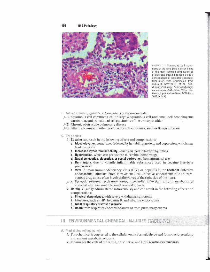
1 06 BRS Pathology
B. Tobacco abuse (Figure 7 - 1) . Associated conditions include:
FIGURE 7-1 Squamous cell carcinoma of the lung. Lung cancer is one of the most common consequences of cigarette smoking. It can also be a consequence of asbestos exposure. (Reprinted with permission from Rubin R, Strayer D, et aI., eds.: Rubin's Pathology. Clinicopathologic Foundations of Medicine, 5th ed. Baltimore, LippincottWiliiams & Wilkins, 2008, p. 149.)
� 1. Squamous cell carcinoma of the larynx, squamous cell and small cell bronchogenic carcinoma, and transitional cell carcinoma of the urinary bladder
� 2. Chronic obstructive pulmonary disease � 3. Atherosclerosis and other vascular occlusive diseases, such as Buerger disease
C. Drug abuse 1. Cocaine can result in the following effects and complications:
a. Mood elevation, sometimes followed by irritability, anxiety, and depression, which may lead to suicide
b. Increased myocardial irritabil ity, which can lead to fatal arrhythmias c. Hypertension, which can predispose to cerebral hemorrhage d. Nasal congestion, ulceration, or septal perforation, from intranasal use e, Burn injury, due to volatile inflammable substances used in cocaine free-base
preparation f. Viral (human immunodeficiency virus [HIV] or hepatitis B) or bacterial (infective
endocarditis) infection (from intravenous use) . Infective endocarditis due to intravenous drug abuse often involves the valves of the right side of the heart.
� g. Epileptic seizures, respiratory arrest, myocardial infarction, and, in newborns of addicted mothers, multiple small cerebral infarcts
2. Heroin is usually administered intravenously and can result in the following effects and complications: a. Physical dependence, with severe withdrawal symptoms b. Infections, such as HN, hepatitis B, and infective endocarditis c. Adult respiratory distress syndrome d. Death from respiratory or cardiac arrest or from pulmonary edema
III. ENVIRONMENTAL CHEMICAL INJURIES (TABLE 1-2) -- - --.. ---- ------- --------��'--�-�---------
A. Methyl alcohol (methanol) 1. This chemical is converted to the cellular toxins formaldehyde and formic acid, resulting
in transient metabolic acidosis. 2. It damages the cells of the retina, optic nerve, and CNS, resulting in blindness.
Chaz
Placed Image

i!ilfl1'!1m E nvi ronmental Patho logy 107
t a b I e 7-2 Some E nvironmental Tox ins a n d Their Effects
Toxin
Methyl alcohol
Carbon monoxide
Carbon tetrachloride
Cyanide
Lead
Mercuric chloride
B. Carbon monoxide (CO)
Predominant Adverse Effects
Blindness Severe hypoxic injury caused by displacement of oxyhemoglobin by carboxyhemoglobin Hepatic centri lobular necrosis and fatty change Cessation of intracellular oxidation because of cytochrome oxidase inhibition Anemia; basophilic stippl ing of erythrocytes; encephalopathy; neuropathy; lead line;
Fanconi syndrome Gastrointestinal ulcerations; ca lcification and necrosis of renal convoluted tubu les
1. This gas inhibits the capacity of hemoglobin to function as an oxygen carrier because hemoglobin has an affinity for CO that is 200 times greater than its affinity for oxygen.
� 2. It can result in irreversible hypoxic injury, often leading to death; neurons of the brain are most vulnerable. Foci of neuronal necrosis in the basal ganglia, lenticular nuclei, and cortical gray areas are characteristic. When fatal, it causes a cherry-red color of the skin, b lood, viscera, and muscles.
� C. Carbon tetrachloride (CCI4). This chemical induces centrilobular necrosis and fatty change in the liver.
D. Cyanide 1 . This chemical inhibits intracellular cytochrome oxidase by binding with ferric iron, thus
preventing cellular oxidation. Death occurs within minutes. 2. Generalized petechial hemorrhages and a scent of bitter almonds are noted at autopsy.
E. Lead. This chemical may be ingested, particularly from lead in paint, or may be inspired, particularly from automotive emissions. When ingested or inhaled in toxic amounts, it is manifested clinically by: 1. Red blood cell changes
� a. Basophil ic stippl ing � b. Hypochromic microcytic anemia
( 1 ) The cause is deficient heme synthesis mediated by the inhibition of 8-aminolevul inic acid (ALA) dehydratase and by decreased incorporation of iron into heme.
(2) Defects result in accumulation of both ALA and erythrocyte protoporphyrin, leading to protoporphyrinemia, porphyrinuria, and aminolevulinic aciduria.
2. Encephalopathy, characterized by irritability and sometimes by seizures and coma 3. Neuropathy, characterized by wristdrop and footdrop 4. Fanconi syndrome, characterized by impaired proximal renal tubular reabsorption of
phosphate, glucose, and amino acids � 5. Lead l ine, characterized by mucosal deposits of lead sulfide at the junction of the teeth
and gums � 6. Increased radiodensity of the epiphyses of the long bones
� F. Mercuric chloride. Ingestion results in focal gastrointestinal ulceration and severe renal damage with widespread necrosis and calcification of the proximal convoluted tubules. Proximal convoluted tubular necrosis is characteristic of injury from a number of nephrotoxins.
� G. Vinyl chloride. This chemical can lead to hemangiosarcoma (angiosarcoma) of the liver.
� H. (3-Naphthylamine and an i l i ne dyes. These chemicals can lead to transitional cell carcinoma of the urinary bladder.

1 08 BRS Pathology
� I. Ethylene glycol. This chemical can cause acute tubular necrosis, as well as tubular precipitation, of calcium oxalate crystals, which can be seen with polarized light.
J. Polychlorinated b iphenyls (PCBs) 1. These nonbiodegradable environmental pol lutants were used to manufacture a variety of
products, such as adhesives and plasticizers. Because of their toxicity, their production is now outlawed.
� 2. Exposure produces a syndrome of chloracne, impotence, and visual changes.
IV. ADVERSE EFFECTS OF THERAPEUTIC DRUGS
These effects can be manifest by a wide variety of clinically significant abnormalities. For example:
A. Antibiotics 1. Development of drug-resistant organisms is often mediated by plasmids carrying specific
drug-resistant genes. 2. Fatal aplastic anemia can occur as a result of an idiosyncratic reaction to chloramphenicol.
B. Sulfonamides 1. Immune complex disease, such as polyarteritis nodosa, can develop when sulfonamides,
acting as hap tens, stimulate antibody production. 2. Crystal l ization of sulfonamides within the renal col lecting system causes calculi with
obstruction, infection, or both. 3. Bone marrow fail ure 4. Acute, self-limited hemolytic anemia may be induced in individuals with erythrocyte
glucose-6-phosphate dehydrogenase (G6PD) deficiency.
C. Analgesics 1. Aspirin
a. Gastroduodenal bleeding may be caused by aspirin-induced gastritis or peptic ulcer or by inhibition of platelet cyclooxygenase with resultant thromboxane A2 deficiency and impaired platelet plug formation.
b. Reye syndrome occurs in children following an acute febrile viral illness, almost always in association with aspirin intake. It is characterized by microvesicular fatty change in the liver and encephalopathy.
c. Al lergic reactions include urticaria, asthma, nasal polyps, and angioneurotic edema. 2. Phenacetin
a. Chronic analgesic nephritis and renal papil lary necrosis (the drug has been withdrawn from the U.S. market)
b. Urothelial neoplasms, especially transitional cell carcinoma of the renal pelvis c. Acute hemolysis in G6PD-deficient individuals
D. Cancer chemotherapeutic drugs 1. Toxic effects, including hair loss, gastrointestinal erosions and ulcerations, and, most
significantly, bone marrow failure 2. Acute leukemia or other malignancies

Review Test
Directions: Each of the numbered items or incomplete statements in this section is followed by answers or by completions of the statement. Select the one lettered answer or completio
'n
that is best in each case.
1. A 32-year-old man is involved in a highspeed motor vehicle accident and brought by ambulance to a Level I trauma center because of the severity of his injuries. On arrival, it is determined that he is bleeding internally, and he is taken immediately to surgery, where the surgeons find that the source of his bleeding is a severe liver laceration. A laceration is a
(A) bruise caused by disruption of underlying blood vessels.
(8) clean cut by a sharp object. (e) deep tubular wound produced by a
sharp, thin object. (D) jagged tear, often with stretching of the
underlying tissue. (E) superficial tearing away of epidermal cells.
2. A forensic pathologist is asked to evaluate a fatal gunshot wound involving the left thorax of a 27-year-old man, a known drug dealer. Even though a gun and suicide note were found next to the body, the pathologist has concluded that the wound was probably not self-inflicted. Which of the following findings is most supportive of that conclusion?
(A) The entrance wound is smaller than the exit wound
(8) The entrance wound is stellate-shaped (e) The exit wound is irregularly shaped (D) There is no stippling of the skin, and the
wound is oval with clean margins (E) There is stippling of the skin from
unburned gunpowder
3. A 4-year-old girl is brought by her mother to the emergency department after the girl was "placed in a hot bath." The patient appears to have extensive blistering of the thighs and buttocks, with slight damage to the underlying dermis. Her burns are best described as
(A) first-degree burns. (8) fourth-degree burns. (e) full-thickness burns. (D) second-degree burns. (E) third-degree burns.
4. A 38-year-old woman is receiving radiation therapy to her abdomen as adjuvant therapy for the treatment of cervical cancer. The radiosensitivity of organs or tissues within the treatment field is a limiting factor in determining the dose of radiation that can be administered. Which of the follOwing is most susceptible to radiation damage?
(A) Bone (8) Gastrointestinal mucosa (e) Peripheral nervous tissue (D) Renal parenchyma (E) Skeletal musculature of the abdomen
5. A 50-year-old chronic alcoholic is seen in the emergency department with ataxic gait, confusion, confabulation, and nystagmus. This constellation of findings is the classic presentation of
(A) Fetal alcohol syndrome. (8) Li-Fraumeni syndrome. (e) Lynch syndrome. (D) Wernicke-Korsakoff syndrome. (E) Wiskott-Aldrich syndrome.
6. A 50-year-old man who works for a chemical company is being held on charges of murdering his wife. When the police found the woman's body, they noted no signs of physical injury but detected a scent of bitter almonds. A small sample of cyanide was recently reported missing from the husband's workplace. Based on these investigational findings, a justifiable working hypothesis for the forensic pathologist is that the death resulted from
109
1 1 0 BRS Pathology
(A) acute tubular necrosis. (8) induction of direct DNA damage. (C) inhibition of intracellular oxidative
phosphorylation. (0) inhibition of the oxygen-carrying capac
ity of hemoglobin. (E) rhabdomyolysis.
7. A 78-year-old man is found in his closed room unresponsive in bed after the first cold day of winter. There is a kerosene heater still on from the previous night. On attempts to arouse him, officers note the cherry hue of his lips, cheeks, and mucous membranes. The likely mechanism of his death was
(A) accidental ingestion of ethylene glycol.
(8) binding of carbon monoxide to hemoglobin.
(C) hepatic necrosis with fatty change. (0) inhibition of hemoglobin production. (E) inhibition of incorporation of iron into
hemoglobin.
8. A 3-year-old child is brought to the emergency department because of a week-long history of abdominal discomfort, irritability, and weakness. A complete blood count and blood smear demonstrate microcytic hypochromic anemia with basophilic stippling of the red blood cells. Further history reveals that the family lives in an old apartment complex. The most likely cause of the hematologic findings is
(A) binding of carbon monoxide to hemo-globin.
(8) inhibition of cytochrome oxidase. (C) inhibition of hemoglobin production. (0) iron deficiency. (E) iron intoxication.
9. A 27-year-old man who was badly burned in an industrial accident requires multiple skin grafting. While in the intensive care unit, he is found to have blood in his stools, and endoscopy confirms the presence of many small ulcers in his stomach. This complication is referred to as
(A) aplastic anemia. (8) Curling ulcer. (C) Cushing ulcer. (0) Reye syndrome. (E) Stevens-Johnson syndrome.
10. Following his return home from a party, a 22-year-old man develops crushing chest pain and is brought to the emergency department by ambulance. Questioning reveals no cardiac risk factors. Electrocardiographic and serum enzyme findings are consistent with acute myocardial infarction. It is suspected that the cardiac damage is related to toxicity of a drug of abuse. Which of the following drugs is most likely?
(A) Cocaine (8) Ethyl alcohol (C) Heroin (0) Methyl alcohol (E) Phenacetin

Answers and Explanations
1 . The answer is D. A laceration, especially of internal organs, is common in cases of blunt trauma, as in a motor vehicle accident. By definition, a laceration is a jagged tear, often with stretching of the underlying tissue. An incision is a clean cut by a sharp object, as in a clean cut by a surgeon. A contusion is a bruise caused by disruption of underlying blood vessels. A puncture is a deep tubular wound produced by a sharp, thin object. An abrasion is a superficial tearing away of epidermal cells.
2. The answer is D. Lack of stippling is a characteristic of gunshot wounds inflicted from long distances. In addition, the wounds are often oval with clean margins. Such evidence is more consistent with a homicide than a "staged" suicide. In general, entrance wounds are smaller than exit wounds, which are often irregularly shaped. Stellate-shaped wounds are associated with wounds over bony areas. Wounds from close range, as in a suicide, would be expected to have stippling and tattooing of skin caused by unburned gunpowder.
3. The answer is D. First- and second-degree burns are both partial-thickness burns. Because the findings in this patient reveal both epithelial and dermal involvement, the burns are, by definition, second-degree in type. Both third- and fourth-degree burns are classified as full-thickness burns; they entail total destruction of dermis and epidermis along with underlying skin appendages that normally serve as a source of cells for regeneration. As such, these wounds require skin grafting.
4. The answer is B. Gastrointestinal mucosal cells are examples of labile cells, and thus are among the most radiosensitive of the tissues or organs listed in the question. The symptoms associated with damage to these cells (nausea, diarrhea, and malabsorption) are likely to limit the total dose of radiation the patient can receive. The other tissues listed are relatively radioresistant and are unlikely to limit the amount of radiation a patient can receive.
5. The answer is D. Wernicke-Korsakoff syndrome presents with the classic triad of ataxia, confabulation, and ophthalmoplegia and is seen in alcoholics secondary to a deficiency of the vitamin thiamine. Fetal alcohol syndrome occurs in infants born to mothers who consume alcohol during pregnancy and results in microcephaly, mental retardation, and facial and cardiac defects. Li-Fraumeni syndrome and the Lynch syndrome are both familial defects in DNA repair that lead to a propensity to develop particular cancers. Wiskott-Aldrich syndrome is an inborn immune defect presenting with eczema, thrombocytopenia, and recurrent infections.
6. The answer is C. Cyanide is an inhibitor of the electron transport chain (ETC) within the mitochondria. Inhibition of the ETC results in the inhibition of intracellular oxidative phosphorylation, with depletion of cellular energy stores and, ultimately, death. Acute tubular necrosis can occur under many circumstances, as in cases of rhabdomyolysis secondary to crush injury. Inhibition of the oxygen-carrying capacity of hemoglobin can result from carbon monoxide poisoning. Induction of direct DNA damage can result from radiation exposure.
7. The answer is B. Carbon monoxide (CO) binds to hemoglobin with approximately 200 times greater affinity than oxygen, preventing delivery of oxygen to tissues. In addition, CO is an inhibitor of cytochrome oxidase, impairing cellular respiration. Ethylene glycol (antifreeze) is associated with kidney damage and the formation of birefringent calcium oxalate crystals in the urinary tract. Hepatic necrosis and fatty change result from carbon tetrachloride poisoning. Lead inhibits a key enzyme in hemoglobin synthesis as well as iron incorporation in hemoglobin.
1 1 1

1 1 2 BRS Pathology
8. The answer is C. This is a typical presentation of lead poisoning. Lead inhibits hemoglobin synthesis by inhibiting both aminolevulinic acid dehydratase and ferroketolase. This inhibits not only the synthesis of hemoglobin but also the incorporation of iron into the molecule. Cyanide and carbon monoxide (CO) inhibit cytochrome oxidase. CO also binds to hemoglobin, preventing adequate oxygen transport. Iron deficiency can lead to similar hematologic findings.
9. The answer is B. Punctate ulcers associated with extensive burn injuries are known as Curling ulcers. A similar phenomenon occurs in patients with head trauma, in which the lesions are known as Cushing ulcers. Aplastic anemia can result from an idiosyncratic reaction in patients taking the antibiotic chloramphenicol. Likewise, sulfonamides can cause a necrotizing eruption around mucous membranes in some individuals. Reye syndrome is associated with extensive microvesicular fatty change of the liver in children taking aspirin during an acute viral illness. The Stevens-Johnson syndrome is characterized by erosions and crusts of the lips and oral mucosa as a component of an extensive form of erythema multiforme, a maculopapular, vesiculobullous eruption often related to drugs (such as sulfonamides), neoplasia, or connective tissue disorders.
10. The answer is A. Cocaine is associated with increased myocardial irritability and sometimes with myocardial infarction, hypertension, and cerebral vascular accident. Both ethyl alcohol and heroin are long-term contributors to heart disease but are unlikely to cause an acute event in a young patient. Methyl alcohol can lead to blindness, as well as kidney damage. Phenacetin, an analgesic related to acetaminophen, can lead to kidney damage and is no longer used in this country.

Nutritional Disorders
I. MALNUTRITION
A. In affluent countries, malnutrition is found in children living below the poverty level; the elderly; alcoholics; persons on fad diets and with eating disorders, such as anorexia nervosa; and patients with severe wasting diseases.
B. In developing countries, protein-calorie malnutrition occurs in two forms-marasmus and kwashiorkor. 1 . Marasmus is caused by widespread deficiency of almost al l nutrients, notably protein and
calories. a. It often coexists with vitamin deficiencies. b. It typically occurs in children younger than 1 year of age who are not breast-fed and do
not have an adequate intake of substitute nutrients. c. Clinical characteristics include retarded growth and loss of muscle and other protein
containing tissue, as well as loss of subcutaneous fat ("wasting away") . 2 . Kwashiorkor i s caused by protein deficiency but with adequate caloric intake.
a. It usually affects children older than 1 year of age who are no longer breast-fed and receive a starch-rich, protein-poor diet.
b. Clinical characteristics include retarded growth and muscle wasting, caused by inadequate protein intake, but with preservation of subcutaneous fat.
c. Kwashiorkor is distinguished from marasmus by the presence of the following abnormalities: ( 1 ) Fatty liver
� (2) Severe edema due to protein deficiency and decreased oncotic pressure (3) Anemia (4) Malabsorption due to atrophy of the small intestinal villi
� (5) Depigmented bands with pale streaking in the hair or skin
II. VITAMINS
A. Water-soluble vitamins (Table 8 - 1 ) 1 . General considerations. Water-soluble vitamins include the B complex vitamins, B[ (thi
amine) , Bz (riboflavin) , B3 (niacin) , B6 (pyridoxine), and BIZ (cobalamin) ; folic acid; and vitamin C (ascorbic acid) . a. Because these vitamins are not stored in the body, regular intake is essential, except for
vitamin BIz' Vitamin BIZ is stored in the liver in quantities sufficiently large so tl1at deprivation for months or years is necessary for defiCiency to develop.
b. Toxicity from excessive intake is rare, because excess vitamin is excreted in the urine.
1 13

1 1 4 BRS Pathology
t a b I e 8-1
Vitamin B, (thiamine)
Vitamin B2 (riboflavin)
Vitamin B3 (niacin, nicotinic acid)
Vitamin B6 (pyridoxine)
Vitamin B'2 (cobalamin)
Folic acid
Vitamin C (ascorbic acid)
Water-Solub le Vitamins
Metabolic Functions
Coenzyme thiamine pyrophosphate plays a key role in carbohydrate and amino acid intermediary meta bolism
Component of FAD and FMN and is essential in a variety of oxidation-reduction processes
Component of NAD and NADP, essentia l to glycolysis, the citric ac id cycle, and to a variety of oxidations (can be synthesized from tryptophan); deficiency requires diet lacking both niacin and tryptophan
Required for transamination, porphyrin synthesis, synthesis of niacin from tryptophan
l -Carbon transfers required for folate synthesis and activation of FH4; N5,IO-methylene FH4 is required for conversion of d UMP to dTMP in DNA synthesis
l -Carbon transfers in a number of metabolic reactions; N5,lO-methylene FH4 required for D N A synthesis
Required for hydroxylation of proline and lysine, which are essential for collagen synthesis; hydroxylation of dopamine in synthesis of norepinephrine; enhances ma intenance of reduced state of other metabolically active agents, such as iron and FH4
Clinical Manifestations of Deficiency
Wet beriberi; dry beriberi; Wernicke-Korsakoff syndrome
Cheilosis; corneal vascularization; g lossitis; dermatitis
Pellagra
Cheilosis; g lossitis; anemia; convulsions in infants; neurologic dysfunction
Megaloblastic anemia; neurologic dysfunction
Megaloblastic anemia; neurologic dysfunction is not a feature (as it is in vitamin B12 deficiency)
Scurvy, defective formation of mesenchymal tissue and osteoid matrix; defective wound healing; hemorrhagic phenomena
FAD = flavin adenine dinucleotide; FMN = flavin mononucleotide; NAD = nicotinamide adenine dinucleotide; NADP = nicotinamide adenine dinucleotide phosphate; FH4 = tetrahydrofolate; N5,10-methylene FH4 = activated tetrahydrofolate
2. Dietary sources a. B complex vitamins (except vitamin BI2) : whole grain cereals, green leafy vegetables,
fish, meat, and dairy foods b. Vitamin B12: foods of animal origin only (vitamin BI2 is synthesized by intestinal bacte
ria in animals) c. Fol ic acid: leafy vegetables, cereals, fruits, and a number of animal products d. Vitamin C: fruits (especially citrus fruits and tomatoes) , vegetables, various meats,
and milk 3. Deficiencies. Clinical manifestations are often shared. The most striking clinical mani
festations are in tissues with active metabolism, because these vitamins are involved in the release and storage of energy. In B complex vitamins, deficiencies are often marked by glossitis, dermatitis, and diarrhea. a. Vitamin B1 (thiamine) deficiency is most often associated with severe malnutrition. (In
Western countries, it is usually associated with alcoholism and fad diets.) It results in three distinct syndromes: ( 1 ) Dry beriberi is characterized by peripheral neuropathy with resultant atrophy of the
muscles of the extremities. (2) Wet beriberi Ii" (a) This condition is marked by h igh-output cardiac fa i lure, often with dilated
cardiomyopathy. (b) It results from peripheral dilation of arterioles and capillaries, leading to
increased arteriovenous shunting, hypervolemia, and cardiac dilation.

....
--
i!lltrtIt:tiJ Nutritiona l D isorders 1 1 5
� (3) Wernicke-Korsakoff syndrome most often occurs in a setting o f thiamine deficiency and alcoholism. (a) This condition is manifest by degenerative changes in the brain stem and
diencephalon, with hemorrhagic lesions of cortical and bilateral paramedian masses of gray matter and the mamillary bod ies.
(b) It is characterized by confusion, ataxia, and ophthalmoplegia (Wernicke triad) and also by marked memory loss and confabulation.
b. Vitamin 82 (riboflavin) deficiency is rare in the United States because riboflavin is almost always added to commercially prepared bread and cereals. ( 1 ) This condition occurs in chronic alcoholics, fad dieters, the elderly, and in per
sons with chronic debilitating diseases. (2) It manifests clinically by cheilosis (skin fissures at the angles of the mouth) , glossi
tis, corneal vascularization, and seborrheic dermatitis of the face, scrotum, or vulva. c. Vitamin B3 (niacin) deficiency
( 1 ) This condition develops only when the diet lacks both niacin and tryptophan (niacin can be synthesized from the essential amino acid tryptophan) . Niacin is a component of the nicotinamide adenine dinucleotides (NAD and NADP) and, as such, is essential to glycolysis, the citric acid cycle, and other metabolic processes.
� (2) It is manifest clinically as pellagra, which is characterized by the "three Ds": dementia, dermatitis, and diarrhea. Dermatitis affects exposed areas, such as the face and neck, and the dorsa of the hands and feet.
d. Vitamin B6 (pyridoxine) deficiency may cause convulsions in infants, due to decreased activity of pyridoxal-dependent glutamate decarboxylase, which leads to deficient production of ),-aminobutyric acid (GABA) , a neurotransmitter. It results in clinical manifestations similar to those of vitamin B2 (riboflavin) deficiency. Although pyridoxine deficiency is uncommon, it occurs in the following conditions: ( 1 ) Chronic alcoholism (2) Association with therapeutic drugs, such as isonicotinic acid hydrazide (INH, an
antituberculous agent) , which react as competitive inhibitors for pyridoxine binding sites
(3) A variety of syndromes characterized by an increased need for pyridoxine, including: (a) Homocystinuria, an inborn error of metabolism (b) Pyridoxine-responsive anemia, a microcytic anemia characterized by reduced
heme synthesis e. Vitamin B'2 (cobalamin) deficiency results in a marked reduction in DNA replication and
cell division. � ( 1 ) This condition is manifest clinically by megaloblastic anemia with prominent neu
rologic dysfunction. (2) The cause is almost always malabsorption but may occur in rare cases on a
dietary basis in strict vegetarians. Cobalamin deficiency is not found in other settings of malnutrition, such as alcoholism. (a) The most common malabsorption disease is pernicious anemia, in which
there is a lack of gastric intrinsic factor, a carrier protein essential to vitamin B I2 absorption in the terminal small bowel.
(b) Less commonly, malabsorption can result from a number of diverse causes, including Crohn disease (which often affects the terminal ileum) , blind loop syndrome, and Diphyllobothrium latum (giant fish tapeworm) infestation.
f. Folic acid deficiency ( 1 ) This condition is most commonly of dietary origin and often occurs in alcoholics
and fad dieters. It can be secondary to intestinal malabsorption or it can occur, without gross dietary deprivation, as a relative deficiency because of increased demand for folate (e.g., in pregnancy and in hemolytic anemia, which is due to shortening of the life span of the red blood cell) . Sometimes it is secondary to cancer chemotherapy containing folic acid antagonists.
(2) The result is megaloblastic anemia.
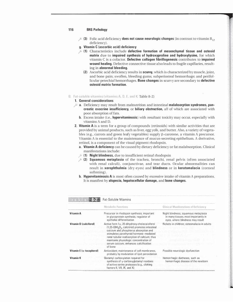
1 1 6 BRS Pathology
� (3) Folic acid deficiency does not cause neurologic changes (in contrast to vitamin B12 deficiency) .
g. Vitamin C (ascorbic acid) deficiency � ( 1 ) Characteristics include defective formation of mesenchymal tissue and osteoid
matrix due to impaired synthesis of hydroxyprol ine and hydroxylysine, for which vitamin C is a cofactor. Defective collagen fibril logenesis contributes to impaired wound healing. Defective connective tissue also leads to fragile capillaries, resulting in abnormal bleeding.
(2) Ascorbic acid deficiency results in scurvy, which is characterized by muscle, joint, and bone pain; swollen, bleeding gums; subperiosteal hemorrhage; and perifollicular petechial hemorrhages. Bone changes in scurvy are secondary to defective osteoid matrix formation.
B. Fat-soluble vitamins (vitamins A, D, E, and K; Table 8-2) 1 . General considerations
I'" a. Deficiency may result from malnutrition and intestinal malabsorption syndromes, pancreatic exocrine insufficiency, or bi l iary obstruction, all of which are associated with poor absorption of fats.
b. Excess intake (i.e., hypervitaminosis) with resultant toxicity may occur, especially with vitamins A and D.
2. Vitamin A is a term for a group of compounds (retinoids) with similar activities that are provided by animal products, such as liver, egg yolk, and butter. Also, a variety ofvegetables (e.g. , carrots and green leafy vegetables) supply l3-carotene, a vitamin A precursor. Vitamin A is essential to the maintenance of mucus-secreting epithelium. A derivative, retinol, is a component of the visual pigment rhodopsin. a. Vitamin A deficiency can be caused by dietary deficiency or fat malabsorption. Clinical
manifestations include: ,I'J ( 1 ) Night b l indness, due to insufficient retinal rhodopsin � (2) Squamous metaplasia of the trachea, bronchi, renal pelvis (often associated
with renal calculi) , conjunctivae, and tear ducts. Ocular abnormalities can result in xerophthalmia (dry eyes) and b l indness or in keratomalacia (corneal softening) .
b. Hypervitaminosis A is most often caused by excessive intake of vitamin A preparations. It is manifest by alopecia, hepatocellular damage, and bone changes.
Vitamin A
Vitamin D (calciferol)
Vitamin E la-tocopherol)
Vitamin K
Fat-Sol u b le Vita mins
Metabolic Functions
Precursor in rhodopsin synthesis; important in glycoprotein synthesis; regulator of epithelial differentiation
Active form l a, 25-dihydroxy-cholecalciferol Il ,25-IOH)2D3' calcitriol) promotes intestinal ca lc ium and phosphorus absorption and stimulates parathyroid hormone-mediated renal tubular reabsorption of calcium; thus maintains physiologic concentration of serum ca lcium; enhances calcification of bone
Antioxidant; maintenance of cel l membranes, probably by modu lation of l ipid peroxidation
Glutamyl carboxylation required for synthesis of jI-carboxyglutamyl residues of active serine proteases le.g., clotting factors I I , VI I , IX, and X)
Clinical Manifestations of Deficiency
Night bl indness; squamous meta plasia in many tissues, most importantly in eyes, where blindness may result
Rickets in children; osteomalacia in adults
Possible neurologic dysfunction
Hemorrhagic diatheses, such as hemorrhagic disease of the newborn
i!iltl"!1MlI Nutritiona l D isorders 117
3. Vitamin 0 is synthesized in the skin by ultraviolet light from the precursor 7 -dehydrocholesterol; exposure to sunlight is required for this biosynthesis. Other sources include foods, such as milk, butter, and eggs. a. Vitamin D promotes intestinal calcium absorption mediated by a specific calcium
binding intestinal transport protein, as well as intestinal phosphorus absorption. In addition, vitamin D enhances bone calcification, apparently through its role in intestinal calcium absorption.
b. Vitamin 0 deficiency manifests clinically as rickets in children and as osteomalacia in adults, both due to deficient calcification of osteoid matrix. It can be caused by the following factors: ( 1 ) Malnutrition (2) Intestinal malabsorption (3) Inadequate exposure to sunlight (4) Liver disease, with impaired hepatic conversion of vitamin D to the 25-hydroxyl
form, a precursor of active vitamin D, lex,25-dihydroxycholecalciferol ( 1 ,25-(OH)zD3, calcitriol)
(5) Renal d isease, with incomplete synthesis of active vitamin D � (6) Vitamin D-resistant rickets due to hereditary renal lex-hydroxylase deficiency,
which causes impaired synthesis of active vitamin D, impaired calcium absorption, and increased parathyroid hormone activity.
c. Hypervitaminosis 0 is manifest in children by growth retardation and is manifest in adults by hypercalciuria, nephrocalcinosis, and renal calculi .
4. Vitamin E deficiency is rare but is thought to result in neurologic dysfunction. 5. Vitamin K
� a. This vitamin is essential for carboxylation of glut amyl residues in the synthesis of the ,), -carboxyglutamyl forms (active forms) of clotting factors I I . VII, IX, and X, and of protein C.
b. It is provided by green and yellow vegetables and by dairy products. c. It is synthesized by intestinal microorganisms. d. Vitamin K deficiency results from fat malabsorption or alterations in the intestinal flora
caused by antibiotics. ( 1 ) This condition is characterized by a hemorrhagic diathesis (abnormal bleeding)
marked by prolongation of the prothrombin and activated partial thromboplastin times.
� (2) It is the cause of hemorrhagic disease of the newborn, which may result from a variety of causes, including deficient intake combined with inadequate intestinal bacterial colonization.
e. There are no known clinical manifestations of excess vitamin K.
[ III. OBESITY
A. Obesity is associated with increased risk of type 2 (non-insulin-dependent) diabetes mellitus, hypertension. gallstones. and osteoarthritis.
B. When central in distribution (fat deposits principally surrounding abdominal viscera and subcutaneous areas of the trunk) , it may be associated with an increased incidence of coronary artery disease.
C. It may, as is suggested by animal studies, be partly related to secretion of leptin, an antiobesity hormone produced by adipocytes, and neuropeptide Y, a pro-obesity polypeptide secreted by the hypothalamus in response to leptin deficiency.

Review Test
Directions: Each of the numbered items or incomplete statements in this section is followed by answers or by completions of the statement. Select the one lettered answer or completion that is best in each case.
1 . While working with an international group of physicians to administer polio vaccines, a medical student sees several children with abdominal distention and pale streaks in the hair and skin. Cursory physical examination reveals significant hepatomegaly. The children likely suffer from
(A) anorexia. (B) beriberi. (e) bulimia. (0) kwashiorkor. (E) marasmus.
2. A 57 -year-old man is admitted to the hospital for treatment of chronic pancreatitis. In patients with chronic pancreatitis, deficiency of which of the following vitamins is most likely?
(A) Folic acid (B) Vitamin Bz (riboflavin) (e) Vitamin B6 (pyridoxine) (0) Vitamin BIZ (cobalamin) (E) Vitamin D
3. A 54-year-old Native American living on a reservation in southwest Arizona presents to a clinic with impaired memory; diarrhea; and a rash on the face, neck, and dorsum of the hands. It is likely that this patient has a deficiency of which of the follOwing nutrients?
(A) Ascorbic acid (B) Folic acid (e) Homocysteine (0) Niacin (E) a-Tocopherol (vitamin E)
4. A 52-year-old recent Asian immigrant is brought to the emergency department after experiencing several convulsions. Further history reveals that she has been
1 1 8
diagnosed with tuberculosis and has recently been started on a multidrug regimen that includes isoniazid. Which of the following is the likely cause of her convulsions?
(A) Vitamin B I (thiamine) deficiency (B) Vitamin Bz (riboflavin) deficiency (e) Vitamin B3 (niacin) deficiency (0) Vitamin B6 (pyridoxine) deficiency (E) Vitamin C (ascorbic acid) deficiency
5. A 64-year-old man undergoes a total gastric resection for adenocarcinoma of the stomach. He has done well for 4 years but now presents with profound anemia, fatigue, and vague neurologic complaints. Position and vibration sensation are markedly diminished, and hyperreflexia is pronounced. Laboratory studies, including examination of the bone marrow, reveal pancytopenia and other findings compatible with a megaloblastic anemia. He is likely suffering a deficiency of which essential vitamin?
(A) Folate (B) Vitamin BIZ (e) Vitamin C (0) Vitamin D (E) Vitamin K
6. A 62-year-old woman with a long history of rheumatoid arthritis was recently placed on therapy with methotrexate (a folic acid antagonist) . The physician should be on the alert for which of the follOwing side effects of this newly added medication?
(A) Defective osteoid matrix production (B) Hemorrhagic lesions of the mamillary
bodies (e) High-output heart failure (0 ) Impaired wound healing (E) Megaloblastic anemia

-
7. A woman from a rural Appalachian community who had recently given birth to a newborn boy at home with the aid of a midwife now brings her infant to the hospital because of continued bleeding and oozing from the umbilical stump. It is likely that the bleeding problem is secondary to a deficiency of which of the following vitamins?
(A) Folic acid (B) Vitamin A (e) Vitamin D (O) Vitamin E (E) Vitamin K
8. A 4-year-old Inuit child from northern Alaska is brought to the pediatrician because of concern about progressive bowing of the legs and enlargement of the costochondral junctions (rachitic rosary) . The underlying defect in this disorder is a defect in
(A) calcification of osteoid matrix. (B) fibrillin. (e) formation of osteoid matrix. (O) hydroxylation of proline residues in
collagen. (E) type I collagen.
9. An abused child is found living in the basement of his parents' home after they are arrested on drug charges. In addition
i!iitt!ltDII Nutritio n a l D isorders 1 1 9
to being severely malnourished, the child is found to have bleeding gums and easy bruisability, along with numerous poorly healing skin ulcerations. Assuming that these findings resulted from vitamin C deficiency, what is the likely mechanism of these findings?
(A) Defective DNA synthesis (B) Defective production of 'Y-aminobutyric
acid (e) Impaired carboxylation of coagulation
factors II, VII, IX, and X (O) Impaired hydroxyproline and hydroxyly
sine production (E) Impaired renal la-hydroxylase
10. An l 8-year-old young man with known cystic fibrosis presents to the physician with his third episode of kidney stones in the past year. In addition, he has begun to complain of difficulty seeing at night. Such changes can be attributed to a deficiency of which vitamin?
(A) Pyridoxine (B) Vitamin A (e) Vitamin Bl (O) Vitamin B12 (E) Vitamin D

Answers and Explanations
1 . The answer is D. These children suffer from kwashiorkor, a form of protein-calorie malnutrition that is associated with a protein-poor diet. Kwashiorkor should be distinguished from the relative deficiency of all calories, known as marasmus. Anorexia and bulimia are psychiatric eating disorders that are significantly more prevalent in developed countries. Beriberi is due to deficiency of thiamine.
2. The answer is E. Deficiency of fat-soluble vitamins (vitamins A, D, E, and K) can occur in chronic pancreatitis due to loss of exocrine pancreas function. Vitamin Bl2 (cobalamin) , folic acid, vitamin B2 (riboflavin) , and vitamin B6 (pyridoxine) are all water-soluble
.
vitamins. It should be noted that most patients with chronic pancreatitis also are alcoholics and that alcoholics often have multiple nutritional deficiencies, including lack of water-soluble vitamins.
3. The answer is D. The clinical scenario depicts the classic findings of pellagra, or niacin deficiency, with diarrhea, dementia, and dermatitis. Niacin is synthesized from the essential amino acid tryptophan, which is particularly deficient in corn-based diets. Vitamin C deficiency results in scurvy. Folic acid deficiency often manifests with anemia. Decreased levels of homocysteine, an amino acid, have been associated with cardiovascular disease. Vitamin E deficiency is rare and can result in neurologic abnormalities.
4. The answer is D. Isoniazid is a competitive inhibitor of pyridoxine (vitamin B6) , which is required for the synthesis of the inhibitory neurotransmitter -v-aminobutyric acid. Riboflavin deficiency is rare and can result in cheilosis, glossitis, and other epithelial changes. Niacin deficiency results in pellagra. Pyridoxine deficiency can be manifest by convulsions. Vitamin C deficiency results in defects in collagen synthesis. Thiamine deficiency results in neuropathy, cardiomyopathy, and mental status changes.
5. The answer is B. Both folate and vitamin Bl2 deficiency lead to megaloblastic anemia, secondary to impaired DNA replication. In marked contrast to folate deficiency, vitamin Bl2 deficiency causes neurologic dysfunction associated with damage to the lateral and dorsal spinal columns. The history of gastric resection is consistent with a deficiency of intrinsic factor, which is required for absorption of vitamin Bl2 in the terminal ileum.
6. The answer is E. Folate is required for DNA replication in rapidly dividing cells, such as red blood cell precursors, and a deficiency results in megaloblastic anemia. Hemorrhagic lesions of the mamillary bodies and high-output heart failure occur with thiamine deficiency. Impaired wound healing and defective osteoid matrix production result from vitamin C deficiency.
7. The answer is E. Deficiency of vitamin K results in abnormal bleeding, based
1 20
on the requirement of the vitamin for -v-carboxylation of clotting factors II , VII, IX, and X. Because newborns have inadequate intestinal flora, the primary source of vitamin K, vitamin K deficiency is relatively common and is estimated to occur in approximately 3% of neonates. The resultant bleeding disorder is termed hemorrhagic disease of the newborn. Folic acid deficiency during early embryogenesis can result in neural tube defects. Vitamin A deficiency results in changes in vision and defects in epithelial cell function. Vitamin D deficiency in children results in rickets.

I!1lIll"mJ Nutritiona l D isorders 121
8. The answer is A. Vitamin D is required for calcification of osteoid matrix, and defective calcification leads to bowing of the legs and the abnormalities in the costochondral junctions known as the rachitic rosary. Mutations in genes involved in the synthesis of type I collagen lead to osteogenesis imperfecta, and defects in fibrillin lead to Marfan syndrome. Defects in hydroxylation of proline residues occur both in the Ehlers-Danlos syndrome and in vitamin C deficiency, leading to defects in the synthesis of osteoid matrix.
9. The answer is D. Because vitamin C is a required cofactor for the synthesis of hydroxyproline and hydroxylysine, which are both required for collagen synthesis, vitamin C deficiency can lead to impaired capillary formation, with consequent bleeding, and impaired wound healing. Both vitamin BI2 and folic acid are required for DNA synthesis. Pyridoxine is required for 'Y-aminobutyric acid synthesis. Vitamin K is required for carboxylation of clotting factors. Patients with vitamin D-resistant rickets have a deficiency of renal la-hydroxylase.
10. The answer is B. Patients with cystic fibrosis often have impaired exocrine pancreas function, resulting in a deficiency of fat -soluble vitamins. Of the fat -soluble vitamins, vitamin A deficiency results in night blindness, xerophthalmia, keratomalacia, and squamous metaplasia of the urinary tract, contributing to the formation of recurrent renal calculi.

Vascular System -I
I
I j
----
I. ARTERIAL DISORDERS
A. Arteriosclerosis. This is a general term for three types of vascular disease, all characterized by rigidity (sclerosis), and often thickening, of blood vessels.
122
1. Miinckeberg arteriosclerosis (medial calcific sclerosis) involves the media of mediumsized muscular arteries, most typically the radial and ulnar arteries, and usually affects persons older than 50 years of age. It does not obstruct arterial flow because the intima is not involved. a. Ring-like calcifications in the media of the arteries are characteristic. b. Stiff, calcific "pipestem" arteries result. c. This form of arteriosclerosis may coexist with atherosclerosis, but it is distinct from
and unrelated to it. 2. Arteriolosclerosis is characterized by hyaline thickening or proliferative changes of smal l
arteries and arterioles, especially i n the kidneys, and i s usually associated with hypertension or diabetes mell itus. It occurs in two variants: a. Hyal ine arteriolosclerosis is characterized by hyaline thickening of arteriolar walls. In
the kidneys, this is called benign nephrosclerosis and is associated with hypertension. Ii' b. Hyperplastic arteriolosclerosis is marked by concentric, laminated, "onionskin" thicken
ing of the a rteriolar walls. It may be accompanied by necrotizing arteriol itis, intramural deposition of fibrinoid material in arterioles with vascular necrosis and inflammation. In the kidneys, it is called malignant nephrosclerosis and is associated with malignant hypertension.
3. Atherosclerosis is the most frequent cause of significant morbidity caused by vascular disease. It is seen worldwide, but the highest incidence occurs in Finland, Great Britain, other northern European countries, the United States, and Canada. The incidence is more than l O-fold greater in Finland than in Japan. a. Characteristics of atherosclerosis. Fibrous plaques, or atheromas, occur within the
intima of arteries, most frequently the proximal portions of the coronary arteries, the larger branches of the carotid arteries, the circle of Willis, the large vessels of the lower extremities, and the renal and mesenteric arteries. (1) The plaques have a central core of cholesterol and cholesterol esters; lipid-laden
macrophages, or foam cells; calcium; and necrotic debris. Ii' (2) The core is covered by a subendothelial fibrous cap, which is made up of smooth mus
cle cells, foam cells, fibrin and other coagulation proteins, as well as extracellular matrix material, such as collagen, elastin, glycosaminoglycans, and proteoglycans.
Ii' (3) The plaques may be complicated by: (a ) Ulceration, hemorrhage into the plaque, or calcification of the plaque (b ) Thrombus formation at the site of the plaque, producing obstructive disease (c) Embolization of an overlying thrombus or of plaque material itself

.....
---
I!ittlltttDlI Vascular System 123
(4) The atheromas can develop from the fatty streak, a lesion that is characterized by focal accumulations in the intima of lipid-laden foam cells that may appear as early as the first year of life and that is present in the aorta of most older children.
b. Consequences of atherosclerosis (1) The most significant consequence is ischemic heart disease and myocardial infarc
tion, the most common cause of death in the United States. (2) Other significant complications include stroke from cerebral ischemia and infarc
tion, ischemic bowel disease, peripheral vascular occlusive disease with findirigs varying from claudication to ischemic necrosis and gangrene, and renal arterial ischemia with secondary hypertension.
(3) Weakening of the vessel wall may lead to aneurysm formation. c. Risk factors for atherosclerosis
(1) Incidence increases with age. (2) Gender plays a role. Atherosclerosis is more common in men in all age groups,
although the incidence increases in postmenopausal women. (3) Considerable evidence links hypercholesterolemia with atherosclerosis.
(a ) Serum cholesterol may be of dietary, or exogenous, origin or of b iosynthetic, endogenous origin. Cholesterol and dietary fats associate with apolipoprotein molecules and circulate as l ipoprotei ns. Figure 9-1 briefly summarizes lipoprotein transport and metabolism.
(b ) Relative concentrations of lipoprotein fractions are used as clinical predictors of atherogenesis; ideally, the ratio between low-density lipoprotein (LDL) and high-density lipoprotein (HDL) cholesterol should be 4:l.
(c) Serum concentrations of LDL, also known as "bad" cholesterol, are directly related to the risk of atherosclerosis, as is the total cholesterol concentration.
Exogenous pathway Endogenous pathway
Dietary Bile acids cholesterol� + � ! j Ch ole'tecol 1
Intestine L�/ --'------
Li:er
Remnant
receptor
[8 1 LDL 8-tOO receptors
\ Chylomicrons
E B-48
Capillaries
-----------00 C l B-100
Capi l laries
LDL receptor
Extrahepatic
tissues
- - Plasma
Lipoprotein lipase Lipoprotein lipase LCAT
FIGURE 9-1 Lipoprotein tra nsport and metabolism. HDL = high-density lipoprotein; LCAT = lecithin:cholesterol acetyltransferase; LDL = lOW-density lipoprotein; IDL = intermediate-density lipoprotein; VLDL = very low density lipoprotein. (Reprinted with permission from Goldstein J, Kita T, Brown M: Defective lipoprotein receptors and atherosclerosis. N Eng/ J Med309:288, 1 983.)

....
124 BRS Pathology
(d) An inverse relationship exists between the HDL concentration and the risk of atherosclerosis. HDl, also known as "good" cholesterol, appears to exert its protective effect by removing cholesterol from tissues and from atherosclerotic plaques.
(4) Hypertension is a major risk factor for, and is associated with, premature atherosclerosis.
(5) Diabetes mell itus is associated with premature atherosclerosis. Atherosclerotic peripheral vascular occlusive disease, often leading to gangrene of the lower extremities, is common in diabetic patients.
� (6) Cigarette smoking is also a well-established risk factor. (7) Less firmly established are obesity, physical inactivity, "type A" personal ity with
stress factors in lifestyle, hyperuricemia, hyperhomocysteinemia, methylene tetrahydrofolate reductase mutations, lipoprotein A, infection with Chlamydia pneumoniae, and use of oral contraceptive drugs, especially in association with cigarette smoking.
d. Pathogenesis of atherosclerosis (1) Older concepts
(a) Insudation hypothesis proposed that the infiltration of the intima with l ip id and protein is the primary atherogenic event, a process accelerated by hypercholesterolemia.
(b ) Encrustation or thrombogenic hypothesis proposed that organization of repeated mural thrombi on the intimal surface leads to buildup of plaques filled with lipid derived from the breakdown of platelets and leukocytes.
(c) Monoclonal hypothesis proposed that smooth muscle migration and prol iferation is analogous to tumor growth and is a primary rather than a secondary event. Smooth muscle proliferations within atheromas are often monoclonalthat is, like neoplasms, they are derived from single cell precursors. This hypothesis held that stimuli such as hyperl ipidemia may incite the prol iferation.
� (2) Current concept: reaction to injury formulation (a) Overall, this view considers the primary event as injury to (or dysfunction of)
arterial endothel ium, which may be produced by hypercholesterolemia, mechanical injury, hypertension, immune mechanisms, toxins, or viruses or other infectious agents. Hyperl ipidemia may initiate endothelial injury, promote foam cell formation, act as a chemotactic factor for monocytes, inhibit macrophage motility, or injure smooth muscle cells.
(b ) It leads to the following: ( i ) Entry of monocytes and l ip id into subendothel ium occurs, sometimes with
platelet adhesion and aggregation at the injury site. ( i i ) Mitogenic factors (platelet-derived growth factor and possibly fibroblast
growth factor, epidermal growth factor, and transforming growth factor-a) are released from platelets and perhaps also from monocytes.
( i i i ) These growth factors induce proliferation and migration of smooth muscle cells into the intima, with the production of connective tissue matrix proteins (collagen, elastin, glycosaminoglycans, and proteoglycans) .
( iv) Monocytes and smooth muscle cells engulf lipid and contribute to the deposition of lipid into lesions. Monocyte conversion to l ip id-laden foam cells is mediated by specific monocyte receptors, the �-VLDL receptor, and the scavenger receptor, which recognizes modified LDL.
B. Aneurysms. These localized abnormal d i lations of either arteries or veins can erode adjacent structures or rupture. They may be of several different types.
� 1. Atherosclerotic aneurysms most frequently occur in the descending, especial ly the abdominal, aorta (Figure 9-2).
2. Aneurysms due to cystic medial necrosis are the most frequent aneurysms of the aortic root .

FIGURE 9-2 Aortic a neurysm. This atherosclerotic aneurysm in the distal aorta contains a large mural thrombus. Additionally the entire aorta, as well as the iliac arteries, are l ined with confluent atherosclerotic plaques. (Reprinted with permission from Rubin R, Strayer D, et aI., eds.: Rubin's Pathology. Clinicopathologic Foundations of Medicine, 5th ed. Ba ltimore, Lippincott Will iams & Wilkins, 2008, p. 420.)
I!1lIlJ11tliiI Vascu lar System 125
� 3. Berry aneurysms are small, saccular lesions most often seen in the smaller arteries of the brain, especially in the circle of Wil l is. They are unrelated to atherosclerosis. a. Berry aneurysms are not present at birth but develop at sites of congenital medial
weakness at bifurcations of cerebral arteries. These aneurysms are the most frequent cause of subarachnoid hemorrhage.
b. Often, there is an association with adult polycystic kidney disease. � 4. Syphil itic ( luetic) aneurysm
a. This aneurysm is a manifestation of tertiary syphi l is, which has become rare with better treatment and control of the disease. It is caused by syphil itic aortitis, which is characterized by obliterative endarteritis of the vasa vasorum and necrosis of the media. Grossly, these changes result in a "tree-bark" appearance.
b. Unlike atherosclerotic aneurysms, syphilitic aneurysms characteristically involve the ascending aorta. Dilation of the ascending aorta may widen the aortic commissures, leading to aortic valve insufficiency.
� 5. Dissecting aneurysm (dissecting hematoma) a. This aneurysm is a longitudinal intraluminal tear, usually in the wall of the ascending
aorta, forming a second arterial lumen within the media. It is typically associated with hypertension or with cystic medial necrosis (can be a component of Marfan syndrome) , which is characterized by degenerative changes in the media with destruction of elastic and muscular tissue. It has no relation to atherosclerosis.
b. Severe, tearing chest pain, often radiating through to the back, is clinically dominant. A radiograph may reveal widening of the aortic shadow.
c. Dissecting aneurysms may be clinically confused with acute myocardial infarction, but the electrocardiogram, serum troponin I, and serum myocardial enzymes are normal.
d. The characteristic result is aortic rupture, most often into the pericardial sac, causing hemopericardium and fatal cardiac tamponade.
Chaz
Placed Image

126 BRS Pathology
6. Arteriovenous fistula (aneurysm) a. This aneurysm is an abnormal communication between an artery and a vein. b. It can be secondary to trauma or other pathologic processes that mechanically pene
trate the walls of both vessels. c. ReSUlting changes may include ischemic changes from the diversion of blood, balloon
ing and aneurysm formation from increased venous pressure, and high-output cardiac fai lure from hypervolemia.
II. VENOUS DISORDERS
A. Venous thrombosis (phlebothrombosis) 1. Venous thrombosis arises most often in the deep veins of the lower extremities. It is often
associated with inflammation and is then termed thrombophlebitis. 2. Predisposing factors include venous circulatory stasis or partially obstructed venous
return, such as occurs with cardiac failure, pregnancy, prolonged bed rest, or varicose veins.
� 3. Embolism with resultant pulmonary infarction may develop. Pulmonary infarcts are characteristically hemorrhagic, subpleural, and wedge-shaped.
B. Varicose veins. These abnormally dilated and tortuous veins occur most often in superficial veins of the lower extremities. Predisposing factors include increased venous pressure, such as occurs with pregnancy, obesity, or thrombophlebitis, and in persons whose occupations require prolonged standing.
III. TUMORS OF BLOOD VESSELS
A. Benign vascular tumors. These are usually not true neoplasms but are better characterized as malformations or hamartomas and include: 1. Spider telangiectasia is a dilated small vessel surrounded by radiating fine channels. It is
associated with hyperestrinism, as seen in chronic liver disease or pregnancy. 2. Hered itary hemorrhagic telangiectasia (Osler-Weber-Rendu syndrome) is an autosomal
dominant condition characterized by localized dilation and convolution of venules and capillaries of the skin and mucous membranes. It is often complicated by epistaxis or gastrointestinal bleeding.
3. Hemangioma (angioma) is a malformation of a larger vessel composed of masses of channels filled with blood. It is the most common tumor of infancy and is responsible for portwine stain birthmarks. It includes the following types: a. Capil lary hemangioma consists of a tangle of closely packed capillary-like channels that
may occur in the skin, subcutaneous tissues, lips, liver, spleen, or kidneys. b. Cavernous hemangioma consists of large cavernous vascular spaces in the skin and
mucosal surfaces and in internal organs such as the liver, pancreas, spleen, and brain. It can occur in von Hippel-lindau (VHL) disease, an autosomal dominant disorder that is also marked by hemangioblastomas of the cerebellum, brain stem, and retina and is also characterized by adenomas and cysts of the liver, kidneys, pancreas, and other organs, and by an increased incidence of renal cell carcinoma.
c. Sturge-Weber syndrome is a disorder characterized by facial port-wine stain, leptomeningeal venous angiomas, and neurologic manifestations including mental deficiency and seizures.
4. Glomangioma (glomus tumor) is a small, purplish, painful subungual nodule in a finger or toe. 5. Cystic hygroma is a cavernous lymphangioma that occurs most often in the neck or
axilla.
.,
I!!lltO"dm Vascular System 127
B. Malignant vascular tumors. These uncommon tumors include the following: 1. Hemangioendothel ioma is intermediate in behavior between a benign and a malignant
tumor. 2. Hemangiopericytoma arises from pericytes and varies in behavior from benign to
malignant. 3. Hemangiosarcoma (angiosarcoma)
a. This rare, malignant, vascular tumor occurs in the skin, musculoskeletal system, breast, or liver.
b. It is associated with toxic exposures to arsenic or the radioactive diagnostic agent thorium dioxide (Thorotrast). Polyvinyl chloride is specifically associated with angiosarcoma of the liver.
4. Kaposi sarcoma (KS) is a malignant vascular tumor that occurs in several forms. a. Classic KS most often affects older men of Ashkenazi Jewish or Mediterranean origin. b. Endemic (or African) KS tends to affect young African men and children and accounts
for as many as 10% of all cancers in Africa. I" c. Epidemic KS occurs as a component of acquired immunodeficiency syndrome (AIDS),
especially in the homosexual male risk group, and is caused by a virus that has been termed human herpesvirus 8 (HHV-8), also called Kaposi sarcoma herpesvirus (KSHV ) . KS is a feature of immunosuppression associated with causes other than AIDS.
IV. VASCULITIS SYNDROMES (VASCULITIDES)
These i nflammatory and often necrotizing vascular lesions occur in almost any organ and are usually mediated by immune mechanisms, most often immune complex depositions. Frequent antigens in immune complexes include DNA, hepatitis B surface antigen, and hepatitis C RNA.
A. Polyarteritis nodosa 1. This condition is characterized by necrotizing immune complex inflammation of small- and
medium-sized arteries. It is marked by the destruction of arterial media and internal elastic lamella, resulting in aneurysmal nodules.
I" 2. There is an association with hepatitis B viral infection in 30% of patients. 3. Clinical manifestations often include fever, weight loss, malaise, abdominal pain,
headache, myalgia, and hypertension. Polyarteritis nodosa is seen in the following sites: a. Kidneys, with immune complex vasculitis in the arterioles and glomeruli; renal lesions
and hypertension cause most deaths from polyarteritis nodosa. b. Coronary arteries, resulting in ischemic heart disease c. Musculoskeletal system, reSUlting in myalgia, arthralgia, or arthritis d. Gastrointestinal tract, manifesting as nausea, vomiting, or abdominal pain e. Central nervous system (CNS) or peripheral nervous system, the eye, or skin
B. Churg-Strauss syndrome (allergic granulomatous angiitis). This is a necrotizing vasculitis considered by some to be a variant of polyarteritis nodosa. It is characterized by prominent involvement of the pulmonary vasculature, marked peripheral eosinophilia, and clinical manifestations of asthma.
C. Hypersensitivity (leukocytoclastic) vasculitis 1. This is a group of immune complex-mediated vascul itides characterized by acute inflam
mation of small blood vessels (arterioles, capillaries, venules); the multiple lesions tend to be of the same age. These are in contrast to the findings in polyarteritis no do sa.
I" 2. It is manifest by palpable purpura when the skin is involved but can involve any site, including the glomeruli or the gastrointestinal tract.

128 BRS Pathology
3. It may be precipitated by exogenous antigens, such as drugs, foods, or infectious organisms; may also occur as a compl ication of systemic i l lnesses, such as connective tissue disorders or malignancies.
4. It presents clinically in distinctive syndromes, including: a. Henoch-Schiinlein purpura is most common in young children.
(1) Characteristics include hemorrhagic urticaria of extensor surfaces of the arms, legs, and buttocks, with fever, arthralgias, and gastrointestinal and renal involvement (often similar to IgA nephropathy; see Chapter 17).
(2) This disorder can sometimes be poststreptococcal in origin. It is associated with antecedent upper respiratory infections, suggesting that infectious agents may be the inciting antigens; other antigens may include drugs or foods.
b. Serum sickness � (1) This syndrome is seen in the experimental model in which rabbits, after seri<;ll
injections of bovine serum albumin, develop general ized deposition of antigenantibody complexes in the heart, joints, and kidneys.
(2) Serum sickness is now rare in humans, but in the past, it was caused by therapeutic administration of various antitoxins (foreign serum containing specialized antibodies prepared by immunization of animals, such as horses) .
D. Wegener granulomatosis. This disease of unknown etiology is characterized by necrotizing granulomatous vasculitis of the small- to medium-sized vessels of the respiratory tract. kidneys, and other organs.
� 1. Wegener granulomatosis is dominated clinically by respi ratory tract signs and symptoms, especially of the paranasal sinuses and lungs, and necrotizing g lomeru lonephritis (sometimes with immune complex deposition) . Manifestations include f ibri no id necrosis of small arteries and veins, ear ly infiltration by neutroph i ls, subsequent mononuclear cell infiltration, and fibrosis. Granuloma formation with giant cells is prominent.
� 2. In most cases, Wegener granulomatosis is associated with circulating antineutrophil cytoplasmic antibodies with a cytoplasmic staining pattern (C-ANCAs).
E. Giant cel l arteritides are seen in medium- to large-sized arteries and are characterized by granuloma formation with giant cells, as well as by infiltrates of mononuclear cells, neutrophils, and eosinophils. They include two distinct clinical syndromes: 1. Temporal arteritis is the most frequently occurring form of vasculitis.
� a. This systemic vasculitis occurs most often in elderly persons. It usually affects branches of the carotid a rtery, particularly the temporal artery.
b. Clinical manifestations include: (1) Malaise and fatigue (2) Headache or claudication of the jaw (3) Tenderness, absent pulse, and palpable nodules along the course of the involved
a rtery (4) Visual impairment, especially with involvement of the ophthalmic artery (5) Polymyalgia rheumatica, a complex of symptoms including proximal muscle pain,
periarticular pain, and morning stiffness (6) Markedly elevated erythrocyte sedimentation rate
2. Takayasu arteritis (pulseless disease) is characterized by inflammation and stenosis of medium- and large-sized arteries with frequent involvement of the aortic arch and its branches, producing aortic arch syndrome. Clinical manifestations include: a. Absent pulses in carotid, radial, or ulnar arteries b. Nonspecific findings, such as fever, night sweats, malaise, myalgia, arthritis and
arthralgia; eye problems; and painful skin nodules
� F. Mucocutaneous lymph node syndrome (Kawasaki d isease) is an acute, self-limited illness of infants and young chi ldren characterized by acute necrotizing vasculitis of small- and medium-sized vessels.

I!lttlJllllDij Vascu lar System 129
1. This syndrome is manifest clinically by fever; hemorrhagic edema of conjunctivae, lips, and oral mucosa; and cervical lymphadenopathy.
2. It can be a cause of coronary artery vasculitis with aneurysm formation.
� G. Thromboangiitis obliterans ( Buerger disease) is an acute inflammation involving small- to medium-sized a rteries of the extremities, extending to adjacent veins and nerves. It occurs with greater frequency in Jewish populations and is most common in young men. 1. This disease results in painful ischemic disease. often leading to gangrene. 2. It is clearly exacerbated by heavy cigarette smoking.
H . Lymphomatoid granulomatosis is a rare granulomatous vasculitis characterized by infiltration by atypical lymphocytoid and plasmacytoid cells. It may progress from a chronic inflammatory condition to a fully developed lymphoproliferative neoplasm. most often a T-cell nonHodgkin lymphoma.
V. FUNCTIONAL VASCULAR DISORDERS
� A. Raynaud disease is manifest by recurrent vasospasm of small arteries and arterioles, with resultant pallor or cyanosis, most often in the fingers and toes. This disease most commonly occurs in young, healthy women. It is most often precipitated by chil l ing.
� B. Raynaud phenomenon is clinically similar to Raynaud disease but is always secondary to an underlying disorder, most characteristically systemic lupus erythematosus or progressive systemic sclerosis (scleroderma).
VI. HYPERTENSION (TABLE 9-1)
A. Essential hypertension is hypertension of unknown etiology, accounting for the majority of cases. It represents an interaction of predisposing determinants with a number of exogenous factors. 1. Determinants of essential hypertension
a. Genetic factors (1) Family history of hypertensive disease is seen in three of four patients with the disorder. (2) It is more common and usually more severe in persons of African l ineage.
b. Environmental factors (1) Evidence linking levels of dietary sodium intake with hypertension prevalence in
population groups is impressive, although not everyone with excessive salt intake develops hypertension.
(2) Stress. probably mediated by neurogenic vasoconstriction, is a factor in the development of hypertension.
(3) Other factors include obesity. cigarette smoking. and physical inactivity. 2. Results of essential hypertension
a. If untreated, essential hypertension can lead eventually to retinal changes, left ventricular hypertrophy and cardiac failure, and benign nephrosclerosis.
b. It can predispose to ischemic heart disease or stroke.
B. Secondary hypertension is secondary to known causes, including: 1. Renal disease is by far the most common cause of secondary hypertension.
a. Causes of renal hypertension (1) Disorders of the renal parenchyma (2) Unilateral renal a rtery stenosis can be caused by atherosclerosis or unilateral fibro
muscular dysplasia. It is marked by atrophy of the affected kidney and may be corrected surgically.

130 BRS Pathology
tab I e 9-1 Types of Hypertension
Type or Cause
Primary (essential) hypertension
Secondary hypertension
Renal parenchymal diseases, such as postinfectious glomerulonephritis, diabetic nephropathy, adult polycystic disease
Renovascular disease
Hyperparathyroidism
Cushing syndrome, of pituitary or adrenal origin
Primary aldosteronism
Congenital adrenal hyperplasia
Pheochromocytoma
Oral contraceptive use
Coarctation of aorta
Toxemia of pregnancy
Increased intracranial pressure
Toxic hypertension
Comments
Unknown etiology; accounts for 90%-95% of cases
Stimulation of renin-angiotensin system
Stimulation of renin-angiotensin system
Excessive production of cortisol
Increased aldosterone secretion; sodium and water retention, often with hypokalemic alkalosis
Occurs in several forms; hypervolemia mediated by increas'ed production of mineralocorticoids in 17-hydroxylase deficiency and ll-hydroxylase deficiency
Secretion of epinephrine and norepinephrine, resulting in sustained or paroxysmal hypertension, which may be cured by resection of the tumor
Hypertension is an infrequent effect
Upper extremity hypertension only, with increased collateral circulation in the intercostal arteries, resulting in notching of ribs
Hypertension usually ceases after delivery
From brain tumors or other expanding intracranial lesions
Poisoning by lead, cadmium, and other agents
,i" b. Mechanism of renal hypertension occurs through stimulation of the renin-angiotensin system. (1) Juxtaglomerular cells respond to decreased vascular tone by secreting renin,
which facilitates the conversion of angiotensinogen to angiotensin I, which is further converted to angiotensin II.
(2) Angiotensin II promotes hypertension by acting both as a vasoconstrictor and as an activator of aldosterone secretion.
(3) Aldosterone promotes sodium and water retention. 2. Endocrine disorders
,i" a. Primary aldosteronism, or Conn syndrome, which is usually associated with an adrenocortical adenoma or bilateral adrenal hyperplasia. In addition to hypertension, it is marked by increased serum sodium and reduced serum potassium.
,i" b. Acromegaly, Cushing syndrome of pituitary or adrenocortical origin, pheochromocytoma, and hyperthyroidism
c. Diabetes mell itus (when complicated by diabetic glomerulosclerosis; see Chapter 17) 3. Other causes include coarctation of the aorta (which causes hypertension limited to the
upper body) and other congenital anomalies; toxemia of pregnancy; CNS disorders, especially brain tumors; and drugs and chemicals, notably amphetamines and steroids.
C. Malignant hypertension can be a complication of either essential (primary) or secondary hypertension. It follows an accelerated clinical course.
,i" 1. Malignant hypertension occurs in less than 5% of patients with elevated blood pressure, most often in young African-American males.
,i" 2. Characteristic features include a marked increase in diastolic blood pressure, focal retinal hemorrhages and papil ledema, left ventricular hypertrophy, and left ventricular failure.
3. It most often results in early death from congestive heart failure, cerebrovascular accident, or renal failure.
,i" 4. It produces the renal changes of malignant nephrosclerosis: arterioles or glomerular capillaries rupture, resulting in "flea-bitten" kidney, multiple pinpoint petechial hemorrhages on the kidney surface; large, swollen kidneys; necrotizing arteriolitis and glomerulitis with fibrinoid necrosis; and hyperplastic arteriolosclerosis, affecting both the glomeruli and arterioles.

Review Test
Directions: Each of the numbered items or incomplete statements in this section is followed by answers or by completions of the statement. Select the one lettered answer or completion
that is best in each case.
1. This segment of the arterial intima of a coronary vessel is observed at autopsy in a 56-yearold man who died suddenly on rising in the morning. Which of the following abnormalities is considered a major risk factor for the development of this lesion?
(Reprinted with permission from Rubin R, Strayer 0, et aI., eds.: Rubin's Pathology. Clinicopathologic Foundations of Medicine, 5th ed. Ba ltimore, Lippincott Williams & Wilkins, 2008, p. 19.)
(A) Congenital vascular muscle weakness (8) Cystic medial necrosis (e) Hypercholesterolemia (D ) Medial calcification (E ) Syphilis
2. A 45-year-old man presents with abdominal pain and hypertension. On physical examination, he is found to have an abdominal mass. Further workup confirms the diagnosis of adult polycystic kidney disease. Which of the following vascular complications is associated with this condition?
(A) Arteriovenous fistula (8) Atherosclerotic aneurysm (e) Berry aneurysm (D ) Dissecting aneurysm (E) Luetic aneurysm
3. A 65-year-old man who has a long history of hypertension presents to the emergency department with tearing chest pain that
radiates to the back. An electrocardiogram is normal, as are cardiac enzymes. A "stat" chest radiograph demonstrates widening of the mediastinum. Which of the following is the most likely?
(A) Arteriovenous fistula (8) Atherosclerotic aneurysm (e) Berry aneurysm (D ) Dissecting aneurysm (E) Syphilitic aneurysm
4. A 14-year-old boy is brought by his family to your clinic in Utah with a long history of recurrent epistaxis. His father also reports such a history. Of the following, which is the most likely condition consistent with these findings?
(A) Cavernous hemangioma (8) Glomangioma (e) Hemangioendothelioma (D ) Osler-Weber-Rendu syndrome (E) Varicose veins
131
Chaz
Placed Image

132 BRS Pathology
5. A 23-year-old man known to have acquired immunodeficiency syndrome (AIDS) is seen by his primary care physician. The patient is concerned by the development of multiple red-to-purple skin plaques that have become nearly confluent on his arms and legs. The skin disorder is most likely
(A) angioedema. (B) angiosarcoma. (C) erythema multiforme. (0) Kaposi sarcoma. (E) multiple telangiectasias of Osler-Weber
Rendu syndrome.
6. A 23-year-old man presents with fever, weight loss, malaise, abdominal pain, and myalgias. Workup reveals that the patient has polyarteritis nodosa. Which of the following is associated with this form of vasculitis?
(A) Arsenic (B) Chlamydia pneumonia (C) Hepatitis B virus (0) Human herpesvirus 8 (E) Human immunodeficiency virus
7. A 19-year-old young woman who emigrated from Taiwan 8 years ago presents with fever, malaise, myalgias, and arthritis and "coldness" in her upper extremities. She has a weak radial pulse bilaterally, and a magnetic resonance angiogram demonstrates nearly 75% stenosis of the main arteries originating from the aorta. She likely has which of the following rheumatologic conditions?
(A) Buerger disease (B) Kawasaki disease (C) Raynaud disease (0) Takayasu arteritis (E) Temporal arteritis
8. A 74-year-old woman presents to her primary care physician with malaise, proximal muscle pain, morning stiffness, and claudication of the jaw, along with occasional visual impairment. On examination, palpation along the lateral aspect of the head elicits tenderness. This is likely a result of
(A) giant cell arteritis. (B) hypersensitivity vasculitis. (C) Kawasaki disease.
(0) Takayasu arteritis. (E ) Wegener granulomatosis.
9. A 3-year-old boy is brought by his parents to the emergency department because they are concerned that he has had a high fever for several days. On examination, the boy has conjunctival and oral erythema. He has palpable cervical lymphadenopathy and erythema of his palms and soles. What is a potential life-threatening complication of this disorder?
(A) Aneurysm of the abdominal aorta (B) Aneurysm of the coronary arteries (C) Dissection of the thoracic aorta (0) Ischemia and gangrene of the extremities (E) Rupture of a berry aneurysm
10. A 35-year-old African-American man is transported to the emergency department because of an unrelenting, severe, excruciating headache. His blood pressure is 245/ 150 mm Hg, and bilateral papilledema is evident on ophthalmoscopic examination of the retina. Despite all interventions, including administration of nitroprusside, the patient dies. At autopsy, which of the following is a likely finding?
(A) Immune complex vasculitis of the glomeruli
(B) Longitudinal intraluminal tears of the ascending aorta
(C) Multiple punctuate hemorrhages on the surface of both kidneys ("flea-bitten kidneys")
(0) "Tree-bark" appearance of the ascending aorta
(E) Unilateral renal artery stenosis
11. A 32-year-old woman returns to her physician for follow-up of hypertension that has been poorly controlled in spite of numerous antihypertensive medications. It is decided to evaluate the patient for possible "secondary" hypertension. Which of the following is a well-known cause of secondary hypertension?
(A) Ethnicity (B) Obesity (C) Renal artery stenosis (0) Smoking (E) Stress

� Answers and Explanations J ��
1 . The answer is C. The figure shows a segment of a large atheromatous plaque. The clinical scenario strongly suggests that this affected vessel is a coronary artery. Cleft-like spaces indicating the presence of cholesterol crystals are very prominent and help in the identification of the lesion. The incidence of atherosclerosis is strongly associated with hypercholesterolemia. Renal disease is the most frequent cause of secondary hypertension. Endocrine disorders, such as pheochromocytoma, Conn syndrome, and acromegaly, represent the next most common cause. Coarctation of the aorta is a frequent cause of hypertension limited to the upper extremities.
2. The answer is C. Berry aneurysms, which occur in 10%-15% of patients with adult polycystic kidney disease, are small saccular lesions that develop at the site of congenital weakness of cerebral arteries, especially those of the circle of Willis. Rupture of these aneurysms is the most common cause of subarachnoid hemorrhage. Arteriovenous fistulas are often secondary to trauma. Dissecting aneurysm is associated with hypertension or with diseases affecting the vascular media, most notably Marfan syndrome. Syphilitic (luetic) aneurysm is associated with tertiary syphilis.
3. The answer is D. The clinical presentation of a dissecting aortic aneurysm mimics that of a myocardial infarction; however, electrocardiographic changes and increased concentration of cardiac enzymes are notably absent. The mediastinum is often widened by radiographic examination. Although there is an association with hypertension and disorders of connective tissue (e.g., Marfan syndrome and Ehlers-Danlos syndrome) , there is no association with atherosclerosis. The presentation exemplified by this scenario is a true surgical emergency!
4. The answer is D. The hemorrhagic phenomena in this patient likely represent recurrent rupture of the convolutions of venules and capillaries in the mucous membranes of the nose, as occurs in the Osler-Weber-Rendu syndrome (hereditary hemorrhagic telangiectasia) . This disorder is inherited as an autosomal dominant condition and also affects the vessels of the gastrointestinal tract. Characteristic lesions can often be seen on the lips, conjunctivae, and mucous membranes of the nose or mouth. Cavernous hemangioma is a common lesion. On occasion, it can be seen as a component of the von Hippel-Lindau syndrome. A glomangioma is a benign tumor of blood vessels on the fingers or toes. A hemangioendothelioma is a rare malignant tumor of blood vessels. Varicose veins are a manifestation of incompetency of the valves in the superficial veins of the legs of older patients.
5. The answer is D. Kaposi sarcoma is a malignant vascular tumor that occurs in men infected with the Kaposi sarcoma herpesvirus (KSHV ) , often as a complication of AIDS. Angiosarcoma is a malignant tumor of blood vessels associated with thorium dioxide (Thorotrast) ; arsenic; or in the case of angiosarcoma of the liver, polyvinyl chloride. Polyarteritis nodosa is a vasculitis sometimes associated wHh hepatitis B infection. Serum sickness is due to deposition of antigen-antibody complexes.
6. The answer is C. Although the significance remains unclear, 30% of patients with polyarteritis nodosa have serum antibodies to the hepatitis B virus. Arsenic is associated with the development of angiosarcoma. Chlamydia pneumonia has been implicated in the development of atherosclerosis. Kaposi sarcoma herpesvirus, human herpesvirus 8, causes Kaposi sarcoma in individuals infected with human immunodeficiency virus.
1 33

-
1 34 BRS Pathology
7. The answer is D. Inflammation and stenosis of branches of the aortic arch is known as Takayasu arteritis, or "pulseless disease." It most commonly occurs in young Asian females. Buerger disease usually affects young Jewish males and involves the arteries of the extremities. The disease is exacerbated by smoking and can lead to gangrene of the extremities. Kawasaki disease affects the branches of the coronary arteries. Raynaud disease is due to vasospasm of small vessels of the fingers and toes, leading to cyanosis and pallor of the affected tissues. Temporal arteritis is usually encountered in older patients and affects the branches of the carotid artery, most commonly the temporal artery.
8. The answer is A. This is the typical presentation of temporal, or giant cell, arteritis. Along with involvement of the branches of the carotid artery, such as the temporal or ophthalmic arteries, there is a close association with a complex of symptoms of proximal muscles termed polymyalgia rheumatica. Hypersensitivity vasculitis manifests with palpable purpura and is associated with drugs, food, or infectious agents. Takayasu arteritis involves the vessels of the aortic arch, whereas Kawasaki disease involves the coronary arteries. Wegener granulomatosis involves vessels of the respiratory tract and kidney.
9. The answer is B. The patient presents with mucocutaneous lymph node syndrome, or Kawasaki disease. It is often a self-limiting condition, although as many as 20% of patients develop an aneurysm of the coronary vessels. Dissection of the thoracic aorta is associated with hypertension. Rupture of a berry aneurysm can result in a subarachnoid hemorrhage. Aneurysm of the abdominal aorta is associated with atherosclerosis. Ischemia and gangrene can be a result of Buerger disease.
1 0. The answer is C. This is a typical presentation for a patient with malignant hypertension. The kidneys appear "flea-bitten" because of multiple petechial hemorrhages on the surface. Microscopically, the glomeruli display fibrinoid necrosis and hyperplastic arteriolosclerosis. Immune complex vasculitis of the glomeruli is typical of polyarteritis nodosa. Longitudinal intraluminal tears of the ascending aorta occur with dissecting aneurysm. A "tree-bark" appearance of the ascending aorta results from tertiary syphilis. Unilateral renal artery stenosis can result from fibromuscular dysplasia.
1 1 . The answer is C. The great majority of cases of hypertension are classified as essential hypertension. Essential hypertension results from the interactions of predisposing determinants and exogenous factors, including family history, ethnicity, stress, obesity, sodium intake, smoking, and physical activity. A small minority of cases of hypertension are due to secondary causes. Unilateral renal artery stenosis is a secondary cause that is typically correctable by surgery.

ctiaQte, 10 The Heart
I. ISCHEMIC HEART DISEASE (IHD)
A. General considerations 1. Causes include partial or complete interruption of arterial blood flow to the myocardium.
In most cases, the cause is atherosclerotic narrowing of the coronary a rteries, sometimes acutely aggravated by superimposed thrombosis or vasospasm.
2. The ischemia may be clinically silent or manifest as angina pectoris, myocardial infarction, or chronic IHD.
3. Frequency is increased in patients who manifest the metabolic syndrome, a group of risk factors including central obesity, atherogenic lipid patterns, hypertension, insulin resistance (sometimes overt diabetes) , and evidence of a proinflammatory state, such as elevated C-reactive protein. Obesity and physical inactivity, and probably genetic factors, predispose to the metabolic syndrome.
B. Angina pectoris is episodic chest pain caused by inadequate oxygenation of the myocardium. 1. Stable angina is the most common form of angina.
a. It is pain that is precipitated by exertion and is relieved by rest or by vasodilators, such as nitroglycerin.
b. It results from severe narrowing of atherosclerotic coronary vessels, which are thus unable to supply sufficient oxygenated blood to support the increased myocardial demands of exertion.
2. Unstable angina is prolonged or recurrent pain at rest. a. It is often indicative of imminent myocardial infarction. b. It is generally caused by disruption of an atherosclerotic plaque with superimposed
thrombosis. It can also be caused by embolization or vasospasm. 3. Prinzmetal angina is intermittent chest pain at rest. It is generally considered to be caused
by vasospasm.
C. Myocardial infarction 1. General considerations
a. Myocardial infarction is the most important cause of morbidity from IHD and is one of the leading causes of death in the Western world.
� b. Myocardial coagulative necrosis caused by coronary artery occlusion is characteristic. � c. Myocardial infarction is marked by a series of progressive changes involving the gross
and microscopic appearances of the heart (Table 10-1). It is also marked by release of myocardial enzymes and other proteins (Table 10-2) into the bloodstream, a process caused by altered membrane permeability of necrotic myocardial cells.
� d. The cells involved in the evolution of a myocardial infarct include neutrophils, macrophages, and fibroblasts. (Lymphocytes and plasma cells are not involved.)
e. There are two distinct patterns of myocardial ischemic necrosis.
135

=
1 36 BRS Pathology
t a b I e 10-1 Progressive Morphologic Changes in Acute Myocard ia l I nfarction
Stage
D-6 hours
After 12 hours
12-24 hours
By 24 hours
By third day
From 7 days
From 10 days
Between second and fourth week
From fifth week
Within 3-6 months
Gross Changes
None
None
Slight swelling and change of color
Pale or reddish brown infarct with surrounding hyperemia
Increasingly yellow color of infarct
Yellow infarcted area surrounded by c ongested red border
Red, newly formed vascular connective tissue encircles and gradually replaces yellow necrotic tissue
Increasing pallor of infarct because of progressive fibrosis
Well-developed graywhite scar
Microscopic Changes
No morphologic changes at first; vascular congestion at perimeter of lesion after the first few hours
First appearance of neutrophils in viable tissue adjacent to the lesion
Cytoplasm displays increasing affinity for acidophilic dyes, and striations are lost; nuclei disappear; neutrophils infiltrate the lesion
Well-developed changes of coagulative nec rosis; progressive infiltration by neutrophils
Replacement of neutrophils by macro phages; phagocytosis of debris begins
Beginning of growth of young fibroblasts and newly formed vessels into the lesion; replacement of neutrophils by macrophages and phagocytosis of debris c ontinues
Growth of fibrovascular tissue continues; replac ement of neutrophils by macrophages and phagocytosis of debris are almost complete
Progressive synthesis of collagen and other intracellular matrix proteins
Progressive fibrosis
Mature fibrous tissue replaces area of infarction
Clinical Correlations
Arrhythmia most common cause of death in early hours
Risk for myocardial rupture greatest within first 4-7 days
Ventricular aneurysm may occur in scarred area
( 1 ) Transmural i nfarction traverses the entire ventricular wall from the endocardium to the epicardium.
(2) Subendocardial infarction is limited to the interior one-third of the wall of the left ventricle.
2. Complications � a. Arrhythmia is the most common cause of death in the first several hours following
infarction.
t a b I e 1 0-2
6 hours
12-16 hours
24 hours
2 days
3 days
4-7 days
Prog ressive Changes of Serum Enzymes and Other Proteins in Acute Myocardia l Infarction
CK-MB Troponi" I
Weakly positive Weakly positive
Strongly positive Strongly positive
Peaks Peaks
Persists Persists
Negative Persists
Persists
CK-MB = creatine kinase-MB fraction; LDH = lactate dehydrogenase.
LOH
Peaks
Persists
Note: Traponin I and C K-MB elevations are highly characteristic of myocardial infarction. Total LDH and total CK are nonspecific indicators that are passing into disuse in the diagnosis of myocardial infarction.

i!iltl'ml!I The Heart 1 37
� b. Myocardial (pump) fai lure can lead to congestive heart failure and/or shock. The likelihood and severity are determined by the size and location of the lesion.
� c. Myocardial rupture is a catastrophic complication that usually occurs within the first 4-7 days and may result in death from cardiac tamponade, compression of the heart by hemorrhage into the pericardial space.
� d. Ruptured papil lary muscle � e. Mural thrombosis is thrombus formation on the endocardium overlying the infarct;
may lead to left-sided embolism. f. Ventricular aneurysm
I I . RHEUMATIC FEVER
A. Definition. Rheumatic fever is a multisystem inflammatory disorder with major cardiac manifestations and sequelae, most often affecting chi ldren between 5 and 15 years of age.
� 1 . It is also characterized by transient mild migratory polyarthritis. � 2. It usually occurs 1-4 weeks after an episode of tonsillitis or other infection caused by
group A �-hemolytic streptococci. An elevated titer of antistreptolysin 0 (ASO) is evidence of a recent streptococcal infection.
B. Etiology 1. Rheumatic fever is apparently of immunologic origin rather than a result of direct bacter
ial involvement; however, the precise nature of the immune mechanisms of injury remains unclear. It is postulated to occur as a result of streptococcal antigens that elicit an antibody response reactive to streptococcal organisms, as well as to human antigens in the heart and other tissues.
2. The incidence has been remarkably reduced in the Western world in recent years.
� C. Aschoff body 1. This is the classic lesion of rheumatic fever. 2. This is an area of focal interstitia l myocardial inflammation that is characterized by frag
mented collagen and fibrinoid material, by large cells (Anitschkow myocytes) , and by occasional multinucleated giant cells (Aschoff cells).
D. Other a natomic changes � Characteristics include pancarditis, inflammation of the pericardium, myocardium, and endo
cardium. 1 . Pericarditis may result in pericardial, pleural, or other serous effusions. 2. Myocarditis may lead to cardiac failure and is the cause of most deaths occurring during
the early stages of acute rheumatic fever. 3. Endocarditis leads to valvular damage.
� a. Rheumatic endocarditis usually occurs in areas subject to the greatest hemodynamic stress, such as the points of valve closure and the posterior wall of the left atrium, resulting in the formation of the so-called MacCallum plaque. The mitral and aortic valves, which are subjected to much greater pressure and turbulence, are more likely to be affected than are the tricuspid and pulmonary valves.
� b. In the early stage, the valve leaflets are red and swollen, and tiny, warty, bead-like, rubbery vegetations (verrucae) form along the lines of closure of the valve leaflet. The small, firm verrucae of acute rheumatic fever are nonfriable and are not a source of peripheral emboli.
� c. As a consequence of fibrotic healing, the valves become thickened, fibrotic, and deformed, often with fusion of valve cusps, as well as thickening of the chordae tendineae. Calcification is often prominent. These late sequelae, which often occur many years after the episode of rheumatic fever, are grouped under the term rheumatic heart disease.
-
1 38 BRS Pathology
Ji' ( 1 ) The mitral valve is the valve that is most frequently involved in rheumatic heart disease.
Ji' (a) It is the only valve affected in almost 50% of cases. (b) It can be affected by stenosis with fish-mouth buttonhole deformity, insuffi
ciency, or a combination of both. (c) Mitral stenosis is marked by diastolic pressure higher in the left atrium than
in the left ventricle. Ji' (2) The aortic valve is affected most often along with the mitral valve. It can be
affected by stenosis or insufficiency. (3) The tricuspid valve is affected along with the mitral valve and aortic valves
(trivalvular involvement) in approximately 5% of cases of rheumatic heart disease. Ji' (4) The pulmonary valve is rarely involved.
E. Noncardiac manifestations of acute rheumatic fever 1. Fever, malaise, and increased erythrocyte sedimentation rate 2. Joint involvement
a. Arthralgia-joint pain without clinically evident inflammation b. Arthritis-overt joint inflammation presenting as painful, red, swollen, hot joints, usu
ally involving larger joints, especially the knees, ankles, wrists, and elbows c. Migratory polyarthritis-sequential involvement of multiple joints
Ji' 3. Skin lesions, including subcutaneous nodules, small painless swellings usually over bony prominences, and erythema marginatum, a distinctive skin rash characteristic of rheumatic fever, often involving the trunk and extremities
Ji' 4. Central nervous system involvement, including Sydenham chorea, characterized by involuntary, purposeless muscular movements, and bizarre grimaces, as well as emotional lability
I I I . OTHER FORMS O F ENDOCARDITIS
A. Infective endocarditis. This bacterial, or sometimes fungal, infection of the endocardium is marked by prominent involvement of the valvular surfaces. 1 . General considerations
Ji' a. Characteristics include large, soft, friable, easily detached vegetations consisting of fibrin and intermeshed inflammatory cells and bacteria.
Ji' b. Complications may include ulceration, often with perforation, of the valve cusps or rupture of one of the chordae tendineae.
2. Classification a. Acute endocarditis is caused by pathogens, such as Staphylococcus aureus (50% of cases).
This type of endocarditis is often secondary to infection occurring elsewhere in the body. b. Subacute (bacterial) endocarditis is caused by less virulent organisms, such as Streptococcus
viridans (more than 50% of cases). This type of endocarditis tends to occur in patients with congenital heart disease or preexisting valvular heart disease, often of rheumatic origin.
3. Clinical features a. Valvular involvement
( 1 ) The mitral valve is most frequently involved. (2) The mitral valve a long with the aortic valve is involved in about 40% of cases.
Ji' (3) The tricuspid valve is involved in more than 50% of cases of endocarditis of intravenous drug users, in whom endocarditis is most often caused by staphylococcal infection.
b. Complications ( 1 ) Distal embolization occurs when vegetations fragment.
Ji' (2) Embolization can occur almost anywhere in the body and can result in septic infarcts in the brain or in other organs.
(3) The renal glomeruli may be the site of focal glomerulonephritis (focal necrotizing glomerulitis) caused by immune complex disease or by septic emboli.
]

FIGURE 1 0-1 Nonbacter ia l thrombotic endocard itis. The small vegetations in this condition can embolize in a manner similar to that in bacterial endocarditis, but these emboli are sterile. (Reprinted with permission from Rubin R, Strayer D, et a I., eds.: Rubin's Pathology. Clinicopathologic Foundations of Medicine, 5th ed. Ba ltimore, Lippincott Wil l iams & Wilkins, 2008, p. 462.)
I!1lJ!.1tttiI1! Th e He art 1 39
B, Nonbacterial thrombotic endocarditis (marantic endocarditis) (Figure 10- l) � 1 . This form of endocarditis is associated with debilitating disorders, such as metastatic
cancer and other wasting conditions. 2. Characteristics include small, sterile fibrin deposits randomly arranged along the line of
closure of the valve leaflets. � 3. The disease can result in peripheral embolization but, unlike infective endocarditis, the
emboli are sterile.
C. libman-Sacks endocarditis occurs in systemic lupus erythematosus (SlE ). It is characterized by smal l vegetations on either or both surfaces of the valve leaflets.
D. Endocarditis of the carc inoid syndrome 1. The cause is secretory products of carcinoid tumors (vasoactive peptides and amines,
especially serotonin [5-hydroxytryptamineJ ) . 2. The valves on the left side of the heart are rarely involved, because serotonin and other
carcinoid secretory products are detoxified in the lung. � 3. This form of endocarditis results in thickened endocard ial plaques characteristically
involving the mural endocardium or the valvular cusps of the right side of the heart.
IV. VALVULAR H EART D ISEASE
A. General considerations 1. Valvular heart disease occurs often as a late result of rheumatic fever. It may be second
ary to various other inflammatory processes. 2. This disease may be congenital. 3. In addition, valvular heart disease can occur even with prosthetic cardiac valves, which
are subject to physical deterioration or can be the site of thrombus formation or infectious endocarditis. They can also cause mechanical disruption of red blood cells, resulting in hemolytic anemia with schistocyte formation.
B. Mitral valve 1. Prolapse is the most frequent valvular lesion, occurring in approximately 7% of the popu
lation, most often in young women (Figure 10-2). � a. Characteristics include myxoid degeneration of the ground substance of the valve (it
can be a component of Marfan syndrome).
Chaz
Stamp

-
1 40 BRS Pathology
FIGURE 10-2 A, B Mitral valve prolapse. Infectious endocard itis with arterial embol ization is a possible consequence of this very common lesion. Antibiotic prophylaxis for dental procedures is a recommended procedure. (Reprinted with permission from Rubin R, Strayer 0, et aI., eds.: Rubin's Pathology. Clinicopathologic Foundations of Medicine, 5th ed. Baltimore, Lippincott Will iams & Wilkins, 2008, p. 464.)
� b. Results include stretching of the posterior mitral valve leaflet, producing a "floppy" cusp (parachute deformity) with prolapse into the atrium during systole. These changes produce a characteristic systol ic murmur with a midsystolic click.
� c. The lesion is usually benign and asymptomatic but can result in mitral insufficiency. It is often associated with a variety of arrhythmias and predisposes to infective endocarditis,
2. Stenosis is almost always due to rheumatic heart disease. 3. Insufficiency is usually a result of rheumatic heart disease. It can also result from mitral valve
prolapse, infective endocarditis, or damage to a papil lary muscle from myocardial infarction. It can be secondary to left ventricular dilation, with stretching of the mitral valve ring.
C. Aortic valve. This valve, along with the mitral valve, is frequently involved in rheumatic heart disease and in infective endocarditis. 1 . Stenosis often presents as calcific aortic stenosis caused by calcification of:
a. An otherwise normal aortic valve as an age-related degenerative change. This condition, called degenerative calcific aortic stenosis, is the most common cause of calcific aortic stenosis in persons older than 60 years of age. This designation is used when the stenotic valve has three cusps.
b. A congenital bicuspid aortic valve c. A valve affected by rheumatic heart disease. In this case, scarring may be evidenced by
fusion of the valve commissures. 2. Insufficiency can be caused by:
a. Nondissecting aortic aneurysm resulting from cystic medial necrosis b. Rheumatic heart disease, usually in association with mitral valve disease c. Syphi litic ( luetic) aortitis (now rare) with dilation of the aortic valve ring
Chaz
Placed Image
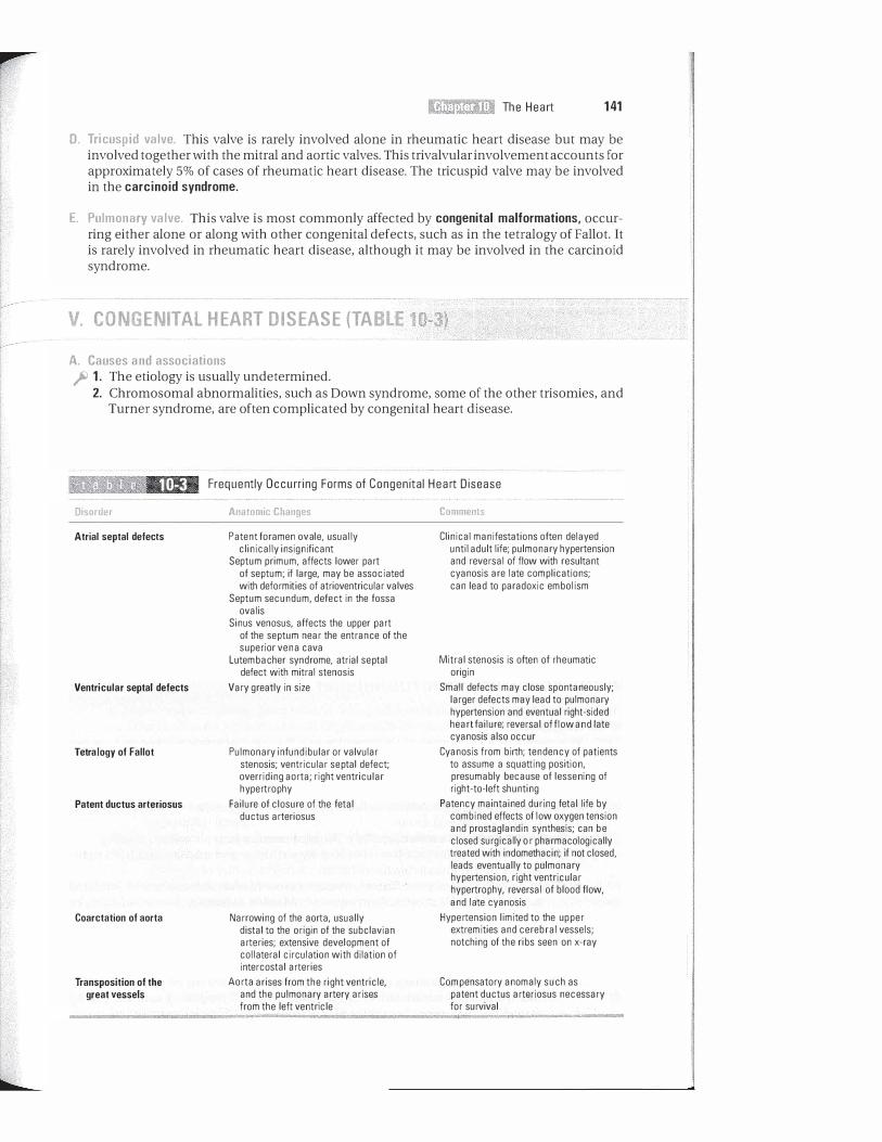
I!!JIt!ll!tIl!I Th e H e a rt 141
D. Tricuspid valve. This valve is rarely involved alone in rheumatic heart disease but may be involved together with the mitral and aortic valves. This trivalvular involvement accounts for approximately 5% of cases of rheumatic heart disease. The tricuspid valve may be involved in the carcinoid syndrome.
E. Pulmonary valve. This valve is most commonly affected by congenital malformations. occurring either alone or along with other congenital defects, such as in the tetralogy of Fallot. It is rarely involved in rheumatic heart disease, although it may be involved in the carcinoid syndrome.
v. CONGENITAL HEART DISEASE (TABLE 10-3)
A. Causes and associations � 1 . The etiology is usually undetermined.
2. Chromosomal abnormalities, such as Down syndrome, some of the other trisomies, and Turner syndrome, are often complicated by congenital heart disease.
t a b I e 10-3 Disorder
Atrial septal defects
Ventricular septal defects
Tetralogy of Fallot
Patent ductus arteriosus
Coarctation of aorta
Transposition of the great vessels
Frequently Occurr ing Forms of Congenital Hea rt D isease
Anatomic Changes
Patent foramen ova Ie, usually clinically insignificant
Septum primum, affects lower part of septum; if large, may be associated with deformities of atrioventricular valves
Septum secundum, defect in the fossa ovalis
Sinus venosus, affects the upper part of the septum near the entrance of the superior vena cava
Lutembacher syndrome, atrial se ptal defect with mitral stenosis
Vary greatly in size
Pulmonary infundibular or valvular stenosis; ventricular se ptal defect; overriding aorta; right ventricular hypertrophy
Failure of closure of the fetal ductus arteriosus
Narrowing of the aorta, usually distal to the origin of the subclavian arteries; extensive development of collateral circulation with dilation of intercostal arteries
Aorta arises from the right ventricle, and the pulmonary artery arises from the left ventricle
Comments
Clinical manifestations often delayed until adult life; pulmonary hypertension and reversal of flow with resultant cyanosis are late complications; can lead to paradoxic embolism
Mitral stenosis is often of rheumatic origin
Small defects may close spontaneously; larger defects may lead to pulmonary hypertension and eventual right-sided heart failure; reversal of flow and late cyanosis also occur
Cyanosis from birth; tendency of patients to assume a squatting position, presumably because of lessening of right-to-Ieft shunting
Patency maintained during fetal life by combined effects of low oxygen tension and prostaglandin synthesis; can be closed surgically or pharmacologically treated with indomethacin; if not closed, leads eventually to pulmonary hypertension, right ventricular hypertrophy, reversal of blood flow, and late cyanosis
Hypertension limited to the upper extremities and cerebral vessels; notching of the ribs seen on x-ray
Compensatory anomaly such as patent ductus arteriosus necessary for survival

---
1 42 BRS Pathology
� a. The association of Turner syndrome with coarctation of the aorta is notable. � b. Endocardial cushion defects, which result in atrial and ventricular septal defects and
atrioventricular valve deformities, are frequent associations of Down syndrome (trisomy 2 1) .
3 . There i s a n apparent increase in the incidence of patent ductus arteriosus in patients living at high altitudes, suggesting an association with fetal oxygen deprivation.
4. Rubella (German measles) infection is a prominent cause of congenital heart disease. � a. There is strong evidence of a link between maternal rubella during the first trimester
of pregnancy and a constellation of fetal defects, known as the congenital rubella syndrome. which includes cardiovascular defects, microcephaly with mental retardation, deafness, cataracts, and growth retardation.
� b. Cardiac malformations are especially frequent and commonly include patent ductus arteriosus, aortic stenosis, ventricular septal defect, and pulmonary infundibular or valvular stenosis, sometimes occurring as part of the tetralogy of Fallot.
� c. Before or during pregnancy, it is often important to determine the mother's immune status to rubella. Demonstration of antirubella antibodies of the IgM class indicates recent primary infection. whereas demonstration of IgG antibodies indicates either recent primary infection, past infection, or reinfection.
� 5. Genetic predisposition. Tetralogy of Fallot can cluster in families, probably because of multifactorial inheritance.
B. Functional abnormalities of congenital heart disease. These can be classified according to the presence or absence of cyanosis.
� 1. Noncyanotic diseases include those with no shunt (e.g., aortic stenosis, coarctation of the aorta) and those with a left-to-right shunt (e.g., patent ductus arteriosus, atrial or ventricular septal defect) . In atrial septal defects, both pressure and oxygen saturation may be equalized between the two atria.
� 2. Cyanotic diseases include transposition of the great vessels (survival depends on the presence of a shunt between the left and right ventricles), malformations with a right -toleft shunt (e.g., the tetralogy of Fallot), and disorders in which a left-to-right shunt reverses to right-to-left because of increased pulmonary arterial pressure (e.g., late cyanosis, tardive cyanosis) .
VI. DISEASES OF THE MYOCARDIUM
A. Cardiomyopathy � 1 . This term refers to diseases of the heart muscle that are noninflammatory and are not
associated with hypertension, congenital heart disease, valvular disease, or coronary artery disease.
� 2. Usually, these diseases are characterized by otherwise unexplained ventricular dysfunction (heart failure unresponsive to digitalis, ventricular enlargement, ventricular arrhythmias) . It occurs in several forms: a. Congestive or di lated cardiomyopathy is the most common form of cardiomyopathy. � (1 ) Characteristics include four-chamber hypertrophy and dilation and both right
and left-sided intractable heart failure. � (2) Etiology is most often unknown. In some cases, dilated cardiomyopathy is related
to alcohol ism (alcohol cardiomyopathy), thiamine deficiency (beriberi heart) , or prior myocarditis. Some forms are associated with mutant cytoskeletal proteins, such as dystrophin or desmin; mutations of sarcomeric proteins, such as cardiac myosin heavy chain; and other muscle proteins, such as actin. Mutations in mitochondrial genes have also been implicated.
(3) A multi-etiologic variant of dilated cardiomyopathy, known as peripartum cardiomyopathy, is associated with the latter stages of pregnancy and the period extending from weeks to months postpartum.

IIliitl'llt31!I The H e a rt 1 43
FIGURE 10-3 Hypertrophic ca rd iomyopathy. The interventricu la r septum is markedly thickened and may c ause obstruction of the left ventricu la r outflow tract . The microsco pic view shows the cha ra cteristic disoriented, tangled, and hypertrophied myocardia l fibers. (Reprinted with permission from Rubin R, Strayer D, et a I . , eds.: Rubin's Pathology. Clinicopathologic Foundations of Medicine, 5th ed. Ba ltimore, L ippincott Wil l iams & Wilkins, 2008, p. 472.)
b. Restrictive cardiomyopathy (1 ) The cause is infiltrative processes within the myocardium that result in stiffening
of the heart muscle, which interferes with pumping action. Jii (2) This cardiomyopathy is exemplified by cardiac amyloidosis, which may result in
both right- and left-sided heart failure. c. Hypertrophic cardiomyopathy is often inherited as an autosomal dominant characteristic.
Thus far, several genes have been implicated in the genesis of this disorder: genes coding for �-myosin heavy chain (most common mutation, thus far), cardiac troponins T and I, a-tropomyosin, myosin-binding protein C, and myosin light chain (Figure 10-3) .
Jii ( 1 ) Gross characteristics include hypertrophy of all chamber wal ls, especial ly the ventricular septum (asymmetric septal hypertrophy).
Jii (2) Microscopic characteristics include d isoriented, tangled, and hypertroph ied myocard ial fibers.
Jii (3) The cardiomyopathy may result in left ventricular outflow obstruction, placing the patient in danger of syncope and even sudden death, which often occurs unexpectedly in young athletes.
d. Arrhythmogenic right ventricular cardiomyopathy is a rare disorder characterized by gradual replacement of right ventricular muscle by fat, by arrhythmias, and sometimes by sudden death in athletes.
B. Myocarditis Jii 1 . This myocardial disease most often presents as biventricular heart failure in young per
sons who do not have valvular, rheumatic, or congenital heart disease. Jii 2. Morphologic characteristics include diffuse myocardial degeneration and necrosis with
an inflammatory infiltrate.
Chaz
Placed Image

1 44 BRS Pathology
� 3. Myocarditis is most often viral, and coxsackievirus is frequently the cause. 4. In parts of South America, myocarditis may be a component of Chagas disease, which is
caused by the protozoan Trypanosoma cruzi.
VII . D ISEASES OF THE PERICARDIUM
A. Noninflammatory conditions 1. Hydropericard ium is an accumulation of serous transudate in the pericardial space. It may
result from any condition causing systemic edema. It is most often caused by congestive heart failure or by edematous conditions due to hypoproteinemia, such as the nephrotic syndrome or chronic liver disease.
2. Hemopericardium is an accumulation of blood in the pericardial sac. It is usually caused by traumatic perforation of the heart or aorta or by myocardial rupture associated with acute myocardial infarction.
B. Acute pericarditis 1. Serous pericarditis is associated with systemic lupus erythematosus, rheumatic fever, and a
variety of viral infections. It is characterized by production of a clear, straw-colored, protein-rich exudate containing small numbers of inflammatory cells.
2. Fibrinous or serofibrinous pericarditis is characterized by a fibrin-rich exudate. It may be caused by uremia, myocardial infarction, or acute rheumatic fever.
3. Purulent or suppurative pericarditis is characterized by a grossly cloudy or frankly purulent inflammatory exudate. It is almost always caused by bacterial infection.
4. Hemorrhagic pericarditis is characterized by a bloody inflammatory exudate. It usually results from tumor invasion of the pericardium, but can also result from tuberculosis or other bacterial infection.
C. Chronic (constrictive) pericard itis. This disease is usually of tuberculous or pyogenic staphylococcal etiology.
� 1. Characteristics include thickening and scarring of the pericardium with resultant loss of elasticity. This prevents the pericardium from stretching and thus interferes with cardiac action and venous return, often mimicking the signs and symptoms of right-sided heart failure.
2. Proliferation of fibrous tissue with occasional small foci of calcification is marked.
VII I . TUMORS OF THE H EART
A. Primary tumors � 1. Myxoma of the left atrium is the most frequently occurring cardiac tumor and is found
most often in adults. 2. Rhabdomyoma is most common in infants and young children and is notable for its asso
ciation with tuberous sclerosis.
B. Metastatic tumors are more frequent than primary tumors.
IX. CONGESTIVE H EART FAI LURE
A . General considerations 1 . Congestive heart failure may be failure of the left ventricle, right ventricle, or both. 2. This condition often presents with dyspnea and/ or edema. Assay of B-type natriuretic pep
tide, which is elevated in heart failure, can aid in the distinction of heart failure from a number of other conditions such as asthma, acute coronary syndrome, chronic obstructive pulmonary disease, or pulmonary embolism, which can also present with dyspnea or edema.

FIGURE 10-4 Pulmonary edema. Pulmonary vascular congestion and proteinaceous pink staining intra-alveolar fluid can be seen in this manifestation of left-sided heart failure. (From Rubin R, Strayer D, et aI., eds.: Rubin's Pathology. Clinicopathologic Fou ndations of Medicine, 5th ed. Ba ltimore, Lippincott Wil l i ams & Wilkins, 2008, p. 231 . Courtesy of UBC Pu lmonary Registry, St. Paul's HospitaL)
B. left-sided heart fai lure 1 . Causes
I!iitft1ttiI!I The H e a rt 1 45
" " i ,
, .. .
I
. . ' " , . .
.... • • e 1 ". I'�'" #
' ..
, _ .. . ': . . � , ;
, t
a. Ischemic heart disease, especially myocardial infarction b. Hypertension c. Aortic and mitral valvular disease d. Myocardial diseases, such as cardiomyopathies and myocarditis
2. Cl inical manifestations � a. Dyspnea and orthopnea caused by pulmonary congestion and edema regularly occurs
(Figure 10-4) . b. Pleural effusion with hydrothorax often results. c. Reduction in renal perfusion, causing activation of the renin-angiotensin-aldosterone
system and leading to retention of salt and water, is less frequent. d. Cerebral anoxia is less frequent.
C. Right-sided heart fai lure 1. Causes
� a. left-sided heart fai lure is the most common cause of right -sided heart failure. b. left-sided lesions, such as mitral stenosis c. Pulmonary hypertension often caused by chronic lung disease (cor pulmonale) d. Various types of cardiomyopathy and diffuse myocarditis e. Tricuspid or pulmonary valvular disease
2. Cl i nical manifestations � a. Renal hypoxia, leading to greater fluid retention and peripheral edema than seen in left
sided failure. Edema occurs first in dependent areas and often manifests early as so-called pitting edema of the ankles. Other manifestations of fluid retention include pleural effusion and sometimes ascites. Hydrothorax can be a manifestation of either left-sided or right-sided heart failure.
� b. Enlarged and congested liver and spleen. Chronic passive congestion of the centrilobular veins of the liver surrounded by relatively pale, sometimes fatty, peripheral regions leads to a "nutmeg" pattern.
c. Distention of the neck veins
X. HYPERTROPHY OF THE H EART
A. Hypertrophy of the left ventricle is most commonly caused by hypertension and aortic or m itral valvular disease.
Chaz
Placed Image

1 46 BRS Pathology
B. Hypertrophy of the right ventricle 1. Causes
a. Left ventricular fai lure b. Chronic lung disease c. Mitral valve disease
FIGURE 10-5 Cor pulmonale. Marked right ventricu lar hypertrophy secondary to disease of the lung or of the pulmonary vascular system defines this entity. (Reprinted with permission from Rubin R, Strayer 0, et aI., eds.: Rubin's Pathology. Clinicopathologic Foundations of Medicine, 5th ed. Ba ltimore, Lippincott Wil l iams & Wilkins, 2008, p. 456.)
d. Congenital heart disease with left-to-right shunt 2. Cor pulmonale (Figure 10-5) � a. Cor pulmonale is defined as right ventricular hypertrophy and/or dilation secondary
to lung disease or primary disease of the pulmonary vasculature, such as primary pulmonary hypertension. Emphysema is a frequent cause.
b. Characteristics include pulmonary arterial hypertension, the common characteristic among the entities that lead to cor pulmonale.
Chaz
Stamp

Review Test
Directions: Each of the numbered items or incomplete statements in this section is followed by answers or by completions of the statement. Select the one lettered answer or completion that is best in each case.
1. A 55-year-old woman presents with complaints of chest pain. She states that the chest pain predictably occurs when she climbs four flights of stairs to reach her apartment or when she has been jogging for more than 10 minutes. She is particularly concerned because her mother died of a myocardial infarction at 50 years of age. Which of the following best describes this patient's state?
(A) Arrhythmia (8) Myocardial infarction (e) Prinzmetal angina (D ) Stable angina pectoris (E) Unstable angina pectoris
2. Yesterday, a 60-year-old man presented to the emergency department with dyspnea, diaphoresis, and crushing substernal chest pain that radiated to his neck and left arm. When asked to describe the pain, he put his fist to the center of his chest and stated that it felt "as if someone is squeezing my heart." An electrocardiogram demonstrated changes consistent with myocardial infarction, and serum troponin I levels were elevated. If the patient unexpectedly dies today, which of the following would almost certainly be found on histologic examination of the affected myocardium?
(A) Coagulative necrosis with neutrophil infiltration
(8) Fibrotic tissue replacing infarcted tissue (e) No histologic changes (D ) Slight swelling of tissue and change of
color (E) Young fibroblasts and new vessels grow
ing into the infarcted tissue
3. A 60-year-old-man is discharged after being observed in the hospital for 4 days following a myocardial infarction. He returns to his normal activities, which include sedentary work only. This point in
time following a myocardial infarct is noteworthy for the special danger of which of the following?
(A) Arrhythmia (8) Mural thrombosis (e) Myocardial (pump) failure (D ) Myocardial rupture (E) Ventricular aneurysm
4. A 10-year-old boy presents with migratory polyarthritis involving several large joints, fever, and malaise. Physical examination reveals a new heart murmur and friction rub on auscultation, and a painless nodule is detected on the extensor surface of the elbow. He had a severe sore throat approximately 2 weeks ago, apparently recovering without antibiotic therapy. The antistreptolysin 0 (ASO) titer is elevated. Which of the following describes the most likely outcome for this patient?
(A) Development of mitral valve stenosis over many months to years
(8) Development of mitral valve stenosis over the next few months
(e) Increasing severity of the current symptoms and findings over the next few decades
(D) Persistence of the current symptoms and signs over the patient's lifetime
(E) Total recovery after 1-2 months with no further complications or sequelae
5. A 9-year-old girl is diagnosed with acute rheumatic fever. Instead of recovering as expected, her condition worsens, and she dies. Which of the following is the most likely cause of death?
(A) Central nervous system involvement (8) Endocarditis (e) Myocarditis (D ) Pericarditis (E) Streptococcal sepsis
1 47

1 48 BRS Pathology
6. A 70-year-old woman has a long history of metastatic colon cancer, and she donates her body for use in medical school anatomy courses. At death, the body is emaciated and cachectic, and gross dissection reveals small fibrin deposits arranged around the line of closure of the leaflets of the mitral valve. The valvular lesions most likely represent
(A) bacterial endocarditis. (8) endocarditis of the carcinoid
syndrome. (e) Libman-Sacks endocarditis. (0) nonbacterial thrombotic (marantic)
endocarditis. (E) rheumatic endocarditis.
7. The myocardial lesions shown in the figure were observed at autopsy examination of a pediatric patient who died after a short illness. During life, which of the following manifestations of his illness was most likely?
(Reprinted with permission from Rubin R, Strayer D, et a I . , eds. : Rubin 's Pathology. Clinicopathologic Foundations of Medi ci ne, 5th ed . Ba ltimore, L ipp in cott Wi l l i ams & Wilkins, 2008, p. 459. )
(A) Chorea (8) Systemic embolization (e) Systemic lupus erythematosus (0) Unstable angina (E) Wasting diseases
8. A 60-year-old man presents with angina. He has no past medical history of heart disease. On questioning, the patient reveals that he had repeated sexually transmitted diseases in the past, including a painless chancre (a hard, round sore) on his penis, for which he never sought medical attention. Rapid plasma reagin (RPR), Venereal Disease Research Laboratory (VDRL) slide test, and fluorescent treponemal antibody (FTA) serologic tests (indicative of syphilis infection) are positive. Echocardiography and computed tomography of the heart are performed. The history of untreated syphilis suggests that these tests will most likely detect which of the following abnormalities?
(A) Aortic valvular insufficiency and linear calcification along the ascending aorta
(8) Bicuspid aortic valve with aortic stenosis (e) Large valvular vegetations from bacterial
endocarditis (0) Right-sided heart failure from the carci
noid syndrome (E) Small fibrin deposits on the mitral valve
from nonbacterial thrombotic (marantic) endocarditis
9. A 50-year-old man presents with sudden weakness in his left leg. He has felt well lately and has no past medical history of coronary artery disease, hyperlipidemia, or hypertension, and no family history of myocardial infarction or stroke. Physical examination reveals motor weakness in the left leg, with no other neurologic deficits, and no cardiac murmur. Magnetic resonance imaging of the brain demonstrates a small ischemic infarct in the arterial distribution of the brain correlating with motor control of the left leg. Angiography and echocardiography reveal normal coronary arteries, normal valves with no vegetations, and a small right -to-left shunt. Which of the following is most likely associated with this scenario?
(A) Atrial septal defect (8) Bacterial endocarditis (e) Nonbacterial thrombotic (marantic)
endocarditis (0) Tetralogy of Fallot (E) Ventricular septal defect
Chaz
Stamp

1 0. A 3-year-old boy presents with cyanosis and shortness of breath that develops when he plays with friends. According to his mother, the boy was born cyanotic. The boy is very small and short for his age, and he squats on the floor next to his mother. Chest radiography reveals a boot -shaped heart, normal heart size, and a right aortic arch. Echocardiography reveals a large ventricular septal defect with an overriding aorta, pulmonary stenosis, and right ventricular hypertrophy. Which of the following is the most likely diagnosis?
(A) Coarctation of the aorta (8) Patent ductus arteriosus (e) Rheumatic heart disease (0) Tetralogy of Fallot (E ) Transposition of the great vessels
1 1 . A 53-year-old woman presents with dyspnea on exertion, orthopnea, paroxysmal nocturnal dyspnea, edema in the legs and feet, and fatigue. She has no history of angina, other signs of coronary artery disease, hypertension, or valvular disease. Echocardiography reveals cardiomegaly, with four-chamber hypertrophy and dilation. Which of the follOwing is the most likely diagnosis?
(A) Congestive or dilated cardiomyopathy (8) Hypertrophic cardiomyopathy (e) Myocarditis (0) Restrictive cardiomyopathy
12. A 56-year-old woman presents with dyspnea on exertion, orthopnea, paroxysmal nocturnal dyspnea, and pulmonary edema. She also presents with severe dizziness and syncope, fatigue, weight loss, and arthralgias. After undergoing several tests, she is
I!1lIlJlllDl!I Th e He a rt 1 49
diagnosed with a primary heart tumor that is causing a "ball-valve obstruction" of her mitral valve. Which of the following is the most likely tumor?
(A) Fibroma (8) Leiomyoma (e) Lipoma (0) Myxoma (E) Rhabdomyoma
1 3. A 64-year-old woman presents with dependent peripheral edema in her ankles and feet. She has long-standing chronic obstructive lung disease and a long history of cigarette smoking. Further investigation reveals that she has cor pulmonale with right-sided heart failure. Which of the following is the most likely cause of the right-sided heart failure in this patient?
(A) Constrictive pericarditis (8) Disease of the lungs or pulmonary vessels (e) Left-sided heart failure (0) Pulmonary infundibular or valvular
stenosis (E) Systemic hypertension
1 4. A 42-year-old man is seen because of a long history of slowly developing congestive heart failure. His blood pressure is normal. Coronary artery angiography reveals no vascular disease. No heart murmurs are heard. The white blood cell count, differential, and erythrocyte sedimentation rate are normal. The most likely diagnosis is
(A) carcinoid heart disease. (8) cardiomyopathy. (e) coarctation of the aorta. (0) constrictive pericarditis. (E) myocardial infarction.

Answers and Explanations . I
1 . The answer is D. This is a classic case of stable angina, which is chest pain that is precipitated by exertion but relieved by rest. Stable angina is due to atherosclerosis of the coronary arteries. This patient has risk factors for ischemic heart disease (IHD) (e.g., cigarette smoking, hypertension, hyperlipidemia, diabetes, family history of IHD / coronary artery disease). Prinzmetal angina is intermittent chest pain at rest, and unstable angina is prolonged chest pain at rest.
2. The answer is A. By 24 hours, well-developed microscopic changes of coagulative necrosis can be detected in infarcted tissue. There is loss of nuclei in cells and infiltration of neutrophils into tissue.
3. The answer is D. Rupture of the left ventricle, a catastrophic complication of acute myocardial infarction, usually occurs when the necrotic area has the least tensile strength, about 4-7 days after an infarction, when repair is just beginning. The anterior wall of the heart is the most frequent site of rupture, usually leading to fatal cardiac tamponade. Internal rupture of the interventricular septum or of a papillary muscle may also occur. The risk of arrhythmia is greatest within the first 6 hours after myocardial infarct. Arrhythmias are the most important early complication of acute myocardial infarction, accounting for almost 50% of deaths shortly after myocardial infarction. Myocardial, or pump, failure and mural thrombosis are other complications that may develop as a result of permanent damage to the heart after infarct. Ventricular aneurysms may develop in the fibrotic scar within 3-6 months after myocardial infarct.
4. The answer is E. This is a case of acute rheumatic fever. Acute rheumatic fever manifests most commonly in patients 5-15 years of age with migratory polyarthritis, pancarditis, subcutaneous nodules, erythema marginatum, and Sydenham chorea. Decades later, severe valvular disease, often manifesting as mitral stenosis, may develop as a feature of rheumatic heart disease. In this chronic stage of rheumatic disease, fibrotic valves may become stenotic, insufficient, or both, but much more commonly, progression to cardiac valve complication does not occur.
5. The answer is C. The most common cause of death that occurs during acute rheumatic fever is cardiac failure secondary to myocarditis.
6. The answer is D. Nonbacterial thrombotic endocarditis, or marantic endocarditis, has been associated with a variety of wasting diseases and is observed most often in patients with cancer.
7. The answer is A. The figure illustrates an Aschoff body, the characteristic lesion of rheumatic fever. This myocardial lesion is most often oval in shape and characterized by swollen, fragmented collagen and fibrinoid material and by characteristic large mesenchymal cells (Anitschkow myocytes) and multinucleated cells (Aschoff cells) . Sydenham chorea is a major manifestation of rheumatic fever.
8. The answer is A. This is a case of syphilitic (luetic) aortitis. In syphilitic aortitis, the elastica of the aorta undergoes calcification and is replaced by fibrous tissue, reSUlting in dilation of the ascending aorta and separation of the aortic valve commissures, with resultant aortic insufficiency. Thus, echo cardiography and computed tomography of the heart reveal calcification in a linear pattern along the ascending aorta, calcification in the coronary arteries (leading to anginal symptoms), and aortic valvular insufficiency.
1 50

I!iillftlttil!I Th e H e a rt 1 51
9. The answer is A. This is a case of paradoxical embolism, which denotes the passage of an embolus of venous origin into the arterial circulation, by way of a right-to-left shunt (e.g., atrial septal defect or patent foramen ovale) . Ordinarily, atrial septal defects result in a left-to-right shunt across the atrial septum, but over time may develop into a right-to-left shunt. The likelihood of right-to-left passage of an embolus is often enhanced by pulmonary hypertension, sometimes secondary to pulmonary thromboembolism.
10. The answer is D. In the tetralogy of Fallot, the characteristic lesions include ventricular, septal defect, overriding aorta, pulmonary valve stenosis, and right ventricular hypertrophy. The pulmonary stenosis and overriding aorta cause increased right ventricular pressure and lead to right-to-left shunting. Cyanosis, which occurs when the arterial concentration of reduced hemoglobin exceeds 5 mg/mL, is seen with a right-to-left shunt, in which venous blood gains direct access to the arterial circulation. In contrast, patent ductus arteriosus, atrial septal defect, and ventricular septal defect are associated with left-to-right blood flow.
1 1 . The answer is A. Congestive or dilated cardiomyopathy is the most common form of cardiomyopathy. It is characterized by four-chamber hypertrophy and dilation as well as right- and left-sided severe heart failure. In some cases, congestive (dilated) cardiomyopathy may be associated with alcoholism, thiamine deficiency, or prior myocarditis.
12. The answer is D. Myxoma of the heart, although rare, is the most common primary cardiac tumor. Because of the jelly-like appearance and myxoid histology similar to that of some organized thrombi, the neoplastic nature of this lesion was debated for many years; however, it is now generally believed that myxoma is a true neoplasm. The most common location of myxoma is in the left atrium. Due to its location, complications may develop due to physical obstruction of blood flow through the mitral valve, resulting in symptoms of congestive heart failure.
1 3. The answer is B. The term cor pulmonale refers to right ventricular hypertrophy caused by pulmonary hypertension secondary to disorders of the lungs or pulmonary vessels. Other causes of right ventricular hypertrophy and failure, such as valvular disease, congenital defects, and left -sided heart failure, are precluded by this definition. Therefore, although in general, the most common cause of right-sided heart failure is left-sided heart failure, cor pulmonale with right-sided heart failure is due to an intrinsic disease originating in the lungs. Constrictive pericarditis can clinically mimic right-sided heart failure but is entirely unrelated to cor pulmonale.
14. The answer is B. Cardiomyopathies are noninflammatory myocardial disorders that are not associated with coronary artery obstruction, hypertension, valvular disease, congenital heart disease, or infectious disease. They are most often characterized by otherwise unexplained ventricular dysfunction, such as cardiac failure, ventricular enlargement, or ventricular arrhythmias.

Anemia
I. GENERAL CONCEPTS
A. Def in itions 1. Anemia is a decrease in whole body red cell mass, a definition that precludes relative
decreases in red blood cell count, hemoglobin, or hematocrit, which occur when the plasma volume is increased.
jiJ 2. Anemia of pregnancy is not anemia but rather is a manifestation of increased plasma volume. 3. A practical working definition of anemia is a decrease in red blood cell count, hemoglo
bin, or hematocrit; all are commonly measured red cell parameters.
B. Causes of anemia (Table 1 1 - 1 ) . Anemia may be caused by two major mechanisms: 1 . Decreased red cell production resulting from:
a. Hematopoietic cell damage from infection, drugs, radiation, and other similar agents b. Deficiency of factors necessary for heme synthesis (iron) or DNA synthesis (vitamin BIZ
or folate) 2. Increased red cell loss due to:
a. External blood loss b. Red cell destruction (hemolytic anemia)
II. ACUTE POSTHEMORRHAGIC ANEMIA
A. Within the first few hours of acute blood loss, prior to hemodilution (compensatory increase in plasma volume), there may be no decrease in the hemoglobin, hematocrit, and red blood cell count because of a parallel loss of both red cells and plasma. There is often a marked reactive increase in platelet count.
B. Significant clinical findings are related to hypovolemia.
III. IRON DEFICIENCY ANEMIA (SEE TABLE 11-1)
A. Causes 1. Chronic blood loss
� a. Such blood loss is the major cause of iron deficiency anemia in adults. jiJ b. Most often causes are menorrhagia or bleeding gastrointestinal lesions, such as carcinoma
of the colon in the United States or hookworm disease in less developed countries. jiJ 2. D ietary defic iency is rare except in infants; because human milk is low in iron, new
born storage iron is depleted within the first 6 months unless it is replaced by dietary
152

I!1ltltlll1I1I Anemia 153
t a b l e 11-1 Examples of Anemia Resulting from Decreased Red Cell Production
Type
Iron deficiency anemia
Pernicious anemia
Folate deficiency
Aplastic anemia
Anemia of chronic disease
Myelophthisic
Mechanisms
Impaired heme synthesis
Autoimmune gastritis leading to lack of gastric intrinsic factor and failure of vitamin B12 absorption; vitamin B12 deficiency causes delayed DNA replication
Delayed D NA replication
Greatly diminished hematopoiesis
Diverse mechanisms
Bone marrow replacement, usually by mal ignant tumor
Diagnostic Features
Hypochromia and microcytosis; decreased serum iron and increased total iron-binding capacity; decreased serum ferritin
Pancytopenia, oval macrocytes, and hypersegmented neutrophils; megaloblastic hyperplasia; ach lorhydria; anti-intrinsic factor antibodies; hyperreflexia, absent position and vibration sensations; impaired vitamin B12 a bsorption corrected by intrinsic factor (abnormal Schilling test)
Pancytopenia, oval macrocytes, and hypersegmented neutrophils; megaloblastic hyperplasia
Pancytopenia, reticu locytopenia, marked hypocel lu larity of bone marrow
Anemia most often normochromic and normocytic or macrocytic; may be hypochromic and micro-cytic with decreased serum iron and decreased serum iron-binding capac ity
Severe anemia; smal l numbers of nuc leated red cel ls and immature granu locytes in the peripheral blood; tumor cells in the bone marrow
Major Etiologic Factors
Dietary deficiency in infants and preadolescents; excess menstrual bleeding; chronic blood loss from the gastrointestinal tract
Autoimmunity
D ietary deficiency; malab-sorption syndromes
Toxic drugs and chemicals; often idiopathic
Various chronic diseases, especial ly rheumatoid arthritis, renal disease, and chronic infection
supplementation. Premature infants are at special risk. Dietary deficiency of iron may rarely occur in elderly persons.
3. Increased iron requirement may occur during pregnancy; iron demands of the fetus can deplete maternal iron stores. It may also occur in infants and preadolescents who may outgrow borderline iron stores.
B. Clinical manifestations 1. Signs and symptoms may include pallor, fatigue, or dyspnea on exertion. Sometimes
angina pectoris may occur in persons with coronary artery narrowing caused by atherosclerotic disease.
2. When extreme, associated features may include glossitis; gastritis; koilonychia (spooning of the nails) ; or Plummer-Vinson syndrome, in which iron deficiency is associated with a partially obstructing upper esophageal web.
C. Laboratory findings 1. Decreased hemoglobin, hematocrit, and red blood cell count 2. Hypochromic microcytic erythrocytes on peripheral smear 3. Decreased serum iron and increased total iron-binding capacity (TIBC) 4. Decreased body iron stores, measured by bone marrow examination for stainable hemo
siderin or by decreased serum ferritin
D. Differentia l diagnosis � 1. Iron deficiency anemia must be distinguished from other causes of hypochromic micro
cytic anemia, such as the anemia of chronic disease and �-thalassemia minor.

1 54 BRS Pathology
� 2. In anemia of chronic disease, the serum iron is low (as in iron deficiency) , but the TIBC is also low.
� 3. In f3-thalassemia minor, the A2 hemoglobin is increased.
IV. MEGALOBLASTIC ANEMIAS
A. General considerations 1. Megaloblastic anemias are defined by large, abnormal-appearing erythroid precursor
cells (megaloblasts) in the bone marrow. 2. These anemias are caused by a deficiency of vitamin B12 or folate. 3. These anemias are characterized by decreased DNA synthesis, with a consequent delay
in DNA replication and nuclear division, and by relatively unimpeded cytoplasmic maturation. Morphologically, they manifest as nuclear-cytoplasmic asynchrony of large erythroid precursor cells with an open, loose-appearing chromatin pattern.
4. Results include anemia caused by impaired red cell production; to a lesser degree, red cell destruction occurs within the bone marrow prior to release of mature erythrocytes into the peripheral blood (ineffective erythropoiesis).
B. laboratory abnormal ities 1. Peripheral blood and bone marrow findings are identical in all forms of megaloblastic anemias.
a. Peripheral blood � (1 ) Pancytopenia (decreased red cells, white cells, and platelets) � (2) Oval macrocytosis. Mean corpuscular volume (MCV) is often greater than 1 10 fl
(normal about 87 fl). � (3) Hypersegmented neutrophils (more than five lobes) (Figure 1 1 - 1 )
� b . Bone marrow: megaloblastic hyperplasia 2. Vitamin B12 and folate levels further define the specific type of megaloblastic anemia.
C. Types of megaloblasti c anemia 1 . Vitamin B12 deficiency
a. Pernicious anemia (1) Pernicious anemia is the most common form of vitamin BI2 deficiency mega
loblastic anemia. It is considered to be an autoimmune disorder; other autoimmune diseases (especially thyroid diseases) occur with increased frequency in persons with pernicious anemia.
FIGURE 11-1 Hypersegmented " polys" in megaloblastic anemias. Normal neutrophils have 3 to 5 lobes . In mega loblastic anemias the number is often increased as shown here . (Reprinted with permission from Rubin R. Strayer D, et a I ., e ds. : Rubin's Pathology. Clinicopathologic Foundations of Medicine, 5th ed. Ba ltimore, Lippincott Wil l iams & Wilkins, 2008, p. 870.)

....
iI!1l1lltttDII Anemia 1 55
� (2) Causes include autoimmune gastritis (previously referred to as fundal or type A gastritis)' which is associated with fai l ure of production of intrinsic factor, essential for vitamin BI2 absorption. The chronic gastritis is also associated with: (a) Achlorhydria (absent gastric free hydrochloric acid) (b) Anti-intrinsic factor and antiparietal cell antibodies (c) Increased incidence of gastric carcinoma
(3) Clinica l findings (a) I nsidious onset with extreme reduction of red blood cell count; in older
persons may be preceded by a lengthy subclinical period in which clinical manifestations are minimal.
(b) Characteristic lemon-yellow skin color (c) Stomatitis and glossitis (d) Subacute combined degeneration of the spinal cord (combined systems disease,
posterolateral degeneration) ( i ) Morphologic characteristics include demyelination of the posterior and
lateral columns. � ( i i ) Clinical manifestations include ataxic gait, hyperreflexia with extensor
plantar reflexes, and impaired position and vibration sensation. (Neurologic abnormalities are associated with vitamin BI2 deficiency, but not with folate deficiency.)
(4) Laboratory findings � (a) Pancytopenia, hypersegmented neutrophils. and megaloblastic hyperplasia of
the bone marrow (findings characteristic of all megaloblastic anemias) � (b) Anti-intrinsic factor antibodies, which are rarely found in other conditions;
anti parietal cell antibodies may be seen in other conditions, but are most frequent in pernicious anemia.
� (c) Abnormal Schi l l ing test, which is characterized by impaired absorption of vitamin BIZ correctable by intrinsic factor
� ( i ) Impaired absorption not corrected by intrinsic factor is characteristic of intestinal malabsorption, such as may occur in Crohn disease, blind-loop syndrome, and giant tapeworm infestation.
� (ij) Normal absorption is characteristic of vitamin BI2 deficiency due to dietary deprivation, which may occur in absolute vegetarians.
b. Other forms of vitamin B'2 deficiency megaloblasti c anemia. These pernicious anemia-like illnesses can be caused by a number of other mechanisms that result in vitamin BI2 deficiency. ( 1 ) Total gastric resection; because intrinsic factor is produced in the gastric fundus,
the clinical picture is the same as in pernicious anemia. (2) Disorders of the distal i leum; intrinsic factor-vitamin BI2 complex is absorbed in
the distal ileum. (3) Strict vegetarian d iet; vitamin BI2 is found only in foods of animal origin. (4) Intestinal malabsorption syndromes (5) Bl ind-loop syndrome; bacterial overgrowth in a surgically induced intestinal blind
loop results in the depletion of vitamin B12. (6) Broad-spectrum antibiotic therapy; can result in intestinal bacterial overgrowth
with vitamin BI2 depletion. (7) Diphyllobothrium tatum infestation; the giant fish tapeworm of man, acquired by
ingestion of freshwater fish, inhabits the intestine and causes vitamin BI2 depletion. 2. Folate deficiency
� a. No neurologic abnormalities (in contrast to vitamin BIZ deficiency) occur. b. Folate deficiency megaloblastic anemia can be caused by a number of diverse
mechanisms: � (1 ) Severe dietary deprivation (most often occurs in chronic alcoholics or fad dieters) � (2) Pregnancy (combination of additional demands of the fetus and borderline
maternal diet) (3) Di lantin (phenytoin), which interferes with the absorption of folate, or oral contra
ceptive therapy

1 56 BRS Pathology
(4) Fol ic acid antagonist chemotherapy for cancer (5) Relative folate deficiency (increased demand because of compensatory acceler
ated erythropoiesis in hemolytic anemia) ;;- (6) Intestinal malabsorption caused by:
(a) Sprue (b) Giardia lamblia infection
V. ANEMIA OF CHRONIC DISEASE
This common form of anemia is second in incidence to iron deficiency anemia.
A. Anemia of chronic disease can be secondary to a wide variety of primary disorders, including rheumatoid arthritis, renal disease, or chronic infection.
B. It is often normochromic and normocytic. When associated with renal disease, it may be moderately macrocytic.
;;- C. When associated with chronic inflammatory states (e.g., rheumatoid arthritis), it may be accompanied by decreased serum iron and hypochromia and microcytosis, mimicking iron deficiency anemia; however, in contrast to iron deficiency anemia, TIBC is characteristically decreased.
VI . APLASTIC ANEMIA (FIGURE 1 1-2)
A. General considerations ;;- 1. Characteristics include a markedly hypocel lular bone marrow with almost total loss of
hematopoietic cells, including erythroid and myeloid precursor cells and megakaryocytes, and peripheral pancytopenia (anemia, leukopenia, and thrombocytopenia) and reticulocytopenia.
;;- 2. Aplastic anemia is most often secondary to toxic exposure; it may also occur without evident cause. In addition, it can be caused by autoimmune dysfunction of cytotoxic T cel ls, and it can also be induced by several other etiologic agents: a. Radiation exposure
;;- b. Chemicals, such as benzene and related organic compounds ;;- c. Therapeutic drugs, such as chloramphenicol. sulfonamides, gold salts, chlorpromazine,
various anti-inflammatory and antimalarial drugs, and alkylating agents used in the treatment of neoplastic diseases
FIGURE 11-2 Aplastic anemia. The marrow is "empty." Spicules of bone and substitution of numerous fat ce lls for the normal cel lu lar bone marrow are a l l that can be seen. (Reprinted with permission from Rubin R, Strayer D, et ai., eds. : Rubin's Pathology. Clinicopathologic Foundations of Medicine, 5th ed. Ba ltimore, Lippincott Wil l iams & Wilkins, 2008, p. 866.) •

-
I!iiIlt!1tD1I An e m i a 1 57
( 1 ) In some persons who have been given chloramphenicol, there is a predictable, dose-related, usually reversible marrow response.
(2) In other persons, there is an idiosyncratic, severe, frequently irreversible effect. d. Viral infection, such as human parvovirus or hepatitis C virus
VII. MYELOPHTH ISIC ANEMIA
This form of bone marrow failure i s caused by replacement of the marrow, most often by a malignant neoplasm.
A. Less commonly it is due to bone marrow destruction from non-neoplastic causes, such as marrow fibrosis.
B. It may be signaled by leukoerythroblastosis, in which small numbers of nucleated red cells and immature granulocytic precursors are seen in the peripheral blood smear.
VI I I. HEMOLYTIC ANEMIAS (TABLE 11-2)
�-�----
These anemias are due to the shortening of the red cell life span (increased red cell destruction) .
A. General considerations 1. I ncreased red cell destruction with liberation of hemoglobin or its degradation products
is manifested by: ;i'J a. Increased unconjugated ( indirect reacting) bi lirubin, resulting in acholuric jaundice,
which is jaundice not accompanied by bilirubinuria. Hyperbilirubinemia may lead to pigment-containing gallstones as a late complication.
b. Increased urine urobi linogen ;i'J c. Hemoglobinemia and hemoglobinuria, which, along with methemalbuminemia and
hemosiderinuria, occur if red cell destruction is very rapid and within the circulation (intravascular hemolysis) .
;i'J d. D isappearance of serum haptoglobins, a group of hemoglobin-binding proteins, which combine with liberated hemoglobin and are no longer demonstrable. In instances of intravascular hemolysis, such as hemolytic transfusion reactions, elevation of serum hemoglobin does not occur until serum haptoglobins are no longer detectable.
e. Hemosiderosis, systemic iron deposition 2. Increased erythropoiesis, compensating in part for the shortened red cell survival, is
manifested by: a. Normoblastic erythroid hyperplasia in bone marrow
;i'J b. Reticulocytosis (increased number of circulating newly formed red cells identified by residual stainable RNA) . Because reticulocytes are larger than other red cells, the MeV may be modestly increased (up to about 105 fl).
c. Polychromatophilia (increased number of somewhat larger red cells that stain with a bluish cast, roughly equivalent to increased reticulocyte count)
B. Terminology 1 . Intra corpuscular hemolytic anemia is marked by defects, most often genetically deter
mined, in the red cell itself. 2. Extracorpuscular hemolytic anemia is marked by defects, most often acquired, of the
extraerythrocytic environment, such as circulating antibodies or an enlarged spleen.

158 BRS Pathology
Examples of Anemias Resulting from Increased Red Cell Destruction
Type
Warm antibody autoimmune hemolytic anemia (primary and secondary forms)
Hemolytic disease of the newborn (erythroblastosis fetal is)
Hereditary spherocytosis
Glucose-6-phosphate dehydrogenase (G6PD) deficiency
Sickle cell anemia
i3-Thalassemia major (Cooley anemia, Mediterranean anemia)
a-Thalassemias
Mechanisms
IgG autoantibodies combine with red cell surface antigens; Fc combining site of IgG antibody further reacts with Fc receptor of phagocytic cells
Maternal al loimmunization to fetal red cell antigens, classically of Rh system; can also be caused by a l loimmunization to ABO blood group antigens
Red cell membrane skeletal protein abnormality
Failure of erythrocyte hexose monophosphate shunt under oxidative stress
i3-globin hemoglobinopathy (mutation in coding sequence of i3-globin gene, GAG [glul �
GTG [vall)
Diverse mutations i n i3-globin g e n e causing decreased synthesis of i3-globin chains; aggregation of excess a-chains causes hemolytic anemia and ineffective erythropoiesis
Deletion of one or more of the four a-globin genes (a-globin gene is reduplicated in tandem on each chromosome 16)
Diagnostic Features
Anemia, spherocytosis, and reticulocytosis; unconjugated hyperbilirubinemia and acholuric iaundice; positive di rect Coombs test
Rising titer of maternal anti-Rh antibodies during the latter part of pregnancy; cord blood at de livery conta ins immature red cel l precursors; d i rect Coombs test positive on cord blood; progressive postnatal increase in unconjugated b i l i rubin
Autosomal dominant inheritance; anemia, spherocytosis, and reticulocytosis; increased mean corpuscular hemoglobin concentration; unconjugated hyperbi l irubinemia and acholuric jaundice; splenomegaly; increased erythrocyte osmotic fragi lity to hypotonic sal ine
Self-limited hemolytic anemia; reduced activity of erythrocyte G6P D
Anemia a n d reticulocytosis; sickle-shaped erythrocytes demonstrable on peripheral blood smear; homozygosity for hemoglobin S demonstra ble by electrophoresis
Severe anemia; thalassemic red cell morphology; increased hemoglobin F
Differ accord ing to the number of deletions
Comments
Often secondary to lymphocytic neoplasms, Hodgkin disease, or autoimmune disease; sometimes associated with a-methyldopa or penicil lin therapy
Prevented by administration of anti-Rh antibody (antio IgG) to mother at time of delivery of first and subsequent children, removing Rh-positive red cells from maternal circulation; treated by exchange transfusion to remove unconjugated bilirubin from the serum of a newborn infant to prevent kernicterus
Quantitative deficiency of spectrin due to diverse mechanisms
X-linked inheritance
Characterized by severe anemia, recurrent painful and ap lastic crises, and nonhea l i ng leg ulcers; recurrent splenic infarcts with progressive fibrosis result in autosplenectomy
Occurs frequently in Mediterranean populations
No clinical abnormalities with one gene deletion; mild-to-moderate thalassemic state with two or three delet ions; intrauterine death with four deletions; hemoglobin Barts h41 in fetal l ife; hemoglob in H (134) in adult l ife

I!1ltftllD1i Anemia 1 59
� C. Immune hemolytic anemias are clinically suggested by hemolytic anemia of recent onset. 1 . Warm antibody autoimmune hemolytic anemia is the most common form of immune
hemolytic anemia. a. This anemia is mediated by IgG autoantibodies that react with red cell surface antigens.
These antibodies are optimally active at 37°C (thus their designation as "warm" antibodies). The anemia is often secondary to underlying disease states such as systemic lupus erythematosus, Hodgkin disease, or non-Hodgkin lymphomas.
b. Clinical characteristics include the following: ( 1 ) General features of hemolytic anemia (2) Spherocytosis due to progressive loss of membrane protein by serial passage of
antibody-coated red cells through the spleen � (3) Positive d irect Coombs test reflecting the binding of IgG autoantibody to the red
cell surface 2. Cold agglutinin disease is mediated by IgM antibodies optimally active at temperatures
below 30°C (cold antibodies, cold agglutinins) . a. Acute cold agglutinin disease is often mediated by antibodies with specificity for the I blood
group antigen. It is often a complication of infectious mononucleosis or Mycoplasma pneumoniae infection. Diagnosis of mycoplasma pneumonia is facilitated by the demonstration of cold agglutinins.
3. Chronic cold agglutinin disease is often associated with lymphoid neoplasms. It is often mediated by anti-I antibodies. Characterized clinically by agglutination and hemolysis in tissue sites exposed to the cold, it may be associated with Raynaud phenomenon. These cases are marked by chronic hemolytic anemia exacerbated by cold weather, punctuated by episodes of jaundice, sometimes with hemoglobinemia and hemoglobinuria.
4. Hemolytic disease of the newborn (erythroblastosis fetalis) occurs when maternal antibodies cross the placenta and react with fetal red cells, resulting in fetal hemolytic anemia.
� a. Causes include maternal alloimmunization to fetal red cell antigens, classically the D antigen of the Rh blood group system. In the most frequently occurring form of Rh -mediated hemolytic disease of the newborn, the mother is typed as d and the fetus as D.
� b. Causes also include ABO incompatibil ity. In most instances of ABO incompatibility, the mother is blood group 0 and the child is blood group A or group B. Other incompatible combinations include: mother A, child B or AB; mother B, child A or AB.
� c. This disease can result in kernicterus, staining of the basal ganglia and other central nervous system structures by unconjugated bilirubin. Kernicterus with resultant neurologic damage is the most significant long-term consequence of hemolytic disease of the newborn. In addition, consequences include stillbirth or in hydrops fetal is, fetal heart failure with massive generalized edema.
� d. The incidence has been markedly reduced by preventive measures. Routine administration of anti-D IgG antiserum to D-negative mothers at the time of delivery (or at the time of termination of pregnancy) of a D-positive child results in the antibodymediated removal of fetal red cells from the maternal circulation, preventing maternal alloimmunization.
D. Paroxysmal nocturnal hemoglobinuria is an uncommon acquired intracorpuscular defect. 1 . Characteristics include increased sensitivity to complement-induced red cell lysis,
resulting in intravascular hemolytic anemia, pancytopenia, and an increased incidence of venous thrombosis. The condition is often marked by the passage of hemoglobincontaining urine on awakening.
� 2. This defect arises from a somatic mutation in a gene known as PIG-A. The mutation causes impaired synthesis of the glycosylphosphatidylinositol (GPI) anchor that is required for the fixation of a number of diverse proteins to cellular surfaces. Among these proteins are CD55, CD59, and C8 binding proteins, which are required for the protection of red cells, granulocytes, and platelets from complement-mediated lysis.
3. Diagnosis now involves flow cytometry demonstrating a population of CD59-negative erythrocytes; the defect was formerly diagnosed by the now archaic Ham (acid serum) test, which demonstrated in vitro complement-induced hemolysis in acidified serum.

1 60 BRS Pathology
E. Hemolytic anemias caused by membrane skeletal protein abnormal ities 1. Hereditary spherocytosis is an autosomal dominant hemolytic anemia. It is the most
common intracorpuscular inherited hemolytic anemia observed in whites. � a. This anemia is characterized by spherocytes (sphere-shaped erythrocytes) that are
selectively trapped or sequestered in the spleen. Splenomegaly is often prominent. � b. An increased erythrocyte osmotic fragil ity to hypotonic saline is characteristic. � c. An increase in the mean corpuscular hemoglobin concentration (MCHC), an abnor
mality seldom observed in other forms of anemia, is also characteristic. � d. The cause is a variety of molecular defects in the genes coding for spectrin, ankyrin,
protein 4. 1 , and other erythrocyte membrane skeletal proteins. These defects are usually manifest as a deficiency of spectrin.
2. Hereditary ell iptocytosis (ovalocytosis) is an autosomal dominant disorder characterized by elongated, oval red cells. It may be marked by hemolytic anemia and splenomegaly. It often does not cause anemia.
F. Enzyme deficiency hemolytic anemias 1 . G lucose-6-phosphate dehydrogenase (G6PD) deficiency is the most common form of
enzyme deficiency hemolytic anemia. � a. This X-l inked disorder occurs in approximately 10% of African Americans and also in
persons of Mediterranean origin. � b. G6PD deficiency is manifest by acute self-limited intravascular hemolytic anemia with
hemoglobinemia and hemoglobinuria caused by oxidative stress induced by infection; by a wide variety of therapeutic drugs, such as primaquine (an anti-malarial agent) , sulfonamides, and many other oxidant drugs; or, in some persons of Mediterranean origin, by fava beans.
c. G6PD deficiency may be suggested by the finding of "bite cells" (erythrocytes that appear as if a piece were munched out) on the peripheral blood smear.
2. Pyruvate kinase deficiency is the second most common enzyme deficiency hemolytic anemia. a. This autosomal recessive disorder is characterized by hereditary nonspherocytic
hemolytic anemia. b. In contrast to the most common form of G6PD deficiency, in which the anemia is
episodic and self-limited, the anemia is chronic and sustained.
G. Hemoglobinopathies. These are hemolytic anemias caused by genetically determined abnormalities of hemoglobin structure. In the United States, they are most importantly represented by disorders involving hemoglobin S; to a lesser extent, hemoglobin C; and, in some urban centers, hemoglobin E. 1 . Hemoglobin S d isorders
a. General considerations � (1 )
� (2)
� (3)
Approximately 7% of African Americans carry the hemoglobin S gene. In some parts of Africa, more than one third of the population is affected; it is thought that the hemoglobin S gene confers resistance to falciparum malarial infection. Similar resistance may be conferred by erythrocyte G6PD deficiency or by the absence of Duffy blood group antigens. Both G6PD deficiency and the Duffy Fy (a- b-) phenotype occur with high incidence in persons of African origin. Hemoglobin S arises from a point mutation in codon 6 of the I)-globin gene and results in a substitution of valine for glutamic acid. At the DNA level, the mutation abolishes a recognition site for the restriction endonuclease MstII. This is the basis for prenatal diagnosis, which can be performed on amniotic cells or on chorionic villus samples.
(4) Hemoglobin S polymerizes at low oxygen tension, forming tactoids that distort the shape of red cells to elongated, sickle shapes; repeated sickling episodes stiffen red cell membranes, making affected cells more subject to hemolysis; rigid, sickled cells are more likely to obstruct the microvasculature.
� (5) All hemoglobin S disorders are characterized by a positive sickle cell preparation (in vitro sickling of red cells on exposure to reducing agents such as sodium

FIGURE 11-3 Sickle cel l anemia. Circulating sickle cells in the peripheral blood in homozygous "SS" disease. (Reprinted with permission from Rubin R, Strayer D, et aI., eds.: Rubin's Pathology. Clinicopathologic Foundations of Medicine, 5th ed. Baltimore, Lippincott Williams & Wilkins, 2008, p. 874.)
I!1im'dDI Anemia 1 61
metabisulfite). The sickle cell preparation is positive whenever hemoglobin S is present (e.g. , sickle cell anemia, sickle cell trait, sickle C disease, sickle cell thalassemia) .
b. Sickle cell anemia (Figure 11-3) . The homozygous form of hemoglobin S leads to sickle cell anemia (sickle cell disease), which is characterized by: ( 1 ) Severe hemolytic anemia (2) Chronic leg ulcers (3) Vaso-occlusive painful crises (severe pain in the limbs, back, chest, and abdomen) ,
often precipitated by infection or dehydration � (4) Repeated infarctions in the lungs and spleen; the spleen is characteristically con
gested and enlarged in childhood, but becomes progressively smaller through repeated infarcts and fibrosis (autosplenectomy).
(5) Aplastic crises (distinguished from painful crises) , characterized by a precipitous fall in hemoglobin concentration, usually provoked by viral infection, such as human parvovirus
(6) I nfectious complications, such as Salmonella osteomyelitis c. Sickle cell trait. Hemoglobin S in the heterozygous form leads to sickle cell trait, which
is generally without clinical consequence. 2. Hemoglobin C disorders are primarily observed in persons of African lineage.
a. When homozygous, they are characterized by mild hemolytic anemia accompanied by prominent splenomegaly, target cells, and, on occasion, intraerythrocytic crystals.
b. When heterozygous, they result in disease only when coinherited with other abnormal hemoglobins, most often hemoglobin S.
3. Hemoglobin E disorders are prevalent in Southeast Asia. a. These disorders have increased significantly in incidence in the United States in
recent years and are now more common than hemoglobin S disorders in some urban areas.
b. Clinical and laboratory manifestations are similar to those of hemoglobin C disorders.
H. Thalassemias � This heterogeneous group of genetic disorders is characterized by deficient production of either
a- or [3-globin chains of hemoglobin. Heme synthesis is unaffected. They are widespread throughout the world, occurring with high frequency in Africa, India, Southeast Asia, and the Mediterranean area.
1 . j3-Thalassemias are the most common forms of thalassemia found in Mediterranean areas and in the United States. They are caused by defects in the promoter sequence, in introns, or in coding regions of the [3-globin gene.

1 62 BRS Pathology
a. �-Thalassemia major is also known as Mediterranean anemia or Cooley anemia. It results from compound heterozygosity or homozygosity for thalassemic variants of the [3-globin gene, and it is characterized clinically by: ( 1 ) Marked anemia resulting from:
(a) Modest decrease in hemoglobin synthesis (b) Marked shortening of red cell life span due to aggregation of insoluble excess
a-chains (c) Ineffective erythropoiesis (d) Relative folate deficiency
� (2) Marked splenomegaly (3) Distortion of skull, facial bones, and long bones because of erythroid marrow expansion
� (4) Thalassemic red cell morphology (marked microcytosis, hypochromia, target cells, extensive changes in size and shape)
� (5) Increased hemoglobin F (a2'Y2) throughout life (6) General ized hemosiderosis due to chronic hemolysis, ineffective erythropoiesis,
and repeated transfusions b. �-Thalassemia minor results from heterozygous inheritance of thalassemic variants of
the [3-globin gene. � (1) Clinical manifestations include minimal hypochromic microcytic anemia and an
increase in hemoglobin A2 (a202) , a normally occurring minor hemoglobin fraction. (2) This finding is useful in distinguishing [3-thalassemia minor from iron deficiency
anemia and the anemia of chronic disease. c. Sickle cell thalassemia results from coinheritance of hemoglobin S gene and a tha
lassemic variant of the [3-globin gene (compound heterozygosity) . It is clinically similar to, but often less severe than, sickle cell anemia.
2. a-Thalassemias are the most common forms of thalassemia in Southeast Asia. They are caused by deletions of one or more of the four a-globin genes (see Table 1 1 -2) . a. In normal persons, there is duplication of the a-globin gene, with a pair of identical
a-globin genes on each member of the chromosome 16 pair. � b. Some variants are characterized by increased concentration of hemoglo bin Barts ('Y 4)
or hemoglobin H ([34)'
I. Hemolytic anemias due to mecha n ica l d isruptio n o f circulating erythrocytes (Figure 1 1 -4) 1. These anemias are associated with aortic valvular prostheses. They are also associated with
disseminated intravascular coagulation and thrombotic thrombocytopenic purpura. In these instances, partial occlusion of small vessels is the cause of the mechanical disruption of the erythrocytes. This phenomenon is referred to as microangiopathic hemolytic anemia.
2. Characteristics include circulating red cell fragments referred to as schistocytes or helmet cells.
FIGURE 11-4 Microangiopathic hemolytic anemia. Note the presence of schistocytes (denoted by arrows). This finding is characteristic of traumatic injury to circu lating red cells and may be seen in disseminated intravascular coagulation, thrombotic thrombocytopenic purpura, and in conjunction with artificial cardiac valve prostheses. (Reprinted with permission from Rubin R, Strayer D, et aI., eds.: Rubin's Pathology. Clinicopathologic Foundations of Medicine, 5th ed. Ba ltimore, Lippincott Will iams & Wilkins, 2008, p. 887.)

Review Test
Directions: Each of the numbered items or incomplete statements in this section is followed by answers or by completions of the statement. Select the one lettered answer or completio
-n
that is best in each case.
1. The peripheral blood smear of an anemic I-year-old child is shown in the illustration. The most likely diagnosis is
( Reprinted with pe rmiss ion from Rubin R, Strayer D, et a I ., eds. : Rubin's Pathology Clinicopathologic Foundations of Medicine, 5th ed. Ba ltimore, Lippincott Wil l iams & Wilkins, 2008, p . 865.)
(A) anemia of chronic disease. (8) aplastic anemia. (e) hereditary spherocytosis. (D) iron deficiency anemia. (E) thalassemia major.
2. A patient with severe anemia has a peripheral blood smear with oval macrocytes, hypersegmented neutrophils, and decreased platelets. The most likely cause of the anemia is
(A) a red cell membrane protein defect. (8) an amino acid substitution in the (3-globin
chain. (e) iron deficiency. (D ) marrow hypoplasia. (E) vitamin B12 or folate deficiency.
3. The patient in question 2 is found to be a severely malnourished alcoholic. The most likely cause of this disorder is
(A) aberrant intestinal bacterial flora. (8) Crohn disease.
(e) fish tapeworm infestation. (D) folate deficiency. (E) pernicious anemia.
4. A primiparous D-negative (Rh-negative) mother has just delivered a D-positive child. Administration of which of the following substances would be indicated?
(A) Anti-D IgG to child (8) Anti-D IgG to mother (e) D-positive red cells to child (D) D-positive red cells to mother
5. A 62-year-old man presents with pallor, fatigue, and dyspnea on exertion. A complete blood count reveals microcytic hypochromic anemia. The most likely cause of these findings is
(A) dietary deficiency of iron. (8) gastrointestinal bleeding. (e) hemodilution. (D) hemolytic anemia. (E) increased iron requirement.
1 63

164 BRS Pathology
6. A 23-year-old man of northern European lineage presents with anemia. His father and paternal aunt had a similar illness that was treated successfully by splenectomy. His peripheral blood smear is similar to that shown in the illustration. Which of the following additional abnormalities is expected?
(A) Bilirubinuria (B ) Decreased mean corpuscular volume (e) Increased direct (conjugated) serum
bilirubin (0) Polychromatophilic erythrocytes on
peripheral blood smear (E) Positive direct antiglobulin (Coombs) test
7. A 72-year-old man who has recently had an aortic valve replacement now presents with pallor and fatigue. The red blood cell count is decreased, and schistocytes are reported on examination of a peripheral blood smear. In addition, his indirect (unconjugated) bilirubin is Significantly elevated. The cause of the anemia is likely
(A) cold agglutinin disease. (B) dietary deficiency. (e) hereditary spherocytosis. (0) mechanical disruption of red cells. (E) paroxysmal nocturnal hemoglobinuria.
8. A 23-year-old African-American man with a history since early childhood of severe anemia requiring many transfusions has nonhealing leg ulcers and recurrent periods of abdominal and chest pain. These signs and symptoms are most likely to be associated with which of the following laboratory abnormalities?
(A) Decreased erythropoietin (B ) Increased erythrocyte osmotic fragility (e) Schistocytes (0) Sickle cells on peripheral blood smear (E) Teardrop-shaped cells
(Reprinted with permission from Rubin R, Strayer D, et aI., eds.: Rubin's Pathology. Clinicopathologic Foundations of Medicine, 5th ed. Ba ltimore, Lippincott Wil l iams & Wilkins, 2008, p. 872.)
9. The spleen of the patient in question 8 would be expected to be
(A) enlarged. (B) normal sized. (e) shrunken.
10. An 18-year-old man is transported to the emergency department within 20 minutes of sustaining a stab wound to the chest. The patient is poorly responsive. Given that he may have lost as much as 1.5 liters of blood at the scene, which of the following is the most likely finding in this patient?
(A) Decreased blood pressure (B ) Decreased hematocrit (e) Decreased hemoglobin (0) Decreased red blood cell count (E) Increased mean corpuscular hemoglo
bin concentration
11. A 23-year-old woman with epilepsy who is desirous of becoming pregnant is found to be anemic. Examination of a peripheral blood smear reveals oval macrocytes and hypersegmented neutrophils. Neurologic examination is entirely normal. Which of the follOwing is the most likely cause of the anemia?
(A) Alloantibodies directed against fetal red cell antigens
(B) Bone marrow aplasia (e) Coating of red cells by IgG "warm"
autoantibodies (0) Folate deficiency (E) Neoplastic replacement of the bone
marrow

12. A 56-year-old woman with a history of breast cancer that was treated 5 years ago with lumpectomy and radiation but with no chemotherapy returns with bone pain, fatigue, and weakness. A complete blood count reveals severe anemia, as well as decreased white blood cells and platelets. Examination of a peripheral blood smear reveals small numbers of nucleated red cells, as well as an occasional "blast" cell and myelocyte. A likely cause of the hematologic abnormalities is
(A) chloramphenicol. (B) Diphyllobothrium latum infestation. (e) megaloblastic anemia. (0) myelophthisic anemia. (E) sickle cell anemia.
13. A 23-year-old man presented to his primary care physician 2 weeks ago with a nonproductive cough and malaise. He was treated with the antibiotic azithromycin for "walking pneumonia" due to Mycoplasma pneumoniae. He now returns with fatigue and pallor. Laboratory studies demonstrate a decreased red blood cell count with polychromatophilia and an increase in indirect (unconjugated) bilirubin. The laboratory findings are most likely due to
(A) acute cold agglutinin disease. (B ) aplastic anemia. (e) aplastic crisis. (0) paroxysmal nocturnal hemoglobinuria.
i!lltf11ttIiI Anemia 165
(E) warm antibody autoimmune hemolytic anemia.
14. Military policy dictates that flight personnel in Iraq receive primaquine chemoprophylaxis for Plasmodium vivax malaria on redeploying to a non-malarious area. Several days after beginning such a regimen, a 26-year-old African-American pilot develops anemia, hemoglobinemia, and hemoglobinuria. Special studies will likely reveal an abnormality in which of the following?
(A) Duffy antigen (B) Glucose-6-phosphate dehydrogenase (e) Intrinsic factor (0) PIG-A (E) Spectrin
15. A 28-year-old woman complains of fatigue, dyspnea, and malaise. She also notes that her urine has been reddishbrown, particularly with the first void of the morning. Subsequent studies confirm that she has paroxysmal nocturnal hemoglobinuria. Which of the following best describes the defect leading to this condition?
(A) Anti-intrinsic factor antibodies (B) Deficiency of the intracellular structural
protein spectrin (e) Impaired synthesis of the cell-surface
GPI anchor (0) Ineffective erythropoiesis (E) Substitution of a valine for a glutamate
residue in the j3-globin gene

Answers and Explanations
1. The answer is D. The illustration shows hypochromia and microcytosis. Iron deficiency is the most frequent cause of hypochromic microcytic anemia. In infants and preadolescents, iron deficiency is most often nutritional in origin. In young women, the cause is most often related to menstrual blood loss compounded by deficient dietary intake. In men, the usual cause is occult gastrointestinal blood loss.
2. The answer is E. Megaloblastic anemia due to deficiency of vitamin BIZ or folate is characterized by oval macrocytes, hypersegmented neutrophils, and decreased platelets.
3. The answer is D. Megaloblastic anemia associated with severe malnutrition is most often due to folate deficiency.
4. The answer is B. Administration of anti-D antiserum to a D-negative mother at the time of delivery of a D-positive child prevents maternal alloimmunization by removing fetal red cells from the maternal circulation.
5. The answer is B. Iron deficiency anemia is the most common cause of hypochromic microcytic anemia, and gastrointestinal bleeding is the most likely cause of iron deficiency in an adult male. Such a finding warrants a complete workup, including colonoscopy to detect the source of the bleeding. Dietary deficiency of iron and increased iron requirements are common causes of iron deficiency in women of child-bearing age, especially during pregnancy. Hemolytic anemia is usually accompanied by reticulocytosis. The reduced red cell parameters (hemoglobin, red blood cell count, hematocrit) that are observed with hemodilution are not truly representative of anemia, which is formally defined as a reduction in whole body red cell mass.
6. The answer is D. Spherocytes are present on the peripheral blood smear and, along with the history, strongly suggest a diagnosis of hereditary spherocytosis. Similar cells are also observed in warm antibody autoimmune hemolytic anemia; therefore, these two conditions must be distinguished by means of the direct Coombs test, which is negative in hereditary spherocytosis and positive in warm antibody autoimmune hemolytic anemia. An expected finding would be an increase in indirect (unconjugated) serum bilirubin, not direct (conjugated). The jaundice is acholuric (no bilirubin in the urine, so bilirubinuria would not be expected). The mean corpuscular hemoglobin concentration (MCHC) is often increased. Because hereditary spherocytosis is a normocytic anemia, the mean corpuscular volume is not decreased. Polychromatophilic erythrocytes are an expected finding, as in any hemolytic anemia.
7. The answer is D. Turbulent blood flow over mechanical heart valves can cause shearing of red blood cells, resulting in fragmented cells termed schistocytes. The hemolysis can result in anemia and hyperbilirubinemia. Hereditary spherocytosis causes hemolytic anemia due to an intrinsic defect in the red blood cells. Dietary deficiencies do not cause fragmented red blood cells. Aplastic anemia would result in severe anemia with marked reticulocytopenia.
8. The answer is D. Sickle cell anemia is the most common hereditary anemia of persons of African lineage. Leg ulcers and recurring painful crises are characteristic. In sickle cell anemia, in contrast to sickle cell trait, sickle cells are often seen on the peripheral blood smear.
9. The answer is C. Repeated episodes of splenic infarction followed by fibrotic healing lead to a fibrotic, shrunken spleen (autosplenectomy) in adult patients with sickle cell anemia. The spleen is enlarged and congested in children with sickle cell anemia.
166

....
I!iltl'l1mI Anemia 1 67
10. The answer is A. Within the first few hours of acute blood loss, findings of hypovolemia predominate, especially with signs of hypovolemic shock, such as decreased blood pressure. It is likely that red cell indices (red blood cell counts, hemoglobin, and hematocrit) eventually decrease as a result of hemodilution. Increased mean corpuscular hemoglobin concentration (MCHC) is an unusual finding but is expected in hereditary spherocytosis.
1 1 . The answer is D. Phenytoin is a commonly used antiseizure medication that can cause _ impaired folate absorption, with resultant folate deficiency and megaloblastic anemia. The drug is contraindicated for use during pregnancy, because folate is required during embryogenesis to prevent neural tube defects. Aplastic anemia results in a major failure of erythropoiesis. Myelophthisic anemia occurs in some cancer patients with metastatic disease. Warm antibody hemolytic anemia is manifest by anemia and reticulocytosis, often with spherocytosis. Hemolytic disease of the newborn most commonly occurs with Rh blood group incompatibility between mother and fetus.
12. The answer is D. Infiltration of tumor cells from cancers, such as those of breast and prostate, displaces bone marrow elements, thereby causing myelophthisic anemia. Chloramphenicol causes aplastic anemia in some individuals. Diphyllobothrium latum infestation can result in megaloblastic anemia due to vitamin B12 deficiency. Sickle cell anemia is more common in black and Mediterranean populations.
13. The answer is A. Acute cold agglutinin disease is a form of hemolytic anemia due to autoantibodies to blood group antigens and is sometimes a complication of Mycoplasma pneumoniae infection. Aplastic anemia is associated with a variety of toxic exposures, including, among others, the antibiotic chloramphenicol, not azithromycin. Aplastic crises can occur as acute events in the course of hemolytic anemias, such as hereditary spherocytosis or sickle cell anemia. Paroxysmal nocturnal hemoglobinuria is an acquired defect that renders red blood cells sensitive to complement-induced lysis. Warm antibody autoimmune hemolytic anemia can be idiopathic or secondary to autoimmune disorders or lymphoid neoplasms.
14. The answer is B. Drugs that cause oxidative stress (e.g., primaquine, sulfa-containing drugs) result in intravascular hemolytic anemia in subjects (most often male) with glucose-6-phosphate dehydrogenase (G6PD) deficiency. Current military policy excepts G6PD-deficient persons from the requirement for primaquine chemoprophylaxis or sets up screening criteria for individuals whose G6PD status is unknown. Duffy antigen is a minor red blood cell antigen, the absence of which confers some resistance to malarial infection. Intrinsic factor may be absent in pernicious anemia. PIG-A deficiency results in paroxysmal nocturnal hemoglobinuria. Spectrin is deficient in hereditary spherocytosis.
1 5. The answer is C. Paroxysmal nocturnal hemoglobinuria results in an acquired intracorpuscular defect in the ability to synthesize the glycosylphosphatidylinositol (GPl) anchors required for appropriate placement of complement regulatory proteins on the surface of red blood cells. Functional deficiency of such proteins as CD55 and CD59 renders the cells sensitive to complement-mediated lysis. Anti-intrinsic factor antibodies are seen in pernicious anemia. Spectrin is deficient in hereditary spherocytosis. Ineffective erythropoiesis is seen in megaloblastic anemia due to folate or vitamin B 12 deficiencies. Substitution of valine for glutamic acid in the j3-g10bin gene underlies the defect in sickle cell anemia .
Neoplastic and Proliferative Disorders of the Hematopoietic and Lymphoid Systems
I. LEUKEMIA
A. General concepts 1 . Leukemia is a general term for a group of malignancies of either lymphoid or hematopoi
etic cell origin. The number of circulating leukocytes is often greatly increased. 2. The bone marrow is diffusely infiltrated with leukemic cells, often with encroachment
on normal hematopoietic cell development. Consequent failure of normal leukocyte, red cell, and platelet production can result in anemia, infection, or hemorrhage.
3. Infiltration ofleukemic cells in the liver, spleen, lymph nodes, and other organs is common.
B. Acute leukemias
1 68
1. General considerations a. A predominance of blasts and closely related cells in the bone marrow and peripheral
blood is characteristic. b. The most common malignancies of the pediatric age group, acute leukemias occur
most often in children. They exhibit a second incidence peak after 60 years of age. c. Cytogenetic abnormalities are frequent. For example, the 9;22 translocation results in
a morphologically unique chromosome, the Philadelphia chromosome (Ph' ). This abnormality, better known for its association with chronic myelogenous leukemia, is associated with a poorer prognosis when it occurs in acute leukemias.
d. Without therapeutic intervention, acute leukemia follows a short and precipitous course, marked by anemia, infection, and hemorrhage, and death occurs within 6 to 12 months.
2. Acute lymphoblastic leukemia (ALL) (Figure 12-1) ,,- a. ALL is the most common malignancy of children. ,,- b. A predominance of Iymphoblasts in the circulating blood and in the bone marrow is
characteristic.
,,- c. ALL is the form of acute leukemia that is most responsive to therapy. ,,- d. Further classification into a number of subgroups is based on differences in morphol
ogy, cytogenetic changes, antigenic cell-surface markers, or rearrangement of the immunoglobulin heavy-chain or T-cell receptor genes. The form that is most common and most amenable to therapy is characterized by blast cells that are positive for the CD 10 marker.
3. Acute myeloid (myeloblastic) leukemia (AML) (Figure 12-2) a. AML occurs most often in adults. b. A predominance of myeloblasts and early promyelocytes is characteristic. c. AML responds to current therapy more poorly than ALL. d. Further classification into several subgroups is based on morphology, cytochemical
characteristics, surface markers, and genetic alterati0l!s.

FIGURE 12-1 Acute lymphoblastic leukemia. Many Iymphoblasts are found in the peripheral blood and bone marrow. As in other acute leukemias, normal hematopoiesis is decreased, and patients often present with anemia, infection, and thrombocytopenic bleeding. (Reprinted with permission from Rubin R, Strayer D, et aI., eds.: Rubin's Pathology. Clinicopathologic Foundations of Medicine, 5th ed. Baltimore, Lippincott Williams & Wilkins, 2008, p. 9 1 2. )
iii1trtltfiij Neoplastic and Proliferative Disorders 169
C. Chron ic l eukemias. These malignancies are characterized by proliferations of lymphoid or hematopoietic cells that are more mature than those of the acute leukemias. The chronic forms have a longer, less devastating clinical course than the acute leukemias but are less responsive to therapeutic intervention. 1 . Chronic lymphocytic leukemia (Cll)
a. General considerations � ( 1 ) CLL is characterized by proliferation of neoplastic lymphoid cel ls (almost always B
cells) with widespread infiltration of the bone marrow, peripheral blood, lymph nodes, spleen, liver, and other organs.
(2) Leukemic cells of CLL are less capable of differentiating into antibody-producing plasma cells.
� (3) CLL most often occurs in persons older than 60 years of age and more frequently in men.
b. Characteristics � ( 1 ) The leukemic cells closely resemble normal mature peripheral blood lympho
cytes and, like these cells, express surface immunoglobulin and pan-B-cell markers such as CD19 and CD20. They are also CDS positive and CDIO negative.
(2) The cells are susceptible to mechanical disruption and often appear on the peripheral blood smear as smudge cells.
FIGURE 12-2 Auer rods in acute myeloblastic leukemia. Although frequently not seen, when they are present, Auer rods are d iagnostic of leukemic mye loblasts. (Reprinted with permission from Rubin R, Strayer D, et aI., eds.: Rubin's Pathology. Clinicopathologic Foundations of Medicine, 5th ed. Baltimore, Lippincott Williams & Wilkins, 2008, p. 903.)

170
I t
BRS Pathology
FIGURE 12-3 Ha i ry ce l l l eukemia . Ha irl i ke projections from these B-ce l l derived neoplastic ce l ls define this condition . (Reprinted with permission from Rub in R, Strayer D , et a I. , eds. : Rubin's Pathology. Clinicopathologic Foundations of Medicine, 5th ed . Ba ltimore, Lippincott Wil l iams & Wilkins, 2008, p . 921 . )
� (3) The peripheral white blood cell count varies from 50,OOO/ pL to 200,OOO/pL, with a preponderance of leukemic cells_
(4) Leukemic cells diffusely infiltrate the bone marrow. c. Complications
( 1 ) Warm antibody autoimmune hemolytic anemia (2) Hypogammaglobul inemia and increased susceptibility to bacterial infection, often
occurring early in the course of this disorder d. Clinical features
( 1 ) The clinical course is usually described as indolent, often with few symptoms and minor disability for protracted periods.
(2) Generalized lymphadenopathy and moderate hepatosplenomegaly are frequent features.
� (3) Mean survival is 3 to 7 years; treatment relieves symptoms but has little effect on overall survival.
2. Hairy cell leukemia is a B-cell disease in which the leukemic cells exhibit characteristic hair-l ike filamentous projections (Figure 12-3) . The cells can be further identified by their positive staining for tartrate-resistant acid phosphatase (TRAP). a. Hairy cell leukemia most often affects middle-aged men, who present with prominent
splenomegaly and pancytopenia. b. The disease has received major attention because of its dramatic response to several ther
apeutic agents, including (X-interferon, 2-chlorodeoxyadenosine, and deoxycoformycin. 3. Chronic myelogenous leukemia (CML) is a neoplastic clonal proliferation of myeloid stem
cells, the precursor cells of erythrocytes, granulocytes, monocytes, and platelets. It is one of the myeloproliferative syndromes (Figure 12-4) .
FIGURE 12-4 Chronic myelogenous leukemia . Proliferation of ce l ls of the myelopoietic l ine dominates the peripheral blood and bone marrow. The figure shows blast cells, promyelocytes, myelocytes, and band forms. (Reprinted with permission from Rubin R, Strayer D, et aI., eds.: Rubin's Pathology. Clinicopathologic Foundations of Medicine, 5th ed. Baltimore, Lippincott Williams & Wilkins, 2008, p. 898.)
a. Molecular changes
I!1lf!ftif!fIEI Neoplastic and Proliferative Disorders 171
� (1) CML is characterized by a reciprocal chromosomal translocation between chromosomes 9 and 22. The Philadelphia chromosome represents a remnant of chromosome 22 with the addition of a small segment of chromosome 9. This cytogenetic change is found in all blood cell lineages (erythroblasts, granulocytes, monocytes, megakaryocytes, B- and T-cell progenitors) , but not in the majority of circulating B or T lymphocytes.
(2) The c-abl proto-oncogene on chromosome 9 is transposed to an area on chromosome 22, adjacent to an oncogene referred to as ber (for breakpoint cluster region) , forming a new hybrid, or fusion, gene, ber-abl.
(3) Bcr-abl codes for a protein (p210) with tyrosine kinase activity, which plays a critical role in the etiopathogenesis of CML.
b. Characteristics � (1 ) Marked leukocytosis, with white blood cell counts varying from 50,000/pL to
200,000/pL � (2) Leukemic cells in the peripheral blood and bone marrow, mainly middle-to-Iate
myeloid (granulocytic) precursor cells, including myelocytes, metamyelocytes, bands, and segmented forms
(3) Small numbers of blasts and promyelocytes (4) The Philadelphia chromosome, found in granulocytic and erythroid precursor
cells and in megakaryocytes (5) Marked reduction in leukocyte alkaline phosphatase activity in the leukemic leuko
cytes c. Clinica l features
(1 ) Prominent splenomegaly and modestly enlarged liver and lymph nodes (2) Peak incidence in middle-age (35-50 years of age) (3) Terminates, in most cases, in an accelerated phase leading to so-called blast cri
sis marked by increased numbers of primitive blast cells and promyelocytes
II. MYELOPROLIFERATIVE DISEASES
This group of clonal proliferations of myeloid stem cells includes chronic myelogenous leukemia, polycythemia vera, chronic idiopathic myelofibrosis (agnogenic myeloid metaplasia), and essential thrombocythemia. It is of great interest that most, if not all, patients with polycythemia rubra vera and a significant number of patients with agnogenic myeloid metaplasia and essential thrombocythemia have mutations in the gene for the Janus2 signaling protein and are JAK2V61 7F positive.
� A. Common characteristics 1. Peak incidence in middle-aged and elderly persons 2. Proliferation of one or more of the myeloid series (erythroid, granulocytic, and megakary-
ocytic) cell types 3. Increase in peripheral blood basophils and nucleated red cells 4. Increase in serum uric acid 5. Prominent splenomegaly
B. Polycythemia vera 1. Clinical characteristics
� a. Marked erythrocytosis b. Moderate increase in circulating granulocytes and platelets c. Splenomegaly d. Decreased erythropoietin
2. Other features a. Sludging of high hematocrit blood often leads to thrombotic or hemorrhagic phenomena.
J

172 BRS Pathology
b. Polycythemia vera often progresses to a late phase in which anemia supervenes. This phase is often marked by bone marrow fibrosis and extramedullary hematopoiesis and an increasing white blood cell count, and it can mimic CML.
c. Acute leukemia may supervene in approximately 3% of patients, most of whom have received antimitotic drugs or radiation therapy.
3. Diagnosis jiJ a. Polycythemia vera is marked by decreased erythropoietin, which distinguishes it from
other forms of polycythemia, all of which are associated with increased erythropoietin. b. It must be distinguished from secondary polycythemia, which is associated with the
following: jiJ ( 1 ) Chronic hypoxia, associated with pulmonary disease, congenital heart disease,
residence at high altitudes, and heavy smoking jiJ (2) I nappropriate production of erythropoietin, associated with androgen therapy, adult
polycystic kidney disease, and tumors, such as renal cell carcinoma, hepatocellular carcinoma, and cerebellar hemangioma
(3) Endocrine abnormalities, prominently including pheochromocytoma and adrenal adenoma with Cushing syndrome
C. Chronic id iopathic myelofibrosis (agnogenic myelo id metap lasia, myel ofibrosis with mye l o i d metaplasia) i s characterized by extensive extramedullary hematopoiesis involving the liver and spleen and sometimes the lymph nodes. Additional manifestations include proliferation of non-neoplastic fibrous tissue within the bone marrow cavity (myelofibrosis) . The fibrous tissue replaces normal hematopoietic cells. This late manifestation is often preceded by marrow hypercellularity. 1. Postulated pathogenetic factors
a. Megakaryocytic proliferation may be the primary abnormality; the elaboration of platelet-derived growth factor and of transforming growth factor-13 (TGF-I3) by platelets and megakaryocytes may be the cause of the fibroblastic proliferation.
jiJ b. Megakaryocytes are spared in the marrow fibrotic process and increase in number, resulting in prominent bone marrow megakaryocytosis and peripheral blood thrombocytosis.
2. Cl inical features a. Peripheral blood smear
jiJ ( 1 ) Teardrop-shaped erythrocytes (2) Granulocytic precursor cells and nucleated red cell precursors in variable
numbers b. Anemia and massive splenomegaly
D . Essential thrombocythemia is characterized by marked thrombocytosis in the peripheral blood and megakaryocytosis in the bone marrow. Platelet counts in excess of 1 ,OOO,OOO/pL are common (normal value is 150,OOO-350,OOO/pL) . Additional features include bleeding and thrombosis.
III. NON-NEOPLASTIC LYMPHOID PROLIFERATIONS - ,�. - - �
A. G eneral considerations. These reactions include acute and chronic nonspecific lymphadenitis occurring in response to a number of infectious agents or immune stimuli.
B. I nfectious mononucleosis (Figure 12-5) jiJ 1. This benign, self-limited disorder is caused by Epstein-Barr virus (EBV) , which has an
affinity for B lymphocytes. It occurs frequently in young adults. 2. Circulating atypical lymphocytes (reactive CD8+ T lymphocytes) are characteristic.
jiJ 3. The disorder is marked by a number of serum antibodies, including anti-EBV antibodies and heterophil antibodies (heterophil agglutinins) directed at sheep erythrocytes; so-called
J

FIGURE 12-5 Atypical lymphocytes in infectious mononucleosis. Cells such as these can be mistaken for the blast cells of acute leukemia. (Reprinted with permission from Rubin R, Strayer D, et aI . , eds. : Rubin's PathologV. Clinicopathologic Foundations of Medicine, 5th ed . B a ltimore, Lippincott Will iams & Wilkins, 200B, p. 90B.)
II!mtt'tttltIEJ Neoplastic and Proliferative Disorders 173
heterophil-negative infectious mononucleosis is most often associated with cytomegalovirus infection.
� 4. Clinical characteristics are prominent sore throat, fever, generalized lymphadenopathy, and often hepatosplenomegaly. The spleen is especial ly susceptible to traumatic rupture.
IV. PLASMA CELL DISORDERS (FIGURE 12-6)
A. General considerations 1 . Plasma cell disorders are neoplastic proliferations of well-differentiated immunoglobulin
producing cells. 2. These disorders include multiple myeloma, Waldenstrom macroglobulinemia, and
benign monoclonal gammopathy, as well as primary amyloidosis and heavy-chain (Franklin) disease.
3. Occurrence is most frequent in persons older than 40-50 years of age.
� B. Multiple myeloma (plasma cell myeloma/plasmacytoma) is a malignant plasma cell tumor usually affecting older persons that typically involves bone and is associated with prominent serum and urinary protein abnormalities. 1. Bone lesions and protein abnormal ities
� a. The neoplastic cell is an end-stage derivative of B lymphocytes that is clearly identifiable as a plasma cell. The neoplastic cells can easily be identified by bone marrow biopsy or aspiration smears.
FIGURE 12-6 Multiple myeloma. Large, often nuc leolated, neoplastic plasma ce l l s infi ltrate the bone ma rrow. (Reprinted with permission from Rubin R, Strayer D, et aI . , eds.: Rubin's Pathology. Clinicopathologic Foundations of Medicine, 5th ed. Ba ltimore, Lippincott Will iams & Wilkins, 200B, p. 91 9.)

174 BRS Pathology
/'! b. The tumor cells produce lytic lesions in bone, especially in the skull and axial skeleton. /'! ( 1 ) The bone lesions appear lucent on radiographic examination, with characteristic
sharp borders, and are referred to as punched-out lesions. They may be manifest radiographically as diffuse demineralization of bone (osteopenia).
(2) The cause is an osteoclast-activating factor secreted by the neoplastic plasma cells.
/'! (3) The lesions are often associated with severe bone pain and spontaneous fractures.
c. Multiple myeloma arises from proliferation of a single clone of malignant antibodyproducing cells.
/'! ( 1 ) The tumor cells produce massive quantities of identical immunoglobulin molecules demonstrable electrophoretically as a narrow serum band or, after densitometric scanning, as a sharp spike referred to as an M protein.
(2) The M protein in multiple myeloma is most often an IgG or 19A immunoglobulin of either kappa or lambda light-chain specificity.
(3) Synthesis of normal immunoglobulins is often impaired. (4) The marked serum immunoglobulin increase is often initially detected by labora
tory screening as increased total protein with an increase in serum globulin (hyperglobulinemia) .
/'! (5) The urine often contains significant quantities of free immunoglobulin light chains, either kappa or lambda, which are referred to as Bence Jones protein.
/'! (6) As a consequence of hyperglobulinemia, the red cells tend to congregate together in a manner reminiscent of a stack of poker chips (rouleaux formation). There is also a marked increase in the erythrocyte sedimentation rate.
2. Other cl inical characteristics of multiple myeloma /'! a. Anemia due to neoplastic encroachment on myeloid precursor cells; possible leukope
nia and thrombocytopenia b. I ncreased susceptibi l ity to infection because of impaired production of normal
immunoglobulins /'! c. Hypercalcemia secondary to bone destruction; in contrast to the increased serum alka
line phosphatase that accompanies most other instances of hypercalcemia, the serum alkaline phosphatase in multiple myeloma is not increased.
/'! d. Renal insufficiency with azotemia due to myeloma kidney (myeloma nephrosis). The renal lesion is characterized by prominent tubular casts of Bence Jones protein, numerous multinucleated macrophage-derived giant cells, and metastatic calcification, and sometimes by interstitial infiltration of malignant plasma cells.
e. Amyloidosis of the primary amyloidosis type
C. Waldenstrom macroglobulinemia is a manifestation of Iymphoplasmacytic lymphoma, a B-cell neoplasm of lymphoid cells of an intermediate stage between B lymphocytes and plasma cells referred to as plasmacytoid lymphocytes. In the case of Waldenstrom macroglobulinemia, the neoplastic cells produce a monoclonal IgM protein (lymphoplasmacytic lymphomas can also occur without protein production) .
1. Defining characteristics /'! a. Serum IgM immunoglobulin of either kappa or lambda specificity occurring as an M
protein /'! b. Plasmacytoid lymphocytes infiltrating the blood, bone marrow, lymph nodes, and spleen /'! c. Bence Jones proteinuria in about 10% of cases
d. Absence of bone lesions 2. Clinical features
a. Most frequently seen in men older than 50 years of age b. Slowly progressive course, often marked by generalized lymphadenopathy and mild
anemia 3. Complications
a. Hyperviscosity syndrome, which results from marked increase in serum IgM. Features include retinal vascular d i lation, sometimes with hemorrhage, confusion, and other

I!il!1'!1Qifi Neoplastic and Proliferative Disorders 175
central nervous system changes. Sometimes, emergency plasmapheresis is required to prevent blindness.
b. Abnormal bleeding, which may be due to vascular and platelet dysfunction secondary to the serum protein abnormality
D. Benign monoclonal gammopathy (monoclonal gammopathy of undetermined significance, or MGUS) occurs in 5%-10% of otherwise healthy older persons.
� 1. A monoclonal M protein spike of less than 2 gl lOO mL, minimal or no Bence Jones proteinuria, less than 5% plasma cells in the bone marrow, and no decrease in concentration of normal immunoglobulins is characteristic.
2. MGUS is most often without clinical consequence.
V. LYM P H O I D NEOPLASMS
A. Hodgkin lymphoma (Hodgkin disease) i s a malignant neoplasm with features (e.g., fever, inflammatory cell infiltrates) resembling an inflammatory disorder. 1 . Genera l considerations
a. Hodgkin lymphoma characteristically affects young adults (predominantly young men); an exception is nodular sclerosis, which frequently affects young women.
b. Associated manifestations often include pruritus, fever, diaphoresis, and leukocytosis reminiscent of an acute infection.
c. With modern staging and aggressive therapy, a clinical cure is often achieved. � d. This neoplasm is characterized in all forms by the presence of Reed-Sternberg cells.
The diagnosis of Hodgkin lymphoma depends on this histologic finding. 2. Reed-Sternberg cells are binucleated, or multinucleated, giant cells with eosinophilic
inclusion-like nucleoli that may be the actual malignant cells of Hodgkin lymphoma. a. Differing numbers are found in varying forms of Hodgkin lymphoma, and the severity
of the disease variants is directly proportional to the number of Reed-Sternberg cells found in the lesions.
b. Conversely, the greater the number of reactive lymphocytes in the Hodgkin lymphoma variant, the better the prognosis.
3. Classification of Hodgkin lymphoma. The World Health Organization (WHO) classification identifies a number of disease variants (Table 12- 1 ) . a. Lymphocyte-predominance Hodgkin lymphoma
( 1 ) Features include large numbers of lymphocytes and histiocytes, as well as a paucity of Reed-Sternberg cells.
(2) There is no association with EBV infection. (3) Peak incidence occurs in young and middle-aged men. (4) The prognosis is relatively good.
b. Lymphocyte-rich Hodgkin lymphoma ( 1 ) There is an association with EBV infection in 40% of cases. (2) This variant is more common in men than in women. (3) The clinical course is moderately aggressive.
c. Mixed cellularity Hodgkin lymphoma ( 1 ) This variant is the one found most often in older persons. It is more common in
men than in women. (2) Characteristic features include a polymorphic infiltrate of eosinophils, plasma cells,
histiocytes, and Reed -Sternberg cells, as well as areas of necrosis and fibrosis. (3) There is an association with EBV infection in 70% of cases. (4) The clinical course is moderately aggressive.
d. Lymphocyte depletion Hodgkin lymphoma ( 1 ) This variant is the least frequently occurring form of Hodgkin lymphoma. (2) Few lymphocytes, numerous Reed-Sternberg cells, and extensive necrosis and
fibrosis are apparent.

1 76 BRS Pathology
t a b l e 12-1
B-Cell Neoplasms
Precursor B-cel l neoplasm
World H ealth Organization (WHO ) Classification of Lymphoid Neoplasms (Abbreviated)
Precursor B lymphoblastic leukemia/lymphoma (precursor B-cel l acute lymphoblastic leukemia)
Mature (peripheral) B-cell neoplasms
B-cell chronic lymphocytic leukemia/smal l lymphocytic lymphoma
Lymphoplasmacytic lymphoma
Hairy cell leukemia
Plasma cell myeloma/plasmacytoma
Extranodal marginal zone B-ceil lymphoma of MALT type
Follicular lymphoma
Mantle cell lymphoma
Diffuse large B-ceil lymphoma
Burkitt lymphoma/Burkitt cell leukemia
T-Cell Neoplasms
Precursor T-cel l neoplasm
Precursor T lymphoblastic lymphoma/leukemia (precursor T-cel l acute lymphoblastic leukemia)
Mature (peripheral) T-cel l neoplasms
T-cell granular lymphocytic leukemia
Adult T-cell lymphoma/leukemia ( HTLV1+)
Enteropathy-type T-cel l lymphoma
Mycosis fungoides (Sezary syndrome)
Hodgkin Lymphoma (Hodgkin Disease)
Classical Hodgkin lymphoma subtypes
Lymphocyte-predominance Hodgkin lymphoma
Lymphocyte-rich Hodgkin lymphoma
Mixed cel lula rity Hodgkin lymphoma
Lymphocyte depletion Hodgkin lymphoma
Nodular sclerosis Hodgkin lymphoma
IModified from Harris NL, Jaffe ES, Diebold J, et al.. The World Health Organization classification of hematological malignancies report of the Clinical Advisory Committee Meeting, Airlie House, Virginia, November 1 997. Modern Pathology 1 3: 1 93-207, 2000.)
(3) There is an association with EBV infection in the great majority of cases; this variant is also more common in persons with human immunodeficiency virus infection.
(4) This type of Hodgkin lymphoma has the poorest prognosis among all the variants. e. Nodular sclerosis Hodgkin lymphoma (Figure 12-7)
( 1 ) This variant is the most frequently occurring form of Hodgkin lymphoma, Unlike other forms of Hodgkin lymphoma, this variant occurs more frequently in young women.
(2) Nodular division of affected lymph nodes by fibrous bands and the presence of lacunar cel ls, Reed-Sternberg cell variants, are characteristic. The neoplasm often arises in the upper mediastinum or lower cervical or supraclavicular nodes.
(3) There is rarely an association with EBV infection. (4) The prognosis is relatively good.
4. Cl inical staging: Ann Arbor classification a. This system of classification is based on the degree of dissemination, involvement
of extralymphatic sites, and presence or absence of systemic signs such as fever (Table 12-2) . It is an essential part of the diagnostic eval uation of patients with
Hodgkin lymphoma. J>l b. Although grading of histopathologic variants roughly correlates with clinical behavior,
prognosis is better predicted by staging (Ann Arbor classification) ,

FIGURE 1 2-7 A, B N o d u l a r sc lerosis H odgkin lymphoma. The lymph node is transected by broad bands of f ibrous connective tissue. (Reprinted with permission from Rubin R, Strayer D, et a i ., eds.: Rubin's Pathology. Clinicopathologic Foundations of Medicine, 5th ed. B a lt imore, L ipp incott Wil l i ams & Wilkins, 2008, p. 925 )
i!iitI'ttttitI Neoplastic and Proliferative Disorders 1 77
B. Non-Hodgk i n lymphomas. These malignant neoplasms arise from lymphoid cells or other cells native to lymphoid tissue. They originate most frequently within lymph nodes or in other lymphoid areas. Tumor involvement of the periaortic lymph nodes is frequent. 1. The WHO classification of lymphoid neoplasms includes Hodgkin lymphoma and all lym
phoid neoplasms, including not only the non-Hodgkin lymphomas, but also the lymphoid leukemias and multiple myeloma (see Table 12-1) .
2. Small lymphocytic lymphoma i s a 8-cell lymphoma that follows an indolent course and occurs most often in older persons (Figure 12-8) . a. Diffuse effacement of lymph node architecture by small mature-appearing lymphocytes
is characteristic. In addition, widespread nodal involvement and involvement of the liver, spleen, and bone marrow frequently occur.
Ann Arbor Classification of Hodgkin and Non-Hodgkin Lymphomas
Stage' Site of Involvement
Involvement of a single lymph node region ( I) or involvement of a single extralymphatic organ or site ( I E)
I I Involvement o f two or more lymph node regions on the same side o f the diaphragm alone ( I I ) or with involvement of l imited contiguous extra lymphatic organ or tissue ( l IE)
I I I Involvement o f lymph node regions on both sides o f the diaphragm ( I I I). which m a y include the spleen ( I l ls), l imited contiguous extralymphatic organ or site ( l I I E). or both ( I l l ES)
IV M ultiple or disseminated' foci of involvement of one or more extralymphatic organs with or without lymphatic involvement
'Stages are further designated on the basis of absence (AI or presence (8) of systemic symptoms, Modified from Carbone Pl et a l : Report of the Committee on Hodgkin's Disease Staging, Cancer Res 31 : 1 860-1 861 , 1 971 .

1 78 BRS Pathology
FIGURE 1 2-8 Sma l l lym phocytic lymphoma. This lymph node is replaced by a "sea" of normal appearing, neoplastic smal l lymphocytes. This appearance is referred to as a "diffuse" pattern of lymph node effacement. (Reprinted with permission from Ru bin R, Strayer 0, et a I ., eds. : Rubin's Pathology. Clinicopathologic Foundations of Medicine, 5th ed. Ba ltimore, Lippincott Wil l iams & Wilkins, 2008, p. 916. )
b. There is a close relationship to elL. In the WHO classification, this disorder is called B-cell chronic lymphocytic leukemia/small lymphocytic lymphoma. The neoplastic cells express surface immunoglobulin and pan-B-cell markers (e.g., CD19 and CD20) and are positive for CDS but negative for CDI0.
3. Foll icular lymphoma is a B-cell lymphoma, often following an indolent course in older persons. It is the most common form of non-Hodgkin lymphoma (Figure 12-9) . a. Proliferation of angulated grooved cells that closely resemble the cells of the lymphoid
follicular center, commonly in a follicular (nodular) pattern is characteristic. These cells express surface immunoglobulin and B cell markers such as CD19 and CD20, are sometimes positive for CDI0, and are CDS negative.
b. A cytogenetic change. t(14; 18) is also characteristic. Expression of bcJ-2, an oncogene also occurs; bel-2 codes for a mitochondrial protein that inhibits apoptosis.
4. Mantle cell lymphoma arises from the mantle zone of lymphoid follicles. It is morphologically and immunophenotypically similar to small lymphocytic lymphoma, with slightly different cellular detail. a. A translocation, t ( 1 1 ; 14), which results in activation of the cyclin Dj gene (bel- I), is
characteristic. b. This disorder is most often manifest as a disseminated, aggressive, incurable disease
that occurs predominantly in older men. 5. Extranodal marginal zone B-cell lymphoma of MALT type tends to arise in sites of chronic
inflammation or sites of autoimmune disease such as the salivary glands in Sjogren
FIGURE 12-9 Fol l icu lar lymphoma. The a lte red arch itecture, whic h gives this mal ignancy its name, is reminiscent of lymphoid fol l ic les in a normal lymph node. This pattern has been termed fol l icu lar or nodu lar. (Reprinted with permission from Rubin R, Strayer 0, et aI., eds. : Rubin's Pathology. Clinicopathologic Foundations of Medicine, 5th ed. Ba ltimore, Lipp incott Williams & Wilkins, 2008, p. 91 5. )
I!illl1tttfIij Neoplastic and Proliferative Disorders 179
syndrome, the thyroid in Hashimoto thyroiditis, or the stomach in Helicobacter pylori gastritis. It is often referred to as a MALToma (MALT = mucosa-associated lymphoid tissue) .
6. Diffuse large B-cell lymphoma usually presents as a large, often extranodal mass followed by widespread aggressive dissemination. Leukemic involvement is rare. The disease most commonly occurs in older persons; however, the age range is wide and many of these lymphomas occur in children.
7. Precursor T lymphoblastic lymphoma/leukemia (precursor T-cell acute lymphoblastic leukemia) often presents clinically as a combination of T-ALL and a mediastinal mass. The disease occurs most often in chi ldren. a. Convoluted-appearing nuclei are characteristic. b. The lymphoma most often arises from thymic lymphocytes. c. The disease rapidly disseminates and progresses to T-ALL.
8. Burkitt lymphoma is an aggressive B-cell lymphoma. The African form frequently involves the maxilla or mandible; the American form usually involves abdominal organs.
� a. There is a close linkage to EBV infection (especially in the African variety) . b. Histologic characteristics include a "starry-sky" appearance. As a result of rapid
cell turnover, the lesions contain abundant cellular debris that is taken up by non-neoplastic macrophages, resulting in this appearance.
c. There is a close relationship to B-ALL (acute lymphoblastic leukemia of late-stage B-cell origin), which is called Burkitt cell leukemia in the WHO classification.
� d. The lymphoma is associated with a characteristic cytogenic change. t(8; 14) . ( 1 ) In this translocation, the c-myc proto-oncogene located on chromosome 8
is transposed to a site adjacent to the immunoglobulin heavy-chain locus on chromosome 14.
(2) Increased expression of the c-myc gene, presumably caused by the proximity of regulatory sequences of the immunoglobulin heavy chain gene, is characteristic.
9. Cutaneous T-cell lymphomas a. Mycosis fungoides
( 1 ) Presenting features include an erythematous, eczematoid, or psoriasiform process, progressing to raised plaques, and then to a tumor stage.
(2) Histologic characteristics include dermal infiltrates of atypical CD4+ T cells with cerebriform nuclei. Small pockets of tumor cells within the epidermis are referred to as Pautrier microabscesses.
(3) The disease eventually disseminates to lymph nodes and internal organs. b. Sezary syndrome. This leukemic form of cutaneous T-cell lymphoma is characterized by
the combination of skin lesions and circulating neoplastic cells with cerebriform nuclei.

-
Review Test
Directions: Each of the numbered items or incomplete statements in this section is followed by answers or by completions of the statement. Select the one lettered answer or completion that is best in each case.
1 . The peripheral blood smear of an asymptomatic 68-year-old white man exhibiting generalized lymphadenopathy and hepatosplenomegaly is shown in the illustration. Which of the following is the most likely diagnosis?
(A) Acute lymphoblastic leukemia (8) Acute myeloblastic leukemia (e) Chronic lymphocytic leukemia (D) Chronic myelogenous leukemia
2. A 45-year-old woman presents with marked splenomegaly. Her leukocyte count is increased to 300,000/pL. The differential count reveals the presence of small numbers of myeloblasts and promyelocytes, with a predominance of myelocytes, metamyelocytes, bands, and segmented neutrophils. Basophils are also increased in number, as are platelets. The patient is not anemic. Leukocyte alkaline phosphatase is decreased. Which of the following describes a major characteristic of this disorder?
(A) 9;22 translocation (8) Expansion of mature B lymphocytes
within multiple lymph nodes (e) Hypogammaglobulinemia (D) Neoplastic cells exhibiting hair-like
filamentous projections (E) Peak incidence at 65 years of age
180
(Reprinted with permission from Rubin R, Strayer D, et a I ., eds.: Rubin's PathologV. Clinicopathologic Foundations of Medicine, 5th ed . B a ltimore , Li pp incott Wil l iams & Wilkins, 2008, p. 9 1 6. )
3. A 3-year-old boy presents with epistaxis and fever. Multiple cutaneous petechiae are evident, and there is generalized enlargement of lymph nodes, as well as palpable splenomegaly. The hemoglobin and platelet count are markedly decreased, and the white blood cell count is elevated to 40,000 cells /pL, with a preponderance of lymphoblasts. Which of the follOwing statements best characterizes this disorder?
(A) It is the form of acute leukemia that is most responsive to therapy.
(8) It occurs most often in adults but can occur in children.
(e) Lymphoblastic cells cause damage to normal blood cells, resulting in low cell counts.
(D) The presence of the CD 1 0 marker is indicative of a poorer prognosis.
4. A 60-year-old man is referred because of splenomegaly and generalized lymphadenopathy. The total white blood cell count is markedly elevated, and the

i!iItftitlIifj Neoplastic and Proliferative Disorders 181
differential count reveals a preponderance of mature-appearing lymphocytes. Bone marrow examination reveals a diffuse infiltration with similar-appearing lymphocytes. Which of the following statements best characterizes this disorder?
(A) A progressive increase in the number of myeloblasts and promyelocytes is indicative of acceleration of the disease process.
(B) Bacterial infections are common early in the disease due to hypogammaglobulinemia.
(C) Mean survival is less than 1 year after diagnosis.
(0) Myelofibrosis is a common complication. (E) The neoplastic lymphoid cells are most
often T cells, not B cells.
5. A 70-year-old man presents with severe bone pain and frequent respiratory infections. Serum protein electrophoresis demonstrates an M protein spike in the gamma region. Radiographs of the skull, long bones, and spine demonstrate mUltiple "punched-out" lesions, and bone marrow aspiration demonstrates large numbers of neoplastic plasma cells. Which of the following statements is true of this disorder?
(A) Although this patient presents at 70 years of age, the average age of presentation is 50 years of age.
(B) Renal insufficiency is a common cause of death.
(C) The M spike is most often an IgM. (0) The M spike is most often polyclonal in
nature. (E) This disorder is the most common T-cell
neoplasm.
6. Radiographic examination of a 65-yearold man with back pain caused by a compression fracture of Tl2 reveals multiple "punched-out" lytic bone lesions. Which of the following additional abnormalities is likely?
(A) A serum IgG kappa M protein (B) Hypocalcemia (C) Increased serum alkaline phosphatase (0) Marked splenomegaly (E) Polyclonal urinary light chains
7. A 60-year-old man is referred for evaluation of marked erythrocytosis and splenomegaly. Laboratory studies confirm
an elevated red blood cell count and additionally demonstrate a moderate increase in circulating granulocytes and platelets. Oxygen saturation studies are normal, and isotopic studies reveal an increase in total red cell mass. Which of the following is characteristic of this disorder?
(A) Frequent association with thrombosis or hemorrhagic phenomena
(B) Increased erythropoietin concentration (C) Manifestation of Cushing syndrome (0) Most often secondary to hypoxia (E) Usual termination in chronic myeloge
nous leukemia
8. A 55-year-old man presents with abdominal discomfort and fullness. Physical examination is remarkable for a massively enlarged spleen. Attempts at bone marrow aspiration are unsuccessful. A bone marrow core biopsy reveals numerous cells that have a single round nucleus surrounded by a cytoplasm with fine fibrillary projections. A stain for tartrate-resistant acid phosphatase (TRAP) confirms the likely diagnosis. Which of the following statements about this disorder is correct?
(A) The cell surface marker CD3 is almost always demonstrable.
(B) The neoplastic cells stain positive for nonspecific esterase, a marker of monocytic maturation.
(C) The typical patient with this disorder presents with a markedly elevated total leukocyte count.
(0) This is an example of a well-known B-cell disorder.
(E) There is currently no effective therapy for this condition.
9. A 23-year-old woman presents with cervical and mediastinal lymphadenopathy. Biopsy of a cervical lymph node reveals a nodular appearance with fibrous bands, effacement of the lymph node architecture, and numerous lacunar cells. Which of the following is characteristic of this disorder?
(A) Benign neoplasm (B ) Frequent association with Epstein-Barr
virus infection (C) Most often a complication of human
immunodeficiency virus infection (0) Peak incidence in early childhood (E) Relatively favorable clinical course

I I i l I I ! I I ,
-
1 82 BRS Pathology
10. Examination of a lymph node from the neck of a 26-year-old man reveals total effacement of architecture, and at higher power, the characteristic cell shown below. Which additional studies are needed to confirm the diagnosis?
(A) Angiotensin-converting factor (B) Gene rearrangement studies (e) Osteoclastic factor assay (D) Urine for Bence Jones protein (E) No additional studies
1 1 . A 60-year-old woman presents with a painless cervical lymph node mass that has been progressively enlarging over the past month. Splenomegaly is noted on abdominal examination. A cervical lymph node biopsy reveals effacement of the architecture by angulated grooved cells in a nodular pattern. Which of the following statements about this disorder is correct?
(A) The findings are those of a benign neoplasm of lymphoid cells.
(B) The findings are those of the least frequently occurring form of non-Hodgkin lymphoma.
(e) The most likely common cytogenetic and molecular change is t ( 14; 1 8) with increased expression of the oncogene bcl-2.
(0) Special stains are required for the diagnosis because the description is that of an anaplastic carcinoma.
(E) This diagnosis cannot be confirmed in the absence of Reed-Sternberg cells.
12. A lO-year-old boy presents with a large abdominal mass. Computed tomography of the abdomen reveals enlarged retroperitoneal and mesenteric lymph nodes. Biopsy of one of the involved lymph nodes shows a "starrysky" appearance, with prominent debriscontaining macrophages. A diagnosis of
(Reprinted with permission from Rubin R, Strayer D, et aI., eds.: Rubin's Pathology. Clinicopathologic Foundations of Medicine, 5th ed . Ba ltimore, Lipp incott Wil l iams & Wilkins, 2008, p. 922.)
Burkitt lymphoma is made. Which of the follOwing statements about this disorder is correct?
(A) The American form is most frequently associated with Epstein-Barr virus.
(B) The disorder is considered to be a derivative of Hodgkin lymphoma, lymphocyte depletion SUbtype.
(e) The most common cytogenetic change is t(8 ;14) , with increased expression of c-myc.
(0) The tumor cells are derivatives of T lymphocytes.
(E) The tumor most often has an indolent clinical course.
13. A 50-year-old man presents because of a pruritic rash of several years' duration. The rash is characterized by erythematous, eczematoid patches, and raised plaques and is distributed asymmetrically over the chest and abdomen. Biopsy of the plaques reveals atypical CD4+ T cells with cerebriform nuclei. Further marker studies lead to a diagnosis of mycosis fungoides. Which of the following is true of this disease?
(A) The disease eventually disseminates to lymph nodes and internal organs.
(B) The neoplastic cells most commonly display cell markers of CD19 and CD20.
(e) The skin rash most commonly disappears over time.
(0) This disease is caused by a chronic fungal infection in the skin.
(E) This is a benign condition and no further workup is necessary.

Answers and Explanations
1 . The answer is C. The illustration shows predominance of mature-appearing lymphocytes, characteristic of chronic lymphocytic leukemia (CLL) . CLL most often affects older per�
sons, many of whom are asymptomatic for many years. Generalized lymphadenopathy and hepatosplenomegaly are frequent findings.
2. The answer is A. Chronic myelogenous leukemia is almost invariably marked by the finding of the Philadelphia chromosome, a small residual chromosome 22 with the addition of a small segment of chromosome 9, resulting from a 9;22 translocation.
3. The answer is A. Acute lymphoblastic leukemia (ALL) is the most common malignancy in children and is the form of acute leukemia that is most responsive to therapy. ALL is characterized by a predominance of lymphoblasts in the circulating blood and in the bone marrow. Other progenitor cells do not mature normally, resulting in neutropenia and thrombocytopenia. CDIO-positive ALL is the most frequently occurring form of ALL and is the most amenable to therapy. Thus CDI O is a favorable prognostic marker of this disease.
4. The answer is B. The diagnosis is chronic lymphocytic leukemia (CLL) . Hypogammaglobulinemia may occur early in the course of the disease, leading to frequent bacterial infections. The mean survival is 3-7 years after diagnosis, although much longer symptom-free survivals are quite common. CLL is characterized by a proliferation of neoplastic mature lymphoid cells, which are almost always B cells.
5. The answer is B. The diagnosis is multiple (plasma cell) myeloma, a neoplastic proliferation of malignant plasma cells (mature B cells, not T cells). Death is often caused by renal insufficiency caused by myeloma kidney. The average age of presentation is approximately 70 years of age. IgM myeloma is very uncommon. Both the neoplastic cells and the serum protein spike are monoclonal rather than polyclonal, and the monoclonal spike protein is most frequently an IgG or an IgA.
6. The answer is A. Widespread "punched-out" lytic bone lesions in a patient in the older age group are highly suggestive of multiple (plasma cell) myeloma. IgG or IgA M proteins are almost always found in multiple myeloma. Frequent additional laboratory abnormalities include hypercalcemia and urinary excretion of Bence Jones protein (free kappa or lambda monoclonal light chains). red cell rouleaux formation resulting from hyperglobulinemia, and indicators of renal insufficiency.
7. The answer is A. The diagnosis is polycythemia vera (primary polycythemia) , one of the myeloproliferative syndromes. The disorder is characterized by prominent erythrocytosis, moderate granulocytosis, and thrombocytosis. Because of hyperviscosity and sludging of blood, there is a frequent association with thrombosis or hemorrhagic phenomena. Marked splenomegaly and decreased erythropoietin are other classic characteristics. Cushing syndrome and hypoxic states are associated with secondary polycythemia, not polycythemia vera. About 3% of patients terminate in acute leukemia, not chronic myelogenous leukemia.
8. The answer is D. The diagnosis is hairy cell leukemia, as evidenced by the presentation with splenomegaly, typical cellular morphology, and a positive stain for tartrate-resistant acid phosphatase (TRAP) . Hairy cell leukemia is a B-cell disease, and the neoplastic cells are positive for the B cell markers CD19 , CD20, and CD22. The most common presentation is in middle-aged men who present with anemia, leukopenia, and thrombocytopenia. The most common physical finding is massive splenomegaly. Hairy cell leukemia is of special interest because of the striking therapeutic efficacy of agents such as a-interferon, 2-chlorodeoxyadenosine, and deoxycoformycin.
1 83

I I I I I
184 BRS Pathology
9. The answer is E. The diagnosis is Hodgkin lymphoma, nodular sclerosis subtype. This form of Hodgkin lymphoma differs from other forms of Hodgkin lymphoma in being the most common in young women, having a relatively favorable clinical course, and having little association with Epstein-Barr virus infection. Lacunar cells are considered a ReedSternberg cell variant, and the diagnosis of nodular sclerosis can be based on the finding of fibrous bands and lacunar cells.
10. The answer is E. The illustration shows Hodgkin lymphoma. A prominent Reed-Sternberg cell can be seen. The diagnosis is based entirely on the biopsy findings, and there are no confirmatory laboratory tests.
1 1 . The answer is C. The findings are those of follicular lymphoma, the most frequently occurring form of non-Hodgkin lymphoma. This particular neoplasm is marked by the presence of the 1 4; 1 8 translocation with increased expression of bcl-2, an inhibitor of apoptosis.
1 2. The answer is C. The typical cytogenetic change associated with Burkitt lymphoma is t(8; 1 4) with increased expression of the c-myc gene. This disorder is an aggressive B-cell non-Hodgkin lymphoma most commonly affecting children. The African form is characterized by the involvement of the maxilla or mandible, whereas the American form usually involves the abdominal organs. Burkitt lymphoma is generally a rapidly growing neoplasm, and the African form has a frequent association with Epstein-Barr virus.
13. The answer is A. Mycosis fungoides is a T-cell lymphoma characterized by a rash that may be sited at any cutaneous location. Atypical CD4+ T cells with cerebriform nuclei are found on biopsy. The disorder may remain localized to the skin for many years, but the neoplastic cells eventually disseminate to lymph nodes and other organs. Sezary syndrome, the leukemic form of this cutaneous T-cell lymphoma, is characterized by the combination of skin lesions and circulating neoplastic cells.
b
Hemorrhagic Disorders
I . DISORDERS OF PRIMARY HEMOSTASIS
A. G eneral considerations
� 1. Disorders of primary hemostasis are defects of in itial platelet plug formation. � 2. Bleeding from small vessels and capillaries, resulting in mucocutaneous bleeding, is
characteristic. Petechial (pinpoint or punctate) hemorrhages occur in the skin and mucous membranes, with bleeding and oozing from the nose (epistaxis) , gums, and gastrointestinal tract. Note: Multiple petechial subcutaneous hemorrhages may sometimes be described as a "rash."
� 3. Another feature of note is often prolonged bleeding time. Other tests, such as the prothrombin time (PT) and activated partial thromboplastin time (APTT or PTT) , are characteristically normal.
4. The causes include lesions of the vasculature, thrombocytopenia or platelet dysfunction, such as Glanzmann thrombasthenia, or alterations in the plasma proteins required for adhesion of platelets to vascular subendothelium.
B. Lesions of the vasculature. Usually no laboratory abnormalities are associated with bleeding due to small blood vessel dysfunction, but a prolonged bleeding time is sometimes noted. Examples include the following: 1. Simple purpura is easy bruising, especially of the upper thighs, in otherwise healthy
persons. 2. Senile purpura is marked by hemorrhagic areas on the back of the hands and forearms of
older persons. This condition is presumed to arise from age-dependent atrophy of vascular supportive tissues.
� 3. Scurvy is vitamin C deficiency. Clinical characteristics include: a. Extensive primary hemostatic bleeding with gingival hemorrhages b. Bleeding into muscles and subcutaneous tissue c. Hemorrhagic perifo l l icular hyperkeratotic papules, each papule surrounding a twisted,
corkscrew-l ike hair 4. Henoch-Schiinlein purpura (al lergic purpura)
a. This condition is a form of leukocytoclastic angiitis-hypersensitivity vasculitis reSUlting from an immune reaction that damages the vascular endothelium.
� b. Characteristic features include hemorrhagic urticaria (palpable purpura) accompanied by fever, arthralgias, and gastrointestinal and renal involvement.
5. Hereditary hemorrhagic telangiectasia (Osler-Weber-Rendu syndrome) is an autosomal dominant disorder marked by localized malformations of venules and capi l l aries of the skin and mucous membranes, often complicated by hemorrhage.
6. Connective tissue disorders include Ehlers-Danlos syndrome, an inherited disorder caused by abnormalities of collagen or elastin and manifested by vascular bleeding, articular hypermobility, dermal hyperelasticity, and tissue fragility.
185

1 86 BRS Pathology
7. Waldenstriim macroglobul inemia produces vascular damage from sludging of hyperviscous blood. It can also cause platelet functional abnormalities.
8. Amyloidosis can cause vessel damage. � 9. R ickettsial and meningococcal diseases include Rocky Mountain spotted fever and
meningococcemia. These disorders involve the vascular endothelium, leading to necrosis and rupture of small blood vessels.
C. Platelet d isorders 1 . Thrombocytopenia (quantitative platelet dysfunction)
a. General considerations � (1 ) Dominant features include petechial cutaneous bleeding, intracranial bleeding,
and oozing from mucosal surfaces. � (2) Characteristics include decreased platelet count and prolonged bleeding time; bone
marrow aspiration reveals decreased megakaryocytes when caused by decreased platelet production and increased megakaryocytes when caused by increased platelet destruction.
(3) Causes include decreased production, increased destruction, unreplaced loss, or dilution of platelets, brought about by a wide variety of etiologic factors.
b. Irradiation, exposure to drugs or chemicals causes decreased production. c. Acute leukemia causes decreased production because of replacement of bone marrow
by blast cells. � d. Myelophthisis causes decreased production because of bone marrow replacement,
usually by tumor cells. e. Aplastic anemia is often caused by exposure to toxic agents such as benzene. It can
also be due to autoimmune destruction by cytotoxic T cells. f. Splenic sequestration results in loss of circulating platelets. g. Multiple transfusions result in dilution.
� h. Disseminated intravascular coagulation (DlC) results in depletion of platelets. i. Thrombocytopenia may be secondary to other diseases, such as acquired immunodefi
ciency syndrome and systemic lupus erythematosus. j . Idiopathic thrombocytopenic purpura ( lTP)
(1 ) ITP is also known as immune (or autoimmune) thrombocytopenic purpura. � (2) In children, ITP is usually an acute, self-limiting reaction to viral infection or
immunization. In adults, ITP is a chronic disorder. � (3) Characteristics include antiplatelet antibodies that coat and damage platelets,
which are then selectively removed by splenic macrophages. Maternal IgG antibodies in affected mothers can cause fetal thrombocytopenia.
� (4) ITP is diagnosed based on thrombocytopenia with normal or increased megakaryocytes, no known exposure to thrombocytopenic agents, and lack of palpable splenomegaly.
k. Thrombotic thrombocytopenic purpura (TIP) � (1 ) Characteristics include platelet-derived hyal ine microaggregates in small vessels,
thrombocytopenia, and microangiopathic hemolytic anemia. The microcirculatory lesions produce mechanical damage to red blood cells as they squeeze through the narrowed vessels, reSUlting in helmet cells and schistocytes.
� (2) Other features include transient neurologic abnormal ities, renal insufficiency, and fever.
(3) Causes include deficiency of von Wille brand factor (vWF) metalloprotease (ADAMTS 13) . Enzyme deficiency results in accumulation of very-high-molecularweight multimers of vWF, promoting platelet micro aggregate formation.
2. Platelet functional abnormalities (qual itative platelet dysfunction). These platelet-mediated bleeding disorders occur in spite of a normal platelet count. They result in mucocutaneous bleeding and are often associated with a prolonged bleeding time. Causes include:
� a. Defects of platelet adhesion, as in von Willebrand disease or Bernard-Soulier disease, an autosomal recessive disorder characterized by unusually large platelets and by lack of a platelet-surface glycoprotein (GPlb-IX-V) needed for platelet adhesion.

i!i.tO\itlIIEI Hemorrhagic Disorders 1 87
b. Defects of platelet aggregation can be either acquired or inherited and include the fol-lowing examples:
- ---
( 1 ) Aspirin-induced acetylation and inactivation of cyclooxygenase (both COX- l and COX-2), which causeS-fai lure of synthesIs of the platelet aggregant thromboxane Az
� (2) Glanzmann thrombasthenia. inaggregability of platelets due to hereditary deficiency of platelet -surface GPIIb- IIIa, required for formation of fibrinogen bridges between adjacent platelets
I I . D ISORDERS OF SECONDARY H EMOSTASI S (TABLE 13-1 )
A. General considerations � 1. Disorders of secondary hemostasis are caused by deficiencies of plasma clotting factors of
the coagulation cascade (see Figure 3-1 ) . � 2 . Manifestations include bleeding from larger vessels. resulting i n hemarthroses, large
hematomas, large ecchymoses, and extensive bleeding with trauma. 3. Bleeding time or platelet count is not affected (thus distinguishing secondary hemosta
tic disorders from primary hemostatic disorders). � 4. Results may include abnormalities in the PI. reflecting deficiencies of fibrinogen or fac
tors II, V, VII, and X; APTT (or PTT). reflecting deficiencies of all of the coagulation factors with the exception of factors VII and XIII; and thrombin time. reflecting deficiency of fibrinogen. (The whole blood clotting time is an older test that detects the same abnormalities as the APTT.)
B. Classic hemophi l i a (hemophi l i a A. factor VIII deficiency) � 1. This common X-l inked disorder with worldwide distribution varies in severity, depending
on factor VIII activity. Severe cases have less than 1 % residual factor VIII activity. 2. Characteristics include bleeding into muscles, subcutaneous tissues, and j oints.
� 3. The disorder is associated with prolongation of the APTT (or PTT) and a normal bleeding time, platelet count, PT, and thrombin time. The prolonged APTT can be corrected in vitro by the addition of normal plasma.
Laboratory Screening Tests in Selected Hemorrhagic D isorders
Bleeding Platelet Thrombin Time/ Confirmatory Tests or Disorder Time Count PT APTI Fibrinogen Assay Other Significant Findings
Vascular Usually Normal Normal Normal Normal bleeding prolonged
Thrombocytopenia Prolonged Decreased Normal Normal Normal Megakaryocytes normal or increased when thrombocytopenia is caused by increased platelet destruction, decreased when due to decreased production
Qualitative platelet Prolonged Normal Normal Normal Normal Platelet aggregation and defects other specia l ized
studies
Classic hemophilia Normal Normal Normal Prolonged Normal Factor VI I I assay
Christmas disease Normal Normal Normal Prolonged Normal Factor IX assay
von Willebrand Prolonged Normal Normal Prolonged Normal vWF assay disease
DIC Prolonged Decreased Prolonged Prolonged Prolonged Fibrin and fibrinogen degradation products
APTT = activated partial thromboplastin time: DIC = disseminated intravascular coagulation: PT = prothrombin time: vWF = von Wil lebrand factor

188 BRS Pathology
C. Christmas disease (hemophi l ia B, factor IX deficiency) 1. Incidence is approximately one-fifth to one-tenth that of classic hemophilia. 2. Hemophilia B is indistinguishable from classic hemophi l ia in mode of inheritance and
clinical features.
D. Vitamin K deficiency 1. In adults, vitamin K deficiency is most often caused by fat malabsorption from pancreatic
or small-bowel disease.
)i' 2.
)i' 3.
In neonates, vitamin K deficiency causes hemorrhagic disease of the newborn, which is due to deficient exogenous vitamin K in breast milk in association with incomplete intestinal colonization by vitamin K-synthesizing bacteria. Results include decreased activity of clotting factors II, VII, IX, and X and is reflected by prolongation of the PT and APTI.
I I I . COM B I N ED PRIMARY AND SECONDARY H EMOSTATIC DEFECTS
A. von Wil lebrand disease is the most common hereditary bleeding disorder. )i' 1 . This autosomal disorder is marked by deficiency of vWF, a large multi me ric protein synthe
sized by endothelial cells and megakaryocytes. vWF is a carrier protein for factor VIII (the antihemophilic factor), and the two proteins circulate together as a complex. It also mediates adhesion of platelets to subendothelium at sites of vascular injury, reacting with the sub endothelium and the platelet-surface glycoprotein complex GPIb-IX-V.
2. Characteristics include impaired platelet adhesion, prolonged bleeding time, and a functional deficiency of factor VIII.
3. Dual hemostatic defects )i' a. Deficiency of vWF leads to a fai lure of platelet adhesion, resulting in deficient platelet
plug formation manifest clinically by primary hemostatic bleeding and prolonged bleeding time.
)i' b. A functional deficiency of factor VI I I occurs as a consequence of the deficit of vWF, its carrier protein. Deficiency is manifest by secondary hemostatic bleeding and prolonged APTT.
B. Disseminated intravascular coagulation (DlC) 1 . Characteristics include widespread clotting with resultant consumption of platelets and
coagulation factors, especially factors II , V, and VIII, and fibrinogen. )i' 2. Clinical manifestations include thrombotic phenomena and hemorrhage. )i' 3. Features include microangiopathic hemolytic anemia with fragmented red cells (schis
tocytes) , increased fibrin and fibrinogen degradation (split) products, thrombocytopenia, and prolonged bleeding time, PT, APTT, and thrombin time.
)i' 4. Other features are microthrombi in the small vessels of many organs. 5. Causes include release of tissue thromboplastin (tissue factor) or activation of the intrinsic
pathway of coagulation, as well as secondary activation of the fibrinolytic system. )i' 6. DIC is seen most commonly in obstetric complications, such as toxemia, amniotic fluid
emboli, retained dead fetus, or abruptio placentae (premature separation of placenta) . It can also result from cancer, notably of the lung, pancreas, prostate, or stomach; from tissue damage caused by infection, especially gram-negative sepsis; trauma, as in chest surgery; or immunologic mechanisms, especially immune complex disease or hemolytic transfusion reactions.
C. Coagulopathy of l iver disease 1 . The coagulopathy arises because all coagulation factors except vWF are produced in the
liver; therefore, as hepatocellular damage progresses, the PI. APTI, and thrombin time are

I!llt!.l'littIY Hemorrhagic Disorders 1 89
prolonged. In addition, prolonged bleeding time due to platelet functional defects or overt thrombocytopenia may occur.
2. In some cases, alleviation may be obtained using vitamin K derivatives, which promote carboxylation of glutamyl residues of precursors of factors II, VII, IX, and X.
D. D i l ut ional coagulopathy 1 . Causes may include multiple transfusions of stored blood deficient in platelets and factors
II, V, and VIII. 2. Manifestations often include persistent bleeding from surgical wounds. 3. The condition may result in thrombocytopenia or prolonged PT or APTT.
Review Test
Directions: Each of the numbered items or incomplete statements in this section is followed by answers or by completions of the statement. Select the one lettered answer or completion that is best in each case.
1. A 40-year-old woman presents with a "skin rash." Questioning reveals easy bruising on minimal trauma, menorrhagia, and frequent bouts of epistaxis. She is not taking any medications, and there is no history of toxic exposures. Physical examination reveals multiple petechial hemorrhages, most prominently on the dependent portions of the lower extremities. Splenomegaly is not detected. Laboratory studies reveal marked thrombocytopenia, and a bone marrow aspiration reveals increased numbers of megakaryocytes. Which of the following is the most likely mechanism of this disorder?
(A) Antibody-mediated platelet destruction (8) Disseminated intravascular coagulation,
with consumption of platelets and coagulation factors
(e) Intravascular spontaneous lysis of platelets due to increased osmotic fragility
(0) Myeloid stem cell suppression in the bone marrow, with inability to produce platelets
(E) Physical destruction of platelets while negotiating through partially blocked microvasculature
2. A 4-year-old boy presents with recurrent joint pain involving the knees and hips. He had always bruised easily, and recently the parents had seen blood in his urine. A presumptive diagnosis of classic hemophilia (hemophilia A) is made, and coagulation blood tests are performed. Which of the following is the most likely set of findings of coagulation screening tests?
(A) Normal bleeding time, platelet count, and thrombin time; prolonged prothrombin time (PT) and activated partial thromboplastin time (APTT)
190
(8) Normal bleeding time, platelet count, thrombin time, and APTT; prolonged PT
(e) Normal bleeding time, platelet count, thrombin time, and PT; prolonged APTT
(0) Normal platelet count and thrombin time; prolonged bleeding time, PT, and APTT
(E) Prolonged bleeding time, PT, APTT, and thrombin time; decreased platelet count
3. A 35-year-old woman presents with fever, fatigue, mucocutaneous bleeding, and changing neurologic signs. Laboratory examination reveals thrombocytopenia, anemia, and reticulocytosis, as well as increased concentrations of creatinine and urea nitrogen. Examination of a peripheral blood smear reveals many fragmented circulating red cells (helmet cells and schistocytes) . The most likely diagnosis is
(A) Bernard-Soulier disease. (8) disseminated intravascular coagulation. (e) idiopathic (immune) thrombocytopenic
purpura. (0) thrombotic thrombocytopenic purpura. (E) von Willebrand disease.
4. A 25-year-old man has a lifelong hemorrhagic diathesis. The prothrombin time and bleeding time are normal, but the activated partial thromboplastin time is prolonged. The most likely cause of the bleeding disorder is
(A) a platelet functional disorder. (8) factor VII deficiency. (e) factor VIII deficiency. (0) factor IX deficiency. (E) von Wille brand disease.

5. A 50-year-old man has been in the medical intensive care unit for septic shock for the past few days. He has now developed rectal bleeding, epistaxis, and gingival bleeding. Disseminated intravascular coagulation is suspected. Which of the following sets of results for a panel of screening tests is most consistent with this diagnosis?
(A) Normal bleeding time, prothrombin time (PT), activated partial thromboplastin time (APTT) , thrombin time, and platelet count
(8) Prolonged bleeding time, PT, APTT, and thrombin time; reduced platelet count
(e) Prolonged PT and APTT; normal bleeding time, platelet count, and thrombin time
(0) Prolonged PT and APTT; reduced platelet count; normal bleeding time and thrombin time
(E) Prolonged bleeding time, PT, and APTT; normal platelet count and thrombin time
6. A 14-year-old girl presents with prolonged bleeding from wounds and minor trauma and severe menorrhagia. Family history reveals that her father also has prolonged bleeding from wounds and minor trauma, as does her brother. Which of the following is the most likely mechanism of this patient's disorder?
(A) Absence of platelet glycoprotein IIb- IlIa (8) Antiplatelet antibodies reacting with
platelet surface glycoproteins (e) Deficiency of factor VIII (0) Deficiency of factor IX (E) Deficiency of von Willebrand factor
7. A 60-year-old chronic alcoholic with known alcoholic cirrhosis presents with upper gastrointestinal hemorrhage. Despite prolonged tamponade, bleeding is persistent. A coagulation defect related to the liver disease is suspected. Which of the following abnormalities is most consistent with this possibility?
(A) Deficiency of all clotting factors except for von Wille brand factor
(8) Deficiency of factors II, VII, IX, and X (e) Deficiency of factors II, V, VII, and X (0) Deficiency of factors IX, X, XI, and XII (E) Deficiency of von Willebrand factor
I!ilElttt!IiEI Hemorrhagic Disorders 191
8. A 55-year-old woman with chronic pancreatitis undergoes coagulation screening tests before surgery. The prothrombin time and activated partial thromboplastin time are found to be prolonged. Given the following choices, which of the following is the most likely reason for the abnormal coagulation test results?
(A) Congenital inherited bleeding disorder (8) Fat malabsorption and vitamin K
deficiency (e) Glutamate deficiency due to impaired
digestion of dietary protein (0) Nutritional vitamin C deficiency (E) Post-pancreatic carcinoma of the
pancreas
9. An 80-year-old woman presents with recent onset of primary hemostatic (mucocutaneous) bleeding. Questioning reveals that she has been maintaining a "tea and toast" diet for the past 4 months. Her gums are hemorrhagic and spongy in consistency, and gingival bleeding is evident. Perifollicular hyperkeratotic papules, each surrounded by a hemorrhagic halo, are scattered over the lower extremities, and each papule surrounds a twisted, corkscrew-like hair. A nutritional deficiency is suspected. Deficiency of which of the following nutrients is most likely related to the findings in this patient?
(A) Vitamin A (8) Vitamin B12 (e) Vitamin C (0) Vitamin K (E) Protein
1 0. A 7-year-old boy presents with palpable purpura on the buttocks and legs, fever, abdominal pain and vomiting, arthritis in his knees and ankles, melena, and hematuria. His mother states that he had an upper respiratory illness approximately 1 week ago, but has otherwise been well. Blood tests reveal mild renal insufficiency. The most likely cause of the bleeding into the skin observed in this patient is
(A) coagulation factor deficiency. (8) qualitative platelet dysfunction. (e) quantitative platelet dysfunction. (0) vasculitis. (E) vitamin deficiency.

192 BRS Pathology
1 1 . A 56-year-old physician who has had a recent episode of unstable angina is advised by his cardiologist to take one "baby aspirin" a day because of the antithrombotic effect of aspirin. What is the mechanism by which aspirin acts as an antithrombotic agent?
(A) Acetylation and activation of both cyclooxygenase- i (COX- i) and cyclooxygenase-2 (COX-2)
(8) Acetylation and inhibition of both COX- i and COX-2
(e) Selective inhibition of COX- i (0) Selective inhibition of COX-2

1 . The answer is A. Idiopathic (immune) thrombocytopenic purpura is a chronic disease .in adults, presumably caused by antibodies that bind to the cell surface of platelets.
2. The answer is C. Classic hemophilia (factor VIII deficiency) is an abnormality of the intrinsic pathway of coagulation proximal to the final common pathway, which begins at factor X ---'> Xa activation. This defect leads to a prolonged activated partial thromboplastin time. The other laboratory tests listed remain normal, because the bleeding time is a measure of platelet plug formation, the prothrombin time a measure of the extrinsic pathway of coagulation, and the thrombin time an assay of the conversion of fibrinogen to fibrin. The presumptive diagnosis is confirmed by specific factor VIII assay.
3. The answer is D. The classic pentad of thrombotic thrombocytopenic purpura includes fever, microangiopathic hemolytic anemia, thrombocytopenia, renal insufficiency, and neurologic abnormalities. Hyaline micro aggregates of platelets in small vessels can be observed on histologic examination. The disorder is caused by deficiency of the enzyme von Willebrand factor (vWF) metalloprotease (also called ADAMTS l3). The enzyme promotes degradation of very-high-molecular-weight multimers ofvWF; and the enzyme deficiency results in multimer accumulation in the plasma and consequent platelet micro aggregate formation. The enzyme deficiency can be caused by a mutation in the gene that codes for the enzyme, or it can be caused by an antibody inhibiting the enzyme. Treatment is by plasma exchange, and the disorder can be fatal if diagnosis and therapy are delayed.
4. The answer is C. The bleeding disorder is most likely factor VIII deficiency. The patient has a disorder of the intrinsic pathway of coagulation (prolonged activated partial thromboplastin time) . The abnormality is localized proximal to factor X ---'> Xa activation because the prothrombin time is normal. Significant platelet-related problems, such as von Willebrand disease, are ruled out by the normal bleeding time. The two most common intrinsic pathway factor deficiencies are factor VIII and factor IX. Of these, factor VIII deficiency occurs 5 to 10 times more frequently than factor IX deficiency and, therefore, is the most likely cause of the bleeding disorder.
5. The answer is B. Disseminated intravascular coagulation (DIC) is characterized by widespread clotting with resultant consumption of platelets, coagulation factors, and fibrinogen, and secondary activation of the fibrinolytic system. Laboratory studies reveal thrombocytopenia; prolonged bleeding time, prothrombin time (PT), activated partial thromboplastin time (APTT) , and thrombin time (reflecting decreased fibrinogen) ; and increased fibrin and fibrinogen split products. In addition, DIC is often marked by microangiopathic hemolytic anemia with circulating fragmented red cells.
6. The answer is E. von Willebrand disease, a disorder transmitted by autosomal modes of inheritance (both dominant and recessive) is the most common hereditary bleeding disorder. There are many variants, all marked by either qualitative or quantitative deficiencies of von Wille brand factor.
7. The answer is A. The liver is the site of production of all coagulation factors except von Willebrand factor (vWF), and severe hepatic dysfunction can thus be associated with multiple factor deficiencies, excluding vWF.
8. The answer is B. Chronic pancreatitis causes fat malabsorption, because pancreatic lipase is required for fat digestion. Fat malabsorption leads to deficiency of the fat -soluble vitamins A, D, E, and K. Vitamin K is required in the synthesis of clotting factors II, VII, IX, and X as a cofactor for the conversion of glutamyl residues to y-carboxyglutamates.
193

-
194 BRS Pathology
9. The answer is C. Vitamin C deficiency occurs in infants aged 6-1 2 months who are fed a diet deficient in citrus fruits or vegetables, or in elderly persons who maintain a "tea and toast" diet. Vitamin C cannot be synthesized by the body, and thus must be supplied by the diet. The body's reserve of vitamin C is approximately 1-3 months with complete dietary absence. Early signs of vitamin C deficiency include those found in this patient.
10. The answer is D. The clinical description is that of Henoch-Schonlein purpura, a form of leukocytoclastic angiitis (hypersensitivity vasculitis) resulting from an immune reaction that damages the vascular endothelium.
1 1 . The answer is B. Aspirin permanently acetylates the active site of cyclooxygenase (both COX- 1 and COX-2) , causing enzyme inhibition. This subsequently inhibits synthesis of the prothrombotic agent thromboxane A2. Thromboxane A2 causes activation and aggregation of platelets.

Respiratory System
I . D ISORDERS O F THE UPPER R ESPIRATORY TRACT
A. Acute rhinitis 1. Common cold. This is the most common of all illnesses and is caused by viruses, espe
cially the adenoviruses. It is manifest by coryza ("runny nose" ) , sneezing, nasal congestion, and mild sore throat.
2. Allergic rhinitis. This is mediated by an IgE type I immune reaction involving mucosal and submucosal mast cells. It is characterized by increased eosinophils in peripheral blood and nasal discharge.
3. Bacterial infection. This infection may be superimposed on acute viral or allergic rhinitis by injury to mucosal cilia, which may also occur from other environmental factors. a. Most commonly, the cause is streptococci, staphylococci, or Haemophilus inJluenzae. b. Fibrous scarring, decreased vascularity, and atrophy of the epithelium and mucous
glands may result.
B. Sinusitis is inflammation of the paranasal sinuses often caused by extension of nasal cavity or dental infection. It results in obstructed drainage outlets from the sinuses, leading to an accumulation of mucoid secretions or exudate.
C. Laryngitis is acute inflammation of the larynx produced by viruses or bacteria, irritants, or overuse of the voice. It is characterized by inflammation and edema of the vocal cords, with resultant hoarseness.
� D. Acute epiglottitis is inflammation of the epiglottis and may be life-threatening in young children. It is usually caused by H. inJluenzae.
E. Acute laryngotracheobronchitis (croup) is acute inflammation of the larynx, trachea, and epiglottis that is potentially life-threatening in infants. It is most often caused by viral infection. Characteristics include a harsh cough and inspiratory stridor.
I I . TUMORS O F THE U PPER RESPIRATORY TRACT
A. Mal ignant tumors of the nose and nasal sinuses 1. Nasopharyngeal carcinoma
a. This carcinoma is most common in Southeast Asia and East Africa. � b. The cause is Epstein-Barr virus. 2. Squamous cell carcinoma is the most frequently occurring malignant nasal tumor. 3. Adenocarcinoma accounts for 5% of malignant tumors of the nose and throat.
195

-
1 96 BRS Pathology
4. Plasmacytoma is a plasma cell neoplasm that, in its extraosseous form, produces tumors in the upper respiratory tract.
B. Tumors of the larynx 1 . Singer's nodule. This small, benign laryngeal polyp, usually induced by chronic irritation,
such as excessive use of the voice, is associated most commonly with heavy cigarette smoking. It is usually localized to the true vocal cords.
2. Laryngeal papi l loma a. This benign neoplasm is usually located on the true vocal cords. b. In adults, the neoplasm usually occurs singly and sometimes undergoes malignant
change. � c. In children, multiple lesions, caused by human papillomavirus, appear. These lesions
are benign but often recur after resection. 3. Squamous cell carcinoma
� a. This neoplasm is the most common mal ignant tumor of the larynx and is usually seen in men older than 40 years of age; it is often associated with the combination of cigarette smoking and alcoholism.
� b. Initially, it most often presents with persistent hoarseness. c. Glottic carcinoma arises from the true vocal cords. It is the most common laryngeal
carcinoma and has the best prognosis. d. Suprag lottic and subglottic carcinomas are less common and typically have a poorer
prognosis.
I I I . CHRO N I C OBSTRUCTIVE PULMONARY D ISEASE (COPD)
A. General considerations 1. COPD is a group of disorders characterized by airflow obstruction (Table 14 - 1 ) . 2 . Characteristics include a marked decrease in the I -second forced expiratory volume
(FEV1) and an increased or normal forced vital capacity (FVC), resulting in a decreased FEV1 :FVC ratio.
3. COPD is often contrasted with restrictive pulmonary disease, a group of disorders characterized by reduced lung capacity due to either chest wall or skeletal abnormalities, such as kyphoscoliosis, or to interstitial or infiltrative parenchymal disease. In restrictive lung disease, the FEV1 and FVC are both decreased proportionately, reSUlting in a normal FEV1 :FVC ratio.
B. Bronchial asthma (Figure 14- 1 ) 1 . Types include extrinsic and intrinsic asthma.
t a b I e 14-1
Disorder
Bronchial asthma
Chronic bronchitis
Pulmonary emphysema
Bronchiectasis
Pathologic Find ings in Chronic Obstructive Pulmonary Disease
Pathologic Findings
Bronchial smooth muscle hypertrophy Hyperplasia of bronchia l submucosal glands and goblet cells Airways plugged by viscid mucus containing Curschmann spirals, eosinophils, and Charcot
Leyden crystals
Hyperplasia of bronchia l submucosal glands, leading to increased Reid index, ratio of the thickness of the gland layer to that of the bronchial wall
Abnormal di lation of a ir spaces with destruction of alveolar walls Reduced lung elasticity
Abnormally dilated bronchi filled with mucus and neutrophils Inflammation and necrosis of bronchial walls and alveolar fibrosis

FIGURE 14-1 Bronch ia l asthma . This specimen came from a patient who died from intractable asthma (status asthmaticus) . Prominent features inc lude th ickening and hya l inization of the basement membrane, smooth muscle hyperplasia, and infiltration ofthe lesion with numerous eosinophi ls. (Reprinted with permission from Fend erson B, Rubin R: Lippincott's Review of Pathologv. Illustrated Interactive a & A. Ba ltimore, Lippincott Wil l iams & Wilkins, 2007, p. 1 1 0.)
i!illll11t1III Respiratory System 1 97
a. Extrinsic ( immune) asthma is mediated by a type I hypersensitivity response involving IgE bound to mast cells. Disease begins in childhood. usually in patients with a family history of allergy.
b. I ntrinsic (non immune) asthma includes asthma associated with chronic bronchitis. as well as other asthma variants such as exercise- or cold-induced asthma. It usually begins in adult l ife and is not associated with a history of allergy.
2. Characteristics a. There is marked episodic dyspnea and wheezing exp iration caused by narrowing of
the airways. Bronchial asthma is related to increased sensitivity of air passages to stimuli.
� b. Morphologic manifestations include bronchial smooth muscle hypertrophy, hyperplasia of go blet cells, thickening and hyalinization of basement membranes, proliferation of eosinophils, and intrabronchial mucous plugs containing whorl-like accumulations of epithelial cells (Curschmann spirals) and crystalloids of eosinophil-derived proteins (Charcot -Leyden crystals) .
3. Complications include superimposed infection. chronic bronchitis. and pulmonary emphysema. Bronchial asthma may lead to status asthmaticus. a prolonged bout of bronchial asthma that can last for days and that responds poorly to therapy. Death can result.
C. Chronic bronchitis � 1. The clinical definition is a productive cough that occurs during at least 3 consecutive
months over at least 2 consecutive years. 2. Chronic bronchitis is clearly linked to cigarette smoking and is also associated with air
pollution, infection, and genetic factors. It may lead to cor pulmonale. � 3. Typical characteristics include hypersecretion of mucus due to marked hyperplasia of
mucus-secreting submucosal g lands.
D . Emphysema 1. General considerations
� a. Emphysema is di lation of air spaces with destruction of alveolar walls and lack of elastic recoil.
b. The disease is strongly associated with cigarette smoking. � c. Clinical characteristics include increased anteroposterior diameter of the chest;
increased total vital capacity; and hypoxia, cyanosis. and respiratory acidosis. 2. Types of emphysema (Figure 14-2)
a. Centri lobular emphysema. Dilation of the respiratory bronchioles is most often localized to the upper part of the pulmonary lobes.
b. Panacinar emphysema

1 98 BRS Pathology
FIGURE 1 4-2 Panac inar e m physema . Th is form of emphysema is chara cterized by marked en la rgement of the alveol i , many of which have damaged wal ls or loss of walls. (Reprinted with p ermission from Rub in R, Strayer D, et a l . , eds. : Rubin's Pathology. Clinicopathologic Foundations of Medicine, 5th ed. B a ltimore, Lippincott Wi l l iams & Wilkins, 2008, p. 51 5.)
( 1 ) Dilation of the entire acinus, including the alveoli, alveolar ducts, respiratory bronchioles, and terminal bronchioles, is most often distributed uniformly throughout the lung.
(2) It is associated with loss of elasticity and sometimes with genetically determined deficiency of a,-antitrypsin (a) -protease inhibitor) .
c. Paraseptal emphysema ( 1 ) Dilation involves mainly the distal part of the acinus, including the alveoli and, to
a lesser extent, the alveolar ducts. It tends to localize subjacent to the pleura and interlobar septa.
� (2) It is associated occasionally with large subpleural bullae, or blebs. d. Irregular emphysema. Irregular involvement of the acinus with scarring within the walls
of enlarged air spaces is usually a complication of various inflammatory processes. 3. Complications
a. Emphysema is often complicated by, or coexistent with, chronic bronchitis. b. I nterstitial emphysema, in which air escapes into the interstitial tissues of the chest
from a tear in the airways, may occur. � c. Other complications of emphysema may include rupture of a surface bleb with
resultant pneumothorax. 4. Postulated causes. Emphysema may result from action of proteolytic enzymes, such as
elastase, on the alveolar wall. Elastase can induce destruction of elastin unless neutralized by the antiproteinase-antielastase activities of a) -antitrypsin. a. Cigarette smoking attracts neutrophils and macrophages, which are sources of elas
tase. It also inactivates a) -antitrypsin. b. Hereditary a,-antitrypsin deficiency accounts for a small subgroup of cases of panaci
nar emphysema. It is caused by variants in the pi (proteinase inhibitor) gene, localized to chromosome 14 . ( 1 ) The piZ a l le le codes for a structural alteration in the protein that interferes with
its hepatic secretion. Hepatic cytoplasmic droplets accumulate, with resultant liver damage.
� (2) The homozygous state (piZZ) is associated with greatly decreased activity in a)antitrypsin, panacinar emphysema, and often hepatic cirrhosis.
E. Bronchiectasis � 1. This condition is permanent abnormal bronchial d i lation caused by chronic infection, with
inflammation and necrosis of the bronchial wall. 2. Predisposing factors include bronchial obstruction, most often by tumor.
� 3. Other predisposing factors include chronic sinusitis accompanied by postnasal drip. Disease rarely may be a manifestation of Kartagener syndrome (sinusitis, bronchiectasis, and situs inversus, sometimes with hearing loss and male ste
'rilityl , caused by a defect in the
l

I!31E1tttDliJ Respiratory System 199
motility of respiratory, auditory, and sperm cilia that is referred to as primary cil iary dyskinesia, an uncommon autosomal recessive syndrome. In this condition, there is a structural defect in dynein arms. Impaired ciliary activity predisposes to infection in the sinuses and bronchi and disturbs embryogenesis, sometimes resulting in situs inversus. Male infertility is an important manifestation of ciliary dyskinesia.
� 4. Bronchiectasis most often involves the lower lobes of both lungs. � 5. Characteristics include production of copious purulent sputum, hemoptysis, and recur-
rent pulmonary infection that may lead to lung abscess. .
IV. RESTRICTIVE PULMONARY DISEASE
A. General considerations 1. Restrictive pulmonary disease is a group of disorders characterized by reduced expansion
of the lung and reduction in total lung capacity. � 2. Examples include abnormalities of the chest wall from bony abnormalities or neuromus
cular d isease that restrict lung expansion. � 3. Also included are the interstitial lung diseases, a heterogeneous group of disorders char
acterized by interstitial accumulations of cells or noncellular material within the alveolar walls that restrict expansion and often interfere with gaseous exchange. Prominent examples are acute conditions, such as the adult and neonatal respiratory distress syndromes; pneumoconioses, such as coal workers' pneumoconiosis, silicosis, and asbestosis; diseases of unknown etiology, such as sarcoidosis and idiopathic pulmonary fibrosis; various other conditions, such as eosinophilic granuloma, hypersensitivity pneumonitis, and chemical- or drug-associated disorders, such as berylliosis or the pulmonary fibrosis associated with bleomycin toxicity; and immune disorders, such as systemic lupus erythematosus, systemic sclerosis (scleroderma) (see Chapter 5), Wegener granulomatosis (see Chapter 9) , and Goodpasture syndrome (see Chapter 17) .
B . Adult respiratory d istress syndrome (ARDS) (Figure 1 4-3) � 1. ARDS is produced by diffuse alveolar damage with resultant increase in alveolar capillary
permeability, causing leakage of protein -rich fluid into alveoli. � 2. Characteristics include the formation of an intra-alveolar hyal ine membrane composed of
fibrin and cellular debris. 3. The result is severe impairment of respiratory gas exchange with consequent severe
hypoxia. 4. Causes include a wide variety of mechanisms and toxic agents, including shock, sepsis,
trauma, uremia, aspiration of gastric contents, acute pancreatitis, inhalation of chemical
FIGURE 1 4-3 Diffuse alveolar damage in ARDS. The alveolar septa are thickened, and the alveoli are l ined with eosinophi l ic hyaline membranes. ( Reprinted with permission from Rubin R, Strayer D, et aI . , eds. : Rubin's Pathology. Clinicopathologic Foundations of Medicine, 5th ed. Ba ltimore, Lippincott Wil l iams & Wilkins, 2008, p. 505.)

200 BRS Pathology
irritants (such as chlorine) , oxygen toxicity, or overdose with street drugs, such as heroin, or therapeutic drugs, such as bleomycin.
5. ARDS can be a manifestation of the severe acute respiratory syndrome (SARS) . The SARS virus is a coronavirus that destroys the type II pneumocytes and causes diffuse alveolar damage.
6. ARDS is initiated by damage to alveolar capillary endothelium and alveolar epithelium and is influenced by the following pathogenetic factors: a. Neutrophils release substances toxic to the alveolar wall. b. Activation of the coagulation cascade is suggested by the presence of microemboli. c. Oxygen toxicity is mediated by the formation of oxygen-derived free radicals.
C. Neonatal respiratory d istress syndrome (hya l ine membrane disease) 1. General considerations
� a. Neonatal respiratory distress syndrome is the most common cause of respiratory failure in the newborn and is the most common cause of death in premature infants.
b. This syndrome is marked by dyspnea, cyanosis, and tachypnea shortly after birth. � c. This syndrome results from a deficiency of surfactant, most often as a result of
immaturity. 2. Pathogenesis
a. Role of surfactant (1) Surfactant reduces surface tension within the lung, facilitating expansion during
inspiration and preventing atelectasis during expiration. (2) Surfactant consists primarily of dipalmitoyl lecithin and is secreted by type II
pneumocytes. (3) Fetal pulmonary maturity can be assessed by measuring the ratio of surfactant
lecithin to sphingomyelin in the amniotic fluid; the lecithin concentration increases from about the 33rd week of pregnancy, while the sphingomyelin concentration remains stable. A lecithin:sphingomyelin ratio of 2 : 1 or greater indicates pulmonary maturity.
b. Predisposing factors (1) Prematurity (2) Maternal diabetes mellitus (3) Birth by cesarean section
3. Pathologic findings � a. Lungs are heavier than usual, with areas of atelectasis alternating with occasional
dilated alveoli or alveolar ducts. � b. Small pulmonary vessels are engorged, with leakage of blood products into the alve
oli and formation of intra-alveolar hyal ine membranes consisting of fibrin and cellular debris (Figure 14-4) .
4. Complications and associated conditions � a. Bronchopulmonary dysplasia, which appears to be precipitated by treatment with
high-concentration oxygen and mechanical ventilation b. Patent ductus arteriosus, caused by failure of closure of the ductus caused by immatu
rity and hypoxia � c. Intraventricular brain hemorrhage (Figure 14-5) � d. Necrotizing enterocolitis, a fulminant inflammation of the small and large intestines
D. Pneumoconioses. These environmental diseases are caused by inhalation of inorganic dust particles. They are exemplified by the following conditions: 1. Anthracosis is caused by inhalation of carbon dust; it is endemic in urban areas and
causes no harm. Characterized by carbon-carrying macrophages, it results in irregular black patches visible on gross inspection.
2. Coal workers' pneumoconiosis is caused by inhalation of coal dust, which contains both carbon and silica.
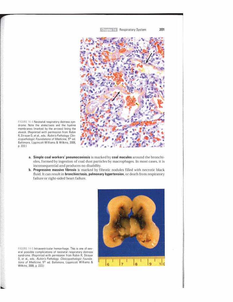
FIGURE 14-4 Neonatal respiratory distress syndrome. Note the atelectasis and the hya l ine membranes (marked by the arrows) l in ing the alveol i . (Reprinted with permission from Rubin R, Strayer D, et aI . , eds. : Rubin's Pathology Clinicopathologic Foundations of Medicine, 5th ed. Ba ltimore, Lippincott Wil l iams & Wilkins, 2008, p . 222.)
i!iltl1ttmI Respiratory System 201
a. Simple coal workers' pneumoconiosis is marked by coal macules around the bronchioles, formed by ingestion of coal dust particles by macrophages. In most cases, it is inconsequential and produces no disability.
b, Progressive massive fibrosis is marked by fibrotic nodules filled with necrotic black fluid. It can result in bronchiectasis. pulmonary hypertension. or death from respiratory failure or right-sided heart failure.
FIGURE 1 4-5 Intraventricu lar hemorrhage . This is one of several possible compl ications of neonatal respiratory distress syndrome. (Reprinted with permission from Rubin R, Strayer D, et aI., eds. : Rubin's PathologV. Clinicopathologic Foundations of Medicine, 5th ed. Ba ltimore, L ippincott Wil l iams & Wilkins, 2008, p. 223.)

202 BRS Pathology
3. Sil icosis is a chronic occupational lung disease caused by exposure to free si l ica dust; it is seen in miners, glass manufacturers, and stone cutters. a. This disease is initiated by ingestion of silica dust by alveolar macrophages; damage
to macrophages initiates an inflammatory response mediated by lysosomal enzymes and various chemical mediators.
b. Sil icotic nodules that enlarge and eventually obstruct the airways and blood vessels are characteristic.
c. Silicosis is associated with increased susceptibility to tuberculosis; the frequent concurrence is referred to as si l icotuberculosis.
4. Asbestosis is caused by inhalation of asbestos fibers. a. This disease is initiated by uptake of asbestos fibers by alveolar macro phages. A fibro
blastic response occurs, probably from release of fibro blast -stimulating growth factors by macrophages, and leads to diffuse interstitial fibrosis, mainly in the lower lobes.
� b. It is characterized by ferruginous bodies, yellow-brown, rod-shaped bodies with clubbed ends that stain positively with Prussian blue; these arise from iron and protein coating on fibers (Figure 14-6). Dense hyal inized fibrocalcific plaques of the parietal pleura are also present.
� c. Asbestosis results in marked predisposition to bronchogenic carcinoma and to malignant mesothelioma of the pleura or peritoneum. Cigarette smoking further increases the risk of bronchogenic carcinoma.
E. Restrictive lung diseases of unknown etiology 1. Sarcoidosis
� a. Characteristics include noncaseating granulomas, often involving multiple organ systems; can involve almost any organ system
� b. Occurrence is most frequent in persons of African l ineage. Sarcoidosis usually becomes clinically apparent during the teenage or young adult years.
c. Common pathologic changes ( 1 ) Interstitial lung disease (2) Enlarged hilar lymph nodes (3) Anterior uveitis (4) Erythema nodosum of the skin (5) Polyarthritis
d. Immunologic phenomena � (1 ) Reduced sensitivity and often anergy to skin test antigens (characteristically neg
ative result on a tuberculin test) (2) Polyclonal hyperglobulinemia
e. Clinical abnormalities. On routine chest radiography, sarcoidosis most often presents with:
� ( 1 ) Bilateral hilar lymphadenopathy � (2) Interstitial lung disease manifest as diffuse reticular densities
FIGURE 14·6 Ferruginous ( asbestos) bodies. These asbestos fiber jJ>1clusions a re coated with protein and iron and will appear b lue when sta ined with Prussian blue. (From Rubin R, Strayer D, et aI . , eds.: Rubin's Pathology. Clinicopathologic Foundations of Medicine, 5th ed. Ba ltimore, L ipp incott Wil l iams & Wilkins, 2008, p. 523. Courtesy of the Armed Forces Institute of Pathology.)

I!lltlIttttifJ Respiratory System 203
t a b I e 14-2 Se lected Examples of Interstitial Lung Disease
Disorder
Hypersensitivity pneumonitis (extrinsic a l lergic alveolitis)
Goodpasture syndrome
Idiopathic pulmonary hemosiderosis
Eosinophilic granuloma
Idiopathic pulmonary fibrosis
Sarcoidosis
1. Laboratory findings
Description
I nterstitial pneumonia caused by inhalation of various antigenic substances; exemplified by inhalation of spores of thermophilic actinomycetes from moldy hay causing "farmer's lung"
Hemorrhagic pneumonitis and glomerulonephritis caused by antibodies d irected •
against glomerular basement membranes
Resembles pulmonary component of Goodpasture syndrome without renal component
Proliferation of histiocytic cells related to Langerhans cells of the skin
Immune complex disease with progressive fibrosis of the alveolar wall
Granulomatous disorder of unknown etiology
� ( 1 ) Hypercalcemia and hypercalciuria (2) Hypergammaglobulinemia
� (3) Increased activity of serum angiotensin-converting enzyme g. Definitive diagnosis requires biopsy demonstrating noncaseating granulomas.
2. Id iopathic pulmonary fibrosis a. This disease is characterized by chronic inflammation and fibrosis of the alveolar wall.
It begins with alveolitis, progresses to fibrosis, and ends in a distorted fibrotic lung filled with cystic spaces (honeycomb lung) .
b, Death often results within 5 years.
F. Other i nterstitia l lung diseases (Table 1 4-2) 1. Eosinophil ic granuloma
a. Morphologic changes involve a localized proliferation of histiocytic cells closely related to the Langerhans cells of the skin. These cells have characteristic cytoplasmic inclusions (Birbeck granules) resembling tennis rackets. Other characteristics include prominent monocytes-macrophages, lymphocytes, and eosinophils.
b. The disease is found in the lung or in bony sites, such as the ribs. c. Eosinophilic granuloma is often grouped with Hand-Schiiller-Christian disease and
Letterer-Siwe syndrome as a variant of histiocytosis X syndrome. 2, Hypersensitivity pneumonitis (see Table 1 4-2)
V. PULM ONARY VASCULAR DISEASE
A, Pulmonary embolism 1. This is found in more than half of all autopsies. 2. Most often, pulmonary embolism originates from venous thrombosis in the lower extremities
or pelvis. Rarely, it can be due to nonthrombotic particulate material, such as fat, amniotic fluid, clumps of tumor cells or bone marrow, or foreign matter, such as bullet fragments.
3. Pulmonary embolism occurs in clinical settings marked by venous stasis, including primary venous disease, congestive heart failure, prolonged bed rest or immobilization, and prolonged sitting while traveling. Other predisposing factors include cancer, multiple fractures, and the use of oral contraceptives.
4. These emboli can result in hemorrhagic, or red, infarcts, usually in patients with compromised circulation, but embolism can occur without infarction because of the dual blood supply to the lungs.
5. Clinical consequences may vary and range from asymptomatic disease to sudden death.
B. Pulmonary hypertension 1. Primary pulmonary hypertension is a disorder of unknown etiology and poor prognosis that
arises in the absence of heart or lung disease.

204 BRS Pathology
2. Secondary pulmonary hypertension is more common than the primary form. Jii a. Most often, the cause is CO PO. Other causes may be increased pulmonary blood flow, as
in congenital left-to-right shunt; increased resistance within the pulmonary circulation, from embolism or vasoconstriction secondary to hypoxia; or increased blood viscosity from polycythemia.
b. This is a cause of right ventricular hypertrophy.
C. Pulmonary edema is intra-alveolar accumulation of fluid. It may be caused by: 1 . I ncreased hydrostatic pressure, as a result of left ventricular failure or mitral stenosis 2. Increased alveolar capil lary permeability, as in inflammatory alveolar reactions, resulting from
inhalation of irritant gases, pneumonia, shock, sepsis, pancreatitis, uremia, or drug overdose Jii 3. Miscellaneous mechanisms, such as rapid ascent to high altitude
VI. PULMONARY INFECTION
A. Pneumonia 1 . General considerations
a. Pneumonia is an inflammatory process of infectious origin affecting the pulmonary parenchyma.
Jii b. It is characterized by chil ls and fever, productive cough, blood-tinged or rusty sputum, pleuritic pain, hypoxia with shortness of breath, and sometimes cyanosis.
c. If bacterial, it is most characteristically associated with neutrophilic leukocytosis with an increase in band neutrophils ("shift-to-the-left") .
Jii 2. Morphologic types of pneumonia. There are three morphologic and clinical patterns: lobar pneumonia, bronchopneumonia, and interstitial pneumonia (Table 1 4-3) .
3. Bacterial pneumonias (Table 14-4) a. lobar pneumonia is most often caused by Streptococcus pneumoniae (the pneumococ
cus) . It is characterized by a predominantly i ntra-alveolar exudate and may involve an entire lobe of the lung.
b. Bronchopneumonia is caused by a wide variety of organisms. It is characterized by a patchy distribution involving one or more lobes, with an inflammatory infiltrate extending from the bronchioles into the adjacent alveoli.
4. I nterstitial (primary atypical) pneumonia is caused by various infectious agents, most commonly Mycoplasma pneumoniae or viruses. It is characterized by diffuse, patchy inflammation localized to interstitial areas of alveolar walls. a. Mycoplasma pneumonia Jii ( 1 ) This is the most common form of interstitial pneumonia; it usually occurs in children
and young adults, and it may occur in epidemics. (2) Onset is more insidious compared to bacterial pneumonia and usually follows a
mild, self-limited course.
t a b I e 14-3 Morphologic Variants of Pneumonia: Causative Organisms and Characteristics
Variant
Lobar pneumonia
Bronchopneumonia
Interstitial pneumonia
Causative Organism
Most frequently Streptococcus pneumoniae {pneumococcusl
Many organisms, including Staphylococcus aureus, Haemophi lus influenzae, Klebsiel la pneumoniae, and Streptococcus pyogenes
Most frequently viruses or Mycoplasma pneumoniae
Characteristics
Predominantly intra-alveolar exudate resulting in consolidation
May involve the entire lobe If untreated, may morphologically evolve through
four stages: congestion, red hepatization, gray hepatization, and resolution
Acute inflammatory infi ltrates extending from the bronchioles into the adjacent alveoli
Patchy distribution involving one or more lobes
Diffuse, patchy inflammation local ized to i nterstitial'areas of the alveolar walls
Distribution involving one or more lobes

t a b 181 14-4
Organism
Streptococcus pneumoniae
Staphylococcus aureus
Streptococcus pyogenes Klebsiella pneumoniae
Haemophilus influenzae
Legionella pneumophila
I!ii!ItttIitJi Respiratory System 205
Important Features of Selected Bacterial Pneumonias
Characteristics
Most common in elderly or debi l itated patients, especial ly those with cardiopulmonary disease, and malnourished persons
Often a compl ication of influenza or viral pneumonias or a result of blood-borne infection in intravenous drug users; seen principally in debi litated hospitalized patients, the elderly, and those with chronic lung disease
Often a complication of influenza or measles
Most frequent in deb ilitated hospitalized patients and diabetic or a lcoholic patients; h igh mortality rate in elderly patients
Usual ly seen in infants and chi ldren, but may occur in debi l itated adults, most often those with chronic obstructive pulmonary disease
I nfection from inhalation of aerosol from contaminated stored water, most often in air-conditioning systems
Complications
May lead to empyema (pus in the pleural cavity)
Focal inflammatory exudates or abscess formation frequent; may lead to empye
-ma
or to other infectious compl ications, including bacterial endocarditis and brain and kidney a bscesses
Lung abscess
Considerable alveolar wa l l damage, leading to necrosis, sometimes with abscess formation
Meningitis and epig lottitis i n infants and chi ldren
� (3) Characteristics include an inflammatory reaction confined to the interstitium, with no exudate in alveolar spaces, and intra-alveolar hyaline membranes.
(4) Diagnosis is by sputum cultures, requiring several weeks of incubation, and by complement-fixing antibodies.
� (5) Mycoplasma pneumonia may be associated with nonspecific cold agglutinins reactive to red cells. This phenomenon is the basis for a facile laboratory test that can provide early diagnostic information.
b. Vira l pneumonias are the most common types of pneumonia in childhood. They are caused most commonly by influenza viruses, adenoviruses, rhinovirus, and respiratory syncytial virus; may also arise after childhood exanthems, such as rubeola (measles) or varicella (chickenpox) ; the measles virus produces giant cell pneumonia, marked by numerous giant cells and often complicated by tracheobronchitis.
c. Rickettsial pneumonias: n fever is the most common rickettsial pneumonia; it is caused by Coxiella burnetii. It may infect persons working with infected cattle or sheep, who inhale dust particles containing the organism, or those who drink unpasteurized mi lk from infected animals.
d. Ornithosis (psittacosis) is caused by an organism of the genus Chlamydia, which is transmitted by inhalation of dried excreta of infected birds.
5. Pneumocystis jiroveci (carinii) pneumon ia is the most common opportunistic infection in patients with acquired immunodeficiency syndrome (AIDS); it also occurs in other forms of immunodeficiency. a. It is caused by P. carinii (recently renamed Pneumocystis jiroveci) , which is now clas
sified as a fungus. b. Diagnosis is by morphologic demonstration of the organism in biopsy or bronchial
washing specimens. 6. Hospital-acqu ired gram-negative pneumonias
a. These pneumonias are often fatal and occur in hospitalized patients, usually those with serious, debilitating diseases.
b. Causes include many gram-negative organisms, including Klebsiella, Pseudomonas aeruginosa, and Escherichia coli. Endotoxins produced by these organisms play an important role in the infection.
B. Lung abscess � 1. This is a local ized area of suppuration within the parenchyma, usually resulting from
bronchial obstruction (often by cancer) or from aspiration of gastric contents; may also be a complication of bacterial pneumonia.
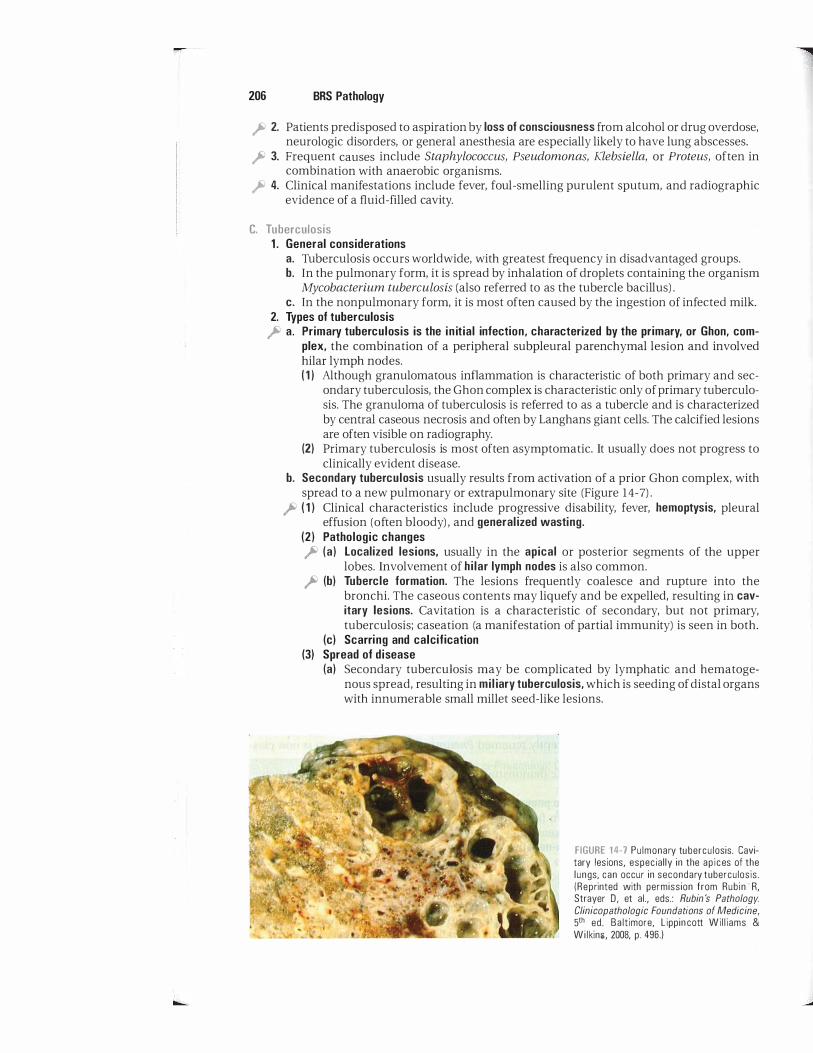
206 BRS Pathology
Ii' 2. Patients predisposed to aspiration by loss of consciousness from alcohol or drug overdose, neurologic disorders, or general anesthesia are especially likely to have lung abscesses.
Ii' 3. Frequent causes include Staphylococcus, Pseudomonas, Klebsiella, or Proteus, often in combination with anaerobic organisms.
Ii' 4. Clinical manifestations include fever, foul-smelling purulent sputum, and radiographic evidence of a fluid-filled cavity.
C. Tuberculosis 1 . General considerations
a. Tuberculosis occurs worldwide, with greatest frequency in disadvantaged groups. b. In the pulmonary form, it is spread by inhalation of droplets containing the organism
Mycobacterium tuberculosis (also referred to as the tubercle bacillus) . c. In the nonpulmonary form, it is most often caused by the ingestion of infected milk.
2. Types of tuberculosis Ii' a. Primary tuberculosis is the in itial infection, characterized by the primary, or Ghon, com
plex, the combination of a peripheral subpleural parenchymal lesion and involved hilar lymph nodes. ( 1 ) Although granulomatous inflammation is characteristic of both primary and sec
ondary tuberculosis, the Ghon complex is characteristic only of primary tuberculosis. The granuloma of tuberculosis is referred to as a tubercle and is characterized by central caseous necrosis and often by Langhans giant cells. The calcified lesions are often visible on radiography.
(2) Primary tuberculosis is most often asymptomatic. It usually does not progress to clinically evident disease.
b. Secondary tuberculosis usually results from activation of a prior Ghon complex, with spread to a new pulmonary or extrapulmonary site (Figure 14-7) .
Ii' ( 1 ) Clinical characteristics include progressive disability, fever, hemoptysis, pleural effusion (often bloody) , and general ized wasting.
(2) Pathologic changes Ii' (a) Localized lesions. usually in the apical or posterior segments of the upper
lobes. Involvement of hilar lymph nodes is also common. Ii' (b) Tubercle formation. The lesions frequently coalesce and rupture into the
bronchi. The caseous contents may liquefy and be expelled, resulting in cavitary lesions. Cavitation is a characteristic of secondary, but not primary, tuberculosis; caseation (a manifestation of partial immunity) is seen in both.
(c) Scarring and calcification (3) Spread of disease
(a) Secondary tuberculosis may be complicated by lymphatic and hematogenous spread, resulting in mil iary tuberculosis, which is seeding of distal organs with innumerable small millet seed-like lesions.
FIGURE 14-7 Pulmonary tuberculosis. Cavi· tary lesions, especial ly in the ap ices of the lungs, can occur in secondary tuberculosis. (Reprinted with permission from Rubin R, Strayer D, et aI., eds.: Rubin's Pathology. Clinicopathologic Foundations of Medicine, 5th ed. Ba ltimore, Lippin cott Wi l l iams & Wilkins, 2008, p. 496.)

i!itt!'ttIDD Respiratory System 207
(b) Hematogenous spread may also result in larger lesions, which may involve almost any organ.
� (c) Prominent examples of extrapulmonary tuberculosis include tuberculous meningitis, Pott disease of the spine, paravertebral abscess, or psoas abscess.
3. Immune mechanisms in pathogenesis of tuberculosis a. The organisms are ingested by macrophages, which process the bacterial antigens for
presentation to CD4 T H i T cells in the context of class II MHC molecules. b. The CD4+ T cells proliferate and secrete cytokines, attracting lymphocytes 'and
macro phages. c. The macrophages ingest and kill some of the tubercle bacilli or are morphologically
altered to form epithelioid cells and Langhans multinucleated giant cells. d. The causes of caseous necrosis remain obscure but most likely include the action of
cytokines elaborated by immunologically stimulated cells. e. Delayed hypersensitivity is marked by a positive tubercul in skin test result. The test
result is positive in both primary and secondary infection, represents hypersensitivity and relative immunity, and usually remains positive throughout life.
D. Mycobacterium avium-intracellulare i nfectio n is an infection with nontuberculous mycobacteria. 1 . This infection is seen most often in patients with AIDS and other immunodeficiency
diseases. 2. Often, nonpulmonary involvement is a manifestation.
E. Infections caused by fungi and fungus-l ike bacteria (Table 14-5) 1. These infections usually result from inhalation of the organism or from inoculation
through the skin. 2. In most instances, they are manifest as inflammatory reactions similar to tuberculosis.
t a b 1""'eJ 1 4-5 Characteristics of Pulmonary Infections Caused by Fungi and Fungus-like Bacteria
Disorder Organism
Actinomycosis Actinomvces, gram· positive anaerobic fi lamentous bacteria no longer classified as a fungus
Nocardiosis Nocardia, gram-positive aerobic, fi lamentous, weakly acid-fast bacteria closely related to Actinomvces
Candidiasis Candida albicans
Cryptococcosis Crvptococcus neoformans
Aspergillosis Aspergillus
Histoplasmosis Histoplasma capsulatum
Coccidioidomycosis Coccidioides immitis
Characteristics
Abscess and sinus tract formation Exudate containing characteristic sulfur granules,
yellow clumps of the organism
Typically opportunistic infection May disseminate to the brain and meninges
In immunocompromised patients, invasive form produces blood-borne dissemination
Pulmonary, renal, and hepatic abscesses and vegetative endocarditis
I nfection usually begins in the lungs but can also produce cryptococcal meningitis
Organ ism's characteristic encapsulated appearance visualized in India ink preparations
I nvasiv� form has predilection for growth into vessels, with consequent widespread hematogenous dissemination
Pulmonary manifestations similar to tuberculosis; occurs in primary and secondary forms
Results in multiple pulmonary lesions with late ca lc ification
Disseminated form, marked by multisystem involvement with infiltrates of macrophages filled with fungal yeast forms
Occurs in primary and disseminated forms Fungal spherules containing endospores found within
granulomas

-
208 BRS Pathology
VII . MISCELLANEOUS DISORDERS OF THE LUNGS
A. Atelectasis 1. Acquired atelectasis is alveolar col lapse caused by bronchial obstruction or external
compression of lung parenchyma by tumors or by pleural accumulation of fluid. 2. Atelectasis neonatorum is failure of alveolar spaces to expand adequately at birth; it
occurs in two forms. a. Primary atelectasis is failure of initial aeration of the lungs at birth; the alveoli remain
collapsed and respiration is never fully established. It is associated with prematurity and intrauterine fetal anoxia.
b. Secondary atelectasis is collapse of previously aerated bronchi.
B. Pulmonary alveolar proteinosis is uncommon and is characterized by accumulation of amorphous, periodic acid-Schiff-positive material in the alveolar air spaces. This material sometimes appears to be surfactant.
VI I I . CANCERS OF THE LUN G
A . General considerations. Most lung tumors are malignant; those that arise from metastases from primary tumors elsewhere occur more frequently than those that originate in the lung (Table 14-6) .
B. Bronchogenic carcinoma 1 . Etiology and epidemiology
a. Bronchogenic carcinoma is the leading cause of death from cancer in both men and women. It is increasing in incidence, especially in women, in parallel with cigarette smoking.
Type
Bronchogenic carcinoma
Squamous cell carcinoma
Adenocarcinoma
Bronchial-derived
Bronchioloalveolar
Smal l cel l (oat cel l ) carcinoma
Large cel l carcinoma
Other carcinomas of the l ung
Carcinoid
Tumors of the Lung
location
Central
Peripheral
Peripheral
Central
Peripheral
Major bronchi
Carcinoma metastatic to the lung
Characteristics
Appears as a h i lar mass a nd frequently results in cavitation; c learly linked to smoking; incidence greatly increased in smokers; may be marked by inappropriate parathyroid hormone (PTH)-like activity with resultant hyperca lcemia
Develops on site of prior pulmonary inflammation or injury (scar carcinoma); less clearly l inked to smoking
Less c learly related to smoking; columnar-to·cuboidal tumor cel ls l ine alveolar walls; multiple densities on x-ray, mimicking pneumonia
Undifferentiated tumor; most aggressive bronchogenic carcinoma; least l ikely form to be cured by surgery; usual ly a l ready metastatic at diagnosis; often associated with ectopic production of corticotrophin (ACTH) or antidiuretic hormone (ADH); incidence greatly increased in smokers
Undifferentiated tumor; may show features of squamous cel l or adenocarcinoma on electron microscopy
Low mal ignancy, spreading by direct extension into adjacent tissues; may result in carcinoid syndrome
Higher incidence than primary lung cancer

i!1lItf'il!DiLI Respiratory System 209
� h. This type of carcinoma is directly proportional in incidence to the number of cigarettes smoked daily and to the number of years of smoking. Various histologic changes, including squamous metaplasia of the respiratory epithelium, often with atypical changes ranging from dysplasia to carcinoma in situ, precede bronchogenic carcinoma in cigarette smokers.
2. Other etiopathogenic factors a. Air pollution
� h. Radiation; incidence increased in radium and uranium workers � c. Asbestos; increased incidence with asbestos and greater increase with combination
of asbestos and cigarette smoking � d. Industrial exposure to nickel and chromates 3. Clinical features
a. The 5-year survival rate is less than 10%. b. The tumor often spreads by local extension into the pleura, pericardium, or ribs. c. Clinical manifestations may include cough, hemoptysis, and bronchial obstruction,
often with atelectasis and pneumonitis. Other clinical features include: � ( 1 ) Superior vena cava syndrome; compression or invasion of the superior vena cava,
resulting in facial swelling and cyanosis along with dilation of the veins of the head, neck, and upper extremities
(2) Pancoast tumor (superior sulcus tumor); involvement of the apex of the lung, often with Horner syndrome (ptosis, miosis, and anhidrosis); due to involvement of the cervical sympathetic plexus
(3) Hoarseness from recurrent laryngeal nerve paralysis (4) Pleural effusion, often bloody; bloody pleural effusion suggests malignancy, tuber
culosis, or trauma. � (5) Paraneoplastic endocrine syndromes, the most frequent of which is adrenocorti
cotropic hormone (ACTH) or ACTH -like activity with small cell carcinoma; also of note are the syndrome of inappropriate antidiuretic hormone secretion (SIADH) with small cell carcinoma of the lung and parathyroid -like activity with squamous cell carcinoma.
4. Classification (Figures 14-8, 14-9, and 14- 1 0) � a. Bronchogenic carcinoma is subclassified into squamous cell carcinoma, adenocarci
noma (including bronchioloalveolar carcinoma), small cell carcinoma, and large cell carcinoma; it appears that all share a common endodermal origin despite their morphologic differences.
h. For therapeutic purposes, the bronchogenic carcinomas are often subclassified into small cell carcinoma, which is not considered amenable to surgery, and non-small cell carcinoma, in which surgical intervention may be considered.
FIGURE 14-8 Squamous ce l l carc inoma of the lung . (Reprinted with permission from Rubin R, Strayer D, et aI . , eds.: Rubin's Pathology. Clinicopathologic Foundations of Medicine, 5th ed . Ba ltimore, Lipp incott Wil l iams & Wilkins, 2008, p. 540.)

210 BRS Pathology
FIGURE 1 4-9 Adenocarc inoma of the lung . (Reprinted with permission from Rub in R, Strayer D, et aI . , eds.: Rubin's Pathology. Clinicopathologic Foundations of Medicine, 5th ed. B a ltimore, Lipp incott Wil l iams & Wilkins, 2008, p . 541 .)
FIGURE 14-10 Small cel l carcinoma of the lung . (Reprinted with permission from Rubin R, Strayer D, et aI., eds. : Rubin's Pathology. Clinicopathologic Foundations of Medicine, 5th ed. Ba ltimore, Lippincott Wil l iams & Wilkins, 2008, p. 542.)

r Review Test
Directions: Each of the numbered items or incomplete statements in this section is followeq by answers or by completions of the statement. Select the one lettered answer or completion that is best in each case.
1. A 3-year-old girl presents to the emergency department with fever, hoarseness, a "seal bark-like" cough, and inspiratory stridor. Her father states that she has had a cold for the past few days, with runny nose, nasal congestion, sore throat, and cough. He is now concerned because her cough has now become loud, harsh, and brassy. Which of the following is the most likely cause of her ailment?
(A) Fungus (8) Gram-negative bacteria (e) Gram-positive bacteria (D) Parasite (E) Virus
2. A 60-year-old man, a heavy smoker, presents for advice to stop smoking. On physical examination, he is thin and has a ruddy complexion. He has a productive cough and a barrel-shaped chest. He sits leaning forward with his lips pursed to facilitate his breathing. A diagnosis of emphysema is made. Which of the following is the most likely histologic finding in the lungs?
(A) Bronchial smooth muscle hypertrophy with proliferation of eosinophils
(8) Diffuse alveolar damage with leakage of protein-rich fluid into alveolar spaces
(e) Dilation of air spaces with destruction of alveolar walls
(D ) Hyperplasia of bronchial mucus-secreting submucosal glands
(E) Permanent bronchial dilation caus\d by chronic infection, with bronchi filled with mucus and neutrophils
3. A 60-year-old woman with a heavy smoking history presents with chronic productive cough that has been present for 3 consecutive months over the past 2 consecutive years. On physical examination, her skin has a bluish tinge, and she is overweight.
The patient is diagnosed with chronic bronchitis. Which of the follOwing is the most likely histologic finding in this patient's lungs?
(A) Bronchial smooth muscle hypertrophy with proliferation of eosinophils
(8) Diffuse alveolar damage with leakage of protein-rich fluid into alveolar spaces
(e) Dilation of air spaces with destruction of alveolar walls
(D) Hyperplasia of bronchial mucus-secreting submucosal glands
(E) Permanent bronchial dilation caused by chronic infection, with bronchi filled with mucus and neutrophils
4. A 65-year-old woman with a significant smoking history presents with cough and shortness of breath. Computed tomography of the chest reveals a central mass near the left mainstem bronchus. Biopsy of the mass is performed. Histologic examination reveals small round blue cells, and a diagnosis of small cell carcinoma is made. Which of the following is a frequent characteristic of this form of lung cancer?
(A) Generally amenable to surgical cure at time of diagnosis
(8) More common in women, and a less clear relation to smoking than other forms of lung cancer
(e) Secretes a parathyroid-like hormone (D) Secretes either corticotrophin or antidi
uretic hormone (E) Usually in a peripheral rather than in a
central location
5. A 23-year-old man presents with radiographic evidence of bilateral hilar lymphadenopathy and interstitial lung disease. A lung biopsy gives findings similar to those shown in the figure. A major characteristic of this disorder is
211

-
212 BRS Pathology
(A) a positive test for tuberculin. (B) highest incidence in persons of Asian
lineage. (e) hypercalcemia. (0) hypogammaglobulinemia. (E) involvement restricted to the lung.
6. An aO-year-old woman, a retirement home resident, has multiple bouts of pneumonia caused by Streptococcus pneumoniae. In an attempt to prevent such infections, polyvalent vaccines directed at multiple serotypes of the organism have been administered but have not elicited long-acting immunity. Which of the following is the probable explanation for this phenomenon?
(A) Memory T lymphocytes respond poorly to polysaccharide antigens.
(B) S. pneumoniae evades host immune response by forming capsular coatings composed of host proteins and recognized as "self" antigens.
(e) The bacterial capsule binds C3b, facilitating activation of the alternative complement pathway, inducing complement-mediated lysis, and preventing immunization.
(0) The capsular polysaccharides of S. pneumoniae have limited hapten potential.
(E) The surface carbohydrate capsule on the surface of the organism acts as an opsonin, facilitating phagocytosis by neutrophils and thus preventing immunization.
7. A 50-year-old man dies of a respiratory illness that had been characterized by dyspnea, cough, and wheezing expiration of
(Reprinted with permission from Ru bin R, Strayer D, et aI . , eds.: Rubin's Pathology. Clinicopathologic Foundations of Medicine, 5th ed. Ba ltimore, L ippincott Wil i iams & Wilkins, 2008, p. 527.)
many years' duration. Initially episodic, his "attacks" had increased in frequency and at the time of death had become continuous and intractable. At autopsy, which of the following is the most likely histologic finding in the lungs?
(A) Bronchial smooth muscle hypertrophy with proliferation of eosinophils
(B) Diffuse alveolar damage with leakage of protein-rich fluid into alveolar spaces
(e) Dilation of air spaces with destruction of alveolar walls
(0) Hyperplasia of bronchial mucus-secreting submucosal glands
(E) Permanent bronchial dilation caused by chronic infection, with bronchi filled with mucus and neutrophils
8. A 25-year-old man presents with a progressive illness of several days' duration characterized by nonproductive cough, fever, and malaise. A lateral view chest radiograph reveals platelike atelectasis. Elevated titers of cold agglutinins are detected. Which of the follOwing is the most likely type of pneumonia in this patient?
(A) Bacterial pneumonia, most likely caused by Streptococcus pneumoniae
(B) Hospital-acquired pneumonia, most likely caused by Pseudomonas aeruginosa
(e) Interstitial pneumonia, most likely caused by Mycoplasma pneumoniae
(0) Pneumocystisjiroveci (carinii) pneumonia, most likely related to an immunocompromised state
(E) Viral pneumonia, most likely caused by influenza virus

9. A 60-year-old man presents with fever and chills, productive cough with rusty sputum, pleuritic pain, and shortness of breath for the past several days. A complete blood count reveals neutrophilia and an increase in band neutrophils. A chest radiograph reveals consolidation involving the entire left lower lobe. Which of the following microorganisms is the most likely etiologic agent?
(A) Haemophilus injluenzae (8) Klebsiella pneumoniae (C) Staphylococcus aureus (0) Streptococcus pneumoniae ( E) Streptococcus pyogenes
10. A 46-year-old woman presents with fever, hemoptysis, weight loss, and night sweats. She has never smoked. She recently returned from a month-long trip to Asia. A chest radiograph reveals apical lesions with cavitation in the left lung. A purified protein derivative (PPD) test is placed, and 48 hours later an 1 8-mm wheal develops. Sputum cultures reveal numerous acid-fast organisms. This patient is put on contact precautions, and a regimen for tuberculosis is started. Which of the following disorders does this patient most likely have?
(A) Acquired immunodeficiency syndrome (8) Congenital immunodeficiency (C) Miliary tuberculosis, with seeding of
distal organs with innumerable small millet seed-like lesions
(0) Primary tuberculosis, characterized by the Ghon complex
(E) Secondary tuberculosis, resulting from activation of a prior Ghon complex, with spread to a new pulmonary site
1 1 . A 25-year-old African-American woman presents with fatigue, dyspnea, nonproductive cough, and chest pain. She does not smoke. A chest radiograph reveals prominent bilateral hilar lymphadenopathy ("potato nodules") and diffuse reticular densities in the interstitium of the lung. Laboratory studies reveal polyclonal hypergammaglobulinemia, hypercalcemia, and increased serum angiotensin-converting enzyme. Which of the following is the most likely diagnosis?
(A) Acute respiratory distress syndrome (8) Adenocarcinoma of the lung
iB1tftttiiU Respiratory System 213
(C) Eosinophilic granuloma (0) Idiopathic pulmonary fibrosis (E) Sarcoidosis
12. A 60-year-old man presents with dyspnea on exertion and a nonproductive cough. He has never smoked, but he worked as a . shipbuilder, with known asbestos exposure approximately 20 years ago. To which of the following conditions is this patient especially predisposed?
(A) Acute respiratory distress syndrome (8) Goodpasture syndrome (C) Idiopathic pulmonary fibrosis (0) Idiopathic pulmonary hemosiderosis (E) Malignant mesothelioma of the pleura
13. A female infant is born prematurely at 28 weeks' gestation. Shortly after birth, she shows signs of dyspnea, cyanosis, and tachypnea. She is placed on a ventilator for assisted breathing, and a diagnosis of neonatal respiratory distress syndrome (hyaline membrane disease) is made. Which of the following is the cause of this syndrome?
(A) Bronchopulmonary dysplasia (8) Intraventricular brain hemorrhage (C) Lack of fetal pulmonary maturity and
deficiency of surfactant (0) Necrotizing enterocolitis (E) Patent ductus arteriosus
1 4. A 50-year-old woman has been immobilized in bed for several days after a motor vehicle accident. She had been improviIlg, but this morning she suffered the sudden onset of pleuritic chest pain, hemoptysis, tachypnea, tachycardia, and dyspnea. What is the likely basis of this set of findings?
(A) Arterial thrombus originating in pulmonary blood vessels
(8) Arterial thrombus originating in the lower extremities with migration to pulmonary veins
(C) Deep venous thrombus of the lower extremities with embolization to branches of the pulmonary artery
(0) Mural thrombus originating in the left heart with migration to pulmonary blood vessels
(E) Venous thrombus originating in pulmonary blood vessels

p
214 BRS Pathology
15. The chest radiograph of a 23-year-old medical student reveals a calcified cavitary pulmonary lesion. The tuberculin test is positive, but sputum smears and cultures are negative for Mycobacterium tuberculosis. A presumptive diagnosis of secondary tuberculosis is made. If further studies, including a biopsy, were performed, which of the
following findings would justify the diagnosis of secondary tuberculosis, as contrasted to primary tuberculosis?
(A) Calcification (B ) Caseating granulomas (e) Cavitation (D ) Langhans giant cells (E ) Positive tuberculin test result

Answers and Explanations
1. The answer is E. This is a classic case of acute laryngotracheobronchitis (croup) , an acute inflammation of the larynx, trachea, and epiglottis. The most common cause of croup is a viral (parainfluenza virus type I) infection.
2. The answer is C. Emphysema is an example of a chronic obstructive pulmonary disease (COPD) . Due to the destruction of alveolar walls, a lack of elastic recoil causes air to become trapped in alveoli, and, thus, airflow obstruction occurs on expiration. In COPD, the forced expiratory volume in one second (FEVl) is decreased, whereas the forced vital capacity (FVC) is normal or increased; therefore, patients with COPD have a decreased FEVl :FVC ratio.
3. The answer is D. Chronic bronchitis is an example of a chronic obstructive pulmonary disease. The pathologic hallmark of chronic bronchitis is marked hyperplasia of bronchial submucosal glands and bronchial smooth muscle hypertrophy, which can be quantified by the Reid index, a ratio of glandular layer thickness to bronchial wall thickness.
4. The answer is D. Small cell carcinoma of the lung is the most aggressive type of bronchogenic carcinoma. The location of this cancer is usually central. This is an undifferentiated tumor with small round blue cells and is least likely to be cured by surgery because it is usually already metastatic at diagnosis. Associated paraneoplastic syndromes include secretion of adrenocorticotropic hormone and antidiuretic hormone.
5. The answer is C. The illustration shows noncaseating granulomas and giant cells, which, in the clinical setting described, are diagnostic of sarcoidosis. A frequent abnormal laboratory finding is polyclonal hypergammaglobulinemia along with hypercalcemia. Anergy to tuberculin is often demonstrable. The disorder is much more common in persons of African lineage. Patients most often present with lung findings and hilar lymphadenopathy, but any organ system can be involved .
6 . The answer is A . Antibody responses to the more than 80 differing carbohydrate capsular antigens of the various strains of Streptococcus pneumoniae are generally T-cell-independent, and antibody formation is entirely B-cell-mediated. Because of this, memory cells are not formed, and long-lasting immunity is not achieved.
7. The answer is A. Bronchial asthma, or hyperreactive airway disease, is a type of chronic obstructive pulmonary disease caused by narrowing of airways. Asthma is manifest morphologically by bronchial smooth muscle hypertrophy, hyperplasia of bronchial submucosal glands and goblet cells, and airways plugged by mucus-containing Curschmann spirals (whorl-like accumulations of epithelial cells), eosinophils, and Charcot-Leyden crystals (crystalloids of eosinophil-derived proteins) .
8. The answer is C. Interstitial (primary atypical) pneumonia is most commonly caused by Mycoplasma pneumoniae or viruses. Interstitial pneumonia is characterized by diffuse, patchy inflammation localized to the interstitial areas of alveolar walls, with no exudate in alveolar spaces, and intra-alveolar hyaline membranes. M. pneumoniae infection is associated with the presence of cold agglutinins, which are IgM antibodies that react nonspecifically with all human red blood cells. Pneumocystis jiroveci (carinii) pneumonia is the most common opportunistic infection in patients with acquired immunodeficiency syndrome or other immunodeficiency disorders. Viral pneumonias are the most common type of pneumonia in childhood, caused most commonly by the influenza virus.
215

21 6 BRS Pathology
9. The answer is D. The most common cause of lobar pneumonia is Streptococcus pneumoniae. The organism is also known as the pneumococcus, and the disease entity is often referred to as pneumococcal pneumonia.
10. The answer is E. Tuberculosis, at one time a frequent hazard in the United States, is now relatively uncommon except in immunocompromised individuals and persons returning from parts of the world where the disease remains a common problem. Primary tuberculosis is the initial infection by Mycobacterium tuberculosis, and is restricted to the primary, or Ghon, complex, a combination of a peripheral subpleural parenchymal lesion and involved hilar lymph nodes. Cavitation and selective localization to the pulmonary apices are characteristics of secondary tuberculosis. Secondary tuberculosis may spread through the lymphatics and blood to other organs, resulting in miliary tuberculosis.
1 1 . The answer is E. Sarcoidosis most often presents as a restrictive pulmonary disease that is characterized morphologically by noncaseating granulomas and can involve any organ system. Diagnostic features of note include highest incidence in persons of African lineage, somewhat higher incidence in women, bilateral interstitial pulmonary involvement, prominent hilar lymphadenopathy, polyclonal hypergammaglobulinemia, and hypercalcemia. Increased serum angiotensin-converting enzyme activity is a nonspecific indicator of granulomatous inflammation.
12. The answer is E. Asbestosis is caused by inhalation of asbestos fibers, characterized by yellow-brown, rod-shaped ferruginous bodies with clubbed ends that stain positively with Prussian blue. Asbestosis results in a marked predisposition to malignant mesothelioma of the pleura or peritoneum. Exposure to asbestos is also a risk factor for bronchogenic carcinoma, as well as for carcinoma of the oropharynx, esophagus, and colon. The risk of bronchogenic carcinoma is greatly increased in cigarette smokers with exposure to asbestos.
13. The answer is C. Neonatal respiratory distress syndrome (hyaline membrane disease) is the most common cause of respiratory failure in newborns and results from a deficiency of surfactant and immature development of the lungs. Surfactant reduces surface tension within the lung, facilitating expansion by inspiration and thus preventing atelectasis during expiration. An indicator of fetal pulmonary maturity is a lecithin:sphingomyelin ratio of approximately 2:1 in the amniotic fluid. Predisposing factors include prematurity, maternal diabetes mellitus, and birth by cesarean section. Known complications of this condition include bronchopulmonary dysplasia, patent ductus arteriosus, intraventricular brain hemorrhage, and necrotizing enterocolitis.
1 4. The answer is C. Pulmonary embolism most often originates from venous thrombosis in the lower extremities or pelvis. An embolus migrates through the venous circulation to the right heart and gets trapped in branches of the pulmonary artery. Pulmonary embolism occurs in clinical settings of venous stasis, such as primary venous disease, congestive heart failure, prolonged bed rest or immobilization, or prolonged sitting while traveling.
15. The answer is C. Cavitation occurs only in secondary tuberculosis. Both primary and secondary tuberculosis are characterized by caseating granulomas, often with Langhans giant cells, which heal by scarring and calcification. The skin test result for tuberculin sensitivity is positive in both forms.

� -----------------
Gastrointestinal Tract
I --'
I. DISEASES OF THE MOUTH AND JAW
A. Inflammatory disorders 1. Herpes labialis (fever blisters, cold sores) is a common vesicular lesion caused by herpes
simplex virus (HSV), most often by HSV type 1 (HSV- l ) . It tends to recur, with activation by febrile illness, trauma, sunshine, or menstruation.
2. Aphthous stomatitis is characterized by painful, recurrent, erosive oral ulcerations. 3. Oral candidiasis (thrush, moniliasis) is a local white, membranous lesion caused by Candida
albicans. It occurs most commonly in debilitated infants and children, immunocompromised patients, and individuals with diabetes.
4. Acute necrotizing u lcerative gingivitis (trench mouth, Vincent infection, fusospirochetosis) a. This severe gingival inflammation occurs in patients with decreased resistance to
infection. b. The cause is concurrent infection with symbiotic bacteria, most often Fusobacterium
species and Borrelia vincentii.
B. Tumors and tumor- l ike conditions 1. Benign tumors of the oral mucosa
a. Papi l loma is the most common benign epithelial tumor of the oral mucosa. It can occur anywhere in the mouth; the most common sites are the tOl).gue, lips, gingivae, or buccal mucosa.
'
b. Fibroma is most often a non-neoplastic hyperplastic lesion resulting from chronic irritation.
c. Hemangioma occurs most commonly on the tongue, lips, or buccal mucosa. d. Epulis refers to any benign (usually non-neoplastic) growth of the gingivae. It is most
often a reparative growth rather than a true neoplasm. 2. Leukoplakia is a clinical term describing irregular white mucosal patches.
a. These patches result from hyperkeratosis, usually secondary to chronic irritation. b. Leukoplakia is usually benign but may represent dysplasia or carcinoma in situ.
3. Odontogenic tumors a. Odontoma is the most common odontogenic tumor. It is a hamartoma derived from
odontogenic epithelium and odontoblastic tissue. b. Ameloblastoma (adamantinoma) is an epithel ial tumor arising from precursor cells of
the enamel organ. (1 ) Most frequently, this tumor occurs in the mandible. It usually appears in individ
uals younger than 35 years of age. (2) Although this tumor is benign, it can lead to slow expansion of the jaw because of
irregular local extension.
217

218 BRS Pathology
4. Oral cancer is most frequently squamous cell carcinoma.
� a. Involvement of the tongue occurs in more than 50% of cases. Carcinoma of the mouth, tongue, and esophagus is often associated with the combined abuse of tobacco and alcohol.
b. Oral cancer may be associated with irritants such as pipe smoking, chewing tobacco, or betel nuts.
II. DISEASES OF THE SALIVARY GLANDS
A. Sialadenitis. This inflammation of the salivary glands may be caused by infection, immunemediated mechanisms, or occlusion of the salivary ducts by stones (sialolithiasis).
B. Acute parotitis. This condition occurs in mumps, but may also be caused by other infectious agents.
C. Sjogren syndrome. This condition is most likely of autoimmune origin.
� 1. Characteristics include keratoconjunctivitis sicca (dry eyes), xerostomia (dry mouth), and an associated connective tissue disease, most often rheumatoid arthritis.
2. Sjogren syndrome is associated with an increased incidence of malignant lymphoma.
D. Mucocele. This cyst-like pool of mucus, lined by granulation tissue, develops near a minor salivary gland. It results from mucous leakage caused by rupture of obstructed or traumatized ducts.
E. Ranula. This is a large mucocele, of salivary gland origin, characteristically localized to the floor of the mouth.
F. Tumors of the salivary gland (Table 15-1). The majority of salivary gland tumors occur in the parotid gland. The majority of tumors of the parotid gland are benign; in contrast, about half of the tumors of the submaxillary gland are malignant.
� 1. Pleomorphic adenoma (mixed tumor) is the most frequently occurring salivary gland tumor. It occurs with greatest frequency in women between 20 and 40 years of age.
t ab I e 15-1 Saliva ry Gland Tumors: Location, Histology, and Characte ristics
Type Typical location Histology
Pleomorphic adenoma Parotid gland; can occur Variable mix of epithelial and (mixed tumor) in submandibular or mesenchyme-like elements
minor salivary glands
Papillary cystadenoma Iymphomatosum (Warthin tumor, adenolymphoma)
Mucoepidermoid tumor
Adenoid cystic carcinoma
Oncocytoma
Parotid gland
Parotid gland
Minor salivary glands
Parotid gland
Cystic spaces lined by doublelayered eosinophilic epithelium, all embedded in lymphoid stroma
Comprised of mucus-producing and epidermoid components and cells intermediate between the two
Variable; most characteristic appearance consists of cribriform pattern with masses of small, dark-staining cells arrayed around cystic spaces
Large, granular-appearing, eosinophilic-staining epitheliaL cells
Characteristics
Most common salivary gland tumor; benign; tends to recur after resection; malignant transformation occurs but is rare
Benign
Behavior varies from benign to highly malignant; tumors with a greater number of epidermoid cells and nonparotid tumors tend to be more aggressive
Tends to infiltrate perineural spaces and cause pain; slowgrowing malignancy with late metastasis
Benign; peak occurrence in the elderly

I!1lftt'd!Dl:i Gastr o intestinal Tract 219
a. This is a benign tumor that frequently recurs; it rarely becomes malignant. b. It has been called "mixed tumor" because of the presence of myxoid and cartilage-like
elements. as well as epithelial cells. c. Histologically, pleomorphic adenomas vary, but most often they demonstrate irregu
lar masses or anastomosing strands of stellate or fusiform epithelial cells, some forming ducts or tubules, all of which are embedded in a myxoid stroma that may display fibrous, cartilage-like, or hyalinized areas.
� d. The tumor is most often localized to the parotid gland (-90%). � e. Usually, the tumor presents as a firm, nontender swelling.
� f. Often, the tumor is difficult to remove completely because of its proximity to the facial nerve, and it is likely to recur after resection.
2. Other salivary gland tumors a. Papillary cystadenoma Iymphomatosum (Warthin tumor) b. Mucoepidermoid tumor c. Adenoid cystic carcinoma d. Oncocytoma
III. DISEASES OF THE ESOPHAGUS
A. Tracheoesophageal fistula. This congenital disorder is suggested in a newborn by copious salivation associated with choking, coughing, and cyanosis on attempts at food intake. It occurs in three distinct variants: 1. In the most common variant (90%), the lower portion of the esophagus communicates
with the trachea near the tracheal bifurcation. The upper esophagus ends in a blind pouch (esophageal atresia). Maternal polyhydramnios (increased amniotic fluid) is a frequently associated abnormality.
2. The second most common variant is characterized by a fistulous connection between the upper esophagus and the trachea; the lower esophageal segment is not connected to the upper esophagus.
3. In a third variant, there is a fistulous connection between the trachea and a completely patent esophagus.
B. Esophageal diverticula are pouches lined by one or more layers of the esophageal wall. 1. Most commonly, false (pulsion ) diverticula result from herniation of the mucosa through
defects in the muscular layer. 2. Less commonly, true (traction ) diverticula consist of mucosal, muscular, and serosal lay
ers. Traction diverticula result from periesophageal inflammation and scarring. 3. Esophageal diverticula occur in three characteristic locations:
a. Immediately above the upper esophageal sphincter (Zenker diverticulum) b. Near the midpoint of the esophagus c. Immediately above the lower esophageal sphincter (epiphrenic diverticulum)
C. Achalasia is persistent contraction of the lower esophageal sphincter and absence of esophageal peristalsis, leading to dilation of the esophagus.
� 1. The condition is caused by a loss of ganglion cells in the myenteric plexus, which leads to the progressive dilation of the esophagus. One important source (principally in South America) is Trypanosoma cruzi infection in Chagas disease. In other cases, ganglion cells are lost for reasons that are not known.
2. Clinical characteristics include difficulty in swallowing. 3. Achalasia can lead to esophageal squamous cell carcinoma in about 5% of subjects.
D. Esophageal varices. These dilated submucosal esophageal veins that occur secondary to portal hypertension can result in upper gastrointestinal hemorrhage. (The other important causes of upper gastrointestinal hemorrhage are bleeding peptic ulcer and the MalloryWeiss syndrome, bleeding from esophagogastric laceration as a result of severe retching.)

L
220 BRS Pathology
E. Inflammatory and related disorders of the esophagus � 1. Gastroesophageal reflux is reflux of gastric acid contents into the esophagus.
a. Characteristics usually include burning pain relieved by antacids. Manifestations often include substernal pain (heartburn).
b. Most commonly, associated conditions include hiatal hernia and incompetent lower esophageal sphincter. Gastroesophageal reflux is also associated with excessive use of alcohol and tobacco and with increased gastric volume, pregnancy, or scleroderma.
c. Assuming a recumbent position often precipitates gastroesophageal reflux. d. Reflux may cause esophagitis, stricture, ulceration, or columnar metaplasia of
esophageal squamous epithelium (Barrett esophagus). � 2. Barrett esophagus is columnar metaplasia of esophageal squamous epithelium; the colum
nar epithelium is often of the intestinal (specialized) type with prominent goblet cells. This condition is a complication of long-standing gastroesophageal reflux and is a wellknown precursor of esophageal adenocarcinoma.
3. Candida esophagitis (moniliasis) a. Associated conditions often include antibiotic use, diabetes mellitus, malignant dis
ease, or immunodeficiency caused by acquired immunodeficiency syndrome or immunosuppressive drugs.
b. Clinical manifestations are white adherent mucosal patches and painful, difficult swallowing.
4. Herpetic esophagitis is caused by HSV-l infection. a. HSV-l infection tends to occur in immunosuppressed persons. b. Characteristics include painful, difficult swallowing.
5. Less common forms of esophagitis are caused by cytomegalovirus (CMV) infection, uremia, radiation therapy, or graft-versus-host (GVH) disease.
6. Esophageal stricture most often results from prolonged esophageal gastric acid reflux but may also be caused by suicidal or accidental ingestion of corrosive acids or lye. It is marked by progressive dysphagia.
F. Carcinoma of the esophagus � 1. This aggressive tumor is manifest clinically by dysphagia, weight loss, and anorexia.
Occasionally, pain or hematemesis occur.
� 2. In the United States, the incidence of squamous cell carcinoma and adenocarcinoma is about equal, which differs from the worldwide distribution, in which squamous cell carcinoma is much more frequent. In the United States, the incidence of squamous cell carcinoma is decreasing, and this is thought to be due to a parallel decrease in the use of tobacco and perhaps alcohol. Adenocarcinoma arises most often in aberrant gastric mucosa or submucosal glands or in the metaplastic columnar epithelium of Barrett esophagus.
� 3. Squamous cell carcinoma arises most frequently in the upper and middle thirds of the esophagus.
� 4. Adenocarcinoma arises most frequently in the lower third of the esophagus. 5. Pathologic manifestations may include protrusion into the esophageal lumen, with
spread by local extension into adjacent structures such as the trachea, bronchi, or aorta, or diffuse infiltration into the esophageal wall.
IV. DISEASES OF THE STOMACH
A. Congenital pyloric stenosis 1. This stenosis is caused by hypertrophy of the circular muscular layer of the pylorus,
often leading to a palpable mass. � 2. The reSUlting obstruction of the gastric outlet causes episodes of projectile vomiting
beginning in the first 2 weeks of life. This condition is much more common in boys. 3. The condition is corrected by surgical incision of the hypertrophied muscle.

B. Gastritis 1. Acute (erosive) gastritis
a. Causes
IB1tf'i1ltIil:i Gastrointestinal Tract 221
(1) Nonsteroidal anti-inflammatory drugs (NSAIDs) (2) Cigarette smoking (3) Heavy alcohol intake (4) Burn injury; Curling ulcer, an acute gastric ulcer in association with severe burns (5) Brain injury; Cushing ulcer, an acute gastric ulcer in association with brain injury
b. Characteristics (1) Focal damage to the gastric mucosa, with acute inflammation, necrosis, and
hemorrhage (2) Manifested as acute gastric ulcers, which are often multiple
2. Chronic gastritis is characterized by chronic mucosal inflammation and atrophy of the mucosal glands.
� a. Autoimmune gastritis is associated with the presence of antibodies to parietal cells (and sometimes to intrinsic factor), achlorhydria (lack of gastric acid secretion), pernicious anemia, and autoimmune diseases, such as chronic thyroiditis and Addison disease. It is also associated with aging, partial gastrectomy, gastric ulcer, and gastric carcinoma.
� b. Helicobacter pylori-associated gastritis is the most common form of chronic gastritis. (1) There is no association with pernicious anemia, antibodies to parietal cells, or
reduced gastric acid secretion. (2) Often, increased gastric acid secretion occurs. H. pylori is also strongly associated
with gastric and duodenal peptic ulcer and is a high suspect in the causality of carcinoma of the stomach and gastric lymphoma of the mucosa-associated lymphoid tissue (MALT) type.
3. Menetrier disease (giant hypertrophic gastritis) is characterized by extreme enlargement of gastric rugae and sometimes by severe loss of plasma proteins from the altered mucosa. Affected patients have an increased risk of stomach cancer.
C. Peptic ulcer of the stomach 1. Most often, the stomach ulcer occurs at or near the lesser curvature, in the antral and
prepyloric regions. 2. The ulcer is not a precursor lesion of carcinoma of the stomach. 3. Unlike peptic ulcer that occurs elsewhere, peptic ulcer of the stomach is not depen
dent on increased gastric acid secretion; however, acid and pepsin are believed to play a role, because gastric peptic ulcers rarely occur in association with absolute achlorhydria.
4. Postulated etiopathogenic mechanisms of gastric peptic ulcer production include: a. H. pylori-mediated processes, in which bacterial ureases and proteases break down
glycoproteins in gastric mucus, thus interfering with epithelial protection b. Increased permeability of the gastric mucosa to hydrogen ion, resulting in back diffu
sion of hydrogen ion with injury to the gastric mucosa c. Bile-induced gastritis leading to gastric ulceration
D. Malignant tumors of the stomach (Figure 15- 1) 1. Carcinoma of the stomach
a. General considerations (1) Carcinoma of the stomach is most common after 50 years of age, with an increased
incidence in men. It occurs more frequently in persons with blood group A, suggesting a genetic predisposition.
(2) Incidence varies greatly from one geographic area to another, with incidence much higher in Japan, Finland, and Iceland.
(3) The incidence is decreasing in the United States. b. Etiologic factors � ( 1) H. pylori is a high suspect.

222 BRS Pathology
FIGURE 15-1 Malignant gastric ulcer. This ulcerating adenocarcinoma of the stomach has no relationship to its benign counterpart, peptic ulcer of the stomach; however, clinical distinction is obviously crucial. Rolled elevated edges in the cancer are suggestive signs, but differentiation by biopsy is essential. (Reprinted with permission from Rubin R, Strayer D, et aI., eds.: Rubin's Pathology. Clinicopathologic Foundations of Medicine, 5th ed. Baltimore, Lippincott Williams & Wilkins, 2008, p. 570.)
(2) Nitrosamines from dietary amines and nitrites used as food preservatives may play a role. Incidence of the disease is greatly increased in populations who eat large amounts of smoked fish and meat and pickled vegetables.
(3) Increased incidence is also associated with excessive salt intake and a diet low in fresh fruits and vegetables.
(4) Stomach carcinoma is also predisposed by: (a) Achlorhydria (b) Chronic gastritis with or without pernicious anemia
c. Characteristics � (1) Histologically, stomach carcinoma is almost always adenocarcinoma.
(2) Involvement of the distal stomach, along the lesser curvature of the antrum or prepyloric region, is most common; rarely involves the fundus.
� (3) Aggressive spread to adjacent organs and the peritoneum and early lymphatic metastasis to regional lymph nodes and the liver occurs.
(4) More distal sites may be involved. (a) Involvement of a supraclavicular lymph node by metastatic carcinoma of the
stomach is referred to as a Virchow node. (b) Bilateral involvement of the ovaries by metastatic carcinoma of the stomach
is referred to as Krukenberg tumors. The tumor cells often contain abundant mucin, displacing the nucleus to one side and resulting in so-called signetring cells.
d. Morphologic variants of stomach carcinoma (1) Intestinal type
(a) Often, this variant is manifest as polypoid (fungating) carcinoma, which forms a solid mass projecting into the lumen of the stomach. It has a high degree of association with H. pylori infection.
(b) The intestinal variant can become ulcerated and must be differentiated from peptic ulcer. Peptic ulcer usually exhibits a smooth base with nonelevated, punched-out margins. In contrast, carcinoma tends to form an ulcer with an irregular necrotic base and firm, raised margins.
(2) Infiltrating or diffuse carcinoma (linitis plastica, leather-bottle stomach) is not associated with H. pylori infection and is characterized by a thickened, rigid stomach wall, caused by diffuse infiltration of tumor cells with accompanying extensive fibrosis (Figure 15-2) .
2. lymphoma accounts for 4% of malignant gastric tumors. They are of the MALT type, and there is a high association with H. pylori infection. The prognosis is better than it is for adenocarcinoma.

fiGURE 15-2 Infiltrating carcinoma (linitis plastical. The stomach with stiff rigid walls caused by infiltrating tumor cells and extensive fibrosis has been referred to as a "leather-bottle stomach." (Reprinted with permission from Rubin R, Strayer 0, et ai., eds.: Rubin's Pathology Clinicopathologic Foundations of Medicine, 5th ed. Ba ltimore, Lippincott Williams & Wilkins, 2008, p. 570.)
i!1lfJ!1!§id Gastrointestinal Tract 223
V. DISEASES OF THE SMALL INTESTINE
A. Peptic ulcer 1. Occurrence is most frequent in the first portion of the duodenum, the stomach, or the
lower end of the esophagus, all of which are exposed to acid and pepsin. 2. Except for peptic ulcer of the stomach, peptic ulcer is always associated with hypersecre
tion of gastric acid and pepsin. Ulceration is closely related to gastric H. pylori infection, which affects essentially all patients with duodenal ulcer and the majority of patients with gastric ulcer. H. pylori increases gastric acid secretion and apparently impairs both gastric and duodenal mucosal defenses.
3. Frequency of occurrence is increased in persons of blood group 0, suggesting that genetic factors may play a role.
4. Peptic ulcer is not a precursor of malignancy. 5. Complications often include hemorrhage with melena (black stools containing blood).
Other important complications include obstruction and perforation. Peptic ulcer is sometimes associated with: a. Intake of aspirin or other NSAIDs. The ulcerogenic effect of these drugs may be medi
ated by inhibition of prostaglandin synthesis. b. Smoking. The incidence of peptic ulcer is two-fold greater in smokers. c. Zollinger-Ellison syndrome, increased tendency toward peptic ulcer formation, which
is caused by gastric acid hypersecretion due to gastrin-secreting islet cell tumor of the pancreas. Recurrent peptic ulcer or peptic ulcer in aberrant sites, such as the jejunum is suggestive of the Zollinger-Ellison syndrome.
d. Primary hyperparathyroidism e. Multiple endocrine neoplasia (MEN) type I (Wermer syndrome), an autosomal domi
nant syndrome characterized by pituitary, thyroid, parathyroid, adrenal cortical, and pancreatic islet cell adenomas or hyperplasias associated with hypergastrinemia and peptic ulcer
B. Crohn disease (Table 15-2) 1. General considerations
� a. This chronic inflammatory condition of unknown etiology may affect any part of the gastrointestinal tract but most commonly involves the distal ileocecum, small intestine, or colon.
b. Crohn disease tends to affect young people in the second and third decades of life, although no age group is exempt. It occurs most frequently in people of Jewish descent.
� c. The disease can lead to carcinoma involving the small intestine or colon. However, neoplastic transformation is much less frequent in Crohn disease than in ulcerative colitis.
2. Morphology a. Chronic inflammation involving all layers of the intestinal wall (transmural
involvement)

F
..
224 BRS Pathology
----------- - -------------
t a b I e 15-2 Compa r is on of C r ohn D is eas e and Ulc e rativ e C olit is ----------- --- --
Crohn Disease
May involve any portion of the gastrointestinal tract, usually the ileocecal region, small intestine, or colon
Chronic inflammatory reaction extends through the entire thickness of the intestinal wall
Lymphocytic infiltrate; noncaseating granulomas; fibrosis; thickening of intestinal wall with narrowing of lumen; fistulous tracts between loops of intestine or between the intestine and other sites; mucosal cobblestone appearance; skip lesions
Incidence of secondary malignancy much lower than in ulcerative colitis
Ulcerative Colitis
Affects only colon
Inflammation and ulceration limited to mucosa and submucosa
Crypt abscesses, pseudopolyps
Greatly increased incidence of colon cancer in long-standing cases
b. Thickening of involved segments, with narrowing of lumen c. Linear ulceration of the mucosa d. Submucosal edema with elevation of the surviving mucosa, producing a cobblestone
appearance (Figure 15-3) � e. Skip lesions (segments of normal intestine between affected regions)
f. Discrete noncaseating granulomas in some cases g. Submucosal fibrosis
3. Clinical manifestations a. Abdominal pain and diarrhea b. Malabsorption c. Fever d. Intestinal obstruction resulting from fibrous stricture
� e. Fistulas between loops of intestine and between the intestine, bladder, vagina, and skin
C. Meckel diverticulum is the most common congenital anomaly of the small intestine. 1. Meckel diverticulum is a remnant of the embryonic vitelline duct and is located in the
distal small bowel. It may contain ectopic gastric, duodenal, colonic, or pancreatic tissue.
� 2. The condition is usually asymptomatic but complications, including peptic ulceration in ectopic gastric mucosa with bleeding or perforation, may occur. Occasional associations include:
� a. Intussusception (invagination of a proximal segment of bowel into a more distal segment), causing bowel obstruction. Intussusception occurs more often without preexisting bowel pathology and is seen most often in infants and young children.
b. Volvulus (twisting of a portion of the gastrointestinal tract about itself) , often causing bowel obstruction
FIGURE 15-3 Crohn disease. Linear ulcerations and edema result in the cobblestone appearance shown here. (Reprinted with permission from Fenderson B, Rubin R: Lippincott's Review of Pathology. Illustrated Interactive Q & A. Baltimore, Lippincott Williams & Wilkins, 2007, p. 129.)

I!L1l'ttt1II:1 Gastr o intestinal Tract 225
tab 1--e1 15-3
Disorder
Celiac disease
Tropical sprue
Malabs orpti on Synd r omes
Morpholog ic Features
Flat mucosal surface with marked villous atrophy; increased lymphocytes and plasma cells in lamina propria
Histologic findings vary from no changes to abnormalities similar to those of celiac disease
Whipple disease Distinctive PAS-positive macro phages in intestinal mucosa
Tropheryma whippelii bacilli visualized by electron microscopy
Disaccharidase deficiency No characteristic histologic changes
Abetalipoproteinemia No characteristic features in the intestine; circulating acanthocytes (red cells with spiny projections) suggest the diagnosis
Intestinal lymphangiectasia Generalized dilation of the small intestinal lymphatics
D. Malabsorption syndromes (Table 15-3)
Comments
Gluten sensitivity
Tropical disease of probable infectious origin; often responds to antibiotics
May affect any organ, most commonly the small intestine; arthralgias and cardiac and neurologic symptoms are common
Deficiency of disaccharidases sited in brush border of mucosal cells of small intestine; lactase deficiency, which leads to milk intolerance, is most frequent
B-lipoprotein deficiency is caused by hereditary deficiency of apoprotein B
Marked gastrointestinal protein loss with resultant hypoproteinemia and generalized edema
1. Celiac disease is caused by sensitivity to gluten in cereal products (Figure 15-4). )i' a. Clinical manifestations include weight loss, weakness, and diarrhea with pale, bulky,
frothy, foul-smelling stools. It is also characterized by growth retardation and general failure to thrive.
)i' b. Disease most often becomes symptomatic in infancy when cereals are first added to the diet.
)i' c. Diagnosis involves documentation of malabsorption, small intestinal biopsy demonstrating blunting of small intestinal villi, and clinical improvement and restoration of normal intestinal morphology on a gluten-free diet.
d. Incidence increases in association with human leukocyte antigens (HLAs) HLA-B8 and HLA-DW3. This finding and the presence of antibodies directed against gliadin (a glycoprotein component of gluten) and trans glutaminase suggest that both genetic and immune-mediated mechanisms may be involved. These antibody tests may also be used for screening prior to definitive diagnosis by biopsy.
FIGURE 15-4 Celiac disease. Blunting of the small intestinal villi and lymphocytic infiltration of the lamina propria are seen. These changes can revert to normal in response to a gluten-free diet. (Reprinted with permission from Rubin R, Strayer D, et aI., eds.: Rubin's PathologV. Clinicopathologic Foundations of Medicine, 5th ed. Baltimore, Lippincott Williams & Wilkins , 2008, p. 584.)

226 BRS Pathology
e. Approximately 10%-15% of cases lead to small intestinal malignancy, most often enteropathy-type T-cell lymphoma.
2. Other malabsorption syndromes include tropical sprue, Whipple disease, disaccharidase defiCiency, abetalipoproteinemia, and intestinal lymphangiectasia. Malabsorption can also be caused by pancreatic insufficiency.
E. Tumors of the small intestine 1. General considerations
a. Tumors of the small intestine make up a small percentage of gastrointestinal neoplasms. b. The most common malignant tumors are adenocarcinoma, lymphoma, and carcinoid.
2. Carcinoid occurs most frequently in the appendix; it is localized to the small intestine in about 30% of cases. a. Although characteristically slow growing, the tumor is of low-grade malignancy; in
contrast to other carcinoids, appendiceal carcinoid almost never metastasizes. b. Carcinoid, when metastatic to the liver can be manifest as the carcinoid syndrome; this
syndrome is: (1) Caused by the elaboration of vasoactive peptides and amines, especially serotonin (2) Manifest clinically by:
(a) Cutaneous flushing (b) Watery diarrhea and abdominal cramps (c) Bronchospasm (d) Valvular lesions of the right side of the heart
3. Lymphoma can arise from the abundant lymphoid tissue of the small intestine. It may present with malabsorption when there is diffuse involvement.
4. Adenocarcinoma. In spite of being rare, it is one of the most common primary malignant tumors of the small intestine.
VI. DISEASES OF THE COLON
� A. Hirschsprung disease (congenital megacolon) is dilation of the colon due to the absence of ganglion cells of the submucosal and myenteric neural plexuses; dilation is proximal to the aganglionic segment.
B. Diverticula are pulsion (or false) diverticula (pockets of mucosa and submucosa herniated through the muscular layer) that most frequently involve the sigmoid colon. They are almost always multiple. Diverticula are most common in older persons. 1. Diverticulosis is defined by the presence of multiple diverticula without inflammation.
a. Occurrence is most common in populations that consume low-fiber diets. b. The condition is most often asymptomatic or associated with vague discomfort.
� 2. Diverticulitis refers to inflammation of diverticula. a. Older persons are affected.
� b. Complications may include perforation, peritonitis, abscess formation, or bowel stenosis. Bright red rectal bleeding is frequent.
� c. Presenting features may include lower abdominal pain and tenderness, fever, leukocytosis, and other signs of acute inflammation.
C. Vascular diseases of the colon 1. Ischemic bowel disease
a. The cause is atherosclerotic occlusion of at least two of the major mesenteric vessels. b. Most often affected are the splenic flexure and the rectosigmoid junction, which lie in
the relatively poorly vascularized regions (so-called watershed areas) between areas supplied by the superior mesenteric artery and the inferior mesenteric and internal iliac arteries.
c. The result is mucosal, mural, or transmural infarction involving the wall of the intestine.
rl

IBltt1ll!1I1J Gastr o intestinal Tract 227
2. Angiodysplasia is tortuous dilation of small vessels spanning the intestinal mucosa or submucosa. a. Lesions are multiple. most often involving the cecum or ascending colon. b. This condition is an extremely common cause of otherwise unexplained lower bowel
bleeding. 3. Hemorrhoids are dilated internal and external venous plexuses in the anal canal. They are
predisposed by a low-fiber diet.
D. Inflammatory disorders of the colon 1. Ulcerative colitis (Figure 15-5)
a. General considerations (1) Ulcerative colitis is of unknown etiology. (2) It is often grouped along with Crohn disease as inflammatory bowel disease. The
two disorders are compared in Table 15-2. (3) Crohn disease and ulcerative colitis share a similar geographic and racial distri
bution; some patients have a family history of either ulcerative colitis or Crohn disease.
(4) Both disorders often demonstrate extra intestinal manifestations, which include: (a) Polyarthritis (b) Uveitis and episcleritis (c) Sclerosing cholangitis. a chronic fibrosing inflammatory process of the biliary
system leading to chronic cholestasis and sometimes to portal hypertension (d) Sacroiliitis (e) Skin manifestations. including erythema nodosum and pyoderma gangrenosum
b. Characteristics � (1)
�(2)
Mucosal inflammation and ulceration limited to the large intestine; the rectum is always affected but the entire colon may be involved. Inflammatory changes almost entirely confined to the mucosa and submucosa; the most characteristic feature is the crypt abscess, in which there are infiltrates of neutrophils in the crypts of Lieberkuhn.
(3) Red, granular appearance of the mucosa; ulceration may be minimal or quite extensive, with only islands of surviving mucosa remaining.
(4) �(5)
Pseudopolyps, mucosal remnants of previous severe ulceration Chronic diarrhea associated with the passage of blood and mucus; the most frequent clinical manifestation is bleeding.
c. Complications (1) Toxic megacolon, a medical emergency in which there is a marked dilation of the
colon
FIGURE 15-5 Ulcerative colitis. In contrast to Crohn disease, which can involve any part of the gastrointestinal tract, ulcerative colitis is limited to the colon. (Reprinted with permission from Rubin R, Strayer D, et aI., eds.: Rubin's Pathology. Clinicopathologic Foundations of Medicine, 5th ed. Baltimore, Lippincott Williams & Wilkins, 2008, p. 597.)

....
228 BRS Pathology
(2) Perforation of the colon � (3) Carcinoma of the colon
2. Pseudomembranous colitis a. This condition is morphologically distinguished by superficial grayish mucosal exu
dates consisting of necrotic, loosely adherent mucosal debris (pseudomembrane). � b. The cause most often is overgrowth of exotoxin-producing Clostridium difficile. Fibri
nous necrosis of the superficial mucosa is caused by the exotoxin, not by bacterial invasion.
� c. Clinical characteristics include fever, toxicity, and diarrhea, most often occurring in patients on broad-spectrum antibiotic therapy.
3. Amebic colitis a. The cause is infection of the colon with Entamoeba histolytica.
� b. Flask-shaped ulcers are characteristic. 4. Cholera
a. The cause is infection with Vibrio cholerae, a noninvasive toxin-producing bacterium. � b. Characteristics include toxin-mediated loss of fluid and electrolytes with mucosa of
the small bowel and colon remaining normal in appearance.
E. Tumors 1. Benign polyps (Table 15-4)
a. Terminology. A polyp is a descriptive term for any elevation of the intestinal surface. (1) Pedunculated polyps are attached by a narrow stem. (2) Sessile polyps have a broad-based attachment.
b. Non-neoplastic polyps (1) Hyperplastic polyps can occur anywhere in the colon or small intestine. They have
no clinical significance but may be mistaken for an adenomatous polyp. (2) Inflammatory polyps include benign lymphoid polyps and inflammatory pseudopolyps
consisting of granulation tissue and remnants of mucosa, caused by chronic inflammatory bowel disease.
(3) Hamartomatous polyps (a) Juvenile polyps occur in the small intestine and colon. They most often occur
in children but are also seen in adults. � (b) Peutz-Jeghers polyps occur as part of the Peutz-]eghers syndrome, which
includes hamartomatous polyps of the colon and small intestine and melanotic
tab I e 15-4 I ntestinal Poly ps
Type
Non-neoplastic polyps
Hyperplastic polyp
Inflammatory polyps:
Lymphoid polyp
Inflammatory pseudopolyp
Hamartomatous polyps:
Juvenile polyp
Peutz-Jeghers polyp
Neoplastic polyps
Tubular adenoma
Tubulovillous adenoma
Villous adenoma
Comments
No clinical significance
Most common site is the rectal mucosa; may be a reaction to local irritation
Associated with ulcerative colitis and other inflammatory diseases of the colon; consists of granulation tissue and residual and regenerating mucosa
Occurs most frequently in children
Associated with Peutz-Jeghers syndrome
Benign but may undergo malignant change; often multiple; hereditary multiple polyposis syndromes associated with greatly increased risk of malignancy
Morphologically resembles tubular adenoma with additional features similar to those of villous adenoma; greater malignant potential than tubular adenoma
Large sessile tumor with velvety surface comprised of finger-like villi; high potential for malignant change

I!il!l'1ttli1'J Gastr o intestinal Tract 229
accumulations in the mouth and on the lips, hands, and genitalia. Peutz-Jeghers polyps have no malignant potential themselves, but the syndrome is associated with increased propensity for adenocarcinoma of the colon (contrary to an older teaching) and malignancy at other sites, such as the stomach, breast, or ovaries.
2. Adenomatous polyps are true neoplasms rather than benign proliferations of tissue. They are usually asymptomatic but can result in rectal bleeding. a. Tubular adenomas jiJ (1) These are the most common type (75%) of adenomatous polyp.
(2) These polyps are usually small and pedunculated. (3) They can contain malignant foci; the likelihood of malignancy is greater in larger
polyps. b. Tubulovillous adenomas
(1) These adenomas account for about 15% of adenomatous polyps. (2) Tubulovillous adenomas resemble tubular adenomas but have a surface covered
by finger-like villi. They are similar histologically to tubular adenomas. (3) They are intermediate in malignant potential between tubular adenomas and vil
lous adenomas. c. Villous adenomas jiJ (1) These polyps are much less common than tubular adenomas and account for
approximately 10% of adenomatous polyps. (2) Villous adenomas are usually larger than tubular adenomas, usually sessile and
velvety, and are characterized by large numbers of finger -like villi. jiJ (3) They have the highest potential for malignancy of all of the adenomatous polyps; they
become malignant in more than 30% of cases. 3. Multiple polyposis syndromes are associated with a greatly increased risk of malignant
transformation (Figure 15-6). jiJ a. Familial polyposis is an autosomal dominant condition characterized by the presence
of numerous adenomatous polyps. The risk of malignant transformation approaches 100%. b. Gardner syndrome is an autosomal dominant condition characterized by the presence
of numerous adenomatous polyps along with osteomas and soft tissue tumors. c. Turcot syndrome is characterized by adenomatous polyps along with tumors of the
central nervous system. 4. Adenocarcinoma of the colon and rectum (Figure 15-7)
a. General considerations jiJ (1)
jiJ (2)
Adenocarcinoma of the colon and rectum is one of the most common neoplasms in the Western world. The peak age incidence is in the sixth to seventh decades. This form of cancer is associated with increased serum concentration of carcinoembryonic antigen (CEA). Because elevated CEA is not specific for colon can-cer, this laboratory determination is most useful for following the course of the disease rather than for making the initial diagnosis.
(3) The cancer develops through a set of anatomic changes progressing from normal mucosa to adenomatous polyp to carcinoma to metastatic tumor, with a parallel
FIGURE 15-6 Multiple polyposis. The polyps are benign, but each of the innumerable polyps has about a 1 % potential for malignant change. (Reprinted with permission from Rubin R, Strayer 0, et aI., eds.: Rubin's Pathology. Clinicopathologic Foundations of Medicine, 5th ed. Baltimore, Lippincott Williams & Wilkins, 2008, p.607.)

po
230 BRS Pathology
FIGURE 15-7 Adenocarcinoma of the colon. (Reprinted with permission from Rubin R, Strayer D, et aI., eds.: Rubin's Pathology. Clinicopathologic Foundations of Medicine, 5th ed. Baltimore, Lippincott Williams & Wilkins, 2008, p. 609.)
set of molecular changes in oncogenes and tumor suppressor genes that have been presented as a model of tumor progression (see Chapter 6).
b. Predisposing factors (1) Adenomatous polyps (2) Inherited multiple polyposis syndromes (3) long-standing ulcerative colitis (4) Genetic factors; up to a four-fold increase in incidence is noted among relatives of
patients with colon cancer. (5) A low-fiber diet that is high in animal fat; the disease is less common in much of
the Third World, where populations consume a high-fiber diet that is low in animal fat.
c. Characteristics (1) Adenocarcinoma varies in gross presentation according to the region of the colon
involved. (2) Carcinoma of the rectosigmoid colon tends to present in an annular manner, pro
ducing early obstruction. (3) Carcinoma of the right colon usually does not obstruct early and often presents
(sometimes quite late) with iron deficiency anemia secondary to chronic blood loss.
VII. DISEASES OF THE APPENDIX
A. Acute appendicitis 1. Occurrence is most frequent in the second and third decades of life. 2. The disease is thought to be caused by obstruction of the appendiceal lumen, most often
by a fecalith, reSUlting in bacterial proliferation and invasion of the mucosa. 3. Gross changes include a congested appendix with a swollen distal half covered by puru
lent exudate; the lumen also contains a purulent exudate and often a fecalith. 4. Histologic characteristics include an acute inflammatory infiltrate extending from the
mucosa through the full thickness of the appendiceal wall. 5. Presenting features include anorexia, nausea, and abdominal pain, most commonly local
ized to the right lower quadrant. and systemic signs of acute inflammation, such as fever. 6. If untreated by surgical resection, appendicitis most often leads to perforation or
abscess, or both.
B. Tumors of the appendix. The most common appendiceal neoplasm is carcinoid, which is usually detected as an incidental finding.

Review Test
Directions: Each of the numbered items or incomplete statements in this section is followed by answers or by completions of the statement. Select the one lettered answer or completion that is best in each case.
1. A 45-year-old woman with long-standing rheumatoid arthritis complains of dry eyes and dry mouth. Bilateral enlargement of the parotids is noted on physical examination. The syndrome described here is best described as
(A) autoimmune. (B) infectious. (e) metabolic. (D) metastatic. (E) primary neoplastic.
2. A 40-year-old woman presents with a painless mass anterior to her left ear. The mass had been slowly enlarging over the past year. The mass is firm and nontender. Computed tomography and magnetic resonance imaging reveal a well-circumscribed, homogeneous mass within the left parotid gland. Biopsy reveals anastomosing strands of stellate and fusiform epithelial cells embedded in a myxoid stroma. Which of the following is a characteristic of the lesion?
(A) It is also called papillary cystadenoma lymphomatosum.
(B) It is most often localized to the submandibular gland.
(e) It is the most common malignant salivary gland tumor.
(D) Recurrence often takes place after surgical resection.
(E) Surgical resection should not be performed, because this condition is usually already metastatic on diagnosis.
3. A 45-year-old man complains of "heartburn" and burning epigastric pain, relieved by antacids and triggered by eating spicy or acidic foods or by assuming a recumbent position. The patient smokes two packs of cigarettes a day and consumes several alcoholic drinks each evening. Which of the following is the usual cause of this patient's condition?
(A) Columnar intestinal metaplasia of esophageal squamous epithelium
(B) Excessive acid production in the stomach
(e) Excessive nonsteroidal antiinflammatory drug use
(D) Helicobacter pylori infection (E) Hiatal hernia and incompetent lower
esophageal sphincter
4. A 60-year-old man presents with hematemesis, melena, guaiac-positive stools, and signs of circulatory collapse. He has a 20-year history of burning midepigastric pain and tenderness relieved by food, milk, or antacids. Also, he has been taking high doses of nonsteroidal anti-inflammatory drugs to relieve the pain of long-standing arthritis. Esophagogastroduodenoscopy reveals a peptic ulcer in the upper duodenum. Which of the following is an important association of duodenal peptic ulcer disease?
(A) Barrett esophagus and columnar intestinal metaplasia of esophageal squamous epithelium
(B) Evolution into carcinoma as a likely sequela
(e) Helicobacter pylori infection (D) Hiatal hernia and incompetent lower
esophageal sphincter (E) Pernicious anemia and achlorhydria
5. A 60-year-old Caucasian man with a 5-year history of gastroesophageal reflux disease (GERD) presents with persistent pyrosis (heartburn) and acid regurgitation. He has had similar symptoms for the past 5 years. Because this patient has a long history of GERD, an esophagogastroduodenoscopy is performed to screen for Barrett esophagus, a well-known complication of long-standing GERD. Results reveal that Barrett esophagus is indeed present. Which of the following is true of Barrett esophagus?
231

232 BRS Pathology
(A) A biopsy will show a histologic finding of columnar-to-squamous metaplasia.
(8 ) It is a known precursor of adenocarcinoma of the esophagus.
(e) It is a known precursor of carcinoma of the stomach.
(0) It is a known precursor of squamous cell carcinoma of the esophagus.
(E) The most common location is the proximal (upper) third of the esophagus.
6. A 65-year-old man presents with dysphagia, weight loss, and anorexia. Physical examination is normal. Esophagogastroduodenoscopy with biopsy of an esophageal lesion is performed, revealing squamous cell carcinoma. Which of the following is true regarding this cancer?
(A) Cigarette smoking and chronic alcohol use are associated risk factors.
(8 ) Gastroesophageal reflux disease and Barrett esophagus are associated risk factors.
(e) Histologic findings include disordered, back-to-back submucosal glands.
(0) It most frequently arises in the lower third of the esophagus.
(E) This cancer is characterized by an indolent course, and long survival is common.
7. A lO-day-old infant presents with projectile vomiting. His mother states that the infant will actively drink his milk, but he forcefully vomits after each feeding. The infant shows signs of failure to thrive, with weight loss, dehydration, and lethargy. Physical examination reveals a firm, nontender, mobile, "oliveshaped" epigastric mass. Which of the following is the most likely diagnosis?
(A) Candida esophagitis (8) Congenital pyloric stenosis (e) Esophageal cancer (0) Gastroesophageal reflux disease (E) Tracheoesophageal fistula
8. A 25-year-old man presents with lowgrade fever, weight loss, fatigue, crampy abdominal pain, episodic diarrhea, and postprandial bloating. Right lower quadrant tenderness is elicited on palpation of the abdomen. A capsule endoscopy reveals thickening of the terminal ileum, edema,
marked luminal narrowing, and a cobblestone appearance of the mucosa. Which of the following is a characteristic of this condition?
(A) Additional typical findings include crypt abscesses and pseudopolyps
(8 ) Inflammation and ulceration limited to mucosa and submucosa with sparing of deeper layers
(e) It can affect any portion of the gastrointestinal tract, but proximal jejunum is most common site of involvement
(0 ) It can cause fistula formation between loops of affected bowel
(E) It is a benign, self-limited disorder with no complicating sequelae
9. A 70-year-old man presents with fatigue, weight loss, abdominal pain, and overt blood in the stools. A complete blood count reveals anemia with hemoglobin of 10.0 gl dL. A colonoscopy and colon biopsy reveal adenocarcinoma. Which of the following is the most likely predisposing lesion that led to this condition?
(A) Familial adenomatous polyposis syndrome
(8 ) Hyperplastic polyp (e) Long-standing ulcerative colitis (0) Peutz-Jeghers polyp ( E) Tubular adenoma
10. For the past week, a 65-year-old woman has been treated for a severe infection with broad-spectrum antibiotics, and she had recovered well. Over the past day, however, she has developed foul-smelling, voluminous, greenish, watery diarrhea, as well as abdominal pain and fever. She is diagnosed with pseudomembranous colitis. Which of the following is the mechanism associated with this condition?
(A) Aggregation of bacterial colonies on the lumen, forming pseudomembranes
(8 ) Bacterial release of exotoxin, inducing necrosis of the mucosa
(e) Physical invasion of bacteria into the superficial mucosa, leading to pseudomembrane formation
(0) Selective killing of Clostridium difficile bacteria by antibiotics
(E) Spread of the previous infection to the colon

r I
L
I!itEJ'!1tD1I Gastr o intestinal Tract 233
11. An elderly woman with chronic constipation dies of a stroke and comes to autopsy. The figure illustrates a portion of her colon. The lesions shown in the figure
( Reprinted with permission from Rubin R, Strayer D, et aI., eds.: Rubin's Pathology Clinicopathologic Foundations of Medicine, 5th ed. Baltimore, Lippincott Williams & Wilkins, 2008, p. 593.)
(A) can be complicated by inflammation, perforation, and peritonitis. (8 ) are most likely related to a high-fiber diet. (C) most frequently occur high in the right side of the colon. (0) occur most often in teenagers.
12. A 69-year-old man was seen for vague abdominal distress. The gastric lesion shown in the figure was resected following initial endoscopic discovery. Which of the following statements about this condition is correct?
(Reprinted with permission from Rubin R, Strayer D, et aI., eds.: Rubin's Pathology Clinicopathologic Foundations of Medicine, 5th ed. Baltimore, Lippincott Williams & Wilkins, 2008, p. 567.)
(A) It has been decreasing in frequency over the past several decades. (8 ) It is more frequent in Japan than in the United States. ( C) It is related to the use of nitrites as food preservatives. (0 ) It may result in Krukenberg tumors. IE) It will most likely heal with conservative management.
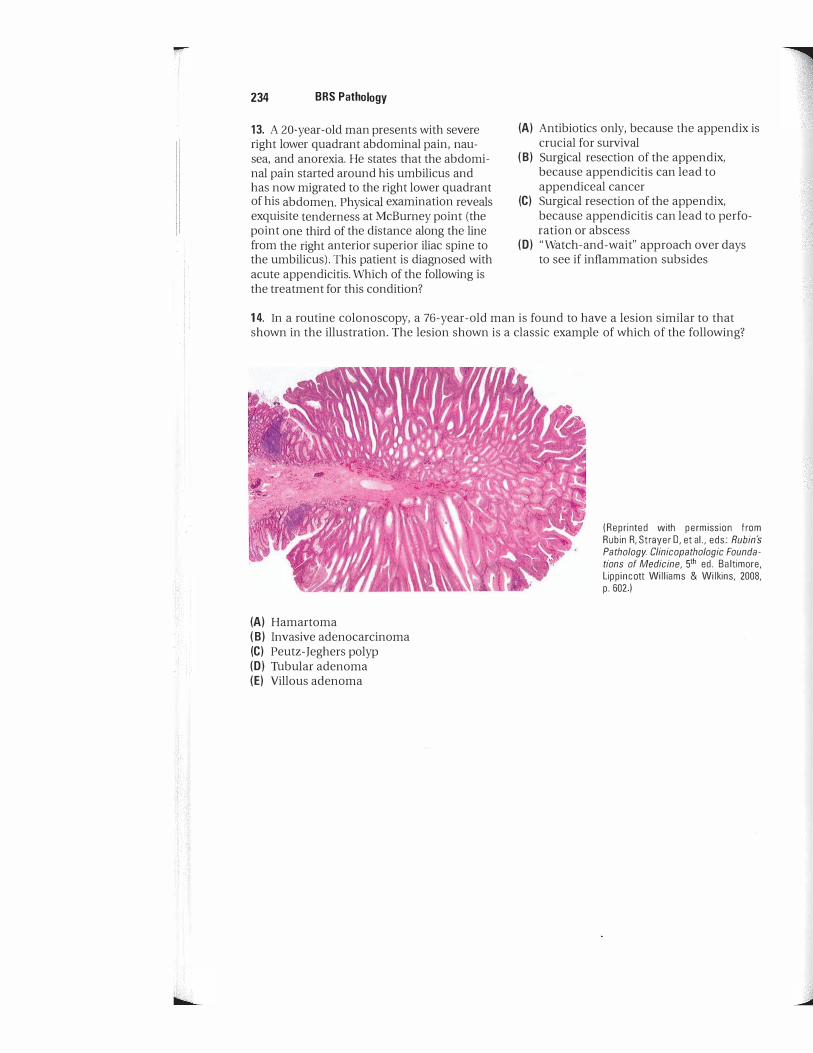
234 BRS Pathology
13. A 20-year-old man presents with severe right lower quadrant abdominal pain, nausea, and anorexia. He states that the abdominal pain started around his umbilicus and has now migrated to the right lower quadrant of his abdomen. Physical examination reveals exquisite tenderness at McBurney point (the point one third of the distance along the line from the right anterior superior iliac spine to the umbilicus). This patient is diagnosed with acute appendicitis. Which of the following is the treatment for this condition?
(A) Antibiotics only, because the appendix is crucial for survival
( B) Surgical resection of the appendix, because appendicitis can lead to appendiceal cancer
(C) Surgical resection of the appendix, because appendicitis can lead to perforation or abscess
(D) "Watch-and-wait" approach over days to see if inflammation subsides
14. In a routine colonoscopy, a 76-year-old man is found to have a lesion similar to that shown in the illustration. The lesion shown is a classic example of which of the following?
(A) Hamartoma ( B) Invasive adenocarcinoma (C) Peutz-Jeghers polyp (0) Tubular adenoma (E) Villous adenoma
(Reprinted with permission from Rubin R, Strayer D, et aI., eds.: Rubin's Pathology. Clinicopathologic Foundations of Medicine, 5th ed. Baltimore, Lippincott Williams & Wilkins, 2008, p.602 )

Answers and Explanations
1. The answer is A. Sjogren syndrome is an autoimmune disorder characterized by keratoconjunctivitis sicca and xerostomia, due to lymphocytic infiltration and parenchymal destruction of the parotid and lacrimal glands in association with a connective tissue disorder, such as rheumatoid arthritis. Malignant lymphoma is a frequent complication.
2. The answer is D. Approximately 80% to 90% of salivary gland tumors originate in the parotid gland and, of these, approximately 70% are pleomorphic adenomas. The term "mixed tumor" properly applies to this benign tumor, which often demonstrates myxoid and cartilage-like elements in addition to stellate or fusiform epithelial cells. Complete surgical resection is difficult because of the tumor's proximity to the facial nerve, and, thus, recurrence is frequent.
3. The answer is E. This is a classic case of gastroesophageal reflux disease (GERD), which is caused by reflux of gastric acid contents into the lower esophagus. GERD manifests as burning epigastric pain on eating spicy foods or on lying recumbent. The pain is usually relieved by antacids. GERD is most commonly associated with hiatal hernia and an incompetent lower esophageal sphincter, as well as with excessive use of alcohol or tobacco, increased gastric volume, pregnancy, and scleroderma. Barrett esophagus, or columnar intestinal metaplasia of the epithelium of the distal esophagus, is a complication of long-standing GERD.
4. The answer is C. Of course, the immediate problem in this patient is life-threatening upper gastrointestinal hemorrhage, an important complication of peptic ulcer disease. Peptic ulcer disease occurs most frequently in the first portion of the duodenum, the lesser curvature of the stomach, or the distal esophagus. Duodenal peptic ulcers are associated with hypersecretion of gastric acid and pepsin and are closely related to gastric Helicobacter pylori infection. Apparently, H. pylori increases gastric acid secretion and impairs mucosal defenses. Other predisposing factors include aspirin or nonsteroidal anti-inflammatory drug intake, smoking, Zollinger-Ellison syndrome, primary hyperparathyroidism, and multiple endocrine neoplasia type 1 .
5. The answer is B. Barrett esophagus is columnar metaplasia of the esophageal squamous epithelium (squamous-to-columnar). The columnar epithelium is often of the intestinal type with goblet cells. Barrett esophagus is a complication of long-standing gastroesophageal reflux disease and is a precursor of esophageal adenocarcinoma. The most common location is in the distal (lower) third of the esophagus.
6. The answer is A. Squamous cell carcinoma of the esophagus is an aggressive cancer with rapid progression and short survival in all stages of disease. It is most common in subjects with a long-term history of cigarette smoking and alcohol use. The tumor arises most commonly in the upper and middle thirds of the esophagus.
7. The answer is B. Congenital pyloric stenosis is caused by hypertrophy of the circular muscular layer of the pylorus, reSUlting in a palpable mass in the epigastrium. Hypertrophy of the pyloric musculature leads to obstruction and the characteristic projectile vomiting. This condition most commonly occurs in male infants within the first several days to weeks of life.
235

236 BRS Pathology
8. The answer is D. Crohn disease and ulcerative colitis are the two classic inflammatory bowel diseases. Crohn disease is a chronic inflammatory condition that can affect any part of the gastrointestinal tract from mouth to anus, but most commonly involves the distal ileocecum, small intestine, or colon. Morphologically, Crohn disease manifests as transmural inflammation (involving all layers of the intestinal wall) , thickening of involved intestine, linear ulceration, a cobblestone appearance, skip lesions (normal intestine between affected regions) , and granulomas. Strictures and fistulae may develop, leading to intestinal obstruction. Crohn disease may lead to carcinoma of the small intestine or colon, but much less commonly than ulcerative colitis.
9. The answer is E. Adenocarcinoma of the colon most commonly develops through a progression of mutations in oncogenes and tumor suppressor genes in a multistep process. Normal mucosa evolves into a tubular adenoma with malignant potential, which then further evolves into carcinoma (the adenoma-carcinoma sequence) . Carcinoma of the rectosigmoid (left-sided) tends to present as early obstruction, with change in bowel habits and decreased caliber of stool, whereas carcinoma of the right colon (right -sided) tends to present late, with iron deficiency anemia due to chronic blood loss from the lesion.
10. The answer is B. Pseudomembranous colitis is caused by overgrowth of Clostridium difficile. This organism produces exotoxin that induces necrosis of the superficial mucosa, leading to pseudomembrane formation. The bacteria itself does not invade the mucosa. This condition most often occurs in patients with a history of broad-spectrum antibiotic use, because elimination of normal intestinal flora promotes overgrowth of C. difficile.
11. The answer is A. The illustration demonstrates diverticulosis of the colon (openings shown by arrows) . These lesions are most common in older persons and are found most often in the sigmoid. The incidence of disease is increased in populations that consume low-fiber diets. Although most often asymptomatic, diverticula may become the site of acute inflammation (diverticulitis) , sometimes with life-threatening complications, such as perforation and peritonitis.
12. The answer is E. The illustration shows a chronic gastric peptic ulcer with characteristic radiating folds of the gastric mucosa starting at the ulcer margins. The lesion has a smooth base with a little fibrin attached and nonelevated, punched-out margins, in contrast to gastric carcinoma, which often has an irregular necrotic base and firm, raised margins. Despite these characteristic findings, the distinction between gastric peptic ulcer and ulcerated carcinoma must be established by biopsy. In contrast to carcinoma, peptic ulcer will usually heal with conservative management.
13. The answer is C. The inflamed appendix in acute appendicitis should be surgically removed because of possible devastating complications of perforation or abscess.
14. The answer is D. The illustration shows a tubular adenoma, which is the most common form of adenomatous polyp. These lesions can be single or multiple, or they can occur as components of various multiple polyposis syndromes. Notable among these syndromes are Gardner (associated with osteomas and soft tissue tumors) , Turcot (associated with central nervous system tumors) , and familial polyposis. All of the foregoing are associated with an increased incidence of colon malignancy. In contrast, the Peutz-Jeghers polyp is a non-neoplastic hamartomatous lesion. Even though the polyp itself does not transform into colon cancer, the Peutz-Jeghers syndrome is associated with an increased incidence of colon cancer and malignancies elsewhere.
Liver, Gallbladder, and Exocrine Pancreas
I. D ISEASES OF THE LIVER
A. Jaundice (Table 16- 1 ) refers to yellow discoloration of skin, sclerae, and tissues caused by hyperbilirubinemia. It is most often associated with hepatocellular disease, biliary obstruction, or hemolytic anemia. 1. Physiologic jaundice of the newborn is commonly noted during the first week of l ife, but
is not usually clinically important. It is characterized chemically by unconjugated hyperbi l i rubinemia. a. This form of jaundice results from both increased bilirubin production and a relative
deficiency of glucuronyl transferase in the immature liver; these phenomena are exaggerated in premature infants.
� b. Physiologic jaundice of the newborn must be distinguished from neonatal cholestasis, which is due to a wide variety of causes, including extrahepatic biliary atresia, (Xl-antitrypsin deficiency, cytomegalovirus infection, and many other conditions.
2. Congenital hyperbi l i rubinemias a. Gi lbert syndrome is extremely common, occurring in almost 5% of the population. This
familial disorder is characterized by a modest elevation of serum unconjugated bi l i rubin; the liver is otherwise unimpaired, and there are no clinical consequences. The cause is a combination of decreased bilirubin uptake by liver cells and reduced activity of glucuronyl transferase.
b. Crigler-Najjar syndrome is a severe familial disorder characterized by unconjugated hyperbilirubinemia caused by a deficiency of glucuronyl transferase. (1) One form leads to early death from kernicterus; damage to the basal ganglia and
other parts of the central nervous system are caused by unconjugated bilirubin. (2) A less severe form responds to phenobarbital therapy, which decreases the serum
concentration of unconjugated bilirubin. c. Dubin-Johnson syndrome is an autosomal recessive form of conjugated hyperbiliru
binemia characterized by defective bilirubin transport. It is characterized by a striking brown-to-black discoloration of the l iver caused by the deposition of granules of very dark pigment, the chemical nature of which is unclear (Figure 16- 1 ) .
d . Rotor syndrome is similar to Dubin-Johnson syndrome, but abnormal pigment i s not present.
B. Acute viral hepatitis (Table 16-2) 1. General considerations
� a. Acute viral hepatitis is characterized by jaundice and extremely high elevations of serum aspartate and alanine aminotransferases.
b. The disease may be caused by a variety of viral agents, including Epstein-Barr virus or cytomegalovirus, but usually results from hepatic infection by any of the following viruses, all of which are RNA viruses, with the exception of hepatitis B virus, which is a DNA virus.
237

238 BRS Pathology
tab I� 16-1 Differential Diagnosis of Jaundice
Type of Jaundice
Hepatocellular
Obstructive
Hemolytic
Hyperbilirubinemia
Conjugated and unconjugated
Conjugated
Unconjugated
Urine Bilirubin
Increased
I ncreased
Absent (acholuria)
AlT = alanine aminotransferase; AST = aspartate aminotransferase.
2_ Hepatitis A vi rus (HAV)
Urine Urobilinogen
Normal to decreased
Decreased
I ncreased
Other Findings
Intrahepatic cholestasis may result in retention of conjugated bilirubin; hepatocellular damage may result in impaired conjugation of bilirubin; enzyme activities of ALT and AST increased; increased alkaline phosphatase indicates intrahepatic obstruction
Alkaline phosphatase and cholesterol increased; ALT and AST variable; with complete obstruction, stools pale and clay-colored and urine urobilinogen undetectable
Degree of urine urobilinogen increase directly related to increased hemoglobin catabolism
� a. Spread occurs by fecal-oral transmission; parenteral infection does not occur. b. The incubation period is 15-45 days. c. HAV does not cause a chronic carrier state or lead to chronic hepatitis; complete recov
ery almost always occurs. There is no relation to hepatocellular carcinoma. 3. Hepatitis B virus (HBV)
a. HBV consists of a central core containing the viral DNA genome, DNA polymerase, hepatitis B core antigen (HBcAg) . and hepatitis B e antigen (HBeAg) , and an outer
FIGURE 16-1 Dubin-Johnson syndrome. The deposition of black melanin-like pigment in hepatocytes and Kupffer cells d ifferentiates this condition from other congenital conjugated hyperbilirubinemias. (Reprinted with permission from Fenderson B, Rubin R: Lippincott's Review of Pathology Illustrated Interactive Q & A. Baltimore, Lippincott Williams & Wiikins, 2007, p. 146.)

I!iMl'!ttfIIlI Liver, Gallbladder, and Exocrine Pancreas 239
tab I e 16-2 Viral Hepatitis: Causative Agents, Transmission, and Cl inical Features
Virus Predominant Modes of Transmission Clinical Features
Hepatitis A virus
Hepatitis B virus
Hepatitis C virus
Fecal-oral
Sexual and parenteral
Parenteral
No carrier state; does not lead to chronic liver disease
Frequently leads to carrier state or chronic liver disease
Most frequent cause of transfusion-mediated hepatitis;
Delta agent (hepatitis D virus)
Hepatitis E virus
Hepatitis G virus
Sexual and parenteral
Fecal-oral
Parenteral
frequently progresses to chronic liver disease
Requires concurrent infection with HBV for replication
Occurs in epidemic form in Third World countries; does not lead to chronic liver disease
Frequent in blood donors; role as cause of acute or chronic hepatitis is not established
lipoprotein coat containing the hepatitis B surface antigen (HBsAg) . The complete virion is known as the Dane particle.
� b. Transmission is via parenteral, sexual. and vertical (mother to neonate) routes. There is an increased incidence of HBV infection in male homosexuals.
c. The incubation period averages 60-90 days. � d. HBV has a major association with hepatocellular carcinoma.
e. Infection is associated with a "ground-glass" appearance of hepatocytes. 1. Disease can result in a carrier state or in chronic liver disease. The sequence in which
the various antigens or antibodies to these antigens appear in the serum is of clinical significance. (1) HBsAg appears in serum some weeks before the onset of clinical findings, then
decreases and generally persists for a total of 3-4 months. Persistence as detectable serum antigen for more than 6 months denotes the carrier state. HBsAg elicits an antibody response (anti-HBsAg) ; antibody appears a few weeks after the disappearance of the antigen and indicates recovery, as well as immunity to future infection.
(2) HBeAg appears shortly after HBsAg and disappears before HBsAg. It is closely correlated with viral infectivity.
(3) Anti-HBcAg appears about 4 weeks after the appearance of HBsAg, is present during the acute illness, and can remain elevated for several years. It is a marker (along with anti-HBeAg) of hepatitis infection during the "window period" between the disappearance of HBsAg and the appearance of anti -HBsAg.
(4) HBV DNA can also be detected in serum and is an index of infectivity. 4. Hepatitis C virus (HeV) is a common cause of what was formerly called non-A, non-B
hepatitis. a. Transmission is parenteral. HCV is a frequent cause of transfusion-mediated hepatitis
and often leads to a carrier state and chronic hepatitis. b. HCV is frequently associated with hepatocellular carcinoma.
5. Hepatitis 0 virus (HOV, delta agent) is a very small, spherical virus consisting of a single RNA strand and the associated delta protein antigen (HDAg) , surrounded by a proteinaceous coat of HBsAg. HDV is replicatively defective, requiring simultaneous infection with HBV for viral replication. It usually causes illness more severe than HBV infection alone. a. Transmission is via sexual or parenteral routes. b. Incidence is especially high in intravenous drug users.
6. Hepatitis E vi rus (HEV) causes an enterically transmitted form of viral hepatitis similar to HAV infection that occurs in water-borne epidemic form in underdeveloped countries. HEV has an as yet unknown association with chronic hepatitis or hepatocellular carcinoma, but it has a high incidence of mortality (about 20%) when occurring in pregnancy.

240 BRS Pathology
7. Hepatitis G virus (HGV) affects 1 %-2% of healthy blood donors, but its pathologic significance is questionable. HGV does not lead to chronic hepatitis and has no known relationship to hepatocellular carcinoma.
C. Chronic hepatitis 1. Chronic hepatitis is defined by the persistence of abnormalities for more than 6 months.
It may result from any of the viral hepatitides except HAV or HEV infection and also from liver damage induced by nonviral agents.
2. Autoimmune hepatitis is morphologically indistinguishable from other forms of chronic hepatitis. It is secondary to various immunologic abnormalities. It is clinically marked by hypergammaglobulinemia and anti-smooth muscle antibodies.
D. Other inflammatory liver disorders 1. Neonatal hepatitis is of unknown etiology.
a. The presence of multinucleated giant cells is characteristic. There may be bile pigment and hemosiderin within parenchymal cells.
b. It may cause jaundice during the first few weeks of life. 2. Other viral infections may involve the liver along with other organ systems.
a. Epstein-Barr virus (EBV) causes infectious mononucleosis, which often has a hepatitic component.
b. Cytomegalovirus (CMV) may involve the liver in infants and immunocompromised persons. Infected liver cells demonstrate characteristic nuclear inclusions surrounded by a halo (owl's eye appearance) .
c. Herpes simplex virus type 1 (HSV-1) may involve the liver in infants and immunocompromised persons.
d. Yellow fever characteristically demonstrates a severe hepatitic component marked by midzonal hepatic necrosis. The dying hepatocytes often condense into eosinophilic contracted forms referred to as Councilman bodies. Similar inclusions are observed in all of the viral hepatitides and are manifestations of apoptosis.
3. Leptospirosis, also known as Weil disease or icterohemorrhagic fever, is caused by Leptospira species. This severe infection is characterized by jaundice, renal failure, and hemorrhagic phenomena.
4. Echinococcus granulosus infestation is caused by ingestion of tapeworm eggs from the excreta of dogs and sheep. It results in hydatid disease of the liver, in which large parasitic cysts invade the liver.
5. Schistosomiasis is caused by infestation with Schistosoma mansoni or S. japonicum. The adult worms lodge in the portal vein and its branches. The eggs are highly antigenic and stimulate granuloma formation, with resultant tissue destruction, scarring, and portal hypertension.
E. Microvesicular fatty liver. This group of serious disorders is associated with the presence of small fat vacuoles in parenchymal liver cells, which differ from the large fat -containing vacuoles characteristic of fatty change. 1. Reye syndrome
� a. This acute disorder of young children is characterized by encephalopathy, coma, and microvesicular fatty liver.
� b. Reye syndrome is associated with aspirin administration to children with acute viral infections.
2. Fatty l iver of pregnancy is acute hepatic failure during the third trimester of pregnancy associated with microvesicular fatty liver. This condition has a high mortality rate.
3. Tetracycline toxicity results in an unpredictable hypersensitivity-like reaction with microvesicular fatty change.
F. Alcoholic liver disease is the constellation of hepatic changes associated with excessive alcohol consumption; it varies from fatty change to alcoholic hepatitis and cirrhosis. The most common form of liver disease in the United States, it may be asymptomatic or may be associated with mild-to-severe hepatic inflammation, cirrhosis, or eBcephalopathy.

Ii1i!lf!ltl1ill Liver, Gal lb ladder, and Exocrine Pancreas 241
FIGURE 16-2 Alcohol ic hepatitis. The arrows ind icate Mallory bodies (alcohol ic hyal in) . Destructive changes of hepatocytes, fatty change (not shown here), and a neutrophi l ic inflammatory infi ltrate are other characteristics. (Reprinted with permission from Rubin R, Strayer D, et aI. , eds.: Rubin's Pathology Clinicopathologic Foundations of Medicine, 5th ed . Baltimore, L ipp incott Wi l l iams & Wilkins, 2008, p. 647.)
1. Fatty change (steatosis) is the most frequent morphologic abnormality caused by alcohol and is reversible. Nonalcohol ic fatty liver disease, or NAFLD, is a related condition unrelated to alcohol consumption.
2. Alcoholic hepatitis (Figure 16-2) � a. Characteristics include fatty change, focal liver cell necrosis, infi ltrates of neutrophi ls,
and the presence of intracytoplasmic eosinophilic hyaline inclusions (Mallory bodies, alcoholic hyalin) derived from cytokeratin intermediate filaments; these inclusions are characteristic, but not entirely specific for alcoholic hepatitis.
b. Alcoholic hepatitis is often associated with irreversible fibrosis that characteristically surrounds central veins and has been referred to as perivenular fibrosis, sclerosing hyaline necrosis, or central hyaline sclerosis. This fibrosis can lead to central vein obstruction and fibrosis surrounding individual liver cells and can result in cirrhosis.
3. Alcoholic cirrhosis
G. Cirrhosis (Table 16-3) 1. General considerations
a. Cirrhosis is a descriptive term for chronic liver disease characterized by generalized disorganization of hepatic architecture with scarring and nodule formation. Liver cell damage, regenerative activity, and generalized fibrosis reSUlting in a nodular pattern are also characteristic.
� b. Classification can be morphologic, on the basis of nodule size (micronodular, macronodular, and mixed macromicronodular forms) .
c. In all forms, there is an increased incidence of hepatocel lular carcinoma.

242 BRS Pathology
tab I e 16-3 Cirrhosis: Types and Features
Tvpe Features
Alcoholic (Laennec, nutritional) Most frequently occurring form of cirrhosis; associated with alcoholism; micronodular pattern evolving in late stages to typical hobnail liver with large, irregular nodules
Postnecrotic (macro nodular, posthepatitic)
Large, irregular nodules containing intact hepatic lobules; diverse etiologies; often the end result of viral hepatitis, especially hepatitis B
Biliarv
Primary Probable autoimmune origin; anti mitochondrial antibodies; obstructive jaundice
End result of long-standing extrahepatic biliary obstruction Secondary
Hemochromatosis
Hereditary (primary) Familial defect in control of iron absorption; massive accumulation of hemosiderin
Secondary
Wilson disease
in hepatic and pancreatic parenchymal cells, myocardium, and other sites; classic triad of cirrhosis, diabetes mellitus, and increased skin pigmentation; cirrhosis micronodular type
Caused by chronic iron overload of diverse etiology
Inborn errors of metabolism
Accumulation of copper in liver, kidney, brain, and cornea; cirrhosis can be micronodular or macronodular; decreased serum ceruloplasmin
Cirrhosis associated with galactosemia, glycogen storage diseases, or <x,-antitrypsin deficiency
d. There are numerous etiologic agents, including: (1) Prolonged a lcohol intake, drugs, and chemical agents (2) Viral hepatitis, biliary obstruction, and hemochromatosis (3) Wilson disease and other inborn errors of metabolism (4) Heart failure with long-standing chronic passive congestion of the liver
2, Alcoholic (Laennec, nutritional) cirrhosis is the prototype for all forms of cirrhosis. a. Clinical manifestations
(1) Findings associated with hepatocellular damage and l iver failure include: (a ) Jaundice, most often mixed conjugated and unconjugated (b ) Hypoalbuminemia, caused by decreased albumin synthesis in damaged
hepatocytes (c ) Coagulation factor deficiencies, caused by decreased synthesis; all coagulation
factors, with the exception of von Wille brand factor, are synthesized in the liver.
(d ) Hyperestrinism, manifest as palmar erythema (liver palms); spider nevi (capillary telangiectases) of the face, upper arms, and chest; loss of body and pubic hair; testicular atrophy; and gynecomastia
(2) Consequences of intrahepatic scarring with increased portal venous pressure include:
� (a ) Esophageal varices, often leading to upper gastrointestinal hemorrhage (b ) Rectal hemorrhoids (c) Periumbil ical venous collaterals (caput medusae) (d) Splenomegaly
(3) Changes due to both l iver cell damage and portal hypertension include: (a) Peripheral edema, ascites, or hydrothorax, caused by � ( i ) Increased portal venous pressure, which leads to increased production of
hepatic lymph � ( i i ) Decreased plasma oncotic pressure secondary to hypoalbuminemia � ( i i i ) Retention of sodium and water as a result of decreased hepatic degradation
of aldosterone, activation of the renin -angiotensin system, or both (b) Encepha lopathy (portal-systemic encephalopathy) facilitated by shunting
from the portal to the systemic circulation, with direct delivery of neurotoxic
J

IBltf'i1tlaliI Liver, Gallbl adder, and Exocrine Pancreas 243
substances, such as ammonia and other enteric degradation products, directly into the systemic circulation. Neurologic manifestations varying from slight confusion to deep coma along with asterixis (flapping tremor of hands) are characteristic features.
b. Morphologic abnormalities (1) The liver may be enlarged or small and shrunken.
� (2) The pattern is most often micronodular. (3) Hepatic architecture is obscured by fibrous bands surrounding nodules of dis
torted liver cell plates. (4) The fibrous bands contain proliferating bile ducts and inflammatory cells, most
often lymphocytes and plasma cells. � (5) In late stages, the nodules tend to become larger and irregular; this pattern results
in a scarred, shrunken liver termed the hobnail liver. 3. Postnecrotic (macronodular, posthepatitic) cirrhosis
a. Morphologic characteristics include broad fibrous bands dividing the liver into large, irregular nodules, often containing intact hepatic lobules.
b. This form of cirrhosis is often a sequela of chronic active hepatitis; HBV and HCV are the most common viral causes. In addition, this disease may be caused by noninfectious hepatotoxic agents. Sometimes, it can result from the progression of micro nodular alcoholic cirrhosis. Often, the etiology is uncertain (cryptogenic cirrhosis).
c. Postnecrotic cirrhosis leads to hepatocellular carcinoma more often than other forms of cirrhosis.
4. Bi l iary cirrhosis occurs as a primary, probably autoimmune, disorder and much more frequently as a secondary form due to biliary obstruction. a. Primary biliary cirrhosis � (1) This form of cirrhosis is most likely of autoimmune origin. There is an increased
incidence of other autoimmune disorders in affected patients, and antimitochondrial antibodies are characteristic.
� (2) Disease is most common in middle-aged women. � (3) Characteristics include severe obstructive jaundice, itching, and hypercholes
terolemia; hypercholesterolemia leads to cutaneous xanthoma formation. (4) Primary biliary cirrhosis is marked by increased parenchymal copper concentra
tion, a finding of unknown significance. b. Secondary bi l iary cirrhosis
(1) The cause is extrahepatic bi l iary obstruction, which leads to dilation and increased pressure within intrahepatic bile ducts and cholangioles, further resulting in ductal injury, ductal and periductal inflammation, and resolution by fibrous tissue formation.
(2) Complications often include ascending cholangitis and bacterial inflammation of the intrahepatic bile ducts.
(3) Secondary biliary cirrhosis is marked histologically by evidence of bile stasis and by bile lakes, accumulations of bile within hepatic parenchyma.
5. Primary sclerosing cholangitis is rare except in association with inflammatory bowel disease, especially ulcerative colitis. a. Characteristics include inflammation, fibrosis, and stenosis of intrahepatic and extra
hepatic bile ducts. It eventually develops into biliary cirrhosis. b. There is an associated increased incidence of cholangiocarcinoma.
6. Hemochromatosis a. Hereditary hemochromatosis is a famil ial defect of iron absorption by the intestinal
mucosa. � (1) This disease has been considered one of the most common autosomal recessive
disorders, but this idea has recently been modified. Although the gene defect is common, the penetrance and development of clinical disease is low.
(2) Hereditary hemochromatosis can be detected and treated successfully before organ damage occurs by screening for increased transferrin iron saturation or, in selected cases, for mutations in the Hfe gene on chromosome 6.

....
244 BRS Pathology
(3) In its fully developed state, characteristics include: /" (a ) The triad of ci rrhosis, diabetes mell itus, and increased skin pigmentation, giving
rise to the older term bronze diabetes (b ) Skin pigmentation caused by hemosiderin and melanin deposition (c ) Pigment cirrhosis (micronodular cirrhosis with enormous accumulation of
parenchymal hemosiderin demonstrable by Pruss ian blue staining) /" (d ) Marked increase in serum iron and modest reduction in total iron-binding
capacity (TIBC, transferrin) . This combination results in increased transferrin iron saturation.
(e) Increased serum ferritin b. Secondary hemochromatosis is most often associated with a combination of ineffective
erythropoiesis and multiple transfusions, such as occurs in thalassemia major. It may also be secondary to various other causes of iron overload.
7. Wilson disease (hepatolenticular degeneration) is an autosomal recessive disorder of copper metabolism.
/" a. A decreased serum ceruloplasmin (copper-binding protein) is characteristic. This is probably not a primary defect but it is secondary to increased copper.
/" b. Manifestations include liver disease that varies from chronic hepatitis to cirrhosis, either micronodular or macronodular in type.
/" c. Wilson disease is also marked by the Kayser-Fleischer ring circumscribing the periphery of the cornea, representing deposition of copper-containing pigment in Descemet membrane.
d. In addition, there may be aminoaciduria and glycosuria due to renal tubular damage. e. Wilson disease results in an abnormal major accumulation of copper in parenchymal
cells of the liver and kidney and in the brain and cornea. /" f. The disease also results in extrapyramidal motor signs caused by involvement of the
basal ganglia, especially the putamen of the lenticular nucleus. 8. Cirrhosis due to inborn errors of metabolism results from several disorders, including:
a. a)-antitrypsin deficiency b. Galactose-I-phosphate uridyl transferase deficiency (galactosemia) c. Glycogen storage diseases
H. Vascular disorders of the liver 1. Portal hypertension is characterized by the development of venous collaterals with
varices in the submucosal veins of the esophagus, the hemorrhoidal plexus, and other sites. This condition is often classified by the site of portal venous obstruction: a. Prehepatic: caused by portal and splenic vein obstruction, most often by thrombosis b. Intrahepatic: caused by intrahepatic vascular obstruction, most often by cirrhosis or
metastatic tumor, and more rarely by exotic entities such as schistosomiasis c. Posthepatic: caused by venous congestion in the distal hepatic venous circulation,
most often as a result of constrictive pericarditis, tricuspid insufficiency, congestive heart failure, or hepatic vein occlusion (Budd-Chiari syndrome)
2. Infarction is unusual, because the liver has a double blood supply (mesenteric and hepatic) . 3. Budd-Chiari syndrome
a. The cause is thrombotic occlusion of the major hepatic veins, resulting in abdominal pain, jaundice, hepatomegaly, ascites, and eventual liver failure.
b. Budd-Chiari syndrome is most often associated with polycythemia vera, hepatocellular carcinoma, and other abdominal neoplasms; may also occur as a complication of pregnancy.
4. Congestive heart fai lure a. In long-standing chronic right-sided heart failure, the cut surface of the liver can
assume an appearance referred to as the nutmeg liver, with dark red congested centrilobular areas alternating with pale portal areas.
b. Eventually, centrilobular fibrosis occurs, resulting in cardiac cirrhosis (cardiac sclerosis) . Similar changes may follow long-standing constrictive pericarditis or tricuspid insufficiency .
I. Hepatic tumors 1. Benign tumors
I!lttlJ'!ttlIll Liver, Gallbl adder, and Exocrine Pancreas 245
a. Hemangioma is the most common benign tumor of the liver. b. Adenoma
� (1) The incidence is apparently related to use of oral contraceptives. � (2) When the adenoma is subcapsular in location, it may rupture, resulting in severe
intraperitoneal hemorrhage. 2. Malignant tumors
a. Metastatic tumors account for the majority of hepatic malignancies. b. Hepatocellular carcinoma is the most common primary malignancy of the liver.
� (1) This hepatic malignancy almost always develops in association with preexisting cirrhosis of any kind, especially when associated with HBV infection.
� (2) Hepatocellular carcinoma has been associated with aflatoxin Bj contamination of nuts and grains; aflatoxin Bj is thought to cause specific point mutations in the p53 gene.
� (3) Frequently, the cancer is marked by increased serum concentration of a-fe top rote in. � (4) There is a propensity for invasion of vascular channels with hematogenous
dissemination. c. Cholangiocarcinoma (bi le duct carcinoma) is less common than hepatocellular carci
noma. This form of hepatic cancer occurs most frequently in the Far East, where it is associated with Clonorchis sinensis (liver fluke) infestation. (1) The cancer originates from intrahepatic biliary epithelium. (2) Like hepatocellular carcinoma, it has a propensity for early invasion of vascular
channels. � (3) Unlike hepatocellular carcinoma, cholangiocarcinoma is not associated with
HBV infection or cirrhosis. (4) Sometimes, it occurs as a late complication of thorium dioxide (Thorotrast)
administration. d. Hemangiosarcoma (angiosarcoma)
(1) This form of hepatic cancer is a rare malignant vascular tumor. � (2) It is associated with toxic exposure to polyvinyl (vinyl) chloride, thorium dioxide
(Thorotrast), and arsenic.
II. DISEASES OF THE GALLBLADDER
A. Cholecystitis 1. Acute cholecystitis is acute inflammation of the gallbladder.
a. The inflammation is most often pyogenic. b. Clinical manifestations include nausea, vomiting, fever, and leukocytosis associated
with right upper quadrant and epigastric pain. 2. Chronic cholecystitis
a. Thickening of the gallbladder wall occurs as a result of extensive fibrosis. b. Chronic inflammation is frequently complicated by gallstones.
B. Cholelithiasis (gallstones) has a higher incidence in women and is often associated with obesity and multiple pregnancies. 1. Stone types
a. Cholesterol stones are often solitary and too large to enter the cystic duct or the common bile duct.
b. Pigment stones (1) Precipitation of excess insoluble unconjugated bi lirub i n results in their
formation. (2) Association often includes hemolytic anemia and bacterial infection.

..
246 BRS Pathology
c. Mixed stones account for most stones (75%-80%). (1) Most of these stones are a mixture of cholesterol and calcium salts. (2) Mixed stones can often be visualized radiographically because of their calcium
content. 2. Clinical manifestations
a. Cholelithiasis is often silent and asymptomatic. b. Fatty food intolerance is characteristic.
3. Complications a. Biliary colic results from impaction of gallstone in cystic or common bile duct. b. Common bile duct obstruction results in obstructive jaundice with conjugated hyper
bilirubinemia, hypercholesterolemia, increased alkaline phosphatase, and hyperbilirubinuria.
c. Ascending cholangitis can result from secondary bacterial infection facilitated by obstructed bile flow.
d. Cholecystitis, acute or chronic. There is a reciprocal association between cholecystitis and cholelithiasis (i.e., each predisposes to the other) .
e. Acute pancreatitis f. Gallstone i leus (intestinal obstruction caused by passage of a large gallstone through
the eroded gallbladder wall into the adjacent small bowel) g. Mucocele (distended, mucus-filled gallbladder secondary to chronic cystic duct
obstruction) h. Malignancy
C. Cholesterolosis (strawberry gallbladder) 1. Characteristics include yellow cholesterol-containing flecks in the mucosal surface. 2. There is no association with inflammatory changes and no special association with
cholelithiasis.
D. Tumors 1. Tumors of the gallbladder
a. Benign tumors of the gallbladder are rare. b. The most common primary tumor of the gallbladder is adenocarcinoma, which is
often associated with gallstones. 2. Carcinoma of the extrahepatic bi l iary ducts and the ampulla of Vater is less common than
carcinoma of the gallbladder. a. This carcinoma is almost always adenocarcinoma. b. Typical presenting features include a progressive, relentless obstructive jaundice. Clin
ical characteristics include the combination of jaundice and a palpably enlarged gallbladder. Tumors that obstruct the common bile duct result in an enlarged, distended gallbladder; obstructing stones do not (Courvoisier law).
III. DISEASES OF THE EXOCRINE PANCREAS
A. Acute pancreatitis
� 1. The cause is activation of pancreatic enzymes, reSUlting in autodigestion of the organ, with hemorrhagic fat necrosis and deposition of calcium soaps, and sometimes formation of pseudocysts (parenchymal cysts not lined with ductal epithelium) .
� 2. Predisposing factors include gallstones and excessive alcohol intake. 3. Clinical manifestations include severe abdominal pain and prostration closely mimicking
an acute surgical abdomen. � 4. There is an association with increased serum amylase. � 5. Characteristics include hypocalcemia caused by loss of circulating calcium into precipi
tated calcium-fatty acid soaps. � 6. Acute pancreatitis can be superimposed on chronic pancreatitis .

Ii1iIl1l1QiliI Liver, Gallbl adder, and Exocrine Pancreas 247
B. Chronic pancreatitis 1. Morphologic characteristics include progressive parenchymal fibrosis. Calcification, which
may be visualized using radiography, is frequent. Pseudocysts may be evident. 2. There is almost always an association with alcohol ism.
� 3. Clinical manifestations are extremely variable and include abdominal and back pain, progressive disability, and steatorrhea, which is a manifestation of pancreatic insufficiency with lipase deficiency. Fat malabsorption may be accompanied by a deficiency of fat-soluble vitamins and can thus lead to such conditions as night blindness (vitamin A deficiency) and osteomalacia (vitamin D deficiency) .
C. Carcinoma of the pancreas is a common tumor. 1. Incidence is increasing; the carcinoma is more common in smokers. 2. The carcinoma is almost always adenocarcinoma.
� 3. More often, the carcinoma arises in the head of the pancreas, causing obstructive jaundice (see Table 16 - 1 ) ; somewhat less often it originates in the pancreatic body or tail. Carcinoma involving the pancreatic tail can cause islet destruction and secondary diabetes mellitus.
� 4. Clinical manifestations may include abdominal pain radiating to the back, weight loss and anorexia, sometimes migratory thrombophlebitis (Trousseau sign), and frequently common bile duct obstruction resulting in obstructive jaundice (often accompanied by a d istended, palpable gal lbladder).
5. Cancer is often silent before widespread dissemination occurs. Death usually results within 1 year.
-l I
I

Review Test
Directions: Each of the numbered items or incomplete statements in this section i s followed by answers or by completions of the statement. Select the one lettered answer or completion that is best in each case.
1. A neonate has been persistently jaundiced from birth despite aggressive phototherapy and exchange transfusions. Laboratory studies demonstrate significantly elevated unconjugated bilirubin. Tests from an outside laboratory confirm the total absence of glucuronyl transferase activity. The neonate most likely has which of the following condition?
(A) Crigler-Najjar syndrome (8) Gilbert syndrome (C) Hemolytic disease of the newborn (D ) Physiologic jaundice of the
newborn (E) Rotor syndrome
2. An 18-year-old man presents to his family physician for a routine physical prior to moving away for college. Other than feeling slightly "stressed" by his soon-to-be new life situation, he has no complaints. A comprehensive metabolic panel reveals a modestly elevated unconjugated bilirubin but a near-normal aspartate aminotransferase and alanine aminotransferase. It is likely that the patient may have which of the following conditions?
(A) Crigler-Najjar syndrome (8) Dubin-Johnson syndrome (C ) Gilbert syndrome (D ) Infectious mononucleosis (E ) Rotor syndrome
3. A 55-year-old alcoholic man died after an illness characterized by increasing jaundice, ascites, and generalized wasting. Laboratory testing revealed hyperbilirubinemia, hypoalbuminemia, and mildly elevated liver enzymes. The appearance of the liver at autopsy is shown in the figure. The most likely diagnosis is
(A) <Xl-antitrypsin deficiency. (8) cirrhosis. (C) hepatitis A. (D) hepatitis C. (E) multiple metastases.
248
(Reprinted with permission from Rubin R, Strayer D, et ai., eds.: Rubin's Pathology. Clinicopathologic Foundations of Medicine, 5th ed. Baltimore, Lippin cott Will iams & Wilkins, 2008, p. 648.)

I!iMJ'1ltlIIIj Liver, Gallbladder, and Exocrine Pancreas 249
4. A 26-year-old woman presents to her primary care physician with fever, malaise, and "yellow eyes." She denies alcohol abuse, but admits to indulging in a dozen raw oysters at happy hour 3 weeks ago. In addition to scleral icterus, physical examination reveals a mildly enlarged liver with tenderness to palpation. Laboratory studies demonstrate a markedly increased aspartate aminotransferase and alanine aminotransferase and increased IgM and anti-hepatitis A titers. Which of the following is the most likely result of this infection?
(A) Cirrhosis (8) Complete resolution (C ) Establishment of a chronic carrier state (0) Fulminant hepatitis (E ) Hepatocellular carcinoma
5. A 32-year-old woman seeking to become pregnant visits her physician for a prepregnancy examination. Routine prenatal laboratory testing demonstrates the following profile: HBsAg (-) , anti-HBsAg (+), anti-HBcAg (-) , anti-HBeAg (-) , and HBV DNA (-) . Which of the following likely represents the status of the patient?
(A) Hepatitis B carrier (8) Immunized against hepatitis B (C) Infected and within the "window period" (0) Infected with hepatitis B and highly
transmissible (E ) Recently infected with hepatitis B
6. While on an international medical rotation, you encounter a pregnant woman in a rural village in India who presents with fever, jaundice, and malaise. The patient unexpectedly expires. This is the second case this month with a similar presentation. Which of the following is the most likely form of hepatitis?
(A) Hepatitis A (8) Hepatitis B (C) Hepatitis C (0) Hepatitis D (E ) Hepatitis E
7. A 56-year-old alcoholic man presents to the emergency department with confusion and lethargy. On physical examination, he is visibly jaundiced with ascites. Laboratory studies reveal increased prothrombin time and prolonged activated partial thromboplastin time, as well as significantly
increased serum ammonia levels. Given a significantly increased serum ammonia, which of the following physical findings might you expect to see?
(A) Asterixis (8) Capillary telangiectasias (C) Caput medusae (0) Gynecomastia (E ) Palmar erythema
8. A 45-year-old woman presents to her primary care physician with jaundice, pruritus, and periocular and intradigital xanthomas. Her laboratory results indicate a significantly increased alkaline phosphatase as well as a positive test for antimitochondrial antibodies. The most likely cause of her symptoms is
(A) leptospirosis. (8) macronodular cirrhosis. (C) primary biliary cirrhosis. (0) primary sclerosing cholangitis. (E) secondary biliary cirrhosis.
9. A 53-year-old man presents to his physician's office for an insurance physical. During the history, you learn that his father had diabetes and died of congestive heart failure. The patient was told it was due to "bronze diabetes." The patient complains only of vague fatigue and arthralgias. You are concerned that the patient may be at risk for hereditary hemochromatosis. Which test would you order to confirm your suspicion?
(A) Antimitochondrial antibodies (8) (Xl-Antitrypsin (C) Ceruloplasmin (0) Heterophil antibodies (E) Transferrin saturation
10. A 13-year-old boy presents to the pediatrician with extrapyramidal signs, including a resting and kinetic tremor. An ophthalmologic examination demonstrates the presence of Kayser-Fleischer rings, and his laboratory studies demonstrate elevated liver enzymes. It is likely that the patient's condition is associated with the accumulation of
(A) copper. (8) Councilman bodies. (C) eosinophilic hyaline inclusions. (0) glycogen. (E ) iron.

-
250 BRS Pathology
11. A 23-year-old woman is involved in a minor motor vehicle accident, prompting an abdominal computed tomography scan, which was read by the emergency department radiologist as normal with the exception of a questionable mass in the right lobe of the liver. A subsequent fine-needle biopsy confirms the presence of a liver adenoma. Which of the following is associated with the development of this lesion?
(A) Hepatitis B (B) Hepatitis C (e) Oral contraceptives (0) Polycythemia vera (E ) Polyvinyl chloride
12. A 36-year-old man from sub-Saharan Africa presents to the clinic with jaundice and right upper quadrant pain. On examination, the liver is palpably enlarged. Laboratory studies demonstrate an increase in liver enzymes. Computed tomography demonstrates a single large mass in the right lobe of the liver, and serum a-fetoprotein is markedly elevated. Which of the follOwing is likely to have contributed to the patient's condition?
(A) Aflatoxin B) (B) Clonorchis sinensis (e) Hepatitis A (0) Polyvinyl chloride (E ) Tetracycline
13. A 43-year-old multigravida presents with nausea, vomiting, fever, and right upper quadrant pain. On examination, she displays arrested inspiration on palpation of the right upper quadrant
(Murphy sign) . Her laboratory results reveal neutrophilia with a "left shift." Which of the following is the most likely diagnosis?
(A) Acute cholecystitis (8) Carcinoma of the ampulla of Vater (e) Cholangiocarcinoma (0) Cholesterolosis (E ) Sclerosing cholangitis
14. A 25-year-old obese woman who denies any history of alcohol abuse presents with severe abdominal pain radiating to the back. Laboratory results indicate an increase in serum amylase and lipase, with a marked decrease in calcium. Which of the following likely has caused this condition?
(A) Abetalipoproteinemia (8) Alcohol (e) Cholelithiasis (0) Cystic fibrosis (E) Mumps
15. A 63-year-old chronic alcoholic presents with weight loss, anorexia, and abdominal pain radiating to the back. Physical examination indicates a palpably enlarged gallbladder, and laboratory studies demonstrate conjugated hyperbilirubinemia. Computed tomography demonstrates a mass in the head of the pancreas. Which of the following is associated with the diagnosis of pancreatic adenocarcinoma?
(A) Asterixis (8) Gallstone ileus (e) Murphy sign (0) Trousseau sign (E ) Whipple triad

Answers and Explanations
1 . The answer is A . A total lack of glucuronyl transferase results in Crigler-Najjar syndrome I, which is invariably fatal by 18 months secondary to kernicterus. Gilbert syndrome is typically mild and usually not detected until later in life. Hemolytic disease of the newborn is due to blood group incompatibility between mother and child, and the bilirubinemia is secondary to the inherently decreased glucuronyl transferase with superimposed hemolytic anemia in the child, further overwhelming the conjugation machinery. Neonatal glucuronyl transferase is relatively deficient, although present, in normal infants, contributing to the transient condition termed physiologic jaundice of the newborn. Rotor syndrome is a relatively benign condition resulting in conjugated hyperbilirubinemia.
2. The answer is C. Gilbert syndrome is an extremely common cause of clinically insignificant unconjugated hyperbilirubinemia. The hyperbilirubinemia is episodic, with increases related to stress, fatigue, alcohol use, or recurrent infection. Crigler-Najjar syndrome I is often lethal, although Crigler-Najjar syndrome II is compatible with life and often presents with extreme jaundice and neurologic defects secondary to kernicterus. Both Dubin-Johnson syndrome and Rotor syndrome cause conjugated hyperbilirubinemia. Infectious mononucleosis can cause liver damage, but would likely be accompanied by malaise and fatigue.
3. The answer is B. The figure demonstrates the typical appearance of micronodular cirrhosis, the most common cause of which is alcoholism. Major clinical manifestations include jaundice, ascites, signs of hyperestrinism (palmar erythema, spider telangiectasia, gynecomastia, testicular atrophy) , consequences of increased portal venous pressure (esophageal varices, distended abdominal veins [caput medusae] , splenomegaly) , and consequences of hypoalbuminemia (ascites, peripheral edema) .
4. The answer is B. The patient has a case of hepatitis A (HAV) , most likely associated with the consumption of uncooked shellfish. Complete resolution occurs in an overwhelming majority of cases. Although possible, fulminant hepatic failure is unlikely with HAV infection. There is no association of HAV with either cirrhosis or hepatocellular carcinoma, as there is for hepatitis B and C.
5. The answer is B. The patient is only positive for antibody to the hepatitis B antigen. This suggests that the patient has been vaccinated for hepatitis B virus (HBV) . The vaccine consists of recombinantly produced HBV surface antigen (HBsAg) alone; antibodies to this protein convey immunity. HBsAg would be seen in the serum within the first 3-4 months after initial infection. Antibodies to the core protein (anti-HBcAg) appear during acute illness and between the disappearance of HBsAg and the appearance of anti-HBsAg, the "window period." Anti-HBeAg appears during the window period as well. HBeAg is closely correlated with viral infectivity. HBV viral DNA is also an index of infectivity.
6. The answer is E. Hepatitis E is a common sporadic cause of viral hepatitis in India. It has close to a 20% mortality in pregnant women. Like hepatitis A, it is transmitted enterically. Hepatitis C can be transmitted parenterally, as can hepatitis B and hepatitis D. Hepatitis D requires coinfection with hepatitis B.
7. The answer is A. Asterixis is a flapping tremor of the hands associated with hepatic encephalopathy. Failure of the liver to detoxify metabolites absorbed from the gastrointestinal tract leads to accumulation of nitrogenous wastes that are neurotoxic. Caput medusae results from dilation of the periumbilical venous collaterals as a result
251

....
252 BRS Pathology
of portal hypertension and activation of portal-caval anastomoses. Palmar erythema, capillary telangiectasias, and gynecomastia result from the hyperestrinism seen in liver disease with failure of the liver to metabolize estrogen.
8. The answer is C. Primary biliary cirrhosis is an autoimmune condition that typically presents in middle-aged women. The itching and hypercholesterolemia are secondary to severe obstructive jaundice. Leptospirosis is a condition caused by a treponemal bacterium that results in jaundice, renal failure, and hemorrhagic phenomena. Macronodular cirrhosis is usually a result of hepatitis B or hepatitis C infection. Primary sclerosing cholangitis is associated with ulcerative colitis and with an increased incidence of cholangiocarcinoma. Secondary biliary cirrhosis is caused by extrahepatic biliary obstruction.
9. The answer is E. The recommended screening test for hemochromatosis is serum transferrin saturation, which is increased. Further molecular testing can be performed to look for mutations in the HIe gene on chromosome 6. Early detection and occasional phlebotomy can prevent multi organ failure attributed to iron accumulation in tissues. Antimitochondrial antibodies are used in the diagnosis of primary biliary cirrhosis. Decreased (Xl-antitrypsin levels are associated with liver disease, as well as panacinar emphysema. Ceruloplasmin levels are decreased in Wilson disease. Heterophil antibodies occur in infectious mononucleosis due to Epstein-Barr virus.
10. The answer is A. Wilson disease is a hereditary condition associated with the accumulation of copper in the liver, brain, and eye. Accumulation of copper in the Descemet membrane of the eye results in the pathognomonic lesion known as the Kayser-Fleischer ring. Accumulation in the liver results in cirrhosis. Accumulation in the brain, specifically in the basal ganglia, results in motor symptoms. Councilman bodies are apoptotic hepatocytes that were first identified in yellow fever. Eosinophilic hyaline inclusions, Mallory bodies, are seen in alcoholic liver disease. Glycogen accumulates in the liver in numerous glycogen storage diseases. Iron accumulates in hemochromatosis.
1 1 . The answer is C. Liver adenomas are benign liver tumors commonly associated with oral contraceptive use in young women. They are of concern because they resemble hepatocellular carcinoma. In addition, if they are subcapsular, they can rupture, causing intra-abdominal hemorrhage. Hepatitis B and hepatitis C can lead to hepatocellular carcinoma. Polycythemia vera is associated with thrombosis of the hepatic veins. Polyvinyl chloride predisposes to angiosarcoma of the liver.
12. The answer is A. Aflatoxin BI is a fungal metabolite found on moldy nuts and grain commonly found in southern Africa. It is a co carcinogen with hepatitis B, which is nearly endemic to this region of the world. Together they greatly increase the incidence of hepatocellular carcinoma, the most prevalent cancer worldwide. Clonorchis sinensis is a parasite associated with the development of cholangiocarcinoma. Hepatitis A has no association with malignancy. Polyvinyl chloride is associated with angiosarcoma of the liver. Tetracyclines can cause microvesicular fatty change in the liver.
13. The answer is A. This patient presents with the classic signs of cholecystitis, or inflammation of the gallbladder wall, which is usually due to obstruction of the cystic duct by gallstones. Carcinoma of the ampulla of Vater is closely related to carcinoma of the extrahepatic biliary duct, and presents with jaundice and a palpably enlarged gallbladder, as opposed to stones, which typically do not cause an enlarged gallbladder. Cholangiocarcinoma is a malignancy of the bile ducts. Cholesterolosis, also known as strawberry gallbladder, is noninflammatory in nature. Sclerosing cholangitis is an inflammatory condition of the extrahepatic bile ducts that is often associated with chronic ulcerative colitis.
1 4. The answer is C. The leading cause of pancreatitis, particularly in nonalcoholic patients, is cholelithiasis, or gallstones. Gallstones obstruct the pancreatic ducts, leading to autodigestion of the pancreas by the enzymes it normally secretes into the duodenum

I!1lliJl1!ttIIj Liver, Gal l bladder, and Exocrine Pancreas 253
(i.e., lipases) . These lipids then form soaps (saponify) with calcium, leading to hypocalcemia. Alcohol is another leading cause of pancreatitis. Cystic fibrosis, abetalipoproteinemia, and mumps infection are all less common causes of pancreatitis.
15. The answer is D. The Trousseau sign, or migratory thrombophlebitis, is associated with carcinoma of the pancreas. The finding of appearing and disappearing thrombosis can affect up to 10% of patients. Only about 20% of lesions are in the head of the pancreas, where they present relatively early with obstructive jaundice. Asterixis is a flapping tremor associated with hepatic encephalopathy. Gallstone ileus is a complication of cholelithiasis when the gallstone erodes through the gallbladder into the adjacent small bowel. The Murphy sign is associated with acute cholecystitis. The Whipple triad is associated with insulinomas, tumors of the endocrine (rather than exocrine) pancreas.

Kidney and Urinary Tract
I . CONGENITAL ANOMALIES OF THE URINARY TRACT
A. Kidney 1. Complete or bi lateral renal agenesis
a. This rare condition is not compatible with life. b. Both kidneys are absent.
� c. Resulting conditions include oligohydramnios. or decreased amniotic fluid. which occurs because the renal system fails to excrete fluid swallowed by the fetus.
� d. Other resulting conditions include multiple fetal anomalies (e.g., hypoplastic lung, defects in extremities) , all caused by oligohydramnios and collectively known as the oligohydramnios, or Potter, sequence. (Hyperhydramnios, or increased amniotic fluid, is associated with duodenal atresia or with tracheoesophageal fistula of the type in which the upper esophagus ends in a blind pouch and the lower esophagus communicates with the trachea.)
2. Unilateral renal agenesis a. One kidney is missing. b. This condition is much more common than complete renal agenesis.
3. Renal ectopia is the abnormal location of a kidney, frequently in the pelvis (pelvic kidney) . 4. Horseshoe kidney
a. This condition refers to two kidneys joined at their lower poles. b. It may cause urinary tract obstruction because of impingement on the ureters.
B. Ureters. Double ureters may affect the ureters alone or may be part of a duplication of the entire urinary collecting system on one side.
I I . GLOMERULAR D ISEASES (TABLE 1 7-1 )
A. Nephrotic syndrome includes a group of conditions characterized by increased basement membrane permeability, permitting the urinary loss of plasma proteins, particularly lowweight proteins, such as albumin.
254
1. Clinical manifestations � a. Massive proteinuria is generally characterized by excretion of more than 4 grams of
protein per day. Unlike disorders with greater disruption of glomerular structure, proteinuria in the nephrotic syndrome is unaccompanied by increased urinary red cells or white cells.
� b. Hypoalbuminemia results from proteinuria and is often marked by a serum concentration of less than 3 gl lOO mL.
� c. General ized edema results from decreased plasma colloid .oncotic pressure.

iBlllJ'[flflfj Kidney and U rinary Tract 255
Gl omerular Di seases
Types
Disorders manifest by nephrotic syndrome
Minimal change disease (lipoid nephrosis)
Focal segmental glomerulosclerosis
Membranous glomerulonephritis
Diabetic nephropathy
Renal amyloidosis
Lupus nephropathy
Disorders manifest by nephritic syndrome
Poststreptococ ca I glomeru lone ph ritis
Rapidly progressive (crescentic) glomerulonephritis
Goodpasture syndrome
Alport syndrome
Other glomerular disorders
IgA nephropathy (Berger disease)
Membranoproliferative glomerulonephritis
Morphologic Findings
No visible basement membrane changes; fused epithelial foot processes; lipid accumul ation in renal tubular cells
No visible basement membrane cha nges; segmental sclerosis of scattered. juxtamedullary glomeruli
Basement membrane markedly thickened by intramembranous and epimembranous (subepithelial) immune complex deposits; granular immunofluorescence; "spike and dome" appearance
Basement membrane markedly thickened; diffuse or nodular mesangial accumulations of basement membrane-like material
Amyloid protein identified by special stains (e.g., Congo red), birefringence under polarized light, or electron microscopic criss-cross fibrillary pattern
Immune complex deposition in subendothelial location may manifest as membranous glomerulonephritis
Subepithelial electron-dense "humps"; "lumpy-bumpy" immunofluorescence
Crescents; a nti neutrophil cytoplasmic a ntibody (ANCA)-negative forms with immune complexes or a ntiglomerular basement membrane a ntibodies; ANCA-positive (pauci·immune) form with Wegener granulomatosis
Linear immunofluorescence caused by a ntiglomerular basement membrane a ntibodies
Split basement membrane
Mesangial lgA deposits
Tram-track appeara nce; deposits of C3, and dense deposits in one variant
� d. Hyperl ipidemia and hypercholesterolemia are caused by increased hepatic lipoprotein synthesis.
2. Minimal change disease ( l ipoid nephrosis) is seen most often in young children but can also occur in older children and adults. It is the prototype of the nephrotic syndrome. a. lipid-laden renal cortices (lipids are intracytoplasmic in tubular cells, particularly in
cells of proximal convoluted tubules) . � b. Light microscopy demonstrates normal-appearing glomeruli. � c. Electron microscopy is normal except for the disappearance or fusing of epithel ia l foot
processes. d. Most often, this condition responds well to adrenal steroid therapy.
3. Focal segmental glomerulosclerosis is clinically similar to minimal change disease but occurs in somewhat older patients. It is characterized by sclerosis within capillary tufts of deep juxtamedullary glomeruli with focal or segmental distribution. a. Focal d istribution is involvement of some, but not all, of the glomeruli. b. Segmental distribution is involvement of only a part of the glomerulus.
4. Membranous glomerulonephritis is an immune complex disease of unknown etiology. � a. This disease is a major primary cause of the nephrotic syndrome. � b. Incidence is highest in teenagers and young adults.
c. The diagnosis should be suspected when the nephrotic syndrome is accompanied by azotemia (increased concentrations of serum urea nitrogen and creatinine) .
� d. Morphologic characteristics include greatly thickened capil lary walls visible by light microscopy and visible by electron microscopy as a 5- to lO-fold thickening of the basement membrane.
� e. Ultrastructural findings include numerous electron-dense immune complexes in intramembranous and epimembranous (subepithel ia l ) locations within and on the basement membrane. This immune complex disease can be mimicked in an animal model resulting from multiple repeated injections of foreign protein.

256
.....
BRS Pathology
f. With special stains, a "spike and dome" appearance resulting from the extension of basement membrane between and around the immune deposits is evident; the spikes are basement membrane material, and the domes are immune complex deposits.
� g. Granular deposits of immunoglobulin G CIgG) or C3 are apparent on immunofluorescence. Granular immunofluorescence is a general characteristic of immune complex diseases.
h. Membranous glomerulonephritis is a slowly progressive disorder that shows little response to steroid therapy.
� i. It is seen in 10% of patients with systemic lupus erythematosus (SLE). j. Associations sometimes include hepatitis E, syphilis, or malaria infection; drugs, such
as gold salts or penicillamine; or malignancy. � k. The disorder sometimes causes renal vein thrombosis, which was previously thought
to be an etiologic factor. 5. Diabetic nephropathy
a. Often, this disease is clinically manifested by the nephrotic syndrome. � b. Electron microscopy demonstrates a striking increase in thickness of the glomerular
basement membrane. Thickening of vascular basement membranes observable by electron microscopy is one of the earliest morphologic changes in diabetes mellitus.
c. An increase in mesangial matrix results in two characteristic morphologic patterns: ( 1 ) Diffuse glomerulosclerosis is marked by a diffusely distributed increase in mesan
gial matrix. � (2) Nodular glomerulosclerosis is marked by nodular accumulations of mesangial
matrix material (Kimmelstiel-Wilson nodules). 6. Renal amyloidosis
� a. This condition is another cause of the nephrotic syndrome. b. Predominantly subendothel ial and mesangial amyloid deposits are characteristic. c. The amyloidosis can be identified by reactivity of amyloid with special stains (e.g.,
Congo red, crystal violet, thioflavin T) and by birefringence under polarized light. It is also demonstrated by a characteristic criss-cross fibrillary pattern of amyloid by electron microscopy.
d. Most often, there are associations with chronic inflammatory diseases, such as rheumatoid arthritis, or plasma cell disorders, such as multiple myeloma.
7. Lupus nephropathy a. This is the renal component of SLE; the severity of the renal lesion often determines
the overall prognosis in patients with SLE. It is often manifest as the nephrotic syndrome; many cases also have major nephritic features.
b. Lupus nephropathy is classified by the World Health Organization (WHO) into five distinct renal patterns. (1) Type I has no observable renal involvement. (2) Type II is the mesangial form of lupus nephropathy.
(a ) Focal and segmental glomerular involvement with an increase in the number of mesangial cells and quantitative increase in mesangial matrix is characteristic.
(b ) Type II disease results most often in slight proteinuria and minimal hematuria; is usually of little clinical consequence.
(3) Type III (focal prol iferative form) usually involves less than half of the glomeruli but can cause extensive damage to individual glomeruli.
(4) Type IV (diffuse prol iferative form) is the prototype of lupus nephropathy and the most severe form of the disease. (a ) Often, there is a combination of the nephrotic and nephritic syndromes. (b ) Almost all of the glomeruli are involved. (c ) Includes glomerular changes, such as marked inflammation with small focal
thromboses and mesangial proliferation, all resulting in extensive scarring. (d ) Characteristic changes include the wire-loop abnormal ity, a light microscopic
finding resulting from immune complex deposition and gross thickening of the glomerular basement membrane, as well as endothelial cell prol iferation,
l

l!illtt!1tDiJ Kidney and U rinary Tract 257
which is often prominent by electron microscopy. Additional characteristics include marked subendothel ial immune complex deposition; this is also a major diagnostic feature.
(5) Type V (membranous form) is indistinguishable from primary membranous glomerulonephritis.
B. Nephritic syndrome is characterized by inflammatory rupture of the glomerular capillaries, with resultant bleeding into the urinary space; proteinuria and edema may be present but usually are mild. 1. Cl inical findings
� a. Ol iguria � b. Azotemia � c. Hypertension � d. Hematuria results from leakage of red cells directly from glomerular capillaries into
the Bowman space. Many of the red cells are aggregated into the shape of the renal tubules and embedded in a proteinaceous matrix forming red cell casts that can be observed in the urine; the patient often reports having "smoky brown urine." Red cell casts can degenerate and become pigmented granular casts.
2. Poststreptococcal glomerulonephritis (acute prol iferative glomerulonephritis) is the prototype of the nephritic syndrome. It is an immune complex disease with the antigen of streptococcal origin.
� a. This disorder most often follows or accompanies infection (tonsillitis, streptococcal impetigo, infected insect bites) with nephritogenic strains of group A �-hemolytic streptococci.
� b. Complete recovery in almost all children and many adults follows. A very small minority develop rapidly progressive glomerulonephritis.
� c. Several laboratory abnormalities are characteristic, including urinary red cells and red cell casts, azotemia, decreased serum C3, and increased titers of antistreptococcal antibodies (antistreptolysin 0 [ASO] , anti-DNAase B, and anticationic proteinase) as evidence of recent streptococcal infection.
d. An i ntense inflammatory reaction involving almost all glomeruli in both kidneys results in: ( 1 ) Innumerable punctate hemorrhages on the surface of both kidneys (2) Enlarged, hypercel lular, swollen, bloodless glomerul i with proliferation of mesan
gial and endothelial cells and sometimes neutrophils (3) Glomerular basement membrane of normal thickness and uniformity despite the
extensive inflammatory changes (4) Characteristic electron-dense "humps" on the epithelial side of the basement
membrane (subepithel ial local ization) (5) "lumpy-bumpy" immunofluorescence (extremely coarse granular immunofluores
cence for IgG or C3) (Figure 17- 1 ) 3 . Rapidly progressive (crescentic ) glomerulonephritis (RPGN) (Figure 1 7-2)
a. By definition, RPGN is the nephritic syndrome that progresses rapidly to renal failure within weeks or months. The disorder is histologically defined by the formation of crescents between the Bowman capsule and the glomerular tuft, which result from deposition of fibrin in the Bowman space and from proliferation of parietal epithelial cells of the Bowman capsule; cells of monocytic origin are often involved.
b. The etiology is poststreptococcal in approximately 50% of cases with immune complex deposition; other immune complex forms of RPGN include, among others, lupus nephropathy and 19A nephropathy.
c. Antiglomerular basement membrane antibodies (nonstreptococcal) are characteristic in approximately 10% of cases; these cases often present clinically as Goodpasture syndrome.
d. RPGN can also be of the pauci-immune type, without immune complex deposition or antiglomerular basement membrane antibodies. This third type of RPGN is associated

258 BRS Pathology
FIGURE 17-1 Poststreptococca l g lomerulonephritis. Coarsely granular immunofluorescence as shown here is characteristic. As in other disorders with immune deposits, such as membranous g lomerulonephritis, lupus g lomerulopathies, and others, immunofluorescence is granu lar in appearance . (Reprinted with permission from Rubin R, Strayer 0, et aI., eds.: Rubin's Pathology Clinicopathologic Foundations of Medicine, 5th ed. Ba ltimore, Lipp incott Wi l l iams & Wilkins, 2008, p. 713.)
with antineutrophil cytoplasmic antibodies (AN CAs) , in contrast with immune complex or antiglomerular basement membrane forms of RPGN, which are ANCAnegative. The ANCA-negative forms of RPGN are designated type I when RPGN is of the antiglomerular basement membrane antibody type and type II when it is of the immune complex type. The ANCA-positive pauci-immune form of RPGN is designated type III .
4. Goodpasture syndrome (antiglomerular basement membrane disease) � a. The cause is antibodies (antiglomerular basement membrane antibodies) directed
against antigens in glomerular and pulmonary alveolar basement membranes. � b. Fluorescent antibody studies for IgG demonstrate l inear immunofluorescence.
c. Clinical manifestations include: � ( 1 ) Nephritic syndrome � (2) Pneumonitis with hemoptysis (hemorrhagic pneumonitis)
(3) Peak incidence in men in their mid-20s (4) RPGN crescentic morphology with linear immunofluorescence
5. Focal glomerulonephritis a. This disease is focal and segmental but differs from focal segmental glomerulosclero
sis in that the changes are inflammatory and proliferative rather than sclerotic.
FIGURE 17-2 Rapid ly progressive ( c rescentic) g lomerulonephritis. There are several etio log ies for this fi n d i n g . Prominent examples i n c l ude poststreptococca l d isease, Goodpasture synd rome, and oth ers. (Re printed with permission from Fenderson B, Rubin R: Lippincott's Review of PathologV. illustrated Interactive Q & A. Ba ltimore, Lippin<;ott Wil i iams & Wilkins, 2007, p. 1 6 1 . )

l!il!ftitDD Kidney and U rinary Tract 259
,Ii" b. Most often this is an immune complex disease, occurring as a manifestation of various disorders, including SLE, subacute bacterial endocarditis, polyarteritis nodosa, Goodpasture syndrome, Wegener granulomatosis, and IgA nephropathy; it can also occur in a primary (idiopathic) form.
6. Alport syndrome a. This disease is hereditary nephritis associated with nerve deafness and ocular disor
ders, such as lens dislocation and cataracts. b. Clinical characteristics include the nephritic syndrome, often progressing to end
stage renal disease by 30 years of age. c. The cause is a mutation in the gene for the as chain of type IV collagen. d. Irregular glomerular basement membrane thickening with foci of splitting of the lam
ina densa is seen.
C. Other glomerular diseases 1. IgA nephropathy ( Berger disease) is an extremely common entity defined by deposition of
IgA in the mesangium. a. Most frequently, the disease is characterized by benign recurrent hematuria in chil
dren, usually following an infection, lasting 1-2 days, and usually of minimal clinical significance.
b. Focal glomerulonephritis may be a presenting feature. c. IgA nephropathy can be a component of Henoch-Schtinlein disease.
2. Membranoprol iferative glomerulonephritis a. Clinical characteristics include slow progression to chronic renal disease. b. Histologic characteristics include both basement membrane thickening and cellular
proliferation. ,Ii" c. Disease is marked by reduplication of glomerular basement membrane into two lay
ers due to expansion of mesangial matrix into the glomerular capillary loops; this results in a characteristic tram-track appearance best seen with silver stains.
d. Disease occurs in two forms: ( 1) Type I is an immune complex nephritis associated with an unknown antigen. It
has a striking tram-track appearance. (2) Type II (dense deposit disease) has a tram-track appearance that is not as apparent
as that of type 1 . ,Ii" (a ) Irregular electron-dense material deposited within the glomerular basement
membrane is characteristic. C3 is demonstrable adjacent to but not within the dense deposits, and serum C3 is characteristically markedly reduced.
(b ) The possible cause is an IgG autoantibody (C3 nephritic factor) with specificity for the C3 convertase of the alternate complement pathway.
I I I . URINARY TRACT OBSTRUCTION
A. This obstruction may occur anywhere in the urinary system.
B. In children, the condition is most often congenital.
C. In adults, the condition is most often acquired, usually occurring as a consequence of renal stones or benign prostatic hyperplasia.
D. Clinical manifestations include: 1. Renal colic, which is excruciating pain caused by acute distention of the ureter, usually
by the transit of a small stone. 2. Hydronephrosis, which is progressive dilation of the renal pelvis and calyces. 3. Infection, which is localized proximal to the site of obstruction. It may lead to infection
of the renal parenchyma.

260 BRS Pathology
IV. INFECTION OF THE URINARY TRACT AND KIDNEY
A. General considerations � 1. Incidence of infection of the urinary tract and kidney is greatly increased in women, pre
sumably because of the shorter length of the female urethra; the incidence is increased during pregnancy.
� 2. This condition can be caused by hematogenous bacterial dissemination to the kidney or by external entry of organisms through the urethra into the bladder; in the latter case infection can spread upward from the bladder into the ureters (vesicoureteral reflux) and through the ureters to the kidney (ascending infection) .
� 3. Most frequently, the infection involves the normal flora of the colon, most often Escherichia coli.
B. Predisposing factors 1. Obstruction of urinary flow, such as that occurring with urethral obstruction in benign
prostatic hyperplasia 2. Surgery on the kidney or urinary tract 3. Catheters inserted through the urethra into the bladder 4. Gynecologic abnormalities
C. Clinical manifestations 1. Urinary frequency: a compelling necessity to void small amounts of urine at frequent intervals 2. Dysuria: painful, burning sensation on urination
� 3. Pyuria: large numbers of neutrophils in the urine 4. Hematuria: blood in the urine; urinary red cells are a nonspecific finding in urinary tract
infection. 5. Bacteriuria: usually defined as more than 1 05 organisms per milliliter of urine; must be
distinguished from contamination of urine specimens by external flora
D. Additional diagnostically significant findings i n acute pyelonephritis (acute infection of the renal parenchyma)
� 1. Fever, leukocytosis, flank tenderness, urinary white cel ls, and white cell casts i n the urine (this latter finding is pathognomonic of acute pyelonephritis) .
2. Greatly increased frequency in women, especially during pregnancy
� E. Cystitis. Characteristics include pyuria and often hematuria, but urinary white cell casts are not found.
V. TUBULAR AND I NTERSTITIAL D ISORDERS OF THE KIDNEY
A. Acute drug-induced interstitial nephritis 1. Most often the trigger is penicillin derivatives, such as methicillin, and other drugs, such
as nonsteroidal anti-inflammatory drugs and diuretics. 2. The disease is most likely of immune etiology. 3. Acute interstitial renal inflammation is characteristic. 4. The nephritis resolves on cessation of exposure to the inciting drug.
B. Renal papil lary necrosis (necrotizing papil l itis) is ischemic necrosis of the tips of the renal papillae.
� 1. This form of necrosis is most often associated with diabetes mell itus, in which it is related to renal infection and coexisting vascular disease. It is occasionally a catastrophic consequence of acute pyelonephritis.

l!ittftltjjfj Kidney and U rinary Tract 261
jiJ 2. Renal papillary necrosis is also associated with long-term persistent abuse of phenacetin, most often in association with aspirin and other analgesics. This can lead to chronic analgesic nephritis, a chronic inflammatory change characterized by loss and atrophy of tubules and interstitial fibrosis and inflammation. Phenacetin is no longer approved for over-the-counter analgesic preparations.
C. Acute tubular necrosis is the most common cause of acute renal failure (acute renal shutdm-vn) . jiJ 1. This condition is reversible. Necrotic renal tubular cells are replaced by new cells in
approximately 2 weeks, with complete return of renal function to normal if the patient is maintained on dialysis. Proper medical management results in complete recovery; otherwise the syndrome is potentially fatal.
2. This condition can also lead to cardiac standstill from hyperkalemia, most often during the in itial ol iguric phase. Oliguria from acute tubular necrosis must be distinguished from oliguria due to prerenal causes, such as reduced blood volume or dehydration.
jiJ 3. The acute condition is most frequently precipitated by renal ischemia, which is often caused by prolonged hypotension or shock, most often induced by gram-negative sepsis, trauma, or hemorrhage.
jiJ 4. Another associated condition is crush injury with myoglobinuria. Myoglobinuria also can be observed after intense exercise, but this is not of clinical consequence.
jiJ 5. Other causes may include direct injury to the proximal renal tubules from mercuric chloride, gentamicin, and several other toxic substances. Ethylene glycol (antifreeze) is extremely toxic when ingested and can result not only in acute tubular necrosis but also in renal oxalosis with massive intratubular oxalate crystal deposition that can be visualized with polarized light.
D. Disorders of renal tubular function 1. Fanconi syndrome
a. This manifestation of generalized dysfunction of the proximal renal tubules may be hereditary or acquired.
b. Impaired reabsorption of glucose, amino acids, phosphate, and bicarbonate is characteristic.
c. Clinical manifestations include glycosuria, hyperphosphaturia and hypophosphatemia, aminoaciduria, and systemic acidosis.
FIGURE 17-3 Chron ic pye lonephritis ( g ross). The image of the kidney shows g ross scarr ing. This is the end result of repeated bouts of infection with acute i nflammation, t issue destruct ion, and fibrous repair. (Reprinted with permission from Rubin R , Strayer D, et a I . , eds. : Rubin's Pathology. Clinicopathologic Foundations of Medicine, 5th ed. B a ltimore, Li pp i n cott Wi l l i ams & Wilk ins, 2008, p . 94.) ,------.• --"-.-. . ---.. ---- ".--

262 BRS Pathology
2. Cystinuria
FIGURE 17-4 Chronic pyelonephritis (m icroscop ic ) . The prote in-fi l l ed , d i l ated rena l tubules a re reminiscent of the appearance of the thyroid. This finding, frequently observed in chronic pyelBnephritis, is referred to as thyroidization. (Reprinted with permission from Rubin R, Strayer D, et a i . , eds. : Rubin's Pathology. Clinicopathologic Foundations of Medicine, 5th ed . B a lt imore, Li pp i ncott Wil l iams & Wilkins, 2008, p. 734.)
a. This impaired tubular reabsorption of cystine is genetically determined. b. Clinical manifestations include cystine stones.
3. Hartnup disease a. This impaired tubular reabsorption of tryptophan is genetically determined. b. This condition leads to pellagra-like manifestations.
E. Chronic pyelonephritis (Figures 17 -3 and 1 7-4) � 1. Coarse, asymmetric corticomedullary scarring and deformity of the renal pelvis and
calyces occurs; these findings are essential to diagnosis. 2. Characteristics include interstitial inflammatory infiltrate in the early stages and later by
interstitial fibrosis and tubular atrophy; atrophic tubules often contain eosinophilic proteinaceous casts, resulting in an appearance reminiscent of thyroid follicles (thyroidization of the kidney).
3. Causes almost always include chronic urinary tract obstruction and repeated bouts of acute inflammation.
4. Consequences include renal hypertension and end-stage renal disease.
VI. D IFFUS E CORTICAL NECROSIS
A. This disease i s acute generalized ischemic infarction of the cortices of both kidneys; the medulla is spared. Infarction is patchy in some cases and is compatible with survival.
B. This form of necrosis is most often associated with obstetric catastrophes, such as abruptio placentae or eclampsia. Diffuse cortical necrosis is also associated with septic shock and other causes of vascular collapse.
C. It is thought that the cause is a combination of end-organ vasospasm and disseminated intravascular coagulation.
VII . NEPHROCALCINOSIS
l
-------�-------
A. This diffuse deposition of calcium in the kidney parenchyma can lead to renal failure.
� B. This condition is caused by hypercalcemia, such as occurs in hyperparathyroidism or in the milk-alkali syndrome (nephrocalcinosis and renal stones reSUlting from self-medication for peptic ulcer with milk and absorbable antacids) .

i!1itft1t1iUI Kidney an d U rinary Tract 263
C. Nephrocalcinosis can also be caused by hyperphosphatemia. This is usually the cause of the nephrocalcinosis that occurs as a component of renal failure. It is of note that nephrocalcinosis can be both a cause and an effect of renal failure.
VI I I . UROLITHIASIS
This condition is characterized by calculi (stones) in the urinary tract. Incidence is increased in men.
A. Calcium stones account for 80%-85% of urinary stones. 1. The stones consist of calcium oxalate or calcium phosphate, or both. 2. They are radiopaque. 3. They are associated with hypercalciuria, which is caused by:
a. Increased intestinal absorption of calcium b. Increased primary renal excretion of calcium c. Hypercalcemia, which may be caused by:
(1) Hyperparathyroidism leads to nephrocalcinosis (calcification ofthe kidney) , as well as urolithiasis.
(2) Malignancy leads to hypercalcemia because of osteolytic metastases or ectopic production of parathyroid hormone (often by a squamous cell carcinoma of the lung) .
(3) Other causes include sarcoidosis, vitamin D intoxication, and the milk-alkali syndrome.
B. Ammonium magnesium phosphate stones are the second most common form of urinary stones. 1 . These stones are formed in alkaline urine, which is caused most often by ammonia
producing (urease-positive) organisms, such as Proteus vulgaris or Staphylococcus. 2. They are radiolucent. 3. They can form large staghorn (struvite) calculi (casts of renal pelvis and calyces) .
C. Uric acid stones are associated with hyperuricemia in approximately half of the patients; hyperuricemia can be secondary to gout or to increased cellular turnover, as in the leukemias or myeloproliferative syndromes.
D. Cystine stones are almost always associated with cystinuria or genetically determined aminoaciduria.
IX. CYSTIC DISEASES OF THE KIDNEY
A. Adult polycystic kidney d isease )i' 1. This disease manifests clinically between 15 and 30 years of age, even though the genetic
defect is present at birth. )i' 2. Autosomal dominant inheritance is characteristic. Adult polycystic kidney disease is the
most common inherited disorder of the kidney. 3. Partial replacement of renal parenchyma by cysts is characteristic. The disease occurs
bilaterally; the kidneys are greatly enlarged. )i' 4. This kidney disease is often associated with berry aneurysm of the circle of Willis. )i' 5. It is also frequently associated with cystic disease of the liver or other organs.
6. Secondary polycythemia may occur as a result. 7. Clinical manifestations include:
)i' a. Hypertension b. Hematuria c. Palpable renal masses
)i' d. Progression to renal failure

.....
264 BRS Pathology
B. Infantile polycystic kidney disease 1. Clinical manifestations include multiple cysts evident at birth. The closed cysts are not
in continuity with the collecting system. 2. Death from this autosomal recessive disorder results shortly after birth.
C. Simple (solitary) renal cyst is a common lesion that occurs in adults; it is often asymptomatic.
D. Uremic medullary cystic disease (nephronophthisis) 1. This very serious (but uncommon) form of cystic disease affects older children. 2. It is characterized by cysts in the medulla that may result in renal failure.
E. Medullary sponge kidney 1. Multiple small medullary cysts and impaired tubular function, usually without renal
failure, are characteristic; renal stones may form in the dilated ducts. 2. This disease may be complicated by infection.
F. Acquired cystic disease 1. This disease is associated with long-term dialysis therapy. 2. Multiple cysts, glomerular and tubular atrophy, and scarring are characteristic. 3. The incidence of renal cell carcinoma is increased.
X. RENAL FAILURE
A. General considerations 1. Renal failure can be acute or chronic and can result from any of the glomerular or tubu
lointerstitial lesions discussed in the preceding sections. 2. Azotemia of renal orig in is always associated. 3. In advanced stages, renal failure results in uremia; the term uremia denotes the biochem
ical and clinical syndrome characteristic of symptomatic renal disease.
B. Major clinical characteristics of uremia 1. Azotemia 2. Acidosis reSUlting from the accumulation of sulfates, phosphates, and organic acids 3. Hyperkalemia 4. Abnormal control of fluid volume
a. An early characteristic is the inability to concentrate urine; a later manifestation is the inability to dilute urine.
b. Sodium and water retention can result in congestive heart failure. 5. Hypocalcemia caused by failure to synthesize the active form of vitamin D; hypocalcemia
can lead to renal osteodystrophy. 6. Anemia caused by decreased secretion of erythropoietin 7. Hypertension caused by hyperproduction of renin
C. Other clinical characteristics of uremia include anorexia, nausea, and vomiting; neurologic disorders, ranging from diminished mental function to convulsions and coma; bleed in!] caused by disordered platelet function; accumulation in the skin of urochrome and other urinary pigments; and fibrinous pericarditis.
XI. NONRENAL CAUSES OF AZOTEMIA
A. Prerenal azotemia. This condition results from decreased renal blood flow caused by blood loss, decreased cardiac output, systemic hypovolemia (as in massive burns), or peripheral pooling of blood due to marked vasodilation (as in gram-negative sepsis) . It is characterized

I!!l!ftitfIii Kidney and U ri nary Tract 265
by increased tubular reabsorption of sodium and water, resulting in oliguria, concentrated urine, and decreased urinary sodium excretion.
1. Measurement of urinary sodium is diagnostically significant in the delineation of the oliguria of shock.
� a. Oliguria may be caused by decreased renal blood flow with consequent decreased glomerular filtration rate, in which case tubular reabsorption of sodium is maximally increased and urinary sodium is low.
� b. Oliguria may be a manifestation of acute tubular necrosis, in which case tubular reabsorption is greatly impaired and urinary sodium is not decreased.
2. The BUN:creatinine ratio is characteristically greater than 15 due to a combination of both decreased glomerular filtration and increased tubular reabsorption of urea.
B. Postrenal azotemia results from mechanical blockage of urinary flow.
XII . TUMORS OF THE KID NEY, URINARY TRACT, AND BLADDER
A. Benign tumors of the kidney 1. Adenoma
a. This tumor is most often small and asymptomatic. It is derived from renal tubules. b. It may be a precursor lesion to renal carcinoma.
2. Angiomyolipoma a. This tumor is a hamartoma consisting of fat, smooth muscle, and blood vessels. b. It is often associated with the tuberous sclerosis syndrome.
B. Mal ignant tumors of the kidney 1. Renal cell carcinoma (Figure 1 7-5)
� a. This cancer is the most common renal malignancy. b. It is more common in men, occurs most often from 50 to 70 years of age, and has a
higher incidence in cigarette smokers. c. In some instances, it is associated with gene deletions in chromosome 3; renal cell
carcinoma can also be associated with von Hippel-Lindau disease, which is caused by alterations in a gene localized to chromosome 3 .
d. The carcinoma originates in renal tubules. Most often, it arises in one of the renal poles, frequently the upper pole.
� e. Frequently the tumor invades renal veins or the vena cava and can extend up the vena cava. Early hematogenous d issemination may occur.
� f. Histologic characteristics include polygonal clear cel ls, sometimes with vestigial tubule formation.
FIGURE 17-5 Renal ce l l carcinoma. The c lear cel l a ppearance of these neoplastic cel ls derived from proximal tubu lar epithel ia l cel ls is remin iscent of the adrena l cortex. ( Reprinted with permission from Rubin R, Strayer D, et a I . , eds. : Rubin's Pathology. Clinicopathologic Foundations of Medicine, 5th ed. Ba ltimore, Lippincott Wil l iams & Wilkins, 2008, p . 743.)

266 BRS Pathology
� g. Presenting features may include the triad of flank pain, palpable mass, and hematuria. Hematuria is the most frequent presenting abnormality. Renal cell carcinoma may be manifest clinically by any of the following additional findings: (1) Fever
� (2) Secondary polycythemia (results from erythropoietin production) � (3) Ectopic production of various hormones or hormone-l ike substances (e.g. , ACTH,
prolactin, gonadotropins, and renin) . Paraneoplastic parathyroid-like hormone can cause hypercalcemia.
2. Wilms tumor (nephroblastoma) � a. This cancer is the most common renal malignancy of early childhood.
b. Incidence peaks in children 2-4 years of age. c. Wilms tumor originates from primitive metanephric tissue. d. Histologic characteristics are varied, with immature stroma, primitive tubules and
glomeruli, and mesenchymal elements, such as fibrous connective tissue, cartilage, bone, and, rarely, striated muscle.
e. Most often, the presenting feature is a palpable flank mass (often huge) . f. Wilms tumor is often associated with deletions of the short arm of chromosome 1 1 .
The WT- l and WT-2 genes localized to this chromosome are cancer suppressor genes. g. The disease can be part of the AGR (or WAGR) complex (Wilms tumor, Aniridia, Gen
itourinary malformations, and mental-motor Retardation) . This set of anomalies is associated with deletion of the WT- l tumor suppressor gene and other nearby genes.
h. The disease also can be a part of the Denys-Drash syndrome, which is characterized by abnormalities of the WT- l gene, intersexual disorders, nephropathy, and Wilms tumor.
i. It can also be associated with hemihypertrophy (gross asymmetry due to unilateral muscular hypertrophy) , macroglossia, organomegaly, neonatal hypoglycemia, and various embryonal tumors. This set of anomalies along with Wilms tumor is collectively referred to as the Beckwith-Wiedemann syndrome and is associated with deletion of the WT-2 gene. This syndrome is an example of genomic imprinting because the abnormal alleles are always of maternal origin.
C. Transitional cel l carcinoma 1. This cancer is the most common tumor of the urinary collecting system and can occur
in renal calyces, pelvis, ureter, or bladder. It is often multifocal in origin. � 2. In the renal pelvis, transitional cell carcinoma has been associated with phenacetin
abuse. 3. This carcinoma is likely to recur after removal.
� 4. Most often, the presenting feature is hematuria. � 5. There is a tendency to spread by local extension to surrounding tissues.
6. Associated toxic exposures may sometimes be involved, including the following: � a. Industrial exposure to benzidine or �-naphthylamine, an aniline dye
b. Cigarette smoking c. Long-term treatment with cyclophosphamide
D. Squamous cel l carcinoma constitutes a minority of urinary tract malignancies. � 1. This cancer may result from chronic inflammatory processes, such as chronic bacterial
infection or Schistosoma haematobium infection. 2. It can also be associated with renal calculi.

Review Test
Directions: Each of the numbered items or incomplete statements in this section is followed by answers or by completions of the statement. Select the one lettered answer or completio'n that is best in each case.
1. A 20-year-old woman with the nephrotic syndrome and slowly progressive impairment of renal function marked by azotemia undergoes a renal biopsy. The patient's response to corticosteroid medication has been unimpressive. The appearance of the biopsy is similar to that shown in the figure. The most likely diagnosis is
(Reprinted with permission from Rubin R, Strayer D, et aI., eds.: Rubin's Pathology. Clinicopathologic Foundations of Medicine, 5th ed. Baltimore, Lipp incott Williams & Wilkins, 2008, p. 706.)
(A) focal segmental glomerulosclerosis. (8) membranous glomerulonephritis. (C ) minimal change disease. (D ) poststreptococcal glomerulonephritis. (E ) rapidly progressive glomerulonephritis.
2. A 3-year-old girl presents with generalized edema shortly after recovery from an upper respiratory infection. Laboratory studies reveal marked albuminuria, as well as hypoalbuminemia and hyperlipidemia. Prior similar episodes responded to adrenal steroid medication. The most likely diagnosis is
(A) focal segmental glomerulosclerosis. (8) membranous glomerulonephritis. (C) minimal change disease. (D) poststreptococcal glomerulonephritis. (E ) rapidly progressive glomerulonephritis
3. A 22-year-old woman presents with fever, malaise, generalized arthralgias, and a skin rash over the nose and malar eminences. Which one of the following possible findings
has the greatest relative significance in the overall prognosis for the patient?
(A) Atypical verrucous vegetations of the mitral valve
(8) Glomerular subendothelial immune complex deposition
(C) Immune complexes at the dermalepidermal junction in skin
(D) Perivascular fibrosis in the spleen (E ) Pleuritis
4. Two weeks after recovery from a severe bout of pharyngitis, an 1 1 -year-old girl is seen because of the acute onset of periorbital edema, hematuria, malaise, nausea, and headache. Which of the following findings is expected?
(A) Hypotension (8) Increased antistreptolysin 0 titer (C) Marked hypoalbuminemia (D ) Polyuria (E ) Positive urine cultures for �-hemolytic
streptococci
1
267
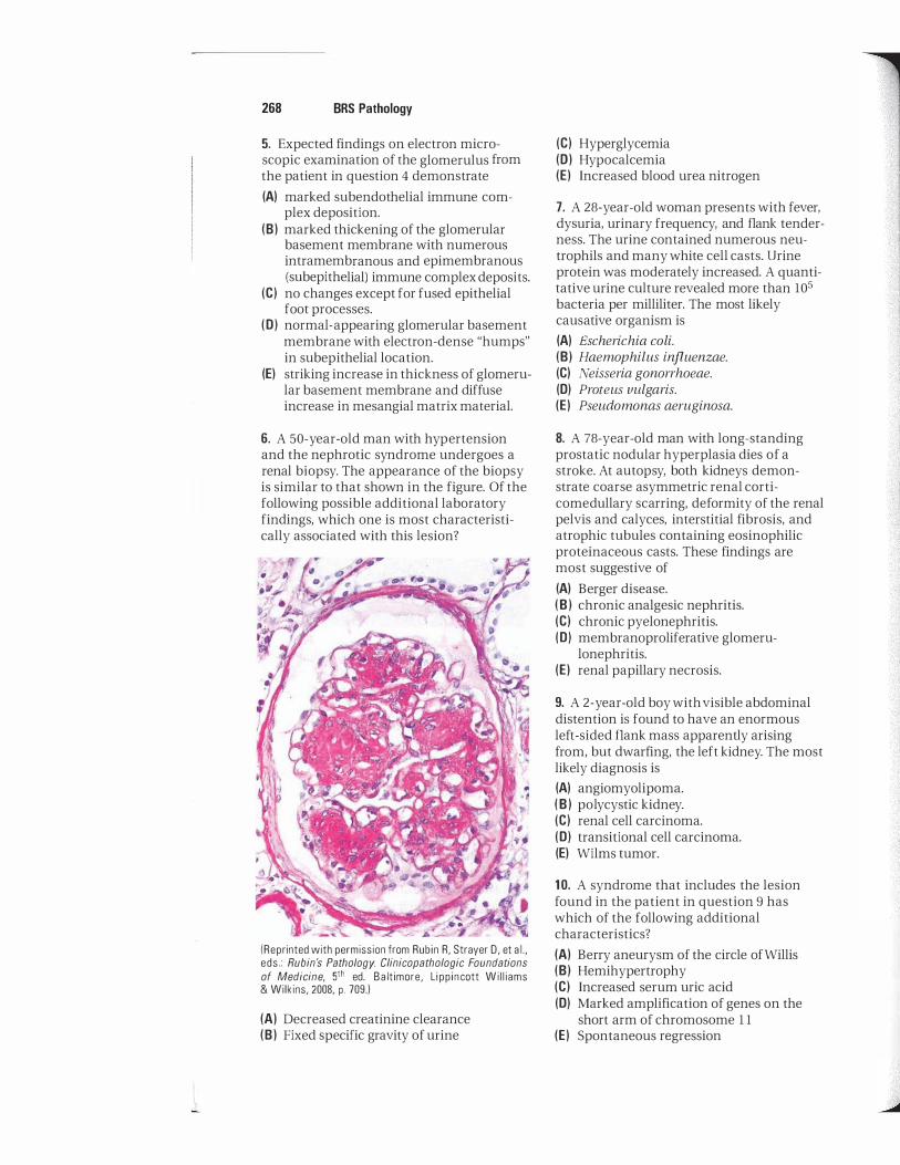
268 BRS Pathology
5. Expected findings on electron microscopic examination of the glomerulus from the patient in question 4 demonstrate
(A) marked subendothelial immune complex deposition.
(B ) marked thickening of the glomerular basement membrane with numerous intramembranous and epimembranous (subepithelial) immune complex deposits.
(e) no changes except for fused epithelial foot processes.
(0) normal-appearing glomerular basement membrane with electron-dense "humps" in subepithelial location.
(E) striking increase in thickness of glomerular basement membrane and diffuse increase in mesangial matrix material.
6. A 50-year-old man with hypertension and the nephrotic syndrome undergoes a renal biopsy. The appearance of the biopsy is similar to that shown in the figure. Of the following possible additional laboratory findings, which one is most characteristically associated with this lesion?
(Reprinted with permission from Rubin R, Strayer D, et a I . , eds . : Rubin's Pathology. Clinicopathologic Foundations of Medicine, 5th ed. Ba lt imore, Li pp in cott Wi l l iams & Wilkins, 2008, p. 709.)
(A) Decreased creatinine clearance (B ) Fixed specific gravity of urine
(e) Hyperglycemia (D ) Hypocalcemia (E) Increased blood urea nitrogen
7. A 28-year-old woman presents with fever, dysuria, urinary frequency, and flank tenderness. The urine contained numerous neutrophils and many white cell casts. Urine protein was moderately increased. A quantitative urine culture revealed more than 105 bacteria per milliliter. The most likely causative organism is
(A) Escherichia coli. (8) Haemophilus influenzae. (e) Neisseria gonorrhoeae. (0) Proteus vulgaris. (E) Pseudomonas aeruginosa.
8. A 78-year-old man with long-standing prostatic nodular hyperplasia dies of a stroke. At autopsy, both kidneys demonstrate coarse asymmetric renal corticomedullary scarring, deformity of the renal pelvis and calyces, interstitial fibrosis, and atrophic tubules containing eosinophilic proteinaceous casts. These findings are most suggestive of
(A) Berger disease. (8) chronic analgesic nephritis. (e) chronic pyelonephritis. (0) membranoproliferative glomeru
lonephritis. (E) renal papillary necrosis.
9. A 2-year-old boy with visible abdominal distention is found to have an enormous left-sided flank mass apparently arising from, but dwarfing, the left kidney. The most likely diagnosis is
(A) angiomyolipoma. (8) polycystic kidney. (e) renal cell carcinoma. (0) transitional cell carcinoma. (E) Wilms tumor.
1 0. A syndrome that includes the lesion found in the patient in question 9 has which of the following additional characteristics?
(A) Berry aneurysm of the circle of Willis (8) Hemihypertrophy (e) Increased serum uric acid (0) Marked amplification of genes on the
short arm of chromosome 1 1 (E) Spontaneous regression

i!iifftit§iii Kidney and U rinary Tract 269
1 1 . A glomerular immunofluorescent pattern for IgG similar to that shown in the figure would be expected in which of the following patients?
(Reprinted with permission from Rubin R, Strayer D, et aI., eds.: Rubin's Pathology. Clinicopathologic Foundations of Medicine, 5th ed. Baltimore, Lippincott Williams & Wilkins, 2008, p. 720.)
(A) A 3-year-old girl with recurrent bouts of the nephrotic syndrome (8) A 9-year-old boy with "smoky" urine 2 weeks after recovery from a streptococcal infection (C) An 18-year-old woman with the nephrotic syndrome and progressive chronic renal disease (0) A 25-year-old man with hemoptysis and hematuria (E) A 26-year-old woman with a "butterfly" rash
12. Enormously enlarged kidneys similar to the one shown in the figure are found at autopsy in a 65-year-old woman. Which of the following is a well-known association or characteristic of this disease process?
( Re printed with permission from Rub in R, Strayer D, et aI., eds. : Rubin's Pathology. Clinicopathologic Foundations of Medicine, 5th ed . Ba ltimore, Lipp incott Wil l iams & Wilkins, 2008, p . 697.)
(A) Berry aneurysm of the circle of Willis (8) Nephrotic syndrome (C) Polycystic ovaries (0) Polycythemia vera (E) X-linked inheritance
13. A 4-year-old boy presents with severe proteinuria, hypoalbuminemia, generalized edema, and hyperlipidemia. The patient
improves on an empiric trial of corticosteroids, with complete resolution of proteinuria. Which of the following is the most likely diagnosis?
(A) Diabetic nephropathy (8) Focal segmental glomerulosclerosis (C) Lupus nephropathy (0) Membranous glomerulonephritis (E) Minimal change disease

270 BRS Pathology
1 4. A 60-year-old woman dies of a tumor that had invaded the renal vein and entered the inferior vena cava. At autopsy, the kidney has the appearance shown in the figure. Which of the following is a characteristic or association of this neoplasm?
(A) Adrenal origin (B) Hematuria (e) Hypocalcemia (0) Tumor cells in the urine (E ) Typical symmetrical bilateral
involvement
15. A 55-year-old man presents with painless hematuria. On cystoscopy, a papillary mass is found in the bladder. Which of the following is a characteristic of this lesion?
(A) Hematuria as a late manifestation (B) Marked tendency to recur after resection (e) Much more likely to be benign than
malignant (0) Occurrence only in the bladder and
nowhere else in the urinary tract (E ) Usual presence of distant metastases at
the time of diagnosis
16. A 25-year-old man presents with generalized edema. Blood tests reveal severe proteinuria, hypoalbuminemia, and hyperlipidemia. The patient does not respond well to a course of corticosteroids. A renal biopsy demonstrates findings indicative of focal segmental glomerulosclerosis. Which of the following best describes the histologic findings in this disorder?
(A) Crescentic formation in glomeruli on light microscopy
( Rep ri nted with pe rmission from Ru b in R, Strayer D, et aI . , eds. : Rubin's Pathology. Clinicopathologic Foundations of Medicine, 5th ed. Ba ltimore, Lipp incott Wi l l iams & Wilkins, 2008, p. 743.)
(8) Intramembranous and epimembranous immune complex deposits in the glomerular basement membrane on electron microscopy
(e) Nodular accumulations of me sa ngia I matrix material on light microscopy
(0) Sclerosis within capillary tufts that involves only some glomeruli and only parts of affected glomeruli on light microscopy
(E ) Wire-loop abnormalities from immune complex deposits and thickening of the glomerular basement membrane on light microscopy
1 7. A 50-year-old woman with a 20-year history of type 2 diabetes mellitus presents with proteinuria, hypoalbuminemia, edema, and hyperlipidemia. She has not monitored her serum glucose levels over the past several years. Diabetic nephropathy is diagnosed. What is the classic morphologic finding in diabetic nephropathy?
(A) Crescentic formation in glomeruli on light microscopy
(8) Intramembranous and epimembranous immune complex deposits in the glomerular basement membrane on electron microscopy
(e) Nodular accumulations of mesangial matrix on light microscopy

L
(0) Sclerosis within capillary tufts that involves only some glomeruli and only parts of affected glomeruli on light microscopy
(E ) Wire-loop abnormalities from immune complex deposits and thickening of the glomerular basement membrane on light microscopy
18. A 5-year-old boy presents with "teacolored urine," oliguria, and periorbital edema. He had a sore throat 2 weeks ago that had resolved before his parents sought medical treatment. The patient is found to be hypertensive. Urea nitrogen and creatinine are elevated. Antistreptolysin 0 titer and anti-DNAase B titer are also elevated. Urine tests are positive for blood and red cell casts. Which of the following is the most likely mechanism for this patient's condition?
(A) Acute bacterial infection of the kidneys
(8) Amyloid deposits (e) ANCA-positive vasculitis (0) Antibody against glomerular basement
membrane antigens (E ) Immune complex deposits
19. A 5-year-old boy diagnosed with poststreptococcal glomerulonephritis was admitted to the hospital several weeks ago. Over the last several weeks, his clinical state has not improved. Severe oliguria has developed, his serum creatinine has continued to rise, and his glomerular filtration rate has decreased by 50% since his admission to the hospital. Which of the following is the most likely diagnosis?
(A) Alport syndrome (8) Membranoproliferative glomeru
lonephritis (e) Membranous glomerulonephritis (0) Rapidly progressive glomeru
lonephritis (E ) Renal papillary necrosis
20. A 25-year-old man presents with hematuria, periorbital edema, hypertension, and hemoptysis. He has also experienced nausea, vomiting, fever, and chills. Serologic testing is positive for antiglomerular basement membrane antibodies. Which of the following is the classic histologic finding in this renal disease?
I!1itfttQiiI Kidney and U rinary Tract 271
(A) Linear immunofluorescence (8) "Lumpy-bumpy" immunofluorescence (e) "Spike and dome" appearance of the
glomerular basement membrane (0) Subendothelial immune complex
deposition (E) Tram-track appearance of the glomeru
lar basement membrane on electron microscopy
21 . A 5-year-old boy presents with hematuria. His mother states that he has had a sore throat for the past 2 days and that he has had hematuria a few times in the past, also concomitantly with a sore throat. She states that his urine usually returns to a normal clear yellow color after a few days. Which of the following is the most likely diagnosis?
(A) Alport syndrome (8) Goodpasture syndrome (e) IgA nephropathy (0) Membranoproliferative glomeru
lonephritis (E ) Poststreptococcal glomerulonephritis
22. An 18-year-old woman presents with suprapubic pain, urinary frequency, dysuria, and hematuria for the past hour. Urine tests show the presence of pyuria but no white cell casts. Physical examination is remarkable only for suprapubic tenderness on palpation. Which of the follOwing is the most likely diagnosis?
(A) Acute pyelonephritis (8) Chronic pyelonephritis (e) Cystitis (0) Fanconi syndrome (E) Nephrocalcinosis
23. A 30-year-old man presents with hypertension, hematuria, palpable renal masses, and flank pain. He states that a kidney disease runs in his family, and his mother and maternal grandfather have it. Laboratory abnormalities confirm renal failure. Which of the following is associated with the disorder described here?
(A) Autosomal recessive inheritance (8) Berry aneurysm of the circle of Willis (e) Clinical manifestation most commonly
at birth (0) Multiple small medullary cysts in both
kidneys (E) Presence of uric acid stones

272 BRS Pathology
24. A 45-year-old man comes to the emergency department in obvious severe pain. He states that he has severe pain in his right flank that extends down to his right groin. The pain is sharp and severe, and it started several minutes earlier. An abdominal radiograph demonstrates the presence of right ureteral urolithiasis. Which of the following
is the most likely composition of this patient's stone?
(A) Ammonium magnesium phosphate (B ) Calcium (e) Cystine (0) Uric acid

Answers and_E_x�p ...... l_a_�tL-.a_ti_o_n�s�" ��] 1 . The answer is B. The diffuse thickening of the walls of the capillary loops seen in the .
illustration is characteristic of membranous glomerulonephritis, the most frequent cause of the nephrotic syndrome in young adults. Expected associated findings in this immune complex disease include granular immunofluorescence and a characteristic "spike and dome" appearance that is best visualized with special stains. Intramembranous and epimembranous (subepithelial) electron-dense immune complex deposits are seen by electron microscopy.
2. The answer is C. The combination of generalized edema, massive proteinuria, hypoalbuminemia, and hyperlipidemia constitutes the nephrotic syndrome, the prototype of which is minimal change disease (lipoid nephrosis) . This disorder characteristically occurs in young children and demonstrates intracytoplasmic lipid in the proximal convoluted tubules, a paucity of glomerular abnormalities by light microscopy, and "fusing" (absenc;:e) of the epithelial foot processes by electron microscopy.
3. The answer is B . The overall prognosis in systemic lupus erythematosus is most closely related to the glomerular lesions in affected patients. These renal lesions are highly variable. In the diffuse proliferative form, almost all of the glomeruli are involved in a marked inflammatory reaction to widespread subendothelial and mesangial immune complex deposition.
4. The answer is B. The combination of hematuria (with red cell casts) , oliguria, azotemia, and hypertension constitutes the nephritic syndrome, the prototype of which is poststreptococcal glomerulonephritis. An indicator of the prior streptococcal infection is an increased titer of antistreptolysin O. Fluid retention is usually minimal, often limited to periorbital edema, and is the result of reduced renal excretion of salt and water, not hypoalbuminemia. Hypertension, not hypotension, is expected. This disorder is an immune complex disorder, and infection of the kidney does not occur.
5. The answer is D. Characteristic electron-dense "humps" on the epithelial side of the basement membrane (subepithelial location) are an extremely important diagnostic feature of poststreptococcal glomerulonephritis. The basement membrane is not thickened in this acute, usually self-limited disorder.
6. The answer is C. The illustration shows nodular glomerulosclerosis (Kimmelstiel-Wilson nodules), the most characteristic glomerular finding in diabetes mellitus. The nodules are accumulations of mesangial matrix-like material.
7. The answer is A. Organisms involved in urinary tract infections are most often normal flora of the colon, and the most frequent of these is Escherichia coli.
8. The answer is C. The combination of coarse asymmetric corticomedullary scarring, deformity of the renal pelvis and calyces, and tubular atrophy is characteristic of chronic pyelonephritis. When the atrophic tubules contain eosinophilic proteinaceous casts, the resultant similarity in appearance to thyroid follicles is referred to as "thyroidization." Although an infectious etiology is assumed, the infectious agent is often not demonstrable.
9. The answer is E. Wilms tumor is the most common renal neoplasm of children. The tumors are often huge and frequently cause abdominal distention. Renal cell carcinoma and adult polycystic kidney can also present as a large flank mass, but not in a child.
273

-
274 BRS Pathology
1 0. The answer is B. Associations of Wilms tumor notably include gene deletions localized to the short arm of chromosome 1 1 ( l I p 13) . In some instances, a "two-hit" mechanism of cancer suppressor gene inactivation, similar to that postulated for retinoblastoma, appears to be operative. Other associations of Wilms tumor include congenital aniridia, genitourinary malformations, and mental-motor retardation (the AGR or WAGR complex) . Another unusual association is "hemihypertrophy" (gross asymmetry of the body) as part of the Beckwith-Wiedemann syndrome; yet another is the Denys-Drash syndrome, which is characterized by intersexual disorders, nephropathy, and Wilms tumor.
1 1 . The answer is D. The illustration demonstrates linear immunofluorescence, which is characteristic of disease caused by antiglomerular basement membrane antibodies. In Goodpasture syndrome, antibodies directed against antigens in the basement membranes of the glomeruli as well as the pulmonary alveoli result in both hemorrhagic pneumonitis with hemoptysis and glomerular disease with hematuria.
1 2. The answer is A. Berry aneurysm of the circle of Will is sometimes leading to subarachnoid hemorrhage is a well-known association of adult polycystic kidney disease. Inheritance is by an autosomal dominant mode of inheritance. Secondary polycythemia, not polycythemia vera, is a complication and is caused by increased secretion of erythropoietin.
13. The answer is E. Minimal change disease (lipoid nephrosis) is the prototype of the nephrotic syndrome in children, who usually respond well to steroid therapy. Minimal change disease is characterized grossly by lipid-laden renal cortices. Light microscopy reveals normal glomeruli, but electron microscopy demonstrates the fusion of epithelial foot processes.
14. The answer is B. The illustration demonstrates a renal cell carcinoma, which most often arises in one of the renal poles, frequently the upper pole. Hematuria is the most frequent presenting sign. The tumor cells have a clear cell appearance, which led to an earlier erroneous concept that this tumor was of adrenal origin and to the older name "hypernephroma." The tumor is often quite large and may result in a palpable mass. Malignant cells are only rarely detected in the urine in renal cell carcinoma.
1 5. The answer is B. The majority of urinary tract tumors are transitional cell carcinomas. Following resection, there is a marked tendency to recur. Even benign-appearing transitional cell "papillomas" are usually malignant. These tumors tend to spread contiguously rather than to distal locations. An early sign is hematuria.
1 6. The answer is D. Focal segmental glomerulosclerosis has clinical manifestations similar to minimal change disease, but it tends to occur in older patients and does not respond well to steroid treatment. Glomerular hyaline sclerosis occurs in a focal (some, but not all, glomeruli) and segmental (only part of an affected glomerulus) distribution.
17. The answer is C. Diabetic nephropathy manifests clinically as the nephrotic syndrome; however, this syndrome is compounded by renal failure and hypertension. Ultrastructural changes include a marked increase in the thickness of the glomerular basement membrane and mesangial accumulation of glycosylated basement membrane-like material. Light microscopy findings include diffuse glomerulosclerosis (a diffuse increase in mesangial matrix) and nodular glomerulosclerosis (nodular accumulations of mesangial matrix) .
18. The answer is E. The clinical description is that of poststreptococcal glomerulonephritis, the prototype of the nephritic syndrome. An antecedent infection, usually of the pharynx or skin, with a group A �-hemolytic streptococcus occurs approximately 1-2 weeks before the onset of the renal manifestations. Poststreptococcal glomerulonephritis is an immune complex disease, with antigen-antibody-complement complexes localizing to the outside (subepithelial side) of the glomerular basement membrane. An intense inflammatory response is elicited, including chemotactic attraction of neutrophils, release of lysosomal enzymes, partial destruction of basement membrane, and bleeding into the Bowman space.

i!1i!l1'dlltld Kidney and U rinary Tract 275
19. The answer is D. The clinical description is that of rapidly progressive glomerulonephritis (RPGN) , which is defined as the nephritic syndrome that progresses rapidly to fulminant renal failure within weeks or months. RPGN is classified into three categories: antiglomerular basement membrane (anti-GBM) antibody disease, immune complex deposit disease, and pauci-immune disease (ANCA-positive) . Characteristic histologic findings in RPGN include crescents between the Bowman capsule and the glomerular tuft. In approximately 50% of cases of RPGN, the disease is of poststreptococcal etiology. It should be. noted, however, that the most common outcome of poststreptococcal glomerulonephritis is complete recovery, and only a small minority of patients progress to RPGN.
20. The answer is A. The clinical description is that of Goodpasture syndrome (antiglomerular basement membrane disease) , caused by antibodies directed against antigens in the glomerular and pulmonary alveolar basement membranes. Because antigens are an intrinsic component of the basement membrane, labeled antibodies "paint" the surface of the basement membrane, reSUlting in the characteristic linear immunofluorescent pattern characteristic of this disorder. "Lumpy-bumpy" immunofluorescence is very coarse, granular immunofluorescence found in poststreptococcal immune complex deposit disease. The tram-track appearance is seen in membranoproliferative glomerulonephritis. The "spike and dome" appearance is seen in membranous glomerulonephritis. Subendothelial immune complex deposition is seen in lupus nephropathy.
21 . The answer is C. IgA nephropathy is the most common glomerular disease and is defined by mesangial deposition of IgA. In its most typical form, the clinical manifestations are limited to a few days of hematuria following an infection; however, there are many etiologic factors, and the condition can vary from one of little import to a severe form of renal disease.
22. The answer is C. The clinical description is that of cystitis, which is characterized by pyuria and hematuria but with no white cell casts in the urine. Patients with acute pyelonephritis present with fever, leukocytosis, flank tenderness, urinary white cells, and white cell casts in the urine. Chronic pyelonephritis is almost always the result of chronic urinary tract obstruction and repeated bouts of acute inflammation in the kidneys.
23. The answer is B. Adult polycystic kidney disease is the most common inherited disorder of the kidney and is characterized by autosomal dominant inheritance. The disease is characterized by partial replacement of the renal parenchyma with cysts. An important association of adult polycystic kidney disease is berry aneurysm of the circle of Willis.
24. The answer is B. Urolithiasis (calculi or stones in the urinary tract) is a very common problem, especially in men. Calcium stones (composed of calcium oxalate or calcium phosphate, or both) account for 80%-85% of urinary stones. Such stones are associated with hypercalciuria, which in turn is caused by increased intestinal absorption of calcium, increased primary renal excretion of calcium, or hypercalcemia. Ammonium magnesium phosphate stones are associated with ammonia-producing (urease-positive) bacteria, such as Proteus vulgaris or Staphylococcus. Uric acid stones are associated with hyperuricemia in 50% of patients and can occur secondary to gout or increased cell turnover (leukemias or myeloproliferative syndromes) . Cystine stones are almost always associated with cystinuria, a genetically determined aminoaciduria.

Male Reproductive System
I. D ISEASES OF THE PENIS
A. Congenital anomalies 1. Hypospadias is an anomaly in which the urethral meatus opens on the ventral surface of
the penis. 2. Epispadias is an anomaly in which the urethral meatus opens on the dorsal surface of the
penis. It is less common than hypospadias.
B. Other abnormalities 1. Phimosis is an abnormally tight foreskin that is difficult or impossible to retract over
the glans penis. This condition may be congenital or result from inflammation or from trauma.
2. Peyronie d isease is subcutaneous fibrosis of the dorsum of the penis. It occurs in the older age group and is of unknown etiology.
3. Priapism is an intractable, often painful erection. This condition is sometimes associated with venous thrombosis of the corpora cavernosa.
C. Inflammatory disorders include a number of sexually transmitted infectious processes.
276
1. Balanitis is inflammation of the glans penis. a. This condition is often associated with poor hygiene. b. It is rare in circumcised individuals.
2. Syphi l is ( l ues) a. The infection is caused by spirochetes of Treponema pallidum, which are demonstra
ble by darkfield examination. b. Syphilis manifests during the primary stage as chancre, an elevated, painless, super
ficially ulcerated, firm papule. This lesion is most commonly located on the glans penis or prepuce and ordinarily heals within 2-6 weeks. Without treatment, it is followed by secondary and tertiary lues.
3. Gonorrhea a. The cause is Neisseria gonorrhoeae, which appears as an intracellular gram-negative
diplococci. � b. Most often, gonorrhea is manifest by acute purulent urethritis. � c. Disease can extend to the prostate and seminal vesicles and can also involve the epi
didymis, but only rarely does it affect the testis. 4. Chlamydial infection
� a. This sexually transmitted disease is a common cause of nongonococcal urethritis. It can also cause epididymitis.
� b. Chlamydial infection should be suspected when bacteria are not demonstrated in a purulent urethral discharge.

D. Neoplasms 1. Carcinoma in situ
i!1lIlftltfi1jj Male Reproductive System 277
a. Bowen disease usually presents as a single erythematous plaque, most often on the shaft of the penis or on the scrotum. ( 1 ) This disease predominantly affects uncircumcised men. The peak incidence is
after the fifth decade. (2) It evolves into invasive carcinoma in fewer than 10% of cases. Additionally, it has
been thought to be associated with an increased risk of visceral malignancy; however, this association has recently been disputed.
b. Erythroplasia of Queyrat usually presents as a single erythematous plaque, most often involving the glans penis or prepuce. ( 1 ) This disease occurs predominantly in uncircumcised men. Median incidence is
in the fifth decade. (2) In approximately 10% of cases, the disease progresses to invasive squamous cell
carcinoma. (3) The disease may be a variant of Bowen disease. It is not associated with visceral
malignancies. c. Bowenoid papulosis
( 1 ) Histologically, this disease resembles Bowen disease and erythroplasia of Queyrat but is considered to be a distinct entity.
(2) This disease differs from Bowen disease and erythroplasia of Queyrat in terms of clinical presentation, appearing as multiple verrucoid (wart-like) lesions often resembling condyloma acuminatum and containing human papillomavirus (HPV) type 16 viral sequences.
(3) The disease affects a younger age group than Bowen disease and erythroplasia of Queyrat.
(4) The disease is not known to progress to invasive carcinoma but is generally considered to be premalignant.
2. Carcinoma a. Penile cancer is most frequently squamous cell carcinoma. b. Disease is rare in circumcised men. c. Incidence is greatly increased in the Far East, Africa, and Central America. d. Predisposing factors include poor personal hygiene and venereal disease. e. Disease is often associated with HPVinfection types 16, 18, 31, and 33.
�-- ------ .-
II. DISEASES OF THE TESTES
A. Cryptorchidism is developmental failure of a testis to descend into the scrotum. � 1. This condition is associated, even after surgical correction, with a greatly increased inci
dence of germ cell tumors, especially seminoma and embryonal carcinoma. � 2. It is associated with testicular atrophy and sterility.
B. Torsion of the spermatic cord compromises blood supply and may result in testicular gangrene.
C. Hydrocele is serous fluid filling and distending the tunica vaginalis. 1 . This condition is most often idiopathic. Sometimes, it is congenital in origin due to per
sistence of continuity of the tunica vaginalis with the peritoneal cavity. It can be secondary to infection or to lymphatic blockage by tumor.
2. Usually, hydroceles can be distinguished clinically from solid testicular tumors by physical examination and transillumination.
D. Hematocele is an accumulation of blood distending the tunica vaginalis. It is most often caused by trauma, although it is occasionally due to a tumor.

278 BRS Pathology
E. Varicocele is a varicose dilation of multiple veins of the spermatic cord.
F. Spermatocele is a sperm-containing cyst. It is most often intratesticular.
G. Testicular atrophy 1 . Etiology is often unknown. 2. This condition may be caused by or associated with:
a. Orchitis, especially mumps orchitis b. Trauma c. Hormonal excess or deficiency due to:
( 1 ) Disorders of the hypothalamus or pituitary (2) Hormonal therapy, especially with estrogens (3) Cirrhosis of the liver
d. Cryptorchidism e. Klinefelter syndrome 1. Chronic debilitating disease g. Old age
H. Inflammation 1 . Orchitis
a. When bacterial, orchitis is often associated with epididymitis. Orchitis may be caused by syphilis.
� b. When viral, orchitis is most often due to mumps virus. � c. When bilateral, orchitis may result in sterility due to atrophy of the seminiferous
tubules. Serum testosterone is decreased, whereas pituitary follicle-stimulating hormone and luteinizing hormone are increased.
2. Epididymitis a. This condition is more common than orchitis. b. Causes most often include:
( 1 ) Neisseria gonorrhoeae (2) Chlamydia trachomatis (3) Escherichia coli (4) Mycobacterium tuberculosis
I. Testicular tumors (Table 18 - 1 ) are most often malignant; more than 9 0% are of germ cell origin . 1 . Seminoma is a malignant germ cell tumor (Figure 18-1 ) .
� a . This tumor is analogous to dysgerminoma, a tumor of the ovary. b. It is the most frequently occurring germ cell tumor, accounting for 4 0%. c. Peak incidence is in the mid-30s age group. d. Presenting features include painless enlargement of the testis. e. Sometimes this tumor is associated with increased serum human chorionic
gonadotropin (hCG) , the same tumor marker associated with choriocarcinoma and other germ cell tumors.
� f. Seminomas are very radiosensitive and can often be cured, even when there are metastasis to abdominal lymph nodes.
2. Embryonal carcinoma is a malignant germ cell tumor. a. This tumor is analogous to a similar tumor occurring in the ovary. b. It is the second most common germ cell tumor, accounting for 20%-3 0%. c. Presenting conditions often include pain or metastasis. d. The serum hCG is often increased. e. The prognosis is much worse than for seminoma.
3. Endodermal sinus (yolk sac) tumor is a malignant germ cell tumor (Figure 1 8-2). a. This tumor is analogous to endodermal sinus tumor of the ovary.
� b. It has a peak incidence in infancy and early childhood and is the most common testicular tumor in this age group.

I!1ifttd1I Male Reproductive System 279
t a blell 18-1 World Hea lth Organization Classification of Testicu l a r Tumors
Types
Germ cell tumors
Tumors of one histologic type:
Seminoma
Spermatocytic seminoma
Embryonal carcinoma
Yolk sac tumor (embryonal carcinoma, infantile type; endodermal sinus tumorl
Polyembryoma
Choriocarcinoma
Teratoma
Mature
Immature
With malignant transformation
Tumors of more than one histologic type:
Embryonal carcinoma and teratoma (teratocarcinoma)
Choriocarcinoma and any other type (specify)
Other combinations (specify)
Stromal sex-cord tumors
Well-differentiated forms:
Leydig cel l tumor
Sertoli cell tumor
Granulosa cell tumor
Mixed forms (specify)
Incompletely differentiated forms
Comments
More than 90% of testicular tumors are of germ cel l origin
Most frequently occurring germ cell tumor; peak incidence at 35 years of age
Older age group; more favorable prognosis
Second most frequently occurring germ cell tumor; often presents with pain or metastasis; somewhat younger age incidence than seminoma
Peak incidence in infancy and early childhood
Almost always malignant
Malignant change of one of the tissues within a teratoma (e.g., development of squamous cel l carcinoma within epidermoid component)
Combinations include mixes of seminoma, embryonal carcinoma, teratoma, and choriocarcinoma
Usually benign; often causes precocious puberty
Usually benign; usually no endocrine manifestations
FIGURE 18-1 Seminoma. Fibrous septa separate nests of tumor cells. A lymphocytic infiltrate i s prominent. (Reprinted with permission from Rubin R, Strayer D, et aI . , eds.: Rubin's Pathology. Clinicopathologic Foundations of Medicine, 5th ed. Baltimore, Lippincott Williams & Wilkins, 2008, p. 769.)

280 BRS Pathology
FIGURE 18-2 Endodermal sinus (yolk sac) tumor. The characteristic glomeruloid SchillerDuval bodies are indicated by arrows. (Reprinted with permission from Rubin R, Strayer D, et aI., eds.: Rubin's Pathology. Clinicopathologic Foundations of Medicine, 5th ed. Baltimore, Lippincott Williams & Wilkins, 2008, p.772 .)
c. This tumor causes an increase in serum a-fetoprotein, the same tumor marker that is associated with hepatocellular carcinoma.
4. Teratoma a. This germ cell tumor is derived from two or more embryonic layers. b. It is most frequently malignant. c. It contains multiple tissue types, such as cartilage islands, ciliated epithelium, liver
cells, neuroglia, embryonic gut, or striated muscle. d. It can be classified as one of three subtypes:
( 1 ) Mature teratoma. This teratoma is almost always malignant, whereas the corresponding ovarian tumor (dermoid cyst) is almost always benign.
(2) Immature teratoma (3) Teratoma with malignant transformation. This teratoma contains malignant tissue,
such as squamous cell carcinoma. 5. Choriocarcinoma is a malignant germ cell tumor that can occur as an element of other
germ cell tumors. a. This tumor is analogous to choriocarcinoma of the ovary. b. Incidence peaks in the second to third decades. c. Histologic characteristics include cells that resemble syncytiotrophoblasts and
cytotrophoblasts. d. This tumor causes an increase in serum hCG.
6. Mixed germ cell tumors consist of varying combinations of germ cell tumor types. � a. Tumors are of variable prognosis determined by the least mature element. � b. Tumors can be teratocarcinoma, that is, a combination of teratoma and embryonal
carcinoma, which is associated with poor prognosis; can also occur in other combinations, including: ( 1 ) Teratoma, embryonal carcinoma, and seminoma (2) Embryonal carcinoma and seminoma
7. Leydig cell ( interstitia l ) tumor is a non-germ cell tumor derived from testicular stroma. a. This tumor is similar to the Sertoli-Leydig cell tumor of the ovary. b. It is most often benign. c. It is often characterized by intracytoplasmic Reinke crystals. The tumor is character
istically known as androgen-producing, but sometimes produces both androgens and estrogens and sometimes corticosteroids.
d. The tumor is most often associated with precocious puberty in children and with gynecomastia in adults.

i!ilfft1tfiEI Male Reproductive System 281
8. Sertoli cel l tumor (androblastoma) is a non-germ cell tumor derived from the sex cordstroma. a. This tumor is also similar to the Sertoli-Leydig cell tumor of the ovary. b. It is usually benign. c. It is characterized by a paucity of endocrine manifestations.
III. DISEASES OF THE PROSTATE
A. Anatomy of the prostate. This chestnut-sized and -shaped structure surrounds the urethra at the base of the bladder. It comprises of four groupings of glands, referred to as the periurethral, transitional, central, and peripheral zones.
� 1. The periurethral . transitional, and central zones collectively are equivalent to the older designation of anterior, middle, and lateral lobes and are often the site of benign prostatic hyperplasia (EPH) .
� 2. The peripheral zone of glands draining into ducts entering the urethral sinus close to the verumontanum is equivalent to the older designation of posterior lobe; it is the characteristic site for carcinoma.
B. BPH (benign nodular hyperplasia). EPH is the most frequent cause of urinary tract obstruction. � 1. EPH is extremely common (almost universal) in older men. � 2. Although EPH has no relation to prostate cancer, the two conditions can coexist.
3. EPH is directly related to the action of dihydrotestosterone (DHT), a testosterone metabolite. DHT is synthesized from testosterone by the action of Sa-reductase, type 2, and inhibition of this enzyme is one approach to the treatment of EPH.
4. EPH may also be caused by an age-related increase in estrogens (estrogens promote expression of receptors for DHT).
5. Hyperplasia of both glandular and fibromuscular stromal elements is characteristic. � 6. EPH is characterized grossly by a rubbery, nodular enlargement of the gland, primarily
affecting the inner groupings of glands, especially the periurethral and transitional zones. 7. The urethra is compressed side to side, resulting in a vertical slit. 8. Most characteristically, EPH results in urinary obstruction, which is manifest by:
� a. Frequency, dysuria, hesitancy (difficulty in starting urination) , and urinary tract infection
b. Incomplete bladder emptying c. Distention and muscular hypertrophy of the bladder; in cases of long duration, bands
of enlarged bladder muscle form characteristic trabeculae. d. Hydroureter and hydronephrosis
C. Adenocarcinoma is extremely common. 1. Prostate cancer occurs in the older age group.
� 2. The course may be indolent or aggressive; this can be predicted by the very useful Glea-son system of grading, based on differentiation. The tumor is often well differentiated.
3. The tumor arises most often from the peripheral group of glands. 4. Diagnosis is most often by rectal examination. 5. Prostate cancer is associated at an early stage with an increase in serum prostate-specific
antigen (PSA). Elevations of PSA reflect a complexed form (bound to a1-antichymotrypsin), which becomes elevated with prostatic cancer, and a free form, which increases with EPH. Thus, an increased total PSA with a decreased fraction of free PSA suggests malignancy, whereas an increased total PSA with a proportionate increase in the fraction of free PSA suggests EPH. A number of other approaches to PSA measurement (e.g., serial PSA, PSA velocity, PSA density) are used to increase the predictive value of this procedure.
6. Prostate cancer is characterized by increased serum prostatic acid phosphatase when the tumor penetrates the capsule into adjacent tissues. Although prostatic acid phosphatase

-.
282 BRS Pathology
is no longer used for detection of early disease because of lack of sensitivity, this enzyme, along with PSA, is useful in the follow-up of disseminated disease.
7. The adenocarcinoma may frequently progress to bony osteoblastic metastasis, which, unfortunately, may be the presenting sign. In this instance, an increase in serum alkal ine phosphatase is often an indicator of osteoblastic lesions and virtual ly ensures that the osteoblastic tumors orig inated in the prostate.
8. When disseminated, the cancer may respond to endocrine therapy because tumor growth is partially related to the activity of androgens.

Review Test
Directions: Each of the numbered items or incomplete statements in this section is followed by answers or by completions of the statement. Select the one lettered answer or completion that is best in each case.
1. A 23-year-old African-American man who is known to have sickle cell anemia presents to the emergency department with a painful erection. The patient explains that the erection had started 3 hours ago. This condition is referred to as
(A) balanitis. (8 ) hypospadias. (e) Peyronie disease. (D) phimosis. (E) priapism.
2. A 43-year-old businessman visits his primary care physician complaining of a purulent penile discharge. He had unprotected sex with a woman he met at a conference 1 week ago. Gram stain of the discharge fails to reveal any organisms. Which of the following is the most likely cause of the discharge?
(A) Bowen disease (8) Chlamydia trachomatis (e) Herpes simplex virus (D ) Neisseria gonorrhoeae (E) Treponema pallidum
3. A 5-year-old boy is brought to the pediatrician for a physical examination prior to beginning elementary school. On examination, the boy has only one palpable testis in the scrotum. Further examination reveals a palpable mass in the left inguinal region. This condition is referred to as
(A) cryptorchidism. (8) hydrocele. (e) orchitis. (D) torsion of the spermatic cord. (E) varicocele.
4. A 36-year-old man presents to his primary care physician complaining of painless
enlargement of the testis. Further laboratory studies reveal an increase in serum human chorionic gonadotropin. Of the following, which of the follOwing is the most likely diagnOSiS?
(A) Dysgerminoma (8) Embryonal carcinoma (e) Seminoma (D) Teratoma (E) Yolk sac tumor
5. A 3-year-old boy is brought to the pediatrician because his mother noticed an abnormal mass in his scrotum while changing his diapers. Further workup demonstrates elevated levels of serum a-fetoprotein. Which of the following is the most likely diagnosis?
(A) Choriocarcinoma (8) Endodermal sinus (yolk sac)
tumor (e) Hepatocellular carcinoma (D) Leydig cell (interstitial) tumor (E) Teratoma
6. A 66-year-old man visits his family physician with complaints of urinary frequency, hesitancy, and dysuria. Digital rectal examination reveals an enlarged prostate, and the consistency is rubbery and nodular. Serum prostate-specific antigen is modestly increased. Which of the following is most closely related to the pathogenesis of the likely disorder described here?
(A) Dihydrotestosterone (8) Estrogen (e) a-Fetoprotein (D) Human chorionic gonadotropin (E) Testosterone
283

284 BRS Pathology
7. A 58-year-old African-American man presents to the emergency department with severe back pain. His history is negative for trauma and he has no other complaints. He denies urinary frequency, hesitancy, or dysuria. A digital rectal examination confirms the presence of a firm, hard, asymmetrical, and stony prostate. Imaging of the spine suggests osteoblastic involvement of
the spine at lumbar vertebrae L3-L4. In addition to an increase in PSA, which serum marker might also be elevated?
(A) Alkaline phosphatase (B) Androgens (e) Carcinoembryonic antigen- 125 (0) a-Fetoprotein (E) Human chorionic gonadotropin

Answers and Explanations
1 . The answer is E. Priapism is an intractable, often painful erection associated with condi-_ tions such as sickle cell anemia, hypercoagulable states, spinal injuries, and some drugs. Balanitis is associated with poor hygiene and results from inflammation of the glans penis. Hypospadias is an anatomical anomaly wherein the urethral meatus opens on the ventral side of the penis. Peyronie disease results from subcutaneous fibrosis of the dorsum of the penis. Phimosis is an abnormally tight foreskin that is difficult or impossible to retract over the glans penis.
2. The answer is B. Chlamydia trachomatis is one of the leading causes of urethritis and should be suspected whenever a gonorrhea-like discharge fails to show gram-negative diplococci within neutrophilic phagocytes. It is a sexually transmitted disease that can also cause epididymitis. Bowen disease presents as a single erythematous plaque on the penis or scrotum and may evolve into invasive carcinoma. Herpes simplex virus can cause a vesicular rash on the penis. Treponema pallidum causes syphilis, which may present with a painless chancre on the penis.
3. The answer is A. Cryptorchidism (undescended testis) predisposes to testicular atrophy and sterility. It is associated with an increased incidence of germ cell tumors of the testis, even if the testis is surgically moved from its ectopic location back to the scrotum. A hydrocele is a serous fluid collection in the scrotum. Orchitis (inflammation of the testis) can result from bacterial or viral infection. A varicocele results from dilation of the veins of the spermatic cord, and the term "bag of worms" aptly describes the abnormality.
4. The answer is C. Seminoma is the most common germ cell tumor of the testis. Serum human chorionic gonadotropin levels, elaborated by syncytiotrophoblasts, are elevated in about 15% of cases, but these elevations are not as high as those seen in choriocarcinoma. The tumor is highly radiosensitive and often curable, even when metastatic. Dysgerminoma is analogous to a tumor that occurs in the ovary. Embryonal carcinoma often presents with pain and is the second most common tumor of the testis. Yolk sac tumor is the most common tumor of the testis during infancy and early childhood and is usually accompanied by an increase in serum a-fetoprotein.
5. The answer is B. Endodermal sinus (yolk sac) tumor is the most common malignant germ cell tumor of the testis in infancy and early childhood. It is characterized by an increase in serum a-fetoprotein, as well as histologically stainable aI-antitrypsin. Choriocarcinoma is less common in this age group and often results in an increase in human chorionic gonadotropin. Although hepatocellular carcinomas also elaborate a-fetoprotein, this is very unlikely in this patient's clinical setting. Leydig cell tumors are derived from testicular stroma and produce androgens and estrogens, often presenting with precocious puberty. Teratomas contain multiple germ layers and numerous tissues, including hair, teeth, and sebaceous tissue. Unlike the teratomas encountered in women, most teratomas in men are malignant.
6. The answer is A. Dihydrotestosterone (DHT) is a major growth factor for prostatic tissue. It is derived from the conversion from testosterone by the action of the enzyme Sa-reductase, type 2. Pharmaceutical inhibition of this enzyme is useful in the medical management of benign prostatic hyperplasia. Estrogens also play an indirect role by stimulating the production of DHT receptors. Human chorionic gonadotropin and a-fetoprotein are serum tumor markers for testicular cancers and bear no relevance to benign prostatic hyperplasia.
285

286 BRS Pathology
7. The answer is A. Serum alkaline phosphatase is an indicator of osteoblastic lesions in this advanced and unfortunate patient presentation. Both prostate-specific antigen and serum prostatic acid phosphatase are increased in prostatic cancer. Although prostatic cancer: are usually quite androgen-responsive, androgens are not monitored for diagnosis or treatment of prostate cancer. Carcinoembryonic antigen- 125 is a tumor marker for ovarian cancer. Serum a-fetoprotein and serum human chorionic gonadotropin are elevated in various germ cell neoplasms of the testis.
1

Female Reproductive System and Breast
I. VULVA AND VAGINA
A. Miscellaneous disorders 1 . Barthol in cyst results from an obstruction of Bartholin ducts. The cyst can become second
arily infected, most often by Neisseria gonorrhoeae or, less often, by Staphylococcus. 2. Vulvar dystrophies are a group of disorders of epithel ial growth that often present with
leukoplakia, a white, patch-like lesion. a. Histologic forms
( 1 ) Lichen sclerosus and hyperplastic dystrophy. which have no malignant potential (2) Atypical hyperplastic dystrophy. a premalignant lesion
� b. Cl inical characteristics. Pruritus and l eukoplakia. Leukoplakia can be a manifestation of several diverse processes and should be evaluated by biopsy.
B. Infectious disorders 1 . Candidiasis (monil iasis) is the most common form of vaginitis.
a. The cause is Candida albicans, a normal component of the vaginal flora. b. Associated conditions include diabetes mellitus, pregnancy, broad-spectrum antibi
otic therapy, oral contraceptive use, and immunosuppression. � c. Candidiasis is characterized by white, patch-like mucosal lesions, a thick white dis
charge, and vulvovaginal pruritus. 2. Trichomoniasis is the second most common type of vaginitis.
a. The cause is Trichomonas vaginalis. b. Trichomoniasis is most often transmitted by sexual contact.
3. Bacterial vaginosis (Gardnerellavaginitis) is the most common cause of vaginal discharge. Characteristically, it is a thin, homogeneous vaginal discharge with a malodorous, fishy amine odor, especially on addition of 10% potassium hydroxide. a. The cause is a loss of the normal vaginal lactobacilli, a consequent overgrowth of
anaerobes (e.g., Prevotella bivia, Mobiluncus spp., Peptostreptococcus spp.) , and a resultant superficial polymicrobial vaginal infection.
b. This type of vaginitis accounts for many cases formerly classified as nonspecific vaginitis. It is usually transmitted by sexual contact.
c. Bacterial vaginosis is associated with increased numbers of the facultative anaerobe Gardnerella vaginalis.
d. The appearance of "clue cells" (vaginal epithelial cells that have a stippled appearance due to adherent coccobacilli) in Papanicolaou (Pap) smear preparations is characteristic.
4. Toxic shock syndrome a. This condition was initially associated with the use of highly absorbent tampons. It is
caused by exotoxin produced by Staphylococcus au reus, which grows in the tampon.
287

--
288 BRS Pathology
b. Characteristic features include fever, vomiting, and diarrhea, sometimes followed by renal failure and shock. A generalized rash is followed by desquamation.
� Gonorrhea •
a. The cause is N. gonorrhoeae. Gonorrhea is a frequent cause of pelvic inflammatory disease. Other frequent etiologic agents of pelvic inflammatory disease are Chlamy
dia trachomatis and enteric bacteria. b. Transmission is by sexual contact. c. The disorder can be asymptomatic but infectious. Disease can ascend to infect the
endocervix, uterine canal, and fallopian tubes. It is characterized by purulent acute inflammation, initially of the urethra, paraurethral and Bartholin glands, and Skene ducts.
d. Gonorrhea can result in extragenital infections, including: ( 1 ) Pharyngitis associated with orogenital sexual contact (2) Proctitis associated with anal intercourse (3) Purulent arthritis, which is most often monoarticular, involving a large joint, such
as the knee, as a consequence of blood-borne infection (4) Ophthalmia neonatorum, a neonatal conjunctival infection acquired at delivery
6. Chlamydia I i nfections a. Chlamydial cervicitis is the most common sexually transmitted disease.
( 1 ) The cause is certain serotypes of C. trachomatis. Chlamydial cervicitis is a frequent cause of pelvic inflammatory disease.
(2) The disease is most often asymptomatic. b. Lymphogranuloma venereum
( 1 ) This disease occurs primarily in the tropics. (2) The cause is C. trachomatis Ll , L2, or L3 serotypes. (3) Lymphogranuloma venereum is manifest initially as a small papule or ulcer, fol
lowed by superficial ulcers and enlargement of regional lymph nodes, which become matted together. It can lead to rectal stricture as a result of inflammatory reaction and scarring.
7. Herpes simplex virus ( HSV) infections a. HSV type 2 infection accounts for the majority of genital herpes cases and is spread
by sexual contact. b. HSV infection produces small vesicles and shallow ulcers that can involve the cervix,
vagina, clitoris, vulva, urethra, and perianal skin. Multinucleated giant cells with viral inclusions are found in cytologic smears from lesions.
8. Syphi l is a. The cause is Treponema pallidum. b. Transmission is by sexual contact. c. The initial stage manifests as a firm, painless ulcer known as a chancre, which is usu
ally not apparent clinically. d. The disease is sometimes manifest during secondary syphilis as condyloma lata,
which are gray, flattened, wart-like lesions. Note: Condyloma lata should not be confused with condyloma acuminatum (see later) .
e. Syphilis is a hazard during pregnancy because spirochetes can cross the placenta and result in fetal malformation.
9. Chancroid a. The cause is Haemophilus ducreyi. b. Transmission is by sexual contact. c. This disease is most common in tropical areas; it is rare in the United States. d. Chancroid is characterized by a soft and painful ulcerated lesion in contrast to the
chancre of syphilis, which is firm and painless. 10. Granuloma inguinale
a. The cause is Calymmatobacterium (Donovania) granulomatis, a gram-negative rod. b. Transmission is probably sexual. c. Donovan bodies, which are multiple organisms filling large histiocytes, are character
istic and are an important diagnostic histopathologic feature.
--

i!i1tftt!llIEI Female Reproductive System and Bre ast 289
d. An infection appears initially as a papule, which becomes superficially ulcerated. It progresses by adjacent lesions coalescing to form large genital or inguinal ulcerations, sometimes with lymphatic obstruction or genital distortion.
C. Neoplasms of the vulva (Table 19-1) 1 . Papil lary hidradenoma is the most common benign tumor of the vulva.
a. This tumor originates from apocrine sweat glands. b. It presents as a labial nodule that may ulcerate and bleed. c. Cure is by simple excision.
2. Condyloma acuminatum (Figure 19- 1 ) a. This benign squamous cell papilloma is caused by human papi l lomavirus (HPV), most
frequently HPV types 6 and 1 1 . b. Transmission is by sexual contact. c. Clinical manifestations include multiple wart-like lesions, venereal warts, in the vul
vovaginal and perianal regions and sometimes on the cervix. d. Histologic characteristics include koilocytes, or expanded epithelial cells with peri
nuclear clearing. 3. Squamous cel l carcinoma is the most common malignant tumor of the vulva.
t'a b l e 19-1 Compa rison of Tumors of the Female Reprodu ctive System .�-��.--
Type Behavior location Comments
Condyloma acuminatum Benign Vulvovaginal, perianal, Most often multiple; etiologic agent (venereal wart) sometimes cervical HPV (types 6 and 11)
Squamous cell carcinoma Malignant Vulva May be preceded by atypical hyperplastic dystrophy
Clear cell adenocarcinoma Malignant Vagina Peak incidence in teenagers and young women exposed in utero to DES
Squamous cell carcinoma Malignant Vagina Uncommon location for primary squamous cel l carcinoma; more often due to extension of squamous cel l carcinoma of the cervix
Squamous cell carcinoma Malignant Uterine cervix Squamocolumnar junction most frequent site of origin; often preceded by dysplasia; HPV (types 16, 18,31, and 33) is suspected to be the etiologic agent
Leiomyoma Benign Uterine corpus Most frequently occurring neoplasm of women; most often multiple; increases in size during pregnancy; regresses with menopause
Endometrial carcinoma Malignant Uterine corpus Peak incidence in older age group; predisposed by estrogen stimulation; incidence increasing
Cystadenoma, serous Benign Ovary or mucinous
Cystadenocarcinoma, serous Malignant Ovary Rupture of mucinous form can lead to or mucinous pseudomyxoma peritonei
Mature teratoma Benign Ovary Most frequent ovarian tumor (dermoid cyst)
Choriocarcinoma Malignant Ovary or gestational tissue Increased hCG in serum and urine
Fibroma Benign Ovary Can be associated with Meigs syndrome (ovarian fibroma, ascites, and hydrothorax)
Granulosa cell tumor Benign Ovary Estrog en·se cretin g
Krukenberg tumors Malignant Ovaries Metastatic replacement of ovaries with' signet·ring cells from primary malignant tumor elsewhere (often from the stomach)
HPV = human papillomavirus; DES = diethylstilbestrol; hCG = human chorionic gonadotropin.
1
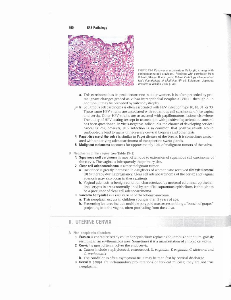
290 BRS Pathology
FIGURE 19-1 Condyloma acuminatum. Koilocytic change with perinuclear haloes is evident. (Reprinted with permission from Rubin R, Strayer 0, et aI., eds.: Rubin's Pathology. Clinicopathologic Foundations of Medicine, 5th ed. Baltimore, Lippincott Williams & Wilkins, 2008, p. 785.)
a. This carcinoma has its peak occurrence in older women. It is often preceded by premalignant changes graded as vulvar intraepithelial neoplasia (YIN) 1 through 3. In addition, it may be preceded by vulvar dystrophy.
� b. Squamous cell carcinoma is often associated with HPV infection type 16, 18 , 3 1 , or 33. These same HPV strains are associated with squamous cell carcinoma of the vagina and cervix. Other HPV strains are associated with papillomatous lesions elsewhere. The utility of HPV testing (except in association with positive Papanicolaou smears) has been questioned. In virus-negative individuals, the chance of developing cervical cancer is low; however, HPV infection is so common that positive results would undoubtedly lead to many unnecessary cervical biopsies and other tests.
4. Paget disease of the vulva is similar to Paget disease of the breast. It is sometimes associated with underlying adenocarcinoma of the apocrine sweat glands.
5. Mal ignant melanoma accounts for approximately 10% of malignant tumors of the vulva.
D. Neoplasms of the vagina (see Table 19-1) 1. Squamous cel l carcinoma is most often due to extension of squamous cell carcinoma of
the cervix. The vagina is infrequently the primary site. 2. Clear cell adenocarcinoma is a rare malignant tumor.
a. Incidence is greatly increased in daughters of women who received diethylstil bestrol (DES) therapy during pregnancy. Clear cell adenocarcinoma of the cervix and vaginal adenosis may also occur in these patients.
b. Vaginal adenosis, a benign condition characterized by mucosal columnar epitheliallined crypts in areas normally lined by stratified squamous epithelium, is thought to be a precursor of clear cell adenocarcinoma.
3. Sarcoma botryoides is a rare variant of rhabdomyosarcoma. a. This neoplasm occurs in children younger than 5 years of age. b. Presenting features include multiple polypoid masses resembling a "bunch of grapes"
projecting into the vagina, often protruding from the vulva.
II. UTERINE CERVIX
A. Non-neoplastic disorders 1. Erosion is characterized by columnar epithelium replacing squamous epithelium, grossly
reSUlting in an erythematous area. Sometimes it is a manifestation of chronic cervicitis. 2. Cervicitis most often involves the endocervix.
a. Causes include staphylococci, enterococci, C. vaginalis, T. vaginalis, C. albicans, and C. trachomatis.
b. The condition is often asymptomatic. It may be manifest by cervical discharge. 3. Cervical polyps are inflammatory proliferations of cervical mucosa; they are not true
neoplasms.

B. Dysplasia and carcinoma in situ
i!1l!J'll!IIllI Female Reproductive System and Breast 291
1. The squamocolumnar junction is most frequently involved. 2. There is a major association with HPV infection types 16, 18 , 3 1 , or 33. 3. Disordered epithelial growth manifest by loss of polarity and nuclear hyperchromasia,
beginning at the basal layer and extending outward, is characteristic. 4. Dysplasia can progress through mild, moderate, and severe forms to carcinoma in situ
and is classified as cervical intraepithel ia l neoplasia (CIN), with subtypes of CIN 1, CIN 2, or CIN 3, depending on the extent of epithelial involvement. CIN 3 (carcinoma in situ) is characterized by atypical changes extending through the entire thickness of the epithelium.
C. Invasive carcinoma 1 . General considerations
a. The occurrence peaks in middle-aged women. � b. The cancer is most often squamous cel l carcinoma; adenocarcinoma accounts for
approximately 5% of cases. c. Most frequently, the carcinoma arises from preexisting CIN at the squamocolumnar
junction. It evolves through a series of increasing epithelial abnormalities proceeding from dysplasia to carcinoma in situ and then to invasive carcinoma.
� d. Since the introduction of the Papanicolaou (Pap) cytologic screening test, squamous cell carcinoma has exhibited a striking decrease in mortality.
� 2. Epidemiologic factors (probable spread by sexual contact) a. Early sexual activity and multiple sexual partners are associated with increased incidence. b. Incidence is high in prostitutes, rare in celibates, and rare in some Jewish populations.
The traditional belief that circumcision of male sexual partners exerts a protective effect has not been confirmed.
c. Incidence is increased in the economically deprived. d. Cigarette smoking is also associated with increased incidence, but the relationship
remains unclear. 3. Role of HPV
a. Dysplastic cells frequently demonstrate koilocytosis (as in HPV-induced condyloma acuminatum) .
� b. HPV sequences are often integrated into genomes of dysplastic or malignant cervical epithelial cells; HPV types 16, 18, 3 1 , and 33 are most common, as in most malignant genital squamous cell tumors, and are associated with more than 90% of cases. HPV viral proteins E6 and E7 bind and inactivate the gene products of p53 and Rh, respectively.
III. UTERINE CORPUS
A. Endometritis 1. Acute endometritis
a. The cause is most often S. aureus or Streptococcus species. b. This condition is most often related to intrauterine trauma from instrumentation,
intrauterine contraceptive devices, or complications of pregnancy, such as postpartum retention of placental fragments.
2. Chronic specific ( granu lomatous) endometritis is most often tuberculous in etiology.
B. Endometriosis � 1 . The presence and proliferation of ectopic endometrial tissue is characteristic.
2. Causes may include retrograde dissemination of endometrial fragments through fallopian tubes during menstruation, with implantation on the ovary or other peritoneal structures, or blood-borne or lymphatic-borne dissemination of endometrial fragments.
3. The condition is characteristically responsive to hormonal variations of the menstrual cycle.

292 BRS Pathology
4. Menstrual-type bleeding occurs into the ectopic endometrium, resulting in blood-filled, or so-called "chocolate," cysts.
� 5. Endometriosis occurs most often in the pelvic area; the ovary is the most common site, followed by the uterine ligaments, rectovaginal septum, pelvic peritoneum, and other sites.
6. Clinical manifestations include severe menstrua l-related pain. 7. Endometriosis is non-neoplastic and has no relation to endometrial cancer. It often
results in infertility.
C. Adenomyosis is characterized by islands of endometrium within myometrium.
D. Endometrial hyperplasia is an abnormal proliferation of endometrial glands. � 1. The cause is usually excess estrogen stimulation, which in turn may be caused by anovu
latory cycles, polycystic ovary disease, estrogen-secreting ovarian tumors, such as granulosa cell tumor, and estrogen replacement therapy.
� 2. This condition is most often manifest clinically by postmenopausal bleeding. � 3. It is sometimes a precursor lesion of endometrial carcinoma; the risk of carcinoma varies
with the degree of cel lular atypia. Simple (cystic or mild) hyperplasias have a low malignant potential, while higher grade (atypical or adenomatous with atypia) hyperplasias have a greater malignant potential.
E. Endometrial polyp � 1. This benign lesion usually occurs in women older than 40 years of age.
2. It may result in uterine bleeding.
F. Leiomyoma (fibroid) (Figure 19-2) � 1. This is the most common uterine tumor and the most common of all tumors in women; the
incidence is increased in women of African lineage. 2. The tumor is a benign neoplasm; putative malignant transformation is rare. 3. Leiomyomas occur in multiple separate foci in most cases.
� 4. The tumors are estrogen-sensitive. They often increase in size during pregnancy, and they almost always decrease in size fol lowing menopause.
5. The tumors may lie within the myometrium (intramural) or in subendometrial (submucous) or subperitoneal (subserous) locations. Leiomyomas, especially if subendometrial, are often manifest clinically by menorrhagia (increased menstrual bleeding) .
G. Leiomyosarcoma 1. This malignant tumor occurs infrequently.
� 2. It arises de novo and is almost never caused by malignant transformation of a leiomyoma.
FIGURE 19-2 Leiomyoma of the uterus. These benign tumors are often multiple and multifocal in origin. They tend to decrease in size after menopause. (Reprinted with permission from Rubin R, Strayer D, et aI., eds.: Rubin's Pathology. Clinicopathologic Foundations of Medicine, 5th ed. Baltimore, Lippincott Williams & Wilkins,
2008, p. 813.) .

H. Endometrial carcinoma
IB:ltftltlI]j Female Reprodu ctive System and Breast 293
1 . Endometrial carcinoma is the most common gynecologic malignancy. In contrast to carcinoma of the cervix, the incidence of this tumor is increasing. It is increased in incidence in association with nulliparity.
� 2. Peak occurrence is in older women, who are affected more by endometrial carcinoma than by carcinoma of the cervix.
� 3. Clinical manifestations most often include postmenopausal bleeding. This often leads to early diagnosis.
4. The cancer is often preceded by endometrial hyperplasia, especially higher grade dysplasias.
� 5. Predisposing factors include prolonged estrogen stimulation, as occurs with exogenous estrogen therapy or estrogen-producing tumors, as well as obesity, diabetes mellitus, and hypertension; the common factor may be obesity because estrone can be synthesized in peripheral adipose tissues.
IV. FALLOPIAN TUBES
A. Salpingitis 1. This disorder is most often associated with inflammation of the ovaries and other adjacent
tissue (pelvic inflammatory disease) . It can be caused by trauma, such as surgical manipulation.
2. Causes are most often N. gonorrhoeae, various anaerobic bacteria, C. trachomatis, streptococci, and other pyogenic organisms.
3. Salpingitis can result in pyosalpinx, a tube filled with pus, or hydrosalpinx, a tube filled with watery fluid; it may also result in a tubo-ovarian abscess.
B. Hematosalpinx is bleeding into the fallopian tube. It is most often caused by ectopic pregnancy.
C. Fallopian tube tumors 1 . Aderiomatoid tumor is the most frequent benign tumor of the fallopian tubes. 2. Adenocarcinoma most often results from direct extension or metastasis from tumors
originating elsewhere.
V. OVARIES
A. Ovarian cysts 1 . Fol l icular cyst
a. This cyst is due to distention of the unruptured graafian follicle. b. It is sometimes associated with hyperestrinism and endometrial hyperplasia.
2. Corpus luteum cyst a. This cyst results from hemorrhage into a persistent mature corpus luteum. b. It is symptomatically associated with menstrual irregularity, occasionally with
intraperitoneal hemorrhage. 3. Theca-lutein cyst
a. This cyst results from gonadotropin stimulation; it can be associated with choriocarcinoma and hydatidiform mole.
b. It is often multiple and bilateral and lined by luteinized theca cells. 4. Chocolate cyst
� a. This cyst is a blood-containing cyst resulting from ovarian endometriosis with hemorrhage.
b. The ovary is the most frequent site of endometriosis.

294 BRS Pathology
5. Polycystic ovary (Stein-Leventhal ) syndrome a. This syndrome characteristically occurs in young women. b. It is an important cause of infertility. c. Clinical characteristics include amenorrhea, infertil ity, obesity, and hirsutism. d. Causes may include excess luteinizing hormone (LH) and androgens. e. Polycystic ovary syndrome may be associated with insulin resistance with an
increased risk of diabetes mellitus. Hyperinsulinemia may lead to increased ovarian androgen production, which may in turn lead to increased LH.
f. Morphologic characteristics include the following: ( 1 ) Markedly thickened ovarian capsule (2) Multiple smal l fol l icular cysts containing a granulosa cell layer and a luteinized
theca intern a (3) Cortical stromal fibrosis with islands of focal luteinization
B. Ovarian tumors are categorized according to the World Health Organization (WHO) classification, which is based on the site of origin of the tumor. The tumors are divided as follows: 1 . Tumors of surface epithel ia l origin make up almost three fourths of ovarian tumors. They
occur in women older than 20 years of age. a. Serous tumors
( 1 ) Serous cystadenoma. This benign cystic tumor is lined with cells similar to fallopian tube epithelium. It accounts for approximately 20% of all ovarian tumors and is frequently bilateral.
(2) Serous cystadenocarcinoma. This malignant tumor accounts for approximately 50% of ovarian carcinomas and is frequently bilateral (Figure 1 9-3) .
b. Mucinous tumors ( 1 ) Mucinous cystadenoma. This benign tumor is characterized by multilocular cysts
lined by mucus-secreting columnar epithelium and filled with mucinous material. (2) Mucinous cystadenocarcinoma (Figure 19-4) ji) (a) This malignant tumor, through rupture or metastasis, can result in
pseudomyxoma peritonei with multiple peritoneal tumor implants, all producing large quantities of intraperitoneal mucinous material.
(b) Although mucinous cystadenocarcinoma of the ovary is the most common cause, pseudomyxoma peritonei can also result from mucinous cystadenoma, carcinomatous mucocele of the appendix, and other mucinous tumors.
c. Endometrioid tumors histologically resemble the endometrium. They are usually malignant.
FIGURE 19-3 Serous cystadenocarcinoma. Papillary projections and psammoma bodies (arrows) are evident. (Reprinted with permission from Rubin R, Strayer D, et ai., eds.: Rubin's Pathology. Clinicopathologic Foundations ' of Medicine, 5th ed. Baltimore, Lippincott Williams & Wilkins, 2 008, p. 82 2 .)

FIGURE 19-4 Mucinous cystadenocarcinoma. This is one of the mucinous tumors that can cause pseudomyxoma peritonei. (Reprinted with permission from Rubin R, Strayer D, et ai., eds.: Rubin's Pathology. Clinicopathologic Foundations of Medicine, 5th ed. Baltimore, Lippincott Williams & Wilkins, 2008, p. 82 2 .)
i!iltl"mliI Female Reproductive System and Breast 295
d. Clear cell tumors are rare tumors that are almost always malignant. e. Brenner tumors are rare, benign tumors. They are characterized by small islands of
epithelial cells resembling bladder transitional epithelium interspersed within a fibrous stroma.
2. Tumors of germ cell origin make up one fourth of ovarian tumors. They account for most ovarian tumors occurring in women younger than 20 years of age. a. Dysgerminoma
( 1 ) This tumor is malignant. � (2) This tumor is analogous to testicular seminoma. b. Endodermal sinus (yolk sac) tumor
( 1 ) This tumor resembles extraembryonic yolk sac structures. It produces a-fetoprotein. (2) This tumor is analogous to endodermal sinus tumor of the testis.
c. Teratomas ( 1 ) These tumors characteristically demonstrate tissue elements derived from two or
three embryonic layers. (2) Teratomas are observed in three distinct forms:
(a) Immature teratoma. This aggressive malignant tumor includes immature cellular elements.
(b) Mature teratoma (dermoid cyst). This accounts for approximately 20% of ovarian tumors and 90% of germ cell tumors.
� ( i ) The dermoid cyst is the most frequent benign ovarian tumor. � ( i i ) The cyst is lined by skin, including hair follicles and other skin
appendages; other elements often include bone; tooth; cartilage; and gastrointestinal, neurologic, respiratory, and thyroid gland tissues. Radiographically visible focal calcifications may lead to diagnosis.
( i i i ) The cyst may arise by reduplication of meiotic maternal chromosomes, giving rise to 46,XX cells of maternal origin.
(c) Monodermal teratoma. This cyst contains only a single tissue element; for example, the most common is struma ovarii, which consists entirely of thyroid tissue and can be hyperfunctional, resulting in hyperthyroidism.
d. Ovarian choriocarcinoma. This aggressive malignant tumor secretes human chorionic gonadotropin (hCG).
3. Tumors of ovarian sex cord-stromal origin account for a small percentage of ovarian neoplasms. Women of all ages are affected. a. Thecoma-fibroma group of tumors
( 1 ) Fibroma (a) This solid tumor consists of bundles of spindle-shaped fibroblasts.

296 BRS Pathology
FIGURE 19-5 Granulosa cell tumor. Call-Exner bodies are formed by tumor cells arranged in a circular pattern. (Reprinted with permission from Rubin R, Strayer 0, et aI., eds.: Rubin's Pathology. Clinicopathologic Foundations of Medicine, 5th ed. Baltimore, Lippincott Williams & Wilkins, 2008, p. 827 .)
(b) It may be associated with Meigs syndrome, a triad of ovarian fibroma, ascites, and hydrothorax.
(2) Thecoma (a) This tumor demonstrates round lipid-containing cells in addition to fibroblasts. (b) It is occasionally estrogen-secreting.
b. Granulosa cel l tumor � ( 1 ) This estrogen-secreting tumor causes precocious puberty.
(2) In adults, it is associated with endometrial hyperplasia or endometrial carcinoma. (3) The tumor consists of small cuboidal, deeply staining granulosa cells arranged in
anastomotic cords. (4) Call-Exner bodies, small follicles filled with eosinophilic secretion, are an impor
tant diagnostic feature (Figure 19-5) . � c. Sertol i-Leydig cell tumor (androblastoma, arrhenoblastoma). This androgen-secreting tumor
is associated with virilism (masculinization) . 4. Tumors metastatic to the ovary account for approximately 5% of all ovarian tumors.
a. These tumors are frequently of gastrointestinal tract, breast, or endometrial origin. b. They are called Krukenberg tumors when ovaries are replaced bilaterally by mucin
secreting signet-ring cel ls; the site of origin is often the stomach (Figure 19-6) .
FIGURE 19-6 Krukenberg ovarian tumor. Typically this metastatic tumor involves both ovaries. The malignant cells are most often of gastric origin and are mucin-producing signet-ring cells. (From Rubin R, Strayer 0, et aI., eds.: Rubin's Pathology. Clinicopathologic Foundations of Medicine, 5th ed. Baltimore, Lippincott Williams & Wilkins, 2008, p. 828. Original source: Woodruff JD, Parmley TH: Atlas of Gynecologic Pathology. New York, Gower Medical Publishing, 1988, with permission from Elsevier.)

I!l1flJ'dliI Female Reprodu ctive System and Breast 297
VI. DISO RDERS OF PREGNANCY
A. Abnormalities of placental attachment 1. Abruptio placentae (p lacental abruption) is premature separation of the placenta.
a. This is an important cause of antepartum bleeding and fetal death. b. It is often associated with disseminated intravascular coagulation (DlC).
2. Placenta accreta is attachment of the placenta directly to the myometrium; the decidual layer is defective.
� a. This is predisposed by endometrial inflammation and old scars from prior cesarean sections or other surgery.
b. It is manifest clinically by impaired placental separation after delivery, sometimes with massive hemorrhage.
3. Placenta previa is an attachment of the placenta to the lower uterine segment, partially or completely covering the cervical as. a. This may coexist with placenta accreta.
� b. It is often manifest by bleeding.
B. Ectopic pregnancy (Figure 19-7) 1. The location is most often in the fallopian tubes. Ectopic pregnancy can also occur in the
ovary, abdominal cavity, or cervix. 2. It is most frequently predisposed by chronic salpingitis, often gonorrheal. Other predis
posing factors are endometriosis and postoperative adhesions. 3. There is frequently no obvious cause. 4. Ectopic pregnancy is the most common cause of hematosa lp inx. Tubal rupture may
result.
C. Toxemia of pregnancy. This disorder is characterized by severe hypertension that most often occurs de novo during pregnancy or complicates preexisting hypertensive disease. Toxemia characteristically occurs during the third trimester, most often in the first pregnancy, and affects the kidneys, liver, and central nervous system. Toxemia of pregnancy occurs in two forms:
FIGURE 19-7 Ectopic pregnancy. Note the presence of a fetus in the opened fallopian tube. (Reprinted with permission from Rubin E, Gorstein F, Rubin R, Schwarting R, Strayer 0, eds.: Rubin's Pathology: Clinicopathologic Foundations of Medicine, 4th ed. Baltimore, Lippincott Williams & Wilkins, 2005, p. 966.)

298 BRS Pathology
� 1. Preeclampsia, a milder form of toxemia characterized by hypertension, albuminuria, and edema. In a variant of preeclampsia, the HELLP syndrome, there may be Hemolysis, Elevated liver enzymes, and Low Platelets.
� 2. Eclampsia, a severe form of toxemia characterized, in addition, by convulsions and DIC; reverses rapidly on termination of pregnancy, but can be fatal.
D. Other peripartal complications of pregnancy 1. Amniotic fluid embolism
a. The cause is a tear in the placental membranes and rupture of maternal veins. b. This condition is characterized by sudden peripartal respiratory difficulty, progress
ing to shock and often to death. � c. Amniotic fluid embolism can cause me. It is marked by masses of debris and epithe
lial squamous cells in the maternal pulmonary microcirculation. � d. This should not be confused with the amniotic fluid aspiration syndrome, which is a
disease of the neonate, not of the mother. This inability to expel amniotic fluid at birth, frequently associated with prematurity, is characterized by squamous epithelial cells of amniotic origin in fetal terminal air spaces and larger bronchi.
� 2. Postpartum anterior pituitary necrosis (Sheehan syndrome) a. This condition is a consequence of severe hypotension, most often from blood loss. b. It is manifest by the insidious onset, over weeks and months following delivery, of
anterior pituitary hypofunction. � 3. Chorioamnionitis
a. This condition often follows premature rupture of membranes. b. It is usually caused by ascending infection from the vagina or cervix.
E. Gestational trophoblastic disease includes disorders characterized by degenerative or neoplastic changes of trophoblastic tissue. 1. Hydatidiform mole (Figures 1 9-8 and 19-9)
a. The disorder is manifest by enlarged, edematous placental villi in a loose stroma, grossly resembling a bunch of grapes.
� b. It is marked by a diagnostically significant increase in hCG. In addition to trophoblastic disease, elevated serum hCG occurs in normal or ectopic pregnancy, gestational choriocarcinoma, and germ cell tumors.
c. It characteristically occurs in early months of pregnancy and eventuates to choriocarcinoma in 2%-3% of cases.
FIGURE 1 9-8 Hydatid iform mole. The gross specimen has been likened to a "bunch of grapes." (Reprinted with permission from Rubin R, Strayer D, et aI., eds.: Rubin's Pathology. Clinicopathologic Foundations of Medicine, 5th ed. Baltimore, Lippincott Williams & Wilkins, 2008, p. 837.)

FIGURE 19-9 Hydatidiform mole (microscopic). A section of a fluid-filled villus is shown. (Reprinted with permission from Rubin R, Strayer 0, et aI., eds.: Rubin's Pathology. Clinicopathologic Foundations of Medicine, 5th ed. Baltimore, Lippincott Williams & Wilkins, 2 008, p. 837 .)
i!llllJ'dlij Female Reproductive System and Breast 299
� d. Clinical characteristics include vaginal bleeding and rapid increase in uterine size. A hydatidiform mole can be mistaken for a normal pregnancy, but the uterus is often too large for the supposed state of gestation.
e. It occurs in two variants: ( 1 ) Complete hydatidiform mole: no embryo is present; 46,XX: karyotype, of exclusively
paternal derivation (androgenesis) (2) Partia l hydatidiform mole: embryo is present; triploidy and rarely tetraploidy occur,
thought to be due to fertilization of the ovum by two or more spermatozoa. The usual result is 69 chromosomes derived from two paternal and one maternal haploid set.
2. Gestational choriocarcinoma is an aggressive malignant neoplasm that occurs more frequently than ovarian choriocarcinoma.
� a. An increased serum concentration of hCG is an important diagnostic sign. b. Characteristics include early hematogenous spread to the lungs. c. The tumor is responsive to chemotherapy. d. Its incidence is greatly increased in Asia and Africa. e. It is preceded by:
( 1 ) Hydatid iform mole in 50% of cases (2) Abortion of ectopic pregnancy in 20% of cases (3) Normal-term pregnancy in 20%-30% of cases
VII. B REAST
A. Fibrocystic disease is the most common disorder of the breast. 1 . This disorder is the most common cause of a palpable breast mass in patients between
25 and 50 years of age. It is uncommon before adolescence or after menopause. 2. Clinical characteristics include lumpy breasts with midcycle tenderness. Fibrocystic dis
ease is usually bilateral. 3. Disease is postulated to result from increased activity of, or sensitivity to, estrogen or to
decreased progesterone activity. � 4. Nonproliferative forms (stromal fibrosis and cyst formation) are not associated with an
increased risk of breast cancer. Epithelial hyperplasia (with atypia) or sclerosing adenosis carries a slightly increased risk, and the risk of cancer is clear when hyperplastic epithelium demonstrates atypia.
5. Morphologic characteristics include: a. Fibrosis of varying extent

300 BRS Pathology
� b. Cysts are grossly visible or may be evident only on histologic examination; they may be filled with fluid, which may appear blue when seen through the cyst wall (blue dome cyst).
c. Epithel ia l changes � ( 1 ) The lining of the epithelium may be flattened, may show apocrine metaplasia, or
may be hyperplastic. (2) Hyperplastic epithelium may show varying degrees of cellular atypia: adenosis is
the proliferation of small ducts and myoepithelial cells; when combined with fibrosis, it is called sclerosing adenosis.
B. Tumors of the breast 1. Fibroadenoma
� a. This is the most common breast tumor in women younger than 25 years of age. � b. This tumor is entirely benign and is not a precursor of breast cancer. � c. The lesion is firm, rubbery, painless, and well-circumscribed.
d. The fibroadenoma is morphologically well-demarcated from adjacent breast tissue; delicate fibrous stroma encloses the epithelial component consisting of gland-like or duct-like spaces lined by cuboidal or columnar cells. The stromal cells are neoplastic, and the ductal epithelial cells are thought to be reactive.
e. The tumors may be classified into two types: ( 1 ) Intra canalicular fibroadenoma: stroma compresses and distorts glands into slitlike
spaces (2) Pericanal icular fibroadenoma: glands retain round shape
2. Phyllodes tumor a. This tumor is a large, bulky mass of variable malignancy with ulceration of overlying
skin. b. Cystic spaces containing leaf-like projections from the cyst walls and myxoid contents
are characteristic. 3. Adenoma of the nipple
a. This tumor presents with serous or bloody discharge and a palpable mass. b. It can be mistaken for malignancy.
4. Intraductal papi l l oma a. This is a benign tumor of the major lactiferous ducts that must be distinguished from
carcinoma. � b. It is clinically manifest by serous or bloody discharge. 5. Carcinoma of the breast (Table 1 9-2)
a. This disease is the second most common malignancy of women (carcinoma of the lung is most common) .
� b. It is the most common cause of a breast mass in postmenopausal patients. c. Breast cancer occurs most frequently in the upper outer quadrant of the breast. d. Sites of metastasis include axillary lymph nodes, lung, liver, and bone. e. There are several histologic types; invasive ductal carcinoma occurs most
frequently. � 1. Breast cancer demonstrates estrogen and progesterone receptors in some tumors but
not in others; presence is correlated with a better prognosis and is thought to be a predictor of the efficacy of antiestrogen therapy. Other prognostic indicators are type and size of tumor, extent of lymph node involvement, and DNA ploidy. Hyperexpression of c-erhB2 (HER-2/ neu) is associated with a poorer prognosis.
g. Current regimens of oral contraceptive therapy are not predisposing factors. Conflicting data from some studies indicate a slightly increased risk with high-dose postmenopausal estrogen therapy.
h. Predisposing factors ( 1 ) Age: incidence increases with increasing age. (2) Positive family history: incidence is greatly increased in fi rst-degree female relatives
of patients with carcinoma of the breast. Inherited mutations in the p53, BRCA - 1 , o r BRCA-2 tumor suppressor genes are associated with increased risk. BRCA-l mutations are also associated with ovarian malignarrcies.

t a b 1 ""eI 19-2 Types
Intraductal carcinoma in situ (comedocarcinoma)
Invasive ductal carcinoma (scirrhous carcinoma)
Paget disease of the breast
Lobular carcinoma in situ
Invasive lobular carcinoma
Medullary carcinoma
Mucinous (colloid) carcinoma
Inflammatory carcinomas
I!ilE.fllllIIli Female Reprod u ctive System and Breast 301
Abbreviated List of Histologic Types of Carcinoma of the Breast
Characteristics
Tumor cells fill ducts; tumor cel l necrosis results in a cheese-like consistency
Most common type; characterized by tumor cel ls arranged in cords, islands, and glands embedded in a dense fibrous stroma; abundant fibrous tissue results in firm consistency
Eczematoid lesion of the nipple or areola; neoplastic Paget cells, characteristic large cel ls surrounded by a clear halo-like area, invade the epidermis; underlying ductal carcinoma almost always present
Clusters of neoplastic cells fill intralobular ductules and acini; may lead to invasive carcinoma (often many years later) in the same or in the contralateral breast; often bilateral at the time of the initial diagnosis
Often multicentric or bilateral; tends to have cells arranged in a l inear fashion ("Indianfile" appearance); better prognosis than that for invasive ductal carcinoma
Cellu lar with scant stroma; soft, fleshy consistency; characteristic lymphocytic infiltrate; prognosis better than that for invasive ductal carcinoma
Pools of extracellular mucus surrounding clusters of tumor cells; gelatinous consistency; prognosis better than that for invasive ductal carcinoma
Lymphatic involvement of skin by underlying carcinoma, causing red, swollen, hot skin resembling an inflammatory process; poor prognosis
(3) History of breast cancer in one breast: associated with increased incidence in the opposite breast
(4) Early menarche and late menopause: may be due to increased duration of repro-ductive life and associated hormonal activity
(5) Obesity: possibly due to production of estrogens by adipose tissue (6) Nul l iparity (7) First pregnancy after 30 years of age (8) Diet high in animal fat: incidence is five times higher in the United States than in
Japan_ (9) Prol iferative fibrocystic disease with atypical epithel ia l hyperplasia

-�� -��=�l
Review Test � Directions: Each of the numbered items or incomplete statements in this section is followed by answers or by completions of the statement. Select the one lettered answer or completion that is best in each case.
1. A 24-year-old woman is seen because of high fever, prostration, vomiting, and diarrhea. Her pulse is rapid and thready, and her blood pressure is 60/40 mm Hg. A diffuse generalized macular rash is noted. Culture of which of the following specimens most likely leads to the correct diagnosis?
(A) Sputum (8) Urine (e) Stool (D) Cervical secretions (E) Cerebrospinal fluid
2. A 56-year-old diabetic woman has recently been treated with a 2-week course of antibiotics for a skin infection. She returns to the clinic for follow-up with a new complaint of a "cottage cheese-like" vaginal discharge with significant vaginal itching. The most likely cause of these symptoms is
(A) Calymmatobacterium granulomatis. (8) Candida albicans. (e) chancroid. (D) herpes simplex. (E) Neisseria meningitidis.
3. A 26-year-old woman presents for routine gynecologic examination and Papanicolaou (Pap) smear. A thin, homogenous vaginal discharge is noted, and a sample is taken. When potassium hydroxide is added to a wet mount of the sample, a fishy odor is noted. In addition, the Pap smear reveals the presence of "clue cells." Which of the following organisms is likely to be present in increased numbers?
(A) Staphylococcus aureus (8) Neisseria gonorrhoeae (e) Candida albicans (D) Trichomonas vaginalis (E) Gardnerella vaginalis
302
4. A 35-year-old prostitute is seen in a community health care clinic. About 4 months earlier, she had a painless labial sore and swelling of a right inguinal lymph node, both of which had subsided uneventfully over a period of several weeks. About 3 weeks later she developed fever and a generalized maculopapular skin rash that involved the palms of the hands and the soles of the feet. She has developed a flattened' wart-like labial lesion that is most likely a
(A) chancre. (8 ) chancroid. (e) condyloma acuminatum. (D ) condyloma lata. (E) papillary hidradenoma.
5. A 25-year-old woman has cauliflowershaped perineal lesions that are diagnosed as condyloma acuminatum. The etiologic agent is
(A) herpes simplex virus. (8 ) Treponema pallidum. (e) Haemophilus ducreyi. (D ) human papillomavirus. (E) Candida albicans.
6. On routine examination, it is discovered that a 35-year-old woman had been exposed in utero to diethylstilbestrol administered to her mother, who had had a history of recurrent spontaneous abortion. This history suggests that the patient might be at increased risk of
(A) adenomyosis. (8 ) clear cell adenocarcinoma. (e) lichen sclerosus. (D ) sarcoma botryoides. (E) squamous cell carcinoma.
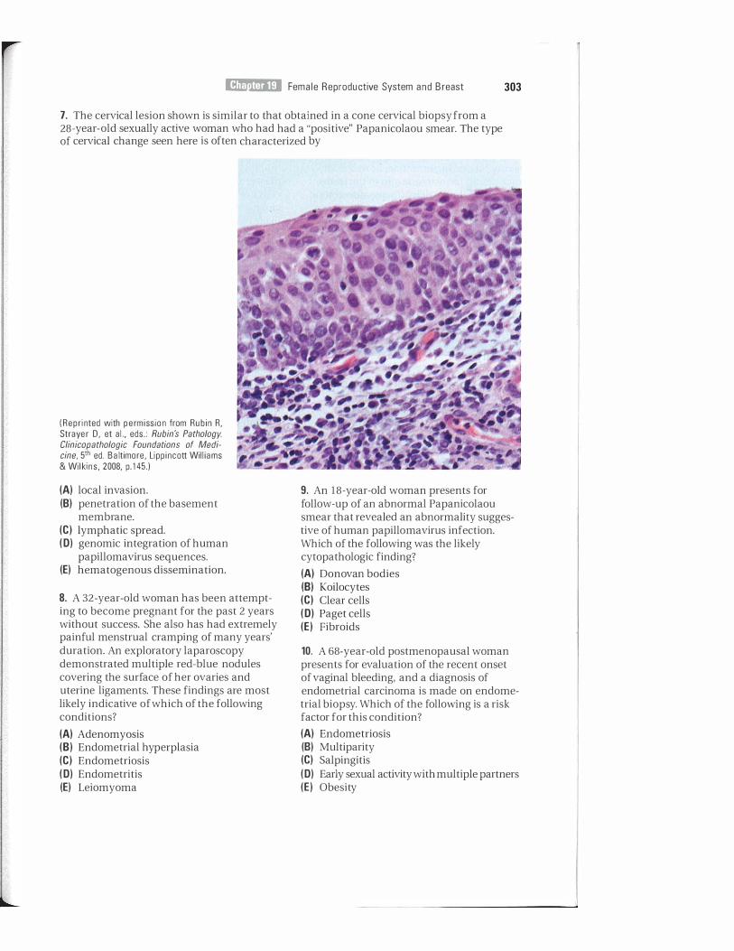
I!1M11ltDliIi Fema le Reproductive System and Breast 303
7. The cervical lesion shown is similar to that obtained in a cone cervical biopsy from a 28-year-old sexually active woman who had had a "positive" Papanicolaou smear. The type of cervical change seen here is often characterized by
(Reprinted with permission from Rubin R, Strayer D, et aI., eds.: Rubin's Pathology. Clinicopathologic Foundations of Medicine, 5th ed. Baltimore, Lippincott Williams & Wilkins, 2008, p. 1 45.)
(A) local invasion. (8) penetration of the basement
membrane. (e) lymphatic spread. (0) genomic integration of human
papillomavirus sequences. (E) hematogenous dissemination.
8. A 32-year-old woman has been attempting to become pregnant for the past 2 years without success. She also has had extremely painful menstrual cramping of many years' duration. An exploratory laparoscopy demonstrated multiple red-blue nodules covering the surface of her ovaries and uterine ligaments. These findings are most likely indicative of which of the following conditions?
(A) Adenomyosis (8) Endometrial hyperplasia (e) Endometriosis (0) Endometritis (E) Leiomyoma
9. An 18-year-old woman presents for follow-up of an abnormal Papanicolaou smear that revealed an abnormality suggestive of human papillomavirus infection. Which of the following was the likely cytopathologic finding?
(A) Donovan bodies (8) Koilocytes (e) Clear cells (0) Paget cells (E) Fibroids
10. A 68-year-old postmenopausal woman presents for evaluation of the recent onset of vaginal bleeding, and a diagnosis of endometrial carcinoma is made on endometrial biopsy. Which of the following is a risk factor for this condition?
(A) Endometriosis (8) Multiparity (e) Salpingitis (0) Early sexual activity with multiple partners (E) Obesity

304 BRS Pathology
1 1 . A 28-year-old woman is evaluated for infertility and amenorrhea. She is obese and has significant facial hair in a male distribution. Laboratory studies reveal an increase in serum luteinizing hormone, as well as in total serum testosterone and in the testosterone metabolite dehydroepiandrosterone sulfate. A pelvic ultrasound examination would probably reveal which of the following findings?
(A) A mass resembling a bunch of grapes projecting into the vagina
(8) Blood-filled cysts in the ovary (e) Fluid accumulation in the fallopian
tubes (0) Islands of endometrium within the
myometrium (E) Multiple small follicular cysts on the
ovary
1 2. A 26-year-old woman experiences the sudden onset of left-sided lower abdominal pain with radiation to the back, pelvis, and thigh. Following a negative test for pregnancy, abdominal radiography shows an enlarged left ovary with multiple calcifications. Laparoscopy reveals adnexal torsion and an ovarian tumor. Ovariectomy is performed. The tumor is most likely a
(A) granulosa cell tumor. (8) Brenner tumor. (e) serous cystadenoma. (0) struma ovarii. (E) dermoid cyst.
13. A 5-year-old girl begins developing secondary sexual characteristics, including pubic hair growth and breast development. Her concerned parents bring her to the pediatrician for evaluation. An abdominal ultrasound examination reveals an ovarian mass. The mass is resected, and pathologic examination reveals it to be a granulosa cell tumor. Which of the following histologic findings is a characteristic of this type of ovarian tumor?
(A) Signet-ring cells (8) Call-Exner bodies (e) Schiller-Duval bodies (0) Hyperfunctioning thyroid tissue ( E) Transitional cell epithelium
14. Soon after an uncomplicated delivery at 38 weeks' gestation, a mother develops respiratory difficulties and uncontrolled vaginal bleeding progressing to shock, multiorgan failure, and death. At autopsy, masses of debris and epithelial squamous cells are apparent in the pulmonary microcirculation. Which of the following was the likely cause of death?
(A) Sheehan syndrome (8 ) Chorioamnionitis (e) Abruptio placentae (0) Amniotic fluid aspiration syndrome (E) Amniotic fluid embolism
1 5. A 23-year-old woman consults an obstetrician because of the onset of vaginal bleeding in what she considers to be the fifth month of pregnancy; however, examination reveals the uterus to be enlarged to the size of a 7 -month pregnancy. Intravaginal ultrasound fails to detect a fetal heartbeat and instead shows a "snowstorm pattern." Human chorionic gonadotropin is markedly elevated. These findings are strongly suggestive of
(A) preeclampsia. (8 ) eclampsia. (e) placenta accreta. (0) ectopic pregnancy. (E ) hydatidiform mole.
1 6. A 65-year-old woman is found to have a I - cm mass in the upper outer quadrant of the left breast. The most likely cause is
(A) fibrocystic disease. (8) acute mastitis. (e) fibroadenoma. (0) carcinoma. (E) Paget disease of the breast.
1 7. A 50-year-old woman has a lumpectomy following mammographic discovery of a carcinoma of the breast. Which of the following is a well-known characteristic or association of breast cancer?
(A) Low-fat diet (8 ) Positive family history (e) Excessive thinness (0) Late menarche (E) Multiparity

i!ilI!J'mEI Female Reproductive System a nd Breast 305
18. A 20-year-old woman presents with a solitary discrete, freely movable, firm, rubbery, nontender, well-circumscribed breast lesion. On resection biopsy, the lesion appears similar to that shown in the figure. The most likely diagnosis is
(Reprinted with permission from Rubin R, Strayer D , et aI., eds.: Rubin's Pathology. Clinicopathologic Foundations of Medicine, 5th ed. Baltimore, Lippincott Williams & Wilkins, 2008, p. 846.)
A) fibrocystic disease. (8 ) fibroadenoma. (e) medullary carcinoma. (0) colloid carcinoma. (E) intraductal carcinoma.
19, A 57-year-old woman who has not seen a physician in more than 20 years now presents with left breast pain. On examination, the left breast is markedly erythematous, swollen, and warm to touch. There is also significant dimpling of the breast (peau d'orange). In addition, the left nipple is completely retracted. Which of the following is the likely diagnosis?
(A) Mastitis (8) Invasive ductal carcinoma
(e) Medullary carcinoma (0) Inflammatory carcinoma (E) Mucinous carcinoma
20. A 27-year-old woman requests a mammogram because both her mother and sister died of metastatic breast cancer before 40 years of age. Which of the follOwing would add to this patient's risk factors for breast cancer?
(A) Multiparity (8) High-fiber diet (e) Oral contraceptive use (0) BRCA-l mutation (E) Bilateral fibroadenomas

I I i l I i
Answers and Explanations
1 . The answer is D. The association of a severe febrile illness with signs of gastrointestinal dysfunction and a diffuse macular rash is strongly suggestive of toxic shock syndrome. This diagnosis is especially likely if clinical abnormalities are associated with concomitant menstruation and the use of highly absorbent tampons. The clinical abnormalities are due to staphylococcal infection of the tampon, demonstrable in vaginal secretions, and the elaboration of staphylococcal exotoxins. The differential diagnosis includes streptococcal infection, usually pharyngeal, with a scarlatiniform eruption; meningococcemia; endotoxemia associated with various gram-negative organisms; rickettsial infections, and so forth. Except for streptococcal infection, all of these conditions are less likely than toxic shock syndrome.
2. The answer is B. Candida albicans is a major cause of vulvovaginitis. Many women are carriers of the yeast, and it therefore constitutes normal vaginal flora, although there are many conditions that alter the vaginal microenvironment, including pregnancy, oral contraceptives, and systemic antibiotics, making overgrowth possible. Calymmatobacterium granulomatis causes granuloma inguinale. Chancroid results in painful genital ulcers and is caused by the organism Haemophilus ducreyi. Herpes simplex virus type 2 can cause genital vesicular lesions. Neisseria meningitidis can cause meningitis.
3. The answer is E. Bacterial vaginosis is the most common cause of vaginal discharge. Characteristically, the discharge is thin and homogeneous. Addition of 10% potassium hydroxide elicits a fishy amine odor. Usually, increased numbers of Gardnerella vaginalis organisms are present and large numbers of "clue cells" are apparent on the Papanicolaou smear. The condition is caused by loss of the normal vaginal lactobacilli, with consequent overgrowth of anaerobes and other bacteria, including G. vaginalis. Neisseria gonorrhoeae is a common cause of pelvic inflammatory disease. Candida causes vulvovaginitis. Trichomonas vaginalis is yet another cause of vagi nos is, but a wet mount displays flagellated motile organisms.
4. The answer is D. The patient presents with a lesion of secondary syphilis known as condyloma lata. The original lesion, the painless ulcer or chancre, is characteristic of primary syphilis. Chancroid is a similar lesion to the primary lesion in syphilis, but instead is painful. Condyloma acuminatum is a similarly named lesion often referred to as a venereal wart and is due to human papillomavirus. Papillary hidradenoma is a common benign tumor of the vulva, presenting as a nodule that may ulcerate and bleed.
5. The answer is D. Condyloma acuminatum is a nonmalignant neoplastic-like papillomatous condition that affects epithelium of the perineal, vulval, perianal, and vaginal regions. It is caused by human papillomavirus (HPY, most often HPV 6 and 1 1) . Genital herpes is usually caused by herpes simplex virus type 2. Treponema pallidum is a spirochete and the etiologic agent of syphilis. Haemophilus ducreyi causes chancroid, a painful, erosive, chancrelike lesion. Candida is not a sexually transmitted organism and is often considered normal vaginal flora, although it is an important cause of vulvovaginitis.
6. The answer is B. In 1 97 1 , it became widely known that there was a danger of development of clear cell adenocarcinoma of the vagina and cervix in young women with a history of in utero exposure to diethylstilbestrol (DES) while their mothers were pregnant. DES has not been administered to pregnant women in this country for more than 30 years.
7. The answer is D. The illustration demonstrates carcinoma in situ of the uterine cervix, which is characterized by disordered epithelial growth manifest by loss of polarity and nuclear hyperchromasia beginning at the basal layer and extending outward. In this
306

i!iltJ'!ltIil£I Female Reprodu ctive System a nd Breast 307
illustration, the changes involve the entire epithelial thickness and would thus be classified as cervical intraepithelial neoplasia grade 3 or carcinoma in situ. Although mild forms can be reversible, the principal significance of cervical intraepithelial neoplasia is its precursor role in the genesis of invasive cervical carcinoma. Invasion and metastases are not associated with cervical intraepithelial neoplasia but only with fully developed invasive carcinoma; however, genomic integration of human papillomavirus DNA sequences, most often types 16, 1 8, 3 1 , or 33, is associated with cervical intraepithelial neoplasia, as well as with frank malignant change.
8. The answer is C. Endometriosis, ectopic non-neoplastic endometrial tissue, is an important cause of infertility. The ectopic tissue replicates the cyclic endometrial changes during the menstrual cycle, including sloughing and bleeding. Such ectopic hemorrhage can result in pain, scarring, tubal obstruction, and infertility. Favored sites of endometriosis include pelvic ligaments, ovaries, and rectum. Adenomyosis is a term denoting the presence of endometrial tissue within the myometrium. Endometrial hyperplasia can be a precursor to endometrial carcinoma and manifests with postmenopausal bleeding. Endometritis results from infection of the endometrium. Leiomyomas are benign tumors of the uterus that can also result in painful menses and can contribute to infertility.
9. The answer is B . Koilocytes are indicative of human papilloma virus-infected epithelial cells. Viral infection causes intracytoplasmic vacuolation that is apparent in cytopathologic and histopathologic preparations. Donovan bodies represent macro phages stuffed with numerous Calymmatobacterium (Donovania) granulomatis organisms. Clear cells are characteristic of clear cell adenocarcinoma of the vagina. Paget cells are characteristic large cells surrounded by a clear, halo-like area that occur in Paget disease of the vulva and Paget disease of the breast. Fibroids are the most common benign tumors of the female reproductive tract.
10. The answer is E. Endometrial cancer is the most common gynecologic malignancy, and a major predisposing factor is prolonged and unopposed exposure to estrogen. In the case of obesity, adipose tissue converts androgens into estrogens, fueling the proliferation of endometrial tissue. Similarly, diabetes, nulliparity (not multiparity) , and hypertension are also predisposing factors. Early sexual activity with multiple partners is associated with cervical cancer. Salpingitis can predispose to ectopic pregnancy. Endometriosis is a benign condition and has no relation to endometrial cancer.
1 1 . The answer is E. The findings are those of the polycystic ovary (Stein-Leventhal) syndrome, an important cause of infertility in young women that is characterized by amenorrhea, infertility, obesity, and hirsutism. Increased levels of luteinizing hormone (LH) and of androgens are often found, and it has been thought that LH may stimulate follicular thecalutein cells, with consequent hyperproduction of androgens. Alternatively, there has been recent attention to the link of this disorder to the metabolic syndrome and insulin resistance. Hyperinsulinemia may lead to increased ovarian androgen production, which then may lead to increased LH. The syndrome is characterized by multiple small follicular cysts, a markedly thickened ovarian capsule, and cortical stromal fibrosis. A mass resembling a bunch of grapes projecting into the vagina is characteristic of sarcoma botryoides. Bloodfilled cysts on the ovary are termed "chocolate cysts" and are associated with endometriosis, another cause of infertility. Fluid accumulation in the fallopian tubes is termed hydrosalpinx. Islands of endometrium in the myometrium are seen in adenomyosis.
12. The answer is E. The radiographic calcifications are highly suggestive of a mature teratoma (dermoid cyst) . This tumor is composed of all three germ layers and often contains skin (including adnexal structures, such as sebaceous glands and hair follicles), bone, teeth, cartilage, and gastrointestinal, neurologic, respiratory, and thyroid tissues. As the tumor enlarges, it is at risk of torsion. Granulosa cell tumor presents with signs and symptoms of excess estrogen production. Brenner tumor is rare and histologically resembles transitional epithelium of the bladder. Serous cystadenoma is commonly bilateral. Struma ovarii is a monodermal teratoma composed of functional ectopic thyroid tissue.

308 BRS Pathology
13. The answer is B. A young patient presenting with precocious puberty and an ovarian mass is likely to have a granulosa cell tumor. Call-Exner bodies, small follicles filled with eosinophilic secretion, are an important diagnostic feature. Signet-ring cells are seen in Krukenberg tumors (bilateral metastatic involvement of the ovaries by a signet-ring carcinoma arising in the stomach) . Schiller-Duval bodies are seen in dysgerminoma, an ovarian tumor analogous to the male seminoma. Hyperfunctional thyroid tissue is found in struma ovarii, and the Brenner tumor resembles transitional cell epithelium.
1 4. The answer is E. The diagnosis is amniotic fluid embolism, which is characterized by sudden peripartal respiratory difficulty, progressing to shock and often to death. Sheehan syndrome is associated with obstetric blood loss, with resultant pituitary ischemia leading to postpartal hypopituitarism. Chorioamnionitis is infection of the placenta, which can have devastating consequences for both the mother and the child. Abruptio placentae is premature separation of the placenta and can lead to antepartal bleeding and fetal death. Amniotic fluid aspiration can occur in the child and can result in difficulties for the infant, particularly if meconium is aspirated. Amniotic fluid embolism and abruptio placentae are well-known causes of disseminated intravascular coagulation, as are retained dead fetus and toxemia.
1 5. The answer is E. Hydatidiform mole should be suspected when the uterus is enlarged beyond the expected size for the time of the pregnancy. Human chorionic gonadotropin is markedly elevated in this gestational trophoblastic disease. Preeclampsia and eclampsia are forms of toxemia of pregnancy marked by severe hypertension, albuminuria, and edema, with the addition of convulsions and disseminated intravascular coagulation in the latter. Placenta accreta occurs when the placenta adheres directly to the myometrium, leading to severe bleeding at the time of delivery. Ectopic pregnancy is usually discovered early in a suspected pregnancy when ultrasound examination reveals the uterus to be empty.
16. The answer is D. The location of the mass in the upper outer quadrant and the patient's age of 65 years suggest that the breast lesion is a carcinoma. A breast mass in a postmenopausal patient is most often a carcinoma.
1 7. The answer is B. A history of breast cancer in first -degree female relatives and a diet high in animal fats are important associations of breast cancer. Associations related to excess estrogen stimulation include high-fat diet, obesity, early menarche, and nulliparity. Additional associations are delayed first pregnancy and exogenous estrogen therapy.
18. The answer is B. Fibroadenoma is a benign tumor most often presenting as a single discrete, freely movable lesion, often demonstrating a pattern of compressed glands and young fibrous stroma similar to that shown in the illustration. Fibroadenoma is the most frequent cause of a palpable mass in the breast in women younger than 25 years of age.
19. The answer is D. The presentation is that of inflammatory carcinoma of the breast, which typically has an extremely poor prognosis. In this variant, cancer cells have invaded the skin and suspensory ligaments of the breast, causing dimpling and distortion of the normal breast architecture.
20. The answer is D. Concern over familial breast cancer is well warranted in this patient, with two first-degree relatives dying of breast cancer at an early age. Both BRCA- l and BRCA-2 mutations are major risk factors for the development of breast cancer. Typically, breasts in a younger woman are too dense to be viewed well on normal screening mammograms, although in this case the mammogram and genetic studies are warranted to determine if she too carries the deleterious mutations. There is evidence that a first pregnancy after 30 years of age, early menarche, and late menopause are also contributing factors. A high-fat diet, not a high-fiber diet, is another apparent risk factor. There is no evidence to support that either oral contraceptive use or fibroadenoma is a risk factor for breast cancer.

Endocrine System
I. PITUITARY
A. Anterior pituitary (adenohypophysis) (Table 20-1) 1. Anterior pituitary hyperfunction
a. Prolactinoma with hyperprolactinemia � ( 1 ) This is the most common pituitary tumor (30% of pituitary tumors) .
(2) Staining is usually chromophobic. � (3) In women, prolactinoma results in amenorrhea and galactorrhea (inappropriate
milk secretion) . (4) It can be caused by hypothalamic lesions or by medications (methyldopa,
reserpine) that interfere with dopamine (prolactin-inhibitory factor) secretion. It can also be associated with estrogen therapy.
b. Somatotropic adenoma with hypersecretion of growth hormone � (1) This is the second most common pituitary tumor. � (2) Staining is usually acidophilic (acidophi l ic adenoma is an older name for this
tumor). � (3) It causes secondary hyperproduction of somatomedins by the liver. End-organ
effects are caused by both growth hormone and somatomedins, especially insulinlike growth factor-I (IGF-I, somatomedin C) .
� (4) Gigantism results if adenoma develops before epiphyseal closure. � (5) Acromegaly results if adenoma develops after epiphyseal closure; acromegaly is
characterized by overgrowth of the jaws, face, hands, and feet, and generalized enlargement of viscera, along with hyperglycemia, osteoporosis, and hypertension.
(6) Other results include local compression effects due to expansion of the tumor within the sella turcica.
c. Corticotropic adenoma and hypersecretion of adrenocorticotropic hormone (ACTH) results in increased production of adrenal cortical hormones (hypercorticism). This condition is called Cushing syndrome or Cushing disease (these terms are used in variable contexts by different authors) . ( 1 ) Cushing disease � (a) This condition classically refers to hypercorticism due to a corticotropic ade
noma of the pituitary (most often a basophi l ic adenoma) . (b) It also refers to hypercorticism associated with very small, sometimes multi
ple pituitary adenomas ( basoph i l ic microadenomas). (2) Cush ing syndrome
(a) This condition refers to hypercorticism regardless of cause. It is most often of pituitary and less often of adrenal origin; some authors use the terms adrenal Cushing syndrome and pituitary Cushing syndrome for clarity.
� (b) The cause may be ectopic ACTH production by various tumors (especially small cell carcinoma of the lung).
309

310 BRS Pathology
t a h I� 20-1 Pituitary Hyperfun ction
Hormone lesion
Prolactin Prolactinoma
Classic Staining of Tumor Cells
Chromophobic
Manifestations
Amenorrhea and galactorrhea in women; impotence and sometimes galactorrhea in men
Somatotropin (growth hormone)
Somatotropic adenoma Acidophil ic Gigantism or acromegaly
Corticotropin (ACTH)
Antidiuretic hormone (ADH)
Corticotropic adenoma or mu ltiple corticotropic microadenomas
Nonpitu itary lesions with ectopic hormone production (small cell carcinoma of the lung)
2. Anterior pituitary hypofunction
Basophilic Pituitary Cushing syndrome
Water retention with d i lutional hyponatremia
a. Pituitary cachexia (Simmonds disease) is general ized panhypopituitarism. ( 1 ) Characteristics include marked wasting. (2) Pituitary cachexia can result from any process that destroys the pituitary. It is
most frequently caused by: (a) Pituitary tumors (h) Postpartum pitu itary necrosis (Sheehan syndrome)
(i) It is caused by ischemic necrosis of the pituitary gland and is characteristically associated with hemorrhage and shock during childbirth.
( i i ) Clinical manifestations are due at first to loss of gonadotropins, then to subsequent loss of thyroid-stimulating hormone (TSH) and ACTH.
h. Selective deficiency of one or more pituitary hormones ( 1 ) Deficiency of growth hormone
(a) In children, this results in growth retardation (pituitary dwarfism) . (h) In adults, this may result in increased insulin sensitivity with hypoglycemia,
decreased muscle strength, and anemia. (2) Deficiency of gonadotropins
(a) In preadolescent children, this results in retarded sexual maturation. (h) In adults, this results in loss of libido, impotence, loss of muscular mass, and
decreased facial hair in men, and in amenorrhea and vaginal atrophy in women.
(3) Deficiency of TSH results in secondary hypothyroidism. (4) Deficiency of ACTH results in secondary adrenal failure. It does not result in
hyperpigmentation of the skin, probably because of the lack of both ACTH and i3-melanocyte-stimulating hormone; this is in contrast to primary adrenal failure (Addison disease), in which ACTH is increased and hyperpigmentation is the rule.
B. Posterior pituitary (neurohypophysis) 1. Hormones are synthesized in the hypothalamus and transported via axons to the poste
rior pituitary. a. Oxytocin induces uterine contraction during labor and ejection of milk from mammary
alveoli. h. Antidiuretic hormone (ADH, vasopressin) promotes water retention through action on
the renal collecting ducts. � 2. Syndrome of inappropriate ADH (SIADH) secretion
a. SIADH is most commonly caused by ectopic production of ADH by various tumors, especially smal l cell carcinoma of the lung.
h. SIADH results in retention of water with consequent dilutional hyponatremia, reduced serum osmolality, and inability to dilute the urine.
3. Deficiency of ADH
iiilIlftitii'D Endocr ine System 311
a. This results in diabetes insipidus; i s characterized by polyuria, with consequent dehydration and insatiable thirst.
b. Causes may include tumors, trauma, inflammatory processes, lipid storage disorders, and other conditions characterized by damage to the neurohypophysis or hypothalamus.
C. Nonfunctioning pituitary tumors 1. Nonsecreting pituitary adenomas are most often chromophobic.
a. Dysfunction results because of local pressure phenomena. � b. These tumors are clinically variable; manifestations include hypopituitarism,
headache, visual disturbances (bilateral hemianopsia [loss of peripheral visual fields due to pressure on optic chiasm] ) , and palsies caused by cranial nerve damage.
2. Craniopharyngioma (adamantinoma) � a. This benign childhood tumor is derived from remnants of the Rathke pouch. It is not
a true pituitary tumor. b. It is similar to ameloblastoma of the jaw.
� c. Characteristics include nests and cords of squamous or columnar cells in a loose stroma, closely resembling the appearance of the embryonic tooth bud enamel organ.
� d. This tumor is often cystic; the lining epithelium of flat or columnar cells often expands into papillary projections.
e. Radiographic detection is often possible because of tumor calcification.
D. Other causes of pituitary hypofunction 1. The empty sella syndrome is caused by conditions that destroy all or part of the pituitary. 2. The Nelson syndrome involves the development of large pituitary adenomas follOwing
bilateral adrenalectomy. This is thought to be due to loss of feedback inhibition on growth of preexisting pituitary micro adenomas.
I I. THYROID GLAND
A. Thyroid hormones include thyroxine (T4) and tri iodothyronine (T3). 1 . Their synthesis depends on sufficient quantities of iodine from dietary sources. 2. The rate of extraction of iodine from the bloodstream and the rate at which T 4 and T 3 are
synthesized and released from storage (as thyroglobulin) and secreted into the bloodstream are regulated by pituitary TSH.
3. Feedback mechanisms regulate pituitary production ofTSH. 4. Serum T3 and T4 are bound to thyroid-binding globulin (TBG) .
B. Congenital anomalies 1 . Thyroglossal duct cyst is the most common thyroid anomaly.
a. This structure is a remnant of the thyroglossal duct. � b. It does not lead to alterations in thyroid function. 2. Ectopic thyroid tissue may be found anywhere along the course of the thyroglossal duct.
C. Goiter is a general term for enlargement of the thyroid. 1. Causes
a. Physiologic enlargement is not uncommon in puberty and pregnancy. b. Iodine deficiency occurs in geographic areas in which the diet is deficient in iodine. c. Hashimoto thyroiditis d. Goitrogens are foods or drugs that suppress synthesis of thyroid hormones. e. Dyshormonogenesis, or partial or complete failure of thyroid hormone synthesis, can
be caused by various enzyme deficiencies.

312 BRS Pathology
2. Terminology a. Simple goiter (nontoxic goiter) is goiter without thyroid hormone dysfunction. b. Toxic goiter is goiter associated with hyperthyroidism; if the patient is euthyroid or
hypothyroid, the term nontoxic goiter is appropriate. c. Endemic goiter is goiter occurring with high frequency in iodine-deficient geographic
areas; the term sporadic goiter is used for goiter caused by similar mechanisms in non-iodine-deficient areas.
d. Nodular goiter is irregular enlargement of the thyroid, reSUlting in nodule formation.
( 1 ) Nodular col loid goiter refers to the late stage of simple goiter in which goiter is most often nodular; nodules may be single or multiple (multinodular goiter) .
(2) Most nodules are hypoplastic and do not take up radioactive iodine ("cold" nodules). (3) Occasionally nodules are hyperplastic and actively produce thyroid hormone and
take up radioactive iodine ("hot" nodules) .
D. Hypothyroidism 1. Laboratory abnormal ities
� a. Decreased serum free T4, increased serum TSH b. Increased serum cholesterol c. Classic thyroid tests now passing into disuse
( 1 ) T3 resin uptake (also known as thyroid hormone-binding ratio [THBR] ) (a) This is colloquially referred to as "T3'" even though it is not a measure of
triiodothyronine. � (b) This is decreased in hypothyroidism.
(c) This is inversely proportional to the number of unbound thyroid hormone binding sites on TBG; it is measured by competitive uptake of radioactive T3 by resin, which competes for unbound sites on TBG.
� (2) Total T4 is decreased in hypothyroidism. � 2. Clinical syndromes. Hypothyroidism is manifest as myxedema in adults or as cretinism
in children. a. Myxedema is more common in women than in men.
( 1 ) Causes (a) Therapy for hyperthyroidism with surgery, irradiation, or drugs, which is a
common cause of myxedema in the United States � (b) Hashimoto thyroiditis
(c) Unknown cause. Primary idiopathic myxedema is a poorly defined form of myxedema, which may be of autoimmune origin (TSH receptor-blocking antibodies have been identified) .
(d) Iodine deficiency, which is the most important cause in non-iodine deficient geographic regions
(2) Clinical characteristics (a) Insidious onset (b) Cold intolerance (c) Tendency to gain weight because of a low metabolic rate (d) Lowered pitch of voice (e) Mental and physical slowness (f) Menorrhagia (g) Constipation (h) Abnormal physical findings:
( i ) Puffiness of face, eyelids, and hands ( i i ) Dry skin ( i i i ) Hair loss, coarse and brittle hair, scant axillary and pubic hair, thinning of
the lateral aspect of the eyebrows ( iv) Increase in relaxation phase of deep tendon reflexes
b. Cretinism ( 1 ) Causes
(a) Iodine deficiency

i!iitft1f1iiiI Endocr ine System 313
(b) Deficiency of enzymes necessary for the synthesis of thyroid hormones (c) Maldevelopment of the thyroid gland (d) Failure of the fetal thyroid to descend from its origin at the base of the tongue (e) Transplacental transfer of antithyroid antibodies from a mother with autoim-
, mune thyroid disease (2) Characteristics
(a) Severe mental retardation (b) Impairment of physical growth with retarded bone development and
dwarfism (c) Large tongue (d ) Protuberant abdomen
E. Hyperthyroidism (thyrotoxicosis) 1. Cl inical features
a. Restlessness, irritability, fatigability b. Tremor c. Heat intolerance; sweating; warm, moist skin (especially of palms) d. Tachycardia, often with arrhythmia and palpitation, sometimes with high-output car-
diac failure e. Muscle wasting and weight loss despite increased appetite f. Fine hair g. Diarrhea h. Menstrual abnormalities, commonly amenorrhea or oligomenorrhea
,!iJ i. Greatly increased free T4 and (in Graves disease, see the following) greatly reduced TSH. In addition, the less commonly used total T 4 and T 3 resin uptake are both increased.
2. Graves disease (Figures 20-1 and 20-2) a. General considerations
( 1 ) Graves disease is hyperthyroidism caused by diffuse toxic goiter. (2) It occurs more frequently in women than in men. (3) The incidence is increased in HLA-DR3- and HLA-BS-positive individuals. (4) Striking exophthalmos (protrusion of the eyes) , possibly due to autoimmune
mechanisms and independent of thyroid hyperfunction, is characteristic. Manifestations include the signs and symptoms of hyperthyroidism.
FIGURE 20-1 Graves disease. Marked exophthalmos and prominent goiter are evident in this young woman. (From Rubin R, Strayer 0, et ai . , eds.: Rubin's Pathology. Clinicopathologic Foundations of Medicine, 5th ed. Baltimore, Lippincott Williams & Wilkins, 2008, p. 945. Original Source: Sandoz Pharmaceutical Corporation.)

314 BRS Pathology
b, Mechanism
FIGURE 20-2 Graves disease (microscopic). Marked glandular hyperplasia with papillary infoldings, scalloping of the colloid, and lymphocytic infiltration are prominent features. (Reprinted with permission from Rubin R, Strayer D, et aI., eds.: Rubin's Pathology. Clinicopathologic Foundations of Medicine, 5th ed. Baltimore, Lippincott Williams & Wilkins, 2008, p. 946.)
( 1 ) Thyroid-stimulating immunog lobul in (TSI), an IgG antibody, reacts with thyroid follicle TSH receptors and stimulates thyroid hormone production.
(2) A similar reaction with thyroid growth immunoglobul in (TGI) stimulates glandular hyperplasia and enlargement.
(3) In addition to TSI and TGI, antimicrosomal and other autoantibodies are characteristic.
3. Other causes of hyperthyroidism a. Plummer disease is a combination of hyperthyroidism, nodular goiter, and absence of
exophthalmos. The "hot" nodules can be adenomas or non-neoplastic areas of nodular hyperplasia.
b. Pituitary hyperfunction can cause excess production of TSH and secondary hyperthyroidism.
c. Struma ovarii is an ovarian teratoma made up of thyroid tissue; can be hyperfunctional. d. Exogenous administration of thyroid hormone
F. Thyroiditis 1 . Hashimoto thyroid itis (Figure 20-3)
a, This'autoimmune disorder occurs more often in women than in men. � b. It is a common cause of hypothyroidism; may occasionally have an early transient
hyperthyroid phase.
FIGURE 20-3 Hashimoto thyroiditis. Marked lymphoid infiltration with germinal center formation and atrophy of thyroid follicles is evident. (Reprinted with permission from Rubin R, Strayer D, et aI., eds.: Rubin's Pathology. Clinicopathologic Foundations of Medicine, 5th ed. Baltimore, Lippincott William� & Wilkins, 2008, p. 948.)

I!Ltf:1tlIfl!I Endocr ine System 31 5
c. Clinical characteristics include a slow, often inapparent course and a modestly enlarged and nontender thyroid; the patient is most often euthyroid at first, but transient hyperthyroidism may occur; hypothyroidism develops late when the gland is shrunken and scarred.
� d. Histologic characteristics include massive infi ltrates of lymphocytes with germinal center formation; thyroid follicles are atrophic and Hlirthle cells (epithelial cells with eosinophilic granular cytoplasm) are prominent.
� e. Hashimoto thyroiditis is associated with various autoantibodies, most prominently antithyroglobulin, antithyroid peroxidase, anti-TSH receptor, and anti-iodine receptor antibodies.
� f. It may also be associated with an increased incidence of other autoimmune disorders such as pernicious anemia, diabetes mellitus, and Sjogren syndrome; incidence is increased in HLA-DR5- and HLA-B5-positive individuals.
2. Subacute (de Querva in, granulomatous) thyroiditis a. The disease is more common in women than in men. b. Focal destruction of thyroid tissue and granulomatous inflammation is characteristic. c. Causes may include a variety of viral infections such as mumps or coxsackievirus. d. Subacute thyroiditis follows a self-limited course of several weeks' duration consisting
of a flu-like illness along with pain and tenderness of the thyroid, sometimes with transient hyperthyroidism.
3. R iedel thyroiditis is characterized by thyroid replacement by fibrous tissue; it is of unknown origin. It can clinically mimic carcinoma.
G. Benign tumors (adenomas) of the thyroid 1. These tumors are most often solitary. 2. They present clinically as nodules and can occur in a variety of histologic patterns (e.g.,
follicular, Hlirthle cell) . 3. Most often they are nonfunctional, but they can occasionally cause hyperthyroidism.
H. Malignant tumors of the thyroid 1 . Papil lary carcinoma
� a. This carcinoma is the most common thyroid cancer. b. Histologic characteristics include papillary projections into gland-like spaces; tumor
cells have characteristic "ground-glass" ("Orphan Annie") nuclei; calcified spheres (psammoma bodies) may be present.
� c. This cancer has a better prognosis than other forms of thyroid cancer, even when adjacent lymph nodes are involved.
d. In some instances, papillary carcinoma can be associated with changes in chromosome 10 (paracentric inversion of chromosome 10 or a reciprocal translocation between chromosomes 1 0 and 17) , which constitutively activate the tyrosine kinase domain of ret. These novel fusion genes are referred to as ret-PTC (for ret and papillary thyroid carcinoma) and are analogous to the ber-abl fusion in chronic myeloid leukemia, which also causes a constitutive activation of a tyrosine kinase domain.
� e. This cancer can be a long-term consequence of prior radiotherapy to the neck. 2. Fol l icular carcinoma
a. This cancer is characterized histologically by relatively uniform follicles. b. It has a poorer prognosis than papillary carcinoma.
3. Medul lary carcinoma � a. This cancer originates from C cells of the thyroid. � b. It produces calcitonin, a calcium-lowering hormone. � c. Histologic characteristics include sheets of tumor cells in an amyloid-containing
stroma. � d. Medullary carcinoma can be associated with multiple endocrine neoplasia (MEN)
syndrome IIa and IIb (III) . 4. Undifferentiated carcinoma tends to occur in older patients and has a very poor prognosis.

316 BRS Pathology
III. PARATHYROID GLANDS
A. Parathyroid hormone (parathormone, PTH) 1. Secretion is not under pituitary control. 2. The parathyroid glands are responsive to the plasma concentration of ionized calcium;
decreased calcium concentration stimulates PTH production.
B. Hyperparathyroidism (Table 20-2) 1. Primary hyperparathyroidism
� a. Most often, the cause is parathyroid adenoma; a few cases are caused by primary parathyroid hyperplasia; carcinoma is rarely a cause.
� b. Less often, the cause is production of PTH-like hormone by nonparathyroid malignant tumors such as bronchogenic squamous cell carcinoma or renal cell carcinoma.
c. Primary hyperparathyroidism can occur as part of MEN I and MEN IIa. d. laboratory findings � (1) Hypercalcemia and hypercalciuria � (2) Decreased serum phosphorus, decreased tubular reabsorption of phosphorus, and
increased urinary phosphorus � (3) Increased serum alkal ine phosphatase � (4) Increased serum PTH e. Cl inical characteristics
(1 ) Osteitis fibrosa cystica, cystic changes in bone due to osteoclastic resorption; it is also known as von Recklinghausen disease of bone; fibrous replacement of resorbed bone may lead to the formation of non-neoplastic tumor-like masses ("brown tumor").
(2) Metastatic calcification affecting various tissues, especially the kidneys (nephrocalcinosis)
(3) Renal calculi, a frequent complication (4) Peptic duodenal ulcer; hypercalcemia predisposes to peptic ulcer
2. Secondary hyperparathyroidism a. Compensatory parathyroid hyperplasia occurs in response to decreased concentra
tion of serum ionized calcium. � b. The most common cause is hypocalcemia of chronic renal disease. Vitamin D conversion
by the kidney to biologically active 1 ,25- (OH)2D3 ( la,25-dihydroxycholecalciferol) is impeded, resulting in decreased intestinal absorption of calcium; also, the increased serum phosphorus of renal disease induces a reciprocal decrease in serum calcium.
t a b I e 20-2 Hyperpa rathyroidism
Causes
Primary
Parathyroid adenoma, hyperplasia, or carcinoma; parathyroid hormone-like hormone production by tumors elsewhere; MEN I and MEN I ta
Secondary
Hypocalcemia (usually due to renal disease I
Clinical Manifestations
Osteitis fibrosa cystica; metastatic calc ification in many tissues; nephrocalcinosis and renal calculi
Diffuse osteoclastic bone disease; metastatic calcification
MEN = multiple endocrine neoplasia syndrome; PTH = parathyroid hormone.
laboratory Abnormalities
Increased PTH; hypercalcemia and hypercalciuria; hypophosphatemia and decreased tubular reabsorption of phosphorus; increased alkal ine phosphatase
Increased PTH; hypocalcemia (may be sl ight); hyperphosphatemia; increased alkaline phosphatase

I!1ifiJ!1ttfJli Endocr ine System 31 7
� c. Characteristics include decreased serum calcium, increased serum phosphorus, and increased serum alkaline phosphatase; diffuse osteoclastic bone disease; and metastatic calcification. PTH is increased.
3. Tertiary hyperparathyroidism a. This persistent parathyroid hyperfunction occurs in spite of correction of hypocal
cemia and preexisting secondary hyperparathyroidism. b. Often, the cause is development of an adenoma in a previously hyperplastic gland.
4. 1a-hydroxylase deficiency (vitamin D-dependent rickets) a. Impaired hydroxylation of 2S-hydroxycholecalciferol is characteristic. b. Results include decreased absorption of calcium and increased PTH secretion.
C. Hypoparathyroidism 1 . The most common cause is accidental surgical excision during thyroidectomy. In rare
instances, this disorder is associated with congenital thymic hypoplasia (DiGeorge syndrome) .
2. Severe hypocalcemia manifest clinically by increased neuromuscular excitability and tetany is characteristic.
D. Pseudohypoparathyroidism 1. Pseudohypoparathyroidism is similar to hypoparathyroidism, with decreased calcium,
increased phosphate, and increased parathyroid hormone. 2. This disorder is characterized by multihormone resistance involving PTH and the pitu
itary hormones TSH, luteinizing hormone, and follicle-stimulating hormone. 3. Additional characteristics include end-organ unresponsiveness of the kidney to PTH, as well
as shortened fourth and fifth metacarpals and metatarsals, short stature, and other skeletal abnormalities. a. These abnormalities are due to mutations in GNASl, a gene encoding certain G pro
teins that mediate tissue receptiveness to hormones. Both alleles are expressed in multiple tissues. However, in the kidney and pituitary, there is expression only of the maternally inherited chromosome.
b. This selective imprinting of the mutant gene (paternal imprinting or silencing) results in maternal inheritance of the end-organ unresponsiveness. The skeletal abnormalities result from inheritance of mutant GNASI from either parent.
4. Similar skeletal abnormalities, without hormone dysfunction, characterize pseudopseudohypoparathyroidism, which requires transmission of a mutant paternal GNASI allele.
IV. A DRENAL G LANDS (TABLE 20-3)
A. Cushing syndrome ( hypercorticism) 1. Causes. Cushing syndrome results from increased circulating glucocorticoids, primarily
cortisol. a. Exogenous corticosteroid medication (most common) b. Hyperproduction of ACTH by corticotrophs of the pituitary
� c. Adrenal cortical adenoma or adrenal carcinoma (less common than adenoma) � d. Ectopic production of ACTH by non pituitary carcinomas, especially small cell carcinoma
of the lung. Hypercorticism is autonomous and cannot be suppressed by exogenous adrenal steroids such as those used in the low- and high-dose dexamethasone suppression tests. Also, the autonomous ACTH stimulation eliminates the normal circadian rhythm of cortisol production.
2. Morphologic changes in adrenal gland a. Bi lateral hyperplasia of the adrenal zona fasciculata occurs when the syndrome results
from ACTH stimulation. b. Adrenal cortical atrophy is seen when exogenous glucocorticoid medication is the
cause.

318 BRS Pathology
t a b 1""iiI 20-3 Adrenal Endocrine Syndromes
Syndrome
Cortex
Cushing syndrome (hypercorticism)
Hyperaldosteronism (aldosteronism)
Primary
Secondary
Adrenal virilism
Hypocorticism
Medulla
Pheochromocytoma
Neuroblastoma
Usual Anatomic lesions
Bi lateral hyperplasia of adrenal zona fasc iculata secondary to hyperactivity of pituitary corticotrophs or to ectopic ACTH-like production by a variety of tumors; adrenal cortical adenoma
Adenoma or hyperplasia of zona glomerulosa
Bi lateral hyperplasia of zona glomerulosa caused by stimulation of renin-ang iotensin system
Adenoma, carcinoma, or hyperplasia of zona reticu laris
Id iopathic adrenal atrophy
Tuberculosis
I nfection
Chromaffin cell tumor; benign or mal ignant
Mal ignant "small b lue cel l" tumor of adrenal medul la
c. Adrenal cortical adenoma or carcinoma ( 1 ) Adenoma is more common.
Comments
May be of pituitary, adrenal, or ectopic origin; can also result from administration of exogenous hormone
Serum renin decreased
Serum renin increased; frequently secondary to edema, regardless of the cause
May be due to hyperplasia resulting from congenital enzyme deficiencies such as 21-hydroxylase and l l -hydroxylase
Probable autoimmune etiology
Most common cause of Addison disease in prior years
Waterhouse-Friderichsen syndrome most often associated with meningococcal infection
Tumor secretes catecholamines (epinephrine and norepinephrine) and causes secondary hypertension
Catecholamine-secreting mal ignancy of early chi ldhood; hypertension
(2) Hypercorticism due to adrenal neoplasms cannot be suppressed by exogenous adrenal steroids in the dexamethasone suppression tests. In contrast, hypercorticism of pituitary origin can usually be suppressed.
(3) In addition to dexamethasone suppression, ACTH determinations are useful diagnostic measures in determining the cause of hypercorticism. ACTH is increased in pituitary hypercorticism and in ectopic ACTH production, and it is low when hypercorticism is of adrenal origin.
3. Cl inical characteristics a. Redistribution of body fat with round moon face, dorsal "buffalo hump," often with
relatively thin extremities caused by muscle wasting; skin atrophy with easy bruising and purplish striae, especially over the abdomen; and hirsutism
h. Muscle weakness, osteoporosis, amenorrhea, hypertension, hyperglycemia, and psychiatric dysfunction
B. Hyperaldosteronism 1 . Primary aldosteronism (Conn syndrome)
a. The cause is primary hyperproduction of adrenal mineralocorticoids. h. This condition usually results from an aldosterone-producing adrenocortical ade
noma (aldosteronoma). It can also result from hyperplasia of the zona glomerulosa. Rarely it may be caused by adrenocortical carcinoma_
� c. Clinical characteristics include hypertension, sodium and water retention, and hypokalemia, often with hypokalemic alkalosis.
� d. Decreased serum renin occurs due to negative feedback of increased blood pressure on renin secretion.

2. Secondary a ldosteronism
I!1llttl1tllf1! Endo c rine System 319
a. This condition is secondary to renal ischemia, renal tumors, and edema (e.g., cirrhosis, nephrotic syndrome, cardiac failure) .
b. The cause is stimulation of the renin-angiotensin system. � c. Serum renin is increased, in contrast to primary aldosteronism. Renin synthesized in the
juxtaglomerular apparatus of the kidney promotes the conversion of angiotensinogen to angiotensin I, which is converted catalytically by angiotensin-converting enzyme (mainly in the lung) to angiotensin II. The release of aldosterone is facilitated' by angiotensin II.
C. Adrenal virilism (adrenogenital syndrome) 1 . Causes
� a. Congenital enzyme defects lead to diminished cortisol production and compensatory increased ACTH, with resultant adrenal hyperplasia with androgenic steroid production. ( 1 ) 21-hydroxylase deficiency, which is most common, can, in its most severe "salt
wasting" form, result in salt loss and hypotension. (2) 1 1 -hydroxylase deficiency, a much less common cause, results in salt retention
and hypertension. b. Tumors of the adrenal cortex
� 2. C l inical characteristics. Adrenal virilism produces virilism in females and precocious puberty in males.
D. Hypocorticism (adrenal hypofunction) 1 . General considerations
a. Hypocorticism can be of primary adrenal cause or secondary to hypothalamic or pituitary dysfunction.
b. Deficiency of glucocorticoids (primarily cortisol) , often with associated mineralocorticoid deficiency, is characteristic.
2. Addison disease (primary adrenocortical deficiency) � a. This disorder is most commonly due to idiopathic adrenal atrophy (autoimmune lym
phocytic adrenalitis) . � b. It can also be caused by tuberculosis (formerly most common cause), metastatic
tumor, and various infections. � c. Characteristics include hypotension; increased pigmentation of skin; decreased
serum sodium, chloride, glucose, and bicarbonate; and increased serum potassium. 3. Waterhouse-Friderichsen syndrome � a. This catastrophic adrenal insufficiency and vascular collapse is due to hemorrhagic
necrosis of the adrenal cortex. � b. This syndrome is often associated with disseminated intravascular coagulation. � c. This syndrome is characteristically due to meningococcemia, most often in association
with meningococcal meningitis.
E. Tumors of the adrenal medulla 1. Pheochromocytoma
a. This tumor is derived from chromaffin cells of the adrenal medulla; if it is derived from extra-adrenal chromaffin cells, it is called paraganglioma.
b. Most often this tumor is benign; 10% are malignant. � c. This uncommon but important cause of surgically correctable hypertension results
from hyperproduction of catecholamines (epinephrine and norepinephrine) by the tumor; the hypertension is usually paroxysmal (episodic) , but may be persistent.
� d. Increased urinary excretion of catecholamines and their metabol ites (metanephrine, normetanephrine, and vani l lylmandelic acid) is characteristic.
� e. This tumor can also cause hyperglycemia. � f. Pheochromocytoma can be part of MEN IIa or MEN lIb (III). It can also be associated
with neurofibromatosis or with von Hippel-Lindau disease.
l I

320 BRS Pathology
2. Neuroblastoma a. This highly malignant catecholamine-producing tumor occurs in early chi ldhood. b. Urinary catecholamines and catecholamine metabolites are the same as in pheochro-
mocytoma. � c. The tumor causes hypertension. � d. It usually originates in the adrenal medulla and often presents as a large abdominal mass. � e. Occasionally it converts into a more differentiated form termed ganglioneuroma.
f. The tumor is characterized by amplification of the N -myc oncogene with thousands of gene copies per cell. ( 1 ) Amplification results in characteristic karyotypic changes (homogeneously stain
ing regions or double minute chromosomes) . (2) The number of N-myc gene copies is related to the aggressiveness of the tumor.
� g. The malignant cells of neuroblastoma sometimes differentiate into benign cells, and this change is reflected by a marked reduction in gene amplification.
V. ENDOCRINE PANCREAS
A. Diabetes mellitus 1 . Classification and general features
a. Type 1 (insulin-dependent diabetes mellitus [IDDM], juvenile or ketosis-prone diabetes mellitus) ( 1 ) Epidemiologic and etiologic factors
(a) Type 1 disease often begins early in l ife, usually before 30 years of age. (b) Type 1 disease is less common than type 2 disease. (c) Disease is due to fai lure of insul in synthesis by beta cells ofthe pancreatic islets.
� (d) The cause may be a genetic predisposition complicated by autoimmune inflammation of islets (insulitis) triggered by a viral infection or environmental factors. Type 1 disease demonstrates a positive family history less frequently than does type 2 disease.
(e) The incidence is greatly increased in individuals with a specific point mutation in the HLA-DQ gene. Incidence is markedly increased in HLA-DR3- and HLA-DR4-positive individuals.
(2) Characteristics (a) Unless insulin is replaced, type 1 diabetes results in marked carbohydrate
intolerance with hyperglycemia, leading to polyuria, polydipsia, weight loss despite increased appetite, ketoacidosis, coma, and death.
� (b) Ketoacidosis results from increased catabolism of fat, with production of "ketone bodies" (principally [3-hydroxybutyric acid and acetoacetic acid along with small quantities of acetone) . It is not limited to diabetic acidosis and, in a much milder form, is seen in starvation.
b. Type 2 (non-insulin-dependent diabetes mellitus [NIDDM] , adult-onset, or ketosisresistant diabetes mellitus) ( 1 ) Epidemiologic and etiologic factors
(a) Type 2 disease is much more common than type 1 disease. (b) Type 2 disease characteristically begins later in life, most often in middle age. (c) A positive family history is more frequent than in type 1 diabetes mellitus. (d) Type 2 disease is due to increased insul in resistance mediated by decreased cell
membrane insulin receptors or postreceptor dysfunction. It may also be associated with impaired processing of pro insulin to insulin, decreased sensing of glucose by beta cells, or impaired function of intracellular carrier proteins.
(e) Type 2 disease is most often associated with mild to moderate obesity. (2) Characteristics
(a) The plasma insulin concentration is normal and often increased. (b) Mild carbohydrate intolerance can most often be managed by diet and oral
antidiabetic agents; insulin therapy is not usually required.

I!!OiI!J1ltlIE! Endocrine System 321
,I>l (c) Ketoacidosis is unusual but does occur, characteristically precipitated by unusual stress such as infection or surgery.
c. Maturity-onset diabetes mell itus of tile young (MODV) ( 1 ) This autosomal dominant syndrome is characterized by mild hyperglycemia and
hyposecretion of insulin but no loss of beta cells. (2) It has an earlier onset than type 2 diabetes mellitus. (3) It is caused by a diverse group of single gene defects.
d. Secondary diabetes mellitus occurs as a secondary phenomenon in pancreatic and other endocrine diseases and pregnancy. ( 1 ) Pancreatic disease
(a) Hereditary hemochromatosis (bronze diabetes) . Characteristics include excess iron absorption and parenchymal deposition of hemosiderin, with reactive fibrosis in various organs, especially the pancreas, liver, and heart.
(b) Pancreatitis. Acute pancreatitis is characterized by hyperglycemia; chronic pancreatitis may result in islet cell destruction and secondary diabetes mellitus.
,I>l (c) Carcinoma of the pancreas. Diabetes mellitus may be the presenting sign. (2) Other endocrine diseases ,I>l (a) Cushing syndrome produces hyperglycemia as a result of increased gluconeo
genesis and impaired peripheral utilization of glucose. ,I>l (b) Acromegaly produces hyperglycemia due to the anti-insulin-like effect of
growth hormone. (c) G lucagon hypersecretion promotes glycogenolysis; is characteristically caused
by an islet alpha cell tumor (glucagonoma) . ,I>l (d) Other endocrine disorders. Pheochromocytoma and hyperthyroidism are
sometimes associated with hyperglycemia. (3) Pregnancy
(a) Pregnancy may be associated with transient diabetes mellitus (gestational diabetes); overt nongestational diabetes sometimes develops later.
(b) Diabetes mellitus is characteristically associated with increased fetal b i rth weight and increased fetal mortality, notably from neonatal respiratory distress syndrome (hyal ine membrane d isease).
,I>l (c) When a mother has hyperglycemia, her infant may be born with hyperplasia of the pancreatic islets and hypoglycemia.
2. Anatomic changes in diabetes mel litus a. Pancreatic islets
( 1 ) Type 1 diabetes. Islets are small and beta cells are greatly decreased in number or are absent; insul itis marked by lymphocytic infiltration is a highly specific early change for this form of diabetes mellitus.
(2) Type 2 diabetes ,I>l (a) Focal islet fibrosis and hyalinization (due to deposits of amyl in) are character
istic but not specific. (b) Amylin (also known as islet amyloid polypeptide, or lAPP) deposition in the
pancreatic islets is characteristic of type 2 diabetes mellitus and is thought to interfere either with the conversion of pro insulin to insulin or with the sensing of insulin by beta cells.
b. Kidney ,I>l ( 1 ) Increased width of glomerular basement membrane is the earliest and most com
mon renal manifestation. ,I>l (2) Diffuse diabetic glomerulosclerosis, nodular diabetic glomerulosclerosis
(Kimmelstiel-Wilson disease) , arteriolar lesions, and exudative lesions, such as the fibrin cap or capsular drop, are renal manifestations of diabetes mellitus.
,I>l (3) The Armanni-Ebstein lesion (tubular deposition of glycogen) is an uncommon result of prolonged untreated hyperglycemia.
,I>l (4) Pyelonephritis is a frequent complication that may be compounded by renal papillary necrosis.

b
322 BRS Pathology
c. Cardiovascular system ( 1 ) The incidence of atherosclerosis is greatly increased; clinically significant athero
sclerotic complications occur at a much earlier age than in the nondiabetic pop
ulation; the incidence in women, both premenopausal and postmenopausal, is
greatly increased. (2) Myocardial infarction and peripheral vascular insufficiency (often with gangrene of
the lower extremities) are frequent complications. (3) Capil lary basement membrane thickening occurs in multiple organs and is thought
to be due to nonenzymatic glycosylation of membrane protein.
d. Eye ( 1 ) Cataract formation is common.
� (2) Proliferative retinopathy (retinal exudates, edema, hemorrhages, and microaneurysms of small vessels) can lead to blindness.
� e. Nervous system. Changes include peripheral neuropathy and changes in the brain and spinal cord.
f. liver. Fatty change is seen. g. Skin
(1) Xanthomas (collections of lipid-laden macrophages in the dermis) (2) Furuncles and abscesses because of increased propensity to infection; frequent
fungal infections, especially with Candida
B. Endocrine tumors ( islet cell tumors) 1. Insulinoma (beta cell tumor) is the most common islet cell tumor.
a. This tumor can be benign or malignant. � b. It is characterized by greatly increased secretion of insulin. The problem of distin
guishing endogenous insulin production from exogenous insulin (therapeutically or surreptitiously administered) is solved by quantitation of C-peptide, a fragment of the proinsulin molecule split off during the synthesis of insulin. Circulating C-peptide is characteristically increased in patients with insulinoma. In contrast, C-peptide is not increased by exogenous insulin administration because it is removed during the purification of commercial insulin preparations.
c. Clinical characteristics include the Whipple triad: � ( 1 ) Episodic hyperinsulinemia and hypoglycemia
(2) Central nervous system (CNS) dysfunction temporally related to hypoglycemia (confusion, anxiety, stupor, convulsions, coma)
(3) Dramatic reversal of CNS abnormalities by glucose administration 2. Gastrinoma
a. This tumor is often malignant and sometimes occurs in extrapancreatic sites. b. It results in gastrin hypersecretion and hypergastrinemia.
� c. It is associated with the Zollinger-Ellison syndrome (marked gastric hypersecretion of hydrochloric acid, recurrent peptic ulcer disease, and hypergastrinemia) .
3. Glucagonoma (alpha cel l tumor) is a rare tumor. a. This endocrine tumor results in secondary diabetes mellitus. b. It causes a characteristic skin lesion called necrolytic migratory erythema.
4. VIPoma is a rare tumor. � a. This endocrine tumor is marked by secretion of vasoactive intestinal peptide (VIP) . � b. It is associated with Watery Diarrhea, Hypokalemia, and Achlorhydria (WDHA) syn-
drome, also known as Verner-Morrison syndrome or pancreatic cholera.
VI. MULTIPLE ENDOCRINE NEOPLASI A (MEN) SYNDROMES
In this group of autosomal dominant syndromes, more than one endocrine organ is hyperfunctional. These conditions may be associated with hyperplasias or tumors.

A. MEN I (Wermer syndrome)
I!iiff11tffEI Endocrine System 323
1 . This syndrome includes hyperplasias or tumors of the pituitary, parathyroid, or pancreatic islets (3 Ps). In addition, it may include hyperplasias or tumors of the thyroid or adrenal cortex.
2. It may manifest its pancreatic component by the Zollinger-Ellison syndrome, hyperinsulinism, or pancreatic cholera.
3. It is linked to mutations in the MEN I gene.
B. MEN lIa (Sipple syndrome) 1 . This syndrome includes pheochromocytoma, medullary carcinoma Of the thyroid, and
hyperparathyroidism due to hyperplasia or tumor. 2. It is linked to mutations in the ret oncogene. This is clinically important. For example,
when a diagnosis of pheochromocytoma is made, the finding of characteristic ret mutations would justify prophylactic thyroidectomy (because of the danger of fatal medullary carcinoma of the thyroid) .
C. MEN lib (MEN II I) 1 . This syndrome includes pheochromocytoma, medullary carcinoma, and multiple
mucocutaneous neuromas or ganglioneuromas. 2. In contrast to MEN IIa, it does not induce hyperparathyroidism. 3. It is linked to different mutations in the ret oncogene compared with MEN IIa.
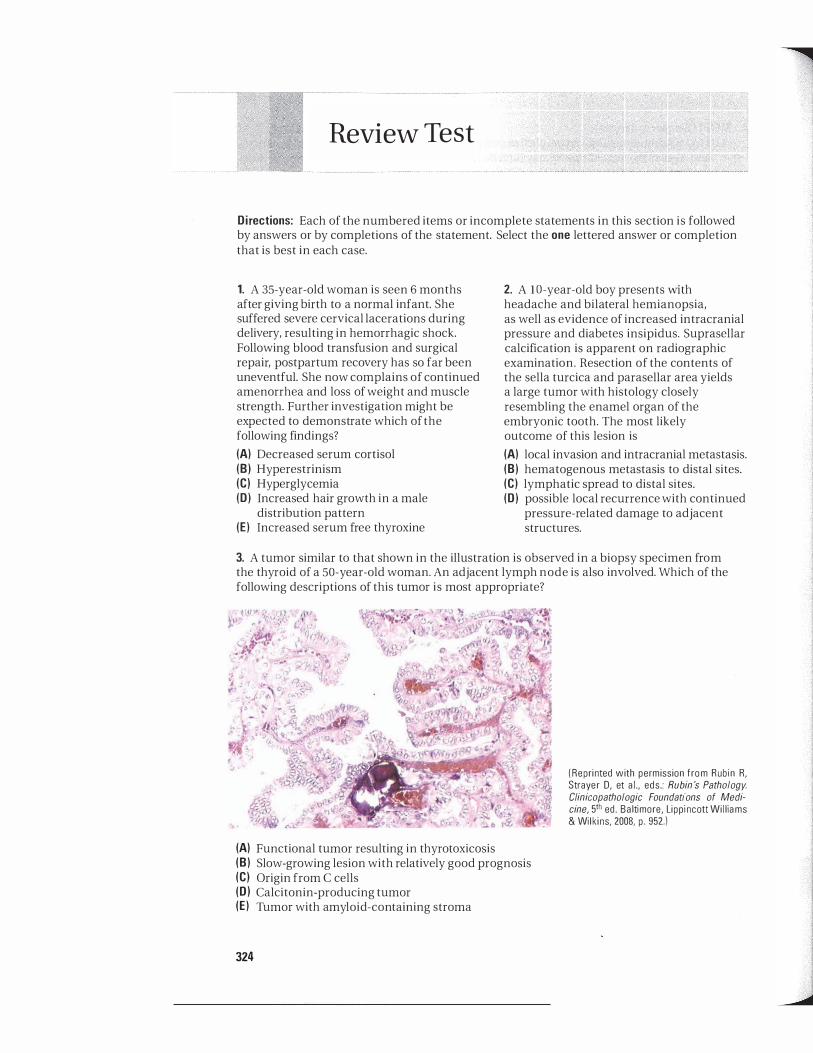
Review Test
Directions: Each of the numbered items or incomplete statements in this section is followed by answers or by completions of the statement. Select the one lettered answer or completion that is best in each case.
1. A 35-year-old woman is seen 6 months after giving birth to a normal infant. She suffered severe cervical lacerations during delivery, resulting in hemorrhagic shock. Following blood transfusion and surgical repair, postpartum recovery has so far been uneventful. She now complains of continued amenorrhea and loss of weight and muscle strength. Further investigation might be expected to demonstrate which of the following findings?
(A) Decreased serum cortisol (8) Hyperestrinism (e) Hyperglycemia (D) Increased hair growth in a male
distribution pattern (E) Increased serum free thyroxine
2. A l O-year-old boy presents with headache and bilateral hemianopsia, as well as evidence of increased intracranial pressure and diabetes insipidus. Suprasellar calcification is apparent on radiographic examination. Resection of the contents of the sella turcica and parasellar area yields a large tumor with histology closely resembling the enamel organ of the embryonic tooth. The most likely outcome of this lesion is
(A) local invasion and intracranial metastasis. (8) hematogenous metastasis to distal sites. (e) lymphatic spread to distal sites. (D) possible local recurrence with continued
pressure-related damage to adjacent structures.
3. A tumor similar to that shown in the illustration is observed in a biopsy specimen from the thyroid of a 50-year-old woman. An adjacent lymph node is also involved. Which of the following descriptions of this tumor is most appropriate?
(A) (8) (e) (D) (E)
324
Functional tumor resulting in thyrotoxicosis Slow-growing lesion with relatively good prognosis Origin from C cells Calcitonin-producing tumor Tumor with amyloid -containing stroma
(Reprinted with permission from Rubin R, Strayer D, et aI., eds.: Rubin's Pathology. Clinicopathologic Foundations of Medicine, 5th ed. Baltimore, Lippincott Williams & Wilkins, 2008, p. 952.)
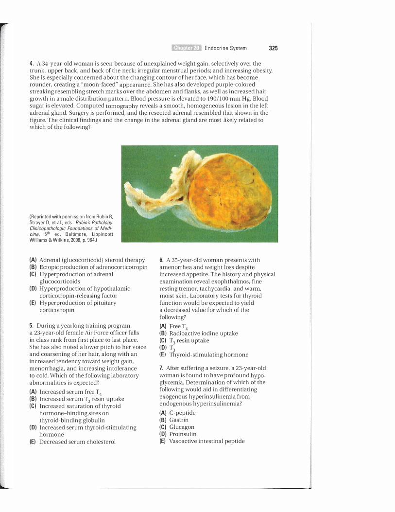
i I �
� Endocrine System 325
4. A 34-year-old woman is seen because of unexplained weight gain, selectively over the trunk, upper back, and back of the neck; irregular menstrual periods; and increasing obesity. She is especially concerned about the changing contour of her face, which has become rounder, creating a "moon-faced" appearance. She has also developed purple-colored streaking resembling stretch marks over the abdomen and flanks, as well as increased hair growth in a male distribution pattern. Blood pressure is elevated to 1 90 1 1 00 mm Hg. Blood sugar is elevated. Computed tomography reveals a smooth, homogeneous lesion in the left adrenal gland. Surgery is performed, and the resected adrenal resembled that shown in the ' figure. The clinical findings and the change in the adrenal gland are most likely related to which of the following?
(Reprinted with permission from Rubin R, Strayer D, et aI., eds.: Rubin's Pathology. Clinicopathologic Foundations of Medicine, 5th ed. Baltimore, Lippincott Williams & Wilkins, 2008, p. 964.)
(A) Adrenal (glucocorticoid) steroid therapy (8 ) Ectopic production of adrenocorticotropin (e) Hyperproduction of adrenal
glucocorticoids (0) Hyperproduction of hypothalamic
corticotropin -releasing factor ( E) Hyperproduction of pituitary
corticotropin
5. During a yearlong training program, a 23-year-old female Air Force officer falls in class rank from first place to last place. She has also noted a lower pitch to her voice and coarsening of her hair, along with an increased tendency toward weight gain, menorrhagia, and increasing intolerance to cold. Which of the following laboratory abnormalities is expected?
(A) Increased serum free T4 (8 ) Increased serum T3 resin uptake (e) Increased saturation of thyroid
hormone-binding sites on thyroid-binding globulin
(0) Increased serum thyroid-stimulating hormone
(E) Decreased serum cholesterol
6. A 35-year-old woman presents with amenorrhea and weight loss despite increased appetite. The history and physical examination reveal exophthalmos, fine resting tremor, tachycardia, and warm, moist skin. Laboratory tests for thyroid function would be expected to yield a decreased value for which of the following?
(A) Free T4 (8 ) Radioactive iodine uptake (e) T3 resin uptake (0) T3 (E) Thyroid-stimulating hormone
7. After suffering a seizure, a 23-year-old woman is found to have profound hypoglycemia. Determination of which of the following would aid in differentiating exogenous hyperinsulinemia from endogenous hyperinsulinemia?
(A) C-peptide (8) Gastrin (e) Glucagon (0) Proinsulin (E) Vasoactive intestinal peptide

326 BRS Pathology
8. A 32-year-old woman presents with amenorrhea, galactorrhea, and visual field defects, all of several months' duration. Magnetic resonance imaging reveals a hypophyseal mass impinging on the optic chiasm. This is most likely a(n)
(A) prolactinoma. (8) somatotropic adenoma. (e) corticotropic adenoma. (0) craniopharyngioma. (E) acidophilic adenoma.
9. A 46-year-old man is referred to an endocrinologist because of the recent onset of diabetes mellitus. His overall appearance is striking, however, and on questioning, he describes marked changes that have been occurring slowly over many years. Comparison with old photographs reveals that he has developed generalized coarseness of his facial features, including frontal bossing, thickening of the nose, prognathism (enlargement and increased prominence of the j aw) , and macroglossia (enlargement of the tongue) . In addition, he has enlarged extremities, with sausagelike fingers, and he says that he is no longer able to wear his wedding ring and that his shoe size has increased. These findings are characteristic of increased activity of which of the following?
(A) Corticotropin (8) Dopamine (e) Insulinlike growth factor-I (0) Prolactin (E) Thyroid-stimulating hormone
10. A 36-year-old man is brought to the emergency department by his wife because of lethargy, weakness, and confusion. Serum sodium and serum osmolality are markedly decreased. Urine osmolality is increased. These findings are most likely related to
(A) adenoma of the anterior pituitary. (8) adenoma of the posterior pituitary. (e) bronchogenic carcinoma. (0) diabetes insipidus. (E) Sheehan syndrome.
1 1 . A 4-month-old child is brought to the pediatrician for evaluation. The mother received no prenatal care and states that she has "a thyroid condition." The child appears
markedly developmentally delayed, with coarse features, macroglossia, and an umbilical hernia. The child likely has which of the following conditions?
(A) Cushing disease (8) Acromegaly (e) Diabetes insipidus (0 ) Cretinism (E) Thyroglossal duct cyst
1 2. A 23-year-old woman presents with tremor, restlessness, heat intolerance, palpitation, and unexplained weight loss. The thyroid is symmetrically enlarged, the pulse is rapid, the skin is moist and warm, and exophthalmos is apparent. This condition is considered to be
(A) autoimmune. (8) congenital. (e) iatrogenic. (0) infectious. (E) nutritional.
13. A palpable mass is noted in the right lobe of the thyroid of a 45-year-old man who visits his physician for a periodic checkup. A biopsy is performed and results in a diagnosis of medullary carcinoma of the thyroid. Which of the following histologic features of thyroid disease would most likely be present in this biopsy specimen?
(A) Tumor cells with "Orphan Annie" nuclei (8) Psammoma bodies (e) Tumor cells embedded in an amyloid
laden stroma (0) Infiltrates of lymphocytes with germinal
center formation (E) Replacement of the thyroid with fibrous
tissue
1 4. A 34-year-old man is referred for evaluation of hypertension and persistent hypokalemia in spite of taking oral potassium supplements. Blood pressure is 1 80 / 1 10 mm Hg. Serum sodium is 149 mEq/L (normal 140-148 mEq/L) ; potassium, 3.3 mEq/L (normal 3 .6-5.2 mEq/L) ; bicarbonate, 29 mEq/L (normal 22-29 mEq/L) ; chloride, 103 mEq/ L (normal 98-107 mEq/LJ ; and urea nitrogen, 23 mg/dL (normal 7-1 8 mg/dL) . Computed tomography demonstrates a 3-cm mass in

the right adrenal gland. The most likely diagnosis is
(A) Addison disease. (B ) Cushing syndrome. (C) Sipple syndrome. (0 ) DiGeorge syndrome. (E ) Conn syndrome.
15. A 14-year-old boy is seen because of increasing weakness, easy fatigability, and weight loss over the past 3 months. In addition, he has recently developed nausea, vomiting, and abdominal pain. His blood pressure is markedly decreased, and he has increased pigmentation of his skin creases. These findings are suggestive of
(A) Cushing syndrome. (B ) secondary hyperaldosteronism. (C) osteitis fibrosa cystica. (0 ) Addison disease. (E ) lex-hydroxylase deficiency.
16. A tentatively female newborn has ambiguous genitalia. What appears to be a vagina is associated with a significantly enlarged clitoris resembling a penis. Other findings include hyponatremia, hyperkalemia, and hypotension. Deficiency of which of the following is suggested by these findings?
(A) 1 1 -Hydroxylase (B ) 17-Hydroxylase (C) 2 1 -Hydroxylase (0 ) Amylin (E ) lex-Hydroxylase
17. An acutely ill 18 -year-old female college student is brought to the emergency department by her roommate. The patient is febrile and markedly hypotensive, and her mental status is obtunded. Numerous petechial and purpuric hemorrhages are scattered over the trunk, and aspiration of a lesion reveals neutrophils engulfing gram-negative diplococci. Serum sodium is markedly decreased, and serum potassium is increased. Coagulation testing reveals increased prothrombin time, activated partial thromboplastin time, and fibrinfibrinogen split products. Which of the following is most likely?
(A) Conn syndrome (B ) Hyperprolactinoma (C) Neuroblastoma
I!1iSl1t.fiJI!I Endocr ine System 327
(0 ) Waterhouse-Friderichsen syndrome (E) Sipple syndrome
18. A 26-year-old woman has episodic hypertension with headache, diaphoresis, and palpitation. Which of the following diagnostic procedures would be most useful in evaluating the possibility that a pheochromocytoma might be the cause of these findings?
(A) Serum C-peptide (B) Serum calcitonin (C) Serum hemoglobin Alc (glycosylated
hemoglobin) (0) Urinary aldosterone (E) Urinary vanillylmandelic acid
19. An autopsy is performed on an 8-year-old child with diabetes mellitus of recent onset who has died en route to the hospital following an automobile accident. Which of the following autopsy findings would favor the diagnosis of type 1 diabetes as contrasted to type 2 diabetes?
(A) Amylin deposition in pancreatic islets (B) Armanni-Ebstein lesion (C) Insulitis (0) Kimmelstiel-Wilson nodules (E) Proliferative retinopathy
20. A 28-year-old man is evaluated for recurrent peptic ulcer disease, apparently refractory to pharmacologic intervention. Serum gastrin is markedly elevated. These findings are most characteristic of which of the following?
(A) Cushing syndrome (B) Glucagonoma (C) Whipple triad (0) Zollinger-Ellison syndrome (E) Acromegaly
21 . A 26-year-old primigravida develops gestational diabetes and remains hyperglycemic during the remainder of her pregnancy. Which of the follOwing abnormalities in the newborn child is likely related to the maternal hyperglycemia?
(A) Ambiguous genitalia (B) Cretinism (C) Increased birth weight (O) Sheehan syndrome (E) Thyroglossal duct cyst

328 BRS Pathology
22. A IS-year-old boy presents to the endocrinologist with multiple mucocutaneous neuromas and a marfanoid habitus (tall with long extremities). His older brother has had a thyroidectomy for medullary carcinoma of the thyroid and later has been diagnosed with bilateral tumors of the adrenal medulla. It is likely that further investigation in both brothers
will demonstrate an abnormality in which of the following genes or gene products?
(A) Bcr-abl (8 ) N-myc (e) Ret (D ) Amylin (E) Insulin-associated polypeptide

Answers and Explanations
1 . The answer is A. The history is strongly suggestive of panhypopituitarism due to ischemic necrosis of the pituitary, occurring as a sequela to childbirth complicated by hemorrhagic shock (Sheehan syndrome) . This syndrome is clinically dominated by overt evidence of gonadotropin and corticotropin deficiencies, along with laboratory evidence of these deficiencies and thyrotropin deficiency. Overt secondary hypothyroidism sometimes occurs.
2. The answer is D. The history is that of a craniopharyngioma (adamantinoma) , a benign tumor that does not invade or metastasize; however, local effects of this tumor can be quite destructive, and recurrence due to incomplete resection is not uncommon. Local growth and tissue destruction result in both anterior and posterior pituitary dysfunction, and a patient often presents with signs of increased intracranial pressure, sometimes with hydrocephalus and frequently with bilateral hemianopsia (loss of peripheral visual fields) due to impingement on the optic chiasm. Diabetes insipidus is also frequent. Calcification apparent on radiograph is often prominent, facilitating diagnosis.
3. The answer is B. The lesion shown is a papillary carcinoma of the thyroid, which is the most common form of thyroid cancer. This tumor most often remains localized to the thyroid and adjacent tissues for many years, even when local lymph nodes are involved. Papillary carcinoma is almost always nonfunctional.
4. The answer is C. The illustration demonstrates a well-circumscribed adrenal cortical adenoma. Cushing syndrome is a manifestation of hyperproduction of adrenal glucocorticoids, and when of adrenal origin, it is most often caused by adrenal cortical adenoma. Pituitary and hypothalamic causes of Cushing syndrome result in bilateral adrenal cortical hyperplasia. In contrast, Cushing syndrome caused by exogenous steroid medication results in adrenal atrophy.
5. The answer is D. The history is strongly suggestive of idiopathic myxedema. Expected laboratory abnormalities include decreased serum free T4, increased thyroid-stimulating hormone (TSH), and increased cholesterol. Also, because hypothyroidism, with secretion of less thyroid hormone, results in less saturation of binding sites on thyroid-binding globulin (or increased unbound binding sites), the T3 resin uptake, which is inversely proportional to the number of unbound sites, will be decreased. Note: Total T 4 and T 3 resin uptake, although falling into clinical disuse, may still appear in examination questions. The most appropriate test today would be TSH (which is greatly elevated in hypothyroidism) andfree T4 (which is greatly reduced in hypothyroidism) .
6. The answer is E. Graves disease is characteristically associated with decreased thyroidstimulating hormone (TSH) activity. Thyroid-follicle TSH receptors are stimulated by thyroid-stimulating immunoglobulin, an IgG autoantibody, not by TSH. Laboratory abnormalities in hyperthyroidism include increases in serum T4, serum T3, T3 resin uptake, and radioactive iodine uptake. Total T4 and T3 resin uptake are falling into disuse. The best screening tests for Graves disease are free T4 (elevated in Graves disease) and TSH (greatly decreased in Graves disease).
7. The answer is A. Distinguishing endogenous insulin production from exogenous insulin (therapeutically or surreptitiously administered) is done by quantitation of C-peptide, a fragment of the pro insulin molecule split off during the synthesis of insulin. Circulating C-peptide is characteristically increased in patients with insulinoma. C-peptide is not increased by exogenous insulin administration because it is removed during the purification of commercial insulin preparations.
329
330 BRS PathologV
8. The answer is A. The findings described are associated with prolactinoma, the most common hormone-secreting tumor of the pituitary. Hyperprolactinemia results in amenorrhea and galactorrhea. The tumor is generally small; however, about 10% are large enough to impinge on adjacent structures, and bitemporal hemianopsia from pressure on the optic chiasm is common. Somatotropic adenoma (an older name is acidophilic adenoma) secretes growth hormone, causing acromegaly or gigantism, whereas corticotropic adenoma secretes adrenocorticotropic hormone, causing Cushing syndrome of pituitary origin (Cushing disease) . Craniopharyngioma is a nonendocrine tumor of Rathke pouch origin that can be locally destructive, indirectly causing panhypopituitarism.
9. The answer is C. The findings are those of acromegaly, which is caused by a pituitary somatotropic adenoma. Growth hormone excess causes elevation in concentration of insulin like growth factor-I (somatomedin C) , measurement of which is a reliable indicator of disease activity. The tumor can also produce local effects, the most common of which is bitemporal hemianopsia from pressure on the optic chiasm.
1 0. The answer is C. The description is that of the syndrome of inappropriate antidiuretic hormone secretion, which is excessive release of antidiuretic hormone, most commonly a manifestation of small cell bronchogenic carcinoma. There are numerous other causes, including a variety of tumors, central nervous system disorders, trauma, and infections. The cardinal features are marked decreases in both serum sodium and osmolality, normal urine sodium, and urine osmolality considerably exceeding that of the serum.
1 1 . The answer is D. The child exhibits signs of congenital hypothyroidism (cretinism) . This disorder can result from many causes, including iodine deficiency, maldevelopment of the fetal thyroid, or transplacental transfer of antithyroid antibodies from a mother with autoimmune thyroid disease (a high possibility in this clinical case) . The diagnosis of congenital hypothyroidism is confirmed in infants, as in adults, by decreased serum free T4 and increased thyroid-stimulating hormone. If maternal antibody transfer is suspected, then tests for antithyroid antibody testing can be performed in both the mother and child.
12. The answer is A. The patient presents with signs and symptoms of Graves disease, an autoimmune disorder in which patients develop autoantibodies that stimulate thyroid hormone production. As with many other autoimmune disorders, there is an association with certain human leukocyte antigens (HLAs) ; in the case of Graves disease, it is HLA-DR3 and HLA-B8.
13. The answer is C. Medullary carcinoma of the thyroid is a calcitonin-producing tumor of C cells of the thyroid. Calcitonin contributes to amyloid deposition within the tumor. Tumor cells with "Orphan Annie" nuclei and the presence of psammoma bodies are seen in papillary carcinoma of the thyroid. Infiltrates of lymphocytes with germinal center formation are seen in Hashimoto thyroiditis. In Riedel thyroiditis, the thyroid is replaced by fibrous tissue and can clinically mimic carcinoma.
1 4. The answer is E. The combination of hypertension, persistent hypokalemia, and slightly elevated serum sodium is highly suggestive of Conn syndrome (primary aldosteronism, hyperaldosteronism) . The diagnosis can be confirmed by demonstration of increased aldosterone, lack of response of aldosterone to sodium loading, and decreased serum renin.
1 5. The answer is D. The clinical findings are suggestive of Addison disease (primary adrenocortical insufficiency) . About 70% of cases are now due to autoimmune adrenalitis, but until recently the most frequent cause was tuberculosis. Hyperpigmentation in Addison disease results from compensatory hypothalamic production of proopiomelanocortin, the precursor peptide of both corticotropin and melanocyte-stimulating factor.

I!ll!iJl1mfl!I Endocr ine System 331
16. The answer is C. Deficiency of 2 1 -hydroxylase is the cause of the most common of the adrenogenital syndromes. This enzyme deficiency results in decreased cortisol, decreased mineralocorticoids, and an increase in sex hormones, with resultant salt-losing hypotension and virilization (masculinization) . Deficiency of l l -hydroxylase causes clinical findings similar to those of 2 1 -hydroxylase deficiency, except for hypertension secondary to increased deoxycorticosterone, which has aldosterone-like activity. Deficiency of 17-hydroxylase results in decreased cortisol, increased mineraiocorticoids, and decreased sex hormones. Amylin accumulates in diabetic islets, and la-hydroxylase is deficient in vitamin D-dependent rickets (type I ) .
1 7. The answer is D. The patient presents with signs and symptoms of the WaterhouseFriderichsen syndrome, a devastating consequence of disseminated meningococcal infection. The disease is characterized by hemorrhagic destruction of the adrenals complicated by disseminated intravascular coagulation.
18. The answer is E. Urinary vanillylmandelic acid, a norepinephrine metabolite, is markedly elevated in pheochromocytoma. Serum C-peptide is elevated in insulinoma. Serum calcitonin is sometimes used to screen for medullary carcinoma of the thyroid. Serum hemoglobin Alc is an indicator of long-term blood glucose control in diabetes mellitus. Urine aldosterone is elevated in aldosteronism, both primary and secondary.
19. The answer is C. All of the findings listed are characteristic of diabetes mellitus, but only insulitis is specific for type 1 diabetes. Amylin deposition in the pancreatic islets, derived from insulin-associated polypeptide, is found especially in type 2 diabetes mellitus. Armanni-Ebstein lesions (deposition of glycogen in renal tubules) are seen in uncontrolled hyperglycemia, which can occur in either type. Glomerular Kimmelstiel-Wilson nodules are seen in long-standing diabetes, regardless of type. Similarly, proliferative retinopathy is a complication of both forms of diabetes.
20. The answer is D. Recurrent intractable peptic ulcer disease is characteristic of the Zollinger-Ellison syndrome with excess gastrin production, most often from a gastrinoma. Cushing syndrome causes an excess of cortisol. Glucagonoma is a rare neuroendocrine tumor of pancreatic alpha cells that can cause hyperglycemia. The Whipple triad (episodic hyperinsulinemia and hypoglycemia causing central nervous system dysfunction reversible by glucose administration) is seen with insulinoma. Acromegaly results from growth hormone excess.
21. The answer is C. The most common effect of maternal diabetes mellitus and hyperglycemia on the child is increased birth weight. This also increases the likelihood of obstetric complications, including the need for caesarean section and increased likelihood of brachial plexus injuries. Another complication is hyaline membrane disease. Cretinism results from deficiency of thyroid hormone during fetal development and during postnatal life. Ambiguous genitalia can occur in any of the adrenogenital syndromes. Sheehan syndrome occurs in the mother and has no relationship to diabetes. Thyroglossal duct cysts do not usually result in endocrine complications.
22. The answer is C. Ret codes for a transmembrane receptor tyrosine kinase that is mutated in the MEN lIa and MEN lIb syndromes, as well as in sporadic cases of medullary carcinoma of the thyroid. Bcr-abl fusion results from the chromosomal translocation of chronic myelogenous leukemia. N- myc is amplified in neuroblastoma. Amylin derived from islet amyloid polypeptide accumulates in the pancreatic islets in type 2 diabetics.

Skin
______ - ___ - ___ - _I
I. TERMINOLOGY RELATING TO SKIN DISEASES
See Table 2 1 - 1 for terms and definitions relating to diseases of the skin.
II. INFLAMMATORY AND VESICULAR LESIONS
A. Eczematous dermatitis is a heterogeneous group of pruritic inflammatory disorders. 1. Etiology
a. Infection b. Chemicals (contact dermatitis) . Chemicals can directly injure skin or may act as anti
gens in type IV cell-mediated hypersensitivity reactions, reSUlting from cooperation of skin macro phages (Langerhans cells) and helper T lymphocytes.
c. Atopy (a l lergy). Eczematous dermatitis frequently occurs in persons with type I anaphylactic-type hypersensitivities, such as hay fever or bronchial asthma; however, the skin manifestations in these atopic patients are most often caused by type IV rather than type I hypersensitivity.
2. Morphologic findings vary depending on the stage of the disorder. a. Acute stage: spongiosis with vesicle formation b. Chronic stage: acanthosis, hyperkeratosis, and lichenification; focal lymphocytic der
mal infiltrates c. Subacute stage: intermediate changes between acute and chronic; less spongiosis and
vesiculation than in acute; less acanthosis and hyperkeratosis than in chronic eczematous dermatitis
B. Neurodermatitis ( l ichen s implex chronicus)
1. This lesion is clinically indistinguishable from chronic eczematous dermatitis. 2. It produces anatomic changes entirely secondary to scratching. The cause of the pruri
tus is unknown but may be psychogenic.
C. Psoriasis
332
1. Erythematous papules and plaques with characteristic silvery scaling are typical of this chronic inflammatory process. Lesions are sharply demarcated.
2. Most often, the lesions involve the extensor surfaces of the elbows and knees, as well as the scalp and sacral area.
3. Psoriasis is most often nonpruritic. It demonstrates histologic epidermal proliferation with acanthosis and highly characteristic parakeratosis; minute neutrophilic abscesses (Munro abscesses) may be found within the parakeratotic stratum corneum.
4. The condition may be of autoimmune etiology. It can be associated with severe destructive rheumatoid arthritis-like lesions (psoriatic arthritis) that most commonly affect the fingers.
------
--
--- �

Term
Macule
Patch
Papule
Plaque
Vesicle
Bulla
Pustule
Crust
Hyperkeratosis
Parakeratosis
Acanthosis
Spongiosis
Acantholysis
Lichenification
l!1lIl1tttD'DI Ski n 333
Terms and Defin itions Appl ied Specif ica l ly to Skin Disorders
Definition
Flat, non palpable lesion of a d ifferent color than the surrounding skin; less than 1 cm in diameter
Similar to macule; larger than 1 cm in diameter
Smal l , palpa ble, elevated skin lesion less than 1 cm in diameter
Simi lar to papule; larger than 1 cm in d iameter
Smal l , fluid-containing blister
Large, fluid-containing b l ister; 0.5 cm or more in diameter
Blister containing pus
D ried exudate from a vesicle, bul la, or pustule
Increased thickness of the stratum corneum
Hyperkeratosis with retention of nuc lei of keratinocytes
Thickening of the epidermis
Epidermal intercel lu lar edema with widening of intercellular spaces
Separation of epidermal cells one from the other; cel ls appear to float within extra cel lu lar fluid
Accentuation of skin markings caused by scratching
Terms are arranged in loosely related groups. generally in order of increasing severity ..
D. Varicella (chickenpox) ji) 1. This viral infection of childhood is characterized by fever and a generalized vesicular
eruption. ji) 2. The varicella-zoster virus may remain latent for years in dorsal root ganglia. ji) 3, Immune individuals may develop herpes zoster (shingles) in adult life. The painful skin
eruption of herpes zoster has a characteristic distribution along the dermatomes corresponding to the affected dorsal root ganglia.
E. Pemphigus vulgaris 1. This acantholytic disorder occurs most often in persons 30 to 60 years of age. 2. The formation of severe intraepidermal bullae is characteristic. The first lesions often
occur in the oral mucosa, and extensive skin involvement follows. Lesions often rupture, leaving large denuded surfaces subject to secondary infection.
3. Prominent intraepidermal acantholysis and sparing of the basal layer occur. 4. This autoimmune disorder is also characterized by IgG autoantibodies directed against
the epidermal intercellular cement substance. Antibodies can be demonstrated in serum or by characteristic immunofluorescence encircling the individual epidermal cells.
5. Pemphigus vulgaris can be fatal.
F. Bullous pemphigoid 1. This disorder resembles pemphigus vulgaris but is clinically much less severe. 2. Features include subepidermal bullae, with a characteristic inflammatory infiltrate of
eosinophils in the surrounding dermis. 3. This is an autoimmune disorder characterized by IgG autoantibodies directed against
epidermal basement membrane. Antibodies can be demonstrated in serum or by a characteristic linear band of immunofluorescence along the basement membrane.
G. Dermatitis herpetiformis 1. Incidence is greatest in persons 20 to 40 years of age. 2. This recurrent pruritic blistering disorder usually involves the extensor surfaces of the
knees and elbows, scalp, upper back, and sacral area. 3. Dermal microabscesses with neutrophils and eosinophils at the tips of dermal papillae,
which become subepidermal blisters, are characteristic. Blisters tend to occur in groups. 4. Deposits of IgA at the tips of dermal papillae occur.

334 BRS Pathology
� 5. Dermatitis herpetiformis is commonly associated with gluten-sensitive enteropathy (celiac disease); both skin lesions and enteropathy improve when patients are placed on gluten-free diets.
H. Erythema multiforme 1. As the name suggests, this disorder presents with multiple types of lesions, including
macules, papules, and vesicles. 2. It is most characteristically associated with a "target" lesion resembling an archer's
bull's-eye. 3. There is usually hypersensitivity to coexistent infectious agents, various drugs, a con
comitant connective tissue disorder, or an associated malignancy.
III. DISORDERS OF PIGMENTATION
A. Albinism is a failure of pigment production by otherwise intact melanocytes. It occurs in two variants. 1. Ocular albinism is a melanin dysfunction that is limited to the eyes. This condition is an
X-linked disorder. � 2. Oculocutaneous albinism is a melanin synthetic defect that involves the eyes, skin, and
hair; it predisposes to actin ic keratosis, basal and squamous cell carcinoma, and mal ignant melanoma because of sensitivity of skin to sunlight. Inheritance is most often autosomal recessive. Oculocutaneous albinism is often subclassified as:
� a. Tyrosinase-negative albinism, which is failure of conversion of tyrosine to dihydroxyphenylalanine (DOPA) , an intermediary in melanin synthesis.
b. Tyrosinase-positive a lb in ism. The mechanisms of deficient melanin synthesis are unknown.
B. Vitil igo � 1. This acquired loss of melanocytes in discrete areas of skin appears as depigmented white
patches. � 2. This disorder has no relationship to albinism.
3. Vitiligo may be of autoimmune etiology; it is associated with other autoimmune disorders, such as Graves disease and Addison disease.
4. Antimelanocyte antibodies are sometimes demonstrable. Vitiligo may be caused by destruction of melanocytes by toxic intermediates of melanin production or by neurochemical factors.
C. Freckle (ephelis) is produced by an increase of melanin pigment within basal keratinocytes.
D. Lentigo is a pigmented macule caused by melanocytic hyperplasia in the epidermis.
E. Pigmented nevi 1. Nevocellular nevus (common mole)
� a. This lesion is variably classified as a benign tumor or hamartoma; nevus cells are derived from melanocytes and ordinarily occur in clusters or nests.
b. The three most common types are: (1) Junctional nevus: nevus cells confined to the epidermal-dermal junction (2) Compound nevus: nevus cells both at the epidermal-dermal junction and in the
dermis (3) Intradermal nevus: nevus cells confined to clusters within the dermis (these cells
are often nonpigmented) 2. Blue nevus
a. This condition is present at birth.
I!ilEtttt1flI Skin 335
b. Characteristics include nodular foci of dendritic, highly pigmented melanocytes in the dermis; the blue external appearance results from the dermal location.
3. Spitz nevus (juvenile melanoma) a. This disorder most often occurs in children.
� b. It is always benign. (The term juvenile melanoma is misleading and is falling into disuse.) c. It is often characterized by spindle-shaped cells and can be confused with malignant
melanoma. 4. Dysplastic nevus
� a. This is an atypical, irregularly pigmented lesion with disorderly proliferation of melanocytes, dermal fibrosis, and often subjacent dermal lymphocytic infiltration.
� b. The lesions may transform into mal ignant melanoma. � c. The disorder is familial in some cases (dysplastic nevus syndrome) ; these cases
exhibit autosomal dominant inheritance and a marked tendency toward conversion to malignant melanoma.
5. Lentigo maligna (Hutchinson freckle) a. This disorder is a nonfamilial precursor to lentigo maligna melanoma. b. This irregular macular pigmented lesion on sun-exposed skin is characterized by
atypical melanocytes at the epidermal-dermal junction.
IV. DISORDERS OF VIRAL ORIGIN
A. Molluscum contagiosum 1. This contagious viral disorder occurs most often in children and adolescents. 2. Transmission is by direct contact. 3. This disorder is due to a DNA poxvirus. 4. Umbilicated, dome-shaped papules are characteristic.
B. Verruca vulgaris (common wart) 1 . This benign papilloma is caused by certain strains of human papillomavirus (HPV),
which are distinct from those associated with gynecologic neoplasms. 2. Vacuolated cells (koilocytes) in the granular cell layer of the epidermis are characteristic.
V. MISCELLANEOUS SKIN DISORDERS
A. Acrochordon (fibroepithelial polyp, skin tag) 1. This extremely common lesion occurs most often on the face near the eyelids, neck,
trunk, or axilla. 2. It consists of a central connective tissue core covered by stratified squamous epithelium.
B. Epidermal inclusion cyst 1. This cyst is lined by stratified squamous epithelium and is filled with keratinous
material. It has erroneously been called a sebaceous cyst; the sebaceous glands are not involved.
2. It manifests clinically as a dome-shaped nodule that is filled with soft gray-white material.
C. Dermatofibroma 1. This benign neoplasm presents as a firm nodule, sometimes with pigmented acanthosis.
It is termed fibrous histiocytoma when histiocytes are prominent. 2. Intertwining bundles of collagen and fibroblasts are characteristic.
D. Dermatofibrosarcoma protuberans. This slowly growing, well-differentiated malignant neoplasm histologically resembles dermatofibroma. It rarely metastasizes.

I.
336 BRS Pathology
E. Seborrheic keratosis 1. This extremely common benign neoplasm of older persons is also called senile keratosis. 2. Manifestations include sharply demarcated raised papules or plaques with a typical
pasted-on appearance; lesions occur on the head, trunk, and extremities.
F. Keratoacanthoma 1. This disorder is generally considered to be a benign neoplasm that closely resembles
squamous cell carcinoma. 2. It regresses spontaneously without therapy.
G. Actinic keratosis � 1. This premalignant epidermal lesion is caused by chronic excessive exposure to sunlight.
2. Characteristics include rough, scaling, poorly demarcated plaques on the face, neck, upper trunk, or extremities.
H. Acanthosis nigricans � 1. This disorder is sometimes a marker of visceral malignancy (stomach, lung, breast,
uterus) . 2. Acanthosis and hyperpigmentation, most often involving flexural areas, are characteristic.
I. Xanthoma 1. This disorder is most often associated with hypercholesterolemia. 2. Characteristics include yellowish papules or nodules composed of focal dermal collec
tions of lipid-laden histiocytes. 3. Xanthoma occurs most frequently on the eyelids (xanthelasma); it can also occur as nod
ules over tendons or joints.
J. Hemangioma is sometimes considered to be a hamartoma rather than a neoplasm. 1. Major forms
a. Capil lary hemangioma: small, blood-filled capillaries lined with a single layer of endothelium. Capillary hemangioma occurs in three variants: (1) Port-wine stain : purple-red area on the face or neck (2) Strawberry hemangioma: bright-red raised lesion (3) Cherry hemangioma: small, dome-shaped red papule
b. Cavernous hemangioma: large, endothelial-lined spaces in the dermis and sub dermis 2. Other manifestations. Hemangiomas occur rarely as part of:
a. Sturge-Weber syndrome (1) This disorder involves port-wine stain of the face, ipsilateral glaucoma, vascular
lesions of ocular choroidal tissue, and extensive hemangiomatous involvement of meninges.
(2) Clinical manifestations include convulsions, mental retardation, and retinal detachment.
b. von H ippel-lindau disease involves multiple vascular tumors and other tumors and cysts that are widely scattered throughout many organ systems.
K. Granuloma pyogenicum 1 . This vascular pedunculated lesion is characterized by numerous capillaries and edema
tous stroma. It is common in skin or mucous membranes. 2. It often develops following trauma.
� l. Keloid 1. This abnormal proliferation of the connective tissue, with deranged arrangement of col
lagen fibers, results in large, raised, tumor-like scars. 2. This condition occurs in genetically susceptible individuals, more frequently in those of
African lineage. 3. It often follows trauma to the skin, such as ear-piercing or surgical wounds. 4. It tends to recur after resection.

VI. SKIN MALIGNANCIES
A. Squamous cel l carcinoma is a common malignant skin tumor.
I!1tmlltlfD Ski n 337
1. This disorder is most often locally invasive; fewer than 5% of tumors metastasize. Excision is usually curative.
� 2. It is associated most often with excessive exposure to sunlight; it occurs most frequently in sun-exposed areas, such as the face and back of the hands; in contrast to basal cell carcinoma, squamous cell carcinoma tends to involve the lower part of the face. It is also associated with chemical carcinogens, such as arsenic, and radiation or radiologic exposure.
3. It frequently originates in a preexisting actinic keratosis. 4. Squamous cell carcinoma most often presents as a scaling, indurated, ulcerated nodule.
Invasion of dermis by sheets and islands of neoplastic epidermal cel ls, often with keratin "pearls" is characteristic.
� B. Basal cel l carcinoma is the most common of all malignant skin tumors.
� 1. This disorder tends to involve sun-exposed areas, most frequently the head and neck; in contrast to squamous cell carcinoma, it tends to involve the upper part of the face.
� 2. It grossly presents as a pearly papule, often with overlying telangiectatic vessels. � 3. It is characterized by clusters of darkly staining basaloid cells with a typical palisade
arrangement of the nuclei of the cells at the periphery of the tumor cell clusters. 4. Basal cell carcinoma can be locally aggressive, ulcerate, and bleed; however, it almost
never metastasizes. It is almost always cured by surgical resection.
C. Mal ignant melanoma 1. General considerations
a. The incidence is increasing. Malignant melanoma is most common in fair-skinned persons.
b. This disorder arises from melanocytes or nevus cells. c. It is most often associated with excessive exposure to sunlight.
2. Growth phases a. Radial ( in itial phase)
( 1 ) Growth occurs in all directions but is predominantly lateral within the epidermis and papillary zone of the dermis.
(2) Lymphocytic response is prominent. (3) Melanomas in the radial growth phase do not metastasize; clinical cure is frequent.
b. Vertical ( later phase) (1) Growth extends into the reticular dermis or beyond. (2) Prognosis varies with the depth of the lesion. (3) Lymphatics or hematogenous metastasis may occur.
3. Cl inica l variants a. Malignant melanomas have a better prognosis when characterized by a long period
of radial growth than when associated with an early vertical growth phase. b. The most important clinical variants include:
( 1 ) Lentigo maligna melanoma occurs on sun-exposed skin. The radial growth phase predominates initially; most often develops from preexisting lentigo maligna (Hutchinson freckle) .
(2) Superficial spreading melanoma is the most common of the variants. The lesion is irregularly bordered with variegated pigmentation; most frequent locations are the trunk and extremities. Radial growth phase predominates.
(3) Nodular melanoma begins with the vertical growth phase. It has the poorest prognosis of the clinical variants.
(4) Acral- Ientiginous melanoma most often appears on the hands and feet of dark
skinned persons.

----- ---
Directions: Each of the numbered items or incomplete statements in this section is followed by answers or by completions of the statement. Select the one lettered answer or completion that is best in each case.
1. A 70-year-old retired farm worker is seen for evaluation of a pearly-appearing papule on the face just below and lateral to the left eye. The lesion is covered by small telangiectatic vessels. An excisional biopsy is performed, and the microscopic appearance is similar to that seen in the figure. Which of the following is characteristic of this disorder?
(A) Distal metastases common at the time of initial diagnosis (8) Frequent origin in a preexisting actinic keratosis (e) Hamartomatous non-neoplastic lesion (D ) Most frequent occurrence is on head or neck
( Reprinted with permission from Rubin R, Strayer D, et ai., eds.: Rubin's Pathology. Clinicopathologic Foundations of Medicine, 5th ed. Baltimore, L ippincott Wil l iams & Wilkins, 2008, p. 1050.)
2. A scaling, ulcerated lesion develops on the forearm of a 45-year-old fisherman. Excisional biopsy is performed, and the histologic appearance is similar to that shown in the figure. Which of the following is most applicable to this lesion?
338
( Re p rinted with permission from Rubin R, Strayer D, et ai., eds.: Rubin's Pathology. Clinicopathologic Foundations of Medicine, 5th ed. Baltimore, L ippincott Will iams & Wilkins, 2008, p.149.)
--

(A) Distal metastases almost always occur
(8) Indicative of an underlying visceral malignancy
(e) Predilection for sun-exposed areas (0) Uncommon skin tumor
3. A 20-year-old woman presents with a skin rash. The rash is localized to the extensor surfaces of her elbows and knees, and is composed of multiple well-demarcated circular- to oval-shaped plaques and papules covered by a silvery scale overlying reddened erythematous skin. The silvery scale can be scraped off, revealing inflamed skin underneath. She states that several family members have a similar rash. Which of the following is the most likely diagnosis?
(A) Neurodermatitis (lichen simplex chronicus)
(8) Psoriasis (e) Pemphigus vulgaris (0) Bullous pemphigoid (E) Dermatitis herpetiformis
4. An 8-year-old boy presents with an intensely pruritic vesicular rash and fever. Several playmates have had similar findings. The etiology of this common infectious exanthem is
(A) autoimmune. (8) bacterial. (e) fungal. (0) parasitic. (E) viral.
5. A 25-year-old man presents with a skin rash of "target" lesions on the palms of the hands, the soles of the feet, and the arms and legs. The rash is composed of macules, papules, and vesicles. The dermatologist makes a diagnosis of erythema multiforme. Which of the following is the probable etiology of this condition?
(A) Physical scratching of the skin (8) IgG autoantibodies directed against
the epidermal intercellular cement substance
(e) IgA antibody deposits localized to the tips of dermal papillae
I!iitftlt!IfD Skin 339
(0) Hypersensitivity to multiple concomitant infectious agents or drugs
(E) Chemical exposure
6. An 80-year-old man presents with sharply demarcated, light brown, flat macules varying markedly in size. The lesions have the appearance of being "stuck on" or "pasted on," and they are particularly numerous on the trunk. Microscopically, sheets of small basaloid cells with some melanin production are seen. Keratin production occurs at the surface, and numerous small keratinfilled cysts are apparent. Which of the following is the likely diagnosis?
(A) Seborrheic keratosis (8) Dermatofibroma (e) Keratoacanthoma (0) Actinic keratosis (E) Acanthosis nigricans
7. A 25-year-old woman presents with concern about a mole on her abdomen. The mole is small, round, symmetric, and darkcolored, and has sharply defined borders. It is diagnosed as a common mole. This lesion is properly termed a
(A) nevocellular nevus. (8) blue nevus. (e) Spitz nevus. (0) dysplastic nevus. (E) lentigo maligna.
8. A 20-year-old woman presents with depigmented white patches of skin on the face, neck, and hands. She has a past history of Graves disease. Which of the following is the most likely diagnosis?
(A) Ocular albinism (8) Oculocutaneous albinism (e) Vitiligo (0) Freckle (E) Verruca vulgaris (common wart)
9. A 70-year-old man presents with a scaling, indurated, ulcerated nodule on the back of his left hand. He states that the nodule has been growing larger over time. The patient has had much direct sun exposure in the past. Which of the following is the most likely histologic finding in this patient's skin lesion?

I I i
.!
340 BRS Pathology
(A) Invasion of the dermis by sheets and islands of neoplastic epidermal cells, often with " keratin pearls"
(8) Clusters of darkly staining basaloid cells, with a palisading arrangement of the nuclei of the cells at the periphery of the clusters
(C) Malignant melanocytes with numerous mitotic figures
(D) Abnormal proliferation of the connective tissue, with deranged arrangement of collagen fibers
10. A 55-year-old man presents with a large, black-colored, asymmetric skin lesion with ill-defined borders on his back. He reports a family history of malignant melanoma. Which of the following clinical variants of malignant melanoma has the poorest prognosis?
(A) Lentigo maligna melanoma (8) Superficial spreading melanoma (C) Nodular melanoma (D ) Acral-Ientiginous melanoma

Answers and Explanations ] ___ ---1 �
1. The answer is D. The basal cell carcinoma shown in the figure has typical palisading of the nuclei of the cells at the periphery of the tumor cell clusters. Unlike squamous cell
.
carcinoma, this tumor does not originate in preexisting actinic keratosis.
2. The answer is C. The lesion shown in the figure is a well-differentiated squamous cell carcinoma demonstrating sheets of neoplastic epidermal cells with keratin "pearls," a very common skin tumor. There is a marked predilection for sun-exposed areas, and most lesions occur on the lower part of the face or the back of the hands. Metastasis occurs in fewer than 5% of cases, because most of these lesions are discovered early and are cured by ablative therapy.
3. The answer is B. Psoriasis is a chronic inflammatory skin disease characterized by erythematous plaques covered with a silvery scale. Histologic findings include epidermal proliferation with acanthosis, parakeratosis, and Munro abscesses (minute neutrophilic abscesses) . Psoriasis is sometimes associated with a rheumatoid arthritis-like condition termed psoriatic arthritis.
4. The answer is E. Chickenpox (varicella) , caused by the varict<lla-zoster virus, is a viral infection of childhood characterized by fever and a predominantly vesicular rash. Following overt varicella, the virus can remain latent for years in dorsal root ganglia and reappear several years later as herpes zoster (shingles) .
5. The answer is D. Erythema multiforme manifests as a variegated group of lesions: macules, papules, and vesicles. The most characteristic of these is the "target" lesion. The disorder is thought to be due to hypersensitivity, often to coexistent stimuli, such as infectious agents or drugs, or to a concomitant connective tissue disorder or an associated malignancy.
6. The answer is A. Seborrheic keratosis is an extremely common benign neoplasm occurring in older persons. This neoplasm is manifest by sharply demarcated, raised papules or plaques with a "pasted-on" appearance on the head, trunk, and extremities.
7. The answer is A. A nevocellular nevus (common mole) is a benign tumor or hamartoma. Nevus cells are derived from melanocytes and ordinarily occur in clusters or nests. A dysplastic nevus is an atypical, irregularly pigmented lesion with disorderly proliferation of melanocytes, dermal fibrosis, and lymphocytic infiltration. Dysplastic nevi may transform into malignant melanoma. Lentigo maligna is characterized by atypical melanocytes at the epidermal-dermal junction and is a precursor to lentigo maligna melanoma.
B. The answer is C. Vitiligo is an acquired loss of melanocytes in discrete areas of skin that appear as depigmented white patches. Vitiligo has no relationship to albinism. It is sometimes associated with autoimmune disorders, such as Graves disease, Addison disease, and antimelanocyte antibodies.
9. The answer is A. This is a case of squamous cell carcinoma of the skin. Squamous cell carcinoma is a common skin tumor associated with excessive sun exposure. Clusters of darkly staining basaloid cells with a palisading arrangement of nuclei are characteristic of basal cell carcinoma. Malignant melanocytes are found in malignant melanoma. A keloid is a tumor-like scar resulting from abnormal proliferation of connective tissue with deranged arrangement of collagen fibers.
341
342 BRS Pathology
1 0. The answer is C. Malignant melanoma arises from melanocytes or nevus cells, is most often associated with excessive sun exposure, and is most common in fair-skinned persons. Of the clinical variants of malignant melanoma, nodular melanoma has the worst prognosis. Malignant melanomas have a better prognosis when characterized by a long period of radial (superficial) growth, as opposed to early vertical growth (as in nodular melanoma) .
l

Musculoskeletal System
_J ------ ----
I . DISEASES OF SKELETAL MUSCLE
A. Muscle atrophies 1. Denervation atrophy is associated with muscle denervation.
a. This type of change involves both type I (red) and type II (white) fibers, which may appear small and angular on cross section. Target fibers, which have a central darker area reminiscent of the bull's-eye of a target, may also be seen.
b. After reinnervation occurs, fiber-type grouping, a cluster of type I fibers adjacent to a cluster of type II fibers, may be seen. This contrasts with the mixture of individual type I and type II fibers characteristic of normal muscle.
2. Disuse atrophy is associated with prolonged immobilization. It is characterized histologically by angular atrophy, primarily of type II fibers.
B. Muscular dystrophies 1. General considerations
a. This group of genetically determined, progressive disorders is characterized by degeneration of skeletal muscle and profound wasting and weakness. They are differentiated by age of onset, muscle groups involved, and mode of inheritance.
b. Increased serum activities of creatine kinase (eK) and other muscle enzymes derived from degenerating muscle fibers are characteristic.
c. Nonspecific degenerative changes on muscle biopsy are also characteristic. Although nonspecific, the muscle biopsy findings are helpful in distinguishing dystrophies from abnormalities secondary to denervation or from entities characterized by specific morphologic changes
2. Duchenne muscular dystrophy is the most common and most severe of the muscular dystrophies.
� a. This type of dystrophy occurs almost entirely in male children.
� b. It begins with weakness in the proximal muscles of the extremities, commencing at about 1 year of age and progressing to immobilization, wasting, muscle contracture, and death in the early teens, most often due to pneumonia caused by weakness of respiratory muscles.
c. The cause is a deficiency of dystrophin. � d. The dystrophy exhibits X-l inked inheritance; as many as one third of cases resulting
from de novo mutation. Various deletions or point mutations occur involving segments of the dystrophin gene, which is located on the short arm of the X chromosome. The mutations are variable but are constant within families and lead characteristically to a DNA coding frame shift, with resultant formation of stop codons and total failure of dystrophin synthesis.
� e. Histologic characteristics include random variation in muscle fiber size, necrosis of individual muscle fibers, and replacement of necrotic fibers by fibrofatty tissue.
343

344 BRS Pathology
� f. Laboratory characteristics include increased serum CK. g. Duchenne muscular dystrophy presents initially in proximal muscles of the extremities.
� h. It is characterized later by compensatory hypertrophy of distal sites, such as the calf muscles, followed eventually by pseudohypertrophy (increased fibrous tissue and adipose tissue) .
3. Becker muscular dystrophy a. Becker muscular dystrophy is clinically similar to, but less severe than, Duchenne
muscular dystrophy. b. It is also caused by an abnormality in dystrophin; the molecule is truncated and pre
sumably less functional. The dystrophin abnormality is caused by segmental deletions within the gene that do not cause a coding frameshift .
4. Facioscapulohumeral muscular dystrophy a. This dystrophy demonstrates autosomal dominant inheritance. b. It is associated with a slowly progressive, nondisabling course and an almost normal
life expectancy. c. It sequentially involves the muscles of the face, scapular area, and humerus.
5. limb-girdle dystrophy a. This disorder demonstrates autosomal recessive inheritance. b. It involves the proximal muscles of the shoulder, pelvic girdle, or both.
6. Myotonic dystrophy a. This disorder demonstrates an autosomal dominant mode of inheritance. b. Characteristics include a weakness associated with myotonia (inability to relax muscles
once contracted), c. Associated features include cataracts, as well as testicular atrophy and baldness in men. d. It is an example of a disorder caused by an increased number of trinucleotide repeats.
Normal individuals have less than 30 CTG repeats in the myotonin protein kinase gene, whereas patients with myotonic dystrophy can have from 50 to 1000 repeats. In addition, the number of trinucleotide repeats increases with each generation (anticipation) ' with a corresponding earlier age of onset and more severe manifestations.
C. Congenital myopathies with specific histologic changes. These disorders are often characterized by floppy infant syndrome, marked hypotonia at birth. They can be distinguished from dystrophies by the combination of specific histologic changes, often with normal serum creatine kinase. 1. Central core disease
a. A loss of mitochondria and other organelles in the central portion of type I muscle fibers occurs.
b. This disease is characterized by muscle weakness and hypotonia, but affected infants eventually become ambulatory.
2. Nemaline myopathy a. This disease demonstrates tangles of small rod-shaped granules, predominantly in
type I fibers. b. It varies clinically from a mild, nonprogressive disease to severe weakness ending in
death from respiratory failure. 3. Mitochondrial myopathies
a. These diseases demonstrate non-Mendelian inheritance. b. They are mediated by maternally transmitted mitochondrial DNA abnormalities
(most often deletions) . c. They may be characterized by a ragged red appearance of muscle fibers and by various
mitochondrial enzyme or coenzyme defects. For example, the Kearns-Sayre syndrome is characterized by ophthalmoplegia, pigmentary retinopathy, heart block, cerebellar ataxia, and an exclusively maternal mode of transmission.
D. Myasthenia gravis � 1. This autoimmune disorder is caused by autoantibodies to acetylcholine receptors of the
neuromuscular junction.

r I!1JIjjj]ffjj Musculoskeletal System 345
2. It is three times more frequent in women than in men. J>! 3. Characteristics include muscle weakness intensified by muscle use, with recovery on rest.
4. Clinical manifestations include effort-associated weakness involving the extraocular and facial muscles, muscles of the extremities, and other muscle groups.
5. Presenting features frequently include ptosis or diplopia, or difficulty in chewing, speaking, or swallowing. This disease can be complicated by respiratory failure.
6. Myasthenia gravis improves dramatically with administration of drugs with anticholinesterase activity, an important diagnostic finding.
J>! 7. The disorder is frequently associated with tumors of the thymus or with thymic hyperplasia.
E. lambert-Eaton syndrome 1. This paraneoplastic disorder (most commonly associated with small cell carcinoma of
the lung) has clinical manifestations similar to those of myasthenia gravis. 2. The cause is a defect in the release of acetylcholine by nerve cells. 3. It may be due to acquired autoantibodies that react with presynaptic voltage-gated
calcium channels.
II. DISEASES OF BONE
A. Metabolic bone d isease is usually characterized by osteopenia (diffuse radiolucency of bone) or alterations in serum calcium, phosphorus, and alkaline phosphatase (Table 22- 1 ) . 1 . Osteoporosis i s characterized by a decrease in bone mass.
a. The cause may be impaired synthesis or increased resorption of bone matrix protein. J>! b. It results in bone structures inadequate for weight bearing; fractures commonly occur,
especially compression fractures of the vertebrae that cause spinal deformity (most typically kyphosis) and shortened stature.
c. Radiographic characteristics include diffuse radiolucency of bone. d. Clinical associations include:
(1) Postmenopausal state (estrogen deficiency is a presumptive cause) (2) Physical inactivity (3) Hypercorticism (4) Hyperthyroidism (5) Calcium deficiency
2. von Recklinghausen disease of bone (osteitis fibrosa cystica) a. The cause is primary or secondary hyperparathyroidism. b. Widespread osteolytic lesions are characteristic. c. von Recklinghausen disease of bone may manifest as "brown tumor" of bone, cystic
spaces that are lined by multinucleated osteoclasts and filled with vascular fibrous stroma, often with brown discoloration resulting from hemorrhage.
d. Diffuse radiolucency of bone mimicking osteoporosis may sometimes be evident. e. Laboratory manifestations of hyperparathyroidism, high serum calcium, low serum
phosphorus, and high serum alkaline phosphatase occur.
tab I e 22-1 B lood Chemistries in Metabolic Bone D isease �------ .---.-�.--- -- -------
Bone Disease Calcium Phosphorus Alkaline Phosphatase
Osteoporosis Normal Normal Normal or decreased
von Recklinghausen disease of bone Increased Decreased Increased
Osteomalacia and rickets Decreased or normal Variable Increased or normal
Paget disease of bone Normal Normal Markedly increased

346 BRS Pathology
3. Osteomalacia a. The cause is vitamin D deficiency in adults. b. Defective calcification of osteoid matrix is characteristic. c. Diffuse radiolucency, which can mimic osteoporosis, is characteristic radiographically. d. When secondary to renal disease, osteomalacia is called renal osteodystrophy.
4. Rickets ,JiJ a. This disorder is caused by vitamin D deficiency in chi ldren. ,JiJ b. Decreased calcification and excess accumulation of osteoid lead to increased thick
ness of the epiphyseal growth plates and other skeletal deformities. c. Clinical manifestations include:
(1) Craniotabes: thinning and softening of occipital and parietal bones (2) late closing of fontanelles (3) Rach itic rosary: thickening of the costochondral junctions that results in a string
of-beads-like appearance (4) Harrison groove: depression along the line of insertion of the diaphragm into the
rib cage (5) Pigeon breast: caused by protrusion of the sternum (6) Decreased height: caused by a spinal deformity
5. Paget disease of bone (osteitis deformans) is most common in the elderly. a. This disorder is of unknown etiology; a viral etiology is suggested by ultrastructural
intranuclear inclusions in osteoclasts; studies suggest the possible role of a slow virus infection by a paramyxovirus.
,JiJ b. Abnormal bone architecture caused by increases in both osteoblastic and osteoclastic activity is characteristic.
,JiJ c. Paget disease of bone most commonly involves the spine, pelvis, calvarium of the skull, femur, and tibia.
,JiJ d. Clinical manifestations may include a marked increase in serum alkaline phosphatase (a manifestation of osteoblastic activity) and normal serum calcium and phosphorus.
e. This disorder can be monostotic (involving only one bone) or polyostotic (involving mUltiple bones) .
f. Morphologic phases (1) Osteolytic phase: osteoclastic resorption predominates.
,JiJ (2) Mixed osteoblastic and osteolytic phase: new bone formation leads to a characteristic mosaic pattern.
(3) late phase: bone density is increased; trabeculae are thick; and mosaic pattern is prominent.
g. Complications ,JiJ (1) Bone pain resulting from fractures: although bone is thick, it lacks strength; frac
tures can lead to deformity. ,JiJ (2) High-output cardiac fai l ure can result from multiple functional arteriovenous
shunts within highly vascular early lesions. (3) Hearing loss is caused by narrowing of the auditory foramen or direct involvement
of the bones of the middle ear. ,JiJ (4) Osteosarcoma occurs in approximately 1 % of cases. Note: Except for its occurrence
in Paget disease, osteosarcoma most often affects younger people.
B. Other non-neoplastic d iseases of bone 1. Scurvy
a. The cause is ascorbic acid (vitamin C) deficiency. ,JiJ b. Scurvy is characterized by bone lesions leading to impaired osteoid matrix formation.
This, in turn, is caused by the failure of the proline and lysine hydroxylation required for collagen synthesis.
c. Manifestations include the follOwing bone changes: (1) Subperiosteal hemorrhage (often painful) (2) Osteoporosis (especially at the metaphyseal ends of bone) (3) Epiphyseal carti lage not replaced by osteoid
.

i!itf!ft1ttI'iI Muscu loskeletal System 347
2. Achondroplasia is one of the most common causes of dwarfism. a. This autosomal dominant disorder is caused by a mutation in the fibroblast growth
factor receptor 3 (FGFR3) gene, which is located at 4p 16.3. b. Short limbs with a normal-sized head and trunk are characteristic. Additional features
include narrow epiphyseal plates and bony sealing off of the area between the epiphyseal plate and the metaphysis; the failure of elongation results in short, thick bones.
3. Fibrous dysplasia a. This disorder is characterized by replacement of portions of bone with fibrous tissue. b. It is of unknown etiology. c. There are three main types:
(1) Monostotic fibrous dysplasia: solitary lesions that are often asymptomatic, but can result in spontaneous fractures with pain, swelling, and deformity.
(2) Polyostotic fibrous dysplasia: multiple sites are involved; it can be associated with severe deformity.
� (3) McCune-Albright syndrome: polyostotic fibrous dysplasia, precocious puberty, cafe-au-Iait spots on skin, and short stature, occurring in very young girls. The skin manifestations have irregular borders, sometimes likened to the "coast of Maine." The disorder is usually caused by a postzygotic somatic cell mutation of the GNASI gene, which codes for a G protein, and is thus genetic, but often not hereditary.
4. Aseptic (avascular) necrosis a. This disorder is most often of unknown etiology. It most often results from infarction
caused by interruption of arterial blood supply. b. It can be secondary to trauma or to embolism of diverse types, such as thrombosis,
decompression syndrome or "the bends," and sickle cell anemia. c. In growing children, avascular necrosis may involve a variety of characteristic sites,
including the head of the femur (legg-Calve-Perthes disease), the tibial tubercle (Osgood-Schlatter disease), or the navicular bone (Kohler bone disease).
5. Osteogenesis imperfecta a. This disorder is characterized by multiple fractures occurring with minimal trauma
(brittle bone disease). b. The cause is a group of specific gene mutations, all reSUlting in defective collagen syn
thesis, which results in generalized connective tissue abnormalities affecting the teeth, skin, eyes, and bones.
� c. Blue sclerae from translucency of thin connective tissue overlying the choroid are often present.
d. The several clinical types vary greatly in severity. In the most common type, an autosomal dominant variant, blue sclerae and multiple childhood fractures are prominent clinical findings.
6. Osteopetrosis (marble bone d isease, Albers-Schonberg disease) a. This disorder is characterized by greatly increased density of the skeleton. b. The cause is failure of osteoclastic activity. c. Osteopetrosis is associated with multiple fractures in spite of increased bone density.
In addition, it is associated with anemia as a result of decreased marrow space, and with blindness, deafness, and cranial nerve involvement because of narrowing and impingement of neural foramina.
d. It occurs in two major clinical forms: an autosomal recessive variant that is usually fatal in infancy and a less severe autosomal dominant variant.
7. Pyogenic osteomyelitis a. Etiology � (1) In children, pyogenic osteomyelitis occurs most often as a result of blood-borne
spread from an infection located elsewhere. Staphylococcus aureus is the most common organism; group B l3-streptococci or Escherichia coli are frequent in newborns; Salmonella is frequent in association with sickle cell anemia.
(2) In adults, the disorder occurs usually as a complication of a compound fracture or a sequela of surgery.
(3) In intravenous drug users, the disorder is often a result of Pseudomonas infection.

348 BRS Pathology
b. Characteristics ( 1 ) Pyogenic osteomyelitis is an acute pyogenic infection of bone.
� (2) It most often initially involves the metaphysis; the distal end of the femur, proximal end of the tibia, and proximal end ofthe humerus are the most common sites.
c. Course � (1 ) In the acute stage, pyogenic osteomyelitis may resolve with antibiotic therapy.
(2) The disorder may compress vasculature with pyogenic exudate, resulting in ischemic necrosis of bone and marrow; necrotic bone (sequestrum) acts as a foreign body and as a locus for persistent infection.
(3) Subperiosteal dissection by pyogenic exudate may further impair blood supply; pus can rupture into surrounding tissues and form sinuses draining through skin.
(4) A sleeve of new bone formation ( involucrum) may surround the infected necrotic area, which may be localized by a surrounding wall of granulation tissue (Brodie abscess) .
(5) The disorder may be complicated by secondary (reactive systemic) amyloidosis. 8. Tuberculous osteomyel itis
a. This disorder is secondary to tuberculous infection located elsewhere. b. It characteristically occurs in:
( 1 ) Vertebrae (Pott disease); vertebral collapse can lead to spinal deformity. (2) Hip (3) long bones, especially the femur and tibia (4) Bones of the hands and feet
9. Histiocytosis X can occur in various sites, including bone. a. This group of disorders is characterized by proliferation of histiocytic cells that closely
resemble the Langerhans cells of the epidermis; Birbeck granules, tennis racket-shaped cytoplasmic structures, are characteristic markers of these cells; distinctive surface antigens also characterize these Langerhans-like cells.
b. Histiocytosis X includes the following variants: (1) letterer-Siwe disease (acute disseminated Langerhans cell histiocytosis)
(a) This disease is an aggressive, usually fatal, disorder of infants and small children. (b) Characteristics include hepatosplenomegaly, lymphadenopathy, pancytope
nia, pulmonary involvement, and recurrent infections as a result of widespread histiocytic proliferation.
(2) Hand-Schul ler-Christian disease (chronic progressive histiocytosis) (a) This disease has a better prognosis than Letterer-Siwe disease. (b) It usually presents before 5 years of age. (c) Characteristics include histiocytic proliferation mixed with inflammatory cells
in bone, especially the skull, liver, spleen, and other tissues. (d) Clinical manifestations include the classic triad of skull lesions, diabetes
insipidus, and exophthalmos caused by involvement of the orbit. (3) Eosinophil ic granuloma
(a) This disease has the best prognosis within the group; fatalities are rare, and lesions sometimes heal without treatment.
(b) This disease can present as a solitary bone lesion; extraskeletal involvement is most often limited to the lung.
(c) Histiocytic proliferation mixed with inflammatory cells, including ordinary macrophages, lymphocytes, and many eosinophils is characteristic.
C. Bone tumors 1. General considerations
a. The most frequently occurring benign tumors of bone are osteochondroma and giant cell tumor.
b. The most frequently occurring malignant tumors of bone are osteosarcoma, chondrosarcoma, and Ewing sarcoma; this excludes metastatic carcinoma and multiple myeloma, which are more common than primary bone tumors.
� c. Table 22-2 presents a comparison of several important bone tumors.

Tvpe
Benign
i!itt1'dllIfij Muscu loskeleta l System 349
Notab le Features of Se lected Bone Tumors
Description Most Frequent Location Incidence
Osteochondroma Cartilage-capped sub-periostea l Lower end of the femur Males under age 25 (exostosisl bony projection from bone surface and upper end of the tibia
Giant cell tumor Tumor characterized by Epiphyses of the long bones, Females ages 20-40 multinucleated giant cells especially at the lower end and fibrous stroma of the femur and the upper
end of the tibia
Enchondroma Intramedul lary carti laginous Bones of the hands and feet No special age or neoplasm sex incidence
Osteoma Tumor of dense mature bone Skull or facial bones; often Males of any age protrudes into a parana sal sinus
Osteoid osteoma Neoplastic proliferation Near the end of the diaphysis Males under age 25 of osteoid and fibrous tissue of the femur or tibia
Osteoblastoma Within vertebrae
Malignant
Osteosarcoma Osteoid- and bone-producing Tibia and femur near the knee Males ages 1 0-20 (osteogenic sarcoma) neoplasm
Chondrosarcoma Carti laginous tumor Pelvic bones, proximal and Males ages 30-60 distal femur, proximal tibia, ribs, vertebrae
Ewing sarcoma Undifferentiated "small Long bones, pelvis, scapulae, Males under age 1 5 blue cell" tumor and ribs
2. Benign bone tumors a. Osteochondroma (exostosis) is the most common benign tumor of bone.
( 1 ) This bone growth is covered by a cap of cartilage projecting from the surface of a bone.
(2) It occurs most frequently in men younger than 25 years of age. (3) It may be a hamartoma rather than a true neoplasm. (4) The tumor most often originates from the metaphysis of long bones, with the
lower end of the femur or the upper end of the tibia being favored locations. (5) It rarely undergoes transition to chondrosarcoma; malignant transformation is
more frequent in multiple familial osteochondromatosis, a rare hereditary variant characterized by multiple lesions.
b. Giant cell tumor (1) This disorder is characterized by oval- or spindle-shaped cells intermingled with
numerous multinuclear giant cells. (2) The peak incidence is in persons between 20 and 40 years of age. It is somewhat
more common in women than in men. (3) The tumor occurs most often on the epiphyseal end of long bones; more than
50% occur about the knee. (4) On radiography, it has a characteristic "soap bubble" appearance. (5) Although this tumor is benign, it is a locally aggressive tumor and often recurs
after local curettage. 3. Malignant bone tumors
ji) a. Osteosarcoma (osteogenic sarcoma) ji) ( 1 ) This is the most common primary malignant tumor of bone. ji) (2) The peak incidence is in males 10 to 20 years of age.
(3) The tumor occurs most frequently in the metaphysis of long bones; the proximal portion of the tibia and most distal portion of the femur (about the knee) are preferred sites.

350 BRS Pathology
(4) Clinical characteristics include: (a) Pain and swelling and occasionally pathologic fracture
� (b) A two- to three-fold increase of serum alkaline phosphatase (c) Lifting of the periosteum by the expanding tumor, which creates a character
istic radiologic appearance known as the Codman triangle. Another radiologic sign is a spiculated "sunburst" pattern of growth.
(d) Early hematogenous spread to the lungs, liver, and brain (5) Predisposing factors
(a) Paget disease of bone, fibrous dysplasia, chondroma, osteochondroma (b) Ionizing radiation (c) Bone infarcts
� (d) Familial retinoblastoma (in these patients, surgical cure of the primary ocular tumor is often followed by the later development of osteosarcoma, presumably due to loss of the Rb suppressor gene locus on chromosome 13)
b. Chondrosarcoma ( 1 ) This is a mal ignant carti laginous tumor. (2) The peak incidence is in men 30 to 60 years of age. (3) The neoplasm may arise as a primary tumor or from transformation of preexist
ing cartilaginous tumors, especially multiple familial osteochondromatosis or multiple enchondromatosis.
(4) Characteristic sites of origin include the pelvis, spine, or scapula; the proximal humerus or proximal femur; and femur or tibia near the knee.
c. Ewing sarcoma � (1 ) This extremely anaplastic "small blue cell" malignant tumor has a morphologic
resemblance to malignant lymphoma. (2) It occurs most often in long bones, ribs, pelvis, and scapula.
� (3) It has a peak incidence in boys younger than 15 years of age. (4) It follows an extremely malignant course with early metastases. (5) It responds to chemotherapy.
� (6) In early stages, Ewing sarcoma may clinically mimic acute osteomyelitis. (7) It is characterized by an 1 1 ;22 chromosomal translocation identical to that found in
peripheral neuroectodermal tumor (a soft tissue neoplasm of neural crest origin) and olfactory neuroblastoma and is most likely closely related to these lesions.
III. DISEASES OF JOINTS (TABLE 22-3)
A. Arthritides of probable autoimmune origin 1 . Rheumatoid arthritis
This chronic inflammatory disorder primarily affects the synovial joints. It is most common in women between 20 and 50 years of age. a. Pathogenetic factors
( 1 ) Rheumatoid arthritis is likely of autoimmune origin. with interplay of genetic and environmental factors.
(2) It is often characterized by the presence of serum rheumatoid factor. an immunoglobulin (most often IgM) with anti-IgG Fc specificity, which is highly characteristic of, but not specific for, rheumatoid arthritis. Rheumatoid factor is useful in diagnosis. Another, recently described marker is anti cyclic citrullinated peptide antibodies (anti-CCP), positive in 70% of early rheumatoid arthritis.
� (3) It occurs most often in HLA-DR4-positive individuals. b. Morphology. Rheumatoid arthritis manifests most characteristically by synovitis. The
disease progresses as follows: (1 ) Earliest changes include an acute inflammatory reaction with edema and an
inflammatory infiltrate, beginning with neutrophils and followed by lymphocytes and plasma cells.

I!ii!lflitlIi!j Muscul oskeletal System 351
t a b I e 22-3 D istinctive Features of Selected Arthritides and Related D i sorders
Type Etiology Incidence Most Frequent Site Notable Features
Rheumatoid Autoimmune Women ages 20-50 Joints of the hands, Subcutaneous arthritis knees, and feet; nodu les;
proximal interpha- rheumatoid langeal and metacar- factor ( l gM pophalangeal joints anti - lgGI
Ankylosing Probably autoimmune; Young men Spine and sacroil iac Almost a l l patients spondylitis may have genetic ioints positive for
component HLA-B27 antigen
Osteoarthritis Mechanical injury After age 50; Weight-bearing joints; Osteophytes; (degenerative ("wea r-a nd-tea r" I; somewhat more distal and proximal Heberden and joint disease) may have a genetic frequent in women interphalangeal joints Bouchard nodes;
component? joint mice
Gout Hyperuricemia with Men older than M etatarsopha la ngea I Tophi; urate deposition of urate age 30 joint of the great toe nephropathy and crystals in multiple nephrolithiasis sites
Gonococcal Infection with Neisseria Variable Knee, wrist, small Often monoarticular arthritis gonorrhoeae joints of the hands
Hypertrophic Secondary manifestation Variable, depending Fingers, radius, and ulna Clubbing of the osteoarthropathy of chronic lung on the primary fingers; periostitis
disease, cyanotic disorder heart disease, and various nonpulmonary systemic disorders
(2) Hyperplasia and hypertrophy of the synovial lining cells eventuate into numerous finger-like villi.
(3) Granulation tissue (pannus) extends over articular cartilage; extension of pannus to subchondral bone results in erosion and cyst formation, leading to deformities of both cartilage and bone.
(4) Scarring, contracture, and deformity result from destructive inflammation of ligaments, tendons, and bursae.
(5) Subcutaneous rheumatoid nodules develop in approximately one third of patients. c. Clinical course
( 1 ) Episodic changes (a) Fatigue, malaise, anorexia, weight loss, fever, and myalgia (b) Swelling of the joints and stiffness, especially in the morning or after inactivity
� (c) Polyarticular and symmetric joint involvement (2) Chronic joint changes � (a) Proximal interphalangeal and metacarpophalangeal j oints of the hands are
frequent sites. (b) Ulnar deviation of fingers results from synovitis of ligaments. (c) Minimal radial deviation of the wrist may occur.
� (3) Extra-articular manifestations (a) Pleural and pericardial effusions (b) Anemia of chronic disease (c) Vasculitis (d) Lymphadenopathy (e) Pulmonary involvement (f) Neurologic abnormalities (g) Secondary reactive amyloidosis
d. Variants of rheumatoid arthritis ( 1 ) Sjogren syndrome with rheumatoid arthritis
� (2) Felty syndrome: splenomegaly, neutropenia, and rheumatoid arthritis

352 BRS Pathology
(3) Still disease (juveni le rheumatoid arthritis), often preceded or accompanied by generalized lymphadenopathy and hepatosplenomegaly and an acute onset marked by fever
2. Seronegative arthritis (spondyloarthropathies) a. Characteristics
( 1 ) Absence of rheumatoid factor (2) Extremely high incidence in HLA-B27-positive individuals (3) Peripheral arthritis (4) Sacroiliitis
b. Types � ( 1 ) Ankylosing spondylitis: HLA-B27 association is most striking with this entity (as
many as 90% of patients) . This chronic condition affects the spine and sacroiliac joints and can lead to rigidity and fixation of the spine as a result of bone fusion (ankylosis).
(2) Reiter syndrome: urethritis, conjunctivitis, and arthritis; is often associated with venereal or intestinal infection.
� (3) Psoriatic arthritis occurs in approximately 10% of patients with psoriasis. (4) Arthritis associated with inflammatory bowel disease: peripheral arthritis or anky
losing spondylitis complicating ulcerative colitis or Crohn disease.
B. Osteoarthritis (degenerative jo int disease) is the most common form of arthritis. 1. This chronic noninflammatory joint disease is characterized by degeneration of articu
lar cartilage accompanied by new bone formation subchondrally and at the margins of the affected joint.
2. The incidence is greater in women, most often beginning after 50 years of age. 3. Osteoarthritis is most often related to mechanical trauma to the affected joints ("wear
and-tear" arthritis) . Characteristic morphologic changes include: a. Loss of elasticity, pitting, and fraying of cartilage; fragments may separate and float
into synovial fluid. b. Eburnation: polished, ivory-like appearance of bone, resulting from erosion of overlying
cartilage. c. Cystic changes in subchondral bone d. New bone formation, resulting in:
( 1 ) Increased density of subchondral bone (2) Osteophyte (bony spur) formation at the perimeter of the articular surface and at
points of ligamental attachment to bone .
e. Osteophytes fracturing and floating into synovial fluid (along with fragments of separated cartilage; these particles are called jo int mice)
f. Heberden nodes: osteophytes at the distal interphalangeal joints of the fingers g. Bouchard nodes: osteophytes at the proximal interphalangeal joints of the fingers
4. Types a. Primary osteoarthritis occurs without known cause. It may result from a complex
interplay of genetic predisposition with a variety of mechanical or inflammatory mechanisms.
b. Secondary osteoarthritis occurs in joints damaged by known mechanisms, including mechanical factors; metabolic disorders, such as ochronosis; and inflammatory disorders.
C. Arthritides of metabolic origin 1 . Gout
a. General considerations � ( 1 ) Deposition of urate crysta ls in several tissues, especially the joints, results from
hyperuricemia. (2) An intense inflammatory reaction begins with opsonization of crystals by IgG,
followed by phagocytosis by neutrophils, and eventuates in the release of proteolytic enzymes and inflammatory mediators from the phagocytic cells.

I!1i!iJ'!lQfij Musculoskeletal System 353
(3) The disorder is manifest by an inflammatory response that leads to extremely painful acute arthritis and bursitis.
� (4) The disorder occurs most frequently in the metatarsophalangeal joint of the great toe. Acute gouty arthritis in this characteristic location is known as podagra.
(5) A large meal or alcohol intake, both of which may increase hyperuricemia, often precipitate an exacerbation of the disorder.
� (6) Gout eventually leads to the formation of nodular tophi, which are located about joints, in the helix and the antihelix of the ear, in the Achilles tendon, and in other sites. Tophi consist of urate crystals in a protein matrix surrounded by fibrous connective tissue, all demonstrating a foreign body giant cell reaction.
(7) Gout often leads to urate nephropathy characterized by interstitial deposition of urate crystals and obstruction of collecting tubules by urate crystals and by formation of urate and calcium stones.
� (8) The diagnosis is based on the finding of hyperuricemia along with urate crystals and neutrophils in synovial fluid or with biopsy evidence of tophaceous deposits; urate crystals are negatively birefringent under polarized light.
� h. Primary gout is the most common form. Characteristics include: � (1) Hyperuricemia without evident cause. No single enzyme defect has been
demonstrated. � (2) Most common form in middle-aged men � (3) A marked familial predisposition c. Secondary gout is much less common. It is characterized by hyperuricemia with evi
dent cause, such as: � (1) Leukemia, multiple myeloma, and myeloproliferative syndromes, with increased cel
lular turnover (2) Decreased urate excretion because of chronic renal disease or intake of various
drugs (3) Lesch-Nyhan syndrome: hyperuricemia with severe neurologic manifestations,
including self-mutilation, due to X-linked hypoxanthine-guanine phosphoribosyltransferase (HGPRT) deficiency
2. Chondrocalcinosis ( pseudogout) a. The cause is calcium pyrophosphate dihydrate crystal deposition, which elicits an
inflammatory reaction in cartilage. h. Pseudogout clinically resembles gout.
D. Infective arthritis is characterized by purulent synovial fluid. 1. Gonococcal arthritis is the most common form of bacterial arthritis.
a. This disorder is most often monoarticular. h. The arthritis most frequently involves the knee; other favored sites are the wrist and
small joints of the hand. 2. Lyme disease
a. The cause is infection with the spirochete Borrelia burgdOlferi, which is most often transmitted by Ixodes dammini, a tick.
h. A skin lesion, erythema chronicum migrans, which is a slowly spreading lesion with prominent erythematous margins and central fading ("bull's-eye" lesion) , is characteristic.
c. Most characteristically, the disorder leads to polyarticular arthritis as a late sequela; typically involves the knees and other large joints. It can also lead to myocardial, pericardial, or neurologic changes as late sequelae.
d. Diagnosis is by demonstration of IgM serum antibodies to B. burgdOlferi. e. Response to early treatment with antibiotics is good.
E. Miscellaneous joint diseases 1. Hypertrophic osteoarthropathy
� a. This disorder is associated with systemic disorders, such as chronic lung disease, congenital cyanotic heart disease, cirrhosis of the liver, and inflammatory bowel disease.

354 BRS Pathology
Ji'l b. This chronic condition may manifest as clubbing of the fingers (the most obvious abnormality) and, more frequently, as associated periostitis at the distal end of the radius and ulna.
c. Presenting features may also include painful swelling and tenderness of the peripheral joints.
2. Ganglion cyst. This small cystic nodule arising in the tendon sheath or the joint capsule of the wrist is thought to be caused by myxoid degeneration of connective tissue.
IV. SOFT TISSUE TUMORS
A. General considerations 1. Tumors originate in fibrous connective tissue, adipose tissue, skeletal muscle, joint tis
sue, and the peripheral nervous system. 2. Tumors most often require diagnostic adjuncts, such as special stains, electron
microscopy, or immunohistochemistry (studies for S- lOO, desmin, vimentin, and cytokeratin are most commonly used).
B. Examples of soft tissue tumors 1. Rhabdomyosarcoma
a. This malignant tumor of skeletal muscle is the most common soft tissue sarcoma of children.
b. It may arise in other soft tissues. c. There are several variants, including pleomorphic rhabdomyosarcoma, embryonal
rhabdomyosarcoma, and alveolar rhabdomyosarcoma. 2. Synovial sarcoma
a. This highly malignant soft tissue tumor most often originates in tissue adjacent to a joint, rather than in a joint cavity.
b. It often occurs in the lower extremities. c. A biphasic growth pattern in which both epithelial and spindle cells occur is
characteristic. 3. Fibrous histiocytoma is a benign tumor consisting of a mixture of fibroblasts and
histiocytes. 4. Malignant fibrous histiocytoma is the most common soft tissue sarcoma of late middle
and old age. 5. Fibrosarcoma is a malignant tumor of fibroblasts characterized by spindle-shaped cells
demonstrating a herringbone pattern. 6. Lipoma is a benign tumor of mature adipose tissue. It is the most common soft tissue
tumor. 7. Liposarcoma is a malignant tumor of adipose tissue.
Review Test J Directions: Each of the numbered items or incomplete statements in this section is followed by answers or by completions of the statement. Select the one lettered answer or completion that is best in each case.
1 . A 4-year-old boy develops weakness of proximal lower back and extremity muscles, manifest by lordosis, a waddling gait, and the need to push on his knees in order to stand (Gower sign) . Examination reveals proximal muscle weakness and bilateral enlargement of the calves. His younger brother has begun to display similar findings, as has his older half-brother, who has the same mother. Serum creatine kinase is markedly elevated. Which of the following is characteristic of this disorder?
(A) Aberrant protein coded by a very small gene sequence on the Y chromosome
(8) Autosomal dominant mode of inheritance
(e) Mitochondrial inheritance (D) Regression of findings in late adolescence
and adult life (E) Total absence or marked decrease of an
important gene product
2. A 40-year-old woman presents with ptosis, diplopia, and dysarthria that fluctuate in intensity and tend to worsen as the day progresses. Edrophonium (an acetylcholinesterase inhibitor) is administered, and, after a minute, there is a striking improvement in muscle strength. Which of the following is characteristic of this disorder?
(A) Tangles of small rod -shaped granules in type I muscle fibers
(8) Autoantibodies to acetylcholine receptors
(e) Multiple CTG trinucleotide repeats (D) Hypothalamic dopamine-mediated
inhibition (E) Paraneoplastic manifestation of small
cell lung cancer
3. An 88-year-old woman with marked kyphosis and loss of height that had been gradually progressive over many years experiences the sudden onset of acute
back pain following a sudden change in position. Radiographic examination demonstrates generalized osteopenia and a fracture of a lower thoracic vertebra. Which of the following is an association or characteristic of the patient's generalized bone disorder?
(A) Increased serum calcium and phosphate levels
(8) Postmenopausal state and estrogen deficiency
(e) Physical inactivity (D) Hypothyroidism (E) Excessive calcium intake
4. A 3-year-old boy, an inner city resident, has multiple bony abnormalities, including bowlegs and knock-knees, thickening of the skull with frontal bossing, knobby deformities of the costochondral junctions and, at the ends of the long bones, distortion of the rib cage with flaring over the diaphragm, and pigeon-breast deformity. A decrease in which of the following is characteristic of this condition?
(A) Bony osteoblastic activity (8) Calcification of osteoid (e) Release of parathyroid hormone (D) Serum alkaline phosphatase activity (E) Synthesis of osteoid
5. When ordering academic attire for a recent graduation, a 65-year-old university professor is surprised to find that his hat size has increased. Shortly thereafter, in a routine checkup, serum alkaline phosphatase activity is found to be markedly elevated. Serum calcium and phosphorus are normal. Examination reveals enlargement of the skull with frontal bossing and enlarged maxilla, and hearing loss is evident. Which of the following abnormalities is associated with the bone disorder suggested by these findings?
355

L
356 BRS Pathology
(A) "Brown tumor" (8) Defective calcification of osteoid matrix (C) Mosaic pattern of bone (0) Polyostotic fibrous dysplasia with severe
deformity (E) Subperiosteal hemorrhage and
osteoporosis
6. A 2-year-old boy presents with his third bone fracture within the past several months. There is no history or evidence of trauma. Several close family members have been similarly affected. The child is small for his age, and the sclerae are tinged a bluish color. Radiographs reveal generalized osteopenia and evidence of multiple fractures, both old and new. Which of the following is the usual mode of inheritance of this disorder?
(A) Autosomal dominant (B) Autosomal recessive (C) X-linked dominant (0) X-linked recessive (E) Mitochondrial
7. A 17 -year-old boy presents with pain and swelling about the left knee for the past month. He thought that this condition resulted from an old football injury and that it would resolve without incident. The pain, however, has persisted and is severe enough to cause him to limp. Radiographs of the knee demonstrate a lifting of the periosteum and a spiculated "sunburst" pattern in the distal femur. Which of the following is the most likely diagnosis?
(A) Osteosarcoma (8) Chondrosarcoma (C) Ewing sarcoma (0) Giant cell tumor (E) Knee sprain
8. A 40-year-old woman presents with symmetric stiffness and swelling of the proximal interphalangeal joints of the hands and of the metacarpophalangeal joints, as well as fatigue, malaise, and myalgia. Symptoms are worse in the morning or after extended
inactivity and improve with movement and use throughout the day. Ulnar deviation of the fingers symmetrically on both hands is noted. Which of the follOwing is the most likely etiology of this condition?
(A) Autoimmune disease (8) Mechanical injury ("wear and tear") (C) Neisseria gonorrhoeae infection (0 ) Secondary manifestation of chronic lung
disease or cyanotic cardiac disease (E) Urate crystal deposition
9. A 60-year-old woman presents with deep, achy joint pain in her fingers. She states that the pain gets worse with extensive use of her fingers. Physical examination reveals reduced range of motion and crepitus, with pain noted asymmetrically in the distal interphalangeal (DIP) joints, proximal interphalangeal joints, and metacarpophalangeal joints. Heberden nodes are noted on the DIP joints of one hand. Which of the follOwing is the most likely etiology of this condition?
(A) Autoimmune disease (8) Mechanical injury ("wear and tear") (C) Neisseria gonorrhoeae infection (0) Secondary manifestation of chronic lung
disease or cyanotic cardiac disease (E) Urate crystal deposition
10. A 50-year-old man presents with recurrent episodes of acute excruciating pain, swelling, and redness of his right great toe, all following a large meal that was accompanied by copious consumption of alcohol. In the past, others in his family have had similar symptoms. The metatarsophalangeal joint of the right great toe is erythematous, edematous, and tender. Which of the follOwing is the most likely etiology of this condition?
(A) Autoimmune disease (8) Mechanical injury ("wear and tear") (C) Neisseria gonorrhoeae infection (0) Secondary manifestation of chronic lung
disease or cyanotic cardiac disease (E) Urate crystal deposition

Answers and Explanations
1 . The answer is E. The clinical picture is that of Duchenne muscular dystrophy, the most common and most severe of the muscular dystrophies. This X-linked disorder is characterized by failure of synthesis of dystrophin, most often because of deletion of one or many exons in the DMD gene. Patients manifest with proximal muscle weakness, progressing to muscle necrosis. Serum creatine kinase is markedly increased. Compensatory hypertrophy is followed by pseudohypertrophy, in which necrotic muscle is replaced by fat and connective tissue. Most patients become wheelchair-bound and die of respiratory or cardiac failure in their late teenage years or in their early twenties.
2. The answer is B. Myasthenia gravis is an autoimmune disorder caused by autoantibodies to postsynaptic acetylcholine receptors of the neuromuscular junction. The disease commonly presents as ptosis, diplopia, and difficulty chewing, speaking, or swallowing. Respiratory failure from diaphragmatic weakness can occur. Marked improvement with cholinesterase inhibitors is characteristic. For unexplained reasons, myasthenia gravis is associated with thymic hyperplasia or thymoma.
3. The answer is B. Osteoporosis is characterized by a decrease in bone mass due to loss of bone matrix, and it is unassociated with abnormalities in mineral metabolism. This condition is the most common bone disorder in older persons. It occurs most commonly in elderly women and is associated with the postmenopausal state and estrogen deficiency. Characteristics include fractures, kyphosis, and shortened stature. Predisposing factors include physical inactivity, hypercorticism, hyperthyroidism, and calcium deficiency. Serum calcium and phosphate levels are typically normal.
4. The answer is B. The child has rickets, which is caused by failure of action of calcitriol ( l ,25-dihydroxycholeca!ciferol), the active form of vitamin D. The bony abnormalities in rickets are caused by failure of osteoid matrix to calcify, leading to excess accumulation of osteoid, increased thickness of the epiphyseal growth plates, and other skeletal deformities. Many of the effects of calcitriol deficiency are mediated by increased release of parathyroid hormone (PTH) . Parathyroid hormone stimulates bony osteoblastic activity, which is mirrored by an increase in serum alkaline phosphatase.
5. The answer is C. A mosaic pattern of bone caused by increases in both osteoblastic and osteoclastic activity is characteristic of Paget disease of bone (osteitis deformans) . Serum alkaline phosphatase is markedly increased. Hearing loss is common (from narrowing of the auditory foramen and compression of the eighth cranial nerve), and an increase in hat size due to frontal bossing is often noted.
6. The answer is A. Osteogenesis imperfecta (or brittle bone disease) is an autosomal dominant disorder characterized by multiple fractures with minimal trauma. It is caused by mutations in either of the genes (COLlAl or COLl A2) that code for type I collagen and is manifest by connective tissue abnormalities affecting the bones, teeth, skin, and eyes. The sclerae can appear blue due to translucency of the thin connective tissue overlying the choroid. The disorder occurs in several variants defined by the age of onset and the severity of the fractures. In the less obvious cases, the principal differential diagnostic consideration is child abuse.
7. The answer is A. Osteosarcoma is the most common primary malignant tumor of bone. The most common presentation is in adolescents, with pain, swelling, and occasionally pathologic fracture in the proximal tibia or distal femur (about the knee) . The characteris tic radiologic findings include the Codman triangle (lifting of the periosteum of the bone caused by the expanding tumor) and a spiculated "sunburst" pattern of growth.
357

po
358 BRS Pathology
8. The answer is A. This is a case of rheumatoid arthritis. Rheumatoid arthritis is a chronic inflammatory autoimmune disorder that primarily affects the synovium, especially in the proximal interphalangeal and metacarpophalangeal joints of the hands. Ulnar deviation of the fingers results from synovitis of the ligaments. In the synovium, an acute inflammatory reaction is followed by hyperplasia and hypertrophy of the synovial lining cells, granulation tissue (pannus) development over the articular cartilage, and scarring, contracture, and deformity from chronic inflammation.
9. The answer is B. This is a case of osteoarthritis (degenerative joint disease) , the most common form of arthritis. Osteoarthritis is a chronic noninflammatory joint disease most often related to mechanical trauma and long-term use of affected joints ("wear-and-tear" arthritis) . Osteoarthritis is characterized by eburnation (polished, ivory-like appearance of bone, due to erosion of overlying cartilage), cystic changes in subchondral bone, and new bone formation. Osteophytes (bony spurs) can form at the distal interphalangeal joints (Heberden nodes) or at the proximal interphalangeal joints (Bouchard nodes).
10. The answer is E. This is a case of gout. Gout is characterized by deposition of monosodium urate crystals in joints and other tissues as a result of hyperuricemia. Gouty arthritis most commonly affects the metacarpophalangeal joint of the great toe (podagra) . Primary gout, the most common type of gout, is characterized by hyperuricemia without evident cause. Secondary gout, much less common, is characterized by hyperuricemia due to evident cause, such as leukemia, multiple myeloma, myeloproliferative syndromes, or Lesch-Nyhan syndrome (hypoxanthine guanine phosphoribosyl transferase deficiency) .

Nervous System
I. CONGENITAL DISORDERS
A. Neural tube defects 1. This group of disorders is characterized by failure of closure of the neural tube. The
resulting defects can involve the vertebrae or skull with or without involvement of the underlying meninges, spinal cord, or brain.
Ji' 2. These disorders are characteristically associated with increased concentration of u-fetoprotein in amniotic fluid or maternal serum. They are also linked with maternal folic acid deficiency.
3. Neural tube defects include: Ji' a. Spina bifida: failure of posterior vertebral arches to close
b. Spina bifida occulta: spina bifida with no clinically apparent abnormalities; vertebral arch defect most often limited to one or two vertebrae
c. Spina bifida cystica: spina bifida complicated by herniation of meninges through a defect Ji' d. Meningocele: herniated membranes consisting of meninges only
e. Meningomyelocele: portion of spinal cord included in herniated tissue Ji' f. Anencephaly: marked diminution (sometimes absence) of fetal brain tissue; usually
associated with the absence of overlying skull.
B. TORCH complex is a group of infections transmitted from the mother to the fetus with similar clinical manifestations. 1. TORCH stands for TOxoplasma, Rubella, Cytomegalovirus, and Herpes simplex virus
(and others, such as congenital syphilis) . 2. TORCH involves the heart, skin, eye, and central nervous system (CNS) and causes
chorioretinitis. Characteristics include microcephaly and focal cerebral calcification.
C. Hydrocephalus 1 . This condition denotes increased volume of cerebrospinal fluid (CSF) within the cranial
cavity. 2. In infants, it is associated with (sometimes marked) enlargement of the skull. 3. Hydrocephalus is most often caused by obstruction to the CSF circulation by mecha
nisms, such as congenital malformations, inflammation, and tumors. It can also result from overproduction of CSF by choroid plexus papilloma (rare) .
Ji' 4. It can also occur as hydrocephalus ex vacuo without obstruction or increased CSF production in disorders characterized by decreased cerebral mass, such as ischemic brain atrophy or advanced Alzheimer disease.
5. Hydrocephalus occurs in these forms: a. Internal hydrocephalus: the increased volume of CSF is entirely within the ventricles b. External hydrocephalus: the increased volume of CSF is confined to the subarach
noid space
359

360 BRS Pathology
c. Communicating hydrocephalus: free flow of CSF between the ventricles and the subarachnoid space
d. Noncommunicating hydrocephalus: obstructed flow of CSF from the ventricles to the subarachnoid space
D. Arnold-Chiari malformation � 1. This is a downward displacement of the cerebellar tonsils and medulla through the fora
men magnum. 2. Arnold-Chiari malformation results in pressure atrophy of displaced brain tissue.
� 3. In addition, it causes hydrocephalus as a result of obstruction of the CSF outflow tract. 4. Presence of a thoracolumbar meningomyelocele is almost always characteristic.
E. Agenesis of the corpus cal losum can be asymptomatic and is often found in association with other abnormalities.
F. Fetal a lcohol syndrome 1. This is associated with excessive maternal alcohol intake during pregnancy.
� 2. Characteristics include facial abnormalities and developmental defects such as microcephaly, atrial septal defect, mental and growth retardation, and other anomalies.
G. Tuberous sclerosis syndrome 1. This disorder includes autosomal dominant nodular proliferation of multinucleated
atypical astrocytes forming tubers (small white nodules scattered in the cerebral cortex and periventricular areas) . It also includes adenoma sebaceum of the skin and angiomyolipoma of the kidney.
2. Seizures and mental retardation beginning in infancy are characteristic.
II. CEREBROVASCULAR DISEASE (TABLE 23-1 )
This is the most common group of CNS disorders; it ranks after heart disease and cancer as the third major cause of death in the United States.
A. Infarction 1. This disorder is more frequent than hemorrhage.
� 2. It is characterized by liquefactive necrosis leading to cyst formation. 3. Causes include arterial occlusion from:
a. Thrombosis, most often caused by atherosclerosis
t a b 19'1 23-1 Cerebrovascu lar D isease
Type
Infarction
Thrombosis
Embolism
Hemorrhage
Intracerebral
Subarachnoid
Principal Predisposing Factors
Atherosclerosis
Cardiac mural thrombi, valvular vegetations, fat emboli
Hypertension, coagulation disorders, hemorrhage within a tumor
Rupture of a congenital berry aneurysm; l ikelihood of rupture compounded by hypertension
Common Sites
Arterial obstruction of the internal and external carotid arteries at origin in the neck, vertebral and basi lar arteries, and vessels branching from the circle of Wil l is, especially the middle cerebral artery
Middle cerebral artery is the most frequent site of embolic occ lusion
Can result from rupture of Charcot-Bouchard aneurysms, which result from long-standing hypertension
Circle of Wil l is and bifurcation of the middle cerebral artery

I!!1!lrtif5iOI Nervous System 361
b. Embolism, from cardiac mural thrombi, vegetations of infected endocarditic valves, clumps of tumor cells, bubbles of air, or droplets of fat. Embolism is much less common than thrombosis.
� 4. Infarction results in clinical manifestations that depend on the site of vascular obstruction and extent of collateral circulation; the carotid bifurcation and the middle cerebral artery are the most frequent sites of thrombotic occlusion, and the middle cerebral artery is the most frequent site of embolic occlusion. Arterial obstruction in this site causes contralateral paralysis, as well as motor and sensory defects and aphasias.
� 5. When infarction is caused by an obstruction of small vessels, it can result in small lesions that are recognizable as lacunae (small pits) on healing. Clinical manifestations of lacunar strokes are focal and are most often purely sensory or motor. Pure motor lacunar stroke most often results from lesions affecting the internal capsule. Pure sensory lacunar stroke most often results from lesions affecting the thalamus.
B. Hemorrhage 1. Intracerebral hemorrhage consists of bleeding into the brain substance.
a. Most frequently, the cause is hypertension, which is often complicated by minute dilations at small artery bifurcations. These Charcot-Bouchard aneurysms may be sites of hemorrhagic rupture.
b. Most often, it occurs in the basal ganglia/thalamus; other favored sites include the pons, cerebellum, and frontal lobe white matter.
2. Subarachnoid hemorrhage consists of bleeding into the subarachnoid space. a. This is frequently associated with berry aneurysm of the circle of Willis . b. Causes include arteriovenous malformations, trauma, or hemorrhagic diatheses.
C. Transient ischemic attacks (TIAs) 1. These brief episodes of impaired neurologic function are caused by a temporary distur
bance of cerebral circulation. 2. TIAs are not associated with permanent damage, but are considered precursors to more
serious occlusive events.
III. H EAD INJURIES
A. Causes. Head injuries result from penetrating wounds, which, in addition to brain damage, can predispose a patient to infection. Other causes include non penetrating injuries; brain injury at the site of impact is referred to as coup injury; injury on the opposite side of the brain from the site of impact is contrecoup injury; contusions characterize both coup and contrecoup injuries.
B. Epidural hematoma � 1. This hematoma is an arterial hemorrhage associated with skull fracture and most often
with the laceration of branches of the middle meningeal artery. 2. Clinical characteristics include a short period of consciousness (lucid interval), followed
by rapidly developing signs of cerebral compression. 3. Epidural hematoma is amenable to emergency surgical intervention because bleeding
into the brain substance itself does not occur.
C. Subdural hematoma (Figure 23-1 ) � 1 . The cause i s venous bleeding, most often from bridging veins joining the cerebrum to
venous sinuses within the dura. 2. Clinical characteristics include gradual signs of cerebral compression occurring hours to
days or even weeks after head injury; venous hemorrhage typically arrests early, but the volume of the hematoma gradually increases as a result of osmotic imbibing of water, resulting in a slowly enlarging tumor-like mass.

362 BRS Pathology
IV. INFECTIONS
A. Portals of entry of infection i nto the CNS Ji' Note: No lymphatics enter the CNS.
1. Hematogenous spread is most common. 2. Trauma
FIGURE 23-1 Subdura l hematoma. The b leeding is from bridging veins and is external to the brain . After cessation of b leed ing, the resultant c lot c a n slowly imbibe water, resulting in signs of i nc reasing intracrania l pressure. ( R ep rinted with p e rmiss ion from Rubin R, Strayer 0, et aI ., eds.: Rubin's PathologV. Clinicopathologic Foundations of Medicine, 5th ed. Ba ltimore, L ippincott Wi l l iams & Wilkins, 2008, p . 1 1 85.)
3. Local spread from paranasal sinuses, dental infections, and so forth 4. Via peripheral nerves
B. Pyogenic meningitis (Figure 23-2) 1. Clinical manifestations include fever, headache, prostration, and nuchal rigidity. 2. Peak incidence is in children (almost 75% of cases) , with a second high incidence peak
in the elderly. Ji' 3. Resulting conditions include reactive fibroblastic arachnoiditis, with scarring, obliter
ation of the subarachnoid space, and hydrocephalus caused by impedance of the flow of CSF.
Ji' 4. Pyogenic meningitis can also lead to leptomeningeal venulitis with venous occlusion and hemorrhagic infarcts, as well as brain abscess.
5. Purulent exudate in the subarachnoid space is characteristic. CSF findings of diagnostic significance include:
Ji' a. Numerous neutrophils Ji' b. Decreased glucose (less than two thirds of the serum glucose concentration) Ji' c. Increased protein 6. Etiology Ji' a. In neonates and infants, pyogenic meningitis is most frequently caused by group B
streptococci, Escherichia coli, and Listeria.

FIGURE 23-2 Pyogenic meningitis. A. The opaque appearance is due to exudate overlying the brain. B . The m i c roscopic view shows the c h a ra cteristic infi ltrate of neutrophi ls in the suba ra chnoid space. ( Reprinted with permission from Rubin R, Strayer D, et aI., eds.: Rubin's Pathology. Clinicopathologic Foundations of Medicine, 5th ed. B a ltimore, Lippincott Wil l iams & Wilkins, 2008, p. 1 1 95.)
I!ll!fllI\tfiJ Nervous System 363
� b. In older infants, children, and young adults, the disease is most frequently caused by Streptococcus pneumoniae (pneumococcus) and Neisseria meningitidis.
c. N. meningitidis occurrence may be sporadic or epidemic and may be accompanied by meningococcemia secondary to a primary infection in the nasopharynx.
� d. Meningococcemia can also be associated with purpuric skin lesions and is sometimes complicated by the Waterhouse-Friderichsen syndrome (hemorrhagic destruction of the adrenal cortex, acute hypocorticism with circulatory collapse, and disseminated intravascular coagulation) .
� e. In older adults, the disease is most frequently caused by S. pneumoniae and gramnegative rods.

364 BRS Pathology
C. Cerebral abscess can result from penetrating skull injuries or from the spread of infection originating elsewhere; sources of infection include the paranasal sinuses or middle ear (the most common source) , bronchopulmonary infections, infective endocarditis, and other sites.
D. Tuberculosis occurs as tuberculosis of the brain substance or as tuberculous meningitis. It is secondary to tuberculous infection occurring elsewhere in the body.
E. Fungal infection 1. Most often, the cause is Cryptococcus neojormans, Coccidioides immitis, Aspergillus, or
Histoplasma. 2. These infections can involve the brain substance or the meninges. 3. It is often associated with impaired resistance to infection.
F. Toxoplasmosis 1. This parasitic infection of the brain is caused by Toxoplasma gondii.
� 2. In neonates, the disease is transmitted transplacentally from the infected mother. � 3. It is also spread by ingestion of foods contaminated by animal urine or feces; household
pets, especially cats, are frequent reservoirs. � 4. In newborns, the disease results in hydrocephalus, mental retardation, and other neu
rologic abnormalities; characteristic periventricular calcifications are demonstrable radiographically; the cerebral cortex, basal ganglia, and retinae, as well as the heart, lungs, and liver are sites of involvement.
� 5. In adults, the disease is most often manifest as lymphadenitis; CNS involvement may occur in immunosuppressed persons.
G. Viral infection 1. Viral infection can be limited to the meninges or can involve the brain or spinal cord.
a. Viral meningitis (lymphocytic or aseptic meningitis) (1) The cause is a variety of viral agents. (2) Clinical manifestations include fever, headache, and nuchal rigidity. (3) CSF demonstrates an increase in lymphocytes, moderately increased protein,
and normal glucose concentration. b. Meningoencephalitis and encephalitis
(1) These disorders have the following morphologic changes in the brain substance: (a) Perivascular cuffing (infiltrate of mononuclear cells within Virchow-Robin
spaces) (b) Inclusion bodies in neurons or glial cells (a frequent but not invariable finding) (c) Gl ial nodules as a result of nonspecific proliferation of microglia
(2) These disorders can present as brain stem disease (e.g., poliomyelitis, which even more characteristically exhibits prominent involvement of the spinal cord) .
2. Examples of viral infection a. Arbovirus encephalitides
( 1 ) St. Louis encepha l itis: reservoir, horses and birds; mosquito vector; disease varies from an asymptomatic state to severe meningoencephalitis.
(2) Eastern equine encephalitis: associated with a high mortality rate; reservoir, horses and birds
(3) Western equine encepha litis: less severe than Eastern equine encephalitis b. Herpes simplex encephalitis
( 1 ) This disorder is most common in teenagers and young adults. (2) It is an uncommon complication of herpes simplex virus infection, but is
nonetheless the most common agent of severe viral encephalitis. c. Poliomyelitis is characterized by degeneration and necrosis of anterior horn cells of the
spinal cord. d. Rabies
(1) This disorder is spread by the bite of such animals as dogs, raccoons, foxes, squirrels, skunks, and bats; saliva contains the virus.

i!1iIlttttlIiiI Nervous System 365
(2) Results include severe encephalitis with increased excitability of the CNS; it is characterized by violent muscle contractions and convulsions after minimal stimuli.
(3) The disorder is aborted by active immunization during the interval between the bite and the projected onset of clinical manifestations; it is usually fatal once clinical signs develop.
(4) Histologic characteristics include neuronal degeneration, perivascular accumulations of mononuclear cells in the brain stem and spinal cord, and characteristic eosinophilic intracytoplasmic inclusions (Negri bodies) in the hippocampus ahd Purkinje cells of the cerebellum.
e. Cytomegalovirus infection � (1) This disorder generally affects immunosuppressed persons. � (2) Results include encephalomyelitis, as well as in lesions of the kidneys, liver, lungs,
and salivary glands. � (3) Giant cells with eosinophil ic inclusions involving both the nucleus and cytoplasm
are characteristic. � (4) In infants, cytomegalovirus may be characterized in severe cases by mental retar
dation, microcephaly, chorioretinitis, and hepatosplenomegaly; periventricular calcification within the brain is demonstrable.
f. Human immunodeficiency virus (HIV) infection (1 ) HIV can cause nervous system dysfunction before the onset of immunodeficiency. (2) This disorder may affect the brain, spinal cord, or peripheral nervous system by
direct HIV infection. Cells of monocyte-macrophage origin are vehicles for viral entry into the nervous system and may serve as the viral reservoir.
(3) It may facilitate opportunistic infection or tumor development within the nervous system, both mediated by immunodeficiency.
(4) Most often, HIV infection results in AIDS dementia complex, although other clinical syndromes also occur; difficulty concentrating, memory impairment, slowness of thinking, depression, personality changes, lethargy, and difficulty with balance, coordination, and motor function are all frequently seen; downhill course with progressive dementia is characteristic.
H. Prion diseases 1. General considerations
� a. Prion diseases are thought to be caused by prions, infectious protein particles termed prion protein (PrP). Prions are considered infectious and transmissible, devoid of DNA or RNA, and resistant to heating and other methods used for inactivation of common infectious agents.
b. These disorders are anatomically defined by the finding of spongiform encephalopathy (spongiosis), which is characterized by clusters of small cysts in CNS gray matter, along with a striking absence of inflammatory response.
� c. Another defining feature is a long incubation period (thus the older classification within the group of "slow virus diseases") and a progressive course.
d. Prion diseases include kuru, Creutzfeldt-Jakob disease, the Gerstmann-StrausslerScheinker syndrome, and fatal familial insomnia in humans, and bovine spongiform encephalopathy (mad cow disease), scrapie, and mink-transmissible encephalopathy in animals.
e. Transmission is thought to be by exposure to (most commonly by ingestion of) prioncontaining animal (or human) tissue, particularly, but not exclusively, brain. For example, it is thought that prion-containing beef products from animals slaughtered during the presymptomatic phase of mad cow disease may present a danger of a variant form of Creutzfeldt -Jakob disease in humans.
2. Kuru � a. In the past, this disease was transmitted by ritual ingestion of human brain by canni
bals of New Guinea. b. Morphologic features include loss of neurons, gliosis, and striking spongiosis in the
cerebrum, cerebellum, and spinal cord; cerebellar atrophy is often present.

366 BRS Pathology
c. Characteristics include cerebellar degeneration with marked tremor, ataxia, slurred speech, and progressive mental deficiency, followed by death within a few months.
3. Creutzfeldt-Jakob disease (subacute spongiform encephalopathy) � a. This disease exhibits morphologic changes similar to those of kuru; spongiosis is
prominent. b. It is believed to be a potential hazard to health workers who work with brain speci
mens. It has also been putatively transmitted by corneal transplantation. c. In a variant form, Creutzfeldt-Jakob disease appears to have been transmitted to
humans by ingestion of beef products from cattle affected by mad cow disease. d. Characteristics include ataxia, rapidly progressive dementia, and early death.
I. Slow virus infections 1. Subacute sclerosing panencephal itis
� a. The cause is persistent infection with an altered measles vi rus; patients are infected in infancy but an asymptomatic interval of several years is followed by neurologic manifestations in late childhood or early teenage years.
b. The disease is slowly progressive and is usually fatal. � c. Characteristics include lack of M component of measles virus, a protein required for
extracellular spread of virus; this deficiency may explain the slow nature of infection. CSF contains oligo clonal immunoglobulins against viral proteins but lacks "anti-M."
2. Progressive mu ltifocal leukoencephalopathy � a. Most often, the cause is the JC polyoma type of papovavirus, which preferentially
infects oligodendrocytes, thus causing demyelination. b. Characteristics include rapidly progressive multiple foci of demyelination in the brain,
and it is associated with abnormal oligodendrocytes and astrocytes. � c. Progressive multifocal leukoencephalopathy is often associated with leukemia or lym
phoma or with immunodeficiency.
V. DEMYELINATING DISEASES
� These disorders are characterized by destruction of myelin with relative preservation of axons.
A. M u lt ip le sclerosis 1. Epidemiology
a. Multiple sclerosis is by far the most common of the demyelinating diseases. b. This disease most often begins between 20 and 30 years of age. c. This disease is more common in women.
2. Etiology a. The etiology is unknown; immune or viral factors are suspected but unproven causes. b. Multiple sclerosis is thought to be multifactorial in origin, with both environmental and
genetic factors playing a role, a view supported by the following: � (1 ) Frequent occurrence of increased CSF immunoglobulin, often manifest as multi
ple ol igoclonal bands on electrophoresis, suggests that viral or immune factors may play a role.
� (2) Increased incidence in association with certain HLA haplotypes (A3, B7, DR2, and DW2) suggests that immune factors may play a role.
(3) Highest incidence occurs in persons of northern European ancestry. � (4) Incidence is directly proportional to the geographic distance from the equator;
predisposition remains when persons move to a low-incidence geographic site if the move is made after 15 years of age.
3. Morphologic changes � a. Changes are confined to the CNS; peripheral nerves are not affected.
b. Characteristics include depletion of myelin-producing oligodendrocytes, with multiple focal areas of demyelination (plaques) that are irregularly scattered in the brain

IO!illtt'!1m:I Nervous System 367
and spinal cord; the optic nerve, brain stem, and paraventricular areas are favored sites; helper CD4+ and cytotoxic CD8+ T lymphocytes and macro phages infiltrate plaques; reactive gliosis occurs later.
4. Clinical manifestations � a. Multiple sclerosis follows a highly variable clinical course depending on the site of
involvement. � b. Characteristics include exacerbations with long asymptomatic remissions and often a
progressive course, leading to invalidism with mental deterioration. � c. This disease manifests by early findings: weakness of the lower extremities, visual dis
turbances and retrobulbar pain, sensory disturbances, and possible loss of bladder control.
� d. The classic Charcot triad (nystagmus, intention tremor, and scanning speech), which is significant for diagnosis, may occur.
B. Acute disseminated encephalomyelitis 1. This disease follows viral illnesses, such as measles, mumps, rubella, and chickenpox,
and is often known as postinfectious encephalitis. 2. It may be a manifestation of a delayed hypersensitivity reaction. 3. The course varies from a complete recovery to a fatal outcome. 4. Characteristics include widespread demyelination.
C. Gui l lain-Barre syndrome 1. This acute inflammatory demyelinating disease primarily involves peripheral nerves. 2. Incidence is highest in young adults. 3. This disease is often preceded by viral infection, immunization, or allergic reactions. 4. Guillain-Barre syndrome is generally considered to be of autoimmune etiology. 5. Clinical manifestations include ascending muscle weakness and paralysis beginning in
the lower part of the lower extremities and ascending upward; respiratory failure and death can occur but most patients recover.
� 6. Guillain-Barre syndrome causes a lbumino-cyto logic dissociation of CSF, a greatly increased protein concentration with only modest increase in cell count, which is an important diagnostic finding.
VI. DEGENERATIVE DISEASES (TABLE 23-2)
A. Alzheimer disease is the most important cause of dementia. This disease was formerly viewed as premature senility occurring in middle-aged persons (presenile dementia) ; the entity now includes dementia at any age if it is associated with characteristic clinical and pathologic findings. 1 . Cl i nical findings
� a. Slow, progressive intellectual deterioration during the course of several years, including: ( 1 ) Loss of recent memory, the most frequent early sign (2) Loss of long-term memory and other intellectual functions, leading to inability to
read, count, or speak b. Motor problems, contractures, and paralysis, which are sometimes terminal events
2. Morphologic abnormalities a. Neurofibrillary tangles: intracytoplasmic bundles of filaments, derived in part from
microtubules and neurofilaments, occur within neurons, especially in the cerebral cortex.
b. Neuritic (senile) plaques: swollen eosinophilic nerve cell processes occurring in spherical focal collections within the cerebral cortex, hippocampus, and amygdala. A central amyloid core with a distinctive peptide structure is characteristic.
c. Granulovacuolar degeneration: intraneuronal cytoplasmic granule-containing vacuoles occurring within the pyramidal cells of the hippocampus

368 BRS Pathology
Degenerative Bra in D isease
Type Clinical Presentation Occurrence Anatomic Changes
Alzheimer disease Progressive dementia Sporadic form presents Generalized cerebral atrophy; neurofibrillary tangles, neuritic (senile I plaques, granulovacuolar degeneration, Hirano bodies; decreased number of neurons in the nucleus basal is of Meynert
Pick disease
Huntington disease
Idiopathic Parkinson disease (paralysis agitans)
Amyotrophic lateral sclerosis (ALS)
Progressive dementia
Chorea and athetoid movements, progressive motor deterioration, wasting
Parkinsonism
Rapidly progressive upper and lower motor neuron failure, leading to death, most often from respiratory failure
at age 60 or later; familial form may present as early as age 40
More frequent in women
Autosomaldominant disorder with delay of onset of c l in ical abnorma lities until age 30-40
Usually presents after age 50
Middle-aged men
d. Amyloid angiopathy: amyloid deposition in and about vessels
Cerebral atrophy with gliosis and loss of cortical neurons, especial ly affecting temporal and frontal lobes; Pick bodies with in some neurons, especially in the horn of Ammon
Atrophy, neuronal depletion, and gl iosis of the caudate nuclei , putamen, and frontal cortex
Neuronal depletion and depigmentation of cel ls of the substantia nigra and locus ceruleus; Lewy bodies
Degeneration of lateral corticospinal tracts and anterior motor neurons of spinal cord
e. Hirano bodies: intracytoplasmic proximal dendritic eosinophilic inclusions consisting of actin
� f. Generalized cerebral atrophy with moderate neuronal loss is most prominent in frontal and hippocampal areas; sulci are widened because of narrowing of the gyri.
3. Lack of specificity of morphologic abnormalities a. Similar morphologic changes are associated with aging.
� b. Patients with Down syndrome who survive to 40 years of age and older often exhibit Alzheimer-like findings.
c. Neurofibrillary tangles are observed in postencephalitic Parkinson disease. 4. Etiology is unknown.
a. Abnormal amyloid gene expression is the most favored etiologic concept today. ( 1 ) Aggregates of A1340 amyloidogenic peptide constitute the amyloid of neuritic
plaques and nearby cerebral vessels. (2) The APP gene on chromosome 21 codes for amyloid precursor protein, a precur
sor to the AI3 peptide. (3) Several mutations in the APP gene have been linked to familial Alzheimer disease. (4) In addition, increased gene copy number has been demonstrated in some
patients with the sporadic form of Alzheimer disease. (5) Alzheimer-like abnormalities occur in trisomy 21 (Down syndrome) .
b. Other genetic abnormalities ( 1 ) Inheritance of the E4 a l lele of apoprotein E (chromosome 19) . The E4 allele occurs
with greater frequency in patients with Alzheimer disease.

I!!ttfltt1iDi Nervous System 369
(2) Mutations in genes coding for preseni l ins. Presenilin- l (chromosome 14) and presenilin-2 (chromosome 1) have been linked to many kindreds with familial early-onset Alzheimer disease.
c. Choline acetyltransferase deficiency. The brain content of the enzyme and its product, acetylcholine, is decreased, especially in the cerebral cortex and hippocampus. Acetylcholine plays a role in learning, and drugs that block its action adversely affect short-term memory. .
/" d. Alterations in the nucleus basalis of Meynert. There is a marked reduction in the number of neurons within the nucleus; neuritic plaques may represent degenerating neuronal processes from this site.
5. Other causes of dementia a. Multi-infarction dementia is the second most frequent cause of dementia after
Alzheimer disease; it is caused by cerebral atherosclerosis. b. Alcohol encephalopathy (Wernicke disease) /" ( 1 ) The cause is the combined effects of alcohol and thiamine deficiency. /" (2) Morphologic features include marked atrophy or demyelination affecting the
cerebral cortex, pons, cerebellar vermis, mamil lary bodies, and other paramedian masses of gray matter in the brain stem and diencephalon.
/" (3) Clinical manifestations may include the Wernicke triad (confusion, ataxia, and ophthalmoplegia) .
c. Binswanger disease (subcortical leukoencephalopathy) is associated with hypertension. It is characterized by the presence of multiple lacunar infarcts and progressive demyelination limited to the subcortical area, with characteristic sparing of the cortex.
d. Pick d isease
B. Pick d isease 1. This disorder clinically resembles Alzheimer disease. 2. It is more frequent in women. 3. Characteristics include marked cortical atrophy, especially of the temporal and frontal
lobes; swollen neurons; and Pick bodies, round intracytoplasmic inclusions consisting of neurofilaments.
C. Huntington disease /" 1. This disease is an autosomal dominant. fatal, progressive degeneration and atrophy of the
striatum (caudate nucleus and putamen) and frontal cortex with neuronal depletion and gliosis.
/" 2. It is characterized by the delay of cl inical abnormalities unti l 30 to 40 years of age; course extends 15 to 20 years, beginning with athetoid movements, followed by progressive deterioration leading to hypertonicity, fecal and urine incontinence, anorexia and weight loss, and eventually dementia and death.
3. Cholinergic and GABA-ergic neurons are especially affected. 4. Huntington disease is marked by increased numbers (more than the normal 1 1-34) of CAG
trinucleotide repeats within the HD (huntingtin) gene on the short arm of chromosome 4. Paternal transmission results in an increased number of CAG repeats and correspondingly earlier onset of disease manifestations in successive generations (anticipation; see Chapter 4) .
5. This disease may be related to a failure in the up-regulation of transcription of brainderived neurotrophic factor (BDNF) , a prosurvival factor for the neurons of the striatum. The transcription of BDNF is apparently decreased in the presence of the huntingtin gene with increased numbers of CAG repeats.
D. Idiopathic Parkinson disease (paralysis agitans) (Figure 23-3) 1 . This disease appears clinically most often after 50 years of age.
/" 2. Histologic manifestations include depigmentation of the substantia n igra and locus ceruleus; damaged cells contain highly characteristic eosinophilic intracytoplasmic inclusions (lewy bodies).

370 BRS Pathology
FIGURE 23-3 Parkinson disease. Depigmentation of the substantia n i g ra, as shown in the cut section on the right, is a prominent c h a ra cteristic . The section o n the left is from a normal brain. (Reprinted with permission from Rubin R, Strayer D, et a I ., eds.: Rubin 's Pathology Clinicopathologic Foundations of Medicine, 5th ed. B a ltimore, Lippincott Wi l l iams & Wilkins, 2008, p. 1 222.)
� 3. Idiopathic Parkinson disease damages neuronal pathways from the substantia nigra to the corpus striatum, resulting in dopamine depletion of the corpus striatum; therapy with L-dopa, a dopamine precursor, is often effective.
4. Idiopathic Parkinson disease is the most common cause of parkinsonism, a group of disorders characterized by resting pill-rolling tremor, masked (expressionless) facies, slowness of movements, muscular rigidity, and festinating (shuffling) gait. Other causes of parkinsonism include: a. von Economo encephalitis, an infectious disorder that appeared transiently from 1915
to 1918 concurrent with the influenza pandemic, caused postencephalitic parkinsonism, most often in older persons affected by that pandemic.
b. Trauma, especially repeated trauma, as may occur in boxers c. Drugs and toxins, especially dopamine antagonists such as MPTP (methyl-phenyl
tetrahydropyridinel . a contaminant in illicit street drugs d. Shy-Drager syndrome, parkinsonism with autonomic dysfunction and orthostatic
hypotension
E. Motor neuro n disease 1 . Amyotrophic lateral sclerosis (AlS, lou Gehrig d isease)
� a. Degeneration of upper and lower motor neurons is characteristic. b. Amyotrophic lateral sclerosis is the most common form of motor neuron disease.
� c. Morphologic characteristics include degeneration and atrophy of the lateral corticospinal tracts, as well as of the anterior motor neurons of the cord.
d. Results include denervation atrophy of musculature. � e. Clinical manifestations include symmetric atrophy and fasciculation (lower motor
neuron signs) , as well as hyperreflexia, spasticity, and pathologic reflexes (upper motor neuron signs) .
1. Clinical onset occurs in early middle age, with a rapid course leading to death (most often from respiratory failure) in 1 to 6 years.
2. Other forms of motor neuron disease a. Progressive bulbar palsy: brain stem and cranial nerve involvement predominate;
characteristic findings include difficulty in swallowing and speaking and termination in respiratory failure.
b. Werdnig-Hoffmann syndrome (infantile progressive spinal muscular atrophy) is an autosomal recessive lower motor neuron disease that manifests clinically in infancy.
VII. OCULAR DISORDERS
A. Conjunctivitis � 1. This disease is most commonly caused by adenovirus infection.
2. It is also frequently caused by bacterial infection.

B. Retinopathy of prematurity (retrolental fibroplasia)
I!I1tt!1ttfiI Nervous System 371
� 1. The disease is due to toxicity of therapeutic oxygen, most often administered because of neonatal respiratory distress syndrome (hyaline membrane disease) .
2. It leads to blindness.
C. Diabetic retinopathy is a major cause of blindness. 1. Nonprol iferative (background) retinopathy
a. Manifestations include micro aneurysms, dilation of veins, hemorrhages, soft (cottonwool) exudates (microinfarcts) , and hard exudates (deposits of protein that have leaked from damaged capillaries) , all of which can be viewed with the ophthalmoscope.
b. The retinopathy also includes increased capillary permeability, edema, and diffuse thickening of basement membranes (microangiopathy) .
c. The disorder is related to the duration of the disease, occurring in most patients with diabetes mellitus after 10 years.
2. Prol iferative retinopathy � a. New retinal vessel formation (neovascularization) and fibrosis, both extending into the
vitreous, is characteristic. b. This disorder can lead to hemorrhage and retinal detachment.
D. Retinitis pigmentosa � 1. This disorder is characterized by hereditary (usually autosomal recessive) night blind
ness with progressive loss of central vision. 2. It is caused by early loss of rods and later loss of cones. 3. Clinical manifestations include retinal pigmentation, which can be seen with the
ophthalmoscope.
E. Macular degeneration of the aged (seni le macular degeneration) � 1. This disorder is a major cause of impaired vision in the elderly. � 2. It is characterized by loss of central vision and pigmentary changes or hemorrhage in the
macula. 3. It is often bilateral.
F. Glaucoma 1. Open-angle glaucoma
a. This is the most common form of glaucoma. b. It is characterized by gradually increasing intraocular pressure, leading to visual
impairment and, eventually, blindness. 2. Angle-closure (congestive) glaucoma
a. This form of glaucoma is caused by narrow anterior chamber angle. � b. It is manifest by an increase in intraocular pressure on dilation of pupil.
G. Retinoblastoma is a malignant retinal tumor of childhood. 1. The tumor is sporadic in approximately 60% of cases; sporadic cases are unilateral and
mono centric in origin. 2. It is familial in approximately 40% of cases; familial cases are frequently bilateral and
multicentric in origin. 3. It demonstrates homozygous deletion of the Rb gene (located on chromosome 13 at
band qI4) . � 4. The tumor is the prototype of the "two-hit" hypothesis of Knudson.
a. First deletion ("hit") is inherited in germ line cells in familial cases or occurs as a somatic mutation in sporadic cases.
b. Second deletion ("hit") results from somatic mutation in both familial and sporadic cases.
c. Both deletions are required for tumor development.

..
372 BRS Pathology
Selected Centra l Nervous System Tumors
Type
Glioblastoma multiforme
Meningioma
Medulloblastoma
Neuroblastoma
Retinoblastoma
Neurilemmoma (schwannoma)
Metastatic tumors
Predominant Incidence
Older persons
More frequent in women
Young chi ldren
Children
Young children; occurs in fami l ia l and sporadic forms
Middle and later life
Variable
VIII. TUMORS ( TABLE 23-3)
A. General considerations
Most Freque nt Site
Cerebral hemispheres
Convexities of cerebral hemispheres, parasagittal region, falx cerebri, sphenoid ridge
Cerebel lum
Cerebral hemispheres
Retina; bi lateral and multifocal in famil ial form; uni lateral and unifocal in sporadic form
Eighth cranial nerve (when schwannoma is intracranial )
From primary sites in the lung, breast, skin, kidney, gastrointestinal tract, and thyroid
Characteristics
Highly mal ignant, rapidly growing tumor; most common primary intracranial neoplasm
Benign tumor external to the brain and usually resectable; second most common primary intracranial neoplasm
Highly malignant tumor; common intracranial tumor of children
Less common than adrenal and other peripheral neuroblastomas; l inked to marked amplification of N-mvc oncogene
Most common eye tumor of young children; l inked to Rb gene de letion or inactivation
Acoustic schwannoma, common intracrania l tumor, ranking third after gl ioblastoma multiforme and meningioma; most often benign and usually resectable
More common than primary central nervous system tumors
1 . Most tumors are intracranial; tumors of the spinal cord are much less frequent. 2. In adults, the majority of intracranial tumors are supratentorial. 3. In children, the majority of intracranial tumors are infratentorial. 4. CNS tumors are the second most common form of malignancy in children (only
leukemia is more frequent) . 5. Primary malignant CNS tumors rarely metastasize. 6. Benign intracranial tumors can result in devastating clinical consequences due to com
pression phenomena. 7. Metastatic tumors to the brain are found more frequently than primary intracranial neo
plasms; in order of frequency, the most common primary intracranial tumors in adults are glioblastoma multiforme, meningioma, and acoustic neuroma. The most common primary intracranial tumors in children are cerebellar astrocytoma and medulloblastoma.
B. Glioblastoma m ultiforme
1 . This disorder is the most common primary intracranial neoplasm. /'" 2. Peak occurrence in the late middle-age group. � 3. This neoplasm is associated with marked anaplasia and pleomorphism; pronounced
vascular changes with endothelial hyperplasia occur. Areas of necrosis and hemorrhage are surrounded by a "pseudopal isade" arrangement of tumor cells .

i!1lttt!ll1fhI Nervous System 373
,JiJ 4. It originates most often in the cerebral hemisphere. ,JiJ 5. Prognosis is very poor; death occurs in less than 1 year.
C. Oligodendroglioma 1. This neoplasm presents as a slow-growing tumor in the middle-age group. 2. Morphologic characteristics include:
a. Closely packed cells with large round nuclei surrounded by a clear halo of cytoplasm ("fried egg" appearance)
b. Site of origin in the cerebral hemispheres c. Tumor divided into groups of cells by delicate capillary strands d. Foci of calcification
D. Ependymoma 1. This neoplasm most frequently occurs in the fourth ventricle. 2. Peak incidence is in childhood and adolescence. 3. Histologic characteristics include tubules or rosettes with cells encircling vessels or
pointing toward a central lumen; tumor cells characteristically demonstrate blepharoplasts, rod-shaped structures near the nucleus representing basal bodies of cilia.
4. Results may include papillary growths that obstruct flow of CSF and lead to hydrocephalus.
E. Meningioma (Figure 23-4) ,JiJ 1. This is the second most common primary intracranial neoplasm. ,JiJ 2. This a benign, slowly growing tumor. ,JiJ 3. This neoplasm most often occurs after 30 years of age. It occurs more frequently in
women than in men. ,JiJ 4. The neoplasm originates in arachnoidal cells of the meninges; the tumor is external to
the brain and can often be successfully removed surgically. ,JiJ 5. This neoplasm occurs most frequently in the convexities of the cerebral hemispheres
and the parasagittal region; other common locations include the falx cerebri, sphenoid ridge, olfactory area, and suprasellar region.
6. Histologic characteristics include a whorled pattern of concentrically arranged spindle cells and laminated calcified psammoma bodies.
F. Medu"oblastoma ,JiJ 1. This is one of the most common neoplasms of childhood. ,JiJ 2. It is a highly malignant tumor of the cerebel lum.
3. Histologic characteristics include sheets of closely packed cells with scant cytoplasm arranged in a rosette or perivascular pseudo rosette pattern.
FIGURE 23·4 M e n i n g i o m a . The tumor a rises from the d u ra, is welle n c a p s u l ated, and is often c u red by surg i c a l removal. ( Re printed with p ermission from R u b i n R, Strayer D , e t a I ., eds . : Rubin's Pathology. Clinicopathologic Foundations of Medicine, 5th e d . B a ltimo re, Li p p i n c ott W i l l i a m s & Wilk ins, 2008, p. 1 232.)

374 BRS Pathology
G. Neuroblastoma 1. This neoplasm is closely related to neuroblastoma of the adrenal medulla or sympa
thetic ganglia. 2. This is much less common than peripheral neuroblastoma. 3. Amplification of the N - myc oncogene (multiple copies demonstrable either as homoge
neously staining regions or as extrachromosomal double minute chromatin bodies) is characteristic; a greater degree of amplification correlates with worse prognosis.
H. Hemangioblastoma 1 . This neoplasm occurs most frequently in the cerebellum. 2. It may be associated with similar lesions in the retina and other organs as part of von
Hippel-Lindau disease. 3. It sometimes produces erythropoietin, leading to secondary polycythemia.
I. Neuri l emmoma (schwannoma) 1 . This benign, slowly growing encapsulated tumor arises from Schwann cells. 2. When intracranial, it is most frequently localized to the eighth cranial nerve (acoustic
neuroma, acoustic schwannoma) ; acoustic neuroma is the third most common primary intracranial neoplasm.
3. It also originates frequently in posterior nerve roots and peripheral nerves. 4. It is characterized histologically by one of two patterns:
a. Antoni A: interlacing bundles of elongated cells with palisading nuclei b. Antoni B: looser, less cellular pattern than Antoni A
J. Neurofibroma 1. This neoplasm occurs as solitary or multiple tumors of peripheral nerves derived from
Schwann cells. 2. It may be part of von Recklinghausen neurofibromatosis.
K. Metastatic tumors 1. These tumors are more common than any of the primary intracranial neoplasms. 2. They originate most frequently from primary sites in lung, breast, skin, kidney, gastroin
testinal tract, and thyroid.

Review Test
Directions: Each of the numbered items or incomplete statements in this section is followed by answers or by completions of the statement. Select the one lettered answer or completiori that is best in each case.
1 . A 75-year-old woman appears well after slipping on wet pavement and striking the right side of her head. When questioned, she says that she does not remember the fall. Subsequently she complains of persistent headache and confusion. Magnetic imaging studies reveal a subdural hematoma over the lateral aspect of the right cerebral hemisphere. Which of the following is a well-known association or characteristic of this disorder?
(A) Bleeding from arteries of the circle of Willis
(8) Rapidly progressive cerebral compression
(e) Characteristically caused by venous hemorrhage
(0) Laceration of branches of middle meningeal artery
(E) Causally associated with hypertension
2. A 2-year-old child presents with fever, headache, prostration, and nuchal rigidity. The cerebrospinal fluid (CSF) is cloudy, and microscopic examination reveals innumerable neutrophils. The CSF protein is increased, and glucose is decreased. The most likely etiologic agent is
(A) Escherichia coli. (8) Haemophilus injluenzae. (e) group B streptococci. (0) Streptococcus pneumoniae. (E) Staphylococcus aureus.
3. A 40-year-old woman who has had progressive localizing signs of central nervous system compression fully recovers following resection of an intracranial neoplasm. These clinical findings are highly suggestive of a specific diagnosis. Assuming that this diagnosis is correct, which of the following is most characteristic?
(A) Extracranial metastases (8) "Fried egg" appearance of tumor cells (e) Multiple areas of necrosis and hemor-
rhage within the tumor (0) Origin in arachnoidal cells of the
meninges (E) Tumor cells arranged in a rosette
pattern
4. A newborn girl is found to have herniation of both the spinal cord and meninges through a defect in the vertebral arch of the spinal column. Her mother had not had prenatal care and had not taken nutritional supplements during pregnancy. Which of the following best describes this defect?
(A) Spina bifida occulta (8) Meningocele (e) Meningomyelocele (0) Anencephaly
5. Following a bar fight, a 22-year-old man is brought unconscious to the emergency department. Several minutes earlier, he had been hit on the head with a heavy iron club and had been briefly unconscious, but had then apparently recovered. One or two minutes later, he had again lost consciousness. Which of the following is the most likely diagnosis?
(A) Epidural hematoma (8) Subarachnoid hemorrhage (e) Subdural hematoma (0) Transient ischemic attack (E) Stroke
6. The figures shown here are representative of the gross and microscopic appearance of an autopsy specimen from a 55-year-old woman who had a I -year history of progressive headache and seizures leading to aphasia. The diagnosis is
375

376 BRS Pathology
(A) ependymoma. (8) glioblastoma multiforme. (e) medulloblastoma. (0) meningioma. (E) oligodendroglioma.
7. A 50-year-old woman with chronic atrial fibrillation is seen in the emergency department because of acute onset of marked weakness of her right arm, drooping of the left side of her face, and verbal aphasia. T2-weighted magnetic resonance imaging reveals a cerebral infarct. Assuming that the lesion has been caused by embolization from the left atrium, which vessel is the most likely site of embolic arrest?
(A) Right middle cerebral artery (8) Left middle cerebral artery (e) Right anterior cerebral artery (0) Left anterior cerebral artery (E ) Right posterior cerebral artery
( R epr inted with p ermission from Rubin R, Strayer D, et a I ., eds.: Rubin's Pathology. Clinicopathologic Foundations of Medicine, 5th ed . B a ltimore, L ippincott Wil l iams & Wil kins, 2008, p. 1 230.)
8. A 25-year-old woman presents with brief episodes of loss of vision in her left eye, paresthesias (sensory loss) , and clumsiness in her hands. She states that these episodes come and go. Magnetic resonance imaging of the head reveals paraventricular plaques of demyelination in the central nervous system white matter. A cerebrospinal fluid (CSF) tap is performed. The patient is diagnosed with multiple sclerosis. Which of the following is the most likely finding in the CSF?
(A) Acellular, with normal glucose and normal protein
(8) Albumino-cytologic dissociation, with markedly increased protein but only modestly increased cell count
(e) Increased immunoglobulin, manifesting as multiple oligoclonal bands on electrophoresis
(0) Increased neutrophils, with decreased glucose and increased protein
9. A 70-year-old man presents with loss of memory for recent events. He has forgotten his grandchildren's names, and he has been unable to manage his personal finances. Also, he has lost his way while driving to familiar locations. Which of the following is the most likely diagnosis?
(A) Alzheimer disease (B) Amyotrophic lateral sclerosis (C) Creutzfeldt-Jakob disease (0) Huntington disease (E) Parkinson disease
10 . A 45-year-old man presents with involuntary facial grimaces and movements of the fingers. His mother had had similar symptoms beginning at about the same age. Her disorder had progressed to dancing movements, writhing of the arms and legs, and eventually coma and death. His maternal grandfather had had a similar disorder but at an age older than the mother. Which of the follOwing is most characteristic of this disease?
(A) Degeneration of upper and lower motor neurons
(B ) Dopamine depletion and depigmentation of the substantia nigra
(C) Increased number of trinucleotide repeats in a gene on chromosome 4
(0) Neurofibrillary tangles and amyloid plaques in the cerebral cortex
(E) Pick bodies, characterized by round intracytoplasmic inclusions consisting of neurofilaments
I!iJEtttlIfDi Nervous System 317
1 1 . Several years ago, a 60-year-old woman had presented with bradykinesia, rigidity, a resting pill-rolling tremor in her right hand, and "mask-like," expressionless facies. She currently presents with gait problems, taking short, shuffling steps and losing her balance easily. Which of the following is the most likely diagnosis?
(A) Alzheimer disease (B) Amyotrophic lateral sclerosis (C) Creutzfeldt-Jakob disease (0) Huntington disease (E) Parkinson disease
12. A 60-year-old man, a suicide victim, comes to autopsy. Before dying, he had been despondent after being informed that he had an extremely aggressive brain tumor. There had been a recent onset of headache, seizures, and mental status changes, and magnetic resonance imaging (MRI) had demonstrated an infiltrating neoplasm invading the cerebral hemispheres and crossing the midline, with areas of necrosis and abnormal blood vessels. The autopsy confirms the MRI findings and also demonstrates hemorrhage and a pseudopalisade arrangement of tumor cells. The tumor is most likely a(n)
(A) ependymoma. (B ) glioblastoma multiforme. (C) meningioma. (0) neurilemmoma (schwannoma) . (E) oligodendroglioma.
Answers and Explanations
1 . The answer is C. Subdural hematoma is characteristically caused by venous bleeding, most often from veins that join the cerebrum to venous sinuses within the dura. The venous hemorrhage typically arrests early, but the volume of the hematoma gradually increases because of osmotic imbibing of water. This results in a slowly enlarging tumor-like mass characterized clinically by gradual signs of cerebral compression occurring hours to days or even weeks after head injury.
2. The answer is D. Pyogenic meningitis in older infants and young children is most frequently caused by Streptococcus pneumoniae or Neisseria meningitidis. In newborns, the most likely agents are group B streptococci, Escherichia coli, and Listeria; in young adults, the most frequent agent is N. meningitidis. In older adults, especially those with impaired resistance to infection, the most common etiologic agents are S. pneumoniae and gram-negative rods.
3. The answer is D. Meningioma is the second most common primary intracranial neoplasm of adults, second only to glioblastoma multiforme. Because this slowly growing benign tumor arises from the arachnoidal cells of the meninges, it is actually external to the brain and is frequently amenable to surgical resection and complete cure.
4. The answer is C. A meningomyelocele is a neural tube defect in which both the meninges and spinal cord are included in the herniated tissue. A meningocele is a defect in which the herniated membranes consist only of meninges, and spina bifida occulta does not manifest any apparent abnormalities. Anencephaly is the diminution or absence of fetal brain tissue. In neural tube defects, an increase in a-fetoprotein in both the maternal serum and the amniotic fluid is noted.
5. The answer is A. An epidural hematoma is an arterial hemorrhage between the dura and the skull, most often resulting from skull fracture and laceration of the middle meningeal artery. Epidural hematomas are characterized clinically by a short period of consciousness (lucid interval) followed by loss of consciousness and signs of cerebral compression. A subdural hematoma is venous hemorrhage underneath the dura, resulting from laceration of the bridging veins. Subdural hematomas are characterized clinically by gradual signs of cerebral compression occurring hours, days, or weeks after injury. Subarachnoid hemorrhage is commonly associated with rupture of a berry aneurysm in the circle of Willis. A transient ischemic attack is a brief episode of impaired neurologic function caused by a brief disturbance in cerebral circulation.
6. The answer is B. The lesion shown in the illustrations is a glioblastoma multiforme, the most frequently occurring primary neoplasm of the central nervous system. Characteristic features include the intracerebral location, prominent hemorrhage and necrosis, and the pseudopalisading appearance of the pleomorphic tumor cells.
7. The answer is B. The middle cerebral arteries are the most common sites of cerebral embolic occlusion in general, and, in this instance, the involvement is almost certainly left-sided. Middle cerebral artery lesions are manifest by contralateral upper extremity weakness and ipsilateral facial weakness. Also, in right-handed individuals, the left side of the cerebral cortex is usually dominant, and verbal aphasias are caused by left -sided lesions in the great majority of cases. Anterior cerebral artery occlusions cause contralateral lower extremity weakness and altered mental status. Posterior artery occlusions cause contralateral homonymous hemianopsia (e.g., right-sided posterior artery occlusion causes left visual field loss) .
378

I!1lIttll!5ii Nervous System 379
8. The answer is C. Multiple sclerosis is the most common demyelinating disease and is characterized by destruction of myelin with relative preservation of axons. The classic cerebrospinal fluid (CSF) electrophoretic finding is that of multiple oligoclonal bands of immunoglobulin. Also, the incidence of multiple sclerosis is increased in association with HLA haplotypes A3, B7, DR2, and DW2, suggesting that immune factors may play a role. The disease is associated with plaques scattered irregularly throughout the central nervous system. Common sites are the optic nerve, brain stem, and paraventricular areas. Although characterized by exacerbations and remissions, the course is progressive, leading to increasing disability. Albumino-cytologic dissociation in the CSF is characteristic of Guillain-Barre syndrome. Increased neutrophils, decreased glucose, and increased protein are characteristics of bacterial meningitis.
9. The answer is A. Alzheimer disease is a major cause of dementia and is characterized by relatively slow, progressive memory loss followed in later stages by motor problems, contractures, and paralysis. Morphologic findings in Alzheimer disease include neurofibrillary tangles within neurons in the cerebral cortex, neuritic (senile) amyloid plaques, Hirano bodies, and generalized cerebral atrophy.
10. The answer is C. This is a case of Huntington disease, which is an autosomal dominant, fatal, progressive degeneration and atrophy of the striatum (caudate nucleus and putamen) . The disorder is characterized by an increased number of trinucleotide (CAG) repeats in the HD (huntingtin) gene on the short arm of chromosome 4. Degeneration of the upper and lower motor neurons is characteristic of amyotrophic lateral sclerosis. Dopamine depletion and depigmentation of the substantia nigra is characteristic of Parkinson disease. Neurofibrillary tangles and amyloid plaques are found in Alzheimer disease. Pick bodies can be found in Pick disease, which clinically resembles Alzheimer disease.
1 1 . The answer is E. Parkinson disease, or parkinsonism, is characterized by a resting pill-rolling tremor, masked facies, slowness of movements, muscular rigidity, and a festinating (shuffling) gait. Parkinsonism can be idiopathic or due to trauma, drugs, toxins (e.g., methyl-phenyl-tetrahydropyridinel. or Shy-Drager syndrome. In the past, a major cause was von Economo encephalitis.
12. The answer is B. Glioblastoma multiforme is the most common primary intracranial neoplasm. This brain tumor has a very poor prognosis, with death occurring in less than 1 year.

Interpretation of Diagnostic Tests: Laboratory Statistics
I. GENERAL CONSIDERATIONS
Laboratory results can be interpreted (often by comparison to reference ranges) as positive or negative.
A. Positive results. These can be further divided into true-positive results and false-positive results. 1 . A true-positive (TP) result is a positive test result for a person with the appropriate disease. 2. A false-positive (FP) result is a positive test result for a healthy person.
B. These definitions depend on determination of the presence or absence of the disease by criteria other than the laboratory test (e.g., through biopsy) .
c. Negative results. Similarly, these can be divided into true-negative results and false-negative results. 1 . A true-negative (TN) result is a negative test result in a healthy person. 2. A false-negative (FN) result is a negative test result in a person with the appropriate disease.
II. SENSITIVITY AND SPECIFICITY (TABLE 24-1 )
A. General considerations 1. These concepts describe quantitatively the ability of a test to correctly identify popula
tions of persons with and without a particular disease. 2. They are independent of the prevalence of the disease, which is the occurrence of a dis
order or trait in a given population, often expressed as a ratio, such as 1 : 10,000. 3. They are also independent of the incidence of the disease, which is the occurrence of
new cases in a defined interval of time.
� B. Sensitivity
380
1 . Sensitivity measures the extent to which a laboratory test is positive in patients (i.e., correctly identifies persons with the appropriate disease) .
2. It is the ratio (expressed as a percentage) of TP results to all results in persons with the disease (TP + FN) .
3. Ideally, sensitivity should be very high when a procedure is used as a screening test, because it is desirable to identify as many persons as possible with the disease. For most analytic procedures (i.e., when abnormal results are higher rather than lower) , the point of maximum sensitivity is the lowest test value that detects subjects with the disease.

Standard deviation
Sensitivity
Specificity
Positive predictive value
Negative predictive value
Coefficient of variation
I'" C. Specificity
II!llttflttfI'I I nterpretation of D iagnostic Tests: Laboratory Statisti cs 381
Frequently Used Laboratory Computations
�L(X - X.)2 n - 1
where L = sum of
or 2 ( LX)2 LX - -
n
n - 1
X - X = difference of each observation from the mea"n
n = number of observations
TP --- x 100 TP + FN
where TP = true positives
FN = false negatives
TN --- x 1 00 TN + FP
where TN = true negatives
FP = false positives
TP TP + FP
x 1 00
TN --- x 1 00 TN + FN
Standard deviation a ------ x l 0
Mean
1 . Specificity measures the extent to which a laboratory test is negative in healthy persons (i.e., correctly identifies persons who do not have the appropriate disease) .
2. It is the ratio of TN results to all results of persons free of the disease (TN + FP) . 3. Ideally, specificity should be very high when a procedure is used as a confirmatory test,
because it is desirable to exclude as many people as possible who do not have the disease. For most analytic procedures, the point of maximum specificity is the highest test value that correctly identifies all subjects without the disease.
m. POSITIVE AND N EGATIVE PREDICTIVE VALUES (SEE TABLE 24-1 )
These values describe quantitatively the likelihood that a positive or negative result in an individual is correctly predictive of the presence or absence of the disease. They are dependent on the prevalence of the disease (in contrast to sensitivity and specificity) .
I'" A. Positive predictive value 1. This value is a measure of the l ikelihood that a person with a positive test result actually
has the disease. 2. It is the ratio of TP results to all positive results (TP + FP) .
I'" B. Negative predictive value 1. This value is a measure of the l ikel ihood that a person with a negative test result is actual ly
free of the disease. 2. This is the ratio of TN results to all negative results (TN + FN).

382 BRS Pathology
IV. VARIATION
A. Physiologic variation 1. This type of variation describes variable laboratory results unrelated to disease
processes. 2. It is called diurnal variation when laboratory results vary systematically according to the
time of day. 3. It is called day-to-day variation when results vary from day to day. 4. In some instances, it is influenced by variable factors, such as diet, exercise, posture, or
the effects of smoking, alcohol, or medications.
B. Analytic variation: precision ( reproducibi l ity) 1 . This type of variation describes the variabil ity of repeated laboratory measurements on
identical specimens over a specified time span. The measurements are most often repeated at daily intervals, thus assessing daily variability.
� 2. It is quantitatively described by the coefficient of variation (CV) (see Table 24- 1 ) . a. The CV i s defined as the ratio of the standard deviation to the mean of the values in the
series and is an inverse measurement of precision. Thus, tests with a low CV are very precise (e.g., hemoglobin, hematocrit) and tests with a high CV are less precise (e.g., most serum enzyme measurements) .
� b. It is desirable to attain a high degree of precision if a test is used to monitor progression of a disease or the effects of therapy (e.g., successive measurements of a cancer marker in a patient with a known tumor).
c. To be analytically useful, the CV ideally should not exceed 25% of the physiologic variation.

Review Test I Directions: Each of the numbered items or incomplete statements in this section is followed by answers or by completions of the statement. Select the one lettered answer or completion that is best in each case.
1. A worker in a research laboratory is developing a new diagnostic procedure making use of a newly discovered marker protein for ovarian cancer. The laboratory has at its disposal lOOO preserved serum samples from patients who are known to have had this tumor. The percentage of the samples that test positive with this new procedure is referred to as the
(A) sensitivity. (B) specificity. (C) precision. (D) accuracy. (E) predictive value.
2. Conversely, the same laboratory has at its disposal 1000 similarly preserved serum samples from normal subjects who are known not to have had ovarian cancer. The ability of the new procedure to correctly identify these individuals as normal is referred to as
(A) sensitivity. (B) specificity. (C) precision. (D) accuracy. (E) predictive value.
Questions 3-7: Questions 3 through 7 refer to the following scenario:
A biotechnology company has just released a new blood screening test for ovarian cancer. The test is positive in 90% of all women with ovarian cancer and negative in 80% of women without ovarian cancer. The prevalence of ovarian cancer in the study population of 1 000 women is 30%.
3. What is the sensitivity of this test?
(A) 32.5% (B) 66% (C) 80% (D) 90% (E) 95%
4. What is the specificity of this test?
(A) 32.5% (B) 66% (C) 80% (D) 90% (E) 95%
5. What is the positive predictive value of this test?
(A) 32.5% (B) 66% (C) 80% (D) 90% (E) 95%
6. What is the negative predictive value of this test?
(A) 32.5% (B) 66% (C) 80% (D) 90% (E) 95%
7. A physician participating in this study finds that one of his patients has a positive test result. What is the likelihood that this patient has ovarian cancer?
(A) 66% (B) 80% (C) 90% (D) 95%
8. A urologist is using prostate-specific antigen (PSA) measurements to monitor the progress of one of his patients, a 73-yearold-man who has high-grade prostatic intraepithelial neoplasia. At a prior office visit, the PSA had been 6 ng/mL (normal less than 4 ng/mL) . At the current visit, PSA is 6.2 ng/mL. Which of the following laboratory measurements is most useful to the urologist in assessing the significance of the change in PSA?
383

384 BRS Pathology
(A) Accuracy (8) Coefficient of variation (e) Positive predictive value (0) Sensitivity (E) Specificity
9. A medical student participating in a summer fellowship program begins to work on an inborn error of metabolism referred to as Z disease. In the course of his investigations, he develops a diagnostic procedure for the disorder. A survey of unaffected normal subjects demonstrates the Z test to always be negative in this group; however, the Z test is found to be positive in 1 60 of 1 000 subjects from a geographically isolated population group in which the prevalence of the disorder (as assessed by clinical features) is known to be 20%. What is the sensitivity of the test?
(A) 65% (8) 70% (e) 75% (0) 80% (E) 85%
Questions 10-12: Questions 10 through 12 refer to the following scenario:
A newly established physician in a small community is asked to advise a local committee that is designing a set of physical standards for potential volunteer firefighters. He is told that firefighters must test negative for a new blood chemistry test, the S test, which predicts susceptibility to smoke inhalation damage. It is known (for unexplained reasons) that susceptibility to smoke damage is uncommon in brown-eyed persons, more common in green-eyed persons, and very common in blue-eyed persons. The S test is found to have a sensitivity of 99% and a specificity of 99%. The S test is further evaluated in three highly selected population subgroups, each with differing eye colors (and, thus, with different prevalences of susceptibility to smoke inhalation damage) .
10. What is the likelihood that the S test will correctly identify a potential firefighter as one with an unacceptable sensitivity to
smoke inhalation damage if the applicant is brown-eyed (prevalence of susceptibility in this group is known to be 1 in 1000)?
(A) 1 % (8) 9% (e) 50% (0) 92% (E) 99%
1 1 . What is the likelihood that the S test will correctly identify a potential firefighter as one with an unacceptable sensitivity to smoke inhalation damage if the applicant is green-eyed (prevalence of susceptibility is known to be 1 in 1 00)?
(A) 1 % (8) 9% (e) 50% (0) 92% (E) 99%
12. What is the likelihood that the S test will correctly identify a potential firefighter as one with an unacceptable sensitivity to smoke inhalation damage if the applicant is blue-eyed (prevalence of susceptibility is known to be 1 in 1 0)?
(A) 1% (8) 9% (e) 50% (0) 92% (E) 99%
13. A screening program is started to test potential blood donors for human immunodeficiency virus infection. Two screening tests are under consideration, and you are asked for your opinion on the use of these testing procedures. Test A has a known sensitivity of 99.9% and a specificity of 99.9%. Test B has a sensitivity of only 90%, but has a specificity of 100%. Which of the following should be your advice on the use of these procedures for screening?
(A) For screening purposes, Test A should be used instead of Test B, since it has the higher sensitivity.
(8) For screening purposes, Test B should be used instead of Test A, since it has the higher specificity.

Answers and Explanations
1 . The answer is A. Sensitivity is defined as the ability of a test to be positive in patients with a specific disease; it is the percentage of known positives who actually test positive. It is calculated by dividing the number of positive results in patients (true positives, or TP) by the total number of patients (true positives plus false negatives, or TP + FN).
2. The answer is B. Specificity is defined as the ability of a test to be negative in healthy persons; it is the percentage of known negatives who actually test negative. It is calculated by dividing the number of negative results in healthy persons (true negatives, or TN) by the total number of healthy persons (true negatives plus false positives, or TN + FP).
3. The answer is D. Sensitivity is defined as the probability that a test will be positive in individuals who are already known to have disease (ovarian cancer) and is stated to be 90%.
4. The answer is C. Specificity is defined as the probability that a test will be negative in individuals who are already known to be healthy (no ovarian cancer) and is stated to be 80%.
5. The answer is B. The positive predictive value is the probability that a woman with a positive test actually has the disease in question. In this example, there are 410 women (270 true positives [90% of 300J + 140 false positives [20% of 700] ) who had a positive test result. The ratio of true positives (TP) to all positives (TP + FP) is 270/410, or 66%.
6. The answer is E. The negative predictive value is the probability that a woman with a negative test does not have the disease in question. In this example, there are 590 women (560 true negatives [TNJ [80% of 700J + 30 false negatives [FN] [ 10% of 300] ) who had a negative test result. The ratio of TN to all negatives (TN + FN) is 560/590, or 95%.
7. The answer is A. This question is actually the same as question 5, just expressed in more familiar terms. The likelihood that this patient actually has ovarian cancer is determined by the predictive value of a positive result, which in this instance is 66%.
8. The answer is B. If a test is used to monitor progressive changes in a patient, the test results must be highly reproducible, that is, have a high degree of precision. The coefficient of variation (eV), a measure of precision, describes the variability of repetitive measurements on the same sample. For most laboratory procedures, precision is determined by successive daily measurements on a stored, properly preserved sample. The standard deviation of the repetitive measurements is divided by the mean of the values, yielding the ev. The rule is that the more reproducible the test, the lower the ev.
9. The answer is D. Sensitivity, the ability of a test to be positive in patients with a specific disease, is expressed as the ratio of true positives to the total number of patients known to have the disease. In this example, the number of true positive results is the same as the total number of positive results because the test is stated to always be negative in healthy persons; that is, the specificity is 100%, and there are no false positives. Thus, the sensitivity is ( 1601 [0.2 X 1 000]) X 100, or 80%. In perhaps simpler terms, a population of 1000 with a prevalence of 20% has 200 persons with the disease. Of these, 160 test positive. Thus, the sensitivity is 1601200 X 1 00, or 80%.
10. The answer is B. A population arbitrarily set at 100,000 (to avoid the use of decimal fractions) includes 100 potential firefighters with unacceptable smoke damage susceptibility (0. 1 % of 100,000) and 99,900 individuals resistant to smoke damage (100,000 - 100) . Of the 100 applicants, 99 test positive (99% of 100) . Of the 99,900 unaffected persons, 999 test positive ( [l00 -99J% of99,900). The total number of positive results is thus 99 (true positives) + 999 (false positives) . The predictive value of a positive result is then 99/ (99 + 999), or approximately 9%.
385

386 BRS PathologV
1 1 . The answer is C. A similar computation yields a predictive value of a positive result of 50%.
12. The answer is D. In this very high-prevalence subgroup, the predictive value of a positive result is 92%. These three examples illustrate the profound effect of prevalence on the usefulness of a diagnostic test. Although these examples are centered on a fictitious laboratory-based employment standard, the real-life corollary is the usefulness of screening procedures in medical practice. A test procedure is more likely to be useful when directed to patients who have a high likelihood of the disorder, as dictated by good clinical judgment, than when used in an indiscriminate, unselective fashion.
13. The answer is A. Tests designed to screen for disease should be highly sensitive in order to capture everyone in the population with the disease. If a test is highly sensitive, then a negative result essentially rules out disease. Test B should be used as a confirmatory test. If a test is highly specific, then a positive result essentially rules in disease.

Comprehensive Examination
1 . During a pre-employment evaluation for an executive position, a 35-year-old man is found to have a serum calcium of 12.4 mgl lOO mL (normal 9- 1 1 ) , a serum phosphorus (phosphate) of 2.0 mgt 100 mL (normal 3 .0-4.5). a serum alkaline phosphatase of 150 U/L (normal 20-70) , and a serum parathyroid hormone (PTH) of 800 pg/mL (normal 225- 650). If untreated, the likely lesion responsible for these findings will result in consequences mediated by which of the following mechanisms?
(A) Liquefactive necrosis (B) Apoptosis (e) Metastatic calcification (0) Oxygen toxicity (E) Amyloid deposition
2. After a football injury, a 16-year-old boy survives a ruptured spleen, hemorrhagic shock, and profound hypotension; however, a period of severe oliguria follows, so the boy undergoes a kidney biopsy. Which one of the following renal tubular changes found represents irreversible cellular injury?
(A) Fatty change (B) Formation of cell blebs (e) Formation of myelin figures (0) Nuclear pyknosis (E) Swelling of organelles
3. A 65-year-old woman fell and sustained a pelvic fracture. After a short period of rapidly progressive mental confusion and respiratory insufficiency, the woman died. Numerous conjunctival petechiae were noted. These abnormalities were most likely due to which one of the following conditions?
(A) Epidural hematoma (B) Aspiration pneumonia (e) Fat embolization (0) Acute tubular necrosis (E) Saddle embolus occluding bifurcation
of pulmonary arteries
4. An Rh-positive neonate born to an Rh-negative mother has severe anemia
and rapidly progressive jaundice. An older sibling, now also known to be Rh-positive, was delivered by a neighbor at home. The mother is unaware of any prior special testing or therapy related to anemia, jaundice, or "Rh problems." Which of the following mechanisms underlies the clinical scenario described here?
(A) Activation of sensitized CD4+ T cells with release of cytokines
(B) "Graft-versus-neonate" response of immunologically incompetent newborn
(e) Immunoglobulin (Ig)E-mediated degranulation of mast cells and basophils
(0) Immune complex deposition with secondary inflammatory reaction
(E) Interaction of IgG antibodies with intrinsic cell-surface antigens
5. A 17-year-old girl develops an acute syndrome characterized by low-grade fever, lassitude, pharyngitis, generalized lymphadenopathy, and a palpable liver and spleen. A peripheral blood smear reveals the presence of "atypical lymphocytes." A heterophil test for antibodies reacting with sheep erythrocytes is negative. Infection with which one of the follOwing viruses is most likely?
(A) Cytomegalovirus (CMV) (B) Epstein-Barr virus (EBV) (e) Measles virus (0) Papillomavirus (E) Parvovirus
6. A 70-year-old man is found to have a papillomatous neoplasm of the bladder. Even though the lesion is extremely well-differentiated, the pathologist makes a diagnosis of transitional cell carcinoma grade I (urothelial neoplasm of low malignant potential) . The distinction of "malignant" from "benign" in this instance was most likely based on which of the following known general characteristics of this type of lesion?
387

388 BRS Pathology
(A) Appearance of oncofetal antigens (8) Chromosomal aneuploidy (e) Loss of contact inhibition in tissue
culture (0) Clinical behavior ( E) Ultrastructural alterations
7. A 24-year-old nonsmoker who does not consume alcohol is found to have both pulmonary emphysema and cirrhosis of the liver. A sister and several close relatives also have had similar findings. This history suggests that this patient's illness may be caused by deficiency of
(A) aI-antitrypsin. (B) galactokinase. (e) glucose-6-phosphatase. (0) glucocerebrosidase. ( E) phenylalanine hydroxylase.
8. Upper and lower gastrointestinal endoscopic examinations are performed on a 45-year-old man. A lesion is found and the patient is told that the lesion has NO malignant potential. Of the following choices, which is the lesion that was most likely found?
(A) Colorectal villous adenoma (8) Crohn disease (e) Duodenal peptic ulcer (0) Familial multiple polyposis (E) Ulcerative colitis
9. A 60-year-old man presents with progressive bone deformity and pain, progressive hearing loss, and increasing skull size. Workup revealed generalized increased bone density with cortical thickening, normal serum calcium and phosphorus, and markedly elevated serum alkaline phosphatase. Biopsy revealed a characteristic "mosaic" pattern. Which one of the following tumors is a known complication of the disorder suggested by this scenario?
(A) Ewing sarcoma (8) Giant cell tumor (e) Metastatic duct carcinoma of the
breast (0) Multiple enchondromas (E) Osteosarcoma
10. A 50-year-old woman had a partial colectomy performed for adenocarcinoma of the sigmoid colon, with apparent complete and
uneventful recovery. At followup visits, her physician is particularly interested in changes that may occur in which of the following laboratory measures?
(A) Human chorionic gonadotropin (hCG) (8) a-fetoprotein (AFP) (e) Vanillylmandelic acid (VMA) (0) Carcinoembryonic antigen (CEA) (E) Estriol
1 1 . A 21-year-old basketball player died suddenly during a game. Autopsy revealed hypertrophy of the left ventricular wall, especially of the ventricular septum. Histologically, the myocardial fibers were arranged in a disorganized pattern. Which of the following best characterizes this disorder?
(A) Can be a manifestation of primary amyloidosis
(B) Can be a result of myocarditis (e) Is often associated with alcohol abuse (0) Is often associated with coronary artery
disease (E) Often demonstrates autosomal
dominant inheritance
1 2. A 40-year-old woman in the 30th week of gestation presents to the emergency room because she has vaginal bleeding and lower abdominal pain. The uterus is tender to palpation and there are signs of fetal distress. Because of hematuria and rectal bleeding, disseminated intravascular coagulation (DIC) is suspected. Which of the following findings would be most supportive of the diagnosis of DIC?
(A) Increased fibrin degradation products (8) Decreased activated partial thrombo-
plastin time (APTT) (e) Decreased prothrombin time (PT) (0) Normal thrombin time (E) Thrombocytosis
13. A lO-day-old infant has high fever, nuchal rigidity, and photophobia. A lumbar puncture is performed to obtain cerebrospinal fluid (CSF) for analysis. Which of the following is the most likely CSF finding?
(A) Decreased protein, decreased glucose (8) Decreased protein, increased glucose (e) Increased protein, decreased glucose (0) Increased protein, increased glucose (E) Normal protein, decreased glucose

14. In an experimental model, an autopsy study of a rat exposed to toxic doses of carbon tetrachloride (CCI4) revealed fatty change and necrosis of hepatocytes. The mechanism of cell injury exemplified here is
(A) activation of apoptosis. (8) acute phase reaction. (e) arrest of cell cycle. (0) free radical injury. (E) thrombosis and ischemia.
1 5. A 48-year-old man with early-stage colon cancer undergoes a partial resection of the colon. Molecular analysis reveals that the tumor tissue harbors a mutation in codon 12 of the ras oncogene. The mutant gene codes for a ras protein product that has
(A) decreased GTPase activity. (8) decreased reverse transcriptase activity. (e) increased protein phosphatase activity. (0) increased responsiveness to growth
factors. (E) increased tyrosine kinase activity.
16. A 68-year-old woman presents with fever, chills, and cough productive of blood-tinged sputum. Fluid aspirated from the right pleural space would most likely
(A) be clear and straw-colored in appearance. (8) contain large numbers of neutrophils. (e) have a glucose content somewhat higher
than the serum glucose. (0) have a protein content of less than 1 g/dL. (E ) have a specific gravity of 1 .0 12.
17. Prior to the birth of a stillborn infant. decreased amniotic fluid (oligohydramnios) for gestational age was demonstrated on ultrasound examination. Which of the following autopsy findings of the stillborn is most consistent with this maternal-fetal abnormality?
(A) Infection with Toxoplasma gondii, plus microcephaly, hydrocephaly, chorioretinitis, and microphthalmia
(8) Infection with rubella virus, along with microcephaly and heart malformations
(e) Infection with CMV; plus microcephaly, hepatitis, and intracranial calcifications
(0) Microcephaly, facial anomalies, and a number of CNS anomalies, as well as a history of major maternal alcohol abuse
(E) Bilateral renal agenesis and hypoplasia of one lung
Comprehensive Examination 389
18. A 45-year-old African-American man has marked splenomegaly. This finding is unusual and unexpected in which of the following disorders?
(A) Infectious mononucleosis (8) Sickle cell anemia (e) Chronic myelogenous leukemia (0) Hereditary spherocytosis (E) Agnogenic myeloid metaplasia
(idiopathic myelofibrosis)
19. A 68-year-old man presented with painless hematuria. Cystoscopic examination revealed a papillary tumor. Excisional biopsy was performed, and the diagnosis of transitional cell carcinoma was made. Which of the following is a well-known association of this type of cancer?
(A) Early hematogenous spread (8) Exposure to aflatoxin (e) Long-term use of methyldopa, an
antihypertensive agent (0) Industrial exposure to aniline dyes, such
as i3-naphthylamine (E) Schistosoma haematobium infection
20. A 16-year-old girl with short stature, rounded face, and shortening of the fourth and fifth metacarpals and metatarsals has hypocalcemia and hyperphosphatemia. She has multiple calcifications involving the basal ganglia, vasculature, and other sites. PTH varies from normal to increased, and administration of PTH does not result in phosphaturia. These findings are characteristic of which of the following?
(A) Primary hyperparathyroidism (8) Secondary hyperparathyroidism (e) Tertiary hyperparathyroidism (0) Hypoparathyroidism ( E) Pseudohypoparathyroidism
21 . A 56-year-old woman presents with bone pain, diffuse demineralization of bone, hypercalcemia, anemia, hypergammaglo bulinemia, proteinuria, and normal serum alkaline phosphatase. This set of findings is most suggestive of
(A) Ewing sarcoma. (8) hyperparathyroidism. (e) multiple myeloma. (0) osteomalacia. (E) Paget disease of bone.
390 BRS Pathology
22. A biopsy of a "cold" nodule from the left lobe of the thyroid from an otherwise asymptomatic 30-year-old woman is found to demonstrate a malignant proliferation of C cells and an amyloid stroma that stains positively with a Congo red stain. This tumor is referred to as which of the following types of carcinoma?
(A) EpidermOid (8) Follicular (C) Medullary (0) Papillary (E) Undifferentiated
23. A 55-year-old woman who died suddenly at home was found at autopsy to have suffered a rupture of the left ventricle. Which one of the following myocardial changes is the most frequent cause of this catastrophic event?
(A) Abscess formation and tissue destruction due to infective endocarditis
(8) Fatty change due to interaction of diphtheria exotoxin and carnitine
(C) Inflammation associated with Aschoff bodies
(0) Inflammation due to coxsackie B infection
( E) Necrosis due to coronary artery obstruction
24. A 4-year-old boy is seen after he suddenly develops a fever, abdominal pain and tenderness, hematuria, and palpable purpuric skin lesions on his buttocks and the extensor surfaces of the arms and legs. The most likely diagnosis is
(A) Henoch-Schonlein purpura. (8) idiopathic (immune) thrombocytopenic
purpura (ITP) . (C) Kawasaki disease. (0) polyarteritis nodosa. (E) thrombotic thrombocytopenic
purpura (TTP) .
25. A 65-year-old man is found to have the combination of increased skin pigmentation, cirrhosis of the liver, and diabetes mellitus. Which pattern of serum iron and total iron-binding capacity (TIBC) is most consistent with the familial illness suggested by these findings?
(A) Serum iron increased, TIBC increased (8) Serum iron increased, TIBC decreased (C) Serum iron normal, TIBC increased
(0) Serum iron decreased, TIBC increased ( E) Serum iron decreased, TIBC decreased
26. A 45-year-old woman dies several days after a partial small bowel resection for repair of a volvulus. The surgery had apparently gone well, but shortly afterward she developed intractable fever, hypotension, multi organ failure, and marked respiratory distress. Just prior to death, chest radiographs showed complete "whiteout" of both lungs. At autopsy, both lungs were found to have collapsed or distended alveoli, many of which were lined with fibrin-rich hyaline membranes. The cause of these pulmonary findings is best characterized as
(A) aspiration. (8) diffuse alveolar damage. (C) generalized atelectasis. (0) lobar consolidation. (E) pneumothorax.
27. A 6-month-old boy with hypertension is found to have a very large tumor arising in the left adrenal gland. Microscopically, the tumor consists of sheets of "small round blue cells" with minimal cytoplasm. Some of the cells contain pink cytoplasm and nucleoli, suggesting differentiation toward ganglion cells, and neurosecretory granules are visualized by electron microscopy. Urinary VMA is markedly increased. Which of the following is a major characteristic of this neoplasm?
(A) Characteristic gene deletions on chromosome 5
(8) Highly differentiated form of malignant ganglioneuroma
(C) Posterior cranial fossa origin with metastasis to both adrenals
(0) Serotonin production (E) Spontaneous differentiation in some
instances
28. A 34-year-old man, who is positive for human immunodeficiency virus (HIV) and a resident of the Ohio-Mississippi River valleys region, developed a systemic illness characterized by hepatosplenomegaly and generalized lymphadenopathy. Which of the follOwing microscopic descriptions of organisms from a resected lesion is most consistent with a presumptive diagnosis of disseminated histoplasmosis?
(A) Budding yeast forms surrounded by empty haloes
(8) Cup-shaped forms demonstrable by silver stain within a foamy amorphous intra-alveolar exudate
(e) Intracavitary mycelial forms (0) Minute fungal yeast forms within
phagocytes (E) Thick-walled spherules filled with
endospores
29. In a 44-year-old man with hemoptysis and hematuria, a linear pattern of glomerular immunofluorescence for IgG is observed in a renal biopsy. The most likely associated laboratory finding is a positive test for antibodies directed to
(A) streptolysin O. (8) C3 convertase. (e) glomerular basement membranes. (0) hepatitis B virus. (E) Sm (Smith) nuclear antigen.
30. After being picked up by the police, a runaway adolescent girl became increasingly somnolent, lapsing into a deep coma 72 hours later. Her respirations were rapid and deep, and she appeared to be severely dehydrated. Laboratory studies revealed a marked reduction in serum bicarbonate and a significant anion gap, as well as neutrophilic leukocytosis. The most likely additional laboratory abnormality is
(A) a blood urea nitrogen (BUN) :creatinine ratio of less than 1 : 10.
(8) decreased serum cortisol. (e) decreased serum thyroxine (T4) . (0) increased CSF protein with n o parallel
increase in cell count. (E) increased serum glucose.
31 . A 65-year-old alcoholic is found to have megaloblastic anemia. He has no neurologic abnormalities. Serum and erythrocyte folate are markedly decreased, and serum vitamin B12 is normal. Which of the following is an expected characteristic of this form of anemia?
(A) Decreased susceptibility to malaria (8) Hypochromic erythrocytes (e) Increased mean corpuscular hemoglo
bin concentration (MCHC) (0) Increased mean corpuscular volume
(MCV) (E) Suppressed j3-chain synthesis
Comprehensive Examination 391
32. A 56-year-old man collapsed at work and died 20 minutes later in the emergency room while blood was being drawn. The patient's history revealed an episode of prolonged chest discomfort 3 months earlier. Which of the following is least likely?
(A) Death from arrhythmia (8) Fibrotic scar in the ventricular septum (e) Loss of myocardial striations and begin-
ning infiltration with neutrophils (0) Normal values for serum creatine kinase,
troponin I, and myoglobin (E) Severe atherosclerotic narrowing of the
anterior descending branch of the left coronary artery with overlying thrombus formation
33. A 25-year-old man is seen because he has a purulent urethral discharge. Microscopic examination reveals the presence of gram-negative diplococci within neutrophilic phagocytes. Which of the following structures is most resistant to infection with the likely organism responsible for this man's findings?
(A) Urethra (8) Prostate (e) Seminal vesicles (0) Epididymis (E) Testes
34. In the diagnostic workup of a 42-year-old man with chronic malabsorption and diarrhea, bacillary forms within periodic acid Schiff (PAS) -positive macrophages in the lamina propria of the small intestinal mucosa were demonstrated by electron microscopy. This finding is characteristic of
(A) celiac disease. (8) Crohn disease. (e) disaccharidase deficiency. (0) tropical sprue. (E) Whipple disease.
35. In a laboratory exercise, a 23-year-old female pathology student was found to have a prolonged bleeding time and a prolonged APPT. Her platelet count was normal. These findings are strongly suggestive of
(A) Christmas disease. (B) classic hemophilia. (e) congenital afibrinogenemia. (0) Glanzmann thrombasthenia. (E) von Willebrand disease.
392 BRS Pathology
36. A 67-year-old man, a heavy smoker, is seen because of dyspnea and cough. A chest X-ray reveals abnormal densities, and a computed tomography (CT) scan is suggestive of a neoplasm involving the pleura. A biopsy confirms the diagnosis of mesothelioma. Other than cigarette smoke, this finding suggests exposure to which toxin?
(A) Aflatoxin Bl (8) Asbestos (e) Diethylstilbestrol (DES) (0) Ionizing radiation (E) I3-Naphthylamine
37. Cytologic findings consistent with carcinoma of the endometrium are found during diagnostic evaluation of a 65-year-old woman with uterine bleeding. Which of the following is associated with this condition?
(A) Arsenic exposure (B) BRCA mutations (e) Endometriosis (0) Prolonged exposure to estrogens (E) Sexual promiscuity
38. A 3-year-old boy is brought to the clinic because of fever and "fussiness," and he is diagnosed as having acute otitis media. In this acute inflammatory reaction, which of the following cells would have reached the site of inflammation first?
(A) Basophils (8) Lymphocytes (e) Monocytes-macrophages (0) Neutrophils (E) Plasma cells
39. A 4-year-old girl with a known chromosomal defect is seen because of fever and a skin rash. The rash consists of numerous small petechial hemorrhages with predominant involvement of the lower extremities. Her platelet count is markedly reduced, and the total white count is markedly increased. A blood smear and bone marrow aspirate reveal large numbers of undifferentiated blast cells, which by flow cytometry are found to be positive for the CDIO antigen. This complication occurs most frequently in association with which one of the following chromosomal disorders?
(A) Cri du chat syndrome (B) Down syndrome (e) Fragile X syndrome (0) Klinefelter syndrome (E) Turner syndrome
40. A renal biopsy taken from a 23-year-old woman with nephrotic syndrome prominently features glomerular immune complex deposits. Of the follOwing disorders that affect the glomerulus, which is suggested by the findings?
(A) Amyloidosis (8) Diabetic nephropathy (e) IgA nephropathy (0) Minimal change disease
(lipoid nephrosis) (E) Membranous glomerulonephritis
41 . A rubbery, well-encapsulated, freely movable mass is found in the breast of a 20-year-old woman. Which of the follOwing is most likely?
(A) Fibroadenoma (8) Fibrocystic disease (e) Intraductal papilloma (0) Paget disease (E) Phyllodes tumor
42. A 38-year-old woman is found to have episodic headache, palpitation, and diaphoresis, along with severe hypertension. She is also found to have hyperglycemia, but diabetes mellitus has been ruled out. These findings suggest an endocrine tumor secreting which of the following hormones?
(A) Antidiuretic hormone (ADH) (8) Catecholamines (e) Insulin (0) PTH ( E) Prolactin
43. A 32-year-old woman was evaluated for severe watery diarrhea. Diagnostic testing revealed achlorhydria and reduced concentrations of serum potassium. Imaging studies revealed the presence of a pancreatic tumor. Special stains will most likely reveal the pancreatic tumor to be which of the following?
(A) Alpha-cell tumor (glucagonoma) (8) Beta-cell tumor (insulinoma) (e) Gastrinoma (0) Somatostatinoma (E) VIPoma

--
44. A 25-year-old woman experiences the sudden onset of fever, chills, right flank pain, and right-sided costovertebral angle tenderness. Her urinary sediment contains numerous gram-negative bacilli. Which of the following additional urinary findings would help establish the likely diagnosis?
(A) Broad waxy casts (8) Decreased protein ( C) Decreased volume (0) Red cell casts (E) White cell casts
45. A 10-day-old boy with projectile vomiting and a palpable midepigastric mass most likely has
(A) congenital pyloric stenosis. (8) infantile polycystic kidney. (C) intussusception. (0) tracheoesophageal fistula. (E) Wilms tumor.
46. A volunteer physician in a village in Rwanda sees a 45-year-old man who has an aortic diastolic murmur and a "waterhammer" pulse. Blood pressure is 200/70. Which of the following associated abnormalities is most likely?
(A) Aneurysm of ascending aorta (8) Cerebral infarct (C) Gangrene of small bowel (0) Mural thrombosis (E) Myocardial infarct
47. Three days after being admitted to the hospital for treatment of a gunshot wound, a 29-year-old man suffered the onset of acute respiratory distress, and diffuse bilateral infiltrates were seen in both lung fields on chest X-ray. A lung biopsy revealed the presence of intra-alveolar edema, along with hyaline membrane formation. These findings are indicative of
(A) bacterial pneumonia. (8) viral pneumonia. (C) diffuse alveolar damage. (0) pulmonary hypertension. (E) left-sided heart failure.
48. In a pre-employment examination, a 23-year-old woman is found to have laboratory values consistent with mild anemia, and a blood smear is reported as demonstrating hypochromia and microcytosis. Which of the following determinations would be most useful in demonstrating
Comprehensive Examination 393
that these findings were manifestations of l3-thalassemia minor, as contrasted to other causes of hypochromia and microcytosis?
(A) Hemoglobin Alc (8) Hemoglobin A2 (C) Hemoglobin value on routine complete
blood count (CBC) (0) Histochemical demonstration of a-chain
aggregates (E) Microscopic examination of peripheral
blood smear
49. A 50-year-old, HIV-positive man has Hodgkin disease. Lymph nodes on both sides of the diaphragm are involved, as are the liver and bone marrow. Histologic examination of an involved node reveals a diffuse infiltrate with large numbers of Reed-Sternberg cells with many bizarre sarcomatous variants. Tests for Epstein-Barr proteins are positive. From this description, which Hodgkin disease variant is most likely?
(A) Lymphocyte predominance (8) Mixed cellularity (C) Lymphocyte depletion (0) Nodular sclerosis
50. A 20-year-old man is found to have hemolytic anemia with jaundice and splenomegaly. A younger brother is found to be similarly affected, and his mother had had a history of splenectomy. Which of the following abnormalities is an expected finding in this patient?
(A) Increased haptoglobin (8) Unconjugated hyperbilirubinemia (C) Increased urine bilirubin (0) Decreased reticulocytes (E) Marrow erythroid hypoplasia
51 . A 10-year-old girl presents with an orange-red, dome-shaped papule on her right leg. Her parents state that the papule has grown from a tiny dot to over a centimeter in length in just a few months. FollOwing biopsy, the dermatologist reassures the parents, stating that although the papule superficially resembles malignant melanoma, it is actually a benign lesion without malignant potential. Which of the follOwing is the likely diagnosis?
(A) Acanthosis nigricans (B) Actinic keratosis (C) Dysplastic nevus (0) Juvenile melanoma (E) Xeroderma pigmentosum

394 BRS Pathology
52. A 40-year-old woman presents with grayish pigmentation of the skin in a large region covering her entire posterior neck and axillae. The hyperpigmented areas started out as smaller macules but have now progressed to form palpable plaques. At times the hyperpigmented areas are pruritic. Which of the following is an important association of this skin lesion?
(A) Hypercholesterolemia ( B) Marker of visceral malignancy (e) Tends to recur after resection (0) Convulsions, mental retardation,
and retinal detachment (E) Viral infection
53. A 35-year-old man with a known history of severe chronic alcohol abuse presents with low-grade fever, jaundice, hepatomegaly, leukocytosis, and markedly abnormal liver function tests. Aspartate aminotransferase (AST) and alanine aminotransferase (ALT) are both elevated. An expected histologic finding in this condition is
(A) effacement of the normal liver architecture by diffuse fibrosis and abnormal regenerating nodules.
(B) Mallory hyaline inclusions, macrovesicular steatosis, and neutrophilic infiltration.
(e) multiple giant cells. (0) nests or cords of well-differentiated cells
separated by dense collagen lamellae. (E) parenchymal deposition of hemosiderin.
54. A 16-year-old boy is referred to a dermatologist by an emergency room physician. The patient has had an intractable, severe, itching, burning, pruritic rash of the hands and lower extremities that has lasted several hours. Earlier in the day he had hiked through a wooded area filled with brush but was unaware of any direct contact with plants or other possible irritants. A skin biopsy revealed many infiltrating T cells and macrophages, suggesting an immune hypersensitivity reaction. Which type of reaction is most likely?
(A) Type I (immediate or anaphylactic) hypersensitivity
( B) Type II (antibody-mediated or cytotoxic) hypersensitivity
(e) Type III (immune complex) hypersensitivity
(0) Type IV (cell-mediated) hypersensitivity
55. A 19-year-old boy is seen because of bilateral enlargement of the kidneys. His father, paternal uncle, and several other family members have similar abnormalities. Which of the following aneurysms is frequently associated with this disorder?
(A) Berry aneurysm of the circle of Willis (8) Dissecting aneurysm (e) Fusiform aneurysm of the abdominal
aorta (0) Saccular aneurysm of the thoracic aorta
56. A 30-year-old African-American woman presents with bilateral hilar lymphadenopathy and reticular densities in both lung fields. Which of the following is a defining characteristic of the disorder suggested by these findings?
(A) Abnormalities restricted to lung and hilar lymph nodes
(8) Hypocalcemia (e) Impaired synthesis of immunoglobulins (0) Marked hyperreactivity to tuberculin (E) Noncaseating granulomas
57. A 25-year-old woman is struck in the left breast in a "steering wheel" injury in what appeared to be a minor auto accident. After several days of pain and tenderness, she noted the persistence of a "lump" at the site of the injury. After excision biopsy; amorphous basophilic material was noted within the mass. The amorphous material is an example of
(A) apocrine metaplasia. (8) dystrophic calcification. (e) enzymatic fat necrosis. (0) granulomatous inflammation. (E) mammary dysplasia.
58. A 56-year-old man was recently diagnosed with early-stage colon cancer. He has no known family history of colon cancer. Which of the following known risk factors is most likely to have contributed to the development of this form of cancer?
(A) A diet low in fiber and high in fat (8) Aflatoxin Bj ingestion (e) Helicobacter pylori infection (0) Hepatitis B infection (E) Tobacco and alcohol abuse
59. A 49-year-old man has a recent diagnosis of small cell carcinoma of the lung. Which of the follOwing is an important characteristic of this form of lung cancer?

(A) Ectodermal origin (8) Frequent peripheral location (e) Less association with cigarette smoking
than other forms of lung cancer (0) Paraneoplastic hyperparathyroidism (E) Poorly amenable to surgery
60. A 45-year-old man presents with a form of bacterial infection in which the invading microorganisms are opsonized prior to their engulfment by phagocytic cells. Of the follOwing complement components or groups of components, which is most likely involved?
(A) C3a and C5a (8) C3b (e) C4a (0) C5a alone (E) C5b-9
61. A 67-year-old man, a two-pack-a-day smoker since age 18, has had a productive cough over the past 20 years. Although continuous through the years, there have been episodic exacerbations of these symptoms, which have worsened during the past 4 or 5 years, lasting 3 or 4 months at a time. Arterial pOz is decreased and pCOz is increased. Total lung capacity measurements are normal. These findings are most suggestive of which of the follOwing pulmonary disorders?
(A) ARDS (8) Bronchial asthma (e) Bronchogenic carcinoma (0) Chronic bronchitis (E ) Panacinar emphysema
62. A 56-year-old man who had been receiving intravenous antibiotics for severe cellulitis develops fever, toxicity, and severe diarrhea. This scenario suggests which of the following disorders?
(A) Celiac sprue (8) CMV infection (e) Pseudomembranous colitis (0) Ulcerative colitis (E) Whipple disease
63. A 54-year-old carpenter was brought to the hospital by ambulance after he was involved in a high-speed automobile chase that terminated in a collision into the trunk of a tree. On admission to the hospital, his skin was cold and clammy,
Comprehensive Examination 395
his pulse was rapid and thready, and his blood pressure was 60 systolic and 40 diastolic. His blood alcohol was 0.29 gl dL (presumptive level for drunkenness is 0.08). In spite of blood transfusions, he died during an emergency laparotomy that revealed a ruptured spleen and a slightly enlarged liver. Microscopic examination of the liver at autopsy revealed intracytoplasmic clear vacuoles displacing the intact nuclei of the hepatocytes to the periphery of the cells. Special stains will most likely demonstrate that the vacuolar material is
(A) bilirubin. (8) fat. (e) glycogen. (0) hemosiderin. (E) water.
64. A 5-year-old boy is seen because he has recurrent hemarthroses and a large painful hematoma involving the soft tissues of his right thigh. Given the following choices, which is the most likely cause of the bleeding?
(A) Ascorbic acid deficiency (8) Christmas disease (e) Disseminated intravascular
coagulation (mC) (0) Rocky Mountain spotted fever (E) Primary thrombocytopenia
65. A 43-year-old woman has a recent diagnosis of Hashimoto thyroiditis. This disease's autoimmune nature is suggested by which of the following features?
(A) Association with human leukocyte antigen (HLA) -DR5 and HLA-B5
(8) Atrophy of thyroid follicles (e) Palpable thyroid (0) Presence of Hiirthle cells (E) Signs and symptoms of
hypothyroidism
66. An autopsy is performed on a 60-year-old man with a history of sustained ethanol abuse. There had been a history of progressive dementia with marked memory loss manifested by a tendency to fabricate false accounts of recent events. Additionally, confusion, ataxic gait, and paralysis of eye movements had been noted. The most likely findings in the brain are
396 BRS Pathology
(A) amyloid-containing neuritic plaques within cerebral cortex, amygdala, and hippocampus.
(8) degeneration of mamillary bodies and paramedian masses of gray matter.
(e) depigmentation of substantia nigra and locus ceruleus.
(0) diffuse cortical atrophy with hydrocephalus ex vacuo.
(E) multiple lacunar infarcts and progressive subcortical demyelination.
67. A 4-year-old girl has had multiple hospitalizations for pneumonia. Additionally, she is small for her age and has had symptoms of fat malabsorption. Her father refers to her as his "little potato chip" because she tastes salty when he kisses her. Chest radiograph demonstrates pulmonary infiltrates indicative of pneumonia. Which of the follOwing is the most likely pathogen causing pulmonary infection in this patient?
(A) Legionella pneumophila (B) Haemophilus inJluenzae (e) Pseudomonas aeruginosa (0) Staphylococcus aureus (E) Streptococcus pneumoniae
68. A 55-year-old woman has cirrhosis. Twenty years ago she received a blood transfusion for profuse bleeding associated with a complication of childbirth. Shortly thereafter, she had an acute disease diagnosed as non-A, non-B hepatitis. Throughout her lifetime, her alcohol consumption has been minimal. Which of the following viruses is most likely responsible for her past and current liver disease?
(A) Hepatitis A (8) Hepatitis B (e) Hepatitis C (0) Hepatitis D (E) Hepatitis E
69. A 65-year-old man has anemia, splenomegaly, and extramedullary hematopoiesis. He has experienced easy fatigability, weight loss, and weakness. Bone marrow biopsy reveals marked proliferation of fibrous tissue (myelofibrosis) consistent with agnogenic myeloid metaplasia. Which of the following is a characteristic finding in this disorder?
(A) Depletion of bone marrow megakaryocytes
(8) Teardrop-shaped erythrocytes (e) Autosplenectomy (0) Neoplastic plasma cells in the bone
marrow (E) Blast crisis in the peripheral blood
70. An autopsy is performed on a 35-year-old African-American man who died after a brief illness characterized by papilledema, severe hypertension, left ventricular hypertrophy and failure, and renal dysfunction. The most likely findings in the kidney are
(A) finely granular renal surface and hyaline arteriolosclerosis of afferent arterioles.
(8) swollen, hypercellular, "bloodless" glomeruli.
(e) nodular mesangial accumulations of basement membrane-like material and hyaline arteriolosclerosis of afferent and efferent arterioles.
(0) surface covered with multiple petechial hemorrhages, hyperplastic arteriolosclerosis, and necrotizing glomerulitis.
(E) swollen, pale kidneys and marked accumulation of lipid in convoluted tubules.
71 . The most likely diagnosis in a 24-year-old woman with the nephrotic syndrome, progressive azotemia, and thickening of glomerular capillary loops apparent on light microscopy is
(A) Alport syndrome. (8) diabetic nephropathy. (e) focal segmental glomerulosclerosis. (0) lipoid nephrosis. (E) membranous glomerulonephritis.
72. A 23-year-old woman consults an obstetrician because she is hoping to become pregnant but is concerned about possible consequences of rubella infection. She received all of her childhood immunizations, but now she has been found to be negative for antibodies to rubella. The obstetrician administers a rubella vaccination and advises her to return for an antirubella titer prior to becoming pregnant. Which of the following is true regarding congenital rubella infection?
(A) Associated fetal defects are limited to the cardiovascular system.
(B) The fetus is most vulnerable during the third trimester of pregnancy.

(e) The majority of cases of congenital heart disease are caused by rubella or other intrauterine infections.
(0) Patent ductus arteriosus and septal defects are the most frequent congenital cardiac abnormalities associated with rubella infection.
(E) A predominant IgG antibody response indicates recent primary infection.
73. A bone marrow aspiration from a 65-year-old man with long-standing profound anemia shows megaloblastic erythroid hyperplasia. Which of the following is the most likely diagnosis?
(A) Anemia of chronic disease (B ) Pelger-Huet anomaly (e) Pernicious anemia (0) Homozygous hemoglobin E (E) Thalassemia
74. A 30-year-old woman has sudden blurring of vision in her right eye, paresthesias, and spasticity. CSF findings include oligoclonal bands on electrophoresis. Magnetic resonance imaging of the brain reveals Tz hyperintensities characteristic of demyelination in the paraventricular regions. Which of the following is a prominent characteristic of this patient's disorder?
(A) Axonal degeneration (B ) Optic nerve, brain stem, and paraven
tricular areas are favored sites (e) Confined to the peripheral nervous
system (0) Associated with leukemia or lymphoma (E) Caused by prions
75. A 65-year-old man is evaluated for abdominal pain radiating through to the back, jaundice, anorexia, and recent weight loss. An additional likely finding is
(A) history of thorium dioxide (Thorotrast) exposure.
(B ) increased AFP. (e) migratory venous thrombosis. (0) pancreatic calcification and pseudocyst
formation. (E) urine test negative for bilirubin.
76. A 25-year-old woman has recently been diagnosed with a suspicious pigmented skin lesion and is waiting for the results of a biopsy. She is concerned because she had heard that such lesions vary, with some
Comprehensive Examination 397
being more serious than others. Which of the following pigmented lesions is most likely to metastasize early?
(A) Dysplastic nevus (B) Nodular melanoma (e) Pigmented nevus (0) Juvenile melanoma (E) Superficial spreading melanoma
77. A 27-year-old man who recently arrived in the United States from Central America is found to have hypochromic microcytic anemia. Which of the following is the most likely cause of this finding?
(A) Hookworm infestation (B ) Fish tapeworm infestation (e) A strict vegetarian diet (0) Prolonged use of the antiseizure
medication phenytoin (E) Anemia of chronic disease
78. A IO-month-old girl presents with recurrent pulmonary infections, steatorrhea, and failure to thrive. Measurement of which substance is the most appropriate procedure in this patient?
(A) Erythrocyte glucose-6-phosphate dehydrogenase (G6PD)
(B ) Serum ceruloplasmin (e) Serum l3-lipoprotein (0) Serum phenylalanine (E ) Sweat chloride
79. A pathologist examining a histologic preparation from an autopsy finds a lesion with abundant granulation tissue. This finding is most likely to be indicative of which of the following?
(A) Cat-scratch disease (B) Foreign body reaction (e) Histoplasmosis (0) Tuberculosis (E) Wound healing
80. A 17-year-old African-American girl recently had her ears pierced for the first time. She now presents to her primary care provider with the complaint of a large "tumor-like" growth in the immediate site of one of the piercings. This lesion is likely a
(A) benign fibrous histiocytoma. (B) dermatofibrosarcoma protuberans. (e) fibroepithelial polyp. (0) keloid. (E) xanthoma.

398 BRS Pathology
81 . A 62-year-old man is seen because of a change in bowel habits. A lesion similar to that illustrated below is resected from the sigmoid colon. The diagnosis is
(A) adenocarcinoma. (8) Crohn disease. (e) non-Hodgkin lymphoma. (0) pseudomembranous colitis. (E) tubular adenoma.
82. While being investigated for long-standing hypertension, a 55-year-old woman is found to have the following serum laboratory test values: normal creatinine, total protein, albumin, and globulin; increased calcium and alkaline phosphatase; and decreased phosphorus. These findings suggest the presence of
(A) carcinoma metastatic to bone. (8) excessive dietary calcium intake. (e) multiple myeloma. (0) parathyroid adenoma. (E) sarcoidosis.
83. A 20-year-old woman with episodic hypertension is found to have a marked increase in urinaryVMA. Several close relatives have had vascular tumors of the eye and cysts of the liver, kidney, and pancreas. Which of the following familial syndromes is most likely?
(A) von Hippel-Lindau disease (8) von Recklinghausen disease (e) Marfan syndrome (0) Familial hypercholesterolemia (E) Tuberous sclerosis
84. A 60-year-old woman presents with fever, chills, dysuria, hematuria, and pain. She reports a long-standing history of use
(Reprinted with permission from Rubin R, Strayer D, et ai., eds.: Rubin's Pathology. Clinicopathologic Foundations of Medicine, 5th ed. Baltimore, Lippincott Williams & Wilkins, 2008, p.609.)
of phenacetin, acetaminophen, nonsteroidal anti-inflammatory drugs (NSAIDS) , and aspirin. Abuse of these medications is associated with necrosis of which of the following sites?
(A) Basal ganglia (8) Hilar lymph nodes (e) Myocardium (0) Renal papillae (E) Splenic arterioles
85. A 3-month-old, apparently female infant is evaluated for ambiguous genitalia. The clitoris appears large, and there are palpable masses in the inguinal region. Further investigation reveals the presence of both ovarian and testicular tissue. Which of the following terms is used to describe these findings?
(A) Male pseudohermaphroditism (8) Female pseudohermaphroditism (e) True hermaphroditism (0) Turner syndrome (E) Klinefelter syndrome
86. A 23-year-old woman with systemic lupus erythematosus (SLE) is placed on high doses of the steroid prednisone. Prednisone is a potent inhibitor of the enzyme phospholipase A2, which normally functions in which of the following events?
(A) Cyclooxygenase pathway (8) Liberation of arachidonic acid from
membrane phospholipids (e) Lipoxygenase pathway (0) Synthesis of prostacyclin (PGI2) (E) Synthesis of thro�boxane A2 (TxA2)

Comprehensive Examination 399
87. A 60-year-old man with pancreatic cancer dies following a terminal episode that began 3 days earlier, characterized by generalized bleeding with oozing from intravenous infusion sites, widespread petechial and ecchymotic cutaneous bleeding, and intractable epistaxis. At autopsy, a glomerular change similar to that shown in the illustration was demonstrated (the lesion is stained with a special stain that stains fibrin purple) . Prior to death, which of the following laboratory measures of blood coagulation would have shown a decrease in the reported value?
(Reprinted with permission from Rubin R, Strayer D, et aI., eds.: Rubin's Pathology. Clinicopathologic Foundations of Medicine, 5th ed. Baltimore, Lippincott Williams & Wilkins, 2008, p. 892.)
(A) Activated partial thromboplastin time (8) Fibrin and fibrinogen degradation
products (e) Platelet count (0) Prothrombin time (E) Thrombin time
88. A 14-year-old girl dies after an illness characterized by progressive motor and mental deterioration. At autopsy, there is profound cortical atrophy, loss of white matter, and ventricular enlargement. Special studies indicate participation of a defective measles virus. Which of the following is the diagnosis?
(A) Creutzfeldt-Jakob disease (8) Guillain-Barre syndrome (e) Kuru (0) Progressive multifocal
leukoencephalopathy (E ) Subacute sclerosing
panencephalitis
89. A 20-year-old man is hospitalized with fever, shaking chills, and widespread cutaneous hemorrhages. He complains of severe headache, and nuchal rigidity is noted on physical examination. Examination of the peripheral blood and CSF reveals gram-negative diplococci within neutrophils. A well-known
complication of this disorder is hemorrhage into the
(A) adrenal cortex. (8) anterior pituitary. (e) brain stem. (0) pancreas. (E) subarachnoid space.
90. A 56-year-old man with a history of stable angina was seen in the emergency room 1 hour following the onset of unrelenting substernal pain not relieved by nitroglycerin. An electrocardiogram (ECG) revealed deep Q waves across the precordium, ST segment elevations, and inverted T waves. Serum levels of creatinine kinase MB (CK-MB) and cardiac troponin I (cTn-I) were within the normal range. What is the best explanation for these findings?
(A) Lactate dehydrogenase (LDH) should have been ordered rather than CK-MB and cTn-I.
(8) The diagnosis is acute myocardial infarction, and CK-MB and cTn-I were determined too early in the course of the disease.
(e) The diagnosis is unstable angina rather than acute myocardial infarction.
(0) The findings are indicative of a dissecting aneurysm (dissecting hematoma) of the aorta.

400 BRS Pathology
(E) CK-MB alone can be misleading, and more definitive information would have been expected from total CK determination.
91. A 28-year-old man was evaluated for progressive weakness, weight loss, and anorexia. He was found to be hypotensive, and has generalized hyperpigmentation involving exposed surfaces of the skin, lips, and buccal mucosa. Which of the following laboratory findings is expected in this patient?
(A) Increased serum sodium (8) Decreased serum potassium (C) Increased serum glucose (0) Decreased plasma cortisol not corrected
by administration of adrenocorticotropic hormone CACTH)
(E) Increased urinary 17-ketosteroids
92. A 35-year-old man whose father had died of Huntington disease has the onset of neurologic abnormalities that had been predicted earlier by genetic analysis. Changes in the distance between the heads of the two caudate nuclei by magnetic resonance imaging are consistent with atrophy of the caudate nucleus and putamen. Which of the following is an important characteristic of this disorder?
(A) Copper deposition in lenticular nuclei (8) Early childhood onset most common (C) Sphingomyelinase deficiency (0) Substantia nigra depigmentation (E) Trinucleotide repeat expansion
93. Membranous glomerulonephritis is found at autopsy in a 25-year-old woman who died in renal failure. Other autopsy findings include pleuritis, diffuse interstitial fibrosis of the lungs, concentric rings of collagen surrounding splenic arterioles, and warty vegetations of the mitral and tricuspid valves affecting the surfaces behind the cusps, as well as the surfaces exposed to the forward flow of blood. Which of the following is an expected laboratory finding?
(A) Increased titer of antistreptolysin 0 CASOl (8) Lymphocytosis (C) Peripheral rim pattern of antinuclear
antibody fluorescence (0) Positive blood cultures for Streptococcus
viridans
(E) Serum antibodies reactive with glomerular and pulmonary alveolar basement membranes
94. A single nodule was resected from the peripheral portion of the right lower lobe of the lung of a 45-year-old woman. The microscopic findings were similar to those shown in the illustration. The diagnosis is
.-
(Reprinted with permission from Rubin R, Strayer D, et aI., eds.: Rubin's Pathology. Clinicopathologic Foundations of Medicine, 5th ed. Baltimore, Lippincott Williams & Wilkins, 2008, p. 542.)
(A) bronchioloalveolar carcinoma. (8) carcinoid. (C) mesothelioma. (0) small cell carcinoma. (E) squamous cell carcinoma.
95. A 54-year-old man presents with multiple recurrent peptic ulcers of the duodenum and the jejunum. Gastric acid secretion is refractory to proton pump inhibitors. These findings suggest an underlying
(A) adenocarcinoma of the pancreas. (B) adenoma of the adrenal medulla. (C) carcinoid of the jejunum. (D) islet cell tumor of the pancreas. (E) pheochromocytoma.
96. A 65-year-old man is seen for the recent onset of jaundice, weight loss, and anorexia. Abdominal examination reveals a distended, palpable gallbladder. Laboratory studies reveal conjugated hyperbilirubinemia, positive urine tests for bilirubin, and total absence of urobilinogen in the urine and stools. The probable.diagnosis is

(A) adenocarcinoma of the pancreas. (B ) amebic abscess of the liver. (e) hepatic vein thrombosis. (0) hepatitis A infection. (E) hereditary spherocytosis.
97. A 33-year-old woman presents with episodic palpitations, sweating, tremor, and a sense of apprehension. During these episodes, her blood pressure is markedly elevated. Which of the following laboratory tests is most likely to be of diagnostic significance?
(A) Serum serotonin (B) Serum gastrin (e) Urine VMA (0) Urine cortisol (E) Serum PTH
98. A 3D-year-old woman dies following a prolonged period in a vegetative state that resulted from injuries sustained in a head-on auto collision 5 years earlier. An expected autopsy finding would be
(A) a cherry-red spot on the macula. (B) asbestos bodies in the lung. (e) autophagic granules in skeletal
muscles. (0) Lewy bodies within neurons. (E) Mallory bodies within hepatocytes.
99. A 5-year-old child in a refugee camp in sub-Saharan Africa is seen by a volunteer doctor in the camp and is diagnosed as having severe protein-calorie malnutrition consistent with kwashiorkor. Which of the following is a characteristic of this disorder?
(A) Impaired apolipoprotein synthesis (B ) Increased fat storage in adipocytes (e) More severe than marasmus (0) Myocardial fatty change exceeds
that in liver (E) Precursor to cirrhosis
100. A 54-year-old man presents with a painless unilateral swelling just anterior to the ear. Biopsy of the mass suggests it is a pleomorphic adenoma, and he is scheduled for surgery to remove the mass. Which of the following is true of this tumor?
(A) Often recurs (B ) Is less prevalent than Warthin tumors
of the salivary gland (e) Totally resectable
Comprehensive Examination 401
(0) Results from mumps infection ( E) Often results in Horner syndrome
101. A 2-year-old girl with a history of repeated pulmonary infections is found to have elevated chloride in a sweat test. An additional expected finding is
(A) hypercalcemia. (B) hypotension. (e) increased metabolism. (0) renal failure. (E) steatorrhea.
102. A 36-year-old woman experiences a severe headache. Investigation including computed tomographic angiography (CTA) reveals a saccular outpouching of the anterior cerebral artery. Which of the following is a major characteristic of this type of vascular change?
(A) Occurs anywhere within branches of internal carotid artery
(B) Infrequent site of subarachnoid hemorrhage
(e) Middle meningeal artery most frequent location
(0) Usually a complication of severe atherosclerosis
(E) Often associated with polycystic kidney
103. A 56-year-old man has severe chest pain, and angiography demonstrates acute occlusion of the circumflex branch of the left coronary artery. Blood flow is successfully restored by percutaneous transluminal coronary angioplasty (PTCA) with stenting. During the period of cellular hypoxic injury, which of the following intracellular changes is likely to have occurred?
(A) Decreased calcium (B) Decreased pH (e) Decreased sodium (0) Increased activity of Na+K+ pump (E) Increased adenosine triphosphate (ATP)
104. A cystic ovarian mass was palpated in a 23-year-old woman. If X-ray films revealed calcifications in the mass, which of the following would be most likely?
(A) Teratoma (B) Brenner tumor (e) Mucinous cystadenocarcinoma (0) Krukenberg tumor (E) Choriocarcinoma

402 BRS Pathology
1 05. A 45-year-old woman presents with an insidious and progressive syndrome characterized by pain and tenderness in multiple joints, with joint stiffness on rising in the morning, and early afternoon fatigue and malaise. Joint involvement is symmetric, with the proximal interphalangeal and metacarpophalangeal joints especially involved. Physical examination reveals tenderness in nearly all inflamed joints. Which of the following laboratory abnormalities is most likely?
(A) Antibodies to double-stranded DNA (B) IgM anti-IgG antibodies (e) Urate crystals and neutrophils in
synovial fluid (0) Anti-DNAase B (E) HLA-B27 antigen
106. A 35-year-old, HIV-positive man presents with productive cough, hemoptysis, fever, night sweats, weight loss, and anorexia. Chest radiograph demonstrates a cavitary lesion in the apex of the left lung. Sputum examination reveals acid-fast bacilli. The patient is diagnosed with tuberculosis, in which the classic histologic feature is granulomatous inflammation. The epithelioid cells and multinucleated giant cells of this form of chronic inflammation are derived from which of the following?
(A) Basophils (B) CD4+ T lymphocytes (e) Eosinophils (0) Monocytes and macro phages (E) Plasma cells
1 07. A 25-year-old woman is seen because of numbness and pain in her fingers and hands on exposure to cold. When either her hands or feet are very cold, they turn white and then blue. These changes are characteristic of which of the following?
(A) Buerger disease (thromboangiitis obliterans)
(B ) Raynaud disease (e) Wegener granulomatosis (0) Kawasaki disease (E) Takayasu arteritis
108. An SO-year-old woman on a "tea and toast" diet presents with bleeding gums, petechiae and easy bruising, and pain
in her arms and legs. She states that she almost never eats fruits and vegetables. She is diagnosed with vitamin C deficiency. Which of the following is the basis of the clinical abnormalities that occur as a result of lack of vitamin C?
(A) Defective calcification of osteoid matrix (B) Increased intestinal absorption of iron (e) Defective hydroxylation of proline
and lysine (0) Increased proliferation of collagen and
fibrous tissue (E) Destruction of endothelial cells
109. A 65-year-old man presents with recurrent fever and painless cervical and supraclavicular lymphadenopathy. Biopsy and further studies reveal that the patient has Hodgkin disease at a stage that is usually associated with a very poor prognosis. Staging is based on which of the following?
(A) Cell of origin (B) Degree of anaplasia (e) Degree of differentiation of tumor cells (0) Distribution and extent of disease (E) Number of mitotic figures
1 10. A 3-year-old girl presents with dark precipitates along gingival margins, radiopaque deposits in the epiphyses of her bones, and urinary excretion of delta-aminolevulinic acid (delta-ALA) . Her father states that they live in an old house that has chipped paint. The patient is diagnosed with lead toxicity. The child's blood would likely also have which of the following?
(A) Basophilic stippling of erythrocytes (B) Schistocytes and helmet cells (e) Increased osmotic fragility of
erythrocytes (0) Clumping of erythrocytes at
temperatures below 30°C (E) MaCl'ocytes
1 1 1 . A 60-year-old woman with uterine bleeding is found to have endometrial hyperplasia and an ovarian tumor. Which is the most likely ovarian tumor?
(A) Serous cystadenocarcinoma (B) Krukenberg tumor (e) Dysgerminoma (0) Teratoma (E) Granulosa cell tUJ110r
.....

1 12. An l8-year-old man presents with left knee pain he's had for several months. Knee radiograph demonstrates elevation of the periosteum of the bone, with areas of a "sunburst" appearance. What is the likely diagnosis?
(A) Osteochondroma ( B) Osteosarcoma (e) Giant cell tumor (0) Ewing sarcoma (E) Chondrosarcoma
1 13. A pathologist examines an excisional biopsy specimen and confirms the working diagnostic impression of adenocarcinoma. Because of the nature of the tumor, he requests molecular diagnostic evaluation by the molecular pathology laboratory. Amplification of the HER-21neu oncogene is demonstrated. This finding is a negative prognostic indicator in carcinoma of the
(A) adrenal. (B) breast. (e) kidney. (0) stomach. (E) thyroid.
1 14. A 35-year-old woman consults a gynecologist because she has postcoital vaginal bleeding. The Papanicolaou (Pap) smear is abnormal. Colposcopy and cervical biopsy lead to a diagnosis of carcinoma of the cervix. Which of the following is most characteristic of this disorder?
(A) Association with human papillomavirus (HPV) infection
(B) History of exogenous estrogen therapy (e) Most common gynecologic malignancy (0) Secretion of APP (E) Spontaneous regression following
menopause
Ouestions 1 15 and 1 16 A male infant was seen for recurrent bacterial infections beginning at age 6 months.
1 1 5. Immunoglobulin assay reveals absent IgG. An additional expected finding is
(A) absence of germinal centers in the lymph nodes.
( B) autosomal recessive inheritance. (e) decreased CD4+ :CD8+ T lymphocyte
ratio. (0) defective leukocytic bacterial killing. (E) impaired phagocytosis.
Comprehensive Examination 403
1 16. In this infant, the period of well-being for the first 6 months of life is best explained by
(A) antibacterial substances supplied by breast -feeding.
(B) deficient opsonization due to immaturity of complement synthesis.
(e) delayed responsiveness of lymphocytes to mitogenic stimuli.
(0) protection by maternal antibodies. (E) need for a viral infection to trigger
immune destruction of thymic tissue.
1 17. A diagnosis of acute hematogenous osteomyelitis is made in a 5-year-old boy who had presented with the sudden onset of a high fever. He had been limping and had had erythema, edema, and pain around his right knee for several days. Which of the following is true of this condition?
(A) It occurs with peak incidence in the elderly.
(B) It most commonly affects the iliac crests. (e) Surgical incision and drainage is almost
always required. (0) It is most commonly caused by Staphy
lococcus aureus. (E) It is more common in females.
1 1 8. A 60-year-old man presents with painless hematuria. Transurethral biopsy of the bladder is performed, and histologic evaluation demonstrates the presence of carcinoma of the urinary bladder. Which of the following is the most common type of carcinoma of the urinary bladder?
(A) Transitional cell carcinoma (B) Squamous cell carcinoma (e) Adenocarcinoma (0) Clear cell carcinoma (E) Small cell carcinoma
1 1 9. A 56-year-old man is seen in the emergency room because of the acute onset of severe crushing precordial chest pain that began on the golf course 1 hour earlier and has persisted until the time of admission. The ECG reveals precordial QS waves and elevated ST segments. Although normal at admission, both the serum creatine kinase M B (CK-MB) and troponin I (cTn-I) are significantly elevated 12 hours later. These changes are related to which type of necrosis?
404 BRS Pathology
(A) Caseous (B) Coagulative (e) Fat (0) Gangrenous (E) Liquefactive
1 20. A 68-year-old woman has fever, generalized lower abdominal pain, and bright red blood in the stools. The white blood cell count is 15 ,OOO/ f.LL, with 85% segmented neutrophils. The most likely diagnosis is
(A) acute appendicitis. (8) carcinoma of the rectum. (e) Crohn disease. (0) diverticulitis. (E) tubular adenoma in sigmoid colon.
121 . Which of the following is a predisposing factor in the pathogenesis of clear cell adenocarcinoma of the vagina?
(A) Excess estrogen stimulation (8) Herpes simplex virus infection (e) HPV infection (0) In utero exposure to DES (E) Oral contraceptive therapy
1 22. A 65-year-old man presents with urinary hesitancy, frequency, urgency, sensation of incomplete bladder emptying, and straining to start the urinary stream. Digital rectal examination is performed. Further workup reveals the diagnosis of nodular prostatic hyperplasia. Which of the following is an association of this disorder?
(A) Tends to arise in the peripheral zone of the prostatic glands
(8) An increase in serum prostate-specific antigen (PSA) may occur
(e) Low incidence in older men (0) May frequently progress to bony
osteoblastic metastases (E) Often improves over time without
intervention
1 23. A 50-year-old chronic alcoholic with jaundice and ascites secondary to known cirrhosis becomes disoriented and confused. Asterixis (flapping tremor) can be demonstrated. Which of the following features of his disease is most closely related to the change in mental status?
(A) Hypoalbuminemia (8) Increased hepatic lymph formation
(e) Increased portal venous pressure (0) Portal-systemic venous shunting (E) R�enal retention of sodium and water
"
124. In preparation for a stem cell transplant in a patient with leukemia, the oncologist considers the differential susceptibility of various tissues and cells to high-dose chemotherapy and lethal irradiation. So-called permanent cells are the least vulnerable to injury, but also have the least capacity to regenerate. Which of the following is a permanent cell?
(A) Bronchial epithelial cell (8) Gastric mucosal cell (e) Hepatocyte (0) Hippocampal neuron (E) Renal tubular cell
125. A 60-year-old man presents with the new onset of dyspnea, chest pain, cough, and weight loss. In the past, he had worked in construction, installing insulation in buildings. Chest radiograph shows a rightsided pleural effusion with marked pleural thickening and mass formation. A malignant neoplasm is demonstrated by biopsy. What is the most likely diagnosis?
(A) Small cell carcinoma (8) Squamous cell carcinoma (e) Mesothelioma (0) Adenocarcinoma (E) Carcinoid tumor
126. A 60-year-old man presents with chronic headache and personality changes since he had been in a car accident a month ago, during which he had suffered a whiplash neck injury. A CT scan of the head demonstrates a hyperdense crescentic region along the inner skull. Which of the following is the most likely diagnosis?
(A) Bruising of the brain substance of the cerebral hemisphere
(8) Enlargement of the cerebral ventricles (e) Epidural hematoma (0) Subdural hematoma (E) Subarachnoid hemorrhage
127. A 50-year-old man is seen in the emergency room. He sustained a head injury after falling from a ladder. He had a brief period of loss of consciousness after the fall, which then resolved. He now has headache, vomiting, .and borderline state

of consciousness. Emergency CT demonstrates a fracture in the temporoparietal area of the skull, as well as a lens-shaped homogeneous density contained within the suture lines. Which of the following is the most likely diagnosis?
(A) Bruising of the brain substance of the cerebral hemisphere
(8) Enlargement of the cerebral ventricles (e) Epidural hematoma (0) Subdural hematoma (E ) Subarachnoid hemorrhage
128. A 55-year-old woman presents with "the worst headache of my life" and severe nausea. She states that the headache and nausea started suddenly several hours ago. A CSF tap reveals frank blood in the CSF. A CT scan of the head reveals diffuse hemorrhage over the surface of the brain. Which of the following is the most likely mechanism causing these findings?
(A) Laceration of the middle meningeal artery
(8) Laceration of the bridging veins (e) Rupture of a berry aneurysm in the
circle of Willis (0) Rupture of a Charcot-Bouchard
aneurysm (E ) Hypertensive intraparenchymal
hemorrhage
129. An 8-year-old boy presents with irritability, vomiting, and gait ataxia. CT scan demonstrates a midline tumor of the cerebellum. Which of the following is the most likely diagnosis?
(A) Glioblastoma multiforme (8) Medulloblastoma (e) Meningioma (0) Neurilemmoma (schwannoma) (E ) Oligodendroglioma
1 30. A 30-year-old woman presents with weakness and hyperreflexia of the left lower extremity. These symptoms had begun as mild weakness, but had slowly and progressively become more severe. A CT scan of the head reveals a well-defined right-sided parasagittal mass compressing (but not invading) the brain parenchyma. Which of the following is the most likely diagnosis?
(A) Glioblastoma multiforme (8) Medulloblastoma
Comprehensive Examination 405
(e) Meningioma (0) Neurilemmoma (schwannoma) (E) Oligodendroglioma
131 . An autopsy is performed on a 75-year-old man, who for the past several years had had a pill-rolling tremor in his hand, slowing of his movements, and mus- ' cle rigidity. Autopsy findings reveal depigmentation of the substantia nigra and locus ceruleus. Which of the following is the most likely diagnosis?
(A) Alzheimer disease (8) Huntington disease (e) Idiopathic Parkinson disease (0) Myasthenia gravis (E) Wernicke-Korsakoff syndrome
132. A 30-year-old woman presents with ptosis and severe generalized muscle weakness. Administration of edrophonium (an anticholinesterase) results in a rapid and dramatic recovery of muscle strength. Which of the following is the most likely diagnosis?
(A) Alzheimer disease (8) Huntington disease (e) Idiopathic Parkinson disease (0) Myasthenia gravis (E) Wernicke-Korsakoff syndrome
133. A 50-year-old man presents with a neurologic disorder that began with choreoathetoid movements and has progressed to dementia. Other family members have been similarly affected. A CT scan reveals atrophy of the caudate nucleus, resulting in the appearance of enlarged lateral ventricles. Which of the following is the most likely diagnosis?
(A) Alzheimer disease (8) Huntington disease (e) Idiopathic Parkinson disease (0) Myasthenia gravis (E) Wernicke-Korsakoff syndrome
134. A 45-year-old man with a history of chronic obstructive pulmonary disease (COPD) manifests marked clubbing and joint pain of the fingers. Radiographs demonstrate soft tissue swelling at the ends of his fingers and osteolysis of the terminal phalanges. Which of the following is the most likely diagnosis?

406 BRS Pathology
(A) Ankylosing spondylitis (B) Hypertrophic osteoarthropathy (e) Osteoarthritis (0) Rheumatoid arthritis (E) Felty syndrome
135. A GO-year-old woman with a lO-year history of severe rheumatoid arthritis presents with splenomegaly and neutropenia. Which of the following is the most likely diagnosis?
(A) Ankylosing spondylitis (B) Hypertrophic osteoarthropathy (e) Osteoarthritis (0) Rheumatoid arthritis (E) Felty syndrome
136. A 45-year-old man presents with breast enlargement, erectile dysfunction, and decreased libido. On physical examination, an intratesticular mass is palpated. Which of the following is the most likely diagnosis?
(A) Androblastoma (Sertoli cell tumor) (8) Endodermal sinus tumor (e) Mature teratoma (0) Mixed germ cell tumor (E) Seminoma
137. A 35-year-old man presents with a painless lump in his right testicle. Scrotal ultrasound reveals a homogeneous intratesticular mass. Histologic examination of the testicular mass tissue demonstrates morphology that closely resembles that of dysgerminoma of the ovary in women. Which of the following is the most likely diagnosis?
(A) Androblastoma (Sertoli cell tumor) (8) Endodermal sinus tumor
(e) Mature teratoma (0) Mixed germ cell tumor (E) Seminoma
138. A 12-year-old boy presents with smoky brown-colored urine, oliguria, azotemia, and hypertension. After further tests, the patient is diagnosed with a nephritic syndrome. Which of the following is the most likely diagnosis?
(A) Renal amyloidosis (8) Diabetic nephropathy (e) Membranous glomerulonephritis (0) Minimal change disease (E) Poststreptococcal glomerulonephritis
139. A 65-year-old woman with a longstanding history of severe rheumatoid arthritis presents with proteinuria, hypertension, edema, and hypoalbuminemia. Which of the following is the most likely diagnosis?
(A) Renal amyloidosis (8) Diabetic nephropathy (e) Membranous glomerulonephritis (0) Minimal change disease (E) Poststreptococca! glomerulonephritis
140. A 60-year-old man presents with proteinuria, hypertension, edema, and hypoalbuminemia. Histologic findings in the glomeruli of his kidneys include mesangial accumulation of basement membrane-like material. Which of the following is the most likely diagnosis?
(A) Renal amyloidosis (8) Diabetic nephropathy (e) Membranous glomerulonephritis (0) Minimal change disease (E) Poststreptococca! glomerulonephritis

Answers and Explanations l 1 . The answer is C. The laboratory abnormalities (increased serum calcium, parathyroid
hormone, and alkaline phosphatase and decreased serum phosphorus) are diagnostic of primary hyperparathyroidism, a well-known cause of hypercalcemia. One of the consequences of hypercalcemia (regardless of cause) is deposition of calcium salts in previously undamaged organs or tissues, a phenomenon known as metastatic calcification.
2. The answer is D. Nuclear pyknosis, along with karyorrhexis and karyolysis, is a sign of necrosis and is, of course, irreversible. Fatty change, formation of cell blebs or myelin figures, and swelling of the cell or of organelles are all reversible changes.
3. The answer is C. The fat embolism syndrome occurs 2 to 3 days after severe fracture injury and includes progressive central nervous system (CNS) dysfunction and severe respiratory insufficiency. Thrombocytopenia with petechial bleeding is common, and petechial hemorrhages can result from obstruction of the microvasculature by embolic fat droplets. Respiratory insufficiency may be due to injury to pulmonary microvessels with leakage of fluid into the alveoli, resulting in adult respiratory distress syndrome (ARDS) .
4. The answer is E. Hemolytic disease of the newborn is a type II hypersensitivity reaction mediated by antibodies directed against intrinsic cell surface antigens (with complement-induced cell lysis) . Modern management requires administration of IgG anti-Rh antibodies to the mother at the birth of a first Rh-positive child in order to avoid maternal sensitization. Another type of type II reaction is exemplified by the reaction of antibodies with cell-surface receptors, as occurs in Graves disease. Activation of sensitized CD4 + T cells with release of cytokines is descriptive of type IV cell-mediated reactions, such as the delayed-type cell-mediated hypersensitivity exemplified by poison ivy dermatitis. "Graft-versus-neonate" is a fictitious condition, but transfusion-mediated transplant rejection is an ever-present danger when immunodeficient persons receive transfusions that contain immunocompetent precursor cells. IgE-mediated degranulation of mast cells and basophils underlies type I hypersensitivity reaction, such as allergic asthma, hay fever, or anaphylaxis. Immune complex deposition with secondary inflammation occurs in generalized immune complex disorders, such as systemic lupus erythematosus (SLE) .
5. The answer is A. The scenario is strongly suggestive of infectious mononucleosis-a benign, self-limited lymphoproliferative disorder that is most often caused by EpsteinBarr virus (EBV) infection; however, the heterophil test for sheep red cell agglutinins is negative, and so-called heterophil-negative infectious mononucleosis is most often caused by cytomegalovirus (CMV).
6. The answer is D. The clinical behavior of neoplasms is the underlying basis for all other indicators that distinguish malignant from benign lesions. For example, some extremely well-differentiated, otherwise benign-appearing lesions are known to metastasize and, thus, are classified as malignant.
7. The answer is A. The combination of emphysema and hepatic cirrhosis in a young person with a family history of similarly affected family members is strongly suggestive of homozygous <Xl -antitrypsin defiCiency. In these instances the emphysema is usually panacinar in type.
407

408 BRS Pathology
8. The answer is C. Peptic ulcer of the duodenum is not a precursor lesion to carcinoma. The risk of malignant transformation in familial multiple polyposis approaches 100%. Colm'ectal villous adenomas undergo malignant change in about 30% of cases, There is a markedly increased incidence of colon cancer in long-standing cases of ulcerative colitis. The incidence of colon cancer is also increased in Crohn disease, but to a lesser degree than in ulcerative colitis.
9. The answer is E. The history is suggestive of Paget disease of bone, which is also marked by bone pain, anterolateral bowing of long bones, and sometimes high-output heart failure. This disorder is complicated by osteosarcoma in about 1 % of cases. Paget disease of bone should not be confused with Paget disease of the breast, which is closely associated with an underlying ductal carcinoma.
10. The answer is D. Although too nonspecific for initial diagnosis or screening, carcinoembryonic antigen (CEA) is useful for followup of cancer of the colon. Increased serum human chorionic gonadotropin (hCG) may be observed in normal pregnancy, hydatidiform mole, choriocarcinoma, and many testicular mixed germ cell tumors. Fetal neural tube defects are associated with increased levels of a-fetoprotein (AFP) in the mother. In addition, elevated levels of AFP (unrelated to pregnancy) may be associated with hepatocellular carcinoma, yolk sac (endodermal sinus) tumors, and some nonseminomatous germ cell tumors of the testes. Vanillylmandelic acid (VMA) is a marker for neuroblastoma and pheochromocytoma. Granulosa cell tumors and thecomas of the ovary characteristically produce estrogen.
1 1 . The answer is E. Hypertrophic cardiomyopathy, a condition that is usually inherited as an autosomal dominant disorder, is often associated with sudden death in young athletes. The ventricular septum is especially involved, with protrusion into the left ventricular cavity (asymmetric septal hypertrophy) , sometimes leading to left ventricular outflow tract obstruction.
12. The answer is A. In disseminated intravascular coagulation (DIC) , widespread thrombosis activates the fibrinolytic system, with degradation of both fibrin and fibrinogen; therefore, fibrin and fibrinogen degradation products are markedly increased. Since platelets are consumed, thrombocytopenia, not thrombocytosis, is an expected finding. In addition, the consumption of coagulation factors results in prolongation of the prothrombin time (PT) , a measure of the extrinsic pathway of coagulation; the activated partial thromboplastin time (APTT), a measure of the intrinsic pathway of coagulation; and the thrombin time, a measure of fibrinogen concentration. The clinical history strongly suggests premature separation of the placenta (abruptio placentae) , a well-known cause of DIe.
13. The answer is C. The clinical history is strongly suggestive of pyogenic meningitis, the most common form of meningitis in the newborn. The diagnosis is confirmed by abnormalities in the cerebrospinal fluid (CSF) . These findings include increased pressure, cloudy appearance, markedly increased cell count with numerous neutrophils, increased protein, and decreased glucose (a useful rule is that the normal CSF glucose concentration is about two-thirds that of the serum glucose) .
14. The answer is D. Carbon tetrachloride (CCI4) injury is the classic model of membrane injury incurred by free radical formation. In this instance, CCl4 is converted to the active free radical CCl3 in the smooth endoplasmic reticulum by the P450 system of mixed function oxidases.
15. The answer is A. GTPase activity, which is required for inactivation, is decreased in mutant ras (p2 1) proteins. This change is mediated by reduced responsiveness to GTPase-activating protein.
16. The answer is B. The clinical history is strongly suggestive of bacterial pneumonia. Pleural fluid from this patient would typically be an exudate rather than a transudate, and would be expected to be cloudy and contain many neutrophils. The fluid would also demonstrate reduced glucose, increased protein, and increased specific gravity,

Comprehensive Examination 409
17. The answer is E. The absence of both fetal kidneys leads to a decreased volume of amniotic fluid (oligohydramnios) because fetal urine is a major source of amniotic fluid. This leads to a group of secondary abnormalities, often including distorted facies and unilateral pulmonary hypoplasia. This sequence of events is referred to as the Potter progression. The other choices refer to a group of related syndromes often grouped together as the TORCH complex (produced by a group of organisms: Toxoplasma, Other etiologies, Rubella virus, Cytomegalovirus, or Herpes simplex virus; the "Other" designation includes diverse viruses and the treponemal spirochete of syphilis) . These ' infections are associated with a similar group of fetal abnormalities, including microcephaly, brain lesions with focal calcifications, and ocular and cardiac anomalies. Microcephaly is also a common feature of the fetal alcohol syndrome.
18. The answer is B. Splenomegaly occurs in children with sickle cell anemia, but repeated bouts of splenic infarction and fibrosis reduce the spleen to a fibrous remnant in adults (autosplenectomy) . The other listed conditions are all well-known causes of splenomegaly.
19. The answer is D. Carcinoma of the urinary bladder, almost always transitional cell carcinoma, is associated with industrial exposure to aniline dyes, such as [)-naphthylamine, usually many years in the past. This type of cancer most often spreads by local extension to surrounding tissues. Hematogenous dissemination is a late finding. Exposure to aflatoxin is associated with hepatocellular carcinoma, and Schistosoma haematobium infection is associated with squamous cell bladder carcinoma, not transitional cell. Methyldopa is an association of bile duct cancer.
20. The answer is E. Pseudohypoparathyroidism is characterized by renal end-organ unresponsiveness to PTH and by shortened fourth and fifth metacarpals and metatarsals, short stature, and other skeletal abnormalities. These abnormalities are due to mutations in GNAS 1 , a G protein that mediates receptiveness to PTH and other hormones. Selective imprinting results in maternal inheritance of the end-organ unresponsiveness. The clinical findings mimic those of hypoparathyroidism, but PTH is most often normal or elevated.
21 . The answer is C. Multiple myeloma often presents with diffuse demineralization of bone, even though punched-out lesions are more characteristic. Findings in this scenario that distinguish multiple myeloma from other conditions also characterized by bony demineralization include anemia, hypergammaglobulinemia, proteinuria, and normal (rather than increased) serum alkaline phosphatase.
22. The answer is C. Medullary carcinoma is characterized histologically by sheets of tumor cells in an amyloid-containing stroma. This neoplasm is a calcitonin-producing tumor derived from "C" cells of the thyroid. Medullary carcinoma can occur singly or as a component of multiple endocrine neoplasia (MEN) syndromes types IIa and IIb.
23. The answer is E. The most frequent cause of cardiac rupture is myocardial infarction. This complication, which often results in hemopericardium and cardiac tamponade, occurs with peak incidence within 4 to 1 0 days after infarction.
24. The answer is A. Henoch-Schonlein purpura is an 19A immune complex disease characterized by involvement of small vessels (venules, capillaries, arterioles) with multiple lesions, all about the same age, and is a form of hypersensitivity or leukocytoclastic vasculitis. The disorder may involve only the skin, presenting as palpable purpura, or it may involve a variety of other sites, including the glomeruli, gastrointestinal tract, lungs, or brain.
25. The answer is B. The vignette is suggestive of long-standing, late stage hereditary hemochromatosis, and the expected findings would include markedly increased serum iron and moderately reduced TIBC. This combination often results in almost 1 00% saturation of iron-binding capacity.

410 BRS Pathology
26. The answer is B. Alveolar hyaline membrane formation is a characteristic finding in the adult respiratory distress syndrome (ARDS) . The common factor in ARDS is diffuse alveolar damage induced by a number of agents or conditions, one of which is septic shock. Other prominent causes include trauma, uremia, gastric aspiration, inhalation of chemical irritants, oxygen toxicity, Mycoplasma infection, and the severe acute respiratory syndrome (SARS) .
27. The answer i s E . The scenario i s typical o f neuroblastoma, the most frequently occurring tumor in infants less than 1 year of age. The tumor may occasionally undergo spontaneous differentiation to a benign ganglioneuroma. Marked amplification of N- myc is characteristic, and greater amplification is a negative prognostic indicator. Most neuroblastomas are peripheral, and the most frequent site of origin is the adrenal medulla or adjacent tissues. CNS neuroblastomas are less common, and most often involve the cerebral hemispheres. Origin in the posterior cranial fossa is rare. Catecholamine production is characteristic.
28. The answer is D. Disseminated histoplasmosis is characterized by widespread dissemination of macro phages filled with fungal yeast forms.
29. The answer is C. A linear pattern of glomerular immunofluorescence for IgG is found in Goodpasture syndrome, which is caused by antibodies that react with both glomerular and alveolar basement membranes.
30. The answer is E. Progressive somnolence leading to metabolic acidosis (low bicarbonate with significant anion gap) , coma, and severe dehydration, often with prerenal azotemia, are all strongly suggestive of diabetic ketoacidosis. Expected findings in this condition include increased serum and urine glucose and ketones.
31 . The answer is D. Folate deficiency leads to megaloblastic anemia. The red cells are macrocytic (increased MCV) and normochromic (increased MCHC) . Since the thickened red cells appear more dense on peripheral blood smears, these cells are often erroneously thought to be hyperchromic; however, the mean corpuscular hemoglobin concentration (MCHC) is normal. An increased MCHC is found in hereditary spherocytosis. Hypochromic erythrocytes are typical of iron deficiency anemia, some cases of the anemia of chronic disease, and the thalassemias. Suppressed l3-chain synthesis is characteristic of the 13-thalassemias. Decreased susceptibility to malaria is associated with absence of the Duffy blood group antigen and with glucose-6-phosphate dehydrogenase (G6PD) deficiency.
32. The answer is C. In the first several hours after myocardial infarction, the most common cause of death is arrhythmia. Although evidence of acute coronary artery obstruction may be found, morphologic myocardial changes and serum myocardial marker protein elevations are most often delayed for several hours. A myocardial fibrotic scar is evidence of an old prior myocardial infarction.
33. The answer is E. The clinical description is most consistent with infection with Neisseria gonorrhoeae, which most often manifests in men as acute purulent urethritis. Without treatment, gonorrheal infection can extend to the prostate and seminal vesicles and sometimes to the epididymis. The testes are rarely involved.
34. The answer is E. Whipple disease, a systemic illness almost always involving the small intestine, is characterized morphologically by distinctive periodic acid Schiff (PAS) positive macrophages within affected organs. O n electron microscopy, the PAS-positive material is seen to consist of numerous bacillary forms of the gram-positive actinomycete Tropheryma whippelii. The disorder responds to a number of antibacterial agents, but without therapy the course is usually progressive and fatal.
35. The answer is E. von Willebrand disease (congenital deficiency of von Willebrand factor [vWFJ ) is characterized by defective platelet adhesion, resulting in a prolonged bleeding time even though the platelets are qualitatively and quantitatively normal. The APTT is also prolonged because of a secondary deficiency of factor VIII . Factor VIII normally circulates in a complex with vWF and is unstable when vWF is deficient.

Comprehensive Examination 411
36. The answer is B. Pleural and peritoneal mesotheliomas are associated with exposure to asbestos, and the apparent tumorigenic effect of asbestosis is markedly enhanced by cigarette smoking. Aflatoxin Bl is associated with hepatocellular carcinoma. Clear cell adenocarcinoma of the vagina has been a hazard to daughters exposed during intrauterine life to diethylstilbestrol (DES) administered to their mothers to prevent spontaneous abortion. Ionizing radiation is associated with many cancers, including leukemias, breast cancer, and thyroid malignancies. !)-Naphthylamine and other aniline dyes are associated with transitional cell carcinoma of the bladder.
37. The answer is D. The most important factor in the pathogenesis of endometrial carcinoma appears to be prolonged estrogen stimulation, such as that associated with estrogen therapy or estrogen-secreting tumors. Obesity and conditions associated with it, such as diabetes mellitus or hypertension, may contribute to hyperestrinism because estrone can be synthesized in peripheral fat cells. Arsenic exposure is associated with carcinomas of the lung and skin and with hepatic hemangiosarcoma. BRCA mutations are associated with breast and ovarian cancer. Endometriosis is not a neoplasm and has no relation to carcinoma of the endometrium. Sexual promiscuity is a risk factor for cervical cancer, not endometrial cancer.
38. The answer is D. During the first several hours of an inflammatory process, the predominant inflammatory cells are neutrophils. After 1 or 2 days, neutrophils are largely replaced by longer-lived monocytes-macrophages.
39. The answer is B. The vignette is consistent with a hematologic diagnosis of acute lymphoblastic leukemia, a condition that occurs with markedly increased incidence in association with Down syndrome.
40. The answer is E. Of the choices listed, only membranous glomerulonephritis is an immune complex disease.
41 . The answer is A. The most common cause of a breast mass in women younger than age 25 is fibroadenoma. Characteristically, this benign tumor presents as a firm, rubbery, painless, well-circumscribed lesion.
42. The answer is B. The association of episodic headache, palpitation, and diaphoresis, along with severe hypertension and hyperglycemia, is most suggestive of a catecholaminesecreting pheochromocytoma. Other nondiabetic endocrine disorders associated with hyperglycemia include Cushing syndrome, either pituitary or adrenal, with hypersecretion of corticotropin or cortisol; acromegaly, with hypersecretion of growth hormone; and hyperthyroidism, with hypersecretion of thyroxine.
43. The answer is E. The VIPoma is an islet cell tumor of the pancreas that is associated with Watery Diarrhea, Hypokalemia, and Achlorhydria (WDHA syndrome or Verner-Morrison syndrome) , all caused by the secretion of vasoactive intestinal peptide (VIP) by the tumor.
44. The answer is E. This is a classic case of acute pyelonephritis, an acute infection of the renal parenchyma. White cell casts in the urine are pathognomonic of acute pyelonephritis. Although microscopic hematuria is a frequent finding in acute pyelonephritis and other urinary tract infections, red cell casts are not seen since the glomeruli tend to be spared in renal infection. Red cell casts are a specific indicator of glomerular inflammation.
45. The answer is A. Congenital pyloric stenosis is an obstruction of the gastric outlet caused by hypertrophy of the pyloric muscularis. The hypertrophiC muscle is often perceived as a palpable mass. The principal manifestation of this condition, more common in boys, is projectile vomiting, most often occurring in the first 2 weeks of life.
46. The answer is A. The combination of aortic diastolic murmur, "water-hammer" pulse, and wide pulse pressure is an indicator of aortic valve insufficiency. Although now rare in the United States, tertiary syphilis remains the most common cause of this abnormality

412 BRS Pathology
in many parts of the world. Aneurysm of the ascending aorta commonly accompanies this valvular lesion. The other listed choices are all complications of atherosclerosis, which infrequently involves the ascending aorta.
47. The answer is C. The history is consistent with the ARDS. ARDS is a cause of severe life-threatening respiratory insufficiency and may be caused by a variety of etiologic agents, among them severe trauma, such as a gunshot wound. The common feature, regardless of etiology, is diffuse alveolar damage.
48. The answer is B. The most frequent causes of mild anemia with hypochromia and microcytosis include iron deficiency anemia, the anemia of chronic disease, and j3-thalassemia minor. In the latter, the diagnosis is confirmed by demonstrating increased concentration of hemoglobin A2 and the characteristic complete blood count (CBC) findings of marked microcytosis, as evidenced by a very low mean corpuscular volume (MCV) with only moderate reduction of the hemoglobin and hematocrit. In iron deficiency anemia, serum iron is decreased, TIBC is increased, and storage iron is depleted, as indicated by decreased serum ferritin and absent bone marrow hemosiderin on Prussian blue stain. Additionally, a source of blood loss is often apparent. The anemia of chronic disease is most often normochromic and normocytic, but can be hypochromic and microcytic. In such cases, a decrease in TIBC and the presence of an obvious chronic disease are indicative of the cause of the anemia.
49. The answer is C. The description is that of lymphocyte depletion, the least frequently occurring form of Hodgkin lymphoma. It is marked by few lymphocytes, numerous Reed-Sternberg cells, and extensive necrosis and fibrosis. Lymphocyte depletion in Hodgkin lymphoma often presents in an advanced stage and has the poorest prognosis of the Hodgkin lymphoma variants. This variant is associated with EBV infection in the great majority of cases and also is more common in persons infected with HIV
50. The answer is B. The jaundice of hemolytic anemia is due to unconjugated hyperbilirubinemia. Because unconjugated bilirubin is not excreted into the urine, the type of jaundice is acholuric, jaundice without bilirubin pigment in the urine. In hemolytic anemia, haptoglobin is markedly decreased. Peripheral red cell destruction is mirrored by marrow erythroid hyperplasia with release of newly formed red cells into the peripheral blood, manifest as reticulocytosis.
51. The answer is D. Juvenile melanoma, or Spitz nevus, is a benign lesion that can be confused with malignant melanoma. Acanthosis nigricans is sometimes an indicator of visceral malignancy. Actinic keratosis is a premalignant epidermal lesion. Dysplastic nevus may transform into malignant melanoma. Xeroderma pigmentosum is associated with a markedly increased incidence of skin cancer caused by failure of DNA repair.
52. The answer is B. The clinical presentation is illustrative of acanthosis nigricans, a cutaneous lesion that is indicative of a visceral malignancy, such as carcinoma of the stomach, lung, breast, or uterus. Other associations of malignancy include migratory venous thrombosis, which is also associated with visceral malignancies; clubbing of the fingers, which may be associated with a number of disorders, including carcinoma of the lung; and marantic endocarditis, which is associated with wasting diseases, such as widespread cancer.
53. The answer is B. The clinical picture is that of alcoholic hepatitis, which is characterized by fatty change, focal liver cell necrosis, infiltrates of neutrophils, and intracytoplasmic hyaline inclusions referred to as Mallory bodies.
54. The answer is D. In spite of the somewhat atypical presentation, the histologic findings are typical of contact dermatitis, which is a classic example of T cell-mediated (type IV) hypersensitivity.
55. The answer is A. Adult polycystic kidney is frequently associated with berry aneurysm of the circle of Willis, often in association with cysts in the liver or paf.1creas.
Comprehensive Examination 413
56. The answer is E. The clinical picture strongly suggests a diagnosis of sarcoidosis. The granulomas of sarcoidosis are characteristically noncaseating. Sarcoidosis is a multisystem disorder. Common findings in this highly variable disorder include anergy to tuberculin, hypercalcemia, and broad-based polyclonal hypergammaglobulinemia.
57. The answer is B. The vignette describes an instance of traumatic fat necrosis, which must be distinguished from enzymatic fat necrosis. The description of amorphous basophilic material is indicative of calcification, and calcification of previous damaged tissue is termed dystrophic calcification. Dystrophic calcification must be distinguished from metastatic calcification, which occurs in the presence of hypercalcemia and affects nondamaged tissues.
58. The answer is A. A diet low in fiber and high in fat is believed to be a risk factor for the development of colon cancer. Both aflatoxin Bl ingestion and hepatitis B infection are risk factors for hepatocellular carcinoma. Helicobacter pylori infection is associated with stomach cancer. Cancers of the mouth, tongue, and esophagus have a marked association with the combined abuse of tobacco and alcohol.
59. The answer is E. Small cell carcinoma of the lung is almost always metastatic at the time of initial diagnosis and is thus poorly amenable to surgery. Despite morphologic differences, the bronchogenic carcinomas, including small cell carcinoma, all share a common endodermal origin. The location is most often central rather than peripheral, and there is a marked association with cigarette smoking. Paraneoplastic syndromes include inappropriate secretion of adrenocorticotropic hormone (ACTH) and ADH. Secretion of a protein with PTH -like activity is an association of squamous cell bronchogenic carcinoma.
60. The answer is B. C3b is an important opsonin. C3a and C5a (anaphylatoxins) mediate degranulation of mast cells and basophils. In addition, C5a is a potent chemotactic agent for neutrophils. C5b-9 is the membrane attack complex that mediates
complement -induced cell lysis.
61 . The answer is D. Chronic bronchitis, which is clearly linked to cigarette smoking, is defined as productive cough occurring during at least 3 consecutive months over at least 2 consecutive years.
62. The answer is C. Severe diarrhea, fever, and toxicity following broad-spectrum antibiotic therapy is likely due to pseudomembranous colitis. This disorder is caused by overgrowth of Clostridium difficile, a commensal microorganism indigenous to the bowel, and is marked morphologically by superficial mucosal erosions with overlying necrotic, loosely adherent mucosal debris. The clostridia remain intraluminal, but secrete an enterotoxin that is responsible for the clinical and pathologic manifestations of the disorder.
63. The answer is B. The description of clear vacuoles displacing intact nuclei to the periphery is characteristic of fatty change (steatosis) of the liver; however, clear intracytoplasmic vacuolization of hepatocytes may be due to accumulations of water or glycogen, and sometimes special stains are required for confirmation of the nature of the vacuoles. In industrialized countries, such as the United States, the most common cause of fatty change of the liver is alcoholism.
64. The answer is B. The patient has secondary hemostatic bleeding, which is characteristic of disorders of the coagulation pathway. Relatively common coagulation pathway disorders include classic hemophilia (factor VIII deficiency) and Christmas disease (factor IX deficiency) . Both are disorders of the intrinsic pathway of coagulation, and are clinically indistinguishable one from the other except by specific factor assays. Disseminated intravascular coagulation (DIC) is characterized by widespread thrombosis and hemorrhage with both primary and secondary hemostatic bleeding. The other choices listed are characterized by primary hemostatic bleeding, which is manifest by punctate cutaneous hemorrhages and oozing from mucosal surfaces.

414 BRS Pathology
65. The answer is A. Association with certain human leukocyte antigen (HIA) types, autoantibodies, and increased incidence in persons with other autoimmune disorders are frequent occurrences in autoimmune disorders. In addition, Hashimoto thyroiditis is characterized by dense lymphocytic infiltrates with germinal center formation, striking morphologic evidence of immune cell (B lymphocyte) participation.
66. The answer is B. Sustained ethanol abuse and progressive dementia are strongly suggestive of the Wernicke-Korsakoff syndrome, which is due to thiamine deficiency, most often in association with chronic alcoholism. Clinical characteristics include the Wernicke triad (confusion, ataxia, and ophthalmoplegia) and often Korsakoff psychosis, characterized by memory loss and confabulation (making up stories in an attempt to hide the inability to remember) . The morphologic counterparts of these changes include degeneration of the mamillary bodies and of paramedian masses of gray matter.
67. The answer is C. This patient has cystic fibrosis. Cystic fibrosis is an autosomal recessive disease caused by mutations in the CFTR gene, which encodes a protein that functions as a chloride channel. Pseudomonas aeruginosa is the most likely pathogen causing chronic pulmonary infection and pulmonary failure, and is the leading cause of death in patients with cystic fibrosis. Pseudomonas aeruginosa is also (after S. aureus and E. coli) the leading cause of nosocomial (hospital-acquired) infections and a frequent cause of death from burns.
68. The answer is C. The most frequent cause of transfusion -related hepatitis is hepatitis C virus infection. Hepatitis C virus is the most frequent cause of what was formerly termed non-A, non-B hepatitis.
69. The answer is B. Agnogenic (idiopathic) myeloid metaplasia is characterized by extensive non-neoplastic myelofibrosis and extramedullary hematopoiesis resulting in hepatosplenomegaly. Teardrop-shaped erythrocytes, as well as scattered nucleated red cells and granulocytiC precursor cells, can be found in the peripheral blood smear. Although marrow myeloid (granulocytic) and erythroid precursor cells are depleted, megakaryocytes tend to be spared and even increased in number.
70. The answer is D. A rapidly fatal course with severe hypertension, left ventricular hypertrophy and failure, papilledema, and renal dysfunction is characteristic of malignant hypertension. This syndrome is most frequently seen in relatively young African-American men. The defining renal arteriolar lesion, malignant nephrosclerosis (hyperplastic arteriolosclerosis, fibrinoid necrosis, necrotizing arteriolitis), and the associated necrotizing glomerular lesion result in capillary rupture and the consequent "flea-bitten" appearance of the surfaces of the kidneys due to petechial hemorrhages.
71 . The answer is E. The observation of thickened glomerular capillary loops apparent on light microscopy permits the diagnosis of membranous glomerulonephritis. This condition is most frequent in young women and is characterized clinically by the nephrotic syndrome and progressive azotemia.
72. The answer is D. Patent ductus arteriosus and septal defects are the most frequent congenital cardiac abnormalities associated with congenital rubella infection; however, defects are not limited to the cardiovascular system, and congenital infection can also lead to deafness and mental retardation. In addition, congenital rubella infection, along with other congenital intrauterine infections, accounts for only a small proportion of cases of congenital heart disease, the majority being of unknown cause. The most severe consequences occur as a result of infection during the first trimester of pregnancy. As with all infections, an IgM antibody response indicates recent primary infection.
73. The answer is C. The hallmark of the megaloblastic anemias is the finding of megaloblastic erythroid hyperplasia in the bone marrow; pernicious anemia is a megaloblastic anemia.

Comprehensive Examination 41 5
74. The answer is B. The clinical history is characteristic of multiple sclerosis, the most frequently occurring of the demyelinating diseases. Multiple sclerosis is characterized by destruction of myelin, with preservation of axons. The optic nerve, brain stem, and paraventricular areas are favored sites of demyelination. Other characteristics of note include mUltiple oligoclonal immunoglobulin bands on CSF electrophoresis, association with certain HLA haplotypes, and geographic distribution, with incidence increasing with distance away from the equator.
75. The answer is C. Carcinoma of the pancreas with common bile duct obstruction is strongly suggested by the clinical findings. Spontaneous migratory venous thrombosis with visceral neoplasms is known as the Trousseau sign or syndrome.
76. The answer is B. Nodular melanoma tends to expand vertically rather than horizontally, a phenomenon associated with a more aggressive course and a greater likelihood of metastasis. Among the malignant melanomas, nodular melanoma has the poorest prognosis.
77. The answer is A. Hypochromic microcytic anemia is most often associated with iron deficiency secondary to chronic blood loss. Hookworm infestation causes chronic blood loss and should not be confused with fish tapeworm infestation, which causes megaloblastic anemia. Folate deficiency with megaloblastic anemia can occur in severely malnourished persons (often alcoholics) or in association with increased demand for folate in pregnancy. Cobalamin (vitamin B12) deficiency megaloblastic anemia can occur in pernicious anemia, in strict vegetarians (vitamin B12 is only found in foods of animal origin) , and in association with surgically induced intestinal blind loops overgrown with microorganisms with high avidity for cobalamin. The anemia of chronic disease is most often normochromic and normocytic, but can be hypochromic and microcytic. Usually, signs and symptoms of the underlying chronic disease are evident.
78. The answer is E. In a pediatric patient, the combination of recurrent pulmonary infections and steatorrhea (presumably due to pancreatic insufficiency) is strongly suggestive of cystic fibrosis. This disorder is characterized by a generalized defect in the reabsorption of anions, leading to increased sweat chloride concentration, an important diagnostic indicator.
79. The answer is E. Granulation tissue is formed in healing wounds and consists of young fibroblasts and newly formed capillaries. Cat -scratch disease, foreign body reaction, histoplasmosis, and tuberculosis are all well-known causes of granulomatous inflammation and have nothing to do with granulation tissue.
80. The answer is D. A keloid is a result of excessive production of collagenous fibrous tissue and is characterized by a tumor-like scar consisting of dense bundles of structurally abnormal collagen. Keloids have a marked tendency to recur after resection. Propensity to keloid formation is markedly increased in persons of African lineage.
81 . The answer is A. The illustration demonstrates an adenocarcinoma diffusely infiltrating the wall of the colon, with elevated borders and ulceration of the mucosa.
82. The answer is D. The combination of increased serum calcium and alkaline phosphatase along with decreased serum phosphorus is most consistent with primary hyperparathyroidism. The most frequent cause of this endocrine abnormality is a parathyroid adenoma. Decreased phosphorus would not be an expected finding in metastatic carcinoma. The normal serum proteins mitigate against multiple myeloma and sarcoidosis. Additionally, the alkaline phosphatase is usually normal in multiple myeloma. Hypercalcemia from increased intake of calcium (as in the milk-alkali syndrome) is usually unaccompanied by significant changes in phosphorus or alkaline phosphatase.

416 BRS Pathology
83. The answer is A. von Hippel-Lindau disease is an autosomal dominant disorder characterized by multiple vascular tumors and multiple cysts of the liver, kidney, and pancreas. The renal cysts have a high potential for malignant transformation. Retinal and CNS hemangioblastomas are characteristic, as are pheochromocytomas.
84. The answer is D. Renal papillary necrosis is a well-known complication of chronic analgesic nephritis, which is caused by long-term abuse of phenacetin or its metabolite, acetaminophen, most often in combination with aspirin or a nonsteroidal anti-inflammatory drug (NSAID) . Another major cause of renal papillary necrosis is diabetes mellitus. Phenacetin abuse is also associated with a markedly increased incidence of transitional cell carcinoma of the renal pelvis.
85. The answer is C. True hermaphroditism requires the presence of both ovarian and testicular tissue, as in this case. The karyotype is either XX (with translocation of at least part of the Y chromosome to an X chromosome or to an autosome) or a mosaicism, such as XX/XXY.
86. The answer is B. Phospholipase A2 catalyzes the release of arachidonic acid from membrane phospholipids. Arachidonic acid metabolism then proceeds through two major pathways, the lip oxygenase and cyclooxygenase pathways. The lip oxygenase pathway yields HETE and leukotrienes. The cyclooxygenase pathway yields thromboxanes and prostaglandins. Prostacyclin (PGI2) is synthesized in endothelial cells, and thromboxane A2 (TxA2) is synthesized in platelets. It should be noted that prednisone inhibits both the cyclooxygenase and lip oxygenase pathways by inhibiting the formation of precursors to each pathway.
87. The answer is C. The illustration demonstrates thrombotic obliteration of glomerular capillary loops and is typical of DIC. In DIC-sometimes termed consumption coagulopathy-coagulation factors, fibrinogen, and platelets are depleted by the widespread thrombotic process. Thus the coagulation assays (APPT, PT, and thrombin time) are prolonged and the platelet count is decreased. Increased fibrin and fibrinogen degradation products are sensitive indicators of DIC.
88. The answer is E . Subacute sclerosing panencephalitis, one of the slow virus infections, is thought to be caused by persistent infection with a defective measles virus. The virus lacks the M component, a protein required for extracellular spread of the virus. This deficiency is thought to explain the slow nature of the infection.
89. The answer is A. The findings are characteristic of meningococcemia with meningococcal meningitis. A well-recognized complication of meningococcemia is the WaterhouseFriderichsen syndrome, which is catastrophic adrenal insufficiency and vascular collapse caused by hemorrhagic necrosis of the adrenal cortex, often with associated DIC.
90. The answer is B. Persistent chest pain unrelieved by nitroglycerin and the abnormal electrocardiogram (ECG) findings are diagnostic of myocardial infarction (Ml) . It would be unusual to observe significant elevations of creatine kinase MB (CK-MB) and cardiac troponin I (cTn-I) as early as 1 hour follOwing an MI. These markers rise in parallel and are weakly positive in about 6 hours and reach peak levels in about 10 to 15 hours follOwing an MI. CK-MB returns to normal levels in 3 to 7 days, while cTn-I can remain elevated a week or longer.
91 . The answer is D. Primary adrenocortical deficiency (Addison disease), as distinguished from adrenal cortical insufficiency secondary to pituitary hypofunction, is indicated by the presence of pigmentation. Also, decreased plasma cortisol in Addison disease is unresponsive to adrenocorticotropic hormone (ACTH) administration. Progressive weakness and hypotension are strongly suggestive of adrenocortical deficiency regardless of cause, and other expected findings include decreased serum sodium, increased serum potassium, decreased serum glucose, and decreased urinary 17 -ketosteroids.

Comprehensive Examination 41 7
92. The answer is E. Huntington disease results from an expansion of the CAG trinucleotide repeat within the huntingtin gene, which can be detected by restriction length polymorphism (RFLP) analysis. Unlike many autosomal dominant disorders, it manifests later in life, in the fourth to fifth decade of life. Deposition of copper in the lenticular nuclei occurs in Wilson disease. A deficiency of sphingomyelinase results in some forms of Niemann-Pick disease. Loss of dopamine-producing neurons in the substantia nigra occurs in Parkinson disease.
93. The answer is C. The combination of membranous glomerulonephritis, pleuritis, and Libman-Sacks endocarditis (vegetations on both surfaces of the mitral or tricuspid valves) as well as proliferative splenic arteriolitis is characteristic of SLE. Diffuse interstitial pulmonary fibrosis also occurs in SLE. A variety of antinuclear antibodies (ANAs) are found; the most specific are antibodies to the Sm (Smith) antigen, antibodies to double-stranded DNA, and antibodies that result in a peripheral rim pattern of nuclear immunofluorescence.
94. The answer is A. In a typical well-differentiated bronchioloalveolar carcinoma, tumor cells line the walls of terminal air spaces, as shown in the illustration. When the tumor is localized to a single nodule, it is potentially curable by surgical resection.
95. The answer is D. The Zollinger-Ellison syndrome, characterized by markedly increased gastric acid production and intractable peptic ulcer, is caused by hypersecretion of a gastrin-producing islet cell tumor (gastrinoma) .
96. The answer is A. Conjugated hyperbilirubinemia and positive urine tests for bilirubin are indicative of obstructive jaundice. The further finding of the complete absence of urine and stool urobilinogen indicates total common bile duct obstruction. Additionally, the palpable gallbladder (Courvoisier sign) strongly suggests that the etiology is a malignant tumor, such as adenocarcinoma of the head of the pancreas.
97. The answer is C. Pheochromocytoma of the adrenal medulla (and its extra-adrenal counterpart paraganglioma) secretes the catecholamines epinephrine and norepinephrine. Increased urinary excretion of catecholamines or their metabolites metanephrine and VMA is a clinical indicator of this tumor.
98. The answer is C. Autophagic granules are intracytoplasmic vacuoles containing debris from degraded organelles, such as mitochondria. They are especially prominent in cells that have become atrophic, such as skeletal muscle cells after prolonged immobilization.
99. The answer is A. Kwashiorkor is a form of protein-calorie malnutrition attributed to a relative lack of protein despite a diet relatively high in carbohydrates. Marasmus, in contrast, is more severe, and results from a major lack of calories from any source. Carbohydrate metabolites are converted to lipid, which is processed and stored by the liver; however, protein sources to serve as precursor amino acids for apolipoprotein synthesis are lacking. The result is hepatic fatty change, not cirrhosis.
100. The answer is A. The majority of salivary gland tumors occur in the parotid, and most are pleomorphic adenomas. This tumor most often presents as a painless mass just anterior to the ear. Due to its proximity to the facial nerve, it is often not completely resectable and therefore tends to recur. Mumps infection can lead to parotid swelling that is often bilateral and painful. Horner syndrome is a consequence of some lung tumors, not of salivary gland tumors.
101 . The answer is E. Repeated pulmonary infections and a positive sweat test are characteristic of cystic fibrosis. In this condition, viscid secretions cause defective exocrine gland function. The lungs and pancreas are the most significant sites of involvement, and the disorder is marked by repeated bouts of pneumonia and by pancreatic failure with wasting and steatorrhea.
418 BRS PathologV
102. The answer is E. An increased incidence of berry aneurysm is associated with adult polycystic kidney. Berry aneurysm occurs at sites of discontinuity of the arterial media, most frequently at bifurcations of vessels of the circle of Willis. The most common locations are the junction of the anterior cerebral and anterior communicating arteries, the bifurcation of the middle cerebral artery, the junction of the internal carotid and posterior communicating arteries, and the junction of the basilar and posterior cerebral arteries. Berry aneurysm is the most frequent cause of subarachnoid hemorrhage, and there is no association with atherosclerosis.
103. The answer is B. Intracellular pH is decreased in severe hypoxic cell injury. This change is caused by mitochondrial damage, which in turn results in decreased oxidative phosphorylation and diminished adenosine triphosphate (ATP) synthesis. Decreased ATP stimulates glycolysis with lactate formation, thus resulting in decreased intracellular pH. Decreased ATP also diminishes the activity of the membrane-associated Na+K+ pump, allowing an influx of sodium and water. The final steps leading to cell death in severe hypoxic injury are associated with massive influx of extracellular calcium.
104. The answer is A. Calcification within a cystic ovarian tumor in a young woman is most characteristic of mature teratoma of the ovary, a benign lesion and the most frequently occurring ovarian tumor.
105. The answer is B. Symmetric polyarthritis with involvement of the proximal interphalangeal and metacarpophalangeal j oints in a female patient are characteristics of rheumatoid arthritis. Rheumatoid factor, an IgM antibody directed against the Fc portion of IgG, is found in about 80% of affected individuals.
106. The answer is D. Both epithelioid cells and multinucleated giant cells are modified macrophages. A macrophage is a mononuclear phagocyte that has migrated into tissue.
107. The answer is B. Raynaud disease is cold-induced vasospasm of arterioles and small arteries, most often involving the fingers and sometimes the hands and feet. Young, otherwise healthy women are most often affected.
108. The answer is C. Vitamin C is required for hydroxylation of proline and lysine residues, which are required steps in collagen and osteoid matrix synthesis. Poor collagen formation contributes to impaired wound healing and fragility of capillary walls, which in turn leads to abnormal bleeding. Vitamin C also maintains the reduced state of metabolically active agents, such as iron and tetrahydrofolate. The maintenance of iron in its divalent ferrous form is required for intestinal iron absorption. Thus iron absorption is decreased, rather than increased, in vitamin C deficiency. Defective osteoid matrix formation occurs in vitamin C deficiency.
109. The answer is D. Staging is based on clinical evaluation of the distribution and extent of the disease process and is contrasted with grading, which is based on histopathologic evaluation of a malignant neoplasm.
1 10. The answer is A. Classic features of lead poisoning include dark precipitates forming a gingival lead line (composed of precipitated lead sulfide) , radiopaque deposits in epiphyses, basophilic stippling of erythrocytes, increased urinary delta-ALA, and peripheral neuropathy and other CNS changes.
1 1 1 . The answer is E. Granulosa cell tumors are sex cord-stromal tumors that typically secrete estrogen. For this reason, endometrial hyperplasia or endometrial carcinoma may be a concomitant finding in women diagnosed with a granulosa cell tumor.
1 1 2. The answer is B. The most common primary malignant neoplasm of bone is osteosarcoma. The classic radiographic findings are the Codman triangle (periosteal elevation by new bone formation) and the "sunburst" appearance (extension of tumor cells through the periosteum) .

Comprehensive Examination 419
1 13. The answer is B. Amplification of the HER-2/ neu oncogene is frequently observed in breast cancer, and an increased degree of amplification is thought to be a negative prognostic indicator.
1 1 4. The answer is A. Carcinoma of the cervix is associated with infection with certain serotypes of human papillomavirus (HPV) . Other characteristics include a history of early sexual activity, squamous cell morphology, and frequent origin at the squamocolumnar junction. Leiomyomas (fibroids) may increase in size during pregnancy and decrease in size following menopause. Endometrial carcinoma is the most common gynecologic malignancy and is associated with hyperestrinism from estrogen therapy. Endodermal sinus (yolk sac) tumors produce a-fetoprotein.
1 1 5. The answer is A. The findings described are characteristic of congenital agammaglobulinemia of Bruton, an X-linked disorder characterized morphologically by agammaglobulinemia, absence of plasma cells, and absent or poorly defined germinal centers in the lymph nodes. Leukocyte functions, such as phagocytosis and bacterial killing, are unimpaired. T lymphocytes are unaffected.
1 16. The answer is D. Maternal antibodies provide passive immunization and protection from bacterial infection during the first months of life in children with congenital agammaglo bulinemia.
1 17. The answer is D. Acute hematogenous osteomyelitis occurs with peak incidence in children, most commonly affects the metaphyses of long bones, and is more common in boys. In the acute stage, pyogenic osteomyelitis often resolves with antibiotic therapy. If the disorder is allowed to progress to necrosis and sequestrum formation, surgical intervention is usually required.
1 18. The answer is A. Transitional cell carcinoma is the most common tumor of the urinary collecting system and can occur in renal calyces, pelvis, ureter, or bladder.
1 19. The answer is B. Chest pain, ECG findings of QS waves with elevated ST segments, and elevated serum creatinine kinase MB (CK-MB) and troponin I (cTn-l) are diagnostic of acute myocardial infarction (MI) . MI is the prototype of coagulative necrosis.
120. The answer is D. Generalized lower abdominal pain, bloody stools, and signs of acute inflammation in an older patient are classic findings in diverticulitis. Appendicitis and Crohn disease occur more often in younger persons, and bloody stools would not be expected. Signs of acute inflammation would not be expected in carcinoma of the rectum or in tubular adenoma.
121 . The answer is D. Clear cell adenocarcinoma of the vagina is a rare malignant tumor that is markedly increased in incidence in daughters of women who received DES therapy during pregnancy.
122. The answer is B. An increase in prostate-specific antigen (PSA) may occur in both nodular prostatic hyperplasia and in carcinoma of the prostate. Both of these disorders have a peak incidence in elderly men, and nodular prostatic hyperplasia tends to arise in the central zone of prostatic glands.
1 23. The answer is D. Portal-systemic venous shunting leads to encephalopathy in end-stage cirrhosis. It also leads to esophageal varices, rectal hemorrhoids, and distention of periumbilical venous collaterals. Hypoalbuminemia, increased hepatic lymph formation, increased portal venous pressure, and renal retention of sodium and water all contribute to the development of ascites, but have little to do with encephalopathy.
124. The answer is D. Neurons are permanent cells, and the classic teaching has been that neurons do not proliferate during adult life (recent evidence casts some doubt on this concept) . Bronchial epithelial cells, gastric mucosal cells and skin epithelial cells are labile cells. Hepatocytes and renal tubular cells are stable cells. Both labile and stable cells are capable of proliferation and regeneration.

420 BRS Pathology
125. The answer is C. Exposure to asbestos (common in construction workers, shipyard workers, or people who have worked with insulation or fire safety materials) markedly predisposes one to mesothelioma of the pleura or peritoneum and is also closely linked to bronchogenic carcinoma (especially in smokers) .
126. The answer is D. Subdural hematoma can be acute (e.g., due to moderate head trauma) or chronic (e.g. , due to whiplash) . Subdural hematomas are caused by venous bleeding, most often from laceration of the bridging veins, which j oin the cerebral vessels to the venous sinuses within the dura. The classic computed tomography (CT) scan finding is formation of a "crescent" along the inner skull, which is due to subdural venous bleeding.
127. The answer is C. Epidural hematoma is most often caused by skull fracture with laceration of the branches of the middle meningeal artery. The classic CT scan finding is formation of a "lens-shaped" density contained within the suture lines of the skull.
128. The answer is C. Subarachnoid hemorrhage is most often caused by rupture of a berry aneurysm of the circle of Willis. Frank blood may be found in the CSF, and diffuse hemorrhage over the surface of the brain is due to subarachnoid bleeding.
129. The answer is B. Medulloblastoma, a highly malignant tumor, is one of the most frequently occurring malignancies of early childhood. The posterior cranial fossa is the most common site of this tumor. Other high-incidence tumors of early childhood include acute leukemia, Wilms tumor, and adrenal neuroblastoma.
130. The answer is C. Meningioma is a benign tumor of the meninges that is external to the brain and is therefore usually surgically resectable. Meningioma is the second most common primary intracranial neoplasm. This tumor causes a mass effect (a space-occupying defect) that physically compresses, but does not invade, brain parenchyma. Symptoms correlate with the location of the meningioma. For example, this patient's right-sided para sagittal meningioma caused slow and progressive left-sided lower extremity weakness.
131 . The a nswer is C. Idiopathic Parkinson disease is manifest morphologically by depigmentation of cells of the substantia nigra and locus ceruleus.
132. The answer is D. Myasthenia gravis is an autoimmune disorder caused by autoantibodies to acetylcholine receptors of the neuromuscular junction. Dramatic improvement in muscle strength with an anticholinesterase agent, such as edrophonium, is a useful diagnostic sign.
133. The answer is B. Huntington disease, a disorder caused by an increased number of trinucleotide repeats, is manifest anatomically by progressive degeneration and atrophy of the caudate nucleus, putamen, and frontal cortex. The caudate nucleus normally bulges convexly into the lateral ventricles; thus atrophy of the caudate nucleus results in the appearance of "bat-wing-shaped" or enlarged lateral ventricles.
134. The answer is B. Hypertrophic osteoarthropathy, manifest as clubbing of the fingers and associated periostitis of the distal radius and ulna, is associated with chronic lung disease, cyanotic heart disease, and other systemic disorders.
135. The answer is E. Felty syndrome is the combination of splenomegaly, neutropenia, and rheumatoid arthritis.
136. The answer is A. Androblastoma (Sertoli cell tumor), a nongerm cell tumor derived from the sex cord, is most often benign. These tumors, along with Leydig (interstitial) cell tumors, may be hormone-producing, sometimes elaborating estrogens and presenting in males with gynecomastia and other feminizing effects.
137. The answer is E. Seminoma, a germ cell tumor of the testis with peak incidence in the mid-30s age group, is analogous to (and histologically closely resembles) dysgerminoma of the ovary.

Comprehensive Examination 421
138. The answer is E. The prototype of the nephritic syndrome is poststreptococcal glomerulonephritis.
139. The answer is A. Renal amyloidosis can occur in both primary and secondary amyloidosis. In the latter, the most frequently occurring underlying illness is rheumatoid arthritis.
140. The answer is B. Diabetic nephropathy is marked by diffuse or nodular mesangial accumulations of glycosylated basement membrane-like material.


Abdominal injury, 103 Abdominal pain, 127, 1 3 1 Q Abetalipoproteinemia, 225t ABO incompatibility, 1 59 Abrasions, 103 Abruptio placentae, 297 Abscess, 23
cerebral, 364 Acanthosis nigricans, 336, 4 1 2A Achalasia, 2 1 9 Achlorhydria, 1 55 Achondroplasia, 347 Acidosis, 264 Acquired cystic disease, 264 Acquired immune deficiency syndrome (AIDS),
12, 74-76, 127, 132Q characteristics, 76 high-risk populations, 75 pathogenesis of, 75
Acquired immunity, 69 Acrocentric chromosomes, 48 Acrochordon, 335 Acromegaly, 130, 309, 321 ACTH. See Adrenocorticotropic hormone Actinic keratosis, 104, 336 Actinomycosis, 207t Acute bone marrow failure, 105 Acute cellular rejection, 86A Acute disseminated encephalomyelitis, 367 Acute endocarditis, 138 Acute epiglottitis, 1 95 Acute gastritis, 221 Acute inflammation, 17-24
capillary permeability in, 18 chronic inflammation and, 23 endogenous mediators of, 20-22 exogenous mediators of, 20-22 hereditary defects in, 23-24 outcome of, 23 vasoactive changes in, 1 8
Acute laryngotracheobronchitis, 195 Acute lymphoblastic leukemia (ALL) , 1 68, 1 83A,
4 1 1A Acute myeloid leukemia (AML) , 84Q, 1 68 Acute necrotizing ulcerative gingivitis, 2 1 7 Acute pancreatitis, 246 Acute parotitis, 2 1 8 Acute passive congestion, 33 Acute posthemorrhagic anemia, 152 Acute promyelotic leukemia, 96 Acute rejection, 72 Acute rhinitis, 195 Acute tubular necrosis, 42, 261 Acute viral hepatitis, 237-240 ADA deficiency. See Adenosine deaminase
deficiency Adamantinoma, 3 1 1 ADCC. See Antibody-dependent cell-mediated
cytotoxicity Addison disease, 319 , 4 1 6A
Index
Adenocarcinoma, 89, 1 1 8Q, 1 95, 222, 404Q. See also specific types
clear cell, 290, 306A, 4 19A of colon, 229-230, 236A esophageal, 220 fallopian tube, 293 of lung, 210f of prostate, 28 1-282 of small intestine, 226 stomach, 222
Adenoid cystic carcinoma, 218t, 2 1 9 Adenomas, 55, 89. See also specific types
adrenal cortical, 329A of breast, 300 corti co tropic, 92, 309 of kidney, 265 of liver, 245, 252A pituitary somatotropic, 330A pleomorphic, 2 1 8-2 19, 401Q somatotropic, 92 tubular, 229, 236A tubulovillous, 229 villous, 229
Adenomatous polyps, 229 Adenomyosis, 292 Adenosine deaminase (ADA) deficiency, 74 Adenosine diphosphate (ADP) , 34 Adhesion, 19 , 34 Adhesion molecules, 17-18 ADP. See Adenosine diphosphate Adrenal endocrine syndromes, 3 18t Adrenal glands, 3 1 7-320 Adrenal virilism, 319 Adrenocorticotropic hormone (ACTH) , 209, 309,
3 10, 3 1 7 Adrenogenital syndromes, 331A Adult polycystic kidney disease, 53, 263-264,
275A, 4 12A Adult respiratory distress syndrome (ARDS)' 199,
4 1 0A, 4 12A Aflatoxin, 252A AFP. See a-fetoprotein Agammaglobulinemia, 74, 4 1 9A Agenesis, 1 AIDS. See Acquired immune deficiency
syndrome Air embolism, 40 Airflow obstruction, 196 ALA dehydratase. See Aminolevulinic acid
dehydratase Alanine aminotransferase (ALT) , 4 Albumino-cytologic dissociation, 367 Albuminuria, 267Q Alcohol abuse, 105, 109Q Alcohol encephalopathy, 369 Alcoholic cirrhosis, 24 1 , 242 Alcoholic hepatitis, 24 1 , 4 1 2A Alcoholic liver disease, 240-241 Alcoholism, 12, 1 42, 163Q, 1 9 1 Q, 248Q,
395Q, 404Q
423

424 I ndex
Aldosterone, 130 Aldosteronism, 1 30 Aldosteronoma, 3 1 8 Alkaline phosphatase, 4, 286A Alkaptonuria, 55t, 58, 66E Alkylating agents, 156 ALL. See Acute lymphoblastic leukemia Allergic reactions, 70 Allergic rhinitis, 195 Alopecia, 1 1 6 Alport syndrome, 259 ALT. See Alanine aminotransferase Alveolar capillaries, 33 Alzheimer disease, 50, 81, 379A Amebic colitis, 228 Ameloblastoma, 217 Amenorrhea, 63Q, 309 Amino acid metabolism, 57 Aminolevulinic acid (ALA) dehydratase, 1 07 AML. See Acute myeloid leukemia Ammonium magnesium phosphate stones,
263 Amniotic fluid emboli, 40, 298, 308A Amyloid, 80-8 1 , 86A
associations of, 80t clinical patterns of, 81
Amyloid angiopathy, 368 Amyloidosis, 80-82, 174
dialysis-associated, 82 forms of, 8 1 primary, 8 1 secondary, 8 1 senile, 82
Amyotrophic lateral sclerosis, 370 ANA. See Antinuclear antibodies Analgesics, 108 Analytic variation, 382 Anaphase lag, 48 Anaphylactic shock, 70 Anaphylatoxins, 2 1 Anaphylaxis, 22, 86A Anaplasia, 88 Anasarca, 40 Anemia, 3, 1 52, 1 72, 232Q, 264, 397Q, 4 1 2A
acute posthemorrhagic, 1 52 aplastic, 108, 153t, 156-157, 1 86 causes of, 1 52 of chronic disease, 153t, 156 Cooley, 1 58t, 162 decreased red cell production and, 153t definitions of, 152 folate deficiency, 153t hemolytic, 1 57-162, 4 1 2A hypochromic microcytic, 397Q, 4 1 5A intracorpuscular hemolytic, 157 iron deficiency, 152-154, 166A Mediterranean, 158t, 162 megaloblastic, 1 15, 154-156, 166A, 4 1 OA,
414A microangiopathic hemolytic, 1 62 microcytic hypochromic, 1 63Q myelophthisic, 1 53t, 1 67A pernicious, 1 15, 153t, 1 54-155 sickle cell, 158t, 161 , 166A, 283Q warm antibody autoimmune hemolytic, 7 1 ,
159, 170 Anemic infarcts, 34 Anencephaly, 359
Aneuploidy, 48 Aneurysm, 54, 125f, 133A
berry, 125f, 133A, 263, 361 , 4 1 8A Charcot-Bouchard, 361 dissecting, 125, 133A nondissecting aortic, 140 syphilitic, 1 25 ventricular, 137
Angelman syndrome, 52 Angina pectoris, 135 Angiodysplasia, 227 Angioedema, 70 Angiomyolipoma, 265 Angiosarcoma, 245 Angiotensin II, 130 Angle-closure glaucoma, 371 Aniline dyes, 107 Ankylosing spondylitis, 69, 352 Ann Arbor system, 98, 1 76, 177t Anoxia, 3-4 Antibiotics, 1 08 Antibodies, 85Q
in autoimmunity, 77 heterophil, 1 72, 387Q
Antibody-dependent cell-mediated cytotoxicity (ADCC) ' 67, 7 1
Anti-D antiserum, 166A Antidiuretic hormone, 93, 3 1 0
deficiency of, 3 1 1 Antigen-antibody complexes, 68 Antigens, 85Q
in autoimmunity, 77 oncofetal, 93
Antinuclear antibodies (ANA) , 77, 78 Anti-oncogenes, 97 Antioxidants, 4
endogenous, 4 exogenous, 4
Antiphospholipid antibody syndrome, 37-38 Anti-receptor antibodies, 71 Aorta, coarctation of, 141t Aortic aneurysm, 54, 125f, 133A Aortic arch syndrome, 1 28 Aortic rupture, 1 25 Aortic valve, 138, 1 40
bicuspid, 140 insufficiency, 140, 4 1 1A prosthesis, 162
Aphasia, 375Q Aphthous stomatitis, 2 1 7 Aplasia, 1 Aplastic anemia, 1 08, 153t, 1 56-157, 1 86 Aplastic crises, 1 6 1 Apocrine metaplasia, 300 Apoprotein E, 368 Apoptosis, 6-8
biochemical events in, 7-8 extrinsic pathway of, 8 intrinsic pathway of, 7 morphologic features of, 7 necrosis v. , 7 regulation of, 8
Appendicitis, 230, 236A Appendix, 230 Arachidonic acid metabolism, 35 Arachidonic acid metabolites, 2 1 Arachnodactyly, 54 Arbovirus encephalitides, 364

ARDS. See Adult respiratory distress syndrome Argyria, 9 Arnold-Chiari malformation, 360 Arrhythmia, 136, 140 Arrhythmogenic right ventricular cardiomyopathy,
143 Arsenic, 127 Arterial disorders, 122-126 Arterial emboli, 39 Arterial thrombi, 38 Arteriolosclerosis, 122 Arteriosclerosis, 122-124 Arteriovenous fistula, 126 Arthralgia, 127, 138 Arthritides, 35 l t Arthritis, 1 27, 138 Arthus reaction, 72 Asbestos, 209, 2 13Q, 420A Asbestosis, 202, 2 1 6A Aschoff body, 137, 1 50A Ascites, 40 Aseptic necrosis, 347 Aspartate aminotransferase CAST) , 3 Aspergillosis, 207 t Aspiration pneumonia, 105 Aspirin, 192Q, 1 94A AST. See Aspartate aminotransferase Asterixis, 251A Asthma, 70, 2 1 5A
extrinsic, 196-197 intrinsic, 196-197
Atelectasis, 208 Atherogenesis, 123 Atheromas, 122 Atheromatous plaque, 1 33A Atherosclerosis, 53, 122, 322
consequences of, 123 pathogenesis of, 1 24 risk factors for, 123
Atopic reactions, 70 Atrial septal defects, 1 4 l t Atrophic testes, 50 Atrophy, 1
brown, 10 denervation, 343 disuse, 343 of frontal cortex, 2f
Atypical lymphocytes, 1 72 Auer rods, 169 Autoimmune gastritis, 1 55 Autoimmune hemolytic anemia, 76 Autoimmunity, 76-77
antibodies, 77 antigens, 77 disordered immunoregulation, 77 environmental factors, 77 genetic factors, 77
Autolysis, postmortem, 5 Autophagic granules, 1 Autosomal dominant disorders, 53-55 Autosomal dominant inheritance, 52 Autosomal recessive disorders, 55-59
types of, 55t Autosomal recessive inheritance, 52 Autosplenectomy, 161 Azotemia, 174, 264, 267Q
nonrenal causes of, 264-265 prerenal, 264-265
B cells, 67, 74 B complex vitamins, 1 13-1 14 Bacterial pneumonias, 204 Bacteri uria, 260 Balanitis, 276 Band neutrophils, 18 Barr bodies, 49 Barrett esophagus, 220, 235A Bartholin cyst, 287 Basal cell carcinoma, 1 04, 34 1A
of skin, 337 Basophilic stippling, 107 Basophils, 18, 2 1 , 69 B-celi lymphoma, 1 77
diffuse large, 179 Becker muscular dystrophy, 344 Bence Jones proteinuria, 1 74
I ndex 425
Benign monoclonal gammopathy, 1 75 Benign nodular hyperplasia (BPH)' 281 Benign polyps, 228 Benign tumors, 89
of thyroid, 315 vascular, 126
Benzene, 156 Beriberi, 1 14 Bernard-Soulier disease, 1 86 Berry aneurysms, 125f, 133A, 263, 361 , 4 1 8A Bicuspid aortic valve, 140 Biliary cirrhosis, 242, 252A Bilirubin, 9, 164Q, 417A Binswanger disease, 369 Birbeck granules, 68, 348 Birthmarks, 126 Bladder, tumors of, 265-266 Bleeding, 395Q
gastroduodenal, 1 08 Blind-loop syndrome, 155 Blindness, 1 06, 1 16 Blood pressure, 393Q Blood vessel tumors, 126-127
malignant, 127 Blue nevus, 334 Blue sclerae, 347 Blunt force injuries, 1 03 Bone disease, 345-350
blood chemistries in, 345t non-neoplastic, 346-348
Bone lesions, 1 73 Bone marrow transplantation, 73, 85Q Bone tumors, 348-350 Bouchard nodes, 352 Bowenoid pap ulosis, 277 BPH. See Benign nodular hyperplasia Bradykinin, 22 Brain contusion, 103 Brain damage, 103 Brain laceration, 1 03 BRCA- l , 308A BRCA-2, 308A Breast, 299-300
adenoma of, 300 cancer, 99Q, 100Q, 165Q, 167A carcinoma of, 300 fibroadenoma of, 300 intraductal papilloma, 300 phyllodes tumor, 300 tumors, 300-301
Brenner tumors, 295
J

426 Index
Brittle bone disease, 347 Bronchiectasis, 1 99 Bronchogenic carcinoma, 208-209 Bronchopneumonia, 204 Bronchopulmonary dysplasia, 200 Bronze diabetes, 1 0 Brown atrophy, 1 0 Brown induration, 33 Brown tumor, 3 1 6, 345 Brudzinski sign, 13 Brushfield spots, 50 Budd-Chiari syndrome, 244 Buerger disease, 129 Bullous pemphigoid, 333 Burkitt lymphoma, 89, 96, 179, 182Q, 1 84A Burns, 1 04, 1 09Q, 1 10Q, 1 1 1A
complications of, 1 04 first-degree, 1 04 infection and, 104 second-degree, 1 04 third-degree, 1 04
C3b, 4 1 3A Cachectin, 92 Cachexia, 92, l O lA
pituitary, 3 1 0 Cafe a u lait spots, 54 CAG trinucleotide repeats, 369 Calcific aortic stenosis, 1 1 Calcification, 1 1
dystrophic, 1 1 metastatic, 1 1
Calcium stones, 263 Call-Exner bodies, 296 C-ANCAs, 128 Cancer. See also specific types
breast, 99Q, 100Q, 1 65Q, 167A cervical, 1 09Q hereditary nonpolyposis colon, 98 lung, 208-209 oncogenes in, 96-97 oral, 2 1 8 prostate, 99Q
Cancer suppressor genes, 97 Candida albicans, 2 1 7, 306A Candida esophagitis, 220 Candidiasis, 207t, 287 Capillary permeability, in edema, 40 Capillary rupture, 33 Carbohydrate metabolism, 57 Carbon monoxide (CO), 1 07, 1 1 1A Carbon tetrachloride (CCI), 4, 107, 389Q, 408A Carcinoembryonic antigen (CEA) , 93, 408A CarCinogenesis, 93-98
chemical, 93-94 radiation, 94 stages of, 93-94 viral, 94-95
Carcinogens direct reacting, 93 indirect-reacting, 93 types of, 93 ultimate, 93
Carcinoid syndrome, 1 4 1 endocarditis of, 139
Carcinoma, 89, 390Q, 415A, 4 1 7A. See also specific types
adenoid cystic, 2 1 8t, 2 1 9
basal cell, 1 04, 337, 341A of breast, 300 bronchogenic, 208-209 embryonal, 278 endometrial, 293, 303Q, 307A, 4 1 1A follicular, 3 1 5 hepatocellular, 24 1 , 245 medullary, 3 1 5, 330A, 373, 409A metastasis of, 92 of pancreas, 247 papillary, 3 15, 329A undifferentiated, 3 15 uterine, 29 1 , 306A
Cardiac arrest, 1 04 Cardiac arrhythmias, 1 04 Cardiac malformations, 1 42 Cardiac tamponade, 137 Cardiogenic shock, 4 1 Cardiomegaly, 149Q Cardiomyopathy, 1 42-143, 1 5 1A
arrhythmogenic right ventricular, 1 43 congestive, 142, 1 5 1A hypertrophic, 143, 408A restrictive, 143
Caseous necrosis, 5t, 6, 25 Catalase-negative organisms, 23 Catalase-positive organisms, 23 Cataracts, 322, 344 Catecholamines, 3 1 9 Cavernous hemangioma, 55, 336 CCI. See Carbon tetrachloride CCR5, 87A CD59-negative erythrocytes, 159 CEA. See Carcinoembryonic antigen Celiac disease, 225-226 Cell death, 3-4 Cell injury
chemical, 4-5 free radical, 4 hypoxic, 3-4, 4 18A
Cellular differentiation, 98 Cellular reaction, 24 Central core, 1 22
disease, 344 Central nervous system (CNS)
congenital defects, 359-360 degenerative disease, 367-370 fungal infection, 364 infections, 362-366 metastases, 374 tumors, 372-374 viral infection, 364-365
Ceramide trihexoside, 59 Cerebral abscess, 364 Cerebral anoxia, 1 45 Cerebral arteries, 378A Cerebral dysfunction, 105 Cerebrospinal fluid (CSF), 376Q, 388Q, 405Q Cerebrovascular disease, 360-36 1 , 360t Ceruloplasmin, 244 Cervical cancer, 1 09Q Cervical intraepithelial neoplasia (CIN), 291 Cervical lymphadenopathy, 132Q Cervical polyps, 290 Cervicitis, 290 CFTR. See Cystic fibrosis transmembrane
conductance regulator Chancroid, 288

Charcot-Bouchard aneurysms, 361 Chediak-Higashi syndrome, 23 Cheilosis, 1 1 5 Chemical carcinogenesis, 93-94 Chemical cell injury, 4-5 Chemical injuries, 106-108 Chemotactic factors, 20t Chemotaxis, 1 9 Chemotherapeutic drugs, 108
folate acid antagonist, 1 55 Cherry hemangioma, 336 Chlamydia, 276, 285A, 288 Chloramphenicol, 1 56 Chocolate cyst, 293 Cholangiocarcinoma, 245 Cholecystitis, 245, 252A Cholelithiasis, 245-246 Cholera, 228 Cholesterolosis, 246 Choline acetyl transferase, 369 Chondrocalcinosis, 353 Chondrosarcoma, 350 Chorioamnionitis, 298 Choriocarcinoma, 280, 298
gestational, 299 Chorionic gonadotropin, 283Q Choristoma, 89 Christmas disease, 188 Chromosomal disorders, 48-52 Chromosome 7, 58 Chromosome 2 1 , 81 Chronic bronchitis, 197 , 2 1 1Q, 2 1 5A, 413A Chronic granulomatous disease, 23 Chronic hepatitis, 240 Chronic idiopathic myelofibrosis, 1 72 Chronic inflammation, 24-26
acute inflammation and, 23 Chronic leg ulcers, 161 Chronic lymphocytic leukemia (CLL) , 1 69-170,
1 78, 1 83A Chronic myelogenous leukemia (CML), 93, 170,
1 83A characteristics of, 1 7 1 clinical features of, 1 7 1 molecular changes in, 1 7 1
Chronic obstructive pulmonary disease, 196-199, 405Q
Chronic passive congestion, 33-34 Chronic pericarditis, 144 Chronic pulmonary disease, 58 Chronic pyelonephritis, 262, 273A Churg-Strauss syndrome, 1 27 Cigarette smoking, 1 24 CrN. See Cervical intraepithelial neoplasia Circle of Willis, 125 Cirrhosis, 241-244, 251A, 396Q, 4 19A
alcoholic, 241 , 242 biliary, 242, 252A pigment, 244 postnecrotic, 242
CK. See Creatine kinase Classic hemophilia, 59, 75, 187-188, 193A Clear cell adenocarcinoma, 306A, 4 1 9A
of vagina, 290 Clear cell tumors, 295 CLL. See Chronic lymphocytic leukemia Clotting factors, 1 16 CML. See Chronic myelogenous leukemia
I ndex 427
CNS. See Central nervous system CO. See Carbon monoxide Coagulation cascade, 35, 36f, 413A Coagulative necrosis, 5t, 150A Coagulopathy of liver disease, 1 88-189 Coal, 201 Coarctation of aorta, 1 4 l t Cobalamin, 1 15 Cocaine, 106, 1 12A Co carcinogens, 93 Coccidioidomycosis, 207 t Cold agglutinin disease, 159, 1 67A, 2 1 2Q Colitis. See Amebic colitis; Pseudomembranous
colitis; Ulcerative colitis Colon, 226-230, 4 13A
adenocarcinoma of, 229-230, 236A inflammatory disorders of, 227-228 vascular disorders of, 226-227
Common variable immunodeficiency, 73 Competence factor, 26 Complement, 71 Complement system, 22-23, 68
alternate pathway, 68 classic pathway, 68
Complement-fixing antibodies, 7 1 Compound nevus, 334 Condyloma acuminatum, 289, 290f, 302Q, 306A Condyloma lata, 306A Congenital heart disease, 50, 141-142
forms of, 1 4 l t Congenital hyperbilirubinemias, 237 Congenital myopathies, 344 Congenital pyloric stenosis, 220, 235A, 4 1 1A Congenital rubella syndrome, 142 Congestive heart failure, 144-145, 1 49Q, 244 Conjunctivitis, 370 Conn syndrome, 130, 3 1 8-3 19, 330A Connective tissue diseases, 76, 77-80 Constipation, 232Q Contact dermatitis, 72, 412A Contusions, 1 03
brain, 103 Cooley anemia, 158t, 1 62 Cor pulmonale, 146, 15 1A, 197 Cori disease, 55t, 57 Corpus callosum, 360 Corpus luteum cyst, 293 Corticotropic adenoma, 92, 309 Cough, 2 1 1 Q C-peptide, 329A Craniopharyngioma, 3 1 1 , 329A Craniotabes, 346 Creatine kinase (CK), 3 CREST syndrome, 78-79, 87A Cretinism, 3 1 2-31 3 Creutzfeldt-Jakob disease, 366 Cri du chat syndrome, 48, 50, 65A Crigler-Najjar syndrome, 237, 251A Crohn disease, 223, 236A Crypt abscess, 227 Cryptococcosis, 207t Cryptorchidism, 277, 285A CSF. See Cerebrospinal fluid Curling ulcer, 104, 1 12A Cushing disease, 309 Cushing syndrome, 93, 130, 172, 309, 3 1 7-3 18, 32 1 Cutaneous burns, 104 Cutaneous T-cell lymphomas, 1 79

428 Index
CXCR4, 87A Cyanide, 107, 109Q, l 1 1A Cyanosis, 142 Cyanotic disease, 1 42 Cyclooxygenase, 2 1 Cystadenocarcinoma
mucinous, 294 serous, 294
Cystadenoma mucinous, 294 serous, 294
Cystic fibrosis, 58, 60, 65E, 1 19Q, 1 2 1A, 4 1 4A, 4 1 5A, 4 1 7A
Cystic fibrosis transmembrane conductance regulator (CFIR), 58
Cystic hygroma, 126 Cystic medial necrosis, 1 25 Cystine stones, 263 Cystinuria, 262 Cystitis, 260, 275A Cytokines, 22, 68
functions, 68t Cytomegalovirus, 76, 240, 365 Cytotoxic I-cell activation, 8 Cytotoxic I-lymphocyte-mediated cytotoxicity, 72
Day-to-day variation, 382 Death, 1 03, 104 Debris, 27 Degenerative disease, 367-370 Degranulation, 70 Delayed hypersensitivity reactions, 67 Deletion, 48 Demyelinating diseases, 366-367 Dendritic cells, 67-68 Denervation atrophy, 343 Dermatan sulfate, 59 Dermatitis, contact, 72 Dermatitis herpetiformis, 333-334 Dermatofibroma, 335 Dermatofibrosarcoma protuberans, 335 Dermatomyositis, 80 DES. See Diethylstilbestrol Desmoplasia, 89, l O lA DHT. See Dihydrotestosterone Diabetes, 124, 130, 260, 270Q, 320-322, 331A
anatomic changes in, 32 1 bronze, 1 0 cardiovascular system in, 322 eye in, 322 kidney in, 321 liver in, 322 nervous system in, 322 pancreatic islets in, 32 1 pregnancy and, 321 secondary, 321 skin in, 322 type 1, 320 type 2, 320-321
Diabetic nephropathy, 256, 274A, 42 1A Diabetic retinopathy, 371 Dialysis-associated amyloidosis, 82 Diarrhea, 41 DIe. See Disseminated intravascular coagulation Diethylstilbestrol (DES) , 290, 302Q Diffuse cortical necrosis, 262 Diffuse large B-cell lymphoma, 179 DiGeorge syndrome, 50, 73-74
Dihydrotestosterone (DIU), 281 , 285A Dilantin, 1 55 Dilated and tortuous veins, 1 26 Dilutional coagulopathy, 1 89 Direct-reacting carcinogens, 93 Disaccharidase deficiency, 225t Dissecting aneurysm, 125, 133A Disseminated intravascular coagulation (DIC) .
38, 1 62, 186, 1 88, 1 9 1Q, 193A, 388Q, 408A, 416A
Distal embolization, 138 Distal ileum, 155 Distorted neurons, 54 Disuse atrophy, 343 Diurnal variation, 382 Diverticula, 226 Diverticulitis, 226 Diverticulosis, 226, 236A DNA viruses, 94-95 Dopamine, 370 Down syndrome, 48, 49-50, 63Q, 65E
complications of. 50 maternal screening for, 50
Drug abuse, 1 06 Dry beriberi, 1 14 Dry gangrene, 6 Dual hemostatic defects, 188 Dubin-Johnson syndrome, 237, 238f Duchenne muscular dystrophy, 343-344, 357A Dukes system, 98 Dysgerminoma, 295 Dysphagia, 220 Dysplasia, 88
bronchopulmonary, 200 uterine cervix, 291
Dysplastic nevus, 335 Dyspnea, 1 45, 153, 197 Dystrophic calcification, II Dysuria, 260
Eburnation, 352 EBV See Epstein-Barr virus Ecchymosis, 33 ECG. See Electrocardiogram Echinococcus granulosus, 240 Echocardiography, 148Q, 1 49Q Eclampsia, 298 Ectopia lentis, 54, 62Q Ectopic pregnancy, 297 Ectopic thyroid tissue, 3 1 1 Eczema, 74, 85Q Eczematous dermatitis, 332 Edema, 1 13, 254, 393Q. See also specific types
capillary permeability in, 40 causes, 40 hydrostatic pressure in, 40 lymphatics in, 40 oncotic pressure in, 40 peripheral, 40 pulmonary, 40, 145f, 204 sodium retention in, 40 types of, 40
Edrophonium, 355Q Edwards syndrome, 50 EGF. See Epidermal growth factor Ehlers-Danlos syndrome, 1 85 Electrical injury, 1 04 Electrocardiogram (ECG). 399Q
Embolism, 38-40, 361 air, 40 amniotic fluid, 40 , 298, 30BA arterial, 39 fat, 40, 1 03, 407A paradoxical, 39, 1 5 1A pulmonary, 38-39, 2 1 6A
Embryonal carcinoma, 278 Emigration, 1 8 Emphysema, 197, 21 1Q, 215A, 407A
interstitial, 198 irregular, 198 panacinar, 198! paraseptal, 198
Empty sella syndrome, 3 1 1 Encephalitis, 364 Encephalopathy, 1 07 Endemic goiter, 3 1 2 Endocardial plaques, 139 Endocarditis, 78, 137, 138-139
acute, 138 of carcinoid syndrome, 139 infective, 140 Libman-Sacks, 139 nonbacterial thrombotic, 138, 150A subacute, 138 valvular involvement in, 1 38
Endocrine abnormalities, 92, 130, 1 72 Endocrine pancreas, 320-322 Endocrine tumors, 322 Endocrinopathies, 93 Endodermal sinus tumor, 278, 280[. 285A Endogenous antioxidants, 4 Endogenous pigments, 9-10 Endometrial carcinoma, 293, 303Q, 307A, 4 1 1A Endometrial hyperplasia, 292 Endometrial polyps, 292 Endometrioid tumors, 294 Endometriosis, 291 , 307A Endometritis, 29 1 Endoplasmic reticulum, swelling of, 3 Endothelial cells, 35 Endothelial injury, 4 1 Entamoeba histolytica, 228 Environmental stress, 1-2 Environmental toxins, 107t Enzymatic cleavages, 68 Enzyme deficiency hemolytic anemias, 1 60 Eosinophilia, 18, 70 Eosinophilic granuloma, 203, 348 Eosinophils, 18 Ependymoma, 373 Epicanthal folds, 50 Epidermal growth factor (EGF), 26 Epidermal inclusion cyst, 335 Epididymitis, 278 Epidural hematoma, 361 , 378A Epiglottitis, 1 95 Epilepsy, 1 64Q Epispadias, 276 Epistaxis, 1 3 1 Q Epithelioid cells, 25 Eponymically named tumors, 89 Epstein-Barr virus (EBV), 75, 172, 195, 240, 393Q Epulis, 2 1 7 Erythema marginatum, 138 Erythema multiforme, 334, 341A Erythroblastosis fetalis, 1 58t
Erythropoietin, 1 72 Escherichia coli, 273A Esophageal diverticula, 2 1 9 Esophageal stricture, 220 Esophageal varices, 2 1 9 Esophagus, diseases of, 219-220
adenocarcinoma of, 220
I n dex 429
squamous cell carcinoma of, 220 Essential hypertension, 129, 134A Ethylene glycol, 1 08 Ewing sarcoma, 350 Exocrine glands, 58 Exocrine pancreas, 246-247 Exogenous antioxidants, 4 Exogenous pigments, 9 Extracorpuscular hemolytic anemia, 157 Extrinsic asthma, 196 Exudate, 4 1
Fabry disease, 59 Factor IX, 37 Factor V Leiden, 37 Factor VIII, 37, 59, 1 93A Factor X, 35 Factor XI, 37 Factor XII, 7 1 Fallopian tubes, 293
adenocarcinoma, 293 tumors, 293
Familial hypercholesterolemia, 53-54 Familial Mediterranean fever, 8 1 Familial polyposis, 229 Fanconi syndrome, 107, 261 Fascioscapulohumeral muscular dystrophy, 344 Fat embolism, 40, 1 03, 407A Fat necrosis, 6
enzymatic, 6 traumatic, 6
Fatigue, 1 53 Fat-soluble vitamins, 1 16-1 17 , 120A Fatty change, 4, 8 Fatty liver of pregnancy, 240 Fatty streak, 123 Felty syndrome, 351, 420A Female hypogonadism, 5 1 Female pseudohermaphrodites, 6 1 Ferruginous bodies, 202 Fetal alcohol syndrome, 1 05, 360 Fetal hemolytic anemia, 1 59 a-fetoprotein (AFP) , 93 Fever, 78 FGF. See Fibroblast growth factor Fibrinogen, 37 Fibrinoid necrosis, 5t, 6 Fibrinolysis, 37 Fibrin-rich exudate, 144 Fibroadenoma, 89, 30BA, 4 1 1A
of breast, 300 Fibroblast growth factor (FGF), 26 Fibroblasts, 27 Fibrocystic disease, 299-300 Fibroma, 2 1 7, 295-296 Fibronectin, 26 Fibrosis, 299 Fibrous bands, 176 Fibrous cap, 122 Fibrous dysplasia, 347 Fibrous histiocytoma, 354

430 I ndex
Fibrous streaks, 51 Fistula, 23
arteriovenous, 126 tracheoesophageal, 2 1 9
Fluorescent treponemal antibody (FTA) , 1 48 Foamy histiocytes, 56 Focal glomerulonephritis, 258-259 Folate, 120A
deficiency, 153t, 155 Folic acid antagonist chemotherapy, 155 Folic acid deficiency, 1 15-1 1 6 Follicular carcinoma, 3 1 5 Follicular cysts, 293 Follicular lymphoma, 96, 178 Foreign body giant cell, 25 Fracture, rib, 103 Fragile X syndrome, 5 1-52 Freckle, 334 Free radical injury, 4 Free radicals, generation of, 4 Freezing, 1 04 Frostbite, 104 FTA. See Fluorescent treponemal antibody Functional vascular disorders, 129 Fungal infection, 364 Fungi, 207t
G6PD deficiency. See Glucose-6-phosphate dehydrogenase
Galactokinase-deficiency galactosemia, 57 Galactorrhea, 309 Galactosemia, 55t, 57 Gallbladder, 245-247
tumors of, 246 Gallstones, 245-246 Ganglion cyst, 354 Gangrene, 1 04 Gangrenous necrosis, 5t, 6
dry, 6 wet, 6
Gardner syndrome, 229 Gastrinoma, 322 Gastritis, 1 05, 155, 22 1
acute, 221 chronic, 22 1
Gastroduodenal bleeding, 108 Gastroesophageal reflux, 220, 23 1 Q , 235A Gastrointestinal tract, 34, 73 Gaucher cells, 56/ Gaucher disease, 55t, 66E Gene amplification, 97 Genetic sex, 60 Genital sex, 60 Genomic imprinting, 52 Germinal centers, 73 Gestational choriocarcinoma, 299 Gestational trophoblastic disease, 298-299 GGT. See -y-glutamyltransferase Giant cell arteritides, 128, 1 34A Giant cell tumor, 349 Gigantism, 309 Gilbert syndrome, 237, 251A Gingival hemorrhages, 185 Gingivitis, 2 1 7 Glandular epithelium, 8 9 Glaucoma, 3 7 1 Glial nodules, 54 Glioblastoma multiforme, 372-374, 378A, 379A
Glioma, 1 00Q Glomangioma, 126 Glomerular changes, 78 Glomerular diseases, 254-259 Glomerulonephritis, 83Q, 255, 259, 273A, 399Q,
4 1 1A focal, 258-259 poststreptococcal, 257, 258f, 271Q, 421A rapidly progressive, 257
Glomerulosclerosis, 255, 256, 274A Glottic carcinoma, 196 Glucagon hypersecretion, 321 Glucagonoma, 322 Glucocerebroside, 56 Glucose-6-phosphate dehydrogenase (G6PD)
deficiency, 60, 90, 158t, 160, 1 67A Glucosylceramidase, 56 -y-glutamyltransferase (GGT), 4 Gluten, 225 Glycogen storage disease, 57 Goiter, 3 1 1-3 12
terminology, 312 Gonadal sex, 60 Gonadotropin, 310 Gonorrhea, 276, 288 Goodpasture syndrome, 7 1 , 258, 274A, 275A, 4 1 0A Gout, 59, 358A
primary, 353 Grading, 98 Graft-versus-host disease, 73, 74, 87A Granulation tissue, 27 Granuloma inguinale, 288 Granuloma pyogenicum, 336 Granulomas, 128 Granulosa cell tumor, 296, 308A, 4 1 8A Granulosa-theca cell tumor, 92 Granulovacuolar degeneration, 367 Graves disease, 7 1 , 3 13, 329A, 330A, 339Q, 407A Growth factor, 96 Growth hormone, 3 1 0 Guillain-Barre syndrome, 367 Gunshot wounds, 103-104, 109Q, l 1 1A Gynecomastia, 50
Haemophilus, 83Q Hageman factor, 7 1 Hairy cell leukemia, 1 70 Hamartoma, 89, 2 1 7 Hamartomatous polyps, 228 Hand-Schuller-Christian disease, 348 Haploid, 48 Haptoglobins, 157 Harrison groove, 346 Hartnup disease, 262 Hashimoto thyroiditis, 76, 3 12, 3 1 4, 395Q HAV. See Hepatitis A virus HBV. See Hepatitis B virus hCG. See Human chorionic gonadotropin HCV. See Hepatitis C virus HDL, 124 HDV. See Hepatitis D virus Head injury, 1 03, 361 Heart, 34 Heart failure cells, 33 Heberden nodes, 352 Helicobacter pylori, 22 1 , 223 Helmet cells, 1 62, 186, 1 90Q Hemangioblastoma, 55, 374

Hemangioendothelioma, 127 Hemangioma, 89, 126, 2 1 7, 245, 336
capillary, 126, 336 cavernous, 126, 336 cherry, 336 major forms of, 336 strawberry, 336
Hemangiosarcoma, 127, 245 Hemarthrosis, 33 Hematocele, 277 Hematoma, 33
epiduraL 361, 378A subdural, 36 1 , 362f, 378A, 420A
Hematopoietic alterations, 105 Hematosalpinx, 293, 297 Hematuria, 266, 270Q, 403Q Hemochromatosis, 9, 242-244, 252A, 409A
secondary, 1 0 Hemoglobin C disorders, 161 Hemoglobin E disorders, 1 6 1 Hemoglobin S, 60, 160 Hemoglobinemia, 1 57 Hemoglobinuria, 1 57 Hemolytic anemia, 157-162, 4 1 2A
enzyme deficiency, 160 extracorpuscular, 157 fetal, 1 59 microangiopathic, 1 62
Hemolytic disease of newborn, 1 58t, 407 A Hemolytic jaundice, 9 Hemolytic transfusion reactions, 7 1 Hemopericardium, 33, 144 Hemoperitoneum, 33 Hemophilia A, 59, 190Q Hemophilia B, 188 Hemophilia, classic, 59, 75, 187-188, 1 93A Hemoptysis, 206 Hemorrhage, 4 1
intracerebraL 361 subarachnoid, 361
Hemorrhagic infarcts, 34 Hemorrhoids, 227 Hemosiderin, 9 Hemosiderosis, 9, 1 62 Hemostasis, 35
combined, 188-189 primary, 1 85-1 87 secondary, 187-188
Hemothorax, 33, 1 03 Henoch-Schonlein purpura, 128, 185, 194A, 409A Heparan sulfate, 59 Heparin-induced thrombocytopenia (HIT)
syndrome, 38 Heparin-like molecules, 35 Hepatic tumors, 245 Hepatitis A virus (HAV), 238, 25 1A Hepatitis B virus (HBV), 127, 133A, 238, 251A Hepatitis C virus (HCV), 157, 239, 4 14A Hepatitis D virus (HDV), 239 Hepatitis E virus (HEV), 239, 251A Hepatitis G virus (HGV), 240 Hepatocellular carcinoma, 24 1 , 245 Hepatocellular jaundice, 9 Hepatomegaly, 57, 1 1 8Q Hepatosplenomegaly, 56, 180Q Her-2/neu, l O lA Hereditary defects, acute inflammation, 23-24 Hereditary elliptocytosis, 1 60
Index 431
Hereditary hemorrhagic telangiectasia, 54, 126 Hereditary non polyposis colon cancer
(HNPCC)' 98 Hereditary spherocytosis, 54, 1 58t, 159, 160, 166A Hereditary thrombophilia, 37 Hermaphrodites, 60-61 , 4 1 6A Heroin, 106 Herpes labialis, 2 1 7 Herpes simplex encephalitiS, 364 Herpes simplex virus (HSV) , 288 Herpetic esophagitis, 220 Heterophil antibodies, 1 72, 387Q HEV: See Hepatitis E virus Hexosaminidase A, 56 HGPRT. See Hypoxanthine-guanine
phosphoribosyltransferase HGV: See Hepatitis G virus High-molecular-weight kininogen, 36 Hirano bodies, 368 Hirschsprung disease, 226 Histamine, 2 1 , 70 Histiocytosis X syndrome, 203, 348 Histocompatibility genes, 69 Histoplasmosis, 207t HIT syndrome. See Heparin-induced
thrombocytopenia syndrome Hrv. See Human immunodeficiency virus HNPCC. See Hereditary non polyposis colon
cancer Hoarseness, 209 Hodgkin disease, 89, 1 75-176, 184A, 4 1 2A
classification of, 1 75 lymphocyte depletion, 175 lymphocyte-predominance, 1 75 lymphocyte-rich, 1 75 mixed cellularity, 1 75 nodular sclerosis, 176, 177f
Homocystinuria, 1 15 Hormones, 310 . See also specific hormones
thyroid, 3 1 1 Horseshoe kidney, 254 HPETE. See Hydroperoxyeicosatetraenoic acid HPV: See Human papillomavirus HSV: See Herpes simplex virus Human chorionic gonadotropin (hCG)' 295 Human immunodeficiency virus (HIV), 74-76,
84Q, 127, 365, 390Q, 402Q mechanisms of, 74-75 seropositivity, 76
Human leukocyte antigen system, 69, 77, 414A B27, 69 class I, 69, 72 class II, 69
Human papillomavirus (HPV), 29 1 , 4 19A Hunter syndrome, 59 Huntington disease, 369, 379A, 4 1 7A, 420A Hurler syndrome, 55t, 56-57 Hyaline changes, 8 Hyaline thickening, 1 22 Hydatidiform mole, 298, 299, 308A Hydrocele, 277 Hydrocephalus, 359
communicating, 360 Hydronephrosis, 259 Hydropericardium, 40, 1 44 Hydroperitoneum, 40 Hydroperoxyeicosatetraenoic acid (HPETE)' 22 Hydrops fetalis, 1 59

432 Index
Hydrostatic pressure, in edema, 40 Hydrothorax, 40 Hydroxylysine, 1 16 Hydroxyproline, 1 16 Hyperacute rejection, 72 Hyperaldosteronism, 3 1 8-3 19 Hypercaicemia, 1 1 , 93, 1 74, 263, 3 1 6 Hypercaiciuria, 1 17 Hypercholesterolemia, 1 23, 255 Hyperchromatism, 88 Hyperemia, 33-34, 83Q Hyperestrinism, 126 Hypergammaglobulinemia, 75 Hyperglobulinemia, 174 Hyperglycemia, 392Q Hyper-IgM syndrome, 74 Hyperinsulinemia, 325Q Hyperkalemia, 264 Hyperlipidemia, 124, 255 Hyperparathyroidism, 3 16t, 4 1 5A
primary, 316 secondary, 3 1 6 tertiary, 3 1 7
Hyperplasia, 1 , 197, 351 Hyperplastic polyps, 228 Hyperprolactinemia, 309 Hypersensitivity pneumonitis, 203 Hypersensitivity reactions, 69, 84Q
type 1, 69-70 type II, 7 1 type III, 7 1-72 type IV; 72
Hypersensitivity vasculitis, 1 27-128 Hypertelorism, 50 Hypertension, 1 24, 125, 129-130, 1 3 1Q, 1 32Q, 264
essential, 1 29, 1 34A malignant, 130, 134A renal, 130 secondary, 1 29 types of, 130t
Hyperthyroidism, 93, 130, 3 13-3 1 4 Hypertrophic cardiomyopathy, 143, 408A Hypertrophic joint diseases, 353-354 Hypertrophic osteoarthropathy, 353-354, 420A Hypertrophy, 1 , 351 Hypertrophy of the heart, 145-146 Hyperviscosity syndrome, 174 Hypoalbuminemia, 254 Hypocalcemia, 264, 316 Hypocellular bone marrow, 1 56 Hypochromia, 1 66A, 393Q Hypochromic microcytic anemia, 397Q, 415A Hypocorticism, 3 1 9 Hypoglycemia, 57, 93 Hypogonadism, 50 Hypoparathyroidism, 3 1 7 Hypoplasia, 1 Hypospadias, 276 Hypothyroidism, 3 1 2-3 13 Hypovolemia, 104, 152, 1 67A Hypovolemic shock, 4 1 Hypoxanthine-guanine phosphoribosyltransferase
(HGPRT), 59 Hypoxia, 1 45, 172 Hypoxic cell injury, 3-4, 4 1 8A
Idiopathic adrenal atrophy, 76 Idiopathic Parkinson disease, 369-370, 420A
Idiopathic pulmonary fibrosis, 199, 203 Idiopathic thrombocytopenic purpura (ITP), 186,
1 93A L-iduronosulfate sulfatase, 59 IgA. See Immunoglobulin A IgE. See Immunoglobulin E IgG. See Immunoglobulin G IgM. See Immunoglobulin M IHD. See Ischemic heart disease IL- I . See Interleukin- l Immobilized postoperative patients, 38 Immune complex vasculitis, 78 Immune injury, mechanisms of, 69-72 Immune system, cells of, 67-68 Immunity
acquired, 69 innate, 69
Immunodeficiency diseases, 73-76 Immunoglobulin, 1 8, 84Q, 90 Immunoglobulin A (IgAL 259, 275A
deficiency, 73 Immunoglobulin E (IgE), 2 1 , 69 Immunoglobulin G (IgG), 20, 142, 269Q,
391Q, 403Q Immunoglobulin M (IgM), 1 42, 174 Inactivated X chromosome, 49 Incidence, 380-38 1 Incisions, 103 Indirect-reacting carcinogens, 93 Infantile polycystic kidney disease, 264 Infarction, 34, 360-361
anemic, 34 hemorrhagic, 34
Infection, 50 burns and, 1 04
Infectious mononucleosis, 172-173, 407 A Infective arthritis, 353 Infective endocarditis, 140 Inferior vena cava, 270Q Inflammation, 392Q. See also Acute inflammation
adhesion molecules in, 1 7-18 capillary permeability in, 18 cardinal signs of, 1 7 causes of, 17 cellular responses in, 1 8-19 chronic, 23 endogenous mediators of, 20-22 exogenous mediators of, 20-22 processes, 1 7 vasoactive changes in, 1 8
Inflammatory bowel disease, 227, 352. See also Crohn disease; Ulcerative colitis
Inflammatory celis, 18 Inflammatory polyps, 228 Injury formulation, 124 Innate immunity, 69 Insulinoma, 322 Integrins, 18 Interleukin- l (IL- l ) , 22 , 27 , 67 Interstitial emphysema, 198 Interstitial lung disease, 2 1 1 Q Interstitial pneumonia, 2 1 5A Intestinal lymphangiectasia, 225t Intestinal polyps, 228 Intestine. See also Colon; Gastrointestinal tract;
Small intestine rupture of, 1 03
Intra-alveolar hyaline membrane, 199

Intracellular ice crystals, 1 04 Intracellular microbial killing, 20 Intracerebral hemorrhage, 361 Intracorpuscular defect, 1 59 Intracorpuscular hemolytic anemia, 1 57 Intradermal nevus, 334 Intravascular thrombosis, 104 Intraventricular hemorrhage, 201 Intrinsic asthma, 196 Intrinsic factor, 155 Intussusception, 224 Invasion, 9 1-92 Inversion, 48 Involution, 7 Ionizing radiation, 94, 104-105 Iron deficiency anemia, 1 52-154, 1 66A
clinical manifestations of, 1 53 differential diagnosis of, 153 laboratory findings in, 153
Irregular emphysema, 1 98 Ischemia, 3 Ischemic bowl disease, 226-227 Ischemic heart disease (IHD) , 135-137 Isochromosome formation, 49 Isolated IgA deficiency, 73 Isoniazid, 120A ITP. See Idiopathic thrombocytopenic purpura
Jaundice, 9, 237, 387Q differential diagnosis of, 238t
Jaw disease, 2 1 7-2 18 inflammatory, 217 tumors, 2 1 7-21 8
Joint disease, 350-354 Junctional nevus, 334
Kaposi sarcoma, 76, 127, 1 33A Karyolysis, 6 Karyorrhexis, 6 Kawasaki disease, 128, 134A Kayser-Fleischer ring, 244 Keloid, 336, 4 1 5A Keratin, 339Q Keratoconjunctivitis sicca, 79, 2 1 8 Keratomalacia, 1 16 Kernicterus, 159 Kernig sign, 13 Ketoacidosis, 320 Kidney, 34, 396Q, 409A
adenomas of, 265 cystic diseases of, 263-264 in diabetes, 321 infection, 260 interstitial disorders of, 260-262 tubular disorders of, 260-262 tumors of, 265-266
Kimmelstiel-Wilson nodules, 256 Kinin system, 22 Klinefelter syndrome, 50 Knife wounds, 1 03 Koilocytes, 307 A Koilocytosis, 29 1 Krukenberg tumors, 222, 296 Kuru, 365-366 Kwashiorkor, 1 13, 120A, 4 1 7A
Labile cells, 26, l I lA Laboratory computations, 381t
Lacerations, 103 , 109Q, l 1 1A brain, 103
I ndex 433
Lactate dehydrogenase (LDH), 3 Lacunar cells, 1 76 Lacunar strokes, 361 LAD. See Leukocyte-adhesion deficiency Lambert-Eaton syndrome, 345 Langerhans cells, 68 Langhans giant cell, 25 Large granular lymphocytes (LGLs), 67 Laryngeal papilloma, 196 Laryngitis, 195 Laryngotracheobronchitis, 195, 215A Larynx tumors, 196 LDH. See Lactate dehydrogenase LDL, 1 23 LE test, 78 Lead, 107, 1 1 2A, 4 18A Lead line, 1 07 Left-sided heart failure, 145 Leiomyoma, l OlA, 292 Leiomyosarcoma, 292-293 Lentigo, 334 Lentigo maligna, 335 Leptospirosis, 240 Lesch-Nyhan syndrome, 59, 353 Letterer-Siwe disease, 348 Leukemia, 50, 65E, 108, 1 68- 1 7 1 , 186
acute, 168 acute lymphoblastic, 1 68, 183A, 4 1 1A acute myeloid, 84Q, 1 68 acute promyelotic, 96 chronic, 169-170 chronic lymphocytic, 1 69-170, 1 78, 1 83A chronic myelogenous, 1 70, 1 7 1 , 183A hairy cell, 1 70
Leukocyte-adhesion deficiency (LAD), 24 Leukocytes, 18-19, 63Q Leukocytosis, 1 7 1 Leukoplakia, 2 1 7 Leukotrienes, 22 Lewy bodies, 369 Leydig cell tumor, 280 LGLs. See Large granular lymphocytes Libman-Sacks endocarditis, 139 Li-Fraumeni syndrome, 1 02A Limb-girdle dystrophy, 344 Lipofuscin, 10 Lipoid nephrosis, 255 Lipoma, 354 Lipopolysaccharide, 4 1 Lipoproteins
metabolism, 123/ transport, 1 23/
Liposarcoma, 354 Lipoxygenase, 22 Liquefactive necrosis, 5t, 6 Liver, 73, 145, 237-245
adenomas, 252A in diabetes, 322 metastasis to, 9 1/ nutmeg, 34 vascular disorders of, 244
Lobar pneumonia, 204, 2 1 6A Lou Gehrig disease, 370 Lung, 34
abscesses, 205-206 adenocarcinoma of, 2 1 0/

434 I ndex
Lung, 34 (Cont.) cancer, 208-209 small cell carcinoma of, 2 1 0f, 2 1 5A
Lupus anticoagulants, 38 Lupus nephropathy, 256 Lyme disease, 353 Lymph nodes, metastasis to, 92f Lymphadenopathy, 78, 170, 180Q, 1 8 1 Q Lymphangioma, 126 Lymphatics, 40 Lymphedema, 40 Lymphoblasts, 168 Lymphocytes, 18, 67, 72, 1 8 1 Q
in mononucleosis, 173 Lymphocytosis, 18 Lymphoid cells, monoclonality of, 90-9 1 Lymphoid neoplasms, 159, 1 75-179
WHO classification of, 1 76t Lymphoma. See also Hodgkin disease;
Non-Hodgkin lymphoma B-cell, l 77 Burkitt, 89, 96, 1 79, 182Q, 1 84A cutaneous T-cell, 1 79 diffuse large B-cell, 1 79 follicular, 96, 1 78 mantle cell, 178-179 of small intestine, 226 stomach, 222 T-cell non-Hodgkin, 129
Lymphomatoid granulomatosis, 1 29 Lymphopenia, 73-74 Lyonization, 49 Lysosomal storage diseases, 55 Lytic lesions, 174, 181Q, 183A
M protein, 174, 175, 181Q Macroorchidism, 52 Macrophages, 67 , 72 , 74 Malabsorption syndromes, 225-226 Malaise, 78 Malaria, 165Q Male pseudohermaphrodites, 61 Malignant fibrous histiocytoma, 354 Malignant hypertension, 1 30, 1 34A Malignant melanoma, 337, 342A Malignant nephrosclerosis, 130 Malignant tumors, 88-89
clinical manifestations of, 92-93 of thyroid, 3 1 5 vascular, 127
Malnutrition, 1 13 Mammogram, 305Q Mantle cell lymphoma, 178-179 Maple syrup urine disease, 58 Marasmus, 1 13 Marfan syndrome, 54, 65E Margination, 1 8 Mast cells, 18, 2 1 Maternal transmission, 52 Maturity-onset diabetes of the young (MODY), 321 McArdle syndrome, 55t, 57 MCTD. See Mixed connective tissue disease Mechanical injury, 1 03-104 Meckel diverticulum, 224 Meconium ileus, 59 Mediterranean anemia, 1 58t, 162 Medullary carcinoma, 3 1 5, 330A, 373, 409A Medullary sponge kidney, 264
Medulloblastoma, 372t, 373, 420A Megakaryocytosis, 172, 186 Megaloblastic anemia, 1 15, 1 54-156, 166A,
4 1 0A, 4 1 4A general considerations in, 154 laboratory abnormalities in, 1 54
Megaloblastic hyperplasia, 1 55 Meigs syndrome, 296 Meiotic nondisjunction, 48 Melanin, 9 Melanoma, 1 04, 4 1 5A
malignant, 337 Membrane damage, 3 Membrane skeletal protein abnormalities, 160 Membranoproliferative glomerulonephritis, 259 MEN I . See Multiple endocrine neoplasia type I MEN lIa. See Multiple endocrine neoplasia type
lIa MEN III . See Multiple endocrine neoplasia type
lib Mendelian disorders, 53-59 Menetrier disease, 22 1 Meningioma, 372t, 373, 378A, 420A Meningitis
meningococcal, 416A pyogenic, 362-363, 378A, 408A viral, 364
Meningocele, 359 Meningoencephalitis, 364 Meningomyelocele, 378A Menorrhagia, 1 52 Menorrhea, 5 1 Mental retardation, 49, 5 1 , 54, 63Q Mercuric chloride, 1 07 Mesothelioma, 392Q, 4 1 1A Metabolic bone disease, 345-350 Metaplasia, 2
myeloid, 2, 4 1 4A osseous, 2 squamous, 2
Metastasis, 91-92 of carcinoma, 92 eNS, 374 heart tumors, 144 to liver, 9 lf lymph nodes, 92f to ovary, 296 preferential routes of, 92 of sarcoma, 92
Metastatic calcification, 1 1 Methyl alcohol, 1 06-107 Methylene tetrahydrofolate reductase
mutation, 37 Microangiopathic hemolytic anemia, 162 Microbial killing
oxygen-dependent, 20 oxygen-independent, 20
Microcephaly, 50 Microcytic hypochromic anemia, 163Q Microcytosis, 1 66A, 393Q Micrognathia, 50 Microvesicular fatty liver, 240 Middle-to-late myeloid precursor cells, 1 7 1 Midsystolic click, 140 Migratory polyarthritis, 138, 147Q Miliary tuberculosis, 206 Mitochondria, 4 1 7A
swelling of, 3

Mitochondrial inheritance, 53, 65E Mitochondrial myopathy, 344 Mitogenic factors, 124 Mitral insufficiency, 140 Mitral stenosis, 39 Mitral valve, l 38, 1 49Q
prolapse, 54, 139, 140f Mixed connective tissue disease (MeID) , 80 Mixed germ cell tumors, 280 MODS. See Multiple organ dysfunction syndrome MODY. See Maturity-onset diabetes of the young Molluscum contagiosum, 335 Monckeberg arteriosclerosis, 122 Monoclonality, 90-93 Monocytes, 18 , 74 Monocytosis, 18 Monogenic disorders, 52-53 Mononuclear cells, 24 Mononucleosis, 1 72-173
lymphocytes in, 173 Monosomy, 48 Mosaicism, 48, 49 Mouth disease, 2 1 7-2 1 8
inflammatory, 2 1 7 tumors, 2 1 7-2 1 8
Mucinous cystadenocarcinoma, 294 Mucinous cystadenoma, 294 Mucinous tumors, 294 Mucocele, 2 1 8 Mucoepidermoid tumor, 218t, 2 1 9 Mucor species, 76 Mucoviscidosis, 58 Multifactorial disorders, 60, 65E Multi-infarction dementia, 369 Multinucleated giant cells, 25 Multiple endocrine neoplasia type I (MEN I ) , 223,
322-323 Multiple endocrine neoplasia type IIa (MEN Ha),
98, 323 Multiple endocrine neoplasia type Hb (MEN III), 323 Multiple myeloma, 1 73/. 1 83A, 409A Multiple organ dysfunction syndrome
(MODS) , 4 1 Multiple polyposis syndromes, 229 Multiple sclerosis, 366-367, 379A, 4 1 5A Mural thrombosis, l37 Muscle atrophies, 343 Muscular dystrophies, 343-344 Myalgia, 1 27 Myasthenia gravis, 344-345, 357A, 420A Mycobacterium avium-intracellulare, 76, 207 Mycoplasma pneumoniae, 1 65Q, 204 Mycosis fungoides, 1 79, 1 82Q, 1 84A Myeloblasts, 1 68 Myelofibrosis, 1 72 Myeloid metaplasia, 2, 414A Myeloma kidney, 174 Myeloperoxidase deficiency, 23 Myeloperoxidase-halide system, 20 Myelophthisic anemia, 153t, 1 67A Myelophthisis, 1 86 Myeloproliferative diseases, 1 7 1-172 Myocardial disease, 1 42-144, 148Q Myocardial enzymes, 3 Myocardial infarction, 13, 1 10Q, 135-l37, 1 47Q,
150A, 409A, 4 1 0A, 4 1 6A morphologic changes in, 136t serum enzymes in, l36t
Myocardial pump failure, l37 Myocarditis, 137, 143-144 Myocardium, 147Q Myoglobin, 4 Myopathy, 105
congenital, 344 mitochondrial, 344 nemaline, 344
I ndex 435
Myotonia, 344 Myotonic dystrophy, 344 Myxedema, 3 12, 329A Myxoma, 1 44, 1 5 1A
j3-Naphthylamine dyes, 1 07 Nasopharyngeal carcinoma, 195 Necrosis, 5-6
acute tubular, 42 apoptosis v., 7 caseous, 5t, 6, 25 coagulative, 5t fat, 6 fibrinoid, 5t, 6 gangrenous, 5t, 6 liquefactive, 5t, 6 types of, 5t
Necrotizing enterocolitis, 200 Negative predictive value, 3 18, 385A Negative results, 380 Negri bodies, 365 Neisseria gonorrhoeae, 276, 4 1 0A Neisseria meningitidis, 363 Nelson syndrome, 3 1 1 Nemaline myopathy, 344 Neonatal hepatitis, 240 Neonatal respiratory distress syndrome, 200, 2 1 6A Neoplasms, 105, 407 A. See also specific types
classification of, 88-89 differentiation of, 88 with DNA defects, 98 grading, 98 invasion of, 9 1-92 of penis, 277 properties of, 90-93 staging, 98 of vagina, 290
Neoplastic lymphoid cells, 1 69 Nephritic syndrome, 257-259, 406Q Nephrocalcinosis, 262-263, 3 1 6 Nephrosclerosis, 1 22
malignant, 1 30 Nephrotic syndrome, 254-257, 268Q, 273A, 274A Neural cells, 74 Neural tube defects, 359 Neurilemmoma, 372t, 374 Neuroblastoma, 97, 1 14, 320, 372t, 374, 4 1 0A Neurodermatitis, 332 Neurofibroma, 374 Neurofibromatosis type I, 54, 55f NeurogeniC shock, 4 1 Neuropathy, 107 Neutrophilia, 18 Neutrophils, 1 8, 1 55
chromatic factors for, 1 9 defects of, 23 degranulation of, 22 in tissue, 19f
Nevocellular nevus, 334, 341A NF- 1 , 1 02A
1

436 I ndex
Niacin. l l5. 120A NIADPH. 23 Niemann-Pick disease. 55t. 56 Nitric oxide. 22 Nitroprusside. 132Q Nitrosamines. 222 NK cells. 67. 71 Nocardiosis, 207t Nodular goiter, 312 Nonbacterial thrombotic endocarditis.
139, 150A Noncyanotic disease, 1 42 Nondisjunction, 48 Nondissecting aortic aneurysm, 140 Non-Hodgkin lymphoma, 76, 177-179, 1 84A Nonimmunologic stimuli. 68 Non -neoplastic lymphoid proliferations,
1 72-173 Non-neoplastic polyps, 228-229 Normochromic, 156 Normocytic, 156 Nuclear proteins, 96 Nuclear pyknosis, 407 A Nuclear-cytoplasmic asynchrony, 154 Nutmeg liver, 34
Obesity, l l7 Obstetric complications, 1 88 Ocular disorders, 370-371 Odontogenic tumors, 2 1 7 Odontoma. 2 1 7 Oligodendroglioma, 373 Oligohydramnios, 389Q Oncocytoma, 218t, 2 1 9 Oncofetal antigens, 93 Oncogenes, 95-97
human cancer and, 96-97 retroviral, 95t viral, 95
Oncotic pressure, in edema, 40 Open-angle glaucoma, 371 Opsonins, 4 1 3A Opsonization. 20 Oral cancer, 2 1 8 Oral candidiasis, 2 1 7 Orchitis, 278 Organ transplantation, 69 Orthopnea, 145 Osler-Weber-Rendu syndrome, 54, 126, 133A, 185 Osseous metaplasia, 2 Osteitis fibrosa cystica, 3 1 6 Osteoarthritis, 352, 358A Osteochondroma, 348, 349 Osteogenesis imperfecta, 347, 357A Osteoid matrix, l l 6 Osteomalacia, l l6. 346 Osteomyelitis
hematogenous, 419A pyogenic, 347-348 tuberculous, 348
Osteopenia, 1 74, 357A Osteopetrosis, 347 Osteophytes, 352 Osteoporosis, 345, 346 Osteosarcoma, 97, 349-350, 357A, 4 18A Ovalocytosis, 1 60 Ovarian choriocarcinoma, 295 Ovariectomy, 304Q
Ovaries, 293-296 metastases to, 296 tumors of, 294-296, 402Q
Oxidative stress, 1 67A Oxygen-dependent microbial killing, 20 Oxygen-independent microbial killing, 20 Oxytocin, 3 1 0
p53, 9 7 Paget disease of bone, 346, 408A Pallor, 1 53 Panacinar emphysema, 198! Pancarditis, 137 Pancoast tumo� 209 Pancreas, 246-247, 399Q
carcinoma, 247 Pancreatic insufficiency, 59 Pancreatic islets, 321 Pancreatitis. l l8Q. 193A, 252A
acute, 246 chronic, 247
Pancytopenia, 1 55, 170 peripheral, 156
Pannus, 351 Pap smear, 302Q, 303Q, 403Q Papillary carcinoma, 3 15, 329A Papillary cystadenoma, 89, 2 18t, 2 1 9 Papilledema, 130 Papilloma, 89. 2 1 7
o f breast, 300 laryngeal, 1 96
Papovavirus, 366 Paradoxical emboli, 39, 1 5 1A Paraneoplastic endocrine syndromes, 209 Paraneoplastic syndromes, 93, l O lA Paraseptal emphysema, 1 98 Parathyroid glands, 3 1 6-3 17 Parathyroid hormone (PTH), 3 1 6 Parkinson disease, 369-370, 379A, 420A Parotitis, 2 1 8 Paroxysmal nocturnal hemoglobinuria, 159, 1 67A Partial prothrombin time (PPT), 37 Parvovirus, 157 Patau syndrome. 50 Patent ductus arteriosus, 1 4 1 t, 142, 200, 414A Paternal transmission, 52 Pautrier microabscesses, 179 Pavementing. 19 PCBs. See Polychlorinated biphenyls PDGF. See Platelet-derived growth factor Pedunculated polyps, 228 Pellagra, l l5, 1 20A Pemphigus vulgaris, 333 Penis, 276-277
neoplasms of, 277 Peptic ulcer, 22 1 , 223, 231Q, 235A, 236A, 408A Pericardial disease. 144 Pericarditis, 137
serofibrinous, 1 44 serous, 144
Periostitis, 354 Peripheral edema, 40 Peripheral neuropathy, 105 Peripheral pancytopenia, 156 Permanent cells, 26, 404Q Pernicious anemia, l l5, 153t, 154-155
clinical findings in, 155 laboratory findings in. 1 55

Petechiae, 33, 185, 387Q Petechial hemorrhages, 33 Peutz-Jeghers polyps, 228 Peyronie disease, 276 Phagocytosis, 19-20 Phagolysosomes, 20 Phagosomes, 19 Phenacetin, 261 Phenylalanine, 57, 66A Phenylketonuria, 55t, 57, 60, 63Q Phenylpyruvic acid, 57 Phenytoin, 1 55, 1 67A Pheochromocytoma, 130, 1 72, 319, 327Q, 33 1A,
4 1 1A, 4 1 7A Philadelphia chromosome, 96, lO lA, 168, 1 7 1 Phimosis, 276 Phlebothrombosis, 38 Phosphofructokinase, 3 Phospholipidase, 4 1 6A Phyllodes tumor, 300 Physical injury, 103-105 Physiologic variation, 382 Pick disease, 369 Pigment cirrhosis, 244 Pigmentation, 334-335 Pigmented nevi, 334 Pipestem arteries, 122 Pituitary, 309-3 1 1
abnormalities, 92 cachexia, 3 1 0
Placenta accreta, 297 Placenta previa, 297 Plasma cells, 73
disorders, 173-175 myeloma, 1 73-1 74
Plasma proteins, 22 Plasmacytoid lymphocytes, 1 74 Plasmacytoma, 1 73-174, 196 Plasmin, 37 Plasminogen, 37 Plasmodium vivax, 1 65Q Platelet TxA2, 22 Platelet-derived growth factor (PDGF), 26, 34 Platelets, 2 1
aggregation, 35 disorders, 1 86-187 functional abnormalities, 186 plug, 35 reactions involving, 34-35
Pleomorphic adenoma, 2 1 8-2 19, 401 Q Pleomorphism, 88 Pleural effusion, 209 Plumbism, 9 Plummer disease, 3 1 4 Pneumoconioses, 200-201 Pneumocystis jiroveci, 76 Pneumonia, 2 1 2Q, 396Q, 408A
aspiration, 105 bacterial, 204 hospital-acquired gram-negative, 205 hypersensitivity, 203 interstitial, 2 15A lobar, 204, 2 1 6A walking, 1 65Q
Pneumothorax, 103 Podagra, 353 Poliomyelitis, 364 Polyarteritis nodosa, 72, 80, 127, 132Q, 133A
I ndex 437
Polychlorinated biphenyls (PCBs), 108 Polychromatophilia, 157 Polyclonality, 90 Polycystic ovary, 294, 307 A Polycythemia, 93, 1 83A
diagnosis of, 172 secondary, 172 vera, 1 7 1-172
Polygenic disorders, 60 Polymyalgia rheumatica, 1 28 Polymyositis, 79 Polyploidy, 48 Polyvinyl chloride, 127 Pompe disease, 55t, 57 Port-wine stain birthmarks, 126, 336 Positive predictive value, 381 , 385A, 386A Positive results, 380 Postmortem autolysis, 5 Postpartum anterior pituitary necrosis, 298 Poststreptococcal glomerulonephritis, 257, 258f,
271Q, 421A Postulated pathogenic factors, 1 72 PPD. See Purified protein derivative PPT. See Partial prothrombin time Prader-Willi syndrome, 52 Predictive values, 381
negative, 381, 385A positive, 381 , 385A, 386A
Prednisone, 398Q Preeclampsia, 298 Pregnancy, 153, 303Q
diabetes and, 321 disorders of, 297-299 ectopic, 297 fatty liver of, 240 toxemia of, 297
Prekallikrein, 36 Premature birth, 2 l3Q Prerenal azotemia, 264-265 Prevalence, 380-381 Priapism, 276, 285A Primary amyloidosis, 3 1 6 Primary ciliary dyskinesia, 199 Primary gout, 353 Primary hemostasis, 185-187 Primary hyperparathyroidism, 316 Primary sclerosing cholangitis, 242 Prinzmetal angina, 135 Prion diseases, 365-366 Procarcinogens, 93 Progressive bulbar palsy, 370 Progressive massive fibrosis, 201 Progressive multifocal leukoencephalopathy, 366 Progressive systemic sclerosis (PSS), 78-79 Prolactinoma, 92, 309, 330A Prolapse, mitral, 139, 140/ Prominent nuclei, 88 Promoter insertion, 96 Promotion, 94 Prostate, 281-282, 404Q
adenocarcinoma, 28 1-282 anatomy of, 281 cancer, 99Q
Prostate-specific antigen (PSA) , 28 1 , 284Q, 383Q, 4 1 9A
Prosthesis, aortic valvular, 1 62 Protein C, 35 Protein folding, 1 1
438 Index
Protein S, 35 Protein tyrosine kinase, 96 Proteinuria, 254 Prothrombin 20210A transition, 37 Prothrombin time (PI), 35, 1 87 Proviral DNA, 75 PSA. See Prostate-specific antigen Pseudohermaphrodites, 61 Pseudohypoparathyroidism, 3 17, 409A Pseudomembranous colitis, 228, 232Q, 236A, 4 13A Pseudomonas aeruginosa, 62Q Pseudopolyps, 227 Psoriasis, 332, 341A Psoriatic arthritis, 352 PSS. See Progressive systemic sclerosis PI. See Prothrombin time PIH. See Parathyroid hormone Pulmonary alveolar proteinosis, 208 Pulmonary edema, 40, 145[, 204 Pulmonary embolism, 216A Pulmonary infarction, 39 Pulmonary infection, 204-207 Pulmonary sarcoidosis, 83Q Pulmonary valve, 138
congenital malformations of, 14 1 Pulmonary vascular disease, 203-204 Punctures, 103 Purified protein derivative (PPD), 213Q Purpura, 33, 127
Henoch-Schonlein, 1 28, 185, 194A, 409A idiopathic thrombocytopenic purpura, 1 86 senile, 1 85 simple, 1 85 thrombotic thrombocytopenic, 1 62
Pyelonephritis, 260, 4 1 1A chronic, 262, 273A
Pyknosis, 6 Pyogenic meningitis, 362-363, 378A, 408A Pyogenic osteomyelitis, 347-348 Pyridoxine, 1 15, 120A Pyruvate kinase deficiency, 160 Pyuria, 260
Q fever, 205
Rabies, 364 Rachitic rosary, 346 Radiation carcinogenesis, 94, 1 56
ionizing, 94 ultraviolet, 94
Radiation injury, 104-105 Radiation therapy, 1 09Q Radiosensitivity, 105, 1 05t Ranula, 218 Rapid plasma reagin (RPR), 148 Rapidly progressive glomerulonephritis (RPGN),
257, 275A ras gene, 96 Raynaud disease, 129, 418A Raynaud phenomenon, 78, 129 Reciprocal translocation, 48 Reed-Sternberg celis, 1 75, 1 84A Reiter syndrome, 352 Rejection, 86A
acute, 72 chronic, 72-73 hyperacute, 72
Release reaction, 34-35 Renal agenesis, 254 Renal amyloidosis, 256, 42 1A Renal angiomyolipomas, 54 Renal calculi, 316 Renal cell carcinoma, 55, 265-266, 274A Renal colic, 259 Renal disease, 1 29 Renal ectopia, 254 Renal failure, 264-265 Renal hypertension, 130 Renal hypoxia, 145 Renal insufficiency, 174 Renal ischemia, 261 Renal papillary necrosis, 260 Renin, 318, 3 19 Reperfusion, 4 Reproducibility, 382, 385A Respiratory arrest, 1 04 Restrictive cardiomyopathy, 143 Restrictive lung diseases, 202-203 Restrictive pulmonary disease, 1 99-203 Results
negative, 380 positive, 380
Ret codes, 331A Reticulocytosis, 157 Retinitis pigmentosa, 371 Retinoblastoma, 97 , 371 , 372t Retinopathy, 371 Retroviruses, 95 Reversible cellular changes, 8-1 1
endogenous pigments, 9-10 exogenous pigments, 9 fatty, 8 hyaline, 8
Reye syndrome, 1 08, 240 Rhabdomyoma, 144 Rhabdomyosarcoma, 354 Rheumatic endocarditis, 137 Rheumatic fever, 137, 139, 147Q, 150A
etiology of, 1 37 noncardiac manifestations of, 138
Rheumatic heart disease, 1 37, 140 Rheumatoid arthritis, 1 18Q, 350, 358A Rheumatoid factor, 350 Rheumatoid nodules, 351 Rhinitis
acute, 195 allergic, 195 seasonal, 70
Rib fracture, 1 03 Riboflavin, 1 1 5 Rickets, 1 16, 346, 357A Rickettsial pneumonia, 205 Riedel thyroiditis, 3 1 5 Right-Sided heart failure, 145, 149Q Robertsonian translocation, 48, 62Q Rocker-bottom feet 50 Rolling, 19 Rotor syndrome, 237 Rouleaux formation, 1 74 RPGN. See Rapidly progressive
glomerulonephritis RPR. See Rapid plasma reagin Rubella, 142, 396Q Ruptured viscera, 103

Saddle emboli, 39 Sal i vary glands, 79, 4 1 7 A
diseases of, 2 1 8-2 19 tun10rs, 2 1 8-2 19 , 235A
Salpingitis, 293 Sarcoidosis, 25, 199, 202, 2 1 5A, 2 1 6A, 4 13A Sarcoma, 89
botryoides, 290 Ewing, 350 metastasis of, 92
Scar, 23, 24, 27 Schilling test, 155 Schistocytes, 1 62, 166A, 1 86, 190Q Schistosomiasis, 240 SCID. See Severe combined immunodeficiency Scleroderma, 78-79, 199 Sclerosing cholangitis, 242 Scrapes, 1 03 Scurvy, 1 16, 185, 346-347 Seasonal rhinitis, 70 Seborrheic keratosis, 336, 341A Secondary amyloidosis, 8 1 Secondary diabetes, 321 Secondary hemochromatosis, 10 Secondary hemostasis, 1 87-188 Secondary hyperparathyroidism, 3 1 6 Secondary hypertension, 129-130 Secondary polycythemia, 172 Secondary tuberculosis, 216A Seizures, 54 Selectins, 17-18
E-selectins, 18 L-selectins, 18 P-selectins, 1 8
Self-mutilation, 59 Seminoma, 278, 279f, 285A Senile amyloidosis, 82 Sensitivity, 380-38 1 , 385A, 386A Septic shock, 4 1 Serofibrinous pericarditis, 144 Seronegative arthritis, 352 Serosal inflammation, 78 Serotonin, 2 1 Serous cystadenoma, 294 Serous pericarditis, 1 44 Serous tumors, 294 Sertoli cell tumor, 281 , 420A Sertoli-Leydig cell tumor, 92 Serum creatine kinase, 80 Serum sickness, 1 28 Sessile polyps, 228 Severe combined immunodeficiency (SCID), 73,
74, 87A Sex chromatin, 49 Sexual differentiation, 60-61 Sezary syndrome, 1 79 Shock, 4 1-42
anaphylactic, 70 cardiogenic, 4 1 hypovolemic, 4 1 morphologic manifestations of, 42 neurogenic, 4 1 septic, 4 1 stages of, 4 1 types of, 4 1
Shrinkage, 7 Shy-Drager syndrome, 369
Sialadenitis, 2 1 8 Sicca syndrome, 79
I ndex 439
Sickle cell anemia, 158t, 1 6 1 , 166A, 283Q Sickle cell thalassemia, 1 62 Signet-ring cells, 222, 296 Silicosis, 202 Silicotuberculosis, 202 Simple goiter, 3 1 2 Simple renal cyst, 264 Singer's nodule, 196 Sinusitis, 195 Sipple syndrome, 323 SIRS. See Systemic inflammatory response
syndrome Sjogren syndrome, 79, 2 1 8, 235A, 351 Skeletal muscle, 343-345 Skin, 73
basal cell carcinoma of, 337 in diabetes, 322 inflammatory lesions, 332-334 malignancies, 337 malignant melanoma and, 337 squamous cell carcinoma of, 337
Skin lesions, 93 SLE. See Systemic lupus erythematosus Slow virus infections, 366 Small cell carcinoma, 21 1 Q , 393Q, 4 1 3A
of lung, 210f, 2 1 5A Small intestine, 223-226
adenocarcinoma of, 226 lymphoma of, 226 tumors of, 226
Smoke inhalation, 1 04 Smudge cells, 169 Sodium retention, in edema, 40 Soft tissue tumors, 354 Somatotropic adenoma, 92 Specificity, 380-381 , 385A Spermatic cord torsion, 277 Spermatocele, 278 Spherocytosis, hereditary, 54, 158t, 159, 1 60, 166A Spider telangiectasia, 126 Spina bifida, 359 Spitz nevus, 335, 4 1 2A Spleen, 34, 173
rupture of, 1 03 Splenectomy, 164Q, 393Q Splenomegaly, 100Q, 162, 170, 1 7 1 , 172, 180Q,
181Q, 182Q, 409A Spongiform encephalopathy, 365 Squamous cell carcinoma, 89, 104, 106f, 195,
232Q, 235A, 341A esophageal, 220 of skin, 337 of urinary tract, 266 of vagina, 290 of vulva, 289-290
Squamous metaplasia, 2, 1 16 of uterine cervix, 2f
Stab wounds, 103, 1 64Q Stable angina, 135, 150A Stable cells, 26 Staghorn calculi, 263 Staging, 98 Staphylococci, 83Q Status asthmaticus, 197 Steatosis, 241, 413A

440 Index
Stein-Leventhal syndrome, 294 Stem cell transplants, 86A Stenosis
aortic, 1 1 , 1 40 congenital pyloric, 220, 235A, 4 1 1A mitral, 39
Still disease, 352 Stippling, l I lA Stomach, diseases of, 220-223
adenocarcinoma, 222 lymphoma, 222 malignant tumors of, 221-223
Strawberry hemangioma, 336 Streptococci, 83Q Streptococcus pneumoniae, 2 1 5A, 363 Struma ovarii, 3 1 4 Sturge-Weber syndrome, 126, 336 Subacute sclerosing panencephalitis, 366, 4 1 6A Subarachnoid hemorrhage, 125, 361 , 420A Subdural hematoma, 361 , 362f, 378A, 420A Subendocardial infarction, 136 Subglottic carcinoma, 196 Subsequent exposure, 70 Sulfonamides, 1 08 Superior vena cava syndrome, 209 Supraglottic carcinoma, 1 96 Surface antigens, 90 Surfactant, 200 Sweat test, 59 Swiss-type agammaglobulinemia, 74 Sydenham chorea, 138 Syndrome of inappropriate antidiuretic hormone
secretion (SIADH) , 93, 209, 3 1 0, 330A Synovial sarcoma, 354 Synovitis, 350 Syphilis, 78, 276, 288, 306A Syphilitic aneurysm, 125 Syphilitic aortitis, 140, 150A Systemic inflammatory response syndrome
(SIRS) , 4 1 Systemic lupus erythematosus (SLE), 77-78, 86A,
273A, 398Q clinical manifestations of, 78 laboratory findings in, 78
Systolic murmur, 140
T cell receptors, 90 T cells, 67, 74
CD4+, 67 CD8+, 67 monoclonality of, 90-91
T helper cells, 86A Takayasu arteritis, 1 28, 134A Tartrate-resistant acid phosphatase, 1 70 Tay-Sachs disease, 55t, 56, 60 T-cell non-Hodgkin lymphoma, 129 Temporal arteritis, 128 Teratoma, 89, 280, 295, 307A Testes, 406Q
diseases of, 277-281 inflammation of, 278 tumors of, 278-281
Testicular atrophy, 278 Tetany, 73 Tetracycline toxicity, 240 Tetralogy of Fallot, 1 4 l t, 1 5 1A TGFs. See Transforming growth factors TGI. See Thyroid growth immunoglobulin
Thalassemias, 1 6 1-162 Sickle cell, 1 62
Theca-lutein cyst, 293 Thecoma, 296 Therapeutic drugs, 108 Thermal injury, 1 04 Thoracic injury, 103 Thorium dioxide, 127 Thrombin, 35 Thrombin time, 1 87 Thromboangiitis obliterans, 128 Thrombocytopenia, 74, 85Q, 172, 1 86-187, 190Q Thrombocytosis, 1 72 Thromboembolism, 38-39 Thrombogenesis, 34-37 Thrombolytic therapy, 12 Thrombomodulin, 35 Thrombophlebitis, 38, 126 Thromboplastin, 1 88 Thromboresistance, 35 Thrombosis, 34-38, 137, 360
intravascular, 1 04 Thrombotic disorders, 37-38 Thrombotic phenomenon, 1 88 Thrombotic thrombocytopenic purpura, 1 62,
193A Thrombus dissolution, 37 Thymus, 67 Thyroglossal duct cyst, 3 1 1 Thyroid, 3 1 1-3 1 5
benign tumors of, 3 1 5 hormones, 3 1 1 malignant tumors of, 3 1 5
ThyrOid growth immunoglobulin (TGI), 3 1 4 Thyroid-stimulating hormone (TSH), 7 1 , 3 1 0 Thyroiditis, 3 1 4-3 15. See also Hashimoto thyroiditis
subacute, 3 1 5 Thyroid -stimulating immunoglobulin (TSl) ,
7 1 , 3 1 4 Thyroxine, 3 1 1 TIAs. See Transient ischemic attacks TIBC, 1 56 Tissue factor, 35-36 Tissue mast cells, 69 Tissue plasminogen activator (TPA) , 35 Tissue repair, 26-27
delaying factors, 27 process, 27
TNF. See Tumor necrosis factor Tobacco, 1 02A, 1 06 Tophi, 353 TORCH complex, 359 Torsion of spermatic cord, 277 Toxemia of pregnancy, 297 Toxic goiter, 3 1 2 Toxic megacolon, 227 Toxic shock syndrome, 287, 306A Toxoplasmosis, 364 TPA. See Tissue plasminogen activator Tracheoesophageal fistula, 2 1 9 Transforming growth factors (TGFs), 2 7 Transfusions, 1 64Q Transglutaminases, 8 Transient ischemic attacks (TIAs), 361 Transitional cell carcinoma, 89 , 266, 274A, 419A Translocation, 48, 49
reciprocal, 48 Robertsonian, 48
---

Transmigration, 1 9 Transmural infarction, 136 Transplantation, 72-73
bone marrow, 85Q Transposition of the great vessels, 1 4 l t Transthyretin, 8 1 Transudate, 40 Transverse division, 49 Trauma, 33 Traumatic fat necrosis, 4 1 3A Treponema pallidum, 276 Trichomoniasis, 287 Tricuspid valve, 1 38, 1 4 1 Triiodothyronine, 3 1 1 Trinucleotide repeats, 51-52 Trisomy 13 , 50 Trisomy 1 8 , 50 Trisomy 2 1 , 49 Trophoblastic tissue abnormalities, 93 Tropical sprue, 225t Troponins, 4 Trousseau sign, 253A True hermaphrodites, 60-61 TSH. See Thyroid-stimulating hormone TSl. See Thyroid-stimulating immunoglobulin Tuberculin reaction, 72, 84Q, 2 14Q Tuberculin test, 86A Tuberculosis, 206-207, 2 1 6A, 364
secondary, 2 16A Tuberculous osteomyelitis, 348 Tuberous sclerosis, 54, 360 Tubular adenomas, 229, 236A Tubulovillous adenomas, 229 Tumor necrosis factor (TNF), 22, 27 Tumors. See also specific types
blood vessel, 1 26-127 bone, 348-350 breast, 300-30 1 eNS, 372-374 colon, 228-230 endocrine, 322 factors associated with, 94t fallopian tube, 293 of gallbladder, 246 grading, 98 of heart, 144 hepatic, 245 ovarian, 294-296, 402Q progression of, 92 of salivary glands, 2 1 8-2 19 , 235A of small intestine, 226 soft-tissue, 354 staging, 98 testicular, 278-281 upper respiratory, 1 95-196 urinary tract, 265-266
Turcot syndrome, 229 Turner syndrome, 5 1 , 66E "Two-hit" hypothesis of Knudson, 97
Ulcer, 23 peptic, 408A
Ulcerative colitis, 224t, 227-228, 236A Ultimate carcinogens, 93 Ultraviolet light, 1 04 Ultraviolet radiation, 94 Undifferentiated carcinoma, 3 1 5 Unstable angina, 135
I ndex 441
Upper respiratory disorders, 195 Upper respiratory tumors, 195-196 Urate nephropathy, 353 Uremia, 264 Uric acid stones, 263 Urinary tract
congenital anomalies of, 254 infection, 260 obstruction, 259 squamous cell carcinoma of, 266 tumors of, 265-266
Urolithiasis, 263, 275A Urticaria, 70 Uterine cervix, 290-29 1 , 306A
dysplasia, 29 1 invasive carcinoma of, 291 , 306A squamous metaplasia, 2f
Uterine corpus, 291-293
Vagina clear cell adenocarcinoma, 290 discharge, 302Q neoplasms of, 290 sarcoma botryoides, 290 squamous cell carcinoma of, 290
Vaginosis, 287, 306A Valvular heart disease, 139-141 Variation, 382
analytic, 382 day-to-day, 382 diurnal, 382 physiologic, 382
Varicella, 333, 341A Varicocele, 277 Vasculature lesions, 1 85-186 Vasculitis syndromes, 127-129 Vasoactive amines, 2 1 Vasoactive changes, in acute inflammation, 1 8 VDRL. See Venereal Disease Research Laboratory Vegetarianism, 1 55 Velocardiofacial syndrome, 50 Venereal Disease Research Laboratory (VDRL) , 1 48 Venous disorders, 1 26 Venous occlusion, 34 Venous thrombi, 38 Venous thrombosis, 39, 203 Ventricular aneurysm, 137 Ventricular septal defects, 1 4 l t Verruca vulgaris, 335 Vesicular lesions, 332-334 Vibrio cholerae, 228 Villous adenomas, 229 VlPorna, 322, 4 1 1A Viral carcinogenesis, 94-95 Viral disorders, 335 Viral meningitis, 364 Viral oncogenes, 95 Virchow node, 222 Vitamin (s) , 1 13-1 1 7
dietary sources of, 1 1 4 fat-soluble, 1 16- 1 1 7, 120A water-soluble, 1 13-1 1 6
Vitamin A , 1 1 6 Vitamin Bl ' 1 14 Vitamin B2, 1 15 Vitamin B3, 1 15 Vitamin B6, 1 1 5 Vitamin B12, 1 14, 1 15, 155
442 I ndex
Vitamin C, 1 14, 1 16, 1 19Q, 12 1A, 194A, 4 1 8A Vitamin D, 1 16-1 17, 12 1A, 346 Vitamin E, 1 16-1 1 7 Vitamin K, 1 16-1 17 , 1 20A, 1 89
deficiency, 188 Vitiligo, 334, 341A Volvulus, 224 Vomiting, 41, 232Q Von Economo encephalitis, 370 von Gierke disease, 55t, 57 von Hippel-Lindau disease, 55, 1 26, 336, 4 1 6A von Recklinghausen disease, 54, 98, 1 00Q, 345 von Willebrand disease, 186, 188, 193A von Willebrand factor (vWF), 34, 1 88, 193A Vulva, 287-290
neoplasms of, 289-290 squamous cell carcinoma of, 289-290
Vulvar dystrophies, 287 vWF. See von Willebrand factor
Waldenstrbm macroglobulinemia, 1 74-175, 1 86 Walking pneumonia, 1 65Q Warm antibody autoimmune hemolytic anemia,
7 1 , 1 59, 170 Wasting, 92 Waterhouse-Friderichsen syndrome, 3 1 9 ,
331A, 363 Water-soluble vitamins, 1 13-1 1 6
dietary sources of, 1 14 Wegener granulomatosis, 128, 199 Weight loss, 78 Werdnig-Hoffmann syndrome, 370 Wermer syndrome, 323 Wernicke-Korsakoff syndrome, 105, l 1 1A, 1 15, 414A
Wet beriberi, 1 1 4 Wet gangrene, 6 Whipple disease, 225t, 4 1 0A White infarcts, 34 WHO. See World Health Organization Whole blood transfusion, 73 Wilms tumor, 89, 266, 273A, 274A Wilson disease, 244, 252A Wiskott-Aldrich syndrome, 74, 87A World Health Organization (WHO)
lymphoid neoplasm classification of, 176t testicular tumor classification of, 279t
WT- l , 97 WT-2, 97
X inactivation, 49 Xanthomas, 54, 336 Xeroderma pigmentosum, 94, 98, 99Q, lOlA Xerophthalmia, 1 16 Xerostomia, 79, 2 1 8 X-linked agammaglobulinemia o f Bruton, 73 X-linked disorder, 1 87 X-linked dominant inheritance, 53 X-linked inheritance, 343 X-linked recessive disorders, 59 X-linked recessive inheritance, 53 XO karyotypes, 5 1 XXX syndrome, 51 X'fY syndrome, 5 1
Y chromosome, 60
Zollinger-Ellison syndrome, 223, 331A, 4 1 7A

1