PATHOLOGY COMPETENCIES FOR MEDICAL EDUCATION ...
228
http://journals.sagepub.com/page/apc/pcme www.apcprods.org/journal PATHOLOGY COMPETENCIES FOR MEDICAL EDUCATION AND EDUCATIONAL CASES JANUARY 2019
-
Upload
khangminh22 -
Category
Documents
-
view
0 -
download
0
Transcript of PATHOLOGY COMPETENCIES FOR MEDICAL EDUCATION ...

http://journals.sagepub.com/page/apc/pcmewww.apcprods.org/journal
Pathology ComPetenCiesfor mediCal eduCation and eduCational CasesJanuary 2019
Editor-in-ChiEfJames M. Crawford, MD, PhD Hofstra Northwell School of Medicine, Hempstead, NY, USA AssoCiAtE EditorsRichard Conran, MD, PhD, JD Eastern Virginia Medical School, Norfolk, VA, USARonald E. Domen, MD Penn State University, Hershey, PA, USABarbara Ducatman, MD West Virginia University, Morgantown, VA, USAKaren L. Kaul, MD, PhD NorthShore University Health System, Evanston, IL, USABarbara Knollmann-Ritschel, MD Uniformed Services University of the Health Sciences, Bethesda, MD, USABrian R. Smith, MD Yale University School of Medicine, New Haven, CT, USA
sEnior AdvisorsAvrum I. Gotlieb, MD, FRCPC University of Toronto, Ontario, CanadaJames L. Madara, MD American Medical Association, Chicago, IL, USADeborah E. Powell, MD University of Minnesota, Minneapolis, MN, USAAlfred P. (Fred) Sanfilippo, MD, PhD Emory University School of Medicine, Atlanta, GA, USA
EditoriAl BoArdPeter G. Anderson, DVM, PhD University of Alabama at Birmingham, Birmingham, AL, USARichard Cote, MD University of Miami, Miami, FL, USAMarie DeFrances, MD, PhD University of Pittsburgh, Pittsburgh, PA, USAKen Gatter, JD, MD Oregon Health Sciences University, Portland, OR, USARoger Geiss, MD University of Illinois, Peoria, IL, USALydia P. Howell, MD University of California Davis, Davis, CA, USADonald Karcher, MD George Washington University, Washington DC, USAMargret S. Magid, MD New York University, New York, NY, USAJon Morrow, MD, PhD Yale University School of Medicine, New Haven, CT, USAWes Naritoku, MD, PhD University of Southern California, Los Angeles, CA, USASuzanne Powell, MD Houston Methodist Hospital System, Houston, TX, USADan Remick, MD Boston University School of Medicine, Boston, MA, USAStan Robboy, MD Duke University, Durham, NC, USAAmyn Rojiani, MD, PhD Georgia Regents University, Augusta, GA, USAJacob J. Steinberg, MD Albert Einstein College of Medicine, Bronx, NY, USA
ExECutivE EditorPriscilla Markwood Association of Pathology Chairs, Wilmington, DE, USA
MAnAging EditorHenry L. Carter Association of Pathology Chairs, Wilmington, DE, USA
soCiAl MEdiA EditorYonah Ziemba, MD Hofstra Northwell School of Medicine, Hempstead, NY, USA
AdMinistrAtivE AssistAntBarbara DeClementi Hofstra Northwell School of Medicine, Hempstead, NY, USA
eISSN: 23742895 | ISSN: 23742895 Academic Pathology is an Open Access journal published by the Association of Pathology Chairs, Inc. (in partnership with SAGE Publications), 100 West 10th Street, Suite 603, Wilmington, DE 19801; email: [email protected]; phone: 302-660-4940. Academic Pathology is currently indexed by Clarivate Analytics: Emerging Sources Citation Index (ESCI), Directory of Open Access Journals (DOAJ), ProQuest, PubMed Central (PMC), and Scopus. For more information about submitting, visit https://journals.sagepub.com/home/apc. To advertise in future volumes or to subscribe to journal alerts, contact [email protected].
ACADEMIC PATHOLOGYthe offiCial PuBliCation of the assoCiation of Pathology Chairs

introduction-1
Regular Article
Pathology Competencies for MedicalEducation and Educational Cases
Barbara E. C. Knollmann-Ritschel, MD1, Donald P. Regula, MD2,Michael J. Borowitz, MD, PhD3, Richard Conran, MD, JD, PhD4,and Michael B. Prystowsky, MD, PhD5
Current medical school curricula predominantly facilitate early integration of basic science principles into clinical
practice to strengthen diagnostic skills and the ability to make treatment decisions. In addition, they promote life-long
learning and understanding of the principles of medical practice. The Pathology Competencies for Medical Education
(PCME) were developed in response to a call to action by pathology course directors nationwide to teach medical
students pathology principles necessary for the practice of medicine. The PCME are divided into three competencies:
1) Disease Mechanisms and Processes, 2) Organ System Pathology, and 3) Diagnostic Medicine and Therapeutic
Pathology. Each of these competencies is broad and contains multiple learning goals with more specific learning
objectives. The original competencies were designed to be a living document, meaning that they will be revised and
updated periodically, and have undergone their first revision with this publication. The development of teaching cases,
which have a classic case-based design, for the learning objectives is the next step in providing educational content that
is peer-reviewed and readily accessible for pathology course directors, medical educators, and medical students.
Application of the PCME and cases promotes a minimum standard of exposure of the undifferentiated medical student
to pathophysiologic principles. The publication of the PCME and the educational cases will create a current educa-
tional resource and repository published through Academic Pathology.
Keywordspathology competencies, pathology objectives, educational cases, disease mechanisms, organ system pathology, diagnostic med-icine, therapeutic pathology
Received May 12, 2017. Received revised June 27, 2017. Accepted for publication July 1, 2017.
Background
Becoming a competent physician requires the ability to gain a
broad foundation of knowledge, skills, and attitudes essential
for independent medical practice. Essential in this is the under-
standing of the normal and pathological processes of each organ
system, the ability to apply disease mechanisms to describe the
pathobiology, and the ability to continually improve the diag-
nostic acumen and optimal treatment decisions through life-
long learning. The initial project to develop pathology
competencies was described in the article “National Standards
in Pathology Education,”1 where over 60 pathology course
directors and department chairs submitted pathology course
objectives that were extensively revised and peer-reviewed,
1 Department of Pathology, Uniformed Services University of the Health
Sciences, Bethesda, MD, USA2 Department of Pathology, Stanford University School of Medicine, Stanford,
CA, USA3 Department of Pathology, Johns Hopkins Medical Institutions, Baltimore, MD,
USA4 Department of Anatomy and Pathology, Eastern Virginia Medical School,
Norfolk, VA, USA5 Department of Pathology, Albert Einstein College of Medicine and
Montefiore Medical Center, Bronx, NY, USA
Corresponding Author:
Barbara E. C. Knollmann-Ritschel, Department of Pathology, Uniformed
Services University of the Health Sciences, Bethesda, MD, USA.
Email: [email protected]
Academic PathologyVolume 4: 1–36ª The Author(s) 2017Reprints and permission:sagepub.com/journalsPermissions.navDOI: 10.1177/2374289517715040journals.sagepub.com/home/apc
Creative Commons CC BY-NC: This article is distributed under the terms of the Creative Commons Attribution-NonCommercial 4.0 License(http://www.creativecommons.org/licenses/by-nc/4.0/) which permits non-commercial use, reproduction and distribution of the work without furtherpermission provided the original work is attributed as specified on the SAGE and Open Access pages (https://us.sagepub.com/en-us/nam/open-access-at-sage).

introduction-2
then posted on the Association of Pathology Chairs (APC)
Website in 2014, where they were available for both course
directors and medical students. A website externally linked
from the APC Website was maintained where anyone could
leave a comment regarding the competencies. This project, the
pathology competencies for medical education (PCME), was
initiated and supported by the Undergraduate Medical Educa-
tion Committee of the APC.2
Most medical schools have transitioned to an integrated
organ–based curriculum,1 where individual courses are no lon-
ger taught as distinct disciplines. The manner in which medical
students are taught has also changed. The lecture format has
largely been replaced with multiple types of active learning,
including integrated small group discussions, team-based
learning exercises, and flipped classrooms. Pathology course
directors have a new challenge in education that has vastly
transformed from the classic 1910 Abraham Flexner module
of basic science education with individual basic science and
clinical skills courses followed by 2 years of clinical practice
apprenticeships3 to an integrated and interdisciplinary medical
school curriculum. In this transformation, individual disci-
plines are no longer presented as stand-alone courses, and the
exponential expanse of medical knowledge requires an ever-
changing and comprehensive understanding of pathobiology by
the competent practicing physician. The importance of under-
standing pathobiology is underscored by the significant amount
of questions included on the current US Medical Licensing
Examination (USMLE), where pathobiology is tested in both
Steps 1 and 3 of the licensing examinations.4 Every medical
student must master disease processes and therapeutics in order
to become a competent intern, resident, and fully practicing
physician. As stated in the current standard 7 of the Liaison
Committee on Medical Education (LCME) publication
“structures and functions of a medical school,”5 there are
important elements that must be understood including: (1)
recognize and interpret symptoms and signs of disease and
(2) develop differential diagnoses and treatment plans.
Pathology has a central role to assist students and physicians
in (1) understanding the mechanisms of disease that lead to the
signs and symptoms that must be recognized in patients,
(2) forming a differential diagnosis, and (3) applying laboratory
medicine that allows physicians to rule in or out individual
diagnoses and make appropriate diagnostic and treatment deci-
sions. This is in line with the Carnegie Foundation’s report
calling for a new reform of learning that is “learner centered”6
and for learning outcomes to be tied to competencies.
The goal of this endeavor is 3-fold: (1) to revise the previ-
ously Website-published pathology competencies as these
must be a living document, meaning revised regularly to keep
pace with current medical practice and understanding, to high-
light the essential (or minimum) for all medical students to
understand for the practice of medicine and remain current with
medical practice; (2) to emphasize laboratory medicine, which
is often not taught, or at best only superficially taught, in many
medical school curricula; and (3) to develop a shared resource
of pathology competencies and educational cases highlighting
the competencies for pathology faculty, educators, and stu-
dents, which are developed by or with pathologists, peer-
reviewed, and represent foundational understanding of patho-
biology essential for clinical practiec that can easily be adapted
into any curriculum.
Pathology Competencies for MedicalEducation
The PCME have detailed learning objectives under each goal
that direct medical students and course directors to important
facets of each learning goal that can be individually applied by
learners. The competencies are divided into 3 sections—dis-
ease mechanisms and processes, organ system pathology, and
diagnostic medicine and therapeutic pathology—and allow
flexibility for each learning institution and learner to apply the
learning goals and objectives in a way that can keep the unique
design of each curriculum or learning plan. The competencies
are purposefully kept broad as they represent the minimum
requirements of what pathology course directors across the
nation have agreed upon to prepare medical students for entry
into any residency program and for the subsequent contempo-
rary practice of medicine.1
The learning objectives of the competencies are meant to be
a living document, being continually commented upon by
pathologists, medical educators, and medical students, and
revised accordingly after review by an editorial group. The
topics, goals, and learning objectives, as well as the comments
submitted on the PCME website before November 2016, have
been extensively reviewed by the original editorial group to
ensure the learning objectives are current with the changing
and expanding medical knowledge. Compared with the original
competencies, additional sections have been developed, learn-
ing objectives have been revised, the format has been standar-
dized, and new introductions for each section have been added.
The revised competencies are found in the following pages of
this article.
Educational Cases
Following publication of the original PCME, the Undergradu-
ate Medical Educators Section (UMEDS) of APC began devel-
oping educational cases for the pathology competencies that
are current, peer-reviewed, and highlight the pathology com-
petencies in learning cases that can easily be adapted to mul-
tiple types of educational modalities. Some of the original cases
were posted on the PCME website.
The format of the cases has evolved from the original cases
posted on the PCMEwebsite to the present format of educational
cases that are presented in this special edition of Academic
Pathology. The educational cases reference the PCME from the
following pages, and address at least 1 primary learning objec-
tive, but may have 1 or more secondary learning objective(s).
The pathology competencies and learning objectives are clearly
indicated in the beginning of each case so that the focus of the
educational case is evident. Key elements of the current format
2 Academic Pathology
introduction-3
include clinical presentation, discussion questions or points,
learning points, and references. The clinical presentation may
include images or laboratory data for the patient’s presentation.
The discussion questions or points are questions or statements
that promote clinical reasoning followed by detailed explana-
tions of the pathology, medicine, or therapeutics brought up in
the discussion point or question. The learning points at the end of
the case highlight the main teaching points from the preceding
discussion. Thus, the cases demonstrate the application of med-
ical reasoning to clinical scenarios that allow the learner to
understand and apply diagnostic principles, incorporating mor-
phologic findings and laboratory values with discussion of the
laboratory medicine essentials for accurate diagnosis and treat-
ment. References are included in each case and will allow the
reader to review the original sources used to create the learning
case or gain additional in-depth information. Thus, the educa-
tional cases are written in a style that can be easily used or
adapted to multiple educational formats, such as small group
discussions or flipped classrooms.
Discussion
Development of the PCME and the educational cases that sup-
port the individual learning objectives is a tremendous under-
taking. On the broadest level, this effort supports the LCME
standard 7, which requires all medical school curricula to pre-
pare the undifferentiated medical student with sufficient breadth
and depth of knowledge for the contemporary practice of med-
icine. This very broad view is the level at which the objectives
were originally created, intended to be the minimum-level objec-
tives course directors nationally felt were needed for adequate
understanding for the practice of medicine. In addition, the
PCME can be used by individual medical schools to gain lever-
age for additional curriculum development, especially in labora-
tory medicine, which is often only minimally taught, and to help
expose students to pathology as a clinical specialty. Having a
national repository of competencies that is peer-reviewed allows
use of learning objectives and educational cases in individual
curricula, potentially relieving some of the load on pathology
course directors to continually update curricula to keep up with
the exponential expanse of knowledge, laboratory testing, and
treatment options. And lastly, a national repository of learning
objectives and cases can potentially be used to support pathology
exposure in integrated curricula to ensure exposure to a mini-
mum amount of pathology for all students. We invite our audi-
ence to comment on the depth and breadth of the competencies
found on the following pages.
The primary audience for the educational cases is our
pathology educators, especially since not all pathology course
directors are pathologists. The cases are peer-reviewed, appro-
priately referenced, and can be used as teaching material by
faculty and students. Discussions that follow each discussion
point should be broad enough to give faculty who may use
these educational cases a deeper understanding of the patho-
biology behind the learning objectives needed to teach or
explain the concepts to the students. Here we emphasize that
the discussions should explain the basic science and medicine
behind each objective at a deeper level than the bare minimum of
the objectives. As educators, we must be able to explain and
build on basic knowledge concepts for our students, tying the
information together in a tightly woven fabric across disciplines.
In addition, the discussions should explain the clinical reasoning
behind each of the discussion points. Thus the educational cases
are not intended to be a bare-bones review, but rather a fuller
discussion building on concepts and explaining the thought pro-
cess that will allow our students to become critical thinkers and
apply new knowledge in the future. Having said this, the cases
may be adapted to different levels or types of learners during
different parts of the curriculum. For example, a case may pres-
ent a simple benign lesion at a basic level where the intent is to
highlight histologic features that help to differentiate between
benign and malignant processes, however, another case may
cover that same lesion in a different scenario to highlight clinical
or diagnostic aspects of that specific disease.
Examples of educational cases can be found in the following
pages for a variety of objectives from the three pathology
competencies. The educational cases allow for individual use
of the cases as students for primary learning or inclusion in
medical school curricula in an adaptable format for a variety of
teaching venues including laboratories, small group discus-
sions or Team-Based-Learning. The educational cases are
intended to facilitate knowledge integration and retention
essential for clinical practice. Moving forward, we invite the
broader community to submit educational cases to Academic
Pathology to grow this national resource.
Conclusion
The educational cases highlight principles of the 3 competen-
cies—(1) disease mechanisms and processes, (2) organ system
pathology, and (3) diagnostic medicine and therapeutic pathol-
ogy—and are presented in a way to help the development of
clinical reasoning and the application of basic science into
medicine, as well as increase the diagnostic acumen and treat-
ment of disease. Continuing to build and review the PCME and
create educational cases to highlight what we as a pathology
education community feel is essential knowledge for the prac-
tice of medicine requires broader input.
We encourage readers to comment on the pathology compe-
tencies to further shape what we as pathologists feel is essential
for medical education. Comments regarding the PCME can be
sent to JenNorman, in theAPCoffice, at [email protected].
As stated above, the competencies are a living document
requiring periodic updates. Comments will be reviewed and
revised competencies will be published. In addition, we invite
readers to submit educational cases directly to Academic
Pathology, following the submission guidelines found on the
Academic Pathology website.
We are grateful to Academic Pathology that the PCME and
educational cases will be published as an easily accessible
resource for educators and students.
Knollmann-Ritschel et al 3

introduction-4
Pathology Competencies for Medical Education
Competency 1
Disease Mechanisms and Processes
A foundational knowledge of mechanisms of disease including
the etiology, local or systemic responses to disease, conse-
quences of disease, and cellular events involved in disease or
adaptive changes is essential for understanding disease pro-
cesses in organ system pathology and in patients.
There are 10 topics within this competency area. Each topic
includes general learning goals and specific objectives that
students should be able to meet before step 1 of the USMLE.
Table 1 lists the topic areas and reference codes and shows the
number of goals and objectives for each.
Topic: Genetic Mechanisms (GM)
This topic includes a basic knowledge of genetic mechanisms
of disease including inherited and somatic disorders with the
resulting consequences leading to disorders of development,
metabolism, aging, stem cell biology, immunology, and the
development of cancer.
Learning Goal 1: Genetic Mechanisms of Developmentaland Functional Abnormalities
Apply knowledge of the genetic mechanisms of disease to
discuss how changes in the genome can cause developmen-
tal and functional abnormalities at the cellular, tissue, and
organism levels.
Objective GM1.1: Types of Mutations.Describe different types of
mutations that can occur in human disease, and discuss how
each of these can produce abnormalities in DNA
transcription and/or alterations in the type or amount of
protein produced.
Objective GM1.2: Inheritance Patterns. Compare and contrast the
inheritance patterns of different types of Mendelian disorders
and give examples of each type of pattern.
Objective GM1.3: Genetic Diseases of Enzyme Function. Provideexamples of genetic diseases associated with abnormal enzyme
function, and compare and contrast with genetic diseases that
produce abnormal structural proteins or other nonenzyme
proteins.
Objective GM1.4: Chromosomal Abnormalities. Discuss
mechanisms that result in developmental abnormalities
involving abnormal chromosomal number and provide ex-
amples of diseases associated with trisomies or chromosomal
deletions.
Objective GM1.5: Multifactorial Inheritance and EnvironmentalFactors. Discuss and give examples of disorders associated with
multifactorial inheritance and describe how environmental
factors can interact with genetic factors to produce or modulate
disease.
Objective GM1.6: Nonclassical Inheritance. Describe the patho-
physiologic mechanisms that result in disorders of a nonclassi-
cal inheritance and mitochondrial inheritance and give clinical
examples of each.
Topic: Neoplasia (N)
This topic includes a basic understanding of characteristics of
benign and malignant neoplasms, epidemiologic and environ-
mental factors that influence neoplastic change, as well as an
understanding of the molecular basis of neoplasia including
oncogenes, tumor suppressor genes, carcinogenic agents, and
host defense.
Learning Goal 1: Genetic Basis of Neoplasia
Apply knowledge of the genetic basis of neoplasia to
explain how genetic changes are acquired, how functional
alterations in these mutated genes lead to the development
of cancer, and how these alterations can be exploited with
therapy.
Objective N1.1: Genetic Mechanisms of Neoplasia. Discuss and
provide examples of molecular genetic mechanisms that under-
lie cancers, including germline mutations (including point
mutations, deletions, amplifications, and translocations) and
epigenetic changes.
Objective N1.2: Oncogenes and Tumor Suppressor Genes. Explainthe action of oncogenes and tumor suppressor genes in growth
factor–initiated signal transduction in both normal and
Table 1. Disease Mechanisms and Processes.
TopicNumberof Goals
Number ofObjectives
ReferenceCode
Genetic mechanisms 1 6 GMNeoplasia 3 13 NEnvironmental mechanisms 2 9 EMMetabolic and nutritionalmechanisms
1 5 MN
Inflammatory mechanisms 1 8 FLAMImmunological mechanisms 1 10 IMInfectious mechanisms 2 16 FECTTissue renewal, regeneration, andrepair
1 7 RRR
Hemodynamic disorders andthromboembolic disease
2 6 HDTD
Adaptation and cell death 3 7 ACD
4 Academic Pathology

introduction-5
neoplastic cells and discuss how this information can be uti-
lized for treatment.
Objective N1.3: Genes that Promote Growth or Inhibit Cell Death.Compare and contrast the actions of genes that promote cell
growth in cancers with those that inhibit cell death and explain
how this information influences the choice of therapeutic agents.
Objective N1.4: DNA Fidelity. Describe how cells maintain DNA
fidelity and discuss, with examples, how mutations in these
pathways produce genomic instability and clonal evolution.
Learning Goal 2: Environmental Influences on Neoplasia
Apply knowledge of the environmental factors that influence
neoplastic transformation.
Objective N2.1: Prevalence and Geographic Impact on Neoplasia.Describe the prevalence of neoplastic diseases and discuss the
environmental factors that influence patients as they move
between geographical regions.
Objective N2.2: Mechanisms of DNA Damage Repair. Describe themechanisms by which exposure to radiation, tobacco, alcohol,
or other environmental chemical agents can produce cancer.
Objective N2.3: Influence of Viruses or Microbial Agents onNeoplasia. Describe the mechanisms by which viruses and other
microbiological agents can contribute to the development of
cancer.
Objective N2.4: Environmental Factors that Influence Neoplasia.Describe environmental factors that influence the incidence
of common tumors.
Learning Goal 3: Characteristics of Neoplasia
Apply knowledge of the characteristics of neoplasia to discuss
the morphologic appearance, classification, biological beha-
vior, and staging of neoplasms.
Objective N3.1: Morphologic Features of Neoplasia. Describe the
essential morphologic features of neoplasms and indicate how
these can be used to diagnose, classify, and predict biological
behavior of cancers.
Objective N3.2: Cellular Capabilities of Neoplasia. Discuss the cel-lular capabilities of neoplasms that enable them to invade tis-
sues and to metastasize and recognize how this differentiates
benign from malignant neoplasms.
Objective N3.3: Stromal Elements in Cancer. Discuss the depen-
dence of cancers on stromal elements and ability to generate their
own blood supply to maintain growth and explain how this infor-
mation can be used to treat cancers.
Objective N3.4: Paraneoplastic Syndromes. Define and provide
examples of paraneoplastic syndromes and describe how sub-
stances produced by cancers can produce systemic effects in
the host.
Objective N3.5: Grading and Staging of Neoplasia. Compare and
contrast the basic grading and staging of neoplastic diseases and
describe the tumor, (lymph) nodes, metastasis (TNM) classifi-
cation for common tumors such as breast and colon carcinoma.
Topic: Environmental Mechanisms (EM)
Etiologies including physical damage resulting from trauma,
particles, extreme temperature, and radiation and chemical
exposures to small molecules and biologic toxins. The
mechanism of injury usually causes direct damage that initiates
a host response that can lead to a range of results from the
process of resolution to a chronic complicated pathologic state.
Learning Goal 1: Cell Injury
Apply knowledge of biochemistry and cellular physiology to
describe the mechanisms leading to cell injury induced by
exposure to external agents including radiation, environmental
toxins, drugs of abuse, and therapeutic agents.
Objective EM1.1: Mechanisms of Cell Injury. Compare and con-
trast different mechanisms of chemical injury, specifically
agents that act by direct binding to and inactivation of cellular
constituents with those that require metabolic activation to
induce toxicity and discuss how genetic factors affect toxicity
of different agents.
Objective EM1.2: Tobacco Use. Discuss the pathogenesis of
tobacco use and the resultant pathologic changes in affected
organs.
Objective EM1.3: Alcohol Use. Discuss the pathogenesis of alco-hol abuse and the resultant pathologic changes in affected
organs.
Objective EM1.4: Drugs of Abuse. Describe the mechanism by
which drugs of abuse induce central nervous system effects
and discuss, with examples, toxicities associated with both
chronic use and acute overdose of these drugs, and withdrawal
effects.
Objective EM1.5: Occupational Exposure. Provide examples of
industrial, occupational, or environmental exposures that produce
disease, the resultant pathologic changes in these affected organs
from chronic exposure, and indicate what organ systems are most
commonly affected by which agents.
Objective EM1.6: Toxicity of Therapeutic Drugs. Discuss, withexamples, how therapeutic drugs can produce toxic effects on
different tissues, distinguishing between idiosyncratic and
dose-dependent effects.
Objective EM1.7: Radiation. Discuss the mechanisms by which
radiation damages cells and tissues and compare and contrast
how ultraviolet radiation, therapeutic radiation, and acute
radiation sickness produce different disease manifestations
in different organ systems. Discuss which organs are suscep-
tible and why.
Knollmann-Ritschel et al 5

introduction-6
Learning Goal 2: Physical Injury
Apply knowledge of biochemistry, anatomy, physiology, and
mechanisms of cell injury to describe the pathogenic mechan-
isms of physical injury.
Objective EM2.1: Mechanical Force Injury. Compare and contrast
the types of injuries associated with mechanical force (blunt vs.
penetrating) with respect to effects on skin, blood vessels, and
the directly affected organs and discuss systemic response to
massive trauma.
Objective EM2.2: Thermal Injury. Discuss thermal injuries, com-
paring and contrasting the direct and systemic effects of ther-
mal burns, hyperthermia, and hypothermia and mechanism of
injury at the cellular level.
Topic: Metabolic and NutritionalMechanisms (MN)
This topic includes the etiologic mechanisms, host responses,
and disease processes leading to impairment of absorption,
transport, and utilization of nutrients and oxygen, storage dis-
orders, and disposal of waste products.
Learning Goal 1: Nutrient Deprivation or Toxicity
Apply knowledge of biochemistry and cellular physiology to
explain the pathogenic mechanisms resulting from nutrient depri-
vation or nutrient toxicity, and the resulting pathology at the cel-
lular, tissue, and organism levels.
Objective MN1.1: Fat- and Water-Soluble Vitamins. Compare and
contrast sources of fat-soluble and water-soluble vitamins
(dietary sources) with respect to absorption, metabolism, and
potential toxicity.
Objective MN1.2: Vitamin-Deficiency Disease. List vitamins and
minerals whose deficiency can be associated with defined patho-
logic states, and explain the mechanisms by which these deficien-
cies produce disease.
Objective MN1.3: Obesity. Discuss the etiology and pathogenesisof obesity, comparing and contrasting genetic and environmen-
tal factors, and describe common clinical consequences.
Objective MN1.4: Malnutrition. Discuss the pathologic conse-
quences of nutritional deficiencies other than vitamin deficien-
cies, with emphasis on severe protein-energy malnutrition, and
discuss the pathologic states that have a significant impact on
nutritional requirements.
Objective MN1.5: Diet and Systemic Disease. Discuss the effect ofdiet and nutritional state on systemic disease, emphasizing the
role it plays in the development of atherosclerosis and cancer.
Topic: Inflammatory Mechanisms (FLAM)
This topic includes the understanding of acute and chronic
inflammation, patterns of inflammation, the cellular compo-
nents, mediators, and systemic effects.
Learning Goal 1: Mechanisms of Inflammation
Apply knowledge of the biochemistry and cellular physiology
to describe pathogenic mechanisms of acute and chronic
inflammation, and the resulting pathology at the cellular,
tissue, and organism levels.
Objective FLAM1.1: Acute Inflammatory Response. Describe the
time course of the vascular and cellular events responsible
for the acute inflammatory response to injury, and discuss
the receptors and ligands that are responsible for these
events.
Objective FLAM1.2: Phagocytosis. Describe phagocytosis and the
molecular mechanisms of intracellular killing.
Objective FLAM1.3: Mediators of Inflammation. Discuss the chem-
ical mediators of inflammation, classifying the mediators
with respect to origins, targets, and mechanisms of action.
Objective FLAM1.4: Systemic Changes in Inflammation. Describesystemic changes seen in inflammation, including metabolic
consequences of changes in levels of serum proteins (acute
phase reactants) and other inflammatory mediators.
Objective FLAM1.5: Outcomes of Inflammation. Summarize the
possible pathological outcomes of inflammation and discuss
factors that determine what outcomes are seen under different
circumstances.
Objective FLAM1.6: Morphologic Patterns of Inflammation. Recog-nize and classify the major types of inflammatory lesions that
can be present in histologic sections, and identify the cellular
and protein constituents in these lesions.
Objective FLAM1.7: Acute, Chronic, and GranulomatousInflammation. Compare and contrast acute, chronic, and granu-
lomatous inflammation with respect to the major cell type(s)
involved in the processes, the types of etiologic agents that
produce each of these, and the mechanisms of tissue injury
seen with these different types of inflammation.
Objective FLAM1.8: Extravascular Fluids AssociatedWith Injury.Clas-sify types of extravascular fluids associated with injury based
on their cellular and protein content, know the terminology
used to define these, and provide examples of pathologic con-
ditions in which these can be found.
Topic: Immunological Mechanisms (IM)
This topic includes the understanding of normal and dysregu-
lated innate and adaptive cellular immune responses resulting
in inflammation, resolution, and disease.
6 Academic Pathology

introduction-7
Learning Goal 1: Immune Dysfunction
Apply knowledge of basic mechanisms of immunology to
explain how dysfunction can produce cellular injury, acute and
chronic inflammation, autoimmunity, allergic reactions, and
susceptibility to infection; how these changes affect organ
function and the health of the organism; and how therapeutic
intervention can mitigate these effects.
Objective IM1.1: Innate and Adaptive Immunity. Compare and
contrast innate and adaptive immunity with respect to the mole-
cules and cells involved in the immune response, and the role of
these systems in host defense.
Objective IM1.2: Cell Types. Compare and contrast the roles
played by T cells, B cells, Natural Killer (NK) cells, macro-
phages, and dendritic cells in the immune response.
Objective IM1.3: Cytokines. Discuss, with examples, the produc-
tion of different cytokines by different immune cells, the roles
that cytokines play in effecting the immune response, and how
knowledge of cytokine action can be exploited in the treatment
of disease.
Objective IM1.4: Hypersensitivity. Compare and contrast the
mechanisms of the 4 hypersensitivity reactions with respect
to the situations in which each is triggered, mechanisms of
injury, resulting pathologic effects on tissue, and the ultimate
clinical consequences.
Objective IM1.5: Complement. Discuss how the complement cas-
cade is activated, the role its activation plays in both inflam-
mation and cellular cytotoxicity, and how abnormalities in
complement function can produce disease.
Objective IM1.6: Immune Tolerance. Define immunological tol-
erance, and describe the role that failure of tolerance plays in
the development of autoimmune diseases.
Objective IM1.7: Human Leukocyte Antigen (HLA). Discuss the
structure and function of human histocompatibility antigens
and describe the role of this system in both transplantation and
susceptibility to certain diseases.
Objective IM1.8: Transplantation. Discuss the consequences of
tissue transplantation, including mechanisms and pathophy-
siology of graft vs. host organ rejection, and the possible ther-
apeutic interventions that can mitigate these effects.
Objective IM1.9: Immunodeficiencies. Compare and contrast the
genetic basis and inheritance patterns of the well-defined pri-
mary immunodeficiency syndromes, discuss the pathogenesis
and clinical sequelae of these disorders, and describe therapeu-
tic interventions that can mitigate or correct them.
Objective IM1.10: Secondary Immune Deficiencies. Describe the
etiology,mechanismsof action, andpossible clinical consequences
of secondary immune deficiencies.
Topic: Infectious Mechanisms (FECT)
This topic includes the mechanisms by which microorganisms,
viruses, and parasites cause disease including virulence factors
produced by microorganisms and host response.
Learning Goal 1: Mechanisms of Infection
Apply knowledge of biochemical and cellular physiology to
describe the pathogenic mechanisms of infectious diseases
including both pathogen and host factors, the resulting pathol-
ogy at the cellular, tissue, and organism levels, and clinical
manifestations.
Objective FECT1.1: Host Barrier. Explain the human host barrier
to infection and describe how organisms spread within the body
once the barrier is broken.
Objective FECT1.2: Categories of Infective Agents. Describe the
general categories of infective agents including bacteria,
viruses, fungi, and parasites and describe the morphologic pat-
terns of infectious diseases and the general mechanisms by
which each of these cause disease.
Objective FECT1.3: Host Responses to Infection. Compare and
contrast host responses to different classes of infectious agents
in terms of morphological features, mechanisms of action, and
mechanisms of immune evasion.
Learning Goal 2: Pathogenic Mechanism of Infection
Apply knowledge of biochemical and cellular physiology to
describe pathogenic mechanisms; the resulting pathology at the
cellular, tissue, and organism levels; and the clinical manifes-
tations of viral, bacterial, fungal, and parasitic infections.
Objective FECT2.1: Viral Mechanisms. Compare and contrast the
mechanisms by which RNA, DNA, and retroviral viruses enter
and damage cells.
Objective FECT2.2: Patterns of Viral Infection. Compare and con-
trast viruses that result in acute transient, chronic latent,
chronic productive and transformative infections and discuss
how these differences result in different disease pathogenesis.
Objective FECT2.3: Histopathologic Features of Viral Infection.Compare and contrast the histopathological features of herpes
virus, cytomegalovirus, human papilloma virus, and adeno-
virus in terms of nuclear features, inclusions, size of cells, and
other unique characteristics; recognize these histopathological
features of viral infections in images of different tissues.
Objective FECT2.4: Mechanisms of Bacterial Damage. Describe themechanisms by which bacteria damage cells and tissues, com-
paring and contrasting mechanisms characteristic of infection
with particular categories of bacteria.
Objective FECT2.5: Transmission Patterns of Bacterial Infection.Discuss the different patterns of transmission of bacterial
Knollmann-Ritschel et al 7

introduction-8
diseases as a function of both the type of organism and the
organ systems involved in the infection.
Objective FECT2.6: Tissue Response to Bacterial Infection. Describethe histologic patterns of tissue response to bacterial infection
as a function of differences in the organisms involved, the
specific organ affected, and the manner by which the bacterium
enters the organ.
Objective FECT2.7: Special Stains for Bacteria. Recognize and com-
pare morphology and cell wall features of bacteria using Gram
stain, Warthin Starry (silver) stain, Acid Fast stain, Partial Acid
Fast stain, and Periodic Acid Schiff stain, and correlate with
diagnosis.
Objective FECT2.8: Fungal Infection. List the different types of
fungal organisms that infect humans and compare and contrast
the mechanisms by which they damage tissues, the inflamma-
tory responses they induce, and the resultant diseases that arise.
Objective FECT2.9: Histopathologic Features of Fungal Infection.Recognize histopathologic evidence of fungal infections and
compare and contrast the histopathological features and stain-
ing characteristics of the following fungi: Candida albicans,
Cryptococcus neoformans, Aspergillus, Histoplasma capsula-
tum, Coccidioides immitis, Blastomyces dermatitis, Pneumo-
cystis jiroveci, and Zygomycetes.
Objective FECT2.10: Fungal Infection in Immunosuppression. Com-
pare and contrast the types of fungal infections that occur in
immunosuppressed and immunocompetent patients with
respect to the organisms involved, the mechanisms of organ
damage, and the resultant clinical manifestations.
Objective FECT2.11: Parasitic Infections. Describe classes of para-sites that produce human disease, give examples of each class,
and discuss their life cycle within humans and within other hosts.
Objective FECT2.12: Tissue Response to Parasitic Infection. Discussthe mechanisms of pathologic damage caused by different para-
sites in different tissues, and describe the diseases, complica-
tions, and possible outcomes associated with such infections.
Objective FECT2.13: Histopathologic Features of Parasitic Infection.Recognize tissues involved with parasitic infections and com-
pare the histopathological features and staining characteristics
of parasites producing the following parasitic diseases: toxo-
plasmosis, giardiasis, amebiasis, malaria, babesiosis, leishma-
niasis, trypanosomiasis, strongyloidiasis, schistosomiasis,
filariasis, and cestode infections.
Topic: Tissue Renewal, Regeneration, andRepair (RRR)
This topic includes an understanding of the pathogenesis of
cellular proliferation for regeneration of cells, including of stem
cells, and tissue and signal mechanisms for the repair process.
Learning Goal 1: Mechanisms of Tissue Regeneration,Renewal, and Repair
Apply knowledge of biochemistry and cellular physiology to
describe the pathogenic mechanisms of tissue regeneration,
renewal, and repair, the resulting pathology at the cellular, tissue,
and organism levels, and describe clinical manifestations.
Objective RRR1.1: Stem Cells. Compare and contrast embryonic
and adult (somatic) stem cells with respect to their ability to
proliferate and differentiate into different cell types; define
induced pluripotent stem cells and compare and contrast them
with the other types of stem cells.
Objective RRR1.2: Cell Cycles. Describe the 5 stages of the cell
cycle, and explain the role of cyclins, cyclin-dependent kinase,
and other proteins in the regulation of progression through the
cell cycle, and how disruption of the cell cycle can lead to
disease with resultant pathology.
Objective RRR1.3: Signaling Pathways. Discuss the signaling path-
ways involved in the regulation of cell growth, listing important
cell surface receptors, and describing the mechanisms whereby
engagement of receptors by growth factors leads to cell growth.
Objective RRR1.4: Extracellular Matrix. List the important proteins
of the extracellular matrix, describe the role of cell–matrix inter-
actions in cell growth and differentiation, and provide examples
of how structural alterations of matrix proteins produce disease.
Objective RRR1.5: Angiogenesis. Describe the regulation of angio-genesis, discussing receptors on vascular endothelium as well
as the role soluble and stromal factors play in the process, and
describe the effect of aberrant angiogenesis in certain diseases.
Objective RRR1.6: Wound Healing. Describe the phases of cuta-
neous wound healing, the mechanisms of healing by first inten-
tion (primary union) and second intention (secondary union)
and possible clinical consequences of abnormal wound healing.
Objective RRR1.7: Anti-inflammatory Drugs and Wound Repair.Explain the effects of anti-inflammatory medications on wound
repair.
Topic: Hemodynamic Disorders andThromboembolic Disease (HDTD)
This topic includes a basic knowledge of edema, congestion, and
shock as well as a basic understanding of the coagulation cascade
to understand the pathogenesis of thromboembolic disorders.
Learning Goal 1: Hemodynamics and Shock
Apply knowledge of the biochemical and cellular physiology to
discuss the pathogenic mechanisms resulting in alterations in
hemodynamics and shock. Describe the resulting pathology at
the cellular, tissue, and organism level and describe clinical
manifestations associated with these pathologic changes.
8 Academic Pathology
introduction-9
Objective HDTD1.1: Edema. Describe the pathophysiologic cate-
gories of edema and compare and contrast, with examples, how
edema can be produced as a result of changes in hydrostatic pressure
or plasma oncotic pressure.
Objective HDTD1.2: Hyperemia and Hemorrhage. Explain the
clinical, morphological, and physiological significance of hyper-
emia, congestion, and hemorrhage, with respect to the disease
states that cause them.
Objective HDTD1.3: Shock. Classify different types of shock
according to etiology and compare and contrast the pathogen-
esis of these different types.
Learning Goal 2: Clotting and Disruption of Blood Flow
Apply knowledge of the biochemical and cellular physiology to
discuss pathogenetic mechanisms that result in alterations in
blood clotting or other disruptions to blood flow. Describe the
resulting pathology at the cellular, tissue, and organism level
and the clinical manifestations associated with these pathologic
changes.
Objective HDTD2.1: Blood Clotting. Discuss the vascular, cellu-
lar, and humoral events involved in blood clotting and pro-
vide examples of genetic or acquired factors that can lead to
either excess clotting or bleeding.
Objective HDTD2.2: Thrombosis and Thromboembolism. Compare
and contrast thrombosis in situ and thromboembolism with
respect to sites of involvement, risk factors, and attendant
pathologic and clinical consequences.
Objective HDTD2.3: Embolism. Compare and contrast the etiol-
ogy and clinical consequences of different types of embolism.
Topic: Adaptation and Cell Death (ACD)
This topic includes a basic understanding of the cellular
responses to cellular stress, mechanisms of cellular injury, and
differentiating necrosis and apoptosis.
Learning Goal 1: Cellular Response to Injury
Apply knowledge of membrane physiology, metabolism,
signal transduction, and macromolecular synthesis to dis-
cuss cellular responses to injury at the cell, tissue, and
organism levels; how these responses affect morphologic
appearance; and how they can be used for diagnostic, prog-
nostic, and therapeutic purposes.
Objective ACD1.1: Adaptation. Discuss the pathogenesis of
hyperplasia, hypertrophy, atrophy, and metaplasia, and com-
pare and contrast their possible physiologic and pathologic
causes.
Objective ACD1.2: Necrosis. Define necrosis and compare and
contrast the forms of necrosis produced in response to different
etiologic agents with respect to their variable clinical and mor-
phologic features.
Objective ACD1.3: Ischemia. Compare and contrast ischemia
and hypoxia and discuss the time course of the molecular
events that occur in a cell in response to lack of oxygen,
emphasizing the events that distinguish reversible from irre-
versible injury.
Objective ACD1.4: Reperfusion Injury. Summarize the cell’s
response to reperfusion injury emphasizing how reperfusion
can exacerbate injury produced by ischemia.
Learning Goal 2: Cell Death
Apply knowledge of biochemistry and cellular physiology to
differentiate between pathogenic and physiologic mechanisms
of cell death, the resulting morphologic appearance, and the
physiologic and clinical settings in which these mechanisms
are activated.
Objective ACD2.1: Apoptosis. Contrast the etiology, mechan-
isms, and morphologic changes of apoptosis with those of
necrosis. Discuss the circumstances in which dysregulation
of apoptosis can produce disease, and circumstances that
determine why cells undergo apoptosis vs. necrosis.
Learning Goal 3: Sublethal Injury
Apply knowledge of cellular physiology, metabolism, and
macromolecular synthesis to discuss cellular and subcellular
responses to sublethal injury or stress on cells; how these
responses affect morphologic appearance at the cell and tissue
level; and how they can affect organ function.
Objective ACD3.1: Cellular Response to Environmental Changes.Discuss, with examples, the changes that occur in cellular orga-
nelles or cytoskeletal proteins of different cell types in response
to environmental alterations.
Objective ACD3.2: Intracellular Accumulations. Describe the
mechanisms of intracellular accumulations and the morpho-
logic and clinical consequences of these accumulations.
Competency 2
Organ System Pathology
Once the student has mastered the fundamental mechanisms
and processes for causing, sustaining, extending, or resol-
ving injury, this knowledge can be integrated to understand
how pathology in each organ system affects the initial
pathologic site, multi-organ systems, and the overall func-
tion of the patient.
There are 23 topics within this competency area. Each topic
includes general learning goals and specific objectives that
medical students should be able to meet upon graduation from
medical school. Table 2 lists the topic areas and shows the
number of goals and objectives for each.
Knollmann-Ritschel et al 9

introduction-10
Topic: Cardiovascular—Blood Vessels (CBV)
Cardiovascular disorders resulting from abnormal develop-
ment, hypoxia, immune dysregulation, infections and smooth
muscle changes as they relate to the blood vessels are
enumerated.
Learning Goal 1: Mechanisms of Atherosclerosis
Apply knowedge of immunologic principles, inflammation, and
tissue repair to explain atherosclerosis and its complications.
Objective CBV1.1: Factors Contributing to Endothelial Injury.Explain how environmental factors (including elevated choles-
terol and LDL complexes, infection, and smoking) can contrib-
ute to endothelial cell injury.
Objective CBV1.2: Feedback in Endothelial Damage. Describe the
positive feedback loop in which damaged endothelial cells
cause further endothelial damage.
Objective CBV1.3: Atherosclerosis Plaque Rupture. Predict the localand distant consequences that are likely to follow rupture of an
atherosclerotic plaque and the resultant clinical presentation.
Objective CBV1.4: Vascular Aneurysm. Describe the morphologic
changes in atherosclerosis and discuss how atrophic changes in
the vessel wall may result in aneurysm formation.
Learning Goal 2: Vascular Damage and Thrombosis
Apply knowledge of the cellular response to injury and basic
hemodynamic principles to explain how defective or excessive
inflammatory and reparative processes damage blood vessels
and how this damage results in thrombus formation.
Objective CBV2.1: Thrombus Formation. Discuss the steps in
thrombus formation and its predisposing factors.
Objective CBV2.2: Aortic Aneurysm and Dissection. Compare and
contrast aortic aneurysms and aortic dissections in terms of
their predisposing factors, the sites of involvement, and patient
populations likely to be affected.
Objective CBV2.3: Abdominal Aortic Aneurysm. Describe the clin-
ical consequences of an abdominal aortic aneurysm.
Learning Goal 3: Vasculitis
Apply knowledge of microbiological principles and mechan-
isms of immunologically mediated disease to discuss the patho-
genesis, clinical presentation, morphological features, and
laboratory diagnosis of the different vasculitides.
Objective CBV3.1: Drug-induced Vasculitis. Describe how a drug-
induced vasculitis depends on a functioning immune system.
Objective CBV3.2: Autoimmune Vasculitis. Compare and contrast
the mechanisms by which an autoimmune disease can appear
as a vasculitis in 1 specific organ or as a generalized disease in
many organs.
Objective CBV3.3: Categories of Vasculitis (Vessel Size). Describethe vasculitides that occur in large, medium, and small vessels.
Topic: Cardiovascular—Heart (CH)
Cardiovascular disorders resulting from abnormal develop-
ment, hypoxia, immune dysregulation, infections and
intrinsic muscle disease as they relate to the heart are
enumerated.
Learning Goal 1: Heart Failure
Apply knowledge of anatomy, physiology, and general patho-
physiologic principles to describe the clinical presentation
associated with heart failure.
Objective CH1.1: Right- and Left-Sided Heart Failure. Compare and
contrast right heart versus left heart failure in terms of clinical
features, pathologic features, and the short-term and long-term
consequences.
Objective CH1.2: Cardiomyopathy. Compare and contrast the clin-
icopathologic features of dilated, restrictive, and hypertrophic
cardiomyopathies.
Table 2. Organ System Pathology.
TopicNumberof Goals
Number ofObjectives
ReferenceCode
Cardiovascular: blood vessels 3 10 CBVCardiovascular: heart 6 22 CHHematopathology: red celldisorders
1 7 HRC
Hematopathology: white celldisorders
7 28 HWC
Hematopathology: platelets andcoagulation disorders
2 19 HPCD
Respiratory system 4 26 RSHead and neck 2 7 HNGastrointestinal tract 8 22 GTHepatobiliary 7 23 HBPancreas 2 5 PKidney 5 17 UTKBladder 3 10 UTBMale reproductive: prostate 2 6 MPMale reproductive: testes 2 5 MTBreast 2 11 BRFemale reproductive: uterus,cervix, and vagina
4 10 FU
Female reproductive: ovary 2 6 FOFemale reproductive: disorders ofpregnancy
1 7 FDP
Endocrine 6 19 ENSkin 5 11 SKMusculoskeletal system 2 12 MSNervous system: CNS 7 27 NSCNervous system: PNS and eye 3 7 NSP
Abbreviations: CNS, central nervous system; PNS, peripheral nervous system.
10 Academic Pathology

introduction-11
Learning Goal 2: Atherosclerosis in Heart Disease
Apply knowledge of anatomy, physiology, and general patho-
physiologic principles to explain how atherosclerosis leads to
heart disease and death.
Objective CH2.1: Ischemic Heart Disease. Explain how ischemic
heart disease can progress while remaining entirely free of
symptoms for many years.
Objective CH2.2: Angina. Contrast the clinical, physiologic, and
histologic differences between exercise-induced angina and
unstable angina.
Objective CH2.3: Reperfusion Versus Ischemic Injury. Contrast thebehavior of the myocardium that has been subjected to chronic
ischemia alone from that of reperfused myocardium following
therapy for infarction.
Objective CH2.4: Timing of Changes in Myocardial Infarction. Com-
pare and contrast the gross and microscopic features of acute
myocardial infarction and remote myocardial infarction, and at
what point gross or microscopic pathology appears.
Objective CH2.5: Histopathology of Myocardial Infarction. Describethe histologic features of acute myocardial infarction and
explain the pathophysiology underlying the histologic changes
from initial infarction through fibrosis and relate to the labora-
tory diagnosis of myocardial infarction.
Objective CH2.6: Complications of Myocardial Infarction. Identifyshort-term and long-term complications of myocardial
infarction.
Learning Goal 3: Cardiovascular Malformation
Apply knowledge of embryologic principles to describe how
improper development of the heart and blood vessels leads to
cardiac dysfunction.
Objective CH3.1: Congenital Heart Disease. Name the most com-
mon forms of congenital heart disease and outline their clinical
presentation, natural history, and long- and short-term
complications.
Objective CH3.2: Congenital Heart Disease Associated with GeneticDisorders. Name several common genetic disorders associated
with congenital heart disease, and describe the clinical
presentation.
Objective CH3.3: Paradoxical Embolism. Describe a paradoxical
embolus in terms of congenital heart disease.
Objective CH3.4: Cardiac Shunts. Define the concepts of left to
right shunt, right to left shunt, and shunt reversal, and correlate
with clinical presentation.
Learning Goal 4: Cardiac Infection
Apply knowledge of immunological and microbiological prin-
ciples to explain the role of infectious agents in myocardial
dysfunction and describe the related clinical presentations.
Objective CH4.1: Rheumatic Fever. Describe the major manifesta-
tions of rheumatic fever and its effect on the endocardium,
myocardium, and pericardium.
Objective CH4.2: Rheumatic Fever and Endocarditis. Compare the
effects of rheumatic fever and bacterial endocarditis on the
endocardium, myocardium, and pericardium.
Objective CH4.3: Infective Endocarditis. Describe the 2 major pat-
terns of infective endocarditis and the pathologic changes seen
in the cardiac valves.
Objective CH4.4: Noninfective Endocarditis. Discuss the patholo-
gic features of noninfective endocarditis on the cardiac valves.
Objective CH4.5: Myocarditis. Describe the clinicopathologic fea-tures of myocarditis.
Objective CH4.6: Pericarditis. Summarize the common causes of
pericarditis and their pathophysiologic features.
Learning Goal 5: Valvular Dysfunction
Apply knowledge of the anatomy and physiology of heart
valves to explain how valvular dysfunction leads to heart fail-
ure and describe the related clinical presentation.
Objective CH5.1: Valve Stenosis. Discuss the complications asso-
ciated with aortic stenosis.
Objective CH5.2: Valve Insufficiency. Describe the clinicopatholo-gic features of mitral valve prolapse.
Learning Goal 6: Hypertension and the Heart
Apply knowledge of the mechanism of response of cardiac mus-
cle to increased resistance to describe the clinical and pathologic
changes seen in systemic and pulmonary hypertension.
Objective CH6.1: Cardiac Changes in Pulmonary Hypertension.Describe the gross and microscopic adaptive changes in the
myocardium that result from pulmonary hypertension.
Objective CH6.2: Cardiac Changes in Systemic Hypertension. Dis-cuss the pathogenesis of hypertension and the gross and micro-
scopic adaptive changes in the myocardium that result from
systemic hypertension.
Topic: Hematopathology—Red CellDisorders (HRC)
Red blood cell disorders resulting from abnormal development,
nutritional derangements, inherited disorders, and intrinsic dis-
ease as they relate to anemia are enumerated.
Knollmann-Ritschel et al 11
introduction-12
Learning Goal 1: Anemia
Apply knowledge of nutritional biochemistry, erythropoiesis,
and red blood cell structure and function to a discussion of the
behavioral, hereditary, developmental, and chronic causes of
anemia.
Objective HRC1.1: Iron-Deficiency Red Blood Cell Development.Explain the contribution of iron to red blood cell development
and function. Describe behaviors and conditions that lead to
iron deficiency and contrast the morphology and laboratory
parameters of normal red cells versus iron-deficient cells.
Objective HRC1.2: Hereditary Spherocytosis. Discuss the patho-
physiology of hereditary spherocytosis.
Objective HRC1.3: Hepcidin Regulation, Iron Overload, and Anemiaof Chronic Disease. Discuss the role of hepcidin as an iron
regulator and describe how different types of alterations in
the hepcidin pathway can produce anemia of chronic disease
or iron overload.
Objective HRC1.4: B12 and Folate Deficiencies. Discuss the role ofvitamin B12 and folic acid in red cell development and
describe the pathophysiology of anemia arising from B12 and
folic acid deficiency.
Objective HRC1.5: Anemias of Red-Cell Destruction. Explain the
mechanisms by which anemia is produced on the basis of shor-
tened red cell survival, distinguishing between intrinsic and
extrinsic causes of red cell destruction.
Objective HRC1.6: Aplastic Anemia. Compare and contrast con-
genital and acquired forms of aplastic anemia.
Objective HRC1.7: Hemoglobinopathies and Thalassemia. Describethe structural alterations and regulatory abnormalities associ-
ated with hemoglobinopathies and thalassemia, and discuss
how these abnormalities give rise to the clinical manifestations
of these diseases.
Topic: Hematopathology—White CellDisorders, Lymph Nodes, Spleen, andThymus (HWC)
White blood cell disorders resulting from abnormal develop-
ment, genetic mutations, infections, and intrinsic disease as they
relate to reactive and neoplastic abnormalities are enumerated.
Learning Goal 1: Development of White Blood Cells andNonneoplastic Causes of Neutropenia
Apply knowledge of anatomy and physiology to describe the
normal development of white blood cells and nonneoplastic
conditions leading to increased or decreased numbers of white
blood cells.
Objective HWC1.1: Morphology of White Cells. Describe the
maturational pathway of white blood cells, naming and
describing the morphology of the cells present at each stage
for each white blood cell type.
Objective HWC1.2: White Cell Growth Factors. Define the role ofgrowth factors in the development and maturation of white
blood cells.
Objective HWC1.3: Leukocytosis. Define leukocytosis, describe
several etiologies leading to it, and contrast it with leukemoid
reaction.
Objective HWC1.4: Leukopenia. Compare and contrast the
causes, mechanisms, and consequences of neutropenia and
lymphopenia.
Objective HWC1.5: Neutrophilia. Describe the common causes
for neutrophilia, lymphocytosis, monocytosis, eosinophilia,
and basophilia.
Objective HWC1.6: Neutropenia. Discuss the common causes for
neutropenia, lymphopenia, and leukopenia and compare with
pancytopenia.
Learning Goal 2: Genetic Mutations in HematologicMalignancy
Apply knowledge of general concepts of neoplasia to explain
how genetic mutations can produce hematologic malignancies
and how the clinical behavior of different malignancies can be
explained by different mutations.
Objective HWC2.1: Germline and Somatic Mutations inHematologic Malignancy. Explain the difference between germ-
line and somatic mutations; give examples and explain how
each mutation contributes to the development of hematologic
malignancies.
Objective HWC2.2: Translocations in Oncogenes. Compare and
contrast, with examples, translocations that result in malig-
nancy by activation of oncogenes with those that produce
fusion proteins.
Objective HWC2.3: Cell Proliferation or Cell Death in Lymphomas.Explain with examples how dysregulation of cell proliferation
or of cell death can give rise to lymphomas, and compare and
contrast diseases arising by each mechanism with respect to
morphologic appearance and clinical behavior.
Objective HWC2.4: Molecular Basis of Leukemia and Lymphoma.Describe how understanding the molecular pathogenesis of
leukemia and lymphoma can suggest targets for therapeutic
intervention and give examples of diseases currently treated
by targeted therapy.
Objective HWC2.5: Multiple Myeloma. Describe the clinicopatho-logic features of multiple myeloma in terms of clinical presen-
tation, laboratory findings, radiologic findings, histologic
features, and prognosis.
12 Academic Pathology

introduction-13
Learning Goal 3: Classification of Leukemiaand Lymphomas
Apply knowledge of hematopoiesis to discuss the pathophysio-
logic basis for the classification of leukemia and lymphomas.
Objective HWC3.1: Morphology of Acute Leukemia and Lymphoma.Describe the morphologic features that characterize typical
cases of acute leukemia and lymphoma.
Objective HWC3.2: Myeloid Neoplasia. Compare and contrast
myelodysplastic syndromes, myeloproliferative neoplasms,
and acute myeloid leukemia with respect to morphologic
appearance, clinical features, and underlying pathophysiology.
Objective HWC3.3: Categories of Lymphoma. Compare and con-
trast low-grade or indolent lymphomas and high-grade or
aggressive lymphomas with respect to underlying pathophy-
siology that yields specific morphologic features and clinical
behavior.
Objective HWC3.4: Morphology of Acute Versus Chronic Leukemia.Discuss the morphologic appearance of a blast and be able to
distinguish acute myeloid leukemia from chronic myelogenous
leukemia.
Objective HWC3.5: Morphology of Lymphomas. Describe the
histologic appearance of typical cases of follicular lymphoma,
diffuse large B-cell lymphoma, small lymphocytic lymphoma/
chronic lymphocytic leukemia, and Hodgkin lymphoma.
Objective HWC3.6: Hodgkin and Non-Hodgkin Lymphoma. Com-
pare and contrast Hodgkin lymphoma with at least 2 non-
Hodgkin lymphomas with respect to age and clinical symptoms
at presentation, sites and pattern of spread of disease, cell of
origin, histologic appearance, and prognosis and response to
therapy.
Learning Goal 4: Clinical Features of HematolymphoidNeoplasms
Discuss the clinical manifestations of hematolymphoid neo-
plasms including age distribution of different tumors, present-
ing symptoms and signs, disease complications, natural history,
and response to therapy.
Objective HWC4.1: Clinical Features of Bone Marrow Neoplasms.Identify the tumors of bone marrow most likely to present with
anemia, leukopenia, or thrombocytopenia and discuss the pre-
senting clinical features most likely to be associated with each.
Objective HWC4.2: B Symptoms in Hematolymphoid Neoplasia.Define B symptoms, list which lymphomas are most and least
likely to be associated with them, and discuss the prognostic
implications of B symptoms in these diseases.
Objective HWC4.3: Staging of Hematolymphoid Neoplasia. Definestaging as it applies to lymphoma and give examples of differ-
ent lymphomas in which staging has different clinical
implications.
Objective HWC4.4: Extranodal Lymphoma. Identify lymphomas
most likely to present in or involve extranodal sites such as the
gastrointestinal tract, bone marrow, blood, skin, or central ner-
vous system.
Learning Goal 5: Stem Cells in HematolymphoidNeoplasia
Describe how stem cells give rise to the diverse cell popu-
lations seen in bone marrow and lymph nodes and discuss
how knowledge of hematopoietic cell development can pro-
vide a framework for understanding hematolymphoid
neoplasia.
Objective HWC5.1: Cell of Origin and the Morphology of Neoplasia.Outline, with examples, the difference between the cell of
origin of a neoplasm and the morphologic expression of that
disease.
Objective HWC5.2: Stem Cells in Myeloid Leukemias. Discuss theevidence that supports the existence of stem cells in myeloid
leukemias and list the features of chronic myeloproliferative
neoplasms that suggest they are derived from stem cells.
Objective HWC5.3: Lymphoid Response to B-Cell Activation.Describe the morphologic and molecular changes that take
place within a lymph node in response to B-cell activation and
explain how these changes relate to different types of B-cell
non-Hodgkin lymphoma.
Learning Goal 6: Thymus
Thymus Apply knowledge of the anatomy and function of the
thymus to summarize how developmental anomalies, immune
disorders, and malignant transformation of epithelial and lym-
phoid cells lead to immune dysfunction.
Objective HWC6.1: Thymoma. Compare and contrast thymoma
from lymphoma and describe the clinicopathologic features of
thymic neoplasms.
Objective HWC6.2: Thymic Development. Explain how deficits in
particular stages of thymic development can produce specific
types of disease.
Learning Goal 7: Spleen
Apply knowledge of the anatomy and function of the spleen to
explain how developmental anomalies, immune, and metabolic
disorders neoplasia lead to splenic dysfunction.
Objective HWC7.1: Splenic Function. Explain the contribution of
normal splenic function to nonneoplastic diseases.
Objective HWC7.2: Splenomegaly. Describe the clinicopathologicfeatures of neoplastic and nonneoplastic disorders leading to
splenomegaly.
Knollmann-Ritschel et al 13

introduction-14
Topic: Hematopathology—Plateletsand Coagulation Disorders (HPCD)
Platelet disorders resulting from abnormal development, inher-
ited disorders, immune, and infectious diseases and their cen-
tral role in blood clotting as they relate to coagulation and
hemostasis abnormalities are enumerated.
Learning Goal 1: Platelets
Apply knowledge of platelet structure and function to discuss qua-
litative and quantitative disorders leading to abnormal bleeding.
Objective HPCD1.1: Platelets in Hemostasis. Summarize the role
played by platelets in hemostasis, including platelet adhesion,
activation, and aggregation.
Objective HPCD1.2: Thrombocytopenia. Identify the examples
of each of the following pathogenetic categories of thrombo-
cytopenia: decreased production, decreased platelet survival,
sequestration, dilutional effect.
Objective HPCD1.3: Thrombocytopenic Syndromes. Compare and
contrast the following thrombocytopenia syndromes: immune
thrombocytopenic purpura, drug-induced thrombocytopenia,
heparin-induced thrombocytopenia.
Objective HPCD1.4: Thrombocytopenic Purpura. Compare and
contrast thrombotic thromobocytopenic purpura with hemoly-
tic uremic syndrome.
Objective HPCD1.5: Platelet Disorders. Explain the biochemical
basis of the following congenital and acquired defective plate-
let disorders: Bernard-Soulier syndrome, Glanzmann throm-
basthenia, storage pool disorders, aspirin-related dysfunction,
uremia-related dysfunction.
Objective HPCD1.6: Bone Marrow Aplasia. Explain the bases of
marrow aplasia/myelophthisis, nutritional deficiency, and mye-
lodysplasia as causes of thrombocytopenia form of marrow
failure.
Learning Goal 2: Hemostasis
Apply knowledge of normal hemostasis, interaction of plate-
lets, and procoagulant and anticoagulant factors to describe
qualitative and quantitative disorders leading to abnormal
bleeding and thrombosis.
Objective HPCD2.1: Types of Hemorrhage. Distinguish among the
following manifestations of hemorrhage: hematoma, petechiae,
purpura, and ecchymoses.
Objective HPCD2.2: Stages of Hemostasis. Compare and con-
trast the following stages of hemostasis: vasoconstriction,
primary hemostasis, secondary hemostasis, and antithrombo-
tic counterregulation.
Objective HPCD2.3: Secondary Hemostasis. Outline the process ofsecondary hemostasis, in terms of intrinsic pathway, extrinsic
pathway, common pathway, fibrin formation, and fibrinolysis.
Objective HPCD2.4: Proteases and the Coagulation Cascade.Describe how particular proteins that regulate the proteases
to activate the clotting cascade either promote or inhibit
coagulation.
Objective HPCD2.5: Mechanisms of Hypercoagulability. Compare
and contrast the roles of endothelial injury, stasis, and altera-
tions in the regulation of blood clotting in the development of
the hypercoagulable state.
Objective HPCD2.6: Risk Factors for Thrombophilia. Give examples
and discuss the pathophysiology of inherited versus acquired
conditions that increase the risk of thrombophilia.
Objective HPCD2.7: Disseminated Intravascular Coagulopathy. Dis-cuss disseminated intravascular coagulopathy (DIC) in terms of
etiologies, pathogenesis, clinical presentation, and course.
Objective HPCD2.8: Inherited Hemophilia. Discuss the pathogen-
esis and clinical and laboratory manifestations of hemophilia A
and explain how it differs from hemophilia B.
Objective HPCD2.9: Vitamin K and Liver Disease. Describe the
pathogenesis and clinical and laboratory findings in liver dis-
ease and vitamin K deficiency.
Objective HPCD2.10: von Willebrand Disease. Compare and con-
trast types I, II, and III von Willebrand disease and explain the
quantitative or qualitative abnormalities and the laboratory fea-
tures observed in each type.
Objective HPCD2.11: Antiphospholipid Antibody Syndrome.Describe the pathogenesis and clinical and laboratory findings
in antiphospholipid antibody syndrome.
Objective HPCD2.12: Heparin-Induced Thrombocytopenia. Explainthe mechanism of heparin-induced thrombocytopenia/throm-
bosis and describe its clinical presentation and approach to
therapy.
Objective HPCD2.13: Thrombophilia in Cancer. Explain the risk of
thrombophilia in cancer, describe the context of Trousseau syn-
drome, and give classic examples of malignancies associated
with thrombophilia.
Topic: Respiratory System (RS)
Respiratory disorders resulting from abnormal development,
genetic mutations, immune, infections, and intrinsic disease
as they relate to lung abnormalities are enumerated.
Learning Goal 1: Vascular Diseases of the Lung
Apply knowledge of the structure and function of blood vessels
to explain the pathogenesis, clinical manifestations, and patho-
logic findings in pulmonary embolism, pulmonary hyperten-
sion, and diffuse pulmonary hemorrhage syndromes.
Objective RS1.1: Clinical Features of Pulmonary Embolism. Compare
and contrast the clinical manifestations, radiographic and
14 Academic Pathology

introduction-15
pathologic findings, and potential consequences of pulmonary
embolism in terms of single versus multiple, and small versus
large emboli.
Objective RS1.2: Conditions Predisposing to Pulmonary Embolism.Discuss the factors, including underlying conditions, which can
impact the incidence and clinical significance of pulmonary
embolism.
Objective RS1.3: Pulmonary Hypertension. Describe the structural
cardiopulmonary conditions that are frequently associated with
pulmonary hypertension.
Objective RS1.4: Conditions Contributing to Pulmonary Hypertension.Explain how each of the following cardiopulmonary conditions
contributes to pulmonary hypertension: increased pulmonary
blood flow or pressure, increased pulmonary vascular resis-
tance, or left heart resistance to blood flow.
Objective RS1.5: Pathogenesis of Pulmonary Hypertension.Describethe pathogenesis of pulmonary hypertension in hereditary and
secondary forms and the characteristic gross and microscopic
morphologic features of each.
Objective RS1.6: Goodpasture Syndrome and WegenerGranulomatosis. Compare and contrast the clinical manifesta-
tions, pathogenesis, and pathologic findings in Goodpasture
Syndrome and granulomatosis with polyangiitis (Wegener
Granulomatosis).
Learning Goal 2: Pulmonary Infection
Apply knowledge of the local pulmonary defense mechanisms
and systemic host resistance to infection to discuss pathogen-
esis, classification, clinical manifestations, and pathologic
findings in lower respiratory tract infections in immunocompe-
tent and immunocompromised hosts.
Objective RS2.1: Pulmonary Infections in the ImmunocompromisedPatient. Discuss the common infectious agents that produce
pulmonary disease that are generally associated with defects
in innate, humoral, or cell-mediated immunity.
Objective RS2.2: Classification of Pneumonia by Agent. Describe theclassification of pneumonias by clinical setting and name the
common etiologic agents for each category.
Objective RS2.3: Clinical Features of Pneumonia. Compare and
contrast the clinical presentation and manifestations, gross and
microscopic pathology, prognosis, and potential complications
for each category of pneumonia.
Objective RS2.4: Categorization of Pneumonia. Define bronchop-
neumonia, lobar pneumonia, and atypical pneumonia/intersti-
tial pneumonitis and compare and contrast the common
etiologic agents and pathologic findings for each.
Objective RS2.5: Tuberculosis. Compare and contrast the clinical
presentation and gross and microscopic findings in primary,
secondary/reactivation, and miliary tuberculosis.
Objective RS2.6: Influenza. Define antigenic drift and antigenic
shift in influenza viruses and discuss how these can result in
epidemics and pandemics.
Objective RS2.7: Clinical Features of Upper- and Lower-RespiratoryInfections. Compare and contrast the pathologic findings in
upper and lower respiratory tract influenza infections.
Objective RS2.8: Aspiration Pneumonia. Name risk factors for
aspiration pneumonia and describe the pathology, prognosis,
and potential complications.
Objective RS2.9: Lung Abscess. Define lung abscess in terms of
pathogenesis, typical microorganisms, clinical presentation and
course, and pathologic findings.
Objective RS2.10: Fungal Pneumonia. Compare and contrast the
causative agents, geographic locations, clinical presentation,
and pathologic findings in chronic pneumonia caused by fungal
organisms.
Objective RS2.11: Features of Pulmonary Infections in theImmunocompromised and Immunocompetent. Discuss the differ-ences in clinical presentation and etiologic agents of pneumo-
nia in immunocompetent versus immunocompromised hosts.
Learning Goal 3: Lung Neoplasia
Apply knowledge of the molecular basis of neoplasia to
describe clinical presentation, biologic behavior, morphologic
appearance, classification, diagnosis, prognosis, and targeted
therapy of lung neoplasms.
Objective RS3.1: Lung Neoplasms. Describe the common loca-
tions for the different types of lung cancer.
Objective RS3.2: Morphologic Features of Lung Neoplasms. Discusskey gross and histopathologic features that may help differenti-
ate between small cell, adenocarcinoma, and squamous cell
carcinoma.
Objective RS3.3: Metastatic Carcinoma to the Lung. Describe fea-tures that favor the diagnosis of metastatic carcinoma over a
primary lung tumor.
Objective RS3.4: Genetics of Lung Cancer. Describe the contribu-
tion of specific genetic mutations that are found in particular
lung cancers and explain how these mutations affect therapeu-
tic decisions.
Objective RS3.5: Environmental Factors Predisposing to Lung Cancer.Explain the environmental factors that predispose to the devel-
opment of lung cancer and illustrate how these factors interact
with genetic factors in the development of cancer.
Learning Goal 4: Obstructive Diseases of the Lung
Apply knowledge of the genetic and environmental factors
leading to cell injury to explain the clinical and pathophysio-
logical consequences that result in obstruction to airflow.
Knollmann-Ritschel et al 15

introduction-16
Objective RS4.1: Emphysema. Describe the role of smoking in
emphysema; name the 4 different types of emphysema, which
is most common, and which lobes of the lungs are most
involved in centrilobular emphysema.
Objective RS4.2: Bronchiectasis. Explain the gross morphologic
changes associated with bronchiectasis and name 2 diseases
that may lead to bronchiectasis.
Objective RS4.3: Pneumoconiosis. Describe the clinicopathologic
features identified with common forms of pneumoconiosis.
Objective RS4.4: Asthma. Compare and contrast the clinico-
pathologic features and causes of asthma and describe the
morphologic changes and consequences that result in airflow
obstruction.
Topic: Head and Neck (HN)
Head and neck disorders resulting from abnormal development,
genetic mutations, immune, and intrinsic disease as they relate to
salivary and upper respiratory abnormalities are enumerated.
Learning Goal 1: Nonneoplastic Salivary Gland Disorders
Apply knowledge of the structure and function of the salivary
glands to an understanding of the clinicopathologic features
associated with disorders presenting with gland enlargement.
Objective HN1.1: Salivary Duct Obstruction. Describe the potentialcauses for obstruction of the salivary gland duct.
Objective HN1.2: Lymphocytic Sialadenitis. Discuss disorders aris-ing from lymphocytic infiltration of the salivary glands and
discuss their potential neoplastic complications.
Objective HN1.3: Sjogren Syndrome. Describe Sjogren syndrome
and discuss how it relates to salivary gland dysfunction, its
effect on multiple organ systems, complications, and long term
risks.
Learning Goal 2: Head and Neck Neoplasia
Apply knowledge of the etiology, pathogenesis, morphological
appearance; and classification of neoplasms involving the sali-
vary glands, oral cavity, upper airways, and larynx to their
diagnosis; and prediction of biological behavior, prevention,
and treatment.
Objective HN2.1: Benign and Mucoepidermoid Tumors of SalivaryGlands. Distinguish the clinicopathologic features of the 2
benign tumors (pleomorphic adenoma or mixed tumor and
Warthin tumor) from the malignant mucoepidermoid
carcinoma.
Objective HN2.2: Squamous Cell Carcinoma of the Oropharynx.Discuss the pathogenesis of squamous cell carcinoma of the
oropharynx and the spectrum of histologic findings from nor-
mal mucosa to invasive disease.
Objective HN2.3: Causes of Oropharyngeal Squamous CellCarcinoma. Compare and contrast human papillomavirus
(HPV)-driven and alcohol-/tobacco-driven development of
squamous cell carcinoma including precursor lesions, tumor
formation and progression, anatomic location, and survival
rate.
Objective HN2.4: Developmental Neck Masses and Other NeckTumors. Compare and contrast developmental lesions that pres-
ent as masses in the neck (branchial cyst and thyroglossal duct
cyst) from a paraganglioma including pathogenesis and mor-
phologic features of each.
Topic: Gastrointestinal Tract (GT)
Gastrointestinal (GI) tract disorders resulting from abnormal
development, genetic mutations, immune, infections, and
intrinsic disease as they relate to the esophagus, small, and
large intestine abnormalities are enumerated.
Learning Goal 1: Embryology of the Gut
Apply knowledge of the embryology of the foregut, midgut,
and hindgut to summarize the morphological features and clin-
ical presentation of developmental anomalies.
Objective GT1.1: Congenital Disorders of the Gut. Discuss the clin-icopathological features of tracheoesophageal fistula, pyloric
stenosis, intestinal atresia, Meckel diverticulum, and Hirsch-
prung disease.
Learning Goal 2: Anatomy and Blood Supply of the Gut
Apply knowledge of the gross anatomy of the GI tract and
hemodynamic principles to discuss vascular disorders.
Objective GT2.1: Ischemic Disorders of the Gut. Explain the patho-genesis and clinicopathological features for common disorders
of the GI tract that arise from hypoxia or ischemia.
Objective GT2.2: Necrotizing Enterocolitis. Compare and contrast
the pathophysiology of necrotizing enterocolitis from bowel
infarction due to shock and atherosclerosis.
Learning Goal 3: Gastrointestinal Neoplasia
Apply knowledge of the molecular basis of neoplasia to explain
the clinical presentation, inheritance risk, biologic behavior,
morphologic appearance, classification, diagnosis, prognosis,
and targeted therapy of gastrointestinal neoplasms.
Objective GT3.1: Precursors to Bowel Carcinoma. Discuss the pre-cursor lesions, risk factors, and hereditary cancer syndromes
that lead to GI neoplasia.
Objective GT3.2: Molecular Basis of Bowel Neoplasms. Summarize
the molecular basis and clinicopathologic features, local and
systemic, for esophageal cancer, gastric cancer, GI lymphoma,
GIST, colon, and anal cancer.
16 Academic Pathology

introduction-17
Objective GT3.3: Esophageal Carcinoma. Describe the location of
adenocarcinomas versus squamous cell carcinomas of the eso-
phagus and list the major risk factors for each.
Objective GT3.4: Colon Carcinoma. Discuss the 2 most important
prognostic factors for colon cancer and explain why they are
most important.
Objective GT3.5: Colonic Polyps. Compare and contrast the differ-
ent types of polyps and their risk of developing cancer.
Learning Goal 4: Features of Gastrointestinal Neoplasms
Apply knowledge of the gross anatomy of the GI tract and its
blood supply to describe presenting signs and symptoms and
pattern of spread of gastrointestinal neoplasms.
Objective GT4.1: Right- and Left-Sided Colon Carcinoma. Distin-guish between carcinomas arising in the left and right colon
in terms of symptoms and morphology.
Objective GT4.2: Staging of Colon Carcinoma. Describe how colon
cancers are staged and list the common sites of metastases.
Learning Goal 5: Immune-Related Disorders of the Bowel
Apply knowledge of immune system dysregulation to discuss
specific immune-related disorders.
Objective GT5.1: Inflammatory Bowel Disease. Compare and con-
trast the pathophysiology and clinicopathological features of
inflammatory bowel disease.
Objective GT5.2: Celiac Disease. Explain the pathophysiology of
gliadin hypersensitivity (celiac disease).
Objective GT5.3: Crohn’s Disease and Ulcerative Colitis. Describethe distribution of Crohn’s disease, pathogenesis, and how
transmural involvement is related to complications and com-
pare and contrast Crohn’s disease with ulcerative colitis
Learning Goal 6: Malabsorption
Apply knowledge of gastrointestinal anatomy and physiology
to summarize the clinicopathologic features, diagnostic cri-
teria, and therapy of disorders presenting with malabsorption.
Objective GT6.1: Systemic Disorders With Malabsorption. Compare
and contrast the pathogenesis and clinicopathologic features
of systemic disorders leading to malabsorption.
Objective GT6.2: Pancreaticobiliary Causes of Malabsorption. Out-line disorders of the pancreas and bile acid metabolism, and
discuss how they lead to malabsorption.
Objective GT6.3: Inflammatory Causes of Malabsorption. Explainhow celiac disease, sprue, gastroenteritis, and inflammatory
bowel disease lead to malabsorption.
Learning Goal 7: Bowel Infections
Apply knowledge of common pathogens and principles of
immunity to describe the morphological features and clinical
presentation of infectious diseases affecting immunocompetent
and immunocompromised patients.
Objective GT7.1: Bowel Infections. Compare the underlying
mechanism and clinicopathologic features of GI tract involve-
ment by common bacterial, fungal, and parasitic pathogens.
Objective GT7.2: Helicobacter Infection. Relate the clinicopatho-
logic features of Helicobacter to chronic gastritis and ulcer
formation.
Learning Goal 8: Mechanical Disorders of Bowel
Apply knowledge of GI anatomy and physiology to explain the
clinicopathologic features, diagnostic criteria, and therapy of
disorders resulting in acid reflux, abnormal GI motility, and
gastrointestinal tract obstruction.
Objective GT8.1: Dysphagia. Describe the pathophysiology and
clinicopathological features of disorders presenting with
dysphagia.
Objective GT8.2: Bowel Obstruction. Compare and contrast the
pathophysiology of gastrointestinal disorders that present with
GI obstruction, including disorders such as volvulus, hernias,
adhesions, and intussusception.
Objective GT8.3: Diverticulosis. Describe the pathogenesis and
complications of diverticulosis.
Objective GT8.4: Appendicitis.Describe the clinicopathologic fea-tures of acute appendicitis and discuss the clinical differential
diagnosis and potential complications of this disorder.
Topic: Hepatobiliary (HB)
Hepatobiliary disorders resulting from abnormal development,
genetic mutations, immune, infections, toxins, and intrinsic
disease as they relate to liver and biliary abnormalities are
enumerated.
Learning Goal 1: Hepatitis
Apply knowledge of pathogenic organisms infecting the liver
and their transmission, natural history, pathogenesis, laboratory
profiles, and histopathological patterns of injury to the preven-
tion and diagnosis of hepatitis.
Objective HB1.1: Transmission of Hepatotropic Viruses. Explain theroutes of transmission of different hepatotropic viruses and how
they relate to the public healthmeasures that have been implemen-
ted to prevent their transmission.
Objective HB1.2: Progression of Hepatitis. Compare and contrast
the possible clinical outcomes of the major hepatotropic viruses
with particular reference to the incidence of progression to
chronic hepatitis and cirrhosis.
Knollmann-Ritschel et al 17

introduction-18
Objective HB1.3: Pathophysiology of Hepatitis. Describe the patho-physiology associated with the major hepatotropic viruses and
explain how this knowledge can be used to assess the presence
of hepatitis, and the management and prognosis of this disease.
Objective HB1.4: Histopathology of Hepatitis. Explain the patho-
genetic mechanisms of injury that result in the histopathologi-
cal findings observed in acute and chronic viral hepatitis.
Objective HB1.5: Hepatic Abscess. Describe the etiology of hepa-tic abscesses and the pathways that infectious agents may take
to reach the liver.
Objective HB1.6: Cirrhosis. Classify types of cirrhosis, in terms of
etiology, pathogenesis, morphologic pattern (gross and micro-
scopic), and their relationship to neoplasia.
Learning Goal 2: Liver Toxins
Apply knowledge of the cellular response to injury, the patho-
genic mechanisms leading to disease and the biochemical
alterations of hepatic function to explain the clinicopathologic
features, prognosis, and treatment of disorders resulting from
ethanol and other drugs and toxins.
Objective HB2.1: Steatosis. Describe the clinicopathologic fea-
tures of excessive ethanol ingestion, focusing on biochemical
pathways and short- and long-term complications, and compare
and contrast alcoholic with nonalcoholic fatty liver disease.
Objective HB2.2: Acetaminophen Toxicity. Describe the clinico-
pathologic features of excessive acetaminophen ingestion
focusing on biochemical pathways and short- and long-term
complications.
Objective HB2.3: Hemochromatosis. Discuss the clinicopatholo-
gic features of excessive iron absorption, focusing on biochem-
ical pathways, genetic factors, and short- and long-term
complications.
Learning Goal 3: Hepatic Neoplasms
Apply knowledge of the molecular basis of neoplasia to
describe the clinical presentation, biologic behavior, morpho-
logic appearance, classification, diagnosis, prognosis, and tar-
geted therapy of hepatic neoplasms.
Objective HB3.1: Causes of Hepatocellular Carcinoma. Compare
and contrast, in the context of geographic location, the epide-
miological importance of the known etiologic agents associ-
ated with the development of hepatocellular carcinoma and
suggest public health measures that might decrease its
incidence.
Objective HB3.2: Pathogenesis. Discuss the pathogenesis of hepa-tocellular carcinoma arising in the setting of hepatitis B and
hepatitis C, chronic hepatitis, and cirrhosis.
Objective HB3.3: Molecular Basis of Hepatic Adenoma. Describehow the molecular basis of a hepatic adenoma contributes to
the risk of malignant transformation.
Objective HB3.4: Radiology of Cirrhosis. Identify the major space-
occupying lesions that may be seen on radiographic imaging of
the normal and cirrhotic liver, and discuss the complications of
cirrhosis.
Objective HB3.5: Metastasis to the Liver. Describe the factors thatlead to metastasis to the liver and the features of metastatic
disease that distinguish it from primary neoplasms.
Learning Goal 4: Inflammatory and CongenitalHepatobiliary Disorders
Apply knowledge of the cellular response to injury, the patho-
genic mechanisms leading to disease and the biochemical
alterations of hepatic function to describe the clinicopathologic
features, prognosis, and treatment of intrahepatic and extrahe-
patic biliary tract diseases.
Objective HB4.1: Inflammatory Disorders of the Liver. Outline howautoimmune hepatitis, primary and secondary biliary cirrhosis,
and primary sclerosing cholangitis differ regarding associated
conditions, incidence, sex predilection, etiology, laboratory
features, clinical features and prognosis.
Objective HB4.2: Congenital Disorders of the Liver. Compare and
contrast the etiology and treatment of biliary atresia and neo-
natal hepatitis.
Learning Goal 5: Molecular Basis of Biliary Neoplasia
Apply knowledge of the molecular basis of neoplasia to an
understanding of the clinical presentation, biologic beha-
vior, morphologic appearance, classification, diagnosis,
prognosis, and targeted therapy of neoplasms involving the
biliary tree.
Objective HB5.1: Extrahepatic Biliary Carcinoma. Describe the epi-demiology, morphology, and clinical features of gallbladder
and extrahepatic biliary tract carcinoma.
Objective HB5.2: Cholangiocarcinoma. Describe the presenting
symptoms of cholangiocarcinoma and how the symptoms
relate to the location.
Learning Goal 6: Nonneoplastic Disorders of Biliary Tree
Apply knowledge of both the embryonic principles of hepatic
and bile tract development and mechanisms of fibro-
inflammatory injury to an understanding of disorders due to
maldevelopment and acquired abnormalities of the biliary tree.
Objective HB6.1: Congenital Hepatic Fibrosis. Describe the inheri-tance, etiology, clinical and laboratory features, and prognosis
of congenital hepatic fibrosis.
18 Academic Pathology

introduction-19
Objective HB6.2: Polycystic Liver Disease. Describe the inheri-
tance, etiology, clinical and laboratory features, and prognosis
of polycystic liver disease.
Learning Goal 7: Cholelithiasis
Apply knowledge of general biochemical principles to an
understanding of how gallstones develop, risk factors for their
development, and their clinical presentation and complications.
Objective HB7.1: Gallstones. Describe the risk factors, clinical
features, complications, mechanisms, and composition of
gallstones.
Objective HB7.2: Cholecystitis. Differentiate the epidemiology,
morphology, clinical features, and complications of acute and
chronic cholecystitis.
Objective HB7.3: Empyema and Hydrops of the Gallbladder. Dif-ferentiate the etiology, pathogenesis, morphology, and clinical
features of empyema and hydrops of the gallbladder.
Topic: Pancreas (P)
Pancreas disorders resulting fromabnormal development, genetic
mutations, immune, infections and intrinsic disease as they relate
to the exocrine pancreatic abnormalities are enumerated.
Learning Goal 1: Nonneoplastic Disorders of the ExocrinePancreas
Apply knowledge of the structure and function of the pancreas
to an understanding of the clinicopathologic features and diag-
nostic criteria of disorders resulting from cellular injury to the
exocrine pancreas.
Objective P1.1: Pancreatitis. Compare and contrast acute and
chronic pancreatitis in terms of etiology, pathogenesis, mor-
phologic features, and complications.
Objective P1.2: Genetic Disorders of the Pancreas. Describe with
examples genetic disorders that affect the function of the exo-
crine pancreas.
Learning Goal 2: Pancreatic Neoplasia
Apply knowledge of the molecular basis of neoplasia to an
understanding of the clinical presentation, biologic behavior,
morphologic appearance, classification, diagnosis, prognosis,
and targeted therapy of pancreatic neoplasms.
Objective P2.1: Neoplasia of the Pancreas. Describe the major
types of neoplasms affecting the exocrine pancreas.
Objective P2.2: Clinical Features of the Pancreatic Adenocarcinoma.Explain how the location of a pancreatic neoplasm determines
its presenting symptoms and discuss the risk factors for pan-
creatic adenocarcinoma.
Objective P2.3: Endocrine Neoplasms of the Pancreas. Describeclinicopathological features of neoplasms of the endocrine
pancreas.
Topic: Kidney (UTK)
Kidney disorders resulting from abnormal development,
genetic mutations, immune, infections, and intrinsic disease
as they relate to renal abnormalities are enumerated.
Learning Goal 1: Renal Neoplasia
Apply knowledge of the molecular basis of neoplasia to explain
the clinical presentation, biologic behavior, morphologic
appearance, classification, diagnosis, prognosis, and targeted
therapy of renal neoplasms.
Objective UTK1.1: Renal Cell Carcinoma. Compare and contrast
the 3 major types of renal cell carcinoma (clear cell, papillary,
and chromophobe) in terms of clinical presentation, diagnostic
morphological features, and molecular pathogenesis.
Objective UTK1.2: Urothelial and Renal Cell Carcinoma. Compare
and contrast pelvic urothelial malignancies with renal cell car-
cinomas in relation to risk factors, microscopic appearance, and
biological behavior.
Objective UTK1.3: Grading and Staging of Renal Carcinoma.Describe how renal cell carcinoma is graded and staged and
discuss the factors that determine prognosis.
Objective UTK1.4: Wilms Tumor. Describe the clinical and patho-logic features and molecular basis for Wilms tumor and list the
histologic features that are important to recognize in determin-
ing prognosis, and the etiology of Wilms tumor as part of
different syndromes.
Learning Goal 2: Structure and Function of the Nephron
Apply knowledge of kidney structure and function to summarize
how acquired and hereditary abnormalities of the renal tubules
and interstitium cause acute and/or chronic renal dysfunction.
Objective UTK2.1: Tubulointerstitial Diseases. Describe the clini-
copathological features and pathogenesis of tubulointerstitial
diseases and discuss how their pathogenesis relates to treatment
and outcomes.
Objective UTK2.2: Nephritis. Compare and contrast acute pyelo-
nephritis, drug-induced interstitial nephritis, and lupus nephri-
tis in terms of pathogenesis, clinical presentation,
histopathological appearance, and treatment.
Objective UTK2.3: Acute Tubular Injury. Compare and contrast
ischemic and nephrotoxic forms of acute tubular injury, includ-
ing typical clinical contexts, pathogenesis of renal failure,
microscopic appearance, and expected outcome.
Knollmann-Ritschel et al 19

introduction-20
Objective UTK2.4: Chronic Inflammatory Injury. Compare and con-
trast chronic pyelonephritis and reflux nephropathy, including
the organisms commonly associated with each.
Learning Goal 3: Renal Vascular Dysfunction
Compare and contrast the common causes of renal vascular
dysfunction in terms of size and types of vessels involved,
characteristic gross and microscopic morphology, pathogen-
esis, and clinical presentation.
Objective UTK3.1: Renal Artery Occlusion. Compare thrombotic
and embolic causes of renal arterial occlusions in terms of
underlying pathogenesis, gross and microscopic pathological
anatomy, and clinical presentation.
Objective UTK3.2: Renal Changes in Hypertension. Discuss how
the pathogenesis of hypertension leads to structural changes in
the renal vasculature and how the characteristic pathological
vascular lesions of the kidney seen in hypertension cause renal
dysfunction.
Objective UTK3.3: HUS and TTP. Compare and contrast typical
hemolytic uremic syndrome (HUS), atypical HUS, and throm-
botic thrombocytopenic purpura (TTP) in terms of clinical pre-
sentation, renal histopathology, pathogenesis, and prognosis.
Learning Goal 4: Congenital Disorders of the Kidney
Apply knowledge of the embryologic principles of kidney and
lower urinary tract development to explain developmental
anomalies.
Objective UTK4.1: Inherited Renal Disorders. Compare autosomal
dominant and autosomal recessive polycystic kidney disease in
terms of pathological anatomy, molecular pathogenesis, and
clinical presentation.
Learning Goal 5: Renal Syndromes
Apply knowledge of the structure and function of the kidney to
describe the pathogenetic mechanisms, diagnostic criteria, and
clinicopathologic features of glomerular diseases presentingwith
asymptomatic proteinuria, nephrotic and nephritic syndrome.
Objective UTK5.1: Nephritic Syndrome. Describe the proliferativeand proinflammatory pathologies of conditions presenting with
nephritic syndrome.
Objective UTK5.2: Nephrotic Syndrome. Describe the pathophy-
siology and morphologic features of nephrotic syndrome, and
contrast with nephritic syndrome.
Objective UTK5.3: Immune-Mediated Renal Disease. Compare and
contrast the mechanisms of immune complex and antibody-
mediated glomerulonephritis.
Objective UTK5.4: Diabetic Nephropathy. Describe the pathogen-esis of diabetic nephropathy and the associated clinicopatholo-
gic features.
Objective UTK5.5: Dysproteinemic Nephropathies. Describe
the pathogenesis of the nephropathies associated with
dysproteinemia.
Topic: Bladder (UTB)
Bladder disorders resulting from abnormal development, genetic
mutations, infections, obstructions and intrinsic disease as they
relate to urothelial abnormalities are enumerated.
Learning Goal 1: Bladder Neoplasia
Apply knowledge of the molecular basis of neoplasia to
describe the clinical presentation, biologic behavior, morpho-
logic appearance, classification, diagnosis, prognosis, and tar-
geted therapy of bladder neoplasms.
Objective UTB1.1: Urothelial Carcinoma. Compare and contrast
the different precursor lesions of urothelial carcinoma in terms
of architecture, cytologic features, molecular–genetic changes,
and propensity for invasion/progression.
Objective UTB1.2: Risk Factors for Urothelial Carcinoma. Relate therisk factors for urothelial carcinoma to general principles of
carcinogenesis.
Objective UTB1.3: Diagnosis and Surveillance of UrothelialCarcinoma. Describe the typical clinical presentation of urothe-
lial carcinoma and the advantages and limitations of urine
cytology in diagnosis and surveillance of urothelial carcinoma.
Objective UTB1.4: Staging of Bladder Cancer. Relate stage of blad-der cancer to prognosis and therapy, including the role of BCG,
in the treatment of low-stage tumors.
Learning Goal 2: Bladder Infection
Apply knowledge of innate and adaptive immunity, pathogenic
organisms infecting the bladder and their transmission to
explain the natural history, pathogenesis, diagnosis, laboratory
profiles, histopathological features, and prevention of cystitis.
Objective UTB2.1: Acute Cystitis. Discuss the typical clinical
symptomatology of acute cystitis and the organisms commonly
causing this disorder.
Objective UTB2.2: Noninfectious Cystitis. Describe the most com-
mon noninfectious causes of cystitis.
Objective UTB2.3: Cystitis Associated With Bladder Mass. Describeexamples in which cystitis may result in mass lesions or mor-
phologic lesions of the urinary bladder, and describe the patho-
genesis of the process.
Learning Goal 3: Urinary Obstruction
Apply knowledge of the anatomy and physiology of the kidney
to describe how disorders may lead to obstruction of urinary
outflow.
20 Academic Pathology

introduction-21
Objective UTB3.1: Bladder Diverticula. Describe the pathogenesisof bladder diverticula, including congenital and acquired, and
their potential role in infection, lithiasis, and obstruction and
occult carcinoma.
Objective UTB3.2: Nephrolithiasis. List the different chemical
types of nephrolithiasis, and explain the pathophysiologic
mechanisms related to development and therapy/prevention
of urinary stones.
Objective UTB3.3: Causes of Urinary Obstruction. Explain and givespecific examples of several causes of urinary obstruction.
Topic: Male Reproductive—Prostate (MP)
Prostrate disorders resulting from genetic mutations, infec-
tions, and intrinsic disease as they relate to prostate abnormal-
ities are enumerated.
Learning Goal 1: Prostate Neoplasia
Apply knowledge of the molecular and cellular origins of pros-
tate cancers, specifically adenocarcinoma, to summarize the
epidemiology, clinicopathological features, natural history, and
treatment strategies for this disease.
Objective MP1.1: Prostate Adenocarcinoma. Outline the cellular
phenotype of the typical adenocarcinoma cell and describe its
molecular and immunohistochemical characteristics.
Objective MP1.2: Histopathologic Criteria for ProstateAdenocarcinoma. Define the histopathological diagnostic cri-
teria for the diagnosis of adenocarcinoma.
Objective MP1.3: Epidemiology of Prostate Adenocarcinoma.Explain the epidemiology of prostate cancer with respect to
age, race, and family history.
Objective MP1.4: “Histological” versus “Clinically Significant”Adenocarcinoma. Compare and contrast the significance of
“histological” adenocarcinoma versus a “clinically significant”
adenocarcinoma.
Learning Goal 2: Nonneoplastic Disorders of the Prostate
Apply knowledge of the molecular and cellular origins of non-
neoplastic disorders of the prostate, specifically prostatitis and
nodular hyperplasia, to explain the epidemiology, clinicopatho-
logical features, natural history, and treatment strategy for
these diseases.
Objective MP2.1: Nodular Hyperplasia. Explain the molecular and
hormonal origins of nodular hyperplasia, the area of the pros-
tate affected, the natural history of the disease, various treat-
ment strategies, and anticipated outcomes of treatment.
Objective MP2.2: Prostatitis. Describe the pathophysiologic basisfor inflammatory conditions affecting the prostate, including
the organisms causing this condition.
Topic: Male Reproductive—Testes (MT)
Testicular disorders resulting from abnormal development,
germ cell lesions, infections, and intrinsic disease as they relate
to testes abnormalities are enumerated.
Learning Goal 1: Nonneoplastic Disorders of the Testes
Apply knowledge of the molecular and cellular origins of non-
neoplastic disorders of the testis to explain the epidemiology,
clinicopathological features, natural history, and treatment
strategy for these diseases.
Objective MT1.1: Cryptorchism. Name the structure through
which the testes descend during fetal development and what
is brought with the testes in the descent. Describe the compli-
cations observed for failure of the testes to descend
(cryptorchidism).
Objective MT1.2: Testicular Torsion. Describe the clinicopatholo-gic features that occur in the testis due to torsion of the sper-
matic cord.
Objective MT1.3: Orchitis. Discuss several inflammatory condi-
tions affecting the testis and the clinicopathologic features
associated with each.
Learning Goal 2: Testicular Neoplasia
Apply knowledge of the molecular and cellular origins of the
common types of testicular cancer to explain the epidemiology,
clinicopathological features, natural history, and treatment stra-
tegies for this disease.
Objective MT2.1: Germ-Cell Tumors of the Testis. Describe the
most important risk factors for development of a germ cell
tumor of the testis and outline the clinicopathologic features
for the different morphologic patterns seen.
Objective MT2.2: Diagnosis of the Testicular Mass. Discuss a dif-
ferential diagnosis for a testicular mass.
Topic: Breast (BR)
Breast disorders resulting from abnormal development, genetic
mutations, immune mediated, infections, and intrinsic disease
as they relate to breast abnormalities are enumerated.
Learning Goal 1: Nonneoplastic Disorders of the Breast
Apply knowledge of the embryology, cellular responses to
injury, underlying etiology, and biologic and molecular altera-
tions to describe the clinical presentation, inheritance risk, bio-
logic behavior, morphologic appearance, classification,
diagnosis, prognosis, and therapy of nonneoplastic disorders
of the breast.
Objective BR1.1: Clinical Presentation of Breast Lesions. Identify themost frequently diagnosed breast lesions by age of the patient,
Knollmann-Ritschel et al 21
introduction-22
based on the most common clinical presentations in males
versus females.
Objective BR1.2: Silicone Breast Implants. Discuss silicone breast
implants in terms of the morphologic changes in the adjacent
breast and the risk of subsequent autoimmune disease and
cancer.
Objective BR1.3: Reactive Breast Conditions. Compare and contrast
reactive breast conditions in terms of etiology, pathogenesis,
morphology, and clinical features.
Objective BR1.4: Fibrocystic Change. Discuss the clinical signifi-
cance of proliferative and nonproliferative fibrocystic change,
with and without atypia, and describe how each of these
changes and the family history affects the subsequent risk of
developing breast cancer.
Learning Goal 2: Molecular Basis of Breast Neoplasms
Apply knowledge of the molecular basis of neoplasia to
describe the clinical presentation, biologic behavior, morpho-
logic appearance, classification, diagnosis, prognosis, and tar-
geted therapy of breast neoplasms.
Objective BR2.1: Fibroadenoma and Phyllodes Tumor. Compare
and contrast fibroadenoma and phyllodes tumor in terms of
clinical features, morphologic findings, and prognosis.
Objective BR2.2: Precursors to Breast Carcinoma.. Describe the
proposed precursor-carcinoma sequence in breast cancer and
name the characteristic morphologic changes.
Objective BR2.3: Ductal Carcinoma-in-Situ. Compare and contrast
ductal carcinoma in situ (DCIS) and lobular carcinoma in situ
(LCIS) in terms of incidence, clinical presentation, morphol-
ogy, biomarker expression, pattern of spread, natural history,
treatment, and prognosis.
Objective BR2.4: Breast Cancer Susceptibility Genes. For the most
common breast cancer susceptibility genes, describe the nor-
mal function of the gene product, incidence of gene mutation,
reasons for its association with cancer, percentage of hereditary
breast cancer, and risk of breast cancer by age 70.
Objective BR2.5: Gene Expression in Breast Cancer. Explain the
major molecular classes of invasive ductal carcinoma of the
breast identified by gene expression profiling, and describe
how each correlates with prognosis and response to therapy.
Objective BR2.6: Categories of Breast Cancer. Construct a table
to compare and contrast invasive ductal carcinoma (NOS),
invasive lobular carcinoma, medullary carcinoma, colloid
(mucinous) carcinoma, tubular carcinoma, and metaplastic car-
cinoma of the breast in terms of incidence, age predilection,
etiology, pathogenesis, clinical presentation, gross and micro-
scopic morphology, grade, molecular classification, patterns of
spread, clinical course, prognostic indicators, treatment
options, and survival rates, and indicate which are more com-
mon in males versus females.
Objective BR2.7: Factors Affecting Response and Prognosis of BreastCancer. Explain the prognosis and likelihood of recurrence and
response to therapy for breast cancer patients based on knowl-
edge of molecular classification and/or gene expression profil-
ing, morphologic classification, grade, prognostic marker
studies, and other predictive factors.
Topic: Female Reproductive—Uterus (FU)
Uterine disorders resulting from abnormal development,
genetic mutations, infections, and intrinsic disease as they
relate to uterine abnormalities are enumerated.
Learning Goal 1: Uterine Neoplasia
Apply knowledge of the molecular basis of neoplasia to
describe the clinical presentation, biologic behavior, morpho-
logic appearance, classification, diagnosis, prognosis, and tar-
geted therapy of uterine neoplasms.
Objective FU1.1: Clinical Features of Uterine Neoplasms. Compare
and contrast common benign and malignant uterine neoplasms,
including important clinicopathological features related to
treatment and prognosis.
Objective FU1.2: Endometrial Carcinoma. Compare and contrast
the precursors, clinical setting, risk factors, pathologic find-
ings and prognosis for type I and type II carcinomas of the
endometrium.
Objective FU1.3: Hereditary Colorectal Cancer and EndometrialCarcinoma. Discuss the relationship of endometrial carcinoma
to hereditary nonpolyposis colorectal carcinoma.
Objective FU1.4: Smooth Muscle Tumors of the Uterus. Discuss thenatural history, clinical presentation, and management of
benign smooth muscle tumors of the uterus and the risk for
malignant transformation.
Learning Goal 2: Nonneoplastic Uterine Disorders
Apply knowledge of uterine physiology, endocrinology, and
anatomy to compare and contrast the clinical presentation and
pathology of common nonneoplastic uterine disorders.
Objective FU2.1: Endometrial Hyperplasia. Define endometrial
hyperplasia and discuss its etiology, classification, and
prognosis.
Objective FU2.2: Menstrual Cycle. Identify the phases of the men-
strual cycle and the major hormonal changes that occur, com-
paring normal menstruation to common causes of abnormal
bleeding in adolescents, perimenopausal, and postmenopausal
women.
Objective FU2.3: Uterine Adenomyosis. Compare and contrast the
pathology of adenomyosis with endometriosis.
22 Academic Pathology
introduction-23
Objective FU2.4: Abnormal Uterine Bleeding. Discuss causes of
abnormal uterine bleeding including hormonal disturbances,
acute and chronic endometritis, and endometrial polyps.
Learning Goal 3: Cervical Disorders
Apply knowledge of cervical physiology and anatomy to com-
pare and contrast the clinical presentation and pathology of
common cervical disorders.
Objective FU3.1: Clinical Features of Cervical Dysplasia andNeoplasms. Discuss the common human papillomavirus (HPV)
types that affect the cervix and discuss the pathogenesis of
cervical dysplasia and neoplasia, and cervical screening meth-
ods and prevention.
Learning Goal 4: Female Genital Tract
Apply knowledge of physiology and anatomy to compare and
contrast the clinical presentation and pathology of common
female genital tract disorders.
Objective FU4.1: Clinical Features of Pelvic Infections. Discuss thecommon pelvic infections including those affecting the vulva,
vagina, cervix, and fallopian tubes, and describe the pathogen-
esis of pelvic inflammatory disease, common organisms
involved, and its complications.
Topic: Female Reproductive—Ovary (FO)
Ovarian disorders resulting from abnormal development,
genetic mutations, infections, immune, and intrinsic disease
as they relate to the ovary are enumerated.
Learning Goal 1: Ovarian Neoplasia
Apply knowledge of the molecular basis of neoplasia to
describe the clinical presentation, biologic behavior, morpho-
logic appearance, classification, diagnosis, prognosis, and tar-
geted therapy of ovarian neoplasms
Objective FO1.1: Ovarian Development. Describe the embryologic
development and the histologic components of the ovary,
including surface Mullerian epithelium, germ cells, and the
sex-cord stromal cells.
Objective FO1.2: Causes of Ovarian Neoplasm. Describe the risk
factors, genetic associations, and molecular basis, including
hereditary cancer syndromes, for ovarian neoplasms, including
those derived from epithelium, sex-cord stromal as well as
germ cell neoplasms.
Learning Goal 2: Nonneoplastic Disorders of the Ovary
Apply knowledge of infectious diseases, embryology, and
immunology to explain the major pathologic features of pro-
cesses affecting the ovary.
Objective FO2.1: Infections Involving the Ovary.Describe the patho-gens, bacterial, fungal, and parasitic that can cause ovarian
disease and explain the underlying mechanisms, clinicopatho-
logic features, and complications.
Objective FO2.2: Polycystic Ovary Syndrome. Explain the patho-
physiologic basis of polycystic ovary syndrome.
Objective FO2.3: Immune Diseases of the Ovary. Explain the
mechanism(s) by which the dysregulation of the immune sys-
tem gives rise to ovarian disease and describe the pathology
observed.
Objective FO2.4: Menopause. Describe the clinicopathologic fea-tures of menopause and the basis for treatment.
Topic: Female Reproductive—Disorders ofPregnancy (FDP)
Pregnancy disorders resulting from abnormal implantation,
genetic mutations, hemodynamic, immune, infections, and
intrinsic disease as they relate to gestational disease abnorm-
alities are enumerated.
Learning Goal 1: Disorders of Pregnancy
Apply knowledge of embryology, cellular responses to injury,
hemodynamics, and molecular alterations to summarize the
clinical presentation, morphologic appearance, classification,
diagnosis, biologic behavior of and therapy for disorders of
pregnancy.
Objective FDP1.1: Ectopic Pregnancy. Describe risk factors, char-
acteristic morphologic findings, potential outcomes, and the
medical/surgical options for management of ectopic pregnancy
in relation to the pathogenesis and likelihood of adverse
consequences.
Objective FDP1.2: Spontaneous Abortion. List 2 fetal and 6 mater-
nal causes for spontaneous abortion and indicate which is the
most common.
Objective FDP1.3: Late Pregnancy. Describe how disorders of late
pregnancy can lead to effects that threaten the mother and/or
fetus.
Objective FDP1.4: Infections during Pregnancy. Discuss the ascend-ing and hematogenous infections occurring during pregnancy
in terms of etiology, pathogenesis, morphology, methods of
diagnosis, prognosis, and treatment.
Objective FDP1.5: Eclampsia. Explain the principal pathophysio-logic aberrations of the placenta and maternal circulation in
preeclampsia and eclampsia; the characteristic morphologic
features in the placenta, liver, kidney, and brain; and how
management is affected by gestational age and severity of
disease.
Objective FDP1.6: Gestational Trophoblastic Disease. Explain with
specific examples how to differentiate forms of gestational
trophoblastic disease based on etiology, pathogenesis, morpho-
logic features, clinical features, and laboratory findings,
Knollmann-Ritschel et al 23

introduction-24
including potential consequences and/or subsequent risks,
treatment, and prognosis for each.
Objective FDP1.7: Gestational Diabetes. Describe the pathophy-
siologic effects of diabetes mellitus on the mother and fetus.
Topic: Endocrine (EN)
Endocrine disorders resulting from abnormal development,
genetic mutations, immune, infections, and intrinsic disease
as they relate to multiple endocrine organ abnormalities are
enumerated.
Learning Goal 1: Hyper- and Hypopituitarism
Apply knowledge of pituitary physiology to describe the patho-
physiology and clinicopathologic features of disorders associ-
ated with hyperpituitarism and hypopituitarism.
Objective EN1.1: Anterior Pituitary. List several causes for destruc-tion of the anterior pituitary and the clinicopathologic features
associated with each.
Objective EN1.2: Sheehan’s Syndrome. Define Sheehan’s syn-
drome and discuss the clinicopathologic features associated
with it.
Objective EN1.3: Posterior Pituitary. Outline the clinicopathologicfeatures associated with disorders affecting the posterior pitui-
tary gland.
Learning Goal 2: Hyper- and Hypothyroidism
Apply knowledge of thyroid physiology to explain the patho-
physiology and clinicopathologic features of disorders associ-
ated with hyperthyroidism and hypothyroidism.
ObjectiveEN2.1: Causes ofHyper- andHypothyroidism.Compare and
contrast the causes of hyperthyroidism versus hypothyroidism.
Objective EN2.2: Clinical Features of Hyper- and Hypothyroidism.Compare and contrast the clinicopathologic features of
hyperthyroidism versus hypothyroidism.
Learning Goal 3: Autoimmune Thyroiditis
Apply knowledge of immune system dysregulation to summar-
ize immune-related disorders of the thyroid.
Objective EN3.1: Graves’ Disease, Hashimoto Thyroiditis, andSubacute Thyroiditis. Compare and contrast the pathophysiology
and clinicopathologic features of Graves’ disease, Hashimoto’s
thyroiditis, and subacute lymphocytic thyroiditis.
Objective EN3.2: Granulomatous Thyroiditis. Compare and con-
trast immune-mediated thyroid disease with subacute granulo-
matous thyroiditis (de Quervain’s thyroiditis).
Learning Goal 4: Hyper- and Hypoadrenalism
Apply knowledge of adrenal physiology to describe the patho-
physiology and clinicopathologic features of disorders associ-
ated with adrenocortical hyperfunction (hyperadrenalism) and
adrenocortical insufficiency.
Objective EN4.1: Cushing Syndrome. Compare and contrast the
causes and clinicopathologic features of hypercortisolism (Cush-
ing syndrome) and the pathophysiologic basis distinguishing
between these causes and the management of this disease.
Objective EN4.2: Hyperaldosteronism. Compare and contrast the
causes and clinicopathologic features of primary and secondary
hyperaldosteronism.
Objective EN4.3: Congenital Adrenal Hyperplasia. Outline the clin-icopathologic features of congenital adrenal hyperplasia.
Objective EN4.4: Adrenocortical Insufficiency. Compare and con-
trast the causes of adrenocortical insufficiency, including the
pathogenesis of primary acute and chronic adrenocortical
insufficiency.
Learning Goal 5: Endocrine Neoplasms
Apply knowledge of the molecular basis of neoplasia to explain
the clinical presentation, biologic behavior, morphologic
appearance, classification, diagnosis, prognosis, and targeted
therapy of endocrine neoplasms.
Objective EN5.1: Thyroid Neoplasms. Compare and contrast the
clinicopathologic features of follicular adenomas, follicular
carcinoma, and papillary thyroid carcinoma.
Objective EN5.2: Medullary Thyroid Carcinoma. Describe the
molecular basis and clinicopathologic features of medullary
thyroid carcinoma.
Objective EN5.3: Pheochromocytoma and Paraganglioma. Outlinethe clinicopathologic features of pheochromocytoma and com-
pare and contrast the hereditary cancer syndromes associated
with paragangliomas/pheochromocytomas.
Objective EN5.4: Pituitary Adenoma. Explain the clinicopatholo-
gic features of pituitary adenomas including their genetic muta-
tions and their associated clinical syndromes.
Objective EN5.5: Endocrine Neoplasia of the Pancreas including IsletCell Tumors. Compare and contrast the clinicopathologic fea-
tures of the pancreatic endocrine tumors including the genetic
alterations and complications of each.
Learning Goal 6: Endocrine Pancreas
Apply knowledge of the structure and function of the endo-
crine pancreas and biochemical principles of carbohydrate
metabolism to summarize the clinicopathologic features,
diagnostic criteria, and therapy of disorders resulting from
excess or decreased production of insulin and other islet cell
hormones.
24 Academic Pathology

introduction-25
Objective EN6.1: Features of Diabetes Mellitus. Compare and con-
trast the clinicopathologic features of type 1 and type 2
diabetes.
Objective EN6.2: Complications of Diabetes Mellitus. Outline the
pathologic complications of diabetes mellitus.
Objective EN6.3: Multiple Endocrine Neoplasia (MEN) Syndromes.Compare and contrast the clinicopathologic features of MEN 1
with MEN 2 and 3.
Topic: Skin (SK)
Skin disorders resulting from abnormal development, genetic
mutations, immune, infections, and intrinsic disease as they
relate to dermal abnormalities are enumerated.
Learning Goal 1: Classification of Skin Disease
Apply knowledge of histology, cell biology, inflammation, and
neoplasia to an understanding of the clinical presentation, bio-
logic behavior, morphologic appearance, and classification of
diseases of the skin.
Objective SK1.1: Pathophysiology of Changes in the Skin. Describethe pathophysiologic basis for changes in the color, surface
texture, swelling, temperature, and sensitivity of skin.
Learning Goal 2: Infections of the Skin
Apply knowledge of the anatomic and immunologic structure
of the skin to discuss the role of skin in protecting against direct
invasion of skin and appendages by pathogens.
Objective SK2.1: Barrier Function of Skin. Explain the anatomic
basis for the skin as a barrier and the role of normal flora that
colonize the skin in this function.
Objective SK2.2: Cutaneous Infections. Describe common bac-
terial, viral, fungal, and parasitic agents that may cause cuta-
neous infections and the particular sites that they infect, and
morphologic features and complications of these infections.
Learning Goal 3: Immune-Related Disorders of the Skin
Apply knowledge of basic concepts in immunopathology and
the key immunologic functions of components of the skin to
understand the pathologic basis of disease caused by reactivity
to exogenous agents versus immunologically driven disease
with a genetic component.
Objective SK3.1: Manifestations of Exogenous Antigens. Describethe clinical features and pathologic basis for skin manifestations to
exogenous antigens including infectious organisms, drugs, chemi-
cals, and environmental agents.
Objective SK3.2: Immune Diseases of the Skin. Describe the clin-ical features and pathologic basis for the following immunolo-
gically driven diseases with a genetic component: eczema,
psoriasis, and vitiligo.
Learning Goal 4: Inherited Disorders of the Skin
Apply knowledge of genetics, skin structure, and function and
basic principles of pathology to an understanding of nonneo-
plastic inherited disorders of the skin.
Objective SK4.1: Inherited Blistering Diseases. Describe the geneticbasis for blistering diseases affecting the skin.
Learning Goal 5: Skin Neoplasia
Apply knowledge of the molecular basis of neoplasia to an
understanding of the clinical presentation, biologic behavior,
morphologic appearance, classification, diagnosis, prognosis,
and therapy of benign and malignant skin neoplasms.
Objective SK5.1: Benign Skin Neoplasms. Describe the clinical
presentation and histopathologic findings of benign skin
growths of the following cellular origins: basal cell, squamous
cell, melanocytes, as well as neoplasms of dermal origin.
Objective SK5.2: Malignant Skin Neoplasms. Describe the clinicalpresentation, precursor lesions, risk factors and hereditary can-
cer syndromes that lead to the following skin cancers: basal cell
carcinomas, squamous cell carcinoma, and melanoma.
Objective SK5.3: Genetic Disorders Predisposing to Skin Cancer.Identify the genetic disorders with high risk of skin cancers
and explain the molecular basis of that risk as well as the
genomic mutations involved.
Objective SK5.4: Sun Exposure. Explain the role of ultraviolet
light and other environmental factors in development of vari-
ous skin cancers.
Objective SK5.5: Cutaneous T-Cell Lymphomas. Describe the var-
ious clinical presentations of cutaneous T-cell lymphoma/myco-
sis fungoides and discuss the natural course of the disease.
Topic: Musculoskeletal System (MS)
Musculoskeletal disorders resulting from abnormal develop-
ment, genetic mutations, nutritional, immune, infections,
and intrinsic disease as they relate to lung abnormalities are
enumerated.
Learning Goal 1: Bone Neoplasia
Apply knowledge of the molecular basis of neoplasia to
describe the clinical presentation, biologic behavior, morpho-
logic appearance, classification, diagnosis, prognosis, and tar-
geted therapy of bone neoplasms.
Objective MS1.1: Categories of Bone Tumors. Describe examples
of bone forming, cartilage forming, and other common bone
tumors including the clinicopathologic features, radiological
features, treatment, and prognosis of each.
Objective MS1.2: Bone-Forming Sarcomas in Children. Describe themost common benign and malignant bone forming tumors in
Knollmann-Ritschel et al 25

introduction-26
children and adolescents in terms of clinical presentation, radi-
ologic findings, histologic features, treatment, and prognosis.
Objective MS1.3: Cartilage-Forming Sarcomas. Describe the most
common benign and malignant cartilaginous tumor of bone in
children and adolescents in terms of clinical presentation, radi-
ologic findings, histologic features, treatment, and prognosis.
Objective MS1.4: Metastatic Tumors. Describe the tumors that
commonly metastasize to bone, the radiologic manifestations
of metastatic lesion involving bone, and the difference between
osteoblastic and osteolytic metastases.
Objective MS1.5: Soft-Tissue Tumors. Describe the common
benign and malignant soft tissue tumors including the genetic
contribution to tumor development and progression.
Learning Goal 2: Nonneoplastic Disorders of theMusculoskeletal System
Apply knowledge of histology, immunology, microbiology,
and biological and molecular alterations to discuss clinical
presentation, biological behavior, morphological appearance,
and natural history of nonneoplastic disorders of bones, joints,
and skeletal muscle.
Objective MS2.1: Osteomalacia and Rickets. Compare and contrast
osteomalacia and rickets with respect to pathogenesis and clin-
icopathologic features.
Objective MS2.2: Osteomyelitis. Discuss the pathogenesis of
osteomyelitis, including predisposing factors, organisms
involved, morphologic appearance, and complications.
Objective MS2.3: Osteoporosis. Distinguish primary from second-
ary osteoporosis in terms of etiology, pathogenesis, and
morphology.
Objective MS2.4: Spinal Degenerative Disease. Describe the com-
mon degenerative diseases of the spine.
Objective MS2.5: Pathologic Fracture. Compare and contrast
pathologic versus nonpathologic fractures including the poten-
tial for healing.
Objective MS2.6: Paget Disease. Discuss the clincopathologic
changes of Paget Disease including the histologic phases,
genetic changes, and complications of this disorder.
Objective MS2.7: Arthritis. Compare and contrast rheumatoid and
osteoarthritis including the etiology, pathogenesis, and mor-
phology of each.
Topic: Nervous System—Central NervousSystem (NSC)
Nervous system disorders resulting from abnormal develop-
ment, genetic mutations, vascular, immune, infections, and
intrinsic disease as they relate to central nervous system (CNS)
abnormalities are enumerated.
Learning Goal 1: CNS Neoplasia
Apply knowledge of the pathological and molecular basis of
common brain tumors to describe their clinical behavior,
effects on the nervous system, and therapies.
Objective NSC1.1: Features of Brain Tumors. Explain the patho-
physiology underlying the signs and symptoms associated with
brain tumors.
Objective NSC1.2: Classification of Brain Tumors. Compare and
contrast the common types of brain tumors that affect the cere-
brum, the cerebellum, the meninges, and the cranial nerves in
adults and children; and outline their molecular basis and clin-
icopathologic features.
Objective NSC1.3: Hereditary Tumor Syndromes. Describe the
major hereditary tumor syndromes of the central nervous sys-
tem, the genes responsible for each syndrome, and the spectrum
of tumors associated with each syndrome.
Objective NSC1.4: Grading of Brain Tumors. Explain the patho-
physiologic basis for grading primary brain tumors and
discuss how grading relates to prognosis and governs patient
management.
Objective NSC1.5: Complications of Brain Tumors. Describe sev-
eral complications of brain tumors and give specific examples.
Objective NSC1.6: Carcinomas Metastasizing to the CNS. Discusscarcinomas that commonly metastasize to the central nervous
system and describe the locations in which metastases may be
seen.
Learning Goal 2: Infection
Apply knowledge of clinical features, neuroimaging studies
and location of lesion(s) to develop a differential diagnosis for
CNS infection.
Objective NSC2.1: Infections of the CNS. Compare and contrast
the clinical, gross, and microscopic manifestations of com-
mon bacterial, viral, and fungal infections of the central ner-
vous system.
Objective NSC2.2: Opportunistic Infections of the CNS. Discuss 5common opportunistic infections that involve the CNS of
immunocompromised individuals and describe their pathologic
features.
Objective NSC2.3: Progressive Multifocal Leukoencephalopathy.Describe the clinicopathologic features of progressive multi-
focal leukoencephalopathy (John Cunningham virus) and con-
trast them with infiltrative astrocytoma.
Objective NSC2.4: Suppurative Meningitis and Abscess. Describethe gross and microscopic features of acute suppurative menin-
gitis and brain abscess; and name the organisms most com-
monly associated with each.
26 Academic Pathology

introduction-27
Learning Goal 3: Spinal Cord Disorders
Apply knowledge of neuroanatomy, pathogenesis, and biologic
behavior to develop differential diagnoses and determine
appropriate therapy for disorders of the spinal cord.
Objective NSC3.1: Ependymoma. Describe the importance of dis-
tinguishing ependymoma from infiltrative astrocytoma intrao-
peratively and list the histologic features of each.
Objective NSC3.2: Spinal Findings in Demyelinating andNeuromuscular Disorders. Explain how examination of a spinal
cord at autopsy is important for the diagnosis and classification
of demyelinating and/or neuromuscular diseases.
Objective NSC3.3: Multiple Sclerosis. Describe the pathogenesis,
clinical presentation, and gross and microscopic pathologic
features of multiple sclerosis.
Learning Goal 4: Neuromuscular Disorders
Apply knowledge of clinical, anatomic, and neuropathologic
principles to the diagnosis of neuromuscular disorders.
Objective NSC4.1: Amyotrophic Lateral Sclerosis. Describe the
etiology, pathogenesis, and clinical features of amyotrophic
lateral sclerosis.
Objective NSC4.2: Mitochondrial Disorders. Describe the etiology,pathogenesis, and clinical features of 2 types of mitochondrial
diseases affecting muscle, and explain why it may be important
to obtain fresh frozen muscle to aid diagnosis.
Learning Goal 5: Dementia
Apply knowledge of structure and function and general patho-
logic concepts to describe disorders where dementia is a
component.
Objective NSC5.1: Amyloid and Tau in Dementia. Define the essen-tial underlying abnormalities of amyloid and tau proteins in the
most common causes of dementia in the United States.
Objective NSC5.2: Abnormal Protein Processing in NeurodegenerativeDisease. Describe the protein processing abnormalities respon-
sible for multiple neurodegenerative diseases.
Objective NSC5.3: Alzheimer’s Disease. Describe the clinical
features, gross pathology, and histopathology of Alzhei-
mer’s disease and name 3 regions of the brain that are
usually involved.
Objective NSC5.4: Genes Implicated in Alzheimer’s Disease. Dis-cuss 3 genes in which mutations have been identified in
patients with early onset Alzheimer’s disease.
Objective NSC5.5: Disorders of the Basal Ganglia. Describe sev-
eral diseases which involve the basal ganglia and describe
how to distinguish among the diseases in terms of gross,
microscopic, and clinical pathology.
Learning Goal 6: Demyelinating Disorders
Apply knowledge of the structure and function of the brain and
general immunopathology concepts to summarize disorders
that result in demyelination in terms of their etiology, patho-
genesis, clinical and morphologic features, natural history, and
therapeutic options.
Objective NSC6.1: Autoimmune Mechanisms in MS. Describe the
autoimmune mechanism mediated by CD4þ T cells that react
against self myelin antigens in multiple sclerosis and outline
the clinicopathologic features of the disease.
Learning Goal 7: Ischemia of the Brain
Apply knowledge of the structure and function of the brain and
general pathology concepts to discuss disorders resulting from
altered blood supply and hypoxia to the brain.
Objective NSC7.1: Stroke. Compare and contrast the 2 major
mechanisms for stroke and how treatment differs for each.
Objective NSC7.2: Traumatic Brain Injury. Describe the pathologicfindings seen in the most common causes of traumatic brain
injury.
Objective NSC7.3: Cranial Hemorrhage. Compare and contrast
the etiologies and clinical presentations of epidural, subdural,
subarachnoid hemorrhages, basal ganglionic, and lobar
hemorrhages.
Objective NSC7.4: Hypertensive Hemorrhage. Describe the
mechanism of hypertensive hemorrhage and name 3 common
locations in which this occurs.
Objective NSC7.5: Embolic Infarction. Describe how embolic
infarcts differ from atherothrombotic infarcts in pathologic
appearance and name 3 sources of emboli.
Objective NSC7.6: Acute Versus Chronic Brain Injury. Compare and
contrast the gross and histopathologic appearance of acute ver-
sus remote brain infarction.
Topic: Nervous System—Peripheral NervousSystem and Eye (NSP)
Nervous system disorders resulting from abnormal develop-
ment, genetic mutations, vascular, immune, infections and
intrinsic disease as they relate to peripheral nervous system
(PNS) and ocular abnormalities are enumerated.
Learning Goal 1: Peripheral Nerve Disorders
Apply knowledge of the structure and function of the peripheral
nerves and general pathology concepts to discuss peripheral
nerve disorders.
Objective NSP1.1: Neuromuscular Junction Disorders. Describe theclinicopathologic features of antibody-mediated disorders of
Knollmann-Ritschel et al 27
Introduction-28
the neuromuscular junction such as myasthenia gravis and
Lambert-Eaton myasthenic Syndrome.
Objective NSP1.2: Neuropathy. Compare and contrast the clini-
copathologic features of inflammatory neuropathies, autoim-
mune neuropathy, and infectious neuropathy.
Objective NSP1.3: Neurofibromatosis. Compare and contrast
the clinicopathologic features of neurofibromatosis types 1
and 2.
Learning Goal 2: PNS Neoplasia
Apply knowledge of the pathological and molecular basis of
common PNS tumors to describe their clinical behavior, effects
on the nervous system, and therapies.
Objective NSP2.1: Hereditary Tumor Syndromes. Describe the
major hereditary tumor syndromes of the peripheral nervous
system, the genes responsible for each syndrome, and the spec-
trum of tumors, including the histology associated with each
syndrome.
Objective NSP2.2: Tumors of the Peripheral Nervous System. Com-
pare and contrast the common benign from malignant PNS
tumors, and outline their molecular basis and clinicopathologic
features.
Learning Goal 3: Ocular Disorders
Apply knowledge of the structure and function of the eye and
general pathology concepts to discuss common ocular
disorders.
Objective NSP3.1: Ocular Disorders. Describe the clinicopatholo-gic features of common primary and secondary disorders of the
eye including macular degeneration and uveitis.
Objective NSP3.2: Ocular Neoplasm. Describe the clinicopatho-
logic features of common neoplasms of the eye including ocu-
lar melanoma and retinoblastoma.
Competency 3
Diagnostic Medicine and TherapeuticPathology
Diagnosis and patient management require the student to apply
their knowledge of disease mechanisms and organ system
pathology to achieve efficient and effective use of clinical
laboratory testing. In addition, the student should learn the
proper use of blood/blood product utilization to enable optional
diagnosis, treatment, and patient care.
There are 10 topics within this competency area. Each topic
includes general learning goals and specific objectives that
medical students should be able to meet upon graduation from
medical school. Table 3 lists the topic areas and shows the
number of goals and objectives for each.
Topic: General Principles (GP)
Every physician should have an appreciation for the prea-
nalytical, analytical, and postanalytical phases of laboratory
testing. In addition, physicians need an appreciation of the
statistical treatment of data that underlies test utilization.
This includes but is not limited to the ability to choose the
correct test to make a diagnosis enabling treatment selection
and to employ the appropriate testing paradigm to monitor
patients with chronic diseases enabling optimal clinical
management.
Learning Goal 1: Laboratory Tests
Apply knowledge of clinical medicine, pathology, and statis-
tics to determine the utility of a laboratory test in making a
diagnosis and in monitoring chronic disease management.
Explain the interpretation and limitations of clinical laboratory
assays.
Objective GP1.1: Pre- and Postanalytical Errors. Give examples of
common sources of preanalytical and postanalytical errors
and categorize errors when the following procedures are not
properly followed: pairing patient/specimen identification
with the requisition forms, using correct specimen contain-
ers/tubes for specific tests, and timing of collection, trans-
port, and storage.
Objective GP1.2: Sensitivity and Specificity. Evaluate the quality of
an assay in differentiating disease versus nondisease states,
including graphically presenting and interpreting the data. Deter-
mine the relationship between sensitivity and specificity for this
assay.
Objective GP1.3: Pretest Probability. Determine the value of an
assay by evaluating the impact of differing pretest probabilities
such as prevalence on the positive and negative predictive
value of the test. Give examples of the laboratory tests used
Table 3. Diagnostic Medicine and Therapeutic Pathology.
TopicNumberof Goals
Number ofObjectives
ReferenceCode
General principles 1 10 GPTransfusion medicine 1 6 TMHematology 4 20 HMicrobiology 6 32 MChemistry 1 8 CHEMImmunology 1 4 IMMGenomics 5 20 GEAutopsy 3 9 AUSurgical pathology 5 10 SUCytopathology 2 7 CYP
28 Academic Pathology

introduction-29
to evaluate clinical disorders where predictive values are used
to develop screening, diagnostic, prognostic, and patient man-
agement protocols.
Objective GP1.4: Reference Intervals. Describe the methods used
to establish reference intervals and how the following condi-
tions apply: the effect of demographics, treatments, or disease
states on reference intervals variability; the difference between
reference ranges and therapeutic ranges and why 5% of labora-
tory test results fall outside a reference range; analytical versus
clinical sensitivity; and mixing test results in the clinical infor-
mation system from different laboratories that use different
methodologies.
Objective GP1.5: Test Variability. Explain the difference between
technical variability and biologic variability including how
physical and chemical parameters, such as sample size, hemo-
lysis, and lipemia, can affect test results. Define analytical
uncertainty, precision, accuracy, and coefficient of variation,
and describe factors that contribute to each.
Objective GP1.6: Turn-around Time. Compare and contrast appro-
priate uses of “stat” and “routine” test priorities with discussion
of critical values and the elements of “turn-around time.” Pre-
dict which elements affect turn-around time the most.
Objective GP1.7: Regulatory Issues. Explain the broad differences
between Food and Drug Administration (FDA)-approved tests
and laboratory-developed tests, including Clinical Laboratory
Improvement Amendments (CLIA) waived and nonwaived
tests, and discuss the regulatory issues involved in physician-
office laboratories, home testing, and provider-performed
microscopy.
Objective GP1.8: Point-of-Care Testing. Explain how “point-of-
care” (POC) testing in the physician office, multispecialty
clinic, and hospital can enable better patient and population
management of acute and chronic disease and why values gen-
erated using POC methods could differ from values generated
in a high-throughput laboratory.
Objective GP1.9: Test Utilization. Create a clinical scenario that
begins with a patient diagnosis and monitors a chronic disease
for years, taking into account the following aspects: a labora-
tory testing decision tree to make the diagnosis, a protocol for
monitoring the patient, the use of test panels and individual
tests, the impact on healthcare cost for overutilization of
laboratory testing, and the potential impact on cost for under-
utilization both at the diagnostic stage and in the management
of chronic disease.
Objective GP1.10 Test Economics. Compare and contrast the cost
of several common laboratory diagnostic tests, such as Com-
plete Blood Count (CBC) and CBC with a manual differential.
Discuss the cost of diagnostic testing and the impact on health-
care costs.
Topic: Transfusion Medicine (TM)
Every physician needs an understanding of transfusion medicine
which encompasses the transfusion of red blood cells, platelets,
and plasma products in order to correct deficiencies in patients or
remove offending antibodies. Transfusions are not without risk
and knowledge of the pathophysiology of the disease and risks of
transfusion are vital for physicians for optimal patient outcomes.
Learning Goal 1: Concepts of Blood Transfusion
Apply knowledge of pathology, hematopoietic cell physiology
and immunology to explain concepts of blood component
transfusion and the therapeutic interventions in transfusion
medicine.
Objective TM1.1: Blood Components. Define the blood compo-
nents and blood component substitutes available for clinical
use; the evidence-based indications and dosing for transfusion
of these components; and how the efficacy of transfusion may
be monitored.
Objective TM1.2: Transfusion Reactions. Compare and contrast the
pathophysiology, presentations, prophylaxis, and acute man-
agement of the different types of transfusion reactions.
Objective TM1.3: Infectious Risks. Discuss infectious disease risksof transfusion.
Objective TM1.4: HLA. Explain the HLA system and its role in
both transfusion and transplantation.
Objective TM1.5: Apheresis. Explain the clinical role of therapeu-tic apheresis in the management of the following disorders:
sickle cell anemia, thrombotic thrombocytopenia, acute and
chronic inflammatory demyelinating polyneuropathy,
myasthenia gravis, antiglomerular basement membrane dis-
ease, organ transplantation, plasma cell dyscrasias, leukemia,
and lymphoma.
Objective TM1.6: Paternity Testing. Explain the role of blood
group testing in determining paternity identification.
Topic: Hematology (H)
Every physician needs a thorough understanding of one of the
most common tests ordered from the laboratory, the complete
blood count or CBC. Differentiating between tests needed for
diagnosis and treatment of anemias and coagulation disorders
is important for appropriate treatment and monitoring of these
disorders.
Learning Goal 1: Normal Coagulation
Apply knowledge of biochemistry, pharmacology, and pathol-
ogy to describe the basic cellular and molecular events associ-
ated with blood coagulation and explain laboratory tests for
diagnosis and management of coagulation disorders.
Knollmann-Ritschel et al 29

introduction-30
Objective H1.1: Platelet Aggregation After Injury. Describe the pro-cess whereby platelets are activated and aggregate after blood
vessel injury.
Objective H1.2: Platelet Inhibitors. Explain the action and the
clinical use of common platelet function inhibitor drugs includ-
ing, but not limited to, aspirin and clopidogrel.
Objective H1.3: Coagulation Cascade. Describe the process of
fibrin formation in terms of the initiation of coagulation reac-
tions by the exposure of tissue factor and/or “contact
activation” and the subsequent proteolytic interactions that
involve coagulation factor proteins.
Objective H1.4: Anticoagulants. Explain the actions and clinical
use of commonly used anticoagulants including warfarin, the
heparins, and the new oral direct inhibitors of thrombin and
factor Xa.
Learning Goal 2: Diagnosis and Management ofCoagulation Disorders
Apply knowledge of biochemistry, pharmacology, and pathol-
ogy to describe the use of specific laboratory tests to diagnose
and manage coagulation disorders.
Objective H2.1: Monitoring Anticoagulation Therapy. Explainselection of appropriate tests for identifying the cause(s) of
bleeding and to monitor therapeutic anticoagulation.
Objective H2.2: Platelet Function Testing. Explain platelet functiontesting and discuss how platelet function testing can be used to
differentiate between disorders of low platelets versus abnor-
mal function of platelets.
Objective H2.3: Clotting Factor Deficiencies. Identify the likely
deficiency of clotting factor(s) using the prothrombin time and
the partial thromboplastin time coagulation tests.
Objective H2.4: Evaluations of Coagulopathies. Compare and
contrast the roles of the following in evaluating coagulopathies:
clinical history, prothrombin time test, partial thromboplastin
time test, D-dimer assay, platelet count, and platelet function
tests.
Objective H2.5: Evaluation of the Bleeding Patient. Describe how to
evaluate a bleeding patient with a hemorrhagic disorder, and
explain how the history influences testing, including the uses
and limitations of screening PT, PTT, and platelet counts.
Objective H2.6: Disseminated Intravascular Coagulation. Explainhow bleeding occurs in patients with disseminated intravascu-
lar coagulation and in patients with severe liver disease using
coagulation testing.
Objective H2.7: Hereditary and Acquired Causes of Thrombosis.Describe the major hereditary and acquired risk factors for
thrombosis and how coagulation testing is used to confirm the
diagnosis.
Learning Goal 3: Mechanisms of Anemia
Apply knowledge of red blood cell (RBC) structure/function
and nutrient metabolism, the mechanisms of anemia, and the
clinical and pathological features of common causes of anemia,
to develop an appropriate differential diagnosis.
Objective H3.1: RBC Function. Summarize laboratory testing for
key cellular structures and functions of the RBC.
Objective H3.2: Nutrients Required for Erythropoiesis. Discuss thelaboratory testing for specific nutrients including iron and vita-
mins to erythropoiesis.
Objective H3.3: Blood Loss. Differentiate between the pathophy-
siology of acute and chronic blood loss.
Learning Goal 4: Diagnosis of the Anemic Patient
Apply knowledge of RBC structure/function and nutrient meta-
bolism, the mechanisms of anemia, and the clinical and patho-
logical features of common causes of anemia, to develop a
diagnostic decision tree and recommend appropriate interven-
tion for a patient with anemia.
Objective H4.1: Causes and Diagnosis of Anemia. Describe the
primary causes of anemia, compare and contrast the clinical
features and mechanisms of each, and discuss the different
testing strategies for normocytic, macrocytic, and microcytic
anemia.
Objective H4.2: Interpreting the CBC. Use the CBC and explain
the contribution of each of the measurements of the CBC, how
they are derived, and how they can help diagnose blood cell
disorders using specific examples.
Objective H4.3: Peripheral Smear Evaluation in Anemia.Discuss theRBC and white blood cell morphology on a peripheral smear to
develop a differential diagnosis for a patient with anemia.
Objective H4.4: Inherited Anemia. Correlate the genetic, patholo-gical, and clinical features in patients with common inherited
anemias.
Objective H4.5: Acquired Anemia. Compare and contrast the clin-
ical features and pathophysiology acquired including mechan-
ical trauma, toxic, and antibody-mediated anemias.
Objective H4.6: Treatment of Anemia. Discuss when specific
interventions should and should not be used for patients with
specific types of anemia.
Topic: Microbiology (M)
Infectious diseases are extremely common and every physician
needs to be able to correlate clinical findings with the appro-
priate testing needed. Some infectious disorders will require
immediate organism identification, and susceptibility to phar-
macotherapy, and understanding principles underlying the dif-
ferent types of microorganisms and their identification is
30 Academic Pathology

introduction-31
essential. Many newer techniques have been recently imple-
mented, including molecular techniques, that allow more defi-
nitive identification and specialized treatment for infectious
organisms.
Learning Goal 1: Pathogenesis, Diagnosis, and Treatmentof Infectious Disease
Apply knowledge of infectious organisms to explain the patho-
genesis of disease and clinical syndromes, appropriate collec-
tion of patient samples, organism identification and
classification, antibiotic choice, and selection of medical/sur-
gical interventions.
Objective M1.1: Preanalytic Factors. Explain the types of preana-
lytical variables that affect diagnostic accuracy and discuss
factors that affect length of turn-around time for microbiologi-
cal workups.
Objective M1.2: Gram Stain. Compare and contrast the interpre-
tations of Gram stains for rapid diagnosis of causative bacterial
agents from sterile and contaminated sites and discuss the clin-
ical settings where recognition of bacteria is most meaningful.
Objective M1.3: Identification. Give examples of the types of
testing, and their optimal usage, performed in microbiology
to identify an infectious disease.
Objective M1.4: Coordination of Treatment. Explain how a process
that coordinates identification of the infectious organism, anti-
biotic sensitivity susceptibility testing, and reporting to the
pharmacy antibiotic steward team and treating physician will
optimize patient care and reduce health-care costs.
Learning Goal 2: Antimicrobials
Integrate knowledge of antimicrobial agents with bacterial cul-
ture and susceptibility testing results to guide treatment of
infectious diseases.
Objective M2.1: Mechanisms of Antibiosis. Associate mechanisms
of action with antimicrobial agents including the following:
disruption of cell wall synthesis, inhibition of protein synthesis,
inhibition of DNA synthesis, and antimetabolites.
Objective M2.2: Antimicrobial Activity. State the spectrum of activ-
ity for common antimicrobial agents.
Objective M2.3: Antibiotic Resistance. Describe mechanisms of
resistance found in common pathogens including the follow-
ing: Penicillinase and mecA in Staphylococcus subspecies,
vanA and vanB in Enterococcus subspecies, extended spec-
trum b-lactamases and carbapenemases in Enterobacteriaceae.
Objective M2.4: Antimicrobial Susceptibility Testing. Describe the
standardized techniques used in antimicrobial susceptibility
testing, why standardization is important, and the differences
between a qualitative and quantitative result including disk
diffusion, broth microdilution, and automated antimicrobial
susceptibility testing systems.
Objective M2.5: Genetics of Susceptibility. Name the genetic ele-
ment detected by extrapolate cefoxitin and oxacillin suscept-
ibility tests and describe how the results for Staphylococcus
subspecies are used to predict activity of other b-lactamantibiotics.
Objective M2.6: Choice of Antibiotics. Describe how the micro-
biology laboratory determines if an isolate from a blood culture
is susceptible or resistant. Describe how the pharmacokinetic
(PK)/pharmacodynamic (PD) models may influence a clini-
cian’s choice of antibiotics given the susceptibility of an organ-
ism using specific examples.
Objective M2.7: Antimicrobial Stewardship. Outline the principlesthat guide an institution’s reporting cascade for the following:
FDA indications, The Clinical and Laboratory Standards Insti-
tute (CLSI) guidance, site of infection, institution formulary,
and antimicrobial stewardship.
Objective M2.8: Institutional Antibiogram. Use the institutional
antibiogram to prescribe therapy before susceptibility test
results are available.
Objective M2.9: Molecular Testing in Microbiology. List examples
of molecular tests that are commonly used in clinical micro-
biology, and explain how they have an important impact on
clinical care.
Objective M2.10: Mass Spectrometry in Microbiology. Explain howthe application of Matrix-assisted Laser Desorption/Ionization-
time of Flight (MALDI-TOF) mass spectrometry in the clinical
microbiology laboratory can impact patient care.
Objective M2.11: Urine Studies for Cystitis. Explain the role of
urine studies, including culture, in selecting antimicrobial ther-
apy for infectious cystitis.
Objective M2.12: Diagnosis of UTI.Describe a testing strategy fora typical uncomplicated community acquired urinary tract
infection (UTI) versus a nosocomial UTI in a patient with a
Foley catheter and list the key microbiological tests in diag-
nosis of UTIs.
Objective M2.13: Diagnosis and Management of Syphilis. Explainthe role of Venereal Disease Research Laboratory / rapid
plasma reagin (VDRL/RPR) and Treponema-specific tests in
the diagnosis and management of syphilis.
Learning Goal 3: Virology
Integrate concepts of virology with diagnostic techniques
including culture, molecular, and antigen diagnostics to iden-
tify viral infections and guide treatment.
Objective M3.1: Hepatotropic Viruses. Describe the laboratory
findings that diagnose hepatitis and correlate with the different
possible clinical outcomes for each of the major hepatotropic
viruses.
Knollmann-Ritschel et al 31

introduction-32
Objective M3.2: Influenza. Explain the diagnosis of influenza in
terms of diagnostic tests used, major antigens present, and the
implications of a major shift in these antigens.
Objective M3.3: Serology, PCR, and Culture. Describe the role of
serology, Polymerase Chain reaction (PCR), and culture in the
diagnosis of viral infections and name which viruses are most
rapidly identified by each.
Objective M3.4: HIV Infection. Explain the testing strategy used todiagnose HIV and the role of viral load and CD4 count in
monitoring HIV infection.
Objective M3.5: Response to HIV Treatment. Describe the tests
available to examine the response of an HIV virus to therapeu-
tic agents, explaining how each test works.
Learning Goal 4: Mycobacteria
Integrate concepts of mycobacteriology with diagnostic
techniques including culture, molecular, and antigen diag-
nostics to identify mycobacterial infections and guide
treatment.
Objective M4.1: Identification of Mycobacteria. Describe the diag-nostic tests available for the identification of mycobacteria
including culture methods and new molecular tests.
Objective M4.2: Antimycobacterial Susceptibility. Compare and
contrast the methods, culture, and molecular tests used to iden-
tify mycobacteria drug susceptibility and the time required for
results by each method.
Learning Goal 5: Mycology
Integrate concepts of mycology with diagnostic techniques
including culture, molecular, and antigen diagnostics to iden-
tify fungal infections and guide treatment.
Objective M5.1: Types of Fungi and Yeast. Differentiate among
filamentous fungi, dimorphic fungi and yeast, and describe the
diagnostic approaches for each type.
Objective M5.2: Sensitivity Testing. Define sensitivity testing and
describe its role and use in the management of yeast infections.
Objective M5.3: Special Testing for Fungi and Pneumocystis.Explain the basis for the galactomannan and b-glucan tests andhow they are utilized to detect fungi and Pneumocystis.
Learning Goal 6: Parasitology
Integrate concepts of parasitology with diagnostic techniques
including culture, molecular, and antigen diagnostics to iden-
tify parasitic infections and guide treatment.
Objective M6.1: Metazoan and Protozoan Parasites. Compare and
contrast metazoan and protozoan parasites and the diagnostic
approaches to each.
Objective M6.2: Stool Testing for Parasites. Explain the role of
stool samples, including number examined, role of microscopy,
and coproantigen detection in the diagnosis of parasitic disease.
Objective M6.3: Serologic Testing for Parasites. Summarize the role
of serology and serological tests to diagnose toxoplasmosis and
assess the risk of transmission during pregnancy.
Objective M6.4: Malaria and Babesiosis. Contrast Plasmodiumfalciparum with other malaria species and babesiosis on a
blood smear and explain the role of thick and thin smears in
the diagnosis and management of malaria.
Objective M6.5: Rapid Testing for Malaria. Name the rapid tests
that do not require blood smears to identify malaria and explain
how these tests work.
Topic: Chemistry (CHEM)
Every physician needs to be able to differentiate betweenmultiple
different chemical tests in order to confirm a diagnosis or to follow
disease progression.Anunderstandingof themajor chemical tests,
their relationship to pathophysiology of disease progression, and
understanding of limitations of such tests is essential for treatment.
Learning Goal 1: Pathogenesis, Diagnosis, and Treatmentof Common Disorders
Apply knowledge of biochemistry, pharmacology, and patho-
genesis of disease and clinical syndromes to describe the basic
cellular and molecular events associated with diseases of spe-
cific tissues and organ systems, and the use of laboratory tests
to diagnose and manage these diseases including the selection
of medical/surgical interventions.
Objective CHEM1.1: Thyroid Disease. Discuss the clinical presen-tation and the pathophysiologic bases of thyroid diseases
including the efficient use of laboratory tests to make a defini-
tive diagnosis and manage the disease.
Objective CHEM1.2: Cardiac Disease. Discuss the clinical presen-tation and the pathophysiologic bases of cardiac diseases
including the efficient use of laboratory tests to make a defini-
tive diagnosis and manage the disease.
Objective CHEM1.3: Endocrine Disease. Discuss the clinical pre-sentation and the pathophysiologic bases of other endocrine
diseases including the efficient use of laboratory tests to make
a definitive diagnosis and manage the disease.
Objective CHEM1.4: Liver and Gastrointestinal Disease.Discuss theclinical presentation and the pathophysiologic bases of liver
and gastrointestinal diseases including the efficient use of
laboratory tests to make a definitive diagnosis and manage the
disease.
Objective CHEM1.5: Renal Disease. Discuss the clinical presenta-tion and the pathophysiologic bases of renal diseases including
32 Academic Pathology

introduction-33
the efficient use of laboratory tests to make a definitive diag-
nosis and manage the disease.
Objective CHEM1.6: Lung Disease. Discuss the clinical presenta-tion and the pathophysiologic bases of lung diseases including
the efficient use of laboratory tests to make a definitive diag-
nosis and manage the disease.
Objective CHEM1.7: Toxicology. Determine the value of testing
for drugs and toxins accounting for the routes of administra-
tion, distribution and metabolism of the agent of interest,
including the specimen source, the analytes to be detected
given the medical questions, and the timing constraints for
specimen collection.
Objective CHEM1.8: Cancer Diagnostics. Select and interpret
appropriate tests for specific cancer diagnostics, including
tumor markers and serum monoclonal protein analysis.
Topic: Immunology (IMM)
Every physician needs an understanding of specific laboratory
tests to differentiate between inflammatory and immune-
mediated diseases. Many newer techniques have been recently
implemented that allow more definitive diagnosis and specia-
lized treatment for these disorders.
Learning Goal 1: Pathogenesis, Diagnosis, and Treatmentof Immunologic Disorders
Apply knowledge of immunology, biochemistry, and pathol-
ogy to describe the basic cellular and molecular events associ-
ated with immune system diseases of specific tissues and organ
systems and the use of laboratory tests to diagnose and manage
these diseases.
Objective IMM1.1: Markers of Inflammations. Compare and con-
trast markers of inflammation in terms of the pathophysiologic
basis and stages of the inflammatory response.
Objective IMM1.2: Autoimmune, Immune Deficiencies, and AllergenTesting. Select and interpret appropriate tests for workup and
interpretation of autoimmune disease, immunodeficiencies,
and allergy testing.
Objective IMM1.3: Serologic Testing for Infection. Discuss, withexamples, the application of serological testing in infectious
diseases to establish immune status and diagnose infection.
Objective IMM1.4: Autoimmune Diseases. Discuss the clinical
presentation and pathophysiologic bases of autoimmune dis-
eases including the efficient use of laboratory tests to make a
definitive diagnosis and manage the disease.
Topic: Genomics (GE)
Every physician needs an appreciation of the complex field of
genomics including Mendelian inheritance patterns to the ever
evolving molecular techniques that are essential for diagnosing
diseases as accurately as possible, and for many diseases cor-
related with specific targeted immune or chemotherapy to max-
imize effectiveness of treatment, decrease side effects, and
optimize patient survival.
Learning Goal 1: Genes
Apply knowledge of genetics including the structure and orga-
nization of the human genome and regulation of gene expres-
sion, genetic variation, and inheritance patterns to basic disease
processes.
Objective GE1.1: Mendelian Inheritance. Describe molecular test-
ing of Mendelian inheritance including autosomal dominant,
autosomal recessive, X-linked; non-Mendelian inheritance
including mitochondrial and imprinting; unstable repeat expan-
sions; and cytogenetic translocations.
Objective GE1.2: Pedigrees and Mutations. Demonstrate how to
take a 3-generation family history and draw a pedigree. Distin-
guish between a nonpathogenic polymorphism and a patho-
genic mutation, and describe the mechanisms that produce
different types of mutations.
Objective GE1.3: Inheritance Patterns. Compare diagnostic testing
of single-gene disorders to diseases with complex inheritance
patterns and include the role of rare, high-risk variants, and
common, low-risk variants.
Objective GE1.4: Linkage Analysis. Outline the principles that
underlie genetic linkage analysis and association studies and
how they are used to identify genes associated with diseases.
Objective GE1.5: Population Genetics. Define the concepts
“founder effect” and “genetic drift” and explain how genetic
variants are distributed within populations.
Objective GE1.6: Genetic Risk. Explain how genetic risk is deter-
mined by carrier status and carrier frequency of a condition and
determine carrier frequencies and incidence of recessive con-
ditions using Hardy-Weinberg Laws.
Objective GE1.7: Phenotypic Expression.Distinguish dominant and
recessive phenotypes and alleles and describe how incomplete
penetrance, variable expressivity, imprinting, and pleiotropy
affect the phenotypic expression of diseases.
Objective GE1.8: Modifier Genes. Describe the concept of a modi-
fier gene and its contribution to phenotypic variability.
Objective GE1.9: Cytogenetics. Define the following cytogenetic
terms and nomenclature: karyotype, euploidy, aneuploidy,
monosomy, trisomy, deletion, ring chromosome, inversion,
isochromosome, translocation, balanced reciprocal transloca-
tion, Robertsonian translocation.
Objective GE1.10: Mosaicism.Define mosaicism and explain how
it affects the phenotype of a chromosomal disorder.
Knollmann-Ritschel et al 33

introduction-34
Learning Goal 2: Chromosomal Disorders
Apply knowledge of genetics to explain the molecular basis of
single-gene and nonneoplastic chromosomal disorders.
Objective GE2.1: Testing for Genetic Disorders. Describe the
genetic and epigenetic causes, pathophysiology and clinical
manifestations, and optimal laboratory tests used to diagnose
the following specific genetic disorders: Mendelian, autosomal
disorders (dominant and recessive), X-linked disorders, chro-
mosomal disorders, and disorders of nonclassic inheritance.
Learning Goal 3: Genetic Basis of Neoplasia
Apply knowledge of genetics to explain the genetic basis for
neoplasia, and the role of genetic testing in diagnosis and treat-
ment of diseases.
Objective GE3.1: Genetic Redisposition to Neoplasia. Describe 3
mechanisms by which genes predispose to neoplasia: onco-
genes, tumor suppressor genes, DNA repair genes.
Objective GE3.2: Genetic Mechanisms of Neoplasia. Describe the
molecular genetic mechanisms that underlie cancers: germline
mutations; somatic mutations including point mutations, dele-
tions, amplifications and translocations; epigenetic changes.
Objective GE3.3: Molecular Testing in Oncology. Explain the appli-cation of molecular testing for diagnosis, prognostication, and
therapeutic follow-up of oncologic diseases.
Learning Goal 4: Reproductive Genetics
Apply knowledge of genetics to explain the role of reproduc-
tive genetics and population screening.
Objective GE4.1: Carrier Testing. Describe the role of preconcep-tion and prenatal carrier testing for genetic disorders depending
upon family history and ethnic background.
Objective GE4.2: Newborn Screening. Describe the rationale for
newborn screening for genetic diseases and explain the differ-
ence between screening and diagnostic testing.
Learning Goal 5: Diagnosis, Treatment, and Counseling
Apply knowledge of genetics to explain the role of genetic
testing in diagnosis and treatment of diseases and in counseling
of patients and families.
Objective GE5.1: Treatment Mechanisms. Explain the mechan-
isms involved in the treatment of genetic diseases: organ trans-
plantation, manipulating metabolic pathways, correction of
defective structural proteins or enzymes, modulation of RNA
expression, alteration of DNA sequence, and alteration of gene
expression.
Objective GE5.2: Genetic Variation in Response to Treatment.Describe how genetic variation can predict response to medi-
cations, dosing, and risk for adverse effects.
Objective GE5.3: Factors to Prevent Disease in the GeneticallyPredisposed. Describe how modification of nongenetic factors
can prevent or mitigate disease in genetically predisposed
individuals.
Objective GE5.4: Genetic Counseling. Describe the role of geneticcounselors in patient care and when to make appropriate refer-
rals for genetics evaluations.
Topic: Autopsy (AU)
Autopsy is a division of anatomic pathology that encompasses the
examination of a deceased person, either for medical or legal
reasons.Understanding thevalueof the autopsy, both for scientific
investigation of potential inherited disorders and for understand-
ing the diseases that led to a patient’s demise will allow clinicians
appropriately discuss this end-of-life medical evaluation.
Learning Goal 1: Value of the Autopsy and ObtainingConsent
Apply knowledge of clinical medicine and quality management
to discuss the value of the autopsy and procedures for obtaining
permission for postmortem examination.
Objective AU1.1: Value of the Autopsy. Provide examples demon-
strating the value of the autopsy toward improvement in clin-
ical diagnosis and management, quality control, medical
education, research, and elucidation of “new” diseases.
Objective AU1.2: Consent for Autopsy. Identify the legal next of
kin or individual authorized to consent when obtaining consent
for an autopsy.
Objective AU1.3: Obtaining Consent from the Family. Describe howto approach a family to request consent for an autopsy, includ-
ing a discussion of the autopsy procedure in language that the
patient’s family can understand.
Objective AU1.4: Professionalism in the Autopsy. Discuss the psy-
chosocial–emotional aspects of the autopsy experience, includ-
ing its role in closure, and the importance of communication
and professionalism among the health-care team.
Learning Goal 2: Death Certificate
Apply knowledge of quality management to discuss the utility
of death certificates and proper approaches for completing
them.
Objective AU2.1: Public Health. Describe the importance of death
certificates for tracking and analysis of public health trends.
Objective AU2.2: Components of the Death Certificate. Discuss thekey components of the death certificate; differences among
immediate, intermediate, and underlying (proximate) cause of
death based on disease process; and the role of mechanisms of
death on a death certificate.
34 Academic Pathology

introduction-35
Objective AU2.3: Medical Errors. Explain how under- or overuti-
lization of medical care, and incorrect diagnoses, therapeutics,
or informed consent can lead to medical errors and give exam-
ples of how an autopsy can identify errors thereby improving
health care and decision-making.
Learning Goal 3: Forensic Autopsy
Apply knowledge of clinical medicine and postmortem exami-
nation to discuss the indications for medical examiner referral
and special procedures in the forensic postmortem examination.
Objective AU3.1: Role of the Medical Examiner. Define the role ofa medical examiner in terms of public health and protection of
legal rights.
Objective AU3.2: Reportable Deaths. Identify circumstances of
death that need to be reported to the medical examiner/coroner.
Topic: Surgical Pathology (SP)
Surgical pathology is the area of anatomic pathology
where all tissues or hardware removed from a patient is
evaluated. Specimens sent to surgical pathology may range
from minute endoscopic biopsies to large organ resections.
Special techniques are commonly used by pathologists in
evaluating these specimens that allow for definitive diag-
nosis and the recommendation of appropriate treatment for
both benign and malignant lesions.
Learning Goal 1: Role in Diagnosis
Apply knowledge of clinical medicine and pathology to
describe the roles cytology and surgical pathology play in diag-
nosis and treatment of benign and malignant disorders. Use
specific examples from the most common diseases and forms
of cancer.
Objective SP1.1: Obtaining the Specimen. Describe the proceduresfor obtaining a biopsy of a tissue lesion or mass in different
sites, including superficial and deep soft tissues, solid organs,
and tubular organs. Associate each procedure and specimen
type to either cytology or surgical pathology and give examples
of possible reasons and follow-up for false negative biopsies.
Objective SP1.2: Differential Diagnosis. List the major differential
diagnoses for each type of cytology or surgical pathology spe-
cimen derived from a lesion or mass and describe appropriate
further studies, both special stains and immunohistochemistry.
Objective SP1.3: Special Studies. After looking at slides of a tissuelesion or mass, the pathologist makes a diagnosis. List options
for surgical and nonsurgical treatment and describe prognostic
and therapy-guiding tests that may be performed on the tissue.
Objective SP1.4: Staging. Describe the information that the
pathologist obtains from a resected tissue specimen, how this
information is reported, how it is combined with clinical
information to stage the tumor, and how staging information
is used to guide treatment.
Learning Goal 2: Immune and Infectious Diseases
Apply knowledge of clinical medicine and pathology to
describe the roles cytology and surgical pathology play in diag-
nosis and treatment of inflammatory disease, in particular those
with immune or infectious etiologies.
Objective SP2.1: Examples of Inflammatory Conditions. Give exam-
ples of specific sites and diseases in which specific pathologic
diagnoses of inflammatory and/or infectious conditions are
critical to treatment and prognosis.
Learning Goal 3: Congenital Disorders
Apply knowledge of clinical medicine and pathology to
describe hereditary/malformative disorders, in terms of clini-
cally useful information that anatomic pathology diagnosis can
provide.
Objective SP3.1: Terminology. Define general terminology for
pathologic features that are associated with hereditary/mal-
formative disorders.
Learning Goal 4: Interpretation of Reports
Apply knowledge of clinical medicine and communication
skills to interpret pathology reports and communicate the
results to patients in the context of risk assessment and patient
prognosis. Determine appropriate action including additional
testing and clinical evaluation.
Objective SP4.1: Explaining the Report to the Patient. Explain the
results of a pathology report to a patient in language the patient
can understand.
Learning Goal 5: Classification of Leukemia andLymphomas
Apply knowledge of pathology and the application of diagnos-
tic decision trees to discuss the classification systems of leu-
kemia and lymphomas, and describe the relative roles of
ancillary laboratory studies in classification.
Objective SP5.1: Special Studies. Describe the roles of immuno-
histochemistry, flow cytometry, cytogenetics, and molecular
diagnostics in the diagnosis and classification of lymphoma, and
explain how, with examples, different techniques are most
appropriate in diagnosis, staging, and management of disease.
Objective SP5.2: Use of Special Studies. Explain how the work up
of lymph nodes at the frozen section bench differs from routine
frozen sections, and how examination of touch preparations of
slides are used to streamline use of additional special
techniques.
Objective SP5.3: Differential Diagnosis. Discuss how a pathologist
can use a diagnostic decision tree to make a diagnosis
Knollmann-Ritschel et al 35

introduction-36
efficiently, minimize the time to report results to the oncolo-
gist, and optimize treatment decisions.
Topic: Cytopathology (CYP)
Cytopathology focuses on the individual cellular components
of disease. Cytopathological examination is an essential tool
for its wide-ranging reach in screening, diagnostics, prognos-
tics, and prevention of advanced disease states. Furthermore,
the minimally and noninvasive nature of ascertaining most
cytological specimens allows for immediate access to viable
cellular material for advanced testing, molecular, and biochem-
ical analyses.
Learning Goal 1: Cytologic Diagnosis
Apply knowledge of general and systems pathology to under-
stand the meaning and context of cytologic diagnoses.
Objective CYP1.1: Obtaining the Specimen. Compare and contrast
the 3 basic methods to obtain cytologic material for diag-
nosis, describe the settings in which these can be used to
diagnose benign and malignant conditions, and discuss the
limitations of each.
Objective CYP1.2: Categorizing Diagnostic Certainty. Compare and
contrast the degree of diagnostic certainty applied to general
categorization in cytologic diagnosis.
Objective CYP1.3: Identifying Infectious Diseases. Describe the usesand limitations of cytology, with examples, in identifying com-
mon infectious diseases.
Objective CYP1.4: Use of Cytology for Staging of Neoplasms.Describe how cytologic specimens can add valuable informa-
tion for tumor staging.
Learning Goal 2: Advantages of Cytopathology
Apply knowledge of clinical medicine, pathology, and health-
care delivery to describe the advantages cytopathologic exam-
ination offers over conventional pathologic tissue examination.
Objective CYP2.1: Screening. Describe the principles of an effec-
tive screening test and the uses and limitations of cytology.
Objective CYP2.2: Adjunct Testing (HPV). Describe how adjunct
testing is used in conjunction with cytology examination.
Objective CYP2.3: Cervical Screening. Describe how to find
and utilize current algorithms for management of cervical
screening.
Authors’ Note
The opinions expressed are those of the authors and do not reflect the
official positions of the Uniformed Services University, the US Army,
Navy, Air Force, or DoD.
Acknowledgments
As editors of the PCME, we thank the 60 pathologists who contributed
to the original project of the National Standards of Pathology Educa-
tion, and also thank Priscilla Markwood, Jennifer Purcell, and Jen
Norman for their generous support of the project, especially Jen Nor-
man for her tremendous administrative support.
Declaration of Conflicting Interests
The author(s) declared no potential conflicts of interest with respect to
the research, authorship, and/or publication of this article.
Funding
The author(s) disclosed receipt of the following financial support for
the research, authorship, and/or publication of this article: The devel-
opment of clinical cases for the Pathology Competencies for Medical
Education as outlined in this article is supported by the Association of
Pathology Chairs.
References
1. Sadofsky M, Knollmann-Ritschel B, Conran R, Prystowsky M.
National standards in pathology education. Arch Pathol Lab Med.
2014;138:328-332 doi:10.5858/arpa.2013-0404-RA.
2. Association of Pathology Chairs web page. 2014. http://www.apc
prods.org. UME section. Accessed February 17, 2017.
3. Irby DM, Cooke M, O’Brien BC. Calls for reform of medical
education by the Carnegie Foundation for the Advancement of
Teaching: 1910 and 2010. Acad Med. 2010;85:220-227.
4. Bulletin of Information. United States Medical Licensing Exami-
nation, updated bulletin. 2015:9-10. http://www.usmle.org/pdfs/
bulletin/2015bulletin.pdf. Accessed February 17, 2017.
5. Functions and Structure of a Medical School, Liaison Committee
on Medical Education. 2016. Standard 7.2:10. http://lcme.org/pub
lications/#Standards. Accessed February 18, 2017.
6. Cooke M, Irby DM, O’Brien B. Educating Physicians: A Call for
Reform of Medical School and Residency. San Francisco, CA:
Jossey-Bass; 2010. Ebook.
36 Academic Pathology

Title/Author Primary Objective
Secondary Objective(s) Page
Cervical Neoplasia: HPV and its Link to Cancer, Teresa Kim, MD, Samer N. Khader, MBBS, and D. Yitzchak Goldstein, MD N3.1 CYP2.1, CYP2.2,
CYP2.3 C1-1
A Uterine Neoplasm: Leiomyoma–A Benign Neoplasm, Margret S. Magid, MD N3.1 – C1-7
Genetic Mutations and Multifactorial Inheritance: Dilated Cardiomyopathy, Eric S. Suarez, MD and Barbara E. C. Knollmann-Ritschel, MD
GM1.1 GM1.5, CH1.2 C1-11
Benign Papilloma of the Breast, Moshe Sadofsky, MD, PhD N2.1N2.2, N3.1, N3.2, N3.3, N3.4, N3.5, BR1.1, BR1.4
C1-15
Lead Poisoning, Barbara E. C. Knollmann-Ritschel, MD and Morri Markowitz, MD EM1.5 – C1-19
Autosomal Recessive Inheritance - Cystic Fibrosis, D. Yitzchak Goldstein, MD and Michael Prystowsky, MD, PhD GM1.2 – C1-23
Fibroadenoma of the Breast, H. James Williams, MD BR2.1 N3.1 C1-27
Iron Overload and Hemochromatosis, Michael J. Borowitz, MD, PhD and Alison Moliterno, MD HRC1.3 GM1.2 C1-31
Com
petency 1
Competency 1: Disease Mechanisms and Processes

Educational Case Background & Submission Instructions
BackgroundBecoming a competent physician requires the ability to gain a broad foundation of knowledge, skills, and attitudes essential for independent medical practice. Essential in this is the understanding of the normal and pathological processes of each organ system, the ability to apply disease mechanisms to describe the pathobiology, and the ability to continually improve the diagnostic acumen and optimal treatment decisions through lifelong learning.
The Pathology Competencies for Medical Education (PCME) have detailed learning objectives under each goal that direct medical students and course directors to important facets of each learning goal that can be individually applied by learners. The competencies are divided into three sections—disease mechanisms and processes, organ system pathology, and diagnostic medicine and therapeutic pathology—and allow flexibility for each medical school and learner to apply the learning goals and objectives in a way that can keep the unique design of each curriculum or learning plan. The competencies are purposefully kept broad as they represent the minimum requirements of what pathology course directors across the nation have agreed upon to prepare medical students for entry into any residency program and for the subsequent contemporary practice of medicine.
Educational Cases for the PCME are current, peer-reviewed, and highlight the pathology competencies through fictional (but realistic) learning cases that can easily be adapted to multiple types of educational modalities. Educational Cases reference at least one primary learning objective, but may have one or more secondary learning objective(s). The pathology competencies and learning objectives are clearly indicated in the beginning of each case so that the focus of the educational case is evident. Key elements of the current format include clinical presentation, discussion questions or points, learning points, and references. The clinical presentation may include images or laboratory data for the patient’s presentation. The discussion questions or points are questions or statements that promote clinical reasoning followed by detailed explanations of the pathology, medicine, or therapeutics brought up in the discussion point or question. The learning points at the end of the case highlight the main teaching points from the preceding discussion. Thus, the cases demonstrate the application of medical reasoning to clinical scenarios that allow the learner to understand and apply diagnostic principles, incorporating morphologic findings and laboratory values with discussion of the laboratory medicine essentials for accurate diagnosis and treatment. References are included in each case and will allow the reader to review the original sources used to create the learning case or gain additional in-depth information. Thus, the Educational Cases are written in a style that can be easily used or adapted to multiple educational formats, such as small group discussions or flipped classrooms.
Case Submission GuidelinesSubmission Portal: https://mc.manuscriptcentral.com/apc Manuscript Type: Educational CaseKey Words:
o list “pathology competencies” as first keywordo list relevant competency, topic, learning goal, and objective keywords, e.g. “disease mechanisms, genetic mechanisms,
inheritance patterns”o other relevant keywords from the case content, e.g. “cystic fibrosis”
Abstract: “None needed”Case Content:
o Primary (and secondary if applicable) learning objective(s), cited from the PCME (doi: 10.1177/2374289517715040)o Example of primary objective formatting:
Objective GM1.2: Inheritance Patterns. Compare and contrast the inheritance patterns of different types of Mendelian disorders and give examples of each type of pattern. Competency 1: Disease Mechanisms and Processes; Topic GM: Genetic Mechanisms; Learning Goal 1: Genetic Mechanisms of Developmental and Functional Abnormalities.
o Patient Presentation: Include presentation (History of present illness, past medical history, etc. and physical examination)o Diagnostic Findings: This can include laboratory findings or histology.o Questions/Discussion Points: The questions and discussion points should be presented in a logical order to promote clinical
reasoning. In addition, you can include additional laboratory data/histologic images in later discussion points. The discussions should thoroughly explain the learning objectives to which the case is applied.
o Teaching Points: These should be covered in your discussion.o References: Linked to the discussion.
Images (if applicable): All images must be original work or have appropriate approval for publication.
Accepted CasesPublished Educational Cases receive the same scholarly recognition, citation and merit as other articles published in Academic Pathology. Cases accepted for publication will incur an Open Access article processing fee of $500.00 for authors who are faculty and students of APC member departments; $750 for non-members. For more information, contact Academic Pathology at [email protected] or 302-660-4940.

Page C1-1
Educational Case
Educational Case: Cervical Neoplasia:HPV and Its Link to Cancer
Teresa Kim, MD1, Samer N. Khader, MBBS1, and D. Yitzchak Goldstein, MD1
The following fictional case is intended as a learning tool within the Pathology Competencies for Medical Education
(PCME), a set of national standards for teaching pathology. These are divided into three basic competencies: Disease
Mechanisms and Processes, Organ System Pathology, and Diagnostic Medicine and Therapeutic Pathology. For addi-
tional information, and a full list of learning objectives for all three competencies, see http://journals.sagepub.com/doi/
10.1177/2374289517715040.
Keywordspathology competencies, disease mechanisms, HPV, human papillomavirus, cervical cancer, neoplasia, disease screening,PAP smear
Received November 14, 2017. Received revised March 12, 2018. Accepted for publication March 22, 2018.
Primary Objective
Objective N3.1: Morphologic Features of Neoplasia: Describe
the essential morphologic features of neoplasms and indicate
how these can be used to diagnose, classify, and predict biolo-
gical behavior of cancers.
Competency 1: Disease Mechanisms and Processes; Topic
N: Neoplasia; Learning Goal 3: Characteristics of Neoplasia.
Secondary Objectives
Objective CYP2.1 Screening:Describe the principles of an effec-
tive screening test and the uses and limitations of cytology.
Objective CYP2.2 Adjunct testing (HPV): Describe how
adjunct testing is used in conjunction with cytology examination.
Objective CYP2.3 Cervical Screening: Describe how to find
and utilize current algorithms for management of cervical
screening.
Competency 3: Diagnostic Medicine and Therapeutic
Pathology; Topic CYP: Cytopathology; Learning Goal 2:
Advantages of Cytopathology.
Patient Presentation
A 45-year-old female presents to her new primary care physi-
cian to establish care after not visiting a doctor for 10 years.
She was recently referred for a follow-up visit after receiving
an abnormal liquid Pap result from a free community outreach
cervical cancer screening program. She denies any significant
past medical or surgical history. The patient’s last menstrual
period was 2 weeks ago, and she is currently sexually active
with multiple partners. She occasionally uses barrier methods
of contraception. Physical examination and pelvic examination
are within normal limits. Before discussing the results, the
patient remembers having a history of abnormal pap smears
that required further testing, but these were not pursued since
she was lost to follow-up.
Diagnostic Findings
Upon further review of the patient’s history, the patient was
35 years old at the time of her last Pap test and pelvic exam-
ination. Cytology of her old Pap test showed a low-grade squa-
mous intraepithelial lesion (LSIL). Human papillomavirus
1 Albert Einstein College of Medicine, Bronx, NY, USA
Corresponding Author:
Teresa Kim, Albert Einstein College of Medicine, 1935 Eastchester Rd, Apt
10D, Bronx, NY 10461, USA.
Email: [email protected]
Academic PathologyVolume 5: 1–6ª The Author(s) 2018Reprints and permission:sagepub.com/journalsPermissions.navDOI: 10.1177/2374289518770651journals.sagepub.com/home/apc
Creative Commons Non Commercial CC BY-NC: This article is distributed under the terms of the Creative Commons Attribution-NonCommercial 4.0 License(http://www.creativecommons.org/licenses/by-nc/4.0/) which permits non-commercial use, reproduction and distribution of the work without furtherpermission provided the original work is attributed as specified on the SAGE and Open Access pages (https://us.sagepub.com/en-us/nam/open-access-at-sage).
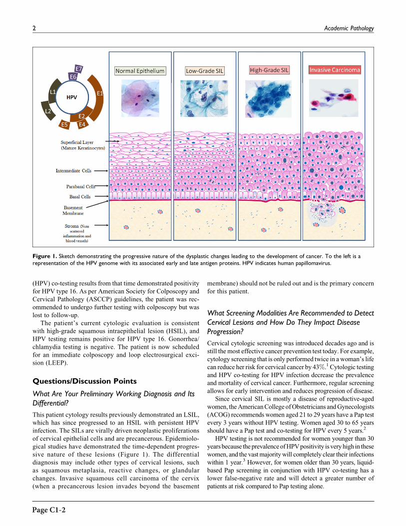
Page C1-2
(HPV) co-testing results from that time demonstrated positivity
for HPV type 16. As per American Society for Colposcopy and
Cervical Pathology (ASCCP) guidelines, the patient was rec-
ommended to undergo further testing with colposcopy but was
lost to follow-up.
The patient’s current cytologic evaluation is consistent
with high-grade squamous intraepithelial lesion (HSIL), and
HPV testing remains positive for HPV type 16. Gonorrhea/
chlamydia testing is negative. The patient is now scheduled
for an immediate colposcopy and loop electrosurgical exci-
sion (LEEP).
Questions/Discussion Points
What Are Your Preliminary Working Diagnosis and ItsDifferential?
This patient cytology results previously demonstrated an LSIL,
which has since progressed to an HSIL with persistent HPV
infection. The SILs are virally driven neoplastic proliferations
of cervical epithelial cells and are precancerous. Epidemiolo-
gical studies have demonstrated the time-dependent progres-
sive nature of these lesions (Figure 1). The differential
diagnosis may include other types of cervical lesions, such
as squamous metaplasia, reactive changes, or glandular
changes. Invasive squamous cell carcinoma of the cervix
(when a precancerous lesion invades beyond the basement
membrane) should not be ruled out and is the primary concern
for this patient.
What Screening Modalities Are Recommended to DetectCervical Lesions and How Do They Impact DiseaseProgression?
Cervical cytologic screening was introduced decades ago and is
still the most effective cancer prevention test today. For example,
cytology screening that is only performed twice in awoman’s life
can reduce her risk for cervical cancer by 43%.1 Cytologic testing
and HPV co-testing for HPV infection decrease the prevalence
and mortality of cervical cancer. Furthermore, regular screening
allows for early intervention and reduces progression of disease.
Since cervical SIL is mostly a disease of reproductive-aged
women, theAmericanCollegeofObstetricians andGynecologists
(ACOG) recommends women aged 21 to 29 years have a Pap test
every 3 years without HPV testing. Women aged 30 to 65 years
should have a Pap test and co-testing for HPV every 5 years.2
HPV testing is not recommended for women younger than 30
yearsbecause theprevalenceofHPVpositivity is veryhigh in these
women, and the vastmajoritywill completely clear their infections
within 1 year.3 However, for women older than 30 years, liquid-
based Pap screening in conjunction with HPV co-testing has a
lower false-negative rate and will detect a greater number of
patients at risk compared to Pap testing alone.
Figure 1. Sketch demonstrating the progressive nature of the dysplastic changes leading to the development of cancer. To the left is arepresentation of the HPV genome with its associated early and late antigen proteins. HPV indicates human papillomavirus.
2 Academic Pathology

Page C1-3
In 2014, the ATHENA (Addressing THE Need for
Advanced HPV diagnostics) study examined the ability of HPV
testing alone to identify women at risk of developing cervical
squamous lesions. The study demonstrated that primary screen-
ing using only HPV testing can be an effective screening strat-
egy in some patients. Based on data from this study, the Food
and Drug Administration (FDA) approved the use of 1 HPV
test (Roche Cobas HPV test) for primary cervical cancer
screening (without cytology) in women aged 25 years and
older.
Although HPV testing may be able to identify the pres-
ence of nucleic acids from high-risk HPV types and serial
testing can demonstrate persistent infection, Pap testing still
has several advantages over HPV testing alone. Since the
Pap smear is not designed solely around HPV, it can
provide tremendous additional information about the cervi-
cal and sexual health of a woman. Although not initially
designed for this purpose, the Pap smear can identify
numerous infections, including herpes, trichomonas, actino-
myces, and bacterial vaginosis (Figure 2). It can even iden-
tify the presence of cancer cells from other gynecological
sites, such as endometrial carcinoma. Additionally, Pap
staining, which is designed to identify the lesions produced
by HPV infections, can provide information about how
progressive the disease has become whereas HPV testing
alone cannot.
Why Is Cervical Cancer, in Particular, a Disease forWhich It Is Appropriate to Create a Screening Program?
In 1968, James Wilson published a World Health Organiza-
tion pamphlet that addressed objective principles which any
successful screening program must meet.4 The principals
were primarily not only related to the disease itself but also
addressed case identification through testing. In brief,
Wilson established that in order for a screening program
to be successful, the associated disease must be a public
health burden, there should be a pathophysiologic under-
standing of the disease, there must be a predisease state in
which we can identify possible cases, there should be a
cost-effective test for the disease and there should be an
available treatment for the disease. Cervical cancer, along
with our understanding of HPV-related neoplasia and pro-
gression to cancer, successfully meets all of these require-
ments and, therefore, was historically a prime candidate for
a screening strategy.4
Figure 2. Common infectious findings on Pap smear: (A) herpes simplex virus, (B) trichomonas vaginalis (arrow pointing to trichomonalparasite), (C) actinomyces, (D) Clue cell (indicating a shift in vaginal flora).
Kim et al 3

Page C1-4
Explain the Pathophysiologic Process That Occurs in theDevelopment of Squamous Intraepithelial Lesion/Cancer:How Is Human Papillomavirus Involved?
The HPV genotypes which have a documented association with
squamous lesions and can progress to invasive cancer are
termed “high risk.” Other “low-risk” genotypes may cause
other lesions such as genital warts but do not progress to can-
cer. Of the high-risk HPV types, genotypes 16 and/or 18 have
been identified in the majority of cervical cancers.
The pathogenesis of cervical cancer requires 2 biologically
interrelated features: a productive HPV viral infection and an
epithelial neoplastic process. According to this model, there is
initially an infection with a high-risk type of HPV that persists
and progresses to a pathologically defined precursor lesion and
ultimately to invasion3 (Figure 1).
Squamous lesions develop after HPV infected the basal or
primitive cells of the immature squamous epithelium through
defects or breaks in the skin or mucous membranes. The early
region of the HPV genome includes transforming regions E6
and E7 whose corresponding proteins bind to and inhibit the
host cell–regulatory proteins, p53 and Rb. This causes unrest-
ricted cell proliferation and blocks cellular apoptosis. Most SIL
starts at the squamocolumnar junction of the transformation
zone of the cervix. Viral DNA replication occurs mostly in the
superficial and intermediate cell layers of the squamous epithe-
lium. As these infected cells move to the epithelial surface,
differentiation-specific transcriptional factors from the host
cells stimulate the production of viral capsid proteins and sub-
sequently intact virions that produce characteristic cytologic
and histologic changes3 (see Figure 1).
Describe the Natural History of Human PapillomavirusInfections and Its Relation to Neoplasia
MostHPV infections are transient, becoming latent or undergoing
immunologic clearance within 1 to 2 years of diagnosis.1 How-
ever, infections with high-risk types of HPV clear more slowly
andpersistenceof infection increases the likelihoodofdeveloping
high-grade lesions. Prevalence of HPV infection peaks in the late
teens to early 20s, while the incidence of cervical cancer in
unscreened populations ranges from 35 to 55 years of age. Since
HSIL is more common than invasive cervical cancer, this sug-
gests that only a small portion ofHSILprogresses tomalignancy.1
We, therefore, understand that although the development of inva-
sive cervical cancer requires persistent infection with high-risk
types of HPV, it is not always sufficient to cause cancer. Other
associated risk factors include cigarette smoking, long-term use
of combined oral contraceptive pills, and immunosuppression.3
Describe the Cytopathological Features of SomeCommon Cervical Infections Which Can be Foundon the Pap Stain
Cervical cytology has relatively high specificity for most organ-
isms and can help guide clinical management (see Figure 2).
A. Herpes simplex virus (HSV): The classic features of
HSV infections on Pap staining are described as the
“three M’s of herpes”: multinucleation, margination
of chromatin, whereby the rim of the individual
nuclei appears darker staining than the center, and
molding which is seen as each nucleus indenting the
neighboring nuclei. With the accumulation of viral
particles, the nuclei may develop a “ground glass”
appearance.
B. Trichomonas vaginalis: The parasitic organisms are
visible with liquid-based preparations. They appear
as pear-shaped organisms with an eccentrically
located nucleus, eosinophilic cytoplasmic granules,
and flagella.
C. Actinomyces: Clumps of woolly filamentous organ-
isms which are usually deeply staining and may
demonstrate acute angle branching.
D. Clue cells: Irregularly shaped squamous cells covered
with darkly staining coccobacilli. These cells typi-
cally indicate a change in the cervical microbiologi-
cal flora and may be an indication of bacterial
vaginosis.
What Systems Are Used to Classify Lesions Identified byCervical Cytology? How About Classification of CervicalHistopathology?
The Bethesda System (TBS) is used to categorize cytological
diagnoses, while the Lower Anogenital Squamous Terminol-
ogy (LAST) project describes histological findings associated
with HPV throughout the anogenital tissues, including the
cervix.5 The Bethesda system utilizes a 2-tiered system of
classification (LSIL or HSIL) and previously histopatholo-
gists utilized a 3-tiered system (cervical intraepithelial neo-
plasia [CIN]-I, CIN-II, and CIN-III). Large studies have
demonstrated significant diagnostic variation between CIN-
II and CIN-III; additionally, CIN-II lesions were found to
demonstrate similar rates of progression of disease to CIN-
III in some instances. For these reasons, the LAST project has
adopted a 2-tiered diagnostic system and both LAST and TBS
are in line with one another.6
Compare and Contrast Cytopathologyand Histopathology in the Images
A. Cytology LSIL:
Cells have enlarged nuclei, about 4 times the size of a
normal intermediate cell nucleus. The nuclei are darker
(hyperchromatic) with wrinkled irregular nuclear membrane
contours. Cells may be multinucleated as seen in the image
and the chromatin is finely granular. Areas of clearing around
the nucleus (koilocytosis), a hallmark of LSIL, are also seen
(Figure 3).
4 Academic Pathology

Page C1-5
B. Histology LSIL (Previously CIN-I):
Nuclei are enlarged and irregularity shaped. There is also
darkening of the chromatin (hyperchromasia). Koilocytes with
their large vacuoles can be seen in the middle layers, while the
superficial layers still resemble a fairly normal basket weave
appearance of unaffected epithelium. Overall, there is
increased thickness of the epithelium.
C. Cytology HSIL:
The degree of nuclear atypia is increased, such that the
nuclei of these cells appear more hyperchromatic and irregular.
The amount of cytoplasm decreases as nuclear to cytoplasmic
(N:C) ratio increases. The classical features of a mature squa-
mous cell with abundant cytoplasm are no longer seen, and
these cells appear less differentiated.
D. Histology HSIL:
Undifferentiated neoplastic cells which no longer resemble
the normal squamous cells are present in all layers of the
epithelium. The lower layers of the epithelium (basal and para-
basal) are thickened, and abnormal mitotic figures are visible.
There is nuclear crowding, and cellular variation (pleomorph-
ism) is seen. These areas may appear “bluer” under the micro-
scope because there is less pink cytoplasm. The N:C ratio is
high due to scant cytoplasm.
What Is the Recommended Management of AbnormalCervical Cancer Screening Tests and Cancer Precursors?How Should You Proceed Now With More AdvancedDisease Present?
The ASCCP Updated consensus guidelines for managing
abnormal cervical cancer screening tests and cancer precur-
sors outlines the recommendations for managing patients with
cervical cytological abnormalities. Although it takes many
years for HSIL to progress to invasive cancer, untreated higher
grade lesions are also more likely to persist and progress than
regress.3 Therefore, appropriate disease follow-up and manage-
ment are crucial.
Since our patient now presents with evidence of HSIL on
cytology, it is recommended that she undergoes an immediate
LEEP or colposcopy with endocervical assessment. Manage-
ment by ASCCP guidelines differs depending on the colpo-
scopy results. If the biopsy confirms HSIL, either excision or
ablation of the “transformation zone” is recommended, fol-
lowed by co-testing at 12 and 24 months.1
What Other Factors Aside From Screening May Alterthe Future Burden of This Disease on the Population?
TheHPV is an immunologically cleared infection. As such, it was
a prime candidate for the development of a multivalent HPV vac-
cine. Currently, there are 3 FDA–approved HPV vaccines on the
Figure 3. Cytologic and histologic images of low-grade SIL (A, B) and high-grade SIL (C, D). SIL indicates squamous intraepithelial lesion.
Kim et al 5

Page C1-6
market: Gardasil, Gardasil 9, and Cervarix. The Cervarix vaccine
is designed for immunity against HPV 16 and HPV 18, the 2 most
common high-risk HPV types that cause about 70% of cervical
cancer. Gardasil adds coverage for 2 additional low-risk HPV
genotypes (6 and 11) which are the major cause of genital warts.
Gardasil 9 extends coverage further to 5 additional high-risk HPV
genotypes associated with cervical cancer. These vaccines consist
of nonreplicative viral-like particles which provoke an immune
response and provide humoral immunologic memory.7 Currently,
several international health-based organizations recommendHPV
vaccination be a part of all routine immunization programs. It is
important to note that screening following immunization is still
required and we have discussed several additional benefits to
screening beyond identification of HPV infection.
Describe Some Possible Therapies That Are BeingDeveloped to Treat Human Papillomavirus–AssociatedLesions and Cancers
While the initial therapy of squamous lesions is currently com-
plete excision, other novel therapies are currently being eval-
uated. We have discussed vaccines to prevent infection that are
widely available and utilize viral structural proteins. There are
currently in development vaccines that help a person’s immune
system to clear the HPV after infection. Most of these vaccines
target HPV oncoproteins E6 and E7.8 This treatment is antici-
pated to become clinically available in the near future to decrease
the HPV-associated disease burden. Furthermore, research on a
pelvic sentinel lymph node biopsy procedure, immunotherapy,
and targeted drug therapy is also being conducted.
Teaching Points
� Characteristic cytologic and histologic features, such as
koilocytosis, enlarged nuclei, and increased N:C ratio,
allow pathologists to diagnose and classify different
grades of squamous cervical pathology from low-grade
to high-grade squamous lesions and ultimately to
carcinoma.
� Classifications based on these features allow one to pre-
dict the biologic behavior and malignant potential of
these precancerous lesions.
� HPV infection, particularly with high-risk types of HPV,
is not only a major risk factor for developing cervical
SIL/cancer but also a crucial factor in the pathogenesis
of this disease.
� Adequate screening for cervical precancerous pathology
may include both cytology Pap and ancillary HPV test-
ing as each method has distinct advantages for the detec-
tion of pathologic conditions.
� Cervical disease and its associated diagnostic testing are
a paradigmatic model of disease screening and interven-
tion and remain one of the most successful screening
programs ever devised.
� Several organizations, including the ACOG and the
ASCCP, regularly publish guidelines related to screen-
ing and management of the cervical cancer.
Declaration of Conflicting Interests
The author(s) declared no potential conflicts of interest with respect to
the research, authorship, and/or publication of this article.
Funding
The author(s) received no financial support for the research, author-
ship, and/or publication of this article.
ORCID iD
D. Yitzchak Goldstein http://orcid.org/0000-0001-6367-8335
References
1. Wright TC, Ronnett BM, Kurman RJ, Ferenczy A. Precancerous
lesions of the cervix. In: Blaustein’s Pathology of the Female
Genital Tract. New York, NY: Springer; 2011:193-252.
2. Saslow D, Solomon D, Lawson HW, et al. American Cancer Soci-
ety, American Society for Colposcopy and Cervical Pathology, and
American Society for Clinical Pathology Screening Guidelines for
the Prevention and Early Detection of Cervical Cancer. CA Cancer
J Clin. 2012;62:147-172.
3. Sellors JW, Karwalajtys TL, Kaczorowski J, et al. Incidence, clear-
ance and predictors of human papillomavirus infection in women.
CMAJ. 2003;168:421-425.
4. Wilson JMG, Jungner G; World Health Organization, Geneva,
Switzerland. Principles and Practice of Screening for Disease;
1968.
5. Darragh TM, Colgan TJ, Cox JT, et al. The Lower Anogenital
Squamous Terminology standardization project for HPV-
associated lesions: background and consensus recommendations
from the College of American Pathologists and the American Soci-
ety for Colposcopy and Cervical Pathology. Arch Pathol Lab Med.
2012;136:1266-1297.
6. Massad LS, Einstein MH, Huh WK, et al. 2012 updated consensus
guidelines for the management of abnormal cervical cancer screen-
ing tests and cancer precursors. J Low Genit Tract Dis. 2013;17:
S1-S27.
7. Arvin A, Campadelli-Fiume G, Mocarski E, et al. Human Herpes-
viruses: Biology, Therapy, and Immunoprophylaxis. Cambridge:
Cambridge University Press; 2007.
8. Nayar R, Wilbur DC. The Bethesda System for Reporting
Cervical Cytology: Definitions, Criteria, and Explanatory Notes.
New York, NY: Springer; 2015.
6 Academic Pathology

Page C1-7
Educational Case
Educational Case: A Uterine Neoplasm:Leiomyoma—A Benign Neoplasm
Margret S. Magid, MD1
The following fictional case is intended as a learning tool within the Pathology Competencies for Medical Education
(PCME), a set of national standards for teaching pathology. These are divided into three basic competencies: Disease
Mechanisms and Processes, Organ System Pathology, and Diagnostic Medicine and Therapeutic Pathology. For additional
information, and a full list of learning objectives for all three competencies, see http://journals.sagepub.com/doi/10.1177/
2374289517715040.
Keywordspathology competencies, disease mechanisms, neoplasia, anaplasia, benign, hyperchromatic, hysterectomy, invasion, leiomyoma,leiomyosarcoma, malignant, metastasis, myomectomy, myometrium, normochromatic, nuclear:cytoplasmic ratio, paraneoplasticsyndrome, pleomorphic, poorly differentiated, smooth muscle tumor, well-differentiated
Received April 05, 2017. Received revised May 06, 2017. Accepted for publication June 16, 2017.
Primary Objective
Objective N3.1: Morphologic Features of Neoplasia. Describe
the essential morphologic features of neoplasms and indicate
how these can be used to diagnose, classify, and predict biolo-
gical behavior of cancers.
Competency 1: Disease Mechanisms and Processes; Topic
N: Neoplasia; Learning Goal 3: Characteristics of Neoplasia.
Patient Presentation
A 39-year-old P3013 woman presented to her gynecologist
with complaints of increased bleeding during menstruation,
back pain, and a feeling of rectal fullness. Pregnancy test was
negative. Her gynecologist palpated a large irregular, asym-
metric uterus. The patient underwent hysterectomy.
Diagnostic Findings
Examination of the hysterectomy specimen in surgical pathol-
ogy showed it to be enlarged and distorted by multiple tumors
within the myometrium. Figure 1 is a cross section through the
uterus and the largest tumor.
Questions/Discussion Points
In Figure 1, describe the cut surface of the tumor.
The cut surface is homogeneously white. There are no areas of
grossly recognizable necrosis or hemorrhage, which may be
indicative of more aggressive tumors.
In Figures 1 and 2, how would you describe the border ofthe tumor nodule with the surrounding uterus?
Figure 2 is a low-power photomicrograph from the specimen.
The borders are smooth and “pushing” (not infiltrating into
the surrounding myometrium).
What information does this give you about the nature of
the tumor?
This gross feature favors that the tumor is benign.
1 NYU School of Medicine, New York, NY, USA.
Corresponding Author:
Margret S. Magid, NYU School of Medicine, 560 First Avenue, New York, NY
10016, USA.
Email: [email protected]
Academic PathologyVolume 5: 1–3ª The Author(s) 2018Article reuse guidelines:sagepub.com/journals-permissionsDOI: 10.1177/2374289517721193journals.sagepub.com/home/apc
Creative Commons Non Commercial CC BY-NC: This article is distributed under the terms of the Creative Commons Attribution-NonCommercial 4.0 License(http://www.creativecommons.org/licenses/by-nc/4.0/) which permits non-commercial use, reproduction and distribution of the work without furtherpermission provided the original work is attributed as specified on the SAGE and Open Access pages (https://us.sagepub.com/en-us/nam/open-access-at-sage).
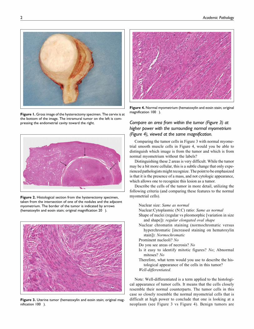
Compare an area from within the tumor (Figure 3) athigher power with the surrounding normal myometrium(Figure 4), viewed at the same magnification.
Comparing the tumor cells in Figure 3 with normal myome-
trial smooth muscle cells in Figure 4, would you be able to
distinguish which image is from the tumor and which is from
normal myometrium without the labels?
Distinguishing these 2 areas is very difficult. While the tumor
may be a bit more cellular, this is a subtle change that only expe-
rienced pathologistsmight recognize. The point to be emphasized
is that it is the presence of a mass, and not cytologic appearance,
which allows one to recognize this lesion as a tumor.
Describe the cells of the tumor in more detail, utilizing the
following criteria (and comparing these features to the normal
myometrial cells).
Nuclear size: Same as normal
Nuclear:Cytoplasmic (N:C) ratio: Same as normal
Shape of nuclei (regular vs pleomorphic [variation in size
and shape]): regular elongated oval shape
Nuclear chromatin staining (normochromatic versus
hyperchromatic [increased staining on hematoxylin
stain]): Normochromatic
Prominent nucleoli? No
Do you see areas of necrosis? No
Is it easy to identify mitotic figures? No; Abnormal
mitoses? No
Therefore, what term would you use to describe the his-
tological appearance of the cells in this tumor?
Well-differentiated.
Note: Well-differentiated is a term applied to the histologi-
cal appearance of tumor cells. It means that the cells closely
resemble their normal counterparts. The tumor cells in this
case so closely resemble the normal myometrial cells that is
difficult at high power to conclude that one is looking at a
neoplasm (see Figure 3 vs Figure 4). Benign tumors areFigure 3. Uterine tumor (hematoxylin and eosin stain; original mag-nification 100�).
Figure 4. Normal myometrium (hematoxylin and eosin stain; originalmagnification 100�).
Figure 1.Gross image of the hysterectomy specimen. The cervix is atthe bottom of the image. The intramural tumor on the left is com-pressing the endometrial cavity toward the right.
Figure 2. Histological section from the hysterectomy specimen,taken from the intersection of one of the nodules and the adjacentmyometrium. The border of the tumor is indicated by arrows(hematoxylin and eosin stain; original magnification 20�).
2 Academic Pathology

Page C1-9
typically well-differentiated. Malignant neoplasms may be
well-differentiated but more often will show a range of
decreasing differentiation that may progress to anaplasia (lack
of differentiation). The histological features that are associ-
ated with decreased differentiation include increased nuclear-
to-cytoplasmic ratio, pleomorphism of the nuclei and cells,
hyperchromatism, and prominent nucleoli—none of which
were observed in this case. Additional features suggestive
of malignancy are increased mitotic activity (particularly in
tissue that is not normally mitotically active), including
abnormal mitotic figures, and the presence of necrosis (both
grossly and/or microscopically), which were also not
observed.
What is your pathological diagnosis?
Leiomyoma of the uterus (A benign tumor: leiomyo ¼ smooth
muscle; oma ¼ benign neoplasm)
By your terminology above, you have identified the tumor
as either benign or malignant.
Summarize the gross and microscopic morphological fea-
tures in this case that support your classification.
Growth pattern: Expansile, pushing, noninvasive growth
Microscopic: Well-differentiated; regular nuclear size and
shape and normochromatic; absent or rare mitoses (less than 5
mitoses/10 high-power fields); absence of necrosis.
Note: The classification of this tumor as benign implies
absence of invasive local growth and inability to metastasize
(discontinuous spread to distant sites). Nonetheless, benign
neoplasms may be problematic and even fatal to the host due
to growth in a sensitive location (eg, in the meninges or car-
diac left atrium), other local effects (eg, ulceration in a hollow
viscus), and secretion of indigenous or inappropriate hor-
mones (ie, hormones that are normally not elaborated by the
normal cellular counterpart of the tumor, resulting in a para-
neoplastic syndrome).1-2
What is the natural history of this tumor?
Leiomyomas are common benign neoplasms of the uterus.
Leiomyomas show expansile growth in the uterus. On cut sec-
tion, they show a white, whorled appearance that gave rise to
their colloquial term, fibroid. Ultrasonography is the standard
diagnostic test. They generally show growth during reproduc-
tive years and regression in menopause. They may be
asymptomatic but, depending on size, location, and number,
may present with abnormal bleeding, urinary symptoms from
compression of the bladder or rectum, pelvic pain, and
interference with fertility and maintenance of pregnancy.
Symptomatic leiomyomas may be treated surgically with
hysterectomy or in a woman desiring pregnancy by myomect-
omy. Medical therapy includes hormonal therapy, uterine
artery embolization, and radiofrequency ablation.
The malignant counterpart of leiomyoma is leiomyosar-
coma (leiomyo ¼ smooth muscle; sarcoma ¼ malignant
mesenchymal neoplasm). The histological appearance of
leiomyosarcomas ranges from well-differentiated tumors that
show increased numbers of mitotic figures to highly anaplastic
tumors with areas of necrosis. These tumors show progressive
invasive growth and metastasis. Leiomyosarcomas are believed
to arise de novo from the myometrial cells. Leiomyomas are
generally NOT considered to be premalignant, so most
asymptomatic leiomyomas do not need be excised.3-4
Teaching Points
� Leiomyoma is a common benign neoplasm of the uterus.
� While frequently asymptomatic, leiomyomas may pres-
ent with symptoms due to local expansion and compres-
sion within the uterus and/or into adjacent pelvic
structures.
� Uterine leiomyomas are not considered to be
premalignant.
� Benign neoplasms are characterized by expansile,
smooth, and noninvasive borders.
� Histologically, benign neoplasms are well-differentiated,
with regular cells and nuclei, rare mitoses, and absence of
necrosis.
Declaration of Conflicting Interests
The author declared no potential conflicts of interest with respect to
the research, authorship, and/or publication of this article.
Funding
The author received no financial support for the research, authorship,
and/or publication of this article.
References
1. Neoplasia. In: Kumar V, Abbas AK, Aster JC, eds. Robbins and
Cotran Pathologic Basis of Disease. 9th ed. Philadelphia, PA:
Elsevier Saunders; 2015.
2. Neoplasia. In: Strayer DS, Rubin E, eds. Rubin’s Pathology. 7th ed.
Philadelphia, PA: Wolters Kluwer; 2015.
3. Stewart EA. Clinical practice. Uterine fibroids. N Engl J Med.
2015;372:1646-1655.
4. Marret H, Fritel X, Ouldamer L, et al. Therapeutic management of
uterine fibroid tumors: updated French guidelines. Eur J Obstet
Gynecol Reprod Biol. 2012;165:156-164.
Magid 3


Page C1-11
Educational Case
Genetic Mutations and MultifactorialInheritance: Dilated Cardiomyopathy
Eric S. Suarez, MD1 and Barbara E. C. Knollmann-Ritschel, MD1
The following fictional case is intended as a learning tool within the Pathology Competencies for Medical Education
(PCME), a set of national standards for teaching pathology. These are divided into three basic competencies: Disease
Mechanisms and Processes, Organ System Pathology, and Diagnostic Medicine and Therapeutic Pathology. For addi-
tional information, and a full list of learning objectives for all three competencies, see http://journals.sagepub.com/doi/
10.1177/2374289517715040.
Keywordspathology competencies, genetic mechanisms, developmental and functional abnormalities, mutations, cardiovascular, cardiomyo-pathy, dilated cardiomyopathy, disease mechanisms, genetic diseases, inherited diseases
Received March 23, 2017. Received revised April 25, 2017. Accepted for publication May 3, 2017.
Primary Objective
Objective GM1.1: Types of Mutations. Describe different types ofmutations that can occur in human disease, and discuss how
each of these can produce abnormalities in DNA transcription
and/or alterations in the type or amount of protein produced.
Competency 1: Disease Mechanisms and Processes; Topic
GM: Genetic Mechanisms; Learning Goal 1: Genetic Mechan-
isms of Developmental and Functional Abnormalities.
Secondary Objectives
Objective GM1.5: Multifactorial Inheritance and EnvironmentalFactors. Discuss and give examples of disorders associated with
multifactorial inheritance and describe how environmental fac-
tors can interact with genetic factors to produce or modulate
disease.
Competency 1: Disease Mechanisms and Processes; Topic
GM: Genetic Mechanisms; Learning Goal 1: Genetic Mechan-
isms of Developmental and Functional Abnormalities.
Objective CH1.2: Cardiomyopathy. Compare and contrast the clin-
icopathologic features of dilated, restrictive, and hypertrophic
cardiomyopathies.
Competency 2: Organ System Pathology; Topic CH: Cardi-
ovascular—Heart; Learning Goal 1: Heart Failure
Patient Presentation
A 26-year-old male presents to the emergency department with
complaints of a 5-month history of progressive dyspnea that
became more severe over the past 6 days and prompts this
evaluation. Medical history is significant for at least 6 upper
respiratory infections over the past 2 years. Family history is
significant for 4 cardiac-related deaths in ages between 24 and
38 years involving the 3 last generations of his family, as well
as a 29-year-old paternal cousin with heart failure. There is no
significant history of alcohol ingestion, toxin exposures, or
illicit drug abuse. Physical examination discloses bilateral
1 Department of Pathology, School of Medicine, Uniformed Services University
of the Health Sciences, Bethesda, MD, USA
Corresponding Author:
Eric S. Suarez, Department of Pathology, School of Medicine, Uniformed
Services University of the Health Sciences, 4301 Jones Bridge Rd, Bethesda,
MD 20814, USA.
Email: [email protected]
Academic PathologyVolume 4: 1–3ª The Author(s) 2017Reprints and permission:sagepub.com/journalsPermissions.navDOI: 10.1177/2374289517711715journals.sagepub.com/home/apc
Creative Commons CC BY-NC: This article is distributed under the terms of the Creative Commons Attribution-NonCommercial 4.0 License(http://www.creativecommons.org/licenses/by-nc/4.0/) which permits non-commercial use, reproduction and distribution of the work without furtherpermission provided the original work is attributed as specified on the SAGE and Open Access pages (https://us.sagepub.com/en-us/nam/open-access-at-sage).

Page C1-12
basal lung rales, displaced apical impulse to the left, systolic
apical murmur, hepatomegaly of 6 cm below the right midcla-
vicular line, and bilateral low extremity pitting edema extend-
ing to 5 cm above the ankles. Recorded vital signs are blood
pressure ¼ 105/65 mm Hg, Respiratory Rate ¼ 28 per minute,
Heart Rate ¼ 138 per minute, and temperature ¼ 98.9�F.
Diagnostic Findings
Echocardiogram shows marked cardiac enlargement, dilation
of all chambers, 35% left ventricle ejection fraction, and left
ventricle thrombi. Imaging studies show normal cardiac valves
and patent coronary arteries. Laboratory studies reveal moder-
ate B-natriuretic peptide elevations and mild troponin and liver
enzyme elevations.
Questions/Discussion Points
What is Your Preliminary Diagnosis and Its Differential?
Dilated cardiomyopathy is the primary diagnosis, based on the
clinical findings consistent with heart failure, cardiac enlarge-
ment of all chambers, imaging studies showing normal valves
and patent coronaries, and functional studies describing
decreased ejection fraction. The differential diagnosis based
on the clinical findings includes other types of cardiomyopathy
such as restrictive or hypertrophic, as well as multiple diseases
included within the syndrome of dilated cardiomyopathy per
se. Dilated cardiomyopathy can be further categorized as
familial and inherited or as acquired. Acquired etiologies
include myocarditis; toxins such as cocaine, amphetamines,
or heavy metals; autoimmune; endocrine diseases such as
hypothyroidism or hyperthyroidism; thiamine deficiency; and
ischemic heart disease.1,2
What Clinical Considerations Should Follow a Diagnosisof Cardiomyopathy?
When suspecting a cardiomyopathy, the initial considerations
are morphologic and functional, which help classify them as
dilated, restrictive, hypertrophic, or one of the more unusual
types of cardiomyopathy such as right ventricle arrhythmo-
genic or left ventricle noncompaction cardiomyopathy. Occa-
sionally, these features may overlap or may not be fully
developed, such as when you evaluate inherited genetic dis-
eases early in their development.
The extent of organ involvement should be recorded. The
manifestations may be limited to only the heart, but the dis-
order may affect other organs as well, such as skeletal muscle,
kidneys or liver as part of the disease process, or may it affect
other organs secondarily.
Constructing a pedigree tree may be very useful in determin-
ing the inheritance pattern. When evaluating the pedigree, it is
possible that the information may be unknown or not investi-
gated at the time of initial presentation. Genetics/familial con-
siderations include autosomal dominant or recessive, X-linked,
mitochondrial, or sporadic inheritance patterns which can
easily be determined with the family history evaluation.3
Etiology of the cardiomyopathy should be investigated. This
may include genetic, viral following a myocarditis, autoim-
mune, toxic, or unknown causes of cardiomyopathy.
The stage of heart failure at the time of presentation should
be recorded using criteria of the American Heart Association or
New York Heart Association.
What Pathophysiologic Mechanisms Are Involvedin the Development of Dilated Cardiomyopathy?
Defects of force transmission, force generation, or calcium han-
dling result in systolic dysfunction, cell death, and fibrosis, pro-
ducing the dilated cardiomyopathy phenotype. Mutations
involving d-sarcoglycanmay cause defects of force transmission;
in a similar fashion, defects of force generationmay be caused by
mutations encoding for sarcomere proteins such as b-myosin.4
Even in nonfamilial cases of cardiomyopathy, inherited genetic
susceptibilities may determine how the cardiac muscle responds
to environmental factors such as alcohol or viral myocarditis.
What Are the Inheritance Patterns of DilatedCardiomyopathy? Can You Give Some Examples?
Approximately 30% to 48% of dilated cardiomyopathies are
inherited or familial. Most of the inherited cases are autosomal
dominant; but autosomal recessive and X-linked patterns have
also been reported. Causative genes predominantly encode for
sarcomere (ie, titin [TTN], actin, myosin, troponin) or cytoskele-
tal (ie, dystrophin, desmin, lamin) proteins. Dystrophin is the
same protein involved in the X-linked Duchenne or Becker mus-
cular dystrophies. Less commonly, an X-linked inheritance pat-
tern may be identified in newborns and children as part of the
Barth syndrome, which is caused by mutations of the gene tafaz-
zin, which codes for an acyltransferase. Twenty percent of auto-
somal dominant, dilated cardiomyopathies identified genetically
affect genes coding for TTN, a protein that provides structure,
flexibility, stability, and chemical signals to the sarcomere.5
These mutations result in an abnormally short TTN protein.6
About 8% of all genetic dilated cardiomyopathy cases are caused
by deletions or sequence variations of the LMNAgene that, when
mutated or deleted, encode for an absent or abnormal lamin A/C
protein. Most of these cases also have an associated atrioventri-
cular block clinically, and in this circumstance, implantation of
intracardiac defibrillator may be a consideration. Rare autosomal
recessive cases result from mutations encoding for troponin I.
How Should You Proceed With the Genetic Evaluationof This Case?
Perform a careful pedigree analysis of all possible affected
family members taking into consideration and annotating all
possible involved cardiac and extracardiac clinical features.
Inherited cardiomyopathies are phenotypically variable in
regard to the age of presentation, expressivity, or disease
2 Academic Pathology
Page C1-13
progression. Mixed forms that do not fit into a single traditional
category have been described as several instances of dilated
cardiomyopathy have been associated with an arrhythmogenic
phenotype. Incomplete penetrance and variable expressivity
warrant a high index of suspicion, particularly for first-
degree relatives. You may consider genetic testing for
first-degree family members, with the caveat that the yield may
be much lower than in the cases of hypertrophic cardiomyo-
pathy. The sensitivity of genetic sequencing panels for familial
dilated cardiomyopathy cases is 25% but rises up to 40% if the
cardiomyopathy coexists with conduction defects. A trun-
cated TTN protein caused by TTN frameshift, nonsense, or
splice mutations have been reported in approximately 25% of
familial dilated cardiomyopathy cases. Titin encompasses
363 exons with an established role in muscle assembly and
function. At present, there seems to be no clear correlation
between genotype and phenotype probably because this large
gene can undergo extensive alternative splicing. These spli-
cing variants beg for a more refined interpretation of genetic
findings.7 Truncating mutations have also been associated
with sporadic cases, suggesting increased susceptibility to
environmental effects. The significance of genetic variations
is unknown in many instances. Frequently, a genotype–
phenotype correlation will be of limited clinical utility in the
treatment of the patient, but some instances warrant genetic
characterization.
Teaching Points
� Clinical evaluation including complete pedigree analy-
sis is essential in the evaluation of genetic disorders such
as dilated cardiomyopathy.
� Penetrance and expressivity are important considera-
tions, especially when evaluating first-degree relatives.
� A truncated protein that fails to transmit or generate
force within or across the sarcomere or adequately han-
dle calcium can result from frameshift, nonsense, or
splice mutations.
� Theclinical significanceofmutations is frequentlyunknown.
� Manydilatedcardiomyopathycases are familial;most com-
monly autosomal dominant disorders, but autosomal reces-
sive andX-linked inheritance patterns can also be observed.
� Dilated cardiomyopathy clinical presents with heart fail-
ure and cardiac enlargement; imaging studies showing
normal valves and patent coronaries and a decreased
ejection fraction.
Authors’ Note
The opinions expressed herein are those of the authors and are not
necessarily representative of those of the Uniformed Services Univer-
sity of the Health Sciences (USUHS), the Department of Defense
(DOD), or the United States Army, Navy, or Air Force.
Declaration of Conflicting Interests
The author(s) declared no potential conflicts of interest with respect to
the research, authorship, and/or publication of this article.
Funding
The author(s) received no financial support for the research, author-
ship, and/or publication of this article.
References
1. Jefferies JL, Towbin JA. Dilated cardiomyopathy. Lancet. 2010;
375:752-762.
2. Weintraub RG, Semsarian C, Macdonald P. Dilated cardiomyopa-
thy. Lancet. 2017. doi:10.1016/s0140-6736(16)31713-5; pii:
s0140-6736(16)31713-5.
3. Arbustini E, Narula N, Dec GW, et al. The Moge(s) classification
for a phenotype–genotype nomenclature of cardiomyopathy. Glo-
bal Heart. 2013;4(N8):355-382.
4. Watkins H, Ashrafian H, Redwood C. Inherited cardiomyopathies.
N Engl J Med. 2011;364(17):1643-1656.
5. National Institutes of Health, National Library of Medicine. Familial
Dilated Cardiomyopathy. Genetics HomeReference. [Online]. 2017.
6. Norton N, Li D, Rampersaud E, et al; National Heart, Lung, and
Blood Institute GO Exome Sequencing Project and the Exome
Sequencing Project Family Studies Project Team. Exome sequen-
cing and genome-wide linkage analysis in 17 families illustrate the
complex contribution of TTN truncating variants to dilated
cardiomyopathy. Circ Cardiovasc Genet. 2013;6(2):144-153.
7. Tayal U, Prasad S, Cook SA. Genetics and genomics of dilated
cardiomyopathy and systolic heart failure. Genome Med. 2017;
9(1):20. doi:10.1186/s13073-017-0410-8.
Suarez and Knollmann-Ritschel 3


Page C1-15
Educational Case
Benign Papilloma of the Breast
Moshe Sadofsky, MD, PhD1
The following fictional case is intended as a learning tool within the Pathology Competencies for Medical Education
(PCME), a set of national standards for teaching pathology. These are divided into three basic competencies: Disease
Mechanisms and Processes, Organ System Pathology, and Diagnostic Medicine and Therapeutic Pathology. For addi-
tional information, and a full list of learning objectives for all three competencies, see http://journals.sagepub.com/doi/
10.1177/2374289517715040.
Keywordspathology competencies, disease mechanisms, organ system pathology, basement membrane, benign, breast, neoplasia, papilloma
Received March 09, 2017. Received revised May 01, 2017. Accepted for publication May 03, 2017.
Primary Objective
Objective N2.1: Prevalence and Geographic Impact on Neoplasia.Describe the prevalence of neoplastic diseases and discuss the
environmental factors that influence patients as they move
between geographical regions.
Competency 1: Disease Mechanisms and Processes; Topic
N: Neoplasia; Learning Goal 2: Environmental Influences on
Neoplasia.
Secondary Objectives
Objective N2.2: Mechanisms of DNA Damage Repair. Describe themechanisms by which exposure to radiation, tobacco, alcohol,
or other environmental chemical agents can produce cancer.
Competency 1: Disease Mechanisms and Processes; Topic
N: Neoplasia; Learning Goal 2: Environmental Influences on
Neoplasia.
Objectives N3.1 through N3.5. Learning Goal 3: Characteristics ofNeoplasia. Apply knowledge of the characteristics of neopla-
sia to discuss the morphologic appearance, classification,
biological behavior, and staging of neoplasms.
Competency 1: Disease Mechanisms and Processes; Topic
N: Neoplasia.
Objective BR1.1: Clinical Presentation of Breast Lesions. Identify themost frequently diagnosed breast lesions by age of the patient,
based on the most common clinical presentations in males
versus females.
Competency 2: Organ System Pathology; Topic BR:
Breast; Learning Goal 1: Nonneoplastic Disorders of the
Breast.
Objective BR1.4: Fibrocystic Change. Discuss the clinical signifi-
cance of proliferative and nonproliferative fibrocystic change,
with and without atypia, and describe how each of these
changes and the family history affects the subsequent risk of
developing breast cancer.
Competency 2: Organ System Pathology; Topic BR:
Breast; Learning Goal 1: Nonneoplastic Disorders of the
Breast.
1 Department of Pathology, Albert Einstein College of Medicine, Bronx, NY,
USA
Corresponding Author:
Moshe Sadofsky, Department of Pathology, Albert Einstein College of
Medicine, 1300 Morris Park Avenue, F514A, Bronx, NY 10461, USA.
Email: [email protected]
Academic PathologyVolume 4: 1–3ª The Author(s) 2017Reprints and permission:sagepub.com/journalsPermissions.navDOI: 10.1177/2374289517711714journals.sagepub.com/home/apc
Creative Commons CC BY-NC: This article is distributed under the terms of the Creative Commons Attribution-NonCommercial 4.0 License(http://www.creativecommons.org/licenses/by-nc/4.0/) which permits non-commercial use, reproduction and distribution of the work without furtherpermission provided the original work is attributed as specified on the SAGE and Open Access pages (https://us.sagepub.com/en-us/nam/open-access-at-sage).

Page C1-16
Patient Presentation
A 37-year-old woman presents to her physician with a palp-
able density in her left breast. She noticed it 1 month ago
and it hasn’t changed in size. Her medical history is unre-
markable. Medications include oral contraceptives. On
examination, she appears somewhat anxious. Her vital signs
are within normal limits. Physical examination is remark-
able for an ill-defined area of firmness in the left breast in
the upper outer quadrant adjacent to the nipple. The region
is not tender, and there is no inflammation or discharge. A
breast ultrasound core biopsy is obtained.
Questions/Discussion Points, Part 1
Before jumping to the histologic sections, it is worth discuss-
ing the differential diagnosis based on the patient
presentation.
What is the differential diagnosis of a palpable breastmass?
Our thinking is guided by the history. Is there pain? If so, is it
within the breast or associated with the chest wall? Does it
change with the hormonal cycle? Is there nipple discharge, and
if so, what is its character? These distinctions help determine
the likelihood of a true neoplasm of breast origin versus other
physiologic possibilities. A thorough discussion is available in
the study by Santen. Most importantly, 90% of new nodules
presenting in premenopausal women are benign. In younger
women, these are usually fibroadenomas, while “in the later
reproductive period, hyperplasia, cysts, and carcinoma in situ
are more common”.1 As occurs elsewhere in the body, growths
may also originate in nonglandular tissues including lipoma, fat
necrosis (with a history of injury), and hemangioma. Imaging
might suggest an area of increased density within the breast,
not explained as simple cyst.
The biopsy was obtained (Figure 1). Describe thehistologic features of this lesion. What is your diagnosisand what is its biologic behavior?
The low-powered Hematoxylin and Eosin (H&E)-stained
slide obtained from the biopsy contains the field in Figure
1. Within a large dilated duct sits a structure folded into
papillary fronds lined by a basophilic epithelium surrounding
Figure 1. Low-power H&E image from core biopsy of breast.
Figure 3. The comparison of the epithelial surface of the centralfronds shows a monolayer of basophilic cells similar in appearance tothe ductal lining and a fibrovascular eosinophilic stroma.
Figure 2. The image of Figure 1 is labeled showing the centralpapillary structure. A box encloses the region magnified below.
2 Academic Pathology

Page C1-17
a pink central fibrovascular stroma. These features are labeled
in Figure 2.
A box in Figure 2 is seen at higher magnification in Figure 3.
Figure 3 shows the duct wall and the epithelial surface of the
papillary fronds to be composed of similar cells, forming a
simple layer resting upon a basement membrane. The latter is
not easy to see directly with these dyes but can be visually
enhanced with special stains. Both tissues have matured in the
manner characteristic for ductal tissue, with a polarized surface
facing the duct lumen. The fronds also contain a stroma and
small blood vessels. The structure is named an “intraductal
papilloma.”
What Does the Name Tell You?
This is a benign proliferation in some ways analogous to adeno-
mas that may occur elsewhere. If it had beenmalignant, the name
would have included the word “carcinoma.” Papillomas have 2
main clinical presentations including solitary lesions near the
nipple or multiple peripheral papillomas. The latter have a higher
rate of atypia or malignancy arising in them. The most important
observation is that there is no evidence of invasion. In other
words, the epitheliumdoes not penetrate the basementmembrane
and spread into adjacent areas. This proliferative neoplastic pro-
cess can be associated with an increased risk of cancer in the
future, especially if there is atypical piling up of the epithelial
cells on each other (not seen here). In the absence of invasion, this
lesion is benign and doesnot need further treatment.However, the
patient should remain alert for changes in the future.
An additional important feature worth remembering is that
neoplasia (new growth) includes benign lesions. These may
grow to large size and can even become life threatening if they
interfere with a function (eg, benign meningioma). Benign
doesn’t mean, “you can live with it.” It means “not invasive.”
Other Manifestations of Benign Breast Disorders
Most important, benign lesions of the breast are much more
common than malignancies. The incidence of these disorders
changes with the age of the patient. An online review of Benign
Breast Disease in Women is referenced.1
Benign lesions of the breast are often classified in terms of
their potential contribution to future breast cancer. In this light,
there are 3 common categories:
� Nonproliferative changes (with little to no increased risk
of cancer). This includes fibrocystic change and cysts;
� Proliferative lesions without atypia, including this case;
� Proliferative lesions with atypia.
Proliferative disease is associated with a 1.5- to 2-fold
increased risk, while proliferative disease with atypia confers
a 4- to 5-fold increased risk.2
What is the structure and function of the basementmembrane that separates the epithelium from thefibrovascular core?
It is widely recognized that all epithelia organize themselves on
a structure called the basement membrane. However, the rela-
tionship of the cell to this structure is much more interesting
and active than many appreciate. Studies reveal “common
functions that include the induction and maintenance of cell
polarity, the establishment of barriers between tissue compart-
ments, the organization of cells into tissues, and the protection
of adherent cells from detachment-induced cell death,
anoikis.”3 Although the term membrane is used, it is not a lipid
membrane as the term is used elsewhere. Rather, the basement
membrane is a complex type of extracellular matrix with an
elaborate organization that includes collagen IV, laminins,
other connective proteins and heparin sulfate containing pro-
teoglycans. The absolute identity of the components of the
basement membrane varies among cell types and are essential
for the growth and maintenance of all epithelia.
Teaching Points
A quick review of the normal histology of ductal tissue in the
breast shows a simple monolayer of epithelial cells with an
underlying basement membrane.
� Despite the proliferative classification of this papilloma,
the basement membrane is not violated, so there is no
invasion.
� Invasion is an essential hallmark of epithelial
malignancy.
Benign lesions of the breast, depending on their microscopic
properties, may range in future risk of developing cancer from
none at all, to a 4- to 5-fold increase.
Acknowledgment
The original photographs were provided by Dr. Susan Fineberg.
References
1. Santen RJ. Benign breast disease in women. In: De Groot LJ,
Chrousos G, Dungan K, et al, eds. South Dartmouth, MA:
MDText.com, Inc; 2000. http://www.endotext.org, https://www.
ncbi.nlm.nih.gov/books/NBK278994/.
2. Kumar V, Abbas A, Aster A, eds. Robbins and Cotran Pathologic
Basis of Disease. 9th ed. Philadelphia, PA: Elsevier Saunders;
2015:1050-1051.
3. Miner JH, Yurchenco PD. Laminin functions in tissue morphogen-
esis. Ann Rev Cell Dev Biol. 2004, 20:255-284.
Sadofsky 3


Page C1-19
Educational Case
Lead Poisoning
Barbara E. C. Knollmann-Ritschel, MD1 and Morri Markowitz, MD2
The following fictional case is intended as a learning tool within the Pathology Competencies for Medical Education
(PCME), a set of national standards for teaching pathology. These are divided into three basic competencies: Disease
Mechanisms and Processes, Organ System Pathology, and Diagnostic Medicine and Therapeutic Pathology. For addi-
tional information, and a full list of learning objectives for all three competencies, see http://journals.sagepub.com/doi/
10.1177/2374289517715040.
Keywordspathology competencies, disease mechanisms, environmental mechanisms, occupational exposure, lead poisoning, cell injury
Received January 12, 2017. Received revised February 16, 2017. Accepted for publication February 21, 2017.
Primary Objective
EM1.5: Occupational Exposure. Provide examples of indus-
trial, occupational, or environmental exposures that pro-
duce disease, the resultant pathologic changes in these
affected organs from chronic exposure, and indicate what
organ systems are most commonly affected by which
agents.
Competency 1: Disease Mechanisms and Processes; Topic
EM: Environmental Mechanisms; Learning Goal EM1: Cell
Injury.
Patient Presentation
A 7-year-old boy is brought to the clinic by his mother with
complaints of increased irritability, vague abdominal pain,
and constipation. The child had been well until these vague
symptoms began approximately 6 months earlier, and these
symptoms are becoming more frequent. The school has
informed the parents that their child has difficulty paying
attention in class over that past few months. The family
lives in an inner-city home. The child has 3 younger
siblings.
On physical examination, the child appears well developed
and well nourished. He is in the 40th percentile for height
and weight. He had measured in the 50th percentile 1 year
ago. He is alert and oriented. He has mild tenderness elicited
by abdominal palpation; the remaining examination is
unremarkable.
Diagnostic Findings
Complete blood count (CBC) is given in Table 1.
Questions/Discussion Points
Given the Clinical History, What Is a BroadDifferential Diagnosis?
The differential diagnoses include attention-deficit disorder,
lead toxicity, iron deficiency anemia, fetal alcohol syndrome,
thyroid disorder, hypercalcemia, and pituitary disorders. One
could also consider vision or hearing impairments. The CBC
findings will help narrow this differential.
1 Department of Pathology, Uniformed Services University of the Health
Sciences, Bethesda, MD, USA2 Lead Poisoning Treatment and Prevention Program, Montefiore Medical
Center, Bronx, New York, NY, USA
Corresponding Author:
Barbara E. C. Knollmann-Ritschel, Department of Pathology, Uniformed
Services University of the Health Sciences, School of Medicine, 4301 Jones
Bridge Rd, Bethesda, MD 20814, USA.
Email: [email protected]
Academic PathologyVolume 4: 1–3ª The Author(s) 2017Reprints and permission:sagepub.com/journalsPermissions.navDOI: 10.1177/2374289517700160journals.sagepub.com/home/apc
Creative Commons CC BY-NC: This article is distributed under the terms of the Creative Commons Attribution-NonCommercial 3.0 License(http://www.creativecommons.org/licenses/by-nc/3.0/) which permits non-commercial use, reproduction and distribution of the work without furtherpermission provided the original work is attributed as specified on the SAGE and Open Access pages (https://us.sagepub.com/en-us/nam/open-access-at-sage).
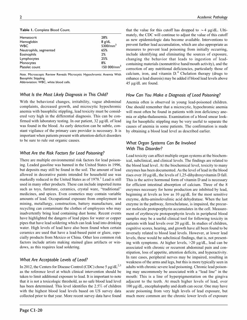
Page C1-20
What Is the Most Likely Diagnosis in This Child?
With the behavioral changes, irritability, vague abdominal
complaints, decreased growth, and microcytic hypochromic
anemia with basophilic stippling, lead toxicity must be consid-
ered very high in the differential diagnosis. This can be con-
firmed with laboratory testing. In our patient, 32 mg/dL of lead
was found in the blood. As early detection can be subtle, con-
stant vigilance of the primary care provider is necessary. It is
important when patients present with attention-deficit disorders
to be sure to rule out organic causes.
What Are the Risk Factors for Lead Poisoning?
There are multiple environmental risk factors for lead poison-
ing. Leaded gasoline was banned in the United States in 1996,
but deposits may still be found in the soil. The amount of lead
allowed in decorative paints intended for household use was
markedly reduced in the United States as of 1978.1 Lead is still
used in many other products. These can include imported items
such as toys, furniture, ceramics, crystal ware, “traditional”
medicines, and spices. Even cosmetics may contain variable
amounts of lead. Occupational exposure from employment in
mining, metallurgy, construction, battery manufacture, and
recycling can contaminate the clothes of employees who may
inadvertently bring lead containing dust home. Recent events
have highlighted the dangers of lead pipes for water or copper
pipes that have lead soldering which can leak lead into drinking
water. High levels of lead have also been found when certain
ceramics are used that have a lead-based paint or glaze, espe-
cially products from Mexico or China. Other less common risk
factors include artists making stained glass artifacts or win-
dows, as this requires lead soldering.
What Are Acceptable Levels of Lead?
In 2012, the Centers for Disease Control (CDC) chose 5 mg/dL2,3
as the reference level at which clinical intervention should be
taken to limit additional exposure to lead. It is important to note
that it is not a toxicologic threshold, as no safe blood lead level
has been determined. This level identifies the 2.5% of children
with the highest blood lead levels based on US survey data
collected prior to that year. More recent survey data have found
that the value for this cutoff has dropped to *4 mg/dL. Ulti-mately, the CDC will continue to adjust the value of this cutoff
as new epidemiologic data become available. Interventions to
prevent further lead accumulation, which are also appropriate as
measures to prevent lead poisoning from initially occurring,
include identifying and eliminating the sources of exposure,
changing the behavior that leads to ingestion of lead-
containing materials (nonnutritive hand/mouth activity), and the
correction of any nutritional deficiencies, particularly those of
calcium, iron, and vitamin D.4 Chelation therapy (drugs to
enhance a lead diuresis) may be added if blood lead levels above
45 mg/dL are found.
How Can You Make a Diagnosis of Lead Poisoning?
Anemia often is observed in young lead-poisoned children.
One should remember that a microcytic, hypochromic anemia
will most often be found in patients with iron deficiency ane-
mia or alpha-thalassemia. Examination of a blood smear look-
ing for basophilic stippling may be very useful to separate the
causes of anemia in some patients. The confirmation is made
by obtaining a blood lead level as described earlier.
What Organ Systems Can Be InvolvedWith This Disorder?
Lead toxicity can affect multiple organ systems at the biochem-
ical, subclinical, and clinical levels. The findings are related to
the blood lead level. At the biochemical level, toxicity to many
enzymes has been documented. As the level of lead in the blood
rises over 10 mg/dL, the levels of 1,25-dihydroxyvitamin D fall.
This is the active hormonal form of vitamin D and is necessary
for efficient intestinal absorption of calcium. Three of the 8
enzymes necessary for heme production are inhibited by lead,
beginning at levels as low as 10 mg/dL for the most sensitive
enzyme, delta-aminolevulinic acid dehydratase. When the last
enzyme in the pathway, ferrochelatase, is impaired, the precur-
sor molecule protoporphyrin accumulates in the cell. Measure-
ment of erythrocyte protoporphyrin levels in peripheral blood
samples may be a useful clinical tool for following toxicity in
patients with lead levels over 20 mg/dL. In studies of children,
cognitive scores, hearing, and growth have all been found to be
inversely related to blood lead levels. However, at lower lead
levels, these would be subclinical findings, that is, not present-
ing with symptoms. At higher levels, >20 mg/dL, lead can be
associated with chronic or recurrent abdominal pain and con-
stipation, loss of appetite, attention deficits, and hyperactivity.
In rare cases, peripheral nerves may be impaired, resulting in
weakness of the arms and legs, but this is more typically seen in
adults with chronic severe lead poisoning. Chronic lead poison-
ing may uncommonly be associated with a “lead line” in the
mouth: This is a line of hyperpigmentation on the gingiva
adjacent to the teeth. At much higher levels of lead, over
100 mg/dL, encephalopathy and death can occur. One may have
acute poisoning from very high levels of lead exposure, but
much more common are the chronic lower levels of exposure
Table 1. Complete Blood Count.
Hematocrit 28%Hemoglobin 8 g/dLWBC 5300/mm3
Neutrophils, segmented 65%Eosinophils 2%Lymphocytes 25%Monocytes 8%Platelet count 150 000/mm3
Note. Microscopic Review Reveals Microcytic Hypochromic Anemia WithBasophilic Stippling.Abbreviation: WBC, white blood cells.
2 Academic Pathology
Page C1-21
to lead. The skeletal system may show increased radiodensities
at the ends of the long bones at the metaphyses. The renal
system may show chronic tubulointerstitial disease over time
with high lead levels. Adults with chronic lead exposure may
show demyelination of the peripheral nerves.
HowWould You Describe the Changes Seen in the BloodSmear of a Patient With Lead Poisoning?
One of the more common systems involved with lead poisoning
is the hematologic system where one can find anemia, red
blood cells with basophilic stippling (which is one of the hall-
marks of diagnosis), and ring sideroblasts on bone marrow
evaluation. The anemia most often found is a microcytic, hypo-
chromic anemia, but this is likely due to other causes such as
iron deficiency. The mechanism of the ring sideroblasts is
related to the accumulation of iron-laden mitochondria in the
red blood cell precursors due to the inhibition of ferrochelatase
by lead.
What Is the Mechanism of Lead Poisoning?
Lead is a heavy metal that binds to proteins potentially altering
their function. It competes with essential metals such as cal-
cium for binding sites. In test tube studies, many calcium-
binding proteins have a higher affinity for lead than calcium.
These can result in multiple effects. For example, neurotrans-
mitter release is a calcium-dependent process perturbed by the
presence of lead. In children, over 70% of retained lead resides
in the skeleton from which it can be released very slowly over
years to decades after cessation of further ingestion.
Is Lead Absorption the Same for Children and Adults?
No. Lead compounds are poorly dissolvable in water. Lead
absorption is dependent on the gut’s ability to digest lead-
containing material; this is enhanced in acidic environments
like the stomach. Like calcium, lead is absorbed more effi-
ciently in children than adults. In children, over 50% of the
digested lead (lead in solution) can be absorbed via the intest-
inal tract compared to 15% in adults.
What Are Possible Treatments of Lead Poisoning?
For acute ingestion, a gastric lavage could be performed to
decrease the amount of lead-containing objects still in the sto-
mach. Cathartics can be given to rapidly remove lead already
further into the intestinal tract. For chronic ingestion, chelation
therapy may be necessary for lead levels of �45 mg/dL as per
CDC/American Academy of Pediatrics (AAP) recommenda-
tions. Treatment successfully improves biochemical profiles;
however, less data support the reversibility of neurocognitive
effects of lead. In all cases of increased lead levels, treatment
should include identification and elimination of the source of
the lead, behavior modification to limit ingestion, and nutrition
maximization.
Teaching Points
� Low lead levels, under 10 mg/dL, can have permanent
neurologic effects in children, especially contributing
to attention deficit disorders and decreased cognitive
abilities.
� Higher lead levels can affect multiple organs such as
its long-term accumulation and retention (over years)
in bone and teeth while causing toxicity in the bone
marrow with shortened red cell survival and the
development of microcytic hypochromic anemia with
basophilic stippling; central nervous system com-
plaints of difficulty learning and focusing; decreased
renal function; and gastrointestinal complaints of
abdominal pain and constipation.
� Chronic low levels of exposure are much more common
than acute high levels of toxicity.
� A high degree of vigilance is required to suspect lead
toxicity, especially in children where absorption of lead
is higher than in adults.
� Risk factors for lead toxicity include the ingestion of
environmental sources of lead such as in drinking water,
lead paint or the dust derived from that paint, contami-
nated soil as well as secondary exposure from occupa-
tional hazards such as workers from foundries, paint
manufacture, or battery factories.
� The mechanism of lead poisoning centers around its affi-
nity for proteins and its competitiveness with essential
metals to protein-binding sites.
Authors’ Note
The opinions expressed herein are those of the authors and are not
necessarily representative of those of theUniformedServicesUniversity
of the Health Sciences (USUHS), the Department of Defense (DOD),
or the United States Army, Navy, or Air Force.
References
1. Lead Poisoning. MedlinePlus. https://medlineplus.gov/leadpoison
ing.html. Updated January 11, 2017. Accessed March 15, 2016.
2. Environmental and nutritional diseases. In: Kumar V, Abbas A,
Aster A, eds. Robbins and Cotran Pathologic Basis of Disease.
9th ed. Philadelphia, PA: Elsevier Saunders; 2015:410-412.
3. Center for Disease Control Lead Level and Poisoning at CDC.gov.
Lead. https://www.cdc.gov/nceh/lead/acclpp/blood_lead_levels.
htm. Page reviewed October 30, 2012. Updated January 2017.
Accessed March 15, 2016.
4. Baadawy M, Connors G. Pediatric Lead Toxicity. Emedicine.
http://emedicine.medscape.com/article/1009587-overview#a4.
Updated June 22, 2016. Accessed March 15, 2016.
Knollmann-Ritschel and Markowitz 3


Page C1-23
Educational Case
Autosomal Recessive Inheritance:Cystic Fibrosis
D. Yitzchak Goldstein, MD1 and Michael Prystowsky, MD, PhD1
The following fictional case is intended as a learning tool within the Pathology Competencies forMedical Education (PCME),
a set of national standards for teaching pathology. These are divided into three basic competencies: Disease Mechanisms and
Processes, Organ System Pathology, and Diagnostic Medicine and Therapeutic Pathology. For additional information, and
a full list of learning objectives for all three competencies, see http://journals.sagepub.com/doi/10.1177/2374289517715040.
Keywordspathology, competencies, cystic fibrosis, developmental and functional abnormalities, disease mechanisms, genetic mechanisms,inheritance patterns
Received November 01, 2016. Received revised December 25, 2016. Accepted for publication January 09, 2017.
Primary Objective
Objective GM1.2: Inheritance Patterns. Compare and contrast the
inheritance patterns of different types of Mendelian disorders
and give examples of each type of pattern.
Competency 1: Disease Mechanisms and Processes; Topic
GM: Genetic Mechanisms; Learning Goal 1: Genetic Mechan-
isms of Developmental and Functional Abnormalities.
Patient Presentation
A 22-year-old Caucasian male presents with a recurrent cough.
He has had frequent respiratory illnesses and abdominal discom-
fort throughout his life. He has always been on the lower range
of normal for height and significantly smaller than his siblings.
His current primary care physician found his lung examination
to be abnormal (wheezing and crackles) as well as an absence of
the vas deferens on genitourinary examination.
Diagnostic Findings, Part 1
What is Your Differential Diagnosis Based on theClinical History?
The patient presents with signs and symptoms that are classical
for cystic fibrosis (CF). Some similar findings (short stature
and chronic lung infections) may be seen in other disease
states, such as primary ciliary dyskinesia (Kartagener syn-
drome) as well as asthma and should be excluded in this
patient. In CF, progressive scarring ultimately leads to atrophy
of the vasa deferentia.
Questions/Discussion Points, Part 1
What Testing is Available for this Patient and Which isRecommended?
Cystic fibrosis results from loss of function of the CF trans-
membrane conductance regulator (CFTR) protein caused by
mutations in the CFTR gene. The classic diagnostic test for
CF is the measurement of sweat chloride levels. This would
be the recommended test for a patient suspected of being
affected with CF.
1 Department of Pathology, Albert Einstein College of Medicine, Bronx,
NY, USA
Corresponding Author:
Michael Prystowsky, Department of Pathology, Albert Einstein College of
Medicine, 1300 Morris Park Ave Belfer 713, Bronx, NY 10467, USA.
Email: [email protected]
Academic PathologyVolume 4: 1–4ª The Author(s) 2017Reprints and permission:sagepub.com/journalsPermissions.navDOI: 10.1177/2374289517691769journals.sagepub.com/home/apc
Creative Commons CC BY-NC: This article is distributed under the terms of the Creative Commons Attribution-NonCommercial 3.0 License(http://www.creativecommons.org/licenses/by-nc/3.0/) which permits non-commercial use, reproduction and distribution of the work without furtherpermission provided the original work is attributed as specified on the SAGE and Open Access pages (https://us.sagepub.com/en-us/nam/open-access-at-sage).

Page C1-24
Biochemical screening tests for newborns typically measure
plasma levels of immunoreactive trypsinogen (a proenzyme
that builds up in the blood due to disease-related pancreatic
duct dysfunction). These tests are advantageous for screening
neonates as they demonstrate high sensitivity, can be run on
dried blood spots, and proenzyme levels are altered regardless
of the specific etiologic genetic alteration.
Genetic screening tests are useful for expectant parents to
determine the “carrier status” of each parent and to assess the
risk that a child born to them might be affected by CF. Since
many genetic tests only evaluate a subset of all possible patho-
genic mutations, patients must be counseled regarding the
small residual possibility of having an affected child.3
Explain the Inheritance of CF and Show How It is Possiblefor an Individual to Inherit a Mutation in Each CFTR Alleleand Present With No Disease, Mild Disease, or Severe CF
Cystic fibrosis is the most common, lethal, inherited disease
in white populations. Approximately 1 in 2500 newborns in
the United States is born with the disease.1 It typically dis-
plays autosomal recessive inheritance requiring each parent
to provide a pathogenic allele to their child for the disease
to manifest. A single genetic mutation is responsible for
70% of cases and consists of a 3-base pair deletion leading
to a loss of phenylalanine at codon 508 (DF508 or del508).
The DF508 mutation displays classical Mendelian inheri-
tance, whereby 2 carrier parents (each heterozygous for
DF508) would have an expected risk of having an affected
child of 25% for each pregnancy (25% risk unaffected and
50% risk of carrier offspring). Not all parents will carry
identical mutations and a child may therefore inherit differ-
ent mutations from each parent, with differing impacts on
the CFTR protein. This is one reason a spectrum of disease
phenotypes may be observed. Additionally, some mutations
may only demonstrate a partial effect, which may only cre-
ate a CF phenotype when identified in concert with other
specific mutations (R117 and poly[d]). This complexity and
the current identification of over 1800 described mutations
in the CFTR gene produce wide variability in the effect that
a given mutation will have on protein function and ulti-
mately on the clinical phenotype.
Explain the Normal Physiologic Function of the CFTRProtein and Which Tissues are Affected by the Lossof CFTR Function
The CFTR protein is an ion channel protein regulating chloride
concentrations across epithelial surfaces. In a healthy individual,
negatively chargedCl� ions are passively transported through the
membrane via the CFTR. Water can then passively diffuse
through the membrane to areas of high solute concentration pro-
ducing typical mucus. The absence of a functional CFTR protein,
either by a mutation that fails to transport it to the membrane or a
mutation within a membrane-bound protein itself, leads to the
inability of chloride to move outward and chloride becomes
sequestered within the cell along with high concentrations of
sodium. Since the movement of water passively follows solute
concentration, secreted mucus in affected patients becomes vis-
cous and tenacious leading to complications of transport.2
The CFTR also exists within the eccrine sweat glands of the
skin to balance the reabsorption of sodium and chloride (salt)
from initially excreted fluid. In the absence of a functional
CFTR, the reabsorption of sodium chloride is ineffective and
the amount of Naþ and Cl� in the excreted sweat remains high.
The CFTR channel exists in many other tissues as well; how-
ever, the effects on the lungs and digestive tract become most
clinically apparent in an affected patient.1
Describe the Pathophysiologic Process that Occursin the Lungs of Patients with Severe CF that Leadsto Bronchiectasis and Chronic Pneumonia Includinga Description of the Histopathology as the DiseaseProgresses
The more viscous secretions produced by failure of water to
thin the mucus covering the lung epithelium inhibits
Figure 1. Autopsy findings. A, Normal lung. B, Lung affected by cystic fibrosis.
2 Academic Pathology

Page C1-25
mucociliary clearance and causes mucus plugging of the
airways. This in effect seals off the terminal airspaces.
Inhaled bacteria cannot be cleared effectively and chronic
infections are the result. The body’s attempts to contain
these infections lead to an exuberant inflammatory response,
progressive fibrosis, dilatation, and ultimately destruction of
the airways.
Case continued—During the next year, the patient develops
a severe pneumonia and is hospitalized. Respiratory cultures
grew out Pseudomonas aeruginosa and the patient’s oxygena-
tion continued to decline. Antibiotics could not clear the
patient’s infection, and he was placed on ventilator support but
ultimately progressed and died.
Diagnostic Findings, Part 2
Compare and Contrast the Gross Pathology in Figure 1Aand B
A. Normal Lung: Note the spongy appearance of the cut
surface and gradual tapering of airways toward the
periphery of the tissue. The surface has a “dry”
appearance as it is actually made up of numerous
tiny alveoli.
B. Lung affected by CF: Bronchiectasis (airway space
enlargement with associated wall thickening) with
bronchi filled with excessive mucopurulent
secretions is seen. The secretions give a more
“glossy” appearance to the cut surface. Note also the
peripheral coalescence of airspaces with mucoid con-
taining cyst-like spaces. These become continually
infected and fibrotic over the course of the disease,
hence, the given name of the disease, “cystic
fibrosis”.
Compare and Contrast the Histopathology in Figure 2A-D
Panel A demonstrates a low power view of a slide from a
healthy individual. The majority of airspaces are intact and
lined by thin walled alveoli. The bronchioles present are patent
and are typically composed of a low columnar to cuboidal
epithelium. Panel B demonstrates a similar power view from
an individual affected by cystic fibrosis with abundant muco-
purulent material within an expanded airway. Panels C and D
show higher power views of the mixed inflammatory infiltrate
which is seen within the individual alveoli in patients affected
with cystic fibrosis.
Questions/Discussion Points, Part 2
What Therapies May Minimize the Symptoms of CF?
There are a variety of biomechanical techniques that are aimed
at clearing the thickened mucus from the airways and often
involve some forms of percussion or vibration. Several
Figure 2. Histopathology. A, Normal Lung with patent airspaces. B, Extensive mucopurulent secretions in dilated airway. C, Alveolar lungparenchyma with mucus and acute and chronic inflammation. D, Higher power view of C demonstrating the mixed inflammatory infiltrateassociated with cystic fibrosis.
Goldstein and Prystowsky 3

Page C1-26
categories of medications are also available to aid in fighting
infections, thinning mucus, and in some cases (given specific
causative mutations) potentiating the CFTR channel to allow
for increased chloride transport.
What are Some Possible Therapies that have a MoreDefinitive Impact on the Disease?
The severe damage caused by the course of the disease leaves
few medical options for improved pulmonary function; how-
ever, for some patients, lung transplantation can provide a
much more definitive impact on lung function as well as the
overall survival. Most importantly, however, lung transplanta-
tion is not a “cure” as it only alleviates the pulmonary symp-
toms of the patient. The other organs affected by the disease are
not modulated by a transplant.
Recent investigations involving large-scale mathematical and
chemical libraries have identified several possibledrugmolecules
that target precise causes of the disease created by individual
mutations. Given the diverse impacts the various CF mutations
can have on protein production, processing, and regulation, it is
not surprising that different drugs are necessary to provide differ-
ing corrective effects. Some drugswork by increasing shuttling of
the CFTR to the membrane, while others act to improve chloride
transport through the CFTR and even others attempt to overcome
nonsense mutations by allowing the ribosome to “read through”
premature stop codons during translation.4
Given that the disease is inherited in a patient’s DNA and
many of the causative genetic mutations have been elucidated,
there is a possibility that in the future, site-directed gene
editing may hold promise for a broader therapy unique to the
patient’s own disease and impacting and improving all
affected organ systems.
Teaching Points
� Cystic fibrosis is the most common inherited autosomal
recessive disease in the Caucasian population.
� The disease affects multiple organ systems and can have
a wide variety of clinical presentations.
� Genetic mutations of the CFTR gene lead to an ineffec-
tive chloride transporter that explains many of the clin-
ical symptoms.
� There is hope that modern technological advancements
could lead to not only symptomatic control but also
possibly even cures for the disease in the future.
References
1. Ratjen F, Doring G. Cystic fibrosis. Lancet. 2003;361(9358):681-689.
2. Stoltz DA, Meyerholz DK, Welsh MJ. Origins of cystic fibrosis
lung disease. N Engl J Med. 2015;372(4):351-362.
3. American College of Obstetricians and Gynecologists Committee
on Genetics. ACOG Committee Opinion No. 486: update on carrier
screening for cystic fibrosis. Obstet Gynecol. 2011;117(4):
1028-1031.
4. Cystic Fibrosis. https://en.wikipedia.org/wiki/Cystic_fibrosis.
4 Academic Pathology

Page C1-27
Educational Case
Educational Case: Fibroadenomaof the Breast
H. James Williams, MD1
The following fictional case is intended as a learning tool within the Pathology Competencies forMedical Education (PCME),
a set of national standards for teaching pathology. These are divided into three basic competencies: Disease Mechanisms and
Processes, Organ System Pathology, and Diagnostic Medicine and Therapeutic Pathology. For additional information, and
a full list of learning objectives for all three competencies, see http://journals.sagepub.com/doi/10.1177/2374289517715040.
Keywordspathology competencies, organ system pathology, disease mechanisms, breast, neoplasia, fibroadenoma, palpable breast lesion,mammographic findings, phyllodes tumor
Received April 02, 2017. Received revised July 15, 2017. Accepted for publication February 13, 2018.
Primary Objective
Objective BR2.1: Fibroadenoma and Phyllodes Tumor. Com-
pare and contrast fibroadenoma and phyllodes tumor in terms
of clinical features, morphologic findings, and prognosis.
Competency 2: Organ System Pathology; Topic BR: Breast;
Learning Goal 2: Molecular Basis of Breast Neoplasms.
Secondary Objective
Objective N3.1: Morphologic Features of Neoplasia. Describe
the essential morphologic features of neoplasms and indicate
how these can be used to diagnose, classify, and predict biolo-
gical behavior of cancers.
Competency 1: Disease Mechanisms and Processes; Topic
N: Neoplasia; Learning Goal 3: Characteristics of Neoplasia.
Patient Presentation
A 37-year-old woman presents to her physician with concern
about a left breast nodule she recently discovered on self-
examination. The patient states that the nodule is approxi-
mately 2 cm in size, close to her left axilla, and feels firm. She
is concerned because her mother, age 54, was recently diag-
nosed with breast cancer.
Questions/Discussion Points, Part 1
What Pertinent Questions Should be Asked as Part of theDetailed History Prior to Physical Examination?
How long has the nodule been there? If the lesion developed veryrecently, one could choose to follow the lesion for a short time
to see if it persists.
Has it changed in size over time? Change in size could include
decreasing, increasing, or fluctuating lesions. Fluctuation in
size would be suggestive of menstrual effect or fibrocystic
change. A decrease in size might indicate a cyst getting
smaller. An increase in size would be more worrisome for a
more serious lesion.
1West Virginia University Health Sciences Center, Morgantown, WV, USA
Corresponding Author:
H. James Williams, West Virginia University Health Sciences Center, P.O. Box
9203, Morgantown, WV 26506, USA.
Email: [email protected]
Academic PathologyVolume 5: 1–4ª The Author(s) 2018Article reuse guidelines:sagepub.com/journals-permissionsDOI: 10.1177/2374289518790926journals.sagepub.com/home/apc
Creative Commons Non Commercial CC BY-NC: This article is distributed under the terms of the Creative Commons Attribution-NonCommercial 4.0 License(http://www.creativecommons.org/licenses/by-nc/4.0/) which permits non-commercial use, reproduction and distribution of the work without furtherpermission provided the original work is attributed as specified on the SAGE and Open Access pages (https://us.sagepub.com/en-us/nam/open-access-at-sage).

Page C1-28
Is the nodule painful? Malignancies are not typically painful;
however, inflammatory lesions can be painful as could benign
lesions.
Has there been nipple discharge and if so, is it bloody? Bloody
nipple discharge may be associated with an intraductal papil-
loma or, more rarely, cancer. A finding of nipple discharge
would need to be investigated more carefully.
Does anyone in your family have or have had breast cancer? If thereis a family history of breast cancer, what is the relationship of
the family member with the patient (ie, is it the patient’s
mother, sister, or daughter)? Also, what was the age at time
of diagnosis of the family member? Younger age of breast
cancer would be much more concerning for a possible famil-
iar component of a breast cancer than breast cancer in an older
first-degree relative. This question is addressing the associ-
ated increased risk of malignancy and/or hereditary disease in
close relatives, especially if they developed breast cancer at a
young age.
On physical examination of the patient’s left breast, what findingswould favor either a benign or malignant diagnosis? Physical exam-
ination can be helpful, as some features are more common
with benign or malignant lesions. Features of benign neo-
plasms include the following: multiple indistinct nodules
(lumpy breast) that favor fibrocystic changes and lesions
beneath the nipple which may suggest an intraductal
papilloma.
However, a fixed, irregular, and firm mass is suspicious for
malignancy as is dimpling of the overlying skin. Another
important factor is location of the lesion within the breast,
as 50% of breast cancers arise in the upper outer quadrant;
thus, lesions in this area would be more suspicious for
malignancy.
Diagnostic Findings, Part 2
The patient undergoes mammography of the left breast nodule.
The mammography is shown in Figure 1.
Questions/Discussion Points, Part 2
What Radiographic Findings in Figure 1 Are Seen, and DoThey Favor a Benign or Malignant Diagnosis?
Similar to physical examination, radiologic examination
is important in working up breast lesions. Several factors
are more suggestive of benign versus malignant lesions.
The following findings favor a benign process when they
Figure 1. Mammogram of left breast nodule. The lesion is homoge-neous and is sharply demarcated from the normal breast tissue.
Figure 2.Microscopic image of left breast nodule at low power, H&E2�. On low power, the lesion is seen to be sharply demarcated fromthe adjacent normal breast tissue.
Figure 3.Microscopic image of left breast nodule at high power, H&E20�. The stroma of the lesion has low cellularity with no mitoses orcytologic atypia. The epithelial component shows bland cytologicfeatures with associated myoepithelial cells.
2 Academic Pathology

Page C1-29
are present: sharp distinct borders, homogenous texture,
and ovoid shape. The following factors favor malignant
when they are present: stellate infiltrative borders, hetero-
genous texture, round shape, and presence of coarse
calcifications.
In the mammographic image for the patient, a round homo-
genous lesion is seen. This would favor a benign process. Con-
firmation of the imaging impression requires histologic
examination of the lesion either by biopsy of the lesion or by
conservative excision.
Diagnostic Findings, Part 3
The breast nodule is excised and sent for pathologic examina-
tion (see Figures 2 and 3).
Questions/Discussion Points, Part 3
What are the Pertinent Histologic Findings Seen inFigures 2 and 3?
On low-power image of the breast biospy, as seen in Figure
2, the lesion is seen to be sharply demarcated from the
adjacent normal breast tissue. This is suggestive of a benign
process. When looking at higher power in Figure 3, there is
a predominance of stromal tissue with compression of the
epithelial component. The stroma is of low cellularity with
no mitoses or cytologic atypia seen. In addition, the epithe-
lial component shows bland cytologic features with associ-
ated myoepithelial cells.
What Is Your Diagnosis?
Fibroadenoma of the breast.
Discuss the Clinical Features and Pathophysiology ofFibroadenomas and What Is the Prognosis andManagement for This Lesion?
Fibroadenomas are the most common benign breast neo-
plasms and typically present in women 20 to 35 years old.1
The tumors are usually solitary but may be multiple (20%).
Clinically, they may be detected by the patient or the physi-
cian on breast examination as a well-demarcated, freely
mobile, firm mass that is usually <3 cm in diameter. The
fibroadenoma is a neoplasm of the specialized lobular stroma
with typically low cellularity, no cytologic atypia, and mito-
tic figures absent or rare. The associated benign epithelium
has associated myoepithelial cells and may have an intra-
canalicular or pericanalicular pattern.2 The radiographic
appearance of fibroadenoma characteristically shows an
ovoid homogenous mass with sharp distinct borders.
Patients with fibroadenomas are associated with a slight
increased risk of breast cancer, particularly when there are
proliferative fibrocystic changes involving the tumor. As
would be anticipated, fibroadenomas are hormonally
responsive (may enlarge during pregnancy and reduce in size
in postmenopause).
As the fibroadenoma is a benign tumor, in the appropriate
clinicoradiographic setting, once the tumor is diagnosed by
fine-needle aspiration or needle core biopsy, it may be safely
followed. If the tumor is a cosmetic problem or if preferred by
the patient, conservative excision can be performed.3
Discuss a Common Tumor in the Differential Diagnosis ofa Fibroadenoma
The histologic differential diagnosis includes the phyllodes
tumor in which there is increased cellularity of the stroma, and
the epithelial component demonstrates a “leaf-like” architec-
tural pattern.1 Low-grade phyllodes tumors have these features
with mild cytologic atypia and occasional mitoses. High-grade
phyllodes tumors in addition to the increased stromal cellular-
ity have marked cytologic atypia and increased mitoses, some
of which may be atypical. Because of the higher risk of recur-
rences, wide excision is indicated for phyllodes tumors. In
addition to the risk of recurrences, the high-grade phyllodes
tumor has the potential to metastasize.
Teaching Points
� Benign features include the following: Multiple indis-
tinct nodules (lumpy breast) favors fibrocystic changes,
and lesions beneath the nipple may suggest a papilloma.
� A fixed irregular firm mass on physical examination is
suspicious for malignancy, as is dimpling of the over-
lying skin.
� On radiography, a benign process tends to have sharp
distinct borders, homogenous texture, and ovoid shape.
� Fibroadenomas are the most common benign breast neo-
plasms and typically present in women 20 to 35 years
old.
� The fibroadenoma is a neoplasm of the specialized lob-
ular stroma with typically low cellularity, no cytologic
atypia, and mitotic figures absent or rare. The associated
benign epithelium has associated myoepithelial cells
and may have an intracanalicular or pericanalicular
pattern.
� The phyllodes tumor has increased cellularity of the
stroma, and the epithelial component demonstrates a
“leaf-like” architectural pattern. Low-grade phyllodes
tumors may have mild cytologic atypia and occasional
mitoses. High-grade phyllodes tumors, in addition to the
increased stromal cellularity, have marked cytologic
atypia and increased mitoses, some of which may be
atypical.
Declaration of Conflicting Interests
The author(s) declared no potential conflicts of interest with respect to
the research, authorship, and/or publication of this article.
Williams 3

Page C1-30
Funding
The author(s) received no financial support for the research, author-
ship, and/or publication of this article.
References
1. KumarV,AbbasAK, FaustoN,Aster JC.Robbins andCotranPatho-
logic Basis of Disease. Elsevier Health Sciences, 2014:1069-1070.
2. Kuijper A, Mommers EC, van der Wall E, van Diest PJ. Histo-
pathology of fibroadenoma of the breast. Am J Clin Pathol. 2001;
115:736-742.
3. Hubbard JL, Cagle K, Davis JW, Kaups KL, Kodama M. Criteria
for excision of suspected fibroadenomas of the breast. Am J Surg.
2015;209:297-301.
4 Academic Pathology

Page C1-31
Educational Case
Educational Case: Iron Overloadand Hemochromatosis
Michael J. Borowitz, MD, PhD1 and Alison Moliterno, MD1
The following fictional case is intended as a learning tool within the Pathology Competencies forMedical Education (PCME),
a set of national standards for teaching pathology. These are divided into three basic competencies: Disease Mechanisms and
Processes, Organ System Pathology, and Diagnostic Medicine and Therapeutic Pathology. For additional information, and
a full list of learning objectives for all three competencies, see http://journals.sagepub.com/doi/10.1177/2374289517715040.
Keywordspathology competencies, organ system pathology, hematopathology, anemia, iron overload, hemochromatosis, genetic mechan-isms, inheritance patterns
Received February 13, 2017. Received revised April 16, 2018. Accepted for publication April 30, 2018.
Primary Pathology Learning Objective
Objective HRC1.3: Hepcidin Regulation, Iron Overload, and
Anemia of Chronic Disease: Discuss the role of hepcidin as an
iron regulator and describe how different types of alterations in
the hepcidin pathway can produce anemia of chronic disease or
iron overload.
Competency 2: Organ System Pathology; Topic HRC:
Hematopathology - Red Cell Disorders; Learning Goal 1:
Anemia.
Secondary Pathology Learning Objective
Objective GM1.2: Inheritance Patterns: Compare and contrast
the inheritance patterns of different types of Mendelian disor-
ders and give examples of each type of pattern.
Competency 1: Disease Mechanisms and Processes; Topic
GM: Genetic Mechanisms; Learning Goal 1: Genetic Mechan-
isms of Developmental and Functional Abnormalities.
Patient Presentation
A 50-year-old Caucasian male presented for evaluation con-
cerned that he may be at risk of developing hemochroma-
tosis based on his family history. His father presented at age
60 with bronzed skin and diabetes; he was found to have
iron overload and subsequently has been undergoing thera-
peutic phlebotomies. The patient’s medical history is unre-
markable with the exception of hypercholesterolemia, for
1 Johns Hopkins School of Medicine, Baltimore, MD, USA
Corresponding Author:
Michael J. Borowitz, Johns Hopkins School of Medicine, 401 N Broadway,
Baltimore, MD 21287, USA.
Email: [email protected]
Table 1. Iron and Genetic Studies of the Patient and His Father.
Lab TestFather—Age
60Patient—Age
50 Normal Range
Serum iron 232 106 65-170 mg/dLTransferrin 284 230 200-400 mg/dLTotal iron bindingcapacity
355 288 250-450 mg/dL
% saturation 65 37 20%-55%Ferritin 1518 217 10-300 ng/mLHFE genotype C282Y Homozygous Heterozygous
Academic PathologyVolume 5: 1–3ª The Author(s) 2018Reprints and permission:sagepub.com/journalsPermissions.navDOI: 10.1177/2374289518779944journals.sagepub.com/home/apc
Creative Commons Non Commercial CC BY-NC: This article is distributed under the terms of the Creative Commons Attribution-NonCommercial 4.0 License(http://www.creativecommons.org/licenses/by-nc/4.0/) which permits non-commercial use, reproduction and distribution of the work without furtherpermission provided the original work is attributed as specified on the SAGE and Open Access pages (https://us.sagepub.com/en-us/nam/open-access-at-sage).

Page C1-32
which he takes a statin. You decide to do some diagnostic
blood work. In addition, the patient has provided you with
results of blood work from his father.
Diagnostic Findings, Part 1
Results of iron studies and HFE studies are presented in
Table 1.
Questions/Discussion Points, Part 1
Interpret the Father’s and the Patient’s Iron Studies
The father has elevated serum iron associated with increased
transferrin saturation and an elevated ferritin indicative of both
increased iron absorption and iron storage. The son has normal
iron studies.
Do the Patient’s Iron Studies Support the Diagnosisof Hemochromatosis?
No. He does not have evidence of either increased absorption or
increased iron stores. Additionally, he is only a carrier of the
HFE mutation, making it highly unlikely that he has a genetic
predisposition to iron overload.
Ferritin and Serum Iron Reflect Different Aspects ofan Individual’s Iron Metabolism. What Is the MajorDifference in the Type of Information Obtained FromEach of These?
Ferritin is a measure of long-term iron storage, while serum
iron (and transferrin saturation) better reflect daily iron absorp-
tion. Interpretation of ferritin testing can be misleading with
only a single measurement because it is an acute phase reactant.
Diagnostic Findings, Part 2
After obtaining the results above, the patient consults his
brother and recommends that he also undergo testing. Results
from his brother, again in comparison to those of his father, are
shown in Table 2.
Questions/Discussion Points, Part 2
Does His Brother Have Iron Overload? Why Is HisBrother’s Ferritin So Much Lower Than His Father’s?
Yes, he has evidence of iron overload. He is younger than his
father was at the time of his iron studies, and the degree of iron
overload is a function of time.
Should Other Siblings Be Tested? What Would Be theBest Approach for Further Molecular Testing? If You HadHad the Opportunity to Evaluate the Father at the FirstSign of Potential Iron Overload, What Testing Would YouHave Done on Him?
Taking the brother’s and father’s genotypes together (and
assuming the brothers have the same father!), mother must be
a carrier, and other children have a 50% chance of being homo-
zygotes also. HFE C282Y homozygotes in general will have an
increased risk of significant iron overload. However, because
there are significant modifiers of iron absorption beyond the
HFE gene (age, gender, diet, and polymorphisms in other genes
affecting iron absorption), not all homozygotes will have iron
overload.1 In this family, however, homozygotes do appear to
have an increased risk of iron overload, so they should be
identified. Because we know the mutation in this case, targeted
mutation analysis for HFE C282Y is the only analysis needed.
If you had seen the father early in his course and had had no
family history of hemochromatosis, it would be appropriate to
do targeted analysis for both the HFE C282Y and H63D muta-
tions, and if these were negative, sequence analysis could be
used to identify other less common mutant alleles associated
with HFE.
Which Genes Can Cause Hemochromatosis WhenMutated? Describe the Inheritance Patterns Seen
The HFE C282Y on chromosome 6p22.2, as present in this
case, is the most common mutation seen in classic (type 1 or
HFE1) hemochromatosis and is associated with autosomal
recessive inheritance. This mutation accounts for >80% of
hemochromatosis cases.2,3 Another common mutation in HFE
(H63D) may be seen as a compound heterozygous (C282Y/
H63D) genotype in some patients with hemochromatosis;
patients with homozygous H63D rarely have clinically signif-
icant iron overload.2,3 Four additional iron overload disorders
labeled hemochromatosis have been identified2:
Juvenile hemochromatosis is the term given to clinically
similar autosomal recessive diseases caused by muta-
tions in 2 different genes:
HFE2A: the hemojuvelin (HJV) gene on chromosome
1q21.
HFE2B: the hepcidin (HAMP) gene on chromosome
19q13.
Table 2. Iron and Genetic Studies of the Father and the Brother ofthe Patient.
Lab TestFather—Age
60Brother—Age
46 Normal Range
Serum iron 232 176 65-170 mg/dLTransferrin 284 230 200-400 mg/dLTotal iron bindingcapacity
355 266 250-450 mg/dL
% saturation 65 65 20%-55%Ferritin 1518 611 10-300 ng/mLHFE genotypeC282Y
Homozygous Homozygous
2 Academic Pathology

Page C1-33
HFE3, also autosomal recessive, is caused by a mutation
in transferrin receptor 2 gene on chromosome 7q22.
HFE4, is autosomal dominant and caused by a mutation
in SLC40A1 gene on chromosome 2q32.
Discuss the Pathophysiology of How Iron OverloadOccurs in Hemochromatosis
Patients with hemochromatosis have in common low hepcidin
levels. Hepcidin normally binds ferroportin in both duodenal
enterocytes and reticuloendothelial macrophages, which in turn
blocks release of iron from these cells. Thus, hepcidin serves to
keep iron frombeingabsorbed fromthegut orbeing released from
storage macrophages back into circulation. In the absence of
hepcidin, this process is reversed, with increased iron absorption
and increased turnover of iron from reticuloendothelial macro-
phages leading to high levels of both serum ferritin and saturation
of transferrin and ultimately to iron deposition in tissues.3
Teaching Points
� Iron overload can be suspected from serum iron studies,
in particular elevated serum iron and transferrin, and
increased transferrin saturation.
� In the absence of a historical explanation for iron over-
load (such as multiple blood transfusions), patients with
iron overload should be investigated for genetic disor-
ders, and in particular for hereditary hemochromatosis.
� If hereditary hemochromatosis is identified, family
members should be tested for evidence of iron overload
as well.
� Other factors modify the effects of the abnormal gene so
that affected family members may vary significantly in
their clinical and laboratory presentations.
� The most common form of hereditary hemochromatosis
is called type 1 and is due to a mutation in theHFE gene.
It is inherited as an autosomal recessive.
Declaration of Conflicting Interests
The author(s) declared no potential conflicts of interest with respect to
the research, authorship, and/or publication of this article.
Funding
The author(s) received no financial support for the research, author-
ship, and/or publication of this article.
References
1. Beutler E. Iron storage disease: facts, fiction and progress. Blood
Cells Mol Dis. 2007;39:140-147.
2. Online Mendelian Inheritance in Man (OMIM). 2018. http://www.
omim.org/entry/235200. Accessed April 14, 2018.
3. Hollerer I, Bachman A, Muckenthaler MU. Pathophysiologic
consequences and benefits of HFE mutations: 20 years of
research. Haematologica. 2017;102:809-817. PMID 28280078.
doi:10.33324/haematol.2016.160432.
Borowitz and Moliterno 3


Com
petency 2
Title/Author Primary Objective
Secondary Objective(s) Page
Primary Osteosarcoma, Aldis H. Petriceks, BA and Darren Salmi, MD MS1.2 SP1.4 C2-1
Wilms Tumor (Nephroblastoma), Taylor E. Anderson, BA and Richard M. Conran, MD, PhD, JD
UTK1.4 C2-9
Membranous Nephropathy, Christina C. Smith, MS and Richard Michael Conran, MD, PhD, JD
UTK5.2 – C2-13
Immune Related Disorders of the Bowel: Celiac Disease, Shaomin Hu, MD, PhD and Nicole C. Panarelli, MD
GT5.2 – C2-19
Symptomatic but Unruptured Abdominal Aortic Aneurysm, Darren Salmi, Aldis Petriceks, John Olivas
CBV2.3 CBV2.2 C2-27
Fibroadenoma of the Breast, H. James Williams, MD BR2.1 N3.1 C2-35
Wilms Tumor, Alison R. Huppmann, MD UTK4.1 – C2-39
Pheochromocytoma, Clinton Westover, MD and Richard Michael Conran, MD, PhD, JD
EN5.3 – C2-45
Iron Overload and Hemochromatosis, Michael J. Borowitz, MD, PhD and Alison Moliterno, MD
HRC1.3 GM1.2 C2-51
Thyroid Neoplasms: Pathogenesis, Diagnosis, and Treatment, Gloria Ramos Rivera, MD, Sheila Segura, MD, and Mark Suhrland, MD
EN5.1 – C2-55
Chronic Lymphocytic Leukemia, Christine G. Roth, MD HWC3.1 – C2-61
Endocrine Neoplasm: Medullary Thyroid Carcinoma, Sheila Segura, MD, Gloria Ramos-Rivera, MD, and Mark Suhrland, MD
EN5.2 – C2-65
Head and Neck Neoplasia: Salivary Gland Tumors, Ryan P. Lau, MD, Melissa Yee-Chang, DO, and Amy Rapkiewicz, MD
HN2.1 CYP1.2 C2-71
Medullary Thyroid Carcinoma, Carl T. McGary, MD, PhD EN5.2 CYP1.2 C2-77
Bladder Carcinoma in-situ (CIS), H. James Williams, MD UTB1.3 – C2-83
Autosomal Recessive Polycystic Kidney Disease, Ashley S. Hafer, MPA and Richard M. Conran, MD, PhD, JD
UTK4.1 – C2-87
Squamous Cell Carcinoma of the Lung, Eric Suarez, MD, and Barbara E. C. Knollmann-Ritschel, MD
RS3.2 RS3.1, RS3.5 C2-91
Subarachnoid Hemorrhage Related to Ruptured Berry Aneurysm, Sarah Meyers, MD
NSC7.3 AU2.2 C2-95
Genetic Mutations and Multifactorial Inheritance: Dilated Cardiomyopathy, Eric S. Suarez, MD and Barbara E. C. Knollmann-Ritschel, MD
GM1.1 GM1.5, CH1.2 C2-99
Benign Papilloma of the Breast, Moshe Sadofsky, MD, PhD N2.1 N2.2, N3.1, N3.2, N3.3, N3.4, N3.5, BR1.1, BR1.4
C2-103
Competency 2: Organ System Pathology

Educational Case Background & Submission Instructions
BackgroundBecoming a competent physician requires the ability to gain a broad foundation of knowledge, skills, and attitudes essential for independent medical practice. Essential in this is the understanding of the normal and pathological processes of each organ system, the ability to apply disease mechanisms to describe the pathobiology, and the ability to continually improve the diagnostic acumen and optimal treatment decisions through lifelong learning.
The Pathology Competencies for Medical Education (PCME) have detailed learning objectives under each goal that direct medical students and course directors to important facets of each learning goal that can be individually applied by learners. The competencies are divided into three sections—disease mechanisms and processes, organ system pathology, and diagnostic medicine and therapeutic pathology—and allow flexibility for each medical school and learner to apply the learning goals and objectives in a way that can keep the unique design of each curriculum or learning plan. The competencies are purposefully kept broad as they represent the minimum requirements of what pathology course directors across the nation have agreed upon to prepare medical students for entry into any residency program and for the subsequent contemporary practice of medicine.
Educational Cases for the PCME are current, peer-reviewed, and highlight the pathology competencies through fictional (but realistic) learning cases that can easily be adapted to multiple types of educational modalities. Educational Cases reference at least one primary learning objective, but may have one or more secondary learning objective(s). The pathology competencies and learning objectives are clearly indicated in the beginning of each case so that the focus of the educational case is evident. Key elements of the current format include clinical presentation, discussion questions or points, learning points, and references. The clinical presentation may include images or laboratory data for the patient’s presentation. The discussion questions or points are questions or statements that promote clinical reasoning followed by detailed explanations of the pathology, medicine, or therapeutics brought up in the discussion point or question. The learning points at the end of the case highlight the main teaching points from the preceding discussion. Thus, the cases demonstrate the application of medical reasoning to clinical scenarios that allow the learner to understand and apply diagnostic principles, incorporating morphologic findings and laboratory values with discussion of the laboratory medicine essentials for accurate diagnosis and treatment. References are included in each case and will allow the reader to review the original sources used to create the learning case or gain additional in-depth information. Thus, the Educational Cases are written in a style that can be easily used or adapted to multiple educational formats, such as small group discussions or flipped classrooms.
Case Submission GuidelinesSubmission Portal: https://mc.manuscriptcentral.com/apc Manuscript Type: Educational CaseKey Words:
o list “pathology competencies” as first keywordo list relevant competency, topic, learning goal, and objective keywords, e.g. “disease mechanisms, genetic mechanisms,
inheritance patterns”o other relevant keywords from the case content, e.g. “cystic fibrosis”
Abstract: “None needed”Case Content:
o Primary (and secondary if applicable) learning objective(s), cited from the PCME (doi: 10.1177/2374289517715040)o Example of primary objective formatting:
Objective GM1.2: Inheritance Patterns. Compare and contrast the inheritance patterns of different types of Mendelian disorders and give examples of each type of pattern. Competency 1: Disease Mechanisms and Processes; Topic GM: Genetic Mechanisms; Learning Goal 1: Genetic Mechanisms of Developmental and Functional Abnormalities.
o Patient Presentation: Include presentation (History of present illness, past medical history, etc. and physical examination)o Diagnostic Findings: This can include laboratory findings or histology.o Questions/Discussion Points: The questions and discussion points should be presented in a logical order to promote clinical
reasoning. In addition, you can include additional laboratory data/histologic images in later discussion points. The discussions should thoroughly explain the learning objectives to which the case is applied.
o Teaching Points: These should be covered in your discussion.o References: Linked to the discussion.
Images (if applicable): All images must be original work or have appropriate approval for publication.
Accepted CasesPublished Educational Cases receive the same scholarly recognition, citation and merit as other articles published in Academic Pathology. Cases accepted for publication will incur an Open Access article processing fee of $500.00 for authors who are faculty and students of APC member departments; $750 for non-members. For more information, contact Academic Pathology at [email protected] or 302-660-4940.
Educational Case
Educational Case: Primary Osteosarcoma
Aldis H. Petriceks, BA1 and Darren Salmi, MD1,2
The following fictional case is intended as a learning tool within the Pathology Competencies forMedical Education (PCME),
a set of national standards for teaching pathology. These are divided into three basic competencies: Disease Mechanisms and
Processes, Organ System Pathology, and Diagnostic Medicine and Therapeutic Pathology. For additional information, and
a full list of learning objectives for all three competencies, see http://journals.sagepub.com/doi/10.1177/2374289517715040.
Keywordspathology competencies, organ system pathology, musculoskeletal, bone neoplasia, bone, osteosarcoma, primary bone tumors,malignant bone tumors, osteosarcoma risk factors, osteosarcoma staging, tumor staging
Received August 20, 2018. Received revised November 07, 2018. Accepted for publication November 21, 2018.
Primary Objective
Objective MS1.2: Bone-Forming Sarcomas in Children:
Describe the most common benign and malignant bone-
forming tumors in children and adolescents in terms of clinical
presentation, radiologic findings, histologic features, treatment,
and prognosis.
Competency 2: Organ System Pathology; Topic: Musculos-
keletal System (MS); Learning Goal 1: Bone Neoplasia.
Secondary Objective
Objective SP1.4: Staging: Describe the information that the
pathologist obtains from a resected tissue specimen, how this
information is reported, how it is combined with clinical infor-
mation to stage the tumor, and how staging information is used
to guide treatment.
Competency 3: Diagnostic Medicine and Therapeutic
Pathology; Topic: Surgical Pathology (SP); Learning Goal 1:
Role in Diagnosis.
Patient Presentation
A 14-year-old male patient presents to the emergency
department with severe pain in his right knee. The pain has
been intermittent for the past 10 weeks, appearing and dis-
appearing sporadically. The discomfort has grown more
regular in the last week and is now compounded by marked
swelling around the knee. Further interview reveals that the
patient plays on a youth soccer team, and his knee feels
especially painful during practice. His parents attributed the
symptoms to “growing pains,” as the patient’s height and
weight have been increasing appropriately for his age, and
he has had no known skeletal diseases—nor any serious
conditions—prior to onset of the pain.
Diagnostic Findings, Part I
Vital signs are evaluated and found to be normal. No sys-
temic symptoms are noted. Physical examination reveals a
large, palpable mass on the anterior aspect of the right
proximal tibia. The mass is tender to palpation, but not
warm to the touch.
1 Department of Surgery, Stanford University School of Medicine, Stanford,
CA, USA2 Department of Pathology, Stanford University School of Medicine, Stanford,
CA, USA
Corresponding Author:
Darren Salmi, Department of Pathology, Stanford University School of
Medicine, 269 Campus Dr, CCSR 0135A, Stanford, CA 94305, USA.
Email: [email protected]
Academic PathologyVolume 6: 1–8ª The Author(s) 2019Article reuse guidelines:sagepub.com/journals-permissionsDOI: 10.1177/2374289518820337journals.sagepub.com/home/apc
Creative Commons Non Commercial No Derivs CC BY-NC-ND: This article is distributed under the terms of the Creative Commons Attribution-NonCommercial-NoDerivs 4.0 License (http://www.creativecommons.org/licenses/by-nc-nd/4.0/) which permits non-commercial use, reproduction anddistribution of the work as published without adaptation or alteration, without further permission provided the original work is attributed as specified on theSAGE and Open Access pages (https://us.sagepub.com/en-us/nam/open-access-at-sage).
Page C2-1

Question/Discussion Points, Part I
Discuss the Differential Based on the ClinicalPresentation
The patient’s large and painful tibial mass is nonspecific, and
the differential diagnosis remains broad. At one level, we may
consider whether this mass is neoplastic or non-neoplastic. If
non-neoplastic, diagnoses might include Osgood-Schlatter dis-
ease, subacute osteomyelitis, or a reactive soft tissue lesion
such as myositis ossificans. If the mass were neoplastic, one
would first have to decide whether the neoplasm was benign or
malignant and whether it involved the bone, soft tissue, or both.
For a patient of this age, the most likely malignancies of the
bone are osteosarcoma and Ewing sarcoma. Chondrosarcoma
may be considered alongside the two—though in its most com-
mon form, chondrosarcoma typically presents in the axial ske-
leton of persons above 40 years old. The mass may be a
metastatic tumor, secondary to cancer elsewhere in the body,
but the patient’s age and lack of prior disease make this
unlikely as well.
Osteosarcoma is the most probable among these malignan-
cies, comprising over 50% of malignant bone tumors for patients
younger than 20 years.1 Ewing sarcoma is the second most
common primary bone malignancy in this age-group, represent-
ing over 30% of bone cancers for children and adolescents.2
Children are the highest risk group for both sarcomas, though
neither sarcoma accounts for more than 3% of overall childhood
cancers.2 Osteosarcoma most commonly occurs in children
between 13 and 16 years of age, while peak incidence of Ewing
sarcoma occurs between 10 and 15 years of age.2,3 This age
dependency may be related to the adolescent growth spurt, espe-
cially as osteosarcoma often manifests near the metaphyseal
growth plate.4 In both cases, males are at higher risk than
females, though the reason for this is unknown.4
Some benign bone tumors are capable of forming palpable
masses similar to the one seen in the present patient. These
include aneurysmal bone cysts (ABCs) and giant cell tumors.
However, as we will see, giant cell tumors are exceedingly rare
in children and adolescents.
Taking these considerations into account, the differential
diagnosis includes Osgood-Schlatter, subacute osteomyelitis,
myositis ossificans, osteosarcoma, Ewing sarcoma, chondro-
sarcoma, metastatic tumor, giant cell tumor, and ABC. At this
stage, the differential is based solely on the clinical presenta-
tion. The patient’s age, his large tibial mass, and his male sex
have been particularly informative. A simple radiograph of the
area surrounding the palpable mass, in addition to histologic
data, will be important for narrowing the differential diagnosis.
Diagnostic Findings, Part II
The Patient’s Radiograph Is Shown in Figure 1. Describethe Findings
Figure 1 shows a simple radiograph of the right distal femur,
proximal tibia, and proximal fibula. There is an ill-defined mass
in the tibial metaphyseal region expanding into the surrounding
soft tissue, with “fluffy” calcification. The mass appears to be a
neoplasm, as a Codman triangle—a small, triangle-shaped dis-
placement of the periosteum which occurs as a tumor pushes
through the bone cortex—can be seen in the medial cortex at the
distal edge of the mass.5 The Codman triangle is identified by a
thin layer of ossification beneath a raised edge of periosteum and
is indicative of bone tumors such as osteosarcoma and Ewing
sarcoma. As such, non-neoplastic disorders can be eliminated
from the differential. To further narrow the diagnosis, a deeper
understanding of the possible neoplasms—including their radi-
ology, histology, and epidemiology—may be of use.
Question/Discussion Points, Part II
Describe the Pathological Features, RadiologicalCharacteristics, and Clinical Courses for the MalignantTumors Under Consideration
At the broadest level, we can delineate between primary and
secondary bone tumors. Primary bone tumors originate within
Figure 1. Simple radiograph of the right distal femur, proximal tibia,and proximal fibula. Note the large irregular mass obscuring theproximal tibial metaphysis. Red arrow denotes Codman triangle (A).Blue arrowheads delineate extension of tumor into the surroundingsoft tissue (B). The mass is approximately 9 cm at its greatestdimension, according to imaging measurement of a subsequent com-puted tomography (CT) scan.
2 Academic Pathology
Page C2-2

bone cells and do not arise from other cancers. Secondary bone
tumors arise from cancers elsewhere in the body and typically
arise through metastasis. (This is different from the classifica-
tion for “secondary osteosarcoma,” referenced below.)
Depending on the disease and stage, primary malignant bone
tumors can have promising outcomes, while secondary bone
tumors often carry poor prognoses.
Within the category of primary bone tumors, there exist
several malignant and benign variants. Benign tumors are
much more common, although malignant bone tumors repre-
sent the sixth most common childhood neoplasm and the third
most common neoplasm in adolescents and young adults.6
Approximately 2400 primary bone malignancies are diagnosed
in the United States each year.4
Osteosarcoma is the most common primary bone malig-
nancy in children and adolescents, representing approximately
400 new cases each year in patients younger than 20 years.2
The malignancy is a bone-forming tumor with several sub-
types, most often producing “woven” osteoid tissue in the
metaphyseal regions of the distal femur or proximal tibia.4
Tumors typically present as painful masses, which may grow
larger over time. Patients rarely present with systemic symp-
toms such as fever or weight loss. Radiographs of osteosarcoma
may depict the Codman triangle, which forms as pleomorphic
osteoid-producing cells expand through the periosteum.4 The
associated soft tissue mass includes regions of both lytic and
sclerotic tissue. Histology shows large, pleomorphic tumor
cells with hyperchromatic nuclei and haphazard, lacelike pat-
terns of osteoid and mature bone.4
Although osteosarcoma is technically a primary bone tumor,
we can further delineate this malignancy into “primary” and
“secondary” categories. Primary osteosarcoma is not directly
related to prior diseases or treatments, while secondary osteo-
sarcoma can develop from prior treatments (eg, irradiation,
chemotherapy) or skeletal disorders (eg, Paget disease).2
Regardless of origin, osteosarcomas are often assumed to have
subclinically metastasized by the time a patient is diagnosed.7
Therefore, treatment often progresses in 3 stages: neoadjuvant
chemotherapy, surgical tumor resection, followed by additional
chemotherapy and/or radiation. Given timely and proper ther-
apy, two-thirds of patients with nonmetastatic primary osteo-
sarcoma will survive long term.2
Ewing sarcoma is the second most common bone neoplasm
in children and adolescents, accounting for over 30% of pri-
mary malignant bone tumors in this age-group.2 About four-
fifths of Ewing patients are younger than 20 years, and males
are slightly more affected than females.5
Most often in Ewing sarcoma, primitive round cells develop
into large lytic masses, producing tan-white tumors which are
often palpable during the physical examination. Nearly 85% of
cases involve a balanced translocation, where the EWSR1 gene
of chromosome 22 is fused, in-frame, to the FLI1 gene of
chromosome 11.4 Under radiographic imaging, the resultant
lytic masses appear as “moth-eaten” lesions of bone and may
elicit the Codman triangle. An associated soft tissue mass may
also be seen in radiographs of Ewing patients.8 Periosteal
layers may appear in an “onion peel” fashion, as the periosteum
degrades into reactive layers. Histologic data typically reveal
consistent groupings of small, round cells with low levels of
cytoplasm.4 In 10% to 20% of Ewing cases, patients may also
have systemic symptoms such as fever, malaise, and weight
loss.5 Eighty to ninety percent of Ewing patients will relapse
if treated solely with local therapy—suggesting, like osteosar-
coma, that many have subclinical metastases at the time of
diagnosis.9 For clinically localized Ewing, treatment therefore
involves a neoadjuvant chemotherapy, surgical resection, and
adjuvant chemotherapy process similar to that osteosarcoma.
When this course is followed, 5-year survival rates for Ewing
can reach up to 75%.4
Finally, chondrosarcomas are malignant, cartilage-
producing tumors, which can also be delineated into smaller
categories. The most common category is conventional chon-
drosarcoma, which accounts for nearly 90% of all chondrosar-
comas.4 The other 10% is divided between dedifferentiated,
clear cell, and mesenchymal chondrosarcoma subtypes. Like
Ewing and osteosarcoma, the cartilaginous tumors of conven-
tional chondrosarcoma can permeate the medullary cavity, pro-
ducing large and painful tumors. However, unlike Ewing and
most osteosarcomas (which are bone-forming), the tumor is
composed of hyaline cartilage. Upon radiographic imaging,
such tumors may resemble bright regions of loosely-clumped,
irregular calcification. And again in contrast to the other
primary bone malignancies, these are typically diagnosed in
persons between 40 and 60 years of age.4 Furthermore, con-
ventional chondrosarcoma is most often found in the axial
skeleton, particularly in the pelvic and shoulder regions.4
Chondrosarcomas are evaluated by a histologic grading sys-
tem, which is highly relevant to their pathology and clinical
course. This system is organized into 3 categories: grade 1,
grade 2, and grade 3. Classifications are made based on the
nuclear size, cellularity, mitotic activity, and staining patterns
of histological specimens.4 Grade 1 tumors have low to mod-
erate cellularity; little mitotic activity; and small, round nuclei
visible in the chondrocyte cells.10 These tumors rarely metas-
tasize, with 5-year survival rates near 80%. As a result, treat-
ment may simply involve local resection of the tumor alongside
local adjuvant treatment (such as phenolization, cryotherapy,
or cementation).10
On the other end of the spectrum, grade 3 chondrosarcomas
are highly cellular, with extreme nuclear pleomorphism and
remarkable mitotic activity. In contrast to grade 1 tumors—
which contain abundant hyaline cartilage—grade 3 tumors
have sparse levels of chondroid matrix.4,10 The 5-year survival
rate for patients with grade 3 chondrosarcoma is only 43%, and
surgical resection is rarely sufficient to prevent relapse.4
Regardless, chondrosarcoma is an unlikely diagnosis given the
patient’s age and tumor localization.
Discuss the Benign Tumors Mentioned in the Differential
In theory, both giant cell tumor and ABC could produce a
palpable mass like the one seen in this patient. And though the
Petriceks and Salmi 3
Page C2-3

patient is not likely suffering from either form of tumor, the
possibility merits consideration.
Giant cell tumors are aggressive collections of multinu-
cleated, osteoclast-like cells.4 Osteoclasts—which break
down and reabsorb bone tissue—are normally quite active and
multinucleated. But in giant cell tumors, these cells expand
through and degrade the overlying bony cortex, producing a
large mass of soft tissue. These osteoclast-like cells can con-
tain over 100 nuclei each and are interwoven with groups of
smaller, uniform mononuclear cells.4 Their resultant tumors
are soft and covered by a thin layer of reactive bone, with little
bone tissue inside. In simple radiographs, they appear as
expansive and lytic masses which are destructive to the bone
cortex. These tumors are highly variable in their clinical beha-
vior, though most are found in the distal femur or proximal
tibia, and can be resected through curettage. Although the size
and location of this patient’s tibial mass correspond to those
of giant cell tumors, such tumors are exceedingly rare in
children and adolescents. Therefore, the patient is not likely
suffering from a giant cell tumor.
Aneurysmal bone cysts most often present in the metaphysis
of the long bones in patients younger than 20 years.4 These
tumors are actually blood-filled cystic bodies, surrounded by
fibrous walls which contain osteoblasts, osteoclasts, and woven
bone tissue. The cysts are expansive and can rapidly degrade
bone tissue, while also eliciting painful swelling. Radiographi-
cally, they appear as circumscribed “soap bubbles” or “egg
shells,” with thin sclerotic rims surrounding the lesion, and
fluid levels indicative of blood within the cystic space. Exci-
sion or curettage are the most effective treatments, though cysts
can still recur afterward. Given this patient’s age, alongside his
tumor location and size, one might be inclined toward a diag-
nosis of ABC. However, the typical ABC—with its “egg shell”
appearance and high fluid levels under radiographic imaging—
contrasts starkly with the mass seen in Figure 1. As such, the
patient is not likely suffering from an ABC, reaffirming the
likelihood of a malignant neoplasm.
Might This Patient Have a Secondary (Metastatic) BoneTumor?
Secondary bone tumors carry very poor prognoses, yet are
much more common than primary bone tumors.4 However, the
patient is not likely suffering from a secondary tumor. Firstly,
because metastatic bone tumors rarely occur in the tibia—a
majority occur in the axial skeleton, especially in the vertebral
column.4 (The patient has no indication of neoplasia in the
axial skeleton, though a full-body positron emission tomogra-
phy [PET] scan would be necessary to rule this out.) Secondly,
most secondary bone tumors have metastasized from a few
characteristic neoplasms. In adults, these are malignancies of
the breast, lungs, thyroid, kidneys, and prostate; in children,
they are neuroblastoma, Wilms tumor, and rhabdomyosar-
coma.4 Because the patient has not suffered from any malig-
nancy prior to the clinical presentation, he is very unlikely to
have a secondary bone tumor. (It is important to note the
difference between secondary bone tumors and secondary
osteosarcoma, mentioned above.) Nonetheless, a full-body
PET scan could be used to identify possible primary cancers
elsewhere in the body.
What Further Testing Is Indicated for This Patient?
Histological data are necessary to form an accurate diagnosis.
These data could be acquired through either an open biopsy or a
core needle biopsy. A full-body PET scan should also be
ordered to rule out metastases and to confirm that the bone
tumor is not secondary to another cancer elsewhere in the body.
Diagnostic Findings, Part III
The PET Scan Reveals No Clinical Metastases. A CoreNeedle Biopsy Is Performed, Yielding the HistologicalSpecimen of Figure 2. Describe the Findings, and HowThey Influence Your Diagnosis
The histological image shows spindle-like, pleomorphic bone-
forming cells, characteristic of a malignant neoplasm. Loosely
woven pink osteoid tissue occupies most of the image, suggest-
ing that these are osteoid-producing cells. (One can tell osteoid
tissue from normal bone, by identifying the lighter, less-
organized immature proteinaceous material.) In the bottom
left-hand corner, darker, solid red tissue indicates more mature
bone. This can be seen from the apparent mineralization (lead-
ing to darker appearance) and characteristic lamellae. Early
ossification is occurring in the upper left-hand corner, shown
by the moderately darkened and partially mineralized tissue.
Figure 2. Histological slide from core needle biopsy of the rightproximal tibia. Note the woven, haphazard arrangement of osteoidtissue and immature bone. Bottom left-hand corner shows region ofdarker, matured lamellar bone. Upper left-hand corner shows regionof immature, ossifying tissue, more organized than surrounding light-pink osteoid material. Magnification ¼ �80.
4 Academic Pathology
Page C2-4

As a whole, the image shows haphazard arrangements of
mature and immature bony material. Clearly, this is a malig-
nant sarcoma which has been producing irregular osteoid
tissue. In combination with the radiograph from Figure 1,
this is likely an osteosarcoma. Given that this patient has no
other clinically notable tumors, the osteosarcoma is a pri-
mary neoplasm.
Question/Discussion Points, Part III
What Would You Have Expected to See If This Were anEwing Sarcoma?
If this patient had Ewing, one would not expect to see pleo-
morphic cells surrounded by irregular osteoid tissue. Instead,
one would expect consistent groupings of small round cells,
each about the size of a lymphocyte.5 These cells would have a
very high nucleus to cytoplasm ratio, with minimal sign of
stroma, giving a characteristically dark stain to the resulting
image. Figure 3 provides an example of this. Additionally,
Ewing sarcoma cells are positive for CD99 by immunohisto-
chemical staining, because their cell surfaces contain the CD99
antigen which is bound by the stain.
Even Though Osteosarcoma Is the Most CommonChildhood Malignant Bone Neoplasm, It Still Accounts forJust 3% of Childhood Cancers. What Risk Factors MightHave Predisposed This Patient to the Disease?
Before delving into specific risk factors, one might revisit the
difference between primary and secondary osteosarcoma. Pri-
mary osteosarcoma, like other primary bone tumors, originates
in bone cells and is not directly associated with another cancer
or disease. Secondary osteosarcoma also originates in bone
cells but is associated with either a skeletal disease or treatment
from another condition (typically cancer). Perhaps confusingly,
both forms are considered “primary bone tumors,” because
both arise from bone cells themselves. Nonetheless, primary
osteosarcoma is the predominant form in children and
adolescents.
Genetic predisposition likely plays a role in childhood pri-
mary osteosarcoma. For example, approximately 70% of spora-
dic osteosarcoma tumors contain acquired mutations in RB, the
negative cell cycle regulator.4 Most commonly, these are loss-
of-heterozygosity mutations, though point mutations also con-
tribute.11 Germline RB mutations also play a role, increasing
osteosarcoma risk 1000-fold.4
Inactivation of TP53, which codes for a protein critical to
apoptosis and DNA repair, has also been linked to osteosar-
coma.2,4,12 For instance, patients with Li-Fraumeni syndrome
(characterized by germ line inactivation of TP53) can suffer
from osteosarcoma in addition to a multitude of other cancers.
Inactivation of the CDKN2A gene—which codes for p14 and
p16, 2 proteins involved in tumor suppression—also increases
risk for osteosarcoma. This occurs through 2 respective path-
ways: those of p53 and Rb, respectively.11 With the former,
p14 functions to inhibit degradation of p53 (the product of
TP53) by sequestering the E3 ubiquitin ligase to the nucleolus,
thereby preventing ubiquitin tagging of p53. In the latter, p16
acts as negative regulator of CDK4, which typically phosphor-
ylates Rb (the product of RB). When CDK4 does phosphorylate
Rb, the cell cycle is allowed to progress from G1 to S phase;
inactivation of p16, therefore, keeps this checkpoint constitu-
tively open.11,12 (It is important to note that loss-of-function
mutations in RB, referenced above, can remove this check-
point entirely.)
Unlike Ewing sarcoma, there is no characteristic transloca-
tion—or any canonical genetic mutation—associated with
osteosarcoma.4,11 However, the latter neoplasia has recently
been linked to chromosomal instability (CIN), a heterogenous,
genome-wide set of chromosomal alterations, which impacts
the number of chromosomes or sections within chromo-
somes.11 In turn, CIN itself arises from dysfunction in the cell
cycle and DNA repair processes, partially explaining the
importance of genes such as RB and TP53 in osteosarcoma.
As noted, a host of chromosomal alterations play a role in
CIN-related osteosarcoma. For instance, chromosomes 3, 6, 9,
10, 13, 17, and 18 contain common locations for deletion
events which impact copy number variation in osteosarcoma
cells.1 These regions are notable for encoding numerous tumor
suppressors, explaining why such deletions may increase risk
for osteosarcoma.11,12 On the other hand, chromosomes 1, 6, 7,
and 17 contain notable oncogenes and are the most common
sites for amplification in osteosarcoma-related copy number
alterations.12 These complex rearrangements have potential for
widespread genetic variations, even between osteosarcoma
cells. As a result, the genetic etiology of osteosarcoma remains
unknown, and the search for targeted molecular treatments
remains unsuccessful.11,12
Prior irradiation—from an earlier primary cancer, for
instance—increases risk for secondary osteosarcoma and may
be responsible for approximately 3% of osteosarcomas.2 Expo-
sure to chemotherapy heightens this effect.2 The incidence of
irradiation- and chemotherapy-related secondary osteosarcoma
Figure 3. Histological slide typical of Ewing Sarcoma (left) and EwingSarcoma under CD99 immunohistochemical staining (right). Note themonotonous presence of immature small round cells, as well as thehigh nucleus to cytoplasm ratio. Magnifications ¼ �400.
Petriceks and Salmi 5
Page C2-5

may only increase with time, as survival rates rise for certain
childhood cancers.
Because 75% of osteosarcoma cases occur in persons
younger than 20 years, much attention is paid to the risk
factors for children and adolescents.4 But in truth, osteosar-
coma displays a bimodal age distribution, with older adults
comprising a smaller, yet still significant proportion of
cases. Interestingly, adult patients are more likely to
develop osteosarcoma in the axial skeleton, and patients
over 60 years old are at highest risk for metastatic tumors.
In contrast to their younger cohorts, these patients often
suffer from secondary osteosarcoma, typically associated
with Paget disease. Paget, notable for its accelerated and
disordered pattern of bone turnover, occurs in 1% of US
adults older than 40 years.4 Just fewer than 1% of Paget
patients will undergo sarcomatous transformation of the dis-
ease, leading to secondary osteosarcoma.2 In polyostotic
cases of Paget (ie, cases involving multiple bones), this
figure increases to 5% to 10%.4
Now That You Have Diagnosed the Patient With PrimaryOsteosarcoma, How Do You Stage the Cancer?
Bone sarcoma staging guidelines, set by the American Joint
Committee on Cancer (AJCC) and Union for International
Cancer Control, operate using the TNM staging system.2,13
In this system, tumors are evaluated for their size and extent
(T), lymph node involvement (N), and metastases (M). There is
also a category for histological grade (G). Each category con-
tains a set of grades, depending on the type of tumor (eg, bone
sarcomas). For instance, bone sarcomas in the appendicular
skeleton have 5 grades for tumor extent: TX (primary tumor
not assessable), T0 (no evidence of primary tumor), T1 (tumor
�8 cm at greatest dimension), T2 (tumor �8 cm at greatest
dimension), and T3 (discontinuous tumors in the primary
bone site). Tumors are also evaluated for their absence
(N0) or presence (N1) of regional lymph node metastases,
alongside their absence (M0) or presence (M1) of distant
metastases. If distant tumors have metastasized to the lung,
they are classified at M1a, while metastases to the bone or
other distant sites are classified as M1b. Finally, histological
specimens are considered well differentiated and low grade
(G1), moderately differentiated and high grade (G2), or
poorly differentiated and high grade (G3). Finally, the AJCC
provides a framework for assigning an overall “stage group”
based on these collective classifications.13
According to imaging analysis, the patient’s tumor is
approximately 9 cm at its greatest dimension (Figure 1), mak-
ing it a T2 bone sarcoma. The full-body PET scan revealed no
metastases, qualifying as both N0 and M0. Finally, the histol-
ogy appears to be moderately differentiated, indicating a G2
grade. Taken together, these classifications place the tumor in
the II-A stage group. One should note that the tumor has been
staged using “clinical staging” practices, which do not include
surgical measurement. A “pathological stage” could be reached
by incorporating data from surgical exploration or from
removal of the tumor. These pathological data do, at times,
alter the conclusions of clinical staging.
The Musculoskeletal Tumor Society (MSTS) provides sep-
arate staging guidelines for bone sarcomas.14 The MSTS guide-
lines categorize malignant bone tumors by grade: stage I (low
grade), stage II (high grade), and stage III (tumors with distant
metastases). Within each stage, tumors are characterized as
type A tumors (intracompartmental, contained within the bone
cortex) and type B (extracompartmental, extending beyond the
cortex). The final stage is used for surgical decision-making,
with no impact on chemotherapeutic approach.
Given the lack of clinically notable metastases, this tumor is
either stage I or stage II. The tumor is large, and histology
shows a marked proliferation of osteoid cells, suggesting that
this is a high-grade stage II tumor. Furthermore, the tumor has
clearly lifted the periosteum (demonstrated by the Codman
triangle) and expanded into the surrounding soft tissue. The
patient can therefore be diagnosed with a stage II-B primary
osteosarcoma when regarding the MSTS guidelines.
Briefly Describe the Therapeutic Approaches for ThisPatient
As noted, the treatment of primary osteosarcoma typically
involves 3 stages: neoadjuvant chemotherapy, surgical resec-
tion of the tumor, and adjuvant chemotherapy. In the absence of
clinically detected metastases, 5-year survival rates range
between 60% and 70%.2,4 When chemotherapy is excluded,
however, up to 80% of patients will develop metastatic tumors
(even if the original cancer is locally controlled).2 This patient
should, therefore, follow the standard 3-stage protocol.
There is no consensus on the ideal chemotherapy for bone
sarcomas. Nonetheless, the American Osteosarcoma Study
Group (AOST) has put forth the MAP regimen, which uses
methotrexate, doxorubicin, and cisplatin for both neoadjuvant
and adjuvant therapy.15 In the AOST protocol, the neoadju-
vant phase lasts for 10 weeks, surgery is performed in week
11, and adjuvant therapy resumes for weeks 12 through 29. A
leucovorin rescue can be used alongside high-dose
methotrexate.
Between the neoadjuvant and adjuvant chemotherapy,
there are 2 main surgical approaches. The first is amputation
of the affected region; the second is limb-sparing resection
of the tumor. (With tumors of the metaphyseal region,
resection is typically “osteoarticular,” removing the tumor,
the tumor-bearing bony portion, and portions of the adjacent
joint.)16 Oncologic outcome is of primary concern when
choosing a surgical approach, and functional outcomes are
secondary. If complete excision of the tumor is anatomically
impossible, or otherwise complicated, amputation remains
the “gold standard” to avoid recurrence and metastasis.16
Nonetheless, the use of limb-sparing resections has
increased with the employment of effective chemotherapy.
And regardless of approach, the level of chemotherapy-
induced tumor necrosis found during surgery is highly rel-
evant to long-term prognosis.4,16
6 Academic Pathology
Page C2-6

Given this patient’s age, activity level, and lack of clinical
metastases, limb-sparing surgery is preferable to amputation.
However, the patient will likely require reconstructive surgery.
An allograft or expandable endoprosthesis may be suitable for
his immature skeleton, allowing for equal development of his
lower limbs.
If initial treatment is deemed successful, the National Com-
prehensive Cancer Network recommends follow-up surveil-
lance every 3 months for years 1 to 2, every 4 months during
year 3, every 6 months for years 4 to 5, and once annually from
year 6 onwards.15 While there is no established time line for the
duration of follow-up, most bone sarcoma recurrences occur
within 10 years of treatment.15
Teaching Points
� Radiographs of osteosarcoma typically depict a soft tis-
sue mass, including lytic and sclerotic tissue, alongside
the notable Codman triangle.
� Bone tumors can be classified as primary (originating in
the bone itself) or secondary (having metastasized from
elsewhere in the body).
� Osteosarcoma and Ewing sarcoma are the most common
primary malignant bone tumors in children and
adolescents.
� Osteosarcomas are malignant bone-forming tumors
which produce woven osteoid tissue and comprise
50% of all childhood bone neoplasms.
� Histology of osteosarcoma shows haphazard arrange-
ments of loosely woven osteoid tissue, surrounded by
sections of mature lamellar bone.
� While there is no established genetic etiology of osteo-
sarcoma, CIN is common among osteosarcoma cells.
� Giant cell tumors are soft, benign collections of multi-
nucleated osteoclast-like cells, which appear near the
epiphysis of the long bones.
� Aneurysmal bone cysts are benign, blood-filled cystic
bodies which typically appear as “egg shell” masses near
the metaphysis of the long bones.
� Malignant bone tumors are staged after diagnosis and
before treatment. The specific stage of any given
tumor will influence the prognosis and treatment of
that cancer.
� In the AJCC guidelines, bone sarcomas are evaluated for
tumor extent (T), lymph node involvement (N), distant
metastases (M), and histologic grade (G). They are then
given an overall “stage group” (stages I-IV, subgroups
A-B) based on these evaluations.
� In the MSTS guidelines, bone sarcomas are staged by
their grade (stages I-III) and whether they are intracom-
partmental (A) or extracompartmental (B).
� Clinical staging of a tumor can be reached through ima-
ging or other nonsurgical measurements, while patholo-
gical staging requires surgical exploration or removal of
the tumor.
� Treatment for osteosarcoma typically involves neoadju-
vant chemotherapy, surgical removal of the tumor, and
adjuvant chemotherapy.
� Chemotherapy of osteosarcoma often follows the MAP
regimen—methotrexate, doxorubicin, cisplatin—for
both neoadjuvant and adjuvant phases.
Declaration of Conflicting Interests
The author(s) declared no potential conflicts of interest with respect to
the research, authorship, and/or publication of this article.
Funding
The author(s) received no financial support for the research, author-
ship, and/or publication of this article.
References
1. Mirabello L, Troisi RJ, Savage SA. Osteosarcoma incidence and
survival rates from 1973 to 2004: data from the Surveillance,
Epidemiology, and End Results Program. Cancer. 2009;115:
1531-1543. doi:10.1002/cncr.24121.
2. Wang LL, Gebhardt MC, Rainusso N. Osteosarcoma: epidemiol-
ogy, pathogenesis, clinical presentation, diagnosis, and histology.
In: UpToDate. 2017. https://www.uptodate.com/contents/osteo
sarcoma-epidemiology-pathogenesis-clinical-presentation-diag
nosis-and-histology?search¼osteosarcoma&source¼search_
result&selectedTitle¼1*82&usage_type¼default&display_
rank¼1. Accessed August 8, 2018.
3. DeLaney TF, Hornicek FJ, Bahrami A. Epidemiology, pathology,
and molecular genetics of the Ewing sarcoma family of tumors.
In: UpToDate. 2018. https://www.uptodate.com/contents/epide
miology-pathology-and-molecular-genetics-of-the-ewing-sar
coma-family-of-tumors?search¼ewing&source¼search_result&
selectedTitle¼3*63&usage_type¼default&display_rank¼3.
Accessed August 8, 2018.
4. Kumar V, Abbas AK, Aster JC. Robbins Basic Pathology. 10th
ed. Kumar V, Abbas AK, Aster JC, eds. Philadelphia, PA: Else-
vier Inc; 2018.
5. DeLaney TF, Hornicek FJ. Clinical presentation, staging, and prog-
nostic factors of theEwing sarcoma familyof tumors. In:UpToDate.
2018. https://www.uptodate.com/contents/clinical-presentation-sta
ging-and-prognostic-factors-of-the-ewing-sarcoma-family-
of-tumors?search¼ewingsarcoma&source¼ search_result&
selectedTitle¼1*63&usage_type¼default&display_rank¼1.
Accessed August 8, 2018.
6. Hornicek FJ, Bredella M. Bone tumors: diagnosis and biopsy
techniques. In: UpToDate; 2016. https://www.uptodate.com/con
tents/bone-tumors-diagnosis-and-biopsy-techniques?search¼bonetumors&source¼search_result&selectedTitle¼1*150&
usage_type¼default&display_rank¼1. Accessed August 8, 2018.
7. Dahlin DC, Unni KK. Osteosarcoma of bone and its important
recognizable varieties. Am J Surg Pathol. 1977;1:61-72.
8. Murphey MD, Senchak LT, Mambalam PK, Logie CI, Klassen-
Fischer MK, Kransdorf MJ. From the radiologic pathology
archives: Ewing sarcoma family of tumors: radiologic-pathologic
Petriceks and Salmi 7
Page C2-7

correlation. Radiographics. 2013;33:803-831. doi:10.1148/rg.
333135005.
9. Gebhardt MC, DuBois SG. Treatment of the Ewing sarcoma fam-
ily of tumors. In: UpToDate. 2017. https://www.uptodate.com/
contents/treatment-of-the-ewing-sarcoma-family-of-tumors.
Accessed August 7, 2018.
10. Gelderblom A, Bovee JB. Chondrosarcoma. In: UpToDate. 2018.
https://www.uptodate.com/contents/chondrosarcoma. Accessed
August 7, 2018.
11. Morrow JJ, Khanna C. Osteosarcoma genetics and epigenetics:
emerging biology and candidate therapies. Crit Rev Oncog. 2015;
20:173-197.
12. Martin JW, Squire JA, Zielenska M. The genetics of osteosar-
coma. Sarcoma. 2012;2012:1-11. doi:10.1155/2012/627254.
13. American Joint Commitee on Cancer. AJCC Cancer Staging
Manual. 8th ed. Amin M, Edge S, Greene F, et al, eds. Chicago,
IL: Springer International Publishing; 2017.
14. Enneking WF. A system of staging musculoskeletal neoplasms.
Clin Orthop Relat Res. 1986;9-24.
15. Janeway KA, Maki R. Chemotherapy and radiation therapy in the
management of osteosarcoma. In: UpToDate. 2018. https://www.
uptodate.com/contents/chemotherapy-and-radiation-therapy-in-
the-management-of-osteosarcoma?search¼Chemotherapyandra
diationtherapyinthemanagementofosteosarcoma&source¼search_result&selectedTitle¼1*150&usage_type¼default&
display_rank¼1. Accessed August 7, 2018.
16. Hornicek F. Bone sarcomas: preoperative evaluation, histolo-
gic classification, and principles of surgical management. In:
UpToDate. 2016. https://www.uptodate.com/contents/bone-
sarcomas-preoperative-evaluation-histologic-classification-
and-principles-of-surgical-management?search¼Bonesar
comas:Preoperativeevaluation,histologicclassification,andpr
inciplesofsurgicalmanagement&source¼. Accessed August 8,
2018.
8 Academic Pathology
Page C2-8

Educational Case
Educational Case: Wilms Tumor(Nephroblastoma)
Taylor E. Anderson, BA1 and Richard M. Conran, MD, PhD, JD1
The following fictional case is intended as a learning tool within the Pathology Competencies forMedical Education (PCME),
a set of national standards for teaching pathology. These are divided into three basic competencies: Disease Mechanisms and
Processes, Organ System Pathology, and Diagnostic Medicine and Therapeutic Pathology. For additional information, and
a full list of learning objectives for all three competencies, see http://journals.sagepub.com/doi/10.1177/2374289517715040.
Keywordspathology competencies, organ system pathology, kidney, renal neoplasia, Wilms tumor, nephroblastoma, abdominal mass,pediatric tumors
Received September 06, 2018. Received revised November 30, 2018. Accepted for publication December 03, 2018.
Primary Objective
Objective UTK1.4: Wilms Tumor: Describe the clinical and
pathologic features and molecular basis for Wilms tumor and
list the histologic features that are important to recognize in
determining prognosis, and the etiology of Wilms tumor as part
of different syndromes.
Competency 2: Organ System Pathology; Topic UTK: Kid-
ney; Learning Goal 1: Renal Neoplasia.
Patient Presentation
A 3-year-old male presents to the clinic with his mother who has
noticed a rapidly growingmass in the left side of his abdomen. She
has alsonoticed apink tinge to his urine andnoted that he often cries
with urination. On physical examination, the physician palpated a
large left-sided abdominal mass with smooth, regular margins that
did not cross the midline. The rest of the physical examination was
unremarkable. Ultrasound and contrast-enhanced computed tomo-
graphy (CT) scans of the abdomen were ordered.
Questions/Discussion Points, Part 1
What Is the Differential Diagnosis for an AbdominalMass in a Young Child?
The differential diagnosis for an abdominal mass in a young child
consists of disorders of renal origin (neoplastic, nonneoplastic),
cysts of other abdominal organs (mesenteric, omental, choledo-
chal), other neoplasms (neuroblastoma, teratoma, hepatoblas-
toma), or congenital (developmental) abnormalities.1
Diagnostic Findings, Part 2
Imaging
Ultrasound revealed an 8 � 7 cm well-defined mass of hetero-
geneous echogenicity arising from the left kidney. Heteroge-
neity is due to hemorrhage and necrosis. On CT scan, a large
mass with low-density areas signifying tumor necrosis was
seen. There was no evidence of nodal or hepatic metastasis.
The right kidney was unremarkable.
Pathologic Examination
The left kidney was resected (Figure 1). On gross examination,
a lobulated tan mass with surrounding pseudocapsule is visua-
lized arising from the left kidney. It appears as a heterogenous
1 Eastern Virginia Medical School, Norfolk, VA, USA
Corresponding Author:
Richard M. Conran, Eastern Virginia Medical School, 700West Olney, Norfolk,
VA 23501, USA.
Email: [email protected]
Academic PathologyVolume 6: 1–4ª The Author(s) 2019Article reuse guidelines:sagepub.com/journals-permissionsDOI: 10.1177/2374289518821381journals.sagepub.com/home/apc
Creative Commons Non Commercial No Derivs CC BY-NC-ND: This article is distributed under the terms of the Creative Commons Attribution-NonCommercial-NoDerivs 4.0 License (http://www.creativecommons.org/licenses/by-nc-nd/4.0/) which permits non-commercial use, reproduction anddistribution of the work as published without adaptation or alteration, without further permission provided the original work is attributed as specified on theSAGE and Open Access pages (https://us.sagepub.com/en-us/nam/open-access-at-sage).
Page C2-9

mass with prominent foci of necrosis and hemorrhage. Tissue
submitted for histological examination demonstrates a mixed
pattern of blastemal, stromal, and epithelial elements (Figure 2).
Questions/Discussion Points, Part 2
What Is the Diagnosis Based on the Clinical, Imaging,Gross and Histological Findings?
The clinical (left-sided abdominal mass), imaging (8 � 7 cm het-
erogenous echogenic mass on ultrasonography), gross (lobulated
tanheterogenousmass), and histological findings (mixedpattern of
blastemal, stromal, and epithelial elements) are consistent with a
diagnosis of Wilms tumor (WT; nephroblastoma).
Describe the Gross and Microscopic Pathologic Featuresof Nephroblastoma
Grossly, the mass may appear as a large, bulging, tan-white
lobulated, homogenous mass that is sharply demarcated from
the renal parenchyma (Figure 3) or as a heterogeneous mass
with necrosis and hemorrhage, as in this case (Figure 1).2
Wilms tumor typically occurs unilaterally, but in 5% to 10%of cases the malignancy appears bilaterally, either simulta-
neously or one after the other. Microscopically, the classic
WT is a combination of blastemal, stromal, and epithelial
elements (mixed tumor; Figure 2). Neoplasms containing all
elements are commonly referred to as triphasic tumors.
When one component consists of more than two-thirds of
a tumor, the term monophasic tumor is used. The triphasic
tumor is the most frequent pattern followed by the blastema-
predominant pattern (Figure 4). Blastemal cells are the least
differentiated and consist of primitive small round blue
cells. The epithelial component ranges from primitive
rosette-like structures to tubules or glomerular-like struc-
tures. The stromal component appears as densely packed
undifferentiated mesenchymal cells interspersed with loose
cellular myxoid regions or contain heterologous elements
(skeletal muscle, cartilage, and bone).3
Figure 1. Kidney sectioned in half showing a hemorrhagic tan-white8 � 7 cm necrotic mass compressing the non-neoplastic renal par-enchyma (arrowheads).
Figure 2. Triphasic tumor composed of blastema (B), epithelial ele-ments (tubules) (T), and stroma (S). H&E, Intermediate power.
Figure 3. The bisected kidney demonstrates a large, tan-white, bul-ging, well-demarcated tumor compressing the normal renalparenchyma.
Figure 4. Blastema-predominant Wilms tumor composed of smallround blue cells (B) intermixed with single tubule (T) and stromalelements (S). H&E, Intermediate power.
2 Academic Pathology
Page C2-10

What Histological Finding Is Classified as UnfavorableHistology?
The presence of diffuse anaplastic histology, observed in
approximately 5% of WTs, would correlate with a poor prog-
nosis. Three features define anaplasia: prominent cytologic
atypia (nuclear diameter greater than 3 times the size of the
adjacent nuclei), hyperchromasia, and atypical mitotic figures
(Figure 5). Anaplasia classified as “unfavorable histology” cor-
relates with TP53 mutations and resistance to chemotherapy.4
What Is the Epidemiology of Wilms Tumor in theUnited States?
Wilms tumor has an incidence of 1 in 10000 children in the
United States.4,5 It is the most prevalent primary renal tumor and
the most common intra-abdominal solid tumor of childhood.
Wilms tumor is the fourthmost commonmalignancy in the pedia-
tric population in the United States. Peak incidence is between
ages 2 and 4 with 80% of cases presenting before age 5.6
Describe the Molecular and DevelopmentalAbnormalities Associated With Wilms Tumor
Both inherited and sporadic forms of WT can arise. Mutation
involving inactivation of the WT1 tumor suppressor gene locus
on chromosome 11p13 often is a predisposing factor. Wilms
tumor 1 protein normally functions as a transcriptional activa-
tor of genes that are involved in differentiation of renal and
gonadal cells. It is a primary regulatory component during
kidney development that governs the transition from mesench-
ymal cells to epithelial cells.7
Define “Nephrogenic Rests”
Nephrogenic rests (NRs) are abnormal areas of embryonic tis-
sue that persist beyond 36 weeks of development and may be
either perilobar or intralobar and are thought to be a precursor
lesion for WT (Figure 6).3,4 Nephrogenic rests are observed in
approximately 25% to 40% of unilateral WT cases and 100% of
bilateral WT cases. Perilobar NRs occur peripheral to the renal
lobules and have been associated with the Beckwith-
Wiedemann syndrome. Intralobar NRs are found within the
central part of the lobule and have been associated with WAGR
and Deny-Drash syndromes.8
What Syndromes Are Associated With Wilms Tumor?
Wilms tumors associated with syndromes account for approx-
imately 10% of all cases.4 WAGR syndrome is one such syn-
drome consisting of WT, aniridia, genital anomalies
(cryptorchidism, hypospadias, etc), and mental retardation.4,6
Approximately one-third of individuals with WAGR syndrome
will develop WT over the course of their lifetime. This syn-
drome is characterized by germline deletion of chromosome
11p13 which carries the WT1 and PAX6 genes coding for WT
and aniridia, respectively. Another syndrome increasing the
risk of developing WT is Denys-Drash syndrome consisting
of pseudohermaphroditism and progressive glomerulonephritis
leading to renal failure. This syndrome is caused by altered
DNA-binding properties of the zinc finger region of the WT1
protein due to a dominant negative missense mutation. Wilms
tumor is seen in these patients only when there is biallelic
inactivation of WT1. Beckwith-Wiedemann syndrome is
another instance of increased risk of development of WT and
occurs due to loss of function of the WT2 gene on chromosome
11p15. This syndrome is characterized by macroglossia,
omphalocele, organomegaly, genitourinary anomalies, and
increased risk of abdominal tumors. Genes present in the
11p15.5 region are normally subjected to imprinting, so only
one of the 2 parental alleles are expressed. The phenotype and
predisposition to tumorigenesis are specific to the type of
imprinting abnormality that is seen in the affected individual.
The insulin-like growth factor-2 gene expression has the
Figure 5. The large hyperchromatic cells (arrowheads) and atypicalmitotic figure (red arrow) are demonstrative of anaplasia in anephroblastoma. H&E, Intermediate power.
Figure 6. Nephrogenic rest. Persistent foci of embryonic cells(arrowheads) are present within the renal parenchyma. H&E, Inter-mediate power.
Anderson and Conran 3
Page C2-11

greatest correlation with tumor predisposition in Beckwith-
Wiedemann syndrome.
What Other Neoplasms Are in the Differential?
Other renal tumors include congenital mesoblastic nephroma,
clear-cell sarcoma, and rhabdoid tumor.5,7 Other small round
blue cell tumors of childhood that should be considered include
neuroblastoma and primitive neuroectodermal tumor. Hepato-
blastoma is another malignancy in the differential. Differential
diagnosis is based on clinical and imaging findings. Diagnosis
is based on pathologic features.
What Are Therapeutic Options After Diagnosis ofWilms Tumor?
Most patients can be cured of WT. Tumor staging is a surgical
and pathological designation; most commonly the North
American National Wilms Tumor Study Group staging sys-
tem is used. In the United States, surgical resection is the first
step in treatment and staging and will be followed by che-
motherapy and radiotherapy if indicated.7 Protocol in other
countries involves primary treatment with chemotherapy to
decrease the risk of intraoperative rupture and hemorrhage
and to reduce the disease stage at the time of surgical resec-
tion. Stage I disease is limited to the kidney with the capsule
intact and can be removed completely. Stage II disease indi-
cates tumor has extended locally beyond the kidney capsule
but is completely excised. Stage III WT is designated for
tumors that cannot be completely excised. After surgery, there
is residual nonhematogenous tumor that may include positive
hilar or periaortic lymph nodes or positive margins at location
of excision. Stage III also includes suspected peritoneal con-
tamination following tumor rupture or surgical biopsy. Pres-
ence of distant tumor deposits such as lung, liver, or bone
involvement qualifies as stage IV disease. Stage V WT is
defined as bilateral renal tumors at the time of diagnosis. Each
tumor is staged separately.9
Teaching Points
� An unidentified mass in an infant can be of renal (55%),
gastrointestinal (15%), pelvic (15%), adrenal (10%), or
hepatobiliary (5%) origin.
� Wilms tumor is the most common renal malignancy in
children with an incidence of 1 in every 10000 children.
� Wilms tumor is primarily a sporadic disease, but it can
be associated with several congenital syndromes
including WAGR syndrome, Denys-Drash syndrome,
and Beckwith-Wiedemann syndrome.
� Diagnosis of WT requires histological confirmation,
and staging is determined by the anatomic extent of
the tumor.
� Therapeutic options at presentation, based on stage, con-
sist primarily of surgical resection followed by adjuvant
chemotherapy or radiation therapy in patients with more
advanced disease.
� Diffuse anaplasia is an adverse prognostic feature
in WT.
Acknowledgment
Figures 1 to 4 were obtained during the scope of US government
employment for Dr Conran.
Declaration of Conflicting Interests
The author(s) declare no potential conflicts of interest with respect to
research, authorship, and/or publication of this article.
Funding
The author(s) received no financial support for the research, author-
ship, and/or publication of this article.
References
1. Light DE, Pianki FR, Ey EH. Abdominal mass in an infant. J Am
Osteopath Coll Radiol. 2015;4:23-25.
2. Fishman EK, Hartman DS, Goldman SM, Siegelman SS. The CT
appearance of Wilms tumor. J Comput Assist Tomogr. 1983;7:
659-665.
3. Charles V, Charles B. Renal tumors of childhood. Histopathology.
1998;32:293-309. doi:10.1046/j.1365-2559.1998.00344.x.
4. Kumar V, Abbas AK, Aster JC. Robbins and Cotran Pathologic
Basis of Disease. Philadelphia, PA: Elsevier-Saunders; 2015.
5. Lowe LH, Isuani BH, Heller RM. Pediatric renal masses: Wilms
tumor and beyond. Radiographics. 2000;20:1585-1603.
6. Waseem M. Pediatric rounds: a 4-year-old boy with constipation
and abdominal pain. Hospital Physician. 2004;40:23-27.
7. Chung EM, Graeber AR, Conran RM. Renal tumors of childhood:
radiologic-pathologic correlation part 1. The 1st decade: from the
Radiologic Pathology Archives. Radiographics. 2016;36:499-522.
doi:10.1148/rg.2016150230.
8. Husain AN, Pysher TJ. The kidney and lower urinary tract. In:
Stocker JT, Dehner LP, Husain AN, eds. Stocker & Dehner’s
Pediatric Pathology. Philadelphia, PA: Lippincott Williams &
Wilkins; 2011:779-836.
9. Metzger ML, Dome JS. Current therapy for Wilms tumor. Oncol-
ogist. 2005;10:815-826. doi:10.1634/theoncologist.10-10-815.
4 Academic Pathology
Page C2-12

Educational Case
Educational Case:Membranous Nephropathy
Christina C. Smith, MS1 and Richard Michael Conran, MD, PhD, JD1
The following fictional case is intended as a learning tool within the Pathology Competencies forMedical Education (PCME),
a set of national standards for teaching pathology. These are divided into three basic competencies: Disease Mechanisms and
Processes, Organ System Pathology, and Diagnostic Medicine and Therapeutic Pathology. For additional information, and
a full list of learning objectives for all three competencies, see http://journals.sagepub.com/doi/10.1177/2374289517715040.
Keywordspathology competencies, organ system pathology, kidney, glomerular disorder, nephrotic syndrome, nephritic syndrome,membranous nephropathy
Received August 21, 2018. Received revised October 08, 2018. Accepted for publication October 22, 2018.
Primary Objective
Objective UTK5.2: Nephrotic Syndrome. Describe the patho-
physiology and morphologic features of nephrotic syndrome
and contrast with nephritic syndrome.
Competency 2: Organ System Pathology; Topic UTK:
Kidney; Learning Goal 5: Renal Syndromes.
Patient Presentation
A 48-year-old Caucasian male with no previous medical his-
tory presented to his family physician complaining of wide-
spread swelling, notably around his eyes and upper and lower
extremities. He stated the swelling began 2 months ago and
complained his urine had a foamy appearance. He denied fati-
gue, fever, chills, cough, shortness of breath, chest pain,
abdominal pain, nausea, vomiting, diarrhea, constipation,
hematuria, and rashes/skin lesions. He denied taking any med-
ications. He did not have a history of diabetes, hypertension,
heart disease, liver disease, or cancer. He denied using tobacco
products or illicit drugs but had consumed alcohol on occasion
(1-2 drinks, 1-2 times per month).
Diagnostic Findings, Part 1
On physical examination, vital signs showed a temperature of
98�F, a heart rate of 82 beats per minute, a respiratory rate of 16
breaths per minute, and a blood pressure of 128/84 mm Hg.
Physical examination showed a well-appearing male with bilat-
eral periorbital edema, 1þ pitting edema two-thirds of the way
from the wrists to the elbows bilaterally with moderately ede-
matous hands and wrists, and 2þ pitting edema to the knees
bilaterally with markedly edematous feet and ankles. There
was shifting dullness on percussion of the posterior thorax. The
remainder of the examination was unremarkable.
Questions/Discussion Points, Part 1
What Is the Differential Diagnosis Based on the Historyand Physical Examination?
The differential diagnosis of generalized edema includes a glo-
merulopathy, heart failure, liver disease, myxedema, lymphatic
obstruction, and medications. Given the patient’s urinary com-
plaint, the glomerulopathies were given particular
consideration.
1 Eastern Virginia Medical School, Norfolk, VA, USA
Corresponding Author:
Richard Michael Conran, Eastern Virginia Medical School, 700 West Olney,
Norfolk, VA 23501, USA.
Email: [email protected]
Academic PathologyVolume 5: 1–6ª The Author(s) 2018Article reuse guidelines:sagepub.com/journals-permissionsDOI: 10.1177/2374289518813673journals.sagepub.com/home/apc
Creative Commons Non Commercial No Derivs CC BY-NC-ND: This article is distributed under the terms of the Creative Commons Attribution-NonCommercial-NoDerivs 4.0 License (http://www.creativecommons.org/licenses/by-nc-nd/4.0/) which permits non-commercial use, reproduction anddistribution of the work as published without adaptation or alteration, without further permission provided the original work is attributed as specified on theSAGE and Open Access pages (https://us.sagepub.com/en-us/nam/open-access-at-sage).
Page C2-13

Diagnostic Findings, Part 2
Laboratory Studies
An extensive laboratory workup including a complete blood
count, comprehensive metabolic panel, prothrombin time, lipid
profile, thyroid profile, urinalysis, and electrocardiogram was
conducted. The complete blood count, prothrombin time, thyr-
oid profile, and electrocardiogram were within reference
ranges. The comprehensive metabolic panel showed a serum
albumin of 1.8 g/dL. The lipid profile showed total cholesterol
of 324 mg/dL and triglycerides of 300 mg/dL. The urinalysis
showed significant proteinuria (4þ).
The findings prompted the following laboratory workup to
determine etiology. An antinuclear antibody test, serum C3 and
C4 complement levels, hepatitis B and C and HIV serologies,
and a 24-hour urine collection were ordered. The antinuclear
antibody test, serum C3 and C4 complement levels, and hepa-
titis B and C and HIV serologies were within reference ranges.
The 24-hour urine collection showed 5.5 g/d of protein.
Imaging
Standard erect and lateral decubitus chest radiographs were
ordered. The lateral film showed a 3-mm-thick fluid layer
(approximately 60 mL of fluid). The posteroanterior film was
unremarkable.
Questions/Discussion Points, Part 2
What Is the Differential Diagnosis Based on theLaboratory Findings?
The tetrad of edema, proteinuria, hypoalbuminemia, and hyper-
lipidemia define the nephrotic syndrome, which may be caused
by a primary glomerular disease or be secondary to a systemic
disease such as diabetes mellitus, systemic lupus erythematous,
amyloidosis, or certain infections (hepatitis B and C and HIV)
or malignancies.1,2 As the patient’s glucose level, antinuclear
antibody test, serum C3 and C4 complement levels, prothrom-
bin time, thyroid profile, and hepatitis B and C and HIV serol-
ogies were within reference ranges and his history was negative
for malignancy, all secondary causes from the differential diag-
nosis were eliminated. The primary causes of nephrotic syn-
drome—membranous nephropathy, minimal change disease,
focal segmental glomerulosclerosis, and membranoprolifera-
tive glomerulonephritis—were still considered.
What Is the Pathogenesis of Nephrotic Syndrome andHow Does It Differ From That of Nephritic Syndrome?
The nephrotic syndrome is a collection of symptoms that result
from increased permeability of the glomerular capillary
walls.1-4 In contrast, the nephritic syndrome is a collection of
symptoms that result from glomerular inflammation.1-3,5 Both
syndromes are caused by immune-mediated injury to the kid-
ney glomerulus.1,2,5,6
Although cell-mediated immunity plays a role in certain
glomerulopathies, most are the result of antibody-mediated
mechanisms.1,5,6 In antibody-mediated glomerular injury, anti-
bodies either react locally, forming immune complexes in situ,
or are circulating and form complexes in the bloodstream that
later become trapped within the glomeruli.1 Regardless of the
site of their formation, these antigen–antibody complexes
deposit in various parts of the glomeruli. To best appreciate
this, an understanding of glomerular anatomy is essential and is
reviewed here.
The glomerulus is an anastomosing arrangement of capil-
laries lined directly by a fenestrated endothelium then 2 layers
of epithelium—visceral and parietal—that encapsulate Bow-
man’s space, where glomerular filtrate collects.1,3,5 The entire
glomerular tuft is supported by mesangium, a group of cells
that lie between the capillaries.1,3,5 Immune complexes may
deposit between the visceral epithelial cells and the glomerular
capillary wall (subepithelial space), between the glomerular
capillary wall and the endothelium (subendothelial space), or
in the mesangium.
The site of immune complex deposition gives rise to the
varied histology of glomerular disease. For instance, immune
complex deposits confined to the subepithelial space, as seen in
some cases of nephrotic syndrome, are not exposed to blood
elements, and generally do not have an inflammatory pathol-
ogy.1,6 Deposits in the subendothelial space and mesangium,
however, are accessible to the circulation and elicit the inflam-
matory response seen in most causes of nephritic syndrome.1,6
How Does the Pathophysiology of Nephrotic SyndromeExplain Its Clinical Presentation? How Does thePathophysiology of Nephritic Syndrome Explain ItsClinical Presentation?
In nephrotic syndrome, capillary wall derangement allows pro-
tein—notably albumin—to leak from the bloodstream into the
urine, causing immense proteinuria (in excess of 3.5 g/d) and
consequent hypoalbuminemia.1,3 This depletion of protein in
the bloodstream decreases capillary oncotic pressure, which
promotes fluid movement from the bloodstream into the inter-
stitium (causing generalized edema) or into body cavities such
as the thorax or abdomen (causing pleural effusion or
ascites).1,3 As fluid leaves the bloodstream, a decrease in the
effective circulating volume is seen.1,3 This decrease in vascu-
lar volume leads to a decrease in renal plasma flow and subse-
quently a decrease in glomerular filtration.1,3 A decreased
glomerular filtration rate stimulates the renin–angiotensin–
aldosterone system and ultimately leads to an increase in
sodium and water retention, which further aggravates the
edema.1,3
Additionally, the hypoalbuminemia stimulates increased
synthesis of lipoproteins by the liver, resulting in hyperlipide-
mia.1,3 The mechanism behind this synthesis is not yet fully
understood, but is not a simple compensatory mechanism,
2 Academic Pathology
Page C2-14

given that lipoproteins will not sufficiently increase the plasma
oncotic pressure to normal because of their large size.7
In nephritic syndrome, inflammatory processes damage the
entire glomeruli, which allows spillage of red blood cells
(hematuria) and protein into the urine.1,3 Of note, the protei-
nuria seen in nephritic syndrome is notably less than that seen
in nephrotic syndrome because the inflamed glomeruli some-
what limit the passage of protein.1,3 As the damaged glomeruli
are unable to appropriately filter the plasma flow, there is an
increase in the blood urea nitrogen and creatinine concentra-
tions (azotemia). Moreover, there is a decrease in the produc-
tion of urine (oliguria) and consequent rise in capillary
hydrostatic pressure.1,3 Elevated hydrostatic pressure leads to
hypertension and stimulates fluid movement from the blood-
stream to the interstitium, which leads to edema.1,3 The edema
may also be a result of the proteinuria (via the same mechanism
discussed with nephrotic syndrome).
Diagnostic Findings, Part 3
What Is the Next Step in Determining the Cause of thePatient’s Nephrotic Syndrome?
A renal biopsy is indicated. The sample contained one core of
renal cortex that contained 9 glomeruli. Light microscopy
showed diffuse thickening of the glomerular capillary walls
throughout all of the glomeruli. The endothelium, mesangium,
and interstitium were unremarkable. No proliferative features
were seen (Figures 1-3). Basement membrane spikes were evi-
dent with methenamine silver stain (Figure 3). Immunofluor-
escence microscopy demonstrated a diffuse granular pattern
that stained positive for IgG and C3 (Figure 4). Electron micro-
scopy demonstrated subepithelial basement membrane
electron-dense deposits (antigen–antibody complexes) (Figure
5). Based on the findings on light, electron, and immunofluor-
escence microscopy, the diagnosis of membranous nephropa-
thy was made.
Questions/Discussion Points, Part 3
What Is Membranous Nephropathy? How Does theDisease-Specific Pathogenesis Explain Its MorphologicalPresentation and What Is the Disease Treatment?
Membranous nephropathy, one of the primary diseases that
cause nephrotic syndrome, occurs when immune complexes
deposit within the subepithelial space and subsequently dam-
age the glomerular visceral epithelial cells.1,3,6,8 These highly
specialized cells—commonly called podocytes—shroud the
glomerular capillaries and possess long processes (called foot
processes) that interdigitate and form filtration slits.1,3,6,8 The
filtration slits maintain the glomerular filtration barrier by fil-
tering proteins based on size.1,3 As such, injury to these cells
results in the massive proteinuria seen in nephrotic
syndrome.1,3
Figure 1. Normocellular glomeruli demonstrating thickening of thecapillary walls (arrrowheads). (H&E stain, intermediate magnification).
Figure 2. Diffuse thickening of the glomerular capillary wall is seenwith no hypercellularity. (Trichrome stain, high power magnification).
Figure 3. Normocellular glomerulus with diffuse thickening of thecapillary wall. The basement membrane matrix that stains positivewith the silver stain is seen extending between the subepithelialdeposits (red arrows; silver methenamine stain, high powermagnification).
Smith and Conran 3
Page C2-15

The immune complexes that form in membranous nephro-
pathy may be in response to exogenous or endogenous antigens
or to a renal autoantigen, the phospholipase A2 receptor.1,3,5-7
As these complexes deposit in the subepithelial space, thicken-
ing of the basement membrane is seen on light microscopy.1,3,6
Over time, additional basement membrane is laid down
between the deposits, giving the appearance of protruding
spikes.1,3,6 Eventually, the additional membrane material
closes over the deposits, further thickening the basement mem-
brane and effacing part of the overlying podocytes, which can
be visualized on electron microscopy.1,3,6 As the immune com-
plexes deposit randomly within the subepithelial space, a dif-
fuse granular pattern is seen on immunofluorescence.1,3,6
The disease prognosis is determined by the level of risk
(low, moderate, or high) of progressive decline in renal func-
tion and is treated with either conservative nonimmunosuppres-
sive therapy or immunosuppressive therapy.7,8 While
nonimmunosuppressive therapy is administered to nearly all
patients with membranous nephropathy, immunosuppressive
therapy is given only to those who remain at a moderate or
high risk of progression of renal function decline.7,8
What Are the Other Diseases That Cause NephroticSyndrome and What Is Their Prevalence? What Are theDiseases That Cause Nephritic Syndrome?
The occurrence of nephrotic syndrome in adults is 3 per
100000 persons (0.003%) per year.4 In addition to membra-
nous nephropathy, the other primary diseases that cause
nephrotic syndrome are minimal change disease, focal segmen-
tal glomerulosclerosis, and membranoproliferative glomerulo-
nephritis.1,3,5 In adults, primary disease accounts for
approximately 60% of cases.1 Refer to Table 1 for the disease
incidence of each. Secondary causes of nephrotic syndrome in
adults, which account for 40% of cases, commonly include
diabetes mellitus, systemic lupus erythematosus, and amyloi-
dosis, with other less common causes being infection and
malignancy.1,3,5
The diseases that commonly cause nephritic syndrome
include postreptococcal (or other postinfectious) glomerulo-
nephritis, IgA nephropathy, antiglomerular basement mem-
brane disease, and the small vessel vasculitides (eg,
granulomatosis with polyangiitis, microscopic polyangiitis,
and eosinophilic granulomatosis with polyangiitis).1,3,5
How Are the Nephrotic and Nephritic SyndromesDiagnosed?
Generally, a diagnosis of nephrotic syndrome is made after
urine tests—urinalysis and 24-hour urine collection—have
shown nephrotic-range proteinuria and blood chemistries have
shown hypoalbuminemia and possibly hyperlipidemia.2,4,8
Follow-on laboratory tests and/or a renal biopsy—examined
Figure 4. Immunofluorescence microscopy demonstrating a diffusegranular pattern for IgG along the glomerular capillary wall.
Figure 5. Electron micrograph demonstrating subepithelial electron-dense deposits in the basement membrane (white arrows) along withfusion of the epithelial cell foot processes.
Table 1. Incidence of Primary Diseases That Cause NephroticSyndrome in Adults.1,3-5
Primary Diseases
Approximate Inci-dence in Adults (%of Primary Disease)
Approximate Inci-dence in Adults (% ofAll Diseases That
Cause Nephrotic Syn-drome)
Minimal change disease 10 6Focal segmentalglomerulonephritis
35 21
Membranousnephropathy
30 18
Membranoproliferativeglomerulonephritis
5 3
Other 20 12
Approximate prevalence of primary disease inadults (%)
60
4 Academic Pathology
Page C2-16

with light, electron, and immunofluorescence microscopy—are
often performed to determine etiology and guide
management.2,4,8
A diagnosis of nephritic syndrome is achieved via a similar
method. Urinalysis will show hematuria and proteinuria, while
blood chemistries will show azotemia, quantified through
blood urea nitrogen and creatinine levels.2 Follow-on labora-
tory tests and imaging studies may be performed to determine
whether the nephritis is from a primary or secondary cause.2
Finally, a renal biopsy may be done if etiology is still
uncertain.2
What Are the Morphological Aspects of Renal Biopsies ofPatients With Nephrotic Syndrome? How Do They DifferFrom Renal Biopsies of Patients With NephriticSyndrome?
As previously mentioned, the morphological presentation of
glomerular disease is diverse. The general characteristics of
renal biopsies in patients with a glomerulopathy are discussed
here, while specific presentations are found in Table 2.
On light microscopy, glomerular injury is described by its
location in the glomerulus and by its prevalence in the biopsy
sample.1,3,6 Injury involving only a portion of the glomerulus is
said to be segmental, while that which involves the entire glo-
merulus is global.1,3,6 Injury involving only some of the glo-
meruli in the sample is said to be focal, while that which
involves most or nearly all of the sample’s glomeruli is
diffuse.1,3,6
Additionally, glomerular injury may cause hypercellularity
either from an increase in the number of endothelial or mesan-
gial cells (proliferative) or from an influx of leukocytes from
the circulation (exudative).1,3,6 Basement membrane thicken-
ing, caused by immune complex deposition then formation and
addition of new basement membrane material, may also be
seen.1,3,6 This thickening is typically observed in more chronic
forms of glomerular injury.1,3,6
Hyalinosis and sclerosis are 2 other tissue responses seen in
long-standing injury to the glomerulus.1,3,6 Hyalinosis is the
accumulation of hyalin, a homogenous composition of plasma
proteins, while sclerosis is the hardening of tissue from abnor-
mal extracellular matrix deposition.1,3,6 Both responses can
cause obstruction and obliteration of glomerular capillary
lumens.1,3,6
As light microscopy is used for a general assessment of
glomerular injury, electron and immunofluorescence micro-
scopy provide a more focused evaluation. Electron microscopy
is largely used for visualization of the basement membrane and
localization of immune deposits, which present as homogenous
and electron-dense.1,3,6 Immunofluorescence microscopy is
used to further examine these deposits by detecting the pres-
ence of certain immune reactants including antibodies such as
IgG, IgA, and IgM and complement proteins such as C3, C4,
and C1q.1,3,6
What Are Complications of Nephrotic Syndrome? WhatAre the Complications of Nephritic Syndrome?
There are several complications of nephrotic syndrome, includ-
ing thrombotic and thromboembolic events, infection, cardio-
vascular disease, hypovolemic crisis, and acute renal
failure.1,2,4 The most common of these, thrombosis and throm-
boembolism,9 are discussed below.
Several coagulation proteins have altered levels in nephro-
tic syndrome.1,2,4,9 As certain endogenous anticoagulants
such as antithrombin III and proteins C and S are lost in the
urine at increased levels, the liver upregulates synthesis of
certain coagulation proteins such as factors V and VII, fibri-
nogen, and von Willebrand factor.1,2,4,9 Together, these aber-
rations lead to a hypercoagulable state, increasing the risk of
deep vein thrombosis, renal vein thrombosis, and pulmonary
embolism.1,2,4,9 Notably, renal vein thrombosis is most com-
mon in membranous nephropathy, affecting approximately
30% of patients.9
In nephritic syndrome, the most notable complications
occur as the symptoms of hypertension and renal insufficiency
worsen if not properly managed.1,2
Teaching Points
� Immune-mediated mechanisms, particularly those
involving immune complexes, underlie most glomerular
diseases.
� Immune complexes that deposit in the glomerulus gen-
erally localize within the subepithelial space, suben-
dothelial space, or mesangium.
� The site of immune complex deposition within the glo-
merulus determines the morphology and clinical presen-
tation of the disease.
� Nephrotic syndrome is a collection of symptoms that
results from increased permeability of the glomerular
capillary walls.
� The tetrad of proteinuria, hypoalbuminemia, generalized
edema, and hyperlipidemia characterize the nephrotic
syndrome.
� Nephritic syndrome is a collection of symptoms that
results from inflammatory damage of the glomeruli.
� Hematuria, azotemia, oliguria, hypertension, protei-
nuria, and edema characterize the nephritic syndrome.
� Diffuse thickening of the glomerular capillary wall
caused by subepithelial immune complex deposits are
the morphological findings of membranous
nephropathy.
� The diseases that cause nephrotic syndrome are classified
as either primary or secondary. The primary causes com-
monly include minimal change disease, focal segmental
glomerulosclerosis, membranous nephropathy, and mem-
branoproliferative glomerulonephritis. Common second-
ary causes include diabetic nephropathy, systemic
lupus erythematosus, and amyloidosis.
Smith and Conran 5
Page C2-17

� The diseases that cause nephritic syndrome include post-
streptococcal glomerulonephritis and other postinfec-
tious causes, IgA nephropathy, antiglomerular
basement membrane disease, and the small vessel
vasculitides.
� A diagnosis of nephrotic or nephritic syndrome is
achieved through a combination of patient history, phys-
ical examination, and serum and urine studies. A renal
biopsy is often done to determine etiology.
� A full assessment of a renal biopsy includes evaluation
by light, electron, and immunofluorescence microscopy.
� Important complications of nephrotic syndrome are
thrombotic and thromboembolic events, while hyperten-
sion and renal insufficiency are the primary complica-
tions seen with nephritic syndrome.
Acknowledgment
Images, except for Figure 5, were obtained during the scope of US
government employment for Dr. Conran. Figure 5 courtesy of Dr.
Jolanta Kowalewska.
Declaration of Conflicting Interests
The author(s) declare no potential conflicts of interest with respect to
research, authorship, and/or publication of this article.
Funding
The author(s) received no financial support for the research, author-
ship, and/or publication of this article.
References
1. Alpers CE, Chang A. The kidney. In: Kumar V, Abbas AK, Aster
JC, eds. Robbins and Cotran Pathologic Basis of Disease. 9th ed.
Philadelphia, PA: Elsevier; 2015:897-957.
2. Teitelbaum I, Kooienga L. Nephrotic syndrome versus nephritic
syndrome. In: Lerma EV, Berns JS, Nissenson AR, eds. CURRENT
Diagnosis and Treatment: Nephrology and Hypertension. New
York, NY: McGraw-Hill; 2009:211-216.
3. Jennette JC. The kidney. In: Strayer DS, Rubin E, Saffitz JE, Schiller
AL, eds. Rubins Pathology: Clinicopathologic Foundations of Med-
icine. 7th ed. Baltimore, MD: Wolters-Kluwer; 2015:903-958.
4. Kodner C. Diagnosis and management of nephrotic syndrome in
adults. Am Fam Physician. 2016;93(6):479-485.
5. Lewis JB, Neilson EG. Glomerular diseases. In: Kasper DL, Fauci
AS, Hauser SL, Longo DL, Jameson JL, Loscalzo J, eds. Harrison’s
Principles of Internal Medicine. 19th ed. New York, NY: McGraw-
Hill; 2015.
6. Johnson RJ, Floege J, Feehally J. Introduction to glomerular
disease: histologic classification and pathogenesis. In: Johnson
RJ, Floege J, Feehally J, eds. Comprehensive Clinical Nephrol-
ogy. 5th ed. Philadelphia, PA: Elsevier; 2015:198-207.
7. Cattran DC. Treatment of idiopathic membranous nephropathy. In:
Lam AQ, Glassock RJ, Fervenza FC, eds. UpToDate. Waltham,
MA: UpToDate; 2018. Accessed May 18, 2018.
8. Fervenza FC, Sethi S, Specks U. Idiopathic membranous nephropa-
thy: diagnosis and treatment.Clin J AmSocNephro. 2008;3:905-919.
9. Kerlin BA, Ayoob R, Smoyer WE. Epidemiology and patho-
physiology of nephrotic syndrome-associated thrombo-
embolic disease. Clin J Am Soc Nephro. 2012;7:513-520.
Table 2. Morphologic Findings of Common Glomerulopathies.1-3,5,6
Light Microscopy Electron MicroscopyImmunofluorescenceMicroscopy
Primaryglomerulardiseases
Common causes of nephrotic syndrome
Minimal change disease Normal No deposits, podocyteeffacement
No immune deposits
Focal segmentalglomerulonephritis
Focal, segmental lesions No deposits, podocyteeffacement
No immune deposits
Membranous nephropathy Diffuse capillary wall thickeningwith no hypercellularity
Diffuse subepithelialdeposits
Diffuse granular capillary wallIgG and C3
Membranoproliferativeglomerulonephritis
Diffuse capillary wall thickeningwith hypercellularity
Diffuse subendothelialdeposits
Diffuse granular capillary wallcomplement, possible IgG
Common causes of nephritic syndrome
IgA nephropathy Diffuse mesangial lesions withmesangial hypercellularity
Diffuse mesangial deposits Diffuse granular mesangial IgA
Antiglomerular basementmembrane disease
Focal necrosis and crescents No deposits Diffuse linear capillary wall IgG
Secondary
glomerular
diseases
Postinfectiousglomerulonephritis
Diffuse enlarged glomeruli,proliferative and exudativeendothelial and mesangialhypercellularity
Diffuse subepithelial hump-like deposits
Diffuse granular IgG and C3 incapillary wall and mesangium
Small-vessel vasculitides Focal necrosis and crescents No deposits No immune deposits
6 Academic Pathology
Page C2-18
Educational Case
Educational Case: Immune-RelatedDisorders of the Bowel: Celiac Disease
Shaomin Hu, MD, PhD1 and Nicole C. Panarelli, MD1
The following fictional case is intended as a learning tool within the Pathology Competencies forMedical Education (PCME),
a set of national standards for teaching pathology. These are divided into three basic competencies: Disease Mechanisms and
Processes, Organ System Pathology, and Diagnostic Medicine and Therapeutic Pathology. For additional information, and
a full list of learning objectives for all three competencies, see http://journals.sagepub.com/doi/10.1177/2374289517715040.
Keywordspathology competencies, organ system pathology, gastrointestinal tract, immune-related disorders of the bowel, celiac disease,collagenous sprue, villous blunting, serologic studies
Received May 1, 2018. Received revised July 30, 2018. Accepted for publication August 06, 2018.
Primary Objective
Objective GT5.2: Celiac Disease. Explain the pathophysiology
of gliadin hypersensitivity (celiac disease).
Competency 2: Organ System Pathology; Topic GT: Gas-
trointestinal Tract; Learning Goal 5: Immune-Related Disor-
ders of the Bowel.
Patient Presentation
A 30-year-old otherwise healthy woman presents with malo-
dorous diarrhea of 6 months’ duration. It is sometimes associ-
ated with abdominal cramps and bloating. It has been getting
worse in the past month, and she also reports a 10-pound
weight loss and chronic fatigue. She is a frequent traveler and
experienced diarrhea during a trip to Canada last summer,
which resolved within a few days. She denies nausea, vomiting,
constipation, dark stool, or blood in stool. The patient is lactose
intolerant, but denies consuming lactose containing dairy prod-
ucts in past 6 months. She has not changed her diet in any other
way. She is not taking nonsteroidal anti-inflammatory drugs
(NSAIDs) or any other over-the-counter medications. No one
else in her family has similar symptoms. She has a cousin with
Crohn disease. Physical examination reveals no fever. The
abdomen is soft, nontender and nondistended, without masses
or organomegaly. The bowel sounds are normal.
Diagnostic Findings, Part 1
The patient’s primary care physician ordered some screening
tests. Complete blood count revealed hemoglobin 11.8 g/dL
(reference range 14.0-17.4 g/dL) and mean corpuscular volume
76 fL (reference range 80.0-96.0 fL). Iron level was 30 mg/dL(reference range 65-175 mg/dL). The stool occult blood, cul-
ture, and ova and parasite tests were negative. Liver function
tests and fecal calprotectin (a marker for intestinal inflamma-
tion and surrogate marker of inflammatory bowel disease) were
within normal ranges. Serum immunoglobulin A (IgA), IgG,
and IgM levels were within normal ranges.
1 Department of Pathology, Albert Einstein College of Medicine, Bronx, NY,
USA
Corresponding Author:
Nicole C. Panarelli, Albert Einstein College of Medicine, Montefiore Medical
Center, 111 East 210th Street, Central 428, Bronx, NY 10467, USA.
Email: [email protected]
Academic PathologyVolume 5: 1–7ª The Author(s) 2018Article reuse guidelines:sagepub.com/journals-permissionsDOI: 10.1177/2374289518799253journals.sagepub.com/home/apc
Creative Commons Non Commercial CC BY-NC: This article is distributed under the terms of the Creative Commons Attribution-NonCommercial 4.0 License(http://www.creativecommons.org/licenses/by-nc/4.0/) which permits non-commercial use, reproduction and distribution of the work without furtherpermission provided the original work is attributed as specified on the SAGE and Open Access pages (https://us.sagepub.com/en-us/nam/open-access-at-sage).
Page C2-19

Questions/Discussion Points, Part 1
What Diseases Would You Consider in Your DifferentialDiagnosis?
Since the patient is a frequent traveler, and she has a medical
history of lactose intolerance and family history of Crohn
disease, chronic parasitic infection, food sensitivities, and
inflammatory bowel disease are important considerations.
Other common differential diagnoses for chronic diarrhea
include celiac disease, chronic viral or bacterial infections,
irritable bowel syndrome, microscopic colitis, autoimmune
enteropathy, and primary immunodeficiency. However, the
foregoing initial screening results make infection, inflamma-
tory bowel disease, and immunodeficiencies unlikely expla-
nations for her symptoms.
Diagnostic Findings, Part 2
Further autoimmune workup revealed anti-tissue transglutami-
nase (anti-TTG) IgA >128 U/mL (reference range <10 U/mL),
anti-deamidated gliadin peptide (anti-DGP) IgA > 142 U/mL
(reference range <10 U/mL), and anti-DGP IgG >302 U/mL
(reference range <10 U/mL).
Questions/Discussion Points, Part 2
How do the Additional Laboratory Findings Help YouNarrow the Differential Diagnosis?
The above serology results make celiac disease highly likely.
Screening for IgA anti-TTG antibodies is the first-line test for
celiac disease. This may be performed in conjunction with
serum IgA levels since patients with selective IgA deficiency
may have false negative TTG results. The presence of IgA-
endomysial antibodies (EMA) supports the diagnosis in
patients with equivocal TTG titers.1 Assays for IgG-TTG,
IgG-DGP, and IgG-EMA are available for IgA deficient
patients, a group at increased risk for development of celiac
disease.
Serologic studies display suboptimal specificity for celiac
disease. Elevated anti-TTG and anti-EMA alone are insuffi-
cient to diagnose this disorder, and biopsy confirmation is
required.2 Furthermore, a small percentage of patients with
negative serologies prove to have celiac disease.3 This may
occur in patients with primary immunodeficiencies (eg, com-
mon variable immunodeficiency, selective IgA deficiency),
those on therapeutic immunosuppression, or early in the dis-
ease course. Seronegativity was recently described in patients
with severe, long-standing disease in whom anti-TTG antibo-
dies were bound to the small intestinal mucosa and sequestered
from circulation.4 At present, serology is considered a diagnos-
tic modality, whereas endoscopic duodenal biopsy represents
the gold standard.
What Is Recommended Next for Evaluation of ThisPatient?
The patient was referred to a gastroenterologist for upper gas-
trointestinal endoscopy evaluation. The gastric mucosa
appeared normal endoscopically. The duodenal endoscopy and
biopsy findings are shown in Figure 1.
Diagnostic Findings, Part 3
What Are the Abnormalities Present in Figure 1? WhatAre the Possible Explanations of the Histologic Findings inFigure 1B?
Figure 1A shows mildly scalloped mucosa in the second por-
tion of the duodenum. This alteration is common in patients
with celiac disease. Figure 1B shows a duodenal biopsy sample
(hematoxylin and eosin stain) displaying partially effaced vil-
lous architecture, increased intraepithelial lymphocytes and
hyperplastic crypts. While these histologic findings suggest
celiac disease, by themselves they are insufficient to distin-
guish celiac disease from its mimics, as discussed subse-
quently. However, based on the positive serologic studies and
characteristic histologic features, a diagnosis of celiac disease
Figure 1. A, Endoscopic view of duodenal mucosa. B, Duodenal biopsy (hematoxylin and eosin stain; original magnification: 20�).
2 Academic Pathology
Page C2-20
was rendered in the case at hand. The patient was put on gluten-
free diet. After 8 weeks, the symptoms improved substantially.
Questions/Discussion Points, Part 3
What Is the Pathophysiology of Celiac Disease?
Celiac disease is an allergic response to dietary gluten in
genetically susceptible individuals. Gluten is partially digested
into a mix of complex proteins, including gliadins and glute-
nins, by intestinal enzymes. Gliadins cause increased intestinal
permeability by binding to the CXCR3 chemokine receptor on
enterocytes and causing release of zonulin, which transiently
weakens intercellular junctions. Undigested peptides leak into
the lamina propria where they are further broken down by
tissue transglutaminase. Resultant deaminated gluten frag-
ments may be recognized as pathogens, in some individuals,
and presented to CD4þ T cells by human leukocyte antigen
(HLA) DQ2/8-bearing antigen presenting cells.5 This activates
Th2 cells and leads to B cell proliferation and Th1-mediated
cytokine release. Inflammation-mediated damage to the intest-
inal epithelium allows passage of more gluten fragments into
the mucosa and incites a self-sustaining inflammatory
response.6 Elimination of dietary gluten is the only effective
treatment.
What Genetic Features Are Associated With CeliacDisease?
Patients with celiac disease often harbor HLA DQ2 and DQ8
alleles. In fact, HLA genotyping may be used as a screening test
for children at risk for celiac disease.7 Absence of these alleles
essentially excludes the diagnosis, particularly in asymptomatic
patients. The risk of celiac disease is 1 in 10 for first-degree
relatives of affected patients, a substantial increase over the
1% prevalence observed in the United States population.8,9
Which Other Disorders Are Associated With CeliacDisease?
Patients with Down syndrome, Turner syndrome, and those with
other immune-mediated disorders, such as autoimmune thyroid
disease and type 1 diabetes are at increased risk for celiac dis-
ease. Patients with celiac disease frequently have extraintestinal
manifestations. Iron deficiency anemia results from inadequate
iron absorption in the duodenum.10 Dermatitis herpetiformis is a
blistering skin disease which is associated with celiac disease.11
Peripheral neuropathy and ataxia may result from impaired small
intestinal uptake of vitamin B12 and folate. Osteoporosis and
infertility are also associated with gluten sensitivity.1
What Would You Expect to See in Endoscopic BiopsySamples From the Duodenum?
The normal duodenal mucosa displays circumferential folds
called “plicae circularis” that are oriented perpendicular to the
long axis of the small intestine; these facilitate absorption of
ingested nutrients (Figure 2A). Histologically, the mucosa is
organized into villous projections with a core of lamina propria
lined by absorptive cells and goblet cells and downward exten-
sions of epithelium between villi, called crypts (Figure 2B).
The crypts are lined by similar epithelia, as well as Paneth and
endocrine cells (Figure 2C). The normal villous to crypt ratio is
approximately 3-4:1. Intraepithelial CD8þ T cells normally
number approximately 20 per 100 enterocytes, or 1 T lympho-
cyte per 6 enterocytes.12 They display a “decrescendo pattern”
of distribution, meaning that their density is lower at the villous
tips compared to crypt bases (Figure 2D). The lamina propria
also contains abundant plasma cells and lymphocytes and occa-
sional neutrophils and eosinophils.
Small intestinal samples from patients with celiac disease
display varying degrees of intraepithelial lymphocytosis, vil-
lous shortening/blunting, increased lymphoplasmacytic lamina
propria inflammation, and crypt hyperplasia. Early or milder
cases of celiac disease may display normal villous architecture
and only increased intraepithelial lymphocytes (Figure 3A and
B). Better-developed cases show markedly shortened or com-
pletely effaced villi (Figure 3C), elongated and hyperplastic
crypts (Figure 3D), and lamina propria expansion by chronic
inflammation (Figure 3D). Although occasional intraepithelial
neutrophils may be present, this is not a prominent feature of
celiac disease.
What Disorders Are in the Histologic DifferentialDiagnosis of Celiac Disease?
Other disorders such as peptic duodenitis, tropical sprue, com-
mon variable immunodeficiency, autoimmune enteropathy,
and small intestinal Crohn’s disease share histologic features
with celiac disease, namely villous blunting and intraepithe-
lial lymphocytosis. Peptic duodenitis typically occurs in the
setting of gastric Helicobacter pylori infection or reactive
gastropathy due to NSAIDs or other gastric irritants. Intrae-
pithelial lymphocytes are only minimally increased and
foveolar metaplasia of the surface epithelium is almost uni-
formly seen. Tropical sprue is a rare disorder that is thought to
be due to an unidentified microbe. It displays all of the fea-
tures of celiac disease, although they may be more pro-
nounced in the ileum compared to the duodenum.13 Patients
with common variable immunodeficiency have markedly
decreased levels of circulating immunoglobulins, and biopsy
samples throughout the gastrointestinal tract may display a
decrease or absence of plasma cells.14 Autoimmune entero-
pathy is more common in young males who may have circu-
lating autoantibodies to goblet cells or parietal cells. Biopsy
samples may also display loss of goblet cells or parietal
cells.15 Finally, Crohn disease is characterized by abnormal
chronic (lymphocytes, plasma cells) and active (neutrophil-
rich) intestinal inflammation, and architectural remodeling.
Non-necrotic epithelioid granulomas are the most helpful
diagnostic findings, but this feature is inconsistently present.
Hu and Panarelli 3
Page C2-21

What Is the Natural History of Celiac Disease?
The vast majority of patients experience resolution of signs and
symptoms of celiac disease shortly after gluten withdrawal.
Only 1% to 2% experience refractory celiac disease defined
as persistent or recurrent symptoms despite adherence to
gluten-free diet for 6 to 12 months. Refractory celiac disease
comprises 2 subgroups; intraepithelial T cells display normal
coexpression of CD3 and CD8 in type 1 refractory celiac
disease, whereas CD8 expression is lost in type 2 disease.16
The latter group is at risk for progression to enteropathy-
associated T-cell lymphoma, an aggressive malignancy with
<20% five-year overall survival.17 The risk of intestinal lym-
phoma is 6 to 7-fold higher in patients with celiac disease
compared to the general population.18 So-called “collagenous
sprue” is a pattern of injury that may be seen in refractory
celiac disease (Figure 4), but can also be seen in patients with
hypersensitivity to other dietary proteins or, rarely, as a
hypersensitivity reaction to angiotensin II receptor antago-
nists.19 Thus, this pattern is not necessarily indicative of glu-
ten sensitivity and patients with the finding of collagenous
sprue may or may not benefit from gluten withdrawal.
Patients with long-standing, untreated celiac disease are at
more than 4-fold increased risk for small intestinal
adenocarcinoma.20 Although the mechanism of carcinoma
development is incompletely understood, recent studies sug-
gest a role for mismatch repair deficiency in these cases.21
What Is the Role of Gluten-Free Diet on the Treatment ofCeliac Disease?
At present, gluten-free diet is the only effective treatment for
celiac disease. Gluten-free diet is a diet that strictly excludes
gluten, a composite of storage proteins termed prolamins and
glutelins found in wheat, barley, rye, oat, and other related
grains. Strict adherence to the gluten-free diet can help to meet
the therapeutic goals in almost all patients with celiac disease.
However, adhering to a gluten-free diet is challenging, espe-
cially for young adults. Many factors including cultural
background, social isolation, financial burden of purchasing
gluten-free foods, and incorrect/lack of food labeling have con-
tributed to the nonadherence. Regular dietetic follow-up and
utilization of gluten-free food database can be helpful to
improve adherence.22,23
Adoption of gluten-free diets and consumption of gluten-
free foods have risen substantially in the United States over the
past 3 decades.24 This is due, in part, to increased awareness of
celiac disease. Elimination of gluten and wheat from the diet
Figure 2. Normal duodenal mucosa. A, Endoscopic view of normal duodenum with smooth mucosa and circumferential intestinal folds (plicaecircularis). B, Normal duodenal mucosa with slender villi and villus to crypt ratio of 3-4:1, and underlying submucosal Brunner’s glands(hematoxylin and eosin stain; original magnification: 10�). C, High power view of duodenal crypts showing goblet cells and Paneth cells(hematoxylin and eosin stain; original magnification: 40�). D, Normal duodenal villi with scattered intraepithelial lymphocytes more prominentin the base (hematoxylin and eosin stain; original magnification: 20�).
4 Academic Pathology
Page C2-22
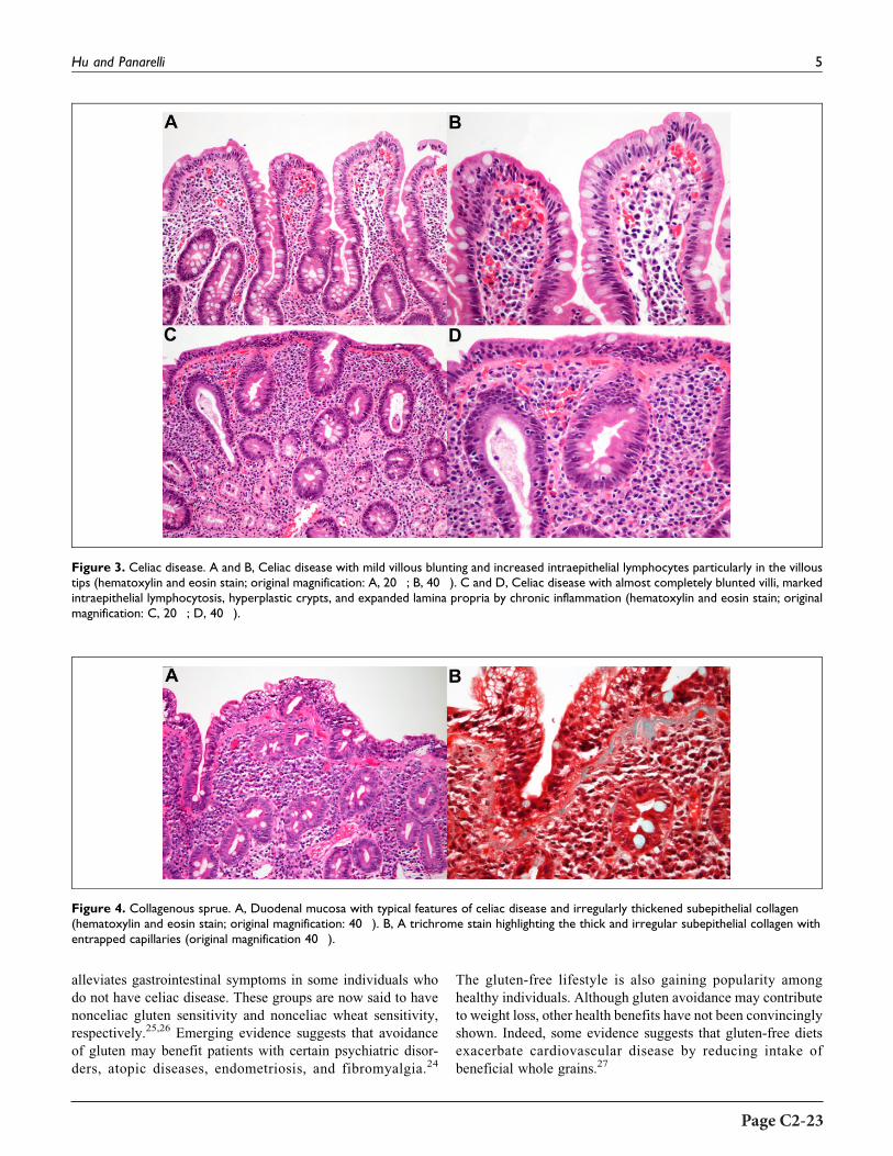
alleviates gastrointestinal symptoms in some individuals who
do not have celiac disease. These groups are now said to have
nonceliac gluten sensitivity and nonceliac wheat sensitivity,
respectively.25,26 Emerging evidence suggests that avoidance
of gluten may benefit patients with certain psychiatric disor-
ders, atopic diseases, endometriosis, and fibromyalgia.24
The gluten-free lifestyle is also gaining popularity among
healthy individuals. Although gluten avoidance may contribute
to weight loss, other health benefits have not been convincingly
shown. Indeed, some evidence suggests that gluten-free diets
exacerbate cardiovascular disease by reducing intake of
beneficial whole grains.27
Figure 3. Celiac disease. A and B, Celiac disease with mild villous blunting and increased intraepithelial lymphocytes particularly in the villoustips (hematoxylin and eosin stain; original magnification: A, 20�; B, 40�). C and D, Celiac disease with almost completely blunted villi, markedintraepithelial lymphocytosis, hyperplastic crypts, and expanded lamina propria by chronic inflammation (hematoxylin and eosin stain; originalmagnification: C, 20�; D, 40�).
Figure 4. Collagenous sprue. A, Duodenal mucosa with typical features of celiac disease and irregularly thickened subepithelial collagen(hematoxylin and eosin stain; original magnification: 40�). B, A trichrome stain highlighting the thick and irregular subepithelial collagen withentrapped capillaries (original magnification 40�).
Hu and Panarelli 5
Page C2-23

Teaching Points
1. Celiac disease is a multifactorial disorder that demon-
strates the interplay between genetic and environmental
factors in immune-mediated diseases.
2. The diagnosis of celiac disease relies upon serologic
evaluation of increased antibodies against TTG, EMA,
and DGP; duodenal biopsy confirmation of villous
shortening/blunting; intraepithelial lymphocytosis;
crypt hyperplasia; and clinical evaluation of patient’s
response to gluten-free diet. This underscores the
importance of interdisciplinary communication in the
management of gastrointestinal disorders.
3. Like many gastrointestinal disorders, celiac disease has
extraintestinal manifestations related to immune dysre-
gulation, such as dermatitis herpetiformis, and nutri-
tional deficiencies such as iron-deficiency anemia.
4. Celiac disease displays a spectrum of histologic
abnormalities ranging from normal mucosal histology
and villous architecture, to mild intraepithelial lympho-
cytic infiltration and partial villous atrophy, to marked
lymphocytosis and total villous blunting, related to dis-
ease duration and severity. 5. Recognition of celiac
disease is clinically important since gluten withdrawal
is the only effective therapy, and untreated disease can
lead to potentially serious sequelae including severe
malnutrition, lymphoma, and adenocarcinoma.
Declaration of Conflicting Interests
The author(s) declared no potential conflicts of interest with respect to
the research, authorship, and/or publication of this article.
Funding
The author(s) received no financial support for the research, author-
ship, and/or publication of this article.
References
1. Lau MS, Sanders DS. Optimizing the diagnosis of celiac disease.
Curr Opin Gastroenterol. 2017;33:173-180.
2. Elitsur Y, Sigman T, Watkins R, et al. Tissue transglutaminase
levels are not sufficient to diagnose celiac disease in North Amer-
ican practices without intestinal biopsies. Dig Dis Sci. 2017;62:
175-179.
3. Rostami K, Kerckhaert J, Tiemessen R, von Blomberg BM, Mei-
jer JW, Mulder CJ. Sensitivity of antiendomysium and antigliadin
antibodies in untreated celiac disease: disappointing in clinical
practice. Am J Gastroenterol. 1999;94:888-894.
4. Volta U, Caio G, Boschetti E, et al. Seronegative celiac disease:
shedding light on an obscure clinical entity. Dig Liver Dis. 2016;
48:1018-1022.
5. Stamnaes J, Sollid LM. Celiac disease: autoimmunity in response
to food antigen. Semin Immunol. 2015;27:343-352.
6. Pagliari D, Urgesi R, Frosali S, et al. The interaction among
microbiota, immunity, and genetic and dietary factors is the
condicio sine qua non celiac disease can develop. J Immunol Res.
2015;2015:123653.
7. Husby S, Koletzko S, Korponay-Szabo IR, et al. European Soci-
ety for Pediatric Gastroenterology, Hepatology, and Nutrition
guidelines for the diagnosis of coeliac disease. J Pediatr Gastro-
enterol Nutr. 2012;54:136-160.
8. Uenishi RH, Gandolfi L, Almeida LM, et al. Screening for celiac
disease in 1st degree relatives: a 10-year follow-up study. BMC
Gastroenterol. 2014;14:36.
9. Rubio-Tapia A, Ludvigsson JF, Brantner TL, Murray JA, Ever-
hart JE. The prevalence of celiac disease in the United States. Am
J Gastroenterol. 2012;107:1538-1544; quiz 1537, 1545.
10. Freeman HJ. Iron deficiency anemia in celiac disease. World J
Gastroenterol. 2015;21:9233-9238.
11. Collin P, Salmi TT, Hervonen K, Kaukinen K, Reunala T. Der-
matitis herpetiformis: a cutaneous manifestation of coeliac dis-
ease. Ann Med. 2017;49:23-31.
12. Pai RK. A practical approach to small bowel biopsy interpreta-
tion: celiac disease and its mimics. Semin Diagn Pathol. 2014;31:
124-136.
13. Brown IS, Bettington A, Bettington M, Rosty C. Tropical sprue:
revisiting an underrecognized disease. Am J Surg Pathol. 2014;
38:666-672.
14. Panarelli NC, Yantiss RK. Inflammatory and infectious manifes-
tations of immunodeficiency in the gastrointestinal tract. Mod
Pathol. 2018;31:844-861.
15. Singhi AD, Goyal A, Davison JM, Regueiro MD, Roche RL,
Ranganathan S. Pediatric autoimmune enteropathy: an entity fre-
quently associated with immunodeficiency disorders. Mod
Pathol. 2014;27:543-553.
16. Malamut G, Meresse B, Cellier C, Cerf-Bensussan N. Refractory
celiac disease: from bench to bedside. Semin Immunopathol.
2012;34:601-613.
17. Ondrejka S, Jagadeesh D. Enteropathy-associated T-cell lym-
phoma. Curr Hematol Malig Rep. 2016;11:504-513.
18. Smedby KE, Akerman M, Hildebrand H, Glimelius B, Ekbom A,
Askling J. Malignant lymphomas in coeliac disease: evidence of
increased risks for lymphoma types other than enteropathy-type T
cell lymphoma. Gut. 2005;54:54-59.
19. Smyrk TC. Practical approach to the flattened duodenal biopsy.
Surg Pathol Clin. 2017;10:823-839.
20. Ilus T, Kaukinen K, Virta LJ, Pukkala E, Collin P. Incidence of
malignancies in diagnosed celiac patients: a population-based
estimate. Am J Gastroenterol. 2014;109:1471-1477.
21. Shenoy S. Genetic risks and familial associations of small bowel
carcinoma. World J Gastrointest Oncol. 2016;8:509-519.
22. Samasca G, Lerner A, Girbovan A, et al. Challenges in gluten-free
diet in coeliac disease: Prague consensus. Eur J Clin Invest. 2017;
47:394-397.
23. Hall NJ, Rubin G, Charnock A. Systematic review: adherence to a
gluten-free diet in adult patients with coeliac disease. Aliment
Pharmacol Ther. 2009;30:315-330.
24. Niland B, Cash BD. Health benefits and adverse effects of a
gluten-free diet in non-celiac disease patients. Gastroenterol
Hepatol (N Y). 2018;14:82-91.
6 Academic Pathology
Page C2-24

25. Staudacher HM, Lomer MC, Anderson JL, et al. Fermentable
carbohydrate restriction reduces luminal bifidobacteria and gas-
trointestinal symptoms in patients with irritable bowel syndrome.
J Nutr. 2012;142:1510-1518.
26. de Roest RH, Dobbs BR, Chapman BA, et al. The low FODMAP
diet improves gastrointestinal symptoms in patients with irritable
bowel syndrome: a prospective study. Int J Clin Pract. 2013;67:
895-903.
27. Potter MDE, Brienesse SC, Walker MM, Boyle A, Talley NJ.
Effect of the gluten-free diet on cardiovascular risk factors in
patients with coeliac disease: a systematic review. J Gastroen-
terol Hepatol. 2018;33:781-791.
Hu and Panarelli 7
Page C2-25


Educational Case
Educational Case: Symptomatic butUnruptured Abdominal Aortic Aneurysm
Aldis H. Petriceks, BA1 , John C. Olivas, BS1, and Darren Salmi, MD1,2
The following fictional case is intended as a learning tool within the Pathology Competencies forMedical Education (PCME),
a set of national standards for teaching pathology. These are divided into three basic competencies: Disease Mechanisms and
Processes, Organ System Pathology, and Diagnostic Medicine and Therapeutic Pathology. For additional information, and
a full list of learning objectives for all three competencies, see http://journals.sagepub.com/doi/10.1177/2374289517715040.
Keywordspathology competencies, organ system pathology, abdominal aortic aneurysm, aortic dissection, aneurysm risk factors, unrup-tured aneurysm, ruptured aneurysm, complications of abdominal aortic aneurysms, classification of aortic dissections
Received May 01, 2018. Received revised July 25, 2018. Accepted for publication August 06, 2018.
Primary Objective
Objective CBV2.3: Abdominal Aortic Aneurysm: Describe the
clinical consequences of an abdominal aortic aneurysm.
Competency 2: Organ System Pathology; Topic: Cardiovas-
cular—Blood Vessels (CBV); Learning Goal 2: Vascular Dam-
age and Thrombosis.
Secondary Objective
Objective CBV2.2: Aortic Aneurysm and Dissection: Compare
and contrast aortic aneurysms and aortic dissections in terms of
their predisposing factors, the sites of involvement, and patient
populations likely to be affected.
Competency 2: Organ System Pathology; Topic: Cardiovas-
cular—Blood Vessels (CBV); Learning Goal 2: Vascular
Damage and Thrombosis.
Patient Presentation
A 68-year-old male patient presents to the emergency depart-
ment with nausea and a pulsating pain in his right groin. He
reports intermittent bouts of syncope in the past week, which he
never had previously. He also has chronic lower back pain but
states that the pain is now more severe and radiates from belly
to back and is very different from his usual back pain. Further
interview reveals that the patient has smoked approximately 10
cigarettes per day for the past 30 years. He is in visible distress,
although alert and oriented. His vital signs are remarkable for
hypertension, with a blood pressure of 170/115.
Diagnostic Findings, Part I
Physical examination of the right groin reveals a small pul-
satile mass. Abdominal palpation is suboptimal because of
the patient’s size. Auscultation of the abdomen reveals an
intermittent bruit, audible during systole, and dependent on
heart rate.
1 Division of Clinical Anatomy, Stanford University School of Medicine,
Stanford, CA, USA2 Department of Pathology, Stanford University School of Medicine, Stanford,
CA, USA
Corresponding Author:
Darren Salmi, Division of Clinical Anatomy, Stanford University School of
Medicine, 269 Campus Dr, CCSR 0135A, Stanford, CA 94305, USA.
Email: [email protected]
Academic PathologyVolume 5: 1–7ª The Author(s) 2018Article reuse guidelines:sagepub.com/journals-permissionsDOI: 10.1177/2374289518798560journals.sagepub.com/home/apc
Creative Commons Non Commercial CC BY-NC: This article is distributed under the terms of the Creative Commons Attribution-NonCommercial 4.0 License(http://www.creativecommons.org/licenses/by-nc/4.0/) which permits non-commercial use, reproduction and distribution of the work without furtherpermission provided the original work is attributed as specified on the SAGE and Open Access pages (https://us.sagepub.com/en-us/nam/open-access-at-sage).
Page C2-27

Question/Discussion Points, Part I
Discuss the Differential Based on the ClinicalPresentation
The patient presents with nausea, hypertension, syncope,
increasing lower back pain, and pulsing unilateral groin pain.
With his obesity, age, and history of hypertension, the patient
likely suffers from atherosclerosis as well. His alertness and
orientation suggest he is hemodynamically stable. These fac-
tors and symptoms suggest a possible abdominal aortic aneur-
ysm (AAA). The particularly important risk factors in this
patient’s history are as follows: (1) his advanced age, as AAA
is rare in persons younger than 60 years1; (2) his hypertension,
as high blood pressure can induce small tears in the extracel-
lular matrix (ECM), leading to smooth muscle cell (SMC)
recruitment, vascular thickening, reduced vascular compliance,
and ultimately aneurysm2; (3) his smoking history, as smoking
is the dominant behavioral risk factor for AAA3; and (4) the
likelihood that he also suffers from atherosclerosis, which is the
dominant physiological risk factor for AAA. This is because
atherosclerotic plaques weaken the arterial wall through a com-
bination of chronic inflammatory response and ischemia of the
underlying media.2
The most important signs and symptoms from his clinical
presentation are (1) the patient’s novel and increasing lower
back pain, (2) his pulsing unilateral groin pain and palpable
mass, and (3) his absence of hypovolemic shock symptoms
(eg, hypotension, decreased alertness), suggesting that any pre-
sumptive aneurysm has not ruptured.2,4
However, the differential diagnosis still includes the fol-
lowing: aortic dissection (AD), pancreatitis (particularly
because of the radiating lower back pain), ischemic bowel,
renal colic, gastrointestinal hemorrhage, pyelonephritis, and
peptic ulcer disease.
The physical examination does little to narrow this differ-
ential, as obesity makes a thorough abdominal examination
difficult. Nonetheless, given the history and clinical presenta-
tion—smoking, hypertension, age, obesity, back and groin
pain, nausea—AAA is the most prominent concern. The uni-
lateral groin pain could result from an associated aneurysm of
the right common iliac artery—a regular finding in AAA—
adding isolated iliac aneurysm to the differential.4 Therefore,
the most likely diagnosis is an unruptured AAA with associated
unruptured isolated right common iliac aneurysm. The patient
is clearly symptomatic and should be evaluated for possible or
imminent rupture. One should note that the diagnosis, to this
point, has been based on clinical findings.
What Further Testing Is Indicated for This Patient?
In hemodynamically unstable patients, timely diagnoses might
preferably be reached via ultrasound. However, computed
tomography (CT) imaging is the preferred method for this
hemodynamically stable patient because (1) CT provides a
more detailed and accurate assessment of the internal
abdominal structures, while also showing relevant vessels and
organs that may be involved with the aneurysm. This is one
reason why the Society for Vascular Surgery (SVS) recom-
mends CT for the assessment of maximum AAA diameter.5
(2) Computed tomography imaging offers less variability in
technician skill and methodology. (3) There are certain regions
of the body where ultrasound is affected by overlying tissue—
for instance, the iliac arteries, where overlying bowel can inter-
fere with the imaging process.6
Therefore, CT scans should be performed to evaluate
dilation of the patient’s aorta and/or other major arteries.
The thorax, abdomen, and lower limbs should all be eval-
uated to ensure full coverage of possible aneurysms, dissec-
tions, or embolisms.
Diagnostic Findings, Part II
Figure 1 Depicts the CT findings for this patient. Describethe CT scan and your assessment for the next step incare of the patient.
The abdominal CT scan shows a widely dilated area of intrave-
nously injected contrast material, revealing a widened aortic
lumen. The mural thrombus can also be seen surrounding this
contrast material. These 2 facts confirm an aortic aneurysm. The
scan was taken distal to the renal arteries, therefore specifying
the diagnosis to AAA. The peritoneal cavity does not contain
any extravasation of contrast material, indicating an unruptured
AAA. The SVS recommends elective surgical repair for aneur-
ysms larger than 5.5 cm, and the figure caption notes that this
aneurysm is 5.8 cm at maximum diameter. As such, this patient
should likely undergo surgical repair of the AAA.
Figure 1. Abdominal CT image of the patient. A ¼ Aortic lumen,highlighted by intravenous contrast material. B ¼ Mural thrombus ofaortic aneurysm. Computed tomography imaging was done withintravenous contrast material. Note the inferior vena cava (IVC)posterior and to the right of the mural thrombus. In a ruptured AAA,this would likely be obliterated. AAA indicates abdominal aorticaneurysm; CT computed tomography.
2 Academic Pathology
Page C2-28

Question/Discussion Points, Part II
What Clinical Sequelae Might Coincide With anUnruptured Abdominal Aortic Aneurysm?
Depending on the specific patient and aneurysm, an unruptured
AAA may elicit a diverse set of clinical sequelae. For example,
most patients with AAA are asymptomatic, and their aneur-
ysms are discovered incidentally during physical examination
or unrelated imaging.4 For patients who are symptomatic—as
the patient in this case—AAA may elicit abdominal or lower
back pain, fever, syncope, a pulsatile mass in the abdomen or
groin, and/or ischemia of the lower limb due to embolism.4
However, these symptoms are not specific to AAA, and must
be corroborated with imaging or further diagnostic testing. In
essence, a patient with unruptured AAA may present with
wide-ranging clinical sequelae depending on blood pressure,
severity of atherosclerosis, and other confounding factors. If
one patient has a stable AAA—for example, only 2.5 cm in
diameter—clinical signs may be absent. Then again, if an
aneurysm is large or aggressive, a patient might present with
several of the characteristic symptoms.
Figure 2 Demonstrates the Gross Anatomy of anUnruptured AAA. Briefly Describe the Anatomy and itsClinical Implications.
Figure 2 shows an infrarenal, unruptured saccular AAA, in the
classic location just superior to the common iliac bifurcation. A
saccular aneurysm is onewhich bulges in a distinct, unilateral man-
ner, creating a discrete pouch of dilated vascular wall. In contrast,
fusiform aneurysms bulge circumferentially and are thus somewhat
symmetrical. Both the saccular and fusiform types are “true” aneur-
ysmsbecause,unless ruptured, all 3 layersof thearterialwall remain
intact (These layers are the intima,media, and adventitia.). Blood is
therefore retained as a mural thrombus adherent to the arterial
lumen, as opposed to in an extravascular hematoma.
The true aneurysm in Figure 2 is 8.0 cm in maximum dia-
meter, making it a remarkably large aneurysm. Such an aneur-
ysm would be at very high risk for rupture, likely necessitating
surgical intervention. Some AAAs reach up to 20 cm in
diameter, although this is uncommon.2 An associated athero-
sclerotic plaque might obstruct blood flow to and through the
iliac arteries, causing lower limb ischemia, which could also
occur through thromboembolism.
Given the asymmetric, unilateral appearance of the refer-
enced AAA, one might confuse this specimen for a
“pseudoaneurysm.” Pseudoaneurysms, like true aneurysms,
result from defects in the vascular wall. However, there is a key
difference between the two. In true aneurysms, all 3 layers of the
arterial wall become dilated, creating an intravascular bulge that
has the potential for rupture. On the other hand, pseudoaneur-
ysms occur when blood ruptures through all 3 arterial layers but
is contained within the extravascular tissue.2 This forms an often
asymmetrical hematoma outside the artery, still contiguous with
the intravascular space. True aneurysms—until they rupture—
retain blood completely within the intravascular space.
What Risk Factors Might Influence the Likelihood ofAbdominal Aortic Aneurysm Rupture in This Patient?
Maximum AAA diameter is highly indicative of rupture risk.
There is <1% yearly risk of rupture for aneurysms smaller than
3.9 cm in maximum diameter, increasing to 1% at 4.0 to 4.9 cm;
1% to 11% at 5.0 to 5.9 cm; 10% to 22% at 6.0 to 6.9 cm; and
30% to 33% at 7.0 cm or larger.1,5 Rapid aneurysm expan-
sion—defined as a baseline diameter increase of 5 mm or more
over a 6-month period, or 10mmover a 12-month period—also
increases risk of rupture.5 For comparison, the average rate of
AAA expansion is approximately 2.6 mm per 12 months.7
Larger aneurysms tend to expand faster than smaller ones,
although expansion is nonlinear and can be quite stochastic.8
Other strong risk factors for rupture include tobacco use,
hypertension, and female sex.5,9 It is difficult to characterize
the exact contributions of tobacco use, because the many com-
ponents of tobacco smoke have wide-ranging, interrelated
bioactivities in vivo. Nonetheless, studies suggest possible
pathways in which T-cells and matrix-degrading enzymes
cause inflammatory damage to the aortic SMCs and ECM,
while also reducing expression of enzymes such as prolyl-4-
hydroxylase.3 These enzymes are critical to collagen synthesis,
and their suppression might inhibit repair of aortic tissue. As
the aortic tissue incurs further damage and is unable to repair
itself, aneurysmal dilation can continue up to rupture.
Studies indicate that females with AAA are more likely than
males to suffer from AAA rupture—regardless of aneurysm size
interval—with a 4-fold greater risk of rupture in aneurysms less
than 5.5 cm wide.10 Obviously, this does not impact the present
patient, but the discrepancy is important to consider nonetheless.
Some explanation may be found in the sex-neutral practices of
AAA management and classification. For example, the SVS
currently recommends observation for asymptomatic aneurysms
<5.5 cm in diameter, regardless of patient sex.5 However, studies
also suggest that females have smaller relative aortic diameters,
indicating that if a female and a male of similar size have AAAs
of equal diameter, the female has undergone a greater
Figure 2. Gross anatomy of abdominal aortic aneurysm (AAA). Notethe superior mesenteric artery superior (to the left in this image) tothe aneurysm, and the bifurcation of the common iliac arteries infer-iorly (to the right). Maximum diameter of aneurysm ¼ 8.0 cm, wellbeyond the standard for surgical intervention.
Petriceks et al 3
Page C2-29

proportional dilation of the aneurysm.11 Therefore, female
patients with aneurysm diameters of 5.5 cm may exceed the
proper minimum threshold for recommendation of elective
surgery. This means that females may incur greater risk of
AAA rupture, because their current sex-neutral thresholds for
elective surgical intervention are too high.
Finally, uncontrolled hypertension can increase risk of AAA
rupture—although the relationship between hypertension and
AAA expansion is less clear—as high blood pressures propa-
gate stress upon an already-dilated arterial wall.1 In order to
prevent further damage to the aorta, initial treatment of AAA
typically involves antihypertensive medication.
The present patient—given his history of smoking, hyper-
tension, and aneurysm size—is certainly at high risk of AAA
rupture. Rupture carries significant potential for mortality, and
the patient should thus be carefully monitored and treated
moving forward.
Discuss the Mortality Rates Associated With RupturedAbdominal Aortic Aneurysm
A ruptured AAA is a serious, often fatal surgical emer-
gency. Upon rupture, patients can present with 3 major
symptoms: pain, pulsatile mass, and hypotension. However,
less than half of patients will present with all three. This
can lead to misdiagnosis, contributing to the 90% overall
mortality rate in AAA.12 There are 4 main sites for AAA
rupture—intraperitoneal, retroperitoneal, aortocaval fistula,
and primary aortoduodenal fistula—and each one presents
with distinct and nuanced clinical features.12
If This Patient Suffered From a Ruptured AbdominalAortic Aneurysm, How Might His Computed TomographyScan Differ From the Original?
One would expect extravasation of the contrast material, lead-
ing to visible retroperitoneal hematoma and loss of the fat plane
between the aorta and its surrounding tissue. In symptomatic
but unruptured AAA, the CT often reveals several distinct
findings in lieu of such extravasation: irregular aortic wall,
draping of the aorta over vertebral bodies, and/or the crescent
shape of a layering hematoma.4
Figure 3 Presents the CT Scan From a Different Patient.Describe the Figure and Diagnose This New Patient.
In Figure 3A, we see a dilated aortic lumen, with high density
of blood in lieu of the periaortic fat plane. This blood appears to
have dissected into the retroperitoneum and obliterated the fat
plane around the inferior vena cava (IVC) and right psoas
muscle. Figure 3B shows a colored version of the same scan,
giving appreciation of the acute blood density surrounding the
periaortic fat plane, obliterated IVC, retroperitoneum, and
psoas muscle. These images are consistent with a ruptured
AAA, as the extravasation almost certainly originated from
an aneurysm directly adjacent to this abdominal region.
Compare and Contrast the Etiology, Clinical Description,Risk Factors, and Epidemiology of Aortic Aneurysm andAortic Dissection
An aortic aneurysm involves dilation of all 3 layers of the
arterial wall—the intima, the media, and the adventitia. In
contrast, AD occurs when blood enters the aortic wall through
an intimal tear and then dissects progressively through the
media.13 A “dissection” then continues as blood pulses through
the tissue plane, splitting the attenuated media. Histological
data from ADs contrast starkly with those of normal aortic
specimens. For instance, Figure 4A depicts a normal, healthy
media identified by numerous elastin fibers colored black by
the Elastic Van Gieson (EVG) stain. (EVG forms a variety of
strong bonds with elastin, staining the elastic fibers black. As
such, EVG is typically used to visualize normal or pathologic
elastic fibers.) In contrast, Figure 4B depicts a medial layer
split by dissecting blood, alongside atherosclerosis between the
intima and attenuated media. The pathological term for this
attenuation of the media is cystic medial necrosis.
Aortic dissection is therefore characterized by a proximal
intimal tear—such as in the ascending aorta—and a medial
Figure 3. Abdominal computed tomography image of a rupturedAAA (A) alongside the same image where the dilated aorta is high-lighted in red, the inferior vena cava in blue, and hemorrhage in orange(B). Maximum aneurysm diameter ¼ 5.8 cm. Computed tomographyimaging was done with intravenous contrast material. AAA indicatesabdominal aortic aneurysm.
4 Academic Pathology
Page C2-30
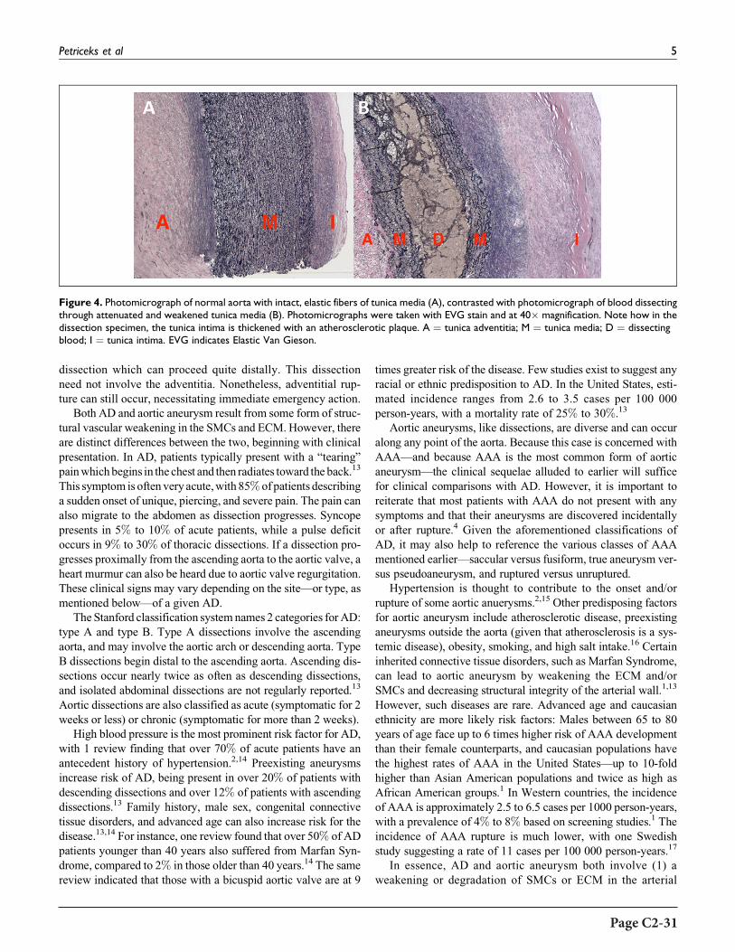
dissection which can proceed quite distally. This dissection
need not involve the adventitia. Nonetheless, adventitial rup-
ture can still occur, necessitating immediate emergency action.
Both AD and aortic aneurysm result from some form of struc-
tural vascular weakening in the SMCs and ECM. However, there
are distinct differences between the two, beginning with clinical
presentation. In AD, patients typically present with a “tearing”
painwhichbegins in the chest and then radiates toward the back.13
This symptom is often very acute,with 85% of patients describing
a sudden onset of unique, piercing, and severe pain. The pain can
also migrate to the abdomen as dissection progresses. Syncope
presents in 5% to 10% of acute patients, while a pulse deficit
occurs in 9% to 30% of thoracic dissections. If a dissection pro-
gresses proximally from the ascending aorta to the aortic valve, a
heart murmur can also be heard due to aortic valve regurgitation.
These clinical signs may vary depending on the site—or type, as
mentioned below—of a given AD.
The Stanford classification systemnames 2 categories for AD:
type A and type B. Type A dissections involve the ascending
aorta, and may involve the aortic arch or descending aorta. Type
B dissections begin distal to the ascending aorta. Ascending dis-
sections occur nearly twice as often as descending dissections,
and isolated abdominal dissections are not regularly reported.13
Aortic dissections are also classified as acute (symptomatic for 2
weeks or less) or chronic (symptomatic for more than 2 weeks).
High blood pressure is the most prominent risk factor for AD,
with 1 review finding that over 70% of acute patients have an
antecedent history of hypertension.2,14 Preexisting aneurysms
increase risk of AD, being present in over 20% of patients with
descending dissections and over 12% of patients with ascending
dissections.13 Family history, male sex, congenital connective
tissue disorders, and advanced age can also increase risk for the
disease.13,14 For instance, one review found that over 50% of AD
patients younger than 40 years also suffered from Marfan Syn-
drome, compared to 2% in those older than 40 years.14 The same
review indicated that those with a bicuspid aortic valve are at 9
times greater risk of the disease. Few studies exist to suggest any
racial or ethnic predisposition to AD. In the United States, esti-
mated incidence ranges from 2.6 to 3.5 cases per 100 000
person-years, with a mortality rate of 25% to 30%.13
Aortic aneurysms, like dissections, are diverse and can occur
along any point of the aorta. Because this case is concerned with
AAA—and because AAA is the most common form of aortic
aneurysm—the clinical sequelae alluded to earlier will suffice
for clinical comparisons with AD. However, it is important to
reiterate that most patients with AAA do not present with any
symptoms and that their aneurysms are discovered incidentally
or after rupture.4 Given the aforementioned classifications of
AD, it may also help to reference the various classes of AAA
mentioned earlier—saccular versus fusiform, true aneurysm ver-
sus pseudoaneurysm, and ruptured versus unruptured.
Hypertension is thought to contribute to the onset and/or
rupture of some aortic anuerysms.2,15 Other predisposing factors
for aortic aneurysm include atherosclerotic disease, preexisting
aneurysms outside the aorta (given that atherosclerosis is a sys-
temic disease), obesity, smoking, and high salt intake.16 Certain
inherited connective tissue disorders, such as Marfan Syndrome,
can lead to aortic aneurysm by weakening the ECM and/or
SMCs and decreasing structural integrity of the arterial wall.1,13
However, such diseases are rare. Advanced age and caucasian
ethnicity are more likely risk factors: Males between 65 to 80
years of age face up to 6 times higher risk of AAA development
than their female counterparts, and caucasian populations have
the highest rates of AAA in the United States—up to 10-fold
higher than Asian American populations and twice as high as
African American groups.1 In Western countries, the incidence
of AAA is approximately 2.5 to 6.5 cases per 1000 person-years,
with a prevalence of 4% to 8% based on screening studies.1 The
incidence of AAA rupture is much lower, with one Swedish
study suggesting a rate of 11 cases per 100 000 person-years.17
In essence, AD and aortic aneurysm both involve (1) a
weakening or degradation of SMCs or ECM in the arterial
Figure 4. Photomicrograph of normal aorta with intact, elastic fibers of tunica media (A), contrasted with photomicrograph of blood dissectingthrough attenuated and weakened tunica media (B). Photomicrographs were taken with EVG stain and at 40� magnification. Note how in thedissection specimen, the tunica intima is thickened with an atherosclerotic plaque. A ¼ tunica adventitia; M ¼ tunica media; D ¼ dissectingblood; I ¼ tunica intima. EVG indicates Elastic Van Gieson.
Petriceks et al 5
Page C2-31
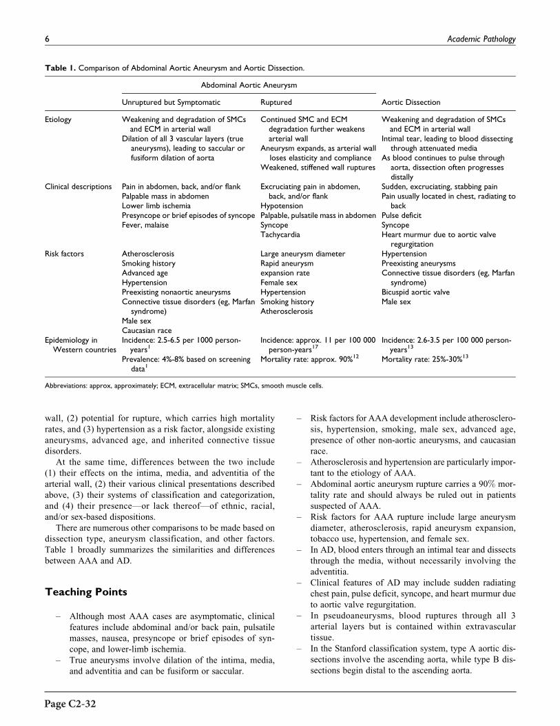
wall, (2) potential for rupture, which carries high mortality
rates, and (3) hypertension as a risk factor, alongside existing
aneurysms, advanced age, and inherited connective tissue
disorders.
At the same time, differences between the two include
(1) their effects on the intima, media, and adventitia of the
arterial wall, (2) their various clinical presentations described
above, (3) their systems of classification and categorization,
and (4) their presence—or lack thereof—of ethnic, racial,
and/or sex-based dispositions.
There are numerous other comparisons to be made based on
dissection type, aneurysm classification, and other factors.
Table 1 broadly summarizes the similarities and differences
between AAA and AD.
Teaching Points
– Although most AAA cases are asymptomatic, clinical
features include abdominal and/or back pain, pulsatile
masses, nausea, presyncope or brief episodes of syn-
cope, and lower-limb ischemia.
– True aneurysms involve dilation of the intima, media,
and adventitia and can be fusiform or saccular.
– Risk factors for AAA development include atherosclero-
sis, hypertension, smoking, male sex, advanced age,
presence of other non-aortic aneurysms, and caucasian
race.
– Atherosclerosis and hypertension are particularly impor-
tant to the etiology of AAA.
– Abdominal aortic aneurysm rupture carries a 90% mor-
tality rate and should always be ruled out in patients
suspected of AAA.
– Risk factors for AAA rupture include large aneurysm
diameter, atherosclerosis, rapid aneurysm expansion,
tobacco use, hypertension, and female sex.
– In AD, blood enters through an intimal tear and dissects
through the media, without necessarily involving the
adventitia.
– Clinical features of AD may include sudden radiating
chest pain, pulse deficit, syncope, and heart murmur due
to aortic valve regurgitation.
– In pseudoaneurysms, blood ruptures through all 3
arterial layers but is contained within extravascular
tissue.
– In the Stanford classification system, type A aortic dis-
sections involve the ascending aorta, while type B dis-
sections begin distal to the ascending aorta.
Table 1. Comparison of Abdominal Aortic Aneurysm and Aortic Dissection.
Abdominal Aortic Aneurysm
Aortic DissectionUnruptured but Symptomatic Ruptured
Etiology Weakening and degradation of SMCsand ECM in arterial wall
Dilation of all 3 vascular layers (trueaneurysms), leading to saccular orfusiform dilation of aorta
Continued SMC and ECMdegradation further weakensarterial wall
Aneurysm expands, as arterial wallloses elasticity and compliance
Weakened, stiffened wall ruptures
Weakening and degradation of SMCsand ECM in arterial wall
Intimal tear, leading to blood dissectingthrough attenuated media
As blood continues to pulse throughaorta, dissection often progressesdistally
Clinical descriptions Pain in abdomen, back, and/or flankPalpable mass in abdomenLower limb ischemiaPresyncope or brief episodes of syncopeFever, malaise
Excruciating pain in abdomen,back, and/or flank
HypotensionPalpable, pulsatile mass in abdomenSyncopeTachycardia
Sudden, excruciating, stabbing painPain usually located in chest, radiating to
backPulse deficitSyncopeHeart murmur due to aortic valve
regurgitationRisk factors Atherosclerosis
Smoking historyAdvanced ageHypertensionPreexisting nonaortic aneurysmsConnective tissue disorders (eg, Marfan
syndrome)Male sexCaucasian race
Large aneurysm diameterRapid aneurysmexpansion rateFemale sexHypertensionSmoking historyAtherosclerosis
HypertensionPreexisting aneurysmsConnective tissue disorders (eg, Marfan
syndrome)Bicuspid aortic valveMale sex
Epidemiology inWestern countries
Incidence: 2.5-6.5 per 1000 person-years1
Prevalence: 4%-8% based on screeningdata1
Incidence: approx. 11 per 100 000person-years17
Mortality rate: approx. 90%12
Incidence: 2.6-3.5 per 100 000 person-years13
Mortality rate: 25%-30%13
Abbreviations: approx, approximately; ECM, extracellular matrix; SMCs, smooth muscle cells.
6 Academic Pathology
Page C2-32

Acknowledgments
The authors would like to thank the Stanford University Department
of Pathology, for the diagnostic, histologic, and gross images used in
this education case.
Declaration of Conflicting Interests
The author(s) declared no potential conflicts of interest with respect to
the research, authorship, and/or publication of this article.
Funding
The author(s) received no financial support for the research, author-
ship, and/or publication of this article.
ORCID iD
Aldis H. Petriceks, BA http://orcid.org/0000-0001-5136-1023
References
1. Chung J. Epidemiology, risk factors, pathogenesis, and natural
history of abdominal aortic aneurysm. In: UpToDate; 2018.
https://www.uptodate.com/contents/epidemiology-risk-factors-
pathogenesis-and-natural-history-of-abdominal-aortic-aneur
ysm?search¼aaaepidemiology&source¼search_result&selected
Title¼1*150&usage_type¼default&display_rank¼1. Accessed
March 29, 2018.
2. Kumar V, Abbas AK, Aster JC. Blood Vessels. In: Kumar V,
Abbas AK, Aster JC, eds. Robbins Basic Pathology. 10th ed.
Philadelphia, PA: Elsevier Inc; 2018.
3. Norman PE, Curci JA. Understanding the effects of tobacco
smoke on the pathogenesis of aortic aneurysm. Arterioscler
Thromb Vasc Biol. 2014;33:1473-1477. doi:10.1161/ATV-
BAHA.112.300158.Understanding.
4. Jim J, Thompson RW. Clinical features and diagnosis of abdom-
inal aortic aneurysm. In: UpToDate; 2018. https://www.uptodate.
com/contents/clinical-features-and-diagnosis-of-abdominal-aortic-
aneurysm?search¼aaa&source¼search_result&selectedTitle¼3
*150&usage_type¼default&display_rank¼3. Accessed March
30, 2018.
5. Chaikof EL, Dalman RL, Eskandari MK, et al. The Society for
Vascular Surgery practice guidelines on the care of patients
with an abdominal aortic aneurysm. J Vasc Surg. 2018;67:2-77.
e2. doi:10.1016/j.jvs.2017.10.044.
6. Beales L, Wolstenhulme S, Evans JA, West R, Scott DJA. Repro-
ducibility of ultrasound measurement of the abdominal aorta. Br J
Surg. 2011;98:1517-1525. doi:10.1002/bjs.7628.
7. Brady AR, Thompson SG, Fowkes FG, Greenhalgh RM, Powell
JT; UK Small Aneurysm Trial Participants. Abdominal aortic
aneurysm expansion: risk factors and time intervals for surveil-
lance. Cardiovasc Surg. 2004;110:16-21. doi:10.1161/01.CIR.
0000133279.07468.9F.
8. Oliver-Williams C, Sweeting MJ, Turton G, et al. Lessons
learned about prevalence and growth rates of abdominal aor-
tic aneurysms from a 25-year ultrasound population screen-
ing programme. Br J Surg. 2018;105:68-74. doi:10.1002/bjs.
10715.
9. Bhak RH, Wininger M, Johnson GR, et al. Factors associated with
small abdominal aortic aneurysm expansion rate. JAMA Surg.
2015;150:44-50. doi:10.1001/jamasurg.2014.2025.
10. Skibba AA, Evans JR, Hopkins SP, et al. Reconsidering gender
relative to risk of rupture in the contemporary management of
abdominal aortic aneurysms. J Vasc Surg. 2013;62:1429-1436.
doi:10.1016/j.jvs.2015.07.079.
11. Forbes TL, Lawlor DK, DeRose G, Harris KA. Gender differ-
ences in relative dilatation of abdominal aortic aneurysms.
Ann Vasc Surg. 2006;20:564-568. doi:10.1007/S10016-006-
9079-y.
12. Assar AN, Zarins CK. Ruptured abdominal aortic aneurysm:
a surgical emergency with many clinical presentations. Post-
grad Med J. 2009;85:268-273. doi:10.1136/pgmj.2008.
074666.
13. Black JH, Manning WJ. Clinical features and diagnosis of acute
aortic dissection. In: UpToDate; 2018. https://www.uptodate.
com/contents/clinical-features-and-diagnosis-of-abdominal-aortic-
aneurysm?search¼aaa&source¼search_result&selectedTitle¼3
*150&usage_type¼default&display_rank¼3. Accessed April 2,
2018.
14. Hagan PG, Nienaber CA, Isselbacher EM, et al. The International
Registry of Acute Aortic Dissection (IRAD): new insights into an
old disease. JAMA. 2000;283:897-903. http://www.ncbi.nlm.nih.
gov/pubmed/10685714. Accessed April 29, 2018.
15. Takagi H, Umemoto T; ALICE (All-Literature Investigation of
Cardiovascular Evidence) Group. Association of hypertension
with abdominal aortic aneurysm expansion. Ann Vasc Surg.
2017;39:74-89. doi:10.1016/j.avsg.2016.04.019.
16. Dalman RL, Mell M. Overview of abdominal aortic aneurysm. In:
UpToDate; 2018. https://www.uptodate.com/contents/overview-
of-abdominal-aortic-aneurysm?search¼aaa&source¼search_
result&selectedTitle¼2*150&usage_type¼default&display_
rank¼2. Accessed March 29, 2018.
17. Acosta S, Ogren M, Bengtsson H, Bergqvist D, Lindblad B, Zda-
nowski Z. Increasing incidence of ruptured abdominal aortic
aneurysm: a population-based study. J Vasc Surg. 2006;44:
237-243. doi:10.1016/j.jvs.2006.04.037.
Petriceks et al 7
Page C2-33


Educational Case
Educational Case: Fibroadenomaof the Breast
H. James Williams, MD1
The following fictional case is intended as a learning tool within the Pathology Competencies forMedical Education (PCME),
a set of national standards for teaching pathology. These are divided into three basic competencies: Disease Mechanisms and
Processes, Organ System Pathology, and Diagnostic Medicine and Therapeutic Pathology. For additional information, and
a full list of learning objectives for all three competencies, see http://journals.sagepub.com/doi/10.1177/2374289517715040.
Keywordspathology competencies, organ system pathology, disease mechanisms, breast, neoplasia, fibroadenoma, palpable breast lesion,mammographic findings, phyllodes tumor
Received April 02, 2017. Received revised July 15, 2017. Accepted for publication February 13, 2018.
Primary Objective
Objective BR2.1: Fibroadenoma and Phyllodes Tumor. Com-
pare and contrast fibroadenoma and phyllodes tumor in terms
of clinical features, morphologic findings, and prognosis.
Competency 2: Organ System Pathology; Topic BR: Breast;
Learning Goal 2: Molecular Basis of Breast Neoplasms.
Secondary Objective
Objective N3.1: Morphologic Features of Neoplasia. Describe
the essential morphologic features of neoplasms and indicate
how these can be used to diagnose, classify, and predict biolo-
gical behavior of cancers.
Competency 1: Disease Mechanisms and Processes; Topic
N: Neoplasia; Learning Goal 3: Characteristics of Neoplasia.
Patient Presentation
A 37-year-old woman presents to her physician with concern
about a left breast nodule she recently discovered on self-
examination. The patient states that the nodule is approxi-
mately 2 cm in size, close to her left axilla, and feels firm. She
is concerned because her mother, age 54, was recently diag-
nosed with breast cancer.
Questions/Discussion Points, Part 1
What Pertinent Questions Should be Asked as Part of theDetailed History Prior to Physical Examination?
How long has the nodule been there? If the lesion developed veryrecently, one could choose to follow the lesion for a short time
to see if it persists.
Has it changed in size over time? Change in size could include
decreasing, increasing, or fluctuating lesions. Fluctuation in
size would be suggestive of menstrual effect or fibrocystic
change. A decrease in size might indicate a cyst getting
smaller. An increase in size would be more worrisome for a
more serious lesion.
1West Virginia University Health Sciences Center, Morgantown, WV, USA
Corresponding Author:
H. James Williams, West Virginia University Health Sciences Center, P.O. Box
9203, Morgantown, WV 26506, USA.
Email: [email protected]
Academic PathologyVolume 5: 1–4ª The Author(s) 2018Article reuse guidelines:sagepub.com/journals-permissionsDOI: 10.1177/2374289518790926journals.sagepub.com/home/apc
Creative Commons Non Commercial CC BY-NC: This article is distributed under the terms of the Creative Commons Attribution-NonCommercial 4.0 License(http://www.creativecommons.org/licenses/by-nc/4.0/) which permits non-commercial use, reproduction and distribution of the work without furtherpermission provided the original work is attributed as specified on the SAGE and Open Access pages (https://us.sagepub.com/en-us/nam/open-access-at-sage).
Page C2-35

Is the nodule painful? Malignancies are not typically painful;
however, inflammatory lesions can be painful as could benign
lesions.
Has there been nipple discharge and if so, is it bloody? Bloody
nipple discharge may be associated with an intraductal papil-
loma or, more rarely, cancer. A finding of nipple discharge
would need to be investigated more carefully.
Does anyone in your family have or have had breast cancer? If thereis a family history of breast cancer, what is the relationship of
the family member with the patient (ie, is it the patient’s
mother, sister, or daughter)? Also, what was the age at time
of diagnosis of the family member? Younger age of breast
cancer would be much more concerning for a possible famil-
iar component of a breast cancer than breast cancer in an older
first-degree relative. This question is addressing the associ-
ated increased risk of malignancy and/or hereditary disease in
close relatives, especially if they developed breast cancer at a
young age.
On physical examination of the patient’s left breast, what findingswould favor either a benign or malignant diagnosis? Physical exam-
ination can be helpful, as some features are more common
with benign or malignant lesions. Features of benign neo-
plasms include the following: multiple indistinct nodules
(lumpy breast) that favor fibrocystic changes and lesions
beneath the nipple which may suggest an intraductal
papilloma.
However, a fixed, irregular, and firm mass is suspicious for
malignancy as is dimpling of the overlying skin. Another
important factor is location of the lesion within the breast,
as 50% of breast cancers arise in the upper outer quadrant;
thus, lesions in this area would be more suspicious for
malignancy.
Diagnostic Findings, Part 2
The patient undergoes mammography of the left breast nodule.
The mammography is shown in Figure 1.
Questions/Discussion Points, Part 2
What Radiographic Findings in Figure 1 Are Seen, and DoThey Favor a Benign or Malignant Diagnosis?
Similar to physical examination, radiologic examination
is important in working up breast lesions. Several factors
are more suggestive of benign versus malignant lesions.
The following findings favor a benign process when they
Figure 1. Mammogram of left breast nodule. The lesion is homoge-neous and is sharply demarcated from the normal breast tissue.
Figure 2.Microscopic image of left breast nodule at low power, H&E2�. On low power, the lesion is seen to be sharply demarcated fromthe adjacent normal breast tissue.
Figure 3.Microscopic image of left breast nodule at high power, H&E20�. The stroma of the lesion has low cellularity with no mitoses orcytologic atypia. The epithelial component shows bland cytologicfeatures with associated myoepithelial cells.
2 Academic Pathology
Page C2-36

are present: sharp distinct borders, homogenous texture,
and ovoid shape. The following factors favor malignant
when they are present: stellate infiltrative borders, hetero-
genous texture, round shape, and presence of coarse
calcifications.
In the mammographic image for the patient, a round homo-
genous lesion is seen. This would favor a benign process. Con-
firmation of the imaging impression requires histologic
examination of the lesion either by biopsy of the lesion or by
conservative excision.
Diagnostic Findings, Part 3
The breast nodule is excised and sent for pathologic examina-
tion (see Figures 2 and 3).
Questions/Discussion Points, Part 3
What are the Pertinent Histologic Findings Seen inFigures 2 and 3?
On low-power image of the breast biospy, as seen in Figure
2, the lesion is seen to be sharply demarcated from the
adjacent normal breast tissue. This is suggestive of a benign
process. When looking at higher power in Figure 3, there is
a predominance of stromal tissue with compression of the
epithelial component. The stroma is of low cellularity with
no mitoses or cytologic atypia seen. In addition, the epithe-
lial component shows bland cytologic features with associ-
ated myoepithelial cells.
What Is Your Diagnosis?
Fibroadenoma of the breast.
Discuss the Clinical Features and Pathophysiology ofFibroadenomas and What Is the Prognosis andManagement for This Lesion?
Fibroadenomas are the most common benign breast neo-
plasms and typically present in women 20 to 35 years old.1
The tumors are usually solitary but may be multiple (20%).
Clinically, they may be detected by the patient or the physi-
cian on breast examination as a well-demarcated, freely
mobile, firm mass that is usually <3 cm in diameter. The
fibroadenoma is a neoplasm of the specialized lobular stroma
with typically low cellularity, no cytologic atypia, and mito-
tic figures absent or rare. The associated benign epithelium
has associated myoepithelial cells and may have an intra-
canalicular or pericanalicular pattern.2 The radiographic
appearance of fibroadenoma characteristically shows an
ovoid homogenous mass with sharp distinct borders.
Patients with fibroadenomas are associated with a slight
increased risk of breast cancer, particularly when there are
proliferative fibrocystic changes involving the tumor. As
would be anticipated, fibroadenomas are hormonally
responsive (may enlarge during pregnancy and reduce in size
in postmenopause).
As the fibroadenoma is a benign tumor, in the appropriate
clinicoradiographic setting, once the tumor is diagnosed by
fine-needle aspiration or needle core biopsy, it may be safely
followed. If the tumor is a cosmetic problem or if preferred by
the patient, conservative excision can be performed.3
Discuss a Common Tumor in the Differential Diagnosis ofa Fibroadenoma
The histologic differential diagnosis includes the phyllodes
tumor in which there is increased cellularity of the stroma, and
the epithelial component demonstrates a “leaf-like” architec-
tural pattern.1 Low-grade phyllodes tumors have these features
with mild cytologic atypia and occasional mitoses. High-grade
phyllodes tumors in addition to the increased stromal cellular-
ity have marked cytologic atypia and increased mitoses, some
of which may be atypical. Because of the higher risk of recur-
rences, wide excision is indicated for phyllodes tumors. In
addition to the risk of recurrences, the high-grade phyllodes
tumor has the potential to metastasize.
Teaching Points
� Benign features include the following: Multiple indis-
tinct nodules (lumpy breast) favors fibrocystic changes,
and lesions beneath the nipple may suggest a papilloma.
� A fixed irregular firm mass on physical examination is
suspicious for malignancy, as is dimpling of the over-
lying skin.
� On radiography, a benign process tends to have sharp
distinct borders, homogenous texture, and ovoid shape.
� Fibroadenomas are the most common benign breast neo-
plasms and typically present in women 20 to 35 years
old.
� The fibroadenoma is a neoplasm of the specialized lob-
ular stroma with typically low cellularity, no cytologic
atypia, and mitotic figures absent or rare. The associated
benign epithelium has associated myoepithelial cells
and may have an intracanalicular or pericanalicular
pattern.
� The phyllodes tumor has increased cellularity of the
stroma, and the epithelial component demonstrates a
“leaf-like” architectural pattern. Low-grade phyllodes
tumors may have mild cytologic atypia and occasional
mitoses. High-grade phyllodes tumors, in addition to the
increased stromal cellularity, have marked cytologic
atypia and increased mitoses, some of which may be
atypical.
Declaration of Conflicting Interests
The author(s) declared no potential conflicts of interest with respect to
the research, authorship, and/or publication of this article.
Williams 3
Page C2-37

Funding
The author(s) received no financial support for the research, author-
ship, and/or publication of this article.
References
1. KumarV,AbbasAK, FaustoN,Aster JC.Robbins andCotranPatho-
logic Basis of Disease. Elsevier Health Sciences, 2014:1069-1070.
2. Kuijper A, Mommers EC, van der Wall E, van Diest PJ. Histo-
pathology of fibroadenoma of the breast. Am J Clin Pathol. 2001;
115:736-742.
3. Hubbard JL, Cagle K, Davis JW, Kaups KL, Kodama M. Criteria
for excision of suspected fibroadenomas of the breast. Am J Surg.
2015;209:297-301.
4 Academic Pathology
Page C2-38

Educational Case
Educational Case: Wilms Tumor
Alison R. Huppmann, MD1
The following fictional case is intended as a learning tool within the Pathology Competencies forMedical Education (PCME),
a set of national standards for teaching pathology. These are divided into three basic competencies: Disease Mechanisms and
Processes, Organ System Pathology, and Diagnostic Medicine and Therapeutic Pathology. For additional information, and
a full list of learning objectives for all three competencies, see http://journals.sagepub.com/doi/10.1177/2374289517715040.
Keywordspathology competencies, organ system pathology, kidney, renal neoplasia, Wilms tumor, pediatric, syndrome
Received February 28, 2018. Received revised April 06, 2018. Accepted for publication April 12, 2018.
Primary Objective
Objective UTK1.4: Wilms Tumor. Describe the clinical and
pathologic features and molecular basis for Wilms tumor and
list the histologic features that are important to recognize in
determining prognosis, and the etiology of Wilms tumor as part
of different syndromes.
Competency 2: Organ System Pathology; Topic UTK:
Kidney; Learning Goal 1: Renal Neoplasia.
Patient Presentation
A 2-year-old boy presents to his pediatrician after his mother
noticed a “lump in his belly.” Further questioning reveals only
that the patient has seemed slightly more tired lately. He has a
history of a urinary tract malformation that required surgical
repair. Family history is negative for malignancy or major
medical problems. Physical examination reveals a blood pres-
sure of 142/94 mm Hg with other vital signs within normal
limits. A large mass is palpable in the abdomen and appears
to be centered on the right side.
Diagnostic Findings
An abdominal ultrasound shows a solid mass in the right abdo-
men. A computed tomography (CT) scan discloses a mass aris-
ing from the right kidney (Figure 1). A nephrectomy is
performed.
Questions/Discussion Points
Which Entities May Present as an Abdominal Massin a Child? Include Both Neoplastic and NonneoplasticLesions
For abdominal masses occurring at birth or within the first few
years of life, congenital anomalies are a strong consideration,
including gastrointestinal tract duplications, cysts arising from
the omentum or mesentery, cysts of the hepatobiliary system,
intussusception, and genitourinary tract anomalies such as
polycystic or hydronephrotic kidneys or an enlarged bladder.
Splenomegaly or hepatomegaly, for any reason, may present as
a mass. If trauma is an antecedent factor, the cause may be a
hematoma or pancreatic pseudocyst. An intra-abdominal
abscess, most commonly arising from the appendix, is another
benign cause. Malignant tumors can also occur in the abdomen,
including neuroblastoma, Wilms tumor, hepatoblastoma, germ
1 Department of Pathology, School of Medicine, Uniformed Services University
of the Health Sciences, Bethesda, MD, USA
Corresponding Author:
Alison R. Huppmann, Department of Pathology, School of Medicine,
Uniformed Services University of the Health Sciences, 4301 Jones Bridge Rd,
Bethesda, MD 20814, USA.
Email: [email protected]
Academic PathologyVolume 5: 1–5ª The Author(s) 2018Article reuse guidelines:sagepub.com/journals-permissionsDOI: 10.1177/2374289518781582journals.sagepub.com/home/apc
Creative Commons Non Commercial CC BY-NC: This article is distributed under the terms of the Creative Commons Attribution-NonCommercial 4.0 License(http://www.creativecommons.org/licenses/by-nc/4.0/) which permits non-commercial use, reproduction and distribution of the work without furtherpermission provided the original work is attributed as specified on the SAGE and Open Access pages (https://us.sagepub.com/en-us/nam/open-access-at-sage).
Page C2-39

cell tumors, sarcomas (eg, rhabdomyosarcoma), and rarely
lymphomas.1
Describe the Gross and Histologic Features inFigures 2-4. Based on the Clinical and PathologicFeatures, What Is Your Diagnosis?
Grossly, a large, tan, fleshy, well-circumscribed mass replaces
most of the renal parenchyma (Figure 2). Foci of yellow dis-
coloration indicate necrosis. A capsule surrounds the periphery
of the mass. Areas of hemorrhage and/or cystic change can be
seen in some cases. The low-power histologic image (Figure 3)
demonstrates a variable appearance due to several different
components of the tumor. A higher power view (Figure 4)
allows closer examination of these varied elements, showing
a mixture of immature tubules, small round blue cells, and
spindled cells. These findings are consistent with a diagnosis
of Wilms tumor (nephroblastoma).
Like some other pediatric tumors, the histology of Wilms
tumor appears similar to primitive (embryonic or fetal) ele-
ments that would have been present in the tissue of origination,
in this case the kidney (Figure 5). A classic Wilms tumor is
triphasic, with epithelial, blastemal, and stromal elements;
although any one of these 3 may predominate in a given tumor.
Immature tubules and glomeruli compose the epithelial areas.
The blastemal cells are undifferentiated-appearing small blue
cells. Stromal cells are usually spindled with a fibrotic or myx-
oid background, and heterologous differentiation (skeletal
muscle, cartilage, osteoid, etc) can be found in some cases
(not seen in these images).2
Wilms tumor is the most common primary renal malignancy
of childhood.2 Other primary renal tumors of childhood include
Figure 1. A computed tomography (CT) scan shows a large mass (*)on the right side with the remaining kidney (arrow) showing the “clawsign” around the mass, indicating that is likely of renal origin.
Figure 2.Grossly, a large, tan, well-circumscribed, encapsulated masshas replaced much of the normal renal parenchyma. Yellowdiscoloration indicates areas of necrosis.
Figure 3. On low power magnification of the mass, the histologyshows a variable appearance due to the different elements composingthe tumor (hematoxylin and eosin, �40).
Figure 4. At intermediate magnification, primitive tubules (arrow),small round blue cells (*), and spindled cells (arrowhead) representepithelial, blastemal, and stromal components, respectively(hematoxylin and eosin, �200).
2 Academic Pathology
Page C2-40

mesoblastic nephroma, clear cell sarcoma of the kidney, rhab-
doid tumor of the kidney, and, in adolescents, renal cell carci-
noma. These tumors do not contain the triphasic elements seen
in Wilms tumors.
What Is the Typical Clinical Presentation of a WilmsTumor?
Wilms tumor most commonly occurs in young children
between age 2 and 5. However, older children and even adults
can be affected. The most common presentation is an abdom-
inal mass. These are often identified by a caregiver while bath-
ing or changing the child’s clothes or by a physician on routine
physical examination. Other frequent signs and symptoms
include hypertension, abdominal pain, fever, painless hema-
turia, and anemia.3
What Other Tumors Are Included in the Category of“Small Round Blue Cells Tumors”? How Are TheseDifferentiated?
Small round blue cell tumors are those composed of cells
with scant cytoplasm, so that the tumor appears “blue”
(basophilic) from low power since the nuclei dominate the
histology. Tumors commonly included in this group are
leukemia/lymphoma, neuroblastoma, Wilms tumor, rhabdo-
myosarcoma, Ewing sarcoma family of tumors, desmoplas-
tic small round cell tumor, rhabdoid tumor, and other
undifferentiated types of sarcoma. Note that most of these
tumor types are more common in children. Many of the
small round blue cell tumors of childhood show histological
features that permit a preliminary diagnosis. Panels of
immunohistochemical stains and genetic or molecular testing
are useful to confirm the findings.
What Are Nephrogenic Rests? What Is TheirSignificance?
Nephrogenic rests are nonencapsulated areas of persistent
immature renal tissue with histologic features that can resemble
Wilms tumor. These rests usually regress or differentiate, but
they can become malignant if they persist. Evidence supporting
this theory includes shared genetic alterations with rests and
tumor in the same patient.2 Although seen in 1% of the general
population,3 nephrogenic rests are identified in the adjacent
renal parenchyma of up to 40% of patients with unilateral
tumors and almost 100% of bilateral tumors. These rests are
important to identify, as they imply an increased risk of
Wilms tumor in the contralateral kidney, necessitating close
monitoring of the patient.2
Which Patient Characteristics Might Lead You to Suspectan Associated Syndrome in a Pediatric Patient WithCancer? Which Congenital Syndromes Are Most OftenAssociated With Wilms Tumor?
The possibility of a syndrome should be considered in any
pediatric oncology patient with a personal or family history
of malformation(s), personal or family history of malig-
nancy, early onset of cancer, certain physical characteristics,
or rare forms of cancer. Approximately 10% of Wilms
tumors occur in patients with congenital syndromes. Those
most commonly associated are WAGR syndrome (Wilms
tumor, aniridia, genital anomalies, mental retardation),
Denys-Drash syndrome (renal mesangial sclerosis, early-
onset renal failure, gonadal dysgenesis), and Beckwith-
Wiedemann syndrome (hemihypertrophy, organomegaly,
macroglossia, and omphalocele). A large number of other
syndromes are less commonly associated.2
Describe Some of the Common Genetic ChangesAssociated With Wilms Tumor
Genetic mutation(s) have been identified in one-third of Wilms
tumors as yet. Some of these have been found due to their
association with the congenital syndromes that convey an
increased risk of Wilms tumor. Both WAGR and Denys-
Drash syndromes are associated with mutations in the WT1
gene, although due to a deletion in WAGR and a missense
mutation in Denys-Drash. WT1 is a tumor suppressor gene and
encodes for a transcription factor that plays a critical role in
development of the kidneys and gonads. The homozygousWT1
mutations detected in Wilms tumors lead to loss of function.2
Beckwith-Wiedemann syndrome is associated with altera-
tions in chromosome 11p15.5. One of the genes in this region,
IGF2, is normally imprinted, with expression only by the pater-
nal allele. Either loss of imprinting or uniparental disomy of
IGF2 has been identified in some Wilms tumors.2
CTNNB1 encodes for b-catenin, a key regulator of the Wnt
signaling pathway. Mutations in CTNNB1 are identified in 10%to 15% of Wilms tumors. Another gene associated with the
Figure 5. This is the histologic appearance of fetal renal cortex at19 weeks’ gestation. Note the nephrogenic zone in the subcapsulararea and its histologic similarity to Wilms tumor, with primitive glo-meruli, tubules, and blastema (hematoxylin and eosin, �100). Thenephrogenic zone should disappear by approximately 36 weeks’gestation.
Huppmann 3
Page C2-41

same pathway, WTX, is mutated in some tumors; TP53 muta-
tions are even less frequent. Many other less frequent genetic
mutations have been identified, either alone or in combination
with those previously discussed.3
What Histological Feature in Wilms Tumor Is Consideredto be Unfavorable?
The feature in Wilms tumor indicative of unfavorable histology
is anaplasia. This is shown in Figure 6. Anaplasia in Wilms
tumor is defined as the presence of cells with large hyperchro-
matic nuclei (generally at least 3 times the size of adjacent
nonanaplastic nuclei) and the presence of abnormal, polyploid
mitotic figures. It is seen in 5% to 10% of Wilms tumors. As a
diffuse finding, anaplasia is associated with resistance to che-
motherapy and conveys a poorer prognosis in advanced-stage
disease. It is linked to TP53 mutations.4
Where Is Wilms Tumor Most Likely to Spread?
Renal vein extension is common. Noncontiguous spread goes
to the regional lymph nodes, with the lungs being the most
common site of distant metastasis.
What Other Factors Affect Prognosis in Wilms Tumor?
Tumor histology is possibly the most critical factor affecting
prognosis, but other aspects considered include tumor stage,
patient age, loss of heterozygosity (LOH) for chromosomes
1p and 16q, tumor weight, and completeness of lung nodule
response. Briefly, staging for Wilms tumors is as follows: stage
I—tumor confined to the kidney and removed completely by
surgery; stage II—tumor extends beyond the kidney but is
completely removed by surgery; stage III—tumor remains in
the patient after surgery but is confined to the abdomen; stage
IV—hematogenous metastases to lungs, liver, lymph nodes
outside the abdomen, and so on; stage V—bilateral tumors at
diagnosis. Notably, if a biopsy is performed prior to resection,
the patient is automatically at least Stage III, which is why
biopsy is not often utilized for pediatric renal tumors.4
Current treatment includes surgery and chemotherapy with
or without radiation. The last 50 years have shown a dramatic
improvement in prognosis for patients with Wilms tumor, with
a current cure rate of approximately 90%.4
Teaching Points
� Wilms tumor is the most common primary renal tumor
of childhood, most commonly presenting as an abdom-
inal mass in a child 2 to 5 years of age.
� Many benign and malignant conditions can present as
an abdominal mass in a child, necessitating a com-
plete evaluation of the patient to arrive at the correct
diagnosis.
� Genetic changes involved in the pathogenesis of Wilms
tumor include the WT1 gene and chromosome 11p15.5.
These were partly identified through their association
with some of the congenital syndromes with an
increased risk of Wilms tumor, WAGR, Denys-Drash,
and Beckwith-Wiedemann syndromes.
� An associated congenital syndrome should be suspected
in a pediatric oncology patient with a personal or family
history of a malformation or malignancy.
� A classic Wilms tumor has triphasic histology, including
epithelial, blastemal, and stromal elements. Nephro-
genic rests are presumed precursors to Wilms tumor and
can have similar histologic features, although they are
not encapsulated.
� Small round blue cells tumors have a similar histologic
appearance of small cells with scant cytoplasm. Many of
these tumors occur in childhood and include Wilms
tumor, neuroblastoma, leukemia/lymphoma, rhabdo-
myosarcoma, and Ewing sarcoma family of tumors.
� Histology is a key predictor of prognosis in Wilms
tumor, as anaplasia is associated with a less favorable
prognosis. Other prognostic markers include age, stage,
LOH 1p and 16q, tumor weight, and responsiveness of
lung metastases.
Author’s Note
The opinions expressed herein are those of the author and are not
necessarily representative of those of the Uniformed Services Univer-
sity of the Health Sciences (USUHS), the Department of Defense
(DOD), or the United States Army, Navy, or Air Force.
Declaration of Conflicting Interests
The author(s) declared no potential conflicts of interest with respect to
the research, authorship, and/or publication of this article.
Figure 6. This tumor contains scattered cells with hyperchromaticnuclei that are much larger than the other nuclei. The inset shows anatypical mitotic figure. These findings indicate anaplasia (hematoxylinand eosin, �100; inset, �1000).
4 Academic Pathology
Page C2-42

Funding
The author(s) received no financial support for the research, author-
ship, and/or publication of this article.
References
1. Davidoff A. Pediatric abdominal masses. In: Myers JA, Millikan
KW, Saclarides TJ, eds. Common Surgical Diseases. New York,
NY: Springer; 2008:251-253.
2. Maitra A. Diseases of infancy and childhood. In: Kumar V, Abbas
A, Aster A, eds. Robbins and Cotran Pathologic Basis of Disease.
9th ed. Philadelphia, PA: Elsevier Saunders; 2015:479-481.
3. Daw NC, Huff V, Anderson PM. Wilms tumor. In: Kliegman RM,
et al., eds. Nelson Textbook of Pediatrics. 20th ed. Philadelphia,
PA: Elsevier; 2015:2464-2467.
4. Dome JS, Perlman EJ, Graf N. Risk stratification for Wilms tumor:
current approach and future directions. Am Soc Clin Oncol Educ
Book. 2014;34:215-223.
Huppmann 5
Page C2-43


Educational Case
Educational Case: Pheochromocytoma
Clinton Westover, MD1 and Richard Michael Conran, MD, PhD, JD1
The following fictional case is intended as a learning tool within the Pathology Competencies for Medical Education
(PCME), a set of national standards for teaching pathology. These are divided into three basic competencies: Disease
Mechanisms and Processes, Organ System Pathology, and Diagnostic Medicine and Therapeutic Pathology. For addi-
tional information, and a full list of learning objectives for all three competencies, see http://journals.sagepub.com/doi/
10.1177/2374289517715040.
Keywordspathology competencies, organ system pathology, endocrine, pheochromocytoma, paraganglioma, adrenal tumor, multipleendocrine neoplasia, secondary hypertension, familial pheochromocytoma-paraganglioma syndrome
Received December 6, 2017. Received revised April 22, 2018. Accepted for publication May 2, 2018.
Primary Objective
EN5.3: Pheochromocytoma and Paraganglioma: Outline the
clinicopathologic features of pheochromocytoma and compare
and contrast the hereditary cancer syndromes associated with
paragangliomas/pheochromocytomas.
Competency 2: Organ System Pathology; Topic EN:
Endocrine. Learning Goal 5: Endocrine Neoplasms.
Part 1: Clinical Case
Patient Presentation
A 47-year-old male with no previous medical history presents
to his family physician complaining of episodic headaches,
sweating, heart palpitations, and a tremor. The symptoms
started a few years ago, have become more frequent, and can
last anywhere between a few seconds to an hour. The episodes
often occur when the patient feels stressed or exercises. He is
frustrated because nothing he does changes the severity of his
symptoms. He does not have a history of serious illnesses,
hospitalizations, or trauma. He is not on any medications. He
has a family history of hypertension. He does not use tobacco
products, cocaine, methamphetamines, or any other illicit
drugs. He has not had fevers, chills, chest pain, shortness of
breath, nausea, vomiting, or diarrhea.
On physical examination, vital signs showed an elevated
blood pressure of 168/96 mm Hg, tachycardia of 116 beats per
minute, a respiratory rate of 20 breaths per minute, and a tem-
perature of 98�F (36.66�C). Physical examination reveals
a diaphoretic male with no cardiopulmonary abnormalities
other than the previously mentioned tachycardia.
Questions/Discussion Points, Part 1
What Is the Differential Diagnosis Based on the Historyand Physical Examination?
The differential diagnoses of episodic headaches, sweating, heart
palpitations, and a tremor include pheochromocytoma, paragan-
glioma, essential hypertension, anxiety disorders, panic attack,
thyrotoxicosis, medications, amphetamine and cocaine abuse,
paroxysmal supraventricular tachycardia, and carcinoid syn-
drome. Neuroendocrine tumors secreting insulin (insulinoma)
1 Eastern Virginia Medical School, Norfolk, VA, USA
Corresponding Author:
Richard Michael Conran, Eastern Virginia Medical School, 700 West Olney,
Norfolk, VA 23507, USA.
Email: [email protected]
Academic PathologyVolume 5: 1–6ª The Author(s) 2018Article reuse guidelines:sagepub.com/journals-permissionsDOI: 10.1177/2374289518780500journals.sagepub.com/home/apc
Creative Commons Non Commercial CC BY-NC: This article is distributed under the terms of the Creative Commons Attribution-NonCommercial 4.0 License(http://www.creativecommons.org/licenses/by-nc/4.0/) which permits non-commercial use, reproduction and distribution of the work without furtherpermission provided the original work is attributed as specified on the SAGE and Open Access pages (https://us.sagepub.com/en-us/nam/open-access-at-sage).
Page C2-45

and heart failure are other considerations. A good medical
history and imaging studies excludes several of these entities.
Part 2: Diagnostic Findings
Laboratory Studies
The complete blood count, comprehensive metabolic panel,
D-dimer, and serial troponins were within reference ranges.
An electrocardiogram indicated sinus tachycardia and left axis
deviation consistent with left ventricular hypertrophy. A
24-hour urine fractionated metanephrine and catecholamine
test showed significant elevations. Urinary metanephrines were
1300 mg/24 hours (normal range: 45-290 mg/24 hours).
Imaging
A chest X-ray was performed demonstrating mild left ventri-
cular hypertrophy. Cardiac ultrasound demonstrated ventricu-
lar hypertrophy. No other abnormalities were present. An
abdominal computed tomography (CT) scan showed a 3-cm-
diameter left adrenal gland mass. The right adrenal gland was
unremarkable.
Questions/Discussion Points, Part 2
What Entities Are in the Differential Diagnosis Based onthe Above Laboratory Findings?
Elevated metanephrines and catecholamines are character-
istic of a pheochromocytoma or paraganglioma.1 Anxiety
and panic disorders are also in the differential. Medications,
for example, tricyclic antidepressants, are another consid-
eration. Metanephrines may also be elevated in neuroblas-
toma, ganglioneuroblastoma, and ganglioneuroma.
Measurement of urinary homovanillic acid and vanillyl-
mandelic acid is the preferred screening laboratory test for
these 3 entities.
Based on the Imaging Findings, What Entities Are in theDifferential Diagnosis of an Adrenal Mass?
Pheochromocytoma, especially with elevated metanephrines,
is the main consideration. Adrenal adenoma, adrenal carci-
noma, myelolipoma, cyst, lipoma, metastatic cancer, hyper-
plasia, or tuberculosis can present as an adrenal mass.2
Neuroblastoma, ganglioneuroblastoma, and ganglioneuroma
also present as adrenal masses. Paraganglioma would be an
extra-adrenal mass.
What Is the Diagnosis Based on the Clinical Scenario?
The paroxysmal headaches, sweating, heart palpitations, and
hypertension (Table 1) with an abnormal 24-hour urine
fractionated metanephrine and catecholamine test are con-
sistent with a pheochromocytoma. This diagnosis is sup-
ported by an adrenal mass. The triad of headache,
sweating, and heart palpitations should raise suspicion of
a pheochromocytoma, especially when concurrent hyperten-
sion exists, but the triad is seen in less than 25% of patients
with pheochromocytomas.1-4
Part 3: Diagnostic Findings
What Would Be the Next Step to Confirm the Diagnosis?
An adrenalectomy is indicated. Examination of the surgically
removed adrenal gland showed a central brown tumor mass that
originated in the adrenal medulla (Figure 1). Microscopic
examination showed cells with abundant granular cytoplasm
arranged in small nests surrounded by a thin fibrovascular
stroma (Figure 2). Immunohistochemistry for the neuroendo-
crine markers, chromogranin and synaptophysin in the chief
cells and S100 in the sustentacular cells were positive, support-
ing the diagnosis of a pheochromocytoma.
Questions/Discussion Points, Part 3
The gross and histological appearance of the tumor confirms
the diagnosis. The term “pheochromocytoma” comes from the
“dusky color” (dark brown) the tumor develops when
immersed in a potassium dichromate solution.1,4 Pheochromo-
cytomas vary in appearance from well-circumscribed lesions to
large hemorrhagic masses weighing up to 4 kg. On average,
most pheochromocytomas weigh approximately 100 g.1,2,4
Table 1. Signs and Symptoms of a Pheochromocytoma.1
1. Sustained or paroxysmal hypertension2. Tachycardia3. Palpitations4. Headache5. Sweating6. Tremor7. A sense of apprehension
Figure 1. The adrenal gland is replaced with a central 3-cm-diameterbrown tumor. A small portion of residual adrenal gland is present(arrows).
2 Academic Pathology
Page C2-46

Histology is variable in pheochromocytomas. The nesting,
alveolar (zellballen) pattern is one of the more common pat-
terns observed in pheochromocytomas. Polygonal to spindle-
shaped chief cells surrounded by sustentacular cells give rise to
the nesting pattern (Figure 2). Round to oval nuclei with a
stippled chromatin pattern, commonly referred to as a salt and
pepper pattern, is characteristic of neuroendocrine tumors
including pheochromocytomas. Other histologic patterns
observed on light microscopy include a diffuse, spindle, or
small cell pattern, or a trabecular arrangement or sclerotic pat-
tern. Immunohistochemistry and electron microscopy demon-
strating membrane-bound secretory granules are helpful when
the histologic pattern is equivocal.1-4
Malignant behavior absent metastatic disease is difficult to
predict.1-3 Pheochromocytomas with bland histology have
metastasized to lymph nodes, liver, and lung.2 In contrast,
pheochromocytomas with marked pleomorphism have not
been associated with metastases. As a generality, larger tumors
with significant necrosis have a greater chance of being malig-
nant. Histology in combination with the following factors (cel-
lularity, necrosis, capsular or vascular invasion, type of
catecholamine secreted, and MIB-1 immunoreactivity) is pro-
posed to classify tumors from well differentiated to poorly
differentiated with a 10-year survival based on degree of
differentiation.2
What Is a Pheochromocytoma? How Does It DifferFrom a Paraganglioma?
Pheochromocytomas and paragangliomas are uncommon neu-
roendocrine tumors. A pheochromocytoma is a catecholamine-
secreting tumor that develops from chromaffin cells in the
adrenal medulla. The annual incidence is between 0.4 and 9.5
per 100 000 population. More recent data indicate that 1.5% to
18% of adrenal incidentalomas discovered during abdominal
imaging for various reasons represent pheochromocytomas.4,5
Most pheochromocytomas are sporadic and solitary tumors.
Bilateral tumors are frequently observed in familial (heredi-
tary) syndromes. Sporadic pheochromocytomas predominantly
occur in adults, with the highest number in patients between 40
and 50 years of age.4 Ten percent occur in children. Tumors
associated with familial syndromes occur at an earlier age. The
male:female ratio is equal.4 Pheochromocytomas secrete both
norepinephrine and epinephrine and present with the clinical
findings in Table 1. Complications are listed in Table 2. Malig-
nant potential is addressed above.1-3
The autonomic nervous system is divided into parasympa-
thetic and sympathetic branches. Aggregates of autonomic neu-
ronal cell bodies, referred to as ganglia, represent the cell
bodies of postsynaptic neurons. Chromaffin cell tumors that
have an extra-adrenal location arising from the sympathetic
and parasympathetic ganglia (paraganglia) are called paragan-
gliomas. Parasympathetic ganglia are mainly localized in the
head and neck region. The carotid body tumor is an example of
a parasympathetic paraganglioma. It is more frequent at high
altitudes. Other sites include the vagus nerve, middle ear, and
larynx.2,3,6 Sympathetic ganglia are located along the vertebral
bodies in the abdomen, pelvis, and thorax and sympathetic
chain. Sites of involvement for sympathetic paragangliomas
include the aortic bifurcation (Organ of Zuckerkandl), urinary
bladder, heart, gallbladder, uterus, and adjacent to the spinal
cord.2,3,6 Paragangliomas possess the potential to secrete the
catecholamine norepinephrine. Most paragangliomas in the
head and neck region are nonfunctional with symptoms
based on location. Malignant potential is approximately 5%for carotid body tumors and treatment is surgical resection.
Germline mutations, as part of the hereditary paraganglioma
syndrome, involving the succinate dehydrogenase gene, are
observed.2,3,6,7
What Are the Possible Complicationsof a Pheochromocytoma?
Complications are often due to the sudden release of catecho-
lamines (Table 2). Sudden release can cause congestive heart
failure, pulmonary edema, myocardial infarction, ventricular
fibrillation, and cerebrovascular accidents. It can also lead to
catecholamine cardiomyopathy.1 Malignant behavior with
metastases is another complication.
Table 2. Possible Complications of a Pheochromocytoma.1
1. Catecholamine cardiomyopathy2. Congestive heart failure3. Pulmonary edema4. Myocardial infarction5. Ventricular fibrillation6. Cerebrovascular accidents
Figure 2. Polyhedral tumor cells with eosinophilic granular cytoplasmand ovoid nuclei are arranged in a characteristic alveolar “zellballen”or nesting pattern surrounded by thin fibrovascular septa. Residualadrenal cortex is present at the periphery (*). H&E, intermediatemagnification.
Westover and Conran 3
Page C2-47
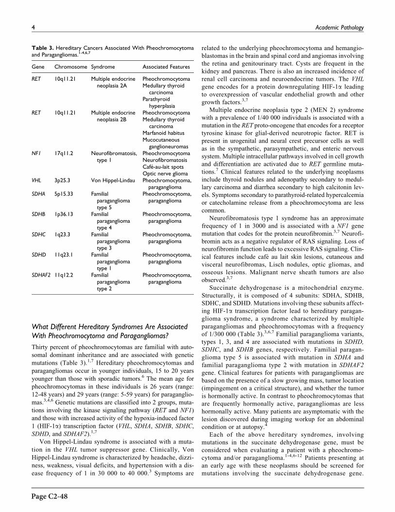
What Different Hereditary Syndromes Are AssociatedWith Pheochromocytoma and Paragangliomas?
Thirty percent of pheochromocytomas are familial with auto-
somal dominant inheritance and are associated with genetic
mutations (Table 3).1,7 Hereditary pheochromocytomas and
paragangliomas occur in younger individuals, 15 to 20 years
younger than those with sporadic tumors.6 The mean age for
pheochromocytomas in these individuals is 26 years (range:
12-48 years) and 29 years (range: 5-59 years) for paraganglio-
mas.3,4,6 Genetic mutations are classified into 2 groups, muta-
tions involving the kinase signaling pathway (RET and NF1)
and those with increased activity of the hypoxia-induced factor
1 (HIF-1a) transcription factor (VHL, SDHA, SDHB, SDHC,
SDHD, and SDHAF2).1,7
Von Hippel-Lindau syndrome is associated with a muta-
tion in the VHL tumor suppressor gene. Clinically, Von
Hippel-Lindau syndrome is characterized by headache, dizzi-
ness, weakness, visual deficits, and hypertension with a dis-
ease frequency of 1 in 30 000 to 40 000.3 Symptoms are
related to the underlying pheochromocytoma and hemangio-
blastomas in the brain and spinal cord and angiomas involving
the retina and genitourinary tract. Cysts are frequent in the
kidney and pancreas. There is also an increased incidence of
renal cell carcinoma and neuroendocrine tumors. The VHL
gene encodes for a protein downregulating HIF-1a leading
to overexpression of vascular endothelial growth and other
growth factors.3,7
Multiple endocrine neoplasia type 2 (MEN 2) syndrome
with a prevalence of 1/40 000 individuals is associated with a
mutation in the RET proto-oncogene that encodes for a receptor
tyrosine kinase for glial-derived neurotropic factor. RET is
present in urogenital and neural crest precursor cells as well
as in the sympathetic, parasympathetic, and enteric nervous
system. Multiple intracellular pathways involved in cell growth
and differentiation are activated due to RET germline muta-
tions.7 Clinical features related to the underlying neoplasms
include thyroid nodules and adenopathy secondary to medul-
lary carcinoma and diarrhea secondary to high calcitonin lev-
els. Symptoms secondary to parathyroid-related hypercalcemia
or catecholamine release from a pheochromocytoma are less
common.
Neurofibromatosis type 1 syndrome has an approximate
frequency of 1 in 3000 and is associated with a NF1 gene
mutation that codes for the protein neurofibromin.3,7 Neurofi-
bromin acts as a negative regulator of RAS signaling. Loss of
neurofibromin function leads to excessive RAS signaling. Clin-
ical features include cafe au lait skin lesions, cutaneous and
visceral neurofibromas, Lisch nodules, optic gliomas, and
osseous lesions. Malignant nerve sheath tumors are also
observed.3,7
Succinate dehydrogenase is a mitochondrial enzyme.
Structurally, it is composed of 4 subunits: SDHA, SDHB,
SDHC, and SDHD. Mutations involving these subunits affect-
ing HIF-1a transcription factor lead to hereditary paragan-
glioma syndrome, a syndrome characterized by multiple
paragangliomas and pheochromocytomas with a frequency
of 1/300 000 (Table 3).3,6,7 Familial paraganglioma variants,
types 1, 3, and 4 are associated with mutations in SDHD,
SDHC, and SDHB genes, respectively. Familial paragan-
glioma type 5 is associated with mutation in SDHA and
familial paraganglioma type 2 with mutation in SDHAF2
gene. Clinical features for patients with paragangliomas are
based on the presence of a slow growing mass, tumor location
(impingement on a critical structure), and whether the tumor
is hormonally active. In contrast to pheochromocytomas that
are frequently hormonally active, paragangliomas are less
hormonally active. Many patients are asymptomatic with the
lesion discovered during imaging workup for an abdominal
condition or at autopsy.4
Each of the above hereditary syndromes, involving
mutations in the succinate dehydrogenase gene, must be
considered when evaluating a patient with a pheochromo-
cytoma and/or paraganglioma.1-4,6-12 Patients presenting at
an early age with these neoplasms should be screened for
mutations involving the succinate dehydrogenase gene.
Table 3. Hereditary Cancers Associated With Pheochromocytomaand Paragangliomas.1-4,6,7
Gene Chromosome Syndrome Associated Features
RET 10q11.21 Multiple endocrineneoplasia 2A
PheochromocytomaMedullary thyroid
carcinomaParathyroid
hyperplasiaRET 10q11.21 Multiple endocrine
neoplasia 2BPheochromocytomaMedullary thyroid
carcinomaMarfanoid habitusMucocutaneous
ganglioneuromasNF1 17q11.2 Neurofibromatosis,
type 1PheochromocytomaNeurofibromatosisCafe-au-lait spotsOptic nerve glioma
VHL 3p25.3 Von Hippel-Lindau Pheochromocytoma,paraganglioma
SDHA 5p15.33 Familialparagangliomatype 5
Pheochromocytoma,paraganglioma
SDHB 1p36.13 Familialparagangliomatype 4
Pheochromocytoma,paraganglioma
SDHC 1q23.3 Familialparagangliomatype 3
Pheochromocytoma,paraganglioma
SDHD 11q23.1 Familialparagangliomatype 1
Pheochromocytoma,paraganglioma
SDHAF2 11q12.2 Familialparagangliomatype 2
Pheochromocytoma,paraganglioma
4 Academic Pathology
Page C2-48

Immunohistochemistry for SDHB has been used to screen
for succinate dehydrogenase mutations.2
What Is the Rule of 10s?
Historically, the rule of 10s was applied to pheochromocyto-
mas. The 10% rule outlined that 10% of pheochromocytomas
(paragangliomas) were extra-adrenal, 10% of pheochromocy-
tomas were bilateral, 10% were malignant, 10% were not asso-
ciated with hypertension, and 10% occurred in children.1 The
10% rule is called into question because tumor location (adre-
nal vs extra-adrenal) and likelihood of malignancy vary based
on genetic mutations. Genetic involvement in tumors is closer
to 30%. Fifty percent of familial pheochromocytomas are bilat-
eral. Twenty to forty percent of paragangliomas are malignant
and malignancy is more common in chromaffin tumors asso-
ciated with germline mutations.1-4,6,7
Teaching Points
� A pheochromocytoma should be considered when eval-
uating an adrenal mass.
� The triad of headache, sweating, and heart palpitations
should raise suspicion of a pheochromocytoma, espe-
cially when concurrent hypertension exists.
� Diagnosis of a pheochromocytoma is based on patholo-
gic findings and is supported by an abnormal 24-hour
urine fractionated metanephrine and catecholamine test.
� Pheochromocytomas are catecholamine-secreting
tumors that originate in the adrenal medulla.
� Pheochromocytoma complications from catecholamine
secretion include congestive heart failure, pulmonary
edema, myocardial infarction, ventricular fibrillation,
cerebrovascular accidents, and catecholamine
cardiomyopathy.
� Paragangliomas are catecholamine-secreting tumors that
arise from the autonomic nervous system ganglia that
represent the cell bodies of postsynaptic neurons. Para-
gangliomas affect both the sympathetic and parasympa-
thetic chains of the autonomic nervous system.
� Sign and symptoms associated with paragangliomas are
based on tumor location, rate of growth, and whether
they are hormonally active.
� Thirty percent of pheochromocytomas are familial with
autosomal dominant inheritance and are associated with
genetic mutations. Genetic mutations are classified into
2 groups, mutations involving the kinase signaling path-
way (RET and NF1) and those with increased activity of
the HIF-1a transcription factor (VHL, SDHA, SDHB,
SDHC, SDHD, and SDHAF2).
� The diagnosis of a pheochromocytoma at a young age
warrants clinical and genetic testing for MEN2 syn-
drome, Von Hippel-Lindau syndrome, neurofibromato-
sis 1, and familial paraganglioma–pheochromocytoma
syndromes.
� Malignant behavior absent metastatic disease is difficult
to predict in these neuroendocrine tumors. Histology in
combination with the following factors (cellularity,
necrosis, capsular or vascular invasion, type of catecho-
lamine secreted, and MIB-1 immunoreactivity) is pro-
posed to classify tumors from well differentiated to
poorly differentiated.
Acknowledgment
Images were obtained during the scope of US government employ-
ment for Dr Conran.
Declaration of Conflicting Interests
The author(s) declared no potential conflicts of interest with respect to
the research, authorship, and/or publication of this article.
Funding
The author(s) received no financial support for the research, author-
ship, and/or publication of this article.
References
1. Maitra A The endocrine system. In: Kumar V, Abbas AK, Aster
JC, eds. Robbins and Cotran Pathologic Basis of Disease.
Philadelphia, PA: Elsevier; 2015:1134-1137.
2. McKenney JK Adrenal gland and other paraganglia. In: Gold-
blum JR, Lamps LW, McKenney JK, Myers JL, eds. Rosai and
Ackerman’s Surgical Pathology. Philadelphia, PA; Elsevier;
2018:1205-1207.
3. DeLellis RA, Lloyd RV, Heitz PU, Eng C, eds. World Health
Organization Classification of Tumours. Pathology and Genetics:
Tumors of Endocrine Organs. Lyon, France: IARC Press; 2004.
4. McNicol AM, YoungWF, Kawashima A, Komminoth P, Tischler
AS Benign phaeochromocytoma. In: DeLellis RA, Lloyd RV,
Heitz PU, Eng C, eds. World Health Organization Classification
of Tumours. Pathology and Genetics: Tumors of Endocrine
Organs. Lyon, France: IARC Press; 2004:151-155.
5. Arnold D, Reed JB, Burt K. Evaluation and management of the
incidental adrenal mass. Proc (Bayl Univ Med Cent). 2003;16:
7-12.
6. Nathanson K, Baysai BE, Drovdlic C, Komminoth P, Neumann
HP. Familial paraganglioma-phaeochromocytoma syndromes
caused by SDHB, SDHC and SDHD mutations. In: DeLellis
RA, Lloyd RV, Heitz PU, Eng C, eds.World Health Organization
Classification of Tumours. Pathology and Genetics: Tumors of
Endocrine Organs. Lyon, France: IARC Press; 2004;4:238-242.
7. Welander J, Sodwerkvist P, Gimm O. Genetics and clinical char-
acteristics of hereditary pheochromocytomas and paraganglio-
mas. Endocr Relat Cancer. 2011;18:R253-R276
8. Tischler AS. Pheochromocytoma and extra-adrenal paragan-
glioma: updates. Arch Pathol Lab Med. 2008;132:1272-1284.
9. Pawlu C, Bausch B, Reisch N, Neumann HP. Genetic testing for
pheochromocytoma-associated syndromes. Ann Endocrinol
(Paris). 2005;66:178-185.
Westover and Conran 5
Page C2-49

10. Shuch B, Ricketts CJ, Metwalli AR, Pacak K, Linehan WM. The
genetic basis of pheochromocytoma and paraganglioma: implica-
tions for management. Urology. 2014;83:1225-1232.
11. US National Library of Medicine. Genetics home reference.
https://ghr.nlm.nih.gov/condition?initial¼p. Accessed November
30, 2017.
12. Kirmani A, Young WF. Hereditary paraganglioma-pheochromo-
cytoma syndromes. In: Adam MP, Ardinger HH, Pagon RA, et al,
eds. Gene Reviews [Internet], Seattle, WA: University of
Washington. https://www.ncbi.nlm.nih.gov/books/NBK1548/.
Published May 21, 2008. Updated November 6, 2014. Accessed
November 30, 2017.
6 Academic Pathology
Page C2-50

Educational Case
Educational Case: Iron Overloadand Hemochromatosis
Michael J. Borowitz, MD, PhD1 and Alison Moliterno, MD1
The following fictional case is intended as a learning tool within the Pathology Competencies forMedical Education (PCME),
a set of national standards for teaching pathology. These are divided into three basic competencies: Disease Mechanisms and
Processes, Organ System Pathology, and Diagnostic Medicine and Therapeutic Pathology. For additional information, and
a full list of learning objectives for all three competencies, see http://journals.sagepub.com/doi/10.1177/2374289517715040.
Keywordspathology competencies, organ system pathology, hematopathology, anemia, iron overload, hemochromatosis, genetic mechan-isms, inheritance patterns
Received February 13, 2017. Received revised April 16, 2018. Accepted for publication April 30, 2018.
Primary Pathology Learning Objective
Objective HRC1.3: Hepcidin Regulation, Iron Overload, and
Anemia of Chronic Disease: Discuss the role of hepcidin as an
iron regulator and describe how different types of alterations in
the hepcidin pathway can produce anemia of chronic disease or
iron overload.
Competency 2: Organ System Pathology; Topic HRC:
Hematopathology - Red Cell Disorders; Learning Goal 1:
Anemia.
Secondary Pathology Learning Objective
Objective GM1.2: Inheritance Patterns: Compare and contrast
the inheritance patterns of different types of Mendelian disor-
ders and give examples of each type of pattern.
Competency 1: Disease Mechanisms and Processes; Topic
GM: Genetic Mechanisms; Learning Goal 1: Genetic Mechan-
isms of Developmental and Functional Abnormalities.
Patient Presentation
A 50-year-old Caucasian male presented for evaluation con-
cerned that he may be at risk of developing hemochroma-
tosis based on his family history. His father presented at age
60 with bronzed skin and diabetes; he was found to have
iron overload and subsequently has been undergoing thera-
peutic phlebotomies. The patient’s medical history is unre-
markable with the exception of hypercholesterolemia, for
1 Johns Hopkins School of Medicine, Baltimore, MD, USA
Corresponding Author:
Michael J. Borowitz, Johns Hopkins School of Medicine, 401 N Broadway,
Baltimore, MD 21287, USA.
Email: [email protected]
Table 1. Iron and Genetic Studies of the Patient and His Father.
Lab TestFather—Age
60Patient—Age
50 Normal Range
Serum iron 232 106 65-170 mg/dLTransferrin 284 230 200-400 mg/dLTotal iron bindingcapacity
355 288 250-450 mg/dL
% saturation 65 37 20%-55%Ferritin 1518 217 10-300 ng/mLHFE genotype C282Y Homozygous Heterozygous
Academic PathologyVolume 5: 1–3ª The Author(s) 2018Reprints and permission:sagepub.com/journalsPermissions.navDOI: 10.1177/2374289518779944journals.sagepub.com/home/apc
Creative Commons Non Commercial CC BY-NC: This article is distributed under the terms of the Creative Commons Attribution-NonCommercial 4.0 License(http://www.creativecommons.org/licenses/by-nc/4.0/) which permits non-commercial use, reproduction and distribution of the work without furtherpermission provided the original work is attributed as specified on the SAGE and Open Access pages (https://us.sagepub.com/en-us/nam/open-access-at-sage).
Page C2-51

which he takes a statin. You decide to do some diagnostic
blood work. In addition, the patient has provided you with
results of blood work from his father.
Diagnostic Findings, Part 1
Results of iron studies and HFE studies are presented in
Table 1.
Questions/Discussion Points, Part 1
Interpret the Father’s and the Patient’s Iron Studies
The father has elevated serum iron associated with increased
transferrin saturation and an elevated ferritin indicative of both
increased iron absorption and iron storage. The son has normal
iron studies.
Do the Patient’s Iron Studies Support the Diagnosisof Hemochromatosis?
No. He does not have evidence of either increased absorption or
increased iron stores. Additionally, he is only a carrier of the
HFE mutation, making it highly unlikely that he has a genetic
predisposition to iron overload.
Ferritin and Serum Iron Reflect Different Aspects ofan Individual’s Iron Metabolism. What Is the MajorDifference in the Type of Information Obtained FromEach of These?
Ferritin is a measure of long-term iron storage, while serum
iron (and transferrin saturation) better reflect daily iron absorp-
tion. Interpretation of ferritin testing can be misleading with
only a single measurement because it is an acute phase reactant.
Diagnostic Findings, Part 2
After obtaining the results above, the patient consults his
brother and recommends that he also undergo testing. Results
from his brother, again in comparison to those of his father, are
shown in Table 2.
Questions/Discussion Points, Part 2
Does His Brother Have Iron Overload? Why Is HisBrother’s Ferritin So Much Lower Than His Father’s?
Yes, he has evidence of iron overload. He is younger than his
father was at the time of his iron studies, and the degree of iron
overload is a function of time.
Should Other Siblings Be Tested? What Would Be theBest Approach for Further Molecular Testing? If You HadHad the Opportunity to Evaluate the Father at the FirstSign of Potential Iron Overload, What Testing Would YouHave Done on Him?
Taking the brother’s and father’s genotypes together (and
assuming the brothers have the same father!), mother must be
a carrier, and other children have a 50% chance of being homo-
zygotes also. HFE C282Y homozygotes in general will have an
increased risk of significant iron overload. However, because
there are significant modifiers of iron absorption beyond the
HFE gene (age, gender, diet, and polymorphisms in other genes
affecting iron absorption), not all homozygotes will have iron
overload.1 In this family, however, homozygotes do appear to
have an increased risk of iron overload, so they should be
identified. Because we know the mutation in this case, targeted
mutation analysis for HFE C282Y is the only analysis needed.
If you had seen the father early in his course and had had no
family history of hemochromatosis, it would be appropriate to
do targeted analysis for both the HFE C282Y and H63D muta-
tions, and if these were negative, sequence analysis could be
used to identify other less common mutant alleles associated
with HFE.
Which Genes Can Cause Hemochromatosis WhenMutated? Describe the Inheritance Patterns Seen
The HFE C282Y on chromosome 6p22.2, as present in this
case, is the most common mutation seen in classic (type 1 or
HFE1) hemochromatosis and is associated with autosomal
recessive inheritance. This mutation accounts for >80% of
hemochromatosis cases.2,3 Another common mutation in HFE
(H63D) may be seen as a compound heterozygous (C282Y/
H63D) genotype in some patients with hemochromatosis;
patients with homozygous H63D rarely have clinically signif-
icant iron overload.2,3 Four additional iron overload disorders
labeled hemochromatosis have been identified2:
Juvenile hemochromatosis is the term given to clinically
similar autosomal recessive diseases caused by muta-
tions in 2 different genes:
HFE2A: the hemojuvelin (HJV) gene on chromosome
1q21.
HFE2B: the hepcidin (HAMP) gene on chromosome
19q13.
Table 2. Iron and Genetic Studies of the Father and the Brother ofthe Patient.
Lab TestFather—Age
60Brother—Age
46 Normal Range
Serum iron 232 176 65-170 mg/dLTransferrin 284 230 200-400 mg/dLTotal iron bindingcapacity
355 266 250-450 mg/dL
% saturation 65 65 20%-55%Ferritin 1518 611 10-300 ng/mLHFE genotypeC282Y
Homozygous Homozygous
2 Academic Pathology
Page C2-52

HFE3, also autosomal recessive, is caused by a mutation
in transferrin receptor 2 gene on chromosome 7q22.
HFE4, is autosomal dominant and caused by a mutation
in SLC40A1 gene on chromosome 2q32.
Discuss the Pathophysiology of How Iron OverloadOccurs in Hemochromatosis
Patients with hemochromatosis have in common low hepcidin
levels. Hepcidin normally binds ferroportin in both duodenal
enterocytes and reticuloendothelial macrophages, which in turn
blocks release of iron from these cells. Thus, hepcidin serves to
keep iron frombeingabsorbed fromthegut orbeing released from
storage macrophages back into circulation. In the absence of
hepcidin, this process is reversed, with increased iron absorption
and increased turnover of iron from reticuloendothelial macro-
phages leading to high levels of both serum ferritin and saturation
of transferrin and ultimately to iron deposition in tissues.3
Teaching Points
� Iron overload can be suspected from serum iron studies,
in particular elevated serum iron and transferrin, and
increased transferrin saturation.
� In the absence of a historical explanation for iron over-
load (such as multiple blood transfusions), patients with
iron overload should be investigated for genetic disor-
ders, and in particular for hereditary hemochromatosis.
� If hereditary hemochromatosis is identified, family
members should be tested for evidence of iron overload
as well.
� Other factors modify the effects of the abnormal gene so
that affected family members may vary significantly in
their clinical and laboratory presentations.
� The most common form of hereditary hemochromatosis
is called type 1 and is due to a mutation in theHFE gene.
It is inherited as an autosomal recessive.
Declaration of Conflicting Interests
The author(s) declared no potential conflicts of interest with respect to
the research, authorship, and/or publication of this article.
Funding
The author(s) received no financial support for the research, author-
ship, and/or publication of this article.
References
1. Beutler E. Iron storage disease: facts, fiction and progress. Blood
Cells Mol Dis. 2007;39:140-147.
2. Online Mendelian Inheritance in Man (OMIM). 2018. http://www.
omim.org/entry/235200. Accessed April 14, 2018.
3. Hollerer I, Bachman A, Muckenthaler MU. Pathophysiologic
consequences and benefits of HFE mutations: 20 years of
research. Haematologica. 2017;102:809-817. PMID 28280078.
doi:10.33324/haematol.2016.160432.
Borowitz and Moliterno 3
Page C2-53


Educational Case
Educational Case: Thyroid Neoplasms:Pathogenesis, Diagnosis, and Treatment
Gloria Ramos Rivera, MD1, Sheila Segura, MD1, and Mark Suhrland, MD1
The following fictional case is intended as a learning tool within the Pathology Competencies for Medical Education
(PCME), a set of national standards for teaching pathology. These are divided into three basic competencies: Disease
Mechanisms and Processes, Organ System Pathology, and Diagnostic Medicine and Therapeutic Pathology. For additional
information, and a full list of learning objectives for all three competencies, see http://journals.sagepub.com/doi/10.1177/
2374289517715040.
Keywordspathology competencies, organ system pathology, endocrine, follicular adenoma, follicular carcinoma, papillary thyroid carcinoma,thyroid neoplasms
Received January 16, 2018. Received revised March 29, 2018. Accepted for publication April 5, 2018.
Primary Objectives
Objective EN5.1: Thyroid Neoplasms. Compare and contrast
the clinicopathologic features of follicular adenomas, follicular
carcinoma, and papillary thyroid carcinoma.
Competency 2: Organ System Pathology; Topic EN: Endo-
crine; Learning Goal 5: Endocrine Neoplasms.
Patient Presentation, 1
A 30-year-old female presents with a palpable right thyroid
nodule. The nodule was first noticed 3 months ago and has
increased in size slightly over time. The patient denies having
hoarseness, dysphagia, weight changes, intolerance to cold or
hot weather, drowsiness, or palpitations. A serum thyroid-
stimulating hormone (TSH) test was performed, and it was
within normal limits. Ultrasonography (US) of the thyroid
revealed a solitary well-circumscribed nodule on the right
lower lobe of the thyroid measuring 2.0 � 1.5 � 1.0 cm.
Diagnostic Findings, Part 1
What Is Your Differential Diagnosis Based on the ClinicalHistory?
Palpable thyroid nodules can be found in 5% of women and 1%of men.1 The prevalence can increase to 20% to 70% if
nonpalpable nodules are included, and these are usually
detected by ultrasound or autopsy.2 Around 7% to 15% of all
nodules are thyroid cancers1; therefore, thyroid nodules should
be evaluated accordingly. The differential diagnosis for a thyr-
oid solitary nodule with a normal TSH in decreasing order of
frequency is hyperplastic nodule, follicular adenoma, papillary
thyroid carcinoma (PTC), and follicular carcinoma. Other neo-
plasms are much less frequent.
Questions/Discussion Points, Part 1
What Testing Is Available for This Patient and Which IsRecommended?
As per the 2015 American Thyroid Association Manage-
ment Guidelines, nodules greater than 1 cm should be fur-
ther evaluated.1 Serum TSH levels can be measured. If the
TSH of the patient is low, a radionuclide (preferably 123I)
thyroid scan should be performed to document whether the
1 Montefiore Hospital and Medical Center, Bronx, NY, USA
Corresponding Author:
Gloria Ramos Rivera, Montefiore Hospital and Medical Center, Bronx, NY,
USA.
Email: [email protected]
Academic PathologyVolume 5: 1–5ª The Author(s) 2018Article reuse guidelines:sagepub.com/journals-permissionsDOI: 10.1177/2374289518777471journals.sagepub.com/home/apc
Creative Commons Non Commercial CC BY-NC: This article is distributed under the terms of the Creative Commons Attribution-NonCommercial 4.0 License(http://www.creativecommons.org/licenses/by-nc/4.0/) which permits non-commercial use, reproduction and distribution of the work without furtherpermission provided the original work is attributed as specified on the SAGE and Open Access pages (https://us.sagepub.com/en-us/nam/open-access-at-sage).
Page C2-55

nodule is hyperfunctioning (“hot”), isofunctioning
(“warm”), or nonfunctioning (“cold”).1 Hyperfunctioning
nodules rarely harbor malignancy.1
Thyroid US is considered the imaging modality of choice
for the investigation of thyroid nodules. Several features are
assessed with the US, including number, size, margin, contours
and shape of the nodule, cystic or solid nodule, vascularity, and
presence of calcifications, among others. Individual US fea-
tures may have limited value distinguishing between benign
and malignant thyroid nodules, but when multiple signs of
malignancy appear in combination, it is possible to make an
accurate prediction. Some of the US features suggestive of
malignancy are ill-defined nodule, irregular margins, hypervas-
cularity, microcalcifications or coarse calcifications, solid and
hypoechoic nodule, and invasion of surrounding tissue.3
A fine needle aspiration (FNA) biopsy will be the pro-
cedure of choice in the evaluation of a thyroid nodule larger
than 1 cm.1 Nodules smaller than 1 cm are not usually
biopsied unless they have sonographically suspicious fea-
tures or other risk factors for malignancy, such as history
of thyroid cancer and radiation exposure, among others.2
Fine needle aspiration is the most cost-effective and least
invasive procedure that will help guide the management and
treatment of a patient. Fine needle aspiration can be per-
formed either with palpation or under ultrasound guidance,
depending on the location of the nodule or how difficult it is
to palpate or properly fix the target.
Diagnostic Findings, Part 2
The patient underwent an US-guided thyroid FNA biopsy.
Microscopic examination of the smears stained by Papani-
colaou method showed clusters and papillary fragments of
follicular cells with nuclear enlargement, pale nuclei, pow-
dery chromatin, nuclear pseudoinclusions, and nuclear
grooves (Figure 1).
Based on the Cytological Findings, What Is the CorrectDiagnosis?
The smears showed the classic cytologic features of a PTC,
malignant (Bethesda VI). Since the risk of malignancy for this
lesion is 94% to 99%, a near total thyroidectomy or lobectomy
is indicated for this patient.
What Is the Bethesda System and How This Can Aidin the Management and Treatment of the Patient?
The Bethesda System for Reporting Thyroid Cytology
(BSRTC) is a set of recommendations on how to report thyroid
cytology specimens in diagnostic categories with associated
risk of malignancy and patient management (Table 14). It is a
consensus on terminology and morphologic criteria that aims to
facilitate communication among the different disciplines in
clinical medicine and pathology.
Using the BSRTC, a diagnosis of malignancy (PTC) on
FNA is made whenever the cytomorphologic features are con-
clusive for malignancy. The criteria for reporting PTC are fol-
licular cells arranged in papillae (fibrovascular cores) or
syncytial monolayers with the characteristic nuclear features
such as enlarged oval or irregular nuclei, nuclear crowding,
longitudinal nuclear grooves, intranuclear cytoplasmic pseu-
doinclusions, pale nuclei with powdery chromatin, and psam-
moma bodies (not always present).
Questions/Discussion Points, Part 2
What Are the Clinical Presentation and Biologic Behaviorof PTC?
Papillary thyroid carcinoma usually presents as a solitary, pain-
less nodule, and around 30% of patients might also have asso-
ciated lymphadenopathy.5 It tends to grow very slowly and
usually involves only 1 thyroid lobe. In large tumors, the pre-
sentation will include dysphagia, stridor, and cough.5 In some
cases, these neoplasms are discovered incidentally during the
workup of a different problem.5
Papillary thyroid carcinoma is the most common thyroid
cancer and accounts for approximately 80% of malignant neo-
plasms of the thyroid with a female to male ratio of 4:1.2,5 It is
associated with radiation exposure and its incidence is higher in
areas with high iodine intake.5
Describe the Pathological Features of PTC
The gross pathology is variable fromwell-circumscribed nodule to
diffuse involvement of the lobe, or multifocal.5 The cut surface is
white-gray, firm, and granular with possible presence of calcifica-
tions.5 The microscopic histologic features are similar to the
Figure 1. A and B, Cytology of thyroid fine needle aspiration (FNA)showing papillary thyroid carcinoma (PTC) nuclear changes: grooves(arrowhead), intranuclear pseudoinclusions (arrow), nucleoli, and pow-dery chromatin, as well as nuclear crowding and nuclear enlargement(A, PAP stained, high-power 60� magnification, B, Diff quick-stainedintermediate power 40� magnification). C, A papillary formation isobserved. (diff quick stained, low power 10� magnification).
2 Academic Pathology
Page C2-56

cytologic features. The papillary structures are more evident and
contain complex branching true papillae with fibrovascular cores.5
The neoplastic cellswill show the classic chromatin clearing called
“Orphan Annie eye” nuclei (Figure 2A).5 There are different var-
iants of PTC (classic or usual, microcarcinoma, follicular variant,
diffuse sclerosing variant, cribriform-morular variant, oncocytic
variant, tall cell variant, columnar cell variant) and their micro-
scopic characteristics will vary slightly from the classic PTC;
however, they must present the characteristic nuclear features
(Figure 2B).5 The classic or usual variant is the most common
presentation of PTC, representing *75% to 80% of cases.
What Genetic Mutations Does This Neoplasm Harbor?
Papillary thyroid cancers frequently have genetic mutations and
rearrangements that lead to activation of the mitogen-activated
protein kinase that promotes cell division.6 In PTC, the most
commonly identified mutations in this pathway are point muta-
tions of the BRAF and RAS genes and RET/PTC rearrangement,
found in >70% of cases.6 The most common genetic alteration
is the BRAF point mutation (V600E), identified in *45% of
PTC cases.
Genetic alterations involving the tyrosine kinase signaling
pathways (RET/RAS/RAF pathway) are interconnected with the
epidermal growth factor receptor activation cascade, which
leads to the syntheses of vascular endothelial growth factor
(VEGF) and VEGF receptor, which has also been found in
PTC, particularly in tumors with BRAF mutations. Drugs tar-
geting these pathways could play a significant role in control-
ling the progression of the disease.
How Is Molecular Testing Useful in Managementof Thyroid Nodules?
Approximately 10% to 15% of thyroid nodules on FNA fall
into the indeterminate category which includes follicular and
oncocytic neoplasms (Bethesda IV) and atypia of undetermined
significance/follicular lesion of undetermined significance
(Bethesda III), according to the most current categorization.4
For these cases, molecular testing is recommended for man-
agement since thyroid cancers are commonly associated with
specific molecular alterations.
What Are the Treatment Options for Thyroid Cancer?
Treatment options for thyroid cancer include surgery, radio-
active iodine (131I) ablation (RAI), molecular-targeted thera-
pies with several tyrosine kinase inhibitors, and external
beam radiation. Surgery is the treatment of choice; however,
the extent of the surgery is still controversial (total thyroi-
dectomy vs near total thyroidectomy vs. lobectomy).5
Lymph node sampling can be performed if there is clinical
or radiographic enlargement. The most common complica-
tions after surgery are damage to the recurrent laryngeal
nerve and hypoparathyroidism. The postoperative disease
status can be evaluated with postoperative serum thyroglo-
bulin and/or ultrasound.1
Radioactive iodine is used in coordination with thyroidect-
omy to completely ablate the thyroid gland and to eradicate
possible residual cancer. The first-dose administration is
referred to as ablation, whereas subsequent administrations for
residual disease is referred to as treatment.3
Tyrosine kinase inhibitors, which primarily target angio-
genesis (specifically VEGF receptor signaling pathways), are
used to treat patients with recurrent or metastatic thyroid can-
cers who do not respond to RAI and TSH-suppressive
Figure 2. Histology of papillary thyroid carcinoma (PTC) showingnuclear changes: chromatin clearing known as “Orphan Annie eye”nuclei (asterisk), grooves (arrow head), intranuclear pseudoinclusions(arrow), nucleoli, powdery chromatin, crowding, and overlapping(H&E stained, high power 60� magnification).
Table 1. The Bethesda System for Reporting Thyroid Cytology Diagnostic Categories.4
Diagnostic CategoryRisk of
Malignancy (%) Management
I Nondiagnostic or Unsatisfactory 5-10 Repeat FNA with ultrasound guidanceII Benign 0-3 Clinical and sonographic follow-upIII Atypia of uncertain significance or Follicular lesion of uncertain significance *10-30 Repeat FNA, molecular testing, or lobectomyIV Follicular neoplasm or Suspicious for a follicular neoplasm 25-40 Molecular testing, lobectomyV Suspicious for malignancy 50-75 Near total thyroidectomy or lobectomyVI Malignant 97-99 Near total thyroidectomy or lobectomy
Rivera et al 3
Page C2-57

hormone therapy.3,7 Two drugs, Sorafenib and Lenvatinib, are
approved by the Food and Drug Administration for use in
selected patients with refractory metastatic differentiated
thyroid cancers.
External beam radiation therapy is only used for palliative
treatment of patients with advanced or inoperable thyroid can-
cer and it may be administered in the adjuvant setting, after
surgical resection, or as primary treatment. It is usually con-
sidered in patients over 45 years of age who have grossly
visible extrathyroidal extension and a high likelihood of resi-
dual disease during surgery, and it is also reserved for tumors
that are unresponsive to RAI.3
What Is the Prognosis of Patients With PTC?
In general, PTC is indolent and has an excellent prognosis.2,5
The 10-year survival rate is greater than 90%.2 Although the
prognosis is excellent, certain clinical and pathologic features
have been identified that portend a somewhat higher risk of
tumor recurrence and cancer-related mortality, including over
45 years of age, male gender, size of carcinoma being greater
than 2 cm3, invasion into surrounding tissue, histological sub-
type, and distant metastasis. It spreads mostly by lymphatic
channels and metastasis to lymph nodes is frequent; however,
this does not affect prognosis.5
The histological subtypes of PTC associated with worse
prognosis are tall cell, insular, and hobnail variants. Patients
with aggressive histologic subtypes are usually treated more
aggressively.8 In addition to its strong correlation with PTC,
the BRAF V600E mutation has also been associated with poor
prognosis and higher recurrence rate.
Patient Presentation, 2
A 55-year-old woman presents with a painless, slowly enlarging
left thyroid mass. A previous FNA of the lesion was reported as
suspicious for a follicular neoplasm (Bethesda IV). Following
this diagnosis, the patient underwent a total thyroidectomy.
Diagnostic Findings, Patient 2
On gross examination, there is a solitary encapsulated mass,
measuring 3 cm in greatest dimension. Microscopically, the
tumor is surrounded with a capsule (encapsulated) and shows
a microfollicular pattern. Upon thorough examination of the
capsule, an area where the tumor cells penetrate through the
whole thickness of the capsule was found. This is referred as
capsular invasion; therefore, a diagnosis of follicular carci-
noma was reported.
Questions/Discussion Points, Patient 2
What Are the Cytologic and Pathologic Featuresof Follicular Carcinoma?
The cytomorphology on FNA specimens will show a marked
cellularity, consisting predominantly of small clusters of
Figure 3. Cytology of thyroid fine needle aspiration (FNA) showingmicrofollicles of a follicular neoplasm (PAP stained, low power10 � magnification). Papillary formations as seen here are composedof follicular cells surrounding a fibrovascular core.
Figure 4. A, Histology of follicular adenoma showing a fibrous capsule(asterisk) and a microfollicular pattern on left side of image (H&Estained, intermediate power 40� magnification). B, Evident capsularinvasion (arrow) is present in follicular carcinoma. Capsule is shownwith asterisks (H&E stained, intermediate power 40� magnification).
4 Academic Pathology
Page C2-58

follicular cells called microfollicles, with scant colloid in the
background (Figure 3). The cytology of adenoma and carci-
noma can be almost identical, so they are both diagnosed as
suspicious for a follicular neoplasm (Bethesda IV).
The distinction between the 2 is made by examining the
capsule for invasion after resection. The resection specimen
of follicular adenoma and carcinoma will show a fibrous
capsule grossly and a microfollicular pattern microscopically
(Figure 4A).5 These lesions will lack the characteristic nuclear
features of PTC.5 To differentiate a follicular adenoma from a
carcinoma, evident capsular invasion must be present (Figure
4B). This distinction is only diagnosed on the resected speci-
men, and thorough examination of the capsule is required. In
follicular carcinomas, the most common alterations include
RAS mutations and PAX8-PPARg rearrangement.6 Many of
these mutations are being explored as therapeutic targets for
thyroid cancer.6
Follicular adenomas are treated with lobectomy and have
an excellent long-term prognosis.5 The treatment of choice for
follicular carcinoma is surgery (lobectomy or thyroidectomy).
A lobectomy is more likely to be performed in younger
patients (<45 years), with a single nodule (<40 mm), and
without thyroiditis or metastatic disease.5 Radioactive iodine
therapy can be indicated in patients with lymph node metas-
tasis who underwent total thyroidectomy. Follicular carci-
noma has an excellent long-term prognosis on minimally
invasive lesions, 97% 20-year survival and a 50% 20-year
survival on widely invasive lesions.5
Teaching Points
� Thyroid nodules are a common finding, and only 7% to
15% are malignant.
� Papillary thyroid carcinoma is the most common thyroid
malignancy and presents with characteristic nuclear fea-
tures on microscopic examination.
� The BSRTC is a guideline to create uniformity in report-
ing thyroid lesions on FNA.
� Most common mutation of PTCs is the point mutations
of the BRAF gene.
� To differentiate a follicular adenoma from a follicular
carcinoma, a thorough examination of the capsule
should be done to assess capsular invasion.
Declaration of Conflicting Interests
The author(s) declared no potential conflicts of interest with respect to
the research, authorship, and/or publication of this article.
Funding
The author(s) received no financial support for the research, author-
ship, and/or publication of this article.
References
1. Haugen BR, Alexandar EK, Bible CK, et al. 2015 American Thyroid
Association Management Guidelines for Adult Patients with Thyr-
oid Nodules and Differentiated Thyroid Cancer: The American
Thyroid Association Guidelines Task Force on Thyroid Nodules and
Differentiated Thyroid Cancer. Thyroid. 2016;26:1-133.
2. Cibas ES, Ducatman BS. Cytology: Diagnostic Principles and
Clinical Correlates. 4th ed. Philadelphia, PA: Elsevier/Saunders;
2014.
3. Nguyen QT, Lee EJ, Huang MG, Park YI, Khullar A, Plodkowski
RA. Diagnosis and treatment of patients with thyroid cancer. Am
Health Drug Benefits. 2015;8:30-40.
4. Cibas ES, Syed AZ. The 2017 Bethesda System for reporting thyr-
oid cytopathology. Thyroid. 2017;27:1341-1346.
5. Thompson LD, Wenig BM.Diagnostic Pathology, Head and Neck.
2nd ed. Philadelphia, PA: Elsevier; 2016.
6. Nikiforov YE. Thyroid carcinoma: molecular pathways and ther-
apeutic targets. Modern pathology.Mod Pathol. 2008;21:S37-S43.
7. Sherman S.Differentiated thyroid cancer refractory to standard
treatment: chemotherapy. Up to Date. Accessed March 8, 2018.
8. Tuttle MR.Papillary thyroid cancer.Up to Date. Accessed March 8,
2018.
Rivera et al 5
Page C2-59


Educational Case
Educational Case: ChronicLymphocytic Leukemia
Christine G. Roth, MD1
The following fictional case is intended as a learning tool within the Pathology Competencies for Medical Education
(PCME), a set of national standards for teaching pathology. These are divided into three basic competencies: Disease
Mechanisms and Processes, Organ System Pathology, and Diagnostic Medicine and Therapeutic Pathology. For additional
information, and a full list of learning objectives for all three competencies, see http://journals.sagepub.com/doi/10.1177/
2374289517715040.
Keywordspathology competencies, organ system pathology, chronic lymphocytic leukemia, classification, hematopathology, morphology
Received January 23, 2018. Received revised March 06, 2018. Accepted for publication March 16, 2018.
Primary Objective
Objective HWC3.1: Morphology of Acute Leukemia and Lym-
phoma. Describe the morphologic features that characterize
typical cases of acute leukemia and lymphoma.
Competency 2: Organ System Pathology; Topic HWC:
Hematopathology—White Cell Disorders, Lymph Nodes,
Spleen, and Thymus; Learning Goal 3: Classification of Leu-
kemia and Lymphomas.
Patient Presentation
A 65-year-old man pursued laboratory testing as part of health
screening offered by his employer and was advised to visit his
physician based on initial test results. He has not experienced
any acute symptoms (fever, chills), nor weight loss, but does
report feeling more tired the past few months. The patient did
not have any personal or family history of cancer and is not
currently taking any medications. Physical examination is
unremarkable. There is no organomegaly or palpable lympha-
denopathy on physical examination.
Diagnostic Findings
Complete blood count (CBC) is provided in Table 1. The auto-
mated differential count is provided in Table 2.
Questions/Discussion Points
What Is the Differential Diagnosis Based on Reviewof the Complete Blood Count Values? What WouldBe the Next Step in the Diagnostic Evaluation?
Review of the CBC values reveals an elevated white blood cell
(WBC) count (leukocytosis). The automated differential count
provided by the automated hematology analyzer shows a pre-
dominance of lymphocytes among the white cells (lymphocy-
tosis). The differential diagnosis for lymphocytosis is quite
broad and would include chronic lymphoid neoplasms as well
as reactive causes such as viral infections, hepatitis, pertussis
(whooping cough), autoimmune disease, and polyclonal B
lymphocytosis (usually in middle-aged women with a smok-
ing history). The clinical history does not provide an obvious
reactive explanation for the lymphocytosis, and therefore
1 Department of Pathology & Immunology, Baylor College of Medicine,
Houston, TX, USA
Corresponding Author:
Christine G. Roth, Department of Pathology & Immunology, Baylor College of
Medicine, One Baylor Plaza, Houston, TX 77030, USA.
Email: [email protected]
Academic PathologyVolume 5: 1–4ª The Author(s) 2018Article reuse guidelines:sagepub.com/journals-permissionsDOI: 10.1177/2374289518776011journals.sagepub.com/home/apc
Creative Commons Non Commercial CC BY-NC: This article is distributed under the terms of the Creative Commons Attribution-NonCommercial 4.0 License(http://www.creativecommons.org/licenses/by-nc/4.0/) which permits non-commercial use, reproduction and distribution of the work without furtherpermission provided the original work is attributed as specified on the SAGE and Open Access pages (https://us.sagepub.com/en-us/nam/open-access-at-sage).
Page C2-61

chronic lymphoid neoplasms should be strongly considered.
Examples of chronic lymphoid leukemias or lymphomas
which may present with leukocytosis would include chronic
lymphocytic leukemia, T-cell large granular lymphocytic leu-
kemia, hairy cell leukemia variant, prolymphocytic leukemia,
adult T cell leukemia/lymphoma, Sezary syndrome, lympho-
plasmacytic lymphoma, splenic marginal zone lymphoma,
and mantle cell lymphoma.
An important next step would be to review the peripheral
smear. Given that an automated WBC differential analysis may
occasionally misclassify blasts, lymphocytes, and atypical lym-
phocytes, and the fact that some artifacts (platelet clumps,
nucleated red blood cells [RBCs], incomplete lysis of RBCs,
etc) may result in a falsely elevated WBC, review of the per-
ipheral blood smear enables the confirmation of cell counts, as
well as the assessment of the morphology of the abnormal
population.1
How Does Review of the Peripheral Blood Film HelpRefine This Differential Diagnosis?
An image from the patient’s peripheral blood film is shown in
Figure 1A. Review confirms the predominance of small,
monotonous-appearing lymphocytes which show smooth
nuclear contours and mature morphology. Maturity can be
determined by assessing the nuclear features: in the current
case, the lymphocytes show clumped chromatin (Figure 1A),
in contrast to the finely dispersed, open chromatin that char-
acterize immature blast cells of an acute leukemia (Figure 1B).
The monotonous appearance and lack of morphologic hetero-
geneity of the small lymphocytes is another clue that the pro-
liferation is neoplastic. In both examples, “smudge” cells,
which represent disrupted lymphocytes, are present due to the
lymphocytosis and fragility of the lymphocytes.
What Diagnostic Study Would Be Most Helpful toConfirm Your Suspicions? What Result Would YouExpect to See?
In order to confirm the morphologic suspicion of a chronic
lymphoid neoplasm, flow cytometry would be the preferred
ancillary study and could be performed on the peripheral blood
sample. In this technique, the cells are incubated with fluores-
cently tagged antibodies specific for hematolymphoid markers
and are subsequently interrogated by a laser beam in a single
cell suspension. If the marker is present, the fluorescent signal
is detected, enabling the rapid characterization of the antigen
profile of cells. In this particular case, the neoplastic cells were
positive for the CD19, CD20 (dim), CD5, and CD23, with
monotypic expression of lambda immunoglobulin light chain
(Figure 2). The expression of CD19 and CD20 would indicate
B cell lineage, and the monotypic surface light chain expres-
sion can be used as a surrogate for clonality and aid in the
establishment of a neoplastic B lymphoid process. The dim
level of expression of CD20 and surface light chain, together
with the coexpression of CD5 and CD23, are characteristic for
chronic lymphocytic leukemia (CLL), which is the most com-
mon adult leukemia in the Western world.2 By definition, there
must be �5 � 109 monoclonal B CLL cells/L in the peripheral
blood in order to distinguish CLL from monoclonal B lympho-
cytosis (MBL). This distinction is important because MBL is
not considered a frank malignancy, and only a small fraction of
MBL patients will develop overt CLL. When the same cells
infiltrate soft tissues or lymph nodes, a diagnosis of small
lymphocytic lymphoma (SLL) is rendered (Figure 3). Chronic
lymphocytic leukemia and SLL are now considered to be dif-
ferent clinical manifestations of the same disease.
What Organ Systems Are Involved in This Disorder? HowWould You Stage This Patient?
CLL/SLL may involve many organ systems, although many
patients with CLL are asymptomatic when initially diagnosed.
Table 1. Complete Blood Count.
Test Patient Normal Range
WBC 14 K/mL 4.8-10.8 K/mLHemoglobin 13.6 g/dL 13-16.8 g/dLHematocrit 41% 40%-50%MCV 91 fL 80-100 fLPlatelets 400 K/cu mm 150-450 K/cu mm
Abbreviation: WBC, white blood cell; MCV, mean corpuscular volume.
Table 2. Automated Differential Count.
DifferentialCount
PatientPercentage
Patient Absolute(�103/mL)
Normal RangeAbsolute (� 103/mL)
Granulocytes 20 2.8 1.4-6.5Lymphocytes 75 10.5 1.2-3.4Monocytes 5 0.7 0.1-0.6Eosinophils 0 0 0-0.5Basophils 0 0 0-0.2
Figure 1. A, Chronic lymphocytic leukemia (CLL). Note the mature,clumped chromatin and smooth nuclear contours of the CLL cells(circle) and smudge cells in the background (arrows). B, Acute lym-phoblastic leukemia (ALL). Note the finely dispersed chromatin in theleukemic blast cells (circle). Note that smudge cells are not specific forCLL and can also be seen in ALL (arrow).
2 Academic Pathology
Page C2-62
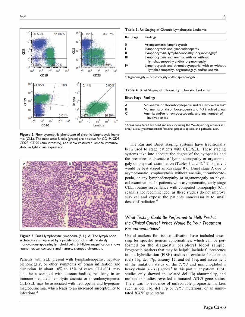
Patients with SLL present with lymphadenopathy, hepatos-
plenomegaly, or other symptoms of organ infiltration and
disruption. In about 10% to 15% of cases, CLL/SLL may
also be associated with autoantibodies, resulting in an
immune-mediated hemolytic anemia or thrombocytopenia.
CLL/SLL may be associated with neutropenia and hypogam-
maglobulinemia, which leads to an increased susceptibility to
infections.2
The Rai and Binet staging systems have traditionally
been used to stage patients with CLL/SLL. These staging
systems take into account the degree of the cytopenias and
the presence or absence of lymphadenopathy or organome-
galy on physical examination (Tables 3 and 4).3 This patient
would be best staged as Rai stage 0 or Binet stage A due to
asymptomatic lymphocytosis without anemia, thrombocyto-
penia, or any lymphadenopathy or organomegaly on physi-
cal examination. In patients with asymptomatic, early-stage
CLL, routine surveillance with computed tomography (CT)
scans is not recommended, as these studies do not improve
survival and expose the patients unnecessarily to small
doses of radiation.4
What Testing Could Be Performed to Help Predictthe Clinical Course? What Would Be Your TreatmentRecommendations?
Useful markers for risk stratification have included asses-
sing for specific genetic abnormalities, which can be per-
formed on the diagnostic peripheral blood sample.
Prognostic markers that may be helpful include fluorescence
in situ hybridization (FISH) studies to evaluate for deletion
(del) 11q, del 17p, trisomy 12, and del 13q, and assessment
of the mutation status of the TP53 and immunoglobulin
heavy chain (IGHV) genes.5 In this particular patient, FISH
studies only showed an isolated del 13q abnormality, and
molecular studies revealed a mutated IGVH gene status.
There was no evidence of unfavorable prognostic markers
such as del 11q, del 17p or TP53 mutations, or an unmu-
tated IGHV gene status.
Figure 3. Small lymphocytic lymphoma (SLL). A, The lymph nodearchitecture is replaced by a proliferation of small, relativelymonotonous-appearing lymphoid cells. B, Higher magnification showsround nuclear contours and mature, clumped chromatin.
Table 3. Rai Staging of Chronic Lymphocytic Leukemia.
Rai Stage Findings
0 Asymptomatic lymphocytosisI Lymphocytosis and lymphadenopathyII Lymphocytosis, lymphadenopathy, organomegaly*III Lymphocytosis and anemia, with or without
lymphadenopathy and/or organomegalyIV Lymphocytosis and thrombocytopenia, with or without
lymphadenopathy, organomegaly, and/or anemia
*Organomegaly ¼ hepatomegaly and/or splenomegaly.
Table 4. Binet Staging of Chronic Lymphocytic Leukemia.
Binet Stage Findings
A No anemia or thrombocytopenia and <3 involved areas*B No anemia or thrombocytopenia and �3 involved areasC Anemia and/or thrombocytopenia, and any number of
involved areas
*Areas considered are head and neck including the Waldeyer ring (counts as 1area), axilla, groin/superficial femoral, palpable spleen, and palpable liver.
Figure 2. Flow cytometric phenotype of chronic lymphocytic leuke-mia (CLL). The neoplastic B cells (green) are positive for CD19, CD5,CD23, CD20 (dim intensity), and show restricted lambda immuno-globulin light chain expression.
Roth 3
Page C2-63

The clinical behavior of CLL/SLL is variable, and early
treatment of asymptomatic patients does not result in
improved survival.3 Therefore, given the overall favorable
prognostic profile and lack of symptoms in this patient,
observation (or “watchful waiting”) may be the best course
of action. Indications for treatment include progressive cyto-
penias, bulky or symptomatic lymphadenopathy or spleno-
megaly, B symptoms, or other signs or symptoms of disease
progression. In a minority of cases (5%-10%), there is trans-
formation to a more aggressive lymphoma (“Richter
syndrome”), most commonly diffuse large B cell lymphoma,
often presenting as a rapidly enlarging mass.2
Current therapies for CLL/SLL emphasize a multimodality
approach including monoclonal antibodies targeting the B cell
marker CD20 (such as rituximab), combined with chemother-
apeutic agents such as purine analogs (fludarabine) or
alkylating agents (cyclophosphamide, bendamustine). Agents
targeting B cell receptor signaling (ibrutinib, idelalisib) or
expression of the anti-apoptotic protein BCL2 (venetoclax) are
also increasingly being incorporated into therapeutic regimens
for CLL/SLL.5
Teaching Points
� Chronic lymphocytic leukemia is the most common
adult leukemia in the Western world and may be asymp-
tomatic at diagnosis
� Chronic lymphocytic leukemia is characterized by an
absolute lymphocytosis composed of monomorphic,
small, mature-appearing lymphoid cells
� Flow cytometry can be used to assess the immunophe-
notype of leukemic cells and demonstrate the character-
istic phenotype
� The clinical behavior is heterogeneous, with many
patients not requiring initial therapeutic intervention
Declaration of Conflicting Interests
The author(s) declared no potential conflicts of interest with respect to
the research, authorship, and/or publication of this article.
Funding
The author(s) received no financial support for the research, author-
ship, and/or publication of this article.
References
1. Chabot-Richards DS, George TI. Leukocytosis. Int J Lab Hematol.
2014;36:279-288.
2. Disorders of white blood cells, lymph nodes, spleen, and thymus.
In: Kumar V, Abbas AK, Aster JC, eds. Robbins and Cotran Patho-
logic Basis of Disease. 9th ed. Philadelphia, PA: Elsevier Sauders;
2015:593-594.
3. Aster JC, Freedman A. Non-Hodgkin lymphomas and chronic lym-
phocytic leukemias. In: Aster JC, Bunn HF, eds. Pathophysiology
of Blood Disorders. 2nd ed. New York, NY: McGraw-Hill Educa-
tion; 2017:280-283.
4. American Society of Hematology Choosing Wisely Recommenda-
tions. 2014. http://www.choosingwisely.org/clinician-lists/ameri
can-society-hematology-baseline-or-routine-surveillance-ct-scans-
for-asymptomatic-early-stage-chronic-lymphocytic-leukemia/.
Accessed December 17, 2017.
5. HallekM. Chronic lymphocytic leukemia: 2017 update on diagnosis,
risk stratification, and treatment. Am J Hematol. 2017;92:946-965.
4 Academic Pathology
Page C2-64

Educational Case
Educational Case: Endocrine Neoplasm:Medullary Thyroid Carcinoma
Sheila Segura, MD1, Gloria Ramos-Rivera, MD1, and Mark Suhrland, MD1
The following fictional case is intended as a learning tool within the Pathology Competencies for Medical Education
(PCME), a set of national standards for teaching pathology. These are divided into three basic competencies: Disease
Mechanisms and Processes, Organ System Pathology, and Diagnostic Medicine and Therapeutic Pathology. For additional
information, and a full list of learning objectives for all three competencies, see http://journals.sagepub.com/doi/10.1177/
2374289517715040.
AbstractMedullary thyroid cancer is a rare neuroendocrine tumor that arises the neural crest-derived parafollicular C cells and accountsfor approximately 5% to 10% of thyroid cancers worldwide. These tumor can occur sporadically or as part of hereditary tumorsyndromes, such as multiple endocrine neoplasia 2 and familial medullary thyroid cancer. The most common clinical presentationis a solitary thyroid nodule. The genetic defect in these disorders involves the RET proto-oncogene which is important fordiagnosis of medullary thyroid cancer (including screening for hereditary medullary thyroid cancer) and for treatment guidance.This review summarizes the molecular basis and clinicopathologic features of medullary thyroid carcinoma.
Keywordspathology competencies, organ system pathology, endocrine neoplasms, medullary thyroid carcinoma, cytologic diagnosis,molecular basis, clinical features, pathologic features
Received January 16, 2018. Received revised March 01, 2018. Accepted for publication March 05, 2018.
Primary Objective
Objective EN5.2: Medullary Thyroid Carcinoma: Describe the
molecular basis and clinicopathologic features of medullary
thyroid carcinoma.
Competency 2: Organ System Pathology; Topic Endocrine
(EN); Learning Goal 5: Endocrine Neoplasms.
Patient Presentation
A 45-year-old woman presented to her endocrinologist with a
single, gradually increasing nodule in her right neck. There was
no relevant past medical or family history. She also mentioned
having diarrhea intermittently. During her initial visit,
the patient was in good condition, her blood pressure was
110/70 mm Hg, and her pulse was 75/minute and regular.
Physical examination revealed a 2.0-cm firm mass with smooth
borders on the right side of the thyroid that moved with swal-
lowing. The rest of the examination was unremarkable.
Ultrasonography of the thyroid revealed a 2.2-cm solid right
thyroid nodule. The results of thyroid function tests were
normal Thyroid-stimulating hormone (TSH) ¼ 0.6 mIU/mL
[range: 0.5-4.70 mIU/mL], T4 ¼ 5.5 mg/dL [4.5-12.5 mg/dL],T3 ¼ 115 ng/dL [80-200 ng/dL], and free T4 ¼ 1.0 ng/dL
1 Montefiore Hospital and Medical Center, Bronx, NY, USA
Corresponding Author:
Sheila Segura, Montefiore Hospital and Medical Center, 111 East 210th Street,
Bronx, NY 10467, USA.
Email: [email protected]
Academic PathologyVolume 5: 1–5ª The Author(s) 2018Reprints and permission:sagepub.com/journalsPermissions.navDOI: 10.1177/2374289518775722journals.sagepub.com/home/apc
Creative Commons Non Commercial CC BY-NC: This article is distributed under the terms of the Creative Commons Attribution-NonCommercial 4.0 License(http://www.creativecommons.org/licenses/by-nc/4.0/) which permits non-commercial use, reproduction and distribution of the work without furtherpermission provided the original work is attributed as specified on the SAGE and Open Access pages (https://us.sagepub.com/en-us/nam/open-access-at-sage).
Page C2-65
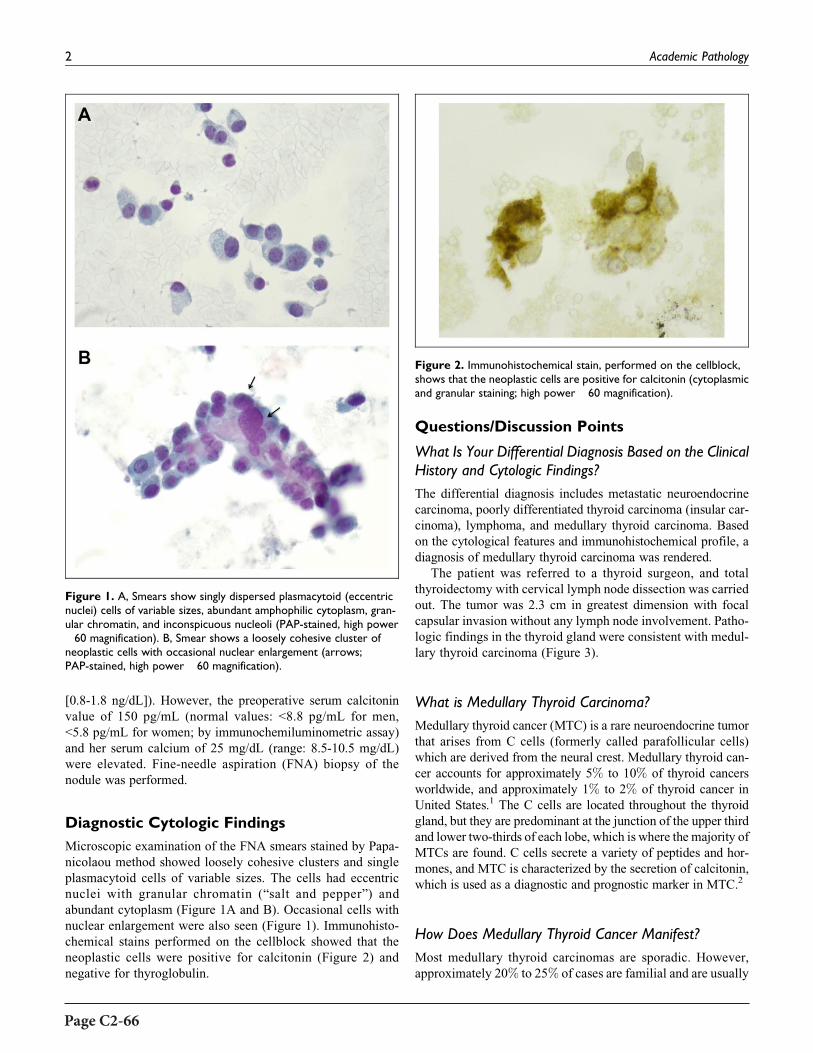
[0.8-1.8 ng/dL]). However, the preoperative serum calcitonin
value of 150 pg/mL (normal values: <8.8 pg/mL for men,
<5.8 pg/mL for women; by immunochemiluminometric assay)
and her serum calcium of 25 mg/dL (range: 8.5-10.5 mg/dL)
were elevated. Fine-needle aspiration (FNA) biopsy of the
nodule was performed.
Diagnostic Cytologic Findings
Microscopic examination of the FNA smears stained by Papa-
nicolaou method showed loosely cohesive clusters and single
plasmacytoid cells of variable sizes. The cells had eccentric
nuclei with granular chromatin (“salt and pepper”) and
abundant cytoplasm (Figure 1A and B). Occasional cells with
nuclear enlargement were also seen (Figure 1). Immunohisto-
chemical stains performed on the cellblock showed that the
neoplastic cells were positive for calcitonin (Figure 2) and
negative for thyroglobulin.
Questions/Discussion Points
What Is Your Differential Diagnosis Based on the ClinicalHistory and Cytologic Findings?
The differential diagnosis includes metastatic neuroendocrine
carcinoma, poorly differentiated thyroid carcinoma (insular car-
cinoma), lymphoma, and medullary thyroid carcinoma. Based
on the cytological features and immunohistochemical profile, a
diagnosis of medullary thyroid carcinoma was rendered.
The patient was referred to a thyroid surgeon, and total
thyroidectomy with cervical lymph node dissection was carried
out. The tumor was 2.3 cm in greatest dimension with focal
capsular invasion without any lymph node involvement. Patho-
logic findings in the thyroid gland were consistent with medul-
lary thyroid carcinoma (Figure 3).
What is Medullary Thyroid Carcinoma?
Medullary thyroid cancer (MTC) is a rare neuroendocrine tumor
that arises from C cells (formerly called parafollicular cells)
which are derived from the neural crest. Medullary thyroid can-
cer accounts for approximately 5% to 10% of thyroid cancers
worldwide, and approximately 1% to 2% of thyroid cancer in
United States.1 The C cells are located throughout the thyroid
gland, but they are predominant at the junction of the upper third
and lower two-thirds of each lobe, which is where the majority of
MTCs are found. C cells secrete a variety of peptides and hor-
mones, and MTC is characterized by the secretion of calcitonin,
which is used as a diagnostic and prognostic marker in MTC.2
How Does Medullary Thyroid Cancer Manifest?
Most medullary thyroid carcinomas are sporadic. However,
approximately 20% to 25% of cases are familial and are usually
Figure 1. A, Smears show singly dispersed plasmacytoid (eccentricnuclei) cells of variable sizes, abundant amphophilic cytoplasm, gran-ular chromatin, and inconspicuous nucleoli (PAP-stained, high power�60 magnification). B, Smear shows a loosely cohesive cluster ofneoplastic cells with occasional nuclear enlargement (arrows;PAP-stained, high power �60 magnification).
Figure 2. Immunohistochemical stain, performed on the cellblock,shows that the neoplastic cells are positive for calcitonin (cytoplasmicand granular staining; high power �60 magnification).
2 Academic Pathology
Page C2-66

a component of multiple endocrine neoplasia (MEN) syndrome
2A or 2B or present as pure familial MTC (FMTC) syndrome.3
Sporadic Medullary Thyroid Cancer
Sporadic MTC accounts for approximately 75% to 80% of all
cases of medullary thyroid cancer. There are typically unilat-
eral with no associated endocrinopathies. The typical age of
presentation is in the fourth and sixth decades of life, more
commonly affecting women in a 3:2 ratio. The most common
clinical presentation of sporadic MTC is that of a solitary thyr-
oid nodule (35%-50%) or enlarged lymph node (up to 70%).
The tumors are generally unilateral and tend to arise in the
posterior thyroid. In some patients, MTC presents with symp-
toms suggesting invasion of the surrounding structures (dys-
phagia, hoarseness, and/or respiratory difficulty) or can present
with distant metastases at the time of diagnosis. The most
common locations for metastatic MTC are the mediastinum,
liver, lungs, and bone.1-3
Systemic symptoms may occur due to hormonal secretion
by the tumor. Tumor secretion of calcitonin, calcitonin
gene-related peptide, or other substances (calcitonin, pros-
taglandins, serotonin, or vasoactive intestinal polypeptide)
can cause diarrhea or facial flushing in patients with
advanced disease.1,3
Hereditary (Familial) Medullary ThyroidCancer
Multiple Endocrine Neoplasia 2A (Sipple Syndrome)
Multiple endocrine neoplasia 2A syndrome or Sipple syn-
drome is the most common form of MEN 2 syndrome,
accounting for 75% of hereditary MTC. It is associated with
MTC, pheochromocytoma (*50%), and hyperparathyroidism
(*20%). This syndrome is inherited in an autosomal
dominant manner, affecting males and females equally. Peak
incidence of syndromic MTC is in the 30s, ranging from
adolescence to early adulthood.3,4
Multiple Endocrine Neoplasia 2B
Multiple endocrine neoplasia 2B accounts for 8% to 15% of
all patients with MEN 2. It is associated with MTC and
pheochromocytoma, and an unusual physical appearance,
characterized by mucosal ganglioneuromas and marfanoid
habitus (long limbs, hyperlaxity, and arachnodactyly).
Inheritance is autosomal dominant as in MEN 2A, again
equally affecting males and females.3 Patients with MEN
2B present MTC early in life, diagnosed in infancy or early
childhood before their 30s.3 Almost 100% of patients with
MEN 2B develop MTC with the disease having a more
aggressive course.
Familial Medullary Carcinoma Without AssociatedEndocrinopathies
The FMTC category of MTC is the least aggressive and is
defined as MTC without other hereditary endocrine tumors.
Similar to other types of thyroid cancers, the peak incidence
is between the ages of 40 and 50 years.3
What Are the Genetic Features of Medullary ThyroidCancer?
The genetic defect in these disorders involves the RET
proto-oncogene on chromosome 10q11.2. RET is a single-
pass transmembrane receptor belonging to the tyrosine kinase
superfamily. The germ line RET mutations lead to the activa-
tion of major intracellular pro-oncogenic pathways (eg, RAS/
MAPK, JUN kinase, PI3K/AKT, and nuclear factor-kB), that iscentral for the development of sporadic and hereditary MTC.
Currently known RET mutations account for about 95% of
MEN 2A and 85% of FMTC families. Somatic RET point
mutations have been identified in about 50% of patients with
sporadic MTC.1-4
Mutations involving the RET proto-oncogene are also seen
in other malignant and nonmalignant diseases. Chromosomal
translocations activating RET proto-oncogene can occur in
20% to 30% of patients with papillary thyroid carcinoma, and
can also be seen, but less frequently, in patients with lung
adenocarcinoma and chronic myelomonocytic leukemia. Inac-
tivating RET mutations occur in patients with hereditary and
sporadic Hirschsprung disease (HD).5
Describe the Pathological Features of Medullary ThyroidCancer
Grossly, sporadic or hereditary MTC present as a well-
demarcated, solid, white-gray/yellow, firm, and gritty
Figure 3. Section of the thyroid nodule showing nests of round andspindle cells outlined by fibrous tissue. The cells display granularcytoplasm and round nuclei with “salt and pepper” chromatin.(Hematoxylin and eosin stained slide, high power �40 magnification).
Segura et al 3
Page C2-67
nodule of variable size. Sporadic MTC tumors typically
present as a single and unilateral nodule. Hereditary MTC
are usually bilateral, multicentric, single, or multiple
nodules. Although the tumors in sporadic and hereditary
MTC are sharply circumscribed, they are not encapsulated.
Medullary thyroid cancer typically is the first neoplasm
observed in both MEN 2A and MEN 2B syndromes. Both
sporadic and FMTC arise at the junctions of the upper and
middle thirds of the lateral lobes.3
The histopathology is quite variable and numerous histolo-
gical subtypes are described. The characteristic features of
MTC, in both sporadic and familial, are sheets, nests, or trabe-
cular arrangement of cells which can vary in shape, round,
polygonal, spindle, or giant cells, all with varying amount of
granular cytoplasm. Tumor cells usually have uniform, round
to oval nuclei with granular or punctate chromatin, also
referred to as “salt and pepper” chromatin. Fibrous or amyloid
stroma can be present. The hereditary MTC is usually preceded
by “C-cell hyperplasia” (CCH) and usually occurs in the upper
two-thirds of the thyroid adjacent to a MTC or may be seen in
asymptomatic carriers of RET mutation. Multiple CCH foci are
a hallmark of MEN 2 syndromes and FMTC syndrome. The
presence of neoplastic CCH foci is considered a paradigm of a
genetically determined condition.1-4
How Is Medullary Thyroid Cancer Diagnosed?
Fine needle aspiration cytology is a widely utilized tool for
the diagnosis of thyroid lesions with a high degree of sen-
sitivity, specificity, and diagnostic accuracy. It is a simple,
rapid, and cost-effective test that can effectively distin-
guish between neoplastic and nonneoplastic lesions of the
thyroid. Fine-needle aspiration can effectively triage
patients with thyroid nodules as to who require surgery
and who do not.6,7
In the year 2007, the National Cancer Institute (NCI),
Bethesda, Maryland, organized the NCI Thyroid Fine Needle
Aspiration State-of-the-Science Conference, and an initiative
was undertaken to publish an atlas and guidelines using a
standardized nomenclature for the interpretation of thyroid
FNAs, known as the Bethesda system for reporting thyroid
cytopathology. The atlas describes 6 diagnostic categories of
lesions: Nondiagnostic/unsatisfactory, benign, atypical folli-
cular lesion of undetermined significance, “suspicious” for
follicular neoplasm, suspicious for malignancy, and malig-
nant. The 6 diagnostic categories of the Bethesda system have
individual implied risks of malignancy that influence man-
agement paradigms.7
The diagnosis of MTC is usually made after FNA biopsy.
The patients typically present with high calcitonin serum levels
that can also be helpful for diagnosis. Fine-needle aspiration
smears of MTC nodules show variable cytologic features. The
classic smear pattern is usually cellular, yielding tumor cells
that are dispersed and are characterized by eccentric nuclei
(plasmacytoid cell pattern), granular chromatin (neuroendo-
crine-type), and inconspicuous nucleoli in a relatively clean
background. The cytoplasm of the tumor cells is faintly gran-
ular but may show conspicuous red granules. Deposition of
amorphous, glassy, eosinophilic material consistent with amy-
loid, can be seen in the background, and sometimes is confused
with colloid material.
Depending on the specific cytomorphology of the tumor, a
number of differential diagnoses may arise and immunohisto-
chemical staining may be helpful. The small cell pattern may
be mistaken for a malignant lymphoma (CD45 positive), poorly
differentiated thyroid carcinoma (thyroglobulin positive), or
metastatic small-cell carcinoma (calcitonin negative) among
others. When MTC is suspected on cytological examination,
immunohistochemical staining for calcitonin is important for
making a diagnosis of MTC.
The 2015 American Thyroid Association (ATA) guide-
lines for MTC management recommend that all cases of
MTC, either sporadic or inherited, and patients with CCH,
should be analyzed for germ line mutations in the RET
proto-oncogene. RET molecular diagnosis is considered the
gold standard for the recognition of patients at risk of MTC
in MEN 2 families, and the recommended method of initial
testing for patients with MEN 2 is either a single or a multi-
tiered analysis to detect RET mutations in exon 10 (codons
609, 611, 618, and 620), exon 11 (codons 630 and 634), and
exons 8, 13, 14, 15, and 16.5
The ATA also recommends that genetic counseling and
genetic testing should be offered in patients with sporadic MTC
found to have a RET mutation; in first-degree relatives of
patients with proven hereditary MTC; in parents whose infants
or young children have the classic phenotype of MEN 2B; in
infants or young children with HD and RET germ line
mutations, and in adults with MEN 2A who have symptoms
suggestive of HD.5
Describe the Treatment Options for Medullary ThyroidCancer
Standard treatment for MTC requires surgical removal of the
thyroid with regional lymph node dissection. Unlike other thyr-
oid malignancies, MTCs are resistant to radioactive iodine
therapy because they do not concentrate radioactive iodine.
The extent of the surgical resection and lymph node dissection
is determined by the size of the primary tumor, the extent of
nodal, and distant metastases. In the presence of advanced
metastatic disease, a more palliative approach is recommended,
and aggressive neck surgery is typically not performed to
improve quality of life.
Prophylactic thyroidectomy is recommended in patients
with germ line RET mutations, and the timing of surgery
depends on the type of MEN syndrome and the RET mutation,
but it is usually recommended before the onset of clinically
significant disease.
For patients with metastatic MTC, treatment depends on
the extent of metastases. Surgery may be an option for
surgically resectable tumor lesion to relieve tumor burden.
Conventional chemotherapy has limited efficacy in patients
4 Academic Pathology
Page C2-68

with MTC. A new class of therapies targeting the RET
receptor tyrosine kinase family has been developed because
of its role in the pathogenesis of MTC. Several clinical trials
have been developed involving tyrosine kinase inhibitors
(such as sorafenib, sunitinib, vandetanib, motesanib, and
cabozantinib) with the rationale that they can block the
molecular pathways involved in MTC. Some of these treat-
ments (like vandetanib and cabozantinib) have been
approved by the US Food and Drug Administration for the
treatment of adults with symptomatic or progressive meta-
static MTC.2,8
What Are the Features That Drive Prognosis in PatientsWith Medullary Thyroid Cancer?
The size of the primary tumor and the presence of nodal and
distant metastasis are important factors predicting survival in
patients with MTC. When the tumor is localized to the thyroid
gland, the 10-year survival rate is approximately 75% to 95%.
Patients with regional lymph node disease have a 5-year overall
survival rate of 75.5%. Distant metastases at initial diagnosis is
associated with a poor prognosis, with a 10-year survival rate of
<50%.2,8 Younger age (<40 years old) at the time of diagnosis
is associated with better prognosis.
Factors that may also predict a poor prognosis includes:
1. Calcitonin doubling times less than 6 to 12 months is
associated with poor survival, while doubling times >24
months are associated with a very favorable prognosis.
2. Specific germ line mutations in RET predict the aggres-
siveness of the tumor (MEN 2B have worse a prognosis).
Recurrent disease develops in approximately 50% of
patients with MTC. Calcitonin levels are very sensitive for
detecting either residual or recurrent disease.
Teaching Points
� Medullary thyroid cancers are rare tumors that arise
from the neural crest–derived parafollicular C cells.
� They occur sporadically or as part of hereditary tumor
syndromes (MEN 2 and FMTC).
� A solitary thyroid nodule is the most common presenta-
tion of sporadic MTC (in 75%-95% of patients). In most
patients, the disease involves regional lymph nodes at
the time of diagnosis.
� Genetic testing for RET mutations is important for diag-
nosis of MTC (including screening for hereditary MTC)
and for treatment guidance. Therefore, germ line RET
testing is recommended in all patients with newly diag-
nosed CCH or apparently sporadic MTC.
� Due to limited adjuvant treatment options, adequate ini-
tial surgical management is essential to the successful
treatment of MTC.
� New molecular targeted treatment options are available
in cases of distant or recurrent disease not amenable to
surgery.
Declaration of Conflicting Interests
The author(s) declared no potential conflicts of interest with respect to
the research, authorship, and/or publication of this article.
Funding
The author(s) received no financial support for the research, author-
ship, and/or publication of this article.
References
1. Tuttle M, Ross DS, Mulder JE. Medullary thyroid cancer: clinical
manifestations, diagnosis, and staging. Up to Date. September 27,
2017. Accessed November 11, 2017. https://www.uptodate.com/
contents/medullary-thyroid-cancer-clinical-manifestations-diagno
sis-and-staging
2. Roy M, Chen H, Sippel RS. Current understanding and manage-
ment of medullary thyroid cancer.Oncologist. 2013;18:1093-1100.
3. Nose V. Familial thyroid cancer: a review. Modern Pathol. 2011;
24:S19-S33.
4. Ball DW. Medullary thyroid cancer: monitoring and therapy. Endo-
crinol Metab Clin North Am. 2007;36:823-837, viii. doi:10.1016/
j.ecl.2007.04.001.
5. Wells SA Jr, Asa SL, Dralle H, et al. Revised American Thyroid
Association guidelines for the management of medullary thyroid
carcinoma. Thyroid. 2015;25:567-610. doi:10.1089/thy.2014.0335.
6. Renuka IV, Saila Bala G, Aparna C, Kumari R, Sumalatha K.
The bethesda system for reporting thyroid cytopathology:
interpretation and guidelines in surgical treatment. Indian J
Otolaryngol Head Neck Surg. 2012;64:305-311. doi:10.1007/
s12070-011-0289-4.
7. Mondal SK, Sinha S, Basak B, Roy DN, Sinha SK. The Bethesda
system for reporting thyroid fine needle aspirates: a cytologic study
with histologic follow-up. J Cytol. 2013;30:94-99. doi:10.4103/
0970-9371.112650.
8. Dhakshinamoorthy G, Paulson E, Duran C, Cabanillas M, Busaidy
N, Charnsangavej C. Current update on medullary thyroid carci-
noma. Am J Roentgenol. 2013;201:W867-W876.
Segura et al 5
Page C2-69

Educational Case
Educational Case: Head and NeckNeoplasia: Salivary Gland Tumors
Ryan P. Lau, MD1, Melissa Yee-Chang, DO1, and Amy Rapkiewicz, MD1
The following fictional case is intended as a learning tool within the Pathology Competencies for Medical Education
(PCME), a set of national standards for teaching pathology. These are divided into three basic competencies: Disease
Mechanisms and Processes, Organ System Pathology, and Diagnostic Medicine and Therapeutic Pathology. For addi-
tional information, and a full list of learning objectives for all three competencies, see http://journals.sagepub.com/doi/
10.1177/2374289517715040.
Keywordspathology competencies, organ system pathology, head and neck neoplasia, salivary gland tumor, warthin tumor, pleomorphicadenoma, mucoepidermoid carcinoma
Received September 26, 2017. Received revised February 23, 2018. Accepted for publication February 28, 2018.
Primary Pathology Learning Objective
Objective HN2.1. Benign and Mucoepidermoid Tumors of
Salivary Glands. Distinguish the clinicopathologic features of
the 2 benign tumors (pleomorphic adenoma or mixed tumor
and Warthin tumor) from the malignant mucoepidermoid
carcinoma.
Competency 2: Organ System Pathology, Topic: Head and
Neck (HN), Learning Goal 2: Head and Neck Neoplasia
Secondary Pathology Learning Objective
Objective CYP1.2. Categorizing Diagnostic Certainty:
Compare and contrast the degree of diagnostic certainty
applied to general diagnostic categorization in cytologic
diagnosis.
Competency 3: Diagnostic Medicine and Laboratory Diag-
nosis; Topic CYP: Cytopathology; Learning Goal I: Cytologic
Diagnosis
Patient Presentation
A 62-year-old obese, hypertensive, Caucasian male presents to
his primary care physician with a slowly growing left cheek
mass for 1-year duration. He has a 40 pack-year smoking his-
tory. He has no family or personal history of carcinoma. He is
HIV-negative and has not experienced any dysphagia, odyno-
phagia, B-symptoms, trismus, or facial weakness.
Diagnostic Findings, Part 1
On physical examination, his primary care physician notes a
palpable, 2 cm, “doughy,” mobile mass in his left parotid gland.
There appears to be mild erythema and slight puckering of the
skin overlying the mass. The contralateral parotid gland has no
palpable masses and there is no cervical lymphadenopathy.
Tongue sensation and facial nerve function are intact. The
patient undergoes imaging studies and fine needle aspiration
(FNA) biopsy of his lesion.
1 New York University School of Medicine, New York, NY, USA
Corresponding Author:
Ryan P. Lau, New York University School of Medicine, 550 1st Ave, New York,
NY 10016, USA.
Email: [email protected]
Academic PathologyVolume 5: 1–5ª The Author(s) 2018Reprints and permission:sagepub.com/journalsPermissions.navDOI: 10.1177/2374289518770655journals.sagepub.com/home/apc
Creative Commons Non Commercial CC BY-NC: This article is distributed under the terms of the Creative Commons Attribution-NonCommercial 4.0 License(http://www.creativecommons.org/licenses/by-nc/4.0/) which permits non-commercial use, reproduction and distribution of the work without furtherpermission provided the original work is attributed as specified on the SAGE and Open Access pages (https://us.sagepub.com/en-us/nam/open-access-at-sage).
Page C2-71

Questions and Discussion Points, Part 1
Given the Clinical History, What Are the Most LikelySalivary Gland Neoplasms This Patient Might Have, andWhat Clinical Features Would Suggest Each Diagnosis?
Salivary gland tumors are rare, making up less than 2% of all
tumors. Up to 80% of salivary gland tumors appear in the
parotid gland, and 70% of parotid gland tumors are benign.
Parotid gland neoplasms are more likely to be benign than
submandibular gland neoplasms, which are more likely to
be benign than minor salivary gland neoplasms, which are
more likely to be benign than sublingual gland neoplasms.
Although certain clinical features can be suggestive, they are
unreliable to definitively differentiate between benign or
malignant.1,2
The most common salivary gland tumor is pleomorphic
adenoma and makes up approximately 50% of all salivary
gland tumors and 80% of all benign salivary gland tumors.
They typically are painless, mobile, and slow growing and
occur most frequently in the parotid gland—features all seen
in our patient. However, pleomorphic adenoma occurs more
frequently in females, and the average age of presentation is
in the mid-40s—unlike our patient.1-3
Warthin tumor is the second most common salivary gland
neoplasm and occurs almost exclusively in the parotid
gland. It is seen more frequently in male patients, the aver-
age age of presentation is in the mid-60s, and it has a strong
correlation with smoking history. In addition, the “doughy”
texture of the mass is a frequent finding in Warthin tumor.
Up to 10% of Warthin tumors are multifocal, and up to 15%of Warthin tumors occur bilaterally, so either of these fea-
tures would also raise the suspicion for this diagnosis.1,2
However, our patient’s lack of a contralateral mass on phys-
ical examination certainly does not rule out this diagnosis.
Mucoepidermoid carcinoma is the most common malignant
salivary gland neoplasm. It affects a broad age range and is the
most common primary malignant salivary gland tumor in both
adults and children. It is likely to demonstrate symptoms
related to its invasive nature, including rapid growth, pain,
immobility, skin changes and facial asymmetry, cervical lym-
phadenopathy, and loss of nerve function.1,2 The patient
demonstrated few of these concerning symptoms (mild
erythema and slight puckering overlying the mass) and the
diagnosis is lower on the differential.
In summary, the differential diagnosis includes but is
certainly not limited to pleomorphic adenoma, Warthin
tumor, and mucoepidermoid carcinoma. The skin erythema
and puckering may be seen in a malignant process (mucoe-
pidermoid carcinoma); however, the mobility, painlessness,
slow growth, and absent lymphadenopathy suggest a
benign tumor (Warthin tumor or pleomorphic adenoma).
These features combined with the “doughy” texture,
patient’s smoking history, male sex, and patient age suggest
a Warthin tumor.
What Risk Factors Predispose a Patient to DevelopSalivary Gland Neoplasms?
Ionizing radiation, in the form of atomic bomb exposure, pre-
vious head and neck radiation therapy, and radioactive iodine
treatment for thyroid disease, have all shown an increased
association with the development of salivary gland neoplasms.
There is also a strong association with smoking and the devel-
opment of Warthin tumor—smokers are 8 times more likely to
have a Warthin tumor than the general population. Certain
genetic alterations occur more frequently in certain salivary
gland tumors but familial aggregation has not yet been
observed in salivary gland tumors.1
What Diagnostic Testing Is Available for This Patient?
Although the clinical history and certain physical examination
findings, such as mobility of the mass or overlying skin
changes, can suggest a benign or malignant process, computed
tomography, magnetic resonance imaging (MRI), and ultraso-
nography are much more accurate. Imaging is primarily used to
assess malignant features, rather than to ascertain a specific
diagnosis. Imaging studies can determine if a neoplasm is con-
tained within the salivary gland or if it has invaded into adja-
cent structures. It can also assess regional lymph node and
distant metastases. However, a tissue biopsy is ultimately
required to make a diagnosis in salivary gland tumors. Tissue
biopsy can be performed via fine needle aspiration biopsy or
core needle biopsy.2
What Are the Advantages and Limitations of FineNeedle Aspiration in Presurgical Assessment ofSalivary Gland Tumors?
Salivary gland tumors are a heterogeneous and morphologi-
cally diverse group of benign and malignant neoplasms.
They are often sampled by fine needle aspiration and/or
core needle biopsy, but given the significant overlap in
morphology and immunohistochemical staining between
salivary gland tumors, tissue biopsy still has limitations in
its ability to confer a definitive diagnosis. Accuracy of FNA
diagnosis varies across studies and practitioners. Generally,
cytologic sampling has approximately 96% to 98% sensitiv-
ity in identifying a salivary gland neoplasm, with 79% sen-
sitivity and 96% specificity in distinguishing benign from
malignant tumors. Even in the absence of a definitive diag-
nosis, however, tissue biopsy can still help narrow down the
differential, making it a valuable tool in triaging patients for
medical or surgical management. Core biopsy may offer
better accuracy than FNA biopsy, but it also causes more
discomfort to the patient and has greater risk of complica-
tions, such as nerve damage and tumor seeding along the
needle tract.4
2 Academic Pathology
Page C2-72
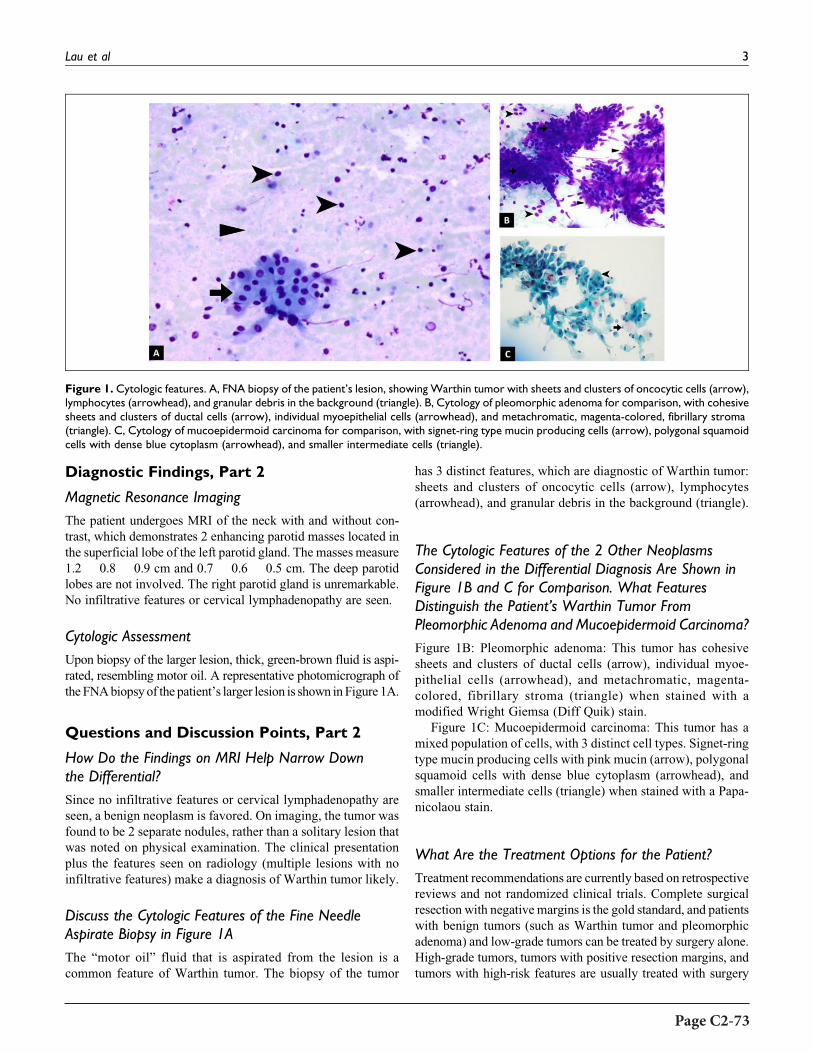
Diagnostic Findings, Part 2
Magnetic Resonance Imaging
The patient undergoes MRI of the neck with and without con-
trast, which demonstrates 2 enhancing parotid masses located in
the superficial lobe of the left parotid gland. The masses measure
1.2 � 0.8 � 0.9 cm and 0.7 � 0.6 � 0.5 cm. The deep parotid
lobes are not involved. The right parotid gland is unremarkable.
No infiltrative features or cervical lymphadenopathy are seen.
Cytologic Assessment
Upon biopsy of the larger lesion, thick, green-brown fluid is aspi-
rated, resembling motor oil. A representative photomicrograph of
theFNAbiopsyof the patient’s larger lesion is shown inFigure 1A.
Questions and Discussion Points, Part 2
How Do the Findings on MRI Help Narrow Downthe Differential?
Since no infiltrative features or cervical lymphadenopathy are
seen, a benign neoplasm is favored. On imaging, the tumor was
found to be 2 separate nodules, rather than a solitary lesion that
was noted on physical examination. The clinical presentation
plus the features seen on radiology (multiple lesions with no
infiltrative features) make a diagnosis of Warthin tumor likely.
Discuss the Cytologic Features of the Fine NeedleAspirate Biopsy in Figure 1A
The “motor oil” fluid that is aspirated from the lesion is a
common feature of Warthin tumor. The biopsy of the tumor
has 3 distinct features, which are diagnostic of Warthin tumor:
sheets and clusters of oncocytic cells (arrow), lymphocytes
(arrowhead), and granular debris in the background (triangle).
The Cytologic Features of the 2 Other NeoplasmsConsidered in the Differential Diagnosis Are Shown inFigure 1B and C for Comparison. What FeaturesDistinguish the Patient’s Warthin Tumor FromPleomorphic Adenoma and Mucoepidermoid Carcinoma?
Figure 1B: Pleomorphic adenoma: This tumor has cohesive
sheets and clusters of ductal cells (arrow), individual myoe-
pithelial cells (arrowhead), and metachromatic, magenta-
colored, fibrillary stroma (triangle) when stained with a
modified Wright Giemsa (Diff Quik) stain.
Figure 1C: Mucoepidermoid carcinoma: This tumor has a
mixed population of cells, with 3 distinct cell types. Signet-ring
type mucin producing cells with pink mucin (arrow), polygonal
squamoid cells with dense blue cytoplasm (arrowhead), and
smaller intermediate cells (triangle) when stained with a Papa-
nicolaou stain.
What Are the Treatment Options for the Patient?
Treatment recommendations are currently based on retrospective
reviews and not randomized clinical trials. Complete surgical
resection with negativemargins is the gold standard, and patients
with benign tumors (such as Warthin tumor and pleomorphic
adenoma) and low-grade tumors can be treated by surgery alone.
High-grade tumors, tumors with positive resection margins, and
tumors with high-risk features are usually treated with surgery
Figure 1. Cytologic features. A, FNA biopsy of the patient’s lesion, showing Warthin tumor with sheets and clusters of oncocytic cells (arrow),lymphocytes (arrowhead), and granular debris in the background (triangle). B, Cytology of pleomorphic adenoma for comparison, with cohesivesheets and clusters of ductal cells (arrow), individual myoepithelial cells (arrowhead), and metachromatic, magenta-colored, fibrillary stroma(triangle). C, Cytology of mucoepidermoid carcinoma for comparison, with signet-ring type mucin producing cells (arrow), polygonal squamoidcells with dense blue cytoplasm (arrowhead), and smaller intermediate cells (triangle).
Lau et al 3
Page C2-73

and adjuvant radiation therapy.Because hehas a benigndiagnosis
of Warthin tumor, he can be treated with surgery alone.5
Diagnostic Findings, Part 3
Histologic Assessment
The patient undergoes superficial parotidectomy to remove the
2 masses. A representative photomicrograph of the patient’s
lesions is shown in Figure 2A.
Questions and Discussion Points, Part 3
Discuss the Histologic Features of the Surgical Excisionin Figure 2A
The cystic lesion is lined by oncocytic cells (arrow) with a
lymphoid stroma (arrowhead). Granular debris fills the cyst
spaces (triangle). These are diagnostic of and consistent with
the initial biopsy diagnosis of Warthin tumor.
The Histologic Features of the 2 Other NeoplasmsConsidered in the Differential Diagnosis Are Shown inFigure 2B and C for Comparison. What FeaturesDistinguish the Patient’s Warthin Tumor FromPleomorphic Adenoma and Mucoepidermoid Carcinoma?
Figure 2B: Pleomorphic adenoma: This tumor has ductal cells
(arrow) surrounded by a layer of myoepithelial cells (arrow-
head), arranged in a chondromyxoid stroma (triangle).
Figure 2C: Mucoepidermoid carcinoma: This cystic tumor
has a mixed population of cells, with 3 distinct cell types—
mucin producing cells (arrow), squamoid cells (arrowhead),
and intermediate cells (triangle).
What Is the Expected Outcome After SuperficialParotidectomy to Excise the Patient’s Warthin Tumor?
Surgical excision of Warthin tumor is curative, and we expect
the patient to have a good clinical outcome. The risk of local
recurrence after complete surgical excision is less than 2%.
The other tumors on our initial differential behave quite
differently. Pleomorphic adenoma is also benign but has a
higher local recurrence rate of up to 5% if treated by paroti-
dectomy, and up to 25% if treated with simple enucleation. The
behavior of mucoepidermoid carcinoma depends on the grade
of the neoplasm. Local recurrence rates range from 15% to
30%, and 5-year survival ranges from 50% to 90%.1,2
Teaching Points
1. Salivary gland neoplasms are rare, accounting for less
than 2% of all tumors.
2. Up to80% of salivaryglandneoplasmsoccur in theparotid
gland, and up to 70% of parotid gland tumors are benign.
3. The likelihood of malignancy in each salivary gland in
increasing order is: parotid gland, submandibular
gland, minor salivary gland, and sublingual gland.
4. Clinical features are not definitively diagnostic of a
benign or malignant process. Malignant tumors are
suggested by rapid growth, pain, immobility, skin
changes and facial asymmetry, cervical lymphadeno-
pathy, and loss of nerve function. Benign tumors are
suggested by absence of pain, mobility, slow growth,
absence of nerve involvement.
Figure 2. Histologic features. A, Surgical excision of the patient’s lesion showing Warthin tumor with oncocytic cells (arrow) and lymphoidstroma (arrowhead). Granular debris fills the cyst spaces (triangle). B, Histology of pleomorphic adenoma for comparison, with ductal cells(arrow) surrounded by a layer of myoepithelial cells (arrowhead), arranged in a chondromyxoid stroma (triangle). C, Histology of mucoepi-dermoid carcinoma for comparison, with mucin producing cells (arrow), squamoid cells (arrowhead), and intermediate cells (triangle).
4 Academic Pathology
Page C2-74

5. Radiologic studies can provide important clues to the
nature of a salivary gland tumor, including invasion
beyond salivary gland parenchyma into adjacent struc-
tures and local lymphatic spread.
6. Fine needle aspiration biopsy of salivary gland tumors
is an important initial diagnostic test that can fre-
quently give an accurate diagnosis. When a specific
diagnosis cannot be rendered, cytologic analysis can
give a differential diagnosis that helps guide clinical/
surgical management.
7. The 3 most common salivary gland tumors (pleo-
morphic adenoma, Warthin tumor, and mucoepider-
moid carcinoma) have distinct cytologic and
histologic appearances.
8. The most common salivary gland neoplasm is pleo-
morphic adenoma, a benign tumor that typically pre-
sents in the mid-40s and has a female predilection.
9. The second most common benign salivary gland neo-
plasm is Warthin tumor, which commonly presents in
male smokers in their mid-60s. On physical examina-
tion, it has a “doughy” texture and can be multifocal or
occur bilaterally. On aspiration, it produces a “motor
oil” like fluid.
10. The most common malignant salivary gland neoplasm
is mucoepidermoid carcinoma. It occurs over a broad
age range and is the most common salivary gland
malignancy in adults and children.
11. Pleomorphic adenoma and Warthin tumor can both be
treated by surgical excision alone and have less than
5% risk of local recurrence.
12. The treatment and outcome of mucoepidermoid carci-
noma is dependent on the tumor grade. High-grade
tumors may require surgery and adjuvant radiation
therapy, with local recurrence rate as high as 30%, and
5-year survival of 50%.
Declaration of Conflicting Interests
The author(s) declared no potential conflicts of interest with respect to
the research, authorship, and/or publication of this article.
Funding
The author(s) received no financial support for the research, author-
ship, and/or publication of this article.
References
1. Barnes L, Eveson JW, Reichart P, Sidransky D, eds. World Health
Organization Classification of Tumours. Pathology and Genetics,
Head and Neck Tumors. Lyon: IARC Press; 2005.
2. Kumar Vinay, Abbas Abul, Aster Jon. Robbins and Cotran Patho-
logic Basis of Disease. 9th ed. Philadelphia, PA: Elsevier/Saun-
ders; 2015.
3. Eveson JW, Cawson RA. Salivary gland tumours: a review of 2410
cases with particular reference to histological types, site, age and
sex distribution. J Pathol. 1985;146:51-58. PubMed PMID:
4009321.
4. Cibas ES, Ducatman BS. Cytology: Diagnostic Principles and
Clinical Correlates. 4th ed. Philadelphia, PA: Elsevier/Saunders;
2014.
5. National Comprehensive Cancer Network. Head and Neck
Cancers (Version 2.2017). https://www.nccn.org/professionals/
physician_gls/pdf/head-and-neck.pdf. Accessed September 21,
2017.
Lau et al 5
Page C2-75


Educational Case
Educational Case: Medullary ThyroidCarcinoma
Carl T. McGary, MD, PhD1
The following fictional case is intended as a learning tool within the Pathology Competencies for Medical Education
(PCME), a set of national standards for teaching pathology. These are divided into three basic competencies: Disease
Mechanisms and Processes, Organ System Pathology, and Diagnostic Medicine and Therapeutic Pathology. For addi-
tional information, and a full list of learning objectives for all three competencies, see http://journals.sagepub.com/doi/
10.1177/2374289517715040.
Keywordspathology competencies, organ system pathology, cytology diagnostic certainty, endocrine, endocrine neoplasms, fine needleaspiration cytology, medullary thyroid carcinoma
Received November 20, 2017. Received revised February 08, 2018. Accepted for publication February 16, 2018.
Primary Objective
EN5.2: Medullary Thyroid Carcinoma. Describe the molecular
basis and clinicopathologic features of medullary thyroid
carcinoma.
Competency 2: Organ System Pathology; Topic EN: Endo-
crine; Learning Goal 5: Endocrine Neoplasms.
Secondary Objective
CYP1.2: Categorizing Diagnostic Certainty. Compare and
contrast the degree of diagnostic certainty applied to general
categorization in cytologic diagnosis.
Competency 3: Diagnostic Medicine and Therapeutic
Pathology; Topic CYP: Cytopathology; Learning Goal 1: Cyto-
logic Diagnosis.
Patient Presentation
A 52-year-old Caucasian male presents to family medicine
with an irregular 4-cm palpable nodule in the mid right thyroid
lobe. He has no significant past medical history other than
occasional diarrhea. Complete head and neck examination
reveals no additional palpable nodules in the thyroid gland and
no palpable adenopathy. The differential diagnosis for a palp-
able thyroid nodule includes a variety of benign and malignant
entities, including colloid nodule, nodular hyperplasia,
follicular thyroid adenoma, follicular thyroid carcinoma, papil-
lary thyroid carcinoma (PTC), medullary thyroid carcinoma
(MTC), anaplastic thyroid carcinoma, and lymphoma. The
patient is referred to the lab for pathologist to perform fine
needle aspiration (FNA) cytology of the nodule.
Diagnostic Findings, Part 1
The pathologist performs 3 FNA passes into the nodule under
direct guidance using 25-gauge needles without suction, and the
initial smears show adequate cellularity (Figure 1). Additional
passes are not performed for cell block preparation. Using The
Bethesda System for Reporting Thyroid Cytology, the patholo-
gist finalizes the report as Bethesda category V, suspicious for
malignancy and MTC. The cytology findings are shown in Fig-
ures 1 and 2 and compared to the more common PTC in Figure
3. The patient is referred to endocrinology for further evaluation.
1 Department of Biomedical Sciences, The University of Minnesota Medical
School–Duluth, Duluth, MN, USA
Corresponding Author:
Carl T. McGary, MD, PhD, Department of Biomedical Sciences, The University
of Minnesota Medical School–Duluth, 1035 University Drive, SMED 227,
Duluth, MN 55812, USA.
Email: [email protected]
Academic PathologyVolume 5: 1–5ª The Author(s) 2018Reprints and permission:sagepub.com/journalsPermissions.navDOI: 10.1177/2374289518770173journals.sagepub.com/home/apc
Creative Commons Non Commercial CC BY-NC: This article is distributed under the terms of the Creative Commons Attribution-NonCommercial 4.0 License(http://www.creativecommons.org/licenses/by-nc/4.0/) which permits non-commercial use, reproduction and distribution of the work without furtherpermission provided the original work is attributed as specified on the SAGE and Open Access pages (https://us.sagepub.com/en-us/nam/open-access-at-sage).
Page C2-77

Questions/Discussion Points, Part 1
Categorizing Diagnostic Certainty: Based on thePathologist’s Interpretation of the FNA, How StatisticallyLikely is the Patient to Harbor a Malignant Neoplasmand What is the Uncertainty of the Diagnosis?
The major categories used in diagnostic cytopathology include
unsatisfactory for diagnosis, negative for malignancy, atypical
cells present, suspicious for malignancy, and positive for
malignancy. Each category carries an implied degree of
certainty with regard to the pathology present. Diagnostic
modifiers are also used in surgical pathology to suggest degrees
of uncertainty, including “cannot rule out,” “suggestive of,”
“consistent with,” and “diagnostic of,” in increasing order of
certainty.1 The issue of uncertainty in anatomic pathology
diagnosis is highlighted by the presence of both interobserver
(2 observers arriving at a different diagnosis on the same case)
and intraobserver (the same observer arriving at different diag-
noses on the same case at different times) diagnostic variation.2
Interinstitutional review of 777 patient specimens revealed a
diagnostic difference in 71 specimens on review at the second
institution. In 45 of the 71 discrepant specimens, the change in
diagnosis led to altered therapy. The discordant diagnoses were
significantly more common in cytology specimens (21%) than
surgical specimens (7.8%), suggesting that cytologic diagnosis
was more prone to uncertainty than histologic diagnosis.3
Historically, cytopathologists at different institutions have
utilized somewhat different diagnostic terminology when report-
ing results of thyroid FNA. To address the variety of reporting
systems and the resulting inherent confusion, the National Can-
cer Institute (NCI) hosted the NCI Thyroid FNA State of the
Science Conference. The conference conclusions led to the pub-
lication of the Bethesda System for Reporting Thyroid Cytol-
ogy,4 which is now in wide use. Similar issues led to publication
of the Paris System for Reporting Urinary Cytology.5
The Bethesda System for Reporting Thyroid Cytology4 con-
tains 6 general diagnostic categories, each with a specific given
risk of underlying malignancy as shown in Table 1. The least
amount of uncertainty is associated with the benign and malig-
nant categories as these have the narrowest range for underly-
ing malignancy. The greatest degree of uncertainty lies with the
atypical category which shows the widest range for underlying
Figure 3. Papanicolaou stain of PTC for comparison with the patientswith FNA cytology. Papillary thyroid carcinoma shows nongranularchromatin, nuclear grooves, small peripheral nucleoli, and occasionalintranuclear pseudo-inclusions. Papillary-shaped irregular branchinggroups are also present. FNA indicates fine needle aspiration cytology;PTC, papillary thyroid carcinoma.
Figure 1. Papanicolaou stain at �200 of the initial FNA smearshowing background red blood cells, scant colloid, and a mixed pop-ulation of neoplastic cells showing epithelioid and spindle cell mor-phology. Nuclear chromatin shows a “‘salt and pepper” granularity andlacks features of papillary carcinoma (Figure 3), and the cell cytoplasmis somewhat granular. FNA indicates fine needle aspiration cytology.
Figure 2. Papanicolaou stain at �400 highlighting random nuclearatypia often seen in neuroendocrine neoplasia, as well as the granularcytoplasm and fine-to-coarse nuclear chromatin of medullary thyroidcarcinoma.
2 Academic Pathology
Page C2-78
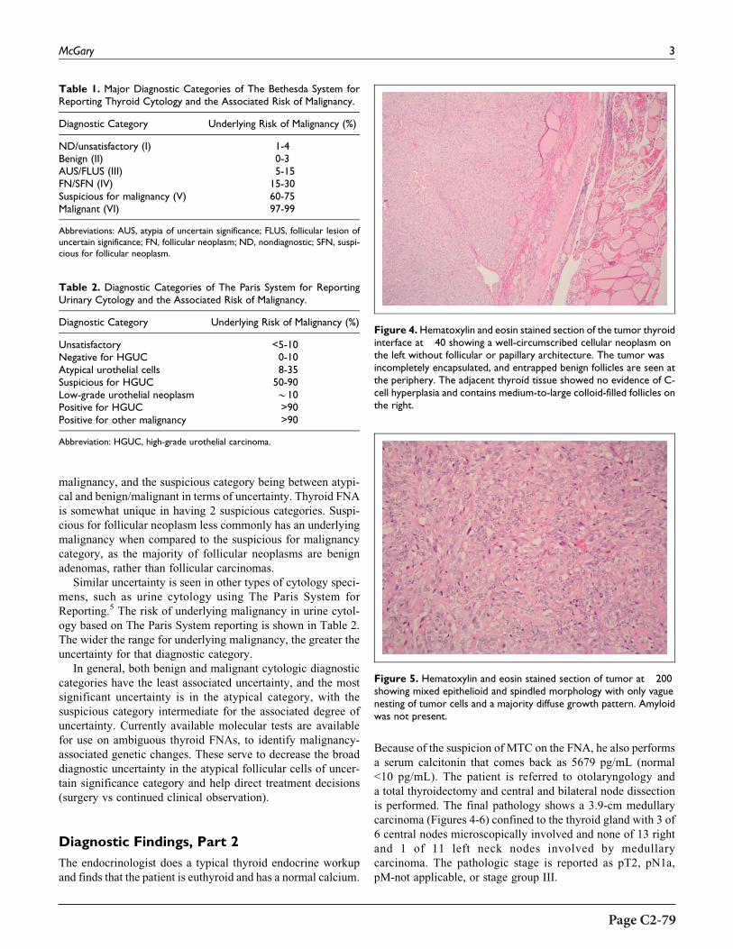
malignancy, and the suspicious category being between atypi-
cal and benign/malignant in terms of uncertainty. Thyroid FNA
is somewhat unique in having 2 suspicious categories. Suspi-
cious for follicular neoplasm less commonly has an underlying
malignancy when compared to the suspicious for malignancy
category, as the majority of follicular neoplasms are benign
adenomas, rather than follicular carcinomas.
Similar uncertainty is seen in other types of cytology speci-
mens, such as urine cytology using The Paris System for
Reporting.5 The risk of underlying malignancy in urine cytol-
ogy based on The Paris System reporting is shown in Table 2.
The wider the range for underlying malignancy, the greater the
uncertainty for that diagnostic category.
In general, both benign and malignant cytologic diagnostic
categories have the least associated uncertainty, and the most
significant uncertainty is in the atypical category, with the
suspicious category intermediate for the associated degree of
uncertainty. Currently available molecular tests are available
for use on ambiguous thyroid FNAs, to identify malignancy-
associated genetic changes. These serve to decrease the broad
diagnostic uncertainty in the atypical follicular cells of uncer-
tain significance category and help direct treatment decisions
(surgery vs continued clinical observation).
Diagnostic Findings, Part 2
The endocrinologist does a typical thyroid endocrine workup
and finds that the patient is euthyroid and has a normal calcium.
Because of the suspicion of MTC on the FNA, he also performs
a serum calcitonin that comes back as 5679 pg/mL (normal
<10 pg/mL). The patient is referred to otolaryngology and
a total thyroidectomy and central and bilateral node dissection
is performed. The final pathology shows a 3.9-cm medullary
carcinoma (Figures 4-6) confined to the thyroid gland with 3 of
6 central nodes microscopically involved and none of 13 right
and 1 of 11 left neck nodes involved by medullary
carcinoma. The pathologic stage is reported as pT2, pN1a,
pM-not applicable, or stage group III.
Table 1. Major Diagnostic Categories of The Bethesda System forReporting Thyroid Cytology and the Associated Risk of Malignancy.
Diagnostic Category Underlying Risk of Malignancy (%)
ND/unsatisfactory (I) 1-4Benign (II) 0-3AUS/FLUS (III) 5-15FN/SFN (IV) 15-30Suspicious for malignancy (V) 60-75Malignant (VI) 97-99
Abbreviations: AUS, atypia of uncertain significance; FLUS, follicular lesion ofuncertain significance; FN, follicular neoplasm; ND, nondiagnostic; SFN, suspi-cious for follicular neoplasm.
Table 2. Diagnostic Categories of The Paris System for ReportingUrinary Cytology and the Associated Risk of Malignancy.
Diagnostic Category Underlying Risk of Malignancy (%)
Unsatisfactory <5-10Negative for HGUC 0-10Atypical urothelial cells 8-35Suspicious for HGUC 50-90Low-grade urothelial neoplasm *10Positive for HGUC >90Positive for other malignancy >90
Abbreviation: HGUC, high-grade urothelial carcinoma.
Figure 4.Hematoxylin and eosin stained section of the tumor thyroidinterface at �40 showing a well-circumscribed cellular neoplasm onthe left without follicular or papillary architecture. The tumor wasincompletely encapsulated, and entrapped benign follicles are seen atthe periphery. The adjacent thyroid tissue showed no evidence of C-cell hyperplasia and contains medium-to-large colloid-filled follicles onthe right.
Figure 5. Hematoxylin and eosin stained section of tumor at �200showing mixed epithelioid and spindled morphology with only vaguenesting of tumor cells and a majority diffuse growth pattern. Amyloidwas not present.
McGary 3
Page C2-79

Questions/Discussion Points, Part 2
What Clinicopathologic Features are Common in BothSporadic and Inherited Medullary Thyroid Carcinoma?
Medullary thyroid carcinoma is a primary tumor of the thyroid
gland which shows differentiation along the lines of thyroid
parafollicular C cells.6-8 Medullary thyroid carcinoma typically
secretes calcitonin, although nonsecretory tumors occur rarely.
Serum calcitonin reflects quantity of tumor present, except in
nonsecreting patients. Medullary thyroid carcinoma is almost
always carcinoembryonic antigen (CEA) positive, and in
noncalcitonin-secreting tumors, serum CEA levels can be fol-
lowed, similar to calcitonin levels in secreting tumors to assess
disease burden and therapeutic response. Even though elevated
calcitonin levels are present, serum calcium is not typically
decreased below normal levels. Medullary thyroid carcinoma
tumors sometimes secrete other hormones, such as adrenal
corticotropic hormone, serotonin, and vasoactive intestinal
peptide (VIP), which can result in a paraneoplastic presentation
such as diarrhea if VIP secretion occurs. The VIP levels were
not measured in this patient, and it is not clear whether his
intermittent diarrhea was related to the MTC. Medullary thyr-
oid carcinomas are relatively uncommon tumors accounting for
less than 2% to 3% of thyroid neoplasia.
Medullary thyroid carcinoma can present as a sporadic
tumor or as part of an inherited tumor syndrome. The histology
of MTC is quite variable, but in general is not distinct for
sporadic or inherited cases. A mixture of spindled and epithe-
lioid cells is common, and plasmacytoid morphology is often
seen. Neuroendocrine features include finely granular cyto-
plasm due to variable presence of neurosecretory granules. The
chromatin is mixed fine and coarse and classically described as
“salt and pepper” chromatin which is commonly seen in neu-
roendocrine tumors. Cells can be arranged in sheets, nested
aggregates, trabeculae, or even follicles, and mitotic figures
tend to be sparse. These features of MTC contrast with the
more common PTC (Figure 3). Nuclear features of PTC
include pale nongranular nuclear chromatin, nuclear grooves,
nuclear pseudoinclusions, and small peripheral nucleoli. Amy-
loid deposition is commonly present in up to 90% of cases,
but was absent in this particular case, and is composed of
calcitonin peptides. Immunochemistry shows positive granular
cytoplasmic staining for calcitonin in the tumor cells and dif-
fusely stains the amyloid deposits. Immunochemistry is also
positive for other neuroendocrine markers, such as chromogra-
nin and synaptophysin. Cytologic atypia ranges from minimal,
to focal, to widespread.
Describe the Features Unique to Sporadic MedullaryThyroid Carcinoma
Medullary thyroid carcinoma occurs as sporadic disease in
70% of cases, and the sporadic cases present typically as a
neck mass, as in this case, but clinical findings may also
include dysphagia and/or hoarseness. When MTC presents
as a neck mass, nodal involvement is relatively frequent
(*70%) and non-nodal metastasis (10%) also occurs. Spora-
dic cases show a slight female predominance and are seen
predominantly in the fifth and sixth decades. These are usu-
ally unilateral and unifocal tumors of the thyroid. Unlike PTC,
ionizing radiation exposure does not seem to be etiologically
involved in sporadic MTC.
Describe the Features Unique to Inherited MedullaryThyroid Carcinoma
Familial cases occur as part of inherited syndromes in 30% of
cases (multiple endocrine neoplasia type 2A [MEN-2A] or
MEN-2B or familial medullary thyroid carcinoma [FMTC])
and are often associated with other endocrine neoplasms.
Medullary thyroid carcinoma in this setting may present either
as a result of endocrine disease in other organs or with symp-
toms in the neck. Familial/inherited cases present at younger
ages than do sporadic cases. Medullary thyroid carcinoma in
the setting of MEN-2B more commonly metastasizes than what
is seen in MEN-2A, FMTC, or sporadic disease. Bilateral thyr-
oid involvement and multifocal disease are more common in
inherited MTC than in sporadic disease. Penetrance of medul-
lary thyroid cancer development is age and mutation depen-
dent; however, MTC inevitably develops in MEN-2 with RET
mutations, and early total thyroidectomy is preventative. Early
onset is typical in MEN-2B with later onset in MEN-2A. Thyr-
oid tissue outside the tumor in inherited cases typically shows
background C cell hyperplasia.
Describe the Molecular Basis of Sporadic MedullaryThyroid Carcinoma
Activating somatic point mutations of the RET proto-oncogene
are noted in sporadic medullary carcinoma in approximately
40% to 60% of cases. The most frequently seen mutation in
Figure 6. Immunochemistry of tumor for calcitonin �400 showingdiffuse granular cytoplasmic staining.
4 Academic Pathology
Page C2-80

sporadic cases is the M918T mutation in exon 16. Interestingly,
this is also the same mutation as the germline mutation in the
vast majority of MEN-2B patients. RASmutations are also seen
in some sporadic cases, usually those that are RET mutation
negative. Thus, in sporadic disease, RAS and RET mutations
commonly show mutual exclusion.
Describe the Molecular Basis of Inherited MedullaryThyroid Carcinoma
Germline mutation of the RET proto-oncogene with gain of
function is the underlying feature in inherited MTC, as seen in
MEN-2A, MEN-2B, and FMTC (a variant of MEN-2A). The
RET proto-oncogene codes for a receptor tyrosine kinase that
binds glial-derived neurotrophic factor and similar ligands.
Ligand binding to receptor leads to intracellular growth signals
as well as signals for differentiation. Various specific RETmuta-
tions occur in MEN-2A, and coexisting parathyroid hyperplasia
and pheochromocytoma are characteristic. The specific RET
mutations in MEN-2B are associated with pheochromocytoma
as well; however, the hyperparathyroidism is absent.
Teaching Points
� Major cytologic diagnostic categories (nondiagnostic,
benign, suspicious, and malignant) each carry an asso-
ciated risk of underlying malignancy and uncertainty,
which have been published for various types of
specimens.
� Cytologic diagnostic categories of benign and malignant
have the least amount of associated diagnostic uncer-
tainty and the atypical category the highest.
� Medullary thyroid carcinoma is a neuroendocrine tumor
showing C-cell differentiation.
� Medullary thyroid carcinoma is immunochemically pos-
itive for calcitonin (which is elevated in serum, except in
rare nonsecreting cases), chromogranin, synaptophysin,
and CEA.
� Medullary thyroid carcinoma can be followed clinically
with serum calcitonin or CEA, and CEA is helpful in
calcitonin nonsecreting cases.
� Occasionally, MTCs secrete other hormones and
may have paraneoplastic presentations, for example,
VIP—diarrhea.
� Medullary thyroid carcinoma can be sporadic or
inherited.
� Sporadic and inherited MTCs have the same histologic
spectrum with amyloid deposition, sporadic disease
typically being unilateral and unifocal and inherited dis-
ease more likely to be bilateral and multifocal.
� Inherited MTC shows C-cell hyperplasia in the non-
neoplastic thyroid tissue.
� Specific genetic and molecular mechanisms underlie
both sporadic and inherited medullary thyroid cancer,
with the RET gene being most frequently involved.
� The inevitable development of medullary thyroid cancer
associated with MEN-2 can be prevented by early pro-
phylactic total thyroidectomy.
ORCID iD
Carl T. McGary, MD, PhD http://orcid.org/0000-0003-4625-3030
References
1. Bracamonte E, Gibson BA, Klein R, Krupinski EA, Weinstein RS.
Communicating uncertainty in surgical pathology reports: a survey
of staff physicians and residents at an academic medical center.
Acad Pathol. 2016;3:1-7. doi:10.1177/2374289516659079.
2. Franc B, De La Salmoniere P, Lange F, et al. Interobserver and
intraobserver reproducibility in the histopathology of follicular
thyroid carcinoma. Hum Pathol. 2003;34(11):1092-1100.
3. Abt AB, Abt LG, Olt GJ. The effect of interinstitution anatomic
pathology consultation on patient care. Arch Pathol Lab Med.
1995;119:514-517.
4. Cibas ES, Ali SZ; NCI Thyroid FNA State of the Science Confer-
ence. The Bethesda System for Reporting Thyroid Cytology. Am J
Clin Pathol. 2009;132:658-665.
5. Barkan GA, Wojcik EM, Nayar R, et al. The Paris System for
Reporting Urinary Cytology: the quest to develop a standardized
terminology. Acta Cytol. 2016;60:185-197.
6. Maitra A. The endocrine system. In: Kumar V, Abbas A, Aster A,
eds. Robbins and Cotran Pathologic Basis of Disease. 9th ed.
Philadelphia, PA: Elsevier Sanders; 2015:1099-1100.
7. Maitra A. The endocrine system. In: Kumar V, Abbas A, Aster A,
eds. Robbins and Cotran Pathologic Basis of Disease. 9th ed.
Philadelphia, PA: Elsevier Sanders; 2015:1136-1137.
8. Lloyd RV, Osamura RY, Kloppel G, Rosi J.WHO Classification of
Tumours of Endocrine Organs. 4th ed. France: International
Agency for Research on Cancer (IARC); 2017:108-113.
McGary 5
Page C2-81

Educational Case
Educational Case: Bladder Carcinoma In Situ
H. James Williams, MD1
The following fictional case is intended as a learning tool within the Pathology Competencies for Medical Education
(PCME), a set of national standards for teaching pathology. These are divided into three basic competencies: Disease
Mechanisms and Processes, Organ System Pathology, and Diagnostic Medicine and Therapeutic Pathology. For addi-
tional information, and a full list of learning objectives for all three competencies, see http://journals.sagepub.com/doi/
10.1177/2374289517715040.
Keywordspathology competencies, organ system pathology, bladder neoplasia, bladder washings, carcinoma in situ (CIS) of the urinarybladder, FISH, screening urine cytology, voided urine cytology
Received April 30, 2017. Received revised May 31, 2017. Accepted for publication June 14, 2017.
Primary Objective
Objective UTB1.3: Diagnosis and Surveillance of Urothelial
Carcinoma. Describe the typical clinical presentation of
urothelial carcinoma and the advantages and limitations of
urine cytology in diagnosis and surveillance of urothelial
carcinoma.
Competency 2: Organ System Pathology; Topic UTB: Blad-
der; Learning Goal 1: Bladder Neoplasia.
Patient Presentation
A 57-year-old man presents with a complaint of cramping pain
on urination (dysuria) and red coloration of his urine (gross
hematuria).
On physical examination, the patient has vague abdomino-
pelvic discomfort on compression with no palpable mass. The
physical examination was otherwise unremarkable, and the
patient is afebrile.
Diagnostic Findings
Initial laboratory tests revealed a urinalysis with numerous red
blood cells and rare white blood cells, a negative urine culture,
normal complete blood cell count, and a normal serum creati-
nine level.
Questions/Discussion Points
What pertinent questions would you ask the patient?
How long have these symptoms been present?
Have they been decreasing or increasing in severity?
Are there difficulties passing urine or associated burning?
Have you passed any stones, clots, or tissues?
Have you ever had these symptoms before?
Are you a smoker?
What kind of work do you do and have you had different jobs in
the past?
Do you take any medications?
Have you ever been diagnosed with a malignancy?
Is there a family history of cancer and, if so, what kind?
1 Department of Pathology, Robert C. Byrd Health Sciences Center, West
Virginia University, Morgantown, WV, USA
Corresponding Author:
H. James Williams, Department of Pathology, Robert C. Byrd Health Sciences
Center, West Virginia University, PO Box 9203, Morgantown, WV 26506, USA.
Email: [email protected]
Academic PathologyVolume 5: 1–3ª The Author(s) 2018Article reuse guidelines:sagepub.com/journals-permissionsDOI: 10.1177/2374289517719858journals.sagepub.com/home/apc
Creative Commons Non Commercial CC BY-NC: This article is distributed under the terms of the Creative Commons Attribution-NonCommercial 4.0 License(http://www.creativecommons.org/licenses/by-nc/4.0/) which permits non-commercial use, reproduction and distribution of the work without furtherpermission provided the original work is attributed as specified on the SAGE and Open Access pages (https://us.sagepub.com/en-us/nam/open-access-at-sage).
Page C2-83

What additional laboratory test(s) would you consider atthis point?
Urine cytology (see Figure 1).
Describe the findings on voided urine cytology (seeFigure 1). What is your diagnosis?
There is a single cell with markedly enlarged hyperchro-
matic nuclei with irregular borders and high N/C ratio in
a background of blood. Voided urine normally is hypocel-
lular with few single benign urothelial cells and inflamma-
tory cells. The presence of cohesive groups of cytologically
bland urothelial cells in a voided urine is considered an
abnormal finding that may be associated with stones, UTI,
recent instrumentation, or low-grade papillary neoplasm.2 In
contrast, the presence of cohesive groups of cytologically
bland urothelial cells in a bladder washing is a common
finding due to instrumentation and mechanical dislodging
of cells from the washing pressure. While this may raise
concern for a low-grade papillary lesion on cytology, clini-
cally it is not an issue as a papillary lesion would be seen
and biopsied on cystoscopy. Bladder washings are useful for
detecting flat CIS when there is not a papillary lesion seen
on cystoscopy. Flat CIS as seen on urine cytology (voided
or washings) has high-grade cytologic features as seen in
this exercise.
The diagnosis is: positive for malignant cells, high-grade
urothelial carcinoma.
What would be your next step?
The patient is referred to an urologist who performs
cystoscopy. On cystoscopy, no discrete lesions or masses
are seen. There are several erythematous (reddened) patchy
areas which are biopsied and a bladder washing is obtained.
Describe the cytologic findings in the bladder washing (see
Figure 2).
How would you explain these findings in the absence ofan identifiable mass on cystoscopy?
In the absence of a discrete lesion/mass in the bladder, the
possibility the malignant cells could be arising in the renal
pelvis or ureters should be considered.
Describe the histologic findings in the bladder biopsytaken from one of the erythematous patches seen oncystoscopy (see Figure 3). What is your diagnosis?
Carcinoma in situ (CIS) of the urinary bladder. The bladder
washing has numerous single discohesive cells similar to that
seen in the voided urine sample. In the bladder biopsy the
urothelium is replaced by disorganized cells with enlarged
hyperchromatic nuclei with increased N/C ratio and occasional
Figure 2. Bladder washing cytology.
Figure 1. Urine cytology.
Figure 3. Bladder biopsy from an erythematous patch seen oncystoscopy.
2 Academic Pathology
Page C2-84

mitoses. Note the basement membrane is intact with no evi-
dence of invasion of the underlying stroma.
In addition to aiding in the primary diagnosis of urinarymalignancies, what other use is there for urine cytology?
Once a patient has been diagnosed and treated for an urothelial
malignancy, the patient is at risk of recurrence or development
of additional urothelial malignancies. In addition to periodic
cystoscopy, urine cytologies are a useful screening test to
detect a recurrent or new malignancy. As an adjuvant study,
fluorescence in situ hybridization can be performed on the
urine specimen looking for chromosomal abnormalities (aneu-
ploidy of chromosomes 3, 7, and 17 and 9p deletions).1
Teaching Points
� Bladder CIS can present with irritative bladder symptoms
(dysuria, nocturia, urinary frequency) or hematuria. Other
clinical possibilities for patients with these symptoms
include urinary tract infection (UTI), interstitial cystitis,
prostatitis (in men), and renal stones. Particularly, in older
patients, the possibility of an urothelial malignancy should
be considered.
� High-grade urothelial carcinoma can be identified in
voided urine; however, it does not distinguish between
flat in situ carcinoma versus a papillary urothelial
neoplasm.
� Voided urine normally is hypocellular with few single
benign urothelial cells and inflammatory cells. The pres-
ence of cohesive groups of cytologically bland urothelial
cells in a voided urine may be associated with stones,
UTI, recent instrumentation, or low-grade papillary
neoplasm.2
� The presence of cohesive groups of cytologically bland
urothelial cells in a bladder washing is a common finding
due to instrumentation and mechanical dislodging of
cells.
� Bladder washings are useful for detecting flat CIS when
there is not a papillary lesion seen on cystoscopy. Flat
CIS as seen on urine cytology (voided or washings) has
high-grade cytologic features as seen in this exercise.
� Urine cytology is an important means of follow-up sur-
veillance in patients who have had urothelial malignancy.
Declaration of Conflicting Interests
The author(s) declared no potential conflicts of interest with respect to
the research, authorship, and/or publication of this article.
Funding
The author(s) received no financial support for the research, author-
ship, and/or publication of this article.
Reference
1. The lower urinary tract and male genital system. In: Kumar V,
Abbas A, Aster J, eds. Robbins and Cotran Pathologic Basis of
Disease. 9th ed. Philadelphia, PA: Elsevier Saunders; 2015:964-968.
Other Suggested Resources
1. Nese N, Gupta R, Bui MH, Amin, MB. Carcinoma in situ of the
urinary bladder: review of clinicopathologic characteristics with an
emphasis on aspects related to molecular diagnostic techniques and
prognosis. J Natl Compr Canc Netw. 2009;7:48-57. http://www.
ncbi.nlm.gov/pubmed/19176205.
2. Tetu B. Diagnosis of urothelial carcinoma from urine. Mod Pathol.
2009;22:S53-S59. doi:10.1038/modpathol.2008.193.
Williams 3
Page C2-85


Educational Case
Autosomal Recessive PolycysticKidney Disease
Ashley S. Hafer, MPA1 and Richard M. Conran, MD, PhD, JD1
The following fictional case is intended as a learning tool within the Pathology Competencies for Medical Education
(PCME), a set of national standards for teaching pathology. These are divided into three basic competencies: Disease
Mechanisms and Processes, Organ System Pathology, and Diagnostic Medicine and Therapeutic Pathology. For addi-
tional information, and a full list of learning objectives for all three competencies, see http://journals.sagepub.com/doi/
10.1177/2374289517715040.
Keywordspathology competencies, organ system pathology, autosomal recessive, cystic disorders, congenital, kidney, oligohydramnios
Received March 22, 2017. Received revised May 16, 2017. Accepted for publication May 22, 2017.
Primary Objective
Objective UTK4.1: Inherited Renal Disorders. Compare autosomal
dominant and autosomal recessive polycystic kidney disease in
terms of pathological anatomy, molecular pathogenesis, and
clinical presentation.
Competency 2: Organ System Pathology; Topic UTK:
Kidney; Learning Goal 4: Congenital Disorders of the
Kidney.
Patient Presentation
A 25-year-old G1 woman with no prenatal care presents to her
physician with absent fetal movement. A diagnosis of in utero
fetal demise is made. The third trimester infant shown was
delivered (Figure 1). Significant oligohydramnios was noted
at delivery.
Diagnostic Findings
An autopsy performed on the stillborn infant showed marked
enlargement of the kidneys (combined weight of 105 g vs
expected of 20.8 g) and the following changes on gross and
microscopic examination in addition to pulmonary hypopla-
sia (combined lung weight of 20 g vs expected of 43 g)
(Figure 2).
Questions/Discussion Points
What is the Differential Diagnosis for Oligohydramnios?
The infant shows the classic features of oligohydramnios
sequence, also called Potter sequence. This sequence includes
limb deformities, such as clubbed feet and hip deformities,
craniofacial anomalies, such as flattened ears and nose, hypo-
plastic mandible and epicanthal malformations, and pulmonary
hypoplasia.1 The differential diagnosis of oligohydramnios
consists of bilateral renal agenesis, autosomal recessive poly-
cystic kidney disease (ARPKD), bilateral cystic renal dyspla-
sia, obstructive uropathy, and chronic loss of amniotic fluid.2
Describe the Gross and Microscopic Pathologic FeaturesObserved in the Kidneys at Autopsy
Grossly, the kidneys are massively enlarged with a reniform
appearance. On sectioning, the kidneys show cystic dilation of
1 Eastern Virginia Medical School, Norfolk, VA, USA
Corresponding Author:
Richard M. Conran, Eastern Virginia Medical School, 700West Olney, Norfolk,
VA 23507, USA.
Email: [email protected]
Academic PathologyVolume 4: 1–4ª The Author(s) 2017Reprints and permission:sagepub.com/journalsPermissions.navDOI: 10.1177/2374289517718560journals.sagepub.com/home/apc
Creative Commons CC BY-NC: This article is distributed under the terms of the Creative Commons Attribution-NonCommercial 4.0 License(http://www.creativecommons.org/licenses/by-nc/4.0/) which permits non-commercial use, reproduction and distribution of the work without furtherpermission provided the original work is attributed as specified on the SAGE and Open Access pages (https://us.sagepub.com/en-us/nam/open-access-at-sage).
Page C2-87

the collecting ducts demonstrating a radial pattern. On micro-
scopic examination, the collecting ducts are cystically dilated
with the long axis of the cyst perpendicular to the capsule. Glo-
meruli and tubules may be seen between the collecting ducts.2
What is the Diagnosis Based on the Autopsy Findings?
The gross and histologic appearance of the kidneys is consis-
tent with ARPKD.
Outline the Etiology and Pathogenesis of AutosomalRecessive Polycystic Kidney Disease
Autosomal recessive polycystic kidney disease has an esti-
mated incidence between 1:10 000 and 1:40 000.3 Neither
race nor gender has been linked to increased prevalence.4
Different clinical presentations are observed. Fetal demise
or death within the first year of life occurs in 30% of patients.
Those who survive through infancy go on to develop progres-
sive renal failure and liver disease leading to portal
hypertension.5
Autosomal recessive polycystic kidney disease is due to amuta-
tion in thePKHD1geneon chromosome6 that codes for the protein
fibrocystin.2 There have been more than 750 PKHD1 mutations
identified, themost commonmutation ofwhich is amissensemuta-
tion on exon 3 that accounts for more than 20% of cases.6 Most
cases are familial, but de novo mutations are possible and account
for 2 to 5% of cases.7 Diagnostic DNA testing is available for some
of the most common causative mutations but is often not needed
due to characteristic phenotypic and histologic findings. Genetic
testing can be useful when the diagnosis is considered prenatally.8
Fibrocystin is a protein highly expressed in the primary cilia
lining the collecting ducts of the kidney, the bile ducts, and
pancreatic ducts. Abnormal expression of fibrocystin leads to
defective intracellular signaling of epithelial cells during devel-
opment of the kidney and biliary tract leading to cystic dilation
of the collecting ducts and ductal plate malformations in the
biliary tree. Defective remodeling of the ductal plate during
development leading to dilation of the bile ducts and accom-
panied by increased periductal fibrosis may eventually lead to
portal hypertension.8
Pulmonary hypoplasia is a complication of ARPKD. Several
factors are involved. Decreased amniotic fluid affects the
expansion of the lungs in utero. External compression of the
fetus by the uterine wall secondary to oligohydramnios and the
enlargement of the kidneys pushing up on the diaphragm
decrease the thoracic cavity volume contributing to the pul-
monary hypoplasia.
Contrast Autosomal Recessive Polycystic Kidney DiseaseWith Autosomal Dominant Polycystic Kidney Disease
In contrast to ARPKD, autosomal dominant polycystic kid-
ney (ADPKD) is most frequently due to a mutation in the
PKD1 gene (chromosome 16) or the PKD2 gene (chromo-
some 4) that codes for the proteins polycystin-1 and
polycystin-2 (Table 1). These proteins are also involved with
ciliary function and intercellular signaling during develop-
ment. Primary cilia are thought to have both chemoreceptor
and mechanoreceptor functions and play a role in cell growth.
Autosomal dominant polycystic kidney disease is observed in 1/
400 to 1/1000 live births and accounts for about 10% of renal
transplants.Most patients are asymptomatic early in life and pres-
ent in middle age with chronic renal failure. Manifestations
include hematuria, proteinuria, and hypertension. A small per-
centage of cases are associated with intracranial berry aneurysms
Figure 2.Gross appearance of kidney demonstrating cystic dilation ofcollecting ducts. Left: Medium power H&E stain of renal parenchymaillustrating cystic dilation of collecting ducts. Right: The long axis of thecysts is perpendicular to the connective tissue capsule. Normal glo-meruli can be seen between collecting ducts.
Figure 1. Infant with oligohydramnios (Potter) sequence. The leftimage shows a third trimester infant with flexion of the upper and lowerextremities and spade-like hands and feet with abnormal facies. Theright image shows the characteristic facial features of beaked nose, low-set flattened ears, prominent epicanthal folds, and receding chin.
2 Academic Pathology
Page C2-88

as well as mitral valve prolapse and hepatic cysts. Grossly, the
kidneys are enlarged and may weigh up to 4 kg (normal kidney
weight 120-170 g). On sectioning, cysts up to 3 to 4 cm in dia-
meter are haphazardly distributed throughout the renal
parenchyma. On microscopic examination, all parts of the
nephronmaybecystically dilated.Normal renal parenchymamay
be seen between cysts.2
Teaching Points
� Oligohydramnios (Potter) sequence is defined as the
presence of clubbed feet, craniofacial anomalies, and
pulmonary hypoplasia in the setting of a below normal
volume of amniotic fluid1:
� Autosomal recessive polycystic kidney disease should
be considered in the differential diagnosis for any
patient presenting with oligohydramnios.2
� In ARPKD, the characteristic histologic appearance of
the kidneys includes large, cystic dilations that are
oriented with the long axis perpendicular to the renal
capsule and represent dilations of the collecting ducts.2
� Thirty percent of affected patients die prenatally or
within the first year of life. Patients surviving past the
first year of life may go on to develop progressive renal
failure and liver disease.8
� Autosomal recessive polycystic kidney disease is due to
abnormal expression of the PKHD gene on chromosome
6 that codes for fibrocystin. Abnormal fibrocystin results
in defective intracellular signaling in utero, leading to
the development of cystic dilations of the renal collect-
ing ducts and biliary tree.8
� Autosomal dominant polycystic kidney disease is due to a
mutation inPKD1 gene on chromosome 16 orPKD2 gene
on chromosome 4. These genes code for polycystin-1 and
polycystin-2, respectively, which are also involved in
intracellular signaling in utero, aswell as ciliary function.2
� Unlike ARPKD, ADPKD presents in middle age with
hematuria, proteinuria, and hypertension, leading even-
tually to renal failure. Other conditions associated with
ADPKD include intracranial berry aneurysms, mitral
valve prolapse, and hepatic cysts.2
� The histologic appearance of ADPKD differs from
ARPKD in that cystic areas are scattered throughout the
renal parenchyma, without uniform orientation, and all
parts of the nephron may be involved, instead of only the
collecting duct as in ARPKD.2
Acknowledgment
Images were obtained during the scope of federal employment for Dr.
Conran.
Declaration of Conflicting Interests
The author(s) declared no potential conflicts of interest with respect to
the research, authorship, and/or publication of this article.
Funding
The author(s) received no financial support for the research, author-
ship, and/or publication of this article.
References
1. Shastry SM, Kolte SS, Sanaagapati PR. Potter’s sequence. J Clin
Neonatol. 2012;1(3):157-159.
2. Alpers CE, Chang A. The kidney. In: Kumar V, Abbas AK, Aster
JC, eds. Robbins and Cotran Pathologic Basis of Disease. 9th ed.
Philadelphia, PA: Elsevier; 2015:897-957.
3. Buscher R, Buscher AK, Weber S, et al. Clinical manifestations of
autosomal recessive polycystic kidney disease (ARPKD): kidney-
related and non-kidney-related phenotypes. Pediatr Nephrol. 2014;
29:1915-1925.
4. Patil A, Sweeney WE Jr, Avner ED, et al. Childhood polycystic
kidney disease. In: Li X, ed. Polycystic Kidney Disease [Internet].
Brisbane, Australia: Codon Publications; 2015. Chap 2. https://
www.ncbi.nlm.nih.gov/books/NBK373381/. Accessed March 7,
2017. doi:10.15586/codon.pkd.2015.ch2.
5. Parfrey P. Autosomal-recessive polycystic kidney disease. Kid Int.
2005;67:1638-1648.
Table 1. Comparison of ARPKD with ADPKD.
Feature ARPKD ADPKD
Incidence 1:10 000 to 1:40 000 1:400 to 1:1 000Involved gene(s) PKHD1 on chromosome 6 PKD1 on chromosome 16
PKD2 on chromosome 4Protein defects Fibrocystin Polycystin-1
Polycystin-2Histologic appearance Diffuse cystic dilation of the collecting ducts with the
long axis of the cyst perpendicular to the capsuleHaphazardly arranged, cystic dilation of all parts of the involvednephron with normal renal parenchyma interspersed
Age at symptom onset Infancy Middle-aged adulthoodComplications In utero fetal demise
Neonatal respiratory distressRenal failureLiver failure
Renal failureRupture of berry aneurysms
Abbreviations: ARPKD, Autosomal recessive polycystic kidney disease; ADPKD, Autosomal dominant polycystic kidney disease.
Hafer and Conran 3
Page C2-89

6. Department of Human Genetics. RWTH Aachen University.
Mutation Database Autosomal Recessive Polycystic Kidney
Disease. www.humgen.rwth-aachen.de. Accessed March 7, 2017.
7. Bergmann C. ARPKD and early manifestations of ADPKD: the
original polycystic kidney disease and phenocopies. Pediatr
Nephrol. 2015;30:15-30.
8. Sweeney WE, Avner ED. . Polycystic kidney disease, autosomal
recessive. In: Pagon RA, Adam MP, Ardinger HH, et al, eds.
GeneReviews® [Internet]. Seattle, WA: University of Washington,
Seattle; 1993-2017. https://www.ncbi.nlm.nih.gov/books/
NBK1326/. Accessed March 7, 2017.
4 Academic Pathology
Page C2-90

Educational Case
Squamous Cell Carcinoma of the Lung
Eric Suarez, MD1, and Barbara E. C. Knollmann-Ritschel, MD1
The following fictional case is intended as a learning tool within the Pathology Competencies for Medical Education
(PCME), a set of national standards for teaching pathology. These are divided into three basic competencies: Disease
Mechanisms and Processes, Organ System Pathology, and Diagnostic Medicine and Therapeutic Pathology. For addi-
tional information, and a full list of learning objectives for all three competencies, see http://journals.sagepub.com/doi/
10.1177/2374289517715040.
Keywordspathology competencies, organ system pathology, morphologic features, paraneoplastic syndrome, pulmonary neoplasia, squa-mous cell carcinoma
Received February 21, 2017. Received revised March 22, 2017. Accepted for publication March 27, 2017.
Primary Objective
Objective RS3.2: Morphologic Features of Lung Neoplasms. Discusskey gross and histopathologic features that may help differenti-
ate between small cell, adenocarcinoma, and squamous cell
carcinoma.
Competency 2: Organ System Pathology; Topic RS:
Respiratory System; Learning Goal 3: Lung Neoplasia.
Secondary Objectives
Objective RS3.1: Lung Neoplasms. Describe the common loca-
tions for the different types of lung cancer.
Competency 2: Organ System Pathology; Topic RS:
Respiratory System; Learning Goal 3: Lung Neoplasia.
Objective RS3.5: Environmental Factors Predisposing to Lung Cancer.Explain the environmental factors that predispose to the devel-
opment of lung cancer and illustrate how these factors interact
with genetic factors in the development of cancer.
Competency 2: Organ System Pathology; Topic RS:
Respiratory System; Learning Goal 3: Lung Neoplasia.
Patient Presentation
A 62-year-old male presents to the clinic with a 4-month
history of dysphagia to solids in addition to nausea and
abdominal pain. In addition, he reports recent hemoptysis and
the onset of hoarseness. He also has had unintentional 10-kg
(22 lb) weight loss over the past 6 months. He has a history of
emphysema. He takes no medication. He has a 26 pack-year
history of cigarette smoking and has a 6-pack beer on week-
ends. Physical examination reveals a thin male who appears
older than his stated age, oriented to time and place. Body
mass index is 18. Vital signs are within normal limits. Chest
auscultation reveals diminished breath sounds over the right
lung fields.
Diagnostic Findings, Part 1
A chest X-ray reveals a 6-cm hilar mass on the right.
Laboratory studies show serum calcium 12 mg/dL (normal
8.5-10.5 mg/dL).
1 Department of Pathology, School of Medicine, Uniformed Services University
of the Health Sciences, Bethesda, MD, USA
Corresponding Author:
Barbara E. C. Knollmann-Ritschel, Department of Pathology, School of
Medicine, Uniformed Services University of the Health Sciences, 4301 Jones
Bridge Rd, Bethesda, MD, USA.
Email: [email protected]
Academic PathologyVolume 4: 1–4ª The Author(s) 2017Reprints and permission:sagepub.com/journalsPermissions.navDOI: 10.1177/2374289517705950journals.sagepub.com/home/apc
Creative Commons CC BY-NC: This article is distributed under the terms of the Creative Commons Attribution-NonCommercial 4.0 License(http://www.creativecommons.org/licenses/by-nc/4.0/) which permits non-commercial use, reproduction and distribution of the work without furtherpermission provided the original work is attributed as specified on the SAGE and Open Access pages (https://us.sagepub.com/en-us/nam/open-access-at-sage).
Page C2-91

Questions/Discussion Points, Part 1
What is Your Differential Diagnosis Based on the ClinicalHistory?
The patient presents with dysphagia, hemoptysis, and hoarse-
ness with a significant weight loss history, which could be
indications of an upper airway neoplasm such as laryngeal
carcinoma, esophageal carcinoma, or a lung carcinoma. The
chest X-ray findings of a hilar mass would make a lung lesion
(neoplasm or infection) most likely, or a middle mediastinum
neoplasm such as lymphoma. Central lung neoplasms would
include a differential of squamous cell carcinoma, small cell
carcinoma, or a carcinoid tumor. Less likely, this could be an
adenocarcinoma. The hypercalcemia may be indicative of a
paraneoplastic syndrome due to squamous cell carcinoma.
What Further Testing is Indicated for this Patient?
Abiopsyof the lesion is indicated to identify theoriginof thismass.
Diagnostic Findings, Part 2
A biopsy was performed because of the high clinical suspi-
cion of a malignant lesion. Figures 1 to 3 show representa-
tive histologic features of the biopsy on low, medium, and
high power.
Questions/Discussion Points, Part 2
Describe the Histologic Heatures of Figures 1 to 3. Basedon the Histologic Findings and Clinical Presentation,What is Your Diagnosis?
The diagnosis, based on the morphologic features, is squamous
cell carcinoma. On low power, there are multiple infiltrating
nests of tumor cells, as well as a large area of necrosis in the
upper right corner (Figure 1). On intermediate magnification
(Figure 2), one can recognize sheets of polygonal cells with a
high nuclear to cytoplasmic ratio, hyperchromatic and pleo-
morphic nuclei on the bottom and left of the image, and large
keratinizing and atypical cells within the necrotic area on the
upper right. Keratinization can be in the form of keratin pearls
or as deeply eosinophilic dyskeratotic malignant cells (Figure
3). If one observes intercellular bridges between the polygonal
cells, this can be helpful in confirming squamous cell
carcinoma.
Histologically one will find a progression of epithelial cells
from normal respiratory bronchial epithelium to squamous
metaplasia from the exposure of cigarette smoke. With time
and continued exposure, the epithelium will progress to dys-
plasia, then carcinoma in situ, and finally, invasive squamous
cell carcinoma once tumor cells have broken through the base-
ment membrane.
Figure 1. Histopathology of the lung biopsy. A low-power imageshowing extensive tumor on the right hand side, with nests of tumorcells infiltrating into the pulmonary parenchyma on the left.
Figure 3. High power of another area of tumor cells showing manykeratinized tumor cells that are hyperchromatic and eosinophilic.
Figure 2. Higher power image of the tumor with neoplastic cells thathave keratinization shown by brightly eosinophilic cytoplasm in theupper right hand side, while the neoplastic cells on the mid and left andlower image show pleomorphism and hyperchromasia.
2 Academic Pathology
Page C2-92

Other common lung tumors include adenocarcinoma and
small cell carcinoma. Adenocarcinoma has a glandular pattern,
it often contains larger cells with lacy cytoplasm and large pleo-
morphic nuclei. These cells may produce mucin. The cells from
small cell carcinoma are medium sized, with a high nuclear to
cytoplasmic ratio, and have hyperchromatic nuclei with a gran-
ular or salt and pepper appearance. Since the small cells are
deficient in intermediate filaments, they conform to the shape
of adjoining cells in a pattern that is referred to as molding.
If the Tumor Spreads locally to the Recurrent LaryngealNerve, Which Clinical Feature would you Probably Elicitfrom the Patient History?
Local spread of lung tumors may produce multiple different
clinical features depending on the structures that are affected
by the invasion of the tumor. Over 50% of patients will present
with a cough if the tumor involves the central airways. About
24% to 50% of patients will present with hemoptysis if there is
hemorrhage secondary to tumor invasion in the airway. Patients
may present with hoarseness if there is recurrent laryngeal
nerve invasion or diaphragm paralysis with phrenic nerve inva-
sion and pleural effusions with pleural invasion.1
Although Chronic Smoking is a Well-known CarcinogenAssociated with Lung Cancer, Cancer Only Develops in11% of Heavy Smokers. What Genetic Risk Factors MostCommonly Promote the Development of Squamous CellCarcinoma within Heavy Smokers?
Carcinogenesis is the process by which normal cells are trans-
formed in carcinoma. Multiple mutations accumulate over
years of growth affecting many aspects of cell development
and regulation until one or more clones emerge with all hall-
marks of cancer. With continued tumor growth, cells compete
for nutrients in the microenvironment, additional mutations
accumulate, and progression leads to a genetically heteroge-
neous neoplasm.2
The duration and the amount of exposure to cigarette smoke
are well associated with lung cancer risk. In cigarettes, partic-
ularly, there are multiple procarcinogens present, which can be
converted to carcinogens via the P-450 monooxygenase sys-
tem. Some patients with P-450 system polymorphisms have an
even greater propensity to activate these cigarette smoke pro-
carcinogens to carcinogens.
In addition, the normal mucosa cells of patients with lung
squamous cell carcinoma frequently have tumor protein p53
(TP53) mutations. TP53 mutations are early events in the
progression from in situ to invasive squamous cell carcinoma.
TP53 mutations are found in 10% to 50% of the cells of
squamous cell dysplasia and in 60% to 90% of carcinoma in
situ cells.3 TP53 mutations are also identified in 75% to 90%of small cell lung carcinoma cells.
Which of the Findings Described in the PatientPresentation Above is Most Closely Associated with theSpecific Histologic Type of the Patient’S Tumor?
This patient exhibits symptoms of a paraneoplastic syndrome.
Such syndromes arise when the cancer cells produce systemic
effects by acquiring the ability to secrete hormones not usually
associated with the tissue of origin. The process of why partic-
ular syndromes are often associated with particular cancers is
not entirely understood.
Paraneoplastic syndromes are important to recognize for
several reasons including: these may cause significant clinical
consequences, such as hypercalcemia, these may be the first
clinical sign of an underlying malignancy, or these may con-
fuse the clinical picture. Hypercalcemia is a common paraneo-
plastic syndrome that can be seen with multiple different types
of neoplasms including squamous cell carcinoma of the lung,
breast carcinomas, renal cell carcinoma, ovarian neoplasm, or
adult T-cell leukemia/lymphoma. The mechanism of the pro-
duction of the paraneoplastic hypercalcemia is due to para-
thyroid hormone-related protein (PTHRP) production, which
can be induced by the primary cancer or in the bone metas-
tasis. Alternatively, it may be related to other factors second-
ary to the malignancy such as transforming growth factor a,tumor necrosis factor, or interleukin 1. Clinically, the hyper-
calcemia may present with nausea and vomiting in addition to
abdominal cramps. Other hypercalcemia symptoms include
constipation, frequent urination, and confusion.
Other common paraneoplastic syndromes include polycythe-
mia secondary to erythropoietin production as seen in renal cell
carcinoma, hypoglycemia due to insulin or insulin-like sub-
stance production as seen in ovarian carcinomas, or Cushing
syndrome due to adrenocorticotropic hormone (ACTH) produc-
tion as seen in small cell carcinoma of the lung.4
In Considering The General Category of Lung Neoplasms,How Does the Location of the Neoplasm in the LungsHelp You Narrow Your Differential of the Specific Type ofLung Cancer?
Common lung neoplasms have a predilection for specific
areas of the lung. A central or hilar mass is most likely to be
a squamous cell carcinoma or a small cell tumor, less com-
monly an adenocarcinoma. These have distinctive morpholo-
gies that are important to differentiate for adequate treatment.
In addition, classifying a lung malignancy as adenocarcinoma
has the added requirement for molecular testing to be per-
formed in order to determine whether the patient is eligible for
targeted therapy. If tumors arise in the periphery of the lungs,
then they tend to be adenocarcinomas, especially if they arise at
a previous scar site. Differentiating the exact cell type of a lung
tumor is important for treatment considerations. Squamous cell
carcinomas and adenocarcinomas tend to be treated with sur-
gical resection as a primary treatment, while small cell carci-
noma is treated with chemotherapy as a first line.
Suarez and Knollmann-Ritschel 3
Page C2-93

Teaching Points
� Squamous cell carcinoma is often a centrally located
tumor close to the hilum.
� Histologic hallmarks of squamous cell carcinoma are
polygonal cells with intercellular bridges, crisp eosino-
philic cytoplasm. Tumors may also contain keratin
pearls, and larger tumors may have extensive necrosis.
� The histologic progression of normal mucosa to squa-
mous cell carcinoma includes normal respiratory epithe-
lium, squamous metaplasia, dysplasia, carcinoma in situ,
and then invasive squamous cell carcinoma once the
basement membrane is breached.
� Squamous cell carcinoma may present with hypercalce-
mia in a paraneoplastic syndrome due to PTHRP
production.
� Adenocarcinoma of the lung is often found at the per-
iphery of the lung, perhaps in a prior scar, and shows a
sheet of larger cells with lacey cytoplasm often within a
glandular pattern.
� Small cell carcinoma of the lung is often found centrally
in the lung and has cells that have a very high nuclear
to cytoplasmic ratio and hyperchromatic with granu-
lar chromatin, and the cells often mold against one
another.
� Cigarette smoke is well associated with lung cancer risk
and contains multiple procarcinogens which can be
converted to carcinogens via the P-450 monooxygenase
system. Some patients with P-450 system polymorph-
isms have an even greater propensity to activate these
cigarette smoke procarcinogens to carcinogens.
Authors’ Note
The opinions expressed herein are those of the authors and are not
necessarily representative of those of the Uniformed Services Univer-
sity of the Health Sciences (USUHS), the Department of Defense
(DOD), or the United States Army, Navy, or Air Force.
References
1. Husain A. The lung. In: Kumar V, Abbas A, Aster A, eds., Robbins
and Cotran Pathologic Basis of Disease. 9th ed. Philadelphia, PA:
Elsevier Saunders; 2015:718.
2. Neoplasia. In: Kumar V, Abbas A, Aster A, eds. Robbins and
Cotran Pathologic Basis of Disease. 9th ed. Philadelphia, PA:
Elsevier Saunders; 2015:280-281.
3. Husain A. The lung. In: Kumar V, Abbas A, Aster A, eds. Robbins
and Cotran Pathologic Basis of Disease. 9th ed. Philadelphia, PA:
Elsevier Saunders; 2015:712-715.
4. Neoplasia. In: Kumar V, Abbas A, Aster A, eds. Robbins and
Cotran Pathologic Basis of Disease. 9th ed. Philadelphia, PA:
Elsevier Saunders; 2015:330-331.
4 Academic Pathology

Educational Case
Subarachnoid Hemorrhage Relatedto Ruptured Berry Aneurysm
Sarah Meyers, MD1
The following fictional case is intended as a learning tool within the Pathology Competencies for Medical Education
(PCME), a set of national standards for teaching pathology. These are divided into three basic competencies: Disease
Mechanisms and Processes, Organ System Pathology, and Diagnostic Medicine and Therapeutic Pathology. For addi-
tional information, and a full list of learning objectives for all three competencies, see http://journals.sagepub.com/doi/
10.1177/2374289517715040.
Keywordspathology competencies, organ system pathology, diagnostic medicine, therapeutic pathology, central nervous system, berryaneurysm, death certificate, death investigation, intracranial hemorrhage, subarachnoid hemorrhage
Received April 15, 2017. Received revised May 03, 2017. Accepted for publication May 22, 2017.
Primary Objective
Objective NSC7.3: Cranial Hemorrhage. Compare and contrast the
etiologies and clinical presentations of epidural, subdural, sub-
arachnoid, basal ganglionic, and lobar hemorrhages.
Competency 2: Organ System Pathology; Topic NSC: Ner-
vous System–Central Nervous System; Learning Goal 7: Ische-
mia of the Brain.
Secondary Objective
Objective AU2.2: Components of the Death Certificate. Discuss thekey components of the death certificate; differences among
immediate, intermediate, and underlying (proximate) cause of
death based on disease process; and the role of mechanisms of
death on a death certificate.
Competency 3: Diagnostic Medicine and Therapeutic Pathol-
ogy; Topic AU: Autopsy; Learning Goal 2: Death Certificate.
Clinical Case
A 47-year-old female with a medical history of migraine head-
aches, with migraine pain controlled by codeine, is found dead
on the floor of her secured residence. No outward signs of
trauma or foul play were identified at the scene of death. Given
her relatively young age and minimal medical history, an
autopsy was requested and performed by the county medical
examiner. Following resection of the skull and dura, the follow-
ing intracranial finding was identified (Figure 1). The patholo-
gist reports that this collection cannot be wiped from the surface
of the brain. The hemorrhage appeared to be concentrated at the
base of the brain. The circle of Willis was dissected and showed
the abnormality identified by the arrow (Figure 2).
Questions/Discussion Points
Given the photograph and the description, what is thename of the finding featured in Figure 1? How wouldthe gross description be different for other types ofintracranial hemorrhage? If a CT was performed for thisentity, how might the findings be reported?
The photograph shows subarachnoid hemorrhage. A subar-
achnoid hemorrhage is confined by the arachnoid layer and
1 Hennepin County Medical Examiner’s Office, Minneapolis, MN, USA
Corresponding Author:
Sarah Meyers, Hennepin County Medical Examiner’s Office, 530 Chicago Ave,
Minneapolis, MN 55415, USA.
Email: [email protected]
Academic PathologyVolume 4: 1–3ª The Author(s) 2017Reprints and permission:sagepub.com/journalsPermissions.navDOI: 10.1177/2374289517716463journals.sagepub.com/home/apc
Creative Commons CC BY-NC: This article is distributed under the terms of the Creative Commons Attribution-NonCommercial 4.0 License(http://www.creativecommons.org/licenses/by-nc/4.0/) which permits non-commercial use, reproduction and distribution of the work without furtherpermission provided the original work is attributed as specified on the SAGE and Open Access pages (https://us.sagepub.com/en-us/nam/open-access-at-sage).
Page C2-95

cannot be wiped or rinsed away from the brain. Radiographi-
cally, subarachnoid hemorrhage would be demonstrated by thin
layers of blood overlying structures, such as the sylvian fissure,
intrahemispheric fissure, or basal cisterns. Subdural hemor-
rhage consists of blood filling the potential space beneath the
dura, but outside the subarachnoid space. At gross inspection,
subdural blood would be able to be wiped or washed away from
the surface of the brain and not retained within the cell layer of
the subarachnoid space. CT imaging generally shows a cres-
centic shaped area of hemorrhage outside the brain parench-
yma, beneath the skull. Intraparenchymal hemorrhages, such as
those related to hypertension or tumors, would be centered
within and usually surrounded by brain parenchyma; however,
these areas of hemorrhage may expand to communicate with
the subarachnoid, subdural, or intraventricular space. CT ima-
ging of intraparenchymal hemorrhage demonstrates blood
within the brain itself.
What is the differential diagnosis for the etiology of thefinding demonstrated in Figure 1?
Ruptured berry (saccular) aneurysm, trauma, ruptured arteriove-
nous malformation, extension of intraparenchymal hemorrhage,
hematologic disorders, and coagulopathies.1 Of these entities,
the most common cause of clinically significant subarachnoid
hemorrhage is due to a ruptured berry aneurysm.1
What is the cause of the hemorrhage based on thefinding in Figure 2?
Ruptured berry (saccular) aneurysm.
What are the major complications associated with theseautopsy findings?
Statistics vary, but up to 50% of patients die within 1 month
of an intracerebral aneurysmal rupture and hemorrhage.1
Similar to this patient, at least 10% of patients die prior
to hospitalization for the first bleed.1 If a patient survives,
major morbidity manifests as delayed ischemic complica-
tions related to vasospasm, rebleeding, seizures, hydroce-
phalus, and hyponatremia.1
Acting as the medical examiner, please list the cause(immediate and underlying) and manner of death.
The cause of death is the injury or disease process that resulted
in a patient’s death. Manner of death is defined by public health
parameters and is a selection of natural, accident, suicide,
homicide, or undetermined. A death certificate should tell the
whole story of a patient’s history and should work backward
from the underlying cause of death to the immediate cause of
death. Care should be taken to ensure that the death certificate
is specific, concise, and avoids nonspecific mechanisms of
death that are universal to all deaths, such as the phrase cardi-
orespiratory arrest. As such, the first disease process that
Figure 1. Brain with subarachnoid hemorrhage.
Figure 2. Dissected circle of Willis demonstrating ruptured berryaneurysm at the bifurcation of the anterior communicating artery andthe anterior cerebral artery.
2 Academic Pathology
Page C2-96

occurred in this patient’s case was the rupture of the berry
aneurysm leading to the subarachnoid hemorrhage. Both of
these processes are natural diseases; therefore, the death cer-
tificate should be completed as follows:
Cause of death: Subarachnoid hemorrhage (immediate
cause)
Due To: Ruptured berry aneurysm (underlying cause)
Manner of Death: Natural
Teaching Points
� Subarachnoid hemorrhage is demonstrated grossly by
a layer of blood confined within the subarachnoid
space.
� The differential diagnosis for subarachnoid hemor-
rhage includes a ruptured berry aneurysm, trauma, rup-
tured arteriovenous malformation, extension of
intraparenchymal hemorrhage, hematologic disorders,
and coagulopathies.
� Ruptured berry aneurysms have high morbidity and
mortality related to short- and long-term complications
related to subarachnoid hemorrhage.
� In completing a death certificate, the cause of death
should be ascribed to the underlying disease or injury
that set the sequence of events in motion to result in the
immediate cause of death.
Declaration of Conflicting Interests
The author(s) declared no potential conflicts of interest with respect to
the research, authorship, and/or publication of this article.
Funding
The author(s) received no financial support for the research, author-
ship, and/or publication of this article.
Reference
1. Frosch MP, Anthony DC, De Girolami U. The central nervous
system. In: Kumar V, Abbas AK, Fausto N, Aster JC, eds. Robbins
and Cotran Pathologic Basis of Disease. 8th ed. Philadelphia, PA:
Saunders Elsevier; 2010.
Meyers 3
Page C2-97


Educational Case
Genetic Mutations and MultifactorialInheritance: Dilated Cardiomyopathy
Eric S. Suarez, MD1 and Barbara E. C. Knollmann-Ritschel, MD1
The following fictional case is intended as a learning tool within the Pathology Competencies for Medical Education
(PCME), a set of national standards for teaching pathology. These are divided into three basic competencies: Disease
Mechanisms and Processes, Organ System Pathology, and Diagnostic Medicine and Therapeutic Pathology. For addi-
tional information, and a full list of learning objectives for all three competencies, see http://journals.sagepub.com/doi/
10.1177/2374289517715040.
Keywordspathology competencies, genetic mechanisms, developmental and functional abnormalities, mutations, cardiovascular, cardiomyo-pathy, dilated cardiomyopathy, disease mechanisms, genetic diseases, inherited diseases
Received March 23, 2017. Received revised April 25, 2017. Accepted for publication May 3, 2017.
Primary Objective
Objective GM1.1: Types of Mutations. Describe different types ofmutations that can occur in human disease, and discuss how
each of these can produce abnormalities in DNA transcription
and/or alterations in the type or amount of protein produced.
Competency 1: Disease Mechanisms and Processes; Topic
GM: Genetic Mechanisms; Learning Goal 1: Genetic Mechan-
isms of Developmental and Functional Abnormalities.
Secondary Objectives
Objective GM1.5: Multifactorial Inheritance and EnvironmentalFactors. Discuss and give examples of disorders associated with
multifactorial inheritance and describe how environmental fac-
tors can interact with genetic factors to produce or modulate
disease.
Competency 1: Disease Mechanisms and Processes; Topic
GM: Genetic Mechanisms; Learning Goal 1: Genetic Mechan-
isms of Developmental and Functional Abnormalities.
Objective CH1.2: Cardiomyopathy. Compare and contrast the clin-
icopathologic features of dilated, restrictive, and hypertrophic
cardiomyopathies.
Competency 2: Organ System Pathology; Topic CH: Cardi-
ovascular—Heart; Learning Goal 1: Heart Failure
Patient Presentation
A 26-year-old male presents to the emergency department with
complaints of a 5-month history of progressive dyspnea that
became more severe over the past 6 days and prompts this
evaluation. Medical history is significant for at least 6 upper
respiratory infections over the past 2 years. Family history is
significant for 4 cardiac-related deaths in ages between 24 and
38 years involving the 3 last generations of his family, as well
as a 29-year-old paternal cousin with heart failure. There is no
significant history of alcohol ingestion, toxin exposures, or
illicit drug abuse. Physical examination discloses bilateral
1 Department of Pathology, School of Medicine, Uniformed Services University
of the Health Sciences, Bethesda, MD, USA
Corresponding Author:
Eric S. Suarez, Department of Pathology, School of Medicine, Uniformed
Services University of the Health Sciences, 4301 Jones Bridge Rd, Bethesda,
MD 20814, USA.
Email: [email protected]
Academic PathologyVolume 4: 1–3ª The Author(s) 2017Reprints and permission:sagepub.com/journalsPermissions.navDOI: 10.1177/2374289517711715journals.sagepub.com/home/apc
Creative Commons CC BY-NC: This article is distributed under the terms of the Creative Commons Attribution-NonCommercial 4.0 License(http://www.creativecommons.org/licenses/by-nc/4.0/) which permits non-commercial use, reproduction and distribution of the work without furtherpermission provided the original work is attributed as specified on the SAGE and Open Access pages (https://us.sagepub.com/en-us/nam/open-access-at-sage).
Page C2-99

basal lung rales, displaced apical impulse to the left, systolic
apical murmur, hepatomegaly of 6 cm below the right midcla-
vicular line, and bilateral low extremity pitting edema extend-
ing to 5 cm above the ankles. Recorded vital signs are blood
pressure ¼ 105/65 mm Hg, Respiratory Rate ¼ 28 per minute,
Heart Rate ¼ 138 per minute, and temperature ¼ 98.9�F.
Diagnostic Findings
Echocardiogram shows marked cardiac enlargement, dilation
of all chambers, 35% left ventricle ejection fraction, and left
ventricle thrombi. Imaging studies show normal cardiac valves
and patent coronary arteries. Laboratory studies reveal moder-
ate B-natriuretic peptide elevations and mild troponin and liver
enzyme elevations.
Questions/Discussion Points
What is Your Preliminary Diagnosis and Its Differential?
Dilated cardiomyopathy is the primary diagnosis, based on the
clinical findings consistent with heart failure, cardiac enlarge-
ment of all chambers, imaging studies showing normal valves
and patent coronaries, and functional studies describing
decreased ejection fraction. The differential diagnosis based
on the clinical findings includes other types of cardiomyopathy
such as restrictive or hypertrophic, as well as multiple diseases
included within the syndrome of dilated cardiomyopathy per
se. Dilated cardiomyopathy can be further categorized as
familial and inherited or as acquired. Acquired etiologies
include myocarditis; toxins such as cocaine, amphetamines,
or heavy metals; autoimmune; endocrine diseases such as
hypothyroidism or hyperthyroidism; thiamine deficiency; and
ischemic heart disease.1,2
What Clinical Considerations Should Follow a Diagnosisof Cardiomyopathy?
When suspecting a cardiomyopathy, the initial considerations
are morphologic and functional, which help classify them as
dilated, restrictive, hypertrophic, or one of the more unusual
types of cardiomyopathy such as right ventricle arrhythmo-
genic or left ventricle noncompaction cardiomyopathy. Occa-
sionally, these features may overlap or may not be fully
developed, such as when you evaluate inherited genetic dis-
eases early in their development.
The extent of organ involvement should be recorded. The
manifestations may be limited to only the heart, but the dis-
order may affect other organs as well, such as skeletal muscle,
kidneys or liver as part of the disease process, or may it affect
other organs secondarily.
Constructing a pedigree tree may be very useful in determin-
ing the inheritance pattern. When evaluating the pedigree, it is
possible that the information may be unknown or not investi-
gated at the time of initial presentation. Genetics/familial con-
siderations include autosomal dominant or recessive, X-linked,
mitochondrial, or sporadic inheritance patterns which can
easily be determined with the family history evaluation.3
Etiology of the cardiomyopathy should be investigated. This
may include genetic, viral following a myocarditis, autoim-
mune, toxic, or unknown causes of cardiomyopathy.
The stage of heart failure at the time of presentation should
be recorded using criteria of the American Heart Association or
New York Heart Association.
What Pathophysiologic Mechanisms Are Involvedin the Development of Dilated Cardiomyopathy?
Defects of force transmission, force generation, or calcium han-
dling result in systolic dysfunction, cell death, and fibrosis, pro-
ducing the dilated cardiomyopathy phenotype. Mutations
involving d-sarcoglycanmay cause defects of force transmission;
in a similar fashion, defects of force generationmay be caused by
mutations encoding for sarcomere proteins such as b-myosin.4
Even in nonfamilial cases of cardiomyopathy, inherited genetic
susceptibilities may determine how the cardiac muscle responds
to environmental factors such as alcohol or viral myocarditis.
What Are the Inheritance Patterns of DilatedCardiomyopathy? Can You Give Some Examples?
Approximately 30% to 48% of dilated cardiomyopathies are
inherited or familial. Most of the inherited cases are autosomal
dominant; but autosomal recessive and X-linked patterns have
also been reported. Causative genes predominantly encode for
sarcomere (ie, titin [TTN], actin, myosin, troponin) or cytoskele-
tal (ie, dystrophin, desmin, lamin) proteins. Dystrophin is the
same protein involved in the X-linked Duchenne or Becker mus-
cular dystrophies. Less commonly, an X-linked inheritance pat-
tern may be identified in newborns and children as part of the
Barth syndrome, which is caused by mutations of the gene tafaz-
zin, which codes for an acyltransferase. Twenty percent of auto-
somal dominant, dilated cardiomyopathies identified genetically
affect genes coding for TTN, a protein that provides structure,
flexibility, stability, and chemical signals to the sarcomere.5
These mutations result in an abnormally short TTN protein.6
About 8% of all genetic dilated cardiomyopathy cases are caused
by deletions or sequence variations of the LMNAgene that, when
mutated or deleted, encode for an absent or abnormal lamin A/C
protein. Most of these cases also have an associated atrioventri-
cular block clinically, and in this circumstance, implantation of
intracardiac defibrillator may be a consideration. Rare autosomal
recessive cases result from mutations encoding for troponin I.
How Should You Proceed With the Genetic Evaluationof This Case?
Perform a careful pedigree analysis of all possible affected
family members taking into consideration and annotating all
possible involved cardiac and extracardiac clinical features.
Inherited cardiomyopathies are phenotypically variable in
regard to the age of presentation, expressivity, or disease
2 Academic Pathology
Page C2-100

progression. Mixed forms that do not fit into a single traditional
category have been described as several instances of dilated
cardiomyopathy have been associated with an arrhythmogenic
phenotype. Incomplete penetrance and variable expressivity
warrant a high index of suspicion, particularly for first-
degree relatives. You may consider genetic testing for
first-degree family members, with the caveat that the yield may
be much lower than in the cases of hypertrophic cardiomyo-
pathy. The sensitivity of genetic sequencing panels for familial
dilated cardiomyopathy cases is 25% but rises up to 40% if the
cardiomyopathy coexists with conduction defects. A trun-
cated TTN protein caused by TTN frameshift, nonsense, or
splice mutations have been reported in approximately 25% of
familial dilated cardiomyopathy cases. Titin encompasses
363 exons with an established role in muscle assembly and
function. At present, there seems to be no clear correlation
between genotype and phenotype probably because this large
gene can undergo extensive alternative splicing. These spli-
cing variants beg for a more refined interpretation of genetic
findings.7 Truncating mutations have also been associated
with sporadic cases, suggesting increased susceptibility to
environmental effects. The significance of genetic variations
is unknown in many instances. Frequently, a genotype–
phenotype correlation will be of limited clinical utility in the
treatment of the patient, but some instances warrant genetic
characterization.
Teaching Points
� Clinical evaluation including complete pedigree analy-
sis is essential in the evaluation of genetic disorders such
as dilated cardiomyopathy.
� Penetrance and expressivity are important considera-
tions, especially when evaluating first-degree relatives.
� A truncated protein that fails to transmit or generate
force within or across the sarcomere or adequately han-
dle calcium can result from frameshift, nonsense, or
splice mutations.
� Theclinical significanceofmutations is frequentlyunknown.
� Manydilatedcardiomyopathycases are familial;most com-
monly autosomal dominant disorders, but autosomal reces-
sive andX-linked inheritance patterns can also be observed.
� Dilated cardiomyopathy clinical presents with heart fail-
ure and cardiac enlargement; imaging studies showing
normal valves and patent coronaries and a decreased
ejection fraction.
Authors’ Note
The opinions expressed herein are those of the authors and are not
necessarily representative of those of the Uniformed Services Univer-
sity of the Health Sciences (USUHS), the Department of Defense
(DOD), or the United States Army, Navy, or Air Force.
Declaration of Conflicting Interests
The author(s) declared no potential conflicts of interest with respect to
the research, authorship, and/or publication of this article.
Funding
The author(s) received no financial support for the research, author-
ship, and/or publication of this article.
References
1. Jefferies JL, Towbin JA. Dilated cardiomyopathy. Lancet. 2010;
375:752-762.
2. Weintraub RG, Semsarian C, Macdonald P. Dilated cardiomyopa-
thy. Lancet. 2017. doi:10.1016/s0140-6736(16)31713-5; pii:
s0140-6736(16)31713-5.
3. Arbustini E, Narula N, Dec GW, et al. The Moge(s) classification
for a phenotype–genotype nomenclature of cardiomyopathy. Glo-
bal Heart. 2013;4(N8):355-382.
4. Watkins H, Ashrafian H, Redwood C. Inherited cardiomyopathies.
N Engl J Med. 2011;364(17):1643-1656.
5. National Institutes of Health, National Library of Medicine. Familial
Dilated Cardiomyopathy. Genetics HomeReference. [Online]. 2017.
6. Norton N, Li D, Rampersaud E, et al; National Heart, Lung, and
Blood Institute GO Exome Sequencing Project and the Exome
Sequencing Project Family Studies Project Team. Exome sequen-
cing and genome-wide linkage analysis in 17 families illustrate the
complex contribution of TTN truncating variants to dilated
cardiomyopathy. Circ Cardiovasc Genet. 2013;6(2):144-153.
7. Tayal U, Prasad S, Cook SA. Genetics and genomics of dilated
cardiomyopathy and systolic heart failure. Genome Med. 2017;
9(1):20. doi:10.1186/s13073-017-0410-8.
Suarez and Knollmann-Ritschel 3
Page C2-101

Educational Case
Benign Papilloma of the Breast
Moshe Sadofsky, MD, PhD1
The following fictional case is intended as a learning tool within the Pathology Competencies for Medical Education
(PCME), a set of national standards for teaching pathology. These are divided into three basic competencies: Disease
Mechanisms and Processes, Organ System Pathology, and Diagnostic Medicine and Therapeutic Pathology. For addi-
tional information, and a full list of learning objectives for all three competencies, see http://journals.sagepub.com/doi/
10.1177/2374289517715040.
Keywordspathology competencies, disease mechanisms, organ system pathology, basement membrane, benign, breast, neoplasia, papilloma
Received March 09, 2017. Received revised May 01, 2017. Accepted for publication May 03, 2017.
Primary Objective
Objective N2.1: Prevalence and Geographic Impact on Neoplasia.Describe the prevalence of neoplastic diseases and discuss the
environmental factors that influence patients as they move
between geographical regions.
Competency 1: Disease Mechanisms and Processes; Topic
N: Neoplasia; Learning Goal 2: Environmental Influences on
Neoplasia.
Secondary Objectives
Objective N2.2: Mechanisms of DNA Damage Repair. Describe themechanisms by which exposure to radiation, tobacco, alcohol,
or other environmental chemical agents can produce cancer.
Competency 1: Disease Mechanisms and Processes; Topic
N: Neoplasia; Learning Goal 2: Environmental Influences on
Neoplasia.
Objectives N3.1 through N3.5. Learning Goal 3: Characteristics ofNeoplasia. Apply knowledge of the characteristics of neopla-
sia to discuss the morphologic appearance, classification,
biological behavior, and staging of neoplasms.
Competency 1: Disease Mechanisms and Processes; Topic
N: Neoplasia.
Objective BR1.1: Clinical Presentation of Breast Lesions. Identify themost frequently diagnosed breast lesions by age of the patient,
based on the most common clinical presentations in males
versus females.
Competency 2: Organ System Pathology; Topic BR:
Breast; Learning Goal 1: Nonneoplastic Disorders of the
Breast.
Objective BR1.4: Fibrocystic Change. Discuss the clinical signifi-
cance of proliferative and nonproliferative fibrocystic change,
with and without atypia, and describe how each of these
changes and the family history affects the subsequent risk of
developing breast cancer.
Competency 2: Organ System Pathology; Topic BR:
Breast; Learning Goal 1: Nonneoplastic Disorders of the
Breast.
1 Department of Pathology, Albert Einstein College of Medicine, Bronx, NY,
USA
Corresponding Author:
Moshe Sadofsky, Department of Pathology, Albert Einstein College of
Medicine, 1300 Morris Park Avenue, F514A, Bronx, NY 10461, USA.
Email: [email protected]
Academic PathologyVolume 4: 1–3ª The Author(s) 2017Reprints and permission:sagepub.com/journalsPermissions.navDOI: 10.1177/2374289517711714journals.sagepub.com/home/apc
Creative Commons CC BY-NC: This article is distributed under the terms of the Creative Commons Attribution-NonCommercial 4.0 License(http://www.creativecommons.org/licenses/by-nc/4.0/) which permits non-commercial use, reproduction and distribution of the work without furtherpermission provided the original work is attributed as specified on the SAGE and Open Access pages (https://us.sagepub.com/en-us/nam/open-access-at-sage).
Page C2-103
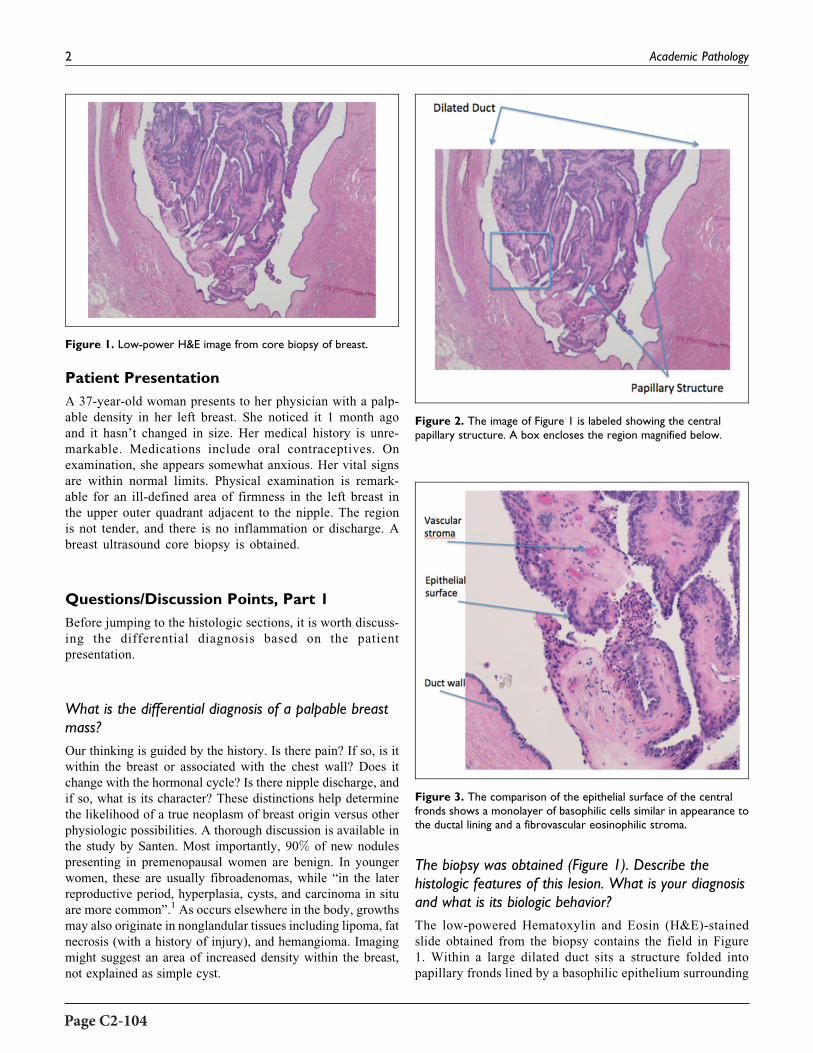
Patient Presentation
A 37-year-old woman presents to her physician with a palp-
able density in her left breast. She noticed it 1 month ago
and it hasn’t changed in size. Her medical history is unre-
markable. Medications include oral contraceptives. On
examination, she appears somewhat anxious. Her vital signs
are within normal limits. Physical examination is remark-
able for an ill-defined area of firmness in the left breast in
the upper outer quadrant adjacent to the nipple. The region
is not tender, and there is no inflammation or discharge. A
breast ultrasound core biopsy is obtained.
Questions/Discussion Points, Part 1
Before jumping to the histologic sections, it is worth discuss-
ing the differential diagnosis based on the patient
presentation.
What is the differential diagnosis of a palpable breastmass?
Our thinking is guided by the history. Is there pain? If so, is it
within the breast or associated with the chest wall? Does it
change with the hormonal cycle? Is there nipple discharge, and
if so, what is its character? These distinctions help determine
the likelihood of a true neoplasm of breast origin versus other
physiologic possibilities. A thorough discussion is available in
the study by Santen. Most importantly, 90% of new nodules
presenting in premenopausal women are benign. In younger
women, these are usually fibroadenomas, while “in the later
reproductive period, hyperplasia, cysts, and carcinoma in situ
are more common”.1 As occurs elsewhere in the body, growths
may also originate in nonglandular tissues including lipoma, fat
necrosis (with a history of injury), and hemangioma. Imaging
might suggest an area of increased density within the breast,
not explained as simple cyst.
The biopsy was obtained (Figure 1). Describe thehistologic features of this lesion. What is your diagnosisand what is its biologic behavior?
The low-powered Hematoxylin and Eosin (H&E)-stained
slide obtained from the biopsy contains the field in Figure
1. Within a large dilated duct sits a structure folded into
papillary fronds lined by a basophilic epithelium surrounding
Figure 1. Low-power H&E image from core biopsy of breast.
Figure 3. The comparison of the epithelial surface of the centralfronds shows a monolayer of basophilic cells similar in appearance tothe ductal lining and a fibrovascular eosinophilic stroma.
Figure 2. The image of Figure 1 is labeled showing the centralpapillary structure. A box encloses the region magnified below.
2 Academic Pathology
Page C2-104

a pink central fibrovascular stroma. These features are labeled
in Figure 2.
A box in Figure 2 is seen at higher magnification in Figure 3.
Figure 3 shows the duct wall and the epithelial surface of the
papillary fronds to be composed of similar cells, forming a
simple layer resting upon a basement membrane. The latter is
not easy to see directly with these dyes but can be visually
enhanced with special stains. Both tissues have matured in the
manner characteristic for ductal tissue, with a polarized surface
facing the duct lumen. The fronds also contain a stroma and
small blood vessels. The structure is named an “intraductal
papilloma.”
What Does the Name Tell You?
This is a benign proliferation in some ways analogous to adeno-
mas that may occur elsewhere. If it had beenmalignant, the name
would have included the word “carcinoma.” Papillomas have 2
main clinical presentations including solitary lesions near the
nipple or multiple peripheral papillomas. The latter have a higher
rate of atypia or malignancy arising in them. The most important
observation is that there is no evidence of invasion. In other
words, the epitheliumdoes not penetrate the basementmembrane
and spread into adjacent areas. This proliferative neoplastic pro-
cess can be associated with an increased risk of cancer in the
future, especially if there is atypical piling up of the epithelial
cells on each other (not seen here). In the absence of invasion, this
lesion is benign and doesnot need further treatment.However, the
patient should remain alert for changes in the future.
An additional important feature worth remembering is that
neoplasia (new growth) includes benign lesions. These may
grow to large size and can even become life threatening if they
interfere with a function (eg, benign meningioma). Benign
doesn’t mean, “you can live with it.” It means “not invasive.”
Other Manifestations of Benign Breast Disorders
Most important, benign lesions of the breast are much more
common than malignancies. The incidence of these disorders
changes with the age of the patient. An online review of Benign
Breast Disease in Women is referenced.1
Benign lesions of the breast are often classified in terms of
their potential contribution to future breast cancer. In this light,
there are 3 common categories:
� Nonproliferative changes (with little to no increased risk
of cancer). This includes fibrocystic change and cysts;
� Proliferative lesions without atypia, including this case;
� Proliferative lesions with atypia.
Proliferative disease is associated with a 1.5- to 2-fold
increased risk, while proliferative disease with atypia confers
a 4- to 5-fold increased risk.2
What is the structure and function of the basementmembrane that separates the epithelium from thefibrovascular core?
It is widely recognized that all epithelia organize themselves on
a structure called the basement membrane. However, the rela-
tionship of the cell to this structure is much more interesting
and active than many appreciate. Studies reveal “common
functions that include the induction and maintenance of cell
polarity, the establishment of barriers between tissue compart-
ments, the organization of cells into tissues, and the protection
of adherent cells from detachment-induced cell death,
anoikis.”3 Although the term membrane is used, it is not a lipid
membrane as the term is used elsewhere. Rather, the basement
membrane is a complex type of extracellular matrix with an
elaborate organization that includes collagen IV, laminins,
other connective proteins and heparin sulfate containing pro-
teoglycans. The absolute identity of the components of the
basement membrane varies among cell types and are essential
for the growth and maintenance of all epithelia.
Teaching Points
A quick review of the normal histology of ductal tissue in the
breast shows a simple monolayer of epithelial cells with an
underlying basement membrane.
� Despite the proliferative classification of this papilloma,
the basement membrane is not violated, so there is no
invasion.
� Invasion is an essential hallmark of epithelial
malignancy.
Benign lesions of the breast, depending on their microscopic
properties, may range in future risk of developing cancer from
none at all, to a 4- to 5-fold increase.
Acknowledgment
The original photographs were provided by Dr. Susan Fineberg.
References
1. Santen RJ. Benign breast disease in women. In: De Groot LJ,
Chrousos G, Dungan K, et al, eds. South Dartmouth, MA:
MDText.com, Inc; 2000. http://www.endotext.org, https://www.
ncbi.nlm.nih.gov/books/NBK278994/.
2. Kumar V, Abbas A, Aster A, eds. Robbins and Cotran Pathologic
Basis of Disease. 9th ed. Philadelphia, PA: Elsevier Saunders;
2015:1050-1051.
3. Miner JH, Yurchenco PD. Laminin functions in tissue morphogen-
esis. Ann Rev Cell Dev Biol. 2004, 20:255-284.
Sadofsky 3
Page C2-105


Title/Author Primary Objective
Secondary Objective(s) Page
Infectious Diseases: Pathogenesis, Diagnosis, Treatment and Prevention, Sheila E. Segura, MD, Gloria Ramos-Rivera, MD, and Mark Suhrland, MD
CYP1.3 – C3-1
Death Certification and the Role of the Medical Examiner/Coroner, Sarah Meyers, MD AU2.2 AU3.2 C3-7
Wrong Blood in Tube, Joan Uehlinger, MD GP1.1 – C3-11
Primary Osteosarcoma, ldis H. Petriceks, BA and Darren Salmi, MD MS1.2 SP1.4 C3-13
Head and Neck Neoplasia: Salivary Gland Tumors, Ryan P. Lau, MD, Melissa Yee-Chang, DO, and Amy Rapkiewicz, MD HN2.1 CYP1.2 C3-21
Cervical Neoplasia: HPV and its Link to Cancer, Teresa Kim, MD, Samer N. Khader, MBBS, and D. Yitzchak Goldstein, MD N3.1 CYP2.1, CYP2.2,
CYP2.3 C3-27
Medullary Thyroid Carcinoma, Carl T. McGary, MD, PhD EN5.2 CYP1.2 C3-33
Subarachnoid Hemorrhage Related to Ruptured Berry Aneurysm, Sarah Meyers, MD NSC7.3 AU2.2 C3-39
Com
petency 3
Competency 3: Diagnostic Medicine and Therapeutic Pathology

Educational Case Background & Submission Instructions
BackgroundBecoming a competent physician requires the ability to gain a broad foundation of knowledge, skills, and attitudes essential for independent medical practice. Essential in this is the understanding of the normal and pathological processes of each organ system, the ability to apply disease mechanisms to describe the pathobiology, and the ability to continually improve the diagnostic acumen and optimal treatment decisions through lifelong learning.
The Pathology Competencies for Medical Education (PCME) have detailed learning objectives under each goal that direct medical students and course directors to important facets of each learning goal that can be individually applied by learners. The competencies are divided into three sections—disease mechanisms and processes, organ system pathology, and diagnostic medicine and therapeutic pathology—and allow flexibility for each medical school and learner to apply the learning goals and objectives in a way that can keep the unique design of each curriculum or learning plan. The competencies are purposefully kept broad as they represent the minimum requirements of what pathology course directors across the nation have agreed upon to prepare medical students for entry into any residency program and for the subsequent contemporary practice of medicine.
Educational Cases for the PCME are current, peer-reviewed, and highlight the pathology competencies through fictional (but realistic) learning cases that can easily be adapted to multiple types of educational modalities. Educational Cases reference at least one primary learning objective, but may have one or more secondary learning objective(s). The pathology competencies and learning objectives are clearly indicated in the beginning of each case so that the focus of the educational case is evident. Key elements of the current format include clinical presentation, discussion questions or points, learning points, and references. The clinical presentation may include images or laboratory data for the patient’s presentation. The discussion questions or points are questions or statements that promote clinical reasoning followed by detailed explanations of the pathology, medicine, or therapeutics brought up in the discussion point or question. The learning points at the end of the case highlight the main teaching points from the preceding discussion. Thus, the cases demonstrate the application of medical reasoning to clinical scenarios that allow the learner to understand and apply diagnostic principles, incorporating morphologic findings and laboratory values with discussion of the laboratory medicine essentials for accurate diagnosis and treatment. References are included in each case and will allow the reader to review the original sources used to create the learning case or gain additional in-depth information. Thus, the Educational Cases are written in a style that can be easily used or adapted to multiple educational formats, such as small group discussions or flipped classrooms.
Case Submission GuidelinesSubmission Portal: https://mc.manuscriptcentral.com/apc Manuscript Type: Educational CaseKey Words:
o list “pathology competencies” as first keywordo list relevant competency, topic, learning goal, and objective keywords, e.g. “disease mechanisms, genetic mechanisms,
inheritance patterns”o other relevant keywords from the case content, e.g. “cystic fibrosis”
Abstract: “None needed”Case Content:
o Primary (and secondary if applicable) learning objective(s), cited from the PCME (doi: 10.1177/2374289517715040)o Example of primary objective formatting:
Objective GM1.2: Inheritance Patterns. Compare and contrast the inheritance patterns of different types of Mendelian disorders and give examples of each type of pattern. Competency 1: Disease Mechanisms and Processes; Topic GM: Genetic Mechanisms; Learning Goal 1: Genetic Mechanisms of Developmental and Functional Abnormalities.
o Patient Presentation: Include presentation (History of present illness, past medical history, etc. and physical examination)o Diagnostic Findings: This can include laboratory findings or histology.o Questions/Discussion Points: The questions and discussion points should be presented in a logical order to promote clinical
reasoning. In addition, you can include additional laboratory data/histologic images in later discussion points. The discussions should thoroughly explain the learning objectives to which the case is applied.
o Teaching Points: These should be covered in your discussion.o References: Linked to the discussion.
Images (if applicable): All images must be original work or have appropriate approval for publication.
Accepted CasesPublished Educational Cases receive the same scholarly recognition, citation and merit as other articles published in Academic Pathology. Cases accepted for publication will incur an Open Access article processing fee of $500.00 for authors who are faculty and students of APC member departments; $750 for non-members. For more information, contact Academic Pathology at [email protected] or 302-660-4940.

Educational Case
Educational Case: Infectious Diseases:Pathogenesis, Diagnosis, Treatment,and Prevention
Sheila E. Segura, MD1, Gloria Ramos-Rivera, MD1, and Mark Suhrland, MD1
The following fictional case is intended as a learning tool within the Pathology Competencies forMedical Education (PCME),
a set of national standards for teaching pathology. These are divided into three basic competencies: Disease Mechanisms and
Processes, Organ System Pathology, and Diagnostic Medicine and Therapeutic Pathology. For additional information, and
a full list of learning objectives for all three competencies, see http://journals.sagepub.com/doi/10.1177/2374289517715040.
Keywordspathology competencies, diagnostic medicine, cytopathology, cytologic diagnosis, infectious diseases, cervix disease, herpesinfection, trichomoniasis, candidiasis
Received March 12, 2018. Received revised May 16, 2018. Accepted for publication May 29, 2018.
Primary Objective
Objective CYP1.3: Identifying Infectious Diseases. Describe
the uses and limitations of cytology, with examples, in identi-
fying common infectious diseases.
Competency 3: Diagnostic Medicine and Therapeutic
Pathology; Topic CYP: Cytopathology; Learning Goal 1: Cyto-
logic diagnosis.
Patient Presentation #1
A 32-year-old female, gravida 1 para 0, at 36 weeks of gesta-
tion, presented to the OB/GYN clinic complaining of itchiness
and pain in the genital area. The patient denied a significant
past medical history. Physical examination revealed several
small vesicles in the vulvar and vaginal area. A cervical vaginal
cytology (PAP) test was performed.
Diagnostic Cytologic Findings, Part 1
Microscopic examination of the SurePath Pap test showed cells
with dense, intranuclear inclusions surrounded by a clear halo
and multinucleated cells with nuclear molding and chromatin
margination beneath the nuclear membrane imparting a clear-
ing or “stained glass appearance” of the nuclei (Figure 1).
What Is Your Differential Diagnosis Based on the ClinicalHistory and Cytologic Findings?
The differential diagnosis for large multinucleated cells on
cervical Pap test includes reactive endocervical cells, herpes
simplex virus (HSV) infection, syncytiotrophoblasts, low-
grade squamous intraepithelial cells, neoplastic cells (such as
carcinosarcoma, choriocarcinoma), and radiation- or
chemotherapy-induced changes. Based on the cytological fea-
tures,1 the final diagnosis for this Pap was “cellular changes
consistent with HSV infection.”
1 Montefiore Hospital and Medical Center, Bronx, NY, USA
Corresponding Author:
Sheila E. Segura, Montefiore Hospital and Medical Center, 111 East 210th
Street, Bronx, New York, NY 10467, USA.
Email: [email protected]
Academic PathologyVolume 5: 1–6ª The Author(s) 2018Article reuse guidelines:sagepub.com/journals-permissionsDOI: 10.1177/2374289518794191journals.sagepub.com/home/apc
Creative Commons Non Commercial CC BY-NC: This article is distributed under the terms of the Creative Commons Attribution-NonCommercial 4.0 License(http://www.creativecommons.org/licenses/by-nc/4.0/) which permits non-commercial use, reproduction and distribution of the work without furtherpermission provided the original work is attributed as specified on the SAGE and Open Access pages (https://us.sagepub.com/en-us/nam/open-access-at-sage).
Page C3-1

Questions/Discussion Points, Part 1
What Is Genital Herpes Simplex Virus Infection?
Herpes simplex virus, commonly known as herpes, is a chronic,
common, life-long viral infection caused by the 2 members of
the herpesvirus family, Herpesviridae. Herpes simplex virus
are categorized into 2 types: HSV-1 and HSV-2. Herpes sim-
plex virus type 1 is mainly transmitted by oral-to-oral contact
and causes infection in or around the mouth (oral herpes).
Herpes simplex virus type 2 is almost exclusively sexually
transmitted causing infection in the genital or anal area (genital
herpes). Most cases of recurrent genital herpes are caused by
HSV-2, and approximately 50 million people in the United
States are infected with this type of genital herpes.2,3
Describe the Symptoms and Transmission of GenitalHerpes Simplex Virus
Genital herpes infections often are asymptomatic or might
present with mild symptoms that sometimes go unrecognized.
Most infected people are unaware that they have the infection.
When symptoms do occur, genital herpes is characterized by
one or more genital or anal blisters or open sores (also called
ulcers), pain during urination, and itching. In addition to genital
ulcers, symptoms of recently acquired genital herpes infections
often include fever, fatigue, body aches, and swollen lymph
nodes. After an initial genital herpes infection with HSV-2,
recurrent symptoms are common but often less severe than the
first outbreak, and the frequency of outbreaks tends to decrease
over time.
Herpes simplex virus type 1 and 2 are transmitted by contact
with an infected person. Herpes simplex virus type 2 is mainly
transmitted during sex, through contact with genital surfaces,
skin, sores, or fluids. Herpes simplex virus type 2 is
periodically shed in the human genital tract, and most sexual
transmissions occur during periods of asymptomatic shedding.
The majority of HSV-1 infections spread easily via contact
with sores, saliva, and surfaces in or around the mouth causing
oral herpes infection. However, HSV-1 can also be transmitted
to the genital area through oral–genital contact to cause genital
herpes.3
Both viruses may also be transmitted vertically during child-
birth. However, the risk of infection transmission is minimal if
the mother has no symptoms or exposed blisters during deliv-
ery. The risk is considerable when the mother is infected with
the virus for the first time during late pregnancy.
How Is Herpes Simplex Virus Infection Diagnosed?
The patient’s prognosis and the type of counseling needed
depend on the type of genital herpes (HSV-1 or HSV-2) caus-
ing the infection; therefore, the clinical diagnosis of genital
herpes should be confirmed by type-specific laboratory testing.
Both type-specific virologic and type-specific serologic tests
for HSV should be available in clinical settings that provide
care to persons with or at risk for sexually transmitted diseases
(STDs).2 The Centers for Disease Control and Prevention
recommends that persons with genital herpes should be tested
for HIV infection as well.2
The following tests are used for HSV diagnosis:
� Viral culture. This test involves taking a tissue sample
or scraping of the sores for examination in the labora-
tory. The sensitivity of viral culture is low, especially
for recurrent lesions, and declines rapidly as lesions
begin to heal.
� Polymerase chain reaction test. Polymerase chain
reaction is used to copy the DNA from a sample of
blood, tissue from a sore, or spinal fluid. The DNA
can then be tested to establish the presence of HSV
and determine which type of HSV. Polymerase chain
reaction is the test of choice for diagnosing HSV
infections affecting central nervous system (CNS) and
systemic infections.
� Blood test. Both type-specific and type-common anti-
bodies to HSV develop during the first several weeks
after infection and persist indefinitely. The blood
sample is analyzed via enzyme-linked immunosor-
bent assay which detects IgM and type-specific IgG.
Although the cytologic detection of cellular changes asso-
ciated with HSV infection is an insensitive and nonspecific
method of diagnosing genital lesions (ie, Tzanck preparation),
the cytologic changes of infected cells are recognized in PAP
cervical smears.
In 1989, the Bethesda System (TBS) was introduced to
standardize the reporting cervical cytology PAP results and
to incorporate the insights into human papillomavirus (HPV)
biology and cervical disease association. The current 2014
Figure 1. Slide shows a single and a multinucleated squamous cellswith nuclear molding and chromatin margination (arrow) beneath thenuclear membrane imparting a clearing or “ground glass appearance”of the nuclei (PAP-stained, high power �60 magnification).
2 Academic Pathology
Page C3-2

TBS, like its predecessors, recommends the use of a specific
format for the cytology cervical report.1
The cytologic features of the HSV-infected squamous cells
are variable. The infected cells can show accumulation of viral
particles within the nuclei, resulting in an eosinophilic inclu-
sion, known as Cowdry inclusions, which are classified as type
A (inclusion in the nucleus) and type B (inclusion in the cyto-
plasm), which are seen in cytomegalovirus infection. The cells
can also show lysis of the chromatin giving a glassy appearance
to the nuclei. The infected cells aggregate forming a Multi-
nucleated cell with nuclear Molding and chromatin Margina-
tion (3 M’s of herpes) features easily recognized under the
microscope1,4; however, some other lesions or changes may
mimic HSV infection on cytologic examination, including
reactive or reparative changes, air drying artifact, multinu-
cleated giant cells, poor cell preservation, radiation effect,
among others.4 Under the current Bethesda system for report-
ing PAP results, for the cytologic changes caused by HSV
infection, it is recommended to interpret it as “cellular changes
consistent with HSV.1
Which Are the Possible Complications ofHerpes Infection?
Severe disease. In immunocompromised people, such as those
with advanced HIV infection, HSV-1 can have more severe
symptoms and more frequent recurrences. Rarely, HSV-1
infection can also lead to more severe complications such as
encephalitis or keratitis (eye infection).
Genital herpes in pregnancy and neonatal herpes. Neonatal herpescan occur when an infant is exposed to HSV in the genital tract
during delivery. This is a rare condition, occurring in an esti-
mated 10 out of every 100 000 births globally, and the risk of
HSV transmission to the neonate from an infected mother is
high (30%-50%) among women who acquire genital herpes
near the time of delivery and low (<1%) among women with
prenatal histories of recurrent herpes or who acquire genital
HSV during the first half of pregnancy.2 Neonatal HSV infec-
tion can be divided into 3 clinical groups: (1) skin, eyes and
mouth disease (SEM) is a localized infection affecting the
skin, eyes, or mouth; (2) CNS disease is defined as encepha-
litis with or without SEM disease, and (3) disseminated dis-
ease involves infection in multiple organ systems and can
include hepatitis, pneumonitis, and disseminated intravascu-
lar coagulation. Cutaneous lesions may be seen in all types
and disseminated disease may occur with or without the pres-
ence of CNS disease.3
Prevention of neonatal herpes depends both on preventing
acquisition of genital HSV infection during late pregnancy
and avoiding exposure of the neonate to herpetic lesions and
viral shedding during delivery. Women without symptoms or
signs of genital herpes or its prodrome can deliver vaginally.
Although cesarean delivery does not completely eliminate the
risk for HSV transmission to the neonate, women with recur-
rent genital herpetic lesions at the onset of labor should
deliver by cesarean delivery to reduce the risk for neonatal
HSV infection.2 Acyclovir can be administered orally to preg-
nant women with first-episode genital herpes or recurrent
herpes and should be administered intravenously to pregnant
women with severe HSV infection. All infants who have neo-
natal herpes should be promptly evaluated and treated with
systemic acyclovir.
What Is the Treatment for Herpes Infection?
Antivirals, such as acyclovir, famciclovir, and valacyclovir, are
the most effective medications available for people infected
with HSV. These can help to reduce the severity and frequency
of symptoms, but cannot cure the infection.2,3
In our case, the patient was treated with oral acyclovir with
improvement of the symptoms. Soon after, she delivered a
healthy female baby. Her HIV testing was negative.
Patient Presentation #2
A 67-year-old male with a past medical history of urolithiasis
and blood in his urine (gross hematuria) presented to his pri-
mary care physician for follow-up. Urine was collected and
sent for cytologic examination.
Diagnostic Cytologic Findings, Part 2
Microscopic examination of the slide revealed benign urothe-
lial and squamous cells in a background of acute inflammation
and red blood cells. Incidentally, scattered, small, oval-shaped
“cells” with eccentric dark nuclei and gray cytoplasm with rare
red cytoplasmic granules were also seen (Figure 2).
Figure 2. Slide shows several oval-shaped organisms (arrow) witheccentric dark nuclei (arrow head) and gray cytoplasm with redgranules. A flagella (asterisk) is also seen. Benign squamous cells arepresent in the background. These organisms are consistent with Tri-chomonas vaginalis (PAP-stained, high power �60 magnification).
Segura et al 3
Page C3-3

What Is Your Differential Diagnosis Based on theClinical History and Cytologic Findings?
The differential diagnosis for these small “cells” includes
degenerated inflammatory cells, cellular debris, cellular frag-
ments, and parasites. The cytologic findings were consistent
with Trichomonas organisms.
Questions/Discussion Points, Part 2
What Is Trichomoniasis and How Is It Transmitted?
Trichomonas vaginalis (commonly known as “trich”) is an
anaerobic, flagellated protozoan parasite and the causative
agent of the STD trichomoniasis. Trichomoniasis is the most
prevalent nonviral STD in the United States, affecting an esti-
mated 3.7 million people.5
Trichomoniasis is typically found in sexually active
patients. Transmission occurs predominantly via sexual inter-
course. In women, the most commonly infected part of the
body is the lower genital tract (vulva, vagina, cervix, or ure-
thra). In men, the most commonly infected body part is the
urethra. It has not been isolated from oral sites, and rectal
prevalence appears to be low in men who have sex with men.5
What Are the Signs and Symptoms of Trichomoniasis?
About 70% to 85% of infected people have minimal symptoms
or are asymptomatic.5 When trichomoniasis does cause symp-
toms, they can range from mild irritation to severe inflamma-
tion. Some people are symptomatic within 5 to 28 days after
infection. Symptoms can come and go, and untreated infections
might last for months to years.5,6
Some infected men have symptoms of urethritis (urethral
discharge) and pain during urination, and some infected women
have vaginal discharge that might be diffuse, malodorous, or
yellow-green with or without vulvar irritation. Other symptoms
includes itching, discomfort during sexual intercourse or urina-
tion, and cervicitis in women, which is characterized by puru-
lent discharge and easily induced endocervical bleeding, also
known as “strawberry” cervix (due to capillary dilation as a
result of the inflammatory response).
What Are the Complications of Trichomoniasis?
Trichomonas vaginalis infection is associated with 2- to 3-fold
increased risk for HIV acquisition. It also increases the suscept-
ibility to other viruses, including herpes and HPV. In pregnant
women, T vaginalis infection has been associated with an
increased risk of low birth weight, preterm delivery, and intrau-
terine infection.5
Neonatal trichomoniasis has been described.6 Respiratory or
genital infection in the newborn may also occur. In men, com-
plications of untreated trichomoniasis include prostatitis, epi-
didymitis, urethral stricture disease, and infertility, potentially
resulting from decreased sperm motility and viability.
How Is Trichomoniasis Diagnosed?
It is not possible to diagnose trichomoniasis based on symp-
toms alone. In women, vaginal trichomoniasis has historically
been diagnosed by wet mount microscopy, which is per-
formed by placing a small amount of vaginal discharge on a
microscope slide and mixing with a few drops of saline solu-
tion. The slide is then examined under a microscope at low or
medium power and a “corkscrew” motility is observed (para-
site moving through the field). It is the most common method
for T vaginalis diagnosis because of convenience and rela-
tively low cost.5,6
Culture was considered the gold standard method for diag-
nosing T vaginalis infection before molecular detection meth-
ods became available. Trichomonas vaginalis may be
accurately identified on Pap smear by morphology alone, but
this test yields low sensitivity (50%-80%) and false-positive
results are also common with this technique.5 Cellular debris
and degenerated inflammatory cells may be mistaken for tri-
chomonads on cytologic examination.4
The use of highly sensitive and specific methods using
molecular techniques for detecting antigens, DNA, or RNA are
currently recommended for diagnosing T vaginalis.5
What Is the Treatment for Trichomoniasis?
Trichomoniasis can be treated with nitroimidazole, which is the
only class of antimicrobial medications known to be effective
against T vaginalis infections. Of these drugs, metronidazole
and tinidazole have been cleared by the Food and Drug Admin-
istration (FDA) for the oral or parenteral treatment of tricho-
moniasis. It is not recommended to drink alcohol within 24
hours after taking this medication.
Because of the high rate of reinfection among women
treated for trichomoniasis (*17%), retesting for T vaginalis
is recommended for all sexually active women within 3 months
following initial treatment regardless of whether they believe
their sex partners were treated.5 Concurrent treatment of all sex
partners is critical for symptomatic relief, microbiologic cure,
and prevention of transmission and reinfections. Current part-
ners should be referred for presumptive therapy to avoid
reinfection.
Although metronidazole crosses the placenta, data suggest
that it poses a low risk to pregnant women. No evidence of
teratogenicity or mutagenic effects in infants has been found in
multiple studies of pregnant women. Women can be treated
with 2 g metronidazole in a single dose at any stage of preg-
nancy. The patient and his partner were treated with oral
metronidazole.
Patient Presentation #3
An 11-month-old girl with 1-month history of emesis, gagging,
and choking with textured foods was brought to her pediatri-
cian by her parents and, after examination, was admitted for
management. The prior month, she was briefly admitted to the
4 Academic Pathology
Page C3-4

hospital for a respiratory syncytial virus bronchiolitis and was
given oral prednisone 10 mg twice a day for 5 days. On current
admission, the baby underwent an esophagogastroduodeno-
scopy. The middle to distal esophagus was covered with
yellow-white plaques scattered over the mucosa. The mucosa
was hyperemic and friable, and the plaques could not be
washed off. The lesions bled easily at the site of attachment,
where they were brushed for cytology, Gram stain, and fungal
culture. Biopsies were also taken.
Diagnostic Cytologic Findings, Part 3
Microscopic examination of the cytology brushing smears
reveled esophageal squamous cells admixed with acute inflam-
matory cells. Fungal pseudohyphae and spores, morphologically
consistent with Candida species, were also present (Figure 3).
The Grocott-Gomori’s methenamine silver (GMS) stain on
smears and culture also showed Candida organisms. The biopsy
results were consistent with chronic esophagitis suggestive of
reflux, with Candida-like organisms. She was treated with oral
fluconazole and omeprazole with improvement of her
symptoms.
Questions/Discussion Points, Part 3
What Is Candidiasis?
Candidiasis is a fungal infection caused by the yeast Candida.
More than 20 types of Candida can cause infection, with Can-
dida albicans being the most common. Candida yeasts are
generally present in healthy humans, frequently as part of the
human body normal oral and intestinal flora, and particularly
on the skin, but it can become pathogenic if the host’s luminal
flora or immune defenses are altered.7,8
Factors that increase the susceptibility of the host to Can-
dida infection are various malignancies, immunodeficiency
states, diabetes, stress, antibiotic treatments, nutrient defi-
ciency, endocrinopathies, long-term use of steroids, and other
immunosuppressive drugs.
What Is the Clinical Presentation of Candidiasis?
Signs and symptoms of candidiasis vary depending on the area
affected. Most candidal infections result in minimal complica-
tions such as redness, itching, and discomfort, though compli-
cations may be severe or even fatal if left untreated in certain
populations.
The clinical presentation can be broadly divided into cuta-
neous candidiasis, mucosal candidiasis, and systemic candi-
diasis. In immunocompetent persons, candidiasis is usually a
localized infection of the skin, fingernails or toenails (ony-
chomycosis), or mucosal membranes. The most common
manifestation of candidal infection in infants is diaper derma-
titis. Candida organisms can also cause intertrigo in older
individuals. The oral infection or oropharyngeal candidiasis,
commonly called “thrush,” frequently occurs in infants and
toddlers. Infection in the esophagus is called esophageal can-
didiasis or Candida esophagitis, and the most common pre-
senting complaints are difficulty and pain with swallowing,
with one-third of patients also having oral thrush. Infection of
the vulva or vagina also referred as “yeast infections” or
“vulvovaginal candidiasis” affects nearly 75% of women,
usually causing mild symptoms, such as itching, burning,
soreness, irritation, and a whitish or whitish-gray cottage
cheese-like discharge.7
Candida organisms can cause severe systemic infections in
immunocompromised patients, such as patients with AIDS,
premature babies, critically ill patients, or patients with cancer.
In these patients, Candida yeast enters the bloodstream and
spreads to internal organs, such as CNS, kidneys, liver, bones
and joints, among others.
How Is Candidiasis Diagnosed?
Depending on the clinical presentation, several diagnostic tests
are available and include the following:
� Mucocutaneous candidiasis—Using a wet mount, scrap-
ings or smears obtained from skin, nails, or oral or vagi-
nal mucosa are examined under the microscope; a
potassium hydroxide (KOH) smear, GMS stain, or
methylene blue is useful for direct demonstration of
fungal organisms.
� Cutaneous candidiasis—Using a wet mount, scrapings
or smears obtained from skin or nails can be examined
under the microscope; KOH smears are also useful.
� Genitourinary candidiasis—A urinalysis and urine fun-
gal cultures are useful. A sample of vaginal discharge
can be examined under the microscope or sent to a
laboratory for a fungal culture.
Figure 3. Microscopic examination of the smears reveled esophagealsquamous mucosa admixed with acute inflammatory cells and fungalhyphae (arrow) and spores (arrow head), morphologically consistentwith Candida species (PAP-stained, high power �60 magnification).
Segura et al 5
Page C3-5

� Gastrointestinal candidiasis—Endoscopy with or with-
out biopsy and brushings. Samples are submitted for
microscopic examination, GMS stain, and culture.
� Systemic candidiasis—Usually blood cultures.
Candida is a common finding on PAP cervical smears and
identification does not necessarily indicate infection (com-
mensal in vaginal mucosa). Noncellular substances such as
fibrin and contaminants such as carpet fibers or other fungi
can be also mistaken with Candida under microscopic exam-
ination. The Bethesda system recommends reporting these
fungi as “fungal organism morphologically consistent with
Candida spp”.1
How Is Candidiasis Treated?
Candidiasis is generally treated with antifungal medications;
these include clotrimazole, nystatin, fluconazole, voriconazole,
amphotericin B, and echinocandins. The management also
depends on the clinical presentation.7
� Cutaneous candidiasis—Most localized cutaneous can-
didiasis infections can be treated with any number of
topical antifungal agents.
� Mucocutaneous candidiasis—For oral cases, topical
antifungals are used. For severe infections, treatment
with oral agents is commonly used.
� Esophageal candidiasis—Treatment requires systemic
therapy with fluconazole.
� Systemic candidiasis—Treatment requires systemic
intravenous therapy.
Teaching Points
� Some infections are easily recognized with cytologic
examination of PAP and other body site specimens.
� Herpes infections, oral and genital, are common chronic
infections due to the herpes virus (HSV-1 and HSV-2),
characterized by itching and blisters/sores on the mouth
and/or genital area.
� Genital HSV in pregnant women can be complicated
with neonatal herpes.
� Trichomoniasis, caused by the T vaginalis parasite, is
one of the most common STD infections in the United
States, and is usually asymptomatic.
� Candida spp is a fungus (yeast), part of the human body
normal mucocutaneous and intestinal flora, but it can
become pathogenic if the host’s luminal flora or immune
defenses are altered.
� Although HSV, Candida spp and Trichomonas infec-
tions can be identified with cytologic examination,
false-positive results are common, sometimes requiring
alternative confirmatory tests.
Declaration of Conflicting Interests
The author(s) declared no potential conflicts of interest with respect to
the research, authorship, and/or publication of this article.
Funding
The author(s) received no financial support for the research, author-
ship, and/or publication of this article.
References
1. Edmund C, Ducatman B. Cytology: Diagnostic Principles and
Clinical Correlates. 4th ed. Philadelphia, PA: Elsevier/Saunders;
2014.
2. Centers for Disease Control and Prevention. 2015 STD Treatment
Guideline-Genital HSV Infections: Diseases Characterized by
Genital, Anal, or Perianal Ulcers. Atlanta, GA: Centers for Dis-
ease Control and Prevention; 2015. Available at: https://www.cdc.
gov/std/tg2015/herpes.htm. Accessed January 2018.
3. World Health Organization. Herpes simplex virus. 2017. Available
at: http://www.who.int/mediacentre/factsheets/fs400/en/. Accessed
January 2018.
4. Khalbuss WE, Monaco SE, Pantanowitz L. Normal and Benign
Pap Test. In Quick Compendium of Cytopathology. Chicago, IL:
American Society for Clinical Pathology; 2013:25-50.
5. Centers for Disease Control and Prevention. Sexually Transmitted
Diseases Treatment Guidelines, 2015: Diseases Characterized by
Vaginal Discharge. Atlanta, GA: Centers for Disease Control and
Prevention; 2015. Available at: https://www.cdc.gov/std/tg2015/
trichomoniasis.htm. Accessed January 2018.
6. Van der Pol B. Trichomonas vaginalis infection: the most prevalent
nonviral sexually transmitted infection receives the least public
health attention. Clin Infect Dis. 2007;44:23-25. doi:10.1086/
509934.
7. Centers for Disease Control and Prevention. Fungal Diseases-
Types of Fungal Diseases-Candidiasis. Centers for Disease Con-
trol and Prevention. Available at: https://www.cdc.gov/fungal/dis
eases/candidiasis/thrush/index.html. Accessed January 2018.
8. Aliye UC, North PE, Burks AW. Esophageal candidiasis in an
infant with reflux esophagitis, case report. J Pediatr Gastroenterol
Nutr. 2000;31:572-574.
6 Academic Pathology
Page C3-6
Educational Case
Educational Case: Death Certification andthe Role of the Medical Examiner/Coroner
Sarah Meyers, MD1
The following fictional case is intended as a learning tool within the Pathology Competencies for Medical Education
(PCME), a set of national standards for teaching pathology. These are divided into three basic competencies: Disease
Mechanisms and Processes, Organ System Pathology, and Diagnostic Medicine and Therapeutic Pathology. For
additional information, and a full list of learning objectives for all three competencies, see http://journals.sagepub.com/
doi/10.1177/2374289517715040.
Keywordspathology competencies, diagnostic medicine, autopsy, death certificate, forensic autopsy, reportable deaths, cause of death,manner of death, coroner, medical examiner
Received April 15, 2017. Received revised June 19, 2017. Accepted for publication July 13, 2017.
Primary Objective
AU 2.2: Components of the Death Certificate. Discuss the key
components of the death certificate; difference among imme-
diate, intermediate, and underlying (proximate) cause of death
based on disease process; and the role of mechanisms of death
on a death certificate.
Competency 3: Diagnostic Medicine and Therapeutic Pathol-
ogy; Topic AU: Autopsy; Learning Goal 2: Death Certificate.
Secondary Objective
AU 3.2: Reportable Deaths. Identify circumstances of death
that need to be reported to the medical examiner/coroner.
Competency 3: Diagnostic Medicine and Therapeutic Pathol-
ogy; Topic AU: Autopsy; Learning Goal 3: Forensic Autopsy.
Patient Presentation
A 45-year-old male is admitted to the hospital from his skilled
nursing facility 1 month prior to his death, with clinical signs and
symptoms of sepsis. Urine and blood cultures confirm the clinical
suspicion of urosepsis due to Escherichia coli. Medical history
includes obesity, quadriplegia, neurogenic bladder with indwel-
ling catheter, autonomic instability, hypertension, obstructive
sleep apnea, and diabetes mellitus. The patient develops
pneumonia and his oxygenation statusworsens 2 days prior to his
death. He is intubated and sedated. A lung biopsy is performed
and shows changes consistent with diffuse alveolar damage. The
clinical team meets with the family for a care conference and
shares the results of the biopsy. The clinical team states that, due
to his underlying diseases and the severity of his current illnesses,
it is not likely that he will survive the current hospitalization. The
family decides to transition the patient to comfort care and he dies
2 days after developing acute respiratory distress syndrome.
Diagnostic Findings, Part 1
No additional information is gathered by this medical provider
and the death certificate is completed as follows:
� (Immediate) Cause of death: Cardiorespiratory failure
� Due to (intermediate cause of death): E coli sepsis
� Manner of death: Natural
1 Hennepin County Medical Examiner’s Office, Minneapolis, MN, USA
Corresponding Author:
Sarah Meyers, Hennepin County Medical Examiner’s Office, 530 Chicago Ave,
Minneapolis, MN 55415, USA.
Email: [email protected]
Academic PathologyVolume 5: 1–3ª The Author(s) 2018Article reuse guidelines:sagepub.com/journals-permissionsDOI: 10.1177/2374289517724724journals.sagepub.com/home/apc
Creative Commons Non Commercial CC BY-NC: This article is distributed under the terms of the Creative Commons Attribution-NonCommercial 4.0 License(http://www.creativecommons.org/licenses/by-nc/4.0/) which permits non-commercial use, reproduction and distribution of the work without furtherpermission provided the original work is attributed as specified on the SAGE and Open Access pages (https://us.sagepub.com/en-us/nam/open-access-at-sage).
Page C3-7

Questions/Discussion Points, Part 1
What is the difference between cause and manner ofdeath?
Cause of death is an injury or disease process that resulted in
an individual’s death.1-4 A myriad of terms may be used to
describe cause of death, including disease processes such as
acute myocardial infarct or injuries such as gunshot wound of
the head. When sequences of injuries or events occur, the cause
of death portion of the death certificate may be further sub-
classified into immediate (the last injury or disease process in
the sequence or the event immediately before death), inter-
mediate, and underlying cause of death (the injury or disease
that set off the sequence of events that resulted in death, also
known as the proximate cause of death).1-4 These terms are
connected via “due to” statements on a death certificate. For
example, if an individual sustains a myocardial infarct, from
arteriosclerotic heart disease, which subsequently ruptures
causing cardiac tamponade from hemopericardium, the cause
of death would be listed as such:
� Immediate cause of death: Hemopericardium with car-
diac tamponade
� Due to: (intermediate cause of death): Ruptured myo-
cardial infarct
� Due to (underlying cause of death/proximate cause of
death): Arteriosclerotic heart disease
Manner of death is a public health categorization that gen-
erally includes only 5 options: natural (a death due to disease
processes), accident (nonnatural injuries caused the death; the
majority of traffic collisions and drug toxicity deaths are certi-
fied as such), suicide (an individual intentionally acted to end
their life), homicide (another individual/s caused injuries that
resulted in a death), and undetermined (certified when there is
conflicting or insufficient information to categorize).1-4 It is
important to note that manner of death classification is strictly
defined by public health terminology and not intended to dic-
tate legal actions (for example, if a 2-year-old finds a gun and
shoots another individual, the death may be classified as a
homicide, as one individual caused the death of another indi-
vidual; however, murder or manslaughter charges are not likely
to be filed against the 2-year-old. In another example, if a
pedestrian is struck by a vehicle and dies as the result of blunt
force injuries sustained in that collision, the manner of death
may be certified as accident. This does not preclude legal
charges, including vehicular manslaughter charges, from being
filed against the driver of the vehicle).
Is this death certificate complete? If it is not, please statewhat should be changed or what additional informationmay be necessary to complete the death certificate?
The death certificate is not complete. At this time, the physi-
cian completing the death certificate does not have enough
information. Although the immediate and intermediate causes
of death are known (acute respiratory distress syndrome due to
E coli urosepsis), the clinician should ask themselves why a
45-year-old male developed these conditions to the degree that
they caused death. Phrased another way, a healthy 45-year-old
male should likely not develop urosepsis, so what is different
about this individual patient that caused this disease to develop
and cause his death? In this situation, the indwelling catheter is
the likely origin for the urosepsis, which was placed due to a
neurogenic bladder related to quadriplegia. The cause of the
patient’s quadriplegia needs to be established, as it may relate
to the underlying cause and manner of death. In addition, as
specific time intervals are known for the individual elements of
the immediate and intermediate causes of death, these time
intervals should be listed in the appropriate section of the
death certificate.
Should “cardiorespiratory failure” be listed as animmediate cause of death?
Cardiorespiratory failure is most appropriately categorized as a
mechanism of death and is the terminal mechanism of death for
most individuals. Death certificates not only serve to provide
family members with important information about disease pro-
cesses but are also important resources for public health data
collection and monitoring of diseases (such as cancer and heart
disease) and injury subtypes. As such, cardiorespiratory failure
is too nonspecific to be of assistance to either the family of
the decedent or public health departments and should not be
included in this death certificate. The background information
provided in the clinical history is sufficient to provide a specific
cause of death.
Diagnostic Findings, Part 2
The original death certificate is filed and the family elects
cremation as final disposition for the body. The funeral director
in charge of the cremation calls the local medical examiner’s
office to inquire if the bullet in the deceased’s spine needs to be
removed prior to cremation.
Questions/Discussion Points, Part 2
What additional steps should the medical examiner’s orcoroner’s office complete to investigate this death?
The medical examiner’s or coroner’s office is tasked with
investigating unexpected, unexplained, and unattended deaths
of individuals in the communities they serve. Reports of deaths
are usually made by medical personnel and law enforcement;
however, anyone, including family members and funeral direc-
tors, may report circumstances of death to the office, as
occurred in this case. Given the remote trauma and potential
retained evidence (the bullet) available for recovery, the med-
ical examiner’s office will usually begin by obtaining law
enforcement and medical records to compile information used
to complete a death certificate. Depending on circumstances,
2 Academic Pathology
Page C3-8

the individual may be brought for a postmortem examination to
retrieve the projectile. Of note, given that remote trauma was
involved in the death, this death would fall under the jurisdic-
tion of the medical examiner or coroner and should have been
reported by medical personnel at the time of his death.
In this situation, law enforcement investigation and medical
records indicate that the deceased was shot by an unknown
assailant 20 years prior to his death, resulting in quadriplegia,
neurogenic bladder, and finally urosepsis, which ultimately
resulted in his respiratory distress syndrome.
Should the death certificate be changed, and if so, pleaselist the immediate, intermediate, and underlying cause ofdeath and the manner of death?
A death certificate should tell the whole story of this patient’s
history and therefore should be amended to work backward
from the underlying cause of death (the disease or injury that
set off the sequence of events that resulted in death) to the
immediate cause of death (the last event/injury in the sequence,
in this case, acute respiratory distress syndrome). A death cer-
tificate should be amended and reissued as the cause of death
statements is incomplete and the manner of death is incorrect.
As another individual caused the injury that set off the chain of
events that resulted in the individual’s death, the most appro-
priate manner of death is homicide. The death certificate, as
signed by the medical examiner following autopsy examination
and investigation, read as follows:
� Immediate cause of death: Acute respiratory distress
syndrome (Time interval: 2 days)
� Due to: E coli urosepsis (time interval: 1 month)
� Due to: Quadriplegia with neurogenic bladder (time
interval: 20 years)
� Due to: Gunshot wound of the back
� Manner of death: Homicide
Teaching Points
� The cause of death is the disease or injury that resulted
in an individual’s death and should be categorized from
the first disease or injury (the underlying cause of
death) that set off the sequence that lead to the imme-
diate cause of death.
� Each death must be traced back to the inciting disease
or injury, which may have occurred years prior to the
death.
� “Cardiorespiratory failure” is a nonspecific mechanism
of death that provides insufficient detail about the cir-
cumstances of death and does not allow for important
information to be passed to decedent’s families or public
health monitoring systems. Whenever possible, specific
disease processes and injuries should be listed as causes
of death for accurate death certification.
� The medical examiner’s office or coroner’s office are
tasked with investigating unexpected, unexplained, or
unattended deaths in a particular region. Medical pro-
viders are responsible for reporting deaths that may fall
under the medical examiner’s or coroner’s jurisdiction
to the appropriate office.
Declaration of Conflicting Interests
The author(s) declared no potential conflicts of interest with respect to
the research, authorship, and/or publication of this article.
Funding
The author(s) received no financial support for the research, author-
ship, and/or publication of this article.
References
1. Death certification. (2005). In: Dolinak D, Matshes E, Lew E, eds.
Forensic Pathology: Principles and Practice. 1st ed. Burlington,
MA: Elsevier Academic Press:663-668.
2. Death certification. National Association of Medical Examiners.
https://netforum.avectra.com/eweb/DynamicPage.aspx?Site¼NAME&WebCode¼DeathCert. Accessed May 31, 2017.
3. Medicolegal Investigative Systems. (2001). In: DiMaio VJ,
DiMaio D, eds. Forensic Pathology. 2nd ed. Boca Raton, FL:
CRC Press:1-19.
4. Trauma and disease. (2006). In: Spitz WU, Spitz DJ, eds. Medico-
legal Investigation of Death. 4th ed. Springfield, IL: Charles C
Thomas:436-459.
Meyers 3
Page C3-9


Educational Case
Educational Case: Wrong Blood in Tube
Joan Uehlinger, MD1
The following fictional case is intended as a learning tool within the Pathology Competencies for Medical Education
(PCME), a set of national standards for teaching pathology. These are divided into three basic competencies: Disease
Mechanisms and Processes, Organ System Pathology, and Diagnostic Medicine and Therapeutic Pathology. For
additional information, and a full list of learning objectives for all three competencies, see http://journals.sagepub.com/
doi/10.1177/2374289517715040.
Keywordspathology competencies, preanalytic error, wrong blood in tube, quality improvement, process control, specimen identification
Received February 17, 2017. Received revised April 6, 2017. Accepted for publication April 13, 2017.
Primary Objective
Objective GP1.1: Pre- and Postanalytical Errors. Give examples of
common sources of preanalytical and postanalytical errors and
categorize errors when the following procedures are not properly
followed: pairing patient/specimen identification with the re-
quisition forms, using correct specimen containers/tubes for
specific tests, and timing of collection, transport, and storage.
Competency 3: Diagnostic Medicine and Therapeutic
Pathology; Topic GP: General Principles; Learning Goal 1:
Laboratory Tests.
Patient Presentation
A 35-year-old woman is scheduled for a tubal ligation, and a pre-
operative type and screen is sent. She had never been transfused at
this facility.A typeandscreenwasdrawn6months agoandshowed
that she was A positive with a negative antibody screen.
Diagnostic Findings, Part 1
Results from the current type and screen revealed the sample
was O positive with a negative antibody screen.
Questions/Discussion Points, Part 1
What Are the First Steps Taken Upon Sample Receipt?
Check that the sample is labeled with 2 identifiers. AABB
publishes standards that are required to ensure the safety of
blood products in all steps from donation of blood to testing
of blood products to transfusion of blood products. Standards
specifically indicate that “identifying information for the
patient and the sample shall correspond and be confirmed at
the time of collection using two independent identifiers.”1,2
Another standard says that “the transfusion service shall con-
firm that all identifying information on the request is in agree-
ment with that on the sample.”1,2 The purpose of using 2
identifiers is to ensure that the blood sample is from the correct
patient, and should that patient need blood products, the correct
blood products can then be issued.
Review the patient history
The history review is a required step in sample processing.
The standards indicate that “there shall be a process to ensure
that the historic records for the following have been revie-
wed . . .ABO group and Rh type. . . .These records shall be com-
pared to current results, and any discrepancies shall be
investigated and appropriate action taken . . . .”2,3 The purpose
of reviewing the historic records is to help ensure that if there is
1 Montefiore Medical Center and The Albert Einstein College of Medicine,
Bronx, NY, USA
Corresponding Author:
Joan Uehlinger, MD, Montefiore Medical Center and The Albert Einstein
College of Medicine, 111 East 210th Street, Bronx, New York, NY 10467, USA.
Email: [email protected]
Academic PathologyVolume 4: 1–2ª The Author(s) 2017Reprints and permission:sagepub.com/journalsPermissions.navDOI: 10.1177/2374289517708310journals.sagepub.com/home/apc
Creative Commons CC BY-NC: This article is distributed under the terms of the Creative Commons Attribution-NonCommercial 4.0 License(http://www.creativecommons.org/licenses/by-nc/4.0/) which permits non-commercial use, reproduction and distribution of the work without furtherpermission provided the original work is attributed as specified on the SAGE and Open Access pages (https://us.sagepub.com/en-us/nam/open-access-at-sage).
Page C3-11

an accidental mislabeling of a patient sample, it will be identified
before any blood products are administered to the patient.
What Possible Explanations Should Be Considered?
Except in 1 particular circumstance, the blood type of a patient
should not change. There are a limited number of possible
explanations for the current findings. The current sample could
be from the wrong patient. The sample taken 6 months ago
could have been from the wrong patient. Or, if the patient had
a stem cell transplant in the interval from a type O allogeneic
donor, her blood type could have changed.
What Should the First Steps Be to Determine the Etiologyof the Problem?
There are 2 very important steps that need to be completed to
investigate a possible mislabeling of a blood sample. The first
is to get the history to be sure the patient did not have a trans-
plant, and in addition to ask for another sample to be drawn
from the patient to see whether it matches the historical data or
the data from the current blood sample.
Diagnostic Findings, Part 2
Evaluation reveals that the patient has not had a stem cell
transplant. A second sample is sent for evaluation which is
determined to be A positive, negative antibody screen.
Questions/Discussion Points, Part 2
What Are Your Conclusions?
The sample drawn earlier that day was from the wrong patient.
The phlebotomy area should be alerted as there may be another
incorrect sample if this was a single direct switch of identifiers on
the tube (another patient who is really an Omay have been typed
as anA). For patients that are hospitalized, it is just as important to
evaluate where the blood sample came from and investigate if
there could be an accidental mislabeling of samples.
Diagnostic Findings, Part 3
A second patient was not found.
Questions/Discussion Points, Part 3
What should be done in follow-up of this event?
Follow-up regarding process control, patient identification,
and labeling should be done. This should include a review
of the training and competency of any involved health-care
professionals, which may reveal the need for additional
training.
Teaching Points
� Patient identification is critical to patient safety and
diagnostic accuracy.
� Possible patient identification errors must be investi-
gated fully and promptly, as more than 1 patient may
be involved.
Reducing errors requires strict adherence to clinical proce-
dures. In this example, samples must be labeled with 2
identifiers in the presence of the patient. All identifying
information on the request must be in agreement with the
sample label.
� The history review is a required step in specimen
processing.
References
1. College of American Pathologists Accreditation Program. Trans-
fusion Medicine Checklist, Standards TRM 40235 and TRM
40250.
2. AABB. Standards for Blood Banks and Transfusion Services. 30th
ed. Bethesda MD: AABB; 2016:34-35, 37.
3. AABB. AABB Technical Manual. 18th ed. Bethesda MD: AABB;
2014:368-369.
2 Academic Pathology
Page C3-12

Educational Case
Educational Case: Primary Osteosarcoma
Aldis H. Petriceks, BA1 and Darren Salmi, MD1,2
The following fictional case is intended as a learning tool within the Pathology Competencies forMedical Education (PCME),
a set of national standards for teaching pathology. These are divided into three basic competencies: Disease Mechanisms and
Processes, Organ System Pathology, and Diagnostic Medicine and Therapeutic Pathology. For additional information, and
a full list of learning objectives for all three competencies, see http://journals.sagepub.com/doi/10.1177/2374289517715040.
Keywordspathology competencies, organ system pathology, musculoskeletal, bone neoplasia, bone, osteosarcoma, primary bone tumors,malignant bone tumors, osteosarcoma risk factors, osteosarcoma staging, tumor staging
Received August 20, 2018. Received revised November 07, 2018. Accepted for publication November 21, 2018.
Primary Objective
Objective MS1.2: Bone-Forming Sarcomas in Children:
Describe the most common benign and malignant bone-
forming tumors in children and adolescents in terms of clinical
presentation, radiologic findings, histologic features, treatment,
and prognosis.
Competency 2: Organ System Pathology; Topic: Musculos-
keletal System (MS); Learning Goal 1: Bone Neoplasia.
Secondary Objective
Objective SP1.4: Staging: Describe the information that the
pathologist obtains from a resected tissue specimen, how this
information is reported, how it is combined with clinical infor-
mation to stage the tumor, and how staging information is used
to guide treatment.
Competency 3: Diagnostic Medicine and Therapeutic
Pathology; Topic: Surgical Pathology (SP); Learning Goal 1:
Role in Diagnosis.
Patient Presentation
A 14-year-old male patient presents to the emergency
department with severe pain in his right knee. The pain has
been intermittent for the past 10 weeks, appearing and dis-
appearing sporadically. The discomfort has grown more
regular in the last week and is now compounded by marked
swelling around the knee. Further interview reveals that the
patient plays on a youth soccer team, and his knee feels
especially painful during practice. His parents attributed the
symptoms to “growing pains,” as the patient’s height and
weight have been increasing appropriately for his age, and
he has had no known skeletal diseases—nor any serious
conditions—prior to onset of the pain.
Diagnostic Findings, Part I
Vital signs are evaluated and found to be normal. No sys-
temic symptoms are noted. Physical examination reveals a
large, palpable mass on the anterior aspect of the right
proximal tibia. The mass is tender to palpation, but not
warm to the touch.
1 Department of Surgery, Stanford University School of Medicine, Stanford,
CA, USA2 Department of Pathology, Stanford University School of Medicine, Stanford,
CA, USA
Corresponding Author:
Darren Salmi, Department of Pathology, Stanford University School of
Medicine, 269 Campus Dr, CCSR 0135A, Stanford, CA 94305, USA.
Email: [email protected]
Academic PathologyVolume 6: 1–8ª The Author(s) 2019Article reuse guidelines:sagepub.com/journals-permissionsDOI: 10.1177/2374289518820337journals.sagepub.com/home/apc
Creative Commons Non Commercial No Derivs CC BY-NC-ND: This article is distributed under the terms of the Creative Commons Attribution-NonCommercial-NoDerivs 4.0 License (http://www.creativecommons.org/licenses/by-nc-nd/4.0/) which permits non-commercial use, reproduction anddistribution of the work as published without adaptation or alteration, without further permission provided the original work is attributed as specified on theSAGE and Open Access pages (https://us.sagepub.com/en-us/nam/open-access-at-sage).
Page C3-13

Question/Discussion Points, Part I
Discuss the Differential Based on the ClinicalPresentation
The patient’s large and painful tibial mass is nonspecific, and
the differential diagnosis remains broad. At one level, we may
consider whether this mass is neoplastic or non-neoplastic. If
non-neoplastic, diagnoses might include Osgood-Schlatter dis-
ease, subacute osteomyelitis, or a reactive soft tissue lesion
such as myositis ossificans. If the mass were neoplastic, one
would first have to decide whether the neoplasm was benign or
malignant and whether it involved the bone, soft tissue, or both.
For a patient of this age, the most likely malignancies of the
bone are osteosarcoma and Ewing sarcoma. Chondrosarcoma
may be considered alongside the two—though in its most com-
mon form, chondrosarcoma typically presents in the axial ske-
leton of persons above 40 years old. The mass may be a
metastatic tumor, secondary to cancer elsewhere in the body,
but the patient’s age and lack of prior disease make this
unlikely as well.
Osteosarcoma is the most probable among these malignan-
cies, comprising over 50% of malignant bone tumors for patients
younger than 20 years.1 Ewing sarcoma is the second most
common primary bone malignancy in this age-group, represent-
ing over 30% of bone cancers for children and adolescents.2
Children are the highest risk group for both sarcomas, though
neither sarcoma accounts for more than 3% of overall childhood
cancers.2 Osteosarcoma most commonly occurs in children
between 13 and 16 years of age, while peak incidence of Ewing
sarcoma occurs between 10 and 15 years of age.2,3 This age
dependency may be related to the adolescent growth spurt, espe-
cially as osteosarcoma often manifests near the metaphyseal
growth plate.4 In both cases, males are at higher risk than
females, though the reason for this is unknown.4
Some benign bone tumors are capable of forming palpable
masses similar to the one seen in the present patient. These
include aneurysmal bone cysts (ABCs) and giant cell tumors.
However, as we will see, giant cell tumors are exceedingly rare
in children and adolescents.
Taking these considerations into account, the differential
diagnosis includes Osgood-Schlatter, subacute osteomyelitis,
myositis ossificans, osteosarcoma, Ewing sarcoma, chondro-
sarcoma, metastatic tumor, giant cell tumor, and ABC. At this
stage, the differential is based solely on the clinical presenta-
tion. The patient’s age, his large tibial mass, and his male sex
have been particularly informative. A simple radiograph of the
area surrounding the palpable mass, in addition to histologic
data, will be important for narrowing the differential diagnosis.
Diagnostic Findings, Part II
The Patient’s Radiograph Is Shown in Figure 1. Describethe Findings
Figure 1 shows a simple radiograph of the right distal femur,
proximal tibia, and proximal fibula. There is an ill-defined mass
in the tibial metaphyseal region expanding into the surrounding
soft tissue, with “fluffy” calcification. The mass appears to be a
neoplasm, as a Codman triangle—a small, triangle-shaped dis-
placement of the periosteum which occurs as a tumor pushes
through the bone cortex—can be seen in the medial cortex at the
distal edge of the mass.5 The Codman triangle is identified by a
thin layer of ossification beneath a raised edge of periosteum and
is indicative of bone tumors such as osteosarcoma and Ewing
sarcoma. As such, non-neoplastic disorders can be eliminated
from the differential. To further narrow the diagnosis, a deeper
understanding of the possible neoplasms—including their radi-
ology, histology, and epidemiology—may be of use.
Question/Discussion Points, Part II
Describe the Pathological Features, RadiologicalCharacteristics, and Clinical Courses for the MalignantTumors Under Consideration
At the broadest level, we can delineate between primary and
secondary bone tumors. Primary bone tumors originate within
Figure 1. Simple radiograph of the right distal femur, proximal tibia,and proximal fibula. Note the large irregular mass obscuring theproximal tibial metaphysis. Red arrow denotes Codman triangle (A).Blue arrowheads delineate extension of tumor into the surroundingsoft tissue (B). The mass is approximately 9 cm at its greatestdimension, according to imaging measurement of a subsequent com-puted tomography (CT) scan.
2 Academic Pathology
Page C3-14

bone cells and do not arise from other cancers. Secondary bone
tumors arise from cancers elsewhere in the body and typically
arise through metastasis. (This is different from the classifica-
tion for “secondary osteosarcoma,” referenced below.)
Depending on the disease and stage, primary malignant bone
tumors can have promising outcomes, while secondary bone
tumors often carry poor prognoses.
Within the category of primary bone tumors, there exist
several malignant and benign variants. Benign tumors are
much more common, although malignant bone tumors repre-
sent the sixth most common childhood neoplasm and the third
most common neoplasm in adolescents and young adults.6
Approximately 2400 primary bone malignancies are diagnosed
in the United States each year.4
Osteosarcoma is the most common primary bone malig-
nancy in children and adolescents, representing approximately
400 new cases each year in patients younger than 20 years.2
The malignancy is a bone-forming tumor with several sub-
types, most often producing “woven” osteoid tissue in the
metaphyseal regions of the distal femur or proximal tibia.4
Tumors typically present as painful masses, which may grow
larger over time. Patients rarely present with systemic symp-
toms such as fever or weight loss. Radiographs of osteosarcoma
may depict the Codman triangle, which forms as pleomorphic
osteoid-producing cells expand through the periosteum.4 The
associated soft tissue mass includes regions of both lytic and
sclerotic tissue. Histology shows large, pleomorphic tumor
cells with hyperchromatic nuclei and haphazard, lacelike pat-
terns of osteoid and mature bone.4
Although osteosarcoma is technically a primary bone tumor,
we can further delineate this malignancy into “primary” and
“secondary” categories. Primary osteosarcoma is not directly
related to prior diseases or treatments, while secondary osteo-
sarcoma can develop from prior treatments (eg, irradiation,
chemotherapy) or skeletal disorders (eg, Paget disease).2
Regardless of origin, osteosarcomas are often assumed to have
subclinically metastasized by the time a patient is diagnosed.7
Therefore, treatment often progresses in 3 stages: neoadjuvant
chemotherapy, surgical tumor resection, followed by additional
chemotherapy and/or radiation. Given timely and proper ther-
apy, two-thirds of patients with nonmetastatic primary osteo-
sarcoma will survive long term.2
Ewing sarcoma is the second most common bone neoplasm
in children and adolescents, accounting for over 30% of pri-
mary malignant bone tumors in this age-group.2 About four-
fifths of Ewing patients are younger than 20 years, and males
are slightly more affected than females.5
Most often in Ewing sarcoma, primitive round cells develop
into large lytic masses, producing tan-white tumors which are
often palpable during the physical examination. Nearly 85% of
cases involve a balanced translocation, where the EWSR1 gene
of chromosome 22 is fused, in-frame, to the FLI1 gene of
chromosome 11.4 Under radiographic imaging, the resultant
lytic masses appear as “moth-eaten” lesions of bone and may
elicit the Codman triangle. An associated soft tissue mass may
also be seen in radiographs of Ewing patients.8 Periosteal
layers may appear in an “onion peel” fashion, as the periosteum
degrades into reactive layers. Histologic data typically reveal
consistent groupings of small, round cells with low levels of
cytoplasm.4 In 10% to 20% of Ewing cases, patients may also
have systemic symptoms such as fever, malaise, and weight
loss.5 Eighty to ninety percent of Ewing patients will relapse
if treated solely with local therapy—suggesting, like osteosar-
coma, that many have subclinical metastases at the time of
diagnosis.9 For clinically localized Ewing, treatment therefore
involves a neoadjuvant chemotherapy, surgical resection, and
adjuvant chemotherapy process similar to that osteosarcoma.
When this course is followed, 5-year survival rates for Ewing
can reach up to 75%.4
Finally, chondrosarcomas are malignant, cartilage-
producing tumors, which can also be delineated into smaller
categories. The most common category is conventional chon-
drosarcoma, which accounts for nearly 90% of all chondrosar-
comas.4 The other 10% is divided between dedifferentiated,
clear cell, and mesenchymal chondrosarcoma subtypes. Like
Ewing and osteosarcoma, the cartilaginous tumors of conven-
tional chondrosarcoma can permeate the medullary cavity, pro-
ducing large and painful tumors. However, unlike Ewing and
most osteosarcomas (which are bone-forming), the tumor is
composed of hyaline cartilage. Upon radiographic imaging,
such tumors may resemble bright regions of loosely-clumped,
irregular calcification. And again in contrast to the other
primary bone malignancies, these are typically diagnosed in
persons between 40 and 60 years of age.4 Furthermore, con-
ventional chondrosarcoma is most often found in the axial
skeleton, particularly in the pelvic and shoulder regions.4
Chondrosarcomas are evaluated by a histologic grading sys-
tem, which is highly relevant to their pathology and clinical
course. This system is organized into 3 categories: grade 1,
grade 2, and grade 3. Classifications are made based on the
nuclear size, cellularity, mitotic activity, and staining patterns
of histological specimens.4 Grade 1 tumors have low to mod-
erate cellularity; little mitotic activity; and small, round nuclei
visible in the chondrocyte cells.10 These tumors rarely metas-
tasize, with 5-year survival rates near 80%. As a result, treat-
ment may simply involve local resection of the tumor alongside
local adjuvant treatment (such as phenolization, cryotherapy,
or cementation).10
On the other end of the spectrum, grade 3 chondrosarcomas
are highly cellular, with extreme nuclear pleomorphism and
remarkable mitotic activity. In contrast to grade 1 tumors—
which contain abundant hyaline cartilage—grade 3 tumors
have sparse levels of chondroid matrix.4,10 The 5-year survival
rate for patients with grade 3 chondrosarcoma is only 43%, and
surgical resection is rarely sufficient to prevent relapse.4
Regardless, chondrosarcoma is an unlikely diagnosis given the
patient’s age and tumor localization.
Discuss the Benign Tumors Mentioned in the Differential
In theory, both giant cell tumor and ABC could produce a
palpable mass like the one seen in this patient. And though the
Petriceks and Salmi 3
Page C3-15

patient is not likely suffering from either form of tumor, the
possibility merits consideration.
Giant cell tumors are aggressive collections of multinu-
cleated, osteoclast-like cells.4 Osteoclasts—which break
down and reabsorb bone tissue—are normally quite active and
multinucleated. But in giant cell tumors, these cells expand
through and degrade the overlying bony cortex, producing a
large mass of soft tissue. These osteoclast-like cells can con-
tain over 100 nuclei each and are interwoven with groups of
smaller, uniform mononuclear cells.4 Their resultant tumors
are soft and covered by a thin layer of reactive bone, with little
bone tissue inside. In simple radiographs, they appear as
expansive and lytic masses which are destructive to the bone
cortex. These tumors are highly variable in their clinical beha-
vior, though most are found in the distal femur or proximal
tibia, and can be resected through curettage. Although the size
and location of this patient’s tibial mass correspond to those
of giant cell tumors, such tumors are exceedingly rare in
children and adolescents. Therefore, the patient is not likely
suffering from a giant cell tumor.
Aneurysmal bone cysts most often present in the metaphysis
of the long bones in patients younger than 20 years.4 These
tumors are actually blood-filled cystic bodies, surrounded by
fibrous walls which contain osteoblasts, osteoclasts, and woven
bone tissue. The cysts are expansive and can rapidly degrade
bone tissue, while also eliciting painful swelling. Radiographi-
cally, they appear as circumscribed “soap bubbles” or “egg
shells,” with thin sclerotic rims surrounding the lesion, and
fluid levels indicative of blood within the cystic space. Exci-
sion or curettage are the most effective treatments, though cysts
can still recur afterward. Given this patient’s age, alongside his
tumor location and size, one might be inclined toward a diag-
nosis of ABC. However, the typical ABC—with its “egg shell”
appearance and high fluid levels under radiographic imaging—
contrasts starkly with the mass seen in Figure 1. As such, the
patient is not likely suffering from an ABC, reaffirming the
likelihood of a malignant neoplasm.
Might This Patient Have a Secondary (Metastatic) BoneTumor?
Secondary bone tumors carry very poor prognoses, yet are
much more common than primary bone tumors.4 However, the
patient is not likely suffering from a secondary tumor. Firstly,
because metastatic bone tumors rarely occur in the tibia—a
majority occur in the axial skeleton, especially in the vertebral
column.4 (The patient has no indication of neoplasia in the
axial skeleton, though a full-body positron emission tomogra-
phy [PET] scan would be necessary to rule this out.) Secondly,
most secondary bone tumors have metastasized from a few
characteristic neoplasms. In adults, these are malignancies of
the breast, lungs, thyroid, kidneys, and prostate; in children,
they are neuroblastoma, Wilms tumor, and rhabdomyosar-
coma.4 Because the patient has not suffered from any malig-
nancy prior to the clinical presentation, he is very unlikely to
have a secondary bone tumor. (It is important to note the
difference between secondary bone tumors and secondary
osteosarcoma, mentioned above.) Nonetheless, a full-body
PET scan could be used to identify possible primary cancers
elsewhere in the body.
What Further Testing Is Indicated for This Patient?
Histological data are necessary to form an accurate diagnosis.
These data could be acquired through either an open biopsy or a
core needle biopsy. A full-body PET scan should also be
ordered to rule out metastases and to confirm that the bone
tumor is not secondary to another cancer elsewhere in the body.
Diagnostic Findings, Part III
The PET Scan Reveals No Clinical Metastases. A CoreNeedle Biopsy Is Performed, Yielding the HistologicalSpecimen of Figure 2. Describe the Findings, and HowThey Influence Your Diagnosis
The histological image shows spindle-like, pleomorphic bone-
forming cells, characteristic of a malignant neoplasm. Loosely
woven pink osteoid tissue occupies most of the image, suggest-
ing that these are osteoid-producing cells. (One can tell osteoid
tissue from normal bone, by identifying the lighter, less-
organized immature proteinaceous material.) In the bottom
left-hand corner, darker, solid red tissue indicates more mature
bone. This can be seen from the apparent mineralization (lead-
ing to darker appearance) and characteristic lamellae. Early
ossification is occurring in the upper left-hand corner, shown
by the moderately darkened and partially mineralized tissue.
Figure 2. Histological slide from core needle biopsy of the rightproximal tibia. Note the woven, haphazard arrangement of osteoidtissue and immature bone. Bottom left-hand corner shows region ofdarker, matured lamellar bone. Upper left-hand corner shows regionof immature, ossifying tissue, more organized than surrounding light-pink osteoid material. Magnification ¼ �80.
4 Academic Pathology
Page C3-16

As a whole, the image shows haphazard arrangements of
mature and immature bony material. Clearly, this is a malig-
nant sarcoma which has been producing irregular osteoid
tissue. In combination with the radiograph from Figure 1,
this is likely an osteosarcoma. Given that this patient has no
other clinically notable tumors, the osteosarcoma is a pri-
mary neoplasm.
Question/Discussion Points, Part III
What Would You Have Expected to See If This Were anEwing Sarcoma?
If this patient had Ewing, one would not expect to see pleo-
morphic cells surrounded by irregular osteoid tissue. Instead,
one would expect consistent groupings of small round cells,
each about the size of a lymphocyte.5 These cells would have a
very high nucleus to cytoplasm ratio, with minimal sign of
stroma, giving a characteristically dark stain to the resulting
image. Figure 3 provides an example of this. Additionally,
Ewing sarcoma cells are positive for CD99 by immunohisto-
chemical staining, because their cell surfaces contain the CD99
antigen which is bound by the stain.
Even Though Osteosarcoma Is the Most CommonChildhood Malignant Bone Neoplasm, It Still Accounts forJust 3% of Childhood Cancers. What Risk Factors MightHave Predisposed This Patient to the Disease?
Before delving into specific risk factors, one might revisit the
difference between primary and secondary osteosarcoma. Pri-
mary osteosarcoma, like other primary bone tumors, originates
in bone cells and is not directly associated with another cancer
or disease. Secondary osteosarcoma also originates in bone
cells but is associated with either a skeletal disease or treatment
from another condition (typically cancer). Perhaps confusingly,
both forms are considered “primary bone tumors,” because
both arise from bone cells themselves. Nonetheless, primary
osteosarcoma is the predominant form in children and
adolescents.
Genetic predisposition likely plays a role in childhood pri-
mary osteosarcoma. For example, approximately 70% of spora-
dic osteosarcoma tumors contain acquired mutations in RB, the
negative cell cycle regulator.4 Most commonly, these are loss-
of-heterozygosity mutations, though point mutations also con-
tribute.11 Germline RB mutations also play a role, increasing
osteosarcoma risk 1000-fold.4
Inactivation of TP53, which codes for a protein critical to
apoptosis and DNA repair, has also been linked to osteosar-
coma.2,4,12 For instance, patients with Li-Fraumeni syndrome
(characterized by germ line inactivation of TP53) can suffer
from osteosarcoma in addition to a multitude of other cancers.
Inactivation of the CDKN2A gene—which codes for p14 and
p16, 2 proteins involved in tumor suppression—also increases
risk for osteosarcoma. This occurs through 2 respective path-
ways: those of p53 and Rb, respectively.11 With the former,
p14 functions to inhibit degradation of p53 (the product of
TP53) by sequestering the E3 ubiquitin ligase to the nucleolus,
thereby preventing ubiquitin tagging of p53. In the latter, p16
acts as negative regulator of CDK4, which typically phosphor-
ylates Rb (the product of RB). When CDK4 does phosphorylate
Rb, the cell cycle is allowed to progress from G1 to S phase;
inactivation of p16, therefore, keeps this checkpoint constitu-
tively open.11,12 (It is important to note that loss-of-function
mutations in RB, referenced above, can remove this check-
point entirely.)
Unlike Ewing sarcoma, there is no characteristic transloca-
tion—or any canonical genetic mutation—associated with
osteosarcoma.4,11 However, the latter neoplasia has recently
been linked to chromosomal instability (CIN), a heterogenous,
genome-wide set of chromosomal alterations, which impacts
the number of chromosomes or sections within chromo-
somes.11 In turn, CIN itself arises from dysfunction in the cell
cycle and DNA repair processes, partially explaining the
importance of genes such as RB and TP53 in osteosarcoma.
As noted, a host of chromosomal alterations play a role in
CIN-related osteosarcoma. For instance, chromosomes 3, 6, 9,
10, 13, 17, and 18 contain common locations for deletion
events which impact copy number variation in osteosarcoma
cells.1 These regions are notable for encoding numerous tumor
suppressors, explaining why such deletions may increase risk
for osteosarcoma.11,12 On the other hand, chromosomes 1, 6, 7,
and 17 contain notable oncogenes and are the most common
sites for amplification in osteosarcoma-related copy number
alterations.12 These complex rearrangements have potential for
widespread genetic variations, even between osteosarcoma
cells. As a result, the genetic etiology of osteosarcoma remains
unknown, and the search for targeted molecular treatments
remains unsuccessful.11,12
Prior irradiation—from an earlier primary cancer, for
instance—increases risk for secondary osteosarcoma and may
be responsible for approximately 3% of osteosarcomas.2 Expo-
sure to chemotherapy heightens this effect.2 The incidence of
irradiation- and chemotherapy-related secondary osteosarcoma
Figure 3. Histological slide typical of Ewing Sarcoma (left) and EwingSarcoma under CD99 immunohistochemical staining (right). Note themonotonous presence of immature small round cells, as well as thehigh nucleus to cytoplasm ratio. Magnifications ¼ �400.
Petriceks and Salmi 5
Page C3-17
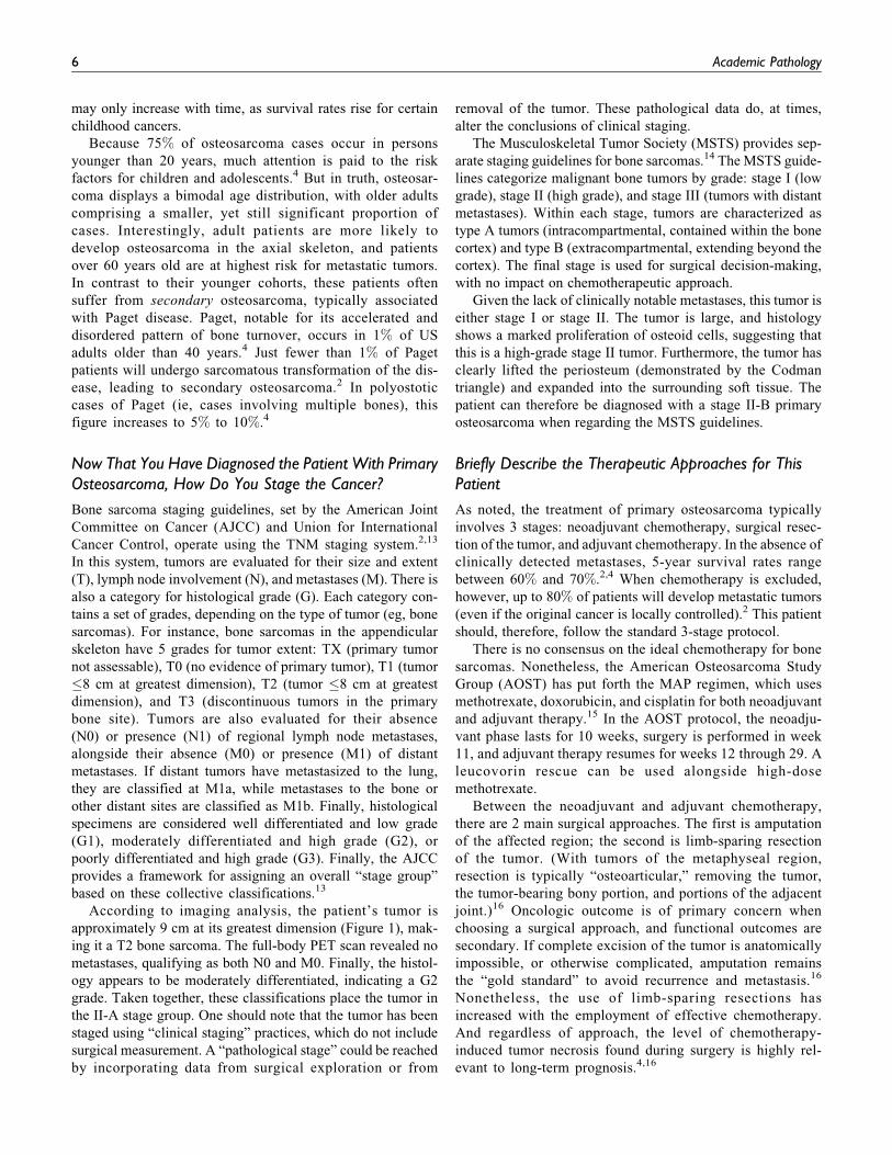
may only increase with time, as survival rates rise for certain
childhood cancers.
Because 75% of osteosarcoma cases occur in persons
younger than 20 years, much attention is paid to the risk
factors for children and adolescents.4 But in truth, osteosar-
coma displays a bimodal age distribution, with older adults
comprising a smaller, yet still significant proportion of
cases. Interestingly, adult patients are more likely to
develop osteosarcoma in the axial skeleton, and patients
over 60 years old are at highest risk for metastatic tumors.
In contrast to their younger cohorts, these patients often
suffer from secondary osteosarcoma, typically associated
with Paget disease. Paget, notable for its accelerated and
disordered pattern of bone turnover, occurs in 1% of US
adults older than 40 years.4 Just fewer than 1% of Paget
patients will undergo sarcomatous transformation of the dis-
ease, leading to secondary osteosarcoma.2 In polyostotic
cases of Paget (ie, cases involving multiple bones), this
figure increases to 5% to 10%.4
Now That You Have Diagnosed the Patient With PrimaryOsteosarcoma, How Do You Stage the Cancer?
Bone sarcoma staging guidelines, set by the American Joint
Committee on Cancer (AJCC) and Union for International
Cancer Control, operate using the TNM staging system.2,13
In this system, tumors are evaluated for their size and extent
(T), lymph node involvement (N), and metastases (M). There is
also a category for histological grade (G). Each category con-
tains a set of grades, depending on the type of tumor (eg, bone
sarcomas). For instance, bone sarcomas in the appendicular
skeleton have 5 grades for tumor extent: TX (primary tumor
not assessable), T0 (no evidence of primary tumor), T1 (tumor
�8 cm at greatest dimension), T2 (tumor �8 cm at greatest
dimension), and T3 (discontinuous tumors in the primary
bone site). Tumors are also evaluated for their absence
(N0) or presence (N1) of regional lymph node metastases,
alongside their absence (M0) or presence (M1) of distant
metastases. If distant tumors have metastasized to the lung,
they are classified at M1a, while metastases to the bone or
other distant sites are classified as M1b. Finally, histological
specimens are considered well differentiated and low grade
(G1), moderately differentiated and high grade (G2), or
poorly differentiated and high grade (G3). Finally, the AJCC
provides a framework for assigning an overall “stage group”
based on these collective classifications.13
According to imaging analysis, the patient’s tumor is
approximately 9 cm at its greatest dimension (Figure 1), mak-
ing it a T2 bone sarcoma. The full-body PET scan revealed no
metastases, qualifying as both N0 and M0. Finally, the histol-
ogy appears to be moderately differentiated, indicating a G2
grade. Taken together, these classifications place the tumor in
the II-A stage group. One should note that the tumor has been
staged using “clinical staging” practices, which do not include
surgical measurement. A “pathological stage” could be reached
by incorporating data from surgical exploration or from
removal of the tumor. These pathological data do, at times,
alter the conclusions of clinical staging.
The Musculoskeletal Tumor Society (MSTS) provides sep-
arate staging guidelines for bone sarcomas.14 The MSTS guide-
lines categorize malignant bone tumors by grade: stage I (low
grade), stage II (high grade), and stage III (tumors with distant
metastases). Within each stage, tumors are characterized as
type A tumors (intracompartmental, contained within the bone
cortex) and type B (extracompartmental, extending beyond the
cortex). The final stage is used for surgical decision-making,
with no impact on chemotherapeutic approach.
Given the lack of clinically notable metastases, this tumor is
either stage I or stage II. The tumor is large, and histology
shows a marked proliferation of osteoid cells, suggesting that
this is a high-grade stage II tumor. Furthermore, the tumor has
clearly lifted the periosteum (demonstrated by the Codman
triangle) and expanded into the surrounding soft tissue. The
patient can therefore be diagnosed with a stage II-B primary
osteosarcoma when regarding the MSTS guidelines.
Briefly Describe the Therapeutic Approaches for ThisPatient
As noted, the treatment of primary osteosarcoma typically
involves 3 stages: neoadjuvant chemotherapy, surgical resec-
tion of the tumor, and adjuvant chemotherapy. In the absence of
clinically detected metastases, 5-year survival rates range
between 60% and 70%.2,4 When chemotherapy is excluded,
however, up to 80% of patients will develop metastatic tumors
(even if the original cancer is locally controlled).2 This patient
should, therefore, follow the standard 3-stage protocol.
There is no consensus on the ideal chemotherapy for bone
sarcomas. Nonetheless, the American Osteosarcoma Study
Group (AOST) has put forth the MAP regimen, which uses
methotrexate, doxorubicin, and cisplatin for both neoadjuvant
and adjuvant therapy.15 In the AOST protocol, the neoadju-
vant phase lasts for 10 weeks, surgery is performed in week
11, and adjuvant therapy resumes for weeks 12 through 29. A
leucovorin rescue can be used alongside high-dose
methotrexate.
Between the neoadjuvant and adjuvant chemotherapy,
there are 2 main surgical approaches. The first is amputation
of the affected region; the second is limb-sparing resection
of the tumor. (With tumors of the metaphyseal region,
resection is typically “osteoarticular,” removing the tumor,
the tumor-bearing bony portion, and portions of the adjacent
joint.)16 Oncologic outcome is of primary concern when
choosing a surgical approach, and functional outcomes are
secondary. If complete excision of the tumor is anatomically
impossible, or otherwise complicated, amputation remains
the “gold standard” to avoid recurrence and metastasis.16
Nonetheless, the use of limb-sparing resections has
increased with the employment of effective chemotherapy.
And regardless of approach, the level of chemotherapy-
induced tumor necrosis found during surgery is highly rel-
evant to long-term prognosis.4,16
6 Academic Pathology

Given this patient’s age, activity level, and lack of clinical
metastases, limb-sparing surgery is preferable to amputation.
However, the patient will likely require reconstructive surgery.
An allograft or expandable endoprosthesis may be suitable for
his immature skeleton, allowing for equal development of his
lower limbs.
If initial treatment is deemed successful, the National Com-
prehensive Cancer Network recommends follow-up surveil-
lance every 3 months for years 1 to 2, every 4 months during
year 3, every 6 months for years 4 to 5, and once annually from
year 6 onwards.15 While there is no established time line for the
duration of follow-up, most bone sarcoma recurrences occur
within 10 years of treatment.15
Teaching Points
� Radiographs of osteosarcoma typically depict a soft tis-
sue mass, including lytic and sclerotic tissue, alongside
the notable Codman triangle.
� Bone tumors can be classified as primary (originating in
the bone itself) or secondary (having metastasized from
elsewhere in the body).
� Osteosarcoma and Ewing sarcoma are the most common
primary malignant bone tumors in children and
adolescents.
� Osteosarcomas are malignant bone-forming tumors
which produce woven osteoid tissue and comprise
50% of all childhood bone neoplasms.
� Histology of osteosarcoma shows haphazard arrange-
ments of loosely woven osteoid tissue, surrounded by
sections of mature lamellar bone.
� While there is no established genetic etiology of osteo-
sarcoma, CIN is common among osteosarcoma cells.
� Giant cell tumors are soft, benign collections of multi-
nucleated osteoclast-like cells, which appear near the
epiphysis of the long bones.
� Aneurysmal bone cysts are benign, blood-filled cystic
bodies which typically appear as “egg shell” masses near
the metaphysis of the long bones.
� Malignant bone tumors are staged after diagnosis and
before treatment. The specific stage of any given
tumor will influence the prognosis and treatment of
that cancer.
� In the AJCC guidelines, bone sarcomas are evaluated for
tumor extent (T), lymph node involvement (N), distant
metastases (M), and histologic grade (G). They are then
given an overall “stage group” (stages I-IV, subgroups
A-B) based on these evaluations.
� In the MSTS guidelines, bone sarcomas are staged by
their grade (stages I-III) and whether they are intracom-
partmental (A) or extracompartmental (B).
� Clinical staging of a tumor can be reached through ima-
ging or other nonsurgical measurements, while patholo-
gical staging requires surgical exploration or removal of
the tumor.
� Treatment for osteosarcoma typically involves neoadju-
vant chemotherapy, surgical removal of the tumor, and
adjuvant chemotherapy.
� Chemotherapy of osteosarcoma often follows the MAP
regimen—methotrexate, doxorubicin, cisplatin—for
both neoadjuvant and adjuvant phases.
Declaration of Conflicting Interests
The author(s) declared no potential conflicts of interest with respect to
the research, authorship, and/or publication of this article.
Funding
The author(s) received no financial support for the research, author-
ship, and/or publication of this article.
References
1. Mirabello L, Troisi RJ, Savage SA. Osteosarcoma incidence and
survival rates from 1973 to 2004: data from the Surveillance,
Epidemiology, and End Results Program. Cancer. 2009;115:
1531-1543. doi:10.1002/cncr.24121.
2. Wang LL, Gebhardt MC, Rainusso N. Osteosarcoma: epidemiol-
ogy, pathogenesis, clinical presentation, diagnosis, and histology.
In: UpToDate. 2017. https://www.uptodate.com/contents/osteo
sarcoma-epidemiology-pathogenesis-clinical-presentation-diag
nosis-and-histology?search¼osteosarcoma&source¼search_
result&selectedTitle¼1*82&usage_type¼default&display_
rank¼1. Accessed August 8, 2018.
3. DeLaney TF, Hornicek FJ, Bahrami A. Epidemiology, pathology,
and molecular genetics of the Ewing sarcoma family of tumors.
In: UpToDate. 2018. https://www.uptodate.com/contents/epide
miology-pathology-and-molecular-genetics-of-the-ewing-sar
coma-family-of-tumors?search¼ewing&source¼search_result&
selectedTitle¼3*63&usage_type¼default&display_rank¼3.
Accessed August 8, 2018.
4. Kumar V, Abbas AK, Aster JC. Robbins Basic Pathology. 10th
ed. Kumar V, Abbas AK, Aster JC, eds. Philadelphia, PA: Else-
vier Inc; 2018.
5. DeLaney TF, Hornicek FJ. Clinical presentation, staging, and prog-
nostic factors of theEwing sarcoma familyof tumors. In:UpToDate.
2018. https://www.uptodate.com/contents/clinical-presentation-sta
ging-and-prognostic-factors-of-the-ewing-sarcoma-family-
of-tumors?search¼ewingsarcoma&source¼ search_result&
selectedTitle¼1*63&usage_type¼default&display_rank¼1.
Accessed August 8, 2018.
6. Hornicek FJ, Bredella M. Bone tumors: diagnosis and biopsy
techniques. In: UpToDate; 2016. https://www.uptodate.com/con
tents/bone-tumors-diagnosis-and-biopsy-techniques?search¼bonetumors&source¼search_result&selectedTitle¼1*150&
usage_type¼default&display_rank¼1. Accessed August 8, 2018.
7. Dahlin DC, Unni KK. Osteosarcoma of bone and its important
recognizable varieties. Am J Surg Pathol. 1977;1:61-72.
8. Murphey MD, Senchak LT, Mambalam PK, Logie CI, Klassen-
Fischer MK, Kransdorf MJ. From the radiologic pathology
archives: Ewing sarcoma family of tumors: radiologic-pathologic
Petriceks and Salmi 7
Page C3-19
correlation. Radiographics. 2013;33:803-831. doi:10.1148/rg.
333135005.
9. Gebhardt MC, DuBois SG. Treatment of the Ewing sarcoma fam-
ily of tumors. In: UpToDate. 2017. https://www.uptodate.com/
contents/treatment-of-the-ewing-sarcoma-family-of-tumors.
Accessed August 7, 2018.
10. Gelderblom A, Bovee JB. Chondrosarcoma. In: UpToDate. 2018.
https://www.uptodate.com/contents/chondrosarcoma. Accessed
August 7, 2018.
11. Morrow JJ, Khanna C. Osteosarcoma genetics and epigenetics:
emerging biology and candidate therapies. Crit Rev Oncog. 2015;
20:173-197.
12. Martin JW, Squire JA, Zielenska M. The genetics of osteosar-
coma. Sarcoma. 2012;2012:1-11. doi:10.1155/2012/627254.
13. American Joint Commitee on Cancer. AJCC Cancer Staging
Manual. 8th ed. Amin M, Edge S, Greene F, et al, eds. Chicago,
IL: Springer International Publishing; 2017.
14. Enneking WF. A system of staging musculoskeletal neoplasms.
Clin Orthop Relat Res. 1986;9-24.
15. Janeway KA, Maki R. Chemotherapy and radiation therapy in the
management of osteosarcoma. In: UpToDate. 2018. https://www.
uptodate.com/contents/chemotherapy-and-radiation-therapy-in-
the-management-of-osteosarcoma?search¼Chemotherapyandra
diationtherapyinthemanagementofosteosarcoma&source¼search_result&selectedTitle¼1*150&usage_type¼default&
display_rank¼1. Accessed August 7, 2018.
16. Hornicek F. Bone sarcomas: preoperative evaluation, histolo-
gic classification, and principles of surgical management. In:
UpToDate. 2016. https://www.uptodate.com/contents/bone-
sarcomas-preoperative-evaluation-histologic-classification-
and-principles-of-surgical-management?search¼Bonesar
comas:Preoperativeevaluation,histologicclassification,andpr
inciplesofsurgicalmanagement&source¼. Accessed August 8,
2018.
8 Academic Pathology
Page C3-20

Educational Case
Educational Case: Head and NeckNeoplasia: Salivary Gland Tumors
Ryan P. Lau, MD1, Melissa Yee-Chang, DO1, and Amy Rapkiewicz, MD1
The following fictional case is intended as a learning tool within the Pathology Competencies for Medical Education
(PCME), a set of national standards for teaching pathology. These are divided into three basic competencies: Disease
Mechanisms and Processes, Organ System Pathology, and Diagnostic Medicine and Therapeutic Pathology. For addi-
tional information, and a full list of learning objectives for all three competencies, see http://journals.sagepub.com/doi/
10.1177/2374289517715040.
Keywordspathology competencies, organ system pathology, head and neck neoplasia, salivary gland tumor, warthin tumor, pleomorphicadenoma, mucoepidermoid carcinoma
Received September 26, 2017. Received revised February 23, 2018. Accepted for publication February 28, 2018.
Primary Pathology Learning Objective
Objective HN2.1. Benign and Mucoepidermoid Tumors of
Salivary Glands. Distinguish the clinicopathologic features of
the 2 benign tumors (pleomorphic adenoma or mixed tumor
and Warthin tumor) from the malignant mucoepidermoid
carcinoma.
Competency 2: Organ System Pathology, Topic: Head and
Neck (HN), Learning Goal 2: Head and Neck Neoplasia
Secondary Pathology Learning Objective
Objective CYP1.2. Categorizing Diagnostic Certainty:
Compare and contrast the degree of diagnostic certainty
applied to general diagnostic categorization in cytologic
diagnosis.
Competency 3: Diagnostic Medicine and Laboratory Diag-
nosis; Topic CYP: Cytopathology; Learning Goal I: Cytologic
Diagnosis
Patient Presentation
A 62-year-old obese, hypertensive, Caucasian male presents to
his primary care physician with a slowly growing left cheek
mass for 1-year duration. He has a 40 pack-year smoking his-
tory. He has no family or personal history of carcinoma. He is
HIV-negative and has not experienced any dysphagia, odyno-
phagia, B-symptoms, trismus, or facial weakness.
Diagnostic Findings, Part 1
On physical examination, his primary care physician notes a
palpable, 2 cm, “doughy,” mobile mass in his left parotid gland.
There appears to be mild erythema and slight puckering of the
skin overlying the mass. The contralateral parotid gland has no
palpable masses and there is no cervical lymphadenopathy.
Tongue sensation and facial nerve function are intact. The
patient undergoes imaging studies and fine needle aspiration
(FNA) biopsy of his lesion.
1 New York University School of Medicine, New York, NY, USA
Corresponding Author:
Ryan P. Lau, New York University School of Medicine, 550 1st Ave, New York,
NY 10016, USA.
Email: [email protected]
Academic PathologyVolume 5: 1–5ª The Author(s) 2018Reprints and permission:sagepub.com/journalsPermissions.navDOI: 10.1177/2374289518770655journals.sagepub.com/home/apc
Creative Commons Non Commercial CC BY-NC: This article is distributed under the terms of the Creative Commons Attribution-NonCommercial 4.0 License(http://www.creativecommons.org/licenses/by-nc/4.0/) which permits non-commercial use, reproduction and distribution of the work without furtherpermission provided the original work is attributed as specified on the SAGE and Open Access pages (https://us.sagepub.com/en-us/nam/open-access-at-sage).
Page C3-21
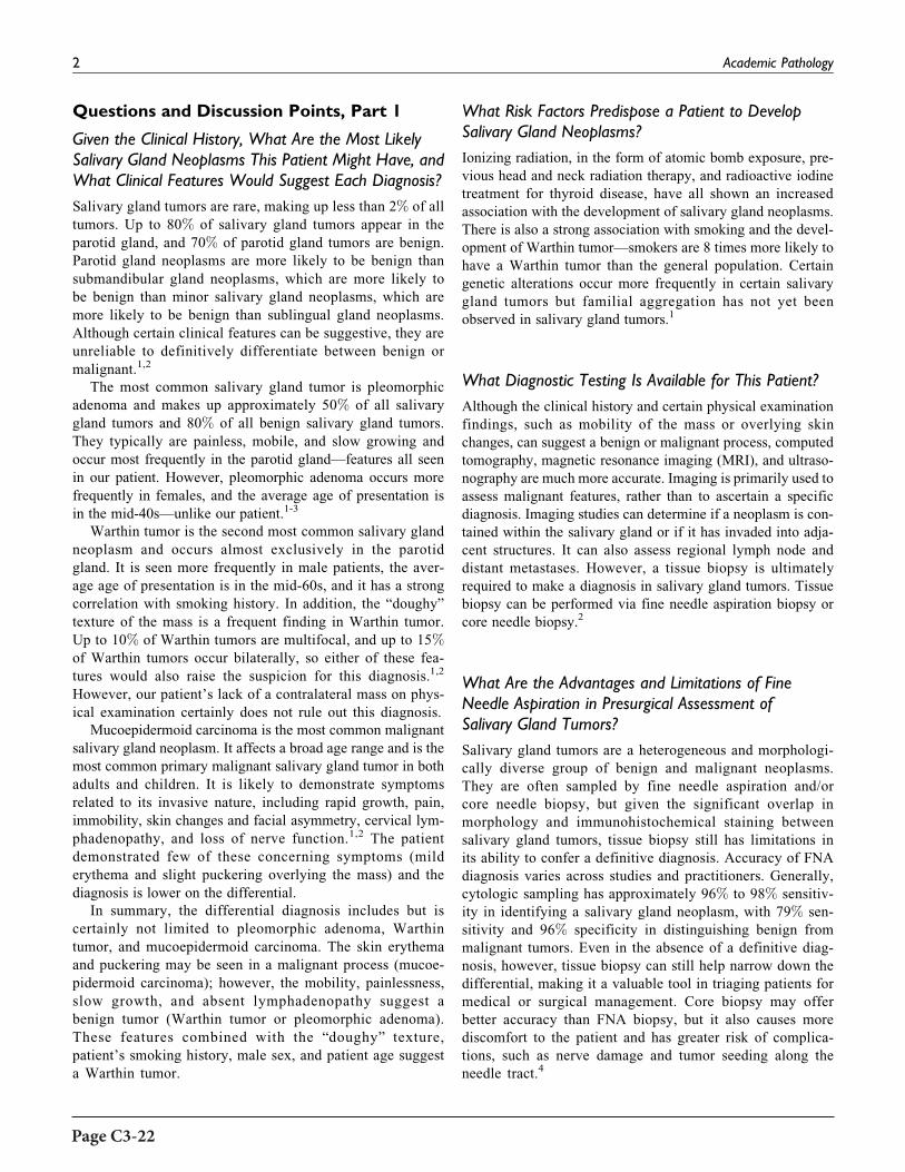
Questions and Discussion Points, Part 1
Given the Clinical History, What Are the Most LikelySalivary Gland Neoplasms This Patient Might Have, andWhat Clinical Features Would Suggest Each Diagnosis?
Salivary gland tumors are rare, making up less than 2% of all
tumors. Up to 80% of salivary gland tumors appear in the
parotid gland, and 70% of parotid gland tumors are benign.
Parotid gland neoplasms are more likely to be benign than
submandibular gland neoplasms, which are more likely to
be benign than minor salivary gland neoplasms, which are
more likely to be benign than sublingual gland neoplasms.
Although certain clinical features can be suggestive, they are
unreliable to definitively differentiate between benign or
malignant.1,2
The most common salivary gland tumor is pleomorphic
adenoma and makes up approximately 50% of all salivary
gland tumors and 80% of all benign salivary gland tumors.
They typically are painless, mobile, and slow growing and
occur most frequently in the parotid gland—features all seen
in our patient. However, pleomorphic adenoma occurs more
frequently in females, and the average age of presentation is
in the mid-40s—unlike our patient.1-3
Warthin tumor is the second most common salivary gland
neoplasm and occurs almost exclusively in the parotid
gland. It is seen more frequently in male patients, the aver-
age age of presentation is in the mid-60s, and it has a strong
correlation with smoking history. In addition, the “doughy”
texture of the mass is a frequent finding in Warthin tumor.
Up to 10% of Warthin tumors are multifocal, and up to 15%of Warthin tumors occur bilaterally, so either of these fea-
tures would also raise the suspicion for this diagnosis.1,2
However, our patient’s lack of a contralateral mass on phys-
ical examination certainly does not rule out this diagnosis.
Mucoepidermoid carcinoma is the most common malignant
salivary gland neoplasm. It affects a broad age range and is the
most common primary malignant salivary gland tumor in both
adults and children. It is likely to demonstrate symptoms
related to its invasive nature, including rapid growth, pain,
immobility, skin changes and facial asymmetry, cervical lym-
phadenopathy, and loss of nerve function.1,2 The patient
demonstrated few of these concerning symptoms (mild
erythema and slight puckering overlying the mass) and the
diagnosis is lower on the differential.
In summary, the differential diagnosis includes but is
certainly not limited to pleomorphic adenoma, Warthin
tumor, and mucoepidermoid carcinoma. The skin erythema
and puckering may be seen in a malignant process (mucoe-
pidermoid carcinoma); however, the mobility, painlessness,
slow growth, and absent lymphadenopathy suggest a
benign tumor (Warthin tumor or pleomorphic adenoma).
These features combined with the “doughy” texture,
patient’s smoking history, male sex, and patient age suggest
a Warthin tumor.
What Risk Factors Predispose a Patient to DevelopSalivary Gland Neoplasms?
Ionizing radiation, in the form of atomic bomb exposure, pre-
vious head and neck radiation therapy, and radioactive iodine
treatment for thyroid disease, have all shown an increased
association with the development of salivary gland neoplasms.
There is also a strong association with smoking and the devel-
opment of Warthin tumor—smokers are 8 times more likely to
have a Warthin tumor than the general population. Certain
genetic alterations occur more frequently in certain salivary
gland tumors but familial aggregation has not yet been
observed in salivary gland tumors.1
What Diagnostic Testing Is Available for This Patient?
Although the clinical history and certain physical examination
findings, such as mobility of the mass or overlying skin
changes, can suggest a benign or malignant process, computed
tomography, magnetic resonance imaging (MRI), and ultraso-
nography are much more accurate. Imaging is primarily used to
assess malignant features, rather than to ascertain a specific
diagnosis. Imaging studies can determine if a neoplasm is con-
tained within the salivary gland or if it has invaded into adja-
cent structures. It can also assess regional lymph node and
distant metastases. However, a tissue biopsy is ultimately
required to make a diagnosis in salivary gland tumors. Tissue
biopsy can be performed via fine needle aspiration biopsy or
core needle biopsy.2
What Are the Advantages and Limitations of FineNeedle Aspiration in Presurgical Assessment ofSalivary Gland Tumors?
Salivary gland tumors are a heterogeneous and morphologi-
cally diverse group of benign and malignant neoplasms.
They are often sampled by fine needle aspiration and/or
core needle biopsy, but given the significant overlap in
morphology and immunohistochemical staining between
salivary gland tumors, tissue biopsy still has limitations in
its ability to confer a definitive diagnosis. Accuracy of FNA
diagnosis varies across studies and practitioners. Generally,
cytologic sampling has approximately 96% to 98% sensitiv-
ity in identifying a salivary gland neoplasm, with 79% sen-
sitivity and 96% specificity in distinguishing benign from
malignant tumors. Even in the absence of a definitive diag-
nosis, however, tissue biopsy can still help narrow down the
differential, making it a valuable tool in triaging patients for
medical or surgical management. Core biopsy may offer
better accuracy than FNA biopsy, but it also causes more
discomfort to the patient and has greater risk of complica-
tions, such as nerve damage and tumor seeding along the
needle tract.4
2 Academic Pathology
Page C3-22

Diagnostic Findings, Part 2
Magnetic Resonance Imaging
The patient undergoes MRI of the neck with and without con-
trast, which demonstrates 2 enhancing parotid masses located in
the superficial lobe of the left parotid gland. The masses measure
1.2 � 0.8 � 0.9 cm and 0.7 � 0.6 � 0.5 cm. The deep parotid
lobes are not involved. The right parotid gland is unremarkable.
No infiltrative features or cervical lymphadenopathy are seen.
Cytologic Assessment
Upon biopsy of the larger lesion, thick, green-brown fluid is aspi-
rated, resembling motor oil. A representative photomicrograph of
theFNAbiopsyof the patient’s larger lesion is shown inFigure 1A.
Questions and Discussion Points, Part 2
How Do the Findings on MRI Help Narrow Downthe Differential?
Since no infiltrative features or cervical lymphadenopathy are
seen, a benign neoplasm is favored. On imaging, the tumor was
found to be 2 separate nodules, rather than a solitary lesion that
was noted on physical examination. The clinical presentation
plus the features seen on radiology (multiple lesions with no
infiltrative features) make a diagnosis of Warthin tumor likely.
Discuss the Cytologic Features of the Fine NeedleAspirate Biopsy in Figure 1A
The “motor oil” fluid that is aspirated from the lesion is a
common feature of Warthin tumor. The biopsy of the tumor
has 3 distinct features, which are diagnostic of Warthin tumor:
sheets and clusters of oncocytic cells (arrow), lymphocytes
(arrowhead), and granular debris in the background (triangle).
The Cytologic Features of the 2 Other NeoplasmsConsidered in the Differential Diagnosis Are Shown inFigure 1B and C for Comparison. What FeaturesDistinguish the Patient’s Warthin Tumor FromPleomorphic Adenoma and Mucoepidermoid Carcinoma?
Figure 1B: Pleomorphic adenoma: This tumor has cohesive
sheets and clusters of ductal cells (arrow), individual myoe-
pithelial cells (arrowhead), and metachromatic, magenta-
colored, fibrillary stroma (triangle) when stained with a
modified Wright Giemsa (Diff Quik) stain.
Figure 1C: Mucoepidermoid carcinoma: This tumor has a
mixed population of cells, with 3 distinct cell types. Signet-ring
type mucin producing cells with pink mucin (arrow), polygonal
squamoid cells with dense blue cytoplasm (arrowhead), and
smaller intermediate cells (triangle) when stained with a Papa-
nicolaou stain.
What Are the Treatment Options for the Patient?
Treatment recommendations are currently based on retrospective
reviews and not randomized clinical trials. Complete surgical
resection with negativemargins is the gold standard, and patients
with benign tumors (such as Warthin tumor and pleomorphic
adenoma) and low-grade tumors can be treated by surgery alone.
High-grade tumors, tumors with positive resection margins, and
tumors with high-risk features are usually treated with surgery
Figure 1. Cytologic features. A, FNA biopsy of the patient’s lesion, showing Warthin tumor with sheets and clusters of oncocytic cells (arrow),lymphocytes (arrowhead), and granular debris in the background (triangle). B, Cytology of pleomorphic adenoma for comparison, with cohesivesheets and clusters of ductal cells (arrow), individual myoepithelial cells (arrowhead), and metachromatic, magenta-colored, fibrillary stroma(triangle). C, Cytology of mucoepidermoid carcinoma for comparison, with signet-ring type mucin producing cells (arrow), polygonal squamoidcells with dense blue cytoplasm (arrowhead), and smaller intermediate cells (triangle).
Lau et al 3
Page C3-23

and adjuvant radiation therapy.Because hehas a benigndiagnosis
of Warthin tumor, he can be treated with surgery alone.5
Diagnostic Findings, Part 3
Histologic Assessment
The patient undergoes superficial parotidectomy to remove the
2 masses. A representative photomicrograph of the patient’s
lesions is shown in Figure 2A.
Questions and Discussion Points, Part 3
Discuss the Histologic Features of the Surgical Excisionin Figure 2A
The cystic lesion is lined by oncocytic cells (arrow) with a
lymphoid stroma (arrowhead). Granular debris fills the cyst
spaces (triangle). These are diagnostic of and consistent with
the initial biopsy diagnosis of Warthin tumor.
The Histologic Features of the 2 Other NeoplasmsConsidered in the Differential Diagnosis Are Shown inFigure 2B and C for Comparison. What FeaturesDistinguish the Patient’s Warthin Tumor FromPleomorphic Adenoma and Mucoepidermoid Carcinoma?
Figure 2B: Pleomorphic adenoma: This tumor has ductal cells
(arrow) surrounded by a layer of myoepithelial cells (arrow-
head), arranged in a chondromyxoid stroma (triangle).
Figure 2C: Mucoepidermoid carcinoma: This cystic tumor
has a mixed population of cells, with 3 distinct cell types—
mucin producing cells (arrow), squamoid cells (arrowhead),
and intermediate cells (triangle).
What Is the Expected Outcome After SuperficialParotidectomy to Excise the Patient’s Warthin Tumor?
Surgical excision of Warthin tumor is curative, and we expect
the patient to have a good clinical outcome. The risk of local
recurrence after complete surgical excision is less than 2%.
The other tumors on our initial differential behave quite
differently. Pleomorphic adenoma is also benign but has a
higher local recurrence rate of up to 5% if treated by paroti-
dectomy, and up to 25% if treated with simple enucleation. The
behavior of mucoepidermoid carcinoma depends on the grade
of the neoplasm. Local recurrence rates range from 15% to
30%, and 5-year survival ranges from 50% to 90%.1,2
Teaching Points
1. Salivary gland neoplasms are rare, accounting for less
than 2% of all tumors.
2. Up to80% of salivaryglandneoplasmsoccur in theparotid
gland, and up to 70% of parotid gland tumors are benign.
3. The likelihood of malignancy in each salivary gland in
increasing order is: parotid gland, submandibular
gland, minor salivary gland, and sublingual gland.
4. Clinical features are not definitively diagnostic of a
benign or malignant process. Malignant tumors are
suggested by rapid growth, pain, immobility, skin
changes and facial asymmetry, cervical lymphadeno-
pathy, and loss of nerve function. Benign tumors are
suggested by absence of pain, mobility, slow growth,
absence of nerve involvement.
Figure 2. Histologic features. A, Surgical excision of the patient’s lesion showing Warthin tumor with oncocytic cells (arrow) and lymphoidstroma (arrowhead). Granular debris fills the cyst spaces (triangle). B, Histology of pleomorphic adenoma for comparison, with ductal cells(arrow) surrounded by a layer of myoepithelial cells (arrowhead), arranged in a chondromyxoid stroma (triangle). C, Histology of mucoepi-dermoid carcinoma for comparison, with mucin producing cells (arrow), squamoid cells (arrowhead), and intermediate cells (triangle).
4 Academic Pathology
Page C3-24

5. Radiologic studies can provide important clues to the
nature of a salivary gland tumor, including invasion
beyond salivary gland parenchyma into adjacent struc-
tures and local lymphatic spread.
6. Fine needle aspiration biopsy of salivary gland tumors
is an important initial diagnostic test that can fre-
quently give an accurate diagnosis. When a specific
diagnosis cannot be rendered, cytologic analysis can
give a differential diagnosis that helps guide clinical/
surgical management.
7. The 3 most common salivary gland tumors (pleo-
morphic adenoma, Warthin tumor, and mucoepider-
moid carcinoma) have distinct cytologic and
histologic appearances.
8. The most common salivary gland neoplasm is pleo-
morphic adenoma, a benign tumor that typically pre-
sents in the mid-40s and has a female predilection.
9. The second most common benign salivary gland neo-
plasm is Warthin tumor, which commonly presents in
male smokers in their mid-60s. On physical examina-
tion, it has a “doughy” texture and can be multifocal or
occur bilaterally. On aspiration, it produces a “motor
oil” like fluid.
10. The most common malignant salivary gland neoplasm
is mucoepidermoid carcinoma. It occurs over a broad
age range and is the most common salivary gland
malignancy in adults and children.
11. Pleomorphic adenoma and Warthin tumor can both be
treated by surgical excision alone and have less than
5% risk of local recurrence.
12. The treatment and outcome of mucoepidermoid carci-
noma is dependent on the tumor grade. High-grade
tumors may require surgery and adjuvant radiation
therapy, with local recurrence rate as high as 30%, and
5-year survival of 50%.
Declaration of Conflicting Interests
The author(s) declared no potential conflicts of interest with respect to
the research, authorship, and/or publication of this article.
Funding
The author(s) received no financial support for the research, author-
ship, and/or publication of this article.
References
1. Barnes L, Eveson JW, Reichart P, Sidransky D, eds. World Health
Organization Classification of Tumours. Pathology and Genetics,
Head and Neck Tumors. Lyon: IARC Press; 2005.
2. Kumar Vinay, Abbas Abul, Aster Jon. Robbins and Cotran Patho-
logic Basis of Disease. 9th ed. Philadelphia, PA: Elsevier/Saun-
ders; 2015.
3. Eveson JW, Cawson RA. Salivary gland tumours: a review of 2410
cases with particular reference to histological types, site, age and
sex distribution. J Pathol. 1985;146:51-58. PubMed PMID:
4009321.
4. Cibas ES, Ducatman BS. Cytology: Diagnostic Principles and
Clinical Correlates. 4th ed. Philadelphia, PA: Elsevier/Saunders;
2014.
5. National Comprehensive Cancer Network. Head and Neck
Cancers (Version 2.2017). https://www.nccn.org/professionals/
physician_gls/pdf/head-and-neck.pdf. Accessed September 21,
2017.
Lau et al 5
Page C3-25


Educational Case
Educational Case: Cervical Neoplasia:HPV and Its Link to Cancer
Teresa Kim, MD1, Samer N. Khader, MBBS1, and D. Yitzchak Goldstein, MD1
The following fictional case is intended as a learning tool within the Pathology Competencies for Medical Education
(PCME), a set of national standards for teaching pathology. These are divided into three basic competencies: Disease
Mechanisms and Processes, Organ System Pathology, and Diagnostic Medicine and Therapeutic Pathology. For addi-
tional information, and a full list of learning objectives for all three competencies, see http://journals.sagepub.com/doi/
10.1177/2374289517715040.
Keywordspathology competencies, disease mechanisms, HPV, human papillomavirus, cervical cancer, neoplasia, disease screening,PAP smear
Received November 14, 2017. Received revised March 12, 2018. Accepted for publication March 22, 2018.
Primary Objective
Objective N3.1: Morphologic Features of Neoplasia: Describe
the essential morphologic features of neoplasms and indicate
how these can be used to diagnose, classify, and predict biolo-
gical behavior of cancers.
Competency 1: Disease Mechanisms and Processes; Topic
N: Neoplasia; Learning Goal 3: Characteristics of Neoplasia.
Secondary Objectives
Objective CYP2.1 Screening:Describe the principles of an effec-
tive screening test and the uses and limitations of cytology.
Objective CYP2.2 Adjunct testing (HPV): Describe how
adjunct testing is used in conjunction with cytology examination.
Objective CYP2.3 Cervical Screening: Describe how to find
and utilize current algorithms for management of cervical
screening.
Competency 3: Diagnostic Medicine and Therapeutic
Pathology; Topic CYP: Cytopathology; Learning Goal 2:
Advantages of Cytopathology.
Patient Presentation
A 45-year-old female presents to her new primary care physi-
cian to establish care after not visiting a doctor for 10 years.
She was recently referred for a follow-up visit after receiving
an abnormal liquid Pap result from a free community outreach
cervical cancer screening program. She denies any significant
past medical or surgical history. The patient’s last menstrual
period was 2 weeks ago, and she is currently sexually active
with multiple partners. She occasionally uses barrier methods
of contraception. Physical examination and pelvic examination
are within normal limits. Before discussing the results, the
patient remembers having a history of abnormal pap smears
that required further testing, but these were not pursued since
she was lost to follow-up.
Diagnostic Findings
Upon further review of the patient’s history, the patient was
35 years old at the time of her last Pap test and pelvic exam-
ination. Cytology of her old Pap test showed a low-grade squa-
mous intraepithelial lesion (LSIL). Human papillomavirus
1 Albert Einstein College of Medicine, Bronx, NY, USA
Corresponding Author:
Teresa Kim, Albert Einstein College of Medicine, 1935 Eastchester Rd, Apt
10D, Bronx, NY 10461, USA.
Email: [email protected]
Academic PathologyVolume 5: 1–6ª The Author(s) 2018Reprints and permission:sagepub.com/journalsPermissions.navDOI: 10.1177/2374289518770651journals.sagepub.com/home/apc
Creative Commons Non Commercial CC BY-NC: This article is distributed under the terms of the Creative Commons Attribution-NonCommercial 4.0 License(http://www.creativecommons.org/licenses/by-nc/4.0/) which permits non-commercial use, reproduction and distribution of the work without furtherpermission provided the original work is attributed as specified on the SAGE and Open Access pages (https://us.sagepub.com/en-us/nam/open-access-at-sage).
Page C3-27

(HPV) co-testing results from that time demonstrated positivity
for HPV type 16. As per American Society for Colposcopy and
Cervical Pathology (ASCCP) guidelines, the patient was rec-
ommended to undergo further testing with colposcopy but was
lost to follow-up.
The patient’s current cytologic evaluation is consistent
with high-grade squamous intraepithelial lesion (HSIL), and
HPV testing remains positive for HPV type 16. Gonorrhea/
chlamydia testing is negative. The patient is now scheduled
for an immediate colposcopy and loop electrosurgical exci-
sion (LEEP).
Questions/Discussion Points
What Are Your Preliminary Working Diagnosis and ItsDifferential?
This patient cytology results previously demonstrated an LSIL,
which has since progressed to an HSIL with persistent HPV
infection. The SILs are virally driven neoplastic proliferations
of cervical epithelial cells and are precancerous. Epidemiolo-
gical studies have demonstrated the time-dependent progres-
sive nature of these lesions (Figure 1). The differential
diagnosis may include other types of cervical lesions, such
as squamous metaplasia, reactive changes, or glandular
changes. Invasive squamous cell carcinoma of the cervix
(when a precancerous lesion invades beyond the basement
membrane) should not be ruled out and is the primary concern
for this patient.
What Screening Modalities Are Recommended to DetectCervical Lesions and How Do They Impact DiseaseProgression?
Cervical cytologic screening was introduced decades ago and is
still the most effective cancer prevention test today. For example,
cytology screening that is only performed twice in awoman’s life
can reduce her risk for cervical cancer by 43%.1 Cytologic testing
and HPV co-testing for HPV infection decrease the prevalence
and mortality of cervical cancer. Furthermore, regular screening
allows for early intervention and reduces progression of disease.
Since cervical SIL is mostly a disease of reproductive-aged
women, theAmericanCollegeofObstetricians andGynecologists
(ACOG) recommends women aged 21 to 29 years have a Pap test
every 3 years without HPV testing. Women aged 30 to 65 years
should have a Pap test and co-testing for HPV every 5 years.2
HPV testing is not recommended for women younger than 30
yearsbecause theprevalenceofHPVpositivity is veryhigh in these
women, and the vastmajoritywill completely clear their infections
within 1 year.3 However, for women older than 30 years, liquid-
based Pap screening in conjunction with HPV co-testing has a
lower false-negative rate and will detect a greater number of
patients at risk compared to Pap testing alone.
Figure 1. Sketch demonstrating the progressive nature of the dysplastic changes leading to the development of cancer. To the left is arepresentation of the HPV genome with its associated early and late antigen proteins. HPV indicates human papillomavirus.
2 Academic Pathology
Page C3-28
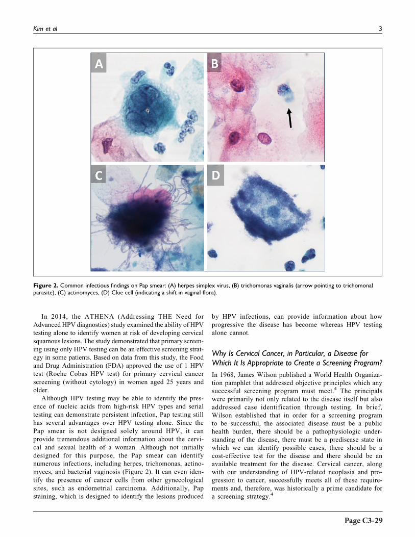
In 2014, the ATHENA (Addressing THE Need for
Advanced HPV diagnostics) study examined the ability of HPV
testing alone to identify women at risk of developing cervical
squamous lesions. The study demonstrated that primary screen-
ing using only HPV testing can be an effective screening strat-
egy in some patients. Based on data from this study, the Food
and Drug Administration (FDA) approved the use of 1 HPV
test (Roche Cobas HPV test) for primary cervical cancer
screening (without cytology) in women aged 25 years and
older.
Although HPV testing may be able to identify the pres-
ence of nucleic acids from high-risk HPV types and serial
testing can demonstrate persistent infection, Pap testing still
has several advantages over HPV testing alone. Since the
Pap smear is not designed solely around HPV, it can
provide tremendous additional information about the cervi-
cal and sexual health of a woman. Although not initially
designed for this purpose, the Pap smear can identify
numerous infections, including herpes, trichomonas, actino-
myces, and bacterial vaginosis (Figure 2). It can even iden-
tify the presence of cancer cells from other gynecological
sites, such as endometrial carcinoma. Additionally, Pap
staining, which is designed to identify the lesions produced
by HPV infections, can provide information about how
progressive the disease has become whereas HPV testing
alone cannot.
Why Is Cervical Cancer, in Particular, a Disease forWhich It Is Appropriate to Create a Screening Program?
In 1968, James Wilson published a World Health Organiza-
tion pamphlet that addressed objective principles which any
successful screening program must meet.4 The principals
were primarily not only related to the disease itself but also
addressed case identification through testing. In brief,
Wilson established that in order for a screening program
to be successful, the associated disease must be a public
health burden, there should be a pathophysiologic under-
standing of the disease, there must be a predisease state in
which we can identify possible cases, there should be a
cost-effective test for the disease and there should be an
available treatment for the disease. Cervical cancer, along
with our understanding of HPV-related neoplasia and pro-
gression to cancer, successfully meets all of these require-
ments and, therefore, was historically a prime candidate for
a screening strategy.4
Figure 2. Common infectious findings on Pap smear: (A) herpes simplex virus, (B) trichomonas vaginalis (arrow pointing to trichomonalparasite), (C) actinomyces, (D) Clue cell (indicating a shift in vaginal flora).
Kim et al 3
Page C3-29

Explain the Pathophysiologic Process That Occurs in theDevelopment of Squamous Intraepithelial Lesion/Cancer:How Is Human Papillomavirus Involved?
The HPV genotypes which have a documented association with
squamous lesions and can progress to invasive cancer are
termed “high risk.” Other “low-risk” genotypes may cause
other lesions such as genital warts but do not progress to can-
cer. Of the high-risk HPV types, genotypes 16 and/or 18 have
been identified in the majority of cervical cancers.
The pathogenesis of cervical cancer requires 2 biologically
interrelated features: a productive HPV viral infection and an
epithelial neoplastic process. According to this model, there is
initially an infection with a high-risk type of HPV that persists
and progresses to a pathologically defined precursor lesion and
ultimately to invasion3 (Figure 1).
Squamous lesions develop after HPV infected the basal or
primitive cells of the immature squamous epithelium through
defects or breaks in the skin or mucous membranes. The early
region of the HPV genome includes transforming regions E6
and E7 whose corresponding proteins bind to and inhibit the
host cell–regulatory proteins, p53 and Rb. This causes unrest-
ricted cell proliferation and blocks cellular apoptosis. Most SIL
starts at the squamocolumnar junction of the transformation
zone of the cervix. Viral DNA replication occurs mostly in the
superficial and intermediate cell layers of the squamous epithe-
lium. As these infected cells move to the epithelial surface,
differentiation-specific transcriptional factors from the host
cells stimulate the production of viral capsid proteins and sub-
sequently intact virions that produce characteristic cytologic
and histologic changes3 (see Figure 1).
Describe the Natural History of Human PapillomavirusInfections and Its Relation to Neoplasia
MostHPV infections are transient, becoming latent or undergoing
immunologic clearance within 1 to 2 years of diagnosis.1 How-
ever, infections with high-risk types of HPV clear more slowly
andpersistenceof infection increases the likelihoodofdeveloping
high-grade lesions. Prevalence of HPV infection peaks in the late
teens to early 20s, while the incidence of cervical cancer in
unscreened populations ranges from 35 to 55 years of age. Since
HSIL is more common than invasive cervical cancer, this sug-
gests that only a small portion ofHSILprogresses tomalignancy.1
We, therefore, understand that although the development of inva-
sive cervical cancer requires persistent infection with high-risk
types of HPV, it is not always sufficient to cause cancer. Other
associated risk factors include cigarette smoking, long-term use
of combined oral contraceptive pills, and immunosuppression.3
Describe the Cytopathological Features of SomeCommon Cervical Infections Which Can be Foundon the Pap Stain
Cervical cytology has relatively high specificity for most organ-
isms and can help guide clinical management (see Figure 2).
A. Herpes simplex virus (HSV): The classic features of
HSV infections on Pap staining are described as the
“three M’s of herpes”: multinucleation, margination
of chromatin, whereby the rim of the individual
nuclei appears darker staining than the center, and
molding which is seen as each nucleus indenting the
neighboring nuclei. With the accumulation of viral
particles, the nuclei may develop a “ground glass”
appearance.
B. Trichomonas vaginalis: The parasitic organisms are
visible with liquid-based preparations. They appear
as pear-shaped organisms with an eccentrically
located nucleus, eosinophilic cytoplasmic granules,
and flagella.
C. Actinomyces: Clumps of woolly filamentous organ-
isms which are usually deeply staining and may
demonstrate acute angle branching.
D. Clue cells: Irregularly shaped squamous cells covered
with darkly staining coccobacilli. These cells typi-
cally indicate a change in the cervical microbiologi-
cal flora and may be an indication of bacterial
vaginosis.
What Systems Are Used to Classify Lesions Identified byCervical Cytology? How About Classification of CervicalHistopathology?
The Bethesda System (TBS) is used to categorize cytological
diagnoses, while the Lower Anogenital Squamous Terminol-
ogy (LAST) project describes histological findings associated
with HPV throughout the anogenital tissues, including the
cervix.5 The Bethesda system utilizes a 2-tiered system of
classification (LSIL or HSIL) and previously histopatholo-
gists utilized a 3-tiered system (cervical intraepithelial neo-
plasia [CIN]-I, CIN-II, and CIN-III). Large studies have
demonstrated significant diagnostic variation between CIN-
II and CIN-III; additionally, CIN-II lesions were found to
demonstrate similar rates of progression of disease to CIN-
III in some instances. For these reasons, the LAST project has
adopted a 2-tiered diagnostic system and both LAST and TBS
are in line with one another.6
Compare and Contrast Cytopathologyand Histopathology in the Images
A. Cytology LSIL:
Cells have enlarged nuclei, about 4 times the size of a
normal intermediate cell nucleus. The nuclei are darker
(hyperchromatic) with wrinkled irregular nuclear membrane
contours. Cells may be multinucleated as seen in the image
and the chromatin is finely granular. Areas of clearing around
the nucleus (koilocytosis), a hallmark of LSIL, are also seen
(Figure 3).
4 Academic Pathology
Page C3-30
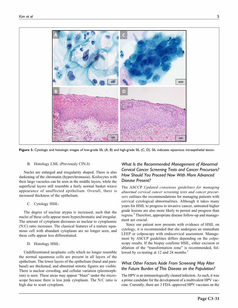
B. Histology LSIL (Previously CIN-I):
Nuclei are enlarged and irregularity shaped. There is also
darkening of the chromatin (hyperchromasia). Koilocytes with
their large vacuoles can be seen in the middle layers, while the
superficial layers still resemble a fairly normal basket weave
appearance of unaffected epithelium. Overall, there is
increased thickness of the epithelium.
C. Cytology HSIL:
The degree of nuclear atypia is increased, such that the
nuclei of these cells appear more hyperchromatic and irregular.
The amount of cytoplasm decreases as nuclear to cytoplasmic
(N:C) ratio increases. The classical features of a mature squa-
mous cell with abundant cytoplasm are no longer seen, and
these cells appear less differentiated.
D. Histology HSIL:
Undifferentiated neoplastic cells which no longer resemble
the normal squamous cells are present in all layers of the
epithelium. The lower layers of the epithelium (basal and para-
basal) are thickened, and abnormal mitotic figures are visible.
There is nuclear crowding, and cellular variation (pleomorph-
ism) is seen. These areas may appear “bluer” under the micro-
scope because there is less pink cytoplasm. The N:C ratio is
high due to scant cytoplasm.
What Is the Recommended Management of AbnormalCervical Cancer Screening Tests and Cancer Precursors?How Should You Proceed Now With More AdvancedDisease Present?
The ASCCP Updated consensus guidelines for managing
abnormal cervical cancer screening tests and cancer precur-
sors outlines the recommendations for managing patients with
cervical cytological abnormalities. Although it takes many
years for HSIL to progress to invasive cancer, untreated higher
grade lesions are also more likely to persist and progress than
regress.3 Therefore, appropriate disease follow-up and manage-
ment are crucial.
Since our patient now presents with evidence of HSIL on
cytology, it is recommended that she undergoes an immediate
LEEP or colposcopy with endocervical assessment. Manage-
ment by ASCCP guidelines differs depending on the colpo-
scopy results. If the biopsy confirms HSIL, either excision or
ablation of the “transformation zone” is recommended, fol-
lowed by co-testing at 12 and 24 months.1
What Other Factors Aside From Screening May Alterthe Future Burden of This Disease on the Population?
TheHPV is an immunologically cleared infection. As such, it was
a prime candidate for the development of a multivalent HPV vac-
cine. Currently, there are 3 FDA–approved HPV vaccines on the
Figure 3. Cytologic and histologic images of low-grade SIL (A, B) and high-grade SIL (C, D). SIL indicates squamous intraepithelial lesion.
Kim et al 5
Page C3-31

market: Gardasil, Gardasil 9, and Cervarix. The Cervarix vaccine
is designed for immunity against HPV 16 and HPV 18, the 2 most
common high-risk HPV types that cause about 70% of cervical
cancer. Gardasil adds coverage for 2 additional low-risk HPV
genotypes (6 and 11) which are the major cause of genital warts.
Gardasil 9 extends coverage further to 5 additional high-risk HPV
genotypes associated with cervical cancer. These vaccines consist
of nonreplicative viral-like particles which provoke an immune
response and provide humoral immunologic memory.7 Currently,
several international health-based organizations recommendHPV
vaccination be a part of all routine immunization programs. It is
important to note that screening following immunization is still
required and we have discussed several additional benefits to
screening beyond identification of HPV infection.
Describe Some Possible Therapies That Are BeingDeveloped to Treat Human Papillomavirus–AssociatedLesions and Cancers
While the initial therapy of squamous lesions is currently com-
plete excision, other novel therapies are currently being eval-
uated. We have discussed vaccines to prevent infection that are
widely available and utilize viral structural proteins. There are
currently in development vaccines that help a person’s immune
system to clear the HPV after infection. Most of these vaccines
target HPV oncoproteins E6 and E7.8 This treatment is antici-
pated to become clinically available in the near future to decrease
the HPV-associated disease burden. Furthermore, research on a
pelvic sentinel lymph node biopsy procedure, immunotherapy,
and targeted drug therapy is also being conducted.
Teaching Points
� Characteristic cytologic and histologic features, such as
koilocytosis, enlarged nuclei, and increased N:C ratio,
allow pathologists to diagnose and classify different
grades of squamous cervical pathology from low-grade
to high-grade squamous lesions and ultimately to
carcinoma.
� Classifications based on these features allow one to pre-
dict the biologic behavior and malignant potential of
these precancerous lesions.
� HPV infection, particularly with high-risk types of HPV,
is not only a major risk factor for developing cervical
SIL/cancer but also a crucial factor in the pathogenesis
of this disease.
� Adequate screening for cervical precancerous pathology
may include both cytology Pap and ancillary HPV test-
ing as each method has distinct advantages for the detec-
tion of pathologic conditions.
� Cervical disease and its associated diagnostic testing are
a paradigmatic model of disease screening and interven-
tion and remain one of the most successful screening
programs ever devised.
� Several organizations, including the ACOG and the
ASCCP, regularly publish guidelines related to screen-
ing and management of the cervical cancer.
Declaration of Conflicting Interests
The author(s) declared no potential conflicts of interest with respect to
the research, authorship, and/or publication of this article.
Funding
The author(s) received no financial support for the research, author-
ship, and/or publication of this article.
ORCID iD
D. Yitzchak Goldstein http://orcid.org/0000-0001-6367-8335
References
1. Wright TC, Ronnett BM, Kurman RJ, Ferenczy A. Precancerous
lesions of the cervix. In: Blaustein’s Pathology of the Female
Genital Tract. New York, NY: Springer; 2011:193-252.
2. Saslow D, Solomon D, Lawson HW, et al. American Cancer Soci-
ety, American Society for Colposcopy and Cervical Pathology, and
American Society for Clinical Pathology Screening Guidelines for
the Prevention and Early Detection of Cervical Cancer. CA Cancer
J Clin. 2012;62:147-172.
3. Sellors JW, Karwalajtys TL, Kaczorowski J, et al. Incidence, clear-
ance and predictors of human papillomavirus infection in women.
CMAJ. 2003;168:421-425.
4. Wilson JMG, Jungner G; World Health Organization, Geneva,
Switzerland. Principles and Practice of Screening for Disease;
1968.
5. Darragh TM, Colgan TJ, Cox JT, et al. The Lower Anogenital
Squamous Terminology standardization project for HPV-
associated lesions: background and consensus recommendations
from the College of American Pathologists and the American Soci-
ety for Colposcopy and Cervical Pathology. Arch Pathol Lab Med.
2012;136:1266-1297.
6. Massad LS, Einstein MH, Huh WK, et al. 2012 updated consensus
guidelines for the management of abnormal cervical cancer screen-
ing tests and cancer precursors. J Low Genit Tract Dis. 2013;17:
S1-S27.
7. Arvin A, Campadelli-Fiume G, Mocarski E, et al. Human Herpes-
viruses: Biology, Therapy, and Immunoprophylaxis. Cambridge:
Cambridge University Press; 2007.
8. Nayar R, Wilbur DC. The Bethesda System for Reporting
Cervical Cytology: Definitions, Criteria, and Explanatory Notes.
New York, NY: Springer; 2015.
6 Academic Pathology
Page C3-32

Educational Case
Educational Case: Medullary ThyroidCarcinoma
Carl T. McGary, MD, PhD1
The following fictional case is intended as a learning tool within the Pathology Competencies for Medical Education
(PCME), a set of national standards for teaching pathology. These are divided into three basic competencies: Disease
Mechanisms and Processes, Organ System Pathology, and Diagnostic Medicine and Therapeutic Pathology. For addi-
tional information, and a full list of learning objectives for all three competencies, see http://journals.sagepub.com/doi/
10.1177/2374289517715040.
Keywordspathology competencies, organ system pathology, cytology diagnostic certainty, endocrine, endocrine neoplasms, fine needleaspiration cytology, medullary thyroid carcinoma
Received November 20, 2017. Received revised February 08, 2018. Accepted for publication February 16, 2018.
Primary Objective
EN5.2: Medullary Thyroid Carcinoma. Describe the molecular
basis and clinicopathologic features of medullary thyroid
carcinoma.
Competency 2: Organ System Pathology; Topic EN: Endo-
crine; Learning Goal 5: Endocrine Neoplasms.
Secondary Objective
CYP1.2: Categorizing Diagnostic Certainty. Compare and
contrast the degree of diagnostic certainty applied to general
categorization in cytologic diagnosis.
Competency 3: Diagnostic Medicine and Therapeutic
Pathology; Topic CYP: Cytopathology; Learning Goal 1: Cyto-
logic Diagnosis.
Patient Presentation
A 52-year-old Caucasian male presents to family medicine
with an irregular 4-cm palpable nodule in the mid right thyroid
lobe. He has no significant past medical history other than
occasional diarrhea. Complete head and neck examination
reveals no additional palpable nodules in the thyroid gland and
no palpable adenopathy. The differential diagnosis for a palp-
able thyroid nodule includes a variety of benign and malignant
entities, including colloid nodule, nodular hyperplasia,
follicular thyroid adenoma, follicular thyroid carcinoma, papil-
lary thyroid carcinoma (PTC), medullary thyroid carcinoma
(MTC), anaplastic thyroid carcinoma, and lymphoma. The
patient is referred to the lab for pathologist to perform fine
needle aspiration (FNA) cytology of the nodule.
Diagnostic Findings, Part 1
The pathologist performs 3 FNA passes into the nodule under
direct guidance using 25-gauge needles without suction, and the
initial smears show adequate cellularity (Figure 1). Additional
passes are not performed for cell block preparation. Using The
Bethesda System for Reporting Thyroid Cytology, the patholo-
gist finalizes the report as Bethesda category V, suspicious for
malignancy and MTC. The cytology findings are shown in Fig-
ures 1 and 2 and compared to the more common PTC in Figure
3. The patient is referred to endocrinology for further evaluation.
1 Department of Biomedical Sciences, The University of Minnesota Medical
School–Duluth, Duluth, MN, USA
Corresponding Author:
Carl T. McGary, MD, PhD, Department of Biomedical Sciences, The University
of Minnesota Medical School–Duluth, 1035 University Drive, SMED 227,
Duluth, MN 55812, USA.
Email: [email protected]
Academic PathologyVolume 5: 1–5ª The Author(s) 2018Reprints and permission:sagepub.com/journalsPermissions.navDOI: 10.1177/2374289518770173journals.sagepub.com/home/apc
Creative Commons Non Commercial CC BY-NC: This article is distributed under the terms of the Creative Commons Attribution-NonCommercial 4.0 License(http://www.creativecommons.org/licenses/by-nc/4.0/) which permits non-commercial use, reproduction and distribution of the work without furtherpermission provided the original work is attributed as specified on the SAGE and Open Access pages (https://us.sagepub.com/en-us/nam/open-access-at-sage).
Page C3-33

Questions/Discussion Points, Part 1
Categorizing Diagnostic Certainty: Based on thePathologist’s Interpretation of the FNA, How StatisticallyLikely is the Patient to Harbor a Malignant Neoplasmand What is the Uncertainty of the Diagnosis?
The major categories used in diagnostic cytopathology include
unsatisfactory for diagnosis, negative for malignancy, atypical
cells present, suspicious for malignancy, and positive for
malignancy. Each category carries an implied degree of
certainty with regard to the pathology present. Diagnostic
modifiers are also used in surgical pathology to suggest degrees
of uncertainty, including “cannot rule out,” “suggestive of,”
“consistent with,” and “diagnostic of,” in increasing order of
certainty.1 The issue of uncertainty in anatomic pathology
diagnosis is highlighted by the presence of both interobserver
(2 observers arriving at a different diagnosis on the same case)
and intraobserver (the same observer arriving at different diag-
noses on the same case at different times) diagnostic variation.2
Interinstitutional review of 777 patient specimens revealed a
diagnostic difference in 71 specimens on review at the second
institution. In 45 of the 71 discrepant specimens, the change in
diagnosis led to altered therapy. The discordant diagnoses were
significantly more common in cytology specimens (21%) than
surgical specimens (7.8%), suggesting that cytologic diagnosis
was more prone to uncertainty than histologic diagnosis.3
Historically, cytopathologists at different institutions have
utilized somewhat different diagnostic terminology when report-
ing results of thyroid FNA. To address the variety of reporting
systems and the resulting inherent confusion, the National Can-
cer Institute (NCI) hosted the NCI Thyroid FNA State of the
Science Conference. The conference conclusions led to the pub-
lication of the Bethesda System for Reporting Thyroid Cytol-
ogy,4 which is now in wide use. Similar issues led to publication
of the Paris System for Reporting Urinary Cytology.5
The Bethesda System for Reporting Thyroid Cytology4 con-
tains 6 general diagnostic categories, each with a specific given
risk of underlying malignancy as shown in Table 1. The least
amount of uncertainty is associated with the benign and malig-
nant categories as these have the narrowest range for underly-
ing malignancy. The greatest degree of uncertainty lies with the
atypical category which shows the widest range for underlying
Figure 3. Papanicolaou stain of PTC for comparison with the patientswith FNA cytology. Papillary thyroid carcinoma shows nongranularchromatin, nuclear grooves, small peripheral nucleoli, and occasionalintranuclear pseudo-inclusions. Papillary-shaped irregular branchinggroups are also present. FNA indicates fine needle aspiration cytology;PTC, papillary thyroid carcinoma.
Figure 1. Papanicolaou stain at �200 of the initial FNA smearshowing background red blood cells, scant colloid, and a mixed pop-ulation of neoplastic cells showing epithelioid and spindle cell mor-phology. Nuclear chromatin shows a “‘salt and pepper” granularity andlacks features of papillary carcinoma (Figure 3), and the cell cytoplasmis somewhat granular. FNA indicates fine needle aspiration cytology.
Figure 2. Papanicolaou stain at �400 highlighting random nuclearatypia often seen in neuroendocrine neoplasia, as well as the granularcytoplasm and fine-to-coarse nuclear chromatin of medullary thyroidcarcinoma.
2 Academic Pathology
Page C3-34

malignancy, and the suspicious category being between atypi-
cal and benign/malignant in terms of uncertainty. Thyroid FNA
is somewhat unique in having 2 suspicious categories. Suspi-
cious for follicular neoplasm less commonly has an underlying
malignancy when compared to the suspicious for malignancy
category, as the majority of follicular neoplasms are benign
adenomas, rather than follicular carcinomas.
Similar uncertainty is seen in other types of cytology speci-
mens, such as urine cytology using The Paris System for
Reporting.5 The risk of underlying malignancy in urine cytol-
ogy based on The Paris System reporting is shown in Table 2.
The wider the range for underlying malignancy, the greater the
uncertainty for that diagnostic category.
In general, both benign and malignant cytologic diagnostic
categories have the least associated uncertainty, and the most
significant uncertainty is in the atypical category, with the
suspicious category intermediate for the associated degree of
uncertainty. Currently available molecular tests are available
for use on ambiguous thyroid FNAs, to identify malignancy-
associated genetic changes. These serve to decrease the broad
diagnostic uncertainty in the atypical follicular cells of uncer-
tain significance category and help direct treatment decisions
(surgery vs continued clinical observation).
Diagnostic Findings, Part 2
The endocrinologist does a typical thyroid endocrine workup
and finds that the patient is euthyroid and has a normal calcium.
Because of the suspicion of MTC on the FNA, he also performs
a serum calcitonin that comes back as 5679 pg/mL (normal
<10 pg/mL). The patient is referred to otolaryngology and
a total thyroidectomy and central and bilateral node dissection
is performed. The final pathology shows a 3.9-cm medullary
carcinoma (Figures 4-6) confined to the thyroid gland with 3 of
6 central nodes microscopically involved and none of 13 right
and 1 of 11 left neck nodes involved by medullary
carcinoma. The pathologic stage is reported as pT2, pN1a,
pM-not applicable, or stage group III.
Table 1. Major Diagnostic Categories of The Bethesda System forReporting Thyroid Cytology and the Associated Risk of Malignancy.
Diagnostic Category Underlying Risk of Malignancy (%)
ND/unsatisfactory (I) 1-4Benign (II) 0-3AUS/FLUS (III) 5-15FN/SFN (IV) 15-30Suspicious for malignancy (V) 60-75Malignant (VI) 97-99
Abbreviations: AUS, atypia of uncertain significance; FLUS, follicular lesion ofuncertain significance; FN, follicular neoplasm; ND, nondiagnostic; SFN, suspi-cious for follicular neoplasm.
Table 2. Diagnostic Categories of The Paris System for ReportingUrinary Cytology and the Associated Risk of Malignancy.
Diagnostic Category Underlying Risk of Malignancy (%)
Unsatisfactory <5-10Negative for HGUC 0-10Atypical urothelial cells 8-35Suspicious for HGUC 50-90Low-grade urothelial neoplasm *10Positive for HGUC >90Positive for other malignancy >90
Abbreviation: HGUC, high-grade urothelial carcinoma.
Figure 4.Hematoxylin and eosin stained section of the tumor thyroidinterface at �40 showing a well-circumscribed cellular neoplasm onthe left without follicular or papillary architecture. The tumor wasincompletely encapsulated, and entrapped benign follicles are seen atthe periphery. The adjacent thyroid tissue showed no evidence of C-cell hyperplasia and contains medium-to-large colloid-filled follicles onthe right.
Figure 5. Hematoxylin and eosin stained section of tumor at �200showing mixed epithelioid and spindled morphology with only vaguenesting of tumor cells and a majority diffuse growth pattern. Amyloidwas not present.
McGary 3
Page C3-35

Questions/Discussion Points, Part 2
What Clinicopathologic Features are Common in BothSporadic and Inherited Medullary Thyroid Carcinoma?
Medullary thyroid carcinoma is a primary tumor of the thyroid
gland which shows differentiation along the lines of thyroid
parafollicular C cells.6-8 Medullary thyroid carcinoma typically
secretes calcitonin, although nonsecretory tumors occur rarely.
Serum calcitonin reflects quantity of tumor present, except in
nonsecreting patients. Medullary thyroid carcinoma is almost
always carcinoembryonic antigen (CEA) positive, and in
noncalcitonin-secreting tumors, serum CEA levels can be fol-
lowed, similar to calcitonin levels in secreting tumors to assess
disease burden and therapeutic response. Even though elevated
calcitonin levels are present, serum calcium is not typically
decreased below normal levels. Medullary thyroid carcinoma
tumors sometimes secrete other hormones, such as adrenal
corticotropic hormone, serotonin, and vasoactive intestinal
peptide (VIP), which can result in a paraneoplastic presentation
such as diarrhea if VIP secretion occurs. The VIP levels were
not measured in this patient, and it is not clear whether his
intermittent diarrhea was related to the MTC. Medullary thyr-
oid carcinomas are relatively uncommon tumors accounting for
less than 2% to 3% of thyroid neoplasia.
Medullary thyroid carcinoma can present as a sporadic
tumor or as part of an inherited tumor syndrome. The histology
of MTC is quite variable, but in general is not distinct for
sporadic or inherited cases. A mixture of spindled and epithe-
lioid cells is common, and plasmacytoid morphology is often
seen. Neuroendocrine features include finely granular cyto-
plasm due to variable presence of neurosecretory granules. The
chromatin is mixed fine and coarse and classically described as
“salt and pepper” chromatin which is commonly seen in neu-
roendocrine tumors. Cells can be arranged in sheets, nested
aggregates, trabeculae, or even follicles, and mitotic figures
tend to be sparse. These features of MTC contrast with the
more common PTC (Figure 3). Nuclear features of PTC
include pale nongranular nuclear chromatin, nuclear grooves,
nuclear pseudoinclusions, and small peripheral nucleoli. Amy-
loid deposition is commonly present in up to 90% of cases,
but was absent in this particular case, and is composed of
calcitonin peptides. Immunochemistry shows positive granular
cytoplasmic staining for calcitonin in the tumor cells and dif-
fusely stains the amyloid deposits. Immunochemistry is also
positive for other neuroendocrine markers, such as chromogra-
nin and synaptophysin. Cytologic atypia ranges from minimal,
to focal, to widespread.
Describe the Features Unique to Sporadic MedullaryThyroid Carcinoma
Medullary thyroid carcinoma occurs as sporadic disease in
70% of cases, and the sporadic cases present typically as a
neck mass, as in this case, but clinical findings may also
include dysphagia and/or hoarseness. When MTC presents
as a neck mass, nodal involvement is relatively frequent
(*70%) and non-nodal metastasis (10%) also occurs. Spora-
dic cases show a slight female predominance and are seen
predominantly in the fifth and sixth decades. These are usu-
ally unilateral and unifocal tumors of the thyroid. Unlike PTC,
ionizing radiation exposure does not seem to be etiologically
involved in sporadic MTC.
Describe the Features Unique to Inherited MedullaryThyroid Carcinoma
Familial cases occur as part of inherited syndromes in 30% of
cases (multiple endocrine neoplasia type 2A [MEN-2A] or
MEN-2B or familial medullary thyroid carcinoma [FMTC])
and are often associated with other endocrine neoplasms.
Medullary thyroid carcinoma in this setting may present either
as a result of endocrine disease in other organs or with symp-
toms in the neck. Familial/inherited cases present at younger
ages than do sporadic cases. Medullary thyroid carcinoma in
the setting of MEN-2B more commonly metastasizes than what
is seen in MEN-2A, FMTC, or sporadic disease. Bilateral thyr-
oid involvement and multifocal disease are more common in
inherited MTC than in sporadic disease. Penetrance of medul-
lary thyroid cancer development is age and mutation depen-
dent; however, MTC inevitably develops in MEN-2 with RET
mutations, and early total thyroidectomy is preventative. Early
onset is typical in MEN-2B with later onset in MEN-2A. Thyr-
oid tissue outside the tumor in inherited cases typically shows
background C cell hyperplasia.
Describe the Molecular Basis of Sporadic MedullaryThyroid Carcinoma
Activating somatic point mutations of the RET proto-oncogene
are noted in sporadic medullary carcinoma in approximately
40% to 60% of cases. The most frequently seen mutation in
Figure 6. Immunochemistry of tumor for calcitonin �400 showingdiffuse granular cytoplasmic staining.
4 Academic Pathology
Page C3-36

sporadic cases is the M918T mutation in exon 16. Interestingly,
this is also the same mutation as the germline mutation in the
vast majority of MEN-2B patients. RASmutations are also seen
in some sporadic cases, usually those that are RET mutation
negative. Thus, in sporadic disease, RAS and RET mutations
commonly show mutual exclusion.
Describe the Molecular Basis of Inherited MedullaryThyroid Carcinoma
Germline mutation of the RET proto-oncogene with gain of
function is the underlying feature in inherited MTC, as seen in
MEN-2A, MEN-2B, and FMTC (a variant of MEN-2A). The
RET proto-oncogene codes for a receptor tyrosine kinase that
binds glial-derived neurotrophic factor and similar ligands.
Ligand binding to receptor leads to intracellular growth signals
as well as signals for differentiation. Various specific RETmuta-
tions occur in MEN-2A, and coexisting parathyroid hyperplasia
and pheochromocytoma are characteristic. The specific RET
mutations in MEN-2B are associated with pheochromocytoma
as well; however, the hyperparathyroidism is absent.
Teaching Points
� Major cytologic diagnostic categories (nondiagnostic,
benign, suspicious, and malignant) each carry an asso-
ciated risk of underlying malignancy and uncertainty,
which have been published for various types of
specimens.
� Cytologic diagnostic categories of benign and malignant
have the least amount of associated diagnostic uncer-
tainty and the atypical category the highest.
� Medullary thyroid carcinoma is a neuroendocrine tumor
showing C-cell differentiation.
� Medullary thyroid carcinoma is immunochemically pos-
itive for calcitonin (which is elevated in serum, except in
rare nonsecreting cases), chromogranin, synaptophysin,
and CEA.
� Medullary thyroid carcinoma can be followed clinically
with serum calcitonin or CEA, and CEA is helpful in
calcitonin nonsecreting cases.
� Occasionally, MTCs secrete other hormones and
may have paraneoplastic presentations, for example,
VIP—diarrhea.
� Medullary thyroid carcinoma can be sporadic or
inherited.
� Sporadic and inherited MTCs have the same histologic
spectrum with amyloid deposition, sporadic disease
typically being unilateral and unifocal and inherited dis-
ease more likely to be bilateral and multifocal.
� Inherited MTC shows C-cell hyperplasia in the non-
neoplastic thyroid tissue.
� Specific genetic and molecular mechanisms underlie
both sporadic and inherited medullary thyroid cancer,
with the RET gene being most frequently involved.
� The inevitable development of medullary thyroid cancer
associated with MEN-2 can be prevented by early pro-
phylactic total thyroidectomy.
ORCID iD
Carl T. McGary, MD, PhD http://orcid.org/0000-0003-4625-3030
References
1. Bracamonte E, Gibson BA, Klein R, Krupinski EA, Weinstein RS.
Communicating uncertainty in surgical pathology reports: a survey
of staff physicians and residents at an academic medical center.
Acad Pathol. 2016;3:1-7. doi:10.1177/2374289516659079.
2. Franc B, De La Salmoniere P, Lange F, et al. Interobserver and
intraobserver reproducibility in the histopathology of follicular
thyroid carcinoma. Hum Pathol. 2003;34(11):1092-1100.
3. Abt AB, Abt LG, Olt GJ. The effect of interinstitution anatomic
pathology consultation on patient care. Arch Pathol Lab Med.
1995;119:514-517.
4. Cibas ES, Ali SZ; NCI Thyroid FNA State of the Science Confer-
ence. The Bethesda System for Reporting Thyroid Cytology. Am J
Clin Pathol. 2009;132:658-665.
5. Barkan GA, Wojcik EM, Nayar R, et al. The Paris System for
Reporting Urinary Cytology: the quest to develop a standardized
terminology. Acta Cytol. 2016;60:185-197.
6. Maitra A. The endocrine system. In: Kumar V, Abbas A, Aster A,
eds. Robbins and Cotran Pathologic Basis of Disease. 9th ed.
Philadelphia, PA: Elsevier Sanders; 2015:1099-1100.
7. Maitra A. The endocrine system. In: Kumar V, Abbas A, Aster A,
eds. Robbins and Cotran Pathologic Basis of Disease. 9th ed.
Philadelphia, PA: Elsevier Sanders; 2015:1136-1137.
8. Lloyd RV, Osamura RY, Kloppel G, Rosi J.WHO Classification of
Tumours of Endocrine Organs. 4th ed. France: International
Agency for Research on Cancer (IARC); 2017:108-113.
McGary 5
Page C3-37


Educational Case
Subarachnoid Hemorrhage Relatedto Ruptured Berry Aneurysm
Sarah Meyers, MD1
The following fictional case is intended as a learning tool within the Pathology Competencies for Medical Education
(PCME), a set of national standards for teaching pathology. These are divided into three basic competencies: Disease
Mechanisms and Processes, Organ System Pathology, and Diagnostic Medicine and Therapeutic Pathology. For addi-
tional information, and a full list of learning objectives for all three competencies, see http://journals.sagepub.com/doi/
10.1177/2374289517715040.
Keywordspathology competencies, organ system pathology, diagnostic medicine, therapeutic pathology, central nervous system, berryaneurysm, death certificate, death investigation, intracranial hemorrhage, subarachnoid hemorrhage
Received April 15, 2017. Received revised May 03, 2017. Accepted for publication May 22, 2017.
Primary Objective
Objective NSC7.3: Cranial Hemorrhage. Compare and contrast the
etiologies and clinical presentations of epidural, subdural, sub-
arachnoid, basal ganglionic, and lobar hemorrhages.
Competency 2: Organ System Pathology; Topic NSC: Ner-
vous System–Central Nervous System; Learning Goal 7: Ische-
mia of the Brain.
Secondary Objective
Objective AU2.2: Components of the Death Certificate. Discuss thekey components of the death certificate; differences among
immediate, intermediate, and underlying (proximate) cause of
death based on disease process; and the role of mechanisms of
death on a death certificate.
Competency 3: Diagnostic Medicine and Therapeutic Pathol-
ogy; Topic AU: Autopsy; Learning Goal 2: Death Certificate.
Clinical Case
A 47-year-old female with a medical history of migraine head-
aches, with migraine pain controlled by codeine, is found dead
on the floor of her secured residence. No outward signs of
trauma or foul play were identified at the scene of death. Given
her relatively young age and minimal medical history, an
autopsy was requested and performed by the county medical
examiner. Following resection of the skull and dura, the follow-
ing intracranial finding was identified (Figure 1). The patholo-
gist reports that this collection cannot be wiped from the surface
of the brain. The hemorrhage appeared to be concentrated at the
base of the brain. The circle of Willis was dissected and showed
the abnormality identified by the arrow (Figure 2).
Questions/Discussion Points
Given the photograph and the description, what is thename of the finding featured in Figure 1? How wouldthe gross description be different for other types ofintracranial hemorrhage? If a CT was performed for thisentity, how might the findings be reported?
The photograph shows subarachnoid hemorrhage. A subar-
achnoid hemorrhage is confined by the arachnoid layer and
1 Hennepin County Medical Examiner’s Office, Minneapolis, MN, USA
Corresponding Author:
Sarah Meyers, Hennepin County Medical Examiner’s Office, 530 Chicago Ave,
Minneapolis, MN 55415, USA.
Email: [email protected]
Academic PathologyVolume 4: 1–3ª The Author(s) 2017Reprints and permission:sagepub.com/journalsPermissions.navDOI: 10.1177/2374289517716463journals.sagepub.com/home/apc
Creative Commons CC BY-NC: This article is distributed under the terms of the Creative Commons Attribution-NonCommercial 4.0 License(http://www.creativecommons.org/licenses/by-nc/4.0/) which permits non-commercial use, reproduction and distribution of the work without furtherpermission provided the original work is attributed as specified on the SAGE and Open Access pages (https://us.sagepub.com/en-us/nam/open-access-at-sage).
Page C3-39

cannot be wiped or rinsed away from the brain. Radiographi-
cally, subarachnoid hemorrhage would be demonstrated by thin
layers of blood overlying structures, such as the sylvian fissure,
intrahemispheric fissure, or basal cisterns. Subdural hemor-
rhage consists of blood filling the potential space beneath the
dura, but outside the subarachnoid space. At gross inspection,
subdural blood would be able to be wiped or washed away from
the surface of the brain and not retained within the cell layer of
the subarachnoid space. CT imaging generally shows a cres-
centic shaped area of hemorrhage outside the brain parench-
yma, beneath the skull. Intraparenchymal hemorrhages, such as
those related to hypertension or tumors, would be centered
within and usually surrounded by brain parenchyma; however,
these areas of hemorrhage may expand to communicate with
the subarachnoid, subdural, or intraventricular space. CT ima-
ging of intraparenchymal hemorrhage demonstrates blood
within the brain itself.
What is the differential diagnosis for the etiology of thefinding demonstrated in Figure 1?
Ruptured berry (saccular) aneurysm, trauma, ruptured arteriove-
nous malformation, extension of intraparenchymal hemorrhage,
hematologic disorders, and coagulopathies.1 Of these entities,
the most common cause of clinically significant subarachnoid
hemorrhage is due to a ruptured berry aneurysm.1
What is the cause of the hemorrhage based on thefinding in Figure 2?
Ruptured berry (saccular) aneurysm.
What are the major complications associated with theseautopsy findings?
Statistics vary, but up to 50% of patients die within 1 month
of an intracerebral aneurysmal rupture and hemorrhage.1
Similar to this patient, at least 10% of patients die prior
to hospitalization for the first bleed.1 If a patient survives,
major morbidity manifests as delayed ischemic complica-
tions related to vasospasm, rebleeding, seizures, hydroce-
phalus, and hyponatremia.1
Acting as the medical examiner, please list the cause(immediate and underlying) and manner of death.
The cause of death is the injury or disease process that resulted
in a patient’s death. Manner of death is defined by public health
parameters and is a selection of natural, accident, suicide,
homicide, or undetermined. A death certificate should tell the
whole story of a patient’s history and should work backward
from the underlying cause of death to the immediate cause of
death. Care should be taken to ensure that the death certificate
is specific, concise, and avoids nonspecific mechanisms of
death that are universal to all deaths, such as the phrase cardi-
orespiratory arrest. As such, the first disease process that
Figure 1. Brain with subarachnoid hemorrhage.
Figure 2. Dissected circle of Willis demonstrating ruptured berryaneurysm at the bifurcation of the anterior communicating artery andthe anterior cerebral artery.
2 Academic Pathology
Page C3-40

occurred in this patient’s case was the rupture of the berry
aneurysm leading to the subarachnoid hemorrhage. Both of
these processes are natural diseases; therefore, the death cer-
tificate should be completed as follows:
Cause of death: Subarachnoid hemorrhage (immediate
cause)
Due To: Ruptured berry aneurysm (underlying cause)
Manner of Death: Natural
Teaching Points
� Subarachnoid hemorrhage is demonstrated grossly by
a layer of blood confined within the subarachnoid
space.
� The differential diagnosis for subarachnoid hemor-
rhage includes a ruptured berry aneurysm, trauma, rup-
tured arteriovenous malformation, extension of
intraparenchymal hemorrhage, hematologic disorders,
and coagulopathies.
� Ruptured berry aneurysms have high morbidity and
mortality related to short- and long-term complications
related to subarachnoid hemorrhage.
� In completing a death certificate, the cause of death
should be ascribed to the underlying disease or injury
that set the sequence of events in motion to result in the
immediate cause of death.
Declaration of Conflicting Interests
The author(s) declared no potential conflicts of interest with respect to
the research, authorship, and/or publication of this article.
Funding
The author(s) received no financial support for the research, author-
ship, and/or publication of this article.
Reference
1. Frosch MP, Anthony DC, De Girolami U. The central nervous
system. In: Kumar V, Abbas AK, Fausto N, Aster JC, eds. Robbins
and Cotran Pathologic Basis of Disease. 8th ed. Philadelphia, PA:
Saunders Elsevier; 2010.
Meyers 3
Page C3-41


The association of Pathology Chairs (aPC) is seeking educational Case submissions to Academic Pathology, the association’s official journal. once published, educational Cases are indexed on Pub med, openly accessible worldwide, and citable for scholarly credit, similar to other peer-reviewed articles. educational Cases are tools for implementing the Pathology Competencies for medical education (see doi: 10.1177/2374289517715040), which is a national standard to provide all medical students with a broad foundation of knowledge, skills, and attitudes essential to understand the normal and pathological processes of each organ system, the ability to apply disease mechanisms to describe the pathobiology, and the ability to continually improve diagnostic acumen and optimal treatment decisions through lifelong learning.
The Pathology Competencies for medical education (PCme) have detailed learning objectives under each goal that direct medical students and course directors to important facets of each learning goal that can be individually applied by learners. The competencies are divided into three sections–disease mechanisms and processes, organ system pathology, and diagnostic medicine and therapeutic pathology–and allow flexibility for each medical school and learner to apply the learning goals and objectives in a way that can keep the unique design of each curriculum or learning plan. The competencies are purposefully kept broad as they represent the minimum requirements of what pathology course directors across the nation have agreed upon to prepare medical students for entry into any residency program and for the subsequent contemporary practice of medicine.
Who Can submit
educational Cases may be submitted by anyone in any medical specialty. developing an educational Case is an important opportunity for faculty to mentor residents and students in developing scholarship and exploring medical education as a profession. mentors are encouraged to work with residents and students in responding to this Call for educational Cases.
how to submit
educational Cases have a unique format. Before preparing your manuscript, please visit the PCme portal at https://journals.sagepub.com/page/apc/pcme, where you will find the educational Case submission guidelines, a link to a webinar on the PCme framework & educational Case application, and a link to all published educational Cases.
Educational Cases responding to this call must be submitted by June 1, 2019 at https://mc.manuscriptcentral.com/apc
review Process
The educational Case review Board will meet in July 2019 to evaluate submissions. authors of educational Cases accepted for publication will be notified by July 31, 2019. in rendering their decision, the review Board will pay particular attention to whether authors: 1) followed the guidelines for educational Case preparation; and 2) prepared an educational Case covering an objective that has no other cases.
The review Board will provide specific feedback on educational Cases that do not meet the above criteria, but may not ultimately accept them for publication, if authors are unable to successfully address their concerns through revision.
Publication fees
Cases accepted for publication will incur an open access article processing fee of $500.00 for authors who are from aPC member departments; $750 for non-members. to inspire early participation in producing scholarship, the society of ’67 fundraising arm of aPC created the open access award program to fund open access fees associated with publishing in Academic Pathology for students, residents and junior faculty. all educational Cases submitted with a first or corresponding author, who is a student, resident or junior faculty, will qualify for the open access award (for more information see https://www.apcprods.org/societyof67-award-oa). Qualifying for the award does not guarantee acceptance for publication.
Questions?? Please contact the Academic Pathology administrative office at [email protected] or 302-660-4940.
call for educational cases