Differential characterization of three alternative spliced isoforms of DPPX
RESEARCH Open Access
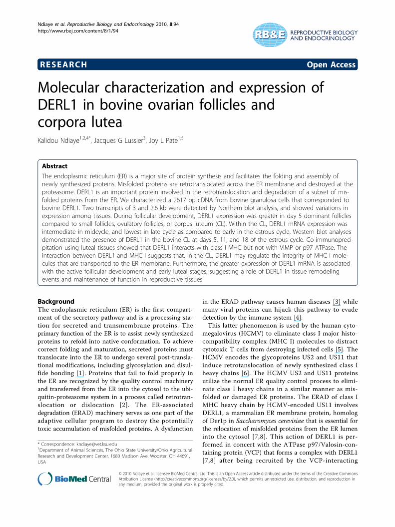
Molecular characterization and expression ofDERL1 in bovine ovarian follicles andcorpora luteaKalidou Ndiaye1,2,4*, Jacques G Lussier3, Joy L Pate1,5
Abstract
The endoplasmic reticulum (ER) is a major site of protein synthesis and facilitates the folding and assembly ofnewly synthesized proteins. Misfolded proteins are retrotranslocated across the ER membrane and destroyed at theproteasome. DERL1 is an important protein involved in the retrotranslocation and degradation of a subset of mis-folded proteins from the ER. We characterized a 2617 bp cDNA from bovine granulosa cells that corresponded tobovine DERL1. Two transcripts of 3 and 2.6 kb were detected by Northern blot analysis, and showed variations inexpression among tissues. During follicular development, DERL1 expression was greater in day 5 dominant folliclescompared to small follicles, ovulatory follicles, or corpus luteum (CL). Within the CL, DERL1 mRNA expression wasintermediate in midcycle, and lowest in late cycle as compared to early in the estrous cycle. Western blot analysesdemonstrated the presence of DERL1 in the bovine CL at days 5, 11, and 18 of the estrous cycle. Co-immunopreci-pitation using luteal tissues showed that DERL1 interacts with class I MHC but not with VIMP or p97 ATPase. Theinteraction between DERL1 and MHC I suggests that, in the CL, DERL1 may regulate the integrity of MHC I mole-cules that are transported to the ER membrane. Furthermore, the greater expression of DERL1 mRNA is associatedwith the active follicular development and early luteal stages, suggesting a role of DERL1 in tissue remodelingevents and maintenance of function in reproductive tissues.
BackgroundThe endoplasmic reticulum (ER) is the first compart-ment of the secretory pathway and is a processing sta-tion for secreted and transmembrane proteins. Theprimary function of the ER is to assist newly synthesizedproteins to refold into native conformation. To achievecorrect folding and maturation, secreted proteins musttranslocate into the ER to undergo several post-transla-tional modifications, including glycosylation and disul-fide bonding [1]. Proteins that fail to fold properly inthe ER are recognized by the quality control machineryand transferred from the ER into the cytosol to the ubi-quitin-proteasome system in a process called retrotran-slocation or dislocation [2]. The ER-associateddegradation (ERAD) machinery serves as one part of theadaptive cellular program to destroy the potentiallytoxic accumulation of misfolded proteins. A dysfunction
in the ERAD pathway causes human diseases [3] whilemany viral proteins can hijack this pathway to evadedetection by the immune system [4].This latter phenomenon is used by the human cyto-
megalovirus (HCMV) to eliminate class I major histo-compatibility complex (MHC I) molecules to distractcytotoxic T cells from destroying infected cells [5]. TheHCMV encodes the glycoproteins US2 and US11 thatinduce retrotanslocation of newly synthesized class Iheavy chains [6]. The HCMV US2 and US11 proteinsutilize the normal ER quality control process to elimi-nate class I heavy chains in a similar manner as mis-folded or damaged ER proteins. The ERAD of class IMHC heavy chain by HCMV-encoded US11 involvesDERL1, a mammalian ER membrane protein, homologof Der1p in Saccharomyces cerevisiae that is essential forthe relocation of misfolded proteins from the ER lumeninto the cytosol [7,8]. This action of DERL1 is per-formed in concert with the ATPase p97/Valosin-con-taining protein (VCP) that forms a complex with DERL1[7,8] after being recruited by the VCP-interacting
* Correspondence: [email protected] of Animal Sciences, The Ohio State University/Ohio AgriculturalResearch and Development Center, 1680 Madison Ave, Wooster, OH 44691,USA
Ndiaye et al. Reproductive Biology and Endocrinology 2010, 8:94http://www.rbej.com/content/8/1/94
© 2010 Ndiaye et al; licensee BioMed Central Ltd. This is an Open Access article distributed under the terms of the Creative CommonsAttribution License (http://creativecommons.org/licenses/by/2.0), which permits unrestricted use, distribution, and reproduction inany medium, provided the original work is properly cited.
membrane protein (VIMP), which can interact withDERL1 [8].The expressions of class I and class II MHC molecules
have been demonstrated in the corpus luteum (CL)where they may function to regulate T lymphocyte acti-vation, thus controlling the production of cytokines[9-13]. The CL is a very heterogeneous population ofcells that becomes rapidly organized into a functionalunit. These diverse cells communicate both directly andthrough paracrine mediators to facilitate the steroido-genic function and the transient nature of the CL. TheCL also contains nonsteroidogenic cells, includingendothelial cells, fibroblasts, and immune cells that havea role in luteal function by communicating with steroi-dogenic cells through the paracrine signaling moleculesthey produce and through direct cell contacts [14,15].The MHC molecule-dependent interaction betweenluteal cells and T cells is one form of direct cell-cell sig-naling that may serve to activate resident immune cells[16]. An association between MHC proteins and theERAD proteins in reproductive tissues has not been pre-viously investigated. Thus, studying the link betweenclass I MHC and DERL1 in the CL may contribute tounderstanding the control of luteal function.There has been significant interest in the role of
DERL1 in the ER quality control machinery. However,nothing is known about the expression of DERL1, itsregulation and physiological relevance in reproductivetissues, such as the ovary. In this study, the expressionof DERL1 in bovine granulosa and luteal cells, and itsregulation at different stages of follicular growth andcorpora lutea development were analyzed. Using normalluteal tissue extracts, putative interactions of DERL1with MHC I, p97 ATPase, and VIMP were alsoinvestigated.
MethodsAnimal model for follicle collectionThe regulation of DERL1 mRNA expression during folli-cular development and ovulation was studied usingin vivo models as previously characterized [17]. Briefly,estrous cycles of normally cyclic crossbred heifers weresynchronized with one injection of prostaglandin F2alpha (PGF2a; 25 mg, im; Lutalyse, Upjohn, Kalamazoo,MI) given in the presence of a CL. Ovarian folliculardevelopment was monitored by daily transrectal ultraso-nography. Following estrous synchronization, heiferswere randomly assigned to the dominant follicle group(DF, n = 4), or the ovulatory, hCG-induced folliclegroup (OF, n = 4). Immediately following ovariectomy,granulosa cells (GC) were collected separately from indi-vidual DF or OF as described previously [17], and storedat -70°C. Additionally, GC were collected from 2-4 mmsmall follicles (SF) obtained from slaughterhouse ovaries,
and a total of three pools of 10-20 SF was prepared.Corpora lutea at day 5 of the estrous cycle wereobtained by ovariectomy and were dissected from theovarian stroma, frozen in liquid nitrogen, and stored at-70°C until RNA was extracted. The Animal EthicsCommittee of the Faculty of Veterinary Medicine of theUniversity of Montreal approved all animal procedures.
Corpora lutea removalCorpora lutea were collected transvaginally from cycliccows at early (day 5) mid (day 11) and late (day 18)luteal phase of the estrous cycle (day of estrus = day 0)and at 1, 4, 8, and 12 hours after a 25-mg injection ofPGF2a (Lutalyse; Upjohn, Kalamazoo, MI) in the midlu-teal phase to induce luteal regression. Total RNA wasextracted from luteal tissues to quantify DERL1 mRNAconcentrations by real time-quantitative PCR. Corporalutea collection was performed at the Ohio State Uni-versity and handling of animals and surgical procedureswere conducted according to protocols approved by theInstitutional Laboratory Animal Care and Use Commit-tee of the Ohio State University.
Cloning of bovine DERL1The DERL1 cDNA was cloned from a bovine cDNAlibrary prepared with polyA+ mRNA isolated from gran-ulosa cells of dominant follicles at day 5 of the estrouscycle. The cDNA library was constructed in lambda ZapExpress vector (Stratagene, La Jolla, CA) by unidirec-tional cloning of cDNAs as described [18]. Following invivo excision of pBluescript phagemids containing thecloned cDNA insert with the Ex-Assist/XLOLR system(Stratagene), single bacterial colonies were picked andtheir phagemid content were purified by mini-prep(Qiagen, Mississauga, ON). The cDNA inserts were ana-lyzed by sequencing, and sequences were compared byBLAST in GenBank database. Alignment of deducedamino acid sequences of bovine DERL1 protein withputative homologs from other organisms was performedusing the ClustalW2 analysis software (EMBL-EBI,Hinxton, Cambridge, UK). DERL1 cDNA was entirelycharacterized by sequencing on an ABI Prism 310(Applied BioSystem).
RNA isolation and northern blot analysisVarious bovine tissues were obtained from a localslaughterhouse and RNA was extracted in lysis buffer(for 500 ml buffer: 4 M guanidium isothiocyanate,2.5 ml Na-N-laurylsarcosine, 25 mM Na-citrate, pH 7),and sedimented, following centrifugation on a cesiumchloride cushion as described [19]. The concentration oftotal RNA was quantified by measurement of opticaldensity at 260 nm, and RNA integrity was evaluated byvisualizing the 28 S and 18 S ribosomal RNA bands
Ndiaye et al. Reproductive Biology and Endocrinology 2010, 8:94http://www.rbej.com/content/8/1/94
Page 2 of 10

following electrophoretic separation on 0.66 M formal-dehyde denaturing 1% agarose gel with ethidium bro-mide. Total RNA (20 μg/tissue) were size-fractionatedon a 0.66 M formaldehyde, 1% agarose gel, transfered bycapillarity to a nylon membrane (Hybond-N; AmershamPharmacia Biotech), and UV-treated (150 mJ) asdescribed [20]. The bovine DERL1 cDNA was used togenerate a radioactive probe incorporating [a32P]-dCTP(NEN Life Sciences, Boston, MA) that was used tohybridize Northern blots as described [19].
Alternative splicing analysis of bovine DERL1Analysis for the presence of putative alternative splicingvariants for bovine DERL1 mRNA was performed by RT-PCR using total RNA from bovine CL. The first step con-sisted of a reverse transcription reaction performed with1 μg of total RNA using the M-MLV Reverse Transcrip-tase (Promega) following the manufacturer’s protocol.The second step consisted of a PCR amplification withspecific primers (foward: 5′-ATGTCGGACATCGGG-GACTGG-3′; reverse: 5′-CTGGTCCCCGAGCCGAAAGC-3′) located respectively at the start codon andstop codon of the DERL1 open reading frame (ORF)amplifying the entire ORF of 754 bp in a 35 cycle PCRreaction. The PCR products were analyzed for size andspecificity using the Agilent DNA 1000 kit on the Agilent2100 BioAnalyzer (Agilent Technologies).
Semi-quantitative RT-PCRExpression of DERL1 mRNA during follicular develop-ment was analyzed by semi-quantitative RT-PCR. TotalRNA was extracted from bovine granulosa cells (GC)collected from 2-4 mm small follicles (SF), dominantfollicles at day 5 of the estrous cycle (DF), ovulatory fol-licles 24 hours after an injection of hCG (OF), and fromday 5 corpora lutea (CL). DERL1 PCR primers were asfollows: (forward: 5′-TATCGCTTCCAGATTTGGAGG-3′; reverse: 5′-GAATCCAAGGATAACCCAAGG-3′).The number of cycles was limited and optimized foranalysis of DERL1 mRNA expression. PCR reaction pro-ducts were separated on a 2% TAE-agarose gel withethidium bromide, visualized by UV light, digitized andanalyzed by densitometry using ImageQuant software(Amersham Pharmacia Biotechniques). GAPDH wasused as a control gene, and specific signals of DERL1were normalized with corresponding GAPDH signals.
qPCR analysisTotal RNA was extracted from luteal tissues using Tri-zol reagent as described [15], quantified using the Nano-drop spectrophotometer (Thermo Scientific), and treatedwith RNase-free DNase I (Roche Molecular Biochem-icals) to eliminate genomic DNA contamination. qPCRwas used to detect and quantify DERL1 mRNA.
Oligonucleotide primers specific for DERL1 transcript,described in the semi-quantitative RT-PCR section, wereused to amplify DERL1 cDNA. Ribosomal protein L19(RPL19) cDNA fragment was amplified as a constitu-tively expressed gene with the following primers (for-ward: 5′-ATCGATCGCCACATGTATCA-3′; reverse: 5′-GCGTGCTTCCTTGGTCTTAG-3′). Total RNA (1 μg)was reverse transcribed using the iScript cDNA Synth-esis Kit (Bio-Rad Laboratories) according to the protocolof the manufacturer. Following the reverse transcriptionreaction, quantitative PCR was performed on the MJResearch Opticon 2 (Bio-Rad Laboratories) as described[15] using the iQSYBR Green Supermix (Bio-RadLaboratories). Homologous standard curve preparedfrom purified DERL1 cDNA PCR product was used tocalculate the relative steady-state concentrations ofDERL1 mRNA in triplicate wells for each sample. ThePCR amplification products were electrophoreticallyseparated on 1.5% agarose gels and visualized with ethi-dium bromide. For initial validation, the specific bandcorresponding to the size of the expected DERL1 cDNAfragment was cut and purified using the QIAquick GelExtraction Kit (Qiagen Sciences) for sequence confirma-tion. A control sample that was not reverse transcribedwas used to confirm that the product obtained was notamplified from genomic DNA.
Protein extraction and immunoblottingProteins were extracted from luteal tissues at days 5, 11,and 18 of the estrous cycle using the CelLytic MT CellLysis Reagent (Sigma-Aldrich Biotechnology) in the pre-sence of the protease inhibitor cocktail (Sigma-AldrichBiotechnology) following the manufacturer’s protocol.Proteins were quantified according to the method ofBradford [21] (Bio-Rad Protein Assay, Bio-Rad Labora-tories). Protein samples (75 μg) were subjected to elec-trophoresis on a 12% SDS-polyacrylamide gel, and theseparated proteins were blotted onto polyvinylidenedifluoride membranes (PVDF; Hybond-P, AmershamPharmacia Biotech). Western blot analyses were per-formed exactly as described [15] using a polyclonal rab-bit anti-DERL1 antibody (NB100-447SS; NovusBiologicals), raised against a synthetic peptide corre-sponding to amino acid residues 150-250 of humanDERL1 at a final concentration of 0.5 μg/ml. Mem-branes were then washed as described [15] and incu-bated with the horseradish peroxidase-labeled anti-rabbit secondary antibody (Amersham Biosciences) at adilution of 1:20 000. The antigen-antibody complex wasvisualized using the enhanced chemiluminescence sys-tem (ECL Western Blotting Analysis System; AmershamBiosciences) following the manufacturer’s protocol.Membranes were exposed to Kodak Biomax light films(Kodak), and the films were developed in the SRX-101A
Ndiaye et al. Reproductive Biology and Endocrinology 2010, 8:94http://www.rbej.com/content/8/1/94
Page 3 of 10
Konica film processor (Konica Corporation, Japan). Pro-tein bands were analyzed by densitometry using theKodak Image Station 4000R software (Eastman KodakCo.). Beta actin was used as an internal control to verifythe integrity of proteins in the samples and specific sig-nals of DERL1 were normalized with corresponding betaactin signals.
Co-immunoprecipitationProtein samples were obtained from luteal tissues asdescribed above. Immunoprecipitation of DERL1 wasperformed following the protocol as described [22]. Pro-tein lysates (100 μg) were first incubated with 100 μl ofS. aureus Cowan I (Pansorbin, Calbiochem) saturatedwith normal rabbit serum and 50 μl of Sepharose CL-4B(Amersham Pharmacia Biotech). After an overnightincubation at 4°C with continuous gentle rocking fol-lowed by centrifugation (10,600 g, 15 min, 4°C), thesupernatant was collected and incubated with 15 μl ofspecific anti-DERL1 antibodies with rocking for 2 hoursat 4°C. The antigen-antibody complex was precipitatedby the addition of 80 μl of protein A Sepharose CL-4B(Amersham Pharmacia Biotech) for 1 hour at 4°C withrocking followed by centrifugation (10,600 g, 1 min, 4°C) to pellet the complex. Supernatants were collected atthis point and stored at -70°C for further analysis. Thepelleted precipitates containing DERL1 and potentialbound proteins were washed 3 times in lysis buffer con-taining 1% Nonidet-P40. Both precipitate and corre-sponding supernatant samples were heated at 90°C for 5min in the presence of Laemmli loading buffer andseparated on SDS-polyacrylamide gel alongside molecu-lar weight markers (Bio-Rad Laboratories). Proteinswere transferred onto PVDF membranes and blottedseparately with specific antibodies against class I MHC(H17A; VMRD), VIMP (V6639; Sigma), and p97(PRO65278; Research Diagnostics Inc.). Additional anti-DERL1 antibodies were obtained from Sigma (D4443) inorder to confirm the presence of DERL1 in theprecipitates.
Statistical analysisFor the data from semi-quantitative PCR experiments,gene-specific signals were normalized with correspond-ing GAPDH signals for each sample. Homogeneity ofvariance among follicular groups and CL was verified byO’Brien and Brown-Forsythe tests. Corrected values ofgene-specific mRNA were compared by one-wayANOVA in GC obtained from bovine follicles collectedat different developmental stages or CL. When ANOVAshowed significant differences, Tukey-Kramer multiplecomparisons was used to determine differences at p <0.05. The same statistical procedure was used forthe western blot data with specific DERL1 signals
normalized with corresponding beta actin signals foreach sampling day. All other statistical analyses wereperformed using the covariate analysis within the mixedmodel of SAS (SAS Inst. Inc., Cary, NC) with RPL19 asthe covariate. Data were presented as least-squaremeans ± SEM and differences were considered signifi-cant at p < 0.05.
ResultsCloning of bovine DERL1The cDNA of DERL1 was cloned from bovine granulosacells (GC) of day 5 dominant follicles and characterizedby sequencing. The cDNA of DERL1 is 2617 bp inlength and encodes a hydrophobic protein of about 28.8kDa containing 251 amino acid residues (GenBank:AF279909). Amino acid analysis indicated that DERL1presents four putative transmembrane domains locatedbetween amino acid residues F19-S35, L99-A115, L120-L136,and L155-I171, a feature of membrane protein. Sequencecomparison showed orthologous proteins from human(GenBank accession number: NP_077271), pig(XP_001928865), mouse (NP_077169), chicken(NP_001006350), and frog (NP_001085401) that were,respectively, 99, 99, 97, 90, and 86% identical to bovineDERL1 (AF279909).
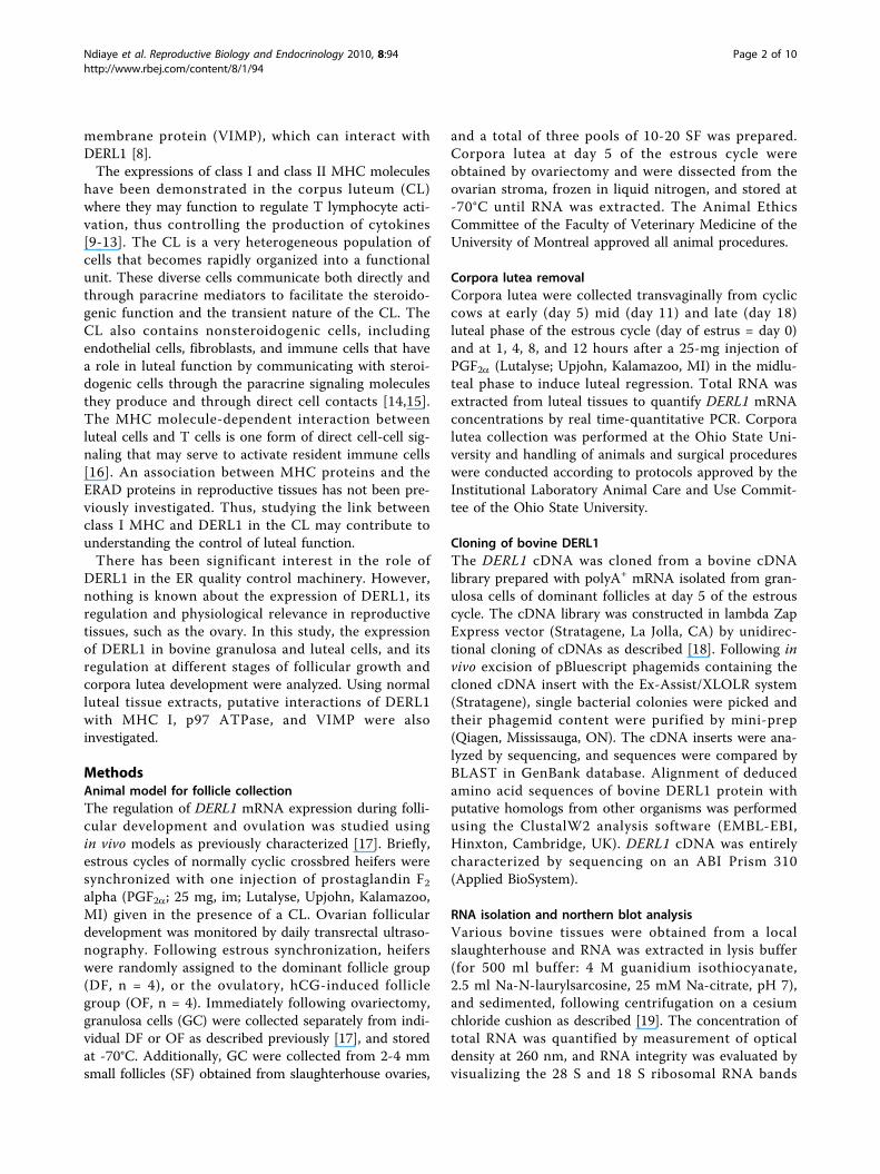
DERL1 mRNA expression and regulation during folliculardevelopmentThe expression of DERL1 mRNA was first analyzed viaNorthern blot using several bovine tissues. Two tran-scripts of DERL1 were detected at 3 kb and 2.6 kb (Fig.1). DERL1 mRNA expression was observed in all tissuesanalyzed, with variable intensities among tissues. The
Figure 1 Analysis of DERL1 mRNA expression in bovine tissuesby Northern blot. Two transcripts of 3 and 2.6 kb correspondingto bovine DERL1 were observed and showed a variable pattern ofexpression among tissues.
Ndiaye et al. Reproductive Biology and Endocrinology 2010, 8:94http://www.rbej.com/content/8/1/94
Page 4 of 10

DERL1 mRNA expression was greatest in adrenal gland,lung and uterus, and was moderate in other tissuesincluding the CL. To verify possible alternative splicingof DERL1 mRNA, PCR experiments amplifying theentire open reading frame of DERL1 were performedand demonstrated a single product (Fig. 2). No amplifi-cation was observed in the control (without reversetranscriptase enzyme) (Fig. 2) showing that the productamplified is not from genomic DNA. This result sug-gests that one protein is encoded from the mRNA andthat the two transcripts observed by Northern blotmight differ at their 3′-untranslated region. This differ-ence could potentially mean a different control of thestability of DERL1 mRNA.The regulation of DERL1 mRNA during follicular
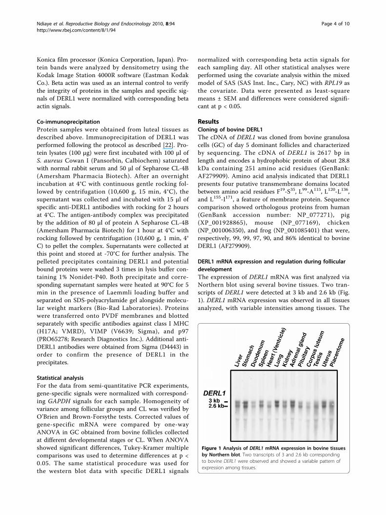
development was analyzed using semi-quantitative RT-PCR. Greater expression of DERL1 was observed ingranulosa cells obtained from day 5 dominant follicles(D5) compared to small follicles (SF), ovulatory follicles(OF), and day 5 corpus luteum (CL) (p < 0.05; Fig. 3).There was no difference in DERL1 mRNA among SF,OF, and CL (Fig. 3). GAPDH used as control gene wasnot different among groups (p < 0.43).
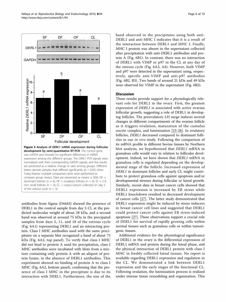
DERL1 mRNA expression in bovine corpus luteumRT-qPCR experiments using total RNA from luteal tis-sues showed that steady state concentration of DERL1
mRNA was intermediate in midcycle (day 11, p =0.064), and lowest in late cycle (day 18, p < 0.05) ascompared to early in the estrous cycle (day 5) (Fig. 4A).In addition, DERL1 in day 11 CL was greater than inday 18 CL (p < 0.05). Injection of PGF2a during themidcycle induced a transient increase of DERL1 mRNAin the CL after 8 hours (p = 0.065) followed by a slightdecrease at 12 hours post PGF2a (p = 0.081), comparedto day 11 CL (Fig. 4B). Additionally, DERL1 mRNA wasgreater at 8 hours post PGF2a than at 1 hour and at12 hours (p < 0.05; Fig. 4B).
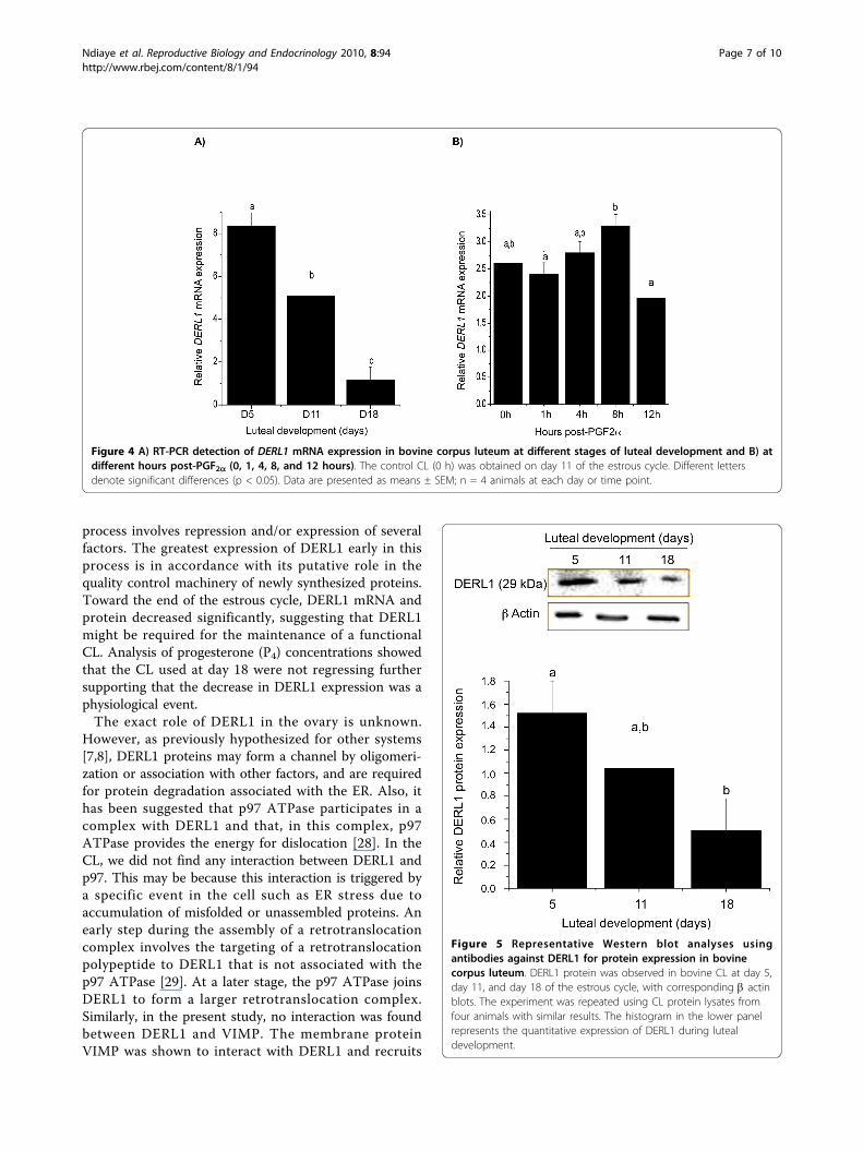
DERL1 protein expressionIn order to analyze the expression of DERL1 protein inthe CL, western blot analyses were performed anddemonstrated the presence of DERL1 in luteal tissuesfrom days 5, 11, and 18 of the estrous cycle (Fig. 5).Similar to the mRNA, DERL1 protein expression wasgreater in day 5 CL as compared to day 18 CL. Betaactin used as the control protein did not show any var-iation throughout the estrous cycle.To determine if DERL1 interacts with class I MHC,
VIMP, or p97 ATPase, immunoprecipitation was per-formed using protein lysate from luteal tissues withanti-DERL1 antibodies from Novus Biologicals (NB100-447SS) followed by western blotting and incubation withvarious antibodies. Incubation with anti-DERL1
Figure 2 Analysis of alternative splicing of DERL1 mRNA. The specific DERL1 PCR product corresponding to the entire open reading frame isrepresented by a single DERL1 PCR product; the sample without RT enzyme control shows that the specific DERL1 product amplified is not fromgenomic DNA.
Ndiaye et al. Reproductive Biology and Endocrinology 2010, 8:94http://www.rbej.com/content/8/1/94
Page 5 of 10
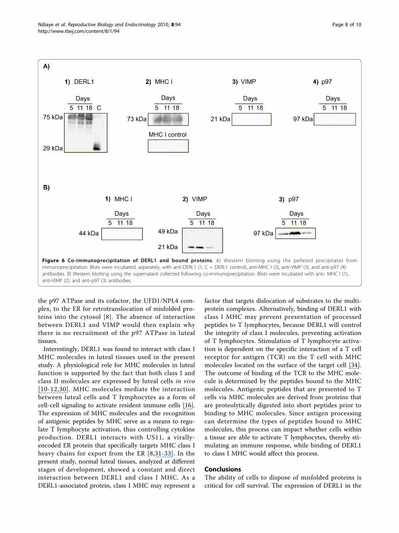
antibodies from Sigma (D4443) showed the presence ofDERL1 in the control sample from day 5 CL at the pre-dicted molecular weight of about 28 kDa, and a secondband was observed at around 75 kDa in the precipatedsamples from days 5, 11, and 18 of the estrous cycle(Fig. 6A1) representing DERL1 and an interacting pro-tein. Class I MHC antibodies used with the same preci-pitates on a separate blot recognized a band of about 73kDa (Fig. 6A2, top panel). To verify that class I MHCdid not bind to protein A used for precipitation, class IMHC antibodies were incubated with blots from a mix-ture containing only protein A with an aliquot of pro-tein lysate, in the absence of DERL1 antibodies. Thisexperiment showed no binding of protein A to class IMHC (Fig. 6A2, bottom panel), confirming that the pre-sence of class I MHC in the precipitate is due to itsinteraction with DERL1. Furthermore, the size of the
band observed in the precipitates using both anti-DERL1 and anti-MHC I indicates that it is a result ofthe interaction between DERL1 and MHC I. Finally,MHC I protein was absent in the supernatant collectedafter precipitation with anti-DERL1 antibodies and pro-tein A (Fig. 6B1). In contrast, there was no interactionof DERL1 with VIMP or p97 in the CL at any day ofthe estrous cycle (Fig. 6A3, A4). However, both VIMPand p97 were detected in the supernatant using, respec-tively, specific anti-VIMP and anti-p97 antibodies(Fig. 6B2, B3). Two bands of around 21 kDa and 49 kDawere observed for VIMP in the supernatant (Fig. 6B2).
DiscussionThese results provide support for a physiologically rele-vant role for DERL1 in the ovary. First, the greatestexpression of DERL1 is associated with active ovarianfollicular growth, suggesting a role of DERL1 in develop-ing follicles. The preovulatory LH surge induces severalchanges in different compartments of the ovarian follicleas it triggers ovulation, maturation of the cumulusoocyte complex, and luteinization [23-26]. In ovulatoryfollicles, DERL1 decreased compared to dominant folli-cles in our in vivo study. Following the comparison ofits mRNA profile in different bovine tissues by Northernblot analysis, we hypothesized that DERL1 mRNA ingranulosa cells would vary in relation to follicular devel-opment. Indeed, we have shown that DERL1 mRNA ingranulosa cells is regulated depending on the develop-mental stage of the follicle. Increased expression ofDERL1 in dominant follicles and early CL might contri-bute to protect granulosa cells against apoptosis and/ordevelopmental stresses during follicular or luteal growth.Similarly, recent data in breast cancer cells showed thatDERL1 expression is increased by ER stress whileDERL1 knockdown resulted in decreased developmentof cancer cells [27]. The latter study demonstrated thatDERL1 expression might be induced by stress inducersin breast cancer cell lines and suggested that DERL1could protect cancer cells against ER stress-inducedapoptosis [27]. These observations support a crucial roleof DERL1 for survival of rapidly growing cells withinnormal tissues such as granulosa cells or within tumori-genic tissues.Additional evidence for the physiological significance
of DERL1 in the ovary is the differential expression ofDERL1 mRNA and protein during the luteal phase, andthe physical interaction of DERL1 protein with class IMHC in freshly collected luteal tissues. No report isavailable regarding DERL1 expression and regulation inthe CL. We demonstrated a link between DERL1expression and the early stages of the functional CL.Following ovulation, the luteinization process is realizedunder intense tissue remodeling and organization. This
Figure 3 Analysis of DERL1 mRNA expression during folliculardevelopment by semi-quantitative RT-PCR. The control genewas GAPDH and showed no significant differences in mRNAexpression among the different groups. The DERL1 PCR signals werenormalized with their corresponding GAPDH signals, and the resultsare presented as a relative change in ratio among groups. Differentletters denote samples that differed significantly (p < 0.05) whenTukey-Kramer multiple comparison tests were performed tocompare group means. Data are presented as means ± SEM. DF =dominant follicles (n = 4); OF = ovulatory follicles (n = 4); SF = 2-4mm small follicles (n = 3); CL = corpus luteum collected on day 5of the estrous cycle (n = 3).
Ndiaye et al. Reproductive Biology and Endocrinology 2010, 8:94http://www.rbej.com/content/8/1/94
Page 6 of 10
process involves repression and/or expression of severalfactors. The greatest expression of DERL1 early in thisprocess is in accordance with its putative role in thequality control machinery of newly synthesized proteins.Toward the end of the estrous cycle, DERL1 mRNA andprotein decreased significantly, suggesting that DERL1might be required for the maintenance of a functionalCL. Analysis of progesterone (P4) concentrations showedthat the CL used at day 18 were not regressing furthersupporting that the decrease in DERL1 expression was aphysiological event.The exact role of DERL1 in the ovary is unknown.
However, as previously hypothesized for other systems[7,8], DERL1 proteins may form a channel by oligomeri-zation or association with other factors, and are requiredfor protein degradation associated with the ER. Also, ithas been suggested that p97 ATPase participates in acomplex with DERL1 and that, in this complex, p97ATPase provides the energy for dislocation [28]. In theCL, we did not find any interaction between DERL1 andp97. This may be because this interaction is triggered bya specific event in the cell such as ER stress due toaccumulation of misfolded or unassembled proteins. Anearly step during the assembly of a retrotranslocationcomplex involves the targeting of a retrotranslocationpolypeptide to DERL1 that is not associated with thep97 ATPase [29]. At a later stage, the p97 ATPase joinsDERL1 to form a larger retrotranslocation complex.Similarly, in the present study, no interaction was foundbetween DERL1 and VIMP. The membrane proteinVIMP was shown to interact with DERL1 and recruits
Figure 4 A) RT-PCR detection of DERL1 mRNA expression in bovine corpus luteum at different stages of luteal development and B) atdifferent hours post-PGF2a (0, 1, 4, 8, and 12 hours). The control CL (0 h) was obtained on day 11 of the estrous cycle. Different lettersdenote significant differences (p < 0.05). Data are presented as means ± SEM; n = 4 animals at each day or time point.
Figure 5 Representative Western blot analyses usingantibodies against DERL1 for protein expression in bovinecorpus luteum. DERL1 protein was observed in bovine CL at day 5,day 11, and day 18 of the estrous cycle, with corresponding b actinblots. The experiment was repeated using CL protein lysates fromfour animals with similar results. The histogram in the lower panelrepresents the quantitative expression of DERL1 during lutealdevelopment.
Ndiaye et al. Reproductive Biology and Endocrinology 2010, 8:94http://www.rbej.com/content/8/1/94
Page 7 of 10
the p97 ATPase and its cofactor, the UFD1/NPL4 com-plex, to the ER for retrotranslocation of misfolded pro-teins into the cytosol [8]. The absence of interactionbetween DERL1 and VIMP would then explain whythere is no recruitment of the p97 ATPase in lutealtissues.Interestingly, DERL1 was found to interact with class I
MHC molecules in luteal tissues used in the presentstudy. A physiological role for MHC molecules in lutealfunction is supported by the fact that both class I andclass II molecules are expressed by luteal cells in vivo[10-12,30]. MHC molecules mediate the interactionbetween luteal cells and T lymphocytes as a form ofcell-cell signaling to activate resident immune cells [16].The expression of MHC molecules and the recognitionof antigenic peptides by MHC serve as a means to regu-late T lymphocyte activation, thus controlling cytokineproduction. DERL1 interacts with US11, a virally-encoded ER protein that specifically targets MHC class Iheavy chains for export from the ER [8,31-33]. In thepresent study, normal luteal tissues, analyzed at differentstages of development, showed a constant and directinteraction between DERL1 and class I MHC. As aDERL1-associated protein, class I MHC may represent a
factor that targets dislocation of substrates to the multi-protein complexes. Alternatively, binding of DERL1 withclass I MHC may prevent presentation of processedpeptides to T lymphocytes, because DERL1 will controlthe integrity of class I molecules, preventing activationof T lymphocytes. Stimulation of T lymphocyte activa-tion is dependent on the specific interaction of a T cellreceptor for antigen (TCR) on the T cell with MHCmolecules located on the surface of the target cell [34].The outcome of binding of the TCR to the MHC mole-cule is determined by the peptides bound to the MHCmolecules. Antigenic peptides that are presented to Tcells via MHC molecules are derived from proteins thatare proteolytically digested into short peptides prior tobinding to MHC molecules. Since antigen processingcan determine the types of peptides bound to MHCmolecules, this process can impact whether cells withina tissue are able to activate T lymphocytes, thereby sti-mulating an immune response, while binding of DERL1to class I MHC would affect this process.
ConclusionsThe ability of cells to dispose of misfolded proteins iscritical for cell survival. The expression of DERL1 in the
Figure 6 Co-immunoprecipitation of DERL1 and bound proteins. A) Western blotting using the pelleted precipitates fromimmunoprecipitation. Blots were incubated, separately, with anti-DERL1 (1; C = DERL1 control), anti-MHC I (2), anti-VIMP (3), and anti-p97 (4)antibodies. B) Western blotting using the supernatant collected following co-immunoprecipitation. Blots were incubated with anti- MHC I (1),anti-VIMP (2), and anti-p97 (3) antibodies.
Ndiaye et al. Reproductive Biology and Endocrinology 2010, 8:94http://www.rbej.com/content/8/1/94
Page 8 of 10

developing follicles and in the functional CL providesstrong evidence that DERL1 might be involved in tissueremodeling events and maintenance of function.Although the exact function of DERL1 in the ovary isnot fully understood, its greater expression seems to bespecifically associated with active follicular growth andearly CL development and function. Furthermore, theobservation in yeast that deletion of Der1p, homolog ofmammalian DERL1, abolished degradation of the sub-strate proteins [35] also adds more evidence that DERL1plays a fundamental role in the cell and might be apivotal factor in cell survival, which is consistent withits expression in growing follicles and functional CL.Finally, although the retrotranslocation pathways appearto require the function of the p97 ATPase complex [8],the present study found no interaction betwen p97 andDERL1 or between VIMP and DERL1. This could sug-gest that the binding of DERL1 to MHC I would be aregulation of MHC I molecules by DERL1 rather than aretrotranslocation process.
AcknowledgementsThis project was supported by National Research Initiative Competitive Grantno. 20043520314789 from the USDA Cooperative State Research, Education,and Extension Service, Animal Reproduction Program and NIH researchgrant HD37550 to JLP. Salaries and research support also provided by Stateand Federal funds appropriated. The project was also supported by aDiscovery Grant (to JGL) from the Natural Sciences and EngineeringResearch Council of Canada (NSERC). The authors also thank Mr. DanielPoole for assistance in material collection and statistical analysis.
Author details1Department of Animal Sciences, The Ohio State University/Ohio AgriculturalResearch and Development Center, 1680 Madison Ave, Wooster, OH 44691,USA. 2Department of Dairy and Animal Science, Pennsylvania StateUniversity, University Park, PA 16802, USA. 3University of Montreal, Faculty ofVeterinary Medicine, Saint-Hyacinthe, Quebec J2 S 7C6, Canada. 4Departmentof Anatomy and Physiology, Kansas State University, 1600 Denison Ave,Manhattan, KS 66506, USA. 5Department of Dairy and Animal Science,Pennsylvania State University, University Park, PA 16802, USA.
Authors’ contributionsKN participated in the design of the study, carried out the experimentsdescribed in the Methods section in the exception of the semi-quantitativeRT-PCR, and drafted the manuscript. JLP participated in the design of thestudy and helped to draft the manuscript. JGL participated in the design ofthe study, performed the semi-quantitative RT-PCR experiments and helpeddrafting the manuscript. All authors read and approved the final manuscript.
Competing interestsThe authors declare that they have no competing interests.
Received: 7 April 2010 Accepted: 3 August 2010Published: 3 August 2010
References1. Ellgaard L, Molinari M, Helenius A: Setting the standards: quality control in
the secretory pathway. Science 1999, 286(5446):1882-1888.2. Brodsky JL, McCracken AA: ER protein quality control and proteasome-
mediated protein degradation. Semin Cell Dev Biol 1999, 10(5):507-513.3. McCracken AA, Brodsky JL: Evolving questions and paradigm shifts in
endoplasmic-reticulum-associated degradation (ERAD). Bioessays 2003,25(9):868-877.
4. Ploegh HL: Viral strategies of immune evasion. Science 1997,280(5361):248-253.
5. Tortorella D, Gewurz BE, Furman MH, Schust DJ, Ploegh HL: Viralsubversion of the immune system. Annu Rev Immunol 2000, 18:861-926.
6. Wiertz EJ, Jones TR, Sun L, Bogyo M, Geuze HJ, Ploegh HL: The humancytomegalovirus US11 gene product dislocates MHC class I heavy chainsfrom the endoplasmic reticulum to the cytosol. Cell 1996, 84(5):769-779.
7. Lilley BN, Ploegh HL: A membrane protein required for dislocation ofmisfolded proteins from the ER. Nature 2004, 429(6994):834-840.
8. Ye Y, Shibata Y, Yun C, Ron D, Rapoport TA: A membrane protein complexmediates retro-translocation from the ER lumen into the cytosol. Nature2004, 429(6994):841-847.
9. Fairchild DL, Pate JL: Interferon-gamma induction of majorhistocompatibility complex antigens on cultured bovine luteal cells. BiolReprod 1989, 40(3):453-457.
10. Benyo DF, Haibel GK, Laufman HB, Pate JL: Expression of majorhistocompatibility complex antigens on the bovine corpus luteumduring the estrous cycle, luteolysis, and early pregnancy. Biol Reprod1991, 45(2):229-234.
11. Bukovsky A, Caudle MR, Keenan JA, Wimalasena J, Upadhyaya NB, VanMeter SE: Is corpus luteum regression an immune-mediated event?Localization of immune system components and luteinizing hormonereceptor in human corpora lutea. Biol Reprod 1995, 53(6):1373-1384.
12. Petroff M, Coggeshall KM, Jones LS, Pate JL: Bovine luteal cells elicit majorhistocompatibility complex class II-dependent T-cell proliferation. BiolReprod 1997, 57(4):887-893.
13. Cannon MJ, Davis JS, Pate JL: Expression of costimulatory molecules inthe bovine corpus luteum. Reprod Biol Endocrinol 2007, 5(5).
14. Cannon MJ, Petroff MG, Pate JL: Effects of prostaglandin F2alpha andprogesterone on the ability of bovine luteal cells to stimulate Tlymphocyte proliferation. Biol Reprod 2003, 69(2):695-700.
15. Ndiaye K, Poole DH, Pate JL: Expression and regulation of functionaloxytocin receptors in bovine T lymphocytes. Biol Reprod 2008,78(4):786-793.
16. Cannon MJ, Pate JL: The role of major histocompatibility complexmolecules in luteal function. Reprod Biol Endocrinol 2003, 1:93.
17. Ndiaye K, Fayad T, Silversides DW, Sirois J, Lussier JG: Identification ofdownregulated messenger RNAs in bovine granulosa cells of dominantfollicles following stimulation with human chorionic gonadotropin. BiolReprod 2005, 73(2):324-333.
18. Brûlé S, Rabahi F, Faure R, Beckers JF, Silversides DW, Lussier JG: Vacuolarsystem-associated protein-60: a protein characterized from bovinegranulosa and luteal cells that is associated with intracellular vesiclesand related to human 80K-H and murine beta-glucosidase II. Biol Reprod2002, 62(3):642-654.
19. Bédard J, Brûlé S, Price CA, Silversides DW, Lussier JG: Serine proteaseinhibitor-E2 (SERPINE2) is differentially expressed in granulosa cells ofdominant follicle in cattle. Mol Reprod Dev 2003, 64(2):152-165.
20. Rabahi F, Brûlé S, Sirois J, Beckers JF, Silversides DW, Lussier JG: Highexpression of bovine alpha glutathione S-transferase (GSTA1, GSTA2)subunits is mainly associated with steroidogenically active cells andregulated by gonadotropins in bovine ovarian follicles. Endocrinology1999, 140(8):3507-3517.
21. Bradford MM: A rapid and sensitive method for the quantitation ofmicrogram quantities of protein utilizing the principle of protein-dyebinding. Anal Biochem 1976, 72:248-254.
22. Brûlé S, Faure R, Doré M, Silversides DW, Lussier JG: Immunolocalization ofvacuolar system-associated protein-60 (VASAP-60). Histochem Cell Biol2003, 119(5):371-381.
23. Sirois J, Richards JS: Transcriptional regulation of the rat prostaglandinendoperoxide synthase 2 gene in granulosa cells. Evidence for the roleof a cis-acting C/EBP beta promoter element. J Biol Chem 1993,268(29):21931-21938.
24. Espey LL, Ujioka T, Russell DL, Skelsey M, Vladu B, Robker RL, Okamura H,Richards JS: Induction of early growth response protein-1 geneexpression in the rat ovary in response to an ovulatory dose of humanchorionic gonadotropin. Endocrinology 2000, 141(7):2385-2391.
25. Eppig JJ, Wigglesworth K, Pendola FL: The mammalian oocyteorchestrates the rate of ovarian follicular development. Proc Natl Acad SciUSA 2002, 99(5):2890-2894.
Ndiaye et al. Reproductive Biology and Endocrinology 2010, 8:94http://www.rbej.com/content/8/1/94
Page 9 of 10
26. Ochsner SA, Day AJ, Rugg MS, Breyer RM, Gomer RH, Richards JS: Disruptedfunction of tumor necrosis factor-alpha-stimulated gene 6 blockscumulus cell-oocyte complex expansion. Endocrinology 2003,144(10):4376-4384.
27. Wang J, Hua H, Ran Y, Zhang H, Liu W, Yang Z, Jiang Y: Derlin-1 isoverexpressed in human breast carcinoma and protects cancer cellsfrom endoplasmic reticulum stress-induced apoptosis. Breast Cancer Res2008, 10(1):R7.
28. Ye Y, Meyer HH, Rapoport TA: Function of the p97-Ufd1-Npl4 complex inretrotranslocation from the ER to the cytosol: dual recognition ofnonubiquitinated polypeptide segments and polyubiquitin chains. J CellBiol 2003, 162(1):71-84.
29. Ye Y, Shibata Y, Kikkert M, van Voorden S, Wiertz E, Rapoport TA: InauguralArticle: Recruitment of the p97 ATPase and ubiquitin ligases to the siteof retrotranslocation at the endoplasmic reticulum membrane. Proc NatlAcad Sci USA 2005, 102(40):14132-14138.
30. Penny LA, Armstrong D, Bramley TA, Webb R, Collins RA, Watson ED:Immune cells and cytokine production in the bovine corpus luteumthroughout the oestrous cycle and after induced luteolysis. J Reprod Fertil1999, 115(1):87-96.
31. Baker BM, Tortorella D: Dislocation of an endoplasmic reticulummembrane glycoprotein involves the formation of partially dislocatedubiquitinated polypeptides. J Biol Chem 2007, 282(37):26845-26856.
32. Loureiro J, Lilley BN, Spooner E, Noriega V, Tortorella D, Ploegh HL: Signalpeptide peptidase is required for dislocation from the endoplasmicreticulum. Nature 2006, 441(7095):894-897.
33. Lilley BN, Ploegh HL: Multiprotein complexes that link dislocation,ubiquitination, and extraction of misfolded proteins from theendoplasmic reticulum membrane. Proc Natl Acad Sci USA 2005,102(40):14296-14301.
34. Altman A, Coggeshall KM, Mustelin T: Molecular events mediating T cellactivation. Adv Immunol 1990, 48:227-360.
35. Knop M, Finger A, Braun T, Hellmuth K, Wolf DH: Der1, a novel proteinspecifically required for endoplasmic reticulum degradation in yeast.EMBO J 1996, 15(4):753-763.
doi:10.1186/1477-7827-8-94Cite this article as: Ndiaye et al.: Molecular characterization andexpression of DERL1 in bovine ovarian follicles and corpora lutea.Reproductive Biology and Endocrinology 2010 8:94.
Submit your next manuscript to BioMed Centraland take full advantage of:
• Convenient online submission
• Thorough peer review
• No space constraints or color figure charges
• Immediate publication on acceptance
• Inclusion in PubMed, CAS, Scopus and Google Scholar
• Research which is freely available for redistribution
Submit your manuscript at www.biomedcentral.com/submit
Ndiaye et al. Reproductive Biology and Endocrinology 2010, 8:94http://www.rbej.com/content/8/1/94
Page 10 of 10
Copyright © 2022 FDOKUMEN