Crosstalk of Notch with p53 and p63 in cancer growth control
9
Complex biological systems can be thought of as scale-free networks, with an uneven distribution of connections to key signalling nodes. Another central characteristic of these systems is that they are intrinsically robust, being capable of repair and recovery on per- turbation 1 . This robustness is the combined result of multiple factors. These include the redundancy of molecules with overlapping functions, feedback and feedforward loops, the modular organization of functionally coherent sets of molecules, such as those of individual signalling pathways, and more global integration at both intracellular and intercellular levels. Two major forms of net- work organization have been proposed: stress signalling networks, which are designed for transient responses to altered cell and tissue homeostasis, and developmental networks, which lead to more permanent cell fate deci- sions 2 . Stress signalling networks are based on direct regulatory loops as well as transcrip- tion and signalling cascades for rapid on–off transitions. Developmental networks share the same basic motifs as stress signalling net- works, but incorporate an additional level of complexity, with intersecting regulatory loops and transcription and signalling cascades that can lock on–off transitions in a more stable cell memory state. The focus of this Opinion article is the crosstalk between the Notch pathway and the p53 and p63 pathways, as a paradigm of coordinated signalling modules in cancer development, at the interface between stress-generated signals and the con- trol of stem cell potential and differentiation. Crosstalk between the Notch pathway and the p53 and p63 pathways can occur at multiple levels, in a manner that is depend- ent on the overall network organization of the individual cell types or tissues in which they operate. I briefly overview the proper- ties of the Notch, p53 and p63 signalling modules (FIGS 1,2), and refer the reader to recent excellent reviews for more detailed information on each of these pathways indi- vidually 3–8 . This is followed by an in-depth discussion of the cross-regulation between the two pathways at the level of Notch con- trol of p53 expression and activity, reciprocal control of Notch by p53, cross-regulation between Notch and p63 (a p53 family member with a more selective role in devel- opment), and indirect cross-modulation between Notch and p53 and p63 by common downstream mediators and by interacting pathways. I conclude with a discussion of the effect that this integrated knowledge could have on new approaches to cancer therapy. The Notch pathway Developmental signalling pathways have a key role in the control of cancer devel- opment, as they impinge on target stem cell populations and their environment, organ morphogenesis and homeostasis. A restricted number of developmental signal- ling pathways is repeatedly used in various combinations and in a synergistic or oppo- site manner, giving rise to an extraordinary array of biological outcomes 9 . Activation of these pathways usually relies on the produc- tion of diffusible ligands — or morphogens — that function at a distance on their cog- nate receptors, which are expressed by other cells of the same or a different type. Notably different is the Notch signalling pathway, which relies instead on non-diffusible ligands, integral membrane proteins that engage and activate surface receptors of immediately adjacent cells 3 (FIG. 1). Notch signalling functions in lateral inhibition, a process whereby negative cross- regulation between Notch receptors and the Delta family of ligands in initially equipotent progenitors eventually produces an asym- metric distribution of cells with divergent cell fates 3 . Notch and the Jagged family of lig- ands can also be part of a positive feedback mechanism, involved in the coordinated control of differentiation along individual cell lineages, resulting in lateral induction and boundary formation 3 . The biological outcome of Notch activa- tion is highly context dependent. This also applies to the control of cancer develop- ment, which Notch activation is usually thought to promote 10,11 but can, in at least some situations, abrogate 12 . Biologically, this is not surprising, as Notch can either sup- press or promote differentiation along cer- tain cell lineages, and cancer is a probable result of deranged stem cell control or dedifferentiation of committed cell populations. The p53 and p63 pathways Although Notch activation can exert entirely opposite effects on basic cell fate deci- sions, such as proliferation, survival and differentiation decisions, p53 functions more consistently as a negative regulator of cell proliferation and inducer of apoptosis, through several effectors (FIG. 2). Therefore, the main role of this protein is that of a sen- sor of acute or chronic alterations in normal cellular physiology and, more specifically, OPINION Crosstalk of Notch with p53 and p63 in cancer growth control G. Paolo Dotto Abstract | Understanding the complexity of cancer depends on an elucidation of the underlying regulatory networks, at the cellular and intercellular levels and in their temporal dimension. This Opinion article focuses on the multilevel crosstalk between the Notch pathway and the p53 and p63 pathways. These two coordinated signalling modules are at the interface of external damaging signals and control of stem cell potential and differentiation. Positive or negative reciprocal regulation of the two pathways can vary with cell type and cancer stage. Therefore, selective or combined targeting of the two pathways could improve the efficacy and reduce the toxicity of cancer therapies. PERSPECTIVES NATURE REVIEWS | CANCER VOLUME 9 | AUGUST 2009 | 587 © 2009 Macmillan Publishers Limited. All rights reserved
-
Upload
independent -
Category
Documents
-
view
2 -
download
0
Transcript of Crosstalk of Notch with p53 and p63 in cancer growth control

Complex biological systems can be thought of as scale-free networks, with an uneven distribution of connections to key signalling nodes. Another central characteristic of these systems is that they are intrinsically robust, being capable of repair and recovery on per-turbation1. This robustness is the combined result of multiple factors. These include the redundancy of molecules with overlapping functions, feedback and feedforward loops, the modular organization of functionally coherent sets of molecules, such as those of individual signalling pathways, and more global integration at both intracellular and intercellular levels. Two major forms of net-work organization have been proposed: stress signalling networks, which are designed for transient responses to altered cell and tissue homeostasis, and developmental networks, which lead to more permanent cell fate deci-sions2. Stress signalling networks are based on direct regulatory loops as well as transcrip-tion and signalling cascades for rapid on–off transitions. Developmental networks share the same basic motifs as stress signalling net-works, but incorporate an additional level of complexity, with intersecting regulatory loops and transcription and signalling cascades that can lock on–off transitions in a more stable cell memory state. The focus of this Opinion article is the crosstalk between the Notch pathway and the p53 and p63 pathways, as a
paradigm of coordinated signalling modules in cancer development, at the interface between stress-generated signals and the con-trol of stem cell potential and differentiation.
Crosstalk between the Notch pathway and the p53 and p63 pathways can occur at multiple levels, in a manner that is depend-ent on the overall network organization of the individual cell types or tissues in which they operate. I briefly overview the proper-ties of the Notch, p53 and p63 signalling modules (FIGS 1,2), and refer the reader to recent excellent reviews for more detailed information on each of these pathways indi-vidually3–8. This is followed by an in-depth discussion of the cross-regulation between the two pathways at the level of Notch con-trol of p53 expression and activity, reciprocal control of Notch by p53, cross-regulation between Notch and p63 (a p53 family member with a more selective role in devel-opment), and indirect cross-modulation between Notch and p53 and p63 by common downstream mediators and by interacting pathways. I conclude with a discussion of the effect that this integrated knowledge could have on new approaches to cancer therapy.
The Notch pathwayDevelopmental signalling pathways have a key role in the control of cancer devel-opment, as they impinge on target stem
cell populations and their environment, organ morphogenesis and homeostasis. A restricted number of developmental signal-ling pathways is repeatedly used in various combinations and in a synergistic or oppo-site manner, giving rise to an extraordinary array of biological outcomes9. Activation of these pathways usually relies on the produc-tion of diffusible ligands — or morphogens — that function at a distance on their cog-nate receptors, which are expressed by other cells of the same or a different type. Notably different is the Notch signalling pathway, which relies instead on non-diffusible ligands, integral membrane proteins that engage and activate surface receptors of immediately adjacent cells3 (FIG. 1).
Notch signalling functions in lateral inhibition, a process whereby negative cross-regulation between Notch receptors and the Delta family of ligands in initially equipotent progenitors eventually produces an asym-metric distribution of cells with divergent cell fates3. Notch and the Jagged family of lig-ands can also be part of a positive feedback mechanism, involved in the coordinated control of differentiation along individual cell lineages, resulting in lateral induction and boundary formation3.
The biological outcome of Notch activa-tion is highly context dependent. This also applies to the control of cancer develop-ment, which Notch activation is usually thought to promote10,11 but can, in at least some situations, abrogate12. Biologically, this is not surprising, as Notch can either sup-press or promote differentiation along cer-tain cell lineages, and cancer is a probable result of deranged stem cell control or dedifferentiation of committed cell populations.
The p53 and p63 pathwaysAlthough Notch activation can exert entirely opposite effects on basic cell fate deci-sions, such as proliferation, survival and differentiation decisions, p53 functions more consistently as a negative regulator of cell proliferation and inducer of apoptosis, through several effectors (FIG. 2). Therefore, the main role of this protein is that of a sen-sor of acute or chronic alterations in normal cellular physiology and, more specifically,
o p i N i o N
Crosstalk of Notch with p53 and p63 in cancer growth controlG. Paolo Dotto
Abstract | Understanding the complexity of cancer depends on an elucidation of the underlying regulatory networks, at the cellular and intercellular levels and in their temporal dimension. This Opinion article focuses on the multilevel crosstalk between the Notch pathway and the p53 and p63 pathways. These two coordinated signalling modules are at the interface of external damaging signals and control of stem cell potential and differentiation. Positive or negative reciprocal regulation of the two pathways can vary with cell type and cancer stage. Therefore, selective or combined targeting of the two pathways could improve the efficacy and reduce the toxicity of cancer therapies.
PersPecTives
NATure revIewS | CanCer vOlume 9 | AuguST 2009 | 587
© 2009 Macmillan Publishers Limited. All rights reserved

P
Delta-like 1–4
JAG1 and JAG2
NOTCH1–4
ADAM10 andADAM17
-secretase
Fringe familyLunaticManicRadical
DTX1
NUMBNumb-like
HesHerp
MYCCCND1CDKN1A
CDK8
FBXW7ITCH
Proteasomaldegradation
JaggedDelta
E3 Ub ligasesNeuralized 1,2Mind Bomb 1,2
Nucleus
Cell membrane
CSL
Nature Reviews | Cancer
NIC
NIC
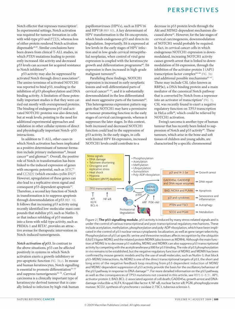
DNA and chromosomal integrity6,7. whereas initial analysis of Trp53-knockout mice sug-gested that this gene is dispensable for devel-opment13, subsequent studies pointed to its involvement in the differentiation of several tissues6–8,14–16. Importantly, this molecule has also recently been implicated as a negative regulator of stem cell potential, with clear implications for cancer development17–19. p53 may carry out some of its functions in con-cert with other family members, such as p63 and p73, which are more closely connected with development. In fact, loss of p63 results in defective epithelial cell fate commitment, specifically in the skin and its adnexa20,21, and loss of p73 affects neuronal development22. p73 is not discussed further, as only limited information is currently available regarding its possible connection with Notch23. As a
result of differential splicing, p63 is produced as several isoforms, and these can either counteract or increase the p53 effects by bind-ing to common promoter sequences of target genes or through separate control of other targets24–29. Additionally, p53 itself can be dif-ferentially spliced to form several isoforms with potentially opposite effects30. Although the best-studied mechanisms of p53 regula-tion are post-transcriptional31, a less appreci-ated but nevertheless important form of p53 regulation is at the level of TP53 gene transcription32–37 (FIG. 2).
Control of p53 by Notch signalling Notch signalling can either suppress or increase p53 activity in a context-dependent manner that is closely connected to tumour promotion or suppression. Four Notch
receptors exist in mammalian cells (FIG. 1). Although most studies have so far focused on the interconnection between p53 and the NOTCH1 receptor, other Notch family members can also have important roles in carcinogenesis. For this reason, in the rest of this article I refer to Notch in general, rather than to NOTCH1, in the cases in which the specific contribution of the various family members is not known or in which NOTCH1 activity probably functionally overlaps with that of the other receptors.
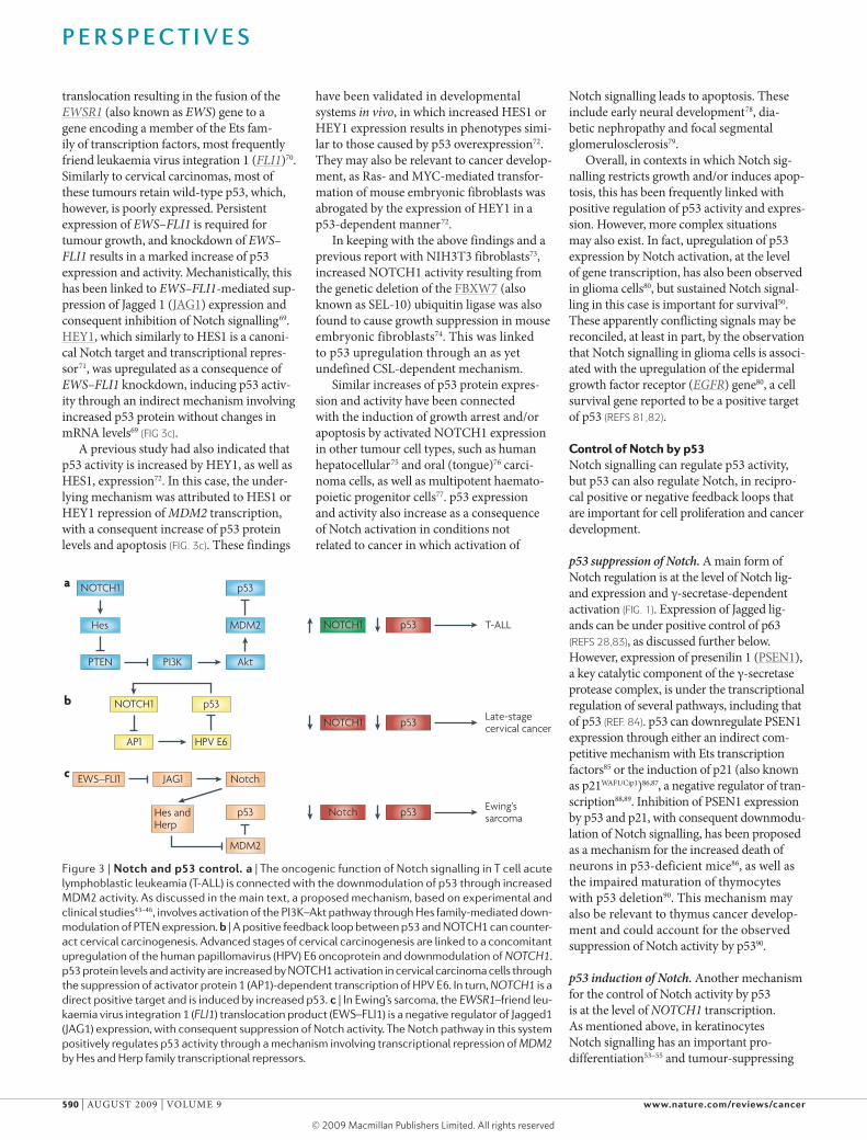
Notch suppression of p53. The most con-vincing demonstration that increased Notch signalling promotes tumour formation, in both clinical and experimental settings, has been provided by studies on T cell acute lymphoblastic leukaemias (T-Alls)10,11. most human T-Alls harbour mutations of NOTCH1, which either facilitate protease-dependent activation of the receptor or increase the stability of its proteolytically cleaved activated form, the Notch intracel-lular domain38. An elegant mouse model of the pathogenesis of the disease has been developed, based on a tetracycline-inducible activated NOTCH1 transgene39. In this model, Notch activation reduces p53 protein levels through increased activity of mDm2, an e3 ubiquitin ligase that targets p53 for degradation (FIG. 3a). The underlying mecha-nism involves decreased expression of ArF (encoded by CDKN2A), a key negative regulator of mDm2 activity. This may be a peculiarity of the system or also may apply to the clinical situation, in the fraction of human T-Alls in which CDKN2A has not been deleted or mutated40. Importantly, tumorigenesis in this system depends on sustained Notch activity, as attenuation of Notch expression dramatically increases p53 levels and tumour regression through apop-tosis. In the presence of activated NOTCH1 expression, increased p53 expression, either through mDm2 inhibition (by small- molecule mDm2 inhibitors such as Nutlin-3) or as a result of ionizing radiation, was sufficient for tumour regression39.
Further studies support the hypothesis that suppression of p53 activity is a key aspect of Notch-induced tumorigenesis. In other work with human T-All, as well as breast cancer and keratinocyte cell lines, Notch activation was shown to increase cell survival through the activation of the PI3K–Akt path-way, which leads to increased mDm2 activity and consequent p53 degradation41–43 (FIG. 3a). A proposed underlying mechanism for Akt activation by Notch is the negative regula-tion of PTEN by HeS1 (ReF. 43), a canonical
Figure 1 | The notch signalling module. Two families of transmembrane Notch ligands and four Notch receptors exist in mammalian cells. The ligands must be ubiquitylated by specific e3 ubiquitin (Ub) ligases to induce Notch activity, which they have been proposed to do through several mecha-nisms, such as exerting a pulling force on the engaged receptor, ligand clustering or internalizing, and undergoing modification and subsequent reinsertion into the membrane. Modification of the extracel-lular domain of Notch receptors by Fringe family glycosyltransferases allows differential activation by the Delta or Jagged ligands. Following ligand engagement, Notch receptors are cleaved first by an ADAM metalloproteinase and then, at the juxta-membrane region, by the presenilin–γ-secretase complex. The resulting Notch intracellular domain (Nic) translocates to the nucleus, where it associ-ates with the DNA binding protein csL (also known as rBPJk), converting it from a repressor to an activator of transcription. This induces the transcription of two classes of target genes. Genes of the Hes family, which encode basic helix–loop–helix transcriptional repressors, and genes of the Herp family, which encode related but distinct basic helix–loop–helix proteins that heterodimerize and cooperate with Hes family members, are ubiquitously induced. cell cycle regulatory genes, such as MYC, CCND1 (which encodes cyclin D1)52 and CDKN1A (which encodes p21) are induced in a cell type-specific manner. in parallel, Notch activation controls neighbouring cells, through an opposite modulation of ligands of the Delta and Jagged families, which can provide a basis for lateral inhibition or lateral induction, respectively (as described in the main text). The Notch transcription-activating complex is terminated by phosphorylation of activated nuclear Notch by cyclin-dependent kinase 8 (cDK8), with subsequent recognition by the ubiquitin ligase FBXW7 (also known as seL-10) and proteasome-dependent degradation. Negative regulatory mechanisms are also provided by the e3 ubiquitin ligases Deltex 1 (DTX1) and iTcH and by NUMB, an asymmetrically distributed protein that has been linked to the recruitment of Notch into endocytic vescicles but also to NOTcH ubiquitylation. For more detailed information, see ReFS 3,4,146.
P e r s P e c t i v e s
588 | AuguST 2009 | vOlume 9 www.nature.com/reviews/cancer
© 2009 Macmillan Publishers Limited. All rights reserved
Nature Reviews | Cancer
p53
p53
MDM2MDMX
Proteasomaldegradation
TP53 transcription
AP1KLF4BCL-6
• Phosphorylation• Acetylation• Methylation• Ubiquitylation• Sumoylation• Poly-ADP ribosylation
p21 and 14-3-3 Growth arrest
GADD45 , GADD45 and DNA repair enzymes DNA repair
BAD, NOXA and PUMA Apoptosis
PTEN and TSC1 Autophagy
SCO2 and PGM
p21 and cytokines
Metabolism
PI3K–Akt
Senescence and inflammation
Stress signals:• DNA damage• Telomere shortening• Oncogene and
replicative stress• Heat shock• Hypoxia• Nutrient (dNTP) depletion
UbNF-κB
p300
ARF
Notch effector that represses transcription3. In experimental settings, Notch activation was required for tumour formation in cells with wild-type p53 and PTeN, whereas loss of either protein rendered Notch activation dispensable44,45. Similar conclusions have been drawn from clinical T-All studies, in which PTEN mutations leading to persist-ently increased Akt activity and decreased p53 levels can account for acquired resistance to Notch inhibitors46.
p53 activity may also be suppressed by activated Notch through direct association47. The amino terminus of activated NOTCH1 was reported to bind p53, resulting in the inhibition of p53 phosphorylation and DNA binding activity. A limitation of these poten-tially important studies is that they were car-ried out mostly with overexpressed proteins. The binding of endogenous p53 and acti-vated NOTCH1 proteins was also detected, but at weak levels, pointing to the need for additional experimental approaches and validation in other cellular systems of direct and physiologically important Notch–p53 interactions.
In addition to T-All, other cases in which Notch activation has been implicated as a positive determinant of tumour forma-tion include primary melanomas48, breast cancer49 and gliomas50. Overall, the positive role of Notch in transformation has been linked to the induced expression of genes with oncogenic potential, such as MYC51 and CCND1 (which encodes cyclin D1)52. However, upregulation of these genes can also lead to a replicative stress signal and consequent p53-dependent apoptosis10. Therefore, a second key function of Notch in transformation is to suppress apoptosis through downmodulation of p53 (ReF. 10). It follows that increasing p53 activity using recently identified low-molecular-mass com-pounds that stabilize p53, such as Nutlin-3, or that induce refolding of p53 mutants into a form with wild-type activity, such as PrImA-1 and rITA6, provides an attrac-tive avenue for therapeutic intervention in Notch-induced tumorigenesis.
Notch activation of p53. In contrast to the above situations, p53 can be affected positively in systems in which Notch activation exerts a growth-inhibitory or pro-apoptotic function (FIG. 3b,c). In mouse and human keratinocytes, Notch signalling is essential to promote differentiation53–55 and suppress tumorigenesis56–59. Cervical carcinoma is a clinically important type of keratinocyte-derived tumour that is caus-ally linked to infection by high-risk human
papillomaviruses (HPvs), such as HPv16 and HPv18 (ReF. 60). A key determinant of HPv transformation is the e6 oncoprotein, which binds endogenous p53 and directs its degradation. Importantly, e6 is expressed at low levels in the early stages of HPv infec-tion and in low-grade cervical intraepithe-lial neoplasias, when control of viral gene expression is coupled with the keratinocyte growth and differentiation programme60. e6 expression is then increased in high-grade malignant tumours60.
Paralleling these findings, NOTCH1 expression is increased in early neoplastic lesions and well-differentiated parts of cervical cancer61–63, and it is substantially downmodulated in the less differentiated and more aggressive parts of the tumours64. This heterogeneous expression pattern sug-gests that NOTCH1 may have a permissive or tumour-promoting function in the early stages of cervical carcinogenesis, whereas it suppresses the later stages. In this context, both increased and decreased NOTCH1 function could lead to the suppression of p53 activity. In the early stages, in cells with limited HPv e6 expression, increased NOTCH1 levels could contribute to a
decrease in p53 protein levels through the Akt and mDm2-dependent mechanism dis-cussed above42. However, for the late stages of cervical carcinogenesis, downmodulation of NOTCH1 would probably be required. In fact, in cervical cancer cells in which endogenous NOTCH1 expression is down-modulated, increasing NOTCH1 activity causes growth arrest that is linked to down-modulation of e6 expression, through the inhibition of the activator protein 1 (AP1) transcription factor complex64–66 (FIG. 3b) and additional possible mechanisms65–67. These may include CSl (also known as rBPJk), a DNA binding protein and a main mediator of the canonical Notch pathway that is converted by Notch from a repressor into an activator of transcription3 (FIG. 1). CSl was recently found to exert a negative regulatory function on TP53 transcription in Hela cells68, which could be relieved by NOTCH1 activation.
ewing’s sarcoma is another type of human tumour that has recently been linked to sup-pression of Notch and p53 activity69. These tumours, which arise in the bone and soft tissues of children and young adults, are characterized by a specific chromosomal
Figure 2 | The p53 signalling module. p53 activity is induced by many stress-related signals and is under the control of various transcriptional and post-transcriptional regulatory mechanisms. These include acetylation, methylation, phosphorylation and poly-ADP ribosylation, which have been impli-cated in the control of p53 nuclear versus cytoplasmic localization, as well as gene target selectivity. Phosphorylation of p53 at specific serine and threonine residues affects recognition by the ubiquitin (Ub) e3 ligase MDM2 and the related protein MDMX (also known as MDM4). Although the main func-tion of MDM2 is to decrease p53 stability, MDM2 and MDMX can also suppress p53 transcriptional activity by competing with the acetyltransferase p300 for p53 binding. The role of p53 phosphorylation in vivo remains to be established, but the negative regulatory function of MDM2 and MDMX has been confirmed by mouse genetic models and by the use of small molecules, such as Nutlin-3, that block p53–MDM2 interactions. As MDM2 is one of the direct transcriptional targets of p53, the short and long arms of the negative feedback loop resulting from p53-dependent induction of MDM2 and MDM2-dependent suppression of p53 activity provide the basis for the oscillatory behaviour of the p53 pathway in response to DNA damage155. For more detailed information on the p53 pathway, as well as the consequences of TP53 mutations not covered in this article, see ReFS 6–8,31. AP1, activator protein 1; BAD, BcL-2-associated agonist of cell death; GADD45α, growth arrest and DNA-damage-inducible-α; KLF4, Kruppel-like factor 4; NF-κB, nuclear factor-κB; PGM, phosphoglycerate mutase; scO2, synthesis of cytochrome c oxidase 2; Tsc1, tuberous sclerosis 1.
P e r s P e c t i v e s
NATure revIewS | CanCer vOlume 9 | AuguST 2009 | 589
© 2009 Macmillan Publishers Limited. All rights reserved
Nature Reviews | Cancer
NOTCH1
Hes
PTEN PI3K Akt
MDM2
p53a
NOTCH1 p53 T-ALL
b NOTCH1
AP1 HPV E6
p53
NOTCH1 p53 Late-stagecervical cancer
JAG1
Hes andHerp
p53
EWS–FLI1
Ewing’ssarcomaNotch p53
MDM2
c Notch
translocation resulting in the fusion of the EWSR1 (also known as EWS) gene to a gene encoding a member of the ets fam-ily of transcription factors, most frequently friend leukaemia virus integration 1 (FLI1)70. Similarly to cervical carcinomas, most of these tumours retain wild-type p53, which, however, is poorly expressed. Persistent expression of EWS–FLI1 is required for tumour growth, and knockdown of EWS–FLI1 results in a marked increase of p53 expression and activity. mechanistically, this has been linked to EWS–FLI1-mediated sup-pression of Jagged 1 (JAg1) expression and consequent inhibition of Notch signalling69. HeY1, which similarly to HeS1 is a canoni-cal Notch target and transcriptional repres-sor71, was upregulated as a consequence of EWS–FLI1 knockdown, inducing p53 activ-ity through an indirect mechanism involving increased p53 protein without changes in mrNA levels69 (FIG 3c).
A previous study had also indicated that p53 activity is increased by HeY1, as well as HeS1, expression72. In this case, the under-lying mechanism was attributed to HeS1 or HeY1 repression of MDM2 transcription, with a consequent increase of p53 protein levels and apoptosis (FIG. 3c). These findings
have been validated in developmental systems in vivo, in which increased HeS1 or HeY1 expression results in phenotypes simi-lar to those caused by p53 overexpression72. They may also be relevant to cancer develop-ment, as ras- and mYC-mediated transfor-mation of mouse embryonic fibroblasts was abrogated by the expression of HeY1 in a p53-dependent manner72.
In keeping with the above findings and a previous report with NIH3T3 fibroblasts73, increased NOTCH1 activity resulting from the genetic deletion of the FBXw7 (also known as Sel-10) ubiquitin ligase was also found to cause growth suppression in mouse embryonic fibroblasts74. This was linked to p53 upregulation through an as yet undefined CSl-dependent mechanism.
Similar increases of p53 protein expres-sion and activity have been connected with the induction of growth arrest and/or apoptosis by activated NOTCH1 expression in other tumour cell types, such as human hepatocellular75 and oral (tongue)76 carci-noma cells, as well as multipotent haemato-poietic progenitor cells77. p53 expression and activity also increase as a consequence of Notch activation in conditions not related to cancer in which activation of
Notch signalling leads to apoptosis. These include early neural development78, dia-betic nephropathy and focal segmental glomerulosclerosis79.
Overall, in contexts in which Notch sig-nalling restricts growth and/or induces apop-tosis, this has been frequently linked with positive regulation of p53 activity and expres-sion. However, more complex situations may also exist. In fact, upregulation of p53 expression by Notch activation, at the level of gene transcription, has also been observed in glioma cells80, but sustained Notch signal-ling in this case is important for survival50. These apparently conflicting signals may be reconciled, at least in part, by the observation that Notch signalling in glioma cells is associ-ated with the upregulation of the epidermal growth factor receptor (EGFR) gene80, a cell survival gene reported to be a positive target of p53 (ReFS 81,82).
Control of Notch by p53Notch signalling can regulate p53 activity, but p53 can also regulate Notch, in recipro-cal positive or negative feedback loops that are important for cell proliferation and cancer development.
p53 suppression of Notch. A main form of Notch regulation is at the level of Notch lig-and expression and γ-secretase-dependent activation (FIG. 1). expression of Jagged lig-ands can be under positive control of p63 (ReFS 28,83), as discussed further below. However, expression of presenilin 1 (PSeN1), a key catalytic component of the γ-secretase protease complex, is under the transcriptional regulation of several pathways, including that of p53 (ReF. 84). p53 can downregulate PSeN1 expression through either an indirect com-petitive mechanism with ets transcription factors85 or the induction of p21 (also known as p21wAF1/Cip1)86,87, a negative regulator of tran-scription88,89. Inhibition of PSeN1 expression by p53 and p21, with consequent downmodu-lation of Notch signalling, has been proposed as a mechanism for the increased death of neurons in p53-deficient mice86, as well as the impaired maturation of thymocytes with p53 deletion90. This mechanism may also be relevant to thymus cancer develop-ment and could account for the observed suppression of Notch activity by p5390.
p53 induction of Notch. Another mechanism for the control of Notch activity by p53 is at the level of NOTCH1 transcription. As mentioned above, in keratinocytes Notch signalling has an important pro-differentiation53–55 and tumour-suppressing
Figure 3 | notch and p53 control. a | The oncogenic function of Notch signalling in T cell acute lymphoblastic leukeamia (T-ALL) is connected with the downmodulation of p53 through increased MDM2 activity. As discussed in the main text, a proposed mechanism, based on experimental and clinical studies43–46, involves activation of the Pi3K–Akt pathway through Hes family-mediated down-modulation of PTeN expression. b | A positive feedback loop between p53 and NOTcH1 can counter-act cervical carcinogenesis. Advanced stages of cervical carcinogenesis are linked to a concomitant upregulation of the human papillomavirus (HPv) e6 oncoprotein and downmodulation of NOTCH1. p53 protein levels and activity are increased by NOTcH1 activation in cervical carcinoma cells through the suppression of activator protein 1 (AP1)-dependent transcription of HPv e6. in turn, NOTCH1 is a direct positive target and is induced by increased p53. c | in ewing’s sarcoma, the EWSR1–friend leu-kaemia virus integration 1 (FLI1) translocation product (eWs–FLi1) is a negative regulator of Jagged1 (JAG1) expression, with consequent suppression of Notch activity. The Notch pathway in this system positively regulates p53 activity through a mechanism involving transcriptional repression of MDM2 by Hes and Herp family transcriptional repressors.
P e r s P e c t i v e s
590 | AuguST 2009 | vOlume 9 www.nature.com/reviews/cancer
© 2009 Macmillan Publishers Limited. All rights reserved

Nature Reviews | Cancer
WNT4p63Stem cell renewaland proliferation
α6, β4 and β1 integrins
IRFsNF-κB
p21,HES1
Term
inal
diff
eren
tiatio
nan
d ce
ll cy
cle
with
draw
al
Basal layer
Basementmembrane
Transit-amplifyingcell
Jagged
NotchNotch
p63
Keratin 1
Keratin 10
a bfunction56–59. Increased tumorigenesis resulting from Notch inhibition has been connected with the downmodulation of NOTCH1 expression in cutaneous squamous cell carcinomas57 and basal cell carcinomas91. Downregulation of NOTCH1 transcription in these tumours can be explained, at least in part, by compromised p53 function. In fact, in primary human keratinocytes, endog-enous p53 binds to the NOTCH1 promoter and p53 knockdown results in downmodu-lation of NOTCH1 expression, whereas increased p53 levels lead to NOTCH1 upreg-ulation57,92,93. Similarly, the already mentioned downregulation of NOTCH1 expression in HPv-positive cervical carcinoma cells can be explained by decreased p53 levels resulting from viral e6-dependent p53 degradation93. Therefore, whereas NOTCH1 activation leads to the suppression of e6 expression and consequently to increased p53 levels, p53 in turn stimulates NOTCH1 transcription, and this positive feedback loop is disrupted in malignant cervical carcinogenesis (FIG. 3b).
Control of NOTCH1 expression by p53 is also relevant for the keratinocyte response to ultraviolet (uv) light, a major aetiologi-cal agent of skin ageing and cancer94,95. p53 has a key role in the DNA damage response of keratinocytes to uv light, controlling not only the decision between growth arrest and apoptosis, but also differentiation92,93,96. underlying differentiation is the induc-tion of NOTCH1 expression through a p53-dependent mechanism, with NOTCH1 exerting a protective function against the DNA damage response to uvB light through suppression of forkhead box O3 (FOXO3), a key pro-apoptotic gene92.
In addition to keratinocytes and their malignant counterparts, NOTCH1 expres-sion is under positive p53 control in lung and prostate cancer cells, in which growth suppression is also caused by increased Notch signalling97, as well as in B cell chronic lym-phocytic leukaemia cells, in which Notch sig-nalling increases cell survival98. In many cases, p53 was found to be specifically involved in the control of NOTCH1 with little or no effects on other family members57,93,97. A survey of the human genome by global chromatin immunoprecipitation99, as well as direct chromatin immunoprecipita-tion assays57, also pointed to the NOTCH1 promoter as a direct p53 binding target in HCT116 colon carcinoma cells. Interestingly, however, p53 did not seem to affect NOTCH1 expression in these cells, pointing to a pos-sibly cell type-specific mechanism of p53 control of NOTCH1 expression downstream of promoter recognition and binding57.
Crosstalk between NoTCH1 and p63whereas NOTCH1 activation restricts the growth potential of keratinocytes and promotes differentiation100, p63 has been linked to epidermal cell fate determina-tion and maintenance of self-renewing cell populations101,102. p63 is expressed by kerati-nocytes of the proliferative compartment of the epidermis and initially committed cell populations, and its expression is suppressed with differentiation. Conversely, p63 is often upregulated in various epithelial tumours, including skin squamous cell carcinomas (for a review of this topic see ReF. 103) in which Notch signalling is downmodulated57. p63 upregulation in tumours has been connected to antagonistic functions of p63 versus p53 and p73 on the control of downstream tar-get genes involved in cell cycle withdrawal, apoptosis and cell senescence7,8,26,104. As dis-cussed below, reciprocal negative regulation has been established between NOTCH1 and p63 expression and activity, with important consequences for the balance between self-renewing and committed cell populations, as well as, possibly, carcinogenesis24 (FIG. 4).
NOTCH1 control of p63. The molecular basis for the control of p63 expression in growing keratinocytes compared
with differentiating keratinocytes, and in tumour cells, has been studied to a limited extent. NOTCH1 inhibits the expression of p63 in these cells both in vitro and in vivo24. Negative regulation of p63 by NOTCH1 is probably cell type-specific (as the opposite effect was reported in NIH3T3 fibroblasts105), is not an indirect consequence of growth arrest and is not caused by key mediators of NOTCH1 function in keratinocytes, such as mem-bers of the Hes and Herp families71 or p21 (ReFS 55,88).
Transcription of TP63 can be initiated at two different promoters, resulting in mrNAs for either the TA-p63 or ΔN-p63 isoforms that contain or lack, respectively, an N-terminal transcription-activating (TA) domain29. The major isoform expressed by keratinocytes is ΔN-p63α. The promoter region for ΔN-p63 contains multiple binding sites for interferon-responsive factors (IrFs) and nuclear factor-κB (NF-κB) subunits, with a potentially significant similarity to the interferon-β enhancer, which depends on a synergistic complex formation among these transcription factors106. Both protein families may be involved in the control of p63 expression by NOTCH1.
Figure 4 | Crosstalk between p63 and notch. reciprocal feedback and feedforward loops between Notch and p63 contribute to the balance between self-renewing keratinocyte populations and cells at various stages of commitment to differentiation. a | in the interfollicular epidermis, keratinocytes undergo a vertical programme of differentiation, as they proceed from the basal proliferating compart-ment to the upper terminally differentiating layers. in the proliferative compartment, a dynamic equi-librium probably exists between a minor fraction of slow cycling cells with increased self-renewal potential (putative stem cells) and already-committed populations that are actively dividing but only for a limited number of times (transit-amplifying cells). Opposing gradients of p63 expression and Notch activity exist in the basal but not the upper epidermal layers. This may result, in part, from their reciprocal negative regulation. b | Mechanistically, increased Notch activity suppresses the expression of p63 through modulation of interferon-responsive factors (irFs) and nuclear factor-κB (NF-κB) signal-ling and, possibly, microrNAs. in turn, p63 binds to the HES1, CDKN1A (which encodes p21) and WNT4 promoters, counteracting the Notch effects on their expression (WNT4 is suppressed by Notch activa-tion through an indirect mechanism mediated by both Hes1 and p21). p63 and Notch also increase and suppress, respectively, integrin expression (α6, β4 and β1 integrins), through less well-defined mechanisms. in parallel with these antagonistic effects, p63 can also synergize with Notch signalling in the early steps of differentiation (characterized by keratin 1 and keratin 10 expression), through a paracrine mechanism (illustrated by dotted arrows) involving the induction of expression of Jagged ligands and Notch activation in neighbouring cells. Downmodulation of p63 by increased Notch signalling could then be a signal for later differentiation stages to occur.
P e r s P e c t i v e s
NATure revIewS | CanCer vOlume 9 | AuguST 2009 | 591
© 2009 Macmillan Publishers Limited. All rights reserved

Activation of the NOTCH1 pathway in keratinocytes results in the selective modu-lation of interferon-responsive genes, with some genes of this family being induced and others suppressed24,107. Among the downmodulated genes are IRF7, a key regulator of the interferon-dependent transcription cascade with oncogenic potential108,109, and IRF3, which physically interacts and functionally overlaps with IrF7 (ReF. 110). Functionally, the ability of NOTCH1 activation to downmodulate p63 expression was overcome by IrF7 overex-pression, and the combined knockdown of IrF3 and IrF7 resulted in decreased p63 expression24. Importantly, the reduction of p63 levels by the concomitant IrF7 and IrF3 knockdown was less than that caused by activated NOTCH1 expression, suggesting that other mechanisms are also in place.
Biochemical and functional evidence indicates that p63 is also negatively regulated by NF-κB in keratinocytes24. As NF-κB activ-ity is induced in these cells with differentia-tion111,112, as well as Notch activation54, it is possible that suppression of p63 by Notch also involves NF-κB activation. However, if NF-κB is involved, Notch-dependent sup-pression occurs through a mechanism that is not blocked by expression of stabilized NFκB inhibitor alpha (IκBα), which inhibits the canonical NF-κB (p65 and p50) pathway24.
An interesting possibility for future stud-ies is whether the above mechanisms func-tion in concert with the recently described microrNA-dependent regulation of p63 expression113,114, given the already demon-strated interconnection between Notch and control of microrNA expression and function in other systems115,116.
p63 control of Notch. p63 is regulated by Notch, but at the same time also controls the Notch pathway. As p53 and p63 share a common DNA recognition sequence29, p63 may participate in the control of NOTCH1 transcription. In fact, endogenous p63 can bind to the NOTCH1 promoter, and there is defective NOTCH1 expression in the epidermis of mouse embryos with p63 deletion117. However, direct control of NOTCH1 expression by p63 may be limited to development, as in mature keratino-cytes p63 functions as a selective modula-tor of downstream Notch signalling (see below) with little or no effect on NOTCH1 transcription34,93.
Persistently increased p63 expression counteracts the ability of Notch to restrict keratinocyte growth potential, through a mechanism involving suppression of p21
expression, as well as, possibly, upregulation of integrin receptors24,118 (FIG. 4). Con-comitantly, p63 exerts both Notch-antagonistic and Notch-synergistic effects on differentiation. In fact, in parallel with a decreasing gradient of p63 expression from basal to upper epidermal layers, persist-ently increased expression of this protein suppresses differentiation markers of the granular and outermost layers, whereas it upregulates those of early entry into dif-ferentiation (spinous layer), which are also under positive Notch control24,119.
Both intracellular and intercellular regu-latory mechanisms are probably involved in this dual role of p63 in differentiation. Intracellularly, p63 functions as a selective modulator of NOTCH1-dependent tran-scription, with HES1 as one of its direct neg-ative targets24. HeS1 in turn is a modulator of differentiation24,34,120.
Besides impinging on intracellular Notch signalling mechanisms, p63 has the potential to induce activation of this path-way in neighbouring cells. JAG1 and JAG2 transcription is positively regulated by p63 (ReFS 28,83). Induction of these genes by p63 can be important for normal skin homeo stasis, serving as a positive reinforce-ment signal to synchronize differentiation of keratinocytes in the suprabasal epidermal layers (FIG. 4). This function of p63 in inter-cellular communication may extend to the thymus, where similar defects in γδ T cell maturation have been observed in Trp63–/– and Jag2–/–mice, and the Trp63–/– phenotype can be explained by decreased JAg2 expression by thymic epithelial cells121.
Crosstalk by downstream mediatorsCross-regulation between the Notch and p53 pathways may also occur further down-stream, at the level of shared intermediates. The Notch transcriptional co-activator mastermind-like 1 (mAml1), or related family members, is required for increased levels of Notch–CSl-dependent transcrip-tion, as mAml1 recruits histone acetyl-transferases, such as p300, to the Notch–CSl complex122–127. Although initially identified as specific mediators of Notch signalling, mAml proteins have now been implicated in other signalling pathways, including those of muscle cell differentiation, β-catenin and p53 (ReF. 128). more specifically, a direct mAml1–p53 association has also been described129. The N-terminal region of mAml1 binds both Notch and p53, raising the possibility of direct competition for this binding site129. A more indirect mechanism is also possible. The N-terminal region of
mAml1 also binds to p300 (ReF. 130), and competition for p300 binding has previ-ously been implicated in the suppression of Notch–mAml1-dependent transcription by p53 (ReF. 124).
Another possible connection between Notch and p53 is provided by NumB, an endocytic protein and determinant of asymmetric cell division that is implicated in the negative control of Notch function3. NumB was found to bind mDm2 in a yeast two-hybrid assay and was subject to mDm2-dependent degradation131, suggesting that mDm2 can increase Notch signalling through NumB downmodulation. By con-trast, NumB itself has recently been found in a complex with mDm2 and p53, in which it prevented p53 ubiquitylation and degrada-tion132. Therefore, it has been proposed that the frequent loss of NumB expression in breast cancer contributes to an aggressive tumour phenotype not only by upregulating a Notch-positive growth signal but also by decreasing p53 activity132.
Cross-interacting pathwaysIn keratinocytes, in addition to increasing proliferation, egFr signalling suppresses differentiation133. This may be relevant to the balance between self-renewing and committed keratinocytes in the basal pro-liferative compartment of the epidermis, where egFr is expressed, as well as in squamous cell carcinomas, in which egFr signalling is characteristically increased (for a review of this topic see ReF. 134). In a chemical genetics screen for small-molecule activators of Notch signalling, egFr sig-nalling through erk was identified as a key negative regulator of NOTCH1 expression in keratinocytes, with downregulation of TP53 — through AP1— as an underlying mechanism34. This concomitant mode of regulation of p53 and NOTCH1 expression by egFr signalling was validated in organ cultures of intact human skin and cutane-ous squamous cell carcinomas, as well as a mouse model of egFr-dependent skin can-cer. egFr has become an important thera-peutic target in cancer, and several selective egFr inhibitors have now been approved for clinical use. Suppression of Notch sig-nalling in cancer cells can synergize with these compounds in the induction of apop-tosis34 (FIG. 5). Therefore, Notch inhibitors may increase the effects of egFr-inhibitory agents on tumours, while simultaneously ameliorating their well-known toxic effects on the skin, which have been attributed, at least in part, to aberrant differentiation135. Similar synergistic induction of apoptosis
P e r s P e c t i v e s
592 | AuguST 2009 | vOlume 9 www.nature.com/reviews/cancer
© 2009 Macmillan Publishers Limited. All rights reserved
Nature Reviews | Cancer
Notch
EGFR
p53
AP1
Cell survival
Slow-cycling self-renewing cells
Actively dividing cells
Differentiatingcells
EGFRinhibitors
Notchinhibitors Differentiation
Self-renewal
has been observed in other cancer cells following concomitant treatment with Notch inhibitors and either erBB2-specific antibodies136, p53 inducers98 or genotoxic agents97.
Transforming growth factor-β (TgFβ)–Smad signalling can be another important point of convergence of the p53 and Notch pathways. In fact, a close connection between p53 and TgFβ–Smad function has been established137, and Smad proteins also functionally and biochemically interact with activated NOTCH1 (ReFS 138–140). This is relevant for the tumour-suppressing function that NOTCH1 and p53 share in specific settings, such as in keratinocytes, with p21 as an important common down-stream target141. NF-κB signalling can also be involved in the reciprocal modulation of Notch and p53 signalling. A recipro-cal crosstalk between p53 and NF-κB has been established142, and NF-κB may be implicated in the positive control of p53 expression143 and ubiquitin-dependent p63 degradation144. NF-κB can also posi-tively control Notch ligand expression145, itself subject to either positive or negative Notch control (for a review of this topic see ReF. 146).
Future directionsrecent debate on the tumorigenic potential of distinct cancer cell populations147–149 has focused on the notion of ‘stemness’ as a
dynamic rather than an intrinsically fixed property of cells150–152. A reversible equi-librium probably exists between cells with high self-renewal potential and more com-mitted cell populations, with reprogram-ming of individual cells associated with stochastic fluctuations of the transcrip-tome153. An exciting area for future stud-ies is the effect that the interconnection between Notch and p53 or p63 can have on self-renewal of cells compared with commitment to senescence or differentia-tion. As discussed at the beginning of this article, in situations in which Notch has a tumour-promoting function, loss or down-modulation of p53 activity renders Notch activation dispensable44–46. Importantly, however, in many tumours TP53 mutations do not abrogate p53 function but confer new, or a wider range of, functions on p53 (ReF. 6) that may considerably affect the crosstalk with Notch.
In addition to intrinsic control mecha-nisms, self-renewal of cells is dependent on cues from the environment. In multicel-lular organisms, cells function as integrated networks, with intrinsically heterogeneous behaviour of individual cells154 being syn-chronized by multiple forms of homotypic and heterotypic intercellular communi-cation, communication at a distance (as mediated, for instance, by hormonal influ-ences) and communication in response to inputs from the external environment.
Crosstalk between the Notch pathway and p53 and p63 pathways in these contexts remains to be explored.
Finally, the design of new or improved therapeutic approaches to complex dis-eases, such as cancer, is dependent on an understanding of the functional integra-tion of cell and tissue homeostasis tempo-rally and spatially. Oscillations of p53 and mDm2 have emerged as a key component of cell cycle progression and DNA repair checkpoint controls155. Intrinsic oscillation mechanisms of Notch signalling have also been demonstrated to be connected with development156,157. Therefore, an important possibility is that the Notch pathway and the p53 and p63 pathways are also dynamically integrated, yielding cell state fluctuations that differ among various cell types, and in normal cells compared with cancer cells. Selective targeting of these pathways in specific tissues and at various times could greatly improve the efficacy of new cancer therapy approaches and reduce their toxic side effects.
G. Paolo Dotto is at the Department of Biochemistry, University of Lausanne, CH‑1066 Epalinges,
Switzerland and at the Cutaneous Biology Research Center, Massachusetts General Hospital,
Charlestown, Maine, 02129, USA. e‑mail: gian‑[email protected]
doi:10.1038/nrc2675Published online 16 July 2009.
1. Stelling, J., Sauer, U., Szallasi, Z., Doyle, F. J. & Doyle, J. Robustness of cellular functions. Cell 118, 675–685 (2004).
2. Alon, U. Network motifs: theory and experimental approaches. Nature Rev. Genet. 8, 450–461 (2007).
3. Bray, S. J. Notch signalling: a simple pathway becomes complex. Nature Rev. Mol. Cell Biol. 7, 678–689 (2006).
4. Hurlbut, G. D., Kankel, M. W., Lake, R. J. & Artavanis-Tsakonas, S. Crossing paths with Notch in the hyper-network. Curr. Opin. Cell Biol. 19, 166–175 (2007).
5. Kopan, R. & Ilagan, M. X. The canonical Notch signaling pathway: unfolding the activation mechanism. Cell 137, 216–233 (2009).
6. Olivier, M. et al. Recent advances in p53 research: an interdisciplinary perspective. Cancer Gene Ther. 16, 1–12 (2009).
7. Riley, T., Sontag, E., Chen, P. & Levine, A. Transcriptional control of human p53-regulated genes. Nature Rev. Mol. Cell Biol. 9, 402–412 (2008).
8. Stiewe, T. The p53 family in differentiation and tumorigenesis. Nature Rev. Cancer 7, 165–168 (2007).
9. Barolo, S. & Posakony, J. W. Three habits of highly effective signaling pathways: principles of transcriptional control by developmental cell signaling. Genes Dev. 16, 1167–1181 (2002).
10. Demarest, R. M., Ratti, F. & Capobianco, A. J. It’s T-ALL about Notch. Oncogene 27, 5082–5091 (2008).
11. Roy, M., Pear, W. S. & Aster, J. C. The multifaceted role of Notch in cancer. Curr. Opin. Genet. Dev. 17, 52–59 (2007).
12. Dotto, G. P. Notch tumor suppressor function. Oncogene 27, 5115–5123 (2008).
13. Donehower, L. A. et al. Mice deficient for p53 are developmentally normal but susceptible to spontaneous tumours. Nature 356, 215–221 (1992).
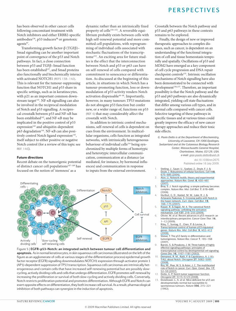
Figure 5 | eGFr–p53–notch: an integrated switch between tumour cell differentiation and apoptosis. As in normal keratinocytes, in skin squamous cell carcinomas (illustrated on the left of the figure as an agglomerate of cells at various stages of the differentiation process) epidermal growth factor receptor (eGFr) signalling downmodulates NOTcH1 expression through activator protein 1 (AP1)-dependent suppression of TP53 transcription. squamous cell carcinomas are intrinsically het-erogeneous and contain cells that have increased self-renewing potential but are possibly slow-cycling, actively dividing cells and cells that undergo differentiation. eGFr promotes self-renewal by increasing the proliferation or survival of both slow-cycling and actively dividing cells. conversely, Notch restricts proliferation potential and promotes differentiation. Although eGFr and Notch can exert opposite effects on differentiation, they both increase cell survival. As a result, pharmacological inhibition of both pathways can synergize in the induction of apoptosis.
P e r s P e c t i v e s
NATure revIewS | CanCer vOlume 9 | AuguST 2009 | 593
© 2009 Macmillan Publishers Limited. All rights reserved

40. Sulong, S. et al. A comprehensive analysis of the CDKN2A gene in childhood acute lymphoblastic leukemia reveals genomic deletion, copy number neutral loss of heterozygosity, and association with specific cytogenetic subgroups. Blood 113, 100–107 (2009).
41. Mungamuri, S. K., Yang, X., Thor, A. D. & Somasundaram, K. Survival signaling by Notch1: mammalian target of rapamycin (mTOR)-dependent inhibition of p53. Cancer Res. 66, 4715–4724 (2006).
42. Nair, P., Somasundaram, K. & Krishna, S. Activated Notch1 inhibits p53-induced apoptosis and sustains transformation by human papillomavirus type 16 E6 and E7 oncogenes through a PI3K-PKB/Akt-dependent pathway. J. Virol. 77, 7106–7112 (2003).
43. Palomero, T., Dominguez, M. & Ferrando, A. A. The role of the PTEN/AKT pathway in NOTCH1-induced leukemia. Cell Cycle 7, 965–970 (2008).
44. Guo, W. et al. Multi-genetic events collaboratively contribute to Pten-null leukaemia stem-cell formation. Nature 453, 529–533 (2008).
45. Uren, A. G. et al. Large-scale mutagenesis in p19(ARF)- and p53-deficient mice identifies cancer genes and their collaborative networks. Cell 133, 727–741 (2008).
46. Palomero, T. et al. Mutational loss of PTEN induces resistance to NOTCH1 inhibition in T-cell leukemia. Nature Med. 13, 1203–1210 (2007).
47. Kim, S. B. et al. Activated Notch1 interacts with p53 to inhibit its phosphorylation and transactivation. Cell Death Differ. 14, 982–991 (2007).
48. Balint, K. et al. Activation of Notch1 signaling is required for β-catenin-mediated human primary melanoma progression. J. Clin. Invest. 115, 3166–3176 (2005).
49. Politi, K., Feirt, N. & Kitajewski, J. Notch in mammary gland development and breast cancer. Semin. Cancer Biol. 14, 341–347 (2004).
50. Purow, B. W. et al. Expression of Notch-1 and its ligands, Delta-like-1 and Jagged-1, is critical for glioma cell survival and proliferation. Cancer Res. 65, 2353–2363 (2005).
51. Weng, A. P. et al. c-Myc is an important direct target of Notch1 in T-cell acute lymphoblastic leukemia/lymphoma. Genes Dev. 20, 2096–2109 (2006).
52. Ronchini, C. & Capobianco, A. J. Induction of cyclin D1 transcription and CDK2 activity by Notchic: implication for cell cycle disruption in transformation by Notchic. Mol. Cell. Biol. 21, 5925–5934 (2001).
53. Lowell, S., Jones, P., Le Roux, I., Dunne, J. & Watt, F. M. Stimulation of human epidermal differentiation by δ-notch signalling at the boundaries of stem-cell clusters. Curr. Biol. 10, 491–500 (2000).
54. Nickoloff, B. J. et al. Jagged-1 mediated activation of notch signaling induces complete maturation of human keratinocytes through NF-κB and PPARγ. Cell Death Differ. 9, 842–855 (2002).
55. Rangarajan, A. et al. Notch signaling is a direct determinant of keratinocyte growth arrest and entry into differentiation. EMBO J. 20, 3427–3436 (2001).
56. Estrach, S., Cordes, R., Hozumi, K., Gossler, A. & Watt, F. M. Role of the Notch ligand Delta1 in embryonic and adult mouse epidermis. J. Invest. Dermatol. 128, 825–832 (2008).
57. Lefort, K. et al. Notch1 is a p53 target gene involved in human keratinocyte tumor suppression through negative regulation of ROCK1/2 and MRCKα kinases. Genes Dev. 21, 562–577 (2007).
58. Nicolas, M. et al. Notch1 functions as a tumor suppressor in mouse skin. Nature Genet. 33, 416–421 (2003).
59. Proweller, A. et al. Impaired notch signaling promotes de novo squamous cell carcinoma formation. Cancer Res. 66, 7438–7444 (2006).
60. zur Hausen, H. Papillomaviruses causing cancer: evasion from host-cell control in early events in carcinogenesis. J. Natl Cancer Inst. 92, 690–698 (2000).
61. Daniel, B., Rangarajan, A., Mukherjee, G., Vallikad, E. & Krishna, S. The link between integration and expression of human papillomavirus type 16 genomes and cellular changes in the evolution of cervical intraepithelial neoplastic lesions. J. Gen. Virol. 78, 1095–1101 (1997).
62. Gray, G. E. et al. Human ligands of the Notch receptor. Am. J. Pathol. 154, 785–794 (1999).
63. Zagouras, P., Stifani, S., Blaumueller, C. M., Carcangiu, M. L. & Artavanis-Tsakonas, S. Alterations in Notch signaling in neoplastic lesions of the human cervix. Proc. Natl Acad. Sci. USA 92, 6414–6418 (1995).
14. Armstrong, J. F., Kaufman, M. H., Harrison, D. J. & Clarke, A. R. High-frequency developmental abnormalities in p53-deficient mice. Curr. Biol. 5, 931–936 (1995).
15. Sah, V. P. et al. A subset of p53-deficient embryos exhibit exencephaly. Nature Genet. 10, 175–180 (1995).
16. Saifudeen, Z., Dipp, S. & El-Dahr, S. S. A role for p53 in terminal epithelial cell differentiation. J. Clin. Invest. 109, 1021–1030 (2002).
17. Jerry, D. J., Tao, L. & Yan, H. Regulation of cancer stem cells by p53. Breast Cancer Res. 10, 304 (2008).
18. Zhang, M. et al. Identification of tumor-initiating cells in a p53-null mouse model of breast cancer. Cancer Res. 68, 4674–4682 (2008).
19. Zheng, H. et al. p53 and Pten control neural and glioma stem/progenitor cell renewal and differentiation. Nature 455, 1129–1133 (2008).
20. Mills, A. A. et al. p63 is a p53 homologue required for limb and epidermal morphogenesis. Nature 398, 708–713 (1999).
21. Yang, A. et al. p63 is essential for regenerative proliferation in limb, craniofacial and epithelial development. Nature 398, 714–718 (1999).
22. Yang, A. et al. p73-deficient mice have neurological, pheromonal and inflammatory defects but lack spontaneous tumours. Nature 404, 99–103 (2000).
23. Hooper, C. et al. TAp73 isoforms antagonize Notch signalling in SH-SY5Y neuroblastomas and in primary neurones. J. Neurochem. 99, 989–999 (2006).
24. Nguyen, B. C. et al. Cross-regulation between Notch and p63 in keratinocyte commitment to differentiation. Genes Dev. 20, 1028–1042 (2006).
25. Osada, M. et al. Differential recognition of response elements determines target gene specificity for p53 and p63. Mol. Cell. Biol. 25, 6077–6089 (2005).
26. Rocco, J. W., Leong, C. O., Kuperwasser, N., DeYoung, M. P. & Ellisen, L. W. p63 mediates survival in squamous cell carcinoma by suppression of p73-dependent apoptosis. Cancer Cell 9, 45–56 (2006).
27. Westfall, M. D., Mays, D. J., Sniezek, J. C. & Pietenpol, J. A. The ΔNp63 α phosphoprotein binds the p21 and 14-3-3σ promoters in vivo and has transcriptional repressor activity that is reduced by Hay-Wells syndrome-derived mutations. Mol. Cell. Biol. 23, 2264–2276 (2003).
28. Wu, G. et al. ΔNp63α and TAp63α regulate transcription of genes with distinct biological functions in cancer and development. Cancer Res. 63, 2351–2357 (2003).
29. Yang, A. et al. p63, a p53 homolog at 3q27–29, encodes multiple products with transactivating, death-inducing, and dominant-negative activities. Mol. Cell 2, 305–316 (1998).
30. Bourdon, J. C. et al. p53 isoforms can regulate p53 transcriptional activity. Genes Dev. 19, 2122–2137 (2005).
31. Toledo, F. & Wahl, G. M. Regulating the p53 pathway: in vitro hypotheses, in vivo veritas. Nature Rev. Cancer 6, 909–923 (2006).
32. Boggs, K. & Reisman, D. C/EBPβ participates in regulating transcription of the p53 gene in response to mitogen stimulation. J. Biol. Chem. 282, 7982–7990 (2007).
33. Bruno, T. et al. Che-1 phosphorylation by ATM/ATR and Chk2 kinases activates p53 transcription and the G2/M checkpoint. Cancer Cell 10, 473–486 (2006).
34. Kolev, V. et al. EGFR signalling as a negative regulator of Notch1 gene transcription and function in proliferating keratinocytes and cancer. Nature Cell Biol. 10, 902–911 (2008).
35. Phan, R. T. & Dalla-Favera, R. The BCL6 proto-oncogene suppresses p53 expression in germinal-centre B cells. Nature 432, 635–639 (2004).
36. Reisman, D. & Loging, W. T. Transcriptional regulation of the p53 tumor suppressor gene. Semin. Cancer Biol. 8, 317–324 (1998).
37. Rowland, B. D., Bernards, R. & Peeper, D. S. The KLF4 tumour suppressor is a transcriptional repressor of p53 that acts as a context-dependent oncogene. Nature Cell Biol. 7, 1074–1082 (2005).
38. Weng, A. P. et al. Activating mutations of NOTCH1 in human T cell acute lymphoblastic leukemia. Science 306, 269–271 (2004).
39. Beverly, L. J., Felsher, D. W. & Capobianco, A. J. Suppression of p53 by Notch in lymphomagenesis: implications for initiation and regression. Cancer Res. 65, 7159–7168 (2005).
64. Talora, C., Sgroi, D. C., Crum, C. P. & Dotto, G. P. Specific down-modulation of Notch1 signaling in cervical cancer cells is required for sustained HPV-E6/E7 expression and late steps of malignant transformation. Genes Dev. 16, 2252–2263 (2002).
65. Wang, L. et al. Overexpressed active Notch1 induces cell growth arrest of HeLa cervical carcinoma cells. Int. J. Gynecol. Cancer 17, 1283–1292 (2007).
66. Yao, J., Duan, L., Fan, M., Yuan, J. & Wu, X. Notch1 induces cell cycle arrest and apoptosis in human cervical cancer cells: involvement of nuclear factor kappa B inhibition. Int. J. Gynecol. Cancer 17, 502–510 (2007).
67. Tamura, K. et al. Stress response gene ATF3 is a target of c-myc in serum-induced cell proliferation. EMBO J. 24, 2590–2601 (2005).
68. Boggs, K., Henderson, B. & Reisman, D. RBP-Jκ binds to and represses transcription of the p53 tumor suppressor gene. Cell Biol. Int. 33, 318–324 (2009).
69. Ban, J. et al. EWS-FLI1 suppresses NOTCH-activated p53 in Ewing’s sarcoma. Cancer Res. 68, 7100–7109 (2008).
70. Riggi, N. & Stamenkovic, I. The biology of Ewing sarcoma. Cancer Lett. 254, 1–10 (2007).
71. Iso, T., Kedes, L. & Hamamori, Y. HES and HERP families: multiple effectors of the Notch signaling pathway. J. Cell Physiol. 194, 237–255 (2003).
72. Huang, Q. et al. Identification of p53 regulators by genome-wide functional analysis. Proc. Natl Acad. Sci. USA 101, 3456–3461 (2004).
73. Small, D. et al. Notch activation suppresses fibroblast growth factor-dependent cellular transformation. J. Biol. Chem. 278, 16405–16413 (2003).
74. Ishikawa, Y., Onoyama, I., Nakayama, K. I. & Nakayama, K. Notch-dependent cell cycle arrest and apoptosis in mouse embryonic fibroblasts lacking Fbxw7. Oncogene 27, 6164–6174 (2008).
75. Qi, R. et al. Notch1 signaling inhibits growth of human hepatocellular carcinoma through induction of cell cycle arrest and apoptosis. Cancer Res. 63, 8323–8329 (2003).
76. Duan, L., Yao, J., Wu, X. & Fan, M. Growth suppression induced by Notch1 activation involves Wnt-β-catenin down-regulation in human tongue carcinoma cells. Biol. Cell 98, 479–490 (2006).
77. Henning, K. et al. Notch1 activation reduces proliferation in the multipotent hematopoietic progenitor cell line FDCP-mix through a p53-dependent pathway but Notch1 effects on myeloid and erythroid differentiation are independent of p53. Cell Death Differ. 15, 398–407 (2008).
78. Yang, X. et al. Notch activation induces apoptosis in neural progenitor cells through a p53-dependent pathway. Dev. Biol. 269, 81–94 (2004).
79. Niranjan, T. et al. The Notch pathway in podocytes plays a role in the development of glomerular disease. Nature Med. 14, 290–298 (2008).
80. Purow, B. W. et al. Notch-1 regulates transcription of the epidermal growth factor receptor through p53. Carcinogenesis 29, 918–925 (2008).
81. Deb, S. P., Munoz, R. M., Brown, D. R., Subler, M. A. & Deb, S. Wild-type human p53 activates the human epidermal growth factor receptor promoter. Oncogene 9, 1341–1349 (1994).
82. Ludes-Meyers, J. H. et al. Transcriptional activation of the human epidermal growth factor receptor promoter by human p53. Mol. Cell. Biol. 16, 6009–6019 (1996).
83. Sasaki, Y. et al. The p53 family member genes are involved in the Notch signal pathway. J. Biol. Chem. 277, 719–724 (2002).
84. Das, H. K. Transcriptional regulation of the presenilin-1 gene: implication in Alzheimer’s disease. Front. Biosci. 13, 822–832 (2008).
85. Pastorcic, M. & Das, H. K. Regulation of transcription of the human presenilin-1 gene by ets transcription factors and the p53 protooncogene. J. Biol. Chem. 275, 34938–34945 (2000).
86. Amson, R. et al. Behavioral alterations associated with apoptosis and down-regulation of presenilin 1 in the brains of p53-deficient mice. Proc. Natl Acad. Sci. USA 97, 5346–5350 (2000).
87. Roperch, J. P. et al. Inhibition of presenilin 1 expression is promoted by p53 and p21WAF-1 and results in apoptosis and tumor suppression. Nature Med. 4, 835–838 (1998).
88. Devgan, V., Mammucari, C., Millar, S. E., Brisken, C. & Dotto, G. P. p21WAF1/Cip1 is a negative transcriptional regulator of Wnt4 expression downstream of Notch1 activation. Genes Dev. 19, 1485–1495 (2005).
P e r s P e c t i v e s
594 | AuguST 2009 | vOlume 9 www.nature.com/reviews/cancer
© 2009 Macmillan Publishers Limited. All rights reserved

89. Lohr, K., Moritz, C., Contente, A. & Dobbelstein, M. p21/CDKN1A mediates negative regulation of transcription by p53. J. Biol. Chem. 278, 32507–32516 (2003).
90. Laws, A. M. & Osborne, B. A. p53 regulates thymic Notch1 activation. Eur. J. Immunol. 34, 726–734 (2004).
91. Thelu, J., Rossio, P. & Favier, B. Notch signalling is linked to epidermal cell differentiation level in basal cell carcinoma, psoriasis and wound healing. BMC Dermatol. 2, 7 (2002).
92. Mandinova, A. et al. The FoxO3a gene is a key negative target of canonical Notch signalling in the keratinocyte UVB response. EMBO J. 27, 1243–1254 (2008).
93. Yugawa, T. et al. Regulation of Notch1 gene expression by p53 in epithelial cells. Mol. Cell. Biol. 27, 3732–3742 (2007).
94. Armstrong, B. K. & Kricker, A. The epidemiology of UV induced skin cancer. J. Photochem. Photobiol. B 63, 8–18 (2001).
95. de Gruijl, F. R. Skin cancer and solar UV radiation. Eur. J. Cancer 35, 2003–2009 (1999).
96. Lee, J. H. et al. Acute effects of UVB radiation on the proliferation and differentiation of keratinocytes. Photodermatol. Photoimmunol. Photomed. 18, 253–261 (2002).
97. Alimirah, F., Panchanathan, R., Davis, F. J., Chen, J. & Choubey, D. Restoration of p53 expression in human cancer cell lines upregulates the expression of Notch1: implications for cancer cell fate determination after genotoxic stress. Neoplasia 9, 427–434 (2007).
98. Secchiero, P. et al. Nutlin-3 upregulates the expression of Notch1 in both myeloid and lymphoid leukemic cells, as part of a negative feed-back anti-apoptotic mechanism. Blood 113, 4300–4308 (2009).
99. Wei, C. L. et al. A global map of p53 transcription-factor binding sites in the human genome. Cell 124, 207–219 (2006).
100. Lefort, K. & Dotto, G. P. Notch signaling in the integrated control of keratinocyte growth/differentiation and tumor suppression. Semin. Cancer Biol. 14, 374–386 (2004).
101. Koster, M. I. & Roop, D. R. The role of p63 in development and differentiation of the epidermis. J. Dermatol. Sci. 34, 3–9 (2004).
102. McKeon, F. p63 and the epithelial stem cell: more than status quo? Genes Dev. 18, 465–469 (2004).
103. Westfall, M. D. & Pietenpol, J. A. p63: molecular complexity in development and cancer. Carcinogenesis 25, 857–864 (2004).
104. Levrero, M. et al. The p53/p63/p73 family of transcription factors: overlapping and distinct functions. J. Cell Sci. 113,1661–1670 (2000).
105. Ross, D. A. & Kadesch, T. Consequences of Notch-mediated induction of Jagged1. Exp. Cell Res. 296, 173–182 (2004).
106. Thanos, D. & Maniatis, T. Virus induction of human IFNβ gene expression requires the assembly of an enhanceosome. Cell 83, 1091–1100 (1995).
107. Perera, R. J. et al. Defining the transcriptome of accelerated and replicatively senescent keratinocytes reveals links to differentiation, interferon signaling, and Notch related pathways. J. Cell Biochem. 98, 394–408 (2006).
108. Zhang, L. & Pagano, J. S. Structure and function of IRF-7. J. Interferon Cytokine Res. 22, 95–101 (2002).
109. Honda, K. et al. IRF-7 is the master regulator of type-I interferon-dependent immune responses. Nature 434, 772–777 (2005).
110. Servant, M. J., Tenoever, B. & Lin, R. Overlapping and distinct mechanisms regulating IRF-3 and IRF-7 function. J. Interferon Cytokine Res. 22, 49–58 (2002).
111. Seitz, C. S., Lin, Q., Deng, H. & Khavari, P. A. Alterations in NF-κB function in transgenic epithelial tissue demonstrate a growth inhibitory role for NF-κB. Proc. Natl Acad. Sci. USA 95, 2307–2312 (1998).
112. van Hogerlinden, M., Rozell, B. L., Ahrlund-Richter, L. & Toftgard, R. Squamous cell carcinomas and increased apoptosis in skin with inhibited Rel/nuclear factor-κB signaling. Cancer Res. 59, 3299–3303 (1999).
113. Lena, A. M. et al. miR-203 represses ‘stemness’ by repressing ΔNp63. Cell Death Differ. 15, 1187–1195 (2008).
114. Yi, R., Poy, M. N., Stoffel, M. & Fuchs, E. A skin microRNA promotes differentiation by repressing ‘stemness’. Nature 452, 225–229 (2008).
115. Thatcher, E. J., Flynt, A. S., Li, N., Patton, J. R. & Patton, J. G. miRNA expression analysis during normal zebrafish development and following inhibition of the Hedgehog and Notch signaling pathways. Dev. Dyn. 236, 2172–2180 (2007).
116. Yoo, A. S. & Greenwald, I. LIN-12/Notch activation leads to microRNA-mediated down-regulation of Vav in C. elegans. Science 310, 1330–1333 (2005).
117. Laurikkala, J. et al. p63 regulates multiple signalling pathways required for ectodermal organogenesis and differentiation. Development 133, 1553–1563 (2006).
118. Okuyama, R. et al. p53 homologue, p51/p63, maintains the immaturity of keratinocyte stem cells by inhibiting Notch1 activity. Oncogene 26, 4478–4488 (2007).
119. Truong, A. B. & Khavari, P. A. Control of keratinocyte proliferation and differentiation by p63. Cell Cycle 6, 295–299 (2007).
120. Moriyama, M. et al. Multiple roles of Notch signaling in the regulation of epidermal development. Dev. Cell 14, 594–604 (2008).
121. Candi, E. et al. ΔNp63 regulates thymic development through enhanced expression of FgfR2 and Jag2. Proc. Natl Acad. Sci. USA 104, 11999–12004 (2007).
122. Wu, L. et al. MAML1, a human homologue of Drosophila mastermind, is a transcriptional co-activator for NOTCH receptors. Nature Genet. 26, 484–489 (2000).
123. Petcherski, A. G. & Kimble, J. Mastermind is a putative activator for Notch. Curr. Biol. 10, R471–R473 (2000).
124. Oswald, F. et al. p300 acts as a transcriptional coactivator for mammalian Notch-1. Mol. Cell. Biol. 21, 7761–7774 (2001).
125. Fryer, C. J., Lamar, E., Turbachova, I., Kintner, C. & Jones, K. A. Mastermind mediates chromatin-specific transcription and turnover of the Notch enhancer complex. Genes Dev. 16, 1397–1411 (2002).
126. Wilson, J. J. & Kovall, R. A. Crystal structure of the CSL-Notch-Mastermind ternary complex bound to DNA. Cell 124, 985–996 (2006).
127. Nam, Y., Sliz, P., Song, L., Aster, J. C. & Blacklow, S. C. Structural basis for cooperativity in recruitment of MAML coactivators to Notch transcription complexes. Cell 124, 973–983 (2006).
128. McElhinny, A. S., Li, J. L. & Wu, L. Mastermind-like transcriptional co-activators: emerging roles in regulating cross talk among multiple signaling pathways. Oncogene 27, 5138–5147 (2008).
129. Zhao, Y. et al. The notch regulator MAML1 interacts with p53 and functions as a coactivator. J. Biol. Chem. 282, 11969–11981 (2007).
130. Saint Just Ribeiro, M., Hansson, M. L. & Wallberg, A. E. A proline repeat domain in the Notch co-activator MAML1 is important for the p300-mediated acetylation of MAML1. Biochem. J. 404, 289–298 (2007).
131. Yogosawa, S., Miyauchi, Y., Honda, R., Tanaka, H. & Yasuda, H. Mammalian Numb is a target protein of Mdm2, ubiquitin ligase. Biochem. Biophys. Res. Commun. 302, 869–872 (2003).
132. Colaluca, I. N. et al. NUMB controls p53 tumour suppressor activity. Nature 451, 76–80 (2008).
133. Peus, D., Hamacher, L. & Pittelkow, M. R. EGF-receptor tyrosine kinase inhibition induces keratinocyte growth arrest and terminal differentiation. J. Invest. Dermatol. 109, 751–756 (1997).
134. Kalyankrishna, S. & Grandis, J. R. Epidermal growth factor receptor biology in head and neck cancer. J. Clin. Oncol. 24, 2666–2672 (2006).
135. Lacouture, M. E. Mechanisms of cutaneous toxicities to EGFR inhibitors. Nature Rev. Cancer 6, 803–812 (2006).
136. Osipo, C. et al. ErbB-2 inhibition activates Notch-1 and sensitizes breast cancer cells to a γ-secretase inhibitor. Oncogene 27, 5019–5032 (2008).
137. Piccolo, S. p53 regulation orchestrates the TGF-β response. Cell 133, 767–769 (2008).
138. Blokzijl, A. et al. Cross-talk between the Notch and TGF-β signaling pathways mediated by interaction of the Notch intracellular domain with Smad3. J. Cell Biol. 163, 723–728 (2003).
139. Itoh, F. et al. Synergy and antagonism between Notch and BMP receptor signaling pathways in endothelial cells. EMBO J. 23, 541–551 (2004).
140. Zavadil, J., Cermak, L., Soto-Nieves, N. & Bottinger, E. P. Integration of TGF-β/Smad and Jagged1/Notch signalling in epithelial-to-mesenchymal transition. EMBO J. 23, 1155–1165 (2004).
141. Niimi, H., Pardali, K., Vanlandewijck, M., Heldin, C. H. & Moustakas, A. Notch signaling is necessary for epithelial growth arrest by TGF-β. J. Cell Biol. 176, 695–707 (2007).
142. Webster, G. A. & Perkins, N. D. Transcriptional cross talk between NF-κB and p53. Mol. Cell. Biol. 19, 3485–3495 (1999).
143. Pei, X. H., Nakanishi, Y., Takayama, K., Bai, F. & Hara, N. Benzo[a]pyrene activates the human p53 gene through induction of nuclear factor κB activity. J. Biol. Chem. 274, 35240–35246 (1999).
144. Lee, H. O., Lee, J. H., Kim, T. Y. & Lee, H. Regulation of ΔNp63α by tumor necrosis factor-α in epithelial homeostasis. FEBS J. 274, 6511–6522 (2007).
145. Bash, J. et al. Rel/NF-κB can trigger the Notch signaling pathway by inducing the expression of Jagged1, a ligand for Notch receptors. EMBO J. 18, 2803–2811 (1999).
146. Osipo, C., Golde, T. E., Osborne, B. A. & Miele, L. A. Off the beaten pathway: the complex cross talk between Notch and NF-κB. Lab. Invest. 88, 11–17 (2008).
147. Quintana, E. et al. Efficient tumour formation by single human melanoma cells. Nature 456, 593–598 (2008).
148. Shamloula, H. K. et al. rugose (rg), a Drosophila A kinase anchor protein, is required for retinal pattern formation and interacts genetically with multiple signaling pathways. Genetics 161, 693–710 (2002).
149. Shmelkov, S. V. et al. CD133 expression is not restricted to stem cells, and both CD133+ and CD133– metastatic colon cancer cells initiate tumors. J. Clin. Invest. 118, 2111–2120 (2008).
150. Jones, P. H., Simons, B. D. & Watt, F. M. Sic transit gloria: farewell to the epidermal transit amplifying cell? Cell Stem Cell 1, 371–381 (2007).
151. Malhotra, S. & Kincade, P. W. Wnt-related molecules and signaling pathway equilibrium in hematopoiesis. Cell Stem Cell 4, 27–36 (2009).
152. Okuyama, R., LeFort, K. & Dotto, G. P. A dynamic model of keratinocyte stem cell renewal and differentiation: role of the p21WAF1/Cip1 and Notch1 signaling pathways. J. Investig. Dermatol. Symp. Proc. 9, 248–252 (2004).
153. Chang, H. H., Hemberg, M., Barahona, M., Ingber, D. E. & Huang, S. Transcriptome-wide noise controls lineage choice in mammalian progenitor cells. Nature 453, 544–547 (2008).
154. Lahav, G. The strength of indecisiveness: oscillatory behavior for better cell fate determination. Sci. STKE 2004, pe55 (2004).
155. Lahav, G. Oscillations by the p53-Mdm2 feedback loop. Adv. Exp. Med. Biol. 641, 28–38 (2008).
156. Giudicelli, F. & Lewis, J. The vertebrate segmentation clock. Curr. Opin. Genet. Dev. 14, 407–414 (2004).
157. Lewis, J. From signals to patterns: space, time, and mathematics in developmental biology. Science 322, 399–403 (2008).
AcknowledgementsI thank C. Brisken and C. Missero for careful reading of the manuscript and J. Aster and I. Screpanti for useful feedback. I apologize to authors whose work was not directly quoted owing to space limitations and the complexity involved. This work was supported by US National Institutes of Health grants AR054856 and AR39190 the Swiss National Foundation and a grant from the European Union (Epistem, Sixth Framework Programme, LSHB-CT-2005-019067).
DATABASESentrez Gene: http://www.ncbi.nlm.nih.gov/entrez/query.fcgi?db=geneCDKN2A | CCND1 | EGFR | EWSR1| FLI1 | FOXO3 | IRF3 | IRF7 | MYC | Trp53 UniProtKB: http://www.uniprot.orgFBXW7 | Hes1 | HeY1 | JAG1 | MAML1 | MDM2 | NOTcH1 | p53 | p63 | p73 | PTeN
FURTHER iNFoRMATioNG. Paolo Dotto’s homepages: http://www.unil.ch/ib/page9489.html | http://www.massgeneral.org/research/researchlab.aspx?id=1069
all links are aCTive in The online pdF
P e r s P e c t i v e s
NATure revIewS | CanCer vOlume 9 | AuguST 2009 | 595
© 2009 Macmillan Publishers Limited. All rights reserved