CrossTalk: The Journal of Defense Software Engineering. Volume 21, Number 9
Upload
independentCategory
view
3download
0
1 3
DOI 10.1007/s00018-013-1488-9 Cellular and Molecular Life SciencesCell. Mol. Life Sci. (2014) 71:1055–1066
REVIEW
Crosstalk between cerebral endothelium and oligodendrocyte
Nobukazu Miyamoto · Loc‑Duyen D. Pham · Ji Hae Seo · Kyu‑Won Kim · Eng H. Lo · Ken Arai
Received: 31 July 2013 / Revised: 12 September 2013 / Accepted: 30 September 2013 / Published online: 17 October 2013 © Springer Basel 2013
brain injury, but would be recovered in the chronic phase to promote brain remodeling and repair. Oligodendrocyte lineage cells play critical roles in white matter function, and under pathological conditions, oligodendrocyte dys-function lead to white matter damage. Therefore, a deeper understanding of the mechanisms of endothelial-oligo-dendrocyte trophic coupling may lead to new therapeu-tic approaches for white matter-related diseases, such as stroke or vascular dementia.
Keywords Oligodendrocytes · Oligodendrocyte precursor cells · Cerebral endothelial cells · Oligovascular niche · Neurovascular unit
Introduction
Brain physiology and pathophysiology are very complex. Several different types of brain cells may work together to maintain, remodel, and repair our brain functions. In this regard, the concept of “neurovascular unit” was raised as a new paradigm to understand the pathology of central nervous system (CNS) diseases, such as stroke [1–5]. This modular concept is defined at an intercellular level that comprises dynamic interactions between cere-bral endothelial cells, glia, neurons, and the extracellular matrix. Dysfunctional crosstalk within the neurovascular unit may lead to multiple aspects of acute pathophysiol-ogy in CNS diseases. For example, impaired glutamate release-reuptake mechanisms in neurons and astro-cytes can amplify excitotoxicity [6]. Perturbed signaling between cerebral endothelium and astrocytes (and some-times pericytes) can disrupt blood–brain barrier integrity [4]. In addition, dysfunctional coupling between neuronal activation and vascular responses can also accelerate
Abstract It is now relatively well accepted that the cer-ebrovascular system does not merely provide inert pipes for blood delivery to the brain. Cerebral endothelial cells may compose an embedded bunker of trophic factors that contribute to brain homeostasis and function. Recent find-ings suggest that soluble factors from cerebral endothelial cells nourish neighboring cells, such as neurons and astro-cytes. Although data are strongest in supporting mecha-nisms of endothelial-neuron and/or endothelial-astrocyte trophic coupling, it is likely that similar interactions also exist between cerebral endothelial cells and oligoden-drocyte lineage cells. In this mini-review, we summarize current advances in the field of endothelial-oligodendro-cyte trophic coupling. These endothelial-oligodendro-cyte interactions may comprise the oligovascular niche to maintain their cellular functions and sustain ongoing angiogenesis/oligodendrogenesis. Importantly, it should be noted that the cell–cell interactions are not static—the trophic coupling is disturbed under acute phase after
N. Miyamoto · L.-D. D. Pham · J. H. Seo · E. H. Lo · K. Arai (*) Neuroprotection Research Laboratory, Departments of Radiology and Neurology, Massachusetts General Hospital and Harvard Medical School, MGH East 149-2401, Charlestown, MA 02129, USAe-mail: [email protected]
J. H. Seo · K.-W. Kim NeuroVascular Coordination Research Center, College of Pharmacy, Seoul National University, Seoul, Korea
K.-W. Kim Department of Molecular Medicine and Biopharmaceutical Sciences, Graduate School of Convergence Science and Technology, Seoul National University, Seoul, Korea

1056 N. Miyamoto et al.
1 3
deleterious spreading depression [7]. Moreover, disor-dered signaling between all neurovascular and gliovascu-lar elements may underlie the evolution of neuroinflam-mation and cell death [8].
Besides the importance of cell–cell interaction, the neurovascular unit also emphasizes the distinct roles of cerebral endothelium on brain functions. Although cere-bral vascular system is the major constituent of the brain, the cerebrovascular system was traditionally thought as a passive conduit for blood stream. However, recent research has proposed that this system plays more active roles in maintaining the CNS homeostasis. As men-tioned, cerebral endothelial cells form the blood–brain barrier (BBB) with astrocytes and pericytes. The BBB constitutes anatomical, physiochemical, and biochemical barrier that controls the exchange of materials between blood, brain, and cerebrospinal fluid. BBB breakdown due to endothelial dysfunction is frequently associated with a myriad of neurological pathologies, including chronic CNS diseases [9–11]. Another example for the importance of cerebral vascular system is that cerebral endothelial cells nourish neighboring neurons. Through releasing trophic factors, cerebral endothelial cells guide developing axons [12], protect neurons against stress [13, 14], and provide a niche for supporting neural stem/pro-genitor cells (NSPCs) [15]. NSPCs were shown to have direct coupling with cerebral endothelial cells [16], and in this so-called neurovascular niche, cell–cell signaling between cerebral endothelial cells and neuronal precursor cells helps mediate and sustain pockets of ongoing neu-rogenesis and angiogenesis in adult brain [15, 17]. Even under the remodeling phase after brain injury, these close relationships are maintained, and both neurogenesis and angiogenesis occur in the neurovascular niche to promote repairing of the brain. Indeed, angiogenic stimulation enhances neurogenesis after stroke [15, 18], and in turn, neuroblasts migrate along perivascular routes and the promotion of neurogenesis enhances vascular re-growth [19].
For the most part, research that studies mecha-nisms of trophic coupling in the neurovascular unit has mainly focused on endothelium–neuron and endothe-lium–astrocyte interactions. However, cell–cell interac-tions between endothelial cells and oligodendrocytes are likely to be important to maintain brain functions as well, especially in white matter. Recent papers indi-cate that some populations of oligodendrocyte lineage cells are located close to cerebral endothelial cells [20, 21] (Fig. 1), and these cells may communicate each other via secreting soluble factors. In this mini-review, therefore, we attempt to overview key findings for the crosstalk between cerebral endothelial cells and oligodendrocytes.
Endothelium‑oligodendrocyte signaling
Oligodendrocyte generation from neural stem cells
The interaction between brain microvascular endothelium and NSPCs is receiving a lot attention as a key mechanism of NSPC biology [22]. During embryonic development, interaction between endothelium and stem cells occurs in both the CNS and non-nervous tissues [22]. Endothelial cells modulate the behavior of NSPCs in the stem cell niche seen in subventricular zone (SVZ) or the hippocampal sub-granular zone (SGZ) [23]. These regions provide a special-ized microenvironment where NSPCs persist throughout their life. The SVZ contains a subpopulation of cells with astroglial properties (so-called type B cells), which give rise to dividing intermediate progenitors or transient ampli-fying progenitors (so-called type C cells) to generate neuro-blasts (so-called type A cells). The type B cells can also dif-ferentiate into oligodendrocyte lineage cells (i.e., migratory OPCs) through Olig2-expressing type C cells. These type B cells-derived OPCs leave the SVZ and migrate to the cor-pus callosum, the white matter tract in the striatum and fim-bria fornix [24]. Thus far, at least two studies have exam-ined the cell–cell interaction between cerebral endothelium and NSPCs in generating oligodendrocytes. Chintawar et al. [25] investigated the interactions of human fetal neu-ral precursor cells (hfNPCs) with human brain endothe-lial cells in their in vitro model systems. They found that in the in vitro sub-endothelial niche, NPCs interacted with endothelial cells to promote NPC differentiation into astro-cytes, neurons, and even oligodendrocytes. This study proposed that the chemokine CCL2/MCP-1 mediated the interaction between endothelium and neural precursor cells for the differentiation into oligodendrocytes [25]. Another study by Plane et al. also examined the mechanisms of dif-ferentiation from postnatal murine forebrain neural stem cells. Interestingly, they showed that conditioned medium from endothelial cells promoted the differentiation of neu-ral stem cells into oligodendrocyte linage cells [26]. These two studies may suggest that cerebral endothelial cells par-ticipate in generating oligodendrocyte linage cells from neural precursor cells under some conditions.
Signaling from cerebral endothelial cells to oligodendrocytes
In the developmental stage, most oligodendrocytes are generated from their precursors (oligodendrocyte precur-sor cells; OPCs). OPCs migrate to colonize the white mat-ter brain (and sometimes gray matter as well) from their sites of origins (e.g., SVZ region) [27, 28], and the differ-entiation into mature oligodendrocytes occurs in a multi-ple steps [29]. Many OPCs differentiate into myelinating

1057Crosstalk between cerebral endothelium and oligodendrocyte
1 3
oligodendrocytes during development, but some popula-tions of OPCs remain at the immature state and are widely distributed within the brain even after adolescence [30]. Several evidences indicate that OPCs persist in the adult SVZ [24], and oligodendrocytes continue to be newly produced in the adult white matter [31–34]. These OPCs in the adult brains may participate in myelin repair after injury [35, 36]. Past studies have demonstrated that adult SVZ progenitors (i.e., type B cells) can generate new OPCs/oligodendrocytes after demyelinating lesions of the corpus callosum [37, 38], seizures [39], or stroke [40–42]. As noted, these SVZ type B cells serve as primary pre-cursors for new oligodendrocytes and give rise to young migrating oligodendrocytes through intermediate Olig2 transit-amplifying cells (so-called type C cells) under nor-mal and injured brains [24]. They are known to move out of the SVZ into the corpus callosum, neighboring striatum, and fimbria fornix to differentiate into non-myelinated and myelinated oligodendrocytes [43]. Compared with the large number of new neurons born in the SVZ, fewer OPCs/oligodendrocytes are normally produced from adult
SVZ type B cells. This may be related to the slow turnover of oligodendrocytes in adult brain [34]. However, the num-ber of OPCs/oligodendrocytes derived from SVZ type B cells are increased after a demyelinating lesion. Under the stress conditions, SVZ-generated oligodendrocytes express markers of myelin, suggesting that they contribute to the remyelination process [37, 38, 43, 44].
Recently, the concept of oligovascular niche was pro-posed to understand the phenomena of trophic coupling between cerebral endothelium and oligodendrocyte pre-cursors [29]. Cerebral endothelial cells may promote the proliferation of OPCs through releasing trophic factors, such as BDNF and bFGF, in vitro [45]. In addition, cer-ebral endothelial-derived VEGF-A increased the mobil-ity of OPCs without affecting their proliferation [46, 47]. Importantly, sub-lethal oxidative stress decreased the lev-els of growth factor productions in endothelial cells, and the “sick” endothelial cells seemed no longer supportive for OPCs [45]. These results indicate that endothelium-oligodendrocyte trophic coupling would be disturbed dur-ing pathological conditions, which lead to white matter
Fig. 1 Oligodendrocyte precursor cells (red: PDGF-R-alpha antibody) are often located closely to cerebral endothelial cells (green: CD31). Adult rat brain, corpus callosum region. Scale bar = 10 μm

1058 N. Miyamoto et al.
1 3
dysfunction. As noted, some OPCs are located close to cerebral endothelial cells in adult white matter (e.g., cor-pus callosum) [20], and therefore, cerebral endothelial cells may participate in the process of oligodendrocyte remodeling and repair under both normal and pathological conditions.
Signaling from oligodendrocytes to cerebral endothelial cells
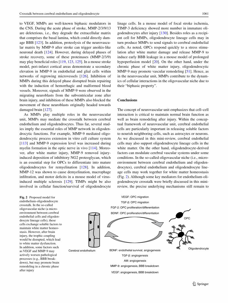
Thus far, we have discussed how cerebral endothelial cells affect functions of oligodendrocyte lineage cells, but the trophic coupling between cerebral endothelial cells and oligodendrocytes should be “two-way”. Oligodendrocyte lineage cells could act as growth factor providers for neigh-boring cells [48]. For example, oligodendrocytes synthe-size defined growth factors and provide trophic signals to nearby neurons. Past studies show that oligodendrocyte-derived trophic factors, such as IGF1 and GDNF, promote neuronal survival and axonal outgrowth in vitro [49]. More-over, oligodendrocytes may serve as a principal metabolic supplier of lactate, which is integral for axonal energy sup-port through monocarboxylate transporter 1 [50]. Although there is little known about the mechanisms by which oligo-dendrocyte lineage cells affect the vascular system, Pham et al. [51] recently reported that after white matter injury, mature oligodendrocytes secreted a well-known angiogenic factor MMP-9 to promote vascular remodeling. Hence, similar to the neurovascular niche, the oligovascular niche may provide an important mechanism for both angiogen-esis and oligodendrogenesis in the adult white matter.
However, it should be noted that oligodendrocyte lineage cells might not always be supportive for the cerebral vascular system. Very recently, OPCs were shown to release MMP-9 under acute phase of white matter injury [20]. This OPC-derived MMP-9 initiated the early onset of BBB breakdown, which would progress to white matter dysfunction at the later time point [20]. These findings suggest that the oligo-dendrocyte-endothelium interaction is not merely static. As the concept of neurovascular unit emphasizes, depending on the context, these cell–cell interactions can be both beneficial and detrimental. Under normal conditions, cell–cell trophic coupling is necessary for maintaining brain homeostasis. On the other hand, under the acute phase of neurodegenerative diseases, these cell–cell trophic couplings could be disrupted and lead to cellular dysfunction. However, in the chronic phase after brain injury, the cell–cell interaction may be recovered to some extent, and brains would try to repair their damaged function. Hence, understanding the precise mech-anisms of oligodendrocyte-endothelium trophic coupling would lead us to find novel therapeutic approaches both in protecting white matter against stress under acute phase and in boosting white matter remodeling/repairing under chronic
phase for white matter-related diseases such as stroke and vascular dementia.
Possible mediators for oligodendrocyte‑endothelial interaction
As mentioned, crosstalk between the vascular and neuronal compartments in the neurovascular niche is mediated by an exchange of soluble signals [3, 4, 14, 52–56]. Many of these trophic factors may also affect oligodendrocyte line-age cells. Moreover, growing literature suggests that simi-lar to cerebral endothelium, oligodendrocyte lineage cells could work as a “bank” for trophic factors (Table 1). The precise regulatory mechanisms that underlie angiogenesis and oligodendrocytes in the oligovascular niche still require to be elucidated. However, we will try to summarize several candidates of mediators for the crosstalk between cerebral endothelium and oligodendrocytes in this section.
Brain-derived neurotrophic factor
Brain-derived neurotrophic factor (BDNF) is a member of the “neurotrophin” family of growth factors, which are related to the canonical nerve growth factor (NGF). BDNF is well known to act on neurons to support the survival of existing
Table 1 Releasing factors from oligodendrocyte linage cells
Releasing Factor cellular type
BDNF Cultured rat oligodendrocytes [67]
Immortalized oligodendroglial cell line (N19) [131]
MBP-positive oligodendrocytes in rat brain [68]
IGF-1 Cultured rat OPCs [132]
NGF Cultured rat OPCs [133]
Cultured rat oligodendrocytes [67, 133]
Immortalized oligodendroglial cell line (N19) [131]
MBP-positive oligodendrocytes in rat brain [68]
NT-3 Cultured rat oligodendrocytes [67]
MBP-positive oligodendrocytes in rat brain [68]
Neuregulin cultured rat OPCs [134]
Cultured adult human oligodendrocytes [135]
GDNF Cultured rat oligodendrocytes [49, 136]
Cultured rat OPCs [136]
Transformed oligodendroglial cell line OLN-93 [136]
Immortalized oligodendrocyte precursor cell line OLI-neu [136]
TGF-beta Cultured rat OPCs [82]
HMGB-1 Oligodendrocyte-like cells in rat brain [137]
AM Human oligodendroglial cell line KG1C [103]
MMP-9 Cultured rat OPCs [20]
MOBP-positive oligodendrocytes [21]

1059Crosstalk between cerebral endothelium and oligodendrocyte
1 3
neurons and encourage the growth and differentiation of new neurons and synapses [57, 58]. BDNF can also modulate non-neuronal cell types. Irrefutably, BDNF is one of the major mediators for endothelium-oligodendrocyte interaction.
BDNF is known to regulate oligodendrocyte function in several ways. Firstly, BDNF promotes OPC prolifera-tion and differentiation into mature oligodendrocytes. Cell culture and in vivo animal studies suggested that Trk-B receptor and ERK pathway mediated the supportive effects of BDNF [59–62]. Secondly, even under pathological con-ditions, BDNF can be supportive for oligodendrocyte lin-eage cells. In in vivo white matter injury models (spinal cord injury or cuprizone-treated model), BDNF was shown to play an important role in regulating the numbers of oli-godendrocyte lineage cells after demyelination [63, 64]. Thirdly, BDNF may also work on neural stem cells (NSCs) to trigger the differentiation into oligodendrocyte line-age cells. Chen et al. [65] reported that BDNF enhanced the cell commitment of NSCs to neuronal and oligoden-drocytic fates by activating Wnt/β-catenin signaling path-way in vitro. Since cerebral endothelial cells are the major BDNF-producing cell type, cerebral endothelial cells may support oligodendrocyte lineage cells via BDNF signaling. As introduced in the previous section, endothelial-derived BDNF was shown to promote the proliferation of OPCs in vitro [45]. But importantly, stressed endothelial cells secrete less BDNF and can no longer support OPCs [45], indicating that endothelial-oligodendrocyte interaction is highly dependent on their cellular conditions.
BDNF also promotes endothelial cell survival and induces angiogenesis in brain [66]. While cell–cell trophic interactions are generally considered as “two-way”, there have been still no direct proofs that BDNF mediates sig-nals from oligodendrocytes to endothelial cells. Neverthe-less, several lines of evidence strongly support the idea that oligodendrocyte lineage cells would produce BDNF to support endothelial cells. In situ hybridization and immu-nocytochemical studies identified expressions of BDNF mRNA/protein in cultured basal forebrain oligodendro-cytes [67, 68]. The physiological relevance of these culture works is supported by detection of BDNF in oligodendro-cytes in vivo. For example, BDNF mRNAs are localized in subpopulations of myelin-basic-protein-positive mature oligodendrocytes in the basal forebrain, cingulate cortex, and corpus callosum of postnatal day 7 rats [68]. Similarly, BDNF mRNA and protein are also expressed in subsets of APC-positive oligodendrocytes of adult spinal cords [69, 70]. The oligodendrocytic BDNF is bioactive because con-ditioned medium from oligodendrocyte cultures increased cholinergic neurons and the effect was partially blocked by co-treatment with anti-BDNF neutralizing antibody [68]. Considering the fact that oligodendrocyte linage cells are often located to cerebral endothelial cells, it would be
reasonable to think that oligodendrocytic BDNF may regu-late cerebral vascular system to some extent.
Fibroblast growth factor-2
Similar to BDNF, fibroblast growth factor-2 (FGF-2/bFGF) can be proposed as an important mediator for the endothe-lium-oligodendrocyte interaction. FGF-2 is a potent stimu-lator of endothelial cell migration, proliferation, sprouting, and tube formation. In addition, past studies have substan-tially revealed the roles of FGF-2 on oligodendrocyte func-tion. FGF-2 by itself stimulates proliferation of late-stage OPCs and blocks their terminal differentiation into mature oligodendrocytes in vitro. Upon removal of FGF-2 from the cell culture medium of OPCs, the cells readily enter the terminal differentiation [71–73]. In addition, FGF-2 cooperates with PDGF to up-regulate the expression of PDGF-receptor-alpha for OPC proliferation [71, 74]. As for OPC migration, the importance of FGF signaling in early stage OPCs has been evaluated both in vivo and in vitro. Using an in vivo transplantation approach, Osterhout et al. [75] demonstrated that OPCs with dominant-negative FGF receptor 1 failed to migrate. Subsequent in vitro study showed that in response to FGF-2 stimulation, OPCs grow-ing in an agarose drop successfully move out from the drop [76]. Another study suggested that FGF-2 also enhanced the migration of pre-OPCs from oligospheres, but might not induce the migration of late-stage OPCs [77]. While still controversial, FGF-2 may affect OPC survival. FGF-2 can prevent OPCs from apoptotic stress [78], but this kind of protective effects were not observed in optic nerve OPCs [79]. Since the cellular localization of FGF-2 is observed in cerebral endothelium both in normal and pathological con-ditions [80], future studies are warranted to examine how FGF-2 modulate the dynamics of endothelium-oligoden-drocyte crosstalk under normal conditions as well as acute/chronic phases after brain injury.
Transforming growth factor-beta
Transforming growth factor-beta (TGF-beta), a prototypic member of a large family of pleiotropic cytokines, is also a potent modulator in the oligovascular niche. Many cells have been reported to produce TGF-beta, and TGF-beta would modulate several cellular functions in most cells [81]. In terms of endothelium-oligodendrocyte interac-tion, oligodendrocytes are reported to express TGF-beta in vitro cell culture systems [82] and in vivo spinal cords [83]. TGF-beta contributes to angiogenesis by stabilizing newly formed capillary sprouts [84]. Thus far, many studies in mouse and human have demonstrated its pivotal roles in modulating angiogenesis after brain injury, such as stroke [85–90]. In addition, dysregulation of TGF-beta signaling

1060 N. Miyamoto et al.
1 3
may cause hereditary vascular disorders [81]. For example, mutations in TGF-beta receptors lead to hereditary hemor-rhagic telangiectasia [91], and in the cerebral white matter, accumulation of TGF-beta1 due to HtrA1 mutation is asso-ciated with a hereditary disorder CRASIL (cerebral autoso-mal recessive arteriopathy with subcortical infarcts and leu-koencephalopathy) [92]. In contrast, the roles of TGF-beta on oligodendrocytic functions are still understudied, but TGF-beta was reported to affect the migration of cultured OPCs when they were maintained on astrocytes [93]. Using the oligodendrocyte precursor cell line OLI-neu, TGF-beta was shown to upregulate a chondroitin sulfate proeoglycan DSD-1-PG on the cell surface [94], indicating that TGF-beta may modulate the cell adhesion property of OPCs to change cell motility.
Adrenomedullin
Adrenomedullin (AM) was originally isolated from pheo-chromocytoma cells [95]. AM has a variety of actions on the vascular systems, such as endothelial survival/prolifera-tion, vasodilatation, regulation of BBB permeability, and modulation of oxidative stress levels in endothelium [96, 97]. AM is secreted from various organs, and in the CNS, AM is mainly expressed in neurons and cerebral endothe-lium [95, 98]. AM has been shown to reduce the infarct volumes in transient stroke models [99, 100]. Under stroke conditions, AM expression is markedly increased via the hypoxia-inducible factor-1 signaling [101], indicating that AM could work for vascular remodeling/repairing after brain injury. Interestingly, in the mouse model of prolonged cerebral hypoperfusion (i.e. vascular dementia model), AM was demonstrated to be protective toward white matters [102], where most oligodendrocytes are populated. In addi-tion, Uezono et al. [103] detected AM mRNAs in human oligodendroglial cell line, and showed the potential effects of AM in modulating oligodendrocyte function through AM receptors in oligodendrocytes. Importantly, past stud-ies have demonstrated that AM increases several growth factors such as VEGF and FGF [104–107]. Hence, AM might work as a “master” modulator for growth factors in the oligovascular niche.
Vascular endothelial growth factor
Vascular endothelial growth factor (VEGF) is a primary regulator of angiogenesis by stimulating endothelial cell proliferation, migration, and tube formation [54], but it is now well recognized that VEGF is not solely an endothelial mediator, rather may represent one of the major mediators in the neurovascular unit [108–110]. For example, VEGF signaling plays a key role in neuronal migration and CNS development [111]. In addition, VEGF may also affect
oligodendrocyte lineage cells. OPCs were positive for the VEGF receptor, Flk-1 (also known as KDR or VEGF-receptor 2) [47], which is primarily responsible for VEGF-induced angiogenesis. Recent in vitro study suggested that VEGF-A significantly accelerated the motility of OPCs through Flk-1, and this effect was partly mediated by ROS production [47]. Importantly, conditioned media from cer-ebral endothelial cells promoted both OPC proliferation and migration. However, endothelial-derived VEGF-A may participate in the OPC migration but not proliferation [46]. In addition to VEGF-A, other VEGF families and VEGF receptors may also be involved in oligodendrocyte func-tions. Le Bras et al. [112] demonstrated that VEGF-C pro-moted OPC proliferation through VEGF-receptor-3. Taken together, these findings indicate that VEGFs/VEGF-recep-tors may play a central role in the endothelial-oligodendro-cyte trophic coupling.
If VEGF is an important modulator for the oligovascu-lar niche, we may need to consider the “biphasic actions” of VEGF to comprehend the dynamics of trophic coupling between cerebral endothelium and oligodendrocytes. Since VEGF is a primary regulator of angiogenesis, VEGF can trigger remodeling responses in endothelial cells (i.e., accelerating angiogenesis) after brain damage, such as stroke. Infusing VEGF into the lateral ventricles stimulated angiogenesis and decreased infarct volumes in rodent mod-els of focal cerebral ischemia [113]. An increase in angio-genesis by VEGF in rats was also associated with reduced neurological deficits after stroke [114]. Similar effects were also reported in neonatal focal rodent stroke [115]. Moreover, in transgenic mice with overexpressing human VEGF165, brain microvessel density was significantly elevated compared to wild-type mice before ischemia, and the microvessel density was higher 3 days after stroke onset [116]. On the other hand, VEGF increases BBB permeabil-ity in the acute phase in stroke. VEGF administration wors-ens BBB leakage by ischemic insults [117, 118]. Therefore, depending on the context of the microenvironment in the oligovascular nice, VEGF may regulate the endothelium-oligodendrocyte interaction to maintain and remodel white matter function.
Matrix metalloproteinases
Matrix metalloproteinases (MMPs) comprise a family of zinc endopeptidases, and their activities are tightly regu-lated by tissue inhibitor of metalloproteinases (TIMPs). This larger MMP network plays major roles in the physi-ology and pathology of the mammalian CNS, includ-ing stroke (MMP-2/3/7/9/13, TIMP-1/2 [119]), vascular dementia (MMP-2, 3, 9 [120, 121]), and CADASIL (cer-ebral autosomal-dominant arteriopathy with subcortical infarcts and leukoencephalopathy: TIMP-3 [122]). Similar
1061Crosstalk between cerebral endothelium and oligodendrocyte
1 3
to VEGF, MMPs are well-known biphasic modulators in the CNS. During the acute phase of stroke, MMP-2/3/9/13 are deleterious, i.e., they degrade the extracellular matrix that comprises the basal lamina, which could directly dam-age BBB [123]. In addition, proteolysis of the neurovascu-lar matrix by MMP-9 after stroke can trigger anoikis-like neuronal death [124]. However, during delayed phases of stroke recovery, some of these proteinases (MMP-2/3/9) may play beneficial roles [119, 123, 125]. In a mouse stroke model, peri-infarct cortical areas demonstrate a secondary elevation in MMP-9 in endothelial and glial cells within networks of regrowing microvessels [126]. Inhibition of MMPs during this delayed phase disrupted brain repairing with the induction of hemorrhagic and malformed blood vessels. Moreover, signals of MMP-9 were observed in the migrating neuroblasts from the subventricular zone after brain injury, and inhibition of these MMPs also blocked the movement of these neuroblasts originally headed towards damaged brain [127].
As MMPs play multiple roles in the neurovascular unit, MMPs may mediate the crosstalk between cerebral endothelium and oligodendrocytes. Thus far, several stud-ies imply the essential roles of MMP network in oligoden-drocytic functions. For example, MMP-9 mediated oligo-dendrocytic process extension in vitro cell culture system [113] and MMP-9 expression level was increased during myelin formation in the optic nerve in vivo [114]. Moreo-ver, after white matter injury, MMP-9 removed injury-induced deposition of inhibitory NG2 proteoglycan, which is an essential step for OPCs to differentiate into mature oligodendrocytes for remyelination [128]. In addition, MMP-12 was shown to cause demyelination, macrophage infiltration, and motor deficits in a mouse model of virus-induced multiple sclerosis [129]. TIMPs might be also involved in cellular function/survival of oligodendrocyte
linage cells. In a mouse model of focal stroke ischemia, TIMP-3 deficiency showed more number in immature oli-godendrocytes after injury [130]. Besides roles as a recipi-ent cell for MMPs, oligodendrocyte lineage cells may in turn produce MMPs to send signals to cerebral endothelial cells. As noted, OPCs respond quickly to a stress stimu-lation after white matter damage and release MMP-9 to induce early BBB leakage in a mouse model of prolonged hypoperfusion model [20]. On the other hand, under the chronic phase of white matter injury, oligodendrocytic MMP-9 may promote vascular remodeling [51]. Hence, as in the neurovascular unit, MMPs contribute to the dynam-ics of cellular interactions in the oligovascular niche due to their “biphasic property”.
Conclusions
The concept of neurovascular unit emphasizes that cell–cell interaction is critical to maintain normal brain function as well as brain remodeling after injury. Within the concep-tual framework of neurovascular unit, cerebral endothelial cells are particularly important in releasing soluble factors to nourish neighboring cells, such as astrocytes or neurons. As we discussed in this mini-review, cerebral endothelial cells may also support oligodendrocyte lineage cells in the white matter. On the other hand, oligodendrocyte-derived factors can modulate cerebral vascular systems under some conditions. In the so-called oligovascular niche (i.e., micro-environment between cerebral endothelium and oligoden-drocytes), cerebral endothelium and oligodendrocyte line-age cells may work together for white matter homeostasis (Fig. 2). Although some key mediators for endothelium-oli-godendrocyte crosstalk were briefly discussed in this mini-review, the precise underlying mechanisms still remain to
Fig. 2 Proposed model for endothelium-oligodendrocyte crosstalk. In the so-called oligovascular niche (a micro-environment between cerebral endothelial cells and oligoden-drocyte lineage cells), these cells exchange soluble factors to maintain white matter homeo-stasis. However, after brain injury, the trophic coupling would be disrupted, which lead to white matter dysfunction. In addition, some factors such as VEGF and MMP-9 may actively worsen pathological processes (e.g., BBB break-down), but may promote brain remodeling in a chronic phase after injury
1062 N. Miyamoto et al.
1 3
be elucidated. Importantly, there may be overlap between factors in the oligovascular niche and the relatively more well-established neurovascular niche. Therefore, a deeper analysis, perhaps using subtractive approaches, may be required in order to rigorously define the regulatory signals that are truly unique to the oligovascular niche. As white matter injury is a key part of most CNS diseases, under-standing the cell–cell interaction between cerebral endothe-lium and oligodendrocytes may lead us effective therapeu-tic approaches for white matter related diseases, such as stroke and vascular dementia.
Acknowledgments This work was supported in part by the National Institutes of Health, Research Abroad from the Uehara Memorial Foundation, National Research Foundation of Korea through the World Class University Program (R31-2008-000-10103-0), and the Global Research Laboratory Program (2011-0021874).
References
1. Lo EH, Dalkara T, Moskowitz MA (2003) Mechanisms, chal-lenges and opportunities in stroke. Nat Rev Neurosci 4:399–415
2. del Zoppo GJ (2006) Stroke and neurovascular protection. N Engl J Med 354:553–555
3. Iadecola C (2004) Neurovascular regulation in the normal brain and in Alzheimer’s disease. Nat Rev Neurosci 5:347–360
4. Zlokovic BV (2008) The blood–brain barrier in health and chronic neurodegenerative disorders. Neuron 57:178–201
5. Lok J, Gupta P, Guo S, Kim WJ, Whalen MJ, van Leyen K, Lo EH (2007) Cell–cell signaling in the neurovascular unit. Neuro-chem Res 32:2032–2045
6. Besancon E, Guo S, Lok J, Tymianski M, Lo EH (2008) Beyond NMDA and AMPA glutamate receptors: emerging mechanisms for ionic imbalance and cell death in stroke. Trends Pharmacol Sci 29:268–275
7. Dreier JP (2011) The role of spreading depression, spreading depolarization and spreading ischemia in neurological disease. Nat Med 17:439–447
8. Iadecola C, Anrather J (2011) The immunology of stroke: from mechanisms to translation. Nat Med 17:796–808
9. Zlokovic BV (2011) Neurovascular pathways to neurodegener-ation in Alzheimer’s disease and other disorders. Nat Rev Neu-rosci 12:723–738
10. Bell RD, Winkler EA, Sagare AP, Singh I, LaRue B, Deane R, Zlokovic BV (2010) Pericytes control key neurovascular func-tions and neuronal phenotype in the adult brain and during brain aging. Neuron 68:409–427
11. Zlokovic BV (2010) Neurodegeneration and the neurovascular unit. Nat Med 16:1370–1371
12. Makita T, Sucov HM, Gariepy CE, Yanagisawa M, Ginty DD (2008) Endothelins are vascular-derived axonal guidance cues for developing sympathetic neurons. Nature 452:759–763
13. Dugas JC, Mandemakers W, Rogers M, Ibrahim A, Daneman R, Barres BA (2008) A novel purification method for CNS projec-tion neurons leads to the identification of brain vascular cells as a source of trophic support for corticospinal motor neurons. J Neurosci 28:8294–8305
14. Guo S, Kim WJ, Lok J, Lee SR, Besancon E, Luo BH, Stins MF, Wang X, Dedhar S, Lo EH (2008) Neuroprotection via matrix-trophic coupling between cerebral endothelial cells and neurons. Proc Natl Acad Sci USA 105:7582–7587
15. Ohab JJ, Fleming S, Blesch A, Carmichael ST (2006) A neu-rovascular niche for neurogenesis after stroke. J Neurosci 26:13007–13016
16. Teng H, Zhang ZG, Wang L, Zhang RL, Zhang L, Morris D, Gregg SR, Wu Z, Jiang A, Lu M, Zlokovic BV, Chopp M (2008) Coupling of angiogenesis and neurogenesis in cultured endothelial cells and neural progenitor cells after stroke. J Cereb Blood Flow Metab 28:764–771
17. Kokovay E, Goderie S, Wang Y, Lotz S, Lin G, Sun Y, Roysam B, Shen Q, Temple S (2010) Adult SVZ lineage cells home to and leave the vascular niche via differential responses to SDF1/CXCR4 signaling. Cell Stem Cell 7:163–173
18. Taguchi A, Soma T, Tanaka H, Kanda T, Nishimura H, Yoshi-kawa H, Tsukamoto Y, Iso H, Fujimori Y, Stern DM, Naritomi H, Matsuyama T (2004) Administration of cd34+ cells after stroke enhances neurogenesis via angiogenesis in a mouse model. J Clin Invest 114:330–338
19. Thored P, Wood J, Arvidsson A, Cammenga J, Kokaia Z, Lind-vall O (2007) Long-term neuroblast migration along blood ves-sels in an area with transient angiogenesis and increased vascu-larization after stroke. Stroke 38:3032–3039
20. Seo JH, Miyamoto N, Hayakawa K, Pham LD, Maki T, Ayata C, Kim KW, Lo EH, Arai K (2013) Oligodendrocyte precur-sors induce early blood-brain barrier opening after white matter injury. J Clin Invest 123:782–786
21. Pham LD, Hayakawa K, Seo JH, Nguyen MN, Som AT, Lee BJ, Guo S, Kim KW, Lo EH, Arai K (2012) Crosstalk between oli-godendrocytes and cerebral endothelium contributes to vascular remodeling after white matter injury. Glia 60:875–881
22. Scadden DT (2006) The stem-cell niche as an entity of action. Nature 441:1075–1079
23. Alvarez-Buylla A, Lim DA (2004) For the long run: maintain-ing germinal niches in the adult brain. Neuron 41:683–686
24. Menn B, Garcia-Verdugo JM, Yaschine C, Gonzalez-Perez O, Rowitch D, Alvarez-Buylla A (2006) Origin of oligodendro-cytes in the subventricular zone of the adult brain. J Neurosci 26:7907–7918
25. Chintawar S, Cayrol R, Antel J, Pandolfo M, Prat A (2009) Blood-brain barrier promotes differentiation of human fetal neural precursor cells. Stem Cells 27:838–846
26. Plane JM, Andjelkovic AV, Keep RF, Parent JM (2010) Intact and injured endothelial cells differentially modulate postnatal murine forebrain neural stem cells. Neurobiol Dis 37:218–227
27. Thomas JL, Spassky N, Perez Villegas EM, Olivier C, Cobos I, Goujet-Zalc C, Martinez S, Zalc B (2000) Spatiotemporal development of oligodendrocytes in the embryonic brain. J Neurosci Res 59:471–476
28. Qi Y, Stapp D, Qiu M (2002) Origin and molecular specifica-tion of oligodendrocytes in the telencephalon. Trends Neurosci 25:223–225
29. Arai K, Lo EH (2009) Oligovascular signaling in white matter stroke. Biol Pharm Bull 32:1639–1644
30. Polito A, Reynolds R (2005) Ng2-expressing cells as oligoden-drocyte progenitors in the normal and demyelinated adult cen-tral nervous system. J Anat 207:707–716
31. Blakemore WF (1972) Observations on oligodendrocyte degen-eration, the resolution of status spongiosus and remyelination in cuprizone intoxication in mice. J Neurocytol 1:413–426
32. Prineas JW, Connell F (1979) Remyelination in multiple sclero-sis. Ann Neurol 5:22–31
33. Kaplan MS, Hinds JW (1980) Gliogenesis of astrocytes and oligodendrocytes in the neocortical grey and white matter of the adult rat: electron microscopic analysis of light radioauto-graphs. J Comp Neurol 193:711–727
34. McCarthy GF, Leblond CP (1988) Radioautographic evi-dence for slow astrocyte turnover and modest oligodendrocyte

1063Crosstalk between cerebral endothelium and oligodendrocyte
1 3
production in the corpus callosum of adult mice infused with 3 h-thymidine. J Comp Neurol 271:589–603
35. Gensert JM, Goldman JE (1997) Endogenous progenitors remyelinate demyelinated axons in the adult CNS. Neuron 19:197–203
36. Chari DM, Blakemore WF (2002) Efficient recolonisation of progenitor-depleted areas of the CNS by adult oligodendrocyte progenitor cells. Glia 37:307–313
37. Nait-Oumesmar B, Decker L, Lachapelle F, Avellana-Adalid V, Bachelin C, Van Evercooren AB (1999) Progenitor cells of the adult mouse subventricular zone proliferate, migrate and differ-entiate into oligodendrocytes after demyelination. Eur J Neuro-sci 11:4357–4366
38. Picard-Riera N, Decker L, Delarasse C, Goude K, Nait-Oumesmar B, Liblau R, Pham-Dinh D, Evercooren AB (2002) Experimental autoimmune encephalomyelitis mobilizes neu-ral progenitors from the subventricular zone to undergo oli-godendrogenesis in adult mice. Proc Natl Acad Sci USA 99:13211–13216
39. Parent JM, von dem Bussche N, Lowenstein DH (2006) Pro-longed seizures recruit caudal subventricular zone glial progeni-tors into the injured hippocampus. Hippocampus 16:321–328
40. Li L, Harms KM, Ventura PB, Lagace DC, Eisch AJ, Cunning-ham LA (2010) Focal cerebral ischemia induces a multilineage cytogenic response from adult subventricular zone that is pre-dominantly gliogenic. Glia 58:1610–1619
41. Zhang RL, Chopp M, Roberts C, Jia L, Wei M, Lu M, Wang X, Pourabdollah S, Zhang ZG (2011) Ascl1 lineage cells contrib-ute to ischemia-induced neurogenesis and oligodendrogenesis. J Cereb Blood Flow Metab 31:614–625
42. Zhang RL, Chopp M, Roberts C, Wei M, Wang X, Liu X, Lu M, Zhang ZG (2012) Sildenafil enhances neurogenesis and oligo-dendrogenesis in ischemic brain of middle-aged mouse. PLoS One 7:e48141
43. Fancy SP, Zhao C, Franklin RJ (2004) Increased expression of Nkx2.2 and Olig2 identifies reactive oligodendrocyte progenitor cells responding to demyelination in the adult CNS. Mol Cell Neurosci 27:247–254
44. Petratos S, Gonzales MF, Azari MF, Marriott M, Minichiello RA, Shipham KA, Profyris C, Nicolaou A, Boyle K, Cheema SS, Kilpatrick TJ (2004) Expression of the low-affinity neuro-trophin receptor, p75(ntr), is upregulated by oligodendroglial progenitors adjacent to the subventricular zone in response to demyelination. Glia 48:64–75
45. Arai K, Lo E (2009) An oligovascular niche: cerebral endothe-lial cells promote the survival and proliferation of oligodendro-cyte precursor cells. J Neurosci 29:4351–4356
46. Hayakawa K, Seo J, Pham L-D, Miyamoto N, Som A, Guo S, Kim K-W, Lo E, Arai K (2012) Cerebral endothelial-derived vascular endothelial growth factor promotes the migration but not the proliferation of oligodendrocyte precursor cells in vitro. Neurosci Lett 513:42–46
47. Hayakawa K, Pham L-D, Som A, Lee B, Guo S, Lo E, Arai K (2011) Vascular endothelial growth factor regulates the migration of oligodendrocyte precursor cells. J Neurosci 31:10666–10770
48. Hansson E, Ronnback L (2003) Glial neuronal signaling in the central nervous system. FASEB J 17:341–348
49. Wilkins A, Majed H, Layfield R, Compston A, Chandran S (2003) Oligodendrocytes promote neuronal survival and axonal length by distinct intracellular mechanisms: a novel role for oli-godendrocyte-derived glial cell line-derived neurotrophic factor. J Neurosci 23:4967–4974
50. Lee Y, Morrison BM, Li Y, Lengacher S, Farah MH, Hoffman PN, Liu Y, Tsingalia A, Jin L, Zhang PW, Pellerin L, Magis-tretti PJ, Rothstein JD (2012) Oligodendroglia metabolically
support axons and contribute to neurodegeneration. Nature 487:443–448
51. Pham LD, Hayakawa K, Seo JH, Nguyen MN, Som AT, Lee BJ, Guo S, Kim KW, Lo EH, Arai K (2012) Crosstalk between oli-godendrocytes and cerebral endothelium contributes to vascular remodeling after white matter injury. Glia 60:875–881
52. Zacchigna S, Lambrechts D, Carmeliet P (2008) Neurovascu-lar signalling defects in neurodegeneration. Nat Rev Neurosci 9:169–181
53. Chopp M, Zhang ZG, Jiang Q (2007) Neurogenesis, angiogene-sis, and MRI indices of functional recovery from stroke. Stroke 38:827–831
54. Greenberg DA, Jin K (2005) From angiogenesis to neuropathol-ogy. Nature 438:954–959
55. Leventhal C, Rafii S, Rafii D, Shahar A, Goldman SA (1999) Endothelial trophic support of neuronal production and recruit-ment from the adult mammalian subependyma. Mol Cell Neu-rosci 13:450–464
56. Shen Q, Goderie SK, Jin L, Karanth N, Sun Y, Abramova N, Vincent P, Pumiglia K, Temple S (2004) Endothelial cells stim-ulate self-renewal and expand neurogenesis of neural stem cells. Science 304:1338–1340
57. Acheson A, Conover JC, Fandl JP, DeChiara TM, Russell M, Thadani A, Squinto SP, Yancopoulos GD, Lindsay RM (1995) A BDNF autocrine loop in adult sensory neurons prevents cell death. Nature 374:450–453
58. Huang EJ, Reichardt LF (2001) Neurotrophins: roles in neuronal development and function. Annu Rev Neurosci 24:677–736
59. Van’t Veer A, Du Y, Fischer TZ, Boetig DR, Wood MR, Drey-fus CF (2009) Brain-derived neurotrophic factor effects on oligodendrocyte progenitors of the basal forebrain are medi-ated through Trkb and the map kinase pathway. J Neurosci Res 87:69–78
60. Vondran MW, Clinton-Luke P, Honeywell JZ, Dreyfus CF (2010) Bdnf+/− mice exhibit deficits in oligodendrocyte line-age cells of the basal forebrain. Glia 58:848–856
61. Xiao J, Ferner AH, Wong AW, Denham M, Kilpatrick TJ, Mur-ray SS (2012) Extracellular signal-regulated kinase 1/2 signal-ing promotes oligodendrocyte myelination in vitro. J Neuro-chem 122:1167–1180
62. Xiao J, Wong AW, Willingham MM, van den Buuse M, Kilpat-rick TJ, Murray SS (2010) Brain-derived neurotrophic factor promotes central nervous system myelination via a direct effect upon oligodendrocytes. Neurosignals 18:186–202
63. VonDran MW, Singh H, Honeywell JZ, Dreyfus CF (2011) Lev-els of BDNF impact oligodendrocyte lineage cells following a cuprizone lesion. J Neurosci 31:14182–14190
64. Nakajima H, Uchida K, Yayama T, Kobayashi S, Guerrero AR, Furukawa S, Baba H (2010) Targeted retrograde gene delivery of brain-derived neurotrophic factor suppresses apoptosis of neurons and oligodendroglia after spinal cord injury in rats. Spine (Phila Pa 1976) 35:497–504
65. Chen BY, Wang X, Wang ZY, Wang YZ, Chen LW, Luo ZJ (2013) Brain-derived neurotrophic factor stimulates prolifera-tion and differentiation of neural stem cells, possibly by trig-gering the Wnt/beta-catenin signaling pathway. J Neurosci Res 91:30–41
66. Kermani P, Hempstead B (2007) Brain-derived neurotrophic factor: a newly described mediator of angiogenesis. Trends Car-diovasc Med 17:140–143
67. Dai X, Qu P, Dreyfus CF (2001) Neuronal signals regulate neu-rotrophin expression in oligodendrocytes of the basal forebrain. Glia 34:234–239
68. Dai X, Lercher LD, Clinton PM, Du Y, Livingston DL, Vieira C, Yang L, Shen MM, Dreyfus CF (2003) The trophic
1064 N. Miyamoto et al.
1 3
role of oligodendrocytes in the basal forebrain. J Neurosci 23:5846–5853
69. Dougherty KD, Dreyfus CF, Black IB (2000) Brain-derived neurotrophic factor in astrocytes, oligodendrocytes, and micro-glia/macrophages after spinal cord injury. Neurobiol Dis 7:574–585
70. Wetmore C, Olson L (1995) Neuronal and nonneuronal expres-sion of neurotrophins and their receptors in sensory and sym-pathetic ganglia suggest new intercellular trophic interactions. J Comp Neurol 353:143–159
71. McKinnon RD, Matsui T, Dubois-Dalcq M, Aaronson SA (1990) FGF modulates the PDGF-driven pathway of oligoden-drocyte development. Neuron 5:603–614
72. Gard AL, Pfeiffer SE (1993) Glial cell mitogens bFGF and PDGF differentially regulate development of o4+galc-oligo-dendrocyte progenitors. Dev Biol 159:618–630
73. Bansal R, Pfeiffer SE (1994) Inhibition of protein and lipid sul-fation in oligodendrocytes blocks biological responses to FGF-2 and retards cytoarchitectural maturation, but not developmental lineage progression. Dev Biol 162:511–524
74. Bogler O, Wren D, Barnett SC, Land H, Noble M (1990) Coop-eration between two growth factors promotes extended self-renewal and inhibits differentiation of oligodendrocyte-type-2 astrocyte (o-2a) progenitor cells. Proc Natl Acad Sci USA 87:6368–6372
75. Osterhout DJ, Ebner S, Xu J, Ornitz DM, Zazanis GA, McKin-non RD (1997) Transplanted oligodendrocyte progenitor cells expressing a dominant-negative FGF receptor transgene fail to migrate in vivo. J Neurosci 17:9122–9132
76. Milner R, Anderson HJ, Rippon RF, McKay JS, Franklin RJ, Marchionni MA, Reynolds R, Ffrench-Constant C (1997) Con-trasting effects of mitogenic growth factors on oligodendrocyte precursor cell migration. Glia 19:85–90
77. Decker L, Avellana-Adalid V, Nait-Oumesmar B, Durbec P, Baron-Van Evercooren A (2000) Oligodendrocyte precur-sor migration and differentiation: combined effects of PSA residues, growth factors, and substrates. Mol Cell Neurosci 16:422–439
78. Yasuda T, Grinspan J, Stern J, Franceschini B, Bannerman P, Pleasure D (1995) Apoptosis occurs in the oligodendroglial lin-eage, and is prevented by basic fibroblast growth factor. J Neu-rosci Res 40:306–317
79. Barres BA, Schmid R, Sendnter M, Raff MC (1993) Multiple extracellular signals are required for long-term oligodendrocyte survival. Development 118:283–295
80. Issa R, AlQteishat A, Mitsios N, Saka M, Krupinski J, Tarkowski E, Gaffney J, Slevin M, Kumar S, Kumar P (2005) Expression of basic fibroblast growth factor MRNA and protein in the human brain following ischaemic stroke. Angiogenesis 8:53–62
81. ten Dijke P, Arthur HM (2007) Extracellular control of TGFbeta signalling in vascular development and disease. Nat Rev Mol Cell Biol 8:857–869
82. McKinnon RD, Piras G, Ida JA Jr, Dubois-Dalcq M (1993) A role for TGF-beta in oligodendrocyte differentiation. J Cell Biol 121:1397–1407
83. Hinks GL, Franklin RJ (1999) Distinctive patterns of PDGF-a, FGF-2, IGF-i, and TGF-beta1 gene expression during remyeli-nation of experimentally-induced spinal cord demyelination. Mol Cell Neurosci 14:153–168
84. Bottner M, Krieglstein K, Unsicker K (2000) The transforming growth factor-betas: structure, signaling, and roles in nervous system development and functions. J Neurochem 75:2227–2240
85. Vivien D, Ali C (2006) Transforming growth factor-beta signalling in brain disorders. Cytokine Growth Factor Rev 17:121–128
86. Krupinski J, Kumar P, Kumar S, Kaluza J (1996) Increased expression of TGF-beta 1 in brain tissue after ischemic stroke in humans. Stroke J Cereb Circ 27:852–857
87. Ata AK, Funa K, Olsson Y (1997) Expression of various TGF-beta isoforms and type i receptor in necrotizing human brain lesions. Acta Neuropathol 93:326–333
88. Slevin M, Krupinski J, Slowik A, Kumar P, Szczudlik A, Gaffney J (2000) Serial measurement of vascular endothelial growth factor and transforming growth factor-beta1 in serum of patients with acute ischemic stroke. Stroke J Cereb Circ 31:1863–1870
89. Zhang ZG, Zhang L, Jiang Q, Chopp M (2002) Bone marrow-derived endothelial progenitor cells participate in cerebral neo-vascularization after focal cerebral ischemia in the adult mouse. Circ Res 90:284–288
90. Krupinski J, Vodovotz Y, Li C, Slowik A, Beevers D, Flanders KC, Lip G, Kumar P, Szczudlik A (1998) Inducible nitric oxide production and expression of transforming growth factor-beta1 in serum and CSF after cerebral ischaemic stroke in man. Nitric Oxide Biol Chem 2:442–453
91. Govani FS, Shovlin CL (2009) Hereditary haemorrhagic tel-angiectasia: a clinical and scientific review. Eur J Hum Genet 17:860–871
92. Hara K, Shiga A, Fukutake T, Nozaki H, Miyashita A, Yoko-seki A, Kawata H, Koyama A, Arima K, Takahashi T, Ikeda M, Shiota H, Tamura M, Shimoe Y, Hirayama M, Arisato T, Yanagawa S, Tanaka A, Nakano I, Ikeda S, Yoshida Y, Yamamoto T, Ikeuchi T, Kuwano R, Nishizawa M, Tsuji S, Onodera O (2009) Association of HTRA1 mutations and familial ischemic cerebral small-vessel disease. N Engl J Med 360:1729–1739
93. Fok-Seang J, DiProspero NA, Meiners S, Muir E, Fawcett JW (1998) Cytokine-induced changes in the ability of astrocytes to support migration of oligodendrocyte precursors and axon growth. Eur J Neurosci 10:2400–2415
94. Schnadelbach O, Mandl C, Faissner A (1998) Expression of DSD-1-pg in primary neural and glial-derived cell line cultures, upregulation by TGF-beta, and implications for cell-substrate interactions of the glial cell line oli-neu. Glia 23:99–119
95. Kitamura K, Kangawa K, Kawamoto M, Ichiki Y, Nakamura S, Matsuo H, Eto T (1993) Adrenomedullin: a novel hypotensive peptide isolated from human pheochromocytoma. Biochem Biophys Res Commun 192:553–560
96. Kitamura K, Kangawa K, Kawamoto M, Ichiki Y, Nakamura S, Matsuo H, Eto T (2012) Adrenomedullin: a novel hypotensive peptide isolated from human pheochromocytoma. 1993. Bio-chem Biophys Res Commun 425:548–555
97. Kato J, Tsuruda T, Kita T, Kitamura K, Eto T (2005) Adre-nomedullin: a protective factor for blood vessels. Arterioscler Thromb Vasc Biol 25:2480–2487
98. Serrano J, Alonso D, Fernandez AP, Encinas JM, Lopez JC, Castro-Blanco S, Fernandez-Vizarra P, Richart A, Santacana M, Uttenthal LO, Bentura ML, Martinez-Murillo R, Martinez A, Cuttitta F, Rodrigo J (2002) Adrenomedullin in the central nerv-ous system. Microsc Res Tech 57:76–90
99. Dogan A, Suzuki Y, Koketsu N, Osuka K, Saito K, Takayasu M, Shibuya M, Yoshida J (1997) Intravenous infusion of adre-nomedullin and increase in regional cerebral blood flow and prevention of ischemic brain injury after middle cerebral artery occlusion in rats. J Cereb Blood Flow Metab 17:19–25
100. Watanabe K, Takayasu M, Noda A, Hara M, Takagi T, Suzuki Y, Yoshia J (2001) Adrenomedullin reduces ischemic brain injury after transient middle cerebral artery occlusion in rats. Acta Neurochir (Wien) 143:1157–1161
101. Eto T (2001) A review of the biological properties and clini-cal implications of adrenomedullin and proadrenomedullin

1065Crosstalk between cerebral endothelium and oligodendrocyte
1 3
n-terminal 20 peptide (PAMP), hypotensive and vasodilating peptides. Peptides 22:1693–1711
102. Maki T, Ihara M, Fujita Y, Nambu T, Miyashita K, Yamada M, Washida K, Nishio K, Ito H, Harada H, Yokoi H, Arai H, Itoh H, Nakao K, Takahashi R, Tomimoto H (2011) Angiogenic and vasoprotective effects of adrenomedullin on prevention of cognitive decline after chronic cerebral hypoperfusion in mice. Stroke 42:1122–1128
103. Uezono Y, Nakamura E, Ueda Y, Shibuya I, Ueta Y, Yokoo H, Yanagita T, Toyohira Y, Kobayashi H, Yanagihara N, Wada A (2001) Production of cAMP by adrenomedullin in human oligo-dendroglial cell line KG1C: comparison with calcitonin gene-related peptide and amylin. Brain Res Mol Brain Res 97:59–69
104. Maki T, Ihara M, Fujita Y, Nambu T, Harada H, Ito H, Nakao K, Tomimoto H, Takahashi R (2011) Angiogenic roles of adre-nomedullin through vascular endothelial growth factor induc-tion. Neuroreport 22:442–447
105. Schwarz N, Renshaw D, Kapas S, Hinson JP (2006) Adre-nomedullin increases the expression of calcitonin-like recep-tor and receptor activity modifying protein 2 MRNA in human microvascular endothelial cells. J Endocrinol 190:505–514
106. Iimuro S, Shindo T, Moriyama N, Amaki T, Niu P, Takeda N, Iwata H, Zhang Y, Ebihara A, Nagai R (2004) Angiogenic effects of adrenomedullin in ischemia and tumor growth. Circ Res 95:415–423
107. Ichikawa-Shindo Y, Sakurai T, Kamiyoshi A, Kawate H, Iinuma N, Yoshizawa T, Koyama T, Fukuchi J, Iimuro S, Moriyama N, Kawakami H, Murata T, Kangawa K, Nagai R, Shindo T (2008) The GPCR modulator protein RAMP2 is essential for angio-genesis and vascular integrity. J Clin Invest 118:29–39
108. Rosenstein JM, Krum JM (2004) New roles for VEGF in nerv-ous tissue–beyond blood vessels. Exp Neurol 187:246–253
109. Lambrechts D, Carmeliet P (2006) VEGF at the neurovascular interface: therapeutic implications for motor neuron disease. Biochim Biophys Acta 1762:1109–1121
110. Chow J, Ogunshola O, Fan SY, Li Y, Ment LR, Madri JA (2001) Astrocyte-derived VEGF mediates survival and tube stabiliza-tion of hypoxic brain microvascular endothelial cells in vitro. Brain Res Dev Brain Res 130:123–132
111. Carmeliet P, Storkebaum E (2002) Vascular and neuronal effects of VEGF in the nervous system: implications for neurological disorders. Semin Cell Dev Biol 13:39–53
112. Le Bras B, Barallobre MJ, Homman-Ludiye J, Ny A, Wyns S, Tammela T, Haiko P, Karkkainen MJ, Yuan L, Muriel MP, Chatzo-poulou E, Breant C, Zalc B, Carmeliet P, Alitalo K, Eichmann A, Thomas JL (2006) VEGF-c is a trophic factor for neural progeni-tors in the vertebrate embryonic brain. Nat Neurosci 9:340–348
113. Sun Y, Jin K, Xie L, Childs J, Mao XO, Logvinova A, Green-berg DA (2003) VEGF-induced neuroprotection, neurogenesis, and angiogenesis after focal cerebral ischemia. J Clin Invest 111:1843–1851
114. Zhang ZG, Zhang L, Jiang Q, Zhang R, Davies K, Powers C, Bruggen N, Chopp M (2000) VEGF enhances angiogenesis and promotes blood–brain barrier leakage in the ischemic brain. J Clin Invest 106:829–838
115. Dzietko M, Derugin N, Wendland MF, Vexler ZS, Ferriero DM (2013) Delayed VEGF treatment enhances angiogenesis and recovery after neonatal focal rodent stroke. Transl Stroke Res 4:189–200
116. Wang Y, Kilic E, Kilic U, Weber B, Bassetti CL, Marti HH, Hermann DM (2005) VEGF overexpression induces post-ischaemic neuroprotection, but facilitates haemodynamic steal phenomena. Brain 128:52–63
117. Fagan SC, Hess DC, Hohnadel EJ, Pollock DM, Ergul A (2004) Targets for vascular protection after acute ischemic stroke. Stroke 35:2220–2225
118. Hansen TM, Moss AJ, Brindle NP (2008) Vascular endothe-lial growth factor and angiopoietins in neurovascular regen-eration and protection following stroke. Curr Neurovasc Res 5:235–244
119. Rosell A, Lo EH (2008) Multiphasic roles for matrix metallo-proteinases after stroke. Curr Opin Pharmacol 8:82–89
120. Rosenberg GA, Sullivan N, Esiri MM (2001) White matter damage is associated with matrix metalloproteinases in vascular dementia. Stroke 32:1162–1168
121. Nakaji K, Ihara M, Takahashi C, Itohara S, Noda M, Takahashi R, Tomimoto H (2006) Matrix metalloproteinase-2 plays a criti-cal role in the pathogenesis of white matter lesions after chronic cerebral hypoperfusion in rodents. Stroke 37:2816–2823
122. Monet-Lepretre M, Haddad I, Baron-Menguy C, Fouillot-Pan-chal M, Riani M, Domenga-Denier V, Dussaule C, Cognat E, Vinh J, Joutel A (2013) Abnormal recruitment of extracellular matrix proteins by excess notch3 ECD: a new pathomechanism in CADASIL. Brain 136:1830–1845
123. Morancho A, Rosell A, Garcia-Bonilla L, Montaner J (2010) Metalloproteinase and stroke infarct size: role for anti-inflam-matory treatment? Ann N Y Acad Sci 1207:123–133
124. Gu Z, Kaul M, Yan B, Kridel SJ, Cui J, Strongin A, Smith JW, Liddington RC, Lipton SA (2002) S-nitrosylation of matrix metalloproteinases: signaling pathway to neuronal cell death. Science 297:1186–1190
125. Zhao BQ, Tejima E, Lo EH (2007) Neurovascular proteases in brain injury, hemorrhage and remodeling after stroke. Stroke 38:748–752
126. Zhao BQ, Wang S, Kim HY, Storrie H, Rosen BR, Mooney DJ, Wang X, Lo EH (2006) Role of matrix metalloproteinases in delayed cortical responses after stroke. Nat Med 12:441–445
127. Lee SR, Kim HY, Rogowska J, Zhao BQ, Bhide P, Parent JM, Lo EH (2006) Involvement of matrix metalloproteinase in neu-roblast cell migration from the subventricular zone after stroke. J Neurosci 26:3491–3495
128. Larsen PH, Wells JE, Stallcup WB, Opdenakker G, Yong VW (2003) Matrix metalloproteinase-9 facilitates remyelination in part by processing the inhibitory NG2 proteoglycan. J Neurosci 23:11127–11135
129. Hansmann F, Herder V, Kalkuhl A, Haist V, Zhang N, Schau-dien D, Deschl U, Baumgartner W, Ulrich R (2012) Matrix metalloproteinase-12 deficiency ameliorates the clinical course and demyelination in Theiler’s murine encephalomyelitis. Acta Neuropathol 124:127–142
130. Yang Y, Jalal FY, Thompson JF, Walker EJ, Candelario-Jalil E, Li L, Reichard RR, Ben C, Sang QX, Cunningham LA, Rosenberg GA (2011) Tissue inhibitor of metalloproteinases-3 mediates the death of immature oligodendrocytes via TNF-alpha/TACE in focal cerebral ischemia in mice. J Neuroinflam-mation 8:108
131. Byravan S, Foster LM, Phan T, Verity AN, Campagnoni AT (1994) Murine oligodendroglial cells express nerve growth fac-tor. Proc Natl Acad Sci USA 91:8812–8816
132. Wilkins A, Chandran S, Compston A (2001) A role for oligo-dendrocyte-derived IGF-1 in trophic support of cortical neu-rons. Glia 36:48–57
133. Gonzalez D, Dees WL, Hiney JK, Ojeda SR, Saneto RP (1990) Expression of beta-nerve growth factor in cultured cells derived from the hypothalamus and cerebral cortex. Brain Res 511:249–258
134. Raabe TD, Clive DR, Wen D, DeVries GH (1997) Neonatal oli-godendrocytes contain and secrete neuregulins in vitro. J Neu-rochem 69:1859–1863
135. Deadwyler GD, Pouly S, Antel JP, Devries GH (2000) Neuregu-lins and ERBB receptor expression in adult human oligoden-drocytes. Glia 32:304–312
1066 N. Miyamoto et al.
1 3
136. Strelau J, Unsicker K (1999) Gdnf family members and their receptors: expression and functions in two oligodendroglial cell lines representing distinct stages of oligodendroglial develop-ment. Glia 26:291–301
137. Kim JB, Lim CM, Yu YM, Lee JK (2008) Induction and subcel-lular localization of high-mobility group box-1 (HMGB1) in the postischemic rat brain. J Neurosci Res 86:1125–1131
Copyright © 2022 FDOKUMEN