Potential Antibiotics for the Treatment of Neonatal Sepsis ...
Upload
independentCategory
view
1download
0
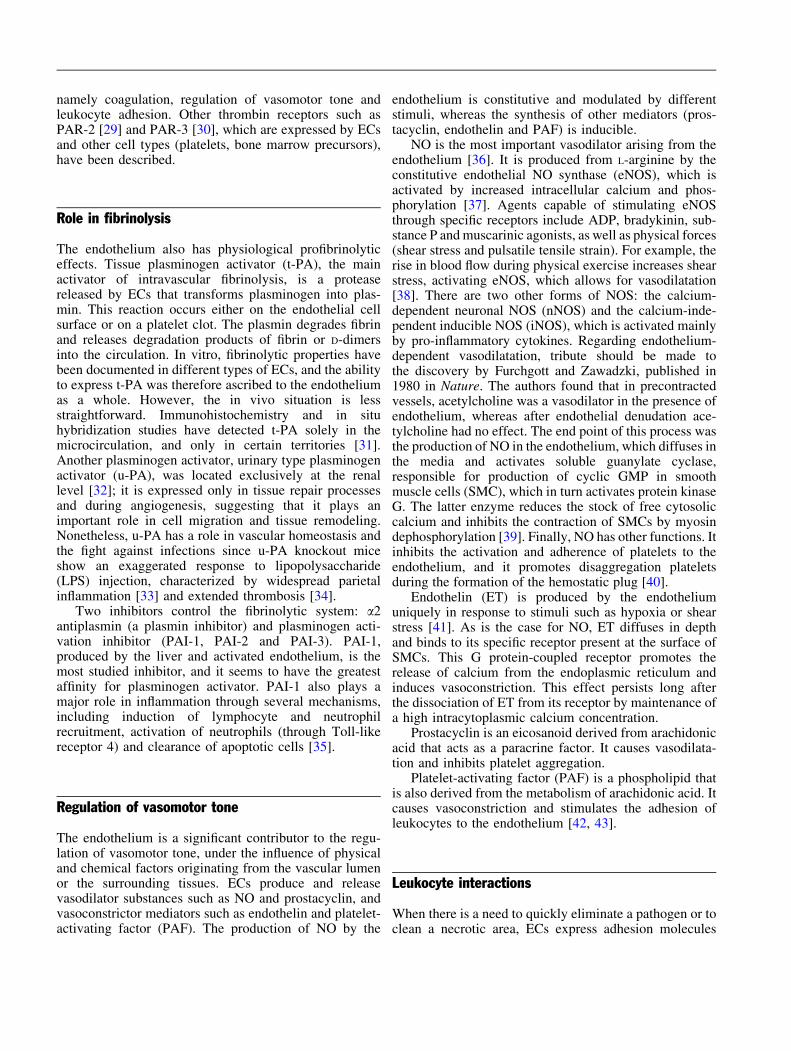
H. Ait-OufellaE. MauryS. LehouxB. GuidetG. Offenstadt
The endothelium: physiological functionsand role in microcirculatory failureduring severe sepsis
Received: 19 November 2009Accepted: 8 March 2010
� Copyright jointly held by Springer andESICM 2010
H. Ait-OufellaInserm U970, PAris ResearchCardiovascular Center (PARCC),Paris, France
H. Ait-Oufella � E. Maury � B. Guidet �G. OffenstadtService de Reanimation Medicale,Hopital Saint-Antoine,AP-HP, Paris 75012, France
E. Maury � B. Guidet � G. OffenstadtUniversite Pierre et Marie Curie-Paris 6,UMR S707, 75012 Paris, France
E. Maury � B. Guidet � G. OffenstadtInserm U707, 75012 Paris, France
S. LehouxLady Davis Institute for Medical Research,McGill University, Montreal, Canada
H. Ait-Oufella ())Service de Reanimation Medicale,Hopital Saint-Antoine, 184 rue du FaubourgSaint-Antoine, 75571 Paris Cedex 12,Francee-mail: [email protected].: ?33-1-49282315
Abstract The endothelium is ahighly dynamic cell layer that isinvolved in a multitude of physio-logical functions, including thecontrol of vascular tone, the move-ment of cells and nutrients, the
maintenance of blood fluidity and thegrowth of new vessels. During severesepsis, the endothelium becomesproadhesive, procoagulant, antifibri-nolytic and is characterized byalterations of vasomotor regulation.Most of these functions have beendiscovered using in vitro and animalmodels, but in vivo exploration ofendothelium in patients remainsdifficult. New tools to analyze endo-thelial dysfunction at bedside have tobe developed.
Keywords Endothelium � Sepsis �Coagulation � Cytokines
Introduction
The endothelium consists of a single cell layer lining allvessels, from their origin at the exit of the heart to theirfinest ramifications, the capillaries. In spite of being bothwidespread and functionally complex, the importance ofthis structure was long disregarded. Originally describedby Harvey in the seventeenth century and further char-acterized by Malpighi, the cardiovascular system wasconsidered as a simple pump and tubing device designedto convey oxygen and nutriments to peripheral organs. Itdoes not help that the clinical setting had no telltale signsby which to identify endothelial health or disease.Nowhere in the classical triad of ‘‘palpation-auscultation-percussion’’ would endothelial state appear, no jaundice
to give away hepatic distress or crackles pointing to leftventricular insufficiency. And when tools that could beused to explore endothelial functionality finally appeared,they were mainly confined to the research world andfailed to greatly impact clinical practice. In spite of allthese difficulties, the endothelium has grown in regardand appreciation, on its way to being recognized as anindividual organ. As of today, the endothelium figures asa main topic in more than 80,000 publications cited inPubMed.
In this review, we will describe the main functionsof the endothelium in an attempt to demonstrate itsimportance and complexity. We will describe both itsphysiological properties (see Table 1) and its anomaliesin pathological states. We will give special notice to
Intensive Care MedDOI 10.1007/s00134-010-1893-6 REVIEW

sepsis, a condition where endothelial distress is extreme,and a daily preoccupation in the intensive care units.
Histological description
The endothelium is composed of a single layer of cellsthat lines the interior surface of all blood vessels. It isestimated to comprise around 1013 cells, representing aweight of 1.5 kg and covering 4,000–7,000 m2, equiva-lent to six football fields [1, 2].
Endothelial cells (EC) lie at the interface of the cir-culating blood and the vessel wall. They are flattenedcells, having a thickness of about 0.5 lm, and are 100 lmlong by 10 lm wide. Their lozenge shapes are juxtaposedin a mosaic such that their long axis is oriented in thedirection of blood flow. They are attached to a basalmembrane rich in collagen and glycoproteins, forming acomplex interface between the circulation and the plate-let-activating, pro-coagulant vascular matrix.
EC appositions can be quite variable from one vas-cular bed to the next, ranging from juxtaposition tooverlapping, allowing for an intricate regulation of pro-tein permeability across the endothelium. At one or manypoints along the intercellular space, neighboring mem-branes form tight junctions or gap junctions. The formeris multifocal membrane fusions that are perfectly tight,whereas the latter consist of a joining of two close cellularmembranes around a central channel that allows theexchange of ions, various metabolites and regulatoryfactors.
At their surface, ECs are lined with a very fine andfragile layer called the glycocalyx. First identified in the1960s thanks to electronic microscopy [3], it consists ofglycoproteins, glycosaminoglycans and proteoglycans [4].There are five types of glycosaminoglycans includingheparan sulfate, a cofactor for antithrombin III thatamplifies its anti-thrombotic properties, and dermatan
sulfate, which interacts with heparin cofactor II [5].Globally, the glycocalyx provides an anticoagulant layerbecause of its negative electrical charge that repels cir-culating platelets and that allows it to interact with andcatalyze the interactions of vitamin K-dependent coagu-lation factors.
In forming a dense and intermeshed network, the gly-cocalyx provides a first-line barrier, regulating the cellularand macromolecular traffic at the forefront of the endothe-lium [6]. Numerous animal studies have demonstrated thatdestruction of the glycocalyx, using enzymatic approaches,for example, leads to increased capillary permeability [7]. Invivo, degradation of the glycocalyx with heparinase favorstight contacts between circulating leukocytes and theendothelium, through denudation of the protective barrierthat normally prevents this [8].
Electronic microscopy recently revealed two charac-teristic elements of endothelial cells: pinocytic vesiclesand Weibel-Palade bodies. Pinocytic vesicles consist oftranscellular transport vesicles that shuttle from the apicalto the basolateral aspect of endothelial cells. Weibel-Palade bodies are protein-stocking vesicles that appear asplentiful dense bodies, typically containing von Wille-brand factor and P selectin.
Endothelial plasticity
At the interface between a liquid phase, blood plasma,and a solid structure, basal membrane, endothelial cellsare highly influenced by their environment. They canrapidly modulate their structure and function in responseto chemical or physical stimuli, such as a change in bloodflow. For example, ECs cultured in vitro have a charac-teristic cobblestone morphology. However, if cells areplaced in a perfusion chamber and exposed to a shearstress that mimics the frictional force of blood flow, theyquickly align in the direction of flow, owing to a rear-rangement in cytoskeletal proteins [9]. The sameelongated morphology is observed in vivo in ECs liningarteries, where shear stress is 30–50 times greater than inveins. In fact, this effect is manifest in coronary bypassgrafts in humans. The ECs from saphenous veins, onceplaced in the arterial context, quickly elongate in thedirection of the long axis, parallel to blood flow [10].Numerous receptors responsible for this response to shearstress, termed mechanosensors, have been identified:tyrosine kinase receptors, integrins, ion channels, G pro-tein-coupled receptors and NADPH oxidases [11].
At the abluminal aspect, the endothelium is affixed toand interacts with the extracellular matrix constituents ofthe basal membrane, such that the nature of this matrixinfluences the form and the function of ECs [12]. Jalaliet al. [13] showed that ECs grown on fibronectin- orvitronectin-coated surfaces respond differently to shear
Table 1 Examples of endothelial marker distribution according toEC location
Markers Vascular territories
Von Willebrand factor Veins [ arteries, absent in hepaticsinusoids
t-PA Abundant in the brain and lung circulation,but not in the pulmonary circulation
TFPI Microvascular endotheliumProtein C receptor Vessels of large diameterThrombomodulin Absent in the brainEndothelial NOS Arteries [ veinsVCAM-1 Abundant in the heart, present in the brain
and the small intestineP-selectin Abundant in the lung, low expression
in the muscle and the brainCD36 Low expression in the brain

stress; surface expression of integrins differed both interms of quantity and distribution from one matrix to thenext. ECs adapt very quickly to changes in their envi-ronment. In humans, it was shown by RT-PCR ofpostoperative amygdala that the genetic programming ofECs removed from their native environment changeswithin a few hours [14]. This should be heeded as acautionary tale when considering the interpretation ofstudies using endothelial cells cultured ex vivo or in vitro.
Physiological functions of the endothelium
Primary hemostasis comprises a series of events thatoccur as a result of a vascular lesion and terminate withthe formation of a stable platelet-rich blood clot, pavingthe way for tissue repair mechanisms that will be fol-lowed by fibrinolysis [15]. Endothelial cells play a role ineach step of this process since they participate in plateletactivation and produce factors involved in the coagulationcascade and in the fibrinolytic system.
When a vascular lesion occurs, an immediate vascularconstriction occurs, which reduces blood flow locally andconcentrates the repair mechanisms. This is quickly fol-lowed by platelet adhesion to the vascular wall [16]. Theendothelium, focally destroyed, leaves exposed in its place tothe underlying matrix rich in collagen and von Willebrandfactor, to which the platelets attach through membrane gly-coproteins. The glycoproteins GPIa and VI quickly andreversibly interact with collagen, whereas GPIb-IX-V bindsvon Willebrand factor. In the veins, where shear stress ismuch lower, platelets can adhere more readily to damagedendothelium via a2b3 integrins [15]. Adherent plateletsbecome activated, shifting from an oblong morphology to anirregular shape with multiple pseudopods, expressingadhesion receptors at their surface (P-selectin and integrins)and shedding their granulocyte contents [adenosine diphos-phate (ADP), serotonin] into the extracellular space. Thesecombined effects result in the attraction and activation ofcirculating platelets, conglomerating into a dense massreinforced by a fibrin network. But ECs also oppose primaryhemostasis by liberating vasodilator substances [prostacy-clin, nitric oxide (NO)] and ADPases that catabolize ADP,one of the most potent platelet activators.
Any deficit in molecules involved in platelet adhesion,such as Bernard-Soulier’s disease (GPIb-IX-V deficit/dysfunction) or Glanzman’s thrombasthenia (a2b3 deficit/dysfunction), is characterized by facilitated bleedingepisodes, either spontaneous or elicited by minor traumas.
Anticoagulant properties
The anticoagulant activity of the endothelium is geared atrestricting the generation of thrombin. Several factors are
implicated in this process. Heparan sulfate and dermatansulfate, two glycocalyx glycosaminoglycans, potentiatethe activity of two anticoagulant enzymes, antithrombinIII (by a factor of 100) [17, 18] and heparin cofactor II,respectively [19]. The endothelium produces tissue factorpathway inhibitor (TFPI), which binds to activated factorX and then inhibits the tissue factor-activated factor VIIcomplex [20]. ECs produce thrombomodulin (TM) that iseither attached to the membrane or released into the cir-culation. Its plasma levels are increased in a number ofpathologies where the endothelium is injured (see sepsis).TM binds to the surface of protein C and increases itsanticoagulant activity by associating with its specificcofactor, protein S [21]. Finally, the endothelium accel-erates activation of protein C by expressing on its surfaceanother receptor, endothelial protein C receptor (EPCR)[22]. Protein C, when activated by all its cofactors,inhibits factors V and VIII. Finally, TM has its ownintrinsic anticoagulant activity since several works havedemonstrated its ability to bind and directly inhibit acti-vated factor X [23].
Procoagulant properties
The major step in the acquisition by the endothelium of aprocoagulant phenotype involves the expression of factortissue (TF). In vitro, TF is induced by several mediatorssuch as thrombin, endotoxin, cytokines, shear stress,hypoxia or oxidized lipids [24, 25]. Its procoagulantactivity is accentuated in the presence of anionic phos-pholipids that are exposed by apoptotic cells [26, 27]. In thepresence of agonists, the levels of TF protein and messengerRNA decline rapidly, presumably to avoid too rapid fibrinextension. Despite these in vitro findings, it remains diffi-cult to show an actual endothelial expression of TF.
Once expressed, TF binds and activates factor VII. TheTF-VIIa complex in turn mainly activates factors IX andX. These factors are anchored by their gammacarboxicresidues to the membrane phospholipids of platelets andendothelial cells. This latter precision serves to remind usthat reactions of the coagulation cascade do not take placein the liquid plasma phase, but rather in the solid phase,usually on cellular membranes or developing clots. Givenits position and surface area, the endothelium is the mainsurface on which the coagulation reaction takes place.
The contact between coagulation factors and the vas-cular wall occurs either via non-specific physicalinteractions or through specific receptors synthesized byECs. Thrombin binds a surface receptor called protease-activating factor (PAR-1) and influences the formation offibrin and amplifies the coagulation cascade. The PAR-1receptor is itself activated and induces the expression ofdifferent genes (TF, NO, endothelin) [28], illustrating thecomplex entanglement of the multiple functions of EC,
namely coagulation, regulation of vasomotor tone andleukocyte adhesion. Other thrombin receptors such asPAR-2 [29] and PAR-3 [30], which are expressed by ECsand other cell types (platelets, bone marrow precursors),have been described.
Role in fibrinolysis
The endothelium also has physiological profibrinolyticeffects. Tissue plasminogen activator (t-PA), the mainactivator of intravascular fibrinolysis, is a proteasereleased by ECs that transforms plasminogen into plas-min. This reaction occurs either on the endothelial cellsurface or on a platelet clot. The plasmin degrades fibrinand releases degradation products of fibrin or D-dimersinto the circulation. In vitro, fibrinolytic properties havebeen documented in different types of ECs, and the abilityto express t-PA was therefore ascribed to the endotheliumas a whole. However, the in vivo situation is lessstraightforward. Immunohistochemistry and in situhybridization studies have detected t-PA solely in themicrocirculation, and only in certain territories [31].Another plasminogen activator, urinary type plasminogenactivator (u-PA), was located exclusively at the renallevel [32]; it is expressed only in tissue repair processesand during angiogenesis, suggesting that it plays animportant role in cell migration and tissue remodeling.Nonetheless, u-PA has a role in vascular homeostasis andthe fight against infections since u-PA knockout miceshow an exaggerated response to lipopolysaccharide(LPS) injection, characterized by widespread parietalinflammation [33] and extended thrombosis [34].
Two inhibitors control the fibrinolytic system: a2antiplasmin (a plasmin inhibitor) and plasminogen acti-vation inhibitor (PAI-1, PAI-2 and PAI-3). PAI-1,produced by the liver and activated endothelium, is themost studied inhibitor, and it seems to have the greatestaffinity for plasminogen activator. PAI-1 also plays amajor role in inflammation through several mechanisms,including induction of lymphocyte and neutrophilrecruitment, activation of neutrophils (through Toll-likereceptor 4) and clearance of apoptotic cells [35].
Regulation of vasomotor tone
The endothelium is a significant contributor to the regu-lation of vasomotor tone, under the influence of physicaland chemical factors originating from the vascular lumenor the surrounding tissues. ECs produce and releasevasodilator substances such as NO and prostacyclin, andvasoconstrictor mediators such as endothelin and platelet-activating factor (PAF). The production of NO by the
endothelium is constitutive and modulated by differentstimuli, whereas the synthesis of other mediators (pros-tacyclin, endothelin and PAF) is inducible.
NO is the most important vasodilator arising from theendothelium [36]. It is produced from L-arginine by theconstitutive endothelial NO synthase (eNOS), which isactivated by increased intracellular calcium and phos-phorylation [37]. Agents capable of stimulating eNOSthrough specific receptors include ADP, bradykinin, sub-stance P and muscarinic agonists, as well as physical forces(shear stress and pulsatile tensile strain). For example, therise in blood flow during physical exercise increases shearstress, activating eNOS, which allows for vasodilatation[38]. There are two other forms of NOS: the calcium-dependent neuronal NOS (nNOS) and the calcium-inde-pendent inducible NOS (iNOS), which is activated mainlyby pro-inflammatory cytokines. Regarding endothelium-dependent vasodilatation, tribute should be made tothe discovery by Furchgott and Zawadzki, published in1980 in Nature. The authors found that in precontractedvessels, acetylcholine was a vasodilator in the presence ofendothelium, whereas after endothelial denudation ace-tylcholine had no effect. The end point of this process wasthe production of NO in the endothelium, which diffuses inthe media and activates soluble guanylate cyclase,responsible for production of cyclic GMP in smoothmuscle cells (SMC), which in turn activates protein kinaseG. The latter enzyme reduces the stock of free cytosoliccalcium and inhibits the contraction of SMCs by myosindephosphorylation [39]. Finally, NO has other functions. Itinhibits the activation and adherence of platelets to theendothelium, and it promotes disaggregation plateletsduring the formation of the hemostatic plug [40].
Endothelin (ET) is produced by the endotheliumuniquely in response to stimuli such as hypoxia or shearstress [41]. As is the case for NO, ET diffuses in depthand binds to its specific receptor present at the surface ofSMCs. This G protein-coupled receptor promotes therelease of calcium from the endoplasmic reticulum andinduces vasoconstriction. This effect persists long afterthe dissociation of ET from its receptor by maintenance ofa high intracytoplasmic calcium concentration.
Prostacyclin is an eicosanoid derived from arachidonicacid that acts as a paracrine factor. It causes vasodilata-tion and inhibits platelet aggregation.
Platelet-activating factor (PAF) is a phospholipid thatis also derived from the metabolism of arachidonic acid. Itcauses vasoconstriction and stimulates the adhesion ofleukocytes to the endothelium [42, 43].
Leukocyte interactions
When there is a need to quickly eliminate a pathogen or toclean a necrotic area, ECs express adhesion molecules
that regulate leukocyte trafficking between the circulat-ing blood and the surrounding tissue. The interactionbetween leukocytes and ECs follows a conventional setof steps that include short contacts, more prolongedcontacts, then leukocyte rolling and strong adhesionbefore transendothelial migration. Different adhesionmolecules intervene at each stage of this process. Theselectins (E-and L-selectin) mediate the first phase, asthey resist the forces of shear stress and bind (andrelease) very quickly [44]. The integrins a4b7 and a4b1are involved in the adhesion of monocytes and polynu-clear neutrophils. The subsequent rolling and firmadhesion involve the molecules of the superfamily ofimmunoglobulins [intercellular adhesion molecule(ICAM-1, ICAM-2) and vascular adhesion molecule(VCAM)] [45, 46], joined by platelet endothelial celladhesion molecule (PECAM) at the time of diapedesis[47]. This latter process is a complex phenomenoninvolving the separation of cadherins at tight junctions[48].
In addition to its participation in the innate immuneresponse, the endothelium participates in the adaptiveresponse involving T lymphocytes [49]. ECs in cultureexpress major histocompatibility complex class I (MHCI) and can present antigen to CD8? cytotoxic T lym-phocytes. Furthermore, after stimulation by interferongamma, they express MHC class II and can interact withCD4? lymphocytes [50]. ECs can also express LFA-3(CD58) or B7.2 (CD86) costimulation molecules at theirsurface, molecules common to other antigen-presentingcells (dendritic cells, macrophages, B lymphocytes) [51].Moreover, it is now accepted that the ECs are involved inthe pathophysiology of graft rejection [52].
Endothelial heterogeneity
All ECs share common properties, but also distinct fea-tures, that are expressed both during physiologicalprocesses and in adverse conditions. This complexity,called endothelial heterogeneity, is so important that weshould use the plural form, endothelia (see Table 2). Thisconcept was first brought up by histologists, who distin-guished, for example, the continuous endothelium of thebrain from the fenestrated endothelium of endocrineglands. The clinicians then showed that ECs were dif-ferentially affected by various pathologies; diabetesdisturbs the renal and retinal microcirculation especially[53], whereas veno-occlusive liver disease targets vesselsof the hepatic sinusoids [54], and thrombotic microangi-opathies affect the entire microcirculation with theexception of liver and lungs [55]. That structural differ-ences distinguish endothelial cells according to organfunction was then confirmed experimentally. Electronmicroscopy highlighted differences in the location of the
nucleus within the cell or the composition of cytoplasmdepending on the site at which ECs are found. Accord-ingly, in the frog the intracellular volume occupied byWeibel-Palade bodies gradually diminishes from theaortic root to the microcirculation (8% of the cytoplasm inthe thoracic aorta versus 0.3% in the capillaries) [56].
This heterogeneity exists for all endothelial functions,and to ignore it exposes us to errors of interpretation. Forexample, a decrease in the endothelial expression of TMwas shown in biopsies of purpuric lesions in children withmeningococcal disease [57]. In the brain, ECs do notexpress TM [58]. Does this mean that the cerebralendothelium was in a state of continuous activation? Ofcourse not. The most likely explanation is that TM is notinvolved in the hemostatic balance of the brain.
EC heterogeneity also concerns the intensity of theendothelial response to stimuli and endothelial responsecannot be likened to a simple on/off switch but rather to adimmer switch where, for a given gene activation cas-cade, the expression of a protein can vary between twoextremes [59]. What is most challenging is to assess thisexpression level and deduce the endothelial status: inac-tivated, activated or abnormally active (dysfunctional).Such distinctions are subtle, and unfortunately limited bythe current lack of tools that allow for precise assessmentof endothelial function.
Endothelial dysfunction during sepsis
The endothelium is actively involved in defending thebody against pathogens by recruiting the leukocytes to
Table 2 Endothelial cell implication in various physiologicalprocesses through secretion or expression of different molecules
HemostasisPrimary hemostasisActivation Von Willebrand factorInhibition Nitric oxide
Coagulation cascadeActivation Tissue factorInhibition Tissue factor pathway inhibitor, endothelial
protein C receptor, thrombomodulin,heparan/dermatan sulfate
FibrinolysisActivation Tissue plasminogen activator, urinary
plasminogen activatorInhibition Plasminogen activator inhibitor
Vasomotor tone regulationDilation Nitric oxide, prostacyclinConstriction Endothelin, thomboxane A2,
platelet-activating factorInflammationAdhesion E-selectin, VCAM, ICAMCo-stimulation LFA-3, CD80/86Chemokines MIP-1 (and -2), MCP-1Cytokines IL-1, IL-6, IL-8, TNFa
infected sites, releasing inflammatory mediators andpromoting local coagulation to prevent the spread ofblood-borne infection. However, this normally focusedadaptative response can become generalized and ampli-fied during sepsis, causing tissue damage. This is referredto as endothelial dysfunction.
Endothelial denudation
Sepsis is accompanied by morphological changes of theendothelium. The injection of LPS in animals induces adetachment of ECs from the basal membrane and causessub-endothelial edema [60, 61]. Cell lesions includenuclear vacuolation, protrusion and cytoplasmic frag-mentation. The time of onset varies from a few minutes,after LPS injection [62], to several hours, in the model ofcecal ligation/perforation [63]. This phenomenon has alsobeen observed in humans [64]. Using a double-tagged cell(Von Willebrand factor receptor and EGF), Mutunga et al.showed that the number of circulating ECs was higher inseptic patients than in healthy subjects, and even higher incases of septic shock. Inflammatory mediators released byleucocytes (TNF, IL1, interferon, oxygen free radicals)and hypoxia increase EC apoptosis [65–67]. ApoptoticECs in turn express adhesion molecules (ICAM-1,VCAM) and release oxygen free radicals, and in doing soamplify the recruitment of white blood cells. ApoptoticECs also externalize negatively charged phosphatidyl-serine on the cell membrane and thus expose thecirculating blood to a procoagulant surface [26]. Mem-brane charge asymmetry coupled with the activity ofcertain enzymes (floppase, scramblase, etc.) allows for thebudding and release of membrane microparticles in thecirculation [68]. These microparticles, a combination ofphospholipid membrane fragments and surface proteins(TF), are involved in spreading endothelial dysfunction[69] and participate in the disseminated intravascularcoagulation [70].
Endothelial barrier impairment favors the passage ofcells, inflammatory mediators and plasma into the inter-stitial compartment. The rapid increase in vascularpermeability to albumin, which occurs within 6 h of theinsult, affects the pulmonary and systemic circulationheterogeneously [71]. In terms of skin and muscles, theinjection of endotoxin induces an increase in vascularpermeability irrespective of oncotic and hydrostatic pres-sure, indicating abnormalities of the cell membrane [72].
Pro-adhesive properties
Increased expression of endothelial adhesion moleculeseither at the membrane level or in the plasma typify the
different models of sepsis [73]. In vitro incubation of ECswith bacterial LPS induces the rapid expression of ICAM-1 and E-selectin mRNA. The same effect is obtained byreplacing LPS with the plasma of healthy volunteerstreated with very low doses of LPS. In this latter exper-iment, the addition of antibodies blocking TNFa and/orIL-1b at the same time as human plasma inhibits thetranscription of mRNA, illustrating the fact that cytokinesreleased by endotoxemia induce in turn the expression ofE-selectin and ICAM-1 [74]. Moreover, there is a closerelationship between plasma levels of adhesion moleculesand consequences of sepsis [75]. In human sepsis, a study hasshown that the higher the plasma levels of ICAM-1, thegreater the number of organs damaged and the mortality.In animals, the genetic or pharmacological blockade ofICAM-1 protects against endotoxin shock [76].
As regards E-selectin, several human studies have alsoshown an increase in its soluble form in healthy volun-teers receiving an injection of LPS [77] or in patientshospitalized for sepsis [78]. The level of E-selectin in theplasma is positively correlated with the severity of sepsisin patients, evaluated by the SAPSII score or the multipleorgan failure (MOF) score [79]. Increased plasma adhe-sion molecules reflect an increase in the endothelialmembrane expression of the proteins and can be directlyrelated to activation and/or dysfunction of the endothe-lium. It promotes rolling and firm adherence ofneutrophils, and ultimately results in their accumulationin organs. This accumulation is in part beneficial, elimi-nating infectious agents, but it may also exacerbate tissuedamage through the production of inflammatory media-tors such as cytokines, proteases and oxygen free radicals[80]. A recently published study illustrated this paradigm.Ye et al. [81] selectively inhibited NF-jB activity in theendothelium of adult mice. After LPS injection, theseanimals were characterized by a decrease in endothelialadhesion molecule expression (ICAM-1, E-selectin), adecrease of neutrophil infiltration in several organs (lung,liver, kidney) and a decrease in tissue damage. In fine,selective endothelial NF-jB inhibition improved survivalin endotoxinemic mice. Platelets also contribute to therecruitment of neutrophils, illustrating the complexentanglement between inflammation and coagulation thatregulates the pathophysiology of septic shock. Using aperfusion chamber model lined with ECs, where leuko-cytes circulate in a closed system, Blanks et al. [82]showed that the simultaneous infusion of plateletssignificantly increases EC-neutrophil interaction. Theyshowed that the binding of platelet P-selectin to itsreceptor P-selectin glycoprotein ligand 1 (PSGL-1) onpolymorphonuclear leukocytes induced the expression ofVLA-4, another molecule responsible for adhesion to theendothelium. The relevance of this interaction in vivo hasbeen documented in the cecal ligation and puncture modelof sepsis in mice, where platelet depletion reduced leukocyteinfiltration in the lung and lesion edema [83].
Pro-coagulant properties
Different studies undertaken in animals and humans haveshown that severe sepsis is characterized by a pro-coag-ulant state [84]. The injection of LPS in healthyvolunteers induces the rapid expression of TF by circu-lating monocytes [85]. Similar results were observed inpatients with meningococcemia, with additive prognosticvalue since the cellular expression of TF was higheramong patients who died [86]. The production of endo-thelial TF is more difficult to document. In infectedanimals, TF was located at the endothelium of tissuesections [87]. However, such direct evidence is scarce inhumans. By analogy, one can cite the example of patientswith sickle cell disease, in whom Solovey et al. [88]identified circulating endothelial cells expressing TF.
The TF expressed during sepsis activates coagulationand leads to the formation of thrombin (see Fig. 1). Inparallel, counter-regulatory systems (involving the endo-thelium) such as protein C and TFPI are defective,maintaining intravascular coagulation. The decrease in
activated plasma protein C (aPC) in sepsis is partly due todecreased hepatic synthesis of protein C and increasedsystemic consumption of the protein. Adding to this arecomplex enzymatic alterations that affect thrombomodu-lin (TM), whose primary function is to amplify theactivation of protein C. In vitro, TNF-a and IL-1 decreaseendothelial expression of TM. Moreover, the granulocytesrecruited at the inflammatory site release elastases thatcleave and inactivate endothelial TM [89, 90]. Theseexperimental results have been corroborated by a study inchildren with purpura fulminans. From skin biopsies,Faust et al. [57] showed a decreased expression ofendothelial TM and endothelial cell protein receptor. Inthe blood, the levels of protein C, protein S and anti-thrombin III were also reduced. Moreover, in anotherstudy on adults with sepsis, a low rate of aPC was iden-tified as a factor of poor prognosis [91]. The accumulatedevidence on the beneficial role of aPC in sepsis has led toserious therapeutic studies. After encouraging results inanimals [92] a multicenter international study was con-ducted. The PROWESS study, published in 2001, showed
TFVIIa/Xa
TFVIIa/Xa
TFVIIa/Xa
t-PA
XVII
TF
plasminogen
plasmin
APCAPC
APC
PAI-1
TM
EPCR
Antithrombinheparan
TF
V
Va
II
fibrin
fibrinogen
plasminogent-PA
plasmin
PC
IIa
APC
PAI-1
TM
EPCR
PC
Antithrombinheparan
TNFαIL-1LPS
TF
TFPI
monocyte
agression
Glycocalyx
microparticles
Physiological condition Severe sepsis
2
3
5
4
78
1neutrophil
PC
PC
PC
TFVIIa/Xa
SMC EC
6
V
Va
II
fibrin
fibrinogen
IIa
TM
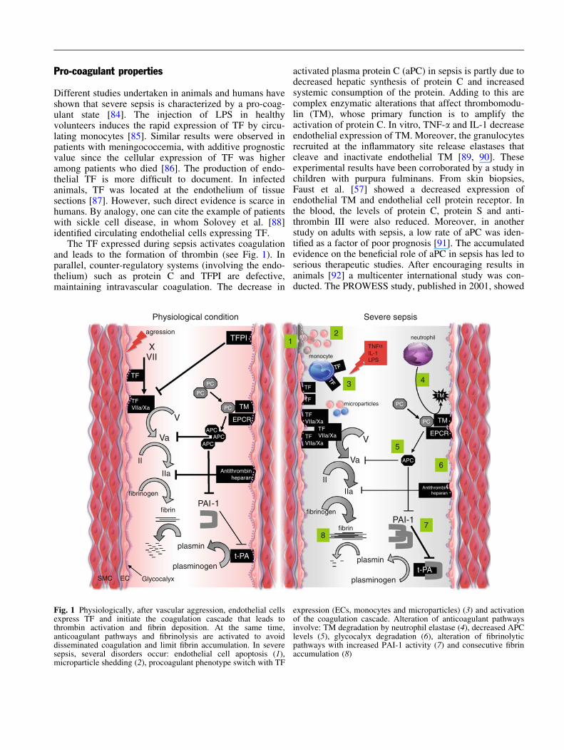
Fig. 1 Physiologically, after vascular aggression, endothelial cellsexpress TF and initiate the coagulation cascade that leads tothrombin activation and fibrin deposition. At the same time,anticoagulant pathways and fibrinolysis are activated to avoiddisseminated coagulation and limit fibrin accumulation. In severesepsis, several disorders occur: endothelial cell apoptosis (1),microparticle shedding (2), procoagulant phenotype switch with TF
expression (ECs, monocytes and microparticles) (3) and activationof the coagulation cascade. Alteration of anticoagulant pathwaysinvolve: TM degradation by neutrophil elastase (4), decreased APClevels (5), glycocalyx degradation (6), alteration of fibrinolyticpathways with increased PAI-1 activity (7) and consecutive fibrinaccumulation (8)
in a cohort of 1,690 patients that administration of aPC inseptic patients with at least two visceral failures reducedthe relative mortality by 20% at 28 days [93]. Unfortu-nately, secondary studies have been disappointing. TheADDRESS study, which evaluated the role of aPC inseptic patients with only one organ failure, provided noevidence of benefit in terms of survival and was stoppedfor ‘‘futility’’ [94]. The RESOLVE study in children withmeningococcemia was also negative on composite criteriameasuring the resolution of organ failure [95]. A number oflimitations have been advanced to explain these negativeresults, and the controversy regarding the place of aPC in thetreatment of severe sepsis remains. Patient selection pro-vides a major point of argument because secondary analysisof the PROWESS trial showed that aPC improved survival inthe more severe patients [96, 97]. It is important to note thataPC has other beneficial functions apart from its role incoagulation. In experimental models, aPC mediates anti-apoptotic effects, reduces endothelial barrier permeabilityand decreases pro-inflammatory cytokine production [98].For all these reasons, the relationship between aPC andsevere sepsis remains relevant.
TFPI is another powerful anticoagulant that binds tofactor VIIa, TF and factor Xa. Several lines of evidencehave highlighted its importance in the pathophysiology oforgan failure during septic shock and especially in theintravascular coagulation that accompanies it. In rabbits,the depletion of TFPI increases intravascular coagulation[99]. Conversely, infusion of TFPI in a model of endotoxinshock in baboon decreases intravascular coagulation,reduces organ dysfunction and most importantly improvessurvival. TFPI also modulates the inflammatory responsesince it can bind directly to endotoxin and prevent itsinteraction with leukocyte CD14 [100]. Unfortunately,TFPI proved to be underwhelming in the clinical setting.The OPTIMIST study published in 2003, featuring criti-cally ill patients with mild coagulopathy (internationalnormalized ration, INR [ 1.2), did not reveal any reduc-tion in mortality in the group receiving TFPI [101].Nevertheless, TFPI had a net beneficial effect on intra-vascular coagulation, accompanied by an improvement inthe markers of coagulation.
Anti-fibrinolytic properties
Regarding the fibrinolytic system, the first in vitro studiesshowed stimulation of ECs in culture with IL-1 and TNFainduced by the expression of t-PA and PAI-1 [102]. Thisorientation towards an anti-fibrinolytic phenotype hasbeen difficult to confirm in humans. Some studies haveshown increased levels of PAI-1 in the blood of septicpatients (both adults and children) [103, 104], and PAI-1levels are positively related to unfavorable outcomes[105]. Some gene polymorphisms of PAI-1 have been
demonstrated to affect the risk of death during menin-gococcal infection [106]. The final result is heterogenousfibrin deposition, which exacerbates local ischemia [107].For example, injection of LPS in mice induced depositionfibrin in the kidney, liver and heart, but not in the lung. Inthe baboon, the injection of lethal doses of Escherichiacoli induced fibrin deposition in the spleen, hepaticsinusoids and renal glomeruli, but had little effect on thebrain, heart and aorta.
Impaired regulation of vasomotor tone
In severe infectious states, emergency care is sought pri-marily to restore hemodynamic stability due to an imbalancein vasomotor tone and interstitial plasma leakage. Endo-thelial dysfunction and especially disturbances in NOproduction are the main causes of circulatory insufficiency.
Disrupted production of NO in the course of sepsis iscomplex and evolves over time. The first stage, callednitrosopenia, is characterized by decreased production ofNO by eNOS [108]. The mechanisms underlying thiseffect are diverse, ranging from changes in surfacereceptors to altered signal transduction to quantitative orqualitative alterations in eNOS. In vitro, stimulation ofECs with TNFa or LPS induced a downregulation ofeNOS mRNA expression [109, 110]. In rats, the inductionof septic shock resulted in the loss of endothelial eNOS[111], whereas in rabbits, the injection of a non-lethaldose of LPS altered endothelium-dependent relaxationbetween 5 and 20 days [61]. In healthy volunteers, a briefexposure to endotoxin decreased endothelium-dependentrelaxation for several days [112, 113]. During early nitro-sopenia, the endothelial homeostatic balance thereforeleans towards vasoconstriction, which affects the proxi-mal arterioles of the first and second order of certainorgans such as the digestive tract.
The second, later stage is characterized by an increasein the production of NO by iNOS. This inducible enzymeproduces nanomolar concentrations of NO (1,000-fold ofwhat is produced by eNOS), which causes diffuse mi-crocirculatory vasodilatation and therefore a fall in bloodpressure. Other effects of NO overproduction have beenassociated with the pathophysiology of septic shock,including the production of peroxynitrite, a potent oxi-dative agent that results from the interaction of NO withthe superoxide anion.
The overproduction of NO in late sepsis inspired anumber of studies aimed at assessing the therapeuticpotential of NOS inhibition. However, results in animalshave been contradictory, in part because different modelsof sepsis were used, coupled with diverse treatments andinhibitors [114–116]. In humans, the first attempts atblocking NOS appeared encouraging, with improvedhemodynamic parameters (but no effect on mortality)
[117, 118]. Finally, a large international multi-centerstudy was carried out in 2004. Seven hundred ninety-seven patients suffering from severe sepsis were treatedwith either a placebo or a non-selective blocker of NOS,L-NAME. The phase III trial was stopped prematurelybecause despite an improvement in circulatory parame-ters, administration of L-NAME was accompanied byexcess mortality [119]. After the failure of this approach,several studies investigated the beneficial properties ofNO. This led to the notion that it is not NO that isresponsible for the circulatory failure in sepsis, but ratherby its overproduction by iNOS. Therefore new researchavenues today are targeting the activation of iNOS [109].
Finally, it is important to remember that advancementsin the field of resuscitation are mainly based on experi-mental animal studies. Disappointing results obtained in theclinical trials could be partially due to significant physio-pathological differences between species. At present, thetools available for the assessment of endothelial function inhumans are limited and difficult to use in the bedside setting.Orthogonal polarized spectral (OPS) imaging in the sub-lingual area is an interesting technique that providesinformation about capillary density, the proportion of per-fused capillaries [120] or glycocalyx size [121]. Topicalapplication of pharmacological agents such as acetylcholineimproves specific analysis of endothelial function. How-ever, analysis of OPS data requires time and computerassistance and cannot yet help clinicians at the bedside.
Vascular oxidative stress
During severe sepsis, reactive oxygen species (ROS) suchas superoxide (O2
-) are produced in large amounts [122].Neutrophils are the main source of ROS, but endothelialcells also synthesize ROS in response to oxidative agentsor cytokines. For example, in vitro LPS/IFNc-stimulatedECs produce superoxide within 2 h through the NADPHoxidase pathway. ROS accumulate in ECs and are mod-ified; dismutation of superoxide forms hydrogen peroxide(H2O2) and nitration of superoxide forms peroxynitrite(ONOO-). Both products are toxic for proteins and DNA,and could participate in endothelial cell damage [123]. ROS
decrease NO availability through peroxynitrite formationand eNOS inhibition, and ultimatly affect vascular tone,platelet adhesion [124] and permeability [125]. Thesemodifications lead to vascular occlusion and exacerbateorgan hypoperfusion. Finally, to illustrate entanglement ofcomplex phenomena in severe sepsis, experimental studieshave shown that ROS could activate NF-jB, a major tran-scription factor for genes involved in inflammation [126],and could induce TF expression [127].
Beneficial effects of antioxidants in sepsis have beenreported. In mice, vitamin C injections prevent LPS-induced edema and hypotension [128]. In a randomized,prospective, double-blind, placebo-controlled trial with226 critically ill patients, 28-day survival was increased inthe patients that received a combination of vitamin C andE. These data represent a new field of study and warrantfurther investigation [129].
Conclusion
After a long period in the dark, the endothelium is finallyarousing interest among clinicians. Despite both itsimposing dimensions and the importance of its functions,the endothelium remains difficult to grasp, as it exhibits alarge degree of plasticity and heterogeneity. ECs play arole in primary hemostasis, coagulation, fibrinolysis andregulation of vasomotor tone. More recently, their role inadaptive immunity has also been demonstrated. Duringsepsis, most endothelial functions are disrupted, leadingto a procoagulant, antifibrinolytic and proadhesive state.Production of NO decreases at first, but then it increasesunduly because of the induction of iNOS. To the diffi-culties linked with the variability of endothelial cells, onecan also add the practical complications confronted whenattempting to assess them; we are not, at the bedside,properly equipped to explore endothelial function.Advances in understanding and treatment of diseasesinvolving the endothelium will require the developmentof more refined tools.
Acknowledgments Stephanie Lehoux, PhD, is funded by theCanadian Institutes of Health Research (CIHR).
References
1. Wolinsky H, Katz D, Markle R, MillsJ, Brem S, Wassertheil-Smoller S(1980) Hydrolase activities in the rataorta. IV. Relation between clearancerates of circulating 125I-labeled low-density lipoproteins and levels oftissue hydrolase activity. Circ Res47:433–442
2. Augustin HG, Kozian DH, JohnsonRC (1994) Differentiation ofendothelial cells: analysis of theconstitutive and activated endothelialcell phenotypes. Bioessays 16:901–906
3. Luft JH (1966) Fine structures ofcapillary and endocapillary layer asrevealed by ruthenium red. Fed Proc25:1773–1783
4. Reitsma S, Slaaf DW, Vink H, vanZandvoort MA, oude Egbrink MG(2007) The endothelial glycocalyx:composition, functions, andvisualization. Pflugers Arch 454:345–359
5. Sugahara K, Mikami T, Uyama T,Mizuguchi S, Nomura K, Kitagawa H(2003) Recent advances in thestructural biology of chondroitinsulfate and dermatan sulfate. CurrOpin Struct Biol 13:612–620
6. Henry CB, Duling BR (1999)Permeation of the luminal capillaryglycocalyx is determined byhyaluronan. Am J Physiol 277:H508–H514
7. Jacob M, Bruegger D, Rehm M,Welsch U, Conzen P, Becker BF(2006) Contrasting effects of colloidand crystalloid resuscitation fluids oncardiac vascular permeability.Anesthesiology 104:1223–1231
8. Constantinescu AA, Vink H, Spaan JA(2003) Endothelial cell glycocalyxmodulates immobilization ofleukocytes at the endothelial surface.Arterioscler Thromb Vasc Biol23:1541–1547
9. Davies PF (1995) Flow-mediatedendothelial mechanotransduction.Physiol Rev 75:519–560
10. Allaire E, Clowes AW (1997)Endothelial cell injury incardiovascular surgery: the intimalhyperplastic response. Ann ThoracSurg 63:582–591
11. Lehoux S (2006) Redox signalling invascular responses to shear andstretch. Cardiovasc Res 71:269–279
12. Aird WC, Edelberg JM, Weiler-Guettler H, Simmons WW, Smith TW,Rosenberg RD (1997) Vascular bed-specific expression of an endothelialcell gene is programmed by the tissuemicroenvironment. J Cell Biol138:1117–1124
13. Jalali S, del Pozo MA, Chen K, MiaoH, Li Y, Schwartz MA, Shyy JY,Chien S (2001) Integrin-mediatedmechanotransduction requires itsdynamic interaction with specificextracellular matrix (ECM) ligands.Proc Natl Acad Sci USA 98:1042–1046
14. Lacorre DA, Baekkevold ES, GarridoI, Brandtzaeg P, Haraldsen G, AmalricF, Girard JP (2004) Plasticity ofendothelial cells: rapiddedifferentiation of freshly isolatedhigh endothelial venule endothelialcells outside the lymphoid tissuemicroenvironment. Blood 103:4164–4172
15. Jackson SP, Mistry N, Yuan Y (2000)Platelets and the injured vessel wall—‘‘rolling into action’’: focus onglycoprotein Ib/V/IX and the plateletcytoskeleton. Trends Cardiovasc Med10:192–197
16. Roth GJ (1992) Platelets and bloodvessels: the adhesion event. ImmunolToday 13:100–105
17. Rosenberg RD (1989) Biochemistry ofheparin antithrombin interactions, andthe physiologic role of this naturalanticoagulant mechanism. Am J Med87:2S–9S
18. Rosenberg RD, Rosenberg JS (1984)Natural anticoagulant mechanisms.J Clin Invest 74:1–6
19. Tollefsen DM, Pestka CA (1985)Heparin cofactor II activity in patientswith disseminated intravascularcoagulation and hepatic failure. Blood66:769–774
20. Broze GJ Jr (1995) Tissue factorpathway inhibitor. Thromb Haemost74:90–93
21. Esmon CT (2001) Protein Canticoagulant pathway and its role incontrolling microvascular thrombosisand inflammation. Crit Care Med29:S48–S51 discussion 51–42
22. Fukudome K, Kurosawa S, Stearns-Kurosawa DJ, He X, Rezaie AR,Esmon CT (1996) The endothelial cellprotein C receptor. Cell surfaceexpression and direct ligand bindingby the soluble receptor. J Biol Chem271:17491–17498
23. Thompson EA, Salem HH (1986)Inhibition by human thrombomodulinof factor Xa-mediated cleavage ofprothrombin. J Clin Invest 78:13–17
24. Moore KL, Andreoli SP, Esmon NL,Esmon CT, Bang NU (1987) Endotoxinenhances tissue factor and suppressesthrombomodulin expression of humanvascular endothelium in vitro. J ClinInvest 79:124–130
25. Bevilacqua MP, Pober JS, Majeau GR,Fiers W, Cotran RS, Gimbrone MA Jr(1986) Recombinant tumor necrosisfactor induces procoagulant activity incultured human vascular endothelium:characterization and comparison withthe actions of interleukin 1. Proc NatlAcad Sci USA 83:4533–4537
26. Bombeli T, Karsan A, Tait JF, HarlanJM (1997) Apoptotic vascularendothelial cells becomeprocoagulant. Blood 89:2429–2442
27. Combes V, Simon AC, Grau GE,Arnoux D, Camoin L, Sabatier F,Mutin M, Sanmarco M, Sampol J,Dignat-George F (1999) In vitrogeneration of endothelialmicroparticles and possibleprothrombotic activity in patients withlupus anticoagulant. J Clin Invest104:93–102
28. Woolkalis MJ, DeMelfi TM Jr,Blanchard N, Hoxie JA, Brass LF(1995) Regulation of thrombinreceptors on human umbilical veinendothelial cells. J Biol Chem270:9868–9875
29. Mirza H, Yatsula V, Bahou WF (1996)The proteinase activated receptor-2(PAR-2) mediates mitogenic responsesin human vascular endothelial cells.J Clin Invest 97:1705–1714
30. Ishihara H, Connolly AJ, Zeng D,Kahn ML, Zheng YW, Timmons C,Tram T, Coughlin SR (1997) Protease-activated receptor 3 is a secondthrombin receptor in humans. Nature386:502–506
31. Todd AS (1959) The histologicallocalisation of fibrinolysin activator.J Pathol Bacteriol 78:281–283
32. Barnathan ES, Kuo A, Kariko K,Rosenfeld L, Murray SC, Behrendt N,Ronne E, Weiner D, Henkin J, CinesDB (1990) Characterization of humanendothelial cell urokinase-typeplasminogen activator receptor proteinand messenger RNA. Blood 76:1795–1806
33. Carmeliet P, Schoonjans L, KieckensL, Ream B, Degen J, Bronson R, DeVos R, van den Oord JJ, Collen D,Mulligan RC (1994) Physiologicalconsequences of loss of plasminogenactivator gene function in mice.Nature 368:419–424
34. Yamamoto K, Loskutoff DJ (1996)Fibrin deposition in tissues fromendotoxin-treated mice correlates withdecreases in the expression ofurokinase-type but not tissue-typeplasminogen activator. J Clin Invest97:2440–2451
35. Wang L, Bastarache JA, Ware LB(2008) The coagulation cascade insepsis. Curr Pharm Des 14:1860–1869
36. Furchgott RF, Zawadzki JV (1980)The obligatory role of endothelial cellsin the relaxation of arterial smoothmuscle by acetylcholine. Nature288:373–376
37. Stamler JS, Singel DJ, Loscalzo J(1992) Biochemistry of nitric oxideand its redox-activated forms. Science258:1898–1902
38. Loscalzo J, Vita JA (1994) Ischemia,hyperemia, exercise, and nitric oxide.Complex physiology and complexmolecular adaptations. Circulation90:2556–2559
39. Moncada S, Palmer RM, Higgs EA(1991) Nitric oxide: physiology,pathophysiology, and pharmacology.Pharmacol Rev 43:109–142
40. Pacher P, Beckman JS, Liaudet L(2007) Nitric oxide and peroxynitritein health and disease. Physiol Rev87:315–424
41. Levin ER (1995) Endothelins. N EnglJ Med 333:356–363
42. Imaizumi TA, Stafforini DM, YamadaY, McIntyre TM, Prescott SM,Zimmerman GA (1995) Platelet-activating factor: a mediator forclinicians. J Intern Med 238:5–20
43. Lorant DE, Zimmerman GA, McIntyreTM, Prescott SM (1995) Platelet-activating factor mediatesprocoagulant activity on the surface ofendothelial cells by promotingleukocyte adhesion. Semin Cell Biol6:295–303
44. McEver RP, Moore KL, CummingsRD (1995) Leukocyte traffickingmediated by selectin–carbohydrateinteractions. J Biol Chem 270:11025–11028
45. Springer TA (1995) Traffic signals onendothelium for lymphocyterecirculation and leukocyteemigration. Annu Rev Physiol57:827–872
46. Languino LR, Plescia J, Duperray A,Brian AA, Plow EF, Geltosky JE,Altieri DC (1993) Fibrinogen mediatesleukocyte adhesion to vascularendothelium through an ICAM-1-dependent pathway. Cell 73:1423–1434
47. Muller WA, Weigl SA (1992)Monocyte-selective transendothelialmigration: dissection of the bindingand transmigration phases by an invitro assay. J Exp Med 176:819–828
48. Dejana E, Corada M, Lampugnani MG(1995) Endothelial cell-to-celljunctions. FASEB J 9:910–918
49. Pober JS, Orosz CG, Rose ML, SavageCO (1996) Can graft endothelial cellsinitiate a host anti-graft immuneresponse? Transplantation 61:343–349
50. Marelli-Berg FM, Hargreaves RE,Carmichael P, Dorling A, Lombardi G,Lechler RI (1996) Majorhistocompatibility complex classII-expressing endothelial cells induceallospecific nonresponsiveness innaive T cells. J Exp Med 183:1603–1612
51. Ait-Oufella H, Salomon BL, PotteauxS, Robertson AK, Gourdy P, Zoll J,Merval R, Esposito B, Cohen JL,Fisson S, Flavell RA, Hansson GK,Klatzmann D, Tedgui A, Mallat Z(2006) Natural regulatory T cellscontrol the development ofatherosclerosis in mice. Nat Med12:178–180
52. Murray AG, Khodadoust MM, PoberJS, Bothwell AL (1994) Porcine aorticendothelial cells activate human Tcells: direct presentation of MHCantigens and costimulation by ligandsfor human CD2 and CD28. Immunity1:57–63
53. Cooper ME, Bonnet F, Oldfield M,Jandeleit-Dahm K (2001) Mechanismsof diabetic vasculopathy: an overview.Am J Hypertens 14:475–486
54. Bearman SI (2000) Veno-occlusivedisease of the liver. Curr Opin Oncol12:103–109
55. Tsai HM (2003) Advances in thepathogenesis, diagnosis, and treatmentof thrombotic thrombocytopenicpurpura. J Am Soc Nephrol 14:1072–1081
56. Steinsiepe KF, Weibel ER (1970)Electron microscopic studies onspecific organelles of endothelial cellsin the frog (Rana temporaria). ZZellforsch Mikrosk Anat 108:105–126
57. Faust SN, Levin M, Harrison OB,Goldin RD, Lockhart MS, KondaveetiS, Laszik Z, Esmon CT, HeydermanRS (2001) Dysfunction of endothelialprotein C activation in severemeningococcal sepsis. N Engl J Med345:408–416
58. Ishii H, Salem HH, Bell CE, LaposataEA, Majerus PW (1986)Thrombomodulin, an endothelialanticoagulant protein, is absent fromthe human brain. Blood 67:362–365
59. Aird WC (2003) Endothelial cellheterogeneity. Crit Care Med31:S221–S230
60. Reidy MA, Schwartz SM (1983)Endothelial injury and regeneration.IV. Endotoxin: a nondenuding injuryto aortic endothelium. Lab Invest48:25–34
61. Leclerc J, Pu Q, Corseaux D, HaddadE, Decoene C, Bordet R, Six I, Jude B,Vallet B (2000) A single endotoxininjection in the rabbit causesprolonged blood vessel dysfunctionand a procoagulant state. Crit CareMed 28:3672–3678
62. Lee MM, Schuessler GB, Chien S(1988) Time-dependent effects ofendotoxin on the ultrastructure ofaortic endothelium. Artery 15:71–89
63. Wang P, Wood TJ, Zhou M, Ba ZF,Chaudry IH (1996) Inhibition of thebiologic activity of tumor necrosisfactor maintains vascular endothelialcell function during hyperdynamicsepsis. J Trauma 40:694–700
64. Mutunga M, Fulton B, Bullock R,Batchelor A, Gascoigne A, GillespieJI, Baudouin SV (2001) Circulatingendothelial cells in patients with septicshock. Am J Respir Crit Care Med163:195–200
65. Polunovsky VA, Wendt CH, IngbarDH, Peterson MS, Bitterman PB(1994) Induction of endothelial cellapoptosis by TNF alpha: modulationby inhibitors of protein synthesis. ExpCell Res 214:584–594
66. Messmer UK, Briner VA, PfeilschifterJ (1999) Tumor necrosis factor-alphaand lipopolysaccharide induceapoptotic cell death in bovineglomerular endothelial cells. KidneyInt 55:2322–2337
67. Stefanec T (2000) Endothelialapoptosis: could it have a role in thepathogenesis and treatment of disease?Chest 117:841–854
68. Piccin A, Murphy WG, Smith OP(2007) Circulating microparticles:pathophysiology and clinicalimplications. Blood Rev 21:157–171
69. Soriano AO, Jy W, Chirinos JA,Valdivia MA, Velasquez HS, JimenezJJ, Horstman LL, Kett DH, ScheinRM, Ahn YS (2005) Levels ofendothelial and platelet microparticlesand their interactions with leukocytesnegatively correlate with organdysfunction and predict mortality insevere sepsis. Crit Care Med 33:2540–2546
70. Levi M, Ten Cate H (1999)Disseminated intravascularcoagulation. N Engl J Med 341:586–592
71. Heckel K, Kiefmann R, Dorger M,Stoeckelhuber M, Goetz AE (2004)Colloidal gold particles as a new invivo marker of early acute lung injury.Am J Physiol Lung Cell Mol Physiol287:L867–L878
72. Parrillo JE (1993) Pathogeneticmechanisms of septic shock. N Engl JMed 328:1471–1477
73. Shapiro NI, Yano K, Sorasaki M,Fischer C, Shih SC, Aird WC (2009)Skin biopsies demonstrate site-specificendothelial activation in mousemodels of sepsis. J Vasc Res 46:495–502
74. Nooteboom A, van der Linden CJ,Hendriks T (2004) Modulation ofadhesion molecule expression onendothelial cells after induction bylipopolysaccharide-stimulated wholeblood. Scand J Immunol 59:440–448
75. Sessler CN, Windsor AC, Schwartz M,Watson L, Fisher BJ, Sugerman HJ,Fowler AA 3rd (1995) CirculatingICAM-1 is increased in septic shock.Am J Respir Crit Care Med 151:1420–1427
76. Xu H, Gonzalo JA, St Pierre Y,Williams IR, Kupper TS, Cotran RS,Springer TA, Gutierrez-Ramos JC(1994) Leukocytosis and resistance toseptic shock in intercellular adhesionmolecule 1-deficient mice. J Exp Med180:95–109
77. Kuhns DB, Alvord WG, Gallin JI(1995) Increased circulating cytokines,cytokine antagonists, and E-selectinafter intravenous administration ofendotoxin in humans. J Infect Dis171:145–152
78. Boldt J, Muller M, Kuhn D, Linke LC,Hempelmann G (1996) Circulatingadhesion molecules in the critically ill:a comparison between trauma andsepsis patients. Intensive Care Med22:122–128
79. Kayal S, Jais JP, Aguini N, ChaudiereJ, Labrousse J (1998) Elevatedcirculating E-selectin, intercellularadhesion molecule 1, and vonWillebrand factor in patients withsevere infection. Am J Respir CritCare Med 157:776–784
80. Harlan JM, Winn RK (2002)Leukocyte-endothelial interactions:clinical trials of anti-adhesion therapy.Crit Care Med 30:S214–S219
81. Ye X, Ding J, Zhou X, Chen G, Liu SF(2008) Divergent roles of endothelialNF-kappaB in multiple organ injuryand bacterial clearance in mousemodels of sepsis. J Exp Med205:1303–1315
82. Blanks JE, Moll T, Eytner R,Vestweber D (1998) Stimulation ofP-selectin glycoprotein ligand-1 onmouse neutrophils activates beta2-integrin mediated cell attachment toICAM-1. Eur J Immunol 28:433–443
83. Asaduzzaman M, Lavasani S, RahmanM, Zhang S, Braun OO, Jeppsson B,Thorlacius H (2009) Platelets supportpulmonary recruitment of neutrophilsin abdominal sepsis. Crit Care Med37:1389–1396
84. Aird WC (2001) Vascular bed-specifichemostasis: role of endothelium insepsis pathogenesis. Crit Care Med29:S28–S34 discussion S34–25
85. Franco RF, de Jonge E, Dekkers PE,Timmerman JJ, Spek CA, vanDeventer SJ, van Deursen P, vanKerkhoff L, van Gemen B, ten Cate H,van der Poll T, Reitsma PH (2000)The in vivo kinetics of tissue factormessenger RNA expression duringhuman endotoxemia: relationship withactivation of coagulation. Blood96:554–559
86. Osterud B, Flaegstad T (1983)Increased tissue thromboplastinactivity in monocytes of patients withmeningococcal infection: related to anunfavourable prognosis. ThrombHaemost 49:5–7
87. Lupu C, Westmuckett AD, Peer G,Ivanciu L, Zhu H, Taylor FB Jr, LupuF (2005) Tissue factor-dependentcoagulation is preferentially up-regulated within arterial branchingareas in a baboon model ofEscherichia coli sepsis. Am J Pathol167:1161–1172
88. Solovey A, Gui L, Key NS, Hebbel RP(1998) Tissue factor expression byendothelial cells in sickle cell anemia.J Clin Invest 101:1899–1904
89. Moore KL, Esmon CT, Esmon NL(1989) Tumor necrosis factor leads tothe internalization and degradation ofthrombomodulin from the surface ofbovine aortic endothelial cells inculture. Blood 73:159–165
90. Nawroth PP, Stern DM (1986)Modulation of endothelial cellhemostatic properties by tumornecrosis factor. J Exp Med 163:740–745
91. Mesters RM, Helterbrand J, UtterbackBG, Yan B, Chao YB, Fernandez JA,Griffin JH, Hartman DL (2000)Prognostic value of protein Cconcentrations in neutropenic patientsat high risk of severe septiccomplications. Crit Care Med28:2209–2216
92. Taylor FB Jr, Chang A, Esmon CT,D’Angelo A, Vigano-D’Angelo S,Blick KE (1987) Protein C preventsthe coagulopathic and lethal effects ofEscherichia coli infusion in thebaboon. J Clin Invest 79:918–925
93. Bernard GR, Vincent JL, Laterre PF,LaRosa SP, Dhainaut JF, Lopez-Rodriguez A, Steingrub JS, GarberGE, Helterbrand JD, Ely EW, FisherCJ Jr (2001) Efficacy and safety ofrecombinant human activated proteinC for severe sepsis. N Engl J Med344:699–709
94. Abraham E, Laterre PF, Garg R, LevyH, Talwar D, Trzaskoma BL, FrancoisB, Guy JS, Bruckmann M, Rea-NetoA, Rossaint R, Perrotin D, SablotzkiA, Arkins N, Utterback BG, MaciasWL (2005) Drotrecogin alfa(activated) for adults with severesepsis and a low risk of death. N EnglJ Med 353:1332–1341
95. Nadel S, Goldstein B, Williams MD,Dalton H, Peters M, Macias WL, Abd-Allah SA, Levy H, Angle R, Wang D,Sundin DP, Giroir B (2007)Drotrecogin alfa (activated) inchildren with severe sepsis: amulticentre phase III randomisedcontrolled trial. Lancet 369:836–843
96. Ely EW, Laterre PF, Angus DC,Helterbrand JD, Levy H, Dhainaut JF,Vincent JL, Macias WL, Bernard GR(2003) Drotrecogin alfa (activated)administration across clinicallyimportant subgroups of patients withsevere sepsis. Crit Care Med 31:12–19
97. Toussaint S, Gerlach H (2009)Activated protein C for sepsis. N EnglJ Med 361:2646–2652
98. Sarangi PP, Lee HW, Kim M (2009)Activated protein C action ininflammation. Br J Haematol [Epubahead of print]
99. Sandset PM, Warn-Cramer BJ, RaoLV, Maki SL, Rapaport SI (1991)Depletion of extrinsic pathwayinhibitor (EPI) sensitizes rabbits todisseminated intravascular coagulationinduced with tissue factor: evidencesupporting a physiologic role for EPIas a natural anticoagulant. Proc NatlAcad Sci USA 88:708–712
100. Creasey AA, Chang AC, Feigen L,Wun TC, Taylor FB Jr, Hinshaw LB(1993) Tissue factor pathway inhibitorreduces mortality from Escherichiacoli septic shock. J Clin Invest91:2850–2860
101. Abraham E, Reinhart K, Opal S,Demeyer I, Doig C, Rodriguez AL,Beale R, Svoboda P, Laterre PF,Simon S, Light B, Spapen H, Stone J,Seibert A, Peckelsen C, De Deyne C,Postier R, Pettila V, Artigas A, PercellSR, Shu V, Zwingelstein C, Tobias J,Poole L, Stolzenbach JC, Creasey AA(2003) Efficacy and safety of tifacogin(recombinant tissue factor pathwayinhibitor) in severe sepsis: arandomized controlled trial. JAMA290:238–247
102. Levin EG, Marotti KR, Santell L(1989) Protein kinase C and thestimulation of tissue plasminogenactivator release from humanendothelial cells. Dependence on theelevation of messenger RNA. J BiolChem 264:16030–16036
103. Green J, Doughty L, Kaplan SS,Sasser H, Carcillo JA (2002) Thetissue factor and plasminogenactivator inhibitor type-1 response inpediatric sepsis-induced multipleorgan failure. Thromb Haemost87:218–223
104. Mavrommatis AC, Theodoridis T,Economou M, Kotanidou A, El Ali M,Christopoulou-Kokkinou V,Zakynthinos SG (2001) Activation ofthe fibrinolytic system and utilizationof the coagulation inhibitors in sepsis:comparison with severe sepsis andseptic shock. Intensive Care Med27:1853–1859
105. Prabhakaran P, Ware LB, White KE,Cross MT, Matthay MA, Olman MA(2003) Elevated levels of plasminogenactivator inhibitor-1 in pulmonaryedema fluid are associated withmortality in acute lung injury. Am JPhysiol Lung Cell Mol Physiol285:L20–L28
106. Hermans PW, Hazelzet JA (2005)Plasminogen activator inhibitor type 1gene polymorphism and sepsis. ClinInfect Dis 41(Suppl 7):S453–S458
107. Regoeczi E, Brain MC (1969) Organdistribution of fibrin in disseminatedintravascular coagulation. Br JHaematol 17:73–81

108. Salzman AL, Wang H, Wollert PS,Vandermeer TJ, Compton CC,Denenberg AG, Fink MP (1994)Endotoxin-induced ileal mucosalhyperpermeability in pigs: role oftissue acidosis. Am J Physiol266:G633–G646
109. Wiel E, Pu Q, Corseaux D, Robin E,Bordet R, Lund N, Jude B, Vallet B(2000) Effect of L-arginine onendothelial injury and hemostasis inrabbit endotoxin shock. J Appl Physiol89:1811–1818
110. Wiel E, Pu Q, Leclerc J, Corseaux D,Bordet R, Lund N, Jude B, Vallet B(2004) Effects of the angiotensin-converting enzyme inhibitorperindopril on endothelial injury andhemostasis in rabbit endotoxic shock.Intensive Care Med 30:1652–1659
111. Zhou M, Wang P, Chaudry IH (1997)Endothelial nitric oxide synthase isdownregulated during hyperdynamicsepsis. Biochim Biophys Acta1335:182–190
112. Bhagat K, Collier J, Vallance P (1996)Local venous responses to endotoxinin humans. Circulation 94:490–497
113. Bhagat K, Moss R, Collier J, VallanceP (1996) Endothelial ‘‘stunning’’following a brief exposure toendotoxin: a mechanism to linkinfection and infarction? CardiovascRes 32:822–829
114. Cobb JP, Natanson C, Quezado ZM,Hoffman WD, Koev CA, Banks S,Correa R, Levi R, Elin RJ, HosseiniJM et al (1995) Differentialhemodynamic effects of L-NMMAin endotoxemic and normal dogs.Am J Physiol 268:H1634–H1642
115. Jourdain M, Tournoys A, Leroy X,Mangalaboyi J, Fourrier F,Goudemand J, Gosselin B, Vallet B,Chopin C (1997) Effects of N omega-nitro-L-arginine methyl ester on theendotoxin-induced disseminatedintravascular coagulation in porcineseptic shock. Crit Care Med 25:452–459
116. Walker TA, Curtis SE, King-VanVlack CE, Chapler CK, Vallet B,Cain SM (1995) Effects of nitric oxidesynthase inhibition on regionalhemodynamics and oxygen transportin endotoxic dogs. Shock 4:415–420
117. Grover R, Zaccardelli D, Colice G,Guntupalli K, Watson D, Vincent JL(1999) An open-label dose escalationstudy of the nitric oxide synthaseinhibitor, N(G)-methyl-L-argininehydrochloride (546C88), in patientswith septic shock. Glaxo WellcomeInternational Septic Shock StudyGroup. Crit Care Med 27:913–922
118. Vincent JL, Zhang H, Szabo C, PreiserJC (2000) Effects of nitric oxide inseptic shock. Am J Respir Crit CareMed 161:1781–1785
119. Lopez A, Lorente JA, Steingrub J,Bakker J, McLuckie A, Willatts S,Brockway M, Anzueto A, Holzapfel L,Breen D, Silverman MS, Takala J,Donaldson J, Arneson C, Grove G,Grossman S, Grover R (2004)Multiple-center, randomized, placebo-controlled, double-blind study of thenitric oxide synthase inhibitor546C88: effect on survival in patientswith septic shock. Crit Care Med32:21–30
120. De Backer D, Creteur J, Preiser JC,Dubois MJ, Vincent JL (2002)Microvascular blood flow is altered inpatients with sepsis. Am J Respir CritCare Med 166:98–104
121. Nieuwdorp M, Meuwese MC, MooijHL, Ince C, Broekhuizen LN,Kastelein JJ, Stroes ES, Vink H (2008)Measuring endothelial glycocalyxdimensions in humans: a potentialnovel tool to monitor vascularvulnerability. J Appl Physiol 104:845–852
122. Li JM, Shah AM (2004) Endothelialcell superoxide generation: regulationand relevance for cardiovascularpathophysiology. Am J Physiol RegulIntegr Comp Physiol 287:R1014–R1030
123. Marechal X, Favory R, Joulin O,Montaigne D, Hassoun S, Decoster B,Zerimech F, Neviere R (2008)Endothelial glycocalyx damage duringendotoxemia coincides withmicrocirculatory dysfunction andvascular oxidative stress. Shock29:572–576
124. Cerwinka WH, Cooper D, KrieglsteinCF, Feelisch M, Granger DN (2002)Nitric oxide modulates endotoxin-induced platelet-endothelial celladhesion in intestinal venules. Am JPhysiol Heart Circ Physiol282:H1111–H1117
125. Cepinskas G, Wilson JX (2008)Inflammatory response inmicrovascular endothelium in sepsis:role of oxidants. J Clin Biochem Nutr42:175–184
126. Lush CW, Cepinskas G, Kvietys PR(2003) Regulation of intestinal nuclearfactor-kappaB activity and E-selectinexpression during sepsis: a role forperoxynitrite. Gastroenterology124:118–128
127. Steffel J, Luscher TF, Tanner FC(2006) Tissue factor in cardiovasculardiseases: molecular mechanisms andclinical implications. Circulation113:722–731
128. Shen KP, Lo YC, Yang RC, Liu HW,Chen IJ, Wu BN (2005) Antioxidanteugenosedin-A protects againstlipopolysaccharide-inducedhypotension, hyperglycaemia andcytokine immunoreactivity in rats andmice. J Pharm Pharmacol 57:117–125
129. Crimi E, Liguori A, Condorelli M,Cioffi M, Astuto M, Bontempo P,Pignalosa O, Vietri MT, Molinari AM,Sica V, Della Corte F, Napoli C (2004)The beneficial effects of antioxidantsupplementation in enteral feeding incritically ill patients: a prospective,randomized, double-blind, placebo-controlled trial. Anesth Analg 99:857–863
Copyright © 2022 FDOKUMEN