Human Papillomavirus Infection and Its Possible Correlation with p63 Expression in Cervical Cancer...
10
Acta Histochem. Cytochem. 42 (6): 181–190, 2009 doi:10.1267/ahc.09030 © 2009 The Japan Society of Histochemistry and Cytochemistry Advance Publication AHC Acta Histochemica et Cytochemica 0044-5991 1347-5800 Japan Society of Histochemistry and Cytochemistry Tokyo, Japan AHC09030 10.1267/ahc.09030 Regular Article Human Papillomavirus Infection and Its Possible Correlation with p63 Expression in Cervical Cancer in Japan, Mongolia, and Myanmar Ulziibat Shirendeb 1 , Yoshitaka Hishikawa 1 , Shingo Moriyama 2 , Ne Win 3 , Minn Minn Myint Thu 4 , Khin Swe Mar 5 , Gerlee Khatanbaatar 6 , Hideaki Masuzaki 2 and Takehiko Koji 1 1 Department of Histology and Cell Biology, Nagasaki University Graduate School of Biomedical Sciences, Nagasaki, Japan, 2 Department of Obstetrics and Gynecology, Nagasaki University Graduate School of Biomedical Sciences, Nagasaki, Japan, 3 Department of Health, National Health Laboratory, Ministry of Health; Department of Public Health Laboratory, University of Public Health; Department of Medical Science, Ministry of Health, Yangon, Union of Myanmar, 4 Pathology Research Division, Department of Medical Research (Lower Myanmar), Yangon, Union of Myanmar, 5 Yangon Central Women’s Hospital, Yangon, Union of Myanmar and 6 Oncology Department, Center of Pathology, Ministry of Health, Mongolia Correspondence to: Professor Takehiko Koji, Department of Histology and Cell Biology, Nagasaki University Graduate School of Biomedical Sciences, 1–12–4 Sakamoto, Nagasaki 852–8523, Japan. E-mail: [email protected] ?? Received September 13, 2009; accepted October 10, 2009; published online December 22, 2009 © 2009 The Japan Society of Histochemistry and Cy- Although human papillomavirus (HPV) 16 is the cause of cervical cancer in most countries including Japan, the involvement of cervical cancer with HPV types in Mongolian and Myanmar populations is largely unknown. We examined the expression of HPV in formalin- fixed and paraffin-embedded cervical tissues from 40 Japanese, 32 Mongolian, and 30 Myanmar cervical cancer patients. We performed immunohistochemistry using anti-HPV16 and anti-HPV 1, 6, 11, 16, 18 and 31 cocktail and then correlated it with the expression of Ki-67 and p63. HPV 16 was detected in 72%, 65% and 50% of Japanese, Mongolian and Myanmar cervical cancer patients, respectively, whereas 5 (13%) of the 40 patients, 8 (25%) of the 32 patients and 7 (23%) of the 30 patients in HPV 16-negative cancers were positive for other HPV types included in the cocktail, respectively. Ki-67 labeling index (LI) as well as p63 LI was significantly higher in HPV 16-positive patients than in HPV 16-negative ones in the Japanese and Mongolian samples. p63 expression was significantly associated with stage III and IV in Japan and Mongolia. These findings suggest that HPV 16 may be asso- ciated with cell proliferative activity and tumor progression, possibly depending upon the expression of p63 in the cervical cancer. In addition, immunohistochemical detection for distinguishing the type of HPV may also be useful for cervical cancer in the clinical setting. Key words: HPV, cervical cancer, Japan, Mongolia, Myanmar I. Introduction Cervical cancer is the second most common cancer in women [23]. Human papillomavirus (HPV) infection has been associated with carcinogenesis and malignant poten- tial of cervical cancer [23, 43]. HPVs are mucosal-trophic viruses infecting basal cells of stratified squamous epithe- lium and represent a common sexually transmitted disease. There are more than 100 types of HPV, and more than twenty HPV types are widely accepted to be cancer- associated [40]. Moreover, geographical differences in HPV types have been reported to exist in the world [5]. In Mon- golia and Myanmar, cervical cancer is the most common type of genital cancer in women. The recent distribution of cervical cancer estimated to be 26.1% per 100,000 in Japan, 22.5% per 100,000 in Mongolia, and 23.3% per 100,000 in Myanmar [26, 39]. In Japan, HPV genotypes 16, 18, 31, 51, 52 and 58 were associated with squamous cell carcinoma (SCC), whereas HPV 16 and 18 were associated
-
Upload
independent -
Category
Documents
-
view
5 -
download
0
Transcript of Human Papillomavirus Infection and Its Possible Correlation with p63 Expression in Cervical Cancer...
Acta Histochem. Cytochem. 42 (6): 181–190, 2009doi:10.1267/ahc.09030
© 2009 The Japan Society of Histochemistry and Cytochemistry
Advance Publication
AHCActa Histochemica et Cytochemica0044-59911347-5800Japan Society of Histochemistry and CytochemistryTokyo, JapanAHC0903010.1267/ahc.09030Regular Article
Human Papillomavirus Infection and Its Possible Correlation with p63 Expression in
Cervical Cancer in Japan, Mongolia, and Myanmar
Ulziibat Shirendeb1, Yoshitaka Hishikawa1, Shingo Moriyama2, Ne Win3,
Minn Minn Myint Thu4, Khin Swe Mar5, Gerlee Khatanbaatar6,
Hideaki Masuzaki2 and Takehiko Koji1
1Department of Histology and Cell Biology, Nagasaki University Graduate School of Biomedical Sciences, Nagasaki, Japan, 2Department of Obstetrics and Gynecology, Nagasaki University Graduate School of Biomedical Sciences, Nagasaki, Japan, 3Department of Health, National Health Laboratory, Ministry of Health; Department of Public Health Laboratory, University of
Public Health; Department of Medical Science, Ministry of Health, Yangon, Union of Myanmar, 4Pathology Research Division,
Department of Medical Research (Lower Myanmar), Yangon, Union of Myanmar, 5Yangon Central Women’s Hospital, Yangon,
Union of Myanmar and 6Oncology Department, Center of Pathology, Ministry of Health, Mongolia
Correspondence to: Professor Takehiko Koji, Department of Histology
and Cell Biology, Nagasaki University Graduate School of Biomedical
Sciences, 1–12–4 Sakamoto, Nagasaki 852–8523, Japan.
E-mail: [email protected]
?? Received September 13, 2009; accepted October 10, 2009; published online December 22, 2009
© 2009 The Japan Society of Histochemistry and Cy-Although human papillomavirus (HPV) 16 is the cause of cervical cancer in most countries
including Japan, the involvement of cervical cancer with HPV types in Mongolian and
Myanmar populations is largely unknown. We examined the expression of HPV in formalin-
fixed and paraffin-embedded cervical tissues from 40 Japanese, 32 Mongolian, and 30
Myanmar cervical cancer patients. We performed immunohistochemistry using anti-HPV16
and anti-HPV 1, 6, 11, 16, 18 and 31 cocktail and then correlated it with the expression of
Ki-67 and p63. HPV 16 was detected in 72%, 65% and 50% of Japanese, Mongolian and
Myanmar cervical cancer patients, respectively, whereas 5 (13%) of the 40 patients, 8 (25%)
of the 32 patients and 7 (23%) of the 30 patients in HPV 16-negative cancers were positive
for other HPV types included in the cocktail, respectively. Ki-67 labeling index (LI) as well
as p63 LI was significantly higher in HPV 16-positive patients than in HPV 16-negative ones
in the Japanese and Mongolian samples. p63 expression was significantly associated with
stage III and IV in Japan and Mongolia. These findings suggest that HPV 16 may be asso-
ciated with cell proliferative activity and tumor progression, possibly depending upon the
expression of p63 in the cervical cancer. In addition, immunohistochemical detection for
distinguishing the type of HPV may also be useful for cervical cancer in the clinical setting.
Key words: HPV, cervical cancer, Japan, Mongolia, Myanmar
I. Introduction
Cervical cancer is the second most common cancer in
women [23]. Human papillomavirus (HPV) infection has
been associated with carcinogenesis and malignant poten-
tial of cervical cancer [23, 43]. HPVs are mucosal-trophic
viruses infecting basal cells of stratified squamous epithe-
lium and represent a common sexually transmitted disease.
There are more than 100 types of HPV, and more
than twenty HPV types are widely accepted to be cancer-
associated [40]. Moreover, geographical differences in HPV
types have been reported to exist in the world [5]. In Mon-
golia and Myanmar, cervical cancer is the most common
type of genital cancer in women. The recent distribution
of cervical cancer estimated to be 26.1% per 100,000 in
Japan, 22.5% per 100,000 in Mongolia, and 23.3% per
100,000 in Myanmar [26, 39]. In Japan, HPV genotypes 16,
18, 31, 51, 52 and 58 were associated with squamous cell
carcinoma (SCC), whereas HPV 16 and 18 were associated

Shirendeb et al.182
with adenocarcinoma (ACC) [29]. Among 110 women who
visited a sexually transmitted disease clinic in Ulaanbaatar,
Mongolia, 39 women (36%) were HPV positive, and among
HPV-positive patients, genotypes 16, 18, 31, 33, 39, 45, 51
and 52 were found in 17 (44%) patients by PCR [9]. How-
ever, less is known whether HPV genotypes are associated
with cervical cancer progression and prognosis in patients
from Mongolia and Myanmar compared with Japan.
The recently cloned transcription factor p63 is a prom-
ising marker to regulate epithelial proliferation and differen-
tiation of the epidermis [42]. p63 expression has been found
in SCC of the oral cavity, lung, head and neck, skin, and
esophagus [8, 12, 25, 41]. The loss of p63 expression is as-
sociated with tumor progression and decreased survival in
endometrial, lung and bladder cancers [19, 34, 37]. In the
cervix, p63 is expressed in basal and parabasal cells of
ectocervix, maturing transformation zone and cervical
intraepithelial neoplasia [16, 27]. Moreover, Wang et al.
[38] reported that p63 expression was positive in 94%
of HPV16 and 50% of HPV18-positive cases in cervical
SCC. However, the function of p63 in the carcinogenesis
of HPV-positive cervical cancer is not well understood yet.
To detect HPV genotypes, PCR is typically used as a
convenient technique. However, the precise spatial localiza-
tion of various types of HPV within tissues is not well
known. To correlate HPV protein expression with aberrant
cell states of proliferation and differentiation directly, im-
munohistochemical analysis is essential.
In this study, we have investigated the expression of
HPV genotypes in cervical cancer in samples from Japan,
Mongolia, and Myanmar and we addressed, using immuno-
histochemistry, whether HPV protein expression was associ-
ated with the Ki-67 labeling index (LI) and p63 expression
in cervical cancer.
II. Materials and Methods
Patients and tissue preparation
Tissue samples included 40 patients (32–73 years) from
Japan, 32 from Mongolia (29–53 years) and 30 from Myan-
mar (32–65 years). In our study, controls included 10 normal
cervixes, 15 cervical intraepithelial neoplasia (CIN) biopsy
tissues taken from Japanese and Mongolian women, respec-
tively. Control samples from Myanmar were unavailable.
The clinicopathological characteristics of the cervical cancer
patients are shown in Table 1. The stages of the disease were
coded according to International Federation of Gynecology
and Obstetrics (FIGO) staging schemes [7]. Patients with
stage I and II had undergone a “radical hysterectomy” and
thus the entire cervix was available for histological examina-
tion, and those with stage III and IV had been treated with
radiation and a cervical biopsy obtained prior to therapy was
available for assessing the histological type and grade. All
specimens were collected in accordance with the approval
protocols from the Human Ethics Review Committee of the
Nagasaki University School of Medicine, the National
Cancer Center of Mongolia and the Cancer registry unit
of Yangon General Hospital, Yangon, Myanmar. Informed
consent was obtained from all subjects.
The surgical and biopsy specimens were fixed in 10%
buffered formalin and embedded in paraffin. The same
paraffin-embedded tissue blocks were used for hematoxy-
lin and eosin staining and immunohistochemistry. Five-μm-
thick sections were mounted on glass slides coated with
3-aminopropyltriethoxysilane (Sigma Chemical Co., St
Louis, MO, USA).
Antibodies
A mouse monoclonal antibody against human HPV 16
(1:50) [10] and a mouse monoclonal antibody against p63
(4A4; TA and ΔNp63; 1:100) were kind gifts from Bio-
Genex (San Ramon, CA, USA). Monoclonal broad spectrum
HPV cocktail (1:50) against human HPV types 1, 6, 11, 16,
18 and 31 was purchased from Biocare Medical (Concord,
CA, USA) [13]. Mouse monoclonal antibody against Ki-67
(MIB-1; 1:100) was purchased from Dako Cytomation,
Kyoto, Japan. Horseradish peroxidase (HRP)-conjugated
goat anti-mouse IgG (1:100) was purchased from Chemicon
International (Temecula, CA, USA). Goat anti-mouse anti-
body conjugated to Alexa 546 (1:100) and FITC labeled
sheep anti-mouse antibody (1:400) were purchased from
Molecular Probes (Invitrogen, Carlsbad, CA, USA). Normal
goat IgG, normal sheep IgG, and normal mouse IgG were
purchased from Sigma Chemical Co.
Immunohistochemistry
Immunohistochemical staining was performed as de-
scribed previously [14, 20, 36]. Briefly, the sections were
dewaxed with toluene, and rehydrated through a graded
ethanol series. The sections were autoclaved at 120°C for 15
min in 10 mM sodium citrate (pH 6.0). After the inhibition
of endogenous peroxidase activity with 0.3% H2O2 in meth-
anol for 15 min, the sections were pre-incubated with 500
μg/ml normal goat IgG and 1% bovine serum albumin in
PBS for 1 hr. Then, the sections were reacted with the prima-
ry antibodies (broad spectrum HPV cocktail, MIB-1, and
p63) for 1 hr. After washing with 0.075% Brij 35 in PBS, the
sections were incubated with HRP-labeled goat anti-mouse
IgG for 1 hr. The sites of HRP were visualized with 3,3'-di-
aminobenzidine tetrahydrochloride (DAB; Dojin Chemical
Co., Kumamoto, Japan), Ni2+, Co2+ and H2O2. As a negative
control, some sections were reacted with normal mouse IgG
at the same concentrations instead of the specific antibodies.
In the case of HPV 16 staining, the Kyowa staining reagents
(Kyowa Medex, Tokyo, Japan) were used. The experimental
procedure was completed with an OptiMax Automated Cell
Staining System (BioGenex, San Ramon, CA) as described
previously [1, 17, 21].
Double staining
For simultaneous detection of HPV 16 and p63, we per-
formed double staining, as described previously [31]. SCC
samples were double-stained for Ki-67 and p63, and the
signals were detected by enzyme-immunohistochemistry

Cervical Cancer in Japan, Mongolia, and Myanmar 183
or fluorescence-immunohistochemistry [11, 35]. After anti-
gen retrieval, the sections were stained with anti-HPV 16
(1:50) for 2 hr and HRP sites were visualized with DAB and
H2O2. Ki-67 localization was detected with goat anti-mouse
secondary antibody conjugated to Alexa 546 (Molecular
Probes). The slides were immersed in 0.1 M glycine-HCl
buffer (pH 2.2) three times for 30 min each. After washing
with Milli-Q water once and with PBS three times, the sec-
tions were reacted with anti-p63 antibody for 2 hr. HRP sites
were visualized with 4 Cl-1-naphthol (Kasei Kogyo, Tokyo,
Japan) and H2O2 solution (blue). In the case of immuno-
fluorescent staining, p63 localization was detected with a
FITC labeled sheep anti-mouse secondary antibody (Cosmo
Bio, Tokyo, Japan). The nuclear staining was performed
with 4,6’-diamidino-2-phenylindole dihydrochloride (DAPI;
Molecular Probes) and the sections were analyzed with
confocal laser scanning microscopy (LSM PASCAL 5, Carl
Zeiss, Gottingen, Germany).
Statistical analysis
For quantitative analysis, more than 2,000 cells were
counted in random fields at ×400 magnification, and the p63
LI and Ki-67 LI were expressed as a percentage of positive
cells per total number of counted cells. The data were
expressed as mean±SD. Mean values were compared with
unpaired Student’s t-test, and categorical variables were
compared with chi-square test and Fisher’s exact probabil-
ity test. A p-value of less than 0.05 was denoted as a statis-
tically significant. Correlation between p63 LI and Ki-67
LI were assessed by linear regression analysis (analysis of
variance; ANOVA). All analyses were performed with a
statistical software package (StatView, version 5.0; Abacus
Concepts, Berkeley, CA).
III. Results
Identification of HPV types in cervical cancer
We investigated the genotypic expression of HPV in
samples from different geographic regions, specifically,
Japan, Mongolia, and Myanmar. First, we detected the
expression of HPV 16 in these three countries. Typical
staining of HPV 16 protein is shown in Figure 1. HPV 16 pro-
tein was not found in normal cervix, whereas it was detected
in CIN and cervical cancer (Fig. 1). The HPV 16 protein
was localized in the nuclei and cytoplasm of cancer cells
in both SCC and ACC (Fig. 1D, E). When the specimens
were reacted with normal mouse IgG, no staining was found
(data not shown).
The expression profile of HPV types in samples from
Japan, Mongolia, and Myanmar is shown in Figure 2A. HPV
16 was positive in 29 (72%) of the 40 patients in Japan, 21
(65%) of the 32 patients in Mongolia, 15 (50%) of the 30
patients in Myanmar, whereas 5 (13%) of the 40 patients,
8 (25%) of the 32 patients and 7 (23%) of the 30 patients
in HPV 16-negative cervical cancers were positive for other
HPV types included in the cocktail. The overall HPV preva-
lence was 85% (34 of 40), 90% (29 of 32) and 73% (22 of
30) in samples from Japan, Mongolia and Myanmar, respec-
tively. The HPV 16-positive ratio in different histological
types of cervical cancer is shown in Figure 2B. In Japan,
HPV 16 was found in 26 (93%) of 28 in SCC versus 3 (25%)
of 12 in ACC. In Mongolia and Myanmar, HPV 16 was
found only in SCC.
Comparison of clinicopathological variables between HPV
16-positive and -negative patients
HPV 16 was significantly higher in younger women
(25–40 years) in Myanmar (p=0.0012), while no significant
correlations were observed between HPV 16 infection and
age of the patients in Japan and Mongolia (Table 2). HPV 16
was correlated positively with the histological type of cervi-
cal cancer in all countries (p<0.0001; p=0.0092; p=0.0169)
and the stage of cervical cancer in Japan (p=0.0433;
Table 2). However, no significant correlation was observed
between HPV 16 positivity and the number of pregnancies,
or parities in any of the samples.
Correlation between Ki-67 LI and HPV infection in normal
and cancerous cervix
When cell proliferating activity was assessed by Ki-67
Table 1. Clinico-pathological characteristics of patients
SCC, squamous cell carcinoma; ACC, adenocarcinoma.
Parameters Japan (n=40) Mongolia (n=32) Myanmar (n=30)
Histological type SCC ACC SCC ACC SCC ACC
n=28 n=12 n=28 n=4 n=24 n=6
Pregnancy (mean±S.D.) 2.2±1.2 2.5±1.4 5.5±2.3 3.0±2.6 4.0±1.8 6.3±1.9
Parity (mean±S.D.) 2.3±1.1 1.9±1.3 4.4±2.4 2.3±2.3 3.4±1.5 5.1±1.7
Age (mean±S.D.) 48.8±12.5 42.2±9.0 44.9±9.2 46.5±18.8 47.9±10.6 51.8±3.6
Tumour stage
I/II 7/12 5/7 4/15 2/2 6/13 1/5
III/IV 5/4 0/0 6/3 0/0 4/1 0/0
Parametrial involvement
P0 10 6 14 4 8 3
P1 18 6 14 0 16 3

Shirendeb et al.184
staining, positive findings were observed only in the nuclei
of parabasal cells in the normal stratified squamous epitheli-
um of cervix (Fig. 3A). However, in CIN I to III, Ki-67-
positive cells were spread from parabasal to the superficial
layer (Fig. 3B, C). Ki-67 staining was found mostly in the
nuclei of cancer cells in SCC and ACC (Fig. 4D–F) and
co-localized with HPV proteins (Fig. 4A–C). We also per-
formed quantitative analysis to determine the correlation of
Ki-67 LI and HPV 16 positivity. The number of Ki-67 LI
was higher in HPV 16-positive cases than it was in HPV
16-negative cases in samples from Japan and Mongolia,
respectively (p=0.0001; p=0.008 Table 3). However, there
was no significant correlation in samples from Myanmar.
Correlation of HPV infection and p63 expression in normal
cervix, cervical intraepithelial neoplasia and cervical cancer
To clarify the relationship between HPV 16 infection
and the squamous cell differentiation marker p63 expression
Fig. 1. HPV 16 localization in normal cervix, cervical intraepithelial neoplasia (CIN) I, III, SCC and ACC. (A) HPV 16 was not expressed in
normal cervix, whereas (B–E) HPV 16 (arrows) was localized in the nuclei and cytoplasm of squamous epithelial cells in CIN I and CIN III and
cancer cells of SCC and ACC.
Fig. 2. (A) HPV-type specific distribution among cervical cancer in
samples from Japan, Mongolia, and Myanmar. Black bar; HPV 16
positive cases (%), gray bar; HPV cocktail positive cases (%), white
bar; HPV negative cases (%). (B) Distribution of HPV 16 in SCC
and ACC.

Cervical Cancer in Japan, Mongolia, and Myanmar 185
Table 2. Clinicopathological or biological variables between HPV 16 (+) and (–) and HPV cocktail (+) and all HPV (–) cervical cancer
SCC, squamous cell carcinoma; ACC, adenocarcinoma.
[Japan]
VariablesHPV 16 HPV cocktail
Positive (n=29) Negative (n=11) p Positive (HPV 16 (–)) (n=5) Negative (n=6) p
Age group (years)25–40 8 3 2 141–56 12 8 NS 3 4 NS≥57 9 0 0 1
No of pregnancies1–2 10 4 2 13–5 15 7 NS 3 5 NS≥6 4 0 0 0
No of parities0 2 1 1 01–2 16 6 NS 2 2 NS3–5 10 4 2 4≥6 1 0 0 0
Histological typeACC 3 9 <0.0001 4 5 NSSCC 26 2 1 1
Stage I/II 20 11 0.0433 5 6 NSIII/IV 9 0 0 0
[Mongolia]
VariablesHPV 16 HPV cocktail
Positive (n=21) Negative (n=11) p Positive (HPV 16 (–)) (n=8) Negative (n=3) p
Age group (years)25–40 7 4 NS 3 141–56 13 5 4 1 NS≥57 1 2 1 1
No of pregnancies1–2 2 4 2 23–5 10 2 NS 2 0 NS≥6 9 5 4 1
No of parities0 0 1 0 11–2 9 4 NS 3 1 NS3–5 7 4 3 1≥6 5 2 2 0
Histological typeACC 0 4 0.0092 1 3 0.0242SCC 21 7 7 0
Stage I/II 16 7 NS 4 3 NSIII/IV 5 4 4 0
[Myanmar]
VariablesHPV 16 HPV cocktail
Positive (n=15) Negative (n=15) p Positive (HPV 16 (–)) (n=7) Negative (n=8) p
Age group (years)25–40 6 1 1 041–56 3 13 0.0012 4 8 NS≥57 6 1 2 0
No of pregnancies1–2 2 2 1 13–5 7 6 NS 1 5 NS≥6 6 7 5 2
No of parities0 0 0 0 01–2 3 3 NS 2 1 NS3–5 11 8 2 6≥6 1 4 3 1
Histological typeACC 0 6 0.0169 4 2 NSSCC 15 9 3 6
Stage I/II 14 11 NS 6 5 NSIII/IV 1 4 1 3

Shirendeb et al.186
was examined in normal cervix, CIN and cervical cancer
tissues. As shown in Figure 3, p63 expression in normal
stratified squamous epithelium was found in the nuclei
of parabasal and basal cells, whereas p63 expression in
CIN I to III increased progressively from the basal layer to
the surface, similar to the expression of Ki-67 (Fig. 3D–F,
discussed below). p63 expression was consistently higher
in SCC throughout the entire thickness of the tumor,
Fig. 3. Ki-67 and p63 localization in normal cervix, cervical intraepithelial neoplasia (CIN) I and III. (A–C) In normal cervix, Ki-67 was local-
ized in parabasal cells. In CIN I to III, Ki-67 expression spread from parabasal layer to the superficial layer. (D–F) p63 staining in normal cer-
vix, CIN I and CIN III. In normal cervix, p63 was localized in the nuclei of basal and parabasal cells. In CIN I to III, p63 was spread from basal
layer to the surface epithelium. Arrows indicate positive cells for Ki-67 and p63 in normal cervix, CIN I and CIN III. Bar=20 μm.
Fig. 4. A and C: Localization of HPV 16; B: HPV cocktail; D–F: Ki-67 and G–I: p63 in cervical cancer. (A, D and G) The panels were obtained
from large cell keratinizing SCC, (B, E and H) from large cell non-keratinizing SCC and (C, F and I) ACC of each adjacent sections. (D, G, E
and H) Ki-67 and p63-positive cells were abundant in SCC; however, (I) p63 was negative in ACC. (A and D, B and E) HPV 16 and HPV cock-
tail were co-expressed with Ki-67 in SCC. Arrows indicate positive cells for HPV 16, Ki-67 and p63 in cervical cancer. Bar=20 μm.

Cervical Cancer in Japan, Mongolia, and Myanmar 187
whereas p63 was not expressed in ACC (Fig. 4G–I). In
SCC the ratio of p63-positive cells was higher in HPV-16-
positive cases compared with negative cases in samples
from Japan and Mongolia (p=0.0002 and p=0.0398, re-
spectively; Table 3). However, a significant correlation
was not seen in samples from Myanmar. The percentage
of p63-positive cells varied from 5–70% with a mean
value±SD of 35.8±26.4 in samples from Japan, 36.8±21.9
in samples from Mongolia, and 24.5±20.7 in samples from
Myanmar, for an average of 32.3±23%. Therefore, we
were categorized as low (<32%) and high (>32%; Table 3),
depending upon the percentage of p63-positive cells in
SCC. High p63-positive cells were observed in HPV
16-positive SCCs in samples from Japan and Mongolia
(p=0.0007; p=0.0357, respectively). Moreover, p63 and
HPV 16 were co-expressed in cancer cells as identified by
double staining (Fig. 5C).
Correlation between p63 and Ki-67 expression in various
stages of HPV 16-positive SCC
To clarify the relationship between p63 expression and
proliferating activity, double staining for p63 and Ki-67 was
performed in normal cervix and HPV 16-positive SCC tis-
sues in Figure 6A and B, respectively. When we examined
the correlation between Ki-67 and p63 LI in HPV 16-
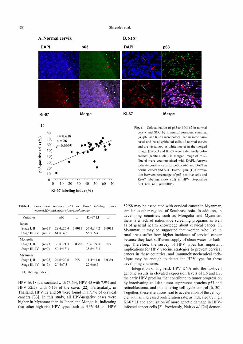
positive SCC, both parameters were significantly correlated
(r=0.618, p=0.0005; Fig. 6C). As for the correlation of the
percentage of p63-positive cells and the stages of SCC, the
percentage of p63-positive cells was significantly higher in
stages III and IV than in stages I and II in samples from
Japan and Mongolia (p=0.0011 and p=0.0385, respectively,
Table 4). Ki-67 LI was significantly higher in stages III
and IV than in stages I and II in samples from Japan and
Myanmar (p=0.0011 and p=0.0394, respectively).
IV. Discussion
In the present study, we assessed the type of HPV in-
fection in cervical cancer in three geographically different
countries, that is, Japan, Mongolia, and Myanmar, and found
that HPV 16 was strongly associated with Japanese patients,
whereas both HPV 16 and other HPV types were frequent in
cervical cancer cases from Mongolia and Myanmar.
International studies of cervical cancer have shown that
HPV 16 is the most prevalent type in Europe (65.1%), North
America (57.9%), Central and South America (50.5%) and
Southeast Asia (42.9%) [5]. In East and Southeast Asia,
specifically, the most prevalent type among invasive
cervical cancers is HPV16/18, followed by HPV 31/33 and
52/58 [3, 5]. In Japan, the most common HPV types in
cervical cancer are HPV 16/18 (58.8%) and 31/33 (11.5%)
[22]. In our study, HPV 16 was the most prevalent type,
and other HPV types (HPV 1, 6, 11, 18 and 31) were also
prevalent, though less so, among Japanese, Mongolian and
Myanmar cervical cancer samples. A pooled analysis of
invasive cervical cancer in Southeast Asia demonstrated that
Table 3. Correlation between HPV 16 and p63 labeling index in squamous cell carcinoma
Low, p63-positive cells <32%; High, p63-positive cells >32%; LI, labeling index.
Variables
HPV 16 (Japan) HPV 16 (Mongolia) HPV 16 (Myanmar)
Positive (n=26)
Negative (n=2)
pPositive (n=21)
Negative (n=7)
pPositive (n=15)
Negative (n=9)
p
p63 LI53.6±10.7 19.5±13.4 0.0002 46.1±16.3 30.8±14.4 0.0398 28.4±14.7 29.3±26.3 NS
(%, mean±SD)
Distribution of p63
Negative 0 1 0 1 0 2
Low 8 1 0.0007 5 4 0.0357 7 1 NS
High 18 0 16 2 8 6
Ki-6746.6±12.1 28.5±11.9 0.0001 37.0±22.1 16.3±13.1 0.008 12.4±12.0 14.2±10.5 NS
(%, mean±SD)
Fig. 5. Double immunostaining for HPV 16 and p63 in SCC. (A–C) Arrows indicate positive cells for HPV 16 and p63 in SCC. (C) HPV 16
immunoreactivity is detectable in most of the p63-positive nuclei. Bar=20 μm.
Shirendeb et al.188
HPV 16/18 is associated with 73.5%, HPV 45 with 7.9% and
HPV 52/58 with 6.1% of the cases [22]. Particularly, in
Thailand, HPV 52 and 58 were found in 17.7% of cervical
cancers [33]. In this study, all HPV-negative cases were
higher in Myanmar than in Japan and Mongolia, indicating
that other high risk-HPV types such as HPV 45 and HPV
52/58 may be associated with cervical cancer in Myanmar,
similar to other regions of Southeast Asia. In addition, in
developing countries, such as Mongolia and Myanmar,
there is a lack of nationwide screening programs as well
as of general health knowledge about cervical cancer. In
Myanmar, it may be suggested that women who live in
rural areas suffer from higher incidence of cervical cancer
because they lack sufficient supply of clean water for bath-
ing. Therefore, the survey of HPV types has important
implications for HPV vaccine strategies to prevent cervical
cancer in these countries, and immunohistochemical tech-
nique may be enough to detect the HPV type for these
developing countries.
Integration of high-risk HPV DNA into the host-cell
genome results in elevated expression levels of E6 and E7,
the early HPV proteins that contribute to tumor progression
by inactivating cellular tumor suppressor proteins p53 and
retinoblastoma, and thus altering cell cycle control [6, 30].
Together, these alterations lead to acceleration of the cell cy-
cle, with an increased proliferation rate, as indicated by high
Ki-67 LI and acquisition of more genetic damage in HPV-
infected cancer cells [2]. Previously, Nair et al. [24] demon-
Fig. 6. Colocalization of p63 and Ki-67 in normal
cervix and SCC by immunofluorescent staining.
(A) p63 and Ki-67 were colocalized in some para-
basal and basal epithelial cells of normal cervix
and are visualized as white nuclei in the merged
image. (B) p63 and Ki-67 were extensively colo-
calized (white nuclei) in merged image of SCC.
Nuclei were counterstained with DAPI. Arrows
indicate positive cells for p63, Ki-67 and DAPI in
normal cervix and SCC. Bar=20 μm. (C) Correla-
tion between percentage of p63-positive cells and
Ki-67 labeling index (LI) in HPV 16-positive
SCC (r=0.618, p=0.0005).
Table 4. Association between p63 or Ki-67 labeling index
(mean±SD) and stage of cervical cancer
LI, labeling index.
Variables p63 p Ki-67 LI p
Japan
Stage I, II (n=31) 28.4±26.4 0.0011 37.4±14.2 0.0011
Stage III, IV (n=9) 61.8±4.3 55.7±5.4
Mongolia
Stage I, II (n=23) 33.8±21.3 0.0385 29.6±24.8 NS
Stage III, IV (n=9) 50.4±13.3 38.6±13.3
Myanmar
Stage I, II (n=25) 24.6±22.6 NS 11.4±11.0 0.0394
Stage III, IV (n=5) 24.4±7.3 22.6±6.5
Cervical Cancer in Japan, Mongolia, and Myanmar 189
strated that HPV 16 and 18 infections resulted in increased
tumor cell proliferation in SCC. Bahnassy et al. [2] reported
that a high Ki-67 LI was significantly associated with stage
and overall survival in patients with invasive SCC. We also
found the association of Ki-67 LI with the stage of cervical
cancer.
p63 is the newest member of the p53 tumor suppressor
gene family [41, 42]. Transcriptionally active (TA) p63
induces p53 target genes and can induce cell cycle arrest and
apoptosis, whereas aminoterminally truncated (ΔN) p63 in-
activates p53, thus having anti-apoptotic activity [41].
ΔNp63 is highly expressed in stem cells of epithelial tissue
and is required for proliferation and maintenance of the epi-
thelial stem cell population [28, 32]. Previous studies have
indicated that TAp63 promotes epithelial cell differentia-
tion, whereas ΔNp63 favors epithelial cell proliferation [8,
18]. In the present study we found the over-expression of
p63 isoforms (TA and ΔN) and the association of p63 with
cell proliferation in SCC, which together may promote
carcinogenesis in cervical cancer. Increased p63 staining
has been reported in head, neck, lung, esophageal, and oral
SCCs, and p63 might also function as a marker of metaplas-
tic breast carcinoma [8, 15]. Interestingly, increased expres-
sion of p63 was highly correlated with the stage of cervical
cancer, suggesting the possible association with the tumor
progression for SCC. Furthermore, we found the correlation
between p63 and HPV 16 expression in SCC, suggesting
that HPV 16 presents a trophism for squamous epithelial
cells, while p63 may provide a positive contribution to the
viral life cycle by blocking apoptosis through the ΔNp63
isoforms [4].
In conclusion, it is suggested that HPV 16 may be asso-
ciated with the cell proliferative activity, and also may be
correlated with the expression of p63, which has a possible
role for tumor progression in the cervical cancer. Our study
indicates that HPV 16 infection is associated strongly with
the Japanese population, whereas both HPV 16 and other
types were similarly frequent in the populations of Mongolia
and Myanmar. Therefore, it is emphasized that the rationale
for HPV screening and the use of the HPV vaccine depend-
ing upon the HPV types to prevent cervical cancer in Japan,
Mongolia, and Myanmar.
V. Acknowledgments
This study was supported in part by a Grant-in-Aid
for Scientific Research from the Japanese Ministry of
Education, Science, Sports and Culture (nos. 13576001,
16406005, 18390060, and 19406005), and a Grant for Ad-
vanced Start-up Program from Nagasaki University.
VI. References
1. Baekelandt, M., Holm, R., Nesland, J. M., Trope, C. G. and
Kristensen, G. B. (2000) Expression of apoptosis-related proteins
is an independent determinant of patient prognosis in advanced
ovarian cancer. J. Clin. Oncol. 18; 3775–3781.
2. Bahnassy, A. A., Zekri, A. R., Saleh, M., Lotayef, M., Moneir,
M. and Shawki, O. (2007) The possible role of cell cycle regula-
tors in multistep process of HPV-associated cervical carcinoma.
BMC Clin. Pathol. 7; 4.
3. Bao, Y. P., Li, N., Smith, J. S. and Qiao, Y. L., ACCPAB
members. (2008) Human papillomavirus type distribution in
women from Asia: a meta-analysis. Int. J. Gynecol. Cancer 18;
71–79.
4. Blandino, G. and Dobbelstein, M. (2004) p73 and p63: why do we
still need them? Cell Cycle 3; 886–894.
5. Bosch, F. X., Manos, M. M., Munoz, N., Sherman, M., Jansen, A.
M., Peto, J., Schiffman, M. H., Moreno, V., Kurman, R. and Shah,
K. V. (1995) Prevalence of human papillomavirus in cervical
cancer: a worldwide perspective. International biological study
on cervical cancer (IBSCC) study group. J. Natl. Cancer Inst. 87;
796–802.
6. Clarke, B. and Chetty, R. (2001) Cell cycle aberrations in the
pathogenesis of squamous cell carcinoma of the uterine cervix.
Gynecol. Oncol. 82; 238–246.
7. Creasman, W. T. (1990) New gynecologic cancer staging. Obstet.
Gynecol. 75; 287–288.
8. Di Como, C. J., Urist, M. J., Babayan, I., Drobnjak, M., Hedvat,
C. V., Teruya-Feldstein, J., Pohar, K., Hoos, A. and Cordon-
Cardo, C. (2002) p63 expression profiles in human normal and
tumor tissues. Clin. Cancer Res. 8; 494–501.
9. Garland, S. M., Tabrizi, S. N., Chen, S., Byambaa, C. and
Davaajav, K. (2001) Prevalence of sexually transmitted infections
(Neisseria gonorrhoeae, Chlamydia trachomatis, Trichomonas
vaginalis and human papillomavirus) in female attendees of a
sexually transmitted diseases clinic in Ulaanbaatar, Mongolia.
Infect. Dis. Obstet. Gynecol. 9; 143–146.
10. Genther, S. M., Sterling, S., Duensing, S., Münger, K., Sattler, C.
and Lambert, P. F. (2003) Quantitative role of the human papillo-
mavirus type 16 E5 gene during the productive stage of the viral
life cycle. J. Virol. 77; 2832–2842.
11. Hishikawa, Y., An, S., Yamamoto-Fukuda, T., Shibata, Y. and
Koji, T. (2009) Improvement of in situ PCR by optimization of
PCR cycle number and proteinase K concentration: localization
of X chromosome-linked phosphoglycerate kinase-1 gene in
mouse reproductive organs. Acta Histochem. Cytochem. 42; 15–
21.
12. Hu, H., Xia, S. H., Li, A. D., Xu, X., Cai, Y., Han, Y. L., Wei, F.,
Chen, B. S., Huang, X. P., Han, Y. S., Zhang, J. W., Zhang, X.,
Wu, M. and Wang, M. R. (2002) Elevated expression of p63
protein in human esophageal squamous cell carcinomas. Int. J.
Cancer 102; 580–583.
13. Johnson, K. M., Kines, R. C., Roberts, J. N., Lowy, D. R.,
Schiller, J. T. and Day, P. M. (2009) Role of heparan sulfate in
attachment to and infection of the murine female genital tract by
human papillomavirus. J. Virol. 83; 2067–2074.
14. Kato, K., Hasui, K., Wang, J., Kawano, Y., Aikou, T. and Murata,
F. (2008) Homeostatic mass control in gastric non-neoplastic
epithelia under infection of Helicobacter pylori: An immuno-
histochemical analysis of cell growth, stem cells and programmed
cell death. Acta Histochem. Cytochem. 41; 23–38.
15. Koker, M. M. and Kleer, C. G. (2004) p63 expression in breast
cancer: a highly sensitive and specific marker of metaplastic car-
cinoma. Am. J. Surg. Pathol. 28; 1506–1512.
16. Kurita, T., Cunha, G. R., Robboy, S. J., Mills, A. A. and Medina,
R. T. (2005) Differential expression of p63 isoforms in female
reproductive organs. Mech. Dev. 122; 1043–1055.
17. Liao, Z., Boileau, T. W., Erdman, J. W. Jr. and Clinton, S. K.
(2002) Interrelationships among angiogenesis, proliferation, and
apoptosis in the tumor microenvironment during N-methyl-N-
nitrosourea androgen-induced prostate carcinogenesis in rats.
Carcinogenesis 23; 1701–1711.
18. Lin, Z., Nan, Y., Zhang, X., Zhao, Y., Kim, C. and Kim, I. (2006)
Shirendeb et al.190
Reverse transcription-polymerase chain reaction and western
blotting analysis for detection of p63 isoforms in uterine cervical
cancers. Int. J. Gynecol. Cancer 16; 1643–1647.
19. Massion, P. P., Taflan, P. M., Jamshedur Rahman, S. M., Yildiz,
P., Shyr, Y., Edgerton, M. E., Westfall, M. D., Roberts, J. R.,
Pietenpol, J. A., Carbone, D. P. and Gonzalez, A. L. (2003)
Significance of p63 amplification and overexpression in lung
cancer development and prognosis. Cancer Res. 63; 7113–7121.
20. Matsuo, Y., Nomata, K., Eguchi, J., Aoki, D., Hayashi, T.,
Hishikawa, Y., Kanetake, H., Shibata, Y. and Koji, T. (2007)
Immunohistochemical analysis of connexin43 expression in
infertile human testes. Acta Histochem. Cytochem. 40; 69–75.
21. McLean, C. S., Churcher, M. J., Meinke, J., Smith, G. L.,
Higgins, G., Stanley, M. and Minson, A. C. (1990) Production
and characterization of a monoclonal antibody to human papillo-
mavirus type 16 using recombinant vaccine virus. J. Clin. Pathol.
43; 488–492.
22. Miura, S., Matsumoto, K., Oki, A., Satoh, T., Tsunoda, H.,
Yasugi, T., Taketani, Y. and Yoshikawa, H. (2006) Do we need a
different strategy for HPV screening and vaccination in East
Asia?. Int. J. Cancer 119; 2713–2715.
23. Münger, K. (2002) The role of human papillomaviruses in human
cancers. Front. Biosci. 7; 641–649.
24. Nair, P., Nair, K. M., Jayaprakash, P. G. and Pillai, M. R. (1999)
Decreased programmed cell death in the uterine cervix associated
with high risk human papillomavirus infection. Pathol. Oncol.
Res. 5; 95–103.
25. Nylander, K., Coates, P. J. and Hall, P. A. (2000) Characterization
of the expression pattern of p63α and ΔNp63α in benign and
malignant oral epithelial lesions. Int. J. Cancer 87; 368–372.
26. Pisani, P., Parkin, D. M., Muñoz, N. and Ferlay, J. (1997) Cancer
and infection: estimates of the attributable fraction in 1990.
Cancer Epidemiol. Biomarkers Prev. 6; 387–400.
27. Quade, B. J., Yang, A., Wang, Y., Sun, D., Park, J., Sheets, E. E.,
Cviko, A., Federschneider, J. M., Peters, R., McKeon, F. D. and
Crum, C. P. (2001) Expression of the p53 homologues p63 in
early cervical neoplasia. Gynecol. Oncol. 80; 24–29.
28. Reis-Filho, J. S. and Schmitt, F. C. (2002) Taking advantage of
basic research: p63 is a reliable myoepithelial and stem cell
marker. Adv. Anat. Pathol. 9; 280–289.
29. Sasagawa, T., Basha, W., Yamazaki, H. and Inoue, M. (2001)
High-risk and multiple human papillomavirus infections asso-
ciated with cervical abnormalities in Japanese women. Cancer
Epidemiol. Biomarkers Prev. 10; 45–52.
30. Scheffner, M., Münger, K., Byrne, J.C. and Howley, P. M. (1991)
The state of the p53 and retinoblastoma genes in human cervical
carcinoma cell lines. Proc. Natl. Acad. Sci. U S A 88; 5523–5527.
31. Shukuwa, K., Izumi, S., Hishikawa, Y., Ejima, K., Inoue, S.,
Muramatsu, M., Ouchi, Y., Kitaoka, T. and Koji, T. (2006)
Diethylstilbestrol increases the density of prolactin cells in male
mouse pituitary by inducing proliferation of prolactin cells and
transdifferentiation of gonadotropic cells. Histochem. Cell Biol.
126; 111–123.
32. Signoretti, S., Waltregny, D., Dilks, J., Isaac, B., Lin, D.,
Garraway, L., Yang, A., Montironi, R., McKeon, F. and Loda, M.
(2000) p63 is a prostate basal cell marker and is required for
prostate development. Am. J. Pathol. 157; 1769–1775.
33. Siriaunkgul, S., Suwiwat, S., Settakorn, J., Khunamornpong,
S., Tungsinmunkong, K., Boonthum, A., Chaisuksunt, V.,
Lekawanvijit, S., Srisomboon, J. and Thorner, P. S. (2008) HPV
genotyping in cervical cancer in Northern Thailand: adapting
the linear array HPV assay for use on paraffin-embedded tissue.
Gynecol. Oncol. 108; 555–560.
34. Stefansson, I. M., Salvesen, H. B. and Akslen, L. A. (2006) Loss
of p63 and cytokeratin 5/6 expression is associated with more
aggressive tumors in endometrial carcinoma patients. Int. J.
Cancer 118; 1227–1233.
35. Suzuki, T., Matsuzaki, T., Hagiwara, H., Aoki, T. and Takata, K.
(2007) Recent advances in fluorescent labeling techniques for
fluorescence microscopy. Acta Histochem. Cytochem. 40; 131–
137.
36. Ulziibat, S., Ejima, K., Shibata, Y., Hishikawa, Y., Kitajima, M.,
Fujishita, A., Ishimaru, T. and Koji, T. (2006) Identification of
estrogen receptor β-positive intraepithelial lymphocytes and their
possible roles in normal and tubal pregnancy oviducts. Hum.
Reprod. 21; 2281–2289.
37. Urist, M. J., Di Como, C. J., Lu, M. L., Charytonowicz, E.,
Verbel, D., Crum, C. P., Ince, T. A., McKeon, F. D. and Cordon-
Cardo, C. (2002) Loss of p63 expression is associated with tumor
progression in bladder cancer. Am. J. Pathol. 161; 1199–1206.
38. Wang, T. Y., Chen, B. F., Yang, Y. C., Chen, H., Wang, Y., Cviko,
A., Quade, B. J., Sun, D., Yang, A., McKeon, F. D. and Crum, C.
P. (2001) Histologic and immunophenotypic classification of
cervical carcinomas by expression of the p53 homologue p63: a
study of 250 cases. Hum. Pathol. 32; 479–486.
39. WHO (2002) Disease specific NCD morbidity and mortality
profile. In “Noncommunicable Diseases in the South-East Asia
Region—A Profile”. New Delhi, pp. 46–48.
40. Woodman, C. B., Collins, S. I. and Young, L. S. (2007) The
natural history of cervical HPV infection: Unresolved issues. Nat.
Rev. Cancer 7; 11–22.
41. Yang, A., Kaghad, M., Wang, Y., Gillett, E., Fleming, M. D.,
Dötsch, V., Andrews, N. C., Caput, D. and McKeon, F. (1998)
p63, a p53 homologue at 3q27-29, encodes multiple products
with transactivating, death-inducing, and dominant-negative
activities. Mol. Cell 2; 305–316.
42. Yang, A., Schweitzer, R., Sun, D., Kaghad, M., Walker, N.,
Bronson, R. T., Tabin, C., Sharpe, A., Caput, D., Crum, C. and
McKeon, F. (1999) p63 is essential for regenerative proliferation
in limb, craniofacial and epithelial development. Nature 398;
714–718.
43. zur Hausen, H. (2002) Papillomaviruses and cancer: from basic
This is an open access article distributed under the Creative Commons Attribu-tion License, which permits unrestricted use, distribution, and reproduction inany medium, provided the original work is properly cited.
studies to clinical application. Nat. Rev. Cancer 2; 342–350.