p63 Expression in Solid Cell Nests of the Thyroid: Further Evidence for a Stem Cell Origin
6
p63 Expression in Solid Cell Nests of the Thyroid: Further Evidence for a Stem Cell Origin Jorge S. Reis-Filho, M.D., Ana Preto, M.Sc., Paula Soares, M.Sc., Ph.D., Sara Ricardo, M.T., José Cameselle-Teijeiro, M.D., Ph.D., Manuel Sobrinho-Simões, M.D., Ph.D. Institute of Molecular Pathology and Immunology (J.S.R.-F., A.P., P.S., S.R., M.S.-S.) and Medical Faculty (P.S., M.S.-S.), University of Porto, Porto, Portugal; School of Health Sciences (J.S.R.-F.), University of Minho, Braga, Portugal; Department of Pathology (J.C.-T.), Hospital Clínico Universitario, Santiago de Compostela, Spain; and Hospital de S. João (M.S.-S.), Porto, Portugal Solid cell nests of the thyroid are embryonic rem- nants of endodermal origin that may be difficult to distinguish from squamous metaplasia, metastatic squamous carcinoma, papillary microcarcinoma, medullary carcinoma, and C-cell hyperplasia. These embryonic structures are composed of main cells and C-cells; cystic structures and mixed follicles are sometimes observed intermingled with solid cell nests. Recently, p63, a p53 homologue that is con- sistently expressed in basal/stem cells of stratified epithelia and plays a major role in triggering the differentiation of some specific cell lineages, has been characterized. We evaluated the immunohis- tochemical expression of p63, cytokeratins (CAM 5.2, AE1/AE3, 34E12, 7, and 20), carcinoembryonic antigen, thyroid transcription factor 1 (TTF-1), thy- roglobulin, and calcitonin using the streptavidin- biotin-peroxidase complex technique in 6 bona fide solid cell nests. We observed that main cells of solid cell nests are strongly decorated by p63, while C-cells and all other thyroid structures were consis- tently negative. Moreover, main cells expressed car- cinoembryonic antigen and all cytokeratins but cy- tokeratin 20 and lacked TTF-1, thyroglobulin and calcitonin. In contrast to this, C-cells of solid cell nests were immunoreactive for calcitonin, CAM 5.2, AE1/AE3, and cytokeratin 7; focal immunoreactivity for TTF-1 was also observed in some C-cells. We conclude that main cells of the solid cell nests dis- play a basal/stem cell phenotype (p63 and basal cytokeratin positivity), whereas C-cells show fea- tures of parafollicular differentiation. We conclude, furthermore, that p63 antibodies may help in dis- tinguishing solid cell nests from their mimics. KEY WORDS: Immunohistochemistry, p63, thyroid, Ultimobranchial body. Mod Pathol 2003;16(1):43–48 Solid cell nests of the thyroid have long fascinated pathologists since their description by Getzowa (1) in 1907. Currently, it is accepted that solid cell nests and the so-called mixed follicles are indeed ultimo- branchial body remnants (2–10), but their biologi- cal significance remains disputable (2, 3, 11–18). It has been suggested that these embryonic structures might constitute the origin of some ectopic struc- tures rarely described in thyroid glands (3, 6), as well as of some peculiar types of thyroid neoplasms (17–20). Moreover, solid cell nests may be a source of confusion in thyroid pathology, as they can mimic squamous metaplasia, primary or metastatic squamous cell carcinomas, thyroglossal cyst, C-cell hyperplasia, and papillary and medullary microcar- cinomas (2, 3). In previous studies, our group (3) and others (2–12) have described the histological, immunohis- tochemical, and ultrastructural features of solid cell nests. It is accepted that solid cell nests are com- posed of two distinctive cell types (3): 1) main cells, which are polygonal to elongated or even spindle- shaped cells with centrally located, oval to fusiform nuclei with uneven nuclear envelope showing oc- casional grooves, and deeply eosinophilic cyto- plasm showing squamoid features (including high molecular weight cytokeratins) but lacking intercel- lular bridges (2– 4); and 2) C-cells, which account for a minor proportion of solid cell nest population and are characterized by clear cytoplasm and cen- trally located, small compact nuclei (2– 4). In up to 81% of the cases, mixed follicles may be found admixed with bona fide solid cell nests (2– 4). Mixed follicles are composed of cells resembling main Copyright © 2003 by The United States and Canadian Academy of Pathology, Inc. VOL. 16, NO. 1, P. 43, 2003 Printed in the U.S.A. Date of acceptance: October 1, 2002. J.S.R.-F. and A.P. contributed equally to the present study. Address reprint requests to: Prof. Manuel Sobrinho-Simões, IPATIMUP, R. Rob- erto Frias, S/N, 4200, Porto, Portugal; e-mail: [email protected]; fax: 351-22-557-0799. DOI: 10.1097/01.MP.0000047306.72278.39 43
Transcript of p63 Expression in Solid Cell Nests of the Thyroid: Further Evidence for a Stem Cell Origin

p63 Expression in Solid Cell Nests of the Thyroid:Further Evidence for a Stem Cell OriginJorge S. Reis-Filho, M.D., Ana Preto, M.Sc., Paula Soares, M.Sc., Ph.D., Sara Ricardo, M.T.,José Cameselle-Teijeiro, M.D., Ph.D., Manuel Sobrinho-Simões, M.D., Ph.D.
Institute of Molecular Pathology and Immunology (J.S.R.-F., A.P., P.S., S.R., M.S.-S.) and Medical Faculty(P.S., M.S.-S.), University of Porto, Porto, Portugal; School of Health Sciences (J.S.R.-F.), University ofMinho, Braga, Portugal; Department of Pathology (J.C.-T.), Hospital Clínico Universitario, Santiago deCompostela, Spain; and Hospital de S. João (M.S.-S.), Porto, Portugal
Solid cell nests of the thyroid are embryonic rem-nants of endodermal origin that may be difficult todistinguish from squamous metaplasia, metastaticsquamous carcinoma, papillary microcarcinoma,medullary carcinoma, and C-cell hyperplasia. Theseembryonic structures are composed of main cellsand C-cells; cystic structures and mixed follicles aresometimes observed intermingled with solid cellnests. Recently, p63, a p53 homologue that is con-sistently expressed in basal/stem cells of stratifiedepithelia and plays a major role in triggering thedifferentiation of some specific cell lineages, hasbeen characterized. We evaluated the immunohis-tochemical expression of p63, cytokeratins (CAM5.2, AE1/AE3, 34�E12, 7, and 20), carcinoembryonicantigen, thyroid transcription factor 1 (TTF-1), thy-roglobulin, and calcitonin using the streptavidin-biotin-peroxidase complex technique in 6 bona fidesolid cell nests. We observed that main cells of solidcell nests are strongly decorated by p63, whileC-cells and all other thyroid structures were consis-tently negative. Moreover, main cells expressed car-cinoembryonic antigen and all cytokeratins but cy-tokeratin 20 and lacked TTF-1, thyroglobulin andcalcitonin. In contrast to this, C-cells of solid cellnests were immunoreactive for calcitonin, CAM 5.2,AE1/AE3, and cytokeratin 7; focal immunoreactivityfor TTF-1 was also observed in some C-cells. Weconclude that main cells of the solid cell nests dis-play a basal/stem cell phenotype (p63 and basalcytokeratin positivity), whereas C-cells show fea-tures of parafollicular differentiation. We conclude,
furthermore, that p63 antibodies may help in dis-tinguishing solid cell nests from their mimics.
KEY WORDS: Immunohistochemistry, p63, thyroid,Ultimobranchial body.
Mod Pathol 2003;16(1):43–48
Solid cell nests of the thyroid have long fascinatedpathologists since their description by Getzowa (1)in 1907. Currently, it is accepted that solid cell nestsand the so-called mixed follicles are indeed ultimo-branchial body remnants (2–10), but their biologi-cal significance remains disputable (2, 3, 11–18). Ithas been suggested that these embryonic structuresmight constitute the origin of some ectopic struc-tures rarely described in thyroid glands (3, 6), aswell as of some peculiar types of thyroid neoplasms(17–20). Moreover, solid cell nests may be a sourceof confusion in thyroid pathology, as they canmimic squamous metaplasia, primary or metastaticsquamous cell carcinomas, thyroglossal cyst, C-cellhyperplasia, and papillary and medullary microcar-cinomas (2, 3).
In previous studies, our group (3) and others(2–12) have described the histological, immunohis-tochemical, and ultrastructural features of solid cellnests. It is accepted that solid cell nests are com-posed of two distinctive cell types (3): 1) main cells,which are polygonal to elongated or even spindle-shaped cells with centrally located, oval to fusiformnuclei with uneven nuclear envelope showing oc-casional grooves, and deeply eosinophilic cyto-plasm showing squamoid features (including highmolecular weight cytokeratins) but lacking intercel-lular bridges (2– 4); and 2) C-cells, which accountfor a minor proportion of solid cell nest populationand are characterized by clear cytoplasm and cen-trally located, small compact nuclei (2– 4). In up to81% of the cases, mixed follicles may be foundadmixed with bona fide solid cell nests (2– 4). Mixedfollicles are composed of cells resembling main
Copyright © 2003 by The United States and Canadian Academy ofPathology, Inc.VOL. 16, NO. 1, P. 43, 2003 Printed in the U.S.A.Date of acceptance: October 1, 2002.J.S.R.-F. and A.P. contributed equally to the present study.Address reprint requests to: Prof. Manuel Sobrinho-Simões, IPATIMUP, R. Rob-erto Frias, S/N, 4200, Porto, Portugal; e-mail: [email protected]; fax:351-22-557-0799.
DOI: 10.1097/01.MP.0000047306.72278.39
43

cells and follicular cells, forming a follicular lumen-like pattern. The lumina contain periodic acid-Schiff-positive colloid-like material, acid mucins,cell debris, and periodic acid-Schiff-positive gran-ular material (3, 5).
p63, a p53-homologue nuclear transcription fac-tor, is consistently expressed in basal/stem cells ofseveral multilayered epithelia, as well as in basalcells of the prostate acini and ducts and in basaland myoepithelial cells of the breast (21–25). p63,located on 3q27, encodes six different isoforms,which harbor either transactivating (TA-p63) ornegative dominant (�N-p63) effects on p53 reportergenes (21–23, 25). At variance with p53, which is amajor tumor suppressor gene, p63 tumor suppress-ing properties are disputable and mutations in thisgene are rather rare in human malignancies (23,25).
It has been described that p63 acts as a nucleartranscription factor that induces the expression ofcytokeratins 5 and 14, as well as other genes asso-ciated with a terminal epidermal differentiation,such as loricrin, involucrin, transglutaminase 1, andflaggrin in basal/stem cells of several epithelial (21–23). Moreover, �N-p63 plays a major role in themaintenance of basal cell populations (21–23, 25).We and others also demonstrated that p63 is notexpressed in follicular and parafollicular cells innormal thyroid glands (24, 26). According to ourprevious results, p63 expression in thyroid neo-plasms seems to be restricted to a small subset ofanaplastic carcinomas and to scattered cells in pap-illary carcinomas with foci of squamous differenti-ation (25, 26).
As p63 is expressed in basal/stem cells and alsohas been implicated in “epidermal”/squamous dif-ferentiation, we investigated the expression of p63,cytokeratins, and markers of follicular (thyroglobu-lin and thyroid transcription factor) and parafol-licular (calcitonin and thyroid transcription factor)differentiation in six solid cell nests. We also com-ment on the usefulness of p63 in thyroid pathologypractice.
MATERIALS AND METHODS
Six cases of thyroid diseases with solid cell nestswere retrieved from the files of the Hospital ClínicoUniversitario, Santiago de Compostela, Spain (fivecases), and from the consultation files of one of theauthors (MS-S). All cases were reviewed by two ofthe authors (JSR-F and PS), and the clinical datawere obtained either from pathology reports orfrom patient charts. The clinical data of the patientsand underlying thyroid diseases associated with thesolid cell nests are summarized in Table 1.Formalin-fixed, paraffin-embedded tissue wasavailable in all cases.
Immunohistochemical analysis was performed in4 �m serial sections using a panel of antibodies(Table 2) according to the streptavidin-biotin-peroxidase complex technique (27). Negative (theprimary antibody was substituted by nonimmunemouse serum) and positive controls were includedin each slide-run. All controls gave satisfactory re-sults. A minimum of 20% positivity was adopted asa cut-off for all markers. For p63 (24, 25) and thy-roid transcription factor (TTF-1) (28), only nuclearimmunoreactivity was considered specific, whilefor all of the other markers only cytoplasmic stain-ing was accepted.
RESULTS
Pathological FindingsAll cases showed solid cell nests composed of a
complex admixture of main cells and C-cells aspreviously described (Fig. 1, A–B). In three cases,cystic structures of variable size, containing eosin-ophilic material and cell debris, were observed (Fig.1A). These structures were lined by cuboidal-to-columnar cells with eosinophilic cytoplasms andround-to-oval nuclei showing finely granular chro-matin; in one case, scattered apical projections,very similar to cilia, were depicted in a minor pro-portion of the lining cells. Mixed follicles partiallycomposed of main cells and partially depictingcuboidal cells with eosinophilic cytoplasm andround-to-oval nuclei was found in one case (Case 4)(Figure 1, A–B, inset).
Adjacent thyroid tissue showed typical morpho-logical features of nodular/adenomatous goiter infour cases; in one case two follicular adenomaswere observed, and in the remaining case there wasa thyroid papillary carcinoma (Table 1). In the lasttwo cases, no continuity between the thyroid neo-plasms and the solid cell nests was observed.
Immunohistochemical AnalysisTable 3 summarizes the immunohistochemical
findings.
p63p63 antibody strongly decorated the nuclei of
main cells in all solid cell nests (Fig. 2, A–B). Even in
TABLE 1. Summary of the Patients’ Clinico-Pathological
Features
Age/Gender Underlying Pathology
39/F Two follicular adenomas 3.6 and 0.8 cm65/F Nodular/adenomatous goiter64/F Nodular/adenomatous goiter55/M Nodular/adenomatous goiter30/M Nodular/adenomatous goiter?/F Papillary carcinoma
44 Modern Pathology

low-power magnification, p63 immunoreactivitywas easily observed and was restricted to solid cellnests (Fig. 2A). C-cells and the cuboidal epitheliumlining the cystic structures were consistently nega-tive for p63 (Fig. 2B). In mixed follicles, there wasstrong nuclear immunoreactivity in solid cell nestmain cells and a gradual reduction toward the
cuboidal, follicular-like cells. In adjacent thyroidtissues, p63 immunoreactivity was not detected inany normal, hyperplastic, or parafollicular cell.Similarly, the two follicular adenomas and the pap-illary carcinoma were also negative for p63.
CytokeratinsCytokeratins AE1/AE3, CAM 5.2, and cytokera-
tin19 consistently decorated both main and C-cellsof all solid cell nests, as well as follicular andparafollicular cells of adjacent thyroid tissues. Cy-tokeratin 34�E12 was intensely expressed in maincells but was not detected in C-cells of the solid cellnests, nor in any cell of adjacent thyroid tissues(Fig. 2C). There was a concurrent expression of p63and cytokeratin 34�E12 in main cells of the solidcell nests. Cytokeratin 20 was not detected in solidcell nests nor in adjacent thyroid tissues.
Carcinoembryonic AntigenMain cells and C-cells diffusely expressed carci-
noembryonic antigen (Fig. 2D). No immunoreactiv-ity in follicular cells of adjacent thyroid tissues wasobserved.
Markers of Follicular and ParafollicularDifferentiation
In solid cell nests, the expression of TTF-1, anuclear transcription factor that plays a major rolein the development of follicular epithelium andC-cells (28, 29), was restricted to the nuclei ofC-cells. All main cells lacked this differentiationmarker (Fig. 2E). In adjacent thyroid tissues, TTF-1was expressed in follicular and parafollicular cells,papillary carcinoma, and follicular adenomas.
Thyroglobulin is considered the most specificmarker of thyroid follicular differentiation. All solidcell nests herein evaluated lacked thyroglobulin(Fig. 2F). Conversely, thyroid follicular cells andcolloid, as well as all the cells of the follicular ade-nomas and the papillary carcinoma, were stronglypositive for thyroglobulin.
TABLE 2. Antibodies Used for Immunohistochemical Analysis
Antigen Antibody (Clone and Source) Dilution Antigen Retrieval
Thyroglobulin Tg6, (DAKO, Glostrp, Denmark) 1:1000 NoneCalcitonin Polyclonal (BioGenex, San Ramon, CA) 1:200 NoneTTF-1 8G7G3/1 (DAKO) 1/100 PCKeratins 1, 5, 10, and 14 34�E12 (Enzo, Farmingdale, NY) 1/10 PC, PKeratins 1, 2, 10, 11, 14, 15, 16, and 19 AE1/AE3 (Signet, Dedham, MA) 1/50 PC, PKeratins 8 and 18 CAM 5.2 (Becton Dickinson, Mountainview, CA) 1:5 PC, PKeratin 19 RCK108 (DAKO) 1:100 PC, PKeratin 7 OV-TL12/30 (DAKO) 1:50 PC, PKeratin 20 M7019 (DAKO) 1:50 PC, PCarcinoembryonic antigen Polyclonal (DAKO) 1:2,000 Nonep63 Monoclonal (Neomarkers) 1:150 Water bath, citrate buffer
PC, pressure cooker; P, protease.
FIGURE 1. Solid cell nests— histological findings. A, Solid cell nestscomposed of a complex admixture of main and C-cells, as well as cysticstructures. B, Cystic solid cell nest. Inset: note a mixed folliclecomposed of cells resembling main cells of solid cell nests and cuboidalcells arranged in a follicle-like structure (A and B, H&E �200; Inset:H&E �400).
p63 in Solid Cell Nests (J.S. Reis-Filho et al.) 45
Calcitonin staining was detected in C-cells of allsolid cell nests and in parafollicular cells of adjacentthyroid tissues (Fig. 2G).
DISCUSSION
Previous immunohistochemical evaluation ofsolid cell nests has revealed that main cells andC-cells express high- and low-molecular weight cy-tokeratins (3– 8, 19), as well as carcinoembryonicantigen, but show a differential expression of neu-roendocrine markers: while main cells are usuallypositive for somatostatin and neurotensin, C-cellsare usually immunoreactive for calcitonin and cal-citonin gene-related peptide (3). Vimentin is usu-ally negative, and thyroglobulin expression is con-troversial in solid cell nests (2, 8, 14, 19). In thepresent study, we confirmed the previously re-ported immunohistochemical profile of main cellsand C-cells (2– 8, 14, 19).
In the present study, we observed a differentialexpression of p63 and TTF-1 in the nuclei of mainand C-cells, respectively. p63 is consistently ex-pressed in basal/stem cells of several types of epi-thelia and is usually absent in partially or terminallydifferentiated cells (21–25); in the present study, wedemonstrated that main cells concurrently expressp63 and basal cell keratin (34�E12) and lack TTF-1expression, thus suggesting that these cells mayhave a basal/stem cell status.
In contrast to main cells, C-cells lack p63 anddisplay TTF-1, calcitonin, calcitonin gene-relatedpeptide, and chromogranin immunoreactivity. AsTTF-1 is an early marker of either follicular orparafollicular cells and the last three antigens maybe interpreted as markers of terminal parafollicular(C-) cell differentiation (27, 28), it is tempting tospeculate that C-cells of the solid cell nests havealready triggered their pathways toward a parafol-licular differentiation.
Mixed follicles showed a differential expressionof p63, cytokeratins, TTF-1, and thyroglobulin:while cells resembling main cells were strongly pos-itive for p63 and cytokeratins 34�E12 and lackedTTF-1 and thyroglobulin, cuboidal cells arranged infollicular-like pattern were positive for thyroglobu-
lin and TTF-1 and were negative for p63 and cyto-keratins 34�E12. These findings suggest that thosecells arranged in follicular-like structures have alsotriggered their differential pathways toward a follic-ular differentiation.
Our hypothesis that solid cell nests display a bas-al/stem cell phenotype is supported, indirectly, bythe presence of adipose, cartilage, and salivarygland-type tissues in the vicinity of or even abuttingsolid cell nests—what has been advanced as evi-dence to support the concept that solid cell nestsrepresent a dysembryological process (3, 14, 16).
Solid cell nests may pose some difficulties in rou-tine thyroid pathology practice. Harach (2) andCameselle-Teijeiro et al. (3) stressed that solid cellnests may be confused with squamous metaplasia,metastatic squamous carcinoma, papillary micro-carcinoma, medullary carcinoma, and C-cell hyper-plasia (2, 3). It has been assumed that using judi-cious morphological evaluation and based on theso-called typical immunohistochemical profile ofsolid cell nests (carcinoembryonic antigen and“epidermal” keratins), they might be differentiatedfrom their mimics (2, 3). However, it should benoted that some neuroendocrine carcinomas andprimary or metastatic mucoepidermoid carcinomasmay co-express carcinoembryonic antigen and“epidermal”/high molecular weight cytokeratins(18, 20, 30 –32). p63 immunoreactivity may help inthe identification of solid cell nests. p63 is not ex-pressed by follicular and parafollicular (C�) cells(24, 26), is absent in medullary and follicular carci-nomas (24, 25). In a previous study (26), we evalu-ated the expression of p63 in 12 papillary carcino-mas of the thyroid and found out that 4 casesfocally expressed p63 in 5–30% of the cells (26).Interestingly, p63 expression was almost restrictedto foci of squamous differentiation (26). Altogether,it seems that p63 may be a useful marker to distin-guish solid cell nests from papillary and medullarycarcinomas, as well as from C-cell hyperplasia.However, p63 seems to play a very limited role, orno role, as an adjunct antibody in the differentia-tion between solid cell nests and foci of squamousmetaplasia.
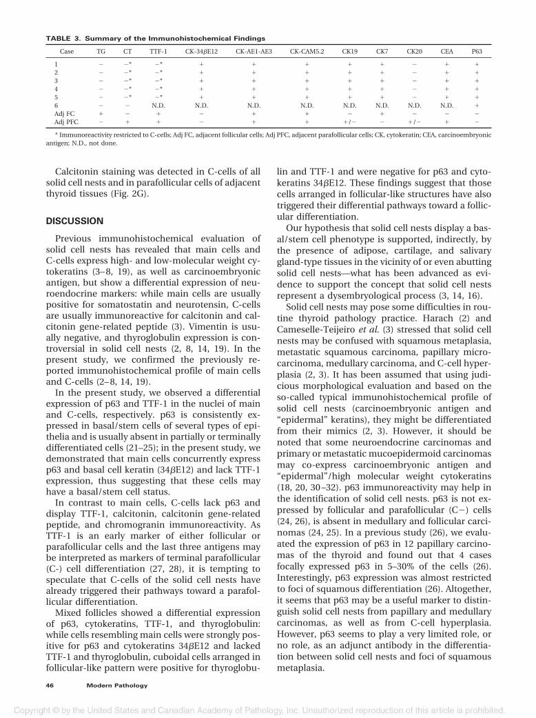
TABLE 3. Summary of the Immunohistochemical Findings
Case TG CT TTF-1 CK-34�E12 CK-AE1-AE3 CK-CAM5.2 CK19 CK7 CK20 CEA P63
1 � �* �* � � � � � � � �2 � �* �* � � � � � � � �3 � �* �* � � � � � � � �4 � �* �* � � � � � � � �5 � �* �* � � � � � � � �6 � � N.D. N.D. N.D. N.D. N.D. N.D. N.D. N.D. �Adj FC � � � � � � � � � � �Adj PFC � � � � � � �/� � �/� � �
* Immunoreactivity restricted to C-cells; Adj FC, adjacent follicular cells; Adj PFC, adjacent parafollicular cells; CK, cytokeratin; CEA, carcinoembryonicantigen; N.D., not done.
46 Modern Pathology

In conclusion, our results demonstrate that maincells of the solid cell nests concurrently express p63(a basal/stem cell marker) and basal cytokeratins,while C-cells lack p63 and show parafollicular dif-ferentiation markers (TTF-1, calcitonin, carcinoem-
bryonic antigen). Based on these findings, we sug-gest that main cells of the solid cell nests display abasal/stem cell phenotype. Moreover, we advocatethat antibodies against p63 should be included inimmunohistochemical panels to distinguish solid
FIGURE 2. A, p63 expression in the nuclei of main cells and rare C-cells. Note that all cystic structures and mixed follicles consistently lack p63expression. B, High power magnification of a solid cell nest showing strong nuclear immunoreactivity for p63 in main cells and lack of p63 in a solidcell nest (arrowhead). C, Cytokeratin 34�E12 expression in solid cell nests. Note that some c-cells lack p63 expression (arrow). D, Solid cell neststained with carcinoembryonic antigen. At the periphery, note immunoreactivity in scattered parafollicular (C�) cells. E, TTF-1 expression in a cysticsolid cell nest. Note rare cells expressing TTF-1 (arrows). On lower right, TTF-1 decorated all follicular cells of normal thyroid follicles. F, Follicularcells and thyroid stroma stained with thyroglobulin. Solid cell nests were negative for this marker. G, Calcitonin immunoreactivity in scattered C-cellsof solid cell nests (Streptavidin-biotin-peroxidase; A, E, F, and G, original magnification 200�; B, C, 400�; D, 100�).
p63 in Solid Cell Nests (J.S. Reis-Filho et al.) 47
cell nests from their mimics, namely from medul-lary and papillary microcarcinomas.
Acknowledgments: This study was partially sup-ported by Ph.D. Grants from the Portuguese Scienceand Technology Foundation (FCT) (referencesPRAXIS XXI/BD/21795/99 - AP and SFRH/BD/5386/2001 - JSRF) and Programa Operacional Ciência,Tecnologia e Inovação (POCTI) do Quadro Comu-nitário de Apoio (QCA) II.
REFERENCES
1. Getzowa S. Ueber die Glandula parathyreoidea, intrathyreoi-dale Zellhaufen derselben und Reste des postbranchialenköpers. Virchows Arch A Pathol Anat Histopathol 1907;188:181–234.
2. Harach HR. Solid cell nests of the thyroid. J Pathol 1988;155:191–200.
3. Cameselle-Teijeiro J, Varela-Duran J, Sambade C, VillanuevaJP, Varela-Nunez R, Sobrinho-Simoes M. Solid cell nests ofthe thyroid: light microscopy and immunohistochemicalprofile. Hum Pathol 1994;25:684 –93.
4. Martin V, Martin L, Viennet G, Hergel M, Carbillet JP, Fell-mann D. Ultrastructural features of “solid cell nest” of thehuman thyroid gland: a study of 8 cases. Ultrastruct Pathol2000;24:1– 8.
5. Harach HR. Mixed follicles of the human thyroid gland. ActaAnat (Basel) 1987;129:27–30.
6. Bykov VL. Tissue of ultimobranchial origin in normal andpathologically altered thyroid gland. Arkh Patol 1993;55:81–4.
7. Fraser BA, Duckworth JW. Ultimobranchial body cysts in thehuman foetal thyroid: pathological implications. J Pathol1979;127:89 –92.
8. Mizukami Y, Nonomura A, Michigishi T, Noguchi M, Hashi-moto T, Nakamura S, et al. Solid cell nests of the thyroid. Ahistologic and immunohistochemical study. Am J ClinPathol 1994;101:186 –91.
9. Ozaki O, Ito K, Sugino K, Yasuda K, Yamashita T, Toshima K,et al. Solid cell nests of the thyroid gland. Virchows Arch APathol Anat Histopathol 1991;418:201–5.
10. Beckner ME, Shultz JJ, Richardson T. Solid and cystic ulti-mobranchial body remnants in the thyroid. Arch Pathol LabMed 1990;114:1049 –52.
11. Martin V, Martin L, Viennet G, Challier B, Carbillet J, Fell-mann D. Solid cell nests and thyroid pathologies. Retrospec-tive study of 1,390 thyroids. Annu Pathol 2000;20:196 –201.
12. Williams ED, Toyn CE, Harach HR. The ultimobranchialgland and congenital thyroid abnormalities in man. J Pathol1989;159:135– 41.
13. Chan JK, Tse CC. Solid cell nest-associated C-cells: anotherpossible explanation for “C-cell hyperplasia” adjacent tofollicular cell tumors. Hum Pathol 1989;20:498 –9.
14. Janzer RC, Weber E, Hedinger C. The relation between solidcell nests and C cells of the thyroid gland: an immunohisto-chemical and morphometric investigation. Cell Tissue Res1979;197:295–312.
15. Fraser BA, Duckworth JW. Position of ultimobranchial bodycysts in the human fetal thyroid gland. Acta Anat (Basel)1979;105:269 –72.
16. Cameselle-Teijeiro J, Varela-Duran J. Intrathyroid salivarygland-type tissue in multinodular goiter. Virchows Arch1994;425:331– 4.
17. Cameselle-Teijeiro J. Mucoepidermoid carcinoma and solidcell nests of the thyroid. Hum Pathol 1996;27:861–3.
18. Cameselle-Teijeiro J, Febles-Perez C, Sobrinho-Simoes M. Pap-illary and mucoepidermoid carcinoma of the thyroid with ana-plastic transformation: a case report with histologic and im-munohistochemical findings that support a provocativehistogenetic hypothesis. Pathol Res Pract 1995;191:1214–21.
19. Harach HR, Vujanic GM, Jasani B. Ultimobranchial bodynests in human fetal thyroid: an autopsy, histological, andimmunohistochemical study in relation to solid cell nestsand mucoepidermoid carcinoma of the thyroid. J Pathol1993;169:465–9.
20. Harach HR. A study on the relationship between solid cellnests and mucoepidermoid carcinoma of the thyroid. His-topathology 1985;9:195–207.
21. Mills AA, Zheng B, Wang XJ, Vogel H, Roop DR, Bradley A.p63 is a p53 homologue required for limb and epidermalmorphogenesis. Nature 1999;398:708 –13.
22. Yang A, Schweitzer R, Sun D, Kaghad M, Walker N, BronsonRT, et al. p63 is essential for regenerative proliferation inlimb, craniofacial and epithelial development. Nature 1999;398:714 – 8.
23. Levrero M, De Laurenzi V, Costanzo A, Gong J, Wang JY,Melino G. The p53/p63/p73 family of transcription factors:overlapping and distinct functions. J Cell Sci 2000;113:1661–70.
24. Di Como CJ, Urist MJ, Babayan I, Drobnjak M, Hedvat CV,Teruya-Feldstein J, et al. p63 Expression Profiles in HumanNormal and Tumor Tissues. Clin Cancer Res 2002;8:494 –501.
25. Reis-Filho JS, Schmitt FC. Taking advantage of basic re-search: p63 is a reliable myoepithelial and stem cell marker.Adv Anat Pathol 2002;9:280 –9.
26. Preto A, Reis-Filho JS, Ricardo S, Soares P. p63 expression inpapillary and anaplastic carcinomas of the thyroid gland:lack of an oncogenetic role in tumorigenesis and progres-sion. Pathol Res Pract 2002;198:449 –54.
27. Reis-Filho JS, Milanezi F, Silva P, Schmitt FC. Maspin expres-sion in myoepithelial tumors of the breast. Pathol Res Pract2001;197:817–21.
28. Reis-Filho JS, Carrilho C, Valenti C, Leitao D, Ribeiro CA,Ribeiro SG, et al. Is TTF1 a good immunohistochemicalmarker to distinguish primary from metastatic lung adeno-carcinomas? Pathol Res Pract 2000;196:835– 40.
29. Ordonez NG. Thyroid transcription factor-1 is a marker oflung and thyroid carcinomas. Adv Anat Pathol 2000;7:123–7.
30. Sturm N, Lantuejoul S, Laverriere MH, Papotti M, BrichonPY, Brambilla C, et al. Thyroid transcription factor 1 andcytokeratins 1, 5, 10, 14 (34betaE12) expression in basaloidand large-cell neuroendocrine carcinomas of the lung. HumPathol 2001;32:918 –25.
31. Schroder S, Wodzynski A, Padberg B. Cytokeratin expressionof benign and malignant epithelial thyroid gland tumors. Animmunohistologic study of 154 neoplasms using 8 differentmonoclonal cytokeratin antibodies. Pathologe 1996;17:425–32.
32. Baloch ZW, Solomon AC, LiVolsi VA. Primary mucoepider-moid carcinoma and sclerosing mucoepidermoid carcinomawith eosinophilia of the thyroid gland: a report of nine cases.Mod Pathol 2000;13:802–7.
48 Modern Pathology