Protective role of glutathione reductase in paraquat induced neurotoxicity
Upload
independentCategory
view
3download
0
FosB Null Mutant Mice Show Enhanced MethamphetamineNeurotoxicity: Potential Involvement of FosB in IntracellularFeedback Signaling and Astroglial Function
Kumi O Kuroda*,1, Veravej G Ornthanalai2, Tadafumi Kato3 and Niall P Murphy2,4
1Unit for Affiliative Social Behavior, RIKEN Brain Science Institute, Saitama, Japan; 2Neuropathology Research Group, RIKEN Brain Science
Institute, Saitama, Japan; 3Laboratory for Molecular Dynamics of Mental Disorder, RIKEN Brain Science Institute, Saitama, Japan
Previous studies show that (1) two members of fos family transcription factors, c-Fos and FosB, are induced in frontal brain regions by
methamphetamine; (2) null mutation of c-Fos exacerbates methamphetamine-induced neurotoxicity; and (3) null mutation of FosB
enhances behavioral responses to cocaine. Here we sought a role of FosB in responses to methamphetamine by studying FosB null
mutant (�/�) mice. After a 10 mg/kg methamphetamine injection, FosB(�/�) mice were more prone to self-injury. Concomitantly, the
intracellular feedback regulators of Sprouty and Rad-Gem-Kir (RGK) family transcripts had lower expression profiles in the frontoparietal
cortex and striatum of the FosB(�/�) mice. Three days after administration of four 10 mg/kg methamphetamine injections, the
frontoparietal cortex and striatum of FosB(�/�) mice contained more degenerated neurons as determined by Fluoro-Jade B staining.
The abundance of the small neutral amino acids, serine, alanine, and glycine, was lower and/or was poorly induced after
methamphetamine administration in the frontoparietal cortex and striatum of FosB(�/�) mice. In addition, methamphetamine-treated
FosB(�/�) frontoparietal and piriform cortices showed more extravasation of immunoglobulin, which is indicative of blood–brain barrier
dysfunction. Methamphetamine-induced hyperthermia, brain dopamine content, and loss of tyrosine hydroxylase immunoreactivity in the
striatum, however, were not different between genotypes. These data indicate that FosB is involved in thermoregulation-independent
protective functions against methamphetamine neurotoxicity in postsynaptic neurons. Our findings suggest two possible mechanisms of
FosB-mediated neuroprotection: one is induction of negative feedback regulation within postsynaptic neurons through Sprouty and RGK.
Another is supporting astroglial function such as maintenance of the blood–brain barrier, and metabolism of serine and glycine, which are
important glial modulators of nerve cells.
Neuropsychopharmacology (2010) 35, 641–655; doi:10.1038/npp.2009.169; published online 4 November 2009
Keywords: FosB; methamphetamine; neurotoxicity; astroglia; sprouty; Rad-Gem-Kir (RGK) family
������������������������������������������������������
INTRODUCTION
Amphetamines, substituted amphetamines (such as metham-phetamine (METH), and 3,4-methylenedioxymethampheta-mine (MDMA or ‘ecstasy’)) and cocaine are widely abusedpsychostimulants (Sulzer et al, 2005). Whereas cocaineblocks the activity of dopamine, norepinephrine, andserotonin transporters, most amphetamine derivativespromote the release of these biogenic amines by reversetransport, effectively discharging enormous amounts of
biogenic amines into the extracellular space. Regardless ofthe mechanism, a major outcome is a strong potentiation ofbiogenic amine signaling, and it is this effect that is widelybelieved to underlie the abuse potential of these drugs.Repeated administration of lower doses or a single high-dose administration of amphetamines may lead to depletionof dopamine and serotonin by causing degeneration ofneuronal terminals (Krasnova and Cadet, 2009). Suchterminal degeneration is not only long-lasting but alsorecoverable in rodents, as these monoaminergic neuronsthemselves are resistant (Bowyer and Schmued, 2006b). Incontrast, amphetamines can elicit death of neurons post-synaptic to these monoaminergic terminals in manyforebrain regions such as the striatum and the frontal andsomatosensory cortices (Krasnova and Cadet, 2009). Twoforms of cell death have been implicated in this process(Davidson et al, 2001; Gold et al, 2009). One is apoptotic celldeath, also referred to as programmed cell death, which ischaracterized by cell shrinkage, specific DNA fragmentation
Received 12 March 2009; revised 17 August 2009; accepted 3September 2009
*Correspondence: Dr KO Kuroda, Unit for Affiliative Social Behavior,RIKEN Brain Science Institute, 2-1 Hirosawa, Wako-shi, Saitama 351-0198, Japan, Tel: + 81 48 467 7556, Fax: + 81 48 467 6853,E-mail: [email protected] address: Department of Psychiatry and BiobehavioralSciences, Semel Institute for Neuroscience and Human Behavior,University of California at Los Angeles, Los Angeles, CA, USA.
Neuropsychopharmacology (2010) 35, 641–655& 2010 Nature Publishing Group All rights reserved 0893-133X/10 $32.00
www.neuropsychopharmacology.org
at about 180 bp intervals, and activation of cysteineproteases called caspases. The other form of cell death,necrosis, is a very rapid process, associated with cellswelling, spillage of intracellular substances into surround-ing tissue, and nonspecific DNA damage.
Multiple events, including oxidative stress, excitotoxicity(toxicity caused by glutamate release), hyperthermia,mitochondrial and endoplasmic reticulum stress, and theblood–brain barrier (BBB) dysfunction, are implicated inamphetamine-induced neurotoxic effects (Krasnova andCadet, 2009). These events appear to be intimately related.For example, preventing hyperthermia by cooling theambient temperature to 41C can decrease molecularmarkers of oxidative stress, attenuate dopaminergic andserotonergic terminal depletion, and decrease neuronaldeath in the parietal cortex (Bowyer, 1995; Eisch et al, 1998).Similarly, the glutamate receptor antagonist MK-801 canattenuate hyperthermia, dopaminergic and serotonergicterminal degeneration, and neuronal death in the parietalcortex (Eisch et al, 1998; Tata and Yamamoto, 2007). Assuch, it is still unclear which mechanism is the mostupstream and/or responsible for each neurotoxic end point.Interestingly, however, a dopamine transport inhibitoradministered before METH can only prevent striataltyrosine hydroxylase immunoreactivity loss, but not hy-perthermia or neuronal death in the parietal cortex (Eischet al, 1998). In addition, it has been shown that the striatalsubregion that contains the greatest number of degeneratedneurons is different from the subregion showing greatestloss of tyrosine hydroxylase immunoreactivity (Bowyeret al, 2008). It is therefore likely that mechanisms of striataldopamine terminal damage exist independent of themechanisms responsible for postsynaptic cell injury andhyperthermia.
Amphetamines stimulate the expression of many im-mediate early genes, particularly those of the Fos familytranscription factors, which include c-Fos, FosB, Fra-1, andFra-2 (Herdegen and Leah, 1998). DFosB, a truncated splicevariant of FosB, is induced in the striatum by chronicexposure to abused drugs (Nestler, 2001). The persistentexpression of DFosB has been implicated in long-termadaptations that underlie patterns of behavior characteristicof addiction. Previous studies have also shown that FosBnull mutant (�/�) mice show exaggerated locomotoractivation following initial cocaine exposure and robustconditioned place preference to a lower dose of cocaine(Hiroi et al, 1998). In addition, c-Fos(�/�) and ( + /�) miceshow exacerbated METH-induced neurotoxicity as reflectedin a greater loss of striatal tyrosine hydroxylase-likeimmunoreactivity and dopamine-reuptake sites and moreneuronal apoptosis in their cortices and striata. Theseresults suggest that Fos family proteins may be involvedin the protective mechanisms against METH-inducedneurotoxicity.
Being important elements of transcriptional regulation ofcellular functions, c-Fos and FosB are also likely to beinvolved in the regulation of many behaviors. An example isparental behavior, which arises from neuronal activity inthe medial preoptic area of the hypothalamus (Brown et al,1996; Numan et al, 1998). As to this, FosB(�/�) mice showabnormal parenting (Brown et al, 1996). Our previousstudies suggest that the role of FosB in this behavior is to
induce in downstream target genes such as Sprouty1(SPRY1) and RAD, under the control of extracellularsignal-regulated kinase (ERK) (Kuroda et al, 2007). TheSprouty family consists of three members in mouse, SPRY1,SPRY2 and SPRY4, and represents a major class of ligand-inducible inhibitors of the receptor tyrosine kinase (RTK)-dependent ERK signaling pathway (Mason et al, 2006). RADis a member of the Rad-Gem-Kir (RGK) family small GTP-binding proteins that encompasses RAD, GEM/KIR, REMand REM2, which function as potent inhibitors of voltage-dependent calcium channels (Kelly, 2005). The ERK–FosB–SPRY1/RAD signaling therefore appears to provide criticalfeedback regulation of neuronal adaptation in the initiationof parental care (Kuroda et al, 2007). It seems likely that thisintracellular signaling cascade is also involved in otherbehaviors, such as those in response to METH and otherpsychostimulants. For instance, c-Fos heterozygous mutant( + /�) mice, in which METH-induced dopaminergic terminaldamage and neuronal apoptosis are exacerbated (Deng et al,1999), show significant impairment in GEM (a member ofRGK family) upregulation in the striatum compared withwild-type mice on METH administration (Cadet et al, 2002).Administration of MDMA causes ERK activation andsubsequent upregulation of REM2, another member ofRGK, along with c-Fos and FosB in the striatum (Salzmannet al, 2006). Induction of FosB and REM2, but not c-Fos byMDMA is blocked by SL327, a specific inhibitor of ERKphosphorylation (Salzmann et al, 2006).
The purpose of this study was to identify the role of FosBin response to METH. Our working hypothesis was that theERK/calcium-c-Fos/FosB-Sprouty/RGK intracellular signal-ing acts as a general negative feedback loop for ERKactivation and calcium influx in stimulated neurons. If thisis true for METH-activated neurons, the FosB(�/�) micewould show enhanced cellular, neurochemical, and beha-vioral responses to METH, and decreased Sprouty/RGKexpression profiles. We found that FosB(�/�) mice indeedshowed greater neuronal degeneration in cortices and thestriatum, increased incidence of self-injuries, alteredregulation of intracellular Sprouty and RGK family tran-scripts compared with those of their wild-type littermates.In addition, FosB(�/�) mice showed signs of astroglialdysfunction, reflected as perturbed regulation in brainamino-acid contents and METH-induced BBB disruption.
MATERIALS AND METHODS
Animals
All animal experiments were approved by the animalexperiment committee of RIKEN. Wild-type C57BL/6J micewere purchased from Japan SLC (Hamamatsu, Japan). TheFosB(�/�) mouse strain was obtained from the JacksonLaboratory (strain C; 129S-Fosbtm1Meg/J). All of the micewere backcrossed at least five times to C57BL/6J andgenerated by FosB( + /�) intercrosses. Mice were housed inventilated shoebox cages (267� 483� 152 mm) with accessto food and water ad libitum and were maintained under a12 : 12 h light/dark cycle. TEK-Fresh Standard (Harlan,Madison, WI, USA) bedding was used for animal housing.All mice used in this study were adult males of 10–20 weeksof age, and studies included age-matched controls. Four
Enhanced METH toxicity in FosB null miceKO Kuroda et al
642
Neuropsychopharmacology
separate groups of FosB(�/�) and ( + / + ) mice were usedin the following four major sets of studies: (1) histologicalstudies, (2) hyperthermia, (3) gene expression, and (4)locomotor activity/analysis of brain biogenic amine andamino-acid content. The effects of METH on self-injurywere observed across studies except in experiment 4. Micewere single-housed in clean cages at least 1 day beforeMETH administration, except in experiment 4, where miceremained group-housed throughout.
Overview of METH Treatment Regimens Used
For histological studies of METH-induced neuronaldamage, we gave mice four injections of 10 mg/kg METH(methamphetamine hydrochloride, uncorrected for mole-cular salt and water content; Dainippon Pharmaceuticals,Tokyo, Japan) at 2-h intervals or saline vehicle subcuta-neously (10 ml/kg). These mice were transcardially perfused3 days later, as previous reports suggest that this time pointis where peak neuronal degeneration is observed (Eischet al, 1998). Mice studied for the hyperthermic effects ofMETH were administered the same regimen as thehistological studies. For the gene expression study, micewere given a single 10 mg/kg injection of METH and killedby cervical dislocation 2 h later, as changes in FosB proteinlevels and gene expression have been reported to peakabout 2 h after METH stimulation (Cadet et al, 2002; Hiroiet al, 1997). For locomotor activity and analysis of brainbiogenic amine and amino-acid content, mice were firstadministered a 1.5 mg/kg dose of METH. On the followingday, the mice were administered 10 mg/kg METH four timesevery 2 h. After 2 days, the mice were administered another1.5 mg/kg METH. More detailed descriptions of the treat-ment regimens and rationales are provided in the appro-priate sections below.
Temperature Measurement
Core body temperature was recorded using a Bat-10thermometer equipped with a RET-3 mouse rectal probe(Physitemp Inc., Clinton, NJ, USA). It has been reportedthat c-Fos(�/�) mice are more susceptible to rectal injuryand are more prone to fatality by repeated rectal temper-ature measurements in conjugation with METH adminis-tration (Deng et al, 1999). Nonetheless, FosB(�/�) micewere not significantly susceptible to this protocol comparedto FosB( + / + ) mice (data not shown).
Behavior and Self-Injury After METH Administration
At 30 min following the first 10 mg/kg METH injection, webriefly observed the subject mice from outside the cage. Allof the mice showed at least one of the following behaviors:stereotypic sniffing, vigorous chewing, increased locomotoractivity, head-bobbing, and excessive salivation, as pre-viously described (Eisch et al, 1998; Wagner et al, 2004).These observations were subjective and qualitative, but notquantitative, as the purpose of the observation was toconfirm the proper METH injection. At 2 h after theinjection, we carefully sought evidence of bleeding on thebedding. Whenever signs of bleeding were observed, micewere further examined to identify skin lesions. We repeated
this survey just before each subsequent injection to seek forself-injury inflicted after the previous injection. Right aftereach injection, we confirmed that there was no immediateblood leakage from the injection site itself. A single mousethat showed immediate bleeding from the injection site wasexcluded from the experiment. Throughout this study, wedid not observe any major convulsions in either genotype,though our rudimentary observation scheme may have beeninsufficient to detect minor convulsions.
Histological Analyses
At 3 days after the four injections of 10 mg/kg METH at 2-hintervals, we anesthetized and perfused mice with 4%paraformaldehyde in sodium phosphate-buffered saline atpH 7.4. Brains were postfixed and cryosectioned at 40 mm.Every fifth serial coronal section was used for tyrosinehydroxylase immunostaining, mouse IgG immunostaining,Fluoro-Jade B (FJB) staining, TdT-mediated dUTP-biotinnick-end labeling (TUNEL) method, or cleaved caspase-3immunodetection. Although the TUNEL method is a widelyused and well-established assay to detect apoptotic cells bylabeling double-stranded DNA fragmentation, immuno-detection of cleaved caspase-3 is more specific and sensitiveto apoptotic cells (Gown and Willingham, 2002). FJB is afluorescent dye that has a high affinity for degeneratingneurons, regardless of the specific insult or mechanism ofcell death (Schmued and Hopkins, 2000).
Indirect immunohistochemical analyses of free-floatingbrain sections were performed essentially as described(Berghorn et al, 1994; Kuroda et al, 2008). Briefly, brainsections were incubated overnight with a primary antibodyand then incubated with the appropriate biotin-conjugatedsecondary antibody (Vector, Burlingame, CA, USA). Thesignal was intensified and visualized using a VectastainElite ABC kit and a diaminobenzidine substrate kit (Vector).Sections were mounted on gelatin-coated slides. Bright-fieldimages were acquired using a digital camera DXM1200C andan Eclipse 80i microscope (Nikon, Kawasaki, Japan). Theprimary antibodies used in this study were an anti-tyrosinehydroxylase mouse monoclonal antibody (1 : 18 000, MAB318;Chemicon, Temecula, CA, USA) and an anti-cleaved caspase-3 rabbit polyclonal antibody (1 : 500, ASP175; Cell Signaling,Danvers, MA, USA; data not shown).
To measure the relative intensity of tyrosine hydroxylaseimmunoreactivity, we used bright-field images of 4–6 brainsections between anterior-posterior positions (AP) + 1.5 to+ 0.8 mm relative to bregma (Franklin and Paxinos, 2007).Rectangular areas of the images of tyrosine hydroxylaseimmunostaining in the dorsal striatum (randomly takenfrom the caudate putamen) and the ventral striatum (takenfrom the nucleus accumbens core) from each photomicro-graph were collected. The size of the rectangular field wasvaried to cover the maximum amount of the anatomicalregion of interest, but not any adjacent areas. The opticaldensity (arbitrary units) of these areas was quantified usingScion Image software version 4.0.3 (Scion Corporation,Frederick, MD, USA) by an experimenter blind to theexperimental groups. The optical density of immunostain-ing in the corpus callosum was subtracted as background.The relative density of the dorsal or ventral striatum was
Enhanced METH toxicity in FosB null miceKO Kuroda et al
643
Neuropsychopharmacology
then averaged in each animal and subjected to statisticalanalysis.
Evaluation of BBB integrity using IgG immunoreactivitywas performed essentially as described (Bowyer and Ali,2006a; Bowyer et al, 2008). Briefly, the same stainingprotocol as the tyrosine hydroxylase immunostaining wasapplied, except that sections were directly incubated with abiotin-conjugated anti-mouse IgG secondary antibody(1 : 2000; Vector) after the blocking step. We confirmedthe previous finding that IgG immunoreactivity in thecortex and striatum after neurotoxic METH treatment wasconfined to perivascular tissue and weakly in microglialcells (Bowyer et al, 2008). To quantify the extent of IgGextravasation, we used bright-field images of 8–10 brainsections between AP + 1.2 to �0.7 mm. The numbers ofdiscrete cross-sections of blood vessels surrounded by IgGimmunostaining were manually counted. We attempted toomit microglia-like IgG-positive cells from the counts,though some minor ambiguities may have been introduced.As a precaution, we therefore designated these as ‘IgGimmunoreactive bodies’ according to a previous study(Bowyer and Ali, 2006a). The density of IgG immunor-eactive bodies was averaged in each brain area of individualanimals and subjected to statistical analysis.
Detection of degenerating neurons by FJB staining wasperformed essentially as described (Schmued and Hopkins,2000). Briefly, the adjacent serial sections used for IgGimmunohistochemistry were mounted on gelatin-coatedslides and dried at room temperature. Slides were washedwith distilled water for 10 min, gently shaken in 0.06%potassium permanganate for 15 min, and washed in distilledwater again for 1 min. Slides were then gently shaken in0.0005% FJB (Histo-Chem Inc., Jefferson, AR, USA) and0.1% acetic acid solution for 30 min in the dark, washed indistilled water, and air-dried in the dark. The slides weredehydrated and coverslipped, and observed as describedabove, except fluorescent microscopy was applied. Thesame anatomical areas were observed as in the IgG stainingdescribed above. As previously shown, FJB stains the cellbodies, dendrites, axons and axon terminals of degeneratingneurons but does not stain healthy neurons, myelin,vascular elements, or neuropil (Schmued and Bowyer,1997; Schmued and Hopkins, 2000). In addition, it is knownthat relatively small morphological differences exist bet-ween healthy and degenerating neurons. We thereforeidentified individual FJB-positive neuronal cell bodies byneuronal morphology and counted them manually using theacquired images of each brain area. The density of FJB-positive neurons was averaged in each brain area ofindividual animals and subjected to statistical analysis.
Quantitative Real-Time PCR (qRT-PCR) Detection ofmRNA
Brain tissue dissections were performed essentially asdescribed (Glowinski and Iversen, 1966) using a mousebrain atlas (Franklin and Paxinos, 2007). Briefly, animalswere killed by cervical dislocation and decapitated. After theskin on the skull was removed, the head was chilled in ice-cold saline for 2 min. After the skull was removed, 1 mmparasagittal cuts starting from AP + 2 mm (relative tobregma) toward the posterior were carried out on both
hemispheres using a scalpel. Then AP + 2 and + 3 mmcoronal cuts were carried out, and the resultant coronalsection was placed on saline-soaked filter paper on ice. Afterremoving tissue ventral to the rhinal fissure, the resultantrostral-most cerebral cortical tissue (designated as ‘fronto-parietal cortex’ in this study for simplicity) was collectedinto 2 ml tubes containing 1.2 ml Trizol reagent (Invitrogen,Carlsbad, CA, USA) on ice. The tissue was homogenizedimmediately by passing through a 20-gauge needle 20 timesand then through a 26-gauge needle 10 times using a 1 mlsyringe. For striatal tissue preparation, the lateral hemi-spheres separated by the parasagittal cuts were soaked inice-cold saline for 2 min. The striatum was then separatedfrom the cortex by inserting a spatula into the borderbetween the striatum and the external capsule from anteriorto posterior using a scooping motion. Finally, the tissueventral to the anterior commissure was removed, and theresultant bilateral tissue (designated as ‘striatum’ in thisstudy) was collected and homogenized in Trizol in the samemanner as the frontoparietal cortex. To minimize theduration from the animal decapitation to complete tissuehomogenization, three experimenters performed this pro-cedure.
Total RNA from the striatum and the frontoparietalcortex was isolated using Trizol and further purified byRNeasy Micro kit (Qiagen, Hilden, Germany). qRT-PCRswere performed with an ABI Prism 7000 Sequence DetectorSystems (Applied Biosystems, Foster City, CA, USA) aspreviously described (Kuroda et al, 2007). The relativeamount of specific mRNA was calculated as 2(Ct(each gene)�Ct
(GAPDH)) in triplicate. The average Ct (GAPDH) did not differbetween groups (data not shown). The TaqMan probe IDsunique to this study were Mm00494584_m1 (GEM), Mm00600529_m1 (REM2), Mm 00442344_m1 (SPRY2), and Mm00442345_01 (SPRY4).
Locomotor Assays
To determine the effect of the neurotoxic schedule of METHadministration on the psychomotor effects of a low (1.5 mg/kg)dose METH administration, we administered to a cohortof mice the same neurotoxic regimen of METH describedabove, preceded and followed by low-dose administrationsof METH, during which locomotion was measured. Thus,all animals were group-housed before and throughoutthis part of the study except for during locomotormonitoring. On the first day, all mice were given a singlesubcutaneous injection of 1.5 mg/kg METH or saline beforebeing individually placed in a 25� 25� 38.5 cm locomotor-monitoring apparatus (Truscan, Coulbourn Instruments,Allentown, PA) for 45 min. The following day, mice weregiven four serial injections of 10 mg/kg METH or salineat 2-h intervals in the home cage. Mice were undisturbedon day 3. On the fourth day, all mice were administered1.5 mg/kg METH or saline, and locomotion was monitoredfor 45 min.
Biochemical Analyses of Tissue Biogenic Amine andAmino-Acid Contents
To investigate the effect of the neurotoxic schedule ofMETH administration on central biogenic amine systems,
Enhanced METH toxicity in FosB null miceKO Kuroda et al
644
Neuropsychopharmacology
the same cohorts of mice used in the locomotor assaystudy were killed immediately following the locomotor-monitoring session on the fourth day by decapitation. Thebrains were rapidly removed and frozen on crushed dry icebefore storage at �801C. This strategy was chosen tominimize the number of animals required for the overallstudy, even though this strategy led to the caveat thatthe results from this part of the study were not directlycomparable with the hyperthermia, immunoreactivity,and FJB studies described above, as they received margin-ally higher (7.5% more in terms of quantity) exposureto METH.
Coronal sections (150 mm) were prepared on a cryostatand mounted immediately onto gelatinized slides. Using a1 mm diameter biopsy punch (Kai Medical, Gifu Prefecture,Japan), we took circular tissue punches from the frontopar-ietal cortex, dorsal, and ventral striatum. A total of 5, 12,and 10 punches were pooled by region from the frontopar-ietal cortex (AP coordinates 5.90–5.34 mm interaurally,according to the atlas (Franklin and Paxinos, 2007)), dorsalstriatum (4.90–3.82 mm) and ventral striatum (5.14–4.18 mm), respectively of individual mice. Samples wereimmediately stored at �801C until further analysis.
The biochemical contents of the tissue were assayed aspreviously described (Katayama et al, 2008) by high-pressure liquid chromatography (HPLC) coupled to eitherelectrochemical detection (for biogenic amines) or fluoro-metric detection following derivatization with o-phthalde-hyde (for amino acids). Briefly, extracts from the tissuepunches were obtained by sonication at 41C in 60 ml ofextraction buffer for the frontoparietal cortex or 100 ml forthe dorsal and ventral striatum. The extraction bufferconsisted of perchloric acid (0.1 M) and isoproterenol(200 nM) or L-norleucine (200 mM) as internal standardsfor monoamine and amino-acid analyses, respectively. Theamino-acid standards used were composed of the L-typestereoisomers (H-type standard; Wako Pure Chemicals,Japan with additional L-type amino acids and g-aminobu-tyric acid (GABA) standards incorporated). A commercialprotein assay kit (Bio-Rad, Hercules, CA) was used todetermine the protein content of the extracts. Turnoverrates were calculated as follows: DA turnover rate¼(DOPAC) + (HVA)/(DA); 5-HT turnover rate¼ (5-HIAA)/(5-HT).
Statistical Analyses
For the statistical analysis of the data, various analyses ofvariance (ANOVAs) and Fisher’s exact probability testswere applied where appropriate (see Results section).Significance levels were set at po0.05 (two tailed).Statistical analyses were performed using SPSS 10.0 forWindows (SPSS Japan, Tokyo, Japan) or StatView (Stat-View, Berkley, CA). All data are expressed as mean±standard error of the mean (SEM).
RESULTS
Enhanced METH Neurotoxicity in FosB(�/�) Mice
To test whether FosB(�/�) neurons exhibit enhancedvulnerability to METH-induced excitotoxicity, we used a
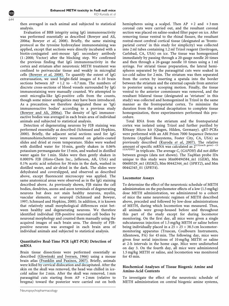
neurotoxic regimen of four injections of 10 mg/kg METHspaced at 2-h intervals as previously described (Eischet al, 1998; Deng et al, 1999; Kita et al, 2003). FosB( + / + )and (�/�) mice were killed 3 days later and examined forneurodegeneration by FJB labeling. FJB is a fluorescent dyethat has high affinity for degenerating neurons, regardlessof specific insult or mechanism of cell death (Schmued andHopkins, 2000). Consistent with previous reports (Ryanet al, 1990; Schmued and Bowyer, 1997; Deng et al, 2001),neurodegeneration occurred in the frontoparietal andpiriform cortices and in the striatum (Figure 1a), inMETH-treated both FosB( + / + ) and (�/�) mice. We foundsome FJB-positive neurons in the hippocampal complex aswell, but to a much lesser extent (data not shown). In bothFosB( + / + ) and (�/�) saline-treated animals, FJB-positiveneurons were rarely found in any brain area (Figure 1b).
The densities of FJB-positive degenerating neurons werequantified and analyzed by a two-way ANOVA withintersubject factors of genotype and treatment (Figure 1b).Significant effects of treatment were found in the fronto-parietal cortex (F1,18¼ 24.84, po0.001), striatum (F1,18¼5.00, p¼ 0.038), and piriform cortex (F1,18¼ 8.40, p¼0.010). Significant effects of genotype were found in thefrontoparietal cortex (F1,18¼ 15.32, p¼ 0.001) and striatum(F1,18¼ 4.61, p¼ 0.046), but not in the piriform cortex(F1,18¼ 2.06, p¼ 0.169). Significant genotype–treatmentinteractions were found in the frontoparietal cortex(F1,18¼ 15.37, p¼ 0.001) and striatum (F1,18¼ 4.59,p¼ 0.046), but not in the piriform cortex (F1,18¼ 2.07,p¼ 0.168). After METH treatment, therefore, the front-oparietal cortex and striatum of FosB(�/�) mice containedsignificantly more FJB-positive neurons than those of theirFosB( + / + ) littermates. In contrast, we did not finda significant genotype–treatment interaction in the piriformcortex, possibly because there was large individual varia-bility in the number of degenerating neurons in both (�/�)and ( + / + ) mice. These data suggested that FosB protectsneurons in the frontoparietal cortex and striatum fromMETH-induced neurotoxicity.
FJB-positive cells tended to cluster in subregions. In theparietal cortex, most of the degenerating cells were layer-IIIpyramidal neurons and superficial layer-IV multipolarneurons in the parietal cortex area 1, as previouslydescribed (Eisch et al, 1998). Moreover, FJB-positive cellstended to be found in the very center of the striatum. In thepiriform cortex, the FJB-positive cells were clustered in thedorsal endopiriform area. These distribution patterns ofFJB-positive neurons are the same in both genotypes.Notably, these distribution patterns were very similar to thearea of BBB dysfunction identified and described below.
We also sought evidence of differences in the amount ofapoptosis by means of the TUNEL histochemical methodand anticleaved caspase-3 immunodetection method. Weobserved much smaller numbers of apoptotic neurons byeither anticleaved caspase-3 immunoreactivity or theTUNEL method in forebrain regions 3 days after four10 mg/kg injections in both FosB(�/�) and ( + / + ) micecompared with the numbers of FJB-positive neurons (datanot shown). Thus, under our experimental conditions, itappeared that METH-induced neuronal death in FosB(�/�)mice was largely due to necrosis or some other nonapopto-tic regulated death program.
Enhanced METH toxicity in FosB null miceKO Kuroda et al
645
Neuropsychopharmacology
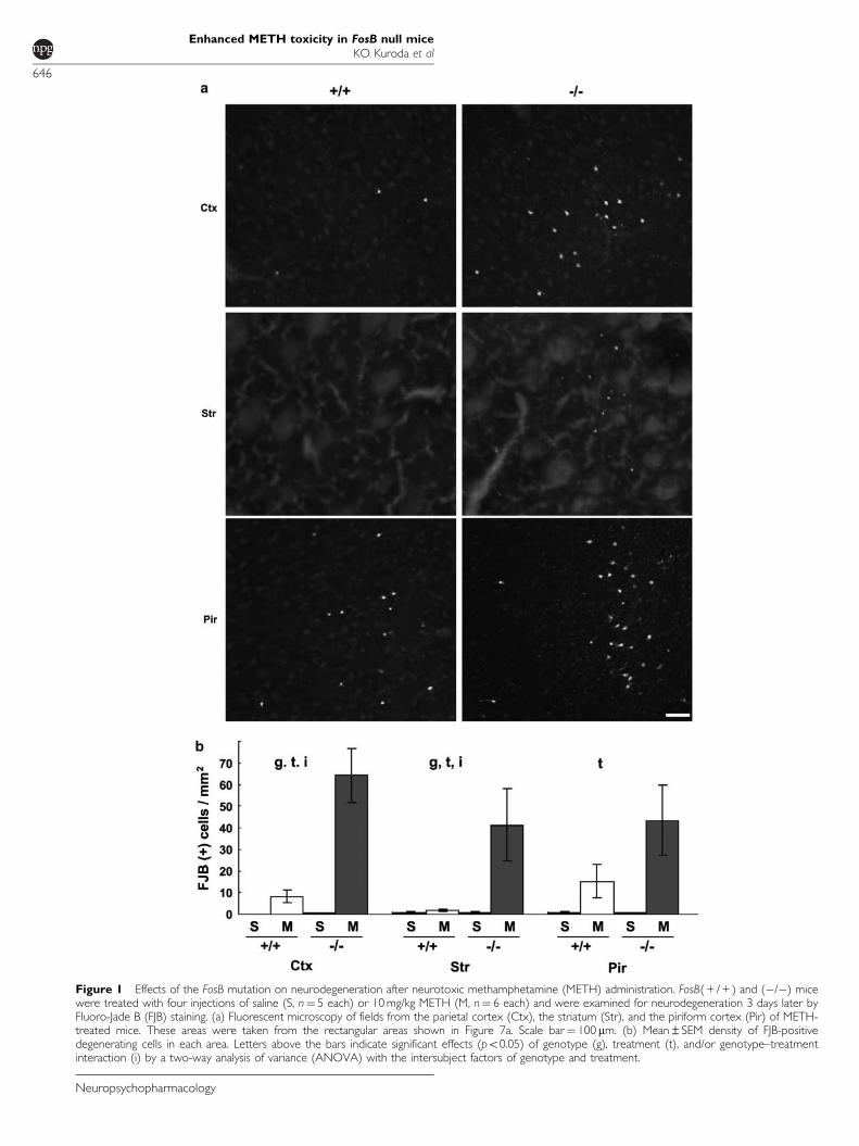
Figure 1 Effects of the FosB mutation on neurodegeneration after neurotoxic methamphetamine (METH) administration. FosB( + / + ) and (�/�) micewere treated with four injections of saline (S, n¼ 5 each) or 10 mg/kg METH (M, n¼ 6 each) and were examined for neurodegeneration 3 days later byFluoro-Jade B (FJB) staining. (a) Fluorescent microscopy of fields from the parietal cortex (Ctx), the striatum (Str), and the piriform cortex (Pir) of METH-treated mice. These areas were taken from the rectangular areas shown in Figure 7a. Scale bar¼ 100 mm. (b) Mean±SEM density of FJB-positivedegenerating cells in each area. Letters above the bars indicate significant effects (po0.05) of genotype (g), treatment (t), and/or genotype–treatmentinteraction (i) by a two-way analysis of variance (ANOVA) with the intersubject factors of genotype and treatment.
Enhanced METH toxicity in FosB null miceKO Kuroda et al
646
Neuropsychopharmacology
No Differences in METH-Induced Hyperthermia inFosB(�/�) Mice
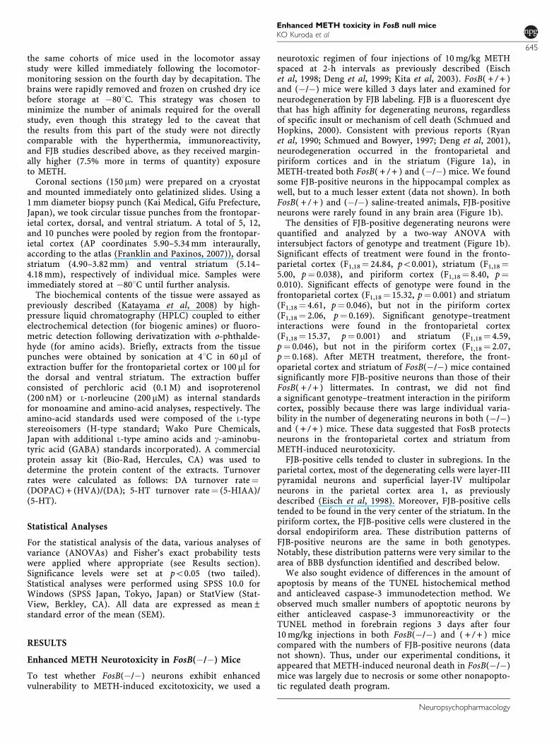
Methamphetamine-induced neurodegeneration is mediatedat least partly by hyperthermia (Bowyer, 1995). Thus,we examined the core body temperature of FosB(�/�) and( + / + ) mice during the neurotoxic regimen of METHadministration. Using a repeated-measures ANOVA withthe intersubject factors of genotype and the within-subjectfactor of time point, we found a significant effect of time incore body temperature (F10,80¼ 28.27, po0.001) due to aprogressive increase in body temperature in both geno-types, which returned to basal levels by the end of the 7-hrecording period (Figure 2). No significant effects ofgenotype were found in core body temperature after METHinjection (F1,8¼ 0.047, p¼ 0.834). FosB(�/�) mice thereforedid not show altered susceptibility to METH-inducedhyperthermia, thus eliminating this factor as the primarycause of enhanced neurodegeneration in these mice.
Increased Incidence of Self-Injury in METH-TreatedFosB(�/�) Mice
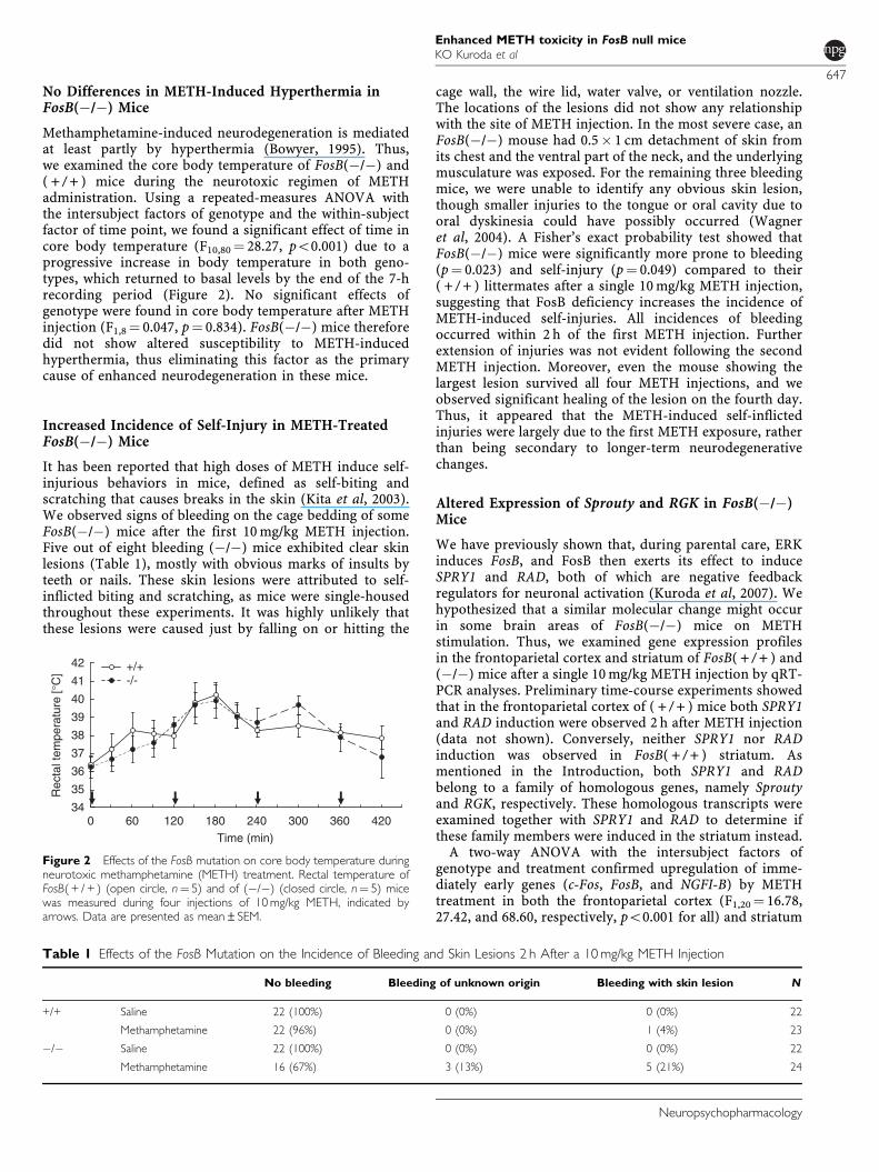
It has been reported that high doses of METH induce self-injurious behaviors in mice, defined as self-biting andscratching that causes breaks in the skin (Kita et al, 2003).We observed signs of bleeding on the cage bedding of someFosB(�/�) mice after the first 10 mg/kg METH injection.Five out of eight bleeding (�/�) mice exhibited clear skinlesions (Table 1), mostly with obvious marks of insults byteeth or nails. These skin lesions were attributed to self-inflicted biting and scratching, as mice were single-housedthroughout these experiments. It was highly unlikely thatthese lesions were caused just by falling on or hitting the
cage wall, the wire lid, water valve, or ventilation nozzle.The locations of the lesions did not show any relationshipwith the site of METH injection. In the most severe case, anFosB(�/�) mouse had 0.5� 1 cm detachment of skin fromits chest and the ventral part of the neck, and the underlyingmusculature was exposed. For the remaining three bleedingmice, we were unable to identify any obvious skin lesion,though smaller injuries to the tongue or oral cavity due tooral dyskinesia could have possibly occurred (Wagneret al, 2004). A Fisher’s exact probability test showed thatFosB(�/�) mice were significantly more prone to bleeding(p¼ 0.023) and self-injury (p¼ 0.049) compared to their( + / + ) littermates after a single 10 mg/kg METH injection,suggesting that FosB deficiency increases the incidence ofMETH-induced self-injuries. All incidences of bleedingoccurred within 2 h of the first METH injection. Furtherextension of injuries was not evident following the secondMETH injection. Moreover, even the mouse showing thelargest lesion survived all four METH injections, and weobserved significant healing of the lesion on the fourth day.Thus, it appeared that the METH-induced self-inflictedinjuries were largely due to the first METH exposure, ratherthan being secondary to longer-term neurodegenerativechanges.
Altered Expression of Sprouty and RGK in FosB(�/�)Mice
We have previously shown that, during parental care, ERKinduces FosB, and FosB then exerts its effect to induceSPRY1 and RAD, both of which are negative feedbackregulators for neuronal activation (Kuroda et al, 2007). Wehypothesized that a similar molecular change might occurin some brain areas of FosB(�/�) mice on METHstimulation. Thus, we examined gene expression profilesin the frontoparietal cortex and striatum of FosB( + / + ) and(�/�) mice after a single 10 mg/kg METH injection by qRT-PCR analyses. Preliminary time-course experiments showedthat in the frontoparietal cortex of ( + / + ) mice both SPRY1and RAD induction were observed 2 h after METH injection(data not shown). Conversely, neither SPRY1 nor RADinduction was observed in FosB( + / + ) striatum. Asmentioned in the Introduction, both SPRY1 and RADbelong to a family of homologous genes, namely Sproutyand RGK, respectively. These homologous transcripts wereexamined together with SPRY1 and RAD to determine ifthese family members were induced in the striatum instead.
A two-way ANOVA with the intersubject factors ofgenotype and treatment confirmed upregulation of imme-diately early genes (c-Fos, FosB, and NGFI-B) by METHtreatment in both the frontoparietal cortex (F1,20¼ 16.78,27.42, and 68.60, respectively, po0.001 for all) and striatum
34
35
36
37
38
39
40
41
42
Time (min)
Rec
tal t
empe
ratu
re [°
C] +/+
-/-
0 60 120 180 240 300 360 420
Figure 2 Effects of the FosB mutation on core body temperature duringneurotoxic methamphetamine (METH) treatment. Rectal temperature ofFosB( + / + ) (open circle, n¼ 5) and of (�/�) (closed circle, n¼ 5) micewas measured during four injections of 10 mg/kg METH, indicated byarrows. Data are presented as mean±SEM.
Table 1 Effects of the FosB Mutation on the Incidence of Bleeding and Skin Lesions 2 h After a 10 mg/kg METH Injection
No bleeding Bleeding of unknown origin Bleeding with skin lesion N
+/+ Saline 22 (100%) 0 (0%) 0 (0%) 22
Methamphetamine 22 (96%) 0 (0%) 1 (4%) 23
�/� Saline 22 (100%) 0 (0%) 0 (0%) 22
Methamphetamine 16 (67%) 3 (13%) 5 (21%) 24
Enhanced METH toxicity in FosB null miceKO Kuroda et al
647
Neuropsychopharmacology
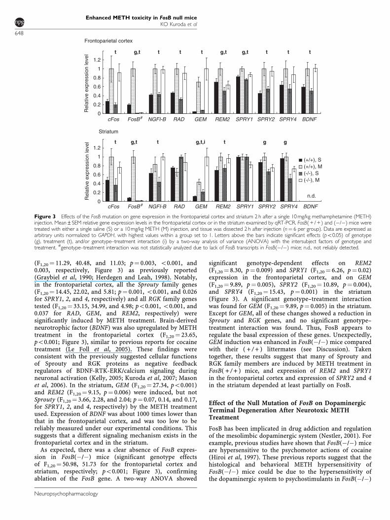
(F1,20¼ 11.29, 40.48, and 11.03; p¼ 0.003, o0.001, and0.003, respectively, Figure 3) as previously reported(Graybiel et al, 1990; Herdegen and Leah, 1998). Notably,in the frontoparietal cortex, all the Sprouty family genes(F1,20¼ 14.45, 22.02, and 5.81; p¼ 0.001, o0.001, and 0.026for SPRY1, 2, and 4, respectively) and all RGK family genestested (F1,20¼ 33.15, 34.99, and 4.98; po0.001, o0.001, and0.037 for RAD, GEM, and REM2, respectively) weresignificantly induced by METH treatment. Brain-derivedneurotrophic factor (BDNF) was also upregulated by METHtreatment in the frontoparietal cortex (F1,20¼ 23.65,po0.001; Figure 3), similar to previous reports for cocainetreatment (Le Foll et al, 2005). These findings wereconsistent with the previously suggested cellular functionsof Sprouty and RGK proteins as negative feedbackregulators of BDNF-RTK-ERK/calcium signaling duringneuronal activation (Kelly, 2005; Kuroda et al, 2007; Masonet al, 2006). In the striatum, GEM (F1,20¼ 27.34, po0.001)and REM2 (F1,20¼ 9.15, p¼ 0.006) were induced, but notSprouty (F1,20¼ 3.66, 2.28, and 2.04; p¼ 0.07, 0.14, and 0.17,for SPRY1, 2, and 4, respectively) by the METH treatmentused. Expression of BDNF was about 1000 times lower thanthat in the frontoparietal cortex, and was too low to bereliably measured under our experimental conditions. Thissuggests that a different signaling mechanism exists in thefrontoparietal cortex and in the striatum.
As expected, there was a clear absence of FosB expres-sion in FosB(�/�) mice (significant genotype effectsof F1,20¼ 50.98, 51.73 for the frontoparietal cortex andstriatum, respectively; po0.001; Figure 3), confirmingablation of the FosB gene. A two-way ANOVA showed
significant genotype-dependent effects on REM2(F1,20¼ 8.30, p¼ 0.009) and SPRY1 (F1,20¼ 6.26, p¼ 0.02)expression in the frontoparietal cortex, and on GEM(F1,20¼ 9.89, p¼ 0.005), SPRY2 (F1,20¼ 10.89, p¼ 0.004),and SPRY4 (F1,20¼ 15.43, p¼ 0.001) in the striatum(Figure 3). A significant genotype–treatment interactionwas found for GEM (F1,20¼ 9.89, p¼ 0.005) in the striatum.Except for GEM, all of these changes showed a reduction inSprouty and RGK genes, and no significant genotype–treatment interaction was found. Thus, FosB appears toregulate the basal expression of these genes. Unexpectedly,GEM induction was enhanced in FosB(�/�) mice comparedwith their ( + / + ) littermates (see Discussion). Takentogether, these results suggest that many of Sprouty andRGK family members are induced by METH treatment inFosB( + / + ) mice, and expression of REM2 and SPRY1in the frontoparietal cortex and expression of SPRY2 and 4in the striatum depended at least partially on FosB.
Effect of the Null Mutation of FosB on DopaminergicTerminal Degeneration After Neurotoxic METHTreatment
FosB has been implicated in drug addiction and regulationof the mesolimbic dopaminergic system (Nestler, 2001). Forexample, previous studies have shown that FosB(�/�) miceare hypersensitive to the psychomotor actions of cocaine(Hiroi et al, 1997). These previous reports suggest that thehistological and behavioral METH hypersensitivity ofFosB(�/�) mice could be due to the hypersensitivity ofthe dopaminergic system to psychostimulants in FosB(�/�)
0
0.2
0.4
0.6
0.8
1
1.2
0
0.2
0.4
0.6
0.8
1
1.2
FosB#
FosB #
BDNF
BDNF
(+/+), S(+/+), M(-/-), S(-/-), M
n.d.Rel
ativ
e ex
pres
sion
leve
lR
elat
ive
expr
essi
on le
vel t
g,t
t
t t tg,t,i g
Frontoparietal cortex
Striatum
g,t g,t g,tt t t t t
cFos
cFos
NGFI-B
NGFI-B
RAD
RAD
GEM
GEM
REM2
REM2
SPRY1
SPRY1
SPRY2
SPRY2
SPRY4
SPRY4
g
Figure 3 Effects of the FosB mutation on gene expression in the frontoparietal cortex and striatum 2 h after a single 10 mg/kg methamphetamine (METH)injection. Mean±SEM relative gene expression levels in the frontoparietal cortex or in the striatum examined by qRT-PCR. FosB( + / + ) and (�/�) mice weretreated with either a single saline (S) or a 10 mg/kg METH (M) injection, and tissue was dissected 2 h after injection (n¼ 6 per group). Data are expressed asarbitrary units normalized to GAPDH, with highest values within a group set to 1. Letters above the bars indicate significant effects (po0.05) of genotype(g), treatment (t), and/or genotype–treatment interaction (i) by a two-way analysis of variance (ANOVA) with the intersubject factors of genotype andtreatment. #genotype–treatment interaction was not statistically analyzed due to lack of FosB transcripts in FosB(�/�) mice; n.d., not reliably detected.
Enhanced METH toxicity in FosB null miceKO Kuroda et al
648
Neuropsychopharmacology
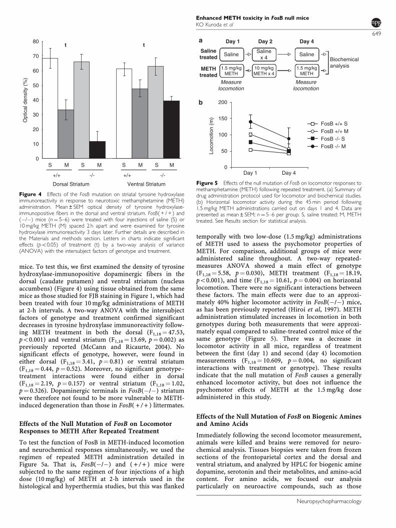
mice. To test this, we first examined the density of tyrosinehydroxylase-immunopositive dopaminergic fibers in thedorsal (caudate putamen) and ventral striatum (nucleusaccumbens) (Figure 4) using tissue obtained from the samemice as those studied for FJB staining in Figure 1, which hadbeen treated with four 10 mg/kg administrations of METHat 2-h intervals. A two-way ANOVA with the intersubjectfactors of genotype and treatment confirmed significantdecreases in tyrosine hydroxylase immunoreactivity follow-ing METH treatment in both the dorsal (F1,18¼ 47.53,po0.001) and ventral striatum (F1,18¼ 13.69, p¼ 0.002) aspreviously reported (McCann and Ricaurte, 2004). Nosignificant effects of genotype, however, were found ineither dorsal (F1,18¼ 3.41, p¼ 0.81) or ventral striatum(F1,18¼ 0.44, p¼ 0.52). Moreover, no significant genotype–treatment interactions were found either in dorsal(F1,18¼ 2.19, p¼ 0.157) or ventral striatum (F1,18¼ 1.02,p¼ 0.326). Dopaminergic terminals in FosB(�/�) striatumwere therefore not found to be more vulnerable to METH-induced degeneration than those in FosB( + / + ) littermates.
Effects of the Null Mutation of FosB on LocomotorResponses to METH After Repeated Treatment
To test the function of FosB in METH-induced locomotionand neurochemical responses simultaneously, we used theregimen of repeated METH administration detailed inFigure 5a. That is, FosB(�/�) and ( + / + ) mice weresubjected to the same regimen of four injections of a highdose (10 mg/kg) of METH at 2-h intervals used in thehistological and hyperthermia studies, but this was flanked
temporally with two low-dose (1.5 mg/kg) administrationsof METH used to assess the psychomotor properties ofMETH. For comparison, additional groups of mice wereadministered saline throughout. A two-way repeated-measures ANOVA showed a main effect of genotype(F1,18¼ 5.58, p¼ 0.030), METH treatment (F1,18¼ 18.19,po0.001), and time (F1,18¼ 10.61, p¼ 0.004) on horizontallocomotion. There were no significant interactions betweenthese factors. The main effects were due to an approxi-mately 40% higher locomotor activity in FosB(�/�) mice,as has been previously reported (Hiroi et al, 1997). METHadministration stimulated increases in locomotion in bothgenotypes during both measurements that were approxi-mately equal compared to saline-treated control mice of thesame genotype (Figure 5). There was a decrease inlocomotor activity in all mice, regardless of treatmentbetween the first (day 1) and second (day 4) locomotionmeasurements (F1,18¼ 10.609, p¼ 0.004, no significantinteractions with treatment or genotype). These resultsindicate that the null mutation of FosB causes a generallyenhanced locomotor activity, but does not influence thepsychomotor effects of METH at the 1.5 mg/kg doseadministered in this study.
Effects of the Null Mutation of FosB on Biogenic Aminesand Amino Acids
Immediately following the second locomotor measurement,animals were killed and brains were removed for neuro-chemical analysis. Tissues biopsies were taken from frozensections of the frontoparietal cortex and the dorsal andventral striatum, and analyzed by HPLC for biogenic aminedopamine, serotonin and their metabolites, and amino-acidcontent. For amino acids, we focused our analysisparticularly on neuroactive compounds, such as those
0
10
20
30
40
50
60
70
80
S
+/+ -/- +/+ -/-
Dorsal Striatum Ventral Striatum
Opt
ical
den
sity
(%
)
t t
M S M S M S M
Figure 4 Effects of the FosB mutation on striatal tyrosine hydroxylaseimmunoreactivity in response to neurotoxic methamphetamine (METH)administration. Mean±SEM optical density of tyrosine hydroxylase-immunopositive fibers in the dorsal and ventral striatum. FosB( + / + ) and(�/�) mice (n¼ 5–6) were treated with four injections of saline (S) or10 mg/kg METH (M) spaced 2 h apart and were examined for tyrosinehydroxylase immunoreactivity 3 days later. Further details are described inthe Materials and methods section. Letters in charts indicate significanteffects (po0.05) of treatment (t) by a two-way analysis of variance(ANOVA) with the intersubject factors of genotype and treatment.
Day 1
Measurelocomotion
Measurelocomotion
METHtreated
Salinetreated Biochemical
analysis1.5 mg/kgMETH
10 mg/kgMETH x 4
SalineSaline
x 4Saline
1.5 mg/kgMETH
0
50
100
150
200
Day 1
Loco
mot
ion
(m)
FosB +/+ S FosB +/+ MFosB -/- SFosB -/- M
Day 4
Day 2 Day 4
Figure 5 Effects of the null mutation of FosB on locomotor responses tomethamphetamine (METH) following repeated treatment. (a) Summary ofdrug administration protocol used for locomotor and biochemical studies.(b) Horizontal locomotor activity during the 45 min period following1.5 mg/kg METH administrations carried out on days 1 and 4. Data arepresented as mean±SEM; n¼ 5–6 per group. S, saline treated; M, METHtreated. See Results section for statistical analysis.
Enhanced METH toxicity in FosB null miceKO Kuroda et al
649
Neuropsychopharmacology
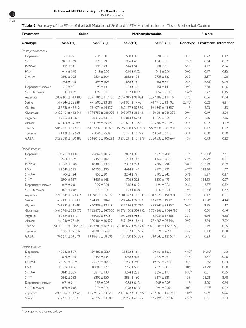
Table 2 Summary of the Effect of the Null Mutation of FosB and METH Administration on Tissue Biochemical Content
Treatment Saline Methamphetamine F score
Genotype FosB(+/+) FosB(�/�) FosB(+/+) FosB(�/�) Genotype Treatment Interaction
Frontoparietal cortex
Dopamine 863±291 644±80 588±47 591±65 0.40 0.92 0.42
5-HT 2103±169 1720±99 1986±67 1640±81 9.50a 0.64 0.02
DOPAC 675±76 737±83 526±58 531±51 0.22 6.17b 0.16
HVA 0.16±0.03 0.18±0.02 0.16±0.02 0.15±0.01 0.02 0.47 0.82
5-HIAA 3143±305 3534±204 2832±173 2759±123 0.50 5.87b 1.08
3-MT 1506±105 1595±109 888±78 909±36 0.35 49.78c 0.14
Dopamine turnover 217±40 199±13 183±10 151±14 0.93 2.58 0.06
5-HT turnover 1.44±0.24 1.92±0.15 1.32±0.09 1.57±0.12 4.66b 1.97 0.45
Aspartate 2 002 101±143 483 2 271 386±117 185 2 057 045±98 824 2 277 182±131 161 3.75 0.06 0.04
Serine 519 244±23 648 471 500±23 081 566 901±14 451 417 910±12 192 23.80c 0.02 6.31b
Glycine 897 738±49 512 791 071±44 137 960 127±52 530 964 342±43 857 1.15 6.03b 1.33
Glutamate 10 822 461±412 541 11 778 759±688 003 11 898 097±388 441 11 130 684±286 375 0.04 0.19 3.04
Arginine 119 562±8832 138 312±13 715 122 813±5723 111 627±6652 0.17 1.30 2.36
Alanine 378 166±19 089 434 195±25 799 420 621±13 531 385 787±21 593 0.25 0.02 4.62b
Taurine 15 049 623±972 040 16 882 232±607 688 15 897 408±590 618 16 609 734±384 983 3.22 0.17 0.62
Tyrosine 71 428±12 650 71 046±7532 75 191±10 976 68 664±6715 0.14 0.00 0.10
GABA 3 283 089±150 883 3 516 012±106 266 3 232 211±131 679 3 320 530±109 647 1.57 0.93 0.32
Dorsal striatum
Dopamine 108 253±6140 95 862±4079 2857±321 4226±2004 1.74 556.44c 2.71
5-HT 2768±169 2451±102 1753±162 1462±282 2.76 29.99c 0.01
DOPAC 18 865±1206 18 489±1327 2357±274 2607±790 0.00 233.29c 0.09
HVA 11 840±515 10 597±293 4624±145 4179±425 4.79b 312.98c 1.07
5-HIAA 1904±124 1855±60 2294±76 2103±242 0.76 5.37b 0.27
3-MT 8804±557 8405±148 1706±205 1520±475 0.55 313.22c 0.07
Dopamine turnover 0.25±0.01 0.27±0.01 2.16±0.12 1.96±0.31 0.36 143.87c 0.52
5-HT turnover 0.64±0.04 0.70±0.03 1.23±0.08 1.49±0.24 1.95 35.74c 0.72
Aspartate 2 210 649±173 916 1 889 815±85 922 2 301 472±181 832 2 017 823±190 934 3.56 0.47 0.01
Serine 622 122±30 893 524 393±6869 794 446±26 922 565 626±49 922 27.75c 11.80a 4.44b
Glycine 746 782±16 438 620 909±23 418 757 266±33 710 699 745±38 857 10.67a 2.55 1.47
Glutamate 10 615 960±533 075 9 962 872±347 905 10 876 150±534 132 9 758 686±1 024 985 1.98 0.00 0.14
Arginine 160 624±8113 166 050±8938 207 216±9881 165 037±17 686 2.57 4.14 4.48b
Alanine 264 040±23 684 300 484±10 927 359 199±18 464 282 208±29 546 0.92 3.24 7.02b
Taurine 20 113 313±1 367 828 19 073 780±469 117 21 804 666±923 787 20 231 585±1 637 668 1.26 1.49 0.05
Tyrosine 36 684±12 916 28 200±5647 79 152±17 535 51 624±7654 2.42 8.13b 0.68
GABA 1 946 677±94 370 1 818 617±58 006 1 939 780±59 306 1 910 845±129 597 0.78 0.23 0.31
Ventral striatum
Dopamine 48 342±5271 59 487±2567 25 582±1611 29 464±1832 4.82b 59.46c 1.13
5-HT 3926±345 3454±135 3288±409 2627±291 3.45 5.77b 0.10
DOPAC 25 091±2525 25 529±4048 16 966±2401 19 558±2377 0.25 5.35b 0.13
HVA 10 936±656 10 801±777 7706±518 7529±507 0.06 24.99c 0.00
5-HIAA 3149±205 2811±133 3274±233 2657±177 6.38b 0.01 0.55
3-MT 5162±582 6295±250 3831±160 3674±329 1.59 26.08c 2.78
Dopamine turnover 0.71±0.11 0.55±0.08 0.88±0.13 0.83±0.09 1.13 5.00b 0.24
5-HT turnover 0.76±0.05 0.76±0.06 0.98±0.13 0.96±0.09 0.00 6.07b 0.02
Aspartate 2 005 782±117 528 1 797 912±74 523 2 175 627±166 697 1 782 605±137 759 5.84b 0.39 0.56
Serine 539 434±46 591 496 727±23 888 636 936±61 195 446 196±32 332 7.55b 0.31 3.04
Enhanced METH toxicity in FosB null miceKO Kuroda et al
650
Neuropsychopharmacology
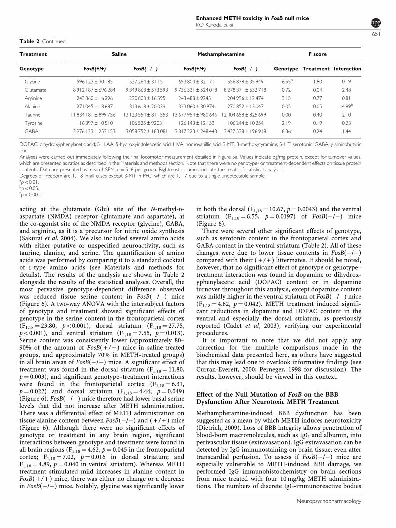
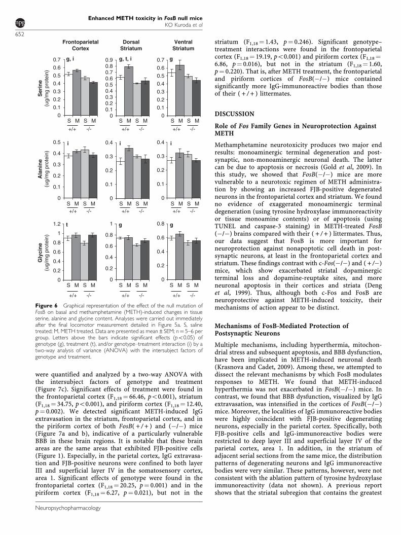
acting at the glutamate (Glu) site of the N-methyl-D-aspartate (NMDA) receptor (glutamate and aspartate), atthe co-agonist site of the NMDA receptor (glycine), GABA,and arginine, as it is a precursor for nitric oxide synthesis(Sakurai et al, 2004). We also included several amino acidswith either putative or unspecified neuroactivity, such astaurine, alanine, and serine. The quantification of aminoacids was performed by comparing it to a standard cocktailof L-type amino acids (see Materials and methods fordetails). The results of the analysis are shown in Table 2alongside the results of the statistical analyses. Overall, themost pervasive genotype-dependent difference observedwas reduced tissue serine content in FosB(�/�) mice(Figure 6). A two-way ANOVA with the intersubject factorsof genotype and treatment showed significant effects ofgenotype in the serine content in the frontoparietal cortex(F1,18¼ 23.80, po0.001), dorsal striatum (F1,18¼ 27.75,po0.001), and ventral striatum (F1,18¼ 7.55, p¼ 0.013).Serine content was consistently lower (approximately 80–90% of the amount of FosB( + / + ) mice in saline-treatedgroups, and approximately 70% in METH-treated groups)in all brain areas of FosB(�/�) mice. A significant effect oftreatment was found in the dorsal striatum (F1,18¼ 11.80,p¼ 0.003), and significant genotype–treatment interactionswere found in the frontoparietal cortex (F1,18¼ 6.31,p¼ 0.022) and dorsal striatum (F1,18¼ 4.44, p¼ 0.049)(Figure 6). FosB(�/�) mice therefore had lower basal serinelevels that did not increase after METH administration.There was a differential effect of METH administration ontissue alanine content between FosB(�/�) and ( + / + ) mice(Figure 6). Although there were no significant effects ofgenotype or treatment in any brain region, significantinteractions between genotype and treatment were found inall brain regions (F1,18¼ 4.62, p¼ 0.045 in the frontoparietalcortex; F1,18¼ 7.02, p¼ 0.016 in dorsal striatum; andF1,18¼ 4.89, p¼ 0.040 in ventral striatum). Whereas METHtreatment stimulated mild increases in alanine content inFosB( + / + ) mice, there was either no change or a decreasein FosB(�/�) mice. Notably, glycine was significantly lower
in both the dorsal (F1,18¼ 10.67, p¼ 0.0043) and the ventralstriatum (F1,18¼ 6.55, p¼ 0.0197) of FosB(�/�) mice(Figure 6).
There were several other significant effects of genotype,such as serotonin content in the frontoparietal cortex andGABA content in the ventral striatum (Table 2). All of thesechanges were due to lower tissue contents in FosB(�/�)compared with their ( + / + ) littermates. It should be noted,however, that no significant effect of genotype or genotype–treatment interaction was found in dopamine or dihydrox-yphenylacetic acid (DOPAC) content or in dopamineturnover throughout this analysis, except dopamine contentwas mildly higher in the ventral striatum of FosB(�/�) mice(F1,18¼ 4.82, p¼ 0.042). METH treatment induced signifi-cant reductions in dopamine and DOPAC content in theventral and especially the dorsal striatum, as previouslyreported (Cadet et al, 2003), verifying our experimentalprocedures.
It is important to note that we did not apply anycorrection for the multiple comparisons made in thebiochemical data presented here, as others have suggestedthat this may lead one to overlook informative findings (seeCurran-Everett, 2000; Perneger, 1998 for discussion). Theresults, however, should be viewed in this context.
Effect of the Null Mutation of FosB on the BBBDysfunction After Neurotoxic METH Treatment
Methamphetamine-induced BBB dysfunction has beensuggested as a mean by which METH induces neurotoxicity(Dietrich, 2009). Loss of BBB integrity allows penetration ofblood-born macromolecules, such as IgG and albumin, intoperivascular tissue (extravasation). IgG extravasation can bedetected by IgG immunostaining on brain tissue, even aftertranscardial perfusion. To assess if FosB(�/�) mice areespecially vulnerable to METH-induced BBB damage, weperformed IgG immunohistochemistry on brain sectionsfrom mice treated with four 10 mg/kg METH administra-tions. The numbers of discrete IgG-immunoreactive bodies
Table 2 Continued
Treatment Saline Methamphetamine F score
Genotype FosB(+/+) FosB(�/�) FosB(+/+) FosB(�/�) Genotype Treatment Interaction
Glycine 596 123±30 185 527 264±31 151 653 804±32 171 556 878±35 949 6.55b 1.80 0.19
Glutamate 8 912 187±696 284 9 349 868±573 593 9 736 331±524 018 8 278 371±532 718 0.72 0.04 2.48
Arginine 243 360±16 296 230 803±16 595 243 488±9245 204 996±12 474 3.15 0.77 0.81
Alanine 271 045±18 687 313 618±20 039 323 060±30 974 270 852±13 047 0.05 0.05 4.89b
Taurine 11 834 181±899 756 13 123 554±811 553 13 677 954±980 646 12 404 658±825 699 0.00 0.40 2.10
Tyrosine 116 397±10 510 106 525±9203 126 143±12 153 106 244±10 254 2.19 0.19 0.23
GABA 3 976 123±253 153 3 058 752±183 081 3 817 223±248 443 3 437 538±196 918 8.36a 0.24 1.44
DOPAC, dihydroxyphenylacetic acid; 5-HIAA, 5-hydroxyindoleacetic acid; HVA, homovanillic acid; 3-MT, 3-methoxytyramine; 5-HT, serotonin; GABA, g-aminobutyricacid.Analyses were carried out immediately following the final locomotor measurement detailed in Figure 5a. Values indicate pg/mg protein, except for turnover values,which are presented as ratios as described in the Materials and methods section. Note that there were no genotype- or treatment-dependent effects on tissue proteincontents. Data are presented as mean±SEM. n¼ 5–6 per group. Rightmost columns indicate the result of statistical analysis.Degrees of freedom are 1, 18 in all cases except 3-MT in PFC, which are 1, 17 due to a single undetectable sample.apo0.01.bpo0.05.cpo0.001.
Enhanced METH toxicity in FosB null miceKO Kuroda et al
651
Neuropsychopharmacology
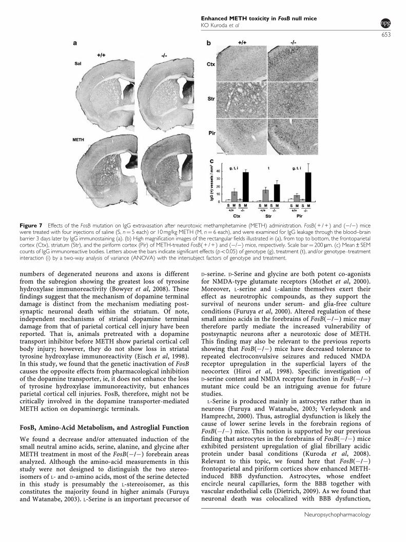
were quantified and analyzed by a two-way ANOVA withthe intersubject factors of genotype and treatment(Figure 7c). Significant effects of treatment were found inthe frontoparietal cortex (F1,18¼ 66.46, po0.001), striatum(F1,18¼ 34.75, po0.001), and piriform cortex (F1,18¼ 12.40,p¼ 0.002). We detected significant METH-induced IgGextravasation in the striatum, frontoparietal cortex, and inthe piriform cortex of both FosB( + / + ) and (�/�) mice(Figure 7a and b), indicative of a particularly vulnerableBBB in these brain regions. It is notable that these brainareas are the same areas that exhibited FJB-positive cells(Figure 1). Especially, in the parietal cortex, IgG extravasa-tion and FJB-positive neurons were confined to both layerIII and superficial layer IV in the somatosensory cortex,area 1. Significant effects of genotype were found in thefrontoparietal cortex (F1,18¼ 20.25, p¼ 0.001) and in thepiriform cortex (F1,18¼ 6.27, p¼ 0.021), but not in the
striatum (F1,18¼ 1.43, p¼ 0.246). Significant genotype–treatment interactions were found in the frontoparietalcortex (F1,18¼ 19.19, po0.001) and piriform cortex (F1,18¼6.86, p¼ 0.016), but not in the striatum (F1,18¼ 1.60,p¼ 0.220). That is, after METH treatment, the frontoparietaland piriform cortices of FosB(�/�) mice containedsignificantly more IgG-immunoreactive bodies than thoseof their ( + / + ) littermates.
DISCUSSION
Role of Fos Family Genes in Neuroprotection AgainstMETH
Methamphetamine neurotoxicity produces two major endresults: monoaminergic terminal degeneration and post-synaptic, non-monoaminergic neuronal death. The lattercan be due to apoptosis or necrosis (Gold et al, 2009). Inthis study, we showed that FosB(�/�) mice are morevulnerable to a neurotoxic regimen of METH administra-tion by showing an increased FJB-positive degeneratedneurons in the frontoparietal cortex and striatum. We foundno evidence of exaggerated monoaminergic terminaldegeneration (using tyrosine hydroxylase immunoreactivityor tissue monoamine contents) or of apoptosis (usingTUNEL and caspase-3 staining) in METH-treated FosB(�/�) brains compared with their ( + / + ) littermates. Thus,our data suggest that FosB is more important forneuroprotection against nonapoptotic cell death in post-synaptic neurons, at least in the frontoparietal cortex andstriatum. These findings contrast with c-Fos(�/�) and ( + /�)mice, which show exacerbated striatal dopaminergicterminal loss and dopamine-reuptake sites, and moreneuronal apoptosis in their cortices and striata (Denget al, 1999). Thus, although both c-Fos and FosB areneuroprotective against METH-induced toxicity, theirmechanisms of action appear to be distinct.
Mechanisms of FosB-Mediated Protection ofPostsynaptic Neurons
Multiple mechanisms, including hyperthermia, mitochon-drial stress and subsequent apoptosis, and BBB dysfunction,have been implicated in METH-induced neuronal death(Krasnova and Cadet, 2009). Among these, we attempted todissect the relevant mechanisms by which FosB modulatesresponses to METH. We found that METH-inducedhyperthermia was not exacerbated in FosB(�/�) mice. Incontrast, we found that BBB dysfunction, visualized by IgGextravasation, was intensified in the cortices of FosB(�/�)mice. Moreover, the localities of IgG immunoreactive bodieswere highly coincident with FJB-positive degeneratingneurons, especially in the parietal cortex. Specifically, bothFJB-positive cells and IgG-immunoreactive bodies wererestricted to deep layer III and superficial layer IV of theparietal cortex, area 1. In addition, in the striatum ofadjacent serial sections from the same mice, the distributionpatterns of degenerating neurons and IgG immunoreactivebodies were very similar. These patterns, however, were notconsistent with the ablation pattern of tyrosine hydroxylaseimmunoreactivity (data not shown). A previous reportshows that the striatal subregion that contains the greatest
0
0.1
0.2
0.3
0.4
0.5
0.6
0.7
S0
0.10.20.30.40.50.60.70.80.9
0
0.1
0.2
0.3
0.4
0.5
0.6
0.7
DorsalStriatum
FrontoparietalCortex
VentralStriatum
Ser
ine
(ug/
mg
prot
ein)
Ala
nin
e(u
g/m
g pr
otei
n)
+/+
0
0.1
0.2
0.3
0.4
0.5
0
0.1
0.2
0.3
0.4
0
0.1
0.2
0.3
0.4
-/-
+/+ -/- +/+ -/- +/+ -/-
+/+ -/-+/+ -/-
0
0.2
0.4
0.6
0.8
1
1.2
0
0.2
0.4
0.6
0.8
1
0
0.2
0.4
0.6
0.8
+/+ -/- +/+ -/-+/+ -/-
Gly
cin
e(u
g/m
g pr
otei
n)
g, i
i
g, t, i
i
g
g
gt
i
M S M S M S M S M S M
S M S MS M S MS M S M
S M S M S M S M S M S M
Figure 6 Graphical representation of the effect of the null mutation ofFosB on basal and methamphetamine (METH)-induced changes in tissueserine, alanine and glycine content. Analyses were carried out immediatelyafter the final locomotor measurement detailed in Figure 5a. S, salinetreated; M, METH treated. Data are presented as mean±SEM; n¼ 5–6 pergroup. Letters above the bars indicate significant effects (po0.05) ofgenotype (g), treatment (t), and/or genotype–treatment interaction (i) by atwo-way analysis of variance (ANOVA) with the intersubject factors ofgenotype and treatment.
Enhanced METH toxicity in FosB null miceKO Kuroda et al
652
Neuropsychopharmacology
numbers of degenerated neurons and axons is differentfrom the subregion showing the greatest loss of tyrosinehydroxylase immunoreactivity (Bowyer et al, 2008). Thesefindings suggest that the mechanism of dopamine terminaldamage is distinct from the mechanism mediating post-synaptic neuronal death within the striatum. Of note,independent mechanisms of striatal dopamine terminaldamage from that of parietal cortical cell injury have beenreported. That is, animals pretreated with a dopaminetransport inhibitor before METH show parietal cortical cellbody injury; however, they do not show loss in striataltyrosine hydroxylase immunoreactivity (Eisch et al, 1998).In this study, we found that the genetic inactivation of FosBcauses the opposite effects from pharmacological inhibitionof the dopamine transporter, ie, it does not enhance the lossof tyrosine hydroxylase immunoreactivity, but enhancesparietal cortical cell injuries. FosB, therefore, might not becritically involved in the dopamine transporter-mediatedMETH action on dopaminergic terminals.
FosB, Amino-Acid Metabolism, and Astroglial Function
We found a decrease and/or attenuated induction of thesmall neutral amino acids, serine, alanine, and glycine afterMETH treatment in most of the FosB(�/�) forebrain areasanalyzed. Although the amino-acid measurements in thisstudy were not designed to distinguish the two stereo-isomers of L- and D-amino acids, most of the serine detectedin this study is presumably the L-stereoisomer, as thisconstitutes the majority found in higher animals (Furuyaand Watanabe, 2003). L-Serine is an important precursor of
D-serine. D-Serine and glycine are both potent co-agonistsfor NMDA-type glutamate receptors (Mothet et al, 2000).Moreover, L-serine and L-alanine themselves exert theireffect as neurotrophic compounds, as they support thesurvival of neurons under serum- and glia-free cultureconditions (Furuya et al, 2000). Altered regulation of thesesmall amino acids in the forebrains of FosB(�/�) mice maytherefore partly mediate the increased vulnerability ofpostsynaptic neurons after a neurotoxic dose of METH.This finding may also be relevant to the previous reportsshowing that FosB(�/�) mice have decreased tolerance torepeated electroconvulsive seizures and reduced NMDAreceptor upregulation in the superficial layers of theneocortex (Hiroi et al, 1998). Specific investigation ofD-serine content and NMDA receptor function in FosB(�/�)mutant mice could be an intriguing avenue for futurestudies.
L-Serine is produced mainly in astrocytes rather than inneurons (Furuya and Watanabe, 2003; Verleysdonk andHamprecht, 2000). Thus, astroglial dysfunction is likely thecause of lower serine levels in the forebrain regions ofFosB(�/�) mice. This notion is supported by our previousfinding that astrocytes in the forebrains of FosB(�/�) miceexhibited persistent upregulation of glial fibrillary acidicprotein under basal conditions (Kuroda et al, 2008).Relevant to this topic, we found here that FosB(�/�)frontoparietal and piriform cortices show enhanced METH-induced BBB dysfunction. Astrocytes, whose endfeetencircle neural capillaries, form the BBB together withvascular endothelial cells (Dietrich, 2009). As we found thatneuronal death was colocalized with BBB dysfunction,
Figure 7 Effects of the FosB mutation on IgG extravasation after neurotoxic methamphetamine (METH) administration. FosB( + / + ) and (�/�) micewere treated with four injections of saline (S, n¼ 5 each) or 10 mg/kg METH (M, n¼ 6 each), and were examined for IgG leakage through the blood–brainbarrier 3 days later by IgG immunostaining (a). (b) High magnification images of the rectangular fields illustrated in (a), from top to bottom, the frontoparietalcortex (Ctx), striatum (Str), and the piriform cortex (Pir) of METH-treated FosB( + / + ) and (�/�) mice, respectively. Scale bar¼ 200 mm. (c) Mean±SEMcounts of IgG immunoreactive bodies. Letters above the bars indicate significant effects (po0.05) of genotype (g), treatment (t), and/or genotype–treatmentinteraction (i) by a two-way analysis of variance (ANOVA) with the intersubject factors of genotype and treatment.
Enhanced METH toxicity in FosB null miceKO Kuroda et al
653
Neuropsychopharmacology
astroglial dysfunction may mediate the enhanced METHtoxicity in the forebrains of FosB(�/�) mice. Indeed,several studies have reported the relevance of BBBdysfunction with neuronal death (Bowyer and Ali, 2006a;Kiyatkin et al, 2007). In the case of the excitotoxic injurymodel made by hippocampal kinate injection, however,neuronal death and BBB breakdown are separate, indepen-dent processes: BBB dysfunction occurs too late to initiateneurodegeneration, and BBB breakdown occurs withoutneuronal death (Chen et al, 1999). The causal relationship ofthese multiple events remains to be clarified.
Another puzzling fact is that FosB expression is confinedprimarily in neurons, as FosB expression in glial cells tendsto be undetectable, even under stimulation (Herdegen andLeah, 1998). The FosB protein in neurons may thereforecause wide arrays of abnormalities in astrocyte metabolismthrough neuron–glia communication. FosB(�/�) micemight be an interesting model system for understandingthe cross talk between neurons and glia cells.
FosB-Mediated Intracellular Signaling Regulation inResponse to Neurotoxic Doses of METH
After psychostimulant administration, elevation of intracel-lular calcium levels and ERK phosphorylation occurs inmany brain areas, resulting in the induction of Fos familytranscription factors (Hope et al, 1994; Valjent et al, 2004).Our previous study shows that the null mutation of FosBattenuates upregulation of SPRY1 and RAD, negativefeedback regulators of ERK and calcium signaling, respec-tively, in the medial preoptic area (Kuroda et al, 2007). Herewe found that basal and stimulated expression of Sproutyand RGK genes tended to be lower in FosB(�/�) mice, withthe only exception of higher GEM expression in the striatumof FosB(�/�) mice. As mentioned in the Introduction,METH-induced GEM upregulation is dependent on c-Fos(Cadet et al, 2002). Therefore GEM induction could beoverly compensated by c-Fos in the striatum of FosB(�/�)mice. Sprouty is a major class of ligand-inducible inhibitorsof RTK/ERK signaling pathway (Mason et al, 2006), andRGK is a potent inhibitor of calcium overinflux intoactivated cells (Kelly, 2005). Reduced expression of Sproutyand RGK can cause the overactivation of neurons, in termsof ERK phosphorylation and intracellular calcium elevation,respectively. Of note, it has been reported that the L-typecalcium channel agonist BayK8644 can cause self-injuriousbiting (Jinnah et al, 1999). RGK proteins are known todownregulate calcium influx by sequestering L-type calciumchannels (Kelly, 2005). These observations by no meansprove our hypothesis that the ERK/calcium–Fos–Sprouty/RGK molecular cascade could exert its effect as a negativefeedback loop against METH-induced neurotoxicity, but atleast are in accord. Fos target genes involved in such ageneral negative feedback might be found more easily thanFos target genes that have more specific functions in certainneuronal types. Indeed, all previous studies that havedetected attenuated RGK induction in Fos-gene-targetedmice (Cadet et al, 2002; Kuroda et al, 2007; Salzmann et al,2006) used grossly dissected brain tissue, containingdifferent neuronal populations.
In summary, the current study suggests two possiblemechanisms of FosB-mediated neuroprotection: one is
through induction of negative feedback regulation withinpostsynaptic neurons through Sprouty and RGK. Another issupporting astroglial function such as maintenance of theBBB, and metabolism of serine and glycine, which areimportant glial modulators of nerve cells. These twomechanisms are not necessarily mutually exclusive, andthe former may cause the latter through the neuron–gliainteraction. It is important to recognize, however, thatultimately the diverse findings provided here are correla-tive, and more directed study is necessary to provide acausative relationship.
ACKNOWLEDGEMENTS
We thank Yoshio Hirabayashi for helpful discussion,Tetsuaki Ara and Taeko Nemoto for technical assistance,and the RIKEN Research Resource Center for maintenanceof animals. This research was supported by RIKEN, Grants-in-Aid for Scientific Research from the Ministry ofEducation, Culture, Sports, Science, and Technology, Japan(2006–2008 to KOK and KT) and by the Uehara MemorialFoundation (2009 to KOK).
DISCLOSURE
The authors declare no conflict of interest.
REFERENCES
Berghorn KA, Bonnett JH, Hoffman GE (1994). cFos immunor-eactivity is enhanced with biotin amplification. J HistochemCytochem 42: 1635–1642.
Bowyer JF (1995). The role of hyperthermia in amphetamine’sinteractions with NMDA receptors, nitric oxide, and age toproduce neurotoxicity. Ann NY Acad Sci 765: 309–310.
Bowyer JF, Ali S (2006a). High doses of methamphetamine thatcause disruption of the blood–brain barrier in limbic regionsproduce extensive neuronal degeneration in mouse hippocam-pus. Synapse 60: 521–532.
Bowyer JF, Robinson B, Ali S, Schmued LC (2008). Neurotoxic-related changes in tyrosine hydroxylase, microglia, myelin, andthe blood–brain barrier in the caudate–putamen from acutemethamphetamine exposure. Synapse 62: 193–204.
Bowyer JF, Schmued LC (2006b). Fluoro-Ruby labeling prior to anamphetamine neurotoxic insult shows a definitive massive lossof dopaminergic terminals and axons in the caudate–putamen.Brain Res 1075: 236–239.
Brown JR, Ye H, Bronson RT, Dikkes P, Greenberg ME (1996). Adefect in nurturing in mice lacking the immediate early genefosB. Cell 86: 297–309.
Cadet JL, Jayanthi S, Deng X (2003). Speed kills: cellular andmolecular bases of methamphetamine-induced nerve terminaldegeneration and neuronal apoptosis. FASEB J 17: 1775–1788.
Cadet JL, McCoy MT, Ladenheim B (2002). Distinct geneexpression signatures in the striata of wild-type and hetero-zygous c-fos knockout mice following methamphetamineadministration: evidence from cDNA array analyses. Synapse44: 211–226.
Chen ZL, Indyk JA, Bugge TH, Kombrinck KW, Degen JL,Strickland S (1999). Neuronal death and blood–brain barrierbreakdown after excitotoxic injury are independent processes.J Neurosci 19: 9813–9820.
Curran-Everett D (2000). Multiple comparisons: philosophies andillustrations. Am J Physiol Regul Integr Comp Physiol 279: R1–R8.
Enhanced METH toxicity in FosB null miceKO Kuroda et al
654
Neuropsychopharmacology
Davidson C, Gow AJ, Lee TH, Ellinwood EH (2001). Methamphe-tamine neurotoxicity: necrotic and apoptotic mechanisms andrelevance to human abuse and treatment. Brain Res Brain ResRev 36: 1–22.
Deng X, Ladenheim B, Tsao LI, Cadet JL (1999). Null mutation ofc-fos causes exacerbation of methamphetamine-induced neuro-toxicity. J Neurosci 19: 10107–10115.
Deng X, Wang Y, Chou J, Cadet JL (2001). Methamphetaminecauses widespread apoptosis in the mouse brain: evidence fromusing an improved TUNEL histochemical method. Brain Res MolBrain Res 93: 64–69.
Dietrich JB (2009). Alteration of blood–brain barrier function bymethamphetamine and cocaine. Cell Tissue Res 336: 385–392.
Eisch AJ, Schmued LC, Marshall JF (1998). Characterizing corticalneuron injury with Fluoro-Jade labeling after a neurotoxicregimen of methamphetamine. Synapse 30: 329–333.
Franklin KBJ, Paxinos G (2007). The Mouse Brain in StereotaxicCoordinates 3rd edn. Academic Press: San Diego. pp 360.
Furuya S, Tabata T, Mitoma J, Yamada K, Yamasaki M, Makino Aet al (2000). -Serine and glycine serve as major astroglia-derivedtrophic factors for cerebellar Purkinje neurons. Proc Natl AcadSci USA 97: 11528–11533.
Furuya S, Watanabe M (2003). Novel neuroglial and glioglialrelationships mediated by L-serine metabolism. Arch Histol Cytol66: 109–121.
Glowinski J, Iversen LL (1966). Regional studies of catecholaminesin the rat brain. I. The disposition of [3H]norepinephrine,[3H]dopamine and [3H]dopa in various regions of the brain.J Neurochem 13: 655–669.
Gold MS, Kobeissy FH, Wang KK, Merlo LJ, Bruijnzeel AW,Krasnova IN et al (2009). Methamphetamine- and trauma-induced brain injuries: comparative cellular and molecularneurobiological substrates. Biol Psychiatry 66: 118–127.
Gown AM, Willingham MC (2002). Improved detection of apoptoticcells in archival paraffin sections: immunohistochemistry usingantibodies to cleaved caspase 3. J Histochem Cytochem 50: 449–454.
Graybiel AM, Moratalla R, Robertson HA (1990). Amphetamineand cocaine induce drug-specific activation of the c-fos gene instriosome-matrix compartments and limbic subdivisions of thestriatum. Proc Natl Acad Sci USA 87: 6912–6916.
Herdegen T, Leah JD (1998). Inducible and constitutive transcrip-tion factors in the mammalian nervous system: control of geneexpression by Jun, Fos and Krox, and CREB/ATF proteins. BrainRes Brain Res Rev 28: 370–490.
Hiroi N, Brown JR, Haile CN, Ye H, Greenberg ME, Nestler EJ (1997).FosB mutant mice: loss of chronic cocaine induction of Fos-relatedproteins and heightened sensitivity to cocaine’s psychomotor andrewarding effects. Proc Natl Acad Sci USA 94: 10397–10402.
Hiroi N, Marek GJ, Brown JR, Ye H, Saudou F, Vaidya VA et al(1998). Essential role of the FosB gene in molecular, cellular,and behavioral actions of chronic electroconvulsive seizures.J Neurosci 18: 6952–6962.
Hope BT, Nye HE, Kelz MB, Self DW, Iadarola MJ, Nakabeppu Yet al (1994). Induction of a long-lasting AP-1 complex composedof altered Fos-like proteins in brain by chronic cocaine andother chronic treatments. Neuron 13: 1235–1244.
Jinnah HA, Yitta S, Drew T, Kim BS, Visser JE, Rothstein JD (1999).Calcium channel activation and self-biting in mice. Proc NatlAcad Sci USA 96: 15228–15232.
Katayama K, Yamada K, Ornthanalai VG, Inoue T, Ota M, MurphyNP et al (2008). Slitrk1-deficient mice display elevated anxiety-like behavior and noradrenergic abnormalities. Mol Psychiatry(E-pub ahead of print).
Kelly K (2005). The RGK family: a regulatory tail of small GTP-binding proteins. Trends Cell Biol 15: 640–643.
Kita T, Wagner GC, Nakashima T (2003). Current research onmethamphetamine-induced neurotoxicity: animal models ofmonoamine disruption. J Pharmacol Sci 92: 178–195.
Kiyatkin EA, Brown PL, Sharma HS (2007). Brain edema andbreakdown of the blood–brain barrier during methamphetamineintoxication: critical role of brain hyperthermia. Eur J Neurosci26: 1242–1253.
Krasnova IN, Cadet JL (2009). Methamphetamine toxicity andmessengers of death. Brain Res Rev 60: 379–407.
Kuroda KO, Meaney MJ, Uetani N, Fortin Y, Ponton A, Kato T(2007). ERK-FosB signaling in dorsal MPOA neurons plays amajor role in the initiation of parental behavior in mice. Mol CelNeurosci 36: 121–131.
Kuroda KO, Meaney MJ, Uetani N, Kato T (2008). Neurobehavioralbasis of the impaired nurturing in mice lacking the immediateearly gene FosB. Brain Res 1211: 57–71.
Le Foll B, Diaz J, Sokoloff P (2005). A single cocaine exposureincreases BDNF and D3 receptor expression: implications fordrug-conditioning. NeuroReport 16: 175–178.
Mason JM, Morrison DJ, Basson MA, Licht JD (2006). Sproutyproteins: multifaceted negative-feedback regulators of receptortyrosine kinase signaling. Trends Cell Biol 16: 45–54.
McCann UD, Ricaurte GA (2004). Amphetamine neurotoxicity:accomplishments and remaining challenges. Neurosci BiobehavRev 27: 821–826.
Mothet JP, Parent AT, Wolosker H, Brady Jr RO, Linden DJ, FerrisCD et al (2000). -Serine is an endogenous ligand for the glycinesite of the N-methyl-D-aspartate receptor. Proc Natl Acad SciUSA 97: 4926–4931.
Nestler EJ (2001). Molecular basis of long-term plasticity under-lying addiction. Nat Rev Neurosci 2: 119–128.
Numan M, Numan MJ, Marzella SR, Palumbo A (1998). Expressionof c-fos, fos B, and egr-1 in the medial preoptic area and bednucleus of the stria terminalis during maternal behavior in rats.Brain Res 792: 348–352.
Perneger TV (1998). What’s wrong with Bonferroni adjustments.BMJ 316: 1236–1238.
Ryan LJ, Linder JC, Martone ME, Groves PM (1990). Histologicaland ultrastructural evidence that D-amphetamine causes degen-eration in neostriatum and frontal cortex of rats. Brain Res 518:67–77.
Sakurai S, Ishii S, Umino A, Shimazu D, Yamamoto N, Nishikawa T(2004). Effects of psychotomimetic and antipsychotic agents onneocortical and striatal concentrations of various amino acids inthe rat. J Neurochem 90: 1378–1388.
Salzmann J, Canestrelli C, Noble F, Marie-Claire C (2006). Analysisof transcriptional responses in the mouse dorsal striatumfollowing acute 3,4-methylenedioxymethamphetamine (ecstasy):identification of extracellular signal-regulated kinase-controlledgenes. Neuroscience 137: 473–482.
Schmued LC, Bowyer JF (1997). Methamphetamine exposure canproduce neuronal degeneration in mouse hippocampal rem-nants. Brain Res 759: 135–140.
Schmued LC, Hopkins KJ (2000). Fluoro-Jade B: a high affinityfluorescent marker for the localization of neuronal degeneration.Brain Res 874: 123–130.
Sulzer D, Sonders MS, Poulsen NW, Galli A (2005). Mechanisms ofneurotransmitter release by amphetamines: a review. ProgNeurobiol 75: 406–433.
Tata DA, Yamamoto BK (2007). Interactions between methamphe-tamine and environmental stress: role of oxidative stress, glutamateand mitochondrial dysfunction. Addiction 102(Suppl 1): 49–60.
Valjent E, Pages C, Herve D, Girault JA, Caboche J (2004).Addictive and non-addictive drugs induce distinct and specificpatterns of ERK activation in mouse brain. Eur J Neurosci 19:1826–1836.
Verleysdonk S, Hamprecht B (2000). Synthesis and release ofL-serine by rat astroglia-rich primary cultures. Glia 30: 19–26.
Wagner GC, Avena N, Kita T, Nakashima T, Fisher H, Halladay AK(2004). Risperidone reduction of amphetamine-induced self-injurious behavior in mice. Neuropharmacology 46: 700–708.
Enhanced METH toxicity in FosB null miceKO Kuroda et al
655
Neuropsychopharmacology
Copyright © 2022 FDOKUMEN