Aggregate formation prevents dTDP-43 neurotoxicity in the Drosophila melanogaster eye
7
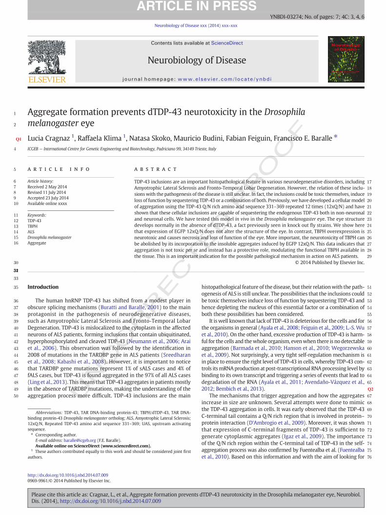
UNCORRECTED PROOF 1 Aggregate formation prevents dTDP-43 neurotoxicity in the Drosophila 2 melanogaster eye Q1 Lucia Cragnaz 1 , Raffaela Klima 1 , Natasa Skoko, Mauricio Budini, Fabian Feiguin, Francisco E. Baralle ⁎ 4 ICGEB — International Centre for Genetic Engineering and Biotechnology, Padriciano 99, 34149 Trieste, Italy abstract 5 article info 6 Article history: 7 Received 2 May 2014 8 Revised 11 July 2014 9 Accepted 23 July 2014 10 Available online xxxx 11 Keywords: 12 TDP-43 13 TBPH 14 ALS 15 Drosophila melanogaster 16 Aggregate 17 TDP-43 inclusions are an important histopathological feature in various neurodegenerative disorders, including 18 Amyotrophic Lateral Sclerosis and Fronto-Temporal Lobar Degeneration. However, the relation of these inclu- 19 sions with the pathogenesis of the disease is still unclear. In fact, the inclusions could be toxic themselves, induce 20 loss of function by sequestering TDP-43 or a combination of both. Previously, we have developed a cellular model 21 of aggregation using the TDP-43 Q/N rich amino acid sequence 331–369 repeated 12 times (12xQ/N) and have 22 shown that these cellular inclusions are capable of sequestering the endogenous TDP-43 both in non-neuronal 23 and neuronal cells. We have tested this model in vivo in the Drosophila melanogaster eye. The eye structure 24 develops normally in the absence of dTDP-43, a fact previously seen in knock out fly strains. We show here 25 that expression of EGFP 12xQ/N does not alter the structure of the eye. In contrast, TBPH overexpression is 26 neurotoxic and causes necrosis and loss of function of the eye. More important, the neurotoxicity of TBPH can 27 be abolished by its incorporation to the insoluble aggregates induced by EGFP 12xQ/N. This data indicates that 28 aggregation is not toxic per se and instead has a protective role, modulating the functional TBPH available in 29 the tissue. This is an important indication for the possible pathological mechanism in action on ALS patients. 30 © 2014 Published by Elsevier Inc. 31 32 33 34 35 Introduction 36 The human hnRNP TDP-43 has shifted from a modest player in 37 obscure splicing mechanisms (Buratti and Baralle, 2001) to the main 38 protagonist in the pathogenesis of neurodegenerative diseases, 39 such as Amyotrophic Lateral Sclerosis and Fronto-Temporal Lobar 40 Degeneration. TDP-43 is mislocalized to the cytoplasm in the affected 41 neurons of ALS patients, forming inclusions that contain ubiquitinated, 42 hyperphosphorylated and cleaved TDP-43 (Neumann et al., 2006; Arai 43 et al., 2006). This observation was followed by the identification in 44 2008 of mutations in the TARDBP gene in ALS patients (Sreedharan 45 et al., 2008; Kabashi et al., 2008). However, it is important to notice 46 that TARDBP gene mutations represent 1% of sALS cases and 4% of 47 fALS cases, but TDP-43 is found aggregated in the 97% of all ALS cases 48 (Ling et al., 2013). This means that TDP-43 aggregates in patients mostly 49 in the absence of TARDBP mutations, making the understanding of the 50 aggregation process more difficult. TDP-43 inclusions are the main 51 histopathological feature of the disease, but their relation with the path- 52 ogenesis of ALS is still unclear. The possibilities that the inclusions could 53 be toxic themselves induce loss of function by sequestering TDP-43 and 54 hence depleting the nucleus of this essential factor or a combination of 55 both these possibilities has been considered. 56 It is well known that lack of TDP-43 is deleterious for the cells and for 57 the organisms in general (Ayala et al., 2008; Feiguin et al., 2009; L.-S. Wu 58 et al., 2010). On the other hand, excessive production of TDP-43 is harm- 59 ful for the cells and the whole organism, even when there is no detectable 60 aggregation (Barmada et al., 2010; Hanson et al., 2010; Wegorzewska 61 et al., 2009). Not surprisingly, a very tight self-regulation mechanism is 62 in place to ensure the right level of TDP-43 in cells, whereby TDP-43 con- 63 trols its mRNA production at post-transcriptional RNA processing level by 64 binding to its own transcript and triggering a series of events that lead to 65 degradation of the RNA (Ayala et al., 2011; Avendaño-Vázquez et al., 2012; Q2 Bembich et al., 2013). 67 The mechanisms that trigger aggregation and how the aggregates 68 increase in size are unknown. Several attempts were done to mimic 69 the TDP-43 aggregation in cells. It was early observed that the TDP-43 70 C-terminal tail contains a Q/N rich region that is involved in protein– 71 protein interaction (D'Ambrogio et al., 2009). Moreover, it was shown 72 that expression of C-terminal fragments of TDP-43 is sufficient to 73 generate cytoplasmic aggregates (Igaz et al., 2009). The importance 74 of the Q/N rich region within the C-terminal tail of TDP-43 in the self- 75 aggregation process was also confirmed by Fuentealba et al. (Fuentealba 76 et al., 2010). Based on this information and with the aim of looking for Neurobiology of Disease xxx (2014) xxx–xxx Abbreviations: TDP-43, TAR DNA-binding protein-43; TBPH/dTDP-43, TAR DNA- binding protein-43 Drosophila melanogaster ortholog; ALS, Amyotrophic Lateral Sclerosis; 12xQ/N, Repeated TDP-43 amino acid sequence 331–369; UAS, upstream activating sequence. ⁎ Corresponding author. E-mail address: [email protected] (F.E. Baralle). Available online on ScienceDirect (www.sciencedirect.com). 1 These authors contributed equally to this work and should be considered joint first authors. YNBDI-03274; No. of pages: 7; 4C: 3, 4, 6 http://dx.doi.org/10.1016/j.nbd.2014.07.009 0969-9961/© 2014 Published by Elsevier Inc. Contents lists available at ScienceDirect Neurobiology of Disease journal homepage: www.elsevier.com/locate/ynbdi Please cite this article as: Cragnaz, L., et al., Aggregate formation prevents dTDP-43 neurotoxicity in the Drosophila melanogaster eye, Neurobiol. Dis. (2014), http://dx.doi.org/10.1016/j.nbd.2014.07.009
-
Upload
independent -
Category
Documents
-
view
5 -
download
0
Transcript of Aggregate formation prevents dTDP-43 neurotoxicity in the Drosophila melanogaster eye
UNCO
RRECTED P
RO
OF
1 Aggregate formation prevents dTDP-43 neurotoxicity in the Drosophila2 melanogaster eye
3Q1 Lucia Cragnaz 1, Raffaela Klima 1, Natasa Skoko, Mauricio Budini, Fabian Feiguin, Francisco E. Baralle ⁎4 ICGEB — International Centre for Genetic Engineering and Biotechnology, Padriciano 99, 34149 Trieste, Italy
a b s t r a c t5 a r t i c l e i n f o
6 Article history:7 Received 2 May 20148 Revised 11 July 20149 Accepted 23 July 201410 Available online xxxx
11 Keywords:12 TDP-4313 TBPH14 ALS15 Drosophila melanogaster16 Aggregate
17TDP-43 inclusions are an important histopathological feature in various neurodegenerative disorders, including18Amyotrophic Lateral Sclerosis and Fronto-Temporal Lobar Degeneration. However, the relation of these inclu-19sionswith the pathogenesis of the disease is still unclear. In fact, the inclusions could be toxic themselves, induce20loss of function by sequestering TDP-43 or a combination of both. Previously, we have developed a cellularmodel21of aggregation using the TDP-43 Q/N rich amino acid sequence 331–369 repeated 12 times (12xQ/N) and have22shown that these cellular inclusions are capable of sequestering the endogenous TDP-43 both in non-neuronal23and neuronal cells. We have tested this model in vivo in the Drosophila melanogaster eye. The eye structure24develops normally in the absence of dTDP-43, a fact previously seen in knock out fly strains. We show here25that expression of EGFP 12xQ/N does not alter the structure of the eye. In contrast, TBPH overexpression is26neurotoxic and causes necrosis and loss of function of the eye. More important, the neurotoxicity of TBPH can27be abolished by its incorporation to the insoluble aggregates induced by EGFP 12xQ/N. This data indicates that28aggregation is not toxic per se and instead has a protective role, modulating the functional TBPH available in29the tissue. This is an important indication for the possible pathological mechanism in action on ALS patients.
30 © 2014 Published by Elsevier Inc.
3132
33
34
35 Introduction
36 The human hnRNP TDP-43 has shifted from a modest player in37 obscure splicing mechanisms (Buratti and Baralle, 2001) to the main38 protagonist in the pathogenesis of neurodegenerative diseases,39 such as Amyotrophic Lateral Sclerosis and Fronto-Temporal Lobar40 Degeneration. TDP-43 is mislocalized to the cytoplasm in the affected41 neurons of ALS patients, forming inclusions that contain ubiquitinated,42 hyperphosphorylated and cleaved TDP-43 (Neumann et al., 2006; Arai43 et al., 2006). This observation was followed by the identification in44 2008 of mutations in the TARDBP gene in ALS patients (Sreedharan45 et al., 2008; Kabashi et al., 2008). However, it is important to notice46 that TARDBP gene mutations represent 1% of sALS cases and 4% of47 fALS cases, but TDP-43 is found aggregated in the 97% of all ALS cases48 (Ling et al., 2013). Thismeans that TDP-43 aggregates in patientsmostly49 in the absence of TARDBP mutations, making the understanding of the50 aggregation process more difficult. TDP-43 inclusions are the main
51histopathological feature of the disease, but their relationwith the path-52ogenesis of ALS is still unclear. The possibilities that the inclusions could53be toxic themselves induce loss of function by sequestering TDP-43 and54hence depleting the nucleus of this essential factor or a combination of55both these possibilities has been considered.56It iswell known that lack of TDP-43 is deleterious for the cells and for57the organisms in general (Ayala et al., 2008; Feiguin et al., 2009; L.-S. Wu58et al., 2010). On the other hand, excessive production of TDP-43 is harm-59ful for the cells and thewhole organism, evenwhen there is no detectable60aggregation (Barmada et al., 2010; Hanson et al., 2010; Wegorzewska61et al., 2009). Not surprisingly, a very tight self-regulation mechanism is62in place to ensure the right level of TDP-43 in cells, whereby TDP-43 con-63trols itsmRNAproduction at post-transcriptional RNAprocessing level by64binding to its own transcript and triggering a series of events that lead to65degradation of the RNA (Ayala et al., 2011; Avendaño-Vázquez et al.,662012; Q2Bembich et al., 2013).67The mechanisms that trigger aggregation and how the aggregates68increase in size are unknown. Several attempts were done to mimic69the TDP-43 aggregation in cells. It was early observed that the TDP-4370C-terminal tail contains a Q/N rich region that is involved in protein–71protein interaction (D'Ambrogio et al., 2009). Moreover, it was shown72that expression of C-terminal fragments of TDP-43 is sufficient to73generate cytoplasmic aggregates (Igaz et al., 2009). The importance74of the Q/N rich region within the C-terminal tail of TDP-43 in the self-75aggregation process was also confirmed by Fuentealba et al. (Fuentealba76et al., 2010). Based on this information and with the aim of looking for
Neurobiology of Disease xxx (2014) xxx–xxx
Abbreviations: TDP-43, TAR DNA-binding protein-43; TBPH/dTDP-43, TAR DNA-binding protein-43 Drosophila melanogaster ortholog; ALS, Amyotrophic Lateral Sclerosis;12xQ/N, Repeated TDP-43 amino acid sequence 331–369; UAS, upstream activatingsequence.⁎ Corresponding author.
E-mail address: [email protected] (F.E. Baralle).Available online on ScienceDirect (www.sciencedirect.com).
1 These authors contributed equally to this work and should be considered joint firstauthors.
YNBDI-03274; No. of pages: 7; 4C: 3, 4, 6
http://dx.doi.org/10.1016/j.nbd.2014.07.0090969-9961/© 2014 Published by Elsevier Inc.
Contents lists available at ScienceDirect
Neurobiology of Disease
j ourna l homepage: www.e lsev ie r .com/ locate /ynbd i
Please cite this article as: Cragnaz, L., et al., Aggregate formation prevents dTDP-43 neurotoxicity in the Drosophila melanogaster eye, Neurobiol.Dis. (2014), http://dx.doi.org/10.1016/j.nbd.2014.07.009

UNCO
RRECTED P
RO
OF
77 methodologies that could model the disease, we have developed a78 cellular model of aggregation using a 30 amino acid TDP-43 C-terminal79 peptide to promote TDP-43 aggregation (Budini et al., 2012a; Budini80 et al., 2012b). The inclusion bodies formed are predominantly cytoplas-81 mic, ubiquitinated and phosphorylated like the ones found in patients.82 An animal model to extend these studies looking at in vivo phenotypes83 was needed and, in the first place, it was essential to test the aggregation84 profile in neurons and the effect it has on TDP-43 concentration in the85 target tissues.86 TDP-43 is a highly conserved protein, which has a very close ortholog87 in Drosophila melanogaster, coded by the TBPH gene. As regards their88 binding activity and their role in splicing, the human and Drosophila89 proteinswere shown to be interchangeable (Ayala et al., 2005). Further-90 more, the deletion of the TBPH gene results in a locomotion defective fly91 (D23 and D142 strains), whose climbing ability could be restored with92 the expression in motor neurons of both Drosophila and human TDP-93 43 (Feiguin et al., 2009). The D23 and D142 flies do not present any ev-94 ident externalmorphology alteration, and the eye structure is complete-95 ly conserved (Feiguin et al., 2009). It follows that while TBPH plays a96 fundamental structural and functional role in locomotion (i.e.: neuro-97 muscular junction development and maintenance), it is not essential98 for the development of a normal eye structure. This organ is then an99 ideal testing ground to study the interplay between TBPH overexpres-100 sion and aggregation.101 The results described here analyze the effect of TBPH overexpression102 in the eye, its toxic effects and their reduction by the formation of TBPH103 aggregates. Our results show that aggregation is not toxic per se. Fur-104 thermore, in the retinal tissue we demonstrated that the aggregates105 have a protective role, as they capture and insolubilize the excess of106 TBPH, blocking its toxic effect.
107 Materials and methods
108 Transgenic flies
109 Endogenous TBPH, its truncated form TBPHΔC (1–332) and human110 Tau cDNA (4N2R isoform) were Flag tagged and cloned in pKS69.111 EGFP-12xQ/N (12 repetitions of the TDP-43 331–369 sequence) and112 EGFP constructs were cloned in the pUASTattB vector (Bischof et al.,113 2007). All the constructs have been sequenced and subsequently used114 to create transgenic flies by standard embryo injections (Best Gene115 Inc.). While random insertions in yellow-white stain have been chosen116 for TBPH, TBPHΔC and human Tau, a specific insertion using strain117 24486 was chosen for EGFP-12xQ/N and EGFP. All transgenic flies118 have been subsequently balanced on the required chromosome.119 W1118, Oregon-R and GMR-Gal4 were obtained from Bloomington120 Drosophila Stock Center at Indiana University. Flies were fed on stan-121 dard cornmeal (2.9%), sugar (4.2%), yeast (6.3%) fly food, maintained122 and crossed in a humidified incubator at 25 °C with a 12 hour–12 hour123 light–dark cycle.
124 Light microscopy of fly eyes
125 Eye phenotypes of 1 day-old flies were analyzedwith a stereomicro-126 scope (Leica MZ75) and photographed with a camera (Leica DFC420C).127 For the quantitative analysis of the induced eye phenotypes we de-128 fined arbitrarily 3 categories: (1) normal eye, (2) loss of pigmentation129 and small regions of necrosis and (3) loss of pigmentation and massive130 regions of necrosis. At least 50 1 day-old flies were analyzed for each131 genotype.
132 Immunoblot
133 Drosophila heads were homogenized in lysis buffer (10 mM Tris–134 HCl, pH 7.4, 150 mM NaCl, 5 mM EDTA, 5 mM EGTA, 10% glycerol,135 50 mM NaF, 5 mM DTT, 4 M urea, and protease inhibitors (Roche
136Diagnostic # 11836170001)). Proteins were separated by 8% SDS-137PAGE, transferred to nitrocellulose membranes (Whatman #138NBA083C) and probed with primary antibodies: rabbit anti-TBPH139(1:3000, home-made) and mouse anti-tubulin (1:4000, Calbiochem #140CP06). The membranes were incubated with the secondary antibodies:141HRP-labeled anti-mouse (1:1000, Thermo Scientific # 32430) or HRP-142labeled anti-rabbit (1:1000, Thermo Scientific # 32460). Finally, protein143detectionwas assessedwith Femto Super Signal substrate (Thermo Sci-144entific # 34095).
145Immunostaining
146Immunostaining was performed according to standard protocols147(J. S. Wu and Luo, 2006). Briefly, wandering third instar larvae were148dissected in phosphate buffer and fixed in ice-cold 4% paraformalde-149hyde (Alfa Aesar # 30525-89-4) for 20 min,washed in Phosphate Buffer150with 0.1% Tween 20 (PBT) and blocked with Normal Goat Serum (NGS,151Chemicon # S26-100 ML) 30 min at room temperature. The samples152were incubated with primary antibodies mouse anti-FlagM5 (1:200,153Sigma # F4042), rabbit anti-GFP (1:250, Life technologies # A11122)154and rat anti-ELAV (1:300, Hydridoma bank # 7E8A10) over night at1554 °C with agitation, and treated with fluorescent conjugated secondary156antibodies Alexa 555 anti-mouse (1:500, Invitrogen # A21422), Alexa157488 anti-rabbit (1:500, Invitrogen # A11008) and Alexa 647 anti-rat158(1:500, Invitrogen # A21472) for 2 h at room temperature. All primary159and secondary antibodies were diluted in PBT-5% NGS. SlowFade Gold160Antifade reagent (Life technologies # S36936) was used as a mounting161medium. The samples were imaged under a confocal laser-scanning162microscope (LSM 510 META; Carl Zeiss, Inc.). Images were acquired163by using 63× oil immersion objective and 2× zoom. Image processing164was done with ImageJ software. All images were displayed as a single165section of 0.5 μm.
166Solubility test
16724 adultfly headswere dissected andhomogenized in 192 ul of RIPA168buffer (50 mM Tris–HCl, pH 8, 150 mM NaCl, 2 mM EDTA, 1% Nonidet-169P40 (v/v), 0.1% SDS, 1% Na-deoxycholate and a cocktail of protease in-170hibitors (Roche Diagnostic # 11836170001)). The samples were incu-171bated under agitation for 1 h at 4 °C and then centrifuged at 1000 g172for 10 min at 4 °C. An aliquot was taken at this point as the input,173and after a further centrifugation step at 100,000 g for 30 min at1744 °C, the supernatant was collected as the soluble fraction. The remain-175ing pellet was re-extracted in 60 ul of urea buffer (9 M urea, 50 mM176Tris–HCl, pH 8, 1% CHAPS and a cocktail of protease inhibitors (Roche177# 04693159001)) and spun down to remove any precipitate, while178the 9 M urea soluble material was collected as the insoluble fraction.179Proteins were separated by 8% SDS-PAGE. The different samples were180loaded in a proportion 1:1:1 for the input, soluble and insoluble frac-181tions. Proteins were electro-blotted to a nitrocellulose membrane182(Whatman # NBA083C) and probedwith the following primary anti-183bodies: rabbit anti-TBPH (1:3000, home-made), mouse anti-GFP184(1:2000, Roche # 11814460001) and mouse anti-tubulin (1:4000,185Calbiochem # CP06). The membranes were incubated with the sec-186ondary antibodies: HRP-labeled anti-mouse (1:1000, Thermo Scien-187tific # 32430) or HRP-labeled anti-rabbit (1:1000, Thermo Scientific188# 32460). Finally, protein detection was assessed with Femto Super Sig-189nal substrate (Thermo Scientific # 34095).
190Climbing assay
1911 day-old flies were transferred without anesthesia to a 15 ml coni-192cal tube, tapped to the bottom of the tube, and their subsequent193climbing activity was quantified as number of flies that reach the top194of the tube in 15 s. At least 100 flies of each genotype were tested. In195each set of experiments 20 flies were introduced in the cylinder and
2 L. Cragnaz et al. / Neurobiology of Disease xxx (2014) xxx–xxx
Please cite this article as: Cragnaz, L., et al., Aggregate formation prevents dTDP-43 neurotoxicity in the Drosophila melanogaster eye, Neurobiol.Dis. (2014), http://dx.doi.org/10.1016/j.nbd.2014.07.009

UNCO
RRECTED P
RO
OF
196 tested three times. The number of top climbing flies was converted into197 % value, and the mean % value (±SEM) was calculated for at least 5198 experiments.
199 Phototaxis assay
200 Phototaxis assay was performed in a Y-maze with one arm exposed201 to violet light (peakwavelength 400 nm) and the other arm completely202 in the dark. Flies from each genotype were independently introduced203 into the stem of the Y-maze, and had the choice between violet light204 and the dark. After 30 s the number of flies that moved towards the205 illuminated chamber was determined. In each test 50 flies were ana-206 lyzed. At least 100 flies of each genotype were tested. The number of207 phototactic flies was converted into % value, and the mean % value208 (±SEM) was calculated.
209 Statistics
210 One-way ANOVA followed by Bonferroni's multiple comparisonwas211 used to compare measures among 4 groups. Unpaired t-test analysis212 was used to compare measures between 2 groups. The significance213 between the variables was shown based on the p-value obtained (ns214 indicates p N 0.05, * indicates p b 0.05, ** indicates p b 0.01, *** indicates215 p b 0.001 and **** indicates p b 0.0001). Values are presented as amean216 and error bars indicate standard error of the mean (SEM).
217 Results and discussion
218 EGFP-12xQ/N expression forms insoluble aggregates in retinal cells with no219 consequence for the eye structure
220 Our previously developed cellular model of TDP-43 aggregation221 showed that repeated TDP-43 amino acid sequence 331–369 (12xQ/222 N) is capable of interacting with TDP-43 inducing its aggregation both223 in non-neuronal and neuronal cells (Budini et al., 2012a).224 To determinatewhether similar interactions occur in vivo, we created225 transgenic D. melanogaster lines encoding either the Drosophila TDP-43226 ortholog, TBPH, or the 12 repetitions of the 331–369 sequence of TDP-227 43 tagged with EGFP (EGFP-12xQ/N), under the control of the upstream228 activating sequence (UAS) (Brand and Perrimon, 1993).
229After crossing the transgenic line EGFP-12xQ/Nwith the eye-specific230GMR-Gal4 driver, we analyzed biochemically the solubility of the231expressed protein and observed that EGFP-12xQ/N is mainly in the232insoluble fraction, while EGFP alone is a soluble protein (Fig. 1a).233Moreover, we found that neither the expression of the EGFP-12xQ/N,234nor the expression of EGFP alone affect the external structure of the235eye (Fig. 1b), meaning that EGFP-12xQ/N aggregates, per se, are not236neurotoxic.
237EGFP-12xQ/N expression rescues TBPH-induced neurodegeneration
238As reported for the human protein (Li et al., 2010; Miguel et al.,2392011), the overexpression of TBPH with the GMR-Gal4 driver induced240a remarkable degeneration of the external surface of the eye, with the241formation of necrotic patches, followed by the consistent loss of eye242pigmentation (compare Figs. 2a and b). Surprisingly, such a strong neu-243rodegeneration was completely reverted by the co-expression of TBPH244together with EGFP-12xQ/N Q3(Fig. 2c). We assumed that this improve-245mentwas due to themodulation of the TBPH cellular levels by sequestra-246tion of the protein in the EGFP-12xQ/N aggregates.247Such recovery of the degenerated phenotype is 12xQ/N related and248cannot be due to a non-specific effect since the co-expression of EGFP,249without the 12xQ/N tail, and TBPH did not prevent the retinal degener-250ation induced by TBPH expression (Fig. 2d). A quantitative analysis of251the phenotypes was performed examining at least 50 flies per genotype252(Fig. 2e).
253The TBPH C-terminal amino acids are essential for the interaction with the254EGFP-12xQ/N aggregates
255In previously published results (Budini et al., 2012a) we demonstrat-256ed that the C-terminal tail of human TDP-43 is the part of the protein257required for the protein–protein interaction. The expression of TBPH258lacking the C-terminal tail under GMR-Gal4 driver induced also a strong259phenotype in the eye that includes rough degeneration of the eye surface260and extensive depigmentationof the retina (Fig. 2f). Although this pheno-261type was milder compared to the expression of the TBPH full-length262protein, we did not observe any modification of the phenotype upon263co-expression of TBPHΔC and EGFP-12xQ/N (compare Figs. 2f and g).264This result suggests that the direct interaction between these proteins265is required to prevent TBPH-mediated neurodegeneration.
b
GMR-Gal4;UAS-EGFP-12xQ/N GMR-Gal4;UAS-EGFP
Anti
Tubu
lin
INPU
T
INPU
TSo
luble
Solu
ble
Inso
lubl
e
Inso
lubl
e
55
72
95
180
36
KDa
28
55
Anti
GFP
GMR-Gal4; UAS-EGFP-12xQ/N
GMR-Gal4; UAS-EGFP a
EGFP-12xQ/N
EGFP
Fig. 1. (a) Western blot of fractionated proteins obtained from GMR-Gal4;UAS-EGFP-12xQ/N and GMR-Gal4;UAS-EGFP adult fly heads. Total proteins were fractionated into soluble andinsoluble fractions. The proteins were detected using an anti-GFP antibody. EGFP-12xQ/N is mainly an insoluble protein, whereas EGFP alone is found only in the soluble fraction. Tubulinserved as loading control. (b) External eye phenotype of flies GMR-Gal4;UAS-EGFP-12xQ/N and GMR-Gal4;UAS-EGFP, the external eye phenotype of both flies is completely normal.
3L. Cragnaz et al. / Neurobiology of Disease xxx (2014) xxx–xxx
Please cite this article as: Cragnaz, L., et al., Aggregate formation prevents dTDP-43 neurotoxicity in the Drosophila melanogaster eye, Neurobiol.Dis. (2014), http://dx.doi.org/10.1016/j.nbd.2014.07.009

UNCO
RRECTED P
RO
OF
266 Moreover, we investigated if the effect seen with EGFP-12xQ/N was267 specific for the neurodegeneration induced by TBPHoverexpression. For268 this purpose, we focused on Tau protein, which is involved in other269 proteinopathies. Tau expression in Drosophila eye caused degeneration270 of the retinal tissue, which could not be rescue by the co-expression of271 EGFP-12xQ/N (compare Figs. 2h and i).
272 EGFP-12xQ/N expression promotes TBPH aggregation in retinal cells
273 Importantly, EGFP-12xQ/N expression does not suppress the TBPH-274 induced eye phenotype simple through down-regulation of transgene
275expression, as reveled by western blot analysis (Fig. 3). Co-expression276of TBPH and EGFP-12xQ/N or EGFP did not change the total TBPH277level, pointing to a different functionality of the protein, most probably278due to its physical status.279In order to further explore the mechanism behind the recovery of280the TBPH-induced eye phenotype by EGFP-12xQ/N expression, we281looked for potential modifications in the intracellular pattern of TBPH282by performing a biochemical fractionation of the Drosophila adult head283proteins extracted from GMR-Gal4/UAS-TBPH;UAS-EGFP and GMR-284Gal4/UAS-TBPH;UAS-EGFP-12xQ/N expressing flies. Fig. 4 shows a285clear shift in the TBPH solubility pattern when it is co-expressed
GMR-Gal4/UAS-TBPH; UAS-EGFP-12xQ/N
GMR-Gal4/UAS-TBPH; UAS-EGFP
GMR-Gal4;UAS-TBPHΔC/ UAS-EGFP-12xQ/N
GMR-Gal4;UAS-TBPHΔC
GMR-Gal4/UAS-TBPH Oregon-R
GMR-Gal4;UAS-Tau/ UAS-EGFP-12xQ/N GMR-Gal4;UAS-Tau
ihg
fd e
c b a
0
20
40
60
80
100
c dbaPerc
enta
ge o
f flie
s w
ith d
iffer
ent
leve
ls o
f eye
def
ects
1 1
2 2
3 3
Fig. 2. External eye phenotype of flies: (a) Oregon-R, (b) GMR-Gal4/UAS-TBPH, (c) GMR-Gal4/UAS-TBPH;UAS-EGFP-12xQ/N, (d) GMR-Gal4/UAS-TBPH;UAS-EGFP, (f) GMR-Gal4;UAS-TBPHΔC, (g) GMR-Gal4;UAS-TBPHΔC/UAS-EGFP-12xQ/N, (h) GMR-Gal4;UAS-Tau, (i) GMR-Gal4;UAS-Tau/UAS-EGFP-12xQ/N. (e) Quantification of eye defects for Oregon-R, GMR-Gal4/UAS-TBPH, GMR-Gal4/UAS-TBPH;UAS-EGFP-12xQ/N, GMR-Gal4/UAS-TBPH;UAS-EGFP flies. We defined arbitrarily 3 categories: (1) normal eye, (2) loss of pigmentation andsmall regions of necrosis and (3) loss of pigmentation and massive regions of necrosis. Expression of TBPH induces degeneration in Drosophila eye (b). The co-expression of EGFP-12xQ/N rescues the eye degeneration (c), whereas the co-expression of EGFP alone does not change the phenotype induced by TBPH expression (d). On the other hand, expression ofTBPHΔC also induces degeneration inDrosophila eye (f). The co-expression of EGFP-12xQ/N does not rescue this degeneration (g). The expression of Tau also induces degeneration inDro-sophila eye (h), and the co-expression of EGFP-12xQ/N does not rescue this degeneration (i).
4 L. Cragnaz et al. / Neurobiology of Disease xxx (2014) xxx–xxx
Please cite this article as: Cragnaz, L., et al., Aggregate formation prevents dTDP-43 neurotoxicity in the Drosophila melanogaster eye, Neurobiol.Dis. (2014), http://dx.doi.org/10.1016/j.nbd.2014.07.009

UNCO
RRECTED P
RO
OF
286 with EGFP-12xQ/N compared with EGFP control. In the case of GMR-287 Gal4/UAS-TBPH;UAS-EGFP flies, TBPH appears mainly in the soluble288 fraction, whereas in GMR-Gal4/UAS-TBPH;UAS-EGFP-12xQ/N flies, the289 majority of the TBPH is present in the insoluble fraction.
290 EGFP-12xQ/N and TBPH co-localize in the Drosophila eye discs
291 The fact that the TBPH-induced degeneration was completely res-292 cued when EGFP-12xQ/N was co-expressed with TBPH (Fig. 2c), taken293 together with the solubility data reported above (Fig. 4), implied that294 TBPH may be sequestered in the EGFP-12xQ/N aggregates, allowing a295 reduction in TBPH availability and avoiding the relative degeneration296 induced by its high level. To test this hypothesis we analyzed the local-297 ization of both proteins in the retinal cells. Immunohistochemistry of298 the third instar larvae eye discs was performed in order to examine299 the cellular distribution of the proteins, in GMR-Gal4/UAS-TBPH;UAS-300 EGFP and GMR-Gal4/UAS-TBPH;UAS-EGFP-12xQ/N transgenic lines. In
301the case of the larvae expressing TBPH and EGFP, both proteins were302homogeneously distributed (Fig. 5a), while TBPHwas found aggregated303and co-localizingwith the EGFP-12xQ/N aggregateswhen both proteins304were co-expressed (Fig. 5b).Moreover, we confirmed that the observed305co-localization of TBPH and EGFP-12xQ/N in the aggregates was abro-306gated when the C-terminal tail of TBPH was not present (Fig. 5c).307It can be concluded that the neurotoxicity caused by increased308level of TBPH in vivo is modulated by its aggregation. In fact, the co-309expression of EGFP-12xQ/N in Drosophila eye is protective by titrating310the excess of the active TBPH.311This effect differs from theprotective role postulated for Lewy bodies312(present in Parkinson's disease and composed mainly by mutant α-313synuclein), hyperphosphorylated Tau aggregates (Alzheimer's disease)314and huntingtin inclusions that result from polyglutamine expansions315(Huntington's disease). In all these cases, it has been proposed that316the toxicity is a consequence of a mutation or abnormal modification317of the protein, and that the aggregation is able to reduce this toxicity318(Arrasate et al., 2004; Cowan and Mudher, 2013; Tanaka et al., 2004).319However, in our case, the aggregates have a protective effect as just320remove the wild type TBPH in excess.
321EGFP-12xQ/N expression restores eye functionality in TBPH expressing flies
322To determinate whether the suppression of TBPH toxicity by EGFP-32312xQ/N is correlated with eye functionality, phototaxis assay was per-324formed with 1 day-old flies of the different genotypes (Fig. 6a). TBPH325overexpressing flies were not attracted to the light source, meaning326that they were blind, consistent with the defective morphology of the327eye structure. In flies co-expressing TBPH and EGFP-12xQ/N the vision328was restored to normal levels, as there was not significant difference329compared to the wild type flies, consistent with the recovery of the330eye morphology. Moreover, the visual deficit was still present in flies331co-expressing EGFP and TBPH.332Fig. 6b shows that the climbing ability of theflies of all the genotypes333analyzed was comparable to the wild type flies, meaning that the nega-334tive phototaxis result is not due to a motor impairment.
335Conclusions
336We have shown here that the neurotoxicity caused by increased337level of TBPH in vivo can be modulated by its aggregation. The co-338expression of EGFP-12xQ/N, an inducer of TBPH aggregation, in339Drosophila eye is protective, because the excess TBPH becomes a340non-functional insoluble protein as part of the induced inclusions.341TDP-43 has a natural tendency to aggregate and in normal circum-342stances a small fraction of the protein is insoluble in the tissues. During343aging, the clearance of TDP-43 aggregates by, for example, the autoph-344agy pathway or by the proteasomes may become more difficult. The345increasing capture of TDP-43 by growing cytoplasmic aggregates may346lower the amount of TDP-43 returning to the nucleus. This drop in levels347will be sensed by the self-regulation mechanism, and may in turn in-348crease the TDP-43 mRNA levels and hence the protein levels. This over-349expression of TDP-43 could be initiallymodulated by the aggregates that350capture the protein in excess, maintaining acceptable levels of active351protein in the cell. However, continued addition to the inclusions will352make them grow to a point in which they will capture so much protein353that the nuclear capacity to produce TDP-43 mRNA will be overcome.354This situation will lead to a lack of TDP-43, and its nuclear function355will be lost, leading to neuronal loss.
356Acknowledgments
357We are very thankful to Giulia Romano and Chiara Appocher for the358advice on experimental techniques and to Sergio Timinetzky for the359discussion.360This work was supported by the AriSLA grant “TARMA”. Q4
a
Tubulin
TBPH 55
KDa 1 2
b
1 20
1
2
3
Expr
essi
on ra
tioTB
PH/T
ubul
in
ns
Fig. 3. (a) Quantification of TBPH level on total protein extracts prepared from adult flyheads GMR-Gal4/UAS-TBPH;UAS-EGFP-12xQ/N (1) and GMR-Gal4/UAS-TBPH;UAS-EGFP(2). A representative western blot is shown. Tubulin was used as a loading control.(b) Image J quantification of TBPH was performed from three independent experiments.ns indicates p N 0.05 (not significant) calculated by unpaired t-test. Error bars indicateSEM.
INPU
T
INPU
TSo
lubl
e
Solu
ble
Inso
luble
Inso
luble
Anti
TBPH
55
72
95
180
250
55
Anti
Tubu
lin
KDa
GMR-Gal4/UAS-TBPH; UAS-EGFP-12xQ/N
GMR-Gal4/UAS-TBPH; UAS-EGFP
HMW species
TBPH
Fig. 4. Western blot of fractionated proteins obtained from GMR-Gal4/UAS-TBPH;UAS-EGFP-12xQ/N and GMR-Gal4/UAS-TBPH;UAS-EGFP adult fly heads. Total proteins wereseparated into soluble and insoluble fractions. Proteins were detected with anti-TBPH an-tibody (arrow and high molecular weight (HMW) species). Tubulin served as a loadingcontrol. Co-expression of EGFP-12xQ/N and TBPH leads to formation of insoluble proteinaggregates. In contrast, TBPH remains mainly soluble when it is co-express with EGFP.
5L. Cragnaz et al. / Neurobiology of Disease xxx (2014) xxx–xxx
Please cite this article as: Cragnaz, L., et al., Aggregate formation prevents dTDP-43 neurotoxicity in the Drosophila melanogaster eye, Neurobiol.Dis. (2014), http://dx.doi.org/10.1016/j.nbd.2014.07.009

UNCO
RRECTED P
RO
OF
361 References
362 Arai, T.,Hasegawa, M.,Akiyama, H.,Ikeda, K.,Nonaka, T.,Mori, H.,Oda, T., 2006. TDP-43 is a363 component of ubiquitin-positive tau-negative inclusions in frontotemporal lobar364 degeneration and amyotrophic lateral sclerosis. Biochem. Biophys. Res. Commun.365 351 (3), 602–611. http://dx.doi.org/10.1016/j.bbrc.2006.10.093.366 Arrasate, M.,Mitra, S., Schweitzer, E.S., Segal, M.R., Finkbeiner, S., 2004. Inclusion body367 formation reduces levels of mutant huntingtin and the risk of neuronal death. Nature368 431 (7010), 805–810. http://dx.doi.org/10.1038/nature02998.369 Avendaño-Vázquez, S.E.,Dhir, A., Bembich, S., Buratti, E., Proudfoot, N., Baralle, F.E., 2012.370 Autoregulation of TDP-43 mRNA levels involves interplay between transcription,371 splicing, and alternative polyA site selection. Genes Dev. 26 (15), 1679–1684.372 http://dx.doi.org/10.1101/gad.194829.112.373 Ayala, Y.M., Pantano, S.,D'Ambrogio, A., Buratti, E., Brindisi, A.,Marchetti, C., Baralle, F.E.,374 2005. Human, Drosophila, and C. elegans TDP43: nucleic acid binding properties and
375splicing regulatory function. J. Mol. Biol. 348 (3), 575–588. http://dx.doi.org/10.3761016/j.jmb.2005.02.038.377Ayala, Y.M.,Misteli, T., Baralle, F.E., 2008. TDP-43 regulates retinoblastoma protein phos-378phorylation through the repression of cyclin-dependent kinase 6 expression. Proc.379Natl. Acad. Sci. U. S. A. 105 (10), 3785–3789. http://dx.doi.org/10.1073/pnas.3800800546105.381Ayala, Y.M., De Conti, L., Avendaño-Vázquez, S.E., Dhir, A., Romano, M., D'Ambrogio, A.,382Baralle, F.E., 2011. TDP-43 regulates its mRNA levels through a negative feedback383loop. EMBO J. 30 (2), 277–288. http://dx.doi.org/10.1038/emboj.2010.310.384Barmada, S.J., Skibinski, G., Korb, E., Rao, E.J.,Wu, J.Y., Finkbeiner, S., 2010. Cytoplasmic385mislocalization of TDP-43 is toxic to neurons and enhanced by a mutation associated386with familial amyotrophic lateral sclerosis. J. Neurosci. Off. J. Soc. Neurosci. 30 (2),387639–649. http://dx.doi.org/10.1523/JNEUROSCI.4988-09.2010.388Bembich, S.,Herzog, J.S.,De Conti, L.,Stuani, C.,Avendaño-Vázquez, S.E.,Buratti, E.,Baralle, F.389E., 2013. Predominance of spliceosomal complex formation over polyadenylation site
Anti ELAV Anti Flag-TBPH Merge Anti EGFP a
b
c
GM
R-G
al4/UAS-TBPH
; U
AS-EGFP-12xQ
/N
GM
R-G
al4/UAS-TBPH
; U
AS-EGFP
GM
R-G
al4;UAS-TBPH
ΔC/
UAS-EG
FP-12xQ/N
Fig. 5. Confocal images of third instar larvae eye disc co-expressing TBPH and EGFP (a), TBPH and EGFP-12xQ/N (b), TBPHΔC and EGFP-12xQ/N (c). Samples were stained for ELAV, Flag-TBPH, and EGFP. All the figures correspond to a single confocal section.
0
50
100
% o
f top
clim
bing
flie
s
0
50
100
% o
f pho
tota
ctic
flie
s
**** ****nsba
ns ns ns
1 2 3 4 1 2 3 4
Fig. 6.Phototaxis assay of 1-day-oldflies of different genotypes indicated as follows: (1)Oregon-R, (2)GMR-Gal4/UAS-TBPH, (3)GMR-Gal4/UAS-TBPH;UAS-EGFP-12xQ/N, (4) GMR-Gal4/UAS-TBPH;UAS-EGFP. Flies expressing TBPH are not attracted to the light, whereas flies co-expressing TBPH and EGFP-12xQ/N are attracted in the same proportion as wild type flies. Onthe contrary, the co-expression of TBPH and EGFPdoesnot restore the vision. **** indicates p b 0.0001 andns indicates p N 0.05 (not significant) calculated by one-wayANOVA followedbyBonferroni'smultiple comparison. Error bars indicate SEM. (b) Climbing assay of 1-day-oldflies of different genotypes. The climbing ability of all the genotypeswas comparable to thewildtype. ns indicates p N 0.05 (not significant) calculated by one-way ANOVA followed by Bonferroni's multiple comparison. Error bars indicate SEM.
6 L. Cragnaz et al. / Neurobiology of Disease xxx (2014) xxx–xxx
Please cite this article as: Cragnaz, L., et al., Aggregate formation prevents dTDP-43 neurotoxicity in the Drosophila melanogaster eye, Neurobiol.Dis. (2014), http://dx.doi.org/10.1016/j.nbd.2014.07.009

UNCO
RRECTED P
RO
OF
390 selection in TDP-43 autoregulation. Nucleic Acids Res. 42 (5), 3362–3371. http://dx.391 doi.org/10.1093/nar/gkt1343.392 Bischof, J.,Maeda, R.K.,Hediger, M.,Karch, F., Basler, K., 2007. An optimized transgenesis393 system for Drosophila using germ-line-specific phiC31 integrases. Proc. Natl. Acad.394 Sci. U. S. A. 104 (9), 3312–3317. http://dx.doi.org/10.1073/pnas.0611511104.395 Brand, a H.,Perrimon, N., 1993. Targeted gene expression as a means of altering cell fates396 and generating dominant phenotypes. Dev. Suppl. 118 (2), 401–415.397 Budini, M.,Buratti, E.,Stuani, C.,Guarnaccia, C.,Romano, V.,De Conti, L.,Baralle, F.E., 2012a.398 Cellular model of TAR DNA-binding protein 43 (TDP-43) aggregation based on its C-399 terminal Gln/Asn-rich region. J. Biol. Chem. 287 (10), 7512–7525. http://dx.doi.org/400 10.1074/jbc.M111.288720.401 Budini, M.,Romano, V.,Avendaño-Vázquez, S.E.,Bembich, S.,Buratti, E.,Baralle, F.E., 2012b.402 Role of selected mutations in the Q/N rich region of TDP-43 in EGFP-12xQ/N-induced403 aggregate formation. Brain Res. 1462, 139–150. http://dx.doi.org/10.1016/j.brainres.404 2012.02.031.405 Buratti, E., Baralle, F.E., 2001. Characterization and functional implications of the RNA406 binding properties of nuclear factor TDP-43, a novel splicing regulator of CFTR exon407 9. J. Biol. Chem. 276 (39), 36337–36343. http://dx.doi.org/10.1074/jbc.M104236200.408 Cowan, C.M.,Mudher, A., 2013. Are tau aggregates toxic or protective in tauopathies?409 Front. Neurol. 4 (August), 114. http://dx.doi.org/10.3389/fneur.2013.00114.410 D'Ambrogio, A.,Buratti, E., Stuani, C.,Guarnaccia, C.,Romano, M.,Ayala, Y.M.,Baralle, F.E.,411 2009. Functional mapping of the interaction between TDP-43 and hnRNP A2 in vivo.412 Nucleic Acids Res. 37 (12), 4116–4126. http://dx.doi.org/10.1093/nar/gkp342.413 Feiguin, F.,Godena, V.K.,Romano, G.,D'Ambrogio, A.,Klima, R.,Baralle, F.E., 2009. Depletion414 of TDP-43 affects Drosophila motoneurons terminal synapsis and locomotive behav-415 ior. FEBS Lett. 583 (10), 1586–1592. http://dx.doi.org/10.1016/j.febslet.2009.04.019.416 Fuentealba, R. a,Udan, M.,Bell, S.,Wegorzewska, I.,Shao, J.,Diamond, M.I.,Baloh, R.H., 2010.417 Interaction with polyglutamine aggregates reveals a Q/N-rich domain in TDP-43. J.418 Biol. Chem. 285 (34), 26304–26314. http://dx.doi.org/10.1074/jbc.M110.125039.419 Hanson, K. a,Kim, S.H.,Wassarman, D. a,Tibbetts, R.S., 2010. Ubiquilin modifies TDP-43420 toxicity in a Drosophila model of amyotrophic lateral sclerosis (ALS). J. Biol. Chem.421 285 (15), 11068–11072. http://dx.doi.org/10.1074/jbc.C109.078527.
422Igaz, L.M.,Kwong, L.K.,Chen-Plotkin, A.,Winton, M.J.,Unger, T.L.,Xu, Y., Lee, V.M.-Y., 2009.423Expression of TDP-43 C-terminal fragments in vitro recapitulates pathological424features of TDP-43 proteinopathies. J. Biol. Chem. 284 (13), 8516–8524. http://425dx.doi.org/10.1074/jbc.M809462200.426Kabashi, E., Valdmanis, P.N., Dion, P., Spiegelman, D., McConkey, B.J., Vande Velde, C.,427Rouleau, G.a., 2008. TARDBPmutations in individualswith sporadic and familial amyo-428trophic lateral sclerosis. Nat. Genet. 40 (5), 572–574. http://dx.doi.org/10.1038/ng.132.429Li, Y.,Ray, P.,Rao, E.J.,Shi, C.,Guo,W.,Chen, X.,Wu, J.Y., 2010. A Drosophilamodel for TDP-43430proteinopathy. Proc. Natl. Acad. Sci. U. S. A. 107 (7), 3169–3174. http://dx.doi.org/10.4311073/pnas.0913602107.432Ling, S.-C., Polymenidou, M.,Cleveland, D.W., 2013. Converging mechanisms in ALS and433FTD: disrupted RNA and protein homeostasis. Neuron 79 (3), 416–438. http://dx.434doi.org/10.1016/j.neuron.2013.07.033.435Miguel, L.,Frébourg, T.,Campion, D.,Lecourtois, M., 2011. Both cytoplasmic and nuclear accu-436mulations of theprotein are neurotoxic inDrosophilamodels of TDP-43 proteinopathies.437Neurobiol. Dis. 41 (2), 398–406. http://dx.doi.org/10.1016/j.nbd.2010.10.007.438Neumann, M.,Sampathu, D.M.,Kwong, L.K.,Truax, A.C.,Micsenyi, M.C., Chou, T.T., Lee,439V.M.-Y., 2006. Ubiquitinated TDP-43 in frontotemporal lobar degeneration and amyo-440trophic lateral sclerosis. Science (New York, N.Y.) 314 (5796), 130–133. http://dx.doi.441org/10.1126/science.1134108.442Sreedharan, J.,Blair, I.P., Tripathi, V.B.,Hu, X.,Vance, C.,Rogelj, B., Shaw, C.E., 2008. TDP-43443mutations in familial and sporadic amyotrophic lateral sclerosis. Science (New York,444N.Y.) 319 (5870), 1668–1672. http://dx.doi.org/10.1126/science.1154584.445Tanaka, M.,Kim, Y.M., Lee, G., Junn, E., Iwatsubo, T.,Mouradian, M.M., 2004. Aggresomes446formed by alpha-synuclein and synphilin-1 are cytoprotective. J. Biol. Chem. 279447(6), 4625–4631. http://dx.doi.org/10.1074/jbc.M310994200.448Wegorzewska, I.,Bell, S.,Cairns, N.J.,Miller, T.M.,Baloh, R.H., 2009. TDP-43mutant transgenic449mice develop features of ALS and frontotemporal lobar degeneration. Proc. Natl. Acad.450Sci. U. S. A. 106 (44), 18809–18814. http://dx.doi.org/10.1073/pnas.0908767106.451Wu, J.S., Luo, L., 2006. A protocol for dissecting Drosophila melanogaster brains for live452imaging or immunostaining. Nat. Protoc. 1 (4), 2110–2115.453Wu, L.-S.,Cheng,W.-C.,Hou, S.-C.,Yan, Y.-T.,Jiang, S.-T.,Shen, C.-K.J., 2010. TDP-43, a neuro-454pathosignature factor, is essential for early mouse embryogenesis. Genesis 48 (1),45556–62. http://dx.doi.org/10.1002/dvg.20584.
456
7L. Cragnaz et al. / Neurobiology of Disease xxx (2014) xxx–xxx
Please cite this article as: Cragnaz, L., et al., Aggregate formation prevents dTDP-43 neurotoxicity in the Drosophila melanogaster eye, Neurobiol.Dis. (2014), http://dx.doi.org/10.1016/j.nbd.2014.07.009