PK11195 labels activated microglia in Alzheimer's disease and in vivo in a mouse model using PET
16
PK11195 labels activated microglia in Alzheimer’s disease and in vivo in a mouse model using PET Sriram Venneti 1 , Brian J. Lopresti 2 , Guoji Wang 1 , Ronald L. Hamilton 1 , Chester A. Mathis 2 , William E Klunk 3 , Udayan M. Apte 1 , and Clayton A. Wiley 1 1 Department of Pathology, University of Pittsburgh School of Medicine, Pittsburgh, Pennsylvania 15213, USA 2 Department of Radiology, University of Pittsburgh School of Medicine, Pittsburgh, Pennsylvania 15213, USA 3 Department of Psychiatry, University of Pittsburgh School of Medicine, Pittsburgh, Pennsylvania 15213, USA Abstract Activated microglia may promote neurodegeneration in Alzheimer’s disease (AD) and may also help in amyloid clearance in immunization therapies. In vivo imaging of activated microglia using positron emission tomography (PET) could assist in defining the role of activated microglia during AD progression and therapeutics. We hypothesized that PK11195, a ligand that binds activated microglia, could label these cells in postmortem AD tissues and in vivo in an animal model of AD using PET. [ 3 H](R)-PK11195 binding was significantly higher in AD frontal cortex compared to controls and correlated mainly with the abundance of immunohistochemically labeled activated microglia. With age, the brains of APP/PS1 transgenic mice showed progressive increase in [ 3 H](R)-PK11195 binding and [ 11 C](R)-PK11195 retention in vivo assessed using microPET, which correlated with the histopathological abundance of activated microglia. These results suggest that PK11195 binding in AD postmortem tissue and transgenic mice in vivo correlates with the extent of microglial activation and may help define the role of activated microglia in the pathogenesis and treatment of AD. Keywords Alzheimer’s disease; microglia; PET; PK11195; Peripheral benzodiazepine Receptor; Astrocyte Introduction AD, the leading cause of dementia, is characterized histopathologicaly by the presence of neurofibrillary tangles and plaques consisting of extracellular amyloid surrounded by activated microglia (McGeer et al., 1988). Activated microglia are thought to secrete neurotoxins that trigger various cellular processes including cell death cascades in neurons (Bianca et al., Corresponding author: Dr. Clayton A. Wiley, Presbyterian University Hospital, Neuropathology Division, 200 Lothrop Street A515, Pittsburgh, PA, USA 15213. Phone: (412) 647-0765. FAX: (412) 647-5602. [email protected]. Disclosure statement: All authors declare no proprietary interest or any conflict of interest related to this study. Publisher's Disclaimer: This is a PDF file of an unedited manuscript that has been accepted for publication. As a service to our customers we are providing this early version of the manuscript. The manuscript will undergo copyediting, typesetting, and review of the resulting proof before it is published in its final citable form. Please note that during the production process errors may be discovered which could affect the content, and all legal disclaimers that apply to the journal pertain. NIH Public Access Author Manuscript Neurobiol Aging. Author manuscript; available in PMC 2010 August 1. Published in final edited form as: Neurobiol Aging. 2009 August ; 30(8): 1217–1226. doi:10.1016/j.neurobiolaging.2007.11.005. NIH-PA Author Manuscript NIH-PA Author Manuscript NIH-PA Author Manuscript
-
Upload
independent -
Category
Documents
-
view
1 -
download
0
Transcript of PK11195 labels activated microglia in Alzheimer's disease and in vivo in a mouse model using PET

PK11195 labels activated microglia in Alzheimer’s disease and invivo in a mouse model using PET
Sriram Venneti1, Brian J. Lopresti2, Guoji Wang1, Ronald L. Hamilton1, Chester A. Mathis2,William E Klunk3, Udayan M. Apte1, and Clayton A. Wiley11 Department of Pathology, University of Pittsburgh School of Medicine, Pittsburgh, Pennsylvania15213, USA2 Department of Radiology, University of Pittsburgh School of Medicine, Pittsburgh, Pennsylvania15213, USA3 Department of Psychiatry, University of Pittsburgh School of Medicine, Pittsburgh, Pennsylvania15213, USA
AbstractActivated microglia may promote neurodegeneration in Alzheimer’s disease (AD) and may also helpin amyloid clearance in immunization therapies. In vivo imaging of activated microglia using positronemission tomography (PET) could assist in defining the role of activated microglia during ADprogression and therapeutics. We hypothesized that PK11195, a ligand that binds activated microglia,could label these cells in postmortem AD tissues and in vivo in an animal model of AD using PET.[3H](R)-PK11195 binding was significantly higher in AD frontal cortex compared to controls andcorrelated mainly with the abundance of immunohistochemically labeled activated microglia. Withage, the brains of APP/PS1 transgenic mice showed progressive increase in [3H](R)-PK11195binding and [11C](R)-PK11195 retention in vivo assessed using microPET, which correlated withthe histopathological abundance of activated microglia. These results suggest that PK11195 bindingin AD postmortem tissue and transgenic mice in vivo correlates with the extent of microglialactivation and may help define the role of activated microglia in the pathogenesis and treatment ofAD.
KeywordsAlzheimer’s disease; microglia; PET; PK11195; Peripheral benzodiazepine Receptor; Astrocyte
IntroductionAD, the leading cause of dementia, is characterized histopathologicaly by the presence ofneurofibrillary tangles and plaques consisting of extracellular amyloid surrounded by activatedmicroglia (McGeer et al., 1988). Activated microglia are thought to secrete neurotoxins thattrigger various cellular processes including cell death cascades in neurons (Bianca et al.,
Corresponding author: Dr. Clayton A. Wiley, Presbyterian University Hospital, Neuropathology Division, 200 Lothrop Street A515,Pittsburgh, PA, USA 15213. Phone: (412) 647-0765. FAX: (412) 647-5602. [email protected] statement: All authors declare no proprietary interest or any conflict of interest related to this study.Publisher's Disclaimer: This is a PDF file of an unedited manuscript that has been accepted for publication. As a service to our customerswe are providing this early version of the manuscript. The manuscript will undergo copyediting, typesetting, and review of the resultingproof before it is published in its final citable form. Please note that during the production process errors may be discovered which couldaffect the content, and all legal disclaimers that apply to the journal pertain.
NIH Public AccessAuthor ManuscriptNeurobiol Aging. Author manuscript; available in PMC 2010 August 1.
Published in final edited form as:Neurobiol Aging. 2009 August ; 30(8): 1217–1226. doi:10.1016/j.neurobiolaging.2007.11.005.
NIH
-PA Author Manuscript
NIH
-PA Author Manuscript
NIH
-PA Author Manuscript

1999, Tan et al., 1999). On the other hand, microglial phagocytosis is one of the proposedmechanisms of amyloid clearance from the brain in both animal models and human subjectsimmunized against amyloid (Bard et al., 2000, Nicoll et al., 2003, Ferrer et al., 2004, Wilcocket al., 2004, Masliah et al., 2005). These studies suggest multiple roles for activated microgliain the pathogenesis and treatment of AD (Morgan et al., 2005). Were it possible to imagemicroglia during life, the dynamic role played by activated microglia in AD and therapiestargeted at Aβ clearance might be better defined.
PK11195, a ligand that binds to the peripheral benzodiazepine receptor (PBR) present in lowlevels in astrocytes and microglia in the normal brain, shows increased uptake in subjects withAD in vivo using PET imaging (Cagnin et al., 2001). Since it is not possible to determine thecellular sources of PK11195 binding in human PET studies in vivo, the contributions ofastrocytes versus microglia to PK11195 binding in AD are not known. Further, it is not knownif PK11195 labels activated microglia in transgenic mouse models of AD in vivo. To addressthese questions we hypothesized that PK11195 binding in human brain tissue from AD casesand APP/PS1 Tg mice assessed in vivo using PET correlates with the extent and distributionof microglial activation.
MethodsAnimals
The University of Pittsburgh Institutional Animal Care and Use Committee approved allexperiments and mice were housed according to standards of the Association for Assessmentand Accreditation of Laboratory Animal Care. APPSwe/PSEN1DeltaE9 (APP/PS1) Tgheterozygote (n=9) and control wild type (n=6) mice on a B6C3 background (retired breedersobtained from the Jackson Laboratory, Maine, USA) between the ages of 13–19 months wereused in this study.
AD brain tissuesThe University of Pittsburgh Institutional Review Board for procurement of human tissuesapproved all procedures for harvesting post mortem brain tissues and experiments conductedon post mortem brain tissues. Frozen brain tissue from the frontal cortex and the cerebellumof 5 AD cases and 6 control cases were obtained from the University of Pittsburgh Alzheimer’sDisease Research Center neuropathology core. AD cases were grade 3 Tau pathology asproposed by the International Classification of Diseases of the Nervous System (ICDNS,www.icdns.org) (Graeber et al., 2004). 3 of these cases were Braak stage 6 and 2 cases withBraak stage 5, mean age 80 ± 6, 1 male and 4 females. All 6 controls were grade 1 Tau pathologyas proposed by ICDNS (2 cases with Braak stage 1–2, 3 cases with Braak stage 1 and 1 casewith Braak stage 0, mean age 65 ± 7, 5 male and 1 female. The post mortem interval was lessthan 12 hrs in all cases.
Filtration and radioligand binding assaysFiltration binding assays were performed as described previously (Venneti et al., 2004). Inbrief, brain tissues were homogenized (protein concentration 150 to 200 μg) and incubatedwith 0.5–100 nM [3H](R)-PK11195 (sp. Act., 89.9 Ci/mmol; NEN Life Sciences Products,Boston, MA) at 4°C for 2 hr in a final volume of 250 μl of HEPES (50 mM, pH 7.4). Nonspecificbinding was determined by the inclusion of 10 μM PK11195. All samples were run in duplicate.Bmax in fmols/mg protein measuring the maximal number of binding sites and reflective ofthe total number of receptors and KD, the dissociation constant reported in nM and inverselyproportional to the binding affinity of the ligand were determined using PRISM software(Graphpad, San Diego, CA).
Venneti et al. Page 2
Neurobiol Aging. Author manuscript; available in PMC 2010 August 1.
NIH
-PA Author Manuscript
NIH
-PA Author Manuscript
NIH
-PA Author Manuscript

PET imagingHigh specific activity [11C](R)-PK11195 ([N-methyl-11C]-PK11195) was produced at theUniversity of Pittsburgh PET Facility as previously described (Venneti et al., 2004). Chemicaland radiochemical purities were ≥95% with specific activities ≥2.0 Ci/μmol at the end of 40min synthesis with yields of high purity [11C](R)-PK11195 in the range of 40–100 mCi.
Mice were initially anesthetized by isoflurane induction (5.0%) in a small induction chamber.After anesthetic induction, mice were removed and placed on an open circuit anesthesia systemwhich delivered isoflurane (1–2% as needed) via a nose cone. The exact amount of anesthesiadelivered to each animal was not determined, however, the depth of anesthesia was monitoredby periodic observations of the respiratory rate of the animals with modulation of the anestheticconcentration to maintain a constant respiratory rate of ~40 breaths/min. APP/PS1 (Tg) (n=9)and control wild type (n=6) mice were injected with [11C](R)-PK11195 (750 –1500 μCi) i.v.and imaged using microPET (Concorde Microsystems, Knoxville TN). Emission data collectedover 60 minutes were reconstructed using 2D filtered backprojection and corrected for photonattenuation and scatter. Regional brain radioactivity concentration were summed over the scanframes and normalized to both the injected dose of [11C](R)-PK11195 and the body mass ofthe animal (%ID/Kg*g) to represent a semi-quantitative measure of [11C](R)-PK11195retention. Regions-of-interest (ROI) were defined on summed PET images and applied to thedynamic PET images to generate time-activity curves. Due to spatial resolution limitations ofmicroPET and the small size of the mouse brain, it is not practical to define ROIs on smallbrain structures. Hence ROI parameters were defined in regions approximating the wholemouse brain (indicated by the boxes in figure 3) and applied to all animals. After PET scans,animals were sacrificed and brain tissue was fixed and paraffin embedded forimmunohistochemistry.
Immunohistochemical labeling and confocal microscopic quantificationImmunostaining and laser confocal microscopic quantification were performed as describedpreviously (Venneti et al., 2004). Paraffin-embedded brain sections from the frontal cortex oftransgenic mice, AD cases and controls were stained with antibodies to GFAP (rabbitpolyclonal, DAKO, Carpinteria, CA) or Aβ (mouse monoclonal, DAKO) used atconcentrations 1:1000 and 1:100 respectively. Activated microglia were stained withantibodies to Iba1 (rabbit polyclonal, cat # 01–1974, WAKO chemicals, Richmond, VA) orCD68 (mouse monoclonal, DAKO) at concentrations of 1:1000 and 1:100 in mouse and humanbrain tissues respectively. Immunostained sections were scanned and quantified on a laserconfocal microscope equipped with an argon laser with 458 nm, 477 nm, 488 nm and 514 nmprimary emission lines (LSM 150, Zeiss, Heidelberg, Germany). Each section was scannedalong the z-axis to define the middle optical plane used in quantification (262,144 pixels/plane;1 pixel= 0.25 μm2). Scanning parameters such as laser power aperture, gain, andphotomultiplier tube settings were kept constant for each wavelength.
Activated microglial and reactive astrocytes were quantified in Tg and AD brain frontal corticaltissues in regions containing plaques (including an area surrounding and inside the plaques)and compared to regions without plaques and with controls. In each case, an individual blindedto the experimental design imaged 10 such areas (40X) encompassing 106,100 μm2. For eachcell phenotype scanned, contribution to signal intensity from autofluorescence was minimizedusing a threshold that was kept constant. In each area the average pixel fluorescence and thepixel counts for a given cell phenotype marker that exceeded the threshold were enumerated.The average pixel fluorescence was multiplied by the total number of pixels to represent thetotal florescence for that cell phenotype marker in that area. The total fluorescence valuesdetermined from the 3 scanned areas in one brain region were averaged to represent a measureof the cell phenotype in that brain region.
Venneti et al. Page 3
Neurobiol Aging. Author manuscript; available in PMC 2010 August 1.
NIH
-PA Author Manuscript
NIH
-PA Author Manuscript
NIH
-PA Author Manuscript

Statistical analysisData were analyzed using PRISM software (Graphpad, San Diego, CA). One-way ANOVAtests with 95% confidence intervals were used to analyze data. Two-way ANOVA using theweights of the animals as a co-variable was used to analyze [11C](R)-PK11195 PET retentiondata. Nonparametric correlations using 95% confidence intervals were performed to quantifythe relationship between (R)-PK11195 binding in AD and Tg mice with various parametersdescribed in Table 1. Results from correlation analyses are represented by r, the Spearman’scoefficient.
Results[3H](R)-PK11195 binding is higher in AD frontal cortex
We used saturation filtration binding to compare [3H](R)-PK11195 specific binding in ADwith controls. Bmax, reflective of the number of binding sites was significantly higher in thefrontal cortex of AD brain tissue but did not differ in the cerebellum (Figure 1A–C, p=0.0002).The binding affinity of [3H](R)-PK11195 reflected by the KD did not differ between AD andcontrol brain tissue in either brain region (Figure 1D, p=0.3187).
[3H](R)-PK11195 binding increases in transgenic mice with ageTg animals, but not wild type control animals showed an age dependent increase [3H](R)-PK11195 binding (Figure 2A–C, Table 1). Bmax values were significantly higher in animalsbetween the ages of 16–19 months compared to controls and Tg and controls between the agesof 13–16 months (Figure 2A–C, p=0.0023). The binding affinity of [3H](R)-PK11195 reflectedby the KD did not differ between Tg and control brain tissue in both age groups (Figure 2D,p=0.7249).
Increased [3H](R)-PK11195 binding correlates best with activated microglia in ADpostmortem tissue
The abundance of activated microglia and reactive astrocytes, assessed by CD68 and GFAPstaining respectively was determined in the frontal cortex of AD postmortem tissue and controlsin the same cases used for filtration binding analyses. Microglia were significantly increasedin AD in areas containing plaques compared to areas with no plaques and controls (Figure 3A–C and Figure 4A, p < 0.0001). GFAP staining was also significantly higher in regions withplaques compared to plaque free areas and control brain tissue (Figure 3A–C and Figure 4B,p = 0.0003).
To determine the cell type responsible for PK11195 binding we determined the relationshipbetween [3H](R)-PK11195 specific binding (Bmax) with the abundance of bothimmunolabeled activated microglia and astrocytes. [3H](R)-PK11195 binding correlatedsignificantly with the abundance of CD68 stained microglia (Figure 5A, r = 0.9393, p = 0.0002)and to a lesser extent with the distribution of astrocytes (Figure 5B, r = 0.6729, p = 0.0390)(Table 1).
Increased [3H](R)-PK11195 binding in older Tg animals correlates with activated microgliaThe abundance of activated microglia and reactive astrocytes, assessed by Iba1 and GFAPstaining respectively was determined in the frontal cortex of Tg animals and wild type controlsin the same cases used for filtration binding analyses. Iba-1 staining did not differ in mice inthe age groups of 13–16 months from controls (4 C, p = 0.6383), but was significantly higherin mice between the ages of 16–19 month compared to the rest of the animals (Figure 3 andFigure 4C, p = 0.048). GFAP staining was significantly higher in Tg mice in both age groupscompared to controls in regions containing plaques (Figure 4D, p = 0.0224). Both Iba-1 and
Venneti et al. Page 4
Neurobiol Aging. Author manuscript; available in PMC 2010 August 1.
NIH
-PA Author Manuscript
NIH
-PA Author Manuscript
NIH
-PA Author Manuscript

GFAP staining in Tg mice in regions devoid of plaques did not differ from controls (Figure3E and F). [3H](R)-PK11195 binding in these same animals correlated significantly with theabundance of Iba1stained microglia (Figure 5C, r = 0.9657, p = 0.0017) but not with thedistribution of GFAP labeled astrocytes (Figure 5D, r = −0.3203, p = 0.5360) (Table 1).
In vivo retention of [11C](R)-PK11195 in the brain of older Tg mice correlates with theabundance of microglia
PET imaging using [11C](R)-PK11195 was assessed in 9 APP/PS1 Tg and 6 control micebetween the ages of 13 to 19 months based on animal availability. Tg animals showed an agedependent increase in brain retention of [11C](R)-PK11195 in animals between the ages of 16–19 months compared to controls and Tg and controls between the ages of 13–16 months (Figure6A-B, Two way ANOVA, p=0.006 and Table 1). As [11C](R)-PK11195 retention in the brainsof Tg and control mice were normalized to the weights of the animals, we determined if theweights of the animals in both Tg and control groups in the two age groups were significantlydifferent. The mean weights (grams) along with standard deviations of the animals are follows:(1) Tg 13–16 months: 37±10, (2) control 13–16 months: 31±2, (3) Tg 16–19 months: 33±8and (4) control 16–19 months: 26±4. The weights on comparison were not significantlydifferent (p = 0.2242, ANOVA). Further, correlational analyses between the weights of theanimals and [11C](R)-PK11195 retention did not show any significant relationship ((r = 0.2981,p = 0.3466). Finally, in vivo [11C](R)-PK11195 retention correlated significantly with Iba-1stained microglia (Figure 6C, r = 0.7690, p = 0.0433), but not with GFAP stained astrocytes(Figure 6D, r = 0.4100, p = 0.3610) (Table 1).
DiscussionActivated microglia as sources of neurotoxins have been hypothesized to promote thedegenerative process in AD (Smith et al., 1997, Bianca et al., 1999, Tan et al., 1999). However,recent evidence suggests that the ability of activated microglia to phagocytose Aβ promotesthe clearance of amyloid deposits. The roles played by activated microglia in both thepathogenesis and treatments of AD may be better delineated by in vivo longitudinal assessmentsof activated microglia in AD.
We sought to determine if PK11195 could label microglial activation in AD postmortem tissueand in vivo in APP/PS1 transgenic mice. In AD tissues, [3H](R)-PK11195 binding wasincreased in the frontal cortex but not the cerebellum, suggesting that the increase in [3H](R)-PK11195 binding was specific to regions of AD pathology, as the cerebellum is usually notaffected until end-stage disease (Figure 1). [3H](R)-PK11195 binding increased in an agedependent manner in frontal cortical tissues of APPS/PS1 mice, but not in control wild typemice, consistent with progression in pathology (Borchelt et al., 1997) (Figure 2 and Table 1).This was paralleled by an age dependent increase in both [11C](R)-PK11195 retention and theabundance of activated microglia in Tg mice between the ages of 16–19 months compared toTg mice between the ages 13–16 months and control wild type mice in both age groups (Figure3–6). In both AD tissues and Tg mice, PK11195 binding correlated with the abundance ofactivated microglia. In AD postmortem tissues, but not Tg mice, [3H](R)-PK11195 bindingcorrelated weakly with reactive astrocytosis (Figure 5 and Table 1). However, both in vivo[11C](R)-PK11195 retention and [3H](R)-PK11195 binding in brain tissues in Tg mice did notshow significant correlations with GFAP labeled astrocytes. While PK11195 binding in braintissues as well as in vivo correlated with the abundance of Iba-1 labeled activated microglia,this correlation was more robust in brain tissues compared to in vivo PET data. This discrepancymay be due to differences in resolution and sampling size between PET, postmortem tissue-filtration binding analyses, and histopathological assessment. Neuropathological studies wereperformed on sections from the frontal cortex and postmortem binding analysis was restricted
Venneti et al. Page 5
Neurobiol Aging. Author manuscript; available in PMC 2010 August 1.
NIH
-PA Author Manuscript
NIH
-PA Author Manuscript
NIH
-PA Author Manuscript

to a mass of frontal cortical brain tissue (~100 mg) in contrast to whole brain estimates in PETanalyses due to limited spatial resolution. However, despite these limitations, in bothpostmortem tissue and in vivo PET analyses PK11195 binding correlated with thehistopathological abundance of activated microglia, but not reactive astrocytes. Ubiquitousdistribution of pathology in the APP1/PS1 Tg brains, microPET resolution limitations and thesmall size of the mouse brain result in a uniform PET signals without discernment of specificanatomical brain areas. Moreover, the rate of clearance of [11C](R)-PK11195 is considerablyfaster than many other widely used PET radioligands (e.g. [11C]McN5652 and [11C]α-CFT)leading to a very low ratio of specific to non-specific binding. The small brain and blood volumeof the laboratory mouse, as well as the finite spatial resolution of the microPET instrumentation,further limit the degree of quantitative information that can be gleaned from in vivo microPETstudies of mice. Despite these limitations, useful observations are still possible using thistechnology. In this case, the microPET studies indicate that it is conceivable to demonstratethe co-localization of microglial activation and Alzheimer’s pathology non-invasively inhuman subjects, which could ultimately provide a missing piece of the puzzle of thepathogenesis of AD. These results support the overall hypothesis that PK11195 binds toactivated microglia in AD post mortem tissues and can be used to image activated microgliain vivo in transgenic mouse models of AD.
Initial studies with [11C]PK11195 in subjects with AD failed to show differences from controls(Groom et al., 1995). A subsequent imaging study with the higher affinity R-enatimomer,[11C](R)-PK11195 showed increased PET ligand retention in various brain regions such as thetemporal gyri, cingulate cortex and entorhinal cortex in 8 subjects with AD compared tocontrols (Cagnin et al., 2001). However, the cellular source of [11C](R)-PK11195 bindingcannot be determined in human subjects in vivo. Further, it is not known if [11C](R)-PK11195can be used to image activated microglia in Tg mouse models of AD. We addressed thesequestions by comparing [3H](R)-PK11195 binding in AD postmortem tissues withhistopathological assessments of both activated microglia and astrocytes from the same brainregions derived from the same cases. Furthermore, animal models provide the advantage ofdirect comparisons of in vivo PET data with histopathologic findings. Our data suggest thatthe majority of PK11195 binding in both human AD tissue and from Tg mice in vivo correspondto the histological abundance of activated microglia.
The relative contributions of astrocytes versus microglia to PK11195 binding in neurologicaldisorders is a subject of continuing debate. Several reports suggest that [3H](R)-PK11195corresponds to activated microglia in rat models of stroke (Myers et al., 1991) and ischemia(Stephenson et al., 1995), experimental autoimmune encephalitis (Vowinckel et al., 1997),multiple sclerosis (Vowinckel et al., 1997, Banati et al., 2000), facial nerve axotomy in rats(Banati et al., 1997), brain trauma in rats (Raghavendra Rao et al., 2000), SIV encephalitis inmacaques (Venneti et al., 2004) and hippocampal lesions in rodents (Pedersen et al., 2006).[3H](R)-PK11195 biniding to astrocytes following an initial increase in microglia is observedin brain tissues obtained from rodents treated with the neurotoxin trimethyltin (Kuhlmann etal., 2000) and cuprizone (Chen et al., 2004). We addressed this question in AD by comparingimmunohistochemical distribution of activated microglia and astrocytes with [3H](R)-PK11195 binding. In human AD tissues, reactive astrocytosis certainly seem to contribute to[3H](R)-PK11195 binding. However, statistical parameters suggest that [3H](R)-PK11195binding is better correlated the abundance of activated microglia. In Tg mice, we observedsignificant correlations with both [3H](R)-PK11195 and [11C](R)-PK11195 to activatedmicroglia only. This may possibly reflect one of the many differences between AD and mousemodels of the disease. For example, the PET ligand PIB binding in mice is different from thatseen in AD suggesting that the amyloid deposits in humans and transgenic mice may not bethe same (Klunk et al., 2005). While our data suggest that PK11195 binding in Tg micecorrelates with the abundance of activated microglia and not reactive astrocytes, it is possible
Venneti et al. Page 6
Neurobiol Aging. Author manuscript; available in PMC 2010 August 1.
NIH
-PA Author Manuscript
NIH
-PA Author Manuscript
NIH
-PA Author Manuscript

that this may not entirely reflect the in vivo nature of PK11195 binding in human subjects withAD. Another distinction between human AD and Tg mice brain tissue was the difference inthe KD values with values in AD tissues being approximately two fold higher compared to Tgtissues. This might likely reflect a species variation as this difference in KD values wereobserved in controls as well. Alternatively, it is likely that postmortem time intervals as wellas archival periods of human brain tissue might influence the qualtity of tissues in contrast toanimal studies where these variables are more easily controlled.
While the deposition of Aβ has been well documented in mouse models, the extent of microglialactivation reported varies with both the histological markers used and the type and extent oftransgene(s) expressed in mice (Dickson, 1999, Morgan et al., 2005). We observed significantincreases in Iba-1 staining in Tg mice from controls only after 16 months of age, whichcorresponded closely with PET retention of [11C](R)-PK11195 in these same animals. Thesedata are similar to age dependent increases in activation of microglia reported in APP/PS1 andother transgenic models (Stalder et al., 1999, Kitazawa et al., 2005, Simard et al., 2006). Theanimals obtained form Jackson laboratories (see methods) were between 13 – 19 months ofage and defined the starting and cut off age points of this study. We were unable to assess bothIba-1 staining, [3H](R)-PK11195 binding and [11C](R)-PK11195 PET imaging in animalsyounger than 13 months or older than 19 months of age due to limitations of animal availability.
The role of activated brain microglia in AD is not clearly understood (reviewed in (Bambergeret al., 2001, Schenk et al., 2002)). The neurotoxic role of brain macrophages draws supportfrom several in vitro studies (reviewed in (Gonzalez-Scarano et al., 1999, Benveniste et al.,2001)), while other studies suggest a beneficial role of activated microglia in promotingphagocytosis of Aβ (Malm et al., 2005, Simard et al., 2006). Moreover, clinical trials aimed atreducing the extent of inflammation in the brain have suggested either reduction in theoccurrence of the disease prior to the onset of AD (Stewart et al., 1997, in t’ Veld et al.,2001) or no effect in patients with mild-moderate AD (Aisen et al., 2003). Activated microgliamay also play a role in immunization therapies by promoting phagocytosis of Aβ (Schenk etal., 1999, Bard et al., 2000, Nicoll et al., 2003, Ferrer et al., 2004). Detecting activated microgliain vivo longitudinally in AD as well as Tg models of AD may be of vital importance in definingthe role of these cells in progression of disease as well as the effectiveness of therapeuticstrategies targeting modulation of the immune system. Our studies suggest that PK11195 maybe one such tool, which may help delineate the role of activated microglia in the pathogenesisand treatment of AD.
AcknowledgmentsWe thank Aaron Schmookler, William Paljug, Jonette Werley, James Kasenchak, Jason Nugyen, and Susan Slagelfor technical help and James Ruskiewicz for help with the PET scans. This work was supported by the NationalInstitutes of Health RO1 MH64921 (CAW), K24 MH01717 (CAW), R21 AG025829 (CAW), RO1 AG020226 (WEK)and P50 AG05133-21-Alzheimer Disease Research Center (RLH).
ReferencesAisen PS, Schafer KA, Grundman M, Pfeiffer E, Sano M, Davis KL, Farlow MR, Jin S, Thomas RG,
Thal LJ. Effects of rofecoxib or naproxen vs placebo on Alzheimer disease progression: a randomizedcontrolled trial. Jama 2003;289:2819–2826. [PubMed: 12783912]
Bamberger ME, Landreth GE. Microglial interaction with beta-amyloid: implications for thepathogenesis of Alzheimer’s disease. Microsc Res Tech 2001;54:59–70. [PubMed: 11455613]
Banati RB, Myers R, Kreutzberg GW. PK (‘peripheral benzodiazepine’)--binding sites in the CNSindicate early and discrete brain lesions: microautoradiographic detection of [3H]PK11195 binding toactivated microglia. J Neurocytol 1997;26:77–82. [PubMed: 9181482]
Venneti et al. Page 7
Neurobiol Aging. Author manuscript; available in PMC 2010 August 1.
NIH
-PA Author Manuscript
NIH
-PA Author Manuscript
NIH
-PA Author Manuscript

Banati RB, Newcombe J, Gunn RN, Cagnin A, Turkheimer F, Heppner F, Price G, Wegner F, GiovannoniG, Miller DH, Perkin GD, Smith T, Hewson AK, Bydder G, Kreutzberg GW, Jones T, Cuzner ML,Myers R. The peripheral benzodiazepine binding site in the brain in multiple sclerosis: quantitative invivo imaging of microglia as a measure of disease activity. Brain 2000;123(Pt 11):2321–2337.[PubMed: 11050032]
Bard F, Cannon C, Barbour R, Burke RL, Games D, Grajeda H, Guido T, Hu K, Huang J, Johnson-WoodK, Khan K, Kholodenko D, Lee M, Lieberburg I, Motter R, Nguyen M, Soriano F, Vasquez N, WeissK, Welch B, Seubert P, Schenk D, Yednock T. Peripherally administered antibodies against amyloidbeta-peptide enter the central nervous system and reduce pathology in a mouse model of Alzheimerdisease. Nat Med 2000;6:916–919. [PubMed: 10932230]
Benveniste EN, Nguyen VT, O’Keefe GM. Immunological aspects of microglia: relevance toAlzheimer’s disease. Neurochem Int 2001;39:381–391. [PubMed: 11578773]
Bianca VD, Dusi S, Bianchini E, Dal Pra I, Rossi F. beta-amyloid activates the O-2 forming NADPHoxidase in microglia, monocytes, and neutrophils. A possible inflammatory mechanism of neuronaldamage in Alzheimer’s disease. J Biol Chem 1999;274:15493–15499. [PubMed: 10336441]
Borchelt DR, Ratovitski T, van Lare J, Lee MK, Gonzales V, Jenkins NA, Copeland NG, Price DL,Sisodia SS. Accelerated amyloid deposition in the brains of transgenic mice coexpressing mutantpresenilin 1 and amyloid precursor proteins. Neuron 1997;19:939–945. [PubMed: 9354339]
Cagnin A, Brooks DJ, Kennedy AM, Gunn RN, Myers R, Turkheimer FE, Jones T, Banati RB. In-vivomeasurement of activated microglia in dementia. Lancet 2001;358:461–467. [PubMed: 11513911]
Chen MK, Baidoo K, Verina T, Guilarte TR. Peripheral benzodiazepine receptor imaging in CNSdemyelination: functional implications of anatomical and cellular localization. Brain2004;127:1379–1392. [PubMed: 15069023]
Dickson DW. Microglia in Alzheimer’s disease and transgenic models. How close the fit? Am J Pathol1999;154:1627–1631.
Ferrer I, Boada Rovira M, Sanchez Guerra ML, Rey MJ, Costa-Jussa F. Neuropathology and pathogenesisof encephalitis following amyloid-beta immunization in Alzheimer’s disease. Brain Pathol2004;14:11–20. [PubMed: 14997933]
Gonzalez-Scarano F, Baltuch G. Microglia as mediators of inflammatory and degenerative diseases. AnnuRev Neurosci 1999;22:219–240. [PubMed: 10202538]
Graeber MB, Lowe J, Radotra B. A free community approach to classifying disease. PLoS Med2004;1:e16. [PubMed: 15578101]
Groom GN, Junck L, Foster NL, Frey KA, Kuhl DE. PET of peripheral benzodiazepine binding sites inthe microgliosis of Alzheimer’s disease. J Nucl Med 1995;36:2207–2210. [PubMed: 8523106]
in t’ Veld BA, Ruitenberg A, Hofman A, Launer LJ, van Duijn CM, Stijnen T, Breteler MM, StrickerBH. Nonsteroidal antiinflammatory drugs and the risk of Alzheimer’s disease. N Engl J Med2001;345:1515–1521. [PubMed: 11794217]
Kitazawa M, Oddo S, Yamasaki TR, Green KN, LaFerla FM. Lipopolysaccharide-induced inflammationexacerbates tau pathology by a cyclin-dependent kinase 5-mediated pathway in a transgenic modelof Alzheimer’s disease. J Neurosci 2005;25:8843–8853. [PubMed: 16192374]
Klunk WE, Lopresti BJ, Ikonomovic MD, Lefterov IM, Koldamova RP, Abrahamson EE, Debnath ML,Holt DP, Huang GF, Shao L, DeKosky ST, Price JC, Mathis CA. Binding of the positron emissiontomography tracer Pittsburgh compound-B reflects the amount of amyloid-beta in Alzheimer’sdisease brain but not in transgenic mouse brain. J Neurosci 2005;25:10598–10606. [PubMed:16291932]
Kuhlmann AC, Guilarte TR. Cellular and subcellular localization of peripheral benzodiazepine receptorsafter trimethyltin neurotoxicity. J Neurochem 2000;74:1694–1704. [PubMed: 10737628]
Malm TM, Koistinaho M, Parepalo M, Vatanen T, Ooka A, Karlsson S, Koistinaho J. Bone-marrow-derived cells contribute to the recruitment of microglial cells in response to beta-amyloid depositionin APP/PS1 double transgenic Alzheimer mice. Neurobiol Dis 2005;18:134–142. [PubMed:15649704]
Masliah E, Hansen L, Adame A, Crews L, Bard F, Lee C, Seubert P, Games D, Kirby L, Schenk D. Abetavaccination effects on plaque pathology in the absence of encephalitis in Alzheimer disease.Neurology 2005;64:129–131. [PubMed: 15642916]
Venneti et al. Page 8
Neurobiol Aging. Author manuscript; available in PMC 2010 August 1.
NIH
-PA Author Manuscript
NIH
-PA Author Manuscript
NIH
-PA Author Manuscript

McGeer PL, Itagaki S, Boyes BE, McGeer EG. Reactive microglia are positive for HLA-DR in thesubstantia nigra of Parkinson’;s and Alzheimer’s disease brains. Neurology 1988;38:1285–1291.[PubMed: 3399080]
Morgan D, Gordon MN, Tan J, Wilcock D, Rojiani AM. Dynamic complexity of the microglial activationresponse in transgenic models of amyloid deposition: implications for Alzheimer therapeutics. JNeuropathol Exp Neurol 2005;64:743–753. [PubMed: 16141783]
Myers R, Manjil LG, Cullen BM, Price GW, Frackowiak RS, Cremer JE. Macrophage and astrocytepopulations in relation to [3H]PK11195 binding in rat cerebral cortex following a local ischaemiclesion. J Cereb Blood Flow Metab 1991;11:314–322. [PubMed: 1997503]
Nicoll JA, Wilkinson D, Holmes C, Steart P, Markham H, Weller RO. Neuropathology of humanAlzheimer disease after immunization with amyloid-beta peptide: a case report. Nat Med2003;9:448–452. [PubMed: 12640446]
Pedersen MD, Minuzzi L, Wirenfeldt M, Meldgaard M, Slidsborg C, Cumming P, Finsen B. Up-regulation of PK11195 binding in areas of axonal degeneration coincides with early microglialactivation in mouse brain. Eur J Neurosci 2006;24:991–1000. [PubMed: 16930426]
Raghavendra Rao VL, Dogan A, Bowen KK, Dempsey RJ. Traumatic brain injury leads to increasedexpression of peripheral-type benzodiazepine receptors, neuronal death, and activation of astrocytesand microglia in rat thalamus. Exp Neurol 2000;161:102–114. [PubMed: 10683277]
Schenk D, Barbour R, Dunn W, Gordon G, Grajeda H, Guido T, Hu K, Huang J, Johnson-Wood K, KhanK, Kholodenko D, Lee M, Liao Z, Lieberburg I, Motter R, Mutter L, Soriano F, Shopp G, VasquezN, Vandevert C, Walker S, Wogulis M, Yednock T, Games D, Seubert P. Immunization with amyloid-beta attenuates Alzheimer-disease-like pathology in the PDAPP mouse. Nature 1999;400:173–177.[PubMed: 10408445]
Schenk DB, Yednock T. The role of microglia in Alzheimer’s disease: friend or foe? Neurobiol Aging2002;23:677–679. [PubMed: 12392771]discussion 683–674.
Simard AR, Soulet D, Gowing G, Julien JP, Rivest S. Bone marrow-derived microglia play a critical rolein restricting senile plaque formation in Alzheimer’s disease. Neuron 2006;49:489–502. [PubMed:16476660]
Smith MA, Richey Harris PL, Sayre LM, Beckman JS, Perry G. Widespread peroxynitrite-mediateddamage in Alzheimer’s disease. J Neurosci 1997;17:2653–2657. [PubMed: 9092586]
Stalder M, Phinney A, Probst A, Sommer B, Staufenbiel M, Jucker M. Association of microglia withamyloid plaques in brains of APP23 transgenic mice. Am J Pathol 1999;154:1673–1684. [PubMed:10362792]
Stephenson DT, Schober DA, Smalstig EB, Mincy RE, Gehlert DR, Clemens JA. Peripheralbenzodiazepine receptors are colocalized with activated microglia following transient globalforebrain ischemia in the rat. J Neurosci 1995;15:5263–5274. [PubMed: 7623150]
Stewart WF, Kawas C, Corrada M, Metter EJ. Risk of Alzheimer’s disease and duration of NSAID use.Neurology 1997;48:626–632. [PubMed: 9065537]
Tan J, Town T, Paris D, Mori T, Suo Z, Crawford F, Mattson MP, Flavell RA, Mullan M. Microglialactivation resulting from CD40-CD40L interaction after beta-amyloid stimulation. Science1999;286:2352–2355. [PubMed: 10600748]
Venneti S, Lopresti BJ, Wang G, Bissel SJ, Mathis CA, Meltzer CC, Boada F, Capuano S 3rd, Kress GJ,Davis DK, Ruszkiewicz J, Reynolds IJ, Murphey-Corb M, Trichel AM, Wisniewski SR, Wiley CA.PET imaging of brain macrophages using the peripheral benzodiazepine receptor in a macaque modelof neuroAIDS. J Clin Invest 2004;113:981–989. [PubMed: 15057304]
Vowinckel E, Reutens D, Becher B, Verge G, Evans A, Owens T, Antel JP. PK11195 binding to theperipheral benzodiazepine receptor as a marker of microglia activation in multiple sclerosis andexperimental autoimmune encephalomyelitis. J Neurosci Res 1997;50:345–353. [PubMed: 9373043]
Wilcock DM, Rojiani A, Rosenthal A, Levkowitz G, Subbarao S, Alamed J, Wilson D, Wilson N,Freeman MJ, Gordon MN, Morgan D. Passive amyloid immunotherapy clears amyloid andtransiently activates microglia in a transgenic mouse model of amyloid deposition. J Neurosci2004;24:6144–6151. [PubMed: 15240806]
Venneti et al. Page 9
Neurobiol Aging. Author manuscript; available in PMC 2010 August 1.
NIH
-PA Author Manuscript
NIH
-PA Author Manuscript
NIH
-PA Author Manuscript

Figure 1. [3H](R)-PK11195 is higher in the AD frontal cortexFiltration binding with [3H](R)-PK11195 was assessed in the frontal cortex (FC) andcerebellum (CB) of AD cases (n=5) and controls (Con) (n=6).(A & B) Representative curves (A) and scatchard plots (B) from the FC (black squares) of ADshowed significantly higher specific binding (per mg protein) than brain tissue obtained fromthe CB of AD cases (gray squares), FC (black open squares) and CB (gray open squares) braintissue obtained from controls.(C) Bmax (reflective of the total number of binding sites) with [3H](R)-PK11195 wassignificantly higher in AD FC (black bars) compared with AD CB (gray bars), FC (clear bars)and CB (hatched bars) tissue obtained from controls (p=0.0002).(D) The KD (reflective of the binding affinity) was not significantly different in all theconditions (p=0.3187). Data was analyzed using one-way ANOVA with 95% confidenceintervals.
Venneti et al. Page 10
Neurobiol Aging. Author manuscript; available in PMC 2010 August 1.
NIH
-PA Author Manuscript
NIH
-PA Author Manuscript
NIH
-PA Author Manuscript

Figure 2. [3H](R)-PK11195 increases in Tg mice with ageFiltration binding with [3H](R)-PK11195 was assessed in the frontal cortex (FC) of Tg miceand wild type controls (Con) in the age groups 13–16 and 16–19 months (n=3, each).(A & B) Representative curves (A) and scatchard plots (B) of Tg mice in the age groups 16–19 months (black squares) showed significantly higher specific binding (per mg protein) thanTg mice in the age group 13–16 months (gray squares) and both control (black and gray squares)groups.(C) Bmax (reflective of the total number of binding sites) with [3H](R)-PK11195 wassignificantly higher in Tg mice in the age groups 16–19 months (black squares) than Tg micein the age group 13–16 months (gray bars), and both control groups (clear and hatched bars)(p=0.0023).(D) The KD (reflective of the binding affinity) was not significantly different in all theconditions (p=0.7249). Data was analyzed using one-way ANOVA with 95% confidenceintervals.
Venneti et al. Page 11
Neurobiol Aging. Author manuscript; available in PMC 2010 August 1.
NIH
-PA Author Manuscript
NIH
-PA Author Manuscript
NIH
-PA Author Manuscript

Figure 3. Immunohistochemical evaluation of activated microglia and reactive astrocytosis in ADand Tg mice(A–C) Frontal tissue from AD in regions containing plaques (A) showed increased stainingfor microglia (CD68, green), astrocytes (GFAP, blue) and Aβ (red) compared to plaque freeareas in the same cases (B) and controls (C).(D–F) Frontal tissue from APP/PS1 mice showed increased staining for microglia (Iba-1,green), astrocytes (GFAP, blue) and Aβ (red) in regions containing plaques (D) compared toplaque free areas in the same mice (E) and control, wild type mice (F).
Venneti et al. Page 12
Neurobiol Aging. Author manuscript; available in PMC 2010 August 1.
NIH
-PA Author Manuscript
NIH
-PA Author Manuscript
NIH
-PA Author Manuscript
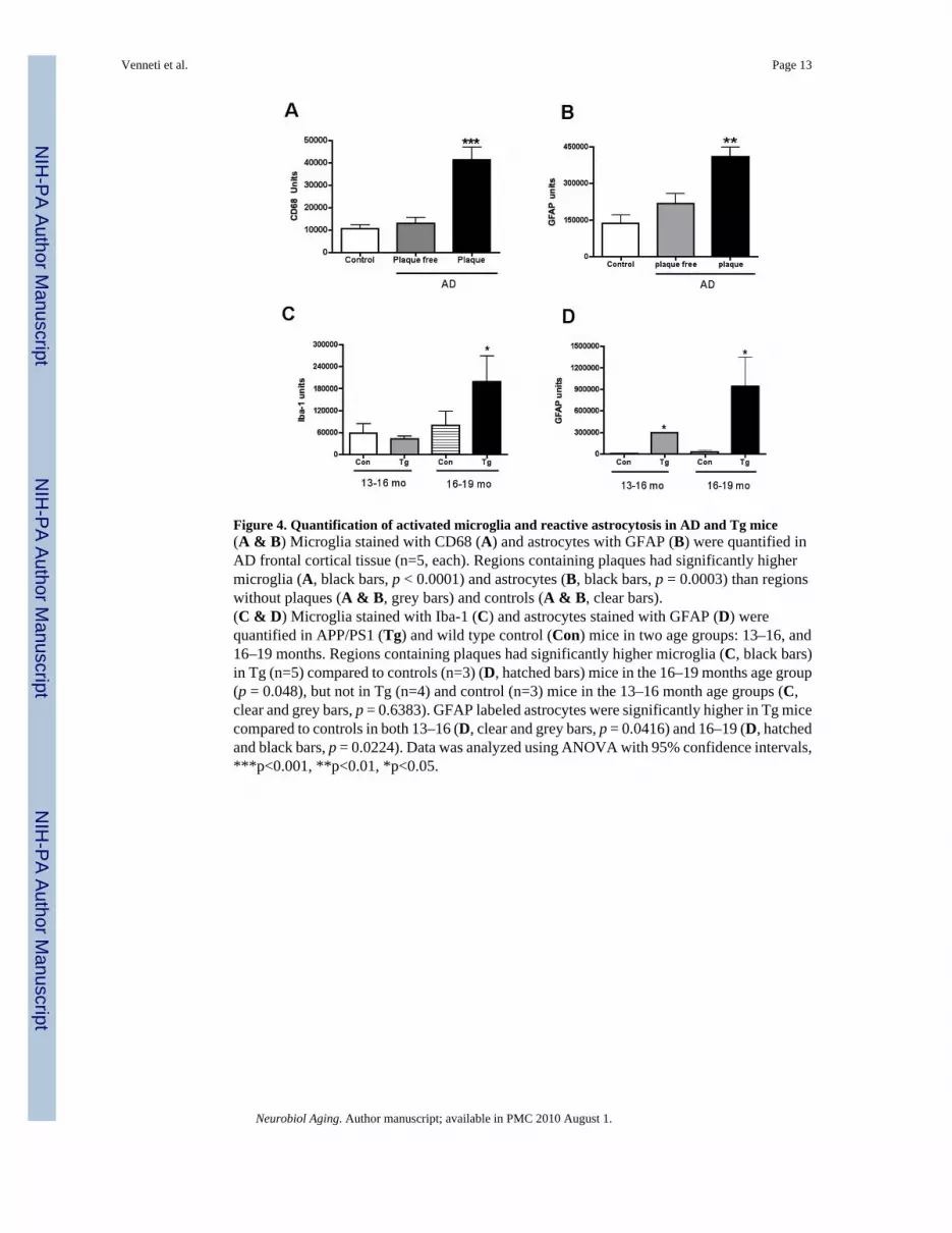
Figure 4. Quantification of activated microglia and reactive astrocytosis in AD and Tg mice(A & B) Microglia stained with CD68 (A) and astrocytes with GFAP (B) were quantified inAD frontal cortical tissue (n=5, each). Regions containing plaques had significantly highermicroglia (A, black bars, p < 0.0001) and astrocytes (B, black bars, p = 0.0003) than regionswithout plaques (A & B, grey bars) and controls (A & B, clear bars).(C & D) Microglia stained with Iba-1 (C) and astrocytes stained with GFAP (D) werequantified in APP/PS1 (Tg) and wild type control (Con) mice in two age groups: 13–16, and16–19 months. Regions containing plaques had significantly higher microglia (C, black bars)in Tg (n=5) compared to controls (n=3) (D, hatched bars) mice in the 16–19 months age group(p = 0.048), but not in Tg (n=4) and control (n=3) mice in the 13–16 month age groups (C,clear and grey bars, p = 0.6383). GFAP labeled astrocytes were significantly higher in Tg micecompared to controls in both 13–16 (D, clear and grey bars, p = 0.0416) and 16–19 (D, hatchedand black bars, p = 0.0224). Data was analyzed using ANOVA with 95% confidence intervals,***p<0.001, **p<0.01, *p<0.05.
Venneti et al. Page 13
Neurobiol Aging. Author manuscript; available in PMC 2010 August 1.
NIH
-PA Author Manuscript
NIH
-PA Author Manuscript
NIH
-PA Author Manuscript

Figure 5. [3H](R)-PK11195 binding corrleates with activated microglia in AD and Tg brain tissues(A & B) [3H](R)-PK11195 Bmax (Y-axis, fmols/mg) values derived from AD and controlcases correlated with the abundance of CD68 labeled activated microglia (A, X-axis, CD68units) assessed in the same area in these same cases (r = 0.9393, p = 0.0002). [3H](R)-PK11195Bmax values (Y-axis, fmols/mg) correlated weakly with the extent of reactive astrocytosis (B,X-axis, GFAP units, r = 0.6729, p = 0.0390).(C & D) [3H](R)-PK11195 Bmax (Y-axis, fmols/mg) values derived from Tg and controlscorrelated with the abundance of Iba-1 labeled activated microglia (C, X-axis, Iba-1 units)assessed in the same area in these same animals (r = 0.9657, p = 0.0007). [3H](R)-PK11195Bmax values (Y-axis, fmols/mg) did not correlated with the extent of reactive astrocytosis(D, X-axis, GFAP units, r = -0.3203, p = 0.5360).
Venneti et al. Page 14
Neurobiol Aging. Author manuscript; available in PMC 2010 August 1.
NIH
-PA Author Manuscript
NIH
-PA Author Manuscript
NIH
-PA Author Manuscript

Figure 6. PET imaging with [11C](R)-PK11195 shows an age-dependent increase in APP/PS1 (Tg)mice, but not controls(A) APP/PS1 (Tg) and wild type control (Con) mice in the age groups 13–16 and 16–19 monthswere imaged with microPET using [11C](R)-PK11195. Retention of the radioligand in the brainis represented as radioactivity concentrations summed over the scan frames and normalized toboth the injected dose of [11C](R)-PK11195 and the body mass of the animal (%ID/g*kg, Y-axis) and is plotted against time in minutes (X-axis). [11C](R)-PK11195 retention was notdifferent between Tg (n=4, grey squares) and Con (n=3, clear squares) in the 13–16 month agegroup, but was higher in Tg mice 16–19 month age group (n=5, red squares) compared to allthe other animal groups (p=0.001). Data was analyzed using one-way ANOVA with 95%confidence intervals.(B) Representative coronal PET images from 16–19 month animals (Tg, and control) and 13–16 month animals (Tg, and control) showing increased [11C](R)-PK11195 retention in the 16–19 month Tg compared to all the other animal groups.(C and D) [11C](R)-PK11195 binding values (%ID/g*kg, Y-axis) derived from Tg and controlscorrelated with the abundance of Iba-1 labeled activated microglia (C, X-axis, Iba-1 units)assessed in the same area in these same animals (r = 0.7690, p = 0.0433). [11C](R)-PK11195binding values (%ID/g*kg, Y-axis) did not correlated with the extent of reactive astrocytosis(D, X-axis, GFAP units, r = 0.4100, p = 0.3610)
Venneti et al. Page 15
Neurobiol Aging. Author manuscript; available in PMC 2010 August 1.
NIH
-PA Author Manuscript
NIH
-PA Author Manuscript
NIH
-PA Author Manuscript

NIH
-PA Author Manuscript
NIH
-PA Author Manuscript
NIH
-PA Author Manuscript
Venneti et al. Page 16Ta
ble
1(R
)-PK
1119
5 bi
ndin
g co
rrel
ates
bes
t with
act
ivat
ed m
icro
glia
Para
met
erM
arke
rSt
atis
tical
val
ue[3 H
](R)-P
K11
195
bind
ing
in A
D p
ost m
orte
m ti
ssue
s[3 H
](R)-P
K11
195
bind
ing
in A
PP/P
S1 b
rain
tiss
ues
[11C
](R)-P
K11
195
rete
ntio
n in
APP
/PS
1 m
ice
in v
ivo
Mic
rogl
iaC
D68
/Iba1
r val
ue0.
9393
0.96
570.
7690
p va
lue
0.00
020.
0007
0.04
33
Ast
rocy
tes
GFA
Pr v
alue
0.67
29−0
.320
30.
4100
p va
lue
0.03
900.
5360
0.36
10
Age
Mon
ths
r val
ueN
A0.
9823
0.76
56
p va
lue
NA
0.00
050.
0162
[3H
](R
)-PK
1119
5 bi
ndin
g in
pos
t mor
tem
tiss
ue in
AD
and
Tg
mic
e fr
onta
l bra
in ti
ssue
or [
11C
](R
)-PK
1119
5 PE
T bi
ndin
g in
viv
o in
Tg
mic
e w
ere
corr
elat
ed w
ith th
e ab
unda
nce
of a
ctiv
ated
mic
rogl
ia(C
D68
or I
ba-1
in A
D a
nd T
g tis
sue
resp
ectiv
ely)
or (
GFA
P) in
the
fron
tal c
ortic
al o
f the
sam
e ca
ses,
and
with
age
(in
mon
ths)
in T
g m
ice
only
. PK
1119
5 bi
ndin
g in
pos
t mor
tem
tiss
ue in
AD
and
Tg
mic
e co
rrel
ated
bes
t with
act
ivat
ed m
icro
glia
and
with
incr
easi
ng a
ge in
Tg
mic
e.
Neurobiol Aging. Author manuscript; available in PMC 2010 August 1.