Human ESC/iPSC-based "Omics" and Bioinformatics for Translational Research
16
Human ESC/iPSC-based “Omics” and Bioinformatics for Translational Research Gerd Müller 1 , Kirill V. Tarasov 2 , Rebekah L. Gundry 3 , and Kenneth R. Boheler 2,4 1 Molecular Oncology, Medical School, University of Leipzig, Leipzig, Germany 2 Molecular Cardiology and Stem Cell Unit, National Institute on Aging, National Institutes of Health, Baltimore, MD, USA 21224 3 The Department of Biochemistry and The Biotechnology and Bioengineering Center, Medical College of Wisconsin, Milwaukee, WI 53226 4 Stem Cell and Regenerative Medicine, LKS Faculty of Medicine, University of Hong Kong, Hong Kong Abstract The establishment of human embryonic stem cell lines (hESCs) created the basis for new approaches in regenerative medicine and drug discovery. Despite the potential of hESCs for cell based therapies, ethical controversies limit their use. These obstacles could be overcome by induced pluripotent stem cells (iPSCs) that are generated by reprogramming somatic cells. Before iPSCs can be used for clinical applications, however, they must be thoroughly analyzed for aberrations in the genome, epigenome, transcriptome, and proteome. Here, we review how ‘omics’ technologies can be employed for a quantitative and definitive assessment of these cells. Introduction Pluripotent stem cells (PSCs) differentiate into all cell types found in the body. The best characterized and standard for PSCs are embryonic stem cells (ESC)[1], but experimentally- derived PSCs, known as induced PSCs (iPSCs), can be generated from almost any type of somatic cell through forced expression of pluripotency-promoting transcription factors or microRNAs [2–6]. The ease of generating iPSCs has fostered the idea of immunologically compatible patient-derived cells. iPSCs thus may represent a viable alternative to human ESCs (hESCs) as the primary source of pluripotent cells for regenerative medicine; however the advantages of iPSCs are counterbalanced by unresolved questions involving differences between the two cell types. Potential iPSC line defects include chromosomal abnormalities, altered gene expression and unanticipated aberrations in the epigenetic landscape and immunogenicity [7]. Taken together, these differences demonstrate that iPSCs must be carefully analyzed on molecular, cellular and functional levels before entering the clinic (Figure 1). Omics approaches, including genome- and proteome-based, offer platforms for fully characterizing and standardizing putative iPSC lines to address these issues of heterogeneity and safety. Corresponding Author: Kenneth R. Boheler, Ph.D., Molecular Cardiology and Stem Cell Unit, Gerontology Research Center, National Institute on Aging, NIH, 5600 Nathan Shock Drive, Baltimore, MD 21224-6825, [email protected], Phone: 410-558-8095, Fax: 410-558-8150. Publisher's Disclaimer: This is a PDF file of an unedited manuscript that has been accepted for publication. As a service to our customers we are providing this early version of the manuscript. The manuscript will undergo copyediting, typesetting, and review of the resulting proof before it is published in its final citable form. Please note that during the production process errors may be discovered which could affect the content, and all legal disclaimers that apply to the journal pertain. NIH Public Access Author Manuscript Drug Discov Today Dis Models. Author manuscript; available in PMC 2013 May 14. Published in final edited form as: Drug Discov Today Dis Models. 2012 ; 9(4): e161–e170. doi:10.1016/j.ddmod.2012.02.003. NIH-PA Author Manuscript NIH-PA Author Manuscript NIH-PA Author Manuscript
Transcript of Human ESC/iPSC-based "Omics" and Bioinformatics for Translational Research

Human ESC/iPSC-based “Omics” and Bioinformatics forTranslational Research
Gerd Müller1, Kirill V. Tarasov2, Rebekah L. Gundry3, and Kenneth R. Boheler2,4
1Molecular Oncology, Medical School, University of Leipzig, Leipzig, Germany2Molecular Cardiology and Stem Cell Unit, National Institute on Aging, National Institutes ofHealth, Baltimore, MD, USA 212243The Department of Biochemistry and The Biotechnology and Bioengineering Center, MedicalCollege of Wisconsin, Milwaukee, WI 532264Stem Cell and Regenerative Medicine, LKS Faculty of Medicine, University of Hong Kong, HongKong
AbstractThe establishment of human embryonic stem cell lines (hESCs) created the basis for newapproaches in regenerative medicine and drug discovery. Despite the potential of hESCs for cellbased therapies, ethical controversies limit their use. These obstacles could be overcome byinduced pluripotent stem cells (iPSCs) that are generated by reprogramming somatic cells. BeforeiPSCs can be used for clinical applications, however, they must be thoroughly analyzed foraberrations in the genome, epigenome, transcriptome, and proteome. Here, we review how ‘omics’technologies can be employed for a quantitative and definitive assessment of these cells.
IntroductionPluripotent stem cells (PSCs) differentiate into all cell types found in the body. The bestcharacterized and standard for PSCs are embryonic stem cells (ESC)[1], but experimentally-derived PSCs, known as induced PSCs (iPSCs), can be generated from almost any type ofsomatic cell through forced expression of pluripotency-promoting transcription factors ormicroRNAs [2–6]. The ease of generating iPSCs has fostered the idea of immunologicallycompatible patient-derived cells. iPSCs thus may represent a viable alternative to humanESCs (hESCs) as the primary source of pluripotent cells for regenerative medicine; howeverthe advantages of iPSCs are counterbalanced by unresolved questions involving differencesbetween the two cell types. Potential iPSC line defects include chromosomal abnormalities,altered gene expression and unanticipated aberrations in the epigenetic landscape andimmunogenicity [7]. Taken together, these differences demonstrate that iPSCs must becarefully analyzed on molecular, cellular and functional levels before entering the clinic(Figure 1). Omics approaches, including genome- and proteome-based, offer platforms forfully characterizing and standardizing putative iPSC lines to address these issues ofheterogeneity and safety.
Corresponding Author: Kenneth R. Boheler, Ph.D., Molecular Cardiology and Stem Cell Unit, Gerontology Research Center, NationalInstitute on Aging, NIH, 5600 Nathan Shock Drive, Baltimore, MD 21224-6825, [email protected], Phone: 410-558-8095,Fax: 410-558-8150.
Publisher's Disclaimer: This is a PDF file of an unedited manuscript that has been accepted for publication. As a service to ourcustomers we are providing this early version of the manuscript. The manuscript will undergo copyediting, typesetting, and review ofthe resulting proof before it is published in its final citable form. Please note that during the production process errors may bediscovered which could affect the content, and all legal disclaimers that apply to the journal pertain.
NIH Public AccessAuthor ManuscriptDrug Discov Today Dis Models. Author manuscript; available in PMC 2013 May 14.
Published in final edited form as:Drug Discov Today Dis Models. 2012 ; 9(4): e161–e170. doi:10.1016/j.ddmod.2012.02.003.
NIH
-PA Author Manuscript
NIH
-PA Author Manuscript
NIH
-PA Author Manuscript

Applications of ‘Omics’ to PSCsThe human genome project, which began in 1990, led to major technological advancementsthat included improved sequencing of the genome and a routine analysis of a cell’s DNA(SNPs, copy number variation, mutations), methylation and histone state (epigenome), RNAabundance (transcriptome) or protein content (proteome). Collectively these analyses,among others, have been termed ‘omics’ research endeavors that are unique from traditionalexperimental designs. This is because ‘omics’ approaches are often large-scale and data-driven, as opposed to purely hypothesis driven [8]. Data generated from ‘omics’ approaches,when combined across platforms, are useful in describing biological relationships related toexperimental and cellular fluctuations. Consequently, the integration of multiple ‘omics’approaches to understand a cell’s phenotype, permits an ‘integrated system’ that more fullydescribes a cell’s response to defined variables. Finally, ‘omics’ approaches requiresignificant statistical and computational efforts to model dynamic systems that by their verynature interact on multiple levels within a cell. Extraction of valuable biological informationfrom ‘omics’ data is challenging, but the results, when properly analyzed, show greatpotential in addressing some of the current problems associated with transplantation andstem cell-based therapies [9].
A quantitative and definitive assessment of human iPSC lines should be possible through theuse of ‘omics’ techniques. Genome-wide evaluations will be useful for defining the state ofputative iPSC lines, and robust statistical techniques should be valuable in pin-pointingpossible differences/aberrations in lines relative to “gold-standard” ESC lines (Figure 2A).More specifically, genome-wide DNA sequencing should uncover any spontaneous DNAmutations that may result during reprogramming, while microarray analysis of RNAsamples or RNA-Seq experiments can provide insights on variations in gene expression thatmay be indicative of residual epigenetic memory. Chromatin immunoprecipitationexperiments (ChIP-chip or ChIP seq) and DNA methylation studies can reveal variations inchromatin structure and transcription factor binding. Proteomic studies may also be valuablein defining variations in protein levels between cells, but perhaps more importantly, thistechnique may be of great value in the development of immunophenotyping techniques thatcan be employed to isolate and characterize “authentic” iPSC lines. By studying cells at the‘omics’ level it should be possible to obtain fingerprints of iPSCs for comparisons withstandard ESC lines and to assess how the reprogramming process affects biologicalprocesses, thus solving many of the current problems surrounding possible differencesamong these forms of PSCs.
Genome and EpigenomeDNA mutations—Similar to problems observed in the sheep Dolly [10], cell autonomousgenetic defects are present in reprogrammed iPSCs. Specifically, iPSC lines show anenrichment of mutations and chromosomal defects that may be introduced during thereprogramming process, independent of the reprogramming vectors, or by culture adaption(Figure 2B) [11–14]. As an example, Gore et al. sequenced the protein coding exons(exomes) of twenty-two human iPSC lines generated by 5 independent methods and nineparental fibroblast lines. On average, they found five protein-coding point mutations in theregions sampled or an estimated six protein coding point mutations per exome. Although themajority of the mutations were non-synonymous, nonsense or splice variants, many of themutations occurred in genes with causative effects in cancers. At least half of the mutationswere present in the parental fibroblast, but the remainder occurred spontaneously eitherduring or after reprogramming. Howden et al. also assessed whether human iPSCs isolatedfrom a patient with gyrate atrophy increased its mutational load with reprogramming [15]. Inthese cells, no abnormalities were detected by standard G-band metaphase analysis;
Müller et al. Page 2
Drug Discov Today Dis Models. Author manuscript; available in PMC 2013 May 14.
NIH
-PA Author Manuscript
NIH
-PA Author Manuscript
NIH
-PA Author Manuscript

however, array comparative genomic hybridization and exome sequencing identified twodeletions, one amplification, and nine mutations in protein coding regions when comparedagainst the parental patient fibroblast cell line. They then performed exome sequencing on agene-targeted iPSC clonal line that corrected the OAT point mutation present in thispatient’s DNA, and on a cassette-free iPSC clone. Somewhat surprisingly, the genomesproved remarkably stable, as no additional mutations or copy number variations wereidentified, excluding the targeted correction in the OAT locus and a single synonymousbase-pair change. These findings led to the conclusion that iPSCs carry a significantmutational load from the parental line, but clonal events and prolonged culturing do not leadto a substantial increase in mutations.
Mutations may however occur at a higher frequency than previously reported. Inunpublished work presented in a recent Stem Cell Research Symposium at the NIH, PaulLiu presented results from deep whole genome sequencing and high-density SNP arrayanalysis. He reported results from episomal-vector reprogrammed hiPSC lines derived fromtwo tissues of a single adult donor. The data revealed over 1000 single nucleotidesubstitutions in each iPSC cell line when compared to the parental cell sources. Although themajority of mutations were in non-coding sequences, 6 and 12 mutations, respectively werelocated in coding regions, and 34 and 22 variations respectively were identified in the 5′ or3′ untranslated regions of genes. Another 362 and 709 differences were observed in intronicregions that might affect gene expression. The majority of mutations were not present in theexome, and SNP analysis was not sufficient to identify these mutations. Since the specificpoint mutations were not conserved among the two iPSC lines, sequence substitutions werenot related to susceptible “hot spots” during reprogramming. Importantly, these resultsestablished the viability of iPSC line whole-genome sequencing, which has now becomecost-effective and more widely accessible to researchers in this field.
DNA Methylation—Closely affiliated with the genome sequence are DNA modifiers (i.e.,DNA methyltransferases (DNMTs)) that add methyl groups at the 5′-position of cytosine.During the reprogramming process, somatic cells reset their pattern of DNA methylation toan ESC-like state. More specifically, DNA of transcriptionally active genes likepluripotency and housekeeping genes is hypomethylated [16,17], while silenced genes arehypermethylated [17,18].
Various groups have now reported that ESCs exhibit unique methylation patterns and thatiPSCs show modest variations in this pattern. This was perhaps best illustrated through theuse of different cell types isolated and reprogrammed from the same mouse. Among thesegenetically identical iPSC clonal lines, the DNA methylation profiles reflected the cell typeof origin, suggesting the presence of residual parental cell “epigenetic memory”.Functionally, methylation pattern permitted efficient differentiation of iPSCs into thesomatic cell type of origin, but these same lines showed reduced efficiencies ofdifferentiation into other lineages. Although the causes are still unclear, it became apparentthat these effects resulted from insufficient methylation (silencing) of genes normallyexpressed in the somatic cells from which the iPSC are derived, and insufficientdemethylation (activation) of ESC specific genes [19–22]. In support of this assumption, theaddition of 5-azacytidine (Aza), a DNA methylation inhibitor, to established iPSCs linesincreased their differentiation potential and made them more like ESC [21]. Moreover,pou5f1 and nanog gene promoters, which are highly methylated in somatic cells, remainlargely methylated in partially reprogrammed cells (Figure 2C); however, exposure to Azareactivates endogenous pou5f1 gene expression. Prolonged cultivation of iPSCs alsodiminished differences in the methylation patterns between iPSCs and ESCs [19,22,23].These latter findings suggest that reprogramming does not completely reverse the epigeneticlandscape in early clonal isolates, a finding confirmed by Kim et al. [21], and that chromatin
Müller et al. Page 3
Drug Discov Today Dis Models. Author manuscript; available in PMC 2013 May 14.
NIH
-PA Author Manuscript
NIH
-PA Author Manuscript
NIH
-PA Author Manuscript

remodeling is a gradual process that takes place over an extended period of time. Thus thestate of DNA methylation at a genome level, and especially at some specific gene loci at afunction of time, is likely to be indicative of the degree of reprogramming of iPSCs.
From an ‘omics’ perspective, current DNA methylation assays are often limited in theirability to characterize a large number of genomic targets. To overcome this limitation,investigators have described methods that capture genomic targets for single-moleculebisulfite sequencing at a single-nucleotide resolution. From these large-scale studies,chromosome-wide methylation patterns were similar, but cytosine methylation proved to beslightly more prevalent in the pluripotent cells than in the fibroblasts [24]. These data, inagreement with other studies, ultimately showed that iPSCs intrinsically display moremethylation than embryonic stem cells. Further improvements in whole genome methylationsequencing based studies have now made it feasible to fully characterize the methylationstatus of iPSCs [25–27].
Histone Modifications—Somatic cells reprogrammed to iPSCs reset post-translationalhistone modifications to an ESC-like state, and like DNA methylation, histone modificationsdiffer between ESCs and iPSCs. Histone deacetylase inhibitors (HDACs), such astrichostatin A (TSA) and valproic acid (VPA), used to promote active histone acetylationmarks, can enhance iPSC reprogramming efficiency [28,29]. To maintain pluripotency, thepromoters of genes encoding Nanog, Oct4, and Sox2 show a high degree of histone 3 lysine4 (H3K4) methylation resulting in a transcriptionally active state [18,30,31]. Partiallyreprogrammed iPSCs, however, retained some histone modification patterns of its parentalcells, indicating an association of histone modification in iPSC memory status.
In genome-wide studies performed by CHiP-chip or CHiP-Seq based techniques, bivalentpatterns of histone methylation have been described that distinguish PSCs from somaticcells (Figure 2C). In bivalent promoters, histone 3 is methylated at both lysines 4 and 27(H3K27). Both modifications are prevalent at transcription start sites of numerousdevelopmental genes whose expression is repressed in ESCs [30,31]. Although H3K4methylation is associated with gene activation and H3K27 methylation by polycomb groupprotein complexes typically results in gene repression, bivalent promoters tended to berepressed, but at times “leaky” [32,33]. With differentiation, this pattern switches from abivalent state to a monovalent state, which results either in transcriptionally active genescharacterized by H3K4 methylation or to non-transcribed genes with H3K27 methylationstate [34]. Thus the poised state was seen as a central requirement for undifferentiated ESCsto maintain their developmental potential. A number of other histone modifications are alsoknown to affect gene activity, including the repressive H3K9me3, H4K20me3 marks andmultiple targets of histone acetylation [33,35,36].
Recent results from Roeder and colleagues provide new insights into these regulatorymechanisms [37]. By studying components of SET1/MLL family complexes, these authorsfound that depletion of Dpy-30 and RbBP5 leads to a generalized reduction in H3K4methylation in mouse ESC. In fact, H3K4 methylation levels were significantly reduced onthe promoters of key stemness genes like nanog, pou5f1, klf4, and sox2, but mRNAabundance was not altered. The proliferation rate level of alkaline phosphatase in ESCs wasalso unaffected by knockdown of Dpy-30. In contrast, Dpy-30 knockdown resulted inprofound defects when ESCs were forced to differentiate by LIF withdrawal or retinoic acidstimulation. The reduction in H3K4 methylation resulted in defects in lineage specificationand impaired plasticity in transcriptional reprogramming. These data in particular illustratewhy large-scale analyses may be required to characterize iPSCs prior to therapeutic usage.More specifically, modest changes in epigenetic regulation may not adversely affect PSC
Müller et al. Page 4
Drug Discov Today Dis Models. Author manuscript; available in PMC 2013 May 14.
NIH
-PA Author Manuscript
NIH
-PA Author Manuscript
NIH
-PA Author Manuscript

self-renewal; however, some differences may have profound effects on the progenygenerated upon differentiation.
TranscriptomeMicroarrays and RNA-Seq—Whole-genome expression profiling is a commonlyemployed approach to compare and characterize different cell populations (Figure 2B).Consequently, microarray and RNA-Seq gene expression profiling have been employed tocompare iPSCs with parental cells or ESCs. During the reprogramming process, many genesexpressed in ESC are reactivated, including endogenous pou5f1, nanog, lin28 and fgf4. Alarge percentage of these genes are only up-regulated during late stages of reprogramming,and in partially reprogrammed cells, a great deal of heterogeneity has been observed.Although the majority of genome-wide studies suggest that ESCs and iPSCs are nearlyidentical, Chin et al. reported that these cells could be distinguished by gene expressionsignatures [22]. Wang et al. [38] compared whole-genome microarray datasets from fivestudies [22,39–42] to show that differences may be related to the methods forreprogramming. Although most of the iPSC lines appeared similar to ESC lines, thetranscriptomes of iPSCs generated with retroviruses differed much more from ESCs thanthose generated with episomal-reprogramming vectors.
Bock et al. recently created an integrated reference map of DNA methylation and geneexpression patterns through a comprehensive evaluation of 20 human ESC and 12 iPSClines[43]. The distribution of DNA methylation and mRNA abundance was calculated forindividual genes or genomic regions. A “reference corridor” was developed that defined therange of methylation/expression for a given gene in pluripotent cells that could be used toestablish transcriptome-based criteria to categorize pluripotent cells. When extended to otherlines, this reference facilitates the identification of genes that fall outside the “referencecorridor”. Although individual outliers may result from cultivation conditions, outliers couldalso be indicative of inappropriately functioning genes. Once identified, iPSC lines thatinappropriately express these transcripts may need to be classified as failing to meetestablished transcriptome-based criteria for authentic pluripotent cells. Also, by combiningthis method with differentiation assays, this reference makes it possible to assay the qualityand the usability of a given cell line for further applications.
Müller et al. subsequently made use of more than 450 genome-wide transcriptional profilesof stem cell lines, differentiated cells, and adult human tissues [44]. In these analyses,expression profiles from 223 hES and 41 hiPS cell lines were included. They developed aweb-based open access tool called “PluriTest”, which allows for the identification ofpluripotent cell lines based on gene expression data with a high degree of reliability. Inaddition, the algorithm is able to discriminate between pluripotent germ cell tumor lines andnormal PSCs as well as between fully and partially reprogrammed iPSC lines. Currently,“PluriTest” is only able to process gene expression data, however, this algorithm should beapplicable to methylation analyses or RNA sequencing data as well, which would furtherimprove the reliability of these predictions.
Transcriptional profiles of ESCs are not only characterized by the expression levels ofcertain genes. The presence of alternatively spliced transcripts that encode protein variantsessential for ESC self-renewal, pluripotency and differentiation are also critical [45–49].Since up to 94% of human multi-exon genes are alternatively spliced [49], the number ofunique proteins in a cell is much higher than the number of genes. Alternative splicing caninfluence protein binding affinities, enzymatic activity, localization, mRNA half-life andsplice products that are quickly degraded by the nonsense-mediated mRNA decay [50,51].Specifically, splice forms for OCT4 (Oct4b and Oct4b1), SALL4 (Sall4a and Sall4b),FOXP1 (FoxP1-ES and FoxP1) Tcf3 and Dnmt3b, which are active in ESCs, modulate
Müller et al. Page 5
Drug Discov Today Dis Models. Author manuscript; available in PMC 2013 May 14.
NIH
-PA Author Manuscript
NIH
-PA Author Manuscript
NIH
-PA Author Manuscript

pluripotency versus cell-type specification [52–56]. The mechanisms of the regulation ofESC-specific alternative splicing remain to be elucidated; however, transcriptomic basedanalyses that assay the presence or absence of these splice variants are possible, principallyby microarrays, but also by RNA-Seq.
microRNAs (miRs)—The discovery by Yu et al. that Lin-28 was critically involved insomatic cell reprogramming provided the first evidence that microRNAs (miRs) wereessential to PSCs [57]. This is because, Lin 28 specifically blocks processing of pri-let-7, amiR that is critical to the regulation of developmental genes activated during earlydifferentiation [58]. MiRs are single-stranded RNA molecules of 21–24 nucleotides that arefully or partially complementary to one or more mRNA molecules [59]. When associatedwith their targets, miRs generally repress translation or promote mRNA degradation toeffectively down-regulate targeted gene expression. More recently, Judson et al. showed thatthe introduction of miRs specific to ESCs into somatic cells enhanced the production ofmouse iPSCs [60]. More importantly, Anokye-Danso et al. recently showed that over-expression of the miR302/367 cluster, in the presence of HDAC inhibitors, reprogrammedmouse and human somatic cells to an iPSC state without the need for exogenoustranscription factors like OCT4, SOX2 or NANOG. MiRs and the proper regulation of miRexpression in PSCs is therefore critical to the proper function of iPSCs [6].
To date, a total of 1424 and 720 miRNA sequences have been identified in the human andmouse genomes, respectively (www.mirbase.org)[61]. Only a subset of these has been foundin hESC or mESCs by cloning and sequencing from small RNA libraries [62,63]. Because amajority of miRs are expressed in somatic cells and only a minority in PSCs, thesemolecules can be used to evaluate the status of putative hiPSCs. Obviously, the presence ofmiRs in iPSCs that are typically only found in somatic cells would be contraindicative offully reprogrammed iPSCs, and would bring their therapeutic viability into question.
Proteome and MetabolomeMorphologically and functionally, good quality iPSCs are nearly indistinguishable fromhESCs, but as described above, a number of molecular indices show differences rangingfrom subtle to profound. The proteomic landscape of PSCs is not yet clearly defined, anduntil recently, the similarity of human ESCs and iPSCs at the protein level was unexplored(Figure 2D). To address this issue, we performed a broad-based comparison of more than 30published proteomic studies of undifferentiated human and mouse PSCs [64]. Theseanalyses resulted in a comprehensive resource of 7,471 and 7,281 proteins identified inmouse and human, respectively, of which 3,114 were found in both species. Unexpectedly,63% of proteins were found in only one or two datasets, illustrating the variability amongstudies to describe and quantify this critical proteome. Also in 2011, Phanstiel et al.compared the proteomes and phosphoproteomes of two ESC lines, one iPSC line and onefibroblast cell line [65]. Statistical analyses revealed subtle, but significant and functionallyrelated differences between proteins and phosphorylation sites in human ESCs and iPSCs. Anumber of these differences were thought to reflect residual regulation characteristics of aniPSCs’ somatic origin. The authors also developed the Stem Cell–Omics Repository(SCOR), a resource designed to collate and display quantitative information across multipleplanes of measurement. These recently established resources are expected to be very usefulfor this rapidly growing field of investigation.
The rate of enzymatic reactions in cells is also regulated by substrate concentration and theirproducts, and in most organisms, no direct relationship exists between cellular metabolites(i.e., intermediates and products of metabolism) and gene function. Moreover, metaboliteconcentrations within cells vary as a consequence of genetic or physiological changes [66];
Müller et al. Page 6
Drug Discov Today Dis Models. Author manuscript; available in PMC 2013 May 14.
NIH
-PA Author Manuscript
NIH
-PA Author Manuscript
NIH
-PA Author Manuscript

consequently, metabolomics, which focuses on the end-products of gene expression(metabolites) as well as other small proteins, toxins, chemicals, and organic compounds,may represent one approach that can provide functional insights regarding iPSC line statesand variations relative to ESCs. To date, however, there are very few reports regarding PSCsand metabolomics. Only recently did Panapoulos et al. report that cellular bioenergetics ofsomatic cells convert from an oxidative state to a glycolytic state in reprogrammed cells, andthat human iPSCs share a pluripotent metabolomic signature with ESCs that is distinct fromparental cells [67]. They also identified several metabolites that differ between iPSCs andESCs and novel metabolic pathways that play a critical role in regulating somatic cellreprogramming [67], thus validating the role of metabolomics in the identification ofmetabolic differences among PSCs. Metabolomics should, however, be considered a“cousin” to proteomics: the strategies and technologies are similar, but this “omic”technology really measures distinct types of biomolecules as well as small peptides.
Perhaps more importantly than either global proteomic or metabolomic approaches arefocused analyses of PSC subproteomes. In particular, we have advocated the need forfocused analyses of the surface proteome (i.e., surfaceome) of PSCs [64,68]. We expectsurface proteins to be uniquely informative of a biological state for specific cell types, asevidenced by the use of surrogate markers to define hematopoietic stem cell (HSC)phenotypes. In fact, immunophenotyping, a process in which the functional potential of acell is related to its surface marker expression pattern, has been used extensively to isolatesubsets of bone marrow-derived HSCs for clinical interventions. Proof-of-principle studiesfor this concept were published in 2009 [69] where the authors used a targetedchemoproteomic strategy to identify 341 cell surface glycoproteins, including 53 CD-annotated proteins from mouse ESCs. The result of this antibody-independent strategyconfirmed the expected decrease in LIF receptor and increase in FGF receptor 2 abundanceduring differentiation into the neural lineage. Such targeted strategies, when extended toreprogrammed cells, are likely to foster the rapid isolation and characterization of morehomogeneous and therapeutically viable patient compatible iPSCs and will accelerate thedevelopment of disease models and clinical strategies for cell replacement therapy to treathuman disease (Figure 3).
How could “omics” strategies be used routinely in the clinic?Successful therapeutic approaches developed with ESC- or patient derived iPSC-progenyare predicted to be a future mainstay of modern medicine. Experiments in animal modelshave already proven that such therapeutic approaches hold a promising potential forregenerative medicine [70, 71], and recent results in macular degeneration suggest that theday is rapidly approaching for therapeutic applications in human [72].
Before these cells and their derivatives can be routinely employed clinically, carefulmolecular, immunological and functional assays must be performed. Conventional assayslike G-banding are not sufficient since only large genetic abnormalities can be detected. Incontrast, a combination of “omics” techniques represents a promising approach to assessthese cells – particularly in pre-clinical stages. However, to date there are no definitivecriteria on how to best define pluripotent cells and their specific cell derivatives, as well asassess the functional consequences of potential alterations in PSC genomes, epigenomes,transcriptomes, proteomes and metabolomes. Moreover, genetic defects are generallysporadic, thus complicating standard clinically accessible analyses. Even if a certain cell linehas been evaluated with a combination of “omics” technologies and aberrations relative to apotential gold standard cell line have been pinpointed, it is not clear which changes or whatdegree of genetic changes are still acceptable for clinical use. Therefore, as an importantstep towards the clinical use of ESCs and iPSCs, well defined standards must be formulatedto best identify cells suited for transplantation in patients and to minimize patient’s risks in
Müller et al. Page 7
Drug Discov Today Dis Models. Author manuscript; available in PMC 2013 May 14.
NIH
-PA Author Manuscript
NIH
-PA Author Manuscript
NIH
-PA Author Manuscript

terms of tumorigenicity and immunogenicity. Thus, generally accepted guidelines regardinggeneration, expansion, manipulation, purification and evaluation of stem cells and stem cellderivatives must be developed (reviewed in [73]) before “omics” technologies can beroutinely applied preclinically. But with that said, one “omics” technology has the potentialof facilitating the daily use of PSCs and their derivatives for therapeutics. This is based onthe identification of cell surface proteins as surrogate markers of a cell’s phenotype and/orfunction analogous to that already described for HSCs. The generation of non-genetic,immunophenotyping methods (Figure 3) for the isolation of defined cell states should permitthe efficient isolation of desired cell types for clinical applications.
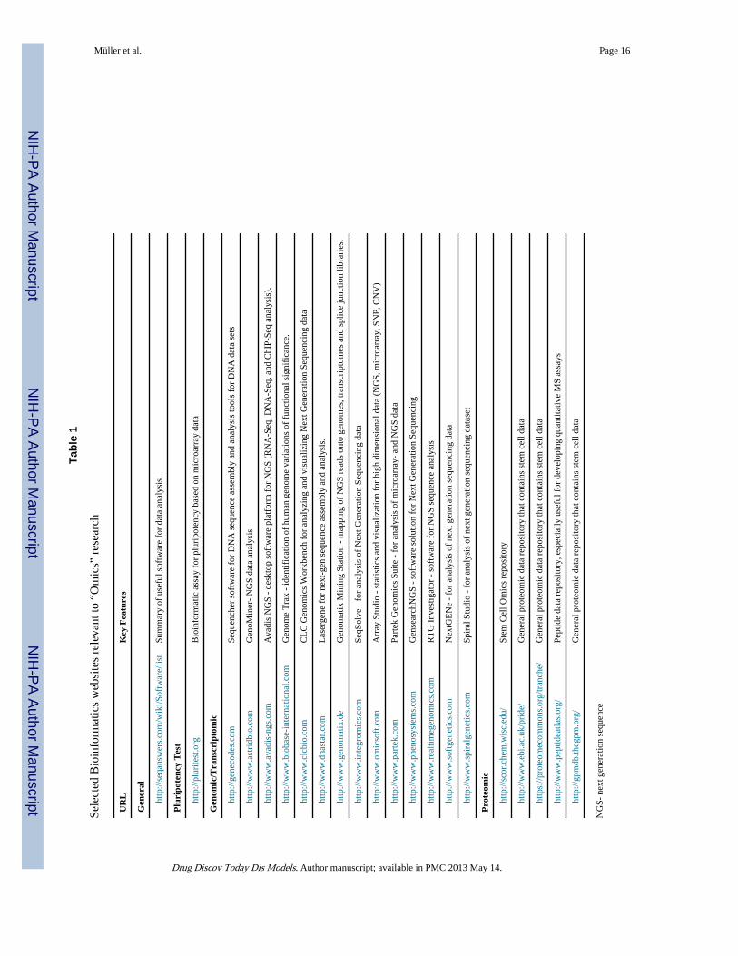
ConclusionsDifferences between ESC and iPSC genomes, epigenomes, transcriptomes, proteomes andmetabolomes are well established. While we have emphasized the differences, good qualityiPSCs are almost identical to ESCs, but currently, there are no fully accepted criteria tomake this determination in human cells. Omics approaches, especially when coupled withbioinformatics tools (Table 1) and functional assays are likely to play a critical role inestablishing, characterizing and eventually defining which populations of iPSCs areappropriate for translational research.
AcknowledgmentsThe authors are supported by 4R00HL094708-03 (RLG), the Innovation Center at the Medical College ofWisconsin (RLG), the Intramural Research Program of the NIH, National Institute on Aging (KRB), and NIHInduced Pluripotent Stem Cell Center (NiPSCC) Pilot Study Award (KRB).
References1. Wobus AM, Boheler KR. Embryonic stem cells: prospects for developmental biology and cell
therapy. Physiol Rev. 2005; 85 (2):635–678. [PubMed: 15788707]
2. Takahashi K, Yamanaka S. Induction of pluripotent stem cells from mouse embryonic and adultfibroblast cultures by defined factors. Cell. 2006; 126(4):663–676. Epub 2006 Aug 2010. [PubMed:16904174]
3. Okita K, et al. Generation of germline-competent induced pluripotent stem cells. Nature. 2007;448(7151):313–317. Epub 2007 Jun 2006. [PubMed: 17554338]
4. Boheler KR. Pluripotency of human embryonic and induced pluripotent stem cells for cardiac andvascular regeneration. Thrombosis and Haemostasis. 2010; 104 (1):23–29. [PubMed: 20458433]
5. Lin SL, et al. Regulation of somatic cell reprogramming through inducible mir-302 expression.Nucleic Acids Research. 2011; 39 (3):1054–1065. [PubMed: 20870751]
6. Anokye-Danso F, et al. Highly Efficient miRNA-Mediated Reprogramming of Mouse and HumanSomatic Cells to Pluripotency. Cell Stem Cell. 2011; 8 (4):376–388. [PubMed: 21474102]
7. Zhao TB, et al. Immunogenicity of induced pluripotent stem cells. Nature. 2011; 474 (7350):212–U251. [PubMed: 21572395]
8. Robert C. Microarray analysis of gene expression during early development: a cautionary overview.Reproduction. 2010; 140 (6):787–801. [PubMed: 20833752]
9. Perkins D, et al. Advances of genomic science and systems biology in renal transplantation: areview. Seminars in Immunopathology. 2011; 33 (2):211–218. [PubMed: 21318414]
10. Wilmut I, et al. Viable offspring derived from fetal and adult mammalian cells [see comments][published erratum appears in Nature 1997 Mar 13; 386(6621):200]. Nature. 1997; 385:810–813.[PubMed: 9039911]
11. Mayshar Y, et al. Identification and Classification of Chromosomal Aberrations in Human InducedPluripotent Stem Cells. Cell Stem Cell. 2010; 7 (4):521–531. [PubMed: 20887957]
12. Gore A, et al. Somatic coding mutations in human induced pluripotent stem cells. Nature. 2011;471 (7336):63–U76. [PubMed: 21368825]
Müller et al. Page 8
Drug Discov Today Dis Models. Author manuscript; available in PMC 2013 May 14.
NIH
-PA Author Manuscript
NIH
-PA Author Manuscript
NIH
-PA Author Manuscript

13. Hussein SM, et al. Copy number variation and selection during reprogramming to pluripotency.Nature. 2011; 471 (7336):58–U67. [PubMed: 21368824]
14. Laurent LC, et al. Dynamic Changes in the Copy Number of Pluripotency and Cell ProliferationGenes in Human ESCs and iPSCs during Reprogramming and Time in Culture. Cell Stem Cell.2011; 8 (1):106–118. [PubMed: 21211785]
15. Howden SE, et al. Genetic correction and analysis of induced pluripotent stem cells from a patientwith gyrate atrophy. Proceedings of the National Academy of Sciences of the United States ofAmerica. 2011; 108 (16):6537–6542. [PubMed: 21464322]
16. Weber M, et al. Distribution, silencing potential and evolutionary impact of promoter DNAmethylation in the human genome. Nature Genetics. 2007; 39 (4):457–466. [PubMed: 17334365]
17. Meissner A, et al. Genome-scale DNA methylation maps of pluripotent and differentiated cells.Nature. 2008; 454 (7205):766–U791. [PubMed: 18600261]
18. Mikkelsen TS, et al. Genome-wide maps of chromatin state in pluripotent and lineage-committedcells. Nature. 2007; 448 (7153):553–U552. [PubMed: 17603471]
19. Polo JM, et al. Cell type of origin influences the molecular and functional properties of mouseinduced pluripotent stem cells. Nature Biotechnology. 2010; 28 (8):848–U130.
20. Ohi Y, et al. Incomplete DNA methylation underlies a transcriptional memory of somatic cells inhuman iPS cells. Nature Cell Biology. 2011; 13 (5):541–U328.
21. Kim K, et al. Epigenetic memory in induced pluripotent stem cells. Nature. 2010; 467 (7313):285–U260. [PubMed: 20644535]
22. Chin MH, et al. Induced Pluripotent Stem Cells and Embryonic Stem Cells Are Distinguished byGene Expression Signatures. Cell Stem Cell. 2009; 5 (1):111–123. [PubMed: 19570518]
23. Nishino K, et al. DNA Methylation Dynamics in Human Induced Pluripotent Stem Cells overTime. Plos Genetics. 2011; 7(5)
24. Deng J, et al. Targeted bisulfite sequencing reveals changes in DNA methylation associated withnuclear reprogramming. Nature Biotechnology. 2009; 27 (4):353–360.
25. Bock C, et al. Quantitative comparison of genome-wide DNA methylation mapping technologies.Nature Biotechnology. 2010; 28 (10):1106–U1196.
26. Butcher LM, Beck S. AutoMeDIP-seq: A high-throughput, whole genome, DNA methylationassay. Methods. 2010; 52 (3):223–231. [PubMed: 20385236]
27. Li N, et al. Whole genome DNA methylation analysis based on high throughput sequencingtechnology. Methods. 2010; 52 (3):203–212. [PubMed: 20430099]
28. Huangfu DW, et al. Induction of pluripotent stem cells from primary human fibroblasts with onlyOct4 and Sox2. Nature Biotechnology. 2008; 26 (11):1269–1275.
29. Huangfu DW, et al. Induction of pluripotent stem cells by defined factors is greatly improved bysmall-molecule compounds. Nature Biotechnology. 2008; 26 (7):795–797.
30. Zhao XD, et al. Whole-genome mapping of histone H3 Lys4 and 27 trimethylations reveals distinctgenomic compartments in human embryonic stem cells. Cell Stem Cell. 2007; 1 (3):286–298.[PubMed: 18371363]
31. Pan GJ, et al. Whole-genome analysis of histone H3 lysine 4 and lysine 27 methylation in humanembryonic stem cells. Cell Stem Cell. 2007; 1 (3):299–312. [PubMed: 18371364]
32. Ringrose L, Paro R. Epigenetic regulation of cellular memory by the polycomb and trithorax groupproteins. Annual Review of Genetics. 2004; 38:413–443.
33. Ringrose L, et al. Distinct contributions of histone H3 lysine 9 and 27 methylation to locus-specificstability of Polycomb complexes. Molecular Cell. 2004; 16 (4):641–653. [PubMed: 15546623]
34. Bernstein BE, et al. A bivalent chromatin structure marks key developmental genes in embryonicstem cells. Cell. 2006; 125 (2):315–326. [PubMed: 16630819]
35. Marion RM, et al. Telomeres Acquire Embryonic Stem Cell Characteristics in Induced PluripotentStem Cells. Cell Stem Cell. 2009; 4 (2):141–154. [PubMed: 19200803]
36. Mali P, et al. Butyrate Greatly Enhances Derivation of Human Induced Pluripotent Stem Cells byPromoting Epigenetic Remodeling and the Expression of Pluripotency-Associated Genes. StemCells. 2010; 28 (4):713–720. [PubMed: 20201064]
Müller et al. Page 9
Drug Discov Today Dis Models. Author manuscript; available in PMC 2013 May 14.
NIH
-PA Author Manuscript
NIH
-PA Author Manuscript
NIH
-PA Author Manuscript
37. Jiang H, et al. Role for Dpy-30 in ES Cell-Fate Specification by Regulation of H3K4 Methylationwithin Bivalent Domains. Cell. 2011; 144 (4):513–525. [PubMed: 21335234]
38. Wang Y, et al. A Transcriptional Roadmap to the Induction of Pluripotency in Somatic Cells. StemCell Reviews and Reports. 2010; 6 (2):282–296. [PubMed: 20336394]
39. Lowry WE, et al. Generation of human induced pluripotent stem cells from dermal fibroblasts.Proceedings of the National Academy of Sciences of the United States of America. 2008; 105 (8):2883–2888. [PubMed: 18287077]
40. Maherali N, et al. A high-efficiency system for the generation and study of human inducedpluripotent stem cells. Cell Stem Cell. 2008; 3 (3):340–345. [PubMed: 18786420]
41. Soldner F, et al. Parkinson’s Disease Patient-Derived Induced Pluripotent Stem Cells Free of ViralReprogramming Factors. Cell. 2009; 136 (5):964–977. [PubMed: 19269371]
42. Yu JY, et al. Human Induced Pluripotent Stem Cells Free of Vector and Transgene Sequences.Science. 2009; 324 (5928):797–801. [PubMed: 19325077]
43. Bock C, et al. Reference Maps of Human ES and iPS Cell Variation Enable High-ThroughputCharacterization of Pluripotent Cell Lines. Cell. 2011; 144 (3):439–452. [PubMed: 21295703]
44. Muller FJ, et al. A bioinformatic assay for pluripotency in human cells. Nature Methods. 2011; 8(4):315–U354. [PubMed: 21378979]
45. Lemischka IR, Pritsker M. Alternative splicing increases complexity of stem cell transcriptome.Cell Cycle. 2006; 5 (4):347–351. [PubMed: 16479168]
46. Pritsker M, et al. Diversification of stem cell molecular repertoire by alternative splicing.Proceedings of the National Academy of Sciences of the United States of America. 2005; 102(40):14290–14295. [PubMed: 16183747]
47. Salomonis N, et al. Alternative splicing regulates mouse embryonic stem cell pluripotency anddifferentiation. Proceedings of the National Academy of Sciences of the United States of America.2010; 107 (23):10514–10519. [PubMed: 20498046]
48. Yeo GW, et al. Alternative splicing events identified in human embryonic stem cells and neuralprogenitors. Plos Computational Biology. 2007; 3 (10):1951–1967. [PubMed: 17967047]
49. Wang ET, et al. Alternative isoform regulation in human tissue transcriptomes. Nature. 2008; 456(7221):470–476. [PubMed: 18978772]
50. Stamm S, et al. Function of alternative splicing. Gene. 2005; 344:1–20. [PubMed: 15656968]
51. Lewis BP, et al. Evidence for the widespread coupling of alternative splicing and nonsense-mediated mRNA decay in humans. Proceedings of the National Academy of Sciences of theUnited States of America. 2003; 100 (1):189–192. [PubMed: 12502788]
52. Atlasi Y, et al. OCT4 Spliced Variants Are Differentially Expressed in Human Pluripotent andNonpluripotent Cells. Stem Cells. 2008; 26 (12):3068–3074. [PubMed: 18787205]
53. Cauffman G, et al. POU5F1 isoforms show different expression patterns in human embryonic stemcells and preimplantation embryos. Stem Cells. 2006; 24 (12):2685–2691. [PubMed: 16916925]
54. Lee J, et al. The human OCT-4 isoforms differ in their ability to confer self-renewal. Journal ofBiological Chemistry. 2006; 281 (44):33554–33565. [PubMed: 16951404]
55. Rao S, et al. Differential Roles of Sall4 Isoforms in Embryonic Stem Cell Pluripotency. Molecularand Cellular Biology. 2010; 30 (22):5364–5380. [PubMed: 20837710]
56. Gabut M, et al. An Alternative Splicing Switch Regulates Embryonic Stem Cell Pluripotency andReprogramming. Cell. 2011; 147 (1):132–146. [PubMed: 21924763]
57. Yu J, et al. Induced pluripotent stem cell lines derived from human somatic cells. Science. 2007;318(5858):1917–1920. Epub 2007 Nov 1920. [PubMed: 18029452]
58. Viswanathan SR, et al. Selective blockade of MicroRNA processing by Lin28. Science. 2008; 320(5872):97–100. [PubMed: 18292307]
59. Bushati N, Cohen SM. microRNA Functions. Annu Rev Cell Dev Biol. 2007; 21:21.
60. Judson RL, et al. Embryonic stem cell-specific microRNAs promote induced pluripotency. NatureBiotechnology. 2009; 27 (5):459–461.
61. Kozomara A, Griffiths-Jones S. miRBase: integrating microRNA annotation and deep-sequencingdata. Nucleic Acids Research. 2011; 39:D152–D157. [PubMed: 21037258]
Müller et al. Page 10
Drug Discov Today Dis Models. Author manuscript; available in PMC 2013 May 14.
NIH
-PA Author Manuscript
NIH
-PA Author Manuscript
NIH
-PA Author Manuscript

62. Houbaviy HB, et al. Embryonic stem cell-specific MicroRNAs. Developmental Cell. 2003; 5 (2):351–358. [PubMed: 12919684]
63. Suh MR, et al. Human embryonic stem cells express a unique set of microRNAs. DevelopmentalBiology. 2004; 270 (2):488–498. [PubMed: 15183728]
64. Gundry RL, et al. Pluripotent stem cell heterogeneity and the evolving role of proteomictechnologies in stem cell biology. Proteomics. 2011; 11 (20):3947–3961. [PubMed: 21834136]
65. Phanstiel DH, et al. Proteomic and phosphoproteomic comparison of human ES and iPS cells.Nature Methods. 2011; 8 (10):821–U884. [PubMed: 21983960]
66. Raamsdonk LM, et al. A functional genomics startegy that uses metabolome data to reveal thephenoytpe of silent mutations. Nat Biotechnol. 2001; 19:45–50. [PubMed: 11135551]
67. Panopoulos AD, et al. The metabolome of induced pluripotent stem cells reveals metabolicchanges occurring in somatic cell reprogramming. Cell Res. 2012; 2012(1):168–177. [PubMed:22064701]
68. Gundry RL, et al. A novel role for proteomics in the discovery of cell-surface markers on stemcells: Scratching the surface. Proteomics Clinical Applications. 2008; 2 (6):892–903. [PubMed:19526049]
69. Wollscheid B, et al. Mass-spectrometric identification and relative quantification of N-linked cellsurface glycoproteins. Nature Biotechnology. 2009; 27 (4):378–386.
70. Hanna J, et al. Treatment of sickle cell anemia mouse model with iPS cells generated fromautologous skin. Science. 2007; 318:1920–1923. [PubMed: 18063756]
71. Wernig M, et al. Neurons derived from reprogrammed fibroblasts functionally integrate into thefetal brain and improve symptoms of rats with Parkinson’s disease. Proc Natl Acad Sci U S A.2008; 105:5856–5861. [PubMed: 18391196]
72. Schwartz SD, et al. Embryonic stem cell trials for macular degeneration: a preliminary report.Lancet. 2012 Jan 24.2012 [Epub ahead of print].
73. Goldring CE, et al. Assessing the safety of stem cell therapeutics. Cell Stem Cell. 2011; 8:618–628. [PubMed: 21624806]
Müller et al. Page 11
Drug Discov Today Dis Models. Author manuscript; available in PMC 2013 May 14.
NIH
-PA Author Manuscript
NIH
-PA Author Manuscript
NIH
-PA Author Manuscript

Highlights
• Pluripotent stem cell heterogeneity
• Genomics, transcriptomics, epigenomics, proteomics, metabolomics
• Omics techniques to evaluate and standardize pluripotent stem cells
• Cell surface proteins to characterize pluripotent stem cells
Müller et al. Page 12
Drug Discov Today Dis Models. Author manuscript; available in PMC 2013 May 14.
NIH
-PA Author Manuscript
NIH
-PA Author Manuscript
NIH
-PA Author Manuscript

Figure 1.Reprogramming of somatic cells (human fibroblasts (Fbs)) to induced pluripotent stem cells(hiPSCs). A number of reprogramming factor combinations are useful for generating iPSCs,including the 7 factors in episomal constructs used here (in blue). Typical characteristics ofstarting somatic cells and putative iPSCs are shown. HiPSC lines should be consideredputative until a full analysis of potency is performed. This requires an analysis based onmorphology, expression of pluripotency transcription (Txn) factors (OCT4, NANOG,SOX2), expression of surface markers (SSEA3, SSEA4, Tra-1-60, Tra-1-81) and teratomaassays. Alternatively “omic” based techniques, as described in the text, may be invaluable toquantitatively assess the quality of these cells.
Müller et al. Page 13
Drug Discov Today Dis Models. Author manuscript; available in PMC 2013 May 14.
NIH
-PA Author Manuscript
NIH
-PA Author Manuscript
NIH
-PA Author Manuscript

Figure 2.Omics approaches and PSCs. A) Human ESCs in culture and after immunostaining with cellsurface antibodies to stage specific antigens. B) Omics techniques to evaluate DNA or RNAsequences or transcript abundance by microarrays reveal possible genetic mutations,changes in RNA expression or splicing. C) Central to epigenetic control is DNA chromatinand the nucleosome, which can be modified through a variety of enzymes and pathways.Epigenomic techniques are useful for monitoring changes to DNA methylation or histonemethylation, both of which can affect “epigenetic memory”. In the example to the right, themethylation states of histone 3 at residues lysine 4 and 27 are shown. The simultaneousmethylation of both residues is associated with a “poised” state, where transcription isgenerally low, but upon differentiation, gene transcription either can be rapidly activated orrepressed. Additional traits that may differ between ESCs and iPSCs are DNA methylation(shown at the left), which represses gene transcription. D) Proteomic based techniques,while only modestly used to date on PSCs, hold great potential for characterizing differencesamong lines, as well as identifying proteins useful for isolating live cells with a well-defineddegree of pluripotency.
Müller et al. Page 14
Drug Discov Today Dis Models. Author manuscript; available in PMC 2013 May 14.
NIH
-PA Author Manuscript
NIH
-PA Author Manuscript
NIH
-PA Author Manuscript

Figure 3.Targeted proteomic strategies for studying surface proteins, post-translational modifications,and protein isoforms are likely to contribute to the development of functionally defined stemcell populations that are applicable for therapy, disease modeling, and drug development.
Müller et al. Page 15
Drug Discov Today Dis Models. Author manuscript; available in PMC 2013 May 14.
NIH
-PA Author Manuscript
NIH
-PA Author Manuscript
NIH
-PA Author Manuscript
NIH
-PA Author Manuscript
NIH
-PA Author Manuscript
NIH
-PA Author Manuscript
Müller et al. Page 16
Tabl
e 1
Sele
cted
Bio
info
rmat
ics
web
site
s re
leva
nt to
“O
mic
s” r
esea
rch
UR
LK
ey F
eatu
res
Gen
eral
ht
tp://
seqa
nsw
ers.
com
/wik
i/Sof
twar
e/lis
tSu
mm
ary
of u
sefu
l sof
twar
e fo
r da
ta a
naly
sis
Plu
ripo
tenc
y T
est
ht
tp://
plur
itest
.org
Bio
info
rmat
ic a
ssay
for
plu
ripo
tenc
y ba
sed
on m
icro
arra
y da
ta
Gen
omic
/Tra
nscr
ipto
mic
ht
tp://
gene
code
s.co
mSe
quen
cher
sof
twar
e fo
r D
NA
seq
uenc
e as
sem
bly
and
anal
ysis
tool
s fo
r D
NA
dat
a se
ts
ht
tp://
ww
w.a
stri
dbio
.com
Gen
oMin
er-
NG
S da
ta a
naly
sis
ht
tp://
ww
w.a
vadi
s-ng
s.co
mA
vadi
s N
GS
- de
skto
p so
ftw
are
plat
form
for
NG
S (R
NA
-Seq
, DN
A-S
eq, a
nd C
hIP-
Seq
anal
ysis
).
ht
tp://
ww
w.b
ioba
se-i
nter
natio
nal.c
omG
enom
e T
rax
- id
entif
icat
ion
of h
uman
gen
ome
vari
atio
ns o
f fu
nctio
nal s
igni
fica
nce.
ht
tp://
ww
w.c
lcbi
o.co
mC
LC
Gen
omic
s W
orkb
ench
for
ana
lyzi
ng a
nd v
isua
lizin
g N
ext G
ener
atio
n Se
quen
cing
dat
a
ht
tp://
ww
w.d
nast
ar.c
omL
aser
gene
for
nex
t-ge
n se
quen
ce a
ssem
bly
and
anal
ysis
.
ht
tp://
ww
w.g
enom
atix
.de
Gen
omat
ix M
inin
g St
atio
n -
map
ping
of
NG
S re
ads
onto
gen
omes
, tra
nscr
ipto
mes
and
spl
ice
junc
tion
libra
ries
.
ht
tp://
ww
w.in
tegr
omic
s.co
mSe
qSol
ve -
for
ana
lysi
s of
Nex
t Gen
erat
ion
Sequ
enci
ng d
ata
ht
tp://
ww
w.o
mic
soft
.com
Arr
ay S
tudi
o -
stat
istic
s an
d vi
sual
izat
ion
for
high
dim
ensi
onal
dat
a (N
GS,
mic
roar
ray,
SN
P, C
NV
)
ht
tp://
ww
w.p
arte
k.co
mPa
rtek
Gen
omic
s Su
ite -
for
ana
lysi
s of
mic
roar
ray-
and
NG
S da
ta
ht
tp://
ww
w.p
heno
syst
ems.
com
Gen
sear
chN
GS
- so
ftw
are
solu
tion
for
Nex
t Gen
erat
ion
Sequ
enci
ng
ht
tp://
ww
w.r
ealti
meg
enom
ics.
com
RT
G I
nves
tigat
or -
sof
twar
e fo
r N
GS
sequ
ence
ana
lysi
s
ht
tp://
ww
w.s
oftg
enet
ics.
com
Nex
tGE
Ne
- fo
r an
alys
is o
f ne
xt g
ener
atio
n se
quen
cing
dat
a
ht
tp://
ww
w.s
pira
lgen
etic
s.co
mSp
iral
Stu
dio
- fo
r an
alys
is o
f ne
xt g
ener
atio
n se
quen
cing
dat
aset
Pro
teom
ic
ht
tp://
scor
.che
m.w
isc.
edu/
Stem
Cel
l Om
ics
repo
sito
ry
ht
tp://
ww
w.e
bi.a
c.uk
/pri
de/
Gen
eral
pro
teom
ic d
ata
repo
sito
ry th
at c
onta
ins
stem
cel
l dat
a
ht
tps:
//pro
teom
ecom
mon
s.or
g/tr
anch
e/G
ener
al p
rote
omic
dat
a re
posi
tory
that
con
tain
s st
em c
ell d
ata
ht
tp://
ww
w.p
eptid
eatla
s.or
g/Pe
ptid
e da
ta r
epos
itory
, esp
ecia
lly u
sefu
l for
dev
elop
ing
quan
titat
ive
MS
assa
ys
ht
tp://
gpm
db.th
egpm
.org
/G
ener
al p
rote
omic
dat
a re
posi
tory
that
con
tain
s st
em c
ell d
ata
NG
S- n
ext g
ener
atio
n se
quen
ce
Drug Discov Today Dis Models. Author manuscript; available in PMC 2013 May 14.