2019 ESC/EAS Guidelines for the management of ...
78
2019 ESC/EAS Guidelines for the management of dyslipidaemias: lipid modification to reduce cardiovascular risk The Task Force for the management of dyslipidaemias of the European Society of Cardiology (ESC) and European Atherosclerosis Society (EAS) Authors/Task Force Members: Franc ¸ois Mach* (Chairperson) (Switzerland), Colin Baigent* (Chairperson) (United Kingdom), Alberico L. Catapano 1 * (Chairperson) (Italy), Konstantinos C. Koskinas (Switzerland), Manuela Casula 1 (Italy), Lina Badimon (Spain), M. John Chapman 1 (France), Guy G. De Backer (Belgium), Victoria Delgado (Netherlands), Brian A. Ference (United Kingdom), Ian M. Graham (Ireland), Alison Halliday (United Kingdom), Ulf Landmesser (Germany), Borislava Mihaylova (United Kingdom), Terje R. Pedersen (Norway), Gabriele Riccardi 1 (Italy), Dimitrios J. Richter (Greece), Marc S. Sabatine (United States of America), Marja-Riitta Taskinen 1 (Finland), Lale Tokgozoglu 1 (Turkey), Olov Wiklund 1 (Sweden) The three chairpersons contributed equally to the document. *Corresponding authors:Franc¸ois Mach, Cardiology Department, Geneva University Hospital, 4 Gabrielle-Perret-Gentil, 1211 Geneva, Switzerland. Tel: þ41 223 727 192, Fax: þ41 223 727 229, Email: [email protected]. Colin Baigent, Nuffield Department of Population Health, University of Oxford, Richard Doll Building, Roosevelt Drive, Oxford OX3 7LF, United Kingdom. Tel: þ44 1865 743 741, Fax: þ44 1865 743 985, Email: [email protected]. Alberico L. Catapano, Department of Pharmacological and Biomolecular Sciences, University of Milan, Via Balzaretti, 9, 20133 Milan, and Multimedica IRCCS, Milan, Italy. Tel: þ39 02 5031 8401, Fax: þ39 02 5031 8386, Email: [email protected]. ESC Committee for Practice Guidelines (CPG), National Cardiac Societies document reviewers and Author/Task Force Member affiliations: listed in the Appendix. 1 Representing the EAS. ESC entities having participated in the development of this document: Associations: Acute Cardiovascular Care Association (ACCA), Association of Cardiovascular Nursing & Allied Professions (ACNAP), European Association of Cardiovascular Imaging (EACVI), European Association of Preventive Cardiology (EAPC), European Association of Percutaneous Cardiovascular Interventions (EAPCI). Councils: Council for Cardiology Practice, Council on Hypertension, Council on Stroke. Working Groups: Aorta and Peripheral Vascular Diseases, Atherosclerosis and Vascular Biology, Cardiovascular Pharmacotherapy, e-Cardiology, Thrombosis. The content of these European Society of Cardiology (ESC) Guidelines has been published for personal and educational use only. No commercial use is authorized. No part of the ESC Guidelines may be translated or reproduced in any form without written permission from the ESC. Permission can be obtained upon submission of a written request to Oxford University Press, the publisher of the European Heart Journal and the party authorized to handle such permissions on behalf of the ESC (journals.permissions@oxfordjour- nals.org). Disclaimer. The ESC/EAS Guidelines represent the views of the ESC and EAS, and were produced after careful consideration of the scientific and medical knowledge, and the evidence available at the time of their publication. The ESC and EAS is not responsible in the event of any contradiction, discrepancy, and/or ambiguity between the ESC/EAS Guidelines and any other official recommendations or guidelines issued by the relevant public health authorities, in particular in relation to good use of healthcare or therapeutic strategies. Health professionals are encouraged to take the ESC/EAS Guidelines fully into account when exercising their clinical judgment, as well as in the determination and the implementation of preventive, diagnostic, or therapeutic medical strategies; however, the ESC/EAS Guidelines do not override, in any way whatsoever, the individual responsibil- ity of health professionals to make appropriate and accurate decisions in consideration of each patient’s health condition and in consultation with that patient and, where appro- priate and/or necessary, the patient’s caregiver. Nor do the ESC/EAS Guidelines exempt health professionals from taking into full and careful consideration the relevant official updated recommendations or guidelines issued by the competent public health authorities, in order to manage each patient’s case in light of the scientifically accepted data pur- suant to their respective ethical and professional obligations. It is also the health professional’s responsibility to verify the applicable rules and regulations relating to drugs and medical devices at the time of prescription. V C The European Society of Cardiology and the European Atherosclerosis Association 2019. All rights reserved. For permissions please email: [email protected]. European Heart Journal (2019) 00,178 ESC/EAS GUIDELINES doi:10.1093/eurheartj/ehz455 Downloaded from https://academic.oup.com/eurheartj/advance-article-abstract/doi/10.1093/eurheartj/ehz455/5556353 by guest on 31 August 2019
-
Upload
khangminh22 -
Category
Documents
-
view
0 -
download
0
Transcript of 2019 ESC/EAS Guidelines for the management of ...
2019 ESC/EAS Guidelines for the management
of dyslipidaemias: lipid modification to reduce
cardiovascular risk
The Task Force for the management of dyslipidaemias of theEuropean Society of Cardiology (ESC) and EuropeanAtherosclerosis Society (EAS)
Authors/Task Force Members: Francois Mach* (Chairperson) (Switzerland),
Colin Baigent* (Chairperson) (United Kingdom), Alberico L. Catapano1*
(Chairperson) (Italy), Konstantinos C. Koskinas (Switzerland), Manuela Casula1
(Italy), Lina Badimon (Spain), M. John Chapman1 (France), Guy G. De Backer
(Belgium), Victoria Delgado (Netherlands), Brian A. Ference (United Kingdom),
Ian M. Graham (Ireland), Alison Halliday (United Kingdom), Ulf Landmesser
(Germany), Borislava Mihaylova (United Kingdom), Terje R. Pedersen (Norway),
Gabriele Riccardi1 (Italy), Dimitrios J. Richter (Greece), Marc S. Sabatine (United
States of America), Marja-Riitta Taskinen1 (Finland), Lale Tokgozoglu1 (Turkey),
Olov Wiklund1 (Sweden)
The three chairpersons contributed equally to the document.
*Corresponding authors: Francois Mach, Cardiology Department, Geneva University Hospital, 4 Gabrielle-Perret-Gentil, 1211 Geneva, Switzerland. Tel: þ41 223 727 192,Fax: þ41 223 727 229, Email: [email protected]. Colin Baigent, Nuffield Department of Population Health, University of Oxford, Richard Doll Building, Roosevelt Drive,Oxford OX3 7LF, United Kingdom. Tel: þ44 1865 743 741, Fax: þ44 1865 743 985, Email: [email protected]. Alberico L. Catapano, Department of Pharmacologicaland Biomolecular Sciences, University of Milan, Via Balzaretti, 9, 20133 Milan, and Multimedica IRCCS, Milan, Italy. Tel: þ39 02 5031 8401, Fax: þ39 02 5031 8386,Email: [email protected].
ESC Committee for Practice Guidelines (CPG), National Cardiac Societies document reviewers and Author/Task Force Member affiliations: listed in the Appendix.1Representing the EAS.
ESC entities having participated in the development of this document:
Associations: Acute Cardiovascular Care Association (ACCA), Association of Cardiovascular Nursing & Allied Professions (ACNAP), European Association of CardiovascularImaging (EACVI), European Association of Preventive Cardiology (EAPC), European Association of Percutaneous Cardiovascular Interventions (EAPCI).
Councils: Council for Cardiology Practice, Council on Hypertension, Council on Stroke.
Working Groups: Aorta and Peripheral Vascular Diseases, Atherosclerosis and Vascular Biology, Cardiovascular Pharmacotherapy, e-Cardiology, Thrombosis.
The content of these European Society of Cardiology (ESC) Guidelines has been published for personal and educational use only. No commercial use is authorized. No part ofthe ESC Guidelines may be translated or reproduced in any form without written permission from the ESC. Permission can be obtained upon submission of a written request toOxford University Press, the publisher of the European Heart Journal and the party authorized to handle such permissions on behalf of the ESC ([email protected]).
Disclaimer. The ESC/EAS Guidelines represent the views of the ESC and EAS, and were produced after careful consideration of the scientific and medical knowledge, and theevidence available at the time of their publication. The ESC and EAS is not responsible in the event of any contradiction, discrepancy, and/or ambiguity between the ESC/EASGuidelines and any other official recommendations or guidelines issued by the relevant public health authorities, in particular in relation to good use of healthcare or therapeuticstrategies. Health professionals are encouraged to take the ESC/EAS Guidelines fully into account when exercising their clinical judgment, as well as in the determination and theimplementation of preventive, diagnostic, or therapeutic medical strategies; however, the ESC/EAS Guidelines do not override, in any way whatsoever, the individual responsibil-ity of health professionals to make appropriate and accurate decisions in consideration of each patient’s health condition and in consultation with that patient and, where appro-priate and/or necessary, the patient’s caregiver. Nor do the ESC/EAS Guidelines exempt health professionals from taking into full and careful consideration the relevant officialupdated recommendations or guidelines issued by the competent public health authorities, in order to manage each patient’s case in light of the scientifically accepted data pur-suant to their respective ethical and professional obligations. It is also the health professional’s responsibility to verify the applicable rules and regulations relating to drugs andmedical devices at the time of prescription.
VC The European Society of Cardiology and the European Atherosclerosis Association 2019. All rights reserved.For permissions please email: [email protected].
European Heart Journal (2019) 00, 1�78
ESC/EAS GUIDELINES
doi:10.1093/eurheartj/ehz455
Dow
nloaded from https://academ
ic.oup.com/eurheartj/advance-article-abstract/doi/10.1093/eurheartj/ehz455/5556353 by guest on 31 August 2019

..
..
..
..
..
..
..
..
..
..
..
..
..
..
..
..
..
..
..
..
..
..
..
..
..
..
..
..
..
..
..
..
..
..
..
..
..
..
..
..
..
..
..
..
.
Document Reviewers: Christian Mueller (ESC Review Coordinator) (Switzerland), Heinz Drexel(EAS Review Coordinator) (Austria), Victor Aboyans (France), Alberto Corsini1 (Italy), Wolfram Doehner(Germany), Michel Farnier (France), Bruna Gigante (Sweden), Meral Kayikcioglu1 (Turkey),Goran Krstacic (Croatia), Ekaterini Lambrinou (Cyprus), Basil S. Lewis (Israel), Josep Masip (Spain),Philippe Moulin1 (France), Steffen Petersen (United Kingdom), Anna Sonia Petronio (Italy),Massimo Francesco Piepoli (Italy), Xavier Pint�o1 (Spain), Lorenz R€aber (Switzerland), Kausik K. Ray1
(United Kingdom), �Zeljko Reiner1 (Croatia), Walter F. Riesen (Switzerland), Marco Roffi (Switzerland),Jean-Paul Schmid (Switzerland), Evgeny Shlyakhto (Russian Federation), Iain A. Simpson (UnitedKingdom), Erik Stroes1 (Netherlands), Isabella Sudano (Switzerland), Alexandros D. Tselepis1 (Greece),Margus Viigimaa1 (Estonia), Cecile Vindis (France), Alexander Vonbank (Austria), Michal Vrablik1 (CzechRepublic), Mislav Vrsalovic (Croatia), Jose Luis Zamorano Gomez (Spain), Jean-Philippe Collet (ESC CPGSupervisor) (France)
The disclosure forms of all experts involved in the development of these Guidelines are available on theESC website www.escardio.org/guidelines
For the Supplementary Data which include background information and detailed discussion of the datathat have provided the basis for the Guidelines see https://academic.oup.com/eurheartj/article-lookup/doi/10.1093/eurheartj/ehz455#supplementary-data
...................................................................................................................................................................................................Keywords Guidelines • dyslipidaemias • cholesterol • triglycerides • low-density lipoproteins • high-density lipopro-
teins • apolipoprotein B • lipoprotein(a) • lipoprotein remnants • total cardiovascular risk • treatment(lifestyle) • treatment (drugs) • treatment (adherence) • very low-density lipoproteins • familialhypercholesterolaemia
Table of contents
Abbreviations and acronyms . . . . . . . . . . . . . . . . . . . . . . . . . . . . . . . . . . . . . . . . 4
1 Preamble . . . . . . . . . . . . . . . . . . . . . . . . . . . . . . . . . . . . . . . . . . . . . . . . . . . . . . . . . 6
2 Introduction . . . . . . . . . . . . . . . . . . . . . . . . . . . . . . . . . . . . . . . . . . . . . . . . . . . . . . 8
2.1 What is new in the 2019 Guidelines? . . . . . . . . . . . . . . . . . . . . . . . . . . 8
3 What is cardiovascular disease prevention? . . . . . . . . . . . . . . . . . . . . . . . . 8
3.1 Definition and rationale . . . . . . . . . . . . . . . . . . . . . . . . . . . . . . . . . . . . . . . 8
3.2 Development of the Joint Task Force Guidelines for the
management of dyslipidaemias . . . . . . . . . . . . . . . . . . . . . . . . . . . . . . . . . . . . 8
4 Total cardiovascular risk . . . . . . . . . . . . . . . . . . . . . . . . . . . . . . . . . . . . . . . . . . 8
4.1 Total cardiovascular risk estimation . . . . . . . . . . . . . . . . . . . . . . . . . . . 8
4.1.1 Rationale for assessing total cardiovascular disease risk . . . . 11
4.1.2 How to use the risk estimation charts . . . . . . . . . . . . . . . . . . . . 14
4.2 Risk levels . . . . . . . . . . . . . . . . . . . . . . . . . . . . . . . . . . . . . . . . . . . . . . . . . . . 15
4.2.1 Role of non-invasive cardiovascular imaging
techniques in the assessment of total cardiovascular
disease risk . . . . . . . . . . . . . . . . . . . . . . . . . . . . . . . . . . . . . . . . . . . . . . . . . . . 16
4.2.2 Risk-based intervention strategies . . . . . . . . . . . . . . . . . . . . . . . . 17
5 Lipids and lipoproteins . . . . . . . . . . . . . . . . . . . . . . . . . . . . . . . . . . . . . . . . . . . 17
5.1 Biological role of lipids and lipoproteins . . . . . . . . . . . . . . . . . . . . . . . 17
5.2 Role of lipids and lipoproteins in the pathophysiology of
atherosclerosis . . . . . . . . . . . . . . . . . . . . . . . . . . . . . . . . . . . . . . . . . . . . . . . . . 17
5.3 Evidence for the causal effects of lipids and lipoproteins on
the risk of atherosclerotic cardiovascular disease . . . . . . . . . . . . . . . . . 18
5.3.1 Low-density lipoprotein cholesterol and risk of
atherosclerosis . . . . . . . . . . . . . . . . . . . . . . . . . . . . . . . . . . . . . . . . . . . . . . . 18
5.3.2 Triglyceride-rich lipoproteins and risk of atherosclerosis . . 18
5.3.3 High-density lipoprotein cholesterol and risk of
atherosclerosis . . . . . . . . . . . . . . . . . . . . . . . . . . . . . . . . . . . . . . . . . . . . . . . 19
5.3.4 Lipoprotein(a) and risk of atherosclerosis . . . . . . . . . . . . . . . . 19
5.4 Laboratory measurement of lipids and lipoproteins . . . . . . . . . . . 19
5.4.1 Lipoprotein measurement . . . . . . . . . . . . . . . . . . . . . . . . . . . . . . . 19
5.4.2 Lipid measurements . . . . . . . . . . . . . . . . . . . . . . . . . . . . . . . . . . . . . 20
5.4.3 Fasting or non-fasting? . . . . . . . . . . . . . . . . . . . . . . . . . . . . . . . . . . . 20
5.5 Recommendations for measuring lipids and lipoproteins to
estimate risk of atherosclerotic cardiovascular disease . . . . . . . . . . . . 20
6 Treatment targets and goals . . . . . . . . . . . . . . . . . . . . . . . . . . . . . . . . . . . . . . 21
7 Lifestyle modifications to improve the plasma lipid profile . . . . . . . . . . 22
7.1 Influence of lifestyle on total cholesterol and low-density
lipoprotein cholesterol levels . . . . . . . . . . . . . . . . . . . . . . . . . . . . . . . . . . . . 24
7.2 Influence of lifestyle on triglyceride levels . . . . . . . . . . . . . . . . . . . . . 24
7.3 Influence of lifestyle on high-density lipoprotein cholesterol
levels . . . . . . . . . . . . . . . . . . . . . . . . . . . . . . . . . . . . . . . . . . . . . . . . . . . . . . . . . . . 25
7.4 Lifestyle recommendations to improve the plasma lipid
profile . . . . . . . . . . . . . . . . . . . . . . . . . . . . . . . . . . . . . . . . . . . . . . . . . . . . . . . . . . 25
7.4.1 Body weight and physical activity . . . . . . . . . . . . . . . . . . . . . . . . . 25
7.4.2 Dietary fat . . . . . . . . . . . . . . . . . . . . . . . . . . . . . . . . . . . . . . . . . . . . . . 25
7.4.3 Dietary carbohydrate and fibre . . . . . . . . . . . . . . . . . . . . . . . . . . 26
7.4.4 Alcohol . . . . . . . . . . . . . . . . . . . . . . . . . . . . . . . . . . . . . . . . . . . . . . . . . 26
7.4.5 Smoking . . . . . . . . . . . . . . . . . . . . . . . . . . . . . . . . . . . . . . . . . . . . . . . . 26
7.5 Dietary supplements and functional foods for the
treatment of dyslipidaemias . . . . . . . . . . . . . . . . . . . . . . . . . . . . . . . . . . . . . 26
7.5.1 Phytosterols . . . . . . . . . . . . . . . . . . . . . . . . . . . . . . . . . . . . . . . . . . . . 26
7.5.2 Monacolin and red yeast rice . . . . . . . . . . . . . . . . . . . . . . . . . . . . . 26
7.5.3 Dietary fibre . . . . . . . . . . . . . . . . . . . . . . . . . . . . . . . . . . . . . . . . . . . . 27
7.5.4 Soy . . . . . . . . . . . . . . . . . . . . . . . . . . . . . . . . . . . . . . . . . . . . . . . . . . . . . 27
7.5.5 Policosanol and berberine . . . . . . . . . . . . . . . . . . . . . . . . . . . . . . . 27
7.5.6 n-3 unsaturated fatty acids . . . . . . . . . . . . . . . . . . . . . . . . . . . . . . . 27
2 ESC/EAS GuidelinesD
ownloaded from
https://academic.oup.com
/eurheartj/advance-article-abstract/doi/10.1093/eurheartj/ehz455/5556353 by guest on 31 August 2019

..
..
..
..
..
..
..
..
..
..
..
..
..
..
..
..
..
..
..
..
..
..
..
..
..
..
..
..
..
..
..
..
..
..
..
..
..
..
..
..
..
..
..
..
..
..
..
..
..
..
..
..
..
..
..
..
..
..
..
..
..
..
..
..
..
..
..
..
..
..
..
..
..
..
..
..
..
..
..
..
..
..
..
..
..
..
.8 Drugs for treatment of dyslipidaemias . . . . . . . . . . . . . . . . . . . . . . . . . . . . 27
8.1 Statins . . . . . . . . . . . . . . . . . . . . . . . . . . . . . . . . . . . . . . . . . . . . . . . . . . . . . . 27
8.1.1 Mechanism of action . . . . . . . . . . . . . . . . . . . . . . . . . . . . . . . . . . . . 27
8.1.2 Effects on lipids . . . . . . . . . . . . . . . . . . . . . . . . . . . . . . . . . . . . . . . . . . 27
8.1.2.1 Low-density lipoprotein cholesterol . . . . . . . . . . . . . . . . . . . . . . 27
8.1.2.2 Triglycerides . . . . . . . . . . . . . . . . . . . . . . . . . . . . . . . . . . . . . . . . . 27
8.1.2.3 High-density lipoprotein cholesterol . . . . . . . . . . . . . . . . . . . . . 27
8.1.2.4 Lipoprotein(a) . . . . . . . . . . . . . . . . . . . . . . . . . . . . . . . . . . . . . . . . 27
8.1.3 Other effects of statins . . . . . . . . . . . . . . . . . . . . . . . . . . . . . . . . . . 28
8.1.3.1 Effect on cardiovascular morbidity and mortality . . . . . . . . . . 28
8.1.4 Adverse effects and interactions of statins . . . . . . . . . . . . . . . . 28
8.1.4.1 Adverse effects on muscle . . . . . . . . . . . . . . . . . . . . . . . . . . . . . 28
8.1.4.2 Adverse effects on the liver . . . . . . . . . . . . . . . . . . . . . . . . . . . . . 29
8.1.4.3 Increased risk of new-onset diabetes mellitus . . . . . . . . . . . . . 29
8.1.4.4 Increased risk of haemorrhagic stroke . . . . . . . . . . . . . . . . . . . 29
8.1.4.5 Adverse effects on kidney function . . . . . . . . . . . . . . . . . . . . . . 29
8.1.4.6 Interactions . . . . . . . . . . . . . . . . . . . . . . . . . . . . . . . . . . . . . . . . . . 29
8.2 Cholesterol absorption inhibitors . . . . . . . . . . . . . . . . . . . . . . . . . . . . 30
8.2.1 Mechanism of action . . . . . . . . . . . . . . . . . . . . . . . . . . . . . . . . . . . . 30
8.2.2 Effects on lipids . . . . . . . . . . . . . . . . . . . . . . . . . . . . . . . . . . . . . . . . . . 30
8.2.3 Effect on cardiovascular morbidity and mortality . . . . . . . . . . 30
8.2.4 Adverse effects and interactions . . . . . . . . . . . . . . . . . . . . . . . . . 30
8.3 Bile acid sequestrants . . . . . . . . . . . . . . . . . . . . . . . . . . . . . . . . . . . . . . . . 30
8.3.1 Mechanism of action . . . . . . . . . . . . . . . . . . . . . . . . . . . . . . . . . . . . 30
8.3.2 Effects on lipids . . . . . . . . . . . . . . . . . . . . . . . . . . . . . . . . . . . . . . . . . . 30
8.3.3 Effect on cardiovascular morbidity and mortality . . . . . . . . . . 30
8.3.4 Adverse effects and interactions . . . . . . . . . . . . . . . . . . . . . . . . . 31
8.4 Proprotein convertase subtilisin/kexin type 9 inhibitors . . . . . . . . 31
8.4.1 Mechanism of action . . . . . . . . . . . . . . . . . . . . . . . . . . . . . . . . . . . . 31
8.4.2 Effects on lipids . . . . . . . . . . . . . . . . . . . . . . . . . . . . . . . . . . . . . . . . . . 31
8.4.2.1 Low-density lipoprotein cholesterol . . . . . . . . . . . . . . . . . . . . . . 31
8.4.2.2 Triglycerides and high-density lipoprotein cholesterol . . . . . . 31
8.4.2.3 Lipoprotein(a) . . . . . . . . . . . . . . . . . . . . . . . . . . . . . . . . . . . . . . . . 31
8.4.3 Effect on cardiovascular morbidity and mortality . . . . . . . . . . 31
8.4.4 Adverse effects and interactions . . . . . . . . . . . . . . . . . . . . . . . . . 32
8.5 Lomitapide . . . . . . . . . . . . . . . . . . . . . . . . . . . . . . . . . . . . . . . . . . . . . . . . . . 32
8.6 Mipomersen . . . . . . . . . . . . . . . . . . . . . . . . . . . . . . . . . . . . . . . . . . . . . . . . 32
8.7 Fibrates . . . . . . . . . . . . . . . . . . . . . . . . . . . . . . . . . . . . . . . . . . . . . . . . . . . . . 32
8.7.1 Mechanism of action . . . . . . . . . . . . . . . . . . . . . . . . . . . . . . . . . . . . 32
8.7.2 Effects on lipids . . . . . . . . . . . . . . . . . . . . . . . . . . . . . . . . . . . . . . . . . . 32
8.7.3 Effect on cardiovascular morbidity and mortality . . . . . . . . . . 33
8.7.4 Adverse effects and interactions . . . . . . . . . . . . . . . . . . . . . . . . . 33
8.8 n-3 fatty acids . . . . . . . . . . . . . . . . . . . . . . . . . . . . . . . . . . . . . . . . . . . . . . . 33
8.8.1 Mechanism of action . . . . . . . . . . . . . . . . . . . . . . . . . . . . . . . . . . . . 33
8.8.2 Effects on lipids . . . . . . . . . . . . . . . . . . . . . . . . . . . . . . . . . . . . . . . . . . 33
8.8.3 Effect on cardiovascular morbidity and mortality . . . . . . . . . . 33
8.8.4 Safety and interactions . . . . . . . . . . . . . . . . . . . . . . . . . . . . . . . . . . . 34
8.9 Nicotinic acid . . . . . . . . . . . . . . . . . . . . . . . . . . . . . . . . . . . . . . . . . . . . . . . 34
8.10 Cholesteryl ester transfer protein inhibitors . . . . . . . . . . . . . . . . . 34
8.11 Future perspectives . . . . . . . . . . . . . . . . . . . . . . . . . . . . . . . . . . . . . . . . 34
8.11.1 New approaches to reduce low-density lipoprotein
cholesterol . . . . . . . . . . . . . . . . . . . . . . . . . . . . . . . . . . . . . . . . . . . . . . . . . . . 34
8.11.2 New approaches to reduce triglyceride-rich
lipoproteins and their remnants . . . . . . . . . . . . . . . . . . . . . . . . . . . . . . . 34
8.11.3 New approaches to increase high-density lipoprotein
cholesterol . . . . . . . . . . . . . . . . . . . . . . . . . . . . . . . . . . . . . . . . . . . . . . . . . . . 35
8.11.4 New approaches to reduce lipoprotein(a) levels . . . . . . . . . 35
8.12 Strategies to control plasma cholesterol . . . . . . . . . . . . . . . . . . . . . 35
8.13 Strategies to control plasma triglycerides . . . . . . . . . . . . . . . . . . . . 35
9 Management of dyslipidaemias in different clinical settings . . . . . . . . . 38
9.1 Familial dyslipidaemias . . . . . . . . . . . . . . . . . . . . . . . . . . . . . . . . . . . . . . . 38
9.1.1 Familial combined hyperlipidaemia . . . . . . . . . . . . . . . . . . . . . . . 38
9.1.2 Familial hypercholesterolaemia . . . . . . . . . . . . . . . . . . . . . . . . . . 38
9.1.2.1 Heterozygous familial hypercholesterolaemia . . . . . . . . . . . . 38
9.1.2.2 Homozygous familial hypercholesterolaemia . . . . . . . . . . . . 41
9.1.2.3 Familial hypercholesterolaemia in children . . . . . . . . . . . . . . . 41
9.1.3 Familial dysbetalipoproteinaemia . . . . . . . . . . . . . . . . . . . . . . . . . 41
9.1.4 Genetic causes of hypertriglyceridaemia . . . . . . . . . . . . . . . . . . 41
9.1.4.1 Action to prevent acute pancreatitis in severe
hypertriglyceridaemia . . . . . . . . . . . . . . . . . . . . . . . . . . . . . . . . . . . . . . . . 41
9.1.5 Other genetic disorders of lipoprotein metabolism . . . . . . . 42
9.2 Women . . . . . . . . . . . . . . . . . . . . . . . . . . . . . . . . . . . . . . . . . . . . . . . . . . . . 42
9.2.1 Effects of statins in primary and secondary prevention . . . . . 42
9.2.2 Non-statin lipid-lowering drugs . . . . . . . . . . . . . . . . . . . . . . . . . . 42
9.2.3 Hormone therapy . . . . . . . . . . . . . . . . . . . . . . . . . . . . . . . . . . . . . . . 42
9.3 Older people . . . . . . . . . . . . . . . . . . . . . . . . . . . . . . . . . . . . . . . . . . . . . . . . 42
9.3.1 Effects of statins in primary and secondary prevention . . . . . 43
9.3.2 Adverse effects, interactions, and adherence . . . . . . . . . . . . . . 43
9.4 Diabetes and metabolic syndrome . . . . . . . . . . . . . . . . . . . . . . . . . . . 43
9.4.1 Specific features of dyslipidaemia in insulin resistance
and type 2 diabetes . . . . . . . . . . . . . . . . . . . . . . . . . . . . . . . . . . . . . . . . . . . 43
9.4.2 Evidence for lipid-lowering therapy . . . . . . . . . . . . . . . . . . . . . . 44
9.4.2.1 Low-density lipoprotein cholesterol . . . . . . . . . . . . . . . . . . . . . . 44
9.4.2.1 Triglycerides and high-density lipoprotein cholesterol . . . . . . 44
9.4.3 Type 1 diabetes . . . . . . . . . . . . . . . . . . . . . . . . . . . . . . . . . . . . . . . . . 45
9.4.4 Management of dyslipidaemia for pregnant women
with diabetes . . . . . . . . . . . . . . . . . . . . . . . . . . . . . . . . . . . . . . . . . . . . . . . . . 45
9.5 Patients with acute coronary syndromes and patients
undergoing percutaneous coronary intervention . . . . . . . . . . . . . . . . . 46
9.5.1 Lipid-lowering therapy in patients with acute
coronary syndromes . . . . . . . . . . . . . . . . . . . . . . . . . . . . . . . . . . . . . . . . . 46
9.5.1.1 Statins . . . . . . . . . . . . . . . . . . . . . . . . . . . . . . . . . . . . . . . . . . . . . . 46
9.5.1.2 Ezetimibe . . . . . . . . . . . . . . . . . . . . . . . . . . . . . . . . . . . . . . . . . . . 46
9.5.1.3 Proprotein convertase subtilisin/kexin type 9 inhibitors . . . . . 46
9.5.1.4 n-3 polyunsaturated fatty acids . . . . . . . . . . . . . . . . . . . . . . . . . 47
9.5.1.5 Cholesteryl ester transfer protein inhibitors . . . . . . . . . . . . . . . 47
9.5.2 Lipid-lowering therapy in patients undergoing
percutaneous coronary intervention . . . . . . . . . . . . . . . . . . . . . . . . . . 47
9.6 Stroke . . . . . . . . . . . . . . . . . . . . . . . . . . . . . . . . . . . . . . . . . . . . . . . . . . . . . . 48
9.7 Heart failure and valvular diseases . . . . . . . . . . . . . . . . . . . . . . . . . . . . 48
9.7.1 Prevention of incident heart failure in coronary artery
disease patients . . . . . . . . . . . . . . . . . . . . . . . . . . . . . . . . . . . . . . . . . . . . . . 48
9.7.2 Chronic heart failure . . . . . . . . . . . . . . . . . . . . . . . . . . . . . . . . . . . . 48
9.7.3 Valvular heart diseases . . . . . . . . . . . . . . . . . . . . . . . . . . . . . . . . . . . 48
9.8 Chronic kidney disease . . . . . . . . . . . . . . . . . . . . . . . . . . . . . . . . . . . . . . 49
9.8.1 Lipoprotein profile in chronic kidney disease . . . . . . . . . . . . . . 49
9.8.2 Evidence for risk reduction through statin-based
therapy in patients with chronic kidney disease . . . . . . . . . . . . . . . . . 49
9.8.3 Safety of lipid management in patients with chronic
kidney disease . . . . . . . . . . . . . . . . . . . . . . . . . . . . . . . . . . . . . . . . . . . . . . . . 49
9.9 Transplantation . . . . . . . . . . . . . . . . . . . . . . . . . . . . . . . . . . . . . . . . . . . . . 50
9.10 Peripheral arterial disease . . . . . . . . . . . . . . . . . . . . . . . . . . . . . . . . . . . 50
9.10.1 Lower extremity arterial disease . . . . . . . . . . . . . . . . . . . . . . . . 50
9.10.2 Carotid artery disease . . . . . . . . . . . . . . . . . . . . . . . . . . . . . . . . . . 51
ESC/EAS Guidelines 3D
ownloaded from
https://academic.oup.com
/eurheartj/advance-article-abstract/doi/10.1093/eurheartj/ehz455/5556353 by guest on 31 August 2019

..
..
..
..
..
..
..
..
..
..
..
..
..
..
..
..
..
..
..
..
..
..
..
..
..
..
..
..
..
..
..
..
..
..
..
..
..
..
..
..
..
..
..
..
..
..
..
..
..
..
..
..
..
..
..
..
..
..
..
..
..
..
..
..
..
..
..
..
..
..
..
..
..
..
..
..
..
..
..
..
..
..
..
..
..
..
.9.10.3 Retinal vascular disease . . . . . . . . . . . . . . . . . . . . . . . . . . . . . . . . . 51
9.10.4 Secondary prevention in patients with
abdominal aortic aneurysm . . . . . . . . . . . . . . . . . . . . . . . . . . . . . . . . . . . 51
9.10.5 Renovascular atherosclerosis . . . . . . . . . . . . . . . . . . . . . . . . . . . 51
9.11 Other special populations at risk of atherosclerotic
cardiovascular disease . . . . . . . . . . . . . . . . . . . . . . . . . . . . . . . . . . . . . . . . . . . 51
10 Inflammation . . . . . . . . . . . . . . . . . . . . . . . . . . . . . . . . . . . . . . . . . . . . . . . . . . . 51
11 Monitoring of lipids and enzymes in patients on lipid-lowering
therapy . . . . . . . . . . . . . . . . . . . . . . . . . . . . . . . . . . . . . . . . . . . . . . . . . . . . . . . . . . . 52
12 Cost-effectiveness of cardiovascular disease prevention by
lipid modification . . . . . . . . . . . . . . . . . . . . . . . . . . . . . . . . . . . . . . . . . . . . . . . . . . 52
13 Strategies to encourage adoption of healthy lifestyle
changes and adherence to lipid-modifying therapies . . . . . . . . . . . . . . . . . 56
14 Key messages . . . . . . . . . . . . . . . . . . . . . . . . . . . . . . . . . . . . . . . . . . . . . . . . . . 56
15 Gaps in evidence . . . . . . . . . . . . . . . . . . . . . . . . . . . . . . . . . . . . . . . . . . . . . . . 57
16 Evidence-based ‘to do’ and ‘not to do’ messages from the
Guidelines . . . . . . . . . . . . . . . . . . . . . . . . . . . . . . . . . . . . . . . . . . . . . . . . . . . . . . . . 58
17 Supplementary data . . . . . . . . . . . . . . . . . . . . . . . . . . . . . . . . . . . . . . . . . . . . 60
18 Appendix . . . . . . . . . . . . . . . . . . . . . . . . . . . . . . . . . . . . . . . . . . . . . . . . . . . . . . 60
19 References . . . . . . . . . . . . . . . . . . . . . . . . . . . . . . . . . . . . . . . . . . . . . . . . . . . . . 61
Tables of RecommendationsRecommendations for cardiovascular imaging for risk
assessment of atherosclerotic cardiovascular disease . . . . . . . . . . . . . . . . 17
Recommendations for cardiovascular disease risk estimation . . . . . . . . 17
Recommendations for lipid analyses for cardiovascular disease
risk estimation . . . . . . . . . . . . . . . . . . . . . . . . . . . . . . . . . . . . . . . . . . . . . . . . . . . . 21
Recommendations for treatment goals for low-density
lipoprotein cholesterol . . . . . . . . . . . . . . . . . . . . . . . . . . . . . . . . . . . . . . . . . . . . 22
Recommendations for pharmacological low-density lipoprotein
cholesterol lowering . . . . . . . . . . . . . . . . . . . . . . . . . . . . . . . . . . . . . . . . . . . . . . . 35
Recommendations for drug treatment of patients with
hypertriglyceridaemia . . . . . . . . . . . . . . . . . . . . . . . . . . . . . . . . . . . . . . . . . . . . . . 38
Recommendations for the detection and treatment of patients
with heterozygous familial hypercholesterolaemia . . . . . . . . . . . . . . . . . . 40
Recommendations for the treatment of dyslipidaemias in
older people (aged >65 years) . . . . . . . . . . . . . . . . . . . . . . . . . . . . . . . . . . . . . 43
Recommendations for the treatment of dyslipidaemias in
diabetes mellitus . . . . . . . . . . . . . . . . . . . . . . . . . . . . . . . . . . . . . . . . . . . . . . . . . . 45
Recommendations for lipid-lowering therapy in very-
high-risk patients with acute coronary syndromes . . . . . . . . . . . . . . . . . . . 47
Recommendations for lipid-lowering therapy in very-high-risk
patients undergoing percutaneous coronary intervention . . . . . . . . . . . 47
Recommendations for lipid-lowering therapy for prevention
of atherosclerotic cardiovascular disease events in patients
with prior ischaemic stroke . . . . . . . . . . . . . . . . . . . . . . . . . . . . . . . . . . . . . . . . 48
Recommendations for the treatment of dyslipidaemias in
chronic heart failure or valvular heart diseases . . . . . . . . . . . . . . . . . . . . . . 49
Recommendations for lipid management in patients with
moderate to severe (Kidney Disease Outcomes Quality
Initiative stages 3�5) chronic kidney disease . . . . . . . . . . . . . . . . . . . . . . . . 49
Recommendations for low-density lipoprotein lowering in
solid organ transplant patients . . . . . . . . . . . . . . . . . . . . . . . . . . . . . . . . . . . . . 50
Recommendations for lipid-lowering drugs in patients with
peripheral arterial disease (including carotid artery disease) . . . . . . . . . . 51
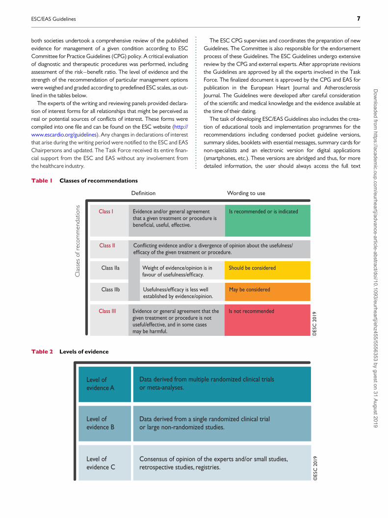
List of tablesTable 1 Classes of recommendations . . . . . . . . . . . . . . . . . . . . . . . . . . . . . . . . 7
Table 2 Levels of evidence . . . . . . . . . . . . . . . . . . . . . . . . . . . . . . . . . . . . . . . . . . 7
Table 3 New recommendations, and new and revised concepts . . . . . . 9
Table 4 Cardiovascular risk categories . . . . . . . . . . . . . . . . . . . . . . . . . . . . . . 15
Table 5 Intervention strategies as a function of total cardiovascular
risk and untreated low-density lipoprotein cholesterol levels . . . . . . . . 16
Table 6 Physical and chemical characteristics of human plasma
lipoproteins . . . . . . . . . . . . . . . . . . . . . . . . . . . . . . . . . . . . . . . . . . . . . . . . . . . . . . . 18
Table 7 Treatment targets and goals for cardiovascular disease
prevention . . . . . . . . . . . . . . . . . . . . . . . . . . . . . . . . . . . . . . . . . . . . . . . . . . . . . . . . 23
Table 8 Impact of specific lifestyle changes on lipid levels . . . . . . . . . . . . 24
Table 9 Food choices to lower low-density lipoprotein
cholesterol and improve the overall lipoprotein profile . . . . . . . . . . . . . . 25
Table 10 Drugs potentially interacting with statins metabolized by
cytochrome P450 3A4 leading to increased risk of myopathy and
rhabdomyolysis . . . . . . . . . . . . . . . . . . . . . . . . . . . . . . . . . . . . . . . . . . . . . . . . . . . 29
Table 11 Genetic disorders of lipoprotein metabolism . . . . . . . . . . . . . . 39
Table 12 Dutch Lipid Clinic Network diagnostic criteria for familial
hypercholesterolaemia . . . . . . . . . . . . . . . . . . . . . . . . . . . . . . . . . . . . . . . . . . . . 39
Table 13 Summary of recommendations for monitoring lipids
and enzymes in patients, before and on lipid-lowering therapy . . . . . . . 53
List of figuresFigure 1 Systematic Coronary Risk Estimation chart for European
populations at high cardiovascular disease risk . . . . . . . . . . . . . . . . . . . . . . 12
Figure 2 Systematic Coronary Risk Estimation chart for European
populations at low cardiovascular disease risk . . . . . . . . . . . . . . . . . . . . . . . 13
Figure 3 Expected clinical benefit of low-density lipoprotein
cholesterol-lowering therapies . . . . . . . . . . . . . . . . . . . . . . . . . . . . . . . . . . . . . 36
Figure 4 Treatment goals and algorithm for low-density lipoprotein
cholesterol-lowering according to cardiovascular disease risk . . . . . . . . 37
Figure 5 Health impact pyramid . . . . . . . . . . . . . . . . . . . . . . . . . . . . . . . . . . . . 54
Figure 6 Absolute reductions in major vascular events with statin
therapy . . . . . . . . . . . . . . . . . . . . . . . . . . . . . . . . . . . . . . . . . . . . . . . . . . . . . . . . . . . 55
List of boxesBox 1 How to use the risk estimation charts . . . . . . . . . . . . . . . . . . . . . . . . 14
Box 2 Risk estimation charts for different countries . . . . . . . . . . . . . . . . . 14
Box 3 Qualifiers . . . . . . . . . . . . . . . . . . . . . . . . . . . . . . . . . . . . . . . . . . . . . . . . . . . 14
Box 4 Factors modifying Systematic Coronary Risk Estimation risks . . 14
Box 5 Risk estimation: key messages . . . . . . . . . . . . . . . . . . . . . . . . . . . . . . . . 15
Box 6 Management of dyslipidaemia in women . . . . . . . . . . . . . . . . . . . . . . 42
Box 7 Summary of dyslipidaemia in metabolic syndrome and
type 2 diabetes mellitus . . . . . . . . . . . . . . . . . . . . . . . . . . . . . . . . . . . . . . . . . . . . 44
Box 8 Key messages . . . . . . . . . . . . . . . . . . . . . . . . . . . . . . . . . . . . . . . . . . . . . . . 55
Box 9 Gaps in the evidence . . . . . . . . . . . . . . . . . . . . . . . . . . . . . . . . . . . . . . . . 55
Box 10 Methods for enhancing adherence to lifestyle changes . . . . . . . 56
Abbreviations and acronymsABI Ankle�brachial indexACCELERATE Assessment of Clinical Effects of Cholesteryl
Ester Transfer Protein Inhibition with
4 ESC/EAS GuidelinesD
ownloaded from
https://academic.oup.com
/eurheartj/advance-article-abstract/doi/10.1093/eurheartj/ehz455/5556353 by guest on 31 August 2019

..
..
..
..
..
..
..
..
..
..
..
..
..
..
..
..
..
..
..
..
..
..
..
..
..
..
..
..
..
..
..
..
..
..
..
..
..
..
..
..
..
..
..
..
..
..
..
..
..
..
..
..
..
..
..
..
..
..
..
..
..
..
..
..
..
..
..
..
..
..
..
..
..
..
..
..
..
..
..
..
..
..
..
..
..
..Evacetrapib in Patients at a High-Risk forVascular Outcomes
ACCORD Action to Control Cardiovascular Risk in DiabetesACS Acute coronary syndromeALT Alanine aminotransferaseANGPTL3 Angiopoietin-like protein 3Apo ApolipoproteinART Antiretroviral treatmentASCEND A Study of Cardiovascular Events iN DiabetesASCOT-LLA Anglo-Scandinavian Cardiac Outcomes Trial �
Lipid-Lowering ArmASCVD Atherosclerotic cardiovascular diseaseASSIGN CV risk estimation model from the Scottish
Intercollegiate Guidelines NetworkAURORA A study to evaluate the Use of Rosuvastatin in
subjects On Regular haemodialysis: anAssessment of survival and cardiovascular events
b.i.d. Twice a day (bis in die)BIOSTAT-CHF BIOlogy Study to TAilored Treatment in
Chronic Heart FailureBIP Bezafibrate Infarction PreventionBMI Body mass indexBP Blood pressureCABG Coronary artery bypass graft surgeryCAC Coronary artery calciumCAD Coronary artery diseaseCANTOS Canakinumab Antiinflammatory Thrombosis
Outcome StudyCETP Cholesteryl ester transfer proteinCHD Coronary heart diseaseCI Confidence intervalCIID Chronic immune-mediated inflammatory diseasesCIRT Cardiovascular Inflammation Reduction TrialCK Creatine kinaseCKD Chronic kidney diseaseCOM-B Capability, Opportunity and MotivationCORONA Controlled Rosuvastatin Multinational Trial in
Heart FailureCPG Committee for Practice GuidelinesCT Computed tomographyCTT Cholesterol Treatment TrialistsCV CardiovascularCVD Cardiovascular diseaseCYP Cytochrome P4504D Die Deutsche Diabetes Dialyse Studiedal-OUTCOMES Effects of Dalcetrapib in Patients with a Recent
Acute Coronary SyndromeDASH Dietary Approaches to Stop HypertensionDGAT-2 Diacylglycerol acyltransferase-2DHA Docosahexaenoic acidDM Diabetes mellitusEAPC European Association of Preventive CardiologyEAS European Atherosclerosis SocietyEBBINGHAUS Evaluating PCSK9 Binding Antibody Influence
on Cognitive Health in High CardiovascularRisk Subjects
eGFR Estimated glomerular filtration rateEMA European Medicines AgencyEPA Eicosapentaenoic acidESC European Society of CardiologyEVOLVE EpanoVa fOr Lowering Very high
triglyceridEsEVOPACS EVOlocumab for early reduction of LDL-
cholesterol levels in patients with AcuteCoronary Syndromes
FCH Familial combined hyperlipidaemiaFCS Familial chylomicronaemia syndromeFDA US Food and Drug AdministrationFH Familial hypercholesterolaemiaFIELD Fenofibrate Intervention and Event Lowering
in DiabetesFOCUS Fixed-Dose Combination Drug for Secondary
Cardiovascular PreventionFOURIER Further Cardiovascular Outcomes Research
with PCSK9 Inhibition in Subjects withElevated Risk
GFR Glomerular filtration rateGI GastrointestinalGISSI Gruppo Italiano per lo Studio della
Sopravvivenza nell’Infarto MiocardicoHbA1c Glycated haemoglobinHeFH Heterozygous familial hypercholesterolaemiaHDL High-density lipoproteinHDL-C High-density lipoprotein cholesterolHF Heart failureHHS Helsinki Heart StudyHIV Human immunodeficiency virusHMG-CoA Hydroxymethylglutaryl-coenzyme AHoFH Homozygous familial hypercholesterolaemiaHPS2-THRIVE Heart Protection Study 2-Treatment of HDL
to Reduce the Incidence of Vascular EventsHR Hazard ratioHTG HypertriglyceridaemiaIDEAL Incremental Decrease In End-points Through
Aggressive Lipid-loweringIDL Intermediate-density lipoproteinsIL InterleukinILLUMINATE Investigation of Lipid Level Management to
Understand its Impact in AtheroscleroticEvents
IMPROVE-IT Improved Reduction of Outcomes: VytorinEfficacy International Trial
IPD Individual participant dataJUPITER Justification for the Use of Statins in
Prevention: an Intervention Trial EvaluatingRosuvastatin
KDIGO Kidney Disease: Improving Global OutcomesLCAT Lecithin cholesterol acyltransferaseLDL Low-density lipoproteinLDL-C Low-density lipoprotein cholesterolLDLR Low-density lipoprotein receptorLEAD Lower extremity arterial disease
ESC/EAS Guidelines 5D
ownloaded from
https://academic.oup.com
/eurheartj/advance-article-abstract/doi/10.1093/eurheartj/ehz455/5556353 by guest on 31 August 2019

..
..
..
..
..
..
..
..
..
..
..
..
..
..
..
..
..
..
..
..
..
..
..
..
..
..
..
..
..
..
..
..
..
..
..
..
..
..
..
..
..
..
..
..
..
..
..
..
..
..
..
..
..
..
..
..
..
..
..
..
..
..
..
..
..
..
..
..
..
..
..
..
..
..
..
..
..
..
..
..
..
..
..
..LEADER Lower Extremity Arterial Disease Event ReductionLPL Lipoprotein lipaseLp(a) Lipoprotein(a)mAb Monoclonal antibodyMACE Major adverse cardiovascular eventsMESA Multi-Ethnic Study of AtherosclerosisMetS Metabolic syndromeMI Myocardial infarctionmRNA Messenger RNAMTP Microsomal triglyceride transfer proteinNAFLD Non-alcoholic fatty liver diseaseNNT Number needed to treatNPC1L1 Niemann-Pick C1-like protein 1NSTE-ACS Non-ST elevation acute coronary syndromeo.d. Once a day (omni die)ODYSSEYOutcomes
Evaluation of Cardiovascular Outcomes Afteran Acute Coronary Syndrome DuringTreatment With Alirocumab
PAD Peripheral arterial diseasePCI Percutaneous coronary interventionPCSK9 Proprotein convertase subtilisin/kexin type 9PPAR-a Peroxisome proliferator-activated receptor-aPREDIMED Prevenci�on con Dieta Mediterr�aneaPROCAM Prospective Cardiovascular Munster StudyPROMINENT Pemafibrate to Reduce Cardiovascular
OutcoMes by Reducing Triglycerides INPatiENts With DiabeTes
PUFA Polyunsaturated fatty acidPURE Prospective Urban Rural EpidemiologyRA Rheumatoid arthritisRCT Randomized controlled trialREDUCE-IT Reduction of Cardiovascular Events with EPA-
Intervention TrialREVEAL Randomized EValuation of the Effects of
Anacetrapib Through Lipid modificationRR Relative riskRYR Red yeast riceSAMS Statin-associated muscle symptomsSBP Systolic blood pressureSCORE Systematic Coronary Risk EstimationSEAS Simvastatin and Ezetimibe in Aortic StenosisSECURE-PCI Statins Evaluation in Coronary Procedures and
RevascularizationSFA Saturated fatty acidSHARP Study of Heart and Renal ProtectionsiRNA Small interfering RNASMI Severe mental illnessSPARCL Stroke Prevention by Aggressive Reduction in
Cholesterol LevelsSTAREE STAtin Therapy for Reducing Events in the
ElderlySTEMI ST-elevation myocardial infarctionSTRENGTH Outcomes Study to Assess STatin Residual
Risk Reduction with EpaNova in HiGh CV RiskPatienTs with Hypertriglyceridemia
TC Total cholesterolT1DM Type 1 diabetes mellitusT2DM Type 2 diabetes mellitusTGs TriglyceridesTIA Transient ischaemic attackTIMI Thrombolysis In Myocardial InfarctionTNF Tumour necrosis factorTNT Treating to New TargetsTRL Triglyceride-rich lipoproteinULN Upper limit of normalVA-HIT Veterans Affairs High Density Lipoprotein
Intervention TrialVITAL VITamin D and OmegA-3 TrialVLDL Very low-density lipoproteinWHO World Health OrganizationWOSCOPS West of Scotland Coronary Prevention Study
1 Preamble
Guidelines summarize and evaluate available evidence with the aim ofassisting health professionals in proposing the best managementstrategies for an individual patient with a given condition. Guidelinesand their recommendations should facilitate decision making ofhealth professionals in their daily practice. However, the final deci-sions concerning an individual patient must be made by the responsi-ble health professional(s) in consultation with the patient andcaregiver as appropriate.
A great number of guidelines have been issued in recent years bythe European Society of Cardiology (ESC) and its partners such asEuropean Atherosclerosis Society (EAS), as well as by other societiesand organisations. Because of their impact on clinical practice, qualitycriteria for the development of guidelines have been established inorder to make all decisions transparent to the user. The recommen-dations for formulating and issuing ESC Guidelines can be found onthe ESC website (http://www.escardio.org/Guidelines-&-Education/Clinical-Practice-Guidelines/Guidelines-development/Writing-ESC-Guidelines). The ESC Guidelines represent the official position of theESC on a given topic and are regularly updated.
The ESC carries out a number of registries which are essential toassess diagnostic/therapeutic processes, use of resources and adher-ence to Guidelines. These registries aim at providing a better under-standing of medical practice in Europe and around the world, basedon data collected during routine clinical practice.
The guidelines are developed together with derivative educationalmaterial addressing the cultural and professional needs for cardiolo-gists and allied professionals. Collecting high-quality observationaldata, at appropriate time interval following the release of ESCGuidelines, will help evaluate the level of implementation of theGuidelines, checking in priority the key end points defined with theESC Guidelines and Education Committees and Task Force membersin charge.
The Members of this Task Force were selected by the ESC andEAS, including representation from relevant ESC sub-specialtygroups, in order to represent professionals involved with the medicalcare of patients with this pathology. Selected experts in the field from
6 ESC/EAS GuidelinesD
ownloaded from
https://academic.oup.com
/eurheartj/advance-article-abstract/doi/10.1093/eurheartj/ehz455/5556353 by guest on 31 August 2019
..
..
..
..
..
..
..
..
..
..
..
..
..
..
..
..
..
..
..
..
..
..
..
..
..
..both societies undertook a comprehensive review of the publishedevidence for management of a given condition according to ESCCommittee for Practice Guidelines (CPG) policy. A critical evaluationof diagnostic and therapeutic procedures was performed, includingassessment of the risk�benefit ratio. The level of evidence and thestrength of the recommendation of particular management optionswere weighed and graded according to predefined ESC scales, as out-lined in the tables below.
The experts of the writing and reviewing panels provided declara-tion of interest forms for all relationships that might be perceived asreal or potential sources of conflicts of interest. These forms werecompiled into one file and can be found on the ESC website (http://www.escardio.org/guidelines). Any changes in declarations of interestthat arise during the writing period were notified to the ESC and EASChairpersons and updated. The Task Force received its entire finan-cial support from the ESC and EAS without any involvement fromthe healthcare industry.
The ESC CPG supervises and coordinates the preparation of newGuidelines. The Committee is also responsible for the endorsementprocess of these Guidelines. The ESC Guidelines undergo extensivereview by the CPG and external experts. After appropriate revisionsthe Guidelines are approved by all the experts involved in the TaskForce. The finalized document is approved by the CPG and EAS forpublication in the European Heart Journal and AtherosclerosisJournal. The Guidelines were developed after careful considerationof the scientific and medical knowledge and the evidence available atthe time of their dating.
The task of developing ESC/EAS Guidelines also includes the crea-tion of educational tools and implementation programmes for therecommendations including condensed pocket guideline versions,summary slides, booklets with essential messages, summary cards fornon-specialists and an electronic version for digital applications(smartphones, etc.). These versions are abridged and thus, for moredetailed information, the user should always access the full text
Table 1 Classes of recommendations
©ES
C 2
019
Cla
sses
of r
ecom
men
datio
ns Class I Evidence and/or general agreement that a given treatment or procedure is
Is recommended or is indicated
Wording to use
Class III Evidence or general agreement that the given treatment or procedure is not useful/effective, and in some cases may be harmful.
Is not recommended
Class IIbestablished by evidence/opinion.
May be considered
Class IIa Weight of evidence/opinion is in Should be considered
Class II
Table 2 Levels of evidence
©ES
C 2
019
Level of evidence A
Data derived from multiple randomized clinical trials or meta-analyses.
Level of evidence B
Data derived from a single randomized clinical trialor large non-randomized studies.
Level of evidence C
Consensus of opinion of the experts and/or small studies, retrospective studies, registries.
ESC/EAS Guidelines 7D
ownloaded from
https://academic.oup.com
/eurheartj/advance-article-abstract/doi/10.1093/eurheartj/ehz455/5556353 by guest on 31 August 2019

..
..
..
..
..
..
..
..
..
..
..
..
..
..
..
..
..
..
..
..
..
..
..
..
..
..
..
..
..
..
..
..
..
..
..
..
..
..
..
..
..
..
..
..
..
..
..
..
..
..
..
..
..
..
..
..
..
..
..
..
..
..
..
..
..
..
..
..
..
..
..
..
..
..
..
..
..
..
..
..
..
..
..
..
..
..
.version of the Guidelines, which is freely available via the ESC andEAS websites and hosted on their journals’ websites (EHJ andAtherosclerosis Journal). The National Cardiac Societies of the ESCare encouraged to endorse, translate and implement all ESCGuidelines. Implementation programmes are needed because it hasbeen shown that the outcome of disease may be favourably influ-enced by the thorough application of clinical recommendations.
Health professionals are encouraged to take the ESC/EASGuidelines fully into account when exercising their clinical judgment,as well as in the determination and the implementation of preventive,diagnostic or therapeutic medical strategies. However, the ESC/EASGuidelines do not override in any way whatsoever the individualresponsibility of health professionals to make appropriate and accu-rate decisions in consideration of each patient’s health condition andin consultation with that patient or the patient’s caregiver whereappropriate and/or necessary. It is also the health professional’sresponsibility to verify the rules and regulations applicable in eachcountry to drugs and devices at the time of prescription.
2 Introduction
The previous ESC/EAS lipid Guidelines were published in August2016.1 The emergence of a substantial body of evidence over the lastfew years has required new, up-to-date Guidelines.
New evidence has confirmed that the key initiating event in athe-rogenesis is the retention of low-density lipoprotein (LDL) choles-terol (LDL-C) and other cholesterol-rich apolipoprotein (Apo) B-containing lipoproteins within the arterial wall.2 Several recentplacebo-controlled clinical studies have shown that the addition ofeither ezetimibe or anti-proprotein convertase subtilisin/kexin type 9(PCSK9) monoclonal antibodies (mAbs) to statin therapy provides afurther reduction in atherosclerotic cardiovascular disease (ASCVD)risk, which is directly and positively correlated with the incrementallyachieved absolute LDL-C reduction. Furthermore, these clinical trialshave clearly indicated that the lower the achieved LDL-C values, thelower the risk of future cardiovascular (CV) events, with no lowerlimit for LDL-C values, or ‘J’-curve effect. In addition, studies of theclinical safety of these very low achieved LDL-C values have provedreassuring, albeit monitoring for longer periods is required. For rais-ing high-density lipoprotein (HDL) cholesterol (HDL-C), recent stud-ies have indicated that the currently available therapies do not reducethe risk of ASCVD. Finally, human Mendelian randomization studieshave demonstrated the critical role of LDL-C, and other cholesterol-rich ApoB-containing lipoproteins, in atherosclerotic plaque forma-tion and related subsequent CV events. Thus, there is no longer an‘LDL-C hypothesis’, but established facts that increased LDL-C valuesare causally related to ASCVD, and that lowering LDL particles andother ApoB-containing lipoproteins as much as possible reduces CVevents.
In order to be aligned with these new findings, the ESC/EAS TaskForce members who have written these Guidelines have proposednew LDL-C goals, as well as a revised CV risk stratification, which areespecially relevant to high- and very-high-risk patients.
These novel ESC/EAS Guidelines on lipids provide important newadvice on patient management, which should enable more cliniciansto efficiently and safely reduce CV risk through lipid modification.
2.1 What is new in the 2019 Guidelines?New recommendations, and new and revised concepts, are pre-sented in Table 3.
3 What is cardiovascular diseaseprevention?
3.1 Definition and rationaleCardiovascular disease (CVD), of which ASCVD is the major compo-nent, is responsible for >4 million deaths in Europe each year. It killsmore women (2.2 million) than men (1.8 million), although CV deathsbefore the age of 65 years are more common in men (490 000 vs.193 000).3 Prevention is defined as a co-ordinated set of actions,either at the population or individual level, aimed at eliminating or min-imizing the impact of CV diseases and their related disabilities. Morepatients are surviving their first CVD event and are at high-risk ofrecurrences. In addition, the prevalence of some risk factors, notablydiabetes (DM) and obesity, is increasing. The importance of ASCVDprevention remains undisputed and should be delivered at the generalpopulation level by promoting healthy lifestyle behaviour,4 and at theindividual level by tackling unhealthy lifestyles and by reducingincreased levels of causal CV risk factors, such as LDL cholesterol orblood pressure (BP) levels.
3.2 Development of the Joint Task ForceGuidelines for the management ofdyslipidaemiasThe present Guidelines represent an evidence-based consensus ofthe European Task Force, including the ESC and the EAS.
By appraising the current evidence and identifying remainingknowledge gaps in the management of dyslipidaemias, the Task Forcehas formulated recommendations to guide action in clinical practiceto prevent ASCVD by modifying plasma lipid levels.
This document has been developed for healthcare professionals tofacilitate informed communication with individuals about their CVrisk and the benefits of adopting and sustaining a healthy lifestyle, andof early modification of their lipid-related CV risk. In addition, theGuidelines provide tools for healthcare professionals to promote up-to-date intervention strategies, integrate these strategies into nationalor regional prevention frameworks, and to translate them into locallydelivered healthcare services, in line with the recommendations ofthe World Health Organization (WHO) Global Status Report onNoncommunicable Diseases 2014.5
A lifetime approach to CV risk should be considered.1 This impliesthat—apart from improving lifestyle habits and reducing risk factorlevels in patients with established ASCVD, and in those at increasedrisk of developing ASCVD—people of all ages should be encouragedto adopt or sustain a healthy lifestyle.
4 Total cardiovascular risk
4.1 Total cardiovascular risk estimationCV risk in the context of these Guidelines means the likelihood of aperson developing an atherosclerotic CV event over a defined period
8 ESC/EAS GuidelinesD
ownloaded from
https://academic.oup.com
/eurheartj/advance-article-abstract/doi/10.1093/eurheartj/ehz455/5556353 by guest on 31 August 2019

Table 3 New recommendations, and new and revised concepts
New recommendations
Cardiovascular imaging for assessment of ASCVD risk
Assessment of arterial (carotid and/or femoral) plaque burden on arterial ultrasonography should be considered as a risk modifier in individuals
at low or moderate risk.
Cardiovascular imaging for assessment of ASCVD risk
CAC score assessment with CT should be considered as a risk modifier in the CV risk assessment of asymptomatic individuals at low or moderate risk.
Lipid analyses for CVD risk estimation
Lp(a) measurement should be considered at least once in each adult person’s lifetime to identify those with very high inherited Lp(a) levels >180 mg/dL
(>430 nmol/L) who may have a lifetime risk of ASCVD equivalent to the risk associated with heterozygous familial hypercholesterolaemia.
Drug treatments of patients with hypertriglyceridaemia
In high-risk (or above) patients with TG between 1.5 and 5.6 mmol/L (135 - 499 mg/dL) despite statin treatment, n-3 PUFAs
(icosapent ethyl 2 � 2g/day) should be considered in combination with statins.
Treatment of patients with heterozygous FH
In primary prevention, for individuals with FH at very-high risk, an LDL-C reduction of >_50% from baseline and an LDL-C goal of <1.4 mmol/L
(<55 mg/dL) should be considered.
Treatment of dyslipidaemias in older people
Treatment with statins is recommended for primary prevention, according to the level of risk, in older people aged <_75.
Treatment of dyslipidaemias in older people
Initiation of statin treatment for primary prevention in older people aged >75 may be considered, if at high risk or above.
Treatment of dyslipidaemias in DM
In patients with T2DM at very-high risk, an LDL-C reduction of >_50% from baseline and an LDL-C goal of <1.4 mmol/L (<55mg/dL) is recommended.
In patients with T2DM at high risk, an LDL-C reduction of >_50% from baseline and an LDL-C goal of <1.8 mmol/L (<70 mg/dL) is recommended.
Statins are recommended in patients with T1DM who are at high or very-high risk.
Treatment of dyslipidaemias in DM
Intensification of statin therapy should be considered before the introduction of combination therapy.
If the goal is not reached, statin combination with ezetimibe should be considered.
Treatment of dyslipidaemias in DM
Statin therapy is not recommended in pre-menopausal patients with DM who are considering pregnancy or not using adequate contraception.
Lipid-lowering therapy in patients with ACS
For patients who present with an ACS, and whose LDL-C levels are not at goal despite already taking a maximally tolerated statin dose and
ezetimibe, adding a PCSK9 inhibitor early after the event (if possible, during hospitalization for the ACS event) should be considered.
Changes in recommendations
Upgrades
2016 2019
Lipid analyses for CVD risk estimation Lipid analyses for CVD risk estimation
ApoB should be considered as an alternative risk marker whenever available,
especially in individuals with high TG.
ApoB analysis is recommended for risk assessment, particularly in
people with high TG, DM, obesity or metabolic syndrome, or very low
LDL-C. It can be used as an alternative to LDL-C, if available, as the
primary measurement for screening, diagnosis, and management,
and may be preferred over non-HDL-C in people with high TG, DM,
obesity, or very low LDL-C.
Pharmacological LDL-C lowering Pharmacological LDL-C lowering
If the LDL goal is not reached, statin combination with a cholesterol
absorption inhibitor should be considered.
If the goals are not achieved with the maximum tolerated dose of statin,
combination with ezetimibe is recommended.
Pharmacological LDL-C lowering Pharmacological LDL-C lowering
In patients at very-high risk, with persistent high LDL-C despite treatment
with maximal tolerated statin dose, in combination with ezetimibe or in
patients with statin intolerance, a PCSK9 inhibitor may be considered.
For secondary prevention, patients at very-high risk not achieving their
goal on a maximum tolerated dose of statin and ezetimibe, a combination
with a PCSK9 inhibitor is recommended.
For very-high-risk FH patients (that is, with ASCVD or with another
major risk factor) who do not achieve their goals on a maximum
tolerated dose of statin and ezetimibe, a combination with a PCSK9
inhibitor is recommended.
Continued
ESC/EAS Guidelines 9D
ownloaded from
https://academic.oup.com
/eurheartj/advance-article-abstract/doi/10.1093/eurheartj/ehz455/5556353 by guest on 31 August 2019

Drug treatments of hypertriglyceridaemia Drug treatments of hypertriglyceridaemia
Statin treatment may be considered as the first drug of choice for
reducing CVD risk in high-risk individuals with hypertriglyceridaemia.
Statin treatment is recommended as the first drug of choice for reducing
CVD risk in high-risk individuals with hypertriglyceridaemia
[TG >2.3 mmol/L (200 mg/dL)].
Treatment of patients with heterozygous FH Treatment of patients with heterozygous FH
Treatment should be considered to aim at reaching an LDL-C <2.6 mmol/L
(<100 mg/dL) or in the presence of CVD <1.8 mmol/L (<70 mg/dL).
If targets cannot be reached, maximal reduction of LDL-C should be
considered using appropriate drug combinations.
For FH patients with ASCVD who are at very-high risk, treatment to
achieve at least a 50% reduction from baseline and an LDL-C
<1.4 mmol/L (<55 mg/dL) is recommended. If goals cannot be achieved,
a drug combination is recommended.
Treatment of patients with heterozygous FH Treatment of patients with heterozygous FH
Treatment with a PCSK9 antibody should be considered in FH patients with
CVD or with other factors putting them at very-high risk for CHD,
such as other CV risk factors, family history, high Lp(a), or statin intolerance.
Treatment with a PCSK9 inhibitor is recommended in very-high-risk
FH patients if the treatment goal is not achieved on maximal tolerated
statin plus ezetimibe.
Treatment of dyslipidaemias in older adults Treatment of dyslipidaemias in older people
Since older people often have comorbidities and have
altered pharmacokinetics, lipid-lowering medication should be started
at a lower dose and then titrated with caution to achieve target lipid
levels that are the same as in younger people.
It is recommended that the statin is started at a low dose if there is
significant renal impairment and/or the potential for drug interactions,
and then titrated upwards to achieve LDL-C treatment goals.
Lipid-lowering therapy in patients with ACS Lipid-lowering therapy in patients with ACS
If the LDL-C target is not reached with the highest tolerated statin dose
and/or ezetimibe, PCSK9 inhibitors may be considered on top of
lipid-lowering therapy; or alone or in combination with ezetimibe in
statin-intolerant patients or in whom a statin is contraindicated.
If the LDL-C goal is not achieved after 4 - 6 weeks despite maximal
tolerated statin therapy and ezetimibe, addition of a PCSK9 inhibitor
is recommended.
Recommendation grading
Class I Class IIa Class IIb Class III
New sections
• A new section is focused on the utility of non-invasive CV imaging for classification of total CVD risk, with implications for recommended
lipid-modifying therapies.
• More data are provided on the biology and physiology of lipids and lipoproteins, and on their roles in pathophysiology. Emerging evidence from observa-
tional studies, RCTs, and genetic (Mendelian randomization) studies unequivocally showing a causal effect of LDL-C in the development of ASCVD is dis-
cussed, and newer evidence regarding the effects of TGs and HDL on ASCVD risk is presented.
• New sections describe novel lipid-modifying medications as well as emerging approaches for lowering LDL-C, TGs, and Lp(a).
• A new section discusses the inflammation-related risk in very high-risk patients and the potential role of inflammation as a therapeutic target to lower ASCVD risk.
• New/revised concepts
More intensive reduction of LDL-C across CV risk categories
• For secondary prevention in very-high-risk patients, an LDL-C reduction of >_50% from baseline and an LDL-C goal of <1.4 mmol/L (<55 mg/dL) are recommended.
� For patients with ASCVD who experience a second vascular event within 2 years (not necessarily of the same type as the first event) while taking maximally toler-
ated statin therapy, an LDL-C goal of <1.0 mmol/L (<40 mg/dL) may be considered.
• In primary prevention, for individuals at very-high risk but without FH, an LDL-C reduction of >_50% from baseline and an LDL-C goal of <1.4 mmol/L
(<55 mg/dL) are recommended. For individuals at very-high risk (that is, with another risk factor but without ASCVD), in primary prevention the same
goals for LDL-C lowering should be considered.
• For patients at high risk, an LDL-C reduction of >_50% from baseline and an LDL-C goal of <1.8 mmol/L (<70 mg/dL) are recommended.
• For individuals at moderate risk, an LDL-C goal of <2.6 mmol/L (<100 mg/dL) should be considered.
• For individuals at low risk, an LDL-C goal of <3.0 mmol/L (<116 mg/dL) may be considered.
The rationale for the revised, lower LDL-C goals across CV risk categories is discussed, based on a critical synthesis of available evidence
from lipid-modifying interventions resulting in reductions in CV risk.
Pharmacological LDL-C-lowering strategies
The section on pharmacological strategies to lower LDL-C emphasizes the concept that the absolute LDL-C reduction (determined by pre-treatment LDL-
C levels and the LDL-lowering efficacy of the medications) dictates the relative risk reduction, which in turn—depending on the baseline CV risk—defines
the associated absolute CV risk reduction in individual patients.
Risk classification in patients with FH
Patients with FH and ASCVD, or another major risk factor, are classified as very-high-risk, and those without known ASCVD and without other risk factors
as high-risk. Recommended treatment goals are defined accordingly.
Adverse effects of statins
The distinction between formal statin myopathy vs. so-called statin-associated muscle symptoms is emphasized, and the discordance in reported frequency
of symptoms in RCTs vs. observational studies are critically discussed on the basis of new relevant evidence.
Continued
10 ESC/EAS GuidelinesD
ownloaded from
https://academic.oup.com
/eurheartj/advance-article-abstract/doi/10.1093/eurheartj/ehz455/5556353 by guest on 31 August 2019

..
..
..
..
..
..
..
..
..
..
..
..
..
..
..
..
..
..
..
..
..
..
..
..
..
..
..
..
..
..
..
..
..
..
..
..
..
..
..
..
..
..
..
..
..
..
..
..
..
..
..
..
..
..
..
..
..
..
..
..
..
..
..
..
..
..
..
..
..
.of time. Total CVD risk expresses the combined effect of a numberof risk factors on this risk estimate. In these Guidelines, we addressthe lipid-related contribution to total CV risk and how to manage itat the clinical level.
4.1.1 Rationale for assessing total cardiovascular disease
risk
All current guidelines on the prevention of ASCVD in clinical practicerecommend the assessment of total CVD risk. Prevention of ASCVDin a given person should relate to his or her total CV risk: the higherthe risk, the more intense the action should be.
Many risk assessment systems are available and have been compre-hensively reviewed (Supplementary Table 1 in the SupplementaryData). Most guidelines use one of these risk assessment systems.6�8
Ideally, risk charts should be based on country-specific cohort data.These are not available for most countries. The SCORE (SystematicCoronary Risk Estimation) system can be recalibrated for use in dif-ferent populations by adjusting for secular changes in CVD mortalityand risk factor prevalence. Calibrated country-specific versions areavailable for many European countries and can be found at http://www.heartscore.org. These are now being updated to provide recali-brated, contemporaneous country-specific charts for all Europeancountries. Other risk estimation systems—using both fatal and non-fatal events—can also be recalibrated, but the process is easier andscientifically more robust for mortality than for total events. TheEuropean Guidelines on CVD prevention in clinical practice (boththe 20129 and 201610 versions) recommend the use of the SCOREsystem because it is based on large, representative European cohortdata sets and because it is relatively straightforward to recalibrate forindividual countries.
Persons with documented ASCVD, type 1 or type 2 DM (T1DMand T2DM, respectively), very high levels of individual risk factors, orchronic kidney disease (CKD) are generally at very-high or high totalCV risk. No risk estimation models are needed for such persons;they all need active management of all risk factors. For other, appa-rently healthy people, the use of a risk estimation system such asSCORE, which estimates the 10 year cumulative risk of a first fatalatherosclerotic event, is recommended to estimate total CV risk,since many people have several risk factors that, in combination, mayresult in high levels of total CV risk.
Risk estimates have been produced as charts for high- and low-riskregions in Europe (Figures 1 and 2).11 All International Classification ofDiseases codes that are related to deaths from vascular origin causedby atherosclerosis are included. The reasons for retaining a systemthat estimates fatal as opposed to total fatal þ non-fatal events arethat non-fatal events are dependent on definition, developments in
diagnostic tests, and methods of ascertainment, all of which can vary,resulting in very variable multipliers to convert fatal to total events. Inaddition, total event charts, in contrast to those based on mortality,are more difficult to recalibrate to suit different populations. That said,work is in progress to produce regional total event charts.
The SCORE data indicate that the total CVD event risk is aboutthree times higher than the risk of fatal CVD for men, so a SCORErisk of 5% translates into a CVD risk of �15% of total (fatal þ non-fatal) CVD endpoints; the multiplier is higher in women and lower inolder people.
Clinicians often ask for thresholds to trigger certain interventions.This is problematic since risk is a continuum and there is no thresholdat which, for example, a drug is automatically indicated. This is truefor all continuous risk factors such as plasma cholesterol or systolicBP (SBP). Therefore, the goals that are proposed in this documentreflect this concept.
A particular problem relates to young people with high levels of riskfactors; a low absolute risk may conceal a very high relative risk requir-ing at least intensive lifestyle advice. To motivate young people (i.e.aged <40 years) not to delay changing their unhealthy lifestyle, an esti-mate of their relative risk—illustrating that lifestyle changes can reducerelative risk substantially—may be helpful (Supplementary Figure 1).
Another approach to this problem is to use CV risk age. The riskage of a person with several CV risk factors is the age of a personwith the same level of risk but with ideal levels of risk factors. Thus, ahigh-risk 40-year-old would have a risk age >_65 years. Risk age can beestimated visually by looking at the SCORE chart (as illustrated inSupplementary Figure 2). In this chart, the risk age of a person with riskfactors is defined as the age at which a person with ideal risk factorlevels would reach the same risk level. Ideal risk factors are non-smoking, total cholesterol (TC) <_4 mmol/L (<_155 mg/dL), and SBP<_120 mmHg. Risk age is also automatically calculated as part of thelatest revision of HeartScore (http://www.HeartScore.org).
Risk age has been shown to be independent of the CV endpointused,6,8 can be used in any population regardless of baseline risk orsecular changes in mortality, and therefore avoids the need forrecalibration.
Lifetime risk is another approach to illustrate the impact of risk fac-tors that may be useful in younger people.12 The greater the burdenof risk factors, the higher the lifetime risk. This approach produceshigher risk figures for younger people because of their longer expo-sure times. Therefore, it is more useful as a way of illustrating riskthan as a guide to treatment, because therapeutic trials have beenbased on a fixed follow-up period and not on lifetime risk.
Another problem relates to older people. In some age categories,the majority of people, especially males, will have estimated 10 year
PCSK9 inhibitors
New outcome study data of PCSK9 inhibitors are presented, and updated recommendations for their clinical use are provided.
Cost-effectiveness
The issue of cost-effectiveness of lipid-modifying interventions is updated in view of changes in the availability of generic products for statins and ezetimibe,
and of PCSK9 inhibitors.
ACS = acute coronary syndrome; ApoB = apolipoprotein B; ASCVD = atherosclerotic cardiovascular disease; CAC = coronary artery calcium; CHD = coronary heart disease;CT = computed tomography; CV = cardiovascular; CVD = cardiovascular disease; DM = diabetes mellitus; FH = familial hypercholesterolaemia; HDL = high-density lipopro-tein; LDL-C = low-density lipoproteins cholesterol; Lp(a) = lipoprotein(a); PCSK9 = proprotein convertase subtilisin/kexin type 9; PUFAs = polyunsaturated fatty acids; RCTs= randomized controlled trials; T1DM = type 1 diabetes mellitus; T2DM = type 2 diabetes mellitus; TGs = triglycerides.
ESC/EAS Guidelines 11D
ownloaded from
https://academic.oup.com
/eurheartj/advance-article-abstract/doi/10.1093/eurheartj/ehz455/5556353 by guest on 31 August 2019
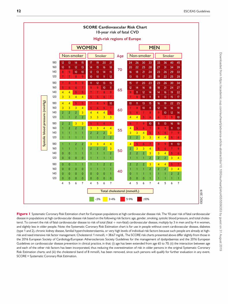
©ES
C 2
019
180
160
140
120
WOMEN
SCORE Cardiovascular Risk Chart10-year risk of fatal CVD
High-risk regions of Europe
MEN
Total cholesterol (mmol/L)
Age
70
65
60
55
50
Non-smoker Smoker
15
8 9
7 7 8 9
40
4
<3%
5 6 7 4 5 6 7
Syst
olic
blo
od p
ress
ure
(mm
Hg)
12 13 14
10 11 12 13
10 10
21
12 13
10 10 11 12
17 19 20
14 15 16 18
14 15
180
160
140
120
9
4 4
3 3 4 4
7 8 8
5 6 6 7
5 5
15
7 7
5 5 6 7
11 12 13
9 9 10 11
8 9
180
160
140
120
5
2 2
1 1 2 2
4 4 5
3 3 3 4
2 3
10
4 4
3 3 3 3
7 8 9
5 6 6 7
4 5
180
160
140
120
3
1 1
1 1 1 1
2 2 3
1 2 2 2
1 1
7
2 2
1 1 2 2
5 5 6
3 3 4 4
2 3
180
160
140
120
2
0 0
0 0 0 0
1 1 2
1 1 1 1
1 1
4
1 1
1 1 1 1
3 3 4
2 2 2 3
1 2
180
160
140
120
1
0 0
3–4% 5–9% ≥10%
0 0 0 0
0 0 1
0 0 0 0
0 0
2
0 0
0 0 0
1 1 2
1 1 1 1
0 1
Non-smoker Smoker
33
16 18
13 15 17 20
4 5 6 7 4 5 6 7
24 26 30
20 22 25 28
21 24
45
23 26
19 22 25 28
33 36 40
27 31 34 39
29 33
23
9 11
7 8 10 11
15 17 20
12 14 16 18
12 14
34
14 16
11 13 15 17
23 26 30
18 21 24 27
19 22
15
5 6
4 4 5 6
10 11 13
7 8 10 11
7 8
25
9 10
6 7 9 10
16 19 22
12 14 16 19
12 14
10
3 3
2 2 3 3
6 7 9
4 5 6 7
4 5
18
5 6
4 4 5 6
11 13 16
8 9 11 13
7 9
7
2 2
1 1 1 2
4 5 6
2 3 3 4
2 3
13
3 4
2 2 3 4
8 9 11
5 6 7 9
5 6
3
0 1
0 0 0 1
2 2 2
1 1 1 2
1 1
7
1 1
1 1 1
4 4 5
2 2 3 4
2 2
0 1
Figure 1 Systematic Coronary Risk Estimation chart for European populations at high cardiovascular disease risk. The 10-year risk of fatal cardiovasculardisease in populations at high cardiovascular disease risk based on the following risk factors: age, gender, smoking, systolic blood pressure, and total choles-terol. To convert the risk of fatal cardiovascular disease to risk of total (fatalþ non-fatal) cardiovascular disease, multiply by 3 in men and by 4 in women,and slightly less in older people. Note: the Systematic Coronary Risk Estimation chart is for use in people without overt cardiovascular disease, diabetes(type 1 and 2), chronic kidney disease, familial hypercholesterolaemia, or very high levels of individual risk factors because such people are already at high-risk and need intensive risk factor management. Cholesterol: 1 mmol/L = 38.67 mg/dL. The SCORE risk charts presented above differ slightly from those inthe 2016 European Society of Cardiology/European Atherosclerosis Society Guidelines for the management of dyslipidaemias and the 2016 EuropeanGuidelines on cardiovascular disease prevention in clinical practice, in that: (i) age has been extended from age 65 to 70; (ii) the interaction between ageand each of the other risk factors has been incorporated, thus reducing the overestimation of risk in older persons in the original Systematic CoronaryRisk Estimation charts; and (iii) the cholesterol band of 8 mmol/L has been removed, since such persons will qualify for further evaluation in any event.SCORE = Systematic Coronary Risk Estimation.
12 ESC/EAS GuidelinesD
ownloaded from
https://academic.oup.com
/eurheartj/advance-article-abstract/doi/10.1093/eurheartj/ehz455/5556353 by guest on 31 August 2019

©ES
C 2
019
180
160
140
120
WOMEN
SCORE Cardiovascular Risk Chart10-year risk of fatal CVD
Low-risk regions of Europe
MEN
Total cholesterol (mmol/L)
Age
70
65
60
55
50
Non-smoker Smoker
9
5 5
4 4 5 5
40
4
<3%
5 6 7 4 5 6 7
Syst
olic
blo
od p
ress
ure
(mm
Hg)
7 8 8
6 6 7 7
6 6
13
7 8
6 6 7 7
11 11 12
9 9 10 11
8 9
180
160
140
120
5
2 3
2 2 2 2
4 4 5
3 3 4 4
3 3
9
4 4
3 3 3 4
7 7 8
5 6 6 7
5 5
180
160
140
120
3
1 1
1 1 1 1
2 3 3
2 2 2 2
1 2
6
2 2
2 2 2 2
4 5 5
3 3 4 4
3 3
180
160
140
120
2
1 1
0 0 0 1
1 1 2
1 1 1 1
1 1
4
1 1
1 1 1 1
3 3 3
2 2 2 3
1 2
180
160
140
120
1
0 0
0 0 0 0
1 1 1
0 0 1 1
0 0
3
1 1
0 0 0 1
2 2 2
1 1 1 2
1 1
180
160
140
120
0
0 0
3–4% 5–9% ≥10%
0 0 0 0
0 0 0
0 0 0 0
0 0
1
0 0
0 0 0
1 1 1
0 0 0 1
0 0
Non-smoker Smoker
17
8 9
7 8 9 10
4 5 6 7 4 5 6 7
12 14 15
10 11 13 14
10 12
24
12 13
10 11 12 14
18 20 22
15 16 18 20
15 17
12
5 5
3 4 5 5
8 9 10
6 7 8 9
6 7
18
7 8
5 6 7 8
12 14 16
9 11 12 14
9 11
8
3 3
2 2 2 3
5 6 7
4 4 5 5
3 4
13
4 5
3 4 4 5
8 10 11
6 7 8 9
6 7
5
1 2
1 1 1 2
3 4 4
2 2 3 3
2 2
9
3 3
2 2 2 3
6 7 8
4 4 5 6
3 4
3
1 1
0 1 1 1
2 2 3
1 1 2 2
1 1
6
1 2
1 1 1 2
4 5 5
2 3 3 4
2 3
1
0 0
0 0 0 0
1 1 1
0 0 1 1
0 0
3
1 1
0 0 0
2 2 3
1 1 1 2
1 1
0 1
Figure 2 Systematic Coronary Risk Estimation chart for European populations at low cardiovascular disease risk. The 10-year risk of fatal cardiovasculardisease in populations at low cardiovascular disease risk based on the following risk factors: age, gender, smoking, systolic blood pressure, and total choles-terol. To convert the risk of fatal cardiovascular disease to risk of total (fatalþ non-fatal) cardiovascular disease, multiply by 3 in men and by 4 in women,and slightly less in older people. Note: the Systematic Coronary Risk Estimation chart is for use in people without overt cardiovascular disease, diabetes(type 1 and 2), chronic kidney disease, familial hypercholesterolaemia, or very high levels of individual risk factors because such people are already at high-risk and need intensive risk factor management. Cholesterol: 1 mmol/L=38.67 mg/dL. The SCORE risk charts presented above differ slightly from those inthe 2016 European Society of Cardiology/European Atherosclerosis Society Guidelines for the management of dyslipidaemias and the 2016 EuropeanGuidelines on cardiovascular disease prevention in clinical practice, in that: (i) age has been extended from age 65 to 70; (ii) the interaction between ageand each of the other risk factors has been incorporated, thus reducing the overestimation of risk in older persons in the original Systematic CoronaryRisk Estimation charts; and (iii) the cholesterol band of 8 mmol/L has been removed since such persons will qualify for further evaluation in any event.SCORE = Systematic Coronary Risk Estimation.
ESC/EAS Guidelines 13D
ownloaded from
https://academic.oup.com
/eurheartj/advance-article-abstract/doi/10.1093/eurheartj/ehz455/5556353 by guest on 31 August 2019

cumulative CV death risks exceeding the 5�10% level, based on ageonly, even when other CV risk factor levels are relatively low.Therefore, before initiating treatment in the elderly, clinicians shouldevaluate patients carefully. The relative strengths of risk factors varywith age and SCORE overestimates risk in older people (that is, thoseaged >65 years).11 These Guidelines include illustrative charts forolder people (see Figures 1 and 2). While older people benefit fromsmoking cessation, and control of hypertension and hyperlipidaemia(see section 9.3), clinical judgement is required to avoid side effectsfrom overmedication.
The additional impact of HDL-C on risk estimation is illustrated inSupplementary Figures 3 and 4; HDL-C can be used to increase theaccuracy of the risk evaluation. In these charts, HDL-C is used cate-gorically. The electronic version of SCORE, HeartScore (http://www.heartscore.org/en_GB/), has been modified to take HDL-C into con-sideration as a continuous variable. Clinicians should be aware that atextremely high values [above �2.3 mmol/L (90 mg/dL)] of HDL-Cthere appears to be an increased risk of ASCVD, so at such levelsHDL-C cannot be used as a risk predictor.
4.1.2 How to use the risk estimation charts
Use of the low- or the high-risk SCORE charts will depend on theCVD mortality experience in each country. While any cut-off point isarbitrary and open to debate, in these Guidelines, the cut-off pointfor calling a country ‘low CVD risk’ is based on WHO data derivedfrom the Global Burden of Disease Study.
Countries are categorized as low-risk if their age-adjusted 2016CVD mortality rate was <150/100 000 (for men and womentogether) (http://www.who.int/healthinfo/global_burden_disease/estimates/en/). Countries with a CVD mortality rate of >_150/100 000 ormore are considered to be at high-risk.
Boxes 1 to 5 summarize the main points regarding the risk estima-tion charts and their use.
Box 1 How to use the risk estimation charts
To estimate a person’s 10-year risk of CVD death, find the table for his/
her gender, smoking status, and age. Within the table, find the cell nearest
to the person’s BP and TC. Risk estimates will need to be adjusted
upwards as the person approaches the next age category.
Risk is initially assessed on the level of TC and systolic BP before treat-
ment, if known. The longer the treatment and the more effective it is, the
greater the reduction in risk, but in general it will not be more than about
one-third of the baseline risk. For example, for a person on antihyperten-
sive drug treatment in whom the pre-treatment BP is not known, if the
total CV SCORE risk is 6%, then the pre-treatment total CV risk may
have been 9%.
Low-risk persons should be offered advice to maintain their low-risk sta-
tus. While no threshold is universally applicable, the intensity of advice
should increase with increasing risk.
The charts may be used to give some indication of the effects of reducing
risk factors, given that there is apparently a time lag before the risk
reduces. In general, people who stop smoking halve their cumulative risk
over a relatively short period of time.
BP = blood pressure; CV = cardiovascular; CVD = cardiovascular disease;SCORE = Systematic Coronary Risk Estimation; TC = total cholesterol.
Box 2 Risk estimation charts for different countries
The low-risk charts should be considered for use in Austria, Belgium,
Cyprus, Denmark, Finland, France, Germany, Greece, Iceland, Ireland,
Israel, Italy, Luxembourg, Netherlands, Norway, Malta, Portugal, Slovenia,
Spain, Sweden, Switzerland, and the UK.
The high-risk charts should be considered for use in Albania, Algeria,
Armenia, Bosnia and Herzegovina, Croatia, Czech Republic, Estonia,
Hungary, Latvia, Lebanon, Libya, Lithuania, Montenegro, Morocco,
Poland, Romania, Serbia, Slovakia, Tunisia, and Turkey.
Some countries have a cardiovascular disease mortality rate >350/100
000, and the high-risk chart may underestimate risk. These are
Azerbaijan, Belarus, Bulgaria, Egypt, Georgia, Kazakhstan, Kyrgyzstan,
North Macedonia, Republic of Moldova, Russian Federation, Syria,
Tajikistan, Turkmenistan, Ukraine, and Uzbekistan.
See http://apps.who.int/gho/data/node.home.
Box 3 Qualifiers
The charts can assist in risk assessment and management, but must be
interpreted in light of the clinician’s knowledge and experience, and of
the patient’s pre-test likelihood of CVD.
Risk will be overestimated in countries with decreasing CVD mortality,
and underestimated in countries in which mortality is increasing. This is
dealt with by recalibration (http://www.heartscore.org/en_GB/).
Risk estimates are lower in women than in men. However, risk is only
deferred in women; the risk of a 60-year-old woman is similar to that of a
50-year-old man. Ultimately, more women die from CVD than men.
Relative risks may be unexpectedly high in young persons, even if abso-
lute risk levels are low. The relative risk chart (Supplementary Figure 1)
and the estimated risk age (Supplementary Figure 2) may be helpful in
identifying and counselling such persons.
CVD = cardiovascular disease.
Box 4 Factors modifying Systematic Coronary RiskEstimation risks
Social deprivation: the origin of many of the causes of CVD.
Obesity and central obesity as measured by the body mass index and
waist circumference, respectively.
Physical inactivity.
Psychosocial stress including vital exhaustion.
Family history of premature CVD (men: <55 years and women: <60
years).
Chronic immune-mediated inflammatory disorder.
Major psychiatric disorders.
Treatment for human immunodeficiency virus infection.
Atrial fibrillation.
Left ventricular hypertrophy.
Chronic kidney disease.
Obstructive sleep apnoea syndrome.
Non-alcoholic fatty liver disease.
CVD = cardiovascular disease.
14 ESC/EAS GuidelinesD
ownloaded from
https://academic.oup.com
/eurheartj/advance-article-abstract/doi/10.1093/eurheartj/ehz455/5556353 by guest on 31 August 2019

..
..
..
..
..
..
..
..
..
..
..
..
..
..
..
..
..
..
..Social deprivation and psychosocial stress set the scene forincreased risk.13 For those at moderate risk, other factors—includingmetabolic factors such as increased ApoB, lipoprotein(a) [Lp(a)], tri-glycerides (TGs), or C-reactive protein; the presence of albuminuria;the presence of atherosclerotic plaque in the carotid or femoralarteries; or the coronary artery calcium (CAC) score—may improverisk classification. Many other biomarkers are also associated withincreased CVD risk, although few of these have been shown to beassociated with appreciable reclassification. Total CV risk will also behigher than indicated in the SCORE charts in asymptomatic personswith abnormal markers of subclinical atherosclerotic vascular dam-age. Reclassification is of value in people identified as being at moder-ate CV risk by using markers such as CAC score >100 Agatstonunits, ankle�brachial index (ABI) <0.9 or >1.40, carotid�femoralpulse wave velocity >10 m/s, or the presence of plaques at carotid orfemoral ultrasonography. In studies comparing these markers, CAChad the best reclassification ability.14�16
Some factors such as a high HDL-C up to 2.3 mmol/L (90mg/dL)17
or a family history of longevity can also be associated with lower risk.
4.2 Risk levelsA total CV risk estimate is part of a continuum. The cut-off pointsthat are used to define high-risk are, in part, both arbitrary and basedon the risk levels at which benefit is evident in clinical trials. In clinicalpractice, consideration should be given to practical issues in relationto the local healthcare systems. Not only should those at high risk beidentified and managed, but those at moderate risk should alsoreceive professional advice regarding lifestyle changes; in some cases,drug therapy will be needed to reduce atherosclerotic risk.
Low-risk people should be given advice to help them maintain thisstatus. Thus, the intensity of preventive actions should be tailored tothe patient’s total CV risk. The strongest driver of total CV risk is age,which can be considered as ‘exposure time’ to risk factors.
For these reasons, the Task Force suggests the following catego-ries of risk and LDL-C goals, based on the best available evidence andin an ideal setting with unlimited resources. These categories repre-sent a counsel of perfection, but these ideals are for guidance onlyand practical decision-making must be based on what is appropriateto the local situation.
With these considerations, we propose the levels of total CV riskpresented in Table 4.
Box 5 Risk estimation: key messages
In apparently healthy persons, CVD risk is most frequently the result of
multiple, interacting risk factors. This is the basis for total CV risk estima-
tion and management.
Risk factor screening including the lipid profile should be considered in men
>40 years old, and in women >50 years of age or post-menopausal.
A risk estimation system such as SCORE can assist in making logical man-
agement decisions, and may help to avoid both under- and
overtreatment.
Certain individuals declare themselves to be at high or very high CVD risk
without needing risk scoring, and all risk factors require immediate attention.
This is true for patients with documented CVD, older individuals with long-
standing DM, familial hypercholesterolaemia, chronic kidney disease, carotid
or femoral plaques, coronary artery calcium score >100, or extreme Lp(a)
elevation.
All risk estimation systems are relatively crude and require attention to
qualifying statements.
Additional factors affecting risk can be accommodated in electronic risk
estimation systems such as HeartScore (www.heartscore.org).
The total risk approach allows flexibility; if optimal control cannot be
achieved with one risk factor, trying harder with the other factors can still
reduce risk.
CV = cardiovascular; CVD = cardiovascular disease; DM = diabetes mellitus;SCORE = Systematic Coronary Risk Estimation.
Table 4 Cardiovascular risk categories
Very-high-
risk
People with any of the following:
Documented ASCVD, either clinical or unequivocal
on imaging. Documented ASCVD includes previous
ACS (MI or unstable angina), stable angina, coronary
revascularization (PCI, CABG, and other arterial
revascularization procedures), stroke and TIA, and
peripheral arterial disease. Unequivocally docu-
mented ASCVD on imaging includes those findings
that are known to be predictive of clinical events,
such as significant plaque on coronary angiography
or CT scan (multivessel coronary disease with two
major epicardial arteries having >50% stenosis), or
on carotid ultrasound.
DM with target organ damage,a or at least three major
risk factors, or early onset of T1DM of long duration
(>20 years).
Severe CKD (eGFR <30 mL/min/1.73 m2).
A calculated SCORE >_10% for 10-year risk of fatal
CVD.
FH with ASCVD or with another major risk factor.
High-risk People with:
Markedly elevated single risk factors, in particular TC
>8 mmol/L (>310 mg/dL), LDL-C >4.9 mmol/L
(>190 mg/dL), or BP >_180/110 mmHg.
Patients with FH without other major risk factors.
Patients with DM without target organ damage,a with DM
duration >_10 years or another additional risk factor.
Moderate CKD (eGFR 30�59 mL/min/1.73 m2).
A calculated SCORE >_5% and <10% for 10-year risk
of fatal CVD.
Moderate-risk Young patients (T1DM <35 years; T2DM <50 years)
with DM duration <10 years, without other risk fac-
tors. Calculated SCORE >_1 % and <5% for 10-year
risk of fatal CVD.
Low-risk Calculated SCORE <1% for 10-year risk of fatal CVD.
ASCVD = atherosclerotic cardiovascular disease; ACS = acute coronary syn-drome; BP = blood pressure; CABG = coronary artery bypass graft surgery;CKD = chronic kidney disease; CT = computed tomography; CVD = cardiovas-cular disease; DM = diabetes mellitus; eGFR = estimated glomerular filtrationrate; FH = familial hypercholesterolaemia; LDL-C = low-density lipoprotein cho-lesterol; MI = myocardial infarction; PCI = percutaneous coronary intervention;SCORE = Systematic Coronary Risk Estimation; T1DM = type 1 DM; T2DM =type 2 DM; TC = total cholesterol; TIA = transient ischaemic attack.aTarget organ damage is defined as microalbuminuria, retinopathy, or neuropathy.
ESC/EAS Guidelines 15D
ownloaded from
https://academic.oup.com
/eurheartj/advance-article-abstract/doi/10.1093/eurheartj/ehz455/5556353 by guest on 31 August 2019

..
..
..
..
..
..
..
..
..
..
..
..
..
..
..
..
..
..
.4.2.1 Role of non-invasive cardiovascular imaging techni-
ques in the assessment of total cardiovascular disease risk
Non-invasive imaging techniques can detect the presence, estimatethe extent, and evaluate the clinical consequences of atheroscleroticvascular damage. Detection of coronary artery calcification with non-contrast computed tomography (CT) gives a good estimate of theatherosclerotic burden and is strongly associated with CV events.18
A recent meta-analysis from the US Preventive Services Task Forcesummarized the available evidence on the value of non-traditionalrisk factors for risk prediction, and found that, although there are norandomized trials showing that the use of CAC reduces health out-comes, nevertheless it improves both discrimination and
reclassification.19 Assessment of carotid or femoral plaque burdenwith ultrasound has also been demonstrated to be predictive of CVevents, comparable to CAC,20�23 while the measurement of the car-otid intima�media thickness is inferior to CAC score and carotid pla-que detection.16,24,25
In asymptomatic patients at low or moderate risk who would beeligible for statin therapy (see Table 5), assessment of ASCVD withimaging may have an impact on medical treatment, both from thephysician’s and the patient’s points of view. Data from the Multi-Ethnic Study of Atherosclerosis (MESA) showed that 41�57% ofindividuals who would be eligible for statin therapy had a CAC scoreof zero and the rate of atherosclerotic CVD events in the 10 year
........................................................................................................................................................................
Table 5 Intervention strategies as a function of total cardiovascular risk and untreated low-density lipoprotein choles-terol levels
Total CV risk
(SCORE) %Untreated LDL-C levels
<1.4 mmol/L
(55 mg/dL)
1.4 to <1.8
mmol/L (55
to <70 mg/dL)
1.8 to <2.6
mmol/L (70
to <100 mg/dL)
2.6 to <3.0
mmol/L (100
to <116 mg/dL)
3.0 to <4.9
mmol/L (116 to
<190 mg/dL)
�4.9 mmol/L
(�190 mg/dL)
Primary
prevention
<1, low-risk Lifestyle
advice
Lifestyle advice Lifestyle advice Lifestyle advice Lifestyle inter-
vention, con-
sider adding
drug if
uncontrolled
Lifestyle inter-
vention and
concomitant
drug
intervention
Classa/Levelb I/C I/C I/C I/C IIa/A IIa/A
�1 to <5, or
moderate risk
(see Table 4)
Lifestyle
advice
Lifestyle advice Lifestyle advice Lifestyle inter-
vention, con-
sider adding
drug if
uncontrolled
Lifestyle inter-
vention, con-
sider adding
drug if
uncontrolled
Lifestyle inter-
vention and
concomitant
drug
intervention
Classa/Levelb I/C I/C IIa/A IIa/A IIa/A IIa/A
�5 to <10, or
high-risk
(see Table 4)
Lifestyle
advice
Lifestyle advice Lifestyle inter-
vention, con-
sider adding
drug if
uncontrolled
Lifestyle inter-
vention and con-
comitant drug
intervention
Lifestyle inter-
vention and
concomitant
drug
intervention
Lifestyle inter-
vention and
concomitant
drug
intervention
Classa/Levelb IIa/A IIa/A IIa/A I/A I/A I/A
�10, or at
very-high
risk due
to a risk condi-
tion
(see Table 4)
Lifestyle
advice
Lifestyle inter-
vention, con-
sider adding
drug if
uncontrolled
Lifestyle inter-
vention and
concomitant
drug
intervention
Lifestyle inter-
vention and con-
comitant drug
intervention
Lifestyle inter-
vention and
concomitant
drug
intervention
Lifestyle inter-
vention and
concomitant
drug
intervention
Classa/Levelb IIa/B IIa/A I/A I/A I/A I/A
Secondary
prevention
Very-high-risk Lifestyle inter-
vention, con-
sider adding
drug if
uncontrolled
Lifestyle inter-
vention and
concomitant
drug
intervention
Lifestyle inter-
vention and
concomitant
drug
intervention
Lifestyle inter-
vention and con-
comitant drug
intervention
Lifestyle inter-
vention and
concomitant
drug
intervention
Lifestyle inter-
vention and
concomitant
drug
intervention
Classa/Levelb IIa/A I/A I/A I/A I/A I/A
CV = cardiovascular; LDL-C = low-density lipoprotein cholesterol; SCORE = Systematic Coronary Risk Estimation.aClass of recommendation.bLevel of evidence.
16 ESC/EAS GuidelinesD
ownloaded from
https://academic.oup.com
/eurheartj/advance-article-abstract/doi/10.1093/eurheartj/ehz455/5556353 by guest on 31 August 2019

..
..
..
..
..
..
..follow-up period was low (1.5�4.9%).26 In contrast, the rates ofASCVD and coronary heart disease (CHD) events in individuals witha CAC score >100 Agatston were 18.9 and 12.7 per 1000 person-years, respectively.18 Compared with a strategy of treating allpatients, the use of CAC score to guide long-term statin therapy hasbeen shown to be cost-effective.27 Note that CAC score is oftenvery low in patients younger than 45 years of age with severe familialhypercholesterolaemia (FH), including homozygous FH (HoFH), andhas low specificity in this population.
Assessment of coronary luminal stenosis >50% and plaque com-position with coronary CT angiography also provides incrementalprognostic value over traditional risk stratification models.28 As aresult, in asymptomatic individuals with moderate risk, the presenceof a CAC score >100 Agatston, and carotid or femoral plaque bur-den on ultrasonography, may reclassify them to a higher risk cate-gory. Therefore, the use of methods to detect these markers shouldbe of interest in that group (see Recommendations for cardiovascularimaging for risk assessment of atherosclerotic cardiovascular diseasebelow).14�16 Overall, CAC score assessment with CT should beconsidered in individuals at low or moderate risk in whom therespective LDL-C goal is not achieved with lifestyle interventionalone, and pharmacological therapy is an option (see Table 5).The use of imaging techniques to determine the presence andextent of atherosclerotic vascular damage in low-risk individuals notbeing considered for statin therapy is not justified due to low prog-nostic yield, and the associated costs and radiation hazards whenmeasuring CAC score, particularly among low-risk women.29 Ofnote, CAC score is increased following statin treatment; therefore,the CAC scores of statin-treated patients should be interpreted withcaution.
4.2.2 Risk-based intervention strategies
Table 5 presents suggested intervention strategies as a function of totalCV risk and LDL-C level. This graded approach is based on evidencefrom multiple meta-analyses and individual randomized controlled tri-als (RCTs), which show a consistent and graded reduction in ASCVDrisk in response to reductions in TC and LDL-C levels (seeRecommendations for cardiovascular disease risk estimation below).31�41
These data are consistent in showing that, since the relative risk
reduction is proportional to the absolute reduction in LDL-C and theabsolute reduction in LDL-C resulting from a particular drug regimendepends only on baseline LDL-C, at any given level of baseline risk thehigher the initial LDL-C level the greater the absolute reduction inrisk. Advice on individual drug treatments is given in section 8.
5 Lipids and lipoproteins
5.1 Biological role of lipids andlipoproteinsLipoproteins in plasma transport lipids to tissues for energy utiliza-tion, lipid deposition, steroid hormone production, and bile acid for-mation. Lipoproteins consist of esterified and unesterifiedcholesterol, TGs, and phospholipids and protein components namedapolipoproteins that act as structural components, ligands for cellularreceptor binding, and enzyme activators or inhibitors.
There are six major lipoproteins in blood: chylomicrons, very low-density lipoprotein (VLDL), intermediate-density lipoprotein (IDL),LDL; Lp(a), and HDL (Table 6 and Supplementary Figure 5).
5.2 Role of lipids and lipoproteins in thepathophysiology of atherosclerosisAll ApoB-containing lipoproteins <70 nm in diameter, includingsmaller TG-rich lipoproteins and their remnant particles, can crossthe endothelial barrier, especially in the presence of endothelial dys-function, where they can become trapped after interaction withextracellular structures such as proteoglycans.42 ApoB-containing
Recommendations for cardiovascular imaging for riskassessment of atherosclerotic cardiovascular disease
Recommendations Classa Levelb
Arterial (carotid and/or femoral) plaque bur-
den on arterial ultrasonography should be
considered as a risk modifier in individuals at
low or moderate risk.29,30
IIa B
CAC score assessment with CT should be
considered as a risk modifier in the CV risk
assessment of asymptomatic individuals at low
or moderate risk.14�16,24,26
IIa B
CAC = coronary artery calcium; CT = computed tomography; CV =cardiovascular.aClass of recommendation.bLevel of evidence.
Recommendations for cardiovascular disease riskestimation
Recommendations Classa Levelb
Total risk estimation using a risk estimation
system such as SCORE is recommended for
asymptomatic adults >40 years of age without
evidence of CVD, DM, CKD, familial hyper-
cholesterolaemia, or LDL-C >4.9 mmol/L
(>190 mg/dL).
I C
It is recommended that high- and very-high-
risk individuals are identified on the basis of
documented CVD, DM, moderate-to-severe
renal disease, very high levels of individual risk
factors, FH, or a high SCORE risk. It is recom-
mended that such patients are considered as a
priority for advice and management of all risk
factors.
I C
Risk scores developed for the general popula-
tion are not recommended for CV risk assess-
ment in patients with DM or FH.
III C
CKD = chronic kidney disease; CV= cardiovascular; CVD = cardiovascular dis-ease; DM = diabetes mellitus; FH = familial hypercholesterolaemia; LDL-C = low-density lipoprotein cholesterol; SCORE = Systematic Coronary Risk Estimation.aClass of recommendation.bLevel of evidence.
ESC/EAS Guidelines 17D
ownloaded from
https://academic.oup.com
/eurheartj/advance-article-abstract/doi/10.1093/eurheartj/ehz455/5556353 by guest on 31 August 2019

..
..
..
..
..
..
..
..
..
..
..
..
..
..
..
..
..
..
..
..
..
..
..
..
..
..
..
..
..
..
..
..
..
..
..
..
..
..
..
..
..
..
..
..
..
..
..
..
..
..
..
..
..
..
..
..
..
..
..
..
..
..
..lipoproteins retained in the arterial wall provoke a complex processthat leads to lipid deposition and the initiation of an atheroma.43
Continued exposure to ApoB-containing lipoproteins leads toadditional particles being retained over time in the artery wall, and tothe growth and progression of atherosclerotic plaques. On average,people with higher concentrations of plasma ApoB-containing lipo-proteins will retain more particles and accumulate lipids faster, result-ing in more rapid growth and the progression of atheroscleroticplaques.
Because atherosclerotic plaques grow over time as additionalApoB-containing lipoprotein particles are retained, the size of thetotal atherosclerotic plaque burden is likely to be determined byboth the concentration of circulating LDL-C and other ApoB-con-taining lipoproteins, and by the total duration of exposure to theselipoproteins. Therefore, a person’s total atherosclerotic plaque bur-den is likely to be proportional to the cumulative exposure to theselipoproteins.44
Eventually, the increase of the atherosclerotic plaque burden alongwith changes in the composition of the plaque reaches a critical pointat which disruption of a plaque can result, with the formation of anoverlying thrombus that acutely obstructs blood flow resulting inunstable angina, myocardial infarction (MI), or death. Therefore, therisk of experiencing an acute ASCVD event rises rapidly as moreApoB-containing lipoproteins become retained and the atheroscleroticplaque burden increases. This provides the rationale for encouraging ahealthy lifestyle to maintain low levels of ApoB-containing lipoproteinsthroughout life to slow the progression of atherosclerosis; it alsoexplains the motivation to recommend treatment to lower LDL-C andother ApoB-containing lipoproteins, for both the primary preventionof ASCVD and the secondary prevention of recurrent CV events.44
5.3 Evidence for the causal effects oflipids and lipoproteins on the risk ofatherosclerotic cardiovascular disease5.3.1 Low-density lipoprotein cholesterol and risk of
atherosclerosis
Plasma LDL-C is a measure of the cholesterol mass carried by LDLparticles, by far the most numerous of the ApoB-containing lipopro-teins, and is an estimate of the concentration of circulating LDL.Numerous epidemiological studies, Mendelian randomization studies,and RCTs have consistently demonstrated a log-linear relationship
between the absolute changes in plasma LDL-C and the risk ofASCVD.34,45�50 The remarkable consistency among these studies, inaddition to biological and experimental evidence, provides compel-ling evidence that LDL-C is causally associated with the risk ofASCVD, and that lowering LDL-C reduces the risk of ASCVD pro-portionally to the absolute achieved reduction in LDL-C.2,51
Furthermore, Mendelian randomization studies have demon-strated that long-term exposure to lower LDL-C levels is associatedwith a much lower risk of CV events as compared with shorter-termexposure to lower LDL-C (as achieved, for example, in randomizedtrials).48,52 These data provide strong support for the concept thatLDL particles have both a causal and cumulative effect on the risk ofASCVD. Therefore, the effect of LDL-C on the risk of ASCVDappears to be determined by both the absolute magnitude and thetotal duration of exposure to LDL-C.2
The clinical benefit of lowering LDL-C is determined by the reduc-tion in circulating LDL particles as estimated by ApoB, which is usuallymirrored by a reduction of cholesterol carried by those particles.2,53
Therefore, the clinical benefit of therapies that lower LDL-C byreducing LDL particle mass will be proportional to the absolutereduction in LDL-C, because—on average—the reduction in LDL-Cand LDL particles will be concordant.34,50,54,55 In contrast, the clinicalbenefit of therapies that lower LDL-C by a mechanism that may dra-matically modify their composition may not be proportional to theobserved absolute reduction in LDL-C, but instead would beexpected to be proportional to the absolute change in LDL particleconcentration as measured by a reduction in ApoB.2,53
5.3.2 Triglyceride-rich lipoproteins and risk of
atherosclerosis
TG-rich VLDL particles and their remnants carry most of the circulat-ing TGs. Therefore, the plasma TG concentration reflects the con-centration of circulating ApoB-containing TG-rich lipoproteins.
Elevated plasma TG levels are associated with an increasing risk ofASCVD, but this association becomes null after adjusting for non-HDL-C, an estimate of the total concentration of all ApoB-containinglipoproteins.45 Similarly, lowering TG with fibrates reduces the risk ofCV events by the same amount as LDL-C-lowering therapies whenmeasured per unit change of non-HDL-C,50 suggesting that the effectof plasma TGs on ASCVD is mediated by changes in the concentra-tion of TG-rich lipoproteins as estimated by non-HDL-C.
......................................................
Table 6 Physical and chemical characteristics of human plasma lipoproteins
Density
(g/mL)
Diameter
(nm)
TGs (%) Cholesteryl
esters (%)
PLs (%) Cholesterol (%) Apolipoproteins
Major Others
Chylomicrons <0.95 80�100 90�95 2�4 2�6 1 ApoB-48 ApoA-I, A-II, A-IV, A-V
VLDL 0.95�1.006 30�80 50�65 8�14 12�16 4�7 ApoB-100 ApoA-I, C-II, C-III, E, A-V
IDL 1.006�1.019 25�30 25�40 20�35 16�24 7�11 ApoB-100 ApoC-II, C-III, E
LDL 1.019�1.063 20�25 4�6 34�35 22�26 6�15 ApoB-100
HDL 1.063�1.210 8�13 7 10�20 55 5 ApoA-I ApoA-II, C-III, E, M
Lp(a) 1.006�1.125 25�30 4�8 35�46 17�24 6�9 Apo(a) ApoB-100
Apo = apolipoprotein; HDL = high-density lipoprotein; IDL = intermediate-density lipoprotein; LDL = low-density lipoprotein; Lp(a) = lipoprotein(a); PLs = phospholipids; TGs= triglycerides; VLDL = very low-density lipoprotein.
18 ESC/EAS GuidelinesD
ownloaded from
https://academic.oup.com
/eurheartj/advance-article-abstract/doi/10.1093/eurheartj/ehz455/5556353 by guest on 31 August 2019

..
..
..
..
..
..
..
..
..
..
..
..
..
..
..
..
..
..
..
..
..
..
..
..
..
..
..
..
..
..
..
..
..
..
..
..
..
..
..
..
..
..
..
..
..
..
..
..
..
..
..
..
..
..
..
..
..
..
..
..
..
..
..
..
..
..
..
..
..
..
..
..
..
..
..
..
..
..
..
..
..
..
..
..
..
..
.Mendelian randomization studies also suggest that the association
between plasma TGs and the risk of CHD may be causal; however,this evidence must be interpreted with caution because nearly all var-iants associated with TGs are also associated with HDL-C, LDL-C, orLp(a).56�59 A recent Mendelian randomization study demonstratedthat TG-lowering lipoprotein lipase (LPL) variants and LDL-C-lower-ing LDL receptor variants had the same effect on the risk of ASCVDper unit change of ApoB, suggesting that all ApoB-containing lipopro-teins have the same effect on the risk of CHD.53 Together, thesestudies strongly suggest that the causal effect of TG-rich lipoproteinsand their remnants on the risk of ASCVD is determined by the circu-lating concentration of ApoB-containing particles, rather than by theTG content itself.
5.3.3 High-density lipoprotein cholesterol and risk of
atherosclerosis
The inverse association between plasma HDL-C and the risk ofASCVD is among the most consistent and reproducible associa-tions in observational epidemiology.45,60 In contrast, Mendelianrandomization studies do not provide compelling evidence thatHDL-C is causally associated with the risk of ASCVD.49,61,62
However, this evidence must be interpreted with caution becausemost genetic variants associated with HDL-C are also associatedwith directionally opposite changes in TGs, LDL-C, or both, thusmaking estimates of the effect of HDL-C on the risk of ASCVDvery difficult using the Mendelian randomization study design.Furthermore, there is no evidence from randomized trials thattherapeutically increasing plasma HDL-C reduces the risk of CVevents.63�67 In the Effects of Dalcetrapib in Patients with a RecentAcute Coronary Syndrome (dal-OUTCOMES) trial, treatmentwith the cholesteryl ester transfer protein (CETP) inhibitor dalce-trapib increased HDL-C without any effect on LDL-C or ApoB,but did not reduce the risk of major CV events.65 Similarly, in theAssessment of Clinical Effects of Cholesteryl Ester TransferProtein Inhibition with Evacetrapib in Patients at a High-Risk forVascular Outcomes (ACCELERATE) and Randomized Evaluationof the Effects of Anacetrapib Through Lipid Modification(REVEAL) trials, treatment with CETP inhibitors more thandoubled HDL-C levels, but did not appear to reduce the risk ofASCVD events beyond that expected from the modest reductionsin ApoB levels.2,63,64 Furthermore, several randomized trials haveshown that directly infused HDL mimetics increase plasma HDL-C concentrations, but do not reduce the progression of athero-sclerosis as measured by intravascular ultrasound.68,69
Therefore, there is currently no randomized trial or genetic evi-dence to suggest that raising plasma HDL-C is likely to reduce therisk of ASCVD events. Whether therapies that alter the function ofHDL particles will reduce the risk of ASCVD is unknown.
5.3.4 Lipoprotein(a) and risk of atherosclerosis
Lp(a) is an LDL particle with an Apo(a) moiety covalently bound toits ApoB component.70 It is <70 nm in diameter and can freely fluxacross the endothelial barrier, where it can become—similarly toLDL—retained within the arterial wall and thus may increase the riskof ASCVD. Pro-atherogenic effects of Lp(a) have also been attrib-uted to pro-coagulant effects as Lp(a) has a similar structure to
plasminogen, and it has pro-inflammatory effects most likely relatedto the oxidized phospholipid load carried by Lp(a).71
Higher plasma Lp(a) concentrations are associated with anincreased risk of ASCVD, but it appears to be a much weaker risk fac-tor for most people than LDL-C.72,73 In contrast, Mendelian random-ization studies have consistently demonstrated that lifelong exposureto higher Lp(a) levels is strongly and causally associated with anincreased risk of ASCVD.74,75 While randomized trials evaluatingtherapies that lower Lp(a) by 20�30% (including niacin and CETPinhibitors) have not provided evidence that lowering Lp(a) reducesthe risk of ASCVD beyond that which would be expected from theobserved reduction in ApoB-containing lipoproteins, recent datawith PCSK9 inhibitors have suggested a possible role for Lp(a) lower-ing in reducing CV risk.76
This conflicting evidence appears to have been reconciled by arecent Mendelian randomization study that showed that thecausal effect of Lp(a) on the risk of ASCVD is proportional to theabsolute change in plasma Lp(a) levels. Importantly, this study alsosuggested that people with extremely high Lp(a) levels >180 mg/dL (>430 nmol/L) may have an increased lifetime risk of ASCVDsimilar to that of people with heterozygous FH (HeFH). Becauseabout 90% of a person’s Lp(a) level is inherited, extremely ele-vated Lp(a) may represent a new inherited lipid disorder that isassociated with extremely high lifetime risk of ASCVD and is two-fold more prevalent than HeFH.77 However, this study77 andanother based on the Heart Protection Study 2-Treatment ofHDL to Reduce the Incidence of Vascular Events (HPS2-THRIVE)trial78 have shown that large absolute changes in Lp(a) may beneeded to produce a clinically meaningful reduction in the risk ofASCVD events.
5.4 Laboratory measurement of lipidsand lipoproteinsMeasurement of lipids and lipoproteins is used to estimate the risk ofASCVD and guide therapeutic decision-making. Quantification ofplasma lipids can be performed on whole plasma and quantificationof lipoproteins can be achieved by measuring their protein compo-nent. Operationally, lipoproteins are classified based on theirhydrated density (see Table 6).
5.4.1 Lipoprotein measurement
Given the central causal role of ApoB-containing lipoproteins in theinitiation and progression of atherosclerosis, direct measurement ofthe circulating concentration of atherogenic ApoB-containing lipo-proteins to both estimate risk and guide treatment decisions wouldbe ideal. Because all ApoB-containing lipoproteins—including VLDL,TG-rich remnant particles, and LDL—contain a single ApoB mole-cule, quantitation of ApoB directly estimates the number of athero-genic particles in plasma.
Standardized, automated, accurate, and inexpensive methods tomeasure ApoB are available. Fasting is not required because even inthe post-prandial state, ApoB48-containing chylomicrons typicallyrepresent <1% of the total concentration of circulating ApoB-con-taining lipoproteins. Furthermore, the analytical performances ofApoB measurement methods are superior to the measurement orcalculation of LDL-C and non-HDL-C.79
ESC/EAS Guidelines 19D
ownloaded from
https://academic.oup.com
/eurheartj/advance-article-abstract/doi/10.1093/eurheartj/ehz455/5556353 by guest on 31 August 2019

..
..
..
..
..
..
..
..
..
..
..
..
..
..
..
..
..
..
..
..
..
..
..
..
..
..
..
..
..
..
..
..
..
..
..
..
..
..
..
..
..
..
..
..
..
..
..
..
..
..
..
..
..
..
..
..
..
..
..
..
..
..
..
..
..
..
..
..
..
..
..
..
..
..
..
..
..
..
..
..
..
..
..
..
..
..5.4.2 Lipid measurements
In clinical practice, the concentration of plasma lipoproteins is notusually measured directly but is instead estimated by measuring theircholesterol content. TC in humans is distributed primarily amongthree major lipoprotein classes: VLDL, LDL, and HDL. Smalleramounts of cholesterol are also contained in two minor lipoproteinclasses: IDL and Lp(a). A standard serum lipid profile measures theconcentration of TC and HDL-C, as well as TG. With these values,the LDL-C concentration can be estimated.
Plasma LDL-C can be measured directly using enzymatic techni-ques or preparative ultracentrifugation, but in clinical medicine it ismost often calculated using the Friedewald formula:
LDL-C ¼ TC � HDL-C � (TG/2.2) in mmol/L
or
LDL-C ¼ TC � HDL-C � (TG/5) in mg/dL
Although convenient, the Friedewald calculated value of LDL-Chas several well-established limitations: (i) methodological errorsmay accumulate since the formula necessitates three separate analy-ses of TC, TGs, and HDL-C; and (ii) a constant cholesterol/TG ratioin VLDL is assumed. With high TG values (>4.5 mmol/L or >400 mg/dL) the formula cannot be used. This should especially be consideredin non-fasting samples.
To overcome the problems associated with calculated LDL-C,direct enzymatic methods for the measurement of LDL-C havebeen developed. These methods are commercially available asready to use tools for automatic analysis. The definition of LDL-Cby the Friedewald equation and by direct measurement is thesame: non-HDL-C � VLDL-C, representing the sum of the choles-terol carried by the biochemically defined LDL, IDL, and Lp(a)subfractions.
For the general population, calculated LDL-C and direct LDL-Cshow very strong correlations.80�83 However, calculated LDL-C hasbeen found to underestimate LDL-C at concentrations of TG >_2mmol/L (177 mg/dL).81,82 Equally, at very low levels of LDL-C, calcu-lated LDL-C may be misleading, especially in the presence of highTG.81,84�86 To avoid some of the problems with the Friedewald for-mula, a number of modifications for the calculation of LDL-C havebeen suggested, but it remains to be proved whether these modifica-tions are superior to Friedewald’s formula for the estimation of CVrisk.81,85�87 It is important to note that direct LDL-C measurementsalso have limitations, including systematic bias and inaccuracy inpatients with dyslipidaemia, especially for high TG levels.88�90
As an alternative calculated LDL-C, non-HDL-C can be calculatedas TC�HDL-C and is a measure of the TC carried by all atherogenicApoB-containing lipoproteins, including TG-rich particles in VLDLand their remnants.100
Several methods for the determination of Lp(a) are available. Thecomplex molecular structure of Lp(a) and the variation in size ofApo(a) has been a challenge in the development of analytical meth-ods for Lp(a). Available methods are, to a varying degree, influencedby the Apo(a) isoform.91 Furthermore, the concentration of Lp(a) isreported as either a molar concentration (nmol/L) or as a mass (mg/dL) by the various assays, and conversion between molar and massconcentrations has been found to be both size- and concentration-dependent.91�93 Therefore, standardization between assays is
needed to establish a reliable and reproducible method for the quan-tification of Lp(a) mass or particle number.92
5.4.3 Fasting or non-fasting?
Traditionally, blood sampling for lipid analyses has been recom-mended in the fasting state. Recent systematic studies comparing fast-ing and non-fasting samples have suggested that the difference issmall for most lipid parameters.85,94�100 Non-fasting samplinghas been used in large population-based studies.100 In most studies,non-fasting samples display a higher TG level of�0.3 mmol/L (27 mg/dL).100,101 On average, and for most individuals, this increment will beof no clinical significance. Indeed, a number of guidelines recommendnon-fasting sampling.100,102,103
For general risk screening, non-fasting samples seem to have atleast the same prognostic value as fasting samples.104 The practicaladvantages of non-fasting samples, including better patient acceptabil-ity, outweigh the potential imprecision in some patients, although thedetermination of some key analytes, such as fasting glucose, may becompromised. Furthermore, even if non-fasting sampling can be usedin most cases, in patients with metabolic syndrome (MetS), DM, orhypertriglyceridaemia (HTG), calculated LDL-C should be inter-preted with caution.
5.5 Recommendations for measuringlipids and lipoproteins to estimate risk ofatherosclerotic cardiovascular diseaseMeasurement of plasma TC is needed to calculate risk using SCORE,while the inclusion of plasma HDL-C level can improve risk estima-tion using the online SCORE calculator. Therefore, both TC andHDL-C should be measured to estimate a person’s risk of ASCVDusing SCORE, or one of the other risk calculators (almost all of whichalso include measurements of TC and HDL-C).
Plasma LDL-C should be measured to estimate the risk of ASCVDthat can be modified with LDL-C-lowering therapies, and to identifywhether markedly elevated LDL-C levels are present that may sug-gest a lifetime high-risk of ASCVD due to lifelong cumulative expo-sure to high levels of atherogenic lipoproteins, such as in FH. PlasmaLDL-C can be either calculated or measured directly.
Plasma TG should be assessed to identify people who may have agreater modifiable risk of ASCVD than is reflected by LDL-C, due tothe presence of an increased concentration of atherogenic ApoB-containing TG-rich lipoproteins and their remnants, and to identifypeople in whom calculated and directly measured LDL-C may under-estimate the risk of ASCVD by underestimating either the concentra-tion of circulating LDL particles or the cholesterol content carried bythose particles, such as those with very low levels of LDL. This maybe especially relevant in patients with DM or MetS.
In general, LDL-C, non-HDL-C, and ApoB concentrations are veryhighly correlated. As a result, under most circumstances, they pro-vide very similar information about ASCVD risk.45,105�108 However,under certain circumstances—including among people with elevatedTG levels, DM, obesity, or very low achieved LDL-C levels—the cal-culated or directly measured LDL-C level may underestimate boththe total concentration of cholesterol carried by LDL and, moreimportantly, underestimate the total concentration of ApoB-contain-ing lipoproteins, thus underestimating the risk of ASCVD. In around
20 ESC/EAS GuidelinesD
ownloaded from
https://academic.oup.com
/eurheartj/advance-article-abstract/doi/10.1093/eurheartj/ehz455/5556353 by guest on 31 August 2019

..
..
..
..
..
..
..
..
..
..
..
..
..
..
..
..
..
..
..
..
..
..
..
..
..
..
..
..
..
..
..
..
..
..
..
..
..
..
..
..
..
..
.20% of patients there may be discordance between measured LDL-Cand ApoB levels.85,109
Considering the potential inaccuracy of LDL-C in dyslipidaemia,among patients with DM or high TG levels, and in patients with verylow LDL-C levels, measurement of both ApoB and non-HDL-C isrecommended as part of routine lipid analysis for risk evaluation inpatients with elevated plasma TGs. Because ApoB provides an accu-rate estimate of the total concentration of atherogenic particlesunder all circumstances, it is the preferred measurement to furtherrefine the estimate of ASCVD risk that is modifiable by lipid-loweringtherapy.
Lp(a) has a similar structure to plasminogen and binds to the plas-minogen receptor, leading to increased thrombosis (pro-thromboticfactor). Measurement of Lp(a) should be considered at least once ineach person’s lifetime, if available, to identify people who have inher-ited an extremely elevated level of Lp(a) >_180 mg/dL (>_430 nmol/L)and therefore have a very high lifetime risk of ASCVD that is approxi-mately equivalent to the risk associated with HeFH. In addition, thisstrategy can identify people with less-extreme Lp(a) elevations whomay be at a higher risk of ASCVD, which is not reflected by theSCORE system, or by other lipid or lipoprotein measurements.Measurement of Lp(a) has been shown to provide clinically significantimproved risk reclassification under certain conditions, and thereforeshould be considered in patients who have an estimated 10-year riskof ASCVD that is close to the threshold between high and moderaterisk.110�112
Recommendations for measuring lipids and lipoproteins to esti-mate the risk of ASCVD are summarized below.
6 Treatment targets and goals
In previous EAS/ESC Guidelines for the management of dyslipidae-mias1,113 and other major guidelines on the treatment of blood cho-lesterol to reduce atherosclerotic CV risk in adults,40,114 theimportance of LDL-C lowering to prevent ASCVD is stronglyemphasized. The European Task Force felt that limiting the currentknowledge on CV prevention only to results from RCTs reduces theexploitation of the potential that is available for the prevention ofASCVD. It is the concordance of the conclusions from many differentapproaches (from basic science, clinical observations, genetics, epi-demiology, RCTs, etc.) that contributes to the understanding of thecauses of ASCVD and to the potential of prevention. The Task Forceis aware of the limitations of some of the sources of evidence andaccepts that RCTs have not examined different LDL-C goals system-atically, but felt that it was appropriate to look at the totality of theevidence. Particular consideration was given to results from meta-analyses confirming the dose-dependent reduction in ASCVD withLDL-C-lowering agents; the greater the absolute LDL-C reduction,the greater the CV risk reduction.35,36,50,115 The benefits related toLDL-C reduction are not specific for statin therapy.33 No level ofLDL-C below which benefit ceases or harm occurs has been defined.
There is considerable individual variability in the LDL-C responseto dietary and drug treatments,31 which is traditionally taken to sup-port a tailored approach to management. Total CV risk reductionshould be individualized, and this can be more specific if goals aredefined. The use of goals can also aid patient�doctor communica-tion. It is judged that a goal approach may facilitate adherence to
Recommendations for lipid analyses for cardiovascular disease risk estimation
Recommendations Classa Levelb
TC is to be used for the estimation of total CV risk by means of the SCORE system. I C
HDL-C analysis is recommended to further refine risk estimation using the online SCORE system. I C
LDL-C analysis is recommended as the primary lipid analysis method for screening, diagnosis, and management. I C
TG analysis is recommended as part of the routine lipid analysis process. I C
Non-HDL-C evaluation is recommended for risk assessment, particularly in people with high TG levels, DM, obesity, or
very low LDL-C levels.I C
ApoB analysis is recommended for risk assessment, particularly in people with high TG levels, DM, obesity, metabolic syn-
drome, or very low LDL-C levels. It can be used as an alternative to LDL-C, if available, as the primary measurement for
screening, diagnosis, and management, and may be preferred over non-HDL-C in people with high TG levels, DM, obesity,
or very low LDL-C levels.
I C
Lp(a) measurement should be considered at least once in each adult person’s lifetime to identify those with very high
inherited Lp(a) levels >180 mg/dL (>430 nmol/L) who may have a lifetime risk of ASCVD equivalent to the risk associated
with heterozygous familial hypercholesterolaemia.
IIa C
Lp(a) should be considered in selected patients with a family history of premature CVD, and for reclassification in people
who are borderline between moderate and high-risk.IIa C
Apo = apolipoprotein; ASCVD = atherosclerotic cardiovascular disease; CV = cardiovascular; CVD = cardiovascular disease; DM = diabetes mellitus; HDL-C = high-density lip-oprotein cholesterol; LDL-C = low-density lipoprotein cholesterol; Lp(a) = lipoprotein(a); SCORE = Systematic Coronary Risk Estimation; TC = total cholesterol; TG =triglyceride.
ESC/EAS Guidelines 21D
ownloaded from
https://academic.oup.com
/eurheartj/advance-article-abstract/doi/10.1093/eurheartj/ehz455/5556353 by guest on 31 August 2019

..
..
..
..
..
..
..
..
..
..
..
..
..
..
..
..
..
..
..
..
..
..
..
..
..
..
..
..
..
..
..
..
..
..
..
..
..
..
..
..
..
..
..
..
..
..treatment, although this consensus opinion has not been fully tested.For all these reasons, the European Task Force retains a goalapproach to lipid management and treatment goals are tailored tothe total CV risk level. There is also evidence suggesting that loweringof LDL-C beyond the goals that were set in the previous EAS/ESCGuidelines is associated with fewer ASCVD events.34,116,117
Therefore, it seems appropriate to reduce LDL-C to as low a level aspossible, at least in patients at very high CV risk, and for this reason aminimum 50% reduction is suggested for LDL reduction, togetherwith reaching the tailored goal.
The lipid goals are part of a comprehensive CV risk reductionstrategy and are summarized in Table 7. The rationales for the non-lipid targets are given in the 2016 ESC Joint Prevention Guidelines.10
The targeted approach to lipid management is primarily aimed atreducing atherosclerotic risk by substantially lowering LDL-C to lev-els that have been achieved in recent large-scale trials of PCSK-9inhibitors. Therefore, for patients at very high CV risk, whether insecondary prevention or (rarely) in primary prevention, LDL-Creduction of >_50% from baseline and an LDL-C goal of <1.4 mmol/L(<55 mg/dL) are recommended. For patients with ASCVD whoexperience a second vascular event within 2 years (not necessarily ofthe same type as the first event) while taking maximally toleratedstatin-based therapy, an LDL-C goal <1.0 mmol/L (<40 mg/dL) maybe considered.119,120 For people at high CV risk, an LDL-C reductionof >_50% from baseline and an LDL-C goal <1.8 mmol/L (<70 mg/dL)are recommended. In patients at moderate CV risk, an LDL-C goal<2.6 mmol/L (<100 mg/dL) should be considered, while for low-riskindividuals a goal of <3.0 mmol/L (<116 mg/dL) may be considered(see Recommendations for treatment goals for low-density lipoprotein cho-lesterol below and Supplementary Table 2).
Secondary goals have also been defined by inference for non-HDL-C and for ApoB; they receive a moderate grading, as they havenot been extensively studied in RCTs. The specific goal for non-HDL-C should be 0.8 mmol/L (30 mg/dL) higher than the corre-sponding LDL-C goal; the adjustment of lipid-lowering therapy inaccordance with these secondary goals may be considered in patientsat very high CV risk after achievement of an LDL-C goal, although theclinical advantages of this approach with respect to outcomes remainto be addressed. When secondary targets are used the recommenda-tions are: (i) non-HDL-C <2.2 mmol/L (<85 mg/dL), <2.6 mmol/L(<100 mg/dL), and <3.4 mmol/L (<130 mg/dL) in people at very high,high, and moderate CV risk, respectively;121�123 and (ii) ApoB <65mg/dL, <80 mg/dL, and <100 mg/dL in very-high, high, and moderatetotal CV risk, respectively.121,123,124
To date, no specific goals for HDL-C or TG levels have beendetermined in clinical trials, although increases in HDL-C predictatherosclerosis regression, and low HDL-C is associated with excessevents and mortality in coronary artery disease (CAD) patients, evenat low LDL levels. Clinicians should use clinical judgment when con-sidering further treatment intensification in patients at high or veryhigh total CV risk.
7 Lifestyle modifications toimprove the plasma lipid profile
The pivotal role of nutrition in the prevention of ASCVD has beenextensively reviewed.125�129 Dietary factors influence the develop-ment of CVD either directly or through their action on traditionalrisk factors, such as plasma lipids, BP, or glucose levels.
Recommendations for treatment goals for low-density lipoprotein cholesterol
Recommendations Classa Levelb
In secondary prevention for patients at very-high risk,c an LDL-C reduction of >_50% from baselined and an LDL-C goal of
<1.4 mmol/L (<55 mg/dL) are recommended.33�35,119,120 I A
In primary prevention for individuals at very-high risk but without FH,c an LDL-C reduction of >_50% from baselined and
an LDL-C goal of <1.4 mmol/L (<55 mg/dL) are recommended.34�36 I C
In primary prevention for individuals with FH at very-high risk, an LDL-C reduction of >_50% from baseline and an LDL-C
goal of <1.4 mmol/L (<55 mg/dL) should be considered.IIa C
For patients with ASCVD who experience a second vascular event within 2 years (not necessarily of the same type as the
first event) while taking maximally tolerated statin-based therapy, an LDL-C goal of <1.0 mmol/L (<40 mg/dL) may be
considered.119,120
IIb B
In patients at high risk,c an LDL-C reduction of >_50% from baselined and an LDL-C goal of <1.8 mmol/L (<70 mg/dL) are
recommended.34,35 I A
In individuals at moderate risk,c an LDL-C goal of <2.6 mmol/L (<100 mg/dL) should be considered.34 IIa A
In individuals at low risk,c an LDL-C goal <3.0 mmol/L (<116 mg/dL) may be considered.36 IIb A
ASCVD = atherosclerotic cardiovascular disease; FH = familial hypercholesterolaemia; LDL-C = low-density lipoprotein cholesterol.aClass of recommendation.bLevel of evidence.cFor definitions see Table 4.dThe term ‘baseline’ refers to the LDL-C level in a person not taking any LDL-C-lowering medication. In people who are taking LDL-C-lowering medication(s), the projectedbaseline (untreated) LDL-C levels should be estimated, based on the average LDL-C-lowering efficacy of the given medication or combination of medications.
22 ESC/EAS GuidelinesD
ownloaded from
https://academic.oup.com
/eurheartj/advance-article-abstract/doi/10.1093/eurheartj/ehz455/5556353 by guest on 31 August 2019

..
..
..
..
..
..
..
..
..
..
..
..
..
..
..
..
..
..
..
..
..
..
..
..
..
..
..
..
..
..
..
..
..
..
..
..
..
..
..
..
..
.Convincing evidence of the causal association between diet and
ASCVD risk is, nevertheless, available indirectly from randomized‘metabolic ward’ studies showing that high saturated fat intake causesincreased LDL-C concentrations, and from cohort studies, geneticepidemiological studies, and randomized trials showing that higherLDL-C levels cause ASCVD.
The lack of concordance between studies is due both to methodo-logical problems (particularly inadequate sample sizes or short studydurations) and the difficulties of evaluating the impact of a single diet-ary factor independently of any other changes in the diet.130 In fact, asfoods are mixtures of different nutrients and other components, it isnot appropriate to attribute the health effects of a food to only oneof its components. Moreover, if energy intake must be kept constant,eating less of one macronutrient implies necessarily eating more ofothers. The quality of the replacement (for instance, unsaturated fatvs. highly refined grains) can influence the effect observed, significantlymodifying the impact on health of the nutrient replaced. These limita-tions suggest caution in interpreting the results of RCTs or evenmeta-analyses of RCTs in relation to the effect of a single dietarychange on ASCVD.130
To overcome, at least in part, these problems, in recent yearsnutrition research has focused on the relationship betweenASCVD on the one hand, and foods and dietary patterns—ratherthan single nutrients—on the other. Consistent evidence fromepidemiological studies indicates that higher consumption of fruit,non-starchy vegetables, nuts, legumes, fish, vegetable oils, yoghurt,and wholegrains, along with a lower intake of red and processed
meats, foods higher in refined carbohydrates, and salt, is associ-ated with a lower incidence of CV events.131 Moreover, it indi-cates that the replacement of animal fats, including dairy fat, withvegetable sources of fats and polyunsaturated fatty acids (PUFAs)may decrease the risk of CVD.132
Dietary patterns that have been more extensively evaluated arethe Dietary Approaches to Stop Hypertension (DASH) diet—partic-ularly in relation to BP control—and the Mediterranean diet; bothhave proved to be effective in reducing CV risk factors and, possibly,to contribute to ASCVD prevention.133 The most relevant differencebetween the Mediterranean and the DASH diet is the emphasis ofthe former on extra-virgin olive oil. The Mediterranean diet is associ-ated with a reduced incidence of CV and other non-communicablediseases in epidemiological studies,134,135 and has been proved inRCTs to be effective in reducing CV events in primary and secondaryprevention.136 In particular, the Prevenci�on con Dieta Mediterr�anea(PREDIMED) trial indicated that participants allocated to aMediterranean-type diet, supplemented with extra-virgin olive oil ornuts, had a significantly lower (around 30%) incidence of major CVevents compared with those who were on a low-fat diet.137
In summary, despite the results of PREDIMED and a few otherintervention studies with ASCVD endpoints that support a healthylifestyle for ASCVD prevention, RCTs cannot represent the solegrounds on which dietary recommendations should rely. They alsoneed to be based on the combination of large observational cohortstudies and relatively short-term randomized trials having intermedi-ate risk factors (such as blood lipids) as outcomes.
Table 7 Treatment targets and goals for cardiovascular disease prevention
Smoking No exposure to tobacco in any form.
Diet Healthy diet low in saturated fat with a focus on wholegrain products, vegetables, fruit, and fish.
Physical activity 3.5�7 h moderately vigorous physical activity per week or 30�60 min most days.
Body weight BMI 20�25 kg/m2, and waist circumference <94 cm (men) and <80 cm (women).
Blood pressure <140/90 mmHg.a
LDL-C Very-high risk in primary or secondary prevention:
A therapeutic regimen that achieves >_50% LDL-C reduction from baselineb and an LDL-C goal of <1.4 mmol/L (<55 mg/dL).
No current statin use: this is likely to require high-intensity LDL-lowering therapy.
Current LDL-lowering treatment: an increased treatment intensity is required.
High risk: A therapeutic regimen that achieves >_50% LDL-C reduction from baselineb and an LDL-C goal of <1.8 mmol/L
(<70 mg/dL).
Moderate risk:
A goal of <2.6 mmol/L (<100 mg/dL).
Low risk:
A goal of <3.0 mmol/L (<116 mg/dL).
Non-HDL-C Non-HDL-C secondary goals are <2.2, 2.6, and 3.4 mmol/L (<85, 100, and 130 mg/dL) for very-high-, high-, and moderate-risk
people, respectively.
ApoB ApoB secondary goals are <65, 80, and 100 mg/dL for very-high-, high-, and moderate-risk people, respectively.
Triglycerides No goal, but <1.7 mmol/L (<150 mg/dL) indicates lower risk and higher levels indicate a need to look for other risk factors.
Diabetes HbA1c: <7% (<53 mmol/mol).
Apo = apolipoprotein; BMI = body mass index; HbA1c = glycated haemoglobin; HDL-C = high-density lipoprotein cholesterol; LDL-C = low-density lipoprotein cholesterol.aLower treatment targets are recommended for most treated hypertensive patients, provided that the treatment is well tolerated.118
bThe term ‘baseline’ refers to the LDL-C level in a person not taking any lipid-lowering medication, or to the extrapolated baseline value for those who are on currenttreatment.
ESC/EAS Guidelines 23D
ownloaded from
https://academic.oup.com
/eurheartj/advance-article-abstract/doi/10.1093/eurheartj/ehz455/5556353 by guest on 31 August 2019

..
..
..
..
..
..
..
..
..
..
..
..
..
..
..
..
..
..
..
..
..
..
..
..
..
..
..
..
..
..
..
..
..
..
..
.Table 8 summarizes the currently available evidence on the influen-ces of lifestyle changes and functional foods on lipoproteins, indicat-ing the magnitudes of the effects and the levels of evidence in relationto the impacts on the specific lipoprotein class; for the reasons out-lined above, the levels of evidence are not based on RCTs withASCVD endpoints. Moreover, within the Guidelines on the manage-ment of dyslipidaemias, information on the potential to improveplasma lipoprotein profiles by dietary means is clinically relevant,even in the absence of a clear demonstration of CV benefits.
7.1 Influence of lifestyle on totalcholesterol and low-density lipoproteincholesterol levelsSaturated fatty acids (SFAs) are the dietary factor with the greatestimpact on LDL-C levels (0.02�0.04 mmol/L or 0.8�1.6 mg/dL ofLDL-C increase for every additional 1% energy coming from satu-rated fat).164 Quantitatively, dietary trans fatty acids have a similar ele-vating effect on LDL-C to that of SFAs; however, while SFAs increaseHDL-C levels, trans fats decrease them.137 Trans unsaturated fattyacids can be found in limited amounts (usually <5% of total fat) indairy products and in meats from ruminants. ‘Partially hydrogenatedfatty acids’ of industrial origin represent the major source of transfatty acids in the diet; the average consumption of trans fatty acids
ranges from 0.2�6.5% of the total energy intake in different popula-tions.165 Unsaturated fat-rich oils from safflower, sunflower,rapeseed, flaxseed, corn, olives, or soybean were shown to reduceLDL-C levels (-0.42 to -0.20 mmol/L) when used in substitution ofSFA-rich foods like butter or lard.166 The effects of carbohydrateconsumption on LDL-C are described in section 7.4.3.
Body weight reduction also influences TC and LDL-C levels, butthe magnitude of the effect is small: in obese people, a decrease inLDL-C concentration of 0.2 mmol/L (8 mg/dL) is observed for every10 kg of weight loss.147,167 The reduction of LDL-C levels induced byregular physical exercise is even smaller.151,168 The benefits of weightreduction and physical exercise on the CV risk profile likely impacton other risk factors, especially hypertension and diabetes.
Table 9 summarizes the possible choices of foods to lower TC andLDL-C levels. Given the cultural diversity of the European popula-tions, they should be translated into practical behaviours, consideringlocal habits and socio-economic factors.
7.2 Influence of lifestyle on triglyceridelevelsWeight reduction improves insulin sensitivity and decreases TG lev-els. Regular physical exercise reduces plasma TG levels over andabove the effect of weight reduction.151,168,169 Alcohol intake has a
Table 8 Impact of specific lifestyle changes on lipid levels
Magnitude of the effect Level Reference
Lifestyle interventions to reduce TC and LDL-C levels
Avoid dietary trans fats þþ A 129,138
Reduce dietary saturated fats þþ A 129,139
Increase dietary fibre þþ A 140,141
Use functional foods enriched with phytosterols þþ A 142,143
Use red yeast rice nutraceuticals þþ A 144�146
Reduce excessive body weight þþ A 147,148
Reduce dietary cholesterol þ B 149,150
Increase habitual physical activity þ B 151
Lifestyle interventions to reduce TG-rich lipoprotein levels
Reduce excessive body weight þ A 147,148
Reduce alcohol intake þþþ A 152,153
Increase habitual physical activity þþ A 151,154
Reduce total amount of dietary carbohydrates þþ A 147,155
Use supplements of n-3 polyunsaturated fats þþ A 156,157
Reduce intake of mono- and disaccharides þþ B 158,159
Replace saturated fats with mono- or polyunsaturated fats þ B 129,137
Lifestyle interventions to increase HDL-C levels
Avoid dietary trans fats þþ A 129,160
Increase habitual physical activity þþþ A 151,161
Reduce excessive body weight þþ A 147,148
Reduce dietary carbohydrates and replace them with unsaturated fats þþ A 147,162
Modest consumption in those who take alcohol may be continued þþ B 153
Quit smoking þ B 163
The magnitude of the effect (þþþ = >10%, þþ = 5�10%, þ = <5%) and the level of evidence refer to the impact of each dietary modification on plasma levels of a specific lip-oprotein class.HDL-C = high-density lipoprotein cholesterol; LDL-C = low-density lipoprotein cholesterol; TC = total cholesterol; TG = triglyceride.
24 ESC/EAS GuidelinesD
ownloaded from
https://academic.oup.com
/eurheartj/advance-article-abstract/doi/10.1093/eurheartj/ehz455/5556353 by guest on 31 August 2019

..
..
..
..
..
..
..
..
..
..
..
..
..
..
..
..
..
..
..
..
..
..
..
..
..
..
..
..
..
..
..
..
..
..
..
..
..
..
..
..
..
..
..
..
.major impact on TG levels, particularly in individuals with HTG.153,170
The detrimental effects of a high-carbohydrate diet on TGs occurmainly when refined carbohydrate-rich foods are consumed, whilethey are much less prominent if the diet is based largely on fibre-rich,low-glycaemic index foods. This applies particularly to people withDM or MetS.171,172
Habitual consumption of significant amounts (>10% energy) ofdietary fructose contributes to TG elevation, particularly in peoplewith HTG or abdominal obesity. These effects are dose-dependent;with a habitual fructose consumption between 15�20% of totalenergy intake, plasma TG increases by as much as 30�40%. Sucrose,a disaccharide containing glucose and fructose, represents an impor-tant source of fructose in the diet.159,173,174
7.3 Influence of lifestyle on high-densitylipoprotein cholesterol levelsWeight reduction increases HDL-C levels; a 0.01 mmol/L (0.4 mg/dL) increase is observed for every kilogram decrease in body weightwhen weight reduction has stabilized. Aerobic physical activity, suchas 25�30 km of brisk walking per week (or any equivalent activity),may increase HDL-C levels by 0.08�0.15 mmol/L (3.1�6 mg/dL).169
Smoking cessation may also contribute to HDL-C elevation, providedthat weight gain is prevented.163
7.4 Lifestyle recommendations toimprove the plasma lipid profileLDL-C lowering represents the primary target for reducing CV riskand therefore deserves special emphasis in the evaluation of lifestylemeasures. The diet recommended to the general population, and
particularly to people at increased CV risk, may also be able to mod-ify plasma TG and HDL-C levels (Table 9). This section focuses ondietary and other lifestyle factors that may be implemented toimprove the overall lipoprotein profile.
7.4.1 Body weight and physical activity
Since overweight, obesity, and—in particular—abdominal adiposityoften contribute to dyslipidaemia, caloric intake should be reducedand energy expenditure increased in those with excessive weightand/or abdominal adiposity.
In the case of excess weight, body weight reduction, even if modest(5�10% of basal body weight), improves lipid abnormalities andfavourably affects the other CV risk factors often present in dyslipidae-mic individuals.148 While the beneficial effects of weight reduction onmetabolic and surrogate markers have been demonstrated, the bene-fits of weight loss on mortality and CV outcome are less clear.175
Weight reduction can be achieved by decreasing the consumptionof energy-dense foods, inducing a caloric deficit of 300�500 kcal/day.The intervention should combine diet and exercise; this approachalso leads to the greatest improvement in physical performance andquality of life, and mitigates reductions in muscle and bone mass, par-ticularly in older people.176 It is always appropriate to advise peoplewith dyslipidaemia to engage in regular physical exercise of moderateintensity for >_30 min/day, even if they are not overweight.168
7.4.2 Dietary fat
Avoiding any consumption of trans fat is a key measure of the dietaryprevention of CVD. The trans fatty acids produced in the partial hydro-genation of vegetable oils account for 80% of total intake. Thanks to
Table 9 Food choices to lower low-density lipoprotein cholesterol and improve the overall lipoprotein profile
To be preferred To be used in moderation To be chosen occasionally in
limited amounts
Cereals Wholegrains Refined bread, rice, and pasta, bis-
cuits, corn flakes
Pastries, muffins, pies, croissants
Vegetables Raw and cooked vegetables Potatoes Vegetables prepared in butter or
cream
Legumes Lentils, beans, fava beans, peas,
chickpeas, soybean
Fruit Fresh or frozen fruit Dried fruit, jelly, jam, canned fruit,
sorbets, ice lollies/popsicles, fruit
juice
Sweets and sweeteners Non-caloric sweeteners Sucrose, honey, chocolate, sweets/
candies
Cakes, ice creams, fructose, soft
drinks
Meat and fish Lean and oily fish, poultry without
skin
Lean cuts of beef, lamb, pork, and
veal, seafood, shellfish
Sausages, salami, bacon, spare ribs,
hot dogs, organ meats
Dairy food and eggs Skimmed milk and yoghurt Low-fat milk, low-fat cheese and
other milk products, eggs
Regular cheese, cream, whole milk
and yoghurt
Cooking fat and dressings Vinegar, mustard, fat-free dressings Olive oil, non-tropical vegetable
oils, soft margarines, salad dressing,
mayonnaise, ketchup
Trans fats and hard margarines
(better to avoid them), palm and
coconut oils, butter, lard, bacon fat
Nuts/seeds All, unsalted (except coconut) Coconut
Cooking procedures Grilling, boiling, steaming Stir-frying, roasting Frying
ESC/EAS Guidelines 25D
ownloaded from
https://academic.oup.com
/eurheartj/advance-article-abstract/doi/10.1093/eurheartj/ehz455/5556353 by guest on 31 August 2019

..
..
..
..
..
..
..
..
..
..
..
..
..
..
..
..
..
..
..
..
..
..
..
..
..
..
..
..
..
..
..
..
..
..
..
..
..
..
..
..
..
..
..
..
..
..
..
..
..
..
..
..
..
..
..
..
..
..
..
..
..
..
..
..
..
..
..
..
..
..
..
..
..
..
..
..
..
..
..
..
..
..
..
..
..
..
.efforts made in different parts of the world, the intake of trans fattyacids has decreased substantially over the past 10�15 years.
As for saturated fat, its consumption should be <10% of the totalcaloric intake and should be further reduced (<7% of energy) in thepresence of hypercholesterolaemia. For most individuals, a widerange of total fat intakes is acceptable, and will depend upon individ-ual preferences and characteristics. However, fat intakes >35�40%of calories are generally associated with increased intakes of bothsaturated fat and calories. Conversely, low intakes of fats and oilsincrease the risk of inadequate intakes of vitamin E and of essentialfatty acids, and may contribute to a reduction of HDL-C.164
Fat intake should predominantly come from sources of monounsa-turated fatty acids, including both n-6 and n-3 PUFAs. Not enoughdata are available to make a recommendation regarding the optimaln-3:n-6 fatty acid ratio.177,178 The cholesterol intake in the diet shouldbe reduced (<300 mg/day), particularly in people with high plasmacholesterol levels.
7.4.3 Dietary carbohydrate and fibre
Dietary carbohydrate has a ‘neutral’ effect on LDL-C, although exces-sive consumption is represented by untoward effects on plasma TGsand HDL-C levels.164 Dietary fibre (particularly of the solubletype)—which is present in legumes, fruits, vegetables, and wholegraincereals (e.g. oats and barley)—has a hypocholesterolaemic effect andrepresents a good dietary substitute for saturated fat to maximizethe effects of the diet on LDL-C levels, and to minimize the untowardeffects of a high-carbohydrate diet on other lipoproteins.140,179
Carbohydrate intake should range between 45�55% of totalenergy intake, since both higher and lower percentages of carbohy-drate diets are associated with increased mortality.180,181 A fat-modified diet that provides 25�40 g per day of total dietary fibre,including >_7�13 g of soluble fibre, is well tolerated, effective, andrecommended for plasma lipid control; conversely, there is no justifi-cation for the recommendation of very low-carbohydrate diets.182
Intake of added sugar should not exceed 10% of total energy (inaddition to the amount present in natural foods such as fruits and dairyproducts); more restrictive advice concerning sugars may be useful forthose needing to lose weight or with high plasma TG values, MetS, orDM. Soft drinks should be used with moderation by the general popu-lation, and should be drastically limited in those individuals with ele-vated TG values or visceral adiposity.158,159,174 The Prospective UrbanRural Epidemiology (PURE) study was a large, epidemiological cohortstudy of 135 335 individuals enrolled in 18 countries with food fre-quency questionnaires recorded. Total fat and types of fat were notassociated with CVD, MI, or CVD mortality, whereas saturated fat hadan inverse association with stroke.181 However, a meta-analysis of epi-demiological studies including the PURE study showed a U-shapedrelationship between carbohydrate intake and mortality: diets associ-ated with the highest mortality rate had carbohydrate intakes >70%and <40% of energy, with minimal risk observed when carbohydrateintake was between 45�55% of total energy intake.180
7.4.4 Alcohol
Moderate alcohol consumption [<_10 g/day (1 unit) for men andwomen] is acceptable for those who drink alcoholic beverages, if TGlevels are not elevated.183,184
7.4.5 Smoking
Smoking cessation has clear benefits regarding overall CV risk, andspecifically on HDL-C levels.163
7.5 Dietary supplements and functionalfoods for the treatment of dyslipidaemiasNutritional evaluation of functional foods includes not only thesearch for clinical evidence of beneficial effects relevant to improvedhealth or the reduction of disease risk, but also the demonstration ofgood tolerability. Overall, the available evidence on functional foodsso far identified in this field is incomplete; the major gap is an absenceof diet-based intervention trials of enough duration to be relevant forthe natural history of dyslipidaemia and CVD.
7.5.1 Phytosterols
The principal phytosterols are sitosterol, campesterol, and stigmas-terol; they occur naturally in vegetable oils and in smaller amounts invegetables, fresh fruits, nuts, grains, and legumes. The dietary intakeof plant sterols ranges between an average of 250 mg/day inNorthern Europe to �500 mg/day in Mediterranean countries.Phytosterols compete with cholesterol for intestinal absorption,thereby modulating TC levels.
Daily consumption of 2 g of phytosterols can effectively lower TCand LDL-C levels by 7�10% in humans (with a certain degree of het-erogeneity among individuals), while it has little or no effect on HDL-C and TG levels.143 However, to date no studies have been per-formed on the subsequent effect on CVD. Based on LDL-C loweringand the absence of adverse signals, functional foods with plant ster-ols/stanols (>_2 g/day with the main meal) may be considered: (i) inindividuals with high cholesterol levels at intermediate or low globalCV risk who do not qualify for pharmacotherapy; (ii) as an adjunct topharmacological therapy in high- and very-high-risk patients who failto achieve LDL-C goals on statins or could not be treated with sta-tins; and (iii) in adults and children (aged >6 years) with FH, in linewith current guidance.142
7.5.2 Monacolin and red yeast rice
Red yeast rice (RYR) is a source of fermented pigment that has beenused in China as a food colorant and flavour enhancer for centuries.Hypocholesterolaemic effects of RYR are related to a statin-likemechanism—inhibition of hydroxymethylglutaryl-coenzyme A(HMG-CoA) reductase—of monacolins, which represent the bioac-tive ingredient. Different commercial preparations of RYR have dif-ferent concentrations of monacolins, and lower TC and LDL-C levelsto variable extents, but the consumer is not able to make that distinc-tion.144,185 Moreover, the long-term safety of the regular consump-tion of these products has not been fully documented and safetyissues due to the possible presence of contaminants in some prepara-tions have been raised. Side effects like those observed with statinshave also been reported.
In the only available RCT in patients with ASCVD, a partially puri-fied extract of RYR reduced recurrent events by 45%.146 A clinicallyrelevant hypocholesterolaemic effect (up to a 20% reduction) hasbeen observed with RYR preparations providing an o.d. [once daily(omni die)] dose of 5�10 mg monacolin K.145 Nutraceuticals con-taining purified RYR may be considered in people with elevated
26 ESC/EAS GuidelinesD
ownloaded from
https://academic.oup.com
/eurheartj/advance-article-abstract/doi/10.1093/eurheartj/ehz455/5556353 by guest on 31 August 2019

..
..
..
..
..
..
..
..
..
..
..
..
..
..
..
..
..
..
..
..
..
..
..
..
..
..
..
..
..
..
..
..
..
..
..
..
..
..
..
..
..
..
..
..
..
..
..
..
..
..
..
..
..
..
..
..
..
..
..
..
..
..
..
..
..
..
..
..
..
..
..
..
..
..
..
..
..
..
..
..
..
..
..
..
..
..
.plasma cholesterol concentrations who do not qualify for treatmentwith statins in view of their global CV risk. However, there is a clearneed for better regulation of RYR supplements. Information regard-ing the precise composition of these products, the quantities of theircomponents, and their purity should be implemented.185
7.5.3 Dietary fibre
Available evidence consistently demonstrates a TC- and LDL-C-lowering effect of b-glucan, a viscous fibre from oat and barley. Foodsenriched with these fibres or supplements are well tolerated, effec-tive, and recommended for LDL-C lowering.186 However, the dosageneeded to achieve a clinically relevant reduction in levels of LDL-C of3�5% varies from 3�10 g per day according to the specific type offibre.187
7.5.4 Soy
The cholesterol-lowering effect of soy is generally attributed to itsisoflavone and phytoestrogen content, which decreases progressivelywith the increasing degree of soybean processing. Soy protein hasalso been indicated as being able to induce a modest LDL-C-loweringeffect when replacing animal protein foods. However, this was notconfirmed when changes in other dietary components were takeninto account.187,188
7.5.5 Policosanol and berberine
Policosanol is a natural mixture of long-chain aliphatic alcoholsextracted primarily from sugarcane wax.189 Studies show that polico-sanol from sugarcane, rice or wheat germ has no significant effect onLDL-C, HDL-C, TG, ApoB, Lp(a), homocysteine, high-sensitivity C-reactive protein, fibrinogen, or blood coagulation factor levels.190
As for berberine, a recent meta-analysis evaluated its effectson plasma lipids in humans.191 The comparative evaluation of berber-ine and lifestyle intervention or placebo indicated that in the berber-ine group, LDL-C and plasma TG levels were more effectivelyreduced than in the control group. However, due to the lack ofhigh-quality randomized clinical trials, the efficacy of berberine fortreating dyslipidaemia needs to be further validated. Moreover, thebioavailability of the different berberine preparations is a matter ofdebate.187
7.5.6 n-3 unsaturated fatty acids
Observational evidence indicates that consumption of fish (at leasttwice a week) and vegetable foods rich in n-3 fatty acids (a-linoleicacid is present in walnuts, some vegetables, and some seed oils) isassociated with lower risk of CV death and stroke, but has no majoreffects on plasma lipoprotein metabolism.178,192 Pharmacologicaldoses of long-chain n-3 fatty acids (2�3 g/day) reduce TG levels byabout 30% and also reduce the post-prandial lipaemic response, but ahigher dosage may increase LDL-C levels. a-Linolenic acid is lesseffective at altering TG levels.156,193 Recently, a significantly lowerrisk of ischaemic events, including CV death, was observed in patientswith elevated TG levels despite the use of statins treated with 2 g oficosapent ethyl b.i.d. [twice a day].194
Other features of a healthy diet contributing to CVD preventionare presented in the Supplementary Data.
8 Drugs for treatment ofdyslipidaemias
8.1 Statins8.1.1 Mechanism of action
Statins reduce the synthesis of cholesterol in the liver by competi-tively inhibiting the enzyme HMG-CoA reductase, the rate-limitingstep in cholesterol biosynthesis. The reduction in intracellular choles-terol promotes increased LDL receptor (LDLR) expression at thesurface of the hepatocytes, which in turn results in increased uptakeof LDL from the blood, and decreased plasma concentrations ofLDL- and other ApoB-containing lipoproteins, including TG-richparticles.
8.1.2 Effects on lipids
8.1.2.1 Low-density lipoprotein cholesterol. The degree of LDL-C reduc-tion is dose-dependent and varies between the different statins. A high-intensity regimen is defined as the dose of a statin that, on average,reduces LDL-C by >_50%; moderate-intensity therapy is defined as thedose expected to reduce LDL-C by 30�50%. Notably, there is consid-erable interindividual variation in LDL-C reduction with the same doseof drug.31 Poor responses to statin treatment in clinical studies are tosome extent caused by poor compliance, but may also be explained bygenetic backgrounds.195,196 Interindividual variations in statin responseswarrant monitoring of responses on initiation of therapy.
Among patients who cannot tolerate the recommended intensityof a statin because of adverse effects or those who do not reach theirgoal, the addition of a non-statin lipid-modifying agent to a maximallytolerated statin is recommended.197,198
8.1.2.2 Triglycerides. Statins usually reduce TG levels by 10�20% frombaseline values.199 More potent statins (atorvastatin, rosuvastatin,and pitavastatin) demonstrate robust lowering of TG levels, espe-cially at high doses and in patients with elevated TGs (HTG), inwhom the absolute risk, and therefore the absolute risk reduction,is larger.
The mechanism of the TG-lowering effect has not been fully eluci-dated, but it seems to be partly independent of the LDLR pathway. Itmay involve the upregulation of VLDL uptake by hepatocytes, as wellas a reduction of the production rate of VLDLs; these effects seem tobe dependent on pre-treatment VLDL concentrations.200
8.1.2.3 High-density lipoprotein cholesterol. In a meta-analysis,201 eleva-tions in HDL-C levels varied with dose among the respective statins;such elevations ranged from 1�10%. However, given the markedstatin-mediated decrement in atherogenic ApoB-containing lipopro-teins, the extent to which the very modest effect on HDL-C levelsmight contribute to the overall observed reductions in CV risk con-sistently observed in statin intervention trials cannot reliably bedisentangled.
8.1.2.4 Lipoprotein(a). Statins only marginally affect Lp(a) plasma levels.Previous studies have reported either no effect on or an increase ofLp(a) levels after statin treatment.202,203 The mechanisms by whichstatins raise oxidized phospholipids on Lp(a) require furtherinvestigation.
ESC/EAS Guidelines 27D
ownloaded from
https://academic.oup.com
/eurheartj/advance-article-abstract/doi/10.1093/eurheartj/ehz455/5556353 by guest on 31 August 2019

..
..
..
..
..
..
..
..
..
..
..
..
..
..
..
..
..
..
..
..
..
..
..
..
..
..
..
..
..
..
..
..
..
..
..
..
..
..
..
..
..
..
..
..
..
..
..
..
..
..
..
..
..
..
..
..
..
..
..
..
..
..
..
..
..
..
..
..
..
..
..
..
..
..
..
..
..
..
..
..
..
..
..
..
.8.1.3 Other effects of statins
Although reduction of LDL-C levels is the major effect of statins, anumber of other, potentially important effects have been suggested(pleiotropic effects of statins).204,205 Among such effects that arepotentially relevant for the prevention of CVD are the anti-inflammatory and antioxidant effects of statin treatment. Theseeffects have been shown in vitro and in experimental systems, buttheir clinical relevance remains unproven.18,206
8.1.3.1 Effect on cardiovascular morbidity and mortality. A large numberof meta-analyses have been performed to analyse the effects ofstatins in populations and in subgroups.34�36,38,51,207�214 In theCholesterol Treatment Trialists (CTT) meta-analysis of individualparticipant data (IPD) from >170 000 participants in 26 RCTs of astatin vs. control or a more vs. less intensive statin regimen,34 foreach 1 mmol/L reduction in LDL-C, statin/more statin reducedmajor vascular events (MI, CAD death, or any stroke or coronaryrevascularization) by �22%, major coronary events by 23%, CADdeath by 20%, total stroke by 17%, and total mortality by 10% over5 years. The proportional effects (per mmol/L reduction in LDL-C)on major vascular events were similar in all subgroups examined, sothe absolute risk reduction was proportional to the absolute base-line risk. The relative benefits were half as large in the first year ascompared with subsequent years. There was no increased risk forany non-CV cause of death, including cancer, in those allocated sta-tins. The absolute benefit from statin treatment was lower in peoplein primary prevention, who are typically at lower risk.36,38,214,215 Inthe CTT meta-analysis of treatment in people with a low-risk of vas-cular disease,36 the relative risk reduction of major vascular eventsper mmol/L reduction in LDL-C was at least as large in low-risk indi-viduals (i.e. in primary prevention). In those without a history of vas-cular disease, statin therapy reduced the risk of all-cause mortality by9% per mmol/L reduction in LDL cholesterol. Similar results werereported in a Cochrane review in 2013.213 The West of ScotlandCoronary Prevention Study (WOSCOPS) data were recently reana-lysed, and demonstrated that even people without DM and a 10 yearpredicted ASCVD risk of <7.5% benefit from statin treatment. Therewas also a legacy effect with a mortality benefit of 18% in all-causedeath over 20 years.216 Statins are effective for the prevention ofASCVD in the elderly, including those aged >75 years.217 Statins arenot effective in a few specific groups, notably those with heart failure(HF) or patients receiving haemodialysis.214,218�222
Current available evidence from meta-analyses suggests that theclinical benefit of statin treatment is largely a class effect, driven bythe absolute LDL-C reduction; therefore, the type of statin usedshould reflect the treatment goals for a given patient.
The following scheme may be proposed.
• Evaluate the total CV risk of the individual.• Determine the treatment goals (depending on current risk).• Involve the patient in decisions on CV risk management.• Choose a statin regimen and, where necessary, additional treat-
ments (e.g. ezetimibe or PCSK9 inhibitors) that can meet thetreatment goals (per cent and absolute value).
• Response to statin treatment is variable, therefore uptitration ofthe statin dose may be required before additional LDL-loweringtreatments are started.
These are general criteria for the choice of drug. Factors such asthe clinical condition of the patient, concomitant medications, drugtolerability, local treatment tradition, and drug cost will play majorroles in determining the final choice of drug and dose.
Furthermore, the effects of statins on a number of other clinicalconditions have been evaluated. For cancer, a meta-analysis of IPDfrom randomized trials has shown that statins do not have any signifi-cant effect on cancer, at least over a period of �5 years.223 Otherconditions, such as dementia,224 hepatic steatosis,225 venous throm-boembolism,226 atrial fibrillation,227,228 and polycystic ovary syn-drome229 have also been studied, and no effect of statins on theseconditions has been reliably demonstrated.
The suggested effect on Alzheimer’s disease was recently reviewedin a Cochrane analysis reporting no conclusive effect from statins.230
Furthermore, neurocognitive functions were extensively investigatedin the Evaluating PCSK9 Binding Antibody Influence on CognitiveHealth in High Cardiovascular Risk Subjects (EBBINGHAUS)study231 and no excess risk was observed among patients on a statinregimen randomized to a PCSK9 mAb.
8.1.4 Adverse effects and interactions of statins
Statins differ in their absorption, bioavailability, plasma proteinbinding, excretion, and lipophilicity. Evening administration is usu-ally recommended. Lovastatin and simvastatin are prodrugs,whereas the other available statins are administered in their activeform. Their bioavailability is relatively low, owing to a first-passeffect in the liver, and many statins undergo significant hepaticmetabolism via cytochrome P450 (CYP) isoenzymes, except pra-vastatin, rosuvastatin, and pitavastatin. These enzymes areexpressed mainly in the liver and gut wall. Although statins aregenerally very well tolerated, they do have some specific adverseeffects on muscle, glucose haemostasis, and haemorrhagic stroke.However, there is also widespread misinformation about poten-tial adverse effects, as reviewed recently.232,233
8.1.4.1 Adverse effects on muscle. Myopathy is the most clinically rele-vant adverse effect of statins. Among the risk factors for myopathy, itis particularly important that interaction with concomitant drug ther-apy is considered (see below). Rhabdomyolysis is the most severeform of statin-induced muscle damage, characterized by severe mus-cular pain, muscle necrosis, and myoglobinuria potentially leading torenal failure and death. In rhabdomyolysis, creatine kinase (CK) levelsare elevated by >_10 times, and often >_40 times, the upper limit ofnormal (ULN).234 The frequency of rhabdomyolysis has been esti-mated to represent 1�3 cases/100 000 patient-years.235 Patients tak-ing statin therapy frequently report muscle symptoms [so-called‘statin-associated muscle symptoms’ (SAMS)], and in non-randomized, observational studies, statins are associated with muscu-lar pain and tenderness (myalgia) without CK elevation or majorfunctional loss, with the reported frequency of SAMS in such studiesvarying between 10�15% among statin-treated individuals.236�238
However, in part because individuals in observational studies are notblind to the treatment they are receiving, such studies are unreliablewhen used to assess the adverse effects of statins.233 In contrast, inblinded randomized trials of statins vs. placebo there is no, or only a
28 ESC/EAS GuidelinesD
ownloaded from
https://academic.oup.com
/eurheartj/advance-article-abstract/doi/10.1093/eurheartj/ehz455/5556353 by guest on 31 August 2019

..
..
..
..
..
..
..
..
..
..
..
..
..
..
..
..
..
..
..
..
..
..
..
..
..
..
..
..
..
..
..
..
..
..
..
..
..
..
..
..
..
..
..
..
..
..
..
..
..
..
..
..
..
..
..
..slightly, increased frequency of muscle symptoms in statin-allocated groups.239,240 The Anglo-Scandinavian CardiacOutcomes Trial � Lipid-Lowering Arm (ASCOT-LLA) studyaddressed this issue by comparing the incidence of four differentadverse events, including muscle-related symptoms, during boththe blinded, placebo-controlled trial and its open-label extensionstudy.238 They concluded that a nocebo effect (i.e. one caused bynegative expectations) may partly explain the higher frequency ofSAMS in observational studies compared with in trials. Suggestedpractical management of muscular symptoms is shown inSupplementary Figure 6.198,234,241 Several studies have shown aconsiderable LDL-C-lowering effect of alternative dosing, such asevery other day or twice a week, with atorvastatin or rosuvasta-tin.242 Although no clinical endpoint trials are available, this strat-egy should be considered in high-risk patients in whom statintreatment with daily doses is not possible.
8.1.4.2 Adverse effects on the liver. The activity of alanine aminotrans-ferase (ALT) in plasma is commonly used to assess hepatocellulardamage. Mild elevation of ALT occurs in 0.5�2.0% of patients on sta-tin treatment, more commonly with potent statins or high doses.The common definition of clinically relevant ALT elevation has beenan increase of three times the ULN on two consecutive occasions.Mild elevation of ALT has not been shown to be associated with truehepatotoxicity or changes in liver function. Progression to liver failureis exceedingly rare, therefore routine monitoring of ALT during statintreatment is no longer recommended.243 Patients with mild ALT ele-vation due to steatosis have been studied during statin treatment andthere is no indication that statins cause any worsening of liverdisease.244�246
8.1.4.3 Increased risk of new-onset diabetes mellitus. Patients on statintreatment have been shown to exhibit an increased risk of dysglycae-mia and development of type 2 diabetes mellitus (T2DM). Several stud-ies have shown that this is a consistent, dose-related effect.232 A minor,not clinically relevant elevation of glycated haemoglobin (HbA1c) hasalso been observed. The number needed to cause one case of diabeteshas been estimated as 255 over 4 years of statin treatment.247
However, the risk is higher with the more potent statins at highdoses,248 and is also higher in the elderly, and in the presence of otherrisk factors for diabetes such as overweight or insulin resistance.249
Overall, the absolute reduction in the risk of CVD in high-risk patientsclearly outweighs the possible adverse effects of a small increase in theincidence of diabetes.233 This effect is probably related to the mecha-nism of action of statins, as Mendelian randomization studies have con-firmed the increased risk of DM in individuals with HMG-CoAreductase polymorphisms that reduce cholesterol synthesis.250
8.1.4.4 Increased risk of haemorrhagic stroke. In observational studies,TC is negatively associated with haemorrhagic stroke, and in theCTT meta-analysis, there was a 21% [95% confidence interval (CI)5�41%; P=0.01] relative increase per mmol/L lower LDL choles-terol in haemorrhagic stroke.34,251,252 However, other meta-analyses
have yielded conflicting findings and there is a need for further explo-ration of the risk of haemorrhagic stroke in particular types ofpatients. Note, however, that the overall benefit on other stroke sub-types greatly outweighs this small (and uncertain) hazard.34,36
8.1.4.5 Adverse effects on kidney function. There is no clear evidencethat statins have a clinically significant beneficial or adverse effect onrenal function.253 An increased frequency of proteinuria has beenreported for all statins, but has been analysed in more detail for rosu-vastatin. With a dose of 80 mg, a frequency of 12% was reported.With the approved doses of <40 mg, the frequency is much lowerand in line with the frequency for other statins. The proteinuriainduced by statins is of tubular origin, usually transitory, and isbelieved to be due to reduced tubular reabsorption and not to glo-merular dysfunction.254,255 In clinical trials, the frequency of proteinu-ria is generally low and, in most cases, is not higher than forplacebo.256
8.1.4.6 Interactions. A number of important drug interactions with sta-tins have been described that may increase the risk of adverse effects.Inhibitors and inducers of enzymatic pathways involved in statinmetabolism are summarized in Table 10. All currently available sta-tins—except pravastatin, rosuvastatin, and pitavastatin—undergomajor hepatic metabolism via the CYPs. These isoenzymes are mainlyexpressed in the liver and intestine. Pravastatin does not undergometabolism through the CYP system, but is metabolized by sulfationand conjugation. CYP3A4 isoenzymes are the most abundant, butother isoenzymes such as CYP2C8, CYP2C9, CYP2C19, andCYP2D6 are frequently involved in the metabolism of statins. Thus,other pharmacological substrates of these CYPs may interfere withstatin metabolism. Conversely, statin therapy may interfere with thecatabolism of other drugs that are metabolized by the same enzy-matic system.
Combination of statins with gemfibrozil enhances the risk of myo-pathy, and its association with statins must be avoided. There is no orvery little increased risk for myopathy when combining statins withother fibrates, such as fenofibrate, bezafibrate, or ciprofibrate.259,260
Table 10 Drugs potentially interacting with statinsmetabolized by cytochrome P450 3A4 leading toincreased risk of myopathy and rhabdomyolysis
Anti-infective
agents
Calcium
antagonists
Other
Itraconazole Verapamil Ciclosporin
Ketoconazole Diltiazem Danazol
Posaconazole Amlodipine Amiodarone
Erythromycin Ranolazine
Clarithromycin Grapefruit juice
Telithromycin Nefazodone
HIV protease inhibitors Gemfibrozil
Adapted from Egan and Colman,257 and Wiklund et al.258
HIV = human immunodeficiency virus.
ESC/EAS Guidelines 29D
ownloaded from
https://academic.oup.com
/eurheartj/advance-article-abstract/doi/10.1093/eurheartj/ehz455/5556353 by guest on 31 August 2019

..
..
..
..
..
..
..
..
..
..
..
..
..
..
..
..
..
..
..
..
..
..
..
..
..
..
..
..
..
..
..
..
..
..
..
..
..
..
..
..
..
..
..
..
..
..
..
..
..
..
..
..
..
..
..
..
..
..
..
..
..
..
..
..
..
..
..
..
..
..
..
..
..
..
..
..
..
..
..
..
..
..
..
..
..
..
.8.2 Cholesterol absorption inhibitors8.2.1 Mechanism of action
Ezetimibe inhibits intestinal uptake of dietary and biliary cholesterolat the level of the brush border of the intestine [by interacting withthe Niemann-Pick C1-like protein 1 (NPC1L1)] without affecting theabsorption of fat-soluble nutrients. By inhibiting cholesterol absorp-tion, ezetimibe reduces the amount of cholesterol delivered to theliver. In response to reduced cholesterol delivery, the liver reacts byupregulating LDLR expression, which in turn leads to increased clear-ance of LDL from the blood.
8.2.2 Effects on lipids
In clinical studies, ezetimibe in monotherapy at 10 mg/day reducesLDL-C in hypercholesterolaemic patients by 15�22% with relativelyhigh interindividual variation.261 A meta-analysis of RCTs thatincluded over 2700 people showed an 18.5% reduction in LDL-C ascompared with placebo.262 In addition, there was a significant 3%increase in HDL-C, a significant 8% reduction in TGs, and a 13%reduction in TC with ezetimibe as compared with placebo.
Ezetimibe added to ongoing statin therapy reduces LDL-C levelsby an additional 21�27% compared with placebo in patients withhypercholesterolaemia with or without established CHD. In statin-naıve patients, ezetimibe and statin combination therapy has resultedin around a 15% greater reduction in LDL-C when compared withthe same statins and doses in monotherapy. In other studies, thiscombination has also significantly improved reductions in LDL-C lev-els when compared with doubling of the statin dose (13�20%), andafter switching from statin monotherapy to ezetimibe and statin com-bination therapy (11�15%).263
Co-administration of ezetimibe and bile acid sequestrants (colese-velam, colestipol, or cholestyramine) has been reported to result inan additional reduction of LDL-C levels by 10�20% when comparedwith the stable bile acid sequestrant regimen alone.264 Co-administration of ezetimibe with PCSK9 inhibitors also results in anadditional effect.265
8.2.3 Effect on cardiovascular morbidity and mortality
The efficacy of ezetimibe in association with simvastatin has beenaddressed in people with aortic stenosis in the Simvastatin andEzetimibe in Aortic Stenosis (SEAS) trial,266 and in patients with CKDin the Study of Heart and Renal Protection (SHARP) trial.222 In boththe SEAS and SHARP trials, a reduction in CV events was demon-strated in the simvastatin�ezetimibe arm vs. placebo.266,267
In the Improved Reduction of Outcomes: Vytorin EfficacyInternational Trial (IMPROVE-IT), ezetimibe was added to simvasta-tin (40 mg) in patients after acute coronary syndrome (ACS).33 Atotal of 18 144 patients were randomized to statin or statin plus eze-timibe, and 5314 patients over 7 years experienced a CV event; 170fewer events (32.7 vs. 34.7%) were recorded in the group taking sim-vastatin plus ezetimibe (P=0.016). The average LDL-C during thestudy was 1.8 mmol/L (70 mg/dL) in the simvastatin group and 1.4mmol/L (55 mg/dL) in patients taking ezetimibe plus simvastatin. Also,ischaemic stroke was reduced by 21% in this trial (P=0.008). Therewas no evidence of harm caused by ezetimibe or the further LDL-Creduction. In this group of patients already treated with statins toreach goals, the absolute CV benefit from added ezetimibe was small,
although significant and in line with the CTT expectations.268
Therefore, the study supports the proposition that LDL-C loweringby means other than statins is beneficial and safe. The beneficial effectof ezetimibe is also supported by genetic studies of mutations inNPC1L1; naturally occurring mutations that inactivate the proteinwere found to be associated with reduced plasma LDL-C andreduced risk for CAD.55,269,270
Taken together with other studies,271 IMPROVE-IT supports theproposal that ezetimibe should be used as second-line therapy inassociation with statins when the therapeutic goal is not achieved atthe maximal tolerated statin dose, or in cases where a statin cannotbe prescribed.272,273
8.2.4 Adverse effects and interactions
Ezetimibe is rapidly absorbed and extensively metabolized to phar-macologically active ezetimibe glucuronide. The recommended doseof ezetimibe of 10 mg/day can be administered in the morning or eve-ning irrespective of food intake. There are no clinically significanteffects of age, sex, or race on ezetimibe pharmacokinetics, and nodosage adjustment is necessary in patients with mild hepatic impair-ment or mild-to-severe renal insufficiency. Life-threatening liver fail-ure with ezetimibe as monotherapy or in combination with statins isextremely rare. The addition of ezetimibe to statin therapy does notappear to increase the incidence of elevated CK levels beyond whatis noted with statin treatment alone.261
8.3 Bile acid sequestrants8.3.1 Mechanism of action
Bile acids are synthesized in the liver from cholesterol and arereleased into the intestinal lumen, but most of the bile acid isreturned to the liver from the terminal ileum via active absorption.The two older bile acid sequestrants, cholestyramine and colestipol,are both bile acid-binding exchange resins. The synthetic drug colese-velam is also available in some countries. As bile acid sequestrants arenot systemically absorbed or altered by digestive enzymes, the bene-ficial clinical effects are indirect. By binding the bile acids, the drugsprevent the reabsorption of both the drug and cholesterol into theblood, and thereby remove a large portion of the bile acids from theenterohepatic circulation. The liver, depleted of bile, synthesizesmore from hepatic cholesterol, therefore increasing the hepaticdemand for cholesterol and increasing LDLR expression, whichresults in a decrease of circulating LDL.
8.3.2 Effects on lipids
At the top daily dose of 24 g of cholestyramine, 20 g of colestipol, or4.5 g of colesevelam, a reduction in LDL-C of 18�25% has beenobserved. No major effect on HDL-C has been reported, while TGsmay increase in some predisposed patients.274 Colesevelam can alsoreduce glucose levels in hyperglycaemic patients.275
8.3.3 Effect on cardiovascular morbidity and mortality
In clinical trials, bile acid sequestrants have contributed greatly to thedemonstration of the efficacy of LDL-C lowering in reducing CVevents in hypercholesterolaemic people, with a benefit proportionalto the degree of LDL-C lowering. Of note, these studies were
30 ESC/EAS GuidelinesD
ownloaded from
https://academic.oup.com
/eurheartj/advance-article-abstract/doi/10.1093/eurheartj/ehz455/5556353 by guest on 31 August 2019

..
..
..
..
..
..
..
..
..
..
..
..
..
..
..
..
..
..
..
..
..
..
..
..
..
..
..
..
..
..
..
..
..
..
..
..
..
..
..
..
..
..
..
..
..
..
..
..
..
..
..
..
..
..
..
..
..
..
..
..
..
..
..
..
..
..
..
..
..
..
..
..
..
..
..
..
..
..
..
..
..
..
..
..
..
..
.performed before many of the modern treatment options wereavailable.276�278
8.3.4 Adverse effects and interactions
Gastrointestinal (GI) adverse effects (most commonly flatulence,constipation, dyspepsia, and nausea) are often present with thesedrugs, even at low doses, which limits their practical use. Theseadverse effects can be attenuated by beginning treatment at lowdoses and ingesting ample fluid with the drug. The dose should beincreased gradually. Reduced absorption of fat-soluble vitamins hasbeen reported. Furthermore, these drugs may increase circulatingTG levels in certain patients.
Bile acid sequestrants have major drug interactions with severalcommonly prescribed drugs, and must therefore be administeredeither 4 h before or 1 h after other drugs. Colesevelam is better tol-erated and has fewer interactions with other drugs, and can be takentogether with statins and several other drugs.279
8.4 Proprotein convertase subtilisin/kexintype 9 inhibitors8.4.1 Mechanism of action
Recently, a new class of drugs, PCSK9 inhibitors, has become avail-able that targets a protein (PCSK9) involved in the control of theLDLR.280 Elevated concentration or function of this protein inplasma reduces LDLR expression by promoting, upon binding,LDLR lysosomal catabolism and a subsequent increase in plasmaLDL concentrations, while lower concentration or function ofPCSK9 is related to lower plasma LDL-C levels.281 Therapeuticstrategies have been developed mainly using mAbs; the mecha-nism of action relates to the reduction of the plasma level ofPCSK9, which in turn is not available to bind the LDLR. Since thisinteraction triggers the intracellular degradation of the LDLR,lower levels of circulating PCSK9 will result in increased expres-sion of LDLRs at the cell surface and therefore in a reduction ofcirculating LDL-C levels.281 Currently, the only approved PCSK9inhibitors are two fully human mAbs, alirocumab and evolocumab.Statin treatment increases circulating PCSK9 serum levels,282 andthus the best effect of these mAbs has been demonstrated in com-bination with statins.
8.4.2 Effects on lipids
8.4.2.1 Low-density lipoprotein cholesterol. In clinical trials, alirocumaband evolocumab—either alone or in combination with statins, and/orother lipid-lowering therapies—have been shown to significantlyreduce LDL-C levels on average by 60%, depending on dose. The effi-cacy appears to be largely independent of any background therapy. Incombination with high-intensity or maximally tolerated statins, aliro-cumab and evolocumab reduced LDL-C by 46�73% more than pla-cebo, and by 30% more than ezetimibe. Among patients in whomstatins cannot be prescribed, PCSK9 inhibition reduced LDL-C whenadministered in combination with ezetimibe.283 Both alirocumab andevolocumab have also been shown to effectively lower LDL-C levelsin patients who are at high CV risk, including those with DM.284
Given the mechanism of action, these drugs are effective at reduc-ing LDL-C in all patients capable of expressing LDLR in the liver.Therefore, this pharmacological approach is effective in the vast
majority of patients, including those with HeFH and, albeit to a lowerlevel, those with HoFH with residual LDLR expression. Receptor-deficient HoFH responds poorly to the therapy.285
8.4.2.2 Triglycerides and high-density lipoprotein cholesterol. Thesehighly efficacious LDL-lowering agents also lower TG levels, andincrease those of HDL-C and ApoA-I as a function of the dosing regi-men. In phase II trials, evolocumab lowered TG levels by 26%, andraised HDL-C and ApoA-I by 9 and 4%, respectively; similar findingshave been reported for alirocumab.286,287 However, the TG effectmust be confirmed in populations with higher starting plasma TGlevels.
8.4.2.3 Lipoprotein(a). In contrast to statins, inhibiting PCSK9 withmAbs also reduces Lp(a) plasma levels. Pooled results of phase IItrials have shown that treatment leads to an Lp(a) reduction ofabout 30�40%.288,289 While recent investigations have attemptedto unravel the mechanism, it remains unclear. However, it appearsto be distinct from that of statins, which also enhance LDLR func-tion but do not lower circulating Lp(a) levels in humans. The rela-tive contribution of this effect to the reduction of risk remains to beaddressed in appropriately designed studies.
8.4.3 Effect on cardiovascular morbidity and mortality
Early preliminary data from phase III trials suggests a reduction of CVevents in line with the LDL-C reduction achieved.286,290,291
Recently, two major studies were completed: the FurtherCardiovascular Outcomes Research with PCSK9 Inhibition inSubjects with Elevated Risk (FOURIER) and the Evaluation ofCardiovascular Outcomes After an Acute Coronary SyndromeDuring Treatment With Alirocumab (ODYSSEY Outcomes) tri-als.119,120 The designs of the trials were similar with regard to the set-tings of secondary prevention and background therapy; however, thepopulations enrolled had either stable CHD, peripheral arterial dis-ease (PAD), or stroke; or a recent (median 2.6 months) ACS, respec-tively. The relative benefit ranged from 15�20% reductions in therisk of the primary endpoints. Both studies had relatively shortfollow-up periods and the evidence from statin trials indicates thatthe clinical benefits of LDL-lowering may only emerge after about 1year,51 so these trials may have underestimated the potential impactof longer-term treatment.120,290
In the FOURIER trial,119 27 564 patients with atheroscleroticCVD, and LDL-C levels of 1.8 mmol/L (70 mg/dL) or higher whowere receiving statin therapy, were randomly assigned to receiveevolocumab or placebo. Allocation to evolocumab reduced medianLDL-C from 2.38 mmol/L (92 mg/dL) at baseline to a mean of 0.78mmol/L (30 mg/dL) at 48 weeks. After a median follow-up of 2.2years, evolocumab treatment significantly reduced the risk of the pri-mary endpoint (composite of CV death, MI, stroke, hospitalizationfor unstable angina, or coronary revascularization) by 15% [hazardratio (HR) 0.85, 95% CI 0.79�0.92]. An analysis of the time to benefitalso showed that there was a lower benefit in the first year than insubsequent years, consistent with the effects of statins observedwithin the CTT meta-analysis.268 In the FOURIER trial, allocation toevolocumab did not reduce the risk of CV mortality (HR 1.05, 95%CI 0.88�1.25) or all-cause mortality.
ESC/EAS Guidelines 31D
ownloaded from
https://academic.oup.com
/eurheartj/advance-article-abstract/doi/10.1093/eurheartj/ehz455/5556353 by guest on 31 August 2019

..
..
..
..
..
..
..
..
..
..
..
..
..
..
..
..
..
..
..
..
..
..
..
..
..
..
..
..
..
..
..
..
..
..
..
..
..
..
..
..
..
..
..
..
..
..
..
..
..
..
..
..
..
..
..
..
..
..
..
..
..
..
..
..
..
..
..
..
..
..
..
..
..
..
..
..
..
..
..
..
..
..
..
..
..
..
.The ODYSSEY Outcomes trial randomized 18 924 patients after
hospitalization for acute MI or unstable angina, treated with statins,and with LDL-C >_1.8 mmol/L (>_70 mg/dL), non-HDL cholesterol>_2.6 mmol/L (>_100 mg/dL), or ApoB >_80 mg/dL, to receive injec-tions of alirocumab or matching placebo. Allocation to alirocumabreduced the mean baseline LDL-C from 2.38 mmol/L (92 mg/dL) to1.24 mmol/L (48 mg/dL) at 12 months. There was a 15% relativereduction in the primary outcome (composite of CHD death, non-fatal MI, ischaemic stroke, or unstable angina requiring hospitaliza-tion) (HR 0.85, 95% CI 0.78�0.93) after a median follow-up of 2.8years.120 Although there was a significant reduction in all-cause mor-tality in the ODYSSEY trial, this was an exploratory outcome andwas not supported by a significant effect on CV death.
8.4.4 Adverse effects and interactions
Anti-PCSK9 mAbs are injected subcutaneously, every other week oronce a month, at different doses depending on the agent used. Thepotential for interaction with orally absorbed drugs is absent, as theywill not interfere with pharmacokinetics or pharmacodynamics.Among the most frequently reported side effects are itching at thesite of injection and flu-like symptoms.292 In some studies, an increaseof patient-reported neurocognitive effects has been described.293
However, the EBBINGHAUS trial,231 which was specifically designedto detect neurocognitive function changes, was reassuring, as werethe safety reports in both the FOURIER and ODYSSEY trials.Mendelian randomization studies have also suggested that PCSK9inhibition may increase the risk of DM with an LDL-C-related effect,as apparently occurs for statins.294 To date, no signal has emergedfrom RCTs.295�297 Although large long-term trials of PCSK9 inhibi-tors are needed to rule out these and other potential side effects ofinhibition of PCSK9,298 7 year data from the IMPROVE-IT study haveshown that prolonged low LDL-C concentrations are not associatedwith any clear adverse effects.299
A potential problem of long-term antibody treatment is the occur-rence of autoantibodies. Evolocumab and alirocumab are fully humanantibodies and, therefore, theoretically less likely to induce autoanti-bodies. To date, only very few cases of antidrug antibodies have beenreported, and no reduction of LDL-C lowering has been observed,but long-term use needs to be monitored. Indeed, the developmentprogramme for a third PCSK9 inhibitor, bococizumab, a humanizedantibody, was discontinued because of an increase of neutralizingantibodies, which resulted in the attenuation of the LDL-C-loweringeffect over time, as well as a higher rate of injection site reactions.300
However, although PCSK9 inhibitors are very effective drugs that canreduce LDL-C and CV events on top of statin and/or ezetimibe treat-ment, considering the costs of the treatments and the limited data onlong-term safety, these drugs are likely to be considered cost-effective only in those patients at very high-risk of ASCVD, and theiruse may not be possible in some countries with limited healthcareresources.
8.5 LomitapideThe microsomal TG transfer protein (MTP) transfers TGs and phos-pholipids from the endoplasmic reticulum to ApoB, as a necessarystep in the formation of VLDL. MTP inhibition thus prevents the for-mation of VLDL in the liver and of chylomicrons in the intestine.
Lomitapide is an MTP inhibitor designed for o.d. oral treatment ofHoFH. In an open-label, single-arm titration study evaluating lomita-pide as adjunct therapy to statins, with or without apheresis and alow-fat diet,301 LDL-C was reduced by 50% from baseline at 26weeks and by 44% at 56 weeks. Lomitapide was also shown todecrease the frequency of apheresis in HoFH patients. It should benoted that the drug’s effect on CV outcomes has not yet beendetermined.
As a consequence of its mechanism of action, lomitapide has beenshown to be associated with increased aminotransferase levels, whichmost likely reflects the increased fat in the liver, as well as poor GItolerability.301,302 The GI side effects were the most frequent reasonspreventing a further increase in the dose of lomitapide in clinical tri-als.301 However, it has been noted that the frequency and intensity ofGI side effects generally decrease with time. Therefore, prescriptionof lomitapide requires careful patient education and liver functionmonitoring during therapy.
8.6 MipomersenMipomersen is an antisense oligonucleotide able to bind the messen-ger RNA (mRNA) of ApoB-100, which triggers the selective degrada-tion of mRNA molecules. After subcutaneous injection, theoligonucleotide is preferentially transported to the liver, where itbinds to a specific mRNA preventing the translation of ApoB proteinand, consequently, reducing the production of atherogenic lipids andlipoproteins, including LDL and Lp(a).303 An adjunct to lipid-loweringmedications and diet, mipomersen is indicated to reduce LDL-C inpatients with HoFH. Mipomersen is currently approved by the USFood and Drug Administration (FDA), but not by the EuropeanMedicines Agency (EMA).
Reactions at the injection site are the most common adverseeffects observed in patients treated with mipomersen.304 However,the main concerns regarding mipomersen’s safety are related to livertoxicity. Mipomersen may lead to the development of steatosis.Treated patients have shown a higher increase of liver fat from base-line compared with patients randomized to placebo.303 The efficacyand safety of long-term mipomersen treatment are currently underevaluation in patients with severe HeFH, and in statin-‘intolerant’patients.
8.7 Fibrates8.7.1 Mechanism of action
Fibrates are agonists of peroxisome proliferator-activated receptor-a (PPAR-a), acting via transcription factors regulating, among otherthings, various steps in lipid and lipoprotein metabolism. As a conse-quence, fibrates have good efficacy in lowering fasting TG levels, aswell as post-prandial TGs and TG-rich lipoprotein (TRL) remnantparticles.
8.7.2 Effects on lipids
Clinical impacts on lipid profiles vary among members of the fibrateclass, but are estimated to reach a 50% reduction of the TG level, a<_20% reduction of the LDL-C level (but a paradoxical small LDL-Cincrease may be observed with high TG levels), and an increase ofthe HDL-C level of <_20%. The magnitude of effect is highly depend-ent on the baseline lipid levels.305 Both the HDL-raising and
32 ESC/EAS GuidelinesD
ownloaded from
https://academic.oup.com
/eurheartj/advance-article-abstract/doi/10.1093/eurheartj/ehz455/5556353 by guest on 31 August 2019
..
..
..
..
..
..
..
..
..
..
..
..
..
..
..
..
..
..
..
..
..
..
..
..
..
..
..
..
..
..
..
..
..
..
..
..
..
..
..
..
..
..
..
..
..
..
..
..
..
..
..
..
..
..
..
..
..
..
..
..
..
..
..
..
..
..
..
..
..
..
..
..
..
..
..
..
..
..
..
..
..
..
..
..
..
..
.TG-lowering effects of fibrates have been reported to be markedlyless (�5 and �20%, respectively) in long-term intervention trials inpeople with T2DM but without elevated levels of TGs.306,307
8.7.3 Effect on cardiovascular morbidity and mortality
The clinical effects of fibrates have been primarily illustrated by sixRCTs: the Helsinki Heart Study (HHS), Veterans Affairs High DensityLipoprotein Intervention Trial (VA-HIT), Bezafibrate InfarctionPrevention (BIP), Lower Extremity Arterial Disease Event Reduction(LEADER), Fenofibrate Intervention and Event Lowering in Diabetes(FIELD), and Action to Control Cardiovascular Risk in Diabetes(ACCORD) trials; in the latter trial, fenofibrate was added to statintherapy.306�311 In CV outcome trials of fibrates, the risk reductionappeared to be proportional to the degree of non-HDL-Clowering.50
Although the HHS reported a significant reduction in CVD out-comes with gemfibrozil, neither the FIELD nor the ACCORD studiesinvolving fenofibrate showed a reduction in total CVD outcomes.The LEADER trial included male participants with lower-extremityarterial disease and failed to show that bezafibrate could lead to aclinically important reduction in CVD combined endpoints.Decreases in the rates of non-fatal MI were reported, although oftenas a result of post hoc analyses. The effect was most evident in peoplewith elevated TG/low HDL-C levels. However, the data on otheroutcome parameters have remained equivocal. Only one study,ACCORD, has analysed the effect of a fibrate as an add-on treatmentto a statin. No overall benefit was reported in two recent meta-analy-ses.312,313 Results from other meta-analyses suggest reduced majorCVD events in patients with high TGs and low HDL-C in fibrate-treated patients, but no decrease in CVD or total mortality.314�316
Thus, the overall efficacy of fibrates on CVD outcomes is much lessrobust than that of statins. Recently, a new selective PPAR-a modula-tor (pemafibrate) has been reported to have marked efficacy inreducing TRLs.317 The study, Pemafibrate to Reduce CardiovascularOutcoMes by Reducing Triglycerides IN PatiENts With DiabeTes(PROMINENT), is an ongoing CVD outcome trial designed to evalu-ate the efficacy of pemafibrate in some 10 000 high-risk diabeticpatients with high TG and low HDL-C levels.318 Overall, the potentialCV benefits of fibrates require further confirmation.
8.7.4 Adverse effects and interactions
Fibrates are generally well tolerated with mild adverse effects, GI dis-turbances being reported in <5% of patients, and skin rashes in2%.319 In general, myopathy, liver enzyme elevations, and cholelithia-sis represent the most well-known adverse effects associated withfibrate therapy.319 The risk of myopathy has been reported to be 5.5-fold greater with fibrate monotherapy (mainly with gemfibrozil) com-pared with a statin, and it varies with different fibrates and statinsused in combination. This is explained by the pharmacological inter-actions between the metabolism of different fibrates and pathways ofglucuronidation of statins. Gemfibrozil inhibits the metabolism of sta-tins via the glucuronidation pathway, which leads to marked increasesin plasma concentrations of statins.320 As fenofibrate does not sharethe same pharmacokinetic pathways as gemfibrozil, the risk of myo-pathy is much less with this combination therapy.319
As a class, fibrates have been reported to raise both serum creati-nine and homocysteine levels in both short- and long-term studies.The increase of serum creatinine by fibrate therapy seems to be fullyreversible when the drug is stopped. Data from meta-analyses sug-gest that a reduction of calculated glomerular filtration rate (GFR)does not reflect any adverse effects on kidney function.315 Fibratesare associated with a slightly increased risk of pancreatitis.321 Theincrease in homocysteine levels caused by fibrates has been consid-ered to be relatively neutral with respect to CVD risk. However, afibrate-induced increase in homocysteine may blunt elevation of bothHDL-C and ApoA1 levels, and this effect may contribute to thesmaller than estimated benefits of fenofibrate in CV outcomeparameters.322
8.8 n-3 fatty acids8.8.1 Mechanism of action
The n-3 (or omega-3) fatty acids [eicosapentaenoic acid (EPA) anddocosahexaenoic acid (DHA)] can be used at pharmacological dosesto lower TGs. n-3 fatty acids (2�4 g/day) affect serum lipids and lipo-proteins, in particular VLDL concentrations. The underlying mecha-nism is poorly understood, although it may be related, at least in part,to their ability to interact with PPARs and to decreased secretion ofApoB.
8.8.2 Effects on lipids
n-3 fatty acids reduce TGs, but their effects on other lipoproteins aretrivial. More detailed data on clinical outcomes are needed to justifywide use of prescription n-3 fatty acids.323 The recommended dosesof total EPA and DHA to lower TGs have varied between 2�4 g/day.Three recent studies in people with high TGs using EPA reported asignificant reduction in serum TG levels of up to 45% in a dose-dependent manner.324�326 The efficacy of omega-3 fatty acids in low-ering serum TGs has also been reported in meta-analyses.157
Recently, the EpanoVa fOr Lowering Very high triglyceridEs II(EVOLVE II) trial confirmed the efficacy of omega-3 fatty acids in low-ering serum TGs.327
8.8.3 Effect on cardiovascular morbidity and mortality
A Cochrane meta-analysis, including 112 059 people from 79 trials,reported no overall effect of omega-3 PUFAs on total mortality (rela-tive risk 0.98, 95% CI 0.90�1.03) or CV events (relative risk 0.99,95% Cl 0.94�1.04), with only a suggestion that omega-3 fatty acidsreduced CHD events (relative risk 0.93, 95% Cl 0.88�0.97).328
Recently, the A Study of Cardiovascular Events iN Diabetes(ASCEND) trial,329 which randomly assigned 15 480 patients withDM but without atherosclerotic CV disease to n-3 fatty acids or pla-cebo, showed no significant difference in the risk of serious vascularevents after a mean follow-up of 7.4 years (relative risk 1.00, 95% Cl0.91�1.09).
The data remain inconclusive and the clinical efficacy of omega-3fatty acids appears to be related to non-lipid effects.330,331 Moreover,studies with omega-3 fatty acids have suffered from the dose used(1 g/day), which does not affect plasma lipids to a large extent, as thedose required to decrease plasma TGs is >2 g/day. The Reduction ofCardiovascular Events with EPA-Intervention Trial (REDUCE-IT)195
aimed to evaluate the potential benefits of omega-3 oil (EPA) on
ESC/EAS Guidelines 33D
ownloaded from
https://academic.oup.com
/eurheartj/advance-article-abstract/doi/10.1093/eurheartj/ehz455/5556353 by guest on 31 August 2019

..
..
..
..
..
..
..
..
..
..
..
..
..
..
..
..
..
..
..
..
..
..
..
..
..
..
..
..
..
..
..
..
..
..
..
..
..
..
..
..
..
..
..
..
..
..
..
..
..
..
..
..
..
..
..
..
..
..
..
..
..
..
..
..
..
..
..
..
..
..
..
..
..
..
..
..
..
..
..
..
..
..
..
..
..
..
.ASCVD outcomes in people with elevated serum TGs; the trialenrolled �8000 patients on statin therapy, with LDL-C levelsbetween 1.0�2.6 mmol/L (41�100 mg/dL) and various CV risk fac-tors, including persistent elevated TGs between 1.7�5.6 mmol/L(150�499 mg/dL), and either established ASCVD or DM, and at leastone other CV risk factor. Use of high doses (2 g b.i.d.) of EPA as com-pared with placebo (mineral oil) resulted in a �25% relative riskreduction (P < 0.001) in major adverse CV events (MACE). Anotherrandomized placebo-controlled trial, Outcomes Study to AssessSTatin Residual Risk Reduction with EpaNova in HiGh CV RiskPatienTs with Hypertriglyceridemia (STRENGTH),332 which aims todetermine whether reduction of TRLs and remnants in statin-treatedpatients will provide additional ASCVD risk reduction, is ongoing.The VITamin D and OmegA-3 TriaL (VITAL), which reportedrecently, was a 2�2 factorial design study in which healthy partici-pants were randomized in a 1:1 fashion to either vitamin D3 (at adose of 2000 IU per day) vs. matching placebo, and n-3 fatty acids (1g per day as a fish-oil capsule containing 840 mg of n-3 fatty acids,including 460 mg of EPA and 380 mg of DHA) vs. matching placebo.It showed that supplementation with either n-3 fatty acids at a doseof 1 g/day, or vitamin D3 at a dose of 2000 IU/day, was not effectivefor primary prevention of CV or cancer events among healthymiddle-aged men and women over 5 years of follow-up.333
8.8.4 Safety and interactions
The administration of n-3 fatty acids appears to be safe and devoid ofclinically significant interactions. The most common side effect is GIdisturbance. The antithrombotic effects may increase the propensityfor bleeding, especially when given in addition to aspirin/clopidogrel.Recently, data from one study have associated a risk of prostate can-cer with high dietary intake of n-3 PUFAs.334
8.9 Nicotinic acidNicotinic acid has key action sites in both the liver and adipose tissue.In the liver, nicotinic acid inhibits diacylglycerol acyltransferase-2resulting in decreased secretion of VLDL particles, which is alsoreflected in reductions of plasma levels of both IDL and LDL par-ticles.335 Nicotinic acid primarily raises HDL-C and ApoA1 by stimu-lating ApoA1 production in the liver.335 Two large randomized trialswith nicotinic acid—one with extended-release niacin66 and one withniacin plus laropiprant67—have shown no beneficial effect and anincreased frequency of serious adverse effects. No medication con-taining nicotinic acid is currently approved in Europe.
8.10 Cholesteryl ester transfer proteininhibitorsTo date, the pharmacological approach that has led to the greatestelevations in HDL-C levels has been direct inhibition of CETP bysmall-molecule inhibitors, which may induce an increase in HDL-C by>_100% on a dose-dependent basis. Torcetrapib was studied in theInvestigation of Lipid Level Management to Understand its Impact inAtherosclerotic Events (ILLUMINATE) trial, which was stopped earlydue to increased mortality.336 Dalcetrapib raises HDL-C levels by30�40% with no appreciable effect on LDL-C, offering specific insightinto pure HDL-C raising. However, dalcetrapib failed to show anybenefit in ACS patients in the dal-OUTCOMES trial. Evacetrapib,
which raises HDL-C levels by 130% and lowers LDL-C by 37%, wasstudied in the ACCELERATE trial,63 which was terminated due tofutility. Recently, anacetrapib, which raises HDL-C and ApoA-I levels(by 104 and 36%, respectively), and lowers LDL-C and ApoB (by 17and 18%, respectively), was studied in the REVEAL trial. Anacetrapibreduced major coronary events by 9% over a median of 4.1 years.64
The magnitude of the relative risk reduction appeared to be consis-tent with the magnitude of LDL-C- or non-HDL-C-level lowering.337
This drug has not been submitted for regulatory approval.
8.11 Future perspectives8.11.1 New approaches to reduce low-density lipoprotein
cholesterol
An alternative approach targeting PCSK9 consists of RNA interfer-ence. In a phase I and a phase II trial, the small interfering RNA(siRNA) molecule inclisiran—which inhibits the synthesis ofPCSK9—reduced LDL-C by up to 50% and the reduction was dose-dependent. Reductions in PCSK9 and LDL-C levels were maintainedfor <_6 months.338,339 No specific serious adverse events wereobserved. HPS4/TIMI65/ORION4, with a planned mean duration of5 years, is currently comparing inclisiran vs. placebo among 15 000patients with a prior MI or stroke.
Bempedoic acid is a novel, first-in-class, oral small molecule thatinhibits cholesterol synthesis by inhibiting the action of ATP citratelyase, a cytosolic enzyme upstream of 3-hydroxy-3-methylglutaryl-coenzyme A reductase.340 So far, it has been tested in diabetic patients,and patients with or without statin ‘intolerance’. In monotherapy, bem-pedoic acid reduces LDL-C levels by�30% and by about 50% in com-bination with ezetimibe. Bempedoic acid is currently being tested inphase III trials and some trials have been completed.341,342
8.11.2 New approaches to reduce triglyceride-rich lipo-
proteins and their remnants
As genetic studies indicate that angiopoietin-like protein 3(ANGPTL3) deficiency protects against atherosclerotic disease andthat this relationship is causal,343 an ANGPTL3 antibody (evinacu-mab) is being developed. Evinacumab has been shown to decreaseTGs, LDL-C, and Lp(a) levels in HoFH patients.344 Another approachthat is currently being investigated is the inhibition of ANGPTL3 pro-duction by antisense oligonucleotides.345 IONIS-ANGPTL3-LRx, anantisense oligonucleotide targeting ANGPTL3, another critical pro-tein in the clearance of TRLs, reduces plasma TGs by about 85%.Thus, the future may yield tools to improve TRL clearance that willbe reflected in the atherogenic load of the remnant particles.
The rapid development of gene-silencing technology has allowedproteins (ApoC-III) that are critical in the regulation of TRL clearanceprocesses to be targeted. A second-generation antisense oligonu-cleotide targeting ApoC-III mRNA has been developed.346 Twophase III trials have evaluated the safety and efficacy of volanesorsenin patients with elevated TG levels.347,348 Volanesorsen reducedplasma TGs by�70% and ApoC-III by 80�90%.349 The EMA recentlyissued a marketing authorization for Waylivra (volanesorsen) as anadjunct to diet in adult patients with genetically confirmed familialchylomicronaemia syndrome (FCS) who are at high-risk for pancrea-titis, in whom response to diet and TG-lowering therapy has beeninadequate.
34 ESC/EAS GuidelinesD
ownloaded from
https://academic.oup.com
/eurheartj/advance-article-abstract/doi/10.1093/eurheartj/ehz455/5556353 by guest on 31 August 2019

..
..
..
..
..
..
..
..
..
..
..
..
..
..
..
..
..
..
..
..
..
..
..
..
..
..
..
..
..
..
..
..
..
..
..
..
..
..
..
..
..
..
..
..
..
..
..
.8.11.3 New approaches to increase high-density lipopro-
tein cholesterol
Although genetic studies suggest that low HDL-C levels are not acause of ASCVD, casting doubt on the possibilities of future treat-ment options to raising HDL-C levels with attenuation of CVD,major developments in the search for efficacious agents to raiseHDL-C and ApoA1 levels with concomitant benefits on atheroscle-rosis and CV events are on the horizon. On the one hand, interest isfocused on ApoA1 mimetic peptides and recombinant forms of HDLpossessing potential for in vivo HDL particle remodelling andenhanced cardioprotective activity.350 On the other, agents thatenhance catabolism of TG-rich lipoproteins, such as the antisense oli-gonucleotide to ApoC-III, and which lead to a concomitant reductionin TGs (�70%) and a marked elevation in HDL-C (�40%) in hyper-triglyceridemia, are under development.351 Importantly, however, wecurrently lack understanding of the relationship between the modal-ity of raising HDL/ApoA-I levels and a possible antiatherogenic func-tion of HDL particles.
8.11.4 New approaches to reduce lipoprotein(a) levels
Another approach under study is the selective decrease of Lp(a) con-centrations. RNA-based therapies are now being evaluated in clinicalsettings. Results from studies of an antisense oligonucleotide inpatients with normal Lp(a) values as well as in patients with elevatedLp(a) concentrations have shown a reduction of >90%.352 Theseapproaches are currently being evaluated in phase II�III studies andan outcome trial is planned to study whether Lp(a) reduction trans-lates into risk reduction.
8.12 Strategies to control plasmacholesterol
Although LDL-C goals are attained with monotherapy in manypatients, a significant proportion of patients at high-risk or with veryhigh LDL-C levels need additional treatment. In this case, combina-tion therapy is reasonable. In patients at very-high risk and with per-sistent high-risk despite being treated with a maximally toleratedstatin, combination with ezetimibe is recommended and, if still not atgoal, the addition of a PCSK9 inhibitor is recommended (see Figure 4and Recommendations for pharmacological low-density lipoprotein choles-terol lowering). Of note, addition of a PCSK9 inhibitor directly to a sta-tin is also feasible120,290 (Figure 4).
As shown in Figure 3, the expected clinical benefit of treatment tolower the LDL-C level of any person can be estimated; it depends onthe intensity of therapy, the baseline LDL-C level, and the baselineestimated risk of ASCVD. This simple algorithm can be used to helpclinicians select the appropriate therapy and quantify the expectedbenefits of LDL-C-lowering therapy to help inform discussions withpatients. For ease of reference, Supplementary Table 3 provides asummary of the absolute LDL-C reductions that can be achievedwith various therapeutic approaches at particular baseline levels ofLDL-C.
8.13 Strategies to control plasmatriglyceridesAlthough CVD risk is increased when fasting TGs are >1.7 mmol/L(>150 mg/dL),56 the use of drugs to lower TG levels may only beconsidered in high-risk patients when TGs are >2.3 mmol/L (>200mg/dL) and TGs cannot be lowered by lifestyle measures. The avail-able pharmacological interventions include statins, fibrates, PCSK9inhibitors, and n-3 PUFAs. A meta-analysis of 10 trials included peo-ple treated with various agents that reduce serum TGs (fibrates, nia-cin, and n-3 PUFAs) and reported a 12% reduction in CV
Recommendations for pharmacological low-density lipoprotein cholesterol lowering
Recommendations Classa Levelb
It is recommended that a high-intensity statin is prescribed up to the highest tolerated dose to reach the goals set for the
specific level of risk.32,34,38 I A
If the goalsc are not achieved with the maximum tolerated dose of a statin, combination with ezetimibe is
recommended.33 I B
For primary prevention patients at very-high risk, but without FH, if the LDL-C goal is not achieved on a maximum toler-
ated dose of a statin and ezetimibe, a combination with a PCSK9 inhibitor may be considered.IIb C
For secondary prevention, patients at very-high risk not achieving their goalc on a maximum tolerated dose of a statin and
ezetimibe, a combination with a PCSK9 inhibitor is recommended.119,120 I A
For very-high-risk FH patients (that is, with ASCVD or with another major risk factor) who do not achieve their goalc on
a maximum tolerated dose of a statin and ezetimibe, a combination with a PCSK9 inhibitor is recommended.I C
If a statin-based regimen is not tolerated at any dosage (even after rechallenge), ezetimibe should be considered.197,265,353 IIa C
If a statin-based regimen is not tolerated at any dosage (even after rechallenge), a PCSK9 inhibitor added to ezetimibe
may also be considered.197,265,353 IIb C
If the goalc is not achieved, statin combination with a bile acid sequestrant may be considered. IIb C
ASCVD = atherosclerotic cardiovascular disease; FH = familial hypercholesterolaemia; LDL-C = low-density lipoprotein cholesterol; PCSK9 = proprotein convertase subtilisin/kexin type 9.aClass of recommendation.bLevel of evidence.cFor definitions see Table 7.
ESC/EAS Guidelines 35D
ownloaded from
https://academic.oup.com
/eurheartj/advance-article-abstract/doi/10.1093/eurheartj/ehz455/5556353 by guest on 31 August 2019

©ES
C 2
019
Intensity of lipid lowering treatment
Treatment Average LDL-C reduction
Moderate intensity statin ≈ 30%
High intensity statin ≈ 50%
High intensity statin plus ≈ 65%ezetimibe
PCSK9 inhibitor ≈ 60%
PCSK9 inhibitor plus high intensity statin ≈ 75%
PCSK9 inhibitor plus high intensity statin ≈ 85%plus ezetimibe
% reduction LDL-C Baseline LDL-C
Absolute reduction LDL-C
Relative risk reduction Baseline risk
Absolute risk reduction
Figure 3 Expected clinical benefits of low-density lipoprotein cholesterol-lowering therapies. The expected clinical benefits of treatment to lower low-density lipoprotein cholesterol for any person can be estimated; it depends on the intensity of therapy, the baseline low-density lipoprotein cholesterollevel, the expected absolute achieved reduction in low-density lipoprotein cholesterol, and the baseline estimated risk of atherosclerotic cardiovasculardisease. The intensity of therapy should be selected to achieve the recommended proportional reduction in low-density lipoprotein cholesterol based onthe person’s estimated risk of atherosclerotic cardiovascular disease. Multiplying the proportional reduction in low-density lipoprotein cholesterol by aperson’s baseline low-density lipoprotein cholesterol level estimates the expected absolute reduction in low-density lipoprotein cholesterol that is likelyto be achieved with that therapy. Because each 1.0 mmol/L absolute reduction in low-density lipoprotein cholesterol is associated with a 20% reduction inthe risk of cardiovascular events, larger absolute reductions in low-density lipoprotein cholesterol lead to larger proportional reductions in risk.Multiplying the proportional reduction in risk expected for the achieved absolute reduction in low-density lipoprotein cholesterol by a person’s estimatedbaseline risk of atherosclerotic cardiovascular disease determines the expected absolute risk reduction for that person. LDL-C = low-density lipoproteincholesterol; PCSK9 = proprotein convertase subtilisin/kexin type 9.
36 ESC/EAS GuidelinesD
ownloaded from
https://academic.oup.com
/eurheartj/advance-article-abstract/doi/10.1093/eurheartj/ehz455/5556353 by guest on 31 August 2019

©ES
C 2
019
Total CV risk assessmentSee Table 4
Baseline LDL-C levels
Lifestyle advice /Lifestyle intervention
Define treatment goalSee Table 7
LDL-C goal reached?
LDL-C goal reached?
Follow-upAnnually, or more frequently
if indicatedAdd ezetimibe
Follow-upAnnually, or more frequently
if indicatedAdd PCSK9 inhibitor
High potency statin at highestrecommended /
tolerable dose to reach the goal
Indication for drug therapy?See Table 5
In selected low- and moderate-risk patients
Risk modifiersimaging (subclinical atherosclerosis)
Risk Reclassification?
• Secondary prevention (very-high-risk)• Primary prevention: patients withFH and another major risk factor(very-high risk)
Consider adding PCSK9 inhibitor
• Primary prevention: patients atvery-high risk but without FH(see Table 4)
Y N
Y N
Y N
Treatment goalfor LDL-C
Low
& ≥50%reduction
frombaseline
Moderate High Very high CV Risk
• SCORE <1%
• SCORE ≥1% and <5%• Young patients (T1DM <35 years; T2DM <50 years) with DM duration <10 years without other risk factors
• SCORE ≥5% and <10%• Markedly elevated single risk factors, in particular TC >8 mmol/L (310 mg/dL) or LDL-C >4.9 mmol/L (190 mg/dL) or BP ≥180/110 mmHg• FH without other major risk factors• Moderate CKD (eGFR 30–59 mL/min)• DM w/o target organ damage, with DM duration ≥10 years or other additional risk factor
• ASCVD (clinical/imaging)• SCORE ≥10%• FH with ASCVD or with another major risk factor• Severe CKD (eGFR <30 mL/min)• DM & target organ damage: ≥3 major risk factors; or early onset of T1DM of long duration (>20 years)
3.0 mmol/L(116 mg/dL)
2.6 mmol/L(100 mg/dL)
1.8 mmol/L(70 mg/dL)
1.4 mmol/L(55 mg/dL)
Low
Moderate
High
Very High
A
B
Figure 4 (A) Treatment algorithm for pharmacological low-density lipoprotein cholesterol lowering. (B) Treatment goals for low-density lipoproteincholesterol across categories of total cardiovascular disease risk. ASCVD = atherosclerotic cardiovascular disease; BP = blood pressure; CKD = chronickidney disease; CV = cardiovascular; DM = diabetes mellitus; eGFR = estimated glomerular filtration rate; FH = familial hypercholesterolaemia; LDL-C =low-density lipoprotein cholesterol; PCSK9 = proprotein convertase subtilisin/kexin type 9; SCORE = Systematic Coronary Risk Estimation; T1DM =type 1 DM; T2DM = type 2 DM; TC = total cholesterol.
ESC/EAS Guidelines 37D
ownloaded from
https://academic.oup.com
/eurheartj/advance-article-abstract/doi/10.1093/eurheartj/ehz455/5556353 by guest on 31 August 2019

..
..
..
..
..
..
..
..
..
..
..
..
..
..
..
..
..
..
..
..
..
..
..
..
..
..
..
..
..
..
..
..
..
..
..
..
..
..
..
..
..
..
..
..
..
..
..
..
..
..
..
..
..
..
..
..
..
..
..
..
..
..
..
..
..
..
..
..
..
..
..
..
..
..
..
..outcomes.354 Recently, the REDUCE-IT trial194 demonstrated that instatin-treated patients with high CV risk with fasting TG levelsbetween 135�499 mg/dL (1.52�1.63 mmol/L), high-dose icosapentethyl, a highly purified and stable EPA (2 g) taken b.i.d., significantlyreduced the risk of ischaemic events, including CV death, by aboutone-quarter over a median follow-up of 4.9 years. In addition, theVITAL trial showed that n-3 fatty acids at the lower dose of 1 g/daywere not effective for primary prevention of CV or cancer eventsamong healthy middle-aged men and women over 5 years of follow-up.333 Recommendations for the treatment of HTG are shownbelow.
9 Management of dyslipidaemiasin different clinical settings
9.1 Familial dyslipidaemiasPlasma lipid levels are, to a very large extent, determined by geneticfactors. In its more extreme forms this is manifested as familial dyslipi-daemias. A number of monogenic lipid disorders have been identified;
among these, FH is the most common and is strongly related to CVD(Table 11). In general, in a patient with dyslipidaemia, the pattern ofinheritance commonly does not suggest that there is a major singlegene (monogenic) disorder causing the abnormality; rather, it stemsfrom the inheritance of more than one gene variant affecting lipopro-tein metabolism that, on its own, might have relatively little effect, butin combination with another or others has a greater influence on TC,TGs, or HDL-C. The pattern of inheritance is polygenic.357 It is com-mon to find that high LDL-C, high TG, or low HDL-C levels affectseveral family members.
9.1.1 Familial combined hyperlipidaemia
Familial combined hyperlipidaemia (FCH) is a highly prevalent mixeddyslipidaemia (1:100�200) characterized by elevated levels of LDL-C, TGs, or both, and is an important cause of premature CAD. FCHis a complex disease, and the phenotype is determined by the interac-tion of multiple susceptibility genes and the environment. It has con-siderable overlap with the dyslipidaemic phenotypes of T2DM andMetS. Even within a family, the phenotype shows high inter- and intra-personal variability based on lipid values (TGs, LDL-C, HDL-C, andApoB). FCH has no monogenic component and is not linked to a sin-gle genetic cause, but the phenotype is high LDL-C and/or highTGs.358,359 Therefore, the diagnosis is commonly missed in clinicalpractice; the combination of ApoB >120 mg/dL and TGs >1.5 mmol/L (>133 mg/dL) with a family history of premature CVD can be usedto identify people who most probably have FCH.360
The concept of mixed dyslipidaemia is also valuable clinically inassessing CV risk. It emphasizes both the importance of consideringfamily history in deciding how rigorously to treat dyslipidaemia andthat elevated LDL-C levels portend a higher risk when HTG is alsopresent. Statin treatment decreases CV risk by the same relativeamount in people with HTG as in those without. Because the abso-lute risk is often greater in those with HTG, they may therefore bene-fit greatly from LDL-lowering therapy.
9.1.2 Familial hypercholesterolaemia
9.1.2.1 Heterozygous familial hypercholesterolaemia. FH is a commoncodominant monogenic dyslipidaemia causing premature CVD dueto lifelong elevation of plasma levels of LDL-C. If left untreated, menand women with HeFH typically develop early CAD before the agesof 55 and 60 years respectively. The risk of CHD among individualswith definite or probable HeFH is estimated to be increased at least10-fold. However, early diagnosis and appropriate treatment can dra-matically reduce the risk for CAD.
The prevalence of HeFH in the population is estimated to be1/200�250,361 translating to a total number of cases between 14�34million worldwide.362,363 Only a minor fraction of these cases is iden-tified and properly treated.
Recommendations for drug treatment of patients withhypertriglyceridaemia
Recommendations Classa Levelb
Statin treatment is recommended as the first
drug of choice to reduce CVD risk in high-risk
individuals with hypertriglyceridaemia [TG lev-
els >2.3 mmol/L (>200 mg/dL)].355
I B
In high-risk (or above) patients with TG levels
between 1.5�5.6 mmol/L (135�499 mg/dL)
despite statin treatment, n-3 PUFAs (icosapent
ethyl 2�2 g/day) should be considered in
combination with a statin.194
IIa B
In primary prevention patients who are at
LDL-C goal with TG levels >2.3 mmol/L
(>200 mg/dL), fenofibrate or bezafibrate may
be considered in combination with
statins.305�307,356
IIb B
In high-risk patients who are at LDL-C goal
with TG levels >2.3 mmol/L (>200 mg/dL),
fenofibrate or bezafibrate may be considered
in combination with statins.305�307,356
IIb C
CVD = cardiovascular disease; LDL-C = low-density lipoprotein cholesterol;PUFA = polyunsaturated fatty acids; TG = triglyceride.aClass of recommendation.bLevel of evidence.
38 ESC/EAS GuidelinesD
ownloaded from
https://academic.oup.com
/eurheartj/advance-article-abstract/doi/10.1093/eurheartj/ehz455/5556353 by guest on 31 August 2019

Table 11 Genetic disorders of lipoprotein metabolism
Disorder Prevalence Gene(s) Effect on lipoproteins
HeFH 1 in 200�250 LDLR
APO B
PCSK9
"LDL-C
HoFH 1 in 160 000�320 000 LDLR
APO B
PCSK9
""LDL-C
FCH 1 in 100/200 USF1 þ modifying genes "LDL-C "VLDL-C "ApoB
Familial dysbetalipoproteinaemia 1 in 5000 APO E ""IDL and chylomicron remnants (bVLDL)
Familial lipoprotein lipase deficiency
(familial chylomicron syndrome)
2 in 106 LPL
APO C2
ApoAV, GPIHBP1
LMF1
""chylomicrons and VLDL-C
Tangier disease (analphalipoproteinaemia) 1 in 106 ABCA1 ##HDL-C
Familial LCAT deficiency 1 in 106 LCAT #HDL-C
Apo = apolipoprotein; FCH = familial combined hyperlipidaemia; HDL-C = high-density lipoprotein cholesterol; HeFH = heterozygous familial hypercholesterolaemia; HoFH =homozygous familial hypercholesterolaemia; IDL = intermediate-density lipoprotein; LCAT = lecithin cholesterol acyltransferase; LDL-C = low-density lipoprotein cholesterol;VLDL = very low-density lipoprotein cholesterol.
Table 12 Dutch Lipid Clinic Network diagnostic criteria for familial hypercholesterolaemia
Criteria Points
1) Family history
First-degree relative with known premature (men aged <55 years; women <60 years) coronary or vascular disease,
or first-degree relative with known LDL-C above the 95th percentile
1
First-degree relative with tendinous xanthomata and/or arcus cornealis, or children aged <18 years with LDL-C above the 95th percentile 2
2) Clinical history
Patient with premature (men aged <55 years; women <60 years) CAD 2
Patient with premature (men aged <55 years; women <60 years) cerebral or peripheral vascular disease 1
3) Physical examinationa
Tendinous xanthomata 6
Arcus cornealis before age 45 years 4
4) LDL-C levels (without treatment)
LDL-C >_8.5 mmol/L (>_325 mg/dL) 8
LDL-C 6.5�8.4 mmol/L (251�325 mg/dL) 5
LDL-C 5.0�6.4 mmol/L (191�250 mg/dL) 3
LDL-C 4.0�4.9 mmol/L (155�190 mg/dL) 1
5) DNA analysis
Functional mutation in the LDLR, apoB, or PCSK9 genes 8
Choose only one score per group, the highest applicable; diagnosis is based on the total number of points obtained
A ‘definite’ FH diagnosis requires >8 points
A ‘probable’ FH diagnosis requires 6�8 points
A ‘possible’ FH diagnosis requires 3�5 points
CAD = coronary artery disease; FH = familial hypercholesterolaemia; LDL-C = low-density lipoprotein cholesterol; PCSK9 = proprotein convertase subtilisin/kexin type 9.aExclusive of each other (i.e. maximum 6 points if both are present).
ESC/EAS Guidelines 39D
ownloaded from
https://academic.oup.com
/eurheartj/advance-article-abstract/doi/10.1093/eurheartj/ehz455/5556353 by guest on 31 August 2019

FH is a monogenic disease caused by loss-of-function mutations inthe LDLR or apoB genes, or a gain-of-function mutation in the PCSK9gene; around 95% of FH cases are caused by mutations in LDLR. Morethan 1000 different mutations that cause FH have been identified inLDLR. The different mutations cause reduced function or completeloss-of-function, the latter being associated with more severe hyper-cholesterolaemia and CVD.
The diagnosis of FH is usually based on clinical presentation. Thecommonly used criteria from the Dutch Lipid Clinic Network areshown in Table 12. Other criteria are the Simon Broome register orthe WHO criteria.364,365
The diagnosis can be verified by showing causative mutations inthe pathogenic genes. However, in most studies, the frequency ofdetectable mutations in patients with a clinically definite or probableHeFH is between 60�80%. This suggests that a considerable propor-tion of patients with FH have either a polygenic cause of the diseaseor that other genes, yet to be identified, are involved.
Genetic testing and cascade screening. Probands (index cases) shouldbe identified according to the following criteria:
• TC >_8 mmol/L (>_310 mg/dL) without treatment in an adult oradult family member (or >95th percentile by age and gender forcountry);
• Premature CHD in the patient or a family member;• Tendon xanthomas in the patient or a family member; or• Sudden premature cardiac death in a family member.
Cascade screening of family members of a known index caseallows for the efficient identification of new cases. Cascade screeningis best performed by a lipid clinic. In most families, the cases may beidentified with TC or LDL-C analysis; however, genetic testing is rec-ommended when the causative mutation is known.
Cholesterol-lowering treatment should be initiated as soon as pos-sible after a diagnosis has been made. To improve risk assessment,the use of imaging techniques to detect asymptomatic atherosclerosisis recommended. The concept of cumulative cholesterol burdenillustrates the importance of early treatment (for children, seebelow). Treatment should be initiated with high-intensity statin ther-apy, in most cases in combination with ezetimibe. In FH patients atvery-high risk of ASCVD due to a prior history of ASCVD or anothermajor risk factor, LDL-C goals are a >_50% reduction of LDL-C frombaseline and an LDL-C <1.4 mmol/L (<55 mg/dL). In the absence ofASCVD or another major risk factor, patients with FH are catego-rized as high-risk, and LDL-C goals are a >_50% reduction of LDL-Cfrom baseline and an LDL-C <1.8 mmol/L (<70 mg/dL).
PCSK9 inhibitors lower LDL-C levels by up to 60% on top of sta-tins. Two RCTs have reported a beneficial effect on clinical endpointsin ASCVD patients without FH.119,120 PCSK9 inhibitors are recom-mended in very-high-risk patients with FH if the treatment goal is notachieved on maximal tolerated statin plus ezetimibe. PCSK9 inhibi-tors are also recommended in FH patients who cannot toleratestatins.366,367
Recommendations for the detection and treatment of patientswith HeFH are shown below.
Recommendations for the detection and treatment ofpatients with heterozygous familial hypercholesterolaemia
Recommendations Classa Levelb
It is recommended that a diagnosis of FH is
considered in patients with CHD aged <55
years for men and <60 years for women, in
people with relatives with premature fatal or
non-fatal CVD, in people with relatives who
have tendon xanthomas, in people with
severely elevated LDL-C [in adults >5 mmol/L
(>190 mg/dL), in children >4 mmol/L (>150
mg/dL)], and in first-degree relatives of FH
patients.
I C
It is recommended that FH should be diag-
nosed using clinical criteria and confirmed,
when possible, via DNA analysis.
I C
Once the index case is diagnosed, family cas-
cade screening is recommended.I C
It is recommended that FH patients with
ASCVD or who have another major risk fac-
tor are treated as very-high-risk, and that
those with no prior ASCVD or other risk fac-
tors are treated as high-risk.
I C
For FH patients with ASCVD who are at very-
high risk, treatment to achieve a >_50% reduc-
tion from baseline and an LDL-C <1.4 mmol/L
(<55 mg/dL) is recommended. If goals cannot
be achieved, a drug combination is
recommended.
I C
In primary prevention, for individuals with FH
at very-high risk, an LDL-C reduction of >_50%
from baseline and an LDL-C goal of <1.4
mmol/L (<55 mg/dL) should be considered.
IIa C
Treatment with a PCSK9 inhibitor is recom-
mended in very-high-risk FH patients if the
treatment goal is not achieved on maximal tol-
erated statin plus ezetimibe.
I C
In children, testing for FH is recommended
from the age of 5 years, or earlier if HoFH is
suspected.
I C
Children with FH should be educated to adopt
a proper diet and treated with a statin from
8�10 years of age. Goals for treatment should
be LDL-C <3.5 mmol/L (<135 mg/dL) at >10
years of age.
IIa C
ASCVD = atherosclerotic cardiovascular disease; CHD = coronary heart disease;CVD = cardiovascular disease; FH = familial hypercholesterolaemia; HoFH =homozygous FH; LDL-C = low-density lipoprotein cholesterol; PCSK9 = propro-tein convertase subtilisin/kexin type 9.aClass of recommendation.bLevel of evidence.
40 ESC/EAS GuidelinesD
ownloaded from
https://academic.oup.com
/eurheartj/advance-article-abstract/doi/10.1093/eurheartj/ehz455/5556353 by guest on 31 August 2019
..
..
..
..
..
..
..
..
..
..
..
..
..
..
..
..
..
..
..
..
..
..
..
..
..
..
..
..
..
..
..
..
..
..
..
..
..
..
..
..
..
..
..
..
..
..
..
..
..
..
..
..
..
..
..
..
..
..
..
..
..
..
..
..
..
..
..
..
..
..
..
..
..
..
..
..
..
..
..
..
..
..
..
..
..
..
.9.1.2.2 Homozygous familial hypercholesterolaemia. HoFH is a rare andlife-threatening disease. The clinical picture is characterized by exten-sive xanthomas, marked premature and progressive CVD, and TC>13 mmol/L (>500 mg/dL). Most patients develop CAD and aorticstenosis before the age of 20 years and die before 30 years of age.The frequency of HoFH is estimated to be 1/160 000�1/320 000.The early identification of these children and prompt referral to aspecialized clinic is crucial. The patients should be treated with inten-sive LDL-lowering drug therapy and, when available, with lipoproteinapheresis. This treatment (every 1�2 weeks) can decrease plasmaLDL-C levels by 55�70%. The procedure frequency may be adjustedfor each patient as lipid levels, symptoms, and other disease-relatedparameters change. Maximally tolerated pharmacological therapymust be maintained.368 For a more detailed discussion on HoFH, seethe EAS consensus statements.366,368
9.1.2.3 Familial hypercholesterolaemia in children. FH is diagnosed inchildren based on phenotypic criteria including elevated LDL-C plus afamily history of elevated LDL-C, premature CAD, and/or positivegenetic testing.369 Testing during childhood is optimal to discriminatebetween FH and non-FH using LDL-C. In children with a family historyof high cholesterol or premature CHD, an accepted cut-off is >_4.0mmol/L (>_160 mg/dL). If a parent has a known genetic defect, the diag-nostic level for the child is >_3.5 mmol/L (>_130 mg/dL). If possible,genetic testing of the child is suggested.
Although there have been no placebo-controlled trials in children,observational studies have suggested that early treatment can reduceLDL-C burden, improve endothelial function, substantially attenuatethe development of atherosclerosis, and improve coronary out-comes.369�371 Treatment of children with FH includes a healthy life-style and statin treatment. A heart-healthy diet should be adoptedearly in life and statin treatment should be considered at 6�10 yearsof age. Statin treatment should be started with low doses and thedose should be increased to reach goals.372 The goal in children >10years of age is an LDL-C <3.5 mmol/L (<135 mg/dL) and at youngerages a >_50% reduction of LDL-C.
9.1.3 Familial dysbetalipoproteinaemia
Familial dysbetalipoproteinaemia (i.e. type III hyperlipoproteinaemia;remnant removal disease) is rare and is generally inherited as an auto-somal recessive disorder with variable penetrance. Familial dysbetali-poproteinaemia produces a characteristic clinical syndrome in whichboth TC and TGs are elevated before treatment, usually both in therange of 7�10 mmol/L. In severe cases, patients develop tuberoerup-tive xanthomas, particularly over the elbows and knees, and palmarxanthomata in the skin creases of the hands and wrists. The risk ofCAD is very high, and accelerated atherosclerosis of the femoral andtibial arteries is also prevalent. The syndrome is usually not expressedat a young age or in women before menopause. The majority of casesare homozygous for the E2 isoform of ApoE. ApoE is important forthe hepatic clearance of chylomicron remnants and IDL. ApoE2 bindsless readily than the E3 and E4 isoforms to hepatic receptors.However, without some coincidental cause of dyslipidaemia such asdyslipidaemia associated with HTG, DM, obesity, or hypo-thyroidism,373�375 ApoE2 homozygosity does not generally causefamilial dysbetalipoproteinaemia.
The detection of ApoE2 homozygosity in a dyslipidaemic patient isdiagnostic and analysis of ApoE isoforms is now available in mostclinical laboratories. The presence of cholesterol remnants charac-teristic of familial dysbetalipoproteinaemia can be reliably predictedon the basis of plasma levels of cholesterol, TGs, and ApoB.376 If sus-picion is confirmed, ApoE genotyping can be performed. In olderpatients with xanthomata resembling those of familial dysbetalipo-proteinaemia who prove not to be homozygous for ApoE2, a para-protein should be sought. The treatment of familialdysbetalipoproteinaemia should be undertaken in a specialist clinic.Most cases respond well to treatment with a statin or, if dominatedby high TGs, a fibrate; often a combination of a statin and a fibratemay be needed.
9.1.4 Genetic causes of hypertriglyceridaemia
Although the genetic aetiology for HTG seems to be very complex,recent data have extended our genetic understanding of HTG, in par-ticular that of chylomicronaemia.37,226,377 Moderate elevation of TGlevels (between 2.0�10.0 mmol/L) is caused by the polygenic effectof multiple genes influencing both VLDL production and removal.Monogenic severe HTG causes chylomicronaemia, pancreatitis, andlipid deposits. Thus far, mutations in six genes (LPL, apoC2, apoA5,LMF1, GPIHBP1, and GPD1) with monogenic effects have been recog-nized to lead to severe elevation of serum TGs due to disruption ofthe chylomicron removal pathways. These mutations are inherited asautosomal recessive traits and are rare. The profound defect in thecatabolism of chylomicrons and VLDL results in chylomicronaemiaand TG levels >11.2 mmol/L (>1000 mg/dL), with turbid and milkyserum. Severe HTG is seen in patients who are homozygous or com-pound heterozygous for mutations of the enzyme LPL, and in othergenes linked to the catabolism of TG-rich lipoproteins.Heterozygous carriers of these same gene mutations commonlyexpress moderate elevations of serum TG levels that expose themto increased CVD risk.378 Recently, gene therapy for LPL deficiencyhas been developed and tested in clinical trials,379 and the alipogenetiparvovec was approved by the EMA in 2013. However, this therapyis no longer available. A gain-of-function mutation in apoC3 that leadsto high ApoC-III levels can also cause severe HTG by inhibiting theactivity of LPL, whereas loss-of-function mutations are associatedwith a favourable lipid profile with low TG levels.380 These findingshave raised the possibility of ApoC-III being a novel lipid drug target.
9.1.4.1 Action to prevent acute pancreatitis in severe hypertriglyceridae-mia. The risk of pancreatitis is clinically significant if TGs are >10 mmol/L(880 mg/dL), particularly when occurring in association with familial chy-lomicronaemia, and actions to prevent acute pancreatitis are manda-tory.381,382 Notably, HTG is the cause of �10% of all cases withpancreatitis, and patients can develop pancreatitis even when their TGconcentration is 5�10 mmol/L (440�880 mg/dL). Recent data from aprospective cohort study reported that the risk of acute pancreatitisincreased significantly over the quartiles of serum TGs, highlighting thefact that, as a risk factor, serum TGs may have been underestimated.383
Any factor that increases VLDL production can aggravate the risk of pan-creatitis, with alcohol consumption being the most common contributingfactor. Either a patient should be admitted to hospital if symptomatic, orcareful and close follow-up of the patient’s TG values should be
ESC/EAS Guidelines 41D
ownloaded from
https://academic.oup.com
/eurheartj/advance-article-abstract/doi/10.1093/eurheartj/ehz455/5556353 by guest on 31 August 2019

..
..
..
..
..
..
..
..
..
..
..
..
..
..
..
..
..
..
..
..
..
..
..
..
..
..
..
..
..
..
..
..
..
..
..
..
..
..
..
..
..
..
..
..
..
..
..
..
..
..
.undertaken. Restriction of calories and fat content (10�15% recom-mended) in the diet, and alcohol abstinence are obligatory. Fibrate ther-apy (fenofibrate) should be initiated, with n-3 fatty acids (2�4 g/day) asadjunct therapy. Lomitapide may also be considered in severe cases.37 Inpatients with DM, insulin therapy should be initiated to achieve good gly-caemic control. In general, a sharp decrease of TG values is seen within2�5 days. In the acute setting, plasmapheresis is able to rapidly lowerTG levels.384 Volanesorsen has been recently approved by the EMA asan adjunct to diet in adult patients with genetically confirmed FCS whoare at high-risk for pancreatitis.
9.1.5 Other genetic disorders of lipoprotein metabolism
Sometimes patients are encountered with extremely low levels ofLDL-C or HDL-C. The most common form of genetic hypolipidae-mia is hypobetalipoproteinaemia, which is dominantly inherited andoften due to truncation of ApoB. Serum LDL-C is typically between0.5�1.5 mmol/L (20�60 mg/dL). A more profound deficiency ofApoB occurs in abetalipoproteinaemia when steatorrhoea, and neu-rological or other complications require specialist treatment. Almostabsent levels of HDL-C occur in Tangier disease (analphalipoprotei-naemia) and very low levels of HDL-C occur in lecithin cholesterolacyltransferase (LCAT) deficiency. Both these conditions are associ-ated with distinct clinical syndromes and require specialist investiga-tion. Very high levels of HDL-C are detected in patients with CETPdeficiency. In the heterozygous form, levels of 2.0�2.3 mmol/L(80�90 mg/dL) are typically observed, and levels >_5 mmol/L (>_200mg/dL) are observed in homozygotes. This is not associated withatherosclerotic disease and may be associated with reduced risk.
Lysosomal acid lipase deficiency or cholesterol ester storage dis-ease (in children with Wolman disease) is a rare cause (recessivetransmission) of elevated LDL-C and low HDL-C, accompanied byhepatomegaly and microvesicular hepatosteatosis. Statin treatmentdoes decrease LDL-C levels and could therefore prevent ASCVD inthese patients, but it cannot stop the progression of liver damage.Treatment with a PCSK9 inhibitor may lead to an even greater over-load of lysosomes.385 Enzyme replacement therapy with sebelipasealfa might offer a treatment solution in the near future.386
9.2 WomenFew randomized trials of statin therapy have reported independentlysignificant CV benefits in women,387,389 chiefly because women havenot been adequately represented in statin trials.
9.2.1 Effects of statins in primary and secondary
prevention
There has previously been controversy over whether statins areeffective for primary prevention in women. Using published data, a2013 Cochrane analysis showed that statin therapy reduced all-causemortality, vascular events, and revascularizations in primary preven-tion, and the proportional effects in women were similar to those inmen.213 The CTT collaboration has provided a more completeassessment of the evidence through a comprehensive analysis of IPDfrom 22 trials of statins vs. control and five trials of more- vs. less-intensive statin therapy.35 Overall, 46 675 (27%) of 174 149 partici-pants were women, and after adjustment for non-gender differences,the proportional reductions per mmol/L reduction in LDL-C in major
vascular events, major coronary events, coronary revascularization,and stroke were similar in women and men.35
9.2.2 Non-statin lipid-lowering drugs
Definitive evidence of the cardioprotective effects of non-statin drugsthat lower LDL-C is now available, and the beneficial effects are simi-lar in both women and men. In the IMPROVE-IT study,33 the relativebenefit of adding ezetimibe to simvastatin was similar in women andmen.33 In the ACCORD lipid study, there was no evidence that feno-fibrate added to the effects of simvastatin in patients with T2DM,306
but an analysis of the FIELD study showed consistent CV eventreduction in both women and men.389 Several outcome trials assess-ing the effects of adding a PCSK9 inhibitor to high-intensity statintherapy have now been reported, with similar proportional reduc-tions in major vascular events in women and men.120,286,290
9.2.3 Hormone therapy
Currently prescribed third-generation, low-dose oestrogen�progestinoral contraceptives do not appear to increase adverse coronaryevents390 and can be used, after baseline lipid profile assessment, inwomen with acceptable TC levels. In contrast, alternative contracep-tive measures should be recommended in women with hypercholes-terolaemia [LDL-C >4 mmol/L (>160 mg/dL)] or with multiple riskfactors, and in those at high-risk of thrombotic events.391 Oestrogenreplacement therapy, despite some favourable effects on lipid profiles,has not been demonstrated to reduce CV risk and cannot be recom-mended for CVD prevention in women.392 No lipid-lowering drugsshould be administered during pregnancy and the period of breastfeed-ing because data on possible adverse effects are lacking. However, bileacid sequestrants may be considered.
Box 6 lists the main measures in the management of dyslipidaemiain women.
9.3 Older peopleThe proportion of older people (defined herein as those aged >65years) in society is increasing and, as a consequence, >80% of individ-uals who die from CVD are >65 years of age. The proportion ofpatients with MI >85 years of age has increased several-fold.393
A meta-analysis of observational studies has shown that higher TCis associated with increased CAD mortality at all ages.62,394
However, since the absolute risk of CAD is higher in older persons,
Box 6 Management of dyslipidaemia in women
Statin treatment is recommended for primary prevention of ASCVD in
high-risk women.34,35
Statins are recommended for secondary prevention in women with the
same indications and goals as in men.34,35
Lipid-lowering drugs should not be given when pregnancy is planned, dur-
ing pregnancy, or during the breastfeeding period. However, for severe
FH patients, bile acid sequestrants (which are not absorbed) and/or LDL
apheresis may be considered.
ASCVD = atherosclerotic cardiovascular disease; FH = familial hypercholestero-laemia; LDL = low-density lipoprotein.
42 ESC/EAS GuidelinesD
ownloaded from
https://academic.oup.com
/eurheartj/advance-article-abstract/doi/10.1093/eurheartj/ehz455/5556353 by guest on 31 August 2019

..
..
..
..
..
..
..
..
..
..
..
..
..
..
..
..
..
..
..
..
..
..
..
..
..
..
..
..
..
..
..
..
..
..
..
..
..
..
..
..
..
..
..
..
..
..
..
..
..
..
..
..
..
..
..
..
..
..
..
..
..
..
..
..
..
..the associated absolute increase in risk for a given increment in TC islarger with increasing age.217
9.3.1 Effects of statins in primary and secondary
prevention
The use of statin therapy declines with increasing age, reflecting dif-ferences in both prescription and compliance.395,396 This trend iseven more prominent among older patients who do not have evi-dence of occlusive vascular disease.396 One explanation for this pat-tern may be uncertainty about the effects of statins in older peopledue to the relatively small number of people aged >75 years whohave been included in statin trials.233,397,398 The CTT collaborationrecently provided a comprehensive assessment of the randomizedevidence on the effects of statin therapy at different ages.217 Among186 854 participants in 28 trials, 14 483 (8%) were aged >75 years atrandomization. Overall, statin therapy produced a 21% relativereduction in major vascular events (relative risk 0.79, 95% CI0.77�0.81) per 1.0 mmol/L reduction in LDL-C, and there was directevidence of benefit among those aged >75 years. The relative reduc-tion in major vascular events was similar, irrespective of age, amongpatients with pre-existing vascular disease, but appeared smalleramong older individuals not known to have vascular disease.Therefore, the available evidence from trials indicates that statin ther-apy produces significant reductions in major vascular events irrespec-tive of age. However, there is less direct evidence of benefit amongpatients aged >75 years who do not already have evidence of occlu-sive vascular disease, and this limitation is currently being addressedby the STAtin therapy for Reducing Events in the Elderly (STAREE)trial in Australia.
9.3.2 Adverse effects, interactions, and adherence
The safety and adverse effects of statins are a matter of special con-cern in older adults because they often have comorbidities, take mul-tiple medications, and have altered pharmacokinetics andpharmacodynamics. Statin�drug interactions are a concern, primarilybecause of their potential to increase muscle-related statin-associ-ated adverse effects such as myalgia without CK elevation, myopathywith CK elevation, and the rare but serious rhabdomyolysis. It is rec-ommended that a statin is started at a low dose if there is significantrenal impairment and/or the potential for drug interactions, and thentitrated upwards to achieve LDL-C treatment goals.
The recommendations for the treatment of dyslipidaemias in olderpeople are shown below.
9.4 Diabetes and metabolic syndromeThe number of people with DM will increase from �415 milliontoday up to 550 million by 2030, but the situation may get evenworse.399 Despite significant advantages in the management strat-egies that lessen atherosclerotic CVD risk factors, CVD has remainedthe leading cause of morbidity and mortality in patients with T2DM.The good news is that fatal CVD outcomes have declined significantlyin both T1DM and T2DM between 1998 and 2014.400 DM itself is anindependent risk factor for CVD and is associated with a higher riskof CVD, even more so in women. The difference in CVD riskbetween individuals with and without DM has narrowed substantiallyover the last few decades,401 and there are strong associationsbetween DM and vascular outcomes.402,403 Recent data indicate thatDM per se increases CVD risk about two-fold on average, but the riskis subject to wide variation depending on the population and currentaggressive prophylactic therapy.401,404 Importantly, those with DMand CAD are at substantially higher CVD risk for future events. InT2DM, the risk of ASCVD is strongly determined by the presence oftarget organ damage—including nephropathy (microalbuminuria),neuropathy, or retinopathy—with the risks increasing in relation tothe number of conditions present.405 Hypertension, dyslipidaemia,abdominal obesity, and non-alcoholic fatty liver disease (NAFLD)commonly coexist with T2DM and further aggravate the risk, whichis highest in people with T2DM and multiple cardiometabolic riskfactors.406�408 Importantly, DM confers excess mortality risk follow-ing ACS despite modern therapies, highlighting the poor prognosis ofcoronary patients with T2DM and the need for intensive therapy.409
How to capture the extra risk beyond the traditional risk factors inclinical practice is a debated issue. A practical approach is that if onecomponent is identified, a systematic search should be made for theothers.410
9.4.1 Specific features of dyslipidaemia in insulin
resistance and type 2 diabetes mellitus
Diabetic dyslipidaemia is a cluster of plasma lipid and lipoproteinabnormalities that are metabolically interrelated. The increase inlarge VLDL particles in T2DM initiates a sequence of events thatgenerates atherogenic remnants, small dense LDL, and small TG-rich dense HDL particles.411 These components are not isolatedabnormalities but are closely linked to each other. Both LDL andHDL particles show variable compositional changes that arereflected in their functions. Notably, ApoC-III levels are increasedin people with T2DM.412 High ApoC-III concentrations prevent
Recommendations for the treatment of dyslipidaemias in older people (aged >65 years)
Recommendations Classa Levelb
Treatment with statins is recommended for older people with ASCVD in the same way as for younger patients.217 I A
Treatment with statins is recommended for primary prevention, according to the level of risk, in older people aged <_75 years.217 I A
Initiation of statin treatment for primary prevention in older people aged >75 years may be considered, if at high-risk or above.217 IIb B
It is recommended that the statin is started at a low dose if there is significant renal impairment and/or the potential for drug
interactions, and then titrated upwards to achieve LDL-C treatment goals.I C
ASCVD = atherosclerotic cardiovascular disease; LDL-C = low-density lipoprotein cholesterol.aClass of recommendation.bLevel of evidence.
ESC/EAS Guidelines 43D
ownloaded from
https://academic.oup.com
/eurheartj/advance-article-abstract/doi/10.1093/eurheartj/ehz455/5556353 by guest on 31 August 2019
MKA_CZ1
Zvýraznění
MKA_CZ1
Zvýraznění

..
..
..
..
..
..
..
..
..
..
..
..
..
..
..
..
..
..
..
..
..
..
..
..
..
..
..
..
..
..
..
..
..
..
..
..
..
..
..
..
..
..
..
..
..
..
..
..
..
..
..
..
..
..
..
..
..
..
..
..
..
..
..
..
..
..
..
..
..
..
..
..
..
..
..
..
..
..
..
..
..
..
..
..
..
..
.the clearance of both TRLs and remnants, resulting in prolongedresidence times of these particles in the circulation.413,414 In fact,the defective catabolism of TRLs seems to be a more importantcontributor to the elevation of plasma TGs than the increasedproduction rate leading to an excess of remnant particles.Together, TRL remnants, small dense LDL, and small dense HDLcomprise the atherogenic lipid profile, which is also characterizedby an increase in ApoB concentration due to an increased numberof ApoB-containing particles. Importantly, TRLs—including chylo-microns, VLDL, and their remnants—carry a single ApoB mole-cule, also like LDL particles. Therefore, the malignant nature ofdiabetic dyslipidaemia is not always revealed by the lipid measuresused in clinical practice, as LDL-C levels may remain within thenormal range. It may be better revealed by non-HDL-C levels.415
Elevation of TGs or low HDL-C levels in the fasting or post-pran-dial state is seen in about one-half of all people with T2DM,416,417
and is also often present in people with abdominal adiposity, insu-lin resistance or impaired glucose tolerance.413
Box 7 summarizes dyslipidaemia in MetS and T2DM.
9.4.2 Evidence for lipid-lowering therapy
9.4.2.1 Low-density lipoprotein cholesterol. LDL-C is the primary targetof lipid-lowering therapy in patients with DM. Trials specifically per-formed in people with T2DM, as well as subsets of individuals withDM in major statin trials, have consistently demonstrated significantbenefits of statin therapy on CVD events in people with T2DM.418
Statin therapy reduces the 5 year incidence of major CVD events by23% per 1 mmol/L reduction in LDL-C, regardless of the initial LDL-C level or other baseline characteristics based on meta-analysis.418
The CTT meta-analysis further indicates that people with T2DM willhave a relative risk reduction that is comparable to that seen in non-diabetic patients; however, being at higher absolute risk, the absolutebenefit will be greater, resulting in a lower number needed to treat(NNT). Thus, statin therapy is the first-line treatment for LDL-C low-ering and for the reduction of CVD burden.419
Ezetimibe lowers LDL-C by�24% and, when added to statin ther-apy, decreases the risk of major vascular events.33 The relative riskreduction in major vascular events is proportional to the absolutedegree of LDL-C lowering and consistent with the relationship seenfor statins. The subset of patients with DM in IMPROVE-IT had, asexpected, a higher rate of major vascular events than patients with-out DM (46 vs. 31% 7 year Kaplan�Meier rate in the placebo arm).Ezetimibe appeared particularly efficacious in patients with DM, witha relative risk reduction of 15% (95% CI 6�22%) and an absolute riskreduction of 5.5%.299
The mAb PCSK9 inhibitors evolocumab and alirocumab lowerLDL-C levels by �60% and, when added to statin therapy, decreasethe risk of major vascular events.119 In the FOURIER study, the rela-tive risk reduction for major vascular events was similar in patientswith and without DM; however, given the higher baseline risk inpatients with DM, the absolute risk reductions tended to be greaterin patients with DM (2.7% absolute decrease in major vascular eventsover 3 years).297 Of note, the achieved LDL-C in the evolocumabarm was 0.8 mmol/L. The same benefits were also recently demon-strated for diabetic patients after ACS in the ODYSSEY trial.420
Recent studies have suggested an increased incidence of DM inpatients treated with statins.247 These observations have been seenin Mendelian randomization studies and in clinical trials, although theeffect appears greatest in patients already at high risk for DM (e.g.those with pre-diabetes). These observations should not lessen ourattention to the treatment of patients, as the overall benefits in CVevent reduction remain and greatly outweigh the increased incidenceof DM. In RCTs, neither ezetimibe nor the PCSK9 inhibitors havebeen reported to increase the risk of DM.297
9.4.2.2 Triglycerides and high-density lipoprotein cholesterol. Lifestylemodification provides the first option to improve atherogenic dyslipi-daemia due to its multifaceted effects. Weight loss is, in most cases,the most effective measure since it is associated with very pro-nounced effects on plasma TG and HDL levels, together with a mod-est decrease in TC and LDL-C levels. Moderate-to-heavy aerobicexercise is also associated with improvement of the plasma lipid pro-file by reducing TG levels and increasing HDL-C concentrations. Inrelation to diet composition, besides the need to eliminate trans fat,the available evidence supports the reduction of saturated fat intakeand its substitution with unsaturated fat, as well as the replacementof a major proportion of refined starchy foods and simple sugars withfibre-rich foods like fruits, vegetables, and wholegrains.179
The clinical benefits achieved by the treatment of high TG and lowHDL-C levels (frequently seen with DM) are still a matter of debate,as the effects of fenofibrate therapy on the major outcome (MACE)remained negative in both the FIELD and the ACCORD studies per-formed in T2DM cohorts.306,307 In a post hoc analysis of the FIELDstudy, fenofibrate reduced CVD events by 27% in those with elevatedTGs [�2.3 mmol/L (200 mg/dL)] and increased HDL-C levels (NNT= 23).416 The ACCORD trial confirmed the following: patients whohad both TG levels in the higher third [�2.3 mmol/L (200 mg/dL)]and an HDL-C level in the lower third [<_0.4 mmol/L (<_34 mg/dL)],representing 17% of all participants, appeared to benefit from theaddition of fenofibrate to simvastatin.306
Recently, post-trial follow-up of the ACCORD lipid trial partici-pants reported the beneficial effect of fenofibrate in people with
Box 7 Summary of dyslipidaemia in metabolic syndromeand type 2 diabetes mellitus
Dyslipidaemia represents a cluster of lipid and lipoprotein abnormalities,
including elevation of both fasting and post-prandial TG, ApoB, and small
dense LDL, and low HDL-C and ApoA1 levels.
Non-HDL-C or ApoB are good markers of TRLs and remnants, and are a
secondary objective of therapy. Non-HDL-C <2.6 mmol/L (<100 mg/dL)
and ApoB <80 mg/dL are desirable in those at high-risk, and non-HDL-C
<2.2 mmol/L (<85 mg/dL) and ApoB <65 mg/dL in those at very high-
risk. For those at very high-risk with recurrent ASCVD events, a goal of
non-HDL-C <1.8 mmol/L (<70 mg/dL) and ApoB <55 mg/dL may be
considered.
Atherogenic dyslipidaemia is one of the major risk factors for CVD in
people with type 2 diabetes, and in people with abdominal obesity and
insulin resistance or impaired glucose tolerance.
Apo = apolipoprotein; ASCVD = atherosclerotic cardiovascular disease; CVD =cardiovascular disease; HDL-C = high-density lipoprotein cholesterol; LDL-C =low-density lipoprotein cholesterol; TG = triglyceride; TRLs = triglyceride-richlipoproteins.
44 ESC/EAS GuidelinesD
ownloaded from
https://academic.oup.com
/eurheartj/advance-article-abstract/doi/10.1093/eurheartj/ehz455/5556353 by guest on 31 August 2019
MKA_CZ1
Zvýraznění
MKA_CZ1
Zvýraznění
MKA_CZ1
Zvýraznění

..
..
..
..
..
..
..
..
..
..
..
..
..
..
..
..
..
..
..
..
..
..
..
..
..
..
..
..
..
..
.HTG and low HDL-C levels at baseline.421 Consistent with thesefindings, a meta-analysis of fibrates in the prevention of CVD in11 590 people with T2DM showed that fibrates significantly reducedthe risk of non-fatal MI by 21%, but had no effect on the risk of overallmortality or coronary mortality.422 In CV outcome trials of fibrates,the risk reduction has appeared to simply be proportional to thedegree of non-HDL-C lowering.50
Overall, available data indicate that diabetic patients with athero-genic dyslipidaemia may derive clinical benefits from TG-loweringtherapy as an add-on to statin treatment.354 The ongoingPROMINENT trial is exploring the efficacy of pemafibrate, a newselective PPAR-a modulator, in reducing CVD outcomes in�10 000diabetic patients with atherogenic dyslipidaemia on a statin.317,423
There are limited data on the impacts on CVD of adding omega-3fatty acids to statin therapy in patients with high plasma TG levelswho are treated with statins. The REDUCE-IT trial examined theeffects of icosapent ethyl 2 g b.i.d. on CV events in 8179 high-riskpatients with HTG who were taking a statin. Over a median of 4.9years, there was a significant (P < 0.001) 25% reduction in the compo-site primary outcome of CV death, non-fatal MI, non-fatal stroke, cor-onary revascularization, or unstable angina, corresponding with anabsolute reduction of 4.8%, which was offset by a 1% increased abso-lute risk of hospitalization for atrial fibrillation or flutter.194 TheSTRENGTH trial is investigating the effect of omega-3 fatty acids, inaddition to a statin, in individuals with HTG and low HDL-C levelswho are at high-risk for CVD. The ASCEND trial was a randomized2�2 factorial design study of aspirin and omega-3 fatty acid supple-ments for the primary prevention of CV events in people with DM,but not specifically with HTG. Among 15 480 people randomized toomega-3 fatty acid supplements vs. placebo over a mean follow-up of7.4 years, there was no significant effect (HR 0.97, 95% CI 0.87�1.08)on serious vascular events [non-fatal MI, non-fatal stroke, transientischaemic attack (TIA), or vascular death].329,424�426
9.4.3 Type 1 diabetes mellitus
T1DM is associated with high CVD risk, in particular in patients withmicroalbuminuria and renal disease.427 Conclusive evidence supportsthe proposition that hyperglycaemia accelerates atherosclerosis.Emerging evidence highlights the frequent coexistence of MetS withT1DM, resulting in the so-called double diabetes increasing CVD risk.428
The lipid profile in T1DM patients with good glycaemic control is‘supernormal’, and is characterized by subnormal TG and LDL-C lev-els, whereas HDL-C levels are usually within the upper normal rangeor slightly elevated. This is explained by subcutaneous administrationof insulin that increases LPL activity in adipose tissue and skeletalmuscle, and consequently the turnover rate of VLDL particles.429
However, there are potentially atherogenic changes in the composi-tions of both HDL and LDL particles.
Consistent data have demonstrated the efficacy of statins inpreventing CV events and reducing CV mortality in patients withDM, with no evidence of sex differences.430,431 A meta-analysisincluding 18 686 patients with DM demonstrated that a statin-induced reduction of LDL-C yielded a 9% reduction in all-causemortality and a 21% reduction in the incidence of major CV eventsper 1.0 mmol/L (40mg/dL) lower LDL cholesterol.418 Similar
benefits were seen in patients with T1DM and T2DM. In diabeticpatients with ACS, intensive statin treatment led to a reduction inall-cause mortality and CV death, and contributed to a reductionin atheroma progression.432
9.4.4 Management of dyslipidaemia for pregnant women
with diabetes
In both T1DM and young-onset T2DM patients, there is a paucity ofevidence to indicate the age at which statin therapy should be initi-ated. To guide an approach, statins are not indicated in pregnancy,433
and should be avoided in both T1DM and T2DM individuals who areplanning pregnancy. If diabetic individuals aged <_30 years have no evi-dence of vascular damage and, in particular, microalbuminuria, itseems reasonable to delay statin therapy in asymptomatic patientsuntil the age of 30. Below this age, statin therapy should be managedon a case-by-case basis, taking into account the presence of microal-buminuria, end organ damage, and ambient LDL-C levels.
Recommendations for the treatment of dyslipidaemias in DM areshown in the table below.
Recommendations for the treatment of dyslipidaemiasin diabetes mellitus
Recommendations Classa Levelb
In patients with T2DM at very-high riskc, an
LDL-C reduction of >_50% from baseline and
an LDL-C goal of <1.4 mmol/L (<55 mg/dL) is
recommended.34,418,432
I A
In patients with T2DM at high risk,c an LDL-C
reduction of >_50% from baseline and an LDL-
C goal of <1.8 mmol/L (<70 mg/dL) is
recommended.418
I A
Statins are recommended in patients with
T1DM who are at high or very-high risk.c 427 I A
Intensification of statin therapy should be con-
sidered before the introduction of combina-
tion therapy.
IIa C
If the goal is not reached, statin combination
with ezetimibe should be considered.33,299 IIa B
Statin therapy is not recommended in pre-
menopausal patients with diabetes who are
considering pregnancy or are not using
adequate contraception.
III C
Statin therapy may be considered in both
T1DM and T2DM patients aged <_30 years
with evidence of end organ damage and/or an
LDL-C level >2.5 mmol/L, as long as preg-
nancy is not being planned.
IIb C
LDL-C = low-density lipoprotein cholesterol; T1DM = type 1 diabetes mellitus;T2DM = type 2 diabetes mellitus.aClass of recommendation.bLevel of evidence.cSee Table 6.
ESC/EAS Guidelines 45D
ownloaded from
https://academic.oup.com
/eurheartj/advance-article-abstract/doi/10.1093/eurheartj/ehz455/5556353 by guest on 31 August 2019
MKA_CZ1
Zvýraznění
MKA_CZ1
Zvýraznění

..
..
..
..
..
..
..
..
..
..
..
..
..
..
..
..
..
..
..
..
..
..
..
..
..
..
..
..
..
..
..
..
..
..
..
..
..
..
..
..
..
..
..
..
..
..
..
..
..
..
..
..
..
..
..
..
..
..
..
..
..
..
..
..
..
..
..
..
..
..
..
..
..
..
..
..
..
..
..
..
..
..
..
..
..9.5 Patients with acute coronarysyndromes and patients undergoingpercutaneous coronary interventionPatients who present with ACS are at increased risk of experiencingrecurrent CV events. For these patients, lipid management should beundertaken in the context of a comprehensive global risk reductionstrategy including lifestyle adaptations, risk factor management, andthe implementation of cardioprotective drug strategies. Ideally,patients should be signed up to cardiac rehabilitation programmes toenhance the control of lipid levels434 and improve overall survival fol-lowing ACS.435 Despite the acknowledged clinical benefits of lower-ing LDL-C in patients with ACS,436 attainment of LDL-C targetvalues remains suboptimal in this very high-risk setting.437
9.5.1 Lipid-lowering therapy in patients with acute
coronary syndromes
LDL-C levels tend to decrease during the first days of ACS and there-fore a lipid profile should be obtained as soon as possible after admis-sion for ACS. Patients do not have to be fasting as this has littleimpact on LDL-C levels.100 Lipid-lowering treatment should be initi-ated as early as possible to increase patient adherence after dis-charge. Lipid levels should be re-evaluated 4�6 weeks after ACS todetermine whether treatment goals have been achieved and to checkfor any safety issues; the therapeutic regimen can then be adaptedaccordingly.
9.5.1.1 Statins. Data from RCTs and meta-analyses indicate that rou-tine early use of high-intensity statin therapy is associated with rapidand sustained clinical benefits.438�442 We recommend the initiationof high-intensity statin therapy in all statin-naıve ACS patients with nocontraindication, regardless of initial LDL-C values; the treatmentgoal is to reach a 50% LDL-C reduction from baseline and an LDL-Cgoal of <1.4 mmol/L (<55 mg/dL). In those with recurrent eventswithin 2 years while taking maximally tolerated statin therapy, a goalof <1.0 mmol/L (<40 mg/dL) for LDL-C should be considered. Theintensity of statin therapy should be increased in those patientsreceiving low- or moderate-intensity statin treatment at presenta-tion, unless there is a definite history of intolerance to high-intensitystatin therapy. The use of lower-intensity statin therapy should beconsidered in patients at increased risk of adverse effects with high-intensity statin therapy, such as in the elderly, patients diagnosed withhepatic or renal impairment, or in the case of a potential risk ofdrug�drug interactions with other essential concomitant therapies.
Regarding the timing of statin treatment initiation, the StatinsEvaluation in Coronary Procedures and Revascularization (SECURE-PCI) randomized, placebo-controlled trial recently assessed theimpact of peri-procedural loading with atorvastatin [two loadingdoses of 80 mg, before and 24 h after the planned percutaneous cor-onary intervention (PCI)] on MACE at 30 days in 4191 patients withACS and planned invasive management.443 All patients received ator-vastatin 40 mg per day starting 24 h after the second loading dose.The authors found no significant treatment benefit in the overallstudy population. In a pre-specified analysis, a significant 28% relativerisk reduction in MACE was observed among patients who under-went PCI (65% of all patients). The benefit was even more
pronounced (46% relative risk reduction) in a post hoc analysis includ-ing 865 ST-elevation MI (STEMI) patients undergoing reperfusion byprimary PCI.443 Based on current evidence, we recommend the initia-tion of high-intensity statin therapy during the first 1�4 days of hospi-talization for the index ACS.438�442 Moreover, pre-treatment (orloading dose for patients already on a statin) with a high-intensity sta-tin should be considered in ACS patients with planned invasivemanagement.443
9.5.1.2 Ezetimibe. In the IMPROVE-IT trial, the addition of ezetimibeto simvastatin therapy provided an additional benefit (6.4% relativerisk reduction in the composite clinical endpoint) to post-ACSpatients.33 The clinical benefit of adding ezetimibe was consistentacross patient subgroups299,444 and also led to a reduction of totalCV events,445 stroke,446 and rehospitalizations.447 Patients at higheratherothrombotic risk [as assessed by the TIMI (Thrombolysis InMyocardial Infarction) risk score for secondary prevention] benefit-ted the most from the addition of ezetimibe.448 In another random-ized, open-label trial including 1734 patients with ACS, the additionof ezetimibe to moderate-intensity statin (pitavastatin 2 mg) therapyfailed to improve outcomes overall, but did reduce the compositeprimary endpoint (death, MI, stroke, unstable angina, and ischaemia-driven revascularization) during 3.9-year follow-up in patients withincreased intestinal absorption of cholesterol (as assessed by ele-vated levels of sitosterol)449; however, this finding requires furtherconfirmation.
9.5.1.3 Proprotein convertase subtilisin/kexin type 9 inhibitors. In theFOURIER trial, which included 27 564 patients with atheroscleroticCV disease, the addition of evolocumab to statin therapy (69% high-intensity therapy) resulted in a 15% relative risk reduction of thecomposite primary endpoint throughout a 2.2 year follow-up.Results were consistent in the subgroup of patients with a history ofMI (81% of all patients).119,450 A subanalysis of the FOURIER trialshowed that patients who achieved the lowest LDL-C values underPCSK9 treatment also had the lowest risk of future MACE.451 In theODYSSEY Outcomes trial, which included 18 924 patients withrecent ACS (1�12 months prior to enrolment, median 2.6 months),alirocumab added to statin therapy (89% high-intensity therapy) alsoresulted in a 15% relative risk reduction in the primary compositeendpoint and was associated with a 15% relative reduction in all-cause mortality throughout a 2.8 year follow-up.120 No serious sideeffects or safety concerns were reported in these two large trials.The optimal timing of initiating PCSK9 inhibition after ACS and itsimpact on clinical outcomes remain to be determined. Regarding thetiming of PCSK9 inhibitor treatment initiation, post hoc analyses fromthe FOURIER trial have shown that the closer to the event this isdone, the better. Treatment initiation with PCSK9 inhibitors duringthe acute phase of ACS is under investigation in the EVOlocumab forEarly Reduction of LDL-cholesterol Levels in Patients With AcuteCoronary Syndromes (EVOPACS) trial.452 Based on current evi-dence, we recommend the initiation of treatment with PCSK9 inhibi-tors in patients with ACS who do not reach their respective LDL-Cgoals (as outlined in Table 7) after 4�6 weeks of maximum toleratedstatin and ezetimibe therapy. In patients who present with an ACSand whose LDL-C levels are not at goal, despite already taking a
46 ESC/EAS GuidelinesD
ownloaded from
https://academic.oup.com
/eurheartj/advance-article-abstract/doi/10.1093/eurheartj/ehz455/5556353 by guest on 31 August 2019
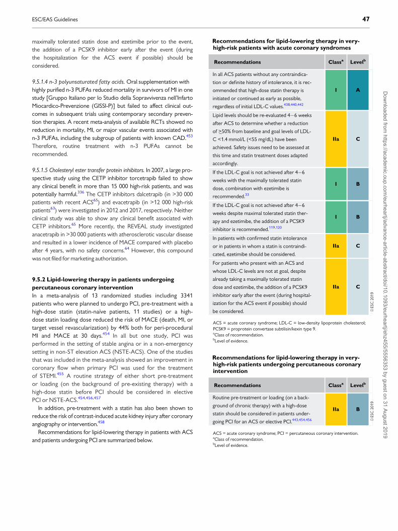
maximally tolerated statin dose and ezetimibe prior to the event,the addition of a PCSK9 inhibitor early after the event (duringthe hospitalization for the ACS event if possible) should beconsidered.
9.5.1.4 n-3 polyunsaturated fatty acids. Oral supplementation withhighly purified n-3 PUFAs reduced mortality in survivors of MI in onestudy [Gruppo Italiano per lo Studio della Sopravvivenza nell’InfartoMiocardico-Prevenzione (GISSI-P)] but failed to affect clinical out-comes in subsequent trials using contemporary secondary preven-tion therapies. A recent meta-analysis of available RCTs showed noreduction in mortality, MI, or major vascular events associated withn-3 PUFAs, including the subgroup of patients with known CAD.453
Therefore, routine treatment with n-3 PUFAs cannot berecommended.
9.5.1.5 Cholesteryl ester transfer protein inhibitors. In 2007, a large pro-spective study using the CETP inhibitor torcetrapib failed to showany clinical benefit in more than 15 000 high-risk patients, and waspotentially harmful.336 The CETP inhibitors dalcetrapib (in >30 000patients with recent ACS65) and evacetrapib (in >12 000 high-riskpatients63) were investigated in 2012 and 2017, respectively. Neitherclinical study was able to show any clinical benefit associated withCETP inhibitors.65 More recently, the REVEAL study investigatedanacetrapib in >30 000 patients with atherosclerotic vascular diseaseand resulted in a lower incidence of MACE compared with placeboafter 4 years, with no safety concerns.64 However, this compoundwas not filed for marketing authorization.
9.5.2 Lipid-lowering therapy in patients undergoing
percutaneous coronary intervention
In a meta-analysis of 13 randomized studies including 3341patients who were planned to undergo PCI, pre-treatment with ahigh-dose statin (statin-naıve patients, 11 studies) or a high-dose statin loading dose reduced the risk of MACE (death, MI, ortarget vessel revascularization) by 44% both for peri-proceduralMI and MACE at 30 days.454 In all but one study, PCI wasperformed in the setting of stable angina or in a non-emergencysetting in non-ST elevation ACS (NSTE-ACS). One of the studiesthat was included in the meta-analysis showed an improvement incoronary flow when primary PCI was used for the treatmentof STEMI.455 A routine strategy of either short pre-treatmentor loading (on the background of pre-existing therapy) with ahigh-dose statin before PCI should be considered in electivePCI or NSTE-ACS.454,456,457
In addition, pre-treatment with a statin has also been shown toreduce the risk of contrast-induced acute kidney injury after coronaryangiography or intervention.458
Recommendations for lipid-lowering therapy in patients with ACSand patients undergoing PCI are summarized below.
Recommendations for lipid-lowering therapy in very-high-risk patients with acute coronary syndromes
Recommendations Classa Levelb
In all ACS patients without any contraindica-
tion or definite history of intolerance, it is rec-
ommended that high-dose statin therapy is
initiated or continued as early as possible,
regardless of initial LDL-C values.438,440,442
I A
Lipid levels should be re-evaluated 4�6 weeks
after ACS to determine whether a reduction
of >_50% from baseline and goal levels of LDL-
C <1.4 mmol/L (<55 mg/dL) have been
achieved. Safety issues need to be assessed at
this time and statin treatment doses adapted
accordingly.
IIa C
If the LDL-C goal is not achieved after 4�6
weeks with the maximally tolerated statin
dose, combination with ezetimibe is
recommended.33
I B
If the LDL-C goal is not achieved after 4�6
weeks despite maximal tolerated statin ther-
apy and ezetimibe, the addition of a PCSK9
inhibitor is recommended.119,120
I B
In patients with confirmed statin intolerance
or in patients in whom a statin is contraindi-
cated, ezetimibe should be considered.
IIa C
For patients who present with an ACS and
whose LDL-C levels are not at goal, despite
already taking a maximally tolerated statin
dose and ezetimibe, the addition of a PCSK9
inhibitor early after the event (during hospital-
ization for the ACS event if possible) should
be considered.
IIa C
ACS = acute coronary syndrome; LDL-C = low-density lipoprotein cholesterol;PCSK9 = proprotein convertase subtilisin/kexin type 9.aClass of recommendation.bLevel of evidence.
Recommendations for lipid-lowering therapy in very-high-risk patients undergoing percutaneous coronaryintervention
Recommendations Classa Levelb
Routine pre-treatment or loading (on a back-
ground of chronic therapy) with a high-dose
statin should be considered in patients under-
going PCI for an ACS or elective PCI.443,454,456
IIa B
ACS = acute coronary syndrome; PCI = percutaneous coronary intervention.aClass of recommendation.bLevel of evidence.
ESC/EAS Guidelines 47D
ownloaded from
https://academic.oup.com
/eurheartj/advance-article-abstract/doi/10.1093/eurheartj/ehz455/5556353 by guest on 31 August 2019

..
..
..
..
..
..
..
..
..
..
..
..
..
..
..
..
..
..
..
..
..
..
..
..
..
..
..
..
..
..
..
..
..
..
..
..
..
..
..
..
..
..
..
..
..
..
..
..
..
..
..
..
..
..
..
..
..
..
..
..
..
..
..
..
..
..
..
..
..
..
..
..
..
..
..
..
..
..
..
..
..
..
..
..
..
..
.9.6 StrokeStroke has a heterogeneous aetiology, including cardiac throm-boembolism [often associated with atrial fibrillation, but also ofuncertain source (embolic stroke of undetermined source)], carotidartery and proximal aortic atherosclerosis and thromboembolism,small-vessel CVD, and intracranial haemorrhage (including intracere-bral and subarachnoid haemorrhage). Dyslipidaemia may play a varia-ble role in the pathogenesis of stroke according to the particularaetiology. The relationship between dyslipidaemia and atherothrom-botic events, including ischaemic stroke and TIA, is well recognized,while the association of dyslipidaemia with other types of stroke isuncertain. Notwithstanding, concomitant control of other aetiologi-cal factors, such as hypertension, is of paramount importance.
Following ischaemic stroke or TIA, patients are at risk not only ofrecurrent cerebrovascular events, but also of other major CV events,including MI. Secondary prevention therapy with statins reduces therisk of recurrent stroke (by 12% per mmol/L reduction in LDL cho-lesterol), MI, and vascular death.459,460 Statin pre-treatment at TIAonset was associated with reduced recurrent early stroke risk inpatients with carotid stenosis in a pooled data analysis, supporting as-early-as-possible initiation of statins after stroke.460�462 Statin ther-apy may yield a small increase in the risk of haemorrhagic stroke, butthe evidence regarding this risk is uncertain.34,36,251,252
9.7 Heart failure and valvular diseases9.7.1 Prevention of incident heart failure in coronary
artery disease patients
Cholesterol lowering with statins reduces the incidence of HF inpatients with CAD (stable CAD or a history of ACS) without previousHF; this has been shown consistently in RCTs that have compared sta-tin vs. no statin treatment463,464 as well as more-intensive vs. less-intensive statin therapy.465�468 A large-scale meta-analysis of primaryand secondary prevention RCTs with statins showed a modest (10%)reduction in first non-fatal HF hospitalizations with statin treatment,with no effect on HF death within the limited RCT period.469 There isno evidence that statins can prevent HF of non-ischaemic origin.
9.7.2 Chronic heart failure
Two large RCTs466,470 (mainly including patients with systolic HF), aswell as a meta-analysis of 24 RCTs, have shown no benefit of statintreatment on CV mortality or stroke;471 a reduction in HF
hospitalizations,218,471 as well as a small reduction in MI, wasobserved in a pooled analysis of the Controlled RosuvastatinMultinational Trial in Heart Failure (CORONA) and GISSI-HF tri-als.472 Based on current evidence, routine administration of statins inpatients with HF without other indications for their use (e.g. CAD) isnot recommended. Because there is no evidence of harm in patientson statin treatment after the occurrence of HF, there is no need forstatin discontinuation for patients already on treatment.
There is no evidence regarding the effect of PCSK9 inhibition inpatients with chronic HF. In the recent PCSK9 clinical outcomes tri-als, FOURIER119 and ODYSSEY Outcomes,120 PCSK9 inhibition inpatients with atherosclerotic CVD or after an ACS did not reducethe risk of HF hospitalization. In the BIOlogy Study to TAiloredTreatment in Chronic Heart Failure (BIOSTAT-CHF) study of 2174patients with worsening HF, multivariable analysis revealed a positivelinear association between PCSK9 levels and the risk of mortality,and the composite of mortality and unplanned HF hospitalization.473
Similarly, there was a negative association between LDLR levels andmortality, indicating a potential relationship between thePCSK9�LDLR axis and outcomes among patients with HF thatrequires further investigation.473,474
Treatment with n-3 PUFAs 1 g o.d. may confer a small benefit inpatients with chronic HF, as shown by a significant 9% relative riskreduction for mortality in the GISSI-HF RCT.475
9.7.3 Valvular heart diseases
Aortic stenosis increases the risk of CV events and mortality, and fre-quently coexists with atherosclerotic CVD. Life-long high levels ofLDL-C476 and Lp(a)477 have been associated with incident aorticvalve stenosis and aortic valve calcification in genetic Mendelian ran-domization studies. Observational studies have suggested possiblebeneficial effects of intensive lipid lowering in slowing the progressionof native valve aortic stenosis.478 However, this has not been con-firmed in RCTs,266,479�481 or in meta-analyses of observational andrandomized trials.482,483 Three modestly sized trials479�481 and onelarge randomized trial (SEAS, which included 1873 patients treatedwith simvastatin 40 mg plus ezetimibe 10 mg or placebo)266 failed toshow a reduction in the clinical progression of aortic stenosis inpatients with mild-to-moderate native valve aortic stenosis. In a posthoc analysis of the SEAS trial, the efficacy of lipid-lowering therapy inimpeding the progression of aortic stenosis increased with higherpre-treatment LDL-C levels and lower peak aortic jet velocity (i.e.milder stenosis at baseline).484 Similarly, a post hoc analysis of threeRCTs, including patients without known aortic valve stenosis at base-line [Treating to New Targets (TNT), Incremental Decrease In End-points Through Aggressive Lipid-lowering (IDEAL), and StrokePrevention by Aggressive Reduction in Cholesterol Levels(SPARCL)] showed no impact of high-dose vs. usual-dose statin ther-apy on the incidence of aortic valve stenosis.485 In patients whounderwent transcatheter aortic valve replacement, statin therapywas associated with improved outcomes in a small observationalstudy.486
Aortic valve sclerosis (calcification of the aortic leaflets without sig-nificant transvalvular pressure gradient) is associated with an increasedrisk of CAD even in the absence of increased risk profiles. Whether ornot statins may be useful both for aortic valve disease and CAD pro-gression in such patients warrants further investigation.487
Recommendations for lipid-lowering therapy for theprevention of atherosclerotic cardiovascular diseaseevents in patients with prior ischaemic stroke
Recommendations Classa Levelb
Patients with a history of ischaemic stroke or
TIA are at very high-risk of ASCVD, particu-
larly recurrent ischaemic stroke, so it is rec-
ommended that they receive intensive LDL-C-
lowering therapy.459,460
I A
ASCVD = atherosclerotic cardiovascular disease; LDL-C = low-density lipopro-tein cholesterol; TIA = transient ischaemic attack.aClass of recommendation.bLevel of evidence.
48 ESC/EAS GuidelinesD
ownloaded from
https://academic.oup.com
/eurheartj/advance-article-abstract/doi/10.1093/eurheartj/ehz455/5556353 by guest on 31 August 2019

..
..
..
..
..
..
..
..
..
..
..
..
..
..
..
..
..
..
..
..
..
..
..
..
..
..
..
..
..
..
..
..
..
..
..
..
..
..
..
..
..
..
..
..
..
..
..Recommendations for lipid-lowering therapy in patients with HFand valvular diseases are shown below.
9.8 Chronic kidney diseaseCKD is defined as abnormalities of kidney structure or function, presentfor >3 months, with implications for health. CKD is classified on thebasis of the cause, GFR category, and category of albuminuria.488 In theadult population, decreasing GFR is associated with increased CVD risk,independent of other CV risk factors.489�492 There is an increased riskof both atherosclerotic vascular disease and structural heart disease.492
Patients with CKD and established CVD have a much higher mortalityrate compared with patients with CVD and normal renal function.493
Therefore, patients with CKD are considered to be at high (stage 3CKD) or very-high risk (stage 4�5 CKD or on dialysis) of CVD, andthere is no need to use risk estimation models in these patients.
9.8.1 Lipoprotein profile in chronic kidney disease
In the initial stages of CKD, TG levels are specifically elevated andHDL-C levels are lowered. LDL subclasses display a shift to an excessof small dense LDL particles. Studies suggest that the kidney has arole in Lp(a) catabolism and that Lp(a) levels are increased in associa-tion with kidney disease. Such acquired abnormalities can be reversedby kidney transplantation or remission of nephrosis.
9.8.2 Evidence for risk reduction through statin-based
therapy in patients with chronic kidney disease
In the Die Deutsche Diabetes Dialyse Studie (4D) trial, which involved1200 patients with diabetes on haemodialysis, atorvastatin had no sig-nificant effect on risk of CVD.220 Similar results were obtained in theAURORA (A study to evaluate the Use of Rosuvastatin in subjectsOn Regular haemodialysis: an Assessment of survival and cardiovascu-lar events) trial, which involved 2776 patients on haemodialysis.221
In the SHARP study,222 simvastatin and ezetimibe combination ther-apy reduced the risk for major atherosclerotic events (coronary death,MI, non-haemorrhagic stroke, or any revascularization) compared withplacebo in persons with CKD stage 3A�5. The trial did not have suffi-cient power to separately assess the effects on the primary outcome indialysis and non-dialysis patients. Although statin-based therapy is clearlyeffective in mild-to-moderate CKD, a major controversy that remainedafter the publication of the 4D, AURORA, and SHARP studies was
whether statin therapy is effective in more advanced CKD, particularlydialysis patients. By combining data from the three CKD trials withother trials in the existing database, the CTT investigators found that,even after adjusting for the smaller LDL-C reductions achieved amongpatients with more advanced CKD and for differences in outcome defi-nitions between dialysis trials, there was a trend towards smaller relativereductions per mmol/L reduction in LDL-C in major atheroscleroticevents as estimated GFR (eGFR) declines (with little evidence of benefitamong dialysis patients).214 This diminution in relative risk reduction asGFR declines implies that, at least in non-dialysis patients, more intensiveLDL-lowering regimens are required to achieve the same benefit.
9.8.3 Safety of lipid management in patients with chronic
kidney disease
Safety issues and dose adjustments are important in advanced stagesof CKD (stages 3�5), as adverse events are commonly dose-relatedand due to increased blood concentrations of compounds. Althoughit has been suggested that preference should be given to regimensand doses that have been shown to be beneficial in RCTs conductedspecifically in such patients,494 the CTT meta-analysis makes clearthat the goal—as in patients without CKD—should be to achieve thelargest possible absolute reduction in LDL-C safely. Although therewere no specific safety concerns raised by the 4D, AURORA, orSHARP trials, statins metabolized via CYP3A4 may result in adverseeffects due to drug�drug interactions and caution is required.
Based on the evidence for lipid management in patients with CKD,the Kidney Disease: Improving Global Outcomes (KDIGO) organiza-tion has developed an updated clinical practice guideline for lipidmanagement in CKD.494 In line with this, but with a focus on thosepatients at high or very-high risk for developing CVD, recommenda-tions are summarized below.
Recommendations for the treatment of dyslipidaemiasin chronic heart failure or valvular heart diseases
Recommendations Classa Levelb
Initiation of lipid-lowering therapy is not rec-
ommended in patients with HF in the absence
of other indications for their use.466,470
III A
Initiation of lipid-lowering treatment in
patients with aortic valvular stenosis without
CAD to slow progression of aortic valve
stenosis in the absence of other indications for
their use is not recommended.266,479�481
III A
CAD = coronary artery disease; HF = heart failure.aClass of recommendation.bLevel of evidence.
Recommendations for lipid management in patientswith moderate-to-severe (Kidney Disease OutcomesQuality Initiative stages 3�5) chronic kidney disease
Recommendations Classa Levelb
It is recommended that patients with Kidney
Disease Outcomes Quality Initiative stage 3�5c
CKD are considered to be at high or very-high risk
of ASCVD.489�493
I A
The use of statins or statin/ezetimibe combina-
tion is recommended in patients with non-dialy-
sis-dependent stage 3�5 CKD.214,222,495,496
I A
In patients already on statins, ezetimibe, or a sta-
tin/ezetimibe combination at the time of dialysis
initiation, continuation of these drugs should be
considered, particularly in patients with ASCVD.
IIa C
In patients with dialysis-dependent CKD who
are free of ASCVD, commencement of statin
therapy is not recommended.220,221
III A
ASCVD = atherosclerotic cardiovascular disease; CKD = chronic kidney disease;eGFR = estimated glomerular filtration rate.aClass of recommendation.bLevel of evidence.cDefined as eGFR < 60ml/min/1.73m2 on two measurements more than 3 monthsapart.
ESC/EAS Guidelines 49D
ownloaded from
https://academic.oup.com
/eurheartj/advance-article-abstract/doi/10.1093/eurheartj/ehz455/5556353 by guest on 31 August 2019

..
..
..
..
..
..
..
..
..
..
..
..
..
..
..
..
..
..
..
..
..
..
..
..
..
..
..
..
..
..
..
..
..
..
..
..
..
..
..
..
..
..
..
..
..
..
..
..
..
..
..
..
..
..
..
..
..
.
9.9 TransplantationDyslipidaemias are very common in patients who have undergoneheart, lung, liver, kidney, or allogenic haematopoietic stem cell trans-plantation, and predispose such patients to an increased risk of devel-oping ASCVD and transplant arterial vasculopathy.497�501 In patientswith CKD undergoing renal transplantation, the risk of ASCVD maybe determined, at least in part, by the increased risk resulting fromCKD itself.
Immunosuppressive drug regimens may have adverse effects onlipid metabolism leading to increases in TC, VLDL, and TGs, and inthe size and density of LDL particles. These effects vary with differentimmunosuppressive drugs.497,498,502�506
The management of dyslipidaemias in transplant recipients is com-parable to what is recommended for patients at high or very highASCVD risk, although more attention is needed regarding the causesof the lipid disturbances and possible side effects due to drug�druginteractions (see Recommendations for low-density lipoprotein in solidorgan transplant patients below).
The clinical effectiveness of statins in renal transplant patientsis uncertain owing to a lack of randomized trials in this population.A systematic review of the benefits and harms of statins inpatients with a functioning kidney transplant included 3465patients, free of CHD, from 22 studies. Although the authors con-cluded that statins may reduce CV events, they also suggested aneed for additional studies.253 However, in patients with a function-ing renal transplant at increased risk of CVD, it may be appropriateto extrapolate from the clear evidence of benefit from statin ther-apy, without safety concerns, in people with moderate reductionsin GFR.214
Several potential drug interactions must also be considered, espe-cially with ciclosporin, which is metabolized through CYP3A4, andmay increase systemic statin exposure and the risk of myopathy.Ciclosporin increases the blood levels of all statins.
Fluvastatin, pravastatin, pitavastatin, and rosuvastatin are metabo-lized through different CYP enzymes than the others and have lesspotential for interaction.507
Tacrolimus is also metabolized by CYP3A4, but appears to haveless potential for harmful interaction with statins than ciclosporin.Other drugs that influence CYP3A4 activity should be avoided if pos-sible, and used with extreme caution in patients receiving both calci-neurin inhibitors and statins.
For transplant patients with dyslipidaemia, ezetimibe could be con-sidered as an alternative for patients unable to take a statin or addedto the highest tolerated statin dose.507�509 No outcome data areavailable for this drug, which should generally be reserved forsecond-line use. Ciclosporin can induce a 2�12-fold increase in theezetimibe level.
Care is required with the use of fibrates, as they can decreaseciclosporin levels and have the potential to cause myopathy.Extreme caution is required if fibrate therapy is planned in combi-nation with a statin. Cholestyramine is not effective as monother-apy in heart transplant patients and has the potential to reduceabsorption of immunosuppressants; this potential is minimized byseparate administration.
9.10 Peripheral arterial diseasePAD encompasses all vascular sites, including carotid, vertebral, upperextremity, mesenteric, renal, and lower extremity arteries. The aortais often included in the term.510 PAD is a common manifestation ofatherosclerosis and such patients are at elevated risk of coronaryevents, with PAD representing an independent risk factor for MI andCV death.510,511 Patients with PAD are at very-high risk and should bemanaged according to the recommendations in Table 7. Elevated CVrisk has led to the inclusion of PAD among the list of ‘risk equivalent’conditions, and therapeutic strategies of secondary prevention shouldbe implemented [see Recommendations for lipid-lowering drugs in patientswith peripheral arterial disease (including carotid artery disease) below].Yet, despite the high CV morbidity and mortality risk, PAD patientsare usually inadequately managed compared with CAD patients.511
9.10.1 Lower extremity arterial disease
A low ABI (0.90) is diagnostic for lower extremity arterial disease(LEAD). Either a low (0.90) or high (1.40, related to stiffened arteries)ABI is predictive of CV morbidity and mortality. Lowering LDL-C lev-els reduces the risk of ischaemic CV events and worsening of claudi-cation, while it also improves walking performance. As for cardiacevents, a systematic review of 18 trials including 10 000 patients, withcholesterol levels ranging from normal to elevated, reported thatlipid-lowering therapy in people affected by atherosclerosis of thelower limbs was associated with a 20% reduction in total CV events,together with a non-significant 14% reduction of all-cause mortal-ity.512 In the Heart Protection Study, the need for non-coronaryrevascularization was reduced by 16% with statin therapy.513
In addition to statins, PSCK9 inhibitors have also been shown toreduce CV events in PAD patients. In a pre-specified subgroup analy-sis of the FOURIER trial, evolocumab significantly reduced the pri-mary endpoint in patients with PAD.514 PAD had larger absolute riskreductions for the primary endpoint (3.5% with PAD and 1.6% with-out PAD). Evolocumab also reduced the risk of major adverse limbevents by 42% in patients, with consistent effects in those with andwithout known PAD. In the FIELD trial, fenofibrate reduced the riskof amputations, particularly minor amputations without known large-vessel disease, probably through non-lipid mechanisms.515
Recommendations for low-density lipoprotein loweringin solid organ transplant patients
Recommendations Classa Levelb
Statins should be considered as first-line agents in
transplant patients. Initiation should be at low
doses with careful uptitration and with caution
regarding potential drug�drug interactions, partic-
ularly for patients on ciclosporin.507
IIa B
In patients who are intolerant of statins or those
with significant dyslipidaemia despite maximally
tolerated statin treatment, alternative or addi-
tional therapy with ezetimibe may be considered.
IIb C
aClass of recommendation.bLevel of evidence.
50 ESC/EAS GuidelinesD
ownloaded from
https://academic.oup.com
/eurheartj/advance-article-abstract/doi/10.1093/eurheartj/ehz455/5556353 by guest on 31 August 2019

..
..
..
..
..
..
..
..
..
..
..
..
..
..
..
..
..
..
..
..
..
..
..
..
..
..
..
..
..
..
..
..
..
..
..
..
..
..
..
..
..
..
..
..
..
..
..
..
..
..
..
..
..
..
..
..
..
..
..
..
..
..
..
..
..
..
..
..
..
..
..
..
..
..
..
..
..
..
..
..
..
..
..
..
..
..
.9.10.2 Carotid artery disease
While there are currently no randomized studies that have assessedwhether lipid-lowering treatments reduce the incidence of CV eventsin patients enrolled on the basis of carotid atherosclerotic diseaseand without previous CV events, lipid-lowering therapy hadreduced stroke in numerous studies. In a meta-analysis of RCTsenrolling >90 000 patients, statin therapy did lead to a 21% reductionin the incidence of all strokes in different populations; this effect wasmainly driven by the extent of LDL-C reduction.460
9.10.3 Retinal vascular disease
Atherosclerotic changes of retinal arteries correlate with TC, LDL-C,TG, and apoB levels and also with CAD.516 Fenofibrate reduces theprogression of diabetic retinopathy.517,518
9.10.4 Secondary prevention in patients with abdominal
aortic aneurysm
The presence of an abdominal aortic aneurysm represents a risk-equivalent condition for CAD and is associated with age, male gender,personal history of atherosclerotic CVD, smoking, hypertension, anddyslipidaemia;519 in contrast, diabetic patients are at decreased risk.
There are currently no available clinical trials on the reduction of CVrisk with lipid-lowering therapy in patients affected by this condition.Systematic reviews,520 mostly based on retrospective non-randomizedstudies, have reported that there is still inconclusive evidence that sta-tin therapy reduces peri-operative CV morbidity and mortality. In anRCT comparing atorvastatin 20 mg with placebo, the composite end-point of cardiac death, MI, stroke, and unstable angina was significantlyreduced in 100 patients undergoing vascular non-cardiac surgery,including abdominal aortic aneurysm repair.521 In another double-blind,placebo-controlled trial in 497 patients undergoing vascular surgery,peri-operative fluvastatin therapy (80 mg/day) was associated with animprovement in post-operative cardiac outcome.522
9.10.5 Renovascular atherosclerosis
Lipid-lowering therapy has never been tested in an RCT in patientsaffected by renovascular atherosclerosis; however, a recent non-randomized population-based study showed that in patients olderthan 65 years of age with atherosclerotic renovascular disease raisedthe hypothesis that such treatment may yield cardiorenal benefits;the risk of a major cardiorenal composite endpoint (MI, stroke, HF,acute renal failure, dialysis, and death) was significantly lower in statinusers than in non-users.523
9.11 Other special populations at risk ofatherosclerotic cardiovascular diseaseIn general, the effects of lowering LDL-C are determined by the abso-lute risk of ASCVD and the achieved reduction in LDL-C, so it isimportant to identify and treat all those at increased risk of ASCVD.There are a few specific groups of patients in whom an underlying dis-ease confers such increased risk, and in addition in whom the stand-ard treatments may themselves cause dyslipidaemia that maycontribute to the risk of ASCVD. These include: (i) chronic immune-mediated inflammatory disease, (ii) patients with human immunodefi-ciency virus (HIV), and (iii) patients with severe mental illness. Themanagement principles are the same in these patient groups, but theirmanagement may need to address specific issues related to individualdyslipidaemias and drug safety. Details are provided in theSupplementary Data document.
10 Inflammation
Recent advances in basic science have established a fundamental rolefor low-degree chronic inflammation in mediating all stages of athero-sclerosis, from initiation through progression and, ultimately, to therupture of plaque and ensuing thrombotic complications of atheroscle-rosis. The cellular and molecular interactions involved during athero-genesis are fundamentally not different from those in chronicinflammatory�fibroproliferative diseases, such as rheumatoid arthritis(RA), glomerulosclerosis, or pulmonary fibrosis.525 Almost all cell typesof the immuno-inflammatory system, such as macrophages, and T- andB-cells, as well as many pro- and anti-inflammatory cytokines and che-mokines, have been identified during the process of atherosclerosis.526
Interestingly, cholesterol accumulation in cells triggers the inflam-masome response and results in the production of inflammatorymediators such as interleukin (IL)-1b. Numerous animal studies, usingthe knockout model, have demonstrated that inflammation and theimmune system both play crucial roles during atherogenesis.527
During inflammatory processes, large numbers of acute-phaseproteins have been identified, and several clinical studies havereported C-reactive protein528 to be the most useful serum markerof inflammation, even though it has poor specificity for any particularinflammation process, including atherosclerosis. The high-sensitivityC-reactive protein diagnostic test was developed to detect very lowlevels of C-reactive protein, and thereby enable a more accurate andprecise measure of chronic inflammation compared with standard C-reactive protein.529 This diagnostic tool differs only in the range of C-reactive protein levels that it can detect. Several studies have foundthat elevated levels of high-sensitivity C-reactive protein in the bloodare associated with an increased risk of CV events and could be usedto predict clinical outcomes independently of cholesterol lev-els.530,531 Other studies have not been able to show any relationshipbetween low-grade chronic inflammation, as indicated by high-sensi-tivity C-reactive protein levels, and increased risk of CV.532�535
Finally, genetic studies of large population cohorts have not demon-strated that chronic elevated high-sensitivity C-reactive proteinincreases the risk of atherosclerotic events.536 Nevertheless, in someguidelines, high-sensitivity C-reactive protein has been added to tradi-tional risk factors for prognostic information, especially for patientsat intermediate risk.537,538
Recommendations for lipid-lowering drugs in patientswith peripheral arterial disease (including carotid arterydisease)
Recommendations Classa Levelb
In patients with PAD, lipid-lowering therapy,
including a maximum tolerated dose of statin,
plus ezetimibe or a combination with a PCSK9
inhibitor if needed, is recommended to reduce
the risk of ASCVD events.512,524
I A
ASCVD = atherosclerotic cardiovascular disease; PAD = peripheral arterial dis-ease; PCSK9 = proprotein convertase subtilisin/kexin type 9.aClass of recommendation.bLevel of evidence.
ESC/EAS Guidelines 51D
ownloaded from
https://academic.oup.com
/eurheartj/advance-article-abstract/doi/10.1093/eurheartj/ehz455/5556353 by guest on 31 August 2019

..
..
..
..
..
..
..
..
..
..
..
..
..
..
..
..
..
..
..
..
..
..
..
..
..
..
..
..
..
..
..
..
..
..
..
..
..
..
..
..
..
..
..
..
..
..
..
..
..
..
..
..
..
..
..
..
..
..
..
..
..
..
..
..
..
..
..
..
..
..
..
..
..
..
..
..
..
..
..
..
..
..
..
..
..
..
.Statins have been shown to reduce C-reactive protein secretion
by hepatocytes,539 and a series of clinical trials and post hoc analyseshave found that beneficial outcomes after statin therapy relate bothto a reduction in cholesterol levels and reduced inflammation.540�544
The Justification for the Use of Statins in Prevention: an InterventionTrial Evaluating Rosuvastatin (JUPITER) trial542 demonstrated that inprimary prevention for individuals with chronically elevated C-reac-tive protein (>2 mg/L), statin treatment markedly reduced CVevents.545 It is of note that other lipid-lowering agents, such as ezeti-mibe and more recently the anti-PCSK9 mAbs, do not influence high-sensitivity C-reactive protein levels,546,547 but lead to further signifi-cant reductions in CV events when added to statin therapy.
Specific anti-inflammatory treatment was tested in theCanakinumab Antiinflammatory Thrombosis Outcome Study(CANTOS) trial.548 In patients with previous MI and chronic elevatedhigh-sensitivity C-reactive protein levels, all on optimal medical treat-ment, including statins, the anti-IL-1b mAb canakinumab dose-dependently reduced high-sensitivity C-reactive protein and signifi-cantly lowered the rate of recurrent CV events compared with pla-cebo, independently of the level of lipid lowering. Not surprisingly,there was a slight increase in the risk of severe and fatal infections asso-ciated with canakinumab. This study was the first to highlight the posi-tive correlation between high-sensitivity C-reactive protein and CVevents, where lower achieved high-sensitivity C-reactive protein valueswere directly correlated with a lower risk of future CV events.548
Nevertheless, the FDA declined to approve canakinumab for CV riskreduction on the strength of data from the CANTOS study. As canaki-numab treatment has not been tested against anti-PCSK9 mAb and/orezetimibe added to statin therapy, the question of residual risk remainsfor patients with elevated high-sensitivity C-reactive protein despiteachieving very low (below goal) LDL-C values, and whether patientswith very low LDL-C would benefit from anti-IL-1b treatment orother anti-inflammatory agents. In addition, all currently recommendedlipid-lowering drugs, including anti-PCSK9 mAbs, have demonstratedbeneficial effects on atherosclerotic plaque composition as well as pla-que volume regression; such results are still missing for anti-inflammatory treatment. Another anti-inflammatory approach usingmethotrexate was tested in the Cardiovascular InflammationReduction Trial (CIRT).549 Very low-dose methotrexate (10 mgweekly), a proven anti-inflammatory regimen that reduces tumournecrosis factor (TNF), IL-6, and C-reactive protein levels and is widelyused in the treatment of RA, was allocated vs. placebo to 7000 stableCAD patients. This study was stopped prematurely due to futility.Interestingly, this regimen of methotrexate had no effect on either IL-6or high-sensitivity C-reactive protein blood levels in this population,which could explain the neutral results of this trial.550 Based on the cur-rent level of evidence, no further recommendations on the use of anti-inflammatory agents can be made.551
11 Monitoring of lipids andenzymes in patients onlipid-lowering therapy
Evidence concerning which tests should be carried out to monitorlipids in patients on treatment is limited. Similar limited evidence
applies to tests of possible toxicity, such as ALT and CK.Recommendations stem from consensus rather than evidence-basedmedicine.
Response to therapy can be assessed at 6�8 weeks from initiationof therapy, but response to lifestyle may take longer. Standard prac-tice for subsequent follow-up monitoring is 6�12 months, but suchmonitoring intervals are arbitrary. As a minimum, LDL-C should beassessed whenever available, but better management decisions willprobably occur if a full lipid profile is performed, including HDL-Cand TGs. Non-HDL-C or ApoB should also be analysed, and used asa secondary treatment target. A separate issue is the impact of regu-lar lipid monitoring in promoting patient adherence to lifestylechanges or drug regimens that impact positively on their health, asfound in a range of studies. It is unclear whether only the process ofmonitoring is critical in achieving this or whether a combination ofeducation, regular contact, and adherence assessment is required.
Where pharmacological lipid-lowering therapy is implemented,safety blood tests are advised, including ALT and CK at baseline, toidentify the limited number of patients where treatment is contraindi-cated. CK should be checked in patients at high-risk for myopathy,such as the very elderly with comorbidities, patients with antecedentsof muscle symptoms, or patients receiving interacting drugs. A mildand typically transient elevation of ALT is seen in about 2% of patientsand normalization is seen with continuing therapy.240,244,552 Recentreviews are encouraging regarding the safety of long-term statin ther-apy and statin-induced liver injury is reported to be veryuncommon.243,244,553�555 ALT is recommended before the start ofstatin therapy; routine control of ALT during treatment is not recom-mended but should be performed, if indicated, based on clinicalobservations. During fibrate therapy, regular ALT control is still rec-ommended. In patients whose liver function tests increase to abovethree times the ULN, explanations such as alcohol ingestion orNAFLD should be sought, and the levels monitored. If levels remainelevated then lipid-lowering therapy should be stopped, but may becautiously reintroduced under monitoring after levels have returnedto normal.
There is no predictive value of routine repeat CK testing for rhab-domyolysis since the level can increase for many reasons, includingmuscle injury or excess muscular exercise. However, CK must beassessed immediately in patients who present with muscle pain andweakness, and especially in the elderly, and treatment stopped if CKrises to >10 times the ULN. Strategies to handle CK elevations aregiven in Table 13.
Due to the increased frequency of DM during statin treat-ment,247,249,556,557 regular checks of HbA1c should be considered inpatients at high-risk of developing DM and under high-dose statintreatment. Groups to be considered for glucose control are the eld-erly or those with MetS, obesity, or signs of insulin resistance.
12 Cost-effectiveness ofcardiovascular disease preventionby lipid modification
In 2015, there were >85 million people in Europe living with CVD.558
Aging populations,559 unhealthy diets, smoking, sedentary lifestyles,
52 ESC/EAS GuidelinesD
ownloaded from
https://academic.oup.com
/eurheartj/advance-article-abstract/doi/10.1093/eurheartj/ehz455/5556353 by guest on 31 August 2019

Table 13 Summary of recommendations for monitoring lipids and enzymes in patients, before and on lipid-loweringtherapy
Testing lipids
How often should lipids be tested?
• Before starting lipid-lowering drug treatment, at least two measurements should be made, with an interval of 1�12 weeks, with the exception of condi-
tions where prompt drug treatment is suggested, such as ACS and very high-risk patients.
How often should a patient’s lipids be tested after starting lipid-lowering treatment?
• After starting treatment: 8 (±4) weeks.
• After adjustment of treatment: 8 (±4) weeks until the goal is achieved.
How often should lipids be tested once a patient has achieved the target or optimal lipid level?
• Annually (unless there are adherence problems or other specific reasons for more frequent reviews).
Monitoring liver and muscle enzymes
How often should liver enzymes (ALT) be routinely measured in patients on lipid-lowering drugs?
• Before treatment.
• Once, 8�12 weeks after starting a drug treatment or after dose increase.
• Routine control of ALT thereafter is not recommended during statin treatment, unless symptoms suggesting liver disease evolve. During treatment with
fibrates, control of ALT is still recommended.
What if liver enzymes become elevated in a person taking lipid-lowering drugs?
If ALT <3� ULN:
• Continue therapy.
• Recheck liver enzymes in 4�6 weeks.
If ALT rises to >_3�ULN
• Stop lipid-lowering therapy or reduce dose and recheck liver enzymes within 4�6 weeks.
• Cautious reintroduction of therapy may be considered after ALT has returned to normal.
• If ALT remains elevated check for the other reasons.
How often should CK be measured in patients taking lipid-lowering drugs?
Pre-treatment
• Before starting therapy.
• If baseline CK is >4� ULN, do not start drug therapy; recheck.
Monitoring:
• Routine monitoring of CK is not necessary.
• Check CK if patient develops myalgia.
Be alert regarding myopathy and CK elevation in patients at risk, such as: elderly patients, those on concomitant interfering therapy, multiple medications,
liver or renal disease, or athletes.
What if CK becomes elevated in a person taking lipid-lowering drugs?
Re-evaluate indication for statin treatment.
If >_4� ULN:
• If CK >10� ULN: stop treatment, check renal function, and monitor CK every 2 weeks.
• If CK <10� ULN: if no symptoms, continue lipid-lowering therapy while monitoring CK between 2 and 6 weeks.
• If CK <10� ULN: if symptoms present, stop statin and monitor normalization of CK, before rechallenge with a lower statin dose.
• Consider the possibility of transient CK elevation for other reasons such as exertion.
• Consider myopathy if CK remains elevated.
• Consider combination therapy or an alternative drug.
If <4�ULN:
• If no muscle symptoms, continue statin (patient should be alerted to report symptoms; check CK).
• If muscle symptoms, monitor symptoms and CK regularly.
• If symptoms persist, stop statin and re-evaluate symptoms after 6 weeks; re-evaluate indication for statin treatment.
• Consider rechallenge with the same or another statin.
• Consider low-dose statin, alternate day or once/twice weekly dosing regimen, or combination therapy.
For details on CK elevation and treatment of muscular symptoms during statin treatment see algorithm in Supplementary Figure 4.
In which patients should HbA1c or blood glucose be checked?
• Regular checks of HbA1c or glucose should be considered in patients at high-risk of developing diabetes, and on high-dose statin treatment.
• Groups to be considered for glucose control are the elderly and patients with metabolic syndrome, obesity, or other signs of insulin resistance.
ACS = acute coronary syndrome; ALT = alanine aminotransferase; CK = creatine kinase; ULN = upper limit of normal.
ESC/EAS Guidelines 53D
ownloaded from
https://academic.oup.com
/eurheartj/advance-article-abstract/doi/10.1093/eurheartj/ehz455/5556353 by guest on 31 August 2019

..
..
..
..
..
..
..
..
..
..
..
..
..
..
..
..
..
..
..
..
..
..
..
..
..
..
..
..
..
..
..
..
..
..
..
..
..
..
..
..
..
..
..
..
..
..
..
..
..
..
..
..
..
..
..
..
..
..
..
..
..
..
..
..
..
..
..
..
..
..
..
..
..
..
..
..
..
..increasing obesity, and diabetes560�563 are the main contributors.CVD cost the European Union about e210 billion in 2015, one-halfof which was in healthcare costs (�8% of total healthcare expendi-ture), and the other half in productivity losses and informal care.558
In these Guidelines, the Joint Task Force recommends a range ofactions to reduce CVD by targeting plasma lipids, ranging frompopulation-wide initiatives to promote healthy lifestyles to individual-level interventions to reduce CVD risk factors, such as unhealthydiets and high lipid levels. Cost-effectiveness analysis can help targetresources for interventions where the net health gain is greatest inrelation to the net resources, and is increasingly required acrossEurope.564 However, cost-effectiveness depends on available resour-ces, the costs of services, and disease risk in the population, andresults obtained in one country might not be valid in another.565 Inaddition, to fully capture the long-term effects of interventions, cost-effectiveness studies combining evidence from RCTs with modellingand limitations in both could affect the reliability of findings. Here, theevidence for the cost-effectiveness of ASCVD preventive interven-tions with respect to lipid modification is summarized; further scru-tiny in view of local circumstances is recommended.
The health impact pyramid summarizes the evidence on the rela-tive effort in relation to health impact (Figure 5), with interventionswith the broadest impact on populations at the base and interven-tions requiring considerable individual effort at the top.566 There isconsensus that all the levels of the pyramid should be targeted butthat emphasis should be placed on the lower levels. This wouldaddress the persistent socio-economic divide in CV health despitemajor improvements in ASCVD treatment.558
More than one-half of the reduction in CV mortality over thelast three decades has been attributed to population-level changes
in CV risk factors, primarily reductions in plasma cholesterol, BPlevels, and smoking.560�563,567 Lifestyle changes at the populationlevel may be more cost-effective than lifestyle and drug interven-tions at the individual level, particularly when targeted to popula-tions at increased risk. Awareness and knowledge of how lifestylerisk factors lead to CVD has increased in recent decades.Moreover, legislation promoting a healthy lifestyle, such asreduced salt intake and smoking bans, has been reported to becost-effective in preventing CVD,568�573 and initiatives toimprove infrastructure and promote physical activity have shownpromise.574,575 A number of structural strategies at international,national, and regional levels combined can substantially reduceCVD morbidity and mortality.576,577 Individual-level interventionsto improve diet,578,579 increase physical activity,580 andstop smoking581 could also be cost-effective.582 However, subop-timal adherence limits benefits,583,584 and interventions toimprove adherence, such as electronic device reminders to rein-force favourable health behaviours, are increasingly beinginvestigated.585
All statin regimens and ezetimibe are now generically availableacross Europe. There is strong evidence that lowering blood choles-terol levels using low-cost statins is widely cost-effective586�590 inmany categories of patients. For secondary prevention of CVD, theevidence suggests that statin treatments are highly cost-effec-tive,586,590,591 and adding low-cost ezetimibe to high-intensity statintherapy further reduces LDL-C and CVD risk cost-effectively.592 Inprimary ASCVD prevention, the evidence indicates that genericstatin-based treatments are cost-effective for people at least down to1% annual total CVD risk and could be cost-effective at even lowerrisk,589 with the highest tolerated statin dose likely the most cost-effective.591,593,594 Importantly, many patients on statin treatment failto take their medications adequately and/or to reach their therapeu-tic goals,595 with clinical and economic consequences.596,597
Reinforcing measures aimed at improving adherence to treatment iscost-effective.598�600
Studies have shown that at mid-2018 prices PCSK9 inhibitorswere largely not cost-effective.601�604 Their cost-effectiveness isimproved in selected high-risk patients, such as those with clinicalCVD or FH, other comorbidities, and high LDL-C levels.605,606
However, at lower prices, PCSK9 inhibitors would become cost-effective in a wider range of high-risk patients; recent price reductionsmay therefore lead to increased use.607 Cost-effectiveness evidencefor other lipid-modifying therapies is lacking.
Effective interventions to prevent ASCVD, including statins, typi-cally exhibit similar relative risk reductions across categories ofpatients, including by ASCVD risk; therefore, health benefits andcost-effectiveness are greater among people at higher ASCVD risk(Figure 6).36,233 Consequently, increased efforts and higher-intensityinterventions should be aimed at individuals and populations at higherASCVD risk.
Counselingand education
Clinical interventions
Long-lasting protectiveinterventions
Changing the context to makeindividuals' default decisions healthy
Socioeconomic factors
Increasingpopulation
impact
Increasingindividual
effort needed
©ES
C 2
019
Figure 5 Health impact pyramid.
54 ESC/EAS GuidelinesD
ownloaded from
https://academic.oup.com
/eurheartj/advance-article-abstract/doi/10.1093/eurheartj/ehz455/5556353 by guest on 31 August 2019

Box 8 lists the key messages regarding the cost-effectiveness ofCVD prevention by lipid modification, and Box 9 highlights gaps in theevidence.
©ES
C 2
019
1,600
1,400
1,200
1,000
800
600
400
200
01
1.52
≥5%, <10%
≥10%, <20%
≥20%, <30%
≥30%
LDL cholesterol reduction (mmol/L)with statin treatment
5 year risk of major
vascular event
Maj
or v
ascu
lar e
vent
s avo
ided
per 1
0,00
0 pa
tient
s tre
ated
for 5
yea
rs730
1130
1440
540
800
1010
370540
680
170 250 310
Figure 6 Absolute reductions in major vascular events with statin therapy.233 LDL = low-density lipoprotein. Reproduced from The Lancet, 388/10059,Collins et al., ‘Interpretation of the evidence for the efficacy and safety of statin therapy’, 2532-2561, 2016, with permission from Elsevier.
Box 8 Key messages
Prevention of CVD by lifestyle changes, medication, or both is cost-effective in many scenarios, including population-based approaches and actions directed
at individuals at increased CVD risk.
Cost-effectiveness depends on several factors, including baseline CVD risk and LDL levels, cost of treatment, and uptake of preventive strategies.
Interventions to prevent CVD are more cost-effective among individuals and populations at higher CVD risk.
Cost-effectiveness analyses are importantly informed by assumptions about long-term disease prognosis and treatment effects. Strengthening of the evidence
to inform these assumptions is encouraged.
CVD = cardiovascular disease; LDL = low-density lipoprotein.
Box 9 Gaps in the evidence
Cost-effectiveness requires evidence for effects of interventions on health and healthcare over a long time period; modelling techniques fill gaps. More data
are needed from RCTs and observational studies.
Direct evidence of effects of lipid-modifying treatments on overall mortality, particularly among people at low-to-moderate CVD risk, older people, and for
newer interventions, is lacking. Long-term post-trial follow-up in RCTs should be encouraged.
The cost-effectiveness of using lifetime CVD risk and more precise CVD risk scores to target interventions needs further investigation.
CVD = cardiovascular disease; RCT = randomized controlled trial.
ESC/EAS Guidelines 55D
ownloaded from
https://academic.oup.com
/eurheartj/advance-article-abstract/doi/10.1093/eurheartj/ehz455/5556353 by guest on 31 August 2019

..
..
..
..
..
..
..
..
..
..
..
..
..
..
..
..
..
..
..
..
..
..
..
..
..
..
..
..
..
..
..
..
..
..
..
..
..
..
..
..
..
..
..
..
..
..
..
..
..
..
..
..
..
..
..
..
..
..
..
..
..
..
..
..
..
..
..
..
..
..
..
..
..
..
..
..
..
..
..
..
..
..
..
..
..
..
.13 Strategies to encourageadoption of healthy lifestylechanges and adherence to lipid-modifying therapies
Helping patients to change to healthier lifestyle habits is most effec-tively achieved through formal programmes of preventive care, possi-bly because of the intensive follow-up and multidisciplinary expertisethey provide.608 However, in everyday care, adherence to bothhealthy lifestyle changes and medication regimens is a challenge topatients and professionals.
A comprehensive patient- and family-centred approach located inone healthcare setting is recommended rather than addressing singlerisk factors with more than one intervention in different locations.Box 10 includes some useful techniques when counselling patients forbehavioural change.
A comprehensive approach to improving adherence to medicationis described in the Supplementary Data document.
14 Key messages
(1) Cholesterol and risk. Prospective studies, randomized trials,
and Mendelian randomization studies have all shown that raised
LDL-C is a cause of ASCVD. Throughout the range of LDL-C lev-
els, ‘lower is better’ with no lower threshold, at least down to �1
mmol/L. Lowering LDL-C may yield worthwhile benefits in
patients with average or below average LDL-C who are already
receiving LDL-C-lowering treatment. The proportional reduction
in ASCVD risk achieved by lowering LDL-C (e.g. with a statin, eze-
timibe, or PCSK9-inhibitor) depends on the absolute reduction in
LDL-C, with each 1 mmol/L reduction corresponding to a reduc-
tion of about one-fifth in ASCVD.
(2) PCSK-9 inhibitors. Large trials have shown that PCSK9 inhibi-
tors further reduce ASCVD risk when given on top of statin-based
therapy and their use may need to be restricted to those at the
highest risk for ASCVD.
(3) Use of cardiac imaging for risk stratification. CAC score
assessment with CT may be helpful in reaching decisions about
treatment in people who are at moderate risk of ASCVD.
Obtaining such a score may assist in discussions about treatment
strategies in patients where the LDL-C goal is not achieved with
lifestyle intervention alone and there is a question of whether to
institute LDL-C-lowering treatment. Assessment of arterial (caro-
tid or femoral) plaque burden on ultrasonography may also be
informative in these circumstances.
(4) Use of ApoB in risk stratification. ApoB may be a better
measure of an individual’s exposure to atherosclerotic lipopro-
teins, and hence its use may be particularly helpful for risk assess-
ment in people where measurement of LDL-C underestimates
this burden, such as those with high TG, DM, obesity, or very low
LDL-C.
(5) Use of Lp(a) in risk stratification. A one-off measurement of
Lp(a) may help to identify people with very high inherited Lp(a)
levels who may have a substantial lifetime risk of ASCVD. It may
also be helpful in further risk stratification of patients at high risk of
ASCVD, in patients with a family history of premature CVD, and
to determine treatment strategies in people whose estimated risk
is on the border of risk categories.
(6) Intensification of treatment goals. It is important to ensure
that treatment of the highest-risk patients achieves the largest
LDL-C reduction possible. These Guidelines aim to support this
by setting both a minimum percentage LDL-C reduction (50%)
and an absolute LDL-C treatment goal of <1.4 mmol/L (<55 mg/
dL) for very-high-risk patients, and <1.8 mmol/L (<70 mg/dL) for
high-risk patients. It is recommended that FH patients with
ASCVD or who have another major risk factor are treated as
very-high-risk, and those with no prior ASCVD or other risk fac-
tors as high-risk.
(7) Treatment of patients with recent ACS. New randomized
trials support a strategy of intensification of LDL-C-lowering ther-
apy in very-high-risk patients with ACS (MI or unstable angina). If
the specified LDL-C treatment goal is not achieved after 4�6
weeks with the highest tolerated statin dose and ezetimibe, it is
appropriate to add a PCSK9 inhibitor.
(8) Safety of low LDL cholesterol concentrations. There are
no known adverse effects of very low LDL-C concentrations [e.g.
<1 mmol/L (40 mg/dL)].
(9) Management of statin ‘intolerance’. While statins rarely
cause serious muscle damage (myopathy, or rhabdomyolysis in the
most severe cases), there is much public concern that statins may
commonly cause less serious muscle symptoms. Such statin ‘intol-
erance’ is frequently encountered by practitioners and may be diffi-
cult to manage. However, placebo-controlled randomized trials
have shown very clearly that true statin intolerance is rare, and
that it is generally possible to institute some form of statin therapy
(e.g. by changing the statin or reducing the dose) in the over-
whelming majority of patients at risk of ASCVD.
(10) Statin treatment for older people. A meta-analysis of
randomized trials has shown that the effects of statin therapy are
determined by the absolute reduction in LDL-C as well as the
baseline ASCVD risk, and are independent of all known risk fac-
tors, including age. Statin therapy in older people should therefore
be considered according to the estimated level of risk and baseline
Box 10 Methods for enhancing adherence to lifestylechanges
1. Explore motivation and identify ambivalence. Weigh pros and cons for
change, assess and build self-efficacy and confidence, and avoid circular
discussion.
2. Offer support, and establish an alliance with the patient and his/her
family.
3. Involve the partner, other household members, or caregiver who may
be influential in the lifestyle of the patient.
4. Use the OARS method (Open-ended questions, Affirmation,
Reflective listening, Summarising when discussing behaviour changes
(www.smartrecovery.org/wp-content/uploads/2017/03/UsingMIinSR.pdf).
5. Tailor advice to an individual patient’s culture, habits, and situation.
6. Use SMART goal setting (negotiate goals for change that are Specific,
Measurable, Achievable, Realistic, and Timely). Follow-up on goals and
record progress on a shared record.
56 ESC/EAS GuidelinesD
ownloaded from
https://academic.oup.com
/eurheartj/advance-article-abstract/doi/10.1093/eurheartj/ehz455/5556353 by guest on 31 August 2019

..
..
..
..
..
.LDL-C, albeit with due regard to an individual’s underlying health
status and the risk of drug interactions. There is less certainty
about the effects of statins in individuals aged >75 years, particu-
larly in primary prevention. Statin therapy should be started at a
low dose if there is significant renal impairment and/or the poten-
tial for drug interactions, and then titrated upwards to achieve
LDL-C treatment goals.
15 Gaps in the evidence
• Prospective studies are needed to investigate the incremental value of reclassifying total CV risk and defining eligibility for lipid-lowering therapy based on
CAC scores in individuals at moderate or high-risk.
• Outcome-based comparisons of CAC scores vs. assessment of arterial (carotid or femoral) plaque burden by ultrasonography for CV risk reclassification
in people at moderate or high-risk are needed.
• Although calibrated country-specific versions of the SCORE system are available for many European countries, risk charts based on country-specific
cohort data are missing for most countries. Regional total event charts (vs. mortality-only charts) are needed.
• Total CV risk estimation by means of the SCORE system and, accordingly, recommendations on eligibility for statins as well as treatment goals are based
on TC, whereas LDL-C is the primary lipid analysis method for screening, diagnosis, and management.
• There are no outcome-based comparisons of LDL-C vs. ApoB as primary measurement methods for screening, diagnosis, and management.
• Against a background of genetic and randomized clinical trial evidence showing no significant effect of increasing HDL levels on the risk of CVD events,
the clinical impact of therapies altering the function of HDL particles is unknown. More evidence is needed regarding the apparently adverse association
of extremely high levels of HDL-C with clinical outcomes.
• Dedicated studies assessing outcomes with specific Lp(a)-lowering therapies are warranted.
• More evidence is needed for PCSK9 inhibitors in specific populations, including patients with severe CKD and on dialysis, patients with HIV infection, in
children and adolescents with FH, after heart transplantation, and during pregnancy.
• The effects of PCSK9 inhibition in all body compartments (as with siRNA or antisense) or only within plasma (as with monoclonal antibodies) remain to
be established.
• How early should a PCSK9 inhibitor be initiated in patients with ACS or stroke? In view of evidence of sustained clinical benefit associated with the early
initiation of statin treatment in the acute phase of ACS or stroke, the optimal timing of PCSK9 inhibitor treatment in ACS and stroke patients remains to
be addressed in outcome studies.
• Whether very low LDL-C levels achieved with the combination of statin, ezetimibe, and PCSK9 inhibitor reduce the need for further PCI remains to be
addressed in outcome studies.
• In patients with chronic HF, a small benefit of n-3 PUFAs has been shown in one RCT and merits further investigation.
• What is the optimal screening programme for detecting FH?
• In view of limited access to genetic testing in several environments, more evidence is needed regarding outcomes with only clinical vs. genetic screening
and diagnosis of FH.
• More RCT evidence is required to support the use of statin-based treatment in older people (aged >_75 years, but particularly in those aged >_80 years).
• More RCT evidence is needed for statin treatment in kidney transplant recipients.
• There are no data on the effects of statins, ezetimibe, or fibrates on CV events in dyslipidaemic HIV-infected patients.
• More evidence is needed regarding attainment of recommended LDL goals among very high-risk patients in real-world practice in the era of increasingly
prescribed combination therapies for LDL lowering.
ESC/EAS Guidelines 57D
ownloaded from
https://academic.oup.com
/eurheartj/advance-article-abstract/doi/10.1093/eurheartj/ehz455/5556353 by guest on 31 August 2019

16 ‘What to do’ and ‘what not to do’ messages from the Guidelines
Recommendations Classa Levelb
CVD risk estimation
Total risk estimation using a risk estimation system such as SCORE is recommended for asymptomatic adults aged >40 years
without evidence of CVD, DMCKD, FH, or LDL > 4.9 mmol/L (>190 mg/dL).I C
High- and very-high-risk individuals may be identified on the basis of documented CVD, DM, moderate-to-severe renal disease,
very high levels of individual risk factors, FH, or a high SCORE risk, and are a priority for advice and management of all risk
factors.
I C
Risk scores developed for the general population are not recommended for CV risk assessment in patients with DM or FH. III C
Lipid analyses for CVD risk estimation
TC is to be used for the estimation of total CV risk by means of the SCORE system. I C
HDL-C analysis is recommended to further refine risk estimation using the online SCORE system. I C
LDL-C analysis is recommended as the primary lipid analysis method for screening, diagnosis, and management. I C
TG analysis is recommended as a part of the routine lipid analysis approach. I C
Non-HDL-C evaluation is recommended for risk assessment, particularly in people with high TG levels, DM, obesity, or very low
LDL-C levels.I C
ApoB analysis is recommended for risk assessment, particularly in people with high TG levels, DM, obesity, or MetS, or very low
LDL-C levels. It can be used as an alternative to LDL-C, if available, as the primary measurement for screening, diagnosis, and
management, and may be preferred over non-HDL-C in people with high TG levels, DM, obesity, or very low LDL-C levels.
I C
Treatment goals for LDL-C
In secondary prevention for patients at very-high risk, an LDL-C reduction of >_50% from baseline and an LDL-C goal of <1.4
mmol/L (<55 mg/dL) are recommended.I A
In primary prevention for individuals at very-high risk, an LDL-C reduction of >_50% from baseline and an LDL-C goal of <1.4
mmol/L (<55 mg/dL) are recommended.I C
In patients at high risk, an LDL-C reduction of >_50% from baseline and an LDL-C goal of <1.8 mmol/L (<70 mg/dL) are
recommended.I A
Pharmacological LDL-C lowering
It is recommended that a high-intensity statin is prescribed up to the highest tolerated dose to reach the goals set for the specific
level of risk.I A
If the goals are not achieved with the maximum tolerated dose of a statin, combination with ezetimibe is recommended. I B
For secondary prevention in patients at very-high risk not achieving their goal on a maximum tolerated dose of a statin and ezeti-
mibe, a combination with a PCSK9 inhibitor is recommended.I A
For very-high-risk FH patients (that is, with ASCVD or with another major risk factor) who do not achieve their goal on a maxi-
mum tolerated dose of a statin and ezetimibe, a combination with a PCSK9 inhibitor is recommended.I C
Drug treatment of patients with HTG
Statin treatment is recommended as the first drug of choice for reducing CVD risk in high-risk individuals with HTG [TGs >2.3
mmol/L (>200 mg/dL)].I B
Management of patients with HeFH
It is recommended that a diagnosis of FH is considered in patients with CHD aged <55 years for men and <60 years for women,
in people with relatives with premature fatal or non-fatal CVD, in people with relatives having tendon xanthomas, in people with
severely elevated LDL-C levels [in adults >5 mmol/L (>190 mg/dL), in children >4 mmol/L (>150 mg/dL)], and in first-degree rel-
atives of FH patients.
I C
It is recommended that FH is diagnosed using clinical criteria and confirmed, when possible, with DNA analysis. I C
Once the index case is diagnosed, family cascade screening is recommended. I C
It is recommended that FH patients with ASCVD or who have another major risk factor are treated as very-high-risk, and those
with no prior ASCVD or other risk factors as high-risk.I C
Continued
58 ESC/EAS GuidelinesD
ownloaded from
https://academic.oup.com
/eurheartj/advance-article-abstract/doi/10.1093/eurheartj/ehz455/5556353 by guest on 31 August 2019

For FH patients with ASCVD who are at very-high risk, treatment to achieve a >_50% reduction from baseline and an LDL-C
<1.4 mmol/L (<55 mg/dL) is recommended. If goals cannot be achieved, a drug combination is recommended.I C
Treatment with a PCSK9 inhibitor is recommended in very-high risk FH patients if the treatment goal is not achieved on a maxi-
mal tolerated statin plus ezetimibe.I C
In children, testing for FH is recommended from the age of 5 years, or earlier if HoFH is suspected. I C
Treatment of dyslipidaemias in older people
Treatment with statins is recommended for older people with ASCVD in the same way as for younger patients. I A
Treatment with statins is recommended for primary prevention, according to the level of risk, in older people aged <_75 years. I A
It is recommended that the statin is started at a low dose if there is significant renal impairment and/or the potential for drug
interactions, and then titrated upwards to achieve LDL-C treatment goals.I C
Treatment of dyslipidaemias in DM
In patients with T2DM at very-high risk,c an LDL-C reduction of >_50% from baseline and an LDL-C goal of <1.4 mmol/L
(<55 mg/dL) is recommended.I A
In patients with T2DM at high risk,c an LDL-C reduction of >_50% from baseline and an LDL-C goal of <1.8 mmol/L (<70 mg/dL)
is recommended.I A
Statins are recommended in patients with T1DM who are at high or very-high risk.c I A
Statin therapy is not recommended in pre-menopausal patients with or without DM who are considering pregnancy, or not using
adequate contraception.III C
Management of patients with ACS
In all ACS patients without any contraindication or definite history of intolerance, it is recommended that high-dose statin therapy
is initiated or continued as early as possible, regardless of initial LDL-C values.I A
If the LDL-C goal is not achieved after 4�6 weeks with the maximally tolerated statin dose, combination with ezetimibe is
recommended.I B
If the LDL-C goal is not achieved after 4�6 weeks despite maximal tolerated statin therapy and ezetimibe, adding a PCSK9 inhibi-
tor is recommended.I B
Lipid-lowering therapy for prevention of ASCVD events in patients with prior ischaemic stroke
Patients with a history of ischaemic stroke or TIA are at very-high risk of ASCVD, particularly recurrent ischaemic stroke, so it is
recommended that they receive intensive LDL-C-lowering therapy.I A
Treatment of dyslipidaemias in chronic HF or valvular heart diseases
Initiation of lipid-lowering therapy is not recommended in patients with HF in the absence of other indications for their use. III A
Initiation of lipid-lowering treatment is not recommended in patients with aortic valvular stenosis without CAD to slow progres-
sion of aortic valve stenosis in the absence of other indications for their use.III A
Lipid management in patients with moderate-to-severe (Kidney Disease Outcomes Quality Initiative stages 3�5) CKD
It is recommended that patients with stage 3�5 CKD are considered to be at high or very-high risk of ASCVD. I A
The use of statins or statin/ezetimibe combination is recommended in patients with non-dialysis-dependent stage 3�5 CKD. I A
In patients with dialysis-dependent CKD who are free of ASCVD, commencement of statin therapy is not recommended. III A
Lipid-lowering drugs in patients with PAD (including carotid artery disease)
In patients with PAD, lipid-lowering therapy—including a maximum tolerated dose of a statin, plus ezetimibe, or a combination
with a PCSK9 inhibitor if needed—is recommended to reduce the risk of ASCVD events.I A
Lipid-lowering drugs in patients with CIID
The use of lipid-lowering drugs only on the basis of the presence of CIID is not recommended. III C
Lipid-lowering drugs in patients with SMI
It is recommended that SMIs are used as modifiers for estimating total ASCVD risk. I C
It is recommended that the same guidelines for the management of total ASCVD risk are used in patients with SMI as are used in
patients without such disease.I C
It is recommended that in patients with SMI, intensified attention is paid to adherence to lifestyle changes and to compliance with
drug treatment.I C
ACS = acute coronary syndrome(s); Apo = apolipoprotein; ASCVD = atherosclerotic cardiovascular disease; CAD = coronary artery disease; CHD = coronary heart disease;CIID = chronic immune-mediated inflammatory diseases; CKD = chronic kidney disease; CV = cardiovascular; CVD = cardiovascular disease; DM = diabetes mellitus; FH = fam-ilial hypercholesterolaemia; HDL-C = high-density lipoprotein cholesterol; HeFH = heterozygous FH; HF = heart failure; HoFH = homozygous FH; HTG = hypertriglyceridae-mia; LDL-C = low-density lipoprotein cholesterol; MetS = metabolic syndrome; PAD = peripheral arterial disease; PCSK9 = proprotein convertase subtilisin/kexin type 9;SCORE = Systematic Coronary Risk Estimation; SMI = severe mental illness; TC = total cholesterol; TG = triglycerides; TIA = transient ischaemic event; T1DM = type 1 DM;T2DM = type 2 DM.aClass of recommendation.bLevel of evidence.
ESC/EAS Guidelines 59D
ownloaded from
https://academic.oup.com
/eurheartj/advance-article-abstract/doi/10.1093/eurheartj/ehz455/5556353 by guest on 31 August 2019

..
..
..
..
..
..
..
..
..
..
..
..
..
..
..
..
..
..
..
..
..
..
..
..
..
..
..
..
..
..
..
..
..
..
..
..
..
..
..
..
..
..
..
..
..
..
..
..
..
..
..
..
..
..
..
..
..
..
..
..
..
..
..
..
..
..
..
..
..
..
..
..
..
..
..
..
..
..
..
..
..
..
..
..
..
..
.17 Supplementary data
Supplementary Data with additional Supplementary Tables, Boxes,and text complementing the full text—as well as sections on otherfeatures of a healthy diet contributing to cardiovascular disease pre-vention, chronic immune-mediated inflammatory diseases, HIVpatients, severe mental illness, and adhering to medications alongwith the related references—are available on the European HeartJournal website and via the ESC website at www.escardio.org/guidelines.
18 Appendix
Author/Task Force Member Affiliations:
Konstantinos C. Koskinas, Cardiology, Bern University Hospital(Inselspital), Bern, Switzerland; Manuela Casula, Epidemiology andPreventive Pharmacology Service (SEFAP), Department ofPharmacological and Biomolecular Sciences (DiSFeB), University ofMilan, Milan, Italy and IRCCS MultiMedica, Sesto S. Giovanni (Milan),Italy; Lina Badimon, Cardiovascular Program-ICCC and CiberCV,IR-Hospital de la Santa Creu i Sant Pau, Barcelona, Spain; M. John
Chapman, National Institute for Health and Medical Research(INSERM), Paris, France; Sorbonne University, Paris, France; andDivision of Endocrinology- Metabolism, Pitie-Salpetriere UniversityHospital, Paris, France; Guy G. De Backer, Public Health andPrimary Care, Faculty of Medicine and Health Sciences, GhentUniversity, Ghent, Belgium; Victoria Delgado, Cardiology, LeidenUniversity Medical Center, Leiden, Netherlands; Brian A. Ference,Centre for Naturally Randomized Trials, Department of PublicHealth and Primary Care, University of Cambridge, Cambridge,United Kingdom; Ian M. Graham, Cardiology, Trinity College,Dublin, Ireland; Alison Halliday, Nuffield Department of Surgery,University of Oxford, Oxford, United Kingdom and NIHR OxfordBiomedical Research Centre, Oxford, United Kingdom; Ulf
Landmesser, Department of Cardiology, ChariteUniversit€atsmedizin Berlin, Berlin, Germany; Berlin Institute of Health(BIH), Berlin, Germany; and German Center of CardiovascularResearch (DZHK), Berlin, Germany; Borislava Mihaylova, NuffieldDepartment of Population Health and NIHR Oxford BiomedicalResearch Centre, University of Oxford, Oxford, United Kingdom andBarts and The London School of Medicine and Dentistry, Queen MaryUniversity of London, London, United Kingdom; Terje R. Pedersen,Preventive Cardiology, Oslo University Hospital, Aker, Oslo, Norway;Gabriele Riccardi, Clinical Medicine and Surgery, Federico IIUniversity, Naples, Italy; Dimitrios J. Richter, Cardiac Department,Euroclinic, Athens, Greece; Marc S. Sabatine, TIMI Study Group,Division of Cardiovascular Medicine, Brigham and Women’s Hospitaland Harvard Medical School, Boston, MA, United States of America;Marja-Riitta Taskinen, Research Program for Clinical and MolecularMetabolism, University of Helsinki, Helsinki, Finland; Lale
Tokgozoglu, Cardiology, Hacettepe University, Ankara, Turkey;Olov Wiklund, Institute of Medicine, The Sahlgrenska Academy atUniversity of Gothenburg, Gothenburg, Sweden.
ESC Committee for Practice Guidelines (CPG): StephanWindecker (Chairperson) (Switzerland), Victor Aboyans (France),
Colin Baigent (United Kingdom), Jean-Philippe Collet (France),Veronica Dean (France), Victoria Delgado (Netherlands), DonnaFitzsimons (United Kingdom), Chris P. Gale (United Kingdom),Diederick E. Grobbee (Netherlands), Sigrun Halvorsen (Norway),Gerhard Hindricks (Germany), Bernard Iung (France), Peter Juni(Canada), Hugo A. Katus (Germany), Ulf Landmesser (Germany),Christophe Leclercq (France), Maddalena Lettino (Italy), Basil S.Lewis (Israel), Bela Merkely (Hungary), Christian Mueller(Switzerland), Steffen Petersen (United Kingdom), Anna SoniaPetronio (Italy), Dimitrios J. Richter (Greece), Marco Roffi(Switzerland), Evgeny Shlyakhto (Russian Federation), Iain A.Simpson (United Kingdom), Miguel Sousa-Uva (Portugal), Rhian M.Touyz (United Kingdom).
ESC National Cardiac Societies actively involved in the reviewprocess of the 2019 ESC/EAS Guidelines for the management of dys-lipidaemias: lipid modification to reduce cardiovascular risk.
Algeria: Algerian Society of Cardiology, Djamaleddine Nibouche;Armenia: Armenian Cardiologists Association, Parounak H.Zelveian; Austria: Austrian Society of Cardiology, PeterSiostrzonek; Azerbaijan: Azerbaijan Society of Cardiology, RuslanNajafov; Belgium: Belgian Society of Cardiology, Philippe van deBorne; Bosnia and Herzegovina: Association of Cardiologists ofBosnia and Herzegovina, Belma Pojskic; Bulgaria: Bulgarian Societyof Cardiology, Arman Postadzhiyan; Cyprus: Cyprus Society ofCardiology, Lambros Kypris; Czech Republic: Czech Societyof Cardiology, Jind�rich �Spinar; Denmark: Danish Society ofCardiology, Mogens Lytken Larsen; Egypt: Egyptian Societyof Cardiology, Hesham Salah Eldin; Estonia: Estonian Society ofCardiology, Margus Viigimaa; Finland: Finnish Cardiac Society, TimoE. Strandberg; France: French Society of Cardiology, Jean Ferrieres;Georgia: Georgian Society of Cardiology, Rusudan Agladze;Germany: German Cardiac Society, Ulrich Laufs; Greece: HellenicSociety of Cardiology, Loukianos Rallidis; Hungary: HungarianSociety of Cardiology, L�aszl�o Bajnok; Iceland: Icelandic Society ofCardiology, Thorbjorn Gudj�onsson; Ireland: Irish Cardiac Society,Vincent Maher; Israel: Israel Heart Society, Yaakov Henkin; Italy:
Italian Federation of Cardiology, Michele Massimo Gulizia;Kazakhstan: Association of Cardiologists of Kazakhstan, AisuluMussagaliyeva; Kosovo (Republic of): Kosovo Society ofCardiology, Gani Bajraktari; Kyrgyzstan: Kyrgyz Societyof Cardiology, Alina Kerimkulova; Latvia: Latvian Society ofCardiology, Gustavs Latkovskis; Lebanon: Lebanese Societyof Cardiology, Omar Hamoui; Lithuania: Lithuanian Society ofCardiology, Rimvydas Slapikas; Luxembourg: Luxembourg Societyof Cardiology, Laurent Visser; Malta: Maltese Cardiac Society, PhilipDingli; Moldova (Republic of): Moldavian Society of Cardiology,Victoria Ivanov; Montenegro: Montenegro Society of Cardiology,Aneta Boskovic; Morocco: Moroccan Society of Cardiology,Mbarek Nazzi; Netherlands: Netherlands Society of Cardiology,Frank Visseren; North Macedonia: North Macedonian Society ofCardiology, Irena Mitevska; Norway: Norwegian Society ofCardiology, Kjetil Retterstøl; Poland: Polish Cardiac Society, PiotrJankowski; Portugal: Portuguese Society of Cardiology, RicardoFontes-Carvalho; Romania: Romanian Society of Cardiology, DanGaita; Russian Federation: Russian Society of Cardiology, Marat
60 ESC/EAS GuidelinesD
ownloaded from
https://academic.oup.com
/eurheartj/advance-article-abstract/doi/10.1093/eurheartj/ehz455/5556353 by guest on 31 August 2019

..
..
..
..
..
..
..
..
..
..
..
..
..
..
..
..
..
..
..
..
..
..
..
..
..
..
..
..
..
..
..
..
..
..
..
..
..
..
..
..
..
..
..
..
..
..
..
..
..
..
..
..
..
..
..
..
..
..
..
..
..
..
..
..
..
..
..
..
..
..
..
..
..
..
..
..
..
..
..
..
..
..
..
..
..
..
.Ezhov; San Marino: San Marino Society of Cardiology, MarinaFoscoli; Serbia: Cardiology Society of Serbia, Vojislav Giga;Slovakia: Slovak Society of Cardiology, Daniel Pella; Slovenia:
Slovenian Society of Cardiology, Zlatko Fras; Spain: Spanish Societyof Cardiology, Leopoldo Perez de Isla; Sweden: Swedish Society ofCardiology, Emil Hagstrom; Switzerland: Swiss Society ofCardiology, Roger Lehmann; Tunisia: Tunisian Society of Cardiologyand Cardio-Vascular Surgery, Leila Abid; Turkey: Turkish Society ofCardiology, Oner Ozdogan; Ukraine: Ukrainian Association ofCardiology, Olena Mitchenko; United Kingdom of Great Britain
and Northern Ireland: British Cardiovascular Society, Riyaz S.Patel.
19 References1. Catapano AL, Graham I, De Backer G, Wiklund O, Chapman MJ, Drexel H, Hoes
AW, Jennings CS, Landmesser U, Pedersen TR, Reiner Z, Riccardi G, TaskinenMR, Tokgozoglu L, Verschuren WMM, Vlachopoulos C, Wood DA, Zamorano JL,Cooney MT; ESC Scientific Document Group. 2016 ESC/EAS Guidelines for themanagement of dyslipidaemias. Eur Heart J 2016;37:2999�3058.
2. Ference BA, Ginsberg HN, Graham I, Ray KK, Packard CJ, Bruckert E, Hegele RA,Krauss RM, Raal FJ, Schunkert H, Watts GF, Boren J, Fazio S, Horton JD, MasanaL, Nicholls SJ, Nordestgaard BG, van de Sluis B, Taskinen MR, Tokgozoglu L,Landmesser U, Laufs U, Wiklund O, Stock JK, Chapman MJ, Catapano AL. Low-density lipoproteins cause atherosclerotic cardiovascular disease. 1. Evidence fromgenetic, epidemiologic, and clinical studies. A consensus statement from theEuropean Atherosclerosis Society Consensus Panel. Eur Heart J2017;38:2459�2472.
3. Townsend N, Nichols M, Scarborough P, Rayner M. Cardiovascular disease inEurope–epidemiological update 2015. Eur Heart J 2015;36:2696�2705.
4. Cooney MT, Dudina A, Whincup P, Capewell S, Menotti A, Jousilahti P, Njolstad I,Oganov R, Thomsen T, Tverdal A, Wedel H, Wilhelmsen L, Graham I; SCOREInvestigators. Re-evaluating the Rose approach: comparative benefits of the popu-lation and high-risk preventive strategies. Eur J Cardiovasc Prev Rehabil2009;16:541�549.
5. World Health Organization. Global Status Report on Noncommunicable Diseases2014. Geneva, Switzerland: World Health Organization; 2014. https://www.who.int/nmh/publications/ncd-status-report-2014/en/ (17 July 2019).
6. Cooney MT, Dudina AL, Graham IM. Value and limitations of existing scores forthe assessment of cardiovascular risk: a review for clinicians. J Am Coll Cardiol2009;54:1209�1227.
7. Hajifathalian K, Ueda P, Lu Y, Woodward M, Ahmadvand A, Aguilar-Salinas CA,Azizi F, Cifkova R, Di Cesare M, Eriksen L, Farzadfar F, Ikeda N, Khalili D, KhangYH, Lanska V, Leon-Munoz L, Magliano D, Msyamboza KP, Oh K, Rodriguez-Artalejo F, Rojas-Martinez R, Shaw JE, Stevens GA, Tolstrup J, Zhou B, SalomonJA, Ezzati M, Danaei G. A novel risk score to predict cardiovascular disease risk innational populations (Globorisk): a pooled analysis of prospective cohorts andhealth examination surveys. Lancet Diabetes Endocrinol 2015;3:339�355.
8. Cooney MT, Dudina A, D’Agostino R, Graham IM. Cardiovascular risk-estimationsystems in primary prevention: do they differ? Do they make a difference? Can wesee the future? Circulation 2010;122:300�310.
9. Perk J, De Backer G, Gohlke H, Graham I, Reiner Z, Verschuren M, Albus C,Benlian P, Boysen G, Cifkova R, Deaton C, Ebrahim S, Fisher M, Germano G,Hobbs R, Hoes A, Karadeniz S, Mezzani A, Prescott E, Ryden L, Scherer M,Syvanne M, Scholte op Reimer WJ, Vrints C, Wood D, Zamorano JL, Zannad F;European Association for Cardiovascular Prevention & Rehabilitation; ESCCommittee for Practice Guidelines (CPG). European Guidelines on cardiovasculardisease prevention in clinical practice (version 2012). The Fifth Joint Task Force ofthe European Society of Cardiology and Other Societies on CardiovascularDisease Prevention in Clinical Practice (constituted by representatives of ninesocieties and by invited experts). Eur Heart J 2012;33:1635�1701.
10. Piepoli MF, Hoes AW, Agewall S, Albus C, Brotons C, Catapano AL, CooneyMT, Corra U, Cosyns B, Deaton C, Graham I, Hall MS, Hobbs FD, Lochen ML,Lollgen H, Marques-Vidal P, Perk J, Prescott E, Redon J, Richter DJ, Sattar N,Smulders Y, Tiberi M, van der Worp HB, van Dis I, Verschuren WM, Authors/Task Force M. 2016 European Guidelines on cardiovascular disease preventionin clinical practice: The Sixth Joint Task Force of the European Society ofCardiology and Other Societies on Cardiovascular Disease Prevention in ClinicalPractice (constituted by representatives of 10 societies and by invitedexperts)Developed with the special contribution of the European Associationfor Cardiovascular Prevention & Rehabilitation (EACPR). Eur Heart J2016;37:2315�2381.
11. Cooney MT, Selmer R, Lindman A, Tverdal A, Menotti A, Thomsen T, DeBackerG, De Bacquer D, Tell GS, Njolstad I, Graham IM; SCORE and CONOR investi-gators. Cardiovascular risk estimation in older persons: SCORE O.P. Eur J PrevCardiol 2016;23:1093�1103.
12. Berry JD, Dyer A, Cai X, Garside DB, Ning H, Thomas A, Greenland P, VanHorn L, Tracy RP, Lloyd-Jones DM. Lifetime risks of cardiovascular disease. NEngl J Med 2012;366:321�329.
13. Foster HME, Celis-Morales CA, Nicholl BI, Petermann-Rocha F, Pell JP, Gill JMR,O’Donnell CA, Mair FS. The effect of socioeconomic deprivation on the associa-tion between an extended measurement of unhealthy lifestyle factors and healthoutcomes: a prospective analysis of the UK Biobank cohort. Lancet Public Health2018;3:e576�e585.
14. Kavousi M, Elias-Smale S, Rutten JH, Leening MJ, Vliegenthart R, Verwoert GC,Krestin GP, Oudkerk M, de Maat MP, Leebeek FW, Mattace-Raso FU, LindemansJ, Hofman A, Steyerberg EW, van der Lugt A, van den Meiracker AH, WittemanJC. Evaluation of newer risk markers for coronary heart disease risk classification:a cohort study. Ann Intern Med 2012;156:438�444.
15. Vlachopoulos C, Xaplanteris P, Aboyans V, Brodmann M, Cifkova R, CosentinoF, De Carlo M, Gallino A, Landmesser U, Laurent S, Lekakis J, Mikhailidis DP,Naka KK, Protogerou AD, Rizzoni D, Schmidt-Trucksass A, Van Bortel L, WeberT, Yamashina A, Zimlichman R, Boutouyrie P, Cockcroft J, O’Rourke M, Park JB,Schillaci G, Sillesen H, Townsend RR. The role of vascular biomarkers for pri-mary and secondary prevention. A position paper from the European Society ofCardiology Working Group on peripheral circulation: endorsed by theAssociation for Research into Arterial Structure and Physiology (ARTERY)Society. Atherosclerosis 2015;241:507�532.
16. Yeboah J, McClelland RL, Polonsky TS, Burke GL, Sibley CT, O’Leary D, Carr JJ,Goff DC, Greenland P, Herrington DM. Comparison of novel risk markers forimprovement in cardiovascular risk assessment in intermediate-risk individuals.JAMA 2012;308:788�795.
17. Madsen CM, Varbo A, Nordestgaard BG. Extreme high high-density lipoproteincholesterol is paradoxically associated with high mortality in men and women:two prospective cohort studies. Eur Heart J 2017;38:2478�2486.
18. Mortensen MB, Falk E, Li D, Nasir K, Blaha MJ, Sandfort V, Rodriguez CJ, OuyangP, Budoff M. Statin trials, cardiovascular events, and coronary artery calcification:implications for a trial-based approach to statin therapy in MESA. JACC CardiovascImaging 2018;11:221�230.
19. Lin JS, Evans CV, Johnson E, Redmond N, Coppola EL, Smith N. Nontraditionalrisk factors in cardiovascular disease risk assessment: updated evidence reportand systematic review for the US Preventive Services Task Force. JAMA2018;320:281�297.
20. Baber U, Mehran R, Sartori S, Schoos MM, Sillesen H, Muntendam P, Garcia MJ,Gregson J, Pocock S, Falk E, Fuster V. Prevalence, impact, and predictive value ofdetecting subclinical coronary and carotid atherosclerosis in asymptomaticadults: the BioImage study. J Am Coll Cardiol 2015;65:1065�1074.
21. McDermott MM, Kramer CM, Tian L, Carr J, Guralnik JM, Polonsky T, Carroll T,Kibbe M, Criqui MH, Ferrucci L, Zhao L, Hippe DS, Wilkins J, Xu D, Liao Y,McCarthy W, Yuan C. Plaque composition in the proximal superficial femoralartery and peripheral artery disease events. JACC Cardiovasc Imaging2017;10:1003�1012.
22. Sillesen H, Sartori S, Sandholt B, Baber U, Mehran R, Fuster V. Carotid plaquethickness and carotid plaque burden predict future cardiovascular events in asymp-tomatic adult Americans. Eur Heart J Cardiovasc Imaging 2018;19:1042�1050.
23. Perrone-Filardi P, Achenbach S, Mohlenkamp S, Reiner Z, Sambuceti G, SchuijfJD, Van der Wall E, Kaufmann PA, Knuuti J, Schroeder S, Zellweger MJ. Cardiaccomputed tomography and myocardial perfusion scintigraphy for risk stratifica-tion in asymptomatic individuals without known cardiovascular disease: a posi-tion statement of the Working Group on Nuclear Cardiology and Cardiac CT ofthe European Society of Cardiology. Eur Heart J 2011;32:1986�1993, 1993a,1993b.
24. Den Ruijter HM, Peters SA, Anderson TJ, Britton AR, Dekker JM, Eijkemans MJ,Engstrom G, Evans GW, de Graaf J, Grobbee DE, Hedblad B, Hofman A,Holewijn S, Ikeda A, Kavousi M, Kitagawa K, Kitamura A, Koffijberg H, Lonn EM,Lorenz MW, Mathiesen EB, Nijpels G, Okazaki S, O’Leary DH, Polak JF, Price JF,Robertson C, Rembold CM, Rosvall M, Rundek T, Salonen JT, Sitzer M,Stehouwer CD, Witteman JC, Moons KG, Bots ML. Common carotid intima-media thickness measurements in cardiovascular risk prediction: a meta-analysis.JAMA 2012;308:796�803.
25. Lorenz MW, Schaefer C, Steinmetz H, Sitzer M. Is carotid intima media thicknessuseful for individual prediction of cardiovascular risk? Ten-year results from theCarotid Atherosclerosis Progression Study (CAPS). Eur Heart J 2010;31:2041�2048.
26. Garg PK, Jorgensen NW, McClelland RL, Leigh JA, Greenland P, Blaha MJ, YoonAJ, Wong ND, Yeboah J, Budoff MJ. Use of coronary artery calcium testing toimprove coronary heart disease risk assessment in a lung cancer screening popu-lation: the Multi-Ethnic Study of Atherosclerosis (MESA). J Cardiovasc ComputTomogr 2018;12:493�499.
ESC/EAS Guidelines 61D
ownloaded from
https://academic.oup.com
/eurheartj/advance-article-abstract/doi/10.1093/eurheartj/ehz455/5556353 by guest on 31 August 2019

..
..
..
..
..
..
..
..
..
..
..
..
..
..
..
..
..
..
..
..
..
..
..
..
..
..
..
..
..
..
..
..
..
..
..
..
..
..
..
..
..
..
..
..
..
..
..
..
..
..
..
..
..
..
..
..
..
..
..
..
..
..
..
..
..
..
..
..
..
..
..
..
..
..
..
..
..
..
..
..
..
..
..
..
..
..
.27. Hong JC, Blankstein R, Shaw LJ, Padula WV, Arrieta A, Fialkow JA, Blumenthal
RS, Blaha MJ, Krumholz HM, Nasir K. Implications of coronary artery calciumtesting for treatment decisions among statin candidates according to the ACC/AHA cholesterol management guidelines: a cost-effectiveness analysis. JACCCardiovasc Imaging 2017;10:938�952.
28. Cho I, Al’Aref SJ, Berger AB OH, Gransar H, Valenti V, Lin FY, Achenbach S,Berman DS, Budoff MJ, Callister TQ, Al-Mallah MH, Cademartiri F, Chinnaiyan K,Chow BJW, DeLago A, Villines TC, Hadamitzky M, Hausleiter J, Leipsic J, ShawLJ, Kaufmann PA, Feuchtner G, Kim YJ, Maffei E, Raff G, Pontone G, Andreini D,Marques H, Rubinshtein R, Chang HJ, Min JK. Prognostic value of coronary com-puted tomographic angiography findings in asymptomatic individuals: a 6-yearfollow-up from the prospective multicentre international CONFIRM study. EurHeart J 2018;39:934�941.
29. Kavousi M, Desai CS, Ayers C, Blumenthal RS, Budoff MJ, Mahabadi AA, Ikram MA,van der Lugt A, Hofman A, Erbel R, Khera A, Geisel MH, Jockel KH, Lehmann N,Hoffmann U, O’Donnell CJ, Massaro JM, Liu K, Mohlenkamp S, Ning H, FrancoOH, Greenland P. Prevalence and prognostic implications of coronary artery calcifi-cation in low-risk women: a meta-analysis. JAMA 2016;316:2126�2134.
30. Lorenz MW, Polak JF, Kavousi M, Mathiesen EB, Volzke H, Tuomainen TP,Sander D, Plichart M, Catapano AL, Robertson CM, Kiechl S, Rundek T,Desvarieux M, Lind L, Schmid C, DasMahapatra P, Gao L, Ziegelbauer K, BotsML, Thompson SG; PROG-IMT Study Group. Carotid intima-media thicknessprogression to predict cardiovascular events in the general population (thePROG-IMT collaborative project): a meta-analysis of individual participant data.Lancet 2012;379:2053�2062.
31. Boekholdt SM, Hovingh GK, Mora S, Arsenault BJ, Amarenco P, Pedersen TR,LaRosa JC, Waters DD, DeMicco DA, Simes RJ, Keech AC, Colquhoun D,Hitman GA, Betteridge DJ, Clearfield MB, Downs JR, Colhoun HM, Gotto AM Jr,Ridker PM, Grundy SM, Kastelein JJ. Very low levels of atherogenic lipoproteinsand the risk for cardiovascular events: a meta-analysis of statin trials. J Am CollCardiol 2014;64:485�494.
32. Brugts JJ, Yetgin T, Hoeks SE, Gotto AM, Shepherd J, Westendorp RG, de CraenAJ, Knopp RH, Nakamura H, Ridker P, van Domburg R, Deckers JW. The bene-fits of statins in people without established cardiovascular disease but with cardi-ovascular risk factors: meta-analysis of randomised controlled trials. BMJ2009;338:b2376.
33. Cannon CP, Blazing MA, Giugliano RP, McCagg A, White JA, Theroux P, DariusH, Lewis BS, Ophuis TO, Jukema JW, De Ferrari GM, Ruzyllo W, De Lucca P, ImK, Bohula EA, Reist C, Wiviott SD, Tershakovec AM, Musliner TA, Braunwald E,Califf RM; IMPROVE-IT Investigators. Ezetimibe added to statin therapy afteracute coronary syndromes. N Engl J Med 2015;372:2387�2397.
34. Cholesterol Treatment Trialists Collaboration, Baigent C, Blackwell L, EmbersonJ, Holland LE, Reith C, Bhala N, Peto R, Barnes EH, Keech A, Simes J, Collins R.Efficacy and safety of more intensive lowering of LDL cholesterol: a meta-analysis of data from 170,000 participants in 26 randomised trials. Lancet2010;376:1670�1681.
35. Cholesterol Treatment Trialists Collaboration, Fulcher J, O’Connell R, VoyseyM, Emberson J, Blackwell L, Mihaylova B, Simes J, Collins R, Kirby A, Colhoun H,Braunwald E, La Rosa J, Pedersen TR, Tonkin A, Davis B, Sleight P, Franzosi MG,Baigent C, Keech A. Efficacy and safety of LDL-lowering therapy among men andwomen: meta-analysis of individual data from 174,000 participants in 27 rando-mised trials. Lancet 2015;385:1397�1405.
36. Cholesterol Treatment Trialists Collaboration, Mihaylova B, Emberson J,Blackwell L, Keech A, Simes J, Barnes EH, Voysey M, Gray A, Collins R, BaigentC. The effects of lowering LDL cholesterol with statin therapy in people at lowrisk of vascular disease: meta-analysis of individual data from 27 randomised tri-als. Lancet 2012;380:581�590.
37. Hegele RA, Ginsberg HN, Chapman MJ, Nordestgaard BG, Kuivenhoven JA,Averna M, Boren J, Bruckert E, Catapano AL, Descamps OS, Hovingh GK,Humphries SE, Kovanen PT, Masana L, Pajukanta P, Parhofer KG, Raal FJ, Ray KK,Santos RD, Stalenhoef AF, Stroes E, Taskinen MR, Tybjaerg-Hansen A, WattsGF, Wiklund O; European Atherosclerosis Society Consensus Panel. The poly-genic nature of hypertriglyceridaemia: implications for definition, diagnosis, andmanagement. Lancet Diabetes Endocrinol 2014;2:655�666.
38. Mills EJ, Rachlis B, Wu P, Devereaux PJ, Arora P, Perri D. Primary prevention ofcardiovascular mortality and events with statin treatments: a network meta-analysis involving more than 65,000 patients. J Am Coll Cardiol2008;52:1769�1781.
39. Pedersen TR, Faergeman O, Kastelein JJ, Olsson AG, Tikkanen MJ, Holme I,Larsen ML, Bendiksen FS, Lindahl C, Szarek M, Tsai J; Incremental Decrease inEnd Points Through Aggressive Lipid Lowering (IDEAL) Study Group. High-doseatorvastatin vs usual-dose simvastatin for secondary prevention after myocardialinfarction: the IDEAL study: a randomized controlled trial. JAMA2005;294:2437�2445.
40. Stone NJ, Robinson JG, Lichtenstein AH, Bairey Merz CN, Blum CB, Eckel RH,Goldberg AC, Gordon D, Levy D, Lloyd-Jones DM, McBride P, Schwartz JS,
Shero ST, Smith SC Jr, Watson K, Wilson PW, Eddleman KM, Jarrett NM,LaBresh K, Nevo L, Wnek J, Anderson JL, Halperin JL, Albert NM, Bozkurt B,Brindis RG, Curtis LH, DeMets D, Hochman JS, Kovacs RJ, Ohman EM, PresslerSJ, Sellke FW, Shen WK, Smith SC Jr, Tomaselli GF; American College ofCardiology/American Heart Association Task Force on Practice Guidelines. 2013ACC/AHA guideline on the treatment of blood cholesterol to reduce athero-sclerotic cardiovascular risk in adults: a report of the American College ofCardiology/American Heart Association Task Force on Practice Guidelines.Circulation 2014;129:S1�S45.
41. Vallejo-Vaz AJ, Robertson M, Catapano AL, Watts GF, Kastelein JJ, Packard CJ,Ford I, Ray KK. Low-density lipoprotein cholesterol lowering for theprimary prevention of cardiovascular disease among men with primary elevationsof low-density lipoprotein cholesterol levels of 190 mg/dL or above: analysesfrom the WOSCOPS (West of Scotland Coronary Prevention Study) 5-yearrandomized trial and 20-year observational follow-up. Circulation 2017;136:1878�1891.
42. Tabas I, Williams KJ, Boren J. Subendothelial lipoprotein retention as the initiatingprocess in atherosclerosis: update and therapeutic implications. Circulation2007;116:1832�1844.
43. Boren J, Williams KJ. The central role of arterial retention of cholesterol-richapolipoprotein-B-containing lipoproteins in the pathogenesis of atherosclerosis: atriumph of simplicity. Curr Opin Lipidol 2016;27:473�483.
44. Ference BA, Graham I, Tokgozoglu L, Catapano AL. Impact of lipids on cardiovas-cular health: JACC Health Promotion Series. J Am Coll Cardiol 2018;72:1141�1156.
45. Emerging Risk Factors Collaboration, Di Angelantonio E, Gao P, Pennells L,Kaptoge S, Caslake M, Thompson A, Butterworth AS, Sarwar N, Wormser D,Saleheen D, Ballantyne CM, Psaty BM, Sundstrom J, Ridker PM, Nagel D, GillumRF, Ford I, Ducimetiere P, Kiechl S, Koenig W, Dullaart RP, Assmann G,D’Agostino RB Sr, Dagenais GR, Cooper JA, Kromhout D, Onat A, Tipping RW,Gomez-de-la-Camara A, Rosengren A, Sutherland SE, Gallacher J, Fowkes FG,Casiglia E, Hofman A, Salomaa V, Barrett-Connor E, Clarke R, Brunner E, JukemaJW, Simons LA, Sandhu M, Wareham NJ, Khaw KT, Kauhanen J, Salonen JT,Howard WJ, Nordestgaard BG, Wood AM, Thompson SG, Boekholdt SM, SattarN, Packard C, Gudnason V, Danesh J. Lipid-related markers and cardiovasculardisease prediction. JAMA 2012;307:2499�2506.
46. Willer CJ, Schmidt EM, Sengupta S, Peloso GM, Gustafsson S, Kanoni S, GannaA, Chen J, Buchkovich ML, Mora S, Beckmann JS, Bragg-Gresham JL, ChangHY, Demirkan A, Den Hertog HM, Do R, Donnelly LA, Ehret GB, Esko T,Feitosa MF, Ferreira T, Fischer K, Fontanillas P, Fraser RM, Freitag DF,Gurdasani D, Heikkila K, Hypponen E, Isaacs A, Jackson AU, Johansson A,Johnson T, Kaakinen M, Kettunen J, Kleber ME, Li X, Luan J, Lyytikainen LP,Magnusson PKE, Mangino M, Mihailov E, Montasser ME, Muller-Nurasyid M,Nolte IM, O’Connell JR, Palmer CD, Perola M, Petersen AK, Sanna S, SaxenaR, Service SK, Shah S, Shungin D, Sidore C, Song C, Strawbridge RJ, Surakka I,Tanaka T, Teslovich TM, Thorleifsson G, Van den Herik EG, Voight BF, VolcikKA, Waite LL, Wong A, Wu Y, Zhang W, Absher D, Asiki G, Barroso I, BeenLF, Bolton JL, Bonnycastle LL, Brambilla P, Burnett MS, Cesana G, Dimitriou M,Doney ASF, Doring A, Elliott P, Epstein SE, Ingi Eyjolfsson G, Gigante B,Goodarzi MO, Grallert H, Gravito ML, Groves CJ, Hallmans G, HartikainenAL, Hayward C, Hernandez D, Hicks AA, Holm H, Hung YJ, Illig T, Jones MR,Kaleebu P, Kastelein JJP, Khaw KT, Kim E, Klopp N, Komulainen P, Kumari M,Langenberg C, Lehtimaki T, Lin SY, Lindstrom J, Loos RJF, Mach F, McArdleWL, Meisinger C, Mitchell BD, Muller G, Nagaraja R, Narisu N, NieminenTVM, Nsubuga RN, Olafsson I, Ong KK, Palotie A, Papamarkou T, Pomilla C,Pouta A, Rader DJ, Reilly MP, Ridker PM, Rivadeneira F, Rudan I, Ruokonen A,Samani N, Scharnagl H, Seeley J, Silander K, Stancakova A, Stirrups K, Swift AJ,Tiret L, Uitterlinden AG, van Pelt LJ, Vedantam S, Wainwright N, Wijmenga C,Wild SH, Willemsen G, Wilsgaard T, Wilson JF, Young EH, Zhao JH, Adair LS,Arveiler D, Assimes TL, Bandinelli S, Bennett F, Bochud M, Boehm BO,Boomsma DI, Borecki IB, Bornstein SR, Bovet P, Burnier M, Campbell H,Chakravarti A, Chambers JC, Chen YI, Collins FS, Cooper RS, Danesh J,Dedoussis G, de Faire U, Feranil AB, Ferrieres J, Ferrucci L, Freimer NB,Gieger C, Groop LC, Gudnason V, Gyllensten U, Hamsten A, Harris TB,Hingorani A, Hirschhorn JN, Hofman A, Hovingh GK, Hsiung CA, HumphriesSE, Hunt SC, Hveem K, Iribarren C, Jarvelin MR, Jula A, Kahonen M, Kaprio J,Kesaniemi A, Kivimaki M, Kooner JS, Koudstaal PJ, Krauss RM, Kuh D, KuusistoJ, Kyvik KO, Laakso M, Lakka TA, Lind L, Lindgren CM, Martin NG, Marz W,McCarthy MI, McKenzie CA, Meneton P, Metspalu A, Moilanen L, Morris AD,Munroe PB, Njolstad I, Pedersen NL, Power C, Pramstaller PP, Price JF, PsatyBM, Quertermous T, Rauramaa R, Saleheen D, Salomaa V, Sanghera DK,Saramies J, Schwarz PEH, Sheu WH, Shuldiner AR, Siegbahn A, Spector TD,Stefansson K, Strachan DP, Tayo BO, Tremoli E, Tuomilehto J, Uusitupa M, vanDuijn CM, Vollenweider P, Wallentin L, Wareham NJ, Whitfield JB,Wolffenbuttel BHR, Ordovas JM, Boerwinkle E, Palmer CNA, ThorsteinsdottirU, Chasman DI, Rotter JI, Franks PW, Ripatti S, Cupples LA, Sandhu MS, RichSS, Boehnke M, Deloukas P, Kathiresan S, Mohlke KL, Ingelsson E, Abecasis
62 ESC/EAS GuidelinesD
ownloaded from
https://academic.oup.com
/eurheartj/advance-article-abstract/doi/10.1093/eurheartj/ehz455/5556353 by guest on 31 August 2019

..
..
..
..
..
..
..
..
..
..
..
..
..
..
..
..
..
..
..
..
..
..
..
..
..
..
..
..
..
..
..
..
..
..
..
..
..
..
..
..
..
..
..
..
..
..
..
..
..
..
..
..
..
..
..
..
..
..
..
..
..
..
..
..
..
..
..
..
..
..
..
..
..
..
..
..
..
..
..
..
..
..
..
..
..
..
.GR; Global Lipids Genetics Consortium. Discovery and refinement of lociassociated with lipid levels. Nat Genet 2013;45:1274�1283.
47. Nikpay M, Goel A, Won HH, Hall LM, Willenborg C, Kanoni S, Saleheen D,Kyriakou T, Nelson CP, Hopewell JC, Webb TR, Zeng L, Dehghan A, Alver M,Armasu SM, Auro K, Bjonnes A, Chasman DI, Chen S, Ford I, Franceschini N,Gieger C, Grace C, Gustafsson S, Huang J, Hwang SJ, Kim YK, Kleber ME, LauKW, Lu X, Lu Y, Lyytikainen LP, Mihailov E, Morrison AC, Pervjakova N, Qu L,Rose LM, Salfati E, Saxena R, Scholz M, Smith AV, Tikkanen E, Uitterlinden A,Yang X, Zhang W, Zhao W, de Andrade M, de Vries PS, van Zuydam NR, AnandSS, Bertram L, Beutner F, Dedoussis G, Frossard P, Gauguier D, Goodall AH,Gottesman O, Haber M, Han BG, Huang J, Jalilzadeh S, Kessler T, Konig IR,Lannfelt L, Lieb W, Lind L, Lindgren CM, Lokki ML, Magnusson PK, Mallick NH,Mehra N, Meitinger T, Memon FU, Morris AP, Nieminen MS, Pedersen NL,Peters A, Rallidis LS, Rasheed A, Samuel M, Shah SH, Sinisalo J, Stirrups KE,Trompet S, Wang L, Zaman KS, Ardissino D, Boerwinkle E, Borecki IB, BottingerEP, Buring JE, Chambers JC, Collins R, Cupples LA, Danesh J, Demuth I, ElosuaR, Epstein SE, Esko T, Feitosa MF, Franco OH, Franzosi MG, Granger CB, Gu D,Gudnason V, Hall AS, Hamsten A, Harris TB, Hazen SL, Hengstenberg C,Hofman A, Ingelsson E, Iribarren C, Jukema JW, Karhunen PJ, Kim BJ, Kooner JS,Kullo IJ, Lehtimaki T, Loos RJF, Melander O, Metspalu A, Marz W, Palmer CN,Perola M, Quertermous T, Rader DJ, Ridker PM, Ripatti S, Roberts R, Salomaa V,Sanghera DK, Schwartz SM, Seedorf U, Stewart AF, Stott DJ, Thiery J, ZallouaPA, O’Donnell CJ, Reilly MP, Assimes TL, Thompson JR, Erdmann J, Clarke R,Watkins H, Kathiresan S, McPherson R, Deloukas P, Schunkert H, Samani NJ,Farrall M. A comprehensive 1,000 Genomes-based genome-wide associationmeta-analysis of coronary artery disease. Nat Genet 2015;47:1121�1130.
48. Ference BA, Yoo W, Alesh I, Mahajan N, Mirowska KK, Mewada A, Kahn J, AfonsoL, Williams KA Sr, Flack JM. Effect of long-term exposure to lower low-density lip-oprotein cholesterol beginning early in life on the risk of coronary heart disease: aMendelian randomization analysis. J Am Coll Cardiol 2012;60:2631�2639.
49. Holmes MV, Asselbergs FW, Palmer TM, Drenos F, Lanktree MB, Nelson CP,Dale CE, Padmanabhan S, Finan C, Swerdlow DI, Tragante V, van Iperen EP,Sivapalaratnam S, Shah S, Elbers CC, Shah T, Engmann J, Giambartolomei C,White J, Zabaneh D, Sofat R, McLachlan S, consortium U, Doevendans PA,Balmforth AJ, Hall AS, North KE, Almoguera B, Hoogeveen RC, Cushman M,Fornage M, Patel SR, Redline S, Siscovick DS, Tsai MY, Karczewski KJ, HofkerMH, Verschuren WM, Bots ML, van der Schouw YT, Melander O, DominiczakAF, Morris R, Ben-Shlomo Y, Price J, Kumari M, Baumert J, Peters A, Thorand B,Koenig W, Gaunt TR, Humphries SE, Clarke R, Watkins H, Farrall M, Wilson JG,Rich SS, de Bakker PI, Lange LA, Davey Smith G, Reiner AP, Talmud PJ, KivimakiM, Lawlor DA, Dudbridge F, Samani NJ, Keating BJ, Hingorani AD, Casas JP.Mendelian randomization of blood lipids for coronary heart disease. Eur Heart J2015;36:539�550.
50. Silverman MG, Ference BA, Im K, Wiviott SD, Giugliano RP, Grundy SM,Braunwald E, Sabatine MS. Association between lowering LDL-C and cardiovas-cular risk reduction among different therapeutic interventions: a systematicreview and meta-analysis. JAMA 2016;316:1289�1297.
51. Baigent C, Keech A, Kearney PM, Blackwell L, Buck G, Pollicino C, Kirby A,Sourjina T, Peto R, Collins R, Simes R; Cholesterol Treatment Trialists’ (CTT)Collaborators. Efficacy and safety of cholesterol-lowering treatment: prospectivemeta-analysis of data from 90,056 participants in 14 randomised trials of statins.Lancet 2005;366:1267�1278.
52. Cohen JC, Boerwinkle E, Mosley TH Jr, Hobbs HH. Sequence variations inPCSK9, low LDL, and protection against coronary heart disease. N Engl J Med2006;354:1264�1272.
53. Ference BA, Kastelein JJP, Ray KK, Ginsberg HN, Chapman MJ, Packard CJ, LaufsU, Oliver-Williams C, Wood AM, Butterworth AS, Di Angelantonio E, Danesh J,Nicholls SJ, Bhatt DL, Sabatine MS, Catapano AL. Association of triglyceride-lowering LPL variants and LDL-C-lowering LDLR variants with risk of coronaryheart disease. JAMA 2019;321:364�373.
54. Ference BA, Majeed F, Penumetcha R, Flack JM, Brook RD. Effect of naturallyrandom allocation to lower low-density lipoprotein cholesterol on the risk ofcoronary heart disease mediated by polymorphisms in NPC1L1, HMGCR, orboth: a 2 x 2 factorial Mendelian randomization study. J Am Coll Cardiol2015;65:1552�1561.
55. Ference BA, Robinson JG, Brook RD, Catapano AL, Chapman MJ, Neff DR,Voros S, Giugliano RP, Davey Smith G, Fazio S, Sabatine MS. Variation in PCSK9and HMGCR and risk of cardiovascular disease and diabetes. N Engl J Med2016;375:2144�2153.
56. Triglyceride Coronary Disease Genetics Consortium, Emerging Risk FactorsCollaboration, Sarwar N, Sandhu MS, Ricketts SL, Butterworth AS, DiAngelantonio E, Boekholdt SM, Ouwehand W, Watkins H, Samani NJ, SaleheenD, Lawlor D, Reilly MP, Hingorani AD, Talmud PJ, Danesh J. Triglyceride-medi-ated pathways and coronary disease: collaborative analysis of 101 studies. Lancet2010;375:1634�1639.
57. Varbo A, Benn M, Tybjaerg-Hansen A, Jorgensen AB, Frikke-Schmidt R,Nordestgaard BG. Remnant cholesterol as a causal risk factor for ischemic heartdisease. J Am Coll Cardiol 2013;61:427�436.
58. Lewis GF, Xiao C, Hegele RA. Hypertriglyceridemia in the genomic era: a newparadigm. Endocr Rev 2015;36:131�147.
59. Dron JS, Hegele RA. Complexity of mechanisms among human proprotein con-vertase subtilisin-kexin type 9 variants. Curr Opin Lipidol 2017;28:161�169.
60. Prospective Studies Collaboration, Lewington S, Whitlock G, Clarke R, SherlikerP, Emberson J, Halsey J, Qizilbash N, Peto R, Collins R. Blood cholesterol andvascular mortality by age, sex, and blood pressure: a meta-analysis of individualdata from 61 prospective studies with 55,000 vascular deaths. Lancet2007;370:1829�1839.
61. Frikke-Schmidt R, Nordestgaard BG, Stene MC, Sethi AA, Remaley AT, SchnohrP, Grande P, Tybjaerg-Hansen A. Association of loss-of-function mutations in theABCA1 gene with high-density lipoprotein cholesterol levels and risk of ischemicheart disease. JAMA 2008;299:2524�2532.
62. Voight BF, Peloso GM, Orho-Melander M, Frikke-Schmidt R, Barbalic M, JensenMK, Hindy G, Holm H, Ding EL, Johnson T, Schunkert H, Samani NJ, Clarke R,Hopewell JC, Thompson JF, Li M, Thorleifsson G, Newton-Cheh C, Musunuru K,Pirruccello JP, Saleheen D, Chen L, Stewart A, Schillert A, Thorsteinsdottir U,Thorgeirsson G, Anand S, Engert JC, Morgan T, Spertus J, Stoll M, Berger K,Martinelli N, Girelli D, McKeown PP, Patterson CC, Epstein SE, Devaney J,Burnett MS, Mooser V, Ripatti S, Surakka I, Nieminen MS, Sinisalo J, Lokki ML,Perola M, Havulinna A, de Faire U, Gigante B, Ingelsson E, Zeller T, Wild P, deBakker PI, Klungel OH, Maitland-van der Zee AH, Peters BJ, de Boer A, GrobbeeDE, Kamphuisen PW, Deneer VH, Elbers CC, Onland-Moret NC, Hofker MH,Wijmenga C, Verschuren WM, Boer JM, van der Schouw YT, Rasheed A,Frossard P, Demissie S, Willer C, Do R, Ordovas JM, Abecasis GR, Boehnke M,Mohlke KL, Daly MJ, Guiducci C, Burtt NP, Surti A, Gonzalez E, Purcell S,Gabriel S, Marrugat J, Peden J, Erdmann J, Diemert P, Willenborg C, Konig IR,Fischer M, Hengstenberg C, Ziegler A, Buysschaert I, Lambrechts D, Van deWerf F, Fox KA, El Mokhtari NE, Rubin D, Schrezenmeir J, Schreiber S, SchaferA, Danesh J, Blankenberg S, Roberts R, McPherson R, Watkins H, Hall AS,Overvad K, Rimm E, Boerwinkle E, Tybjaerg-Hansen A, Cupples LA, Reilly MP,Melander O, Mannucci PM, Ardissino D, Siscovick D, Elosua R, Stefansson K,O’Donnell CJ, Salomaa V, Rader DJ, Peltonen L, Schwartz SM, Altshuler D,Kathiresan S. Plasma HDL cholesterol and risk of myocardial infarction: aMendelian randomisation study. Lancet 2012;380:572�580.
63. Lincoff AM, Nicholls SJ, Riesmeyer JS, Barter PJ, Brewer HB, Fox KAA, GibsonCM, Granger C, Menon V, Montalescot G, Rader D, Tall AR, McErlean E, WolskiK, Ruotolo G, Vangerow B, Weerakkody G, Goodman SG, Conde D, McGuireDK, Nicolau JC, Leiva-Pons JL, Pesant Y, Li W, Kandath D, Kouz S, Tahirkheli N,Mason D, Nissen SE; ACCELERATE Investigators. Evacetrapib and cardiovascularoutcomes in high-risk vascular disease. N Engl J Med 2017;376:1933�1942.
64. HPS/TIMI/REVEAL Collaborative Group, Bowman L, Hopewell JC, Chen F,Wallendszus K, Stevens W, Collins R, Wiviott SD, Cannon CP, Braunwald E,Sammons E, Landray MJ. Effects of anacetrapib in patients with atheroscleroticvascular disease. N Engl J Med 2017;377:1217�1227.
65. Schwartz GG, Olsson AG, Abt M, Ballantyne CM, Barter PJ, Brumm J, ChaitmanBR, Holme IM, Kallend D, Leiter LA, Leitersdorf E, McMurray JJ, Mundl H,Nicholls SJ, Shah PK, Tardif JC, Wright RS; dal-OUTCOMES Investigators. Effectsof dalcetrapib in patients with a recent acute coronary syndrome. N Engl J Med2012;367:2089�2099.
66. Aim-High Investigators, Boden WE, Probstfield JL, Anderson T, Chaitman BR,Desvignes-Nickens P, Koprowicz K, McBride R, Teo K, Weintraub W. Niacin inpatients with low HDL cholesterol levels receiving intensive statin therapy. NEngl J Med 2011;365:2255�2267.
67. Group HTC, Landray MJ, Haynes R, Hopewell JC, Parish S, Aung T, Tomson J,Wallendszus K, Craig M, Jiang L, Collins R, Armitage J. Effects of extended-releaseniacin with laropiprant in high-risk patients. N Engl J Med 2014;371:203�212.
68. Andrews J, Janssan A, Nguyen T, Pisaniello AD, Scherer DJ, Kastelein JJ, MerkelyB, Nissen SE, Ray K, Schwartz GG, Worthley SG, Keyserling C, Dasseux JL,Butters J, Girardi J, Miller R, Nicholls SJ. Effect of serial infusions of reconstitutedhigh-density lipoprotein (CER-001) on coronary atherosclerosis: rationale anddesign of the CARAT study. Cardiovasc Diagn Ther 2017;7:45�51.
69. Tardif JC, Ballantyne CM, Barter P, Dasseux JL, Fayad ZA, Guertin MC, KasteleinJJ, Keyserling C, Klepp H, Koenig W, L’Allier PL, Lesperance J, Luscher TF, PaoliniJF, Tawakol A, Waters DD; Can HDL Infusions Significantly QUickenAtherosclerosis REgression (CHI-SQUARE) Investigators. Effects of the high-density lipoprotein mimetic agent CER-001 on coronary atherosclerosis inpatients with acute coronary syndromes: a randomized trial. Eur Heart J2014;35:3277�3286.
70. Nordestgaard BG, Langsted A. Lipoprotein (a) as a cause of cardiovascular dis-ease: insights from epidemiology, genetics, and biology. J Lipid Res2016;57:1953�1975.
ESC/EAS Guidelines 63D
ownloaded from
https://academic.oup.com
/eurheartj/advance-article-abstract/doi/10.1093/eurheartj/ehz455/5556353 by guest on 31 August 2019

..
..
..
..
..
..
..
..
..
..
..
..
..
..
..
..
..
..
..
..
..
..
..
..
..
..
..
..
..
..
..
..
..
..
..
..
..
..
..
..
..
..
..
..
..
..
..
..
..
..
..
..
..
..
..
..
..
..
..
..
..
..
..
..
..
..
..
..
..
..
..
..
..
..
..
..
..
..
..
..
..
..
..
..
..
..
.71. van der Valk FM, Bekkering S, Kroon J, Yeang C, Van den Bossche J, van Buul JD,
Ravandi A, Nederveen AJ, Verberne HJ, Scipione C, Nieuwdorp M, Joosten LA,Netea MG, Koschinsky ML, Witztum JL, Tsimikas S, Riksen NP, Stroes ES.Oxidized phospholipids on lipoprotein(a) elicit arterial wall inflammation and aninflammatory monocyte response in humans. Circulation 2016;134:611�624.
72. Nordestgaard BG, Chapman MJ, Ray K, Boren J, Andreotti F, Watts GF,Ginsberg H, Amarenco P, Catapano A, Descamps OS, Fisher E, Kovanen PT,Kuivenhoven JA, Lesnik P, Masana L, Reiner Z, Taskinen MR, Tokgozoglu L,Tybjærg-Hansen A; European Atherosclerosis Society Consensus Panel.Lipoprotein(a) as a cardiovascular risk factor: current status. Eur Heart J2010;31:2844�2853.
73. Emerging Risk Factors Collaboration, Erqou S, Kaptoge S, Perry PL, DiAngelantonio E, Thompson A, White IR, Marcovina SM, Collins R, ThompsonSG, Danesh J. Lipoprotein(a) concentration and the risk of coronary heart dis-ease, stroke, and nonvascular mortality. JAMA 2009;302:412�423.
74. Clarke R, Peden JF, Hopewell JC, Kyriakou T, Goel A, Heath SC, Parish S,Barlera S, Franzosi MG, Rust S, Bennett D, Silveira A, Malarstig A, Green FR,Lathrop M, Gigante B, Leander K, de Faire U, Seedorf U, Hamsten A, Collins R,Watkins H, Farrall M; PROCARDIS Consortium. Genetic variants associatedwith Lp(a) lipoprotein level and coronary disease. N Engl J Med2009;361:2518�2528.
75. Kamstrup PR, Tybjaerg-Hansen A, Steffensen R, Nordestgaard BG. Geneticallyelevated lipoprotein(a) and increased risk of myocardial infarction. JAMA2009;301:2331�2339.
76. O’Donoghue ML, Fazio S, Giugliano RP, Stroes ESG, Kanevsky E, Gouni-BertholdI, Im K, Lira Pineda A, Wasserman SM, Ceska R, Ezhov MV, Jukema JW, JensenHK, Tokgozoglu SL, Mach F, Huber K, Sever PS, Keech AC, Pedersen TR,Sabatine MS. Lipoprotein(a), PCSK9 inhibition, and cardiovascular risk. Circulation2019;139:1483�1492.
77. Burgess S, Ference BA, Staley JR, Freitag DF, Mason AM, Nielsen SF, Willeit P,Young R, Surendran P, Karthikeyan S, Bolton TR, Peters JE, Kamstrup PR,Tybjaerg-Hansen A, Benn M, Langsted A, Schnohr P, Vedel-Krogh S, KobyleckiCJ, Ford I, Packard C, Trompet S, Jukema JW, Sattar N, Di Angelantonio E,Saleheen D, Howson JMM, Nordestgaard BG, Butterworth AS, Danesh J;European Prospective Investigation Into Cancer and Nutrition�CardiovascularDisease (EPIC-CVD) Consortium. Association of LPA variants with risk of coro-nary disease and the implications for lipoprotein(a)-lowering therapies: aMendelian randomization analysis. JAMA Cardiol 2018;3:619�627.
78. Parish S, Hopewell JC, Hill MR, Marcovina S, Valdes-Marquez E, Haynes R, OfferA, Pedersen TR, Baigent C, Collins R, Landray M, Armitage J; HPS2-THRIVECollaborative Group. Impact of apolipoprotein(a) isoform size on lipoprotein(a)lowering in the HPS2-THRIVE Study. Circ Genom Precis Med 2018;11:e001696.
79. Langlois MR, Chapman MJ, Cobbaert C, Mora S, Remaley AT, Ros E, Watts GF,Boren J, Baum H, Bruckert E, Catapano A, Descamps OS, von Eckardstein A,Kamstrup PR, Kolovou G, Kronenberg F, Langsted A, Pulkki K, Rifai N,Sypniewska G, Wiklund O, Nordestgaard BG; European Atherosclerosis Society(EAS) and the European Federation of Clinical Chemistry and LaboratoryMedicine (EFLM) Joint Consensus Initiative. Quantifying atherogenic lipoproteins:current and future challenges in the era of personalized medicine and very lowconcentrations of LDL cholesterol. A consensus statement from EAS and EFLM.Clin Chem 2018;64:1006�1033.
80. Jialal I, Inn M, Siegel D, Devaraj S. Underestimation of low density lipoprotein-cholesterol with the Friedewald equation versus a direct homogenous low den-sity lipoprotein-cholesterol assay. Lab Med 2017;48:220�224.
81. Martin SS, Blaha MJ, Elshazly MB, Brinton EA, Toth PP, McEvoy JW, Joshi PH,Kulkarni KR, Mize PD, Kwiterovich PO, Defilippis AP, Blumenthal RS, Jones SR.Friedewald-estimated versus directly measured low-density lipoprotein choles-terol and treatment implications. J Am Coll Cardiol 2013;62:732�739.
82. Nauck M, Warnick GR, Rifai N. Methods for measurement of LDL-cholesterol: acritical assessment of direct measurement by homogeneous assays versus calcu-lation. Clin Chem 2002;48:236�254.
83. Razi F, Forouzanfar K, Bandarian F, Nasli-Esfahani E. LDL-cholesterol measure-ment in diabetic type 2 patients: a comparison between direct assay and popularequations. J Diabetes Metab Disord 2017;16:43.
84. Robinson JG, Rosenson RS, Farnier M, Chaudhari U, Sasiela WJ, Merlet L, MillerK, Kastelein JJ. Safety of very low low-density lipoprotein cholesterol levels withalirocumab: pooled data from randomized trials. J Am Coll Cardiol2017;69:471�482.
85. Sathiyakumar V, Park J, Golozar A, Lazo M, Quispe R, Guallar E, Blumenthal RS,Jones SR, Martin SS. Fasting versus nonfasting and low-density lipoprotein choles-terol accuracy. Circulation 2018;137:10�19.
86. Whelton SP, Meeusen JW, Donato LJ, Jaffe AS, Saenger A, Sokoll LJ, BlumenthalRS, Jones SR, Martin SS. Evaluating the atherogenic burden of individuals with aFriedewald-estimated low-density lipoprotein cholesterol <70 mg/dL comparedwith a novel low-density lipoprotein estimation method. J Clin Lipidol2017;11:1065�1072.
87. Meeusen JW, Lueke AJ, Jaffe AS, Saenger AK. Validation of a proposed novelequation for estimating LDL cholesterol. Clin Chem 2014;60:1519�1523.
88. Langlois MR, Descamps OS, van der Laarse A, Weykamp C, Baum H, Pulkki K,von Eckardstein A, De Bacquer D, Boren J, Wiklund O, Laitinen P, OosterhuisWP, Cobbaert C; EAS-EFLM Collaborative Project. Clinical impact of directHDLc and LDLc method bias in hypertriglyceridemia. A simulation study of theEAS-EFLM Collaborative Project Group. Atherosclerosis 2014;233:83�90.
89. Miida T, Nishimura K, Okamura T, Hirayama S, Ohmura H, Yoshida H, MiyashitaY, Ai M, Tanaka A, Sumino H, Murakami M, Inoue I, Kayamori Y, Nakamura M,Nobori T, Miyazawa Y, Teramoto T, Yokoyama S. A multicenter study on theprecision and accuracy of homogeneous assays for LDL-cholesterol: comparisonwith a beta-quantification method using fresh serum obtained from non-diseasedand diseased subjects. Atherosclerosis 2012;225:208�215.
90. Miller WG, Myers GL, Sakurabayashi I, Bachmann LM, Caudill SP, Dziekonski A,Edwards S, Kimberly MM, Korzun WJ, Leary ET, Nakajima K, Nakamura M,Nilsson G, Shamburek RD, Vetrovec GW, Warnick GR, Remaley AT. Sevendirect methods for measuring HDL and LDL cholesterol compared with ultra-centrifugation reference measurement procedures. Clin Chem 2010;56:977�986.
91. Marcovina SM, Albers JJ. Lipoprotein (a) measurements for clinical application.J Lipid Res 2016;57:526�537.
92. Marcovina SM, Koschinsky ML, Albers JJ, Skarlatos S. Report of the NationalHeart, Lung, and Blood Institute Workshop on Lipoprotein(a) andCardiovascular Disease: recent advances and future directions. Clin Chem2003;49:1785�1796.
93. Tsimikas S, Fazio S, Viney NJ, Xia S, Witztum JL, Marcovina SM. Relationship oflipoprotein(a) molar concentrations and mass according to lipoprotein(a) thresh-olds and apolipoprotein(a) isoform size. J Clin Lipidol 2018;12:1313�1323.
94. Mora S, Rifai N, Buring JE, Ridker PM. Fasting compared with nonfasting lipidsand apolipoproteins for predicting incident cardiovascular events. Circulation2008;118:993�1001.
95. Jorgensen AB, Frikke-Schmidt R, West AS, Grande P, Nordestgaard BG,Tybjaerg-Hansen A. Genetically elevated non-fasting triglycerides and calculatedremnant cholesterol as causal risk factors for myocardial infarction. Eur Heart J2013;34:1826�1833.
96. Kolovou GD, Mikhailidis DP, Kovar J, Lairon D, Nordestgaard BG, Ooi TC,Perez-Martinez P, Bilianou H, Anagnostopoulou K, Panotopoulos G. Assessmentand clinical relevance of non-fasting and postprandial triglycerides: an expertpanel statement. Curr Vasc Pharmacol 2011;9:258�270.
97. Mihas C, Kolovou GD, Mikhailidis DP, Kovar J, Lairon D, Nordestgaard BG, OoiTC, Perez-Martinez P, Bilianou H, Anagnostopoulou K, Panotopoulos G.Diagnostic value of postprandial triglyceride testing in healthy subjects: a meta-analysis. Curr Vasc Pharmacol 2011;9:271�280.
98. Nordestgaard BG, Varbo A. Triglycerides and cardiovascular disease. Lancet2014;384:626�635.
99. Baca AM, Warnick GR. Estimation of LDL-associated apolipoprotein B from meas-urements of triglycerides and total apolipoprotein B. Clin Chem 2008;54:907�910.
100. Chapman MJ, Ginsberg HN, Amarenco P, Andreotti F, Boren J, Catapano AL,Descamps OS, Fisher E, Kovanen PT, Kuivenhoven JA, Lesnik P, Masana L,Nordestgaard BG, Ray KK, Reiner Z, Taskinen MR, Tokgozoglu L, Tybjærg-Hansen A, Watts GF; European Atherosclerosis Society Consensus Panel.Triglyceride-rich lipoproteins and high-density lipoprotein cholesterol inpatients at high risk of cardiovascular disease: evidence and guidance for man-agement. Eur Heart J 2011;32:1345�1361.
101. Cartier LJ, Collins C, Lagace M, Douville P. Comparison of fasting and non-fasting lipid profiles in a large cohort of patients presenting at a community hos-pital. Clin Biochem 2018;52:61�66.
102. National Clinical Guideline Centre (UK). Lipid Modification: Cardiovascular RiskAssessment and the Modification of Blood Lipids for the Primary and SecondaryPrevention of Cardiovascular Disease. London: National Institute for Health andCare Excellence (UK); 2014.
103. Joint British Societies Board. Joint British Societies’ consensus recommendationsfor the prevention of cardiovascular disease (JBS3). Heart 2014;100:ii1�ii67.
104. Doran B, Guo Y, Xu J, Weintraub H, Mora S, Maron DJ, Bangalore S.Prognostic value of fasting versus nonfasting low-density lipoprotein cholesterollevels on long-term mortality: insight from the National Health and NutritionExamination Survey III (NHANES-III). Circulation 2014;130:546�553.
105. Harari G, Green MS, Magid A, Zelber-Sagi S. Usefulness of non-high-density lip-oprotein cholesterol as a predictor of cardiovascular disease mortality in menin 22-year follow-up. Am J Cardiol 2017;119:1193�1198.
106. Gu X, Yang X, Li Y, Cao J, Li J, Liu X, Chen J, Shen C, Yu L, Huang J, Gu D.Usefulness of low-density lipoprotein cholesterol and non-high-density lipopro-tein cholesterol as predictors of cardiovascular disease in Chinese. Am J Cardiol2015;116:1063�1070.
107. van den Berg MJ, van der Graaf Y, de Borst GJ, Kappelle LJ, Nathoe HM,Visseren FLJ; SMART Study Group. Low-density lipoprotein cholesterol, non-high-density lipoprotein cholesterol, triglycerides, and apolipoprotein B and
64 ESC/EAS GuidelinesD
ownloaded from
https://academic.oup.com
/eurheartj/advance-article-abstract/doi/10.1093/eurheartj/ehz455/5556353 by guest on 31 August 2019

..
..
..
..
..
..
..
..
..
..
..
..
..
..
..
..
..
..
..
..
..
..
..
..
..
..
..
..
..
..
..
..
..
..
..
..
..
..
..
..
..
..
..
..
..
..
..
..
..
..
..
..
..
..
..
..
..
..
..
..
..
..
..
..
..
..
..
..
..
..
..
..
..
..
..
..
..
..
..
..
..
..
..
..
..
..
.cardiovascular risk in patients with manifest arterial disease. Am J Cardiol2016;118:804�810.
108. van Deventer HE, Miller WG, Myers GL, Sakurabayashi I, Bachmann LM,Caudill SP, Dziekonski A, Edwards S, Kimberly MM, Korzun WJ, Leary ET,Nakajima K, Nakamura M, Shamburek RD, Vetrovec GW, Warnick GR,Remaley AT. Non-HDL cholesterol shows improved accuracy for cardiovascu-lar risk score classification compared to direct or calculated LDL cholesterol ina dyslipidemic population. Clin Chem 2011;57:490�501.
109. Sniderman AD, Islam S, Yusuf S, McQueen MJ. Discordance analysis of apolipo-protein B and non-high density lipoprotein cholesterol as markers of cardiovas-cular risk in the INTERHEART study. Atherosclerosis 2012;225:444�449.
110. Cook NR, Mora S, Ridker PM. Lipoprotein(a) and cardiovascular risk predictionamong women. J Am Coll Cardiol 2018;72:287�296.
111. Kamstrup PR, Tybjaerg-Hansen A, Nordestgaard BG. Extreme lipoprotein(a)levels and improved cardiovascular risk prediction. J Am Coll Cardiol2013;61:1146�1156.
112. Willeit P, Kiechl S, Kronenberg F, Witztum JL, Santer P, Mayr M, Xu Q, Mayr A,Willeit J, Tsimikas S. Discrimination and net reclassification of cardiovascularrisk with lipoprotein(a): prospective 15-year outcomes in the Bruneck Study.J Am Coll Cardiol 2014;64:851�860.
113. European Association for Cardiovascular Prevention & Rehabilitation, Reiner Z,Catapano AL, De Backer G, Graham I, Taskinen MR, Wiklund O, Agewall S,Alegria E, Chapman MJ, Durrington P, Erdine S, Halcox J, Hobbs R, Kjekshus J,Filardi PP, Riccardi G, Storey RF, Wood D; ESC Committee for PracticeGuidelines Committees (CPG) 2008-2010 and 2010-2012 Committees. ESC/EAS Guidelines for the management of dyslipidaemias: the Task Force for themanagement of dyslipidaemias of the European Society of Cardiology (ESC)and the European Atherosclerosis Society (EAS). Eur Heart J2011;32:1769�1818.
114. Lloyd-Jones DM, Morris PB, Ballantyne CM, Birtcher KK, Daly DD Jr, DePalmaSM, Minissian MB, Orringer CE, Smith SC Jr. 2017 Focused Update of the 2016ACC Expert Consensus Decision Pathway on the role of non-statin therapiesfor LDL-cholesterol lowering in the management of atherosclerotic cardiovas-cular disease risk: a report of the American College of Cardiology Task Force onExpert Consensus Decision Pathways. J Am Coll Cardiol 2017;70:1785�1822.
115. Navarese EP, Robinson JG, Kowalewski M, Kolodziejczak M, Andreotti F, BlidenK, Tantry U, Kubica J, Raggi P, Gurbel PA. Association between baseline LDL-Clevel and total and cardiovascular mortality after LDL-C lowering: a systematicreview and meta-analysis. JAMA 2018;319:1566�1579.
116. Hsia J, MacFadyen JG, Monyak J, Ridker PM. Cardiovascular event reduction andadverse events among subjects attaining low-density lipoprotein cholesterol<50 mg/dl with rosuvastatin. The JUPITER trial (Justification for the Use ofStatins in Prevention: an Intervention Trial Evaluating Rosuvastatin). J Am CollCardiol 2011;57:1666�1675.
117. McCormack T, Dent R, Blagden M. Very low LDL-C levels may safely provideadditional clinical cardiovascular benefit: the evidence to date. Int J Clin Pract2016;70:886�897.
118. Williams B, Mancia G, Spiering W, Agabiti Rosei E, Azizi M, Burnier M, ClementDL, Coca A, de Simone G, Dominiczak A, Kahan T, Mahfoud F, Redon J,Ruilope L, Zanchetti A, Kerins M, Kjeldsen SE, Kreutz R, Laurent S, Lip GYH,McManus R, Narkiewicz K, Ruschitzka F, Schmieder RE, Shlyakhto E, Tsioufis C,Aboyans V, Desormais I; Authors/Task Force Members. 2018 ESC/ESHGuidelines for the management of arterial hypertension: The Task Force forthe management of arterial hypertension of the European Society ofCardiology and the European Society of Hypertension: The Task Force for themanagement of arterial hypertension of the European Society of Cardiologyand the European Society of Hypertension. J Hypertens 2018;36:1953�2041.
119. Sabatine MS, Giugliano RP, Keech AC, Honarpour N, Wiviott SD, Murphy SA,Kuder JF, Wang H, Liu T, Wasserman SM, Sever PS, Pedersen TR; FOURIERSteering Committee and Investigators. Evolocumab and clinical outcomes inpatients with cardiovascular disease. N Engl J Med 2017;376:1713�1722.
120. Schwartz GG, Steg PG, Szarek M, Bhatt DL, Bittner VA, Diaz R, Edelberg JM,Goodman SG, Hanotin C, Harrington RA, Jukema JW, Lecorps G, MahaffeyKW, Moryusef A, Pordy R, Quintero K, Roe MT, Sasiela WJ, Tamby JF, TricociP, White HD, Zeiher AM; ODYSSEY OUTCOMES Committees andInvestigators. Alirocumab and cardiovascular outcomes after acute coronarysyndrome. N Engl J Med 2018;379:2097�2107.
121. Charlton-Menys V, Betteridge DJ, Colhoun H, Fuller J, France M, Hitman GA,Livingstone SJ, Neil HA, Newman CB, Szarek M, DeMicco DA, Durrington PN.Targets of statin therapy: LDL cholesterol, non-HDL cholesterol, and apolipo-protein B in type 2 diabetes in the Collaborative Atorvastatin Diabetes Study(CARDS). Clin Chem 2009;55:473�480.
122. Thompson A, Danesh J. Associations between apolipoprotein B, apolipoproteinAI, the apolipoprotein B/AI ratio and coronary heart disease: a literature-basedmeta-analysis of prospective studies. J Intern Med 2006;259:481�492.
123. Thanassoulis G, Williams K, Ye K, Brook R, Couture P, Lawler PR, de Graaf J,Furberg CD, Sniderman A. Relations of change in plasma levels of LDL-C, non-HDL-C and apoB with risk reduction from statin therapy: a meta-analysis ofrandomized trials. J Am Heart Assoc 2014;3:e000759.
124. Robinson JG, Wang S, Smith BJ, Jacobson TA. Meta-analysis of the relationshipbetween non-high-density lipoprotein cholesterol reduction and coronary heartdisease risk. J Am Coll Cardiol 2009;53:316�322.
125. Dalen JE, Devries S. Diets to prevent coronary heart disease 1957-2013: whathave we learned? Am J Med 2014;127:364�369.
126. Eckel RH, Jakicic JM, Ard JD, de Jesus JM, Houston Miller N, Hubbard VS, LeeIM, Lichtenstein AH, Loria CM, Millen BE, Nonas CA, Sacks FM, Smith SC Jr,Svetkey LP, Wadden TA, Yanovski SZ, Kendall KA, Morgan LC, Trisolini MG,Velasco G, Wnek J, Anderson JL, Halperin JL, Albert NM, Bozkurt B, BrindisRG, Curtis LH, DeMets D, Hochman JS, Kovacs RJ, Ohman EM, Pressler SJ,Sellke FW, Shen WK, Smith SC Jr, Tomaselli GF; American College ofCardiology/American Heart Association Task Force on PracticeGuidelines. 2013 AHA/ACC guideline on lifestyle management to reduce cardi-ovascular risk: a report of the American College of Cardiology/AmericanHeart Association Task Force on Practice Guidelines. Circulation 2014;129:S76�S99.
127. Mente A, de Koning L, Shannon HS, Anand SS. A systematic review of the evi-dence supporting a causal link between dietary factors and coronary heart dis-ease. Arch Intern Med 2009;169:659�669.
128. Chowdhury R, Warnakula S, Kunutsor S, Crowe F, Ward HA, Johnson L,Franco OH, Butterworth AS, Forouhi NG, Thompson SG, Khaw KT,Mozaffarian D, Danesh J, Di Angelantonio E. Association of dietary, circulating,and supplement fatty acids with coronary risk: a systematic review and meta-analysis. Ann Intern Med 2014;160:398�406.
129. Mozaffarian D, Micha R, Wallace S. Effects on coronary heart disease of increas-ing polyunsaturated fat in place of saturated fat: a systematic review and meta-analysis of randomized controlled trials. PLoS Med 2010;7:e1000252.
130. Forouhi NG, Krauss RM, Taubes G, Willett W. Dietary fat and cardiometabolichealth: evidence, controversies, and consensus for guidance. BMJ2018;361:k2139.
131. Mozaffarian D. Natural trans fat, dairy fat, partially hydrogenated oils, and cardi-ometabolic health: the Ludwigshafen Risk and Cardiovascular Health Study. EurHeart J 2016;37:1079�1081.
132. Chen M, Li Y, Sun Q, Pan A, Manson JE, Rexrode KM, Willett WC, Rimm EB,Hu FB. Dairy fat and risk of cardiovascular disease in 3 cohorts of US adults.Am J Clin Nutr 2016;104:1209�1217.
133. Moore TJ, Vollmer WM, Appel LJ, Sacks FM, Svetkey LP, Vogt TM, Conlin PR,Simons-Morton DG, Carter-Edwards L, Harsha DW. Effect of dietary patternson ambulatory blood pressure : results from the Dietary Approaches to StopHypertension (DASH) Trial. DASH Collaborative Research Group.Hypertension 1999;34:472�477.
134. Sofi F, Macchi C, Abbate R, Gensini GF, Casini A. Mediterranean diet and healthstatus: an updated meta-analysis and a proposal for a literature-based adher-ence score. Public Health Nutr 2014;17:2769�2782.
135. Grosso G, Marventano S, Yang J, Micek A, Pajak A, Scalfi L, Galvano F, KalesSN. A comprehensive meta-analysis on evidence of Mediterranean diet and car-diovascular disease: are individual components equal? Crit Rev Food Sci Nutr2017;57:3218�3232.
136. de Lorgeril M, Salen P, Martin JL, Monjaud I, Delaye J, Mamelle N.Mediterranean diet, traditional risk factors, and the rate of cardiovascular com-plications after myocardial infarction: final report of the Lyon Diet Heart Study.Circulation 1999;99:779�785.
137. Estruch R, Ros E, Salas-Salvad�o J, Covas MI, Corella D, Ar�os F, G�omez-GraciaE, Ruiz-Gutierrez V, Fiol M, Lapetra J, Lamuela-Raventos RM, Serra-Majem L,Pint�o X, Basora J, Mu~noz MA, Sorl�ı JV, Mart�ınez JA, Mart�ınez-Gonz�alez MA.Retraction and republication: primary prevention of cardiovascular disease witha Mediterranean diet. N Engl J Med 2018;378:2441�2442.
138. Mozaffarian D, Aro A, Willett WC. Health effects of trans-fatty acids: experi-mental and observational evidence. Eur J Clin Nutr 2009;63:S5�S21.
139. Clifton PM, Keogh JB. A systematic review of the effect of dietary saturated andpolyunsaturated fat on heart disease. Nutr Metab Cardiovasc Dis2017;27:1060�1080.
140. Brown L, Rosner B, Willett WW, Sacks FM. Cholesterol-lowering effects ofdietary fiber: a meta-analysis. Am J Clin Nutr 1999;69:30�42.
141. Hollaender PL, Ross AB, Kristensen M. Whole-grain and blood lipid changes inapparently healthy adults: a systematic review and meta-analysis of randomizedcontrolled studies. Am J Clin Nutr 2015;102:556�572.
142. Gylling H, Plat J, Turley S, Ginsberg HN, Ellegard L, Jessup W, Jones PJ, LutjohannD, Maerz W, Masana L, Silbernagel G, Staels B, Boren J, Catapano AL, De BackerG, Deanfield J, Descamps OS, Kovanen PT, Riccardi G, Tokgozoglu L, ChapmanMJ; European Atherosclerosis Society Consensus Panel on Phytosterols. Plant
ESC/EAS Guidelines 65D
ownloaded from
https://academic.oup.com
/eurheartj/advance-article-abstract/doi/10.1093/eurheartj/ehz455/5556353 by guest on 31 August 2019

..
..
..
..
..
..
..
..
..
..
..
..
..
..
..
..
..
..
..
..
..
..
..
..
..
..
..
..
..
..
..
..
..
..
..
..
..
..
..
..
..
..
..
..
..
..
..
..
..
..
..
..
..
..
..
..
..
..
..
..
..
..
..
..
..
..
..
..
..
..
..
..
..
..
..
..
..
..
..
..
..
..
..
..
..
..
.sterols and plant stanols in the management of dyslipidaemia and prevention ofcardiovascular disease. Atherosclerosis 2014;232:346�360.
143. Musa-Veloso K, Poon TH, Elliot JA, Chung C. A comparison of the LDL-cholesterol lowering efficacy of plant stanols and plant sterols over a continu-ous dose range: results of a meta-analysis of randomized, placebo-controlledtrials. Prostaglandins Leukot Essent Fatty Acids 2011;85:9�28.
144. Poli A, Barbagallo CM, Cicero AFG, Corsini A, Manzato E, Trimarco B, BerniniF, Visioli F, Bianchi A, Canzone G, Crescini C, de Kreutzenberg S, Ferrara N,Gambacciani M, Ghiselli A, Lubrano C, Marelli G, Marrocco W, Montemurro V,Parretti D, Pedretti R, Perticone F, Stella R, Marangoni F. Nutraceuticals andfunctional foods for the control of plasma cholesterol levels. An intersocietyposition paper. Pharmacol Res 2018;134:51�60.
145. Li Y, Jiang L, Jia Z, Xin W, Yang S, Yang Q, Wang L. A meta-analysis of red yeastrice: an effective and relatively safe alternative approach for dyslipidemia. PLoSOne 2014;9:e98611.
146. Lu Z, Kou W, Du B, Wu Y, Zhao S, Brusco OA, Morgan JM, Capuzzi DM;Chinese Coronary Secondary Prevention Study Group, Li S. Effect of Xuezhikang,an extract from red yeast Chinese rice, on coronary events in a Chinese popula-tion with previous myocardial infarction. Am J Cardiol 2008;101:1689�1693.
147. Nordmann AJ, Nordmann A, Briel M, Keller U, Yancy WS Jr, Brehm BJ, BucherHC. Effects of low-carbohydrate vs low-fat diets on weight loss and cardiovas-cular risk factors: a meta-analysis of randomized controlled trials. Arch InternMed 2006;166:285�293.
148. Zomer E, Gurusamy K, Leach R, Trimmer C, Lobstein T, Morris S, James WP,Finer N. Interventions that cause weight loss and the impact on cardiovascularrisk factors: a systematic review and meta-analysis. Obes Rev2016;17:1001�1011.
149. Berger S, Raman G, Vishwanathan R, Jacques PF, Johnson EJ. Dietary cholesteroland cardiovascular disease: a systematic review and meta-analysis. Am J Clin Nutr2015;102:276�294.
150. Griffin JD, Lichtenstein AH. Dietary cholesterol and plasma lipoprotein profiles:randomized-controlled trials. Curr Nutr Rep 2013;2:274�282.
151. Shaw K, Gennat H, O’Rourke P, Del Mar C. Exercise for overweight or obesity.Cochrane Database Syst Rev 2006;4:CD003817.
152. Droste DW, Iliescu C, Vaillant M, Gantenbein M, De Bremaeker N, Lieunard C,Velez T, Meyer M, Guth T, Kuemmerle A, Gilson G, Chioti A. A daily glass ofred wine associated with lifestyle changes independently improves blood lipidsin patients with carotid arteriosclerosis: results from a randomized controlledtrial. Nutr J 2013;12:147.
153. Rimm EB, Williams P, Fosher K, Criqui M, Stampfer MJ. Moderate alcohol intakeand lower risk of coronary heart disease: meta-analysis of effects on lipids andhaemostatic factors. BMJ 1999;319:1523�1528.
154. Yu-Poth S, Zhao G, Etherton T, Naglak M, Jonnalagadda S, Kris-Etherton PM.Effects of the National Cholesterol Education Program’s Step I and Step II diet-ary intervention programs on cardiovascular disease risk factors: a meta-analy-sis. Am J Clin Nutr 1999;69:632�646.
155. Santos FL, Esteves SS, da Costa Pereira A, Yancy WS Jr, Nunes JP. Systematicreview and meta-analysis of clinical trials of the effects of low carbohydratediets on cardiovascular risk factors. Obes Rev 2012;13:1048�1066.
156. Rivellese AA, Maffettone A, Vessby B, Uusitupa M, Hermansen K, Berglund L,Louheranta A, Meyer BJ, Riccardi G. Effects of dietary saturated, monounsatu-rated and n-3 fatty acids on fasting lipoproteins, LDL size and post-prandial lipidmetabolism in healthy subjects. Atherosclerosis 2003;167:149�158.
157. Wei MY, Jacobson TA. Effects of eicosapentaenoic acid versus docosahexae-noic acid on serum lipids: a systematic review and meta-analysis. Curr AtherosclerRep 2011;13:474�483.
158. Kelishadi R, Mansourian M, Heidari-Beni M. Association of fructose consump-tion and components of metabolic syndrome in human studies: a systematicreview and meta-analysis. Nutrition 2014;30:503�510.
159. Stanhope KL, Schwarz JM, Keim NL, Griffen SC, Bremer AA, Graham JL,Hatcher B, Cox CL, Dyachenko A, Zhang W, McGahan JP, Seibert A, KraussRM, Chiu S, Schaefer EJ, Ai M, Otokozawa S, Nakajima K, Nakano T, Beysen C,Hellerstein MK, Berglund L, Havel PJ. Consuming fructose-sweetened, notglucose-sweetened, beverages increases visceral adiposity and lipids anddecreases insulin sensitivity in overweight/obese humans. J Clin Invest2009;119:1322�1334.
160. Gayet-Boyer C, Tenenhaus-Aziza F, Prunet C, Marmonier C, Malpuech-BrugereC, Lamarche B, Chardigny JM. Is there a linear relationship between the dose ofruminant trans-fatty acids and cardiovascular risk markers in healthy subjects:results from a systematic review and meta-regression of randomised clinical tri-als. Br J Nutr 2014;112:1914�1922.
161. Kelley GA, Kelley KS. Impact of progressive resistance training on lipids and lip-oproteins in adults: a meta-analysis of randomized controlled trials. Prev Med2009;48:9�19.
162. Kastorini CM, Milionis HJ, Esposito K, Giugliano D, Goudevenos JA,Panagiotakos DB. The effect of Mediterranean diet on metabolic syndrome and
its components: a meta-analysis of 50 studies and 534,906 individuals. J Am CollCardiol 2011;57:1299�1313.
163. Maeda K, Noguchi Y, Fukui T. The effects of cessation from cigarette smokingon the lipid and lipoprotein profiles: a meta-analysis. Prev Med2003;37:283�290.
164. Mensink RP, Zock PL, Kester AD, Katan MB. Effects of dietary fatty acids andcarbohydrates on the ratio of serum total to HDL cholesterol and on serumlipids and apolipoproteins: a meta-analysis of 60 controlled trials. Am J Clin Nutr2003;77:1146�1155.
165. Micha R, Khatibzadeh S, Shi P, Fahimi S, Lim S, Andrews KG, Engell RE, PowlesJ, Ezzati M, Mozaffarian D; Global Burden of Diseases Nutrition and ChronicDiseases Expert Group NutriCoDE. Global, regional, and national consumptionlevels of dietary fats and oils in 1990 and 2010: a systematic analysis including266 country-specific nutrition surveys. BMJ 2014;348:g2272.
166. Schwingshackl L, Bogensberger B, Bencic A, Knuppel S, Boeing H, Hoffmann G.Effects of oils and solid fats on blood lipids: a systematic review and networkmeta-analysis. J Lipid Res 2018;59:1771�1782.
167. Dattilo AM, Kris-Etherton PM. Effects of weight reduction on blood lipids andlipoproteins: a meta-analysis. Am J Clin Nutr 1992;56:320�328.
168. Huffman KM, Hawk VH, Henes ST, Ocampo CI, Orenduff MC, Slentz CA,Johnson JL, Houmard JA, Samsa GP, Kraus WE, Bales CW. Exercise effects onlipids in persons with varying dietary patterns-does diet matter if they exercise?Responses in studies of a targeted risk reduction intervention through definedexercise I. Am Heart J 2012;164:117�124.
169. Kraus WE, Houmard JA, Duscha BD, Knetzger KJ, Wharton MB, McCartney JS,Bales CW, Henes S, Samsa GP, Otvos JD, Kulkarni KR, Slentz CA. Effects of theamount and intensity of exercise on plasma lipoproteins. N Engl J Med2002;347:1483�1492.
170. Brien SE, Ronksley PE, Turner BJ, Mukamal KJ, Ghali WA. Effect of alcohol con-sumption on biological markers associated with risk of coronary heart disease:systematic review and meta-analysis of interventional studies. BMJ 2011;342:d636.
171. De Natale C, Annuzzi G, Bozzetto L, Mazzarella R, Costabile G, Ciano O,Riccardi G, Rivellese AA. Effects of a plant-based high-carbohydrate/high-fiberdiet versus high-monounsaturated fat/low-carbohydrate diet on postprandiallipids in type 2 diabetic patients. Diabetes Care 2009;32:2168�2173.
172. Liu S, Manson JE, Stampfer MJ, Holmes MD, Hu FB, Hankinson SE, Willett WC.Dietary glycemic load assessed by food-frequency questionnaire in relation toplasma high-density-lipoprotein cholesterol and fasting plasma triacylglycerols inpostmenopausal women. Am J Clin Nutr 2001;73:560�566.
173. Stanhope KL, Medici V, Bremer AA, Lee V, Lam HD, Nunez MV, Chen GX,Keim NL, Havel PJ. A dose-response study of consuming high-fructose cornsyrup-sweetened beverages on lipid/lipoprotein risk factors for cardiovasculardisease in young adults. Am J Clin Nutr 2015;101:1144�1154.
174. Taskinen MR, Soderlund S, Bogl LH, Hakkarainen A, Matikainen N, PietilainenKH, Rasanen S, Lundbom N, Bjornson E, Eliasson B, Mancina RM, Romeo S,Almeras N, Pepa GD, Vetrani C, Prinster A, Annuzzi G, Rivellese A, DespresJP, Boren J. Adverse effects of fructose on cardiometabolic risk factors and hep-atic lipid metabolism in subjects with abdominal obesity. J Intern Med2017;282:187�201.
175. Look Ahead Research Group, Wing RR, Bolin P, Brancati FL, Bray GA, ClarkJM, Coday M, Crow RS, Curtis JM, Egan CM, Espeland MA, Evans M, Foreyt JP,Ghazarian S, Gregg EW, Harrison B, Hazuda HP, Hill JO, Horton ES, HubbardVS, Jakicic JM, Jeffery RW, Johnson KC, Kahn SE, Kitabchi AE, Knowler WC,Lewis CE, Maschak-Carey BJ, Montez MG, Murillo A, Nathan DM, Patricio J,Peters A, Pi-Sunyer X, Pownall H, Reboussin D, Regensteiner JG, Rickman AD,Ryan DH, Safford M, Wadden TA, Wagenknecht LE, West DS, Williamson DF,Yanovski SZ. Cardiovascular effects of intensive lifestyle intervention in type 2diabetes. N Engl J Med 2013;369:145�154.
176. Batsis JA, Gill LE, Masutani RK, Adachi-Mejia AM, Blunt HB, Bagley PJ, Lopez-Jimenez F, Bartels SJ. Weight loss interventions in older adults with obesity: asystematic review of randomized controlled trials since 2005. J Am Geriatr Soc2017;65:257�268.
177. Harris WS, Mozaffarian D, Rimm E, Kris-Etherton P, Rudel LL, Appel LJ, EnglerMM, Engler MB, Sacks F. Omega-6 fatty acids and risk for cardiovascular disease:a science advisory from the American Heart Association NutritionSubcommittee of the Council on Nutrition, Physical Activity, and Metabolism;Council on Cardiovascular Nursing; and Council on Epidemiology andPrevention. Circulation 2009;119:902�907.
178. Mozaffarian D, Lemaitre RN, King IB, Song X, Huang H, Sacks FM, Rimm EB,Wang M, Siscovick DS. Plasma phospholipid long-chain omega-3 fatty acids andtotal and cause-specific mortality in older adults: a cohort study. Ann Intern Med2013;158:515�525.
179. Riccardi G, Vaccaro O, Costabile G, Rivellese AA. How well can we controldyslipidemias through lifestyle modifications? Curr Cardiol Rep 2016;18:66.
180. Seidelmann SB, Claggett B, Cheng S, Henglin M, Shah A, Steffen LM, FolsomAR, Rimm EB, Willett WC, Solomon SD. Dietary carbohydrate intake and
66 ESC/EAS GuidelinesD
ownloaded from
https://academic.oup.com
/eurheartj/advance-article-abstract/doi/10.1093/eurheartj/ehz455/5556353 by guest on 31 August 2019

..
..
..
..
..
..
..
..
..
..
..
..
..
..
..
..
..
..
..
..
..
..
..
..
..
..
..
..
..
..
..
..
..
..
..
..
..
..
..
..
..
..
..
..
..
..
..
..
..
..
..
..
..
..
..
..
..
..
..
..
..
..
..
..
..
..
..
..
..
..
..
..
..
..
..
..
..
..
..
..
..
..
..
..
..
..
.mortality: a prospective cohort study and meta-analysis. Lancet Public Health2018;3:e419�e428.
181. Dehghan M, Mente A, Rangarajan S, Sheridan P, Mohan V, Iqbal R, Gupta R,Lear S, Wentzel-Viljoen E, Avezum A, Lopez-Jaramillo P, Mony P, Varma RP,Kumar R, Chifamba J, Alhabib KF, Mohammadifard N, Oguz A, Lanas F,Rozanska D, Bostrom KB, Yusoff K, Tsolkile LP, Dans A, Yusufali A, OrlandiniA, Poirier P, Khatib R, Hu B, Wei L, Yin L, Deeraili A, Yeates K, Yusuf R, IsmailN, Mozaffarian D, Teo K, Anand SS, Yusuf S; Prospective Urban RuralEpidemiology study investigators. Association of dairy intake with cardiovasculardisease and mortality in 21 countries from five continents (PURE): a prospec-tive cohort study. Lancet 2018;392:2288�2297.
182. Poli A, Marangoni F, Paoletti R, Mannarino E, Lupattelli G, Notarbartolo A,Aureli P, Bernini F, Cicero A, Gaddi A, Catapano A, Cricelli C, Gattone M,Marrocco W, Porrini M, Stella R, Vanotti A, Volpe M, Volpe R, Cannella C,Pinto A, Del Toma E, La Vecchia C, Tavani A, Manzato E, Riccardi G,Sirtori C, Zambon A; Nutrition Foundation of Italy. Non-pharmacologicalcontrol of plasma cholesterol levels. Nutr Metab Cardiovasc Dis2008;18:S1�S16.
183. Global Burden of Disease 2016 Alcohol Collaborators. Alcohol use and burdenfor 195 countries and territories, 1990�2016: a systematic analysis for theGlobal Burden of Disease Study 2016. Lancet 2018;392:1015�1035.
184. Wood AM, Kaptoge S, Butterworth AS, Willeit P, Warnakula S, Bolton T, PaigeE, Paul DS, Sweeting M, Burgess S, Bell S, Astle W, Stevens D, Koulman A,Selmer RM, Verschuren WMM, Sato S, Njolstad I, Woodward M, Salomaa V,Nordestgaard BG, Yeap BB, Fletcher A, Melander O, Kuller LH, Balkau B,Marmot M, Koenig W, Casiglia E, Cooper C, Arndt V, Franco OH, WennbergP, Gallacher J, de la Camara AG, Volzke H, Dahm CC, Dale CE, Bergmann MM,Crespo CJ, van der Schouw YT, Kaaks R, Simons LA, Lagiou P, Schoufour JD,Boer JMA, Key TJ, Rodriguez B, Moreno-Iribas C, Davidson KW, Taylor JO,Sacerdote C, Wallace RB, Quiros JR, Tumino R, Blazer DG II, Linneberg A,Daimon M, Panico S, Howard B, Skeie G, Strandberg T, Weiderpass E, NietertPJ, Psaty BM, Kromhout D, Salamanca-Fernandez E, Kiechl S, Krumholz HM,Grioni S, Palli D, Huerta JM, Price J, Sundstrom J, Arriola L, Arima H, Travis RC,Panagiotakos DB, Karakatsani A, Trichopoulou A, Kuhn T, Grobbee DE,Barrett-Connor E, van Schoor N, Boeing H, Overvad K, Kauhanen J, WarehamN, Langenberg C, Forouhi N, Wennberg M, Despres JP, Cushman M, CooperJA, Rodriguez CJ, Sakurai M, Shaw JE, Knuiman M, Voortman T, Meisinger C,Tjonneland A, Brenner H, Palmieri L, Dallongeville J, Brunner EJ, Assmann G,Trevisan M, Gillum RF, Ford I, Sattar N, Lazo M, Thompson SG, Ferrari P, LeonDA, Smith GD, Peto R, Jackson R, Banks E, Di Angelantonio E, Danesh J;Emerging Risk Factors Collaboration/EPIC-CVD/UK Biobank Alcohol StudyGroup. Risk thresholds for alcohol consumption: combined analysis ofindividual-participant data for 599 912 current drinkers in 83 prospective stud-ies. Lancet 2018;391:1513�1523.
185. De Backer GG. Food supplements with red yeast rice: more regulations areneeded. Eur J Prev Cardiol 2017;24:1429�1430.
186. Hartley L, May MD, Loveman E, Colquitt JL, Rees K. Dietary fibre for the pri-mary prevention of cardiovascular disease. Cochrane Database Syst Rev2016;1:CD011472.
187. Pirro M, Vetrani C, Bianchi C, Mannarino MR, Bernini F, Rivellese AA. Jointposition statement on "Nutraceuticals for the treatment of hypercholesterole-mia" of the Italian Society of Diabetology (SID) and of the Italian Society for theStudy of Arteriosclerosis (SISA). Nutr Metab Cardiovasc Dis 2017;27:2�17.
188. Dewell A, Hollenbeck PL, Hollenbeck CB. Clinical review: a critical evaluationof the role of soy protein and isoflavone supplementation in the control ofplasma cholesterol concentrations. J Clin Endocrinol Metab 2006;91:772�780.
189. Mas R, Castano G, Illnait J, Fernandez L, Fernandez J, Aleman C, Pontigas V,Lescay M. Effects of policosanol in patients with type II hypercholesterolemiaand additional coronary risk factors. Clin Pharmacol Ther 1999;65:439�447.
190. Reiner Z, Tedeschi-Reiner E, Romic Z. Effects of rice policosanol on serum lip-oproteins, homocysteine, fibrinogen and C-reactive protein in hypercholestero-laemic patients. Clin Drug Investig 2005;25:701�707.
191. Lan J, Zhao Y, Dong F, Yan Z, Zheng W, Fan J, Sun G. Meta-analysis of theeffect and safety of berberine in the treatment of type 2 diabetes mellitus,hyperlipemia and hypertension. J Ethnopharmacol 2015;161:69�81.
192. Sacks FM, Lichtenstein AH, Wu JHY, Appel LJ, Creager MA, Kris-Etherton PM,Miller M, Rimm EB, Rudel LL, Robinson JG, Stone NJ, Van Horn LV; AmericanHeart Association. Dietary fats and cardiovascular disease: a presidential advi-sory from the American Heart Association. Circulation 2017;136:e1�e23.
193. Jacobson TA, Glickstein SB, Rowe JD, Soni PN. Effects of eicosapentaenoic acidand docosahexaenoic acid on low-density lipoprotein cholesterol and other lip-ids: a review. J Clin Lipidol 2012;6:5�18.
194. Bhatt DL, Steg PG, Miller M, Brinton EA, Jacobson TA, Ketchum SB, Doyle RTJr, Juliano RA, Jiao L, Granowitz C, Tardif JC, Ballantyne CM; REDUCE-ITInvestigators. Cardiovascular risk reduction with icosapent ethyl for hypertrigly-ceridemia. N Engl J Med 2019;380:11�22.
195. Chasman DI, Giulianini F, MacFadyen J, Barratt BJ, Nyberg F, Ridker PM.Genetic determinants of statin-induced low-density lipoprotein cholesterolreduction: the Justification for the Use of Statins in Prevention: an InterventionTrial Evaluating Rosuvastatin (JUPITER) trial. Circ Cardiovasc Genet2012;5:257�264.
196. Reiner Z. Resistance and intolerance to statins. Nutr Metab Cardiovasc Dis2014;24:1057�1066.
197. Moriarty PM, Thompson PD, Cannon CP, Guyton JR, Bergeron J, Zieve FJ,Bruckert E, Jacobson TA, Kopecky SL, Baccara-Dinet MT, Du Y, Pordy R, GipeDA; ODYSSEY ALTERNATIVE Investigators. Efficacy and safety of alirocumabvs ezetimibe in statin-intolerant patients, with a statin rechallenge arm: TheODYSSEY ALTERNATIVE randomized trial. J Clin Lipidol 2015;9:758�769.
198. Stroes E, Colquhoun D, Sullivan D, Civeira F, Rosenson RS, Watts GF, BruckertE, Cho L, Dent R, Knusel B, Xue A, Scott R, Wasserman SM, Rocco M;GAUSS-2 Investigators. Anti-PCSK9 antibody effectively lowers cholesterol inpatients with statin intolerance: the GAUSS-2 randomized, placebo-controlledphase 3 clinical trial of evolocumab. J Am Coll Cardiol 2014;63:2541�2548.
199. Reiner Z. Managing the residual cardiovascular disease risk associated withHDL-cholesterol and triglycerides in statin-treated patients: a clinical update.Nutr Metab Cardiovasc Dis 2013;23:799�807.
200. Sharma A, Joshi PH, Rinehart S, Thakker KM, Lele A, Voros S. Baseline verylow-density lipoprotein cholesterol is associated with the magnitude of trigly-ceride lowering on statins, fenofibric acid, or their combination in patients withmixed dyslipidemia. J Cardiovasc Transl Res 2014;7:465�474.
201. Barter PJ, Brandrup-Wognsen G, Palmer MK, Nicholls SJ. Effect of statins onHDL-C: a complex process unrelated to changes in LDL-C: analysis of theVOYAGER Database. J Lipid Res 2010;51:1546�1553.
202. Tsimikas S, Witztum JL, Miller ER, Sasiela WJ, Szarek M, Olsson AG, SchwartzGG; Myocardial Ischemia Reduction with Aggressive Cholesterol Lowering(MIRACL) Study Investigators. High-dose atorvastatin reduces total plasma lev-els of oxidized phospholipids and immune complexes present on apolipopro-tein B-100 in patients with acute coronary syndromes in the MIRACL trial.Circulation 2004;110:1406�1412.
203. Khera AV, Everett BM, Caulfield MP, Hantash FM, Wohlgemuth J, Ridker PM, MoraS. Lipoprotein(a) concentrations, rosuvastatin therapy, and residual vascular risk: ananalysis from the JUPITER Trial (Justification for the Use of Statins in Prevention: anIntervention Trial Evaluating Rosuvastatin). Circulation 2014;129:635�642.
204. Davignon J. Beneficial cardiovascular pleiotropic effects of statins. Circulation2004;109:III39�III43.
205. Oesterle A, Laufs U, Liao JK. Pleiotropic effects of statins on the cardiovascularsystem. Circ Res 2017;120:229�243.
206. Pedersen TR. Pleiotropic effects of statins: evidence against benefits beyondLDL-cholesterol lowering. Am J Cardiovasc Drugs 2010;10:10�17.
207. Genser B, Marz W. Low density lipoprotein cholesterol, statins and cardiovas-cular events: a meta-analysis. Clin Res Cardiol 2006;95:393�404.
208. Gould AL, Rossouw JE, Santanello NC, Heyse JF, Furberg CD. Cholesterolreduction yields clinical benefit. A new look at old data. Circulation1995;91:2274�2282.
209. Gould AL, Rossouw JE, Santanello NC, Heyse JF, Furberg CD. Cholesterol reduc-tion yields clinical benefit: impact of statin trials. Circulation 1998;97:946�952.
210. LaRosa JC, He J, Vupputuri S. Effect of statins on risk of coronary disease: ameta-analysis of randomized controlled trials. JAMA 1999;282:2340�2346.
211. Mills EJ, Wu P, Chong G, Ghement I, Singh S, Akl EA, Eyawo O, Guyatt G,Berwanger O, Briel M. Efficacy and safety of statin treatment for cardiovasculardisease: a network meta-analysis of 170,255 patients from 76 randomized trials.QJM 2011;104:109�124.
212. Ray KK, Seshasai SR, Erqou S, Sever P, Jukema JW, Ford I, Sattar N. Statins andall-cause mortality in high-risk primary prevention: a meta-analysis of 11randomized controlled trials involving 65,229 participants. Arch Intern Med2010;170:1024�1031.
213. Taylor F, Huffman MD, Macedo AF, Moore TH, Burke M, Davey Smith G,Ward K, Ebrahim S. Statins for the primary prevention of cardiovascular dis-ease. Cochrane Database Syst Rev 2013;1:CD004816.
214. Cholesterol Treatment Trialists Collaboration, Herrington WG, Emberson J,Mihaylova B, Blackwell L, Reith C, Solbu MD, Mark PB, Fellstrom B, Jardine AG,Wanner C, Holdaas H, Fulcher J, Haynes R, Landray MJ, Keech A, Simes J,Collins R, Baigent C. Impact of renal function on the effects of LDL cholesterollowering with statin-based regimens: a meta-analysis of individual participantdata from 28 randomised trials. Lancet Diabetes Endocrinol 2016;4:829�839.
215. Naci H, Brugts JJ, Fleurence R, Tsoi B, Toor H, Ades AE. Comparative benefitsof statins in the primary and secondary prevention of major coronary eventsand all-cause mortality: a network meta-analysis of placebo-controlled andactive-comparator trials. Eur J Prev Cardiol 2013;20:641�657.
216. Ford I, Murray H, McCowan C, Packard CJ. Long-term safety and efficacy of low-ering low-density lipoprotein cholesterol with statin therapy: 20-year follow-up ofWest of Scotland Coronary Prevention Study. Circulation 2016;133:1073�1080.
ESC/EAS Guidelines 67D
ownloaded from
https://academic.oup.com
/eurheartj/advance-article-abstract/doi/10.1093/eurheartj/ehz455/5556353 by guest on 31 August 2019

..
..
..
..
..
..
..
..
..
..
..
..
..
..
..
..
..
..
..
..
..
..
..
..
..
..
..
..
..
..
..
..
..
..
..
..
..
..
..
..
..
..
..
..
..
..
..
..
..
..
..
..
..
..
..
..
..
..
..
..
..
..
..
..
..
..
..
..
..
..
..
..
..
..
..
..
..
..
..
..
..
..
..
..
..
..
.217. Cholesterol Treatment Trialists C. Efficacy and safety of statin therapy in older
people: a meta-analysis of individual participant data from 28 randomised con-trolled trials. Lancet 2019;393:407�415.
218. Rogers JK, Jhund PS, Perez AC, Bohm M, Cleland JG, Gullestad L, Kjekshus J,van Veldhuisen DJ, Wikstrand J, Wedel H, McMurray JJ, Pocock SJ. Effect ofrosuvastatin on repeat heart failure hospitalizations: the CORONA Trial(Controlled Rosuvastatin Multinational Trial in Heart Failure). JACC Heart Fail2014;2:289�297.
219. Tavazzi L, Maggioni AP, Marchioli R, Barlera S, Franzosi MG, Latini R, Lucci D,Nicolosi GL, Porcu M, Tognoni G; Gissi-HF Investigator. Effect of rosuvastatinin patients with chronic heart failure (the GISSI-HF trial): a randomised, double-blind, placebo-controlled trial. Lancet 2008;372:1231�1239.
220. Wanner C, Krane V, Marz W, Olschewski M, Mann JF, Ruf G, Ritz E;German Diabetes and Dialysis Study Investigators. Atorvastatin in patientswith type 2 diabetes mellitus undergoing hemodialysis. N Engl J Med2005;353:238�248.
221. Fellstrom BC, Jardine AG, Schmieder RE, Holdaas H, Bannister K, Beutler J,Chae DW, Chevaile A, Cobbe SM, Gronhagen-Riska C, De Lima JJ, Lins R,Mayer G, McMahon AW, Parving HH, Remuzzi G, Samuelsson O, Sonkodi S,Sci D, Suleymanlar G, Tsakiris D, Tesar V, Todorov V, Wiecek A, Wuthrich RP,Gottlow M, Johnsson E, Zannad F; AURORA Study Group. Rosuvastatin andcardiovascular events in patients undergoing hemodialysis. N Engl J Med2009;360:1395�1407.
222. Baigent C, Landray MJ, Reith C, Emberson J, Wheeler DC, Tomson C, WannerC, Krane V, Cass A, Craig J, Neal B, Jiang L, Hooi LS, Levin A, Agodoa L,Gaziano M, Kasiske B, Walker R, Massy ZA, Feldt-Rasmussen B, Krairittichai U,Ophascharoensuk V, Fellstrom B, Holdaas H, Tesar V, Wiecek A, Grobbee D,de Zeeuw D, Gronhagen-Riska C, Dasgupta T, Lewis D, Herrington W,Mafham M, Majoni W, Wallendszus K, Grimm R, Pedersen T, Tobert J,Armitage J, Baxter A, Bray C, Chen Y, Chen Z, Hill M, Knott C, Parish S,Simpson D, Sleight P, Young A, Collins R; SHARP Investigators. The effects oflowering LDL cholesterol with simvastatin plus ezetimibe in patients withchronic kidney disease (Study of Heart and Renal Protection): a randomisedplacebo-controlled trial. Lancet 2011;377:2181�2192.
223. Cholesterol Treatment Trialists Collaboration, Emberson JR, Kearney PM,Blackwell L, Newman C, Reith C, Bhala N, Holland L, Peto R, Keech A, CollinsR, Simes J, Baigent C. Lack of effect of lowering LDL cholesterol on cancer:meta-analysis of individual data from 175,000 people in 27 randomised trials ofstatin therapy. PLoS One 2012;7:e29849.
224. McGuinness B, Craig D, Bullock R, Malouf R, Passmore P. Statins for the treat-ment of dementia. Cochrane Database Syst Rev 2014;7:CD007514.
225. Eslami L, Merat S, Malekzadeh R, Nasseri-Moghaddam S, Aramin H. Statins fornon-alcoholic fatty liver disease and non-alcoholic steatohepatitis. CochraneDatabase Syst Rev 2013;12:CD008623.
226. Rahimi K, Bhala N, Kamphuisen P, Emberson J, Biere-Rafi S, Krane V, RobertsonM, Wikstrand J, McMurray J. Effect of statins on venous thromboembolicevents: a meta-analysis of published and unpublished evidence from randomisedcontrolled trials. PLoS Med 2012;9:e1001310. doi: 10.1371/jour-nal.pmed.1001310. Epub 18 September 2012.
227. Rahimi K, Emberson J, McGale P, Majoni W, Merhi A, Asselbergs FW, Krane V,Macfarlane PW; PROSPER Executive. Effect of statins on atrial fibrillation: col-laborative meta-analysis of published and unpublished evidence from rando-mised controlled trials. BMJ 2011;342:d1250.
228. Zheng Z, Jayaram R, Jiang L, Emberson J, Zhao Y, Li Q, Du J, Guarguagli S, HillM, Chen Z, Collins R, Casadei B. Perioperative rosuvastatin in cardiac surgery.N Engl J Med 2016;374:1744�1753.
229. Raval AD, Hunter T, Stuckey B, Hart RJ. Statins for women with polycysticovary syndrome not actively trying to conceive. Cochrane Database Syst Rev2011;10:CD008565.
230. McGuinness B, Craig D, Bullock R, Passmore P. Statins for the prevention ofdementia. Cochrane Database Syst Rev 2016;1:CD003160.
231. Giugliano RP, Mach F, Zavitz K, Kurtz C, Im K, Kanevsky E, Schneider J, WangH, Keech A, Pedersen TR, Sabatine MS, Sever PS, Robinson JG, Honarpour N,Wasserman SM, Ott BR; EBBINGHAUS Investigators. Cognitive function in arandomized trial of evolocumab. N Engl J Med 2017;377:633�643.
232. Mach F, Ray KK, Wiklund O, Corsini A, Catapano AL, Bruckert E, De Backer G,Hegele RA, Hovingh GK, Jacobson TA, Krauss RM, Laufs U, Leiter LA, Marz W,Nordestgaard BG, Raal FJ, Roden M, Santos RD, Stein EA, Stroes ES,Thompson PD, Tokgozoglu L, Vladutiu GD, Gencer B, Stock JK, Ginsberg HN,Chapman MJ; European Atherosclerosis Society Consensus Panel. Adverseeffects of statin therapy: perception vs. the evidence - focus on glucose homeo-stasis, cognitive, renal and hepatic function, haemorrhagic stroke and cataract.Eur Heart J 2018;39:2526�2539.
233. Collins R, Reith C, Emberson J, Armitage J, Baigent C, Blackwell L, Blumenthal R,Danesh J, Smith GD, DeMets D, Evans S, Law M, MacMahon S, Martin S, Neal B,Poulter N, Preiss D, Ridker P, Roberts I, Rodgers A, Sandercock P, Schulz K,
Sever P, Simes J, Smeeth L, Wald N, Yusuf S, Peto R. Interpretation of the evi-dence for the efficacy and safety of statin therapy. Lancet 2016;388:2532�2561.
234. Stroes ES, Thompson PD, Corsini A, Vladutiu GD, Raal FJ, Ray KK, Roden M,Stein E, Tokgozoglu L, Nordestgaard BG, Bruckert E, De Backer G, Krauss RM,Laufs U, Santos RD, Hegele RA, Hovingh GK, Leiter LA, Mach F, Marz W,Newman CB, Wiklund O, Jacobson TA, Catapano AL, Chapman MJ, GinsbergHN; European Atherosclerosis Society Consensus Panel. Statin-associatedmuscle symptoms: impact on statin therapy-European Atherosclerosis SocietyConsensus Panel Statement on Assessment, Aetiology and Management. EurHeart J 2015;36:1012�1022.
235. Law M, Rudnicka AR. Statin safety: a systematic review. Am J Cardiol2006;97:52C�60C.
236. Bruckert E, Hayem G, Dejager S, Yau C, Begaud B. Mild to moderate muscularsymptoms with high-dosage statin therapy in hyperlipidemic patients–thePRIMO study. Cardiovasc Drugs Ther 2005;19:403�414.
237. Davidson MH, Clark JA, Glass LM, Kanumalla A. Statin safety: an appraisal fromthe adverse event reporting system. Am J Cardiol 2006;97:32C�43C.
238. Gupta A, Thompson D, Whitehouse A, Collier T, Dahlof B, Poulter N, CollinsR, Sever P; ASCOT Investigators. Adverse events associated with unblinded,but not with blinded, statin therapy in the Anglo-Scandinavian CardiacOutcomes Trial-Lipid-Lowering Arm (ASCOT-LLA): a randomised double-blind placebo-controlled trial and its non-randomised non-blind extensionphase. Lancet 2017;389:2473�2481.
239. Finegold JA, Manisty CH, Goldacre B, Barron AJ, Francis DP. What proportionof symptomatic side effects in patients taking statins are genuinely caused bythe drug? Systematic review of randomized placebo-controlled trials to aid indi-vidual patient choice. Eur J Prev Cardiol 2014;21:464�474.
240. Naci H, Brugts J, Ades T. Comparative tolerability and harms of individual sta-tins: a study-level network meta-analysis of 246 955 participants from 135randomized, controlled trials. Circ Cardiovasc Qual Outcomes 2013;6:390�399.
241. Moriarty PM, Jacobson TA, Bruckert E, Thompson PD, Guyton JR, Baccara-Dinet MT, Gipe D. Efficacy and safety of alirocumab, a monoclonal antibody toPCSK9, in statin-intolerant patients: design and rationale of ODYSSEYALTERNATIVE, a randomized phase 3 trial. J Clin Lipidol 2014;8:554�561.
242. Keating AJ, Campbell KB, Guyton JR. Intermittent nondaily dosing strategies inpatients with previous statin-induced myopathy. Ann Pharmacother2013;47:398�404.
243. Marcum ZA, Vande Griend JP, Linnebur SA. FDA drug safety communications:a narrative review and clinical considerations for older adults. Am J GeriatrPharmacother 2012;10:264�271.
244. Chalasani N, Aljadhey H, Kesterson J, Murray MD, Hall SD. Patients with ele-vated liver enzymes are not at higher risk for statin hepatotoxicity.Gastroenterology 2004;126:1287�1292.
245. Dongiovanni P, Petta S, Mannisto V, Mancina RM, Pipitone R, Karja V, MaggioniM, Kakela P, Wiklund O, Mozzi E, Grimaudo S, Kaminska D, Rametta R, CraxiA, Fargion S, Nobili V, Romeo S, Pihlajamaki J, Valenti L. Statin use and non-alcoholic steatohepatitis in at risk individuals. J Hepatol 2015;63:705�712.
246. Vuppalanchi R, Teal E, Chalasani N. Patients with elevated baseline liverenzymes do not have higher frequency of hepatotoxicity from lovastatin thanthose with normal baseline liver enzymes. Am J Med Sci 2005;329:62�65.
247. Sattar N, Preiss D, Murray HM, Welsh P, Buckley BM, de Craen AJ, Seshasai SR,McMurray JJ, Freeman DJ, Jukema JW, Macfarlane PW, Packard CJ, Stott DJ,Westendorp RG, Shepherd J, Davis BR, Pressel SL, Marchioli R, Marfisi RM,Maggioni AP, Tavazzi L, Tognoni G, Kjekshus J, Pedersen TR, Cook TJ, GottoAM, Clearfield MB, Downs JR, Nakamura H, Ohashi Y, Mizuno K, Ray KK, FordI. Statins and risk of incident diabetes: a collaborative meta-analysis of rando-mised statin trials. Lancet 2010;375:735�742.
248. Preiss D, Seshasai SR, Welsh P, Murphy SA, Ho JE, Waters DD, DeMicco DA,Barter P, Cannon CP, Sabatine MS, Braunwald E, Kastelein JJ, de Lemos JA,Blazing MA, Pedersen TR, Tikkanen MJ, Sattar N, Ray KK. Risk of incident diabe-tes with intensive-dose compared with moderate-dose statin therapy: a meta-analysis. JAMA 2011;305:2556�2564.
249. Waters DD, Ho JE, Boekholdt SM, DeMicco DA, Kastelein JJ, Messig M,Breazna A, Pedersen TR. Cardiovascular event reduction versus new-onset dia-betes during atorvastatin therapy: effect of baseline risk factors for diabetes.J Am Coll Cardiol 2013;61:148�152.
250. Swerdlow DI, Preiss D, Kuchenbaecker KB, Holmes MV, Engmann JE, Shah T,Sofat R, Stender S, Johnson PC, Scott RA, Leusink M, Verweij N, Sharp SJ, GuoY, Giambartolomei C, Chung C, Peasey A, Amuzu A, Li K, Palmen J, Howard P,Cooper JA, Drenos F, Li YR, Lowe G, Gallacher J, Stewart MC, Tzoulaki I,Buxbaum SG, van der AD, Forouhi NG, Onland-Moret NC, van der SchouwYT, Schnabel RB, Hubacek JA, Kubinova R, Baceviciene M, Tamosiunas A, PajakA, Topor-Madry R, Stepaniak U, Malyutina S, Baldassarre D, Sennblad B,Tremoli E, de Faire U, Veglia F, Ford I, Jukema JW, Westendorp RG, de BorstGJ, de Jong PA, Algra A, Spiering W, Maitland-van der Zee AH, Klungel OH, deBoer A, Doevendans PA, Eaton CB, Robinson JG, Duggan D, Consortium D,
68 ESC/EAS GuidelinesD
ownloaded from
https://academic.oup.com
/eurheartj/advance-article-abstract/doi/10.1093/eurheartj/ehz455/5556353 by guest on 31 August 2019

..
..
..
..
..
..
..
..
..
..
..
..
..
..
..
..
..
..
..
..
..
..
..
..
..
..
..
..
..
..
..
..
..
..
..
..
..
..
..
..
..
..
..
..
..
..
..
..
..
..
..
..
..
..
..
..
..
..
..
..
..
..
..
..
..
..
..
..
..
..
..
..
..
..
..
..
..
..
..
..
..
..
..
..
..
..
.Consortium M, InterAct C, Kjekshus J, Downs JR, Gotto AM, Keech AC,Marchioli R, Tognoni G, Sever PS, Poulter NR, Waters DD, Pedersen TR,Amarenco P, Nakamura H, McMurray JJ, Lewsey JD, Chasman DI, Ridker PM,Maggioni AP, Tavazzi L, Ray KK, Seshasai SR, Manson JE, Price JF, Whincup PH,Morris RW, Lawlor DA, Smith GD, Ben-Shlomo Y, Schreiner PJ, Fornage M,Siscovick DS, Cushman M, Kumari M, Wareham NJ, Verschuren WM, RedlineS, Patel SR, Whittaker JC, Hamsten A, Delaney JA, Dale C, Gaunt TR, Wong A,Kuh D, Hardy R, Kathiresan S, Castillo BA, van der Harst P, Brunner EJ,Tybjaerg-Hansen A, Marmot MG, Krauss RM, Tsai M, Coresh J, Hoogeveen RC,Psaty BM, Lange LA, Hakonarson H, Dudbridge F, Humphries SE, Talmud PJ,Kivimaki M, Timpson NJ, Langenberg C, Asselbergs FW, Voevoda M, Bobak M,Pikhart H, Wilson JG, Reiner AP, Keating BJ, Hingorani AD, Sattar N. HMG-coenzyme A reductase inhibition, type 2 diabetes, and bodyweight: evidencefrom genetic analysis and randomised trials. Lancet 2015;385:351�361.
251. McKinney JS, Kostis WJ. Statin therapy and the risk of intracerebral hemor-rhage: a meta-analysis of 31 randomized controlled trials. Stroke2012;43:2149�2156.
252. Hackam DG, Woodward M, Newby LK, Bhatt DL, Shao M, Smith EE, DonnerA, Mamdani M, Douketis JD, Arima H, Chalmers J, MacMahon S, Tirschwell DL,Psaty BM, Bushnell CD, Aguilar MI, Capampangan DJ, Werring DJ, De Rango P,Viswanathan A, Danchin N, Cheng CL, Yang YH, Verdel BM, Lai MS, Kennedy J,Uchiyama S, Yamaguchi T, Ikeda Y, Mrkobrada M. Statins and intracerebralhemorrhage: collaborative systematic review and meta-analysis. Circulation2011;124:2233�2242.
253. Palmer SC, Navaneethan SD, Craig JC, Perkovic V, Johnson DW, Nigwekar SU,Hegbrant J, Strippoli GF. HMG CoA reductase inhibitors (statins) for kidneytransplant recipients. Cochrane Database Syst Rev 2014;1:CD005019.
254. Agarwal R. Effects of statins on renal function. Am J Cardiol 2006;97:748�755.255. Sidaway JE, Davidson RG, McTaggart F, Orton TC, Scott RC, Smith GJ,
Brunskill NJ. Inhibitors of 3-hydroxy-3-methylglutaryl-CoA reductase reducereceptor-mediated endocytosis in opossum kidney cells. J Am Soc Nephrol2004;15:2258�2265.
256. Davidson MH. Rosuvastatin safety: lessons from the FDA review and post-approval surveillance. Expert Opin Drug Saf 2004;3:547�557.
257. Egan A, Colman E. Weighing the benefits of high-dose simvastatin against therisk of myopathy. N Engl J Med 2011;365:285�287.
258. Wiklund O, Pirazzi C, Romeo S. Monitoring of lipids, enzymes, and creatine kin-ase in patients on lipid-lowering drug therapy. Curr Cardiol Rep 2013;15:397.
259. Franssen R, Vergeer M, Stroes ES, Kastelein JJ. Combination statin-fibrate ther-apy: safety aspects. Diabetes Obes Metab 2009;11:89�94.
260. Holoshitz N, Alsheikh-Ali AA, Karas RH. Relative safety of gemfibrozil and feno-fibrate in the absence of concomitant cerivastatin use. Am J Cardiol2008;101:95�97.
261. Phan BA, Dayspring TD, Toth PP. Ezetimibe therapy: mechanism of action andclinical update. Vasc Health Risk Manag 2012;8:415�427.
262. Pandor A, Ara RM, Tumur I, Wilkinson AJ, Paisley S, Duenas A, Durrington PN,Chilcott J. Ezetimibe monotherapy for cholesterol lowering in 2,722 people:systematic review and meta-analysis of randomized controlled trials. J InternMed 2009;265:568�580.
263. Morrone D, Weintraub WS, Toth PP, Hanson ME, Lowe RS, Lin J, Shah AK,Tershakovec AM. Lipid-altering efficacy of ezetimibe plus statin and statinmonotherapy and identification of factors associated with treatment response:a pooled analysis of over 21,000 subjects from 27 clinical trials. Atherosclerosis2012;223:251�261.
264. Jones MR, Nwose OM. Role of colesevelam in combination lipid-lowering ther-apy. Am J Cardiovasc Drugs 2013;13:315�323.
265. Nissen SE, Stroes E, Dent-Acosta RE, Rosenson RS, Lehman SJ, Sattar N, PreissD, Bruckert E, Ceska R, Lepor N, Ballantyne CM, Gouni-Berthold I, Elliott M,Brennan DM, Wasserman SM, Somaratne R, Scott R, Stein EA; Gauss-3Investigators. Efficacy and tolerability of evolocumab vs ezetimibe in patientswith muscle-related statin intolerance: the GAUSS-3 randomized clinical trial.JAMA 2016;315:1580�1590.
266. Rossebø AB, Pedersen TR, Boman K, Brudi P, Chambers JB, Egstrup K, GerdtsE, Gohlke-B€arwolf C, Holme I, Kes€aniemi YA, Malbecq W, Nienaber CA, Ray S,Skjaerpe T, Wachtell K, Willenheimer R; SEAS Investigators. Intensive lipid low-ering with simvastatin and ezetimibe in aortic stenosis. N Engl J Med2008;359:1343�1356.
267. Sharp Collaborative Group. Study of Heart and Renal Protection (SHARP):randomized trial to assess the effects of lowering low-density lipoprotein cho-lesterol among 9,438 patients with chronic kidney disease. Am Heart J2010;160:785�794.e10.
268. Ference BA, Cannon CP, Landmesser U, Luscher TF, Catapano AL, Ray KK.Reduction of low density lipoprotein-cholesterol and cardiovascular eventswith proprotein convertase subtilisin-kexin type 9 (PCSK9) inhibitors and sta-tins: an analysis of FOURIER, SPIRE, and the Cholesterol Treatment TrialistsCollaboration. Eur Heart J 2018;39:2540�2545.
269. Myocardial Infarction Genetics Consortium Investigators, Stitziel NO, WonHH, Morrison AC, Peloso GM, Do R, Lange LA, Fontanillas P, Gupta N, DugaS, Goel A, Farrall M, Saleheen D, Ferrario P, Konig I, Asselta R, Merlini PA,Marziliano N, Notarangelo MF, Schick U, Auer P, Assimes TL, Reilly M,Wilensky R, Rader DJ, Hovingh GK, Meitinger T, Kessler T, Kastrati A, LaugwitzKL, Siscovick D, Rotter JI, Hazen SL, Tracy R, Cresci S, Spertus J, Jackson R,Schwartz SM, Natarajan P, Crosby J, Muzny D, Ballantyne C, Rich SS, O’DonnellCJ, Abecasis G, Sunyaev S, Nickerson DA, Buring JE, Ridker PM, Chasman DI,Austin E, Ye Z, Kullo IJ, Weeke PE, Shaffer CM, Bastarache LA, Denny JC,Roden DM, Palmer C, Deloukas P, Lin DY, Tang ZZ, Erdmann J, Schunkert H,Danesh J, Marrugat J, Elosua R, Ardissino D, McPherson R, Watkins H, ReinerAP, Wilson JG, Altshuler D, Gibbs RA, Lander ES, Boerwinkle E, Gabriel S,Kathiresan S. Inactivating mutations in NPC1L1 and protection from coronaryheart disease. N Engl J Med 2014;371:2072�2082.
270. Pirillo A, Catapano AL, Norata GD. Niemann-Pick C1-Like 1 (NPC1L1) inhibi-tion and cardiovascular diseases. Curr Med Chem 2016;23:983�999.
271. Tsujita K, Sugiyama S, Sumida H, Shimomura H, Yamashita T, Yamanaga K,Komura N, Sakamoto K, Oka H, Nakao K, Nakamura S, Ishihara M, Matsui K,Sakaino N, Nakamura N, Yamamoto N, Koide S, Matsumura T, Fujimoto K,Tsunoda R, Morikami Y, Matsuyama K, Oshima S, Kaikita K, Hokimoto S,Ogawa H; PRECISE-IVUS Investigators. Impact of dual lipid-lowering strategywith ezetimibe and atorvastatin on coronary plaque regression in patients withpercutaneous coronary intervention: the multicenter randomized controlledPRECISE-IVUS trial. J Am Coll Cardiol 2015;66:495�507.
272. Khan SU, Talluri S, Riaz H, Rahman H, Nasir F, Bin Riaz I, Sattur S, Ahmed H,Kaluski E, Krasuski R. A Bayesian network meta-analysis of PCSK9 inhibitors,statins and ezetimibe with or without statins for cardiovascular outcomes. Eur JPrev Cardiol 2018;25:844�853.
273. Koskinas KC, Siontis GCM, Piccolo R, Mavridis D, Raber L, Mach F, WindeckerS. Effect of statins and non-statin LDL-lowering medications on cardiovascularoutcomes in secondary prevention: a meta-analysis of randomized trials. EurHeart J 2018;39:1172�1180.
274. Mazidi M, Rezaie P, Karimi E, Kengne AP. The effects of bile acid sequestrantson lipid profile and blood glucose concentrations: a systematic review andmeta-analysis of randomized controlled trials. Int J Cardiol 2017;227:850�857.
275. Ooi CP, Loke SC. Colesevelam for type 2 diabetes mellitus: an abridgedCochrane review. Diabet Med 2014;31:2�14.
276. The Lipid Research Clinics Program. Pre-entry characteristics of participants inthe Lipid Research Clinics’ Coronary Primary Prevention Trial. J Chronic Dis1983;36:467�479.
277. The Lipid Research Clinics Program. The Lipid Research Clinics CoronaryPrimary Prevention Trial results. I. Reduction in incidence of coronary heart dis-ease. JAMA 1984;251(3):351�64.
278. The Lipid Research Clinics Program. The Lipid Research Clinics CoronaryPrimary Prevention Trial. Results of 6 years of post-trial follow-up. The LipidResearch Clinics Investigators. Arch Intern Med 1992;152:1399�1410.
279. He L, Wickremasingha P, Lee J, Tao B, Mendell-Harary J, Walker J, Wight D.Lack of effect of colesevelam HCl on the single-dose pharmacokinetics ofaspirin, atenolol, enalapril, phenytoin, rosiglitazone, and sitagliptin. Diabetes ResClin Pract 2014;104:401�409.
280. Abifadel M, Varret M, Rabes JP, Allard D, Ouguerram K, Devillers M, Cruaud C,Benjannet S, Wickham L, Erlich D, Derre A, Villeger L, Farnier M, Beucler I,Bruckert E, Chambaz J, Chanu B, Lecerf JM, Luc G, Moulin P, Weissenbach J,Prat A, Krempf M, Junien C, Seidah NG, Boileau C. Mutations in PCSK9 causeautosomal dominant hypercholesterolemia. Nat Genet 2003;34:154�156.
281. Norata GD, Tibolla G, Catapano AL. Targeting PCSK9 for hypercholesterole-mia. Annu Rev Pharmacol Toxicol 2014;54:273�293.
282. Nozue T. Lipid lowering therapy and circulating PCSK9 concentration.J Atheroscler Thromb 2017;24:895�907.
283. Cho L, Rocco M, Colquhoun D, Sullivan D, Rosenson RS, Dent R, Xue A, ScottR, Wasserman SM, Stroes E. Clinical profile of statin intolerance in the phase 3GAUSS-2 Study. Cardiovasc Drugs Ther 2016;30:297�304.
284. Schmidt AF, Pearce LS, Wilkins JT, Overington JP, Hingorani AD, Casas JP.PCSK9 monoclonal antibodies for the primary and secondary prevention of car-diovascular disease. Cochrane Database Syst Rev 2017;4:CD011748.
285. Thedrez A, Blom DJ, Ramin-Mangata S, Blanchard V, Croyal M, Chemello K,Nativel B, Pichelin M, Cariou B, Bourane S, Tang L, Farnier M, Raal FJ, LambertG. Homozygous familial hypercholesterolemia patients with identicalmutations variably express the LDLR (low-density lipoprotein receptor):implications for the efficacy of evolocumab. Arterioscler Thromb Vasc Biol2018;38:592�598.
286. Robinson JG, Farnier M, Krempf M, Bergeron J, Luc G, Averna M, Stroes ES,Langslet G, Raal FJ, El Shahawy M, Koren MJ, Lepor NE, Lorenzato C, Pordy R,Chaudhari U, Kastelein JJ; ODYSSEY LONG TERM Investigators. Efficacy andsafety of alirocumab in reducing lipids and cardiovascular events. N Engl J Med2015;372:1489�1499.
ESC/EAS Guidelines 69D
ownloaded from
https://academic.oup.com
/eurheartj/advance-article-abstract/doi/10.1093/eurheartj/ehz455/5556353 by guest on 31 August 2019

..
..
..
..
..
..
..
..
..
..
..
..
..
..
..
..
..
..
..
..
..
..
..
..
..
..
..
..
..
..
..
..
..
..
..
..
..
..
..
..
..
..
..
..
..
..
..
..
..
..
..
..
..
..
..
..
..
..
..
..
..
..
..
..
..
..
..
..
..
..
..
..
..
..
..
..
..
..
..
..
..
..
..
..
..
..
.287. Stein EA, Turner TA. Are the PCSK9 inhibitors the panacea of atherosclerosis
treatment? Expert Rev Cardiovasc Ther 2017;15:491�494.288. Gaudet D, Kereiakes DJ, McKenney JM, Roth EM, Hanotin C, Gipe D, Du Y,
Ferrand AC, Ginsberg HN, Stein EA. Effect of alirocumab, a monoclonal pro-protein convertase subtilisin/kexin 9 antibody, on lipoprotein(a) concentrations(a pooled analysis of 150 mg every two weeks dosing from phase 2 trials). Am JCardiol 2014;114:711�715.
289. Raal FJ, Giugliano RP, Sabatine MS, Koren MJ, Langslet G, Bays H, Blom D,Eriksson M, Dent R, Wasserman SM, Huang F, Xue A, Albizem M, Scott R,Stein EA. Reduction in lipoprotein(a) with PCSK9 monoclonal antibody evolo-cumab (AMG 145): a pooled analysis of more than 1,300 patients in 4 phase IItrials. J Am Coll Cardiol 2014;63:1278�1288.
290. Sabatine MS, Giugliano RP, Wiviott SD, Raal FJ, Blom DJ, Robinson J, BallantyneCM, Somaratne R, Legg J, Wasserman SM, Scott R, Koren MJ, Stein EA; Open-Label Study of Long-Term Evaluation against LDL Cholesterol (OSLER)Investigators. Efficacy and safety of evolocumab in reducing lipids and cardiovas-cular events. N Engl J Med 2015;372:1500�1509.
291. Navarese EP, Kolodziejczak M, Kereiakes DJ, Tantry US, O’Connor C, Gurbel PA.Proprotein convertase subtilisin/kexin type 9 monoclonal antibodies for acutecoronary syndrome: a narrative review. Ann Intern Med 2016;164:600�607.
292. Cicero AF, Tartagni E, Ertek S. Safety and tolerability of injectable lipid-loweringdrugs: a review of available clinical data. Expert Opin Drug Saf2014;13:1023�1030.
293. Lipinski MJ, Benedetto U, Escarcega RO, Biondi-Zoccai G, Lhermusier T, BakerNC, Torguson R, Brewer HB Jr, Waksman R. The impact of proprotein conver-tase subtilisin-kexin type 9 serine protease inhibitors on lipid levels and out-comes in patients with primary hypercholesterolaemia: a network meta-analysis. Eur Heart J 2016;37:536�545.
294. Schmidt AF, Swerdlow DI, Holmes MV, Patel RS, Fairhurst-Hunter Z, Lyall DM,Hartwig FP, Horta BL, Hypponen E, Power C, Moldovan M, van Iperen E,Hovingh GK, Demuth I, Norman K, Steinhagen-Thiessen E, Demuth J, BertramL, Liu T, Coassin S, Willeit J, Kiechl S, Willeit K, Mason D, Wright J, Morris R,Wanamethee G, Whincup P, Ben-Shlomo Y, McLachlan S, Price JF, Kivimaki M,Welch C, Sanchez-Galvez A, Marques-Vidal P, Nicolaides A, Panayiotou AG,Onland-Moret NC, van der Schouw YT, Matullo G, Fiorito G, Guarrera S,Sacerdote C, Wareham NJ, Langenberg C, Scott R, Luan J, Bobak M, MalyutinaS, Pajak A, Kubinova R, Tamosiunas A, Pikhart H, Husemoen LL, Grarup N,Pedersen O, Hansen T, Linneberg A, Simonsen KS, Cooper J, Humphries SE,Brilliant M, Kitchner T, Hakonarson H, Carrell DS, McCarty CA, Kirchner HL,Larson EB, Crosslin DR, de Andrade M, Roden DM, Denny JC, Carty C,Hancock S, Attia J, Holliday E, O’Donnell M, Yusuf S, Chong M, Pare G, van derHarst P, Said MA, Eppinga RN, Verweij N, Snieder H, LifeLines Cohort study g,Christen T, Mook-Kanamori DO, Gustafsson S, Lind L, Ingelsson E, Pazoki R,Franco O, Hofman A, Uitterlinden A, Dehghan A, Teumer A, Baumeister S,Dorr M, Lerch MM, Volker U, Volzke H, Ward J, Pell JP, Smith DJ, Meade T,Maitland-van der Zee AH, Baranova EV, Young R, Ford I, Campbell A,Padmanabhan S, Bots ML, Grobbee DE, Froguel P, Thuillier D, Balkau B,Bonnefond A, Cariou B, Smart M, Bao Y, Kumari M, Mahajan A, Ridker PM,Chasman DI, Reiner AP, Lange LA, Ritchie MD, Asselbergs FW, Casas JP,Keating BJ, Preiss D, Hingorani AD; UCLEB consortium, Sattar N. PCSK9genetic variants and risk of type 2 diabetes: a Mendelian randomisation study.Lancet Diabetes Endocrinol 2017;5:97�105.
295. Cao YX, Liu HH, Dong QT, Li S, Li JJ. Effect of proprotein convertase subtilisin/kexin type 9 (PCSK9) monoclonal antibodies on new-onset diabetes mellitusand glucose metabolism: a systematic review and meta-analysis. Diabetes ObesMetab 2018;20:1391�1398.
296. de Carvalho LSF, Campos AM, Sposito AC. Proprotein convertase subtilisin/kexin type 9 (PCSK9) inhibitors and incident type 2 diabetes: a systematicreview and meta-analysis with over 96,000 patient-years. Diabetes Care2018;41:364�367.
297. Sabatine MS, Leiter LA, Wiviott SD, Giugliano RP, Deedwania P, De FerrariGM, Murphy SA, Kuder JF, Gouni-Berthold I, Lewis BS, Handelsman Y, PinedaAL, Honarpour N, Keech AC, Sever PS, Pedersen TR. Cardiovascular safetyand efficacy of the PCSK9 inhibitor evolocumab in patients with and withoutdiabetes and the effect of evolocumab on glycaemia and risk of new-onset dia-betes: a prespecified analysis of the FOURIER randomised controlled trial.Lancet Diabetes Endocrinol 2017;5:941�950.
298. Zhang XL, Zhu L, Wei ZH, Zhu QQ, Qiao JZ, Dai Q, Huang W, Li XH, Xie J,Kang LN, Wang L, Xu B. Comparative efficacy and safety of everolimus-elutingbioresorbable scaffold versus everolimus-eluting metallic stents: a systematicreview and meta-analysis. Ann Intern Med 2016;164:752�763.
299. Giugliano RP, Cannon CP, Blazing MA, Nicolau JC, Corbalan R, Spinar J, ParkJG, White JA, Bohula EA, Braunwald E; IMPROVE-IT (Improved Reduction ofOutcomes: Vytorin Efficacy International Trial) Investigators. Benefit of addingezetimibe to statin therapy on cardiovascular outcomes and safety in patientswith versus without diabetes mellitus: results from IMPROVE-IT (Improved
Reduction of Outcomes: Vytorin Efficacy International Trial). Circulation2018;137:1571�1582.
300. Ridker PM, Tardif JC, Amarenco P, Duggan W, Glynn RJ, Jukema JW, KasteleinJJP, Kim AM, Koenig W, Nissen S, Revkin J, Rose LM, Santos RD, Schwartz PF,Shear CL, Yunis C; SPIRE Investigators. Lipid-reduction variability and antidrug-antibody formation with bococizumab. N Engl J Med 2017;376:1517�1526.
301. Cuchel M, Meagher EA, du Toit Theron H, Blom DJ, Marais AD, Hegele RA,Averna MR, Sirtori CR, Shah PK, Gaudet D, Stefanutti C, Vigna GB, Du PlessisAM, Propert KJ, Sasiela WJ, Bloedon LT, Rader DJ; Phase 3 HoFH LomitapideStudy investigators. Efficacy and safety of a microsomal triglyceride transfer pro-tein inhibitor in patients with homozygous familial hypercholesterolaemia: asingle-arm, open-label, phase 3 study. Lancet 2013;381:40�46.
302. Cuchel M, Bloedon LT, Szapary PO, Kolansky DM, Wolfe ML, Sarkis A, MillarJS, Ikewaki K, Siegelman ES, Gregg RE, Rader DJ. Inhibition of microsomal trigly-ceride transfer protein in familial hypercholesterolemia. N Engl J Med2007;356:148�156.
303. Agarwala A, Jones P, Nambi V. The role of antisense oligonucleotide therapy inpatients with familial hypercholesterolemia: risks, benefits, and managementrecommendations. Curr Atheroscler Rep 2015;17:467.
304. Li N, Li Q, Tian XQ, Qian HY, Yang YJ. Mipomersen is a promising therapy inthe management of hypercholesterolemia: a meta-analysis of randomized con-trolled trials. Am J Cardiovasc Drugs 2014;14:367�376.
305. Chapman MJ, Redfern JS, McGovern ME, Giral P. Niacin and fibrates in athero-genic dyslipidemia: pharmacotherapy to reduce cardiovascular risk. PharmacolTher 2010;126:314�345.
306. Accord Study Group, Ginsberg HN, Elam MB, Lovato LC, Crouse JR III, LeiterLA, Linz P, Friedewald WT, Buse JB, Gerstein HC, Probstfield J, Grimm RH,Ismail-Beigi F, Bigger JT, Goff DC Jr, Cushman WC, Simons-Morton DG,Byington RP. Effects of combination lipid therapy in type 2 diabetes mellitus. NEngl J Med 2010;362:1563�1574.
307. Keech A, Simes RJ, Barter P, Best J, Scott R, Taskinen MR, Forder P, Pillai A,Davis T, Glasziou P, Drury P, Kesaniemi YA, Sullivan D, Hunt D, Colman P,d’Emden M, Whiting M, Ehnholm C, Laakso M; FIELD study investigators.Effects of long-term fenofibrate therapy on cardiovascular events in 9795 peo-ple with type 2 diabetes mellitus (the FIELD study): randomised controlled trial.Lancet 2005;366:1849�1861.
308. Frick MH, Elo O, Haapa K, Heinonen OP, Heinsalmi P, Helo P, Huttunen JK,Kaitaniemi P, Koskinen P, Manninen V, et al. Helsinki Heart Study: primary-prevention trial with gemfibrozil in middle-aged men with dyslipidemia. Safetyof treatment, changes in risk factors, and incidence of coronary heart disease. NEngl J Med 1987;317:1237�1245.
309. Rubins HB, Robins SJ, Collins D, Fye CL, Anderson JW, Elam MB, Faas FH,Linares E, Schaefer EJ, Schectman G, Wilt TJ, Wittes J. Gemfibrozil for the sec-ondary prevention of coronary heart disease in men with low levels of high-density lipoprotein cholesterol. Veterans Affairs High-Density LipoproteinCholesterol Intervention Trial Study Group. N Engl J Med 1999;341:410�418.
310. Lipids and lipoproteins in symptomatic coronary heart disease. Distribution,intercorrelations, and significance for risk classification in 6,700 men and 1,500women. The Bezafibrate Infarction Prevention (BIP) Study Group, Israel.Circulation 1992;86:839�848.
311. Meade T, Zuhrie R, Cook C, Cooper J. Bezafibrate in men with lower extremityarterial disease: randomised controlled trial. BMJ 2002;325:1139.
312. Ip CK, Jin DM, Gao JJ, Meng Z, Meng J, Tan Z, Wang JF, Geng DF. Effects ofadd-on lipid-modifying therapy on top of background statin treatment on majorcardiovascular events: a meta-analysis of randomized controlled trials. Int JCardiol 2015;191:138�148.
313. Keene D, Price C, Shun-Shin MJ, Francis DP. Effect on cardiovascular risk ofhigh density lipoprotein targeted drug treatments niacin, fibrates, and CETPinhibitors: meta-analysis of randomised controlled trials including 117,411patients. BMJ 2014;349:g4379.
314. Bruckert E, Labreuche J, Deplanque D, Touboul PJ, Amarenco P. Fibrates effecton cardiovascular risk is greater in patients with high triglyceride levels or athe-rogenic dyslipidemia profile: a systematic review and meta-analysis. J CardiovascPharmacol 2011;57:267�272.
315. Jun M, Zhu B, Tonelli M, Jardine MJ, Patel A, Neal B, Liyanage T, Keech A, CassA, Perkovic V. Effects of fibrates in kidney disease: a systematic review andmeta-analysis. J Am Coll Cardiol 2012;60:2061�2071.
316. Lee M, Saver JL, Towfighi A, Chow J, Ovbiagele B. Efficacy of fibrates for cardio-vascular risk reduction in persons with atherogenic dyslipidemia: a meta-analy-sis. Atherosclerosis 2011;217:492�498.
317. Fruchart JC. Pemafibrate (K-877), a novel selective peroxisome proliferator-activated receptor alpha modulator for management of atherogenic dyslipidae-mia. Cardiovasc Diabetol 2017;16:124.
318. Pradhan AD, Paynter NP, Everett BM, Glynn RJ, Amarenco P, Elam M, GinsbergH, Hiatt WR, Ishibashi S, Koenig W, Nordestgaard BG, Fruchart JC, Libby P,Ridker PM. Rationale and design of the Pemafibrate to Reduce Cardiovascular
70 ESC/EAS GuidelinesD
ownloaded from
https://academic.oup.com
/eurheartj/advance-article-abstract/doi/10.1093/eurheartj/ehz455/5556353 by guest on 31 August 2019

..
..
..
..
..
..
..
..
..
..
..
..
..
..
..
..
..
..
..
..
..
..
..
..
..
..
..
..
..
..
..
..
..
..
..
..
..
..
..
..
..
..
..
..
..
..
..
..
..
..
..
..
..
..
..
..
..
..
..
..
..
..
..
..
..
..
..
..
..
..
..
..
..
..
..
..
..
..
..
..
..
..
..
..
..
..
.Outcomes by Reducing Triglycerides in Patients with Diabetes (PROMINENT)study. Am Heart J 2018;206:80�93.
319. Davidson MH, Armani A, McKenney JM, Jacobson TA. Safety considerationswith fibrate therapy. Am J Cardiol 2007;99:3C�18C.
320. Graham DJ, Staffa JA, Shatin D, Andrade SE, Schech SD, La Grenade L, GurwitzJH, Chan KA, Goodman MJ, Platt R. Incidence of hospitalized rhabdomyolysis inpatients treated with lipid-lowering drugs. JAMA 2004;292:2585�2590.
321. Preiss D, Tikkanen MJ, Welsh P, Ford I, Lovato LC, Elam MB, LaRosa JC,DeMicco DA, Colhoun HM, Goldenberg I, Murphy MJ, MacDonald TM,Pedersen TR, Keech AC, Ridker PM, Kjekshus J, Sattar N, McMurray JJ. Lipid-modifying therapies and risk of pancreatitis: a meta-analysis. JAMA2012;308:804�811.
322. Taskinen MR, Sullivan DR, Ehnholm C, Whiting M, Zannino D, Simes RJ, KeechAC, Barter PJ; FIELD study investigators. Relationships of HDL cholesterol,ApoA-I, and ApoA-II with homocysteine and creatinine in patients with type 2diabetes treated with fenofibrate. Arterioscler Thromb Vasc Biol2009;29:950�955.
323. Balk EM, Lichtenstein AH, Chung M, Kupelnick B, Chew P, Lau J. Effects ofomega-3 fatty acids on serum markers of cardiovascular disease risk: a system-atic review. Atherosclerosis 2006;189:19�30.
324. Ballantyne CM, Bays HE, Kastelein JJ, Stein E, Isaacsohn JL, Braeckman RA, SoniPN. Efficacy and safety of eicosapentaenoic acid ethyl ester (AMR101) therapyin statin-treated patients with persistent high triglycerides (from the ANCHORstudy). Am J Cardiol 2012;110:984�992.
325. Bays HE, Ballantyne CM, Kastelein JJ, Isaacsohn JL, Braeckman RA, Soni PN.Eicosapentaenoic acid ethyl ester (AMR101) therapy in patients with very hightriglyceride levels (from the Multi-center, plAcebo-controlled, Randomized,double-blINd, 12-week study with an open-label Extension [MARINE] trial). AmJ Cardiol 2011;108:682�690.
326. Kastelein JJ, Maki KC, Susekov A, Ezhov M, Nordestgaard BG, Machielse BN,Kling D, Davidson MH. Omega-3 free fatty acids for the treatment of severehypertriglyceridemia: the EpanoVa fOr Lowering Very high triglyceridEs(EVOLVE) trial. J Clin Lipidol 2014;8:94�106.
327. Stroes ESG, Susekov AV, de Bruin TWA, Kvarnstrom M, Yang H, DavidsonMH. Omega-3 carboxylic acids in patients with severe hypertriglyceridemia:EVOLVE II, a randomized, placebo-controlled trial. J Clin Lipidol2018;12:321�330.
328. Abdelhamid AS, Brown TJ, Brainard JS, Biswas P, Thorpe GC, Moore HJ, DeaneKH, AlAbdulghafoor FK, Summerbell CD, Worthington HV, Song F, Hooper L.Omega-3 fatty acids for the primary and secondary prevention of cardiovascu-lar disease. Cochrane Database Syst Rev 2018;7:CD003177.
329. ASCEND Study Collaborative Group, Bowman L, Mafham M, Wallendszus K,Stevens W, Buck G, Barton J, Murphy K, Aung T, Haynes R, Cox J, MurawskaA, Young A, Lay M, Chen F, Sammons E, Waters E, Adler A, Bodansky J,Farmer A, McPherson R, Neil A, Simpson D, Peto R, Baigent C, Collins R,Parish S, Armitage J. Effects of n-3 fatty acid supplements in diabetes mellitus. NEngl J Med 2018;379:1540�1550.
330. Hooper L, Thompson RL, Harrison RA, Summerbell CD, Ness AR, Moore HJ,Worthington HV, Durrington PN, Higgins JP, Capps NE, Riemersma RA,Ebrahim SB, Davey Smith G. Risks and benefits of omega 3 fats for mortality,cardiovascular disease, and cancer: systematic review. BMJ 2006;332:752�760.
331. Marchioli R, Barzi F, Bomba E, Chieffo C, Di Gregorio D, Di Mascio R, FranzosiMG, Geraci E, Levantesi G, Maggioni AP, Mantini L, Marfisi RM, MastrogiuseppeG, Mininni N, Nicolosi GL, Santini M, Schweiger C, Tavazzi L, Tognoni G, TucciC, Valagussa F; GISSI-Prevenzione Investigators. Early protection against suddendeath by n-3 polyunsaturated fatty acids after myocardial infarction: time-course analysis of the results of the Gruppo Italiano per lo Studio dellaSopravvivenza nell’Infarto Miocardico (GISSI)-Prevenzione. Circulation2002;105:1897�1903.
332. Nicholls SJ, Lincoff AM, Bash D, Ballantyne CM, Barter PJ, Davidson MH,Kastelein JJP, Koenig W, McGuire DK, Mozaffarian D, Pedersen TR, Ridker PM,Ray K, Karlson BW, Lundstrom T, Wolski K, Nissen SE. Assessment of omega-3 carboxylic acids in statin-treated patients with high levels of triglycerides andlow levels of high-density lipoprotein cholesterol: rationale and design of theSTRENGTH trial. Clin Cardiol 2018;41:1281�1288.
333. Manson JE, Cook NR, Lee IM, Christen W, Bassuk SS, Mora S, Gibson H,Albert CM, Gordon D, Copeland T, D’Agostino D, Friedenberg G, Ridge C,Bubes V, Giovannucci EL, Willett WC, Buring JE; VITAL Research Group.Marine n-3 fatty acids and prevention of cardiovascular disease and cancer. NEngl J Med 2019;380:23�32.
334. Brasky TM, Darke AK, Song X, Tangen CM, Goodman PJ, Thompson IM,Meyskens FL Jr, Goodman GE, Minasian LM, Parnes HL, Klein EA, Kristal AR.Plasma phospholipid fatty acids and prostate cancer risk in the SELECT trial.J Natl Cancer Inst 2013;105:1132�41.
335. Kamanna VS, Kashyap ML. Mechanism of action of niacin. Am J Cardiol2008;101:20B�26B.
336. Barter PJ, Caulfield M, Eriksson M, Grundy SM, Kastelein JJ, Komajda M,Lopez-Sendon J, Mosca L, Tardif JC, Waters DD, Shear CL, Revkin JH, BuhrKA, Fisher MR, Tall AR, Brewer B; ILLUMINATE Investigators. Effects of tor-cetrapib in patients at high risk for coronary events. N Engl J Med2007;357:2109�2122.
337. Ference BA, Kastelein JJP, Ginsberg HN, Chapman MJ, Nicholls SJ, Ray KK,Packard CJ, Laufs U, Brook RD, Oliver-Williams C, Butterworth AS, Danesh J,Smith GD, Catapano AL, Sabatine MS. Association of genetic variants related toCETP inhibitors and statins with lipoprotein levels and cardiovascular risk. JAMA2017;318:947�956.
338. Fitzgerald K, White S, Borodovsky A, Bettencourt BR, Strahs A, Clausen V,Wijngaard P, Horton JD, Taubel J, Brooks A, Fernando C, Kauffman RS, KallendD, Vaishnaw A, Simon A. A highly durable RNAi therapeutic inhibitor ofPCSK9. N Engl J Med 2017;376:41�51.
339. Ray KK, Landmesser U, Leiter LA, Kallend D, Dufour R, Karakas M, Hall T,Troquay RP, Turner T, Visseren FL, Wijngaard P, Wright RS, Kastelein JJ.Inclisiran in patients at high cardiovascular risk with elevated LDL cholesterol.N Engl J Med 2017;376:1430�1440.
340. Saeed A, Ballantyne CM. Bempedoic acid (ETC-1002): a current review. CardiolClin 2018;36:257�264.
341. Ballantyne CM, Banach M, Mancini GBJ, Lepor NE, Hanselman JC, Zhao X,Leiter LA. Efficacy and safety of bempedoic acid added to ezetimibe in statin-intolerant patients with hypercholesterolemia: a randomized, placebo-controlled study. Atherosclerosis 2018;277:195�203.
342. Ray KK, Bays HE, Catapano AL, Lalwani ND, Bloedon LT, Sterling LR, RobinsonPL, Ballantyne CM; CLEAR Harmony Trial. Safety and efficacy of bempedoicacid to reduce LDL cholesterol. N Engl J Med 2019;380:1022�1032.
343. Stitziel NO, Khera AV, Wang X, Bierhals AJ, Vourakis AC, Sperry AE, NatarajanP, Klarin D, Emdin CA, Zekavat SM, Nomura A, Erdmann J, Schunkert H,Samani NJ, Kraus WE, Shah SH, Yu B, Boerwinkle E, Rader DJ, Gupta N,Frossard PM, Rasheed A, Danesh J, Lander ES, Gabriel S, Saleheen D, MusunuruK, Kathiresan S; PROMIS and Myocardial Infarction Genetics ConsortiumInvestigators. ANGPTL3 deficiency and protection against coronary artery dis-ease. J Am Coll Cardiol 2017;69:2054�2063.
344. Gaudet D, Gipe DA, Pordy R, Ahmad Z, Cuchel M, Shah PK, Chyu KY, SasielaWJ, Chan KC, Brisson D, Khoury E, Banerjee P, Gusarova V, Gromada J, StahlN, Yancopoulos GD, Hovingh GK. ANGPTL3 inhibition in homozygous familialhypercholesterolemia. N Engl J Med 2017;377:296�297.
345. Graham MJ, Lee RG, Brandt TA, Tai LJ, Fu W, Peralta R, Yu R, Hurh E, Paz E,McEvoy BW, Baker BF, Pham NC, Digenio A, Hughes SG, Geary RS, WitztumJL, Crooke RM, Tsimikas S. Cardiovascular and metabolic effects of ANGPTL3antisense oligonucleotides. N Engl J Med 2017;377:222�232.
346. Graham MJ, Lee RG, Bell TA III, Fu W, Mullick AE, Alexander VJ, Singleton W,Viney N, Geary R, Su J, Baker BF, Burkey J, Crooke ST, Crooke RM. Antisenseoligonucleotide inhibition of apolipoprotein C-III reduces plasma triglycerides inrodents, nonhuman primates, and humans. Circ Res 2013;112:1479�1490.
347. Gouni-Berthold I, Alexander V, Digenio A, DuFour R, Steinhagen-Thiessen E,Martin S, Moriarty P, Hughes S, Jones R, Witztum JL, Gaudet D. ApolipoproteinC-III inhibition with volanesorsen in patients with hypertriglyceridemia(COMPASS): a randomized, double-blind, placebo-controlled trial.Atherosclerosis Supp 2017;28:e1�e2.
348. Gaudet D, Digenio A, Alexander V, Arca M, Jones A, Stroes E, Bergeron J,Civeira F, Hemphill L, Blom D, Flaim J, Hughes S, Geary R, Tsimikas S, WitztumJ, Bruckert E. The approach study: a randomized, double-blind, placebo-con-trolled, phase 3 study of volanesorsen administered subcutaneously to patientswith familial chylomicronemia syndrome (FCS). Atherosclerosis 2017;263:e10.
349. Rocha NA, East C, Zhang J, McCullough PA. ApoCIII as a cardiovascular riskfactor and modulation by the novel lipid-lowering agent volanesorsen. CurrAtheroscler Rep 2017;19:62.
350. Didichenko SA, Navdaev AV, Cukier AM, Gille A, Schuetz P, Spycher MO,Therond P, Chapman MJ, Kontush A, Wright SD. Enhanced HDL functionalityin small HDL species produced upon remodeling of HDL by reconstitutedHDL, CSL112: effects on cholesterol efflux, anti-inflammatory and antioxidativeactivity. Circ Res 2016;119:751�763.
351. Digenio A, Dunbar RL, Alexander VJ, Hompesch M, Morrow L, Lee RG,Graham MJ, Hughes SG, Yu R, Singleton W, Baker BF, Bhanot S, Crooke RM.Antisense-mediated lowering of plasma apolipoprotein C-III by volanesorsenimproves dyslipidemia and insulin sensitivity in type 2 diabetes. Diabetes Care2016;39:1408�1415.
352. Viney NJ, van Capelleveen JC, Geary RS, Xia S, Tami JA, Yu RZ, Marcovina SM,Hughes SG, Graham MJ, Crooke RM, Crooke ST, Witztum JL, Stroes ES,Tsimikas S. Antisense oligonucleotides targeting apolipoprotein(a) in peoplewith raised lipoprotein(a): two randomised, double-blind, placebo-controlled,dose-ranging trials. Lancet 2016;388:2239�2253.
353. Schreml J, Gouni-Berthold I. Role of anti-PCSK9 antibodies in the treatment ofpatients with statin intolerance. Curr Med Chem 2018;25:1538�1548.
ESC/EAS Guidelines 71D
ownloaded from
https://academic.oup.com
/eurheartj/advance-article-abstract/doi/10.1093/eurheartj/ehz455/5556353 by guest on 31 August 2019

..
..
..
..
..
..
..
..
..
..
..
..
..
..
..
..
..
..
..
..
..
..
..
..
..
..
..
..
..
..
..
..
..
..
..
..
..
..
..
..
..
..
..
..
..
..
..
..
..
..
..
..
..
..
..
..
..
..
..
..
..
..
..
..
..
..
..
..
..
..
..
..
..
..
..
..
..
..
..
..
..
..
..
..
..
..
.354. Maki KC, Guyton JR, Orringer CE, Hamilton-Craig I, Alexander DD, Davidson
MH. Triglyceride-lowering therapies reduce cardiovascular disease event risk insubjects with hypertriglyceridemia. J Clin Lipidol 2016;10:905�914.
355. Vallejo-Vaz AJ, Fayyad R, Boekholdt SM, Hovingh GK, Kastelein JJ, Melamed S,Barter P, Waters DD, Ray KK. Triglyceride-rich lipoprotein cholesterol and riskof cardiovascular events among patients receiving statin therapy in the TNTtrial. Circulation 2018;138:770�781.
356. Catapano AL, Farnier M, Foody JM, Toth PP, Tomassini JE, Brudi P,Tershakovec AM. Combination therapy in dyslipidemia: where are we now?Atherosclerosis 2014;237:319�335.
357. Global Lipids Genetics Consortium, Willer CJ, Schmidt EM, Sengupta S, PelosoGM, Gustafsson S, Kanoni S, Ganna A, Chen J, Buchkovich ML, Mora S, BeckmannJS, Bragg-Gresham JL, Chang HY, Demirkan A, Den Hertog HM, Do R, DonnellyLA, Ehret GB, Esko T, Feitosa MF, Ferreira T, Fischer K, Fontanillas P, Fraser RM,Freitag DF, Gurdasani D, Heikkila K, Hypponen E, Isaacs A, Jackson AU,Johansson A, Johnson T, Kaakinen M, Kettunen J, Kleber ME, Li X, Luan J,Lyytikainen LP, Magnusson PK, Mangino M, Mihailov E, Montasser ME, Muller-Nurasyid M, Nolte IM, O’Connell JR, Palmer CD, Perola M, Petersen AK, Sanna S,Saxena R, Service SK, Shah S, Shungin D, Sidore C, Song C, Strawbridge RJ,Surakka I, Tanaka T, Teslovich TM, Thorleifsson G, Van den Herik EG, Voight BF,Volcik KA, Waite LL, Wong A, Wu Y, Zhang W, Absher D, Asiki G, Barroso I,Been LF, Bolton JL, Bonnycastle LL, Brambilla P, Burnett MS, Cesana G, DimitriouM, Doney AS, Doring A, Elliott P, Epstein SE, Eyjolfsson GI, Gigante B, GoodarziMO, Grallert H, Gravito ML, Groves CJ, Hallmans G, Hartikainen AL, HaywardC, Hernandez D, Hicks AA, Holm H, Hung YJ, Illig T, Jones MR, Kaleebu P,Kastelein JJ, Khaw KT, Kim E, Klopp N, Komulainen P, Kumari M, Langenberg C,Lehtimaki T, Lin SY, Lindstrom J, Loos RJ, Mach F, McArdle WL, Meisinger C,Mitchell BD, Muller G, Nagaraja R, Narisu N, Nieminen TV, Nsubuga RN,Olafsson I, Ong KK, Palotie A, Papamarkou T, Pomilla C, Pouta A, Rader DJ,Reilly MP, Ridker PM, Rivadeneira F, Rudan I, Ruokonen A, Samani N, ScharnaglH, Seeley J, Silander K, Stancakova A, Stirrups K, Swift AJ, Tiret L, UitterlindenAG, van Pelt LJ, Vedantam S, Wainwright N, Wijmenga C, Wild SH, WillemsenG, Wilsgaard T, Wilson JF, Young EH, Zhao JH, Adair LS, Arveiler D, Assimes TL,Bandinelli S, Bennett F, Bochud M, Boehm BO, Boomsma DI, Borecki IB,Bornstein SR, Bovet P, Burnier M, Campbell H, Chakravarti A, Chambers JC,Chen YD, Collins FS, Cooper RS, Danesh J, Dedoussis G, de Faire U, Feranil AB,Ferrieres J, Ferrucci L, Freimer NB, Gieger C, Groop LC, Gudnason V, GyllenstenU, Hamsten A, Harris TB, Hingorani A, Hirschhorn JN, Hofman A, Hovingh GK,Hsiung CA, Humphries SE, Hunt SC, Hveem K, Iribarren C, Jarvelin MR, Jula A,Kahonen M, Kaprio J, Kesaniemi A, Kivimaki M, Kooner JS, Koudstaal PJ, KraussRM, Kuh D, Kuusisto J, Kyvik KO, Laakso M, Lakka TA, Lind L, Lindgren CM,Martin NG, Marz W, McCarthy MI, McKenzie CA, Meneton P, Metspalu A,Moilanen L, Morris AD, Munroe PB, Njolstad I, Pedersen NL, Power C,Pramstaller PP, Price JF, Psaty BM, Quertermous T, Rauramaa R, Saleheen D,Salomaa V, Sanghera DK, Saramies J, Schwarz PE, Sheu WH, Shuldiner AR,Siegbahn A, Spector TD, Stefansson K, Strachan DP, Tayo BO, Tremoli E,Tuomilehto J, Uusitupa M, van Duijn CM, Vollenweider P, Wallentin L, WarehamNJ, Whitfield JB, Wolffenbuttel BH, Ordovas JM, Boerwinkle E, Palmer CN,Thorsteinsdottir U, Chasman DI, Rotter JI, Franks PW, Ripatti S, Cupples LA,Sandhu MS, Rich SS, Boehnke M, Deloukas P, Kathiresan S, Mohlke KL, IngelssonE, Abecasis GR. Discovery and refinement of loci associated with lipid levels. NatGenet 2013;45:1274�1283.
358. Brahm AJ, Hegele RA. Combined hyperlipidemia: familial but not (usually) mon-ogenic. Curr Opin Lipidol 2016;27:131�140.
359. Ripatti P, Ramo JT, Soderlund S, Surakka I, Matikainen N, Pirinen M, PajukantaP, Sarin AP, Service SK, Laurila PP, Ehnholm C, Salomaa V, Wilson RK, PalotieA, Freimer NB, Taskinen MR, Ripatti S. The contribution of GWAS loci in fami-lial dyslipidemias. PLoS Genet 2016;12:e1006078.
360. Veerkamp MJ, de Graaf J, Bredie SJ, Hendriks JC, Demacker PN, Stalenhoef AF.Diagnosis of familial combined hyperlipidemia based on lipid phenotype expres-sion in 32 families: results of a 5-year follow-up study. Arterioscler Thromb VascBiol 2002;22:274�282.
361. Akioyamen LE, Genest J, Shan SD, Reel RL, Albaum JM, Chu A, Tu JV.Estimating the prevalence of heterozygous familial hypercholesterolaemia: a sys-tematic review and meta-analysis. BMJ Open 2017;7:e016461.
362. de Ferranti SD, Rodday AM, Mendelson MM, Wong JB, Leslie LK, Sheldrick RC.Prevalence of familial hypercholesterolemia in the 1999 to 2012 United StatesNational Health and Nutrition Examination Surveys (NHANES). Circulation2016;133:1067�1072.
363. Nordestgaard BG, Chapman MJ, Humphries SE, Ginsberg HN, Masana L,Descamps OS, Wiklund O, Hegele RA, Raal FJ, Defesche JC, Wiegman A,Santos RD, Watts GF, Parhofer KG, Hovingh GK, Kovanen PT, Boileau C,Averna M, Boren J, Bruckert E, Catapano AL, Kuivenhoven JA, Pajukanta P, RayK, Stalenhoef AF, Stroes E, Taskinen MR, Tybjærg-Hansen A; EuropeanAtherosclerosis Society Consensus Panel. Familial hypercholesterolaemia isunderdiagnosed and undertreated in the general population: guidance for
clinicians to prevent coronary heart disease: consensus statement of theEuropean Atherosclerosis Society. Eur Heart J 2013;34:3478�3490a.
364. Risk of fatal coronary heart disease in familial hypercholesterolaemia. ScientificSteering Committee on behalf of the Simon Broome Register Group. BMJ1991;303:893�896.
365. World Health Organization. Human Genetics Programme. Familial hypercholes-terolemia: Report of a second WHO consultation. WHO/HGN/FH/Cons/99.2.Geneva: World Health Organization; 1999. https://apps.who.int/iris/handle/10665/66346 (17 July 2019).
366. Landmesser U, Chapman MJ, Farnier M, Gencer B, Gielen S, Hovingh GK,Luscher TF, Sinning D, Tokgozoglu L, Wiklund O, Zamorano JL, Pinto FJ,Catapano AL; European Society of Cardiology (ESC); European AtherosclerosisSociety (EAS). European Society of Cardiology/European AtherosclerosisSociety Task Force consensus statement on proprotein convertase subtilisin/kexin type 9 inhibitors: practical guidance for use in patients at very high cardio-vascular risk. Eur Heart J 2017;38:2245�2255.
367. Ridker PM, Revkin J, Amarenco P, Brunell R, Curto M, Civeira F, Flather M,Glynn RJ, Gregoire J, Jukema JW, Karpov Y, Kastelein JJP, Koenig W, LorenzattiA, Manga P, Masiukiewicz U, Miller M, Mosterd A, Murin J, Nicolau JC, Nissen S,Ponikowski P, Santos RD, Schwartz PF, Soran H, White H, Wright RS, VrablikM, Yunis C, Shear CL, Tardif JC; SPIRE Cardiovascular Outcome Investigators.Cardiovascular efficacy and safety of bococizumab in high-risk patients. N Engl JMed 2017;376:1527�1539.
368. Cuchel M, Bruckert E, Ginsberg HN, Raal FJ, Santos RD, Hegele RA,Kuivenhoven JA, Nordestgaard BG, Descamps OS, Steinhagen-Thiessen E,Tybjaerg-Hansen A, Watts GF, Averna M, Boileau C, Boren J, Catapano AL,Defesche JC, Hovingh GK, Humphries SE, Kovanen PT, Masana L, Pajukanta P,Parhofer KG, Ray KK, Stalenhoef AF, Stroes E, Taskinen MR, Wiegman A,Wiklund O, Chapman MJ; European Atherosclerosis Society Consensus Panelon Familial Hypercholesterolaemia. Homozygous familial hypercholesterolae-mia: new insights and guidance for clinicians to improve detection and clinicalmanagement. A position paper from the Consensus Panel on FamilialHypercholesterolaemia of the European Atherosclerosis Society. Eur Heart J2014;35:2146�2157.
369. Wiegman A, Gidding SS, Watts GF, Chapman MJ, Ginsberg HN, Cuchel M, Ose L,Averna M, Boileau C, Boren J, Bruckert E, Catapano AL, Defesche JC, DescampsOS, Hegele RA, Hovingh GK, Humphries SE, Kovanen PT, Kuivenhoven JA, MasanaL, Nordestgaard BG, Pajukanta P, Parhofer KG, Raal FJ, Ray KK, Santos RD,Stalenhoef AF, Steinhagen-Thiessen E, Stroes ES, Taskinen MR, Tybjaerg-Hansen A,Wiklund O; European Atherosclerosis Society Consensus Panel. Familial hypercho-lesterolaemia in children and adolescents: gaining decades of life by optimizingdetection and treatment. Eur Heart J 2015;36:2425�2437.
370. Braamskamp M, Langslet G, McCrindle BW, Cassiman D, Francis GA, Gagne C,Gaudet D, Morrison KM, Wiegman A, Turner T, Miller E, Kusters DM, RaichlenJS, Martin PD, Stein EA, Kastelein JJP, Hutten BA. Effect of rosuvastatin on caro-tid intima-media thickness in children with heterozygous familial hypercholes-terolemia: the CHARON study (Hypercholesterolemia in Children andAdolescents Taking Rosuvastatin Open Label). Circulation 2017;136:359�366.
371. Vuorio A, Kuoppala J, Kovanen PT, Humphries SE, Tonstad S, Wiegman A,Drogari E, U Ramaswami. Statins for children with familial hypercholesterole-mia. Cochrane Database Syst Rev 2017;7:CD006401.
372. Reiner Z. Treatment of children with homozygous familial hypercholesterolae-mia. Eur J Prev Cardiol 2018;25:1095�1097.
373. Koopal C, Marais AD, Visseren FL. Familial dysbetalipoproteinemia: an under-diagnosed lipid disorder. Curr Opin Endocrinol Diabetes Obes 2017;24:133�139.
374. Mahley RW, Weisgraber KH, Huang Y. Apolipoprotein E: structure determinesfunction, from atherosclerosis to Alzheimer’s disease to AIDS. J Lipid Res2009;50:S183�S188.
375. Marais D. Dysbetalipoproteinemia: an extreme disorder of remnant metabo-lism. Curr Opin Lipidol 2015;26:292�297.
376. Sniderman A, Couture P, de Graaf J. Diagnosis and treatment of apolipoproteinB dyslipoproteinemias. Nat Rev Endocrinol 2010;6:335�346.
377. Brahm AJ, Hegele RA. Chylomicronaemia–current diagnosis and future thera-pies. Nat Rev Endocrinol 2015;11:352�362.
378. Bauer RC, Khetarpal SA, Hand NJ, Rader DJ. Therapeutic targets of triglyceridemetabolism as informed by human genetics. Trends Mol Med 2016;22:328�340.
379. Gaudet D, Methot J, Dery S, Brisson D, Essiembre C, Tremblay G, Tremblay K,de Wal J, Twisk J, van den Bulk N, Sier-Ferreira V, van Deventer S. Efficacy andlong-term safety of alipogene tiparvovec (AAV1-LPLS447X) gene therapy forlipoprotein lipase deficiency: an open-label trial. Gene Ther 2013;20:361�369.
380. Huff MW, Hegele RA. Apolipoprotein C-III: going back to the future for a lipiddrug target. Circ Res 2013;112:1405�1408.
381. Moulin P, Dufour R, Averna M, Arca M, Cefalu AB, Noto D, D’Erasmo L, DiCostanzo A, Marcais C, Alvarez-Sala Walther LA, Banach M, Boren J, Cramb R,Gouni-Berthold I, Hughes E, Johnson C, Pinto X, Reiner Z, van Lennep JR,Soran H, Stefanutti C, Stroes E, Bruckert E. Identification and diagnosis of
72 ESC/EAS GuidelinesD
ownloaded from
https://academic.oup.com
/eurheartj/advance-article-abstract/doi/10.1093/eurheartj/ehz455/5556353 by guest on 31 August 2019

..
..
..
..
..
..
..
..
..
..
..
..
..
..
..
..
..
..
..
..
..
..
..
..
..
..
..
..
..
..
..
..
..
..
..
..
..
..
..
..
..
..
..
..
..
..
..
..
..
..
..
..
..
..
..
..
..
..
..
..
..
..
..
..
..
..
..
..
..
..
..
..
..
..
..
..
..
..
..
..
..
..
..
..
..
..
.patients with familial chylomicronaemia syndrome (FCS): expert panel recom-mendations and proposal of an "FCS score". Atherosclerosis 2018;275:265�272.
382. Meyers CD, Tremblay K, Amer A, Chen J, Jiang L, Gaudet D. Effect of theDGAT1 inhibitor pradigastat on triglyceride and apoB48 levels in patients withfamilial chylomicronemia syndrome. Lipids Health Dis 2015;14:8.
383. Lindkvist B, Appelros S, Regner S, Manjer J. A prospective cohort study on riskof acute pancreatitis related to serum triglycerides, cholesterol and fasting glu-cose. Pancreatology 2012;12:317�324.
384. Ewald N, Hardt PD, Kloer HU. Severe hypertriglyceridemia and pancreatitis:presentation and management. Curr Opin Lipidol 2009;20:497�504.
385. Chora JR, Alves AC, Medeiros AM, Mariano C, Lobarinhas G, Guerra A,Mansilha H, Cortez-Pinto H, Bourbon M. Lysosomal acid lipase deficiency: ahidden disease among cohorts of familial hypercholesterolemia? J Clin Lipidol2017;11:477�484.e2.
386. Burton BK, Balwani M, Feillet F, Baric I, Burrow TA, Camarena Grande C,Coker M, Consuelo-Sanchez A, Deegan P, Di Rocco M, Enns GM, Erbe R, EzguF, Ficicioglu C, Furuya KN, Kane J, Laukaitis C, Mengel E, Neilan EG, NightingaleS, Peters H, Scarpa M, Schwab KO, Smolka V, Valayannopoulos V, Wood M,Goodman Z, Yang Y, Eckert S, Rojas-Caro S, Quinn AG. A phase 3 trial ofsebelipase alfa in lysosomal acid lipase deficiency. N Engl J Med2015;373:1010�1020.
387. Hague W, Forder P, Simes J, Hunt D, Tonkin A; LIPID Investigators. Effect ofpravastatin on cardiovascular events and mortality in 1516 women with coro-nary heart disease: results from the Long-Term Intervention with Pravastatin inIschemic Disease (LIPID) study. Am Heart J 2003;145:643�651.
388. Miettinen TA, Pyorala K, Olsson AG, Musliner TA, Cook TJ, Faergeman O,Berg K, Pedersen T, Kjekshus J. Cholesterol-lowering therapy in women andelderly patients with myocardial infarction or angina pectoris: findings from theScandinavian Simvastatin Survival Study (4S). Circulation 1997;96:4211�4218.
389. d’Emden MC, Jenkins AJ, Li L, Zannino D, Mann KP, Best JD, Stuckey BG, ParkK, Saltevo J, Keech AC; FIELD Study Investigators. Favourable effects of fenofi-brate on lipids and cardiovascular disease in women with type 2 diabetes:results from the Fenofibrate Intervention and Event Lowering in Diabetes(FIELD) study. Diabetologia 2014;57:2296�2303.
390. Spitzer WO, Faith JM, MacRae KD. Myocardial infarction and third generation oralcontraceptives: aggregation of recent studies. Hum Reprod 2002;17:2307�2314.
391. Shufelt CL, Bairey Merz CN. Contraceptive hormone use and cardiovasculardisease. J Am Coll Cardiol 2009;53:221�231.
392. Manson JE, Hsia J, Johnson KC, Rossouw JE, Assaf AR, Lasser NL, Trevisan M,Black HR, Heckbert SR, Detrano R, Strickland OL, Wong ND, Crouse JR, SteinE, Cushman M; Women’s Health Initiative Investigators. Estrogen plus progestinand the risk of coronary heart disease. N Engl J Med 2003;349:523�534.
393. Rosengren A. Better treatment and improved prognosis in elderly patients withAMI: but do registers tell the whole truth? Eur Heart J 2012;33:562�563.
394. Second Joint Task Force of European and other Societies. Prevention of coro-nary heart disease in clinical practice. Recommendations of the Second JointTask Force of European and other Societies on coronary prevention. Eur HeartJ 1998;19:1434�1503.
395. Koopman C, Vaartjes I, Heintjes EM, Spiering W, van Dis I, Herings RM, BotsML. Persisting gender differences and attenuating age differences in cardiovascu-lar drug use for prevention and treatment of coronary heart disease, 1998-2010. Eur Heart J 2013;34:3198�3205.
396. Salami JA, Warraich H, Valero-Elizondo J, Spatz ES, Desai NR, Rana JS, ViraniSS, Blankstein R, Khera A, Blaha MJ, Blumenthal RS, Lloyd-Jones D, Nasir K.National trends in statin use and expenditures in the US adult population from2002 to 2013: insights from the medical expenditure panel survey. JAMA Cardiol2017;2:56�65.
397. Strandberg TE, Kolehmainen L, Vuorio A. Evaluation and treatment of olderpatients with hypercholesterolemia: a clinical review. JAMA 2014;312:1136�1144.
398. Mangin D, Sweeney K, Heath I. Preventive health care in elderly people needsrethinking. BMJ 2007;335:285�287.
399. Zimmet PZ. Diabetes and its drivers: the largest epidemic in human history?Clin Diabetes Endocrinol 2017;3:1.
400. Rawshani A, Rawshani A, Franzen S, Eliasson B, Svensson AM, Miftaraj M,McGuire DK, Sattar N, Rosengren A, Gudbjornsdottir S. Mortality and cardio-vascular disease in type 1 and type 2 diabetes. N Engl J Med2017;376:1407�1418.
401. Olesen KKW, Madsen M, Egholm G, Thim T, Jensen LO, Raungaard B, BotkerHE, Sorensen HT, Maeng M. Patients with diabetes without significant angio-graphic coronary artery disease have the same risk of myocardial infarction aspatients without diabetes in a real-world population receiving appropriate pro-phylactic treatment. Diabetes Care 2017;40:1103�1110.
402. Emerging Risk Factors Collaboration, Sarwar N, Gao P, Seshasai SR, Gobin R,Kaptoge S, Di Angelantonio E, Ingelsson E, Lawlor DA, Selvin E, Stampfer M,Stehouwer CD, Lewington S, Pennells L, Thompson A, Sattar N, White IR, RayKK, Danesh J. Diabetes mellitus, fasting blood glucose concentration, and risk of
vascular disease: a collaborative meta-analysis of 102 prospective studies. Lancet2010;375:2215�2222.
403. Gregg EW, Li Y, Wang J, Burrows NR, Ali MK, Rolka D, Williams DE, Geiss L.Changes in diabetes-related complications in the United States, 1990-2010. NEngl J Med 2014;370:1514�1523.
404. Sattar N. Revisiting the links between glycaemia, diabetes and cardiovasculardisease. Diabetologia 2013;56:686�695.
405. Brownrigg JR, Hughes CO, Burleigh D, Karthikesalingam A, Patterson BO, HoltPJ, Thompson MM, de Lusignan S, Ray KK, Hinchliffe RJ. Microvascular diseaseand risk of cardiovascular events among individuals with type 2 diabetes: apopulation-level cohort study. Lancet Diabetes Endocrinol 2016;4:588�597.
406. Despres JP. Body fat distribution and risk of cardiovascular disease: an update.Circulation 2012;126:1301�1313.
407. Targher G. Non-alcoholic fatty liver disease as driving force in coronary heartdisease? Gut 2017;66:213�214.
408. Targher G, Lonardo A, Byrne CD. Nonalcoholic fatty liver disease and chronicvascular complications of diabetes mellitus. Nat Rev Endocrinol 2018;14:99�114.
409. Donahoe SM, Stewart GC, McCabe CH, Mohanavelu S, Murphy SA, CannonCP, Antman EM. Diabetes and mortality following acute coronary syndromes.JAMA 2007;298:765�775.
410. Levesque V, Poirier P, Despres JP, Almeras N. Relation between a simple life-style risk score and established biological risk factors for cardiovascular disease.Am J Cardiol 2017;120:1939�1946.
411. Taskinen MR, Boren J. New insights into the pathophysiology of dyslipidemia intype 2 diabetes. Atherosclerosis 2015;239:483�495.
412. Taskinen MR, Boren J. Why is apolipoprotein CIII emerging as a novel therapeutic tar-get to reduce the burden of cardiovascular disease? Curr Atheroscler Rep 2016;18:59.
413. Boren J, Watts GF, Adiels M, Soderlund S, Chan DC, Hakkarainen A, LundbomN, Matikainen N, Kahri J, Verges B, Barrett PH, Taskinen MR. Kinetic and relateddeterminants of plasma triglyceride concentration in abdominal obesity: multicen-ter tracer kinetic study. Arterioscler Thromb Vasc Biol 2015;35:2218�2224.
414. Gordts PL, Nock R, Son NH, Ramms B, Lew I, Gonzales JC, Thacker BE, BasuD, Lee RG, Mullick AE, Graham MJ, Goldberg IJ, Crooke RM, Witztum JL, EskoJD. ApoC-III inhibits clearance of triglyceride-rich lipoproteins through LDLfamily receptors. J Clin Invest 2016;126:2855�2866.
415. Mark L, Vallejo-Vaz AJ, Reiber I, Paragh G, Kondapally Seshasai SR, Ray KK.Non-HDL cholesterol goal attainment and its relationship with triglyceride con-centrations among diabetic subjects with cardiovascular disease: a nationwidesurvey of 2674 individuals in Hungary. Atherosclerosis 2015;241:62�68.
416. Scott R, O’Brien R, Fulcher G, Pardy C, D’Emden M, Tse D, Taskinen MR,Ehnholm C, Keech A; Fenofibrate Intervention Event Lowering in Diabetes(FIELD) Study Investigators. Effects of fenofibrate treatment on cardiovasculardisease risk in 9,795 individuals with type 2 diabetes and various components ofthe metabolic syndrome: the Fenofibrate Intervention and Event Lowering inDiabetes (FIELD) study. Diabetes Care 2009;32:493�498.
417. Annuzzi G, Giacco R, Patti L, Di Marino L, De Natale C, Costabile G, Marra M,Santangelo C, Masella R, Rivellese AA. Postprandial chylomicrons and adiposetissue lipoprotein lipase are altered in type 2 diabetes independently of obesityand whole-body insulin resistance. Nutr Metab Cardiovasc Dis 2008;18:531�538.
418. Cholesterol Treatment Trialists Collaboration, Kearney PM, Blackwell L, CollinsR, Keech A, Simes J, Peto R, Armitage J, Baigent C. Efficacy of cholesterol-lowering therapy in 18,686 people with diabetes in 14 randomised trials of sta-tins: a meta-analysis. Lancet 2008;371:117�125.
419. American Diabetes Association. Cardiovascular disease and risk management:standards of medical care in diabetes-2018. Diabetes Care 2018;41:S86�S104.
420. Ray KK, Colhoun HM, Szarek M, Baccara-Dinet M, Bhatt DL, Bittner VA, BudajAJ, Diaz R, Goodman SG, Hanotin C, Harrington RA, Jukema JW, Loizeau V,Lopes RD, Moryusef A, Murin J, Pordy R, Ristic AD, Roe MT, Tu~n�on J, WhiteHD, Zeiher AM, Schwartz GG, Steg G; ODYSSEY OUTCOMES Committeesand Investigators. Effects of alirocumab on cardiovascular and metabolic out-comes after acute coronary syndrome in patients with or without diabetes: aprespecified analysis of the ODYSSEY OUTCOMES randomised controlledtrial. Lancet Diabetes Endocrinol 2019; pii: S2213-8587(19)30158-5. doi: 10.1016/S2213-8587(19)30158-5. [Epub ahead of print] Erratum in: Lancet DiabetesEndocrinol. 2019 July 8.
421. Elam MB, Ginsberg HN, Lovato LC, Corson M, Largay J, Leiter LA, Lopez C,O’Connor PJ, Sweeney ME, Weiss D, Friedewald WT, Buse JB, Gerstein HC,Probstfield J, Grimm R, Ismail-Beigi F, Goff DC Jr, Fleg JL, Rosenberg Y,Byington RP; ACCORDION Study Investigators. Association of fenofibratetherapy with long-term cardiovascular risk in statin-treated patients with type 2diabetes. JAMA Cardiol 2017;2:370�380.
422. Saha SA, Arora RR. Fibrates in the prevention of cardiovascular disease inpatients with type 2 diabetes mellitus–a pooled meta-analysis of randomizedplacebo-controlled clinical trials. Int J Cardiol 2010;141:157�166.
423. Araki E, Yamashita S, Arai H, Yokote K, Satoh J, Inoguchi T, Nakamura J,Maegawa H, Yoshioka N, Tanizawa Y, Watada H, Suganami H, Ishibashi S.
ESC/EAS Guidelines 73D
ownloaded from
https://academic.oup.com
/eurheartj/advance-article-abstract/doi/10.1093/eurheartj/ehz455/5556353 by guest on 31 August 2019

..
..
..
..
..
..
..
..
..
..
..
..
..
..
..
..
..
..
..
..
..
..
..
..
..
..
..
..
..
..
..
..
..
..
..
..
..
..
..
..
..
..
..
..
..
..
..
..
..
..
..
..
..
..
..
..
..
..
..
..
..
..
..
..
..
..
..
..
..
..
..
..
..
..
..
..
..
..
..
..
..
..
..
..
..
..
.Effects of pemafibrate, a novel selective PPARalpha modulator, on lipid and glu-cose metabolism in patients with type 2 diabetes and hypertriglyceridemia: arandomized, double-blind, placebo-controlled, phase 3 trial. Diabetes Care2018;41:538�546.
424. Arca M, Borghi C, Pontremoli R, De Ferrari GM, Colivicchi F, Desideri G,Temporelli PL. Hypertriglyceridemia and omega-3 fatty acids: their often over-looked role in cardiovascular disease prevention. Nutr Metab Cardiovasc Dis2018;28:197�205.
425. Bhatt DL, Steg PG, Brinton EA, Jacobson TA, Miller M, Tardif JC, Ketchum SB,Doyle RT Jr, Murphy SA, Soni PN, Braeckman RA, Juliano RA, Ballantyne CM;REDUCE-IT Investigators. Rationale and design of REDUCE-IT: Reduction ofCardiovascular Events with Icosapent Ethyl-Intervention Trial. Clin Cardiol2017;40:138�148.
426. ASCEND Study Collaborative Group, Bowman L, Mafham M, Wallendszus K,Stevens W, Buck G, Barton J, Murphy K, Aung T, Haynes R, Cox J, MurawskaA, Young A, Lay M, Chen F, Sammons E, Waters E, Adler A, Bodansky J,Farmer A, McPherson R, Neil A, Simpson D, Peto R, Baigent C, Collins R,Parish S, Armitage J. Effects of aspirin for primary prevention in persons withdiabetes mellitus. N Engl J Med 2018;379:1529�1539.
427. Laing SP, Swerdlow AJ, Slater SD, Burden AC, Morris A, Waugh NR, GatlingW, Bingley PJ, Patterson CC. Mortality from heart disease in a cohort of 23,000patients with insulin-treated diabetes. Diabetologia 2003;46:760�765.
428. Chillaron JJ, Flores Le-Roux JA, Benaiges D, Pedro-Botet J. Type 1 diabetes,metabolic syndrome and cardiovascular risk. Metabolism 2014;63:181�187.
429. Annuzzi G, Iovine C, Mandarino B, Patti L, Di Marino L, Riccardi G, Rivellese AA.Effect of acute exogenous hyperinsulinaemia on very low density lipoprotein sub-fraction composition in normal subjects. Eur J Clin Invest 2001;31:118�124.
430. Cannon CP, Steinberg BA, Murphy SA, Mega JL, Braunwald E. Meta-analysis ofcardiovascular outcomes trials comparing intensive versus moderate statin ther-apy. J Am Coll Cardiol 2006;48:438�445.
431. Mills EJ, O’Regan C, Eyawo O, Wu P, Mills F, Berwanger O, Briel M. Intensivestatin therapy compared with moderate dosing for prevention of cardiovascularevents: a meta-analysis of >40 000 patients. Eur Heart J 2011;32:1409�1415.
432. Nicholls SJ, Tuzcu EM, Kalidindi S, Wolski K, Moon KW, Sipahi I, SchoenhagenP, Nissen SE. Effect of diabetes on progression of coronary atherosclerosis andarterial remodeling: a pooled analysis of 5 intravascular ultrasound trials. J AmColl Cardiol 2008;52:255�262.
433. Karalis DG, Hill AN, Clifton S, Wild RA. The risks of statin use in pregnancy: asystematic review. J Clin Lipidol 2016;10:1081�1090.
434. Gencer B, Koskinas KC, R€aber L, Karagiannis A, Nanchen D, Auer R, CarballoD, Carballo S, Klingenberg R, Heg D, Matter CM, Luscher TF, Rodondi N, MachF, Windecker S. Eligibility for PCSK9 inhibitors according to American Collegeof Cardiology (ACC) and European Society of Cardiology/EuropeanAtherosclerosis Society (ESC/EAS) guidelines after acute coronary syndromes.J Am Heart Assoc 2017;6:e006537.
435. Kureshi F, Kennedy KF, Jones PG, Thomas RJ, Arnold SV, Sharma P, Fendler T,Buchanan DM, Qintar M, Ho PM, Nallamothu BK, Oldridge NB, Spertus JA.Association between cardiac rehabilitation participation and health status out-comes after acute myocardial infarction. JAMA Cardiol 2016;1:980�988.
436. Szummer K, Wallentin L, Lindhagen L, Alfredsson J, Erlinge D, Held C, James S,Kellerth T, Lindahl B, Ravn-Fischer A, Rydberg E, Yndigegn T, Jernberg T. Improvedoutcomes in patients with ST-elevation myocardial infarction during the last 20years are related to implementation of evidence-based treatments: experiencesfrom the SWEDEHEART registry 1995-2014. Eur Heart J 2017;38:3056�3065.
437. Gitt AK, Lautsch D, Ferrieres J, De Ferrari GM, Vyas A, Baxter CA, Bash LD,Ashton V, Horack M, Almahmeed W, Chiang FT, Poh KK, Brudi P,Ambegaonkar B. Cholesterol target value attainment and lipid-lowering therapyin patients with stable or acute coronary heart disease: results from theDyslipidemia International Study II. Atherosclerosis 2017;266:158�166.
438. de Lemos JA, Blazing MA, Wiviott SD, Lewis EF, Fox KA, White HD, RouleauJL, Pedersen TR, Gardner LH, Mukherjee R, Ramsey KE, Palmisano J, BilheimerDW, Pfeffer MA, Califf RM, Braunwald E; for the A to Z Investigators. Earlyintensive vs a delayed conservative simvastatin strategy in patients with acutecoronary syndromes: phase Z of the A to Z trial. JAMA 2004;292:1307�1316.
439. Navarese EP, Kowalewski M, Andreotti F, van Wely M, Camaro C,Kolodziejczak M, Gorny B, Wirianta J, Kubica J, Kelm M, de Boer MJ,Suryapranata H. Meta-analysis of time-related benefits of statin therapy inpatients with acute coronary syndrome undergoing percutaneous coronaryintervention. Am J Cardiol 2014;113:1753�1764.
440. Ray KK, Cannon CP, McCabe CH, Cairns R, Tonkin AM, Sacks FM, Jackson G,Braunwald E; PROVE IT-TIMI 22 Investigators. Early and late benefits of high-dose atorvastatin in patients with acute coronary syndromes: results from thePROVE IT-TIMI 22 trial. J Am Coll Cardiol 2005;46:1405�1410.
441. Schwartz GG, Fayyad R, Szarek M, DeMicco D, Olsson AG. Early, intensive sta-tin treatment reduces ‘hard’ cardiovascular outcomes after acute coronary syn-drome. Eur J Prev Cardiol 2017;24:1294�1296.
442. Schwartz GG, Olsson AG, Ezekowitz MD, Ganz P, Oliver MF, Waters D,Zeiher A, Chaitman BR, Leslie S, Stern T; Myocardial Ischemia Reductionwith Aggressive Cholesterol Lowering (MIRACL) Study Investigators. Effectsof atorvastatin on early recurrent ischemic events in acute coronary syn-dromes: the MIRACL study: a randomized controlled trial. JAMA2001;285:1711�1718.
443. Berwanger O, Santucci EV, de Barros ESPGM, Jesuino IA, Damiani LP, BarbosaLM, Santos RHN, Laranjeira LN, Egydio FM, Borges de Oliveira JA, Dall OrtoFTC, Beraldo de Andrade P, Bienert IRC, Bosso CE, Mangione JA, PolanczykCA, Sousa A, Kalil RAK, Santos LM, Sposito AC, Rech RL, Sousa ACS,Baldissera F, Nascimento BR, Giraldez R, Cavalcanti AB, Pereira SB, Mattos LA,Armaganijan LV, Guimaraes HP, Sousa J, Alexander JH, Granger CB, Lopes RD;SECURE-PCI Investigators. Effect of loading dose of atorvastatin prior toplanned percutaneous coronary intervention on major adverse cardiovascularevents in acute coronary syndrome: the SECURE-PCI randomized clinical trial.JAMA 2018;319:1331�1340.
444. Kato ET, Cannon CP, Blazing MA, Bohula E, Guneri S, White JA, Murphy SA,Park JG, Braunwald E, Giugliano RP. Efficacy and safety of adding ezetimibe tostatin therapy among women and men: insight from IMPROVE-IT (ImprovedReduction of Outcomes: Vytorin Efficacy International Trial). J Am Heart Assoc2017;6:e006901.
445. Murphy SA, Cannon CP, Blazing MA, Giugliano RP, White JA, Lokhnygina Y,Reist C, Im K, Bohula EA, Isaza D, Lopez-Sendon J, Dellborg M, Kher U,Tershakovec AM, Braunwald E. Reduction in total cardiovascular events withezetimibe/simvastatin post-acute coronary syndrome: the IMPROVE-IT trial.J Am Coll Cardiol 2016;67:353�361.
446. Bohula EA, Wiviott SD, Giugliano RP, Blazing MA, Park JG, Murphy SA, WhiteJA, Mach F, Van de Werf F, Dalby AJ, White HD, Tershakovec AM, CannonCP, Braunwald E. Prevention of stroke with the addition of ezetimibe to statintherapy in patients with acute coronary syndrome in IMPROVE-IT (ImprovedReduction of Outcomes: Vytorin Efficacy International Trial). Circulation2017;136:2440�2450.
447. Pokharel Y, Chinnakondepalli K, Vilain K, Wang K, Mark DB, Davies G, BlazingMA, Giugliano RP, Braunwald E, Cannon CP, Cohen DJ, Magnuson EA. Impactof ezetimibe on the rate of cardiovascular-related hospitalizations and associ-ated costs among patients with a recent acute coronary syndrome: results fromthe IMPROVE-IT trial (Improved Reduction of Outcomes: Vytorin EfficacyInternational Trial). Circ Cardiovasc Qual Outcomes 2017;10:e003201.
448. Bohula EA, Morrow DA, Giugliano RP, Blazing MA, He P, Park JG, Murphy SA,White JA, Kesaniemi YA, Pedersen TR, Brady AJ, Mitchel Y, Cannon CP,Braunwald E. Atherothrombotic risk stratification and ezetimibe for secondaryprevention. J Am Coll Cardiol 2017;69:911�921.
449. Hagiwara N, Kawada-Watanabe E, Koyanagi R, Arashi H, Yamaguchi J, Nakao K,Tobaru T, Tanaka H, Oka T, Endoh Y, Saito K, Uchida T, Matsui K, Ogawa H.Low-density lipoprotein cholesterol targeting with pitavastatin þ ezetimibe forpatients with acute coronary syndrome and dyslipidaemia: the HIJ-PROPERstudy, a prospective, open-label, randomized trial. Eur Heart J2017;38:2264�2276.
450. Sabatine MS, De Ferrari GM, Giugliano RP, Huber K, Lewis BS, Ferreira J, KuderJF, Murphy SA, Wiviott SD, Kurtz CE, Honarpour N, Keech AC, Sever PS,Pedersen TR. Clinical benefit of evolocumab by severity and extent of coronaryartery disease. Circulation 2018;138:756�766.
451. Giugliano RP, Pedersen TR, Park JG, De Ferrari GM, Gaciong ZA, Ceska R,Toth K, Gouni-Berthold I, Lopez-Miranda J, Schiele F, Mach F, Ott BR, KanevskyE, Pineda AL, Somaratne R, Wasserman SM, Keech AC, Sever PS, Sabatine MS;FOURIER Investigators. Clinical efficacy and safety of achieving very low LDL-cholesterol concentrations with the PCSK9 inhibitor evolocumab: a prespeci-fied secondary analysis of the FOURIER trial. Lancet 2017;390:1962�1971.
452. Koskinas KC, Windecker S, Buhayer A, Gencer B, Pedrazzini G, Mueller C,Cook S, Muller O, Matter CM, Raber L, Heg D, Mach F; EVOPACSInvestigators. Design of the randomized, placebo-controlled evolocumab forearly reduction of LDL-cholesterol levels in patients with acute coronary syn-dromes (EVOPACS) trial. Clin Cardiol 2018;41:1513�1520.
453. Aung T, Halsey J, Kromhout D, Gerstein HC, Marchioli R, Tavazzi L, GeleijnseJM, Rauch B, Ness A, Galan P, Chew EY, Bosch J, Collins R, Lewington S,Armitage J, Clarke R; Omega-3 Treatment Trialists’ Collaboration. Associationsof omega-3 fatty acid supplement use with cardiovascular disease risks: meta-analysis of 10 trials involving 77917 individuals. JAMA Cardiol 2018;3:225�234.
454. Patti G, Cannon CP, Murphy SA, Mega S, Pasceri V, Briguori C, Colombo A,Yun KH, Jeong MH, Kim JS, Choi D, Bozbas H, Kinoshita M, Fukuda K, Jia XW,Hara H, Cay S, Di Sciascio G. Clinical benefit of statin pretreatment in patientsundergoing percutaneous coronary intervention: a collaborative patient-levelmeta-analysis of 13 randomized studies. Circulation 2011;123:1622�1632.
455. Kim JS, Kim J, Choi D, Lee CJ, Lee SH, Ko YG, Hong MK, Kim BK, Oh SJ, JeonDW, Yang JY, Cho JR, Lee NH, Cho YH, Cho DK, Jang Y. Efficacy of high-doseatorvastatin loading before primary percutaneous coronary intervention in ST-
74 ESC/EAS GuidelinesD
ownloaded from
https://academic.oup.com
/eurheartj/advance-article-abstract/doi/10.1093/eurheartj/ehz455/5556353 by guest on 31 August 2019

..
..
..
..
..
..
..
..
..
..
..
..
..
..
..
..
..
..
..
..
..
..
..
..
..
..
..
..
..
..
..
..
..
..
..
..
..
..
..
..
..
..
..
..
..
..
..
..
..
..
..
..
..
..
..
..
..
..
..
..
..
..
..
..
..
..
..
..
..
..
..
..
..
..
..
..
..
..
..
..
..
..
..
..
..
..
.segment elevation myocardial infarction: the STATIN STEMI trial. JACCCardiovasc Interv 2010;3:332�339.
456. Briguori C, Visconti G, Focaccio A, Golia B, Chieffo A, Castelli A, Mussardo M,Montorfano M, Ricciardelli B, Colombo A. Novel approaches for preventing orlimiting events (Naples) II trial: impact of a single high loading dose of atorvasta-tin on periprocedural myocardial infarction. J Am Coll Cardiol2009;54:2157�2163.
457. Di Sciascio G, Patti G, Pasceri V, Gaspardone A, Colonna G, Montinaro A.Efficacy of atorvastatin reload in patients on chronic statin therapy undergoingpercutaneous coronary intervention: results of the ARMYDA-RECAPTURE(Atorvastatin for Reduction of Myocardial Damage During Angioplasty)randomized trial. J Am Coll Cardiol 2009;54:558�565.
458. Navarese EP, Gurbel PA, Andreotti F, Kołodziejczak MM, Palmer SC, Dias S,Buffon A, Kubica J, Kowalewski M, Jadczyk T, Laskiewicz M, Jedrzejek M,Brockmeyer M, Airoldi F, Ruospo M, De Servi S, Wojakowski W, O’Connor C,Strippoli GF. Prevention of contrast-induced acute kidney injury in patientsundergoing cardiovascular procedures-a systematic review and network meta-analysis. PLoS One 2017;12:e0168726.
459. Amarenco P, Bogousslavsky J, Callahan A III, Goldstein LB, Hennerici M,Rudolph AE, Sillesen H, Simunovic L, Szarek M, Welch KM, Zivin JA; StrokePrevention by Aggressive Reduction in Cholesterol Levels (SPARCL)Investigators. High-dose atorvastatin after stroke or transient ischemic attack. NEngl J Med 2006;355:549�559.
460. Amarenco P, Labreuche J. Lipid management in the prevention of stroke:review and updated meta-analysis of statins for stroke prevention. Lancet Neurol2009;8:453�463.
461. Merwick A, Albers GW, Arsava EM, Ay H, Calvet D, Coutts SB, Cucchiara BL,Demchuk AM, Giles MF, Mas JL, Olivot JM, Purroy F, Rothwell PM, Saver JL,Sharma VK, Tsivgoulis G, Kelly PJ. Reduction in early stroke risk in carotidstenosis with transient ischemic attack associated with statin treatment. Stroke2013;44:2814�2820.
462. Flint AC, Conell C, Ren X, Kamel H, Chan SL, Rao VA, Johnston SC. Statinadherence is associated with reduced recurrent stroke risk in patients with orwithout atrial fibrillation. Stroke 2017;48:1788�1794.
463. The Long-Term Intervention with Pravastatin in Ischemic Disease (LIPID) StudyGroup. Prevention of cardiovascular events and death with pravastatin inpatients with coronary heart disease and a broad range of initial cholesterol lev-els. The Long-Term Intervention with Pravastatin in Ischaemic Disease (LIPID)Study Group. N Engl J Med 1998;339:1349�1357.
464. Kjekshus J, Pedersen TR, Olsson AG, Faergeman O, Pyorala K. The effects ofsimvastatin on the incidence of heart failure in patients with coronary heart dis-ease. J Card Fail 1997;3:249�254.
465. Khush KK, Waters DD, Bittner V, Deedwania PC, Kastelein JJ, Lewis SJ, WengerNK. Effect of high-dose atorvastatin on hospitalizations for heart failure: sub-group analysis of the Treating to New Targets (TNT) study. Circulation2007;115:576�583.
466. Kjekshus J, Apetrei E, Barrios V, Bohm M, Cleland JG, Cornel JH, Dunselman P,Fonseca C, Goudev A, Grande P, Gullestad L, Hjalmarson A, Hradec J, J�anosi A,Kamensk�y G, Komajda M, Korewicki J, Kuusi T, Mach F, Mareev V, McMurray JJ,Ranjith N, Schaufelberger M, Vanhaecke J, van Veldhuisen DJ, Waagstein F,Wedel H, Wikstrand J; CORONA Group. Rosuvastatin in older patients withsystolic heart failure. N Engl J Med 2007;357:2248�2261.
467. Sacks FM, Pfeffer MA, Moye LA, Rouleau JL, Rutherford JD, Cole TG, Brown L,Warnica JW, Arnold JM, Wun CC, Davis BR, Braunwald E. The effect of pravas-tatin on coronary events after myocardial infarction in patients with averagecholesterol levels. Cholesterol and Recurrent Events Trial investigators. N EnglJ Med 1996;335:1001�1009.
468. Scirica BM, Morrow DA, Cannon CP, Ray KK, Sabatine MS, Jarolim P, Shui A,McCabe CH, Braunwald E; PROVE IT-TIMI 22 Investigators. Intensive statintherapy and the risk of hospitalization for heart failure after an acute coronarysyndrome in the PROVE IT-TIMI 22 study. J Am Coll Cardiol2006;47:2326�2331.
469. Preiss D, Campbell RT, Murray HM, Ford I, Packard CJ, Sattar N, Rahimi K,Colhoun HM, Waters DD, LaRosa JC, Amarenco P, Pedersen TR, Tikkanen MJ,Koren MJ, Poulter NR, Sever PS, Ridker PM, MacFadyen JG, Solomon SD, DavisBR, Simpson LM, Nakamura H, Mizuno K, Marfisi RM, Marchioli R, Tognoni G,Athyros VG, Ray KK, Gotto AM, Clearfield MB, Downs JR, McMurray JJ. Theeffect of statin therapy on heart failure events: a collaborative meta-analysis ofunpublished data from major randomized trials. Eur Heart J 2015;36:1536�1546.
470. Tavazzi L, Maggioni AP, Marchioli R, Barlera S, Franzosi MG, Latini R, Lucci D,Nicolosi GL, Porcu M, Tognoni G; GISSI-HF Investigators. Effect of rosuvastatinin patients with chronic heart failure (the GISSI-HF trial): a randomised, double-blind, placebo-controlled trial. Lancet 2008;372:1231�1239.
471. Al-Gobari M, Le HH, Fall M, Gueyffier F, Burnand B. No benefits of statins forsudden cardiac death prevention in patients with heart failure and reduced
ejection fraction: a meta-analysis of randomized controlled trials. PLoS One2017;12:e0171168.
472. Feinstein MJ, Jhund P, Kang J, Ning H, Maggioni A, Wikstrand J, Kjekshus J,Tavazzi L, McMurray J, Lloyd-Jones DM. Do statins reduce the risk of myocar-dial infarction in patients with heart failure? A pooled individual-level reanalysisof CORONA and GISSI-HF. Eur J Heart Fail 2015;17:434�441.
473. Bayes-Genis A, Nunez J, Zannad F, Ferreira JP, Anker SD, Cleland JG, DicksteinK, Filippatos G, Lang CC, Ng LL, Ponikowski P, Samani NJ, van Veldhuisen DJ,Zwinderman AH, Metra M, Lupon J, Voors AA. The PCSK9-LDL receptor axisand outcomes in heart failure: BIOSTAT-CHF subanalysis. J Am Coll Cardiol2017;70:2128�2136.
474. Francis GS. Cholesterol and heart failure: is there an important connection?J Am Coll Cardiol 2017;70:2137�2138.
475. Tavazzi L, Maggioni AP, Marchioli R, Barlera S, Franzosi MG, Latini R, Lucci D,Nicolosi GL, Porcu M, Tognoni G; GISSI-HF Investigators. Effect of n-3 polyunsa-turated fatty acids in patients with chronic heart failure (the GISSI-HF trial): arandomised, double-blind, placebo-controlled trial. Lancet 2008;372:1223�1230.
476. Smith JG, Luk K, Schulz CA, Engert JC, Do R, Hindy G, Rukh G, Dufresne L,Almgren P, Owens DS, Harris TB, Peloso GM, Kerr KF, Wong Q, Smith AV,Budoff MJ, Rotter JI, Cupples LA, Rich S, Kathiresan S, Orho-Melander M,Gudnason V, O’Donnell CJ, Post WS, Thanassoulis G; Cohorts for Heart andAging Research in Genetic Epidemiology (CGARGE) Extracoronary CalciumWorking Group. Association of low-density lipoprotein cholesterol-relatedgenetic variants with aortic valve calcium and incident aortic stenosis. JAMA2014;312:1764�1771.
477. Tsimikas S. A test in context: lipoprotein(a): diagnosis, prognosis, controversies,and emerging therapies. J Am Coll Cardiol 2017;69:692�711.
478. Novaro GM, Tiong IY, Pearce GL, Lauer MS, Sprecher DL, Griffin BP. Effect ofhydroxymethylglutaryl coenzyme a reductase inhibitors on the progression ofcalcific aortic stenosis. Circulation 2001;104:2205�2209.
479. Chan KL, Teo K, Dumesnil JG, Ni A, Tam J; ASTRONOMER Investigators.Effect of Lipid lowering with rosuvastatin on progression of aortic stenosis:results of the aortic stenosis progression observation: measuring effects ofrosuvastatin (ASTRONOMER) trial. Circulation 2010;121:306�314.
480. Cowell SJ, Newby DE, Prescott RJ, Bloomfield P, Reid J, Northridge DB, BoonNA; Scottish Aortic Stenosis and Lipid Lowering Trial, Impact on Regression(SALTIRE) Investigators. A randomized trial of intensive lipid-lowering therapyin calcific aortic stenosis. N Engl J Med 2005;352:2389�2397.
481. Dichtl W, Alber HF, Feuchtner GM, Hintringer F, Reinthaler M, Bartel T,Sussenbacher A, Grander W, Ulmer H, Pachinger O, Muller S. Prognosis andrisk factors in patients with asymptomatic aortic stenosis and their modulationby atorvastatin (20 mg). Am J Cardiol 2008;102:743�748.
482. Thiago L, Tsuji SR, Nyong J, Puga ME, Gois AF, Macedo CR, Valente O, AtallahAN. Statins for aortic valve stenosis. Cochrane Database Syst Rev2016;9:CD009571.
483. Zhao Y, Nicoll R, He YH, Henein MY. The effect of statins on valve functionand calcification in aortic stenosis: a meta-analysis. Atherosclerosis2016;246:318�324.
484. Greve AM, Bang CN, Boman K, Egstrup K, Forman JL, Kesaniemi YA, Ray S,Pedersen TR, Best P, Rajamannan NM, Wachtell K. Effect modifications of lipid-lowering therapy on progression of aortic stenosis (from the Simvastatin andEzetimibe in Aortic Stenosis [SEAS] Study). Am J Cardiol 2018;121:739�745.
485. Arsenault BJ, Boekholdt SM, Mora S, DeMicco DA, Bao W, Tardif JC,Amarenco P, Pedersen T, Barter P, Waters DD. Impact of high-dose atorvasta-tin therapy and clinical risk factors on incident aortic valve stenosis in patientswith cardiovascular disease (from TNT, IDEAL, and SPARCL). Am J Cardiol2014;113:1378�1382.
486. Huded CP, Benck LR, Stone NJ, Sweis RN, Ricciardi MJ, Malaisrie SC, DavidsonCJ, Flaherty JD. Relation of intensity of statin therapy and outcomes after trans-catheter aortic valve replacement. Am J Cardiol 2017;119:1832�1838.
487. Milin AC, Vorobiof G, Aksoy O, Ardehali R. Insights into aortic sclerosis and itsrelationship with coronary artery disease. J Am Heart Assoc 2014;3:e001111.
488. Stevens PE, Levin A; Kidney Disease: Improving Global Outcomes Chronic KidneyDisease Guideline Development Work Group Members. Evaluation and manage-ment of chronic kidney disease: synopsis of the kidney disease: improving global out-comes 2012 clinical practice guideline. Ann Intern Med 2013;158:825�830.
489. Franczyk-Skora B, Gluba A, Banach M, Rozentryt P, Polonski L, Rysz J. Acutecoronary syndromes in patients with chronic kidney disease. Curr VascPharmacol 2013;11:758�767.
490. Gansevoort RT, Correa-Rotter R, Hemmelgarn BR, Jafar TH, Heerspink HJ,Mann JF, Matsushita K, Wen CP. Chronic kidney disease and cardiovascular risk:epidemiology, mechanisms, and prevention. Lancet 2013;382:339�352.
491. Olechnowicz-Tietz S, Gluba A, Paradowska A, Banach M, Rysz J. The risk ofatherosclerosis in patients with chronic kidney disease. Int Urol Nephrol2013;45:1605�1612.
ESC/EAS Guidelines 75D
ownloaded from
https://academic.oup.com
/eurheartj/advance-article-abstract/doi/10.1093/eurheartj/ehz455/5556353 by guest on 31 August 2019

..
..
..
..
..
..
..
..
..
..
..
..
..
..
..
..
..
..
..
..
..
..
..
..
..
..
..
..
..
..
..
..
..
..
..
..
..
..
..
..
..
..
..
..
..
..
..
..
..
..
..
..
..
..
..
..
..
..
..
..
..
..
..
..
..
..
..
..
..
..
..
..
..
..
..
..
..
..
..
..
..
..
..
..
..
..
.492. Chronic Kidney Disease Prognosis Consortium, Matsushita K, van der Velde M,
Astor BC, Woodward M, Levey AS, de Jong PE, Coresh J, Gansevoort RT.Association of estimated glomerular filtration rate and albuminuria with all-cause and cardiovascular mortality in general population cohorts: a collabora-tive meta-analysis. Lancet 2010;375:2073�2081.
493. Loncar G, Barthelemy O, Berman E, Kerneis M, Petroni T, Payot L, Choussat R,Silvain J, Collet JP, Helft G, Montalescot G, Le Feuvre C. Impact of renal failureon all-cause mortality and other outcomes in patients treated by percutaneouscoronary intervention. Arch Cardiovasc Dis 2015;108:554�562.
494. Tonelli M, Wanner C; Kidney Disease: Improving Global Outcomes LipidGuideline Development Work Group Members. Lipid management in chronickidney disease: synopsis of the Kidney Disease: Improving Global Outcomes2013 clinical practice guideline. Ann Intern Med 2014;160:182.
495. Barylski M, Nikfar S, Mikhailidis DP, Toth PP, Salari P, Ray KK, Pencina MJ,Rizzo M, Rysz J, Abdollahi M, Nicholls SJ, Banach M; Lipid and Blood PressureMeta-Analysis Collaboration Group. Statins decrease all-cause mortality onlyin CKD patients not requiring dialysis therapy–a meta-analysis of 11 random-ized controlled trials involving 21,295 participants. Pharmacol Res2013;72:35�44.
496. Palmer SC, Navaneethan SD, Craig JC, Johnson DW, Perkovic V, Hegbrant J,Strippoli GF. HMG CoA reductase inhibitors (statins) for people with chronic kid-ney disease not requiring dialysis. Cochrane Database Syst Rev 2014;5:CD007784.
497. Agarwal A, Prasad GV. Post-transplant dyslipidemia: mechanisms, diagnosis andmanagement. World J Transplant 2016;6:125�134.
498. Bamgbola O. Metabolic consequences of modern immunosuppressive agents insolid organ transplantation. Ther Adv Endocrinol Metab 2016;7:110�127.
499. Numakura K, Kagaya H, Yamamoto R, Komine N, Saito M, Hiroshi T, AkihamaS, Inoue T, Narita S, Tsuchiya N, Habuchi T, Niioka T, Miura M, Satoh S.Characterization of clinical and genetic risk factors associated with dyslipidemiaafter kidney transplantation. Dis Markers 2015;2015:179434.
500. Pinto AS, Chedid MF, Guerra LT, Cabeleira DD, Kruel CD. Dietary managementfor dyslipidemia in liver transplant recipients. Arq Bras Cir Dig 2016;29:246�251.
501. Riella LV, Gabardi S, Chandraker A. Dyslipidemia and its therapeutic challengesin renal transplantation. Am J Transplant 2012;12:1975�1982.
502. Claes K, Meier-Kriesche HU, Schold JD, Vanrenterghem Y, Halloran PF, EkbergH. Effect of different immunosuppressive regimens on the evolution of distinctmetabolic parameters: evidence from the Symphony study. Nephrol DialTransplant 2012;27:850�857.
503. Deleuze S, Garrigue V, Delmas S, Chong G, Swarcz I, Cristol JP, Mourad G.New onset dyslipidemia after renal transplantation: is there a differencebetween tacrolimus and cyclosporine? Transplant Proc 2006;38:2311�2313.
504. Kasiske BL, de Mattos A, Flechner SM, Gallon L, Meier-Kriesche HU, Weir MR,Wilkinson A. Mammalian target of rapamycin inhibitor dyslipidemia in kidneytransplant recipients. Am J Transplant 2008;8:1384�1392.
505. Li HY, Li B, Wei YG, Yan LN, Wen TF, Zhao JC, Xu MQ, Wang WT, Ma YK,Yang JY. Higher tacrolimus blood concentration is related to hyperlipidemia inliving donor liver transplantation recipients. Dig Dis Sci 2012;57:204�209.
506. Morrisett JD, Abdel-Fattah G, Hoogeveen R, Mitchell E, Ballantyne CM, PownallHJ, Opekun AR, Jaffe JS, Oppermann S, Kahan BD. Effects of sirolimus onplasma lipids, lipoprotein levels, and fatty acid metabolism in renal transplantpatients. J Lipid Res 2002;43:1170�1180.
507. Page RL II, Miller GG, Lindenfeld J. Drug therapy in the heart transplant recipi-ent: part IV: drug-drug interactions. Circulation 2005;111:230�239.
508. Almutairi F, Peterson TC, Molinari M, Walsh MJ, Alwayn I, Peltekian KM. Safetyand effectiveness of ezetimibe in liver transplant recipients with hypercholester-olemia. Liver Transpl 2009;15:504�508.
509. Shaw SM, Chaggar P, Ritchie J, Shah MK, Baynes AC, O’Neill N, Fildes JE, YonanN, Williams SG. The efficacy and tolerability of ezetimibe in cardiac transplantrecipients taking cyclosporin. Transplantation 2009;87:771�775.
510. European Stroke OrganisationTendera M, Aboyans V, Bartelink ML,Baumgartner I, Clement D, Collet JP, Cremonesi A, De Carlo M, Erbel R,Fowkes FG, Heras M, Kownator S, Minar E, Ostergren J, Poldermans D,Riambau V, Roffi M, Rother J, Sievert H, van Sambeek M, Zeller T; ESCCommittee for Practice Guidelines. ESC Guidelines on the diagnosis and treat-ment of peripheral artery diseases: Document covering atherosclerotic diseaseof extracranial carotid and vertebral, mesenteric, renal, upper and lowerextremity arteries: the Task Force on the Diagnosis and Treatment ofPeripheral Artery Diseases of the European Society of Cardiology (ESC). EurHeart J 2011;32:2851�2906.
511. McDermott MM, Mandapat AL, Moates A, Albay M, Chiou E, Celic L,Greenland P. Knowledge and attitudes regarding cardiovascular disease risk andprevention in patients with coronary or peripheral arterial disease. Arch InternMed 2003;163:2157�2162.
512. Aung PP, Maxwell HG, Jepson RG, Price JF, Leng GC. Lipid-lowering for periph-eral arterial disease of the lower limb. Cochrane Database Syst Rev2007;4:CD000123.
513. Collins R, Armitage J, Parish S, Sleigh P, Peto R; Heart Protection StudyCollaborative Group. MRC/BHF Heart Protection Study of cholesterol-lowering with simvastatin in 5963 people with diabetes: a randomised placebo-controlled trial. Lancet 2003;361:2005�2016.
514. Bonaca MP, Nault P, Giugliano RP, Keech AC, Pineda AL, Kanevsky E, Kuder J,Murphy SA, Jukema JW, Lewis BS, Tokgozoglu L, Somaratne R, Sever PS,Pedersen TR, Sabatine MS. Low-density lipoprotein cholesterol lowering withevolocumab and outcomes in patients with peripheral artery disease: insightsfrom the FOURIER trial (Further Cardiovascular Outcomes Research WithPCSK9 Inhibition in Subjects With Elevated Risk). Circulation 2018;137:338�350.
515. Rajamani K, Colman PG, Li LP, Best JD, Voysey M, D’Emden MC, Laakso M,Baker JR, Keech AC; FIELD study investigators. Effect of fenofibrate on amputa-tion events in people with type 2 diabetes mellitus (FIELD study): a prespecifiedanalysis of a randomised controlled trial. Lancet 2009;373:1780�1788.
516. Tedeschi-Reiner E, Strozzi M, Skoric B, Reiner Z. Relation of atheroscleroticchanges in retinal arteries to the extent of coronary artery disease. Am J Cardiol2005;96:1107�1109.
517. Accord Study Group, Accord Eye Study Group, Chew EY, Ambrosius WT,Davis MD, Danis RP, Gangaputra S, Greven CM, Hubbard L, Esser BA, LovatoJF, Perdue LH, Goff DC Jr, Cushman WC, Ginsberg HN, Elam MB, Genuth S,Gerstein HC, Schubart U, Fine LJ. Effects of medical therapies on retinopathyprogression in type 2 diabetes. N Engl J Med 2010;363:233�244.
518. Keech AC, Mitchell P, Summanen PA, O’Day J, Davis TM, Moffitt MS, TaskinenMR, Simes RJ, Tse D, Williamson E, Merrifield A, Laatikainen LT, d’Emden MC,Crimet DC, O’Connell RL, Colman PG; FIELD study investigators. Effect offenofibrate on the need for laser treatment for diabetic retinopathy (FIELDstudy): a randomised controlled trial. Lancet 2007;370:1687�1697.
519. Golledge J, Muller J, Daugherty A, Norman P. Abdominal aortic aneurysm:pathogenesis and implications for management. Arterioscler Thromb Vasc Biol2006;26:2605�2613.
520. Paraskevas KI, Liapis CD, Hamilton G, Mikhailidis DP. Can statins reduce perio-perative morbidity and mortality in patients undergoing non-cardiac vascularsurgery? Eur J Vasc Endovasc Surg 2006;32:286�293.
521. Durazzo AE, Machado FS, Ikeoka DT, De Bernoche C, Monachini MC, Puech-Leao P, Caramelli B. Reduction in cardiovascular events after vascular surgerywith atorvastatin: a randomized trial. J Vasc Surg 2004;39:967�975; discussion975�976.
522. Schouten O, Boersma E, Hoeks SE, Benner R, van Urk H, van Sambeek MR,Verhagen HJ, Khan NA, Dunkelgrun M, Bax JJ, Poldermans D; DutchEchocardiographic Cardiac Risk Evaluation Applying Stress EchocardiographyStudy Group. Fluvastatin and perioperative events in patients undergoing vascu-lar surgery. N Engl J Med 2009;361:980�989.
523. Hackam DG, Wu F, Li P, Austin PC, Tobe SW, Mamdani MM, Garg AX. Statinsand renovascular disease in the elderly: a population-based cohort study. EurHeart J 2011;32:598�610.
524. Subherwal S, Patel MR, Kober L, Peterson ED, Bhatt DL, Gislason GH, OlsenAM, Jones WS, Torp-Pedersen C, Fosbol EL. Peripheral artery disease is a cor-onary heart disease risk equivalent among both men and women: results from anationwide study. Eur J Prev Cardiol 2015;22:317�325.
525. Ross R. Atherosclerosis–an inflammatory disease. N Engl J Med 1999;340:115�126.526. Tabas I, Lichtman AH. Monocyte-macrophages and T cells in atherosclerosis.
Immunity 2017;47:621�634.527. Emini Veseli B, Perrotta P, De Meyer GRA, Roth L, Van der Donckt C,
Martinet W, De Meyer GRY. Animal models of atherosclerosis. Eur J Pharmacol2017;816:3�13.
528. Tillett WS, Francis T. Serological reactions in pneumonia with a non-proteinsomatic fraction of Pneumococcus. J Exp Med 1930;52:561�571.
529. Sabatine MS, Morrow DA, Jablonski KA, Rice MM, Warnica JW, Domanski MJ,Hsia J, Gersh BJ, Rifai N, Ridker PM, Pfeffer MA, Braunwald E; PEACEInvestigators. Prognostic significance of the Centers for Disease Control/American Heart Association high-sensitivity C-reactive protein cut points forcardiovascular and other outcomes in patients with stable coronary artery dis-ease. Circulation 2007;115:1528�1536.
530. Ridker PM, Cushman M, Stampfer MJ, Tracy RP, Hennekens CH. Inflammation,aspirin, and the risk of cardiovascular disease in apparently healthy men. N EnglJ Med 1997;336:973�979.
531. Ridker PM, Hennekens CH, Buring JE, Rifai N. C-reactive protein and othermarkers of inflammation in the prediction of cardiovascular disease in women.N Engl J Med 2000;342:836�843.
532. Danesh J, Wheeler JG, Hirschfield GM, Eda S, Eiriksdottir G, Rumley A, LoweGD, Pepys MB, Gudnason V. C-reactive protein and other circulating markersof inflammation in the prediction of coronary heart disease. N Engl J Med2004;350:1387�1397.
533. Elliott P, Chambers JC, Zhang W, Clarke R, Hopewell JC, Peden JF, Erdmann J,Braund P, Engert JC, Bennett D, Coin L, Ashby D, Tzoulaki I, Brown IJ, Mt-Isa S,McCarthy MI, Peltonen L, Freimer NB, Farrall M, Ruokonen A, Hamsten A, Lim
76 ESC/EAS GuidelinesD
ownloaded from
https://academic.oup.com
/eurheartj/advance-article-abstract/doi/10.1093/eurheartj/ehz455/5556353 by guest on 31 August 2019

..
..
..
..
..
..
..
..
..
..
..
..
..
..
..
..
..
..
..
..
..
..
..
..
..
..
..
..
..
..
..
..
..
..
..
..
..
..
..
..
..
..
..
..
..
..
..
..
..
..
..
..
..
..
..
..
..
..
..
..
..
..
..
..
..
..
..
..
..
..
..
..
..
..
..
..
..
..
..
..
..
..
..
..
..N, Froguel P, Waterworth DM, Vollenweider P, Waeber G, Jarvelin MR,Mooser V, Scott J, Hall AS, Schunkert H, Anand SS, Collins R, Samani NJ,Watkins H, Kooner JS. Genetic loci associated with C-reactive protein levelsand risk of coronary heart disease. JAMA 2009;302:37�48.
534. Emerging Risk Factors Collaboration, Kaptoge S, Di Angelantonio E, Pennells L,Wood AM, White IR, Gao P, Walker M, Thompson A, Sarwar N, Caslake M,Butterworth AS, Amouyel P, Assmann G, Bakker SJ, Barr EL, Barrett-Connor E,Benjamin EJ, Bjorkelund C, Brenner H, Brunner E, Clarke R, Cooper JA,Cremer P, Cushman M, Dagenais GR, D’Agostino RB Sr, Dankner R, Davey-Smith G, Deeg D, Dekker JM, Engstrom G, Folsom AR, Fowkes FG, Gallacher J,Gaziano JM, Giampaoli S, Gillum RF, Hofman A, Howard BV, Ingelsson E, Iso H,Jorgensen T, Kiechl S, Kitamura A, Kiyohara Y, Koenig W, Kromhout D, KullerLH, Lawlor DA, Meade TW, Nissinen A, Nordestgaard BG, Onat A,Panagiotakos DB, Psaty BM, Rodriguez B, Rosengren A, Salomaa V, Kauhanen J,Salonen JT, Shaffer JA, Shea S, Ford I, Stehouwer CD, Strandberg TE, TippingRW, Tosetto A, Wassertheil-Smoller S, Wennberg P, Westendorp RG,Whincup PH, Wilhelmsen L, Woodward M, Lowe GD, Wareham NJ, Khaw KT,Sattar N, Packard CJ, Gudnason V, Ridker PM, Pepys MB, Thompson SG,Danesh J. C-reactive protein, fibrinogen, and cardiovascular disease prediction.N Engl J Med 2012;367:1310�1320.
535. Zacho J, Tybjaerg-Hansen A, Jensen JS, Grande P, Sillesen H, Nordestgaard BG.Genetically elevated C-reactive protein and ischemic vascular disease. N Engl JMed 2008;359:1897�1908.
536. Miller DT, Zee RY, Suk Danik J, Kozlowski P, Chasman DI, Lazarus R, CookNR, Ridker PM, Kwiatkowski DJ. Association of common CRP gene variantswith CRP levels and cardiovascular events. Ann Hum Genet 2005;69:623�638.
537. Koenig W. High-sensitivity C-reactive protein and atherosclerotic disease: fromimproved risk prediction to risk-guided therapy. Int J Cardiol 2013;168:5126�5134.
538. Grundy SM, Stone NJ, Bailey AL, Beam C, Birtcher KK, Blumenthal RS, BraunLT, Braun LT, de Ferranti S, Faiella-Tommasino J Forman, DE Goldberg, RHeidenreich, PA Hlatky, MA Jones, DW Lloyd-Jones, D Lopez-Pajares, NNdumele, CE Orringer, CE Peralta, CA Saseen, JJ Smith, SC Jr, Sperling L, ViraniSS, Yeboah J. 2018 AHA/ACC/AACVPR/AAPA/ABC/ACPM/ADA/AGS/APhA/ASPC/NLA/PCNA guideline on the management of blood cholesterol: execu-tive summary: a report of the American College of Cardiology/American HeartAssociation Task Force on Clinical Practice Guidelines. J Am Coll Cardiol2018:S0735�1097:39033�39038.
539. Arnaud C, Burger F, Steffens S, Veillard NR, Nguyen TH, Trono D, Mach F.Statins reduce interleukin-6-induced C-reactive protein in human hepatocytes:new evidence for direct antiinflammatory effects of statins. Arterioscler ThrombVasc Biol 2005;25:1231�1236.
540. Bohula EA, Giugliano RP, Cannon CP, Zhou J, Murphy SA, White JA,Tershakovec AM, Blazing MA, Braunwald E. Achievement of dual low-densitylipoprotein cholesterol and high-sensitivity C-reactive protein targets more fre-quent with the addition of ezetimibe to simvastatin and associated with betteroutcomes in IMPROVE-IT. Circulation 2015;132:1224�1233.
541. Ridker PM, Cannon CP, Morrow D, Rifai N, Rose LM, McCabe CH, Pfeffer MA,Braunwald E; Pravastatin or Atorvastatin Evaluation and Infection Therapy-Thrombolysis in Myocardial Infarction 22 (PROVE IT-TIMI 22) Investigators. C-reactive protein levels and outcomes after statin therapy. N Engl J Med2005;352:20�28.
542. Ridker PM, Danielson E, Fonseca FA, Genest J, Gotto AM Jr, Kastelein JJ, KoenigW, Libby P, Lorenzatti AJ, MacFadyen JG, Nordestgaard BG, Shepherd J,Willerson JT, Glynn RJ; JUPITER Study Group. Rosuvastatin to prevent vascularevents in men and women with elevated C-reactive protein. N Engl J Med2008;359:2195�2207.
543. Ridker PM, Rifai N, Clearfield M, Downs JR, Weis SE, Miles JS, Gotto AM Jr; AirForce/Texas Coronary Atherosclerosis Prevention Study Investigators.Measurement of C-reactive protein for the targeting of statin therapy in the pri-mary prevention of acute coronary events. N Engl J Med 2001;344:1959�1965.
544. Ridker PM, Rifai N, Pfeffer MA, Sacks FM, Moye LA, Goldman S, Flaker GC,Braunwald E. Inflammation, pravastatin, and the risk of coronary events aftermyocardial infarction in patients with average cholesterol levels. Cholesteroland Recurrent Events (CARE) Investigators. Circulation 1998;98:839�844.
545. Ridker PM, Danielson E, Fonseca FA, Genest J, Gotto AM Jr, Kastelein JJ, KoenigW, Libby P, Lorenzatti AJ, Macfadyen JG, Nordestgaard BG, Shepherd J, WillersonJT, Glynn RJ; JUPITER Trial Study Group. Reduction in C-reactive protein andLDL cholesterol and cardiovascular event rates after initiation of rosuvastatin: aprospective study of the JUPITER trial. Lancet 2009;373:1175�1182.
546. Barbosa SP, Lins LC, Fonseca FA, Matos LN, Aguirre AC, Bianco HT, Amaral JB,Franca CN, Santana JM, Izar MC. Effects of ezetimibe on markers of synthesisand absorption of cholesterol in high-risk patients with elevated C-reactive pro-tein. Life Sci 2013;92:845�851.
547. Sahebkar A, Di Giosia P, Stamerra CA, Grassi D, Pedone C, Ferretti G,Bacchetti T, Ferri C, Giorgini P. Effect of monoclonal antibodies to PCSK9 on
high-sensitivity C-reactive protein levels: a meta-analysis of 16 randomized con-trolled treatment arms. Br J Clin Pharmacol 2016;81:1175�1190.
548. Ridker PM, Everett BM, Thuren T, MacFadyen JG, Chang WH, Ballantyne C,Fonseca F, Nicolau J, Koenig W, Anker SD, Kastelein JJP, Cornel JH, Pais P,Pella D, Genest J, Cifkova R, Lorenzatti A, Forster T, Kobalava Z, Vida-SimitiL, Flather M, Shimokawa H, Ogawa H, Dellborg M, Rossi PRF, Troquay RPT,Libby P, Glynn RJ; CANTOS Trial Group. Antiinflammatory therapywith canakinumab for atherosclerotic disease. N Engl J Med 2017;377:1119�1131.
549. Ridker PM. Testing the inflammatory hypothesis of atherothrombosis: scientificrationale for the cardiovascular inflammation reduction trial (CIRT). J ThrombHaemost 2009;7:332�339.
550. Ridker PM, Everett BM, Pradhan A, MacFadyen JG, Solomon DH, Zaharris E,Mam V, Hasan A, Rosenberg Y, Iturriaga E, Gupta M, Tsigoulis M, Verma S,Clearfield M, Libby P, Goldhaber SZ, Seagle R, Ofori C, Saklayen M, Butman S,Singh N, Le May M, Bertrand O, Johnston J, Paynter NP, Glynn RJ; CIRTInvestigators. Low-dose methotrexate for the prevention of atheroscleroticevents. N Engl J Med 2019;380:752�762.
551. Weber C, Badimon L, Mach F, van der Vorst EPC. Therapeutic strategies foratherosclerosis and atherothrombosis: past, present and future. ThrombHaemost 2017;117:1258�1264.
552. Newman C, Tsai J, Szarek M, Luo D, Gibson E. Comparative safety of atorvasta-tin 80 mg versus 10 mg derived from analysis of 49 completed trials in 14,236patients. Am J Cardiol 2006;97:61�67.
553. Bays H, Cohen DE, Chalasani N, Harrison SA, The National Lipid Association’sStatin Safety Task Force. An assessment by the Statin Liver Safety Task Force:2014 update. J Clin Lipidol 2014;8:S47�S57.
554. Bjornsson E, Jacobsen EI, Kalaitzakis E. Hepatotoxicity associated with statins:reports of idiosyncratic liver injury post-marketing. J Hepatol 2012;56:374�380.
555. Clarke AT, Johnson PC, Hall GC, Ford I, Mills PR. High dose atorvastatin associ-ated with increased risk of significant hepatotoxicity in comparison to simvasta-tin in UK GPRD cohort. PLoS One 2016;11:e0151587.
556. Cederberg H, Stancakova A, Yaluri N, Modi S, Kuusisto J, Laakso M. Increasedrisk of diabetes with statin treatment is associated with impaired insulin sensitiv-ity and insulin secretion: a 6 year follow-up study of the METSIM cohort.Diabetologia 2015;58:1109�1117.
557. Mora S, Glynn RJ, Hsia J, MacFadyen JG, Genest J, Ridker PM. Statins for the pri-mary prevention of cardiovascular events in women with elevated high-sensitivity C-reactive protein or dyslipidemia: results from the Justification forthe Use of Statins in Prevention: An Intervention Trial Evaluating Rosuvastatin(JUPITER) and meta-analysis of women from primary prevention trials.Circulation 2010;121:1069�1077.
558. Atlas Writing Group, Timmis A, Townsend N, Gale C, Grobbee R, ManiadakisN, Flather M, Wilkins E, Wright L, Vos R, Bax J, Blum M, Pinto F, Vardas P.European Society of Cardiology: cardiovascular disease statistics 2017. EurHeart J 2018;39:508�579.
559. Roth GA, Forouzanfar MH, Moran AE, Barber R, Nguyen G, Feigin VL, NaghaviM, Mensah GA, Murray CJ. Demographic and epidemiologic drivers of globalcardiovascular mortality. N Engl J Med 2015;372:1333�1341.
560. Aspelund T, Gudnason V, Magnusdottir BT, Andersen K, Sigurdsson G, ThorssonB, Steingrimsdottir L, Critchley J, Bennett K, O’Flaherty M, Capewell S. Analysingthe large decline in coronary heart disease mortality in the Icelandic populationaged 25-74 between the years 1981 and 2006. PLoS One 2010;5:e13957.
561. Bjorck L, Rosengren A, Bennett K, Lappas G, Capewell S. Modelling thedecreasing coronary heart disease mortality in Sweden between 1986 and2002. Eur Heart J 2009;30:1046�1056.
562. Pereira M, Azevedo A, Lunet N, Carreira H, O’Flaherty M, Capewell S, BennettK. Explaining the decline in coronary heart disease mortality in Portugalbetween 1995 and 2008. Circ Cardiovasc Qual Outcomes 2013;6:634�642.
563. Unal B, Sozmen K, Arik H, Gerceklioglu G, Altun DU, Simsek H, Doganay S,Demiral Y, Aslan O, Bennett K, O’Flaherty M, Capewell S, Critchley J.Explaining the decline in coronary heart disease mortality in Turkey between1995 and 2008. BMC Public Health 2013;13:1135.
564. EunetHta Joint Action 2, Work Package 7, Subgroup 3; Heintz E, Gerber-GroteA, Ghabri S, Hamers FF, Rupel VP, Slabe-Erker R, Davidson T. Is there aEuropean view on health economic evaluations? Results from a synopsis ofmethodological guidelines used in the EUnetHTA partner countries.Pharmacoeconomics 2016;34:59�76.
565. Drummond M, Barbieri M, Cook J, Glick HA, Lis J, Malik F, Reed SD, Rutten F,Sculpher M, Severens J. Transferability of economic evaluations across jurisdictions:ISPOR Good Research Practices Task Force report. Value Health 2009;12:409�418.
566. Frieden TR. A framework for public health action: the health impact pyramid.Am J Public Health 2010;100:590�595.
567. Hotchkiss JW, Davies CA, Dundas R, Hawkins N, Jhund PS, Scholes S, Bajekal M,O’Flaherty M, Critchley J, Leyland AH, Capewell S. Explaining trends in Scottish
ESC/EAS Guidelines 77D
ownloaded from
https://academic.oup.com
/eurheartj/advance-article-abstract/doi/10.1093/eurheartj/ehz455/5556353 by guest on 31 August 2019

..
..
..
..
..
..
..
..
..
..
..
..
..
..
..
..
..
..
..
..
..
..
..
..
..
..
..
..
..
..
..
..
..
..
..
..
..
..
..
..
..
..
..
..
..
..
..
..
..
..
..
..
..
..
..
..
..
..
..
..
..
..
..
..
..
..
..
..
..
..
..
..
..
..
..
..
..
..
..
..
..
..
..
..
..
.coronary heart disease mortality between 2000 and 2010 using IMPACTSECmodel: retrospective analysis using routine data. BMJ 2014;348:g1088.
568. Cobiac LJ, Magnus A, Lim S, Barendregt JJ, Carter R, Vos T. Which interventionsoffer best value for money in primary prevention of cardiovascular disease?PLoS One 2012;7:e41842.
569. Collins M, Mason H, O’Flaherty M, Guzman-Castillo M, Critchley J,Capewell S. An economic evaluation of salt reduction policies to reducecoronary heart disease in England: a policy modeling study. Value Health2014;17:517�524.
570. Mason H, Shoaibi A, Ghandour R, O’Flaherty M, Capewell S, Khatib R, Jabr S,Unal B, Sozmen K, Arfa C, Aissi W, Ben Romdhane H, Fouad F, Al-Ali R,Husseini A; MedCHAMPS project team. A cost effectiveness analysis of saltreduction policies to reduce coronary heart disease in four EasternMediterranean countries. PLoS One 2014;9:e84445.
571. Moreira PV, Baraldi LG, Moubarac JC, Monteiro CA, Newton A, Capewell S,O’Flaherty M. Comparing different policy scenarios to reduce the consumptionof ultra-processed foods in UK: impact on cardiovascular disease mortalityusing a modelling approach. PLoS One 2015;10:e0118353.
572. O’Keeffe C, Kabir Z, O’Flaherty M, Walton J, Capewell S, Perry IJ. Modellingthe impact of specific food policy options on coronary heart disease and strokedeaths in Ireland. BMJ Open 2013;3:e002837.
573. Barton P, Andronis L, Briggs A, McPherson K, Capewell S. Effectiveness andcost effectiveness of cardiovascular disease prevention in whole populations:modelling study. BMJ 2011;343:d4044.
574. Muennig PA, Epstein M, Li G, DiMaggio C. The cost-effectiveness of NewYork City’s Safe Routes to School Program. Am J Public Health2014;104:1294�1299.
575. Roux L, Pratt M, Tengs TO, Yore MM, Yanagawa TL, Van Den Bos J, Rutt C,Brownson RC, Powell KE, Heath G, Kohl HW III, Teutsch S, Cawley J, Lee IM,West L, Buchner DM. Cost effectiveness of community-based physical activityinterventions. Am J Prev Med 2008;35:578�588.
576. Jørgensen T, Capewell S, Prescott E, Allender S, Sans S, Zdrojewski T, DeBacquer D, de Sutter J, Franco OH, Løgstrup S, Volpe M, Malyutina S, Marques-Vidal P, Reiner Z, Tell GS, Verschuren WM, Vanuzzo D; PEP section of EACPR.Population-level changes to promote cardiovascular health. Eur J Prev Cardiol2013;20:409�421.
577. Mozaffarian D, Appel LJ, Van Horn L. Components of a cardioprotective diet:new insights. Circulation 2011;123:2870�2891.
578. Dalziel K, Segal L. Time to give nutrition interventions a higher profile:cost-effectiveness of 10 nutrition interventions. Health Promot Int 2007;22:271�283.
579. Ahern AL, Wheeler GM, Aveyard P, Boyland EJ, Halford JCG, Mander AP,Woolston J, Thomson AM, Tsiountsioura M, Cole D, Mead BR, Irvine L, TurnerD, Suhrcke M, Pimpin L, Retat L, Jaccard A, Webber L, Cohn SR, Jebb SA.Extended and standard duration weight-loss programme referrals for adults in pri-mary care (WRAP): a randomised controlled trial. Lancet 2017;389:2214�2225.
580. Neumann A, Lindholm L, Norberg M, Schoffer O, Klug SJ, Norstrom F. Thecost-effectiveness of interventions targeting lifestyle change for the preventionof diabetes in a Swedish primary care and community based prevention pro-gram. Eur J Health Econ 2017;18:905�919.
581. Hoogendoorn M, Feenstra TL, Hoogenveen RT, Rutten-van Molken MP. Long-term effectiveness and cost-effectiveness of smoking cessation interventions inpatients with COPD. Thorax 2010;65:711�718.
582. Forster M, Veerman JL, Barendregt JJ, Vos T. Cost-effectiveness of diet andexercise interventions to reduce overweight and obesity. Int J Obes (Lond)2011;35:1071�1078.
583. Chow CK, Jolly S, Rao-Melacini P, Fox KA, Anand SS, Yusuf S. Association ofdiet, exercise, and smoking modification with risk of early cardiovascular eventsafter acute coronary syndromes. Circulation 2010;121:750�758.
584. Loveman E, Frampton GK, Shepherd J, Picot J, Cooper K, Bryant J, Welch K,Clegg A. The clinical effectiveness and cost-effectiveness of long-term weightmanagement schemes for adults: a systematic review. Health Technol Assess2011;15:1�182.
585. Guerriero C, Cairns J, Roberts I, Rodgers A, Whittaker R, Free C. The cost-effectiveness of smoking cessation support delivered by mobile phone textmessaging: Txt2stop. Eur J Health Econ 2013;14:789�797.
586. McConnachie A, Walker A, Robertson M, Marchbank L, Peacock J, Packard CJ,Cobbe SM, Ford I. Long-term impact on healthcare resource utilization of statintreatment, and its cost effectiveness in the primary prevention of cardiovasculardisease: a record linkage study. Eur Heart J 2014;35:290�298.
587. Peura P, Martikainen J, Soini E, Hallinen T, Niskanen L. Cost-effectiveness of sta-tins in the prevention of coronary heart disease events in middle-aged Finnishmen. Curr Med Res Opin 2008;24:1823�1832.
588. Heller DJ, Coxson PG, Penko J, Pletcher MJ, Goldman L, Odden MC, Kazi DS,Bibbins-Domingo K. Evaluating the impact and cost-effectiveness of statin use
guidelines for primary prevention of coronary heart disease and stroke.Circulation 2017;136:1087�1098.
589. Pandya A, Sy S, Cho S, Weinstein MC, Gaziano TA. Cost-effectiveness of 10-year risk thresholds for initiation of statin therapy for primary prevention ofcardiovascular disease. JAMA 2015;314:142�150.
590. Heart Protection Study C, Mihaylova B, Briggs A, Armitage J, Parish S, Gray A,Collins R. Lifetime cost effectiveness of simvastatin in a range of risk groups andage groups derived from a randomised trial of 20,536 people. BMJ 2006;333:1145.
591. Ward S, Lloyd Jones M, Pandor A, Holmes M, Ara R, Ryan A, Yeo W, Payne N.A systematic review and economic evaluation of statins for the prevention ofcoronary events. Health Technol Assess 2007;11:1�160, iii�iv.
592. Davies GM, Vyas A, Baxter CA. Economic evaluation of ezetimibe treatment incombination with statin therapy in the United States. J Med Econ 2017;20:723�731.
593. Lindgren P, Graff J, Olsson AG, Pedersen TJ, Jonsson B; IDEAL TrialInvestigators. Cost-effectiveness of high-dose atorvastatin compared with regu-lar dose simvastatin. Eur Heart J 2007;28:1448�1453.
594. Stam-Slob MC, van der Graaf Y, Greving JP, Dorresteijn JA, Visseren FL. Cost-effectiveness of intensifying lipid-lowering therapy with statins based on individ-ual absolute benefit in coronary artery disease patients. J Am Heart Assoc2017;6;e004648.
595. Kotseva K, De Bacquer D, De Backer G, Ryden L, Jennings C, Gyberg V, AbreuA, Aguiar C, Conde AC, Davletov K, Dilic M, Dolzhenko M, Gaita D, GeorgievB, Gotcheva N, Lalic N, Laucevicius A, Lovic D, Mancas S, Milicic D, Oganov R,Pajak A, Pogosova N, Reiner Z, Vulic D, Wood D, On Behalf Of TheEuroaspire Investigators. Lifestyle and risk factor management in people at highrisk of cardiovascular disease. A report from the European Society ofCardiology European Action on Secondary and Primary Prevention byIntervention to Reduce Events (EUROASPIRE) IV cross-sectional survey in 14European regions. Eur J Prev Cardiol 2016;23:2007�2018.
596. Cherry SB, Benner JS, Hussein MA, Tang SS, Nichol MB. The clinical and eco-nomic burden of nonadherence with antihypertensive and lipid-lowering ther-apy in hypertensive patients. Value Health 2009;12:489�497.
597. Vonbank A, Agewall S, Kjeldsen KP, Lewis BS, Torp-Pedersen C, Ceconi C,Funck-Brentano C, Kaski JC, Niessner A, Tamargo J, Walther T, Wassmann S,Rosano G, Schmidt H, Saely CH, Drexel H. Comprehensive efforts to increaseadherence to statin therapy. Eur Heart J 2017;38:2473�2479.
598. Corrao G, Scotti L, Zambon A, Baio G, Nicotra F, Conti V, Capri S, Tragni E,Merlino L, Catapano AL, Mancia G. Cost-effectiveness of enhancing adherenceto therapy with statins in the setting of primary cardiovascular prevention.Evidence from an empirical approach based on administrative databases.Atherosclerosis 2011;217:479�485.
599. Chapman RH, Kowal SL, Cherry SB, Ferrufino CP, Roberts CS, Chen L. The mod-eled lifetime cost-effectiveness of published adherence-improving interventions forantihypertensive and lipid-lowering medications. Value Health 2010;13:685�694.
600. Brown MT, Bussell JK. Medication adherence: WHO cares? Mayo Clin Proc2011;86:304�314.
601. Bonow RO, Harrington RA, Yancy CW. Cost-effectiveness of PCSK9 inhibitors:proof in the modeling. JAMA Cardiol 2017;2:1298�1299.
602. Arrieta A, Hong JC, Khera R, Virani SS, Krumholz HM, Nasir K. Updated cost-effectiveness assessments of PCSK9 inhibitors from the perspectives of thehealth system and private payers: insights derived from the FOURIER trial.JAMA Cardiol 2017;2:1369�1374.
603. Fonarow GC, Keech AC, Pedersen TR, Giugliano RP, Sever PS, Lindgren P, vanHout B, Villa G, Qian Y, Somaratne R, Sabatine MS. Cost-effectiveness of evolo-cumab therapy for reducing cardiovascular events in patients with atheroscler-otic cardiovascular disease. JAMA Cardiol 2017;2:1069�1078.
604. Korman M, Wisloff T. Modelling the cost-effectiveness of PCSK9 inhibitors vs.ezetimibe through LDL-C reductions in a Norwegian setting. Eur Heart JCardiovasc Pharmacother 2018;4:15�22.
605. Annemans L, Packard CJ, Briggs A, Ray KK. ‘Highest risk-highest benefit’ strat-egy: a pragmatic, cost-effective approach to targeting use of PCSK9 inhibitortherapies. Eur Heart J 2018;39:2546�2550.
606. Robinson JG, Huijgen R, Ray K, Persons J, Kastelein JJ, Pencina MJ. Determiningwhen to add nonstatin therapy: a quantitative approach. J Am Coll Cardiol2016;68:2412�2421.
607. Kazi DS, Penko J, Coxson PG, Moran AE, Ollendorf DA, Tice JA, Bibbins-Domingo K. Updated cost-effectiveness analysis of PCSK9 inhibitors based onthe results of the FOURIER trial. JAMA 2017;318:748�750.
608. Wood DA, Kotseva K, Connolly S, Jennings C, Mead A, Jones J, Holden A, DeBacquer D, Collier T, De Backer G, Faergeman O; EUROACTIONStudy Group. Nurse-coordinated multidisciplinary, family-based cardiovascu-lar disease prevention programme (EUROACTION) for patients withcoronary heart disease and asymptomatic individuals at high risk of cardiovas-cular disease: a paired, cluster-randomised controlled trial. Lancet2008;371:1999�2012.
78 ESC/EAS GuidelinesD
ownloaded from
https://academic.oup.com
/eurheartj/advance-article-abstract/doi/10.1093/eurheartj/ehz455/5556353 by guest on 31 August 2019