Immunofluorescent visualization of mouse interneuron subtypes
17
F1000Research Open Peer Review , University of Edinburgh UK Sally Lowell , University of Helsinki Tomi PJ Rantamäki Finland , St John's Laboratory Ltd. Mei Yee Leung UK Discuss this article (0) Comments 3 2 1 RESEARCH NOTE Immunofluorescent visualization of mouse interneuron subtypes [version 2; referees: 2 approved, 1 approved with reservations] Previously titled: Immunohistochemical visualization of mouse interneuron subtypes Simon Molgaard , Maj Ulrichsen , Simon Boggild , Marie-Louise Holm , Christian Vaegter , Jens Nyengaard , Simon Glerup 1,3,4 The Lundbeck Foundation Research Center MIND, Department of Biomedicine, Aarhus University, Aarhus, 8000 C, Denmark Stereology and Electron Microscopy Laboratory, Department of Clinical institute, Aarhus University, Aarhus, 8000 C, Denmark Department of Neuroscience, Mayo Clinic, Jacksonville, FL, FL 32224, USA Danish Research Institute of Translational Neuroscience DANDRITE, Aarhus, 8000, Denmark Abstract The activity of excitatory neurons is controlled by a highly diverse population of inhibitory interneurons. These cells show a high level of physiological, morphological and neurochemical heterogeneity, and play highly specific roles in neuronal circuits. In the mammalian hippocampus, these are divided into 21 different subtypes of GABAergic interneurons based on their expression of different markers, morphology and their electrophysiological properties. Ideally, all can be marked using an antibody directed against the inhibitory neurotransmitter GABA, but parvalbumin, calbindin, somatostatin, and calretinin are also commonly used as markers to narrow down the specific interneuron subtype. Here, we describe a journey to find the necessary immunological reagents for studying GABAergic interneurons of the mouse hippocampus. Based on web searches there are several hundreds of different antibodies on the market directed against these four markers. Searches in the literature databases allowed us to narrow it down to a subset of antibodies most commonly used in publications. However, in our hands the most cited ones did not work for immunofluorescence stainings of formaldehyde fixed tissue sections and cultured hippocampal neurons, and we had to immunostain our way through thirteen different commercial antibodies before finally finding a suitable antibody for each of the four markers. The antibodies were evaluated based on signal-to-noise ratios as well as if positive cells were found in layers of the hippocampus where they have previously been described. Additionally, the antibodies were also tested on sections from mouse spinal cord with similar criteria for specificity of the antibodies. Using the antibodies with a high rating on pAbmAbs, an antibody review database, stainings with high signal-to-noise ratios and location of the immunostained cells in accordance with the literature could be obtained, making these antibodies suitable choices for studying the GABAergic system. 1-4 1,4 1,2 1 1,4 1,2 1,3,4 1 2 3 4 Referee Status: Invited Referees version 3 published 04 Jun 2015 version 2 published 20 Nov 2014 version 1 published 13 Oct 2014 1 2 3 report report report 13 Oct 2014, :242 (doi: ) First published: 3 10.12688/f1000research.5349.1 20 Nov 2014, :242 (doi: ) Second version: 3 10.12688/f1000research.5349.2 04 Jun 2015, :242 (doi: ) Latest published: 3 10.12688/f1000research.5349.3 v2 Page 1 of 17 F1000Research 2014, 3:242 Last updated: 22 FEB 2016
-
Upload
independent -
Category
Documents
-
view
6 -
download
0
Transcript of Immunofluorescent visualization of mouse interneuron subtypes

F1000Research
Open Peer Review
, University of Edinburgh UKSally Lowell
, University of HelsinkiTomi PJ Rantamäki
Finland
, St John's Laboratory Ltd.Mei Yee Leung
UK
Discuss this article
(0)Comments
3
2
1
RESEARCH NOTE
Immunofluorescent visualization of mouse interneuron subtypes [version 2; referees: 2 approved, 1 approved with
reservations]Previously titled: Immunohistochemical visualization of mouse interneuron subtypes
Simon Molgaard , Maj Ulrichsen , Simon Boggild , Marie-Louise Holm , Christian Vaegter , Jens Nyengaard , Simon Glerup1,3,4
The Lundbeck Foundation Research Center MIND, Department of Biomedicine, Aarhus University, Aarhus, 8000 C, DenmarkStereology and Electron Microscopy Laboratory, Department of Clinical institute, Aarhus University, Aarhus, 8000 C, DenmarkDepartment of Neuroscience, Mayo Clinic, Jacksonville, FL, FL 32224, USADanish Research Institute of Translational Neuroscience DANDRITE, Aarhus, 8000, Denmark
AbstractThe activity of excitatory neurons is controlled by a highly diverse population ofinhibitory interneurons. These cells show a high level of physiological,morphological and neurochemical heterogeneity, and play highly specific rolesin neuronal circuits. In the mammalian hippocampus, these are divided into 21different subtypes of GABAergic interneurons based on their expression ofdifferent markers, morphology and their electrophysiological properties. Ideally,all can be marked using an antibody directed against the inhibitoryneurotransmitter GABA, but parvalbumin, calbindin, somatostatin, andcalretinin are also commonly used as markers to narrow down the specificinterneuron subtype. Here, we describe a journey to find the necessaryimmunological reagents for studying GABAergic interneurons of the mousehippocampus. Based on web searches there are several hundreds of differentantibodies on the market directed against these four markers. Searches in theliterature databases allowed us to narrow it down to a subset of antibodies mostcommonly used in publications. However, in our hands the most cited ones didnot work for immunofluorescence stainings of formaldehyde fixed tissuesections and cultured hippocampal neurons, and we had to immunostain ourway through thirteen different commercial antibodies before finally finding asuitable antibody for each of the four markers. The antibodies were evaluatedbased on signal-to-noise ratios as well as if positive cells were found in layersof the hippocampus where they have previously been described. Additionally,the antibodies were also tested on sections from mouse spinal cord with similarcriteria for specificity of the antibodies. Using the antibodies with a high ratingon pAbmAbs, an antibody review database, stainings with high signal-to-noiseratios and location of the immunostained cells in accordance with the literaturecould be obtained, making these antibodies suitable choices for studying theGABAergic system.
1-4 1,4 1,2 1
1,4 1,2 1,3,4
1
2
3
4
Referee Status:
Invited Referees
version 3published04 Jun 2015
version 2published20 Nov 2014
version 1published13 Oct 2014
1 2 3
report report report
13 Oct 2014, :242 (doi: )First published: 3 10.12688/f1000research.5349.1 20 Nov 2014, :242 (doi: )Second version: 3 10.12688/f1000research.5349.2
04 Jun 2015, :242 (doi: )Latest published: 3 10.12688/f1000research.5349.3
v2
Page 1 of 17
F1000Research 2014, 3:242 Last updated: 22 FEB 2016

F1000Research
This article is included in the Antibody validations
channel.
Simon Glerup ( )Corresponding author: [email protected] Molgaard S, Ulrichsen M, Boggild S How to cite this article: et al. Immunofluorescent visualization of mouse interneuron subtypes
2014, :242 (doi: )[version 2; referees: 2 approved, 1 approved with reservations] F1000Research 3 10.12688/f1000research.5349.2 © 2014 Molgaard S . This is an open access article distributed under the terms of the ,Copyright: et al Creative Commons Attribution Licence
which permits unrestricted use, distribution, and reproduction in any medium, provided the original work is properly cited. Data associated with thearticle are available under the terms of the (CC0 1.0 Public domain dedication).Creative Commons Zero "No rights reserved" data waiver
This study was funded by the Lundbeck Foundation, Danish Medical Research Council, Fonden til forskning af sindslidelserGrant information:and Agnes og Poul Friis Fond.
Competing interests: No competing interests were disclosed.
13 Oct 2014, :242 (doi: ) First published: 3 10.12688/f1000research.5349.1
Page 2 of 17
F1000Research 2014, 3:242 Last updated: 22 FEB 2016

IntroductionHippocampal networks are composed of a large portion of excita-tory principal cells and a smaller cohort of inhibitory interneurons1. Inhibitory interneurons release γ-aminobutyric acid (GABA), which is the major inhibitory neurotransmitter in the brain. Its princi-pal action is mediated through ubiquitous fast ionotropic GABA
A
receptors by increasing the membrane permeability to Cl- ions2. This inhibitory mechanism regulates the excitability of both princi-pal cells and GABAergic interneurons. In this way, GABA is able to efficiently control the rhythms of cortical networks3, which is believed to be of critical importance for information processing4 alterations in cortical network rhythms in specific brain networks that may underlie neuropsychiatric disorders, such as schizophre-nia, depression and bipolar disorder, is thought to involve a defec-tive GABA system5.
Inhibitory interneurons of the dentate gyrus is a highly diverse population and early studies identified up to 21 different subtypes in this region alone6. Immunostaining against GABA have shown discrepancy when compared to in-situ hybridization against glu-tamate decarboxylase, the enzyme that catalyzes the decarboxyla-tion of glutamate to GABA, indicating that some cells may express very low levels of GABA leaving this as an insufficient choice for immunostaining7–9. These 21 subtypes can be distinguished based on axonal distribution, synaptic targets, neuropeptide or calcium-binding protein content and physiological characteristics10. In order to fully characterize a subtype, all parameters must be taken into account. When immunostaining against neuropeptides or calcium-binding proteins, this is not possible, and immunostaining therefore only allows characterization of subgroups.
One such subgroup is the parvalbumin expressing interneurons. Parvalbumin-labelled cell bodies are found primarily near the gran-ule cell layer and are most prominent at the base of the granule cell layer. However, few are also found near the border of the granule cell and molecular layers and some in the hilus as well10. Although this is considered the largest group of the subgroups in the hip-pocampus, in the dentate gyrus these only represent around 20% of the total number of GABAergic interneurons as compared to around 40% in CA1 and CA311.
Several distinct populations are found that express the calcium-binding protein calretinin. Most notably, calretinin is also found in mossy cells of the hilus12, and such mossy cells are particular numerous in the ventral hilus. Calretinin is also found in axon ter-minals of mossy cells which creates a dense band of labelling in the inner third of the molecular layer13.
Despite labelling of mossy cells in the hilus, some GABAergic interneurons can also be found in the hilus near the granule layer14. These can often be distinguished by the more intense labelling when staining for calretinin compared to that of mossy cells.
Another subgroup is the somatostatin expressing interneurons. This subgroup comprises the largest group of GABAergic interneurons in the dentate gyrus and these are almost exclusively found within the hilus where they comprise approximately 55% of the total num-ber of GABAergic interneurons with a slight increase from the dor-sal to the ventral part of hippocampus15. As almost all somatostatin positive interneurons are found within the hilus, little labelling is found within the granule cell layer, except from a large number of axons from hilar somatostatin interneurons that project through this layer15,16.
Calbindin has been found to be present in both inhibitory and excitatory neurons with a rather strong staining of granule cells in the dentate gyrus. Misplaced granule cells found in the stratum radiatum of the CA3 subfield are often mistaken for GABAergic interneurons but these are not positive for GABA1. All other cells in the dentate gyrus should be considered GABAergic interneurons and generally stain for GABA1. A precise percentage of calbindin interneurons is not available, but around 10–12% of total number of GABAergic interneurons is considered a close estimate17. Very few calbindin positive interneurons are found in the dentate gyrus com-pared to the CA-regions and these are difficult to detect due to the strong staining of granule cells, but calbindin positive interneurons can be found in the stratum moleculare and hilus1.
Importantly, markers of hippocampal GABAergic interneurons do not readily apply to other regions such as the spinal cord GABAe-rgic interneurons. The inhibitory interneurons of the spinal dorsal horn use primarily GABA and/or glycine. GABAergic interneurons are primarily located in laminae I, II and III of the dorsal horn and constitute approximately 25%, 30% and 40% of rat laminae I, II and III neurons, respectively18,19. The inhibitory effect of glycine is facilitated by activation of ionotropic ligand-gated glycine recep-tors that mediate an influx of chloride ions20 and within lamina I-III glycine immunostaining is largely restricted to GABAergic neurons18,19.
GABAergic interneurons of the spinal dorsal horn can be identi-fied by immunostaining against, for instance, parvalbumin and the neuronal form of nitric oxide synthase (n-NOS) besides GABA and glycine. Parvalbumin is expressed by a subpopulation of spinal cord dorsal horn interneurons that co-express GABA and glycine21–23. Conversely, calretinin, somatostatin and calbindin do not co-localize with GABA in interneurons of the dorsal horn, for which reason they are thought to co-localize to excitatory interneurons21,23–25. Thus, care should be taken when extrapolating interneuron markers from one region of the CNS to another. In the present study, we have evaluated a number of different antibodies (Table 2) against GABAergic markers using both cultured neurons and tissue sec-tions. All tested antibodies have previously been reported to recog-nize GABAergic interneurons both in peer-reviewed publications and by the manufacturers.
Amendments from Version 1
Manuscript text and title have been edited according to reviewer’s recommendations and suggestions. No changes have been made to data and figures, however figure legends have been rewritten to clarify the figures, as requested by reviewers. No further experiments have been added. For further details, see rebuttal to reviewer.
See referee reports
REVISED
Page 3 of 17
F1000Research 2014, 3:242 Last updated: 22 FEB 2016

Materials and methodsAll experiments were approved by the Danish Animal Experiments Inspectorate under the Ministry of Justice (Permit 2011/561-119) and carried out according to institutional and national guidelines.
For a full list of reagents and chemicals, please see Table 1.
Hippocampal section preparation and immunostaining• Hippocampal sections. Adult C57BL/6j Bomtac (wild type
(wt)) mice (Taconic), aged 8 weeks were deeply anesthe-tized by intraperitoneal injection of 5 mg/ml pentobarbital and perfused transcardially with cold 4% (w/v) formaldehyde (pH 7.4, Hounisen) for five minutes. The brains were hereafter removed and post-fixed in 4% (w/v) formaldehyde overnight at 4°C. The next day the brains were moved to 30% (w/v) sucrose (Merck Millipore) for cryoprotection and left at 4°C for 48 hours, moulded in Tissue-Tek® (Sakura) and stored at -20°C. Coronal hippocampal sections (10 µm) were cut at -20°C using a Leica CM1900 cryostat (using low-profile dis-posable blades 819 from Leica Biosystems) and the sections were afterwards stored at -20°C until use.
• Immunostaining of tissue. Antigen epitopes shielded by for-maldehyde cross-linked lysine side chains were retrieved in a heat-mediated antigen retrieval step using Target Retrieval Solution (Dako), according to manufacturers’ protocol. Here-after, the sections were washed three times in Tris-buffered saline (TBS; pH 7.4) of ten minutes intervals, and incubated in a solu-tion of TBS containing 0.3% Triton X-100 (Applichem) and 1% bovine serum albumin (BSA; Sigma) for thirty minutes. Following a ten minute washing step in TBS, the sections were incubated with primary antibody (Table 2) in a 50 mM Tris-based (TB) buffer solution (pH 7.4) containing 1% BSA (Sigma) at 4°C in a moisturized chamber overnight. The next day, the sections were left at room temperature (RT) for one hour, and subsequently washed three times in TBS. Sections were then incubated with secondary antibody (Table 3) in a 50 mM TB buffer solution containing 1% BSA (Sigma) at RT for four hours. Finally, the sections were washed three times five minutes in TBS, with Hoechst (5 µg/µl, Sigma-Aldrich) being included in the last wash. The sections were hereafter mounted using Fluorescence Mounting Medium (Dako) and stored at 4°C. As negative controls of the immunostaining, simultaneous stainings were done using a similar protocol, except primary antibody was omitted. All immunostatings were tested on at least three different wild type males and repeated at least three times.
Spinal cord section preparation and immunostaining• Spinal cord sections. Adult C57BL/6j Bomtac (wt) mice aged
16 weeks were deeply anaesthetized using 4% isoflurane (IsoFlo® vet, Abbott) prior to decapitation and hydraulic spi-nal cord extrusion26 using ice-cold phosphate-buffered saline (PBS; pH 7.4) as the extrusion liquid. Spinal cords were fixed in 4% (w/v) paraformaldehyde (PFA; Sigma) in PBS (pH 7.4) overnight at 4°C. The spinal cords were then cryoprotected overnight by immersion in 25% (w/v) sucrose in PBS (pH 7.4) at 4°C. Lumbar sections 2–4 of the spinal cords were iso-lated and embedded in TissueTek® (Sakura) prior to freezing, which was performed by lowering the tissue into dry-ice cold
iso-pentane (VWR BDH Prolabo®). The tissues were stored at -80°C until further use. Transverse sections of 20 µm thickness were cut at -20°C using the CryoJane® Tape-Transfer System (Leica Microsystems) on a Leica CM1900 cryostat (using low-profile disposable blades 819 from Leica Biosystems) and the sections were stored at -20°C.
• Immunostaining of tissue. This step was done similar to previ-ously described for immunostaining of hippocampal tissue.
Primary hippocampal neurons culture preparation and immunostaining
• Culture of primary hippocampal neurons. Postnatal day 0 (P0) C57BL/6j Bomtac (wt) mice pups were sacrificed by decapi-tation, brains removed and hippocampi dissected into ice cold PBS. The tissue was dissociated for thirty minutes in 20 U/mL activated papain (Worthington Biochemical Corporation). After dissociation, the tissue was washed once in DMEM (Lonza) containing 0.01 mg/mL DNaseI (Sigma) before being tritu-rated in DMEM (Lonza) containing 0.01 mg/mL DNaseI (Sigma). After this, Neurobasal-A medium (Gibco) contain-ing B-27 Supplement (Gibco), 2 mM GlutaMAX (Gibco), 100 µg/mL Primocin (Invivogen) and 20 µM floxuridine + 20 µM uridine (Sigma) was added to the cells and the cells were seeded on poly-D-lysine (Sigma-Aldrich) and laminin (Invitrogen) pre-coated coverslips at a density of 100.000 cells per coverslip and left for fourteen days at 37°C and 5% CO
2,
with medium change every second day, before being fixed in PBS containing 4% PFA.
• Immunostaining of cultured hippocampal neurons. Neurons fixed in 4% PFA was briefly washed in PBS prior to three consecutive washes in PBS containing 0.1% Triton X-100 of ten minute intervals. Hereafter, the cells were washed once in PBS before being incubated in PBS containing 10% FBS (Gibco) for thirty minutes at RT. After this, the cells were incubated with primary antibody (Table 2) overnight at 4°C. The next day, the immunostaining were left at RT for one hour before continuing the immunostaining protocol. Hereafter, the cells were washed three times five minutes in PBS containing 0.1% Triton-X 100. Subsequently, the cells were incubated with secondary antibodies (Table 3) for four hours at RT. The coverslips were then washed two times five minutes in PBS followed by a five minute wash in PBS containing Hoechst (5 µg/µl, Sigma-Aldrich) before being mounted using Fluo-rescence Mounting Medium (Dako) and stored at 4°C. As negative controls of the immunostaining, simultaneous stain-ings were done using a similar protocol, except primary anti-body was omitted.
Confocal microscopy of hippocampal tissue, spinal cord tissue and cultured hippocampal neurons
• Confocal microscopy. The samples were analysed on a Zeiss confocal LSM 780 microscope (Carl Zeiss) using 20X/0.8 M27 and 63X/1.20 W Korr (Water immersion correction ring) objectives. Appropriate filters were used upon excitation of the different fluorophores to match their maximum fluores-cence emission. The channels used were H258 and A568 and they were configured to obtain the best signal during image
Page 4 of 17
F1000Research 2014, 3:242 Last updated: 22 FEB 2016

Table 1. List of chemicals and reagents. The use of each chemical can be found in the materials and methods section. The products are listed in alphabetic order.
Reagent Working Concentration Manufacturer Catalog number
Bovine Serum Albumin (BSA) 1% w/v in TBS or TB buffer Sigma® A4503
B-27® Supplement 1x Gibco® by Life Technologies 17504-044
Deoxyribonuclease 1 (DNAse1) 0.01 mg/mL Sigma® DN25
DMEM 1x Lonza BE12-604F/U1
D-PBS 1x Gibco® by Life Technologies 14190-094
Fetal bovine serum (FBS) 1x Gibco® by Life Technologies 10270-106
Fluorescence Mounting Medium n/a Dako S3023
Floxuridine + Uridine
20 μM 20 μM
Sigma® Sigma®
F0503 U3750
Formaldehyde 4% Hounisen 1000.5000
GlutaMAXTM Supplement 2 mM Gibco® by Life Technologies 35050-061
Hoechst 5 μg/μL Sigma-Aldrich® 861405
IsoFlo® vet 4% gas Abbott 002185
Iso-Pentane n/a VWR BDH Prolabo® 24872.298
Laminin 20 μg/mL Invitrogen 23017-015
Neurobasal-A® Medium n/a Gibco® by Life Technologies 10888-022
Pentobarbital 50 mg/mL 5 mg/mL The pharmacy at Aarhus University
Paraformaldehyde 4% w/v in PBS, pH 7.4 Sigma Aldrich® P6148
Papain 20 U/mL Worthington Biochemical Corporation LS003126
Poly-D-Lysine 0.1 mg/mL Sigma-Aldrich® P6407
PrimocinTM 100 μg/mL Invivogen ant-pm-2
Sucrose 30% w/v in PBS Merck Millipore 1.07687.1000
Target Retrieval Solution 1x Dako S1699
Tissue-Tek® O.C.TTM compound n/a Sakura 4583
Tris Base buffer (TB buffer) 50 mM Tris Base Calbiochem 648311
Tris-buffered saline (TBS) 50 mM Tris Base 150 mM NaCL
Calbiochem Merck Millipore
648311 1.06404.1000
Triton® X-100 0.3% in TBS for IHC 0.1% in PBS for ICC Applichem A1388
Page 5 of 17
F1000Research 2014, 3:242 Last updated: 22 FEB 2016

Table 3. Secondary antibodies used for immunostaining of 1hippocampal sections, 2hippocampal neurons and 3spinal cord sections.
Antibody Host Fluorescent dye Dilution factor Company Catalog nr.
α-Rabbit IgG (H+L)1–3 Donkey Alexa Fluor® 568 1:300 Molecular probes® A-10042
α-Mouse IgG (H+L)1,2 Donkey Alexa Fluor® 568 1:300 Molecular probes® A-10037
α-Sheep IgG (H+L)1,2 Donkey Alexa Fluor® 568 1:300 Molecular probes® A-21099
α-Guinea Pig IgG (H+L)1,2 Donkey CFTM 488A 1:300 Sigma SAB4600033
α-Rat IgG (H+L)1,2 Goat Alexa Fluor® 568 1:300 Molecular probes® A-11077
α-Rat IgG (H+L)3 Donkey Alexa Fluor® 594 1:300 Molecular probes® A-21209
Table 2. Primary antibodies used for immunostaining of 1hippocampal sections, 2hippocampal neurons and 3spinal cord sections. The pAbmAbs rating reflects the average rating of the antibodies as of October 2014.
Antibody Host Clonality Immunogen Dilution factor Company Catalog nr.
batch nr. RRID pAbmAbs rating (1–5)
Anti-Calbindin1,3 Rabbit Polyclonal
Recombinant mouse calbindin
1:500 Millipore Ab1778 2040376 AB_2068336 ★★★★★
Anti-Calbindin1,2 Mouse Monoclonal Bovine kidney
calbindin-D 1:500 Sigma-Aldrich®
C9848 052M4833 AB_476894 ★★
Anti-Calbindin1,2 Rabbit Monoclonal
Chicken gut calbindin D-28k
1:200 Swant D28K 07 (F) n/a ★
Anti-Calretinin1,3 Rabbit Polyclonal Recombinant
rat calretinin 1:1000 Millipore Ab5054 20 xx 170 AB_2068506 ★★★★★
Anti-Calretinin1,2 Sheep Polyclonal Native guinea
pig calretinin 1:500 Rockland 200-601-D13 28000 AB_11183443 ★★
Anti-Calretinin1,2 Mouse Monoclonal Recombinant
rat calretinin 1:1000 Millipore Mab1568 2123143 AB_94259 ★★★★
Anti-Calretinin1,2 Mouse Monoclonal
Recombinant human calretinin
1:200 Swant 6B3 010399 AB_10000320 ★★★
Anti-Parvalbumin1–3 Rabbit Polyclonal Rat
parvalbumin 1:1000 Abcam Ab11427 GR101095-2 AB_298032 ★★★★★
Anti-Parvalbumin1,2 Guinea pig Polyclonal Recombinant
rat parvalbumin 1:250 Synaptic systems
195 004 195004/11 AB_2156476 ★★★★★
Anti-Parvalbumin1,2 Mouse Monoclonal Frog muscle
parvalbumin 1:2000 Sigma-Aldrich®
P3088 100M4797 AB_477329 ★★★
Anti-Parvalbumin1,2 Rabbit Polyclonal Synthetic
peptide 1:250 Millipore Ab15736 1869268 AB_838238 ★★
Anti-Somatostatin1–3 Rat Monoclonal Synthetic
peptide 1:1000 Millipore Mab354 2060939 AB_2255365 ★★★★
Anti-Somatostatin1,2 Rabbit Polyclonal Synthetic
human peptide 1:250 Sigma-Aldrich®
SAB4502861 310328 AB_10747468 ★
Page 6 of 17
F1000Research 2014, 3:242 Last updated: 22 FEB 2016

acquisition of the samples in order to prevent bleed through between the different probes. The range indicator was used to adjust gain and offset so acquired images were optimally held within the dynamic range of the detector. Frame size was selected to be “optimal” and an averaging of 16 was selected upon image acquisition in order to acquire an appropriate number of pixels and to achieve a maximum of signal-to-noise-ratio, respectively. Image acquisition was performed with foci adjusted with respect to the 568 nm fluorophores, as they were used to visualize the markers of interneurons; parvalbumin, calretinin, calbindin and somatostatin. Process-ing of the acquired images were performed in Zen 2011 (Carl Zeiss) Image Processing. All images presented were subjected to similar brightness and contrast adjustments.
Results and discussionInterneurons of the hippocampusInitially, we screened the antibody specificity by staining of cul-tured hippocampal neurons, evaluating antibodies based on their ability to mark a distinct subset of neurons. Hereafter, when stain-ing hippocampal sections, the antibodies were rated based on the expected localization and abundance of interneurons positive for the specific staining.
The localization of parvalbumin interneurons within the dentate gyrus is very well described so cells staining positive in layers
where parvalbumin interneurons are not expected were considered as unspecific immunostaining. For several of the immunostainings, very little, if any, signal was obtained. However, the anti-parvalbumin ab11427 antibody from Abcam gave a clear and intense staining of parvalbumin interneurons both in culture and in hippocampal tissue sections (Figure 1 and Table 2). As the positive neurons were found in layers of the dentate gyrus, where parvalbumin positive interneu-rons have previously been described to be located, at an expected frequency, this was considered a specific staining and was therefore rated with 5 out of 5 stars on pAbmAbs (www.pAbmAbs.com).
Unlike parvalbumin, calretinin is found not only in interneurons but also in mossy cells within the dentate gyrus. These can often be distinguished based on the intensity of the labelling. When rating these antibodies, the correct localization of positive neurons was therefore considered not only in relation to interneurons but also to mossy cells. Both antibodies from Millipore showed high specificity against calretinin, and especially the anti-calretinin ab5054 anti-body gave a very specific staining with a high signal-to-noise ratio and was therefore given 5 out of 5 stars on pAbmAbs (Figure 2 and Table 2).
Similarly, antibodies against somatostatin were evaluated based on signal-to-noise and localization of neurons positive for somatosta-tin. In most cases, staining against somatostatin gave a high back-ground with very low signal. However, using the anti-somatostatin
Figure 1. Staining against parvalbumin interneurons. Figure 1 shows immunostaining against parvalbumin on A) cultured hippocampal neurons and B) hippocampal tissue. Left pictures shows an example of an immunostaining considered to be specific while right picture shows an example where immunostaining using other primary antibodies did not meet the criteria and therefore was considered unspecific. Scale bar represents 20 μm.
Figure 2. Staining against calretinin. Figure 2 shows immunostaining against calretinin on A) cultured hippocampal neurons and B) hippocampal tissue. Left pictures shows an example of an immunostaining considered to be specific while right picture shows an example where immunostaining using other primary antibodies did not meet the criteria and therefore was considered unspecific. Scale bar represents 20 μm.
Page 7 of 17
F1000Research 2014, 3:242 Last updated: 22 FEB 2016
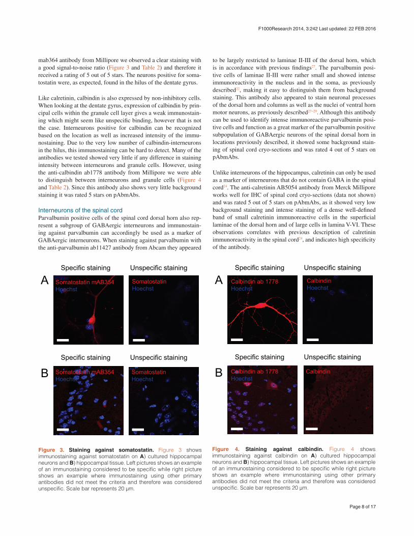
mab364 antibody from Millipore we observed a clear staining with a good signal-to-noise ratio (Figure 3 and Table 2) and therefore it received a rating of 5 out of 5 stars. The neurons positive for soma-tostatin were, as expected, found in the hilus of the dentate gyrus.
Like calretinin, calbindin is also expressed by non-inhibitory cells. When looking at the dentate gyrus, expression of calbindin by prin-cipal cells within the granule cell layer gives a weak immunostain-ing which might seem like unspecific binding, however that is not the case. Interneurons positive for calbindin can be recognized based on the location as well as increased intensity of the immu-nostaining. Due to the very low number of calbindin-interneurons in the hilus, this immunostaining can be hard to detect. Many of the antibodies we tested showed very little if any difference in staining intensity between interneurons and granule cells. However, using the anti-calbindin ab1778 antibody from Millipore we were able to distinguish between interneurons and granule cells (Figure 4 and Table 2). Since this antibody also shows very little background staining it was rated 5 stars on pAbmAbs.
Interneurons of the spinal cordParvalbumin positive cells of the spinal cord dorsal horn also rep-resent a subgroup of GABAergic interneurons and immunostain-ing against parvalbumin can accordingly be used as a marker of GABAergic interneurons. When staining against parvalbumin with the anti-parvalbumin ab11427 antibody from Abcam they appeared
to be largely restricted to laminae II-III of the dorsal horn, which is in accordance with previous findings27. The parvalbumin posi-tive cells of laminae II-III were rather small and showed intense immunoreactivity in the nucleus and in the soma, as previously described22, making it easy to distinguish them from background staining. This antibody also appeared to stain neuronal processes of the dorsal horn and columns as well as the nuclei of ventral horn motor neurons, as previously described27–29. Although this antibody can be used to identify intense immunoreactive parvalbumin posi-tive cells and function as a great marker of the parvalbumin positive subpopulation of GABAergic neurons of the spinal dorsal horn in locations previously described, it showed some background stain-ing of spinal cord cryo-sections and was rated 4 out of 5 stars on pAbmAbs.
Unlike interneurons of the hippocampus, calretinin can only be used as a marker of interneurons that do not contain GABA in the spinal cord24. The anti-calretinin AB5054 antibody from Merck Millipore works well for IHC of spinal cord cryo-sections (data not shown) and was rated 5 out of 5 stars on pAbmAbs, as it showed very low background staining and intense staining of a dense well-defined band of small calretinin immunoreactive cells in the superficial laminae of the dorsal horn and of large cells in lamina V-VI. These observations correlates with previous description of calretinin immunoreactivity in the spinal cord24, and indicates high specificity of the antibody.
Figure 3. Staining against somatostatin. Figure 3 shows immunostaining against somatostatin on A) cultured hippocampal neurons and B) hippocampal tissue. Left pictures shows an example of an immunostaining considered to be specific while right picture shows an example where immunostaining using other primary antibodies did not meet the criteria and therefore was considered unspecific. Scale bar represents 20 μm.
Figure 4. Staining against calbindin. Figure 4 shows immunostaining against calbindin on A) cultured hippocampal neurons and B) hippocampal tissue. Left pictures shows an example of an immunostaining considered to be specific while right picture shows an example where immunostaining using other primary antibodies did not meet the criteria and therefore was considered unspecific. Scale bar represents 20 μm.
Page 8 of 17
F1000Research 2014, 3:242 Last updated: 22 FEB 2016

In contrast to IHC of hippocampal sections with the anti-somatostatin MAB354 antibody from Millipore, this antibody gave a low signal when staining against somatostatin on spinal cord sections. Using this antibody, it was difficult to identify somatostatin positive cells in the spinal dorsal horn that otherwise previously have been described to be located in a dense band in lamina II of rat25 and mouse21 spinal dorsal horn. Therefore, the antibody was rated 2 out of 5 stars on pAbmAbs. This antibody was rated 5 out of 5 for the hip-pocampal staining, leading to an average rating of 3.5 on pAbmAbs.
Like calretinin and somatostatin, calbindin can be used as a marker of spinal dorsal horn interneurons that do not contain GABA23. A dense band of calbindin immunoreactivity has previously been shown in lamina II and a more sparse band in lamina I, III and IV of the rat spinal dorsal horn23. This localization of calbindin immu-noreactivity is also seen when using the anti-calbindin AB1778 antibody from Merck Millipore (data not shown). Also, the cells that constitute the central channel and motor neurons of the ventral horn also show calbindin immunoreactivity when staining with this antibody, which is in accordance with previously findings28,30. The antibody showed very intense staining of cytoplasm and nuclei, as well as processes of the outer lamina of the dorsal horn and showed low background staining. On the basis of these observations the antibody was rated 5 out of 5 stars on pAbmAbs.
Dataset 1. Interneurons of hippocampus and spinal cord
http://dx.doi.org/10.5256/f1000research.5349.d36682
The raw microscopy images for both hippocampal and spinal cord interneurons are shown in the .czi files provided.
ConclusionIn conclusion, staining against interneurons can be a very tedious task and great consideration is needed to ensure that it is actually only interneurons that are being stained. Optimizing protocols for immunostaining can be a, not only time consuming, but also an expensive task in a market full of different antibody options. By creating an information-sharing platform, pAbmAbs allows for a fast and cost-free screening of the current antibodies out there and thereby ensures that only the best antibodies are used. In the cur-rent study, we tested antibodies against parvalbumin, calretinin, calbindin and somatostatin, all markers of hippocampal GABAe-rgic interneurons, both in culture and on hippocampal and spinal cord tissue. These antibodies were rated on specificity, and signal-to-noise ratio, for both tissue and culture. When immunostaining
tissue, we also looked at the localization of positive cells within the tissue to ensure that only cells in the expected layers of the tissue stained positive for the GABAergic markers. When staining against parvalbumin we found that out of four different antibodies, the anti-parvalbumin ab11427 antibody from Abcam got a high score as it stained cells specifically with a high signal-to-noise ratio with the expected localization within the tissue. When staining against cal-retinin, the anti-calretinin ab5054 antibody from Millipore obtained the highest score on pAbmAbs. This antibody gave a very nice signal-to-noise ratio compared to the other antibodies tested. The anti-somatostatin mab354 antibody from Millipore was found to be the best antibody for stainings against somatostatin. Similar to the other antibodies with high pAbmAbs ratings, this also had a high signal-to-noise ratio compared to other antibodies tested. Finally, the anti-calbindin ab1178 antibody from Millipore obtained the highest rating out of the antibodies tested against this GABAergic subgroup. Overall, the antibody tested gave varying results when using our protocols. The specificities of the antibodies are therefore reflected on pAbmAbs which, by serving as a database, will help fast and cost-free evaluation of antibodies.
Data availabilityF1000Research: Dataset 1. Interneurons of hippocampus and spinal cord, 10.5256/f1000research.5349.d3668231
Author contributionsSM and SG conceived this study. SM designed the experiments. SM, MU and SBH carried out the research. SG, JRN and CV con-tributed to the design of the experiment and expertise in immuno-histochemistry. SM, SG and MU contributed to the preparation of the manuscript. All authors were involved in the revision of the draft manuscript and have agreed to the final content.
Competing interestsNo competing interests were disclosed.
Grant informationThis study was funded by the Lundbeck Foundation, Danish Medical Research Council, Fonden til forskning af sindslidelser and Agnes og Poul Friis Fond.
AcknowledgementsWe thank Helene Andersen, Anja Aagaard and Benedicte Vestergaard for excellent technical assistance.
References
1. Freund TF, Buzsaki G: Interneurons of the hippocampus. Hippocampus. 1996; 6(4): 347–470. PubMed Abstract | Publisher Full Text
2. Farrant M, Nusser Z: Variations on an inhibitory theme: phasic and tonic activation of GABA(A) receptors. Nat Rev Neurosci. 2005; 6(3): 215–29. PubMed Abstract | Publisher Full Text
3. Wang XJ, Buzsaki G: Gamma oscillation by synaptic inhibition in a hippocampal interneuronal network model. J Neurosci. 1996; 16(20): 6402–13. PubMed Abstract
4. Buzsaki G, Draguhn A: Neuronal oscillations in cortical networks. Science. 2004; 304(5679): 1926–1929. PubMed Abstract | Publisher Full Text
5. Benes FM, Berretta S: GABAergic interneurons: implications for understanding schizophrenia and bipolar disorder. Neuropsychopharmacology. 2001; 25(1): 1–27. PubMed Abstract | Publisher Full Text
6. Amaral DG: A Golgi study of cell types in the hilar region of the hippocampus in the rat. J Comp Neurol. 1978; 182(4 Pt 2): 851–914. PubMed Abstract | Publisher Full Text
Page 9 of 17
F1000Research 2014, 3:242 Last updated: 22 FEB 2016

20. Zeilhofer HU: The glycinergic control of spinal pain processing. Cell Mol Life Sci. 2005; 62(18): 2027–35. PubMed Abstract | Publisher Full Text
21. Heinke B, Ruscheweyh R, Forsthuber L, et al.: Physiological, neurochemical and morphological properties of a subgroup of GABAergic spinal lamina II neurones identified by expression of green fluorescent protein in mice. J Physiol. 2004; 560(Pt 1): 249–66. PubMed Abstract | Publisher Full Text | Free Full Text
22. Laing I, Todd AJ, Heizmann CW, et al.: Subpopulations of GABAergic neurons in laminae I–III of rat spinal dorsal horn defined by coexistence with classical transmitters, peptides, nitric oxide synthase or parvalbumin. Neuroscience. 1994; 61(1): 123–32. PubMed Abstract | Publisher Full Text
23. Antal M, Polgár E, Chalmers J, et al.: Different populations of parvalbumin- and calbindin-D28k-immunoreactive neurons contain GABA and accumulate 3H-D-aspartate in the dorsal horn of the rat spinal cord. J Comp Neurol. 1991; 314(1): 114–24. PubMed Abstract | Publisher Full Text
24. Ren K, Ruda MA: A comparative study of the calcium-binding proteins calbindin-D28K, calretinin, calmodulin and parvalbumin in the rat spinal cord. Brain Res Brain Res Rev. 1994; 19(2): 163–79. PubMed Abstract | Publisher Full Text
25. Proudlock F, Spike RC, Todd AJ: Immunocytochemical study of somatostatin, neurotensin, GABA, and glycine in rat spinal dorsal horn. J Comp Neurol. 1993; 327(2): 289–97. PubMed Abstract | Publisher Full Text
26. Meikle AD, Martin AH: A rapid method for removal of the spinal cord. Stain Technol. 1981; 56(4): 235–7. PubMed Abstract | Publisher Full Text
27. Hughes DI, Sikander S, Kinnon CM, et al.: Morphological, neurochemical and electrophysiological features of parvalbumin-expressing cells: a likely source of axo-axonic inputs in the mouse spinal dorsal horn. J Physiol. 2012; 590(Pt 16): 3927–51. PubMed Abstract | Publisher Full Text | Free Full Text
28. Ince P, Stout N, Shaw P, et al.: Parvalbumin and calbindin D-28k in the human motor system and in motor neuron disease. Neuropathol Appl Neurobiol. 1993; 19(4): 291–9. PubMed Abstract | Publisher Full Text
29. Antal M, Freund TF, Polgar E: Calcium-binding proteins, parvalbumin- and calbindin-D 28k-immunoreactive neurons in the rat spinal cord and dorsal root ganglia: a light and electron microscopic study. J Comp Neurol. 1990; 295(3): 467–84. PubMed Abstract | Publisher Full Text
30. Holm MM, Nieto-Gonzalez JL, Vardya I, et al.: Mature BDNF, but not proBDNF, reduces excitability of fast-spiking interneurons in mouse dentate gyrus. J Neurosci. 2009; 29(40): 12412–8. PubMed Abstract | Publisher Full Text
31. Molgaard S, Ulrichsen M, Boggild S, et al.: Dataset 1. Interneurons of hippocampus and spinal cord. F1000Research. 2014. Data Source
7. Erlander MG, Tillakaratne NJ, Feldblum S, et al.: Two genes encode distinct glutamate decarboxylases. Neuron. 1991; 7(1): 91–100. PubMed Abstract | Publisher Full Text
8. Ribak CE, Vaughn JE, Saito K: Immunocytochemical localization of glutamic acid decarboxylase in neuronal somata following colchicine inhibition of axonal transport. Brain Res. 1978; 140(2): 315–32. PubMed Abstract | Publisher Full Text
9. Jinno S, Aika Y, Fukuda T, et al.: Quantitative analysis of GABAergic neurons in the mouse hippocampus, with optical disector using confocal laser scanning microscope. Brain Res. 1998; 814(1–2): 55–70. PubMed Abstract | Publisher Full Text
10. Somogyi P, Klausberger T: Defined types of cortical interneurone structure space and spike timing in the hippocampus. J Physiol. 2005; 562(Pt 1): 9–26. PubMed Abstract | Publisher Full Text | Free Full Text
11. Ribak CE, Nitsch R, Seress L: Proportion of parvalbumin-positive basket cells in the GABAergic innervation of pyramidal and granule cells of the rat hippocampal formation. J Comp Neurol. 1990; 300(4): 449–61. PubMed Abstract | Publisher Full Text
12. Blasco-Ibanez JM, Freund TF: Distribution, ultrastructure, and connectivity of calretinin-immunoreactive mossy cells of the mouse dentate gyrus. Hippocampus. 1997; 7(3): 307–20. PubMed Abstract | Publisher Full Text
13. Fujise N, Liu Y, Hori N, et al.: Distribution of calretinin immunoreactivity in the mouse dentate gyrus: II. Mossy cells, with special reference to their dorsoventral difference in calretinin immunoreactivity. Neuroscience. 1998; 82(1): 181–200. PubMed Abstract | Publisher Full Text
14. Gulyas AI, Hajos N, Freund TF: Interneurons containing calretinin are specialized to control other interneurons in the rat hippocampus. J Neurosci. 1996; 16(10): 3397–411. PubMed Abstract
15. Jinno S, Kosaka T: Patterns of expression of neuropeptides in GABAergic nonprincipal neurons in the mouse hippocampus: Quantitative analysis with optical disector. J Comp Neurol. 2003; 461(3): 333–49. PubMed Abstract | Publisher Full Text
16. Katona I, Acsady L, Freund TF: Postsynaptic targets of somatostatin-immunoreactive interneurons in the rat hippocampus. Neuroscience. 1999; 88(1): 37–55. PubMed Abstract | Publisher Full Text
17. Gulyas AI, Toth K, Danos P, et al.: Subpopulations of GABAergic neurons containing parvalbumin, calbindin D28k, and cholecystokinin in the rat hippocampus. J Comp Neurol. 1991; 312(3): 371–8. PubMed Abstract | Publisher Full Text
18. Todd AJ, Sullivan AC: Light microscope study of the coexistence of GABA-like and glycine-like immunoreactivities in the spinal cord of the rat. J Comp Neurol. 1990; 296(3): 496–505. PubMed Abstract | Publisher Full Text
19. Polgar E, Hughes DI, Riddell JS, et al.: Selective loss of spinal GABAergic or glycinergic neurons is not necessary for development of thermal hyperalgesia in the chronic constriction injury model of neuropathic pain. Pain. 2003; 104(1–2): 229–39. PubMed Abstract | Publisher Full Text
Page 10 of 17
F1000Research 2014, 3:242 Last updated: 22 FEB 2016

F1000Research
Open Peer Review
Current Referee Status:
Version 1
21 October 2014Referee Report
doi:10.5256/f1000research.5710.r6402
Mei Yee LeungSt John's Laboratory Ltd., London, UK
This is an informative, concise article with clear aims that highlights the potential difficulties in selectingthe right antibodies for specific cell types and research applications. In this study, the authorssystematically tested commercial antibodies against calbindin, calretinin, parvalbumin and somatostatin -markers of GABAergic interneuron subtypes. Of the 13 antibodies tested, only 4 were deemed reliableand useful for characterizing these subtypes in mouse brain.
The authors’ rigorous approach in selecting the right antibodies is commendable, a fact which often goesunnoticed in publications. The ranking of antibody performance in pAbmAbs, a review-based platform is amuch needed resource for scientists whose research depends on the validity of the antibodies.
As someone not in this research area, I found the manuscript scientifically well construed and therationale easy to follow. It is noteworthy that this study by Molgaard adds to the understanding ofet al.GABAergic subtypes in the mouse hippocampus and spinal cord, information which is sparse in theliterature. In addition, by demonstrating specific staining of these markers, they have confirmed previouslyreported localization of these cells. A few comments/suggestions
Would a change in the title be more appropriate? e.g. Immunofluorescent instead ofimmunohistochemicalIs there any reason why the age of the mouse used for hippocampal staining and that for spinalcord staining is different?It would add value to this paper is images of spinal cord staining was also shownAlthough the without primary images are very clean, using these antibodies on tissues not knownto express these targets would be a more superior negative controlCould the authors offer an explanation for why the polyclonal antibodies appear to perform betterthan the monoclonals?To obtain better idea of reproducibility, it would be good to give an indication of how many timesthe experiment was performed and how many sections were stained per experiment
I have read this submission. I believe that I have an appropriate level of expertise to confirm thatit is of an acceptable scientific standard.
No competing interests were disclosed.Competing Interests:
Page 11 of 17
F1000Research 2014, 3:242 Last updated: 22 FEB 2016

F1000Research
Author Response 12 Nov 2014, Aarhus University, DenmarkSimon Molgaard
"Would a change in the title be more appropriate? e.g. Immunofluorescent instead ofimmunohistochemical"
: OKReply "Is there any reason why the age of the mouse used for hippocampal staining and that for spinalcord staining is different?"
: No particular reason. The expression of GABAergic markers in the hippocampus and spinalReplycord of adult animals is expected to be fairly constant throughout adulthood. "It would add value to this paper is images of spinal cord staining was also shown"
: DoneReply "Although the without primary images are very clean, using these antibodies on tissues not knownto express these targets would be a more superior negative control"
: This point may rely on a possible misunderstanding. The images in the paper denotedReply“Unspecific staining” refers to stainings using primary antibodies for which no specific signal wasobserved. We have changed the legends to make this more clear. "Could the authors offer an explanation for why the polyclonal antibodies appear to perform betterthan the monoclonals?"
: We have no clear answer to this but it may be that polyclonal antibodies generally give aReplyhigher signal compared monoclonal antibodies due to the presence of multiple epitopes. "To obtain better idea of reproducibility, it would be good to give an indication of how many timesthe experiment was performed and how many sections were stained per experiment"
: This is now stated in the Methods section. Reply
No competing interests were disclosed.Competing Interests:
17 October 2014Referee Report
doi:10.5256/f1000research.5710.r6403
Tomi PJ RantamäkiNeuroscience Center, University of Helsinki, Helsinki, Finland
Molgaard have investigated the suitability of various commercially available antibodies for theet al.identification of GABAergic interneurons in mice. Among 13 tested antibodies against calbindin (3),calretinin (4), parvalbumin (4) and somatostatin (2), the authors found 1-2 antibodies per each marker thatproduced high quality, sensitive and specific staining in mouse brain sections and cultured hippocampal
Page 12 of 17
F1000Research 2014, 3:242 Last updated: 22 FEB 2016

F1000Research
calretinin (4), parvalbumin (4) and somatostatin (2), the authors found 1-2 antibodies per each marker thatproduced high quality, sensitive and specific staining in mouse brain sections and cultured hippocampalneurons obtained from P0 mouse pups. Their findings remind us about the tediousness ofimmunostainings in general and challenges about the identification of proper antibody and antibodyconditions suitable for high quality research. Although the antibodies have not been investigated in allpossible experimental conditions (e.g. fixation protocols, dilutions), the study provides very valuablereference information for researchers aiming to investigate GABAergic markers in mice. In general the manuscript has been written and constructed well. Abstract nicely describes the summaryof the study. The introduction provides very good background knowledge for the reader (incl. relevantcitations). The methods section is described in a manner that allows scientific reproduction efficiently.Tables are clear and useful. Overall the representative figures are good but the paper would benefit withmore comprehensive set of immunostainings (as main figures). I have few minor comments/questions:
GABAergic interneurons are considered as small neuronal population in the text. In respect toglutamatergic neurons this is indeed the case, but overall I consider 20% quite a significant fraction(cf. monoaminergic neurons).The authors could have clarified what is “pAbmAbs” in the abstract.In the Introduction the authors state that breakdown of cortical network rhythms underlieneuropsychiatric disorders. I would rather say that alterations in cortical network rhythms in specificbrain networks may underlie neuropsychiatric disorders.The authors could have explained that glutamate decarboxylase is expressed in GABAergicneurons and it synthesizes GABA (Introduction, first and second paragraph).The authors could have described the gender and amount of adult mice used for the study.Moreover, where all the antibodies tested in specimens derived from same conditions (e.g. sameanimal).I would have used “sections” rather than “slices” throughout the paper.Table 2. would be even more clear if the antibodies against the four different markers would havebeen divided from each other more clearly (e.g. using different background colors).Why there appears no Hoechst staining in some of the unspecific stainings?Why the authors choose not to show the representative figures from spinal cord?The authors could have emphasized that the quality of polyclonal antibodies is significantlydetermined by the lot/batch. This should be kept in mind when reproducing the findings.Figure legends should have been clearer. In optimal case, the reader understands the figuresthoroughly without the main text (e.g. age of cultures).It would have been very useful to test, at least selected, antibodies in rats as well (brain sections,culture)
I have read this submission. I believe that I have an appropriate level of expertise to confirm thatit is of an acceptable scientific standard.
No competing interests were disclosed.Competing Interests:
Author Response 12 Nov 2014, Aarhus University, DenmarkSimon Molgaard
"GABAergic interneurons are considered as small neuronal population in the text. In respect toglutamatergic neurons this is indeed the case, but overall I consider 20% quite a significant fraction(cf. monoaminergic neurons)."
Page 13 of 17
F1000Research 2014, 3:242 Last updated: 22 FEB 2016

F1000Research
: Comment well taken. The word “small” has now been removed from the abstract.Reply
"The authors could have clarified what is “pAbmAbs” in the abstract."
: This has been added to abstract.Reply "In the Introduction the authors state that breakdown of cortical network rhythms underlieneuropsychiatric disorders. I would rather say that alterations in cortical network rhythms in specificbrain networks may underlie neuropsychiatric disorders."
: We agree. The text is now changed accordingly.Reply "The authors could have explained that glutamate decarboxylase is expressed in GABAergicneurons and it synthesizes GABA (Introduction, first and second paragraph)."
: This has now been clarified in the introduction.Reply "The authors could have described the gender and amount of adult mice used for the study.Moreover, where all the antibodies tested in specimens derived from same conditions (e.g. sameanimal)."
: A paragraph has been added to the Methods clarifying these issues.Reply "I would have used “sections” rather than “slices” throughout the paper."
: This has now been corrected.Reply "Table 2. would be even more clear if the antibodies against the four different markers would havebeen divided from each other more clearly (e.g. using different background colors)."
: Although we agree that a color-code would be desirable, we feel that the current table isReplysufficiently clear. "Why there appears no Hoechst staining in some of the unspecific stainings?"
We apologize for the lack of clarity regarding this issue. The “unspecific” images withReply:Hoechst are shown when we observed no signal at all. The “unspecific” images without Hoechstare shown when staining was present but distributed in an unspecific manner. "Why the authors choose not to show the representative figures from spinal cord?"
: The images are already included in the data availability section.Reply "The authors could have emphasized that the quality of polyclonal antibodies is significantlydetermined by the lot/batch. This should be kept in mind when reproducing the findings."
: Good point. In this regard we will refer to the article in the antibody validation collection byReplyDr Jan Voskuil.
Page 14 of 17
F1000Research 2014, 3:242 Last updated: 22 FEB 2016

F1000Research
1.
2.
3.
4.
5.
"Figure legends should have been clearer. In optimal case, the reader understands the figuresthoroughly without the main text (e.g. age of cultures)."
: The figure legends have now been improvedReply "It would have been very useful to test, at least selected, antibodies in rats as well (brain sections,culture) "
: We agree but as stated in the title we have only used mouse. Reply
No competing interests were disclosed.Competing Interests:
14 October 2014Referee Report
doi:10.5256/f1000research.5710.r6401
Sally LowellMRC Centre for Regenerative Medicine, Institute for Stem Cell Research, School of Biological Sciences,University of Edinburgh, Edinburgh, UK
The authors of this paper set out to identify antibodies that can be used to identify particular subtypes ofGABAergic neurons within the mouse hippocampus. They explain that many of the antibodies that theyhave tested for this purpose do not seem to work, and they present data for those antibodies that they didfind to be useful for this purpose. This article could therefore save readers a lot of wasted effort andmoney in identifying useful antibodies for their own studies in this area.
The article starts with a nice overview of the different subtypes of GABAergic neurons and the markersthat are commonly used to characterise them. I am not an expert in this area so I cannot review theaccuracy of the information, but I did find it to be concise and useful introduction.
There are a few issues that could be addressed to improve the results section of the article.Figure legends should explain what "unspecific staining" refers to. Does this mean secondary onlycontrol? If so then this form of labelling could be misleading as non-specific binding of the primaryantibody will not be picked up by a secondary-antibody-only control. Why are there no Hoechst positive cells in some of the 'unspecific staining' panels? The evidence that these antibodies are specific to particular subtypes of neurons comes mainlyfrom the observation that their staining pattern is restricted to the expected regions of thehippocampus. However the figures as presented do not make this clear. Is it possible to provide aclearer demonstration of these restricted expression domains on hippocampus sections? Perhapsit would be helpful to see an accompanying diagram showing the expected distribution of eachmarker on these sections? In Fig 3B, the 'specific staining' of antibody mAB354 is barely visible so it is difficult to assesswhether there is a real difference between signal and background. The article reports results from immunostaining of the spinal cord, but no figures are presented to
Page 15 of 17
F1000Research 2014, 3:242 Last updated: 22 FEB 2016

F1000Research
5. The article reports results from immunostaining of the spinal cord, but no figures are presented tosupport the authors’ findings. It would be helpful to see these images.
General comments: The article is written clearly and concisely, but would benefit from being proof-readfor minor grammatical errors.
I have read this submission. I believe that I have an appropriate level of expertise to confirm thatit is of an acceptable scientific standard, however I have significant reservations, as outlinedabove.
No competing interests were disclosed.Competing Interests:
Author Response 12 Nov 2014, Aarhus University, DenmarkSimon Molgaard
"Figure legends should explain what "unspecific staining" refers to. Does this mean secondary onlycontrol? If so then this form of labelling could be misleading as non-specific binding of the primaryantibody will not be picked up by a secondary-antibody-only control."
: The figure legends have been changed to make this more clear.Reply "Why are there no Hoechst positive cells in some of the 'unspecific staining' panels?"
: We apologize for the lack of clarity regarding this issue. The “unspecific” images withReplyHoechst are shown when we observed no signal at all. The “unspecific” images without Hoechstare shown when staining was present but distributed in an unspecific manner. "The evidence that these antibodies are specific to particular subtypes of neurons comes mainlyfrom the observation that their staining pattern is restricted to the expected regions of thehippocampus. However the figures as presented do not make this clear. Is it possible to provide aclearer demonstration of these restricted expression domains on hippocampus sections? Perhapsit would be helpful to see an accompanying diagram showing the expected distribution of eachmarker on these sections?"
: The interneuron subtype distribution is presently used in a qualitative manner to validateReplywhether positive staining localizes in the expected and with an expected frequency in the dentategyrus of the hippocampus. Quantitative assessment of interneuron number using for examplestereology is not feasible in the present study. "In Fig 3B, the 'specific staining' of antibody mAB354 is barely visible so it is difficult to assesswhether there is a real difference between signal and background."
: We agree that the staining is weaker; however it is still distinct and specific.Reply "The article reports results from immunostaining of the spinal cord, but no figures are presented tosupport the authors’ findings. It would be helpful to see these images."
: The images are already included in the data availability section. Reply
No competing interests were disclosed.Competing Interests:
Page 16 of 17
F1000Research 2014, 3:242 Last updated: 22 FEB 2016

F1000Research
No competing interests were disclosed.Competing Interests:
Page 17 of 17
F1000Research 2014, 3:242 Last updated: 22 FEB 2016