Studies of intestinal morphology and cathepsin B expression in a transgenic mouse aiming at...
11
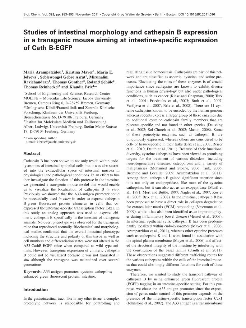
Biol. Chem., Vol. 392, pp. 983–993, November 2011 • Copyright by Walter de Gruyter • Berlin • Boston. DOI 10.1515/BC.2011.096 2011/155 Article in press - uncorrected proof Studies of intestinal morphology and cathepsin B expression in a transgenic mouse aiming at intestine-specific expression of Cath B-EGFP Maria Arampatzidou 1 , Kristina Mayer 1 , Maria E. Iolyeva 1 , Seblewongel Gebre Asrat 1 , Mirunalini Ravichandran 1 , Thomas Gu ¨ nther 2 , Roland Schu ¨le 2 , Thomas Reinheckel 3 and Klaudia Brix 1, * 1 School of Engineering and Science, Research Center MOLIFE – Molecular Life Science, Jacobs University Bremen, Campus Ring 6, D-28759 Bremen, Germany 2 Urologische Klinik/Frauenklinik und Zentrale Klinische Forschung, Klinikum der Universita ¨t Freiburg, Breisacherstrasse 66, D-79106 Freiburg, Germany 3 Institut fu ¨r Molekulare Medizin und Zellforschung, Albert-Ludwigs-Universita ¨t Freiburg, Stefan-Meier-Strasse 17, D-79104 Freiburg, Germany * Corresponding author e-mail: [email protected] Abstract Cathepsin B has been shown to not only reside within endo- lysosomes of intestinal epithelial cells, but it was also secret- ed into the extracellular space of intestinal mucosa in physiological and pathological conditions. In an effort to fur- ther investigate the function of this protease in the intestine, we generated a transgenic mouse model that would enable us to visualize the localization of cathepsin B in vivo. Previously we showed that the A33-antigen promoter could be successfully used in vitro in order to express cathepsin B-green fluorescent protein chimeras in cells that co- expressed the intestine-specific transcription factor Cdx1. In this study an analog approach was used to express chi- meric cathepsin B specifically in the intestine of transgenic animals. No overt phenotype was observed for the transgenic mice that reproduced normally. Biochemical and morpholog- ical studies confirmed that the overall intestinal phenotype including the structure and polarity of this tissue as well as cell numbers and differentiation states were not altered in the A33-CathB-EGFP mice when compared to wild type ani- mals. However, transgenic expression of chimeric cathepsin B could not be visualized because it was not translated in situ although the transgene was maintained over several generations. Keywords: A33-antigen promoter; cysteine cathepsins; enhanced green fluorescent protein; intestine. Introduction In the gastrointestinal tract, like in any other tissue, a complex proteolytic network is responsible for controlling and regulating tissue homeostasis. Cathepsins are part of this net- work and are classified as aspartic, cysteine, and serine pro- teases. Elucidating the roles of these enzymes is of crucial importance since cathepsins are known to exhibit diverse functions in human physiology but also under pathological conditions, such as cancer (Riese and Chapman, 2000; Turk et al., 2001; Friedrichs et al., 2003; Buth et al., 2007; Vasiljeva et al., 2007; Brix et al., 2008). There are 11 cys- teine cathepsins known to be encoded by the human genome whereas rodents express a larger group of these enzymes due to additional cysteine cathepsin family members that are placenta-specific and not found in other species (Deussing et al., 2002; Sol-Church et al., 2002; Mason, 2008). Some of these proteolytic enzymes, such as cathepsin B, are ubiquitously expressed, whereas others are considered to be cell- or tissue-specific in their tasks (Brix et al., 2008; Reiser et al., 2010; Dauth et al., 2011). Because of their functional diversity, cysteine cathepsins have been viewed as promising targets for the treatment of various disorders, including neurodegenerative diseases, osteoporosis and a variety of malignancies (Mohamed and Sloane, 2006; Turk, 2006; Bromme and Lecaille, 2009; Arampatzidou et al., 2011). Among them, cathepsin B gained significant attention since it is not only an endopeptidase, like most of the cysteine cathepsins, but it can also act as an exopeptidase (Musil et al., 1991; Mort and Buttle, 1997; Nagler et al., 1997; Kos et al., 2005; Brix et al., 2008). In the intestine, cathepsin B has been proposed to have a direct role in collagen degradation for extracellular matrix (ECM) remodeling (Vreemann et al., 2009), while it has also been identified as an important play- er during inflammatory bowel disease (Menzel et al., 2006). In intestinal epithelial cells, cathepsin B has been predomi- nantly localized within endo-lysosomes (Mayer et al., 2006; Arampatzidou et al., 2011), whereas other cysteine proteases such as cathepsins K and L were found in association with the apical plasma membrane (Mayer et al., 2006) and affect- ed the structural integrity of the intestine by interfering with the constitution of the basal lamina (Dauth et al., 2011). These observations suggested different trafficking routes for the various cathepsins within the cells of the intestinal muco- sa that could also imply different functions for each of these enzymes. Therefore, we wanted to study the transport pathway of cathepsin B by using enhanced green fluorescent protein (EGFP) tagging in an intestine-specific setting. For this pur- pose, we chose the A33-antigen promoter since the expres- sion of genes under control of this promoter depends on the presence of the intestine-specific transcription factor Cdx1 (Johnstone et al., 2002). The A33 antigen is a transmembrane
-
Upload
independent -
Category
Documents
-
view
6 -
download
0
Transcript of Studies of intestinal morphology and cathepsin B expression in a transgenic mouse aiming at...
Biol. Chem., Vol. 392, pp. 983–993, November 2011 • Copyright � by Walter de Gruyter • Berlin • Boston. DOI 10.1515/BC.2011.096
2011/155
Article in press - uncorrected proof
Studies of intestinal morphology and cathepsin B expression
in a transgenic mouse aiming at intestine-specific expression
of Cath B-EGFP
Maria Arampatzidou1, Kristina Mayer1, Maria E.Iolyeva1, Seblewongel Gebre Asrat1, MirunaliniRavichandran1, Thomas Gunther2, Roland Schule2,Thomas Reinheckel3 and Klaudia Brix1,*1 School of Engineering and Science, Research CenterMOLIFE – Molecular Life Science, Jacobs UniversityBremen, Campus Ring 6, D-28759 Bremen, Germany2 Urologische Klinik/Frauenklinik und Zentrale KlinischeForschung, Klinikum der Universitat Freiburg,Breisacherstrasse 66, D-79106 Freiburg, Germany3 Institut fur Molekulare Medizin und Zellforschung,Albert-Ludwigs-Universitat Freiburg, Stefan-Meier-Strasse17, D-79104 Freiburg, Germany
* Corresponding authore-mail: [email protected]
Abstract
Cathepsin B has been shown to not only reside within endo-lysosomes of intestinal epithelial cells, but it was also secret-ed into the extracellular space of intestinal mucosa inphysiological and pathological conditions. In an effort to fur-ther investigate the function of this protease in the intestine,we generated a transgenic mouse model that would enableus to visualize the localization of cathepsin B in vivo.Previously we showed that the A33-antigen promoter couldbe successfully used in vitro in order to express cathepsinB-green fluorescent protein chimeras in cells that co-expressed the intestine-specific transcription factor Cdx1. Inthis study an analog approach was used to express chi-meric cathepsin B specifically in the intestine of transgenicanimals. No overt phenotype was observed for the transgenicmice that reproduced normally. Biochemical and morpholog-ical studies confirmed that the overall intestinal phenotypeincluding the structure and polarity of this tissue as well ascell numbers and differentiation states were not altered in theA33-CathB-EGFP mice when compared to wild type ani-mals. However, transgenic expression of chimeric cathepsinB could not be visualized because it was not translated insitu although the transgene was maintained over severalgenerations.
Keywords: A33-antigen promoter; cysteine cathepsins;enhanced green fluorescent protein; intestine.
Introduction
In the gastrointestinal tract, like in any other tissue, a complexproteolytic network is responsible for controlling and
regulating tissue homeostasis. Cathepsins are part of this net-work and are classified as aspartic, cysteine, and serine pro-teases. Elucidating the roles of these enzymes is of crucialimportance since cathepsins are known to exhibit diversefunctions in human physiology but also under pathologicalconditions, such as cancer (Riese and Chapman, 2000; Turket al., 2001; Friedrichs et al., 2003; Buth et al., 2007;Vasiljeva et al., 2007; Brix et al., 2008). There are 11 cys-teine cathepsins known to be encoded by the human genomewhereas rodents express a larger group of these enzymes dueto additional cysteine cathepsin family members that areplacenta-specific and not found in other species (Deussinget al., 2002; Sol-Church et al., 2002; Mason, 2008). Someof these proteolytic enzymes, such as cathepsin B, areubiquitously expressed, whereas others are considered to becell- or tissue-specific in their tasks (Brix et al., 2008; Reiseret al., 2010; Dauth et al., 2011). Because of their functionaldiversity, cysteine cathepsins have been viewed as promisingtargets for the treatment of various disorders, includingneurodegenerative diseases, osteoporosis and a variety ofmalignancies (Mohamed and Sloane, 2006; Turk, 2006;Bromme and Lecaille, 2009; Arampatzidou et al., 2011).Among them, cathepsin B gained significant attention sinceit is not only an endopeptidase, like most of the cysteinecathepsins, but it can also act as an exopeptidase (Musil etal., 1991; Mort and Buttle, 1997; Nagler et al., 1997; Kos etal., 2005; Brix et al., 2008). In the intestine, cathepsin B hasbeen proposed to have a direct role in collagen degradationfor extracellular matrix (ECM) remodeling (Vreemann et al.,2009), while it has also been identified as an important play-er during inflammatory bowel disease (Menzel et al., 2006).In intestinal epithelial cells, cathepsin B has been predomi-nantly localized within endo-lysosomes (Mayer et al., 2006;Arampatzidou et al., 2011), whereas other cysteine proteasessuch as cathepsins K and L were found in association withthe apical plasma membrane (Mayer et al., 2006) and affect-ed the structural integrity of the intestine by interfering withthe constitution of the basal lamina (Dauth et al., 2011).These observations suggested different trafficking routes forthe various cathepsins within the cells of the intestinal muco-sa that could also imply different functions for each of theseenzymes.
Therefore, we wanted to study the transport pathway ofcathepsin B by using enhanced green fluorescent protein(EGFP) tagging in an intestine-specific setting. For this pur-pose, we chose the A33-antigen promoter since the expres-sion of genes under control of this promoter depends on thepresence of the intestine-specific transcription factor Cdx1(Johnstone et al., 2002). The A33 antigen is a transmembrane

984 M. Arampatzidou et al.
Article in press - uncorrected proof
Figure 1 Genotyping of the A33-CathB-EGFP transgenic mice.(A) Semi-quantitative PCR analysis using a transgene-specific primer pair shows different amounts of transgene in the various A33-CathB-EGFP transgenic mice. No PCR product was detected for wild type mouse (WT) while the plasmid pA33-CathB-EGFP served aspositive control. (B) Scattergram of the analyzed A33-CathB-EGFP mice indicating the amount of transgene through three generations. Notethat the transgene was kept and present in comparable copy numbers over the different generations.
protein that is expressed only in the intestine and isspecifically localized at the basolateral membrane of intes-tinal epithelial cells of all lineages (Johnstone et al., 2000).The A33-antigen promoter contains positive cis-regulatoryelements, including caudal-related homeobox (Cdx1) bindingsites. Cdx1 is an intestine-specific transcription factor whichis crucial for A33-antigen promoter activation and couldthereby induce expression of the chimeric protein cathepsinB-EGFP (Johnstone et al., 2000). By proof-of-principle stud-ies we confirmed that the A33-antigen promoter can serveas a tool for induction of Cdx1-dependent CathB-EGFPexpression in vitro (Mayer et al., 2008). In the present study,we used a similar approach for expressing CathB-EGFP inthe intestine of a transgenic mouse model that was expectedto enable us to elucidate and directly visualize the functionand trafficking of cathepsin B in vivo.
Results
Generation of transgenic mice expressing cathepsin
B-EGFP under the control of the A33-antigen
promoter
A transgenic mouse model was established in order to beable to study the trafficking and function of cathepsin B insitu. These transgenic animals were expected to express thechimeric protein cathepsin B-EGFP under the control of theA33-antigen promoter, using the principle of intestine-specific expression of the A33 antigen (Mayer et al., 2008).C57BL/6NCrl mice were used for this study and the linear-ized A33-CathB-EGFP construct was microinjected into themale pronucleus of fertilized oocytes. After integration oftransgene was proven to be successful, one transgenic foun-der was used for generation of transgenic offspring whichwere comparable to wild type littermate controls with respectto weight, size and breeding behavior. In addition, A33-
CathB-EGFP transgenic mice appeared at the expected Men-delian frequency. Tail biopsies of the transgenic animals wereanalyzed in order to examine the incorporation of the trans-gene by using semi-quantitative polymerase chain reaction(PCR) analysis. Different amounts of transgene were detect-ed in the A33-CathB-EGFP mice of different generations(Figure 1A) but the transgene was kept over the three gen-erations analyzed (Figure 1B).
Expression of A33-CathB-EGFP transgene
The next step was to check the expression of the A33-CathB-EGFP transgene for presence in the intestine of trans-genic mice. For this purpose, intestinal tissue was obtainedfrom both transgenic and wild type mice and was dividedinto duodenum, jejunum, ileum, and colon. Liver was alsoobtained since it served as negative control due to theabsence of Cdx1 as an A33-inducing factor. Total RNA wasisolated from all different segments and liver and was usedfor RT-PCR. It was found that the A33-CathB-EGFP micedid not express the transgene in any of the intestinal parts(Figure 2, upper panel) while CHO-K1 cells co-transfectedwith pCdx1-DsRed-Express and pA33-CathB-EGFP gavethe expected band at 1606 bp. These data suggested thatthere was possibly a problem at the transcriptional level thatresulted in a lack of expression of the transgene in the A33-CathB-EGFP mice. We then checked the expression of theendogenous mouse cathepsin B and no difference wasobserved between wild type and transgenic mice (Figure 2,middle panel), showing that integration of the A33-CathB-EGFP construct did not influence expression of the endog-enous protease.
A33-CathB-EGFP mice do not translate the chimeric
protein cathepsin B-EGFP in their intestine
Immunoblotting experiments with a GFP-specific antibodywere performed in order to confirm the lack of cathepsin

Cathepsin B-EGFP transgenic mice 985
Article in press - uncorrected proof
Figure 2 Expression of A33-CathB-EGFP transgene and endogenous cathepsin B.Total RNA isolated from intestinal segments (Dsduodenum, Jsjejunum, Isileum, Cscolon) and liver (L) of wild type and transgenic micewas used for RT-PCR with two pairs of primers that can distinguish between chimeric rat cathepsin B-EGFP and endogenous mousecathepsin B. CHO-K1 cells co-transfected with pCdx1-DsRed-Express and pA33-CathB-EGFP were used as positive control. By using therat cathepsin B-specific primers, an RT-PCR product of 1606 bp was only detected for co-transfected CHO-K1 cells but not for the wildtype or any of the transgenic mice. Mouse cathepsin B-specific primers gave the expected product of 67 bp in both wild type and transgenicmice but not in co-transfected CHO-K1 cells. The b-actin gene product was used as a control for RNA integrity.
Figure 3 Transgenic mice do not express cathepsin B-EGFP chi-meric protein.Immunoblot analysis was performed using GFP-specific antibodyon cell lysates and tissue extracts. Cell lysates were prepared fromnon-transfected, single or co-transfected CHO-K1 cells while intes-tine tissue from wild type and transgenic mice was used for prep-aration of tissue extracts. The band at approximately 60 kDacorresponding to the molecular mass of cathepsin B-EGFP chimeraswas only detected in lysates of CHO-K1 cells co-transfected withpCdx1-DsRed-Express and pA33-CathB-EGFP plasmids, but not innon-transfected or singly transfected cells or wild type or transgenicmice. Molecular mass markers are given in the left margin.
B-EGFP also at the protein level. Intestine tissue extractswere prepared from wild type and transgenic mice, as wellas cell lysates from non-transfected, single (pCdx1-DsRed-Express) or co-transfected (pCdx1-DsRed-Express andpA33-CathB-EGFP) CHO-K1 cells. The latter cell lysatesserved as positive controls of cathepsin B-EGFP expressionunder the control of the A33-antigen promoter. The band atapproximately 60 kDa corresponding to the molecular massof the chimeric protein was detected only in lysates of co-transfected CHO-K1 cells, thereby reproducing the results ofour previous proof-of-principle experiments (Mayer et al.,2008). However, no signal corresponding to cathepsin B-EGFP was detected in the intestine tissue extracts prepared
from both wild type and transgenic mice (Figure 3). Theseresults confirmed that the chimeric protein was not translatedin the intestine of A33-CathB-EGFP mice even though itscoding sequence was integrated into the mouse genome andkept over three generations at well detectable levels.
Cryosections of small and large intestine and also liverwere prepared from wild type and transgenic mice and werechecked for green fluorescence by microscopic analysis. Greenfluorescence signal was detected in a few cells (Figure 4,arrows) but the intensity was equal in the tissue of both wildtype (Figure 4, A–E) and transgenic mice (Figure 4, F–L),suggesting that such signals were due to tissue auto-fluores-cence and not to the presence of the chimeric protein cathep-sin B-EGFP. In an effort to enhance a potentially weak signalof cathepsin B-EGFP, an antibody specific for GFP was usedfor immunolabeling of intestine tissue cryosections of trans-genic and wild type control animals. In this case we used asecondary antibody coupled with red-emitting fluorophore inorder to avoid cross-talk with the signal from the chimericprotein. However, no fluorescence signal was observed in theintestinal tissue of transgenic mice stained with anti-GFPantibodies (Figure 5, F–L). The same result was alsoobserved in the wild type animals (Figure 5, A–E). Thesestudies therefore confirmed the lack of translation of cathep-sin B-EGFP chimeric protein in our transgenic mouse model.
A33-CathB-EGFP and wild type mice revealed
comparable intestinal morphology
Morphological studies were performed in an effort to inves-tigate whether the A33-CathB-EGFP mice had a generallyaltered intestinal phenotype due to integration of the trans-gene. We first stained the nuclei with DRAQ5TM (BiostatusLimited, Shepshed, Leicestershire, UK) in order to be ableto compare the cell numbers of the different intestinal partsand also of the liver between transgenic and wild type mice.By this experiment we expected to also observe potentialalterations in the intestinal structure. We observed that the

986 M. Arampatzidou et al.
Article in press - uncorrected proof
Figure 4 Lack of green fluorescence in the intestine of transgenic mice.Cryosections of intestine segments and liver isolated from wild type (A–E) and transgenic mouse (F–L). Tissue cryosections were inspectedby fluorescence microscopy under blue light excitation required to directly detect the introduced EGFP tag without any prior immunolabeling.Liver served as negative control. Green fluorescence signals were detected in a few cells (arrows) but these were of equal intensities forboth wild type and transgenic mice, indicating that such fluorescence signals were due to tissue auto-fluorescence. Bars, 50 mm.
Figure 5 Enhancement of GFP signal by immunolabeling.In order to enhance a potentially weak green fluorescence emitted by CathB-EGFP chimeras, tissue cryosections from wild type (A–E) andtransgenic mice (F–L) were immunolabeled with GFP-specific and red fluorophore-conjugated secondary antibodies. No specific red fluo-rescence was detected, again confirming lack of cathepsin B-EGFP protein. Bars, 50 mm.
cell numbers found in the intestine and liver of transgenicmice were comparable to those found in the control animals(Figure 6). Moreover, the structure of the intestine did notshow any alterations in the A33-CathB-EGFP mice, imply-ing that integration of the transgene did not result in anydevelopmental changes during tissue morphogenesis (Figure6, phase contrast pictures). In addition, an antibody that candetect both the endogenous (mouse) and the chimeric (rat)cathepsin B was used in order to check whether the locali-zation and expression pattern of the endogenous protein wasaltered in the transgenic mice. However, the amounts andlocalization of endogenous cathepsin B were comparablebetween wild type (Figure 7, A–E) and transgenic (Figure7, F–L) animals.
The expression pattern and localization of the brush borderenzyme aminopeptidase N (APN) was also analyzed. APNis an integral plasma membrane protease with a broad sub-
strate specificity that serves as an apical plasma membranemarker in the small intestine (Mina-Osorio, 2008). The rea-son we wanted to study such markers was to check whetherthe polarity of the intestinal epithelium was affected in thetransgenic mouse model. Cryosections of intestine and livertissue were used for immunofluorescence analysis and con-focal laser scanning micrographs prepared from transgenicand wild type mice revealed a steady expression of APN(green color) without changes in the distribution of this pro-tein in both transgenic (Figure 8, F–L) and wild type (Figure8, A–E) mice. Note that localization of APN at the apicalplasma membrane in the small intestine (Figure 8, A–C andF–H) is replaced by basolateral plasma membrane localiza-tion in the large intestine (Figure 8, D and K). The resultsfrom both the small and large intestine suggested that theoverall structure and polarity of this tissue was not affectedin A33-CathB-EGFP mice.

Cathepsin B-EGFP transgenic mice 987
Article in press - uncorrected proof
Figure 6 Comparison of intestinal morphology by nuclei staining.The cell numbers of the different intestinal segments and the liver were analyzed by fluorescence microscopy after staining with the nuclearmarker DRAQ5TM. Corresponding phase contrast micrographs are also shown. No morphological alterations of intestine or liver tissue wereobserved in the transgenic animals (F–L) when compared to the wild type controls (A–E). Bars, 50 mm.
Figure 7 Cathepsin B follows the same expression and localization pattern in wild type and transgenic mice.Immunofluorescence analysis was performed using a polyclonal cathepsin B-specific antibody that recognizes both the endogenous (mouse)and the chimeric (rat) protein. The expression levels as well as the localization of endogenous cathepsin B (green fluorescence) remainedunaltered in the transgenic mice in comparison to wild type animals, confirming normal intestinal morphology of those animals. DRAQ5TM
was used as nuclear counter-stain (blue fluorescence). Negative controls are shown on the left. Bars, 50 mm.
Expression and localization of Cdx1 and A33 antigen
in the intestine of A33-CathB-EGFP mice
In an attempt to identify reasons for the non-expression ofcathepsin B-EGFP chimeric protein in the intestine of
A33-CathB-EGFP mice, we next wanted to check whether ageneral problem with the regulation of the A33-antigen pro-moter occurred upon transgene incorporation. For this pur-pose, we focused our studies on the intestine-specifictranscription factor Cdx1 that is crucial for A33-antigen pro-

988 M. Arampatzidou et al.
Article in press - uncorrected proof
Figure 8 Expression and localization of the brush border enzyme aminopeptidase N.Confocal laser scanning micrographs of cryosections prepared from wild type (A–E) and transgenic (F–L) mice after staining with APN-specific antibody. A steady expression of APN (green fluorescence) without changes in the localization of this enzyme was observed inboth wild type (A–E) and transgenic (F–L) mice. Note the localization of APN at the apical plasma membrane in the small intestine (A–Cand F–H) that is replaced by basolateral plasma membrane localization in the colon (D and K). DRAQ5TM was used as nuclear counter-stain (blue fluorescence). Negative controls are shown on the left. Bars, 50 mm.
Figure 9 Presence and localization of Cdx1 and A33 antigen in the intestine of A33-CathB-EGFP mice.Confocal laser scanning micrographs of cryosections prepared from intestine (A, B, E), heart (C) and liver (D) tissue of A33-CathB-EGFPmice after staining with Cdx1 and A33 antigen-specific antibodies as indicated. Cdx1 was observed within the nuclei of enterocytes (A andB) whereas the A33 antigen was found in the basolateral plasma membrane domain of intestinal epithelial cells (E). Corresponding phasecontrast micrographs are also shown (A9–E9). Higher magnification of the crypt regions (B) revealed absence of Cdx1 in stem cells atposition q4 but its presence in differentiated cells of the mouse intestinal mucosa as expected. Cdx1 was absent from heart and liver tissue(C–D). The presence of Cdx1 and A33 antigen in the transgenic mouse intestine and their localization patterns following the expecteddistribution reject the possibility of altered and non-functional regulation in general of the intestine-specific Cdx1-driven expression of genesunder the control of the A33-antigen promoter. EpCs, epithelial cells; SCs, stem cells and PanCs, Paneth cells. Bars, 50 mm.
moter activation, and we also investigated distribution of theintestinal protein A33 antigen which is under the control ofthis same promoter. Cryosections of intestine tissue isolatedfrom transgenic mice were used for these studies in whichthe presence of Cdx1 (Figure 9A and B) was confirmed inthe intestine, but not in tissues such as heart and liver whichare known to be Cdx1-negative in mice (Figure 9C and D).Cdx1 protein was present in differentiated cells of the intes-tinal epithelium only, but absent from the stem cells at the
crypt base (Figure 9B), as expected. Furthermore, the Cdx1-response gene A33 antigen was expressed as obvious fromthe presence of A33 antigen on the protein level in the intes-tine of the transgenic mice (Figure 9E). Both Cdx1 and A33antigen were localized as expected in the nuclei of entero-cytes and at the basolateral plasma membrane of intestinalepithelial cells, respectively. These observations ruled out thepossibility that lack of transgenic cathepsin B-EGFP expres-sion in the intestine of A33-CathB-EGFP mice was due to

Cathepsin B-EGFP transgenic mice 989
Article in press - uncorrected proof
altered regulation of the A33-antigen promoter, or wascaused by alterations in the differential expression pattern ofCdx1, its inducing and intestine-specific transcription factor.
Discussion
Cysteine cathepsin-deficient and transgenic mouse
models for analysis of proteolytic functions of this
diverse family of proteolytic enzymes
Cathepsin-deficient mice have been important tools inelucidating the roles and functions of these proteases in vivo(Reiser et al., 2010). Among them, cathepsin B-deficientanimals exhibit a normal phenotype and cannot be distin-guished from wild type littermates unless they are challenged(Guicciardi et al., 2000; Halangk et al., 2000; Reinheckel etal., 2001; Friedrichs et al., 2003; Brix et al., 2008). In thisstudy we wanted to create a transgenic mouse model thatwould enable us to investigate the physiological role andtrafficking of cathepsin B in vivo. The A33-CathB-EGFPmice were generated based on a principle previously provedfunctional in an in vitro cell culture model (Mayer et al.,2008) for expression of the chimeric protein cathepsin B-EGFP under the A33-antigen promoter that was induced byco-expression of the intestine-specific transcription factorCdx1. Furthermore, it was initially envisaged that, apart frombeing able to monitor and visualize cathepsin B in situ, thetransgenic mouse model would also serve as a system toanalyze transgenic over-expression of cathepsin B. As suchthe A33-CathB-EGFP mice would be ideal for comparisonwith cathepsin B-deficient mice, since they would be twoopposite cases, one of cathepsin B over-expression and oneof cathepsin B absence.
Moreover, the reason we were interested in investigatingthe function of cathepsin B specifically in the intestine wasprevious studies of our group suggesting a role of cathepsinB in the ECM remodeling of mouse intestine (Vreemannet al., 2009). More specifically, it was suggested that cathep-sin B might have a role in collagen IV degradation. Collag-enase activity has also been shown for other cathepsins, forexample cathepsin K, which is known to be involved in boneremodeling (Saftig et al., 1998; Li et al., 2002). Recently, wehave shown that cathepsin K deficiency affects the gastro-intestinal tract structurally (Dauth et al., 2011). Therefore acomparison of the intestinal tasks of cathepsin B that resideswithin endo-lysosomes of human enterocytes versus cathep-sin K that is secreted from intestinal cells (Mayer et al.,2006) would have been interesting and within reach. Anotheraspect of interest would be to elucidate potential alterationsin the overall intestinal tissue architecture and function dueto over-expression of cathepsin B. It is well established thatmisbalance of expression or localization of proteases andtheir inhibitors is often associated with pathological condi-tions of the gastrointestinal tract, such as inflammation of theintestine (Medina and Radomski, 2006). In a mouse modelof colitis, for example, it was shown that inhibition ofcathepsins B and L or of cathepsin D can result in reduceddamage of mucosal tissue (Menzel et al., 2006). Hence,
cysteine cathepsins may not simply be considered endo-lysosomal proteolytic enzymes important for general proteinturnover, but they may entail more specific functions ingastrointestinal tissues.
Transgenic mice develop normally
The first step in characterizing the generated A33-CathB-EGFP mice was to confirm incorporation of the transgene.We found that the transgene was successfully integrated andwas kept over three generations while the A33-CathB-EGFPmice had no obvious phenotype and showed normal breedingbehavior. For our further studies, we focused on each intes-tinal segment (duodenum, jejunum, ileum, and colon) sepa-rately since these parts are known to have unique functionaland morphological features. Moreover, the expression patternof various intestinal proteins is graded along the anterior-posterior axis, as in the case of the intestine-specific tran-scription factor Cdx1, for which the highest expression levelsare observed in the distal colon (Duluc et al., 1997). Thus,we investigated the different parts of the intestine for expres-sion of the A33-CathB-EGFP transgene, because the highestprotein levels could have been predicted to be expressed inthe colon due to the high amounts of Cdx1. However, noexpression of the chimeric protein was observed in any ofthe intestinal segments of the A33-CathB-EGFP transgenicmice. On the other hand, expression of the endogenousmouse cathepsin B was detectable in all intestinal parts, andreassuringly without any differences observed between wildtype and A33-CathB-EGFP mice.
Based on these results we continued our studies at theprotein level. Immunoblotting experiments showed lack ofthe chimeric protein cathepsin B-EGFP in the intestine ofA33-CathB-EGFP mice, a finding that was also confirmedby morphological studies in which no green fluorescencewas observed.
The above results then led us to a more general questionof whether the A33-CathB-EGFP mice had an overall alteredintestinal phenotype due to transgene integration. Could it bethat the A33-CathB-EGFP transgene was incorporated in asite leading to expression differences of other proteins asso-ciated with tissue structure, polarity and function? To answerthis question we performed morphological studies in whichthe cell numbers of the different intestinal parts were com-pared between wild type and A33-CathB-EGFP mice as wellas their general structure. Since no alterations were observed,we suggest that integration of the transgene had no effectduring development and tissue morphogenesis. Furthermore,immunostainings for cathepsin B confirmed our in vitro stud-ies (Mayer et al., 2008) where we had shown that the local-ization and trafficking of the endogenous cathepsin B is notaltered due to the presence of the chimeric CathB-EGFP.
An important function of the gastrointestinal tract is toserve as a barrier, protecting the organism from the variouspathogens found in the intestinal lumen (Schneeman, 2002;Turner and Turner, 2010). This barrier function stronglydepends on the polarity of the intestinal epithelium, whichis why we wanted to check for potential changes in suchpolarity-associated markers. One of those markers is the

990 M. Arampatzidou et al.
Article in press - uncorrected proof
APN also known as CD13. APN is a brush border enzymethat serves as an apical plasma membrane marker in thesmall intestine but in the large intestine it is found in thebasolateral plasma membrane domain. APN, also calledmoonlighting enzyme, is of major interest since multiplefunctions, including differentiation, proliferation, apoptosisand signal transduction, have been assigned to this protein(Mina-Osorio, 2008). The localization and expression patternof APN was not changed in the A33-CathB-EGFP mice,another finding suggesting that these mice were comparableto wild type littermates and that transgene integration wasnot compromising homeostasis of the intestine.
Conclusions
Since our analyses showed that the transgenic mouse modelgenerated in this study had no dramatic alterations in theoverall intestinal phenotype and that the animals were similarto wild type mice, the key question remaining is why wewere not able to detect cathepsin B-EGFP?
In order to approach this question in more detail we want-ed to check whether a potentially altered regulation of theA33-antigen promoter had led to a lack of cathepsin B-EGFPtranslation in the intestine of transgenic mice. For instance,a potential absence of Cdx1 could have resulted in non-expression of the chimeric protein because this intestine-spe-cific transcription factor is known to be crucial for theactivation of the A33-antigen promoter (Johnstone et al.,2002). Moreover, the levels of Cdx1 would also affect theextent of expression of one of its target genes, the A33 anti-gen in the mouse intestine. However, by immunofluores-cence analysis we confirmed the presence of both Cdx1 andA33 antigen, as well as their expected localization patternsin the intestine of A33-CathB-EGFP animals. These resultsstrongly suggested that Cdx1-driven gene expression andtranslation of proteins under the control of the A33-antigenpromoter were still functional in the transgenic animals thatwere generated in this study.
We concluded that problems in the regulation and acti-vation of the A33-antigen promoter were not the reason forthe observed lack of cathepsin B-EGFP. On the other hand,a possible reason for this would be that the transgene wasintegrated in a site where it was silenced, resulting in notranscription. Another possibility could be the production ofunstable mRNA, which we were not able to detect due torapid degradation. One crucial factor for RNA degradationis the truncation of the polyA-tails of transcription products(Couttet et al., 1997; Brown and Johnson, 2001). The pA33-CathB-EGFP plasmid was linearized prior to its microinjec-tion into the pronuclei of developing zygotes, meaning that,indeed, the polyA-tail might have been shortened or elimi-nated during the process. Another likely explanation for thelack of chimeric protein translation is the absence of an inter-nal sequence that would promote splicing during transcriptgeneration, and hence premature disintegration of transcriptswas possibly involved (Choi et al., 1991; Palmiter et al.,1991; Auerbach, 2004). In another recent study in which theA33-antigen promoter was used in order to express humanTGF-bRII specifically in the intestine of transgenic mice
wFlentjar et al., 2007x, such splicing was planned for andtransgenic expression in an intestine-specific manner wasachieved. We therefore conclude that expression of solublecysteine cathepsins under the control of a promoter thatdrives intestine-specific expression of a typical enterocytetransmembrane protein was not productive.
Further experiments are planned to better understand theregulation of cysteine cathepsin expression in intestinaltissues. So far, it is known that immunosuppressivetransforming growth factor-b1 (TGF-b1) results in down-regulation of cathepsin B and L expression, whereas theproinflammatory cytokine IL-6 leads to dose-dependent up-regulation of cysteine cathepsin expression (Gerber et al.,2001; Reisenauer et al., 2007). Similarly, IL-13 is known tostimulate the expression of matrix metalloproteinases butalso of cathepsins in the lungs of mice suffering fromemphysema (Zheng et al., 2000). Furthermore, activation ofspecific, pro-inflammatory caspases was proposed to bemodulated by cathepsin-mediated proteolysis (Schotte et al.,1998). It is therefore a possible hypothesis (but beyond thescope of this report) that in addition to Cdx1, further factors,normally accounting for an efficient immune response duringacute or chronic inflammation, would have been required toregulate the expression levels of cysteine peptidases such ascathepsin B in the transgenic mouse model realized in thisstudy. Our own results with a trauma model of surgicalmanipulation of the mouse intestine argue in favor of thislatter proposal, because only transient and localized inflam-matory responses were observed after surgery while cathep-sin B mRNA levels remained constant throughout theregeneration phase of several days (Vreemann et al., 2009).The localization pattern of cathepsin B was, however, dra-matically changed within a few hours, both in our in vitro(Mayer et al., 2009) and in the in vivo model of intestinalmanipulation (Vreemann et al., 2009). The findings of theseand of the present study may thus indicate that a redundantsystem of cysteine peptidases in the gastrointestinal tractensures rapid reactions to challenging conditions by proteasere-location rather than by alteration in the protease expres-sion levels.
Materials and methods
Generation of A33-CathB-EGFP transgenic mice
In order to establish a transgenic mouse model, pA33-CathB-EGFP(Mayer et al., 2008) was linearized by digestion of 100 mg plasmidDNA with ApaLI in Yq Tango buffer (both from MBI Fermentas,St. Leon-Rot, Germany) for 1 h at 378C. Inactivation of the restric-tion enzyme was performed for 20 min at 658C, the linearized plas-mid DNA was purified using a PCR Purification Kit (Qiagen,Hilden, Germany) and used for microinjection into the pronuclei ofdeveloping zygotes. Pronuclear injection into fertilized C57BL/6NCrl oocytes was performed by T.G. and R.S. at the TransgenicCore Facility of the Freiburg University Medical Center (Freiburg,Germany), yielding the founder transgenic mice, A33-CathB-EGFP,which were transferred to the Jacobs University Bremen. Housingand breeding of wild type and transgenic animals were conductedin accordance with institutional guidelines and took place in the S1-

Cathepsin B-EGFP transgenic mice 991
Article in press - uncorrected proof
laboratories of Jacobs University Bremen, Germany, registered asSfAFGJS Az. 513-30-00/2-15-32 and 522-27-11/3-1, 05-A20 andA21. Mice were housed under standard conditions, with a 12 h/12 hlight/dark cycle with lights on from 07:00 to 19:00, and ad libitumwater and food.
Genotyping of the A33-CathB-EGFP transgenic mice
Tail biopsies of the transgenic animals were analyzed in order toexamine the incorporation of the transgene by using semi-quantitative PCR. Total DNA was isolated with DNeasy Blood andTissue Kit (Qiagen) and 300 ng were used as a template for PCRreactions. The primers used for genotyping were designed based onthe published sequence of pEGFP-N1 (Clontech Laboratories, Hei-delberg, Germany). The forward primer (59-GTT ATC CCC TGATTC TGT GG-39) is located upstream of the Asel restriction siteand the reverse primer (59-GTG GCG ACC GGT GGA TC-39) bindsdownstream of the BamHI restriction site that was used for gener-ation of pCathB-EGFP (Linke et al., 2002; Mayer et al., 2008). PCRproducts were separated on 1% agarose gels and visualized by inclu-sion of 0.3% ethidium bromide.
Tissue preparation
A33-CathB-EGFP and wild type mice were anesthetized, theabdominal and thoracic cavities were opened and the abdominalaorta was cut. For perfusion via the heart, 0.9% NaCl supplementedwith 200 IU heparin (Braun Melsungen AG, Melsungen, Germany)was used. Subsequently, the small and large intestines were isolatedand kept on ice. The mesenteries were removed and the intestineswere washed with ice-cold 0.9% NaCl solution. The small intestinewas further divided into its three segments, duodenum, jejunum,and ileum. Apart from the small and large intestine (colon), the heartand liver were also obtained serving as negative controls. Eachintestinal segment was divided into two parts. The anterior part wasfixed using 4% paraformaldehyde (PFA) in 200 mM 4-(2-hydroxye-thyl)-1-piperazineethane-sulfonic acid (HEPES), pH 7.4, and usedfor morphological studies while the posterior part was snap-frozenin liquid nitrogen and used for biochemical analysis.
Analysis of transgene expression
Total RNA was isolated from the different intestinal segments andthe liver using the RNeasy Mini Extraction Kit and subjected toDNase treatment according to the manufacturer’s instructions(Qiagen). Reverse transcription was performed with the OmniscriptReverse Transcription Kit (Qiagen) at 378C for 1 h. Each reactioncontained 2 mg RNA, 0.5 mM dNTPs, 1 mM random oligonucleo-tide primers, and 4 U Omniscript Reverse Transcriptase. ThecDNAs were amplified with the following primers: 59-CCT TGATCC CTC TCT CTT GCC TGC-39 (forward) and 59-TGG TTGTCG GGC AGC AGC AC-39 (reverse) to include the coding regionsof the entire chimera consisting of cathepsin B and EGFP. With thisprimer combination the endogenous mouse cathepsin B is notamplified. PCR reactions were performed in a total volume of 20 mlin the presence of 2 ml RT reaction buffer, 100 pmol of each spe-cific primer, 2 U Taq DNA polymerase, 1.25 mM MgCl2 and 0.2 mM
dNTPs (MBI Fermentas). In addition, primers amplifying theendogenous mouse cathepsin B were used (forward, 59-TGC GTTCGG TGA GGA CAT AG-39 and reverse, 59-CGG GCA GTT GGACCA TTG-39), as well as primers specific for b-actin (forward, 59-GCT CGT CGT CGA CAA CGG CTC-39 and reverse, 59-CAAACA TGA TCT GGG TCA TCT TCT C-39).
Immunoblot analysis
Total tissue extracts were isolated with lysis buffer PBS containing0.5% Triton X-100, and homogenization of the samples was doneusing a Potter S homogenizer (Sartorius, Gottingen, Germany) at1000 rpm for 5 min on ice. Homogenates were kept in a rotarymixer for 45 min while all steps were performed at 48C. After cen-trifugation for 10 min at 10 000 g, supernatants were stored at-208C. Protein concentration was determined using BSA as a proteinstandard (Neuhoff et al., 1979). For each sample, 10 mg of tissueextract was boiled in sample buffer consisting of 10 mM Tris-HCl,pH 7.6, 0.5% (w/v) SDS, 25 mM DTT, 10% (w/v) glycerol, and25 mg/ml bromophenol blue. Prestained protein standards were usedas molecular mass markers (MBI Fermentas). Samples were ana-lyzed on 8–12.5% gradient acrylamide gels and then blotted ontonitrocellulose membranes using a semi-dry blotting procedure. Afterblocking overnight at 48C with 5% milk powder in PBS containing0.3% Tween-20, primary antibodies were applied. The antibodiesused were goat anti-mouse cathepsin B (Neuromics, through AcrisAntibodies, Herford, Germany), mouse anti-GFP (Roche Diagnos-tics, Mannheim, Germany), and rabbit anti-mouse A33 antigen(Abcam, Cambridge, UK). After several washing steps, membraneswere incubated for 1 h at room temperature with HRP-conjugatedrabbit anti-goat, goat anti-mouse, and goat anti-rabbit secondaryantibodies (all from Southern Biotech, Birmingham, Alabama,USA). Visualization of immunoreactions was achieved by usingenhanced chemiluminescence substrate on CL-XPosure film (bothfrom Pierce through Perbio Science Europe, Bonn, Germany).
Morphological analysis by immunolabeling
The three small intestine segments, the colon, and the liver werefixed in 4% PFA in 200 mM HEPES buffer, pH 7.4 and left over-night at 48C. PFA was washed out and samples were incubatedovernight in 200 mM HEPES buffer (pH 7.4). Intestine sampleswere then cut into pieces of approximately 1 cm length and seg-ments representing duodenum, jejunum, and ileum were taken fromthe beginning, middle, and end parts, respectively. The above pieceswere incubated overnight in Tissue Freezing Medium (Jung, throughLeica Microsystems, Nussloch, Germany), and were subsequentlyfrozen on dry ice.
Cryosections of 5 mm were prepared from each sample by usinga Leica CM1900 cryostat (Leica Microsystems) and mounted onmicroscope slides. Prior to staining, slides were incubated with PBSovernight at 48C in order to remove the remaining embedding mate-rial. Non-specific binding sites on sections were blocked with 3%bovine serum albumin (BSA) which was followed by incubationwith primary antibodies diluted in 0.1% BSA in calcium- and mag-nesium-free (CMF) PBS.
Specific primary antibodies were rabbit anti-GFP (Abcam), goatanti-mouse cathepsin B (Neuromics), rabbit anti-human Cdx1(Abcam), rabbit anti-mouse A33 antigen (Abcam), and goat anti-mouse aninopeptidase N (R&D Systems, Wiesbaden, Germany).After washing, sections were incubated for 1 h at 378C withsecondary Alexa-543-coupled goat anti-rabbit and rabbit anti-goatIgG (Invitrogen through Molecular Probes, Karlsruhe, Germany).DRAQ5TM (Biostatus Limited, Shepshed, Leicestershire, UK) in afinal concentration of 5 mM served as a nuclear counter-stain. Neg-ative controls were prepared in which the specific primary anti-bodies were omitted and sections were incubated only withsecondary antibodies and DRAQ5TM. Immunolabeled samples wereviewed with a Zeiss LSM 510 META laser scanning microscopeequipped with Argon and Helium-Neon lasers (Carl Zeiss GmbH,Jena, Germany). Optical sections were obtained with a pinhole

992 M. Arampatzidou et al.
Article in press - uncorrected proof
setting of 1 Airy unit and at a resolution of 1024=1024 pixels andwere further analyzed by using LSM 510 software, Release 3.2 (CarlZeiss).
Cell lines and transfections
CHO-K1 cells were used for transfection with pCdx1-DsRed-Express and pA33-CathB-EGFP vectors in order to serve aspositive control for RT-PCR reactions and for immunoblotting. Cellculture and transfection procedures were performed as previouslydescribed (Mayer et al., 2008).
Acknowledgments
This study was supported by Jacobs University Bremen, projects2140/90033 and 2140/90140 to K.B., and by the DeutscheForschungsgemeinschaft (DFG), grants BR 1308/7-1 to 7-3, and BR1308/10-1 to K.B., and SFB 850 Project B7 to T.R.; M.I. and M.R.received stipends from the School of Engineering and Science andthe Research Center MOLIFE at Jacobs University Bremen. T.R.was supported by the European Union Framework Program (FP7‘MICROENVIMET’ No 201279).
References
Arampatzidou, M., Rehders, M., Dauth, S., Yu, D., Tedelind, S., andBrix, K. (2011). Imaging of protease functions – current guideto spotting cysteine cathepsins in classical and novel scenes ofaction in mammalian epithelial cells and tissues. Ital. J. Anat.Embryol. 116, 1–19.
Auerbach, A.B. (2004). Production of functional transgenic mice byDNA pronuclear microinjection. Acta Biochim. Pol. 51, 9–31.
Brix, K., Dunkhorst, A., Mayer, K., and Jordans, S. (2008). Cysteinecathepsins: cellular roadmap to different functions. Biochimie90, 194–207.
Bromme, D. and Lecaille, F. (2009). Cathepsin K inhibitors forosteoporosis and potential off-target effects. Expert Opin. Inves-tig. Drugs 18, 585–600.
Brown, J.T. and Johnson, A.W. (2001). A cis-acting element knownto block 39 mRNA degradation enhances expression of polyA-minus mRNA in wild-type yeast cells and phenocopies a skimutant. RNA 7, 1566–1577.
Buth, H., Luigi Buttigieg, P., Ostafe, R., Rehders, M., Dannenmann,S.R., Schaschke, N., Stark, H.J., Boukamp, P., and Brix, K.(2007). Cathepsin B is essential for regeneration of scratch-wounded normal human epidermal keratinocytes. Eur. J. CellBiol. 86, 747–761.
Choi, T., Huang, M., Gorman, C., and Jaenisch, R. (1991). A genericintron increases gene expression in transgenic mice. Mol. CellBiol. 11, 3070–3074.
Couttet, P., Fromont-Racine, M., Steel, D., Pictet, R., and Grange,T. (1997). Messenger RNA deadenylylation precedes decappingin mammalian cells. Proc. Natl. Acad. Sci. USA 94, 5628–5633.
Dauth, S., Arampatzidou, M., Rehders, M., Yu, D., Fuhrer, D., andBrix, K. (2011). Thyroid cathepsin K – roles in physiology andthyroid disease. Clin. Rev. Bone Miner. Metab. 9, 94–106.
Deussing, J., Kouadio, M., Rehman, S., Werber, I., Schwinde, A.,and Peters, C. (2002). Identification and characterization of adense cluster of placenta-specific cysteine peptidase genes andrelated genes on mouse chromosome 13. Genomics 79,225–240.
Duluc, I., Lorentz, O., Fritsch, C., Leberquier, C., Kedinger, M., andFreund, J.N. (1997). Changing intestinal connective tissue inter-actions alters homeobox gene expression in epithelial cells.J. Cell. Sci. 110, 1317–1324.
Flentjar, N., Chu, P.Y., Ng, A.Y., Johnstone, C.N., Heath, J.K., Ernst,M., Hertzog, P.J., and Pritchard, M.A. (2007). TGF-bRII rescuesdevelopment of small intestinal epithelial cells in Elf3-deficientmice. Gastroenterology 132, 1410–1419.
Friedrichs, B., Tepel, C., Reinheckel, T., Deussing, J., von Figura,K., Herzog, V., Peters, C., Saftig, P., and Brix, K. (2003).Thyroid functions of mouse cathepsins B, K and L. J. Clin.Invest. 111, 1733–1745.
Gerber, A., Wille, A., Welte, T., Ansorge, S., and Buhling, F. (2001).Interleukin-6 and transforming growth factor-b1 control expres-sion of cathepsins B and L in human lung epithelial cells. J.Interferon Cytokine Res. 21, 11–19.
Guicciardi, M.E., Deussing, J., Miyoshi, H., Bronk, S.F., Svingen,P.A., Peters, C., Kaufmann, S.H., and Gores, G.J. (2000).Cathepsin B contributes to TNF-a-mediated hepatocyte apop-tosis by promoting mitochondrial release of cytochrome C. J.Clin. Invest. 106, 1127–1137.
Halangk, W., Lerch, M.M., Brandt-Nedelev, B., Roth, W.,Ruthenbuerger, M., Reinheckel, T., Domschke, W., Lippert, H.,Peters, C., and Deussing, J. (2000). Role of cathepsin B in intra-cellular trypsinogen activation and the onset of acute pancrea-titis. J. Clin. Invest. 106, 773–781.
Johnstone, C.N., Tebbutt, N.C., Abud, H.E., White, S.J., Stenvers,K.L., Hall, N.E., Cody, S.H., Whitehead, R.H., Catimel, B.,Nice, E.C., et al. (2000). Characterization of mouse A33 antigen,a definitive marker for basolateral surfaces of intestinal epithelialcells. Am. J. Physiol. Gastrointest. Liver Physiol. 279,G500–G510.
Johnstone, C.N., White, S.J., Tebbutt, N.C., Clay, F.J., Ernst, M.,Biggs, W.H., Viars, C.S., Czekay, S., Arden, K.C., and Heath,J.K. (2002). Analysis of the regulation of the A33 antigen genereveals intestine-specific mechanisms of gene expression.J. Biol. Chem. 277, 34531–34539.
Kos, J., Sekirnik, A., Premzl, A., Zavasnik Bergant, V., Langerholc,T., Turk, B., Werle, B., Golouh, R., Repnik, U., Jeras, M., et al.(2005). Carboxypeptidases cathepsins X and B display distinctprotein profile in human cells and tissues. Exp. Cell Res. 306,103–113.
Li, Z., Hou, W.S., Escalante-Torres, C.R., Gelb, B.D., and Bromme,D. (2002). Collagenase activity of cathepsin K depends on com-plex formation with chondroitin sulfate. J. Biol. Chem. 277,28669–28676.
Linke, M., Herzog, V., and Brix, K. (2002). Trafficking of lysosomalcathepsin B-green fluorescent protein to the surface of thyroidepithelial cells involves the endosomal/lysosomal compartment.J. Cell Sci. 115, 4877–4889.
Mason, R.W. (2008). Emerging functions of placental cathepsins.Placenta 29, 385–390.
Mayer, K., Iolyeva, M.E., Meyer-Grahle, U., and Brix, K. (2008).Intestine-specific expression of green fluorescent protein-taggedcathepsin B: proof-of-principle experiments. Biol. Chem. 389,1085–1096.
Mayer, K., Schwartz, S., Lentze, M.J., Kalff, J.C., and Brix, K.(2006). Extracellular localization of intestinal cathepsins: impli-cations of their actions during post-operative ileus. In: XLI con-gress of the European society for surgical research, B. Vollmar,ed. (Rostock, Germany: Medimond International Proceedings),pp. 63–66.
Mayer, K., Vreemann, A., Qu, H., and Brix, K. (2009). Release ofendo-lysosomal cathepsins B, D, and L from IEC6 cells in a cell

Cathepsin B-EGFP transgenic mice 993
Article in press - uncorrected proof
culture model mimicking intestinal manipulation. Biol. Chem.390, 471–480.
Medina, C. and Radomski, M.W. (2006). Role of matrix metallo-proteinases in intestinal inflammation. J. Pharmacol. Exp. Ther.318, 933–938.
Menzel, K., Hausmann, M., Obermeier, F., Schreiter, K., Dunger,N., Bataille, F., Falk, W., Scholmerich, J., Herfarth, H., andRogler, G. (2006). Cathepsins B, L and D in inflammatory boweldisease macrophages and potential therapeutic effects of cathep-sin inhibition in vivo. Clin. Exp. Immunol. 146, 169–180.
Mina-Osorio, P. (2008). The moonlighting enzyme CD13: old andnew functions to target. Trends Mol. Med. 14, 361–371.
Mohamed, M.M. and Sloane, B.F. (2006). Cysteine cathepsins: mul-tifunctional enzymes in cancer. Nat. Rev. Cancer 6, 764–775.
Mort, J.S. and Buttle, D.J. (1997). Cathepsin B. Int. J. Biochem.Cell Biol. 29, 715–720.
Musil, D., Zucic, D., Turk, D., Engh, R.A., Mayr, I., Huber, R.,Popovic, T., Turk, V., Towatari, T., Katunuma, N., et al. (1991).The refined 2.15 A X-ray crystal structure of human livercathepsin B: the structural basis for its specificity. EMBO J. 10,2321–2330.
Nagler, D.K., Storer, A.C., Portaro, F.C., Carmona, E., Juliano, L.,and Menard, R. (1997). Major increase in endopeptidase activityof human cathepsin B upon removal of occluding loop contacts.Biochemistry 36, 12608–12615.
Neuhoff, V., Philipp, K., Zimmer, H.G., and Mesecke, S. (1979). Asimple, versatile, sensitive and volume-independent method forquantitative protein determination which is independent of otherexternal influences. Hoppe-Seyler’s Z. Physiol. Chem. 360,1657–1670.
Palmiter, R.D., Sandgren, E.P., Avarbock, M.R., Allen, D.D., andBrinster, R.L. (1991). Heterologous introns can enhance expres-sion of transgenes in mice. Proc. Natl. Acad. Sci. USA 88,478–482.
Reinheckel, T., Deussing, J., Roth, W., and Peters, C. (2001).Towards specific functions of lysosomal cysteine peptidases:phenotypes of mice deficient for cathepsin B or cathepsin L.Biol. Chem. 382, 735–741.
Reisenauer, A., Eickelberg, O., Wille, A., Heimburg, A., Reinhold,A., Sloane, B.F., Welte, T., and Buhling, F. (2007). Increasedcarcinogenic potential of myeloid tumor cells induced by aber-rant TGF-b1-signaling and upregulation of cathepsin B. Biol.Chem. 388, 639–650.
Reiser, J., Adair, B., and Reinheckel, T. (2010). Specialized roles
for cysteine cathepsins in health and disease. J. Clin. Invest. 120,3421–3431.
Riese, R.J. and Chapman, H.A. (2000). Cathepsins and compart-mentalization in antigen presentation. Curr. Opin. Immunol. 12,107–113.
Saftig, P., Hunziker, E., Wehmeyer, O., Jones, S., Boyde, A.,Rommerskirch, W., Moritz, J.D., Schu, P., and von Figura, K.(1998). Impaired osteoclastic bone resorption leads to osteope-trosis in cathepsin-K-deficient mice. Proc. Natl. Acad. Sci. USA95, 13453–13458.
Schneeman, B.O. (2002). Gastrointestinal physiology and functions.Br. J. Nutr. 88 Suppl 2, S159–S163.
Schotte, P., Van Criekinge, W., Van de Craen, M., Van Loo, G.,Desmedt, M., Grooten, J., Cornelissen, M., De Ridder, L., Van-dekerckhove, J., Fiers, W., et al. (1998). Cathepsin B-mediatedactivation of the proinflammatory caspase-11. Biochem.Biophys. Res. Commun. 251, 379–387.
Sol-Church, K., Picerno, G.N., Stabley, D.L., Frenck, J., Xing, S.,Bertenshaw, G.P., and Mason, R.W. (2002). Evolution of placen-tally expressed cathepsins. Biochem. Biophys. Res. Commun.293, 23–29.
Turk, B. (2006). Targeting proteases: successes, failures and futureprospects. Nat. Rev. Drug Discov. 5, 785–799.
Turk, V., Turk, B., and Turk, D. (2001). Lysosomal cysteine prote-ases: facts and opportunities. EMBO J. 20, 4629–4633.
Turner, H.L. and Turner, J.R. (2010). Good fences make good neigh-bors: gastrointestinal mucosal structure. Gut Microbes 1, 22–29.
Vasiljeva, O., Reinheckel, T., Peters, C., Turk, D., Turk, V., and Turk,B. (2007). Emerging roles of cysteine cathepsins in disease andtheir potential as drug targets. Curr. Pharm. Des. 13, 387–403.
Vreemann, A., Qu, H., Mayer, K., Andersen, L.B., Stefana, M.I.,Wehner, S., Lysson, M., Farcas, A.M., Peters, C., Reinheckel,T., et al. (2009). Cathepsin B release from rodent intestinemucosa due to mechanical injury results in extracellular matrixdamage in early post-traumatic phases. Biol. Chem. 390,481–492.
Zheng, T., Zhu, Z., Wang, Z., Homer, R.J., Ma, B., Riese, R.J., Jr.,Chapman, H.A., Jr., Shapiro, S.D., and Elias, J.A. (2000). Induc-ible targeting of IL-13 to the adult lung causes matrix metallo-proteinase- and cathepsin-dependent emphysema. J. Clin. Invest.106, 1081–1093.
Received May 13, 2011; accepted July 21, 2011; previouslypublished online August 29, 2011